EP3373820B1 - System mit indikatormerkmalen in hochauflösenden mikro-ultraschallbildern - Google Patents
System mit indikatormerkmalen in hochauflösenden mikro-ultraschallbildern Download PDFInfo
- Publication number
- EP3373820B1 EP3373820B1 EP16863276.8A EP16863276A EP3373820B1 EP 3373820 B1 EP3373820 B1 EP 3373820B1 EP 16863276 A EP16863276 A EP 16863276A EP 3373820 B1 EP3373820 B1 EP 3373820B1
- Authority
- EP
- European Patent Office
- Prior art keywords
- features
- indicator
- classifier
- feature
- candidate
- Prior art date
- Legal status (The legal status is an assumption and is not a legal conclusion. Google has not performed a legal analysis and makes no representation as to the accuracy of the status listed.)
- Active
Links
Images


Classifications
-
- G—PHYSICS
- G16—INFORMATION AND COMMUNICATION TECHNOLOGY [ICT] SPECIALLY ADAPTED FOR SPECIFIC APPLICATION FIELDS
- G16H—HEALTHCARE INFORMATICS, i.e. INFORMATION AND COMMUNICATION TECHNOLOGY [ICT] SPECIALLY ADAPTED FOR THE HANDLING OR PROCESSING OF MEDICAL OR HEALTHCARE DATA
- G16H50/00—ICT specially adapted for medical diagnosis, medical simulation or medical data mining; ICT specially adapted for detecting, monitoring or modelling epidemics or pandemics
- G16H50/70—ICT specially adapted for medical diagnosis, medical simulation or medical data mining; ICT specially adapted for detecting, monitoring or modelling epidemics or pandemics for mining of medical data, e.g. analysing previous cases of other patients
-
- A—HUMAN NECESSITIES
- A61—MEDICAL OR VETERINARY SCIENCE; HYGIENE
- A61B—DIAGNOSIS; SURGERY; IDENTIFICATION
- A61B8/00—Diagnosis using ultrasonic, sonic or infrasonic waves
- A61B8/08—Clinical applications
- A61B8/0833—Clinical applications involving detecting or locating foreign bodies or organic structures
- A61B8/085—Clinical applications involving detecting or locating foreign bodies or organic structures for locating body or organic structures, e.g. tumours, calculi, blood vessels, nodules
-
- A—HUMAN NECESSITIES
- A61—MEDICAL OR VETERINARY SCIENCE; HYGIENE
- A61B—DIAGNOSIS; SURGERY; IDENTIFICATION
- A61B8/00—Diagnosis using ultrasonic, sonic or infrasonic waves
- A61B8/12—Diagnosis using ultrasonic, sonic or infrasonic waves in body cavities or body tracts, e.g. by using catheters
-
- A—HUMAN NECESSITIES
- A61—MEDICAL OR VETERINARY SCIENCE; HYGIENE
- A61B—DIAGNOSIS; SURGERY; IDENTIFICATION
- A61B8/00—Diagnosis using ultrasonic, sonic or infrasonic waves
- A61B8/48—Diagnostic techniques
- A61B8/481—Diagnostic techniques involving the use of contrast agents, e.g. microbubbles introduced into the bloodstream
-
- A—HUMAN NECESSITIES
- A61—MEDICAL OR VETERINARY SCIENCE; HYGIENE
- A61B—DIAGNOSIS; SURGERY; IDENTIFICATION
- A61B8/00—Diagnosis using ultrasonic, sonic or infrasonic waves
- A61B8/48—Diagnostic techniques
- A61B8/483—Diagnostic techniques involving the acquisition of a 3D volume of data
-
- A—HUMAN NECESSITIES
- A61—MEDICAL OR VETERINARY SCIENCE; HYGIENE
- A61B—DIAGNOSIS; SURGERY; IDENTIFICATION
- A61B8/00—Diagnosis using ultrasonic, sonic or infrasonic waves
- A61B8/48—Diagnostic techniques
- A61B8/485—Diagnostic techniques involving measuring strain or elastic properties
-
- A—HUMAN NECESSITIES
- A61—MEDICAL OR VETERINARY SCIENCE; HYGIENE
- A61B—DIAGNOSIS; SURGERY; IDENTIFICATION
- A61B8/00—Diagnosis using ultrasonic, sonic or infrasonic waves
- A61B8/48—Diagnostic techniques
- A61B8/488—Diagnostic techniques involving Doppler signals
-
- A—HUMAN NECESSITIES
- A61—MEDICAL OR VETERINARY SCIENCE; HYGIENE
- A61B—DIAGNOSIS; SURGERY; IDENTIFICATION
- A61B8/00—Diagnosis using ultrasonic, sonic or infrasonic waves
- A61B8/52—Devices using data or image processing specially adapted for diagnosis using ultrasonic, sonic or infrasonic waves
- A61B8/5215—Devices using data or image processing specially adapted for diagnosis using ultrasonic, sonic or infrasonic waves involving processing of medical diagnostic data
- A61B8/5223—Devices using data or image processing specially adapted for diagnosis using ultrasonic, sonic or infrasonic waves involving processing of medical diagnostic data for extracting a diagnostic or physiological parameter from medical diagnostic data
-
- A—HUMAN NECESSITIES
- A61—MEDICAL OR VETERINARY SCIENCE; HYGIENE
- A61B—DIAGNOSIS; SURGERY; IDENTIFICATION
- A61B8/00—Diagnosis using ultrasonic, sonic or infrasonic waves
- A61B8/52—Devices using data or image processing specially adapted for diagnosis using ultrasonic, sonic or infrasonic waves
- A61B8/5215—Devices using data or image processing specially adapted for diagnosis using ultrasonic, sonic or infrasonic waves involving processing of medical diagnostic data
- A61B8/5238—Devices using data or image processing specially adapted for diagnosis using ultrasonic, sonic or infrasonic waves involving processing of medical diagnostic data for combining image data of patient, e.g. merging several images from different acquisition modes into one image
- A61B8/5246—Devices using data or image processing specially adapted for diagnosis using ultrasonic, sonic or infrasonic waves involving processing of medical diagnostic data for combining image data of patient, e.g. merging several images from different acquisition modes into one image combining images from the same or different imaging techniques, e.g. color Doppler and B-mode
-
- G—PHYSICS
- G06—COMPUTING OR CALCULATING; COUNTING
- G06F—ELECTRIC DIGITAL DATA PROCESSING
- G06F18/00—Pattern recognition
- G06F18/20—Analysing
- G06F18/21—Design or setup of recognition systems or techniques; Extraction of features in feature space; Blind source separation
- G06F18/214—Generating training patterns; Bootstrap methods, e.g. bagging or boosting
- G06F18/2148—Generating training patterns; Bootstrap methods, e.g. bagging or boosting characterised by the process organisation or structure, e.g. boosting cascade
-
- G—PHYSICS
- G06—COMPUTING OR CALCULATING; COUNTING
- G06F—ELECTRIC DIGITAL DATA PROCESSING
- G06F18/00—Pattern recognition
- G06F18/20—Analysing
- G06F18/24—Classification techniques
- G06F18/241—Classification techniques relating to the classification model, e.g. parametric or non-parametric approaches
- G06F18/2415—Classification techniques relating to the classification model, e.g. parametric or non-parametric approaches based on parametric or probabilistic models, e.g. based on likelihood ratio or false acceptance rate versus a false rejection rate
-
- G—PHYSICS
- G06—COMPUTING OR CALCULATING; COUNTING
- G06F—ELECTRIC DIGITAL DATA PROCESSING
- G06F18/00—Pattern recognition
- G06F18/20—Analysing
- G06F18/24—Classification techniques
- G06F18/243—Classification techniques relating to the number of classes
- G06F18/2431—Multiple classes
-
- G—PHYSICS
- G06—COMPUTING OR CALCULATING; COUNTING
- G06T—IMAGE DATA PROCESSING OR GENERATION, IN GENERAL
- G06T7/00—Image analysis
- G06T7/0002—Inspection of images, e.g. flaw detection
- G06T7/0012—Biomedical image inspection
-
- G—PHYSICS
- G06—COMPUTING OR CALCULATING; COUNTING
- G06T—IMAGE DATA PROCESSING OR GENERATION, IN GENERAL
- G06T7/00—Image analysis
- G06T7/0002—Inspection of images, e.g. flaw detection
- G06T7/0012—Biomedical image inspection
- G06T7/0014—Biomedical image inspection using an image reference approach
-
- G—PHYSICS
- G06—COMPUTING OR CALCULATING; COUNTING
- G06T—IMAGE DATA PROCESSING OR GENERATION, IN GENERAL
- G06T7/00—Image analysis
- G06T7/10—Segmentation; Edge detection
- G06T7/11—Region-based segmentation
-
- G—PHYSICS
- G16—INFORMATION AND COMMUNICATION TECHNOLOGY [ICT] SPECIALLY ADAPTED FOR SPECIFIC APPLICATION FIELDS
- G16H—HEALTHCARE INFORMATICS, i.e. INFORMATION AND COMMUNICATION TECHNOLOGY [ICT] SPECIALLY ADAPTED FOR THE HANDLING OR PROCESSING OF MEDICAL OR HEALTHCARE DATA
- G16H30/00—ICT specially adapted for the handling or processing of medical images
- G16H30/40—ICT specially adapted for the handling or processing of medical images for processing medical images, e.g. editing
-
- G—PHYSICS
- G16—INFORMATION AND COMMUNICATION TECHNOLOGY [ICT] SPECIALLY ADAPTED FOR SPECIFIC APPLICATION FIELDS
- G16H—HEALTHCARE INFORMATICS, i.e. INFORMATION AND COMMUNICATION TECHNOLOGY [ICT] SPECIALLY ADAPTED FOR THE HANDLING OR PROCESSING OF MEDICAL OR HEALTHCARE DATA
- G16H40/00—ICT specially adapted for the management or administration of healthcare resources or facilities; ICT specially adapted for the management or operation of medical equipment or devices
- G16H40/60—ICT specially adapted for the management or administration of healthcare resources or facilities; ICT specially adapted for the management or operation of medical equipment or devices for the operation of medical equipment or devices
- G16H40/63—ICT specially adapted for the management or administration of healthcare resources or facilities; ICT specially adapted for the management or operation of medical equipment or devices for the operation of medical equipment or devices for local operation
-
- G—PHYSICS
- G16—INFORMATION AND COMMUNICATION TECHNOLOGY [ICT] SPECIALLY ADAPTED FOR SPECIFIC APPLICATION FIELDS
- G16H—HEALTHCARE INFORMATICS, i.e. INFORMATION AND COMMUNICATION TECHNOLOGY [ICT] SPECIALLY ADAPTED FOR THE HANDLING OR PROCESSING OF MEDICAL OR HEALTHCARE DATA
- G16H50/00—ICT specially adapted for medical diagnosis, medical simulation or medical data mining; ICT specially adapted for detecting, monitoring or modelling epidemics or pandemics
- G16H50/20—ICT specially adapted for medical diagnosis, medical simulation or medical data mining; ICT specially adapted for detecting, monitoring or modelling epidemics or pandemics for computer-aided diagnosis, e.g. based on medical expert systems
-
- G—PHYSICS
- G06—COMPUTING OR CALCULATING; COUNTING
- G06F—ELECTRIC DIGITAL DATA PROCESSING
- G06F16/00—Information retrieval; Database structures therefor; File system structures therefor
-
- G—PHYSICS
- G06—COMPUTING OR CALCULATING; COUNTING
- G06T—IMAGE DATA PROCESSING OR GENERATION, IN GENERAL
- G06T2207/00—Indexing scheme for image analysis or image enhancement
- G06T2207/10—Image acquisition modality
- G06T2207/10132—Ultrasound image
-
- G—PHYSICS
- G06—COMPUTING OR CALCULATING; COUNTING
- G06T—IMAGE DATA PROCESSING OR GENERATION, IN GENERAL
- G06T2207/00—Indexing scheme for image analysis or image enhancement
- G06T2207/20—Special algorithmic details
- G06T2207/20076—Probabilistic image processing
-
- G—PHYSICS
- G06—COMPUTING OR CALCULATING; COUNTING
- G06T—IMAGE DATA PROCESSING OR GENERATION, IN GENERAL
- G06T2207/00—Indexing scheme for image analysis or image enhancement
- G06T2207/20—Special algorithmic details
- G06T2207/20081—Training; Learning
-
- G—PHYSICS
- G06—COMPUTING OR CALCULATING; COUNTING
- G06T—IMAGE DATA PROCESSING OR GENERATION, IN GENERAL
- G06T2207/00—Indexing scheme for image analysis or image enhancement
- G06T2207/20—Special algorithmic details
- G06T2207/20084—Artificial neural networks [ANN]
-
- G—PHYSICS
- G06—COMPUTING OR CALCULATING; COUNTING
- G06T—IMAGE DATA PROCESSING OR GENERATION, IN GENERAL
- G06T2207/00—Indexing scheme for image analysis or image enhancement
- G06T2207/30—Subject of image; Context of image processing
- G06T2207/30004—Biomedical image processing
- G06T2207/30081—Prostate
-
- G—PHYSICS
- G06—COMPUTING OR CALCULATING; COUNTING
- G06T—IMAGE DATA PROCESSING OR GENERATION, IN GENERAL
- G06T2207/00—Indexing scheme for image analysis or image enhancement
- G06T2207/30—Subject of image; Context of image processing
- G06T2207/30004—Biomedical image processing
- G06T2207/30096—Tumor; Lesion
-
- G—PHYSICS
- G06—COMPUTING OR CALCULATING; COUNTING
- G06V—IMAGE OR VIDEO RECOGNITION OR UNDERSTANDING
- G06V2201/00—Indexing scheme relating to image or video recognition or understanding
- G06V2201/03—Recognition of patterns in medical or anatomical images
- G06V2201/031—Recognition of patterns in medical or anatomical images of internal organs
Definitions
- the present disclosure relates generally to medical imaging and diagnostics, and more specifically to Indicator Features in high resolution micro- ultrasound images of the prostate.
- the prostate is a walnut sized gland found beneath the bladder and in front of the rectum that surrounds part of the male urethra.
- the prostate goes through two main periods of growth. In early puberty, the prostate doubles in size, and then, around age 25, the prostate begins to grow again and continues to grow throughout most of a man's life. The continuing enlargement of the prostate does not usually cause problems until later in life.
- Benign prostatic hypertrophy or hyperplasia is one of the most common medical problems experienced by men over 50 years old. Hyperplastic enlargement of the prostate gland, or enlargement due to abnormal but benign multiplication of the cells thereof, often leads to compression of the urethra thereby resulting in obstruction of the urinary tract. Benign prostatic hyperplasia is due to a combination of stromal and glandular hyperplasia, predominantly of the transitional zone (as opposed to prostate cancer, which typically originates in the peripheral zone).
- prostatitis can occur in both younger (men in age groups of 18-50) and older men (over the age of 50), with the median reported patient age at about 40 years of age.
- Type I is acute bacterial prostatitis
- Type II is chronic bacterial prostatitis
- Type III is chronic (non-bacterial) prostatitis and/or chronic pelvic pain syndrome (CPPS)
- Type IV is asymptomatic inflammatory prostatitis.
- Prostate cancer is the most prevalent newly diagnosed malignancy in men, second only to lung cancer in causing cancer-related deaths.
- Clinically localized disease is usually suspected based on an elevated prostate specific antigen (PSA) test or abnormal digital rectal exam (DRE), prompting transrectal ultrasound (TRUS) guided biopsy of the prostate for definitive diagnosis.
- TRUS transrectal ultrasound
- TRUS is not reliable enough to be used solely as a template for biopsy.
- PSA screened populations the accuracy of TRUS was only 52% due to false-positive findings encountered.
- Increased tumor vessels (angiogenesis) have been shown microscopically in prostate cancer compared with benign prostate tissue.
- Transrectal ultrasound (TRUS) guided prostate biopsy is the current standard of care for the diagnosis of prostate cancer, and may be indicated in the setting of an elevated PSA, abnormal DRE, and/or abnormality of other serum or urinary tests specific for prostate cancer (e.g. PHI, PCA3).
- ultrasound waves do not pass well through certain types of tissues and anatomical features, and ultrasound images typically have weaker contrast and lower spatial resolution than X-Ray and MRI images.
- ultrasonic imaging has difficulties distinguishing between acoustically homogenous tissues (i.e. tissues having similar ultrasonic properties).
- hypoechoic areas are often (approximately 50% of cases) seen on conventional transrectal ultrasound ( Dähnert WF et al: Radiology 1986, 158: 97 ; Toi A: The Prostate: In: Diagnostic Ultrasound 4th Edition: Maryland Heights: Mosby 2011, pp 392-428 ; Carter HB et al: J. Urol. 1989, 142: 1008 ) their value in predicting biopsy malignancy is poor, providing a positive predictive value of only 18-42% ( Loch T. et al: World J. Urol. 22: 357 ; Flanigan RC et al: J. Urol. 1994 152: 1506 ).
- High frequency ultrasound refers to ultrasound systems which can transmit ultrasound at a center transducer frequency of 15 MHz or higher, compared to 6-9 MHz center on conventional clinical prostate ultrasound imaging systems.
- Exemplary micro-ultrasound devices (ExactVu TM , Exact Imaging, Toronto, Canada) operate at 29 MHz (21 MHz center). This increased frequency provides superior spatial resolution down to 70 microns, leading to 300% improved resolution over existing platforms. These improvements in resolution provide as yet unseen detail in prostate ultrasound images, which may enable improved visualization and targeting of suspicious regions as well as systematic image-guided prostate biopsy.
- a need remains for identifying what prostate cancer looks like in a high resolution ultrasound image of the prostate with a selected statistical confidence. Additionally there is a need for a system assisting in diagnosis of prostate cancer by reducing the rate of false negative by guiding biopsies to suspicious patches of tissue or to allow targeted biopsies of high risk tissues in the prostate.
- US 2008/170770 A1 discloses an image guidance system for improving tissue culture extraction where the tissue is extracted under the guidance of a knowledge-based system.
- WO 2014/186899 A1 discloses systems and methods for classifying tissue using quantitative ultrasound techniques.
- These Indicator Features can be used to train a linear or non-linear classifier, which can be used to classify patient images of the prostate.
- These Indicator Features can be used to guide a clinician as to where to take a biopsy core from the prostate, as well as assist in diagnosis of the tissue.
- a program storage device according to claim 12. Additional features for advantageous embodiments of the present invention are provided in the dependent claims.
- This invention provides a method of generating Indicator Features, Indicator Features and their use in a system comprising high resolution micro-ultrasound to determine a degree of risk for cancer in prostate tissue.
- Indicator Features are provided in high resolution micro-ultrasound images and are used to train a classifier, which can be used to determine a risk of cancer in other images, or can be grouped into a multiclass classifier, which can be used to determine a risk of cancer.
- High resolution micro-ultrasound image is an image which is generated using a high resolution micro-ultrasound transducer (15MHZ or higher) to obtain electrical signals, which are then converted into an image.
- This image may be constituted in greyscale pixels (B-Mode), or may be rendered in color using known processing techniques such as Color Flow Imaging (Velocity or Power Doppler), Elastography, Contrast Enhancement, and/or Spectral Analysis.
- the scanning modality can be transrectal ultrasound (TRUS), transperineal, transabdominal, and/or intra surgery.
- Possible Features are General Features, which are observed to correlate to tissue for which pathology is known, ranging from benign to the highest grade of cancer. Possible Features can be identified by using some form of Object-Based Image Analysis (OBIA), and are then characterized and given a useful label.
- OBIA Object-Based Image Analysis
- Candidate Features and / or combinations of Candidate Features will then be selected from the Possible Features (a useful Subset of Possible Features) and used to identify Possible Features and/or combinations thereof in a new set of training images for which the pathology is known but not provided to the reader of the image at the time of feature identification (reader blinded to the pathology).
- Candidate Features differ from Possible Features in that Possible Features could have a subjective bias as they were identified in the image while knowing the pathology of the corresponding tissue.
- Candidate Features are Possible Features which have been identified in tissue while the reader of the image was blinded to the pathology.
- Candidate Features which have been identified in a set of training images, will be used to populate a Candidate Feature Table and will be assigned the (blinded) pathology score. Additional patient data may be included in the Candidate Feature Table to generate an Augmented Feature Table.
- the Feature Table or Augmented Feature Table will be used to train a linear or non-linear classifier to determine the predictive probability of a Candidate Feature and/or combinations of Candidate Features being indicative of benign tissue or a grade of cancerous tissue.
- Candidate Feature and/or combinations of Candidate Features which demonstrate little or no statistical significance, will be eliminated to yield Indicator Features
- Indicator Features are features appearing alone and/or in combination with other Indicator Features in high resolution micro-ultrasound images, which have been determined to be significantly statistically correlated to either benign tissue or some grade of cancerous tissue on the basis of predictive probabilities.
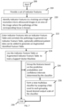
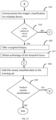
- Flow processing begins at step 100 and immediately proceeds to step 102.
- receive a set of high resolution micro-ultrasound images of prostate tissue which corresponds to tissue for which the pathology has been determined.
- analyze the image to detect Possible Features by, for example, conducting some form of object-based image analysis (OBIA) in the areas of the image (groups of pixels) where the pathology of the tissue in the image is known.
- OBIA object-based image analysis
- step 108 add Possible Features thereby detected and/or combinations of Possible Features observed to occur together into the List of Possible Features.
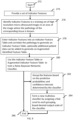
- step 122 obtain a set of high resolution micro-ultrasound images of prostate tissue that corresponds to tissue for which the pathology is known. It is possible to use the same training set but preferred to have a new set.
- step 124 a reader identifies Possible Features present in the images corresponding to the area for which the pathology of the tissue is known, while blinded to the pathology to generate Candidate Features.
- step 126 Enter Candidate Features into a Candidate Feature Table and correlate the pathology to generate a Candidate Feature Table, optionally additional patient data can be added to generate an Augmented Candidate Feature Table.
- step 128, Use the Candidate Feature Table or Augmented Candidate Feature Table to train a linear or non-linear classifier.
- step 130 eliminate Candidate Features and/or combinations of Candidate Features which are not statistically significant to produce a plurality of Indicator Features at step 132. If sufficient Indicator Features have been generated, then processing stops.
- Indicator Features can then be used to generate an Indicator Feature Table, which can be used to train a classifier, which can then be used to classify future patient images. Additional patient data may be included in the Indicator Feature Table to generate an Augmented Indicator Feature Table.
- An Indicator Feature Table which constitutes a training set to generate a predictive probability for the correlation between the presence of a feature in an image and the presence of cancer in the tissue being imaged.
- the classifier can be used to classify the features in images of patients generated using high-resolution micro-ultrasound devices.
- these predicted probabilities can be used to create a multiclass classifier by grouping and ranking the features according to the similarities and differences between their weights or predicted probabilities.
- the multiclass classifier can be used to classify the features in images of patients generated using high-resolution micro-ultrasound devices.
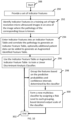
- FIG. 3 illustrates one embodiment of the present method for generating a classifier and/or multiclassifier comprising Indicator Features high resolution micro-ultrasound images.
- Flow processing begins at step 136 and immediately proceeds to step 138.
- step 138 provide a set of Indicator Features.
- step 140 identify Indicator Features in each of the training images in an area of the image where the pathology is known, but blinded to the reader.
- the pathology data is linked to the sonographic features in the Indicator Feature Table because the tissue sample and training image are taken from the same physical location in the prostate.
- additional information can be added to the Indicator Feature Table to generate an Augmented Feature Table.
- the Indicator Feature Table or the Augmented Feature Table is used to train a linear or non-linear classifier.
- the process can stop here, or optionally, proceed to step 162, to group the features based on the predictive probabilities of each Indicator Feature and/or combination of Indicator Features.
- form a new multiclass classifier based on the desired output scale of the classifier and the groupings of the features and the desired confidence intervals.
- the classifier or the multiclass classifier can be further trained using either the training data or test data.
- the desired ultrasound for use with the disclosed methods can be applied, transmitted and received using an ultrasonic scanning device that can supply ultrasound at a center frequency sufficient to accurately resolve sonographic features on the order of 70 um.
- a system with a center frequency transmit of at least about 15 MHz to the highest practical frequency can be used.
- ultrasound can be supplied at a center frequency of 15 MHz, 16MHz, 17 MHz, 18MHz, 19 MHz, 20 MHz, 21 MHz, 22MHz, 23 MHz, 24 MHz, 25 MHz, 26 MHz, 30 MHz, 31 MHz, 32 MHz, 35 MHz, 40 MHz, 45 MHz, 50 MHz, 55 MHz, 60 MHz, 65 MHz, 70 MHz, or higher than 70 MHz.
- an ultrasound system or device capable of operating at a center frequency at 15 MHz or above can be used.
- One such exemplary system is the 29 MHz transrectal micro-ultrasound system and transducer (ExactVu TM micro-ultrasound, Exact Imaging, Toronto, Canada). Another such exemplary system can have the components and functionality described in US Provisional Application No. 62253310 . Another exemplary system can have components and functionality described in PCT/IB2015/053458 . Another such exemplary system can have components and functionality described in PCT/IB2015/052882 .
- any system capable of producing an ultrasound image using a high frequency micro-ultrasound can be used.
- the methods can be practiced using a mechanically scanned ultrasound system that can translate an ultrasound beam as it sweeps along a path.
- the methods can also be practiced using an array based system where the beam is translated by electrical steering of an ultrasound beam along the elements of the transducer.
- beams translated from either type system can be used in the described methods, without any limitation to the type of system employed. The type of system is therefore not intended to be a limitation to any described method because array and mechanically scanned systems can be used interchangeably to perform the described methods.
- Embodiments described herein include a side-fire ultrasonic probe with an alignment feature that, when used to connect the probe to a needle guide for intra-cavity medical procedures, facilitates alignment of one or more needles translated through the needle guide with an imaging plane of an ultrasonic transducer.
- the alignment feature is configured such that alignment of a needle within the imaging plane is accomplished even when a protective sheath is disposed between the alignment feature and the needle guide.
- an ultrasonic image can be used to image an advancing needle with respect to an intra-cavity structure of interest.
- This ability is particularly useful when the ultrasonic transducer has a frequency and/or resolution sufficient to image intra-structure or intra-organ features.
- Simultaneously imaging the structure of interest and the needle permits navigation of the needle to a specific intra-cavity structure within a human body, or, given sufficient resolution of the ultrasonic transducer, navigation of the needle to a specific location within the structure. This can then improve the diagnostic capability of the procedure or effectiveness of the therapy. Allowing for positioning of a needle oriented at different angles with respect to the probe enables access to a range of locations within the body or structure by the needles while reducing the manipulation of the probe. This can improve patient comfort during the procedure, as well as patient safety.
- an ultrasonic probe in an embodiment, includes a cylindrical housing that includes a needle guide alignment feature on the surface of the housing.
- the alignment feature is used to connect a needle guide to the cylindrical housing and to align the needle guide such that a needle translated through the guide is translated in an imaging plane of the ultrasonic transducer.
- the alignment feature is configured such that the needle is aligned in the imaging plane even when a protective sheath is disposed between the housing and the needle guide.
- An exemplary needle guide alignment feature is provided by U.S. Patent Number 9,113,825 .
- Possible Features are discernable, recurring patterns, or features, appearing in high-resolution micro-ultrasound images, which are observed to correlate to tissue for which pathology is known, ranging from benign to the highest grade of cancer.
- the Gleason Sum scale ranges from Benign, 6, 7, 8, 9, & 10. Note that 2-5 used to be used as well but are generally now just labelled as benign. Other scales for grading the degree of cancer, or the stage of the cancer, in the tissue, though the concept remains the same.
- Possible Features are qualitative, not quantitative.
- Possible Features can be identified by using some form of Object-Based Image Analysis (OBIA), which employs two main processes, segmentation and classification. Possible Features are then characterized and given a useful label. A label could be simply Feature A, Feature B, Feature C, etc. A label could be given a descriptive name that describes the texture present in the image that is somewhat intuitive such a "bright spot,” or “mottled.” This process is conducted for all the Possible Features that can be identified along the needle path of the biopsy needle or in a region corresponding to biopsied tissue.
- OBIA Object-Based Image Analysis
- a digital image is a rectangular array of pixels. Each pixel is characterized by its position in the array and a plurality of numerical pixel values associated with the pixel. The pixel values represent color information for various image layers. For example, grayscale digital images are represented by a single image layer, whereas RGB true-color images are represented by three image layers.
- semantic network systems that apply semantic networks perform object-oriented picture analysis, as opposed to solely statistical pixel-oriented analysis. Consequently, semantic network systems classify not only pixels, but also data objects linked to the pixels. The data objects that are linked to the pixels and to one another represent measurable information about the digital images.
- Possible Features are identified by image segmentation in the region of the image corresponding to tissue, which has been biopsied. It is the process of determining areas (comprising groups of pixels) with similar image characteristics (e.g., color, intensity, texture) and which contrast to adjacent regions that are discernably different with respect to the same characteristic.
- the image is a greyscale image
- an area (constituted by a group of pixels), which are almost white in color and appear adjacent to and surrounded by other areas, which are dark grey in color with a sharp contrast boundary between the two
- the white area might be labelled a "bright spot.”
- an area might appear light grey with "fuzzy" boundaries of slightly darker area, might be grouped together and labelled “mottled.”
- Variations between areas may occur on a distinctive spatial scale (large or small areas of pixels), and may be described by relative changes in average brightness, sharpness and regularity of borders, and/or changes in gray-level textural parameters such as entropy, variability, contrast, or correlation. Regions with parameters different from typical normal tissue may be segmented and grouped based on these parameters.
- a human is used to generate the list of Potential Features.
- a computer can be used to read the images and identify Possible Features and/or combinations of Possible Features in the images.
- a human assisted by a computer can read the images and identify Possible Features and/or combinations of Possible Features in the images.
- the first step entails determining distinguishable recurring patterns in high resolution transrectal micro-ultrasound images of prostate glands.
- a 29 MHz transrectal micro-ultrasound system and transducer (ExactVu TM micro-ultrasound, Exact Imaging, Toronto, Canada) is used to acquire cine loops during each biopsy taken using the high resolution micro-ultrasound platform on patients who have had biopsies or radical prostatectomy such that clinical analysis data exists to determine whether the image correlates with cancerous tissue or not.
- biopsy locations may be selected based on anatomy (biopsy of a particular region of the gland) or based on the sonographic features noted in the subject (targeted biopsy).
- whole-mount radical prostatectomy slides registered to micro-ultrasound images can be used as a starting point to suggest useful imaging features.
- randomly-selected micro-ultrasound cine loops each captured during one specific biopsy can be analyzed by one or more experts to determine imaging findings along the biopsy needle path indicative of carcinomas, as well as normal tissue.
- the radical prostatectomy slides or biopsy data are not used to assign a link between a feature and cancerous tissue, but to assist in noticing which distinguishable and recurring aspects of the images might be suggestive or consistently present in tissue having a certain stage of prostate cancer.
- a series of images or video cine loops can be reviewed by a series of experts to identify a large set of distinguishable, recurring features.
- Each reviewer assigns a term to any non-normal or non-uniform pattern encountered, then the reviewers meet to compare terms and select the most common nomenclature for each feature.
- text descriptions may be analyzed by a computer using natural language processing systems to extract these features.
- a series of still images and/or cine loops can be analyzed by a computer for pattern detection to determine a list of distinguishable and recurring features existing in the images, presenting potential candidate features which might be found to be consistently able to be correlated to a stage or risk of prostate cancer.
- the Possible Features comprise features having the same or similar characteristics as: Feature Heterogenous-Bright Echoes-Finger/Funky Shadows Heterogenous-Finger/Funky Shadows Heterogenous-Coarse-Shadows Heterogenous-Hyperechoic-Coarse Heterogenous-Irregular Heterogenous-Lesion Heterogenous-Coarse-Finger/Funky Shadows Heterogenous-Irregular-Coarse Heterogenous-Shadows Heterogenous-Defined Heterogenous-Defined-Coarse Heterogenous-Hyperechoic Heterogenous-Hyperechoic-Irregular Heterogenous-Hypoechoic-Lesion-Bright Echoes Heterogenous "Cauliflower” Heterogenous "Smudged” Heterogenous "Mottled” Heterogenous "Cauliflower/Smudgy
- the Possible Features are then further processed by having someone skilled in the art of reading images, a computer or a combination thereof, identify the presence or absence of a Possible Features in a set of new training images, but blinded to the pathology while recording the presence or absence of features along the path of the biopsy needle in the image. Only features that overlap with the biopsy path are used. Any of the Possible Features that are assigned to images in the new training set are now considered Candidate Features. Since the individual is blinded to the pathology of the biopsy results, this step accounts for possible subjective interpretation biases of the features by the individual.
- combinations of features can be included and treated as a singular feature, ie., when the individual features are present alone, then they are treated as one piece of data, but when the features are present together, they are treated both as one piece of data and also recorded as individual features.
- results are used to construct a Candidate Feature Table, where they are correlated to the actual pathology of the tissue imaged in the region where the Candidate Feature was identified.
- a computer can be used to read the images and identify Candidate Features and/or combinations of Candidate Features in the new training set of images.
- other patient data is analyzed along with the features (with or without biopsy data), to increase the confidence that a specific feature is associated with cancer, comprising data obtained from: spatial information about the image, spatial information about the features, 3-D information, spectral information about the image, and, images collected using other modalities such as B-mode (grey-scale), Velocity Color Flow Imaging, Power Doppler Color Flow Imaging, Elastography, Contrast Enhanced Ultrasound, Spectral Analysis, conventional resolution ultrasound, MRI, CT, or PET imaging, patient age, DRE results, PSA readings, free PSA, PSA density, family history, race, prostate volume, transition zone volume, relevant biomarkers, protein/hormonal biomarker levels, genetic screening results, or number of prior negative biopsies
- Candidate Feature and/or combinations of Candidate Features which demonstrate little or no statistical significance will be eliminated to yield Indicator Features.
- the range of the confidence interval on predictive probability spanning 1 constitutes the threshold of statistical significance.
- Candidate Features can be identified in new set of training images, for which the pathology is known, but blinded to the reader of the images.
- a set of Possible Features to be used to identify Candidate Features comprise features having the same or similar characteristics as
- a set of Possible Features to be used to identify Candidate Features comprise features having the same or similar characteristics as:
- 200 randomly-selected micro-ultrasound cine loops each captured during one specific biopsy (from a total of 121 patients), can be analysed by one or more experts to determine imaging findings along the biopsy needle path indicative of high grade (Gleason Sum greater than 7) and low-intermediate grade (Gleason Sum 7 or less) carcinomas, as well as normal tissue.
- the sample set can include 100 benign, 50 low-grade and 50 high-grade biopsy-proven samples.
- experts can be blinded to the pathological findings of each biopsy sample, and record the variety of Potential Features present along the needle path in each image, which once identified (blinded) become Candidate Features. These Candidate Features can then be analyzed to see how often they are associated with biopsy-proven benign tissue or cancer.
- other information is analyzed along with the features (with or without biopsy data), to increase the confidence that a specific feature is associated with cancer, comprising, with additional data pertaining to the patient based on data obtained from: B-mode (grey-scale) images, Velocity Color Flow Imaging, Power Doppler Color Flow Imaging, Elastography, Contrast Enhanced Ultrasound, Spectral Analysis, conventional resolution ultrasound, MRI, CT, or PET imaging, patient data comprising patient age, DRE results, PSA readings, free PSA, PSA density, family history, race, prostate volume, transition zone volume, relevant biomarker levels, protein/hormonal biomarker levels, genetic screening results, or number of prior negative biopsies.
- Table 1 - An exemplary Candidate Feature Table comprising a list of sonographic features for which biopsy results are attributed.
- Feature N Total
- N Cancer
- Hyperechoic, with or without ductal patches 50 14 Mild heterogeneity 42
- Bright Echoes in hyperechoic tissue 10 4
- Heterogeneous "cauliflower/smudgy/mottled" appearance 32
- Bright Echoes 30 18
- Irregular Prostate (PZ) 1 1 Irregular PZ border 1 1 Mixed-echo lesions 2 Irregular Shadowing 12 11
- the objective is to determine when a feature is present in a sonographic image, what is the probability that it is associated with the tissue being imaged having a grade of prostate cancer versus benign tissue.
- the pathological diagnosis is the dependent variable
- the sonographic feature abnormality
- the mathematical task is to ascertain the likelihood that the presence of the independent variable (the sonographic feature) is dependent upon the presence of cancer in that tissue.
- the answer can be binomial in that the equation seeks to prove either cancer is present or absent with each feature.
- the answer may be expressed as a beta random variable, expressing the increased or decreased risk of cancer on a continuous scale between 0 (no risk) and 1 (certainty of cancer). This continuous scale may also be expressed as an odds ratio or relative risk between 0 (no risk) and infinity (certainty of cancer).
- a univariate relative risk (RR) and a confidence interval (CI) are calculated for each sonographic feature, and used to assign a risk parameter to each Candidate Feature and/or combination of Candidate Features.
- RR relative risk
- CI confidence interval
- Indicator Features are features appearing alone and/or in combination with other Indicator Features in high resolution micro-ultrasound images, which have been determined to be significantly statistically correlated to either benign tissue or some grade of cancerous tissue on the basis of predictive probabilities.
- Indicator Features can then be used to generate an Indicator Feature Table, which can be used to train a classifier, which can then be used to classify future patient images.
- a classifier can then be used to classify future patient images.
- the classifier can be used to classify the features in images of patients generated using high-resolution micro-ultrasound devices.
- these predicted probabilities can be used to create a multiclass classifier by grouping and ranking the features according to the similarities and differences between their weights or predicted probabilities.
- the multiclass classifier can be used to classify the features in images of patients generated using high-resolution micro-ultrasound devices.
- the Indicator Features comprise features having the same or similar characteristics as
- a set of Indicator Features can then be used to generate an Indicator Feature Table. In an embodiment, this is generated by re-using the data that used to evaluate the Candidate Features (for which the statistically insignificant Candidate Features have now been removed).
- the Table can be used to train a classifier, which can then be used to classify future patient images.
- the results of this classifier can optionally be grouped to generate a multiclass classifier, which then can be used to classify future patient images.
- a new set of images (for which the pathology is known) can be used to identify Indicator Features in the images, while blinded to the pathology, then use the results of assigning Indicator Features to the new set of images plus the corresponding pathology to populate an Indicator Feature Table.
- additional patient data can be included in the Table.
- the Table can be used to train a classifier, which can then be used to classify future patient images.
- the results of this classifier can optionally be grouped to generate a multiclass classifier, which then can be used to classify future patient images.
- a set of Indicator Features comprise features having the same or similar characteristics as:
- a set of Indicator Features comprise features having the same or similar characteristics as:
- patient data is analyzed along with the features (with or without biopsy data), to increase the confidence that a specific feature is associated with cancer, comprising data obtained from, B-mode (grey-scale) images, Velocity Color Flow Imaging, Power Doppler Color Flow Imaging, Elastography, Contrast Enhanced Ultrasound, Spectral Analysis, conventional resolution ultrasound, MRI, CT, or PET imaging, patient data comprising patient age, DRE results, PSA readings, free PSA, PSA density, family history, race, prostate volume, transition zone volume, relevant biomarker levels, protein/hormonal biomarker levels, genetic screening results, or number of prior negative biopsies.
- B-mode grey-scale
- Velocity Color Flow Imaging Velocity Color Flow Imaging
- Power Doppler Color Flow Imaging Power Doppler Color Flow Imaging
- Elastography Contrast Enhanced Ultrasound
- Spectral Analysis conventional resolution ultrasound, MRI, CT, or PET imaging
- patient data comprising patient age, DRE results, PSA readings, free PSA, PS
- a training set comprising Indicator Features, which have been correlated to clinical pathology data for the same tissue imaged in the sonogram.
- this is accomplished by using a high-resolution transrectal micro-ultrasound system to acquire images of the needle path followed during each biopsy taken on patients. The pathological analysis of the tissue collected in each biopsy is then used determine image findings (features) along the biopsy needle path indicative of carcinomas, as well as normal tissue.
- a univariate relative risk (RR) and a confidence interval (CI) are calculated for each sonographic feature, and used to assign a risk parameter to each Indicator Feature and/or combination of Indicator Features.
- the calculating step uses machine learning.
- Common machine learning algorithms may be used to create a classifier based on the feature data, such as binary classification trees, "random forests", support vector machines, artificial neural networks, and naive bayesian networks.
- multivariate relative risk is calculated for a combination of features. A skilled person would understand as the number of features increases machine learning is used since the complexity of selecting appropriate thresholds and managing generalizability increases exponentially with the number of features or combination of features
- Figure 4 illustrates the process of building a multiclass classifier calculating a univariate relative risk (RR) and a confidence interval (CI) for each sonographic feature, and used to assign a risk parameter to each sonographic feature in the Indicator Features Table, which can be then used to generate a multiclass classifier.
- the RR ratio is the relative occurrence of a given feature or symptom in two distinct populations.
- the RR for each sonographic feature indicates the relative occurrence of each feature in a population of cancerous tissue versus non-cancerous tissue. Large differences in occurrence rates suggest that the feature may be used to predict which population a given image or patient belongs to.
- Equation (1) is used to calculate a relative risk ratio (Table 2) of each Indicator Feature.
- CI e m ⁇ 1.96 ⁇
- Table 3 illustrates relative risks (RR) and confidence intervals (CI) calculated for example Indicator Features from blinded analysis of 100 biopsy-proven benign and 100 biopsy-proven malignant cine loops Indicator Features N (Tota l) N (Cance r) RR [90% CI] Small regular ducts "Swiss cheese" 7 1 0.28 [0.05 - 1.72] Hyperechoic, with or without ductal patches 50 14 0.49 [0.31 - 0.78] Mild heterogeneity 42 24 1.19 [0.87 - 1.62] Bright Echoes in hyperechoic tissue 10 4 0.79 [0.37 - 1.71] Heterogeneous "cauliflower/smudgy/mottled" appearance 32 22 1.48 [1.11 - 1.97] Bright Echoes 30 18 1.24 [0.89 - 1.73] Irregular Prostate (PZ) 1 1 2.01 [1.75 - 2.
- Flow processing begins at step 152 and immediately proceeds to step 154.
- step 154 provide a set of Indicator Features.
- step 156 identify Indicator Features in each of the training images in an area of the image where the pathology is known, but blinded to the reader.
- the pathology data is linked to the sonographic features because the tissue sample and training image are taken from the same physical location in the prostate.
- step 160 a univariate relative risk and a confidence interval are calculated for each sonographic feature or group of features.
- group the features based on the relative risk and the confidence interval of each individual feature.
- step 164 form a new multiclass classifier based on the desired output scale of the classifier and the groupings of the features and the desired confidence intervals.
- the singular features are grouped based on the relative risk and confidence interval of each individual feature. If a linear classifier is used, combinations of features may also be added if sufficient data is available. A new multiclass classifier is formed based on the desired output scale of the classifier and the groupings of the features and the desired confidence intervals.
- predefined thresholds are used to group features based on mean relative risk values. For a data set with balanced numbers of cancerous and benign observations, these predetermined values split the theoretical 0-2 range of the relative risk value into sections that provide useful assessment of the underlying risk, for example: Risk Score Mean RR Range Assignment 1 0-0.4 Very low risk 2 0.4-0.6 Some risk 3 0.6-1.2 Indeterminate risk 4 1.2-1.6 Significant risk 5 1.6+ Very High risk
- automated thresholding is used to determine the output scale.
- the following procedure may be used:
- k-nearest neighbors clustering may be used with a fixed number of predefined starting classes spaced evenly over the RR space.
- the point scale may be less than 5 points. In an embodiment, the point scale may be 5 points. In an embodiment, the point scale may be 6 points. In an embodiment, the point scale may be 7 points. In an embodiment, the point scale may be 8 points. In an embodiment, the point scale may be 9 points. In an embodiment, the point scale may be 10 points. In an embodiment, the point scale may be more than 10 points.
- the point scale may include sub classifications.
- the point scale is 1, 2, 3A, 3B, 3C, 4, 5.
- the machine learning analysis is used to rank and group features with similar risk profiles into an easy to use point scale.
- Table 4 illustrates how the RR and CI results can be used to group and assign a Risk Score to the Indicator Features, thereby creating a multiclass classifier for high resolution micro ultrasound sonograms of the prostate.
- the multiclass classifier comprises a possible risk level of:
- the multiclass classifier comprises a risk level of:
- RR and CI results are used to generate predictive probabilities for Indicator Features.
- an alternative mathematical processing such as binary classification trees, "random forests", support vector machines, artificial neural networks, and naive bayesian networks, of the same results found in the Indicator Feature Table or Augmented Indicator Feature Table.
- each Indicator Feature and/or combination of Indicator Features is considered as a separate column of a feature matrix, which may take on Boolean values (present or absent).
- One row of this matrix is created for each image/cine loop (biopsy sample) available.
- Pathological result is also modelled as a boolean value in the form of a vector with one entry per image/cine loop (biopsy sample) indicating the presence or absence of cancer.
- FIG. 5 illustrates one embodiment of the present method for generating a classifier and/or multiclassifier comprising Indicator Features high resolution micro-ultrasound images.
- FIG. 5 illustrates the process of training a binary classification tree classifier using an Indicator Feature Table or Augmented Indicator Feature Table.
- Flow processing begins at step 172 and immediately proceeds to step 174.
- step 174 provide a set of Indicator Features.
- step 176 identify Indicator Features in each of the training images in an area of the image where the pathology is known, but blinded to the reader.
- the pathology data is linked to the sonographic features in the Indicator Feature Table because the tissue sample and training image are taken from the same physical location in the prostate.
- additional information can be added to the Indicator Feature Table to generate an Augmented Feature Table.
- the Indicator Feature Table or the Augmented Feature Table is used to train a binary classification tree classifier.
- the process can stop here at step 186, or optionally, proceed to step 182, to group the features based on the predictive probabilities of each Indicator Feature and/or combination of Indicator Features.
- form a new multiclass classifier based on the desired output scale of the classifier and the groupings of the features and the desired confidence intervals.
- the classifier or the multiclass classifier can be further trained using either training data or test data.
- a binary classification tree may be created using the following algorithm:
- the classifier is queried by starting at the root node and following each branch based on the input until a "leaf' or terminal node is reached.
- FIG. 6 illustrates one embodiment of the present method for generating a classifier and/or multiclassifier comprising Indicator Features high resolution micro-ultrasound images.
- FIG. 6 illustrates the process of training a random forest classifier using an Indicator Feature Table or Augmented Indicator Feature Table.
- Flow processing begins at step 192 and immediately proceeds to step 194.
- step 194 provide a set of Indicator Features.
- step 196 identify Indicator Features in each of the training images in an area of the image where the pathology is known, but blinded to the reader.
- the pathology data is linked to the sonographic features in the Indicator Feature Table because the tissue sample and training image are taken from the same physical location in the prostate.
- additional information can be added to the Indicator Feature Table to generate an Augmented Feature Table.
- the Indicator Feature Table or the Augmented Feature Table is used to train a random forest classifier.
- the process can stop here at step 206, or optionally, proceed to step 202, to group the features based on the predictive probabilities of each Indicator Feature and/or combination of Indicator Features.
- form a new multiclass classifier based on the desired output scale of the classifier and the groupings of the features and the desired confidence intervals.
- the classifier or the multiclass classifier can be further trained using either training data or test data.
- a random forest classifier may be created using the following algorithm:
- the classifier is queried by querying each individual tree with a given input, and taking the average response of the set of trees. For example, for the input case "Bright echoes AND irregular border" the trees may range in suspicion of cancer from 70-90%, one might report the outcome as having a mean value of 80% with a confidence interval of 72-85%.
- FIG. 7 illustrates one embodiment of the present method for generating a classifier and/or multiclassifier comprising Indicator Features high resolution micro-ultrasound images.
- FIG. 7 illustrates the process of training a support vector machine using an Indicator Feature Table or Augmented Indicator Feature Table.
- Flow processing begins at step 208 and immediately proceeds to step 210.
- step 210 provide a set of Indicator Features.
- step 220 identify Indicator Features in each of the training images in an area of the image where the pathology is known, but blinded to the reader.
- the pathology data is linked to the sonographic features in the Indicator Feature Table because the tissue sample and training image are taken from the same physical location in the prostate.
- additional information can be added to the Indicator Feature Table to generate an Augmented Feature Table.
- the Indicator Feature Table or the Augmented Feature Table is used to train a support vector machine.
- the process can stop here at step 230, or optionally, proceed to step 226, to group the features based on the predictive probabilities of each Indicator Feature and/or combination of Indicator Features.
- form a new multiclass classifier based on the desired output scale of the classifier and the groupings of the features and the desired confidence intervals.
- the classifier or the multiclass classifier can be further trained using either training data or test data.
- Quadratic programming techniques such as Sequential Minimization Optimization or Iterative Single Data Algorithm may be applied to determine the N-dimensional hyperplane best separating the benign from cancerous data points (where N is the number of biopsy samples available, the number of rows in the feature matrix).
- a kernel function is not required so long as independent, binary feature values are used.
- FIG. 8 illustrates one embodiment of the present method for generating a classifier and/or multiclassifier comprising Indicator Features high resolution micro-ultrasound images.
- FIG. 8 illustrates the process of training an artificial neural network classifier using an Indicator Feature Table or Augmented Indicator Feature Table.
- Flow processing begins at step 232 and immediately proceeds to step 234.
- step 234 provide a set of Indicator Features.
- step 238, identify Indicator Features in each of the training images in an area of the image where the pathology is known, but blinded to the reader.
- the pathology data is linked to the sonographic features in the Indicator Feature Table because the tissue sample and training image are taken from the same physical location in the prostate.
- additional information can be added to the Indicator Feature Table to generate an Augmented Feature Table.
- the Indicator Feature Table or the Augmented Feature Table is used to train an artificial neural network classifier.
- the process can stop here at step 246, or optionally, proceed to step 242, to group the features based on the predictive probabilities of each Indicator Feature and/or combination of Indicator Features.
- step 244 form a new multiclass classifier based on the desired output scale of the classifier and the groupings of the features and the desired confidence intervals.
- the classifier or the multiclass classifier can be further trained using either training data or test data.
- An artificial neural network may be created with N input nodes (one for each feature) and a single output node with a continuous output representing the probability of cancer.
- N input nodes one for each feature
- M layers of perceptrons each summing weighted input from the previous layer and exhibiting a sigmoidal response curve.
- FIG. 9 illustrates one embodiment of the present method for generating a classifier and/or multiclassifier comprising Indicator Features high resolution micro-ultrasound images.
- FIG. 9 illustrates the process of training a naive Bayesian network classifier using an Indicator Feature Table or Augmented Indicator Feature Table.
- Flow processing begins at step 272 and immediately proceeds to step 274.
- step 274 provide a set of Indicator Features.
- step 276 identify Indicator Features in each of the training images in an area of the image where the pathology is known, but blinded to the reader.
- the pathology data is linked to the sonographic features in the Indicator Feature Table because the tissue sample and training image are taken from the same physical location in the prostate.
- additional information can be added to the Indicator Feature Table to generate an Augmented Feature Table.
- the Indicator Feature Table or the Augmented Feature Table is used to train an artificial neural network classifier.
- the process can stop here at step 286, or optionally, proceed to step 282, to group the features based on the predictive probabilities of each Indicator Feature and/or combination of Indicator Features.
- form a new multiclass classifier based on the desired output scale of the classifier and the groupings of the features and the desired confidence intervals.
- the classifier or the multiclass classifier can be further trained using either training data or test data.
- Figure 8 illustrates the process of training a linear discriminate analysis classifier using an Indicator Feature Table or Augmented Indicator Feature Table.
- FIG. 10 illustrates one embodiment of the present method for generating a classifier and/or multiclassifier comprising Indicator Features high resolution micro-ultrasound images.
- FIG. 10 illustrates the process of training a linear discriminate analysis classifier using an Indicator Feature Table or Augmented Indicator Feature Table.
- Flow processing begins at step 288 and immediately proceeds to step 290.
- step 290 provide a set of Indicator Features.
- step 292 identify Indicator Features in each of the training images in an area of the image where the pathology is known, but blinded to the reader.
- the pathology data is linked to the sonographic features in the Indicator Feature Table because the tissue sample and training image are taken from the same physical location in the prostate.
- additional information can be added to the Indicator Feature Table to generate an Augmented Feature Table.
- the Indicator Feature Table or the Augmented Feature Table is used to train a linear discriminate analysis classifier.
- the process can stop here at step 302, or optionally, proceed to step 298, to group the features based on the predictive probabilities of each Indicator Feature and/or combination of Indicator Features.
- form a new multiclass classifier based on the desired output scale of the classifier and the groupings of the features and the desired confidence intervals.
- the classifier or the multiclass classifier can be further trained using either training data or test data.
- ⁇ are the class means
- ⁇ are the class covariances
- subscripts B and C are for the benign and cancerous classes respectively.
- the vector w then represents the normal to the hyperplane separating the two classes and scalar c the threshold along that hyperplane.
- a new observation (N) may be classified by multiplying by w and then finding the distance (i.e. ⁇ c - wN ⁇ ) and sign (i.e. sgn(c-wN)) to the point c.
- a classifier is validated by using it with a new independent set of images for which the pathology is known (including a mixture of both benign and malignant tissue) but not revealed to the person or computer reading the images to determine the presence of Indicator Features.
- the results are entered into an Indicator Feature Table and correlated with the pathology. This data is used to calculate ROC's for each reader. Accuracy is calculated by using the AUC under each reader's ROC.
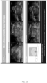
- FIG. 11 shows exemplary sonographic features used in an embodiment of a multiclass classfier.
- FIG. 11G Irregular shadowing presents as vertical hypoechoic regions with no obvious source of attenuation (such as a calcification or gas bubble).
- FIG. 11H a mixed echo lesion with well-defined borders is present pressing on the rectal wall causing the prostate border to be irregular. In both cases the biopsy samples show a Gleason Sum of 9.
- a database comprising high resolution micro-ultrasound prostate images, wherein Indicator Features in the images have been correlated to tissue biopsy results and optionally a risk score assigned by a classifier or multiclass classifier.
- the database is structured to: i) permit queries regarding co-occurrence of features, ii) examine various instances of each feature in order to train an automated analysis system such as a deep learning image analyzer to recognize the features; and iii) add new images, marked with indicator features so that the method can be continually improved by providing a better assessment of the predicted probability of cancer for each feature or combination of features.
- the database can optionally comprise additional data pertaining to the patient data obtained from, B-mode (grey-scale) images, Velocity Color Flow Imaging, Power Doppler Color Flow Imaging, Elastography, Contrast Enhanced Ultrasound, Spectral Analysis, conventional resolution ultrasound, MRI, CT, or PET imaging, patient data comprising patient age, DRE results, PSA readings, free PSA, PSA density, family history, race, prostate volume, transition zone volume, relevant biomarker levels, protein/hormonal biomarker levels, genetic screening results, or number of prior negative biopsies.
- embodiments of the present invention can be implemented in various forms of hardware, software, firmware, special purpose processes, or a combination thereof.
- the present invention can be implemented in software as an application program tangible embodied on a computer readable program storage device.
- the application program can be uploaded to, and executed by, a machine comprising any suitable architecture.
- FIG. 14 is a block diagram of an exemplary computer system for implementing a method and system for classifying a patient's risk of having prostate cancer according to an embodiment of the invention.
- a computer system 700 for implementing the present invention can comprise, inter alia, a central processing unit (CPU) 702, a memory 704 and an input/output (I/O) interface 706.
- the computer system 700 is generally coupled through the I/O interface 706 to a display 708 and various input devices 710 such as a mouse and a keyboard.
- the support circuits can include circuits such as cache, power supplies, clock circuits, and a communication bus.
- the memory 704 can include random access memory (RAM), read only memory (ROM), disk drive, tape drive, etc., or a combinations thereof.
- RAM random access memory
- ROM read only memory
- the present invention can be implemented as a routine 712 that is stored in memory 704 and executed by the CPU 702 to process the signal from the signal source 714.
- the computer system is a general purpose computer system that becomes a specific purpose computer system when executing the routine 712 of the present invention.
- the computer system 700 also includes an operating system and micro instruction code.
- the various processes and functions described herein can either be part of the micro instruction code or part of the application program (or combination thereof) which is executed via the operating system.
- various other peripheral devices can be connected to the computer platform such as an additional data storage device and a printing device.
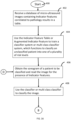
- FIG. 16 illustrates one embodiment of the present method using the multiclass classifier for classifying patient risk for prostate cancer in accordance with the teachings hereof.
- Flow processing begins at step 400 and immediately proceeds to step 402.
- step 402 receive a database of micro-ultrasound images containing Indicator Features and correlated to pathology results.
- the features are: Small regular ducts, Hyperechoic tissue with our without ductal patches, mild heterogeneity, bright echoes in hyperechoic tissue, heterogeneous appearance, bright echoes, irregular peripheral zone, irregular prostate border, mixed-echo lesions, irregular shadowing.
- a multiclass classifier uses the multiclass classifier to classify the unclassified image into one of a plurality of classes. In an embodiment, this is accomplished by the operator consulting a printed flow chart or other visual aid (for example, see FIG. 12 ) generated based on step 404 which begins with high-risk features and proceeds to low risk features. In another embodiment, an automated system is used to classify the image.
- step 412 communicate the image's classification to a display device.
- the patient classification is communicated to any of: a storage device, a wireless handheld device, a laptop, tablet-PC, and a workstation.
- step 414 a determination is made whether the classified image suggests a significant risk for having prostate cancer.
- a targeted biopsy may be taken to confirm the diagnosis.
- images are being analyzed off-line in an active surveillance patient and the tissue identified by the image in step 414 is determined to have increased in risk level, the patient may be recalled so that a new biopsy sample may be taken, or potentially recommended for treatment.
- step 418 once pathology results have been gathered (e.g., step 417) for any biopsy or radical prostatectomy samples taken, the identified sonographic features and corresponding pathology may optionally be added (step 418) to the database of step 402. Step 404 may then be repeated to improve the classification system.
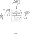
- FIG. 18 shows a block diagram of one example system for performing various aspects of the present (using the Classifier)
- a training set (collectively at 501) comprising images for a plurality of subjects are retrieved from a database 502.
- Database 502 is a storage device wherein records are stored, manipulated, and retrieved in response to a query. Such records, in various embodiments, take the form of Tables of Indicator Features and/or Tables of Augmented Indicator Features.
- the database is shown as an external device, the database 502 may be internal to the workstation mounted, for example, on a hard disk therein.
- the training set is provided to the classifier system for training purposes. The features of the training set are obtained from the sonographic images
- the classifier system comprises a plurality of modules.
- Learning Module 503 processes the training data contained in the records of the training set such that the classifier system can be trained.
- the Learning Module 503 further functions to prune the training set, as desired, such that the classifier is trained with data which meet a pre-determined criteria such as acceptable image quality.
- Learning Module 503 signals Classification Module 504 to receive an image of a yet-to-be classified patient 506.
- the unclassified patient's images 506 are received or are otherwise obtained by the classifier system which, in turn, proceeds to classify the unclassified image into one of a number of risk levels.
- Processor retrieves machine readable program instructions from Memory 505 and is provided to facilitate the functionality of the various modules comprising the classifier system.
- the processor operating alone or in conjunction with other processors and memory, may be configured to assist or otherwise facilitate the functionality of any of the processors and modules of system.
- the classifier system of FIG. 18 is shown in communication with a workstation.
- a computer case of the workstation houses various components such as a motherboard with a processor and memory, a network card, a video card, a hard drive capable of reading/writing to machine readable media such as a floppy disk, optical disk, CD-ROM, DVD, magnetic tape, and the like, and other software and hardware needed to perform the functionality of a computer workstation.
- the workstation further includes a display device, such as a CRT, LCD, or touchscreen device, for displaying information, video, measurement data, computed values, medical information, results, locations, and the like. A user can view any of that information and make a selection from menu options displayed thereon. Keyboard and mouse effectuate a user input.
- the workstation has an operating system and other specialized software configured to display alphanumeric values, menus, scroll bars, dials, slideable bars, pull-down options, selectable buttons, and the like, for entering, selecting, modifying, and accepting information needed for processing in accordance with the teachings hereof.
- the workstation is further enabled to display the sonographic image and patient classifications as they are derived.
- the workstation may further display interim values, boundary conditions, and the like, in real-time as the classifier system performs its intended functionality as described herein in detail.
- a user or technician may use the user interface of the workstation to set parameters, view/adjust/delete values in the training set, and adjust various aspects of the classifier system as needed or as desired, depending on the implementation. Any of these selections or input may be stored/retrieved to storage device. Default settings can be retrieved from the storage device.
- a user of the workstation is also able to view or manipulate any of the records contained in the training set via pathways not shown.
- the workstation can be a laptop, mainframe, or a special purpose computer such as an ASIC, circuit, or the like.
- the embodiment of the workstation of FIG 18 is illustrative and may include other functionality known in the arts. Any of the components of the workstation may be placed in communication with the classifier system or any devices in communication therewith. Any of the modules of the classifier system can be placed in communication with storage device and/or computer readable media and may store/retrieve therefrom data, variables, records, parameters, functions, and/or machine readable/executable program instructions, as needed to perform their intended functions. Each of the modules of the classifier system may be placed in communication with one or more remote devices over network.
Landscapes
- Health & Medical Sciences (AREA)
- Engineering & Computer Science (AREA)
- Life Sciences & Earth Sciences (AREA)
- Medical Informatics (AREA)
- Public Health (AREA)
- General Health & Medical Sciences (AREA)
- Biomedical Technology (AREA)
- Physics & Mathematics (AREA)
- Radiology & Medical Imaging (AREA)
- Nuclear Medicine, Radiotherapy & Molecular Imaging (AREA)
- Pathology (AREA)
- Heart & Thoracic Surgery (AREA)
- Veterinary Medicine (AREA)
- Animal Behavior & Ethology (AREA)
- Biophysics (AREA)
- Surgery (AREA)
- Molecular Biology (AREA)
- Data Mining & Analysis (AREA)
- Theoretical Computer Science (AREA)
- Computer Vision & Pattern Recognition (AREA)
- Epidemiology (AREA)
- Primary Health Care (AREA)
- General Physics & Mathematics (AREA)
- Databases & Information Systems (AREA)
- Quality & Reliability (AREA)
- Evolutionary Computation (AREA)
- Evolutionary Biology (AREA)
- Bioinformatics & Computational Biology (AREA)
- General Engineering & Computer Science (AREA)
- Bioinformatics & Cheminformatics (AREA)
- Artificial Intelligence (AREA)
- Vascular Medicine (AREA)
- Hematology (AREA)
- Business, Economics & Management (AREA)
- General Business, Economics & Management (AREA)
- Physiology (AREA)
- Probability & Statistics with Applications (AREA)
- Ultra Sonic Daignosis Equipment (AREA)
Claims (13)
- Computer-implementiertes Verfahren zur Erzeugung von einem oder mehreren Indikatormerkmalen in hochauflösenden Ultraschallbildern, die bei der Zuordnung des Risikos von Prostatakrebs während der hochauflösenden Mikro-Ultraschallabbildung verwendet werden können, wobei das Verfahren die folgenden Schritte umfasst:i) Erzeugen einer Liste von möglichen Merkmalen in den Bildern durch:a) Erhalten eines hochauflösenden Mikro-Ultraschallbildes eines Prostatagewebes, das einem Gewebe entspricht, das auch biopsiert wurde und für ein Krebsstadium eingestuft wurde, das von gutartig bis zum höchsten Grad reicht;b) Segmentieren des Bereichs des Bildes, der dem biopsierten Gewebe entspricht, auf der Grundlage von kontrastierenden Bereichen oder Gruppen von Pixeln in dem Bild, wobei jeder Bereich oder jede Gruppe von Pixeln ein Merkmal darstellt; undc) Charakterisieren und Bereitstellen einer eindeutigen Bezeichnung für alle erkennbaren Merkmale und/oder Kombinationen von Merkmalen nur in dem Bereich des Bildes, der dem biopsierten Gewebe entspricht,ii) Wiederholen der Schritte a) bis d), bis alle wiederkehrenden Muster in die Liste von Möglichen Merkmale aufgenommen worden sind;iii) Erzeugen einer Tabelle von Kandidatenmerkmalen durch:wobei die Merkmale alle erkennbaren, wiederkehrenden Muster sind, die in hochauflösenden Mikro-Ultraschallbildern erscheinen.d) Lesen der Mikro-Ultraschallbilder in dem Bereich, der dem Gewebe entspricht, das für ein Krebsstadium eingestuft wurde, dessen Ergebnisse dem Leser jedoch nicht bekannt sind, Identifizieren und Aufzeichnen in der Tabelle von Kandidatenmerkmalen und/oder von Kombinationen von Kandidatenmerkmalen, die in dem Bereich beobachtet wurden, der dem eingestuften Gewebe entspricht;e) Korrelieren in der Tabelle von Kandidatenmerkmalen des Krebsstadiums in dem biopsierten Gewebe, das dem Bereich in dem Bild entspricht, in dem jedes der Kandidatenmerkmale und/oder Kombinationen von Kandidatenmerkmalen identifiziert werden, wobei das Stadium von gutartig bis zum höchsten Grad reicht;f) Verwenden der Tabelle von Kandidatenmerkmalen als einen Trainingssatz von Merkmalen, um einen linearen oder nichtlinearen Klassifikator zu trainieren, um ein Kandidatenmerkmal oder eine Kombination von Kandidatenmerkmalen als krebsartig oder nicht krebsartig zu klassifizieren und dadurch eine vorhergesagte Wahrscheinlichkeit von Krebs für ein gegebenes Kandidatenmerkmal oder eine Kombination von Kandidatenmerkmalen zu bestimmen, wobei die vorhergesagte Wahrscheinlichkeit ein univariates relatives Risiko und ein Konfidenzintervall ist, das für jedes Kandidatenmerkmal berechnet wird, und verwendet wird, um jedem Kandidatenmerkmal und/oder jeder Kombination von Kandidatenmerkmalen einen Risikograd zuzuweisen;g) Eliminieren von Kandidatenmerkmalen oder Kombinationen von Kandidatenmerkmalen, die keine für ein Indikatormerkmal erforderlichen vorhergesagten Wahrscheinlichkeiten ergeben; und
- Verfahren nach Anspruch 1, wobei das hochauflösende Mikro-Ultraschallbild unter Verwendung einer Ultraschall-Abtastvorrichtung erzeugt wird, die Ultraschall mit einer Mittenfrequenz von 15 MHz bis 70 MHz liefern kann.
- Verfahren nach Anspruch 1 oder 2, wobei das Bild ist: B-Mode (Graustufen), Geschwindigkeits-Farbflussbildgebung, Power-Doppler-Farbflussbildgebung, Elastographie, Kontrastverstärkung und/oder Spektralanalyse.
- Verfahren nach einem der Ansprüche 1 bis 3, wobei die Ablesung durchgeführt wird von:i) einem Menschen;ii) einem Computer; oderiii) einem Menschen und einem Computer.
- Verfahren nach einem der Ansprüche 1 bis 4, wobei zusätzliche Patientendaten in die Tabelle von Kandidatenmerkmalen aufgenommen und mit jedem Kandidatenmerkmal oder jeder Kombination von Kandidatenmerkmalen mit dem im Gewebe vorhandenen Krebsstadium korreliert werden, um eine erweiterte Tabelle von Kandidatenmerkmalen zu erzeugen, die dann zum Trainieren des linearen oder nichtlinearen Klassifikators in Schritt f) verwendet wird.
- Verfahren zum Implementieren eines Klassifikators, wobei das Verfahren die Schritte nach einem der Ansprüche 1 bis 5 umfasst, und ferner die folgenden Schritte umfasst:i) Bereitstellen eines Trainingssatzes von Indikatormerkmalen, der Indikatormerkmale und/oder Kombinationen von Indikatormerkmalen umfasst, die mit der Pathologie des Gewebes korreliert sind;ii) Verwenden des Trainingssatzes von Indikatormerkmalen, um den linearen oder nichtlinearen Klassifikator zu trainieren, der ein Indikatormerkmal und/oder Kombinationen von Indikatormerkmalen als gutartig oder krebsartig klassifiziert und dadurch eine vorhergesagte Krebswahrscheinlichkeit für ein Indikatormerkmal und/oder Kombinationen von Indikatormerkmalen bestimmt,wobei die vorhergesagte Wahrscheinlichkeit ein univariates relatives Risiko und ein Konfidenzintervall ist, das für jedes Kandidatenmerkmal berechnet wird, und verwendet wird, um jedem Indikatormerkmal und/oder jeder Kombination von Indikatormerkmalen ein Risikoniveau zuzuweisen.
- Verfahren nach Anspruch 6, das zusätzlich den Schritt der Aufnahme zusätzlicher Patientendaten in den Trainingssatz von Indikatormerkmalen umfasst, wobei die zusätzlichen Patientendaten mit einem Indikatormerkmal oder einer Kombination von Indikatormerkmalen korreliert sind.
- Verfahren zum Implementieren eines Multiklassen-Klassifikators, wobei das Verfahren die Schritte nach Anspruch 6 oder 7 umfasst und ferner die folgenden Schritte umfassti) Gruppieren der Indikatormerkmale und Kombinationen von Indikatormerkmalen auf der Grundlage der vorhergesagten Wahrscheinlichkeiten und Konfidenzintervalle, die durch den Klassifikator bestimmt werden; undii) Bilden des Multiklassen-Klassifikators durch Zuweisen eines Risikoniveaus zu jeder Gruppierung von Indikatormerkmalen auf der Grundlage der gewünschten Ausgabeskala des Klassifikators.
- Verfahren zur Unterstützung bei der Diagnose von Prostatakrebs, wobei das Verfahren umfasst:Durchführen des Verfahrens nach Anspruch 6 oder 7, undVerwenden des implementierten Klassifikators als unterstützende Information;
oderDurchführen des Verfahrens nach Anspruch 8, undVerwenden des implementierten Multiklassen-Klassifikators als unterstützende Information. - Verfahren zum Anweisen eines Klinikers, wo ein Biopsiekern aus der Prostata zu entnehmen ist, wobei das Verfahren umfasst:Durchführung des Verfahrens nach Anspruch 6 oder 7, undVerwenden des implementierten Klassifikators als Anweisungsinformation;
oderDurchführen des Verfahrens nach Anspruch 8, undVerwenden des implementierten Multiklassen-Klassifikators als Anweisungsinformation. - Verfahren nach einem der Ansprüche 1 bis 10, wobei ferner vordefinierte Schwellenwerte verwendet werden, um Merkmale auf der Grundlage mittlerer relativer Risikoniveaus zu gruppieren; und/oder wobei ein Datensatz, der eine ausgewogene Anzahl von karzinomatösen und gutartigen Beobachtungen umfasst, diese vorbestimmten Werte den theoretischen 0-2-Bereich der relativen Risikoniveaus in Abschnitte aufteilt, die eine Bewertung des zugrunde liegenden Risikos liefern.
- Eine Programmspeichervorrichtung, die von einem Computer lesbar ist und die ein Programm mit Anweisungen verkörpert, die von dem Computer ausführbar sind, um die Verfahrensschritte zur Implementierung eines Klassifikators durchzuführen, wobei das Verfahren die folgenden Schritte umfasst:i) Bereitstellen eines Trainingssatzes von Indikatormerkmalen in hochauflösenden Mikro-Ultraschallbildern der Prostata, die Indikatormerkmale und/oder Kombinationen von Indikatormerkmalen umfassen, die mit der Pathologie des Gewebes korreliert sind;ii) Verwenden des Trainingssatzes von Indikatormerkmalen, um einen linearen oder nichtlinearen Klassifikator zu trainieren, der ein Indikatormerkmal und/oder Kombinationen von Indikatormerkmalen als gutartig oder krebsartig klassifiziert und dadurch eine vorhergesagte Wahrscheinlichkeit von Krebs für ein Indikatormerkmal und/oder Kombinationen von Indikatormerkmalen bestimmt, wobei die vorhergesagte Wahrscheinlichkeit ein univariates relatives Risiko und ein Konfidenzintervall ist, das für jedes Kandidatenmerkmal berechnet wird, und verwendet wird, um jedem Indikatormerkmal und/oder jeder Kombination von Indikatormerkmalen ein Risikoniveau zuzuweisen; undwobei die Merkmale alle erkennbaren, wiederkehrenden Muster sind, die in hochauflösenden Mikro-Ultraschallbildern erscheinen.
- Programmspeichervorrichtung nach Anspruch 12, die so konfiguriert ist, dass sie die zusätzlichen Verfahrensschritte durchführt, um einen Multiklassen-Klassifikator zu implementieren, wobei die zusätzlichen Schritte sind:i) Gruppieren der Indikatormerkmale und Kombinationen von Indikatormerkmalen auf der Grundlage der vorhergesagten Wahrscheinlichkeiten und Konfidenzintervalle, die durch den Klassifikator bestimmt werden; undii) Bilden des Multiklassen-Klassifikators durch Zuweisen eines Risikoniveaus zu jeder Gruppierung von Indikatormerkmalen, basierend auf der gewünschten Ausgabeskala des Klassifikators.
Applications Claiming Priority (2)
| Application Number | Priority Date | Filing Date | Title |
|---|---|---|---|
| US201562253310P | 2015-11-10 | 2015-11-10 | |
| PCT/CA2016/051313 WO2017079843A1 (en) | 2015-11-10 | 2016-11-10 | A system comprising indicator features in high-resolution micro-ultrasound images |
Publications (3)
| Publication Number | Publication Date |
|---|---|
| EP3373820A1 EP3373820A1 (de) | 2018-09-19 |
| EP3373820A4 EP3373820A4 (de) | 2019-06-26 |
| EP3373820B1 true EP3373820B1 (de) | 2024-09-11 |
Family
ID=58694618
Family Applications (1)
| Application Number | Title | Priority Date | Filing Date |
|---|---|---|---|
| EP16863276.8A Active EP3373820B1 (de) | 2015-11-10 | 2016-11-10 | System mit indikatormerkmalen in hochauflösenden mikro-ultraschallbildern |
Country Status (4)
| Country | Link |
|---|---|
| US (1) | US11051790B2 (de) |
| EP (1) | EP3373820B1 (de) |
| CA (1) | CA3004657A1 (de) |
| WO (1) | WO2017079843A1 (de) |
Families Citing this family (22)
| Publication number | Priority date | Publication date | Assignee | Title |
|---|---|---|---|---|
| US10039513B2 (en) * | 2014-07-21 | 2018-08-07 | Zebra Medical Vision Ltd. | Systems and methods for emulating DEXA scores based on CT images |
| US10588589B2 (en) | 2014-07-21 | 2020-03-17 | Zebra Medical Vision Ltd. | Systems and methods for prediction of osteoporotic fracture risk |
| CA3004657A1 (en) * | 2015-11-10 | 2017-05-18 | Exact Imaging, Inc. | A system comprising indicator features in high-resolution micro-ultrasound images |
| US10599527B2 (en) * | 2017-03-29 | 2020-03-24 | Commvault Systems, Inc. | Information management cell health monitoring system |
| US10846367B2 (en) * | 2017-09-15 | 2020-11-24 | Case Western Reserve University University | Predicting recurrence in early stage non-small cell lung cancer (NSCLC) with integrated radiomic and pathomic features |
| US10682098B2 (en) | 2018-03-22 | 2020-06-16 | Shenzhen Mindray Bio-Medical Electronics Co., Ltd. | Predictive use of quantitative imaging |
| WO2019210292A1 (en) * | 2018-04-27 | 2019-10-31 | Delphinus Medical Technologies, Inc. | System and method for feature extraction and classification on ultrasound tomography images |
| EP3573072A1 (de) * | 2018-05-22 | 2019-11-27 | Koninklijke Philips N.V. | Durchführung einer prognostischen auswertung |
| WO2019245597A1 (en) * | 2018-06-18 | 2019-12-26 | Google Llc | Method and system for improving cancer detection using deep learning |
| CN109522715A (zh) * | 2018-11-07 | 2019-03-26 | 浙江大学华南工业技术研究院 | 一种面向安全智能电网的数据融合分类方法与系统 |
| US11087170B2 (en) * | 2018-12-03 | 2021-08-10 | Advanced Micro Devices, Inc. | Deliberate conditional poison training for generative models |
| CN109767819B (zh) * | 2018-12-11 | 2021-06-04 | 东软集团股份有限公司 | 病历的分组方法、装置、存储介质和电子设备 |
| CN110222216B (zh) * | 2019-03-27 | 2023-04-07 | 重庆工商大学 | 多功能智能防卫系统 |
| CN114025670A (zh) * | 2019-04-03 | 2022-02-08 | 布弗莱运营公司 | 用于超声数据的收集和可视化的方法和装置 |
| US11532084B2 (en) * | 2020-05-11 | 2022-12-20 | EchoNous, Inc. | Gating machine learning predictions on medical ultrasound images via risk and uncertainty quantification |
| KR20230037568A (ko) * | 2020-07-15 | 2023-03-16 | 제넨테크, 인크. | 머신 러닝 기법을 이용한 디지털 병리 영상 내 특징의 이질성 평가 |
| US12198331B2 (en) * | 2020-07-23 | 2025-01-14 | Nec Corporation | Multi-scale tumor cell detection and classification |
| JP7529592B2 (ja) * | 2021-03-03 | 2024-08-06 | 株式会社日立ハイテク | 画像診断支援装置、画像診断支援方法、遠隔診断支援システム、ネット受託サービスシステム |
| WO2022221187A1 (en) * | 2021-04-12 | 2022-10-20 | Children's National Medical Center | Pediatric hydronephrosis severity from ultrasound images |
| US12367421B2 (en) * | 2021-05-14 | 2025-07-22 | Oracle International Corporation | Automated data hierarchy extraction and prediction using a machine learning model |
| CN114881925B (zh) * | 2022-03-30 | 2024-07-23 | 什维新智医疗科技(上海)有限公司 | 一种基于弹性超声图像的结节良恶性判定装置 |
| CN117017355B (zh) * | 2023-10-08 | 2024-01-12 | 合肥合滨智能机器人有限公司 | 一种基于多模态生成式对话的甲状腺自主扫查系统 |
Family Cites Families (22)
| Publication number | Priority date | Publication date | Assignee | Title |
|---|---|---|---|---|
| US6385474B1 (en) * | 1999-03-19 | 2002-05-07 | Barbara Ann Karmanos Cancer Institute | Method and apparatus for high-resolution detection and characterization of medical pathologies |
| US7556602B2 (en) * | 2000-11-24 | 2009-07-07 | U-Systems, Inc. | Breast cancer screening with adjunctive ultrasound mammography |
| US7664300B2 (en) * | 2005-02-03 | 2010-02-16 | Sti Medical Systems, Llc | Uterine cervical cancer computer-aided-diagnosis (CAD) |
| US8855738B2 (en) * | 2005-08-18 | 2014-10-07 | University Of Rochester | Acoustically induced blood stasis and in vivo optical spectroscopy |
| EP1934603A4 (de) * | 2005-09-19 | 2010-01-06 | Univ Johns Hopkins | Biomarker für prostatakrebs |
| US8425418B2 (en) | 2006-05-18 | 2013-04-23 | Eigen, Llc | Method of ultrasonic imaging and biopsy of the prostate |
| US8175350B2 (en) * | 2007-01-15 | 2012-05-08 | Eigen, Inc. | Method for tissue culture extraction |
| US8092388B2 (en) * | 2007-09-25 | 2012-01-10 | Siemens Medical Solutions Usa, Inc. | Automated view classification with echocardiographic data for gate localization or other purposes |
| US7876943B2 (en) | 2007-10-03 | 2011-01-25 | Siemens Medical Solutions Usa, Inc. | System and method for lesion detection using locally adjustable priors |
| WO2009058915A1 (en) * | 2007-10-29 | 2009-05-07 | The Trustees Of The University Of Pennsylvania | Computer assisted diagnosis (cad) of cancer using multi-functional, multi-modal in-vivo magnetic resonance spectroscopy (mrs) and imaging (mri) |
| NZ601248A (en) * | 2008-04-14 | 2014-06-27 | Halozyme Inc | Modified hyaluronidases and uses in treating hyaluronan-associated diseases and conditions |
| US8488863B2 (en) * | 2008-11-06 | 2013-07-16 | Los Alamos National Security, Llc | Combinational pixel-by-pixel and object-level classifying, segmenting, and agglomerating in performing quantitative image analysis that distinguishes between healthy non-cancerous and cancerous cell nuclei and delineates nuclear, cytoplasm, and stromal material objects from stained biological tissue materials |
| US20120163693A1 (en) * | 2010-04-20 | 2012-06-28 | Suri Jasjit S | Non-Invasive Imaging-Based Prostate Cancer Prediction |
| WO2011155168A1 (ja) | 2010-06-07 | 2011-12-15 | パナソニック株式会社 | 組織悪性腫瘍検出方法、組織悪性腫瘍検出装置 |
| KR101857462B1 (ko) * | 2010-08-13 | 2018-05-14 | 소마로직, 인크. | 췌장암 바이오마커 및 그것의 용도 |
| US9478022B2 (en) * | 2011-08-22 | 2016-10-25 | Siemens Medical Solutions Usa, Inc. | Method and system for integrated radiological and pathological information for diagnosis, therapy selection, and monitoring |
| US9239373B2 (en) * | 2011-11-16 | 2016-01-19 | Siemens Medical Solutions Usa, Inc. | Adaptive image optimization in induced wave ultrasound imaging |
| US20150118221A1 (en) * | 2012-05-08 | 2015-04-30 | The John Hopkins University | Compositions and methods for treating cardiac hypertrophy |
| EP3003159B1 (de) | 2013-05-24 | 2023-01-11 | Sunnybrook Research Institute | Verfahren zur klassifizierung und charakterisierung von gewebe mittels statistiken erster und zweiter ordnung von quantitativen parametrischen ultraschallkarten |
| CN104346620B (zh) * | 2013-07-25 | 2017-12-29 | 佳能株式会社 | 对输入图像中的像素分类的方法和装置及图像处理系统 |
| US20160238568A1 (en) * | 2015-02-18 | 2016-08-18 | Riverside Research Institute | Typing and imaging of biological and non-biological materials using quantitative ultrasound |
| CA3004657A1 (en) * | 2015-11-10 | 2017-05-18 | Exact Imaging, Inc. | A system comprising indicator features in high-resolution micro-ultrasound images |
-
2016
- 2016-11-10 CA CA3004657A patent/CA3004657A1/en active Pending
- 2016-11-10 WO PCT/CA2016/051313 patent/WO2017079843A1/en not_active Ceased
- 2016-11-10 US US15/775,216 patent/US11051790B2/en active Active
- 2016-11-10 EP EP16863276.8A patent/EP3373820B1/de active Active
Also Published As
| Publication number | Publication date |
|---|---|
| EP3373820A1 (de) | 2018-09-19 |
| US20180333140A1 (en) | 2018-11-22 |
| CA3004657A1 (en) | 2017-05-18 |
| EP3373820A4 (de) | 2019-06-26 |
| US11051790B2 (en) | 2021-07-06 |
| WO2017079843A1 (en) | 2017-05-18 |
Similar Documents
| Publication | Publication Date | Title |
|---|---|---|
| EP3373820B1 (de) | System mit indikatormerkmalen in hochauflösenden mikro-ultraschallbildern | |
| Zheng et al. | Artificial intelligence performance in detecting tumor metastasis from medical radiology imaging: A systematic review and meta-analysis | |
| US11375984B2 (en) | Method and system for managing feature reading and scoring in ultrasound and/or optoacoustic images | |
| Liang et al. | A nomogram based on a multiparametric ultrasound radiomics model for discrimination between malignant and benign prostate lesions | |
| Ali et al. | A systematic review of automated melanoma detection in dermatoscopic images and its ground truth data | |
| Llobet et al. | Computer-aided detection of prostate cancer | |
| US11246527B2 (en) | Method and system for managing feature reading and scoring in ultrasound and/or optoacoustice images | |
| CN101084511A (zh) | 自动开发在医学诊断成像中产生医学有意义的描述符的高性能分类器的方法和装置 | |
| EP4211647B1 (de) | Verfahren zur biomarkerschätzung | |
| Huang et al. | Texture feature‐based classification on transrectal ultrasound image for prostatic cancer detection | |
| Lu et al. | A review of the role of ultrasound radiomics and its application and limitations in the investigation of thyroid disease | |
| Yong et al. | Malignant thoracic lymph node classification with deep convolutional neural networks on real-time endobronchial ultrasound (EBUS) images | |
| Kelly et al. | A novel risk calculator incorporating clinical parameters, multiparametric magnetic resonance imaging, and prostate-specific membrane antigen positron emission tomography for prostate cancer risk stratification before transperineal prostate biopsy | |
| Chen et al. | The ultrasound-based radiomics-clinical machine learning model to predict papillary thyroid microcarcinoma in TI-RADS 3 nodules | |
| WO2020257482A1 (en) | Method and system for managing feature reading and scoring in ultrasound and/or optoacoustice images | |
| Yang et al. | Value of a combined magnetic resonance imaging-based radiomics-clinical model for predicting extracapsular extension in prostate cancer: a preliminary study | |
| Zhang et al. | Development and external validation of a simple-to-Use dynamic nomogram for predicting breast malignancy based on ultrasound morphometric features: a retrospective multicenter study | |
| Zhang et al. | Ultrasonic radiomics in predicting pathologic type for thyroid cancer: a preliminary study using radiomics features for predicting medullary thyroid carcinoma | |
| US20220117544A1 (en) | Optoacoustic feature score correlation to ipsilateral axillary lymph node status | |
| Liu et al. | The application of a clinical-multimodal ultrasound radiomics model for predicting cervical lymph node metastasis of thyroid papillary carcinoma | |
| Mao et al. | Radiomic nomogram for discriminating parotid pleomorphic adenoma from parotid adenolymphoma based on grayscale ultrasonography | |
| Arshad et al. | Radiomics in early detection of pancreatic ductal adenocarcinoma: a close look at its current status and challenges to clinical implementation | |
| CN116746962A (zh) | 甲状腺癌淋巴结转移预测方法、装置、设备及存储介质 | |
| Fan et al. | Clinical value of the nomogram model based on endoscopic ultrasonography radiomics and clinical indicators in identifying benign and malignant lesions of the pancreas | |
| Kolay et al. | Exploring Breast Cancer Detection Techniques Using Artificial Intelligence |
Legal Events
| Date | Code | Title | Description |
|---|---|---|---|
| STAA | Information on the status of an ep patent application or granted ep patent |
Free format text: STATUS: THE INTERNATIONAL PUBLICATION HAS BEEN MADE |
|
| PUAI | Public reference made under article 153(3) epc to a published international application that has entered the european phase |
Free format text: ORIGINAL CODE: 0009012 |
|
| STAA | Information on the status of an ep patent application or granted ep patent |
Free format text: STATUS: REQUEST FOR EXAMINATION WAS MADE |
|
| 17P | Request for examination filed |
Effective date: 20180607 |
|
| AK | Designated contracting states |
Kind code of ref document: A1 Designated state(s): AL AT BE BG CH CY CZ DE DK EE ES FI FR GB GR HR HU IE IS IT LI LT LU LV MC MK MT NL NO PL PT RO RS SE SI SK SM TR |
|
| AX | Request for extension of the european patent |
Extension state: BA ME |
|
| DAV | Request for validation of the european patent (deleted) | ||
| DAX | Request for extension of the european patent (deleted) | ||
| A4 | Supplementary search report drawn up and despatched |
Effective date: 20190527 |
|
| RIC1 | Information provided on ipc code assigned before grant |
Ipc: G16H 30/40 20180101ALI20190521BHEP Ipc: G16H 40/63 20180101ALI20190521BHEP Ipc: A61B 34/10 20160101ALI20190521BHEP Ipc: A61B 8/13 20060101ALI20190521BHEP Ipc: G16H 50/20 20180101ALI20190521BHEP Ipc: A61B 8/00 20060101AFI20190521BHEP Ipc: A61B 34/00 20160101ALI20190521BHEP Ipc: G06T 7/10 20170101ALI20190521BHEP |
|
| REG | Reference to a national code |
Ref country code: DE Ref legal event code: R079 Free format text: PREVIOUS MAIN CLASS: A61B0008000000 Ipc: G16H0050700000 Ref document number: 602016089387 Country of ref document: DE |
|
| RIC1 | Information provided on ipc code assigned before grant |
Ipc: G16H 50/70 20180101AFI20210525BHEP |
|
| STAA | Information on the status of an ep patent application or granted ep patent |
Free format text: STATUS: EXAMINATION IS IN PROGRESS |
|
| 17Q | First examination report despatched |
Effective date: 20210806 |
|
| GRAP | Despatch of communication of intention to grant a patent |
Free format text: ORIGINAL CODE: EPIDOSNIGR1 |
|
| STAA | Information on the status of an ep patent application or granted ep patent |
Free format text: STATUS: GRANT OF PATENT IS INTENDED |
|
| INTG | Intention to grant announced |
Effective date: 20240408 |
|
| GRAS | Grant fee paid |
Free format text: ORIGINAL CODE: EPIDOSNIGR3 |
|
| GRAA | (expected) grant |
Free format text: ORIGINAL CODE: 0009210 |
|
| STAA | Information on the status of an ep patent application or granted ep patent |
Free format text: STATUS: THE PATENT HAS BEEN GRANTED |
|
| AK | Designated contracting states |
Kind code of ref document: B1 Designated state(s): AL AT BE BG CH CY CZ DE DK EE ES FI FR GB GR HR HU IE IS IT LI LT LU LV MC MK MT NL NO PL PT RO RS SE SI SK SM TR |
|
| REG | Reference to a national code |
Ref country code: GB Ref legal event code: FG4D |
|
| REG | Reference to a national code |
Ref country code: CH Ref legal event code: EP |
|
| REG | Reference to a national code |
Ref country code: DE Ref legal event code: R096 Ref document number: 602016089387 Country of ref document: DE |
|
| REG | Reference to a national code |
Ref country code: IE Ref legal event code: FG4D |
|
| REG | Reference to a national code |
Ref country code: NL Ref legal event code: FP |
|
| PGFP | Annual fee paid to national office [announced via postgrant information from national office to epo] |
Ref country code: DE Payment date: 20241127 Year of fee payment: 9 |
|
| REG | Reference to a national code |
Ref country code: LT Ref legal event code: MG9D |
|
| PG25 | Lapsed in a contracting state [announced via postgrant information from national office to epo] |
Ref country code: NO Free format text: LAPSE BECAUSE OF FAILURE TO SUBMIT A TRANSLATION OF THE DESCRIPTION OR TO PAY THE FEE WITHIN THE PRESCRIBED TIME-LIMIT Effective date: 20241211 |
|
| PG25 | Lapsed in a contracting state [announced via postgrant information from national office to epo] |
Ref country code: GR Free format text: LAPSE BECAUSE OF FAILURE TO SUBMIT A TRANSLATION OF THE DESCRIPTION OR TO PAY THE FEE WITHIN THE PRESCRIBED TIME-LIMIT Effective date: 20241212 Ref country code: FI Free format text: LAPSE BECAUSE OF FAILURE TO SUBMIT A TRANSLATION OF THE DESCRIPTION OR TO PAY THE FEE WITHIN THE PRESCRIBED TIME-LIMIT Effective date: 20240911 |
|
| PGFP | Annual fee paid to national office [announced via postgrant information from national office to epo] |
Ref country code: GB Payment date: 20241127 Year of fee payment: 9 |
|
| PG25 | Lapsed in a contracting state [announced via postgrant information from national office to epo] |
Ref country code: BG Free format text: LAPSE BECAUSE OF FAILURE TO SUBMIT A TRANSLATION OF THE DESCRIPTION OR TO PAY THE FEE WITHIN THE PRESCRIBED TIME-LIMIT Effective date: 20240911 |
|
| PGFP | Annual fee paid to national office [announced via postgrant information from national office to epo] |
Ref country code: FR Payment date: 20241128 Year of fee payment: 9 |
|
| PG25 | Lapsed in a contracting state [announced via postgrant information from national office to epo] |
Ref country code: LV Free format text: LAPSE BECAUSE OF FAILURE TO SUBMIT A TRANSLATION OF THE DESCRIPTION OR TO PAY THE FEE WITHIN THE PRESCRIBED TIME-LIMIT Effective date: 20240911 |
|
| PG25 | Lapsed in a contracting state [announced via postgrant information from national office to epo] |
Ref country code: HR Free format text: LAPSE BECAUSE OF FAILURE TO SUBMIT A TRANSLATION OF THE DESCRIPTION OR TO PAY THE FEE WITHIN THE PRESCRIBED TIME-LIMIT Effective date: 20240911 |
|
| PG25 | Lapsed in a contracting state [announced via postgrant information from national office to epo] |
Ref country code: ES Free format text: LAPSE BECAUSE OF FAILURE TO SUBMIT A TRANSLATION OF THE DESCRIPTION OR TO PAY THE FEE WITHIN THE PRESCRIBED TIME-LIMIT Effective date: 20240911 Ref country code: RS Free format text: LAPSE BECAUSE OF FAILURE TO SUBMIT A TRANSLATION OF THE DESCRIPTION OR TO PAY THE FEE WITHIN THE PRESCRIBED TIME-LIMIT Effective date: 20241211 |
|
| PGFP | Annual fee paid to national office [announced via postgrant information from national office to epo] |
Ref country code: IT Payment date: 20241127 Year of fee payment: 9 |
|
| PG25 | Lapsed in a contracting state [announced via postgrant information from national office to epo] |
Ref country code: RS Free format text: LAPSE BECAUSE OF FAILURE TO SUBMIT A TRANSLATION OF THE DESCRIPTION OR TO PAY THE FEE WITHIN THE PRESCRIBED TIME-LIMIT Effective date: 20241211 Ref country code: NO Free format text: LAPSE BECAUSE OF FAILURE TO SUBMIT A TRANSLATION OF THE DESCRIPTION OR TO PAY THE FEE WITHIN THE PRESCRIBED TIME-LIMIT Effective date: 20241211 Ref country code: LV Free format text: LAPSE BECAUSE OF FAILURE TO SUBMIT A TRANSLATION OF THE DESCRIPTION OR TO PAY THE FEE WITHIN THE PRESCRIBED TIME-LIMIT Effective date: 20240911 Ref country code: HR Free format text: LAPSE BECAUSE OF FAILURE TO SUBMIT A TRANSLATION OF THE DESCRIPTION OR TO PAY THE FEE WITHIN THE PRESCRIBED TIME-LIMIT Effective date: 20240911 Ref country code: GR Free format text: LAPSE BECAUSE OF FAILURE TO SUBMIT A TRANSLATION OF THE DESCRIPTION OR TO PAY THE FEE WITHIN THE PRESCRIBED TIME-LIMIT Effective date: 20241212 Ref country code: FI Free format text: LAPSE BECAUSE OF FAILURE TO SUBMIT A TRANSLATION OF THE DESCRIPTION OR TO PAY THE FEE WITHIN THE PRESCRIBED TIME-LIMIT Effective date: 20240911 Ref country code: ES Free format text: LAPSE BECAUSE OF FAILURE TO SUBMIT A TRANSLATION OF THE DESCRIPTION OR TO PAY THE FEE WITHIN THE PRESCRIBED TIME-LIMIT Effective date: 20240911 Ref country code: BG Free format text: LAPSE BECAUSE OF FAILURE TO SUBMIT A TRANSLATION OF THE DESCRIPTION OR TO PAY THE FEE WITHIN THE PRESCRIBED TIME-LIMIT Effective date: 20240911 |
|
| REG | Reference to a national code |
Ref country code: AT Ref legal event code: MK05 Ref document number: 1723450 Country of ref document: AT Kind code of ref document: T Effective date: 20240911 |
|
| PG25 | Lapsed in a contracting state [announced via postgrant information from national office to epo] |
Ref country code: IS Free format text: LAPSE BECAUSE OF FAILURE TO SUBMIT A TRANSLATION OF THE DESCRIPTION OR TO PAY THE FEE WITHIN THE PRESCRIBED TIME-LIMIT Effective date: 20250111 Ref country code: PT Free format text: LAPSE BECAUSE OF FAILURE TO SUBMIT A TRANSLATION OF THE DESCRIPTION OR TO PAY THE FEE WITHIN THE PRESCRIBED TIME-LIMIT Effective date: 20250113 |
|
| PG25 | Lapsed in a contracting state [announced via postgrant information from national office to epo] |
Ref country code: RO Free format text: LAPSE BECAUSE OF FAILURE TO SUBMIT A TRANSLATION OF THE DESCRIPTION OR TO PAY THE FEE WITHIN THE PRESCRIBED TIME-LIMIT Effective date: 20240911 Ref country code: SM Free format text: LAPSE BECAUSE OF FAILURE TO SUBMIT A TRANSLATION OF THE DESCRIPTION OR TO PAY THE FEE WITHIN THE PRESCRIBED TIME-LIMIT Effective date: 20240911 |
|
| PG25 | Lapsed in a contracting state [announced via postgrant information from national office to epo] |
Ref country code: EE Free format text: LAPSE BECAUSE OF FAILURE TO SUBMIT A TRANSLATION OF THE DESCRIPTION OR TO PAY THE FEE WITHIN THE PRESCRIBED TIME-LIMIT Effective date: 20240911 Ref country code: AT Free format text: LAPSE BECAUSE OF FAILURE TO SUBMIT A TRANSLATION OF THE DESCRIPTION OR TO PAY THE FEE WITHIN THE PRESCRIBED TIME-LIMIT Effective date: 20240911 |
|
| PG25 | Lapsed in a contracting state [announced via postgrant information from national office to epo] |
Ref country code: CZ Free format text: LAPSE BECAUSE OF FAILURE TO SUBMIT A TRANSLATION OF THE DESCRIPTION OR TO PAY THE FEE WITHIN THE PRESCRIBED TIME-LIMIT Effective date: 20240911 Ref country code: PL Free format text: LAPSE BECAUSE OF FAILURE TO SUBMIT A TRANSLATION OF THE DESCRIPTION OR TO PAY THE FEE WITHIN THE PRESCRIBED TIME-LIMIT Effective date: 20240911 |
|
| PG25 | Lapsed in a contracting state [announced via postgrant information from national office to epo] |
Ref country code: SK Free format text: LAPSE BECAUSE OF FAILURE TO SUBMIT A TRANSLATION OF THE DESCRIPTION OR TO PAY THE FEE WITHIN THE PRESCRIBED TIME-LIMIT Effective date: 20240911 |
|
| REG | Reference to a national code |
Ref country code: DE Ref legal event code: R097 Ref document number: 602016089387 Country of ref document: DE |
|
| REG | Reference to a national code |
Ref country code: CH Ref legal event code: PL |
|
| PG25 | Lapsed in a contracting state [announced via postgrant information from national office to epo] |
Ref country code: MC Free format text: LAPSE BECAUSE OF FAILURE TO SUBMIT A TRANSLATION OF THE DESCRIPTION OR TO PAY THE FEE WITHIN THE PRESCRIBED TIME-LIMIT Effective date: 20240911 |
|
| PG25 | Lapsed in a contracting state [announced via postgrant information from national office to epo] |
Ref country code: DK Free format text: LAPSE BECAUSE OF FAILURE TO SUBMIT A TRANSLATION OF THE DESCRIPTION OR TO PAY THE FEE WITHIN THE PRESCRIBED TIME-LIMIT Effective date: 20240911 |
|
| PG25 | Lapsed in a contracting state [announced via postgrant information from national office to epo] |
Ref country code: LU Free format text: LAPSE BECAUSE OF NON-PAYMENT OF DUE FEES Effective date: 20241110 |
|
| REG | Reference to a national code |
Ref country code: CH Ref legal event code: PL |
|
| PLBE | No opposition filed within time limit |
Free format text: ORIGINAL CODE: 0009261 |
|
| STAA | Information on the status of an ep patent application or granted ep patent |
Free format text: STATUS: NO OPPOSITION FILED WITHIN TIME LIMIT |
|
| PG25 | Lapsed in a contracting state [announced via postgrant information from national office to epo] |
Ref country code: CH Free format text: LAPSE BECAUSE OF NON-PAYMENT OF DUE FEES Effective date: 20241130 |
|
| 26N | No opposition filed |
Effective date: 20250612 |
|
| REG | Reference to a national code |
Ref country code: BE Ref legal event code: MM Effective date: 20241130 |
|
| PG25 | Lapsed in a contracting state [announced via postgrant information from national office to epo] |
Ref country code: SE Free format text: LAPSE BECAUSE OF FAILURE TO SUBMIT A TRANSLATION OF THE DESCRIPTION OR TO PAY THE FEE WITHIN THE PRESCRIBED TIME-LIMIT Effective date: 20240911 |
|
| PG25 | Lapsed in a contracting state [announced via postgrant information from national office to epo] |
Ref country code: BE Free format text: LAPSE BECAUSE OF NON-PAYMENT OF DUE FEES Effective date: 20241130 |
|
| PG25 | Lapsed in a contracting state [announced via postgrant information from national office to epo] |
Ref country code: IE Free format text: LAPSE BECAUSE OF NON-PAYMENT OF DUE FEES Effective date: 20241110 |
|
| PGFP | Annual fee paid to national office [announced via postgrant information from national office to epo] |
Ref country code: NL Payment date: 20251126 Year of fee payment: 10 |