EP3368088B1 - Compositions and methods for treatment of peroxisomal disorders and leukodystrophies - Google Patents
Compositions and methods for treatment of peroxisomal disorders and leukodystrophies Download PDFInfo
- Publication number
- EP3368088B1 EP3368088B1 EP16791282.3A EP16791282A EP3368088B1 EP 3368088 B1 EP3368088 B1 EP 3368088B1 EP 16791282 A EP16791282 A EP 16791282A EP 3368088 B1 EP3368088 B1 EP 3368088B1
- Authority
- EP
- European Patent Office
- Prior art keywords
- dendrimer
- leukodystrophy
- dendrimers
- disease
- agent
- Prior art date
- Legal status (The legal status is an assumption and is not a legal conclusion. Google has not performed a legal analysis and makes no representation as to the accuracy of the status listed.)
- Active
Links
- 208000036546 leukodystrophy Diseases 0.000 title claims description 63
- 239000000203 mixture Substances 0.000 title claims description 63
- 238000000034 method Methods 0.000 title claims description 56
- 208000020547 Peroxisomal disease Diseases 0.000 title claims description 53
- 238000011282 treatment Methods 0.000 title claims description 40
- 239000000412 dendrimer Substances 0.000 claims description 260
- 229920000736 dendritic polymer Polymers 0.000 claims description 260
- 201000011452 Adrenoleukodystrophy Diseases 0.000 claims description 139
- 239000003814 drug Substances 0.000 claims description 105
- OBKXEAXTFZPCHS-UHFFFAOYSA-N gamma-phenylbutyric acid Natural products OC(=O)CCCC1=CC=CC=C1 OBKXEAXTFZPCHS-UHFFFAOYSA-N 0.000 claims description 105
- 102100024643 ATP-binding cassette sub-family D member 1 Human genes 0.000 claims description 93
- 108010049137 Member 1 Subfamily D ATP Binding Cassette Transporter Proteins 0.000 claims description 90
- 229920000962 poly(amidoamine) Polymers 0.000 claims description 75
- 208000037265 diseases, disorders, signs and symptoms Diseases 0.000 claims description 72
- 208000018126 Adrenomyeloneuropathy Diseases 0.000 claims description 44
- 239000003795 chemical substances by application Substances 0.000 claims description 43
- PWKSKIMOESPYIA-BYPYZUCNSA-N L-N-acetyl-Cysteine Chemical compound CC(=O)N[C@@H](CS)C(O)=O PWKSKIMOESPYIA-BYPYZUCNSA-N 0.000 claims description 42
- 150000004669 very long chain fatty acids Chemical class 0.000 claims description 41
- 239000013543 active substance Substances 0.000 claims description 39
- 201000010099 disease Diseases 0.000 claims description 39
- 208000034800 Leukoencephalopathies Diseases 0.000 claims description 36
- 229940124597 therapeutic agent Drugs 0.000 claims description 34
- 229960000516 bezafibrate Drugs 0.000 claims description 33
- 210000000274 microglia Anatomy 0.000 claims description 33
- GVJHHUAWPYXKBD-UHFFFAOYSA-N (±)-α-Tocopherol Chemical compound OC1=C(C)C(C)=C2OC(CCCC(C)CCCC(C)CCCC(C)C)(C)CCC2=C1C GVJHHUAWPYXKBD-UHFFFAOYSA-N 0.000 claims description 32
- 230000002490 cerebral effect Effects 0.000 claims description 32
- HYAFETHFCAUJAY-UHFFFAOYSA-N pioglitazone Chemical compound N1=CC(CC)=CC=C1CCOC(C=C1)=CC=C1CC1C(=O)NC(=O)S1 HYAFETHFCAUJAY-UHFFFAOYSA-N 0.000 claims description 31
- IIBYAHWJQTYFKB-UHFFFAOYSA-N bezafibrate Chemical compound C1=CC(OC(C)(C)C(O)=O)=CC=C1CCNC(=O)C1=CC=C(Cl)C=C1 IIBYAHWJQTYFKB-UHFFFAOYSA-N 0.000 claims description 30
- 208000024891 symptom Diseases 0.000 claims description 30
- -1 benzafibrate Chemical compound 0.000 claims description 28
- AUYYCJSJGJYCDS-LBPRGKRZSA-N Thyrolar Chemical class IC1=CC(C[C@H](N)C(O)=O)=CC(I)=C1OC1=CC=C(O)C(I)=C1 AUYYCJSJGJYCDS-LBPRGKRZSA-N 0.000 claims description 25
- 230000001225 therapeutic effect Effects 0.000 claims description 24
- 210000002569 neuron Anatomy 0.000 claims description 23
- 102100022146 Arylsulfatase A Human genes 0.000 claims description 22
- 108010036867 Cerebroside-Sulfatase Proteins 0.000 claims description 22
- 102100028496 Galactocerebrosidase Human genes 0.000 claims description 22
- 108010042681 Galactosylceramidase Proteins 0.000 claims description 22
- 102000004190 Enzymes Human genes 0.000 claims description 21
- 108090000790 Enzymes Proteins 0.000 claims description 21
- 230000014509 gene expression Effects 0.000 claims description 21
- 210000002824 peroxisome Anatomy 0.000 claims description 21
- 208000010796 X-linked adrenoleukodystrophy Diseases 0.000 claims description 20
- 239000000243 solution Substances 0.000 claims description 20
- 229940036555 thyroid hormone Drugs 0.000 claims description 20
- 239000005495 thyroid hormone Substances 0.000 claims description 20
- YBJHBAHKTGYVGT-ZKWXMUAHSA-N (+)-Biotin Chemical compound N1C(=O)N[C@@H]2[C@H](CCCCC(=O)O)SC[C@@H]21 YBJHBAHKTGYVGT-ZKWXMUAHSA-N 0.000 claims description 19
- 230000000694 effects Effects 0.000 claims description 19
- WIGCFUFOHFEKBI-UHFFFAOYSA-N gamma-tocopherol Natural products CC(C)CCCC(C)CCCC(C)CCCC1CCC2C(C)C(O)C(C)C(C)C2O1 WIGCFUFOHFEKBI-UHFFFAOYSA-N 0.000 claims description 18
- 210000004885 white matter Anatomy 0.000 claims description 18
- QNVSXXGDAPORNA-UHFFFAOYSA-N Resveratrol Natural products OC1=CC=CC(C=CC=2C=C(O)C(O)=CC=2)=C1 QNVSXXGDAPORNA-UHFFFAOYSA-N 0.000 claims description 17
- LUKBXSAWLPMMSZ-OWOJBTEDSA-N Trans-resveratrol Chemical group C1=CC(O)=CC=C1\C=C\C1=CC(O)=CC(O)=C1 LUKBXSAWLPMMSZ-OWOJBTEDSA-N 0.000 claims description 17
- 229930003427 Vitamin E Natural products 0.000 claims description 16
- 229960004308 acetylcysteine Drugs 0.000 claims description 16
- 235000021283 resveratrol Nutrition 0.000 claims description 16
- 229940016667 resveratrol Drugs 0.000 claims description 16
- 235000019165 vitamin E Nutrition 0.000 claims description 16
- 239000011709 vitamin E Substances 0.000 claims description 16
- 229940046009 vitamin E Drugs 0.000 claims description 16
- OBKXEAXTFZPCHS-UHFFFAOYSA-M 4-phenylbutyrate Chemical compound [O-]C(=O)CCCC1=CC=CC=C1 OBKXEAXTFZPCHS-UHFFFAOYSA-M 0.000 claims description 15
- QNAZTOHXCZPOSA-UHFFFAOYSA-N Sobetirome Chemical compound C1=C(O)C(C(C)C)=CC(CC=2C(=CC(OCC(O)=O)=CC=2C)C)=C1 QNAZTOHXCZPOSA-UHFFFAOYSA-N 0.000 claims description 15
- 230000003110 anti-inflammatory effect Effects 0.000 claims description 15
- 230000003959 neuroinflammation Effects 0.000 claims description 15
- 229960005095 pioglitazone Drugs 0.000 claims description 15
- 229950007873 sobetirome Drugs 0.000 claims description 15
- 208000036110 Neuroinflammatory disease Diseases 0.000 claims description 14
- 230000008685 targeting Effects 0.000 claims description 14
- 208000010055 Globoid Cell Leukodystrophy Diseases 0.000 claims description 13
- 201000011442 Metachromatic leukodystrophy Diseases 0.000 claims description 13
- 230000003376 axonal effect Effects 0.000 claims description 13
- 150000004668 long chain fatty acids Chemical class 0.000 claims description 13
- ZYTXTXAMMDTYDQ-DGEXFFLYSA-N vamorolone Chemical group C([C@H]12)CC3=CC(=O)C=C[C@]3(C)C1=CC[C@@]1(C)[C@H]2C[C@@H](C)[C@]1(O)C(=O)CO ZYTXTXAMMDTYDQ-DGEXFFLYSA-N 0.000 claims description 13
- 201000004525 Zellweger Syndrome Diseases 0.000 claims description 12
- 208000002839 megalencephalic leukoencephalopathy with subcortical cysts Diseases 0.000 claims description 12
- 230000000929 thyromimetic effect Effects 0.000 claims description 12
- 239000003963 antioxidant agent Substances 0.000 claims description 11
- 235000006708 antioxidants Nutrition 0.000 claims description 11
- 210000003169 central nervous system Anatomy 0.000 claims description 11
- 235000017471 coenzyme Q10 Nutrition 0.000 claims description 11
- ACTIUHUUMQJHFO-UPTCCGCDSA-N coenzyme Q10 Chemical compound COC1=C(OC)C(=O)C(C\C=C(/C)CC\C=C(/C)CC\C=C(/C)CC\C=C(/C)CC\C=C(/C)CC\C=C(/C)CC\C=C(/C)CC\C=C(/C)CC\C=C(/C)CCC=C(C)C)=C(C)C1=O ACTIUHUUMQJHFO-UPTCCGCDSA-N 0.000 claims description 11
- 230000007659 motor function Effects 0.000 claims description 11
- 230000036542 oxidative stress Effects 0.000 claims description 11
- 108700023155 Aspartoacylases Proteins 0.000 claims description 10
- ACTIUHUUMQJHFO-UHFFFAOYSA-N Coenzym Q10 Natural products COC1=C(OC)C(=O)C(CC=C(C)CCC=C(C)CCC=C(C)CCC=C(C)CCC=C(C)CCC=C(C)CCC=C(C)CCC=C(C)CCC=C(C)CCC=C(C)C)=C(C)C1=O ACTIUHUUMQJHFO-UHFFFAOYSA-N 0.000 claims description 10
- 208000028226 Krabbe disease Diseases 0.000 claims description 10
- 102000018919 aspartoacylase Human genes 0.000 claims description 10
- 230000007850 degeneration Effects 0.000 claims description 10
- 238000007254 oxidation reaction Methods 0.000 claims description 10
- 102100020970 ATP-binding cassette sub-family D member 2 Human genes 0.000 claims description 9
- DPUOLQHDNGRHBS-UHFFFAOYSA-N Brassidinsaeure Natural products CCCCCCCCC=CCCCCCCCCCCCC(O)=O DPUOLQHDNGRHBS-UHFFFAOYSA-N 0.000 claims description 9
- URXZXNYJPAJJOQ-UHFFFAOYSA-N Erucic acid Natural products CCCCCCC=CCCCCCCCCCCCC(O)=O URXZXNYJPAJJOQ-UHFFFAOYSA-N 0.000 claims description 9
- 208000021236 Hereditary diffuse leukoencephalopathy with axonal spheroids and pigmented glia Diseases 0.000 claims description 9
- 101000783774 Homo sapiens ATP-binding cassette sub-family D member 2 Proteins 0.000 claims description 9
- WHUUTDBJXJRKMK-VKHMYHEASA-N L-glutamic acid Chemical compound OC(=O)[C@@H](N)CCC(O)=O WHUUTDBJXJRKMK-VKHMYHEASA-N 0.000 claims description 9
- 230000003078 antioxidant effect Effects 0.000 claims description 9
- 229960002685 biotin Drugs 0.000 claims description 9
- YNNUSGIPVFPVBX-NHCUHLMSSA-N clemastine Chemical compound CN1CCC[C@@H]1CCO[C@@](C)(C=1C=CC(Cl)=CC=1)C1=CC=CC=C1 YNNUSGIPVFPVBX-NHCUHLMSSA-N 0.000 claims description 9
- DPUOLQHDNGRHBS-KTKRTIGZSA-N erucic acid Chemical compound CCCCCCCC\C=C/CCCCCCCCCCCC(O)=O DPUOLQHDNGRHBS-KTKRTIGZSA-N 0.000 claims description 9
- 229930195712 glutamate Natural products 0.000 claims description 9
- 201000001996 leukoencephalopathy with vanishing white matter Diseases 0.000 claims description 9
- 230000000858 peroxisomal effect Effects 0.000 claims description 9
- 230000002739 subcortical effect Effects 0.000 claims description 9
- 102000004127 Cytokines Human genes 0.000 claims description 8
- 108090000695 Cytokines Proteins 0.000 claims description 8
- 208000018909 Neonatal adrenoleukodystrophy Diseases 0.000 claims description 8
- 230000005856 abnormality Effects 0.000 claims description 8
- 239000002260 anti-inflammatory agent Substances 0.000 claims description 8
- 235000020958 biotin Nutrition 0.000 claims description 8
- 239000011616 biotin Substances 0.000 claims description 8
- 229960002881 clemastine Drugs 0.000 claims description 8
- 230000007812 deficiency Effects 0.000 claims description 8
- 238000004519 manufacturing process Methods 0.000 claims description 8
- 230000000770 proinflammatory effect Effects 0.000 claims description 8
- 230000035755 proliferation Effects 0.000 claims description 8
- 206010012289 Dementia Diseases 0.000 claims description 7
- 208000008498 Infantile Refsum disease Diseases 0.000 claims description 7
- 229940110767 coenzyme Q10 Drugs 0.000 claims description 7
- 230000002950 deficient Effects 0.000 claims description 7
- 108020004707 nucleic acids Proteins 0.000 claims description 7
- 102000039446 nucleic acids Human genes 0.000 claims description 7
- 150000007523 nucleic acids Chemical class 0.000 claims description 7
- 230000000069 prophylactic effect Effects 0.000 claims description 7
- 210000005250 spinal neuron Anatomy 0.000 claims description 7
- 102100026198 Aspartate-tRNA ligase, mitochondrial Human genes 0.000 claims description 6
- 206010058314 Dysplasia Diseases 0.000 claims description 6
- 208000017493 Pelizaeus-Merzbacher disease Diseases 0.000 claims description 6
- 102100036325 Sterol 26-hydroxylase, mitochondrial Human genes 0.000 claims description 6
- 208000002552 acute disseminated encephalomyelitis Diseases 0.000 claims description 6
- 229940121363 anti-inflammatory agent Drugs 0.000 claims description 6
- 208000001088 cerebrotendinous xanthomatosis Diseases 0.000 claims description 6
- 208000031513 cyst Diseases 0.000 claims description 6
- 238000012423 maintenance Methods 0.000 claims description 6
- 210000004498 neuroglial cell Anatomy 0.000 claims description 6
- 208000023269 peroxisome biogenesis disease Diseases 0.000 claims description 6
- 208000011580 syndromic disease Diseases 0.000 claims description 6
- XUIIKFGFIJCVMT-UHFFFAOYSA-N thyroxine-binding globulin Natural products IC1=CC(CC([NH3+])C([O-])=O)=CC(I)=C1OC1=CC(I)=C(O)C(I)=C1 XUIIKFGFIJCVMT-UHFFFAOYSA-N 0.000 claims description 6
- XUIIKFGFIJCVMT-GFCCVEGCSA-N D-thyroxine Chemical compound IC1=CC(C[C@@H](N)C(O)=O)=CC(I)=C1OC1=CC(I)=C(O)C(I)=C1 XUIIKFGFIJCVMT-GFCCVEGCSA-N 0.000 claims description 5
- 101000630206 Homo sapiens Aspartate-tRNA ligase, mitochondrial Proteins 0.000 claims description 5
- 230000002159 abnormal effect Effects 0.000 claims description 5
- 210000001642 activated microglia Anatomy 0.000 claims description 5
- 239000000839 emulsion Substances 0.000 claims description 5
- 230000012010 growth Effects 0.000 claims description 5
- 229940021182 non-steroidal anti-inflammatory drug Drugs 0.000 claims description 5
- 230000028327 secretion Effects 0.000 claims description 5
- 239000000725 suspension Substances 0.000 claims description 5
- 229940034208 thyroxine Drugs 0.000 claims description 5
- 208000022526 Canavan disease Diseases 0.000 claims description 4
- 206010058359 Hypogonadism Diseases 0.000 claims description 4
- 101000713578 Rattus norvegicus Tubulin beta-3 chain Proteins 0.000 claims description 4
- 230000002715 bioenergetic effect Effects 0.000 claims description 4
- 150000002344 gold compounds Chemical class 0.000 claims description 4
- 230000013632 homeostatic process Effects 0.000 claims description 4
- 230000003137 locomotive effect Effects 0.000 claims description 4
- 210000003007 myelin sheath Anatomy 0.000 claims description 4
- 239000000041 non-steroidal anti-inflammatory agent Substances 0.000 claims description 4
- 206010068533 18q minus syndrome Diseases 0.000 claims description 3
- KISWVXRQTGLFGD-UHFFFAOYSA-N 2-[[2-[[6-amino-2-[[2-[[2-[[5-amino-2-[[2-[[1-[2-[[6-amino-2-[(2,5-diamino-5-oxopentanoyl)amino]hexanoyl]amino]-5-(diaminomethylideneamino)pentanoyl]pyrrolidine-2-carbonyl]amino]-3-hydroxypropanoyl]amino]-5-oxopentanoyl]amino]-5-(diaminomethylideneamino)p Chemical compound C1CCN(C(=O)C(CCCN=C(N)N)NC(=O)C(CCCCN)NC(=O)C(N)CCC(N)=O)C1C(=O)NC(CO)C(=O)NC(CCC(N)=O)C(=O)NC(CCCN=C(N)N)C(=O)NC(CO)C(=O)NC(CCCCN)C(=O)NC(C(=O)NC(CC(C)C)C(O)=O)CC1=CC=C(O)C=C1 KISWVXRQTGLFGD-UHFFFAOYSA-N 0.000 claims description 3
- HVCOBJNICQPDBP-UHFFFAOYSA-N 3-[3-[3,5-dihydroxy-6-methyl-4-(3,4,5-trihydroxy-6-methyloxan-2-yl)oxyoxan-2-yl]oxydecanoyloxy]decanoic acid;hydrate Chemical compound O.OC1C(OC(CC(=O)OC(CCCCCCC)CC(O)=O)CCCCCCC)OC(C)C(O)C1OC1C(O)C(O)C(O)C(C)O1 HVCOBJNICQPDBP-UHFFFAOYSA-N 0.000 claims description 3
- 208000032194 Acute haemorrhagic leukoencephalitis Diseases 0.000 claims description 3
- 208000034014 Adult-onset autosomal dominant leukodystrophy Diseases 0.000 claims description 3
- 208000033237 Aicardi-Goutières syndrome Diseases 0.000 claims description 3
- 208000011403 Alexander disease Diseases 0.000 claims description 3
- 206010003591 Ataxia Diseases 0.000 claims description 3
- 206010003694 Atrophy Diseases 0.000 claims description 3
- 208000033917 CACH syndrome Diseases 0.000 claims description 3
- 206010008025 Cerebellar ataxia Diseases 0.000 claims description 3
- 229930186217 Glycolipid Natural products 0.000 claims description 3
- 101000728860 Homo sapiens Ribonuclease T2 Proteins 0.000 claims description 3
- 206010055001 Hypodontia Diseases 0.000 claims description 3
- 208000018175 Hypomyelination-hypogonadotropic hypogonadism-hypodontia syndrome Diseases 0.000 claims description 3
- 206010061216 Infarction Diseases 0.000 claims description 3
- 108700010229 Leukoencephalopathy with Brainstem and Spinal Cord Involvement and Lactate Elevation Proteins 0.000 claims description 3
- 208000025121 Leukoencephalopathy with brain stem and spinal cord involvement-high lactate syndrome Diseases 0.000 claims description 3
- 201000002961 MASA syndrome Diseases 0.000 claims description 3
- 102000047918 Myelin Basic Human genes 0.000 claims description 3
- 101710107068 Myelin basic protein Proteins 0.000 claims description 3
- 208000005587 Refsum Disease Diseases 0.000 claims description 3
- 102100029683 Ribonuclease T2 Human genes 0.000 claims description 3
- 208000021386 Sjogren Syndrome Diseases 0.000 claims description 3
- 230000001154 acute effect Effects 0.000 claims description 3
- 208000030597 adult Refsum disease Diseases 0.000 claims description 3
- 201000001452 adult-onset autosomal dominant demyelinating leukodystrophy Diseases 0.000 claims description 3
- 201000008445 adult-onset leukoencephalopathy with axonal spheroids and pigmented glia Diseases 0.000 claims description 3
- 206010002583 anodontia Diseases 0.000 claims description 3
- 230000037444 atrophy Effects 0.000 claims description 3
- 210000004227 basal ganglia Anatomy 0.000 claims description 3
- 210000001638 cerebellum Anatomy 0.000 claims description 3
- 208000007118 chronic progressive multiple sclerosis Diseases 0.000 claims description 3
- 230000005213 hypodontia Effects 0.000 claims description 3
- 230000007574 infarction Effects 0.000 claims description 3
- 230000006540 mitochondrial respiration Effects 0.000 claims description 3
- 208000018648 ovarioleukodystrophy Diseases 0.000 claims description 3
- 208000020930 peroxisome biogenesis disorder 1B Diseases 0.000 claims description 3
- 239000002294 steroidal antiinflammatory agent Substances 0.000 claims description 3
- 238000003860 storage Methods 0.000 claims description 3
- 201000006680 tooth agenesis Diseases 0.000 claims description 3
- 230000002477 vacuolizing effect Effects 0.000 claims description 3
- 230000003442 weekly effect Effects 0.000 claims description 3
- 229940079593 drug Drugs 0.000 description 67
- 210000004556 brain Anatomy 0.000 description 55
- 239000013256 coordination polymer Substances 0.000 description 47
- 229950009215 phenylbutanoic acid Drugs 0.000 description 45
- 210000004027 cell Anatomy 0.000 description 42
- 230000015572 biosynthetic process Effects 0.000 description 39
- 210000000278 spinal cord Anatomy 0.000 description 35
- 208000035475 disorder Diseases 0.000 description 33
- 150000001875 compounds Chemical class 0.000 description 32
- 238000006243 chemical reaction Methods 0.000 description 28
- 238000003786 synthesis reaction Methods 0.000 description 26
- 125000006850 spacer group Chemical group 0.000 description 24
- 230000008499 blood brain barrier function Effects 0.000 description 23
- 241000699670 Mus sp. Species 0.000 description 21
- 210000002540 macrophage Anatomy 0.000 description 20
- 230000002829 reductive effect Effects 0.000 description 20
- VHYFNPMBLIVWCW-UHFFFAOYSA-N 4-Dimethylaminopyridine Chemical compound CN(C)C1=CC=NC=C1 VHYFNPMBLIVWCW-UHFFFAOYSA-N 0.000 description 18
- YMWUJEATGCHHMB-UHFFFAOYSA-N Dichloromethane Chemical compound ClCCl YMWUJEATGCHHMB-UHFFFAOYSA-N 0.000 description 18
- 239000002202 Polyethylene glycol Substances 0.000 description 18
- 239000012216 imaging agent Substances 0.000 description 18
- 239000000463 material Substances 0.000 description 18
- 229920001223 polyethylene glycol Polymers 0.000 description 18
- 229920000642 polymer Polymers 0.000 description 18
- XLYOFNOQVPJJNP-UHFFFAOYSA-N water Substances O XLYOFNOQVPJJNP-UHFFFAOYSA-N 0.000 description 18
- 150000002148 esters Chemical class 0.000 description 17
- 125000002887 hydroxy group Chemical group [H]O* 0.000 description 17
- SENLDUJVTGGYIH-UHFFFAOYSA-N n-(2-aminoethyl)-3-[[3-(2-aminoethylamino)-3-oxopropyl]-[2-[bis[3-(2-aminoethylamino)-3-oxopropyl]amino]ethyl]amino]propanamide Chemical compound NCCNC(=O)CCN(CCC(=O)NCCN)CCN(CCC(=O)NCCN)CCC(=O)NCCN SENLDUJVTGGYIH-UHFFFAOYSA-N 0.000 description 17
- HAXFWIACAGNFHA-UHFFFAOYSA-N aldrithiol Chemical compound C=1C=CC=NC=1SSC1=CC=CC=N1 HAXFWIACAGNFHA-UHFFFAOYSA-N 0.000 description 16
- 230000021615 conjugation Effects 0.000 description 16
- 125000005647 linker group Chemical group 0.000 description 16
- RTZKZFJDLAIYFH-UHFFFAOYSA-N Diethyl ether Chemical compound CCOCC RTZKZFJDLAIYFH-UHFFFAOYSA-N 0.000 description 15
- XEKOWRVHYACXOJ-UHFFFAOYSA-N Ethyl acetate Chemical compound CCOC(C)=O XEKOWRVHYACXOJ-UHFFFAOYSA-N 0.000 description 15
- 238000009825 accumulation Methods 0.000 description 15
- VLKZOEOYAKHREP-UHFFFAOYSA-N n-Hexane Chemical compound CCCCCC VLKZOEOYAKHREP-UHFFFAOYSA-N 0.000 description 15
- 208000027418 Wounds and injury Diseases 0.000 description 14
- 230000006378 damage Effects 0.000 description 14
- 238000009472 formulation Methods 0.000 description 14
- 208000014674 injury Diseases 0.000 description 14
- 108090000623 proteins and genes Proteins 0.000 description 13
- QOSSAOTZNIDXMA-UHFFFAOYSA-N Dicylcohexylcarbodiimide Chemical compound C1CCCCC1N=C=NC1CCCCC1 QOSSAOTZNIDXMA-UHFFFAOYSA-N 0.000 description 12
- RWSXRVCMGQZWBV-WDSKDSINSA-N glutathione Chemical compound OC(=O)[C@@H](N)CCC(=O)N[C@@H](CS)C(=O)NCC(O)=O RWSXRVCMGQZWBV-WDSKDSINSA-N 0.000 description 12
- 239000011541 reaction mixture Substances 0.000 description 12
- 210000004204 blood vessel Anatomy 0.000 description 11
- 125000003178 carboxy group Chemical group [H]OC(*)=O 0.000 description 11
- 238000003745 diagnosis Methods 0.000 description 11
- 239000000032 diagnostic agent Substances 0.000 description 11
- 230000001771 impaired effect Effects 0.000 description 11
- 230000004807 localization Effects 0.000 description 11
- 239000000047 product Substances 0.000 description 11
- 239000007787 solid Substances 0.000 description 11
- 238000002560 therapeutic procedure Methods 0.000 description 11
- 125000003396 thiol group Chemical group [H]S* 0.000 description 11
- 210000001519 tissue Anatomy 0.000 description 11
- 108060008682 Tumor Necrosis Factor Proteins 0.000 description 10
- 102000000852 Tumor Necrosis Factor-alpha Human genes 0.000 description 10
- 230000006735 deficit Effects 0.000 description 10
- NIJJYAXOARWZEE-UHFFFAOYSA-N di-n-propyl-acetic acid Natural products CCCC(C(O)=O)CCC NIJJYAXOARWZEE-UHFFFAOYSA-N 0.000 description 10
- 238000005886 esterification reaction Methods 0.000 description 10
- 238000003384 imaging method Methods 0.000 description 10
- 238000001727 in vivo Methods 0.000 description 10
- 102000004169 proteins and genes Human genes 0.000 description 10
- 241000282414 Homo sapiens Species 0.000 description 9
- 239000002131 composite material Substances 0.000 description 9
- 229940039227 diagnostic agent Drugs 0.000 description 9
- 230000006870 function Effects 0.000 description 9
- 230000010354 integration Effects 0.000 description 9
- 230000035772 mutation Effects 0.000 description 9
- 238000002360 preparation method Methods 0.000 description 9
- 108090000765 processed proteins & peptides Proteins 0.000 description 9
- 235000018102 proteins Nutrition 0.000 description 9
- 241001465754 Metazoa Species 0.000 description 8
- 230000003542 behavioural effect Effects 0.000 description 8
- 230000002757 inflammatory effect Effects 0.000 description 8
- 239000002105 nanoparticle Substances 0.000 description 8
- 230000000750 progressive effect Effects 0.000 description 8
- 208000016192 Demyelinating disease Diseases 0.000 description 7
- 206010061218 Inflammation Diseases 0.000 description 7
- PEEHTFAAVSWFBL-UHFFFAOYSA-N Maleimide Chemical group O=C1NC(=O)C=C1 PEEHTFAAVSWFBL-UHFFFAOYSA-N 0.000 description 7
- 238000004458 analytical method Methods 0.000 description 7
- 230000015556 catabolic process Effects 0.000 description 7
- 230000030570 cellular localization Effects 0.000 description 7
- 210000002950 fibroblast Anatomy 0.000 description 7
- 210000004884 grey matter Anatomy 0.000 description 7
- 230000004054 inflammatory process Effects 0.000 description 7
- 238000002347 injection Methods 0.000 description 7
- 239000007924 injection Substances 0.000 description 7
- 210000004185 liver Anatomy 0.000 description 7
- 230000007935 neutral effect Effects 0.000 description 7
- 230000009467 reduction Effects 0.000 description 7
- VPZRWNZGLKXFOE-UHFFFAOYSA-M sodium phenylbutyrate Chemical compound [Na+].[O-]C(=O)CCCC1=CC=CC=C1 VPZRWNZGLKXFOE-UHFFFAOYSA-M 0.000 description 7
- 238000003756 stirring Methods 0.000 description 7
- 238000007910 systemic administration Methods 0.000 description 7
- NSXCURRVJMPAPA-UHFFFAOYSA-N tert-butyl 3-hydroxypropanoate Chemical compound CC(C)(C)OC(=O)CCO NSXCURRVJMPAPA-UHFFFAOYSA-N 0.000 description 7
- 229960000604 valproic acid Drugs 0.000 description 7
- 206010012305 Demyelination Diseases 0.000 description 6
- 108010024636 Glutathione Proteins 0.000 description 6
- 239000004793 Polystyrene Substances 0.000 description 6
- FAPWRFPIFSIZLT-UHFFFAOYSA-M Sodium chloride Chemical compound [Na+].[Cl-] FAPWRFPIFSIZLT-UHFFFAOYSA-M 0.000 description 6
- 125000002843 carboxylic acid group Chemical group 0.000 description 6
- 239000000969 carrier Substances 0.000 description 6
- 238000012512 characterization method Methods 0.000 description 6
- HVYWMOMLDIMFJA-DPAQBDIFSA-N cholesterol Chemical compound C1C=C2C[C@@H](O)CC[C@]2(C)[C@@H]2[C@@H]1[C@@H]1CC[C@H]([C@H](C)CCCC(C)C)[C@@]1(C)CC2 HVYWMOMLDIMFJA-DPAQBDIFSA-N 0.000 description 6
- 238000006731 degradation reaction Methods 0.000 description 6
- LOKCTEFSRHRXRJ-UHFFFAOYSA-I dipotassium trisodium dihydrogen phosphate hydrogen phosphate dichloride Chemical compound P(=O)(O)(O)[O-].[K+].P(=O)(O)([O-])[O-].[Na+].[Na+].[Cl-].[K+].[Cl-].[Na+] LOKCTEFSRHRXRJ-UHFFFAOYSA-I 0.000 description 6
- 229960003180 glutathione Drugs 0.000 description 6
- 239000000543 intermediate Substances 0.000 description 6
- 150000002632 lipids Chemical class 0.000 description 6
- 230000007246 mechanism Effects 0.000 description 6
- 239000012528 membrane Substances 0.000 description 6
- 125000000325 methylidene group Chemical group [H]C([H])=* 0.000 description 6
- 239000002953 phosphate buffered saline Substances 0.000 description 6
- 102000004196 processed proteins & peptides Human genes 0.000 description 6
- 238000012552 review Methods 0.000 description 6
- 239000000126 substance Substances 0.000 description 6
- 231100000419 toxicity Toxicity 0.000 description 6
- 230000001988 toxicity Effects 0.000 description 6
- 230000005526 G1 to G0 transition Effects 0.000 description 5
- 241000699666 Mus <mouse, genus> Species 0.000 description 5
- OTCCIMWXFLJLIA-UHFFFAOYSA-N N-acetyl-DL-aspartic acid Natural products CC(=O)NC(C(O)=O)CC(O)=O OTCCIMWXFLJLIA-UHFFFAOYSA-N 0.000 description 5
- OTCCIMWXFLJLIA-BYPYZUCNSA-N N-acetyl-L-aspartic acid Chemical compound CC(=O)N[C@H](C(O)=O)CC(O)=O OTCCIMWXFLJLIA-BYPYZUCNSA-N 0.000 description 5
- VYPSYNLAJGMNEJ-UHFFFAOYSA-N Silicium dioxide Chemical compound O=[Si]=O VYPSYNLAJGMNEJ-UHFFFAOYSA-N 0.000 description 5
- 125000003118 aryl group Chemical group 0.000 description 5
- 230000007845 axonopathy Effects 0.000 description 5
- 239000008280 blood Substances 0.000 description 5
- 239000006227 byproduct Substances 0.000 description 5
- 125000002091 cationic group Chemical group 0.000 description 5
- 238000001514 detection method Methods 0.000 description 5
- 238000011161 development Methods 0.000 description 5
- 230000018109 developmental process Effects 0.000 description 5
- 231100000673 dose–response relationship Toxicity 0.000 description 5
- 239000003937 drug carrier Substances 0.000 description 5
- 239000003480 eluent Substances 0.000 description 5
- 238000000338 in vitro Methods 0.000 description 5
- 238000011068 loading method Methods 0.000 description 5
- 238000002595 magnetic resonance imaging Methods 0.000 description 5
- 239000003921 oil Substances 0.000 description 5
- 235000019198 oils Nutrition 0.000 description 5
- 230000003647 oxidation Effects 0.000 description 5
- 239000008194 pharmaceutical composition Substances 0.000 description 5
- 229920002223 polystyrene Polymers 0.000 description 5
- 239000000376 reactant Substances 0.000 description 5
- 239000000741 silica gel Substances 0.000 description 5
- 229910002027 silica gel Inorganic materials 0.000 description 5
- MSRILKIQRXUYCT-UHFFFAOYSA-M valproate semisodium Chemical compound [Na+].CCCC(C(O)=O)CCC.CCCC(C([O-])=O)CCC MSRILKIQRXUYCT-UHFFFAOYSA-M 0.000 description 5
- JWDFQMWEFLOOED-UHFFFAOYSA-N (2,5-dioxopyrrolidin-1-yl) 3-(pyridin-2-yldisulfanyl)propanoate Chemical compound O=C1CCC(=O)N1OC(=O)CCSSC1=CC=CC=N1 JWDFQMWEFLOOED-UHFFFAOYSA-N 0.000 description 4
- MZOFCQQQCNRIBI-VMXHOPILSA-N (3s)-4-[[(2s)-1-[[(2s)-1-[[(1s)-1-carboxy-2-hydroxyethyl]amino]-4-methyl-1-oxopentan-2-yl]amino]-5-(diaminomethylideneamino)-1-oxopentan-2-yl]amino]-3-[[2-[[(2s)-2,6-diaminohexanoyl]amino]acetyl]amino]-4-oxobutanoic acid Chemical compound OC[C@@H](C(O)=O)NC(=O)[C@H](CC(C)C)NC(=O)[C@H](CCCN=C(N)N)NC(=O)[C@H](CC(O)=O)NC(=O)CNC(=O)[C@@H](N)CCCCN MZOFCQQQCNRIBI-VMXHOPILSA-N 0.000 description 4
- RYCNUMLMNKHWPZ-SNVBAGLBSA-N 1-acetyl-sn-glycero-3-phosphocholine Chemical compound CC(=O)OC[C@@H](O)COP([O-])(=O)OCC[N+](C)(C)C RYCNUMLMNKHWPZ-SNVBAGLBSA-N 0.000 description 4
- LMDZBCPBFSXMTL-UHFFFAOYSA-N 1-ethyl-3-(3-dimethylaminopropyl)carbodiimide Chemical compound CCN=C=NCCCN(C)C LMDZBCPBFSXMTL-UHFFFAOYSA-N 0.000 description 4
- DKIDEFUBRARXTE-UHFFFAOYSA-N 3-mercaptopropanoic acid Chemical compound OC(=O)CCS DKIDEFUBRARXTE-UHFFFAOYSA-N 0.000 description 4
- FWBHETKCLVMNFS-UHFFFAOYSA-N 4',6-Diamino-2-phenylindol Chemical compound C1=CC(C(=N)N)=CC=C1C1=CC2=CC=C(C(N)=N)C=C2N1 FWBHETKCLVMNFS-UHFFFAOYSA-N 0.000 description 4
- ZKHQWZAMYRWXGA-UHFFFAOYSA-N Adenosine triphosphate Natural products C1=NC=2C(N)=NC=NC=2N1C1OC(COP(O)(=O)OP(O)(=O)OP(O)(O)=O)C(O)C1O ZKHQWZAMYRWXGA-UHFFFAOYSA-N 0.000 description 4
- 206010053684 Cerebrohepatorenal syndrome Diseases 0.000 description 4
- 108020004414 DNA Proteins 0.000 description 4
- ULGZDMOVFRHVEP-RWJQBGPGSA-N Erythromycin Chemical compound O([C@@H]1[C@@H](C)C(=O)O[C@@H]([C@@]([C@H](O)[C@@H](C)C(=O)[C@H](C)C[C@@](C)(O)[C@H](O[C@H]2[C@@H]([C@H](C[C@@H](C)O2)N(C)C)O)[C@H]1C)(C)O)CC)[C@H]1C[C@@](C)(OC)[C@@H](O)[C@H](C)O1 ULGZDMOVFRHVEP-RWJQBGPGSA-N 0.000 description 4
- WQZGKKKJIJFFOK-GASJEMHNSA-N Glucose Natural products OC[C@H]1OC(O)[C@H](O)[C@@H](O)[C@@H]1O WQZGKKKJIJFFOK-GASJEMHNSA-N 0.000 description 4
- 108091034117 Oligonucleotide Proteins 0.000 description 4
- DNIAPMSPPWPWGF-UHFFFAOYSA-N Propylene glycol Chemical compound CC(O)CO DNIAPMSPPWPWGF-UHFFFAOYSA-N 0.000 description 4
- 208000036813 Zellweger spectrum disease Diseases 0.000 description 4
- 210000004369 blood Anatomy 0.000 description 4
- 230000037396 body weight Effects 0.000 description 4
- 150000005829 chemical entities Chemical class 0.000 description 4
- 230000008045 co-localization Effects 0.000 description 4
- 238000004440 column chromatography Methods 0.000 description 4
- 239000012043 crude product Substances 0.000 description 4
- 230000003210 demyelinating effect Effects 0.000 description 4
- 238000010511 deprotection reaction Methods 0.000 description 4
- 239000008121 dextrose Substances 0.000 description 4
- 235000014113 dietary fatty acids Nutrition 0.000 description 4
- 239000003085 diluting agent Substances 0.000 description 4
- 210000003958 hematopoietic stem cell Anatomy 0.000 description 4
- 230000002209 hydrophobic effect Effects 0.000 description 4
- CGIGDMFJXJATDK-UHFFFAOYSA-N indomethacin Chemical compound CC1=C(CC(O)=O)C2=CC(OC)=CC=C2N1C(=O)C1=CC=C(Cl)C=C1 CGIGDMFJXJATDK-UHFFFAOYSA-N 0.000 description 4
- 238000001990 intravenous administration Methods 0.000 description 4
- 210000003734 kidney Anatomy 0.000 description 4
- 125000002496 methyl group Chemical group [H]C([H])([H])* 0.000 description 4
- 210000003470 mitochondria Anatomy 0.000 description 4
- 238000012544 monitoring process Methods 0.000 description 4
- 238000010172 mouse model Methods 0.000 description 4
- 230000002314 neuroinflammatory effect Effects 0.000 description 4
- 230000037361 pathway Effects 0.000 description 4
- 238000000425 proton nuclear magnetic resonance spectrum Methods 0.000 description 4
- 239000012264 purified product Substances 0.000 description 4
- 239000011780 sodium chloride Substances 0.000 description 4
- 229960002232 sodium phenylbutyrate Drugs 0.000 description 4
- 230000000638 stimulation Effects 0.000 description 4
- 125000005931 tert-butyloxycarbonyl group Chemical group [H]C([H])([H])C(OC(*)=O)(C([H])([H])[H])C([H])([H])[H] 0.000 description 4
- 239000003981 vehicle Substances 0.000 description 4
- 239000003039 volatile agent Substances 0.000 description 4
- GHYOCDFICYLMRF-UTIIJYGPSA-N (2S,3R)-N-[(2S)-3-(cyclopenten-1-yl)-1-[(2R)-2-methyloxiran-2-yl]-1-oxopropan-2-yl]-3-hydroxy-3-(4-methoxyphenyl)-2-[[(2S)-2-[(2-morpholin-4-ylacetyl)amino]propanoyl]amino]propanamide Chemical compound C1(=CCCC1)C[C@@H](C(=O)[C@@]1(OC1)C)NC([C@H]([C@@H](C1=CC=C(C=C1)OC)O)NC([C@H](C)NC(CN1CCOCC1)=O)=O)=O GHYOCDFICYLMRF-UTIIJYGPSA-N 0.000 description 3
- QFLWZFQWSBQYPS-AWRAUJHKSA-N (3S)-3-[[(2S)-2-[[(2S)-2-[5-[(3aS,6aR)-2-oxo-1,3,3a,4,6,6a-hexahydrothieno[3,4-d]imidazol-4-yl]pentanoylamino]-3-methylbutanoyl]amino]-3-(4-hydroxyphenyl)propanoyl]amino]-4-[1-bis(4-chlorophenoxy)phosphorylbutylamino]-4-oxobutanoic acid Chemical compound CCCC(NC(=O)[C@H](CC(O)=O)NC(=O)[C@H](Cc1ccc(O)cc1)NC(=O)[C@@H](NC(=O)CCCCC1SC[C@@H]2NC(=O)N[C@H]12)C(C)C)P(=O)(Oc1ccc(Cl)cc1)Oc1ccc(Cl)cc1 QFLWZFQWSBQYPS-AWRAUJHKSA-N 0.000 description 3
- AZQWKYJCGOJGHM-UHFFFAOYSA-N 1,4-benzoquinone Chemical group O=C1C=CC(=O)C=C1 AZQWKYJCGOJGHM-UHFFFAOYSA-N 0.000 description 3
- HVAUUPRFYPCOCA-AREMUKBSSA-N 2-O-acetyl-1-O-hexadecyl-sn-glycero-3-phosphocholine Chemical compound CCCCCCCCCCCCCCCCOC[C@@H](OC(C)=O)COP([O-])(=O)OCC[N+](C)(C)C HVAUUPRFYPCOCA-AREMUKBSSA-N 0.000 description 3
- DJBRKGZFUXKLKO-UHFFFAOYSA-N 3-(pyridin-2-yldisulfanyl)propanoic acid Chemical compound OC(=O)CCSSC1=CC=CC=N1 DJBRKGZFUXKLKO-UHFFFAOYSA-N 0.000 description 3
- 108050008780 ATP-binding cassette sub-family D member 1 Proteins 0.000 description 3
- RZVAJINKPMORJF-UHFFFAOYSA-N Acetaminophen Chemical compound CC(=O)NC1=CC=C(O)C=C1 RZVAJINKPMORJF-UHFFFAOYSA-N 0.000 description 3
- 206010001367 Adrenal insufficiency Diseases 0.000 description 3
- IAZDPXIOMUYVGZ-UHFFFAOYSA-N Dimethylsulphoxide Chemical compound CS(C)=O IAZDPXIOMUYVGZ-UHFFFAOYSA-N 0.000 description 3
- 206010061818 Disease progression Diseases 0.000 description 3
- 108090000371 Esterases Proteins 0.000 description 3
- 208000024556 Mendelian disease Diseases 0.000 description 3
- OKKJLVBELUTLKV-UHFFFAOYSA-N Methanol Chemical compound OC OKKJLVBELUTLKV-UHFFFAOYSA-N 0.000 description 3
- 102000006386 Myelin Proteins Human genes 0.000 description 3
- 108010083674 Myelin Proteins Proteins 0.000 description 3
- JGFZNNIVVJXRND-UHFFFAOYSA-N N,N-Diisopropylethylamine (DIPEA) Chemical compound CCN(C(C)C)C(C)C JGFZNNIVVJXRND-UHFFFAOYSA-N 0.000 description 3
- 206010028980 Neoplasm Diseases 0.000 description 3
- 241000283973 Oryctolagus cuniculus Species 0.000 description 3
- 108010003541 Platelet Activating Factor Proteins 0.000 description 3
- 108010039918 Polylysine Proteins 0.000 description 3
- 229930003756 Vitamin B7 Natural products 0.000 description 3
- JLCPHMBAVCMARE-UHFFFAOYSA-N [3-[[3-[[3-[[3-[[3-[[3-[[3-[[3-[[3-[[3-[[3-[[5-(2-amino-6-oxo-1H-purin-9-yl)-3-[[3-[[3-[[3-[[3-[[3-[[5-(2-amino-6-oxo-1H-purin-9-yl)-3-[[5-(2-amino-6-oxo-1H-purin-9-yl)-3-hydroxyoxolan-2-yl]methoxy-hydroxyphosphoryl]oxyoxolan-2-yl]methoxy-hydroxyphosphoryl]oxy-5-(5-methyl-2,4-dioxopyrimidin-1-yl)oxolan-2-yl]methoxy-hydroxyphosphoryl]oxy-5-(6-aminopurin-9-yl)oxolan-2-yl]methoxy-hydroxyphosphoryl]oxy-5-(6-aminopurin-9-yl)oxolan-2-yl]methoxy-hydroxyphosphoryl]oxy-5-(6-aminopurin-9-yl)oxolan-2-yl]methoxy-hydroxyphosphoryl]oxy-5-(6-aminopurin-9-yl)oxolan-2-yl]methoxy-hydroxyphosphoryl]oxyoxolan-2-yl]methoxy-hydroxyphosphoryl]oxy-5-(5-methyl-2,4-dioxopyrimidin-1-yl)oxolan-2-yl]methoxy-hydroxyphosphoryl]oxy-5-(4-amino-2-oxopyrimidin-1-yl)oxolan-2-yl]methoxy-hydroxyphosphoryl]oxy-5-(5-methyl-2,4-dioxopyrimidin-1-yl)oxolan-2-yl]methoxy-hydroxyphosphoryl]oxy-5-(5-methyl-2,4-dioxopyrimidin-1-yl)oxolan-2-yl]methoxy-hydroxyphosphoryl]oxy-5-(6-aminopurin-9-yl)oxolan-2-yl]methoxy-hydroxyphosphoryl]oxy-5-(6-aminopurin-9-yl)oxolan-2-yl]methoxy-hydroxyphosphoryl]oxy-5-(4-amino-2-oxopyrimidin-1-yl)oxolan-2-yl]methoxy-hydroxyphosphoryl]oxy-5-(4-amino-2-oxopyrimidin-1-yl)oxolan-2-yl]methoxy-hydroxyphosphoryl]oxy-5-(4-amino-2-oxopyrimidin-1-yl)oxolan-2-yl]methoxy-hydroxyphosphoryl]oxy-5-(6-aminopurin-9-yl)oxolan-2-yl]methoxy-hydroxyphosphoryl]oxy-5-(4-amino-2-oxopyrimidin-1-yl)oxolan-2-yl]methyl [5-(6-aminopurin-9-yl)-2-(hydroxymethyl)oxolan-3-yl] hydrogen phosphate Polymers Cc1cn(C2CC(OP(O)(=O)OCC3OC(CC3OP(O)(=O)OCC3OC(CC3O)n3cnc4c3nc(N)[nH]c4=O)n3cnc4c3nc(N)[nH]c4=O)C(COP(O)(=O)OC3CC(OC3COP(O)(=O)OC3CC(OC3COP(O)(=O)OC3CC(OC3COP(O)(=O)OC3CC(OC3COP(O)(=O)OC3CC(OC3COP(O)(=O)OC3CC(OC3COP(O)(=O)OC3CC(OC3COP(O)(=O)OC3CC(OC3COP(O)(=O)OC3CC(OC3COP(O)(=O)OC3CC(OC3COP(O)(=O)OC3CC(OC3COP(O)(=O)OC3CC(OC3COP(O)(=O)OC3CC(OC3COP(O)(=O)OC3CC(OC3COP(O)(=O)OC3CC(OC3COP(O)(=O)OC3CC(OC3COP(O)(=O)OC3CC(OC3CO)n3cnc4c(N)ncnc34)n3ccc(N)nc3=O)n3cnc4c(N)ncnc34)n3ccc(N)nc3=O)n3ccc(N)nc3=O)n3ccc(N)nc3=O)n3cnc4c(N)ncnc34)n3cnc4c(N)ncnc34)n3cc(C)c(=O)[nH]c3=O)n3cc(C)c(=O)[nH]c3=O)n3ccc(N)nc3=O)n3cc(C)c(=O)[nH]c3=O)n3cnc4c3nc(N)[nH]c4=O)n3cnc4c(N)ncnc34)n3cnc4c(N)ncnc34)n3cnc4c(N)ncnc34)n3cnc4c(N)ncnc34)O2)c(=O)[nH]c1=O JLCPHMBAVCMARE-UHFFFAOYSA-N 0.000 description 3
- 230000004913 activation Effects 0.000 description 3
- 208000017515 adrenocortical insufficiency Diseases 0.000 description 3
- 150000001412 amines Chemical class 0.000 description 3
- 239000003242 anti bacterial agent Substances 0.000 description 3
- 229940088710 antibiotic agent Drugs 0.000 description 3
- 230000001588 bifunctional effect Effects 0.000 description 3
- 239000003613 bile acid Substances 0.000 description 3
- 238000010322 bone marrow transplantation Methods 0.000 description 3
- 201000011510 cancer Diseases 0.000 description 3
- 210000001175 cerebrospinal fluid Anatomy 0.000 description 3
- 229940125773 compound 10 Drugs 0.000 description 3
- 229940125797 compound 12 Drugs 0.000 description 3
- 229940126214 compound 3 Drugs 0.000 description 3
- 230000001268 conjugating effect Effects 0.000 description 3
- 239000002872 contrast media Substances 0.000 description 3
- 230000002596 correlated effect Effects 0.000 description 3
- 230000034994 death Effects 0.000 description 3
- 230000007423 decrease Effects 0.000 description 3
- 230000007547 defect Effects 0.000 description 3
- 238000000502 dialysis Methods 0.000 description 3
- 235000005911 diet Nutrition 0.000 description 3
- 230000005750 disease progression Effects 0.000 description 3
- 208000037765 diseases and disorders Diseases 0.000 description 3
- 125000002228 disulfide group Chemical group 0.000 description 3
- AFOSIXZFDONLBT-UHFFFAOYSA-N divinyl sulfone Chemical compound C=CS(=O)(=O)C=C AFOSIXZFDONLBT-UHFFFAOYSA-N 0.000 description 3
- 230000007613 environmental effect Effects 0.000 description 3
- 210000001723 extracellular space Anatomy 0.000 description 3
- 229930195729 fatty acid Natural products 0.000 description 3
- 239000000194 fatty acid Substances 0.000 description 3
- 150000004665 fatty acids Chemical class 0.000 description 3
- 229960003692 gamma aminobutyric acid Drugs 0.000 description 3
- BTCSSZJGUNDROE-UHFFFAOYSA-N gamma-aminobutyric acid Chemical compound NCCCC(O)=O BTCSSZJGUNDROE-UHFFFAOYSA-N 0.000 description 3
- 238000011134 hematopoietic stem cell transplantation Methods 0.000 description 3
- 238000004128 high performance liquid chromatography Methods 0.000 description 3
- 230000008676 import Effects 0.000 description 3
- ZLVXBBHTMQJRSX-VMGNSXQWSA-N jdtic Chemical compound C1([C@]2(C)CCN(C[C@@H]2C)C[C@H](C(C)C)NC(=O)[C@@H]2NCC3=CC(O)=CC=C3C2)=CC=CC(O)=C1 ZLVXBBHTMQJRSX-VMGNSXQWSA-N 0.000 description 3
- 230000003902 lesion Effects 0.000 description 3
- 239000003446 ligand Substances 0.000 description 3
- 239000007788 liquid Substances 0.000 description 3
- 230000033001 locomotion Effects 0.000 description 3
- 230000006724 microglial activation Effects 0.000 description 3
- 230000004065 mitochondrial dysfunction Effects 0.000 description 3
- 210000005012 myelin Anatomy 0.000 description 3
- 230000004770 neurodegeneration Effects 0.000 description 3
- 238000000655 nuclear magnetic resonance spectrum Methods 0.000 description 3
- 239000003960 organic solvent Substances 0.000 description 3
- 230000008506 pathogenesis Effects 0.000 description 3
- 230000010412 perfusion Effects 0.000 description 3
- 210000000578 peripheral nerve Anatomy 0.000 description 3
- 230000011597 peroxisome degradation Effects 0.000 description 3
- 229920000728 polyester Polymers 0.000 description 3
- 229920000570 polyether Polymers 0.000 description 3
- 229920000656 polylysine Polymers 0.000 description 3
- 230000008569 process Effects 0.000 description 3
- 230000002250 progressing effect Effects 0.000 description 3
- 238000011555 rabbit model Methods 0.000 description 3
- 229960003876 ranibizumab Drugs 0.000 description 3
- 238000011160 research Methods 0.000 description 3
- 230000004044 response Effects 0.000 description 3
- 150000003384 small molecules Chemical class 0.000 description 3
- 238000010186 staining Methods 0.000 description 3
- 210000000130 stem cell Anatomy 0.000 description 3
- 150000003431 steroids Chemical class 0.000 description 3
- 235000000346 sugar Nutrition 0.000 description 3
- 150000003573 thiols Chemical group 0.000 description 3
- 210000001685 thyroid gland Anatomy 0.000 description 3
- 235000011912 vitamin B7 Nutrition 0.000 description 3
- 239000011735 vitamin B7 Substances 0.000 description 3
- QYEAAMBIUQLHFQ-UHFFFAOYSA-N (2,5-dioxopyrrolidin-1-yl) 6-[3-(pyridin-2-yldisulfanyl)propanoylamino]hexanoate Chemical compound O=C1CCC(=O)N1OC(=O)CCCCCNC(=O)CCSSC1=CC=CC=N1 QYEAAMBIUQLHFQ-UHFFFAOYSA-N 0.000 description 2
- CNWPIIOQKZNXBB-UHFFFAOYSA-N (25R)-3alpha,7alpha,12alpha-trihydroxy-5beta-cholestan-26-oic acid Natural products OC1CC2CC(O)CCC2(C)C2C1C1CCC(C(CCCC(C)C(O)=O)C)C1(C)C(O)C2 CNWPIIOQKZNXBB-UHFFFAOYSA-N 0.000 description 2
- JSPNNZKWADNWHI-PNANGNLXSA-N (2r)-2-hydroxy-n-[(2s,3r,4e,8e)-3-hydroxy-9-methyl-1-[(2r,3r,4s,5s,6r)-3,4,5-trihydroxy-6-(hydroxymethyl)oxan-2-yl]oxyoctadeca-4,8-dien-2-yl]heptadecanamide Chemical compound CCCCCCCCCCCCCCC[C@@H](O)C(=O)N[C@H]([C@H](O)\C=C\CC\C=C(/C)CCCCCCCCC)CO[C@@H]1O[C@H](CO)[C@@H](O)[C@H](O)[C@H]1O JSPNNZKWADNWHI-PNANGNLXSA-N 0.000 description 2
- HSINOMROUCMIEA-FGVHQWLLSA-N (2s,4r)-4-[(3r,5s,6r,7r,8s,9s,10s,13r,14s,17r)-6-ethyl-3,7-dihydroxy-10,13-dimethyl-2,3,4,5,6,7,8,9,11,12,14,15,16,17-tetradecahydro-1h-cyclopenta[a]phenanthren-17-yl]-2-methylpentanoic acid Chemical compound C([C@@]12C)C[C@@H](O)C[C@H]1[C@@H](CC)[C@@H](O)[C@@H]1[C@@H]2CC[C@]2(C)[C@@H]([C@H](C)C[C@H](C)C(O)=O)CC[C@H]21 HSINOMROUCMIEA-FGVHQWLLSA-N 0.000 description 2
- RLCKHJSFHOZMDR-UHFFFAOYSA-N (3R, 7R, 11R)-1-Phytanoid acid Natural products CC(C)CCCC(C)CCCC(C)CCCC(C)CC(O)=O RLCKHJSFHOZMDR-UHFFFAOYSA-N 0.000 description 2
- 108091032973 (ribonucleotides)n+m Proteins 0.000 description 2
- ZOBPZXTWZATXDG-UHFFFAOYSA-N 1,3-thiazolidine-2,4-dione Chemical compound O=C1CSC(=O)N1 ZOBPZXTWZATXDG-UHFFFAOYSA-N 0.000 description 2
- XDOFQFKRPWOURC-UHFFFAOYSA-N 16-methylheptadecanoic acid Chemical compound CC(C)CCCCCCCCCCCCCCC(O)=O XDOFQFKRPWOURC-UHFFFAOYSA-N 0.000 description 2
- 238000005160 1H NMR spectroscopy Methods 0.000 description 2
- RLCKHJSFHOZMDR-PWCSWUJKSA-N 3,7R,11R,15-tetramethyl-hexadecanoic acid Chemical compound CC(C)CCC[C@@H](C)CCC[C@@H](C)CCCC(C)CC(O)=O RLCKHJSFHOZMDR-PWCSWUJKSA-N 0.000 description 2
- CNWPIIOQKZNXBB-VCVMUKOKSA-N 3alpha,7alpha,12alpha-trihydroxy-5beta-cholestan-26-oic acid Chemical compound C([C@H]1C[C@H]2O)[C@H](O)CC[C@]1(C)[C@@H]1[C@@H]2[C@@H]2CC[C@H]([C@@H](CCCC(C)C(O)=O)C)[C@@]2(C)[C@@H](O)C1 CNWPIIOQKZNXBB-VCVMUKOKSA-N 0.000 description 2
- 102000005416 ATP-Binding Cassette Transporters Human genes 0.000 description 2
- 108010006533 ATP-Binding Cassette Transporters Proteins 0.000 description 2
- 239000000275 Adrenocorticotropic Hormone Substances 0.000 description 2
- BSYNRYMUTXBXSQ-UHFFFAOYSA-N Aspirin Chemical compound CC(=O)OC1=CC=CC=C1C(O)=O BSYNRYMUTXBXSQ-UHFFFAOYSA-N 0.000 description 2
- 208000018152 Cerebral disease Diseases 0.000 description 2
- 102400000739 Corticotropin Human genes 0.000 description 2
- 101800000414 Corticotropin Proteins 0.000 description 2
- LVGKNOAMLMIIKO-UHFFFAOYSA-N Elaidinsaeure-aethylester Natural products CCCCCCCCC=CCCCCCCCC(=O)OCC LVGKNOAMLMIIKO-UHFFFAOYSA-N 0.000 description 2
- 206010015866 Extravasation Diseases 0.000 description 2
- WSFSSNUMVMOOMR-UHFFFAOYSA-N Formaldehyde Chemical compound O=C WSFSSNUMVMOOMR-UHFFFAOYSA-N 0.000 description 2
- 208000028782 Hereditary disease Diseases 0.000 description 2
- 241000282412 Homo Species 0.000 description 2
- 208000031226 Hyperlipidaemia Diseases 0.000 description 2
- HEFNNWSXXWATRW-UHFFFAOYSA-N Ibuprofen Chemical compound CC(C)CC1=CC=C(C(C)C(O)=O)C=C1 HEFNNWSXXWATRW-UHFFFAOYSA-N 0.000 description 2
- 102100025889 Lon protease homolog 2, peroxisomal Human genes 0.000 description 2
- 101710163230 Lon protease homolog 2, peroxisomal Proteins 0.000 description 2
- 208000008238 Muscle Spasticity Diseases 0.000 description 2
- 238000012879 PET imaging Methods 0.000 description 2
- 108010016731 PPAR gamma Proteins 0.000 description 2
- 208000002193 Pain Diseases 0.000 description 2
- 102100038825 Peroxisome proliferator-activated receptor gamma Human genes 0.000 description 2
- 239000004721 Polyphenylene oxide Substances 0.000 description 2
- 240000001341 Reynoutria japonica Species 0.000 description 2
- 201000008539 Rhizomelic chondrodysplasia punctata type 1 Diseases 0.000 description 2
- 241000283984 Rodentia Species 0.000 description 2
- 235000019486 Sunflower oil Nutrition 0.000 description 2
- WDLRUFUQRNWCPK-UHFFFAOYSA-N Tetraxetan Chemical compound OC(=O)CN1CCN(CC(O)=O)CCN(CC(O)=O)CCN(CC(O)=O)CC1 WDLRUFUQRNWCPK-UHFFFAOYSA-N 0.000 description 2
- 229940123464 Thiazolidinedione Drugs 0.000 description 2
- HEDRZPFGACZZDS-MICDWDOJSA-N Trichloro(2H)methane Chemical compound [2H]C(Cl)(Cl)Cl HEDRZPFGACZZDS-MICDWDOJSA-N 0.000 description 2
- 101150045969 abcD1 gene Proteins 0.000 description 2
- 229960001138 acetylsalicylic acid Drugs 0.000 description 2
- 239000002253 acid Substances 0.000 description 2
- 230000002378 acidificating effect Effects 0.000 description 2
- 230000000996 additive effect Effects 0.000 description 2
- 230000001919 adrenal effect Effects 0.000 description 2
- 125000001931 aliphatic group Chemical group 0.000 description 2
- MBMBGCFOFBJSGT-KUBAVDMBSA-N all-cis-docosa-4,7,10,13,16,19-hexaenoic acid Chemical compound CC\C=C/C\C=C/C\C=C/C\C=C/C\C=C/C\C=C/CCC(O)=O MBMBGCFOFBJSGT-KUBAVDMBSA-N 0.000 description 2
- VREFGVBLTWBCJP-UHFFFAOYSA-N alprazolam Chemical compound C12=CC(Cl)=CC=C2N2C(C)=NN=C2CN=C1C1=CC=CC=C1 VREFGVBLTWBCJP-UHFFFAOYSA-N 0.000 description 2
- 150000001408 amides Chemical class 0.000 description 2
- 229940124599 anti-inflammatory drug Drugs 0.000 description 2
- 239000000739 antihistaminic agent Substances 0.000 description 2
- 239000007864 aqueous solution Substances 0.000 description 2
- 238000003556 assay Methods 0.000 description 2
- 210000001130 astrocyte Anatomy 0.000 description 2
- 230000004900 autophagic degradation Effects 0.000 description 2
- 230000007844 axonal damage Effects 0.000 description 2
- 229960004099 azithromycin Drugs 0.000 description 2
- MQTOSJVFKKJCRP-BICOPXKESA-N azithromycin Chemical compound O([C@@H]1[C@@H](C)C(=O)O[C@@H]([C@@]([C@H](O)[C@@H](C)N(C)C[C@H](C)C[C@@](C)(O)[C@H](O[C@H]2[C@@H]([C@H](C[C@@H](C)O2)N(C)C)O)[C@H]1C)(C)O)CC)[C@H]1C[C@@](C)(OC)[C@@H](O)[C@H](C)O1 MQTOSJVFKKJCRP-BICOPXKESA-N 0.000 description 2
- 238000009227 behaviour therapy Methods 0.000 description 2
- 230000008901 benefit Effects 0.000 description 2
- WQZGKKKJIJFFOK-VFUOTHLCSA-N beta-D-glucose Chemical compound OC[C@H]1O[C@@H](O)[C@H](O)[C@@H](O)[C@@H]1O WQZGKKKJIJFFOK-VFUOTHLCSA-N 0.000 description 2
- 230000008436 biogenesis Effects 0.000 description 2
- 239000000090 biomarker Substances 0.000 description 2
- 210000001218 blood-brain barrier Anatomy 0.000 description 2
- 210000001185 bone marrow Anatomy 0.000 description 2
- 208000025698 brain inflammatory disease Diseases 0.000 description 2
- 210000005013 brain tissue Anatomy 0.000 description 2
- 239000000872 buffer Substances 0.000 description 2
- 150000001720 carbohydrates Chemical class 0.000 description 2
- 235000014633 carbohydrates Nutrition 0.000 description 2
- 150000001732 carboxylic acid derivatives Chemical class 0.000 description 2
- 238000004113 cell culture Methods 0.000 description 2
- 230000030833 cell death Effects 0.000 description 2
- 230000024245 cell differentiation Effects 0.000 description 2
- 210000000170 cell membrane Anatomy 0.000 description 2
- 230000003727 cerebral blood flow Effects 0.000 description 2
- 229930183167 cerebroside Natural products 0.000 description 2
- RIZIAUKTHDLMQX-UHFFFAOYSA-N cerebroside D Natural products CCCCCCCCCCCCCCCCC(O)C(=O)NC(C(O)C=CCCC=C(C)CCCCCCCCC)COC1OC(CO)C(O)C(O)C1O RIZIAUKTHDLMQX-UHFFFAOYSA-N 0.000 description 2
- 230000008859 change Effects 0.000 description 2
- 239000003153 chemical reaction reagent Substances 0.000 description 2
- 235000012000 cholesterol Nutrition 0.000 description 2
- 208000001020 chondrodysplasia punctata Diseases 0.000 description 2
- 210000002987 choroid plexus Anatomy 0.000 description 2
- 230000001684 chronic effect Effects 0.000 description 2
- MYSWGUAQZAJSOK-UHFFFAOYSA-N ciprofloxacin Chemical compound C12=CC(N3CCNCC3)=C(F)C=C2C(=O)C(C(=O)O)=CN1C1CC1 MYSWGUAQZAJSOK-UHFFFAOYSA-N 0.000 description 2
- 230000004087 circulation Effects 0.000 description 2
- 239000007979 citrate buffer Substances 0.000 description 2
- 229960002626 clarithromycin Drugs 0.000 description 2
- AGOYDEPGAOXOCK-KCBOHYOISA-N clarithromycin Chemical compound O([C@@H]1[C@@H](C)C(=O)O[C@@H]([C@@]([C@H](O)[C@@H](C)C(=O)[C@H](C)C[C@](C)([C@H](O[C@H]2[C@@H]([C@H](C[C@@H](C)O2)N(C)C)O)[C@H]1C)OC)(C)O)CC)[C@H]1C[C@@](C)(OC)[C@@H](O)[C@H](C)O1 AGOYDEPGAOXOCK-KCBOHYOISA-N 0.000 description 2
- 238000002648 combination therapy Methods 0.000 description 2
- 229940125904 compound 1 Drugs 0.000 description 2
- 235000005687 corn oil Nutrition 0.000 description 2
- 239000002285 corn oil Substances 0.000 description 2
- 239000003246 corticosteroid Substances 0.000 description 2
- IDLFZVILOHSSID-OVLDLUHVSA-N corticotropin Chemical compound C([C@@H](C(=O)N[C@@H](CO)C(=O)N[C@@H](CCSC)C(=O)N[C@@H](CCC(O)=O)C(=O)N[C@@H](CC=1NC=NC=1)C(=O)N[C@@H](CC=1C=CC=CC=1)C(=O)N[C@@H](CCCNC(N)=N)C(=O)N[C@@H](CC=1C2=CC=CC=C2NC=1)C(=O)NCC(=O)N[C@@H](CCCCN)C(=O)N1[C@@H](CCC1)C(=O)N[C@@H](C(C)C)C(=O)NCC(=O)N[C@@H](CCCCN)C(=O)N[C@@H](CCCCN)C(=O)N[C@@H](CCCNC(N)=N)C(=O)N[C@@H](CCCNC(N)=N)C(=O)N1[C@@H](CCC1)C(=O)N[C@@H](C(C)C)C(=O)N[C@@H](CCCCN)C(=O)N[C@@H](C(C)C)C(=O)N[C@@H](CC=1C=CC(O)=CC=1)C(=O)N1[C@@H](CCC1)C(=O)N[C@@H](CC(N)=O)C(=O)NCC(=O)N[C@@H](C)C(=O)N[C@@H](CCC(O)=O)C(=O)N[C@@H](CC(O)=O)C(=O)N[C@@H](CCC(O)=O)C(=O)N[C@@H](CO)C(=O)N[C@@H](C)C(=O)N[C@@H](CCC(O)=O)C(=O)N[C@@H](C)C(=O)N[C@@H](CC=1C=CC=CC=1)C(=O)N1[C@@H](CCC1)C(=O)N[C@@H](CC(C)C)C(=O)N[C@@H](CCC(O)=O)C(=O)N[C@@H](CC=1C=CC=CC=1)C(O)=O)NC(=O)[C@@H](N)CO)C1=CC=C(O)C=C1 IDLFZVILOHSSID-OVLDLUHVSA-N 0.000 description 2
- 229960000258 corticotropin Drugs 0.000 description 2
- 125000004122 cyclic group Chemical group 0.000 description 2
- 108010086416 cyclo(arginyl-glycyl-aspartyl-phenylalanyl-cysteinyl) Proteins 0.000 description 2
- 230000003247 decreasing effect Effects 0.000 description 2
- 230000001627 detrimental effect Effects 0.000 description 2
- 206010012601 diabetes mellitus Diseases 0.000 description 2
- 230000037213 diet Effects 0.000 description 2
- 238000009826 distribution Methods 0.000 description 2
- 239000002552 dosage form Substances 0.000 description 2
- 238000012377 drug delivery Methods 0.000 description 2
- 239000000975 dye Substances 0.000 description 2
- 230000004064 dysfunction Effects 0.000 description 2
- 206010014599 encephalitis Diseases 0.000 description 2
- 229960003276 erythromycin Drugs 0.000 description 2
- LVGKNOAMLMIIKO-QXMHVHEDSA-N ethyl oleate Chemical compound CCCCCCCC\C=C/CCCCCCCC(=O)OCC LVGKNOAMLMIIKO-QXMHVHEDSA-N 0.000 description 2
- 229940093471 ethyl oleate Drugs 0.000 description 2
- 238000002474 experimental method Methods 0.000 description 2
- 239000000284 extract Substances 0.000 description 2
- 230000036251 extravasation Effects 0.000 description 2
- OVBPIULPVIDEAO-LBPRGKRZSA-N folic acid Chemical compound C=1N=C2NC(N)=NC(=O)C2=NC=1CNC1=CC=C(C(=O)N[C@@H](CCC(O)=O)C(O)=O)C=C1 OVBPIULPVIDEAO-LBPRGKRZSA-N 0.000 description 2
- 125000000524 functional group Chemical group 0.000 description 2
- 235000010382 gamma-tocopherol Nutrition 0.000 description 2
- 238000005227 gel permeation chromatography Methods 0.000 description 2
- 230000036541 health Effects 0.000 description 2
- 210000002216 heart Anatomy 0.000 description 2
- 230000002440 hepatic effect Effects 0.000 description 2
- 229940088597 hormone Drugs 0.000 description 2
- 239000005556 hormone Substances 0.000 description 2
- 230000007062 hydrolysis Effects 0.000 description 2
- 238000006460 hydrolysis reaction Methods 0.000 description 2
- 229960001680 ibuprofen Drugs 0.000 description 2
- 238000003364 immunohistochemistry Methods 0.000 description 2
- 230000006872 improvement Effects 0.000 description 2
- 238000011065 in-situ storage Methods 0.000 description 2
- 229960000905 indomethacin Drugs 0.000 description 2
- 230000006698 induction Effects 0.000 description 2
- 210000004969 inflammatory cell Anatomy 0.000 description 2
- 230000028709 inflammatory response Effects 0.000 description 2
- 239000003999 initiator Substances 0.000 description 2
- 238000001361 intraarterial administration Methods 0.000 description 2
- 230000003834 intracellular effect Effects 0.000 description 2
- 238000007912 intraperitoneal administration Methods 0.000 description 2
- 238000011813 knockout mouse model Methods 0.000 description 2
- 239000012669 liquid formulation Substances 0.000 description 2
- 230000007774 longterm Effects 0.000 description 2
- 210000004072 lung Anatomy 0.000 description 2
- 230000014759 maintenance of location Effects 0.000 description 2
- 230000001404 mediated effect Effects 0.000 description 2
- 108020004999 messenger RNA Proteins 0.000 description 2
- 230000002503 metabolic effect Effects 0.000 description 2
- 230000004060 metabolic process Effects 0.000 description 2
- 230000002025 microglial effect Effects 0.000 description 2
- 230000005012 migration Effects 0.000 description 2
- 238000013508 migration Methods 0.000 description 2
- 230000002438 mitochondrial effect Effects 0.000 description 2
- DDOVBCWVTOHGCU-QMXMISKISA-N n-[(e,2s,3r)-3-hydroxy-1-[(2r,3r,4s,5r,6r)-3,4,5-trihydroxy-6-(hydroxymethyl)oxan-2-yl]oxynonadec-4-en-2-yl]octadecanamide Chemical compound CCCCCCCCCCCCCCCCCC(=O)N[C@H]([C@H](O)\C=C\CCCCCCCCCCCCCC)CO[C@@H]1O[C@H](CO)[C@H](O)[C@H](O)[C@H]1O DDOVBCWVTOHGCU-QMXMISKISA-N 0.000 description 2
- 230000001537 neural effect Effects 0.000 description 2
- 230000003982 neuronal uptake Effects 0.000 description 2
- 210000004940 nucleus Anatomy 0.000 description 2
- ZQPPMHVWECSIRJ-KTKRTIGZSA-N oleic acid Chemical compound CCCCCCCC\C=C/CCCCCCCC(O)=O ZQPPMHVWECSIRJ-KTKRTIGZSA-N 0.000 description 2
- 210000000056 organ Anatomy 0.000 description 2
- 239000012074 organic phase Substances 0.000 description 2
- 230000001717 pathogenic effect Effects 0.000 description 2
- 230000007170 pathology Effects 0.000 description 2
- 210000005259 peripheral blood Anatomy 0.000 description 2
- 239000011886 peripheral blood Substances 0.000 description 2
- 208000033808 peripheral neuropathy Diseases 0.000 description 2
- 230000000144 pharmacologic effect Effects 0.000 description 2
- 239000012071 phase Substances 0.000 description 2
- 238000001556 precipitation Methods 0.000 description 2
- 230000002265 prevention Effects 0.000 description 2
- PAHGJZDQXIOYTH-UHFFFAOYSA-N pristanic acid Chemical compound CC(C)CCCC(C)CCCC(C)CCCC(C)C(O)=O PAHGJZDQXIOYTH-UHFFFAOYSA-N 0.000 description 2
- 235000013772 propylene glycol Nutrition 0.000 description 2
- HHJTWTPUPVQKNA-PIIMIWFASA-N psychosine Chemical compound CCCCCCCCCCCCC\C=C\[C@@H](O)[C@@H](N)CO[C@@H]1O[C@H](CO)[C@H](O)[C@H](O)[C@H]1O HHJTWTPUPVQKNA-PIIMIWFASA-N 0.000 description 2
- 125000004076 pyridyl group Chemical group 0.000 description 2
- ZAHRKKWIAAJSAO-UHFFFAOYSA-N rapamycin Natural products COCC(O)C(=C/C(C)C(=O)CC(OC(=O)C1CCCCN1C(=O)C(=O)C2(O)OC(CC(OC)C(=CC=CC=CC(C)CC(C)C(=O)C)C)CCC2C)C(C)CC3CCC(O)C(C3)OC)C ZAHRKKWIAAJSAO-UHFFFAOYSA-N 0.000 description 2
- 239000003642 reactive oxygen metabolite Substances 0.000 description 2
- 238000004007 reversed phase HPLC Methods 0.000 description 2
- WVYADZUPLLSGPU-UHFFFAOYSA-N salsalate Chemical compound OC(=O)C1=CC=CC=C1OC(=O)C1=CC=CC=C1O WVYADZUPLLSGPU-UHFFFAOYSA-N 0.000 description 2
- 238000012216 screening Methods 0.000 description 2
- 238000012764 semi-quantitative analysis Methods 0.000 description 2
- 210000002966 serum Anatomy 0.000 description 2
- 230000011664 signaling Effects 0.000 description 2
- 238000002603 single-photon emission computed tomography Methods 0.000 description 2
- 229960002930 sirolimus Drugs 0.000 description 2
- QFJCIRLUMZQUOT-HPLJOQBZSA-N sirolimus Chemical compound C1C[C@@H](O)[C@H](OC)C[C@@H]1C[C@@H](C)[C@H]1OC(=O)[C@@H]2CCCCN2C(=O)C(=O)[C@](O)(O2)[C@H](C)CC[C@H]2C[C@H](OC)/C(C)=C/C=C/C=C/[C@@H](C)C[C@@H](C)C(=O)[C@H](OC)[C@H](O)/C(C)=C/[C@@H](C)C(=O)C1 QFJCIRLUMZQUOT-HPLJOQBZSA-N 0.000 description 2
- 159000000000 sodium salts Chemical class 0.000 description 2
- 239000002904 solvent Substances 0.000 description 2
- 239000003549 soybean oil Substances 0.000 description 2
- 235000012424 soybean oil Nutrition 0.000 description 2
- 238000001228 spectrum Methods 0.000 description 2
- 239000003381 stabilizer Substances 0.000 description 2
- 239000007858 starting material Substances 0.000 description 2
- 238000007920 subcutaneous administration Methods 0.000 description 2
- 239000002600 sunflower oil Substances 0.000 description 2
- 230000004083 survival effect Effects 0.000 description 2
- 230000009885 systemic effect Effects 0.000 description 2
- RAOIDOHSFRTOEL-UHFFFAOYSA-N tetrahydrothiophene Chemical group C1CCSC1 RAOIDOHSFRTOEL-UHFFFAOYSA-N 0.000 description 2
- CWERGRDVMFNCDR-UHFFFAOYSA-N thioglycolic acid Chemical class OC(=O)CS CWERGRDVMFNCDR-UHFFFAOYSA-N 0.000 description 2
- 210000000115 thoracic cavity Anatomy 0.000 description 2
- 231100000331 toxic Toxicity 0.000 description 2
- 230000002588 toxic effect Effects 0.000 description 2
- 238000002604 ultrasonography Methods 0.000 description 2
- 238000005303 weighing Methods 0.000 description 2
- DGVVWUTYPXICAM-UHFFFAOYSA-N β‐Mercaptoethanol Chemical compound OCCS DGVVWUTYPXICAM-UHFFFAOYSA-N 0.000 description 2
- 239000002478 γ-tocopherol Substances 0.000 description 2
- QUEDXNHFTDJVIY-DQCZWYHMSA-N γ-tocopherol Chemical compound OC1=C(C)C(C)=C2O[C@@](CCC[C@H](C)CCC[C@H](C)CCCC(C)C)(C)CCC2=C1 QUEDXNHFTDJVIY-DQCZWYHMSA-N 0.000 description 2
- OGNSCSPNOLGXSM-UHFFFAOYSA-N (+/-)-DABA Natural products NCCC(N)C(O)=O OGNSCSPNOLGXSM-UHFFFAOYSA-N 0.000 description 1
- VYEKJNGRMIAFMH-UHFFFAOYSA-N (2,5-dioxopyrrolidin-1-yl) 4-(1-pyridin-2-ylsulfanylethyl)benzoate Chemical compound C=1C=C(C(=O)ON2C(CCC2=O)=O)C=CC=1C(C)SC1=CC=CC=N1 VYEKJNGRMIAFMH-UHFFFAOYSA-N 0.000 description 1
- ZLCOWUKVVFVVKA-WDSKDSINSA-N (2r)-3-[[(2r)-2-acetamido-2-carboxyethyl]disulfanyl]-2-aminopropanoic acid Chemical compound CC(=O)N[C@H](C(O)=O)CSSC[C@H](N)C(O)=O ZLCOWUKVVFVVKA-WDSKDSINSA-N 0.000 description 1
- ABXYOVCSAGTJAC-JGWLITMVSA-N (2r,3s,4r,5r)-2,3,4,5,6-pentahydroxyhexanethial Chemical compound OC[C@@H](O)[C@@H](O)[C@H](O)[C@@H](O)C=S ABXYOVCSAGTJAC-JGWLITMVSA-N 0.000 description 1
- RDJGLLICXDHJDY-NSHDSACASA-N (2s)-2-(3-phenoxyphenyl)propanoic acid Chemical compound OC(=O)[C@@H](C)C1=CC=CC(OC=2C=CC=CC=2)=C1 RDJGLLICXDHJDY-NSHDSACASA-N 0.000 description 1
- BHQCQFFYRZLCQQ-UHFFFAOYSA-N (3alpha,5alpha,7alpha,12alpha)-3,7,12-trihydroxy-cholan-24-oic acid Natural products OC1CC2CC(O)CCC2(C)C2C1C1CCC(C(CCC(O)=O)C)C1(C)C(O)C2 BHQCQFFYRZLCQQ-UHFFFAOYSA-N 0.000 description 1
- RUDATBOHQWOJDD-UHFFFAOYSA-N (3beta,5beta,7alpha)-3,7-Dihydroxycholan-24-oic acid Natural products OC1CC2CC(O)CCC2(C)C2C1C1CCC(C(CCC(O)=O)C)C1(C)CC2 RUDATBOHQWOJDD-UHFFFAOYSA-N 0.000 description 1
- AEGSIYIIMVBZQU-CIUDSAMLSA-N (3s)-3-[[2-[[(2s)-2-amino-5-(diaminomethylideneamino)pentanoyl]amino]acetyl]amino]-4-[[(1r)-1-carboxy-2-sulfanylethyl]amino]-4-oxobutanoic acid Chemical compound NC(N)=NCCC[C@H](N)C(=O)NCC(=O)N[C@@H](CC(O)=O)C(=O)N[C@@H](CS)C(O)=O AEGSIYIIMVBZQU-CIUDSAMLSA-N 0.000 description 1
- SGKRLCUYIXIAHR-AKNGSSGZSA-N (4s,4ar,5s,5ar,6r,12ar)-4-(dimethylamino)-1,5,10,11,12a-pentahydroxy-6-methyl-3,12-dioxo-4a,5,5a,6-tetrahydro-4h-tetracene-2-carboxamide Chemical compound C1=CC=C2[C@H](C)[C@@H]([C@H](O)[C@@H]3[C@](C(O)=C(C(N)=O)C(=O)[C@H]3N(C)C)(O)C3=O)C3=C(O)C2=C1O SGKRLCUYIXIAHR-AKNGSSGZSA-N 0.000 description 1
- FFTVPQUHLQBXQZ-KVUCHLLUSA-N (4s,4as,5ar,12ar)-4,7-bis(dimethylamino)-1,10,11,12a-tetrahydroxy-3,12-dioxo-4a,5,5a,6-tetrahydro-4h-tetracene-2-carboxamide Chemical compound C1C2=C(N(C)C)C=CC(O)=C2C(O)=C2[C@@H]1C[C@H]1[C@H](N(C)C)C(=O)C(C(N)=O)=C(O)[C@@]1(O)C2=O FFTVPQUHLQBXQZ-KVUCHLLUSA-N 0.000 description 1
- DRPGHWNJQZBQPL-MSTDGNSRSA-N (8S,9S,10R,13S,14S,17R)-16,16-dihydroxy-13-methyl-17-[(2R)-6-methylheptan-2-yl]-1,2,3,4,5,6,7,8,9,11,12,14,15,17-tetradecahydrocyclopenta[a]phenanthrene-10-carboxylic acid Chemical compound C1CC2CCCC[C@]2(C(O)=O)[C@@H]2[C@@H]1[C@@H]1CC(O)(O)[C@H]([C@H](C)CCCC(C)C)[C@@]1(C)CC2 DRPGHWNJQZBQPL-MSTDGNSRSA-N 0.000 description 1
- XLAIWHIOIFKLEO-UHFFFAOYSA-N (E)-4-<2-(4-hydroxyphenyl)ethenyl>phenol Natural products C1=CC(O)=CC=C1C=CC1=CC=C(O)C=C1 XLAIWHIOIFKLEO-UHFFFAOYSA-N 0.000 description 1
- WRIDQFICGBMAFQ-UHFFFAOYSA-N (E)-8-Octadecenoic acid Natural products CCCCCCCCCC=CCCCCCCC(O)=O WRIDQFICGBMAFQ-UHFFFAOYSA-N 0.000 description 1
- GVJHHUAWPYXKBD-IEOSBIPESA-N (R)-alpha-Tocopherol Natural products OC1=C(C)C(C)=C2O[C@@](CCC[C@H](C)CCC[C@H](C)CCCC(C)C)(C)CCC2=C1C GVJHHUAWPYXKBD-IEOSBIPESA-N 0.000 description 1
- XUBOMFCQGDBHNK-JTQLQIEISA-N (S)-gatifloxacin Chemical compound FC1=CC(C(C(C(O)=O)=CN2C3CC3)=O)=C2C(OC)=C1N1CCN[C@@H](C)C1 XUBOMFCQGDBHNK-JTQLQIEISA-N 0.000 description 1
- PMGQWSIVQFOFOQ-BDUVBVHRSA-N (e)-but-2-enedioic acid;(2r)-2-[2-[1-(4-chlorophenyl)-1-phenylethoxy]ethyl]-1-methylpyrrolidine Chemical compound OC(=O)\C=C\C(O)=O.CN1CCC[C@@H]1CCOC(C)(C=1C=CC(Cl)=CC=1)C1=CC=CC=C1 PMGQWSIVQFOFOQ-BDUVBVHRSA-N 0.000 description 1
- PMGQWSIVQFOFOQ-WLHGVMLRSA-N (e)-but-2-enedioic acid;2-[2-[1-(4-chlorophenyl)-1-phenylethoxy]ethyl]-1-methylpyrrolidine Chemical group OC(=O)\C=C\C(O)=O.CN1CCCC1CCOC(C)(C=1C=CC(Cl)=CC=1)C1=CC=CC=C1 PMGQWSIVQFOFOQ-WLHGVMLRSA-N 0.000 description 1
- 125000003088 (fluoren-9-ylmethoxy)carbonyl group Chemical group 0.000 description 1
- GJJVAFUKOBZPCB-ZGRPYONQSA-N (r)-3,4-dihydro-2-methyl-2-(4,8,12-trimethyl-3,7,11-tridecatrienyl)-2h-1-benzopyran-6-ol Chemical class OC1=CC=C2OC(CC/C=C(C)/CC/C=C(C)/CCC=C(C)C)(C)CCC2=C1 GJJVAFUKOBZPCB-ZGRPYONQSA-N 0.000 description 1
- 229940005561 1,4-benzoquinone Drugs 0.000 description 1
- PEAFNIBKNAERKN-UHFFFAOYSA-N 1-[1-(2,5-dioxopyrrol-1-yl)-1-hydroxy-2-(2-hydroxyethoxy)ethyl]pyrrole-2,5-dione Chemical compound O=C1C=CC(=O)N1C(O)(COCCO)N1C(=O)C=CC1=O PEAFNIBKNAERKN-UHFFFAOYSA-N 0.000 description 1
- KXBULHLXXUTQJU-UHFFFAOYSA-N 1-[1-(2,5-dioxopyrrol-1-yl)-1-hydroxy-2-[2-(2-hydroxyethoxy)ethoxy]ethyl]pyrrole-2,5-dione Chemical compound O=C1C=CC(=O)N1C(O)(COCCOCCO)N1C(=O)C=CC1=O KXBULHLXXUTQJU-UHFFFAOYSA-N 0.000 description 1
- RVRLFABOQXZUJX-UHFFFAOYSA-N 1-[1-(2,5-dioxopyrrol-1-yl)ethyl]pyrrole-2,5-dione Chemical compound O=C1C=CC(=O)N1C(C)N1C(=O)C=CC1=O RVRLFABOQXZUJX-UHFFFAOYSA-N 0.000 description 1
- AASYSXRGODIQGY-UHFFFAOYSA-N 1-[1-(2,5-dioxopyrrol-1-yl)hexyl]pyrrole-2,5-dione Chemical compound O=C1C=CC(=O)N1C(CCCCC)N1C(=O)C=CC1=O AASYSXRGODIQGY-UHFFFAOYSA-N 0.000 description 1
- ASNTZYQMIUCEBV-UHFFFAOYSA-N 2,5-dioxo-1-[6-[3-(pyridin-2-yldisulfanyl)propanoylamino]hexanoyloxy]pyrrolidine-3-sulfonic acid Chemical compound O=C1C(S(=O)(=O)O)CC(=O)N1OC(=O)CCCCCNC(=O)CCSSC1=CC=CC=N1 ASNTZYQMIUCEBV-UHFFFAOYSA-N 0.000 description 1
- UEJJHQNACJXSKW-UHFFFAOYSA-N 2-(2,6-dioxopiperidin-3-yl)-1H-isoindole-1,3(2H)-dione Chemical compound O=C1C2=CC=CC=C2C(=O)N1C1CCC(=O)NC1=O UEJJHQNACJXSKW-UHFFFAOYSA-N 0.000 description 1
- WNYJVAMZRBTOPE-YVSFHVDLSA-N 2-[(2s,5r,8r,11s)-5-benzyl-11-[3-(diaminomethylideneamino)propyl]-3,6,9,12,15-pentaoxo-8-(sulfanylmethyl)-1,4,7,10,13-pentazacyclopentadec-2-yl]acetic acid Chemical compound N1C(=O)[C@H](CC(O)=O)NC(=O)CNC(=O)[C@H](CCCN=C(N)N)NC(=O)[C@H](CS)NC(=O)[C@H]1CC1=CC=CC=C1 WNYJVAMZRBTOPE-YVSFHVDLSA-N 0.000 description 1
- WZPBZJONDBGPKJ-VEHQQRBSSA-L 2-[(z)-[1-(2-amino-1,3-thiazol-4-yl)-2-[[(2s,3s)-2-methyl-4-oxo-1-sulfonatoazetidin-3-yl]amino]-2-oxoethylidene]amino]oxy-2-methylpropanoate Chemical compound O=C1N(S([O-])(=O)=O)[C@@H](C)[C@@H]1NC(=O)C(=N/OC(C)(C)C([O-])=O)\C1=CSC(N)=N1 WZPBZJONDBGPKJ-VEHQQRBSSA-L 0.000 description 1
- GQWWGRUJOCIUKI-UHFFFAOYSA-N 2-[3-(2-methyl-1-oxopyrrolo[1,2-a]pyrazin-3-yl)propyl]guanidine Chemical group O=C1N(C)C(CCCN=C(N)N)=CN2C=CC=C21 GQWWGRUJOCIUKI-UHFFFAOYSA-N 0.000 description 1
- LQJBNNIYVWPHFW-UHFFFAOYSA-N 20:1omega9c fatty acid Natural products CCCCCCCCCCC=CCCCCCCCC(O)=O LQJBNNIYVWPHFW-UHFFFAOYSA-N 0.000 description 1
- VPCSYAVXDAUHLT-UHFFFAOYSA-N 3-[3,5-dibromo-4-(4-hydroxy-3-propan-2-ylphenoxy)anilino]-3-oxopropanoic acid Chemical compound C1=C(O)C(C(C)C)=CC(OC=2C(=CC(NC(=O)CC(O)=O)=CC=2Br)Br)=C1 VPCSYAVXDAUHLT-UHFFFAOYSA-N 0.000 description 1
- HIQIXEFWDLTDED-UHFFFAOYSA-N 4-hydroxy-1-piperidin-4-ylpyrrolidin-2-one Chemical compound O=C1CC(O)CN1C1CCNCC1 HIQIXEFWDLTDED-UHFFFAOYSA-N 0.000 description 1
- DTRIDVOOPAQEEL-UHFFFAOYSA-N 4-sulfanylbutanoic acid Chemical class OC(=O)CCCS DTRIDVOOPAQEEL-UHFFFAOYSA-N 0.000 description 1
- GCIZMJUHGHGRNW-UHFFFAOYSA-N 5-sulfanylpentanoic acid Chemical class OC(=O)CCCCS GCIZMJUHGHGRNW-UHFFFAOYSA-N 0.000 description 1
- CMNQZZPAVNBESS-UHFFFAOYSA-N 6-sulfanylhexanoic acid Chemical class OC(=O)CCCCCS CMNQZZPAVNBESS-UHFFFAOYSA-N 0.000 description 1
- WLCZTRVUXYALDD-IBGZPJMESA-N 7-[[(2s)-2,6-bis(2-methoxyethoxycarbonylamino)hexanoyl]amino]heptoxy-methylphosphinic acid Chemical compound COCCOC(=O)NCCCC[C@H](NC(=O)OCCOC)C(=O)NCCCCCCCOP(C)(O)=O WLCZTRVUXYALDD-IBGZPJMESA-N 0.000 description 1
- QSBYPNXLFMSGKH-UHFFFAOYSA-N 9-Heptadecensaeure Natural products CCCCCCCC=CCCCCCCCC(O)=O QSBYPNXLFMSGKH-UHFFFAOYSA-N 0.000 description 1
- GSDSWSVVBLHKDQ-UHFFFAOYSA-N 9-fluoro-3-methyl-10-(4-methylpiperazin-1-yl)-7-oxo-2,3-dihydro-7H-[1,4]oxazino[2,3,4-ij]quinoline-6-carboxylic acid Chemical compound FC1=CC(C(C(C(O)=O)=C2)=O)=C3N2C(C)COC3=C1N1CCN(C)CC1 GSDSWSVVBLHKDQ-UHFFFAOYSA-N 0.000 description 1
- GJCOSYZMQJWQCA-UHFFFAOYSA-N 9H-xanthene Chemical compound C1=CC=C2CC3=CC=CC=C3OC2=C1 GJCOSYZMQJWQCA-UHFFFAOYSA-N 0.000 description 1
- ZKHQWZAMYRWXGA-KQYNXXCUSA-J ATP(4-) Chemical compound C1=NC=2C(N)=NC=NC=2N1[C@@H]1O[C@H](COP([O-])(=O)OP([O-])(=O)OP([O-])([O-])=O)[C@@H](O)[C@H]1O ZKHQWZAMYRWXGA-KQYNXXCUSA-J 0.000 description 1
- QTBSBXVTEAMEQO-UHFFFAOYSA-M Acetate Chemical compound CC([O-])=O QTBSBXVTEAMEQO-UHFFFAOYSA-M 0.000 description 1
- QTXZASLUYMRUAN-QLQASOTGSA-N Acetyl coenzyme A (Acetyl-CoA) Chemical group O[C@@H]1[C@H](OP(O)(O)=O)[C@@H](COP(O)(=O)OP(O)(=O)OCC(C)(C)[C@@H](O)C(=O)NCCC(=O)NCCSC(=O)C)O[C@H]1N1C2=NC=NC(N)=C2N=C1.O[C@@H]1[C@H](OP(O)(O)=O)[C@@H](COP(O)(=O)OP(O)(=O)OCC(C)(C)[C@@H](O)C(=O)NCCC(=O)NCCSC(=O)C)O[C@H]1N1C2=NC=NC(N)=C2N=C1 QTXZASLUYMRUAN-QLQASOTGSA-N 0.000 description 1
- 102100035785 Acyl-CoA-binding protein Human genes 0.000 description 1
- 239000012115 Alexa Fluor 660 Substances 0.000 description 1
- 239000012116 Alexa Fluor 680 Substances 0.000 description 1
- 239000012117 Alexa Fluor 700 Substances 0.000 description 1
- 239000012118 Alexa Fluor 750 Substances 0.000 description 1
- 239000012119 Alexa Fluor 790 Substances 0.000 description 1
- 239000012099 Alexa Fluor family Substances 0.000 description 1
- 208000024827 Alzheimer disease Diseases 0.000 description 1
- 235000021411 American diet Nutrition 0.000 description 1
- WZPBZJONDBGPKJ-UHFFFAOYSA-N Antibiotic SQ 26917 Natural products O=C1N(S(O)(=O)=O)C(C)C1NC(=O)C(=NOC(C)(C)C(O)=O)C1=CSC(N)=N1 WZPBZJONDBGPKJ-UHFFFAOYSA-N 0.000 description 1
- IYMAXBFPHPZYIK-BQBZGAKWSA-N Arg-Gly-Asp Chemical class NC(N)=NCCC[C@H](N)C(=O)NCC(=O)N[C@@H](CC(O)=O)C(O)=O IYMAXBFPHPZYIK-BQBZGAKWSA-N 0.000 description 1
- 108010065272 Aspartate-tRNA ligase Proteins 0.000 description 1
- PYIXHKGTJKCVBJ-UHFFFAOYSA-N Astraciceran Natural products C1OC2=CC(O)=CC=C2CC1C1=CC(OCO2)=C2C=C1OC PYIXHKGTJKCVBJ-UHFFFAOYSA-N 0.000 description 1
- 208000035404 Autolysis Diseases 0.000 description 1
- KPYSYYIEGFHWSV-UHFFFAOYSA-N Baclofen Chemical compound OC(=O)CC(CN)C1=CC=C(Cl)C=C1 KPYSYYIEGFHWSV-UHFFFAOYSA-N 0.000 description 1
- 241000894006 Bacteria Species 0.000 description 1
- SPFYMRJSYKOXGV-UHFFFAOYSA-N Baytril Chemical compound C1CN(CC)CCN1C(C(=C1)F)=CC2=C1C(=O)C(C(O)=O)=CN2C1CC1 SPFYMRJSYKOXGV-UHFFFAOYSA-N 0.000 description 1
- NDVRQFZUJRMKKP-UHFFFAOYSA-N Betavulgarin Natural products O=C1C=2C(OC)=C3OCOC3=CC=2OC=C1C1=CC=CC=C1O NDVRQFZUJRMKKP-UHFFFAOYSA-N 0.000 description 1
- 108091003079 Bovine Serum Albumin Proteins 0.000 description 1
- BVKZGUZCCUSVTD-UHFFFAOYSA-L Carbonate Chemical compound [O-]C([O-])=O BVKZGUZCCUSVTD-UHFFFAOYSA-L 0.000 description 1
- 208000002177 Cataract Diseases 0.000 description 1
- 102000016289 Cell Adhesion Molecules Human genes 0.000 description 1
- 108010067225 Cell Adhesion Molecules Proteins 0.000 description 1
- 206010057248 Cell death Diseases 0.000 description 1
- 206010051288 Central nervous system inflammation Diseases 0.000 description 1
- 229930186147 Cephalosporin Natural products 0.000 description 1
- 206010008027 Cerebellar atrophy Diseases 0.000 description 1
- 239000004380 Cholic acid Substances 0.000 description 1
- 208000006545 Chronic Obstructive Pulmonary Disease Diseases 0.000 description 1
- LUKBXSAWLPMMSZ-UPHRSURJSA-N Cis-resveratrol Chemical compound C1=CC(O)=CC=C1\C=C/C1=CC(O)=CC(O)=C1 LUKBXSAWLPMMSZ-UPHRSURJSA-N 0.000 description 1
- 108010078777 Colistin Proteins 0.000 description 1
- 206010010904 Convulsion Diseases 0.000 description 1
- 208000032065 Convulsion neonatal Diseases 0.000 description 1
- 239000004971 Cross linker Substances 0.000 description 1
- 229920000858 Cyclodextrin Polymers 0.000 description 1
- 208000026292 Cystic Kidney disease Diseases 0.000 description 1
- 201000003883 Cystic fibrosis Diseases 0.000 description 1
- 102100038637 Cytochrome P450 7A1 Human genes 0.000 description 1
- FBPFZTCFMRRESA-KVTDHHQDSA-N D-Mannitol Chemical compound OC[C@@H](O)[C@@H](O)[C@H](O)[C@H](O)CO FBPFZTCFMRRESA-KVTDHHQDSA-N 0.000 description 1
- 229920002307 Dextran Polymers 0.000 description 1
- RPNUMPOLZDHAAY-UHFFFAOYSA-N Diethylenetriamine Chemical compound NCCNCCN RPNUMPOLZDHAAY-UHFFFAOYSA-N 0.000 description 1
- KMSNYNIWEORQDJ-UHFFFAOYSA-N Dihydro-2(3H)-thiophenone Chemical class O=C1CCCS1 KMSNYNIWEORQDJ-UHFFFAOYSA-N 0.000 description 1
- BWGNESOTFCXPMA-UHFFFAOYSA-N Dihydrogen disulfide Chemical compound SS BWGNESOTFCXPMA-UHFFFAOYSA-N 0.000 description 1
- 239000006144 Dulbecco’s modified Eagle's medium Substances 0.000 description 1
- 206010066054 Dysmorphism Diseases 0.000 description 1
- 206010067601 Dysmyelination Diseases 0.000 description 1
- 102100032055 Elongation of very long chain fatty acids protein 1 Human genes 0.000 description 1
- 241000196324 Embryophyta Species 0.000 description 1
- 241000283074 Equus asinus Species 0.000 description 1
- 241000919496 Erysimum Species 0.000 description 1
- 241000233866 Fungi Species 0.000 description 1
- 108010063907 Glutathione Reductase Proteins 0.000 description 1
- 102100036442 Glutathione reductase, mitochondrial Human genes 0.000 description 1
- 102000004457 Granulocyte-Macrophage Colony-Stimulating Factor Human genes 0.000 description 1
- 108010017213 Granulocyte-Macrophage Colony-Stimulating Factor Proteins 0.000 description 1
- 208000009396 Group II Malformations of Cortical Development Diseases 0.000 description 1
- 244000020551 Helianthus annuus Species 0.000 description 1
- 208000005176 Hepatitis C Diseases 0.000 description 1
- 206010019842 Hepatomegaly Diseases 0.000 description 1
- 101000760602 Homo sapiens ATP-binding cassette sub-family D member 1 Proteins 0.000 description 1
- 101000929799 Homo sapiens Acyl-CoA-binding protein Proteins 0.000 description 1
- 101000957672 Homo sapiens Cytochrome P450 7A1 Proteins 0.000 description 1
- 101000921370 Homo sapiens Elongation of very long chain fatty acids protein 1 Proteins 0.000 description 1
- 101001064282 Homo sapiens Platelet-activating factor acetylhydrolase IB subunit beta Proteins 0.000 description 1
- 101000904173 Homo sapiens Progonadoliberin-1 Proteins 0.000 description 1
- 101000836765 Homo sapiens Protein amnionless Proteins 0.000 description 1
- 108091006905 Human Serum Albumin Proteins 0.000 description 1
- 102000008100 Human Serum Albumin Human genes 0.000 description 1
- 206010021118 Hypotonia Diseases 0.000 description 1
- 208000026350 Inborn Genetic disease Diseases 0.000 description 1
- 208000028547 Inborn Urea Cycle disease Diseases 0.000 description 1
- 108090000978 Interleukin-4 Proteins 0.000 description 1
- 208000032382 Ischaemic stroke Diseases 0.000 description 1
- XUJNEKJLAYXESH-REOHCLBHSA-N L-Cysteine Chemical compound SC[C@H](N)C(O)=O XUJNEKJLAYXESH-REOHCLBHSA-N 0.000 description 1
- CKLJMWTZIZZHCS-REOHCLBHSA-N L-aspartic acid Chemical compound OC(=O)[C@@H](N)CC(O)=O CKLJMWTZIZZHCS-REOHCLBHSA-N 0.000 description 1
- FFFHZYDWPBMWHY-VKHMYHEASA-N L-homocysteine Chemical compound OC(=O)[C@@H](N)CCS FFFHZYDWPBMWHY-VKHMYHEASA-N 0.000 description 1
- AGPKZVBTJJNPAG-WHFBIAKZSA-N L-isoleucine Chemical compound CC[C@H](C)[C@H](N)C(O)=O AGPKZVBTJJNPAG-WHFBIAKZSA-N 0.000 description 1
- HXEACLLIILLPRG-YFKPBYRVSA-N L-pipecolic acid Chemical compound [O-]C(=O)[C@@H]1CCCC[NH2+]1 HXEACLLIILLPRG-YFKPBYRVSA-N 0.000 description 1
- XUIIKFGFIJCVMT-LBPRGKRZSA-N L-thyroxine Chemical compound IC1=CC(C[C@H]([NH3+])C([O-])=O)=CC(I)=C1OC1=CC(I)=C(O)C(I)=C1 XUIIKFGFIJCVMT-LBPRGKRZSA-N 0.000 description 1
- OUYCCCASQSFEME-QMMMGPOBSA-N L-tyrosine Chemical compound OC(=O)[C@@H](N)CC1=CC=C(O)C=C1 OUYCCCASQSFEME-QMMMGPOBSA-N 0.000 description 1
- KZSNJWFQEVHDMF-BYPYZUCNSA-N L-valine Chemical compound CC(C)[C@H](N)C(O)=O KZSNJWFQEVHDMF-BYPYZUCNSA-N 0.000 description 1
- 238000008214 LDL Cholesterol Methods 0.000 description 1
- GSDSWSVVBLHKDQ-JTQLQIEISA-N Levofloxacin Chemical compound C([C@@H](N1C2=C(C(C(C(O)=O)=C1)=O)C=C1F)C)OC2=C1N1CCN(C)CC1 GSDSWSVVBLHKDQ-JTQLQIEISA-N 0.000 description 1
- OYHQOLUKZRVURQ-HZJYTTRNSA-N Linoleic acid Chemical compound CCCCC\C=C/C\C=C/CCCCCCCC(O)=O OYHQOLUKZRVURQ-HZJYTTRNSA-N 0.000 description 1
- 101710176764 Lon protease 2 Proteins 0.000 description 1
- 238000012307 MRI technique Methods 0.000 description 1
- 229930195725 Mannitol Natural products 0.000 description 1
- SBDNJUWAMKYJOX-UHFFFAOYSA-N Meclofenamic Acid Chemical compound CC1=CC=C(Cl)C(NC=2C(=CC=CC=2)C(O)=O)=C1Cl SBDNJUWAMKYJOX-UHFFFAOYSA-N 0.000 description 1
- ZRVUJXDFFKFLMG-UHFFFAOYSA-N Meloxicam Chemical compound OC=1C2=CC=CC=C2S(=O)(=O)N(C)C=1C(=O)NC1=NC=C(C)S1 ZRVUJXDFFKFLMG-UHFFFAOYSA-N 0.000 description 1
- FQISKWAFAHGMGT-SGJOWKDISA-M Methylprednisolone sodium succinate Chemical compound [Na+].C([C@@]12C)=CC(=O)C=C1[C@@H](C)C[C@@H]1[C@@H]2[C@@H](O)C[C@]2(C)[C@@](O)(C(=O)COC(=O)CCC([O-])=O)CC[C@H]21 FQISKWAFAHGMGT-SGJOWKDISA-M 0.000 description 1
- 108700011259 MicroRNAs Proteins 0.000 description 1
- 241000699660 Mus musculus Species 0.000 description 1
- 101000913652 Mus musculus Fibronectin type III domain-containing protein 5 Proteins 0.000 description 1
- 208000007379 Muscle Hypotonia Diseases 0.000 description 1
- 206010028570 Myelopathy Diseases 0.000 description 1
- NQTADLQHYWFPDB-UHFFFAOYSA-N N-Hydroxysuccinimide Chemical compound ON1C(=O)CCC1=O NQTADLQHYWFPDB-UHFFFAOYSA-N 0.000 description 1
- OVBPIULPVIDEAO-UHFFFAOYSA-N N-Pteroyl-L-glutaminsaeure Natural products C=1N=C2NC(N)=NC(=O)C2=NC=1CNC1=CC=C(C(=O)NC(CCC(O)=O)C(O)=O)C=C1 OVBPIULPVIDEAO-UHFFFAOYSA-N 0.000 description 1
- BAWFJGJZGIEFAR-NNYOXOHSSA-O NAD(+) Chemical compound NC(=O)C1=CC=C[N+]([C@H]2[C@@H]([C@H](O)[C@@H](COP(O)(=O)OP(O)(=O)OC[C@@H]3[C@H]([C@@H](O)[C@@H](O3)N3C4=NC=NC(N)=C4N=C3)O)O2)O)=C1 BAWFJGJZGIEFAR-NNYOXOHSSA-O 0.000 description 1
- 229940127523 NMDA Receptor Antagonists Drugs 0.000 description 1
- BLXXJMDCKKHMKV-UHFFFAOYSA-N Nabumetone Chemical compound C1=C(CCC(C)=O)C=CC2=CC(OC)=CC=C21 BLXXJMDCKKHMKV-UHFFFAOYSA-N 0.000 description 1
- CMWTZPSULFXXJA-UHFFFAOYSA-N Naproxen Natural products C1=C(C(C)C(O)=O)C=CC2=CC(OC)=CC=C21 CMWTZPSULFXXJA-UHFFFAOYSA-N 0.000 description 1
- 206010060860 Neurological symptom Diseases 0.000 description 1
- JZFPYUNJRRFVQU-UHFFFAOYSA-N Niflumic acid Chemical compound OC(=O)C1=CC=CN=C1NC1=CC=CC(C(F)(F)F)=C1 JZFPYUNJRRFVQU-UHFFFAOYSA-N 0.000 description 1
- 240000007817 Olea europaea Species 0.000 description 1
- 239000005642 Oleic acid Substances 0.000 description 1
- ZQPPMHVWECSIRJ-UHFFFAOYSA-N Oleic acid Natural products CCCCCCCCC=CCCCCCCCC(O)=O ZQPPMHVWECSIRJ-UHFFFAOYSA-N 0.000 description 1
- 241000581017 Oliva Species 0.000 description 1
- 241000233855 Orchidaceae Species 0.000 description 1
- 229930040373 Paraformaldehyde Natural products 0.000 description 1
- 235000019483 Peanut oil Nutrition 0.000 description 1
- 229930182555 Penicillin Natural products 0.000 description 1
- JGSARLDLIJGVTE-MBNYWOFBSA-N Penicillin G Chemical compound N([C@H]1[C@H]2SC([C@@H](N2C1=O)C(O)=O)(C)C)C(=O)CC1=CC=CC=C1 JGSARLDLIJGVTE-MBNYWOFBSA-N 0.000 description 1
- 102100038831 Peroxisome proliferator-activated receptor alpha Human genes 0.000 description 1
- 240000003109 Persicaria chinensis Species 0.000 description 1
- 239000004264 Petrolatum Substances 0.000 description 1
- IHPVFYLOGNNZLA-UHFFFAOYSA-N Phytoalexin Natural products COC1=CC=CC=C1C1OC(C=C2C(OCO2)=C2OC)=C2C(=O)C1 IHPVFYLOGNNZLA-UHFFFAOYSA-N 0.000 description 1
- 229920002873 Polyethylenimine Polymers 0.000 description 1
- 102100024028 Progonadoliberin-1 Human genes 0.000 description 1
- 108010029485 Protein Isoforms Proteins 0.000 description 1
- 102000001708 Protein Isoforms Human genes 0.000 description 1
- 206010037213 Psychomotor retardation Diseases 0.000 description 1
- 102000013009 Pyruvate Kinase Human genes 0.000 description 1
- 108020005115 Pyruvate Kinase Proteins 0.000 description 1
- 235000019484 Rapeseed oil Nutrition 0.000 description 1
- 208000017442 Retinal disease Diseases 0.000 description 1
- 206010038923 Retinopathy Diseases 0.000 description 1
- 235000018167 Reynoutria japonica Nutrition 0.000 description 1
- 201000004964 Rhizomelic Chondrodysplasia Punctata Diseases 0.000 description 1
- 229930182475 S-glycoside Natural products 0.000 description 1
- 235000019485 Safflower oil Nutrition 0.000 description 1
- 102000000344 Sirtuin 1 Human genes 0.000 description 1
- 108010041191 Sirtuin 1 Proteins 0.000 description 1
- 108020004459 Small interfering RNA Proteins 0.000 description 1
- 235000021355 Stearic acid Nutrition 0.000 description 1
- 229930006000 Sucrose Natural products 0.000 description 1
- CZMRCDWAGMRECN-UGDNZRGBSA-N Sucrose Chemical compound O[C@H]1[C@H](O)[C@@H](CO)O[C@@]1(CO)O[C@@H]1[C@H](O)[C@@H](O)[C@H](O)[C@@H](CO)O1 CZMRCDWAGMRECN-UGDNZRGBSA-N 0.000 description 1
- QAOWNCQODCNURD-UHFFFAOYSA-L Sulfate Chemical compound [O-]S([O-])(=O)=O QAOWNCQODCNURD-UHFFFAOYSA-L 0.000 description 1
- 101000996723 Sus scrofa Gonadotropin-releasing hormone receptor Proteins 0.000 description 1
- 210000001744 T-lymphocyte Anatomy 0.000 description 1
- 208000034799 Tauopathies Diseases 0.000 description 1
- GUGOEEXESWIERI-UHFFFAOYSA-N Terfenadine Chemical compound C1=CC(C(C)(C)C)=CC=C1C(O)CCCN1CCC(C(O)(C=2C=CC=CC=2)C=2C=CC=CC=2)CC1 GUGOEEXESWIERI-UHFFFAOYSA-N 0.000 description 1
- 102000004243 Tubulin Human genes 0.000 description 1
- 108090000704 Tubulin Proteins 0.000 description 1
- KZSNJWFQEVHDMF-UHFFFAOYSA-N Valine Natural products CC(C)C(N)C(O)=O KZSNJWFQEVHDMF-UHFFFAOYSA-N 0.000 description 1
- 241001593968 Vitis palmata Species 0.000 description 1
- 206010072731 White matter lesion Diseases 0.000 description 1
- 208000019291 X-linked disease Diseases 0.000 description 1
- PVQATPQSBYNMGE-UHFFFAOYSA-N [benzhydryloxy(phenyl)methyl]benzene Chemical group C=1C=CC=CC=1C(C=1C=CC=CC=1)OC(C=1C=CC=CC=1)C1=CC=CC=C1 PVQATPQSBYNMGE-UHFFFAOYSA-N 0.000 description 1
- 238000002679 ablation Methods 0.000 description 1
- 229940022720 acetadote Drugs 0.000 description 1
- QTBSBXVTEAMEQO-UHFFFAOYSA-N acetic acid Substances CC(O)=O QTBSBXVTEAMEQO-UHFFFAOYSA-N 0.000 description 1
- 230000009471 action Effects 0.000 description 1
- 210000001056 activated astrocyte Anatomy 0.000 description 1
- 229940062328 actos Drugs 0.000 description 1
- 230000001464 adherent effect Effects 0.000 description 1
- 210000004100 adrenal gland Anatomy 0.000 description 1
- 238000012382 advanced drug delivery Methods 0.000 description 1
- 230000002411 adverse Effects 0.000 description 1
- 229960002833 aflibercept Drugs 0.000 description 1
- 108010081667 aflibercept Proteins 0.000 description 1
- 230000032683 aging Effects 0.000 description 1
- 230000001476 alcoholic effect Effects 0.000 description 1
- 208000026935 allergic disease Diseases 0.000 description 1
- 230000000735 allogeneic effect Effects 0.000 description 1
- ZOJBYZNEUISWFT-UHFFFAOYSA-N allyl isothiocyanate Chemical compound C=CCN=C=S ZOJBYZNEUISWFT-UHFFFAOYSA-N 0.000 description 1
- 229940087168 alpha tocopherol Drugs 0.000 description 1
- 125000003277 amino group Chemical group 0.000 description 1
- AVKUERGKIZMTKX-NJBDSQKTSA-N ampicillin Chemical compound C1([C@@H](N)C(=O)N[C@H]2[C@H]3SC([C@@H](N3C2=O)C(O)=O)(C)C)=CC=CC=C1 AVKUERGKIZMTKX-NJBDSQKTSA-N 0.000 description 1
- 229960000723 ampicillin Drugs 0.000 description 1
- 239000004037 angiogenesis inhibitor Substances 0.000 description 1
- 238000010171 animal model Methods 0.000 description 1
- 125000000129 anionic group Chemical group 0.000 description 1
- 150000001450 anions Chemical group 0.000 description 1
- 239000005557 antagonist Substances 0.000 description 1
- 239000000730 antalgic agent Substances 0.000 description 1
- 230000003712 anti-aging effect Effects 0.000 description 1
- 230000000879 anti-atherosclerotic effect Effects 0.000 description 1
- 230000001078 anti-cholinergic effect Effects 0.000 description 1
- 230000001387 anti-histamine Effects 0.000 description 1
- 230000003326 anti-histaminergic effect Effects 0.000 description 1
- 230000002402 anti-lipaemic effect Effects 0.000 description 1
- 230000001028 anti-proliverative effect Effects 0.000 description 1
- 229960005475 antiinfective agent Drugs 0.000 description 1
- 239000004599 antimicrobial Substances 0.000 description 1
- 239000008365 aqueous carrier Substances 0.000 description 1
- 239000012736 aqueous medium Substances 0.000 description 1
- 239000003125 aqueous solvent Substances 0.000 description 1
- 108010072041 arginyl-glycyl-aspartic acid Proteins 0.000 description 1
- 108010006195 arginyl-glycyl-aspartyl-cysteine Proteins 0.000 description 1
- 230000001174 ascending effect Effects 0.000 description 1
- 230000000923 atherogenic effect Effects 0.000 description 1
- 230000006472 autoimmune response Effects 0.000 description 1
- 229940120638 avastin Drugs 0.000 description 1
- 210000003050 axon Anatomy 0.000 description 1
- 229960003644 aztreonam Drugs 0.000 description 1
- 210000003719 b-lymphocyte Anatomy 0.000 description 1
- 229960000794 baclofen Drugs 0.000 description 1
- 230000004888 barrier function Effects 0.000 description 1
- 229960004669 basiliximab Drugs 0.000 description 1
- 230000009286 beneficial effect Effects 0.000 description 1
- UCMIRNVEIXFBKS-UHFFFAOYSA-N beta-alanine Chemical group NCCC(O)=O UCMIRNVEIXFBKS-UHFFFAOYSA-N 0.000 description 1
- 229960000397 bevacizumab Drugs 0.000 description 1
- 230000000975 bioactive effect Effects 0.000 description 1
- 239000000560 biocompatible material Substances 0.000 description 1
- 239000012620 biological material Substances 0.000 description 1
- 230000017531 blood circulation Effects 0.000 description 1
- 230000036765 blood level Effects 0.000 description 1
- 210000001124 body fluid Anatomy 0.000 description 1
- 239000010839 body fluid Substances 0.000 description 1
- 229940098773 bovine serum albumin Drugs 0.000 description 1
- 230000004641 brain development Effects 0.000 description 1
- 230000003925 brain function Effects 0.000 description 1
- 210000000133 brain stem Anatomy 0.000 description 1
- 229940057372 buphenyl Drugs 0.000 description 1
- BPKIGYQJPYCAOW-FFJTTWKXSA-I calcium;potassium;disodium;(2s)-2-hydroxypropanoate;dichloride;dihydroxide;hydrate Chemical compound O.[OH-].[OH-].[Na+].[Na+].[Cl-].[Cl-].[K+].[Ca+2].C[C@H](O)C([O-])=O BPKIGYQJPYCAOW-FFJTTWKXSA-I 0.000 description 1
- 238000004364 calculation method Methods 0.000 description 1
- 238000000237 capillary viscometry Methods 0.000 description 1
- 239000002775 capsule Substances 0.000 description 1
- 125000001951 carbamoylamino group Chemical group C(N)(=O)N* 0.000 description 1
- 239000000298 carbocyanine Substances 0.000 description 1
- 125000004432 carbon atom Chemical group C* 0.000 description 1
- 125000005587 carbonate group Chemical group 0.000 description 1
- 150000007942 carboxylates Chemical class 0.000 description 1
- 230000003197 catalytic effect Effects 0.000 description 1
- QYIYFLOTGYLRGG-GPCCPHFNSA-N cefaclor Chemical compound C1([C@H](C(=O)N[C@@H]2C(N3C(=C(Cl)CS[C@@H]32)C(O)=O)=O)N)=CC=CC=C1 QYIYFLOTGYLRGG-GPCCPHFNSA-N 0.000 description 1
- 229960005361 cefaclor Drugs 0.000 description 1
- 229960001668 cefuroxime Drugs 0.000 description 1
- JFPVXVDWJQMJEE-IZRZKJBUSA-N cefuroxime Chemical compound N([C@@H]1C(N2C(=C(COC(N)=O)CS[C@@H]21)C(O)=O)=O)C(=O)\C(=N/OC)C1=CC=CO1 JFPVXVDWJQMJEE-IZRZKJBUSA-N 0.000 description 1
- 230000020411 cell activation Effects 0.000 description 1
- 210000003855 cell nucleus Anatomy 0.000 description 1
- 230000004098 cellular respiration Effects 0.000 description 1
- 230000004700 cellular uptake Effects 0.000 description 1
- 208000015114 central nervous system disease Diseases 0.000 description 1
- 238000005119 centrifugation Methods 0.000 description 1
- ZAIPMKNFIOOWCQ-UEKVPHQBSA-N cephalexin Chemical compound C1([C@@H](N)C(=O)N[C@H]2[C@@H]3N(C2=O)C(=C(CS3)C)C(O)=O)=CC=CC=C1 ZAIPMKNFIOOWCQ-UEKVPHQBSA-N 0.000 description 1
- 229940106164 cephalexin Drugs 0.000 description 1
- 229940124587 cephalosporin Drugs 0.000 description 1
- 150000001780 cephalosporins Chemical class 0.000 description 1
- 206010008129 cerebral palsy Diseases 0.000 description 1
- 239000002738 chelating agent Substances 0.000 description 1
- 239000013626 chemical specie Substances 0.000 description 1
- RUDATBOHQWOJDD-BSWAIDMHSA-N chenodeoxycholic acid Chemical compound C([C@H]1C[C@H]2O)[C@H](O)CC[C@]1(C)[C@@H]1[C@@H]2[C@@H]2CC[C@H]([C@@H](CCC(O)=O)C)[C@@]2(C)CC1 RUDATBOHQWOJDD-BSWAIDMHSA-N 0.000 description 1
- 229960001091 chenodeoxycholic acid Drugs 0.000 description 1
- BHQCQFFYRZLCQQ-OELDTZBJSA-N cholic acid Chemical compound C([C@H]1C[C@H]2O)[C@H](O)CC[C@]1(C)[C@@H]1[C@@H]2[C@@H]2CC[C@H]([C@@H](CCC(O)=O)C)[C@@]2(C)[C@@H](O)C1 BHQCQFFYRZLCQQ-OELDTZBJSA-N 0.000 description 1
- 229960002471 cholic acid Drugs 0.000 description 1
- 235000019416 cholic acid Nutrition 0.000 description 1
- 229960003405 ciprofloxacin Drugs 0.000 description 1
- 229960002689 clemastine fumarate Drugs 0.000 description 1
- 238000011260 co-administration Methods 0.000 description 1
- 238000011278 co-treatment Methods 0.000 description 1
- 239000005515 coenzyme Substances 0.000 description 1
- 230000006999 cognitive decline Effects 0.000 description 1
- 230000003920 cognitive function Effects 0.000 description 1
- 229960003346 colistin Drugs 0.000 description 1
- 230000000052 comparative effect Effects 0.000 description 1
- 229940125782 compound 2 Drugs 0.000 description 1
- 229940125898 compound 5 Drugs 0.000 description 1
- 238000010226 confocal imaging Methods 0.000 description 1
- 239000003433 contraceptive agent Substances 0.000 description 1
- 230000002254 contraceptive effect Effects 0.000 description 1
- 229940039231 contrast media Drugs 0.000 description 1
- 238000013270 controlled release Methods 0.000 description 1
- 239000011258 core-shell material Substances 0.000 description 1
- 210000000877 corpus callosum Anatomy 0.000 description 1
- 229960001334 corticosteroids Drugs 0.000 description 1
- 235000012343 cottonseed oil Nutrition 0.000 description 1
- 239000002385 cottonseed oil Substances 0.000 description 1
- ZYGHJZDHTFUPRJ-UHFFFAOYSA-N coumarin Chemical compound C1=CC=C2OC(=O)C=CC2=C1 ZYGHJZDHTFUPRJ-UHFFFAOYSA-N 0.000 description 1
- 230000008878 coupling Effects 0.000 description 1
- 239000007822 coupling agent Substances 0.000 description 1
- 238000010168 coupling process Methods 0.000 description 1
- 238000005859 coupling reaction Methods 0.000 description 1
- 229940111134 coxibs Drugs 0.000 description 1
- 238000005520 cutting process Methods 0.000 description 1
- 229940097362 cyclodextrins Drugs 0.000 description 1
- 239000003255 cyclooxygenase 2 inhibitor Substances 0.000 description 1
- UFULAYFCSOUIOV-UHFFFAOYSA-N cysteamine Chemical compound NCCS UFULAYFCSOUIOV-UHFFFAOYSA-N 0.000 description 1
- XUJNEKJLAYXESH-UHFFFAOYSA-N cysteine Natural products SCC(N)C(O)=O XUJNEKJLAYXESH-UHFFFAOYSA-N 0.000 description 1
- 235000018417 cysteine Nutrition 0.000 description 1
- 229960002806 daclizumab Drugs 0.000 description 1
- 230000000254 damaging effect Effects 0.000 description 1
- 238000007405 data analysis Methods 0.000 description 1
- 230000006196 deacetylation Effects 0.000 description 1
- 238000003381 deacetylation reaction Methods 0.000 description 1
- 230000003412 degenerative effect Effects 0.000 description 1
- 239000007857 degradation product Substances 0.000 description 1
- 230000003111 delayed effect Effects 0.000 description 1
- 230000002939 deleterious effect Effects 0.000 description 1
- KXGVEGMKQFWNSR-UHFFFAOYSA-N deoxycholic acid Natural products C1CC2CC(O)CCC2(C)C2C1C1CCC(C(CCC(O)=O)C)C1(C)C(O)C2 KXGVEGMKQFWNSR-UHFFFAOYSA-N 0.000 description 1
- 230000001419 dependent effect Effects 0.000 description 1
- 230000000994 depressogenic effect Effects 0.000 description 1
- 238000013461 design Methods 0.000 description 1
- 229960003957 dexamethasone Drugs 0.000 description 1
- UREBDLICKHMUKA-CXSFZGCWSA-N dexamethasone Chemical compound C1CC2=CC(=O)C=C[C@]2(C)[C@]2(F)[C@@H]1[C@@H]1C[C@@H](C)[C@@](C(=O)CO)(O)[C@@]1(C)C[C@@H]2O UREBDLICKHMUKA-CXSFZGCWSA-N 0.000 description 1
- 238000002405 diagnostic procedure Methods 0.000 description 1
- 229960001259 diclofenac Drugs 0.000 description 1
- DCOPUUMXTXDBNB-UHFFFAOYSA-N diclofenac Chemical compound OC(=O)CC1=CC=CC=C1NC1=C(Cl)C=CC=C1Cl DCOPUUMXTXDBNB-UHFFFAOYSA-N 0.000 description 1
- 229960001193 diclofenac sodium Drugs 0.000 description 1
- 230000000378 dietary effect Effects 0.000 description 1
- 235000015872 dietary supplement Nutrition 0.000 description 1
- HUPFGZXOMWLGNK-UHFFFAOYSA-N diflunisal Chemical compound C1=C(O)C(C(=O)O)=CC(C=2C(=CC(F)=CC=2)F)=C1 HUPFGZXOMWLGNK-UHFFFAOYSA-N 0.000 description 1
- 229960000616 diflunisal Drugs 0.000 description 1
- 238000010790 dilution Methods 0.000 description 1
- 239000012895 dilution Substances 0.000 description 1
- 239000012153 distilled water Substances 0.000 description 1
- 229940090949 docosahexaenoic acid Drugs 0.000 description 1
- 235000020669 docosahexaenoic acid Nutrition 0.000 description 1
- 229960003722 doxycycline Drugs 0.000 description 1
- 235000015071 dressings Nutrition 0.000 description 1
- 229960001850 droxicam Drugs 0.000 description 1
- OEHFRZLKGRKFAS-UHFFFAOYSA-N droxicam Chemical compound C12=CC=CC=C2S(=O)(=O)N(C)C(C2=O)=C1OC(=O)N2C1=CC=CC=N1 OEHFRZLKGRKFAS-UHFFFAOYSA-N 0.000 description 1
- 229940126534 drug product Drugs 0.000 description 1
- 238000002296 dynamic light scattering Methods 0.000 description 1
- 230000008482 dysregulation Effects 0.000 description 1
- 230000002526 effect on cardiovascular system Effects 0.000 description 1
- 230000001819 effect on gene Effects 0.000 description 1
- 238000002091 elastography Methods 0.000 description 1
- 239000003792 electrolyte Substances 0.000 description 1
- 238000001493 electron microscopy Methods 0.000 description 1
- 230000027721 electron transport chain Effects 0.000 description 1
- 238000002330 electrospray ionisation mass spectrometry Methods 0.000 description 1
- 238000010828 elution Methods 0.000 description 1
- 201000002491 encephalomyelitis Diseases 0.000 description 1
- 238000001839 endoscopy Methods 0.000 description 1
- 210000001163 endosome Anatomy 0.000 description 1
- 210000002889 endothelial cell Anatomy 0.000 description 1
- 229960000740 enrofloxacin Drugs 0.000 description 1
- 229950011248 eprotirome Drugs 0.000 description 1
- 230000032050 esterification Effects 0.000 description 1
- 229960005293 etodolac Drugs 0.000 description 1
- XFBVBWWRPKNWHW-UHFFFAOYSA-N etodolac Chemical compound C1COC(CC)(CC(O)=O)C2=N[C]3C(CC)=CC=CC3=C21 XFBVBWWRPKNWHW-UHFFFAOYSA-N 0.000 description 1
- 229960004945 etoricoxib Drugs 0.000 description 1
- MNJVRJDLRVPLFE-UHFFFAOYSA-N etoricoxib Chemical compound C1=NC(C)=CC=C1C1=NC=C(Cl)C=C1C1=CC=C(S(C)(=O)=O)C=C1 MNJVRJDLRVPLFE-UHFFFAOYSA-N 0.000 description 1
- 210000003527 eukaryotic cell Anatomy 0.000 description 1
- 230000003492 excitotoxic effect Effects 0.000 description 1
- 231100000063 excitotoxicity Toxicity 0.000 description 1
- 239000004060 excitotoxin Substances 0.000 description 1
- 230000029142 excretion Effects 0.000 description 1
- 239000013604 expression vector Substances 0.000 description 1
- 238000000605 extraction Methods 0.000 description 1
- 235000019197 fats Nutrition 0.000 description 1
- 230000004129 fatty acid metabolism Effects 0.000 description 1
- 229960001419 fenoprofen Drugs 0.000 description 1
- 229940125753 fibrate Drugs 0.000 description 1
- 239000000945 filler Substances 0.000 description 1
- 229960002524 firocoxib Drugs 0.000 description 1
- FULAPETWGIGNMT-UHFFFAOYSA-N firocoxib Chemical compound C=1C=C(S(C)(=O)=O)C=CC=1C=1C(C)(C)OC(=O)C=1OCC1CC1 FULAPETWGIGNMT-UHFFFAOYSA-N 0.000 description 1
- 235000013312 flour Nutrition 0.000 description 1
- 229960004369 flufenamic acid Drugs 0.000 description 1
- LPEPZBJOKDYZAD-UHFFFAOYSA-N flufenamic acid Chemical compound OC(=O)C1=CC=CC=C1NC1=CC=CC(C(F)(F)F)=C1 LPEPZBJOKDYZAD-UHFFFAOYSA-N 0.000 description 1
- 239000012530 fluid Substances 0.000 description 1
- 229960001347 fluocinolone acetonide Drugs 0.000 description 1
- FEBLZLNTKCEFIT-VSXGLTOVSA-N fluocinolone acetonide Chemical compound C1([C@@H](F)C2)=CC(=O)C=C[C@]1(C)[C@]1(F)[C@@H]2[C@@H]2C[C@H]3OC(C)(C)O[C@@]3(C(=O)CO)[C@@]2(C)C[C@@H]1O FEBLZLNTKCEFIT-VSXGLTOVSA-N 0.000 description 1
- 125000005519 fluorenylmethyloxycarbonyl group Chemical group 0.000 description 1
- GNBHRKFJIUUOQI-UHFFFAOYSA-N fluorescein Chemical compound O1C(=O)C2=CC=CC=C2C21C1=CC=C(O)C=C1OC1=CC(O)=CC=C21 GNBHRKFJIUUOQI-UHFFFAOYSA-N 0.000 description 1
- 239000007850 fluorescent dye Substances 0.000 description 1
- 229940124307 fluoroquinolone Drugs 0.000 description 1
- 229960002390 flurbiprofen Drugs 0.000 description 1
- SYTBZMRGLBWNTM-UHFFFAOYSA-N flurbiprofen Chemical compound FC1=CC(C(C(O)=O)C)=CC=C1C1=CC=CC=C1 SYTBZMRGLBWNTM-UHFFFAOYSA-N 0.000 description 1
- 229960000304 folic acid Drugs 0.000 description 1
- 235000019152 folic acid Nutrition 0.000 description 1
- 239000011724 folic acid Substances 0.000 description 1
- 101150022753 galc gene Proteins 0.000 description 1
- 150000002270 gangliosides Chemical class 0.000 description 1
- 229960003923 gatifloxacin Drugs 0.000 description 1
- 238000001415 gene therapy Methods 0.000 description 1
- 230000009395 genetic defect Effects 0.000 description 1
- 208000016361 genetic disease Diseases 0.000 description 1
- 230000002068 genetic effect Effects 0.000 description 1
- 230000002518 glial effect Effects 0.000 description 1
- 239000003862 glucocorticoid Substances 0.000 description 1
- 230000004110 gluconeogenesis Effects 0.000 description 1
- ZDXPYRJPNDTMRX-UHFFFAOYSA-N glutamine Natural products OC(=O)C(N)CCC(N)=O ZDXPYRJPNDTMRX-UHFFFAOYSA-N 0.000 description 1
- 230000000285 glutaminergic effect Effects 0.000 description 1
- 230000002641 glycemic effect Effects 0.000 description 1
- ZSDBFLMJVAGKOU-UHFFFAOYSA-N glycerol phenylbutyrate Chemical compound C=1C=CC=CC=1CCCC(=O)OCC(OC(=O)CCCC=1C=CC=CC=1)COC(=O)CCCC1=CC=CC=C1 ZSDBFLMJVAGKOU-UHFFFAOYSA-N 0.000 description 1
- 125000003976 glyceryl group Chemical group [H]C([*])([H])C(O[H])([H])C(O[H])([H])[H] 0.000 description 1
- 150000002334 glycols Chemical class 0.000 description 1
- XLXSAKCOAKORKW-UHFFFAOYSA-N gonadorelin Chemical compound C1CCC(C(=O)NCC(N)=O)N1C(=O)C(CCCN=C(N)N)NC(=O)C(CC(C)C)NC(=O)CNC(=O)C(NC(=O)C(CO)NC(=O)C(CC=1C2=CC=CC=C2NC=1)NC(=O)C(CC=1NC=NC=1)NC(=O)C1NC(=O)CC1)CC1=CC=C(O)C=C1 XLXSAKCOAKORKW-UHFFFAOYSA-N 0.000 description 1
- 208000019622 heart disease Diseases 0.000 description 1
- 208000034737 hemoglobinopathy Diseases 0.000 description 1
- IPCSVZSSVZVIGE-UHFFFAOYSA-M hexadecanoate Chemical compound CCCCCCCCCCCCCCCC([O-])=O IPCSVZSSVZVIGE-UHFFFAOYSA-M 0.000 description 1
- 102000057773 human AMN Human genes 0.000 description 1
- 102000044921 human PAFAH1B1 Human genes 0.000 description 1
- 208000003532 hypothyroidism Diseases 0.000 description 1
- 230000002989 hypothyroidism Effects 0.000 description 1
- 230000028993 immune response Effects 0.000 description 1
- 230000001506 immunosuppresive effect Effects 0.000 description 1
- 239000003018 immunosuppressive agent Substances 0.000 description 1
- 239000000411 inducer Substances 0.000 description 1
- 230000008595 infiltration Effects 0.000 description 1
- 238000001764 infiltration Methods 0.000 description 1
- 208000027866 inflammatory disease Diseases 0.000 description 1
- 230000008798 inflammatory stress Effects 0.000 description 1
- 238000001802 infusion Methods 0.000 description 1
- 239000004615 ingredient Substances 0.000 description 1
- 208000018337 inherited hemoglobinopathy Diseases 0.000 description 1
- 208000015978 inherited metabolic disease Diseases 0.000 description 1
- 239000003112 inhibitor Substances 0.000 description 1
- 230000002401 inhibitory effect Effects 0.000 description 1
- 239000007972 injectable composition Substances 0.000 description 1
- 230000000266 injurious effect Effects 0.000 description 1
- 230000015788 innate immune response Effects 0.000 description 1
- 229910052500 inorganic mineral Inorganic materials 0.000 description 1
- 230000003993 interaction Effects 0.000 description 1
- 230000006662 intracellular pathway Effects 0.000 description 1
- 230000004068 intracellular signaling Effects 0.000 description 1
- 230000010189 intracellular transport Effects 0.000 description 1
- 238000010255 intramuscular injection Methods 0.000 description 1
- 239000007927 intramuscular injection Substances 0.000 description 1
- 238000007913 intrathecal administration Methods 0.000 description 1
- 238000010253 intravenous injection Methods 0.000 description 1
- 238000007914 intraventricular administration Methods 0.000 description 1
- 238000011835 investigation Methods 0.000 description 1
- 230000007794 irritation Effects 0.000 description 1
- AGPKZVBTJJNPAG-UHFFFAOYSA-N isoleucine Natural products CCC(C)C(N)C(O)=O AGPKZVBTJJNPAG-UHFFFAOYSA-N 0.000 description 1
- 229960000310 isoleucine Drugs 0.000 description 1
- QXJSBBXBKPUZAA-UHFFFAOYSA-N isooleic acid Natural products CCCCCCCC=CCCCCCCCCC(O)=O QXJSBBXBKPUZAA-UHFFFAOYSA-N 0.000 description 1
- 229950002252 isoxicam Drugs 0.000 description 1
- YYUAYBYLJSNDCX-UHFFFAOYSA-N isoxicam Chemical compound OC=1C2=CC=CC=C2S(=O)(=O)N(C)C=1C(=O)NC=1C=C(C)ON=1 YYUAYBYLJSNDCX-UHFFFAOYSA-N 0.000 description 1
- DKYWVDODHFEZIM-UHFFFAOYSA-N ketoprofen Chemical compound OC(=O)C(C)C1=CC=CC(C(=O)C=2C=CC=CC=2)=C1 DKYWVDODHFEZIM-UHFFFAOYSA-N 0.000 description 1
- 229960000991 ketoprofen Drugs 0.000 description 1
- 229960004752 ketorolac Drugs 0.000 description 1
- OZWKMVRBQXNZKK-UHFFFAOYSA-N ketorolac Chemical compound OC(=O)C1CCN2C1=CC=C2C(=O)C1=CC=CC=C1 OZWKMVRBQXNZKK-UHFFFAOYSA-N 0.000 description 1
- HXEACLLIILLPRG-RXMQYKEDSA-N l-pipecolic acid Natural products OC(=O)[C@H]1CCCCN1 HXEACLLIILLPRG-RXMQYKEDSA-N 0.000 description 1
- 238000002684 laminectomy Methods 0.000 description 1
- 150000002617 leukotrienes Chemical class 0.000 description 1
- 229960003376 levofloxacin Drugs 0.000 description 1
- 229950008325 levothyroxine Drugs 0.000 description 1
- QDLAGTHXVHQKRE-UHFFFAOYSA-N lichenxanthone Natural products COC1=CC(O)=C2C(=O)C3=C(C)C=C(OC)C=C3OC2=C1 QDLAGTHXVHQKRE-UHFFFAOYSA-N 0.000 description 1
- 229950003488 licofelone Drugs 0.000 description 1
- UAWXGRJVZSAUSZ-UHFFFAOYSA-N licofelone Chemical compound OC(=O)CC=1N2CC(C)(C)CC2=C(C=2C=CC=CC=2)C=1C1=CC=C(Cl)C=C1 UAWXGRJVZSAUSZ-UHFFFAOYSA-N 0.000 description 1
- 230000000670 limiting effect Effects 0.000 description 1
- 230000037356 lipid metabolism Effects 0.000 description 1
- OXROWJKCGCOJDO-JLHYYAGUSA-N lornoxicam Chemical compound O=C1C=2SC(Cl)=CC=2S(=O)(=O)N(C)\C1=C(\O)NC1=CC=CC=N1 OXROWJKCGCOJDO-JLHYYAGUSA-N 0.000 description 1
- 229960002202 lornoxicam Drugs 0.000 description 1
- 229960002373 loxoprofen Drugs 0.000 description 1
- BAZQYVYVKYOAGO-UHFFFAOYSA-M loxoprofen sodium hydrate Chemical compound O.O.[Na+].C1=CC(C(C([O-])=O)C)=CC=C1CC1C(=O)CCC1 BAZQYVYVKYOAGO-UHFFFAOYSA-M 0.000 description 1
- 229940076783 lucentis Drugs 0.000 description 1
- 229960000994 lumiracoxib Drugs 0.000 description 1
- KHPKQFYUPIUARC-UHFFFAOYSA-N lumiracoxib Chemical compound OC(=O)CC1=CC(C)=CC=C1NC1=C(F)C=CC=C1Cl KHPKQFYUPIUARC-UHFFFAOYSA-N 0.000 description 1
- 210000003712 lysosome Anatomy 0.000 description 1
- 230000001868 lysosomic effect Effects 0.000 description 1
- 230000005291 magnetic effect Effects 0.000 description 1
- 230000036244 malformation Effects 0.000 description 1
- 239000000594 mannitol Substances 0.000 description 1
- 235000010355 mannitol Nutrition 0.000 description 1
- 239000003264 margarine Substances 0.000 description 1
- 235000013310 margarine Nutrition 0.000 description 1
- 239000003550 marker Substances 0.000 description 1
- 238000004949 mass spectrometry Methods 0.000 description 1
- 230000035800 maturation Effects 0.000 description 1
- 238000005259 measurement Methods 0.000 description 1
- 230000010534 mechanism of action Effects 0.000 description 1
- 229960003803 meclofenamic acid Drugs 0.000 description 1
- 229960003464 mefenamic acid Drugs 0.000 description 1
- HYYBABOKPJLUIN-UHFFFAOYSA-N mefenamic acid Chemical compound CC1=CC=CC(NC=2C(=CC=CC=2)C(O)=O)=C1C HYYBABOKPJLUIN-UHFFFAOYSA-N 0.000 description 1
- 229960001929 meloxicam Drugs 0.000 description 1
- 229960001810 meprednisone Drugs 0.000 description 1
- PIDANAQULIKBQS-RNUIGHNZSA-N meprednisone Chemical compound C1CC2=CC(=O)C=C[C@]2(C)[C@@H]2[C@@H]1[C@@H]1C[C@H](C)[C@@](C(=O)CO)(O)[C@@]1(C)CC2=O PIDANAQULIKBQS-RNUIGHNZSA-N 0.000 description 1
- 229960003151 mercaptamine Drugs 0.000 description 1
- DZVCFNFOPIZQKX-LTHRDKTGSA-M merocyanine Chemical compound [Na+].O=C1N(CCCC)C(=O)N(CCCC)C(=O)C1=C\C=C\C=C/1N(CCCS([O-])(=O)=O)C2=CC=CC=C2O\1 DZVCFNFOPIZQKX-LTHRDKTGSA-M 0.000 description 1
- 208000030159 metabolic disease Diseases 0.000 description 1
- 239000002207 metabolite Substances 0.000 description 1
- 229960004584 methylprednisolone Drugs 0.000 description 1
- 239000011785 micronutrient Substances 0.000 description 1
- 235000013369 micronutrients Nutrition 0.000 description 1
- 239000011707 mineral Substances 0.000 description 1
- 239000002480 mineral oil Substances 0.000 description 1
- 235000010446 mineral oil Nutrition 0.000 description 1
- 229960004023 minocycline Drugs 0.000 description 1
- 239000003595 mist Substances 0.000 description 1
- 230000009456 molecular mechanism Effects 0.000 description 1
- 210000001616 monocyte Anatomy 0.000 description 1
- 210000003205 muscle Anatomy 0.000 description 1
- 201000006938 muscular dystrophy Diseases 0.000 description 1
- 239000008164 mustard oil Substances 0.000 description 1
- JORAUNFTUVJTNG-BSTBCYLQSA-N n-[(2s)-4-amino-1-[[(2s,3r)-1-[[(2s)-4-amino-1-oxo-1-[[(3s,6s,9s,12s,15r,18s,21s)-6,9,18-tris(2-aminoethyl)-3-[(1r)-1-hydroxyethyl]-12,15-bis(2-methylpropyl)-2,5,8,11,14,17,20-heptaoxo-1,4,7,10,13,16,19-heptazacyclotricos-21-yl]amino]butan-2-yl]amino]-3-h Chemical compound CC(C)CCCCC(=O)N[C@@H](CCN)C(=O)N[C@H]([C@@H](C)O)CN[C@@H](CCN)C(=O)N[C@H]1CCNC(=O)[C@H]([C@@H](C)O)NC(=O)[C@H](CCN)NC(=O)[C@H](CCN)NC(=O)[C@H](CC(C)C)NC(=O)[C@@H](CC(C)C)NC(=O)[C@H](CCN)NC1=O.CCC(C)CCCCC(=O)N[C@@H](CCN)C(=O)N[C@H]([C@@H](C)O)CN[C@@H](CCN)C(=O)N[C@H]1CCNC(=O)[C@H]([C@@H](C)O)NC(=O)[C@H](CCN)NC(=O)[C@H](CCN)NC(=O)[C@H](CC(C)C)NC(=O)[C@@H](CC(C)C)NC(=O)[C@H](CCN)NC1=O JORAUNFTUVJTNG-BSTBCYLQSA-N 0.000 description 1
- 229960004270 nabumetone Drugs 0.000 description 1
- 229960002009 naproxen Drugs 0.000 description 1
- CMWTZPSULFXXJA-VIFPVBQESA-N naproxen Chemical compound C1=C([C@H](C)C(O)=O)C=CC2=CC(OC)=CC=C21 CMWTZPSULFXXJA-VIFPVBQESA-N 0.000 description 1
- 229930003811 natural phenol Natural products 0.000 description 1
- 230000007830 nerve conduction Effects 0.000 description 1
- 210000000653 nervous system Anatomy 0.000 description 1
- 208000004296 neuralgia Diseases 0.000 description 1
- 230000003188 neurobehavioral effect Effects 0.000 description 1
- 208000015122 neurodegenerative disease Diseases 0.000 description 1
- 230000009251 neurologic dysfunction Effects 0.000 description 1
- 208000015015 neurological dysfunction Diseases 0.000 description 1
- 230000000926 neurological effect Effects 0.000 description 1
- 230000003227 neuromodulating effect Effects 0.000 description 1
- 230000017511 neuron migration Effects 0.000 description 1
- 230000004031 neuronal differentiation Effects 0.000 description 1
- 230000002981 neuropathic effect Effects 0.000 description 1
- 208000021722 neuropathic pain Diseases 0.000 description 1
- 230000007171 neuropathology Effects 0.000 description 1
- 238000006386 neutralization reaction Methods 0.000 description 1
- 229930027945 nicotinamide-adenine dinucleotide Natural products 0.000 description 1
- BOPGDPNILDQYTO-NNYOXOHSSA-N nicotinamide-adenine dinucleotide Chemical compound C1=CCC(C(=O)N)=CN1[C@H]1[C@H](O)[C@H](O)[C@@H](COP(O)(=O)OP(O)(=O)OC[C@@H]2[C@H]([C@@H](O)[C@@H](O2)N2C3=NC=NC(N)=C3N=C2)O)O1 BOPGDPNILDQYTO-NNYOXOHSSA-N 0.000 description 1
- 229960000916 niflumic acid Drugs 0.000 description 1
- 229960000965 nimesulide Drugs 0.000 description 1
- HYWYRSMBCFDLJT-UHFFFAOYSA-N nimesulide Chemical compound CS(=O)(=O)NC1=CC=C([N+]([O-])=O)C=C1OC1=CC=CC=C1 HYWYRSMBCFDLJT-UHFFFAOYSA-N 0.000 description 1
- 239000012457 nonaqueous media Substances 0.000 description 1
- 231100000252 nontoxic Toxicity 0.000 description 1
- 230000003000 nontoxic effect Effects 0.000 description 1
- 231100000956 nontoxicity Toxicity 0.000 description 1
- 239000000346 nonvolatile oil Substances 0.000 description 1
- 229960001180 norfloxacin Drugs 0.000 description 1
- OGJPXUAPXNRGGI-UHFFFAOYSA-N norfloxacin Chemical compound C1=C2N(CC)C=C(C(O)=O)C(=O)C2=CC(F)=C1N1CCNCC1 OGJPXUAPXNRGGI-UHFFFAOYSA-N 0.000 description 1
- 238000009206 nuclear medicine Methods 0.000 description 1
- 239000002773 nucleotide Substances 0.000 description 1
- 125000003729 nucleotide group Chemical group 0.000 description 1
- 235000015097 nutrients Nutrition 0.000 description 1
- QIQXTHQIDYTFRH-UHFFFAOYSA-N octadecanoic acid Chemical compound CCCCCCCCCCCCCCCCCC(O)=O QIQXTHQIDYTFRH-UHFFFAOYSA-N 0.000 description 1
- OQCDKBAXFALNLD-UHFFFAOYSA-N octadecanoic acid Natural products CCCCCCCC(C)CCCCCCCCC(O)=O OQCDKBAXFALNLD-UHFFFAOYSA-N 0.000 description 1
- 229960001699 ofloxacin Drugs 0.000 description 1
- 229940049964 oleate Drugs 0.000 description 1
- 210000004248 oligodendroglia Anatomy 0.000 description 1
- 239000004006 olive oil Substances 0.000 description 1
- 235000008390 olive oil Nutrition 0.000 description 1
- 150000002895 organic esters Chemical class 0.000 description 1
- 229960002739 oxaprozin Drugs 0.000 description 1
- OFPXSFXSNFPTHF-UHFFFAOYSA-N oxaprozin Chemical compound O1C(CCC(=O)O)=NC(C=2C=CC=CC=2)=C1C1=CC=CC=C1 OFPXSFXSNFPTHF-UHFFFAOYSA-N 0.000 description 1
- 230000004792 oxidative damage Effects 0.000 description 1
- 229960005489 paracetamol Drugs 0.000 description 1
- 229920002866 paraformaldehyde Polymers 0.000 description 1
- 230000005298 paramagnetic effect Effects 0.000 description 1
- 229960004662 parecoxib Drugs 0.000 description 1
- TZRHLKRLEZJVIJ-UHFFFAOYSA-N parecoxib Chemical compound C1=CC(S(=O)(=O)NC(=O)CC)=CC=C1C1=C(C)ON=C1C1=CC=CC=C1 TZRHLKRLEZJVIJ-UHFFFAOYSA-N 0.000 description 1
- 238000007911 parenteral administration Methods 0.000 description 1
- 230000001575 pathological effect Effects 0.000 description 1
- 230000007310 pathophysiology Effects 0.000 description 1
- 239000000312 peanut oil Substances 0.000 description 1
- 229940005014 pegaptanib sodium Drugs 0.000 description 1
- 229940049954 penicillin Drugs 0.000 description 1
- NQPDZGIKBAWPEJ-UHFFFAOYSA-N pentanoic acid group Chemical group C(CCCC)(=O)O NQPDZGIKBAWPEJ-UHFFFAOYSA-N 0.000 description 1
- 239000000816 peptidomimetic Substances 0.000 description 1
- 108091008725 peroxisome proliferator-activated receptors alpha Proteins 0.000 description 1
- 229940066842 petrolatum Drugs 0.000 description 1
- 235000019271 petrolatum Nutrition 0.000 description 1
- 239000003208 petroleum Substances 0.000 description 1
- 239000000825 pharmaceutical preparation Substances 0.000 description 1
- 150000003904 phospholipids Chemical class 0.000 description 1
- 230000004962 physiological condition Effects 0.000 description 1
- 230000035479 physiological effects, processes and functions Effects 0.000 description 1
- 239000000280 phytoalexin Substances 0.000 description 1
- 150000001857 phytoalexin derivatives Chemical class 0.000 description 1
- QWYZFXLSWMXLDM-UHFFFAOYSA-M pinacyanol iodide Chemical compound [I-].C1=CC2=CC=CC=C2N(CC)C1=CC=CC1=CC=C(C=CC=C2)C2=[N+]1CC QWYZFXLSWMXLDM-UHFFFAOYSA-M 0.000 description 1
- 229960002702 piroxicam Drugs 0.000 description 1
- QYSPLQLAKJAUJT-UHFFFAOYSA-N piroxicam Chemical compound OC=1C2=CC=CC=C2S(=O)(=O)N(C)C=1C(=O)NC1=CC=CC=N1 QYSPLQLAKJAUJT-UHFFFAOYSA-N 0.000 description 1
- 230000036470 plasma concentration Effects 0.000 description 1
- 229920000333 poly(propyleneimine) Polymers 0.000 description 1
- 229920000223 polyglycerol Polymers 0.000 description 1
- XDJYMJULXQKGMM-UHFFFAOYSA-N polymyxin E1 Natural products CCC(C)CCCCC(=O)NC(CCN)C(=O)NC(C(C)O)C(=O)NC(CCN)C(=O)NC1CCNC(=O)C(C(C)O)NC(=O)C(CCN)NC(=O)C(CCN)NC(=O)C(CC(C)C)NC(=O)C(CC(C)C)NC(=O)C(CCN)NC1=O XDJYMJULXQKGMM-UHFFFAOYSA-N 0.000 description 1
- KNIWPHSUTGNZST-UHFFFAOYSA-N polymyxin E2 Natural products CC(C)CCCCC(=O)NC(CCN)C(=O)NC(C(C)O)C(=O)NC(CCN)C(=O)NC1CCNC(=O)C(C(C)O)NC(=O)C(CCN)NC(=O)C(CCN)NC(=O)C(CC(C)C)NC(=O)C(CC(C)C)NC(=O)C(CCN)NC1=O KNIWPHSUTGNZST-UHFFFAOYSA-N 0.000 description 1
- 229920001184 polypeptide Polymers 0.000 description 1
- 235000020777 polyunsaturated fatty acids Nutrition 0.000 description 1
- 239000011148 porous material Substances 0.000 description 1
- 238000002600 positron emission tomography Methods 0.000 description 1
- 239000003755 preservative agent Substances 0.000 description 1
- 150000003141 primary amines Chemical group 0.000 description 1
- 238000012545 processing Methods 0.000 description 1
- 239000000651 prodrug Substances 0.000 description 1
- 229940002612 prodrug Drugs 0.000 description 1
- DXGIRFAFSFKYCF-UHFFFAOYSA-N propanehydrazide Chemical compound CCC(=O)NN DXGIRFAFSFKYCF-UHFFFAOYSA-N 0.000 description 1
- 230000017854 proteolysis Effects 0.000 description 1
- 229950000033 proxetil Drugs 0.000 description 1
- HHJTWTPUPVQKNA-UHFFFAOYSA-N psychosine Natural products CCCCCCCCCCCCCC=CC(O)C(N)COC1OC(CO)C(O)C(O)C1O HHJTWTPUPVQKNA-UHFFFAOYSA-N 0.000 description 1
- 238000000746 purification Methods 0.000 description 1
- 238000002601 radiography Methods 0.000 description 1
- 229940115145 ravicti Drugs 0.000 description 1
- 230000007342 reactive astrogliosis Effects 0.000 description 1
- 238000011897 real-time detection Methods 0.000 description 1
- 239000001044 red dye Substances 0.000 description 1
- 235000020095 red wine Nutrition 0.000 description 1
- 230000010282 redox signaling Effects 0.000 description 1
- NPCOQXAVBJJZBQ-UHFFFAOYSA-N reduced coenzyme Q9 Natural products COC1=C(O)C(C)=C(CC=C(C)CCC=C(C)CCC=C(C)CCC=C(C)CCC=C(C)CCC=C(C)CCC=C(C)CCC=C(C)CCC=C(C)C)C(O)=C1OC NPCOQXAVBJJZBQ-UHFFFAOYSA-N 0.000 description 1
- 230000018406 regulation of metabolic process Effects 0.000 description 1
- 230000008458 response to injury Effects 0.000 description 1
- 230000000284 resting effect Effects 0.000 description 1
- 230000000717 retained effect Effects 0.000 description 1
- 230000004141 reverse cholesterol transport Effects 0.000 description 1
- 230000002441 reversible effect Effects 0.000 description 1
- PYWVYCXTNDRMGF-UHFFFAOYSA-N rhodamine B Chemical compound [Cl-].C=12C=CC(=[N+](CC)CC)C=C2OC2=CC(N(CC)CC)=CC=C2C=1C1=CC=CC=C1C(O)=O PYWVYCXTNDRMGF-UHFFFAOYSA-N 0.000 description 1
- 229960000371 rofecoxib Drugs 0.000 description 1
- RZJQGNCSTQAWON-UHFFFAOYSA-N rofecoxib Chemical compound C1=CC(S(=O)(=O)C)=CC=C1C1=C(C=2C=CC=CC=2)C(=O)OC1 RZJQGNCSTQAWON-UHFFFAOYSA-N 0.000 description 1
- 239000003813 safflower oil Substances 0.000 description 1
- 235000005713 safflower oil Nutrition 0.000 description 1
- YGSDEFSMJLZEOE-UHFFFAOYSA-M salicylate Chemical compound OC1=CC=CC=C1C([O-])=O YGSDEFSMJLZEOE-UHFFFAOYSA-M 0.000 description 1
- 229960001860 salicylate Drugs 0.000 description 1
- 229960000953 salsalate Drugs 0.000 description 1
- 201000000980 schizophrenia Diseases 0.000 description 1
- 210000003497 sciatic nerve Anatomy 0.000 description 1
- 230000028043 self proteolysis Effects 0.000 description 1
- 239000008159 sesame oil Substances 0.000 description 1
- 235000011803 sesame oil Nutrition 0.000 description 1
- JGMJQSFLQWGYMQ-UHFFFAOYSA-M sodium;2,6-dichloro-n-phenylaniline;acetate Chemical compound [Na+].CC([O-])=O.ClC1=CC=CC(Cl)=C1NC1=CC=CC=C1 JGMJQSFLQWGYMQ-UHFFFAOYSA-M 0.000 description 1
- 208000018198 spasticity Diseases 0.000 description 1
- 241000894007 species Species 0.000 description 1
- 210000003594 spinal ganglia Anatomy 0.000 description 1
- 230000006641 stabilisation Effects 0.000 description 1
- 238000011105 stabilization Methods 0.000 description 1
- 239000008117 stearic acid Substances 0.000 description 1
- 238000009168 stem cell therapy Methods 0.000 description 1
- 238000009580 stem-cell therapy Methods 0.000 description 1
- 150000003436 stilbenoids Chemical class 0.000 description 1
- 210000002330 subarachnoid space Anatomy 0.000 description 1
- KZNICNPSHKQLFF-UHFFFAOYSA-N succinimide Chemical group O=C1CCC(=O)N1 KZNICNPSHKQLFF-UHFFFAOYSA-N 0.000 description 1
- 239000005720 sucrose Substances 0.000 description 1
- 150000008163 sugars Chemical class 0.000 description 1
- 229960000894 sulindac Drugs 0.000 description 1
- MLKXDPUZXIRXEP-MFOYZWKCSA-N sulindac Chemical compound CC1=C(CC(O)=O)C2=CC(F)=CC=C2\C1=C/C1=CC=C(S(C)=O)C=C1 MLKXDPUZXIRXEP-MFOYZWKCSA-N 0.000 description 1
- 239000006228 supernatant Substances 0.000 description 1
- 230000000153 supplemental effect Effects 0.000 description 1
- 239000004094 surface-active agent Substances 0.000 description 1
- 239000000375 suspending agent Substances 0.000 description 1
- 231100000057 systemic toxicity Toxicity 0.000 description 1
- 238000002626 targeted therapy Methods 0.000 description 1
- 229960002871 tenoxicam Drugs 0.000 description 1
- LZNWYQJJBLGYLT-UHFFFAOYSA-N tenoxicam Chemical compound OC=1C=2SC=CC=2S(=O)(=O)N(C)C=1C(=O)NC1=CC=CC=N1 LZNWYQJJBLGYLT-UHFFFAOYSA-N 0.000 description 1
- 210000001550 testis Anatomy 0.000 description 1
- 229940072172 tetracycline antibiotic Drugs 0.000 description 1
- 229960003433 thalidomide Drugs 0.000 description 1
- 238000011287 therapeutic dose Methods 0.000 description 1
- 238000001931 thermography Methods 0.000 description 1
- 239000002562 thickening agent Substances 0.000 description 1
- 150000003569 thioglycosides Chemical class 0.000 description 1
- 230000034005 thiol-disulfide exchange Effects 0.000 description 1
- NBOMNTLFRHMDEZ-UHFFFAOYSA-N thiosalicylic acid Chemical group OC(=O)C1=CC=CC=C1S NBOMNTLFRHMDEZ-UHFFFAOYSA-N 0.000 description 1
- 229940103494 thiosalicylic acid Drugs 0.000 description 1
- 108090000721 thyroid hormone receptors Proteins 0.000 description 1
- 102000004217 thyroid hormone receptors Human genes 0.000 description 1
- 239000003749 thyromimetic agent Substances 0.000 description 1
- 229960000707 tobramycin Drugs 0.000 description 1
- NLVFBUXFDBBNBW-PBSUHMDJSA-N tobramycin Chemical compound N[C@@H]1C[C@H](O)[C@@H](CN)O[C@@H]1O[C@H]1[C@H](O)[C@@H](O[C@@H]2[C@@H]([C@@H](N)[C@H](O)[C@@H](CO)O2)O)[C@H](N)C[C@@H]1N NLVFBUXFDBBNBW-PBSUHMDJSA-N 0.000 description 1
- 229960000984 tocofersolan Drugs 0.000 description 1
- AOBORMOPSGHCAX-DGHZZKTQSA-N tocofersolan Chemical compound OCCOC(=O)CCC(=O)OC1=C(C)C(C)=C2O[C@](CCC[C@H](C)CCC[C@H](C)CCCC(C)C)(C)CCC2=C1C AOBORMOPSGHCAX-DGHZZKTQSA-N 0.000 description 1
- 229930003799 tocopherol Natural products 0.000 description 1
- 239000011732 tocopherol Substances 0.000 description 1
- 235000019149 tocopherols Nutrition 0.000 description 1
- 229930003802 tocotrienol Natural products 0.000 description 1
- 239000011731 tocotrienol Substances 0.000 description 1
- 229940068778 tocotrienols Drugs 0.000 description 1
- 235000019148 tocotrienols Nutrition 0.000 description 1
- 229960002905 tolfenamic acid Drugs 0.000 description 1
- YEZNLOUZAIOMLT-UHFFFAOYSA-N tolfenamic acid Chemical compound CC1=C(Cl)C=CC=C1NC1=CC=CC=C1C(O)=O YEZNLOUZAIOMLT-UHFFFAOYSA-N 0.000 description 1
- 235000018991 trans-resveratrol Nutrition 0.000 description 1
- 238000013518 transcription Methods 0.000 description 1
- 230000035897 transcription Effects 0.000 description 1
- 230000026683 transduction Effects 0.000 description 1
- 238000010361 transduction Methods 0.000 description 1
- 238000013519 translation Methods 0.000 description 1
- 238000002054 transplantation Methods 0.000 description 1
- 230000032258 transport Effects 0.000 description 1
- 150000003626 triacylglycerols Chemical class 0.000 description 1
- 230000004102 tricarboxylic acid cycle Effects 0.000 description 1
- 229940035722 triiodothyronine Drugs 0.000 description 1
- 208000001072 type 2 diabetes mellitus Diseases 0.000 description 1
- OUYCCCASQSFEME-UHFFFAOYSA-N tyrosine Natural products OC(=O)C(N)CC1=CC=C(O)C=C1 OUYCCCASQSFEME-UHFFFAOYSA-N 0.000 description 1
- 229960004747 ubidecarenone Drugs 0.000 description 1
- 229940035936 ubiquinone Drugs 0.000 description 1
- 208000030954 urea cycle disease Diseases 0.000 description 1
- 229960002004 valdecoxib Drugs 0.000 description 1
- LNPDTQAFDNKSHK-UHFFFAOYSA-N valdecoxib Chemical compound CC=1ON=C(C=2C=CC=CC=2)C=1C1=CC=C(S(N)(=O)=O)C=C1 LNPDTQAFDNKSHK-UHFFFAOYSA-N 0.000 description 1
- 239000004474 valine Substances 0.000 description 1
- 229940124549 vasodilator Drugs 0.000 description 1
- 239000003071 vasodilator agent Substances 0.000 description 1
- 239000013598 vector Substances 0.000 description 1
- 235000013311 vegetables Nutrition 0.000 description 1
- 229940088594 vitamin Drugs 0.000 description 1
- 229930003231 vitamin Natural products 0.000 description 1
- 235000013343 vitamin Nutrition 0.000 description 1
- 239000011782 vitamin Substances 0.000 description 1
- 235000019156 vitamin B Nutrition 0.000 description 1
- 239000011720 vitamin B Substances 0.000 description 1
- 150000003722 vitamin derivatives Chemical class 0.000 description 1
- 230000036642 wellbeing Effects 0.000 description 1
- 239000010497 wheat germ oil Substances 0.000 description 1
- 235000004835 α-tocopherol Nutrition 0.000 description 1
- 239000002076 α-tocopherol Substances 0.000 description 1
- 150000003952 β-lactams Chemical class 0.000 description 1
- QUEDXNHFTDJVIY-UHFFFAOYSA-N γ-tocopherol Chemical class OC1=C(C)C(C)=C2OC(CCCC(C)CCCC(C)CCCC(C)C)(C)CCC2=C1 QUEDXNHFTDJVIY-UHFFFAOYSA-N 0.000 description 1
Images
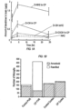
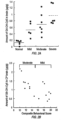
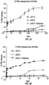
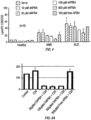
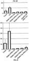


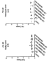
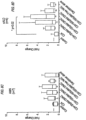
Classifications
-
- A—HUMAN NECESSITIES
- A61—MEDICAL OR VETERINARY SCIENCE; HYGIENE
- A61K—PREPARATIONS FOR MEDICAL, DENTAL OR TOILETRY PURPOSES
- A61K31/00—Medicinal preparations containing organic active ingredients
- A61K31/045—Hydroxy compounds, e.g. alcohols; Salts thereof, e.g. alcoholates
- A61K31/05—Phenols
-
- A—HUMAN NECESSITIES
- A61—MEDICAL OR VETERINARY SCIENCE; HYGIENE
- A61K—PREPARATIONS FOR MEDICAL, DENTAL OR TOILETRY PURPOSES
- A61K31/00—Medicinal preparations containing organic active ingredients
- A61K31/185—Acids; Anhydrides, halides or salts thereof, e.g. sulfur acids, imidic, hydrazonic or hydroximic acids
- A61K31/19—Carboxylic acids, e.g. valproic acid
- A61K31/192—Carboxylic acids, e.g. valproic acid having aromatic groups, e.g. sulindac, 2-aryl-propionic acids, ethacrynic acid
-
- A—HUMAN NECESSITIES
- A61—MEDICAL OR VETERINARY SCIENCE; HYGIENE
- A61K—PREPARATIONS FOR MEDICAL, DENTAL OR TOILETRY PURPOSES
- A61K31/00—Medicinal preparations containing organic active ingredients
- A61K31/185—Acids; Anhydrides, halides or salts thereof, e.g. sulfur acids, imidic, hydrazonic or hydroximic acids
- A61K31/19—Carboxylic acids, e.g. valproic acid
- A61K31/195—Carboxylic acids, e.g. valproic acid having an amino group
-
- A—HUMAN NECESSITIES
- A61—MEDICAL OR VETERINARY SCIENCE; HYGIENE
- A61K—PREPARATIONS FOR MEDICAL, DENTAL OR TOILETRY PURPOSES
- A61K31/00—Medicinal preparations containing organic active ingredients
- A61K31/56—Compounds containing cyclopenta[a]hydrophenanthrene ring systems; Derivatives thereof, e.g. steroids
- A61K31/57—Compounds containing cyclopenta[a]hydrophenanthrene ring systems; Derivatives thereof, e.g. steroids substituted in position 17 beta by a chain of two carbon atoms, e.g. pregnane or progesterone
- A61K31/573—Compounds containing cyclopenta[a]hydrophenanthrene ring systems; Derivatives thereof, e.g. steroids substituted in position 17 beta by a chain of two carbon atoms, e.g. pregnane or progesterone substituted in position 21, e.g. cortisone, dexamethasone, prednisone or aldosterone
-
- A—HUMAN NECESSITIES
- A61—MEDICAL OR VETERINARY SCIENCE; HYGIENE
- A61K—PREPARATIONS FOR MEDICAL, DENTAL OR TOILETRY PURPOSES
- A61K47/00—Medicinal preparations characterised by the non-active ingredients used, e.g. carriers or inert additives; Targeting or modifying agents chemically bound to the active ingredient
- A61K47/50—Medicinal preparations characterised by the non-active ingredients used, e.g. carriers or inert additives; Targeting or modifying agents chemically bound to the active ingredient the non-active ingredient being chemically bound to the active ingredient, e.g. polymer-drug conjugates
- A61K47/51—Medicinal preparations characterised by the non-active ingredients used, e.g. carriers or inert additives; Targeting or modifying agents chemically bound to the active ingredient the non-active ingredient being chemically bound to the active ingredient, e.g. polymer-drug conjugates the non-active ingredient being a modifying agent
- A61K47/56—Medicinal preparations characterised by the non-active ingredients used, e.g. carriers or inert additives; Targeting or modifying agents chemically bound to the active ingredient the non-active ingredient being chemically bound to the active ingredient, e.g. polymer-drug conjugates the non-active ingredient being a modifying agent the modifying agent being an organic macromolecular compound, e.g. an oligomeric, polymeric or dendrimeric molecule
- A61K47/59—Medicinal preparations characterised by the non-active ingredients used, e.g. carriers or inert additives; Targeting or modifying agents chemically bound to the active ingredient the non-active ingredient being chemically bound to the active ingredient, e.g. polymer-drug conjugates the non-active ingredient being a modifying agent the modifying agent being an organic macromolecular compound, e.g. an oligomeric, polymeric or dendrimeric molecule obtained otherwise than by reactions only involving carbon-to-carbon unsaturated bonds, e.g. polyureas or polyurethanes
- A61K47/595—Polyamides, e.g. nylon
-
- A—HUMAN NECESSITIES
- A61—MEDICAL OR VETERINARY SCIENCE; HYGIENE
- A61K—PREPARATIONS FOR MEDICAL, DENTAL OR TOILETRY PURPOSES
- A61K47/00—Medicinal preparations characterised by the non-active ingredients used, e.g. carriers or inert additives; Targeting or modifying agents chemically bound to the active ingredient
- A61K47/50—Medicinal preparations characterised by the non-active ingredients used, e.g. carriers or inert additives; Targeting or modifying agents chemically bound to the active ingredient the non-active ingredient being chemically bound to the active ingredient, e.g. polymer-drug conjugates
- A61K47/51—Medicinal preparations characterised by the non-active ingredients used, e.g. carriers or inert additives; Targeting or modifying agents chemically bound to the active ingredient the non-active ingredient being chemically bound to the active ingredient, e.g. polymer-drug conjugates the non-active ingredient being a modifying agent
- A61K47/56—Medicinal preparations characterised by the non-active ingredients used, e.g. carriers or inert additives; Targeting or modifying agents chemically bound to the active ingredient the non-active ingredient being chemically bound to the active ingredient, e.g. polymer-drug conjugates the non-active ingredient being a modifying agent the modifying agent being an organic macromolecular compound, e.g. an oligomeric, polymeric or dendrimeric molecule
- A61K47/59—Medicinal preparations characterised by the non-active ingredients used, e.g. carriers or inert additives; Targeting or modifying agents chemically bound to the active ingredient the non-active ingredient being chemically bound to the active ingredient, e.g. polymer-drug conjugates the non-active ingredient being a modifying agent the modifying agent being an organic macromolecular compound, e.g. an oligomeric, polymeric or dendrimeric molecule obtained otherwise than by reactions only involving carbon-to-carbon unsaturated bonds, e.g. polyureas or polyurethanes
- A61K47/60—Medicinal preparations characterised by the non-active ingredients used, e.g. carriers or inert additives; Targeting or modifying agents chemically bound to the active ingredient the non-active ingredient being chemically bound to the active ingredient, e.g. polymer-drug conjugates the non-active ingredient being a modifying agent the modifying agent being an organic macromolecular compound, e.g. an oligomeric, polymeric or dendrimeric molecule obtained otherwise than by reactions only involving carbon-to-carbon unsaturated bonds, e.g. polyureas or polyurethanes the organic macromolecular compound being a polyoxyalkylene oligomer, polymer or dendrimer, e.g. PEG, PPG, PEO or polyglycerol
-
- A—HUMAN NECESSITIES
- A61—MEDICAL OR VETERINARY SCIENCE; HYGIENE
- A61K—PREPARATIONS FOR MEDICAL, DENTAL OR TOILETRY PURPOSES
- A61K49/00—Preparations for testing in vivo
- A61K49/0004—Screening or testing of compounds for diagnosis of disorders, assessment of conditions, e.g. renal clearance, gastric emptying, testing for diabetes, allergy, rheuma, pancreas functions
-
- A—HUMAN NECESSITIES
- A61—MEDICAL OR VETERINARY SCIENCE; HYGIENE
- A61K—PREPARATIONS FOR MEDICAL, DENTAL OR TOILETRY PURPOSES
- A61K49/00—Preparations for testing in vivo
- A61K49/001—Preparation for luminescence or biological staining
- A61K49/0013—Luminescence
- A61K49/0017—Fluorescence in vivo
- A61K49/0019—Fluorescence in vivo characterised by the fluorescent group, e.g. oligomeric, polymeric or dendritic molecules
- A61K49/0021—Fluorescence in vivo characterised by the fluorescent group, e.g. oligomeric, polymeric or dendritic molecules the fluorescent group being a small organic molecule
- A61K49/0032—Methine dyes, e.g. cyanine dyes
-
- A—HUMAN NECESSITIES
- A61—MEDICAL OR VETERINARY SCIENCE; HYGIENE
- A61K—PREPARATIONS FOR MEDICAL, DENTAL OR TOILETRY PURPOSES
- A61K49/00—Preparations for testing in vivo
- A61K49/001—Preparation for luminescence or biological staining
- A61K49/0013—Luminescence
- A61K49/0017—Fluorescence in vivo
- A61K49/005—Fluorescence in vivo characterised by the carrier molecule carrying the fluorescent agent
- A61K49/0054—Macromolecular compounds, i.e. oligomers, polymers, dendrimers
-
- A—HUMAN NECESSITIES
- A61—MEDICAL OR VETERINARY SCIENCE; HYGIENE
- A61P—SPECIFIC THERAPEUTIC ACTIVITY OF CHEMICAL COMPOUNDS OR MEDICINAL PREPARATIONS
- A61P15/00—Drugs for genital or sexual disorders; Contraceptives
- A61P15/08—Drugs for genital or sexual disorders; Contraceptives for gonadal disorders or for enhancing fertility, e.g. inducers of ovulation or of spermatogenesis
-
- A—HUMAN NECESSITIES
- A61—MEDICAL OR VETERINARY SCIENCE; HYGIENE
- A61P—SPECIFIC THERAPEUTIC ACTIVITY OF CHEMICAL COMPOUNDS OR MEDICINAL PREPARATIONS
- A61P17/00—Drugs for dermatological disorders
- A61P17/18—Antioxidants, e.g. antiradicals
-
- A—HUMAN NECESSITIES
- A61—MEDICAL OR VETERINARY SCIENCE; HYGIENE
- A61P—SPECIFIC THERAPEUTIC ACTIVITY OF CHEMICAL COMPOUNDS OR MEDICINAL PREPARATIONS
- A61P25/00—Drugs for disorders of the nervous system
-
- A—HUMAN NECESSITIES
- A61—MEDICAL OR VETERINARY SCIENCE; HYGIENE
- A61P—SPECIFIC THERAPEUTIC ACTIVITY OF CHEMICAL COMPOUNDS OR MEDICINAL PREPARATIONS
- A61P25/00—Drugs for disorders of the nervous system
- A61P25/28—Drugs for disorders of the nervous system for treating neurodegenerative disorders of the central nervous system, e.g. nootropic agents, cognition enhancers, drugs for treating Alzheimer's disease or other forms of dementia
-
- A—HUMAN NECESSITIES
- A61—MEDICAL OR VETERINARY SCIENCE; HYGIENE
- A61P—SPECIFIC THERAPEUTIC ACTIVITY OF CHEMICAL COMPOUNDS OR MEDICINAL PREPARATIONS
- A61P3/00—Drugs for disorders of the metabolism
- A61P3/06—Antihyperlipidemics
-
- A—HUMAN NECESSITIES
- A61—MEDICAL OR VETERINARY SCIENCE; HYGIENE
- A61P—SPECIFIC THERAPEUTIC ACTIVITY OF CHEMICAL COMPOUNDS OR MEDICINAL PREPARATIONS
- A61P39/00—General protective or antinoxious agents
- A61P39/06—Free radical scavengers or antioxidants
-
- A—HUMAN NECESSITIES
- A61—MEDICAL OR VETERINARY SCIENCE; HYGIENE
- A61P—SPECIFIC THERAPEUTIC ACTIVITY OF CHEMICAL COMPOUNDS OR MEDICINAL PREPARATIONS
- A61P43/00—Drugs for specific purposes, not provided for in groups A61P1/00-A61P41/00
Definitions
- the field of the invention is generally related to targeted compositions including dendrimer nanodevices for the imaging, diagnosis, and treatment of central nervous system inflammation, particular of the type seen in adrenoleukodystrophy and other leukodystrophies and peroxisomal disorders.
- ALD X-linked adrenoleukodystrophy
- ABCD1 encodes ALDP, a protein responsible for the import of very long chain fatty acids (VLCFAs) into the peroxisome for degradation, the pathogenic hallmark of ALD, and it is believed that the accumulation of very long chain fatty acid will lead to mitochondrial dysfunction, and oxidative stress. Furthermore, accumulation of very long chain fatty acids in the cell membrane may lead to microglial activation resulting in neuroinflammation.
- VLCFAs very long chain fatty acids
- ALD primarily affects boys who are born normally and have normal initial development.
- the two most prevalent phenotypes of ALD are the childhood cerebral ALD (ccALD) which is a rapidly progressive, fatal demyelinating cerebral disorder, and the adult onset adrenomyeloneuropathy (AMN), which is a slowly progressive "dying-back" axonopathy of the long tracts in the spinal cord and peripheral nerves ( Powers, et al., Journal of neuropathology and experimental neurology, 60(5):493-501 (2001 )).
- Postmortem studies of long tracts in AMN have shown lipidic inclusions in mitochondria suggestive of mitochondrial dysfunction.
- ALD Alzheimer's disease
- HSCT allogeneic hematopoietic stem cell transplantation
- a combination of glyceryl trioleate-trierucate famously referred to as Lorenzo's oil (due to a movie depiction), has been shown to effectively reduce blood very long chain fatty acids, but has not been able to stop disease progression in ccALD.
- compositions and methods of use thereof for treatment of peroxisomal disorders and leukodystrophies are provided.
- references to methods of treatment in the summary and detailed description of the invention in this description are to be interpreted as references to the compounds, pharmaceutical compositions and medicaments of the present invention for use in a method for treatment of the human/animal body by therapy or for diagnosis.
- compositions including pharmaceutical compositions and dosage units, and methods of use thereof for diagnosing and treating peroxisomal disorders and leukodystrophies in a subject in need thereof typically include dendrimers complexed, covalently attached or intra-molecularly dispersed or encapsulated with a therapeutic, prophylactic or diagnostic agent for treatment or diagnosis of the disorder.
- the invention relates to a dendrimer complex for use in a method of treatment of a peroxisomal disorder in a subject, wherein the complex comprises generation 4-10 poly(amidoamine) (PAMAM) hydroxyl-terminated dendrimers complexed, conjugated or intra-molecularly dispersed or encapsulated with at least one therapeutic or prophylactic agent, wherein the agent is for the treatment of a peroxisomal disorder, and wherein the complex targets microglia.
- PAMAM poly(amidoamine)
- compositions include poly(amidoamine) hydroxyl-terminated dendrimers G4-G10, preferably G4-G6, complexed with therapeutic, prophylactic and/or diagnostic agent in an effective amount to treat, and/or prevent one or more symptoms associated with axonal degeneration.
- therapeutic agents include steroidal anti-inflammatory agents, non-steroidal anti-inflammatory agents, and gold compound anti-inflammatory agents.
- the dendrimer is complexed, covalently attached or intra-molecularly dispersed or encapsulated with an anti-inflammatory or antioxidant and an agent such as N-acetylcysteine, 4-phenylbutyrate, bezafibrate, thyroid hormone (T3), sobetirome, pioglitazone, resveratrol, VBP15, Vitamin E, erucic acid, biotin, Coenzyme Q10, clemastine, galactosylceramidase (GALC), or Arylsulfatase A (ARSA).
- an agent such as N-acetylcysteine, 4-phenylbutyrate, bezafibrate, thyroid hormone (T3), sobetirome, pioglitazone, resveratrol, VBP15, Vitamin E, erucic acid, biotin, Coenzyme Q10, clemastine, galactosylceramidase (GALC), or Aryl
- the therapeutic agents conjugated to the dendrimers are a therapeutically active agent for localizing and targeting Neuron-specific class III beta-tubulin (TUJ-1) positive spinal neurons.
- the compositions include the dendrimer with two or more different terminal linkers, and/or spacers, for conjugating with two or more different therapeutic agents.
- Methods of administering the compositions are provided to treat one or more symptoms associated with axonal degeneration in individuals with peroxisomal disorders and leukodystrophies, especially an extremely severe type X-linked adrenoleukodystrophy (ALD), which occurs due to mutations in the peroxisomal ABC-transporter, ABCD1, and affects cerebral white matter, spinal cord, and peripheral nerves, with some phenotypes progressing rapidly and terminally at young age.
- the methods typically include systemically administering to the subject an effective amount a pharmaceutically acceptable composition including the dendrimer composition.
- compositions are suitable for use in treatment of peroxisomal disorders that affect the growth or maintenance of the myelin sheath that insulates nerve cells, and leukodystrophies such as adrenoleukodystrophy (ALD) (including X-linked ALD), metachromatic leukodystrophy (MLD), Krabbe disease (globoid leukodystrophy), and Leukoencephalopathy with brainstem and spinal cord involvement and lactate elevation (LBSL)/DARS2 Leukoencephalopathy.
- ALD adrenoleukodystrophy
- MLD metachromatic leukodystrophy
- Krabbe disease globoid leukodystrophy
- Leukoencephalopathy with brainstem and spinal cord involvement and lactate elevation (LBSL)/DARS2 Leukoencephalopathy.
- the dendrimers conjugated to therapeutic agent is in a unit dosage in an amount effective to reduce, prevent, or otherwise alleviate oxidative stress, neuroinflammation, long chain fatty acid production, loss of motor function, or a combination thereof; promote, increase, or improve peroxisome proliferation, very long chain fatty acid removal, motor function, ABCD2 expression, enzymes mutated or deficient in peroxisomal disorders or leukodystrophies, or a combination thereof.
- therapeutic agent refers to an agent that can be administered to prevent or treat one or more symptoms of a disease or disorder.
- examples include a nucleic acid, a nucleic acid analog, a small molecule, a peptidomimetic, a protein, peptide, carbohydrate or sugar, lipid, or surfactant, or a combination thereof.
- treating refers to preventing or alleviating one or more symptoms of a disease, disorder or condition. Treating the disease or condition includes ameliorating at least one symptom of the particular disease or condition, even if the underlying pathophysiology is not affected, such as treating the pain of a subject by administration of an analgesic agent even though such agent does not treat the cause of the pain.
- prevention means to administer a composition to a subject or a system at risk for or having a predisposition for one or more symptom, caused by a disease or disorder, in an amount effective to cause cessation of a particular symptom of the disease or disorder, a reduction or prevention of one or more symptoms of the disease or disorder, a reduction in the severity of the disease or disorder, the complete ablation of the disease or disorder, stabilization or delay of the development or progression of the disease or disorder.
- biocompatible refers to a material that along with any metabolites or degradation products thereof that are generally non-toxic to the recipient and do not cause any significant adverse effects to the recipient.
- biocompatible materials are materials which do not elicit a significant inflammatory or immune response when administered to a patient.
- biodegradable generally refers to a material that will degrade or erode under physiologic conditions to smaller units or chemical species that are capable of being metabolized, eliminated, or excreted by the subject.
- the degradation time is a function of composition and morphology. Degradation times can be from hours to weeks.
- compositions, polymers and other materials and/or dosage forms which are, within the scope of sound medical judgment, suitable for use in contact with the tissues of human beings and animals without excessive toxicity, irritation, allergic response, or other problem or complication, commensurate with a reasonable benefit/risk ratio.
- pharmaceutically acceptable carrier refers to pharmaceutically acceptable materials, compositions or vehicles, such as a liquid or solid filler, diluent, solvent or encapsulating material involved in carrying or transporting any subject composition, from one organ, or portion of the body, to another organ, or portion of the body. Each carrier must be “acceptable” in the sense of being compatible with the other ingredients of a subject composition and not injurious to the patient.
- therapeutically effective amount refers to an amount of the therapeutic agent that produces some desired effect at a reasonable benefit/risk ratio applicable to any medical treatment.
- the effective amount may vary depending on such factors as the disease or condition being treated, the particular targeted constructs being administered, the size of the subject, or the severity of the disease or condition.
- One of ordinary skill in the art may empirically determine the effective amount of a particular compound without necessitating undue experimentation.
- molecular weight generally refers to the mass or average mass of a material. If a polymer or oligomer, the molecular weight can refer to the relative average chain length or relative chain mass of the bulk polymer. In practice, the molecular weight of polymers and oligomers can be estimated or characterized in various ways including gel permeation chromatography (GPC) or capillary viscometry.
- GPC gel permeation chromatography
- dendrimer as used herein includes a molecular architecture with an interior core, interior layers (or “generations”) of repeating units regularly attached to this initiator core, and an exterior surface of terminal groups attached to the outermost generation.
- dendrimers include PAMAM, polyester, polylysine, and PPI.
- the PAMAM dendrimers have hydroxyl terminations and can be generation 4 PAMAM dendrimers, generation 5 PAMAM dendrimers, generation 6 PAMAM dendrimers, generation 7 PAMAM dendrimers, generation 8 PAMAM dendrimers, generation 9 PAMAM dendrimers, or generation 10 PAMAM dendrimers.
- Dendrimers used in the method of the invention are polyamidoamine (PAMAM) dendrimers.
- Other dendrimers suitable for use include polypropylamine (POPAM), polyethylenimine, polylysine, polyester, iptycene, aliphatic poly(ether), and/or aromatic polyether dendrimers.
- POPAM polypropylamine
- Each dendrimer of the dendrimer complex may be of similar or different chemical nature than the other dendrimers (e.g., the first dendrimer may include a PAMAM dendrimer, while the second dendrimer may comprise a POPAM dendrimer).
- the first or second dendrimer may further include an additional agent.
- the multiarm PEG polymer includes a polyethylene glycol having at least two branches bearing sulfhydryl or thiopyridine terminal groups; however, embodiments disclosed herein are not limited to this class and PEG polymers bearing other terminal groups such as succinimidyl or maleimide terminations can be used.
- the PEG polymers in the molecular weight 10 kDa to 80 kDa can be used.
- a dendrimer complex includes multiple dendrimers.
- the dendrimer complex can include a third dendrimer; wherein the third-dendrimer is complexed with at least one other dendrimer.
- a third agent can be complexed with the third dendrimer.
- the first and second dendrimers are each complexed to a third dendrimer, wherein the first and second dendrimers are PAMAM dendrimers and the third dendrimer is a POPAM dendrimer. Additional dendrimers can be incorporated. When multiple dendrimers are utilized, multiple agents can also be incorporated. This is not limited by the number of dendrimers complexed to one another.
- PAMAM dendrimer means poly(amidoamine) dendrimer, which may contain different cores, with amidoamine building blocks.
- the method for making them is known to those of skill in the art and generally, involves a two-step iterative reaction sequence that produces concentric shells (generations) of dendritic ⁇ -alanine units around a central initiator core.
- This PAMAM core-shell architecture grows linearly in diameter as a function of added shells (generations).
- the surface groups amplify exponentially at each generation according to dendritic-branching mathematics. They are available in generations G0 - 10 with 5 different core types and 10 functional surface groups.
- the dendrimer-branched polymer may consist of polyamidoamine (PAMAM), polyglycerol, polyester, polyether, polylysine, or polyethylene glycol (PEG), polypeptide dendrimers.
- the PAMAM dendrimers used are generation 4-10 dendrimers with hydroxyl groups attached to their functional surface groups.
- the multiarm PEG polymer comprises polyethylene glycol having 2 and more branches bearing sulfhydryl or thiopyridine terminal groups; however, embodiments are not limited to this class and PEG polymers bearing other terminal groups such as succinimidyl or maleimide terminations can be used.
- the PEG polymers in the molecular weight 10 kDa to 80 kDa can be used.
- the dendrimers are in nanoparticle form and are described in detail in international patent publication Nos. WO2009/046446 , PCT/US2015/028386 , PCT/US2015/045112 , PCT/US2015/045104 , and U.S. Patent No. 8,889,101 .
- D-NAC Dendrimer-NAC
- N-succinimidyl 3-(2-pyridyldithio)propionate SPDP
- A-succinimidyl 3-(2-pyridyldithio)propionate (SPDP) is performed by a two-step procedure, Scheme 1. First, 3-mercaptopropionic acid is reacted by thiol-disulfide exchange with 2,2'-dipyridyl disulfide to give 2-carboxyethyl 2-pyridyl disulfide.
- the succinimide group is reacted with 2-carboxyethyl 2-pyridyl disulfide to obtain N -succinimidyl 3-(2-pyridyldithio)propionate, by esterification with N-hydroxysuccinimide by using N , N '-dicyclohexylcarbodiimide and 4-dimethylaminopyridine.
- PAMAM-NH 2 dendrimers are reacted with the heterobifunctional cross-linker SPDP, Scheme 2.
- the N-succinimidyl activated ester of SPDP couples to the terminal primary amines to yield amide-linked 2-pyridyldithiopropanoyl (PDP) groups, Scheme 2.
- PAMAM-NH-PDP can be analyzed using RP-HPLC to determine the extent to which SPDP has reacted with the dendrimers.
- valproic acid is functionalized with a thiol-reactive group.
- a short PEG-SH having three repeating units of (CH 2 ) 2 O- is reacted with valproic acid using DCC as coupling reagent as shown in Scheme 4.
- the crude PEG-VPA obtained is purified by column chromatography and characterized by proton NMR. In the NMR spectrum, there was a down-shift of the peak of CH 2 protons neighboring to OH group of PEG to 4.25 ppm from 3.65 ppm that confirmed the formation of PEG-VPA.
- the thiol group also may be susceptible to reacting with acid functionality, the NMR spectra did not indicate any downward shift of the peak belonging to CH 2 protons adjacent to thiol group of PEG. This suggests that the thiol group is free to react with the thiol-reactive functionalized dendrimer.
- a disulfide bond is introduced between the dendrimer and valproic acid, Scheme 5.
- First the dendrimer is converted to a bifunctional dendrimer (Compound 1) by reacting the dendrimer with fluorenylmethyloxycarbonyl (Fmoc) protected ⁇ -aminobutyric acid (GABA).
- Fmoc fluorenylmethyloxycarbonyl
- GABA protected ⁇ -aminobutyric acid
- Conjugation of PEG-VPA to the bifunctional dendrimer involved a two-step process: the first step is the reaction of amine-functionalized bifunctional dendrimer (Compound 1) with N -succinimidyl-3-(2-pyridyldithio)-propionate (SPDP), and the second step involves conjugating the thiol-functionalized valproic acid. SPDP is reacted with the intermediate (Compound 2 ) in the presence of N , N -diisopropylethylamine (DIEA) to obtain pyridyldithio (PDP)-functionalized dendrimer (Compound 3).
- DIEA N -diisopropylethylamine
- the formation of the final conjugate and loading of VPA were confirmed by 1 H NMR, and the purity of the conjugate was evaluated by reverse-phase HPLC. In the NMR spectrum, multiplets between 0.85 and 1.67 ppm for aliphatic protons of VPA, multiplets between 3.53 and 3.66 ppm for CH 2 protons of PEG, and absence of pyridyl aromatic protons confirmed the conjugate formation.
- the loading of the VPA is ⁇ 21 molecules, estimated using a proton integration method, which suggests that 1-2 amine groups are left unreacted.
- the elution time of D-VPA (17.2 min) is different from that for G4-OH (9.5 min), confirming that the conjugate is pure, with no measurable traces of VPA (23.4 min) and PEG-VPA (39.2 min).
- the percentage of VPA loading to the dendrimer is ⁇ 12% w/w and validates the method for making gram quantities in three different batches.
- PBA 4-phenyl butyric acid
- a propionyl linker was utilized as a spacer both to provide enough space for drug molecules on dendrimer surface and to facilitate their release. Since the attachment of linker is also based on an esterification reaction, a BOC group protection/deprotection strategy was followed to modify PBA molecules and then conjugation to dendrimer surface was performed for both 4th and 6th generation PAMAM dendrimers (Scheme 6).
- dendrimers conjugated with two or more different drugs via two or more different linkers are used.
- dendrimer conjugate that has two different drugs with two different linkers was successfully synthesized by the attachment of PBA and NAC drug molecules to 4 th generation PAMAM dendrimer sequentially.
- Scheme 7 represents the reaction steps to obtain D-NAC&PBA conjugate.
- Bezafibrate (BEZA) was conjugated to hydroxyl functionalized PAMAM dendrimer via a pH labile ester linkage.
- the same strategy was applied for the synthesis of bezafibrate-PAMAM conjugates as in the sythesis of D-PBA conjugates. This conjugation depends on the same BOC group protection/deprotection strategy for the sequential esterification reactions, first to attach the linker to bezafibrate, and then to conjugate the drug-linker compound to the dendrimer surface.
- the same propionyl linker was utilized as a spacer to provide enough space for drug molecules on the dendrimer surface and to facilitate their release. Synthesis of conjugates with bezafibrate was performed for both 4th and 6th generation PAMAM dendrimers (Scheme 8).
- Dendrimer complexes can be formed of therapeutically active agents or compounds (hereinafter "agent") conjugated or attached to a dendrimer. The attachment can occur via an appropriate spacer that provides a disulfide bridge between the agent and the dendrimer.
- agent therapeutically active agents or compounds
- the dendrimer complexes are capable of rapid release of the agent in vivo by thiol exchange reactions, under the reduced conditions found in body.
- spacers as used herein is intended to include compositions used for linking a therapeutically active agent to the dendrimer.
- the spacer can be either a single chemical entity or two or more chemical entities linked together to bridge the polymer and the therapeutic agent or imaging agent.
- the spacers can include any small chemical entity, peptide or polymers having sulfhydryl, thiopyridine, succinimidyl, maleimide, vinylsulfone, and carbonate terminations.
- the spacer can be chosen from among a class of compounds terminating in sulfhydryl, thiopyridine, succinimidyl, maleimide, vinylsulfone and carbonate group.
- the spacer can comprise thiopyridine terminated compounds such as dithiodipyridine, N-Succinimidyl 3-(2-pyridyldithio)-propionate (SPDP), Succinimidyl 6-(3-[2-pyridyldithio]-propionamido)hexanoate LC-SPDP or Sulfo-LC-SPDP.
- the spacer can also include peptides wherein the peptides are linear or cyclic essentially having sulfhydryl groups such as glutathione, homocysteine, cysteine and its derivatives, arg-gly-asp-cys (RGDC), cyclo(Arg-Gly-Asp-d-Phe-Cys) (c(RGDfC)), cyclo(Arg-Gly-Asp-D-Tyr-Cys), cyclo(Arg-Ala-Asp-d-Tyr-Cys).
- RGDC arg-gly-asp-cys
- c(RGDfC) cyclo(Arg-Gly-Asp-D-Tyr-Cys)
- cyclo(Arg-Ala-Asp-d-Tyr-Cys cyclo(Arg-Ala-Asp-d-Tyr-Cys).
- the spacer can be a mercapto acid derivative such as 3 mercapto propionic acid, mercapto acetic acid, 4 mercapto butyric acid, thiolan-2-one, 6 mercaptohexanoic acid, 5 mercapto valeric acid and other mercapto derivatives such as 2 mercaptoethanol and 2 mercaptoethylamine.
- a mercapto acid derivative such as 3 mercapto propionic acid, mercapto acetic acid, 4 mercapto butyric acid, thiolan-2-one, 6 mercaptohexanoic acid, 5 mercapto valeric acid and other mercapto derivatives such as 2 mercaptoethanol and 2 mercaptoethylamine.
- the spacer can be thiosalicylic acid and its derivatives, (4-succinimidyloxycarbonyl-methyl- ⁇ -2-pyridylthio)toluene, (3-[2-pyridithio]propionyl hydrazide,
- the spacer can have maleimide terminations wherein the spacer comprises polymer or small chemical entity such as bis-maleimido diethylene glycol and bis-maleimido triethylene glycol, Bis-Maleimidoethane, bismaleimidohexane.
- the spacer can comprise vinylsulfone such as 1,6-Hexane-bis-vinylsulfone.
- the spacer can comprise thioglycosides such as thioglucose.
- the spacer can be reduced proteins such as bovine serum albumin and human serum albumin, any thiol terminated compound capable of forming disulfide bonds.
- the spacer can include polyethylene glycol having maleimide, succinimidyl and thiol terminations.
- two or more different spacers are used on the same dendrimer molecule to conjugate with two or more different drugs.
- dendrimer complexes refers to the combination of a dendrimer with a therapeutically, prophylactically and/or diagnostic active agent.
- the dendrimers may also include a targeting agent, but as demonstrated by the examples, these are not required for delivery to injured tissue.
- These dendrimer complexes include one or more agent that is attached or conjugated to PAMAM dendrimers, which are capable of preferentially releasing the drug intracellularly under the reduced conditions found in vivo.
- the dendrimer complex when administered by i.v. injection, can preferentially localize to damaged or disease neurons, particularly in the gray matter of the spinal cord, over normal cells.
- the dendrimer complexes are also useful for targeted delivery of the therapeutics in inflammatory disorders, and particularly in peroxisomal diseases and leukodystrophies.
- the agent can be either covalently attached or intra-molecularly dispersed or encapsulated.
- the dendrimer is preferably a PAMAM dendrimer generation 4 to 6, having hydroxyl terminations.
- the PEG polymer is a star shaped polymer having 2 or more arms and a molecular weight of 10 kDa to 80 kDa.
- the PEG polymer has sulfhydryl, thiopyridine, succinimidyl, or maleimide terminations.
- the dendrimer is linked to the agents via a spacer ending in disulfide, ester or amide bonds.
- the dosage of active agent when administered with dendrimer, can be lower, the number of administrations can be reduced, or a combination thereof to achieve the same or greater therapeutic effect compared to administering the active agent in the absence of dendrimer.
- this allows delivery of agents that are otherwise impractical to administer to a subject in need thereof (1) due to the prohibitively large dose needed to achieve therapeutic effects when the agent is administered absent dendrimer, (2) because the agent when administered alone and untargeted is prohibitively toxic to normal or healthy cells, (3) because active agent is not targeted to the diseased tissue in an effective amount to therapeutically efficacious when alone and untargeted, or (4) a combination thereof.
- two or more active agents are administered to a subject in need thereof.
- the two or more active agents can be covalently attached or intra-molecularly dispersed or encapsulated in the same or different dendrimers.
- the dendrimers can be of the same or different composition.
- one or more active agents are covalently attached or intra-molecularly dispersed or encapsulated in dendrimer, while one or more other active agents are delivered by another suitable means without being covalently attached or intra-molecularly dispersed or encapsulated in dendrimer.
- compositions and formulations including an effective amount of dendrimer and an active agent to treat a peroxisomal disease or leukodystrophy such as ALD are provided.
- the therapeutic agent is one that reduces, prevents, or otherwise alleviates oxidative stress, neuroinflammation, long chain fatty acid production, loss of motor function, or a combination thereof; promotes, increases, or improves peroxisome proliferation, very long chain fatty acid removal, motor function, ABCD2 expression, expression of wildtype copies of an enzyme mutated or deficient in a peroxisomal disorder or leukodystrophy, or any combination thereof.
- Preferred active agents include N-acetylcysteine, 4-phenylbutyrate, bezafibrate, thyroid hormone (T3), sobetirome, pioglitazone, resveratrol, VBP15, Vitamin E, galactosylceramidase (GALC), and Arylsulfatase A (ARSA).
- T3 thyroid hormone
- sobetirome sobetirome
- pioglitazone resveratrol
- VBP15 Vitamin E
- GLC galactosylceramidase
- Arylsulfatase A Arylsulfatase A
- Other suitable active agents including anti-inflammatory and imaging agents are also discussed in more detail below.
- Preferred active agents include agents that prevent or reduce very long chain fatty acid production, agents that promote peroxisome proliferation, promote very long chain fatty acid removal (e.g., 4-phenyl butyrate) agents that increase ABCD2 expression (e.g., benzafibrate), thyromimetics (e.g., sobetirome), enzymes ( e.g. Galactosylceramidase and Arylsulfatase A, Aspartoacylase), agents that reduce neuroinflammation (e.g, N-acetyl cysteine, Pioglitazone, Vitamin E) and RNA oligonucleotides that interfere with gene transcription or translation.
- agents that prevent or reduce very long chain fatty acid production e.g., agents that promote peroxisome proliferation, promote very long chain fatty acid removal (e.g., 4-phenyl butyrate) agents that increase ABCD2 expression (e.g., benzafibrate), thyromimetics (e.g
- the agent is N-acetylcysteine, 4-phenylbutyrate, bezafibrate, thyroid hormone (T3), sobetirome, pioglitazone, resveratrol, VBP15, Vitamin E, galactosylceramidase (GALC), Aspartoacylase (ASPA), or Arylsulfatase A (ARSA).
- Acetylcysteine also known as N-acetylcysteine or N-acetyl-L-cysteine (NAC)
- NAC N-acetyl-L-cysteine
- diseases include cystic fibrosis and chronic obstructive pulmonary disease.
- Numerous formulations are known in the art and have been administered numerous routes including intravenous, by mouth, or inhaled as a mist. Numerous commercial formulations are also available and include, for example, ACETADOTE ® , which is discussed in U.S. Patent Nos. 8,148,356 , 8,399,445 , 8,653,061 , 8,722,738 .
- NAC N-acetylcysteine
- acALD is a fatal adult disease with no existing therapy, it may be particularly suited for a human trial of dendrimer-N-acetylcysteine. Also, children with advanced stages of ccALD who no longer qualify for HSCT are in great need for a therapeutic intervention.
- the boys subject to the study in Tolar, et al. were treated with 140 mg/kg/day intravenously (i.v.) followed by 70 mg/kg four times daily orally of NAC.
- the dosage of NAC can be lower, the number of administrations can be reduced, or a combination thereof to achieve the same or greater therapeutic effect compared to administering NAC in the absence of dendrimers.
- the active agent can be 4-phenylbutyrate, or 4-phenyl butyric acid.
- Commercial formulations of sodium phenylbutyrate (4-phenylbutyrate sodium salt) indicated for treatment of urea cycle disorders include BUPHENYL ® (sodium phenylbutyrate) (Horizon Pharma), AMMONAPS ® (Swedish Orphan Biovitrum International AB), and TRIBUTYRATE ® (Fyrlklövern Scandinavia AB).
- Other formulations include, for example, RAVICTI ® (described in U.S. Patent Nos. 5,968,979 , 8,404,215 , 8,642,012 , 9,095,559 ).
- the daily dose of sodium phenylbutyrate has been 450 - 600 mg/kg/day in children weighing less than 20 kg, and 9.9 - 13.0 g/m 2 /day in children weighing more than 20 kg, adolescents and adults.
- the dosage of 4-phenylbutyrate can be lower, the number of administrations can be reduced, or a combination thereof to achieve the same or greater therapeutic effect compared to administering 4-phenylbutyrate in the absence of dendrimers.
- the active agent can be bezafibrate.
- Bezafibrate is a fibrate drug used for the treatment of hyperlipidaemia, and has been investigated for use in treatment of hepatitis C, tauopathy ( Dumont, et al., Human Molecular Genetics, 21 (23):5091-5105 (2012 ), and cancer ( University of Birmingham. "Contraceptive, cholesterol-lowering drugs used to treat cancer.” ScienceDaily, 14 May 2015 ; and Southam, et al., Cancer Research, 2015; DOI: 10.1158/0008-5472.CAN-15-0202 ).
- Commercial bezafibrate formulations for treatment of hyperlipidaemia include, among others, BEZALIP ® (Actavis Group PTC ehf).
- Bezafibrate reduces VLCFA levels in X-ALD fibroblasts by inhibiting ELOVL1, an enzyme involved in the VLCFA synthesis ( Engelen, et al., Journal of inherited metabolic disease, 35(6): 1137-45 (2012 )).
- ELOVL1 an enzyme involved in the VLCFA synthesis
- a clinical trial failed to reduce plasma VLCFA levels in ALD patients while only low plasma levels were achieved ( Engelen, et al., PloS one, 7(7):e41013 (2012 )). It is believed that targeted delivery to the diseased tissue using dendrimers will increase the therapeutic efficacy of bezafibrate in subjects with ALD, and other leukodystrophies
- the active agent can be thyroid hormone.
- the hormone is the thyroid hormone triiodothyronine (T3), or a prohormone thereof.
- T3 thyroid hormone triiodothyronine
- T4 prohormone, thyroxine
- T3 and T4 are tyrosine-based hormones produced by the thyroid gland that are primarily responsible for regulation of metabolism.
- T3 and T4 Natural and synthetic T3 and T4, and mixtures thereof, are known in the art and used to treat hypothyroidism.
- Popular commercial formulations include levothyroxine, a synthetic thyroid hormone that is chemically identical to thyroxine (T4), and liothyronine, a synthetic form of thyroid hormone (T3).
- T3 Through its receptor TR ⁇ , T3 can induce hepatic ABCD2 expression in rodents and transiently normalize the VLCFA level in fibroblasts of ABCD1 null mice ( Fourcade, et al., Molecular pharmacology, 63(6):1296-303 (2003 )). Yet clinical trials with thyroid hormone are unlikely due to the systemic side effects it would exert. Thyroid mimetics are currently under investigation. Administration of thyroid hormone with dendrimers provides an avenue for targeted therapy with reduced systemic toxicity.
- the agent is a thyromimetic.
- a thyromimetic is an agent that produces effects similar to those of thyroid hormones or the thyroid gland.
- Exemplary thyromimetics include eprotirome and sobetirome.
- Thyromimetics that increase the expression of hepatic CYP7A1 include MB07811, KB-141, T-0681, and sobetirome ( Pedrelli, et al., World J Gastroenterol., 16(47): 5958-5964 (2010 )).
- the active agent is sobetirome.
- Sobetirome is a thyroid hormone receptor isoform beta-1 liver-selective analog with antilipidemic and antiatherosclerotic activity.
- sobetirome reduced serum lipids, decreased cholesterol levels, and stimulated steps of reverse cholesterol transport, which promotes the reverse transport of cholesterol from atherogenic macrophages back to the liver for excretion.
- sobetirome lowers plasma LDL cholesterol and reduces plasma triglycerides, while its liver-selective activity helped avoid the side effects seen with many other thyromimetic agents.
- the active agent can be pioglitazone.
- Pioglitazone is a thiazolidinedione (TZD) used to treat diabetes.
- Pioglitazone selectively stimulates the nuclear receptor peroxisome proliferator-activated receptor gamma (PPAR- ⁇ ) and to a lesser extent PPAR- ⁇ .
- PPAR- ⁇ nuclear receptor peroxisome proliferator-activated receptor gamma
- PPAR- ⁇ nuclear receptor peroxisome proliferator-activated receptor gamma
- PMID 10983737 Smith, et al., J Clin Pract Suppl, (121):13-8 (2001 )
- Commercial formulations include ACTOS ® (Takeda Pharmaceuticals U.S.A., Inc.) which is indicated for glycemic control in adults with type 2 diabetes mellitus in doses of 15 mg, 30 mg, and 45 mg per day.
- Pioglitazone has been shown to restore mitochondrial content and expression of master regulators of biogenesis, neutralized oxidative damage to proteins and DNA, and reversed bioenergetic failure in terms of ATP levels, NAD+/NADH ratios, pyruvate kinase and glutathione reductase activities in ABCD1 KO mice ( Morato, et al., Brain: a journal of neurology, 136(Pt 8):2432-43 (2013 )). Most importantly, the treatment halted locomotor disability and axonal damage in ABCD1 KO mice.
- the active agent can be a resveratrol, such as trans-resveratrol, cis-resveratrol, trans-resveratrol-3- O - ⁇ -glucoside, or cis-resveratrol-3- O - ⁇ -glucoside.
- Resveratrol is a stilbenoid, a type of natural phenol, and a phytoalexin produced by several plants in response to injury or when infected with bacteria or fungi ( Fremount, Life Sciences, 66(8):663-673 (2000 ). Resveratrol has been investigated in anti-aging applications, and to treat heart disease, cancer, Alzheimer's disease, and diabetes.
- Resveratrol is a Sirt1 inducer, and has also been shown to normalize redox homeostasis, mitochondrial respiration, bioenergetic failure, axonal degeneration and associated locomotor disabilities in the X-ALD mice ( Morato, et al., Cell Death and Differentiation, 22:1742-1753 (2015 )).
- resveratrol (Orchid Chemicals & Pharmaceuticals Ltd, India) (0.04% w/w) was mixed into AIN-93G chow from Dyets (Bethlehem, PA, USA) to provide a dose of 400 mg/kg/day ( Morato, et al., Cell Death and Differentiation, 22:1742-1753 (2015 ).
- the compound is commercially available for human consumption in the form of nutritional supplements.
- Some resveratrol capsules sold in the U.S. contain extracts from the Japanese and Chinese knotweed plant Polygonum cuspidatum or are made from red wine or red grape extracts. Numerous human doses have been reported ranging from 25 mg to 5,000 mg ( Higdon, et al., "Resveratrol,” Linus Pauling Institute Micronutrient Information Center, accessed October 2015 ).
- the active agent can be VBP15.
- VBP15 is a steroid analogue, a modified glucocorticoid. Studies in mice showed it is an anti-inflammatory and membrane-stabilizer that improves muscular dystrophy without side effects ( Heier, et al, EMBO Mol Med., 5(10): 1569-1585 (2013 ), Nagaraju, et al., "Delta 9-11 Compound, VBP15: Potential Therapy for DMD” accessed October 2015 ), and in 2015 it was announced that it would be the subject of a Phase I, first-in-humans clinical trial for treating the same ( Olivas, "ReveraGen BioPharma Announces Start of Phase 1 Clinical Trial of VBP15 Dissociative Steroid Drug," media release, February 18, 2015 ). Dosages in some mouse studies include 5 mg/kg, 15 mg/kg, 30 mg/kg, and 45 mg/kg per day. The compound may also be effective for treating leukodystrophies.
- the active agent can be erucic acid.
- Erucic Acid is a monounsaturated very long-chain fatty acid with a 22-carbon backbone and a single double bond originating from the 9th position from the methyl end, with the double bond in the cis- configuration. It is prevalent in wallflower seed with a reported content of 20 to 54% in high erucic acid rapeseed oil, and 42% in mustard oil.
- the dosage of erucic acid When administered with dendrimers, the dosage of erucic acid can be lower, the number of administrations can be reduced, or a combination thereof to achieve the same or greater therapeutic effect compared to administering erucic acid in the absence of dendrimers.
- the active agent can be Vitamin E.
- Vitamin E refers to a group of compounds that include both tocopherols and tocotrienols.
- ⁇ -tocopherol is the most common form found in the North American diet.
- ⁇ -Tocopherol can be found in corn oil, soybean oil, margarine, and dressings.
- ⁇ -tocopherol the most biologically active form of vitamin E, is the second-most common form of vitamin E in the diet. This variant can be found most abundantly in wheat germ oil, sunflower, and safflower oils.
- As a fat-soluble antioxidant it interrupts the propagation of reactive oxygen species that spread through biological membranes or through a fat when its lipid content undergoes oxidation by reacting with more-reactive lipid radicals to form more stable products.
- the dosage of Vitamin E When administered with dendrimers, the dosage of Vitamin E can be lower, the number of administrations can be reduced, or a combination thereof to achieve the same or greater therapeutic effect compared to administering Vitamin E in the absence of dendrimers.
- the active agent can be Coenzyme Q10. It is also known as ubiquinone, ubidecarenone, coenzyme Q, and abbreviated at times to CoQ10. It is a 1,4-benzoquinone, where Q refers to the quinone chemical group and 10 refers to the number of isoprenyl chemical subunits in its tail. This fat-soluble substance, which resembles a vitamin, is present in most eukaryotic cells, primarily in the mitochondria. It is a component of the electron transport chain and participates in aerobic cellular respiration, which generates energy in the form of ATP. When administered with dendrimers, the dosage of Coenzyme Q10 can be lower, the number of administrations can be reduced, or a combination thereof to achieve the same or greater therapeutic effect compared to administering Coenzyme Q10 in the absence of dendrimers.
- the active agent can be biotin.
- Biotin is a water-soluble B-vitamin, also called vitamin B7, and formerly known as vitamin H or coenzyme R. It is composed of a ureido ring fused with a tetrahydrothiophene ring. A valeric acid substituent is attached to one of the carbon atoms of the tetrahydrothiophene ring.
- Biotin is a coenzyme for carboxylase enzymes, involved in the synthesis of fatty acids, isoleucine, and valine, and in gluconeogenesis. When administered with dendrimers, the dosage of biotin can be lower, the number of administrations can be reduced, or a combination thereof to achieve the same or greater therapeutic effect compared to administering biotin in the absence of dendrimers.
- the active agent can be clemastine.
- Clemastine also known as meclastin, is an antihistamine and anticholinergic.
- Clemastine fumarate belongs to the benzhydryl ether group of antihistaminic compounds.
- the chemical name is (+)-2-[-2- [(p-chloro- ⁇ -methyl- ⁇ -phenylbenzyl) oxy] ethyl]-1-methylpyrrolidine hydrogen fumarate.
- the dosage of clemastine can be lower, the number of administrations can be reduced, or a combination thereof to achieve the same or greater therapeutic effect compared to administering clemastine in the absence of dendrimers.
- the active agent is an enzyme, particularly an enzyme whose mutation, deficiency, or other dysregulation is associated with a peroxisomal disease or leukodystrophy.
- the enzyme is galactosylceramidase (GALC), Aspartoacylase (ASPA), or Arylsulfatase A (ARSA).
- GALC hydrolyzes galactolipids, including galactosylceramide and psychosine.
- Galactosylceramide is an important component of myelin.
- Psychosine forms during the production of myelin, and then it breaks down with help of galactosylceramidase.
- Krabbe disease is associated with mutations (more than 70 have been identified) in the GALC gene.
- ARSA is an enzyme that breaks down sulfatides, particularly cerebroside 3-sulfate, into cerebroside and sulfate. Deficiency of ARSA is associated with metachromatic leukodystrophy. Aspartoacylase (ASPA) catalyzes the deacetylation of N-acetylaspartic acid (NAA) to produce acetate and L-aspartate. NAA occurs in high concentration in brain and its hydrolysis NAA plays a significant part in the maintenance of intact white matter. Canavan Disease is associated with mutations in ASPA resulting in accumulation of NAA and spongiform degeneration of cerebral white matter.
- the agent can be the protein, or a nucleic acid encoding the protein, for example a DNA expression vector or an in vitro transcribed mRNA.
- the agents can be peptides, proteins, carbohydrates, nucleotides or oligonucleotides, small molecules, or combinations thereof.
- the nucleic acid can be an oligonucleotide encoding a protein, for example, a DNA expression cassette or an mRNA.
- Exemplary therapeutic agents include anti-inflammatory drugs, antiproliferatives, chemotherapeutics, vasodilators, and anti-infective agents.
- Antibiotics include ⁇ -lactams such as penicillin and ampicillin, cephalosporins such as cefuroxime, cefaclor, cephalexin, cephydroxil, cepfodoxime and proxetil, tetracycline antibiotics such as doxycycline and minocycline, microlide antibiotics such as azithromycin, erythromycin, rapamycin and clarithromycin, fluoroquinolones such as ciprofloxacin, enrofloxacin, ofloxacin, gatifloxacin, levofloxacin and norfloxacin, tobramycin, colistin, or aztreonam as well as antibiotics which are known to possess anti-inflammatory activity, such as erythromycin, azithromycin, or clarithromycin.
- a preferred anti-inflammatory is an antioxidant drug including N-acetylcysteine.
- Preferred NSAIDS include mefenamic acid, aspirin, Diflunisal, Salsalate, Ibuprofen, Naproxen, Fenoprofen, Ketoprofen, Deacketoprofen, Flurbiprofen, Oxaprozin, Loxoprofen, Indomethacin, Sulindac, Etodolac, Ketorolac, Diclofenac, Nabumetone, Piroxicam, Meloxicam, Tenoxicam, Droxicam, Lornoxicam, Isoxicam, Meclofenamic acid, Flufenamic acid, Tolfenamic acid, elecoxib, Rofecoxib, Valdecoxib, Parecoxib, Lumiracoxib, Etoricoxib, Firocoxib, Sulphonanilides, Nimesulide, Niflumic acid, and Licofelone.
- Representative small molecules include steroids such as methyl prednisone, dexamethasone, non-steroidal anti-inflammatory agents, including COX-2 inhibitors, corticosteroid anti-inflammatory agents, gold compound anti-inflammatory agents, immunosuppressive, anti-inflammatory and anti-angiogenic agents, anti-excitotoxic agents such as valproic acid, D-aminophosphonovalerate, D-aminophosphonoheptanoate, inhibitors of glutamate formation/release, baclofen, NMDA receptor antagonists, salicylate anti-inflammatory agents, ranibizumab, anti-VEGF agents, including aflibercept, and rapamycin.
- steroids such as methyl prednisone, dexamethasone
- non-steroidal anti-inflammatory agents including COX-2 inhibitors
- corticosteroid anti-inflammatory agents including COX-2 inhibitors
- corticosteroid anti-inflammatory agents including COX-2 inhibitors
- gold compound anti-inflammatory agents include immunosuppressive, anti-inflammatory and anti
- anti-inflammatory drugs include nonsteroidal drug such as indomethacin, aspirin, acetaminophen, diclofenac sodium and ibuprofen.
- the corticosteroids can be fluocinolone acetonide and methylprednisolone.
- the peptide drug can be streptidokinase.
- the molecules can include antibodies, including, for example, daclizumab, bevacizumab (avastin ® ), ranibizumab (Lucentis ® ), basiliximab, ranibizumab, and pegaptanib sodium or peptides like SN50, and antagonists of NF.
- antibodies including, for example, daclizumab, bevacizumab (avastin ® ), ranibizumab (Lucentis ® ), basiliximab, ranibizumab, and pegaptanib sodium or peptides like SN50, and antagonists of NF.
- oligonucleotides include siRNAs, microRNAs, DNA, and RNA.
- the therapeutic agent can be a PAMAM dendrimer with hydroxyl terminations.
- Exemplary diagnostic agents include paramagnetic molecules, fluorescent compounds, magnetic molecules, and radionuclides, x-ray imaging agents, and contrast media. These may also be ligands or antibodies which are labelled with the foregoing or bind to labelled ligands or antibodies which are detectable by methods known to those skilled in the art.
- Exemplary diagnostic agents include dyes, fluorescent dyes, Near infra-red dyes, SPECT imaging agents, PET imaging agents and radioisotopes.
- Representative dyes include carbocyanine, indocarbocyanine, oxacarbocyanine, thuicarbocyanine and merocyanine, polymethine, coumarine, rhodamine, xanthene, fluorescein, boron-dipyrromethane (BODIPY), Cy5, Cy5.5, Cy7, VivoTag-680, VivoTag-S680, VivoTag-S750, AlexaFluor660, AlexaFluor680, AlexaFluor700, AlexaFluor750, AlexaFluor790, Dy677, Dy676, Dy682, Dy752, Dy780, DyLight547, Dylight647, HiLyte Fluor 647, HiLyte Fluor 680, HiLyte Fluor
- Representative SPECT or PET imaging agents include chelators such as di-ethylene tri-amine penta-acetic acid (DTPA), 1,4,7,10-tetra-azacyclododecane-1,4,7,10-tetraacetic acid (DOTA), di-amine dithiols, activated mercaptoacetyl-glycyl-glycyl-gylcine (MAG3), and hydrazidonicotinamide (HYNIC).
- chelators such as di-ethylene tri-amine penta-acetic acid (DTPA), 1,4,7,10-tetra-azacyclododecane-1,4,7,10-tetraacetic acid (DOTA), di-amine dithiols, activated mercaptoacetyl-glycyl-glycyl-gylcine (MAG3), and hydrazidonicotinamide (HYNIC).
- DTPA di-ethylene tri-amine penta-acetic acid
- isotopes include Tc-94m, Tc-99m, In-111, Ga-67, Ga-68, Gd 3+ , Y-86, Y-90, Lu-177, Re-186, Re-188, Cu-64, Cu-67, Co-55, Co-57, F-18, Sc-47, Ac-225, Bi-213, Bi-212, Pb-212, Sm-153, Ho-166, and Dy-i66.
- Targeting moieties include folic acid, RGD peptides either linear or cyclic, TAT peptides, LHRH and BH3.
- the dendrimer complexes linked to a bioactive compound or therapeutically active agent can be used to perform several functions including targeting, localization at a diseased site, releasing the drug, and imaging purposes.
- the dendrimer complexes can be tagged with or without targeting moieties such that a disulfide bond between the dendrimer and the agent or imaging agent is formed via a spacer or linker molecule.
- the dendrimers can be administered parenterally by subdural, intravenous, intrathecal, intraventricular, intraarterial, intra-amniotic, intraperitoneal, or subcutaneous routes.
- the carriers or diluents used herein may be solid carriers or diluents for solid formulations, liquid carriers or diluents for liquid formulations, or mixtures thereof.
- pharmaceutically acceptable carriers may be, for example, aqueous or non-aqueous solutions, suspensions, emulsions or oils.
- Parenteral vehicles for subcutaneous, intravenous, intraarterial, or intramuscular injection
- non-aqueous solvents are propylene glycol, polyethylene glycol, and injectable organic esters such as ethyl oleate.
- Aqueous carriers include, for example, water, alcoholic/aqueous solutions, cyclodextrins, emulsions or suspensions, including saline and buffered media.
- the dendrimers can also be administered in an emulsion, for example, water in oil.
- oils are those of petroleum, animal, vegetable, or synthetic origin, for example, peanut oil, soybean oil, mineral oil, olive oil, sunflower oil, fish-liver oil, sesame oil, cottonseed oil, corn oil, olive, petrolatum, and mineral.
- Suitable fatty acids for use in parenteral formulations include, for example, oleic acid, stearic acid, and isostearic acid. Ethyl oleate and isopropyl myristate are examples of suitable fatty acid esters.
- Formulations suitable for parenteral administration can include antioxidants, buffers, bacteriostats, and solutes that render the formulation isotonic with the blood of the intended recipient, and aqueous and non-aqueous sterile suspensions that can include suspending agents, solubilizers, thickening agents, stabilizers, and preservatives.
- Intravenous vehicles can include fluid and nutrient replenishers, electrolyte replenishers such as those based on Ringer's dextrose.
- water, saline, aqueous dextrose and related sugar solutions, and glycols such as propylene glycols or polyethylene glycol are preferred liquid carriers, particularly for injectable solutions.
- Injectable pharmaceutical carriers for injectable compositions are well-known to those of ordinary skill in the art (see, e.g., Pharmaceutics and Pharmacy Practice, J.B. Lippincott Company, Philadelphia, PA, Banker and Chalmers, eds., pages 238-250 (1982 ), and ASHP Handbook on Injectable Drugs, Trissel, 15th ed., pages 622-630 (2009 )).
- Formulations for convection enhanced delivery include solutions of low molecular weight sales and sugars such as mannitol.
- PCT/US2015/045112 and Kannan, et al., Sci Transl Med., 4(130): 130ra46 (2012) doi: 10. 1 126/scitranslmed. 3 003 162 demonstrate that poly(amidoamine) dendrimers target inflammation in the central nervous system (CNS) and deliver drugs to produce functional improvements in a rabbit model of cerebral palsy.
- CNS central nervous system
- systemic administration of the dendrimer also leads to significant accumulation of the dendrimer in the injured areas of the spinal cord in mice with ALD, with further selective localization in the inflammatory cells. This selective localization of the dendrimer in the injured brain and spinal cord in these mice has implications for treatment of peroxisomal disorders and leukodystrophies including ALD.
- the methods include administering a subject in need thereof with an effective amount of dendrimer complexes including a combination of a dendrimer with one or more a therapeutic or prophylactic and/or diagnostic active agents.
- the dendrimers may also include a targeting agent, but as demonstrated by the examples, these are not required for delivery to injured tissue in the spinal cord.
- the dendrimer complexes include an agent that is attached or conjugated to PAMAM dendrimers, which are capable of preferentially releasing the drug intracellularly under the reduced conditions found in vivo.
- the agent can be either covalently attached or intra-molecularly dispersed or encapsulated.
- the amount of dendrimer complexes administered to the subject can be an effective amount to reduce, prevent, or otherwise alleviate one or more clinical or molecular symptoms of the disease or disorder to be treated compared to a control, for example a subject absent treatment or a subject treated with the active agent alone absent dendrimer.
- the amount of dendrimer complexes is effective to reduce, prevent, or otherwise alleviate one or more desired pharmacologic and/or physiologic effects compared to a control, for example a subject absent treatment or a subject treated with the active agent alone absent dendrimer.
- the dendrimer complexes are administered to a subject in need thereof in an effective amount to reduce, prevent, or otherwise alleviate oxidative stress, neuroinflammation, long chain fatty acid production, loss of motor function, or a combination thereof; promote, increase, or improve peroxisome proliferation, long chain fatty acid removal, motor function, ABCD2 expression, expression of wildtype copies of an enzyme mutated or deficient in a peroxisomal disorder or leukodystrophy, or any combination thereof.
- Suitable physiological and molecular effects and symptoms can be those generally associated with peroxisomal disorders or leukodystrophies or associated with a particular disease or condition, including those discussed in more detail below or otherwise known in the art.
- the subject has one or more molecular or clinical symptoms, but has not been diagnosed with a peroxisomal disorder or leukodystrophy, or does not meet the clinical requirements to an affirmative diagnosis. Accordingly, methods of improving each of the disclosed molecular and clinical symptoms disclosed herein in a subject in need thereof by administering the subject an effective amount of dendrimer complexes including an active agent are also each specifically disclosed.
- the subject is an infant or child.
- the infant is between about birth and about 2 years of age.
- the infant is between about birth and about 1 year of age.
- the subject is at least one month old (e.g., not a new born).
- a child can be between about 1 or 2 and about 18 years old.
- the child is between about 1 or 2 years of age and about 3, 4, 5, 6, 7, 8, 9, 10, 11, 12, 13, 14, 15, 16, or 17 years of age.
- an attending physician will decide the dosage of the composition with which to treat each individual subject, taking into consideration a variety of factors, such as age, body weight, general health, diet, sex, compound to be administered, route of administration, and the severity of the condition being treated.
- the dose of the compositions can be about 0.0001 to about 1000 mg/kg body weight of the subject being treated, from about 0.01 to about 100 mg/kg body weight, from about 0.1 mg/kg to about 10 mg/kg, and from about 0.5 mg to about 5 mg/kg body weight
- timing and frequency of administration will be adjusted to balance the efficacy of a given treatment or diagnostic schedule with the side-effects of the given delivery system.
- exemplary dosing frequencies include continuous infusion, single and multiple administrations such as hourly, daily, weekly, monthly or yearly dosing.
- Dosing regimens used in the methods can be any length of time sufficient to treat the disclosed diseases and disorders in the subject.
- the dendrimer complexes target neuroinflammatory cells in the brain, neurons in the spinal cord, or a combination thereof.
- the dendrimer complexes target Neuron-specific class III beta-tubulin (TUJ-1) positive neurons, particularly those in the spinal cord.
- the dendrimer complexes preferentially or selectively target injured, diseased, or disordered neurons compared to non-injured, non-diseased, or non-disordered neurons. As illustrated in the Example below, dendrimers can also accumulate preferentially or selectively in the gray matter compared to the white matter of the spinal cord of the same subject.
- the peroxisomal disorder is a peroxisome biogenesis disorder.
- the disorder is a peroxisomal disorder or leukodystrophy characterized by detrimental effects on the growth or maintenance of the myelin sheath that insulates nerve cells.
- the leukodystrophy can be, for example, 18q Syndrome with deficiency of myelin basic protein, Acute Disseminated Encephalomyelitis (ADEM), Acute Disseminated Leukoencephalitis, Acute Hemorrhagic Leukoencephalopathy, X-Linked Adrenoleukodystrophy (ALD), Adrenomyeloneuropathy (AMN), Aicardi-Goutieres Syndrome, Alexander Disease, Adult-onset Autosomal Dominant Leukodystrophy (ADLD), Autosomal Dominant Diffuse Leukoencephalopathy with neuroaxonal spheroids (HDLS), Autosomal Dominant Late-Onset Leukoencephalopathy, Childhood Ataxia with diffuse CNS Hypomyelination (CACH or Vanishing White Matter Disease), Canavan Disease, Cerebral Autosomal Dominant Arteropathy with Subcortical Infarcts and Leukoencephalopathy (CADASIL), Cerebrotendin
- the leukodystrophy is adrenoleukodystrophy (ALD) (including X-linked ALD), metachromatic leukodystrophy (MLD), Krabbe disease (globoid leukodystrophy), or DARS2 Leukoencephalopathy.
- ALD adrenoleukodystrophy
- MLD metachromatic leukodystrophy
- Krabbe disease globoid leukodystrophy
- DARS2 Leukoencephalopathy DARS2 Leukoencephalopathy
- the dendrimer compositions typically include generation 4-6 poly(amidoamine) (PAMAM) hydroxyl-terminated dendrimers complexed, covalently attached or intra-molecularly dispersed or encapsulated with at least one therapeutic agent, diagnostic, or imaging agent.
- PAMAM dendrimers are generation 6 PAMAM dendrimers.
- the dendrimers can be conjugated to or complexed with therapeutic agent and administered to a subject in an amount effective to alleviate one or more clinical or molecular symptoms of the peroxisomal disorder or leukodystrophy in the subject.
- the therapeutic agent can be, for example, one that reduces, prevents, or otherwise alleviates oxidative stress, neuroinflammation, long chain fatty acid production, loss of motor function; promotes, increases, or improves peroxisome proliferation, long chain fatty acid removal, motor function, ABCD2 expression, expression of enzymes mutated or deficient in peroxisomal disorders or leukodystrophies; or any combination thereof.
- the therapeutic agent can be an anti-inflammatory or antioxidant.
- the anti-inflammatory can be a steroidal anti-inflammatory agents, non-steroidal anti-inflammatory agents, or gold compound anti-inflammatory agents.
- the anti-inflammatory is VBP15.
- the therapeutic agent can be wildtype copies of an enzyme mutated or deficient in peroxisomal disorders or leukodystrophies or a nucleic acid encoding the enzyme.
- exemplary enzymes are galactosylceramidase (GALC) and Arylsulfatase A (ARSA).
- the therapeutic agent can be a thyroid hormone or a thyromimetic.
- the thyroid hormone is natural or synthetic triiodothyronine (T3), its prohormone thyroxine (T4), or a mixture thereof.
- T3 triiodothyronine
- T4 prohormone thyroxine
- T4 prohormone thyroxine
- the thyromimetic can be sobetirome.
- the therapeutic agent can be an agent that prevents or reduces long chain fatty acid production, promotes peroxisome proliferation, promotes long chain fatty acid removal, or a combination thereof, such as 4-phenyl butyrate.
- the therapeutic agent can be one that increases ABCD2 expression, such as benzafibrate.
- the therapeutic agent can reduce neuroinflammation such as N-acetylcysteine, pioglitazone, or vitamin E.
- the therapeutic agent improves redox homeostasis and/or mitochondrial respiration, reduces or reverses bioenergetic failure, axonal degeneration, and/or associated locomotor disabilities, or a combination thereof.
- An exemplary agent is resveratrol.
- the dendrimer complexed, covalently attached or intra-molecularly dispersed or encapsulated with at least two therapeutic agent for example an anti-inflammatory or antioxidant and an agent selected from the group consisting of N-acetylcysteine, 4-phenylbutyrate, bezafibrate, thyroid hormone (T3), sobetirome, pioglitazone, resveratrol, VBP15, Vitamin E, galactosylceramidase (GALC), and Arylsulfatase A (ARSA).
- the dendrimer complex includes a therapeutically active agent for localizing and targeting Neuron-specific class III beta-tubulin (TUJ-1) positive spinal neurons.
- the dendrimer conjugates or complexes can be formulated in a suspension, emulsion, or solution.
- the dendrimer-therapeutic agent is administered to an individual with a peroxisomal disorder or a leukodystrophy, for example to treat or diagnosis the disorder.
- the composition can be administered to the subject in a time period selected from the group consisting of: every other day, every three days, every 4 days, weekly, biweekly, monthly, and bimonthly.
- the subject is a child, for example, between about birth and 18 years of age.
- the subject is between about 1 or 2 year olds and about 1, 2, 3, 4, 5, 6, 7, 8, 9, or 10 years old.
- Methods of detecting the presence, location or extent of spinal neuron injury and detecting or diagnosing peroxisomal disorders and leukodystrophies typically include administering a subject in need thereof a dendrimer-diagnostic or imaging agent and then detecting the location of the complex or conjugate in the spinal cord.
- Methods for monitoring the progression of spinal neuron injury or a symptom of a peroxisomal disorder or leukodystrophy, or monitoring efficacy of a therapeutic agent for treatment of a spinal neuron injury or a symptom of a peroxisomal disorder or leukodystrophy are also disclosed.
- the methods typically include administering a subject in need thereof a dendrimer-diagnostic agent complex or conjugate and then detecting the location of the complex or conjugate in the spinal cord at a first time point, administering the subject the dendrimer-diagnostic agent complex or conjugate and then detecting the location of the complex or conjugate in the spinal cord at a second time point, and comparing the detection results from the first and second time points to determine if the injury or symptom has worsened, improved, or remained the same.
- Peroxisomal disorders are a group of genetically heterogeneous metabolic diseases linked by dysfunction of the peroxisome. Whereas the mitochondria facilitate oxidation of dietary fatty acids (palmitate, oleate and linolate), peroxisomes are responsible for the beta oxidation of very-long-chain fatty acids VLCFAs (C24:0 and C26:0), pristanic acid (from dietary phytanic acid), and dihydroxycholestanoic acid (DHCA) or trihydroxycholestanoic acid (THCA). The two compounds lead to the formation of bile acids, cholic acid, and chenodeoxycholic acid from cholesterol in the liver.
- VLCFAs very-long-chain fatty acids
- DHCA dihydroxycholestanoic acid
- THCA trihydroxycholestanoic acid
- the peroxisome-based beta-oxidation system enables biosynthesis of polyunsaturated fatty acid (C22:6w3), and assists in the shorting of fatty acid chains, which are in turn degraded in the mitochondria and leading to formation of the acetylcoenzyme A (acetyl-CoA) units utilized in the Krebs cycle to produce energy (adenosine triphosphate [ATP]) ( Wanders RJ. "Peroxisomes, lipid metabolism, and human disease.” Cell Biochem Biophys. 2000. 32 Spring:89-106 .).
- acetylcoenzyme A acetyl-CoA
- Peroxisomes also act as intracellular signaling platforms in redox, lipid, inflammatory, and innate immunity signaling ( Schonenberger and Kovacs, Front Cell Dev Biol., 3:42, 19 pages (2015), doi: 10.3389/fcell.2015.00042 ).
- the peroxisome disorder is an isolated enzyme deficiency, a peroxisome degradation disorder, or most preferably a peroxisome biogenesis disorder (PBD).
- PBD peroxisome biogenesis disorder
- Peroxisome homeostasis is preserved by balancing assembly and biogenesis with degradation of peroxisomes.
- peroxisome degradation three mechanisms have been reported: selective autophagy (pexophagy), proteolysis by peroxisomal Lon protease 2 (LONP2), and 15-lipoxygenase-1 (ALOX15)-mediated autolysis ( Till, et al., Int. J. Cell Biol. 2012:512721. 10.1155/2012/512721 )).
- VLCFAs Abnormal accumulation of VLCFAs (C24, C26) is a hallmark of peroxisomal biogenesis disorders. VLCFAs have deleterious effects on membrane structure and function, increasing microviscosity of RBC membranes and damaging the ability of adrenal cells to respond to adrenocorticotropic hormone (ACTH). In the central nervous system, VLCFA accumulation may cause demyelination associated with an inflammatory response in the white matter and increased levels of leukotrienes due to beta-oxidation deficiency ( Jedlitschky and Keppler, Adv Enzyme Regul., 33:181-94 (1993 )).
- TNF- ⁇ Associated with this response is a perivascular infiltration by T cells, B cells, and macrophages in a pattern indicative of an autoimmune response.
- the level of TNF- ⁇ is elevated in astrocytes and macrophages at the outermost edge of the demyelinating lesion indicating cytokine-mediated mechanism.
- VLCFAs are believed to be components of gangliosides and cell-adhesion molecules in growing axons and radial glia, and therefore to contribute to migration defects in the CNS.
- ether phospholipids including plasmalogen and platelet-activating factor (PAF)
- PAF platelet-activating factor
- Migrational abnormalities are the most likely causes of the severe seizures and psychomotor retardation associated with many types of peroxisomal disorders.
- the severity of migration defects is correlated with the elevation of VLCFAs, with depressed levels of ether-linked phospholipids, and with elevated levels of bile-acid intermediates ( Wanders, et al., Biochim Biophys Acta., 1801(3):272-80 (2010 )).
- Peroxisome biogenesis disorders and the genetic mutations contributing thereto, are discussed in numerous reviews including, for example, ( Powers and Moser, Brain Pathol., 8(1):101-20 (1998 ); Steinberg, et al., Biochim Biophys Acta., 1763(12): 1733-48 (2006 ); Khan, et al., J Lipid Res., 51(7): 1685-1695 (2010 ); Fujiki, et al., Front Physiol., 5:307 (2014), doi: 10.3389/fphys.2014.00307 ; and Wiesinger, et al., Appl Clin Genet., 8:109-121 (2015 )).
- Neurological dysfunction is a prominent feature of most peroxisomal disorders ( Powers and Moser, Brain Pathol., 8(1):101-20 (1998 )).
- neuropathologic lesions can be divided in three major classes: (i) abnormalities in neuronal migration or differentiation, (ii) defects in the formation or maintenance of central white matter, and (iii) post-developmental neuronal degenerations.
- Central white matter lesions can be categorized as (i) inflammatory demyelination, (ii) non-inflammatory dysmyelination, and (iii) non-specific reductions in myelin volume or staining with or without reactive astrocytosis.
- the neuronal degenerations are of two major types: (i) the axonopathy of adrenomyeloneuropathy (AMN) involving ascending and descending tracts of the spinal cord, and (ii) cerebellar atrophy in rhizomelic chondrodysplasia punctata and probably infantile Refsum's disease (IRD).
- AMD axonopathy of adrenomyeloneuropathy
- ITD infantile Refsum's disease
- Prominent peroxisomal disorders include Zellweger syndrome (ZWS), Zellweger-like syndrome, rhizomelic chondrodysplasia punctata type 1 (RCDP1), adrenomyeloneuropathy (AMN), infantile Refsum's disease (IRD), and X-linked adrenoleukodystrophy (X-ALD).
- ZWS Zellweger syndrome
- RCDP1 rhizomelic chondrodysplasia punctata type 1
- APN adrenomyeloneuropathy
- ITD infantile Refsum's disease
- X-ALD X-linked adrenoleukodystrophy
- Peroxisomal disorders can include a range of symptoms over a range of severity.
- Common symptoms include facial dysmorphism, CNS malformations, demyelination, neonatal seizures, hypotonia, hepatomegaly, cystic kidneys, short limbs with stippled epiphyses (chondrodysplasia punctata), cataracts, retinopathy, hearing deficit, psychomotor delay, and peripheral neuropathy.
- Diagnosis is by detecting elevated blood levels of VLCFA, phytanic acid, bile acid intermediates, and pipecolic acid.
- Experimental treatment with docosahexaenoic acid DHA-levels of which are reduced in patients with disorders of peroxisome formation has shown some promise ( Fong, "Peroxisomal Disorders," Merck Manuals Profession Edition (2010 )).
- the disorder is a leukodystrophy.
- Peroxisomal disorders that include effects on the growth or maintenance of the myelin sheath that insulates nerve cells are referred to as leukodystrophies.
- Leukodystrophies are rare, typically progressive, genetic disorders.
- Acute Disseminated Encephalomyelitis Acute Disseminated Encephalomyelitis
- Acute Disseminated Leukoencephalitis Acute Hemorrhagic Leukoencephalopathy
- ALD Adrenoleukodystrophy
- APN Adrenomyeloneuropathy
- Aicardi-Goutieres Syndrome Alexander Disease, Adult-onset Autosomal Dominant Leukodystrophy (ADLD), Autosomal Dominant Diffuse Leukoencephalopathy with neuroaxonal spheroids (HDLS), Autosomal Dominant Late-Onset Leukoencephalopathy, Childhood Ataxia with diffuse CNS Hypomyelination (CACH or Vanishing White Matter Disease), Canavan Disease, Cerebral Autosomal Dominant Arteropathy with Subcortical Infarcts and Leukoencephal
- the disorder is adrenoleukodystrophy (ALD) (including X-linked ALD), metachromatic leukodystrophy (MLD), or Krabbe disease (globoid leukodystrophy).
- ALD adrenoleukodystrophy
- MLD metachromatic leukodystrophy
- Krabbe disease globoid leukodystrophy
- the disorder can be a hereditary leukoencephalopathy with brainstem and spinal cord involvement (lesions) and leg spasticity such as DARS2 Leukoencephalopathy, which is caused by mutations in the mitochondrial aspartyl tRNA-synthetase encoding gene ( Wolf, et al., Neurology, 84(3):226-30 (2015 )).
- the disorder is X-linked adrenoleukodystrophy (X-linked ALD), a monogenic disease caused by mutations in the ABCD1 gene located on Xq28.1 (reviewed in Wiesinger, et al., Appl Clin Genet., 8:109-121 (2015 )).
- the ABCD1 gene codes for the peroxisomal transporter ATP-binding cassette subfamily D member 1 (ABCD1, formerly ALDP), which mediates the import of very long-chain fatty acid (VLCFA) CoA esters across the peroxisomal membrane.
- X-ALD can present with a wide range of phenotypes ( Engelen, et al., Orphanet J Rare Dis. 2012;7:51 , and Moser et al., In: Scriver R, et al. editors. The Metabolic and Molecular Bases of Inherited Disease. 8th ed. New York, NY, USA: McGraw-Hill Book Co; 2001 .).
- Two major phenotypes are adrenomyeloneuropathy (AMN) and the cerebral form of X-ALD (CALD).
- AMN adrenomyeloneuropathy
- CAD cerebral form of X-ALD
- acALD adult cerebral ALD
- CALD usually only affects males and presents with rapidly progressive inflammatory demyelination in the brain, leading to rapid cognitive and neurological decline ( Moser et al., In: Scriver R, et al. editors. The Metabolic and Molecular Bases of Inherited Disease. 8th ed. New York, NY, USA: McGraw-Hill Book Co; 2001 ; Semmler, et al., Expert Rev. Neurother, 8:1367-1379 (2008 )).
- the mutation in ABCD1 is needed, but not sufficient, for CALD to occur, because additional genetic or environmental factors are required to trigger the brain inflammation. Thirty-five percent of X-linked ALD in males present at 4-6 years of age as childhood cerebral ALD, which is typically fatal within 2-3 years after diagnosis.
- ALD is a rare disorder with an over frequency (Males+Females) of 1:17,000.
- the dysfunction of ABCD1 results in impaired degradation of VLCFAs in peroxisomes leading to their accumulation in various lipid species in tissues and body fluids ( Di Biase et al., Neurochem. Int. 44:215-221 (2004 )). While accumulation of VLCFAs is believed to directly contribute to the demyelinating pathology in AMN, the molecular mechanism by which VLCFAs are involved in the onset or progression of inflammation in CALD is still not entirely clear.
- Methods of diagnosis include analysis of biomarkers including VLCFAs accumulated in plasma, leucocytes, and fibroblasts from X-ALD patients, which can occur independent of phenotype.
- VLCFAs accumulated in plasma, leucocytes, and fibroblasts from X-ALD patients, which can occur independent of phenotype.
- an elevated level of VLCFAs represents the standard biomarker for diagnosis of X-ALD, but does not predict the phenotype or progression of disease.
- Other diagnostic markers include microglial activation, blood-brain-barrier impairment, and neuroinflammation ( Eichler, et al., Ann Neurol., 63(6):729-42 (2008) doi: 10. 1002/ana.21391 ).
- subjects with an ALD such as ccALD or caALD are administered an effective amount of dendrimer complexes including N -acetylcysteine (NAC).
- ALD ALD
- Oxidative stress is a major mechanism of injury underlying axonal degeneration ( Galea, et al., Biochim Biophys Acta., 1822(9):1475-88 (2012) doi: 10.1016/j.bbadis.2012.02.005 ), and it is believed that dendrimer-NAC complexes can overcome impaired blood-brain-barrier and target the microglia while serving as both an antioxidant and/or an anti-inflammatory to reduce one or more molecular symptoms, one or more clinical symptoms, or preferably a combination thereof.
- the subjects are between about 2 and 17 years of age, have a MRI LOES score ( Loes, et al., AJNR Am J Neuroradiol, 15:1761-1766 (1994 )) of between about 9 and 16, exhibit a progression of loss of cognitive function and/or increased neurological symptoms, or a combination thereof.
- MRI LOES score Loes, et al., AJNR Am J Neuroradiol, 15:1761-1766 (1994 )
- the dendrimer complexes are administered to a subject in need thereof in an effective amount to reduce or inhibit peroxisomal beta oxidation, glutamate secretion, one or more pro-inflammatory cytokines, or any combination thereof, in one or more cell types involved in the pathogenesis of a peroxisomal disorder, leukodystrophy, or any combination thereof.
- the dendrimer complexes are administered to a subject in need thereof in an effective amount to reduce or inhibit protein expression and/or secretion of one or more pro-inflammatory cytokines in one or more cell types involved in the pathogenesis of a peroxisomal disorder, leukodystrophy, or any combination thereof.
- pro-inflammatory cytokines include IL1 ⁇ , IL1 ⁇ , IL2, IL6, IL8, and TNF ⁇ .
- the compositions are effective in reducing the activity and/or quantity of one or more pro-inflammatory cytokines in one or more cell types, for example, in microglia/macrophage.
- the compositions lead to direct, and/or indirect reduction of one or more pro-inflammatory cytokines such as TNF ⁇ by 5%, 10%, 20%, 30%, 40%, 50%, 60%, 70%, 80%, 90%, or more than 90%. In some embodiments, the compositions lead to direct, and/or indirect reduction in glutamate secretion and/or expression by 5%, 10%, 20%, 30%, 40%, 50%, 60%, 70%, 80%, 90%, or more than 90%.
- the dendrimer complexes are administered to a subject in need thereof in an effective amount to increase glutathione expression in one or more cell types involved in the pathogenesis of a peroxisomal disorder, leukodystrophy, or any combination thereof.
- the compositions lead to direct, and/or indirect increase in glutathione levels by 10%, 20%, 30%, 40%, 50%, 60%, 70%, 80%, 90%, 100%, 200%, 300%, 400% or more than 400%.
- the dendrimer complexes can be administered in combination with one or more additional therapeutically active agents, particularly those which are known to be capable of treating conditions or diseases discussed above, and/or with other remedies such as bone-marrow transplantation.
- additional therapeutically active agents particularly those which are known to be capable of treating conditions or diseases discussed above, and/or with other remedies such as bone-marrow transplantation.
- Other exemplary combinations includes co-treatment with symptomatic therapy of adrenal or gonadal insufficiency, neuropathic pain, and spasticity ( Singh, Methods Enzymol, 352:361-372 (2002 )).
- the combination therapies can include administration of the active agents, dendrimer complexes, or combinations thereof together in the same admixture, or in separate admixtures. Therefore, in some embodiments, the pharmaceutical composition includes two, three, or more active agents. The different active agents can have the same mechanism or different mechanisms of action. In some embodiments, the combination results in an additive effect on the treatment of the disease or disorder. In some embodiments, the combinations results in a more than additive effect on the treatment of the disease or disorder.
- the pharmaceutical compositions can be formulated as a pharmaceutical dosage unit, also referred to as a unit dosage form.
- dendrimer complexes are administered as an adjunct to bone marrow transplantation, particularly in a subject with ALD or another dystrophy. It is generally recognized that oxidative stress and inflammation are detrimental to stem cell survival and growth. Therapy with dendrimer complexes can treat inflammation and oxidative stress in the brain and promote stem cell survival. Bone marrow transplantation is a particularly viable treatment when brain inflammation is detected early ( Fourcade, et al., Hum. Mol. Genet. 17: 1762-1773 (2008 )).
- hematopoietic stem cell therapy is believed to only arrest the inflammatory demyelination and not impact the non-inflammatory axonopathy ( Wheeler, et al., Brain, 131: 3092-3102 (2008 ), therefore, by itself it is generally not considered to be a therapeutic option for AMN patients without inflammatory involvement.
- the selective localization of dendrimer tagged with an imaging agent to inflammatory cells can also be used a diagnostic tool for early detection of neuroinflammation in susceptible patients.
- the dendrimer tagged with an imaging agent, with or without a targeting moiety can target neuroinflammatory cells in the brain, neurons in the spinal cord, or a combination thereof.
- the dendrimer-based imaging agents target TUJ-1 positive neurons, particularly those in the spinal cord.
- the dendrimer-based imaging agents preferentially or selectively target injured, diseased, or disordered neurons compared to non-injured, non-diseased, or non-disordered neurons.
- Suitable imaging agents are discussed in more detail above and methods of detecting neuroinflammation using imaging and contrast agents are well known in the art.
- a subject in need thereof is administered an effective amount of dendrimer complexes including an imaging agent to localize to the target cells or tissue.
- the subject can be scanned or imaged to detect the dendrimer complexes.
- Imaging procedures include X-ray radiography, magnetic resonance imaging, medical ultrasonography or ultrasound, endoscopy, elastography, tactile imaging, thermography, medical photography and nuclear medicine functional imaging techniques as positron emission tomography.
- the imaging or contrast agent can be selected based on the desired imaging or scanning technique utilized, or vice versa.
- a series of scans or images are taken at different time points (e.g., hours, days, weeks, months, or years apart) and compared to monitor the progression of a disease or disorder over a period of time.
- the subject is administered a treatment for the disease or disorder over the period of time and the scans or images are compared to review, analyze, or otherwise determine the affect or efficacy of the treatment.
- Treatments include those disclosed here as well as other that are conventional or otherwise known in the art for treatment the disease or disorder.
- Disease and disorders include neuorinflammation and injury in the brain and/or spinal cord, as well as the peroxisomal disorders and leukodystrophies such as those discussed above.
- the subject is imaged by MRI and evaluated using the LOES scale (see, e.g., Loes, et al., AJNR Am J Neuroradiol, 15:1761-1766 (1994 )).
- the detection methods utilizing dendrimer complexes can be employed for non-invasive, real-time detection of CNS inflammation for early detection and diagnosis, and treatment monitoring of ALD and other peroxisome disorders and leukodystrophies before symptoms develop and before they can be detected by standard MRI techniques.
- kits including containers holding one or more of the compositions including dendrimers, dendrimer complexes, or other disclosed agents, are also provided.
- the kits can optionally include pharmaceutical carriers for dilution thereof and instructions for administration.
- two or more of the compositions can be present as components in a single container, in a pharmaceutically acceptable carrier, for co-administration.
- the compositions or pharmaceutical compositions thereof can also be provided in dosage units.
- Adenoleukodystrophy is an X-linked disease affecting cerebral white matter and spinal cord, some phenotypes progressing rapidly and terminally at young age.
- a common mouse model used is the ABCD1 knockout mouse.
- ABCD1 encodes ALDP, a protein responsible for the import of very long chain fatty acids (VLCFAs) into the peroxisome for degradation, the pathogenic hallmark of ALD.
- VLCFAs very long chain fatty acids
- Aging ABCD1 KO mice also exhibit an abnormal neurological and behavioral phenotype, starting at around 15 months ( Pujol, et al., Human molecular genetics, 11(5):499-505 (2002 )). This is correlated with slower nerve conduction, and axonal anomalies detectable in the spinal cord and sciatic nerve as seen in electron microscopy, resembling the human AMN phenotype.
- Several antioxidants have been shown to halt axonal degeneration in the ABCD1 KO mouse, yet it is difficult to deliver equivalent therapeutic doses to patients with ALD ( Lopez-Erauskin, et al., Annals of neurology, 70(1):84-92 (2011 )).
- rodent spine is fixated at 4°C in 4% formalin solution for 24hr, following with processing with sucrose gradient.
- Spine is frozen in Optimal Cutting Temperature (OCT) solution and cryosectioned into cervical ( ⁇ 10 slices), thoracic ( ⁇ 10 slices) and lumbar ( ⁇ 5 slices) sections, with each slice have a thickness of 10-15 ⁇ m.
- OCT Optimal Cutting Temperature
- mouse spinal cord slices were stained with anti-beta III tubulin antibody (TUJ-1, labelled with Alexa Fluor ® 488) (Abcam, USA), to study the D-Cy5 localization in microglia/macrophage, mouse spinal cord slices were stained with rabbit anti-Ibal antibody (Wako, Japan), following with donkey anti rabbit Alexa flour 488 secondary antibody (Lifetechnology, USA). 4',6-diamidino-2-phenylindole (DAPI) was used to stain cell nuclei in all slices. For confocal study, each image was taken under the same imaging settings.
- DAPI 4',6-diamidino-2-phenylindole
- D-Cy5 accumulation in the ALD and wild type mice in cervical, thoracic and lumbar sections were imaged. D-Cy5 had significantly higher accumulation in the spinal cord of ALD mice than wild type. For the spinal cord section of ALD mice, gray matter showed higher D-Cy5 accumulation relative to white matter. Images were taken under 10X with tile scan using confocal microscope. DAPI was utilized to visualize nuclei.
- D-Cy5 was mostly taken up by the neuron (staining by TUJ 1) in the gray matter spinal cord sections of the ALD mice.
- the analysis also showed that in the WT mice, there were some D-Cy5 taken up by neurons (staining by TUJ 1) in the gray matter of spinal cord, but was not significant compared with ALD mice. Images were taken under 40X with tile scan using confocal microscope. DAPI was utilized to visualize nuclei. TUJ 1 detection was utilized to identify neuron cells.
- dendrimer-Cy5 D-Cy5
- Tuj 1 positive neurons in the spinal cord of ABCD1 knockout mice
- pathological studies have shown axonal degeneration in ACBD1 KO mice.
- dendrimer can be used as a vehicle for targeted delivery of therapeutic and/or diagnostic agent to the affected spinal neurons, with applications in the treatment and diagnosis of peroxisomal disorders and leukodystrophies, and molecular and clinical symptoms thereof.
- the neuroinflammatory process results in injury to the surrounding oligodendrocytes and neurons, and disruption of the BBB at the site of injury ( Li, et al., Proc. Natl. Acad. Sci. 2005, 102, 9936-9941 ; Stolp, et al., Cardiovascular Psychiatry and Neurology 2011, 2011, Article ID 469046 ), which can be chronic ( de Vries, et al., Pharmacological Reviews 1997, 49, 143-155 , Petty, et al., Progress in Neurobiology 2002, 68, 311-323 ). Following systemic administration, dendrimers will need to cross an impaired BBB to access the brain microenvironment.
- PAMAM dendrimers ranging from 3 nm to 14 nm were used to characterize the impaired BBB pore size in ischemic stroke, showing that a size of less than 11 nm is desirable to cross the impaired BBB in that model ( Zheng, et al., Advanced Healthcare Materials 2014 ).
- experiments were carried out to determine how dendrimer size and molecular weight impact ability to cross the BBB in the CP model in regions of BBB breakdown.
- Dendrimer selectively localizes at sites of injury in the newborn brain
- G4-OH was present only in the activated glial ribbon of the PVR of animals with CP and in the choroid plexus, where there was significant blood vessel supply and cerebral spinal fluid (CSF)-blood exchange.
- CSF cerebral spinal fluid
- Movement within the brain parenchyma is governed by nanoparticle size and surface functionality
- ECS brain extracellular space
- Activated microglia/astrocytes are often distributed diffusely throughout the brain in the ECS, and can be several microns from the nearest blood vessel ( Bickel, et al., Advanced Drug Delivery Reviews 2001, 46, 247-279 ; Pawlik and Bing, Brain Res. 1981, 2008, 35-58 ; Schlageter, et al., Microvasc. Res. 1999, 58, 312-328 ).
- G4-OH and G4-NH2 were injected intraparenchymally in newborn kits with CP, and G4-OH was able to rapidly diffuse several millimeters away from the point of injection within 4h, and localize in cells only in regions of injury, whereas G4-NH2 remained trapped at the site of injection. Based on screening the brain using confocal imaging, PS nanoparticles and G4-NH2 were only able to follow routes of CSF flow, back along the injection track, into the subarachnoid space or into the choroid plexus, where they remained despite the presence of BBB impairment in the PVR.
- Dendrimer uptake and cellular localization in the injured newborn brain is a function of time and dendrimer surface functionality
- G4-OH was able to extravasate and rapidly localize in activated microglia within 4h in regions of BBB impairment.
- G4-NH 2 remained trapped within blood vessels, likely due to charge interactions with negatively charged endothelial cell membranes ( Jallouli, et al., International Journal of Pharmaceutics 2007, 344, 103-109 ).
- G3.5-COOH was not present in cells or blood vessels of the brain at 0.5h after injection, and was present in blood vessels at 4h and 24h, and in microglia cells at 24h. The delay in G3.5-COOH uptake in microglia cells compared to G4-OH uptake suggests that the neutral surface functionality on a dendrimer may be desirable for rapid escape from blood vessels.
- Cationic PAMAM dendrimers have been shown to be taken up in the brain when administered intraparenchymally or intraventricularly ( Albertazzi, et al., Molecular Pharmaceutics ), but are also toxic at higher generations and higher concentrations through systemic and intranasal administration routes. This can lead to a negative effect on gene expression and the induction of autophagy due to increased intracellular reactive oxygen species generation ( Win-Shwe, et al., Toxicol. Lett 2014, 228, 207-218 ; Wang, et al., Biomaterials 2014, 35, 7588-7597 ).
- the percent injected dose (%ID) of each dendrimer was calculated as the total amount of dendrimer in the brain ( ⁇ g) over the total amount of brain tissue analyzed (g tissue). Peak uptake for all G4 dendrimers was observed at 4h after administration in PND1 CP kits, with a decrease in total amount in the brain by 24h ( Figure 1A ).
- G4-NH 2 was the most abundant in the brain at all time-points, yet was never present in cells within the parenchyma.
- G3.5-COOH and G4-OH had similar amounts in the brain at all time-points; however, the cellular localization of G3.5-COOH and G4-OH at each time point varied.
- the maximum %ID of G4-OH in the brain of kits with CP was 0.04%, compared to 0.003%ID of G4-OH in the brain of healthy control kits (> 10-fold overall uptake in the brain of CP kits).
- the amount of G4-OH in the brain is 100-fold higher than that of a free drug (NAC), and the G4-OH is predominantly localized in target cells.
- the dose of dendrimer in this study is comparable to that of the dose of D-NAC that produced motor function improvement in CP showing that targeting the injured region of the brain, and specific cells, can lead to a profound effect ( Kannan, et al., Science Translational Medicine 2012, 4(130), 130ra46 ; Mishra, et al., ACS Nano 2014, 8, 2134-2147 ).
- microglia Upon activation after an injury, microglia undergo a pronounced change in morphology from ramified to an amoeboid structure and proliferate, increasing in number ( Perry, et al., Nature Reviews. Neurology 2010, 6, 193-201 ; Block, et al., Nature Reviews. Neuroscience 2007, 8, 57-69 ). The number of total microglia showed a 3.5-fold increase in the PVR of CP kits compared with healthy controls. However, the number of microglia in the cortex of CP kits remained comparable to that of healthy controls ( Figure 1B ).
- the amoeboid population of microglia was 83% of the total microglia, compared to only 11% of total microglia in the PVR of healthy controls.
- the number of microglia increases in the presence of inflammation, and there is an associated decrease in ramified "resting" microglia and an increase in amoeboid "activated” microglia.
- the microglia morphology in the cortex of both healthy and CP kits was predominantly ramified, with less than 4% of microglia classified as amoeboid.
- the number of microglia that was positive for both G4-OH and Iba-1 at each time point was determined.
- the number of Iba-1+ microglia with G4-Cy5 increases in the PVR of kits with CP from 0.5h to 4h, and reaches a maximum of 90% of cells containing G4-OH.
- Dendrimer is retained in the injured newborn brain
- G4-OH toxicity of G4-OH, even at high doses, is minimal compared to cationic dendrimers, and the G4-OH dendrimer is cleared intact on the order of hours from blood circulation, and over 24-48h from the kidney ( Lesniak, et al., Molecular Pharmaceutics 2013, 10, 4560-4571 ; Jones, et al., ACS Nano 2012, 6, 9900-9910 ; Jones, et al., Molecular Pharmaceutics 2012, 9, 1599-1611 ).
- G4-OH only accumulates in regions of injury where there is BBB impairment and cell activation, and not in normal healthy tissue or non-activated cells. Therefore, the extent of dendrimer uptake can be correlated to the extent of disease in the brain.
- G4-OH was used to examine dendrimer uptake as a function of disease severity. In normal healthy control kits, minimal dendrimer accumulation (0.004%ID) was observed in the brain. In CP kits, up to 13-fold higher accumulation in kits with a severe phenotype, as assessed by composite behavioral score, was observed ( Figure 2A ). The amount of G4-OH uptake in the newborn CP brain was statistically greater in the severe group compared to normal (p ⁇ 0.001) and mild kits (p ⁇ 0.05). The G4-OH uptake in moderate and mild CP kits was significantly higher than healthy kits (p ⁇ 0.005). However, there was no significant difference in the amount of G4-OH uptake in the severe kits compared to moderate kits, or in the moderate kits compared to mild kits.
- G6-OH-Cy5 Cy5-labeled, generation-6 dendrimer
- G6-OH has a longer circulation time compared to G4-OH ( Kannan, et al., Journal of Internal Medicine 2014, 276(6), 579-617 ) and thus has greater uptake in the CP brain.
- G6-OH is still small enough in size and possesses neutral surface functionality (Table 1) to pass the impaired BBB and localize within microglial cells in the PVR of CP kits.
- a correlation (R2 0.51) between amount of G6-OH dendrimer in the brain ( ⁇ g/g) and an increase in disease severity from mild to moderate was observed ( Figure 2B ). More importantly, the average amount of G6-OH uptake in moderate kits (1.33 ⁇ g/g) was significantly greater (p ⁇ 0.05) than the average amount of G6-OH uptake in mild kits (0.79 ⁇ g/g). This trend was less when assessing individual behavioral scores (R2 ⁇ 0.50) in moderate CP kits that are statistically worse than mild CP kits. This shows that a comprehensive behavioral analysis, as performed clinically, is a more accurate assessment of disease severity than a single behavioral test.
- Hydroxy functionalized enthylenediamine core generation 4.0 and 6.0 polyamidoamine (PAMAM) dendrimer (G4-OH; 64 hydroxyl end-groups and G6-OH; 256 hydroxyl end-groups) were purchased from Dendritech Inc. (Midland, MI, USA).
- N-acetylcystine (NAC), benzotriazol-1-yl-oxytripyrrolidinophosphonium hexafluorophosphate (PyBOP), 4-dimethyl aminopyridine (DMAP), N,N'-dicyclohexyl carbodiimide (DCC), 1-Ethyl-3-(3-dimethylaminopropyl)carbodiimide (EDCI), 3-mercaptopropanoic acid, tert-butyl 3-hydroxypropanoate and N,N'-dimethylformaimide (DMF) were purchased from Sigma-Aldrich (St Louis, MO, USA). 4-Phenyl butyrate was purchased from Cayman Chemicals (Michigan, MI, USA). Dialysis membranes (MWCO: 2kD) were purchased from Spectrum Laboratories Inc. (Ranco Dominguez, CA, USA).
- PBA 4-phenyl butyric acid
- a propionyl linker was utilized as a spacer both to provide enough space for drug molecules on dendrimer surface and to facilitate their release. Since the attachment of linker is also based on an esterification reaction, a BOC group protection/deprotection strategy was followed to modify PBA molecules and then conjugation to dendrimer surface was performed for both 4th and 6th generation PAMAM dendrimers (Scheme 1).
- Drug molecules were received in the form of sodium salt, where the carboxylic acid group in their structure is in anion form.
- these carboxylic acid groups were protonated via extraction by 1M HCl solution.
- the sodium salt of PBA is extremely water soluble, it (1 g, 5.34 mmol) was dissolved in a minimum amount of distilled water and then washed with 1M HCl (50 mL) and CH2Cl2 (50 mL) to collect the neutralized form of drug in organic phase. After removing excess water by NaSO4, organic phase was evaporated under vacuum and 4-phenyl butyric acid (PBA) (Compound 6 ) was obtained as a white solid quantitatively (0.87 g).
- PBA 4-phenyl butyric acid
- D-PBA conjugates were synthesized by the attachment of PBA-linker molecules to the surface of PAMAM dendrimers of both 4th and 6th generations (G4 and G6). The conjugation is based on the esterification reaction between carboxylic acid group of PBA-linker (deprotected) and hydroxyl groups of dendrimer. Table 3 summarizes the characteristic details of all conjugates synthesized as D-PBA.
- the percentage loading of drug conjugated to dendrimer can be calculated from integration values of proton resonances belonging to amine protons of dendrimer emerging around 8.3-8.0 ppm, ester protons on both dendrimer and drug-linker molecule upon conjugation and on drug.
- Ester protons on PBA and propionyl linker appear at around 4.40 and 4.20 ppm as triplicates and the broad singlet at 4.02 ppm represents the ester protons formed upon reaction of hydroxyl groups on dendrimers with the drug-linker molecule.
- Example 4 Efficacy of dendrimer-4PBA in ALD/AMN patient-derived fibroblasts and macrophages
- the C26:0, C22:0, and C20:0 very long chain fatty acid fraction of lysophosphatidyl choline (LysoPC) was measured in the harvested cells and the Lyso PC C26/C22 fraction was then calculated as a measure of impaired peroxisomal beta-oxidation.
- peripheral blood mononucleocytes were derived from a cerebral ALD patient, two AMN and one control subject differentiated in culture using same protocol as above for D-NAC therapy.
- cells were treated with various doses of D4-PBA, and then again on day 5, and day 7.
- macrophages were again stimulated with very long chain fatty acids (VLCFA) as in the D-NAC macrophage study mentioned above. Cells were then harvested at 6h after stimulation.
- VLCFA very long chain fatty acids
- Example 5 Preparation of hybrid dendrimer drug conjugates containing two drugs: NAC-Dendrimer-4PBA ((G4)-NAC&PBA)
- Dendrimer conjugate that has two different drugs with two different linkers was successfully synthesized by attachment of PBA and NAC molecules to 4th generation PAMAM dendrimer sequentially.
- Scheme 7 represents all the reaction steps to obtain D-NAC&PBA conjugate.
- NAC molecules were conjugated to dendrimer by replacing PDS ring on the D-PDS&PBA conjugate.
- disulfide exchange reaction Upon disulfide exchange reaction, a remarkable decrease in the integration values around aromatic region indicates that this replacement reaction took place.
- appearance of the broad singlet at 1.86 ppm refers to methyl protons of NAC, whose integration is consistent with the number of PDS groups on conjugate before.
- Peripheral blood monocytes were derived from patient and control blood immediately following venous blood draw, using double gradient centrifugation. M1-like adherent macrophages were differentiated in DMEM (ThermoFisher, Waltham, MA), 10% FBS (Thermo Fisher, Waltham, MA), 10.000U/mL PenStrep (Corning, Pittsburgh, PA), 1% Glutamine (Thermo Fisher, PA), 1% NEAA (Thermofischer, Waltham, MA), GM-CSF (Thermofischer, Waltham, MA) and IL-4 (Thermo Fisher, Waltham, MA) for 7 days with media replaced on days 3, 5 and 7.
- DMEM ThermoFisher, Waltham, MA
- FBS Thermo Fisher, Waltham, MA
- PenStrep Corning, Pittsburgh, PA
- Glutamine Thermo Fisher, PA
- NEAA Thermofischer, Waltham, MA
- GM-CSF
- Macrophages were stimulated with 30 ⁇ M very long chain fatty acids (VLCFA) (C24:0 and C26:0 suspended in 10% heat inactivated FBS (Thermo Fisher, Waltham, MA)) and concomitantly treated with various doses of Dendrimer-NAC. Cell and supernatant were harvested 6h after stimulation and treatment.
- VLCFA very long chain fatty acids
- FBS heat inactivated FBS
- Dendrimer-NAC conjugates show dose-dependent efficacy in attenuating TNF ⁇ expression (inflammation) and glutamate secretion (excitotoxicity) in cALD patient-derived macrophages, without affecting the cells from healthy or AMN patients.
- VLCFA stimulation resulted in a significant increase in TNF-alpha and glutamate levels in macrophages of AMN and cerebral ALD patients but not in controls or ALD heterozygotes.
- D-NAC Concomitant Dendrimer-NAC
- cALD cerebral ALD
- Glutamate release was reduced in a dose dependent manner in both AMN and cerebral ALD macrophages.
- Cerebral ALD macrophages showed a dramatically reduced total glutathione level after VLCFA stimulation which was increased in a dose dependent manner with D-NAC ( FIGs.8A-8D ).
- Bezafibrate (BEZA) was conjugated to hydroxyl functionalized PAMAM dendrimer via a pH labile ester linkage. Same strategy was applied for the synthesis of bezafibrate-PAMAM conjugates as in the sythesis of D-PBA conjugates mentioned above. This conjugation depends on the same BOC group protection/deprotection strategy for the sequential esterification reactions first to attach the linker to bezafibrate, and then conjugate the drug-linker compound to dendrimer surface. Same propionyl linker was utilized as a spacer here as well both to provide enough space for drug molecules on dendrimer surface and to facilitate their release. Synthesis of conjugates with bezafibrate was performed for both 4th and 6th generation PAMAM dendrimers (Scheme 8).
- D-BEZA conjugates were synthesized by the attachment of BEZA-linker molecules to the surface of PAMAM dendrimers of both 4 th and 6 th generations. The conjugation is based on the esterification reaction between carboxylic acid group of BEZA -linker (deprotected) and hydroxyl groups of dendrimer. Table 4 summarizes the characteristic details of all conjugates synthesized as D-BEZA. Table 4. Synthesis and characterization details of prepared D-BEZA conjugates No D a No.
- the percentage loading of drug conjugated to dendrimer can be calculated from integration values of proton resonances belonging to amide protons of dendrimer emerging around 8.3-8.0 ppm, ester protons on both dendrimer and drug-linker molecule upon conjugation and on drug. Ester protons of propionyl linker appeared at around 4.40 ppm as multiplicate , and another multiplicate at around 4.00 ppm represented the ester protons formed upon reaction of hydroxyl groups on dendrimers with the drug-linker molecule.
Description
- This invention was made with Government Support under Awards 1R01HD076901-01A1 and 1R01HD069562-01 by the National Institutes of Health. The Government has certain rights in the invention.
- The field of the invention is generally related to targeted compositions including dendrimer nanodevices for the imaging, diagnosis, and treatment of central nervous system inflammation, particular of the type seen in adrenoleukodystrophy and other leukodystrophies and peroxisomal disorders.
- Leukodystrophies are neurodegenerative disorders primarily involving the white matter tracts and are progressive and debilitating. Of these, an extremely severe type is X-linked adrenoleukodystrophy (ALD), which occurs due to mutations in the peroxisomal ABC-transporter, ABCD1, and affects cerebral white matter, spinal cord, and peripheral nerves, with some phenotypes progressing rapidly and terminally at young age (Berger, et al., Biochimie, 98:135-42 (2014)). ALD is biochemically characterized by accumulation of very long chain fatty acids in the nervous system white matter, the adrenal glands and testicles, due to impaired peroxisomal fatty acid metabolism. ABCD1 encodes ALDP, a protein responsible for the import of very long chain fatty acids (VLCFAs) into the peroxisome for degradation, the pathogenic hallmark of ALD, and it is believed that the accumulation of very long chain fatty acid will lead to mitochondrial dysfunction, and oxidative stress. Furthermore, accumulation of very long chain fatty acids in the cell membrane may lead to microglial activation resulting in neuroinflammation.
- ALD primarily affects boys who are born normally and have normal initial development. The two most prevalent phenotypes of ALD are the childhood cerebral ALD (ccALD) which is a rapidly progressive, fatal demyelinating cerebral disorder, and the adult onset adrenomyeloneuropathy (AMN), which is a slowly progressive "dying-back" axonopathy of the long tracts in the spinal cord and peripheral nerves (Powers, et al., Journal of neuropathology and experimental neurology, 60(5):493-501 (2001)). Postmortem studies of long tracts in AMN have shown lipidic inclusions in mitochondria suggestive of mitochondrial dysfunction.
- About 35% of all males with this genetic defect will present between 4-6 years of age with a rapidly progressive fatal neuroinflammatory demyelinating disorder involving the cerebral white matter. This phenotype defines childhood cerebral ALD (ccALD) and leads to death within 2-3 years after onset of symptoms. The remaining 65% of males will be asymptomatic during childhood but develop an adult onset slowly progressive myelopathy, referred to as adrenomyeloneuropathy (AMN), which is a degenerative long tract axonopathy and progresses over decades and also has a peripheral neuropathy component. Additionally, adult men with AMN carry a 20% chance of developing the same neuroinflammatory demyelinating cerebral disease as in the younger boys and are referred to as adult cerebral ALD (acALD). In addition to the nervous system involvement, nearly all males develop at some point during their lifetime adrenal insufficiency, which can lead to a life threatening emergency, if left untreated. About half of all female carriers will also develop a milder version of AMN but do not develop any neuroinflammation. The total incidence of ALD (males and females combined) is estimated to be 1: 17,000, making ALD the most common leukodystrophy with no ethnic or geographic variation. Newborn screening for this disorder was started on January 1, 2014, in the State of New York, and will likely be expanded to several other high-birth rate states within the next 1-2 years.
- The only available therapy for ccALD is allogeneic hematopoietic stem cell transplantation (HSCT), although this procedure is only effective if performed during early disease stages and has a high morbidity and mortality. The mechanism of action is not yet entirely clear, but it is presumed that the exogenous hematopoietic stem cells migrate to the CNS and differentiate into microglia, which arrest the inflammatory demyelination. This implies that targeting microglia may be an effective therapeutic strategy. Several neuromodulatory drugs have been utilized to arrest the inflammatory process (cycophosphamide, IVIG, thalidomide, IFN-δ) without success. A combination of glyceryl trioleate-trierucate, famously referred to as Lorenzo's oil (due to a movie depiction), has been shown to effectively reduce blood very long chain fatty acids, but has not been able to stop disease progression in ccALD. A multicenter trial was initiated of ex vivo lentiviral-based gene transduction of autologous hematopoietic stem cells in boys with ccALD who do not have a related bone marrow match and are identified during early disease stages (Study HGB-205: gene therapy for hemoglobinopathies via transplantation of autologous hematopoietic stem cells transduced ex vivo with a lentiviral betaa-t87q-globin vector (Lentiglobin BB305 Drug Product, Sponsor: BlueBirdBio, Inc.).
-
US patent publication no. 2012/003155 was published on 5 January 2012 and was entitled "Dendrimer based nanodevices for therapeutic and imaging purposes". - Manoj K. Mishra et al., "Dendrimer Brain Uptake and Targeted Therapy for Brain Injury in a Large Animal Model of Hypothermic Circulatory Arrest", ACS NANO, US, (20140325), vol. 8, no. 3, pages 2134 - 2147 explored whether systemically administered polyamidoamine (PAMAM) dendrimers could be effective in reaching target cells in the brain and deliver therapeutics in a large animal model.
- Elizabeth Nance et al., "Systemic dendrimer-drug treatment of ischemia-induced neonatal white matter injury", Journal of Controlled Release., NL, (20150901), vol. 214, pages 112 - 120 disclose investigations in which a mouse model of ischemia-induced neonatal white matter injury was used to study the biodistribution of
generation 4, hydroxyl-functionalized polyamidoamine dendrimers. - International patent publication no.
WO 2014/178892 was published on 6 November 2014 and was entitled "Use of sobetirome in the treatment of X-linked adrenolenoleukodystrophy". - International patent publication no.
WO 00/18394 was published on 6 April 2000 - In view of the lack of available therapies, there remains a need for improved remedies for treating these disorders.
- Therefore, it is an object of the invention to provide compositions and methods of use thereof for treatment of peroxisomal disorders and leukodystrophies.
- It is also an object of the invention to provide compositions and methods for target delivery of therapeutic agents to neurons.
- It is a further object of the invention to provide compositions and methods for preferential delivery of therapeutic agents to neurons with axonal degeneration over healthy or otherwise undamaged neurons, particularly those located in the spinal cord, more particularly in the gray matter of the spinal cord.
- Note that the references to methods of treatment in the summary and detailed description of the invention in this description are to be interpreted as references to the compounds, pharmaceutical compositions and medicaments of the present invention for use in a method for treatment of the human/animal body by therapy or for diagnosis.
- Compositions, including pharmaceutical compositions and dosage units, and methods of use thereof for diagnosing and treating peroxisomal disorders and leukodystrophies in a subject in need thereof typically include dendrimers complexed, covalently attached or intra-molecularly dispersed or encapsulated with a therapeutic, prophylactic or diagnostic agent for treatment or diagnosis of the disorder.
- The invention relates to a dendrimer complex for use in a method of treatment of a peroxisomal disorder in a subject, wherein the complex comprises generation 4-10 poly(amidoamine) (PAMAM) hydroxyl-terminated dendrimers complexed, conjugated or intra-molecularly dispersed or encapsulated with at least one therapeutic or prophylactic agent, wherein the agent is for the treatment of a peroxisomal disorder, and wherein the complex targets microglia.
- In some embodiments, the compositions include poly(amidoamine) hydroxyl-terminated dendrimers G4-G10, preferably G4-G6, complexed with therapeutic, prophylactic and/or diagnostic agent in an effective amount to treat, and/or prevent one or more symptoms associated with axonal degeneration. Exemplary therapeutic agents include steroidal anti-inflammatory agents, non-steroidal anti-inflammatory agents, and gold compound anti-inflammatory agents. In some embodiments, the dendrimer is complexed, covalently attached or intra-molecularly dispersed or encapsulated with an anti-inflammatory or antioxidant and an agent such as N-acetylcysteine, 4-phenylbutyrate, bezafibrate, thyroid hormone (T3), sobetirome, pioglitazone, resveratrol, VBP15, Vitamin E, erucic acid, biotin, Coenzyme Q10, clemastine, galactosylceramidase (GALC), or Arylsulfatase A (ARSA). In some embodiments, the therapeutic agents conjugated to the dendrimers are a therapeutically active agent for localizing and targeting Neuron-specific class III beta-tubulin (TUJ-1) positive spinal neurons. In further embodiments, the compositions include the dendrimer with two or more different terminal linkers, and/or spacers, for conjugating with two or more different therapeutic agents.
- Methods of administering the compositions are provided to treat one or more symptoms associated with axonal degeneration in individuals with peroxisomal disorders and leukodystrophies, especially an extremely severe type X-linked adrenoleukodystrophy (ALD), which occurs due to mutations in the peroxisomal ABC-transporter, ABCD1, and affects cerebral white matter, spinal cord, and peripheral nerves, with some phenotypes progressing rapidly and terminally at young age. The methods typically include systemically administering to the subject an effective amount a pharmaceutically acceptable composition including the dendrimer composition.
- The compositions are suitable for use in treatment of peroxisomal disorders that affect the growth or maintenance of the myelin sheath that insulates nerve cells, and leukodystrophies such as adrenoleukodystrophy (ALD) (including X-linked ALD), metachromatic leukodystrophy (MLD), Krabbe disease (globoid leukodystrophy), and Leukoencephalopathy with brainstem and spinal cord involvement and lactate elevation (LBSL)/DARS2 Leukoencephalopathy. In some embodiments, the dendrimers conjugated to therapeutic agent is in a unit dosage in an amount effective to reduce, prevent, or otherwise alleviate oxidative stress, neuroinflammation, long chain fatty acid production, loss of motor function, or a combination thereof; promote, increase, or improve peroxisome proliferation, very long chain fatty acid removal, motor function, ABCD2 expression, enzymes mutated or deficient in peroxisomal disorders or leukodystrophies, or a combination thereof.
-
-
Figure 1A is a graph of the amount of dendrimer in brain (µg/g) over time (hour). -
Figure 1B is a graph of the number of microglia versus control, PVR, CP PVR, control cortex and CP cortex. -
Figure 2A is a graph of amount of G4-OH-Cys5 in brain (µg/ml) for normal, mild, moderate and severe. -
Figure 2B is a graph of the amount of G6-OH-Cy5 in CP in brain (µg/ml) composite behavior score. -
Figures 3A and 3B are plots showing the release of free PBA over a period of 45 days from Dendrimer-PBA conjugates of 4th generation PAMAM dendrimers (FIG.3A ), or from 6th generation PAMAM dendrimers (FIG.3B ) in pH 7.4 PBS, pH 5.5 citrate buffer, or pH 5.5 in the presence of esterase. -
Figure 4 is a bar graph showing LysoPC C26/C22 ratio of fibroblasts derived from healthy patients, or fibroblasts derived from adrenomyeloneuropathy (AMN), or adrenoleukodystrophy (ALD) patients activated by very long chain fatty acid, in the presence of no D4PBA, 10 µM D4PBA, 30 µM D4PBA, 100 µM D4PBA, 300 µM D4PBA, or 100 µM free 4PBA. -
Figures 5A-5C are bar graphs showing TNFα levels in patient-derived mononucleocytes from healthy control (FIG.5A ), adrenomyeloneuropathy (AMN) patients (FIG.5B ), or adrenoleukodystrophy (ALD) patients (FIG.5C ), in the presence or absence of long chain fatty acid (C24), with or without 30 µM D4PBA, 100 µM D4PBA, 300 µM D4PBA, or 300 µM free 4PBA. -
Figures 6A-6D are floating bar charts showing TNFα levels in patient-derived macrophages from healthy control (FIG.6A ), heterozyogote carrier (FIG.6B ), adrenomyeloneuropathy (AMN) patients (FIG.6C ), or cerebral adrenoleukodystrophy (cALD) patients (FIG.6D ), in the presence or absence of long chain fatty acid (C24), with or without 30 µM DNAC, 100 µM DNAC, 300 µM DNAC, 300 µM free NAC, or 300 µM Dendrimer. -
Figures 7A-7D are floating bar charts showing levels of glutamate in patient-derived macrophages from healthy control (FIG.7A ), heterozyogote carrier (FIG.7B ), adrenomyeloneuropathy (AMN) patients (FIG.7C ), or cerebral adrenoleukodystrophy (cALD) patients (FIG.7D ), in the presence or absence of long chain fatty acid (C24), with or without 30 µM DNAC, 100 µM DNAC, 300 µM DNAC, 300 µM free NAC, or 300 µM Dendrimer. -
Figures 8A-8D are floating bar charts showing fold changes in glutathione levels in patient-derived macrophages from healthy control (FIG.8A ), heterozyogote carrier (FIG.8B ), adrenomyeloneuropathy (AMN) patients (FIG.8C ), or cerebral adrenoleukodystrophy (cALD) patients (FIG.8D ), in the presence or absence of long chain fatty acid (C24), with or without 30 µM DNAC, 100 µM DNAC, 300 µM DNAC, 300 µM free NAC, or 300 µM Dendrimer. - The term "therapeutic agent" refers to an agent that can be administered to prevent or treat one or more symptoms of a disease or disorder. Examples include a nucleic acid, a nucleic acid analog, a small molecule, a peptidomimetic, a protein, peptide, carbohydrate or sugar, lipid, or surfactant, or a combination thereof.
- The term "treating" refers to preventing or alleviating one or more symptoms of a disease, disorder or condition. Treating the disease or condition includes ameliorating at least one symptom of the particular disease or condition, even if the underlying pathophysiology is not affected, such as treating the pain of a subject by administration of an analgesic agent even though such agent does not treat the cause of the pain.
- The term "prevention" or "preventing" means to administer a composition to a subject or a system at risk for or having a predisposition for one or more symptom, caused by a disease or disorder, in an amount effective to cause cessation of a particular symptom of the disease or disorder, a reduction or prevention of one or more symptoms of the disease or disorder, a reduction in the severity of the disease or disorder, the complete ablation of the disease or disorder, stabilization or delay of the development or progression of the disease or disorder.
- The term "biocompatible", refers to a material that along with any metabolites or degradation products thereof that are generally non-toxic to the recipient and do not cause any significant adverse effects to the recipient. Generally speaking, biocompatible materials are materials which do not elicit a significant inflammatory or immune response when administered to a patient.
- The term "biodegradable", generally refers to a material that will degrade or erode under physiologic conditions to smaller units or chemical species that are capable of being metabolized, eliminated, or excreted by the subject. The degradation time is a function of composition and morphology. Degradation times can be from hours to weeks.
- The phrase "pharmaceutically acceptable" refers to compositions, polymers and other materials and/or dosage forms which are, within the scope of sound medical judgment, suitable for use in contact with the tissues of human beings and animals without excessive toxicity, irritation, allergic response, or other problem or complication, commensurate with a reasonable benefit/risk ratio. The phrase "pharmaceutically acceptable carrier" refers to pharmaceutically acceptable materials, compositions or vehicles, such as a liquid or solid filler, diluent, solvent or encapsulating material involved in carrying or transporting any subject composition, from one organ, or portion of the body, to another organ, or portion of the body. Each carrier must be "acceptable" in the sense of being compatible with the other ingredients of a subject composition and not injurious to the patient.
- The phrase "therapeutically effective amount" refers to an amount of the therapeutic agent that produces some desired effect at a reasonable benefit/risk ratio applicable to any medical treatment. The effective amount may vary depending on such factors as the disease or condition being treated, the particular targeted constructs being administered, the size of the subject, or the severity of the disease or condition. One of ordinary skill in the art may empirically determine the effective amount of a particular compound without necessitating undue experimentation.
- The term "molecular weight", generally refers to the mass or average mass of a material. If a polymer or oligomer, the molecular weight can refer to the relative average chain length or relative chain mass of the bulk polymer. In practice, the molecular weight of polymers and oligomers can be estimated or characterized in various ways including gel permeation chromatography (GPC) or capillary viscometry.
- The term "dendrimer" as used herein includes a molecular architecture with an interior core, interior layers (or "generations") of repeating units regularly attached to this initiator core, and an exterior surface of terminal groups attached to the outermost generation. Examples of dendrimers include PAMAM, polyester, polylysine, and PPI. The PAMAM dendrimers have hydroxyl terminations and can be
generation 4 PAMAM dendrimers,generation 5 PAMAM dendrimers, generation 6 PAMAM dendrimers,generation 7 PAMAM dendrimers,generation 8 PAMAM dendrimers, generation 9 PAMAM dendrimers, orgeneration 10 PAMAM dendrimers. Dendrimers used in the method of the invention are polyamidoamine (PAMAM) dendrimers. Other dendrimers suitable for use include polypropylamine (POPAM), polyethylenimine, polylysine, polyester, iptycene, aliphatic poly(ether), and/or aromatic polyether dendrimers. Each dendrimer of the dendrimer complex may be of similar or different chemical nature than the other dendrimers (e.g., the first dendrimer may include a PAMAM dendrimer, while the second dendrimer may comprise a POPAM dendrimer). In some embodiments, the first or second dendrimer may further include an additional agent. The multiarm PEG polymer includes a polyethylene glycol having at least two branches bearing sulfhydryl or thiopyridine terminal groups; however, embodiments disclosed herein are not limited to this class and PEG polymers bearing other terminal groups such as succinimidyl or maleimide terminations can be used. The PEG polymers in themolecular weight 10 kDa to 80 kDa can be used. - A dendrimer complex includes multiple dendrimers. For example, the dendrimer complex can include a third dendrimer; wherein the third-dendrimer is complexed with at least one other dendrimer. Further, a third agent can be complexed with the third dendrimer. In another embodiment, the first and second dendrimers are each complexed to a third dendrimer, wherein the first and second dendrimers are PAMAM dendrimers and the third dendrimer is a POPAM dendrimer. Additional dendrimers can be incorporated. When multiple dendrimers are utilized, multiple agents can also be incorporated. This is not limited by the number of dendrimers complexed to one another.
- As used herein, the term "PAMAM dendrimer" means poly(amidoamine) dendrimer, which may contain different cores, with amidoamine building blocks. The method for making them is known to those of skill in the art and generally, involves a two-step iterative reaction sequence that produces concentric shells (generations) of dendritic β-alanine units around a central initiator core. This PAMAM core-shell architecture grows linearly in diameter as a function of added shells (generations). Meanwhile, the surface groups amplify exponentially at each generation according to dendritic-branching mathematics. They are available in generations G0 - 10 with 5 different core types and 10 functional surface groups. The dendrimer-branched polymer may consist of polyamidoamine (PAMAM), polyglycerol, polyester, polyether, polylysine, or polyethylene glycol (PEG), polypeptide dendrimers.
- In accordance with the invention, the PAMAM dendrimers used are generation 4-10 dendrimers with hydroxyl groups attached to their functional surface groups. The multiarm PEG polymer comprises polyethylene glycol having 2 and more branches bearing sulfhydryl or thiopyridine terminal groups; however, embodiments are not limited to this class and PEG polymers bearing other terminal groups such as succinimidyl or maleimide terminations can be used. The PEG polymers in the
molecular weight 10 kDa to 80 kDa can be used. - In some embodiments, the dendrimers are in nanoparticle form and are described in detail in international patent publication Nos.
WO2009/046446 ,PCT/US2015/028386 ,PCT/US2015/045112 ,PCT/US2015/045104 , andU.S. Patent No. 8,889,101 . - Below is a synthetic scheme for conjugating N-acetylcysteine to an amine-terminated fourth generation PAMAM dendrimer (PAMAM-NH2), using N-succinimidyl 3-(2-pyridyldithio)propionate (SPDP) as a linker. Synthesis of A-succinimidyl 3-(2-pyridyldithio)propionate (SPDP) is performed by a two-step procedure, Scheme 1. First, 3-mercaptopropionic acid is reacted by thiol-disulfide exchange with 2,2'-dipyridyl disulfide to give 2-carboxyethyl 2-pyridyl disulfide. To facilitate linking of amine-terminated dendrimers to SPDP, the succinimide group is reacted with 2-carboxyethyl 2-pyridyl disulfide to obtain N-succinimidyl 3-(2-pyridyldithio)propionate, by esterification with N-hydroxysuccinimide by using N,N'-dicyclohexylcarbodiimide and 4-dimethylaminopyridine.

- To introduce sulfhydryl-reactive groups, PAMAM-NH2 dendrimers are reacted with the heterobifunctional cross-linker SPDP,
Scheme 2. The N-succinimidyl activated ester of SPDP couples to the terminal primary amines to yield amide-linked 2-pyridyldithiopropanoyl (PDP) groups,Scheme 2. After the reaction with SPDP, PAMAM-NH-PDP can be analyzed using RP-HPLC to determine the extent to which SPDP has reacted with the dendrimers.

- In another embodiment, the synthetic routes described in
Scheme 3, below, can be used in order to synthesize D-NAC up to the pyridyldithio (PDP)-functionalized dendrimer (Compound 3).Compound 3 is then reacted with NAC in DMSO, overnight at room temperature to obtain D-NAC (Compound 5).
- Initially, valproic acid is functionalized with a thiol-reactive group. A short PEG-SH having three repeating units of (CH2)2O- is reacted with valproic acid using DCC as coupling reagent as shown in
Scheme 4. The crude PEG-VPA obtained is purified by column chromatography and characterized by proton NMR. In the NMR spectrum, there was a down-shift of the peak of CH2 protons neighboring to OH group of PEG to 4.25 ppm from 3.65 ppm that confirmed the formation of PEG-VPA. Although the thiol group also may be susceptible to reacting with acid functionality, the NMR spectra did not indicate any downward shift of the peak belonging to CH2 protons adjacent to thiol group of PEG. This suggests that the thiol group is free to react with the thiol-reactive functionalized dendrimer.
- To conjugate PEG-VPA to the PAMAM-OH, a disulfide bond is introduced between the dendrimer and valproic acid,
Scheme 5. First the dendrimer is converted to a bifunctional dendrimer (Compound 1) by reacting the dendrimer with fluorenylmethyloxycarbonyl (Fmoc) protected γ-aminobutyric acid (GABA). Conjugation of PEG-VPA to the bifunctional dendrimer involved a two-step process: the first step is the reaction of amine-functionalized bifunctional dendrimer (Compound 1) with N-succinimidyl-3-(2-pyridyldithio)-propionate (SPDP), and the second step involves conjugating the thiol-functionalized valproic acid. SPDP is reacted with the intermediate (Compound 2 ) in the presence of N,N-diisopropylethylamine (DIEA) to obtain pyridyldithio (PDP)-functionalized dendrimer (Compound 3).
- Even though this is an in situ reaction process, the structure was established by 1H NMR. In the spectrum, new peaks between 6.7 and 7.6 ppm for aromatic protons of pyridyl groups confirmed the formation of the product. The number of pyridyl groups and number of GABA linkers were verified to be the same, which indicates that most of the amine groups reacted with the SPDP. Since this is a key step for the conjugation of the drug to the dendrimer, the use of mole equivalents of SPDP per amine group and time required for the reaction was validated. Finally, the PEG-VPA is reacted with the PDP-functionalized dendrimer in situ to get dendrimer-PEG-valproic acid (D-VPA). The formation of the final conjugate and loading of VPA were confirmed by 1H NMR, and the purity of the conjugate was evaluated by reverse-phase HPLC. In the NMR spectrum, multiplets between 0.85 and 1.67 ppm for aliphatic protons of VPA, multiplets between 3.53 and 3.66 ppm for CH2 protons of PEG, and absence of pyridyl aromatic protons confirmed the conjugate formation. The loading of the VPA is ~21 molecules, estimated using a proton integration method, which suggests that 1-2 amine groups are left unreacted. In the HPLC chart, the elution time of D-VPA (17.2 min) is different from that for G4-OH (9.5 min), confirming that the conjugate is pure, with no measurable traces of VPA (23.4 min) and PEG-VPA (39.2 min).The percentage of VPA loading to the dendrimer is ~12% w/w and validates the method for making gram quantities in three different batches.
- 4-phenyl butyric acid (PBA) was conjugated to hydroxyl-functionalized PAMAM dendrimer via a pH labile ester linkage. A propionyl linker was utilized as a spacer both to provide enough space for drug molecules on dendrimer surface and to facilitate their release. Since the attachment of linker is also based on an esterification reaction, a BOC group protection/deprotection strategy was followed to modify PBA molecules and then conjugation to dendrimer surface was performed for both 4th and 6th generation PAMAM dendrimers (Scheme 6).
- Since PBA, in its neutralized form, is highly hydrophobic and water insoluble, feed ratio for drug conjugation reactions were kept low in order to obtain a conjugate which is both water soluble and has an enough multivalency with respect to multiple drug molecules attached to the same dendrimer molecule, with the aim of getting improved drug efficacy.

- In some embodiments, dendrimers conjugated with two or more different drugs via two or more different linkers are used. As an example, dendrimer conjugate that has two different drugs with two different linkers was successfully synthesized by the attachment of PBA and NAC drug molecules to 4th generation PAMAM dendrimer sequentially.
Scheme 7 represents the reaction steps to obtain D-NAC&PBA conjugate. - Based on the nature of functional groups on both drug molecules and linkers, first pyridyl disulfide (PDS) containing propionyl linker was attached to dendrimer via an esterification reaction. Then as a second step, PBA-linker (deprotected) which was already PBA conjugated, was made to react with hydroxyls on dendrimer with the same type of reaction via an ester bond, not to interfere with the carboxylic acid group on NAC molecules afterwards. Lastly, PDS units on the dendrimer were replaced with NAC molecules to form a disulfide bond via disulfide exchange reaction. All the intermediates were purified at each step of the whole synthesis pathway via both dialysis over DMF and precipitation in diethyl ether to give the final conjugate in its pure form.

- Bezafibrate (BEZA) was conjugated to hydroxyl functionalized PAMAM dendrimer via a pH labile ester linkage. The same strategy was applied for the synthesis of bezafibrate-PAMAM conjugates as in the sythesis of D-PBA conjugates. This conjugation depends on the same BOC group protection/deprotection strategy for the sequential esterification reactions, first to attach the linker to bezafibrate, and then to conjugate the drug-linker compound to the dendrimer surface. The same propionyl linker was utilized as a spacer to provide enough space for drug molecules on the dendrimer surface and to facilitate their release. Synthesis of conjugates with bezafibrate was performed for both 4th and 6th generation PAMAM dendrimers (Scheme 8).

- Dendrimer complexes can be formed of therapeutically active agents or compounds (hereinafter "agent") conjugated or attached to a dendrimer. The attachment can occur via an appropriate spacer that provides a disulfide bridge between the agent and the dendrimer. The dendrimer complexes are capable of rapid release of the agent in vivo by thiol exchange reactions, under the reduced conditions found in body.
- The term "spacers" as used herein is intended to include compositions used for linking a therapeutically active agent to the dendrimer. The spacer can be either a single chemical entity or two or more chemical entities linked together to bridge the polymer and the therapeutic agent or imaging agent. The spacers can include any small chemical entity, peptide or polymers having sulfhydryl, thiopyridine, succinimidyl, maleimide, vinylsulfone, and carbonate terminations.
- The spacer can be chosen from among a class of compounds terminating in sulfhydryl, thiopyridine, succinimidyl, maleimide, vinylsulfone and carbonate group. The spacer can comprise thiopyridine terminated compounds such as dithiodipyridine, N-Succinimidyl 3-(2-pyridyldithio)-propionate (SPDP), Succinimidyl 6-(3-[2-pyridyldithio]-propionamido)hexanoate LC-SPDP or Sulfo-LC-SPDP. The spacer can also include peptides wherein the peptides are linear or cyclic essentially having sulfhydryl groups such as glutathione, homocysteine, cysteine and its derivatives, arg-gly-asp-cys (RGDC), cyclo(Arg-Gly-Asp-d-Phe-Cys) (c(RGDfC)), cyclo(Arg-Gly-Asp-D-Tyr-Cys), cyclo(Arg-Ala-Asp-d-Tyr-Cys). The spacer can be a mercapto acid derivative such as 3 mercapto propionic acid, mercapto acetic acid, 4 mercapto butyric acid, thiolan-2-one, 6 mercaptohexanoic acid, 5 mercapto valeric acid and other mercapto derivatives such as 2 mercaptoethanol and 2 mercaptoethylamine. The spacer can be thiosalicylic acid and its derivatives, (4-succinimidyloxycarbonyl-methyl-α-2-pyridylthio)toluene, (3-[2-pyridithio]propionyl hydrazide, The spacer can have maleimide terminations wherein the spacer comprises polymer or small chemical entity such as bis-maleimido diethylene glycol and bis-maleimido triethylene glycol, Bis-Maleimidoethane, bismaleimidohexane. The spacer can comprise vinylsulfone such as 1,6-Hexane-bis-vinylsulfone. The spacer can comprise thioglycosides such as thioglucose. The spacer can be reduced proteins such as bovine serum albumin and human serum albumin, any thiol terminated compound capable of forming disulfide bonds. The spacer can include polyethylene glycol having maleimide, succinimidyl and thiol terminations.
- In some embodiments, two or more different spacers are used on the same dendrimer molecule to conjugate with two or more different drugs.
- The term "dendrimer complexes" refers to the combination of a dendrimer with a therapeutically, prophylactically and/or diagnostic active agent. The dendrimers may also include a targeting agent, but as demonstrated by the examples, these are not required for delivery to injured tissue. These dendrimer complexes include one or more agent that is attached or conjugated to PAMAM dendrimers, which are capable of preferentially releasing the drug intracellularly under the reduced conditions found in vivo. The dendrimer complex, when administered by i.v. injection, can preferentially localize to damaged or disease neurons, particularly in the gray matter of the spinal cord, over normal cells. The dendrimer complexes are also useful for targeted delivery of the therapeutics in inflammatory disorders, and particularly in peroxisomal diseases and leukodystrophies.
- The agent can be either covalently attached or intra-molecularly dispersed or encapsulated. The dendrimer is preferably a
PAMAM dendrimer generation 4 to 6, having hydroxyl terminations. The PEG polymer is a star shaped polymer having 2 or more arms and a molecular weight of 10 kDa to 80 kDa. The PEG polymer has sulfhydryl, thiopyridine, succinimidyl, or maleimide terminations. The dendrimer is linked to the agents via a spacer ending in disulfide, ester or amide bonds. - It is believed that in some embodiments, when administered with dendrimer, the dosage of active agent can be lower, the number of administrations can be reduced, or a combination thereof to achieve the same or greater therapeutic effect compared to administering the active agent in the absence of dendrimer. In some embodiments, this allows delivery of agents that are otherwise impractical to administer to a subject in need thereof (1) due to the prohibitively large dose needed to achieve therapeutic effects when the agent is administered absent dendrimer, (2) because the agent when administered alone and untargeted is prohibitively toxic to normal or healthy cells, (3) because active agent is not targeted to the diseased tissue in an effective amount to therapeutically efficacious when alone and untargeted, or (4) a combination thereof.
- In some embodiments, two or more active agents are administered to a subject in need thereof. The two or more active agents can be covalently attached or intra-molecularly dispersed or encapsulated in the same or different dendrimers. When two or more dendrimer compositions are utilized, the dendrimers can be of the same or different composition. Furthermore, in some embodiments, one or more active agents are covalently attached or intra-molecularly dispersed or encapsulated in dendrimer, while one or more other active agents are delivered by another suitable means without being covalently attached or intra-molecularly dispersed or encapsulated in dendrimer.
- Compositions and formulations including an effective amount of dendrimer and an active agent to treat a peroxisomal disease or leukodystrophy such as ALD are provided. In preferred embodiments, the therapeutic agent is one that reduces, prevents, or otherwise alleviates oxidative stress, neuroinflammation, long chain fatty acid production, loss of motor function, or a combination thereof; promotes, increases, or improves peroxisome proliferation, very long chain fatty acid removal, motor function, ABCD2 expression, expression of wildtype copies of an enzyme mutated or deficient in a peroxisomal disorder or leukodystrophy, or any combination thereof. Preferred active agents include N-acetylcysteine, 4-phenylbutyrate, bezafibrate, thyroid hormone (T3), sobetirome, pioglitazone, resveratrol, VBP15, Vitamin E, galactosylceramidase (GALC), and Arylsulfatase A (ARSA). Other suitable active agents, including anti-inflammatory and imaging agents are also discussed in more detail below.
- Preferred active agents include agents that prevent or reduce very long chain fatty acid production, agents that promote peroxisome proliferation, promote very long chain fatty acid removal (e.g., 4-phenyl butyrate) agents that increase ABCD2 expression (e.g., benzafibrate), thyromimetics (e.g., sobetirome), enzymes (e.g. Galactosylceramidase and Arylsulfatase A, Aspartoacylase), agents that reduce neuroinflammation (e.g, N-acetyl cysteine, Pioglitazone, Vitamin E) and RNA oligonucleotides that interfere with gene transcription or translation. In particularly preferred embodiments, the agent is N-acetylcysteine, 4-phenylbutyrate, bezafibrate, thyroid hormone (T3), sobetirome, pioglitazone, resveratrol, VBP15, Vitamin E, galactosylceramidase (GALC), Aspartoacylase (ASPA), or Arylsulfatase A (ARSA).
- Acetylcysteine, also known as N-acetylcysteine or N-acetyl-L-cysteine (NAC), is a medication used to treat paracetamol (acetaminophen) overdose and diseases include cystic fibrosis and chronic obstructive pulmonary disease. Numerous formulations are known in the art and have been administered numerous routes including intravenous, by mouth, or inhaled as a mist. Numerous commercial formulations are also available and include, for example, ACETADOTE®, which is discussed in
U.S. Patent Nos. 8,148,356 ,8,399,445 ,8,653,061 ,8,722,738 . - A pilot study of three boys with advanced ccALD who had received N-acetylcysteine (NAC) showed slowing of MRI progression and reversal of gadolinium-contrast enhancement on MRI, a highly predictive marker of disease progression (Tolar, et al., Bone Marrow Transplant, 39(4), 211-215 (2007)). The authors concluded that the anti-oxidative effect of NAC may be beneficial in ccALD. Given that microglial activation and pathology is a key player in ALD and since there is also evidence of oxidative stress and mitochondrial dysfunction, utilization of targeted delivery of NAC to microglia would be an effective way to block disease progression even during later disease stages in ccALD and acALD. Since acALD is a fatal adult disease with no existing therapy, it may be particularly suited for a human trial of dendrimer-N-acetylcysteine. Also, children with advanced stages of ccALD who no longer qualify for HSCT are in great need for a therapeutic intervention.
- The boys subject to the study in Tolar, et al., were treated with 140 mg/kg/day intravenously (i.v.) followed by 70 mg/kg four times daily orally of NAC. When administered with dendrimers, the dosage of NAC can be lower, the number of administrations can be reduced, or a combination thereof to achieve the same or greater therapeutic effect compared to administering NAC in the absence of dendrimers.
- The active agent can be 4-phenylbutyrate, or 4-phenyl butyric acid. Commercial formulations of sodium phenylbutyrate (4-phenylbutyrate sodium salt) indicated for treatment of urea cycle disorders include BUPHENYL® (sodium phenylbutyrate) (Horizon Pharma), AMMONAPS® (Swedish Orphan Biovitrum International AB), and TRIBUTYRATE® (Fyrlklövern Scandinavia AB). Other formulations include, for example, RAVICTI® (described in
U.S. Patent Nos. 5,968,979 ,8,404,215 ,8,642,012 ,9,095,559 - 4-phenylbutyrate treatment of cells from both X-ALD patients and X-ALD knockout mice has been shown to result in decreased levels of and increased beta-oxidation of very-long-chain fatty acids; increased expression of the peroxisomal protein ALDRP; and induction of peroxisome proliferation (Gondcaille, et al., The Journal of cell biology, 169(1):93-104 (2005)). A clinical trial for treatment of ALD has not been pursued due to the need for very high doses in human. When administered with dendrimers, the dosage of 4-phenylbutyrate can be lower, the number of administrations can be reduced, or a combination thereof to achieve the same or greater therapeutic effect compared to administering 4-phenylbutyrate in the absence of dendrimers.
- The active agent can be bezafibrate. Bezafibrate is a fibrate drug used for the treatment of hyperlipidaemia, and has been investigated for use in treatment of hepatitis C, tauopathy (Dumont, et al., Human Molecular Genetics, 21 (23):5091-5105 (2012), and cancer (University of Birmingham. "Contraceptive, cholesterol-lowering drugs used to treat cancer." ScienceDaily, 14 May 2015; and Southam, et al., Cancer Research, 2015; DOI: 10.1158/0008-5472.CAN-15-0202). Commercial bezafibrate formulations for treatment of hyperlipidaemia include, among others, BEZALIP® (Actavis Group PTC ehf).
- Bezafibrate reduces VLCFA levels in X-ALD fibroblasts by inhibiting ELOVL1, an enzyme involved in the VLCFA synthesis (Engelen, et al., Journal of inherited metabolic disease, 35(6): 1137-45 (2012)). However, a clinical trial failed to reduce plasma VLCFA levels in ALD patients while only low plasma levels were achieved (Engelen, et al., PloS one, 7(7):e41013 (2012)). It is believed that targeted delivery to the diseased tissue using dendrimers will increase the therapeutic efficacy of bezafibrate in subjects with ALD, and other leukodystrophies
- The active agent can be thyroid hormone. In preferred embodiments, the hormone is the thyroid hormone triiodothyronine (T3), or a prohormone thereof. The thyroid hormone triiodothyronine (T3) and its prohormone, thyroxine (T4), are tyrosine-based hormones produced by the thyroid gland that are primarily responsible for regulation of metabolism.
- Natural and synthetic T3 and T4, and mixtures thereof, are known in the art and used to treat hypothyroidism. Popular commercial formulations include levothyroxine, a synthetic thyroid hormone that is chemically identical to thyroxine (T4), and liothyronine, a synthetic form of thyroid hormone (T3).
- Through its receptor TRβ, T3 can induce hepatic ABCD2 expression in rodents and transiently normalize the VLCFA level in fibroblasts of ABCD1 null mice (Fourcade, et al., Molecular pharmacology, 63(6):1296-303 (2003)). Yet clinical trials with thyroid hormone are unlikely due to the systemic side effects it would exert. Thyroid mimetics are currently under investigation. Administration of thyroid hormone with dendrimers provides an avenue for targeted therapy with reduced systemic toxicity.
- In some embodiments, the agent is a thyromimetic. A thyromimetic is an agent that produces effects similar to those of thyroid hormones or the thyroid gland. Exemplary thyromimetics include eprotirome and sobetirome. Thyromimetics that increase the expression of hepatic CYP7A1 include MB07811, KB-141, T-0681, and sobetirome (Pedrelli, et al., World J Gastroenterol., 16(47): 5958-5964 (2010)).
- In some embodiments, the active agent is sobetirome. Sobetirome is a thyroid hormone receptor isoform beta-1 liver-selective analog with antilipidemic and antiatherosclerotic activity. In animal models sobetirome reduced serum lipids, decreased cholesterol levels, and stimulated steps of reverse cholesterol transport, which promotes the reverse transport of cholesterol from atherogenic macrophages back to the liver for excretion. In humans, sobetirome lowers plasma LDL cholesterol and reduces plasma triglycerides, while its liver-selective activity helped avoid the side effects seen with many other thyromimetic agents.
- The active agent can be pioglitazone. Pioglitazone is a thiazolidinedione (TZD) used to treat diabetes. Pioglitazone selectively stimulates the nuclear receptor peroxisome proliferator-activated receptor gamma (PPAR-γ) and to a lesser extent PPAR-α.( Gillies, et al. "Pioglitazone," Drugs, 60(2):333-43 (2000); discussion 344-5. doi: 10.2165/00003495-200060020-00009. PMID 10983737., Smith, et al., J Clin Pract Suppl, (121):13-8 (2001)). Commercial formulations include ACTOS® (Takeda Pharmaceuticals U.S.A., Inc.) which is indicated for glycemic control in adults with
type 2 diabetes mellitus in doses of 15 mg, 30 mg, and 45 mg per day. - Pioglitazone has been shown to restore mitochondrial content and expression of master regulators of biogenesis, neutralized oxidative damage to proteins and DNA, and reversed bioenergetic failure in terms of ATP levels, NAD+/NADH ratios, pyruvate kinase and glutathione reductase activities in ABCD1 KO mice (Morato, et al., Brain: a journal of neurology, 136(Pt 8):2432-43 (2013)). Most importantly, the treatment halted locomotor disability and axonal damage in ABCD1 KO mice.
- The active agent can be a resveratrol, such as trans-resveratrol, cis-resveratrol, trans-resveratrol-3-O-β-glucoside, or cis-resveratrol-3-O-β-glucoside. Resveratrol is a stilbenoid, a type of natural phenol, and a phytoalexin produced by several plants in response to injury or when infected with bacteria or fungi (Fremount, Life Sciences, 66(8):663-673 (2000). Resveratrol has been investigated in anti-aging applications, and to treat heart disease, cancer, Alzheimer's disease, and diabetes. Resveratrol is a Sirt1 inducer, and has also been shown to normalize redox homeostasis, mitochondrial respiration, bioenergetic failure, axonal degeneration and associated locomotor disabilities in the X-ALD mice (Morato, et al., Cell Death and Differentiation, 22:1742-1753 (2015)). In some mouse studies, resveratrol (RSV) (Orchid Chemicals & Pharmaceuticals Ltd, Chennai, India) (0.04% w/w) was mixed into AIN-93G chow from Dyets (Bethlehem, PA, USA) to provide a dose of 400 mg/kg/day (Morato, et al., Cell Death and Differentiation, 22:1742-1753 (2015).
- The compound is commercially available for human consumption in the form of nutritional supplements. Some resveratrol capsules sold in the U.S. contain extracts from the Japanese and Chinese knotweed plant Polygonum cuspidatum or are made from red wine or red grape extracts. Numerous human doses have been reported ranging from 25 mg to 5,000 mg (Higdon, et al., "Resveratrol," Linus Pauling Institute Micronutrient Information Center, accessed October 2015).
- The active agent can be VBP15. VBP15 is a steroid analogue, a modified glucocorticoid. Studies in mice showed it is an anti-inflammatory and membrane-stabilizer that improves muscular dystrophy without side effects (Heier, et al, EMBO Mol Med., 5(10): 1569-1585 (2013), Nagaraju, et al., "Delta 9-11 Compound, VBP15: Potential Therapy for DMD" accessed October 2015), and in 2015 it was announced that it would be the subject of a Phase I, first-in-humans clinical trial for treating the same (Olivas, "ReveraGen BioPharma Announces Start of Phase 1 Clinical Trial of VBP15 Dissociative Steroid Drug," media release, February 18, 2015). Dosages in some mouse studies include 5 mg/kg, 15 mg/kg, 30 mg/kg, and 45 mg/kg per day. The compound may also be effective for treating leukodystrophies.
- The active agent can be erucic acid. Erucic Acid is a monounsaturated very long-chain fatty acid with a 22-carbon backbone and a single double bond originating from the 9th position from the methyl end, with the double bond in the cis- configuration. It is prevalent in wallflower seed with a reported content of 20 to 54% in high erucic acid rapeseed oil, and 42% in mustard oil.
- When administered with dendrimers, the dosage of erucic acid can be lower, the number of administrations can be reduced, or a combination thereof to achieve the same or greater therapeutic effect compared to administering erucic acid in the absence of dendrimers.
- The active agent can be Vitamin E. Vitamin E refers to a group of compounds that include both tocopherols and tocotrienols. Of the many different forms of vitamin E, γ-tocopherol is the most common form found in the North American diet. γ-Tocopherol can be found in corn oil, soybean oil, margarine, and dressings. α-tocopherol, the most biologically active form of vitamin E, is the second-most common form of vitamin E in the diet. This variant can be found most abundantly in wheat germ oil, sunflower, and safflower oils. As a fat-soluble antioxidant, it interrupts the propagation of reactive oxygen species that spread through biological membranes or through a fat when its lipid content undergoes oxidation by reacting with more-reactive lipid radicals to form more stable products.
- When administered with dendrimers, the dosage of Vitamin E can be lower, the number of administrations can be reduced, or a combination thereof to achieve the same or greater therapeutic effect compared to administering Vitamin E in the absence of dendrimers.
- The active agent can be Coenzyme Q10. It is also known as ubiquinone, ubidecarenone, coenzyme Q, and abbreviated at times to CoQ10. It is a 1,4-benzoquinone, where Q refers to the quinone chemical group and 10 refers to the number of isoprenyl chemical subunits in its tail. This fat-soluble substance, which resembles a vitamin, is present in most eukaryotic cells, primarily in the mitochondria. It is a component of the electron transport chain and participates in aerobic cellular respiration, which generates energy in the form of ATP. When administered with dendrimers, the dosage of Coenzyme Q10 can be lower, the number of administrations can be reduced, or a combination thereof to achieve the same or greater therapeutic effect compared to administering Coenzyme Q10 in the absence of dendrimers.
- The active agent can be biotin. Biotin is a water-soluble B-vitamin, also called vitamin B7, and formerly known as vitamin H or coenzyme R. It is composed of a ureido ring fused with a tetrahydrothiophene ring. A valeric acid substituent is attached to one of the carbon atoms of the tetrahydrothiophene ring. Biotin is a coenzyme for carboxylase enzymes, involved in the synthesis of fatty acids, isoleucine, and valine, and in gluconeogenesis. When administered with dendrimers, the dosage of biotin can be lower, the number of administrations can be reduced, or a combination thereof to achieve the same or greater therapeutic effect compared to administering biotin in the absence of dendrimers.
- The active agent can be clemastine. Clemastine, also known as meclastin, is an antihistamine and anticholinergic. Clemastine fumarate belongs to the benzhydryl ether group of antihistaminic compounds. The chemical name is (+)-2-[-2- [(p-chloro-α-methyl-α-phenylbenzyl) oxy] ethyl]-1-methylpyrrolidine hydrogen fumarate. When administered with dendrimers, the dosage of clemastine can be lower, the number of administrations can be reduced, or a combination thereof to achieve the same or greater therapeutic effect compared to administering clemastine in the absence of dendrimers.
- In some embodiments, the active agent is an enzyme, particularly an enzyme whose mutation, deficiency, or other dysregulation is associated with a peroxisomal disease or leukodystrophy. In preferred embodiments, the enzyme is galactosylceramidase (GALC), Aspartoacylase (ASPA), or Arylsulfatase A (ARSA). GALC hydrolyzes galactolipids, including galactosylceramide and psychosine. Galactosylceramide is an important component of myelin. Psychosine forms during the production of myelin, and then it breaks down with help of galactosylceramidase. Krabbe disease is associated with mutations (more than 70 have been identified) in the GALC gene. ARSA is an enzyme that breaks down sulfatides, particularly cerebroside 3-sulfate, into cerebroside and sulfate. Deficiency of ARSA is associated with metachromatic leukodystrophy. Aspartoacylase (ASPA) catalyzes the deacetylation of N-acetylaspartic acid (NAA) to produce acetate and L-aspartate. NAA occurs in high concentration in brain and its hydrolysis NAA plays a significant part in the maintenance of intact white matter. Canavan Disease is associated with mutations in ASPA resulting in accumulation of NAA and spongiform degeneration of cerebral white matter. The agent can be the protein, or a nucleic acid encoding the protein, for example a DNA expression vector or an in vitro transcribed mRNA.
- Other representative therapeutic (including prodrugs), prophylactic or diagnostic agents are also provided. The agents can be peptides, proteins, carbohydrates, nucleotides or oligonucleotides, small molecules, or combinations thereof. The nucleic acid can be an oligonucleotide encoding a protein, for example, a DNA expression cassette or an mRNA.
- Exemplary therapeutic agents include anti-inflammatory drugs, antiproliferatives, chemotherapeutics, vasodilators, and anti-infective agents. Antibiotics include β-lactams such as penicillin and ampicillin, cephalosporins such as cefuroxime, cefaclor, cephalexin, cephydroxil, cepfodoxime and proxetil, tetracycline antibiotics such as doxycycline and minocycline, microlide antibiotics such as azithromycin, erythromycin, rapamycin and clarithromycin, fluoroquinolones such as ciprofloxacin, enrofloxacin, ofloxacin, gatifloxacin, levofloxacin and norfloxacin, tobramycin, colistin, or aztreonam as well as antibiotics which are known to possess anti-inflammatory activity, such as erythromycin, azithromycin, or clarithromycin. A preferred anti-inflammatory is an antioxidant drug including N-acetylcysteine. Preferred NSAIDS include mefenamic acid, aspirin, Diflunisal, Salsalate, Ibuprofen, Naproxen, Fenoprofen, Ketoprofen, Deacketoprofen, Flurbiprofen, Oxaprozin, Loxoprofen, Indomethacin, Sulindac, Etodolac, Ketorolac, Diclofenac, Nabumetone, Piroxicam, Meloxicam, Tenoxicam, Droxicam, Lornoxicam, Isoxicam, Meclofenamic acid, Flufenamic acid, Tolfenamic acid, elecoxib, Rofecoxib, Valdecoxib, Parecoxib, Lumiracoxib, Etoricoxib, Firocoxib, Sulphonanilides, Nimesulide, Niflumic acid, and Licofelone.
- Representative small molecules include steroids such as methyl prednisone, dexamethasone, non-steroidal anti-inflammatory agents, including COX-2 inhibitors, corticosteroid anti-inflammatory agents, gold compound anti-inflammatory agents, immunosuppressive, anti-inflammatory and anti-angiogenic agents, anti-excitotoxic agents such as valproic acid, D-aminophosphonovalerate, D-aminophosphonoheptanoate, inhibitors of glutamate formation/release, baclofen, NMDA receptor antagonists, salicylate anti-inflammatory agents, ranibizumab, anti-VEGF agents, including aflibercept, and rapamycin. Other anti-inflammatory drugs include nonsteroidal drug such as indomethacin, aspirin, acetaminophen, diclofenac sodium and ibuprofen. The corticosteroids can be fluocinolone acetonide and methylprednisolone. The peptide drug can be streptidokinase.
- In some embodiments, the molecules can include antibodies, including, for example, daclizumab, bevacizumab (avastin®), ranibizumab (Lucentis®), basiliximab, ranibizumab, and pegaptanib sodium or peptides like SN50, and antagonists of NF.
- Representative oligonucleotides include siRNAs, microRNAs, DNA, and RNA. The therapeutic agent can be a PAMAM dendrimer with hydroxyl terminations.
- Exemplary diagnostic agents include paramagnetic molecules, fluorescent compounds, magnetic molecules, and radionuclides, x-ray imaging agents, and contrast media. These may also be ligands or antibodies which are labelled with the foregoing or bind to labelled ligands or antibodies which are detectable by methods known to those skilled in the art.
- Exemplary diagnostic agents include dyes, fluorescent dyes, Near infra-red dyes, SPECT imaging agents, PET imaging agents and radioisotopes. Representative dyes include carbocyanine, indocarbocyanine, oxacarbocyanine, thuicarbocyanine and merocyanine, polymethine, coumarine, rhodamine, xanthene, fluorescein, boron-dipyrromethane (BODIPY), Cy5, Cy5.5, Cy7, VivoTag-680, VivoTag-S680, VivoTag-S750, AlexaFluor660, AlexaFluor680, AlexaFluor700, AlexaFluor750, AlexaFluor790, Dy677, Dy676, Dy682, Dy752, Dy780, DyLight547, Dylight647, HiLyte Fluor 647, HiLyte Fluor 680, HiLyte Fluor 750, IRDye 800CW, IRDye 800RS, IRDye 700DX, ADS780WS, ADS830WS, and ADS832WS.
- Representative SPECT or PET imaging agents include chelators such as di-ethylene tri-amine penta-acetic acid (DTPA), 1,4,7,10-tetra-azacyclododecane-1,4,7,10-tetraacetic acid (DOTA), di-amine dithiols, activated mercaptoacetyl-glycyl-glycyl-gylcine (MAG3), and hydrazidonicotinamide (HYNIC).
- Representative isotopes include Tc-94m, Tc-99m, In-111, Ga-67, Ga-68, Gd3+, Y-86, Y-90, Lu-177, Re-186, Re-188, Cu-64, Cu-67, Co-55, Co-57, F-18, Sc-47, Ac-225, Bi-213, Bi-212, Pb-212, Sm-153, Ho-166, and Dy-i66.
- Targeting moieties include folic acid, RGD peptides either linear or cyclic, TAT peptides, LHRH and BH3.
- The dendrimer complexes linked to a bioactive compound or therapeutically active agent can be used to perform several functions including targeting, localization at a diseased site, releasing the drug, and imaging purposes. The dendrimer complexes can be tagged with or without targeting moieties such that a disulfide bond between the dendrimer and the agent or imaging agent is formed via a spacer or linker molecule.
- The dendrimers can be administered parenterally by subdural, intravenous, intrathecal, intraventricular, intraarterial, intra-amniotic, intraperitoneal, or subcutaneous routes.
- The carriers or diluents used herein may be solid carriers or diluents for solid formulations, liquid carriers or diluents for liquid formulations, or mixtures thereof.
- For liquid formulations, pharmaceutically acceptable carriers may be, for example, aqueous or non-aqueous solutions, suspensions, emulsions or oils. Parenteral vehicles (for subcutaneous, intravenous, intraarterial, or intramuscular injection) include, for example, sodium chloride solution, Ringer's dextrose, dextrose and sodium chloride, lactated Ringer's and fixed oils. Examples of non-aqueous solvents are propylene glycol, polyethylene glycol, and injectable organic esters such as ethyl oleate. Aqueous carriers include, for example, water, alcoholic/aqueous solutions, cyclodextrins, emulsions or suspensions, including saline and buffered media. The dendrimers can also be administered in an emulsion, for example, water in oil. Examples of oils are those of petroleum, animal, vegetable, or synthetic origin, for example, peanut oil, soybean oil, mineral oil, olive oil, sunflower oil, fish-liver oil, sesame oil, cottonseed oil, corn oil, olive, petrolatum, and mineral. Suitable fatty acids for use in parenteral formulations include, for example, oleic acid, stearic acid, and isostearic acid. Ethyl oleate and isopropyl myristate are examples of suitable fatty acid esters.
- Formulations suitable for parenteral administration can include antioxidants, buffers, bacteriostats, and solutes that render the formulation isotonic with the blood of the intended recipient, and aqueous and non-aqueous sterile suspensions that can include suspending agents, solubilizers, thickening agents, stabilizers, and preservatives. Intravenous vehicles can include fluid and nutrient replenishers, electrolyte replenishers such as those based on Ringer's dextrose. In general, water, saline, aqueous dextrose and related sugar solutions, and glycols such as propylene glycols or polyethylene glycol are preferred liquid carriers, particularly for injectable solutions.
- Injectable pharmaceutical carriers for injectable compositions are well-known to those of ordinary skill in the art (see, e.g., Pharmaceutics and Pharmacy Practice, J.B. Lippincott Company, Philadelphia, PA, Banker and Chalmers, eds., pages 238-250 (1982), and ASHP Handbook on Injectable Drugs, Trissel, 15th ed., pages 622-630 (2009)).
- Formulations for convection enhanced delivery ("CED") include solutions of low molecular weight sales and sugars such as mannitol.
-
PCT/US2015/045112 and Kannan, et al., Sci Transl Med., 4(130): 130ra46 (2012) doi: 10. 1 126/scitranslmed. 3 003 162 demonstrate that poly(amidoamine) dendrimers target inflammation in the central nervous system (CNS) and deliver drugs to produce functional improvements in a rabbit model of cerebral palsy. The Examples below show that systemic administration of the dendrimer also leads to significant accumulation of the dendrimer in the injured areas of the spinal cord in mice with ALD, with further selective localization in the inflammatory cells. This selective localization of the dendrimer in the injured brain and spinal cord in these mice has implications for treatment of peroxisomal disorders and leukodystrophies including ALD. - Methods of treating a subject in need thereof are provided. Typically the methods include administering a subject in need thereof with an effective amount of dendrimer complexes including a combination of a dendrimer with one or more a therapeutic or prophylactic and/or diagnostic active agents. The dendrimers may also include a targeting agent, but as demonstrated by the examples, these are not required for delivery to injured tissue in the spinal cord. As discussed above, the dendrimer complexes include an agent that is attached or conjugated to PAMAM dendrimers, which are capable of preferentially releasing the drug intracellularly under the reduced conditions found in vivo. The agent can be either covalently attached or intra-molecularly dispersed or encapsulated. The amount of dendrimer complexes administered to the subject can be an effective amount to reduce, prevent, or otherwise alleviate one or more clinical or molecular symptoms of the disease or disorder to be treated compared to a control, for example a subject absent treatment or a subject treated with the active agent alone absent dendrimer. In some embodiments, the amount of dendrimer complexes is effective to reduce, prevent, or otherwise alleviate one or more desired pharmacologic and/or physiologic effects compared to a control, for example a subject absent treatment or a subject treated with the active agent alone absent dendrimer. In particular embodiments, the dendrimer complexes are administered to a subject in need thereof in an effective amount to reduce, prevent, or otherwise alleviate oxidative stress, neuroinflammation, long chain fatty acid production, loss of motor function, or a combination thereof; promote, increase, or improve peroxisome proliferation, long chain fatty acid removal, motor function, ABCD2 expression, expression of wildtype copies of an enzyme mutated or deficient in a peroxisomal disorder or leukodystrophy, or any combination thereof.
- In addition those specifically recited above, other suitable physiological and molecular effects and symptoms can be those generally associated with peroxisomal disorders or leukodystrophies or associated with a particular disease or condition, including those discussed in more detail below or otherwise known in the art. In some embodiments, the subject has one or more molecular or clinical symptoms, but has not been diagnosed with a peroxisomal disorder or leukodystrophy, or does not meet the clinical requirements to an affirmative diagnosis. Accordingly, methods of improving each of the disclosed molecular and clinical symptoms disclosed herein in a subject in need thereof by administering the subject an effective amount of dendrimer complexes including an active agent are also each specifically disclosed.
- Some of the diseases and disorders discussed in more detail below manifest in infancy or childhood, and can even lead to childhood death. Therefore, in some embodiments, the subject is an infant or child. In some embodiments, the infant is between about birth and about 2 years of age. In some embodiments, the infant is between about birth and about 1 year of age. In some embodiments, the subject is at least one month old (e.g., not a new born). A child can be between about 1 or 2 and about 18 years old. In some embodiments, the child is between about 1 or 2 years of age and about 3, 4, 5, 6, 7, 8, 9, 10, 11, 12, 13, 14, 15, 16, or 17 years of age. Typically, an attending physician will decide the dosage of the composition with which to treat each individual subject, taking into consideration a variety of factors, such as age, body weight, general health, diet, sex, compound to be administered, route of administration, and the severity of the condition being treated. The dose of the compositions can be about 0.0001 to about 1000 mg/kg body weight of the subject being treated, from about 0.01 to about 100 mg/kg body weight, from about 0.1 mg/kg to about 10 mg/kg, and from about 0.5 mg to about 5 mg/kg body weight
- In general the timing and frequency of administration will be adjusted to balance the efficacy of a given treatment or diagnostic schedule with the side-effects of the given delivery system. Exemplary dosing frequencies include continuous infusion, single and multiple administrations such as hourly, daily, weekly, monthly or yearly dosing.
- Dosing regimens used in the methods can be any length of time sufficient to treat the disclosed diseases and disorders in the subject. The term "chronic" as used herein, means that the length of time of the dosage regimen can be hours, days, weeks, months, or possibly years.
- In some embodiments, the dendrimer complexes, with or without a targeting moiety, target neuroinflammatory cells in the brain, neurons in the spinal cord, or a combination thereof. In some embodiments, the dendrimer complexes target Neuron-specific class III beta-tubulin (TUJ-1) positive neurons, particularly those in the spinal cord. In some embodiments, the dendrimer complexes preferentially or selectively target injured, diseased, or disordered neurons compared to non-injured, non-diseased, or non-disordered neurons. As illustrated in the Example below, dendrimers can also accumulate preferentially or selectively in the gray matter compared to the white matter of the spinal cord of the same subject.
- In some embodiments, the peroxisomal disorder is a peroxisome biogenesis disorder. In preferred embodiments the disorder is a peroxisomal disorder or leukodystrophy characterized by detrimental effects on the growth or maintenance of the myelin sheath that insulates nerve cells. The leukodystrophy can be, for example, 18q Syndrome with deficiency of myelin basic protein, Acute Disseminated Encephalomyelitis (ADEM), Acute Disseminated Leukoencephalitis, Acute Hemorrhagic Leukoencephalopathy, X-Linked Adrenoleukodystrophy (ALD), Adrenomyeloneuropathy (AMN), Aicardi-Goutieres Syndrome, Alexander Disease, Adult-onset Autosomal Dominant Leukodystrophy (ADLD), Autosomal Dominant Diffuse Leukoencephalopathy with neuroaxonal spheroids (HDLS), Autosomal Dominant Late-Onset Leukoencephalopathy, Childhood Ataxia with diffuse CNS Hypomyelination (CACH or Vanishing White Matter Disease), Canavan Disease, Cerebral Autosomal Dominant Arteropathy with Subcortical Infarcts and Leukoencephalopathy (CADASIL), Cerebrotendinous Xanthomatosis (CTX), Craniometaphysical Dysplasia with Leukoencephalopathy, Cystic Leukoencephalopathy with RNASET2, Extensive Cerebral White Matter abnormality without clinical symptoms, Familial Adult-Onset Leukodystrophy manifesting as cerebellar ataxia and dementia, Familial Leukodystrophy with adult onset dementia and abnormal glycolipid storage, Globoid Cell Leukodystrophy (Krabbe Disease), Hereditary Adult Onset Leukodystrophy simulating chronic progressive multiple sclerosis, Hypomyelination with Atrophy of the Basal Ganglia and Cerebellum (HABC), Hypomyelination, Hypogonadotropic, Hypogonadism and Hypodontia (4H Syndrome), Lipomembranous Osteodysplasia with Leukodystrophy (Nasu Disease), Metachromatic Leukodystrophy (MLD), Megalencephalic Leukodystrophy with subcortical Cysts (MLC), Neuroaxonal Leukoencephalopathy with axonal spheroids (Hereditary diffuse leukoencephalopathy with spheroids - HDLS), Neonatal Adrenoleukodystrophy (NALD), Oculodetatoldigital Dysplasia with cerebral white matter abnormalities, Orthochromatic Leukodystrophy with pigmented glia, Ovarioleukodystrophy Syndrome, Pelizaeus Merzbacher Disease (X-linked spastic paraplegia), Refsum Disease, Sjogren-Larssen Syndrome, Sudanophilic Leukodystrophy, Van der Knaap Syndrome (Vacuolating Leukodystrophy with Subcortical Cysts or MLC), Vanishing White Matter Disease (VWM) or Childhood ataxia with diffuse central nervous system hypomyelination, (CACH), X-linked Adrenoleukodystrophy (X-ALD), and Zellweger Spectrum disorders including Zellweger Syndrome, Neonatal Adrenoleukodystrophy, Infantile Refsum Disease, Leukoencephalopathy with brainstem and spinal cord involvement and lactate elevation (LBSL), or DARS2 Leukoencephalopathy.
- In preferred embodiments, the leukodystrophy is adrenoleukodystrophy (ALD) (including X-linked ALD), metachromatic leukodystrophy (MLD), Krabbe disease (globoid leukodystrophy), or DARS2 Leukoencephalopathy.
- The dendrimer compositions typically include generation 4-6 poly(amidoamine) (PAMAM) hydroxyl-terminated dendrimers complexed, covalently attached or intra-molecularly dispersed or encapsulated with at least one therapeutic agent, diagnostic, or imaging agent. In preferred embodiments, the PAMAM dendrimers are generation 6 PAMAM dendrimers. For methods of treatment, the dendrimers can be conjugated to or complexed with therapeutic agent and administered to a subject in an amount effective to alleviate one or more clinical or molecular symptoms of the peroxisomal disorder or leukodystrophy in the subject.
- The therapeutic agent can be, for example, one that reduces, prevents, or otherwise alleviates oxidative stress, neuroinflammation, long chain fatty acid production, loss of motor function; promotes, increases, or improves peroxisome proliferation, long chain fatty acid removal, motor function, ABCD2 expression, expression of enzymes mutated or deficient in peroxisomal disorders or leukodystrophies; or any combination thereof.
- The therapeutic agent can be an anti-inflammatory or antioxidant. The anti-inflammatory can be a steroidal anti-inflammatory agents, non-steroidal anti-inflammatory agents, or gold compound anti-inflammatory agents. In a particular embodiment the anti-inflammatory is VBP15.
- The therapeutic agent can be wildtype copies of an enzyme mutated or deficient in peroxisomal disorders or leukodystrophies or a nucleic acid encoding the enzyme. Exemplary enzymes are galactosylceramidase (GALC) and Arylsulfatase A (ARSA).
- The therapeutic agent can be a thyroid hormone or a thyromimetic. In particular embodiments, the thyroid hormone is natural or synthetic triiodothyronine (T3), its prohormone thyroxine (T4), or a mixture thereof. The thyromimetic can be sobetirome.
- The therapeutic agent can be an agent that prevents or reduces long chain fatty acid production, promotes peroxisome proliferation, promotes long chain fatty acid removal, or a combination thereof, such as 4-phenyl butyrate. The therapeutic agent can be one that increases ABCD2 expression, such as benzafibrate. The therapeutic agent can reduce neuroinflammation such as N-acetylcysteine, pioglitazone, or vitamin E.
- In some embodiments, the therapeutic agent improves redox homeostasis and/or mitochondrial respiration, reduces or reverses bioenergetic failure, axonal degeneration, and/or associated locomotor disabilities, or a combination thereof. An exemplary agent is resveratrol.
- The dendrimer complexed, covalently attached or intra-molecularly dispersed or encapsulated with at least two therapeutic agent, for example an anti-inflammatory or antioxidant and an agent selected from the group consisting of N-acetylcysteine, 4-phenylbutyrate, bezafibrate, thyroid hormone (T3), sobetirome, pioglitazone, resveratrol, VBP15, Vitamin E, galactosylceramidase (GALC), and Arylsulfatase A (ARSA). Preferably, the dendrimer complex includes a therapeutically active agent for localizing and targeting Neuron-specific class III beta-tubulin (TUJ-1) positive spinal neurons. The dendrimer conjugates or complexes can be formulated in a suspension, emulsion, or solution.
- The dendrimer-therapeutic agent is administered to an individual with a peroxisomal disorder or a leukodystrophy, for example to treat or diagnosis the disorder. The composition can be administered to the subject in a time period selected from the group consisting of: every other day, every three days, every 4 days, weekly, biweekly, monthly, and bimonthly. In some embodiments, the subject is a child, for example, between about birth and 18 years of age. In some embodiments, the subject is between about 1 or 2 year olds and about 1, 2, 3, 4, 5, 6, 7, 8, 9, or 10 years old.
- Methods of detecting the presence, location or extent of spinal neuron injury and detecting or diagnosing peroxisomal disorders and leukodystrophies typically include administering a subject in need thereof a dendrimer-diagnostic or imaging agent and then detecting the location of the complex or conjugate in the spinal cord. Methods for monitoring the progression of spinal neuron injury or a symptom of a peroxisomal disorder or leukodystrophy, or monitoring efficacy of a therapeutic agent for treatment of a spinal neuron injury or a symptom of a peroxisomal disorder or leukodystrophy are also disclosed. The methods typically include administering a subject in need thereof a dendrimer-diagnostic agent complex or conjugate and then detecting the location of the complex or conjugate in the spinal cord at a first time point, administering the subject the dendrimer-diagnostic agent complex or conjugate and then detecting the location of the complex or conjugate in the spinal cord at a second time point, and comparing the detection results from the first and second time points to determine if the injury or symptom has worsened, improved, or remained the same.
- Peroxisomal disorders are a group of genetically heterogeneous metabolic diseases linked by dysfunction of the peroxisome. Whereas the mitochondria facilitate oxidation of dietary fatty acids (palmitate, oleate and linolate), peroxisomes are responsible for the beta oxidation of very-long-chain fatty acids VLCFAs (C24:0 and C26:0), pristanic acid (from dietary phytanic acid), and dihydroxycholestanoic acid (DHCA) or trihydroxycholestanoic acid (THCA). The two compounds lead to the formation of bile acids, cholic acid, and chenodeoxycholic acid from cholesterol in the liver. Additionally, the peroxisome-based beta-oxidation system enables biosynthesis of polyunsaturated fatty acid (C22:6w3), and assists in the shorting of fatty acid chains, which are in turn degraded in the mitochondria and leading to formation of the acetylcoenzyme A (acetyl-CoA) units utilized in the Krebs cycle to produce energy (adenosine triphosphate [ATP]) (Wanders RJ. "Peroxisomes, lipid metabolism, and human disease." Cell Biochem Biophys. 2000. 32 Spring:89-106.). Peroxisomes also act as intracellular signaling platforms in redox, lipid, inflammatory, and innate immunity signaling (Schonenberger and Kovacs, Front Cell Dev Biol., 3:42, 19 pages (2015), doi: 10.3389/fcell.2015.00042).
- In some embodiments, the peroxisome disorder is an isolated enzyme deficiency, a peroxisome degradation disorder, or most preferably a peroxisome biogenesis disorder (PBD). Peroxisome homeostasis is preserved by balancing assembly and biogenesis with degradation of peroxisomes. With respect to peroxisome degradation, three mechanisms have been reported: selective autophagy (pexophagy), proteolysis by peroxisomal Lon protease 2 (LONP2), and 15-lipoxygenase-1 (ALOX15)-mediated autolysis (Till, et al., Int. J. Cell Biol. 2012:512721. 10.1155/2012/512721)). Abnormal accumulation of VLCFAs (C24, C26) is a hallmark of peroxisomal biogenesis disorders. VLCFAs have deleterious effects on membrane structure and function, increasing microviscosity of RBC membranes and damaging the ability of adrenal cells to respond to adrenocorticotropic hormone (ACTH). In the central nervous system, VLCFA accumulation may cause demyelination associated with an inflammatory response in the white matter and increased levels of leukotrienes due to beta-oxidation deficiency (Jedlitschky and Keppler, Adv Enzyme Regul., 33:181-94 (1993)). Associated with this response is a perivascular infiltration by T cells, B cells, and macrophages in a pattern indicative of an autoimmune response. The level of TNF-α is elevated in astrocytes and macrophages at the outermost edge of the demyelinating lesion indicating cytokine-mediated mechanism. VLCFAs are believed to be components of gangliosides and cell-adhesion molecules in growing axons and radial glia, and therefore to contribute to migration defects in the CNS.
- Furthermore, biosynthesis of ether phospholipids (including plasmalogen and platelet-activating factor (PAF)) are important for cell membrane integrity, especially in the CNS, and PAF deficiency impairs glutaminergic signaling and has been implicated in human lissencephaly and neuronal migration disorders. Migrational abnormalities are the most likely causes of the severe seizures and psychomotor retardation associated with many types of peroxisomal disorders. The severity of migration defects is correlated with the elevation of VLCFAs, with depressed levels of ether-linked phospholipids, and with elevated levels of bile-acid intermediates (Wanders, et al., Biochim Biophys Acta., 1801(3):272-80 (2010)). Peroxisome biogenesis disorders, and the genetic mutations contributing thereto, are discussed in numerous reviews including, for example, (Powers and Moser, Brain Pathol., 8(1):101-20 (1998); Steinberg, et al., Biochim Biophys Acta., 1763(12): 1733-48 (2006); Khan, et al., J Lipid Res., 51(7): 1685-1695 (2010); Fujiki, et al., Front Physiol., 5:307 (2014), doi: 10.3389/fphys.2014.00307; and Wiesinger, et al., Appl Clin Genet., 8:109-121 (2015)).
- Neurological dysfunction is a prominent feature of most peroxisomal disorders (Powers and Moser, Brain Pathol., 8(1):101-20 (1998)). According to Powers, et al., neuropathologic lesions can be divided in three major classes: (i) abnormalities in neuronal migration or differentiation, (ii) defects in the formation or maintenance of central white matter, and (iii) post-developmental neuronal degenerations. Central white matter lesions can be categorized as (i) inflammatory demyelination, (ii) non-inflammatory dysmyelination, and (iii) non-specific reductions in myelin volume or staining with or without reactive astrocytosis. The neuronal degenerations are of two major types: (i) the axonopathy of adrenomyeloneuropathy (AMN) involving ascending and descending tracts of the spinal cord, and (ii) cerebellar atrophy in rhizomelic chondrodysplasia punctata and probably infantile Refsum's disease (IRD).
- Prominent peroxisomal disorders include Zellweger syndrome (ZWS), Zellweger-like syndrome, rhizomelic chondrodysplasia punctata type 1 (RCDP1), adrenomyeloneuropathy (AMN), infantile Refsum's disease (IRD), and X-linked adrenoleukodystrophy (X-ALD). Peroxisomal disorders can include a range of symptoms over a range of severity. Common symptoms include facial dysmorphism, CNS malformations, demyelination, neonatal seizures, hypotonia, hepatomegaly, cystic kidneys, short limbs with stippled epiphyses (chondrodysplasia punctata), cataracts, retinopathy, hearing deficit, psychomotor delay, and peripheral neuropathy. Diagnosis is by detecting elevated blood levels of VLCFA, phytanic acid, bile acid intermediates, and pipecolic acid. Experimental treatment with docosahexaenoic acid (DHA-levels of which are reduced in patients with disorders of peroxisome formation) has shown some promise (Fong, "Peroxisomal Disorders," Merck Manuals Profession Edition (2010)).
- In some embodiments, the disorder is a leukodystrophy. Peroxisomal disorders that include effects on the growth or maintenance of the myelin sheath that insulates nerve cells are referred to as leukodystrophies. Leukodystrophies are rare, typically progressive, genetic disorders.
- The United Leukodystrophy Foundation reports that up to forty leukodystrophies have been identified, including 18q Syndrome with deficiency of myelin basic protein, Acute Disseminated Encephalomyelitis (ADEM), Acute Disseminated Leukoencephalitis, Acute Hemorrhagic Leukoencephalopathy, X-Linked Adrenoleukodystrophy (ALD), Adrenomyeloneuropathy (AMN), Aicardi-Goutieres Syndrome, Alexander Disease, Adult-onset Autosomal Dominant Leukodystrophy (ADLD), Autosomal Dominant Diffuse Leukoencephalopathy with neuroaxonal spheroids (HDLS), Autosomal Dominant Late-Onset Leukoencephalopathy, Childhood Ataxia with diffuse CNS Hypomyelination (CACH or Vanishing White Matter Disease), Canavan Disease, Cerebral Autosomal Dominant Arteropathy with Subcortical Infarcts and Leukoencephalopathy (CADASIL), Cerebrotendinous Xanthomatosis (CTX), Craniometaphysical Dysplasia with Leukoencephalopathy, Cystic Leukoencephalopathy with RNASET2, Extensive Cerebral White Matter abnormality without clinical symptoms, Familial Adult-Onset Leukodystrophy manifesting as cerebellar ataxia and dementia, Familial Leukodystrophy with adult onset dementia and abnormal glycolipid storage, Globoid Cell Leukodystrophy (Krabbe Disease), Hereditary Adult Onset Leukodystrophy simulating chronic progressive multiple sclerosis, Hypomyelination with Atrophy of the Basal Ganglia and Cerebellum (HABC), Hypomyelination, Hypogonadotropic, Hypogonadism and Hypodontia (4H Syndrome), Lipomembranous Osteodysplasia with Leukodystrophy (Nasu Disease), Metachromatic Leukodystrophy (MLD), Megalencephalic Leukodystrophy with subcortical Cysts (MLC), Neuroaxonal Leukoencephalopathy with axonal spheroids (Hereditary diffuse leukoencephalopathy with spheroids - HDLS), Neonatal Adrenoleukodystrophy (NALD), Oculodetatoldigital Dysplasia with cerebral white matter abnormalities, Orthochromatic Leukodystrophy with pigmented glia, Ovarioleukodystrophy Syndrome, Pelizaeus Merzbacher Disease (X-linked spastic paraplegia), Refsum Disease, Sjogren-Larssen Syndrome, Sudanophilic Leukodystrophy, Van der Knaap Syndrome (Vacuolating Leukodystrophy with Subcortical Cysts or MLC), Vanishing White Matter Disease (VWM) or Childhood ataxia with diffuse central nervous system hypomyelination, (CACH), X-linked Adrenoleukodystrophy (X-ALD), and Zellweger Spectrum disorders including Zellweger Syndrome, Neonatal Adrenoleukodystrophy, and Infantile Refsum Disease.
- In particular embodiments, the disorder is adrenoleukodystrophy (ALD) (including X-linked ALD), metachromatic leukodystrophy (MLD), or Krabbe disease (globoid leukodystrophy). The disorder can be a hereditary leukoencephalopathy with brainstem and spinal cord involvement (lesions) and leg spasticity such as DARS2 Leukoencephalopathy, which is caused by mutations in the mitochondrial aspartyl tRNA-synthetase encoding gene (Wolf, et al., Neurology, 84(3):226-30 (2015)).
- In a particularly preferred embodiment, the disorder is X-linked adrenoleukodystrophy (X-linked ALD), a monogenic disease caused by mutations in the ABCD1 gene located on Xq28.1 (reviewed in Wiesinger, et al., Appl Clin Genet., 8:109-121 (2015)). The ABCD1 gene codes for the peroxisomal transporter ATP-binding cassette subfamily D member 1 (ABCD1, formerly ALDP), which mediates the import of very long-chain fatty acid (VLCFA) CoA esters across the peroxisomal membrane.
- Clinically, X-ALD can present with a wide range of phenotypes (Engelen, et al., Orphanet J Rare Dis. 2012;7:51, and Moser et al., In: Scriver R, et al. editors. The Metabolic and Molecular Bases of Inherited Disease. 8th ed. New York, NY, USA: McGraw-Hill Book Co; 2001.). Two major phenotypes are adrenomyeloneuropathy (AMN) and the cerebral form of X-ALD (CALD). Sixty-five percent of X-linked ALD in males present as AMN, which is characterized by slowly progressive axonopathy. The first symptoms in males usually appear between 20 and 30 years of age, while affected females may develop some symptoms of AMN with an average onset between 40 and 50 years. Twenty percent of these subjects will develop the cerebral form and rapidly progress adult cerebral ALD (acALD). Symptoms of acALD are similar to those of schizophrenia and can include, for example, dementia. The progression of the disorder is rapid, with the average time from the initial symptoms to vegetative state or death being approximately 3-4 years.
- CALD usually only affects males and presents with rapidly progressive inflammatory demyelination in the brain, leading to rapid cognitive and neurological decline (Moser et al., In: Scriver R, et al. editors. The Metabolic and Molecular Bases of Inherited Disease. 8th ed. New York, NY, USA: McGraw-Hill Book Co; 2001; Semmler, et al., Expert Rev. Neurother, 8:1367-1379 (2008)). The mutation in ABCD1 is needed, but not sufficient, for CALD to occur, because additional genetic or environmental factors are required to trigger the brain inflammation. Thirty-five percent of X-linked ALD in males present at 4-6 years of age as childhood cerebral ALD, which is typically fatal within 2-3 years after diagnosis.
- Almost all adult males with ALD, as well as some female carriers, develop adrenal insufficiency. ALD is a rare disorder with an over frequency (Males+Females) of 1:17,000. The dysfunction of ABCD1 results in impaired degradation of VLCFAs in peroxisomes leading to their accumulation in various lipid species in tissues and body fluids (Di Biase et al., Neurochem. Int. 44:215-221 (2004)). While accumulation of VLCFAs is believed to directly contribute to the demyelinating pathology in AMN, the molecular mechanism by which VLCFAs are involved in the onset or progression of inflammation in CALD is still not entirely clear. Methods of diagnosis include analysis of biomarkers including VLCFAs accumulated in plasma, leucocytes, and fibroblasts from X-ALD patients, which can occur independent of phenotype. Thus, an elevated level of VLCFAs represents the standard biomarker for diagnosis of X-ALD, but does not predict the phenotype or progression of disease. Other diagnostic markers include microglial activation, blood-brain-barrier impairment, and neuroinflammation (Eichler, et al., Ann Neurol., 63(6):729-42 (2008) doi: 10. 1002/ana.21391).
- In some particular embodiments, subjects with an ALD, such as ccALD or caALD are administered an effective amount of dendrimer complexes including N-acetylcysteine (NAC). Oxidative stress is a major mechanism of injury underlying axonal degeneration (Galea, et al., Biochim Biophys Acta., 1822(9):1475-88 (2012) doi: 10.1016/j.bbadis.2012.02.005), and it is believed that dendrimer-NAC complexes can overcome impaired blood-brain-barrier and target the microglia while serving as both an antioxidant and/or an anti-inflammatory to reduce one or more molecular symptoms, one or more clinical symptoms, or preferably a combination thereof.
- In some embodiments, the subjects are between about 2 and 17 years of age, have a MRI LOES score (Loes, et al., AJNR Am J Neuroradiol, 15:1761-1766 (1994)) of between about 9 and 16, exhibit a progression of loss of cognitive function and/or increased neurological symptoms, or a combination thereof.
- In some embodiments, the dendrimer complexes are administered to a subject in need thereof in an effective amount to reduce or inhibit peroxisomal beta oxidation, glutamate secretion, one or more pro-inflammatory cytokines, or any combination thereof, in one or more cell types involved in the pathogenesis of a peroxisomal disorder, leukodystrophy, or any combination thereof.
- In some embodiments, the dendrimer complexes are administered to a subject in need thereof in an effective amount to reduce or inhibit protein expression and/or secretion of one or more pro-inflammatory cytokines in one or more cell types involved in the pathogenesis of a peroxisomal disorder, leukodystrophy, or any combination thereof. Exemplary pro-inflammatory cytokines include IL1α, IL1β, IL2, IL6, IL8, and TNFα. Typically, the compositions are effective in reducing the activity and/or quantity of one or more pro-inflammatory cytokines in one or more cell types, for example, in microglia/macrophage. In some embodiments, the compositions lead to direct, and/or indirect reduction of one or more pro-inflammatory cytokines such as TNFα by 5%, 10%, 20%, 30%, 40%, 50%, 60%, 70%, 80%, 90%, or more than 90%. In some embodiments, the compositions lead to direct, and/or indirect reduction in glutamate secretion and/or expression by 5%, 10%, 20%, 30%, 40%, 50%, 60%, 70%, 80%, 90%, or more than 90%.
- In some embodiments, the dendrimer complexes are administered to a subject in need thereof in an effective amount to increase glutathione expression in one or more cell types involved in the pathogenesis of a peroxisomal disorder, leukodystrophy, or any combination thereof. In some embodiments, the compositions lead to direct, and/or indirect increase in glutathione levels by 10%, 20%, 30%, 40%, 50%, 60%, 70%, 80%, 90%, 100%, 200%, 300%, 400% or more than 400%.
- The dendrimer complexes can be administered in combination with one or more additional therapeutically active agents, particularly those which are known to be capable of treating conditions or diseases discussed above, and/or with other remedies such as bone-marrow transplantation. Other exemplary combinations includes co-treatment with symptomatic therapy of adrenal or gonadal insufficiency, neuropathic pain, and spasticity (Singh, Methods Enzymol, 352:361-372 (2002)).
- The combination therapies can include administration of the active agents, dendrimer complexes, or combinations thereof together in the same admixture, or in separate admixtures. Therefore, in some embodiments, the pharmaceutical composition includes two, three, or more active agents. The different active agents can have the same mechanism or different mechanisms of action. In some embodiments, the combination results in an additive effect on the treatment of the disease or disorder. In some embodiments, the combinations results in a more than additive effect on the treatment of the disease or disorder. The pharmaceutical compositions can be formulated as a pharmaceutical dosage unit, also referred to as a unit dosage form.
- In some embodiments, dendrimer complexes are administered as an adjunct to bone marrow transplantation, particularly in a subject with ALD or another dystrophy. It is generally recognized that oxidative stress and inflammation are detrimental to stem cell survival and growth. Therapy with dendrimer complexes can treat inflammation and oxidative stress in the brain and promote stem cell survival. Bone marrow transplantation is a particularly viable treatment when brain inflammation is detected early (Fourcade, et al., Hum. Mol. Genet. 17: 1762-1773 (2008)). However, hematopoietic stem cell therapy (HSCT) is believed to only arrest the inflammatory demyelination and not impact the non-inflammatory axonopathy (Wheeler, et al., Brain, 131: 3092-3102 (2008), therefore, by itself it is generally not considered to be a therapeutic option for AMN patients without inflammatory involvement.
- The selective localization of dendrimer tagged with an imaging agent to inflammatory cells can also be used a diagnostic tool for early detection of neuroinflammation in susceptible patients. In some embodiments, the dendrimer tagged with an imaging agent, with or without a targeting moiety, can target neuroinflammatory cells in the brain, neurons in the spinal cord, or a combination thereof. In some embodiments, the dendrimer-based imaging agents target TUJ-1 positive neurons, particularly those in the spinal cord. In some embodiments, the dendrimer-based imaging agents preferentially or selectively target injured, diseased, or disordered neurons compared to non-injured, non-diseased, or non-disordered neurons.
- Suitable imaging agents are discussed in more detail above and methods of detecting neuroinflammation using imaging and contrast agents are well known in the art. For example, in some embodiments, a subject in need thereof is administered an effective amount of dendrimer complexes including an imaging agent to localize to the target cells or tissue. The subject can be scanned or imaged to detect the dendrimer complexes. Imaging procedures include X-ray radiography, magnetic resonance imaging, medical ultrasonography or ultrasound, endoscopy, elastography, tactile imaging, thermography, medical photography and nuclear medicine functional imaging techniques as positron emission tomography. The imaging or contrast agent can be selected based on the desired imaging or scanning technique utilized, or vice versa.
- In some embodiments, a series of scans or images are taken at different time points (e.g., hours, days, weeks, months, or years apart) and compared to monitor the progression of a disease or disorder over a period of time. In some embodiments, the subject is administered a treatment for the disease or disorder over the period of time and the scans or images are compared to review, analyze, or otherwise determine the affect or efficacy of the treatment. Treatments include those disclosed here as well as other that are conventional or otherwise known in the art for treatment the disease or disorder. Disease and disorders include neuorinflammation and injury in the brain and/or spinal cord, as well as the peroxisomal disorders and leukodystrophies such as those discussed above.
- In some embodiments, the subject is imaged by MRI and evaluated using the LOES scale (see, e.g., Loes, et al., AJNR Am J Neuroradiol, 15:1761-1766 (1994)). The detection methods utilizing dendrimer complexes can be employed for non-invasive, real-time detection of CNS inflammation for early detection and diagnosis, and treatment monitoring of ALD and other peroxisome disorders and leukodystrophies before symptoms develop and before they can be detected by standard MRI techniques.
- Medical kits including containers holding one or more of the compositions including dendrimers, dendrimer complexes, or other disclosed agents, are also provided. The kits can optionally include pharmaceutical carriers for dilution thereof and instructions for administration. In addition, two or more of the compositions can be present as components in a single container, in a pharmaceutically acceptable carrier, for co-administration. The compositions or pharmaceutical compositions thereof can also be provided in dosage units.
- Adenoleukodystrophy (ALD) is an X-linked disease affecting cerebral white matter and spinal cord, some phenotypes progressing rapidly and terminally at young age. A common mouse model used is the ABCD1 knockout mouse. ABCD1 encodes ALDP, a protein responsible for the import of very long chain fatty acids (VLCFAs) into the peroxisome for degradation, the pathogenic hallmark of ALD. In the mouse model, this leads to increased serum VLCFAs, higher markers of oxidative stress and has shown axonal damage in the spinal cord at 3.5 months (Galino, et al., Antioxidants & redox signaling, 15(8):2095-2107 (2011)). Aging ABCD1 KO mice also exhibit an abnormal neurological and behavioral phenotype, starting at around 15 months (Pujol, et al., Human molecular genetics, 11(5):499-505 (2002)). This is correlated with slower nerve conduction, and axonal anomalies detectable in the spinal cord and sciatic nerve as seen in electron microscopy, resembling the human AMN phenotype. Several antioxidants have been shown to halt axonal degeneration in the ABCD1 KO mouse, yet it is difficult to deliver equivalent therapeutic doses to patients with ALD (Lopez-Erauskin, et al., Annals of neurology, 70(1):84-92 (2011)).
- 6 Month old ABCD1 KO mice were injected with Cy5-labeled dendrimer (D-Cy5) through intraperitoneal administration at a dose of 20 mg/kg, and euthanized at 24 hours post D-Cy5 administration, followed by whole animal perfusion fixation. Perfusion is performed using first phosphate buffered saline (PBS), then 4% Paraformaldehyde solution into the circulatory system. Whole spine removal is performed by removing the dorsal skin and paravertebral muscles, laminectomy of the vertebral pedicles and disconnection to spinal ganglia along the entire length of the spinal cord.
- To further process the collected spine for immunohistochemistry study, rodent spine is fixated at 4°C in 4% formalin solution for 24hr, following with processing with sucrose gradient. Spine is frozen in Optimal Cutting Temperature (OCT) solution and cryosectioned into cervical (~10 slices), thoracic (~10 slices) and lumbar (~5 slices) sections, with each slice have a thickness of 10-15 µm. To study the D-Cy5 distribution and neuronal uptake localization, mouse spinal cord slices were stained with anti-beta III tubulin antibody (TUJ-1, labelled with Alexa Fluor® 488) (Abcam, USA), to study the D-Cy5 localization in microglia/macrophage, mouse spinal cord slices were stained with rabbit anti-Ibal antibody (Wako, Japan), following with donkey anti rabbit Alexa flour 488 secondary antibody (Lifetechnology, USA). 4',6-diamidino-2-phenylindole (DAPI) was used to stain cell nuclei in all slices. For confocal study, each image was taken under the same imaging settings.
- D-Cy5 accumulation in the ALD and wild type (WT) mice in cervical, thoracic and lumbar sections were imaged. D-Cy5 had significantly higher accumulation in the spinal cord of ALD mice than wild type. For the spinal cord section of ALD mice, gray matter showed higher D-Cy5 accumulation relative to white matter. Images were taken under 10X with tile scan using confocal microscope. DAPI was utilized to visualize nuclei.
- Higher magnification showed D-Cy5 was mostly taken up by the neuron (staining by TUJ 1) in the gray matter spinal cord sections of the ALD mice. The analysis also showed that in the WT mice, there were some D-Cy5 taken up by neurons (staining by TUJ 1) in the gray matter of spinal cord, but was not significant compared with ALD mice. Images were taken under 40X with tile scan using confocal microscope. DAPI was utilized to visualize nuclei. TUJ 1 detection was utilized to identify neuron cells.
- In summary, these results show:
- 1. Dendrimers were found to mostly accumulate at the gray matter of spinal cord in the ALD mice.
- 2. Dendrimers were mostly taken up by the neurons in the spinal cord of ALD mice. The localization of the dendrimers in the neurons in the spinal cord is a new finding with significant implications in ALD and other disorders.
- 3. The neuronal uptake of dendrimers is significantly less in the spinal cord of wild type (WT) mice.
- Studies revealed colocalization of dendrimer-Cy5 (D-Cy5) with Tuj 1 positive neurons in the spinal cord of ABCD1 knockout ("KO") mice, while no clear D-Cy5 costaining was seen in healthy control mice. Pathological studies have shown axonal degeneration in ACBD1 KO mice. The studies illustrate that dendrimer can be used as a vehicle for targeted delivery of therapeutic and/or diagnostic agent to the affected spinal neurons, with applications in the treatment and diagnosis of peroxisomal disorders and leukodystrophies, and molecular and clinical symptoms thereof.
- Given the strong medical need for optimizing therapeutic delivery to overcome biological barriers, reduce off-site toxicity, and achieve efficacy, it is important to explore the in vivo mechanism of how these PAMAM dendrimers, with no targeting ligands, selectively localize in cells that mediate neuroinflammation. An in vivo rabbit model of CP, with features similar to CP in humans,(Saadani-Makki, et al., American Journal of Obstetrics and Gynecology 2008, 199(651), e651-657) was used to (1) characterize the impact of nanoparticle size on passage across an impaired BBB, (2) understand how dendrimer surface functionality dictates movement in the brain parenchyma and uptake by activated microglia, and (3) quantify dendrimer uptake and localization in the injured newborn brain as a function of disease severity.
- Generation-4 PAMAM dendrimers, with hydroxyl (G4-OH), amine (G4-NH2), and carboxylate (G3.5-COOH) end groups, were covalently conjugated with Cy5, a near-infrared (IR) imaging agent (details in supplemental material). Each dendrimer-Cy5 conjugate had 1-2 molecules of Cy5 on the surface of the dendrimer (5 wt%). The Cy5 conjugates were highly soluble in water, PBS buffer, and stable at physiological conditions G4-NH2 and G3.5-COOH are reference examples.
- The neuroinflammatory process results in injury to the surrounding oligodendrocytes and neurons, and disruption of the BBB at the site of injury (Li, et al., Proc. Natl. Acad. Sci. 2005, 102, 9936-9941; Stolp, et al., Cardiovascular Psychiatry and Neurology 2011, 2011, Article ID 469046), which can be chronic (de Vries, et al., Pharmacological Reviews 1997, 49, 143-155, Petty, et al., Progress in Neurobiology 2002, 68, 311-323). Following systemic administration, dendrimers will need to cross an impaired BBB to access the brain microenvironment. PAMAM dendrimers ranging from 3 nm to 14 nm were used to characterize the impaired BBB pore size in ischemic stroke, showing that a size of less than 11 nm is desirable to cross the impaired BBB in that model (Zheng, et al., Advanced Healthcare Materials 2014). Thus, experiments were carried out to determine how dendrimer size and molecular weight impact ability to cross the BBB in the CP model in regions of BBB breakdown.
- The extent of extravasation, following systemic administration, into areas of injury in the brain of postnatal day 1 (PND1) rabbit kits with CP was evaluated for 70 kDa linear polymer dextran-FITC, and a hard spherical 20 nm polystyrene (PS) nanoparticle, and compared to that of G4-OH. The physicochemical properties of these compounds, including size and surface charge, are provided in Table 1.
Table 1. Physicochemical properties of various platforms used to determine extravasation across the BBB and cellular uptake within the brain in CP kits. Platform Physiological pH MW b (kDa) Size ± SEM a (nm) Zeta potential ± SEMa (mV) G4-OH Neutral 14.1 4.3 ± 0.2 +4.5 ± 0.1 G4-NH2 Cationic 14.1 3.9 ± 0.3 +19.5 ±0.1 G3.5-COOH Anionic 11.1 3.2 ± 0.4 -12.2 ± 0.2 20 nm PS Anionic NA 21 ± 1 -23 ± 0.9 Linear dextran Neutral 70.0 13.9 ± 1.3 NA G6-OH Neutral 58.0 6.7 ± 0.1 0.25 ± 0.4 a Hydrodynamic diameter (size) and surface charge (zetapotential) were measured using dynamic light scattering in PBS, pH 7.4 at room temperature.
b Molecular weight was provided by the company, or determined using mass spectrometry for dendrimers. - In regions of BBB impairment, dextran-FITC and 20 nm PS nanoparticles did not escape the blood vessel, or extravasate into the tissue 24h following systemic administration. On the other hand, G4-OH escaped the blood vessels and localized in cells in the periventricular region (PVR). In the brain of perfusion-fixed healthy animals, none of the materials showed measurable uptake or cellular localization up to 24h, since there was no BBB impairment.
- In the developing brain, new cell formation takes place, which is essential for normal development and maturation to occur. It is important to identify both the cells that do and do not take up dendrimers. There is BBB impairment and increased pro-inflammatory microglia expression in the PVR in CP kits.(Developmental Neuroscience 2011, 33, 231-240; Saadani-Makki, et al., J. Child Neurol. 2009, 24, 1179-1189).
- At 4h after administration, G4-OH was present only in the activated glial ribbon of the PVR of animals with CP and in the choroid plexus, where there was significant blood vessel supply and cerebral spinal fluid (CSF)-blood exchange. In this model, it was shown that G4-OH only localized in this region of injury, and not in the subventricular zone (SVZ), where neuronal progenitor cells were present, or in the corpus callosum and cortex. This pattern of localization was observed even at later time points.
- After crossing an intact or impaired BBB, the brain extracellular space (ECS) is a conduit through which drug delivery platforms must diffuse. Activated microglia/astrocytes are often distributed diffusely throughout the brain in the ECS, and can be several microns from the nearest blood vessel (Bickel, et al., Advanced Drug Delivery Reviews 2001, 46, 247-279; Pawlik and Bing, Brain Res. 1981, 2008, 35-58; Schlageter, et al., Microvasc. Res. 1999, 58, 312-328). Even in regions of BBB impairment, both size and surface charge are critical to the ability of a drug delivery platform to cross the BBB,(Mayhan and Heistad, The American Journal of Physiology 1985, 248, H712-718; Pardridge, Journal of Cerebral Blood Flow and Metabolism: Official Journal of the International Society of Cerebral Blood Flow and Metabolism 2012, 32, 1959-1972) penetrate within the brain parenchyma,(Nance, et al., ) and reach diffuse cells often associated with CNS disorders to have maximum therapeutic effect.
- It was found that, unlike G4-OH, 20 nm PS nanoparticles injected intraparenchymally in PND1 CP kits were not able to penetrate within the brain parenchyma away from the site of injection. This result was consistent to what has been previously demonstrated with unmodified (negatively charged) PS nanoparticles of sizes ranging from 40nm to 200nm (Nance, et al., ).
- G4-OH and G4-NH2 were injected intraparenchymally in newborn kits with CP, and G4-OH was able to rapidly diffuse several millimeters away from the point of injection within 4h, and localize in cells only in regions of injury, whereas G4-NH2 remained trapped at the site of injection. Based on screening the brain using confocal imaging, PS nanoparticles and G4-NH2 were only able to follow routes of CSF flow, back along the injection track, into the subarachnoid space or into the choroid plexus, where they remained despite the presence of BBB impairment in the PVR.
- It is important to understand the effect of dendrimer surface functionality on the dendrimer's ability to extravasate and localize in activated glial cells. The time dependence of G4-NH2, G3.5-COOH, and G4-OH uptake in the brain was studied following systemic administration on PND1. These three dendrimers have approximately the same size and molecular weight, but different surface functionalities and zeta potentials at physiological pH (Table 1).
- All animals were perfused with 1xPBS at time of sacrifice. G4-OH was able to extravasate and rapidly localize in activated microglia within 4h in regions of BBB impairment. At all the time points investigated in this study, G4-NH2 remained trapped within blood vessels, likely due to charge interactions with negatively charged endothelial cell membranes (Jallouli, et al., International Journal of Pharmaceutics 2007, 344, 103-109). G3.5-COOH was not present in cells or blood vessels of the brain at 0.5h after injection, and was present in blood vessels at 4h and 24h, and in microglia cells at 24h. The delay in G3.5-COOH uptake in microglia cells compared to G4-OH uptake suggests that the neutral surface functionality on a dendrimer may be desirable for rapid escape from blood vessels.
- In the confocal images, the varying pattern of intracellular distribution between G4-OH and G3.5-COOH was supported by previous intracellular trafficking studies, which showed G4-OH traffics to late lysosomes and G3.5-COOH sequesters in endosomes. G3.5-COOH could be useful for application in neuroinflammation since it also co-localizes in microglia, albeit in a delayed manner, and the different method of internalization compared to G4-OH could lead to targeting of specific intracellular pathways. G4-OH and G3.5-COOH localization at 24h after injection was also present in astrocytes in the PVR of CP kits.
- In the brain of healthy PND1 kits, dendrimers did not cross the intact BBB, and remained localized within blood vessel structures, independent of dendrimer surface functionality. In CP kits, biodistribution in the heart, liver, and lungs, as well as clearance from the body via the kidneys, was similar for all G4 dendrimers studied. Based on previous biodistribution analysis of G4-OH, accumulation in the kidneys occurred up to 24h, as G4-OH was cleared from circulation (Lesniak, et al., ). There was no significant difference in biodistribution in the heart, liver, lungs and kidneys in control verse CP kits at this age.
- The uptake and specific cellular localization of the dendrimer platforms can play a significant role in targeted delivery, especially if toxicity is of concern. Cationic PAMAM dendrimers have been shown to be taken up in the brain when administered intraparenchymally or intraventricularly (Albertazzi, et al., Molecular Pharmaceutics), but are also toxic at higher generations and higher concentrations through systemic and intranasal administration routes. This can lead to a negative effect on gene expression and the induction of autophagy due to increased intracellular reactive oxygen species generation (Win-Shwe, et al., Toxicol. Lett 2014, 228, 207-218; Wang, et al., ).
- The inability of cationic dendrimers to diffuse within the brain parenchyma is also limiting, even if no toxicity for G4 or lower cationic dendrimers at low concentrations has been reported in vivo (Shcharbin, et al., Journal of Controlled Release 2014, 181, 40-42). It is important to emphasize that minimal or no G4-OH dendrimer uptake was seen in regions of healthy tissue, or in regions with new cell formation critical to normal brain development and function, which will reduce off-site toxicity and minimize long term negative impact. The ability of the neutral G4-OH to deliver drugs to activated glia, without associated toxicity, offers new avenues for targeted delivery.
- The amount of dendrimer in the PVR of the brain, after perfusion, was quantified. The percent injected dose (%ID) of each dendrimer was calculated as the total amount of dendrimer in the brain (µg) over the total amount of brain tissue analyzed (g tissue). Peak uptake for all G4 dendrimers was observed at 4h after administration in PND1 CP kits, with a decrease in total amount in the brain by 24h (
Figure 1A ). G4-NH2 was the most abundant in the brain at all time-points, yet was never present in cells within the parenchyma. G3.5-COOH and G4-OH had similar amounts in the brain at all time-points; however, the cellular localization of G3.5-COOH and G4-OH at each time point varied. The maximum %ID of G4-OH in the brain of kits with CP was 0.04%, compared to 0.003%ID of G4-OH in the brain of healthy control kits (> 10-fold overall uptake in the brain of CP kits). Importantly, the amount of G4-OH in the brain is 100-fold higher than that of a free drug (NAC), and the G4-OH is predominantly localized in target cells. The dose of dendrimer in this study is comparable to that of the dose of D-NAC that produced motor function improvement in CP showing that targeting the injured region of the brain, and specific cells, can lead to a profound effect (Kannan, et al., Science Translational Medicine 2012, 4(130), 130ra46; Mishra, et al., ). - Cellular localization of dendrimer was evaluated using semi-quantitative analysis of the confocal images. In recent years, a number of in vitro and in vivo studies have implicated microglial cells in the development of CP (Kannan, et al., Science Translational Medicine 2012, 4(130), 130ra46; Mallard, et al., Pediatric Research 2014, 75, 234-240). In the healthy brain, microglia are involved in surveillance functions, monitoring neuronal well-being (Billiards, et al., The Journal of Comparative Neurology 2006, 497, 199-208). Upon activation after an injury, microglia undergo a pronounced change in morphology from ramified to an amoeboid structure and proliferate, increasing in number (Perry, et al., Nature Reviews. Neurology 2010, 6, 193-201; Block, et al., Nature Reviews. ). The number of total microglia showed a 3.5-fold increase in the PVR of CP kits compared with healthy controls. However, the number of microglia in the cortex of CP kits remained comparable to that of healthy controls (
Figure 1B ). In the PVR of PND1 CP kits, the amoeboid population of microglia was 83% of the total microglia, compared to only 11% of total microglia in the PVR of healthy controls. In the rabbit model of CP, the number of microglia increases in the presence of inflammation, and there is an associated decrease in ramified "resting" microglia and an increase in amoeboid "activated" microglia. The microglia morphology in the cortex of both healthy and CP kits was predominantly ramified, with less than 4% of microglia classified as amoeboid. - Given the rapid uptake and previous use of G4-OH-drug conjugate in efficacy studies in CP (Kannan, et al., Science Translational Medicine 2012, 4(130), 130ra46), the cell specific change in localization of G4-OH over time was analyzed in the PVR and cortex of both healthy newborn kits and CP kits. The difference in co-localization of G4-OH over time corresponds to G4-OH movement from blood vessels at 0.5h to intracellular localization within microglia by 4h. Analysis of a representative region in the PVR showed co-localization of the G4-OH only with Iba-1 stained microglia, with no co-localization seen in the parenchyma. By analyzing a subset of 30µm thick sections within the PVR, the number of microglia that was positive for both G4-OH and Iba-1 at each time point was determined. The number of Iba-1+ microglia with G4-Cy5 increases in the PVR of kits with CP from 0.5h to 4h, and reaches a maximum of 90% of cells containing G4-OH. There was no uptake in microglia in the cortex of CP kits, or in the PVR or cortex of healthy control kits, due to the lack of BBB impairment. Based on previous cytokine data analysis in brains of kits with CP, it can be extrapolated that the dendrimer is localizing in "activated" microglia.
- The uptake, long term retention, and release kinetics of dendrimer-drug conjugates will dictate both the timing of administration, as well as initial design of dendrimer-therapies. To determine if dendrimer is still present in microglia many days after administration, the retention of G4-OH in activated microglia in CP kits was measured. The longest average life expectancy of a CP kit without therapy is 9 days. At PND9 (8 days after systemic administration), G4-OH remained localized in microglia in the PVR. Unlike in PND1 kits, G4-OH was not present in blood vessels in PND9 CP kits, suggesting G4-OH that was not internalized by cells outside the brain tissue. The qualitative amount of G4-OH in the brain of PND9 kits was also reduced compared to 4h after systemic administration.
- The toxicity of G4-OH, even at high doses, is minimal compared to cationic dendrimers, and the G4-OH dendrimer is cleared intact on the order of hours from blood circulation, and over 24-48h from the kidney (Lesniak, et al., ; Jones, et al., ACS Nano 2012, 6, 9900-9910; Jones, et al., Molecular Pharmaceutics 2012, 9, 1599-1611). G4-OH only accumulates in regions of injury where there is BBB impairment and cell activation, and not in normal healthy tissue or non-activated cells. Therefore, the extent of dendrimer uptake can be correlated to the extent of disease in the brain.
- Animals were evaluated in a blinded manner for neurobehavioral measures, prior to dendrimer injection on PND1. A composite behavioral score was generated based on behavioral tests that were significantly different at PND1 between control kits and CP kits used in this study. Newborn kits with CP (n=18 total) were classified into the following categories: severe (n=6 kits, composite score 3-9), moderate (n=7 kits, composite score 10-14), and mild (n=5 kits, composite score 15-20). Normal healthy kits (n=8) had a composite behavioral score greater than 23. No kits with CP had a composite behavioral score greater than 20.
- G4-OH was used to examine dendrimer uptake as a function of disease severity. In normal healthy control kits, minimal dendrimer accumulation (0.004%ID) was observed in the brain. In CP kits, up to 13-fold higher accumulation in kits with a severe phenotype, as assessed by composite behavioral score, was observed (
Figure 2A ). The amount of G4-OH uptake in the newborn CP brain was statistically greater in the severe group compared to normal (p<0.001) and mild kits (p<0.05). The G4-OH uptake in moderate and mild CP kits was significantly higher than healthy kits (p<0.005). However, there was no significant difference in the amount of G4-OH uptake in the severe kits compared to moderate kits, or in the moderate kits compared to mild kits. - Therefore it was determined if one could better delineate phenotype in the mild-moderate range based on dendrimer uptake in the CP brain. A Cy5-labeled, generation-6 dendrimer (G6-OH-Cy5) was used to evaluate uptake as a function of disease severity in CP kits (n=17 kits total) that fell into the mild (n=8) and moderate phenotype (n=9), with the same composite behavioral score ranges as described above. G6-OH has a longer circulation time compared to G4-OH (Kannan, et al., Journal of Internal Medicine 2014, 276(6), 579-617) and thus has greater uptake in the CP brain. However, G6-OH is still small enough in size and possesses neutral surface functionality (Table 1) to pass the impaired BBB and localize within microglial cells in the PVR of CP kits. A correlation (R2 = 0.51) between amount of G6-OH dendrimer in the brain (µg/g) and an increase in disease severity from mild to moderate was observed (
Figure 2B ). More importantly, the average amount of G6-OH uptake in moderate kits (1.33µg/g) was significantly greater (p<0.05) than the average amount of G6-OH uptake in mild kits (0.79µg/g). This trend was less when assessing individual behavioral scores (R2<0.50) in moderate CP kits that are statistically worse than mild CP kits. This shows that a comprehensive behavioral analysis, as performed clinically, is a more accurate assessment of disease severity than a single behavioral test. - Hydroxy functionalized enthylenediamine core generation 4.0 and 6.0 polyamidoamine (PAMAM) dendrimer (G4-OH; 64 hydroxyl end-groups and G6-OH; 256 hydroxyl end-groups) were purchased from Dendritech Inc. (Midland, MI, USA). N-acetylcystine (NAC), benzotriazol-1-yl-oxytripyrrolidinophosphonium hexafluorophosphate (PyBOP), 4-dimethyl aminopyridine (DMAP), N,N'-dicyclohexyl carbodiimide (DCC), 1-Ethyl-3-(3-dimethylaminopropyl)carbodiimide (EDCI), 3-mercaptopropanoic acid, tert-butyl 3-hydroxypropanoate and N,N'-dimethylformaimide (DMF) were purchased from Sigma-Aldrich (St Louis, MO, USA). 4-Phenyl butyrate was purchased from Cayman Chemicals (Michigan, MI, USA). Dialysis membranes (MWCO: 2kD) were purchased from Spectrum Laboratories Inc. (Ranco Dominguez, CA, USA).
- 4-phenyl butyric acid (PBA) was conjugated to hydroxyl-functionalized PAMAM dendrimer via a pH labile ester linkage. A propionyl linker was utilized as a spacer both to provide enough space for drug molecules on dendrimer surface and to facilitate their release. Since the attachment of linker is also based on an esterification reaction, a BOC group protection/deprotection strategy was followed to modify PBA molecules and then conjugation to dendrimer surface was performed for both 4th and 6th generation PAMAM dendrimers (Scheme 1).
- Since PBA, in its neutralized form, is highly hydrophobic and water insoluble, feed ratio for drug conjugation reactions were kept low in order to obtain a conjugate which is both water soluble and has an enough multivalency with respect to multiple drug molecules attached to the same dendrimer molecule, with the aim of getting improved drug efficacy in both in vitro and in vivo studies.
- Drug molecules were received in the form of sodium salt, where the carboxylic acid group in their structure is in anion form. In order to obtain the neutral form, these carboxylic acid groups were protonated via extraction by 1M HCl solution. Since the sodium salt of PBA is extremely water soluble, it (1 g, 5.34 mmol) was dissolved in a minimum amount of distilled water and then washed with 1M HCl (50 mL) and CH2Cl2 (50 mL) to collect the neutralized form of drug in organic phase. After removing excess water by NaSO4, organic phase was evaporated under vacuum and 4-phenyl butyric acid (PBA) (Compound 6) was obtained as a white solid quantitatively (0.87 g).
- Compound 6 (800.0 mg, 4.87 mmol) was dissolved in 10.0 mL anhydrous CH2Cl2, and then DMAP (238.0 mg, 1.95 mmol) and DCC (1.106 g, 5.36 mmol) were dissolved in 15.0 mL anhydrous CH2Cl2 and added in the round bottom flask. After the activation of carboxylic acid of Compound 6 by stirring the reaction mixture at 0°C for 30 minutes, tert-butyl 3-hydroxypropanoate (Compound 7) (1.08 mL, 7.31 mmol) diluted in 15.0 mL anhydrous CH2Cl2 was added and the reaction mixture was continued for 24 hours at room temperature (25 °C). Then all the volatiles were evaporated and the reaction crude mixture was purified by column chromatograph using silica gel as stationary phase and mixture of ethyl acetate/hexane (30:70) as eluent. The product was dried under vacuum and obtained as a white solid (Compound 8) (1.075 g, 76% yield).
- Compound 8 (1.0 g, 3.42 mmol) was dissolved in 3.5 mL anhydrous CH2Cl2 and cooled down to 0°C. Then 10.0 mL TFA was added into the clear solution and the reaction mixture stirred at 0°C until the consumption of the starting material was observed on TLC. The crude mixture was purified by column chromatography using silica gel as stationary phase and mixture of ethyl acetate/hexane (40:60) as eluent. The product was dried under vacuum and obtained as a white solid (Compound 9) (0.7 g, 87% yield). High Resolution ESI-MS confirmed the molecular weight of the PBA-linker: Calculated: 236.264 (C13H16O4); Found: 259.094 {M+Na+}.
- D-PBA conjugates were synthesized by the attachment of PBA-linker molecules to the surface of PAMAM dendrimers of both 4th and 6th generations (G4 and G6). The conjugation is based on the esterification reaction between carboxylic acid group of PBA-linker (deprotected) and hydroxyl groups of dendrimer. Table 3 summarizes the characteristic details of all conjugates synthesized as D-PBA.
- Compound 8 (330.8 mg, 1.40 mmol) was dissolved in 10.0 mL anydrous DMF and into this clear solution DMAP (85.5 mg, 0.70 mmol) and pyBOP (1.09 g, 2.10 mmol) dissolved in 15.0 mL anhydrous DMF were added. After stirring the reaction mixture at 0°C for 30 minutes, G4-PAMAM dendrimer (1 g, 0.07 mmol) dissolved in 5.0 mL anhydrous DMF was added and the reaction was left to continue for 2 days at room temperature (25 °C). Then the crude product was diluted with DMF and dialyzed against DMF to remove by-products and excess reactants, followed by H2O to get rid of any organic solvent. Finally purified product was lyophilized and obtained as a white yellow solid (Conjugate 2) (1.24 g). The purity was subsequently verified on HPLC.
Table 3. Synthesis and characterization details of prepared D-PBA conjugates No D a No. of PBA / D a % of PBA (w/w) MW b (g/mol) Amount (mg) Conjugate 1 G4 11.4 11.2 16703 237 Conjugate 2G6 54 12.3 69835 190 Conjugate 3G4 15 14.0 17489 1240 Conjugate 4G6 51 12.1 69180 910 a D: PAMAM Dendrimer
b Molecular weight (MW) refers to the theoretical molecular weight of the conjugates. - The percentage loading of drug conjugated to dendrimer can be calculated from integration values of proton resonances belonging to amine protons of dendrimer emerging around 8.3-8.0 ppm, ester protons on both dendrimer and drug-linker molecule upon conjugation and on drug. Ester protons on PBA and propionyl linker appear at around 4.40 and 4.20 ppm as triplicates and the broad singlet at 4.02 ppm represents the ester protons formed upon reaction of hydroxyl groups on dendrimers with the drug-linker molecule. 1H NMR spectra of D(G4)-OH, PBA-linker-deprotected, and D(G4)-PBA (500MHz) clearly showed extra peaks coming from the structure of drug-linker molecules compared to unreacted PAMAM dendrimer. Internal methylene bridge (CH2) protons of PBA were seen to appear around 1.8 ppm as multiplicate, which can also be used to determine the number of drug molecules on dendrimer.
- Release profiles of PBA conjugated to PAMAM dendrimers of both 4th and 6th generations were investigated in three different environmental conditions. Since the linker is between an ester bond, the drug molecules on conjugates are expected to be released by hydrolysis in aqueous media less and comparably faster in acidic conditions. That is why conjugates were prepared as 2 mg/mL solutions in pH 7.4 PBS and pH 5.5 citrate buffer. Moreover, separate solutions for both conjugates were prepared in acidic media and porcine liver esterase was added as 1 unit per 1 µmol of ester in the conjugates. Catalytic activity of esterase was ensured by replenishing it at every other day during the release process. All the solutions were incubated at 37°C and samples were taken from those solutions at certain time points. Analysis of these samples using HPLC revealed the amount of drug released from the conjugate by calculating the amount of free drug quantitatively in the samples based on the AUC values of the peak of free drug.
- According to obtained release profiles (
Figures 3A-3B ), it was clearly seen that there was an initial 30-40% drug release for both conjugates in the first few days, which then increased gradually over time. Up to 40 days, almost all PBA on G4 dendrimer was released, whereas for G6-PBA conjugate this value was about 65%. - Primary fibroblasts from male patients with either cerebral adrenoleukodystrophy (ALD) or adrenomyeloneuropathy (AMN) phenotype were thawed and plated in wells to grow for 4 days. On the 4th day cells were treated with various doses of Dendrimer-4phenylbutyrate (D4PBA) or free 4PBA and maintained in the culture, the treatment was refreshed on
Day 7. Cells were harvested for analysis on Day 11. The C26:0, C22:0, and C20:0 very long chain fatty acid fraction of lysophosphatidyl choline (LysoPC) was measured in the harvested cells and the Lyso PC C26/C22 fraction was then calculated as a measure of impaired peroxisomal beta-oxidation. - As shown in
FIG. 4 , there was a dose dependent reduction of LysoPC C26/C22 ratio in the AMN cells, while a significant reduction was seen only at 300 micromolar D4PBA in the cerebral ALD cells. Free 4PBA had no effect on the Lyso PC C26/C22 ratio. - In a further experiment, peripheral blood mononucleocytes were derived from a cerebral ALD patient, two AMN and one control subject differentiated in culture using same protocol as above for D-NAC therapy. On
day 3, cells were treated with various doses of D4-PBA, and then again onday 5, andday 7. Onday 7, macrophages were again stimulated with very long chain fatty acids (VLCFA) as in the D-NAC macrophage study mentioned above. Cells were then harvested at 6h after stimulation. - As shown in
FIGs. 5A-5C , all doses of D4PBA (30, 100, 300 micromolar) as well as free PBA at 300 micromolar reduced the VLCFA-induced TNF-alpha response both in controls and in the cerebral ALD and AMN patients. The results show that D-PBA improved peroxisomal beta oxidation and diminish the pro-inflammatory state of macrophages in ALD, and in AMN. - Dendrimer conjugate that has two different drugs with two different linkers was successfully synthesized by attachment of PBA and NAC molecules to 4th generation PAMAM dendrimer sequentially.
Scheme 7 represents all the reaction steps to obtain D-NAC&PBA conjugate. - Based on the nature of functional groups on both drug molecules and linkers, first pyridyl disulfide (PDS) containing propionyl linker was attached to dendrimer via an esterification reaction. Then as a second step, PBA-linker (deprotected) which was used for PBA conjugation, was reacted with hydroxyls on dendrimer with the same type of reaction via an ester bond, not to interfere with the carboxylic acid group on NAC molecules afterwards. Lastly, PDS units on the dendrimer were replaced with NAC molecules to form a disulfide bond via disulfide exchange reaction. All the intermediates were purified at each step of the whole synthesis pathway via both dialyses over DMF and precipitation in diethyl ether to give the final conjugate in its pure form.
- Although this conjugate was synthesized by the attachment of PDS-linker to D-PDA conjugate which was prepared by the methodology mentioned above, the number of PDS-linkers conjugated to dendrimer was very few, which may be attributed to the hydrophobic nature of PBA molecules on dendrimer surface. However with this synthesis pathway, the dendrimer conjugate with two different drugs was successfully synthesized and obtained as a light yellow fluffy compound. Furthermore, these two different drugs can be released in different environmental conditions, and at different rates.
- 2-pyridyldisulfide (PDP) group containing linker molecule (Compound 10) was synthesized and then purified by column chromatography. Briefly, aldrithiol (2.07 g, 9.42 mmol) was dissolved in 20.0 mL MeOH, and then 3-mercapto propionic acid (410.5 µL, 4.71 mmol) was added in the round bottom flask. The reaction mixture was stirred for 24 hours at room temperature (25°C). Then all the volatiles were evaporated and the reaction crude was purified by column chromatography using silica gel as stationary phase and mixture of ethyl acetate/hexane (30:70) as eluent. The product was dried under vacuo and obtained as a yellow solid (Compound 10) (868.0 mg, 80% yield).
- Next, Compound 10 (290.4 mg, 1.27 mmol) was dissolved in 1.0 mL anhydrous DMF and into this clear solution DMAP (77.3 mg, 0.63 mmol) and pyBOP (988.2 mg, 1.90 mmol) dissolved in 3.0 mL anhydrous DMF were added. After stirring the reaction mixture at 0°C for 30 minutes, G4-PAMAM dendrimer (300.0 mg, 21.1 µmol) dissolved in 2.0 mL anhydrous DMF was added and the reaction was left to continue for 2 days at room temperature (25°C). Then the crude product was dialyzed against DMF to remove by-products and excess reactants, and then precipitated in diethyl ether to remove DMF. Finally purified product was re-dissolved in H2O, lyophilized and obtained as a yellow fluffy compound (355.0 mg). The theoretical MW of the product was 18440 gmol-1, and number of PDS/PAMAM was 20.
- Compound 9 (77.0 mg, 0.326 mmol) was dissolved in 2.0 mL anhydrous DMF and into this clear solution DMAP (19.9 mg, 0.163 mmol) and pyBOP (254.5 mg, 0.489 mmol) dissolved in 2.0 mL anhydrous DMF were added. After stirring the reaction mixture at 0oC for 30 minutes, D-PDS conjugate (300.0 mg, 16.3 µmol) dissolved in 1.0 mL anhydrous DMF was added and the reaction was left to continue for 2 days at room temperature (25°C). Then the crude product was dialyzed against DMF to remove by-products and excess reactants, and then precipitated in diethyl ether to get rid of DMF. Finally purified product was re-dissolved in H2O, lyophilized and obtained as a light yellow fluffy compound (312.0 mg). The theoretical MW of the product was 21060 g/mol, and number of PBA/PAMAM was 12.
- D-PDS and PBA conjugate (300.0 mg, 14.2 µmol) was dissolved in 3.0 mL anhydrous DMF, and then NAC (58.1 mg, 0.356 mmol) dissolved in 2.0 mL anhydrous DMF was added in the round bottom flask. The reaction mixture was stirred for 24 hours at room temperature (25°C). Then all the volatiles were evaporated and the reaction crude was purified by dialysis against DMF to remove by-products and excess reactants, and then followed by water to get rid of all organic solvents. Lastly it was lyophilized and obtained as a light yellow fluffy compound (285.0 mg). The theoretical MW of the product was 22100 g/mol, % of PBA by weight: 8.9, # of NAC/PAMAM: 20, % of NAC by weight: 14.8.
- 1H NMR spectra of PAMAM dendrimer without any conjugation, the intermediates during the synthesis pathway, and the final conjugate as D-NAC&PBA were analyzed. Upon attachment of PDS-propionyl linker, the aromatic protons of PDS ring show up around 7-8 ppm, some of which were overlapped with the internal amine protons of PAMAM dendrimer at around 8 ppm. Ester protons formed on dendrimer via linker attachment can be detected clearly at 4.02 ppm, whose integration values were utilized for the calculation of number of PDS groups conjugated to dendrimer surface.
- Next, PBA drug was inserted to conjugate structure via the esterification reaction through a propionyl linker it was already attached. After several purification steps, increase in proton signals at aromatic region at around 7.0-7.5 ppm and additional ester protons appearing as triplates in the upper region of ester protons of dendrimer clearly proves the conjugation of PBA-linker molecules. Apart from the integration values of these signals, integration value of internal CH2's of PBA at around 1.8 ppm can also be used for calculating PBA payload per dendrimer.
- Lastly, NAC molecules were conjugated to dendrimer by replacing PDS ring on the D-PDS&PBA conjugate. Upon disulfide exchange reaction, a remarkable decrease in the integration values around aromatic region indicates that this replacement reaction took place. Moreover, appearance of the broad singlet at 1.86 ppm refers to methyl protons of NAC, whose integration is consistent with the number of PDS groups on conjugate before.
- The appearance of new peaks and shifts of protons at the reaction region in the structure clearly proves the successful synthesis of D-NAC&PBA conjugate with two different linkers, together with the integration values of characteristic peaks belonging to both dendrimer and individual drug molecules.
- Peripheral blood monocytes were derived from patient and control blood immediately following venous blood draw, using double gradient centrifugation. M1-like adherent macrophages were differentiated in DMEM (ThermoFisher, Waltham, MA), 10% FBS (Thermo Fisher, Waltham, MA), 10.000U/mL PenStrep (Corning, Pittsburgh, PA), 1% Glutamine (Thermo Fisher, PA), 1% NEAA (Thermofischer, Waltham, MA), GM-CSF (Thermofischer, Waltham, MA) and IL-4 (Thermo Fisher, Waltham, MA) for 7 days with media replaced on
days - Macrophages were stimulated with 30 µM very long chain fatty acids (VLCFA) (C24:0 and C26:0 suspended in 10% heat inactivated FBS (Thermo Fisher, Waltham, MA)) and concomitantly treated with various doses of Dendrimer-NAC. Cell and supernatant were harvested 6h after stimulation and treatment.
- Commercially available assays were performed to determine levels of TNFα (Cayman, Ann Arbor, MA), Glutamate (Cayman, Ann Arbor, MA) and Glutathione (Abeam, Cambridge, MA). Spectrophotometer measurement was performed using a Spectramax® M5 from Molecular Devices (Sunnyvale, CA).
- Dendrimer-NAC conjugates show dose-dependent efficacy in attenuating TNFα expression (inflammation) and glutamate secretion (excitotoxicity) in cALD patient-derived macrophages, without affecting the cells from healthy or AMN patients. As shown below in
FIGs.6A-6D andFIGs.7A-7D , VLCFA stimulation resulted in a significant increase in TNF-alpha and glutamate levels in macrophages of AMN and cerebral ALD patients but not in controls or ALD heterozygotes. Concomitant Dendrimer-NAC (D-NAC) therapy reduced the TNF-alpha response at 30 and 100 micromolar but not at 300 micromolar concentration in AMN patient cells, while there was a clear dose response in cerebral ALD (cALD) macrophages. Glutamate release was reduced in a dose dependent manner in both AMN and cerebral ALD macrophages. Cerebral ALD macrophages showed a dramatically reduced total glutathione level after VLCFA stimulation which was increased in a dose dependent manner with D-NAC (FIGs.8A-8D ). - Bezafibrate (BEZA) was conjugated to hydroxyl functionalized PAMAM dendrimer via a pH labile ester linkage. Same strategy was applied for the synthesis of bezafibrate-PAMAM conjugates as in the sythesis of D-PBA conjugates mentioned above. This conjugation depends on the same BOC group protection/deprotection strategy for the sequential esterification reactions first to attach the linker to bezafibrate, and then conjugate the drug-linker compound to dendrimer surface. Same propionyl linker was utilized as a spacer here as well both to provide enough space for drug molecules on dendrimer surface and to facilitate their release. Synthesis of conjugates with bezafibrate was performed for both 4th and 6th generation PAMAM dendrimers (Scheme 8).
- Since bezafibrate is very hydrophobic and water insoluble like PBA drug, feed ratio for bezafibrate conjugation reactions were kept low as well in order to obtain a conjugate which is both water soluble and has an enough multivalency degree referring to drug payload with the aim of getting a better drug efficacy for both in vitro and in vivo studies.
- Bezafibrate (800.0 mg, 2.21 mmol) was dissolved in 10.0 mL anhydrous CH2Cl2, and then DMAP (108.0 mg, 0.88 mmol) and DCC (501.8 mg, 2.43 mmol) were dissolved in 15.0 mL anhydrous CH2Cl2 and added in the round bottom flask. After the activation of carboxylic acid of drug by stirring the reaction mixture at 0°C for 30 minutes, tert-butyl 3-hydroxypropanoate (2) (0.49 mL, 3.32 mmol) diluted in 15.0 mL anhydrous CH2Cl2 was added and the reaction mixture was continued for 24 hours at room temperature (25 °C). Then all the volatiles were evaporated and the reaction crude was purified by column chromatograph using silica gel as stationary phase and mixture of ethyl acetate/hexane (30:70) as eluent. The product was dried under vacuo and obtained as a white solid (Compound 11) (1.04 g, 96% yield).
- Compound 11 (1.0 g, 2.04 mmol) was dissolved in 3.5 mL anhydrous CH2Cl2 and cooled down to 0oC. Then 6.10 mL TFA was added into the clear solution and the reaction mixture let to stir at 0oC until the consumption of the starting material was observed on TLC. The crude was purified by column chromatograph using silica gel as stationary phase and mixture of ethyl acetate/hexane (40:60) as eluent. The product was dried under vacuo and obtained as a white solid (Compound 12) (0.88 g, 88% yield). High Resolution ESLMS confirmed the molecular weight of the BEZA-linker: Calculated: 434.137 (C22H25ClNO6); Found: 434.138 M+ 11, 456.121 {M+Na+1}
- D-BEZA conjugates were synthesized by the attachment of BEZA-linker molecules to the surface of PAMAM dendrimers of both 4th and 6th generations. The conjugation is based on the esterification reaction between carboxylic acid group of BEZA -linker (deprotected) and hydroxyl groups of dendrimer. Table 4 summarizes the characteristic details of all conjugates synthesized as D-BEZA.
Table 4. Synthesis and characterization details of prepared D-BEZA conjugates No D a No. of BEZA / D a % of BEZA (w/w) MW b (g/mol) Amount (mg) Conjugate 6 G4 10 19.7 18374 120 Conjugate 7G6 42 20.1 75515 215 Conjugate 8G4 8 16.5 17542 950 Conjugate 9 G6 28 14.5 69693 1090 a D: PAMAM Dendrimer
b Molecular weight (MW) refers to the theoretical molecular weight of the conjugates. - Compound 12 (610.0 mg, 1.40 mmol) was dissolved in 10.0 mL anhydrous DMF and into this clear solution DMAP (85.5 mg, 0.70 mmol) and pyBOP (1.09 g, 2.10 mmol) dissolved in 15.0 mL anhydrous DMF were added. After stirring the reaction mixture at 0 °C for 30 minutes, G4-PAMAM dendrimer (1 g, 0.07 mmol) dissolved in 5.0 mL anhydrous DMF was added and the reaction was left to continue for 2 days at room temperature (25°C). Then the crude product was diluted with DMF and dialyzed against DMF to remove by-products and excess reactants, followed by H2O to get rid of any organic solvent. Finally purified product was lyophilized and obtained as a white yellow solid (Conjugate 8) (0.95 g). The theoretical MW of product was 17542 gmol-1, and No. of BEZA /PAMAM was 8% of BEZA by weight: 16.5.
- The percentage loading of drug conjugated to dendrimer can be calculated from integration values of proton resonances belonging to amide protons of dendrimer emerging around 8.3-8.0 ppm, ester protons on both dendrimer and drug-linker molecule upon conjugation and on drug. Ester protons of propionyl linker appeared at around 4.40 ppm as multiplicate , and another multiplicate at around 4.00 ppm represented the ester protons formed upon reaction of hydroxyl groups on dendrimers with the drug-linker molecule. 1H NMR spectra of Bezafibrate, BEZA-linker-Boc protected, and BEZA-linker-deprotected (CDCl3, 500MHz) showed extra peaks coming from the structure of drug-linker molecules compared to unreacted PAMAM dendrimer. Methyl (CH3) protons of BEZA were seen to appear around 1.4 ppm as broad singlet, which can also be used to determine the number of drug molecules on dendrimer.
Claims (14)
- A dendrimer complex, for use in a method of treatment of a peroxisomal disorder in a subject, wherein the complex comprises generation 4-10 poly(amidoamine) (PAMAM) hydroxyl-terminated dendrimers complexed, conjugated or intra-molecularly dispersed or encapsulated with at least one therapeutic or prophylactic agent, wherein the agent is for the treatment of a peroxisomal disorder, and wherein the complex targets microglia.
- A dendrimer complex for use in a method according to claim 1, wherein the peroxisomal disorder:is a peroxisome biogenesis disorder;comprises effects on the growth or maintenance of the myelin sheath that insulates nerve cells; oris a leukodystrophy.
- A dendrimer complex for use in a method according to claim 2, wherein the leukodystrophy is selected from the group consisting of 18q Syndrome with deficiency of myelin basic protein, Acute Disseminated Encephalomyelitis (ADEM), Acute Disseminated Leukoencephalitis, Acute Hemorrhagic Leukoencephalopathy, X-Linked Adrenoleukodystrophy (ALD), Adrenomyeloneuropathy (AMN), Aicardi-Goutieres Syndrome, Alexander Disease, Adult-onset Autosomal Dominant Leukodystrophy (ADLD), Autosomal Dominant Diffuse Leukoencephalopathy with neuroaxonal spheroids (HDLS), Autosomal Dominant Late-Onset Leukoencephalopathy, Childhood Ataxia with diffuse CNS Hypomyelination (CACH or Vanishing White Matter Disease), Canavan Disease, Cerebral Autosomal Dominant Arteropathy with Subcortical Infarcts and Leukoencephalopathy (CADASIL), Cerebrotendinous Xanthomatosis (CTX), Craniometaphysical Dysplasia with Leukoencephalopathy, Cystic Leukoencephalopathy with RNASET2, Extensive Cerebral White Matter abnormality without clinical symptoms, Familial Adult-Onset Leukodystrophy manifesting as cerebellar ataxia and dementia, Familial Leukodystrophy with adult onset dementia and abnormal glycolipid storage, Globoid Cell Leukodystrophy (Krabbe Disease), Hereditary Adult Onset Leukodystrophy simulating chronic progressive multiple sclerosis, Hypomyelination with Atrophy of the Basal Ganglia and Cerebellum (HABC), Hypomyelination, Hypogonadotropic, Hypogonadism and Hypodontia (4H Syndrome), Lipomembranous Osteodysplasia with Leukodystrophy (Nasu Disease), Metachromatic Leukodystrophy (MLD), Megalencephalic Leukodystrophy with subcortical Cysts (MLC), Neuroaxonal Leukoencephalopathy with axonal spheroids (Hereditary diffuse leukoencephalopathy with spheroids - HDLS), Neonatal Adrenoleukodystrophy (NALD), Oculodetatoldigital Dysplasia with cerebral white matter abnormalities, Orthochromatic Leukodystrophy with pigmented glia, Ovarioleukodystrophy Syndrome, Pelizaeus Merzbacher Disease (X-linked spastic paraplegia), Refsum Disease, Sjogren-Larssen Syndrome, Sudanophilic Leukodystrophy, Van der Knaap Syndrome (Vacuolating Leukodystrophy with Subcortical Cysts or MLC), Vanishing White Matter Disease (VWM) or Childhood ataxia with diffuse central nervous system hypomyelination, (CACH), Infantile Refsum Disease, and Leukoencephalopathy with brainstem and spinal cord involvement and lactate elevation (LBSL)/DARS2 Leukoencephalopathy.
- A dendrimer complex for use in a method according to any one of claims 1-3, wherein the subject is a child between birth and 18 years of age.
- A dendrimer complex for use in a method according to any one of claims 1-4, wherein the PAMAM dendrimers are generation 6 PAMAM dendrimers.
- A dendrimer complex for use in a method according to any one of claims 1-5, wherein the dendrimers are conjugated to the therapeutic agent in an amount effective to reduce or inhibit peroxisomal β-oxidation, glutamate secretion, one or more pro-inflammatory cytokines, or any combination thereof in the activated microglia.
- A dendrimer complex for use in a method according to any one of claims 1-6, wherein the dendrimers are conjugated to one or more therapeutic agents in an amount effective to reduce or otherwise alleviate oxidative stress, neuroinflammation, long chain fatty acid production, loss of motor function, or a combination thereof; to promote, increase, or improve peroxisome proliferation, very long chain fatty acid removal, motor function, ABCD2 expression, enzymes mutated or deficient in peroxisomal disorders, or a combination thereof.
- A dendrimer complex for use in a method according to any one of claims 1-7, wherein the therapeutic agent is:an anti-inflammatory or antioxidant; optionallywherein the anti-inflammatory is selected from the group consisting of steroidal anti-inflammatory agents, non-steroidal anti-inflammatory agents, and gold compound anti-inflammatory agents; further optionallywherein the anti-inflammatory is VBP15;a wildtype copy of an enzyme that is otherwise mutated or deficient in a peroxisomal disorder or leukodystrophy or a nucleic acid encoding the enzyme; optionally
wherein the enzyme is galactosylceramidase (GALC), Aspartoacylase (ASPA) or Arylsulfatase A (ARSA);a thyroid hormone or a thyromimetic;optionallywherein the thyroid hormone is a natural or synthetic triiodothyronine (T3), its prohormone thyroxine (T4), or a mixture thereof; orwherein the thyromimetic is sobetirome;4-phenyl butyrate, benzafibrate, N-acetylcysteine, pioglitazone, or Vitamin E;an agent that improves redox homeostasis and/or mitochondrial respiration;an agent that reduces or reverses bioenergetic failure, axonal degeneration, and/orassociated locomotor disabilities, or a combination thereof; optionallywherein the agent is Resveratrol. - A dendrimer complex for use in a method according to any one of claims 1-8 wherein the dendrimer is conjugated to two different therapeutic agents; optionallywherein the first therapeutic agent is an anti-inflammatory or antioxidant; andwherein the second therapeutic agent is selected from the group consisting of N-acetylcysteine, 4-phenylbutyrate, bezafibrate, thyroid hormone (T3), sobetirome, pioglitazone, resveratrol, VBP15, Vitamin E, erucic acid, biotin, Coenzyme Q10, clemastine, galactosylceramidase (GALC), and Arylsulfatase A (ARSA).
- A dendrimer complex for use in a method according to any one of claims 1-9, wherein the therapeutically active agent is for localizing and targeting Neuron-specific class III beta-tubulin (TUJ-1) positive spinal neurons.
- The dendrimer complex for use in a method of any one of claims 1-10, wherein the dendrimer complex is formulated in a suspension, emulsion, or solution; and/or
is administered to the subject in a time period selected from the group consisting of: every other day, every three days, every 4 days, weekly, biweekly, monthly, and bimonthly. - A dendrimer composition comprising dendrimers complexed, covalently attached or intra-molecularly dispersed or encapsulated with a therapeutic agent, wherein the dendrimers are generation 4-6 poly(amidoamine) (PAMAM) hydroxyl-terminated dendrimers and the therapeutic agent is Coenzyme Q10, clemastine, VBP15, wildtype copies of galactosylceramidase (GALC) or Arylsulfatase A (ARSA) or a nucleic acid encoding GALC or ARSA, a thyroid hormone, a thyromimetic, 4-phenyl butyrate, erucic acid, pioglitazone, benzafibrate, or any combination thereof.
- The dendrimer composition of claim 12 wherein the thyroid hormone is natural or synthetic triiodothyronine (T3), its prohormone thyroxine (T4), or a mixture thereof.
- The dendrimer composition of claim 12 wherein the thyromimetic is sobetirome.
Applications Claiming Priority (2)
| Application Number | Priority Date | Filing Date | Title |
|---|---|---|---|
| US201562248163P | 2015-10-29 | 2015-10-29 | |
| PCT/US2016/059697 WO2017075580A1 (en) | 2015-10-29 | 2016-10-31 | Compositions and methods for treatment of peroxisomal disorders and leukodystrophies |
Publications (3)
| Publication Number | Publication Date |
|---|---|
| EP3368088A1 EP3368088A1 (en) | 2018-09-05 |
| EP3368088C0 EP3368088C0 (en) | 2023-06-07 |
| EP3368088B1 true EP3368088B1 (en) | 2023-06-07 |
Family
ID=57233968
Family Applications (1)
| Application Number | Title | Priority Date | Filing Date |
|---|---|---|---|
| EP16791282.3A Active EP3368088B1 (en) | 2015-10-29 | 2016-10-31 | Compositions and methods for treatment of peroxisomal disorders and leukodystrophies |
Country Status (7)
| Country | Link |
|---|---|
| US (2) | US20170119899A1 (en) |
| EP (1) | EP3368088B1 (en) |
| JP (2) | JP7088473B2 (en) |
| CN (1) | CN108430514B (en) |
| AU (1) | AU2016343855B2 (en) |
| CA (1) | CA3003589C (en) |
| WO (1) | WO2017075580A1 (en) |
Families Citing this family (10)
| Publication number | Priority date | Publication date | Assignee | Title |
|---|---|---|---|---|
| CN106573070A (en) * | 2014-08-13 | 2017-04-19 | 约翰霍普金斯大学 | Selective dendrimer delivery to brain tumors |
| EP3615086A4 (en) | 2017-04-27 | 2021-01-06 | The Johns Hopkins University | Dendrimer compositions for use in angiography |
| WO2018208707A1 (en) * | 2017-05-08 | 2018-11-15 | Neurovia, Inc. | Methods and compositions for treating demyelinating diseases |
| US11918657B2 (en) | 2017-11-10 | 2024-03-05 | The Johns Hopkins University | Dendrimer delivery system and methods of use thereof |
| WO2019118951A2 (en) * | 2017-12-15 | 2019-06-20 | Duke University | Compositions and methods of enhancing the homing and/or engraftment of hematopoietic cells in the central nervous system |
| CA3116729A1 (en) * | 2018-10-19 | 2020-04-23 | Disarm Therapeutics, Inc. | Inhibitors of sarm1 in combination with nad+ or a nad+ precursor |
| WO2020142373A1 (en) * | 2019-01-02 | 2020-07-09 | Mayo Foundation For Medical Education And Research | Methods and materials for treating leukodystrophies |
| CA3163892A1 (en) | 2019-12-04 | 2021-06-10 | Ashvattha Therapeutics, Inc. | Dendrimer compositions and methods for drug delivery to the eye |
| CA3181171A1 (en) * | 2020-04-24 | 2021-10-28 | Ashvattha Therapeutics, Inc. | Dendrimer compositions and methods for treatment of severe acute respiratory distress syndrome |
| WO2023081750A1 (en) * | 2021-11-04 | 2023-05-11 | The Children's Hospital Of Philadelphia | Compositions and methods for treating metachromatic leukodystrophy disease and related disorders |
Family Cites Families (14)
| Publication number | Priority date | Publication date | Assignee | Title |
|---|---|---|---|---|
| AU4858796A (en) | 1995-02-07 | 1996-08-27 | Saul W. Brusilow | Triglycerides and ethyl esters of phenylalkanoic acid and phenylalkenoic acid useful in treatment of various disorders |
| AU6269799A (en) * | 1998-09-28 | 2000-04-17 | Johns Hopkins University, The | Adrenoleukodystrophy treatments and drug screening |
| US8148356B2 (en) | 2005-08-24 | 2012-04-03 | Cumberland Pharmaceuticals, Inc. | Acetylcysteine composition and uses therefor |
| US8138272B2 (en) * | 2006-05-22 | 2012-03-20 | Elan Pharmaceuticals, Inc. | Preparation of polymer conjugates of therapeutic, agricultural, and food additive compounds |
| DK2214646T3 (en) | 2007-10-05 | 2021-10-04 | Univ Wayne State | DENDRIMERS FOR SUSTAINED RELEASE OF COMPOUNDS |
| PL3133396T3 (en) | 2008-04-29 | 2019-03-29 | Horizon Therapeutics, Llc | Methods of treatment using ammonia-scavenging drugs |
| WO2009142754A1 (en) * | 2008-05-22 | 2009-11-26 | Goverment Of The United States Of America, As Represented By The Secretary, Department Of Health And Human Services | Dendritic conjugates and methods of use |
| US8889101B2 (en) * | 2009-06-15 | 2014-11-18 | Wayne State University | Dendrimer based nanodevices for therapeutic and imaging purposes |
| EP2596112A4 (en) | 2010-07-21 | 2015-01-21 | Cumberland Pharmaceuticals Inc | Acetycysteine compositions and methods of use thereof |
| DK2760479T3 (en) | 2011-09-30 | 2017-07-17 | Horizon Therapeutics Llc | Nitrogen-removing drug for use in a method of treating a nitrogen retention disorder |
| WO2014178892A1 (en) * | 2013-05-03 | 2014-11-06 | Scanlan Thomas S | Use of sobetirome in the treatment of x-linked adrenolenoleukodystrophy |
| CA2914773A1 (en) * | 2013-06-07 | 2014-12-11 | The Administrators Of The Tulane Educational Fund | Analogs of pituitary adenylate cyclase-activating polypeptide (pacap) and methods for their use |
| EP3036004B1 (en) * | 2013-08-21 | 2022-12-07 | Concordia University | Dendrimer-resveratrol complex |
| CN114392360A (en) * | 2014-08-13 | 2022-04-26 | 约翰霍普金斯大学 | Dendrimer compositions and use in the treatment of neurological and CNS disorders |
-
2016
- 2016-10-31 JP JP2018521617A patent/JP7088473B2/en active Active
- 2016-10-31 CA CA3003589A patent/CA3003589C/en active Active
- 2016-10-31 EP EP16791282.3A patent/EP3368088B1/en active Active
- 2016-10-31 US US15/339,486 patent/US20170119899A1/en not_active Abandoned
- 2016-10-31 CN CN201680073817.9A patent/CN108430514B/en active Active
- 2016-10-31 AU AU2016343855A patent/AU2016343855B2/en active Active
- 2016-10-31 WO PCT/US2016/059697 patent/WO2017075580A1/en active Application Filing
-
2021
- 2021-09-20 US US17/479,400 patent/US20220071923A1/en not_active Abandoned
-
2022
- 2022-05-30 JP JP2022087580A patent/JP2022105767A/en active Pending
Non-Patent Citations (3)
| Title |
|---|
| BERGER J ET AL: "Pathophysiology of X-linked adrenoleukodystrophy", BIOCHIMIE, MASSON, PARIS, FR, vol. 98, 4 December 2013 (2013-12-04), pages 135 - 142, ISSN: 0300-9084, DOI: 10.1016/J.BIOCHI.2013.11.023 * |
| GONDCAILLE CATHERINE ET AL: "Phenylbutyrate up-regulates the adrenoleukodystrophy-related gene as a nonclassical peroxisome proliferator", THE JOURNAL OF CELL BIOLOGY, vol. 169, no. 1, 11 April 2005 (2005-04-11), US, pages 93 - 104, ISSN: 0021-9525, Retrieved from the Internet <URL:https://www.ncbi.nlm.nih.gov/pmc/articles/PMC2171887/pdf/200501036.pdf> DOI: 10.1083/jcb.200501036 * |
| KEMP S ET AL: "GENE REDUNDANCY AND PHARMACOLOGICAL GENE THERAPY: IMPLICATIONS FOR X-LINKED ADRENOLEUKODYSTROPHY", NATURE MEDICINE, NATURE PUBLISHING GROUP US, NEW YORK, vol. 4, no. 11, 1 November 1998 (1998-11-01), pages 1261 - 1268, ISSN: 1078-8956, DOI: 10.1038/3242 * |
Also Published As
| Publication number | Publication date |
|---|---|
| AU2016343855B2 (en) | 2019-10-03 |
| CN108430514B (en) | 2023-11-03 |
| EP3368088C0 (en) | 2023-06-07 |
| JP2022105767A (en) | 2022-07-14 |
| JP2018531965A (en) | 2018-11-01 |
| US20170119899A1 (en) | 2017-05-04 |
| AU2016343855A1 (en) | 2018-05-10 |
| JP7088473B2 (en) | 2022-06-21 |
| CA3003589A1 (en) | 2017-05-04 |
| EP3368088A1 (en) | 2018-09-05 |
| CN108430514A (en) | 2018-08-21 |
| CA3003589C (en) | 2021-12-07 |
| WO2017075580A1 (en) | 2017-05-04 |
| US20220071923A1 (en) | 2022-03-10 |
Similar Documents
| Publication | Publication Date | Title |
|---|---|---|
| US20220071923A1 (en) | Compositions and methods for treatment of peroxisomal disorders and leukodystrophies | |
| Tamura et al. | Polyrotaxane-based systemic delivery of β-cyclodextrins for potentiating therapeutic efficacy in a mouse model of Niemann-Pick type C disease | |
| Aso et al. | Poly (propylene imine) dendrimers with histidine-maltose shell as novel type of nanoparticles for synapse and memory protection | |
| JP6531164B2 (en) | Dendrimer compositions and uses in the treatment of neurological and CNS disorders | |
| Hettiarachchi et al. | Nanoparticle-mediated approaches for Alzheimer’s disease pathogenesis, diagnosis, and therapeutics | |
| Rho et al. | Self-assembled hyaluronic acid nanoparticles: Implications as a nanomedicine for treatment of type 2 diabetes | |
| Martin | Olesoxime, a cholesterol-like neuroprotectant for the potential treatment of amyotrophic lateral sclerosis | |
| US20220080056A1 (en) | Dendrimer compositions and use in treatment of necrotizing enterocolitis and other gastrointestinal disorders | |
| Cheong et al. | Superparamagnetic iron oxide nanoparticles-loaded chitosan-linoleic acid nanoparticles as an effective hepatocyte-targeted gene delivery system | |
| AU2015301575A1 (en) | Selective dendrimer delivery to brain tumors | |
| JP2017517528A (en) | Compositions and methods for the treatment of neurological diseases and cerebral injury | |
| Tallon et al. | Dendrimer-2PMPA delays muscle function loss and denervation in a murine model of amyotrophic lateral sclerosis | |
| CA3189682A1 (en) | Dendrimer compositions and methods for drug delivery to injured kidney | |
| US20210322567A1 (en) | Carbohydrate-functionalized nanoparticles and uses thereof | |
| Cuesta et al. | A targeted polypeptide-based nanoconjugate as a nanotherapeutic for alcohol-induced neuroinflammation | |
| EP3930711A2 (en) | Materials and methods for treating a neurodegenerative disease | |
| Haroon et al. | Bio-clickable, small extracellular vesicles-COCKTAIL therapy for ischemic stroke | |
| Damavandi et al. | Advances in nanotechnology versus stem cell therapy for the theranostics of multiple sclerosis disease | |
| Machtoub et al. | Amyotrophic Lateral Sclerosis: Advances and Perspectives of Neuronanomedicine | |
| Li et al. | New insights to atherosclerosis management: Role of nanomaterials | |
| US20230338554A1 (en) | Galactosylated dendrimers for targeted intracellular delivery to hepatocytes | |
| Haroon et al. | Engineered exosomes mediated targeted delivery of neuroprotective peptide NR2B9c for the treatment of traumatic brain injury | |
| Uthaman et al. | Inhibiting the cGAS-STING Pathway in Ulcerative Colitis with Programmable Micelles | |
| Popovich | Development of Therapeutics, Models and Diagnostics for Multiple Sclerosis and Neurodegeneration Research | |
| WO2022261182A1 (en) | Treatment for down syndrome-related accelerated aging |
Legal Events
| Date | Code | Title | Description |
|---|---|---|---|
| STAA | Information on the status of an ep patent application or granted ep patent |
Free format text: STATUS: UNKNOWN |
|
| STAA | Information on the status of an ep patent application or granted ep patent |
Free format text: STATUS: THE INTERNATIONAL PUBLICATION HAS BEEN MADE |
|
| PUAI | Public reference made under article 153(3) epc to a published international application that has entered the european phase |
Free format text: ORIGINAL CODE: 0009012 |
|
| STAA | Information on the status of an ep patent application or granted ep patent |
Free format text: STATUS: REQUEST FOR EXAMINATION WAS MADE |
|
| 17P | Request for examination filed |
Effective date: 20180502 |
|
| AK | Designated contracting states |
Kind code of ref document: A1 Designated state(s): AL AT BE BG CH CY CZ DE DK EE ES FI FR GB GR HR HU IE IS IT LI LT LU LV MC MK MT NL NO PL PT RO RS SE SI SK SM TR |
|
| AX | Request for extension of the european patent |
Extension state: BA ME |
|
| DAV | Request for validation of the european patent (deleted) | ||
| DAX | Request for extension of the european patent (deleted) | ||
| STAA | Information on the status of an ep patent application or granted ep patent |
Free format text: STATUS: EXAMINATION IS IN PROGRESS |
|
| 17Q | First examination report despatched |
Effective date: 20200107 |
|
| STAA | Information on the status of an ep patent application or granted ep patent |
Free format text: STATUS: EXAMINATION IS IN PROGRESS |
|
| STAA | Information on the status of an ep patent application or granted ep patent |
Free format text: STATUS: EXAMINATION IS IN PROGRESS |
|
| REG | Reference to a national code |
Ref country code: DE Ref legal event code: R079 Ref document number: 602016080017 Country of ref document: DE Free format text: PREVIOUS MAIN CLASS: A61K0047500000 Ipc: A61K0047600000 |
|
| GRAP | Despatch of communication of intention to grant a patent |
Free format text: ORIGINAL CODE: EPIDOSNIGR1 |
|
| STAA | Information on the status of an ep patent application or granted ep patent |
Free format text: STATUS: GRANT OF PATENT IS INTENDED |
|
| RIC1 | Information provided on ipc code assigned before grant |
Ipc: A61P 17/18 20060101ALI20211118BHEP Ipc: A61P 25/28 20060101ALI20211118BHEP Ipc: A61P 3/06 20060101ALI20211118BHEP Ipc: A61P 15/08 20060101ALI20211118BHEP Ipc: A61P 25/00 20060101ALI20211118BHEP Ipc: A61P 39/06 20060101ALI20211118BHEP Ipc: A61P 43/00 20060101ALI20211118BHEP Ipc: A61K 31/573 20060101ALI20211118BHEP Ipc: A61K 31/195 20060101ALI20211118BHEP Ipc: A61K 31/192 20060101ALI20211118BHEP Ipc: A61K 31/05 20060101ALI20211118BHEP Ipc: A61K 49/00 20060101ALI20211118BHEP Ipc: A61K 47/50 20170101ALI20211118BHEP Ipc: A61K 47/59 20170101ALI20211118BHEP Ipc: A61K 47/60 20170101AFI20211118BHEP |
|
| INTG | Intention to grant announced |
Effective date: 20211214 |
|
| GRAJ | Information related to disapproval of communication of intention to grant by the applicant or resumption of examination proceedings by the epo deleted |
Free format text: ORIGINAL CODE: EPIDOSDIGR1 |
|
| STAA | Information on the status of an ep patent application or granted ep patent |
Free format text: STATUS: EXAMINATION IS IN PROGRESS |
|
| GRAP | Despatch of communication of intention to grant a patent |
Free format text: ORIGINAL CODE: EPIDOSNIGR1 |
|
| STAA | Information on the status of an ep patent application or granted ep patent |
Free format text: STATUS: GRANT OF PATENT IS INTENDED |
|
| INTG | Intention to grant announced |
Effective date: 20220509 |
|
| GRAJ | Information related to disapproval of communication of intention to grant by the applicant or resumption of examination proceedings by the epo deleted |
Free format text: ORIGINAL CODE: EPIDOSDIGR1 |
|
| STAA | Information on the status of an ep patent application or granted ep patent |
Free format text: STATUS: EXAMINATION IS IN PROGRESS |
|
| GRAP | Despatch of communication of intention to grant a patent |
Free format text: ORIGINAL CODE: EPIDOSNIGR1 |
|
| STAA | Information on the status of an ep patent application or granted ep patent |
Free format text: STATUS: GRANT OF PATENT IS INTENDED |
|
| INTC | Intention to grant announced (deleted) | ||
| INTG | Intention to grant announced |
Effective date: 20221007 |
|
| GRAS | Grant fee paid |
Free format text: ORIGINAL CODE: EPIDOSNIGR3 |
|
| GRAA | (expected) grant |
Free format text: ORIGINAL CODE: 0009210 |
|
| STAA | Information on the status of an ep patent application or granted ep patent |
Free format text: STATUS: THE PATENT HAS BEEN GRANTED |
|
| AK | Designated contracting states |
Kind code of ref document: B1 Designated state(s): AL AT BE BG CH CY CZ DE DK EE ES FI FR GB GR HR HU IE IS IT LI LT LU LV MC MK MT NL NO PL PT RO RS SE SI SK SM TR |
|
| REG | Reference to a national code |
Ref country code: GB Ref legal event code: FG4D |
|
| REG | Reference to a national code |
Ref country code: CH Ref legal event code: EP Ref country code: AT Ref legal event code: REF Ref document number: 1572942 Country of ref document: AT Kind code of ref document: T Effective date: 20230615 |
|
| REG | Reference to a national code |
Ref country code: DE Ref legal event code: R096 Ref document number: 602016080017 Country of ref document: DE |
|
| U01 | Request for unitary effect filed |
Effective date: 20230628 |
|
| U07 | Unitary effect registered |
Designated state(s): AT BE BG DE DK EE FI FR IT LT LU LV MT NL PT SE SI Effective date: 20230706 |
|
| REG | Reference to a national code |
Ref country code: LT Ref legal event code: MG9D |
|
| PG25 | Lapsed in a contracting state [announced via postgrant information from national office to epo] |
Ref country code: NO Free format text: LAPSE BECAUSE OF FAILURE TO SUBMIT A TRANSLATION OF THE DESCRIPTION OR TO PAY THE FEE WITHIN THE PRESCRIBED TIME-LIMIT Effective date: 20230907 Ref country code: ES Free format text: LAPSE BECAUSE OF FAILURE TO SUBMIT A TRANSLATION OF THE DESCRIPTION OR TO PAY THE FEE WITHIN THE PRESCRIBED TIME-LIMIT Effective date: 20230607 |
|
| PG25 | Lapsed in a contracting state [announced via postgrant information from national office to epo] |
Ref country code: RS Free format text: LAPSE BECAUSE OF FAILURE TO SUBMIT A TRANSLATION OF THE DESCRIPTION OR TO PAY THE FEE WITHIN THE PRESCRIBED TIME-LIMIT Effective date: 20230607 Ref country code: HR Free format text: LAPSE BECAUSE OF FAILURE TO SUBMIT A TRANSLATION OF THE DESCRIPTION OR TO PAY THE FEE WITHIN THE PRESCRIBED TIME-LIMIT Effective date: 20230607 Ref country code: GR Free format text: LAPSE BECAUSE OF FAILURE TO SUBMIT A TRANSLATION OF THE DESCRIPTION OR TO PAY THE FEE WITHIN THE PRESCRIBED TIME-LIMIT Effective date: 20230908 |
|
| U20 | Renewal fee paid [unitary effect] |
Year of fee payment: 8 Effective date: 20231027 |
|
| PG25 | Lapsed in a contracting state [announced via postgrant information from national office to epo] |
Ref country code: SK Free format text: LAPSE BECAUSE OF FAILURE TO SUBMIT A TRANSLATION OF THE DESCRIPTION OR TO PAY THE FEE WITHIN THE PRESCRIBED TIME-LIMIT Effective date: 20230607 |
|
| PGFP | Annual fee paid to national office [announced via postgrant information from national office to epo] |
Ref country code: GB Payment date: 20231027 Year of fee payment: 8 |
|
| PG25 | Lapsed in a contracting state [announced via postgrant information from national office to epo] |
Ref country code: IS Free format text: LAPSE BECAUSE OF FAILURE TO SUBMIT A TRANSLATION OF THE DESCRIPTION OR TO PAY THE FEE WITHIN THE PRESCRIBED TIME-LIMIT Effective date: 20231007 |
|
| PG25 | Lapsed in a contracting state [announced via postgrant information from national office to epo] |
Ref country code: SM Free format text: LAPSE BECAUSE OF FAILURE TO SUBMIT A TRANSLATION OF THE DESCRIPTION OR TO PAY THE FEE WITHIN THE PRESCRIBED TIME-LIMIT Effective date: 20230607 Ref country code: SK Free format text: LAPSE BECAUSE OF FAILURE TO SUBMIT A TRANSLATION OF THE DESCRIPTION OR TO PAY THE FEE WITHIN THE PRESCRIBED TIME-LIMIT Effective date: 20230607 Ref country code: RO Free format text: LAPSE BECAUSE OF FAILURE TO SUBMIT A TRANSLATION OF THE DESCRIPTION OR TO PAY THE FEE WITHIN THE PRESCRIBED TIME-LIMIT Effective date: 20230607 Ref country code: IS Free format text: LAPSE BECAUSE OF FAILURE TO SUBMIT A TRANSLATION OF THE DESCRIPTION OR TO PAY THE FEE WITHIN THE PRESCRIBED TIME-LIMIT Effective date: 20231007 Ref country code: CZ Free format text: LAPSE BECAUSE OF FAILURE TO SUBMIT A TRANSLATION OF THE DESCRIPTION OR TO PAY THE FEE WITHIN THE PRESCRIBED TIME-LIMIT Effective date: 20230607 |
|
| PG25 | Lapsed in a contracting state [announced via postgrant information from national office to epo] |
Ref country code: PL Free format text: LAPSE BECAUSE OF FAILURE TO SUBMIT A TRANSLATION OF THE DESCRIPTION OR TO PAY THE FEE WITHIN THE PRESCRIBED TIME-LIMIT Effective date: 20230607 |