EP3367907B1 - Dispositif pour détermination de suv en tomographie par émission - Google Patents
Dispositif pour détermination de suv en tomographie par émission Download PDFInfo
- Publication number
- EP3367907B1 EP3367907B1 EP16788052.5A EP16788052A EP3367907B1 EP 3367907 B1 EP3367907 B1 EP 3367907B1 EP 16788052 A EP16788052 A EP 16788052A EP 3367907 B1 EP3367907 B1 EP 3367907B1
- Authority
- EP
- European Patent Office
- Prior art keywords
- patient
- suv
- data
- radiotracer
- imaging
- Prior art date
- Legal status (The legal status is an assumption and is not a legal conclusion. Google has not performed a legal analysis and makes no representation as to the accuracy of the status listed.)
- Active
Links
- 238000003325 tomography Methods 0.000 title claims description 32
- 239000000700 radioactive tracer Substances 0.000 claims description 102
- 238000003384 imaging method Methods 0.000 claims description 81
- 238000000034 method Methods 0.000 claims description 54
- 230000000694 effects Effects 0.000 claims description 41
- 230000002547 anomalous effect Effects 0.000 claims description 29
- 230000005855 radiation Effects 0.000 claims description 29
- 230000033001 locomotion Effects 0.000 claims description 27
- WQZGKKKJIJFFOK-GASJEMHNSA-N Glucose Natural products OC[C@H]1OC(O)[C@H](O)[C@@H](O)[C@@H]1O WQZGKKKJIJFFOK-GASJEMHNSA-N 0.000 claims description 18
- 239000008103 glucose Substances 0.000 claims description 18
- 238000005259 measurement Methods 0.000 claims description 16
- 238000011156 evaluation Methods 0.000 claims description 13
- 238000002603 single-photon emission computed tomography Methods 0.000 claims description 12
- 230000036772 blood pressure Effects 0.000 claims description 10
- 210000004369 blood Anatomy 0.000 claims description 8
- 239000008280 blood Substances 0.000 claims description 8
- 230000029058 respiratory gaseous exchange Effects 0.000 claims description 5
- 229910003798 SPO2 Inorganic materials 0.000 claims description 4
- 101100478210 Schizosaccharomyces pombe (strain 972 / ATCC 24843) spo2 gene Proteins 0.000 claims description 4
- 230000036760 body temperature Effects 0.000 claims description 4
- 238000002347 injection Methods 0.000 description 33
- 239000007924 injection Substances 0.000 description 33
- 210000001519 tissue Anatomy 0.000 description 16
- 238000004891 communication Methods 0.000 description 12
- 238000009206 nuclear medicine Methods 0.000 description 10
- 206010028980 Neoplasm Diseases 0.000 description 9
- 238000010586 diagram Methods 0.000 description 8
- 238000002059 diagnostic imaging Methods 0.000 description 6
- 238000009826 distribution Methods 0.000 description 6
- 230000035508 accumulation Effects 0.000 description 5
- 238000009825 accumulation Methods 0.000 description 5
- 208000037265 diseases, disorders, signs and symptoms Diseases 0.000 description 5
- 238000012544 monitoring process Methods 0.000 description 5
- 230000004044 response Effects 0.000 description 5
- 230000000284 resting effect Effects 0.000 description 5
- 230000001360 synchronised effect Effects 0.000 description 5
- 238000002560 therapeutic procedure Methods 0.000 description 5
- 238000005303 weighing Methods 0.000 description 5
- 238000001514 detection method Methods 0.000 description 4
- 201000010099 disease Diseases 0.000 description 4
- 238000005516 engineering process Methods 0.000 description 4
- 230000006870 function Effects 0.000 description 4
- 230000003387 muscular Effects 0.000 description 4
- 239000012217 radiopharmaceutical Substances 0.000 description 4
- 229940121896 radiopharmaceutical Drugs 0.000 description 4
- 230000002799 radiopharmaceutical effect Effects 0.000 description 4
- 238000003860 storage Methods 0.000 description 4
- 238000012546 transfer Methods 0.000 description 4
- 210000000707 wrist Anatomy 0.000 description 4
- 238000012879 PET imaging Methods 0.000 description 3
- 230000005540 biological transmission Effects 0.000 description 3
- 210000004027 cell Anatomy 0.000 description 3
- 238000012937 correction Methods 0.000 description 3
- 238000010191 image analysis Methods 0.000 description 3
- 230000003902 lesion Effects 0.000 description 3
- 210000003205 muscle Anatomy 0.000 description 3
- 230000008569 process Effects 0.000 description 3
- 238000011002 quantification Methods 0.000 description 3
- 210000003486 adipose tissue brown Anatomy 0.000 description 2
- 238000004458 analytical method Methods 0.000 description 2
- 230000008901 benefit Effects 0.000 description 2
- 201000011510 cancer Diseases 0.000 description 2
- 230000008859 change Effects 0.000 description 2
- DDRJAANPRJIHGJ-UHFFFAOYSA-N creatinine Chemical compound CN1CC(=O)NC1=N DDRJAANPRJIHGJ-UHFFFAOYSA-N 0.000 description 2
- 238000007405 data analysis Methods 0.000 description 2
- 230000001419 dependent effect Effects 0.000 description 2
- 238000013461 design Methods 0.000 description 2
- 230000000670 limiting effect Effects 0.000 description 2
- 230000004060 metabolic process Effects 0.000 description 2
- 238000010295 mobile communication Methods 0.000 description 2
- 238000010606 normalization Methods 0.000 description 2
- 230000003287 optical effect Effects 0.000 description 2
- 239000002245 particle Substances 0.000 description 2
- 238000002360 preparation method Methods 0.000 description 2
- 238000012545 processing Methods 0.000 description 2
- 230000002829 reductive effect Effects 0.000 description 2
- 210000002966 serum Anatomy 0.000 description 2
- 230000011664 signaling Effects 0.000 description 2
- 238000013518 transcription Methods 0.000 description 2
- 230000035897 transcription Effects 0.000 description 2
- 210000003462 vein Anatomy 0.000 description 2
- 230000000007 visual effect Effects 0.000 description 2
- HIIJZYSUEJYLMX-UHFFFAOYSA-N 1-fluoro-3-(2-nitroimidazol-1-yl)propan-2-ol Chemical compound FCC(O)CN1C=CN=C1[N+]([O-])=O HIIJZYSUEJYLMX-UHFFFAOYSA-N 0.000 description 1
- 208000019901 Anxiety disease Diseases 0.000 description 1
- RYGMFSIKBFXOCR-UHFFFAOYSA-N Copper Chemical compound [Cu] RYGMFSIKBFXOCR-UHFFFAOYSA-N 0.000 description 1
- 208000012661 Dyskinesia Diseases 0.000 description 1
- 102000005548 Hexokinase Human genes 0.000 description 1
- 108700040460 Hexokinases Proteins 0.000 description 1
- 230000002159 abnormal effect Effects 0.000 description 1
- 230000009471 action Effects 0.000 description 1
- 230000004913 activation Effects 0.000 description 1
- 230000036506 anxiety Effects 0.000 description 1
- 210000001367 artery Anatomy 0.000 description 1
- 230000006399 behavior Effects 0.000 description 1
- WQZGKKKJIJFFOK-VFUOTHLCSA-N beta-D-glucose Chemical compound OC[C@H]1O[C@@H](O)[C@H](O)[C@@H](O)[C@@H]1O WQZGKKKJIJFFOK-VFUOTHLCSA-N 0.000 description 1
- 230000004397 blinking Effects 0.000 description 1
- 210000000746 body region Anatomy 0.000 description 1
- 238000004422 calculation algorithm Methods 0.000 description 1
- 238000004364 calculation method Methods 0.000 description 1
- 230000001413 cellular effect Effects 0.000 description 1
- 230000033077 cellular process Effects 0.000 description 1
- 238000012512 characterization method Methods 0.000 description 1
- 238000004140 cleaning Methods 0.000 description 1
- 230000001010 compromised effect Effects 0.000 description 1
- 238000013170 computed tomography imaging Methods 0.000 description 1
- 238000004590 computer program Methods 0.000 description 1
- 238000012790 confirmation Methods 0.000 description 1
- 239000002872 contrast media Substances 0.000 description 1
- 239000010949 copper Substances 0.000 description 1
- 229910052802 copper Inorganic materials 0.000 description 1
- 229940109239 creatinine Drugs 0.000 description 1
- 238000013500 data storage Methods 0.000 description 1
- 230000003247 decreasing effect Effects 0.000 description 1
- 230000001934 delay Effects 0.000 description 1
- 206010012601 diabetes mellitus Diseases 0.000 description 1
- 238000003745 diagnosis Methods 0.000 description 1
- 239000003814 drug Substances 0.000 description 1
- 229940079593 drug Drugs 0.000 description 1
- 210000003414 extremity Anatomy 0.000 description 1
- 239000000835 fiber Substances 0.000 description 1
- 238000011010 flushing procedure Methods 0.000 description 1
- 230000006377 glucose transport Effects 0.000 description 1
- 230000036541 health Effects 0.000 description 1
- 230000003862 health status Effects 0.000 description 1
- 230000004941 influx Effects 0.000 description 1
- 238000001802 infusion Methods 0.000 description 1
- 230000000977 initiatory effect Effects 0.000 description 1
- 230000010354 integration Effects 0.000 description 1
- 230000003993 interaction Effects 0.000 description 1
- 238000001990 intravenous administration Methods 0.000 description 1
- 238000010253 intravenous injection Methods 0.000 description 1
- 230000014759 maintenance of location Effects 0.000 description 1
- 238000007726 management method Methods 0.000 description 1
- 230000002503 metabolic effect Effects 0.000 description 1
- 238000012806 monitoring device Methods 0.000 description 1
- 230000000414 obstructive effect Effects 0.000 description 1
- 230000000771 oncological effect Effects 0.000 description 1
- 210000000056 organ Anatomy 0.000 description 1
- 238000006213 oxygenation reaction Methods 0.000 description 1
- 230000010412 perfusion Effects 0.000 description 1
- 230000026731 phosphorylation Effects 0.000 description 1
- 238000006366 phosphorylation reaction Methods 0.000 description 1
- 238000013186 photoplethysmography Methods 0.000 description 1
- 230000037081 physical activity Effects 0.000 description 1
- 230000035790 physiological processes and functions Effects 0.000 description 1
- 230000005195 poor health Effects 0.000 description 1
- 238000002600 positron emission tomography Methods 0.000 description 1
- 238000012805 post-processing Methods 0.000 description 1
- 230000000750 progressive effect Effects 0.000 description 1
- 238000001303 quality assessment method Methods 0.000 description 1
- 238000000275 quality assurance Methods 0.000 description 1
- 238000004445 quantitative analysis Methods 0.000 description 1
- 230000035945 sensitivity Effects 0.000 description 1
- 239000000243 solution Substances 0.000 description 1
- 238000010561 standard procedure Methods 0.000 description 1
- 230000003068 static effect Effects 0.000 description 1
- 230000001954 sterilising effect Effects 0.000 description 1
- 238000004659 sterilization and disinfection Methods 0.000 description 1
- 230000032258 transport Effects 0.000 description 1
- 210000004881 tumor cell Anatomy 0.000 description 1
- 230000007306 turnover Effects 0.000 description 1
- 238000002604 ultrasonography Methods 0.000 description 1
Images


Classifications
-
- A—HUMAN NECESSITIES
- A61—MEDICAL OR VETERINARY SCIENCE; HYGIENE
- A61B—DIAGNOSIS; SURGERY; IDENTIFICATION
- A61B6/00—Apparatus or devices for radiation diagnosis; Apparatus or devices for radiation diagnosis combined with radiation therapy equipment
- A61B6/02—Arrangements for diagnosis sequentially in different planes; Stereoscopic radiation diagnosis
- A61B6/03—Computed tomography [CT]
- A61B6/037—Emission tomography
-
- A—HUMAN NECESSITIES
- A61—MEDICAL OR VETERINARY SCIENCE; HYGIENE
- A61B—DIAGNOSIS; SURGERY; IDENTIFICATION
- A61B5/00—Measuring for diagnostic purposes; Identification of persons
- A61B5/02—Detecting, measuring or recording pulse, heart rate, blood pressure or blood flow; Combined pulse/heart-rate/blood pressure determination; Evaluating a cardiovascular condition not otherwise provided for, e.g. using combinations of techniques provided for in this group with electrocardiography or electroauscultation; Heart catheters for measuring blood pressure
- A61B5/024—Detecting, measuring or recording pulse rate or heart rate
- A61B5/02416—Detecting, measuring or recording pulse rate or heart rate using photoplethysmograph signals, e.g. generated by infrared radiation
-
- A—HUMAN NECESSITIES
- A61—MEDICAL OR VETERINARY SCIENCE; HYGIENE
- A61B—DIAGNOSIS; SURGERY; IDENTIFICATION
- A61B5/00—Measuring for diagnostic purposes; Identification of persons
- A61B5/02—Detecting, measuring or recording pulse, heart rate, blood pressure or blood flow; Combined pulse/heart-rate/blood pressure determination; Evaluating a cardiovascular condition not otherwise provided for, e.g. using combinations of techniques provided for in this group with electrocardiography or electroauscultation; Heart catheters for measuring blood pressure
- A61B5/024—Detecting, measuring or recording pulse rate or heart rate
- A61B5/02438—Detecting, measuring or recording pulse rate or heart rate with portable devices, e.g. worn by the patient
-
- A—HUMAN NECESSITIES
- A61—MEDICAL OR VETERINARY SCIENCE; HYGIENE
- A61B—DIAGNOSIS; SURGERY; IDENTIFICATION
- A61B5/00—Measuring for diagnostic purposes; Identification of persons
- A61B5/103—Detecting, measuring or recording devices for testing the shape, pattern, colour, size or movement of the body or parts thereof, for diagnostic purposes
- A61B5/11—Measuring movement of the entire body or parts thereof, e.g. head or hand tremor, mobility of a limb
- A61B5/1113—Local tracking of patients, e.g. in a hospital or private home
- A61B5/1114—Tracking parts of the body
-
- A—HUMAN NECESSITIES
- A61—MEDICAL OR VETERINARY SCIENCE; HYGIENE
- A61B—DIAGNOSIS; SURGERY; IDENTIFICATION
- A61B5/00—Measuring for diagnostic purposes; Identification of persons
- A61B5/103—Detecting, measuring or recording devices for testing the shape, pattern, colour, size or movement of the body or parts thereof, for diagnostic purposes
- A61B5/11—Measuring movement of the entire body or parts thereof, e.g. head or hand tremor, mobility of a limb
- A61B5/1118—Determining activity level
-
- A—HUMAN NECESSITIES
- A61—MEDICAL OR VETERINARY SCIENCE; HYGIENE
- A61B—DIAGNOSIS; SURGERY; IDENTIFICATION
- A61B5/00—Measuring for diagnostic purposes; Identification of persons
- A61B5/68—Arrangements of detecting, measuring or recording means, e.g. sensors, in relation to patient
- A61B5/6801—Arrangements of detecting, measuring or recording means, e.g. sensors, in relation to patient specially adapted to be attached to or worn on the body surface
- A61B5/6802—Sensor mounted on worn items
- A61B5/681—Wristwatch-type devices
-
- A—HUMAN NECESSITIES
- A61—MEDICAL OR VETERINARY SCIENCE; HYGIENE
- A61B—DIAGNOSIS; SURGERY; IDENTIFICATION
- A61B6/00—Apparatus or devices for radiation diagnosis; Apparatus or devices for radiation diagnosis combined with radiation therapy equipment
- A61B6/42—Arrangements for detecting radiation specially adapted for radiation diagnosis
- A61B6/4208—Arrangements for detecting radiation specially adapted for radiation diagnosis characterised by using a particular type of detector
- A61B6/4258—Arrangements for detecting radiation specially adapted for radiation diagnosis characterised by using a particular type of detector for detecting non x-ray radiation, e.g. gamma radiation
-
- A—HUMAN NECESSITIES
- A61—MEDICAL OR VETERINARY SCIENCE; HYGIENE
- A61B—DIAGNOSIS; SURGERY; IDENTIFICATION
- A61B6/00—Apparatus or devices for radiation diagnosis; Apparatus or devices for radiation diagnosis combined with radiation therapy equipment
- A61B6/46—Arrangements for interfacing with the operator or the patient
- A61B6/467—Arrangements for interfacing with the operator or the patient characterised by special input means
-
- A—HUMAN NECESSITIES
- A61—MEDICAL OR VETERINARY SCIENCE; HYGIENE
- A61B—DIAGNOSIS; SURGERY; IDENTIFICATION
- A61B6/00—Apparatus or devices for radiation diagnosis; Apparatus or devices for radiation diagnosis combined with radiation therapy equipment
- A61B6/54—Control of apparatus or devices for radiation diagnosis
- A61B6/545—Control of apparatus or devices for radiation diagnosis involving automatic set-up of acquisition parameters
-
- A—HUMAN NECESSITIES
- A61—MEDICAL OR VETERINARY SCIENCE; HYGIENE
- A61B—DIAGNOSIS; SURGERY; IDENTIFICATION
- A61B6/00—Apparatus or devices for radiation diagnosis; Apparatus or devices for radiation diagnosis combined with radiation therapy equipment
- A61B6/56—Details of data transmission or power supply, e.g. use of slip rings
-
- G—PHYSICS
- G16—INFORMATION AND COMMUNICATION TECHNOLOGY [ICT] SPECIALLY ADAPTED FOR SPECIFIC APPLICATION FIELDS
- G16H—HEALTHCARE INFORMATICS, i.e. INFORMATION AND COMMUNICATION TECHNOLOGY [ICT] SPECIALLY ADAPTED FOR THE HANDLING OR PROCESSING OF MEDICAL OR HEALTHCARE DATA
- G16H10/00—ICT specially adapted for the handling or processing of patient-related medical or healthcare data
- G16H10/60—ICT specially adapted for the handling or processing of patient-related medical or healthcare data for patient-specific data, e.g. for electronic patient records
-
- G—PHYSICS
- G16—INFORMATION AND COMMUNICATION TECHNOLOGY [ICT] SPECIALLY ADAPTED FOR SPECIFIC APPLICATION FIELDS
- G16H—HEALTHCARE INFORMATICS, i.e. INFORMATION AND COMMUNICATION TECHNOLOGY [ICT] SPECIALLY ADAPTED FOR THE HANDLING OR PROCESSING OF MEDICAL OR HEALTHCARE DATA
- G16H40/00—ICT specially adapted for the management or administration of healthcare resources or facilities; ICT specially adapted for the management or operation of medical equipment or devices
- G16H40/60—ICT specially adapted for the management or administration of healthcare resources or facilities; ICT specially adapted for the management or operation of medical equipment or devices for the operation of medical equipment or devices
- G16H40/63—ICT specially adapted for the management or administration of healthcare resources or facilities; ICT specially adapted for the management or operation of medical equipment or devices for the operation of medical equipment or devices for local operation
Definitions
- the present invention relates to a device and method for standard uptake value (SUV) determination in emission tomography, such as particle emission tomography (PET) or single-photon emission computed tomography (SPECT).
- EDT particle emission tomography
- SPECT single-photon emission computed tomography
- the present invention relates further to an emission tomography system, such as a PET or SPECT system, and to a wearable device configured to be worn by a person (e.g. the patient, a technologist, supporting clinical staff that escorts the patient, and/or a care giver), in particular for use in such an emission tomography system.
- a person e.g. the patient, a technologist, supporting clinical staff that escorts the patient, and/or a care giver
- Positron Emission Tomography is a medical imaging modality that allows extracting quantitative information about bio distribution of metabolic active contrast agents, e.g. FDG, FET, FLT, FMISO, etc.
- PET not only allows visually representing the distribution of administered, metabolically active radiopharmaceuticals, but it also allows quantifying how much of the radiopharmaceutical (also called radiotracer herein) has accumulated within a specific region.
- FDG a glucose analogue
- Tumor cells are highly metabolically active and uptake and retain higher levels of FDG when compared to normal tissues.
- SUV Standard Uptake Values
- the SUV is used for quantifying the relative average activity and the relative maximum activity of lesions.
- a correct computation of SUV values is especially important for evaluation of tumors to therapy.
- evaluation criteria exist, such as RECIST, PERCIST, EORTC, WHO, wherein the evaluation criteria PERCIST and EORTC evaluate the SUV values in treated tumors.
- These schemes indicate how to interpret quantitative PET images for deciding if a cancer responds to a therapy or not, e.g. the EORTC criteria recommends to classify a cancer disease as progressive if the SUV increases by more than 15 % from one PET scan to a second one. An improved computation of SUV values thus leads to improved evaluation of the disease.
- a reconstructed PET image is converted to absolute activity of the administered radiopharmaceutical in each image pixel.
- the advantage of this form of interpreting PET images is that it allows comparing clinically suspicious tracer accumulation in organs or parts of the body at different point in time in order to forecast evolution or response to treatment. PET quantitation can however be hampered by several factors. To minimize the impact of this factors and the associated source of errors several standards have been defined, which define guidelines of good practice.
- the SUV value is computed for each PET image pixel. For better characterization, normally the maximum value (SUVmax) of a determined region of interest (ROI) and the mean value (SUVmean) of a determined ROI are reported.
- the concentration of the radiotracer in the tissue is generally extracted from the reconstructed image, patient weight and injected dose have to be provided by the clinicians.
- Alternative ways to compute the SUV rely also on patient height or total body surface area.
- the charged syringe is located in a dose calibrator. The clinician then has to write down the dose, type it into a mask for the patient and image information, together with the exact time of measurement. Ideally, these steps are repeated after injection of the radiotracer to estimate the residual dose in the syringe. All these steps are cumbersome and error prone, particularly due to the generally high workload of the clinical staff. Patient comfort is also a key driver since patient stress and poor waiting conditions result in uptake of the radiotracer (e.g. FDG) in muscle or brown fat and therefore affect SUV quantification.
- FDG radiotracer
- US 2011/112856 A1 discloses a method for managing a plurality of medical imaging procedures.
- the method comprises associating a machine readable tag with each of a plurality of patients designated for a plurality of medical imaging procedures, monitoring a progress of at least one of the plurality of patients in a respective the medical imaging procedure, and managing at least one medical imaging resource for performing the plurality of medical imaging procedures according to the progress.
- US20130131422A1 discloses various systems and methods for communicating dose calibration information.
- One method includes determining dose calibration information of a radiopharmaceutical at a dose calibrator.
- the method also includes automatically storing the dose calibration information in a memory.
- the method further includes communicating the stored dose calibration information to a host system.
- the invention is defined by claim 1. Further embodiments of the invention are defined by the dependent claims.
- patient comfort is increased by the present invention since patient stress and poor waiting conditions result in uptake of the radiotracer in muscle or brown fat and therefore disadvantageously affect the SUV quantification.
- patient stress that usually involves muscular tension is conventionally undetected and only shows up during the image reconstruction process. By use of the present invention this can be detected and improved.
- an integrated hardware solution that records the time of occurrence of at least the time of a radiotracer uptake by the patient, which is one piece of SUV-related data required for SUV determination. Further, event data relating to one or more events that may affect the SUV determination are recorded, including e.g. different time events, from patient preparation, resting and actual examination.
- the patient wears a wearable device, preferably loaded with a software app, which is able to sense and record the different events, preferably together with the time of occurrence of the respective event.
- the software app may also be able to process digital information from intelligent weight and height scales, blood pressure and glucose analyzers. These data are then processed to determine the SUV.
- anomalous event information indicating one or more anomalous events that affect the SUV determination is determined according to the present invention.
- “Anomalous” in this context means that an event may be such that its occurrence, its time of occurrence, its strength of occurrence, or any other characteristic of the event has an influence on the determination of the SUV.
- said SUV determination unit is configured to determine the SUV from SUV-related data including the radiotracer activity concentration in the patient's tissue, the administered dose of the radiotracer uptake and the patient's biometrical data, in particular the patient's weight, height and/or total surface area.
- data may be obtained automatically from other connected devices (biometrical data acquisitions units, e.g. an intelligent weight scale or height scale) and/or via input (from the user or the patient), e.g. into the wearable device, or may be retrieved from a central storage, such as the record of the patient stored in a hospital's central database, e.g. the electronic health record of the patient.
- the device may further comprise an interface for interconnecting with a radiotracer dose calibrator, a radiotracer dose injector, one or more biometrical data acquisition units, in particular a weighing scale, a height measuring scale, a blood pressure meter, and/or a blood glucose analyzer, and/or an imaging arrangement to obtain one or more pieces of SUV-related data including the radiotracer activity concentration of the patient's tissue, the administered dose of the radiotracer uptake and the patient's biometrical data.
- said input is further configured to obtain event data including the time of the one or more events and said anomalous event determination unit is configured to use the time of the one or more events in determining the anomalous event information.
- the proposed device further comprises a synchronization unit for time synchronization of the device with the one or more wearable devices and/or the one or more biometrical data acquisition units, in particular for generating a system clock provided to the one or more wearable devices and/or the one or more biometrical data acquisition units.
- a synchronization unit for time synchronization of the device with the one or more wearable devices and/or the one or more biometrical data acquisition units, in particular for generating a system clock provided to the one or more wearable devices and/or the one or more biometrical data acquisition units.
- all components of the proposed system such as a central computer of the system or the imaging device, are synchronized.
- all clocks in an imaging facility used to record activity/injection measurements may be synchronized to standard time reference within +/-1 second.
- the wearable device is preferably kept synchronized with the remaining elements of the system, such as the dose calibrator, injector and imaging device.
- the time of occurrence of each event is transmitted by the wearable device to a master logger which routes the information to the imaging device control console. For every examination these parameters may then be stored and can be monitored either in real-time or after the examination.
- a wearable device that can be worn by the patient is proposed.
- it can have any form and can be worn at any body part, but preferably it has the form of a wrist-worn device, wherein the holding element is a wrist band.
- Other embodiments may e.g. be a body worn device, such as a device worn like a belt or a necklace.
- the wearable device is carried by a technologist, care giver or supporting clinical staff that escorts the patient.
- the wearable device generally comprises one or more sensing elements for sensing SUV-related data and event data relating to one or more events that may affect the SUV determination.
- sensing elements may generally have different forms and/or functions and may include one or more of a motion sensor, an accelerometer, a position detection sensor, a vital sign sensor (e.g. a heart beat sensor, a temperature sensor, a respiration sensor, a blood pressure sensor, a skin conductivity sensor, an SpO2 sensor, etc.), a gamma radiation sensor, a camera, or any other sensing element that may be used to acquire data or monitor the quality of the data use in the SUV determination.
- a motion sensor e.g. a motion sensor, an accelerometer, a position detection sensor, a vital sign sensor (e.g. a heart beat sensor, a temperature sensor, a respiration sensor, a blood pressure sensor, a skin conductivity sensor, an SpO2 sensor, etc.), a gamma radiation sensor, a camera, or any other sensing element
- the wearable device may further comprise a time measurement unit for recording the time of occurrence of the one or more events sensed as event data, wherein said output is configured to output the event data together with the time of occurrence of the related event. It has been found that the time at which SUV-related events occur play an important role in the determination of the SUV.
- the wearable device may further comprise a synchronization unit for time synchronization of the wearable device with a device for SUV determination as disclosed herein, in particular based on a provided system clock generated and distributed e.g. by a master clock unit.
- the wearable device may further comprise a user interface enabling input and/or output of user information.
- the user who may also be the patient
- event data e.g. there may be a button to confirm certain events (e.g. injection of radiotracer) in response to which the time of this event is automatically recorded.
- information may be provided to the user, such as the remaining waiting time, request to the user what he should do, etc.
- the data used for the SUV determination may generally be provided by the one or more wearable devices worn by the patient. However, some of these data may also be obtained in another way, e.g. via input at a user interface of a wearable device or at the SUV determination device or in (wired or wireless) transmission manner from one or more additional sensors, as proposed in an embodiment, for sensing one or more event data.
- patient's biometrical data acquisition units e.g. a weighing scale, height measuring scale, blood pressure, blood glucose analyzer
- relevant for the SUV calculation or quality assessment can be used to acquire and transmit biometrical data of the patient to the wearable in a digital form.
- the device for SUV determination may further be configured to additionally use the one or more event data sensed by one or more additional sensors in the SUV determination.
- additional sensors may e.g. include one or more cameras for monitoring the patient, e.g. while he is walking around to recognize if he e.g. uses the toilette or moves fast, etc.
- sensors for monitoring the passing of a person may be used.
- conventional body-worn sensors such as a conventional heart rate sensor or SpO2 sensor, or a camera configured for remote photoplethysmography may be used for acquiring one or more vital signs.
- the wearable device may be considered as a mobile patient tracking device (also called nuclear medicine (NM) tracker or NMT) equipped with a unique assembly of sensors tailored to support the workflow specific data logging and communication tasks.
- the NMT is preferably assigned and handed out to the patient (e.g. during its registration procedure), and accompanies him until he leaves the department again.
- it may offer a simple user interface e.g. to display procedure related information to the patient.
- the device Using the inbuilt gamma radiation sensor, the device automatically detects a radiotracer injection event. Further workflow related information is captured during the procedure, e.g. via a (near-field) RF-interface and/or the inbuilt camera (e.g. in combination with QR-tag or OCR technology).
- the NMT communicates with a central base station, representing the SUV determination device, to transfer collected path (motion) tracking information and recorded technical and physiological parameters.
- a central base station representing the SUV determination device
- An application running on the SUV determination device may then analyze the incoming data, check it for workflow state consistency, and/or raise warnings to the medical staff in case of any undesirable events. It may furthermore offer an interface to submit further information to the remotely connected NMTs.
- the wearable device further comprises a communication unit for electronically exchanging data with one or more biometrical data acquisition units, e.g. using a WLAN, Bluetooth, mobile communication, or any other (preferably wireless) communication means.
- a communication unit for electronically exchanging data with one or more biometrical data acquisition units, e.g. using a WLAN, Bluetooth, mobile communication, or any other (preferably wireless) communication means.
- Nuclear medicine (NM) imaging non-invasively provides functional information at the molecular and cellular level that contributes to the determination of health status by measuring the uptake and turnover of target-specific radiotracers in tissue.
- Radiation emitted from the decay of the administered radioisotope is detected using body-external sensors.
- SPECT these detectors (gamma cameras) rotate on a gantry around the patient, while in PET a static ring detector setup is typically used to map the emitted radiation pattern.
- spotted non-physiological tracer accumulations are further evaluated by the clinician for their significance in indicating ongoing disease specific processes. For certain clinical applications and disease monitoring, e.g.
- the imaging workflow requires a delay between the tracer administration and the start of the imaging procedure (typically 45-60 minutes).
- a delay between the tracer administration and the start of the imaging procedure typically 45-60 minutes.
- patients are pre-examined, injected and asked to stay in the waiting zone during the data acquisition of other patients.
- the patients in waiting should diminish muscular activities (e.g. walking around) to avoid related physiological tracer accumulation. Any relevant deviations from recommendations should be recorded to consider this behavior during the data analysis.
- clinical staffing situation typically does not allow for such a personalized monitoring which could technical be easily achieved.
- the patient is generally asked to empty his/her bladder, in order to avoid high local activity related imaging artifacts and the risk of sudden urge to urinate during the scan as well as to reduce the bladder dose.
- the patient did not yet follow this explicit advice, which then leads to an unfavorable delay (for the current and following scans), especially in elderly and less mobile patients.
- These (accumulating) delays could be reduced via a mobile monitoring device capable to log (and remotely indicating to the medical staff) the patients latest bathroom visit.
- Fig. 1 shows a schematic diagram of an embodiment of an emission tomography system 1 according to the present invention.
- the system may e.g. be a PET system, a SPECT system, or a combined PET/SPECT, PET/MR, PET/CT or SPECT/CT system for acquiring medical images of a patient.
- the system 1 comprises an imaging arrangement 10 for acquiring image data of a patient.
- Said imaging arrangement 10 may e.g. be a conventional PET imaging or SPECT imaging unit comprising a gantry and a gamma detector, and optionally an x-ray imaging system or a CT imaging system.
- the system 1 further comprises one or more wearable devices 20 configured to be worn by the patient (or by a person accompanying the person, such as a care giver).
- the wearable device 20 which may e.g. be configured to be worn like a wristwatch, is configured to acquire SUV-related data required for SUV determination and event data relating to one or more events that may affect the SUV determination.
- Embodiments of such a wearable device 20 will be explained in more detail below. It may also be possible that a patient wears not only a single wearable device, but two or more (preferably different) wearable devices for obtaining different kinds of data.
- the system 1 preferably further comprises one or more biometrical data acquisition units 30, 31 for acquiring biometrical data.
- biometrical data acquisition units 30, 31 may include, but are not limited to, one or more of a weighing scale for acquiring the patient's weight, a height measuring scale for measuring the patient's height, a blood pressure meter for measuring the patient's blood pressure, and/or a blood glucose analyzer for analyzing the patient's glucose level.
- the system 1 further comprises a device 40 for SUV determination from SUV-related data and event data obtained from the one or more wearable devices 20 and/or the one or more biometrical data acquisition units 30, 31.
- a device 40 may be implemented by use of hardware, software or a combination of hard- and software.
- a particular implementation uses an appropriately programmed processor or computer. Embodiments of such a device 40 will be explained in more detail below.
- the system 1 further comprises an evaluation unit 50 for evaluating the acquired image data using the determined SUV.
- the evaluation unit 50 may also be implemented by use of hardware, software or a combination of hard- and software.
- a particular implementation uses an appropriately programmed processor or computer, e.g. the same processor or computer as used for implementing the device 40.
- a workstation used for image reconstruction of the medical images from the data acquired by the image data of the patient by the imaging arrangement 10 may also be used for implementing the device 40 and the evaluation unit 50.
- the system 1 may further comprise one or more additional sensors 60, 61 for sensing one or more event data, which are then used by the device 40 for SUV determination in the SUV determination.
- additional sensors may include, but are not limited to, a movement sensor 60 carried by the patient (e.g. a GPS sensor or an indoor navigation sensor, which may be implemented in the wrist worn device) for recording movement and/or physical activity of the patient and/or the location of the patient, or a camera 61 for the same purpose.
- Other sensors may include passage sensors provided at doors to record if the patient passes through said door including a recording of the time of stay in the room.
- a reader may be provided to recognize the patient, e.g. from a tag worn by patient, or from other characteristic features of the patient (e.g. using face recognition, voice recognition, etc.).
- the system 1 may further comprise a radiotracer dose calibrator 70 for determination of the activity and/or volume activity of the radiotracer and/or a radiotracer dose injector 80 for automatic injection (e.g. intravenous infusion) of the radiotracer.
- a radiotracer dose calibrator 70 for determination of the activity and/or volume activity of the radiotracer
- a radiotracer dose injector 80 for automatic injection (e.g. intravenous infusion) of the radiotracer.
- the system 1 may further comprise a synchronization device 90 for time synchronization of the components of the system 1.
- the imaging arrangement 10 may also be used to obtain one or more pieces of SUV-related data, such as the radiotracer activity concentration of the patient's tissue, the administered dose of the radiotracer uptake and the patient's biometrical data.
- SUV-related data such as the radiotracer activity concentration of the patient's tissue, the administered dose of the radiotracer uptake and the patient's biometrical data.
- the components of the system 1 are preferably configured to provide acquired data to other components, e.g. to the SUV determination device 40 and/or the evaluation unit 50, as required to perform its function by the respective device.
- the biometrical data acquired by the biometrical data acquisition unit 30, 31 are provided to the wearable device 20, preferably through wireless transmission (e.g. via Wi-Fi, Bluetooth, mobile communication or a hospital network).
- the other components are preferably configured for wireless (or wired) transmission and/or reception of data.
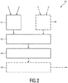
- Fig. 2 shows a schematic diagram of an embodiment of a device 40 for SUV determination according to the present disclosure.
- the device 40 comprises an input 41 for obtaining, from one or more wearable devices 20 and/or one or more biometrical data acquisition units 30, 31, e.g. worn by a patient or by a technologist, care giver or supporting clinical staff, SUV-related data required for SUV determination.
- Said SUV-related data particularly include the time of a radiotracer uptake by the patient.
- event data relating to one or more events that may affect the SUV determination are obtained, particularly from the wearable device 20 and/or one or more biometrical data acquisition units 30, 31 and/or one or more additional sensors 60, 61.
- the input 41 may e.g. be implemented as a data interface for wireless or wired reception or retrieval of data from other components.
- the device 40 further comprises an anomalous event determination unit 42 configured to determine from the obtained event data anomalous event information indicating one or more anomalous events that affect the SUV determination and an SUV determination unit 43 configured to determine the SUV from said SUV-related data taking into account the anomalous event information.
- the anomalous event determination unit 42 may e.g. be implemented as a processing unit or algorithm programmed on processor or computer.
- anomalous shall be understood such that the occurrence, the time of occurrence, the strength of occurrence, or any other characteristic of an event has an influence on the determination of the SUV.
- the SUV determination unit 43 may be configured to determine the SUV from SUV-related data including the radiotracer activity concentration in the patient's tissue, the administered dose of the radiotracer uptake and the patient's biometrical data, in particular the patient's weight, height and/or total surface area.
- the device 40 may further comprise an interface 44 for interconnecting with a radiotracer dose calibrator 70, a radiotracer dose injector 80, one or more biometrical data acquisition units 30, 31 and/or an imaging arrangement 10 to obtain one or more pieces of SUV-related data including the radiotracer activity concentration of the patient's tissue, the administered dose of the radiotracer uptake and the patient's biometrical data, which are then used in the determination of the anomalous events and/or of the SUV.
- Said interface 44 may also be configured as a wired or wireless data interface, and may be even be the same or a similar interface as the input 41.
- the input 41 may further be configured to obtain event data, e.g. from one or more additional sensors 60, 61, including the time of the one or more events. These event data are then used by said anomalous event determination unit 42 is configured to use the time of the one or more events in determining the anomalous event information.
- the device 40 further comprises a synchronization unit 45 for time synchronization of the device with the one or more wearable devices 20 and/or the one or more biometrical data acquisition units 30, 31.
- the synchronization unit 45 generates a system clock which is provided to the one or more wearable devices 20 and/or the one or more biometrical data acquisition units 30, 31, in particular to all other components of the system 1, i.e. the synchronization unit 45 takes over the task of the system's synchronization device 90, which can thus be omitted.
- Fig. 3 shows a schematic diagram of a first embodiment of a wearable device 20a according to the present disclosure. It comprises a holding element 21 for holding the wearable device at the patient's body.
- the holding element 21 may e.g. be a wristband for wearing the wearable device 20a like a wristwatch, in which case the wearable device 20a preferably has the form of a wristwatch.
- the holding element 21 may include a belt, chain, sticker, etc. for mounting the wearable device 20a in some way to the patient's body.
- the wearable device 20a further comprises one or more sensing elements 22, 23 for sensing SUV-related data required for the SUV determination.
- Said SUV-related data include at least the time of a radiotracer uptake by the patient, but preferably includes further data as will be explained below.
- the one or more sensing elements 22, 23 are configured to sense event data relating to one or more events that may affect the SUV determination.
- event data may e.g. comprise movement data and/or activity data reflecting movements and/or activities of the patient, preferably including not only the kind of movement / activity, but also the location, the time, the intensity, and/or the duration.
- the sensing elements 22, 23 may e.g. include one or more of a motion sensor, an accelerometer, a position detection sensor, a vital sign sensor, a gamma radiation sensor and a camera.
- An output 24 is provided for outputting the sensed SUV-related data and event data.
- Said output 24 may e.g. be implemented as a data interface for wired or wireless output of data, which is able to exchange data with the input 41 of the SUV determination device 40.
- the wearable device 20a may further comprise a time measurement unit 25 for recording the time of occurrence of the one or more events sensed as event data, and the output 24 may be configured to output the event data together with the time of occurrence of the related event.
- the wearable device 20a may further comprise a synchronization unit 26 for time synchronization of the wearable device 20a with the device 40 for SUV determination, in particular based on a provided system clock, which may e.g. be provided by the synchronization device 90 of the system 1 or the synchronization unit 45 of the device 40.
- the wearable device 20a may comprise a user interface 27 enabling input and/or output of user information.
- a user in particular the patient, to input SUV-related data, event data and/or biometrical data, e.g. input some personal data or press a button if a predetermined event happens, etc.
- the patient may be informed or instructed via the user interface 27, e.g. to perform a certain activity, go to a certain location, etc.
- the user interface 27 may e.g. be implemented as a display, keyboard, touchscreen, etc.
- the wearable device 20a may comprise a communication unit 28 for electronically exchanging data with one or more biometrical data acquisition units 30, 31 and/or with other components of the system 1, in particular via the output 24.
- the elements 22 to 28 of the wearable device 20a are preferably arranged in a housing 29 to which the holding element 21 is preferably mounted.
- the device 40 may be loaded with an imaging protocol that defines the correct time order of the events or the accepted ranges for a given measurement. If that protocol is not being followed, the conclusions taken from the outcome SUV are somehow compromised and in that case and if possible should be accounted for.
- the patient has to be scanned within 55 to 75 minutes after injection (uptake period from 55 to 75 minutes), with the ideal value being 60 minutes.
- a second examination of the same patient e.g. a follow up PET exam for instance after some therapy
- the uptake period has to be equal to the first ⁇ 10 minutes as target according to the protocol. Deviations are allowed up to maximum ⁇ 15 minutes.
- the wearable device knows if the patient is making a first PET or a follow up examination and is recording the different time events. Every time that this uptake period is not respected, the device has to provide a warning to the technologists.
- Other protocols may define different target and acceptable uptake periods. The settings of the wearable device depend on the protocol being followed by each specific clinical service.
- the measurement of the local radioactivity in the patient arm by the wearable device via the on-board gamma radiation sensor as function of time can also provide an indication of occurrence of paraveneous administration of the tracer.
- the gamma radiation sensor will provide a response that would have a very slow decay allowing to warn the clinical staff that the injection was not fully successful and some tracer will be trapped at the injection site, e.g. at the patient arm. In that case such an occurrence has to be registered to be accounted for during the image analysis.
- the muscular activity during the uptake period may be considered.
- the patient is instructed to be in silence and minimum movement during the uptake period. Patterns of motion during that period of time can be logged by the wearable device through a sensor.
- clinicians can determine if abnormal movement has taken place that could have introduced abnormal radiotracer accumulation in certain body regions that are uncorrelated with the tumor or lesion under examination.
- plasma glucose competes with 18F-FDG (the most used radiotracer in PET imaging) for transport into the cells and phosphorylation by hexokinase may be considered.
- the FDG uptake will be inversely related to the plasma glucose concentration.
- the serum glucose concentration at the time of FDG injection has to be obtained.
- Glucose portable systems connected to the wearable device via a wireless link can be used to record the amount of glucose at that time once again avoiding that way the potential transcription errors. If the serum glucose is high, there may be an increase in false negative findings in oncological PET imaging.
- UPICT Uniform Protocols for Imaging in Clinical Trials
- Images obtained with moderate to severe scenarios should not be used for therapy response assessment unless a correction is derived based on the amount of estimated paraveneous injection.
- the amount of radiotracer activity trap in the paraveneous injection site can be used to correct the Total Injected Activity in the SUV formula described herein if correctly estimated before the examination.
- UPICT also defines the allowed levels of plasma glucose at the time of the injection. For non-diabetic patients concentrations above 200 mg/dL require that the scan is rescheduled, between 150 and 200 mg/dL the referring physician has to be consulted. Values below 150 mg/dl are considered acceptable.
- a reconstructed image (e.g. a PET image) is a 3D volume composed of L x M x N voxels.
- L, M and N might be equal or differ depending on the image acquisition protocol.
- each voxel has between 1x1x1 mm3 to 4x4x4 mm 3 .
- SUV Reconstructed Activity in Patient Tissue Total Injected Activity ⁇ Patient Weight
- Reconstructed Activity in Patient Tissue is the radioactivity activity concentration [kBq/ml] measured by the PET scanner within a region of interest (ROI)
- the Total Injected Activity is the decay-corrected amount of injected radiolabeled FDG [kBq]
- the Patient Weight is the weight of the patient [g] measured on the day of the PET exam, which is used as a normalization factor.
- the wearable device can collect from a wireless weight scale information on the Patient Weight, avoiding for instance transcription errors that can directly affect the accuracy of the SUV outcome.
- the wearable device can also collect from an automated dose injector via a wireless communication channel the Total Injected Activity and the time of injection T 0 .
- the technologists or clinical staff types in the wearable user interface the injected amount right after the injection and not only some time later.
- Certain events like for example the toilette 30 minute after uptake should be recorded in order to confirm that the patient has complied with imaging protocol. They do not directly enter into the SUV computation formula, but are certain tasks that guarantee that the reconstructed imaging are not affected by physiological radiotracer background that can hide the presence of a tumor. Flushing the bladder is needed in order to prevent background activity that compromises the reconstructed activity in the patient issue. Motion and anxiety level of the patient during the 50-70 minutes uptake period must be recorded since it will increase the uptake in the healthy muscle affecting of course the overall uptake in the tumor region. This is why the wearable device may be equipped with a motion sensor and rise alarms to the technologists.
- Fig. 4 shows a front view of a second embodiment of a wearable device 20b according to the present disclosure, which will be explained in the following in the context of the particular use scenario.
- the wearable device 20b is configured as a wrist-worn device (the holding element is not shown, but may the same as for the wearable device 20a).
- Fig. 4 particularly shows a housing 29 around a user interface 27 in the form of a touchscreen showing a plurality of buttons that may be actuated by the patient for entering information, e.g. the time of predetermined events or for prompting predetermined actions.
- buttons on the user interface 27 may e.g. include
- buttons for other events can be foreseen as well.
- the wearable device 20b is attached to the patient and is able to establish a (preferably bi-directional) communication link, e.g. via NFC, Wi-Fi, Bluetooth or an equivalent wireless link with a central station.
- a GPS or equivalent indoor positioning receiver may also be included.
- Other biometrical data acquisitions means like bio-sensors, accelerometers, etc., in particular for motion detection, may be included as well.
- an on-board gamma radiation dosimeter might as well be included in order to provide real-time and the reference value for the gamma radiation dose received by the patient during the examination.
- the wearable device 20b is preferably synchronized with a central station and is loaded with a software application ('app') that includes one button for each of the clinical procedures relevant for the imaging procedure, e.g. relevant for PET quantification.
- the app may register the time for each of the clinical events and associated information (for example, patient weight or injected dose, etc.).
- Anomalous events can also be recorded with the wearable device 20b, like for example patient movement in the resting room prior to the examination (e.g. a PET examination) using the on-board positioning receiver or physiological events, like motor stress, that can impair an adequate radiotracer uptake.
- a central station such as the synchronization device 90 shown in Fig. 1 , or the SUV determination device 50, may generate a reference system clock.
- the clock is wirelessly distributed to the wearable device 20b and either by a wired or wireless connection to the other components of the system as explained above (e.g. to the place of patient injection or automatic injector/dispenser, PET data acquisition system, PET exam console).
- a stream of the different clinical or anomalous events together with the time of occurrence may be generated by the wearable device 20b, which is transmitted to the SUV determination device 40 and stored in a database, preferably with a link to the patient examination. It has been found that the time at which SUV-related events occur play an important role in the determination of the SUV. For instance, in an exemplary situation, although the target uptake period is 60 minutes, due to the service workload it was not possible to take the patient to the imaging scanner earlier said 75 minutes. However, and because that is not done in the clinical routine, the technologist had not registered the exact hour of the beginning of the uptake period and just assumes 60 minutes neglecting the 15 minutes extra.
- the introduced error in the SUV determination would be almost 15% having a direct impact of the SUV outcome measured in the image analysis phase after the exam. That can mislead the image interpretation by the medical doctor since that is no longer doing a quantitative assessment of the SUV that might choose a different type of treatment for that patient.
- a central role may be taken by the synchronization device 90, which may also be called master clock generator and exam logger, i.e. which may not only generate and distribute a synchronization time, but may also log events.
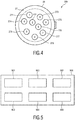
- a schematic diagram of a generic implementation of such a master clock generator and exam logger 900 is shown in Fig. 5 .
- Different hardware implementations are possible, either using a full fledge computer of a dedicated hardware running FPGA or an equivalent processor 901.
- This device 900 receives from the external world a clock signal.
- Correct synchronization can be achieved using e.g. the Consistent Time Integration Profile as defined in the IHE IT Infrastructure Technical Framework.
- the Consistent Time Profile requires the use of the Network Time Protocol (NTP) (as defined at www.NTP.org).
- NTP Network Time Protocol
- the device 900 has several LAN physical interfaces 904 (wither copper or fiber) to interconnect to the dose calibrator 70 (for dose calibrators which have also a LAN interface), to a dose injector dispenser/automatic perfusion 80 (if available), biometrical data acquisition units 30, 31 and to the imaging device 10.
- the LAN link is used to further re-distribute the synchronization clocks as well as the associated time of a particular clinical event.
- the device 900 further comprises a wireless hardware unit 905 in order to keep communication with the wearable device used by the patient and a memory 906 for storing e.g. the processing software.
- a mobile patient tracking device generally called wearable device herein, but also called NM-Tracker or NMT
- the wearable device is assigned and handed out to the patient (e.g. during its registration procedure), and accompanies him/her until he/she leaves the department again. It offers a simple user interface to display procedure related information to the patient.
- the device automatically detects a radiotracer injection event.
- Further workflow related information may be captured during the procedure via a (near-field) RF-interface and/or an inbuilt camera (e.g. in combination with QR-tag or OCR technology).
- a central SUV determination device Via e.g. the existing wireless network in the clinic, the device communicates with a central SUV determination device to transfer collected path (motion) tracking information and recorded technical and physiological parameters).
- An application running on the SUV determination device analyzes the incoming data, checks for workflow state consistency, raises warnings to the medical staff in case of any undesirable events. It furthermore offers an interface to submit further information to the remotely connected wearable devices.
- the wearable device may be designed in such a way that it hampers obvious possibilities of accidental misuse.
- it should be attached to the patient in a non-obstructive way.
- a possible design supporting this could be in form of a (smart) wristwatch.
- its design should consider special clinical requirements regarding sterilization, etc.
- An embodiment of the wearable device comprises a tailored display to pass information to the patient, a simple way to allow the patient to provide feedback (e.g. to remotely confirm a new schedule), a wireless two-way communication interface (802.11x, Bluetooth, ...), a inbuilt digital camera, a 3D-acceleration sensor, a gamma radiation sensor, a data processing unit and an interface to wirelessly re-charge the internal batteries (e.g. via Qi-Technology).
- the gamma radiation sensor module (realized e.g. in FGMOSFET technology) may be designed to reliably detect gamma radiation for standard NM isotope particle energies. It preferably provides a higher sensitivity in one spatial direction. If attached to the patient, the preferred direction is towards the body.
- the wearable device may be centrally stored and re-charged, e.g. at the patient registration desk. During charging, the wearable device's internal clock is synchronized with a central clock unit. Data not yet transferred to the SUV determination device 40 (sometimes also called NMT server or NMT base station) are securely uploaded during this time and finally deleted from the device.
- the SUV determination device 40 sometimes also called NMT server or NMT base station
- the wearable device When put off the charging station, the wearable device may automatically set to active mode and expects to be assigned to a new patient. The assignment can be performed e.g. via putting a patient record QR-/bar-code in front of the wearable device's camera. After identifying a valid patient ID tag, the device switches to a state-machine controlled tracking mode (displayed at the wearable device e.g. via a color change) according to the examination specific workflow steps.
- Patient movement through the clinical area may be recorded via information of the motion sensors and/or the analysis of the signal strength from multiple WLAN access points.
- the information is frequently automatically uploaded to the SUV determination device for further evaluation.
- all exchanged data are encrypted.
- Automatic motion pattern analysis is applied to make the staff aware of patients requiring further assistance and guidance to optimize the examination result.
- the medical staff can also directly access additional related information via the SUV determination device, e.g. whether and when the patient last visited the bathroom.
- the motion detection can be used to directly provide location guidance to patients in order to help them to find their way through large departments.
- not yet prepared/injected patients may receive new scheduling information via the display. This allows them to leave the waiting area and e.g. visit the clinic's recreation zone.
- An interaction (e.g. confirmation) request might be additionally indicated via a special visual signal (e.g. a blinking of the wearable device).
- additional parameters may be stored and transferred via the wearable device.
- the syringe comes with (e.g. a printed) QR-tag containing related radiotracer type and calibration (e.g. timestamp, amount) information. Recognizing the QR-code via the wearable device's camera activates the internal gamma radiation sensor.
- the wearable device distally attached to the patient from where the radiotracer enters the body (e.g. worn at the left wrist, when injecting into the right medial cubital vein), immediately detects the radiotracer bolus and logs the event together with the timestamp.
- the data transferred to the SUV determination device 40 enable the application there to determine a study optimal image acquisition schedule. Adding the dwell-counter determined remaining tracer-activity in the syringe, the SUV determination device 40 exactly knows all the parameters to calculate what amount of tracer has been administered to the patient at what point in time.
- Start and duration of the subsequent image acquisition can be logged (e.g. via detecting typical orientation and movement patterns during the couch positioning, eventually in combination with detecting the additional gamma radiation dose during the anatomical/scouting scan e.g. on PET/CT, SPECT/CT scanner combinations).
- the clinician may check during the debriefing with the patient whether all relevant patient information for subsequent quantitative data analysis has been transferred to the SUV determination device 40. This might be indicated e.g. via a specific color code of the device. If that is not the case he/she takes care of finalizing this step, before the patient is sent home.
- the patient When leaving the department, the patient returns the wearable device to the reception desk where again the device status is checked again and the device prepared for cleaning/recharging.
- a device 40 for standard uptake value, SUV, determination during an emission tomography imaging procedure of a patient to which a radiotracer dose has been administered at an imaging facility is disclosed.
- the emission tomography imaging procedure may for example be a PET or a SPECT imaging procedure.
- the device 40 includes at least one input 41, 44 that is configured to receive SUV-related data required for SUV determination and event data relating to one or more events that may affect the SUV determination.
- the SUV-related data may include one or more of the following: a time of administration of the radiotracer dose to the patient; a radiotracer dose administered to the patient; a radiotracer activity concentration in a region within the patient; the patient's weight; the patient's height; the patient's total surface area, calibration data indicative of a residual activity in a syringe after administration the radiotracer dose to the patient.
- One or more of these SUV-related data may be used in the computation of an SUV from the emission tomography imaging procedure as described above in relation to the SUV equation.
- the present invention recognizes that one or more events, herein described as event data, may affect the accuracy of the so-computed SUV.
- the event data received by the at least one input 41, 44 of the device 40 may include at least one of: i) a time at which an emission tomography imaging procedure of the patient is performed; ii) patient motion data indicative of the patient's motion during the period between administration of the radiotracer dose and a start of the emission tomography imaging procedure; iii) patient position data indicative of the patient's position within the imaging facility during the period between administration of the radiotracer dose and a start of the emission tomography imaging procedure; iv) patient vital signs data indicative of the patient's vital signs during the period between administration of the radiotracer dose and a start of the emission tomography imaging procedure.
- patient position data that, for example, indicates whether the patient is resting in a desired room, or making trips to the bathroom, or is indeed elsewhere in a hospital or imaging facility can also affect the calculated SUV. Again, as described below, such data may be used to correct the SUV as described below.
- patient vital signs during the uptake period e.g. one or more of patient heart rate sensor, body temperature, respiration, blood pressure, skin conductivity, blood oxygenation i.e. SPO2, blood glucose, may also be indicative of errors in the calculated SUV. For example when any of these data lie outside a predetermined range and error may be expected due to e.g. excessive exercise during the uptake period. Again, these maybe corrected-for as described below.
- the device 40 also includes an anomalous event determination unit 42 that is configured to determine, based on one or more of the above event data, anomalous event information indicative of one or more anomalous events that affect the SUV determination. As outlined above the anomalous event determination unit assesses the relevant data, compares this to an expected range, or expected data, and, where necessary flags that an anomaly has occurred. Moreover, the device 40 includes an SUV determination unit 43 that is configured to determine the SUV based on said SUV-related data taking into account the anomalous event information. With respect to i), for example the imaging duration can be extended or shortened, or correction factors may be applied to the calculated SUV.
- the device of the present invention allows for the computation of an accurate SUV by compensating for the various described anomalous events.
- a method of SUV determination is disclosed.
- the method may be used in emission tomography during an emission tomography imaging procedure of a patient to which a radiotracer dose has been administered at an imaging facility.
- the method comprises the steps of: a) obtaining SUV-related data required for SUV determination, b) obtaining event data relating to one or more events that may affect the SUV determination, c) determining based on the obtained event data anomalous event information indicating one or more anomalous events that affect the SUV determination, and d) determining the SUV based on said SUV-related data taking into account the anomalous event information.
- the SUV-related data and event data may be as described in the previous embodiment.
- the above method steps may be stored as instructions or program code in a computer-readable format. Moreover the method steps may be carried-out by a computer.
- a wearable device 20, 20a, 20b is disclosed.
- the wearable device may be used for standard uptake value, SUV, determination during an emission tomography imaging procedure of a patient to which a radiotracer dose has been administered at an imaging facility.
- the wearable device comprises a holding element 21, for example a strap, a belt or a necklace, for holding the wearable device at the patient's body; and one or more sensing elements 22, 23 for sensing SUV-related data required for SUV determination in emission tomography.
- the sensed SUV-related data may be one or more of the above data described in relation to the previous embodiment.
- Preferably said SUV-related data includes at least a time of administration of the radiotracer dose to the patient.
- the wearable device includes an output 24 for outputting the sensed SUV-related data.
- the one or more sensing elements 22, 23 for sensing the time of administration of the radiotracer dose to the patient includes at least one of a) a wireless sensor configured to wirelessly receive the time at which a radiotracer dose injector 80 administers the radiotracer dose to the patient; and b) a gamma radiation sensor arranged such that when the wearable device is worn by the patient the radiation sensor senses gamma radiation emitted from the patient's body; the gamma radiation sensor being further configured to provide a timestamp indicative of the time at which the sensed gamma radiation meets a predetermined threshold condition.
- the wireless sensor thus provides on-patient SUV-related data storage that can be later read-out, for example by the device 40, and used to determine a SUV for a region within a patient.
- the wireless sensor provides automated data transfer, thereby reducing the chance of human errors, as compared to a manually-recorded time of dose administration. Moreover, because the data is acquired by the wearable device it is inherently patient-specific, the chance of mixing-up patient data is reduced.
- the gamma radiation sensor (b) may be used on its own to monitor the post-injection influx of the radiotracer to the patient, for example from a vein or artery or other in-body region of the patient such as a limb, i.e.
- the wrist or leg and thereby provide a time at which the dose was administered to the patient. This may be used to validate a manually-recorded time of injection, or to provide the actual time of injection.
- the gamma radiation sensor (b) may be used in combination with the wireless sensor (a) in order to confirm the time of automated dose injection, and to identify, for example that a radiotracer was actually injected into the patient at all.
- Wireless RF as used in e.g. Near Field Communication, RFID, and Bluetooth may be for example used for this purpose although these specific implementations should not be seen as limiting. Indeed any wireless transmitter-receiver system may be used to transfer the desired data, including RF, infrared, ultrasound and optical communication.
- the wireless sensor may also be used receive additional SUV-related data as described above.
- This may be received wirelessly from wireless-transmitting systems.
- wireless-transmitting systems include for example the radiotracer dose administered to the patient, or radiotracer dose calibration data indicative of a residual activity in a syringe after administration the radiotracer dose to the patient, either of which may be received e.g. from radiotracer dose injector 80, the patient's weight, which may be received from a wireless patient weighing scale, and the patient's height or surface area that may be received from a wireless height measurement system, or a computer system configured to determine the patient's surface area based on a model having, as input, the patient height and weight.
- the wireless sensor may additionally be used to receive, and to store, event data relating to one or more events that may affect the SUV determination.
- the event data is described above, and may be received wirelessly from one or more sensors, or be sensed directly by a sensor that forms part of the wearable device.
- sensors include an accelerometer, a patient position sensor, a patient vital signs sensor selected from the group: a heart rate sensor, a body temperature sensor, a respiration sensor, a blood pressure sensor, a skin conductivity sensor, an SPO2 sensor, a blood glucose sensor.
- the so-defined wearable device 20, 20a, 20b may be used to collect and store various SUV-related data and/ or various event data which can serve as input to the device 40 for use in providing an SUV for the patient.
- the wearable device 20, 20a, 20b may include a time measurement unit 25 for recording the time of measurement of the corresponding event data; this being configured to output the time of measurement of the event data and/ or the corresponding event data.
- the wearable device 20, 20a, 20b may include a synchronization unit 26 for time synchronization of the wearable device with the device 40 for SUV determination.
- an imaging arrangement 10 comprises an emission tomography imaging system for acquiring image data of a patient, in particular a PET or a SPECT imaging system; one or more wearable devices 20, 20a, 20b as described above in the previous embodiment; and a device 40 for standard uptake value, SUV, determination as describe above.
- the device 40 is configured to determine an SUV based on the SUV-related data and the event data received by the at least one input 41, 44 of the device 40; and based on at least the SUV-related data outputted by the one or more wearable devices 20, 20a, 20b.
- the device 40 includes an evaluation unit 50 for evaluating the acquired image data using the determined SUV.
- the device 40 of the imaging arrangement 10 may also be configured to determine the SUV based further on the event data outputted by the one or more wearable devices 20, 20a, 20b.
- a computer program may be stored/distributed on a suitable non-transitory medium, such as an optical storage medium or a solid-state medium supplied together with or as part of other hardware, but may also be distributed in other forms, such as via the Internet or other wired or wireless telecommunication systems.
- a suitable non-transitory medium such as an optical storage medium or a solid-state medium supplied together with or as part of other hardware, but may also be distributed in other forms, such as via the Internet or other wired or wireless telecommunication systems.
Landscapes
- Health & Medical Sciences (AREA)
- Life Sciences & Earth Sciences (AREA)
- Engineering & Computer Science (AREA)
- Medical Informatics (AREA)
- General Health & Medical Sciences (AREA)
- Biomedical Technology (AREA)
- Public Health (AREA)
- Animal Behavior & Ethology (AREA)
- Biophysics (AREA)
- Heart & Thoracic Surgery (AREA)
- Physics & Mathematics (AREA)
- Molecular Biology (AREA)
- Surgery (AREA)
- Veterinary Medicine (AREA)
- Pathology (AREA)
- Radiology & Medical Imaging (AREA)
- High Energy & Nuclear Physics (AREA)
- Nuclear Medicine, Radiotherapy & Molecular Imaging (AREA)
- Optics & Photonics (AREA)
- Cardiology (AREA)
- Physiology (AREA)
- Oral & Maxillofacial Surgery (AREA)
- Dentistry (AREA)
- Epidemiology (AREA)
- Primary Health Care (AREA)
- Computer Networks & Wireless Communication (AREA)
- Human Computer Interaction (AREA)
- Business, Economics & Management (AREA)
- General Business, Economics & Management (AREA)
- Nuclear Medicine (AREA)
Claims (9)
- Agencement d'imagerie (10) comprenant:- un système d'imagerie par tomographie par émission pour acquérir des données d'image d'un patient, en particulier un système d'imagerie TEP ou SPECT,- un ou plusieurs dispositifs portables (20, 20a, 20b) pour obtenir des données liées à une valeur de fixation normalisée, SUV, requises pour la détermination d'une SUV, lesdits dispositifs portables comprenant:où les données liées à une SUV comprennent au moins un temps d'administration de la dose de radiotraceur au patient;- un élément de maintien (21) pour maintenir le dispositif portable sur le corps du patient;- un ou plusieurs éléments de détection (22, 23) pour détecter les données liées à une SUV requises pour la détermination d'une SUV dans une tomographie par émission, y compris au moins un temps d'administration de la dose de radiotraceur au patient, et des données d'événement relatives à un ou plusieurs événements qui peuvent influer sur la détermination d'une SUV; et- une sortie (24) pour sortir les données détectées liées à une SUV et les données d'événement; l'agencement d'imagerie comprenant en outre:- un dispositif (40) pour la détermination d'une SUV pendant une procédure d'imagerie par tomographie par émission d'un patient auquel une dose de radiotraceur a été administrée dans une installation d'imagerie, ledit dispositif (40) comprenant:- au moins une entrée (41, 44) configurée pour recevoir des données liées à une SUV requises pour la détermination d'une SUV, comme une dose administrée de radiotraceur, et des données d'événement relatives à un ou plusieurs événements qui peuvent influer sur la détermination de la SUV;
et où les données d'événement comprennent au moins l'un parmi:- un temps auquel une procédure d'imagerie par tomographie par émission du patient est effectuée;- des données de mouvement du patient indiquant le mouvement du patient pendant la période entre une administration de la dose de radiotraceur et un début de la procédure d'imagerie par tomographie par émission;- des données de position du patient indiquant la position du patient dans l'installation d'imagerie pendant la période entre une administration de la dose de radiotraceur et un début de la procédure d'imagerie par tomographie par émission;- des données de signes vitaux indiquant les signes vitaux du patient pendant la période entre une administration de la dose de radiotraceur et un début de la procédure d'imagerie par tomographie par émission;le dispositif comprenant en outre:- une unité de détermination d'événement anormal (42) configurée pour déterminer, sur la base des données d'événement, des informations d'événement anormal indiquant un ou plusieurs événements anormaux qui influent sur la détermination d'une SUV, et- une unité de détermination d'une SUV (43) configurée pour déterminer la SUV sur la base desdites données liées à une SUV en tenant compte des informations d'événement anormal, où le dispositif (40) est configuré pour déterminer une SUV sur la base des données liées à une SUV et des données d'événement reçues par l'au moins une entrée (41, 44) du dispositif (40); et sur la base d'au moins les données liées à une SUV fournies par l'une ou les plusieurs dispositifs portables (20, 20a, 20b), etl'agencement d'imagerie comprenant en outre:- une unité d'évaluation (50) pour évaluer les données d'image acquises à l'aide de la SUV déterminée. - Dispositif d'imagerie selon la revendication 1, dans lequel les données liées à une SUV comprend en outre au moins l'un parmi:- une dose de radiotraceur administrée au patient;- une concentration d'activité de radiotraceur dans une région chez le patient;- le poids du patient;- la taille du patient;- la surface totale du patient.
- Agencement d'imagerie selon l'une quelconque des revendications 1 à 2, dans lequel l'au moins une entrée (41, 44) du dispositif est configurée pour recevoir au moins l'une parmi les données suivantes de l'un ou des plusieurs dispositifs portables par le patient (20, 20a, 20b):- le temps d'administration de la dose de radiotraceur au patient;- le temps auquel une procédure d'imagerie par tomographie par émission du patient est effectuée;- la dose de radiotraceur administrée au patient,- les données de mouvement du patient indiquant le mouvement du patient pendant la période entre une administration de la dose de radiotraceur et un début de la procédure d'imagerie par tomographie par émission;- les données de position du patient indiquant la position du patient dans l'installation d'imagerie pendant la période entre une administration de la dose de radiotraceur et un début de la procédure d'imagerie par tomographie par émission;- les données de signes vitaux du patient indiquant les signes vitaux du patient pendant la période entre une administration de la dose de radiotraceur et un début de la procédure d'imagerie par tomographie par émission.
- Agencement d'imagerie selon la revendication 1, dans lequel l'au moins une entrée (41, 44) du dispositif est configurée en outre pour recevoir les données suivantes liées à une SUV: les données d'étalonnage de la dose de radiotraceur indiquant une activité résiduelle dans une seringue après administration de la dose de radiotraceur au patient.
- Agencement d'imagerie selon la revendication 3, dans lequel le dispositif comprend en outre une unité de synchronisation (45) pour la synchronisation temporelle du dispositif avec l'un ou les plusieurs dispositifs portables par le patient (20, 20a, 20b).
- Agencement d'imagerie selon l'une quelconque des revendications 1 à 5, dans lequel l'un ou les plusieurs éléments de détection (22, 23) de l'un ou des plusieurs dispositifs portables pour détecter le temps d'administration de la dose de radiotraceur au patient comprennent au moins un parmi:- un capteur sans fil configuré pour recevoir sans fil le temps auquel un injecteur de dose de radiotraceur (80) administre la dose de radiotraceur au patient;- un capteur de rayonnement gamma agencé de telle sorte que lorsque le dispositif portable est porté par le patient, le capteur de rayonnement détecte le rayonnement gamma émis par le corps du patient; le capteur de rayonnement gamma étant en outre configuré pour fournir un horodatage indicatif du moment auquel le rayonnement gamma détecté rencontre une condition de seuil prédéterminée.
- Agencement d'imagerie selon l'une quelconque des revendications 1 à 6, dans lequel l'un ou les plusieurs éléments de détection (22, 23) du dispositif portable sont en outre configurés pour détecter des données d'événement relatives à un ou plusieurs événements qui peuvent influer sur la détermination d'une SUV; et dans lequel ladite sortie (24) du dispositif portable est en outre configurée pour sortir les données d'événement; et dans lequel l'un ou les plusieurs éléments de détection (22, 23) du dispositif portable comprennent au moins l'un des suivants:un accéléromètre, un capteur de position du patient, un capteur de signes vitaux du patient choisi dans le groupe: un capteur de fréquence cardiaque, un capteur de température corporelle, un capteur de respiration, un capteur de pression sanguine, un capteur de conductivité cutanée, un capteur de SPO2, un capteur de glycémie ; et/ouun capteur sans fil configuré pour recevoir des données indiquant au moins l'une parmi:
la fréquence cardiaque du patient, la température corporelle du patient, la respiration du patient, la pression sanguine du patient, la conductivité cutanée du patient, la SPO2 du patient, la glycémie du patient, la position du patient dans l'installation d'imagerie sur la base des données d'image d'une caméra. - Agencement d'imagerie selon l'une quelconque des revendications 1 à 7, dans lequel l'un ou les plusieurs dispositifs portables comprennent en outre une unité de mesure du temps (25) pour enregistrer le temps de mesure des données d'événement correspondantes; et
où ladite sortie (24) du dispositif portable est configurée pour sortir les données d'événement et le temps de mesure des données d'événement correspondantes. - Agencement d'imagerie (10) selon la revendication 1, dans lequel le dispositif (40) est configuré pour déterminer la SUV sur la base en outre des données d'événement sorties par l'un ou les plusieurs dispositifs portables (20, 20a, 20b).
Applications Claiming Priority (2)
| Application Number | Priority Date | Filing Date | Title |
|---|---|---|---|
| EP15191876 | 2015-10-28 | ||
| PCT/EP2016/075302 WO2017072030A1 (fr) | 2015-10-28 | 2016-10-21 | Dispositif et procédé de détermination de suv en tomographie par émission |
Publications (2)
| Publication Number | Publication Date |
|---|---|
| EP3367907A1 EP3367907A1 (fr) | 2018-09-05 |
| EP3367907B1 true EP3367907B1 (fr) | 2020-12-09 |
Family
ID=54476701
Family Applications (1)
| Application Number | Title | Priority Date | Filing Date |
|---|---|---|---|
| EP16788052.5A Active EP3367907B1 (fr) | 2015-10-28 | 2016-10-21 | Dispositif pour détermination de suv en tomographie par émission |
Country Status (5)
| Country | Link |
|---|---|
| US (1) | US10849571B2 (fr) |
| EP (1) | EP3367907B1 (fr) |
| JP (1) | JP6941098B2 (fr) |
| CN (1) | CN108348212B (fr) |
| WO (1) | WO2017072030A1 (fr) |
Families Citing this family (31)
| Publication number | Priority date | Publication date | Assignee | Title |
|---|---|---|---|---|
| US10448844B2 (en) | 2015-08-31 | 2019-10-22 | Masimo Corporation | Systems and methods for patient fall detection |
| CN108348212B (zh) * | 2015-10-28 | 2022-03-29 | 皇家飞利浦有限公司 | 用于发射断层摄影中的suv确定的设备和方法 |
| CN113350659A (zh) | 2016-02-24 | 2021-09-07 | 禾木(中国)生物工程有限公司 | 柔性增强的神经血管导管 |
| CN110381855B (zh) | 2017-01-06 | 2023-07-04 | 因赛普特有限责任公司 | 用于动脉瘤治疗装置的抗血栓涂层 |
| EP3631516A4 (fr) * | 2017-05-31 | 2021-01-20 | The Royal Institution for the Advancement of Learning / McGill University | Mesure non invasive de la fonction d'entrée artérielle pour imagerie par tomographie par émission de positons |
| US11051773B2 (en) * | 2017-12-04 | 2021-07-06 | General Electric Company | Systems and methods for imaging with improved dosages |
| WO2019134879A1 (fr) * | 2018-01-03 | 2019-07-11 | Koninklijke Philips N.V. | Estimation d'image tep à dose complète à partir d'imagerie tep à faible dose en utilisant l'apprentissage profond |
| WO2019212984A1 (fr) | 2018-05-01 | 2019-11-07 | Imperative Care, Inc. | Dispositifs et procédés de retrait de matériau obstructif d'un site intravasculaire |
| US11395665B2 (en) | 2018-05-01 | 2022-07-26 | Incept, Llc | Devices and methods for removing obstructive material, from an intravascular site |
| US11471582B2 (en) | 2018-07-06 | 2022-10-18 | Incept, Llc | Vacuum transfer tool for extendable catheter |
| WO2020010310A1 (fr) | 2018-07-06 | 2020-01-09 | Imperative Care, Inc. | Cathéter neurovasculaire extensible étanche |
| US11766539B2 (en) | 2019-03-29 | 2023-09-26 | Incept, Llc | Enhanced flexibility neurovascular catheter |
| US20200329993A1 (en) | 2019-04-17 | 2020-10-22 | Masimo Corporation | Electrocardiogram device |
| USD919094S1 (en) | 2019-08-16 | 2021-05-11 | Masimo Corporation | Blood pressure device |
| USD917704S1 (en) | 2019-08-16 | 2021-04-27 | Masimo Corporation | Patient monitor |
| USD985498S1 (en) | 2019-08-16 | 2023-05-09 | Masimo Corporation | Connector |
| CN110464326B (zh) | 2019-08-19 | 2022-05-10 | 上海联影医疗科技股份有限公司 | 一种扫描参数推荐方法、系统、装置及存储介质 |
| CN110477941B (zh) * | 2019-08-19 | 2023-06-27 | 上海联影医疗科技股份有限公司 | 摄取值的校正方法、装置、计算机设备和可读存储介质 |
| CN110599472B (zh) * | 2019-09-03 | 2021-03-02 | 佛山原子医疗设备有限公司 | 计算spect定量断层图像中suv归一化系数的方法及系统 |
| EP4044906A4 (fr) | 2019-10-15 | 2023-05-24 | Imperative Care, Inc. | Systèmes et procédés de détection d'attaque multivariable |
| USD927699S1 (en) | 2019-10-18 | 2021-08-10 | Masimo Corporation | Electrode pad |
| US11439799B2 (en) | 2019-12-18 | 2022-09-13 | Imperative Care, Inc. | Split dilator aspiration system |
| CN113365687A (zh) | 2019-12-18 | 2021-09-07 | 因普瑞缇夫护理公司 | 治疗静脉血栓栓塞疾病的方法和系统 |
| US11633272B2 (en) | 2019-12-18 | 2023-04-25 | Imperative Care, Inc. | Manually rotatable thrombus engagement tool |
| EP4117762A4 (fr) | 2020-03-10 | 2024-05-08 | Imperative Care Inc | Cathéter neurovasculaire à flexibilité améliorée |
| USD933232S1 (en) | 2020-05-11 | 2021-10-12 | Masimo Corporation | Blood pressure monitor |
| USD979516S1 (en) | 2020-05-11 | 2023-02-28 | Masimo Corporation | Connector |
| EP4166085A4 (fr) * | 2020-07-13 | 2023-07-05 | Shanghai United Imaging Healthcare Co., Ltd. | Procédé et système de balayage d'image pour dispositif médical |
| US11207497B1 (en) | 2020-08-11 | 2021-12-28 | Imperative Care, Inc. | Catheter with enhanced tensile strength |
| CN112415562A (zh) * | 2020-12-10 | 2021-02-26 | 重庆建安仪器有限责任公司 | 一种固定区域γ辐射监测系统及监测方法 |
| DE102022207474A1 (de) | 2022-07-21 | 2023-07-06 | Siemens Healthcare Gmbh | Vorrichtung und Verfahren zum Auswerten von medizinischen Scannerdaten |
Citations (6)
| Publication number | Priority date | Publication date | Assignee | Title |
|---|---|---|---|---|
| WO2006129301A2 (fr) * | 2005-06-01 | 2006-12-07 | Spectrum Dynamics | Gestion unifiee de distribution, d'administration et d'imagerie d'agent radiopharmaceutique |
| JP2007333471A (ja) * | 2006-06-13 | 2007-12-27 | Shimadzu Corp | 核医学診断装置 |
| US20090304582A1 (en) * | 2005-07-19 | 2009-12-10 | Spectrum Dynamics Llc | Imaging Protocols |
| US20130131422A1 (en) * | 2011-11-23 | 2013-05-23 | General Electric Company | Systems and methods for communicating dose calibration information |
| US20130225950A1 (en) * | 2010-10-19 | 2013-08-29 | Koninklijke Philips Electronics N.V. | Anxiety monitoring |
| US20140312242A1 (en) * | 2012-06-01 | 2014-10-23 | Landauer, Inc. | System for Wireless, Motion and Position-Sensing, Integrating Radiation Sensor and Energy Harvester for Occupational and Environmental Dosimetry |
Family Cites Families (10)
| Publication number | Priority date | Publication date | Assignee | Title |
|---|---|---|---|---|
| CN100479779C (zh) * | 2006-03-13 | 2009-04-22 | 山东省肿瘤医院 | 一种体模运动平台及进行运动模拟的方法 |
| WO2010004536A2 (fr) | 2008-07-11 | 2010-01-14 | Spectrum Dynamics Llc | Procédures d’imagerie médicale ainsi que leur procédé et système de gestion |
| GB0818490D0 (en) * | 2008-10-09 | 2008-11-12 | Siemens Medical Solutions | Post injection interval time correction of SUV in static PET scans |
| CN102317975B (zh) * | 2009-02-17 | 2015-02-04 | 皇家飞利浦电子股份有限公司 | 功能成像 |
| WO2011070484A2 (fr) * | 2009-12-08 | 2011-06-16 | Koninklijke Philips Electronics N.V. | Procédé et système de correction pour corriger des mesures d'absorption de marqueur |
| WO2012069960A2 (fr) * | 2010-11-23 | 2012-05-31 | Koninklijke Philips Electronics N.V. | Etalonnages d'un appareil de tep avec des fenêtres de coïncidence variables |
| US9606245B1 (en) * | 2015-03-24 | 2017-03-28 | The Research Foundation For The State University Of New York | Autonomous gamma, X-ray, and particle detector |
| CN108348212B (zh) * | 2015-10-28 | 2022-03-29 | 皇家飞利浦有限公司 | 用于发射断层摄影中的suv确定的设备和方法 |
| US10182735B2 (en) * | 2015-10-29 | 2019-01-22 | Cardiac Pacemakers, Inc. | Systems and methods for detecting atrial tachyarrhythmia using heart sounds |
| US10506987B2 (en) * | 2016-07-06 | 2019-12-17 | Cardiac Pacemakers, Inc. | Readmission risk assessment based on chronobiological rhythms |
-
2016
- 2016-10-21 CN CN201680063618.XA patent/CN108348212B/zh active Active
- 2016-10-21 US US15/766,861 patent/US10849571B2/en active Active
- 2016-10-21 JP JP2018521613A patent/JP6941098B2/ja active Active
- 2016-10-21 EP EP16788052.5A patent/EP3367907B1/fr active Active
- 2016-10-21 WO PCT/EP2016/075302 patent/WO2017072030A1/fr active Application Filing
Patent Citations (6)
| Publication number | Priority date | Publication date | Assignee | Title |
|---|---|---|---|---|
| WO2006129301A2 (fr) * | 2005-06-01 | 2006-12-07 | Spectrum Dynamics | Gestion unifiee de distribution, d'administration et d'imagerie d'agent radiopharmaceutique |
| US20090304582A1 (en) * | 2005-07-19 | 2009-12-10 | Spectrum Dynamics Llc | Imaging Protocols |
| JP2007333471A (ja) * | 2006-06-13 | 2007-12-27 | Shimadzu Corp | 核医学診断装置 |
| US20130225950A1 (en) * | 2010-10-19 | 2013-08-29 | Koninklijke Philips Electronics N.V. | Anxiety monitoring |
| US20130131422A1 (en) * | 2011-11-23 | 2013-05-23 | General Electric Company | Systems and methods for communicating dose calibration information |
| US20140312242A1 (en) * | 2012-06-01 | 2014-10-23 | Landauer, Inc. | System for Wireless, Motion and Position-Sensing, Integrating Radiation Sensor and Energy Harvester for Occupational and Environmental Dosimetry |
Also Published As
| Publication number | Publication date |
|---|---|
| CN108348212B (zh) | 2022-03-29 |
| JP2018536156A (ja) | 2018-12-06 |
| US10849571B2 (en) | 2020-12-01 |
| WO2017072030A1 (fr) | 2017-05-04 |
| CN108348212A (zh) | 2018-07-31 |
| EP3367907A1 (fr) | 2018-09-05 |
| US20180289340A1 (en) | 2018-10-11 |
| JP6941098B2 (ja) | 2021-09-29 |
Similar Documents
| Publication | Publication Date | Title |
|---|---|---|
| EP3367907B1 (fr) | Dispositif pour détermination de suv en tomographie par émission | |
| RU2556590C2 (ru) | Функциональная визуализация | |
| US9723988B2 (en) | Brain disease diagnosis system | |
| CN103167831B (zh) | 焦虑监测 | |
| US11660054B2 (en) | Medical diagnostic and treatment systems and their methods of use | |
| CN101401725B (zh) | 使用混合成像系统治疗患者的装置 | |
| US20040183683A1 (en) | Medical support system and medical support apparatus | |
| CN102647945A (zh) | 用于校正示踪剂摄取测量结果的方法和校正系统 | |
| US20130035601A1 (en) | Apparatus and method for generating digital health screening form | |
| Lachant et al. | Heart rate expenditure correlates with right ventricular function | |
| US10722171B2 (en) | Device for measuring a plurality of parameters in patient subject to a treatment with radiopharmaceuticals | |
| van Gils et al. | A method to measure the absorbed dose of the thyroid during I‐131 therapy, using a collar detector system and a SPECT acquisition | |
| CN106073734A (zh) | 一种健康仪 | |
| CN104305965A (zh) | 一种健康管理系统 | |
| Allado et al. | Protocol: Innovative measurement of routine physiological variables (heart rate, respiratory rate and oxygen saturation) using a remote photoplethysmography imaging system: a prospective comparative trial protocol | |
| McNeil et al. | A 5-Minute rest period improves the accuracy of blood pressure measurement at time of patient intake | |
| Barneveld et al. | 82Rb PET/CT of Myocardial Perfusion | |
| KR20090083741A (ko) | 피검자의 건강정보 관리 방법 |
Legal Events
| Date | Code | Title | Description |
|---|---|---|---|
| STAA | Information on the status of an ep patent application or granted ep patent |
Free format text: STATUS: UNKNOWN |
|
| STAA | Information on the status of an ep patent application or granted ep patent |
Free format text: STATUS: THE INTERNATIONAL PUBLICATION HAS BEEN MADE |
|
| PUAI | Public reference made under article 153(3) epc to a published international application that has entered the european phase |
Free format text: ORIGINAL CODE: 0009012 |
|
| STAA | Information on the status of an ep patent application or granted ep patent |
Free format text: STATUS: REQUEST FOR EXAMINATION WAS MADE |
|
| 17P | Request for examination filed |
Effective date: 20180528 |
|
| AK | Designated contracting states |
Kind code of ref document: A1 Designated state(s): AL AT BE BG CH CY CZ DE DK EE ES FI FR GB GR HR HU IE IS IT LI LT LU LV MC MK MT NL NO PL PT RO RS SE SI SK SM TR |
|
| AX | Request for extension of the european patent |
Extension state: BA ME |
|
| DAV | Request for validation of the european patent (deleted) | ||
| DAX | Request for extension of the european patent (deleted) | ||
| STAA | Information on the status of an ep patent application or granted ep patent |
Free format text: STATUS: EXAMINATION IS IN PROGRESS |
|
| PUAG | Search results despatched under rule 164(2) epc together with communication from examining division |
Free format text: ORIGINAL CODE: 0009017 |
|
| 17Q | First examination report despatched |
Effective date: 20190215 |
|
| B565 | Issuance of search results under rule 164(2) epc |
Effective date: 20190215 |
|
| RIC1 | Information provided on ipc code assigned before grant |
Ipc: A61B 6/03 20060101AFI20190212BHEP Ipc: G16H 10/65 20180101ALI20190212BHEP Ipc: A61B 5/00 20060101ALI20190212BHEP Ipc: A61B 6/00 20060101ALI20190212BHEP Ipc: G01T 1/00 20060101ALI20190212BHEP |
|
| RAP1 | Party data changed (applicant data changed or rights of an application transferred) |
Owner name: KONINKLIJKE PHILIPS N.V. |
|
| GRAJ | Information related to disapproval of communication of intention to grant by the applicant or resumption of examination proceedings by the epo deleted |
Free format text: ORIGINAL CODE: EPIDOSDIGR1 |
|
| GRAP | Despatch of communication of intention to grant a patent |
Free format text: ORIGINAL CODE: EPIDOSNIGR1 |
|
| GRAP | Despatch of communication of intention to grant a patent |
Free format text: ORIGINAL CODE: EPIDOSNIGR1 |
|
| STAA | Information on the status of an ep patent application or granted ep patent |
Free format text: STATUS: GRANT OF PATENT IS INTENDED |
|
| INTG | Intention to grant announced |
Effective date: 20200515 |
|
| GRAS | Grant fee paid |
Free format text: ORIGINAL CODE: EPIDOSNIGR3 |
|
| GRAA | (expected) grant |
Free format text: ORIGINAL CODE: 0009210 |
|
| STAA | Information on the status of an ep patent application or granted ep patent |
Free format text: STATUS: THE PATENT HAS BEEN GRANTED |
|
| AK | Designated contracting states |
Kind code of ref document: B1 Designated state(s): AL AT BE BG CH CY CZ DE DK EE ES FI FR GB GR HR HU IE IS IT LI LT LU LV MC MK MT NL NO PL PT RO RS SE SI SK SM TR |
|
| REG | Reference to a national code |
Ref country code: GB Ref legal event code: FG4D |
|
| REG | Reference to a national code |
Ref country code: AT Ref legal event code: REF Ref document number: 1342535 Country of ref document: AT Kind code of ref document: T Effective date: 20201215 Ref country code: CH Ref legal event code: EP |
|
| REG | Reference to a national code |
Ref country code: DE Ref legal event code: R096 Ref document number: 602016049496 Country of ref document: DE |
|
| REG | Reference to a national code |
Ref country code: IE Ref legal event code: FG4D |
|
| PG25 | Lapsed in a contracting state [announced via postgrant information from national office to epo] |
Ref country code: FI Free format text: LAPSE BECAUSE OF FAILURE TO SUBMIT A TRANSLATION OF THE DESCRIPTION OR TO PAY THE FEE WITHIN THE PRESCRIBED TIME-LIMIT Effective date: 20201209 Ref country code: GR Free format text: LAPSE BECAUSE OF FAILURE TO SUBMIT A TRANSLATION OF THE DESCRIPTION OR TO PAY THE FEE WITHIN THE PRESCRIBED TIME-LIMIT Effective date: 20210310 Ref country code: NO Free format text: LAPSE BECAUSE OF FAILURE TO SUBMIT A TRANSLATION OF THE DESCRIPTION OR TO PAY THE FEE WITHIN THE PRESCRIBED TIME-LIMIT Effective date: 20210309 Ref country code: RS Free format text: LAPSE BECAUSE OF FAILURE TO SUBMIT A TRANSLATION OF THE DESCRIPTION OR TO PAY THE FEE WITHIN THE PRESCRIBED TIME-LIMIT Effective date: 20201209 |
|
| REG | Reference to a national code |
Ref country code: AT Ref legal event code: MK05 Ref document number: 1342535 Country of ref document: AT Kind code of ref document: T Effective date: 20201209 |
|
| PG25 | Lapsed in a contracting state [announced via postgrant information from national office to epo] |
Ref country code: LV Free format text: LAPSE BECAUSE OF FAILURE TO SUBMIT A TRANSLATION OF THE DESCRIPTION OR TO PAY THE FEE WITHIN THE PRESCRIBED TIME-LIMIT Effective date: 20201209 Ref country code: BG Free format text: LAPSE BECAUSE OF FAILURE TO SUBMIT A TRANSLATION OF THE DESCRIPTION OR TO PAY THE FEE WITHIN THE PRESCRIBED TIME-LIMIT Effective date: 20210309 Ref country code: SE Free format text: LAPSE BECAUSE OF FAILURE TO SUBMIT A TRANSLATION OF THE DESCRIPTION OR TO PAY THE FEE WITHIN THE PRESCRIBED TIME-LIMIT Effective date: 20201209 |
|
| REG | Reference to a national code |
Ref country code: NL Ref legal event code: MP Effective date: 20201209 |
|
| PG25 | Lapsed in a contracting state [announced via postgrant information from national office to epo] |
Ref country code: HR Free format text: LAPSE BECAUSE OF FAILURE TO SUBMIT A TRANSLATION OF THE DESCRIPTION OR TO PAY THE FEE WITHIN THE PRESCRIBED TIME-LIMIT Effective date: 20201209 Ref country code: NL Free format text: LAPSE BECAUSE OF FAILURE TO SUBMIT A TRANSLATION OF THE DESCRIPTION OR TO PAY THE FEE WITHIN THE PRESCRIBED TIME-LIMIT Effective date: 20201209 |
|
| REG | Reference to a national code |
Ref country code: LT Ref legal event code: MG9D |
|
| PG25 | Lapsed in a contracting state [announced via postgrant information from national office to epo] |
Ref country code: SK Free format text: LAPSE BECAUSE OF FAILURE TO SUBMIT A TRANSLATION OF THE DESCRIPTION OR TO PAY THE FEE WITHIN THE PRESCRIBED TIME-LIMIT Effective date: 20201209 Ref country code: PT Free format text: LAPSE BECAUSE OF FAILURE TO SUBMIT A TRANSLATION OF THE DESCRIPTION OR TO PAY THE FEE WITHIN THE PRESCRIBED TIME-LIMIT Effective date: 20210409 Ref country code: RO Free format text: LAPSE BECAUSE OF FAILURE TO SUBMIT A TRANSLATION OF THE DESCRIPTION OR TO PAY THE FEE WITHIN THE PRESCRIBED TIME-LIMIT Effective date: 20201209 Ref country code: LT Free format text: LAPSE BECAUSE OF FAILURE TO SUBMIT A TRANSLATION OF THE DESCRIPTION OR TO PAY THE FEE WITHIN THE PRESCRIBED TIME-LIMIT Effective date: 20201209 Ref country code: SM Free format text: LAPSE BECAUSE OF FAILURE TO SUBMIT A TRANSLATION OF THE DESCRIPTION OR TO PAY THE FEE WITHIN THE PRESCRIBED TIME-LIMIT Effective date: 20201209 Ref country code: CZ Free format text: LAPSE BECAUSE OF FAILURE TO SUBMIT A TRANSLATION OF THE DESCRIPTION OR TO PAY THE FEE WITHIN THE PRESCRIBED TIME-LIMIT Effective date: 20201209 Ref country code: EE Free format text: LAPSE BECAUSE OF FAILURE TO SUBMIT A TRANSLATION OF THE DESCRIPTION OR TO PAY THE FEE WITHIN THE PRESCRIBED TIME-LIMIT Effective date: 20201209 |
|
| PG25 | Lapsed in a contracting state [announced via postgrant information from national office to epo] |
Ref country code: PL Free format text: LAPSE BECAUSE OF FAILURE TO SUBMIT A TRANSLATION OF THE DESCRIPTION OR TO PAY THE FEE WITHIN THE PRESCRIBED TIME-LIMIT Effective date: 20201209 Ref country code: AT Free format text: LAPSE BECAUSE OF FAILURE TO SUBMIT A TRANSLATION OF THE DESCRIPTION OR TO PAY THE FEE WITHIN THE PRESCRIBED TIME-LIMIT Effective date: 20201209 |
|
| REG | Reference to a national code |
Ref country code: DE Ref legal event code: R097 Ref document number: 602016049496 Country of ref document: DE |
|
| PG25 | Lapsed in a contracting state [announced via postgrant information from national office to epo] |
Ref country code: IS Free format text: LAPSE BECAUSE OF FAILURE TO SUBMIT A TRANSLATION OF THE DESCRIPTION OR TO PAY THE FEE WITHIN THE PRESCRIBED TIME-LIMIT Effective date: 20210409 |
|
| PLBE | No opposition filed within time limit |
Free format text: ORIGINAL CODE: 0009261 |
|
| STAA | Information on the status of an ep patent application or granted ep patent |
Free format text: STATUS: NO OPPOSITION FILED WITHIN TIME LIMIT |
|
| PG25 | Lapsed in a contracting state [announced via postgrant information from national office to epo] |
Ref country code: AL Free format text: LAPSE BECAUSE OF FAILURE TO SUBMIT A TRANSLATION OF THE DESCRIPTION OR TO PAY THE FEE WITHIN THE PRESCRIBED TIME-LIMIT Effective date: 20201209 Ref country code: IT Free format text: LAPSE BECAUSE OF FAILURE TO SUBMIT A TRANSLATION OF THE DESCRIPTION OR TO PAY THE FEE WITHIN THE PRESCRIBED TIME-LIMIT Effective date: 20201209 |
|
| 26N | No opposition filed |
Effective date: 20210910 |
|
| PG25 | Lapsed in a contracting state [announced via postgrant information from national office to epo] |
Ref country code: DK Free format text: LAPSE BECAUSE OF FAILURE TO SUBMIT A TRANSLATION OF THE DESCRIPTION OR TO PAY THE FEE WITHIN THE PRESCRIBED TIME-LIMIT Effective date: 20201209 Ref country code: SI Free format text: LAPSE BECAUSE OF FAILURE TO SUBMIT A TRANSLATION OF THE DESCRIPTION OR TO PAY THE FEE WITHIN THE PRESCRIBED TIME-LIMIT Effective date: 20201209 |
|
| PG25 | Lapsed in a contracting state [announced via postgrant information from national office to epo] |
Ref country code: ES Free format text: LAPSE BECAUSE OF FAILURE TO SUBMIT A TRANSLATION OF THE DESCRIPTION OR TO PAY THE FEE WITHIN THE PRESCRIBED TIME-LIMIT Effective date: 20201209 |
|
| REG | Reference to a national code |
Ref country code: CH Ref legal event code: PL |
|
| PG25 | Lapsed in a contracting state [announced via postgrant information from national office to epo] |
Ref country code: IS Free format text: LAPSE BECAUSE OF FAILURE TO SUBMIT A TRANSLATION OF THE DESCRIPTION OR TO PAY THE FEE WITHIN THE PRESCRIBED TIME-LIMIT Effective date: 20210409 |
|
| REG | Reference to a national code |
Ref country code: BE Ref legal event code: MM Effective date: 20211031 |
|
| PG25 | Lapsed in a contracting state [announced via postgrant information from national office to epo] |
Ref country code: MC Free format text: LAPSE BECAUSE OF FAILURE TO SUBMIT A TRANSLATION OF THE DESCRIPTION OR TO PAY THE FEE WITHIN THE PRESCRIBED TIME-LIMIT Effective date: 20201209 |
|
| PG25 | Lapsed in a contracting state [announced via postgrant information from national office to epo] |
Ref country code: LU Free format text: LAPSE BECAUSE OF NON-PAYMENT OF DUE FEES Effective date: 20211021 Ref country code: BE Free format text: LAPSE BECAUSE OF NON-PAYMENT OF DUE FEES Effective date: 20211031 |
|
| PG25 | Lapsed in a contracting state [announced via postgrant information from national office to epo] |
Ref country code: LI Free format text: LAPSE BECAUSE OF NON-PAYMENT OF DUE FEES Effective date: 20211031 Ref country code: CH Free format text: LAPSE BECAUSE OF NON-PAYMENT OF DUE FEES Effective date: 20211031 |
|
| PG25 | Lapsed in a contracting state [announced via postgrant information from national office to epo] |
Ref country code: FR Free format text: LAPSE BECAUSE OF NON-PAYMENT OF DUE FEES Effective date: 20211031 |
|
| PG25 | Lapsed in a contracting state [announced via postgrant information from national office to epo] |
Ref country code: IE Free format text: LAPSE BECAUSE OF NON-PAYMENT OF DUE FEES Effective date: 20211021 |
|
| PGFP | Annual fee paid to national office [announced via postgrant information from national office to epo] |
Ref country code: GB Payment date: 20221018 Year of fee payment: 7 Ref country code: DE Payment date: 20220628 Year of fee payment: 7 |
|
| PG25 | Lapsed in a contracting state [announced via postgrant information from national office to epo] |
Ref country code: HU Free format text: LAPSE BECAUSE OF FAILURE TO SUBMIT A TRANSLATION OF THE DESCRIPTION OR TO PAY THE FEE WITHIN THE PRESCRIBED TIME-LIMIT; INVALID AB INITIO Effective date: 20161021 |
|
| PG25 | Lapsed in a contracting state [announced via postgrant information from national office to epo] |
Ref country code: CY Free format text: LAPSE BECAUSE OF FAILURE TO SUBMIT A TRANSLATION OF THE DESCRIPTION OR TO PAY THE FEE WITHIN THE PRESCRIBED TIME-LIMIT Effective date: 20201209 |
|
| PG25 | Lapsed in a contracting state [announced via postgrant information from national office to epo] |
Ref country code: MK Free format text: LAPSE BECAUSE OF FAILURE TO SUBMIT A TRANSLATION OF THE DESCRIPTION OR TO PAY THE FEE WITHIN THE PRESCRIBED TIME-LIMIT Effective date: 20201209 |