EP3352768B1 - Methods of promoting hepatic regeneration - Google Patents
Methods of promoting hepatic regeneration Download PDFInfo
- Publication number
- EP3352768B1 EP3352768B1 EP16849506.7A EP16849506A EP3352768B1 EP 3352768 B1 EP3352768 B1 EP 3352768B1 EP 16849506 A EP16849506 A EP 16849506A EP 3352768 B1 EP3352768 B1 EP 3352768B1
- Authority
- EP
- European Patent Office
- Prior art keywords
- liver
- compound
- oca
- pve
- disease
- Prior art date
- Legal status (The legal status is an assumption and is not a legal conclusion. Google has not performed a legal analysis and makes no representation as to the accuracy of the status listed.)
- Active
Links
- 230000008929 regeneration Effects 0.000 title claims description 77
- 238000011069 regeneration method Methods 0.000 title claims description 77
- 230000002440 hepatic effect Effects 0.000 title claims description 55
- 238000000034 method Methods 0.000 title claims description 41
- 230000001737 promoting effect Effects 0.000 title claims description 8
- 210000004185 liver Anatomy 0.000 claims description 298
- 150000001875 compounds Chemical class 0.000 claims description 150
- 239000003814 drug Substances 0.000 claims description 38
- 230000001965 increasing effect Effects 0.000 claims description 32
- 230000006378 damage Effects 0.000 claims description 30
- 230000003908 liver function Effects 0.000 claims description 30
- 206010008635 Cholestasis Diseases 0.000 claims description 29
- 230000007870 cholestasis Effects 0.000 claims description 28
- 231100000359 cholestasis Toxicity 0.000 claims description 28
- 208000014674 injury Diseases 0.000 claims description 25
- 208000027418 Wounds and injury Diseases 0.000 claims description 23
- 230000002829 reductive effect Effects 0.000 claims description 22
- 150000003839 salts Chemical class 0.000 claims description 20
- 208000037265 diseases, disorders, signs and symptoms Diseases 0.000 claims description 19
- 201000010099 disease Diseases 0.000 claims description 18
- 206010028980 Neoplasm Diseases 0.000 claims description 14
- 229940079593 drug Drugs 0.000 claims description 13
- 238000012753 partial hepatectomy Methods 0.000 claims description 9
- 230000036210 malignancy Effects 0.000 claims description 8
- 201000011510 cancer Diseases 0.000 claims description 7
- 230000001575 pathological effect Effects 0.000 claims description 6
- 208000018565 Hemochromatosis Diseases 0.000 claims description 4
- 208000002972 Hepatolenticular Degeneration Diseases 0.000 claims description 4
- 208000012347 Parenteral nutrition associated liver disease Diseases 0.000 claims description 4
- 208000033147 Parenteral nutrition-associated cholestasis Diseases 0.000 claims description 4
- 102100036325 Sterol 26-hydroxylase, mitochondrial Human genes 0.000 claims description 4
- 208000018839 Wilson disease Diseases 0.000 claims description 4
- 208000001088 cerebrotendinous xanthomatosis Diseases 0.000 claims description 4
- 230000001684 chronic effect Effects 0.000 claims description 4
- 208000022309 Alcoholic Liver disease Diseases 0.000 claims description 3
- 206010003827 Autoimmune hepatitis Diseases 0.000 claims description 3
- 206010019799 Hepatitis viral Diseases 0.000 claims description 3
- 231100000844 hepatocellular carcinoma Toxicity 0.000 claims description 3
- 206010073071 hepatocellular carcinoma Diseases 0.000 claims description 3
- 208000010157 sclerosing cholangitis Diseases 0.000 claims description 3
- 201000001862 viral hepatitis Diseases 0.000 claims description 3
- 206010004637 Bile duct stone Diseases 0.000 claims description 2
- 201000009331 Choledocholithiasis Diseases 0.000 claims description 2
- 206010056533 Congenital hepatic fibrosis Diseases 0.000 claims description 2
- 208000015872 Gaucher disease Diseases 0.000 claims description 2
- 208000009329 Graft vs Host Disease Diseases 0.000 claims description 2
- 208000024815 Granulomatous liver disease Diseases 0.000 claims description 2
- 208000012868 Overgrowth Diseases 0.000 claims description 2
- 206010040047 Sepsis Diseases 0.000 claims description 2
- 208000021386 Sjogren Syndrome Diseases 0.000 claims description 2
- 208000006682 alpha 1-Antitrypsin Deficiency Diseases 0.000 claims description 2
- 230000010108 arterial embolization Effects 0.000 claims description 2
- 230000001580 bacterial effect Effects 0.000 claims description 2
- 208000024908 graft versus host disease Diseases 0.000 claims description 2
- 201000002161 intrahepatic cholestasis of pregnancy Diseases 0.000 claims description 2
- 208000014861 isolated congenital hepatic fibrosis Diseases 0.000 claims description 2
- 201000006038 polycystic kidney disease 4 Diseases 0.000 claims description 2
- 201000000306 sarcoidosis Diseases 0.000 claims description 2
- ZXERDUOLZKYMJM-ZWECCWDJSA-N obeticholic acid Chemical compound C([C@@]12C)C[C@@H](O)C[C@H]1[C@@H](CC)[C@@H](O)[C@@H]1[C@@H]2CC[C@]2(C)[C@@H]([C@H](C)CCC(O)=O)CC[C@H]21 ZXERDUOLZKYMJM-ZWECCWDJSA-N 0.000 description 183
- 229960001601 obeticholic acid Drugs 0.000 description 167
- 239000000203 mixture Substances 0.000 description 78
- 241001465754 Metazoa Species 0.000 description 77
- 230000014509 gene expression Effects 0.000 description 60
- 241000283973 Oryctolagus cuniculus Species 0.000 description 51
- 241000700159 Rattus Species 0.000 description 40
- 238000011282 treatment Methods 0.000 description 40
- 239000003613 bile acid Substances 0.000 description 39
- HSINOMROUCMIEA-FGVHQWLLSA-N (2s,4r)-4-[(3r,5s,6r,7r,8s,9s,10s,13r,14s,17r)-6-ethyl-3,7-dihydroxy-10,13-dimethyl-2,3,4,5,6,7,8,9,11,12,14,15,16,17-tetradecahydro-1h-cyclopenta[a]phenanthren-17-yl]-2-methylpentanoic acid Chemical class C([C@@]12C)C[C@@H](O)C[C@H]1[C@@H](CC)[C@@H](O)[C@@H]1[C@@H]2CC[C@]2(C)[C@@H]([C@H](C)C[C@H](C)C(O)=O)CC[C@H]21 HSINOMROUCMIEA-FGVHQWLLSA-N 0.000 description 37
- 239000000243 solution Substances 0.000 description 33
- XLYOFNOQVPJJNP-UHFFFAOYSA-N water Substances O XLYOFNOQVPJJNP-UHFFFAOYSA-N 0.000 description 31
- 230000000694 effects Effects 0.000 description 30
- 210000003494 hepatocyte Anatomy 0.000 description 30
- 238000009472 formulation Methods 0.000 description 29
- 230000012010 growth Effects 0.000 description 29
- 102100038495 Bile acid receptor Human genes 0.000 description 27
- 101000603876 Homo sapiens Bile acid receptor Proteins 0.000 description 27
- 239000003833 bile salt Substances 0.000 description 27
- 210000005161 hepatic lobe Anatomy 0.000 description 24
- 230000010102 embolization Effects 0.000 description 23
- 208000019423 liver disease Diseases 0.000 description 22
- 229940124597 therapeutic agent Drugs 0.000 description 22
- YMWUJEATGCHHMB-UHFFFAOYSA-N Dichloromethane Chemical compound ClCCl YMWUJEATGCHHMB-UHFFFAOYSA-N 0.000 description 21
- 238000002591 computed tomography Methods 0.000 description 21
- 206010016654 Fibrosis Diseases 0.000 description 20
- 230000037396 body weight Effects 0.000 description 20
- OKKJLVBELUTLKV-UHFFFAOYSA-N Methanol Chemical compound OC OKKJLVBELUTLKV-UHFFFAOYSA-N 0.000 description 19
- 101150082429 FGFR4 gene Proteins 0.000 description 18
- 230000001587 cholestatic effect Effects 0.000 description 18
- 210000002381 plasma Anatomy 0.000 description 18
- 230000035755 proliferation Effects 0.000 description 18
- 238000002271 resection Methods 0.000 description 18
- 206010019663 Hepatic failure Diseases 0.000 description 17
- 231100000835 liver failure Toxicity 0.000 description 17
- 208000007903 liver failure Diseases 0.000 description 17
- 210000003240 portal vein Anatomy 0.000 description 17
- 238000002360 preparation method Methods 0.000 description 17
- XEKOWRVHYACXOJ-UHFFFAOYSA-N Ethyl acetate Chemical compound CCOC(C)=O XEKOWRVHYACXOJ-UHFFFAOYSA-N 0.000 description 16
- 206010067125 Liver injury Diseases 0.000 description 16
- 239000012071 phase Substances 0.000 description 16
- 238000001356 surgical procedure Methods 0.000 description 16
- HEMHJVSKTPXQMS-UHFFFAOYSA-M Sodium hydroxide Chemical compound [OH-].[Na+] HEMHJVSKTPXQMS-UHFFFAOYSA-M 0.000 description 15
- 239000004480 active ingredient Substances 0.000 description 15
- 210000003405 ileum Anatomy 0.000 description 15
- 238000005259 measurement Methods 0.000 description 15
- 108090000623 proteins and genes Proteins 0.000 description 15
- 101150075266 CYP7A1 gene Proteins 0.000 description 14
- 230000007882 cirrhosis Effects 0.000 description 14
- 208000019425 cirrhosis of liver Diseases 0.000 description 14
- 230000006698 induction Effects 0.000 description 14
- 125000002496 methyl group Chemical group [H]C([H])([H])* 0.000 description 14
- 239000011541 reaction mixture Substances 0.000 description 13
- 210000002966 serum Anatomy 0.000 description 13
- 230000011664 signaling Effects 0.000 description 13
- 230000007863 steatosis Effects 0.000 description 13
- 231100000240 steatosis hepatitis Toxicity 0.000 description 13
- 239000003981 vehicle Substances 0.000 description 13
- CSCPPACGZOOCGX-UHFFFAOYSA-N Acetone Chemical compound CC(C)=O CSCPPACGZOOCGX-UHFFFAOYSA-N 0.000 description 12
- 101100390675 Mus musculus Fgf15 gene Proteins 0.000 description 12
- WYURNTSHIVDZCO-UHFFFAOYSA-N Tetrahydrofuran Chemical compound C1CCOC1 WYURNTSHIVDZCO-UHFFFAOYSA-N 0.000 description 12
- 230000007423 decrease Effects 0.000 description 12
- 229940121360 farnesoid X receptor (fxr) agonists Drugs 0.000 description 12
- 229910052739 hydrogen Inorganic materials 0.000 description 12
- 239000001257 hydrogen Substances 0.000 description 12
- 230000000968 intestinal effect Effects 0.000 description 12
- 230000002062 proliferating effect Effects 0.000 description 12
- 102000002260 Alkaline Phosphatase Human genes 0.000 description 11
- 108020004774 Alkaline Phosphatase Proteins 0.000 description 11
- 230000001969 hypertrophic effect Effects 0.000 description 11
- 239000000463 material Substances 0.000 description 11
- 210000000941 bile Anatomy 0.000 description 10
- 231100000753 hepatic injury Toxicity 0.000 description 10
- 125000004435 hydrogen atom Chemical group [H]* 0.000 description 10
- 239000000843 powder Substances 0.000 description 10
- WEVYAHXRMPXWCK-UHFFFAOYSA-N Acetonitrile Chemical compound CC#N WEVYAHXRMPXWCK-UHFFFAOYSA-N 0.000 description 9
- LFQSCWFLJHTTHZ-UHFFFAOYSA-N Ethanol Chemical compound CCO LFQSCWFLJHTTHZ-UHFFFAOYSA-N 0.000 description 9
- 206010061218 Inflammation Diseases 0.000 description 9
- 241000699670 Mus sp. Species 0.000 description 9
- 238000004458 analytical method Methods 0.000 description 9
- 230000004054 inflammatory process Effects 0.000 description 9
- 210000005228 liver tissue Anatomy 0.000 description 9
- 208000008338 non-alcoholic fatty liver disease Diseases 0.000 description 9
- 230000037361 pathway Effects 0.000 description 9
- 239000008194 pharmaceutical composition Substances 0.000 description 9
- 230000002441 reversible effect Effects 0.000 description 9
- 102100036475 Alanine aminotransferase 1 Human genes 0.000 description 8
- 108010082126 Alanine transaminase Proteins 0.000 description 8
- WZUVPPKBWHMQCE-UHFFFAOYSA-N Haematoxylin Chemical compound C12=CC(O)=C(O)C=C2CC2(O)C1C1=CC=C(O)C(O)=C1OC2 WZUVPPKBWHMQCE-UHFFFAOYSA-N 0.000 description 8
- NBIIXXVUZAFLBC-UHFFFAOYSA-N Phosphoric acid Chemical compound OP(O)(O)=O NBIIXXVUZAFLBC-UHFFFAOYSA-N 0.000 description 8
- JLJSYHOPCNWUNE-IEOVAKBOSA-N [99Tc].CC1=CC(C)=C(NC(=O)CN(CC(O)=O)CC(O)=O)C(C)=C1Br Chemical compound [99Tc].CC1=CC(C)=C(NC(=O)CN(CC(O)=O)CC(O)=O)C(C)=C1Br JLJSYHOPCNWUNE-IEOVAKBOSA-N 0.000 description 8
- 208000006454 hepatitis Diseases 0.000 description 8
- 125000002887 hydroxy group Chemical group [H]O* 0.000 description 8
- 239000007788 liquid Substances 0.000 description 8
- -1 3-hydroxyl Chemical class 0.000 description 7
- 101150048692 ABCB11 gene Proteins 0.000 description 7
- 206010020880 Hypertrophy Diseases 0.000 description 7
- 238000000585 Mann–Whitney U test Methods 0.000 description 7
- 101150004781 Slc10a1 gene Proteins 0.000 description 7
- 230000001154 acute effect Effects 0.000 description 7
- 210000000013 bile duct Anatomy 0.000 description 7
- 229940093761 bile salts Drugs 0.000 description 7
- 238000009550 cholescintigraphy Methods 0.000 description 7
- 230000002596 correlated effect Effects 0.000 description 7
- 230000004761 fibrosis Effects 0.000 description 7
- 230000013632 homeostatic process Effects 0.000 description 7
- 230000001771 impaired effect Effects 0.000 description 7
- 239000012074 organic phase Substances 0.000 description 7
- 229910021653 sulphate ion Inorganic materials 0.000 description 7
- 238000003786 synthesis reaction Methods 0.000 description 7
- 210000001519 tissue Anatomy 0.000 description 7
- BPYKTIZUTYGOLE-IFADSCNNSA-N Bilirubin Chemical compound N1C(=O)C(C)=C(C=C)\C1=C\C1=C(C)C(CCC(O)=O)=C(CC2=C(C(C)=C(\C=C/3C(=C(C=C)C(=O)N\3)C)N2)CCC(O)=O)N1 BPYKTIZUTYGOLE-IFADSCNNSA-N 0.000 description 6
- WSFSSNUMVMOOMR-UHFFFAOYSA-N Formaldehyde Chemical compound O=C WSFSSNUMVMOOMR-UHFFFAOYSA-N 0.000 description 6
- PIWKPBJCKXDKJR-UHFFFAOYSA-N Isoflurane Chemical compound FC(F)OC(Cl)C(F)(F)F PIWKPBJCKXDKJR-UHFFFAOYSA-N 0.000 description 6
- 239000007832 Na2SO4 Substances 0.000 description 6
- PMZURENOXWZQFD-UHFFFAOYSA-L Sodium Sulfate Chemical compound [Na+].[Na+].[O-]S([O-])(=O)=O PMZURENOXWZQFD-UHFFFAOYSA-L 0.000 description 6
- UIIMBOGNXHQVGW-UHFFFAOYSA-M Sodium bicarbonate Chemical compound [Na+].OC([O-])=O UIIMBOGNXHQVGW-UHFFFAOYSA-M 0.000 description 6
- YXFVVABEGXRONW-UHFFFAOYSA-N Toluene Chemical compound CC1=CC=CC=C1 YXFVVABEGXRONW-UHFFFAOYSA-N 0.000 description 6
- ZMANZCXQSJIPKH-UHFFFAOYSA-N Triethylamine Chemical class CCN(CC)CC ZMANZCXQSJIPKH-UHFFFAOYSA-N 0.000 description 6
- 230000004913 activation Effects 0.000 description 6
- 230000015572 biosynthetic process Effects 0.000 description 6
- 235000005911 diet Nutrition 0.000 description 6
- 230000037213 diet Effects 0.000 description 6
- 235000019441 ethanol Nutrition 0.000 description 6
- 235000019439 ethyl acetate Nutrition 0.000 description 6
- 125000001495 ethyl group Chemical group [H]C([H])([H])C([H])([H])* 0.000 description 6
- 238000002474 experimental method Methods 0.000 description 6
- 229960002725 isoflurane Drugs 0.000 description 6
- 239000003550 marker Substances 0.000 description 6
- 230000000414 obstructive effect Effects 0.000 description 6
- 238000003305 oral gavage Methods 0.000 description 6
- 239000002245 particle Substances 0.000 description 6
- 230000008569 process Effects 0.000 description 6
- 238000000746 purification Methods 0.000 description 6
- 229910052938 sodium sulfate Inorganic materials 0.000 description 6
- 239000002904 solvent Substances 0.000 description 6
- 238000007619 statistical method Methods 0.000 description 6
- 230000002103 transcriptional effect Effects 0.000 description 6
- 238000000207 volumetry Methods 0.000 description 6
- 108091032973 (ribonucleotides)n+m Proteins 0.000 description 5
- 108010003415 Aspartate Aminotransferases Proteins 0.000 description 5
- 102000004625 Aspartate Aminotransferases Human genes 0.000 description 5
- 101150096887 CDC25B gene Proteins 0.000 description 5
- 102000004127 Cytokines Human genes 0.000 description 5
- 108090000695 Cytokines Proteins 0.000 description 5
- 238000012752 Hepatectomy Methods 0.000 description 5
- 102100023172 Nuclear receptor subfamily 0 group B member 2 Human genes 0.000 description 5
- 239000004372 Polyvinyl alcohol Substances 0.000 description 5
- VYPSYNLAJGMNEJ-UHFFFAOYSA-N Silicium dioxide Chemical compound O=[Si]=O VYPSYNLAJGMNEJ-UHFFFAOYSA-N 0.000 description 5
- FAPWRFPIFSIZLT-UHFFFAOYSA-M Sodium chloride Chemical compound [Na+].[Cl-] FAPWRFPIFSIZLT-UHFFFAOYSA-M 0.000 description 5
- 108090000340 Transaminases Proteins 0.000 description 5
- 239000000556 agonist Substances 0.000 description 5
- 239000008346 aqueous phase Substances 0.000 description 5
- 239000012267 brine Substances 0.000 description 5
- 239000002775 capsule Substances 0.000 description 5
- 238000006243 chemical reaction Methods 0.000 description 5
- 238000002648 combination therapy Methods 0.000 description 5
- 229940125904 compound 1 Drugs 0.000 description 5
- 239000003937 drug carrier Substances 0.000 description 5
- 230000006870 function Effects 0.000 description 5
- 102000006640 gamma-Glutamyltransferase Human genes 0.000 description 5
- 231100000234 hepatic damage Toxicity 0.000 description 5
- 230000008818 liver damage Effects 0.000 description 5
- 230000001404 mediated effect Effects 0.000 description 5
- 108010003814 member 2 group B nuclear receptor subfamily 0 Proteins 0.000 description 5
- 230000036961 partial effect Effects 0.000 description 5
- BASFCYQUMIYNBI-UHFFFAOYSA-N platinum Chemical compound [Pt] BASFCYQUMIYNBI-UHFFFAOYSA-N 0.000 description 5
- 229920002451 polyvinyl alcohol Polymers 0.000 description 5
- 239000002244 precipitate Substances 0.000 description 5
- 238000011555 rabbit model Methods 0.000 description 5
- 238000011084 recovery Methods 0.000 description 5
- 230000001172 regenerating effect Effects 0.000 description 5
- 230000004044 response Effects 0.000 description 5
- HPALAKNZSZLMCH-UHFFFAOYSA-M sodium;chloride;hydrate Chemical compound O.[Na+].[Cl-] HPALAKNZSZLMCH-UHFFFAOYSA-M 0.000 description 5
- 230000001225 therapeutic effect Effects 0.000 description 5
- LHSZAKRHUYJKIU-CNFLPKCYSA-N (4r)-4-[(3r,5s,6r,8s,9s,10s,13r,14s,17r)-6-ethyl-3-hydroxy-10,13-dimethyl-7-oxo-1,2,3,4,5,6,8,9,11,12,14,15,16,17-tetradecahydrocyclopenta[a]phenanthren-17-yl]pentanoic acid Chemical compound C([C@@H]12)C[C@]3(C)[C@@H]([C@H](C)CCC(O)=O)CC[C@H]3[C@@H]1C(=O)[C@H](CC)[C@H]1[C@]2(C)CC[C@@H](O)C1 LHSZAKRHUYJKIU-CNFLPKCYSA-N 0.000 description 4
- 125000004169 (C1-C6) alkyl group Chemical group 0.000 description 4
- 206010002091 Anaesthesia Diseases 0.000 description 4
- SPFYMRJSYKOXGV-UHFFFAOYSA-N Baytril Chemical compound C1CN(CC)CCN1C(C(=C1)F)=CC2=C1C(=O)C(C(O)=O)=CN2C1CC1 SPFYMRJSYKOXGV-UHFFFAOYSA-N 0.000 description 4
- 101150025841 CCND1 gene Proteins 0.000 description 4
- 108050006400 Cyclin Proteins 0.000 description 4
- 101150034834 Foxm1 gene Proteins 0.000 description 4
- PEDCQBHIVMGVHV-UHFFFAOYSA-N Glycerine Chemical compound OCC(O)CO PEDCQBHIVMGVHV-UHFFFAOYSA-N 0.000 description 4
- 241000282412 Homo Species 0.000 description 4
- 101150003028 Hprt1 gene Proteins 0.000 description 4
- 102000009339 Proliferating Cell Nuclear Antigen Human genes 0.000 description 4
- 101150099493 STAT3 gene Proteins 0.000 description 4
- 239000000443 aerosol Substances 0.000 description 4
- 229910000147 aluminium phosphate Inorganic materials 0.000 description 4
- 230000037005 anaesthesia Effects 0.000 description 4
- 238000000540 analysis of variance Methods 0.000 description 4
- 239000002585 base Substances 0.000 description 4
- 230000009286 beneficial effect Effects 0.000 description 4
- 230000008901 benefit Effects 0.000 description 4
- 210000004369 blood Anatomy 0.000 description 4
- 239000008280 blood Substances 0.000 description 4
- RMRJXGBAOAMLHD-IHFGGWKQSA-N buprenorphine Chemical compound C([C@]12[C@H]3OC=4C(O)=CC=C(C2=4)C[C@@H]2[C@]11CC[C@]3([C@H](C1)[C@](C)(O)C(C)(C)C)OC)CN2CC1CC1 RMRJXGBAOAMLHD-IHFGGWKQSA-N 0.000 description 4
- 230000022131 cell cycle Effects 0.000 description 4
- 239000003795 chemical substances by application Substances 0.000 description 4
- IJOOHPMOJXWVHK-UHFFFAOYSA-N chlorotrimethylsilane Chemical compound C[Si](C)(C)Cl IJOOHPMOJXWVHK-UHFFFAOYSA-N 0.000 description 4
- 239000002552 dosage form Substances 0.000 description 4
- 230000003828 downregulation Effects 0.000 description 4
- 210000002603 extrahepatic bile duct Anatomy 0.000 description 4
- 230000009716 hepatic expression Effects 0.000 description 4
- 231100000283 hepatitis Toxicity 0.000 description 4
- 206010020718 hyperplasia Diseases 0.000 description 4
- 239000004615 ingredient Substances 0.000 description 4
- MHPZZZZLAQGTHT-UHFFFAOYSA-N mebrofenin Chemical compound CC1=CC(C)=C(NC(=O)CN(CC(O)=O)CC(O)=O)C(C)=C1Br MHPZZZZLAQGTHT-UHFFFAOYSA-N 0.000 description 4
- 229960004950 mebrofenin Drugs 0.000 description 4
- 230000004060 metabolic process Effects 0.000 description 4
- FNBYYFMYNRYPPC-YLOOLPOOSA-N methyl (4r)-4-[(3r,5s,8r,9s,10s,13r,14s,17r)-3-hydroxy-10,13-dimethyl-7-oxo-1,2,3,4,5,6,8,9,11,12,14,15,16,17-tetradecahydrocyclopenta[a]phenanthren-17-yl]pentanoate Chemical compound C1C[C@@H](O)C[C@H]2CC(=O)[C@H]3[C@@H]4CC[C@H]([C@H](C)CCC(=O)OC)[C@@]4(C)CC[C@@H]3[C@]21C FNBYYFMYNRYPPC-YLOOLPOOSA-N 0.000 description 4
- 206010053219 non-alcoholic steatohepatitis Diseases 0.000 description 4
- 231100000252 nontoxic Toxicity 0.000 description 4
- 230000003000 nontoxic effect Effects 0.000 description 4
- 235000021590 normal diet Nutrition 0.000 description 4
- 230000002265 prevention Effects 0.000 description 4
- 125000001436 propyl group Chemical group [H]C([*])([H])C([H])([H])C([H])([H])[H] 0.000 description 4
- 230000009719 regenerative response Effects 0.000 description 4
- 239000007787 solid Substances 0.000 description 4
- 238000010254 subcutaneous injection Methods 0.000 description 4
- 239000007929 subcutaneous injection Substances 0.000 description 4
- 239000004094 surface-active agent Substances 0.000 description 4
- 239000000725 suspension Substances 0.000 description 4
- 239000003826 tablet Substances 0.000 description 4
- VZGDMQKNWNREIO-UHFFFAOYSA-N tetrachloromethane Chemical compound ClC(Cl)(Cl)Cl VZGDMQKNWNREIO-UHFFFAOYSA-N 0.000 description 4
- 238000002054 transplantation Methods 0.000 description 4
- 101150026740 ABCC3 gene Proteins 0.000 description 3
- 208000007788 Acute Liver Failure Diseases 0.000 description 3
- 206010000804 Acute hepatic failure Diseases 0.000 description 3
- 206010003694 Atrophy Diseases 0.000 description 3
- UHOVQNZJYSORNB-UHFFFAOYSA-N Benzene Chemical compound C1=CC=CC=C1 UHOVQNZJYSORNB-UHFFFAOYSA-N 0.000 description 3
- 102100020683 Beta-klotho Human genes 0.000 description 3
- 101710104526 Beta-klotho Proteins 0.000 description 3
- 102100031734 Fibroblast growth factor 19 Human genes 0.000 description 3
- 108020004206 Gamma-glutamyltransferase Proteins 0.000 description 3
- 108010010803 Gelatin Proteins 0.000 description 3
- 101000846394 Homo sapiens Fibroblast growth factor 19 Proteins 0.000 description 3
- 101100409158 Mus musculus Prl2c3 gene Proteins 0.000 description 3
- 229910004727 OSO3H Inorganic materials 0.000 description 3
- 206010030113 Oedema Diseases 0.000 description 3
- DNIAPMSPPWPWGF-UHFFFAOYSA-N Propylene glycol Chemical compound CC(O)CO DNIAPMSPPWPWGF-UHFFFAOYSA-N 0.000 description 3
- 231100000836 acute liver failure Toxicity 0.000 description 3
- 230000037444 atrophy Effects 0.000 description 3
- 230000017531 blood circulation Effects 0.000 description 3
- 229960001736 buprenorphine Drugs 0.000 description 3
- 125000003178 carboxy group Chemical group [H]OC(*)=O 0.000 description 3
- 239000000969 carrier Substances 0.000 description 3
- 238000002512 chemotherapy Methods 0.000 description 3
- 230000001447 compensatory effect Effects 0.000 description 3
- 239000002299 complementary DNA Substances 0.000 description 3
- 229940125782 compound 2 Drugs 0.000 description 3
- 230000034994 death Effects 0.000 description 3
- 231100000517 death Toxicity 0.000 description 3
- 230000002950 deficient Effects 0.000 description 3
- 238000011161 development Methods 0.000 description 3
- 230000018109 developmental process Effects 0.000 description 3
- 239000003085 diluting agent Substances 0.000 description 3
- 239000000796 flavoring agent Substances 0.000 description 3
- 235000013355 food flavoring agent Nutrition 0.000 description 3
- 239000008273 gelatin Substances 0.000 description 3
- 229920000159 gelatin Polymers 0.000 description 3
- 235000019322 gelatine Nutrition 0.000 description 3
- 235000011852 gelatine desserts Nutrition 0.000 description 3
- 230000002068 genetic effect Effects 0.000 description 3
- 208000002672 hepatitis B Diseases 0.000 description 3
- 238000004128 high performance liquid chromatography Methods 0.000 description 3
- 230000002390 hyperplastic effect Effects 0.000 description 3
- RAXXELZNTBOGNW-UHFFFAOYSA-N imidazole Natural products C1=CNC=N1 RAXXELZNTBOGNW-UHFFFAOYSA-N 0.000 description 3
- 230000006872 improvement Effects 0.000 description 3
- 238000001727 in vivo Methods 0.000 description 3
- 230000001939 inductive effect Effects 0.000 description 3
- 238000002347 injection Methods 0.000 description 3
- 239000007924 injection Substances 0.000 description 3
- 238000007918 intramuscular administration Methods 0.000 description 3
- 238000001990 intravenous administration Methods 0.000 description 3
- NBQNWMBBSKPBAY-UHFFFAOYSA-N iodixanol Chemical compound IC=1C(C(=O)NCC(O)CO)=C(I)C(C(=O)NCC(O)CO)=C(I)C=1N(C(=O)C)CC(O)CN(C(C)=O)C1=C(I)C(C(=O)NCC(O)CO)=C(I)C(C(=O)NCC(O)CO)=C1I NBQNWMBBSKPBAY-UHFFFAOYSA-N 0.000 description 3
- 229960004359 iodixanol Drugs 0.000 description 3
- 238000004519 manufacturing process Methods 0.000 description 3
- 210000001758 mesenteric vein Anatomy 0.000 description 3
- 108020004999 messenger RNA Proteins 0.000 description 3
- 230000002503 metabolic effect Effects 0.000 description 3
- 229920000609 methyl cellulose Polymers 0.000 description 3
- 239000001923 methylcellulose Substances 0.000 description 3
- 235000010981 methylcellulose Nutrition 0.000 description 3
- 150000007522 mineralic acids Chemical class 0.000 description 3
- 230000002297 mitogenic effect Effects 0.000 description 3
- 239000003921 oil Substances 0.000 description 3
- 230000000144 pharmacologic effect Effects 0.000 description 3
- 229910052698 phosphorus Inorganic materials 0.000 description 3
- 230000003389 potentiating effect Effects 0.000 description 3
- 239000003380 propellant Substances 0.000 description 3
- 229910000030 sodium bicarbonate Inorganic materials 0.000 description 3
- 239000007921 spray Substances 0.000 description 3
- 238000010186 staining Methods 0.000 description 3
- 239000006228 supernatant Substances 0.000 description 3
- YLQBMQCUIZJEEH-UHFFFAOYSA-N tetrahydrofuran Natural products C=1C=COC=1 YLQBMQCUIZJEEH-UHFFFAOYSA-N 0.000 description 3
- 230000000699 topical effect Effects 0.000 description 3
- 102000014898 transaminase activity proteins Human genes 0.000 description 3
- 210000003462 vein Anatomy 0.000 description 3
- 230000003612 virological effect Effects 0.000 description 3
- BHQCQFFYRZLCQQ-UHFFFAOYSA-N (3alpha,5alpha,7alpha,12alpha)-3,7,12-trihydroxy-cholan-24-oic acid Natural products OC1CC2CC(O)CCC2(C)C2C1C1CCC(C(CCC(O)=O)C)C1(C)C(O)C2 BHQCQFFYRZLCQQ-UHFFFAOYSA-N 0.000 description 2
- RYHBNJHYFVUHQT-UHFFFAOYSA-N 1,4-Dioxane Chemical compound C1COCCO1 RYHBNJHYFVUHQT-UHFFFAOYSA-N 0.000 description 2
- 101150092939 Abcc4 gene Proteins 0.000 description 2
- IKHGUXGNUITLKF-UHFFFAOYSA-N Acetaldehyde Chemical compound CC=O IKHGUXGNUITLKF-UHFFFAOYSA-N 0.000 description 2
- GUBGYTABKSRVRQ-XLOQQCSPSA-N Alpha-Lactose Chemical compound O[C@@H]1[C@@H](O)[C@@H](O)[C@@H](CO)O[C@H]1O[C@@H]1[C@@H](CO)O[C@H](O)[C@H](O)[C@H]1O GUBGYTABKSRVRQ-XLOQQCSPSA-N 0.000 description 2
- QGZKDVFQNNGYKY-UHFFFAOYSA-N Ammonia Chemical compound N QGZKDVFQNNGYKY-UHFFFAOYSA-N 0.000 description 2
- 241000416162 Astragalus gummifer Species 0.000 description 2
- IJGRMHOSHXDMSA-UHFFFAOYSA-N Atomic nitrogen Chemical compound N#N IJGRMHOSHXDMSA-UHFFFAOYSA-N 0.000 description 2
- 208000008439 Biliary Liver Cirrhosis Diseases 0.000 description 2
- 208000033222 Biliary cirrhosis primary Diseases 0.000 description 2
- KZMGYPLQYOPHEL-UHFFFAOYSA-N Boron trifluoride etherate Chemical compound FB(F)F.CCOCC KZMGYPLQYOPHEL-UHFFFAOYSA-N 0.000 description 2
- 108010078791 Carrier Proteins Proteins 0.000 description 2
- HEDRZPFGACZZDS-UHFFFAOYSA-N Chloroform Chemical compound ClC(Cl)Cl HEDRZPFGACZZDS-UHFFFAOYSA-N 0.000 description 2
- 239000004380 Cholic acid Substances 0.000 description 2
- 206010008909 Chronic Hepatitis Diseases 0.000 description 2
- 101100447432 Danio rerio gapdh-2 gene Proteins 0.000 description 2
- 108010051542 Early Growth Response Protein 1 Proteins 0.000 description 2
- 102100023226 Early growth response protein 1 Human genes 0.000 description 2
- 102000004190 Enzymes Human genes 0.000 description 2
- 108090000790 Enzymes Proteins 0.000 description 2
- 208000004930 Fatty Liver Diseases 0.000 description 2
- 208000005422 Foreign-Body reaction Diseases 0.000 description 2
- 102000004315 Forkhead Transcription Factors Human genes 0.000 description 2
- 108090000852 Forkhead Transcription Factors Proteins 0.000 description 2
- 101710107035 Gamma-glutamyltranspeptidase Proteins 0.000 description 2
- 101150112014 Gapdh gene Proteins 0.000 description 2
- 101710173228 Glutathione hydrolase proenzyme Proteins 0.000 description 2
- 206010073069 Hepatic cancer Diseases 0.000 description 2
- 206010019837 Hepatocellular injury Diseases 0.000 description 2
- 102000003745 Hepatocyte Growth Factor Human genes 0.000 description 2
- 108090000100 Hepatocyte Growth Factor Proteins 0.000 description 2
- 206010019842 Hepatomegaly Diseases 0.000 description 2
- UFHFLCQGNIYNRP-UHFFFAOYSA-N Hydrogen Chemical compound [H][H] UFHFLCQGNIYNRP-UHFFFAOYSA-N 0.000 description 2
- 101150101999 IL6 gene Proteins 0.000 description 2
- XEEYBQQBJWHFJM-UHFFFAOYSA-N Iron Chemical compound [Fe] XEEYBQQBJWHFJM-UHFFFAOYSA-N 0.000 description 2
- YQEZLKZALYSWHR-UHFFFAOYSA-N Ketamine Chemical compound C=1C=CC=C(Cl)C=1C1(NC)CCCCC1=O YQEZLKZALYSWHR-UHFFFAOYSA-N 0.000 description 2
- FFEARJCKVFRZRR-BYPYZUCNSA-N L-methionine Chemical compound CSCC[C@H](N)C(O)=O FFEARJCKVFRZRR-BYPYZUCNSA-N 0.000 description 2
- GUBGYTABKSRVRQ-QKKXKWKRSA-N Lactose Natural products OC[C@H]1O[C@@H](O[C@H]2[C@H](O)[C@@H](O)C(O)O[C@@H]2CO)[C@H](O)[C@@H](O)[C@H]1O GUBGYTABKSRVRQ-QKKXKWKRSA-N 0.000 description 2
- 235000010643 Leucaena leucocephala Nutrition 0.000 description 2
- 240000007472 Leucaena leucocephala Species 0.000 description 2
- 101100409159 Mus musculus Prl2c5 gene Proteins 0.000 description 2
- 102000004160 Phosphoric Monoester Hydrolases Human genes 0.000 description 2
- 108090000608 Phosphoric Monoester Hydrolases Proteins 0.000 description 2
- 208000012654 Primary biliary cholangitis Diseases 0.000 description 2
- 102100027378 Prothrombin Human genes 0.000 description 2
- 108010094028 Prothrombin Proteins 0.000 description 2
- 238000011529 RT qPCR Methods 0.000 description 2
- 101150032199 Rplp0 gene Proteins 0.000 description 2
- 229910006069 SO3H Inorganic materials 0.000 description 2
- YIQKLZYTHXTDDT-UHFFFAOYSA-H Sirius red F3B Chemical compound C1=CC(=CC=C1N=NC2=CC(=C(C=C2)N=NC3=C(C=C4C=C(C=CC4=C3[O-])NC(=O)NC5=CC6=CC(=C(C(=C6C=C5)[O-])N=NC7=C(C=C(C=C7)N=NC8=CC=C(C=C8)S(=O)(=O)[O-])S(=O)(=O)[O-])S(=O)(=O)O)S(=O)(=O)O)S(=O)(=O)[O-])S(=O)(=O)[O-].[Na+].[Na+].[Na+].[Na+].[Na+].[Na+] YIQKLZYTHXTDDT-UHFFFAOYSA-H 0.000 description 2
- 101150043341 Socs3 gene Proteins 0.000 description 2
- 238000003646 Spearman's rank correlation coefficient Methods 0.000 description 2
- CZMRCDWAGMRECN-UGDNZRGBSA-N Sucrose Chemical compound O[C@H]1[C@H](O)[C@@H](CO)O[C@@]1(CO)O[C@@H]1[C@H](O)[C@@H](O)[C@H](O)[C@@H](CO)O1 CZMRCDWAGMRECN-UGDNZRGBSA-N 0.000 description 2
- 229930006000 Sucrose Natural products 0.000 description 2
- 229920001615 Tragacanth Polymers 0.000 description 2
- 102000003929 Transaminases Human genes 0.000 description 2
- 238000010521 absorption reaction Methods 0.000 description 2
- RBYGDVHOECIAFC-UHFFFAOYSA-L acetonitrile;palladium(2+);dichloride Chemical compound [Cl-].[Cl-].[Pd+2].CC#N.CC#N RBYGDVHOECIAFC-UHFFFAOYSA-L 0.000 description 2
- 239000002253 acid Substances 0.000 description 2
- 231100000354 acute hepatitis Toxicity 0.000 description 2
- 238000000246 agarose gel electrophoresis Methods 0.000 description 2
- 230000001476 alcoholic effect Effects 0.000 description 2
- IAJILQKETJEXLJ-QTBDOELSSA-N aldehydo-D-glucuronic acid Chemical compound O=C[C@H](O)[C@@H](O)[C@H](O)[C@H](O)C(O)=O IAJILQKETJEXLJ-QTBDOELSSA-N 0.000 description 2
- 230000036592 analgesia Effects 0.000 description 2
- 238000010171 animal model Methods 0.000 description 2
- 239000003963 antioxidant agent Substances 0.000 description 2
- 235000006708 antioxidants Nutrition 0.000 description 2
- QVGXLLKOCUKJST-UHFFFAOYSA-N atomic oxygen Chemical compound [O] QVGXLLKOCUKJST-UHFFFAOYSA-N 0.000 description 2
- 230000003190 augmentative effect Effects 0.000 description 2
- 230000001363 autoimmune Effects 0.000 description 2
- 229940105596 baytril Drugs 0.000 description 2
- 230000033228 biological regulation Effects 0.000 description 2
- 239000000872 buffer Substances 0.000 description 2
- 239000006172 buffering agent Substances 0.000 description 2
- 238000010805 cDNA synthesis kit Methods 0.000 description 2
- 125000004432 carbon atom Chemical group C* 0.000 description 2
- 108010046616 cdc25 Phosphatases Proteins 0.000 description 2
- 102000007588 cdc25 Phosphatases Human genes 0.000 description 2
- 210000004027 cell Anatomy 0.000 description 2
- 230000023359 cell cycle switching, meiotic to mitotic cell cycle Effects 0.000 description 2
- 230000008859 change Effects 0.000 description 2
- 230000035603 choleresis Effects 0.000 description 2
- BHQCQFFYRZLCQQ-OELDTZBJSA-N cholic acid Chemical compound C([C@H]1C[C@H]2O)[C@H](O)CC[C@]1(C)[C@@H]1[C@@H]2[C@@H]2CC[C@H]([C@@H](CCC(O)=O)C)[C@@]2(C)[C@@H](O)C1 BHQCQFFYRZLCQQ-OELDTZBJSA-N 0.000 description 2
- 235000019416 cholic acid Nutrition 0.000 description 2
- 229960002471 cholic acid Drugs 0.000 description 2
- 229960001231 choline Drugs 0.000 description 2
- OEYIOHPDSNJKLS-UHFFFAOYSA-N choline Chemical compound C[N+](C)(C)CCO OEYIOHPDSNJKLS-UHFFFAOYSA-N 0.000 description 2
- 229940110456 cocoa butter Drugs 0.000 description 2
- 235000019868 cocoa butter Nutrition 0.000 description 2
- 230000000052 comparative effect Effects 0.000 description 2
- 229940126214 compound 3 Drugs 0.000 description 2
- 238000012937 correction Methods 0.000 description 2
- 239000006071 cream Substances 0.000 description 2
- 230000003247 decreasing effect Effects 0.000 description 2
- 238000012217 deletion Methods 0.000 description 2
- 230000037430 deletion Effects 0.000 description 2
- KXGVEGMKQFWNSR-UHFFFAOYSA-N deoxycholic acid Natural products C1CC2CC(O)CCC2(C)C2C1C1CCC(C(CCC(O)=O)C)C1(C)C(O)C2 KXGVEGMKQFWNSR-UHFFFAOYSA-N 0.000 description 2
- 230000001419 dependent effect Effects 0.000 description 2
- 238000013461 design Methods 0.000 description 2
- 238000004821 distillation Methods 0.000 description 2
- 238000009826 distribution Methods 0.000 description 2
- 230000003073 embolic effect Effects 0.000 description 2
- 239000000839 emulsion Substances 0.000 description 2
- 229960000740 enrofloxacin Drugs 0.000 description 2
- YQGOJNYOYNNSMM-UHFFFAOYSA-N eosin Chemical compound [Na+].OC(=O)C1=CC=CC=C1C1=C2C=C(Br)C(=O)C(Br)=C2OC2=C(Br)C(O)=C(Br)C=C21 YQGOJNYOYNNSMM-UHFFFAOYSA-N 0.000 description 2
- MMXKVMNBHPAILY-UHFFFAOYSA-N ethyl laurate Chemical compound CCCCCCCCCCCC(=O)OCC MMXKVMNBHPAILY-UHFFFAOYSA-N 0.000 description 2
- 238000013401 experimental design Methods 0.000 description 2
- 238000001914 filtration Methods 0.000 description 2
- 239000010419 fine particle Substances 0.000 description 2
- 238000003818 flash chromatography Methods 0.000 description 2
- 239000000499 gel Substances 0.000 description 2
- 235000011187 glycerol Nutrition 0.000 description 2
- 239000008187 granular material Substances 0.000 description 2
- 238000010438 heat treatment Methods 0.000 description 2
- 231100000437 hepatocellular injury Toxicity 0.000 description 2
- 150000002430 hydrocarbons Chemical group 0.000 description 2
- 230000001976 improved effect Effects 0.000 description 2
- 230000002757 inflammatory effect Effects 0.000 description 2
- 239000011872 intimate mixture Substances 0.000 description 2
- 238000010255 intramuscular injection Methods 0.000 description 2
- 239000007927 intramuscular injection Substances 0.000 description 2
- 238000010253 intravenous injection Methods 0.000 description 2
- 229960003299 ketamine Drugs 0.000 description 2
- 239000008101 lactose Substances 0.000 description 2
- 210000005240 left ventricle Anatomy 0.000 description 2
- 239000003446 ligand Substances 0.000 description 2
- ZCSHNCUQKCANBX-UHFFFAOYSA-N lithium diisopropylamide Chemical compound [Li+].CC(C)[N-]C(C)C ZCSHNCUQKCANBX-UHFFFAOYSA-N 0.000 description 2
- 231100000832 liver cell necrosis Toxicity 0.000 description 2
- 230000005976 liver dysfunction Effects 0.000 description 2
- 239000006210 lotion Substances 0.000 description 2
- 239000007937 lozenge Substances 0.000 description 2
- 238000007726 management method Methods 0.000 description 2
- 230000007246 mechanism Effects 0.000 description 2
- 239000002207 metabolite Substances 0.000 description 2
- 229930182817 methionine Natural products 0.000 description 2
- 210000003928 nasal cavity Anatomy 0.000 description 2
- 238000011587 new zealand white rabbit Methods 0.000 description 2
- 238000010606 normalization Methods 0.000 description 2
- 235000019198 oils Nutrition 0.000 description 2
- 239000002674 ointment Substances 0.000 description 2
- 210000000056 organ Anatomy 0.000 description 2
- 150000007524 organic acids Chemical class 0.000 description 2
- 235000005985 organic acids Nutrition 0.000 description 2
- 239000001301 oxygen Substances 0.000 description 2
- 229910052760 oxygen Inorganic materials 0.000 description 2
- 238000007911 parenteral administration Methods 0.000 description 2
- 239000006072 paste Substances 0.000 description 2
- 230000007170 pathology Effects 0.000 description 2
- 239000000546 pharmaceutical excipient Substances 0.000 description 2
- 229910052697 platinum Inorganic materials 0.000 description 2
- 229920001223 polyethylene glycol Polymers 0.000 description 2
- 238000003752 polymerase chain reaction Methods 0.000 description 2
- 230000002980 postoperative effect Effects 0.000 description 2
- 230000003334 potential effect Effects 0.000 description 2
- 239000003755 preservative agent Substances 0.000 description 2
- 239000000047 product Substances 0.000 description 2
- 229940039716 prothrombin Drugs 0.000 description 2
- 230000002685 pulmonary effect Effects 0.000 description 2
- 238000003753 real-time PCR Methods 0.000 description 2
- 102000005962 receptors Human genes 0.000 description 2
- 108020003175 receptors Proteins 0.000 description 2
- 230000001105 regulatory effect Effects 0.000 description 2
- 230000008439 repair process Effects 0.000 description 2
- 238000011160 research Methods 0.000 description 2
- 239000012047 saturated solution Substances 0.000 description 2
- 239000000741 silica gel Substances 0.000 description 2
- 229910002027 silica gel Inorganic materials 0.000 description 2
- 238000002603 single-photon emission computed tomography Methods 0.000 description 2
- 229910000033 sodium borohydride Inorganic materials 0.000 description 2
- 239000012279 sodium borohydride Substances 0.000 description 2
- 239000011780 sodium chloride Substances 0.000 description 2
- 230000002269 spontaneous effect Effects 0.000 description 2
- 230000000638 stimulation Effects 0.000 description 2
- 238000003756 stirring Methods 0.000 description 2
- 238000007920 subcutaneous administration Methods 0.000 description 2
- 239000005720 sucrose Substances 0.000 description 2
- 235000000346 sugar Nutrition 0.000 description 2
- 239000000829 suppository Substances 0.000 description 2
- 230000001629 suppression Effects 0.000 description 2
- 230000002459 sustained effect Effects 0.000 description 2
- 208000024891 symptom Diseases 0.000 description 2
- 238000004448 titration Methods 0.000 description 2
- JOXIMZWYDAKGHI-UHFFFAOYSA-N toluene-4-sulfonic acid Chemical compound CC1=CC=C(S(O)(=O)=O)C=C1 JOXIMZWYDAKGHI-UHFFFAOYSA-N 0.000 description 2
- 231100000331 toxic Toxicity 0.000 description 2
- 230000002588 toxic effect Effects 0.000 description 2
- 239000003053 toxin Substances 0.000 description 2
- 231100000765 toxin Toxicity 0.000 description 2
- 108700012359 toxins Proteins 0.000 description 2
- 239000000196 tragacanth Substances 0.000 description 2
- 235000010487 tragacanth Nutrition 0.000 description 2
- 229940116362 tragacanth Drugs 0.000 description 2
- 230000008733 trauma Effects 0.000 description 2
- 239000005051 trimethylchlorosilane Substances 0.000 description 2
- VQJMAIZOEPPELO-KYGIZGOZSA-N (1S,2S,6R,14R,15R,16R)-5-(cyclopropylmethyl)-16-(2-hydroxy-5-methylhexan-2-yl)-15-methoxy-13-oxa-5-azahexacyclo[13.2.2.12,8.01,6.02,14.012,20]icosa-8(20),9,11-trien-11-ol hydrochloride Chemical compound Cl.CO[C@]12CC[C@@]3(C[C@@H]1C(C)(O)CCC(C)C)[C@H]1Cc4ccc(O)c5O[C@@H]2[C@]3(CCN1CC1CC1)c45 VQJMAIZOEPPELO-KYGIZGOZSA-N 0.000 description 1
- DXOCDBGWDZAYRQ-UHFFFAOYSA-N (3alpha,5beta)-3-Hydroxy-7-oxocholan-24 -oic acid Natural products C1CC(O)CC2CC(=O)C3C4CCC(C(CCC(O)=O)C)C4(C)CCC3C21C DXOCDBGWDZAYRQ-UHFFFAOYSA-N 0.000 description 1
- RUDATBOHQWOJDD-UHFFFAOYSA-N (3beta,5beta,7alpha)-3,7-Dihydroxycholan-24-oic acid Natural products OC1CC2CC(O)CCC2(C)C2C1C1CCC(C(CCC(O)=O)C)C1(C)CC2 RUDATBOHQWOJDD-UHFFFAOYSA-N 0.000 description 1
- MZOFCQQQCNRIBI-VMXHOPILSA-N (3s)-4-[[(2s)-1-[[(2s)-1-[[(1s)-1-carboxy-2-hydroxyethyl]amino]-4-methyl-1-oxopentan-2-yl]amino]-5-(diaminomethylideneamino)-1-oxopentan-2-yl]amino]-3-[[2-[[(2s)-2,6-diaminohexanoyl]amino]acetyl]amino]-4-oxobutanoic acid Chemical compound OC[C@@H](C(O)=O)NC(=O)[C@H](CC(C)C)NC(=O)[C@H](CCCN=C(N)N)NC(=O)[C@H](CC(O)=O)NC(=O)CNC(=O)[C@@H](N)CCCCN MZOFCQQQCNRIBI-VMXHOPILSA-N 0.000 description 1
- 125000006273 (C1-C3) alkyl group Chemical group 0.000 description 1
- 125000004178 (C1-C4) alkyl group Chemical group 0.000 description 1
- WRIDQFICGBMAFQ-UHFFFAOYSA-N (E)-8-Octadecenoic acid Natural products CCCCCCCCCC=CCCCCCCC(O)=O WRIDQFICGBMAFQ-UHFFFAOYSA-N 0.000 description 1
- DDMOUSALMHHKOS-UHFFFAOYSA-N 1,2-dichloro-1,1,2,2-tetrafluoroethane Chemical compound FC(F)(Cl)C(F)(F)Cl DDMOUSALMHHKOS-UHFFFAOYSA-N 0.000 description 1
- 125000005273 2-acetoxybenzoic acid group Chemical group 0.000 description 1
- LQJBNNIYVWPHFW-UHFFFAOYSA-N 20:1omega9c fatty acid Natural products CCCCCCCCCCC=CCCCCCCCC(O)=O LQJBNNIYVWPHFW-UHFFFAOYSA-N 0.000 description 1
- 102100022464 5'-nucleotidase Human genes 0.000 description 1
- 102100032645 7-alpha-hydroxycholest-4-en-3-one 12-alpha-hydroxylase Human genes 0.000 description 1
- DXOCDBGWDZAYRQ-AURDAFMXSA-N 7-oxolithocholic acid Chemical compound C1C[C@@H](O)C[C@H]2CC(=O)[C@H]3[C@@H]4CC[C@H]([C@@H](CCC(O)=O)C)[C@@]4(C)CC[C@@H]3[C@]21C DXOCDBGWDZAYRQ-AURDAFMXSA-N 0.000 description 1
- ZCYVEMRRCGMTRW-UHFFFAOYSA-N 7553-56-2 Chemical compound [I] ZCYVEMRRCGMTRW-UHFFFAOYSA-N 0.000 description 1
- QSBYPNXLFMSGKH-UHFFFAOYSA-N 9-Heptadecensaeure Natural products CCCCCCCC=CCCCCCCCC(O)=O QSBYPNXLFMSGKH-UHFFFAOYSA-N 0.000 description 1
- 102100028162 ATP-binding cassette sub-family C member 3 Human genes 0.000 description 1
- 229920001817 Agar Polymers 0.000 description 1
- 108010088751 Albumins Proteins 0.000 description 1
- 102000009027 Albumins Human genes 0.000 description 1
- 208000007848 Alcoholism Diseases 0.000 description 1
- ITPDYQOUSLNIHG-UHFFFAOYSA-N Amiodarone hydrochloride Chemical compound [Cl-].CCCCC=1OC2=CC=CC=C2C=1C(=O)C1=CC(I)=C(OCC[NH+](CC)CC)C(I)=C1 ITPDYQOUSLNIHG-UHFFFAOYSA-N 0.000 description 1
- 201000001320 Atherosclerosis Diseases 0.000 description 1
- 208000023275 Autoimmune disease Diseases 0.000 description 1
- 206010056375 Bile duct obstruction Diseases 0.000 description 1
- 108010039209 Blood Coagulation Factors Proteins 0.000 description 1
- 102000015081 Blood Coagulation Factors Human genes 0.000 description 1
- 208000014644 Brain disease Diseases 0.000 description 1
- COVZYZSDYWQREU-UHFFFAOYSA-N Busulfan Chemical compound CS(=O)(=O)OCCCCOS(C)(=O)=O COVZYZSDYWQREU-UHFFFAOYSA-N 0.000 description 1
- 206010008609 Cholangitis sclerosing Diseases 0.000 description 1
- 206010067969 Cholestatic liver injury Diseases 0.000 description 1
- 108090000943 Cholesterol 7-alpha-monooxygenases Proteins 0.000 description 1
- 102000004410 Cholesterol 7-alpha-monooxygenases Human genes 0.000 description 1
- 208000000419 Chronic Hepatitis B Diseases 0.000 description 1
- 206010009208 Cirrhosis alcoholic Diseases 0.000 description 1
- 102000012422 Collagen Type I Human genes 0.000 description 1
- 108010022452 Collagen Type I Proteins 0.000 description 1
- 102000001187 Collagen Type III Human genes 0.000 description 1
- 108010069502 Collagen Type III Proteins 0.000 description 1
- 206010009944 Colon cancer Diseases 0.000 description 1
- 208000001333 Colorectal Neoplasms Diseases 0.000 description 1
- RYGMFSIKBFXOCR-UHFFFAOYSA-N Copper Chemical compound [Cu] RYGMFSIKBFXOCR-UHFFFAOYSA-N 0.000 description 1
- 229920002261 Corn starch Polymers 0.000 description 1
- 229920000858 Cyclodextrin Polymers 0.000 description 1
- 241000701022 Cytomegalovirus Species 0.000 description 1
- FBPFZTCFMRRESA-FSIIMWSLSA-N D-Glucitol Natural products OC[C@H](O)[C@H](O)[C@@H](O)[C@H](O)CO FBPFZTCFMRRESA-FSIIMWSLSA-N 0.000 description 1
- FBPFZTCFMRRESA-KVTDHHQDSA-N D-Mannitol Chemical compound OC[C@@H](O)[C@@H](O)[C@H](O)[C@H](O)CO FBPFZTCFMRRESA-KVTDHHQDSA-N 0.000 description 1
- FBPFZTCFMRRESA-JGWLITMVSA-N D-glucitol Chemical compound OC[C@H](O)[C@@H](O)[C@H](O)[C@H](O)CO FBPFZTCFMRRESA-JGWLITMVSA-N 0.000 description 1
- 108020004414 DNA Proteins 0.000 description 1
- 239000004338 Dichlorodifluoromethane Substances 0.000 description 1
- BUDQDWGNQVEFAC-UHFFFAOYSA-N Dihydropyran Chemical compound C1COC=CC1 BUDQDWGNQVEFAC-UHFFFAOYSA-N 0.000 description 1
- 206010072268 Drug-induced liver injury Diseases 0.000 description 1
- 238000002965 ELISA Methods 0.000 description 1
- LVGKNOAMLMIIKO-UHFFFAOYSA-N Elaidinsaeure-aethylester Natural products CCCCCCCCC=CCCCCCCCC(=O)OCC LVGKNOAMLMIIKO-UHFFFAOYSA-N 0.000 description 1
- 208000032274 Encephalopathy Diseases 0.000 description 1
- 208000010334 End Stage Liver Disease Diseases 0.000 description 1
- OTMSDBZUPAUEDD-UHFFFAOYSA-N Ethane Chemical compound CC OTMSDBZUPAUEDD-UHFFFAOYSA-N 0.000 description 1
- 239000001856 Ethyl cellulose Substances 0.000 description 1
- ZZSNKZQZMQGXPY-UHFFFAOYSA-N Ethyl cellulose Chemical compound CCOCC1OC(OC)C(OCC)C(OCC)C1OC1C(O)C(O)C(OC)C(CO)O1 ZZSNKZQZMQGXPY-UHFFFAOYSA-N 0.000 description 1
- 206010015719 Exsanguination Diseases 0.000 description 1
- 102100027844 Fibroblast growth factor receptor 4 Human genes 0.000 description 1
- 101710115821 Flavin reductase (NADPH) Proteins 0.000 description 1
- IAJILQKETJEXLJ-UHFFFAOYSA-N Galacturonsaeure Natural products O=CC(O)C(O)C(O)C(O)C(O)=O IAJILQKETJEXLJ-UHFFFAOYSA-N 0.000 description 1
- WQZGKKKJIJFFOK-GASJEMHNSA-N Glucose Natural products OC[C@H]1OC(O)[C@H](O)[C@@H](O)[C@@H]1O WQZGKKKJIJFFOK-GASJEMHNSA-N 0.000 description 1
- 206010061998 Hepatic lesion Diseases 0.000 description 1
- 206010019708 Hepatic steatosis Diseases 0.000 description 1
- 208000005176 Hepatitis C Diseases 0.000 description 1
- 206010019728 Hepatitis alcoholic Diseases 0.000 description 1
- 101000986633 Homo sapiens ATP-binding cassette sub-family C member 3 Proteins 0.000 description 1
- 101000917134 Homo sapiens Fibroblast growth factor receptor 4 Proteins 0.000 description 1
- 241000701044 Human gammaherpesvirus 4 Species 0.000 description 1
- 108010091358 Hypoxanthine Phosphoribosyltransferase Proteins 0.000 description 1
- 102100029098 Hypoxanthine-guanine phosphoribosyltransferase Human genes 0.000 description 1
- DGAQECJNVWCQMB-PUAWFVPOSA-M Ilexoside XXIX Chemical group C[C@@H]1CC[C@@]2(CC[C@@]3(C(=CC[C@H]4[C@]3(CC[C@@H]5[C@@]4(CC[C@@H](C5(C)C)OS(=O)(=O)[O-])C)C)[C@@H]2[C@]1(C)O)C)C(=O)O[C@H]6[C@@H]([C@H]([C@@H]([C@H](O6)CO)O)O)O.[Na+] DGAQECJNVWCQMB-PUAWFVPOSA-M 0.000 description 1
- 206010061216 Infarction Diseases 0.000 description 1
- 108090001005 Interleukin-6 Proteins 0.000 description 1
- 206010023126 Jaundice Diseases 0.000 description 1
- 238000012313 Kruskal-Wallis test Methods 0.000 description 1
- FBOZXECLQNJBKD-ZDUSSCGKSA-N L-methotrexate Chemical compound C=1N=C2N=C(N)N=C(N)C2=NC=1CN(C)C1=CC=C(C(=O)N[C@@H](CCC(O)=O)C(O)=O)C=C1 FBOZXECLQNJBKD-ZDUSSCGKSA-N 0.000 description 1
- 239000004166 Lanolin Substances 0.000 description 1
- 206010064912 Malignant transformation Diseases 0.000 description 1
- 241000124008 Mammalia Species 0.000 description 1
- 229930195725 Mannitol Natural products 0.000 description 1
- AFVFQIVMOAPDHO-UHFFFAOYSA-N Methanesulfonic acid Chemical compound CS(O)(=O)=O AFVFQIVMOAPDHO-UHFFFAOYSA-N 0.000 description 1
- GXCLVBGFBYZDAG-UHFFFAOYSA-N N-[2-(1H-indol-3-yl)ethyl]-N-methylprop-2-en-1-amine Chemical compound CN(CCC1=CNC2=C1C=CC=C2)CC=C GXCLVBGFBYZDAG-UHFFFAOYSA-N 0.000 description 1
- 206010028851 Necrosis Diseases 0.000 description 1
- 206010061309 Neoplasm progression Diseases 0.000 description 1
- 101001122350 Neurospora crassa (strain ATCC 24698 / 74-OR23-1A / CBS 708.71 / DSM 1257 / FGSC 987) Dihydrolipoyllysine-residue acetyltransferase component of pyruvate dehydrogenase complex, mitochondrial Proteins 0.000 description 1
- 102100038494 Nuclear receptor subfamily 1 group I member 2 Human genes 0.000 description 1
- 208000008589 Obesity Diseases 0.000 description 1
- 239000005642 Oleic acid Substances 0.000 description 1
- ZQPPMHVWECSIRJ-UHFFFAOYSA-N Oleic acid Natural products CCCCCCCCC=CCCCCCCCC(O)=O ZQPPMHVWECSIRJ-UHFFFAOYSA-N 0.000 description 1
- 238000010222 PCR analysis Methods 0.000 description 1
- KDLHZDBZIXYQEI-UHFFFAOYSA-N Palladium Chemical compound [Pd] KDLHZDBZIXYQEI-UHFFFAOYSA-N 0.000 description 1
- 235000019483 Peanut oil Nutrition 0.000 description 1
- 239000004698 Polyethylene Substances 0.000 description 1
- 239000002202 Polyethylene glycol Substances 0.000 description 1
- 102100037935 Polyubiquitin-C Human genes 0.000 description 1
- 108010001511 Pregnane X Receptor Proteins 0.000 description 1
- 229940124158 Protease/peptidase inhibitor Drugs 0.000 description 1
- 238000010240 RT-PCR analysis Methods 0.000 description 1
- 241000700157 Rattus norvegicus Species 0.000 description 1
- 206010039163 Right ventricular failure Diseases 0.000 description 1
- 241000283984 Rodentia Species 0.000 description 1
- 102000007562 Serum Albumin Human genes 0.000 description 1
- 108010071390 Serum Albumin Proteins 0.000 description 1
- 239000004147 Sorbitan trioleate Substances 0.000 description 1
- PRXRUNOAOLTIEF-ADSICKODSA-N Sorbitan trioleate Chemical compound CCCCCCCC\C=C/CCCCCCCC(=O)OC[C@@H](OC(=O)CCCCCCC\C=C/CCCCCCCC)[C@H]1OC[C@H](O)[C@H]1OC(=O)CCCCCCC\C=C/CCCCCCCC PRXRUNOAOLTIEF-ADSICKODSA-N 0.000 description 1
- 229920002472 Starch Polymers 0.000 description 1
- 108010058254 Steroid 12-alpha-Hydroxylase Proteins 0.000 description 1
- 238000000692 Student's t-test Methods 0.000 description 1
- 235000019486 Sunflower oil Nutrition 0.000 description 1
- 108700027337 Suppressor of Cytokine Signaling 3 Proteins 0.000 description 1
- 102100024283 Suppressor of cytokine signaling 3 Human genes 0.000 description 1
- 238000008050 Total Bilirubin Reagent Methods 0.000 description 1
- 108091023040 Transcription factor Proteins 0.000 description 1
- 102000040945 Transcription factor Human genes 0.000 description 1
- 102000004887 Transforming Growth Factor beta Human genes 0.000 description 1
- 108090001012 Transforming Growth Factor beta Proteins 0.000 description 1
- 108060008682 Tumor Necrosis Factor Proteins 0.000 description 1
- 102000000852 Tumor Necrosis Factor-alpha Human genes 0.000 description 1
- 108010056354 Ubiquitin C Proteins 0.000 description 1
- 208000024248 Vascular System injury Diseases 0.000 description 1
- 208000012339 Vascular injury Diseases 0.000 description 1
- 208000036142 Viral infection Diseases 0.000 description 1
- 238000001793 Wilcoxon signed-rank test Methods 0.000 description 1
- 101100184711 Xenopus laevis cdc25-1-b gene Proteins 0.000 description 1
- 101150063830 abcB4 gene Proteins 0.000 description 1
- 210000001015 abdomen Anatomy 0.000 description 1
- 238000012084 abdominal surgery Methods 0.000 description 1
- 230000002159 abnormal effect Effects 0.000 description 1
- 230000001133 acceleration Effects 0.000 description 1
- 238000009825 accumulation Methods 0.000 description 1
- DPXJVFZANSGRMM-UHFFFAOYSA-N acetic acid;2,3,4,5,6-pentahydroxyhexanal;sodium Chemical compound [Na].CC(O)=O.OCC(O)C(O)C(O)C(O)C=O DPXJVFZANSGRMM-UHFFFAOYSA-N 0.000 description 1
- 238000010306 acid treatment Methods 0.000 description 1
- 230000002378 acidificating effect Effects 0.000 description 1
- 239000012190 activator Substances 0.000 description 1
- 239000012042 active reagent Substances 0.000 description 1
- 239000000654 additive Substances 0.000 description 1
- 239000004479 aerosol dispenser Substances 0.000 description 1
- 239000008272 agar Substances 0.000 description 1
- 238000013019 agitation Methods 0.000 description 1
- 206010001584 alcohol abuse Diseases 0.000 description 1
- 208000025746 alcohol use disease Diseases 0.000 description 1
- 208000002353 alcoholic hepatitis Diseases 0.000 description 1
- 208000010002 alcoholic liver cirrhosis Diseases 0.000 description 1
- 150000001298 alcohols Chemical class 0.000 description 1
- 235000010443 alginic acid Nutrition 0.000 description 1
- 239000000783 alginic acid Substances 0.000 description 1
- 229920000615 alginic acid Polymers 0.000 description 1
- 229960001126 alginic acid Drugs 0.000 description 1
- 150000004781 alginic acids Chemical class 0.000 description 1
- 239000003513 alkali Substances 0.000 description 1
- WNROFYMDJYEPJX-UHFFFAOYSA-K aluminium hydroxide Chemical compound [OH-].[OH-].[OH-].[Al+3] WNROFYMDJYEPJX-UHFFFAOYSA-K 0.000 description 1
- 230000001668 ameliorated effect Effects 0.000 description 1
- 150000001412 amines Chemical class 0.000 description 1
- 229960005260 amiodarone Drugs 0.000 description 1
- 229910021529 ammonia Inorganic materials 0.000 description 1
- VZTDIZULWFCMLS-UHFFFAOYSA-N ammonium formate Chemical compound [NH4+].[O-]C=O VZTDIZULWFCMLS-UHFFFAOYSA-N 0.000 description 1
- 230000003321 amplification Effects 0.000 description 1
- 239000012491 analyte Substances 0.000 description 1
- 230000003466 anti-cipated effect Effects 0.000 description 1
- 238000013459 approach Methods 0.000 description 1
- 239000007864 aqueous solution Substances 0.000 description 1
- 230000006472 autoimmune response Effects 0.000 description 1
- WPYMKLBDIGXBTP-UHFFFAOYSA-N benzoic acid group Chemical group C(C1=CC=CC=C1)(=O)O WPYMKLBDIGXBTP-UHFFFAOYSA-N 0.000 description 1
- 102000015736 beta 2-Microglobulin Human genes 0.000 description 1
- 108010081355 beta 2-Microglobulin Proteins 0.000 description 1
- WQZGKKKJIJFFOK-VFUOTHLCSA-N beta-D-glucose Chemical compound OC[C@H]1O[C@@H](O)[C@H](O)[C@@H](O)[C@@H]1O WQZGKKKJIJFFOK-VFUOTHLCSA-N 0.000 description 1
- 210000003445 biliary tract Anatomy 0.000 description 1
- 239000011230 binding agent Substances 0.000 description 1
- 230000003115 biocidal effect Effects 0.000 description 1
- 230000023555 blood coagulation Effects 0.000 description 1
- 239000003114 blood coagulation factor Substances 0.000 description 1
- 210000004204 blood vessel Anatomy 0.000 description 1
- 210000001124 body fluid Anatomy 0.000 description 1
- 239000010839 body fluid Substances 0.000 description 1
- 238000004364 calculation method Methods 0.000 description 1
- 238000011088 calibration curve Methods 0.000 description 1
- WIKQEUJFZPCFNJ-UHFFFAOYSA-N carbonic acid;silver Chemical compound [Ag].[Ag].OC(O)=O WIKQEUJFZPCFNJ-UHFFFAOYSA-N 0.000 description 1
- 239000001768 carboxy methyl cellulose Substances 0.000 description 1
- 150000001735 carboxylic acids Chemical class 0.000 description 1
- 230000015556 catabolic process Effects 0.000 description 1
- 239000003054 catalyst Substances 0.000 description 1
- 230000006369 cell cycle progression Effects 0.000 description 1
- 239000001913 cellulose Substances 0.000 description 1
- 229920002678 cellulose Polymers 0.000 description 1
- 229920002301 cellulose acetate Polymers 0.000 description 1
- 238000005119 centrifugation Methods 0.000 description 1
- 239000003153 chemical reaction reagent Substances 0.000 description 1
- 230000010109 chemoembolization Effects 0.000 description 1
- 229940009025 chenodeoxycholate Drugs 0.000 description 1
- RUDATBOHQWOJDD-BSWAIDMHSA-N chenodeoxycholic acid Chemical compound C([C@H]1C[C@H]2O)[C@H](O)CC[C@]1(C)[C@@H]1[C@@H]2[C@@H]2CC[C@H]([C@@H](CCC(O)=O)C)[C@@]2(C)CC1 RUDATBOHQWOJDD-BSWAIDMHSA-N 0.000 description 1
- 238000000546 chi-square test Methods 0.000 description 1
- KYKAJFCTULSVSH-UHFFFAOYSA-N chloro(fluoro)methane Chemical class F[C]Cl KYKAJFCTULSVSH-UHFFFAOYSA-N 0.000 description 1
- 208000003167 cholangitis Diseases 0.000 description 1
- 230000001989 choleretic effect Effects 0.000 description 1
- 208000020403 chronic hepatitis C virus infection Diseases 0.000 description 1
- 208000011444 chronic liver failure Diseases 0.000 description 1
- 229940096422 collagen type i Drugs 0.000 description 1
- 239000008139 complexing agent Substances 0.000 description 1
- 230000001010 compromised effect Effects 0.000 description 1
- 238000001816 cooling Methods 0.000 description 1
- 229910052802 copper Inorganic materials 0.000 description 1
- 239000010949 copper Substances 0.000 description 1
- 230000036757 core body temperature Effects 0.000 description 1
- 235000005687 corn oil Nutrition 0.000 description 1
- 239000002285 corn oil Substances 0.000 description 1
- 239000008120 corn starch Substances 0.000 description 1
- 238000010219 correlation analysis Methods 0.000 description 1
- 230000000875 corresponding effect Effects 0.000 description 1
- 235000012343 cottonseed oil Nutrition 0.000 description 1
- 239000002385 cottonseed oil Substances 0.000 description 1
- 238000011262 co‐therapy Methods 0.000 description 1
- 239000012043 crude product Substances 0.000 description 1
- 238000002425 crystallisation Methods 0.000 description 1
- 230000008025 crystallization Effects 0.000 description 1
- 238000009109 curative therapy Methods 0.000 description 1
- 238000013211 curve analysis Methods 0.000 description 1
- 238000005520 cutting process Methods 0.000 description 1
- WZHCOOQXZCIUNC-UHFFFAOYSA-N cyclandelate Chemical compound C1C(C)(C)CC(C)CC1OC(=O)C(O)C1=CC=CC=C1 WZHCOOQXZCIUNC-UHFFFAOYSA-N 0.000 description 1
- 229940097362 cyclodextrins Drugs 0.000 description 1
- 230000016396 cytokine production Effects 0.000 description 1
- 230000006837 decompression Effects 0.000 description 1
- 230000007850 degeneration Effects 0.000 description 1
- 238000006731 degradation reaction Methods 0.000 description 1
- 230000001934 delay Effects 0.000 description 1
- 230000003111 delayed effect Effects 0.000 description 1
- 230000002939 deleterious effect Effects 0.000 description 1
- 238000001514 detection method Methods 0.000 description 1
- 238000001784 detoxification Methods 0.000 description 1
- 229940006919 dexdomitor Drugs 0.000 description 1
- HRLIOXLXPOHXTA-NSHDSACASA-N dexmedetomidine Chemical compound C1([C@@H](C)C=2C(=C(C)C=CC=2)C)=CN=C[N]1 HRLIOXLXPOHXTA-NSHDSACASA-N 0.000 description 1
- 229960004253 dexmedetomidine Drugs 0.000 description 1
- VPNGEIHDPSLNMU-MERQFXBCSA-N dexmedetomidine hydrochloride Chemical compound Cl.C1([C@@H](C)C=2C(=C(C)C=CC=2)C)=CNC=N1 VPNGEIHDPSLNMU-MERQFXBCSA-N 0.000 description 1
- PXBRQCKWGAHEHS-UHFFFAOYSA-N dichlorodifluoromethane Chemical compound FC(F)(Cl)Cl PXBRQCKWGAHEHS-UHFFFAOYSA-N 0.000 description 1
- 235000019404 dichlorodifluoromethane Nutrition 0.000 description 1
- 229940042935 dichlorodifluoromethane Drugs 0.000 description 1
- 229940087091 dichlorotetrafluoroethane Drugs 0.000 description 1
- 230000003292 diminished effect Effects 0.000 description 1
- 208000016097 disease of metabolism Diseases 0.000 description 1
- 208000035475 disorder Diseases 0.000 description 1
- 239000002270 dispersing agent Substances 0.000 description 1
- 125000003438 dodecyl group Chemical group [H]C([H])([H])C([H])([H])C([H])([H])C([H])([H])C([H])([H])C([H])([H])C([H])([H])C([H])([H])C([H])([H])C([H])([H])C([H])([H])C([H])([H])* 0.000 description 1
- 210000001198 duodenum Anatomy 0.000 description 1
- 239000003480 eluent Substances 0.000 description 1
- 238000005516 engineering process Methods 0.000 description 1
- 230000002708 enhancing effect Effects 0.000 description 1
- 238000006911 enzymatic reaction Methods 0.000 description 1
- 150000002148 esters Chemical class 0.000 description 1
- 235000019325 ethyl cellulose Nutrition 0.000 description 1
- 229920001249 ethyl cellulose Polymers 0.000 description 1
- LVGKNOAMLMIIKO-QXMHVHEDSA-N ethyl oleate Chemical compound CCCCCCCC\C=C/CCCCCCCC(=O)OCC LVGKNOAMLMIIKO-QXMHVHEDSA-N 0.000 description 1
- 229940093471 ethyl oleate Drugs 0.000 description 1
- 230000029142 excretion Effects 0.000 description 1
- 238000010195 expression analysis Methods 0.000 description 1
- 230000003176 fibrotic effect Effects 0.000 description 1
- 239000000945 filler Substances 0.000 description 1
- 238000001506 fluorescence spectroscopy Methods 0.000 description 1
- 235000013305 food Nutrition 0.000 description 1
- 239000007789 gas Substances 0.000 description 1
- 210000001035 gastrointestinal tract Anatomy 0.000 description 1
- 238000002695 general anesthesia Methods 0.000 description 1
- 239000008103 glucose Substances 0.000 description 1
- 230000009229 glucose formation Effects 0.000 description 1
- 229940097043 glucuronic acid Drugs 0.000 description 1
- 150000002334 glycols Chemical class 0.000 description 1
- 230000036541 health Effects 0.000 description 1
- 238000007490 hematoxylin and eosin (H&E) staining Methods 0.000 description 1
- 208000007386 hepatic encephalopathy Diseases 0.000 description 1
- 208000005252 hepatitis A Diseases 0.000 description 1
- 208000010710 hepatitis C virus infection Diseases 0.000 description 1
- 231100000334 hepatotoxic Toxicity 0.000 description 1
- 230000003082 hepatotoxic effect Effects 0.000 description 1
- 230000002962 histologic effect Effects 0.000 description 1
- 230000036732 histological change Effects 0.000 description 1
- 230000003284 homeostatic effect Effects 0.000 description 1
- 238000011577 humanized mouse model Methods 0.000 description 1
- XMBWDFGMSWQBCA-UHFFFAOYSA-N hydrogen iodide Chemical compound I XMBWDFGMSWQBCA-UHFFFAOYSA-N 0.000 description 1
- UWYVPFMHMJIBHE-OWOJBTEDSA-N hydroxymaleic acid group Chemical group O/C(/C(=O)O)=C/C(=O)O UWYVPFMHMJIBHE-OWOJBTEDSA-N 0.000 description 1
- 238000010191 image analysis Methods 0.000 description 1
- 238000003384 imaging method Methods 0.000 description 1
- NBZBKCUXIYYUSX-UHFFFAOYSA-N iminodiacetic acid Chemical compound OC(=O)CNCC(O)=O NBZBKCUXIYYUSX-UHFFFAOYSA-N 0.000 description 1
- 230000036737 immune function Effects 0.000 description 1
- 238000003364 immunohistochemistry Methods 0.000 description 1
- 238000000338 in vitro Methods 0.000 description 1
- 238000011065 in-situ storage Methods 0.000 description 1
- 239000003701 inert diluent Substances 0.000 description 1
- 230000007574 infarction Effects 0.000 description 1
- 230000004941 influx Effects 0.000 description 1
- 230000000977 initiatory effect Effects 0.000 description 1
- 230000000266 injurious effect Effects 0.000 description 1
- 229910052500 inorganic mineral Inorganic materials 0.000 description 1
- 208000001024 intrahepatic cholestasis Diseases 0.000 description 1
- 230000007872 intrahepatic cholestasis Effects 0.000 description 1
- 238000007912 intraperitoneal administration Methods 0.000 description 1
- 239000011630 iodine Substances 0.000 description 1
- 229910052740 iodine Inorganic materials 0.000 description 1
- 229910052742 iron Inorganic materials 0.000 description 1
- 230000002427 irreversible effect Effects 0.000 description 1
- 125000000959 isobutyl group Chemical group [H]C([H])([H])C([H])(C([H])([H])[H])C([H])([H])* 0.000 description 1
- 238000010829 isocratic elution Methods 0.000 description 1
- QXJSBBXBKPUZAA-UHFFFAOYSA-N isooleic acid Natural products CCCCCCCC=CCCCCCCCCC(O)=O QXJSBBXBKPUZAA-UHFFFAOYSA-N 0.000 description 1
- 125000001449 isopropyl group Chemical group [H]C([H])([H])C([H])(*)C([H])([H])[H] 0.000 description 1
- 229940039717 lanolin Drugs 0.000 description 1
- 235000019388 lanolin Nutrition 0.000 description 1
- 238000002350 laparotomy Methods 0.000 description 1
- 239000010410 layer Substances 0.000 description 1
- ACKFDYCQCBEDNU-UHFFFAOYSA-J lead(2+);tetraacetate Chemical compound [Pb+2].CC([O-])=O.CC([O-])=O.CC([O-])=O.CC([O-])=O ACKFDYCQCBEDNU-UHFFFAOYSA-J 0.000 description 1
- 230000000670 limiting effect Effects 0.000 description 1
- 201000007270 liver cancer Diseases 0.000 description 1
- 210000005229 liver cell Anatomy 0.000 description 1
- 208000014018 liver neoplasm Diseases 0.000 description 1
- 238000011068 loading method Methods 0.000 description 1
- 230000007774 longterm Effects 0.000 description 1
- 239000000314 lubricant Substances 0.000 description 1
- VTHJTEIRLNZDEV-UHFFFAOYSA-L magnesium dihydroxide Chemical compound [OH-].[OH-].[Mg+2] VTHJTEIRLNZDEV-UHFFFAOYSA-L 0.000 description 1
- 239000000347 magnesium hydroxide Substances 0.000 description 1
- 229910001862 magnesium hydroxide Inorganic materials 0.000 description 1
- 230000014759 maintenance of location Effects 0.000 description 1
- 230000036212 malign transformation Effects 0.000 description 1
- 239000000594 mannitol Substances 0.000 description 1
- 235000010355 mannitol Nutrition 0.000 description 1
- HRLIOXLXPOHXTA-UHFFFAOYSA-N medetomidine Chemical compound C=1C=CC(C)=C(C)C=1C(C)C1=CN=C[N]1 HRLIOXLXPOHXTA-UHFFFAOYSA-N 0.000 description 1
- 229960002140 medetomidine Drugs 0.000 description 1
- 238000011880 melting curve analysis Methods 0.000 description 1
- 208000030159 metabolic disease Diseases 0.000 description 1
- 230000007102 metabolic function Effects 0.000 description 1
- 230000037323 metabolic rate Effects 0.000 description 1
- 229960000485 methotrexate Drugs 0.000 description 1
- LXCFILQKKLGQFO-UHFFFAOYSA-N methylparaben Chemical compound COC(=O)C1=CC=C(O)C=C1 LXCFILQKKLGQFO-UHFFFAOYSA-N 0.000 description 1
- 239000004005 microsphere Substances 0.000 description 1
- 235000010755 mineral Nutrition 0.000 description 1
- 239000011707 mineral Substances 0.000 description 1
- 239000003595 mist Substances 0.000 description 1
- 230000000116 mitigating effect Effects 0.000 description 1
- 239000003226 mitogen Substances 0.000 description 1
- 230000000877 morphologic effect Effects 0.000 description 1
- 230000004660 morphological change Effects 0.000 description 1
- 238000000465 moulding Methods 0.000 description 1
- 238000010172 mouse model Methods 0.000 description 1
- 210000004400 mucous membrane Anatomy 0.000 description 1
- 125000004108 n-butyl group Chemical group [H]C([H])([H])C([H])([H])C([H])([H])C([H])([H])* 0.000 description 1
- 125000004123 n-propyl group Chemical group [H]C([H])([H])C([H])([H])C([H])([H])* 0.000 description 1
- 230000017074 necrotic cell death Effects 0.000 description 1
- 210000000440 neutrophil Anatomy 0.000 description 1
- 229960000564 nitrofurantoin Drugs 0.000 description 1
- NXFQHRVNIOXGAQ-YCRREMRBSA-N nitrofurantoin Chemical compound O1C([N+](=O)[O-])=CC=C1\C=N\N1C(=O)NC(=O)C1 NXFQHRVNIOXGAQ-YCRREMRBSA-N 0.000 description 1
- 229910052757 nitrogen Inorganic materials 0.000 description 1
- 238000013546 non-drug therapy Methods 0.000 description 1
- 238000003199 nucleic acid amplification method Methods 0.000 description 1
- 235000003715 nutritional status Nutrition 0.000 description 1
- 235000020824 obesity Nutrition 0.000 description 1
- ZQPPMHVWECSIRJ-KTKRTIGZSA-N oleic acid Chemical compound CCCCCCCC\C=C/CCCCCCCC(O)=O ZQPPMHVWECSIRJ-KTKRTIGZSA-N 0.000 description 1
- 235000021313 oleic acid Nutrition 0.000 description 1
- 239000004006 olive oil Substances 0.000 description 1
- 235000008390 olive oil Nutrition 0.000 description 1
- CKNAQFVBEHDJQV-UHFFFAOYSA-N oltipraz Chemical compound S1SC(=S)C(C)=C1C1=CN=CC=N1 CKNAQFVBEHDJQV-UHFFFAOYSA-N 0.000 description 1
- 229950008687 oltipraz Drugs 0.000 description 1
- 238000001543 one-way ANOVA Methods 0.000 description 1
- 239000012044 organic layer Substances 0.000 description 1
- 238000007427 paired t-test Methods 0.000 description 1
- 239000012188 paraffin wax Substances 0.000 description 1
- 235000011837 pasties Nutrition 0.000 description 1
- 235000010603 pastilles Nutrition 0.000 description 1
- 239000000312 peanut oil Substances 0.000 description 1
- 239000000137 peptide hydrolase inhibitor Substances 0.000 description 1
- 239000002304 perfume Substances 0.000 description 1
- 230000010412 perfusion Effects 0.000 description 1
- 235000019271 petrolatum Nutrition 0.000 description 1
- 239000003208 petroleum Substances 0.000 description 1
- 239000008024 pharmaceutical diluent Substances 0.000 description 1
- 230000003285 pharmacodynamic effect Effects 0.000 description 1
- 238000011458 pharmacological treatment Methods 0.000 description 1
- WLJVXDMOQOGPHL-UHFFFAOYSA-N phenylacetic acid Chemical compound OC(=O)CC1=CC=CC=C1 WLJVXDMOQOGPHL-UHFFFAOYSA-N 0.000 description 1
- RGCLLPNLLBQHPF-HJWRWDBZSA-N phosphamidon Chemical compound CCN(CC)C(=O)C(\Cl)=C(/C)OP(=O)(OC)OC RGCLLPNLLBQHPF-HJWRWDBZSA-N 0.000 description 1
- 239000008363 phosphate buffer Substances 0.000 description 1
- 229920003023 plastic Polymers 0.000 description 1
- 239000004033 plastic Substances 0.000 description 1
- 229920000573 polyethylene Polymers 0.000 description 1
- 229940068917 polyethylene glycols Drugs 0.000 description 1
- 229920005862 polyol Polymers 0.000 description 1
- 150000003077 polyols Chemical class 0.000 description 1
- 229920001592 potato starch Polymers 0.000 description 1
- 238000001556 precipitation Methods 0.000 description 1
- 239000002243 precursor Substances 0.000 description 1
- 238000002203 pretreatment Methods 0.000 description 1
- 125000002924 primary amino group Chemical group [H]N([H])* 0.000 description 1
- 201000000742 primary sclerosing cholangitis Diseases 0.000 description 1
- 238000004393 prognosis Methods 0.000 description 1
- 230000000750 progressive effect Effects 0.000 description 1
- 230000009696 proliferative response Effects 0.000 description 1
- 230000002035 prolonged effect Effects 0.000 description 1
- 238000011321 prophylaxis Methods 0.000 description 1
- 108010043671 prostatic acid phosphatase Proteins 0.000 description 1
- 238000002731 protein assay Methods 0.000 description 1
- 230000006920 protein precipitation Effects 0.000 description 1
- 238000001243 protein synthesis Methods 0.000 description 1
- 235000018102 proteins Nutrition 0.000 description 1
- 102000004169 proteins and genes Human genes 0.000 description 1
- 238000002106 pulse oximetry Methods 0.000 description 1
- 238000005086 pumping Methods 0.000 description 1
- 230000001698 pyrogenic effect Effects 0.000 description 1
- 238000011002 quantification Methods 0.000 description 1
- 150000003242 quaternary ammonium salts Chemical class 0.000 description 1
- 230000009467 reduction Effects 0.000 description 1
- QEVHRUUCFGRFIF-MDEJGZGSSA-N reserpine Chemical compound O([C@H]1[C@@H]([C@H]([C@H]2C[C@@H]3C4=C(C5=CC=C(OC)C=C5N4)CCN3C[C@H]2C1)C(=O)OC)OC)C(=O)C1=CC(OC)=C(OC)C(OC)=C1 QEVHRUUCFGRFIF-MDEJGZGSSA-N 0.000 description 1
- 238000004007 reversed phase HPLC Methods 0.000 description 1
- 125000002914 sec-butyl group Chemical group [H]C([H])([H])C([H])([H])C([H])(*)C([H])([H])[H] 0.000 description 1
- 238000012764 semi-quantitative analysis Methods 0.000 description 1
- 239000008159 sesame oil Substances 0.000 description 1
- 235000011803 sesame oil Nutrition 0.000 description 1
- 238000007493 shaping process Methods 0.000 description 1
- 229920000260 silastic Polymers 0.000 description 1
- LKZMBDSASOBTPN-UHFFFAOYSA-L silver carbonate Substances [Ag].[O-]C([O-])=O LKZMBDSASOBTPN-UHFFFAOYSA-L 0.000 description 1
- KQTXIZHBFFWWFW-UHFFFAOYSA-L silver(I) carbonate Inorganic materials [Ag]OC(=O)O[Ag] KQTXIZHBFFWWFW-UHFFFAOYSA-L 0.000 description 1
- 238000009097 single-agent therapy Methods 0.000 description 1
- 210000000813 small intestine Anatomy 0.000 description 1
- 239000011734 sodium Substances 0.000 description 1
- 229910052708 sodium Inorganic materials 0.000 description 1
- 235000017557 sodium bicarbonate Nutrition 0.000 description 1
- 235000019812 sodium carboxymethyl cellulose Nutrition 0.000 description 1
- 229920001027 sodium carboxymethylcellulose Polymers 0.000 description 1
- AKHNMLFCWUSKQB-UHFFFAOYSA-L sodium thiosulfate Chemical compound [Na+].[Na+].[O-]S([O-])(=O)=S AKHNMLFCWUSKQB-UHFFFAOYSA-L 0.000 description 1
- 235000019337 sorbitan trioleate Nutrition 0.000 description 1
- 229960000391 sorbitan trioleate Drugs 0.000 description 1
- 239000000600 sorbitol Substances 0.000 description 1
- 235000010356 sorbitol Nutrition 0.000 description 1
- 239000003549 soybean oil Substances 0.000 description 1
- 235000012424 soybean oil Nutrition 0.000 description 1
- 241000894007 species Species 0.000 description 1
- 238000001228 spectrum Methods 0.000 description 1
- 235000019698 starch Nutrition 0.000 description 1
- 238000000528 statistical test Methods 0.000 description 1
- 238000003860 storage Methods 0.000 description 1
- 239000000126 substance Substances 0.000 description 1
- 150000008163 sugars Chemical class 0.000 description 1
- QAOWNCQODCNURD-UHFFFAOYSA-L sulfate group Chemical group S(=O)(=O)([O-])[O-] QAOWNCQODCNURD-UHFFFAOYSA-L 0.000 description 1
- 230000019635 sulfation Effects 0.000 description 1
- 238000005670 sulfation reaction Methods 0.000 description 1
- 150000003467 sulfuric acid derivatives Chemical class 0.000 description 1
- XTHPWXDJESJLNJ-UHFFFAOYSA-N sulfurochloridic acid Chemical compound OS(Cl)(=O)=O XTHPWXDJESJLNJ-UHFFFAOYSA-N 0.000 description 1
- 239000002600 sunflower oil Substances 0.000 description 1
- 239000002511 suppository base Substances 0.000 description 1
- 208000011580 syndromic disease Diseases 0.000 description 1
- 230000009885 systemic effect Effects 0.000 description 1
- 239000000454 talc Substances 0.000 description 1
- 229910052623 talc Inorganic materials 0.000 description 1
- 230000008685 targeting Effects 0.000 description 1
- 230000028016 temperature homeostasis Effects 0.000 description 1
- 125000000999 tert-butyl group Chemical group [H]C([H])([H])C(*)(C([H])([H])[H])C([H])([H])[H] 0.000 description 1
- BCNZYOJHNLTNEZ-UHFFFAOYSA-N tert-butyldimethylsilyl chloride Chemical compound CC(C)(C)[Si](C)(C)Cl BCNZYOJHNLTNEZ-UHFFFAOYSA-N 0.000 description 1
- 238000012360 testing method Methods 0.000 description 1
- ZRKFYGHZFMAOKI-QMGMOQQFSA-N tgfbeta Chemical compound C([C@H](NC(=O)[C@H](C(C)C)NC(=O)CNC(=O)[C@H](CCC(O)=O)NC(=O)[C@H](CCCNC(N)=N)NC(=O)[C@H](CC(N)=O)NC(=O)[C@H](CC(C)C)NC(=O)[C@H]([C@@H](C)O)NC(=O)[C@H](CCC(O)=O)NC(=O)[C@H]([C@@H](C)O)NC(=O)[C@H](CC(C)C)NC(=O)CNC(=O)[C@H](C)NC(=O)[C@H](CO)NC(=O)[C@H](CCC(N)=O)NC(=O)[C@@H](NC(=O)[C@H](C)NC(=O)[C@H](C)NC(=O)[C@@H](NC(=O)[C@H](CC(C)C)NC(=O)[C@@H](N)CCSC)C(C)C)[C@@H](C)CC)C(=O)N[C@@H]([C@@H](C)O)C(=O)N[C@@H](C(C)C)C(=O)N[C@@H](CC=1C=CC=CC=1)C(=O)N[C@@H](C)C(=O)N1[C@@H](CCC1)C(=O)N[C@@H]([C@@H](C)O)C(=O)N[C@@H](CC(N)=O)C(=O)N[C@@H](CCC(O)=O)C(=O)N[C@@H](C)C(=O)N[C@@H](CC=1C=CC=CC=1)C(=O)N[C@@H](CCCNC(N)=N)C(=O)N[C@@H](C)C(=O)N[C@@H](CC(C)C)C(=O)N1[C@@H](CCC1)C(=O)N1[C@@H](CCC1)C(=O)N[C@@H](CCCNC(N)=N)C(=O)N[C@@H](CCC(O)=O)C(=O)N[C@@H](CCCNC(N)=N)C(=O)N[C@@H](CO)C(=O)N[C@@H](CCCNC(N)=N)C(=O)N[C@@H](CC(C)C)C(=O)N[C@@H](CC(C)C)C(O)=O)C1=CC=C(O)C=C1 ZRKFYGHZFMAOKI-QMGMOQQFSA-N 0.000 description 1
- 238000002560 therapeutic procedure Methods 0.000 description 1
- 231100000419 toxicity Toxicity 0.000 description 1
- 230000001988 toxicity Effects 0.000 description 1
- 238000013518 transcription Methods 0.000 description 1
- 230000035897 transcription Effects 0.000 description 1
- 230000001052 transient effect Effects 0.000 description 1
- 230000014616 translation Effects 0.000 description 1
- 238000011269 treatment regimen Methods 0.000 description 1
- CYRMSUTZVYGINF-UHFFFAOYSA-N trichlorofluoromethane Chemical compound FC(Cl)(Cl)Cl CYRMSUTZVYGINF-UHFFFAOYSA-N 0.000 description 1
- 229940029284 trichlorofluoromethane Drugs 0.000 description 1
- 230000001960 triggered effect Effects 0.000 description 1
- 230000005751 tumor progression Effects 0.000 description 1
- WFKWXMTUELFFGS-UHFFFAOYSA-N tungsten Chemical compound [W] WFKWXMTUELFFGS-UHFFFAOYSA-N 0.000 description 1
- 229910052721 tungsten Inorganic materials 0.000 description 1
- 239000010937 tungsten Substances 0.000 description 1
- 238000007492 two-way ANOVA Methods 0.000 description 1
- 230000003827 upregulation Effects 0.000 description 1
- RUDATBOHQWOJDD-UZVSRGJWSA-N ursodeoxycholic acid Chemical compound C([C@H]1C[C@@H]2O)[C@H](O)CC[C@]1(C)[C@@H]1[C@@H]2[C@@H]2CC[C@H]([C@@H](CCC(O)=O)C)[C@@]2(C)CC1 RUDATBOHQWOJDD-UZVSRGJWSA-N 0.000 description 1
- 229960001661 ursodiol Drugs 0.000 description 1
- 230000009385 viral infection Effects 0.000 description 1
- 239000000341 volatile oil Substances 0.000 description 1
- 238000005406 washing Methods 0.000 description 1
- 239000001993 wax Substances 0.000 description 1
- 235000019786 weight gain Nutrition 0.000 description 1
Images


Classifications
-
- A—HUMAN NECESSITIES
- A61—MEDICAL OR VETERINARY SCIENCE; HYGIENE
- A61K—PREPARATIONS FOR MEDICAL, DENTAL OR TOILETRY PURPOSES
- A61K31/00—Medicinal preparations containing organic active ingredients
- A61K31/56—Compounds containing cyclopenta[a]hydrophenanthrene ring systems; Derivatives thereof, e.g. steroids
- A61K31/575—Compounds containing cyclopenta[a]hydrophenanthrene ring systems; Derivatives thereof, e.g. steroids substituted in position 17 beta by a chain of three or more carbon atoms, e.g. cholane, cholestane, ergosterol, sitosterol
-
- A—HUMAN NECESSITIES
- A61—MEDICAL OR VETERINARY SCIENCE; HYGIENE
- A61K—PREPARATIONS FOR MEDICAL, DENTAL OR TOILETRY PURPOSES
- A61K31/00—Medicinal preparations containing organic active ingredients
- A61K31/56—Compounds containing cyclopenta[a]hydrophenanthrene ring systems; Derivatives thereof, e.g. steroids
-
- A—HUMAN NECESSITIES
- A61—MEDICAL OR VETERINARY SCIENCE; HYGIENE
- A61P—SPECIFIC THERAPEUTIC ACTIVITY OF CHEMICAL COMPOUNDS OR MEDICINAL PREPARATIONS
- A61P1/00—Drugs for disorders of the alimentary tract or the digestive system
- A61P1/16—Drugs for disorders of the alimentary tract or the digestive system for liver or gallbladder disorders, e.g. hepatoprotective agents, cholagogues, litholytics
Definitions
- Liver failure occurs in a number of chronic and acute clinical conditions, including drug-induced hepatotoxicity, viral infections, vascular injury, autoimmune disease, and trauma.
- patients subject to genetic errors of metabolism may be at risk for developing liver failure.
- Symptoms of liver failure occurring as a result of these clinical conditions include, for example, acute hepatitis, chronic hepatitis, or cirrhosis.
- Chronic liver disease is marked by the gradual destruction of liver tissue over time.
- cirrhosis and fibrosis the latter of which is often the precursor of cirrhosis.
- Chronic hepatitis C virus infection and non-alcoholic steatohepatitis are two major causes of chronic liver disease. Once cirrhosis or fibrosis has occurred in the liver, it is generally considered irreversible.
- Conventional treatments currently focus on preventing any further progression of cirrhosis in the liver and mitigating the complications that can arise from cirrhosis. In more advanced stages of cirrhosis, the only known treatment is a liver transplant.
- liver In acute liver disease, the liver is able to regenerate after being injured. If the disease progresses beyond the liver's capacity to regenerate new cells, the body's entire metabolism is severely affected. Loss of liver function may result in metabolic instability combined with disruption of essential bodily functions (i.e., energy supply, acid-base balance and thermoregulation). After large liver damage, liver tissue loses its regenerative and metabolic functions, and liver transplantation is a therapeutic strategy commonly used.
- essential bodily functions i.e., energy supply, acid-base balance and thermoregulation
- liver transplantation is limited by the availability of human hepatocytes, liver tissue and the number of liver cells that can be transplanted safely at one time.
- a patient's ability to restore the pre-operative liver mass following major liver resection is well known.
- mediators are known to be hepatic mitogens, both in-vitro and in-vivo, but the precise mechanisms involved in liver regeneration remain to be defined ( Michalopoulos, et al. Science, 1997, 276, 60-66 ).
- a significant problem with efforts to promote hepatic regeneration is that many therapeutic agents possess limited effectiveness in-vivo. The availability of pharmacological treatments to promote the regeneration of an adequate functional liver mass would therefore be a significant advance that could prevent many deaths from liver failure.
- hepatocyte proliferation in the clinical setting would have several important applications. It would allow previously unresectable hepatic malignancies to be resected by increasing the quantity of healthy hepatic tissue and preventing the patient's death from liver failure in the post-operative period due to inadequate remaining functional liver mass. Further, patients suffering from liver failure from toxic, metabolic, or viral causes may be spared death or a liver transplant if the native liver could be induced to regenerate at a rate that would restore adequate hepatic function prior to liver failure.
- WO2008/002573 and US2014/057886 describe bile acid derivatives as Farnesoid X Receptor (FXR) ligands for the prevention or treatment of FXR-mediated diseases or conditions.
- FXR Farnesoid X Receptor
- the use of FXR agonists as therapeutic agents for non-alcoholic fatty liver disease is described in Carr, RM et al., Current Atherosclerosis Reports, Current Science, 17, 4, pages 1-14 . Advances in the development of FXR agonists are reviewed in Ali, AH et al., Annals of translational medicine, 1 January 2015 . Further selective FXR agonists are described, along with methods for their production and use, in US2014/0371190 .
- the present invention relates to a compound of formula (1):
- the present invention relates to a compound of formula (1): or a pharmaceutically acceptable salt thereof, for use in a method of increasing liver mass in a subject with reduced liver function, the method comprising administering to the subject a therapeutically effective amount of the compound of formula (1).
- liver disease refers to a hepatic disorder.
- a liver disease may be caused by any condition that results in the disturbance of the morphological and/or functional integrity of a body's liver.
- the etiology and treatment of liver diseases are described, e.g., in Oxford Textbook of Medicine ( Warrell, Oxford Textbook of Medicine, David A. Warrell, Timothy M. Cox, John D. Firth, Oxford University Press, USA; Fifth edition (July 22, 2010 )).
- chronic liver disease refers to a disease process of the liver that involves progressive destruction and regeneration of the liver parenchyma leading to fibrosis and cirrhosis.
- Chronic liver disease causes can be any condition that results in the gradual degradation and renewal of the tissue cells with a body's liver. This process usually results in fibrosis or cirrhosis and can be potentially fatal in cases of chronic liver failure.
- the classification of the sources of chronic liver diseases fall into five groupings: (i) viral causes such as hepatitis B and C or cytomegalovirus or Epstein Barr virus, (ii) metabolic causes such as Haemochromatosis, non-alcoholic fatty liver disease or Wilson's disease, (iii) autoimmune response causes such as autoimmune chronic hepatitis, primary biliary cirrhosis or primary sclerosing cholangitis, (iv) toxin-related causes such as alcoholic liver disease or nitrofurantoin, amiodarone, or methotrexate, and (v) other miscellaneous causes such as right heart failure.
- the main cause of chronic liver disease is overuse of alcohol, leading to cirrhosis and hepatitis. Therefore, the highest risk group is people who are prone to alcohol abuse.
- the symptoms associated with chronic liver disease depend on the level of degeneration within the liver.
- liver disease refers to the appearance of severe complications rapidly after the first signs of liver disease, such as jaundice, and indicates that the liver has sustained damage.
- the complications are, e.g., hepatic encephalopathy and impaired protein synthesis, as measured, for instance, by the levels of serum albumin and the prothrombin time in the blood.
- the 1993 classification defines hyperacute as within 1 week, acute as 8-28 days and subacute as 4-12 weeks ( Williams, et al. Lancet, 1993, 342, 273 ). This reflects the fact that the pace of disease evolution strongly influences prognosis. Underlying etiology is another significant determinant of outcome ( Grady, et al.
- Acute liver failure occurs when the liver rapidly loses its ability to function. Acute liver failure is a complex multi-systemic illness that evolves quickly after a catastrophic insult to the liver leading to the development of encephalopathy. Whereas, more commonly, liver failure develops slowly over the course of years, in acute liver failure, liver failure develops in a matter of days.
- transplanted liver refers a liver transplanted into a subject and also includes the so-called “partial liver transplant” which corresponds to a graft consisting of the part of the liver of a donor. Liver transplantation also refers to injection of hepatocytes (genetically modified or stimulated to proliferate or differentiate) into the portal vein.
- Portal vein embolization PVE can be used to increase future remnant liver volume in patients scheduled for major liver resection.
- liver regeneration refers to morphologic changes in which lost liver tissues are replaced by hepatocyte growth of a liver transplant or partial liver transplant, but also includes biochemical changes such as improvement, recovery, or normalization of hepatic functions.
- Specific subjects to be treated by the composition of the invention includes, for example, patients who received partial liver transplant after the liver had been wholly resected for treating hepatic failure caused by liver diseases such as hepatitis, hepatic cirrhosis of alcoholic, viral, drug or unknown cause, or hepatic cancer.
- accelerating hepatic regeneration is demonstrated by a shorter period needed to reach the final liver mass or increased final liver mass (for example, as determined by computed tomography), or both, to an increase in the final mass and the rate of reaching that mass, as compared to an untreated control.
- a “therapeutically effective amount” of a compound of formula (A) or, in embodiments of the invention the compound of formula (1) is an amount (quantity or concentration) of compound or compounds.
- the amount of the compound to be administered to a subject depends on the particular stage of liver damage, the mode of administration, co-administered compounds, if any, and the characteristics of the subject, such as general health, other diseases, age, sex, genotype, body weight and tolerance to drugs. The skilled artisan will be able to determine appropriate dosages depending on these and other factors.
- subject refers to a human or animal (in the case of an animal, more typically a mammal). In one aspect, the subject is a human.
- C 1 -C 6 alkyl refers to a straight-chain or branched hydrocarbon moiety having 1, 2, 3, 4, 5, or 6 carbon atoms.
- C 1 -C 4 alkyl refers to a straight-chain or branched hydrocarbon moiety having 1, 2, 3, or 4 carbon atoms.
- Examples of C 1 -C 6 alkyl moieties include, but are not limited to, methyl, ethyl, n -propyl, isopropyl, n -butyl, isobutyl, sec -butyl, and t -butyl.
- the term "metabolite” refers to glucuronidated and sulphated derivatives of the compounds of formula (A) or, in embodiments of the invention the compound of formula (1), wherein one or more glucuronic acid or sulphate moieties are linked to the compound of the invention.
- Glucuronic acid moieties may be linked to the compounds through glycosidic bonds with the hydroxyl groups of the compounds (e.g., 3-hydroxyl, 7-hydroxyl, 11-hydroxyl, and/or 12-hydroxyl).
- Sulphated derivatives of the compounds may be formed through sulfation of the hydroxyl groups (e.g. , 3-hydroxyl, 7-hydroxyl, 11-hydroxyl, and/or 12-hydroxyl).
- metabolites include, but are not limited to, 3-O-glucuronide, 7-O-glucuronide, 11-O-glucuronide, 12-O-glucuronide, 3-O-7-O-diglucuronide, 3-O-11-O-diglucuronide, 3-O-12-O-diglucuronide, 7-O-12-O-diglucuronide, and 3-O-7-O-12-O-triglucuronide, of the compounds described herein, and 3-sulphate, 7-sulphate, 11-sulphate, 12-sulphate, 3,7-bisulphate, 3,11-bisulphate, 3,12-bisulphate, 7,12-bisulphate, 3,7,11-trisulphate, and 3,7,12-trisulphate, of the compounds described herein.
- pharmaceutically acceptable salts refer to derivatives of a compound of the disclosure or a compound of the invention wherein the parent compound is modified by forming acid or base salts thereof.
- pharmaceutically acceptable salts include, but are not limited to, mineral or organic acid salts of basic residues such as amines; alkali or organic salts of acidic residues such as carboxylic acids; and the like.
- the pharmaceutically acceptable salts include the conventional non-toxic salts or the quaternary ammonium salts of the parent compound formed, for example, from non-toxic inorganic or organic acids.
- such conventional non-toxic salts include, but are not limited to, those derived from inorganic and organic acids selected from 2-acetoxybenzoic, 2-hydroxyethane sulfonic, acetic, ascorbic, benzene sulfonic, benzoic, bicarbonic, carbonic, citric, edetic, ethane disulfonic, fumaric, glucoheptonic, gluconic, glutamic, glycolic, glycollyarsanilic, hexylresorcinic, hydrabamic, hydrobromic, hydrochloric, hydroiodide, hydroxymaleic, hydroxynaphthoic, isethionic, lactic, lactobionic, lauryl sulfonic, maleic, malic, mandelic, methane sulfonic, napsylic, nitric, oxalic, pamoic, pantothenic, phenylacetic, phosphoric, polygalacturonic, propi
- phrases "pharmaceutically acceptable carrier” is art-recognized, and includes, for example, pharmaceutically acceptable materials, compositions or vehicles, such as a liquid or solid filler, diluent, excipient, solvent or encapsulating material, involved in carrying or transporting any subject composition from one organ, or portion of the body, to another organ, or portion of the body.
- a pharmaceutically acceptable carrier is non-pyrogenic.
- materials which may serve as pharmaceutically acceptable carriers include: (1) sugars, such as lactose, glucose and sucrose; (2) starches, such as corn starch and potato starch; (3) cellulose, and its derivatives, such as sodium carboxymethyl cellulose, ethyl cellulose and cellulose acetate; (4) powdered tragacanth; (5) malt; (6) gelatin; (7) talc; (8) excipients, such as cocoa butter and suppository waxes; (9) oils, such as peanut oil, cottonseed oil, sunflower oil, sesame oil, olive oil, corn oil and soybean oil; (10) glycols, such as propylene glycol; (11) polyols, such as glycerin, sorbitol, mannitol and polyethylene glycol; (12) esters, such as ethyl oleate and ethyl laurate; (13) agar; (14) buffering agents, such as magnesium hydroxide and aluminum hydroxide; (15) alg
- a "pharmaceutical composition” refers to a formulation containing a compound of formula (A) or, in embodiments of the invention the compound of formula (1), or a pharmaceutically acceptable salt thereof.
- the pharmaceutical composition is in bulk or in unit dosage form.
- the unit dosage form is any of a variety of forms, including, for example, a capsule, an IV bag, a tablet, a single pump on an aerosol inhaler, or a vial.
- the quantity of active ingredient (e.g. , a formulation of a compound of the invention or salts thereof) in a unit dose of composition is an effective amount and is varied according to the particular treatment involved.
- the dosage will also depend on the route of administration.
- routes of administration A variety of routes are contemplated, including oral, ocular, ophthalmic, pulmonary, rectal, parenteral, transdermal, subcutaneous, intravenous, intramuscular, intraperitoneal, intranasal, and the like.
- Dosage forms for the topical or transdermal administration of a compound of this application include powders, sprays, ointments, pastes, creams, lotions, gels, solutions, patches and inhalants.
- the active compound is mixed under sterile conditions with a pharmaceutically acceptable carrier, and with any preservatives, buffers, or propellants that are required.
- Combination therapy refers to the administration of a compound of the disclosure or a compound of the invention and at least a second agent as part of a specific treatment regimen intended to provide the beneficial effect from the co-action of these therapeutic agents (i.e. , the compound of the invention and at least a second agent).
- the beneficial effect of the combination includes, but is not limited to, pharmacokinetic or pharmacodynamic co-action resulting from the combination of therapeutic agents.
- Administration of these therapeutic agents in combination typically is carried out over a defined time period (usually minutes, hours, days or weeks depending upon the combination selected).
- “Combination therapy” may, but generally is not, intended to encompass the administration of two or more of these therapeutic agents as part of separate monotherapy regimens that incidentally and arbitrarily result in the combinations of the present application.
- “Combination therapy” is intended to embrace administration of these therapeutic agents in a sequential manner, that is, wherein each therapeutic agent is administered at a different time, as well as administration of these therapeutic agents, or at least two of the therapeutic agents, in a substantially simultaneous manner. Substantially simultaneous administration can be accomplished, for example, by administering to the subject a single capsule having a fixed ratio of each therapeutic agent or in multiple, single capsules for each of the therapeutic agents.
- each therapeutic agent can be effected by any appropriate route including, but not limited to, oral routes, intravenous routes, intramuscular routes, and direct absorption through mucous membrane tissues.
- the therapeutic agents can be administered by the same route or by different routes.
- a first therapeutic agent of the combination selected may be administered by intravenous injection while the other therapeutic agents of the combination may be administered orally.
- all therapeutic agents may be administered orally or all therapeutic agents may be administered by intravenous injection.
- the sequence in which the therapeutic agents are administered is not narrowly critical.
- Combination therapy also embraces the administration of the therapeutic agents as described above in further combination with other biologically active ingredients and non-drug therapies (e.g., surgery or mechanical treatments).
- the combination therapy further comprises a non-drug treatment
- the non-drug treatment may be conducted at any suitable time so long as a beneficial effect from the co-action of the combination of the therapeutic agents and non-drug treatment is achieved.
- the beneficial effect is still achieved when the non-drug treatment is temporally removed from the administration of the therapeutic agents, perhaps by days or even weeks.
- the present disclosure provides compounds, or pharmaceutically acceptable salts thereof, for use in methods of accelerating, promoting or increasing hepatic regeneration, or increasing liver mass in a subject with reduced liver function, comprising administering to the subject a therapeutically effective amount of a compound of formula (A): or a pharmaceutically acceptable salt thereof, wherein:
- the method relates to promoting or increasing hepatic regeneration. In another example, the method relates to promoting hepatic regeneration. In yet another example, the method relates to increasing hepatic regeneration. In one example, the method relates to accelerating hepatic regeneration. In another example, the method relates to increasing liver mass.
- the compound for use in the disclosure is a compound of formula (A), wherein R 1 is hydrogen.
- the compound for use in the disclosure is a compound of formula (A), wherein R 1 is unsubstituted C 1 -C 6 alkyl.
- the compound for use in the disclosure is a compound of formula (A), wherein R 1 is unsubstituted C 1 -C 3 alkyl.
- the compound for use in the disclosure is a compound of formula (A), wherein R 1 is selected from methyl, ethyl, and propyl.
- the compound for use in the disclosure is a compound of formula (A), wherein R 1 is ethyl.
- the compound for use in the disclosure is a compound of formula (A), wherein R 2 is hydrogen. In one example, the compound for use in the disclosure is a compound of formula (A), wherein R 1 is hydroxyl.
- the compound for use in the disclosure is a compound of formula (A), wherein R 3 is hydrogen. In one example, the compound for use in the disclosure is a compound of formula (A), wherein R 3 is hydroxyl.
- the compound for use in the disclosure is a compound of formula (A), wherein R 4 is CO 2 H.
- the compound for use in the disclosure is a compound of formula (A), wherein R 4 is C(O)NH(CH 2 ) m SO 3 H.
- the compound for use in the disclosure is a compound of formula (A), wherein R 4 is C(O)NH(CH 2 ) n CO 2 H.
- the compound for use in the disclosure is a compound of formula (A), wherein R 4 is OSO 3 H.
- the compound for use in the disclosure is a compound of formula (A), wherein R 4 is CO 2 H or OSO 3 H.
- the compound for use in the disclosure is a compound of formula (A), wherein R 5 is hydrogen. In one example, the compound for use in the disclosure is a compound of formula (A), wherein R 5 is hydroxyl.
- the compound for use in the disclosure is a compound of formula (A), wherein R 6 is hydrogen. In one example, the compound for use in the disclosure is a compound of formula (A), wherein R 6 is hydroxyl. In one example, the compound for use in the disclosure is a compound of formula (A), wherein R 6 is ⁇ -hydroxyl. In one example, the compound for use in the disclosure is a compound of formula (A), wherein R 6 is ⁇ -hydroxyl.
- the compound for use in the disclosure is a compound of formula (A), wherein R 1 is methyl, ethyl or propyl, and R 5 is hydrogen. In one example, the compound for use in the disclosure is a compound of formula (A), wherein R 1 is methyl, ethyl or propyl, and R 6 is hydrogen. In one example, the compound for use in the disclosure is a compound of formula (A), wherein R 1 is methyl, ethyl or propyl, and R 5 and R 6 are each hydrogen.
- the compound for use in the disclosure is a compound of formula (A), wherein R 2 is hydrogen, and R 3 is hydroxyl.
- the compound for use in the disclosure is a compound of formula (A), wherein m is 1. In one example, the compound for use in the disclosure is a compound of formula (A), wherein m is 2. In one example, the compound for use in the disclosure is a compound of formula (A), wherein m is 3.
- the compound for use in the disclosure is a compound of formula (A), wherein n is 1. In one example, the compound for use in the disclosure is a compound of formula (A), wherein n is 2. In one example, the compound for use in the disclosure is a compound of formula (A), wherein n is 3.
- the compound for use in the invention is a compound of formula (A), wherein the compound is or a pharmaceutically acceptable salt thereof (also referred to as a compound of formula (1)).
- the compound for use in the disclosure is a compound of formula (A), wherein the compound is or a pharmaceutically acceptable salt thereof.
- the compound for use in the disclosure is a compound of formula (A), wherein the compound is a pharmaceutically acceptable salt.
- the compound for use in the disclosure is a compound of formula (A), wherein the salt is a sodium or triethylammonium salt.
- the compound for use in the disclosure is N-(2-aminoethyl)-2-aminoethyl-N-(2-aminoethyl)-2-aminoethyl-N-(2-aminoethyl)-2-aminoethyl-N-(2-aminoethyl)-2-aminoethyl-N-(2-aminoethyl)-2-aminoethyl
- the compound for use in the disclosure is N-(2-aminoethyl)-2-aminoethyl-N-(2-aminoethyl)-2-aminoethyl-N-(2-aminoethyl)-2-aminoethyl-N-(2-aminoethyl)-2-aminoethyl-N-(2-aminoethyl)-2-aminoethyl
- the compounds of formula (A) or, in embodiments of the invention the compound of formula (1) are/is an FXR agonist(s).
- Liver regeneration is severely impaired in patients with parenchymal pathology caused by, e.g., chemotherapy, steatosis, or cholestasis [DeMeijer 2010; Farges 2013; Hubert 2015].
- the compound of formula (A), or in embodiments of the invention the compound of formula (1) is for use in regenerating damaged liver, increasing the mass of a damaged liver or improving function of the damaged liver, or a combination thereof.
- the enhanced liver regeneration and/or increase in liver mass and/or improvement in function may be desired where a liver or liver section is implanted to a subject to replace the subject's damaged or malfunctioning liver.
- the subject has a transplanted liver.
- the subject has a resected liver.
- the reduced liver function is the result of surgical operation, disease, pathological condition, or injury.
- the reduced liver function is the result of surgical operation.
- the surgical operation is hepatic arterial embolization or portal venous manipulations.
- the surgical operation is transarterial chemoembolization (TACE).
- TACE transarterial chemoembolization
- the surgical operation is partial hepatectomy.
- the compound is administered prior to or after the surgical operation, or in combination.
- the reduced liver function is the result of an injury selected from drug-induced injury or physical injury.
- the compound of formula (A) or, in embodiments of the invention the compound of formula (1) may be applied directly on the liver to be implanted while it is still ex-vivo, immediately during the operation to the liver recipient and/or several days post operation.
- the period of administration can be divided to pre-operation and post-operation administration period. For example, where the administration is for 4-5 days it is possible to administer the compound of formula (A) or, in embodiments of the invention the compound of formula (1), for 1-2 days prior to the operation and 3-4 additional days after the operation.
- the conditions that require liver regeneration include a situation where a part of the liver is removed due to surgery, where liver is damaged due to trauma, or where the liver is damaged due to a disease process (without being removed, e.g. hepatitis) that caused significant degree of acute liver dysfunction.
- the disease or condition that may be the cause for damage of the liver is selected from acute liver damage induced by exposure to alcohol, e.g. steatosis, alcoholic hepatitis or cirrhosis; acute viral hepatitis, such as hepatitis type A; a metabolic disease resulting in abnormal storage of copper, such as Wilson's disease, or iron (hemochromatosis); acute liver damage caused by exposure to drugs or toxins, acute hepatitis caused by autoimmune processes, such as autoimmune hepatitis, or acute liver damage caused by obesity or other causes of acute steatohepatitis.
- alcohol e.g. steatosis, alcoholic hepatitis or cirrhosis
- acute viral hepatitis such as hepatitis type A
- a metabolic disease resulting in abnormal storage of copper such as Wilson's disease, or iron (hemochromatosis)
- acute liver damage caused by exposure to drugs or toxins acute hepatitis caused by autoimmune
- the disease or condition that may be the cause for damage of the liver is selected from chronic liver damage induced by hepatitis B or C virus infections or by alcohol.
- Chronic hepatitis B, cirrhosis, but also nonalcoholic fatty liver disease (NAFLD) are included.
- NAFLD is a term chosen to describe a clinical and pathological syndrome that spans a spectrum from simple steatosis to non-alcoholic steatohepatitis (NASH).
- the reduced liver function is the result of a disease or pathological condition selected from cerebrotendinous xanthomatosis (CTX), primary sclerosing cholangitis (PSC), drug induced cholestasis, intrahepatic cholestasis of pregnancy, parenteral nutrition associated cholestasis (PNAC), bacterial overgrowth or sepsis associated cholestasis, autoimmune hepatitis, chronic viral hepatitis, alcoholic liver disease, liver transplant associated graft versus host disease, congenital hepatic fibrosis, choledocholithiasis, granulomatous liver disease, Sjogren's syndrome, sarcoidosis, Wilson's disease, Gaucher's disease, hemochromatosis, and alpha 1-antitrypsin deficiency.
- CCTX cerebrotendinous xanthomatosis
- PSC primary sclerosing cholangitis
- PNAC parenteral nutrition associated chol
- the reduced liver function is the result of a disease or condition selected from hepatocellular carcinoma, intrahepatic malignancy and extrahepatic malignancy.
- liver regeneration should be functional resulting in an improvement of liver functions evident by higher levels in the serum of proteins that are produced by the liver, such as various coagulation factors, as compared to untreated control.
- liver function or integrity may be assessed by measuring any of a number of parameters as is well known in the art; for example, prolonged serum prothrombin time (blood coagulation) is a sign of damaged liver; albumin levels are decreased in chronic liver disease; alkaline phosphatase levels in plasma rise with large bile duct obstruction, intrahepatic cholestasis or infiltrative diseases of the liver; increased total bilirubin may be a sign of problems in the liver; gamma glutamyl transpeptidase (GGT) may be elevated with even minor, subclinical levels of liver dysfunction; 5' nucleotidase levels reflect cholestasis or damage to the intra or extrahepatic biliary system; or liver glucose production is reduced in a damaged liver.
- prolonged serum prothrombin time blood coagulation
- a “pharmaceutical composition” is a formulation containing one or more compounds of formula (A), or in embodiments of the invention a compound of formula (1), in a form suitable for administration to a subject.
- the pharmaceutical composition is in bulk or in unit dosage form. It can be advantageous to formulate compositions in dosage unit form for ease of administration and uniformity of dosage.
- the specification for the dosage unit forms are dictated by and directly dependent on the unique characteristics of the active reagent and the particular therapeutic effect to be achieved.
- Possible formulations include those suitable for oral, sublingual, buccal, parenteral (e.g., subcutaneous, intramuscular, or intravenous), rectal, topical including transdermal, intranasal and inhalation administration. Most suitable means of administration for a particular patient will depend on the nature and severity of the disease being treated or the nature of the therapy being used and on the nature of the active compound, but where possible, oral administration may be used for the prevention and treatment of FXR mediated diseases and conditions.
- Formulations suitable for oral administration may be provided as discrete units, such as tablets, capsules, cachets, lozenges, each containing a predetermined amount of the active compound; as powders or granules; as solutions or suspensions in aqueous or non-aqueous liquids; or as oil-in-water or water-in-oil emulsions.
- Formulations suitable for sublingual or buccal administration include lozenges comprising the active compound and, typically a flavored base, such as sugar and acacia or tragacanth and pastilles comprising the active compound in an inert base, such as gelatin and glycerin or sucrose acacia.
- a flavored base such as sugar and acacia or tragacanth
- pastilles comprising the active compound in an inert base, such as gelatin and glycerin or sucrose acacia.
- Formulations suitable for parenteral administration typically comprise sterile aqueous solutions containing a predetermined concentration of the active compound; the solution may be isotonic with the blood of the intended recipient. Additional formulations suitable for parenteral administration include formulations containing physiologically suitable co-solvents and/or complexing agents such as surfactants and cyclodextrins. Oil-in-water emulsions are also suitable formulations for parenteral formulations. Although such solutions may be administered intravenously, they may also be administered by subcutaneous or intramuscular injection.
- Formulations suitable for rectal administration may be provided as unit-dose suppositories comprising the active ingredient in one or more solid carriers forming the suppository base, for example, cocoa butter.
- Formulations suitable for topical or intranasal application include ointments, creams, lotions, pastes, gels, sprays, aerosols and oils.
- Suitable carriers for such formulations include petroleum jelly, lanolin, polyethyleneglycols, alcohols, and combinations thereof.
- Formulations of the invention may be prepared by any suitable method, typically by uniformly and intimately admixing the active compound with liquids or finely divided solid carriers or both, in the required proportions and then, if necessary, shaping the resulting mixture into the desired shape.
- a tablet may be prepared by compressing an intimate mixture comprising a powder or granules of the active ingredient and one or more optional ingredients, such as a binder, lubricant, inert diluent, or surface active dispersing agent, or by molding an intimate mixture of powdered active ingredient and inert liquid diluent.
- Suitable formulations for administration by inhalation include fine particle dusts or mists which may be generated by means of various types of metered dose pressurized aerosols, nebulizers, or insufflators.
- the particle size of the powder or droplets is typically in the range 0.5-10 ⁇ m or 1-5 ⁇ m, to ensure delivery into the bronchial tree.
- a particle size in the range 10-500 ⁇ m may be used to ensure retention in the nasal cavity.
- Metered dose inhalers are pressurized aerosol dispensers, typically containing a suspension or solution formulation of the active ingredient in a liquefied propellant. During use, these devices discharge the formulation through a valve adapted to deliver a metered volume, typically from 10 to 150 ⁇ m, to produce a fine particle spray containing the active ingredient.
- Suitable propellants include certain chlorofluorocarbon compounds, for example, dichlorodifluoromethane, trichlorofluoromethane, dichlorotetrafluoroethane and mixtures thereof.
- the formulation may additionally contain one or more co-solvents, for example, ethanol surfactants, such as oleic acid or sorbitan trioleate, anti-oxidants and suitable flavoring agents.
- Nebulizers are commercially available devices that transform solutions or suspensions of the active ingredient into a therapeutic aerosol mist either by means of acceleration of a compressed gas typically air or oxygen, through a narrow venturi orifice, or by means of ultrasonic agitation.
- Suitable formulations for use in nebulizers consist of the active ingredient in a liquid carrier and comprise up to 40% w/w of the formulation, preferably less than 20% w/w.
- the carrier is typically water or a dilute aqueous alcoholic solution, preferably made isotonic with body fluids by the addition of, for example, sodium chloride.
- Optional additives include preservatives if the formulation is not prepared sterile, for example, methyl hydroxy-benzoate, anti-oxidants, flavoring agents, volatile oils, buffering agents and surfactants.
- Suitable formulations for administration by insufflation include finely comminuted powders which may be delivered by means of an insufflator or taken into the nasal cavity in the manner of a snuff.
- the powder is contained in capsules or cartridges, typically made of gelatin or plastic, which are either pierced or opened in situ and the powder delivered by air drawn through the device upon inhalation or by means of a manually- operated pump.
- the powder employed in the insufflator consists either solely of the active ingredient or of a powder blend comprising the active ingredient, a suitable powder diluent, such as lactose, and an optional surfactant.
- the active ingredient typically comprises from 0.1 to 100% w/w of the formulation.
- the present invention provides a pharmaceutical composition
- a pharmaceutical composition comprising, as active ingredient, a compound of the invention together, and/or in admixture, with at least one pharmaceutical carrier or diluent.
- These pharmaceutical compositions may be used to increase hepatic regeneration.
- the carrier is pharmaceutically acceptable and must be compatible with, i.e. not have a deleterious effect upon, the other ingredients in the composition.
- the carrier may be a solid or liquid and is preferably formulated as a unit dose formulation, for example, a tablet which may contain from 0.05 to 95% by weight of the active ingredient. If desired, other physiologically active ingredients may also be incorporated in the pharmaceutical compositions of the invention.
- formulations of the present invention may include other agents known to those skilled in the art of pharmacy, having regard for the type of formulation in issue.
- formulations suitable for oral administration may include flavoring agents and formulations suitable for intranasal administration may include perfumes.
- the mixture was heated to 95-105 °C and was kept at that temperature for a few hours to allow the 3 ⁇ -hydroxy-6 ⁇ -ethyl-7-keto-5 ⁇ -cholanic acid to convert into the corresponding epimer of the desired 3 ⁇ -hydroxy-6 ⁇ -ethyl-7-keto-5 ⁇ -cholanic acid.
- the suspension was filtered, and the catalyst was recovered.
- 5.1 kg of 85% phosphoric acid 9.6 kg of ethyl acetate were added to the filtered solution and the reaction mixture was heated to a temperature between 40 and 70 °C. It was cooled to a temperature between 0 and 30 °C and the precipitate was recovered by filtration.
- This product was dissolved in 50 kg of demineralised water and 1.10 kg of 30% ammonia. The mixture was then stirred until a complete solution was obtained. The mixture was kept at 20-50 °C, and 1.50 kg of phosphoric acid was poured. The precipitated mixture was stirred at a temperature between 20 and 50 °C, then the precipitate was recovered by filtration, washed with water and dried. 4.50 kg of 3 ⁇ ,7 ⁇ -di-hydroxy-6 ⁇ -ethyl-5 ⁇ -cholanic acid (Compound 1). Stoichiometric yield: 89.6%.
- PVE is a clinical procedure that increases hepatic regeneration in the section of the liver in advance of a planned hepatic resection of the other section.
- a needle is inserted percutaneously into the liver and into the blood vessel on the section of the liver where the greatest part of the tumor is being supplied.
- Microspheres are infused into the portal vein supplying blood flow to the embolizing area resulting in cutting off the blood flow.
- This blockade of the embolized lobe prompts the non-embolized lobe to grow, in effect tricking the liver to regenerate.
- the growth has the effect of enlarging the remaining liver segments on which the patient will depend on after surgery. Patients whose tumors were previously deemed inoperable due to the small amount of liver that would remain, can now undergo surgery to remove the tumor. After several weeks, the non-embolized side of the liver should regenerate to a level where surgery is a viable option.
- Figure 1 describes the study outline and schedule for computed tomography (CT) and portography measurements, and portal vein embolization in rabbits.
- CT computed tomography
- FIG 1 describes the study outline and schedule for computed tomography (CT) and portography measurements, and portal vein embolization in rabbits.
- the rabbit liver consists of four liver lobes, three of which are positioned cranially, with the fourth located caudally.
- the rabbit PVE model the cranial liver lobes, which account for approximately 80% of the total liver volume, were embolized.
- the rabbit was placed in a supine position after subcutaneous injection of 0.03 mg/kg buprenorphine and 0.02 mg/kg enrofloxacin. Rabbits were given enrofloxacin 0.02 mg/kg subcutaneously once a day for 3d postoperatively. Animals were anesthetized by intramuscular injection of 25.0 mg/kg ketamine and 0.2 mg/kg dexmedetomidine. Isoflurane 1%-2% with O 2 /air (1:0.7 L/min) was used to maintain anesthesia. Heart rate and arterial oxygen saturation were measured by pulse oximetry continuously throughout the procedure.
- Multiphasic CT scans were performed using a 64-slice CT scan (Brilliance 64-channel; Philips, Eindhoven, The Netherlands). Rabbits were placed in supine position. After a blank series, a contrast enhanced scan was performed 15 s (arterial phase), 30 s (portal phase), and 45 s (venous phase) after contrast injection (4 mL Visipaque), followed by 3 mL NaCl. 3D-reconstructions of the liver were made using reconstructed 2 mm axial slices. The total liver and the cranial and caudal liver lobes were manually delineated and total liver volume (TLV) and the caudal liver volume (CLV) and cranial liver volume (CrLV) were calculated.
- TLV total liver volume
- CLV caudal liver volume
- CrLV cranial liver volume
- Figure 3 is a graph indicating the increase in caudal liver volume (CLV; %) of the embolized lobe versus time.
- the data in Figure 3 demonstrate that 6-ECDCA accelerates hypertrophy of the non-embolized lobe.
- Figure 5 is a graph indicating the decrease in cranial liver lobe volume (CrLV, %) levels of the embolized lobe of the PVE study rabbits versus time.
- the data in Figure 5 indicate that 6-ECDCA does not affect atrophy of the embolized lobe.
- Figures 6A-6D describe the effect of 6-ECDCA on the relative expression of cell cycle-related transcripts in the hypertrophic and atrophic sections of the liver at day 7 post embolization.
- Figures 6E-6H describe the effect of 6-ECDCA on the relative expression of bile salt homeostasis-related transcripts in the hypertrophic and atrophic sections of the liver at day 7 post embolization.
- Figures 7A-7G describe the effect of 6-ECDCA on ileal transcripts at day 7 post embolization.
- Serum bile salt concentration was determined at baseline, at 3 h, and at days 1, 3, and 7 post PVE.
- Total serum bile salts were assayed by an enzymatic method as per manufacturer's instructions (Diazyme Laboratories, Poway, CA).
- plasma bile salts were examined as predictive factors for the regenerative response following PVE. It was demonstrated that plasma bile salt levels early after PVE strongly correlated with the regenerative response in a rabbit model of PVE ( Hoekstra, et al. J. Surgical Research, 2012, 178, 773-778 ).
- PVE Portal vein embolization
- FXR farnesoid X receptor
- OCA obeticholic acid
- ketamine 25 mg/kg, Nimatek, Eurovet, Bladel, The Netherlands
- medetomidine 0.2 mg/kg, Dexdomitor, Orion, Espoo, Finland
- Isoflurane 2% (Forene; Abbott Laboratories, Kent, United Kingdom) mixed with O 2 /air (1:1, 3 L/min) was used to maintain anesthesia.
- Preoperative analgesia consisted of buprenorphine (0.03 mg/kg, Temgesic, Reckitt Benckiser Healthcare, Hull, United Kingdom).
- Antibiotic prophylaxis consisted of subcutaneous injection Baytril (0.2 mg/kg body weight, Bayer Healthcare, Berlin, Germany).
- Polyvinyl alcohol particles (90-180 ⁇ m and 300-500 ⁇ m in diameter, Cook, Bloomington, IN) and 2 fibered platinum coils (4.0 and 6.0 mm; Boston Scientific) were infused through the catheter to occlude the portal branches to the cranial lobes.
- PVE was confirmed by portography, and the mesenteric vein was closed using a ligature. The abdomen was closed in two layers. Baytril was administered daily for 3 days following PVE.
- Liver function was assessed using hepatobiliary scintigraphy (HBS) with 99m Tc-labeled (2,4,6 trimethyl-3-bromo) iminodiacetic acid ( 99m Tc-mebrofenin, Bridatec, GE Healthcare, Eindhoven, the Netherlands) at days -7, -1, +3 and +7.
- SPECT/CT single photon emission computed tomography
- Regions of interest were drawn around the left ventricle for blood pool readings, around the entire liver for total liver uptake, and around the caudal liver lobe ( Figure 10 ; the yellow region of interest (ROI) delineates the left ventricle, the red ROI the total liver and the pink ROI the caudal liver lobe; a marked decrease in cranial liver lobe activity and increase in caudal liver lobe activity was seen following portal vein embolization.).
- ROI yellow region of interest
- Per rabbit a dose of 50 MBq 99m Tc-mebrofenin was administered via a lateral ear vein directly before the start of acquisition.
- Hepatic 99m Tc-mebrofenin uptake rate was calculated as an increase of 99m Tc-mebrofenin uptake over two minutes, corrected for perfusion.
- Total liver uptake was represented by the total hepatic 99m Tc-mebrofenin uptake rate, and calculated as a percentage of the injected dose per minute.
- Liver tissue (left lateral and caudal lobes) was fixed in buffered formalin for 48 h, and subsequently dehydrated and embedded in paraffin.
- Four-micron sections of liver tissue were cut and stained with standard hematoxylin and eosin stain. Sections were scored for lobular and portal inflammation according to Table 1. Additionally, liver sections were stained with Ki67 antibodies to quantify hepatocyte proliferation, and hematoxylin counterstain as described in detail previously ( van der Loos CM, et al. JHistochem Cytochem 2013; 61(1):11-8 ; Marsman HA, et al. Br J Surg 2013; 100(5):674-83 ).
- Table 1 Scoring Lobular inflammation 0 - None 1 - Mild 2 - Moderate 3 - Severe Intralobular inflammation 1 - ⁇ 2 foci 2 - 2 to 5 foci 3 - > 5 foci Sinuoidal dilatation 0 - None 1 - Mild 2 - Moderate 3 - Severe Foreign body reaction in portal veins. 0 - No 1 - Yes Small droplet microvesicular steatosis 0 - None 1 - Mild 2 - Moderate 3 - Severe
- Serum alanine transaminase (ALT), amino aspartate aminotransferase (AST), gamma-glutamyl transferase (yGT), and alkaline phosphatase (ALP) were determined by the Department of Clinical Chemistry (Academic Medical Center, Amsterdam, The Netherlands) using a Cobas 8000 modular analyzer (Roche, Basel, Switzerland) on samples obtained from 12 animals per treatment group.
- ALT serum alanine transaminase
- AST amino aspartate aminotransferase
- yGT gamma-glutamyl transferase
- ALP alkaline phosphatase
- Animals were pre-treated with the FXR agonist obeticholic acid (OCA) for 7 days before undergoing embolization of the cranial liver lobes. Volume of the total (TLV), caudal (CLV, regenerating after PVE) and cranial lobes (CrLV, atrophying after PVE) was assessed during the course of the experiment. Animals were sacrificed at three and seven days after PVE.
- OCA obeticholic acid
- mice Seven days of OCA pretreatment was chosen to ensure adequate tissue levels of OCA at the time of PVE. 24 In mice, diet enriched with cholic acid induced spontaneous liver growth in an FXR dependent manner ( Huang W, et al. Science 2006; 312(5771):233-6 ). Therefore, it was first assessed whether liver growth was induced by OCA by analyzing TLV (volume of the total) in the pre-treatment period (day -7 until day -1) ( Huang W, et al. Science 2006; 312(5771):233-6 ).
- TLV corrected for body weight
- Liver hypertrophy of the caudal lobe was assessed 3 and 7 days after PVE and expressed as percent increase from day -1 values.
- ⁇ indicates p ⁇ 0.05, ⁇ indicated P ⁇ 0.01, and ⁇ indicated P ⁇ 0.001 between groups.
- OCA obeticholic acid
- TLV total liver volume
- CLV caudal liver volume
- CrLV cranial liver volume
- TLF total liver function
- CLF caudal liver function.
- OCA accelerated liver regeneration after PVE in a rabbit model.
- OCA treatment could therefore increase the efficacy of PVE and, thereby, prevent liver failure after major liver resection and increase resectability.
- Obeticholic acid is not associated with notable hepatocellular and biliary injury.
- a foreign body reaction caused by backflow of some embolic material in the caudal lobe was observed in a total of four animals, two in the control group sacrificed at day 3, one in the OCA group sacrificed at day 3 and one in the OCA group sacrificed at day 7.
- OCA activates ileal and hepatic FXR
- FXR target gene expression was analyzed in terminal ileum and liver harvested at 3 and 7 days after PVE. FXR is expressed in both terminal ileum and the liver, and it is conceivable that both contribute to liver regeneration in the rabbit ( Borude, et al. Hepatology 2012; 56(6):2344-52 ; Zhang, et al. Hepatology 2012; 56(6):2336-43 ).
- OCA obeticholic acid
- the hepatic receptor for Fgf19 is formed by Fgfr4 and ⁇ Klotho. Expression of Fgfr4 was not affected by OCA treatment, with minor downregulation at the transcript level was observed for ⁇ Klotho after three days ( Figure 13B ). These findings are consistent with downregulation of Cyp7a1 by OCA-mediated induction of ileal Fgf19.
- OCA treatment resulted in reduced hepatic Fxr expression at day +7. Ost ⁇ expression in the liver was elevated in OCA treated animals at day +3. Conversely, while ileal Shp expression is induced by OCA, this is not observed in the liver. The period between last OCA dose and sacrifice ranged between 9-12 hrs, and may have been suboptimal to observe consistent long-term transcriptional effects. Nonetheless, the aggregated findings from the gene expression analysis indicate that OCA treatment resulted in activation of both ileal and hepatic FXR
- the current data show that the potent FXR agonist OCA strongly increased the volume gain of the caudal lobe 3 days after PVE in a standardized rabbit model. Increased volume is due to hyperplasia as inferred from enhanced hepatocytic positivity for the proliferative marker Ki67.
- OCA might be able to reduce the time from PVE to liver resection, which could have several advantages such as avoiding the need for chemotherapy after PVE.
- OCA might improve the liver's growth response to PVE, which could increase the resectability of patients with very small FLRs.
- the increased liver regeneration by OCA could prevent postresectional liver failure.
- Bile salt signaling is required for compensatory liver regrowth following surgical loss of liver tissue.
- the bile salt receptor FXR plays an important role in this process.
- FXR portal vein embolization
- PVE resulted in a similar pattern of serum bile salt elevation, with levels increasing after 3 hrs and normalizing at day +3.
- Analysis of tissues harvested at day +3, revealed that hepatic bile salt content was reduced (60.1 [IQR 16.0] vs 100.1 [IQR 75.1] nmol/g in controls; P 0.016) ( Figure 14 ) in the hypertrophied segments of OCA-treated animals.
- OCA accelerated the PVE-induced volume gain of the caudal lobe through a hyperplastic effect.
- OCA lowered bile salt content in the expanding caudal lobe.
- Reduced bile salt synthesis ( Cyp7a1 ) and enhanced bile salt efflux ( Ostb ) may contribute to the reduced caudal bile salt content.
- OCA enhanced expression of Cdc25b, a phosphatase required for entry into mitosis, in the caudal lobe. This correlated with expression of FXR-regulated genes, and tended to be associated with CLV gain.
- OCA accelerated liver regeneration in the first 3 days after PVE in rabbits, with control and OCA-treated animals having a similar volume gain in the non-embolized segments after 7 days.
- Improved bile salt homeostasis and induction of proliferative genes may underlie the augmented growth rate in the initial phase after PVE.
- OCA treatment has potential in extending resectability of hepatic lesions and prevention of post-resectional liver failure.
- Example 6 The FXR agonist obeticholic acid induces liver growth in rats with obstructive cholestasis
- the rats After the acclimatization period, the rats either continued on a regular chow diet or were switched to a methionine and choline-deficient (MCD) diet (Harlan Teklad, Madison, WI) for 3 weeks to induce moderate hepatic steatosis [Heger 2011a].
- MCD methionine and choline-deficient
- the rats At the start of the second diet week, the rats either underwent sham surgery or were subjected to reversible bile duct ligation (rBDL).
- Groups of rBDL rats were left untreated or received a daily oral gavage of the FXR agonist obeticholic acid (OCA, 10 mg/kg in 0.5% methylcellulose, 1.5 mL per 300 g body weight) between 7:30 and 9:00 AM
- OCA FXR agonist obeticholic acid
- Scheme 1 shows a schematic overview of the study design.
- PHx partial hepatectomy
- n 4-6/group.
- rBDL reversible bile duct ligation
- Chol. cholestasis
- MCD methionine and choline-deficient diet
- OCA obeticholic acid
- rBDL reversible bile duct ligation
- steatochol. combined simple steatosis and cholestasis).
- RNAlater Qiagen, Venlo, the Netherlands
- Liver regrowth was calculated from the regenerated liver mass, and was expressed as percentage of total liver mass prior to PHx.
- the projected remnant liver mass was calculated using the weight of the resected liver segments, which represent 70% of the total liver mass. Dry liver weights were measured to correct for hepatic water content [Kloek 2010a].
- liver specimens were dehydrated, paraffin-embedded, and stained with hematoxylin and eosin (H&E), the collagen type I and III stain picrosirius red, or the proliferation marker Ki67 [Marsman 2013].
- H&E hematoxylin and eosin
- Ki67-positive hepatocyte nuclei were manually counted by two observers (RFG and PBO) in four randomly-selected microscopic fields at 200 ⁇ magnification. Table 1.
- Bile acids were separated and quantified by reverse-phase high-performance liquid chromatography (HPLC) [Liffons 2016]. Bile or plasma samples (20 ⁇ L) were deproteinated by the addition of 5 volumes of acetonitrile. Following centrifugation (10 min at 20,000 ⁇ g), solvent was evaporated from supernatants and bile salts were solubilized in 200 ⁇ L of 25% methanol. For liver homogenates, 100 mg liver tissue was sonicated for 30-90 s in 500 ⁇ L of water. One mL of acetonitrile was added for protein precipitation. Samples were subsequently centrifuged at 20,000 ⁇ g for 10 min, after which the supernatant was used for analysis. Results were corrected for homogenate protein content.
- HPLC reverse-phase high-performance liquid chromatography
- Liver regeneration is often studied in animals with healthy livers, which is in stark contrast with surgical practice that is dominated by patients with a liver affected by chemotherapy, steatosis, cholestasis, or a combination of these factors. As these conditions impair liver regeneration and negatively affect post-operative outcomes [DeMeijer 2010; Farges 2013; Hubert 2015], the first aim was to investigate liver regeneration after 70% PHx under various conditions of parenchymal pathology. The effects of rBDL and MCD treatment on parenchymal inflammation prior to PHx were reported previously [Liffons 2016].
- Figure 16 shows that healthy rat livers regenerate to approximately 90% of their original mass in five days after PHx ( Figure 16A , second, Control curve).
- Rat livers with simple steatosis regenerated as effectively as undamaged livers ( Figures 16A , first, MCD curve), which corroborates earlier findings that steatohepatitis rather than the more prevalent simple steatosis compromises hepatic regeneration [Marsman 2013; Reiniers 2013; Reddy 2012; Farrell 2002].
- liver regrowth was markedly impaired in rBDL animals with and without simple steatosis ( Figure 16A-C ). Impaired liver regeneration in these groups was already manifest one day after PHx ( Figure 16A-B ). The growth of rBDL livers stagnated on day 2 after PHx and did not exceed 60% of the initial liver mass, which was less than in the other study arms ( Figure 16A and C ).
- Obeticholic acid induces liver growth in cholestatic rats prior to liver resection
- the diminished regenerative potential of rBDL rats likely results from abrogated intestinal Fxr signaling caused by interrupted intestinal BA delivery and toxicity associated with intrahepatic BA accumulation. Impaired liver regeneration in rBDL rats could be ameliorated by restoring intestinal FXR signaling through oral administration of the FXR agonist OCA.
- hepatocyte proliferation measured by Ki67 staining and expression of cell cycle marker CyclinD1, encoded by Ccnd1 was more pronounced in the rBDL+OCA group compared to untreated rBDL or non-cholestatic animals ( Figure 17C-E ). Both Ki67 staining and Ccnd1 expression also correlated positively with dry liver mass ( Figure 18A-C ). As hepatocytes in non-injured liver are in a quiescent state ( Fig. 17C-D ), these results suggest that proliferation in the rBDL groups is part of a hepatic homeostatic response that aims to repair cholestatic liver damage. However, liver hyperplasia did not occur during the seven-day rBDL period in the absence of OCA treatment ( Figure 17B ), despite the proliferative signaling.
- OCA-induced hepatocyte proliferation reportedly proceeds via a direct (hepatic) pathway and/or an indirect intestinal pathway.
- the direct pathway is mediated by the hepatocellular Fxr target gene Foxm1 [Zhang 2012], which mediates cell cycle progression and is indispensable for effective liver regeneration after PHx [Huang 2006; Chen 2010c; Wang 2002a].
- the indirect pathway involves intestinal Fxr-linked production of mitogenic Fgf15 and signaling via hepatic Fgfr4 [PadrissaAltés 2015; Kong 2014; Uriarte 2013]. It should be noted that there is no consensus on whether the indirect pathway also increases hepatic Foxm1b expression [Zhang 2012; PadrissaAltés 2015].
- Fgf15 and Shp expression were strongly suppressed in untreated rBDL animals ( Figure 17F ), likely owing to the failed delivery of ileal Fxr ligands (i.e., BAs) after rBDL [Naugler 2014].
- Fgf15 binds to its cognate (co)receptors Fgfr4 and ⁇ Klotho on hepatocytes, which leads to proliferative signaling through activation of the Stat3 pathway [PadrissaAltés 2015].
- Fgfr4 was downregulated at the transcript level ( Figure 17E ), possibly through a negative feedback loop [Fong 2003; Wong 2002], Stat3 was considerably upregulated ( Figure 17E ), suggesting that signaling through Fgfr4 was still effective.
- Figures 17A-F show that obeticholic acid induces hepatocyte proliferation and hepatomegaly in cholestatic rats prior to liver resection.
- ⁇ indicates p ⁇ 0.05, ⁇ indicates p ⁇ 0.01, and ⁇ indicates p ⁇ 0.001, all versus the control group.
- # indicates p ⁇ 0.05, ## indicates p ⁇ 0.01, and ### indicates p ⁇ 0.001, all versus the experimental group indicated by the solid line).
- liver-to-body-weight ratio of rBDL+OCA rats was similar to control animals five days after PHx ( Figure 19B ). This ratio remained lower in the untreated rBDL group relative to controls, indicating that the slower regeneration rate in the OCA group ultimately did not compromise the extent of liver mass restoration.
- hepatic Fgfr4 expression remained low on OCA-treated animals during the regrowth phase. Effects of OCA on hepatic Fxr were most pronounced the first two days after Phx, as is indicated by the high transcript levels of the Fxr target Shp relative to untreated rBDL and control rats. The latter is supported by the slight decrease in liver Fxr mRNA on day 1 post-PHx. Despite the successful induction of hepatic Fxr targets by OCA, hepatic Foxm1 expression did not change during liver regeneration in any group ( Figure 19D ).
- the number of Ki67 + hepatocytes was highest in the OCA group on day one after PHx ( Figures 19C and 20 ), which indicates increased proliferation. This discrepancy may stem from the OCA-induced increase in liver mass prior to PHx.
- the induction of proliferative signaling by OCA before resection may have caused the peak in Ki67 staining observed on day one after PHx, while the loss of Fgfr4 expression and induction of signals for termination of liver regeneration such as transforming growth factor beta ( Tgf ⁇ ) and suppressor of cytokine signaling 3 ( Socs3 ) ( Figure 19D ) could account for the delayed recovery of liver mass in the OCA group.
- Tgf ⁇ transforming growth factor beta
- Socs3 suppressor of cytokine signaling 3
- Figure 19 shows that liver regrowth is stalled in post-cholestatic rats treated with obeticholic acid.
- Figure 19A shows liver regrowth after partial (70%) hepatectomy (PHx) of healthy rats (black line), post-cholestatic rats (rBDL, green line), and rBDL rats treated with obeticholic acid (OCA, red line).
- Figure 19B shows dry weights over the remnant liver immediately (left) and five days after PHx (right).
- Figure 19C shows the number of proliferating hepatocytes on day 1 (left) and day 5 (right) after PHx.
- ⁇ indicates p ⁇ 0.05 versus the control group.
- # signifies p ⁇ 0.05 versus the experimental group indicated by the solid line.
- $ indicates p ⁇ 0.05 versus baseline (day 0) within an experimental group).
- OCA aggravates biliary injury during obstructive cholestasis by modulating bile acid transport
- Fxr agonists [Liu 2003] or the Fxr target FGF19/Fgf15 [Luo 2014; Modica 2012] attenuate cholestatic liver injury. It has been also found that genetic deletion of Fxr actually reduces BDL-induced liver injury by altering BA detoxification and excretion [Wagner 2003a; Stedman 2006].
- serum and histological markers for hepatic injury were assessed prior to PHx and the first five days after PHx. The expression of various genes related to BA homeostasis was monitored concomitantly.
- ALT and ALP alkaline phosphatase
- Plasma gamma glutamyltransferase concentrations followed the same dynamics as ALP. Histological analysis revealed a similar extent of hepatocellular necrosis and fibrotic changes in the rBDL groups at the time of PHx ( Figure 21D ). As histological changes are a lagging indicator for cholestatic injury, the apparent disconnect between plasma and histological injury profiles probably relates to the relatively short duration of combined OCA treatment and BDL.
- Ntcp Basolateral BA importer Ntcp was mildly upregulated in the OCA group ( Figure 22E), whereas Ntcp is normally repressed by Fxr during cholestasis to limit hepatocyte BA loading [Zollner 2005; Lionarons 2016].
- hepatic Cyp7a1 would be repressed by OCA [Liu 2003], either via Shp, via Fgf15-Fgfr4, or a combination of both [Inagaki 2005; Modica 2012; Naugler 2015; Li 2014a].
- ALT A
- ALP B
- C bilirubin levels
- PHx and biliary drainage were performed simultaneously in this model, whereas PHC patients usually undergo biliary drainage several weeks prior to surgery, to allow the liver to recover from the cholestatic hit.
- the increase in biliary injury seen in cholestatic animals treated with OCA, which has also been observed with other choleresis-inducing compounds such as oltipraz [Weerachayaphorn 2014] and ursodeoxycholic acid [Fickert 2002] likely causes little harm in the setting of pre-operative biliary drainage.
- OCA has been exclusively used to treat patients with benign disorders such as non-alcoholic steatohepatitis [NeuschwanderTetri 2014].
- benign disorders such as non-alcoholic steatohepatitis [NeuschwanderTetri 2014].
- the potential effect of OCA on malignancies may differ per cancer type.
- the FXR target Fgf15 seems to expedite the growth of FGFR4 -positive hepatocellular carcinomas [Ho 2009].
- FXR suppresses the growth of PHC [Dai 2011] and colorectal cancer [Modica 2008].
- Preliminary safety studies indicate that 2 years of high-dose treatment (25 mg/kg) does not lead to malignant transformation in mice (Adorini L., Intercept Pharmaceuticals).
Description
- Liver failure occurs in a number of chronic and acute clinical conditions, including drug-induced hepatotoxicity, viral infections, vascular injury, autoimmune disease, and trauma. In addition, patients subject to genetic errors of metabolism may be at risk for developing liver failure. Symptoms of liver failure occurring as a result of these clinical conditions include, for example, acute hepatitis, chronic hepatitis, or cirrhosis.
- Chronic liver disease is marked by the gradual destruction of liver tissue over time. Several liver diseases fall under this category, including cirrhosis and fibrosis, the latter of which is often the precursor of cirrhosis. Chronic hepatitis C virus infection and non-alcoholic steatohepatitis are two major causes of chronic liver disease. Once cirrhosis or fibrosis has occurred in the liver, it is generally considered irreversible. Conventional treatments currently focus on preventing any further progression of cirrhosis in the liver and mitigating the complications that can arise from cirrhosis. In more advanced stages of cirrhosis, the only known treatment is a liver transplant.
- In acute liver disease, the liver is able to regenerate after being injured. If the disease progresses beyond the liver's capacity to regenerate new cells, the body's entire metabolism is severely affected. Loss of liver function may result in metabolic instability combined with disruption of essential bodily functions (i.e., energy supply, acid-base balance and thermoregulation). After large liver damage, liver tissue loses its regenerative and metabolic functions, and liver transplantation is a therapeutic strategy commonly used.
- However, the clinical application of liver transplantation is limited by the availability of human hepatocytes, liver tissue and the number of liver cells that can be transplanted safely at one time.
- A patient's ability to restore the pre-operative liver mass following major liver resection is well known. A variety of mediators are known to be hepatic mitogens, both in-vitro and in-vivo, but the precise mechanisms involved in liver regeneration remain to be defined (Michalopoulos, et al. Science, 1997, 276, 60-66). A significant problem with efforts to promote hepatic regeneration is that many therapeutic agents possess limited effectiveness in-vivo. The availability of pharmacological treatments to promote the regeneration of an adequate functional liver mass would therefore be a significant advance that could prevent many deaths from liver failure.
- The ability to promote or increase hepatocyte proliferation in the clinical setting would have several important applications. It would allow previously unresectable hepatic malignancies to be resected by increasing the quantity of healthy hepatic tissue and preventing the patient's death from liver failure in the post-operative period due to inadequate remaining functional liver mass. Further, patients suffering from liver failure from toxic, metabolic, or viral causes may be spared death or a liver transplant if the native liver could be induced to regenerate at a rate that would restore adequate hepatic function prior to liver failure.
-
WO2008/002573 andUS2014/057886 describe bile acid derivatives as Farnesoid X Receptor (FXR) ligands for the prevention or treatment of FXR-mediated diseases or conditions. The use of FXR agonists as therapeutic agents for non-alcoholic fatty liver disease is described in Carr, RM et al., Current Atherosclerosis Reports, Current Science, 17, 4, pages 1-14. Advances in the development of FXR agonists are reviewed in Ali, AH et al., Annals of translational medicine, 1 January 2015. Further selective FXR agonists are described, along with methods for their production and use, inUS2014/0371190 . - Despite ongoing research efforts, there remains a need for improved methods of promoting hepatic regeneration and repair. The development of therapeutic strategies that promote hepatocyte proliferation to treat liver damage caused by a range of hepatotoxic agents, diseases or pathological conditions is needed. The present invention addresses such needs.
-
-
Figure 1 is a chart depicting the study outline or an experimental approach and schedule for computed tomography (CT) and portography measurements, and portal vein embolization in rabbits (herein referred to as "PVE study rabbits"). - Figure 2 is a graph indicating the body weight (grams) of the PVE study rabbits versus time (days).
-
Figure 3 is a graph indicating the increase in caudal liver volume (CLV, %) of the non-embolized lobe of the PVE study rabbits (n=11 per group) versus time (days). -
Figure 4 is a graph indicating the computed tomographic volume (CT, mL) versus liver weight (grams). -
Figure 5 is a graph indicating the decrease in cranial liver lobe volume (CrLV, %) levels of the embolized lobe of the PVE study rabbits (n=11 per group) versus time (days). -
Figure 6A is a bar graph indicating the relative expression of proliferating cell nuclear antigen (PCNA) in the hypertrophic and atrophic liver sections of thePVE study rabbits 7 days after PVE. -
Figure 6B is a bar graph indicating the relative expression of hepatocyte growth factor (HGF) in the hypertrophic and atrophic liver sections of thePVE study rabbits 7 days after PVE. -
Figure 6C is a bar graph indicating the relative expression of forkhead box mlb (Foxm1b) in the hypertrophic and atrophic liver sections of thePVE study rabbits 7 days after PVE. -
Figure 6D is bar graph indicating the relative expression of the cell division cycle 25B (Cdc25b) in the hypertrophic and atrophic liver sections of thePVE study rabbits 7 days after PVE. -
Figure 6E is a bar graph indicating the relative expression ofcholesterol 7 α-hydroxylase (Cyp7a1) in the hypertrophic and atrophic liver sections of thePVE study rabbits 7 days after PVE. -
Figure 6F is a bar graph indicating the relative expression ofsterol 12 α-hydroxylase (Cyp8b1) in the hypertrophic and atrophic liver sections of the PVE study rabbits. -
Figure 6G is a bar graph indicating the relative expression of organic solute transporter beta (OSTB) in the hypertrophic and atrophic liver sections of the PVE study rabbits. -
Figure 6H is a bar graph indicating the relative expression of small heterodimer partner (SHP) in the hypertrophic and atrophic liver sections of the PVE study rabbits. -
Figure 7A is a bar graph indicating the relative expression of farnesoid X receptor (FXR) in the ileum of the PVE study rabbits. -
Figure 7B is a bar graph indicating the relative expression of small heterodimer partner (SHP) in the ileum of the PVE study rabbits. -
Figure 7C is a bar graph indicating the relative expression of proliferating cell nuclear antigen (PCNA) in the ileum of the PVE study rabbits. -
Figure 7D is a bar graph indicating the relative expression of organic solute transporter beta (OSTB) in the ileum of the PVE study rabbits. -
Figure 7E is a bar graph indicating the relative expression of early growth response protein 1 (EGR1) in the ileum of the PVE study rabbits. -
Figure 7F is a bar graph indicating the relative expression of forkhead box mlb (Foxmlb) in the ileum of the PVE study rabbits. -
Figure 7G is a bar graph indicating the relative expression of cell division cycle 25b (cdc25b) in the ileum of the PVE study rabbits. -
Figure 8 is a graph indicating the total concentration of serum bile salts (µmol/L) in the PVE study rabbits (n=5 per group) versus time (hours and days). -
Figure 9 is a graph of CLV determined by CT volumetry correlated to caudal liver lobe weight at sacrifice. -
Figure 10 shows hepatobiliary scintigraphic images of a single rabbit at all sequential scans. -
Figure 11A is a bar graph showing TLV per kg body weight in the pretreatment phase. -
Figure 11B is a graph showing increase in CLV following PVE. -
Figure 11C is a graph showing decrease in CrLV following PVE. -
Figure 11D is a bar graph of total liver uptake of 99mTc-mebrofenin determined by hepatobiliary scintigraphy. -
Figure 11E is a bar graph showing increase in the share of the caudal liver lobe to 99mTcmebrofenin uptake from baseline measurements at day -7. -
Figure 11F shows images of Ki67 stained liver sections at 100× magnification and quantification of Ki67-positive hepatocytes. -
Figure 11G is a graph showing percentage change in body weight relative to values at day -7. -
Figure 12A shows plasma ALT (alanine transaminase) levels before and after PVE. -
Figure 12B shows plasma AST (aspartate transaminase) levels before and after PVE. -
Figure 12C shows plasma yGT (gamma-glutamyl transferase) levels before and after PVE. -
Figure 12D shows plasma ALP (alkaline phosphatase) levels before and after PVE. -
Figure 12E shows quantitative scoring of H&E-stained sections of the caudal liver lobe. -
Figure 13 A-B shows effect of obeticholic acid on ileal and hepatic gene expression. -
Figure 14 shows levels of circulating bile salts. -
Figure 15 shows bile salt homeostasis/proliferation related transcript levels. -
Figure 16A-E shows effects of parenchymal liver disease on regeneration following partial hepatectomy. -
Figure 17A shows the total liver weight (expressed as percentage of body weight) of controls (sham surgery) and after 7 days of rBDL with or OCA treatment. -
Figure 17B shows the total liver weight (expressed as dry weight per 300 g body weight) of controls (sham surgery) and after 7 days of rBDL with or OCA treatment. -
Figure 17C shows representative Ki67-stained liver sections. -
Figure 17D quantitative assessment of Ki67 positivity. -
Figure 17E shows expression levels of proliferation- and FXR-related genes in liver. -
Figure 17F shows expression levels of proliferation- and FXR-related genes in ileum. -
Figure 18 shows correlation analysis of liver growth and hepatobiliary injury parameters. -
Figure 19A shows liver regrowth after partial (70%) hepatectomy (PHx) of healthy rats (top line), post-cholestatic rats (rBDL, middle line), and rBDL rats treated with obeticholic acid (OCA, bottom line). -
Figure 19B shows dry weights over the remnant liver immediately (left) and five days after PHx (right). -
Figure 19C shows the number of proliferating hepatocytes on day 1 (left) and day 5 (right) after PHx. -
Figure19D shows the hepatic expression of various proliferation and FXR-related genes. -
Figure 20 shows representative images of hematoxylin and eosin- (H&E, left), picrosirius red-(PSR, top right) and Ki67-stained liver slides. -
Figure 21 shows effect of OCA on cholestatic injury through Bsep-mediated bile acid. -
Figure 22 shows the influence of obstructive cholestasis and obeticholic acid treatment on the bile acid pool composition of plasma, liver, and bile in rats. - In one aspect, the present invention relates to a compound of formula (1):

- or a pharmaceutically acceptable salt thereof,
- for use in a method of accelerating, promoting or increasing hepatic regeneration in a subject with reduced liver function, wherein the method comprises administering to the subject a therapeutically effective amount of the compound of formula (1).
- In another aspect, the present invention relates to a compound of formula (1):

for use in a method of increasing liver mass in a subject with reduced liver function, the method comprising administering to the subject a therapeutically effective amount of the compound of formula (1). - Unless otherwise defined, all technical and scientific terms used herein have the same meaning as commonly understood by one of ordinary skill in the art to which this invention belongs. Although methods and materials similar or equivalent to those described herein can be used in the practice or testing of the present invention, suitable methods and materials are described below. The materials, methods, and examples are illustrative only and are not intended to be limiting.
- Other features and advantages of the invention will be apparent from the following detailed description.
- Certain terms used in the specification and claims are collected here.
- The term "liver disease" as used herein refers to a hepatic disorder. Generally, a liver disease may be caused by any condition that results in the disturbance of the morphological and/or functional integrity of a body's liver. The etiology and treatment of liver diseases are described, e.g., in Oxford Textbook of Medicine (Warrell, Oxford Textbook of Medicine, David A. Warrell, Timothy M. Cox, John D. Firth, Oxford University Press, USA; Fifth edition (July 22, 2010)).
- As used herein, the term "chronic liver disease" refers to a disease process of the liver that involves progressive destruction and regeneration of the liver parenchyma leading to fibrosis and cirrhosis. Chronic liver disease causes can be any condition that results in the gradual degradation and renewal of the tissue cells with a body's liver. This process usually results in fibrosis or cirrhosis and can be potentially fatal in cases of chronic liver failure. The classification of the sources of chronic liver diseases fall into five groupings: (i) viral causes such as hepatitis B and C or cytomegalovirus or Epstein Barr virus, (ii) metabolic causes such as Haemochromatosis, non-alcoholic fatty liver disease or Wilson's disease, (iii) autoimmune response causes such as autoimmune chronic hepatitis, primary biliary cirrhosis or primary sclerosing cholangitis, (iv) toxin-related causes such as alcoholic liver disease or nitrofurantoin, amiodarone, or methotrexate, and (v) other miscellaneous causes such as right heart failure. The main cause of chronic liver disease is overuse of alcohol, leading to cirrhosis and hepatitis. Therefore, the highest risk group is people who are prone to alcohol abuse. The symptoms associated with chronic liver disease depend on the level of degeneration within the liver.
- The term "acute liver disease", as used herein, refers to the appearance of severe complications rapidly after the first signs of liver disease, such as jaundice, and indicates that the liver has sustained damage. The complications are, e.g., hepatic encephalopathy and impaired protein synthesis, as measured, for instance, by the levels of serum albumin and the prothrombin time in the blood. The 1993 classification defines hyperacute as within 1 week, acute as 8-28 days and subacute as 4-12 weeks (Williams, et al. Lancet, 1993, 342, 273). This reflects the fact that the pace of disease evolution strongly influences prognosis. Underlying etiology is another significant determinant of outcome (Grady, et al. Postgrad Med J, 2005, 81, 148). "Acute liver failure" occurs when the liver rapidly loses its ability to function. Acute liver failure is a complex multi-systemic illness that evolves quickly after a catastrophic insult to the liver leading to the development of encephalopathy. Whereas, more commonly, liver failure develops slowly over the course of years, in acute liver failure, liver failure develops in a matter of days.
- As used herein "transplanted liver" refers a liver transplanted into a subject and also includes the so-called "partial liver transplant" which corresponds to a graft consisting of the part of the liver of a donor. Liver transplantation also refers to injection of hepatocytes (genetically modified or stimulated to proliferate or differentiate) into the portal vein. Portal vein embolization (PVE) can be used to increase future remnant liver volume in patients scheduled for major liver resection.
- As used in the context of liver transplantation, the term "liver regeneration" refers to morphologic changes in which lost liver tissues are replaced by hepatocyte growth of a liver transplant or partial liver transplant, but also includes biochemical changes such as improvement, recovery, or normalization of hepatic functions. Specific subjects to be treated by the composition of the invention includes, for example, patients who received partial liver transplant after the liver had been wholly resected for treating hepatic failure caused by liver diseases such as hepatitis, hepatic cirrhosis of alcoholic, viral, drug or unknown cause, or hepatic cancer.
- The phrases "accelerating hepatic regeneration", "promoting hepatic regeneration" and "increasing hepatic regeneration", as used herein, are demonstrated by a shorter period needed to reach the final liver mass or increased final liver mass (for example, as determined by computed tomography), or both, to an increase in the final mass and the rate of reaching that mass, as compared to an untreated control.
- A "therapeutically effective amount" of a compound of formula (A) or, in embodiments of the invention the compound of formula (1), is an amount (quantity or concentration) of compound or compounds. The amount of the compound to be administered to a subject depends on the particular stage of liver damage, the mode of administration, co-administered compounds, if any, and the characteristics of the subject, such as general health, other diseases, age, sex, genotype, body weight and tolerance to drugs. The skilled artisan will be able to determine appropriate dosages depending on these and other factors.
- As used herein, "subject" refers to a human or animal (in the case of an animal, more typically a mammal). In one aspect, the subject is a human.
- The term "C1-C6 alkyl", as used herein, refers to a straight-chain or branched hydrocarbon moiety having 1, 2, 3, 4, 5, or 6 carbon atoms. "C1-C4 alkyl" refers to a straight-chain or branched hydrocarbon moiety having 1, 2, 3, or 4 carbon atoms. Examples of C1-C6 alkyl moieties include, but are not limited to, methyl, ethyl, n-propyl, isopropyl, n-butyl, isobutyl, sec-butyl, and t-butyl.
- As used herein, the term "metabolite" refers to glucuronidated and sulphated derivatives of the compounds of formula (A) or, in embodiments of the invention the compound of formula (1), wherein one or more glucuronic acid or sulphate moieties are linked to the compound of the invention. Glucuronic acid moieties may be linked to the compounds through glycosidic bonds with the hydroxyl groups of the compounds (e.g., 3-hydroxyl, 7-hydroxyl, 11-hydroxyl, and/or 12-hydroxyl). Sulphated derivatives of the compounds may be formed through sulfation of the hydroxyl groups (e.g., 3-hydroxyl, 7-hydroxyl, 11-hydroxyl, and/or 12-hydroxyl). Examples of metabolites include, but are not limited to, 3-O-glucuronide, 7-O-glucuronide, 11-O-glucuronide, 12-O-glucuronide, 3-O-7-O-diglucuronide, 3-O-11-O-diglucuronide, 3-O-12-O-diglucuronide, 7-O-12-O-diglucuronide, and 3-O-7-O-12-O-triglucuronide, of the compounds described herein, and 3-sulphate, 7-sulphate, 11-sulphate, 12-sulphate, 3,7-bisulphate, 3,11-bisulphate, 3,12-bisulphate, 7,12-bisulphate, 3,7,11-trisulphate, and 3,7,12-trisulphate, of the compounds described herein.
- As used herein, "pharmaceutically acceptable salts" refer to derivatives of a compound of the disclosure or a compound of the invention wherein the parent compound is modified by forming acid or base salts thereof. Examples of pharmaceutically acceptable salts include, but are not limited to, mineral or organic acid salts of basic residues such as amines; alkali or organic salts of acidic residues such as carboxylic acids; and the like. The pharmaceutically acceptable salts include the conventional non-toxic salts or the quaternary ammonium salts of the parent compound formed, for example, from non-toxic inorganic or organic acids. For example, such conventional non-toxic salts include, but are not limited to, those derived from inorganic and organic acids selected from 2-acetoxybenzoic, 2-hydroxyethane sulfonic, acetic, ascorbic, benzene sulfonic, benzoic, bicarbonic, carbonic, citric, edetic, ethane disulfonic, fumaric, glucoheptonic, gluconic, glutamic, glycolic, glycollyarsanilic, hexylresorcinic, hydrabamic, hydrobromic, hydrochloric, hydroiodide, hydroxymaleic, hydroxynaphthoic, isethionic, lactic, lactobionic, lauryl sulfonic, maleic, malic, mandelic, methane sulfonic, napsylic, nitric, oxalic, pamoic, pantothenic, phenylacetic, phosphoric, polygalacturonic, propionic, salicylic, stearic, subacetic, succinic, sulfamic, sulfanilic, sulfuric, tannic, tartaric, and toluene sulfonic.
- The phrase "pharmaceutically acceptable carrier" is art-recognized, and includes, for example, pharmaceutically acceptable materials, compositions or vehicles, such as a liquid or solid filler, diluent, excipient, solvent or encapsulating material, involved in carrying or transporting any subject composition from one organ, or portion of the body, to another organ, or portion of the body. Each carrier is "acceptable" in the sense of being compatible with the other ingredients of a subject composition and not injurious to the patient. In certain embodiments, a pharmaceutically acceptable carrier is non-pyrogenic. Some examples of materials which may serve as pharmaceutically acceptable carriers include: (1) sugars, such as lactose, glucose and sucrose; (2) starches, such as corn starch and potato starch; (3) cellulose, and its derivatives, such as sodium carboxymethyl cellulose, ethyl cellulose and cellulose acetate; (4) powdered tragacanth; (5) malt; (6) gelatin; (7) talc; (8) excipients, such as cocoa butter and suppository waxes; (9) oils, such as peanut oil, cottonseed oil, sunflower oil, sesame oil, olive oil, corn oil and soybean oil; (10) glycols, such as propylene glycol; (11) polyols, such as glycerin, sorbitol, mannitol and polyethylene glycol; (12) esters, such as ethyl oleate and ethyl laurate; (13) agar; (14) buffering agents, such as magnesium hydroxide and aluminum hydroxide; (15) alginic acid; (16) pyrogen-free water; (17) isotonic saline; (18) Ringer's solution; (19) ethyl alcohol; (20) phosphate buffer solutions; and (21) other non-toxic compatible substances employed in pharmaceutical formulations.
- As used herein, a "pharmaceutical composition" refers to a formulation containing a compound of formula (A) or, in embodiments of the invention the compound of formula (1), or a pharmaceutically acceptable salt thereof. In one example, the pharmaceutical composition is in bulk or in unit dosage form. The unit dosage form is any of a variety of forms, including, for example, a capsule, an IV bag, a tablet, a single pump on an aerosol inhaler, or a vial. The quantity of active ingredient (e.g., a formulation of a compound of the invention or salts thereof) in a unit dose of composition is an effective amount and is varied according to the particular treatment involved. One skilled in the art will appreciate that it may be necessary to make routine variations to the dosage depending on the age and condition of the patient. The dosage will also depend on the route of administration. A variety of routes are contemplated, including oral, ocular, ophthalmic, pulmonary, rectal, parenteral, transdermal, subcutaneous, intravenous, intramuscular, intraperitoneal, intranasal, and the like. Dosage forms for the topical or transdermal administration of a compound of this application include powders, sprays, ointments, pastes, creams, lotions, gels, solutions, patches and inhalants. In another embodiment, the active compound is mixed under sterile conditions with a pharmaceutically acceptable carrier, and with any preservatives, buffers, or propellants that are required.
- "Combination therapy" (or "co-therapy") refers to the administration of a compound of the disclosure or a compound of the invention and at least a second agent as part of a specific treatment regimen intended to provide the beneficial effect from the co-action of these therapeutic agents (i.e., the compound of the invention and at least a second agent). The beneficial effect of the combination includes, but is not limited to, pharmacokinetic or pharmacodynamic co-action resulting from the combination of therapeutic agents. Administration of these therapeutic agents in combination typically is carried out over a defined time period (usually minutes, hours, days or weeks depending upon the combination selected). "Combination therapy" may, but generally is not, intended to encompass the administration of two or more of these therapeutic agents as part of separate monotherapy regimens that incidentally and arbitrarily result in the combinations of the present application. "Combination therapy" is intended to embrace administration of these therapeutic agents in a sequential manner, that is, wherein each therapeutic agent is administered at a different time, as well as administration of these therapeutic agents, or at least two of the therapeutic agents, in a substantially simultaneous manner. Substantially simultaneous administration can be accomplished, for example, by administering to the subject a single capsule having a fixed ratio of each therapeutic agent or in multiple, single capsules for each of the therapeutic agents. Sequential or substantially simultaneous administration of each therapeutic agent can be effected by any appropriate route including, but not limited to, oral routes, intravenous routes, intramuscular routes, and direct absorption through mucous membrane tissues. The therapeutic agents can be administered by the same route or by different routes. For example, a first therapeutic agent of the combination selected may be administered by intravenous injection while the other therapeutic agents of the combination may be administered orally. Alternatively, for example, all therapeutic agents may be administered orally or all therapeutic agents may be administered by intravenous injection. The sequence in which the therapeutic agents are administered is not narrowly critical.
- "Combination therapy" also embraces the administration of the therapeutic agents as described above in further combination with other biologically active ingredients and non-drug therapies (e.g., surgery or mechanical treatments). Where the combination therapy further comprises a non-drug treatment, the non-drug treatment may be conducted at any suitable time so long as a beneficial effect from the co-action of the combination of the therapeutic agents and non-drug treatment is achieved. For example, in appropriate cases, the beneficial effect is still achieved when the non-drug treatment is temporally removed from the administration of the therapeutic agents, perhaps by days or even weeks.
- The present disclosure provides compounds, or pharmaceutically acceptable salts thereof, for use in methods of accelerating, promoting or increasing hepatic regeneration, or increasing liver mass in a subject with reduced liver function, comprising administering to the subject a therapeutically effective amount of a compound of formula (A):

- R1 is hydrogen or C1-C6 alkyl;
- R2, R3, R5, and R6 are each independently hydrogen or hydroxyl;
- R4 is CO2H, C(O)NH(CH2)mSO3H, C(O)NH(CH2)nCO2H, or OSO3H; and
- m and n are each independently 1, 2, or 3.
- In one example, the method relates to promoting or increasing hepatic regeneration. In another example, the method relates to promoting hepatic regeneration. In yet another example, the method relates to increasing hepatic regeneration. In one example, the method relates to accelerating hepatic regeneration. In another example, the method relates to increasing liver mass.
- In one example, the compound for use in the disclosure is a compound of formula (A), wherein R1 is hydrogen. In one example, the compound for use in the disclosure is a compound of formula (A), wherein R1 is unsubstituted C1-C6 alkyl. In a further example, the compound for use in the disclosure is a compound of formula (A), wherein R1 is unsubstituted C1-C3 alkyl. In a further example, the compound for use in the disclosure is a compound of formula (A), wherein R1 is selected from methyl, ethyl, and propyl. In a further example, the compound for use in the disclosure is a compound of formula (A), wherein R1 is ethyl.
- In one example, the compound for use in the disclosure is a compound of formula (A), wherein R2 is hydrogen. In one example, the compound for use in the disclosure is a compound of formula (A), wherein R1 is hydroxyl.
- In one example, the compound for use in the disclosure is a compound of formula (A), wherein R3 is hydrogen. In one example, the compound for use in the disclosure is a compound of formula (A), wherein R3 is hydroxyl.
- In one example, the compound for use in the disclosure is a compound of formula (A), wherein R4 is CO2H. In one example, the compound for use in the disclosure is a compound of formula (A), wherein R4 is C(O)NH(CH2)mSO3H. In one example, the compound for use in the disclosure is a compound of formula (A), wherein R4 is C(O)NH(CH2)nCO2H. In one example, the compound for use in the disclosure is a compound of formula (A), wherein R4 is OSO3H. In one example, the compound for use in the disclosure is a compound of formula (A), wherein R4 is CO2H or OSO3H.
- In one example, the compound for use in the disclosure is a compound of formula (A), wherein R5 is hydrogen. In one example, the compound for use in the disclosure is a compound of formula (A), wherein R5 is hydroxyl.
- In one example, the compound for use in the disclosure is a compound of formula (A), wherein R6 is hydrogen. In one example, the compound for use in the disclosure is a compound of formula (A), wherein R6 is hydroxyl. In one example, the compound for use in the disclosure is a compound of formula (A), wherein R6 is α-hydroxyl. In one example, the compound for use in the disclosure is a compound of formula (A), wherein R6 is β-hydroxyl.
- In one example, the compound for use in the disclosure is a compound of formula (A), wherein R1 is methyl, ethyl or propyl, and R5 is hydrogen. In one example, the compound for use in the disclosure is a compound of formula (A), wherein R1 is methyl, ethyl or propyl, and R6 is hydrogen. In one example, the compound for use in the disclosure is a compound of formula (A), wherein R1 is methyl, ethyl or propyl, and R5 and R6 are each hydrogen.
- In one example, the compound for use in the disclosure is a compound of formula (A), wherein R2 is hydrogen, and R3 is hydroxyl.
- In one example, the compound for use in the disclosure is a compound of formula (A), wherein m is 1. In one example, the compound for use in the disclosure is a compound of formula (A), wherein m is 2. In one example, the compound for use in the disclosure is a compound of formula (A), wherein m is 3.
- In one example, the compound for use in the disclosure is a compound of formula (A), wherein n is 1. In one example, the compound for use in the disclosure is a compound of formula (A), wherein n is 2. In one example, the compound for use in the disclosure is a compound of formula (A), wherein n is 3.
- In one embodiment, the compound for use in the invention is a compound of formula (A), wherein the compound is

- In one example, the compound for use in the disclosure is a compound of formula (A), wherein the compound is

- In one example, the compound for use in the disclosure is a compound of formula (A), wherein the compound is a pharmaceutically acceptable salt.
- In one example, the compound for use in the disclosure is a compound of formula (A), wherein the salt is a sodium or triethylammonium salt.
- In one example, the compound for use in the disclosure is

- In another example, the compound for use in the disclosure is

- In yet another example, the compounds of formula (A) or, in embodiments of the invention the compound of formula (1), are/is an FXR agonist(s).
- Liver regeneration is severely impaired in patients with parenchymal pathology caused by, e.g., chemotherapy, steatosis, or cholestasis [DeMeijer 2010; Farges 2013; Hubert 2015].
- The risks of performing major liver resection in patients with obstructive cholestasis in particular were recognized decades ago [Dixon 1983] and have shaped the current surgical management of jaundiced patients with a biliary malignancy. The poor regenerative capacity of cholestatic livers has spurred the introduction of invasive procedures such as preoperative biliary drainage (van der Gaag, et al. N Engl J Med. 2010, 362(2): 129-37), portal vein embolization (PVE) [VanGulik 2008], and ALPPS (Oldhafer, et al. Ann Surg. 2016, 263(5):839-41) to relieve cholestasis, or increase liver size prior to resection.
- In certain examples, the compound of formula (A), or in embodiments of the invention the compound of formula (1) is for use in regenerating damaged liver, increasing the mass of a damaged liver or improving function of the damaged liver, or a combination thereof.
- The enhanced liver regeneration and/or increase in liver mass and/or improvement in function may be desired where a liver or liver section is implanted to a subject to replace the subject's damaged or malfunctioning liver. In one embodiment, the subject has a transplanted liver. In one embodiment, the subject has a resected liver. In one embodiment, the reduced liver function is the result of surgical operation, disease, pathological condition, or injury. In another embodiment, the reduced liver function is the result of surgical operation. In one embodiment, the surgical operation is hepatic arterial embolization or portal venous manipulations. In one embodiment, the surgical operation is transarterial chemoembolization (TACE). In one embodiment, the surgical operation is partial hepatectomy. In one embodiment, the compound is administered prior to or after the surgical operation, or in combination. In one embodiment, the reduced liver function is the result of an injury selected from drug-induced injury or physical injury.
- In order to promote hepatic regeneration or increase mass of a transplanted liver or liver section, the compound of formula (A) or, in embodiments of the invention the compound of formula (1), may be applied directly on the liver to be implanted while it is still ex-vivo, immediately during the operation to the liver recipient and/or several days post operation. Where liver regeneration is required due to planned removal of a part of the liver by surgery (for example due to tumor in the liver), or due to hepatitis, the period of administration can be divided to pre-operation and post-operation administration period. For example, where the administration is for 4-5 days it is possible to administer the compound of formula (A) or, in embodiments of the invention the compound of formula (1), for 1-2 days prior to the operation and 3-4 additional days after the operation.
- The conditions that require liver regeneration include a situation where a part of the liver is removed due to surgery, where liver is damaged due to trauma, or where the liver is damaged due to a disease process (without being removed, e.g. hepatitis) that caused significant degree of acute liver dysfunction.
- In one embodiment, the disease or condition that may be the cause for damage of the liver is selected from acute liver damage induced by exposure to alcohol, e.g. steatosis, alcoholic hepatitis or cirrhosis; acute viral hepatitis, such as hepatitis type A; a metabolic disease resulting in abnormal storage of copper, such as Wilson's disease, or iron (hemochromatosis); acute liver damage caused by exposure to drugs or toxins, acute hepatitis caused by autoimmune processes, such as autoimmune hepatitis, or acute liver damage caused by obesity or other causes of acute steatohepatitis.
- In a separate embodiment, the disease or condition that may be the cause for damage of the liver is selected from chronic liver damage induced by hepatitis B or C virus infections or by alcohol. Chronic hepatitis B, cirrhosis, but also nonalcoholic fatty liver disease (NAFLD) are included. NAFLD is a term chosen to describe a clinical and pathological syndrome that spans a spectrum from simple steatosis to non-alcoholic steatohepatitis (NASH).
- In one embodiment, the reduced liver function is the result of a disease or pathological condition selected from cerebrotendinous xanthomatosis (CTX), primary sclerosing cholangitis (PSC), drug induced cholestasis, intrahepatic cholestasis of pregnancy, parenteral nutrition associated cholestasis (PNAC), bacterial overgrowth or sepsis associated cholestasis, autoimmune hepatitis, chronic viral hepatitis, alcoholic liver disease, liver transplant associated graft versus host disease, congenital hepatic fibrosis, choledocholithiasis, granulomatous liver disease, Sjogren's syndrome, sarcoidosis, Wilson's disease, Gaucher's disease, hemochromatosis, and alpha 1-antitrypsin deficiency.
- In one embodiment, the reduced liver function is the result of a disease or condition selected from hepatocellular carcinoma, intrahepatic malignancy and extrahepatic malignancy.
- The liver regeneration should be functional resulting in an improvement of liver functions evident by higher levels in the serum of proteins that are produced by the liver, such as various coagulation factors, as compared to untreated control. In particular, liver function or integrity may be assessed by measuring any of a number of parameters as is well known in the art; for example, prolonged serum prothrombin time (blood coagulation) is a sign of damaged liver; albumin levels are decreased in chronic liver disease; alkaline phosphatase levels in plasma rise with large bile duct obstruction, intrahepatic cholestasis or infiltrative diseases of the liver; increased total bilirubin may be a sign of problems in the liver; gamma glutamyl transpeptidase (GGT) may be elevated with even minor, subclinical levels of liver dysfunction; 5' nucleotidase levels reflect cholestasis or damage to the intra or extrahepatic biliary system; or liver glucose production is reduced in a damaged liver.
- A "pharmaceutical composition" is a formulation containing one or more compounds of formula (A), or in embodiments of the invention a compound of formula (1), in a form suitable for administration to a subject. In one embodiment, the pharmaceutical composition is in bulk or in unit dosage form. It can be advantageous to formulate compositions in dosage unit form for ease of administration and uniformity of dosage. The specification for the dosage unit forms are dictated by and directly dependent on the unique characteristics of the active reagent and the particular therapeutic effect to be achieved.
- Possible formulations include those suitable for oral, sublingual, buccal, parenteral (e.g., subcutaneous, intramuscular, or intravenous), rectal, topical including transdermal, intranasal and inhalation administration. Most suitable means of administration for a particular patient will depend on the nature and severity of the disease being treated or the nature of the therapy being used and on the nature of the active compound, but where possible, oral administration may be used for the prevention and treatment of FXR mediated diseases and conditions. Formulations suitable for oral administration may be provided as discrete units, such as tablets, capsules, cachets, lozenges, each containing a predetermined amount of the active compound; as powders or granules; as solutions or suspensions in aqueous or non-aqueous liquids; or as oil-in-water or water-in-oil emulsions.
- Formulations suitable for sublingual or buccal administration include lozenges comprising the active compound and, typically a flavored base, such as sugar and acacia or tragacanth and pastilles comprising the active compound in an inert base, such as gelatin and glycerin or sucrose acacia.
- Formulations suitable for parenteral administration typically comprise sterile aqueous solutions containing a predetermined concentration of the active compound; the solution may be isotonic with the blood of the intended recipient. Additional formulations suitable for parenteral administration include formulations containing physiologically suitable co-solvents and/or complexing agents such as surfactants and cyclodextrins. Oil-in-water emulsions are also suitable formulations for parenteral formulations. Although such solutions may be administered intravenously, they may also be administered by subcutaneous or intramuscular injection.
- Formulations suitable for rectal administration may be provided as unit-dose suppositories comprising the active ingredient in one or more solid carriers forming the suppository base, for example, cocoa butter.
- Formulations suitable for topical or intranasal application include ointments, creams, lotions, pastes, gels, sprays, aerosols and oils. Suitable carriers for such formulations include petroleum jelly, lanolin, polyethyleneglycols, alcohols, and combinations thereof.
- Formulations of the invention may be prepared by any suitable method, typically by uniformly and intimately admixing the active compound with liquids or finely divided solid carriers or both, in the required proportions and then, if necessary, shaping the resulting mixture into the desired shape.
- For example, a tablet may be prepared by compressing an intimate mixture comprising a powder or granules of the active ingredient and one or more optional ingredients, such as a binder, lubricant, inert diluent, or surface active dispersing agent, or by molding an intimate mixture of powdered active ingredient and inert liquid diluent. Suitable formulations for administration by inhalation include fine particle dusts or mists which may be generated by means of various types of metered dose pressurized aerosols, nebulizers, or insufflators.
- For pulmonary administration via the mouth, the particle size of the powder or droplets is typically in the range 0.5-10 µm or 1-5 µm, to ensure delivery into the bronchial tree. For nasal administration, a particle size in the range 10-500 µm may be used to ensure retention in the nasal cavity.
- Metered dose inhalers are pressurized aerosol dispensers, typically containing a suspension or solution formulation of the active ingredient in a liquefied propellant. During use, these devices discharge the formulation through a valve adapted to deliver a metered volume, typically from 10 to 150 µm, to produce a fine particle spray containing the active ingredient. Suitable propellants include certain chlorofluorocarbon compounds, for example, dichlorodifluoromethane, trichlorofluoromethane, dichlorotetrafluoroethane and mixtures thereof. The formulation may additionally contain one or more co-solvents, for example, ethanol surfactants, such as oleic acid or sorbitan trioleate, anti-oxidants and suitable flavoring agents.
- Nebulizers are commercially available devices that transform solutions or suspensions of the active ingredient into a therapeutic aerosol mist either by means of acceleration of a compressed gas typically air or oxygen, through a narrow venturi orifice, or by means of ultrasonic agitation. Suitable formulations for use in nebulizers consist of the active ingredient in a liquid carrier and comprise up to 40% w/w of the formulation, preferably less than 20% w/w. The carrier is typically water or a dilute aqueous alcoholic solution, preferably made isotonic with body fluids by the addition of, for example, sodium chloride. Optional additives include preservatives if the formulation is not prepared sterile, for example, methyl hydroxy-benzoate, anti-oxidants, flavoring agents, volatile oils, buffering agents and surfactants.
- Suitable formulations for administration by insufflation include finely comminuted powders which may be delivered by means of an insufflator or taken into the nasal cavity in the manner of a snuff. In the insufflator, the powder is contained in capsules or cartridges, typically made of gelatin or plastic, which are either pierced or opened in situ and the powder delivered by air drawn through the device upon inhalation or by means of a manually- operated pump. The powder employed in the insufflator consists either solely of the active ingredient or of a powder blend comprising the active ingredient, a suitable powder diluent, such as lactose, and an optional surfactant. The active ingredient typically comprises from 0.1 to 100% w/w of the formulation.
- In a further embodiment, the present invention provides a pharmaceutical composition comprising, as active ingredient, a compound of the invention together, and/or in admixture, with at least one pharmaceutical carrier or diluent. These pharmaceutical compositions may be used to increase hepatic regeneration.
- The carrier is pharmaceutically acceptable and must be compatible with, i.e. not have a deleterious effect upon, the other ingredients in the composition. The carrier may be a solid or liquid and is preferably formulated as a unit dose formulation, for example, a tablet which may contain from 0.05 to 95% by weight of the active ingredient. If desired, other physiologically active ingredients may also be incorporated in the pharmaceutical compositions of the invention.
- In addition to the ingredients specifically mentioned above, the formulations of the present invention may include other agents known to those skilled in the art of pharmacy, having regard for the type of formulation in issue. For example, formulations suitable for oral administration may include flavoring agents and formulations suitable for intranasal administration may include perfumes.
- Compounds of formula (A) may be readily prepared by those skilled in the art. In particular, compounds of the invention may be prepared according to the published procedures in
U.S. Patent Nos. 7,786,102 ,7,994,352 , and7,932,244 . -

- Preparation of methyl 3α-hydroxy-7-keto-5β-cholanate.
- 17.0 kg of 3α-hydroxy-7-keto-5β-cholanic acid, 68 kg of methanol and 0.17 kg of methansulphonic acid were charged into a reactor. The reaction mixture was then heated to 30-60 °C for 1 hour and 25.5 kg of demineralised water was added. The mixture obtained was then stirred, cooled to 20-25 °C until a good precipitation was obtained, then cooled further to 0-15 °C. The precipitate was filtered and washed with a mixture of water and methanol and further dried in an oven at about 40 °C. 15 kg of methyl 3α-hydroxy-7-keto-5β-cholanate was thus obtained. The stoichiometric yield was 85.2%.
- Preparation of methyl 3α-trimethylsiloxy-7-keto-5β-cholanate.
- 15.0 kg of methyl 3α-hydroxy-7-keto-5β-cholanate, 45 kg of toluene, 7.5 kg of triethylamine, and 7.5 kg of trimethylchlorosilane were charged into a reactor. The mixture was heated to 70-80 °C and was kept under stirring at that temperature for about 1 hour, then 37.5 kg of water was added and the mixture was stirred at 15-20 °C. The lower aqueous phase was then separated and eliminated. The organic phase was concentrated until an oily residue was obtained, to which 15 kg of tetrahydrofuran was added. The solution thus obtained containing methyl 3α-trimethylsiloxy-7-keto-5β-cholanate was used in the following step.
- Preparation of methyl 3α,7α-di-trimethylsililoxy-5β-cholanate.
- 30 kg of tetrahydrofuran was loaded in a reaction vessel, then the mixture was brought to a temperature between -90 ° and -60 °C. 9.8 kg of 100% lithium diisopropylamide and 9.3 kg of trimethylchlorosilane were added, and the whole solution of tetrahydrofuran prepared in (b) and containing methyl 3α-trimethylsiloxy-7-keto-5β-cholanate was poured. The mixture was then stirred for about 1 hour at a temperature between -60 and -90 °C for 1 hour. A solution of 4.50 kg of sodium bicarbonate and 60 kg of water was then poured and the mixture was stirred at 0-10 °C, and the lower aqueous phase was separated and eliminated. The lower phase was then concentrated until an oily residue was obtained, to which 45.0 kg of methylene chloride was added. The solution of methyl 3α,7α-di-trimethylsililoxy-5β-cholanate thus obtained was sent to the next stage.
- Preparation of methyl 3α-hydroxy-6-ethylidene-7-keto-5β-cholanate.
- The entire solution of methyl 3α,7α-di-trimethylsililoxy-5β-cholanate in methylene chloride coming from the preceding step was charged into a reactor and cooled to between -90 and -60 °C. 1.97 kg of acetaldehyde and 5.5 kg of boron trifluoride etherate were then added. The reaction mixture was kept under stirring at the above temperature for 2-4 hours, after which it was heated to 30-35 °C and kept at that temperature for about 2-4 hours. Then 60 kg of water was added. The mixture obtained was stirred and the aqueous phase was separated. The solution thus obtained containing methyl 3α-hydroxy-6-ethylidene-7-keto-5β-cholanate was used in the next step.
- Preparation of 3α-hydroxy-6-ethylidene-7-keto-5β-cholanic acid.
- The solution of methyl 3α-hydroxy-6-ethylidene-7-keto-5β-cholanate in methylene chloride obtained in the previous step was charged into a reactor. The solvent was then removed by distillation until an oily residue was obtained, to which 15 kg of methanol was added. The reaction mixture was then heated to 45-50 °C and 7.5 kg of 30% sodium hydroxide was added, and the reaction mixture was kept at the above temperature for about 1 hour. Then 30 kg of water was added, followed by 45.0 kg of methylene chloride and 7.5 kg of 85% phosphoric acid. The lower organic phase was separated and the aqueous phase was eliminated subsequently. The solvent was removed from the organic phase by distillation until a pasty residue was obtained. About 37.5 kg of ethyl acetate was added to the residue and the mixture was heated to 65-75 °C, then cooled to 10-35 °C. The precipitate was obtained, filtered and washed with ethyl acetate, and was dried. 8.0 kg of 3α-hydroxy-6-ethyliden-7-keto-5β-cholanic acid was obtained, with a stoichiometric yield of 51.8% calculated on methyl 3α-hydroxy-7-keto-5β-cholanate.
- Preparation of 3α-hydroxy-6β-ethyl-7-keto-5β-cholanic acid.
- 8.0 kg of 3α-hydroxy-6-ethylidene-7-keto-5β-cholanic acid, 48.0 kg of water, 5.1 kg of 30% sodium hydroxide, 0.80 kg of 5% palladium/carbon were charged into a reactor. The reaction mixture was hydrogenated at a pressure between 1 and 3 atmospheres, until the hydrogen absorption was no longer noted.
- Preparation of 3α-hydroxy-6α-ethyl-7-keto-5β-cholanic acid.
- At the end of the reaction directly above, the mixture was heated to 95-105 °C and was kept at that temperature for a few hours to allow the 3α-hydroxy-6β-ethyl-7-keto-5β-cholanic acid to convert into the corresponding epimer of the desired 3α-hydroxy-6α-ethyl-7-keto-5β-cholanic acid. The suspension was filtered, and the catalyst was recovered. 5.1 kg of 85% phosphoric acid 9.6 kg of ethyl acetate were added to the filtered solution and the reaction mixture was heated to a temperature between 40 and 70 °C. It was cooled to a temperature between 0 and 30 °C and the precipitate was recovered by filtration. After washing with ethyl acetate, the precipitate was dried in an oven at 65 °C. 5.0 kg of 3α-hydroxy-6α-ethyl-7-keto-5β-cholanic acid was obtained. Stoichiometric yield: 62.2%. m.p. 185-188 °C.
- Preparation of 3α,7α-dihydroxy-6α-ethyl-5β-cholanic acid (Compound 1).
- 5.0 kg of 3α-hydroxy-6α-ethyl-7-keto-5β-cholanic acid, 5.0 kg of water, 2.50 kg of sodium hydroxide were loaded in a reactor. The mixture was then heated to 70-105 °C and a mixture of sodium borohydride dissolved in 2.50 kg of water was poured, the mixture was then kept warm for 1 hour, cooled to room temperature, and 10.0 kg of demineralised water, 15.0 kg of methylene chloride and 3.00 kg of 85% phosphoric acid were added. The mixture was stirred, the lower organic phase was separated and the aqueous phase was removed. Crystallization of the crude product was obtained by cooling the organic solution. This product was dissolved in 50 kg of demineralised water and 1.10 kg of 30% ammonia. The mixture was then stirred until a complete solution was obtained. The mixture was kept at 20-50 °C, and 1.50 kg of phosphoric acid was poured. The precipitated mixture was stirred at a temperature between 20 and 50 °C, then the precipitate was recovered by filtration, washed with water and dried. 4.50 kg of 3α,7α-di-hydroxy-6α-ethyl-5β-cholanic acid (Compound 1). Stoichiometric yield: 89.6%.
-

- Preparation of 3α-tetrahydropyranyloxy-7-keto-5β-cholan-24-oic Acid (2A). 3,4-dihydro-2H-pyrane (1.74 ml, 19 mmol) in dioxane (12 ml) was dropped slowly to a solution of p-toluenesulfonic acid (115 mg, 0.6 ml) and 6α-ethyl-7-ketolithocholic acid (5.0 g, 12 mmol) in dioxane (55 ml). The reaction mixture was stirred at room temperature for 2 hours. Water (40 ml) was then added, and the mixture was partially concentrated under vacuum and extracted with EtOAc (4 times/25 ml). The combined organic fractions were washed with brine (1 times/50 ml), dried over anhydrous Na2SO4 and evaporated under vacuum to afford 6 g of compound 2A. The crude derivative was used for the next step without further purification.
- Preparation of 3α-tetrahydropyranyloxy-6α-ethyl-7-keto-24-nor-5β-cholan-23-iodide (3A).
- Under irradiation with a 300 W tungsten lamp, iodine (5 g, 20 mmol) in CCl4 (75 ml) was added dropwise to a solution of 2A (5.5 g, 11 mmol) and lead tetra-acetate (4.9 g, 11 mmol) in CCl4 (200 ml). The reaction mixture was stirred until the color was permanent (18 h). The mixture was cooled and filtered on Celite®. The organic phase was washed with a 5% Na2S2O3 solution, 5% NaOH, brine (15 ml), dried over anhydrous Na2SO4 and evaporated under vacuum. The residue was purified by silica gel flash chromatography using a mixture of light petroleum/EtOAc 95/5 as mobile phase to give 4.6 g of compound 3A (40% yield).
- Preparation of 3α-hydroxy-6α-ethyl-7-keto-24-nor-5β-cholan-23-iodide (4A).
- Compound 3A (2.2 g, 3.8 mmol) was stirred in a solution of HCl 37% in THF (50 ml) overnight at room temperature. The reaction mixture was washed with a saturated solution of NaHCO3 (20 ml), H2O (20 ml), and brine (20 ml), dried over Na2SO4, and evaporated under vacuum to afford 1.4 g of compound 4A (80% yield). The crude derivative was used for the next step without further purification.
- Preparation of 3α-tert-Buthyldimethylsilyloxy-6α-ethyl-7-keto-24-nor-5β-cholan-23-iodide (5A).
- To a solution of 4A (1.4 g, 2.8 mmol) in CH2Cl2 (30 ml), tert-butyldimethylsilylchloride (496 mg, 3.22 mmol) and imidazole (230 mg, 3.36 mmol) were added and the mixture was stirred overnight at room temperature. The reaction mixture was washed with a saturated solution of NaHCO3 (30 ml), brine (30 ml), and dried over anhydrous Na2SO4. The organic phase was evaporated under vacuum to afford 1.5 g of compound 5A (87% yield). The crude derivative was used for the next step without further purification.
- Preparation of 3α-tert-butyldimethylsilyloxy-6α-ethyl-7-keto-24-nor-5β-cholan-23-ole (6A).
- To a solution of 5A (1.2 g, 1.96 mmol) in acetone (12 ml), Ag2CO3 (1.1 g, 3.9 mmol) was added. The reaction mixture was refluxed overnight and then cooled to r.t., filtered on Celite® washed with acetone and the combined organic phases were concentrated to yield 1 g of compound 6A. The crude derivative was used for the next step without further purification.
- Preparation of 3α-tert-Buthyldimethylsilyloxy-7α-hydroxy-6α-ethyl-24-nor-5β-cholan-23-ole (7A).
- To a solution of 6A (1 g, 1.96 mmol) in a mixture of THF (50 ml) and H2O (12.5 ml), NaBH4 (740 mg, 19.6 mmol) was added and the mixture was stirred at room temperature for 1 hours and 30 minutes. The reaction solution was partially concentrated under vacuum and extracted with CHCl3 (3 times/20 ml). The combined organic layers were washed with brine (1 time /50 ml), dried over anhydrous Na2SO4, and evaporated under vacuum. The crude residue was purified by silica gel flash chromatography using a mixture of CH2Cl2:MeOH 99:1 as mobile phase to give 0.8 g of 7A (81% yield).
- Preparation of 3α-tert-butyldimethylsilyloxy-7α-hydroxy-6α-ethyl-24-nor-5β-cholan-23-sulphate triethyl ammonium salt (Compound 2).
- To a solution of 7A (0.5 g, 0.99 mmol) in THF (7 ml) cooled at -3°C, Et3N (0.3 ml, 2.1 mmol) was added and the resulting mixture was stirred for 10 min. ClSO3H (0.1 ml, 1.5 mmol) was added and the mixture was stirred overnight at room temperature. Water (10 ml) was then added and the mixture was extracted with CH2Cl2 (3 times/15 ml), dried over anhydrous Na2SO4, and evaporated under vacuum. The crude sulphate derivative was used for the next step without further purification.
- Preparation of 3α,7α,23-trihydroxy-6α-ethyl-24-nor-5β-cholan-23-sulphate triethyl ammonium salt (Compound 3).
- To a solution of Compound 2 (0.5 g, 0.77 mmol) in acetone (8 ml), PdCl2(CH3CN)2 (10 mg, 0.05 eq) was added and the mixture was stirred at room temperature for 3 hours. The reaction mixture was filtered, concentrated under vacuum and purified by medium pressure Lichroprep RP-8 using a MeOH/H2O 8/2 mixture as mobile phase to afford 0.115g of Compound 3 (mp 118-121 °C).
- Preparation of 3α,7α,23-trihydroxy-6α-ethyl-24-nor-5β-cholan-23-sulphate sodium salt (Compound 4).
- To a solution of Compound 2 (0.4 g, 0.72 mmol) in a mixture of acetone (4 ml) and H2O (0.08 ml), PdCl2 (CH3CN)2 (10 mg, 0.05 eq) was added and the resulting mixture was stirred at room temperature for 3 hours. The reaction mixture was filtered over Celite® and concentrated under vacuum. The resulting residue was treated with a methanolic solution of 10% NaOH for 2 h. The resulting mixture was concentrated under vacuum and submitted to liquid medium pressure purification using a mixture of CH3OH/H2O (7:3) as mobile phase to afford 0.09 g of Compound 4 (25% yield).
- Complete resection of hepatic tumors is a surgical operation for curative treatment of malignant liver tumors. The remnant liver, however, may be too small to meet the demands of proper liver function and volume. For this reason, the livers in these patients are considered unresectable. Various procedures have been developed to increase the size and function of the future remnant liver (FRL) preoperatively. One procedure to increase the FRL in unresectable patients is portal vein embolization (PVE).
- PVE is a clinical procedure that increases hepatic regeneration in the section of the liver in advance of a planned hepatic resection of the other section. During the procedure, a needle is inserted percutaneously into the liver and into the blood vessel on the section of the liver where the greatest part of the tumor is being supplied. Microspheres are infused into the portal vein supplying blood flow to the embolizing area resulting in cutting off the blood flow. This blockade of the embolized lobe prompts the non-embolized lobe to grow, in effect tricking the liver to regenerate. The growth has the effect of enlarging the remaining liver segments on which the patient will depend on after surgery. Patients whose tumors were previously deemed inoperable due to the small amount of liver that would remain, can now undergo surgery to remove the tumor. After several weeks, the non-embolized side of the liver should regenerate to a level where surgery is a viable option.
- The results of the in-vivo animal model below demonstrated that 6-ECDCA accelerates liver regeneration and increases liver mass.
- Female New Zealand White rabbits with a mean weight of 3.0±0.5 kg were acclimatized for 1 week under standardized laboratory conditions in a temperature-controlled room. The animals were individually housed, had free access to standard laboratory food and water, and were subjected to a 12-h light/dark cycle per day. The rabbits were divided into two groups receiving 6-ECDCA and two control groups receiving vehicle. Specifically, the PVE study rabbits were divided into following groups:
Group 1: Normal diet (6-ECDCA 10mg/kg/day, oral gavage) (N=6), animals sacrificed at day 7, post PVE.Group 2: Normal diet (Vehicle) (N=6), animals sacrificed at day 7 post PVE.Group 3: Normal diet (6-ECDCA 10mg/kg/day, oral gavage) (N=6), animals sacrificed at day 3, post PVE.Group 4: Normal diet (Vehicle) (N=6), animals sacrificed at day 3, post PVE. -
Figure 1 describes the study outline and schedule for computed tomography (CT) and portography measurements, and portal vein embolization in rabbits. - The rabbit liver consists of four liver lobes, three of which are positioned cranially, with the fourth located caudally. In the rabbit PVE model the cranial liver lobes, which account for approximately 80% of the total liver volume, were embolized. The rabbit was placed in a supine position after subcutaneous injection of 0.03 mg/kg buprenorphine and 0.02 mg/kg enrofloxacin. Rabbits were given enrofloxacin 0.02 mg/kg subcutaneously once a day for 3d postoperatively. Animals were anesthetized by intramuscular injection of 25.0 mg/kg ketamine and 0.2 mg/kg dexmedetomidine.
Isoflurane 1%-2% with O2/air (1:0.7 L/min) was used to maintain anesthesia. Heart rate and arterial oxygen saturation were measured by pulse oximetry continuously throughout the procedure. - To identify the individual portal branches, portography was performed. After passing the portal branch to the caudal liver lobe, a microcatheter was positioned into the main portal branch supplying the cranial liver lobes. Animals received an initial mixture of contrast (Visipaque) and 90-180 µm polyvinyl alcohol (PVA) particles, followed by injection of 300-500 µm PVA particles until cessation of flow and placement of three platinum coils (6 mm). Portography directly after PVE confirmed total occlusion of the cranial portal blood flow in the embolization groups. The hypertrophy and atrophy responses of the caudal and cranial lobes were measured using CT volumetry before embolization and on
day - Multiphasic CT scans were performed using a 64-slice CT scan (Brilliance 64-channel; Philips, Eindhoven, The Netherlands). Rabbits were placed in supine position. After a blank series, a contrast enhanced scan was performed 15 s (arterial phase), 30 s (portal phase), and 45 s (venous phase) after contrast injection (4 mL Visipaque), followed by 3 mL NaCl. 3D-reconstructions of the liver were made using reconstructed 2 mm axial slices. The total liver and the cranial and caudal liver lobes were manually delineated and total liver volume (TLV) and the caudal liver volume (CLV) and cranial liver volume (CrLV) were calculated.
- CLV before PVE were expressed as percentage of TLV using the formula:

- After PVE, %CLV were calculated using the formula:

- Increase in CLV was calculated using the formula:

- CrLV was calculated accordingly.
- Statistical analysis was performed with Statistical Package for Social Sciences (SPSS 18.0; SPSS, Chicago, Illinois) and GraphPad Prism (GraphPad Software, San Diego, CA). CT volumetry data were compared using a mixed-model analysis based on ranked data. Continuous nonparametric data were compared by the Mann-Whitney U test. The Wilcoxon signed rank test was used for nonparametric continuous data for different time points within groups. Correlation between variables was tested using the Pearson r correlation coefficient. All statistical tests were 2-tailed and differences were considered significant at a P value of ≤0.05. Data were expressed as means ± SD, unless stated otherwise.
- The following measurements were carried out: (1) body weight, (2) computed tomography of the embolized and non-embolized lobes, (3) expression of cell cycle-related and bile salt homeostasis-related transcripts in the liver, (4) expression of ileal transcripts, and (5) serum total bile salt concentration.
- The overall weight of each rabbit was measured on each day of the study and this data is provided on Figure 2. The data indicates that the difference between the weights of the animals treated with 6-ECDCA or vehicle is not statistically significant.
- Computed tomography measurements were performed at baseline, at
day 6, and atdays Figure 3 is a graph indicating the increase in caudal liver volume (CLV; %) of the embolized lobe versus time. The data inFigure 3 demonstrate that 6-ECDCA accelerates hypertrophy of the non-embolized lobe.Figure 5 is a graph indicating the decrease in cranial liver lobe volume (CrLV, %) levels of the embolized lobe of the PVE study rabbits versus time. The data inFigure 5 indicate that 6-ECDCA does not affect atrophy of the embolized lobe. -
Figures 6A-6D describe the effect of 6-ECDCA on the relative expression of cell cycle-related transcripts in the hypertrophic and atrophic sections of the liver atday 7 post embolization.Figures 6E-6H describe the effect of 6-ECDCA on the relative expression of bile salt homeostasis-related transcripts in the hypertrophic and atrophic sections of the liver atday 7 post embolization. -
Figures 7A-7G describe the effect of 6-ECDCA on ileal transcripts atday 7 post embolization. - Serum bile salt concentration was determined at baseline, at 3 h, and at
days Figure 8 is a graph indicating the total concentration of serum bile salts (µmol/L) in the PVE study rabbits (n=5 per group) versus time (days). Previously, plasma bile salts were examined as predictive factors for the regenerative response following PVE. It was demonstrated that plasma bile salt levels early after PVE strongly correlated with the regenerative response in a rabbit model of PVE (Hoekstra, et al. J. Surgical Research, 2012, 178, 773-778). - In contrast, the data in
Figure 8 indicate that the concentration of the serum bile salts in animals treated with 6-ECDCA was relatively lower atday 1 post PVE compared to control even though 6-ECDCA was shown to accelerate hypertrophy on the non-embolized lobe (seeFigure 3 ) relative to control. - Portal vein embolization (PVE) is used to increase future remnant liver volume in patients scheduled for major liver resection. The bile salt-activated transcription factor farnesoid X receptor (FXR) is a key mediator of proliferative bile salt signaling, an event implicated in the early phase of compensatory liver growth following partial hepatectomy. The aim of this study was to evaluate the effect of a potent FXR agonist (obeticholic acid, OCA) on PVE-induced liver growth. Liver growth was assessed by CT scanning, hepatobiliary scintigraphy and image analysis, and by immunohistochemistry for the proliferative marker Ki67. Liver injury was examined by measurement of plasma transaminases and assessment of liver histology.
- Thirty six rabbits were randomized between daily oral gavage with OCA (10mg/kg) or vehicle starting 7 days before PVE and continued until 7 days post-PVE. PVE of the cranial liver lobes was performed using polyvinyl alcohol particles and coils at
day 0. Caudal liver volume (CLV) was analyzed by CT volumetry at days -7, -1, +3 and +7. Liver function (i.e. mebrofenin uptake) was quantified in subgroups of animals using hepatobiliary scintigraphy. Additional parameters analyzed were plasma transaminase levels, histological scoring of H&E- and Ki67-stained liver sections, and FXR target gene expression. - The animal ethics and welfare committee of the Academic Medical Center approved all experimental protocols (BEX35AC and BEX35AD). Thirty six New Zealand White rabbits (Charles River, Gennat, France) with a mean weight of 2941(±267) gram were allowed to acclimatize for 1 week before inclusion in the experiments. Rabbits were housed in groups in a temperature-controlled room with a 12 h light/dark cycle and ad libitum access to water and standard chow.
- Six groups of six rabbits were planned for PVE. Animals were randomly allocated to either daily obeticholic acid (Intercept Pharmaceuticals) treatment (10 mg/kg in 1% methyl cellulose) or vehicle (1% methyl cellulose, Sigma Aldrich, Zwijndrecht, the Netherlands) via oral gavage (1.5 mL for a 3 kg animal). Treatment was started 7 days before PVE, and continued until sacrifice at 3 or 7 days after PVE.
- Animals were anesthetized by subcutaneous injection of ketamine (25 mg/kg, Nimatek, Eurovet, Bladel, The Netherlands) and medetomidine (0.2 mg/kg, Dexdomitor, Orion, Espoo, Finland).
Isoflurane 2% (Forene; Abbott Laboratories, Kent, United Kingdom) mixed with O2/air (1:1, 3 L/min) was used to maintain anesthesia. Preoperative analgesia consisted of buprenorphine (0.03 mg/kg, Temgesic, Reckitt Benckiser Healthcare, Hull, United Kingdom). Antibiotic prophylaxis consisted of subcutaneous injection Baytril (0.2 mg/kg body weight, Bayer Healthcare, Berlin, Germany). - PVE was performed as described previously (van den Esschert JW, et al. Ann Surg 2012; 255(2):311-8). Following a midline laparotomy, a branch of the inferior mesenteric vein was cannulated using a 18G catheter (Hospira Venisystems, Lake Forest, IL, US). Under digital subtraction portography a Renegade 3F microcatheter (Boston Scientific, Natick, MA) with a Transend-ex 0.36 mm × 182-cm guide wire (Boston Scientific) was positioned in the main portal branch to the cranial liver lobes. Polyvinyl alcohol particles (90-180 µm and 300-500 µm in diameter, Cook, Bloomington, IN) and 2 fibered platinum coils (4.0 and 6.0 mm; Boston Scientific) were infused through the catheter to occlude the portal branches to the cranial lobes. PVE was confirmed by portography, and the mesenteric vein was closed using a ligature. The abdomen was closed in two layers. Baytril was administered daily for 3 days following PVE.
- Multiphase CT-scans (
Brilliance 64, Philips, Eindhoven, The Netherlands) were performed at day -7, -1, +3, and +7. Animals (n=18 per treatment group) were anesthetized and a 22G catheter was placed in the lateral ear vein. A baseline scan was made and 3 mL of contrast solution (Visipaque, GE Healthcara, Waukesha, WI) was injected. Arterial, portal and venous phase scans were made after 15, 30, 45 seconds respectively. Volumetric analysis was performed on 3D reconstruction of 5-mm axial slices using manual delineation. Caudal liver volume (CLV) and total liver volume (TLV) were determined and increase in CLV was determined using the following formula:
- The same formula was used to calculate the decrease in cranial liver volume (CrLV; CrLV = TLV-CLV). Increase in CLV and decrease in CrLV was calculated using day -1 values as baseline. Note that for graphical purposes, changes in CLV and CrLV are displayed starting at day 0 (time point of PVE). Regeneration rate was calculated using the formula:

- In order to validate CT volumetric data, the volumetric measurements at sacrifice were correlated to the actual liver weight (precision scale, Sartorius, Göttingen, Germany) at sacrifice (
Figure 9 ; gray squares represent animals from the control group, black triangles represent animals treated with OCA; correlation was tested using Spearman's rank correlation coefficient). - Liver function was assessed using hepatobiliary scintigraphy (HBS) with 99mTc-labeled (2,4,6 trimethyl-3-bromo) iminodiacetic acid (99mTc-mebrofenin, Bridatec, GE Healthcare, Eindhoven, the Netherlands) at days -7, -1, +3 and +7. Rabbits (n=6 per treatment group) were anesthetized and positioned on an imaging table with the liver and heart positioned under a large field-of-view single photon emission computed tomography (SPECT/CT) camera (Siemens Symbia T16). Regions of interest were drawn around the left ventricle for blood pool readings, around the entire liver for total liver uptake, and around the caudal liver lobe (
Figure 10 ; the yellow region of interest (ROI) delineates the left ventricle, the red ROI the total liver and the pink ROI the caudal liver lobe; a marked decrease in cranial liver lobe activity and increase in caudal liver lobe activity was seen following portal vein embolization.). Per rabbit, a dose of 50 MBq 99mTc-mebrofenin was administered via a lateral ear vein directly before the start of acquisition. - The geometric mean dataset of the anterior and posterior cameras was used for analysis. Hepatic 99mTc-mebrofenin uptake rate was calculated as an increase of 99mTc-mebrofenin uptake over two minutes, corrected for perfusion. Total liver uptake was represented by the total hepatic 99mTc-mebrofenin uptake rate, and calculated as a percentage of the injected dose per minute. The fractional 99mTc-mebrofenin uptake rate was calculated for the caudal liver lobe, based of distribution of segmental activity and was corrected for baseline measurements at t = -7 days.
- Liver tissue (left lateral and caudal lobes) was fixed in buffered formalin for 48 h, and subsequently dehydrated and embedded in paraffin. Four-micron sections of liver tissue were cut and stained with standard hematoxylin and eosin stain. Sections were scored for lobular and portal inflammation according to Table 1. Additionally, liver sections were stained with Ki67 antibodies to quantify hepatocyte proliferation, and hematoxylin counterstain as described in detail previously (van der Loos CM, et al. JHistochem Cytochem 2013; 61(1):11-8; Marsman HA, et al. Br J Surg 2013; 100(5):674-83). Ki67-positive hepatocytes were counted in a total of five high power fields per animal, by a hepatopathologist (JV) blinded to the group allocation. Liver histology was assessed at
day Table 1 Scoring Lobular inflammation 0 - None 1 - Mild 2 - Moderate 3 - Severe Intralobular inflammation 1 - <2 foci 2 - 2 to 5 foci 3 - > 5 foci Sinuoidal dilatation 0 - None 1 - Mild 2 - Moderate 3 - Severe Foreign body reaction in portal veins. 0 - No 1 - Yes Small droplet microvesicular steatosis 0 - None 1 - Mild 2 - Moderate 3 - Severe - Serum alanine transaminase (ALT), amino aspartate aminotransferase (AST), gamma-glutamyl transferase (yGT), and alkaline phosphatase (ALP) were determined by the Department of Clinical Chemistry (Academic Medical Center, Amsterdam, The Netherlands) using a Cobas 8000 modular analyzer (Roche, Basel, Switzerland) on samples obtained from 12 animals per treatment group.
- Total RNA was isolated from terminal ileum and (non)embolized liver lobes using Tri Reagent (Ambion). Following treatment with DNAseI (Promega, Leiden, the Netherlands), 750 ng total RNA was converted to cDNA using the iSCRIPT cDNA synthesis kit (BioRad, Veenendaal, the Netherlands). Quantitative RT-PCR (real-time polymerase chain reaction) was performed on an IQ5 Cycler using SYBR Green chemistry (SYBR Green MasterMix, BioRad) and cDNA equivalent to 7.5 ng total RNA as template. Expression levels were calculated using LinReg software (Ramakers C, et al. Neurosci Lett 2003; 339(1):62-6) and normalized to the geometric mean of Rplp0, Hprt and Gapdh. Primer sequences are provided in Table 2. Predicted product size was checked by agarose gel electrophoresis. PCR analysis was performed using tissue obtained at
day Table 2: Primer sequences Forward Reverse Rplp0 CCTCGTGAGAGTGACATCGT CGCCCACGATGAAGCATTTT Hprt GACCAGTCAACAGGGGACAT ATCCAACAAAGTCTGGCCTGT Gapdh CCACTACATGGTCTACATGTTCC TCACCCCACTTGATGTTGGC Fxr ACAAGTGACGTCGACAACGA AGGTCTGAAACCCTGGCAAC Slc51b TGGGAACAGGAGCCAGAAAC CGTCAGGGCAAGGATGGAAT Shp GCCCCAAGGAATACGCCTAC CCGGAATGGACTTGAGGGTG Fgfr4 GAAAACCAGCAATGGCCGC GAGCCCCCAAGTGTGAAGAT Klb GAGAACGGCTGGTTCACAGA TCGAAGCCATCCAGGAGAGA Cyp7a1 ATATGATGAGGAGCTCTGAAGC GGGACTCCTTGATGATGCTGT - Differences in non-parametric data between groups were tested using Mann-Whitney U-tests or Kruskal-Wallis tests. Effects of OCA on CLV increase or CrLV decrease were analyzed using Mann-Whitney U-tests on values obtained by area under the curve analysis. Differences between plasma laboratory values were analyzes using repeated measurements ANOVA. Correlations were tested using Spearman's rank correlation coefficient. All statistical analysis was performed using Graphpad Prism 6.0 (Graphpad Inc, La Jolla, CA).
Results: Obeticholic acid accelerates liver regeneration following portal vein embolization - Animals were pre-treated with the FXR agonist obeticholic acid (OCA) for 7 days before undergoing embolization of the cranial liver lobes. Volume of the total (TLV), caudal (CLV, regenerating after PVE) and cranial lobes (CrLV, atrophying after PVE) was assessed during the course of the experiment. Animals were sacrificed at three and seven days after PVE.
- Seven days of OCA pretreatment was chosen to ensure adequate tissue levels of OCA at the time of PVE.24 In mice, diet enriched with cholic acid induced spontaneous liver growth in an FXR dependent manner (Huang W, et al. Science 2006; 312(5771):233-6). Therefore, it was first assessed whether liver growth was induced by OCA by analyzing TLV (volume of the total) in the pre-treatment period (day -7 until day -1) (Huang W, et al. Science 2006; 312(5771):233-6). At both time points, TLV, corrected for body weight, was similar between groups, and similar between the time points in both groups (
Figure 11A ; data were analyzed with Wilcoxon matched-pairs signed rank and Mann-Whitney U-tests. N=17 per group). Thus, OCA did not induce spontaneous liver growth. - Next, the effect of OCA on PVE-induced liver growth was examined. The PVE procedure was tolerated well, although 2 animals died before the end of the experiments due to a technical complication during induction of anesthesia and mesenteric vein cannulation. Accordingly, volumetric data was available for 34 rabbits up to 3 days after PVE, while data from 22 animals was available for
liver volume assessment 7 days after PVE. - Liver hypertrophy of the caudal lobe was assessed 3 and 7 days after PVE and expressed as percent increase from day -1 values. At 3 days after PVE, the volume of the caudal non-embolized liver had increased 2.2-fold in the OCA group, compared to the vehicle controls (
Figure 11B , 56.1 ± 20.3% vs. 26.1 ± 15.4%, P < 0.001; values represent percentage increase relative to volume at day -1; data were analyzed using Mann-Whitney U-tests on area under the curve values at individual time points. N=17 per group untilday 3 and N=11 per group at day 7). Atday 7 after PVE, the increase in caudal liver volume remained 1.5-fold higher in OCA-treated animals (102.0 ± 38.2 vs. 67.6 ± 17.7 %, P = 0.02), indicating that OCA accelerates liver regeneration in the first seven days. The decrease in liver volume of the embolized segments was similar between the two groups (Figure 11C ; values represent percentage decrease relative to volume at day -1; data were analyzed using Mann-Whitney U-tests on area under the curve values at individual time points. N=17 per group untilday 3 and N=11 per group at day 7). Without being bound by theory, these data suggest that OCA has direct effects on regeneration and does not affect atrophy of the embolized cranial lobes. - The volume gain induced by OCA is only relevant when actual liver function is also increased. This was assessed by quantifying mebrofenin uptake, a marker for liver function routinely used in clinic-surgical practice. Total liver uptake of mebrofenin was similar at days -1 remained stable in both groups (
Figure 11D ; data were analyzed using repeated measurements ANOVA. N=6 per group). The contribution of the non-embolized caudal liver lobe (CLF share) to total liver mebrofenin uptake increased in bothgroups animals 3 days after PVE (44.5 ± 5.4% vs. 36.0 ± 3.7%, P = 0.02), indicating that OCA promotes increase of functional capacity of the non-embolized liver lobe (Figure 11E ; data were analyzed using repeated measurements ANOVA. N=5-6 per group). In order to examine whether increased volume gain in OCA treated animals reflect hypertrophy or hyperplasia, liver sections were stained for the proliferation marker Ki67, which is not expressed in quiescent hepatocytes. Increased numbers of Ki67-positive hepatocytes were apparent in the OCA group at day +3, with similar numbers in the groups at day +7. (Figure 11F ; data were analyzed using Mann-Whitney U-tests). Increased hepatocellular proliferation thus underlies augmented liver growth in OCA treated animals atday 3 after PVE. - To exclude changes in body weight as a confounding factor in the liver volume calculations, body weight of the animals was measured daily. Body weight decreased after PVE in both groups to a similar extent (
Figure 11G ; data were analyzed using repeated measurements ANOVA. N = 17 per group). Body weight gain prior to PVE was similar in both groups, indicating that OCA was tolerated well. - In Figures 9A-G, ∗ indicates p < 0.05, ∗∗ indicated P < 0.01, and ∗∗∗ indicated P < 0.001 between groups. Abbreviations: OCA, obeticholic acid; TLV, total liver volume; CLV, caudal liver volume; CrLV, cranial liver volume; TLF, total liver function; CLF, caudal liver function.
- Three days after PVE, the increase of CLV in the OCA group was 2.2-fold greater than in controls (56.1±20.3% vs. 26.1±15.4, P<0.001), and this increase remained significantly higher 7 days after PVE (+1.5 fold, P=0.02). The increase in caudal liver function at day +3 was greater in OCA treated animals (+1.2 fold, P=0.02). The number of Ki67-positive hepatocytes was 1.6-fold higher in OCA-treated
animals 3 days after PVE (P<0.05). Plasma transaminase levels in the OCA- and vehicle-treated animals were similar and the histological scoring of H&E sections was also similar in both groups. Transcript analysis demonstrated both ileal and hepatic FXR activation. - OCA accelerated liver regeneration after PVE in a rabbit model. OCA treatment could therefore increase the efficacy of PVE and, thereby, prevent liver failure after major liver resection and increase resectability.
- This study shows that OCA accelerates volume gain of the non-embolized segment over the first 3 days after PVE through a hyperplastic action. OCA has potential as pharmacologic intervention to enhance liver regeneration.
- Obeticholic acid is not associated with notable hepatocellular and biliary injury.
- In order to examine whether OCA treatment results in liver injury, transaminase levels were measured during the course of the experiment. PVE induced a transient elevation of ALT and AST, with levels peaking at day +1 in both groups, and returning to baseline values afterwards (
Figure 12A-B ). Levels were similar between groups throughout the measurements (Figure 12A-B ). γGT and ALP also remained stable in both groups prior to PVE. After PVE yGT and ALP were slightly higher in OCA treated animals, however levels did not increase above baseline after PVE in both groups. (Figure 12C, D ). These results show that OCA did not cause hepatocellular injury or cholestasis. InFigures 12A-D data are presented as mean (± SEM) for n=5-11 per group. Differences between groups were tested using two-way ANOVA. ∗ indicates P < 0.05, ∗∗ indicates P < 0.01. - In order to examine potential effects of OCA on liver histology, H&E stained sections of the caudal lobe were scored in a blinded fashion. Mild portal and lobular inflammation and mild sinusoidal dilatation were observed in all animals with no differences between groups (
Figure 12E ; data are presented as median (range) for n=5-6 per group; representative liver sections of both groups atday day 3, one in the OCA group sacrificed atday 3 and one in the OCA group sacrificed atday 7. This was not different between groups and the backflow of embolic material did not appear to affect liver hypertrophy. Small droplet macrovesicular steatosis was observed in both groups atday - In order to assess transcriptional effects of OCA, FXR target gene expression was analyzed in terminal ileum and liver harvested at 3 and 7 days after PVE. FXR is expressed in both terminal ileum and the liver, and it is conceivable that both contribute to liver regeneration in the rabbit (Borude, et al. Hepatology 2012; 56(6):2344-52; Zhang, et al. Hepatology 2012; 56(6):2336-43).
- OCA had no effect on ileal expression of Fxr per se, but resulted in induction of ileal Shp at day +3 (
Figure 13A ; animals were pre-treated with obeticholic acid (OCA) for 7 days and underwent embolization of the cranial liver lobes. Terminal ileum (panel A) and caudal liver (panel B) was harvested at sacrifice at three and seven days after PVE. Gene expression was analyzed by RTqPCR. Data are expressed as fold expression compared to the vehicle group. Data are presented as mean (± SEM) for n=5-6 per group. ∗ indicates P < 0.05 and ∗∗ indicates P < 0.01 between groups). This is in line with Shp being a target gene of Fxr. Ileal expression of Ostβ, reported to be regulated by Fxr in rodents and humans (Landrier, et al. Am J Physiol Gastrointest Liver Physiol 2006; 290(3): G476-85; Zollner, et al. Am J Physiol Gastrointest Liver Physiol 2006; 290(5):G923-32), was however not affected by OCA. Hepatic expression of the bile salt synthetic enzyme Cyp7a1 was strongly suppressed in OCA treated animals at day +3 (Figure 13B ). This occurred without transcriptional induction of the repressor Shp, suggesting that Shp independent Fgf19 signaling may be responsible for the observed repression of Cyp7a1. The hepatic receptor for Fgf19 is formed by Fgfr4 and βKlotho. Expression of Fgfr4 was not affected by OCA treatment, with minor downregulation at the transcript level was observed for βKlotho after three days (Figure 13B ). These findings are consistent with downregulation of Cyp7a1 by OCA-mediated induction of ileal Fgf19. - OCA treatment resulted in reduced hepatic Fxr expression at day +7. Ostβ expression in the liver was elevated in OCA treated animals at day +3. Conversely, while ileal Shp expression is induced by OCA, this is not observed in the liver. The period between last OCA dose and sacrifice ranged between 9-12 hrs, and may have been suboptimal to observe consistent long-term transcriptional effects. Nonetheless, the aggregated findings from the gene expression analysis indicate that OCA treatment resulted in activation of both ileal and hepatic FXR
- In this study the effect of the potent FXR agonist OCA on liver regeneration in a rabbit model of PVE was examined. OCA accelerated liver regeneration in the 7 days after PVE, as evidenced by a 2.2-fold increase in CLV at
day 3 and a 1.5-fold increase in CLV atday 7 after PVE in OCA-treated animals compared to vehicle controls. In addition, hepatobiliary scintigraphy revealed an enhanced uptake capacity (1.2 fold, P=0.02) of thecaudal liver lobe 3 days after PVE in OCA treated animals. This was accompanied by an increase in number of Ki67+ hepatocytes, indicating that PVE plus OCA elicited a stronger hyperplastic response than PVE without OCA. The accelerated liver regeneration induced by OCA holds great clinical potential. - The current data show that the potent FXR agonist OCA strongly increased the volume gain of the
caudal lobe 3 days after PVE in a standardized rabbit model. Increased volume is due to hyperplasia as inferred from enhanced hepatocytic positivity for the proliferative marker Ki67. These results indicate that OCA might be able to reduce the time from PVE to liver resection, which could have several advantages such as avoiding the need for chemotherapy after PVE. Moreover, OCA might improve the liver's growth response to PVE, which could increase the resectability of patients with very small FLRs. Furthermore, when these results are extrapolated to hepatectomy, the increased liver regeneration by OCA could prevent postresectional liver failure. Patients are most prone to liver failure the first days after extended liver resection (Etra JW, et al. HPB (Oxford) 2014; 16(10):875-83), and early initiation of liver regeneration is associated with lower incidences of liver failure (Shirabe K, et al. Scand J Surg 2013; 102(2):101-5). Through enhancing liver regeneration in these first days, OCA has a benefit in reducing the risk of liver failure and consequent morbidity and mortality. - The volumetric as well as functional gain and increased number of Ki67+ hepatocytes indicate accelerated regeneration, which was most pronounced 3 days after PVE. We have previously shown that functional increase of FRL in patients precedes volumetric increase after PVE (de Graaf W, et al. Br J Surg 2011; 98(6):825-34). Thus, OCA- treated animals may have increased uptake capacity over control animals already in the initial period after PVE, prior to assessment of liver function at day +3. In line with the higher metabolic rate in rabbits, regeneration of the liver occurs at a higher pace in rabbits compared to humans, whereas in mice and rats, metabolism is even higher than in rabbits (West GB, et al. J Exp Biol 2005; 208(Pt 9):1575-92). The median increase in CLV of 67-71% in vehicle treated
rabbits 7 days after PVE (van den Esschert, et al. Ann Surg. 2012; 255(2):311-8).
is comparable to the 62% increase of future remnant liver volume after a median of 34 days in series of select patients who underwent PVE that included segment 4 (Shindoh J, et al. J Am Coll Surg 2013; 217(1): 126-33; discussion 133-4). In the setting of ALPPS, CLV increase 7 days after PVE corresponds with a 74% increase in FRL volume after a median time of 9 days following the first stage of ALPPS (Shindoh J, et al. J Am Coll Surg 2013; 217(1): 126-33; discussion 133-4). Therefore, the 7 days in rabbits resemble 4 to 5 weeks of PVE in humans and 9 days after ALPPS. In these time intervals, OCA potentially accelerates the regeneration process in humans.
OCA can be used as a pharmacological intervention that effectively enhances liver regeneration. Using OCA in a rabbit model of PVE, it can be shown that accelerated liver growth in terms of liver volume, liver function and hepatocyte proliferation, without signs of hepatic injury as assessed by histology and plasma transaminases can be achieved. OCA has potential in increasing the efficacy of PVE and reducing the time from PVE to liver resection. as well as in the prevention of postresectional liver failure following major hepatectomy [Olthof,et al. 2016 submitted for publication]. - Bile salt signaling is required for compensatory liver regrowth following surgical loss of liver tissue. The bile salt receptor FXR plays an important role in this process. In this study we explored whether FXR is involved in the regenerative response following portal vein embolization (PVE), an intervention to increase future remnant liver volume in patients scheduled for extended liver surgery.
- Adult female rabbits received vehicle or the FXR agonist obeticholic acid (OCA; 10 mg/kg, daily oral gavage) for 7 days prior to, and after embolization (day 0) of the cranial liver lobes. Effectiveness of PVE was confirmed by portography, and caudal liver volume (CLV) was analyzed by CT-volumetric analysis at days -7, -1, +3 and +7. Rabbits were sacrificed at day +3 and +7 (n=5-6 per group).
- OCA induced a larger increase in CLV at
day 3 after PVE (59.3±19.2% vs 29.7±16.1% in controls, P=0.001), with both groups attaining a similar volume gain after 7 days. In both groups, PVE resulted in a similar pattern of serum bile salt elevation, with levels increasing after 3 hrs and normalizing at day +3. Analysis of tissues harvested at day +3, revealed that hepatic bile salt content was reduced (60.1 [IQR 16.0] vs 100.1 [IQR 75.1] nmol/g in controls; P=0.016) (Figure 14 ) in the hypertrophied segments of OCA-treated animals. Reduced expression of the bile salt synthetic enzyme Cyp7a1 (-7.1 fold; P=0.004) and enhanced expression of the basolateral bile salt efflux pump subunit Slc51b (+6.5 fold; P=0.004) may have contributed to this lowering. Expression of Cdc25b, a phosphatase required for entry into mitosis, was elevated in the hypertrophic (+1.6 fold; P=0.006) but not atrophic (+1.1 fold; P=0.52) liver segments of OCA-treated animals. Cdc25b expression in the non-embolized segments correlated with FXR target genes Slc51b (p=+0.80, P=0.002) and Cyp7a1 (p=-0.62, P=0.033), and tended to be associated with percentage increase in CLV at day +3 (ρ=+0.57, P=0.055) (Figure 15 ). - OCA accelerated the PVE-induced volume gain of the caudal lobe through a hyperplastic effect. OCA lowered bile salt content in the expanding caudal lobe. Reduced bile salt synthesis (Cyp7a1) and enhanced bile salt efflux (Ostb) may contribute to the reduced caudal bile salt content. OCA enhanced expression of Cdc25b, a phosphatase required for entry into mitosis, in the caudal lobe. This correlated with expression of FXR-regulated genes, and tended to be associated with CLV gain.
- OCA accelerated liver regeneration in the first 3 days after PVE in rabbits, with control and OCA-treated animals having a similar volume gain in the non-embolized segments after 7 days. Improved bile salt homeostasis and induction of proliferative genes (e.g. Cdc25b) may underlie the augmented growth rate in the initial phase after PVE.
- OCA treatment has potential in extending resectability of hepatic lesions and prevention of post-resectional liver failure.
- Adult male Wistar rats (300-325 g) were purchased from Harlan (Horst, the Netherlands), acclimated for one week, and housed under standardized laboratory conditions (12 h light-dark cycle, room temperature = 21±2 °C, humidity = 50±10%) with unlimited access to water and chow (Hope Farms, Woerden, the Netherlands). Prior to each surgical procedure, animals received 0.025 mg/kg of buprenorphine subcutaneously as analgesia, after which general anesthesia was induced and maintained with a mixture of air/O2 (1:1 volume ratio, 2 L/min) and 2-3 % of isoflurane (Forene, Abbott Laboratories, Chicago, IL). The core body temperature of all rats was maintained at 37.0±0.2 °C with a heating mat and heating lamp during the procedure [NRC 2011].
- After the acclimatization period, the rats either continued on a regular chow diet or were switched to a methionine and choline-deficient (MCD) diet (Harlan Teklad, Madison, WI) for 3 weeks to induce moderate hepatic steatosis [Heger 2011a]. At the start of the second diet week, the rats either underwent sham surgery or were subjected to reversible bile duct ligation (rBDL). For rBDL, the extrahepatic bile duct was cannulated with a polyethylene catheter (length = ±1.5 cm, inner diameter = 0.4 mm, outer diameter = 0.9 mm, Braun, Melsungen, Germany) connected to Silastic Tubing with a closed distal tip (length = ±7.5 cm, inner diameter = 0.8 mm, outer diameter = 1.4 mm, Dow Corning, Midland, MI) [Kloek 2008]. Sham-operated animals underwent abdominal surgery, including bile duct mobilization, without inducing a biliary obstruction. Groups of rBDL rats were left untreated or received a daily oral gavage of the FXR agonist obeticholic acid (OCA, 10 mg/kg in 0.5% methylcellulose, 1.5 mL per 300 g body weight) between 7:30 and 9:00 AM
- Seven days after BDL or sham operation, all rats were subjected to 70% PHx [DeGraaf 2011]. In all rBDL rats, intestinal bile flow was restored directly prior to PHx by removing the closed cannula tip and inserting the cannula into the duodenum. Animals in all treatment groups were put on regular chow after PHx. OCA treatment was continued after PHx until sacrifice. As depicted in
Scheme 1, the study consisted of five experimental groups (n = 4-6 animals per group per time point): control (sham), steatosis (MCD), cholestasis (rBDL), combined steatosis and cholestasis (rBDL+MCD), and cholestasis + OCA (rBDL+OCA). -

-
Scheme 1 shows a schematic overview of the study design. One hundred thirty rats were divided over five study arms. Animals were sacrificed before partial hepatectomy (PHx, i.e., sacrifice day 0) or 1- 5 days after PHx (n=4-6/group). In the rBDL groups, biliary drainage was performed directly after PHx (rBDL = reversible bile duct ligation; Chol. = cholestasis; MCD = methionine and choline-deficient diet; OCA = obeticholic acid; rBDL = reversible bile duct ligation; steatochol. = combined simple steatosis and cholestasis). - At baseline (i.e., directly before PHx) and one to five days after PHx, animals were euthanized under isoflurane anesthesia by exsanguination. Blood samples were collected from the caval vein in heparin-anticoagulated vacutainers (BD, Franklin Lakes, NJ) and centrifuged for 10 min (3000 × g, 4 °C). In rBDL animals sacrificed at baseline, bile was aspirated with a syringe from the dilated extrahepatic bile duct, weighed, and stored at -80°C. The liver and ileum were excised, weighed, loafed, and either fixed in a 10% (vol/vol) neutralized formalin solution (J.T. Baker, Center Valley, PA), snap-frozen in liquid nitrogen and stored at -80 °C, or collected in RNAlater (Qiagen, Venlo, the Netherlands) and stored at -20 °C. Liver regrowth was calculated from the regenerated liver mass, and was expressed as percentage of total liver mass prior to PHx. The projected remnant liver mass was calculated using the weight of the resected liver segments, which represent 70% of the total liver mass. Dry liver weights were measured to correct for hepatic water content [Kloek 2010a].
- After formalin fixation, liver specimens were dehydrated, paraffin-embedded, and stained with hematoxylin and eosin (H&E), the collagen type I and III stain picrosirius red, or the proliferation marker Ki67 [Marsman 2013]. Semi-quantitative analysis of H&E- and picrosirius red-stained liver histology was performed by an experienced hepatopathologist (JV) blinded to the experimental groups using a scoring system detailed in Table 1. Ki67-positive hepatocyte nuclei were manually counted by two observers (RFG and PBO) in four randomly-selected microscopic fields at 200 × magnification.
Table 1. Histological scoring system Confluent necrosis Ductular reaction Absent 0 Absent 0 Present 1 Periportal 1 Septal 2 Cirrhosis 3 Fibrosis Absent 0 (Peri) portal inflammation 1 Septal/bridging 2 Cirrhosis 3 - Transcriptional analysis was performed [Olthof 2015]. Liver samples were homogenized with a MagNA Lyser and total RNA was extracted with the High Pure RNA Tissue Kit (both from Roche Applied Sciences, Penzberg, Germany). One µg of RNA was reverse transcribed to cDNA using the SensiFAST cDNA Synthesis Kit (Bioline, London, UK) according to manufacturer's instructions. Quantitative reverse transcriptase polymerase chain reaction was performed on a LightCycler 480 (Roche, Basel, Switzerland) using SensiFAST SYBR No-ROX mix (Bioline). Fluorescence data was processed and analyzed and normalized to hypoxanthine-guanine phosphoribosyltransferase (Hprt) for ileum samples, and the geometric mean of ubiquitin C (Ubc) and beta-2-microglobulin (B2m) for liver samples. These reference genes proved most stable over the experimental groups and time points (data not shown). Primers were designed using NCBI Primer Blast (http://www.ncbi.nlm.nih.gov/tools/primer-blast/) to span an intron or exon-exon junction and prevent amplification of residual genomic DNA (Table 2). Melting curve analysis and agarose gel electrophoresis were used to validate primer specificity.
Table 2. qRT-PCR primers Target Forward (5' → 3') Reverse (5' → 3') Cyp7a1 GCAGCCTCTGAAGAAGTGAGTGG GATGCTGTCTAGTACCGGCAGG Ntcp TGAACCTCAGCATCGTGATGACC GGACGATCCCTATGGTGCAAGG Mrp3 ATGCTGGCCAAAATGCGGTTGC CCAGGAGCCCTTGCAGTATTCC Mrp4 GTTCTGGCAAAGACCTTGGATGC CCGTGTATTCAATCACCCTCTCC Bsep CTCTGCTTTGCCTTTTCCCAGG AGAGACCACCCTGAAAACGTGG Mrp2 GAGTCTGAGGATGAATCTCGACC TGCCCTATGCTCAGGTTGTCACC Fxr GTCATCCTCTCTCCAGACAGACA GGTTGAATGTCCGGAGTTCTGTC Shp GCTAGAGGAACCCAACAGTGGT CCTGGCACATCTGGGTTGAAGA Ostβ 

B2m CCACCGGAGAATGGGAAGCCC TCTCGGTCCCAGGTGACGGT Ubc ACACCAAGAAGGTCAAACAGGAAGA AGACACCTCCCCATCAAACCCA Foxm1 AGGCGCCCTCAAGAGCATCA TGGTGCCAACACTTCCAGCCT Ccnd1 AGTGTGACCCGGACTGCCTC CCTCGGTGGCCTTGGGATCG Fgfr4 AGCTCCAGGCGGGTGAGTGT CGCTGACCACCTTCCTGGCT Fgf15 AGGGCCAGAAACCTTCAAAC GATCCATGCTGTCGCTCTC Cyp8b1 CCTGAAGGGAATGCGGGCCA TGGGGCCAAAGGAGAGGGGA Stat3 CCGGCAAGGGCTTCTCGTTCTG CCCGGGGGCTTTGTGCTTAGGAT Socs3 GGGGCCCCTTCCTTTTCTTTACCA GGCCCCCTCTGACCCTTTCTTTG Tgfβ ATACAGGGCTTTCGCTTCAGTGCT CCCGGGTTGTGTTGGTTGTAGAG Hprt CCTCAGTCCCAGCGTCGTGATTA TCAGCACACAGAGGGCCACA - A
Diax 900 tissue homogenizer (Heidolph, Schwabach, Germany) was used to homogenize ±100 mg of rat liver on ice in 1400 µL of 5 mM NaPi buffer (pH = 7.4) containing a protease inhibitor cocktail (cOmplete ULTRA, Roche). The homogenate was centrifuged for 10 min at 10,000 × g (4 °C) and the TNF-α and IL-6 levels in the supernatant were measured by ELISA as per the kit manual (R&D systems, Minneapolis, MN). The cytokine concentration was normalized to the homogenate protein content (Protein Assay Kit, Pierce, Rockford, IL). - Bile acids were separated and quantified by reverse-phase high-performance liquid chromatography (HPLC) [Lionarons 2016]. Bile or plasma samples (20 µL) were deproteinated by the addition of 5 volumes of acetonitrile. Following centrifugation (10 min at 20,000 × g), solvent was evaporated from supernatants and bile salts were solubilized in 200 µL of 25% methanol. For liver homogenates, 100 mg liver tissue was sonicated for 30-90 s in 500 µL of water. One mL of acetonitrile was added for protein precipitation. Samples were subsequently centrifuged at 20,000 × g for 10 min, after which the supernatant was used for analysis. Results were corrected for homogenate protein content. One hundred µL of sample was applied to a Hypersil C18 HPLC column (Thermo Scientific, Waltham, MA) operated at 20 °C. The starting eluent consisted of 6.8 mM ammonium formate (pH = 3.9), followed by linear gradient or isocratic elution with acetonitrile at the following concentrations: 28% (1 min), 38% (13 min), 42% (19 min), 61% (20 min), 63% (25 min), 80% (28 min), 80% (31 min) and 0% (33 min). The flow rate was 0.8 mL/min. Detection was done using a Nano Quantity Analyte Detector QT-500 (Quant Technologies, Blaine, MN). Bile acids were quantified using a separate calibration curve per species. Biliary bile acid concentrations were multiplied by the total volume of bile to calculate biliary bile acid output.
- All statistical analyses were performed with GraphPad Prism (version 6.0, La Jolla, CA) abiding by a significance level (α) of 0.05. All numerical variables were assumed to (approximately) follow a Gaussian distribution and were therefore analyzed using a Student's t-test or one-way ANOVA with Tukey's post-hoc correction. Paired variables were analyzed using a paired t-test. Histological scoring was analyzed using chi-square tests. Correlations were tested using Pearson product-moment correlation coefficient.
- Liver regeneration is often studied in animals with healthy livers, which is in stark contrast with surgical practice that is dominated by patients with a liver affected by chemotherapy, steatosis, cholestasis, or a combination of these factors. As these conditions impair liver regeneration and negatively affect post-operative outcomes [DeMeijer 2010; Farges 2013; Hubert 2015], the first aim was to investigate liver regeneration after 70% PHx under various conditions of parenchymal pathology. The effects of rBDL and MCD treatment on parenchymal inflammation prior to PHx were reported previously [Lionarons 2016].
-
Figure 16 shows that healthy rat livers regenerate to approximately 90% of their original mass in five days after PHx (Figure 16A , second, Control curve). Rat livers with simple steatosis regenerated as effectively as undamaged livers (Figures 16A , first, MCD curve), which corroborates earlier findings that steatohepatitis rather than the more prevalent simple steatosis compromises hepatic regeneration [Marsman 2013; Reiniers 2013; Reddy 2012; Farrell 2002]. In contrast, liver regrowth was markedly impaired in rBDL animals with and without simple steatosis (Figure 16A-C ). Impaired liver regeneration in these groups was already manifest one day after PHx (Figure 16A-B ). The growth of rBDL livers stagnated onday 2 after PHx and did not exceed 60% of the initial liver mass, which was less than in the other study arms (Figure 16A and C ). - In line with the liver regeneration paradigm [Fausto 2006; Michalopoulos 1997], hepatic levels of the cytokines Il-6 and Tnf-α were quantified to determine whether the diverging regrowth rates were related to post-PHx cytokine production. Inherent to pre-existent inflammation [Lionarons 2016], hepatic cytokine levels were already elevated prior to PHx in all animals with parenchymal liver disease (
Figure 16D-E ). Il-6 and Tnf-α increased onday 1 after PHx in healthy rats compared to baseline, whereas this increase was not observed in the other study groups (Figure 16D-E ). The lack of a post-PHx cytokine surge could contribute to defective liver regrowth in rats with pre-existent parenchymal liver injury. Based on the regeneration profiles (Figure 16A ), the remainder of the experiments focused on improving liver regeneration after PHx in rBDL animals. - The diminished regenerative potential of rBDL rats (Scheme 1) likely results from abrogated intestinal Fxr signaling caused by interrupted intestinal BA delivery and toxicity associated with intrahepatic BA accumulation. Impaired liver regeneration in rBDL rats could be ameliorated by restoring intestinal FXR signaling through oral administration of the FXR agonist OCA.
- Gross liver mass of animals sacrificed prior to PHx was higher in both rBDL groups compared with the control group (
Figure 17A ). However, after correcting for hepatic water content (e.g., due to inflammatory edema), increased liver mass was only observed in OCA-treated animals (Figure 17B ), confirming that rBDL-induced cholestasis was associated with inflammation (Lionarons, et al. Sci Rep. 2016, 6:31829) and that OCA stimulated liver growth. Accordingly, hepatocyte proliferation measured by Ki67 staining and expression of cell cycle marker CyclinD1, encoded by Ccnd1, was more pronounced in the rBDL+OCA group compared to untreated rBDL or non-cholestatic animals (Figure 17C-E ). Both Ki67 staining and Ccnd1 expression also correlated positively with dry liver mass (Figure 18A-C ). As hepatocytes in non-injured liver are in a quiescent state (Fig. 17C-D ), these results suggest that proliferation in the rBDL groups is part of a hepatic homeostatic response that aims to repair cholestatic liver damage. However, liver hyperplasia did not occur during the seven-day rBDL period in the absence of OCA treatment (Figure 17B ), despite the proliferative signaling. - OCA-induced hepatocyte proliferation reportedly proceeds via a direct (hepatic) pathway and/or an indirect intestinal pathway. The direct pathway is mediated by the hepatocellular Fxr target gene Foxm1 [Zhang 2012], which mediates cell cycle progression and is indispensable for effective liver regeneration after PHx [Huang 2006; Chen 2010c; Wang 2002a]. The indirect pathway involves intestinal Fxr-linked production of mitogenic Fgf15 and signaling via hepatic Fgfr4 [PadrissaAltés 2015; Kong 2014; Uriarte 2013]. It should be noted that there is no consensus on whether the indirect pathway also increases hepatic Foxm1b expression [Zhang 2012; PadrissaAltés 2015].
- Hepatic expression levels of Fxr and Foxm1 were comparable between both rBDL groups (
Figure 17E ). Accordingly, the direct pathway was most likely not responsible for the proliferative signaling induced by OCA. In contrast, ileal Fgf15 transcription was strongly upregulated (~20-fold) in rBDL rats receiving OCA, and this coincided with marked upregulation of the Fxr target gene Shp (Figure 17F ). Thus, OCA treatment led to activation of intestinal Fxr, with a minor downregulation of ileal Fxr itself. Ileal Fgf15 and Shp expression was strongly suppressed in untreated rBDL animals (Figure 17F ), likely owing to the failed delivery of ileal Fxr ligands (i.e., BAs) after rBDL [Naugler 2014]. Fgf15 binds to its cognate (co)receptors Fgfr4 and βKlotho on hepatocytes, which leads to proliferative signaling through activation of the Stat3 pathway [PadrissaAltés 2015]. Although Fgfr4 was downregulated at the transcript level (Figure 17E ), possibly through a negative feedback loop [Fong 2003; Wong 2002], Stat3 was considerably upregulated (Figure 17E ), suggesting that signaling through Fgfr4 was still effective. Accordingly, Fgfr4 transcript levels were negatively correlated to dry liver mass, while Stat3 transcript levels exhibited a strong positive correlation (Figure 18 ). Above findings support the involvement of the gut-liver axis in liver growth in rBDL rats treated with OCA. -
Figures 17A-F show that obeticholic acid induces hepatocyte proliferation and hepatomegaly in cholestatic rats prior to liver resection. Specifically,Figures 17A-B show the total liver weight of controls (sham surgery) and after 7 days of rBDL with or OCA treatment (liver weight is expressed as percentage of body weight (A) or as dry weight per 300 g body weight (B)); representative Ki67-stained liver sections (C) and quantitative assessment of Ki67 positivity (D); and expression levels of proliferation- and Fxr-related genes in liver (E) or ileum (F) (wherein BW = body weight; FOV = field of view; OCA = obeticholic acid; rBDL = reversible bile duct ligation (cholestasis).∗ indicates p < 0.05, ∗∗ indicates p <0.01, and ∗∗∗ indicates p < 0.001, all versus the control group. # indicates p < 0.05, ## indicates p < 0.01, and ### indicates p < 0.001, all versus the experimental group indicated by the solid line). - After establishing that OCA increases the size of cholestatic livers, it was investigated whether OCA also accelerates liver regeneration following 70% PHx. Unexpectedly, liver regrowth was stalled in OCA-treated rBDL animals compared to untreated rBDL and control rats (
Figure 19A ). There are two possible explanations for this unexpected finding. First, Fgfr4 expression was markedly downregulated on the day of PHx (Figure 19D ). The slower recovery of liver mass in the OCA group could therefore result from reduced relay of mitogenic Fgf15 signaling. The finding that Fgfr4 transcript levels were elevated onday 1 after PHx in control and untreated rBDL animals supports this line of thought (Figure 19D , discussed below). Moreover, the early induction of Fgfr4 following PHx in rBDL rats not treated with OCA is followed by a sharp decline of Fgfr4 mRNA levels onday 2 after PHx, which coincides with the cessation of liver regrowth (Fig. 19AD ). Second, the dry weight of the remnant liver at the time of PHx was already higher in the OCA group than in the other two groups (Figure 19B ). As the rate of liver regeneration is proportional to the amount of liver removed, and thus inversely related to the size of the remnant liver [Michalopoulos 1997; Yamanaka 1993], the reduced regrowth rate after PHx in OCA-treated animals may have resulted from the expansion of liver size before resection. In line with this premise, the liver-to-body-weight ratio of rBDL+OCA rats was similar to control animals five days after PHx (Figure 19B ). This ratio remained lower in the untreated rBDL group relative to controls, indicating that the slower regeneration rate in the OCA group ultimately did not compromise the extent of liver mass restoration. - To closer investigate these findings, the expression of FXR-related genes and proliferation parameters were monitored the first five days after PHx. Apart from Fgfr4, hepatic Fxr was markedly upregulated on day one after PHx in controls and untreated rBDL rats (
Figure 19D ). The simultaneous induction of Ccnd1 (Figure 19D ) and appearance of Ki67+ hepatocytes (Figure 19C ) in these groups supports previous notions that an Fxr-Fgf15-Fgfr4 signaling axis is required for liver regeneration (Uriarte, et al. Gut. 2013, 62(6):899-910; Naugler PLoS One. 2014, 9(5):e97426). Accordingly, the stagnating regrowth in untreated rBDL animals on day two after PHx was paralleled by a loss of Fxr and Fgfr4 expression (Figure 19A-D ). This finding could not only explain the exhaustion of regeneration in cholestatic livers, but also strengthens the rationale for targeting this pathway to enhance liver regeneration. - In line with the strong pre-PHx induction of intestinal Fgf15 expression (
Figure 17 ), hepatic Fgfr4 expression remained low on OCA-treated animals during the regrowth phase. Effects of OCA on hepatic Fxr were most pronounced the first two days after Phx, as is indicated by the high transcript levels of the Fxr target Shp relative to untreated rBDL and control rats. The latter is supported by the slight decrease in liver Fxr mRNA onday 1 post-PHx. Despite the successful induction of hepatic Fxr targets by OCA, hepatic Foxm1 expression did not change during liver regeneration in any group (Figure 19D ). - In contrast to the regrowth kinetics, the number of Ki67+ hepatocytes was highest in the OCA group on day one after PHx (
Figures 19C and20 ), which indicates increased proliferation. This discrepancy may stem from the OCA-induced increase in liver mass prior to PHx. The induction of proliferative signaling by OCA before resection may have caused the peak in Ki67 staining observed on day one after PHx, while the loss of Fgfr4 expression and induction of signals for termination of liver regeneration such as transforming growth factor beta (Tgfβ) and suppressor of cytokine signaling 3 (Socs3) (Figure 19D ) could account for the delayed recovery of liver mass in the OCA group. This stalled regrowth in the OCA group also fits the 'hepatostat' paradigm (Moschetta, et al. Gastroenterology 2015, 149(3):537-40; Naugler, et al. Gastroenterology 2015, 149(3):728-40), as livers that are larger prior to PHx (i.e., OCA-treated livers (Figure 17B ) require less expansion to reach a fixed, predefined liver-to-body-weight ratio. This notion is substantiated byFigure 19B , which shows that liver size in the OCA group was comparable to healthy controls five days after PHx. -
Figure 19 shows that liver regrowth is stalled in post-cholestatic rats treated with obeticholic acid. Specifically,Figure 19A shows liver regrowth after partial (70%) hepatectomy (PHx) of healthy rats (black line), post-cholestatic rats (rBDL, green line), and rBDL rats treated with obeticholic acid (OCA, red line).Figure 19B shows dry weights over the remnant liver immediately (left) and five days after PHx (right).Figure 19C shows the number of proliferating hepatocytes on day 1 (left) and day 5 (right) after PHx.Figure 19D portrays the hepatic expression of various proliferation and Fxr-related genes (BW = body weight; rBDL = reversible bile duct ligation (cholestasis); OCA = obeticholic acid. ∗ indicates p < 0.05 versus the control group. # signifies p < 0.05 versus the experimental group indicated by the solid line. $ indicates p < 0.05 versus baseline (day 0) within an experimental group). - It has been demonstrated that Fxr agonists [Liu 2003] or the Fxr target FGF19/Fgf15 [Luo 2014; Modica 2012] attenuate cholestatic liver injury. It has been also found that genetic deletion of Fxr actually reduces BDL-induced liver injury by altering BA detoxification and excretion [Wagner 2003a; Stedman 2006]. To further examine the effects of OCA treatment, serum and histological markers for hepatic injury were assessed prior to PHx and the first five days after PHx. The expression of various genes related to BA homeostasis was monitored concomitantly.
- Prior to PHx, alanine aminotransferase (ALT) and alkaline phosphatase (ALP) levels were considerably higher in rBDL rats receiving OCA than in the other two groups, indicating aggravated hepatobiliary injury (
Figure 21A-C ). Plasma gamma glutamyltransferase concentrations followed the same dynamics as ALP. Histological analysis revealed a similar extent of hepatocellular necrosis and fibrotic changes in the rBDL groups at the time of PHx (Figure 21D ). As histological changes are a lagging indicator for cholestatic injury, the apparent disconnect between plasma and histological injury profiles probably relates to the relatively short duration of combined OCA treatment and BDL. - To explore the effects of OCA on liver biochemistry at baseline, the expression of various Fxr target genes related to BA metabolism and transport was assessed. RBDL rats adapted to BA overload by upregulating basolateral BA exporter Mrp3
Figure 21F , which is analogous to the response seen in cholestatic mice and patients [Schaap 2009; Boyer 2006]. OCA treatment led to a more robust induction of Mrp3 than in untreated rBDL animals (Figure 21F ), and concurrently upregulated basolateral BA exporters Ostβ and Mrp4 (Figure 21F ). In addition, the archetypal Fxr target Bsep, coding for the canalicular BA exporter, was almost 8-fold upregulated (Figure 21F ). As OCA induces choleresis [Roda 2014; Fiorucci 2005a], the liver injury profile at baseline is likely caused by forced pumping of BAs via Bsep into the obstructed biliary tree. This can cause biliary infarcts due to heightened biliary pressures [Wagner 2003a; Fickert 2002]. The substantial increase in bile retrieved from the extrahepatic bile duct and elevated biliary BA output after seven days of OCA treatment also supports this notion (Figure 21E ). Comparable side effects of choleretics on BDL-induced liver injury have been observed [Weerachayaphorn 2014]. A crucial role for Bsep in mediating this effect is supported by previous findings that pharmacological suppression of Bsep reduces cholestatic injury in mice [Chen 2015]. The latter is also corroborated by the positive correlation between Bsep expression, extrahepatic bile volume, and markers for biliary injury (Figure 18 ) seen prior to PHx. Despite the reduction in hepatic BA load by OCA (Figure 21E ), hepatocellular injury was also increased in OCA-treated animals at baseline (Figure 21A ). This could either be secondary to ductular reaction and related neutrophil influx (Figure 20 ) or could relate to the slight changes in the composition of the BA pool in favor of the more toxic chenodeoxycholate (Figure 22 ). - Notably, the basolateral BA importer Ntcp was mildly upregulated in the OCA group (Figure 22E), whereas Ntcp is normally repressed by Fxr during cholestasis to limit hepatocyte BA loading [Zollner 2005; Lionarons 2016]. A similar loss of Ntcp suppression was previously seen in mice with intrahepatic (but not extrahepatic) cholestasis treated with Fxr agonist GW4064 [Liu 2003]. It was also expected that hepatic Cyp7a1 would be repressed by OCA [Liu 2003], either via Shp, via Fgf15-Fgfr4, or a combination of both [Inagaki 2005; Modica 2012; Naugler 2015; Li 2014a]. Despite signs of hepatocyte Fxr activation (discussed above) and ileal Fgf15 expression (Figure 22F), OCA did not alter hepatocyte Shp (
Figure 21D ) or Cyp7a1 mRNA levels (Figure 22F). Both findings reaffirm that (regulation of) BA synthesis during cholestasis is not independently controlled by FXR and SHP, but involves other BA receptors and transcriptional (co)activators such as, e.g., the pregnane X receptor and hepatic nuclear factors [Geier 2005; Fiorucci 2014]. The interspecies differences in regulation of Ntcp [Jung 2004;Geier 2007] and Cyp7a1 [Wagner 2003a; Schaap 2009] under cholestatic conditions could also explain these observations. The rise in hepatic BA content caused by rBDL, however, was effectively counteracted by OCA treatment (Figure 22E), which suggests that sustained BA production by Cyp7a1 and BA uptake by Ntcp during OCA treatment likely has little harmful consequences. - Consistent with the restoration of regular bile flow, hepatobiliary injury markers normalized within 48 h after PHx in rBDL groups, albeit recovery was more gradual in OCA-treated animals (
Figure 22A-C ). Despite a ~4-fold decline, ALP levels remained higher in the OCA group than in the other study arms during regeneration (Figure 22B ). Bilirubin clearance also lagged marginally in OCA-treated animals (Figure 22C ), which in light of the low ALT values may reflect impaired canalicular bilirubin export by MRP2 and compensatory basolateral export by MRP3 (Figure 22F) [Keppler 2014] rather than compromised liver function. On a histological level, the modest degree of hepatocellular necrosis seen in the rBDL groups prior to PHx gradually subsided during the regeneration phase (Figure 5F), which is consistent with the downward trend in serum injury markers. Periportal fibrosis with a non-ductular etiology was observed in all control animals onday 5 after PHx, suggesting this is part of the normal regenerative response. A similar degree of periportal to septal fibrosis was seen in both rBDL groups. This indicates that the increase in biliary injury markers caused by OCA prior to PHx did not exacerbate liver injury after hepatectomy. Accordingly, there was no mortality noted in any study group during the regeneration phase. - As shown in
Figure 21 , ALT (A), ALP (B), and bilirubin levels (C) in plasma obtained before (t=0) and after partial hepatectomy (PHx) in controls rats (Control line), and (post)cholestatic rats left untreated (rBDL line) or receiving obeticholic acid (OCA, rBDL-OCA line). Histologic scoring of liver sections shown as mean score ± range (D). The scoring system and representative images are shown in Table 1 andFigure 20 . Total bile acid (BA) level of plasma, liver tissue, and biliary BA output prior to PHx (E). The total BA data for the control and rBDL groups were also published in an earlier report [Lionarons 2016]. Expression of genes related to hepatocyte BA homeostasis (F). Group color coding in (F) is similar to (A-E). (Abbreviations: ALP = alkaline phosphatase; ALT = alanine aminotransferase; rBDL = reversible bile duct ligation (cholestasis); OCA = obeticholic acid. ∗ indicates p < 0.05 versus the control group and # signifies p < 0.05 versus the experimental group indicated by the solid line). - Impaired liver regeneration remains a serious risk for patients with parenchymal liver disease that undergo major liver surgery. The data presented here indicate that the Fxr agonist OCA may be used to improve the surgical management of cholestatic patients. Most importantly, OCA triggered the growth of cholestatic rat livers prior to PHx. A similar increase in liver size was observed in healthy mice fed a cholic acid-containing diet, but was not seen in Fxr knock-out animals [Huang 2006]. This indicates that Fxr (agonists) can stimulate liver growth in absence of established mitogenic triggers such as PHx. Although the underlying mechanism is incompletely understood, BAs appear to be able to alter liver size. It was recently shown that unrestrained BA synthesis and the consequent rise in circulating BAs increased the size of humanized mouse livers by ~3-fold [Naugler 2015]. It is however questionable whether an expanded BA pool by itself is sufficient to trigger hepatocyte proliferation, as BDL also increases systemic BA levels but does not induce liver growth, possibly due to the lack of intestinal Fxr stimulation under BDL conditions. The correction for hepatic water content seems important in that respect, as gross liver mass increases following BDL in rats as well as mice [Modica 2012], indicating that the increase in wet liver mass likely represents an inflammatory artifact (e.g. edema).
- As Fxr is expressed at both ends of the gut-liver axis, it is precarious to discern whether the effects of OCA on liver growth originate in the liver and/or the small intestine. Our transcriptional analyses point towards the latter, as the strong induction of intestinal Fxr targets such as Fgf15 was coupled to activation of the proliferative hepatic Fgfr4-Stat3 pathway [Kong 2014; PadrissaAltés 2015]. The effect of OCA on the expression of hepatic Fxr targets such as Ntcp and Cyp7a1 was less clear. The apparent importance of intestinal Fxr in driving liver growth is supported by studies stating that Fgf15 is indispensable for liver regeneration after PHx [Uriarte 2013; PadrissaAltés 2015], whereas the selective genetic deletion of hepatocyte Fxr only slightly delays regeneration [Borude 2012]. Further defining the relative contribution of intestinal and hepatic Fxr to liver regeneration is also exacted in light of the increase in biliary injury caused by OCA during rBDL (discussed below). The recently developed FGF19 analog M70 could be used to circumvent this drawback of hepatic Fxr stimulation during obstructive cholestasis. Although the anticipated effect of engineered FGF19 on liver growth remains to be experimentally proven, M70 reduces liver injury in Mdr2 -/- and in BDL mice [Zhou 2015; Luo 2014], and suppresses BA synthesis in healthy volunteers [Luo 2014].
- The current experiments cannot determine whether OCA is also able to accelerate liver regeneration after PHx, considering that the post-PHx liver mass recovery is strongly influenced by pre-resection hepatomegaly in the OCA group. The differences in liver size and downregulation of Fgfr4 blunt the proliferative response [Yamanaka 1993] and may even direct the mode of regeneration towards hypertrophy instead of proliferation [Miyaoka 2012], thereby preventing an accurate and reliable comparison with regeneration dynamics in the other groups. In light of translatability, the ability to increase liver size before surgery is more appealing than accelerating the growth of a small liver remnant.
- PHx and biliary drainage were performed simultaneously in this model, whereas PHC patients usually undergo biliary drainage several weeks prior to surgery, to allow the liver to recover from the cholestatic hit. The increase in biliary injury seen in cholestatic animals treated with OCA, which has also been observed with other choleresis-inducing compounds such as oltipraz [Weerachayaphorn 2014] and ursodeoxycholic acid [Fickert 2002], likely causes little harm in the setting of pre-operative biliary drainage. This premise is corroborated by the rapid normalization of injury markers during the regeneration phase, and the therapeutic success of OCA in treating cholestatic conditions with an incomplete obstruction of bile flow such as primary biliary cirrhosis [Hirschfield 2014]. Considering the strong induction of BA exporters, it is conceivable that OCA could even expedite BA clearance following biliary drainage. If so, OCA might reduce drainage-associated complications such as cholangitis [Yokoyama 2014] by narrowing the interval between drainage and surgery. The notion that Fxr agonists such as ursedeoxycholic acid also augment biliary innate immune function could further suppress drainage-related complications [Daldebert 2009]. Temporally separating biliary decompression and liver resection in an animal model would also limit the influence of parameters such as inflammation, edema, and nutritional status on liver and body weight after resection, which could not be discounted in the current work.
- OCA has been exclusively used to treat patients with benign disorders such as non-alcoholic steatohepatitis [NeuschwanderTetri 2014]. The vast majority of patients who would benefit from the effects of OCA on liver growth, however, suffer from a hepatobiliary malignancy. All proliferative interventions carry the risk of inducing tumor progression as side effect, as was demonstrated earlier for PVE [Hoekstra 2012b; Simoneau 2015; Kokudo 2001]. The potential effect of OCA on malignancies may differ per cancer type. On the one hand, the FXR target Fgf15 seems to expedite the growth of FGFR4-positive hepatocellular carcinomas [Ho 2009]. On the other hand, FXR suppresses the growth of PHC [Dai 2011] and colorectal cancer [Modica 2008]. Preliminary safety studies indicate that 2 years of high-dose treatment (25 mg/kg) does not lead to malignant transformation in mice (Adorini L., Intercept Pharmaceuticals).
Claims (9)
- A compound of formula (1):

for use in a method of accelerating, promoting or increasing hepatic regeneration in a subject with reduced liver function, wherein the method comprises administering to the subject a therapeutically effective amount of the compound of formula (1). - The compound for use of claim 1, wherein the compound is a pharmaceutically acceptable salt.
- The compound for use of any one of claims 1-2, wherein:(i) the subject has a transplanted liver, or(ii) the subject has a resected liver.
- The compound for use of any one of claims 1-2, wherein the reduced liver function is the result of surgical operation, disease, pathological condition, or injury.
- The compound for use of claim 4, wherein the reduced liver function is the result of a surgical operation;
optionally wherein the surgical operation is(i) hepatic arterial embolization or portal venous manipulations, or(ii) partial hepatectomy. - The compound for use of claim 5, wherein the compound is administered prior to or after the surgical operation, or in combination.
- The compound for use of claim 4, wherein the reduced liver function is the result of:(i) a disease or pathological condition selected from cerebrotendinous xanthomatosis (CTX), primary sclerosing cholangitis (PSC), drug induced cholestasis, intrahepatic cholestasis of pregnancy, parenteral nutrition associated cholestasis (PNAC), bacterial overgrowth or sepsis associated cholestasis, autoimmune hepatitis, chronic viral hepatitis, alcoholic liver disease, liver transplant associated graft versus host disease, congenital hepatic fibrosis, choledocholithiasis, granulomatous liver disease, Sjogren's syndrome, sarcoidosis, Wilson's disease, Gaucher's disease, hemochromatosis, and alpha 1 -antitrypsin deficiency; or(ii) a disease or condition selected from hepatocellular carcinoma, intrahepatic malignancy and extrahepatic malignancy.
- The compound for use of claim 4, wherein the reduced liver function is the result of an injury selected from drug-induced injury or physical injury.
- A compound of formula (1):
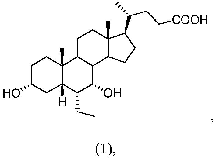
for use in a method of increasing liver mass in a subject with reduced liver function, the method comprising administering to the subject a therapeutically effective amount of the compound of formula (1).
Applications Claiming Priority (2)
| Application Number | Priority Date | Filing Date | Title |
|---|---|---|---|
| US201562221324P | 2015-09-21 | 2015-09-21 | |
| PCT/US2016/052886 WO2017053428A1 (en) | 2015-09-21 | 2016-09-21 | Methods of promoting hepatic regeneration |
Publications (3)
| Publication Number | Publication Date |
|---|---|
| EP3352768A1 EP3352768A1 (en) | 2018-08-01 |
| EP3352768A4 EP3352768A4 (en) | 2019-04-24 |
| EP3352768B1 true EP3352768B1 (en) | 2022-11-02 |
Family
ID=58387124
Family Applications (1)
| Application Number | Title | Priority Date | Filing Date |
|---|---|---|---|
| EP16849506.7A Active EP3352768B1 (en) | 2015-09-21 | 2016-09-21 | Methods of promoting hepatic regeneration |
Country Status (10)
| Country | Link |
|---|---|
| US (1) | US10548906B2 (en) |
| EP (1) | EP3352768B1 (en) |
| JP (1) | JP6824966B2 (en) |
| KR (1) | KR20180054770A (en) |
| CN (1) | CN108348543A (en) |
| AU (1) | AU2016326428A1 (en) |
| CA (1) | CA2998876A1 (en) |
| HK (1) | HK1254056A1 (en) |
| IL (1) | IL258114A (en) |
| WO (1) | WO2017053428A1 (en) |
Families Citing this family (7)
| Publication number | Priority date | Publication date | Assignee | Title |
|---|---|---|---|---|
| JP2018528230A (en) | 2015-09-24 | 2018-09-27 | インターセプト ファーマシューティカルズ, インコーポレイテッド | Methods and intermediates for the preparation of bile acid derivatives |
| EP3442946A4 (en) * | 2016-04-13 | 2019-12-04 | Intercept Pharmaceuticals, Inc. | Methods of treating cancer |
| WO2019020067A1 (en) | 2017-07-26 | 2019-01-31 | 正大天晴药业集团股份有限公司 | Method for preparing steroid derivative fxr agonist |
| GB201812382D0 (en) | 2018-07-30 | 2018-09-12 | Nzp Uk Ltd | Compounds |
| CN112057606B (en) * | 2020-09-14 | 2023-09-19 | 上海市儿童医院 | Use of FGF19 in a medicament for the treatment and/or prevention of sepsis-induced organ damage |
| CN113041242A (en) * | 2021-05-18 | 2021-06-29 | 上海交通大学医学院附属仁济医院 | Application of dexmedetomidine in promoting liver regeneration |
| CN116554252A (en) * | 2023-04-07 | 2023-08-08 | 华南理工大学 | High-crystallinity allocholic acid and application thereof in preparation of medicines for preventing and treating cholestatic liver diseases |
Citations (2)
| Publication number | Priority date | Publication date | Assignee | Title |
|---|---|---|---|---|
| WO2002072598A1 (en) | 2001-03-12 | 2002-09-19 | Roberto Pellicciari | Steroids as agonists for fxr |
| US20140371190A1 (en) | 2013-05-14 | 2014-12-18 | TES Pharma SrI. | Farnesoid X receptor modulators |
Family Cites Families (5)
| Publication number | Priority date | Publication date | Assignee | Title |
|---|---|---|---|---|
| ITMI20050912A1 (en) | 2005-05-19 | 2006-11-20 | Erregierre Spa | PROCESS OF PREPARATION OF ACIDS 3-A-YA (B) -DIDROSSI-6-A (B) -ALCHIL-5B-COLANICI |
| PL2040713T3 (en) * | 2006-06-27 | 2014-11-28 | Intercept Pharmaceuticals Inc | Bile acid derivatives as fxr ligands for the prevention or treatment of fxr-mediated deseases or conditions |
| SG10201607230SA (en) * | 2012-06-19 | 2016-10-28 | Intercept Pharmaceuticals Inc | Preparation, Uses And Solid Forms Of Obeticholic Acid |
| US20140206657A1 (en) | 2013-01-18 | 2014-07-24 | City Of Hope | Bile acid analog tgr5 agonists |
| EP3985118A1 (en) * | 2013-11-22 | 2022-04-20 | MiNA Therapeutics Limited | C/ebp alpha short activating rna compositions and methods of use |
-
2016
- 2016-09-21 KR KR1020187010888A patent/KR20180054770A/en unknown
- 2016-09-21 JP JP2018514849A patent/JP6824966B2/en active Active
- 2016-09-21 EP EP16849506.7A patent/EP3352768B1/en active Active
- 2016-09-21 WO PCT/US2016/052886 patent/WO2017053428A1/en active Application Filing
- 2016-09-21 CA CA2998876A patent/CA2998876A1/en active Pending
- 2016-09-21 AU AU2016326428A patent/AU2016326428A1/en not_active Abandoned
- 2016-09-21 US US15/760,769 patent/US10548906B2/en active Active
- 2016-09-21 CN CN201680062384.7A patent/CN108348543A/en active Pending
-
2018
- 2018-03-14 IL IL258114A patent/IL258114A/en unknown
- 2018-10-12 HK HK18113119.9A patent/HK1254056A1/en unknown
Patent Citations (3)
| Publication number | Priority date | Publication date | Assignee | Title |
|---|---|---|---|---|
| WO2002072598A1 (en) | 2001-03-12 | 2002-09-19 | Roberto Pellicciari | Steroids as agonists for fxr |
| EP1392714B1 (en) | 2001-03-12 | 2005-08-31 | Intercept Pharmaceuticals, Inc. | Steroids as agonists for fxr |
| US20140371190A1 (en) | 2013-05-14 | 2014-12-18 | TES Pharma SrI. | Farnesoid X receptor modulators |
Non-Patent Citations (33)
| Title |
|---|
| "Liver Regeneration : Basic Mechanisms , relevant Models and Clinical Applications", 1 January 2015, ELSEVIER, ISBN: 978-0-12-420128-6, article FAN MINGJIE, DING LILI, HUANG WENDONG: "Bile Acid Receptors and Liver Regeneration", pages: 125 - 135, XP093096263, DOI: 10.1016/B978-0-12-420128-6.00009-9 |
| AHMAD H ALI, ELIZABETH J CAREY, KEITH D LINDOR: "Recent advances in the development of farnesoid X receptor agonists", ANNALS OF TRANSLATIONAL MEDICINE, vol. 1, no. 5, 1 January 2015 (2015-01-01), pages 1 - 16, XP055522463, DOI: 10.3978/j.issn.2305-5839.2014.12.06 |
| ANONYMOUS: "Intercept Announces New FLINT Trial Data Showing OCA Treatment Increases Fibrosis Resolution and Cirrhosis Prevention in High-Risk NASH Patients OCA Treatment Resulted in Significant Improvements in Fibrosis Resolution in Addition to Previously Reported Benefits on Fibrosis Improvement, NASH Resolut", 23 April 2015 (2015-04-23), pages 1 - 3, XP093096246, Retrieved from the Internet <URL:https://ir.interceptpharma.com/static-files/5cd470fd-db66-44b0-a1eb-80f5beabc334> [retrieved on 20231030] |
| ANONYMOUS: "International Nonproprietary Names for Pharmaceutical Substances (INN)", WHO DRUG INFORMATION, vol. 23, no. 2, 1 January 2009 (2009-01-01), pages 129 - 192, XP055858023 |
| ANONYMOUS: "Obeticholic Acid (OCA) in Primary Sclerosing Cholangitis (PSC)", CLINICAL TRIALS, NCT02177136, 26 June 2014 (2014-06-26), pages 1 - 5, XP093098254 |
| ANONYMOUS: "Public summary of opinion on orphan designation - Obeticholic acid for the treatment of primary sclerosing cholangitis", 21 March 2014 (2014-03-21), pages 1 - 4, XP093091155, Retrieved from the Internet <URL:https://www.ema.europa.eu/en/documents/orphan-designation/eu/3/13/1228-public-summary-opinion-orphan-designation-obeticholic-acid-treatment-primary-sclerosing_en.pdf> [retrieved on 20231012] |
| ANONYMOUS: "Public summary of opinion on orphan designation 6alpha-ethyl-chenodeoxycholic acid for treatment of primary biliary cirrhosis ", 4 August 2010 (2010-08-04), pages 1 - 4, XP093091098, Retrieved from the Internet <URL:https://www.ema.europa.eu/en/documents/orphan-designation/eu/3/10/753-public-summary-opinion-orphan-designation-6alpha-ethyl-chenodeoxycholic-acid-treatment-primary_en.pdf> [retrieved on 20231012] |
| ANONYMOUS: "Study of INT-747 as Monotherapy in Patients With PBC", CLINICAL TRIALS, NCT00570765, 9 January 2012 (2012-01-09), pages 1 - 9, XP093098255 |
| ANONYMOUS: "The Farnesoid X Receptor (FXR) Ligand Obeticholic Acid in NASH Treatment Trial(FLINT) (FLINT)", CLINICAL TRIALS, NCT01265498, 27 July 2015 (2015-07-27), pages 1 - 46, XP093098259 |
| CHEN WEI-DONG, WANG YAN-DONG, MENG ZHIPENG, ZHANG LISHENG, HUANG WENDONG: "Nuclear bile acid receptor FXR in the hepatic regeneration", BIOCHIMICA ET BIOPHYSICA ACTA, vol. 1812, no. 8, 1 August 2011 (2011-08-01), NL , pages 888 - 892, XP093096274, ISSN: 0925-4439, DOI: 10.1016/j.bbadis.2010.12.006 |
| CHRISTOPHE CORPECHOT: "Primary biliary cirrhosis and bile acids", CLINICS AND RESEARCH IN HEPATOLOGY AND GASTROENTEROLOGY, vol. 36, 1 September 2017 (2017-09-01), pages S13 - S20, XP055723590, DOI: 10.1016/S2210-7401(12)70016-5 |
| CIENFUEGOS JAVIER A, ROTELLAR FERNANDO, BAIXAULI JORGE, MARTÍNEZ-REGUEIRA FERNANDO, PARDO FERNANDO, HERNÁNDEZ-LIZOÁIN JOSÉ LUIS: "Liver regeneration--the best kept secret. A model of tissue injury response", REVISTA ESPAÑOLA DE ENFERMEDADES DIGESTIVAS, vol. 106, no. 3, 1 March 2014 (2014-03-01), ES , pages 171 - 194, XP093091150, ISSN: 1130-0108 |
| FAN MINGJIE, WANG XICHUN, XU GANYU, YAN QINGFENG, HUANG WENDONG: "Bile acid signaling and liver regeneration", BIOCHIMICA ET BIOPHYSICA ACTA, vol. 1849, no. 2, 1 February 2015 (2015-02-01), AMSTERDAM, NL , pages 196 - 200, XP093091091, ISSN: 1874-9399, DOI: 10.1016/j.bbagrm.2014.05.021 |
| GADALETA RAFFAELLA M., VAN MIL SASKIA W.C., OLDENBURG BAS, SIERSEMA PETER D., KLOMP LEO W.J., VAN ERPECUM KAREL J.: "Bile acids and their nuclear receptor FXR: Relevance for hepatobiliary and gastrointestinal disease", BIOCHIMICA ET BIOPHYSICA ACTA (BBA) - MOLECULAR AND CELL BIOLOGY OF LIPIDS, vol. 1801, no. 7, 1 July 2010 (2010-07-01), AMSTERDAM, NL , pages 683 - 692, XP093091043, ISSN: 1388-1981, DOI: 10.1016/j.bbalip.2010.04.006 |
| HUANG XIONG-FEI, ZHAO WEI-YU, HUANG WEN-DONG: "FXR and liver carcinogenesis", ACTA PHARMACOLOGICA SINICA, vol. 36, no. 1, 1 January 2015 (2015-01-01), GB , pages 37 - 43, XP093098157, ISSN: 1671-4083, DOI: 10.1038/aps.2014.117 |
| JING FU, PEI SI, MINGYUE ZHENG, LILI CHEN, XU SHEN, YUN TANG, WEIHUA LI: "Discovery of new non-steroidal FXR ligands via a virtual screening workflow based on Phase shape and induced fit docking", BIOORGANIC & MEDICINAL CHEMISTRY LETTERS, vol. 22, no. 22, 1 November 2012 (2012-11-01), Amsterdam NL , pages 6848 - 6853, XP055368988, ISSN: 0960-894X, DOI: 10.1016/j.bmcl.2012.09.045 |
| KOHKICHI MORIMOTO , HIROSHI ITOH , MITSUHIRO WATANABE: "Developments in understanding bile acid metabolism", EXPERT REVIEW OF ENDOCRINOLOGY & METABOLISM, vol. 8, no. 1, 1 January 2013 (2013-01-01), pages 59 - 69, XP009549243, DOI: 10.1586/eem.12.75 |
| LI GUODONG, L. GUO GRACE: "Farnesoid X receptor, the bile acid sensing nuclear receptor, in liver regeneration", ACTA PHARMACEUTICA SINICA B, vol. 5, no. 2, 1 March 2015 (2015-03-01), pages 93 - 98, XP093091142, ISSN: 2211-3835, DOI: 10.1016/j.apsb.2015.01.005 |
| LINDER KEITH D: "Farnesoid X receptor agonists for primary biliary cirrhosis", CURRENT OPINION IN GASTROENTEROLOGY, vol. 27, no. 3, 1 May 2011 (2011-05-01), pages 285 - 288, XP093091126, DOI: 10.1097/MOG.Ob013e32834452c8 |
| MASON ANDREW L, LINDOR KEITH D, HIRSCHFIELD GIDEON M, GORDON STUART C, MOYO MARLYN J, KOWD KRIS V, PARES ALBERT, TROUNER MICHAEL, : "FARNESOID·X RECEPTOR AGONISTS: A NEW CLASS OF DRUGS FOR THE TREATMENT OF PBC? AN INTER· NATIONAL STUDY EVALUATING THE ADDITION OF OBETICHOLIC ACID (INT·747) TO URSODEOXYCHOLIC ACID ", HEMATOLOGY AASLD ABSTRACTS, no. 4, 1 January 2010 (2010-01-01), pages 257A - 257A, XP093096335 |
| MENG ZHIPENG, WANG YANDONG, WANG LIN, JIN WEN, LIU NIAN, PAN HAO, LIU LUCY, WAGMAN LAWRENCE, FORMAN BARRY M., HUANG WENDONG: "FXR Regulates Liver Repair after CCl4-Induced Toxic Injury", MOLECULAR ENDOCRINOLOGY, vol. 24, no. 5, 1 May 2010 (2010-05-01), US , pages 886 - 897, XP093091105, ISSN: 0888-8809, DOI: 10.1210/me.2009-0286 |
| NEUSCHWANDER-TETRI BRENT A; LOOMBA ROHIT; SANYAL ARUN J; LAVINE JOEL E; VAN NATTA MARK L; ABDELMALEK MANAL F; CHALASANI NAGA; DASA: "Farnesoid X nuclear receptor ligand obeticholic acid for non-cirrhotic, non-alcoholic steatohepatitis (FLINT): a multicentre, randomised, placebo-controlled trial", THE LANCET, vol. 385, no. 9972, 14 March 2015 (2015-03-14), AMSTERDAM, NL , pages 956 - 965, XP009510956, ISSN: 0140-6736, DOI: 10.1016/S0140-6736(14)61933-4 |
| PHILIPPE LEFEBVRE, BERTRAND CARIOU, FLEUR LIEN, FOLKERT KUIPERS, BART STAELS: "Role of Bile Acids and Bile Acid Receptors in Metabolic Regulation", PHYSIOLOGICAL REVIEWS, vol. 89, no. 1, 1 January 2009 (2009-01-01), US , pages 147 - 191, XP055489509, ISSN: 0031-9333, DOI: 10.1152/physrev.00010.2008 |
| ROBERTO PELLICCIARI, GABRIELE COSTANTINO, EMIDIO CAMAIONI, BAHMAN M. SADEGHPOUR, ANTONIO ENTRENA, TIMOTHY M. WILLSON, STEFANO FIOR: "Bile acid derivatives as ligands of the farnesoid X receptor. Synthesis, evaluation, and structure-activity relationship of a series of body and side chain modified analogues of chenodeoxycholic acid", JOURNAL OF MEDICINAL CHEMISTRY, vol. 47, no. 18, 1 January 2004 (2004-01-01), US , pages 4559 - 4569, XP002669041, ISSN: 0022-2623, DOI: 10.1021/JM049904B |
| ROBERTO PELLICCIARI, STEFANO FIORUCCI, EMIDIO CAMAIONI, CARLO CLERICI, GABRIELE COSTANTINO, PATRICK R. MALONEY, ANTONIO MORELLI, D: "6.alpha.-Ethyl-Chenodeoxycholic Acid (6-ECDCA), a Potent and Selective FXR Agonist Endowed with Anticholestatic Activity", JOURNAL OF MEDICINAL CHEMISTRY, vol. 45, no. 17, 20 July 2002 (2002-07-20), US , pages 3569 - 3572, XP002370635, ISSN: 0022-2623, DOI: 10.1021/jm025529g |
| TANG XIAOWEN, LILI DING, QIAOLING YANG, XIAOYUAN NIU, LI YANG, ZHENGTAO WANG: "Advances in Bile Acids-Mediated Liver Injury and Liver Regeneration", ADVANCES IN BIOCHEMISTRY, vol. 2, no. 6, 1 December 2014 (2014-12-01), pages 85 - 59, XP093096271, ISSN: 2329-0870, DOI: 10.11648/j.ab.20140206.11 |
| THOMAS D. BOYER, MICHAEL P. MANNS AND ARUN J. SANYAL: "Zakim and Boyer's Hepatology", 1 January 2012, SAUNDERS ELSEVIER , ISBN: 978-1-4377-0881-3, article POYNARD THIERRY, FRANCOISE IMBERT-BISMUT: "Laboratory Testing for Liver Disease", pages: 201 - 204, XP093098155, DOI: 10.1016/C2009-0-39323-X |
| VYAS SOUMIL; MARKAR SHERAZ; PARTELLI STEFANO; FOTHERINGHAM TIM; LOW DEBORAH; IMBER CHARLES; MALAGO MASSIMO; KOCHER HEMANT M.: "Portal Vein Embolization and Ligation for Extended Hepatectomy", INDIAN JOURNAL OF SURGICAL ONCOLOGY, vol. 5, no. 1, 6 January 2014 (2014-01-06), India , pages 30 - 42, XP035305140, ISSN: 0975-7651, DOI: 10.1007/s13193-013-0279-y |
| W. HUANG: "Nuclear Receptor-Dependent Bile Acid Signaling Is Required for Normal Liver Regeneration", SCIENCE, vol. 312, no. 5771, 14 April 2006 (2006-04-14), US , pages 233 - 236, XP055160374, ISSN: 0036-8075, DOI: 10.1126/science.1121435 |
| YAN-DONG WANG, WEI-DONG CHEN, DAVID D MOORE, WENDONG HUANG: "FXR: a metabolic regulator and cell protector", CELL RESEARCH, vol. 18, no. 11, 1 November 2008 (2008-11-01), pages 1087 - 1095, XP055086473, ISSN: 10010602, DOI: 10.1038/cr.2008.289 |
| YIMAM KIDIST K, BOWLUS CHRISTOPHER L: "Obeticholic acid for the treatment of primary biliary cirrhosis", EXPERT OPINION ON ORPHAN DRUGS, vol. 2, no. 12, 1 December 2014 (2014-12-01), pages 1351 - 1358, XP093091136, DOI: 10.1517/21678707.2014.964206 |
| ZHANG LISHENG, WANG YAN-DONG, CHEN WEI-DONG, WANG XICHUN, LOU GUIYU, LIU NIAN, LIN MIN, FORMAN BARRY M., HUANG WENDONG: "Promotion of liver regeneration/repair by farnesoid X receptor in both liver and intestine in mice", HEPATOLOGY, vol. 56, no. 6, 1 December 2012 (2012-12-01), US , pages 1 - 15, XP093091116, ISSN: 0270-9139, DOI: 10.1002/hep.25905 |
| ZHIPENG MENG; NIAN LIU; XIANGHUI FU; XIAOQIONG WANG; YAN-DONG WANG; WEI-DONG CHEN; LISHENG ZHANG; BARRY M. FORMAN; WENDONG HUANG;: "Insufficient bile acid signaling impairs liver repair inmice", JOURNAL OF HEPATOLOGY, vol. 55, no. 4, 18 December 2010 (2010-12-18), AMSTERDAM, NL , pages 885 - 895, XP028294646, ISSN: 0168-8278, DOI: 10.1016/j.jhep.2010.12.037 |
Also Published As
| Publication number | Publication date |
|---|---|
| JP6824966B2 (en) | 2021-02-03 |
| CN108348543A (en) | 2018-07-31 |
| KR20180054770A (en) | 2018-05-24 |
| IL258114A (en) | 2018-05-31 |
| CA2998876A1 (en) | 2017-03-30 |
| HK1254056A1 (en) | 2019-07-12 |
| US10548906B2 (en) | 2020-02-04 |
| JP2018527389A (en) | 2018-09-20 |
| EP3352768A1 (en) | 2018-08-01 |
| US20180256602A1 (en) | 2018-09-13 |
| EP3352768A4 (en) | 2019-04-24 |
| AU2016326428A1 (en) | 2018-04-12 |
| WO2017053428A1 (en) | 2017-03-30 |
Similar Documents
| Publication | Publication Date | Title |
|---|---|---|
| EP3352768B1 (en) | Methods of promoting hepatic regeneration | |
| Rautiainen et al. | Budesonide combined with UDCA to improve liver histology in primary biliary cirrhosis: a three‐year randomized trial | |
| EP3280421B1 (en) | Pharmaceutical compositions for combination therapy | |
| KR20150108946A (en) | Methods and compositions for treating cancer | |
| EP3426348A1 (en) | 3-desoxy derivative and pharmaceutical compositions thereof | |
| Huang et al. | Postponing the hypoglycemic response to partial hepatectomy delays mouse liver regeneration | |
| Tan et al. | Geniposide inhibits alpha-naphthylisothiocyanate-induced intrahepatic cholestasis: the downregulation of STAT3 and NF κ B signaling plays an important role | |
| EP4176878A1 (en) | Combination therapy for prostate cancer | |
| van Golen et al. | The pathophysiology of human obstructive cholestasis is mimicked in cholestatic Gold Syrian hamsters | |
| EP2392337A2 (en) | Use of 24-norUDCA | |
| Koelfat et al. | Bile salt and FGF19 signaling in the early phase of human liver regeneration | |
| TW200900080A (en) | Mineralcorticoid receptor antagonists for the treatment of endometriosis | |
| JP2018507880A (en) | How to adjust bone density | |
| US20230285416A1 (en) | Use of oxygenated cholesterol sulfates for treating at least one of insulin resistance, diabetes, and prediabetes | |
| WO2020243679A1 (en) | Anti-mullerian hormone receptor binding peptides | |
| US20040162274A1 (en) | Inhibition of steroid synthesis by use of progesterone or a progesterone receptor agonist | |
| EP1648439B1 (en) | Antiglucocorticoid therapy for the prevention of neurological damage in premature infants | |
| EP2160190B1 (en) | 5beta, 14beta-androstane derivatives useful for the treatment of diseases due to organ fibrosis | |
| van Mierlo et al. | obeticholic acid does not stimulate liver regeneration in hepatectomized mice | |
| EP1818052A1 (en) | Use of formoterol in the prophylactic and/or therapeutic treatment of muscle wasting and/or cachectic syndrome which are associated with catabolic conditions of certain diseases, such as cancer, aids, infections, diabetes and others | |
| Pravda et al. | Novel Combination Therapy Induced Histological Remission in Patients with Refractory | |
| Korolenko et al. | Drug-Induced Models of Cholestasis and Lysosomes | |
| Uriarte et al. | New insights into the regulation of bile acids synthesis during the early stages of liver regeneration: A human and experimental study | |
| WO2022082183A1 (en) | Compounds and methods for the treatment of inflammatory and metabolic diseases | |
| Cazzagon et al. | Metabolic syndrome induces a more rapid progression of fibrosis evaluated with transient elastography in early primary biliary cirrhosis: 334 |
Legal Events
| Date | Code | Title | Description |
|---|---|---|---|
| STAA | Information on the status of an ep patent application or granted ep patent |
Free format text: STATUS: THE INTERNATIONAL PUBLICATION HAS BEEN MADE |
|
| PUAI | Public reference made under article 153(3) epc to a published international application that has entered the european phase |
Free format text: ORIGINAL CODE: 0009012 |
|
| STAA | Information on the status of an ep patent application or granted ep patent |
Free format text: STATUS: REQUEST FOR EXAMINATION WAS MADE |
|
| 17P | Request for examination filed |
Effective date: 20180420 |
|
| AK | Designated contracting states |
Kind code of ref document: A1 Designated state(s): AL AT BE BG CH CY CZ DE DK EE ES FI FR GB GR HR HU IE IS IT LI LT LU LV MC MK MT NL NO PL PT RO RS SE SI SK SM TR |
|
| AX | Request for extension of the european patent |
Extension state: BA ME |
|
| RAV | Requested validation state of the european patent: fee paid |
Extension state: MD Effective date: 20180420 |
|
| A4 | Supplementary search report drawn up and despatched |
Effective date: 20190321 |
|
| RIC1 | Information provided on ipc code assigned before grant |
Ipc: A61K 31/58 20060101ALI20190315BHEP Ipc: A61K 31/56 20060101ALI20190315BHEP Ipc: A61K 31/79 20060101AFI20190315BHEP |
|
| STAA | Information on the status of an ep patent application or granted ep patent |
Free format text: STATUS: EXAMINATION IS IN PROGRESS |
|
| 17Q | First examination report despatched |
Effective date: 20200311 |
|
| STAA | Information on the status of an ep patent application or granted ep patent |
Free format text: STATUS: EXAMINATION IS IN PROGRESS |
|
| STAA | Information on the status of an ep patent application or granted ep patent |
Free format text: STATUS: EXAMINATION IS IN PROGRESS |
|
| REG | Reference to a national code |
Ref country code: DE Ref legal event code: R079 Ref document number: 602016076117 Country of ref document: DE Free format text: PREVIOUS MAIN CLASS: A61K0031790000 Ipc: A61K0031560000 |
|
| GRAP | Despatch of communication of intention to grant a patent |
Free format text: ORIGINAL CODE: EPIDOSNIGR1 |
|
| STAA | Information on the status of an ep patent application or granted ep patent |
Free format text: STATUS: GRANT OF PATENT IS INTENDED |
|
| RIC1 | Information provided on ipc code assigned before grant |
Ipc: A61P 1/16 20060101ALI20220324BHEP Ipc: A61K 31/575 20060101ALI20220324BHEP Ipc: A61K 31/56 20060101AFI20220324BHEP |
|
| RAP3 | Party data changed (applicant data changed or rights of an application transferred) |
Owner name: INTERCEPT PHARMACEUTICALS, INC. |
|
| INTG | Intention to grant announced |
Effective date: 20220420 |
|
| GRAS | Grant fee paid |
Free format text: ORIGINAL CODE: EPIDOSNIGR3 |
|
| GRAA | (expected) grant |
Free format text: ORIGINAL CODE: 0009210 |
|
| STAA | Information on the status of an ep patent application or granted ep patent |
Free format text: STATUS: THE PATENT HAS BEEN GRANTED |
|
| AK | Designated contracting states |
Kind code of ref document: B1 Designated state(s): AL AT BE BG CH CY CZ DE DK EE ES FI FR GB GR HR HU IE IS IT LI LT LU LV MC MK MT NL NO PL PT RO RS SE SI SK SM TR |
|
| REG | Reference to a national code |
Ref country code: GB Ref legal event code: FG4D |
|
| REG | Reference to a national code |
Ref country code: CH Ref legal event code: EP Ref country code: AT Ref legal event code: REF Ref document number: 1528252 Country of ref document: AT Kind code of ref document: T Effective date: 20221115 |
|
| REG | Reference to a national code |
Ref country code: DE Ref legal event code: R096 Ref document number: 602016076117 Country of ref document: DE |
|
| REG | Reference to a national code |
Ref country code: IE Ref legal event code: FG4D |
|
| REG | Reference to a national code |
Ref country code: NL Ref legal event code: FP |
|
| REG | Reference to a national code |
Ref country code: LT Ref legal event code: MG9D |
|
| REG | Reference to a national code |
Ref country code: AT Ref legal event code: MK05 Ref document number: 1528252 Country of ref document: AT Kind code of ref document: T Effective date: 20221102 |
|
| PG25 | Lapsed in a contracting state [announced via postgrant information from national office to epo] |
Ref country code: SE Free format text: LAPSE BECAUSE OF FAILURE TO SUBMIT A TRANSLATION OF THE DESCRIPTION OR TO PAY THE FEE WITHIN THE PRESCRIBED TIME-LIMIT Effective date: 20221102 Ref country code: PT Free format text: LAPSE BECAUSE OF FAILURE TO SUBMIT A TRANSLATION OF THE DESCRIPTION OR TO PAY THE FEE WITHIN THE PRESCRIBED TIME-LIMIT Effective date: 20230302 Ref country code: NO Free format text: LAPSE BECAUSE OF FAILURE TO SUBMIT A TRANSLATION OF THE DESCRIPTION OR TO PAY THE FEE WITHIN THE PRESCRIBED TIME-LIMIT Effective date: 20230202 Ref country code: LT Free format text: LAPSE BECAUSE OF FAILURE TO SUBMIT A TRANSLATION OF THE DESCRIPTION OR TO PAY THE FEE WITHIN THE PRESCRIBED TIME-LIMIT Effective date: 20221102 Ref country code: FI Free format text: LAPSE BECAUSE OF FAILURE TO SUBMIT A TRANSLATION OF THE DESCRIPTION OR TO PAY THE FEE WITHIN THE PRESCRIBED TIME-LIMIT Effective date: 20221102 Ref country code: ES Free format text: LAPSE BECAUSE OF FAILURE TO SUBMIT A TRANSLATION OF THE DESCRIPTION OR TO PAY THE FEE WITHIN THE PRESCRIBED TIME-LIMIT Effective date: 20221102 Ref country code: AT Free format text: LAPSE BECAUSE OF FAILURE TO SUBMIT A TRANSLATION OF THE DESCRIPTION OR TO PAY THE FEE WITHIN THE PRESCRIBED TIME-LIMIT Effective date: 20221102 |
|
| PG25 | Lapsed in a contracting state [announced via postgrant information from national office to epo] |
Ref country code: RS Free format text: LAPSE BECAUSE OF FAILURE TO SUBMIT A TRANSLATION OF THE DESCRIPTION OR TO PAY THE FEE WITHIN THE PRESCRIBED TIME-LIMIT Effective date: 20221102 Ref country code: PL Free format text: LAPSE BECAUSE OF FAILURE TO SUBMIT A TRANSLATION OF THE DESCRIPTION OR TO PAY THE FEE WITHIN THE PRESCRIBED TIME-LIMIT Effective date: 20221102 Ref country code: LV Free format text: LAPSE BECAUSE OF FAILURE TO SUBMIT A TRANSLATION OF THE DESCRIPTION OR TO PAY THE FEE WITHIN THE PRESCRIBED TIME-LIMIT Effective date: 20221102 Ref country code: IS Free format text: LAPSE BECAUSE OF FAILURE TO SUBMIT A TRANSLATION OF THE DESCRIPTION OR TO PAY THE FEE WITHIN THE PRESCRIBED TIME-LIMIT Effective date: 20230302 Ref country code: HR Free format text: LAPSE BECAUSE OF FAILURE TO SUBMIT A TRANSLATION OF THE DESCRIPTION OR TO PAY THE FEE WITHIN THE PRESCRIBED TIME-LIMIT Effective date: 20221102 Ref country code: GR Free format text: LAPSE BECAUSE OF FAILURE TO SUBMIT A TRANSLATION OF THE DESCRIPTION OR TO PAY THE FEE WITHIN THE PRESCRIBED TIME-LIMIT Effective date: 20230203 |
|
| P01 | Opt-out of the competence of the unified patent court (upc) registered |
Effective date: 20230525 |
|
| REG | Reference to a national code |
Ref country code: DE Ref legal event code: R026 Ref document number: 602016076117 Country of ref document: DE |
|
| PLBI | Opposition filed |
Free format text: ORIGINAL CODE: 0009260 |
|
| PG25 | Lapsed in a contracting state [announced via postgrant information from national office to epo] |
Ref country code: SM Free format text: LAPSE BECAUSE OF FAILURE TO SUBMIT A TRANSLATION OF THE DESCRIPTION OR TO PAY THE FEE WITHIN THE PRESCRIBED TIME-LIMIT Effective date: 20221102 Ref country code: RO Free format text: LAPSE BECAUSE OF FAILURE TO SUBMIT A TRANSLATION OF THE DESCRIPTION OR TO PAY THE FEE WITHIN THE PRESCRIBED TIME-LIMIT Effective date: 20221102 Ref country code: EE Free format text: LAPSE BECAUSE OF FAILURE TO SUBMIT A TRANSLATION OF THE DESCRIPTION OR TO PAY THE FEE WITHIN THE PRESCRIBED TIME-LIMIT Effective date: 20221102 Ref country code: DK Free format text: LAPSE BECAUSE OF FAILURE TO SUBMIT A TRANSLATION OF THE DESCRIPTION OR TO PAY THE FEE WITHIN THE PRESCRIBED TIME-LIMIT Effective date: 20221102 Ref country code: CZ Free format text: LAPSE BECAUSE OF FAILURE TO SUBMIT A TRANSLATION OF THE DESCRIPTION OR TO PAY THE FEE WITHIN THE PRESCRIBED TIME-LIMIT Effective date: 20221102 |
|
| PLAB | Opposition data, opponent's data or that of the opponent's representative modified |
Free format text: ORIGINAL CODE: 0009299OPPO |
|
| PLBI | Opposition filed |
Free format text: ORIGINAL CODE: 0009260 |
|
| PLAX | Notice of opposition and request to file observation + time limit sent |
Free format text: ORIGINAL CODE: EPIDOSNOBS2 |
|
| 26 | Opposition filed |
Opponent name: TEVA PHARMACEUTICAL INDUSTRIES LTD. Effective date: 20230725 Opponent name: HAMM&WITTKOPP PATENTANWAELTE PARTMBB Effective date: 20230727 |
|
| PG25 | Lapsed in a contracting state [announced via postgrant information from national office to epo] |
Ref country code: SK Free format text: LAPSE BECAUSE OF FAILURE TO SUBMIT A TRANSLATION OF THE DESCRIPTION OR TO PAY THE FEE WITHIN THE PRESCRIBED TIME-LIMIT Effective date: 20221102 Ref country code: AL Free format text: LAPSE BECAUSE OF FAILURE TO SUBMIT A TRANSLATION OF THE DESCRIPTION OR TO PAY THE FEE WITHIN THE PRESCRIBED TIME-LIMIT Effective date: 20221102 |
|
| 26 | Opposition filed |
Opponent name: SANDOZ AG Effective date: 20230801 |
|
| R26 | Opposition filed (corrected) |
Opponent name: HAMM&WITTKOPP PATENTANWAELTE PARTMBB Effective date: 20230727 |
|
| PGFP | Annual fee paid to national office [announced via postgrant information from national office to epo] |
Ref country code: NL Payment date: 20230926 Year of fee payment: 8 Ref country code: GB Payment date: 20230927 Year of fee payment: 8 |
|
| VS25 | Lapsed in a validation state [announced via postgrant information from nat. office to epo] |
Ref country code: MD Free format text: LAPSE BECAUSE OF FAILURE TO SUBMIT A TRANSLATION OF THE DESCRIPTION OR TO PAY THE FEE WITHIN THE PRESCRIBED TIME-LIMIT Effective date: 20221102 |
|
| PLAF | Information modified related to communication of a notice of opposition and request to file observations + time limit |
Free format text: ORIGINAL CODE: EPIDOSCOBS2 |
|
| PG25 | Lapsed in a contracting state [announced via postgrant information from national office to epo] |
Ref country code: SI Free format text: LAPSE BECAUSE OF FAILURE TO SUBMIT A TRANSLATION OF THE DESCRIPTION OR TO PAY THE FEE WITHIN THE PRESCRIBED TIME-LIMIT Effective date: 20221102 |
|
| PGFP | Annual fee paid to national office [announced via postgrant information from national office to epo] |
Ref country code: FR Payment date: 20230925 Year of fee payment: 8 Ref country code: DE Payment date: 20230927 Year of fee payment: 8 Ref country code: BE Payment date: 20230927 Year of fee payment: 8 |
|
| PLAB | Opposition data, opponent's data or that of the opponent's representative modified |
Free format text: ORIGINAL CODE: 0009299OPPO |
|
| R26 | Opposition filed (corrected) |
Opponent name: SANDOZ AG Effective date: 20230801 |
|
| PLBB | Reply of patent proprietor to notice(s) of opposition received |
Free format text: ORIGINAL CODE: EPIDOSNOBS3 |
|
| RDAF | Communication despatched that patent is revoked |
Free format text: ORIGINAL CODE: EPIDOSNREV1 |