EP3348575A1 - Jaml-spezifische bindungsmittel, antikörper und verwendungen davon - Google Patents
Jaml-spezifische bindungsmittel, antikörper und verwendungen davon Download PDFInfo
- Publication number
- EP3348575A1 EP3348575A1 EP18157016.9A EP18157016A EP3348575A1 EP 3348575 A1 EP3348575 A1 EP 3348575A1 EP 18157016 A EP18157016 A EP 18157016A EP 3348575 A1 EP3348575 A1 EP 3348575A1
- Authority
- EP
- European Patent Office
- Prior art keywords
- jaml
- antibody
- cells
- seq
- antibodies
- Prior art date
- Legal status (The legal status is an assumption and is not a legal conclusion. Google has not performed a legal analysis and makes no representation as to the accuracy of the status listed.)
- Withdrawn
Links
Images


Classifications
-
- C—CHEMISTRY; METALLURGY
- C07—ORGANIC CHEMISTRY
- C07K—PEPTIDES
- C07K16/00—Immunoglobulins [IGs], e.g. monoclonal or polyclonal antibodies
- C07K16/42—Immunoglobulins [IGs], e.g. monoclonal or polyclonal antibodies against immunoglobulins
-
- A—HUMAN NECESSITIES
- A61—MEDICAL OR VETERINARY SCIENCE; HYGIENE
- A61P—SPECIFIC THERAPEUTIC ACTIVITY OF CHEMICAL COMPOUNDS OR MEDICINAL PREPARATIONS
- A61P11/00—Drugs for disorders of the respiratory system
-
- A—HUMAN NECESSITIES
- A61—MEDICAL OR VETERINARY SCIENCE; HYGIENE
- A61P—SPECIFIC THERAPEUTIC ACTIVITY OF CHEMICAL COMPOUNDS OR MEDICINAL PREPARATIONS
- A61P31/00—Antiinfectives, i.e. antibiotics, antiseptics, chemotherapeutics
-
- A—HUMAN NECESSITIES
- A61—MEDICAL OR VETERINARY SCIENCE; HYGIENE
- A61P—SPECIFIC THERAPEUTIC ACTIVITY OF CHEMICAL COMPOUNDS OR MEDICINAL PREPARATIONS
- A61P35/00—Antineoplastic agents
-
- A—HUMAN NECESSITIES
- A61—MEDICAL OR VETERINARY SCIENCE; HYGIENE
- A61P—SPECIFIC THERAPEUTIC ACTIVITY OF CHEMICAL COMPOUNDS OR MEDICINAL PREPARATIONS
- A61P9/00—Drugs for disorders of the cardiovascular system
-
- C—CHEMISTRY; METALLURGY
- C07—ORGANIC CHEMISTRY
- C07K—PEPTIDES
- C07K16/00—Immunoglobulins [IGs], e.g. monoclonal or polyclonal antibodies
- C07K16/18—Immunoglobulins [IGs], e.g. monoclonal or polyclonal antibodies against material from animals or humans
- C07K16/28—Immunoglobulins [IGs], e.g. monoclonal or polyclonal antibodies against material from animals or humans against receptors, cell surface antigens or cell surface determinants
- C07K16/2803—Immunoglobulins [IGs], e.g. monoclonal or polyclonal antibodies against material from animals or humans against receptors, cell surface antigens or cell surface determinants against the immunoglobulin superfamily
-
- A—HUMAN NECESSITIES
- A61—MEDICAL OR VETERINARY SCIENCE; HYGIENE
- A61K—PREPARATIONS FOR MEDICAL, DENTAL OR TOILETRY PURPOSES
- A61K39/00—Medicinal preparations containing antigens or antibodies
- A61K2039/505—Medicinal preparations containing antigens or antibodies comprising antibodies
-
- C—CHEMISTRY; METALLURGY
- C07—ORGANIC CHEMISTRY
- C07K—PEPTIDES
- C07K2317/00—Immunoglobulins specific features
- C07K2317/30—Immunoglobulins specific features characterized by aspects of specificity or valency
- C07K2317/33—Crossreactivity, e.g. for species or epitope, or lack of said crossreactivity
-
- C—CHEMISTRY; METALLURGY
- C07—ORGANIC CHEMISTRY
- C07K—PEPTIDES
- C07K2317/00—Immunoglobulins specific features
- C07K2317/70—Immunoglobulins specific features characterized by effect upon binding to a cell or to an antigen
- C07K2317/75—Agonist effect on antigen
-
- C—CHEMISTRY; METALLURGY
- C07—ORGANIC CHEMISTRY
- C07K—PEPTIDES
- C07K2317/00—Immunoglobulins specific features
- C07K2317/70—Immunoglobulins specific features characterized by effect upon binding to a cell or to an antigen
- C07K2317/76—Antagonist effect on antigen, e.g. neutralization or inhibition of binding
Definitions
- This disclosure relates to agents that specifically bind JAM-like protein (JAML).
- the binding agents such as antibodies, bind the extracellular domain of JAML making them particularly useful in a variety of settings such as diagnostic screening, bioassays, and therapeutic intervention for diseases or conditions that are associated with JAML and/or CAR such as inflammation, cancer, scarring, wound healing, respiratory diseases, and other disorders related to epithelial inflammation.
- PMNs Polymorphonuclear neutrophils
- PMNs encountering proinflammatory stimuli undergo morphological changes as well as changes in the expression of surface adhesive receptors.
- ⁇ 2-integrin CD11b/CD18 which is important for the initial PMN adhesion to both endothelium and epithelium, is upregulated upon PMN activation.
- expression of L-selectin another adhesive receptor that mediates initial PMN rolling in blood vessels, is completely lost upon PMN activation and migration.
- Epithelial and endothelial proteins expressed at intercellular junctions and members of the immunoglobulin superfamily (IgSf) members have been implicated directly or indirectly in regulating PMN trafficking and a variety of epithelial/endothelial cell functions. Function of these proteins is, in part, mediated through homo- and heterotypic interactions with related family members and integrins.
- JAM-A for example is abundantly expressed at the epithelial apical junctional complexes (AJC) as well as on PMNs.
- PMNs from JAM-A deficient mice (JAMA -/-) exhibit impaired migration.
- Another related molecule, JAML is not present in epithelial or endothelial cells.
- JAML has been shown to bind specifically to the cosackie adenovirus receptor (CAR), which is another member of the Ig superfamily. CAR is expressed at tight junctions of epithelial cells and to some degree on subtypes of endothelial cells, and localizes to the tight junctions (TJ). Both JAML and CAR are type I glycoproteins composed of two extracellular immunoglobulin domains, a single transmembrane helix, and a cytoplasmic domain. CAR has been shown to form homodimers in cis via the membrane distal D1 domain. CAR has been implicated in regulating epithelial permeability and cell adhesion to extracellular matrix by controlling the localization of cell integrins.
- CAR cosackie adenovirus receptor
- JAML plays a role in regulating monocyte transendothelial migration and neutrophil transepithelial migration by binding to the endothelial CAR and other tight junction-associated adhesive molecules. See Zen et al., Mol. Biol. Cell, 2005, 16(6):2694-2703 , Ya-Lan et al., Arteriosclerosis, Thrombosis, and Vascular Biology, 2009, 29: 75-83 , and Witherden et al., Science, 2010, 329 (5996): 1205-1210 . CAR and JAML are cell signaling receptors of the immune system with implications for asthma, cancer, and chronic non-healing wounds. See Verdino et al., Science, 2010, 1210-1211 .
- Monoclonal anti-mouse JAML antibody (clone 4E10) may be purchased from eBiosciences. See also Luissint et al., J Cell Biol, 2008, 183:1159-1173 ; U.S. Patent No 8,017,344 ; and U.S. Published Application No. 2008/0248502 .
- Certain embodiments of the disclosure relate to targeted binding agents that specifically bind to JAM-like protein and therein inhibit epithelial damage. Although it is not intended that embodiments of the disclosure be limited by any particular mechanism, it is believed that a possible mechanism by which this can be achieved may include, but are not limited to, either inhibition of binding of JAML to the CAR, inhibition of JAML induced CAR signaling, or increased clearance of JAML from the body of a subject, therein reducing the effective concentration of soluble JAML.
- the specific binding agent is a fully human antibody or chimera that specifically binds to human JAML, such as the JAML D1, and prevents JAML binding to CAR.
- Certain embodiments of the disclosure contemplate antibodies comprising human IgG type, e.g., IgG1, IgG2, IgG3, and IgG4.
- the disclosure relates to binding agents such as antibodies with an epitope to the extracellular domain of human JAML, such as D1.
- the antibody may be a monoclonal antibody that does not substantially bind GBP, JAMA, JAMC, CLMP, EVA, SIRPA, and CAR.
- the antibody may substantially inhibit binding of CAR to JAML.
- the antibody may slow PMN transmigration across epithelial cells.
- the antibody may be a human antibody or human chimera.
- the binding agents are isolated antibodies such as DW100 expressed by the hybridoma having ATCC Deposit No. PTA-13051.
- the binding agent is an antigen binding fragment wherein the antigen-binding fragment retains the binding specificity of the monoclonal antibody expressed by the cell line for DW100 having ATCC No PTA-13051 .
- the disclosure relates to a host cell line for DW100 having ATCC No. PTA-13051.
- the binding agents comprising a polypeptide of a more than 5, 6, 7, 8, 9, 10, 11, 12, 13, 14, 15, 16, 17, 18, 19, or 20 continuous amino acids within DW100, DW55, and DW216, e.g., having at least 8 or 9 is contemplated.
- Contemplated continuous sequences are typically within the variable regions of the light and heavy chains of the antibodies, e.g., CDR regions.
- a binding agent substantially prevents JAML from binding CAR.
- the antibody does not substantially bind to JAMA or JAMC.
- the antibody binds the D1 domain of JAML.
- the antibody is a monoclonal antibody.
- the disclosure relates to a JAML binding agent comprising a sequence selected from SEQ ID NOs: 1, 2, 3, 4, 5, 6, 7, 8, 9, 10, 11, 12, 13, 14, 15, 16, 17, and 18 or fragment thereof and a human immunoglobulin provided the fragment is at least 4, 5, 6, 7, 8, 9, or 10 more continuous amino acids.
- the disclosure relates to a binding agent such as an antibody comprising a heavy chain and a light chain, wherein said heavy chain comprises a heavy chain variable region selected from the group consisting of: H1 of DW100 (SEQ ID NO:19)
- the disclosure also provides a specific binding agent comprising at least one, two, or three peptides selected from the group consisting of: (SEQ ID NO:1); (SEQ ID NO:2); (SEQ ID NO:3); (SEQ ID NO:4); (SEQ ID NO:5); (SEQ ID NO:6); (SEQ ID NO:7); (SEQ ID NO:8); (SEQ ID NO:9); (SEQ ID NO:10); (SEQ ID NO:11); (SEQ ID NO:12); (SEQ ID NO:13); (SEQ ID NO:14); (SEQ ID NO: 15); (SEQ ID NO:16); (SEQ ID NO:17) ; and (SEQ ID NO:18).
- the binding agent may contain one, two, or three mutations or conservative substitutions in any of SEQ ID NO:1-18.
- the disclosure also provides a specific binding agent comprising; (SEQ ID NO:19); (SEQ ID NO:20); (SEQ ID NO:21); (SEQ ID NO:22); (SEQ ID NO:23); (SEQ ID NO:24) or those with 60%, 65%, 70%, 75%, 80%, 83%, 85%, 87%, 90%, 91%, 92%, 93%, 94%, 95%, 96%, 97%, 98% identity with SEQ ID NO: 19-24.
- SEQ ID NOs: 19-24 comprises 1 or 2 mutations or 3 or 4 mutation.
- SEQ ID NOs: 19-24 comprises 1 or 2 amino acid insertions or 3 or 4 amino acid insertions. The identity may be determined over a 30, 40, 50, 60, or 70 amino acid sequence window within or from the N or C terminal ends of SEQ ID NO: 19-24.
- the disclosure also provides a specific binding agent comprising; (SEQ ID NO:79); (SEQ ID NO:80); (SEQ ID NO:81); (SEQ ID NO:82) (SEQ ID NO:85) or those with 60%, 65%, 70%, 75%, 80%, 83%, 85%, 87%, 90%, 91%, 92%, 93%, 94%, 95%, 96%, 97%, 98% identity with SEQ ID NO:79-82, 85.
- SEQ ID NOs:79-82, 85 comprise 1 or 2 mutations or 3 or 4 mutation.
- SEQ ID NOs:79-82, 85 comprise 1 or 2 amino acid insertions or 3 or 4 amino acid insertions.
- the identity may be determined over a 30, 40, 50, 60, 70, 80, 90, 100, or 150, or 200 amino acid sequence window within or from the N or C terminal ends of SEQ ID NO:19-24.
- the disclosure is directed to an isolated antibody comprising a heavy chain and light chain, the heavy chain comprising a heavy chain variable domain and the light chain comprising a light chain variable domain, wherein the heavy chain variable domain comprises 1, 2, or 3 heavy chain CDRs selected from the group of H CDRs consisting of SEQ ID NOs:1, 2, 3, 4, 5, 6, 7, 8, and 9, or fragment thereof and wherein the antibody specifically binds to at least one of JAML and/or JAML D1.
- the disclosure is directed to an isolated antibody which comprises a light chain and a heavy chain, wherein the light chain comprises a light chain variable domain and the heavy chain comprises a heavy chain variable domain, wherein the light chain variable domain comprises 1, 2, or 3, light chain CDRs selected from the group of L CDRs consisting of SEQ ID NOs:10, 11, 12, 13, 14, 15, 16, 17, and 18, or fragment thereof and wherein the antibody specifically binds to at least one of JAML and/or JAML D1.
- the disclosure is an isolated antibody which comprises a heavy chain and a light chain, wherein the heavy chain comprises a heavy chain variable domain and the light chain comprises a light chain variable domain, wherein the heavy chain comprises 3 heavy chain (HC) CDRs and said light chain variable domain comprises 3 light chain (LC) CDRs, wherein the sequences of said HC and LC CDRs of the antibody are selected from the group consisting of:
- the specific binding agent can be, for example, an antibody, such as a polyclonal, monoclonal, chimeric, humanized, or a fully human antibody.
- the antibody may also be a single chain antibody with one or two heavy and/or light chains disclosed herein conjugated to a human immunoglobulin.
- Other examples of specific binding agents include peptibodies, avimers, other forms of peptide molecules (such as Fc-fusion molecules and Ab-fusion molecules) that contain peptide sequences which recognize and bind to a JAML or JAML D1.
- the disclosure relates to isolated binding agents that bind to JAML or JAML D1 comprising the sequence IHPGSGGTVYN (SEQ ID NO:25) further optionally comprising the sequence YYGSSYN (SEQ ID NO:26) further optionally comprising the sequence DYE (SEQ ID NO:27).
- the disclosure relates to isolated binding agents that bind to JAML or JAML D1 comprising the sequence HPGSGGTVYN (SEQ ID NO:28) further optionally comprising the sequence YGS (SEQ ID NO:29) further optionally comprising the sequence DYE (SEQ ID NO:27).
- the disclosure relates to isolated binding agents that bind to JAML or JAML D1 comprising the sequence IHPGSGGTVY (SEQ ID NO:30) further optionally comprising the sequence YGSSYN (SEQ ID NO:31) further optionally comprising the sequence DYE (SEQ ID NO:27).
- the disclosure relates to isolated binding agents that bind to JAML or JAML D1 comprising the sequence HPGSGGTVY (SEQ ID NO:32) further optionally comprising the sequence YGSSY (SEQ ID NO:33) further optionally comprising the sequence DYE (SEQ ID NO:27).
- the disclosure relates to isolated binding agents that bind to JAML or JAML D1 comprising the sequence HPGSGGTV (SEQ ID NO:34) further optionally comprising the sequence GSSYN (SEQ ID NO:35) further optionally comprising the sequence DYE (SEQ ID NO:27).
- the disclosure relates to isolated binding agents that bind to JAML or JAML D1 comprising the sequence HPGSGGT (SEQ ID NO:36) further optionally comprising the sequence GSSYN (SEQ ID NO:35) further optionally comprising the sequence DYE (SEQ ID NO:27).
- the disclosure relates to isolated binding agents that bind to JAML or JAML D1 comprising the sequence PGSGGT (SEQ ID NO:37) further optionally comprising the sequence GSSYN (SEQ ID NO:35) further optionally comprising the sequence DYE (SEQ ID NO:27).
- the disclosure relates to isolated binding agents that bind to JAML or JAML D1 comprising the sequence RASENIYYSLA (SEQ ID NO:38) further optionally comprising the sequence NANSLEDGVPS (SEQ ID NO:39).
- the disclosure relates to isolated binding agents that bind to JAML or JAML D1 comprising the sequence ASENIYYSLA (SEQ ID NO: 40) further optionally comprising the sequence ANSLEDGVPS (SEQ ID NO:41).
- the disclosure relates to isolated binding agents that bind to JAML or JAML D1 comprising the sequence ASENIYYSL (SEQ ID NO:43) further optionally comprising the sequence ANSLEDGVP (SEQ ID NO:44).
- the disclosure relates to isolated binding agents that bind to JAML or JAML D1 comprising the sequence SENIYYSL (SEQ ID NO:45) further optionally comprising the sequence NSLEDGVP (SEQ ID NO:46).
- the disclosure relates to isolated binding agents that bind to JAML or JAML D1 comprising the sequence SENIYYS (SEQ ID NO:47) further optionally comprising the sequence NSLEDGV (SEQ ID NO:48).
- the disclosure relates to isolated binding agents that bind to JAML or JAML D1 comprising the sequence ENIYYS (SEQ ID NO:49) further optionally comprising the sequence SLEDGV (SEQ ID NO:50).
- the disclosure relates to isolated binding agents that bind to JAML or JAML D1 comprising the sequence IGIIHPG (SEQ ID NO:51) further optionally comprising the sequence YYGSSYN (SEQ ID NO:26).
- the disclosure relates to isolated binding agents that bind to JAML or JAML D1 comprising the sequence IGIIHP (SEQ ID NO:52) further optionally comprising the sequence YYGSSYN (SEQ ID NO:26).
- the disclosure relates to isolated binding agents that bind to JAML or JAML D1 comprising the sequence GIIHPG (SEQ ID NO:53) further optionally comprising the sequence YYGSSYN (SEQ ID NO:26).
- the disclosure relates to isolated binding agents that bind to JAML or JAML D1 comprising the sequence GIIHP (SEQ ID NO:54) further optionally comprising the sequence YYGSSYN (SEQ ID NO:26).
- the disclosure relates to isolated binding agents that bind to JAML or JAML D1 comprising the sequence PGSGGTVYNH (SEQ ID NO:55) further optionally comprising the sequence YYGSSYN (SEQ ID NO:26).
- the disclosure relates to isolated binding agents that bind to JAML or JAML D1 comprising the sequence GSGGTVYNH (SEQ ID NO:56) further optionally comprising the sequence YYGSSYN (SEQ ID NO:26).
- the disclosure relates to isolated binding agents that bind to JAML or JAML D1 comprising the sequence SGGTVYNH (SEQ ID NO:57) further optionally comprising the sequence YYGSSYN (SEQ ID NO:26).
- the disclosure relates to isolated binding agents that bind to JAML or JAML D1 comprising the sequence GGTVYNH (SEQ ID NO:58) further optionally comprising the sequence YGS (SEQ ID NO:29).
- the disclosure relates to isolated binding agents that bind to JAML or JAML D1 comprising the sequence IGIIHPG (SEQ ID NO:51) further optionally comprising the sequence YGS (SEQ ID NO:29).
- the disclosure relates to isolated binding agents that bind to JAML or JAML D1 comprising the sequence IGIIHP (SEQ ID NO:52) further optionally comprising the sequence YGS (SEQ ID NO:29).
- the disclosure relates to isolated binding agents that bind to JAML or JAML D1 comprising the sequence GIIHPG (SEQ ID NO:53) further optionally comprising the sequence YGS (SEQ ID NO:29).
- the disclosure relates to isolated binding agents that bind to JAML or JAML D1 comprising the sequence GIIHP (SEQ ID NO:54) further optionally comprising the sequence YGS (SEQ ID NO:29).
- the disclosure relates to isolated binding agents that bind to JAML or JAML D1 comprising the sequence PGSGGTVYNH (SEQ ID NO:55) further optionally comprising the sequence YGS (SEQ ID NO:29).
- the disclosure relates to isolated binding agents that bind to JAML or JAML D1 comprising the sequence GSGGTVYNH (SEQ ID NO:56) further optionally comprising the sequence YGS (SEQ ID NO:29).
- the disclosure relates to isolated binding agents that bind to JAML or JAML D1 comprising the sequence SGGTVYNH (SEQ ID NO:57) further optionally comprising the sequence YGS (SEQ ID NO:29).
- the disclosure relates to isolated binding agents that bind to JAML or JAML D1 comprising the sequence GGTVYNH (SEQ ID NO:58) further optionally comprising the sequence YGS (SEQ ID NO:29).
- the disclosure relates to isolated binding agents that bind to JAML or JAML D1 comprising the sequence RASENIYY (SEQ ID NO:59) further optionally comprising the sequence NANSLEDGVP (SEQ ID NO:60).
- the disclosure relates to isolated binding agents that bind to JAML or JAML D1 comprising the sequence RASENIY (SEQ ID NO:61) further optionally comprising the sequence NANSLEDGVP (SEQ ID NO:60).
- the disclosure relates to isolated binding agents that bind to JAML or JAML D1 comprising the sequence RASENIY (SEQ ID NO:62) further optionally comprising the sequence NANSLEDGVP (SEQ ID NO:60).
- the disclosure relates to isolated binding agents that bind to JAML or JAML D1 comprising the sequence RASENI (SEQ ID NO:63) further optionally comprising the sequence NANSLEDGVP (SEQ ID NO:60).
- the disclosure relates to isolated binding agents that bind to JAML or JAML D1 comprising the sequence RASEN (SEQ ID NO:64) further optionally comprising the sequence NANSLEDGVP (SEQ ID NO:60).
- the disclosure relates to isolated binding agents that bind to JAML or JAML D1 comprising the sequence RASENIY (SEQ ID NO:62) further optionally comprising the sequence ANSLEDGVP (SEQ ID NO:65).
- the disclosure relates to isolated binding agents that bind to JAML or JAML D1 comprising the sequence RASENIY (SEQ ID NO:62) further optionally comprising the sequence NSLEDGVP (SEQ ID NO:66).
- a specific embodiment of the disclosure relates to peptibodies that bind JAML or JAML D1 such as a HI, H2, H3, and/or L1, L2, L3 alone or in combination that can be fused with human Fc Ig.
- Other embodiments of the disclosure include the peptide portion of these fusions as well as similar JAML or JAML D1 binding peptides that can be made by addition, deletion, and/or insertion of amino acids to and from the peptide. Similar additions, deletions, or insertions can be made to the Fc portion of the peptibody.
- the disclosure relates to pharmaceutical composition
- a pharmaceutical composition comprising binding agents described herein such as antibodies and a pharmaceutically acceptable excipient, dilutant, or carrier.
- a medicinal product may be used in methods of treating or preventing an inflammatory disease, skin inflammatory diseases, eye inflammatory diseases, autoimmune disease, cancer, viral infection, or scaring by administering an effective amount to a subject in need thereof.
- the inflammatory disease may be atherosclerosis, and ischemic heart disease, acne vulgaris, asthma, bronchitis, chronic prostatitis, hypersensitivities, inflammatory bowel diseases, pelvic inflammatory disease, reperfusion injury, or transplant rejection of a kidney, liver, or pancreas.
- the autoimmune disease may be rheumatoid arthritis, amyotrophic lateral sclerosis, Crohn's disease, type 1 diabetes, psoriasis, sarcoidosis, interstitial cystitis, glomerulonephritis, vasculitis, and ulcerative colitis.
- the subject may be in need thereof because the subject is diagnosed with, at risk of, or exhibiting symptoms of an inflammatory disease, autoimmune disease, respiratory disease, heart disease, myocardial infarction, viral infection, or cancer.
- the disclosure contemplates modulating barrier functions and permeability such as in drug delivery and vaccines.
- the disclosure contemplates administering effective amounts of binding agents and antibodies disclosed herein to a subj ect in need thereof in combination with therapeutic agents and vaccines.
- binding agents and antibodies disclosed herein may be used to increase permeability by enhancing barrier when soluble JAML is present by binding to JAML and allowing CAR to seal the epithelium. CAR is in the heart and JAML released by PMN in a heart attack may compromise cardiomyocyte function by binding to CAR.
- the disclosure relates to administering a binding agent or antibody disclosed herein to a subject at risk of, diagnosed with, or exhibiting symptoms or a heart attack or cardiovascular disease to promote cardiac healing.
- the disclosure contemplates uses of binding agents and antibodies disclosed herein for detecting soluble JAML in samples, e.g., body fluids, as an indication of leukocyte activation and/or transmigration. Detection of JAML cleavage or degradation is also contemplated.
- the disclosure further relates to a hybridoma that produces a monoclonal antibody according to the disclosure, as well as a cell lines containing (through any means such as by transfection, transformation, electroporation) with the nucleic acid sequences necessary to express the present specific binding agents such as the antibodies described herein.
- the disclosure relates to conjugates as described herein.
- the conjugate can be, for example, a specific binding agent (such as an antibody) of the disclosure conjugated to other proteinatious, carbohydrate, lipid, or mixed moiety molecule(s).
- the disclosure further relates to nucleic acid molecules encoding the specific binding agents (such as an antibody) of the disclosure or any sequences disclosed herein, as well as a vector comprising such nucleic acid molecule, as well as a host cell containing the vector.
- the disclosure provides a method of making a specific binding agent comprising, (a) transforming a host cell with at least one nucleic acid molecule encoding the specific binding agent; (b) expressing the nucleic acid molecule in said host cell; and (c) isolating said specific binding agent.
- the disclosure further provides a method of making an antibody comprising: (a) transforming a host cell with at least one nucleic acid molecule encoding the antibody as disclosed herein; (b) expressing the nucleic acid molecule in said host cell; and (c) isolating said specific binding agent.
- the disclosure also provides a specific binding agent comprising heavy or light chain complementarity determining region 1 (CDR 1) of any of the antibodies disclosed herein and antigen binding fragments thereof.
- CDR 1 heavy or light chain complementarity determining region 1
- CDR 2 heavy or light chain complementarity determining region 2
- CDR 3 heavy or light chain complementarity determining region 3
- inventions of the disclosure include isolated nucleic acid molecules encoding any of the antibodies described herein, vectors having isolated nucleic acid molecules encoding anti-JAML and/or anti-JAML D1 antibodies or a host cell transformed with any of such nucleic acid molecules.
- one embodiment of the disclosure relates to a method of producing an anti-JAML and/or anti-JAML D1 antibody by culturing host cells under conditions wherein a nucleic acid molecule is expressed to produce the antibody followed by recovering the antibody.
- embodiments of the disclosure also include any nucleic acid molecule which encodes an antibody or fragment of an antibody or unsubstantial mutation or conservative substitution of the disclosure including nucleic acid sequences optimized for increasing yields of antibodies or fragments thereof when transfected into host cells for antibody production.
- a further embodiment herein includes a method of producing high affinity antibodies to anti-JAML and/or anti-JAML D1 by immunizing a mammal with human JAML or JAML D1, or a fragment thereof, and one or more orthologous sequences or fragments thereof.
- the disclosure relates to a method of detecting the level JAML or JAML D1 in a biological sample by (a) contacting a specific binding agent of the disclosure with the sample; and (b) determining the extent of binding of the specific binding agent to the sample
- the disclosure also relates to a method of inhibiting undesired inflammation in a mammal comprising administering a therapeutically effective amount of a polypeptide or composition as described herein.
- the disclosure also relates to a method of modulating inflammation in a mammal comprising administering a therapeutically effective amount of a polypeptide or composition as described herein.
- compositions of the present disclosure include a method of modulating or inhibiting JAML or CAR activity comprising administering to a patient the isolated polypeptide, binding agent or antibody described herein.
- methods of modulating or inhibiting JAML or CAR activity comprise administering to a patient the polypeptide, binding agent, or antibody described herein.
- a combination therapy as a method of treating conditions or diseases disclosed herein in a mammal comprising administering a therapeutically effective amount of an isolated polypeptide, binding agent or antibody described herein and a second therapeutic agent.
- JAML-CAR interactions promote PMN adhesion to epithelium.
- JAML is released (shed) from the PMN surface during transepithelial migration while it is retained on PMN surface during migration across endothelium.
- Soluble JAML that is shed from PMNs significantly impair both epithelial barrier and wound restitution.
- monoclonal antibodies that completely block JAML/CAR binding reverse delayed epithelial wound closure under conditions which increased JAML, such as during acute inflammation.
- infiltrating PMNs contribute to disruption of the epithelial cell function by protease-dependent shedding of JAML from PMN surface during TEM. Shed JAML is able to bind CAR expressed at the epithelial tight junctions, and significantly impaired epithelial barrier function, and wound restitution.
- JAML belongs to Ig superfamily and shares significant homology with other members, however in contrast to other members its expression is believed to be restricted to certain cells, e.g., granulocytes and subsets of T lymphocytes, but not to epithelial or endothelial cells.
- JAML has been described as unconventional immunoglobulin-domain receptor due to the more rigid assembly of its Ig domains and an extended, flexible stalk which allows its interaction with CAR on the cell surface of an adjacent cell.
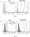
- Human JAML-CAR binding occurs at the interface of membrane distal D1 domains of both JAML and CAR, and can be blocked with DW100 mAb, which specifically binds JAML D1 domain as shown in Fig 1 .
- DW100 is the first mAb that has been demonstrated to specifically block JAML-CAR binding. It is used to study the functional consequences of JAML shedding.
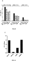
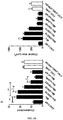
- DW100 completely inhibited JAML/CAR interactions and significantly inhibited aggregation of mixed cultures of CHO transfectants, whereas DW216 did not.
- DW55 was partly inhibitory and suggested that its epitope on JAML was close to the binding site for CAR.
- the size of the ligands was identified by biotinylation of the cells and immunoprecipitation. The blots revealed bands at ⁇ 60 KD, suggesting a high degree of glycosylation.
- the same antibodies precipitated a protein from CHO-JAML transfectants that runs as a smaller protein on SDS-PAGE gels and that could be isoforms of JAML or truncated JAML.
- Soluble JAML delayed both epithelial barrier recovery and wound restitution ( Figs 9 , 10 ). While there are a few possible ways to explain these effects, one likely mechanism would involve JAML binding to epithelial CAR, and interfering with its function. In polarized epithelial cell lines, CAR is expressed at the apical pole of the lateral membrane, where it colocalizes with the cytoplasmic plaque TJ protein zonula occludens-1 (ZO-1). CAR is mainly recognized as the receptor that mediates viral attachment, however it has been also implicated in regulating epithelial permeability and affecting cancer progression in malignancies.
- CAR has been shown to dimerize in cis, as well as suggested to bind in trans, thus mediating hemophilic binding interaction.
- CAR-CAR binding as well as any aggregation of CHO-CAR transfectants failed.
- CAR dimerization occurs at the membrane distal D1 domain, which is also the binding domain for JAML, it is possible that JAML binding would prevent CAR dimerization and thus decrease its signaling ability and function. Binding and aggregation was not observed between CAR molecules in the absence of JAML.
- JAML might also be able to dimerize. While recombinant protein binding assays failed to demonstrate JAML-JAML homophilic interactions, cell aggregation assay ( Fig 1 ) suggested that it is indeed possible. CHO cells stably expressing JAML formed significant number of aggregates, specific to JAML-JAML interactions, and it could not be blocked with DW100 mAb ( Fig 1 ).
- JAML that is shed from transmigrating PMNs might bind to ⁇ T-cells that have been attracted to the wound and promote their activation, or alternatively prevent their activation and the ensuing anti-inflammatory effects by interfering with their binding to CAR.
- soluble JAML that is shed from transmigrating PMNs can potentially act both on the basal and the apical sides of the epithelium.
- CAR mediates attachment of invading viruses
- the shedding of JAML by migrating PMNs might be an additional mechanism to protect against viral infections.
- DW100 is a specific monoclonal antibody that could reduce the level of epithelial damage by blocking the interaction between CAR and JAML.
- Suitable specific binding agents may be prepared using methods known in the art.
- An exemplary JAML and JAML D1 polypeptide specific binding agent of the present disclosure is capable of binding a certain portion of the JAML and JAML D1 polypeptides, and preferably modulating the activity or function of JAML and JAML D1 polypeptides.
- Specific binding agents such as antibodies and antibody fragments that specifically bind JAML polypeptides are within the scope of the present disclosure.
- the antibodies may be polyclonal including mono-specific polyclonal, monoclonal (mAbs), recombinant, chimeric, humanized such as CDR-grafted, human, single chain, catalytic, multi-specific and/or bi-specific, as well as antigen-binding fragments, variants, and/or derivatives thereof.
- mAbs mono-specific polyclonal
- recombinant recombinant
- chimeric humanized such as CDR-grafted, human, single chain, catalytic, multi-specific and/or bi-specific, as well as antigen-binding fragments, variants, and/or derivatives thereof.
- host cells either eukaryotic or prokaryotic, may be used to express the monoclonal antibody polynucleotides using recombinant techniques well known and routinely practiced in the art.
- transgenic animals are produced wherein a polynucleotide encoding the desired specific binding agent is introduced into the genome of a recipient animal, such as, for example, a mouse, rabbit, goat, or cow, in a manner that permits expression of the polynucleotide molecules encoding a monoclonal antibody or other specific binding agent.
- the polynucleotides encoding the monoclonal antibody or other specific binding agent can be ligated to mammary-specific regulatory sequences, and the chimeric polynucleotides can be introduced into the germline of the target animal.
- the resulting transgenic animal then produces the desired antibody in its milk [ Pollock et al., J Immunol Meth 231:147-157 (1999 ); Little et al., Immunol Today 8:364-370 (2000 )].
- plants may be used to express and produce JAML or JAML D1 specific binding agents such as monoclonal antibodies by transfecting suitable plants with the polynucleotides encoding the monoclonal antibodies or other specific binding agents.
- a monoclonal or polyclonal antibody or fragment thereof that is derived from other than a human species may be "humanized” or “chimerized”.
- Methods for humanizing non-human antibodies are well known in the art. (see U.S. Pat. Nos. 5,859,205 , 5,585,089 , and 5,693,762 ).
- Humanization is performed, for example, using methods described in the art [ Jones et al., Nature, 321: 522-525 (1986 ); Riechmann et al., Nature, 332: 323-327 (1988 ); Verhoeyen et al., Science, 239:1534-1536 (1988 )] by substituting at least a portion of, for example a rodent, complementarity-determining region (CDRs) for the corresponding regions of a human antibody.
- CDRs complementarity-determining region
- the disclosure also provides variants and derivatives of these human antibodies as discussed herein and well known in the art.
- transgenic animals e.g., mice
- transgenic animals that are capable of producing a repertoire of human antibodies in the absence of endogenous immunoglobulin production can be used to generate such antibodies. This can be accomplished by immunization of the animal with an JAML or JAML D1 antigen or fragments thereof where the JAML or JAML D1 fragments have an amino acid sequence that is unique to JAML or JAML D1.
- immunogens can be optionally conjugated to a carrier.
- transgenic animals are produced by incapacitating the endogenous loci encoding the heavy and light immunoglobulin chains therein, and inserting loci encoding human heavy and light chain proteins into the genome thereof.
- Partially modified animals that are those having less than the full complement of these modifications, are then crossbred to obtain an animal having all of the desired immune system modifications.
- these transgenic animals When administered an immunogen, these transgenic animals are capable of producing antibodies with human variable regions, including human (rather than e.g., murine) amino acid sequences, that are immuno-specific for the desired antigens. See PCT application Nos., PCT/US96/05928 and PCT/US93/06926 . Additional methods are described in U.S. Pat. No. 5,545,807 , PCT application Nos. PCT/US91/245 , PCT/GB89/01207 , and in EP 546073B1 and EP 546073A1 . Human antibodies may also be produced by the expression of recombinant DNA in host cells or by expression in hybridoma cells as described herein.
- Transgenesis is achieved in a number of different ways. See, for example, Bruggeman et al., Immunol Today 17:391-7 (1996 ).
- a minilocus is constructed such that gene segments in a germline configuration are brought artificially close to each other. Due to size limitations (i.e., having generally less than 30 kb), the resulting minilocus will contain a limited number of differing gene segments, but is still capable of producing a large repertoire of antibodies.
- Miniloci containing only human DNA sequences, including promoters and enhancers are fully functional in the transgenic mouse.
- yeast artificial chromosomes When larger number of gene segments are desired in the transgenic animal, yeast artificial chromosomes (YACs) are utilized. YACs can range from several hundred kilobases to 1 Mb and are introduced into the mouse (or other appropriate animal) genome via microinjection directly into an egg or via transfer of the YAC into embryonic stem (ES)-cell lines. In general, YACs are transferred into ES cells by lipofection of the purified DNA, or yeast spheroplast fusion wherein the purified DNA is carried in micelles and fusion is carried out in manner similar to hybridoma fusion protocols. Selection of desired ES cells following DNA transfer is accomplished by including on the YAC any of the selectable markers known in the art.
- bacteriophage P1 vectors are used which are amplified in a bacterial E. coli host. While these vectors generally carry less inserted DNA than a YAC, the clones are readily grown in high enough yield to permit direct microinjection into a mouse egg. Use of a cocktail of different P1 vectors has been shown to lead to high levels of homologous recombination.
- transgenic mouse or other appropriate animal
- any of the techniques known in the art to detect serum levels of a circulating antibody e.g., ELISA
- the transgenic animal is crossed with a mouse in which the endogenous Ig locus has been disrupted.
- the result provides progeny wherein essentially all B cells express human antibodies.
- the entire animal Ig locus is replaced with the human Ig locus, wherein the resulting animal expresses only human antibodies.
- portions of the animal's locus are replaced with specific and corresponding regions in the human locus.
- the animals resulting from this procedure may express chimeric antibodies, as opposed to fully human antibodies, depending on the nature of the replacement in the mouse Ig locus.
- Human antibodies can also be produced by exposing human splenocytes (B or T cells) to an antigen in vitro, then reconstituting the exposed cells in an immunocompromised mouse, e.g. SCID or nod/SCID. See Brams et al., J Immunol, 160: 2051-2058 [1998 ]; Carballido et al., Nat Med, 6: 103-106 [2000 ].
- SEA Staphylococcal Enterotoxin A
- an entirely synthetic human heavy chain repertoire is created from unrearranged V gene segments by assembling each human VH segment with D segments of random nucleotides together with a human J segment [ Hoogenboom et al., J Mol Biol 227:381-388 (1992 )].
- a light chain repertoire is constructed by combining each human V segment with a J segment [ Griffiths et al., EMBO J. 13:3245-3260 (1994 )].
- Nucleotides encoding the complete antibody i.e., both heavy and light chains
- this polynucleotide is ligated to a nucleotide encoding a filamentous phage minor coat protein. When this fusion protein is expressed on the surface of the phage, a polynucleotide encoding a specific antibody is identified by selection using an immobilized antigen.
- antibody fragments are assembled as two Fab fragments by fusion of one chain to a phage protein and secretion of the other into bacterial periplasm [ Hoogenboom et al., Nucl Acids Res 19:4133-4137 [1991] ; Barbas et al., Proc Natl Acad Sci (USA) 88:7978-7982 (1991 )].
- chimeric, humanized, CDR-grafted, and fully human antibodies, or antigen-binding fragments thereof are typically produced by recombinant methods.
- Polynucleotide molecule(s) encoding the heavy and light chains of each antibody or antigen-binding fragments thereof can be introduced into host cells and expressed using materials and procedures described herein.
- the antibodies are produced in mammalian host cells, such as CHO cells.
- the specific binding agents of the present disclosure can further comprise any constant region known in the art.
- the light chain constant region can be, for example, a kappa- or lambda-type light chain constant region, e.g., a human kappa- or lambda-type light chain constant region.
- the heavy chain constant region can be, for example, an alpha-, delta-, epsilon-, gamma-, or mu-type heavy chain constant regions, e.g., a human alpha-, delta-, epsilon-, gamma-, or mu-type heavy chain constant region.
- the light or heavy chain constant region is a fragment, derivative, variant, or mutant of a naturally occurring constant region.
- the specific binding agents of the present disclosure such as the antibodies, antibody fragments, and antibody derivatives of the disclosure comprise an IgG.
- IgG antibodies may be derived from an IgM antibody, for example, and vice versa.
- Such techniques allow the preparation of new antibodies that possess the antigen-binding properties of a given antibody (the parent antibody), but also exhibit biological properties associated with an antibody isotype or subclass different from that of the parent antibody.
- Recombinant DNA techniques may be employed. Cloned DNA encoding particular antibody polypeptides may be employed in such procedures, e.g., DNA encoding the constant domain of an antibody of the desired isotype. See also Lantto et al., 2002, Methods Mol. Biol. 178:303-16 .
- the specific binding agents of the present disclosure may comprise the IgG1 heavy chain constant domain or a fragment of the IgG1 heavy chain domain.
- the antibodies, antibody fragments, and antibody derivatives of the disclosure may further comprise the constant light chain kappa or lambda domains or a fragment of these. Light chain constant regions and polynucleotides encoding them are provided herein below.
- the antibodies, antibody fragments, and antibody derivatives of the disclosure further comprise a heavy chain constant domain, or a fragment thereof, such as the IgG2 heavy chain constant region also shown herein below.
- the nucleic acid (DNA) encoding constant heavy and constant light chain domains, and the amino acids sequences of heavy and light chain domains are provided herein below.
- Lambda variable domains can be fused to lambda constant domains and kappa variable domains can be fused to kappa constant domains.
- the specific binding agents of the present disclosure include those comprising, for example, the variable domain combinations H1L1, H2L2, H3L3 having a desired isotype (for example, IgA, IgG1, IgG2, IgG3, IgG4, IgM, IgE, and IgD) as well as Fab or F(ab') 2 fragments thereof.
- an IgG4 it may also be desired to introduce a point mutation in the hinge region as described in Bloom et al., 1997, Protein Science 6:407 (incorporated by reference herein) to alleviate a tendency to form intra-H chain disulfide bonds that can lead to heterogeneity in the IgG4 antibodies.
- IgG1, IgG2, IgG3, and IgG4 isotypes are used in combination with the variable heavy chain sequences of the antibodies of the present disclosure to make a specific desired isotype of said antibody:
- polypeptides comprising the amino acid sequence variable domains of JAML or JAML D1 antibodies may be fused at either the N-terminus or the C-terminus to one or more domains of an Fc region of human IgG.
- an Fc domain can provide longer half-life or incorporate such functions as Fc receptor binding, Protein A binding, complement fixation and perhaps even placental transfer.
- the antibody hinge, CH2 and CH3 regions may be fused at either the N-terminus or C-terminus of the specific binding agent polypeptides such as an anti-JAML or JAML D1 Fab or Fv fragment using methods known to the skilled artisan.
- the resulting fusion protein may be purified by use of a Protein A or Protein G affinity column. Peptides and proteins fused to an Fc region have been found to exhibit a substantially greater half-life in vivo than the unfused counterpart. Also, a fusion to an Fc region allows for dimerization/multimerization of the fusion polypeptide.
- the Fc region may be a naturally occurring Fc region, or may be altered to improve certain qualities, such as therapeutic qualities, circulation time, decrease aggregation problems, etc.
- Other examples known in the art include those wherein the Fc region, which may be human or another species, or may be synthetic, is fused to the N-terminus of CD30L to treat Hodgkin's disease, anaplastic lymphoma and T-cell leukemia ( U.S. Pat. No.
- the Fc region is fused to the TNF receptor to treat septic shock [ Fisher et al., N Engl J Med, 334: 1697-1702 (1996 )], and the Fc region is fused to the Cd4 receptor to treat AIDS [ Capon et al., Nature, 337: 525-31 (1989 )].
- Catalytic antibodies are another type of fusion molecule and include antibodies to which one or more cytotoxic, or more generally one or more biologically active, moieties are attached to the specific binding agent. See, for example Rader et al., Chem Eur J 12:2091-2095 (2000 ). Cytotoxic agents of this type improve antibody-mediated cytotoxicity, and include such moieties as cytokines that directly or indirectly stimulate cell death, radioisotopes, chemotherapeutic drugs (including prodrugs), bacterial toxins (ex. pseudomonas exotoxin, diphtheria toxin, etc.), plant toxins (ex.
- the cytotoxic agent can be "attached" to one component of a bi-specific or multi-specific antibody by binding of this agent to one of the alternative antigen recognition sites on the antibody.
- protein cytotoxins can be expressed as fusion proteins with the specific binding agent following ligation of a polynucleotide encoding the toxin to a polynucleotide encoding the binding agent.
- the specific binding agent can be covalently modified to include the desired cytotoxin.
- fusion proteins are immunogenic polypeptides, proteins with long circulating half-lives, such as immunoglobulin constant regions, marker proteins, proteins or polypeptides that facilitate purification of the desired specific binding agent polypeptide, and polypeptide sequences that promote formation of multimeric proteins (such as leucine zipper motifs that are useful in dimer formation/stability).
- This type of insertional variant generally has all or a substantial portion of the native molecule, linked at the N- or C-terminus, to all or a portion of a second polypeptide.
- fusion proteins typically employ leader sequences from other species to permit the recombinant expression of a protein in a heterologous host.
- Another useful fusion protein includes the addition of an immunologically active domain, such as an antibody epitope, to facilitate purification of the fusion protein. Inclusion of a cleavage site at or near the fusion junction will facilitate removal of the extraneous polypeptide after purification.
- Other useful fusions include linking of functional domains, such as active sites from enzymes, glycosylation domains, cellular targeting signals or transmembrane regions.
- GST glutathione-S-transferase
- NEB maltose binding protein
- FLAG FLAG system
- 6 X His 6 X His system
- both the FLAG system and the 6 x His system add only short sequences, both of which are known to be poorly antigenic and which do not adversely affect folding of the polypeptide to its native conformation.
- Another N-terminal fusion that is contemplated to be useful is the fusion of a Met-Lys dipeptide at the N-terminal region of the protein or peptides. Such a fusion may produce beneficial increases in protein expression or activity.
- a particularly useful fusion construct may be one in which a specific binding agent peptide is fused to a hapten to enhance immunogenicity of a specific binding agent fusion construct which is useful, for example, in the production of anti-idiotype antibodies of the disclosure.
- Such fusion constructs to increase immunogenicity are well known to those of skill in the art, for example, a fusion of specific binding agent with a helper antigen such as hsp70 or peptide sequences such as from diphtheria toxin chain or a cytokine such as IL-2 will be useful in eliciting an immune response.
- fusion construct can be made which will enhance the targeting of the antigen binding agent compositions to a specific site or cell.
- fusion constructs including heterologous polypeptides with desired properties, e.g., an Ig constant region to prolong serum half-life or an antibody or fragment thereof for targeting also are contemplated.
- Other fusion systems produce polypeptide hybrids where it is desirable to excise the fusion partner from the desired polypeptide.
- the fusion partner is linked to the recombinant specific binding agent polypeptide by a peptide sequence containing a specific recognition sequence for a protease. Examples of suitable sequences are those recognized by the Tobacco Etch Virus protease (Life Technologies, Gaithersburg, Md.) or Factor Xa (New England Biolabs, Beverley, Mass.).
- the disclosure also provides fusion polypeptides comprising all or part of a variable domain of an JAML or JAML D1 antibody, such as a heavy chain variable region with an amino acid sequence as described herein or a light chain variable region with an amino acid sequence as described herein in combination with truncated tissue factor (tTF), a vascular targeting agent consisting of a truncated form of a human coagulation-inducing protein that acts as a tumor blood vessel clotting agent.
- tTF tissue factor
- the fusion of tTF to the anti-JAML or JAML D1 antibody, or fragments thereof may facilitate the delivery of anti-JAML or JAML D1 to target cells.
- Variants of Specific Binding Agents of the present disclosure include insertion, deletion, and/or substitution variants.
- insertion variants are provided wherein one or more amino acid residues supplement a specific binding agent amino acid sequence. Insertions may be located at either or both termini of the protein, or may be positioned within internal regions of the specific binding agent amino acid sequence. Insertional variants with additional residues at either or both termini can include, for example, fusion proteins and proteins including amino acid tags or labels. Insertion variants include specific binding agent polypeptides wherein one or more amino acid residues are added to a specific binding agent amino acid sequence, or fragment thereof.
- Variant products of the disclosure also include mature specific binding agent products. Such specific binding agent products have the leader or signal sequences removed, however the resulting protein has additional amino terminal residues. The additional amino terminal residues may be derived from another protein, or may include one or more residues that are not identifiable as being derived from a specific protein.
- Specific binding agent products with an additional methionine residue at position-1 are contemplated, as are specific binding agent products with additional methionine and lysine residues at positions -2 and -1 (Met.2-Lys-1-specific binding agent).
- Variants of specific binding agents having additional Met, Met-Lys, Lys residues are particularly useful for enhanced recombinant protein production in bacterial host cells.
- the disclosure also embraces specific binding agent variants having additional amino acid residues that arise from use of specific expression systems.
- use of commercially available vectors that express a desired polypeptide as part of glutathione-S-transferase (GST) fusion product provides the desired polypeptide having an additional glycine residue at amino acid position-1 after cleavage of the GST component from the desired polypeptide.
- GST glutathione-S-transferase
- Variants which result from expression in other vector systems are also contemplated, including those wherein poly-histidine tags are incorporated into the amino acid sequence, generally at the carboxy and/or amino terminus of the sequence.
- Insertional variants also include fusion proteins as described above, wherein the amino and/or carboxy termini of the specific binding agent-polypeptide is fused to another polypeptide, a fragment thereof, or amino acid sequences which are not generally recognized to be part of any specific protein sequence.
- the disclosure provides deletion variants wherein one or more amino acid residues in a specific binding agent polypeptide are removed.
- Deletions can be effected at one or both termini of the specific binding agent polypeptide, or from removal of one or more residues within the specific binding agent amino acid sequence.
- Deletion variants necessarily include all fragments of a specific binding agent polypeptide.
- Antibody fragments include those portions of the antibody that bind to an epitope on the antigen polypeptide. Examples of such fragments include Fab and F(ab')2 fragments generated, for example, by enzymatic or chemical cleavage of full-length antibodies. Other binding fragments include those generated by recombinant DNA techniques, such as the expression of recombinant plasmids containing nucleic acid sequences encoding antibody variable regions.
- the disclosure also embraces polypeptide fragments of a JAML or JAML D1 binding agent wherein the fragments maintain the ability to specifically bind an JAML or JAML D1 polypeptide.
- Fragments comprising at least 5, 10, 15, 20, 25, 30, 35, 40, 45 or 50 or more consecutive amino acids of a peptide or polypeptide of the disclosure are comprehended herein.
- Preferred polypeptide fragments display immunological properties unique to or specific for the antigen-binding agent so of the disclosure. Fragments of the disclosure having the desired immunological properties can be prepared by any of the methods well known and routinely practiced in the art.
- substitution variants are generally considered to be “similar” to the original polypeptide or to have a certain "percent identity" to the original polypeptide, and include those polypeptides wherein one or more amino acid residues of a polypeptide are removed and replaced with alternative residues.
- substitutions are conservative in nature, however, the disclosure embraces substitutions that are also non-conservative.
- Typical methods to determine the relatedness or percent identity of two polypeptides are designed to give the largest match between the sequences tested. Methods to determine identity are described in publicly available computer programs. Preferred computer program methods to determine identity between two sequences include, but are not limited to, the GCG program package, including GAP ( Devereux et al., Nucl. Acid. Res., 12:387 (1984 ); Genetics Computer Group, University of Wisconsin, Madison, Wis., BLASTP, BLASTN, and FASTA ( Altschul et al., J. Mol. Biol., 215:403-410 (1990 )). The BLASTX program is publicly available from the National Center for Biotechnology Information (NCBI) and other sources (BLAST Manual, Altschul et al. NCB/NLM/NIH Bethesda, Md. 20894; Altschul et al., supra (1990)). The well-known Smith Waterman algorithm may also be used to determine identity.
- NCBI National Center for Biotechnology Information
- the selected alignment method will result in an alignment that spans at least ten percent of the full length of the target polypeptide being compared, i.e., at least 40 contiguous amino acids where sequences of at least 400 amino acids are being compared, 30 contiguous amino acids where sequences of at least 300 to about 400 amino acids are being compared, at least 20 contiguous amino acids where sequences of 200 to about 300 amino acids are being compared, and at least 10 contiguous amino acids where sequences of about 100 to 200 amino acids are being compared.
- GAP Genetics Computer Group, University of Wisconsin, Madison, Wis.
- two polypeptides for which the percent sequence identity is to be determined are aligned for optimal matching of their respective amino acids (the "matched span", as determined by the algorithm).
- a gap opening penalty which is typically calculated as 3 times the average diagonal; the "average diagonal” is the average of the diagonal of the comparison matrix being used; the “diagonal” is the score or number assigned to each perfect amino acid match by the particular comparison matrix
- a gap extension penalty which is usually 1/10 times the gap opening penalty
- a comparison matrix such as PAM 250 or BLOSUM 62
- a standard comparison matrix (see Dayhoff et al., Atlas of Protein Sequence and Structure, 5(3)(1978 ) for the PAM 250 comparison matrix; Henikoff et al., Proc. Natl. Acad. Sci. USA, 89:10915-10919 (1992 ) for the BLOSUM 62 comparison matrix) is also used by the algorithm.
- the parameters for a polypeptide sequence comparison include the following:
- the GAP program may be useful with the above parameters.
- the aforementioned parameters are the default parameters for polypeptide comparisons (along with no penalty for end gaps) using the GAP algorithm.
- the GAP program may also be useful with the above parameters.
- the aforementioned parameters are the default parameters for polynucleotide molecule comparisons.
- gap opening penalties may be used, including those set forth in the Program Manual, Wisconsin Package, Version 9, September, 1997.
- the particular choices to be made will be apparent to those of skill in the art and will depend on the specific comparison to be made, such as DNA-to-DNA, protein-to-protein, protein-to-DNA; and additionally, whether the comparison is between given pairs of sequences (in which case GAP or BestFit are generally preferred) or between one sequence and a large database of sequences (in which case FASTA or BLASTA are preferred).
- the amino acids may have either L or D stereochemistry (except for Gly, which is neither L nor D) and the polypeptides and compositions of the present disclosure may comprise a combination of stereochemistries. However, the L stereochemistry is typical.
- the disclosure also provides reverse molecules wherein the amino terminal to carboxy terminal sequence of the amino acids is reversed. For example, the reverse of a molecule having the normal sequence X1-X2-X3 would be X3-X2-X1.
- the disclosure also provides retro-reverse molecules wherein, as above, the amino terminal to carboxy terminal sequence of amino acids is reversed and residues that are normally "L" enantiomers are altered to the "D" stereoisomer form.
- Stereoisomers e.g., D-amino acids of the twenty conventional amino acids, unnatural amino acids such as alpha-, alpha-disubstituted amino acids, N-alkyl amino acids, lactic acid, and other unconventional amino acids may also be suitable components for polypeptides of the present disclosure.
- unconventional amino acids include, without limitation: aminoadipic acid, beta-alanine, beta-aminopropionic acid, aminobutyric acid, piperidinic acid, aminocaprioic acid, aminoheptanoic acid, aminoisobutyric acid, aminopimelic acid, diaminobutyric acid, desmosine, diaminopimelic acid, diaminopropionic acid, N-ethylglycine, N-ethylaspargine, hyroxylysine, allo-hydroxylysine, hydroxyproline, isodesmosine, allo-isoleucine, N-methylglycine, sarcosine, N-methylisoleucine, N-methylvaline, norvaline, norleucine, orithine, 4-hydroxyproline, gamma-carboxyglutamate, epsilon-N,N,N-trimethyllysine, epsilon-N-acetyllysine, O-
- the left-hand end of single-stranded polynucleotide sequences is the 5' end; the left-hand direction of double-stranded polynucleotide sequences is referred to as the 5' direction.
- the direction of 5' to 3' addition of nascent RNA transcripts is referred to as the transcription direction; sequence regions on the DNA strand having the same sequence as the RNA and which are 5' to the 5' end of the RNA transcript are referred to as "upstream sequences"; sequence regions on the DNA strand having the same sequence as the RNA and which are 3' to the 3' end of the RNA transcript are referred to as "downstream sequences".
- amino acid residues may encompass non-naturally occurring amino acid residues, which are typically incorporated by chemical peptide synthesis rather than by synthesis in biological systems. These include peptidomimetics and other reversed or inverted forms of amino acid moieties.
- Naturally occurring residues may be divided into classes based on common side chain properties:
- non-conservative substitutions may involve the exchange of a member of one of these classes for a member from another class.
- substituted residues may be introduced into regions of the human antibody that are homologous with non-human antibodies, or into the non-homologous regions of the molecule.
- the hydropathic index of amino acids may be considered.
- Each amino acid has been assigned a hydropathic index on the basis of its hydrophobicity and charge characteristics. They are: isoleucine (+4.5); valine (+4.2); leucine (+3.8); phenylalanine (+2.8); cysteine/cystine (+2.5); methionine (+1.9); alanine (+1.8); glycine (-0.4); threonine (-0.7); serine (-0.8); tryptophan (-0.9); tyrosine (-1.3); proline (-1.6); histidine (-3.2); glutamate (-3.5); glutamine (-3.5); aspartate (-3.5); asparagine (-3.5); lysine (-3.9); and arginine (-4.5).
- hydropathic amino acid index in conferring interactive biological function on a protein is understood in the art. Kyte et al., J. Mol. Biol., 157:105-131 (1982 ). It is known that certain amino acids may be substituted for other amino acids having a similar hydropathic index or score and still retain a similar biological activity. In making changes based upon the hydropathic index, in certain embodiments, the substitution of amino acids whose hydropathic indices are within 0.2 is included. In certain embodiments, those which are within 0.1 are included, and in certain embodiments, those within 0.5 are included.
- the substitution of like amino acids can be made effectively on the basis of hydrophilicity, particularly where the biologically functional protein or peptide thereby created is intended for use in immunological embodiments, as in the present case.
- the greatest local average hydrophilicity of a protein as governed by the hydrophilicity of its adjacent amino acids, correlates with its immunogenicity and antigenicity, i.e., with a biological property of the protein.
- hydrophilicity values have been assigned to these amino acid residues: arginine (+3.0); lysine (+3.0); aspartate (+3.0); glutamate (+3.0); serine (+0.3); asparagine (+0.2); glutamine (+0.2); glycine (0); threonine (-0.4); proline (-0.5); alanine (-0.5); histidine (-0.5); cysteine (-1.0); methionine (-1.3); valine (-1.5); leucine (-1.8); isoleucine (-1.8); tyrosine (-2.3); phenylalanine (-2.5) and tryptophan (-3.4).
- the substitution of amino acids whose hydrophilicity values are within 0.2 is included, in certain embodiments, those which are within 0.1 are included, and in certain embodiments, those within 0.5 are included.
- polypeptide structure A skilled artisan will be able to determine suitable variants of the polypeptide as set forth herein using well-known techniques.
- one skilled in the art may identify suitable areas of the molecule that may be changed without destroying activity by targeting regions not believed to be important for activity.
- even areas that may be important for biological activity or for structure may be subject to conservative amino acid substitutions without destroying the biological activity or without adversely affecting the polypeptide structure.
- One skilled in the art can also analyze the three-dimensional structure and amino acid sequence in relation to that structure in similar polypeptides. In view of such information, one skilled in the art may predict the alignment of amino acid residues of an antibody with respect to its three dimensional structure. In certain embodiments, one skilled in the art may choose not to make radical changes to amino acid residues predicted to be on the surface of the protein, since such residues may be involved in important interactions with other molecules. Moreover, one skilled in the art may generate test variants containing a single amino acid substitution at each desired amino acid residue. The variants can then be screened using activity assays known to those skilled in the art. Such variants could be used to gather information about suitable variants.
- One method of predicting secondary structure is based upon homology modeling. For example, two polypeptides or proteins which have a sequence identity of greater than 30%, or similarity greater than 40% often have similar structural topologies.
- the recent growth of the protein structural database (PDB) has provided enhanced predictability of secondary structure, including the potential number of folds within a polypeptide's or protein's structure. See Holm et al., Nuci. Acid. Res., 27(1):244-247 (1999 ). It has been suggested ( Brenner et al., Curr. Op. Struct. Biol., 7(3):369-376 (1997 )) that there are a limited number of folds in a given polypeptide or protein and that once a critical number of structures have been resolved, structural prediction will become dramatically more accurate.
- Additional methods of predicting secondary structure include “threading” ( Jones, D., Curr. Opin. Struct. Biol., 7(3):377-87 (1997 ); Sippl et al., Structure, 4(1):15-19 (1996 )), “profile analysis” ( Bowie et al., Science, 253:164-170 (1991 ); Gribskov et al., Meth. Enzym., 183:146-159 (1990 ); Gribskov et al., Proc. Nat. Acad. Sci., 84(13):4355-4358 (1987 )), and “evolutionary linkage” (See Holm, supra (1999), and Brenner, supra (1997)).
- antibody variants include glycosylation variants wherein the number and/or type of glycosylation site has been altered compared to the amino acid sequences of the parent polypeptide.
- protein variants comprise a greater or a lesser number of N-linked glycosylation sites than the native protein.
- An N-linked glycosylation site is characterized by the sequence: Asn-X-Ser or Asn-X-Thr, wherein the amino acid residue designated as X may be any amino acid residue except proline.
- the substitution of amino acid residues to create this sequence provides a potential new site for the addition of an N-linked carbohydrate chain. Alternatively, substitutions which eliminate this sequence will remove an existing N-linked carbohydrate chain.
- N-linked carbohydrate chains wherein one or more N-linked glycosylation sites (typically those that are naturally occurring) are eliminated and one or more new N-linked sites are created.
- Additional preferred antibody variants include cysteine variants wherein one or more cysteine residues are deleted from or substituted for another amino acid (e.g., serine) as compared to the parent amino acid sequence. Cysteine variants may be useful when antibodies must be refolded into a biologically active conformation such as after the isolation of insoluble inclusion bodies. Cysteine variants generally have fewer cysteine residues than the native protein, and typically have an even number to minimize interactions resulting from unpaired cysteines.
- amino acid substitutions are those which: (1) reduce susceptibility to proteolysis, (2) reduce susceptibility to oxidation, (3) alter binding affinity for forming protein complexes, (4) alter binding affinities, and/or (5) confer or modify other functional properties on such polypeptides.
- single or multiple amino acid substitutions may be made in the naturally-occurring sequence (in certain embodiments, in the portion of the polypeptide outside the domain(s) forming intermolecular contacts).
- a conservative amino acid substitution typically may not substantially change the structural characteristics of the parent sequence (e.g., a replacement amino acid should not tend to break a helix that occurs in the parent sequence, or disrupt other types of secondary structure that characterizes the parent sequence).
- a replacement amino acid should not tend to break a helix that occurs in the parent sequence, or disrupt other types of secondary structure that characterizes the parent sequence.
- Examples of art-recognized polypeptide secondary and tertiary structures are described in Proteins, Structures and Molecular Principles (Creighton, Ed., W.H. Freeman and Company, New York (1984 )); Introduction to Protein Structure (C. Branden and J. Tooze, eds., Garland Publishing, New York, N.Y. (1991 )); and Thornton et at. Nature 354:105 (1991 ).
- the specific binding agent molecules of this disclosure that are polypeptide or peptide substitution variants may have up to about ten to twelve percent of the original amino acid sequence replaced.
- the heavy chain may have 50, 49, 48, 47, 46, 45, 44, 43, 42, 41, 40, 39, 38, 37, 36, 35, 34, 33, 32, 31, 30, 29, 28, 27, 26, 25, 24, 23, 22, 21, 20, 19, 18, 17, 16, 15, 14, 13, 12, 11, 10, 9, 8, 7, 6, 5, 4, 3, 2, or 1 amino acid replaced, while the light chain may have 25, 24, 23, 22, 21, 20, 19, 18, 17, 16, 15, 14, 13, 12, 11, 10, 9, 8, 7, 6, 5, 4, 3, 2, or 1 amino acid replaced.
- the disclosure also provides derivatives of specific binding agent polypeptides.
- Derivatives include specific binding agent polypeptides bearing modifications other than insertion, deletion, or substitution of amino acid residues.
- the modifications are covalent in nature, and include for example, chemical bonding with polymers, lipids, other organic, and inorganic moieties.
- Derivatives of the disclosure may be prepared to increase circulating half-life of a specific binding agent polypeptide, or may be designed to improve targeting capacity for the polypeptide to desired cells, tissues, or organs.
- the disclosure further embraces derivative binding agents covalently modified to include one or more water soluble polymer attachments such as polyethylene glycol, polyoxyethylene glycol, or polypropylene glycol as described U.S. Pat. Nos. 4,640,835 , 4,496,689 , 4,301,144 , 4,670,417 , 4,791,192 and 4,179,337 .
- Still other useful polymers known in the art include monomethoxy-polyethylene glycol, dextran, cellulose, or other carbohydrate based polymers, poly-(N-vinyl pyrrolidone)-polyethylene glycol, propylene glycol homopolymers, a polypropylene oxide/ethylene oxide co-polymer, polyoxyethylated polyols (e.g., glycerol) and polyvinyl alcohol, as well as mixtures of these polymers.
- Particularly preferred are specific binding agent products covalently modified with polyethylene glycol (PEG) subunits.
- Water-soluble polymers may be bonded at specific positions, for example at the amino terminus of the specific binding agent products, or randomly attached to one or more side chains of the polypeptide.
- PEG for improving the therapeutic capacity for specific binding agent, and for humanized antibodies in particular, is described in U.S. Pat. No. 6,133,426 to Gonzales et al., issued Oct. 17, 2000 .
- Certain strategies can be employed to manipulate inherent properties of a JAML or JAML D1-specific antibody, such as the affinity of the antibody for its target. These strategies include the use of site-specific or random mutagenesis of the polynucleotide molecule encoding the antibody to generate antibody variants, followed by a screening step designed to recover antibody variants that exhibit the desired change, e.g. increased or decreased affinity.
- amino acid residues most commonly targeted in mutagenic strategies are those in the CDRs. As described supra, these regions contain the residues that actually interact with JAML or JAML D1 and other amino acids that affect the spatial arrangement of these residues. However, amino acids in the framework regions of the variable domains outside the CDR regions have also been shown to make contributions to the antigen-binding properties of the antibody, and can be targeted to manipulate such properties. See Hudson, Curr Opin Biotech, 9:395-402 (1999 ) and references therein.
- Smaller and more effectively screened libraries of antibody variants can be produced by restricting random or site-directed mutagenesis to sites in the CDRs that correspond to areas prone to "hyper-mutation" during the somatic affinity maturation process. See Chowdhury and Pastan, Nature Biotech, 17: 568-572 [1999 ] and references therein.
- the types of DNA elements known to define hyper-mutation sites in this manner include direct and inverted repeats, certain consensus sequences, secondary structures, and palindromes.
- the consensus DNA sequences include the tetrabase sequence Purine-G-Pyrimidine-A/T (i.e. A or G-G-C or T-A or T) and the serine codon AGY (wherein Y can be a C or a T).
- an embodiment of the present disclosure includes mutagenic strategies with the goal of increasing the affinity of an antibody for its target. These strategies include mutagenesis of the entire variable heavy and light chain, mutagenesis of the CDR regions only, mutagenesis of the consensus hypermutation sites within the CDRs, mutagenesis of framework regions, or any combination of these approaches ("mutagenesis" in this context could be random or site-directed).
- Definitive delineation of the CDR regions and identification of residues comprising the binding site of an antibody can be accomplished though solving the structure of the antibody in question, and the antibody-ligand complex, through techniques known to those skilled in the art, such as X-ray crystallography.
- Various methods based on analysis and characterization of such antibody crystal structures are known to those of skill in the art and can be employed, although not definitive, to approximate the CDR regions. Examples of such commonly used methods include the Kabat, Chothia, AbM and contact definitions.
- the Kabat definition is based on the sequence variability and is the most commonly used definition to predict CDR regions. [ Johnson and Wu, Nucleic Acids Res, 28: 214-8 (2000 )].
- the Chothia definition is based on the location of the structural loop regions. [ Chothia et al., J Mol Biol, 196: 901-17 (1986 ); Chothia et al., Nature, 342: 877-83 (1989 )].
- the AbM definition is a compromise between the Kabat and Chothia definition.
- AbM is an integral suite of programs for antibody structure modeling produced by Oxford Molecular Group [ Martin et al., Proc Natl Acad Sci (USA) 86:9268-9272 (1989 ); Rees, et al., ABM.TM., a computer program for modeling variable regions of antibodies, Oxford, UK; Oxford Molecular, Ltd.].
- the AbM suite models the tertiary structure of an antibody from primary sequencing using a combination of knowledge databases and ab initio methods.
- An additional definition known as the contact definition, has been recently introduced. [ MacCallum et al., J Mol Biol, 5:732-45 (1996 )]. This definition is based on an analysis of the available complex crystal structures.
- the CDR regions in the heavy chain are typically referred to as HI, H2 and H3 and are numbered sequentially in order counting from the amino terminus to the carboxy terminus.
- the CDR regions in the light chain are typically referred to as L1, L2 and L3 and are numbered sequentially in order counting from the amino terminus to the carboxy terminus.
- the CDR-H1 is approximately 10 to 12 residues in length and typically starts 4 residues after a Cys according to the Chothia and AbM definitions or typically 5 residues later according to the Kabat definition.
- the H1 is typically followed by a Trp, typically Trp-Val, but also Trp-Ile, or Trp-Ala.
- the length of H1 is approximately 10 to 12 residues according to the AbM definition while the Chothia definition excludes the last 4 residues.
- the CDR-H2 typically starts 15 residues after the end of H1 according to the Kabat and AbM definition.
- the residues preceding H2 are typically Leu-Glu-Trp-11e-Gly but there are a number of variations.
- H2 is typically followed by the amino acid sequence Lys/Arg-Leu/Ile/Val/Phe/Thr/Ala-Thr/Ser/Ile/Ala.
- the length of the H2 is approximately 16 to 19 residues where the AbM definition predicts the length to be typically 9 to 12 residues.
- the CDR-H3 typically starts 33 residues after the end of H2 and is typically preceded by the amino acid sequence (typically Cys-Ala-Arg).
- the H3 is typically followed by the amino acid sequence-Gly.
- the length of H3 can be anywhere between 3 to 25 residues.
- the CDR-L1 typically starts at approximately residue 24 and will typically follow a Cys.
- the residue after the CDR-L1 is always a Trp and will typically begin the sequence Trp-Tyr-Gln, Trp-Leu-Gln, Trp-Phe-Gln, or Trp-Tyr-Leu.
- the length of CDR-L1 is approximately 10 to 17 residues.
- the punitive CDR-L1 for the antibodies of the disclosure follows this pattern exactly with a Cys residue followed by 15 amino acids then Trp-Tyr-Gln.
- the CDR-L2 starts approximately 16 residues after the end of L1. It will generally follow residues Ile-Tyr, Val-Tyr, Ile-Lys or Ile-Phe. The length of CDR-L2 is approximately 7 residues.
- the CDR-L3 typically starts 33 residues after the end of L2 and typically follows a Cys.
- L3 is typically followed by the amino acid sequence Phe-Gly-XXX-Gly.
- the length of L3 is approximately 7 to 11 residues.
- each humanized immunoglobulin chain will usually comprise, in addition to the CDRs, amino acids from the donor immunoglobulin framework that are, e.g., capable of interacting with the CDRs to affect binding affinity, such as one or more amino acids which are immediately adjacent to a CDR in the donor immunoglobulin or those within about 3 angstroms as predicted by molecular modeling.
- the heavy and light chains may each be designed by using any one or all of various position criteria.
- the humanized immunoglobulins of the present disclosure will be substantially non-immunogenic in humans and retain substantially the same affinity as the donor immunoglobulin to the antigen, such as a protein or other compound containing an epitope. See also, related methods in U.S. Pat. No. 5,693,761 to Queen, et al., issued Dec. 2, 1997 ("Polynucleotides encoding improved humanized immunoglobulins”); U.S. Pat. No. 5,693,762 to Queen, et al., issued Dec. 2, 1997 ("Humanized Immunoglobulins"); U.S. Pat. No. 5,585,089 to Queen, et al. issued Dec. 17, 1996 (“Humanized Immunoglobulins”).
- U.S. Pat. No. 5,565,332 to Hoogenboom et al. issued Oct. 15, 1996 (“Production of chimeric antibodies--a combinatorial approach”) describes methods for the production of antibodies, and antibody fragments which have similar binding specificity as a parent antibody but which have increased human characteristics.
- Humanized antibodies are obtained by chain shuffling, using, for example, phage display technology, and a polypeptide comprising a heavy or light chain variable domain of a non-human antibody specific for an antigen of interest is combined with a repertoire of human complementary (light or heavy) chain variable domains.
- Hybrid pairings that are specific for the antigen of interest are identified and human chains from the selected pairings are combined with a repertoire of human complementary variable domains (heavy or light).
- a component of a CDR from a non-human antibody is combined with a repertoire of component parts of CDRs from human antibodies.
- hybrids are selected and used in a second humanizing shuffling step. Alternatively, this second step is eliminated if the hybrid is already of sufficient human character to be of therapeutic value. Methods of modification to increase human character are also described. See also Winter, FEBS Letts 430:92-92 (1998 ).
- U.S. Pat. No. 6,054,297 to Carter et al. issued Apr. 25, 2000 describes a method for making humanized antibodies by substituting a CDR amino acid sequence for the corresponding human CDR amino acid sequence and/or substituting a FR amino acid sequence for the corresponding human FR amino acid sequences.
- Modified antibody variable domains describes methods for identifying the amino acid residues of an antibody variable domain which may be modified without diminishing the native affinity of the antigen binding domain while reducing its immunogenicity with respect to a heterologous species and methods for preparing these modified antibody variable domains which are useful for administration to heterologous species. See also U.S. Pat. No. 5,869,619 to Studnicka issued Feb. 9, 1999 .
- Modification of an antibody by any of the methods known in the art is typically designed to achieve increased binding affinity for an antigen and/or reduce immunogenicity of the antibody in the recipient.
- humanized antibodies can be modified to eliminate glycosylation sites in order to increase affinity of the antibody for its cognate antigen [ Co et al., Mol Immunol 30:1361-1367 (1993 )].
- Techniques such as “reshaping,” “hyperchimerization,” and “veneering/resurfacing” have produced humanized antibodies with greater therapeutic potential. [ Vaswami et al., Annals of Allergy, Asthma, & Immunol 81:105 (1998 ); Roguska et al., Prot Engineer 9:895-904 (1996 )].
- humanizing antibodies result in a loss of antigen binding capacity. It is therefore preferable to "back mutate" the humanized antibody to include one or more of the amino acid residues found in the original (most often rodent) antibody in an attempt to restore binding affinity of the antibody. See, for example, Saldanha et al., Mol Immunol 36:709-19 (1999 ).
- nonpeptide specific binding agent analogs of peptides that provide a stabilized structure or lessened bio-degradation, are also contemplated.
- Specific binding agent peptide mimetic analogs can be prepared based on a selected inhibitory peptide by replacement of one or more residues by nonpeptide moieties.
- the nonpeptide moieties permit the peptide to retain its natural confirmation, or stabilize a preferred, e.g., bioactive, confirmation which retains the ability to recognize and bind JAML or JAML D1.
- the resulting analog/mimetic exhibits increased binding affinity for JAML or JAML D1.
- the specific binding agent peptides of the disclosure can be modified, for instance, by glycosylation, amidation, carboxylation, or phosphorylation, or by the creation of acid addition salts, amides, esters, in particular C-terminal esters, and N-acyl derivatives of the peptides of the disclosure.
- the specific binding agent peptides also can be modified to create peptide derivatives by forming covalent or noncovalent complexes with other moieties.
- Covalently-bound complexes can be prepared by linking the chemical moieties to functional groups on the side chains of amino acids comprising the specific binding agent peptides, or at the N- or C-terminus.
- the specific binding agent peptides can be conjugated to a reporter group, including, but not limited to a radiolabel, a fluorescent label, an enzyme (e.g., that catalyzes a colorimetric or fluorometric reaction), a substrate, a solid matrix, or a carrier (e.g., biotin or avidin).
- a reporter group including, but not limited to a radiolabel, a fluorescent label, an enzyme (e.g., that catalyzes a colorimetric or fluorometric reaction), a substrate, a solid matrix, or a carrier (e.g., biotin or avidin).
- a reporter group selected from the group consisting of a radiolabel, a fluorescent label, an enzyme, a substrate, a solid matrix, and a carrier.
- Such labels are well known to those of skill in the art, e.g., biotin labels are particularly contemplated.
- Specific binding agents of the present disclosure that are proteins can be prepared by chemical synthesis in solution or on a solid support in accordance with conventional techniques.
- the current limit for solid phase synthesis is about 85-100 amino acids in length.
- chemical synthesis techniques can often be used to chemically ligate a series of smaller peptides to generate full length polypeptides.
- Various automatic synthesizers are commercially available and can be used in accordance with known protocols. See, for example, Stewart and Young, Solid Phase Peptide Synthesis, 2d.
- Solid phase peptide synthesis methods use a copoly(styrene-divinylbenzene) containing 0.1-1.0 mM amines/g polymer. These methods for peptide synthesis use butyloxycarbonyl (t-BOC) or 9-fluorenylmethyloxy-carbonyl(FMOC) protection of alpha-amino groups. Both methods involve stepwise syntheses whereby a single amino acid is added at each step starting from the C-terminus of the peptide (See, Coligan et al., Current Protocols in Immunology, Wiley Interscience, 1991, Unit 9 ).
- the synthetic peptide can be deprotected to remove the t-BOC or FMOC amino acid blocking groups and cleaved from the polymer by treatment with acid at reduced temperature (e.g., liquid HF-10% anisole for about 0.25 to about 1 hour at 0 degree C.).
- acid at reduced temperature e.g., liquid HF-10% anisole for about 0.25 to about 1 hour at 0 degree C.
- the specific binding agent peptides are extracted from the polymer with 1% acetic acid solution that is then lyophilized to yield the crude material. This can normally be purified by such techniques as gel filtration on Sephadex G-15 using 5% acetic acid as a solvent.
- Lyophilization of appropriate fractions of the column will yield the homogeneous specific binding agent peptide or peptide derivatives, which can then be characterized by such standard techniques as amino acid analysis, thin layer chromatography, high performance liquid chromatography, ultraviolet absorption spectroscopy, molar rotation, solubility, and quantitated by the solid phase Edman degradation.
- Recombinant DNA techniques are a convenient method for preparing full length antibodies and other large proteinaceous specific binding agents of the present disclosure, or fragments thereof.
- a cDNA molecule encoding the antibody or fragment may be inserted into an expression vector, which can in turn be inserted into a host cell for production of the antibody or fragment. It is understood that the cDNAs encoding such antibodies may be modified to vary from the "original" cDNA (translated from the mRNA) to provide for codon degeneracy or to permit codon preference usage in various host cells.
- a DNA molecule encoding an antibody can be obtained using procedures described herein in the Examples. Where it is desirable to obtain Fab molecules or CDRs that are related to the original antibody molecule, one can screen a suitable library (phage display library; lymphocyte library, etc.) using standard techniques to identify and clone related Fabs/CDRs. Probes used for such screening may be full length or truncated Fab probes encoding the Fab portion of the original antibody, probes against one or more CDRs from the Fab portion of the original antibody, or other suitable probes. Where DNA fragments are used as probes, typical hybridization conditions are those such as set forth in Ausubel et al.
- the probed blot can be washed at a suitable stringency, depending on such factors as probe size, expected homology of probe to clone, the type of library being screened, and the number of clones being screened. Examples of high stringency screening are 0.1 times SSC, and 0.1 percent SDS at a temperature between 50-65 degree C.
- a variety of expression vector/host systems may be utilized to contain and express the polynucleotide molecules encoding the specific binding agent polypeptides of the disclosure.
- These systems include but are not limited to microorganisms such as bacteria transformed with recombinant bacteriophage, plasmid or cosmid DNA expression vectors; yeast transformed with yeast expression vectors; insect cell systems infected with virus expression vectors (e.g., baculovirus); plant cell systems transfected with virus expression vectors (e.g., cauliflower mosaic virus, CaMV; tobacco mosaic virus, TMV) or transformed with bacterial expression vectors (e.g., Ti or pBR322 plasmid); or animal cell systems.
- microorganisms such as bacteria transformed with recombinant bacteriophage, plasmid or cosmid DNA expression vectors; yeast transformed with yeast expression vectors; insect cell systems infected with virus expression vectors (e.g., baculovirus); plant cell systems transfected with virus expression vectors
- Mammalian cells that are useful in recombinant specific binding agent protein productions include but are not limited to VERO cells, HeLa cells, Chinese hamster ovary (CHO) cell lines, COS cells (such as COS-7), W138, BHK, HepG2, 3T3, RIN, MDCK, A549, PC12, K562 and 293 cells, as well as hybridoma cell lines as described herein.
- Mammalian cells are preferred for preparation of those specific binding agents such as antibodies and antibody fragments that are typically glycosylated and require proper refolding for activity.
- Preferred mammalian cells include CHO cells, hybridoma cells, and myeloid cells.
- expression vector refers to a plasmid, phage, virus or vector, for expressing a polypeptide from a DNA (RNA) sequence.
- An expression vector can comprise a transcriptional unit comprising an assembly of (1) a genetic element or elements having a regulatory role in gene expression, for example, promoters or enhancers, (2) a structural or sequence that encodes the binding agent which is transcribed into mRNA and translated into protein, and (3) appropriate transcription initiation and termination sequences.
- Structural units intended for use in yeast or eukaryotic expression systems preferably include a leader sequence enabling extracellular secretion of translated protein by a host cell.
- recombinant specific binding agent protein when expressed without a leader or transport sequence, it may include an amino terminal methionine residue. This residue may or may not be subsequently cleaved from the expressed recombinant protein to provide a final specific binding agent product.
- the specific binding agents may be recombinantly expressed in yeast using a commercially available expression system, e.g., the Pichia Expression System (Invitrogen, San Diego, Calif.), following the manufacturer's instructions.
- This system also relies on the pre-pro-alpha sequence to direct secretion, but transcription of the insert is driven by the alcohol oxidase (AOX1) promoter upon induction by methanol.
- AOX1 alcohol oxidase
- the secreted specific binding agent peptide is purified from the yeast growth medium by, e.g., the methods used to purify the peptide from bacterial and mammalian cell supernatants.
- the cDNA encoding the specific binding agent peptide may be cloned into the baculovirus expression vector pVL1393 (PharMingen, San Diego, Calif.). This vector can be used according to the manufacturer's directions (PharMingen) to infect Spodoptera frugiperda cells in sF9 protein-free media and to produce recombinant protein.
- the specific binding agent protein can be purified and concentrated from the media using a heparin-Sepharose column (Pharmacia).
- the peptide may be expressed in an insect system.
- Insect systems for protein expression are well known to those of skill in the art.
- Autographa californica nuclear polyhedrosis virus (AcNPV) can be used as a vector to express foreign genes in Spodoptera frugiperda cells or in Trichoplusia larvae.
- the specific binding agent peptide coding sequence can be cloned into a nonessential region of the virus, such as the polyhedrin gene, and placed under control of the polyhedrin promoter. Successful insertion of the specific binding agent peptide will render the polyhedrin gene inactive and produce recombinant virus lacking coat protein coat.
- the recombinant viruses can be used to infect S.
- the DNA sequence encoding the specific binding agent peptide can be amplified by PCR and cloned into an appropriate vector for example, pGEX-3X (Pharmacia).
- the pGEX vector is designed to produce a fusion protein comprising glutathione-S-transferase (GST), encoded by the vector, and a specific binding agent protein encoded by a DNA fragment inserted into the vector's cloning site.
- GST glutathione-S-transferase
- the primers for the PCR can be generated to include for example, an appropriate cleavage site.
- the recombinant specific binding agent fusion protein may then be cleaved from the GST portion of the fusion protein.
- the pGEX-3X/specific binding agent peptide construct is transformed into E. coli XL-1 Blue cells (Stratagene, La Jolla Calif.), and individual transformants isolated and grown. Plasmid DNA from individual transformants can be purified and partially sequenced using an automated sequencer to confirm the presence of the desired specific binding agent encoding nucleic acid insert in the proper orientation.
- Specific binding agents made in bacterial cells may be produced as an insoluble inclusion body in the bacteria, can be purified as follows. Host cells can be sacrificed by centrifugation; washed in 0.15 M NaCl, 10 mM Tris, pH 8, 1 mM EDTA; and treated with 0.1 mg/ml lysozyme (Sigma, St. Louis, Mo.) for 15 minutes at room temperature. The lysate can be cleared by sonication, and cell debris can be pelleted by centrifugation for 10 minutes at 12,000.times.g.
- the specific binding agent-containing pellet can be resuspended in 50 mM Tris, pH 8, and 10 mM EDTA, layered over 50% glycerol, and centrifuged for 30 min. at 6000 times g.
- the pellet can be resuspended in standard phosphate buffered saline solution (PBS) free of Mg and Ca.
- PBS phosphate buffered saline solution
- the specific binding agent can be further purified by fractionating the resuspended pellet in a denaturing SDS polyacrylamide gel (Sambrook et al., supra). The gel can be soaked in 0.4 M KCl to visualize the protein, which can be excised and electroeluted in gel-running buffer lacking SDS. If the GST fusion protein is produced in bacteria, as a soluble protein, it can be purified using the GST Purification Module (Pharmacia).
- Host cell strains can be chosen for a particular ability to process the expressed protein or produce certain post-translation modifications that will be useful in providing protein activity.
- modifications of the polypeptide include, but are not limited to, acetylation, carboxylation, glycosylation, phosphorylation, lipidation and acylation.
- Different host cells such as CHO, HeLa, MDCK, 293, WI38, as well as hybridoma cell lines, and the like have specific cellular machinery and characteristic mechanisms for such post-translational activities and can be chosen to ensure the correct modification and processing of the introduced, foreign protein.
- selection systems can be used to recover the cells that have been transformed for recombinant protein production.
- selection systems include, but are not limited to, HSV thymidine kinase, hypoxanthine-guanine phosphoribosyltransferase and adenine phosphoribosyltransferase genes, in tk-, hgprt- or aprt- cells, respectively.
- anti-metabolite resistance can be used as the basis of selection for DHFR which confers resistance to methotrexate; gpt which confers resistance to mycophenolic acid; neo which confers resistance to the aminoglycoside G418 and confers resistance to chlorsulfuron; and hygro which that confers resistance to hygromycin.
- Additional selectable genes that may be useful include trpB, which allows cells to utilize indole in place of tryptophan, or hisD, which allows cells to utilize histinol in place of histidine.
- Markers that give a visual indication for identification of transformants include anthocyanins, beta-glucuronidase and its substrate, GUS, and luciferase and its substrate, luciferin.
- the specific binding agents produced using procedures described above may need to be "refolded” and oxidized into a proper tertiary structure and generating di-sulfide linkages in order to be biologically active.
- Refolding can be accomplished using a number of procedures well known in the art. Such methods include, for example, exposing the solubilized polypeptide agent to a pH usually above 7 in the presence of a chaotropic agent.
- a chaotrope is similar to the choices used for inclusion body solubilization, however a chaotrope is typically used at a lower concentration.
- An exemplary chaotropic agent is guanidine.
- the refolding/oxidation solution will also contain a reducing agent plus its oxidized form in a specific ratio to generate a particular redox potential which allows for dusykfide shuffling to occur for the formation of cysteine bridges.
- Some commonly used redox couples include cysteine/cystamine, glutathione/dithiobisGSH, cupric chloride, dithiothreitol DTT/dithiane DTT, and 2-mercaptoethanol (bME)/dithio-bME.
- a cosolvent may be used to increase the efficiency of the refolding.
- cosolvents include glycerol, polyethylene glycol of various molecular weights, and arginine.
- Protein purification techniques are well known to those of skill in the art. These techniques involve, at one level, the crude fractionation of the polypeptide and non-polypeptide fractions. Having separated the specific binding agent polypeptide from other proteins, the polypeptide of interest can be further purified using chromatographic and electrophoretic techniques to achieve partial or complete purification (or purification to homogeneity). Analytical methods particularly suited to the preparation of a pure specific binding agent peptide are ion-exchange chromatography, exclusion chromatography; polyacrylamide gel electrophoresis; isoelectric focusing. A particularly efficient method of purifying peptides is fast protein liquid chromatography or even HPLC.
- purified specific binding agent protein or peptide as used herein, is intended to refer to a composition, isolatable from other components, wherein the specific binding agent protein or peptide is purified to any degree relative to its naturally-obtainable state.
- a purified specific binding agent protein or peptide therefore also refers to a specific binding agent protein or peptide, free from the environment in which it may naturally occur.
- purified will refer to a specific binding agent composition that has been subjected to fractionation to remove various other components, and which composition substantially retains its expressed biological activity. Where the term “substantially purified” is used, this designation will refer to a specific binding agent composition in which the specific binding agent protein or peptide forms the major component of the composition, such as constituting about 50%, about 60%, about 70%, about 80%, about 90%, about 95% or more of the proteins in the composition.
- Various methods for quantifying the degree of purification of the specific binding agent will be known to those of skill in the art in light of the present disclosure. These include, for example, determining the specific binding activity of an active fraction, or assessing the amount of specific binding agent polypeptides within a fraction by SDS/PAGE analysis.
- a preferred method for assessing the purity of a specific binding agent fraction is to calculate the binding activity of the fraction, to compare it to the binding activity of the initial extract, and to thus calculate the degree of purification, herein assessed by a "-fold purification number.”
- the actual units used to represent the amount of binding activity will, of course, be dependent upon the particular assay technique chosen to follow the purification and whether or not the expressed specific binding agent protein or peptide exhibits a detectable binding activity.
- Partial purification may be accomplished by using fewer purification steps in combination, or by utilizing different forms of the same general purification scheme. For example, it is appreciated that a cation-exchange column chromatography performed utilizing an HPLC apparatus will generally result in a greater "-fold" purification than the same technique utilizing a low-pressure chromatography system. Methods exhibiting a lower degree of relative purification may have advantages in total recovery of specific binding agent protein product, or in maintaining binding activity of an expressed specific binding agent protein.
- Immunological binding assays typically utilize a capture agent to bind specifically to and often immobilize the analyte target antigen.
- the capture agent is a moiety that specifically binds to the analyte.
- the capture agent is an antibody or fragment thereof that specifically binds JAML or JAML D1.
- Immunological binding assays frequently utilize a labeling agent that will signal the existence of the bound complex formed by the capture agent and antigen.
- the labeling agent can be one of the molecules comprising the bound complex; i.e. it can be labeled specific binding agent or a labeled anti-specific binding agent antibody.
- the labeling agent can be a third molecule, commonly another antibody, which binds to the bound complex.
- the labeling agent can be, for example, an anti-specific binding agent antibody bearing a label.
- the second antibody, specific for the bound complex may lack a label, but can be bound by a fourth molecule specific to the species of antibodies which the second antibody is a member of.
- the second antibody can be modified with a detectable moiety, such as biotin, which can then be bound by a fourth molecule, such as enzyme-labeled streptavidin.
- a detectable moiety such as biotin
- a fourth molecule such as enzyme-labeled streptavidin.
- Other proteins capable of specifically binding immunoglobulin constant regions such as protein A or protein G may also be used as the labeling agent. These binding proteins are normal constituents of the cell walls of streptococcal bacteria and exhibit a strong non-immunogenic reactivity with immunoglobulin constant regions from a variety of species [see, generally Akerstrom, J Immunol, 135:2589-2542 (1985 ); and Chaubert, Mod Pathol, 10:585-591 (1997 )].
- incubation and/or washing steps may be required after each combination of reagents. Incubation steps can vary from about 5 seconds to several hours, preferably from about 5 minutes to about 24 hours. However, the incubation time will depend upon the assay format, analyte, volume of solution, concentrations, and the like. Usually, the assays will be carried out at ambient temperature, although they can be conducted over a range of temperatures.
- Immunological binding assays can be of the non-competitive type. These assays have an amount of captured analyte that is directly measured.
- the capture agent antibody
- the capture agent can be bound directly to a solid substrate where it is immobilized. These immobilized antibodies then capture (bind to) antigen present in the test sample.
- the protein thus immobilized is then bound to a labeling agent, such as a second antibody having a label.
- the second antibody lacks a label, but can be bound by a labeled antibody specific for antibodies of the species from which the second antibody is derived.
- the second antibody also can be modified with a detectable moiety, such as biotin, to which a third labeled molecule can specifically bind, such as streptavidin.
- a detectable moiety such as biotin
- streptavidin See, Harlow and Lane, Antibodies, A Laboratory Manual, Ch 14, Cold Spring Harbor Laboratory, NY (1988 ), incorporated herein by reference.
- Immunological binding assays can be of the competitive type.
- the amount of analyte present in the sample is measure indirectly by measuring the amount of an added analyte displaced, or competed away, from a capture agent by the analyte present in the sample.
- a known amount of analyte, usually labeled is added to the sample and the sample is then contacted with an antibody (the capture agent).
- the amount of labeled analyze bound to the antibody is inversely proportional to the concentration of analyte present in the sample. (See, Harlow and Lane, Antibodies, A Laboratory Manual, Ch 14, pp. 579-583 , supra).
- the antibody is immobilized on a solid substrate.
- the amount of protein bound to the antibody may be determined either by measuring the amount of protein present in a protein/antibody complex, or alternatively by measuring the amount of remaining uncomplexed protein.
- the amount of protein may be detected by providing a labeled protein. See, Harlow and Lane, Antibodies, A Laboratory Manual, Ch 14, supra).
- hapten inhibition is utilized.
- a known analyte is immobilized on a solid substrate.
- a known amount of antibody is added to the sample, and the sample is contacted with the immobilized analyte.
- the amount of antibody bound to the immobilized analyte is inversely proportional to the amount of analyte present in the sample.
- the amount of immobilized antibody may be detected by detecting either the immobilized fraction of antibody or the fraction that remains in solution. Detection may be direct where the antibody is labeled or indirect by the subsequent addition of a labeled moiety that specifically binds to the antibody as described above.
- the competitive binding assays can be used for cross-reactivity determinations to permit a skilled artisan to determine if a protein or enzyme complex which is recognized by a specific binding agent of the disclosure is the desired protein and not a cross-reacting molecule or to determine whether the antibody is specific for the antigen and does not bind unrelated antigens.
- antigen can be immobilized to a solid support and an unknown protein mixture is added to the assay, which will compete with the binding of the specific binding agents to the immobilized protein.
- the competing molecule also binds one or more antigens unrelated to the antigen.
- the ability of the proteins to compete with the binding of the specific binding agents antibodies to the immobilized antigen is compared to the binding by the same protein that was immobilized to the solid support to determine the cross-reactivity of the protein mix.
- the present disclosure also provides Western blot methods to detect or quantify the presence of JAML or JAML D1 in a sample.
- the technique generally comprises separating sample proteins by gel electrophoresis on the basis of molecular weight and transferring the proteins to a suitable solid support, such as nitrocellulose filter, a nylon filter, or derivatized nylon filter.
- a suitable solid support such as nitrocellulose filter, a nylon filter, or derivatized nylon filter.
- the sample is incubated with antibodies or fragments thereof that specifically bind JAML or JAML D1 and the resulting complex is detected.
- These antibodies may be directly labeled or alternatively may be subsequently detected using labeled antibodies that specifically bind to the antibody.
- the antibodies or antigen-binding fragments thereof of present disclosure are useful for the diagnosis of conditions or diseases characterized by expression of JAML or JAML D1 or subunits, or in assays to monitor patients being treated with inducers of JAML or JAML D1, its fragments, agonists or inhibitors of JAML or JAML D1 activity.
- Diagnostic assays for JAML or JAML D1 include methods utilizing a specific binding agent and a label to detect JAML or JAML D1 in human body fluids or extracts of cells or tissues.
- the specific binding agents of the present disclosure can be used with or without modification. In a preferred diagnostic assay, the specific binding agents will be labeled by attaching, e.g., a label or a reporter molecule.
- a wide variety of labels and reporter molecules are known, some of which have been already described herein. In particular, the present disclosure is useful for diagnosis of human disease.
- a variety of protocols for measuring JAML or JAML D1 proteins using either polyclonal or monoclonal antibodies specific for the respective protein are known in the art. Examples include enzyme-linked immunosorbent assay (ELISA), radioimmunoassay (RIA) and fluorescence activated cell sorting (FACS).
- ELISA enzyme-linked immunosorbent assay
- RIA radioimmunoassay
- FACS fluorescence activated cell sorting
- a two-site, monoclonal-based immunoassay utilizing monoclonal antibodies reactive to two non-interfering epitopes on JAML or JAML D1 is preferred, but a competitive binding assay can be employed. These assays are described, for example, in Maddox et al., J Exp Med, 158:1211 [1983 ].
- normal or standard values for human JAML or JAML D1 expression are usually established. This determination can be accomplished by combining body fluids or cell extracts from normal subjects, preferably human, with a specific binding agent, for example, an antibody, to JAML or JAML D1, under conditions suitable for complex formation that are well known in the art.
- the amount of standard complex formation can be quantified by comparing the binding of the specific binding agents to known quantities of JAML or JAML D1 protein, with both control and disease samples. Then, standard values obtained from normal samples can be compared with values obtained from samples from subjects potentially affected by disease. Deviation between standard and subject values suggests a role for JAML or JAML D1 in the disease state.
- detectable moiety can be any one that is capable of producing, either directly or indirectly, a detectable signal.
- the detectable moiety may be a radioisotope, such as 3 H, 14 C, 32 P, 35 S, or 125 I, a fluorescent or chemiluminescent compound, such as fluorescein isothiocyanate, rhodamine, or luciferin; or an enzyme, such as alkaline phosphatase, beta-galactosidase, or horseradish peroxidase [ Bayer et al., Meth Enz, 184: 138-163, (1990 )].
- the present disclosure provides a specific binding agent that binds to JAML or JAML D1 that is useful for the treatment of human diseases and pathological conditions.
- Agents that modulate JAML or JAML D1 binding activity, or other cellular activity, may be used in combination with other therapeutic agents to enhance their therapeutic effects or decrease potential side effects.
- the present disclosure provides reagents and methods useful for treating diseases and conditions characterized by undesirable or aberrant levels of JAML or JAML D1 activity in an epithelial cell such as allergies, scarring, periodontal disease, and respiratory diseases such as athsma, chronic obstructive pulmonary disorder, and multiple sclerosis.
- diseases include inflammatory diseases, viral infections cancers, and other hyperproliferative conditions, such as hyperplasia, psoriasis, contact dermatitis, immunological disorders, and infertility.
- the inflammatory disease is atherosclerosis, and ischaemic heart disease, acne vulgaris, asthma, chronic prostatitis, hypersensitivities, inflammatory bowel diseases, pelvic inflammatory disease, reperfusion injury, or transplant rejection.
- the autoimmune disease is rheumatoid arthritis, amyotrophic lateral sclerosis, Crohn's disease, type 1 diabetes, psoriasis, sarcoidosis, interstitial cystitis, glomerulonephritis, vasculitis, and ulcerative colitis.
- contemplated diseases or conditions include acne vulgaris, Celiac disease, chronic prostatitis, glomerulonephritis, and hypersensitivities.
- retinopathies including diabetic retinopathy and age-related macular degeneration
- angiogenesis is involved
- disorders/diseases of the female reproductive tract such as endometriosis, uterine fibroids, and other such conditions associated with dysfunctional vascular proliferation (including endometrial microvascular growth) during the female reproductive cycle.
- the present disclosure also provides methods of treating cancer in an animal, including humans, comprising administering to the animal an effective amount of a specific binding agent that inhibits or decreases JAML or JAML D1 activity.
- the disclosure is further directed to methods of inhibiting cancer cell growth, including processes of cellular proliferation, invasiveness, and metastasis in biological systems.
- Methods include use of a compound of the disclosure as an inhibitor of cancer cell growth.
- the methods are employed to inhibit or reduce cancer cell growth, invasiveness, metastasis, or tumor incidence in living animals, such as mammals.
- Methods of the disclosure are also readily adaptable for use in assay systems, e.g., assaying cancer cell growth and properties thereof, as well as identifying compounds that affect cancer cell growth. Cancers whose invasiveness or metastasis is associated with JAML or JAML D1 expression or activity are especially susceptible to being inhibited or even induced to regress by means of the disclosure.
- the cancers treatable by methods of the present disclosure preferably occur in mammals.
- Mammals include, for example, humans and other primates, as well as pet or companion animals such as dogs and cats, laboratory animals such as rats, mice and rabbits, and farm animals such as horses, pigs, sheep, and cattle.
- the disclosure can also be practiced by including with a specific binding agent of the disclosure, such as an antibody, in combination with another therapeutic agent.
- a specific binding agent of the disclosure such as an antibody
- Chemotherapy treatment can employ anti-neoplastic agents including, for example, alkylating agents including: nitrogen mustards, such as mechlorethamine, cyclophosphamide, ifosfamide, melphalan and chlorambucil; nitrosoureas, such as carmustine (BCNU), lomustine (CCNU), and semustine (methyl-CCNU); ethylenimines/methylmelamine such as thriethylenemelamine (TEM), triethylene, thiophosphoramide (thiotepa), hexamethylmelamine (HMM, altretamine); alkyl sulfonates such as busulfan; triazines such as dacarbazine (DTIC); antimetabolites including folic acid analogs such as methotrexate and trimetrexate, pyrimidine analogs such as 5-fluorouracil, fluorodeoxyuridine, gemcitabine, cytosine arabinoside (AraC, cy
- Combination therapy can done in conjunction with the growth factors listed below or with agents that are designed to inhibit the growth factors listed below.
- the growth factors include cytokines, lymphokines, growth factors, or other hematopoietic factors such as M-CSF, GM-CSF, TNF, IL-1, IL-2, IL-3, IL-4, IL-5, IL-6, IL-7, IL-8, IL-9, IL-10, IL-11, IL-12, IL-13, IL-14, IL-15, IL-16, IL-17, IL-18, IFN, TNF0, TNF1, TNF2, G-CSF, Meg-CSF, GM-CSF, thrombopoietin, stem cell factor, and erythropoietin.
- the binding agent may be administered in combination with short acting beta 2-adrenoceptor agonists (SABA), such as salbutamol (albuterol), anticholinergic medications, such as ipratropium bromide, anticholinergic, epinephrine, fluticasone propionate, glucocorticoids, long acting beta-adrenoceptor agonists (LABA) leukotriene antagonists such as zafirlukast, mast cell stabilizers such as cromolyn sodium.
- SABA beta 2-adrenoceptor agonists
- albuterol albuterol
- anticholinergic medications such as ipratropium bromide, anticholinergic, epinephrine, fluticasone propionate, glucocorticoids
- LPA long acting beta-adrenoceptor agonists
- leukotriene antagonists such as zafirlukast
- mast cell stabilizers such as cromolyn sodium
- the combination therapy also contemplates use of the disclosed pharmaceutical compositions with radiation therapy or surgery, as an alternative, or a supplement, to a second therapeutic or chemotherapeutic agent.
- the disclosure relates to treating a viral infection by administering JAML or JAML D1 specific binding agents disclosed herein in combination with a second antiviral agent.
- a GM-CSF and IL-7 conjugate is administered in combination with one or more of the following agents: abacavir, acyclovir, acyclovir, adefovir, amantadine, amprenavir, ampligen, arbidol, atazanavir, atripla, boceprevir, cidofovir, combivir,darunavir, delavirdine, didanosine, docosanol, edoxudine, efavirenz, emtricitabine, enfuvirtide, entecavir, famciclovir, fomivirsen, fosamprenavir, foscarnet, fosfonet, ganciclovir, ibacitabine, imunovir, idox
- compositions of JAML or JAML D1 specific binding agents are within the scope of the present disclosure.
- Pharmaceutical compositions comprising antibodies are described in detail in, for example, U.S. Pat. No. 6,171,586, to Lam et al., issued Jan. 9, 2001 .
- Such compositions comprise a therapeutically or prophylactically effective amount of a specific binding agent, such as an antibody, or a fragment, variant, derivative or fusion thereof as described herein, in admixture with a pharmaceutically acceptable agent.
- pharmaceutical compositions comprise antagonist specific binding agents that modulate partially or completely at least one biological activity of JAML in admixture with a pharmaceutically acceptable agent.
- the specific binding agents will be sufficiently purified for administration to an animal
- the pharmaceutical composition may contain formulation materials for modifying, maintaining or preserving, for example, the pH, osmolarity, viscosity, clarity, color, isotonicity, odor, sterility, stability, rate of dissolution or release, adsorption or penetration of the composition.
- formulation materials for modifying, maintaining or preserving for example, the pH, osmolarity, viscosity, clarity, color, isotonicity, odor, sterility, stability, rate of dissolution or release, adsorption or penetration of the composition.
- Suitable formulation materials include, but are not limited to, amino acids (such as glycine, glutamine, asparagine, arginine or lysine); antimicrobials; antioxidants (such as ascorbic acid, sodium sulfite or sodium hydrogen-sulfite); buffers (such as borate, bicarbonate, Tris-HCl, citrates, phosphates, other organic acids); bulking agents (such as mannitol or glycine), chelating agents [such as ethylenediamine tetraacetic acid (EDTA)]; complexing agents (such as caffeine, polyvinylpyrrolidone, beta-cyclodextrin or hydroxypropyl-beta-cyclodextrin); fillers; monosaccharides; disaccharides and other carbohydrates (such as glucose, mannose, or dextrins); proteins (such as serum albumin, gelatin or immunoglobulins); coloring; flavoring and diluting agents; emulsifying agents; hydrophil
- compositions will be determined by one skilled in the art depending upon, for example, the intended route of administration, delivery format, and desired dosage. See for example, Remington's Pharmaceutical Sciences, supra. Such compositions may influence the physical state, stability, rate of in vivo release, and rate of in vivo clearance of the specific binding agent.
- the primary vehicle or carrier in a pharmaceutical composition may be either aqueous or non-aqueous in nature.
- a suitable vehicle or carrier may be water for injection, physiological saline solution or artificial cerebrospinal fluid, possibly supplemented with other materials common in compositions for parenteral administration.
- Neutral buffered saline or saline mixed with serum albumin are further exemplary vehicles.
- Other exemplary pharmaceutical compositions comprise Tris buffer of about pH 7.0-8.5, or acetate buffer of about pH 4.0-5.5, which may further include sorbitol or a suitable substitute therefore.
- binding agent compositions may be prepared for storage by mixing the selected composition having the desired degree of purity with optional formulation agents (Remington's Pharmaceutical Sciences, supra) in the form of a lyophilized cake or an aqueous solution. Further, the binding agent product may be formulated as a lyophilizate using appropriate excipients such as sucrose.
- compositions can be selected for parenteral delivery.
- compositions may be selected for inhalation or for enteral delivery such as orally, aurally, opthalmically, rectally, or vaginally.
- enteral delivery such as orally, aurally, opthalmically, rectally, or vaginally.
- preparation of such pharmaceutically acceptable compositions is within the skill of the art.
- the formulation components are present in concentrations that are acceptable to the site of administration.
- buffers are used to maintain the composition at physiological pH or at slightly lower pH, typically within a pH range of from about 5 to about 8.
- the therapeutic compositions for use in this disclosure may be in the form of a pyrogen-free, parenterally acceptable aqueous solution comprising the desired specific binding agent in a pharmaceutically acceptable vehicle.
- a particularly suitable vehicle for parenteral injection is sterile distilled water in which a binding agent is formulated as a sterile, isotonic solution, properly preserved.
- Yet another preparation can involve the formulation of the desired molecule with an agent, such as injectable microspheres, bio-erodible particles, polymeric compounds (polylactic acid, polyglycolic acid), beads, or liposomes, that provides for the controlled or sustained release of the product which may then be delivered via a depot injection.
- Hyaluronic acid may also be used, and this may have the effect of promoting sustained duration in the circulation.
- Other suitable means for the introduction of the desired molecule include implantable drug delivery devices.
- compositions suitable for parenteral administration may be formulated in aqueous solutions, preferably in physiologically compatible buffers such as Hanks' solution, ringer's solution, or physiologically buffered saline.
- Aqueous injection suspensions may contain substances that increase the viscosity of the suspension, such as sodium carboxymethyl cellulose, sorbitol, or dextran.
- suspensions of the active compounds may be prepared as appropriate oily injection suspensions.
- Suitable lipophilic solvents or vehicles include fatty oils, such as sesame oil, or synthetic fatty acid esters, such as ethyl oleate, triglycerides, or liposomes.
- Non-lipid polycationic amino polymers may also be used for delivery.
- the suspension may also contain suitable stabilizers or agents to increase the solubility of the compounds and allow for the preparation of highly concentrated solutions.
- a pharmaceutical composition may be formulated for inhalation.
- a binding agent may be formulated as a dry powder for inhalation.
- Polypeptide or nucleic acid molecule inhalation solutions may also be formulated with a propellant for aerosol delivery.
- solutions may be nebulized. Pulmonary administration is further described in PCT Application No. PCT/US94/001875 , which describes pulmonary delivery of chemically modified proteins.
- binding agent molecules that are administered in this fashion can be formulated with or without those carriers customarily used in the compounding of solid dosage forms such as tablets and capsules.
- a capsule may be designed to release the active portion of the formulation at the point in the gastrointestinal tract when bioavailability is maximized and pre-systemic degradation is minimized.
- Additional agents can be included to facilitate absorption of the binding agent molecule. Diluents, flavorings, low melting point waxes, vegetable oils, lubricants, suspending agents, tablet disintegrating agents, and binders may also be employed.
- compositions for oral administration can also be formulated using pharmaceutically acceptable carriers well known in the art in dosages suitable for oral administration.
- Such carriers enable the pharmaceutical compositions to be formulated as tablets, pills, dragees, capsules, liquids, gels, syrups, slurries, suspensions, and the like, for ingestion by the patient.
- compositions for oral use can be obtained through combining active compounds with solid excipient and processing the resultant mixture of granules (optionally, after grinding) to obtain tablets or dragee cores.
- auxiliaries can be added, if desired.
- Suitable excipients include carbohydrate or protein fillers, such as sugars, including lactose, sucrose, mannitol, and sorbitol; starch from corn, wheat, rice, potato, or other plants; cellulose, such as methyl cellulose, hydroxypropylmethylcellulose, or sodium carboxymethylcellulose; gums, including arabic and tragacanth; and proteins, such as gelatin and collagen.
- disintegrating or solubilizing agents may be added, such as the cross-linked polyvinyl pyrrolidone, agar, and alginic acid or a salt thereof, such as sodium alginate.
- Dragee cores may be used in conjunction with suitable coatings, such as concentrated sugar solutions, which may also contain gum arabic, talc, polyvinylpyrrolidone, carbopol gel, polyethylene glycol, and/or titanium dioxide, lacquer solutions, and suitable organic solvents or solvent mixtures.
- suitable coatings such as concentrated sugar solutions, which may also contain gum arabic, talc, polyvinylpyrrolidone, carbopol gel, polyethylene glycol, and/or titanium dioxide, lacquer solutions, and suitable organic solvents or solvent mixtures.
- Dyestuffs or pigments may be added to the tablets or dragee coatings for product identification or to characterize the quantity of active compound, i.e., dosage.
- compositions that can be used orally also include push-fit capsules made of gelatin, as well as soft, sealed capsules made of gelatin and a coating, such as glycerol or sorbitol.
- Push-fit capsules can contain active ingredients mixed with fillers or binders, such as lactose or starches, lubricants, such as talc or magnesium stearate, and, optionally, stabilizers.
- the active compounds may be dissolved or suspended in suitable liquids, such as fatty oils, liquid, or liquid polyethylene glycol with or without stabilizers.
- Another pharmaceutical composition may involve an effective quantity of binding agent in a mixture with non-toxic excipients that are suitable for the manufacture of tablets.
- Suitable excipients include, but are not limited to, inert diluents, such as calcium carbonate, sodium carbonate or bicarbonate, lactose, or calcium phosphate; or binding agents, such as starch, gelatin, or acacia; or lubricating agents such as magnesium stearate, stearic acid, or talc.
- sustained- or controlled-delivery formulations include formulations involving binding agent molecules in sustained- or controlled-delivery formulations.
- Techniques for formulating a variety of other sustained- or controlled-delivery means such as liposome carriers, bio-erodible microparticles or porous beads and depot injections, are also known to those skilled in the art. See for example, PCT/US93/00829 that describes controlled release of porous polymeric microparticles for the delivery of pharmaceutical compositions.
- sustained-release preparations include semipermeable polymer matrices in the form of shaped articles, e.g. films, or microcapsules. Sustained release matrices may include polyesters, hydrogels, polylactides ( U.S. Pat. No.
- Sustained-release compositions also include liposomes, which can be prepared by any of several methods known in the art. See e.g., Eppstein et al., Proc Natl Acad Sci (USA), 82:3688-3692 (1985 ); EP 36,676 ; EP 88,046 ; EP 143,949 .
- compositions to be used for in vivo administration typically must be sterile. This may be accomplished by filtration through sterile filtration membranes. Where the composition is lyophilized, sterilization using this method may be conducted either prior to or following lyophilization and reconstitution.
- the composition for parenteral administration may be stored in lyophilized form or in solution.
- parenteral compositions generally are placed into a container having a sterile access port, for example, an intravenous solution bag or vial having a stopper pierceable by a hypodermic injection needle.
- the pharmaceutical composition may be stored in sterile vials as a solution, suspension, gel, emulsion, solid, or a dehydrated or lyophilized powder.
- Such formulations may be stored either in a ready-to-use form or in a form (e.g., lyophilized) requiring reconstitution prior to administration.
- kits for producing a single-dose administration unit may each contain both a first container having a dried protein and a second container having an aqueous formulation. Also included within the scope of this disclosure are kits containing single and multi-chambered pre-filled syringes (e.g., liquid syringes and lyosyringes).
- An effective amount of a pharmaceutical composition to be employed therapeutically will depend, for example, upon the therapeutic context and objectives.
- One skilled in the art will appreciate that the appropriate dosage levels for treatment will thus vary depending, in part, upon the molecule delivered, the indication for which the binding agent molecule is being used, the route of administration, and the size (body weight, body surface or organ size) and condition (the age and general health) of the patient. Accordingly, the clinician may titer the dosage and modify the route of administration to obtain the optimal therapeutic effect.
- a typical dosage may range from about 0.1 mg/kg to up to about 100 mg/kg or more, depending on the factors mentioned above. In other embodiments, the dosage may range from 0.1 mg/kg up to about 100 mg/kg; or 1 mg/kg up to about 100 mg/kg; or 5 mg/kg up to about 100 mg/kg.
- the therapeutically effective dose can be estimated initially either in cell culture assays or in animal models such as mice, rats, rabbits, dogs, or pigs.
- animal models such as mice, rats, rabbits, dogs, or pigs.
- An animal model may also be used to determine the appropriate concentration range and route of administration. Such information can then be used to determine useful doses and routes for administration in humans.
- Dosage and administration are adjusted to provide sufficient levels of the active compound or to maintain the desired effect. Factors that may be taken into account include the severity of the disease state, the general health of the subject, the age, weight, and gender of the subject, time and frequency of administration, drug combination(s), reaction sensitivities, and response to therapy. Long-acting pharmaceutical compositions may be administered every 3 to 4 days, every week, or biweekly depending on the half-life and clearance rate of the particular formulation.
- the frequency of dosing will depend upon the pharmacokinetic parameters of the binding agent molecule in the formulation used.
- a composition is administered until a dosage is reached that achieves the desired effect.
- the composition may therefore be administered as a single dose, or as multiple doses (at the same or different concentrations/dosages) over time, or as a continuous infusion. Further refinement of the appropriate dosage is routinely made. Appropriate dosages may be ascertained through use of appropriate dose-response data.
- the route of administration of the pharmaceutical composition is in accord with known methods, e.g. orally, through injection by intravenous, intraperitoneal, intracerebral (intra-parenchymal), intracerebroventricular, intramuscular, intra-ocular, intraarterial, intraportal, intralesional routes, intramedullary, intrathecal, intraventricular, transdermal, subcutaneous, intraperitoneal, intranasal, enteral, topical, sublingual, urethral, vaginal, or rectal means, by sustained release systems or by implantation devices.
- the compositions may be administered by bolus injection or continuously by infusion, or by implantation device.
- the composition may be administered locally via implantation of a membrane, sponge, or another appropriate material on to which the desired molecule has been absorbed or encapsulated.
- a membrane, sponge, or another appropriate material on to which the desired molecule has been absorbed or encapsulated may be used.
- the device may be implanted into any suitable tissue or organ, and delivery of the desired molecule may be via diffusion, timed-release bolus, or continuous administration.
- compositions in an ex vivo manner.
- cells, tissues, or organs that have been removed from the patient are exposed to the pharmaceutical compositions after which the cells, tissues and/or organs are subsequently implanted back into the patient.
- a binding agent which is a polypeptide can be delivered by implanting certain cells that have been genetically engineered, using methods such as those described herein, to express and secrete the polypeptide.
- Such cells may be animal or human cells, and may be autologous, heterologous, or xenogeneic.
- the cells may be immortalized.
- the cells may be encapsulated to avoid infiltration of surrounding tissues.
- the encapsulation materials are typically biocompatible, semi-permeable polymeric enclosures or membranes that allow the release of the protein product(s) but prevent the destruction of the cells by the patient's immune system or by other detrimental factors from the surrounding tissues.
- JAM Junctional adhesion molecules
- JAM-like protein or "JAML” refer to a polypeptide containing two extracellular immunoglobulin-like domains, a transmembrane segment, and a cytoplasmic tail with a conserved dimerization motif among JAM members.
- One contemplated human isoform of JAML has the sequence (SEQ ID NO:67) MFCPLKLILL PVLLDYSLGL NDLNVSPPEL TVHVGDSALM GCVFQSTEDK CIFKIDWTLS 61 PGEHAKDEYV LYYYSNLSVP IGRFQNRVHL MGDILCNDGS LLLQDVQEAD QGTYICEIRL 121 KGESQVFKKA VVLHVLPEEP KELMVHVGGL IQMGCVFQST EVKHVTKVEW IFSGRRAKEE 181 IVFRYYHKLR MSVEYSQSWG HFQNRVNLVG DIFRNDGSIM LQGVRESDGG NYTCSIHLGN 241 LVFKKTIVLH VSPEEPRTLV TPAALRPLVL GGNQLVIIVG IVCATILLLP VLILIVKKTC 301 GNKSSVNSTV LVKNTKKTNP EIKEKPCHFE RCEGEKHIYS PIIVREVIEE EEP
- the N-terminal D1 domain of human junctional adhesion molecules and like molecules contains two antiparallel ⁇ -sheets which are an Ig-like domain of the variable type.
- One contemplated human JAML D1 domain has the sequence of a.a 1-140.
- specific binding agent refers to a molecule, preferably a proteinaceous molecule, that binds JAML or in certain instances JAML D1 with a greater affinity than other JAM proteins or domains, e.g., JAMA, JAMC, JAML D2.
- the specific binding agent is an antibody, such as a polyclonal antibody, a monoclonal antibody (mAb), a chimeric antibody, a CDR-grafted antibody, a multi-specific antibody, a bi-specific antibody, a catalytic antibody, a humanized antibody, a human antibody, an anti-idiotypic (anti-Id) antibody, and antibodies that can be labeled in soluble or bound form, as well as antigen-binding fragments, variants or derivatives thereof, either alone or in combination with other amino acid sequences, provided by known techniques.
- an antibody such as a polyclonal antibody, a monoclonal antibody (mAb), a chimeric antibody, a CDR-grafted antibody, a multi-specific antibody, a bi-specific antibody, a catalytic antibody, a humanized antibody, a human antibody, an anti-idiotypic (anti-Id) antibody, and antibodies that can be labeled in soluble or bound form, as well as antigen-binding fragments,
- polyclonal antibody refers to a heterogeneous mixture of antibodies that recognize and bind to different epitopes on the same antigen. Polyclonal antibodies may be obtained from crude serum preparations or may be purified using, for example, antigen affinity chromatography, or Protein A/Protein G affinity chromatography.
- monoclonal antibodies refers to a collection of antibodies encoded by the same nucleic acid molecule that are optionally produced by a single hybridoma (or clone thereof) or other cell line, or by a transgenic mammal such that each monoclonal antibody will typically recognize the same epitope on the antigen.
- the term “monoclonal” is not limited to any particular method for making the antibody, nor is the term limited to antibodies produced in a particular species, e.g., mouse, rat, etc.
- chimeric antibodies refers to antibodies in which a portion of the heavy and/or light chain is identical with or homologous to a corresponding sequence in an antibody derived from a particular species or belonging to a particular antibody class or subclass, while the remainder of the chain(s) is/are identical with or homologous to a corresponding sequence in antibodies derived from another species or belonging to another antibody class or subclass. Also included are antigen-binding fragments of such antibodies that exhibit the desired biological activity (i.e., the ability to specifically bind JAML or JAML-D1). See, U.S. Pat. No. 4,816,567 and Morrison et al. , Proc Natl Acad Sci (USA), 81:6851-6855 [1985 ].
- CDR grafted antibody refers to an antibody in which the CDR from one antibody of a particular species or isotype is recombinantly inserted into the framework of another antibody of the same or different species or isotype.
- multi-specific antibody refers to an antibody having variable regions that recognize more than one epitope on one or more antigens.
- a subclass of this type of antibody is a "bi-specific antibody” which recognizes two distinct epitopes on the same or different antigens.
- humanized antibody refers to a specific type of CDR-grafted antibody in which the antibody framework region is derived from a human but each CDR is replaced with that derived from another species, such as a murine CDR.
- CDR is defined infra.
- the term "fully human” antibody refers to an antibody in which both the CDR and the framework are derived from one or more human DNA molecules.
- variants include those polypeptides wherein amino acid residues are inserted into, deleted from and/or substituted into the naturally occurring (or at least a known) amino acid sequence for the binding agent.
- variants of the disclosure include fusion proteins as described below.
- Specifically binds refers to the ability of a specific binding agent (such as an antibody or fragment thereof) of the present disclosure to recognize and bind mature, full-length or partial-length target polypeptide (herein JAML and JAML D1), or an ortholog thereof, such that its affinity (as determined by, e.g., Affinity ELISA or assays as described herein) or its neutralization capability (as determined by e.g., Neutralization ELISA assays described herein, or similar assays) is at least 10 times as great, but optionally 50 times as great, 100, 250 or 500 times as great, or even at least 1000 times as great as the affinity or neutralization capability of the same for any other or other peptide or polypeptide.
- affinity as determined by, e.g., Affinity ELISA or assays as described herein
- neutralization capability as determined by e.g., Neutralization ELISA assays described herein, or similar assays
- antigen binding domain or "antigen binding region” refers to that portion of the specific binding agent (such as an antibody molecule) which contains the specific binding agent amino acid residues (or other moieties) that interact with an antigen and confer on the binding agent its specificity and affinity for the antigen.
- the antigen-binding domain is commonly referred to as the "complementarity-determining region, or CDR.”
- epitope refers to that portion of any molecule capable of being recognized by and bound by a specific binding agent, e.g. an antibody, at one or more of the binding agent's antigen binding regions.
- Epitopes usually consist of chemically active surface groupings of molecules, such as for example, amino acids or carbohydrate side chains, and have specific three-dimensional structural characteristics as well as specific charge characteristics.
- Epitopes as used herein may be contiguous or non-contiguous.
- epitopes may be mimetic in that they comprise a three dimensional structure that is identical to the epitope used to generate the antibody, yet comprise none or only some of the amino acid residues found in the JAML or JAML D1 used to stimulate the antibody immune response.
- inhibiting and/or neutralizing epitope is an epitope, which when bound by a specific binding agent such as an antibody, results in the loss of (or at least the decrease in) biological activity of the molecule, cell, or organism containing such epitope, in vivo, in vitro, or in situ.
- the neutralizing epitope is located on or is associated with a biologically active region of JAML.
- antibody fragment refers to a peptide or polypeptide which comprises less than a complete, intact antibody.
- Complete antibodies comprise two functionally independent parts or fragments: an antigen binding fragment known as "Fab," and a carboxy terminal crystallizable fragment known as the "Fc" fragment.
- the Fab fragment includes the first constant domain from both the heavy and light chain (CHI and CL1) together with the variable regions from both the heavy and light chains that bind the specific antigen.
- Each of the heavy and light chain variable regions includes three complementarity determining regions (CDRs) and framework amino acid residues which separate the individual CDRs.
- the Fc region comprises the second and third heavy chain constant regions (CH2 and CH3) and is involved in effector functions such as complement activation and attack by phagocytic cells.
- the Fc and Fab regions are separated by an antibody "hinge region," and depending on how the full length antibody is proteolytically cleaved, the hinge region may be associated with either the Fab or Fc fragment.
- the hinge region may be associated with either the Fab or Fc fragment.
- cleavage of an antibody with the protease papain results in the hinge region being associated with the resulting Fc fragment, while cleavage with the protease pepsin provides a fragment wherein the hinge is associated with both Fab fragment simultaneously. Because the two Fab fragments are in fact covalently linked following pepsin cleavage, the resulting fragment is termed the F(ab')2 fragment.
- An Fc domain may have a relatively long serum half-life, whereas a Fab is short-lived. [ Capon et al., Nature, 337: 525-31 (1989 )] When expressed as part of a fusion protein, an Fc domain can impart longer half-life or incorporate such functions as Fc receptor binding, Protein A binding, complement fixation and perhaps even placental transfer into the protein to which it is fused.
- the Fc region may be a naturally occurring Fc region, or may be altered to improve certain qualities, such as therapeutic qualities or circulation time.
- variable region refers to a portion of the light and/or heavy chains of an antibody, typically including approximately the amino-terminal 120 to 130 amino acids in the heavy chain and about 100 to 110 amino terminal amino acids in the light chain.
- the variable regions typically differ extensively in amino acid sequence even among antibodies of the same species.
- the variable region of an antibody typically determines the binding and specificity of each particular antibody for its particular antigen.
- the variability in sequence is concentrated in those regions referred to as complementarity-determining regions (CDRs), while the more highly conserved regions in the variable domain are called framework regions (FR).
- CDRs complementarity-determining regions
- FR framework regions
- the CDRs of the light and heavy chains contain within them the amino acids which are largely responsible for the direct interaction of the antibody with antigen, however, amino acids in the FRs can significantly affect antigen binding/recognition as discussed herein infra.
- light chain when used in reference to an antibody collectively refers to two distinct types, called kappa (k) or lambda (1) based on the amino acid sequence of the constant domains.
- heavy chain when used in reference to an antibody collectively refers to five distinct types, called alpha, delta, epsilon, gamma and mu, based on the amino acid sequence of the heavy chain constant domain.
- the combination of heavy and light chains give rise to five known classes of antibodies: IgA, IgD, IgE, IgG and IgM, respectively, including four known subclasses of IgG, designated as IgG 1 , IgG 2 , IgG 3 and IgG 4 .
- Naturally occurring when used in connection with biological materials such as nucleic acid molecules, polypeptides, host cells, and the like, refers to those which are found in nature and not modified by a human being.
- isolated when used in relation to a specific binding agent of JAML refers to a compound that is free from at least one contaminating polypeptide or compound that is found in its natural environment, and preferably substantially free from any other contaminating mammalian polypeptides that would interfere with its therapeutic or diagnostic use, e.g., purified.
- the term "combination with” when used to describe administration with an additional treatment means that the agent may be administered prior to, together with, or after the additional treatment, or a combination thereof.
- subject refers to any animal, typically a human patient, livestock, or domestic pet.
- X means an amino acid e.g., A, R, N, D, C, E, Q, G, H, I, L, K, M, F, P, S, T, W, Y, V, as recognized by IUPAC/IUBMB.
- Example 1 Generation of monoclonal antibodies specific for Human JAML
- fusions using standard methods were employed from immunizing mice with rabbit Fc-tagged soluble human JAML followed by screening with an ELISA to detected antibody binding to same antigen construct.
- antibodies that reacted by ELISA to purified rabbit Fc were identified in order to eliminate antibodies that bound to the Fc tag.
- antibodies reactive to the Fc tag were found, but only weak signals against JAML. Over four years fusion attempts failed to identify any monoclonal antibodies specific for human JAML.
- soluble extracellular domains of JAML were employed as well as full-length JAML expressed on the cell surface of CHO-transfectants. Soluble recombinant, and full length JAML were tagged at the C-terminus with 6xMyc since the immunogen consisted of the extracellular domain of JAML tagged with 10 Histidine residues. Constructs were cloned into pcDNA3.0 for eukaryotic expression.
- Soluble recombinant extracellular domain of human JAML was generated in the supernatants of transfected HEK293T cells and identified by dot blot, spotting 5 ⁇ l supernatant on a nitrocellulose membrane and using the mAb 9E10 anti Myc, followed by HRP-conjugated secondary antibody and chemiluminescence.
- a novel capture ELISA was designed by coating Immulon II plates with the purified extracellular domain of CAR that was tagged with GST (CAR-GST). After blocking and washing coated wells, supernatants of HEK293T cells expressing soluble JAML-6XMyc were added to the CAR-coated plate.
- Parallel assays were performed that involved screening for direct antibody binding to CHO-K1 cells transfected with pcDNA3 construct encoding the full-length JAML gene with a 6xMyc tag at its C-terminus using the PEI reagent. After 3 days post transfection, cultures were selected with 500 ⁇ g G418 and incubated in the selection medium for 7-10 days at which time the culture was trypsinized. To identify JAML expressing transfectants, cells were stained with immune serum from the JAML immunized mice and cloned by limiting dilution. Individual clones were again stained with polyclonal anti JAML serum and positively labeled cells were selected for high surface expression of JAML. JAML expression was also revealed by Western Blot using the Myc antibody 9E10.
- Highly purified recombinant soluble JAML-His was generated by transfecting the soluble JAML gene construct into human HEK293Tcells.
- the recombinant protein tagged at the C-terminus with 10 Histidine residues was purified on Ni-Agarose column, eluted with imidazole and subjected to size-exclusion chromatography on a Biologic Duo Flow System. Protein purity was verified by gel electrophoresis and Coomassie staining as well as Western Blotting using an HRP-conjugated goat anti His.
- mice were immunized by intraperitoneal injection (I.P.) with 50 ⁇ g of purified, recombinant soluble JAML-His in complete Freund's adjuvant. This first injection was followed by an extended schedule of repeated boosts of 50 ⁇ g each at 2-week intervals in incomplete Freund's adjuvant. After four immunizations, animals were allowed to rest for three weeks followed by a final boost administered three days before the fusion. Serum was collected prior sacrificing the animal.
- I.P. intraperitoneal injection
- Splenocytes were collected and fused to P3U1 myeloma cells (propagated in DMEM, 10% FBS with supplements) at a ratio of 3 to 1 respectively, using 1500 MW polyethylene glycol (PEG) (Roche Diagnostics, Mannheim, Germany).
- Resultant hybridomas were plated in 10 x 96 tissue culture wells and selected in HAT medium (ATCC, Manassas, VA).
- hybridomas were further tested for reactivity against tagged, full-length JAML expressed on the surface of cultured cells.
- CHO-JAML-6XMyc transfectants were aliquoted and pelleted into round bottom 96 well plates. 100 ⁇ l hybridoma supernatants were added for 1 hr at 4 °C. Cells were washed and positive wells were identified by FACS analysis using Goat anti Mouse-AlexaFluor 488. Cells from positive wells were cloned by limiting dilution at one cell per well and expanded first in DMEM-10%FBS containing HAT then in medium containing HT (ATCC) and later DMEM, 10% FBS.
- the cloned cells of interest were sent to Harlan Laboratories (Indianapolis, IN) for production of ascites.
- Antibodies were purified from ascitic fluid using proteinA-Sepharose (GE Healthcare) followed by dialysis against PBS. Aliquots were frozen at -80 °C. Ig subclass of each antibody was determined by using the IsoStrip Mouse Monoclonal Antibody Isotyping Kit (Roche Applied Science). Using this approach, several hybridoma were identified that react with JAML. However, a hybridoma was identified that cross reacted with JAM-A. Thus, further specificity testing was done.
- soluble full length JAML.D1D2-His sJAML-His
- truncated extracellular fragments soluble membrane distal fragment JAML.D1-Fc
- sJAML.D2-Fc soluble membrane proximal fragment JAML.D2-Fc
- Figure 1B shows that antibodies bound to sJAML.D1D2 and to sJAML.D1.
- Polyclonal serum (M3) was collected from the immunized mouse prior the fusion and was used as the positive control in the binding assay.
- IgSF receptors such as JAMC (Aurrand-Lions et al., 2000; Arrate et al., 2001), CLMP (CAR-like membrane protein (Raschperger et al., 1997)), CAR (Bergelson et al., 1997), EVA1 ( Chatterjee et al., BBRC 2008, 372(2) 412-417 ) and SIRPA ( van Beek E.M, et al., 2005 J Immunol. 175, 7781-7787 ).
- JAMC Arrand-Lions et al., 2000; Arrate et al., 2001
- CLMP CAR-like membrane protein
- CAR Bergelson et al., 1997)
- EVA1 Chatterjee et al., BBRC 2008, 372(2) 412-417
- SIRPA van Beek E.M, et al., 2005 J Immunol. 175, 7781-7787 ).
- JAML expression was detected on freshly isolated PMN and blood monocytes ( Figure 2A ) ( Luissint et al., 2008, JCB 183 No6 1159-1173 ). We further extended these observations to neutrophil-like cell lines, HL60 (a promyeloblast cell line) and the subclone of HL60, PLB-985. As seen in Figure 2C , when undifferentiated, both cell lines expressed JAML at very low levels but the expression was significantly increased during differentiation. The expression of CD11b/CD18 was also increased during differentiation as detected with mAb CBRM1/29.
- JAML interaction with epithelial CAR plays a role in PMN transepithelial migration ( Zen et al., 2005, Mol. Biol. Cell, Vol 16, 2694-2703 ).
- JAML-Fc fusion protein was reported to partially block PMN transepithelial migration.
- highly specific mAb DW100 only had a small inhibitory effect ( ⁇ 15%).
- TEM PMN transepithelial migration
- PMN were allowed to migrate across epithelial cells (T84/Caco2) in the physiologically relevant basolateral to apical direction towards an fMLF gradient into wells coated with poly 2-hydroxyethylmethacrylate (to keep PMN in suspension).
- Transmigrated PMN were collected, stained for JAML using two different monoclonal antibodies and analyzed by flow cytometry ( Fig.3 ).
- Fig.3 a monoclonal antibodies
- surface expression of JAML on PMNs was completely lost.
- CD11b/CD18 expression was dramatically increased, which is indicative of PMN activation ( Fig 3A ).
- the loss of JAML in post-migrated PMNs was also confirmed by immunofluorescence.
- Example 5 Shedding of JAML ectodomain is a Zinc Metalloprotease-dependent process.
- PMA was added to cells in the presence or absence of a variety of protease inhibitors.
- the protease inhibitor cocktail from Sigma that targets serine proteases was used as well as cysteine proteases and several independent inhibitors. No decrease in JAML shedding was observed with the Sigma protease inhibitor cocktail, AEBSF (inhibits thrombin and other serine proteases) or N-Ethyl maleimide (inhibits cysteine proteases). While PMSF showed some inhibition, these results are most likely due to toxic effects since AEBSF had no effect.
- Zinc-dependent metalloproteinases have been shown to be active at the surface of CHO cells upon stimulation with PMA. ( Le Gall et al., 2003, JBC 278 No46 45255-45268 ).
- CHO-JAML-His transfectants cells were stimulated with PMA in the presence or absence of Phenanthroline or TAPI-2, both are specific inhibitors of Zn++ dependent metalloproteinases and strongly inhibited JAML release due to PMA activation. The specificity of the shedding inhibition was confirmed in other cell lines, as seen in Figure 6B , with PLB-985 using a very specific inhibitor TAPI2.
- CHO-CAR CHO cells stably expressing CAR protein
- PLB-985 that release high levels of soluble JAML upon activation,( Fig 2 )
- recombinant soluble JAML stained with DW216 to detect bound JAML.
- JAML binding to CHO-CAR was detected in both cases ( Fig 7A ).
- JAML-CAR interactions are involved in PMN adhesion to epithelium.
- Data demonstrates that during transepitehlial migration PMN undergo activation and shedding of surface JAML that is able to bind epithelial CAR.
- Zen et al., 2005, Mol. Biol. Cell, Vol 16, 2694-2703 reported that PMN adhesion to purified, immobilized CAR could be decreased by the addition of an anti-human JAML antiserum, suggesting that JAML-CAR interactions could contribute to PMN adhesion to epithelium. Indeed adhesion of both HL-60 cells and PMNs to epithelium is partly dependent upon JAML-CAR binding.
- Example 7 Soluble JAML decreases the recovery of epithelial barrier after Ca2+ switch.
- Example 8 JAML released from activated PMNs delays epithelial wound closure by interacting with epithelial CAR.
- CAR CAR-regulated at the edges of closing wounds. Knock down of CAR in Caco2 cells was observed to resulted in impaired wound restitution ( Fig. 12 ). Whether JAML binding to CAR would affect epithelial wound restitution was investigated. Incubation of wounded epithelial monolayers with PMNs resulted in significantly delayed wound restitution ( Fig 10A,B ). If soluble JAML interactions with CAR play a role, the effect could be reversed with the addition of blocking DW100 mAb.
- JAML binding CAR to inhibit epithelial cell proliferation was investigated as a possible mechanism for impeding wound healing. Indeed, as shown in figure 10 , panels C and D, soluble JAML significantly inhibits proliferation of T84 intestinal epithelial cells and this is blocked by addition of DW100.
- CAR affects wound closure suggesting that JAML binding to CAR may and mimic loss of CAR in a regional manner, e.g., at sites of inflammation.
- JAML ligation of CAR neutralizes the pro-barrier and pro wound-healing effects of CAR and our antibodies ablate this negative effect of JAML.
- DW100 and DW55 may specifically and locally target inflammation. CAR is important in the heart muscle and since acute myocardial infarction is associated with massive recruitment of PMNs followed by monocytes to the injured heart, DW100 and DW55 could reduce some of the damage associated with myocardial infarction by improved healing.
- the light and heavy chains of DW100, DW55, and DW216 were sequenced and provided below.
- JAML binding peptides from these sequences, typically the consensus sequences, to express them in E. coli as fusions to human IgG1 Fc.
- the amino acid sequence of a human IgG1 Fc portion of certain embodied peptide-Fc fusion proteins are as follows:
- amino acid sequence of a human IgG-Light chain constant region is:
- chromatographic purification steps e.g., ion exchange chromatography, Protein A, or hydroxyapatite chromatography.
- the engineering process to generate fully human mAbs from murine Fvs consists of five main steps: (1) design of framework-optimized VH and VL template sequences, (SEQ ID NOs: 19-24) (2) identification of the closest matching human germline sequence for the framework-optimized VH and VL, (3) screening of all possible single substitutions that increase the sequence identity of the framework-optimized sequence to the closest human germline sequence, (4) screening of VH and VL variants consisting of combinations of neutral or affinity enhancing single substitutions, and (5) screening of the highest-affinity VH and VL pairs to generate the final fully human mAb.
- Human identity is defined as the number of exact sequence matches between the Fv and the highest identity human germline VH, V ⁇ , JH, and J ⁇ chains (the D-segment for the heavy chain is not included).
- the second score is the number of total "human 9-mers", which is an exact count of 9-mer stretches in the Fv that perfectly match any one of the corresponding stretches of nine amino acids in our set of functional human germline sequences. Both human 9-mers and human identity are expressed as percentages throughout in order to enable comparison between antibody Fvs of different lengths.
- Example 11 ligation of CAR at the JAML binding site delays colonic mucosal wound healing in-vivo.
- Colonic mucosal wounds were generated using a mouse colonoscope ( Figure 13 , Panel A), and wound recovery was examined 2 and 4 days post wounding by endoscopic imaging in the presence of Ad5, Ad11 (administered I.P, twice a day10 ⁇ g in 200 ⁇ l PBS) or PBS alone. Ad5 but not Ad11 or PBS treatment resulted in significantly delayed mucosal wound healing 4 days after wounding ( ⁇ 20% vs. 76 and 83% respectively, Figure 13 , Panel B). The delayed wound closure was also evident from histological analysis (representative images, Figure 13 , Panel C) and whole mount preparation (representative images, Figure 13 , Panel D).
- HL60 and PLB-985 promyelocytic leukemia cells lines, CHO-K1 (CHO) wild type Chinese hamster ovary cell line, HEK293T human embryonic kidney cell line with T antigen are available from ATCC and were propagated according to provided protocols.
- HL60 and PLB-985 were passaged in RPMI supplemented with 20% heat inactivated FBS (Atlanta Biologicals) with supplements in 5% CO2 at 37°C. These cells can be differentiated into banded and segmented neutrophils by stimulation with DMSO for 5 to 7 days. (Pubmed 276884).
- cells are plated at 0.25 x 10 6 / ml in T162 flasks with addition of 1.25% DMSO. Differentiated cells are harvested at d.6. Both CHO and HEK293T cells were passaged in DMEM high Glucose with 10% FBS and supplements. Human T84 and Caco-2 epithelial cell lines were grown in high glucose DMEM (Dulbecco's modified Eagle's medium), supplemented with 10% FBS, 100 units/ml penicillin, 100 ⁇ g/ml streptomycin, 15 mM Hepes (pH 7.4), 2 mM L-glutamine, and 1% non-essential amino acids.
- DMEM Dulbecco's modified Eagle's medium
- T84 human epithelial cells (passage 68-72) ( Parkos et al., J. Clin. Invest., 1991, 88:1605-1612 ) and Caco-2 cells (passage 28-38) were grown as previously described.
- HDMEC human dermal microvascular endothelial cells
- passage 4-9 were a kind gift from Nancy Louis and grown according to manufacturer's instructions.
- PMNs were isolated by density gradient centrifugation as previously described ( Mackarel, A.J. J. Immunol. 167:2839-2846 ) and resuspended in HBSS with 10mM HEPES pH 7.4 without Ca and Mg at a concentration of 5 X 10 7 cells/ml. PMNs were used in experiments within 2 hours of isolation.
- Plasmid constructs were generated at the Custom Cloning Center Facility of Emory University by Dr. Oskar Laur and fully sequenced.
- JAML gene (Accession # AJ515553) was cloned in pcDNA3.0 vector (Invitrogen Life Technologies) and tagged at the C-terminal with either 10 Histidine residues or with 6XMyc tag sequence or with rabbit Fc.
- Truncated constructs were generated by PCR, soluble JAMLD1D2 was truncated and tagged after residue Leucine259, soluble JAML-D1 was truncated at residue Proline 140.
- RmcB monoclonal antibody (mAb) to human CAR and 9E10 mAb anti myc are available at ATCC, CBRM1/29 mAb anti CD11b/CD18 is described in Balsam et al., JI, 1998, 160:5058-5065 .
- Goat anti Mouse-Alexa 488 was obtained from Invitrogen (Carlsbad, CA).
- HRP-conjugated goat anti mouse IgG, goat anti rabbit Ig ⁇ specific were obtained from Jackson Immunoresearch (West Grove, PA).
- HRP-conjugated anti GST was obtained from GE Healthcare and rabbit anti His from Abcam, Cambridge, MA.
- HRP-conjugated Streptavidin was purchased from Zymed, San Francisco, CA.
- Antibodies were biotinylated using Ezlink Sulfo-NHS-LC-Biotin (Thermo Scientific, Rockford, IL) according to manufacturer's directions. Antibodies were conjugated to CNBr-Sepharose 4 Fast Flow beads (GE Healthcare) according to manufacturer's instructions.
- beads were washed with 100 mM HCl and equilibrated in coupling buffer (0.1M Na Carbonate buffer/ 0.5 M NaCl) at pH9.0. Beads were pelleted for 3 min. at 500 RPM. Antibody solution was brought to pH 9.0 by addition of 1M Na Carbonate buffer pH 9 (1/10 of antibody volume) and added to the beads. The suspension was placed on a rotator at 4°C for 3 hours or overnight. Beads were pelleted, antibody bound was estimated by comparing optical densities at 280nm of samples pre and post conjugation.
- coupling buffer 0.1M Na Carbonate buffer/ 0.5 M NaCl
- Unbound protein was washed with 3 cycles of alternating buffers: 100mM Na-Acetate buffer pH 4.0, 500 mM NaCl followed by 100mM Tris-HCl buffer at pH 8.0, 500mM NaCl. Beads were finally equilibrated in PBS as a 50/50 (vol/vol) suspension.
- ABTS (2,2'-azinobis-(3-ethylbenzothiazoline-6-sulfonic acid), 2,10-Phenanthroline monohydrate, N-Ethylmaleimide, Phenylmethylsulfonyl fluoride (PMSF), protease inhibitor cocktail (cat # P8340), PMSF and Phorbol Myristate Acetate (PMA) are available at Sigma, St Louis, MO as well as chemotactic peptide fMLF.
- TAPI-2 was purchased from Calbiochem, Merk, Darmstadt, Germany.
- HL60, PLB-985 or PMN were washed in cold Hanks medium without Ca or Mg (Sigma) and biotinylated as follows: briefly, after washing cells were resuspended and incubated with 8 mg Biotin reagent dissolved in 1 ml PBS. Final concentration of cells is 50 x 10 6 /ml. The reaction is allowed to proceed for 30 min at 4°C. Cells were spun 8 min. at 1,200 RPM at 4°C and resuspended in 20 ml 40mM Tris-HCl pH 8.0, 100mM NaCl for another 30 min to quench the unreacted moieties. Cells were washed once and resuspended at 50 x 10 6 cells /ml.
- Lysates of biotinylated cells were prepared in RIPA buffer containing protease inhibitors (Sigma), incubated for 1 hour on ice, with frequent vortexing. Lysates were sonicated briefly and spun at 10,000 RPM for 10 min. at 4°C. For preclearing, Sepharose 4B was equilibrated in PBS and 400 ⁇ l sepharose bead suspensions (50/50) were aliquoted in Eppindorf tubes. Supernatants were aspirated off and lysates were added to pelleted beads. Tubes were rotated overnight at 4°C. Beads were spun and cleared lysates were transferred to new tubes containing 100 ⁇ l pelleted antibody-conjugated beads.
- biotinylated cells were resuspended in small volume of Hanks solution with Ca and Mg. Phorbol Myristate Acetate (PMA) was added to appropriate tubes to a final concentration of 200nM. Cells were incubated at 37oC for 30 min. then spun at 1,200 RPM and supernatants were collected and added first to Sepharose beads O.N. followed by antibody-conjugated beads as described above.
- PMA Phorbol Myristate Acetate
- Purified antibody DW100 (100 ⁇ l/well of 10 ⁇ g/ml dilution) was coated onto a high binding 96 well plate (Immulon II) overnight at 4oC or 11 ⁇ 2 hr at 37oC. After washing, 200 ⁇ l of 5% blocking solution (Roche) was added to each well for 1 hr at room temperature (RT). After washing, 100 ⁇ l supernatant samples in triplicates were added to the wells as well as serial dilutions of purified sJAML-His and incubated for 1 hr RT. After washing, 100 ⁇ l biotinylated DW216 antibodies was added at 10 ⁇ g/ml. This was followed after a wash by Streptavidin-HRP diluted at 1:4000. The wells were washed again and after addition of ABTS substrate, wells were analyzed using a microplate reader at 405nm wavelength.
- FACS buffer 2% FBS in PBS + 0.1% Na Azide. Approximately, 3-5 x 10 5 cells were incubated with 5-10 ⁇ g/ml of primary antibody for 1 hr at 4°C followed by 3 washes in FACS buffer and incubation with 1 ⁇ g/ml secondary antibody conjugated with Alexa-488. After final washes, cells were analyzed using FACS Calibur.
- All adhered cells were harvested by treating the monolayers with trypsin and prepared for flow cytometry as described above. All samples were stained for JAML (DW100 mAb) and CD11b/CD18 (CBRM 1/29), and only CD11b/CD18 positive cells (PMNs) were analyzed for JAML expression.
- Epithelial cells (T84/ Caco-2) were seeded on 24-well plates (10 6 cells/well) and cultured until they reached confluence. PMNs or HL60 cells were loaded with CellTrackerTM Green (CMFDA, 10 min at 370 C) and were allowed to adhere (2.5x10 5 cell/well) for 1 hour in HBSS(-) buffer at 37°C. After three washes adherent cells were harvested by treating the monolayers with trypsin, and lysed with 1% Triton 100X. Fluorescence intensity was measured using FluoStar Galaxy plate reader at excitation/emission wavelengths of 485/535 nm. The background autofluorescence of the epithelial cells was subtracted from all samples.
- CMFDA CellTrackerTM Green
- Cell proliferation was assessed in 5-ethyl-2'-deoxyuridine (EdU) incorporation assay, by using a Click-iT EdU Alexa 488 cell proliferation kit (Invitrogen).
- Cell apoptosis was assessed by staining with sulforhodamine labeled caspase inhibitor SR-VAD-FMK from APO LOGIX Sulforhodamine Caspase Detection kit (Cell Technology). Scratch wounded or confluent cells were subjected to treatment as indicated in the figure were incubated for 24 hrs at 370 C before addition of EdU or APO LOGIX Sulforhodamine Caspase Detection kits. At least 5 random fields per each condition were analyzed, the data are presented as % proliferating/apoptotic cells out of total cells in the field.
Landscapes
- Health & Medical Sciences (AREA)
- Chemical & Material Sciences (AREA)
- Organic Chemistry (AREA)
- Immunology (AREA)
- Life Sciences & Earth Sciences (AREA)
- Medicinal Chemistry (AREA)
- General Health & Medical Sciences (AREA)
- Veterinary Medicine (AREA)
- Chemical Kinetics & Catalysis (AREA)
- Animal Behavior & Ethology (AREA)
- Nuclear Medicine, Radiotherapy & Molecular Imaging (AREA)
- Public Health (AREA)
- General Chemical & Material Sciences (AREA)
- Pharmacology & Pharmacy (AREA)
- Biochemistry (AREA)
- Proteomics, Peptides & Aminoacids (AREA)
- Molecular Biology (AREA)
- Genetics & Genomics (AREA)
- Biophysics (AREA)
- Engineering & Computer Science (AREA)
- Bioinformatics & Cheminformatics (AREA)
- Pulmonology (AREA)
- Oncology (AREA)
- Communicable Diseases (AREA)
- Heart & Thoracic Surgery (AREA)
- Cardiology (AREA)
- Peptides Or Proteins (AREA)
Applications Claiming Priority (4)
| Application Number | Priority Date | Filing Date | Title |
|---|---|---|---|
| US201161523886P | 2011-08-16 | 2011-08-16 | |
| US201161563064P | 2011-11-23 | 2011-11-23 | |
| PCT/US2012/050286 WO2013025479A1 (en) | 2011-08-16 | 2012-08-10 | Jaml specific binding agents, antibodies, and uses related thereto |
| EP12824085.0A EP2744828A4 (de) | 2011-08-16 | 2012-08-10 | Jaml-spezifische bindungsmittel, antikörper und verwendungen davon |
Related Parent Applications (1)
| Application Number | Title | Priority Date | Filing Date |
|---|---|---|---|
| EP12824085.0A Division EP2744828A4 (de) | 2011-08-16 | 2012-08-10 | Jaml-spezifische bindungsmittel, antikörper und verwendungen davon |
Publications (1)
| Publication Number | Publication Date |
|---|---|
| EP3348575A1 true EP3348575A1 (de) | 2018-07-18 |
Family
ID=47715381
Family Applications (2)
| Application Number | Title | Priority Date | Filing Date |
|---|---|---|---|
| EP12824085.0A Ceased EP2744828A4 (de) | 2011-08-16 | 2012-08-10 | Jaml-spezifische bindungsmittel, antikörper und verwendungen davon |
| EP18157016.9A Withdrawn EP3348575A1 (de) | 2011-08-16 | 2012-08-10 | Jaml-spezifische bindungsmittel, antikörper und verwendungen davon |
Family Applications Before (1)
| Application Number | Title | Priority Date | Filing Date |
|---|---|---|---|
| EP12824085.0A Ceased EP2744828A4 (de) | 2011-08-16 | 2012-08-10 | Jaml-spezifische bindungsmittel, antikörper und verwendungen davon |
Country Status (3)
| Country | Link |
|---|---|
| US (1) | US9605083B2 (de) |
| EP (2) | EP2744828A4 (de) |
| WO (1) | WO2013025479A1 (de) |
Families Citing this family (4)
| Publication number | Priority date | Publication date | Assignee | Title |
|---|---|---|---|---|
| WO2005106016A1 (ja) * | 2004-04-28 | 2005-11-10 | Eisai R & D Management Co., Ltd. | Th1細胞の検出方法 |
| US10465005B2 (en) * | 2011-08-16 | 2019-11-05 | Emory Patent Group | JAML specific binding agents, antibodies, and uses related thereto |
| JP6976322B2 (ja) * | 2016-10-10 | 2021-12-08 | クラウン バイオサイエンス,インコーポレイテッド(タイツァン) | 新規抗ctla4抗体 |
| CN115436636A (zh) * | 2022-11-08 | 2022-12-06 | 济南市中心医院 | 连接粘附分子样蛋白jaml在结直肠癌中的应用 |
Citations (39)
| Publication number | Priority date | Publication date | Assignee | Title |
|---|---|---|---|---|
| US3773919A (en) | 1969-10-23 | 1973-11-20 | Du Pont | Polylactide-drug mixtures |
| US3817837A (en) | 1971-05-14 | 1974-06-18 | Syva Corp | Enzyme amplification assay |
| US3850752A (en) | 1970-11-10 | 1974-11-26 | Akzona Inc | Process for the demonstration and determination of low molecular compounds and of proteins capable of binding these compounds specifically |
| US3939350A (en) | 1974-04-29 | 1976-02-17 | Board Of Trustees Of The Leland Stanford Junior University | Fluorescent immunoassay employing total reflection for activation |
| US3996345A (en) | 1974-08-12 | 1976-12-07 | Syva Company | Fluorescence quenching with immunological pairs in immunoassays |
| US4179337A (en) | 1973-07-20 | 1979-12-18 | Davis Frank F | Non-immunogenic polypeptides |
| US4277437A (en) | 1978-04-05 | 1981-07-07 | Syva Company | Kit for carrying out chemically induced fluorescence immunoassay |
| EP0036676A1 (de) | 1978-03-24 | 1981-09-30 | The Regents Of The University Of California | Verfahren zur Herstellung von Liposomen gleicher Grösse und so hergestellte Liposome |
| US4301144A (en) | 1979-07-11 | 1981-11-17 | Ajinomoto Company, Incorporated | Blood substitute containing modified hemoglobin |
| EP0058481A1 (de) | 1981-02-16 | 1982-08-25 | Zeneca Limited | Pharmazeutische Zusammensetzungen für die kontinuierliche Freigabe des Wirkstoffes |
| EP0088046A2 (de) | 1982-02-17 | 1983-09-07 | Ciba-Geigy Ag | Lipide in wässriger Phase |
| US4496689A (en) | 1983-12-27 | 1985-01-29 | Miles Laboratories, Inc. | Covalently attached complex of alpha-1-proteinase inhibitor with a water soluble polymer |
| EP0133988A2 (de) | 1983-08-02 | 1985-03-13 | Hoechst Aktiengesellschaft | Regulatorische Peptide enthaltende pharmazeutische Präparate mit protrahierter Freisetzung und Verfahren zu deren Herstellung |
| EP0143949A1 (de) | 1983-11-01 | 1985-06-12 | TERUMO KABUSHIKI KAISHA trading as TERUMO CORPORATION | Pharmazeutische Zusammensetzung mit Gehalt an Urokinase |
| US4640835A (en) | 1981-10-30 | 1987-02-03 | Nippon Chemiphar Company, Ltd. | Plasminogen activator derivatives |
| US4670417A (en) | 1985-06-19 | 1987-06-02 | Ajinomoto Co., Inc. | Hemoglobin combined with a poly(alkylene oxide) |
| US4791192A (en) | 1986-06-26 | 1988-12-13 | Takeda Chemical Industries, Ltd. | Chemically modified protein with polyethyleneglycol |
| US4816567A (en) | 1983-04-08 | 1989-03-28 | Genentech, Inc. | Recombinant immunoglobin preparations |
| EP0546073A1 (de) | 1990-08-29 | 1993-06-16 | Genpharm Int | Nicht-menschliche, zur produktion heterologer antikörper fähige transgenetiere. |
| US5424398A (en) | 1992-03-13 | 1995-06-13 | Akzo N.V. | Peptides and nucleic acid sequences related to the Epstein Barr virus |
| US5480981A (en) | 1992-05-26 | 1996-01-02 | Immunex Corporation | CD30 ligand |
| US5530101A (en) | 1988-12-28 | 1996-06-25 | Protein Design Labs, Inc. | Humanized immunoglobulins |
| US5545807A (en) | 1988-10-12 | 1996-08-13 | The Babraham Institute | Production of antibodies from transgenic animals |
| US5565332A (en) | 1991-09-23 | 1996-10-15 | Medical Research Council | Production of chimeric antibodies - a combinatorial approach |
| US5766886A (en) | 1991-12-13 | 1998-06-16 | Xoma Corporation | Modified antibody variable domains |
| US5859205A (en) | 1989-12-21 | 1999-01-12 | Celltech Limited | Humanised antibodies |
| US5869619A (en) | 1991-12-13 | 1999-02-09 | Xoma Corporation | Modified antibody variable domains |
| US6054297A (en) | 1991-06-14 | 2000-04-25 | Genentech, Inc. | Humanized antibodies and methods for making them |
| WO2000024782A2 (en) | 1998-10-23 | 2000-05-04 | Amgen Inc. | Modified peptides, comprising an fc domain, as therapeutic agents |
| US6072035A (en) | 1992-09-25 | 2000-06-06 | Novartis Corporation | Reshaped monoclonal antibodies against an immunoglobulin isotype |
| US6133426A (en) | 1997-02-21 | 2000-10-17 | Genentech, Inc. | Humanized anti-IL-8 monoclonal antibodies |
| US6171586B1 (en) | 1997-06-13 | 2001-01-09 | Genentech, Inc. | Antibody formulation |
| WO2003057134A2 (en) | 2001-10-11 | 2003-07-17 | Amgen, Inc. | Specific binding agents of human angiopoietin-2 |
| US20080248502A1 (en) | 2004-04-28 | 2008-10-09 | Eisai R&D Management Co., Ltd. | Methods For Detecting Th1 Cells |
| US9100245B1 (en) | 2012-02-08 | 2015-08-04 | Amazon Technologies, Inc. | Identifying protected media files |
| US9300829B2 (en) | 2014-04-04 | 2016-03-29 | Canon Kabushiki Kaisha | Image reading apparatus and correction method thereof |
| US9306926B2 (en) | 2013-03-15 | 2016-04-05 | Brian A. Truong | User authentication using unique hidden identifiers |
| US9401875B2 (en) | 2012-06-01 | 2016-07-26 | Nippon Telegraph And Telephone Corporation | Packet transfer processing method and packet transfer processing device |
| US9605928B2 (en) | 2007-09-17 | 2017-03-28 | J. Craig Oxford | Apparatus and method for broad spectrum radiation attenuation |
Family Cites Families (3)
| Publication number | Priority date | Publication date | Assignee | Title |
|---|---|---|---|---|
| US20030054987A1 (en) * | 1997-06-16 | 2003-03-20 | Genentech, Inc. | Secreted and transmembrane polypeptides and nucleic acids encoding the same |
| US7183388B2 (en) * | 2001-03-30 | 2007-02-27 | The Regents Of The University Of California | Anti-MUC-1 single chain antibodies for tumor targeting |
| US20060073141A1 (en) * | 2001-06-28 | 2006-04-06 | Domantis Limited | Compositions and methods for treating inflammatory disorders |
-
2012
- 2012-08-10 EP EP12824085.0A patent/EP2744828A4/de not_active Ceased
- 2012-08-10 WO PCT/US2012/050286 patent/WO2013025479A1/en active Application Filing
- 2012-08-10 US US14/238,556 patent/US9605083B2/en active Active
- 2012-08-10 EP EP18157016.9A patent/EP3348575A1/de not_active Withdrawn
Patent Citations (44)
| Publication number | Priority date | Publication date | Assignee | Title |
|---|---|---|---|---|
| US3773919A (en) | 1969-10-23 | 1973-11-20 | Du Pont | Polylactide-drug mixtures |
| US3850752A (en) | 1970-11-10 | 1974-11-26 | Akzona Inc | Process for the demonstration and determination of low molecular compounds and of proteins capable of binding these compounds specifically |
| US3817837A (en) | 1971-05-14 | 1974-06-18 | Syva Corp | Enzyme amplification assay |
| US4179337A (en) | 1973-07-20 | 1979-12-18 | Davis Frank F | Non-immunogenic polypeptides |
| US3939350A (en) | 1974-04-29 | 1976-02-17 | Board Of Trustees Of The Leland Stanford Junior University | Fluorescent immunoassay employing total reflection for activation |
| US3996345A (en) | 1974-08-12 | 1976-12-07 | Syva Company | Fluorescence quenching with immunological pairs in immunoassays |
| EP0036676A1 (de) | 1978-03-24 | 1981-09-30 | The Regents Of The University Of California | Verfahren zur Herstellung von Liposomen gleicher Grösse und so hergestellte Liposome |
| US4277437A (en) | 1978-04-05 | 1981-07-07 | Syva Company | Kit for carrying out chemically induced fluorescence immunoassay |
| US4301144A (en) | 1979-07-11 | 1981-11-17 | Ajinomoto Company, Incorporated | Blood substitute containing modified hemoglobin |
| EP0058481A1 (de) | 1981-02-16 | 1982-08-25 | Zeneca Limited | Pharmazeutische Zusammensetzungen für die kontinuierliche Freigabe des Wirkstoffes |
| US4640835A (en) | 1981-10-30 | 1987-02-03 | Nippon Chemiphar Company, Ltd. | Plasminogen activator derivatives |
| EP0088046A2 (de) | 1982-02-17 | 1983-09-07 | Ciba-Geigy Ag | Lipide in wässriger Phase |
| US4816567A (en) | 1983-04-08 | 1989-03-28 | Genentech, Inc. | Recombinant immunoglobin preparations |
| EP0133988A2 (de) | 1983-08-02 | 1985-03-13 | Hoechst Aktiengesellschaft | Regulatorische Peptide enthaltende pharmazeutische Präparate mit protrahierter Freisetzung und Verfahren zu deren Herstellung |
| EP0143949A1 (de) | 1983-11-01 | 1985-06-12 | TERUMO KABUSHIKI KAISHA trading as TERUMO CORPORATION | Pharmazeutische Zusammensetzung mit Gehalt an Urokinase |
| US4496689A (en) | 1983-12-27 | 1985-01-29 | Miles Laboratories, Inc. | Covalently attached complex of alpha-1-proteinase inhibitor with a water soluble polymer |
| US4670417A (en) | 1985-06-19 | 1987-06-02 | Ajinomoto Co., Inc. | Hemoglobin combined with a poly(alkylene oxide) |
| US4791192A (en) | 1986-06-26 | 1988-12-13 | Takeda Chemical Industries, Ltd. | Chemically modified protein with polyethyleneglycol |
| US5545807A (en) | 1988-10-12 | 1996-08-13 | The Babraham Institute | Production of antibodies from transgenic animals |
| US5693762A (en) | 1988-12-28 | 1997-12-02 | Protein Design Labs, Inc. | Humanized immunoglobulins |
| US5530101A (en) | 1988-12-28 | 1996-06-25 | Protein Design Labs, Inc. | Humanized immunoglobulins |
| US5693761A (en) | 1988-12-28 | 1997-12-02 | Protein Design Labs, Inc. | Polynucleotides encoding improved humanized immunoglobulins |
| US5585089A (en) | 1988-12-28 | 1996-12-17 | Protein Design Labs, Inc. | Humanized immunoglobulins |
| US5859205A (en) | 1989-12-21 | 1999-01-12 | Celltech Limited | Humanised antibodies |
| EP0546073A1 (de) | 1990-08-29 | 1993-06-16 | Genpharm Int | Nicht-menschliche, zur produktion heterologer antikörper fähige transgenetiere. |
| EP0546073B1 (de) | 1990-08-29 | 1997-09-10 | GenPharm International, Inc. | Produktion und Nützung nicht-menschliche transgentiere zur Produktion heterologe Antikörper |
| US6054297A (en) | 1991-06-14 | 2000-04-25 | Genentech, Inc. | Humanized antibodies and methods for making them |
| US5565332A (en) | 1991-09-23 | 1996-10-15 | Medical Research Council | Production of chimeric antibodies - a combinatorial approach |
| US5766886A (en) | 1991-12-13 | 1998-06-16 | Xoma Corporation | Modified antibody variable domains |
| US5869619A (en) | 1991-12-13 | 1999-02-09 | Xoma Corporation | Modified antibody variable domains |
| US5424398A (en) | 1992-03-13 | 1995-06-13 | Akzo N.V. | Peptides and nucleic acid sequences related to the Epstein Barr virus |
| US5480981A (en) | 1992-05-26 | 1996-01-02 | Immunex Corporation | CD30 ligand |
| US6072035A (en) | 1992-09-25 | 2000-06-06 | Novartis Corporation | Reshaped monoclonal antibodies against an immunoglobulin isotype |
| US6133426A (en) | 1997-02-21 | 2000-10-17 | Genentech, Inc. | Humanized anti-IL-8 monoclonal antibodies |
| US6171586B1 (en) | 1997-06-13 | 2001-01-09 | Genentech, Inc. | Antibody formulation |
| WO2000024782A2 (en) | 1998-10-23 | 2000-05-04 | Amgen Inc. | Modified peptides, comprising an fc domain, as therapeutic agents |
| WO2003057134A2 (en) | 2001-10-11 | 2003-07-17 | Amgen, Inc. | Specific binding agents of human angiopoietin-2 |
| US20080248502A1 (en) | 2004-04-28 | 2008-10-09 | Eisai R&D Management Co., Ltd. | Methods For Detecting Th1 Cells |
| US8017344B2 (en) | 2004-04-28 | 2011-09-13 | Eisai R&D Management Co., Ltd. | Methods for detecting Th1 cells |
| US9605928B2 (en) | 2007-09-17 | 2017-03-28 | J. Craig Oxford | Apparatus and method for broad spectrum radiation attenuation |
| US9100245B1 (en) | 2012-02-08 | 2015-08-04 | Amazon Technologies, Inc. | Identifying protected media files |
| US9401875B2 (en) | 2012-06-01 | 2016-07-26 | Nippon Telegraph And Telephone Corporation | Packet transfer processing method and packet transfer processing device |
| US9306926B2 (en) | 2013-03-15 | 2016-04-05 | Brian A. Truong | User authentication using unique hidden identifiers |
| US9300829B2 (en) | 2014-04-04 | 2016-03-29 | Canon Kabushiki Kaisha | Image reading apparatus and correction method thereof |
Non-Patent Citations (113)
| Title |
|---|
| "Biocomputing: Informatics and Genome Projects", 1993, ACADEMIC PRESS |
| "Computational Molecular Biology", 1988, OXFORD UNIVERSITY PRESS |
| "Computer Analysis of Sequence Data", 1994, HUMANA PRESS |
| "Immunology-A Synthesis", 1991, SINAUER ASSOCIATES |
| "Introduction to Protein Structure", 1991, GARLAND PUBLISHING |
| "Proteins, Structures and Molecular Principles", 1984, W.H. FREEMAN AND COMPANY |
| "Remington's Pharmaceutical Sciences", 1990, MACK PUBLISHING COMPANY |
| "Sequence Analysis Primer", 1991, M. STOCKTON PRESS |
| AKERSTROM, J IMMUNOL, vol. 135, 1985, pages 2589 - 2542 |
| ALTSCHUL ET AL., J. MOL. BIOL., vol. 215, 1990, pages 403 - 410 |
| ALTSCHUL ET AL.: "BLAST Manual", NCB/NLM/NIH |
| AUSUBEL ET AL.: "Current Protocols in Molecular Biology", 1994, CURRENT PROTOCOLS PRESS |
| BALSAM ET AL., JI, vol. 160, 1998, pages 5058 - 5065 |
| BARANY ET AL., INT. J. PEPTIDE PROTEIN RES., vol. 30, 1987, pages 705 - 739 |
| BARANY; MERRIFIELD: "The Peptides, Gross and Meienhofer", ACADEMIC PRESS, pages: 1 - 284 |
| BARBAS ET AL., PROC NATL ACAD SCI (USA, vol. 88, 1991, pages 7978 - 7982 |
| BAYER ET AL., METH ENZ, vol. 184, 1990, pages 138 - 163 |
| BERNETT ET AL., J MOL. BIOLOGY, vol. 396, no. 5, 2010, pages 1474 - 1490 |
| BLOOM ET AL., PROTEIN SCIENCE, vol. 6, 1997, pages 407 |
| BOUSSIF ET AL., PROC. NATL. ACAD. SCI. U. S. A., vol. 92, 1995, pages 7297 - 7301 |
| BOWIE ET AL., SCIENCE, vol. 253, 1991, pages 164 - 170 |
| BRAMS ET AL., J IMMUNOL, vol. 160, 1998, pages 2051 - 2058 |
| BRENNER ET AL., CURR. OP. STRUCT. BIOL., vol. 7, no. 3, 1997, pages 369 - 376 |
| BRUGGEMAN ET AL., IMMUNOL TODAY, vol. 17, 1996, pages 391 - 7 |
| BRUGGERMANN ET AL., YEAR IN IMMUNO, vol. 7, 1993, pages 33 |
| CAPALDI ET AL., BIOCHEM BIOPHYS\RES COMM, vol. 76, 1977, pages 425 |
| CAPON ET AL., NATURE, vol. 337, 1989, pages 525 - 31 |
| CAPON ET AL., NATURE, vol. 337, 1989, pages 525 - 531 |
| CARBALLIDO ET AL., NAT MED, vol. 6, 2000, pages 103 - 106 |
| CARILLO ET AL., SIAM J. APPLIED MATH., vol. 48, 1988, pages 1073 |
| CHATTERJEE ET AL., BBRC, vol. 372, no. 2, 2008, pages 412 - 417 |
| CHAUBERT, MOD PATHOL, vol. 10, 1997, pages 585 - 591 |
| CHOTHIA ET AL., J MOL BIOL, vol. 196, 1986, pages 901 - 17 |
| CHOTHIA ET AL., NATURE, vol. 342, 1989, pages 877 - 83 |
| CHOU ET AL., ADV. ENZYMOL. RELAT. AREAS MOL. BIOL., vol. 47, 1978, pages 45 - 148 |
| CHOU ET AL., ANN. REV. BIOCHEM., vol. 47, pages 251 - 276 |
| CHOU ET AL., BIOCHEMISTRY, vol. 113, no. 2, 1974, pages 211 - 222 |
| CHOU ET AL., BIOCHEMISTRY, vol. 13, no. 2, 1974, pages 222 - 245 |
| CHOU ET AL., BIOPHYS. J., vol. 26, 1979, pages 367 - 384 |
| CHOWDHURY; PASTAN, NATURE BIOTECH, vol. 17, 1999, pages 568 - 572 |
| CO ET AL., MOL IMMUNOL, vol. 30, 1993, pages 1361 - 1367 |
| COLIGAN ET AL.: "Current Protocols in Immunology", 1991, WILEY INTERSCIENCE |
| DAYHOFF ET AL.: "Atlas of Protein Sequence and Structure", vol. 5, 1978 |
| DEVEREUX ET AL., NUCL. ACID. RES., vol. 12, 1984, pages 387 |
| EKEN ET AL., J. IMMUNOL., vol. 180, 2008, pages 817 - 824 |
| ENGELHARD ET AL., PROC NAT ACAD SCI (USA, vol. 91, 1994, pages 3224 - 7 |
| EPPSTEIN ET AL., PROC NATL ACAD SCI (USA, vol. 82, 1985, pages 3688 - 3692 |
| FISHER ET AL., N ENGL J MED, vol. 334, 1996, pages 1697 - 1702 |
| GILLILAND ET AL., J IMMUNOL, vol. 62, no. 6, 1999, pages 3663 - 71 |
| GRIBSKOV ET AL., METH. ENZYM., vol. 183, 1990, pages 146 - 159 |
| GRIBSKOV ET AL., PROC. NAT. ACAD. SCI., vol. 84, no. 13, 1987, pages 4355 - 4358 |
| GRIFFITHS ET AL., EMBO J., vol. 13, 1994, pages 3245 - 3260 |
| HARLOW; LANE: "Antibodies, A Laboratory Manual", 1988, COLD SPRING HARBOR LABORATORY |
| HARLOW; LANE: "Antibodies, A Laboratory Manual", pages: 579 - 583 |
| HEINJE, G.: "Sequence Analysis in Molecular Biology", 1987, ACADEMIC PRESS |
| HENIKOFF ET AL., PROC. NATL. ACAD. SCI. USA, vol. 89, 1992, pages 10915 - 10919 |
| HOLM ET AL., NUCI. ACID. RES., vol. 27, no. 1, 1999, pages 244 - 247 |
| HOOGENBOOM ET AL., J MOL BIOL, vol. 227, 1992, pages 381 - 388 |
| HOOGENBOOM ET AL., NUCL ACIDS RES, vol. 19, 1991, pages 4133 - 4137 |
| HUDSON, CURR OPIN BIOTECH, vol. 9, 1999, pages 395 - 402 |
| IFVERSEN ET AL., SEM IMMUNOL, vol. 8, 1996, pages 243 - 248 |
| JAKOBOVITS ET AL., NATURE, vol. 362, 1993, pages 255 - 258 |
| JAKOBOVITS ET AL., PROC NATL ACAD SCI (USA, vol. 90, 1993, pages 2551 - 2555 |
| JOHNSON; WU, NUCLEIC ACIDS RES, vol. 28, 2000, pages 214 - 8 |
| JONES ET AL., NATURE, vol. 321, 1986, pages 522 - 525 |
| JONES, D., CURR. OPIN. STRUCT. BIOL., vol. 7, no. 3, 1997, pages 377 - 87 |
| KE ZEN ET AL: "Neutrophil Migration across Tight Junctions Is Mediated by Adhesive Interactions between Epithelial Coxsackie and Adenovirus Receptor and a Junctional Adhesion Molecule-like Protein on Neutrophils", MOLECULAR BIOLOGY OF THE CELL, vol. 16, 1 June 2005 (2005-06-01), pages 2694 - 2703, XP055143324, DOI: 10.1091/mbc.E05-01-0036 * |
| KYTE ET AL., J. MOL. BIOL., vol. 157, 1982, pages 105 - 131 |
| LANGER ET AL., CHEM TECH, vol. 12, 1982, pages 98 - 105 |
| LANGER ET AL., J BIOMED MATER RES, vol. 15, 1981, pages 167 - 277 |
| LANTTO ET AL., METHODS MOL. BIOL., vol. 178, 2002, pages 303 - 16 |
| LE GALL ET AL., JBC, vol. 278, no. 46, 2003, pages 45255 - 45268 |
| LITTLE ET AL., IMMUNOL TODAY, vol. 8, 2000, pages 364 - 370 |
| LUISSINT ET AL., J CELL BIOL, vol. 183, 2008, pages 1159 - 1173 |
| LUISSINT ET AL., JCB, vol. 183, no. 6, 2008, pages 1159 - 1173 |
| MACCALLUM ET AL., J MOL BIOL, vol. 5, 1996, pages 732 - 45 |
| MACKAREL, A.J., J. IMMUNOL., vol. 167, pages 2839 - 2846 |
| MADDOX ET AL., J EXP MED, vol. 158, 1983, pages 1211 |
| MARTENSSON ET AL., IMMUNOL, vol. 83, 1994, pages 1271 - 179 |
| MARTENSSON ET AL., IMMUNOL, vol. 84, 1995, pages 224 - 230 |
| MARTIN ET AL., PROC NATL ACAD SCI (USA, vol. 86, 1989, pages 9268 - 9272 |
| MCCUNE ET AL., SCIENCE, vol. 241, 1988, pages 1532 - 1639 |
| MERRIFIELD, SCIENCE, vol. 232, 1986, pages 341 - 347 |
| MORRISON ET AL., PROC NATL ACAD SCI (USA, vol. 81, 1985, pages 6851 - 6855 |
| MOSIER ET AL., NATURE, vol. 335, 1988, pages 256 - 259 |
| MOULT J., CURR. OP. IN BIOTECH., vol. 7, no. 4, 1996, pages 422 - 427 |
| MURPHY ET AL., BLOOD, vol. 86, 1995, pages 1946 - 1953 |
| NACHMAN ET AL., REGUL PEPT, vol. 57, 1995, pages 359 - 370 |
| NANCY KENDRICK: "A gene's mRNA level does not usually predict its protein level A gene's mRNA level does not usually predict its protein level", 25 September 2014 (2014-09-25), XP055222549, Retrieved from the Internet <URL:http://www.kendricklabs.com/WP1_mRNAvsProtein-New2014.pdf> [retrieved on 20151021] * |
| NEEDLEMAN ET AL., J. MOL. BIOL., vol. 48, 1970, pages 443 - 453 |
| PARKOS ET AL., J. CLIN. INVEST., vol. 88, 1991, pages 1605 - 1612 |
| POLLOCK ET AL., J IMMUNOL METH, vol. 231, 1999, pages 147 - 157 |
| RADER ET AL., CHEM EUR J, vol. 12, 2000, pages 2091 - 2095 |
| REES ET AL.: "ABM.TM., a computer program for modeling variable regions of antibodies", OXFORD MOLECULAR, LTD. |
| RIECHMANN ET AL., NATURE, vol. 332, 1988, pages 323 - 327 |
| ROGUSKA ET AL., PROT ENGINEER, vol. 9, 1996, pages 895 - 904 |
| SALDANHA ET AL., MOL IMMUNOL, vol. 36, 1999, pages 709 - 19 |
| SIDMAN ET AL., BIOPOLYMERS, vol. 22, 1983, pages 547 - 556 |
| SIPPL ET AL., STRUCTURE, vol. 4, no. 1, 1996, pages 15 - 19 |
| SMITH ET AL., J VIROL, vol. 46, 1983, pages 584 |
| STEWART; YOUNG: "Solid Phase Peptide Synthesis", 1984, PIERCE CHEMICAL CO. |
| TAM ET AL., J AM CHEM SOC, vol. 105, 1983, pages 6442 |
| THORNTON, NATURE, vol. 354, 1991, pages 105 |
| VAN BEEK E.M ET AL., J IMMUNOL., vol. 175, 2005, pages 7781 - 7787 |
| VASWAMI ET AL., ANNALS OF ALLERGY, ASTHMA, & IMMUNOL, vol. 81, 1998, pages 105 |
| VERDINO ET AL., SCIENCE, 2010, pages 1210 - 1211 |
| VERHOEYEN ET AL., SCIENCE, vol. 239, 1988, pages 1534 - 1536 |
| WINTER, FEBS LETTS, vol. 430, 1998, pages 92 - 92 |
| WITHERDEN ET AL., SCIENCE, vol. 329, no. 5996, 2010, pages 1205 - 1210 |
| YA-LAN ET AL., ARTERIOSCLEROSIS, THROMBOSIS, AND VASCULAR BIOLOGY, vol. 29, 2009, pages 75 - 83 |
| ZEN ET AL., MOL. BIOL OF THE CELL, vol. 16, 2005, pages 2694 - 2703 |
| ZEN ET AL., MOL. BIOL. CELL, vol. 16, 2005, pages 2694 - 2703 |
| ZEN ET AL., MOL. BIOL. CELL, vol. 16, no. 6, 2005, pages 2694 - 2703 |
Also Published As
| Publication number | Publication date |
|---|---|
| US20140242068A1 (en) | 2014-08-28 |
| WO2013025479A1 (en) | 2013-02-21 |
| EP2744828A1 (de) | 2014-06-25 |
| US9605083B2 (en) | 2017-03-28 |
| EP2744828A4 (de) | 2014-12-31 |
Similar Documents
| Publication | Publication Date | Title |
|---|---|---|
| US10336820B2 (en) | Antibodies directed to angiopoietin-1 and angiopoietin-2 and uses thereof | |
| ES2809455T3 (es) | Métodos para tratamiento tumoral usando anticuerpo biespecífico CD3xCD20 | |
| JP6912441B2 (ja) | ブラジキニンb1受容体リガンドに対する抗体 | |
| AU2011200824B2 (en) | Angiopoietin-2 specific binding agents | |
| AU2002343498A1 (en) | Angiopoietin-2 specific binding agents | |
| CN102933602A (zh) | 人gdf8的抗体 | |
| US9605083B2 (en) | JAML specific binding agents, antibodies, and uses related thereto | |
| US10081678B2 (en) | Specific binding antibodies of glycoprotein IB alpha as selective ectodomain shedding inhibitors | |
| US10465005B2 (en) | JAML specific binding agents, antibodies, and uses related thereto | |
| EP3283518B1 (de) | Anti-plasmodium-parasiten-antikörper | |
| AU2013205324B2 (en) | Antibodies directed to angiopoietin-1 and angiopoietin-2 and use thereof | |
| AU2006228095B2 (en) | Angiopoietin-2 specific binding agents | |
| EA041355B1 (ru) | ЧЕЛОВЕЧЕСКИЕ АНТИТЕЛА К ТОКСИНУ ГЕМОЛИЗИНУ А S.aureus |
Legal Events
| Date | Code | Title | Description |
|---|---|---|---|
| PUAI | Public reference made under article 153(3) epc to a published international application that has entered the european phase |
Free format text: ORIGINAL CODE: 0009012 |
|
| AC | Divisional application: reference to earlier application |
Ref document number: 2744828 Country of ref document: EP Kind code of ref document: P |
|
| AK | Designated contracting states |
Kind code of ref document: A1 Designated state(s): AL AT BE BG CH CY CZ DE DK EE ES FI FR GB GR HR HU IE IS IT LI LT LU LV MC MK MT NL NO PL PT RO RS SE SI SK SM TR |
|
| STAA | Information on the status of an ep patent application or granted ep patent |
Free format text: STATUS: THE APPLICATION IS DEEMED TO BE WITHDRAWN |
|
| 18D | Application deemed to be withdrawn |
Effective date: 20190119 |