EP3344305B1 - Synthetische nischenmatrizen für stammzellkultur - Google Patents
Synthetische nischenmatrizen für stammzellkultur Download PDFInfo
- Publication number
- EP3344305B1 EP3344305B1 EP16763469.0A EP16763469A EP3344305B1 EP 3344305 B1 EP3344305 B1 EP 3344305B1 EP 16763469 A EP16763469 A EP 16763469A EP 3344305 B1 EP3344305 B1 EP 3344305B1
- Authority
- EP
- European Patent Office
- Prior art keywords
- cells
- supermatrix
- niches
- synthetic
- niche
- Prior art date
- Legal status (The legal status is an assumption and is not a legal conclusion. Google has not performed a legal analysis and makes no representation as to the accuracy of the status listed.)
- Active
Links
Images




Classifications
-
- A—HUMAN NECESSITIES
- A61—MEDICAL OR VETERINARY SCIENCE; HYGIENE
- A61L—METHODS OR APPARATUS FOR STERILISING MATERIALS OR OBJECTS IN GENERAL; DISINFECTION, STERILISATION OR DEODORISATION OF AIR; CHEMICAL ASPECTS OF BANDAGES, DRESSINGS, ABSORBENT PADS OR SURGICAL ARTICLES; MATERIALS FOR BANDAGES, DRESSINGS, ABSORBENT PADS OR SURGICAL ARTICLES
- A61L27/00—Materials for grafts or prostheses or for coating grafts or prostheses
- A61L27/36—Materials for grafts or prostheses or for coating grafts or prostheses containing ingredients of undetermined constitution or reaction products thereof, e.g. transplant tissue, natural bone, extracellular matrix
- A61L27/38—Materials for grafts or prostheses or for coating grafts or prostheses containing ingredients of undetermined constitution or reaction products thereof, e.g. transplant tissue, natural bone, extracellular matrix containing added animal cells
- A61L27/3804—Materials for grafts or prostheses or for coating grafts or prostheses containing ingredients of undetermined constitution or reaction products thereof, e.g. transplant tissue, natural bone, extracellular matrix containing added animal cells characterised by specific cells or progenitors thereof, e.g. fibroblasts, connective tissue cells, kidney cells
- A61L27/3834—Cells able to produce different cell types, e.g. hematopoietic stem cells, mesenchymal stem cells, marrow stromal cells, embryonic stem cells
-
- A—HUMAN NECESSITIES
- A61—MEDICAL OR VETERINARY SCIENCE; HYGIENE
- A61L—METHODS OR APPARATUS FOR STERILISING MATERIALS OR OBJECTS IN GENERAL; DISINFECTION, STERILISATION OR DEODORISATION OF AIR; CHEMICAL ASPECTS OF BANDAGES, DRESSINGS, ABSORBENT PADS OR SURGICAL ARTICLES; MATERIALS FOR BANDAGES, DRESSINGS, ABSORBENT PADS OR SURGICAL ARTICLES
- A61L27/00—Materials for grafts or prostheses or for coating grafts or prostheses
- A61L27/50—Materials characterised by their function or physical properties, e.g. injectable or lubricating compositions, shape-memory materials, surface modified materials
-
- A—HUMAN NECESSITIES
- A61—MEDICAL OR VETERINARY SCIENCE; HYGIENE
- A61L—METHODS OR APPARATUS FOR STERILISING MATERIALS OR OBJECTS IN GENERAL; DISINFECTION, STERILISATION OR DEODORISATION OF AIR; CHEMICAL ASPECTS OF BANDAGES, DRESSINGS, ABSORBENT PADS OR SURGICAL ARTICLES; MATERIALS FOR BANDAGES, DRESSINGS, ABSORBENT PADS OR SURGICAL ARTICLES
- A61L27/00—Materials for grafts or prostheses or for coating grafts or prostheses
- A61L27/50—Materials characterised by their function or physical properties, e.g. injectable or lubricating compositions, shape-memory materials, surface modified materials
- A61L27/56—Porous materials, e.g. foams or sponges
-
- C—CHEMISTRY; METALLURGY
- C12—BIOCHEMISTRY; BEER; SPIRITS; WINE; VINEGAR; MICROBIOLOGY; ENZYMOLOGY; MUTATION OR GENETIC ENGINEERING
- C12N—MICROORGANISMS OR ENZYMES; COMPOSITIONS THEREOF; PROPAGATING, PRESERVING, OR MAINTAINING MICROORGANISMS; MUTATION OR GENETIC ENGINEERING; CULTURE MEDIA
- C12N5/00—Undifferentiated human, animal or plant cells, e.g. cell lines; Tissues; Cultivation or maintenance thereof; Culture media therefor
- C12N5/0068—General culture methods using substrates
-
- C—CHEMISTRY; METALLURGY
- C12—BIOCHEMISTRY; BEER; SPIRITS; WINE; VINEGAR; MICROBIOLOGY; ENZYMOLOGY; MUTATION OR GENETIC ENGINEERING
- C12N—MICROORGANISMS OR ENZYMES; COMPOSITIONS THEREOF; PROPAGATING, PRESERVING, OR MAINTAINING MICROORGANISMS; MUTATION OR GENETIC ENGINEERING; CULTURE MEDIA
- C12N5/00—Undifferentiated human, animal or plant cells, e.g. cell lines; Tissues; Cultivation or maintenance thereof; Culture media therefor
- C12N5/06—Animal cells or tissues; Human cells or tissues
- C12N5/0602—Vertebrate cells
- C12N5/0603—Embryonic cells ; Embryoid bodies
- C12N5/0606—Pluripotent embryonic cells, e.g. embryonic stem cells [ES]
-
- C—CHEMISTRY; METALLURGY
- C12—BIOCHEMISTRY; BEER; SPIRITS; WINE; VINEGAR; MICROBIOLOGY; ENZYMOLOGY; MUTATION OR GENETIC ENGINEERING
- C12N—MICROORGANISMS OR ENZYMES; COMPOSITIONS THEREOF; PROPAGATING, PRESERVING, OR MAINTAINING MICROORGANISMS; MUTATION OR GENETIC ENGINEERING; CULTURE MEDIA
- C12N5/00—Undifferentiated human, animal or plant cells, e.g. cell lines; Tissues; Cultivation or maintenance thereof; Culture media therefor
- C12N5/06—Animal cells or tissues; Human cells or tissues
- C12N5/0602—Vertebrate cells
- C12N5/0652—Cells of skeletal and connective tissues; Mesenchyme
- C12N5/0662—Stem cells
- C12N5/0663—Bone marrow mesenchymal stem cells (BM-MSC)
-
- A—HUMAN NECESSITIES
- A61—MEDICAL OR VETERINARY SCIENCE; HYGIENE
- A61L—METHODS OR APPARATUS FOR STERILISING MATERIALS OR OBJECTS IN GENERAL; DISINFECTION, STERILISATION OR DEODORISATION OF AIR; CHEMICAL ASPECTS OF BANDAGES, DRESSINGS, ABSORBENT PADS OR SURGICAL ARTICLES; MATERIALS FOR BANDAGES, DRESSINGS, ABSORBENT PADS OR SURGICAL ARTICLES
- A61L2400/00—Materials characterised by their function or physical properties
- A61L2400/18—Modification of implant surfaces in order to improve biocompatibility, cell growth, fixation of biomolecules, e.g. plasma treatment
-
- C—CHEMISTRY; METALLURGY
- C12—BIOCHEMISTRY; BEER; SPIRITS; WINE; VINEGAR; MICROBIOLOGY; ENZYMOLOGY; MUTATION OR GENETIC ENGINEERING
- C12N—MICROORGANISMS OR ENZYMES; COMPOSITIONS THEREOF; PROPAGATING, PRESERVING, OR MAINTAINING MICROORGANISMS; MUTATION OR GENETIC ENGINEERING; CULTURE MEDIA
- C12N2533/00—Supports or coatings for cell culture, characterised by material
- C12N2533/30—Synthetic polymers
-
- C—CHEMISTRY; METALLURGY
- C12—BIOCHEMISTRY; BEER; SPIRITS; WINE; VINEGAR; MICROBIOLOGY; ENZYMOLOGY; MUTATION OR GENETIC ENGINEERING
- C12N—MICROORGANISMS OR ENZYMES; COMPOSITIONS THEREOF; PROPAGATING, PRESERVING, OR MAINTAINING MICROORGANISMS; MUTATION OR GENETIC ENGINEERING; CULTURE MEDIA
- C12N2535/00—Supports or coatings for cell culture characterised by topography
Definitions
- the present invention relates to a support for cell culture, in particular of stem cells.
- the present invention relates to a three-dimensional structure comprising matrices of synthetic niches.
- Said invention has application in particular in the field of regenerative medicine and tissue engineering.
- Regenerative medicine adopts living cells in treatments developed to repair injured or diseased cell tissues. Whenever such cells are associated with matrices that support their regeneration, the regenerative strategy is referred to as tissue engineering.
- the aim is to replace injured organs or tissues by culturing cells taken from patients on a support in cultures suitable for the formation of an engineered tissue to be implanted in the patient.
- the basic strategy usually adopted is to isolate a cell population from a patient and to expand it in vitro, seeding the cells on a suitable support known as a "scaffold".
- This porous structure guides the development of the tissue and provides temporary structural support up until when it is implanted in the patient, after which the support is left to degrade or remain in place.
- Isolated cells indeed, do not have the capacity to maintain tissue architecture because they lack a support that drives them.
- the scaffold In addition to merely providing support, the scaffold must be capable of promoting the growth and differentiation of the cells that adhere to it, and must have determined mechanical characteristics that vary depending on the type of cell being cultured.
- scaffold must not affect sample analysis. Scaffold signals sometimes mask or disturb signals from cells, especially during optical microscopy measurements (fluorescence and other types), in this way seriously compromising the study of culture results.
- stem cells are a potential solution to various problems in regenerative medicine, such as the scarcity of organs for transplants.
- Promoting and supporting self-renewal in vitro is essential in developing and maintaining culture lines with stem cells. Indeed, whenever conditioning suited to maintain them as such is lacking, these cells inexorably tend to differentiate.
- stem cells In the human body, adult stem cells are located inside tissues in sites known as stem cell niches that constitute the microenvironment in which the cells live, proliferate, and differentiate.
- an artificial scaffold should be capable of controlling cell proliferation and, more importantly, differentiation: the cells can remain in quiescent state until an external stimulus induces them to divide and differentiate into specialized cells.
- a scaffold that allows stem cells to proliferate while at the same time remaining in their undifferentiated state is therefore desired.
- 3D scaffolds in natural polymers and synthetic polymers are known.
- the natural polymers adopted so far that have been shown capable of making effective contributions to the maintenance of stemness include hyaluronic acid, collagen, and chitosan.
- the scaffolds in synthetic polymer material used to promote stemness are generally composed of biodegradable polymers, such as polylactic acid (PLA), polyglycolic acid (PGA) and their copolymer form, PLGA.
- PLA polylactic acid
- PGA polyglycolic acid
- the traditional scaffolds do not offer cells a truly 3-D culture environment, given that most scaffolds have pores with dimensions orders of magnitude greater than those of cells, with the result that the cells do not perceive the three-dimensionality of these structures whose surfaces are essentially two-dimensional on a micrometric scale.
- So-called non-traditional micro-fabrication techniques allow to obtain scaffolds with controlled, and above all, pre-defined micro-structure, often allowing the structure to be developed by starting from a Computer-Aided Design (CAD) model.
- CAD Computer-Aided Design
- lithographic such as photolithography, soft-lithography, stereo-lithography, x-ray lithography, and electronic lithography (e-beam lithography).
- Photolithography is one of the most important technologies in the fabrication of structures on micro-scale, and is a process used to selectively remove parts of a slender film or substrate. It uses a beam of light to transfer a geometrical pattern from a photomask to a photosensitive chemical substance (photosensitive resin or photoresist) deposited over the substrate.
- the photomask is a sheet, usually in quartz, that is coated by a metallic layer in order to generate areas of transparency or opacity in the geometric pattern to be reproduced.
- Subsequent chemical or physical treatments model the pattern on the material. Exposure transfers the pattern of the desired characteristics by means of the photomask positioned between the substrate, covered by a photosensitive material (photoresist), and the light source. Subsequently, the material can be removed from the substrate using chemical techniques (etching).
- Photolithography is an intrinsically two-dimensional (2D) technique.
- SLS Selective Laser Sintering
- the scaffold is designed and a three-dimensional CAD model is produced; the fabrication process is very similar to that of 3D printing: a fine layer of powder is used and irradiated by an infrared laser (typically a CO 2 laser) in order to sinter the particles in the desired form and create an initial layer of the structure.
- an infrared laser typically a CO 2 laser
- the structure is then lowered and the process is repeated until the scaffold is completed; the non-sintered powder is eliminated.
- PCL polycaprolactone
- calcium-phosphate compounds that possess the adequate mechanical and morphological characteristics for cell growth.
- the only known fabrication technique that provides sub-micrometric control over scaffold geometry in 3D is 2PP, which can be used to obtain structures with controlled geometry to sub-micrometric precision by irradiating a photosensitive material with a highly focalized pulsed infrared laser beam.
- the simultaneous absorption of two laser photons triggers a photochemical process that causes the polymerization of the focal volume, in this way permitting the definition inside the material of an element of volume (volume pixel, voxel) with transversal and longitudinal resolution lower than the wavelength.
- the 2PP technique meets the need to create three-dimensional scaffolds whose geometry and micro/nano surface patterns are highly controllable.
- this technique in the fabrication of scaffolds for cellular growth.
- Most studies in scientific literature aim to demonstrate the biocompatibility of the material used and to assess cell behavior in respect to the characteristics of the scaffolds developed; clinical applications of structures fabricated using 2PP, instead, are not yet available.
- Numerous studies conducted on scaffolds fabricated using 2PP Karl-Heinz Haas and Herbert Wolter. Synthesis, properties and applications of inorganic-organic copolymers.
- 2PP technique is therefore promising for applications of biological nature in which 3D structures with spatial resolutions of only a few micrometers must be prepared, and in particular, for tissue engineering, in which the development of techniques that enable the control of many different aspects of cell behavior, such as adhesion, migration, proliferation and differentiation is highly important.
- Structures obtained through 2PP have enormous potential in regenerative medicine: a simple, ordered micro-structure composed of parallel microfibers is already capable of physically directing the regeneration of neural tissue (rissaia Melissinaki, AA Gill, Ilida Ortega, Maria Vamvakaki, Anthi Ranella, JW Haycock, C Fotakis, M Farsari, and F Claeyssens. Direct laser writing of 3d scaffolds for neural tissue engineering applications. Biofabrication, 3(4):045005, 2011 ) or guiding the orientation of human fibroblasts ( LE Sima, EC Buruiana, T Buruiana, A Matei, G Epurescu, M Zamfirescu, A Moldovan, SM Petrescu, and M Dinescu. Dermal cells distribution on laser-structured ormosils. Journal of tissue engineering and regenerative medicine, 7(2):129-138, 2013 ).
- 2PP has permitted the fabrication of ultra-precise and ordered structures with geometry controlled at cellular scale (10 ⁇ m) and extremely high spatial resolution (less than 1 ⁇ m).
- Biopolymer scaffolds fabricated through 2PP adequately mimic the cellular micro-environment in architectural terms, but often lack the necessary mechanical stability.
- Some patent literature relates to the fabrication of 3D scaffolds, also for cell culture.
- WO2012018304 discloses a method for the fabrication of a three-dimensional scaffold comprising encapsulated cells.
- the method uses two-photon laser photolithography.
- the cells are encapsulated during the formation of the 3D structure in order to avoid the need for the cell seeding procedure.
- the scaffold was not tested on stem cells but on endothelial and hepatic cells, therefore the problem of maintaining stem cell pluripotency was not addressed.
- WO2009048314 discloses 3D structures fabricated through laser photo-polymerization in which cells proliferate. However, these are structures that can be implanted in vivo for the regeneration of bone and cartilage and not substrates for in vitro cell culture. Therefore, they are designed for the creation of osteo-inductive and osteo-conductive environments.
- US20070249044 discloses a 3D system for cell culture comprising micro-structures suspended in a cell culture gel. These are polymeric structures used for fibroblast culture. Here as well, however, the issue of stem cell growth is not addressed.
- WO2012041522 describes a method for the production of layered 3D structures that can be used in cell culture. The problem of creating synthetic niches for stem cell growth is not addressed.
- Ovsianikov A., Malinauskas, M., Schlie, S., Chichkov, B., Gittard, S., Narayan, R., Löbler, M., Sternberg, K., Schmitz, K.-P., and Haverich, A., in "Three-dimensional laser micro- and nano-structuring of acrylated poly (ethylene glycol) materials and evaluation of their cytoxicity for tissue engineering applications", 2011, Acta Biomaterialia, 7 (2011) 967-974 , studied 3D structures for applications in tissue engineering.
- scaffolds suited to promote the proliferation of stem cells while at the same time impeding or decelerating their differentiation are still highly desired.
- scaffolds of prior art have a small extension and therefore do not address the problems of mechanical stability that emerge when millimeter-sized surfaces must be covered.
- the absence of physical confinement inside single units in traditional scaffolds has never allowed to demonstrate the maintenance of cell stemness but instead their differentiation inside such structures.
- Vishnubhatla, Giulio Cerullo, R.Osellame: Micromachines, 2014, Vol. 5, pp. 341-358 describe structures, referred to as synthetic niches, composed of a polymeric resin known as SZ2080 that was expressly developed for 2PP ( Materials Processing: Two-photon fabrication. M. Farsari, B.N. Chichkov: Nature Photonics, 2009, Vol. 3 ). These structures are referred to as synthetic niches because they artificially reproduce the stem cell niches environment. The definitive morphology of the scaffold was selected by optimizing parameters such as "homing", i.e. the capacity of the niche to promote the entry of the cells and their maintenance inside it, along with cellular proliferation and adhesion.
- homoing i.e. the capacity of the niche to promote the entry of the cells and their maintenance inside it, along with cellular proliferation and adhesion.
- niche patterns were fabricated with different distributions in space with a hexagonal configuration. More in particular, sample structures were fabricated in which 6 niches were arranged at the vertexes of a hexagon with 200-600 ⁇ m sides and another structure was positioned at the center in such way that all were equidistant. In tests with MSC, the micro-structured niches were observed to be capable not only of promoting stem cell homing but also of guiding the formation of cell aggregates.
- Nava et al. Interactions between structural and chemical biomimetism in synthetic stem cell niches. Michele M Nava, Manuela T Raimondi, Caterina Credi, Carmela De Marco, Stefano Turri, Giulio Cerullo and Roberto Osellame. Biomed. Mater. 10 (2015) 015012 ) describe synthetic niches arranged in a triangular pattern at 200 ⁇ m distance coated with hydrogel and tested with rat mesenchymal stem cells.
- the support surface covered by the synthetic niches must be as large as possible.
- the synthetic niches proposed in the state of the art do not solve the problem because the niches cover a limited surface area of 10% of the total support surface area available for cellular adhesion. Therefore they can be used for 3D culture of very low numbers of cells, and this places a serious limit on industrial and commercial applications.
- micro-structures Increasing the surface area covered by micro-structures requires a structure that minimizes the contribution of flat surfaces between the individual niches.
- a supermatrix of synthetic niches composed in this way ensures, for example, that any disruption and/or detachment of a niche matrix does not damage the niche matrices nearby in such way that even when two-dimensional behavior of the cells seeded is observed in such area, it may be considered negligible because it does not influence the development of the culture in the other matrices.
- the 3D structure object of the present invention also permits to obtain reproducible engineered niches.
- these supermatrices can be re-utilized.
- Using the supermatrix according to the invention as a substrate for multipotent or pluripotent cell culture permits potency maintenance without continuously inserting chemical factors that condition the cells in the culture medium.
- the difference between using chemical factors and using synthetic niches lies, in fact, in the different duration of the differentiating change: cells usually remain pluripotent only if chemical factors are inserted frequently, whereas with the use of the supermatrix according to the invention, both multipotency and pluripotency are able to be maintained even after many divisions of the first stem cell seeded.
- the supermatrices according to the invention are therefore suited to the culture of any type of stem cell, in particular adult stem cells, more in particular mesenchymal, and embryonic stem cells.
- said maximum value for n and m is 100.
- m and n are the same and each matrix has a square form.
- n and n are both equal to 5, so that every synthetic niche matrix is a 5x5 matrix (5 synthetic niches x 5 synthetic niches).
- the height of the supermatrix is comprised between 30 and 100 ⁇ m. This permits the use of all known techniques for the analysis of the cells, regardless of their differences in terms of dimension, deformability, and substrate adherence kinetics among the various stem cell populations.
- the supermatrix according to the invention is obtained using the two-photon laser polymerization (2PP) technique.
- Said support is, for example, a mono- or multi-well culture plate or a flask.
- the use of the supermatrix according to the invention for stem cell culture, particularly in vitro, is another object of the present invention.
- Said cells are preferably adult stem cells. In another embodiment, they are embryonic stem cells.
- the supermatrix according to the invention is used for in vivo implant in a subject in order to promote tissue regeneration.
- the supermatrix according to the invention has the main advantage of allowing the increase of the surface available for cell culture covered by niches.
- the synthetic niches according to the invention can induce nuclei conformations such to prevent or significantly delay stem cell differentiation, unlike encapsulation or seeding on natural 3D polymer substrates, which do not guarantee the same degree of geometrical control.
- every synthetic niche is surrounded by an external perimeter that is permeable to the diffusion of molecules but obstacles the migration of cells from one synthetic niche to the adjacent ones. This permits to address and solve the problem of maintaining pluripotency and delaying differentiation of stem cells.
- said supermatrix permits reductions in production times, an essential requirement for the scale-up of the process and in particular for mass production.
- the same supermatrix can be used again in subsequent experiments without requiring the production of new structure samples for every new use.
- 3D matrices in collagen, chitosan, hyaluronic acid or synthetic polymers instead present marked structural variations even under equivalent conditions of production.
- the supermatrix according to the invention is particularly useful in the field of regenerative medicine and tissue engineering, where it can be used to provide a number of cells sufficient for applications involving the repopulation of decellularized organs or in situ treatments, for example.
- supermatrix means a three-dimensional structure comprising more than one synthetic niche or synthetic niche matrices.
- the term synthetic niche means a synthetic three-dimensional structure suited to reproducing the geometric constraints to which stem cells are subjected in their native micro-environment (the stem cell niche).
- a synthetic niche is surrounded by an external perimeter that is permeable to the diffusion of molecules but obstacles the migration of cells from a synthetic niche to the adjacent ones (physical confinement).
- "Nichoid" as a synonym of synthetic niche, will also be used in the foregoing of the present description.
- stem cells mean primitive, unspecialized cells being able to transform into various types of body cells through a process known as cellular differentiation.
- mesenchymal stem cells mean adult stem cells found in bone marrow that can differentiate into every type of musculo-skeletal tissue cell, including osteocytes, chondrocytes, myocytes, and fibroblasts.
- pluripotent stem cell mean cells that are capable of dividing and differentiating into any one of three germ layers: endodermal (stomach lining, gastrointestinal tract, lung), mesodermal (muscle, bone, blood, urogenital), or ectodermal (epidermal tissue and nervous system cells).
- multipotent stem cells mean cells capable of differentiating into a limited number of cellular lineages; these cells are also known as «progenitor cells».
- the supermatrix according to the invention is formed of matrices of synthetic niches.
- FIG. 1 One embodiment of the supermatrix according to the invention is depicted in Figure 1 .
- Each matrix is formed by a certain number n x m of synthetic niches.
- each synthetic niche has one or more walls in common with the other(s) synthetic niche(s) of the matrix.
- each synthetic niche of the matrix has two or more walls in common with the other synthetic niches of the matrix.
- FIG. 2 An exemplary embodiment of a matrix of synthetic niches is depicted in Figure 2 .
- the matrix can have various shapes. In one preferred embodiment, it has a square shape.
- m and n have a value ⁇ 1, provided that at least one of m or n is ⁇ 2.
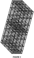
- the minimum matrix is a 2x1 matrix, as shown in figure 3 .
- each niche has one wall in common with the other niche.
- m and n are the ones that permits to obtain a matrix without ruptures or deformations such to compromise the stability of the supermatrix. This can be easily evaluated by a person skilled in the art with the knowledge of the techniques commonly known in the sector, in particular for niche fabrication.
- the distance between one synthetic niche matrix and the next is defined by the value d.
- Said distance d is always greater than zero, therefore the matrices are necessarily spaced one from another.
- said distance is the minimum obtainable using today's fabrication techniques.
- every single synthetic niche has a square section with 90 ⁇ m sides (b), 100 ⁇ m height (a), and having 6 stacked layers of lattices. In every lattice, the pores have dimensions that vary between 10 ⁇ m and 30 ⁇ m. Confinement walls around the niche's perimeter, made of parallel rods at a distance of 5 ⁇ m (f) one from the next, are also provided. The rods of the niche protrude for around one micron beyond the structure.
- the dimensions of the single niche may vary according to the specific type of cell being cultured, while always maintaining three-dimensional structure. A person skilled in the art can determine the modifications to be made to the niche on the basis of cell type.
- the width and depth of the niche may vary between 20 and 500 ⁇ m with pores that vary from 5 to 100 ⁇ m, preferably from 10 ⁇ m to 30 ⁇ m. Height may vary from 5 to 500 ⁇ m, preferably from 30 ⁇ m to 100 ⁇ m.
- Each niche can have several layers of lattices, for example from 2 to 10 layers, preferably from 4 to 6 layers. Confinement walls are preferably made of parallel rods at a distance one from the other which may vary, for example, from 2 to 30 ⁇ m, preferably from 2 to 10 ⁇ m, more preferably it is 5 ⁇ m.
- the height of the supermatrix is 30 ⁇ m, in particular, whenever it is used for mesenchymal stem cell culture. Said height permits all known microscopy techniques for cellular analysis to be applied to the cells that grow in the niches.
- pluripotent stem cells e.g. embryonic
- a height of more than 30 ⁇ m is preferable.
- the supermatrices according to the invention are typically fabricated over a flat support.
- Said flat support is typically made of a material optically transparent to the two-photon laser, preferably glass or a polymer, for example of the polyolefin family.
- the flat support on which the supermatrices of niches are fabricated is chemically or physically modified for the purpose of minimizing cell adhesion to the support areas outside and below the area in which 3D structures are present.
- the flat culture support is coated with an agent that reduces the wettability of the cell culture surface, for example by an antifouling agent such as perfluoropolyether.
- the supermatrices according to the invention are surrounded by a circular crown of Polydimethylsiloxane (PDMS) or equivalent material characterized by elevated biocompatibility and stability.
- PDMS is produced through known art in the sector by mixing two different components: a base and a cross-linking agent in a 10:1 weight ratio, in such way as to permit polymerization.
- PDMS obstructs both physically and chemically the adhesion of the cells outside the area in which 3D structures are present.
- the walls of the niches of the supermatrix are functionalized with molecules.
- these molecules provide an inductive signal for pluripotency maintenance or facilitate the adhesion of the cells to the structure.
- each niche in the structure can be coated with appropriate substances or molecules, ad example for the purpose of modifying the response of the cells cultured.
- each niche can be coated with hyaluron-based and gelatin-based hydrogels in order to simulate a different elasticity in the cellular matrix, as described in an article published by Nava et al. in 2015.
- the supermatrix according to the invention is fabricated using the known technique of two-photon laser polymerization.
- the specific protocols for the production of a supermatrix according to the invention may vary on the basis of the type of cell that is cultured.
- a expert in the field is capable of defining such technical specifications on the basis of his general knowledge in the field.
- the material utilized for production is a resin.
- it is a photopolymerizable resin.
- Said resin is typically an organic/inorganic hybrid photosensitive material that polymerizes when it is illuminated by laser.
- the resin is a photopolymerizable resin known as SZ2080.
- This resin is known ( Ovsianikov A, Viertl J, Chichkov B, Oubaha M, MacCraith B, Sakellari I, et al. Ultra-low shrinkage hybrid photosensitive material for two-photon polymerization micro-fabrication. ACS Nano 2008;2:2257-62 ) and has been developed recently. Its use is preferable thanks to its good optical qualities and mechanical stability.
- the two main components of SZ2080 are meth acrylol oxypropyl trimethoxy silane (MAPTMS) and zirconium propoxide (ZPO).
- the resin's index of refraction can be modified by varying the molar ratio between these two components, and increases proportionally with the increase in ZPO.
- the supermatrix production process comprises three principal phases: baking, laser writing, and development.
- the samples are prepared by hardening the above-mentioned resin.
- a photoinitiator must be incorporated into the resin in order to permit two-photon absorption.
- One preferred photoinitiator is IRG (Irgacure 369,2-Benzyl-2-dimethyl-1-(4-morpholinophenyl)-butanone-1), which is not cytotoxic.
- Another possible photoinitiator is BIS (4-4'bis(diethylamine)benzophenone).
- the resin and photoinitiator solution is deposited by dropcasting, for example, in a volume that varies from 30 to 50 ⁇ L, onto the support on which the supermatrix must be obtained, such as a coverglass, for example, or directly inside multi-well culture plates in which the cells will be seeded.
- the sample is then positioned on a plate.
- This process (baking) is necessary for the solidification of the resin on the glass substrate and to ensure that the solvents in which it is dissolved evaporate. In this way, its consistency changes from liquid to semi-solid.
- the sample then undergoes the writing process.
- the sample is usually positioned in a special housing chamber connected to a motion system composed of a piezoelectric position transducer controlled by software.
- An ultra-short pulsed laser system is used for writing. Any laser commonly used in 2PP technique can be utilized. For example, reference can be made to Raimondi et al., 2013.
- a commercially-available laser can be used, such as the Femtoregen model produced by the HighQ Company, a model based on a mode-locked Ytterbium (Yb) oscillator followed by an amplifier.
- Yb Ytterbium
- the laser typically has a pulse duration in the range of 10 fs and 10 ps, and a wavelength in the range of 400 and 1300 nm.
- the laser is used at a power in the range of 12 and 21 mW and at a writing speed of 1000 ⁇ m/s.
- Stages that permit the translation of the sample mounted on a sample-holder that is solidly connected with the 3D movement system are used to permit complex geometries to be written in the resin.
- Movement commands are given by software via G-code, a language commonly used in numerically controlled machine tools.
- the software program is written in such way as to fill the circular surface area defined in the parameters with all the rods first; then, all the lattices on the different levels are written, first in one direction then the other before finishing the process with the confinement walls that enclose every single structure.
- the supermatrix is obtained by programming the software in such way as to write a series of niche matrices, with the limitation of writing only inside one-quarter of a circle, and then repeating it in all four directions in order to complete the surface. See Figures 11.3 and 11.4 ., for example.
- the movement of the sample compared to that of the laser beam can be programmed using a Computer-Aided Design(CAD)/ Computer Aided Machining(CAM) system or the joint and integrated use of software systems for, CAD, and CAM.
- CAD Computer-Aided Design
- CAM Computer Aided Machining
- PC personal computer
- a person skilled in the art is able to compile the various software that permit the laser writing to be set in accordance with the fabrication desired and the general knowledge in the field.
- the sample undergoes the development phase, i.e. the elimination of the fraction of non-polymerized resin.
- Development typically takes place by immersing the sample in a development solution selected on the basis of the photosensitive resin used.
- a development solution selected on the basis of the photosensitive resin used.
- a 50:50 isopropanol and pentanone solution can be used for around 10 - 15 minutes (duration depends on the quantity of material used and the complexity of the structures created).
- the supports thus obtained can be used for cell culture.
- the seeding of the support with the cells to be cultured can be performed using known techniques in the sector. On the basis of this general knowledge, a person skilled in the art is able to adjust the timing and the other parameters required for cell culture.
- the supermatrix according to the invention can be advantageously used in a wide-scale cell culture system with ample industrial and commercial applications.
- said supermatrix is used to coat the bottom of a culture well, said well that is part, for example, of a mono- or a multi-well culture plate.
- FIG. 12 An exemplary embodiment of a multi-well culture plate whose bottom is coated with supermatrices of synthetic niches according to the invention is depicted in figure 12 .
- the surface of the culture plate can be functionalized with suitable molecules before being coated with the supermatrices.
- it can be chemically or physically modified for the purpose of minimizing cell adhesion to the support areas outside and below the area in which 3D structures are present.
- the culture plate is coated with an agent that reduces the wettability of the cell culture surface, for example it is coated with an antifouling agent such as perfluoropolyether.
- the supports containing the supermatrices according to the invention can be used for any type of cell culture that requires a three-dimensional structure in order to increase the volume occupied by the cells.
- the supports containing the supermatrices according to the invention are used for stem cell culture.
- Said stem cells can be multipotent or pluripotent.
- these can be natural pluripotent cells, such as embryonic stem cells or induced pluripotent cells, i.e. adult somatic cells that are made to regress to pluripotent stem cells through genetic engineering techniques.
- natural pluripotent cells such as embryonic stem cells or induced pluripotent cells, i.e. adult somatic cells that are made to regress to pluripotent stem cells through genetic engineering techniques.
- Adult stem cells can be taken from different types of adult organism tissue, such as umbilical cord, amniotic sac, blood, bone marrow, placenta, and adipose tissue.
- the use of the supermatrices according to the invention for the culture of mesenchymal stem cells is particularly preferred. These can be taken, for example, from the bone marrow or other parts of the body, such as the umbilical cord or adipose tissue, of adult donor volunteers.
- Cells cultured on the supermatrices of the invention can be used on wide scale in regenerative medicine or for the production of tissues to be implanted in vivo.
- they can be used as a therapeutic product to modulate inflammation in degenerative diseases or to avoid immune-rejection in organ transplantation.
- a critical number of cells can be made to differentiate or kept undifferentiated, as required by therapeutic need, and then injected into a patient to treat currently incurable diseases, such as Parkinson's disease, for example.
- secreted by said cultured cells which are known as “secretomes”
- secretomes can be used as therapeutic products, for example in the treatment of degenerative diseases.
- the above-mentioned critical number of cells can be suspended in culture for the purpose of producing a high quantity of "secretome” that can be injected in patients on repetitive basis, for example in order to reduce inflammatory response in degenerative diseases such as Crohn's disease, arthrosis, rheumatoid arthritis, and Alzheimer's disease .
- the supermatrices according to the invention are also useful as laboratory tools and in wide-scale biological research thanks to the possibility to culture a high number of stem cells to be kept undifferentiated for use in substrate for cellular analysis.
- supermatrices according to the invention can also be used as 3D supports to be implanted in vivo in a subject for the purpose of promoting the regeneration of tissue or an organ through cellular and micro-vascular re-population. This can be done using methods commonly known in the field.
- EXAMPLE 1 MICRO-FABRICATION OF THE NICHOID MATRIX FOR STEM CELLS
- the pulses of the femtosecond laser passed first through a power control consisting of a birefringent plate followed by a linear polarizer. The pulses were then focused using an objective lens inside the photoresist, and the position of the sample compared to that of the beam was shifted with the use of a computer-controlled 3-axis motion stage.
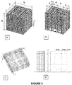
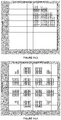
- the software program for niche fabrication was written in G-code in order to fill the circular surface defined in the parameters with all the rods first ( Figure 11.1 ), and then all the lattices on the different levels, first in one direction then the other ( Figure 11.2 ) before finishing the process with the confinement walls that enclose every single structure.
- the nichoid supermatrix was fabricated by programming a series of niches, with the limitation of writing only inside one-quarter of a circle ( Figure 11.3 ), and then repeating it in all four directions in order to complete the coverglass ( Figure 11.4 ).
- the samples were immersed in a 50:50 solution of isopropanol and pentanone in such way as to eliminate the non-polymerized part of the photoresist.
- the niches were observed through optical and SEM microscope and then surrounded with a circular crown of Polydimethylsiloxane (PDMS) in order to obtain a non-fouling surface for the cells around the circular area coated with niches.
- PDMS Polydimethylsiloxane
- RT-PCR Real-Time Polymerase Chain Reaction
- Bone marrow was taken from 2 healthy donors aged 18 and 65 respectively during routine orthopedic surgery procedures with the consent of the local ethics boards (Basel University hospital) after first obtaining informed donor consent.
- the bone marrow aspirate was deposited in a standard flask and cultured for a week in ⁇ -MEM with the addition of 20% FCS, 1% sodium pyruvate, 1% PSG (penicillin G and 0.1 mg/mL streptomycin) and 1% HEPES.
- the culture medium was changed regularly twice a weeks until confluence.
- the MSC were recovered from the bone marrow by taking advantage of their tendency to adhere tightly to culture plates, whereas the other non-adhering cells were removed.
- Adherent cells were detached by trypsin-EDTA (0.5-0.2 g/L; Invitrogen, Carlsbad, CA, USA), counted, and cryo-preserved in ⁇ -MEM supplemented with 30% FCS and 5% dimethyl sulfoxide (DMSO) until use.
- the 2PP-patterned coverglasses (referred to below simply as 2PP samples) were washed thoroughly, kept for 12 hours in deionized water, disinfected for 12 hours in ethanol at 7 %, washed repeatedly in sterile deionized water, and then UV-sterilized. Each sample was positioned in an Ultra-Low Attachment 24-well cell culture plate (Costar 3473, Corning, Corning, NY, United States of America).
- the cryo-preserved cells were resuscitated, suspended in complete medium and seeded directly in the wells at a density of 10,000 cells/cm 2 . The cells were incubated for 21 days with medium freshly replaced twice a week.
- clonogenicity the capacity of cells to form colonies
- the cells were detached, counted, and reseeded in triplicate in Petri plates. In each plate, 600 cells were cultured in complete medium changed twice weekly. After 2 weeks of culture, the cells were washed in PBS, fixed with 3.7% paraformaldehyde in PBS and stained with Crystal Violet for 10 minutes. The dye was then collected and the plates were washed with water and left to dry.
- Clonogenicity capacity was calculated as the percentage between the number of colonies counted manually and the number of cells seeded.
- the number of clonogenic cells i.e the cells capable of forming colonies, CFU was calculated as the product of the total number of cells trypsinized after 3 weeks of culture and the clonogenicity capacity percentage.
- Real-time PCR was performed using the ABIPrism 77000 Sequence Detection System (Perkin Elmer / Applied Biosystem, Rotnch, Svizzera), and the levels of expression of the genes of interest RUNX2, Bone SialoProtein (BSP) and PPAR-gamma were normalized for the reference gene GAPDH. This gene is, in fact, the one that is actively transcribed and translated at relatively high level.
- cells cultured in the sample 2PP structures for three weeks were re-suspended in 200 ⁇ l of BSA at 0.5% in PBS (FACS buffer) with antibodies bonded with fluorochromes for the protein in question.
- the antibodies were incubated for 30 minutes at 4 °C.
- the antibodies used were CD146-PE and IgG1-PE (Becton, Dickinson and Company, Franklin Lakes, NJ). All the antibodies were used with 1:50 dilution.
- the cells were washed 2 times with FACS buffer, re-suspended in PBS, and analyzed by FACS (Becton, Dickinson and Company).
- the cells were fixed in 2% paraformaldehyde, permeabilized with 0.2% Triton, blocked with 2% goat serum albumin (GSA) in PBS, and fluorescently marked.
- GSA goat serum albumin
- the DNA was stained by incubation with 4',6'-diamidino-2-phenylindoledihydrochloride (DAPI) in solution at 10 ⁇ g/ml.
- DAPI 4',6'-diamidino-2-phenylindoledihydrochloride
- a secondary antibody Alexa Fluor 566 nm was used to identify the integrin of the membrane CD146.
- Image acquisition and 3D reconstruction were performed at 20 ⁇ and 60 ⁇ using a confocal laser microscope (Nikon A1R, Japan).
- Adipogenic assays were conducted using standard protocol ( A. Barbero, S. Ploegert, M. Heberer, and I. Martin. Plasticity of clonal populations of dedifferentiated adult human articular chondrocytes. Arthritis Rheum, 48(5):1315-1325, 2003 ).
- osteogenic assays were conducted using the standard protocol described in literature ( N. Jaiswal, S.E. Haynesworth, A.I. Caplan, and S.P. Bruder. Osteogenic differentiation of purified, culture-expanded human mesenchymal stem cells in vitro. J Cell Biochem, 64(2):295-312, 1997 ).
- the matrices M2 and M3 presented the problem of the shrinkage of the polymer during expansion ( Figure 9 ), that caused mechanical stress and deformation, which provided the reason for dividing the structure in smaller matrices (for example of 450 ⁇ m in side, spaced 20 ⁇ m apart).
- the excessive dimensions of the surface covered by the structures risked damaging certain parts of the niche, with repercussions throughout the rest of the structure.
- niches provide stem cells with an increase in the surface area/volume ratio that favors their adhesion and allows much more space for proliferation.
- the synthetic niche system is capable of guiding aggregate formation even with hBM-MSC cells.
- This spontaneous tendency to form aggregates is not species-specific. Differences in cell density values, on the contrary, may instead be species-specific.
- Clonogenic assays were performed on cells cultured on both 2PP sample structures and on glass substrate samples. Even if no statistical significance was observed, a higher average number of cells maintained clonogenic in 2PP samples than on glass. Compared with cells cultured on glass substrates, cells expanded on 2PP substrates showed a significantly greater colony diameter, which is an index of clonogenic potential.
- human MSC expanded on the synthetic niche substrate maintained their proliferative potential, clonogenic capacity and bilineage differentiation potential more effectively than cells expanded on glass substrates and in some aspects were comparable to non-expanded cells.
- the niches and in a particularly advantageous way the extended substrate, can be used for the culture and expansion of undifferentiated MSC.
- the artificial niche substrate permits a reduction of the effects of 2D surfaces that affects biological results. This demonstrates that 3D geometry and the consequent method of cellular adhesion is an important source of stimulation, perhaps the most important, in determining the maintenance of MSC multipotency.
- mESc murine embryonic stem cells
- the experiments on the nichoid supermatrix were conducted using murine embryonic stem cells (mESc). Not adhesion-dependent, these cells are highly clonogenic and characterized by their reduced size (10 ⁇ m diameter) caused by a limited cytoplasmic region, in this way promoting perfect homing inside the engineered niches.
- the mESc utilized are a part of the R1 line and were obtained from Nagy Lab at Mount Sinai Institute, Toronto, Canada.
- the in vitro cell cultures were kept in a controlled-atmosphere incubator at 37°C with 5% CO 2 and 95% relative humidity.
- the culture medium was composed as follows: DMEM (Gibco, 11960), 10% decomplemented FBS (Gibco, 16141), 1% L-Glutamine (Invitrogen, 25030024), 1% non-essential amino acids (Invitrogen, 11140035), 1% sodium pyruvate (Gibco, 11360), 1% Penicillin-Streptomycin (Invitrogen, 15140122), 0.,1% ⁇ -mercaptoethanol 0.1 mM (Sigma M7522) e 1000 U/ml LIF (Millipore, ESG1107).
- the maintenance of the line was promoted by the presence of feeder cells: here, primary CD1 mouse embryonic fibroblasts were used after being treated in p3 with mitomycin-C (Roche, 10107409001), an alkylating agent that induces the formation of a cross-link at DNA level, in this way preventing duplication and therefore cellular proliferation. This treatment is essential in order to enable precise control of the density of these cells in culture.
- the expansion of the mouse embryonic fibroblasts (MEF) up to p3 was conducted in a specific medium composed of DMEM, 10% FBS, 1% Penicillin-Streptomycin and 1% non-essential amino acids.
- the detachment process consisted of an initial incubation phase with trypsin (0.05% in medium R1) at 37 °C for 3 minutes, a second centrifugation step at 1200 rpm for 5 minutes, and lastly, the re-suspension of the pellet in complete R1 medium (R1+LIF) followed by new seeding in flask in the presence of MEF.
- Cryo-preservation was performed with DMSO (dimethyl-sulfoxide), which serves as an anti-crystallizing agent, and subsequent storage in liquid nitrogen atmosphere at temperatures that should never exceed -140 °C.
- the cryo-preservation medium was composed of 10% DMSO, 10% FBS, and 80% complete R1 medium with the addition of LIF.
- tissue culture hood Jupiter, Cell Bio: a constant flow of air in the range of 545-550 m 3 /h permits operations to be performed in sterile conditions. Sterility is guaranteed by a HEPA filter positioned downstream from the inflow designed to withhold 99.97% of the particles with greater than 0.3 ⁇ m diameter.
- the configuration was modified in order to obtain higher niches surface density.
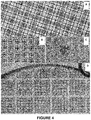
- Two new sets of niches were produced: the first generation had a continuous matrix of synthetic niches produced by the pairing of single niches that completely covered a 6 mm diameter circular region. This region was enclosed by a PDMS polymer crown that permits the maximization of the area covered by the niches compared to the glass substrate's two-dimensional surface, in this way enabling the coverage of 96% of the total surface (see Figure 4A ).
- the 2D controls structure
- the two-dimensional sample is the negative control structure adopted in our experiment, meaning that in this particular condition we expect the cells to completely lose their capacity for self-renewal and generate the expression of the characteristic genes of differentiated phenotype.
- Glass was selected for the 2D control structure.
- the control structure had a PDMS crown.
- the crown is obtained by pouring the base and cross-linking solution (10:1) in a metal mold that is inserted in an oven for the baking step conducted at 80 °C for 30-45 minutes as required by the hardness that must be given to the crown.
- Air-plasma treatment is performed under a tissue culture hood, with an ionizer in order to expose and load the hydroxyl groups (-OH), which in this way are capable of bonding together by ionic bonding with the silicon atom in the glass's siloxane groups.
- the samples fixed at the three established measurement time intervals were examined by fluorescence microscopy.
- the protein associated with the gene was recognized through the primary antibodies that bond directly to the target. Viewing under fluorescence microscopy was enabled by a fluorophore bonded to the secondary antibody that possesses affinity for the primary antibody to which it binds. Stemness and differentiation markers are then used for distinction.
- Oct4 Oletamer-Binding Transcription factor 4
- a-SMA Smooth Muscle Actinin
- NKX2-5 Homeobox-protein NKX2-5
- GATA-4 Homeobox-protein NKX2-5
- SOX-17 ⁇ III-Tubulin.
- Matrix markers Osteocalcin; Collagen type I.
- the samples were analyzed using various microscope techniques depending on what was to be observed, the degree of detail to be reached, and whether the culture being examined was to be kept alive or not.
- the types of microscopic analysis adopted were phase contrast microscopy, confocal microscopy, and Scanning Electron Microscope (SEM).
- the first type of quantification in this regard is the measurement of the diameters of the colonies present throughout the entire sample for all three types of scaffold.
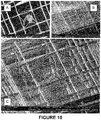
- the three-dimensional niche environment exerts a different effect on cultures than the 2D control structure. After seeding, the cells precipitate inside the medium until reaching the niche: some cells fall directly into the pores on the top side and adapt to the 3D structure, making use of their podia and protrusions (see Figure 10 ), whereas others, where the pores are smaller, stop on the top side without penetrating.
- the 2D control structure constantly promotes higher average diameters than those of the colonies on the nichoid over time.
- the two niche configurations seem to affect growth in the same way.
- One important aspect is the ratio between average colony diameter and characteristic niche dimension. The fact that these two dimensions remain similar provides important indication of the fact that the colonies are effectively inside the niche because they exert, as previously mentioned, a physical containment effect: this is undeniably true for the measurement made at the first time interval. After around 7 days, however, a part of the colonies begins growing beyond the 90 ⁇ m containment area, and after 14 days, the formation of EBs has become predominant with the development of colonies above the top side of the niche.
- the setting was conducted by culturing mES on 18x18 mm square coverglasses having the same mechanical and surface characteristics as the glass substrate of the 2D control structures and the niches.
- the definitive markers used were therefore OCT4, GATA4, NKX2-5, and ⁇ SMA.
- the three differentiation markers all characterize the cardio-myocyte colonies in various ways.
- first generation nichoids were used, in other words, those with surfaces inside the PDMS crown completely written and occupied by the structure. Also niches obtained through trypsinization used in a previous test were tested.
- the NKX2-5 cardiac marker was observed to be not expressed in all culture conditions.
- the expression trend proved to be as expected and already qualitatively assessed on glass in the calibration test.
- GATA4 displays the classic up-down-up expression on the 2D control structure, and as already observed, presents high levels at T0, whereas ⁇ SMA increases in terms of area as the culture proceeds.
- Nichoids instead, demonstrate very different trends for the two markers, but share the characteristic of reducing both.
- an initial level of GATA4 comparable to that of the control structure, the expression of the gene in successive measurements was completely extinguished in the niche.
- the niche was shown to discourage such expression, even if at culture day 14 it was present in minimum degree, but much lower than in the 2D control structure, however.
- One important aspect is that the differentiation marker results obtained with new and re-utilized nichoids were entirely comparable, and this means that the structure maintains its properties even when it is reused.
- the niche was shown to promote pluripotency more than the other two control structures while also inhibiting differentiation.
- the 2D control structure demonstrated the behaviors expected, promoting the expression of GATA4, and above all, ⁇ SMA, which localized on the sides of the colonies in contact with glass, confirming that the stiffness of the substrate is fundamental for its promotion.
- mESc murine embryonic stem cells
- two separate substrates were cultured: the niches and a 2D control structure (glass).
- the cultures were allowed to proceed for 14 days without any chemical conditioning (LIF).
- LIF chemical conditioning
- the results show that the niche promoted pluripotency maintenance with the expression of OCT4 to a much greater degree than the control structure present while at the same time inhibiting differentiation, characterized by the markers such as GATA4, ⁇ SMA, and NKX2-5 that were instead expressed on the glass control.
- the synthetic niche is capable of exerting a tension state favorable to maintaining the pluripotency of mESc as well.
- the supermatrix according to the invention maintains the same characteristics of the individual niche, with the fundamental advantage of offering a much larger surface combined with elevated structural stability that increases culture capacity considerably.
Landscapes
- Health & Medical Sciences (AREA)
- Life Sciences & Earth Sciences (AREA)
- Engineering & Computer Science (AREA)
- Biomedical Technology (AREA)
- Chemical & Material Sciences (AREA)
- Zoology (AREA)
- Bioinformatics & Cheminformatics (AREA)
- Genetics & Genomics (AREA)
- Wood Science & Technology (AREA)
- Biotechnology (AREA)
- Organic Chemistry (AREA)
- General Health & Medical Sciences (AREA)
- Cell Biology (AREA)
- Developmental Biology & Embryology (AREA)
- Microbiology (AREA)
- General Engineering & Computer Science (AREA)
- Biochemistry (AREA)
- Public Health (AREA)
- Animal Behavior & Ethology (AREA)
- Veterinary Medicine (AREA)
- Epidemiology (AREA)
- Dermatology (AREA)
- Medicinal Chemistry (AREA)
- Oral & Maxillofacial Surgery (AREA)
- Transplantation (AREA)
- Hematology (AREA)
- Gynecology & Obstetrics (AREA)
- Reproductive Health (AREA)
- Chemical Kinetics & Catalysis (AREA)
- Urology & Nephrology (AREA)
- Botany (AREA)
- Immunology (AREA)
- Rheumatology (AREA)
- Dispersion Chemistry (AREA)
- Apparatus Associated With Microorganisms And Enzymes (AREA)
- Micro-Organisms Or Cultivation Processes Thereof (AREA)
Claims (15)
- Supermatrix aus synthetischen Nischen mit mindestens zwei Matrizen aus synthetischen Nischen,- wobei jede Matrix n × m synthetische Nischen aufweist, wobei n und m, die gleich oder voneinander verschieden sind, einen Wert ≥ 1 aufweisen, vorausgesetzt, dass einer von m oder n ≥ 2 und ein Maximalwert von m und n ist, der ein Aufrechterhalten der Struktur der einzelnen synthetischen unversehrten Nischen ermöglicht, so dass ein Schrumpfen des Materials keine signifikanten Störungen verursacht, und- wobei der Abstand (d) zwischen einer synthetischen Nischenmatrix und der anderen größer als Null ist, und- wobei in jeder Matrix jede synthetische Nische eine oder mehrere Wände gemeinsam mit der (den) anderen synthetischen Nische (n) der Matrix aufweist.
- Supermatrix nach Anspruch 1, wobei der Maximalwert für n und m 100 ist.
- Supermatrix nach Anspruch 1 oder 2, wobei m und n den gleichen Wert aufweisen.
- Supermatrix nach einem der Ansprüche 1 bis 3, wobei die Höhe der Supermatrix zwischen 30 und 100 µm liegt.
- Supermatrix nach einem der Ansprüche 1 bis 4, die aus einem photopolymerisierbaren Harz hergestellt ist.
- Supermatrix nach einem der Ansprüche 1 bis 5, wobei die Wände einer oder mehrerer der synthetischen Nischen mit Molekülen, die ein Signal vorsehen, das eine Erhaltung der Pluripotenz erzeugt, und / oder mit Molekülen bedeckt sind, die ein Anhaften von Zellen an der Nische erleichtern.
- Unterlage für die Kultur von Zellen mit mindestens einer Supermatrix nach einem der Ansprüche 1 bis 6.
- Unterlage nach Anspruch 7, die aus Glas oder einem Polymer hergestellt ist.
- Unterlage nach einem der Ansprüche 7 bis 8, bei der die Oberfläche, auf der die Supermatrizen hergestellt sind, mit einem Mittel, vorzugsweise einem Antifoulingmittel, beschichtet ist, das die Benetzbarkeit der Oberfläche verringert.
- Unterlage nach einem der Ansprüche 7 bis 9, die eine Monowell- oder Multiwell-Kulturplatte oder ein Kolben ist.
- Multiwell- Kulturplatte, bei der der Boden der Platte von einer oder mehreren Supermatrizen aus synthetischen Nischen nach einem der Ansprüche 1-6 bedeckt ist.
- Verwendung der Supermatrix nach einem der Ansprüche 1-6 oder der Unterlage nach einem der Ansprüche 7-10 für die In-Vitro-Kultur von Zellen.
- Verwendung nach Anspruch 12, wobei die Zellen Stammzellen, vorzugsweise multipotente oder pluripotente Stammzellen, sind.
- Verwendung der Supermatrix nach einem der Ansprüche 1 bis 6 oder der Unterlage nach einem der Ansprüche 7-10 bei der Analyse von Zellproben, insbesondere auf diagnostischem Gebiet.
- Supermatrix nach einem der Ansprüche 1 bis 6 zur Verwendung für ein in-vivo Implantat in einem Subjekt, um eine Gewebe- oder Organregeneration durch eine Zellrepopulation zu fördern.
Applications Claiming Priority (2)
| Application Number | Priority Date | Filing Date | Title |
|---|---|---|---|
| ITUB2015A003414A ITUB20153414A1 (it) | 2015-09-04 | 2015-09-04 | Matrici di nicchioidi sintetiche per la coltivazione di cellule staminali |
| PCT/EP2016/070500 WO2017037108A1 (en) | 2015-09-04 | 2016-08-31 | Synthetic niche matrices for stem cell culture |
Publications (2)
| Publication Number | Publication Date |
|---|---|
| EP3344305A1 EP3344305A1 (de) | 2018-07-11 |
| EP3344305B1 true EP3344305B1 (de) | 2019-07-03 |
Family
ID=55085709
Family Applications (1)
| Application Number | Title | Priority Date | Filing Date |
|---|---|---|---|
| EP16763469.0A Active EP3344305B1 (de) | 2015-09-04 | 2016-08-31 | Synthetische nischenmatrizen für stammzellkultur |
Country Status (6)
| Country | Link |
|---|---|
| US (1) | US11027045B2 (de) |
| EP (1) | EP3344305B1 (de) |
| ES (1) | ES2743973T3 (de) |
| IT (1) | ITUB20153414A1 (de) |
| LT (1) | LT3344305T (de) |
| WO (1) | WO2017037108A1 (de) |
Families Citing this family (4)
| Publication number | Priority date | Publication date | Assignee | Title |
|---|---|---|---|---|
| US11273023B2 (en) | 2017-12-21 | 2022-03-15 | Politecnico Di Milano | Implantable medical device |
| IT201900003377A1 (it) | 2019-03-08 | 2020-09-08 | Univ Degli Studi Milano | Induzione geometrica di pluripotenza |
| EP4602153A1 (de) | 2022-10-11 | 2025-08-20 | Fondazione Telethon ETS | 3d-zellkulturverfahren |
| CN118086048B (zh) * | 2024-03-27 | 2024-12-03 | 苏州大学 | 一种三维细胞球培养装置及其培养方法 |
-
2015
- 2015-09-04 IT ITUB2015A003414A patent/ITUB20153414A1/it unknown
-
2016
- 2016-08-31 ES ES16763469T patent/ES2743973T3/es active Active
- 2016-08-31 EP EP16763469.0A patent/EP3344305B1/de active Active
- 2016-08-31 US US15/755,011 patent/US11027045B2/en active Active
- 2016-08-31 WO PCT/EP2016/070500 patent/WO2017037108A1/en not_active Ceased
- 2016-08-31 LT LT16763469T patent/LT3344305T/lt unknown
Non-Patent Citations (1)
| Title |
|---|
| None * |
Also Published As
| Publication number | Publication date |
|---|---|
| LT3344305T (lt) | 2019-10-25 |
| US20200046881A1 (en) | 2020-02-13 |
| ITUB20153414A1 (it) | 2017-03-04 |
| US11027045B2 (en) | 2021-06-08 |
| WO2017037108A1 (en) | 2017-03-09 |
| EP3344305A1 (de) | 2018-07-11 |
| ES2743973T3 (es) | 2020-02-21 |
Similar Documents
| Publication | Publication Date | Title |
|---|---|---|
| Colle et al. | Bioprinting predifferentiated adipose-derived mesenchymal stem cell spheroids with methacrylated gelatin ink for adipose tissue engineering | |
| Zhou et al. | Screening platform for cell contact guidance based on inorganic biomaterial micro/nanotopographical gradients | |
| Ovsianikov et al. | Three-dimensional laser micro-and nano-structuring of acrylated poly (ethylene glycol) materials and evaluation of their cytoxicity for tissue engineering applications | |
| JP6949385B2 (ja) | 細胞培養支持体、細胞培養装置、細胞培養キット、及び細胞シート | |
| Jeon et al. | A mini‐review: cell response to microscale, nanoscale, and hierarchical patterning of surface structure | |
| Marino et al. | The Osteoprint: a bioinspired two-photon polymerized 3-D structure for the enhancement of bone-like cell differentiation | |
| Tayalia et al. | 3D cell‐migration studies using two‐photon engineered polymer scaffolds | |
| Wu et al. | Construction of biomimetic natural wood hierarchical porous-structure bioceramic with micro/nanowhisker coating to modulate cellular behavior and osteoinductive activity | |
| US20130203146A1 (en) | Microfabricated scaffold structures | |
| Zhang et al. | Topographical patterning: characteristics of current processing techniques, controllable effects on material properties and co-cultured cell fate, updated applications in tissue engineering, and improvement strategies | |
| EP1668117A4 (de) | Nanotopographische zusammensetzungen und verfahren zur zellulären organisation in geweberekonstruktionsstrukturen | |
| JP6021802B2 (ja) | 培養方法及び薬物スクリーニング方法 | |
| JP2012509663A (ja) | 細胞培養のための、突起間隔を開けた基板および装置 | |
| US11027045B2 (en) | Synthetic niche matrices for stem cell culture | |
| Liu et al. | Synergistic effect of surface chemistry and surface topography gradient on osteogenic/adipogenic differentiation of hMSCs | |
| Psycharakis et al. | Tailor-made three-dimensional hybrid scaffolds for cell cultures | |
| Tejavibulya et al. | Directed self-assembly of large scaffold-free multi-cellular honeycomb structures | |
| Li et al. | Improved neuron culture using scaffolds made of three-dimensional PDMS micro-lattices | |
| Griffin et al. | Development of mechano-responsive polymeric scaffolds using functionalized silica nano-fillers for the control of cellular functions | |
| Gill et al. | Towards the fabrication of artificial 3D microdevices for neural cell networks | |
| Lee et al. | Controlling cell adhesion on human tissue by soft lithography | |
| Zhang et al. | Effects of biomimetic micropatterned surfaces on the adhesion and morphology of cervical cancer cells | |
| Estevez et al. | Biomaterials & architecture, a possible future: Bio printing architecture | |
| Trautmann et al. | Large 3D direct laser written scaffolds for tissue engineering applications | |
| EP3585879A1 (de) | Neuartige biomaterialsubstrate, zellkultursysteme damit und verwendungen davon in zell-screening-anwendungen |
Legal Events
| Date | Code | Title | Description |
|---|---|---|---|
| STAA | Information on the status of an ep patent application or granted ep patent |
Free format text: STATUS: THE INTERNATIONAL PUBLICATION HAS BEEN MADE |
|
| PUAI | Public reference made under article 153(3) epc to a published international application that has entered the european phase |
Free format text: ORIGINAL CODE: 0009012 |
|
| STAA | Information on the status of an ep patent application or granted ep patent |
Free format text: STATUS: REQUEST FOR EXAMINATION WAS MADE |
|
| 17P | Request for examination filed |
Effective date: 20180312 |
|
| AK | Designated contracting states |
Kind code of ref document: A1 Designated state(s): AL AT BE BG CH CY CZ DE DK EE ES FI FR GB GR HR HU IE IS IT LI LT LU LV MC MK MT NL NO PL PT RO RS SE SI SK SM TR |
|
| AX | Request for extension of the european patent |
Extension state: BA ME |
|
| DAV | Request for validation of the european patent (deleted) | ||
| DAX | Request for extension of the european patent (deleted) | ||
| RIC1 | Information provided on ipc code assigned before grant |
Ipc: A61L 27/56 20060101AFI20190109BHEP Ipc: C12N 5/0735 20100101ALI20190109BHEP Ipc: C12N 5/00 20060101ALI20190109BHEP Ipc: C12N 5/0775 20100101ALI20190109BHEP Ipc: A61L 27/38 20060101ALI20190109BHEP Ipc: A61L 27/50 20060101ALI20190109BHEP |
|
| GRAP | Despatch of communication of intention to grant a patent |
Free format text: ORIGINAL CODE: EPIDOSNIGR1 |
|
| STAA | Information on the status of an ep patent application or granted ep patent |
Free format text: STATUS: GRANT OF PATENT IS INTENDED |
|
| INTG | Intention to grant announced |
Effective date: 20190215 |
|
| GRAS | Grant fee paid |
Free format text: ORIGINAL CODE: EPIDOSNIGR3 |
|
| GRAA | (expected) grant |
Free format text: ORIGINAL CODE: 0009210 |
|
| STAA | Information on the status of an ep patent application or granted ep patent |
Free format text: STATUS: THE PATENT HAS BEEN GRANTED |
|
| AK | Designated contracting states |
Kind code of ref document: B1 Designated state(s): AL AT BE BG CH CY CZ DE DK EE ES FI FR GB GR HR HU IE IS IT LI LT LU LV MC MK MT NL NO PL PT RO RS SE SI SK SM TR |
|
| REG | Reference to a national code |
Ref country code: GB Ref legal event code: FG4D |
|
| REG | Reference to a national code |
Ref country code: AT Ref legal event code: REF Ref document number: 1150203 Country of ref document: AT Kind code of ref document: T Effective date: 20190715 Ref country code: CH Ref legal event code: EP |
|
| REG | Reference to a national code |
Ref country code: IE Ref legal event code: FG4D |
|
| REG | Reference to a national code |
Ref country code: DE Ref legal event code: R096 Ref document number: 602016016417 Country of ref document: DE |
|
| REG | Reference to a national code |
Ref country code: NL Ref legal event code: FP |
|
| PG25 | Lapsed in a contracting state [announced via postgrant information from national office to epo] |
Ref country code: HR Free format text: LAPSE BECAUSE OF FAILURE TO SUBMIT A TRANSLATION OF THE DESCRIPTION OR TO PAY THE FEE WITHIN THE PRESCRIBED TIME-LIMIT Effective date: 20190703 Ref country code: FI Free format text: LAPSE BECAUSE OF FAILURE TO SUBMIT A TRANSLATION OF THE DESCRIPTION OR TO PAY THE FEE WITHIN THE PRESCRIBED TIME-LIMIT Effective date: 20190703 Ref country code: CZ Free format text: LAPSE BECAUSE OF FAILURE TO SUBMIT A TRANSLATION OF THE DESCRIPTION OR TO PAY THE FEE WITHIN THE PRESCRIBED TIME-LIMIT Effective date: 20190703 Ref country code: PT Free format text: LAPSE BECAUSE OF FAILURE TO SUBMIT A TRANSLATION OF THE DESCRIPTION OR TO PAY THE FEE WITHIN THE PRESCRIBED TIME-LIMIT Effective date: 20191104 Ref country code: SE Free format text: LAPSE BECAUSE OF FAILURE TO SUBMIT A TRANSLATION OF THE DESCRIPTION OR TO PAY THE FEE WITHIN THE PRESCRIBED TIME-LIMIT Effective date: 20190703 Ref country code: NO Free format text: LAPSE BECAUSE OF FAILURE TO SUBMIT A TRANSLATION OF THE DESCRIPTION OR TO PAY THE FEE WITHIN THE PRESCRIBED TIME-LIMIT Effective date: 20191003 Ref country code: BG Free format text: LAPSE BECAUSE OF FAILURE TO SUBMIT A TRANSLATION OF THE DESCRIPTION OR TO PAY THE FEE WITHIN THE PRESCRIBED TIME-LIMIT Effective date: 20191003 |
|
| REG | Reference to a national code |
Ref country code: ES Ref legal event code: FG2A Ref document number: 2743973 Country of ref document: ES Kind code of ref document: T3 Effective date: 20200221 |
|
| PG25 | Lapsed in a contracting state [announced via postgrant information from national office to epo] |
Ref country code: IS Free format text: LAPSE BECAUSE OF FAILURE TO SUBMIT A TRANSLATION OF THE DESCRIPTION OR TO PAY THE FEE WITHIN THE PRESCRIBED TIME-LIMIT Effective date: 20191103 Ref country code: RS Free format text: LAPSE BECAUSE OF FAILURE TO SUBMIT A TRANSLATION OF THE DESCRIPTION OR TO PAY THE FEE WITHIN THE PRESCRIBED TIME-LIMIT Effective date: 20190703 Ref country code: LV Free format text: LAPSE BECAUSE OF FAILURE TO SUBMIT A TRANSLATION OF THE DESCRIPTION OR TO PAY THE FEE WITHIN THE PRESCRIBED TIME-LIMIT Effective date: 20190703 Ref country code: AL Free format text: LAPSE BECAUSE OF FAILURE TO SUBMIT A TRANSLATION OF THE DESCRIPTION OR TO PAY THE FEE WITHIN THE PRESCRIBED TIME-LIMIT Effective date: 20190703 Ref country code: GR Free format text: LAPSE BECAUSE OF FAILURE TO SUBMIT A TRANSLATION OF THE DESCRIPTION OR TO PAY THE FEE WITHIN THE PRESCRIBED TIME-LIMIT Effective date: 20191004 |
|
| PG25 | Lapsed in a contracting state [announced via postgrant information from national office to epo] |
Ref country code: TR Free format text: LAPSE BECAUSE OF FAILURE TO SUBMIT A TRANSLATION OF THE DESCRIPTION OR TO PAY THE FEE WITHIN THE PRESCRIBED TIME-LIMIT Effective date: 20190703 |
|
| PG25 | Lapsed in a contracting state [announced via postgrant information from national office to epo] |
Ref country code: RO Free format text: LAPSE BECAUSE OF FAILURE TO SUBMIT A TRANSLATION OF THE DESCRIPTION OR TO PAY THE FEE WITHIN THE PRESCRIBED TIME-LIMIT Effective date: 20190703 Ref country code: PL Free format text: LAPSE BECAUSE OF FAILURE TO SUBMIT A TRANSLATION OF THE DESCRIPTION OR TO PAY THE FEE WITHIN THE PRESCRIBED TIME-LIMIT Effective date: 20190703 Ref country code: EE Free format text: LAPSE BECAUSE OF FAILURE TO SUBMIT A TRANSLATION OF THE DESCRIPTION OR TO PAY THE FEE WITHIN THE PRESCRIBED TIME-LIMIT Effective date: 20190703 Ref country code: DK Free format text: LAPSE BECAUSE OF FAILURE TO SUBMIT A TRANSLATION OF THE DESCRIPTION OR TO PAY THE FEE WITHIN THE PRESCRIBED TIME-LIMIT Effective date: 20190703 |
|
| PG25 | Lapsed in a contracting state [announced via postgrant information from national office to epo] |
Ref country code: LU Free format text: LAPSE BECAUSE OF NON-PAYMENT OF DUE FEES Effective date: 20190831 Ref country code: SM Free format text: LAPSE BECAUSE OF FAILURE TO SUBMIT A TRANSLATION OF THE DESCRIPTION OR TO PAY THE FEE WITHIN THE PRESCRIBED TIME-LIMIT Effective date: 20190703 Ref country code: SK Free format text: LAPSE BECAUSE OF FAILURE TO SUBMIT A TRANSLATION OF THE DESCRIPTION OR TO PAY THE FEE WITHIN THE PRESCRIBED TIME-LIMIT Effective date: 20190703 Ref country code: IS Free format text: LAPSE BECAUSE OF FAILURE TO SUBMIT A TRANSLATION OF THE DESCRIPTION OR TO PAY THE FEE WITHIN THE PRESCRIBED TIME-LIMIT Effective date: 20200224 Ref country code: MC Free format text: LAPSE BECAUSE OF FAILURE TO SUBMIT A TRANSLATION OF THE DESCRIPTION OR TO PAY THE FEE WITHIN THE PRESCRIBED TIME-LIMIT Effective date: 20190703 |
|
| REG | Reference to a national code |
Ref country code: BE Ref legal event code: MM Effective date: 20190831 |
|
| REG | Reference to a national code |
Ref country code: DE Ref legal event code: R097 Ref document number: 602016016417 Country of ref document: DE |
|
| PLBE | No opposition filed within time limit |
Free format text: ORIGINAL CODE: 0009261 |
|
| STAA | Information on the status of an ep patent application or granted ep patent |
Free format text: STATUS: NO OPPOSITION FILED WITHIN TIME LIMIT |
|
| PG2D | Information on lapse in contracting state deleted |
Ref country code: IS |
|
| PG25 | Lapsed in a contracting state [announced via postgrant information from national office to epo] |
Ref country code: IE Free format text: LAPSE BECAUSE OF NON-PAYMENT OF DUE FEES Effective date: 20190831 |
|
| 26N | No opposition filed |
Effective date: 20200603 |
|
| PG25 | Lapsed in a contracting state [announced via postgrant information from national office to epo] |
Ref country code: SI Free format text: LAPSE BECAUSE OF FAILURE TO SUBMIT A TRANSLATION OF THE DESCRIPTION OR TO PAY THE FEE WITHIN THE PRESCRIBED TIME-LIMIT Effective date: 20190703 Ref country code: BE Free format text: LAPSE BECAUSE OF NON-PAYMENT OF DUE FEES Effective date: 20190831 |
|
| REG | Reference to a national code |
Ref country code: AT Ref legal event code: UEP Ref document number: 1150203 Country of ref document: AT Kind code of ref document: T Effective date: 20190703 |
|
| PG25 | Lapsed in a contracting state [announced via postgrant information from national office to epo] |
Ref country code: CY Free format text: LAPSE BECAUSE OF FAILURE TO SUBMIT A TRANSLATION OF THE DESCRIPTION OR TO PAY THE FEE WITHIN THE PRESCRIBED TIME-LIMIT Effective date: 20190703 |
|
| PG25 | Lapsed in a contracting state [announced via postgrant information from national office to epo] |
Ref country code: MT Free format text: LAPSE BECAUSE OF FAILURE TO SUBMIT A TRANSLATION OF THE DESCRIPTION OR TO PAY THE FEE WITHIN THE PRESCRIBED TIME-LIMIT Effective date: 20190703 Ref country code: HU Free format text: LAPSE BECAUSE OF FAILURE TO SUBMIT A TRANSLATION OF THE DESCRIPTION OR TO PAY THE FEE WITHIN THE PRESCRIBED TIME-LIMIT; INVALID AB INITIO Effective date: 20160831 |
|
| PG25 | Lapsed in a contracting state [announced via postgrant information from national office to epo] |
Ref country code: MK Free format text: LAPSE BECAUSE OF FAILURE TO SUBMIT A TRANSLATION OF THE DESCRIPTION OR TO PAY THE FEE WITHIN THE PRESCRIBED TIME-LIMIT Effective date: 20190703 |
|
| P01 | Opt-out of the competence of the unified patent court (upc) registered |
Effective date: 20230615 |
|
| PGFP | Annual fee paid to national office [announced via postgrant information from national office to epo] |
Ref country code: NL Payment date: 20250821 Year of fee payment: 10 |
|
| PGFP | Annual fee paid to national office [announced via postgrant information from national office to epo] |
Ref country code: ES Payment date: 20250926 Year of fee payment: 10 |
|
| PGFP | Annual fee paid to national office [announced via postgrant information from national office to epo] |
Ref country code: LT Payment date: 20250723 Year of fee payment: 10 Ref country code: DE Payment date: 20250820 Year of fee payment: 10 |
|
| PGFP | Annual fee paid to national office [announced via postgrant information from national office to epo] |
Ref country code: IT Payment date: 20250704 Year of fee payment: 10 |
|
| PGFP | Annual fee paid to national office [announced via postgrant information from national office to epo] |
Ref country code: GB Payment date: 20250821 Year of fee payment: 10 |
|
| PGFP | Annual fee paid to national office [announced via postgrant information from national office to epo] |
Ref country code: FR Payment date: 20250828 Year of fee payment: 10 Ref country code: AT Payment date: 20250821 Year of fee payment: 10 |
|
| PGFP | Annual fee paid to national office [announced via postgrant information from national office to epo] |
Ref country code: CH Payment date: 20250901 Year of fee payment: 10 |