EP3337386B1 - System for treating acute and chronic heart failure - Google Patents
System for treating acute and chronic heart failure Download PDFInfo
- Publication number
- EP3337386B1 EP3337386B1 EP16757430.0A EP16757430A EP3337386B1 EP 3337386 B1 EP3337386 B1 EP 3337386B1 EP 16757430 A EP16757430 A EP 16757430A EP 3337386 B1 EP3337386 B1 EP 3337386B1
- Authority
- EP
- European Patent Office
- Prior art keywords
- patient
- flow limiting
- controller
- limiting element
- occlusion
- Prior art date
- Legal status (The legal status is an assumption and is not a legal conclusion. Google has not performed a legal analysis and makes no representation as to the accuracy of the status listed.)
- Active
Links
Images


Classifications
-
- A—HUMAN NECESSITIES
- A61—MEDICAL OR VETERINARY SCIENCE; HYGIENE
- A61B—DIAGNOSIS; SURGERY; IDENTIFICATION
- A61B5/00—Measuring for diagnostic purposes; Identification of persons
- A61B5/02—Detecting, measuring or recording for evaluating the cardiovascular system, e.g. pulse, heart rate, blood pressure or blood flow
- A61B5/021—Measuring pressure in heart or blood vessels
- A61B5/0215—Measuring pressure in heart or blood vessels by means inserted into the body
-
- A—HUMAN NECESSITIES
- A61—MEDICAL OR VETERINARY SCIENCE; HYGIENE
- A61B—DIAGNOSIS; SURGERY; IDENTIFICATION
- A61B17/00—Surgical instruments, devices or methods
- A61B17/12—Surgical instruments, devices or methods for ligaturing or otherwise compressing tubular parts of the body, e.g. blood vessels or umbilical cord
- A61B17/12022—Occluding by internal devices, e.g. balloons or releasable wires
- A61B17/12027—Type of occlusion
- A61B17/12036—Type of occlusion partial occlusion
-
- A—HUMAN NECESSITIES
- A61—MEDICAL OR VETERINARY SCIENCE; HYGIENE
- A61B—DIAGNOSIS; SURGERY; IDENTIFICATION
- A61B17/00—Surgical instruments, devices or methods
- A61B17/12—Surgical instruments, devices or methods for ligaturing or otherwise compressing tubular parts of the body, e.g. blood vessels or umbilical cord
- A61B17/12022—Occluding by internal devices, e.g. balloons or releasable wires
- A61B17/12027—Type of occlusion
- A61B17/1204—Type of occlusion temporary occlusion
-
- A—HUMAN NECESSITIES
- A61—MEDICAL OR VETERINARY SCIENCE; HYGIENE
- A61B—DIAGNOSIS; SURGERY; IDENTIFICATION
- A61B17/00—Surgical instruments, devices or methods
- A61B17/12—Surgical instruments, devices or methods for ligaturing or otherwise compressing tubular parts of the body, e.g. blood vessels or umbilical cord
- A61B17/12022—Occluding by internal devices, e.g. balloons or releasable wires
- A61B17/12099—Occluding by internal devices, e.g. balloons or releasable wires characterised by the location of the occluder
- A61B17/12109—Occluding by internal devices, e.g. balloons or releasable wires characterised by the location of the occluder in a blood vessel
-
- A—HUMAN NECESSITIES
- A61—MEDICAL OR VETERINARY SCIENCE; HYGIENE
- A61B—DIAGNOSIS; SURGERY; IDENTIFICATION
- A61B17/00—Surgical instruments, devices or methods
- A61B17/12—Surgical instruments, devices or methods for ligaturing or otherwise compressing tubular parts of the body, e.g. blood vessels or umbilical cord
- A61B17/12022—Occluding by internal devices, e.g. balloons or releasable wires
- A61B17/12131—Occluding by internal devices, e.g. balloons or releasable wires characterised by the type of occluding device
- A61B17/12136—Balloons
-
- A—HUMAN NECESSITIES
- A61—MEDICAL OR VETERINARY SCIENCE; HYGIENE
- A61B—DIAGNOSIS; SURGERY; IDENTIFICATION
- A61B5/00—Measuring for diagnostic purposes; Identification of persons
- A61B5/02—Detecting, measuring or recording for evaluating the cardiovascular system, e.g. pulse, heart rate, blood pressure or blood flow
- A61B5/024—Measuring pulse rate or heart rate
- A61B5/02438—Measuring pulse rate or heart rate with portable devices, e.g. worn by the patient
-
- A—HUMAN NECESSITIES
- A61—MEDICAL OR VETERINARY SCIENCE; HYGIENE
- A61B—DIAGNOSIS; SURGERY; IDENTIFICATION
- A61B5/00—Measuring for diagnostic purposes; Identification of persons
- A61B5/02—Detecting, measuring or recording for evaluating the cardiovascular system, e.g. pulse, heart rate, blood pressure or blood flow
- A61B5/026—Measuring blood flow
-
- A—HUMAN NECESSITIES
- A61—MEDICAL OR VETERINARY SCIENCE; HYGIENE
- A61B—DIAGNOSIS; SURGERY; IDENTIFICATION
- A61B5/00—Measuring for diagnostic purposes; Identification of persons
- A61B5/24—Detecting, measuring or recording bioelectric or biomagnetic signals of the body or parts thereof
- A61B5/25—Bioelectric electrodes therefor
- A61B5/279—Bioelectric electrodes therefor specially adapted for particular uses
- A61B5/28—Bioelectric electrodes therefor specially adapted for particular uses for electrocardiography [ECG]
- A61B5/283—Invasive
- A61B5/29—Invasive for permanent or long-term implantation
-
- A—HUMAN NECESSITIES
- A61—MEDICAL OR VETERINARY SCIENCE; HYGIENE
- A61B—DIAGNOSIS; SURGERY; IDENTIFICATION
- A61B5/00—Measuring for diagnostic purposes; Identification of persons
- A61B5/68—Arrangements of detecting, measuring or recording means, e.g. sensors, in relation to patient
- A61B5/6846—Arrangements of detecting, measuring or recording means, e.g. sensors, in relation to patient specially adapted to be brought in contact with an internal body part, i.e. invasive
- A61B5/6847—Arrangements of detecting, measuring or recording means, e.g. sensors, in relation to patient specially adapted to be brought in contact with an internal body part, i.e. invasive mounted on an invasive device
- A61B5/6852—Catheters
- A61B5/6853—Catheters with a balloon
-
- A—HUMAN NECESSITIES
- A61—MEDICAL OR VETERINARY SCIENCE; HYGIENE
- A61B—DIAGNOSIS; SURGERY; IDENTIFICATION
- A61B5/00—Measuring for diagnostic purposes; Identification of persons
- A61B5/68—Arrangements of detecting, measuring or recording means, e.g. sensors, in relation to patient
- A61B5/6846—Arrangements of detecting, measuring or recording means, e.g. sensors, in relation to patient specially adapted to be brought in contact with an internal body part, i.e. invasive
- A61B5/6847—Arrangements of detecting, measuring or recording means, e.g. sensors, in relation to patient specially adapted to be brought in contact with an internal body part, i.e. invasive mounted on an invasive device
- A61B5/6852—Catheters
- A61B5/6858—Catheters with a distal basket, e.g. expandable basket
-
- A—HUMAN NECESSITIES
- A61—MEDICAL OR VETERINARY SCIENCE; HYGIENE
- A61B—DIAGNOSIS; SURGERY; IDENTIFICATION
- A61B5/00—Measuring for diagnostic purposes; Identification of persons
- A61B5/74—Details of notification to user or communication with user or patient; User input means
- A61B5/746—Alarms related to a physiological condition, e.g. details of setting alarm thresholds or avoiding false alarms
-
- A—HUMAN NECESSITIES
- A61—MEDICAL OR VETERINARY SCIENCE; HYGIENE
- A61M—DEVICES FOR INTRODUCING MEDIA INTO, OR ONTO, THE BODY; DEVICES FOR TRANSDUCING BODY MEDIA OR FOR TAKING MEDIA FROM THE BODY; DEVICES FOR PRODUCING OR ENDING SLEEP OR STUPOR
- A61M25/00—Catheters; Hollow probes
- A61M25/10—Balloon catheters
- A61M25/1018—Balloon inflating or inflation-control devices
- A61M25/10184—Means for controlling or monitoring inflation or deflation
-
- A—HUMAN NECESSITIES
- A61—MEDICAL OR VETERINARY SCIENCE; HYGIENE
- A61M—DEVICES FOR INTRODUCING MEDIA INTO, OR ONTO, THE BODY; DEVICES FOR TRANSDUCING BODY MEDIA OR FOR TAKING MEDIA FROM THE BODY; DEVICES FOR PRODUCING OR ENDING SLEEP OR STUPOR
- A61M60/00—Blood pumps; Devices for mechanical circulatory actuation; Balloon pumps for circulatory assistance
- A61M60/10—Location thereof with respect to the patient's body
- A61M60/122—Implantable pumps or pumping devices, i.e. the blood being pumped inside the patient's body
- A61M60/126—Implantable pumps or pumping devices, i.e. the blood being pumped inside the patient's body implantable via, into, inside, in line, branching on, or around a blood vessel
- A61M60/13—Implantable pumps or pumping devices, i.e. the blood being pumped inside the patient's body implantable via, into, inside, in line, branching on, or around a blood vessel by means of a catheter allowing explantation, e.g. catheter pumps temporarily introduced via the vascular system
-
- A—HUMAN NECESSITIES
- A61—MEDICAL OR VETERINARY SCIENCE; HYGIENE
- A61M—DEVICES FOR INTRODUCING MEDIA INTO, OR ONTO, THE BODY; DEVICES FOR TRANSDUCING BODY MEDIA OR FOR TAKING MEDIA FROM THE BODY; DEVICES FOR PRODUCING OR ENDING SLEEP OR STUPOR
- A61M60/00—Blood pumps; Devices for mechanical circulatory actuation; Balloon pumps for circulatory assistance
- A61M60/10—Location thereof with respect to the patient's body
- A61M60/122—Implantable pumps or pumping devices, i.e. the blood being pumped inside the patient's body
- A61M60/126—Implantable pumps or pumping devices, i.e. the blood being pumped inside the patient's body implantable via, into, inside, in line, branching on, or around a blood vessel
- A61M60/135—Implantable pumps or pumping devices, i.e. the blood being pumped inside the patient's body implantable via, into, inside, in line, branching on, or around a blood vessel inside a blood vessel, e.g. using grafting
-
- A—HUMAN NECESSITIES
- A61—MEDICAL OR VETERINARY SCIENCE; HYGIENE
- A61M—DEVICES FOR INTRODUCING MEDIA INTO, OR ONTO, THE BODY; DEVICES FOR TRANSDUCING BODY MEDIA OR FOR TAKING MEDIA FROM THE BODY; DEVICES FOR PRODUCING OR ENDING SLEEP OR STUPOR
- A61M60/00—Blood pumps; Devices for mechanical circulatory actuation; Balloon pumps for circulatory assistance
- A61M60/20—Type thereof
- A61M60/295—Balloon pumps for circulatory assistance
-
- A—HUMAN NECESSITIES
- A61—MEDICAL OR VETERINARY SCIENCE; HYGIENE
- A61M—DEVICES FOR INTRODUCING MEDIA INTO, OR ONTO, THE BODY; DEVICES FOR TRANSDUCING BODY MEDIA OR FOR TAKING MEDIA FROM THE BODY; DEVICES FOR PRODUCING OR ENDING SLEEP OR STUPOR
- A61M60/00—Blood pumps; Devices for mechanical circulatory actuation; Balloon pumps for circulatory assistance
- A61M60/40—Details relating to driving
- A61M60/497—Details relating to driving for balloon pumps for circulatory assistance
-
- A—HUMAN NECESSITIES
- A61—MEDICAL OR VETERINARY SCIENCE; HYGIENE
- A61M—DEVICES FOR INTRODUCING MEDIA INTO, OR ONTO, THE BODY; DEVICES FOR TRANSDUCING BODY MEDIA OR FOR TAKING MEDIA FROM THE BODY; DEVICES FOR PRODUCING OR ENDING SLEEP OR STUPOR
- A61M60/00—Blood pumps; Devices for mechanical circulatory actuation; Balloon pumps for circulatory assistance
- A61M60/50—Details relating to control
- A61M60/508—Electronic control means, e.g. for feedback regulation
- A61M60/515—Regulation using real-time patient data
-
- A—HUMAN NECESSITIES
- A61—MEDICAL OR VETERINARY SCIENCE; HYGIENE
- A61M—DEVICES FOR INTRODUCING MEDIA INTO, OR ONTO, THE BODY; DEVICES FOR TRANSDUCING BODY MEDIA OR FOR TAKING MEDIA FROM THE BODY; DEVICES FOR PRODUCING OR ENDING SLEEP OR STUPOR
- A61M60/00—Blood pumps; Devices for mechanical circulatory actuation; Balloon pumps for circulatory assistance
- A61M60/80—Constructional details other than related to driving
- A61M60/855—Constructional details other than related to driving of implantable pumps or pumping devices
- A61M60/861—Connections or anchorings for connecting or anchoring pumps or pumping devices to parts of the patient's body
-
- A—HUMAN NECESSITIES
- A61—MEDICAL OR VETERINARY SCIENCE; HYGIENE
- A61B—DIAGNOSIS; SURGERY; IDENTIFICATION
- A61B17/00—Surgical instruments, devices or methods
- A61B17/12—Surgical instruments, devices or methods for ligaturing or otherwise compressing tubular parts of the body, e.g. blood vessels or umbilical cord
- A61B17/12022—Occluding by internal devices, e.g. balloons or releasable wires
- A61B17/12131—Occluding by internal devices, e.g. balloons or releasable wires characterised by the type of occluding device
- A61B17/12168—Occluding by internal devices, e.g. balloons or releasable wires characterised by the type of occluding device having a mesh structure
- A61B17/12172—Occluding by internal devices, e.g. balloons or releasable wires characterised by the type of occluding device having a mesh structure having a pre-set deployed three-dimensional shape
-
- A—HUMAN NECESSITIES
- A61—MEDICAL OR VETERINARY SCIENCE; HYGIENE
- A61B—DIAGNOSIS; SURGERY; IDENTIFICATION
- A61B17/00—Surgical instruments, devices or methods
- A61B2017/00017—Electrical control of surgical instruments
- A61B2017/00022—Sensing or detecting at the treatment site
-
- A—HUMAN NECESSITIES
- A61—MEDICAL OR VETERINARY SCIENCE; HYGIENE
- A61B—DIAGNOSIS; SURGERY; IDENTIFICATION
- A61B17/00—Surgical instruments, devices or methods
- A61B2017/00017—Electrical control of surgical instruments
- A61B2017/00022—Sensing or detecting at the treatment site
- A61B2017/00039—Electric or electromagnetic phenomena other than conductivity, e.g. capacity, inductivity, Hall effect
- A61B2017/00044—Sensing electrocardiography, i.e. ECG
-
- A—HUMAN NECESSITIES
- A61—MEDICAL OR VETERINARY SCIENCE; HYGIENE
- A61B—DIAGNOSIS; SURGERY; IDENTIFICATION
- A61B17/00—Surgical instruments, devices or methods
- A61B2017/00017—Electrical control of surgical instruments
- A61B2017/00115—Electrical control of surgical instruments with audible or visual output
-
- A—HUMAN NECESSITIES
- A61—MEDICAL OR VETERINARY SCIENCE; HYGIENE
- A61B—DIAGNOSIS; SURGERY; IDENTIFICATION
- A61B17/00—Surgical instruments, devices or methods
- A61B2017/00017—Electrical control of surgical instruments
- A61B2017/00137—Details of operation mode
- A61B2017/00154—Details of operation mode pulsed
-
- A—HUMAN NECESSITIES
- A61—MEDICAL OR VETERINARY SCIENCE; HYGIENE
- A61B—DIAGNOSIS; SURGERY; IDENTIFICATION
- A61B17/00—Surgical instruments, devices or methods
- A61B2017/00017—Electrical control of surgical instruments
- A61B2017/00199—Electrical control of surgical instruments with a console, e.g. a control panel with a display
-
- A—HUMAN NECESSITIES
- A61—MEDICAL OR VETERINARY SCIENCE; HYGIENE
- A61B—DIAGNOSIS; SURGERY; IDENTIFICATION
- A61B17/00—Surgical instruments, devices or methods
- A61B2017/00017—Electrical control of surgical instruments
- A61B2017/00221—Electrical control of surgical instruments with wireless transmission of data, e.g. by infrared radiation or radiowaves
-
- A—HUMAN NECESSITIES
- A61—MEDICAL OR VETERINARY SCIENCE; HYGIENE
- A61B—DIAGNOSIS; SURGERY; IDENTIFICATION
- A61B17/00—Surgical instruments, devices or methods
- A61B2017/00367—Details of actuation of instruments, e.g. relations between pushing buttons, or the like, and activation of the tool, working tip, or the like
- A61B2017/00398—Details of actuation of instruments, e.g. relations between pushing buttons, or the like, and activation of the tool, working tip, or the like using powered actuators, e.g. stepper motors, solenoids
-
- A—HUMAN NECESSITIES
- A61—MEDICAL OR VETERINARY SCIENCE; HYGIENE
- A61B—DIAGNOSIS; SURGERY; IDENTIFICATION
- A61B17/00—Surgical instruments, devices or methods
- A61B2017/00367—Details of actuation of instruments, e.g. relations between pushing buttons, or the like, and activation of the tool, working tip, or the like
- A61B2017/00411—Details of actuation of instruments, e.g. relations between pushing buttons, or the like, and activation of the tool, working tip, or the like actuated by application of energy from an energy source outside the body
-
- A—HUMAN NECESSITIES
- A61—MEDICAL OR VETERINARY SCIENCE; HYGIENE
- A61B—DIAGNOSIS; SURGERY; IDENTIFICATION
- A61B17/00—Surgical instruments, devices or methods
- A61B2017/00681—Aspects not otherwise provided for
- A61B2017/00734—Aspects not otherwise provided for battery operated
-
- A—HUMAN NECESSITIES
- A61—MEDICAL OR VETERINARY SCIENCE; HYGIENE
- A61B—DIAGNOSIS; SURGERY; IDENTIFICATION
- A61B17/00—Surgical instruments, devices or methods
- A61B2017/00831—Material properties
- A61B2017/00889—Material properties antimicrobial, disinfectant
-
- A—HUMAN NECESSITIES
- A61—MEDICAL OR VETERINARY SCIENCE; HYGIENE
- A61M—DEVICES FOR INTRODUCING MEDIA INTO, OR ONTO, THE BODY; DEVICES FOR TRANSDUCING BODY MEDIA OR FOR TAKING MEDIA FROM THE BODY; DEVICES FOR PRODUCING OR ENDING SLEEP OR STUPOR
- A61M25/00—Catheters; Hollow probes
- A61M25/10—Balloon catheters
- A61M2025/1043—Balloon catheters with special features or adapted for special applications
- A61M2025/1052—Balloon catheters with special features or adapted for special applications for temporarily occluding a vessel for isolating a sector
-
- A—HUMAN NECESSITIES
- A61—MEDICAL OR VETERINARY SCIENCE; HYGIENE
- A61M—DEVICES FOR INTRODUCING MEDIA INTO, OR ONTO, THE BODY; DEVICES FOR TRANSDUCING BODY MEDIA OR FOR TAKING MEDIA FROM THE BODY; DEVICES FOR PRODUCING OR ENDING SLEEP OR STUPOR
- A61M2205/00—General characteristics of the apparatus
- A61M2205/33—Controlling, regulating or measuring
- A61M2205/3331—Pressure; Flow
-
- A—HUMAN NECESSITIES
- A61—MEDICAL OR VETERINARY SCIENCE; HYGIENE
- A61M—DEVICES FOR INTRODUCING MEDIA INTO, OR ONTO, THE BODY; DEVICES FOR TRANSDUCING BODY MEDIA OR FOR TAKING MEDIA FROM THE BODY; DEVICES FOR PRODUCING OR ENDING SLEEP OR STUPOR
- A61M2230/00—Measuring parameters of the user
- A61M2230/005—Parameter used as control input for the apparatus
-
- A—HUMAN NECESSITIES
- A61—MEDICAL OR VETERINARY SCIENCE; HYGIENE
- A61M—DEVICES FOR INTRODUCING MEDIA INTO, OR ONTO, THE BODY; DEVICES FOR TRANSDUCING BODY MEDIA OR FOR TAKING MEDIA FROM THE BODY; DEVICES FOR PRODUCING OR ENDING SLEEP OR STUPOR
- A61M2230/00—Measuring parameters of the user
- A61M2230/30—Blood pressure
-
- A—HUMAN NECESSITIES
- A61—MEDICAL OR VETERINARY SCIENCE; HYGIENE
- A61M—DEVICES FOR INTRODUCING MEDIA INTO, OR ONTO, THE BODY; DEVICES FOR TRANSDUCING BODY MEDIA OR FOR TAKING MEDIA FROM THE BODY; DEVICES FOR PRODUCING OR ENDING SLEEP OR STUPOR
- A61M25/00—Catheters; Hollow probes
- A61M25/10—Balloon catheters
- A61M25/1018—Balloon inflating or inflation-control devices
- A61M25/10184—Means for controlling or monitoring inflation or deflation
- A61M25/10187—Indicators for the level of inflation or deflation
- A61M25/10188—Inflation or deflation data displays
Definitions
- the disclosure relates to methods and systems for improving cardiac function in patients suffering from heart failure including patients with reduced ejection fraction.
- Heart failure is a major cause of global mortality. Heart failure often results in multiple long-term hospital admissions, especially in the later phases of the disease. Absent heart transplantation, the long term prognosis for such patients is bleak, and pharmaceutical approaches are palliative only. Consequently, there are few effective treatments to slow or reverse the progression of this disease.
- Heart failure can result from any of multiple initiating events. Heart failure may occur as a consequence of ischemic heart disease, hypertension, valvular heart disease, infection, inherited cardiomyopathy, pulmonary hypertension, or under conditions of metabolic stress including pregnancy. Heart failure also may occur without a clear cause - also known as idiopathic cardiomyopathy.
- the term heart failure encompasses left ventricular, right ventricular, or biventricular failure.
- the damaged portion of the heart typically will begin to remodel as the heart struggles to continue to pump blood with reduced muscle mass or less contractility. This in turn often leads to overworking of the myocardium, such that the cardiac muscle in the compromised region becomes progressively thinner, enlarged and further overloaded.
- the ejection fraction of the damaged ventricle drops, leading to lower cardiac output and higher average pressures and volumes in the chamber throughout the cardiac cycle, the hallmarks of heart failure.
- LVEDV left ventricular end diastolic volume
- RVVEDV right ventricular end diastolic volume
- RVVEDP right ventricular end diastolic pressure
- U.S. Patent No. 4,546,759 to Solar describes a triple balloon catheter designed for placement such that a distal balloon intermittently occludes the superior vena cava, a proximal balloon intermittently occludes the inferior vena cava, and an intermediate balloon expands synchronously with occurrence of systole of the right ventricle, thereby enhancing ejection of blood from the right ventricle.
- the patent describes that the system is inflated and deflated in synchrony with the normal heart rhythm, and is designed to reduce the load on the right ventricle to permit healing of injury or defect of the right ventricle. It does not describe or suggest that the proposed regulation of flow into and out of the right ventricle will have an effect on either LVEDV or LVEDP, nor that it could be used to arrest or reverse acute/chronic heart failure.
- U.S. Patent Publication No. US 2006/0064059 to Gelfand describes a system and method intended to reduce cardiac infarct size and/or myocardial remodeling after an acute myocardial infarction by reducing the stress in the cardiac walls.
- the system described in the patent includes a catheter having a proximal portion with an occlusion balloon configured for placement in the inferior vena cava and a distal portion configured for placement through the tricuspid and pulmonary valves into the pulmonary artery.
- the patent application describes that by partially occluding the inferior vena cava, the system regulates the amount of blood entering the ventricles, and consequently, reduces the load on the ventricles, permitting faster healing and reducing the expansion of the myocardial infarct.
- the system described in Gelfand includes sensors mounted on the catheter that are read by a controller to adjust regulation of the blood flow entering the heart, and other measured parameters, to within predetermined limits.
- the patent application does not describe or suggest that the system could be used to treat, arrest or reverse congestive heart failure once the heart has already undergone the extensive remodeling typically observed during patient re-admissions to address the symptoms of congestive heart failure.
- U.S. Patent Publication No. US 2010/0331876 to Cedeno describes a system and method intended to treat congestive heart failure, similar in design to described in Gelfand, by regulating the return of venous blood through the inferior vena cava.
- the system described in Cedeno describes that a fixed volume balloon disposed in the inferior vena cava will limit blood flow in the IVC.
- the degree of occlusion varies as the vessel expands and contracts during inspiration and expiration, to normalize venous blood return.
- the patent application further describes that the symptoms of heart failure improve within three months of use of the claimed system.
- Applicants have observed during their own research that fully occluding the inferior vena cava not only reduces left ventricular volume, but undesirably also left ventricular pressure, leading to reduced systemic blood pressure and cardiac output. Moreover, full inferior vena cava occlusion may increase venous congestion within the renal, hepatic, and mesenteric veins; venous congestion is a major cause of renal failure in congestive heart failure patients.
- the IVC has to be reached via the femoral vein or via the internal jugular vein. If approached via the femoral vein, then the patient will be required to remain supine and will be unable to ambulate. If approached via the jugular or subclavian veins, the apparatus would have to traverse the superior vena cava and right atrium, thereby requiring cardiac penetration, which predisposes to potential risk involving right atrial injury, induction of arrhythmias including supraventricular tachycardia or bradycardia due to heart block.
- a classic breathing pattern in HF is known as Cheynes Stokes respiration, which is defined by intermittent periods of apnea where the IVC may collapse and the balloon will cause complete occlusion resulting in lower systemic blood pressure and higher renal vein pressure; d) If prolonged cardiac unloading is required to see a clinical improvement or beneficial changes in cardiac structure or function, then IVC occlusion will not be effective since sustained IVC occlusion will compromise blood pressure and kidney function. Third, the approach defined by Cedeno will require balloon customization depending on IVC size, which may be highly variable. Fourth, many patients with heart failure have IVC filters due to an increased propensity for deep venous thrombosis, which would preclude broad application of IVC therapy.
- venous regulation via the SVC can be used to reduce LVEDP, LVEDV, RVEDP, and/or RVEDV, to arrest or reverse ventricular myocardial remodeling.
- venous regulation via the SVC can be used to reduce LVEDP, LVEDV, RVEDP, and/or RVEDV, to arrest or reverse ventricular myocardial remodeling.
- LVSP left ventricular systolic pressure
- the present disclosure provides a beneficial reduction in LVEDP, LVEDV, RVEDP, and/or RVEDV, with negligible impact on LVSP, but improved stroke volume (cardiac output), and reduced risk for venous congestion resulting in increased co-morbidities.
- the systems and methods described herein provide acute improvement in cardiac filling pressures and function to benefit patients at risk for acutely decompensated heart failure.
- SVC occlusion can be intermittent or prolonged depending on the magnitude of unloading required. Unlike IVC occlusion, prolonged SVC occlusion maintains systemic blood pressure and improves cardiac output. This allows for sustained unloading of both the right and left ventricle, which allows for both acute hemodynamic benefit and the potential for long term beneficial effects on cardiac structure or function.
- SVC occlusion does not depend on patient respiration.
- the SVC device can be programmed and personalized for each patient's conditions. Fifth, by placing the device in the SVC, the device can be used in patients with existing IVC filters.
- partial intermittent occlusion of the SVC over multiple cardiac cycles is expected to permit the myocardium to heal, such that the reduced wall stress in the heart muscle arrests or reverses the remodeling that is symptomatic of the progression of heart failure.
- intermittent occlusion of the SVC permits the heart, when implemented over a period of hours, days, weeks, or months, to transition from a Starling curve indicative of heart failure with reduced ejection fraction towards a Starling curve having LVEDP and LVEDP more indicative of normal cardiac function.
- inventive system over a period of hours, days, weeks, or months, e.g., 3-6 months, may not only arrest the downward spiral typical of the disease, but also may enable the heart to recover function sufficiently for the patient to terminate use of either the system of the present disclosure, pharmaceutical treatments, or both.
- a system comprising a catheter having a flow limiting element configured for placement in the SVC, and a controller for controlling actuation of the flow limiting element.
- the controller is programmed to receive an input indicative of fluctuations in the patient's hemodynamic state and to regulate actuation of the flow limiting element responsive to that input. The fluctuations in the patient's hemodynamic state may result from the patient's ambulatory activity.
- the controller is programmed at the time of implantation of the catheter to retain full or partial occlusion of the SVC over a predetermined time interval based on the patient's resting heart rate, and this preset number of cycles or time interval may be continually adjusted by the controller responsive to the patient's heart rate input.
- the controller further receives signals from sensors and potentially also from electrodes indicative of sensed parameters reflecting the hemodynamic state, e.g., blood flow rate, blood volume, pressure including cardiac filling pressure, and the controller may continually adjust the preset number of cycles or time interval responsive to the sensed parameter(s).
- sensed parameters e.g., blood flow rate, blood volume, pressure including cardiac filling pressure
- the catheter is configured to be implanted via the patient's left subclavian vein, so that the flow limiting element is disposed in the SVC just proximal of the right atrium.
- a proximal end of the catheter may be coated or impregnated with an antibacterial agent to reduce infection at the site where the catheter passes transcutaneously.
- the controller preferably is battery-powered, and includes a quick-connect coupling that permits the actuation mechanism of the controller to operatively couple to the flow limiting element.
- the controller is sufficiently small that it may be worn by the patient in a harness around the shoulder.
- the system of the present disclosure is configured so that the patient could be ambulatory and can go about most daily activities, thereby enhancing the patient's quality-of-life and improving patient compliance with the course of treatment using the inventive system.
- the controller is configured for implantation at a suitable location within the patient, e.g., subcutaneously under the clavicle.
- the implantable controller is configured for bidirectional communication with an external controller, e.g., mobile device or system-specific device.
- the external controller may be configured to charge the battery of the implantable controller, e.g., via respective inductive coils in each controller, and may receive data indicative of the sensed parameters including heart rate, blood flow rate, blood volume, pressure including cardiac filling pressure.
- One or more external power sources may be in electrical communication with the implantable controller and also may be configured to provide power to the controller to charge the battery of the implantable controller. The one or more external power sources may generate an alert when a power level of the one or more external power sources is below a threshold power level.
- the flow limiting element comprises a non-compliant or semi-compliant balloon affixed to a distal region of the catheter, such that the controller actuates the balloon by periodically inflating and deflating the balloon to selectively fully or partially occlude the SVC.
- the flow limiting element may comprise a membrane covered umbrella, basket or other mechanical arrangement capable of being rapidly transitioned between deployed and contracted positions, e.g., by a driveline connected to the controller.
- the flow limiting element may take the form of a butterfly valve or ball valve, provided the flow limiting element does not create stagnant flow zones in the SVC when in the contracted or open position.
- the inventive system may include a sensor disposed on the catheter for placement within the venous vasculature to measure the patient's heart rate or blood pressure.
- the sensor preferably generates an output signal that is used as an input to the controller to adjust the degree or timing of the occlusion created by the flow limiting element.
- the controller may be configured to couple to a third-party heart rate sensor, such as those typically used by sporting enthusiasts, e.g., the Fitbit, via available wireless standards, such as Bluetooth, via the patient's smartphone.
- the cost, size and complexity of the controller may be reduced by integrating it with commercially available third-party components.
- a method for controlling blood flow in a patient comprises inserting and guiding to the vena cava of a patient a venous occlusion device, coupling the occlusion device to a controller worn externally by, or implanted in, the patient; and activating the venous occlusion device intermittently, for intervals spanning multiple cardiac cycles, so that over a period of several minutes, hours, days, weeks, or months, remodeling of the myocardium is arrested or reversed.
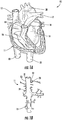
- FIGS. 1A and 1B the human anatomy in which the present disclosure is designed for placement and operation is described as context for the system of the present disclosure.
- deoxygenated blood returns to heart 10 through vena cava 11, which comprises superior vena cava 12 and inferior vena cava 13 coupled to right atrium 14 of the heart.
- Blood moves from right atrium 14 through tricuspid valve 15 to right ventricle 16, where it is pumped via pulmonary artery 17 to the lungs.
- Oxygenated returns from the lungs to left atrium 18 via the pulmonary vein.
- the oxygenated blood then enters left ventricle 19, which pumps the blood through aorta 20 to the rest of the body.
- superior vena cava 12 is positioned at the top of vena cava 11, while inferior vena cava 13 is located at the bottom of the vena cava.
- FIG. 1B also shows some of the major veins connecting to the vena cava, including right hepatic vein 21, middle hepatic vein 22, left hepatic vein 23 and suprarenal vein 24.
- occlusion of the inferior vena cava 13 may pose risks of venous congestion, and in particular, potential blockage or enlargement of the hepatic veins and/or suprarenal vein that may worsen, rather than improve, the patient's cardiovascular condition and overall health.
- sccluding the superior vena cava poses fewer potential adverse risks that occlusion of the inferior vena cava (“IVC").
- IVC inferior vena cava
- applicants' preliminary animal testing reveals that controlling the return of venous blood to the right ventricle by partially or fully occluding the SVC beneficially lowers RVEDP, RVEDV, LVEDP and LVEDV without adversely reducing left ventricular systolic pressure (LVSP), as was observed when occluding the IVC in applicants' animal model.
- LVSP left ventricular systolic pressure
- Applicants expect that selective intermittent occlusion of the SVC position will reduce the risk of worsening congestion of the kidneys, which is a major cause of 'cardio-renal' syndrome, as compared to IVC occlusion.
- Cardio-renal syndrome is impaired renal function due to volume overload and neurohormonal activation in patients with heart failure.
- implantation in the SVC permits a supra-diaphragmatic device implant that could not be used in the IVC without cardiac penetration and crossing the right atrium.
- implantation of the occluder in the SVC avoids the need for groin access as required by IVC implantation, which would limit mobility making an ambulatory device impractical for short term or long term use.
- the system and methods of the present disclosure may be used to treat any disease to improve cardiac function by arresting or reversing myocardial remodeling, and particularly those conditions in which a patient suffers from heart failure.
- Such conditions include but are not limited to, e.g., systolic heart failure, diastolic (non-systolic) heart failure, decompensated heart failure patients in (ADHF), chronic heart failure, acute heart failure.
- the system and methods of the present disclosure also may be used as a prophylactic to mitigate the aftermath of acute right or left ventricle myocardial infarction, pulmonary hypertension, RV failure, post-cardiotomy shock, or post-orthotopic heart transplantation (OHTx) rejection.
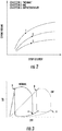
- the relationship between left ventricular pressure or left ventricular volume and stroke volume is often referred to as the Frank-Starling relationship, or "Starling curve.” That relationship states that cardiac stroke volume is dependent on preload, contractility, and afterload.
- Preload refers to the volume of blood returning to the heart; contractility is defined as the inherent ability of heart muscle to contract; and afterload is determined by vascular resistance and impedance.
- reduced stroke volume leads to increased volume and pressure increase in the left ventricle, which can result in pulmonary edema.
- Increased ventricular volume and pressure also results in increased workload and increased myocardial oxygen consumption.
- topmost curve depicts functioning of a normal heart.
- stroke volume increases with increasing LVEDP or LVEDV, and begins to flatten out, i.e., the slope of the curve decreases, only at very high pressures or volumes.
- AMI acute myocardial infarction
- curve 2 will exhibit reduced stroke volume at every value of LVEDV or LVEDP.
- FIG. 3 illustratively shows pressure-volume loops for a normal heart, labeled "normal”, corresponding to curve 1 in FIG. 2 , and a heart suffering from congestive heart failure, labeled "CHF" (curve 3 in FIG. 2 ).
- the ventricular volume and pressure at the end of diastole correspond to the lower-most, right-most corner of the loop (point A), while the uppermost, left-most corner of each loop corresponds to the beginning systole (point B).
- the stroke volume for each pressure-volume loop corresponds to the area enclosed within the loop. Accordingly, the most beneficial venous regulation regime is one that reduces the volume and pressure at point A while not also causing negligible reduction in point B, thereby maximizing the stroke volume.
- the system and methods of the present disclosure are designed, over the course of hours, days, weeks, or months, to shift or transition the Starling curve of the patient's heart leftwards on the diagram of FIG. 2 (or to move the pressure-volume loop in FIG. 3 leftwards and downwards). This may be accomplished by intermittently fully or partially occluding the SVC to reduce the volume and hence pressure of blood entering the right ventricle, and which must then be pumped by the left ventricle.
- Applicants' preliminary animal testing indicates that such intermittent occlusion, maintained over several cardiac cycles, reduces the workload and wall stress in the myocardium throughout the cardiac cycle, reduces myocardial oxygen consumption, and improves contractile function. See also, FIGS. 13 and 14 discussed below.
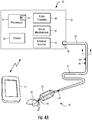
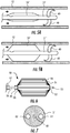
- System 30 includes catheter 31 having flow limiting element 32 coupled to controller 33 programmed to intermittently actuate flow limiting element 32.
- system 30 optionally may be configured to transfer information bi-directionally with conventional computing device 45 such as a smartphone, laptop, smartwatch, or tablet, illustratively an Apple iPhone 5 or iPad, available from Apple Inc., Cupertino, California, on which a special-purpose application has been installed to communicate and/or control controller 33.
- conventional computing device 45 such as a smartphone, laptop, smartwatch, or tablet, illustratively an Apple iPhone 5 or iPad, available from Apple Inc., Cupertino, California, on which a special-purpose application has been installed to communicate and/or control controller 33.
- catheter 31 comprises a flexible tube having distal portion 34 configured for placement in the SVC.
- Distal portion 34 includes flow limiting element 32 that, in use, is disposed in superior vena cava 12 (see FIG. 2 ) of a patient to selectively impede blood flow into right atrium 14.
- flow limiting element 32 illustratively comprises a balloon capable of transitioning between a contracted state, allowing transluminal placement and an expanded, deployed state.
- Flow limiting element 32 preferably is sized and shaped so that it partially or fully occludes flow in the SVC in the expanded state.
- Catheter 31 is coupled at proximal end 35 to controller 33, which houses drive mechanism 36 (e.g., motor, pump) for actuating flow limiting element 32, processor 37 programmed to control signals to drive mechanism 36, and optional sensor 38 for monitoring a physiologic parameter of the patient, such as heart rate or blood pressure.
- drive mechanism 36 e.g., motor, pump
- processor 37 programmed to control signals to drive mechanism 36
- sensor 38 for monitoring a physiologic parameter of the patient, such as heart rate or blood pressure.
- Controller 33 may include source of inflation medium 48 (e.g., gas or fluid) and drive mechanism 36 may transfer the inflation medium between the source and flow limiting element 32 responsive to commands from processor 37.
- source of inflation medium 48 e.g., gas or fluid
- drive mechanism 36 may transfer the inflation medium between the source and flow limiting element 32 responsive to commands from processor 37.
- flow limiting element 32 When flow limiting element 32 is inflated with inflation medium, it partially or fully occludes venous blood flow through the SVC; when the inflation medium is withdrawn, flow limiting element 32 deflates to remove the occlusion, thereby permitting flow to resume in the SVC.
- Flow limiting element 32 may be a balloon that preferably comprises a compliant or semi-compliant material, e.g., nylon, which permits the degree of expansion of the balloon to be adjusted to effectuate the desired degree of partial or complete occlusion of the SVC.
- catheter 31 when partially external, provides a fail-safe design, in that flow limiting element 32 only can be inflated to provide occlusion when the proximal end of catheter 31 is coupled to controller 33.
- flow limiting element 32 only can be inflated to provide occlusion when the proximal end of catheter 31 is coupled to controller 33.
- Such a quick-disconnect coupling 40 at proximal end 35 permits the catheter to be rapidly disconnected from controller 33 for cleaning and/or emergency.
- Controller 33 preferably also includes power supply 39 (e.g., battery) that provides the power needed to operate processor 37, drive mechanism 36 and data transfer circuit sensor 38. Controller 33 may be sized and of such a weight that it can be worn in a harness under the patient's clothing, so that the system can be used while the patient is ambulatory or such that controller 33 may be implanted within the patient. As discussed herein below, processor 37 includes memory 41 for storing computer software for operating the controller 33. Controller 33 may be configured for implantation at a suitable location within the patient, e.g., subcutaneously under the clavicle. In such an embodiment, the implantable controller is configured for bidirectional communication with an external controller, e.g., computing device 45 or system-specific device.
- an external controller e.g., computing device 45 or system-specific device.
- An external controller may be used to charge the battery of the implantable controller, e.g., via respective inductive coils in or coupled to each controller, and may receive data indicative of the sensed parameters resulting from the patient's ambulatory activity including heart rate, blood flow rate, blood volume, pressure including cardiac filling pressure.
- data transfer circuit 38 monitors an input from an external sensor, e.g., positioned on catheter 31, and provides that signal to processor 37.
- Processor 37 is programmed to receive the input from data transfer circuit 38 and adjust the interval during which flow limiting element 32 is maintained in the expanded state, or to adjust the degree of occlusion caused by flow limiting element 32.
- catheter 31 may have optional sensor 42 positioned within distal region 34 of the catheter to measure parameters, e.g., heart rate, blood flow rate, blood volume, pressure including cardiac filling pressure and central venous pressure.
- sensor 42 is relayed to data transfer circuit 38 of controller 33, which may pre-process the input signal, e.g., decimate and digitize the output of sensor 42, before it is supplied to processor 37.
- the signal provided to processor 37 allows for assessment of the effectiveness of the flow limiting element, e.g., by showing reduced venous pressure during occlusion and during patency, and may be used for patient or clinician to determine how much occlusion is required to regulate venous blood return based on the severity of congestion in the patient.
- sensor 43 may be included on catheter 31 proximal to flow limiting element 32, to measure parameters, e.g., heart rate, blood flow rate, blood volume, pressure including cardiac filling pressure and central venous pressure. Sensor 43 may be used to determine the extent of occlusion caused by element 32, for example, by monitoring the pressure drop across the flow limiting element.
- catheter 31 may include electrodes 44 for sensing the patient's heart rate. It is expected that it may be desirable to adjust the interval during which occlusion of the SVC is maintained responsive to the patient's ambulatory activities, which typically will be reflected in the patient's hemodynamic state by a sensed physiological parameter(s), e.g., heart rate, blood flow rate, blood volume, pressure including cardiac filling pressure and/or central venous pressure. Accordingly, electrodes 44 may provide a signal to data transfer circuit 38, which in turn processes that signal for use by the programmed routines run by processor 37.
- a sensed physiological parameter(s) e.g., heart rate, blood flow rate, blood volume, pressure including cardiac filling pressure and/or central venous pressure.
- electrodes 44 may provide a signal to data transfer circuit 38, which in turn processes that signal for use by the programmed routines run by processor 37.
- processor 37 may be programmed to maintain partial or full occlusion in the SVC for a preset number of cardiac cycles determined at the time of initial implantation of the catheter.
- Sensor inputs provided to data transfer circuit 38 such as hemodynamic state, also may be used to adjust the duty cycle of the flow limiting element responsive to the patient's detected level of activity.
- processor 37 may be programmed to maintain partial or full occlusion in the SVC for a preset number of cardiac cycles after adjustment to the predetermined occlusion interval is made.
- Data transfer circuit 38 also may be configured to provide bi-directional transfer of data, for example, by including wireless circuitry to transfer data from controller 33 to an external unit for display, review or adjustment.
- data transfer circuit may include Bluetooth circuitry that enables controller 33 to communicate with patient's computing device 45.
- controller may send information regarding functioning of the system directly to computing device 45 for display of vital physiologic or system parameters using a suitably configured mobile application.
- the patient may review the data displayed on the screen of computing device 45 and determine whether he or she needs to seek medical assistance to address a malfunction or to adjust the system parameters.
- the mobile application resident on computing device 45 may be configured to automatically initiate an alert to the clinician's monitoring service via the cellular telephone network.
- data transfer circuit 38 may be configured to synchronize to receive data from other mobile applications on computing device 45, and thus reduce the cost and complexity of the inventive system.
- a number of third party vendors such as Fitbit, Inc., San Francisco, California, market monitors that measure physiologic parameters in real time, such as the Charge HR wristband monitor, that measures physical activity and heart rate.
- data transfer circuit 38 can be programmed to receive an input from such a third-party monitor via wireless communication with computing device 45, and that processor 37 may be programmed to control activation of drive mechanism 36 responsive to that input.
- the catheter need not include optional sensor 42, sensor 43 or electrodes 44, thereby greatly simplifying the construction of catheter 31 and coupling 40.
- Catheter 31 may include anchor member 46 configured to anchor flow limiting element 32 within the SVC.
- Anchor member 46 may be contractable for delivery in a contracted state and expandable upon release from a delivery device, e.g., a sheath.
- Anchor member 46 may be coupled to catheter proximal or distal to flow limiting element 32 and/or may be coupled to flow limiting element 32.
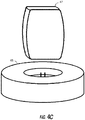
- external power source 47 may be configured to charge power supply 39 (e.g., battery) of the implantable controller.
- power supply 39 e.g., battery
- external power source 47 may transcutaneously charge power supply 39 via respective inductive coils.
- External power source 47 may be integrated into clothing or a harness worn by the patient.
- external power source 47 may be placed in a pocket or holder configured to receive external power source 47.
- the pocket or holder may be designed to place external power source 47 in close proximity to battery 39 for efficient transcutaneous charging. More than one external power source 47 may be integrated into the garment to provide additional power.
- the one or more external power sources may be permanently integrated into the garment or harness or may be removably engaged with the garment or harness such that each may be individually removed and attached.
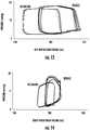
- two external power sources 47 may be integrated into specially designed pockets of vest 64 as is illustrated in FIG. 4B .
- Vest 64 may include wire 66 incorporated into vest 64 to permit electrical communication between the two external power sources.
- Power source 47 may generate an alert when an available power supply reaches or falls below a certain threshold power level.
- power source 47 may have a visual indicator and/or an auditory indicator for providing a warning to the patient or caregiver.
- the visual indicator may be an LED light system or a display embedded into a surface of power source 47 that visually provides information regarding the available power supply.
- the auditory indicator may be a speaker embedded into power source 47 that sounds an alarm when the available power supply reaches a certain threshold.
- a signal indicating that the available power supply of power source 47 has reached a certain threshold also may or alternatively be communicated directly to an external device, e.g. computing device 45, and/or to controller 33 and then from controller 33 to an external device, e.g.
- computing device 45 which may be programmed to initiate a visual or audio alert.
- An additional power source 47 may supply power to power supply 39 when the primary power source runs out of power to ensure power can be continuously provided to power supply 39.
- Power source 47 may include a processor with memory for transcutaneously transmitting and receiving data from processor 37. The processor of power source 47 may be used to reprogram processor 37 and/or store information about operating parameters to be later downloaded by an external device, e.g. computing device 45.
- Each external power source 47 may be placed in electrical communication with a wall power outlet or base charger 65 shown in FIG. 4C to charge the external power source.
- Base charger 65 may be in electrical communication with a wall power outlet and may be configured to charge one or more external power sources 47 at the same time.
- external power sources 47 may be periodically disengaged from the vest and charged such that at least one external power source 47 is in electrical communication with power supply 39 while the other external power source 47 is being charged in base charger 65.
- catheter 31' is constructed similarly to catheter 31 of FIG. 4A and FIG. 4B except with a modified anchor.
- catheter 31' may include radially expanding anchoring arms 49.
- Anchoring arms 49 are configured to radially expand, e.g., when exposed from a delivery sheath, to contact the inner wall of superior vena cava 12 and anchor flow limiting element 32' therein.
- Flow limiting element 50 may be formed of a biocompatible material, such as nickel-titanium or stainless steel, and comprises plurality of axially or spirally extending wires 51 that are biased to expand radially outward when compressed.
- Flow limiting element 50 preferably includes a biocompatible membrane covering, so that it partially or fully occludes flow in the SVC in the expanded state.
- Wires 51 may be coupled at distal end 52 to distal end 53 of actuation wire 54, and affixed to ring 55 at their proximal ends 56.
- Ring 55 is disposed to slide on actuation wire 54 so that when actuation wire 54 is pulled in the proximal direction against sheath 57 (see FIGS. 5A and 5B ), wires 51 expand radially outward.
- actuation wire 54 in response to a force applied to the proximal end of actuation wire 54 by drive mechanism 36, actuation wire 54 is retracted proximally against sheath 57 of the catheter; transitioning flow limiting element 50 to its expanded deployed state.
- spring force applied by wires 51 pulls actuation wire 54 in the proximal direction, thereby enabling wires 51 to return to their uncompressed state, lying substantially flat against actuation wire 54.
- flow limiting element 50 has a "fail safe" design, so that the flow limiting element resumes the collapsed, contracted state shown in FIG. 5A when catheter 31 is uncoupled from drive mechanism 36.
- drive mechanism 36 may be a motor, which may be a linear motor, rotary motor, solenoid-piston, or wire motor.
- Flow limiting element 50 may be constructed so that it is biased to the contracted position when catheter 31 is disconnected from controller 33, so that flow limiting element 50 can only be transitioned to the expanded, deployed state when the catheter is coupled to controller 33 and the processor has signaled drive mechanism 36 to expand the flow limiting element.
- flow limiting element 50 preferably includes a sheer biocompatible elastic membrane 58 disposed on wires 51, such as expanded polytetrafluoroethylene (ePTFE), which occludes blood flow through the SVC when the flow limiting element is in the expanded deployed state.
- ePTFE expanded polytetrafluoroethylene
- a suitable ePTFE material can be obtained, for example, from W. L. Gore & Associates, Inc., Flagstaff, Arizona.
- catheter 31 preferably includes at least three lumens 60, 61, 62.
- Lumen 60 may be used as an inflation lumen ( FIGS. 4-5B ) and/or for carrying actuation wire 54 ( FIG. 6 ) that extends between flow limiting element 50 and the drive mechanism 36 of controller 33.
- Lumen 61 permits optional sensors 42, 43 or electrodes to communicate with data transfer circuit 38, and optional lumen 62 for delivering a pharmacological agent (e.g., a drug) to the heart.
- a pharmacological agent e.g., a drug
- catheter 31 with flow limiting element 32/50 is inserted into the patient's subclavian vein and guided to the SVC of the patient, e.g., to a position proximal of the entrance to the right atrium (see FIG. 1A ).
- Techniques known in the art can be used to insert and fix flow limiting element 32/50 at the desired venous location in the patient. Proper localization of the device may be confirmed using, for example, vascular ultrasound.
- flow limiting element 32/50 may be inserted through the jugular vein and guided to the SVC under fluoroscopic or ultrasound guidance.
- controller 33 initiates a process in which the occlusion element is expanded and contracted such that blood flow in the SVC is intermittently occludes and resumed.
- the extent to which the flow limiting element impedes blood flow can be regulated by adjusting the degree to which the flow limiting element expands radially, and also for time interval for the occlusion, e.g., over how many heart beats.
- the flow limiting element may impede blood flow in the SVC by anywhere from at least 50% up to 100%. Impedance of blood flow may be confirmed using methods known in the art, e.g., by measuring reductions in pressure or visually using ultrasound.
- controller 33 includes software stored in memory 41 that controls the timing and duration of the successive expansions and contractions of flow limiting element 32/50.
- the programmed routines run by processor 37 may use as an input the patient's cardiac cycle.
- the software may be configured to actuate flow limiting element 50 to maintain partial or complete occlusion of the SVC over multiple cardiac cycles, for example, four or more successive heart beats in the subject.
- Controller 33 may accept as input via data transfer circuit 38 an output of electrodes 44 representative of the patient's electrocardiogram (ECG), or alternatively may receive such an input wirelessly from a third-party heart rate application running on the patient's smartphone, such that the software running on processor 37 can adjust the interval and/or degree of the occlusion provided by system 30 responsive to the patient's heart rate.
- ECG electrocardiogram
- system 30 may accept an input via data transfer circuit 38 a value, measured by optional sensors 42 and 43, or a third party application and device, such as a blood pressure cuff, representative of the patient's blood pressure, such that controller 37 regulates flow through the SVC responsive to the patient's blood pressure.
- a value measured by optional sensors 42 and 43
- a third party application and device such as a blood pressure cuff, representative of the patient's blood pressure
- Controller 33 may be programmed to cause the flow limiting element (from FIGS. 4 , 5A, 5B, 6 , 8A-10B ) to expand when a sensed parameter is outside a predetermined range and/or above or below a predetermined threshold.
- controller 33 may cause the flow limiting element to expand when right atrium ("RA") pressure is sensed by optional sensors 42 and/or 43 to be within a predetermined range, e.g., 15 to 30mmHg, 18 to 30mmHg, 20 to 30mmHg, 20 to 25mmHg, or above a predetermined threshold, e.g., 15 mmHg, 18 mmHg, 20 mmHg, 22 mmHg, 25 mmHg, 30 mmHg.
- RA right atrium
- controller 33 may cause the flow limiting element to expand when the mean pulmonary artery ("PA") pressure is sensed by optional sensors 42 and/or 43 to be within a predetermined range, e.g., 15 to 30mmHg, 18 to 30mmHg, 20 to 30mmHg, 20 to 25mmHg, or above a predetermined threshold e.g., 15 mmHg, 18 mmHg, 20 mmHg, 22 mmHg, 25 mmHg, 30 mmHg.
- the predetermined range and/or the predetermined threshold may be patient specific and controller 33 may be programmed and reprogrammed for individual patients.
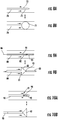
- FIGS. 8-10 alternative forms of intravenous flow limiting elements suitable for use to occlude the SVC are described.
- FIGS. 4-6 depict a cylindrical flow limiting element, other shapes may be used.
- anchoring members may be included.
- the pair-wise drawings depict that each flow limiting element has a collapsed contracted state ( FIGS. 8A, 9A and 10A ), where the flow limiting element does not significantly impede blood flow, and an expanded deployed state ( FIGS. 8B, 9B and 10B ), in which the flow limiting element partially of fully occludes blood flow through the SVC.
- catheter 70 includes balloon 71 attached to distal end 72.
- Balloon 71 is illustrated as having a rounded ball shape.
- catheter 80 includes flow limiting element 81 comprising spring-loaded plug 82 formed of a biocompatible material (e.g., beryllium) and having a tapered conical shape.
- Spring-loaded plug 82 is captured in its collapsed contracted state within sheath 83 disposed at distal end 84 of catheter 80. More particularly, a vertex of conically-shaped plug 82 is positioned adjacent the proximal end 85 of sheath 83.
- spring-loaded plug 82 is captured within sheath 83 in its low-profile state to allow blood flow in the SVC.
- force is applied via actuation wire 86 to withdraw plug 82 from sheath 83.
- plug 82 is biased to return within sheath 83 when the proximal force is removed from the proximal end of actuation wire 86, so that the flow limiting element 82 remains in its collapsed contracted state if disconnected from controller 33.
- catheter 90 depicts a further alternative embodiment of occlusive device 91, which takes the form of spring-loaded plug 92.
- Spring-loaded plug 92 is similar to plug 82 of FIGS. 9A and 9B , and has a tapered conical shape and is loaded within sheath 93 disposed at distal end 94 of catheter 90.
- spring-loaded plug 92 is pushed out of distal end of sheath 94 and expands to occlude the SVC.
- spring-loaded plug 92 retracts to its collapsed contracted state within sheath 94, thereby permitting blood to flow substantially unimpeded through the SVC.
- FIG. 11 simultaneous LV pressure and volume and Aortic pressure measurements are shown across 40-50 successive heart beats in a swine model of heart failure following either full occlusion of the inferior vena cava (IVC; FIG 11 A and B ) as suggested in the foregoing published Cedeno patent application or full occlusion of the superior vena cava (SVC; FIG 11C and D ),.
- IVC inferior vena cava
- SVC superior vena cava
- IVC occlusion rapidly and decreased LV pressure, volume, and aortic pressure to critically low values with potentially dangerous consequences to the patient ( FIG 11 A and B ).
- SVC occlusion marginally decreased LV systolic pressure and aortic pressures, but significantly decreased LV diastolic pressure, which is the primary marker of congestive heart failure ( FIG 11 C and D ).
- IVC occlusion increased renal vein pressure from 5mmHg to 15mmHg within the 30-40 seconds of occlusion ( FIG 12A ). After 2 minutes peak renal vein pressure was 23mmHg. In contrast, SVC occlusion did not affect renal vein pressure across any time period studied ( FIG 12B ).
- the method of the present disclosure of partially occluding the SVC appears to have little or no impact on ejection fraction during systole, but reduces wall stress in the ventricles during diastole. Moreover, occlusion of the SVC is expected to be tolerated well by the patient, will not contribute to congestion of the renal or hepatic veins, and will not exacerbate complications often associated with congestive heart failure.
- FIGS. 13-14 are graphs showing the changes in pressure as a function of left and right ventricular volume, respectively, during occlusion of the superior vena cava (SVC) and release in a swine treated for heart failure in accordance with the principles of the present disclosure.
- SVC occlusion led to a significant reduction in left ventricular (LV) volume (240 to 220mL) and a reduction in LV diastolic pressure (25 to 10mmHg).
- LV left ventricular
- SVC occlusion also was associated with reduction in LV systolic pressure (94 to 90mmHg).
- SVC occlusion also decreased right ventricular (RV) volume (230 to 210mL), diastolic pressure (12 to 4mmHg), and RV systolic pressure (27 to 16mmHg).
- RV right ventricular
- SVC occlusion in accordance with the systems and methods described herein reduces biventricular volume and diastolic (filling) pressures without negatively impacting systemic blood pressure (LV systolic pressure).
- FIG. 15 includes graphs showing that superior vena cava (SVC) occlusion in accordance with the principles of the present disclosure on a swine subject improves cardiac function.
- the graphs each show the results for partial inferior vena cava (IVC) occlusion (left side of each graph) versus full SVC occlusion (right side of each graph).
- the graphs show measured left ventricle (LV) stroke volume, cardiac output, LV contractility, LV diastolic pressure, LV systolic pressure, and end-systolic volume.
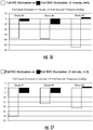
- FIG. 16 is graph showing that SVC occlusion in accordance with the principles of the present disclosure on three swine subjects does not harm systolic blood pressure.
- the graph shows the full caval occlusion (1 minute) LV end systolic pressure (mmHg) for full IVC occlusion (left side of each study) versus full SVC occlusion (right column of each study). Less reduction in LV-end-systolic pressure with SVC occlusion compared to IVC occlusion.
- FIG. 17 is graph showing that SVC occlusion in accordance with the principles of the present disclosure on three swine subjects does not harm LV diastolic filling.
- the graph shows the full caval occlusion (1 minute) LV end diastolic pressure (mmHg) for full IVC occlusion (left side of each study) versus full SVC occlusion (right side of each study). Less reduction in LV-end-diastolic pressure with SVC occlusion compared to IVC occlusion.
- FIG. 18 is graph showing that SVC occlusion in accordance with the principles of the present disclosure on three swine subjects improves LV stroke volume.
- the graph shows the full caval occlusion (1 minute) LV stroke volume (mL/beat) for full IVC occlusion (left side of each study) versus full SVC occlusion (right side of each study). Increased LV stroke volume with SVC occlusion compared to reduced LV stroke volume IVC occlusion.
- FIG. 19 is graph showing that SVC occlusion in accordance with the principles of the present disclosure on three swine subjects improves LV contractility.
- the graph shows the full caval occlusion (1 minute) LV contractility (mmHg/sec) for full IVC occlusion (left side of each study) versus full SVC occlusion (right side of each study). Increased LV contractility with SVC occlusion compared to reduced LV contractility with IVC occlusion.
- FIG. 20 is four graphs depicting LV total volume and LV pressure for IVC occlusion (upper left), RV total volume and RV pressure for IVC occlusion (upper right), LV total volume and LV pressure for SVC occlusion (lower left), and RV total volume and RV pressure for SVC occlusion (lower right).
- FIG. 20 illustrates that SVC occlusion provides a significant reduction in LV and RV diastolic pressures without a major reduction in LV systolic pressure as compared to IVC occlusion.
- FIG. 21 is two graphs depicting measured pulmonary artery pressure and renal vein pressure in a swine subject for IVC occlusion (left graph) and SVC occlusion (right graph).
- Line 100 shows the measured pulmonary artery pressure while line 102 shows the measured renal vein pressure for IVC occlusion.
- Line 104 shows the measured pulmonary artery pressure while line 106 shows the measured renal vein pressure for SVC occlusion.
- the max renal vein pressure is measured to be 22 mmHg for IVC occlusion whereas the max renal vein pressure is measured to be 7 mmHg for SVC occlusion.
- FIG. 21 demonstrates that SVC occlusion reduces pulmonary artery pressures without increasing renal vein pressure as compared to IVC occlusion.
- FIG. 22 is a graph depicting measured left subclavian vein pressure and renal vein pressure in a swine subjected to SVC occlusion in accordance with the principles of the present disclosure.
- Line 108 shows the measured left subclavian vein pressure while line 110 shows the measured renal vein pressure for SVC occlusion.
- the measured change in left subclavian vein pressure is 5 to 12 mmHg during SVC occlusion.
- FIG. 22 demonstrates that proximal left subclavian vein pressure increases nominally during SVC occlusion.
- a system constructed in accordance with the principles of the present disclosure may be designed to be implanted or worn by the patient continuously and in an ambulatory setting, rather than being tethered to a bed, e.g., in an acute-care setting, the patient will see continuous improvement in myocardial function throughout the course of treatment.
- the system provides both reduced cost and reduced complexity.
- Applicants expect that the systems and methods of the present disclosure may be used alone, as described in the examples, above, or in combination with other devices configured to assist cardiac function, such as an intra-aortic balloon pump ("IABP"), a percutaneous left ventricular assistance device (LVAD) or with a surgical LVAD, thereby allowing for synchronous or asynchronous, (venous and arterial) unloading of cardiac preload and afterload, respectively.
- IABP intra-aortic balloon pump
- LVAD percutaneous left ventricular assistance device
- surgical LVAD surgical LVAD
Landscapes
- Health & Medical Sciences (AREA)
- Life Sciences & Earth Sciences (AREA)
- Heart & Thoracic Surgery (AREA)
- Engineering & Computer Science (AREA)
- Veterinary Medicine (AREA)
- Public Health (AREA)
- Biomedical Technology (AREA)
- Animal Behavior & Ethology (AREA)
- General Health & Medical Sciences (AREA)
- Surgery (AREA)
- Cardiology (AREA)
- Medical Informatics (AREA)
- Molecular Biology (AREA)
- Vascular Medicine (AREA)
- Biophysics (AREA)
- Hematology (AREA)
- Pathology (AREA)
- Physics & Mathematics (AREA)
- Anesthesiology (AREA)
- Mechanical Engineering (AREA)
- Reproductive Health (AREA)
- Nuclear Medicine, Radiotherapy & Molecular Imaging (AREA)
- Physiology (AREA)
- Transplantation (AREA)
- Child & Adolescent Psychology (AREA)
- Pulmonology (AREA)
- External Artificial Organs (AREA)
- Electrotherapy Devices (AREA)
- Surgical Instruments (AREA)
Applications Claiming Priority (3)
| Application Number | Priority Date | Filing Date | Title |
|---|---|---|---|
| US14/828,429 US9393384B1 (en) | 2015-08-17 | 2015-08-17 | Systems and methods for treating acute and chronic heart failure |
| US15/203,437 US10279152B2 (en) | 2015-08-17 | 2016-07-06 | Systems and methods for treating acute and chronic heart failure |
| PCT/US2016/047055 WO2017031068A1 (en) | 2015-08-17 | 2016-08-15 | System for treating acute and chronic heart failure |
Publications (2)
| Publication Number | Publication Date |
|---|---|
| EP3337386A1 EP3337386A1 (en) | 2018-06-27 |
| EP3337386B1 true EP3337386B1 (en) | 2020-12-02 |
Family
ID=56801817
Family Applications (1)
| Application Number | Title | Priority Date | Filing Date |
|---|---|---|---|
| EP16757430.0A Active EP3337386B1 (en) | 2015-08-17 | 2016-08-15 | System for treating acute and chronic heart failure |
Country Status (6)
| Country | Link |
|---|---|
| US (3) | US10279152B2 (enExample) |
| EP (1) | EP3337386B1 (enExample) |
| JP (1) | JP6946266B2 (enExample) |
| CN (2) | CN113558629B (enExample) |
| ES (1) | ES2848424T3 (enExample) |
| WO (1) | WO2017031068A1 (enExample) |
Cited By (2)
| Publication number | Priority date | Publication date | Assignee | Title |
|---|---|---|---|---|
| US11883030B2 (en) | 2022-04-29 | 2024-01-30 | inQB8 Medical Technologies, LLC | Systems, devices, and methods for controllably and selectively occluding, restricting, and diverting flow within a patient's vasculature |
| US11974751B2 (en) | 2022-04-29 | 2024-05-07 | inQB8 Medical Technologies, LLC | Systems, devices, and methods for controllably and selectively occluding, restricting, and diverting flow within a patient's vasculature |
Families Citing this family (49)
| Publication number | Priority date | Publication date | Assignee | Title |
|---|---|---|---|---|
| US9539081B2 (en) | 2009-12-02 | 2017-01-10 | Surefire Medical, Inc. | Method of operating a microvalve protection device |
| US10852069B2 (en) | 2010-05-04 | 2020-12-01 | Fractal Heatsink Technologies, LLC | System and method for maintaining efficiency of a fractal heat sink |
| CN108742951B (zh) | 2012-06-06 | 2021-05-25 | 洋红医疗有限公司 | 人工肾脏瓣膜 |
| US10583231B2 (en) | 2013-03-13 | 2020-03-10 | Magenta Medical Ltd. | Blood pump |
| CN113616920B (zh) | 2013-03-13 | 2024-10-25 | 马真塔医药有限公司 | 血液泵浦装置及制造血液泵浦的方法 |
| US9968740B2 (en) | 2014-03-25 | 2018-05-15 | Surefire Medical, Inc. | Closed tip dynamic microvalve protection device |
| US20160287839A1 (en) | 2015-03-31 | 2016-10-06 | Surefire Medical, Inc. | Apparatus and Method for Infusing an Immunotherapy Agent to a Solid Tumor for Treatment |
| WO2016185473A1 (en) | 2015-05-18 | 2016-11-24 | Magenta Medical Ltd. | Blood pump |
| US10279152B2 (en) | 2015-08-17 | 2019-05-07 | Tufts Medical Center, Inc. | Systems and methods for treating acute and chronic heart failure |
| US11872361B2 (en) | 2015-08-17 | 2024-01-16 | Tufts Medical Center, Inc. | Systems and methods for selectively occluding the superior vena cava for treating heart conditions |
| US10842974B2 (en) | 2015-08-17 | 2020-11-24 | Tufts Medical Center, Inc. | Systems and methods for selectively occluding the superior vena cava for treating heart conditions |
| CN108366799B (zh) | 2015-11-09 | 2023-01-03 | 瑞普医药有限公司 | 用于心血管治疗的血流量减压器 |
| US11400263B1 (en) | 2016-09-19 | 2022-08-02 | Trisalus Life Sciences, Inc. | System and method for selective pressure-controlled therapeutic delivery |
| US10780250B1 (en) | 2016-09-19 | 2020-09-22 | Surefire Medical, Inc. | System and method for selective pressure-controlled therapeutic delivery |
| EP3556409B1 (en) | 2016-10-25 | 2022-01-05 | Magenta Medical Ltd. | Ventricular assist device |
| CA3039302C (en) * | 2016-11-23 | 2025-05-13 | Magenta Medical Ltd. | BLOOD PUMPS |
| CA3049539C (en) | 2017-01-12 | 2022-09-20 | The Regents Of The University Of California | Endovascular perfusion augmentation for critical care |
| US10588636B2 (en) | 2017-03-20 | 2020-03-17 | Surefire Medical, Inc. | Dynamic reconfigurable microvalve protection device |
| EP3612086B1 (en) | 2017-04-21 | 2026-04-01 | The Regents of the University of California | Aortic flow meter and pump for partial-aortic occlusion |
| US10813648B2 (en) * | 2017-10-06 | 2020-10-27 | Boehringer Technologies, Lp | Systems and methods for effecting the total and partial occlusion of the aorta of a living being |
| EP3909524A1 (en) * | 2017-10-24 | 2021-11-17 | Tufts Medical Center, Inc. | Systems for selectively occluding the superior vena cava for treating heart conditions |
| CN115025386B (zh) | 2018-01-10 | 2025-07-25 | 马真塔医药有限公司 | 心室辅助装置 |
| WO2019221971A1 (en) | 2018-05-12 | 2019-11-21 | Venacore Inc. | Controlling rate of blood flow to right atrium |
| US11219753B2 (en) * | 2018-07-30 | 2022-01-11 | Cardiovascular Systems, Inc. | Intravascular pump with expandable and collapsible inlet region and methods thereof |
| US10960118B2 (en) * | 2018-07-31 | 2021-03-30 | Abiomed, Inc. | Systems and methods for controlling a heart pump to minimize myocardial oxygen consumption |
| US11850398B2 (en) | 2018-08-01 | 2023-12-26 | Trisalus Life Sciences, Inc. | Systems and methods for pressure-facilitated therapeutic agent delivery |
| CA3016047A1 (en) | 2018-08-31 | 2020-02-29 | Instant Brands Inc. | Induction heating food processor |
| US11338117B2 (en) | 2018-10-08 | 2022-05-24 | Trisalus Life Sciences, Inc. | Implantable dual pathway therapeutic agent delivery port |
| WO2020121309A1 (en) * | 2018-12-11 | 2020-06-18 | Revamp Medical Ltd. | Systems, devices, and methods for adjusting blood flow in a body lumen |
| EP4653041A3 (en) | 2019-01-24 | 2026-03-04 | Magenta Medical Ltd. | Ventricular assist device |
| EP3920850A1 (en) | 2019-02-06 | 2021-12-15 | Inqb8 Medical Technologies, LLC | Intra-cardiac left atrial and dual support systems |
| US12128228B2 (en) | 2019-05-23 | 2024-10-29 | Magenta Medical Ltd | Blood pumps |
| US12433597B2 (en) | 2019-06-04 | 2025-10-07 | Trisalus Life Sciences, Inc. | Atraumatic occlusive system with compartment for measurement of vascular pressure change |
| WO2021034784A1 (en) | 2019-08-16 | 2021-02-25 | Poltorak Technologies, LLC | Device and method for medical diagnostics |
| WO2021067325A1 (en) * | 2019-10-01 | 2021-04-08 | Boston Scientific Scimed, Inc. | Devices, systems, and methods for detecting fluid flow |
| JP2023502265A (ja) | 2019-11-22 | 2023-01-23 | タフツ メディカル センター インコーポレイテッド | 心臓病を治療するために上大静脈を選択的に閉塞するシステム及び方法 |
| US20210275783A1 (en) * | 2020-03-06 | 2021-09-09 | University Of Utah Research Foundation | Blood pressure regulation system for the treatment of neurologic injuries |
| WO2021188602A2 (en) * | 2020-03-16 | 2021-09-23 | Certus Critical Care, Inc. | Blood flow control devices, systems, and methods and error detection thereof |
| JP7467668B2 (ja) * | 2020-03-25 | 2024-04-15 | ボストン サイエンティフィック サイムド,インコーポレイテッド | 非代償性心不全を治療するための医療デバイス |
| CN114746142B (zh) | 2020-04-07 | 2026-01-02 | 马真塔医药有限公司 | 心室辅助装置 |
| WO2021230830A1 (en) * | 2020-05-13 | 2021-11-18 | Mehmet Hakan Akpinar | A dynamic flow controller for heart failure |
| WO2022103921A1 (en) * | 2020-11-13 | 2022-05-19 | Teleflex Life Sciences Limited | Wire assemblies and methods for blood vessel occlusion in vascular procedures |
| CN116549833B (zh) | 2021-03-09 | 2025-08-29 | 马真塔医药有限公司 | 心室辅助装置 |
| EP4304524A4 (en) | 2021-03-12 | 2025-01-22 | Cardio-Renal Solutions, Inc. | Device and method for variable blood flow occlusion |
| EP4547313A1 (en) * | 2022-08-05 | 2025-05-07 | Edwards Lifesciences Corporation | Pressure-induced modulation systems |
| EP4429754B1 (en) | 2022-09-14 | 2025-02-12 | Magenta Medical Ltd. | Pump-head portion of ventricular assist device |
| CN115944846B (zh) * | 2022-10-11 | 2025-12-16 | 新疆天地集团有限公司 | 一种人工仿生心脏控制方法 |
| CN117982258B (zh) * | 2022-10-27 | 2025-07-25 | 合源医疗器械(上海)有限公司 | 医疗装置 |
| AU2024266379A1 (en) * | 2023-05-04 | 2025-11-20 | Abiomed, Inc. | Method and system for holistically reducing preload and/or modifying the positioning or curvature of an interventricular septum |
Family Cites Families (19)
| Publication number | Priority date | Publication date | Assignee | Title |
|---|---|---|---|---|
| US4546759A (en) | 1983-07-29 | 1985-10-15 | Mladen Solar | Method and apparatus for assisting human heart function |
| US4662358A (en) * | 1984-04-17 | 1987-05-05 | Thoratec Laboratories Corporation | Electronic control system for a cardiac prosthesis |
| US6735532B2 (en) * | 1998-09-30 | 2004-05-11 | L. Vad Technology, Inc. | Cardiovascular support control system |
| US7204798B2 (en) * | 2003-01-24 | 2007-04-17 | Proteus Biomedical, Inc. | Methods and systems for measuring cardiac parameters |
| IL154531A (en) | 2003-02-19 | 2006-04-10 | Yair Tal | Device and method for regulating blood flow |
| CN1819855A (zh) * | 2003-05-13 | 2006-08-16 | 萨瓦柯公司 | 检测、诊断和治疗心血管疾病的系统和方法 |
| US20060064059A1 (en) * | 2004-09-21 | 2006-03-23 | Mark Gelfand | Treatment of infarct expansion by partially occluding vena cava |
| US8968239B2 (en) | 2008-09-02 | 2015-03-03 | Jose E. Herrera | Catheter device for the dynamic regulation of the venous return to the heart for the treatment of patients with heart failure |
| EG26158A (en) * | 2009-01-08 | 2013-03-31 | داود حنا ايهاب | Complete artificial heart (fixed inside the heart) for patients with myocardial infarction and weakness |
| US20100228310A1 (en) * | 2009-03-09 | 2010-09-09 | Shuros Allan C | Systems and methods for autonomic nerve modulation |
| US11337707B2 (en) | 2010-05-25 | 2022-05-24 | Miracor Medical Sa | Treating heart tissue |
| KR101462176B1 (ko) | 2013-07-16 | 2014-11-21 | 삼성전자주식회사 | 열교환기 |
| WO2015109028A1 (en) | 2014-01-14 | 2015-07-23 | Kaiser Daniel Walter | Apparatus and methods for optimizing intra cardiac filling pressures, heart rate, and cardiac output |
| US9878080B2 (en) * | 2014-01-14 | 2018-01-30 | CardioFlow Technologies, LLC | Apparatus and methods for optimizing intra cardiac filling pressures, heart rate, and cardiac output |
| US8876850B1 (en) * | 2014-06-19 | 2014-11-04 | Aria Cv, Inc. | Systems and methods for treating pulmonary hypertension |
| US10279152B2 (en) | 2015-08-17 | 2019-05-07 | Tufts Medical Center, Inc. | Systems and methods for treating acute and chronic heart failure |
| US9393384B1 (en) | 2015-08-17 | 2016-07-19 | Tufts Medical Center, Inc. | Systems and methods for treating acute and chronic heart failure |
| US10842974B2 (en) | 2015-08-17 | 2020-11-24 | Tufts Medical Center, Inc. | Systems and methods for selectively occluding the superior vena cava for treating heart conditions |
| CN108366799B (zh) | 2015-11-09 | 2023-01-03 | 瑞普医药有限公司 | 用于心血管治疗的血流量减压器 |
-
2016
- 2016-07-06 US US15/203,437 patent/US10279152B2/en active Active
- 2016-08-15 EP EP16757430.0A patent/EP3337386B1/en active Active
- 2016-08-15 CN CN202110509289.XA patent/CN113558629B/zh active Active
- 2016-08-15 WO PCT/US2016/047055 patent/WO2017031068A1/en not_active Ceased
- 2016-08-15 US US15/753,300 patent/US10758715B2/en active Active
- 2016-08-15 CN CN201680046092.4A patent/CN107920741B/zh active Active
- 2016-08-15 JP JP2018508148A patent/JP6946266B2/ja active Active
- 2016-08-15 ES ES16757430T patent/ES2848424T3/es active Active
-
2019
- 2019-05-02 US US16/402,194 patent/US10842975B2/en active Active
Non-Patent Citations (1)
| Title |
|---|
| None * |
Cited By (3)
| Publication number | Priority date | Publication date | Assignee | Title |
|---|---|---|---|---|
| US11883030B2 (en) | 2022-04-29 | 2024-01-30 | inQB8 Medical Technologies, LLC | Systems, devices, and methods for controllably and selectively occluding, restricting, and diverting flow within a patient's vasculature |
| US11974751B2 (en) | 2022-04-29 | 2024-05-07 | inQB8 Medical Technologies, LLC | Systems, devices, and methods for controllably and selectively occluding, restricting, and diverting flow within a patient's vasculature |
| US12569251B2 (en) | 2022-04-29 | 2026-03-10 | Relief Cardiovascular, Inc. | Systems, devices, and methods for controllably and selectively occluding, restricting, and diverting flow within a patient's vasculature |
Also Published As
| Publication number | Publication date |
|---|---|
| US10758715B2 (en) | 2020-09-01 |
| US20190255302A1 (en) | 2019-08-22 |
| CN113558629A (zh) | 2021-10-29 |
| CN113558629B (zh) | 2024-11-01 |
| ES2848424T3 (es) | 2021-08-09 |
| US20180243541A1 (en) | 2018-08-30 |
| JP6946266B2 (ja) | 2021-10-06 |
| US20170049946A1 (en) | 2017-02-23 |
| WO2017031068A1 (en) | 2017-02-23 |
| JP2018523534A (ja) | 2018-08-23 |
| CN107920741B (zh) | 2021-06-01 |
| CN107920741A (zh) | 2018-04-17 |
| EP3337386A1 (en) | 2018-06-27 |
| US10279152B2 (en) | 2019-05-07 |
| US10842975B2 (en) | 2020-11-24 |
Similar Documents
| Publication | Publication Date | Title |
|---|---|---|
| US10842975B2 (en) | Systems and methods for treating acute and chronic heart failure | |
| US12357799B2 (en) | Systems and methods for selectively occluding the superior vena cava for treating heart conditions | |
| US9393384B1 (en) | Systems and methods for treating acute and chronic heart failure | |
| US20250387123A1 (en) | Systems and methods for selectively occluding the superior vena cava for treating heart conditions | |
| US11872361B2 (en) | Systems and methods for selectively occluding the superior vena cava for treating heart conditions | |
| US20260069853A1 (en) | Systems and methods for selectively occluding the superior vena cava for treating heart conditions | |
| HK40059315A (en) | Systems for selectively occluding the superior vena cava for treating heart conditions |
Legal Events
| Date | Code | Title | Description |
|---|---|---|---|
| STAA | Information on the status of an ep patent application or granted ep patent |
Free format text: STATUS: THE INTERNATIONAL PUBLICATION HAS BEEN MADE |
|
| PUAI | Public reference made under article 153(3) epc to a published international application that has entered the european phase |
Free format text: ORIGINAL CODE: 0009012 |
|
| STAA | Information on the status of an ep patent application or granted ep patent |
Free format text: STATUS: REQUEST FOR EXAMINATION WAS MADE |
|
| 17P | Request for examination filed |
Effective date: 20180307 |
|
| AK | Designated contracting states |
Kind code of ref document: A1 Designated state(s): AL AT BE BG CH CY CZ DE DK EE ES FI FR GB GR HR HU IE IS IT LI LT LU LV MC MK MT NL NO PL PT RO RS SE SI SK SM TR |
|
| AX | Request for extension of the european patent |
Extension state: BA ME |
|
| DAV | Request for validation of the european patent (deleted) | ||
| DAX | Request for extension of the european patent (deleted) | ||
| STAA | Information on the status of an ep patent application or granted ep patent |
Free format text: STATUS: EXAMINATION IS IN PROGRESS |
|
| 17Q | First examination report despatched |
Effective date: 20190322 |
|
| GRAP | Despatch of communication of intention to grant a patent |
Free format text: ORIGINAL CODE: EPIDOSNIGR1 |
|
| STAA | Information on the status of an ep patent application or granted ep patent |
Free format text: STATUS: GRANT OF PATENT IS INTENDED |
|
| INTG | Intention to grant announced |
Effective date: 20200519 |
|
| GRAS | Grant fee paid |
Free format text: ORIGINAL CODE: EPIDOSNIGR3 |
|
| GRAJ | Information related to disapproval of communication of intention to grant by the applicant or resumption of examination proceedings by the epo deleted |
Free format text: ORIGINAL CODE: EPIDOSDIGR1 |
|
| GRAL | Information related to payment of fee for publishing/printing deleted |
Free format text: ORIGINAL CODE: EPIDOSDIGR3 |
|
| STAA | Information on the status of an ep patent application or granted ep patent |
Free format text: STATUS: EXAMINATION IS IN PROGRESS |
|
| GRAR | Information related to intention to grant a patent recorded |
Free format text: ORIGINAL CODE: EPIDOSNIGR71 |
|
| STAA | Information on the status of an ep patent application or granted ep patent |
Free format text: STATUS: GRANT OF PATENT IS INTENDED |
|
| GRAA | (expected) grant |
Free format text: ORIGINAL CODE: 0009210 |
|
| STAA | Information on the status of an ep patent application or granted ep patent |
Free format text: STATUS: THE PATENT HAS BEEN GRANTED |
|
| INTC | Intention to grant announced (deleted) | ||
| INTG | Intention to grant announced |
Effective date: 20201021 |
|
| AK | Designated contracting states |
Kind code of ref document: B1 Designated state(s): AL AT BE BG CH CY CZ DE DK EE ES FI FR GB GR HR HU IE IS IT LI LT LU LV MC MK MT NL NO PL PT RO RS SE SI SK SM TR |
|
| REG | Reference to a national code |
Ref country code: GB Ref legal event code: FG4D |
|
| REG | Reference to a national code |
Ref country code: AT Ref legal event code: REF Ref document number: 1339985 Country of ref document: AT Kind code of ref document: T Effective date: 20201215 Ref country code: CH Ref legal event code: EP |
|
| REG | Reference to a national code |
Ref country code: DE Ref legal event code: R096 Ref document number: 602016049050 Country of ref document: DE |
|
| REG | Reference to a national code |
Ref country code: IE Ref legal event code: FG4D |
|
| REG | Reference to a national code |
Ref country code: CH Ref legal event code: NV Representative=s name: VOSSIUS AND PARTNER PATENTANWAELTE RECHTSANWAE, CH |
|
| REG | Reference to a national code |
Ref country code: NL Ref legal event code: FP |
|
| PG25 | Lapsed in a contracting state [announced via postgrant information from national office to epo] |
Ref country code: NO Free format text: LAPSE BECAUSE OF FAILURE TO SUBMIT A TRANSLATION OF THE DESCRIPTION OR TO PAY THE FEE WITHIN THE PRESCRIBED TIME-LIMIT Effective date: 20210302 Ref country code: RS Free format text: LAPSE BECAUSE OF FAILURE TO SUBMIT A TRANSLATION OF THE DESCRIPTION OR TO PAY THE FEE WITHIN THE PRESCRIBED TIME-LIMIT Effective date: 20201202 Ref country code: FI Free format text: LAPSE BECAUSE OF FAILURE TO SUBMIT A TRANSLATION OF THE DESCRIPTION OR TO PAY THE FEE WITHIN THE PRESCRIBED TIME-LIMIT Effective date: 20201202 Ref country code: GR Free format text: LAPSE BECAUSE OF FAILURE TO SUBMIT A TRANSLATION OF THE DESCRIPTION OR TO PAY THE FEE WITHIN THE PRESCRIBED TIME-LIMIT Effective date: 20210303 |
|
| REG | Reference to a national code |
Ref country code: AT Ref legal event code: MK05 Ref document number: 1339985 Country of ref document: AT Kind code of ref document: T Effective date: 20201202 |
|
| PG25 | Lapsed in a contracting state [announced via postgrant information from national office to epo] |
Ref country code: SE Free format text: LAPSE BECAUSE OF FAILURE TO SUBMIT A TRANSLATION OF THE DESCRIPTION OR TO PAY THE FEE WITHIN THE PRESCRIBED TIME-LIMIT Effective date: 20201202 Ref country code: PL Free format text: LAPSE BECAUSE OF FAILURE TO SUBMIT A TRANSLATION OF THE DESCRIPTION OR TO PAY THE FEE WITHIN THE PRESCRIBED TIME-LIMIT Effective date: 20201202 Ref country code: LV Free format text: LAPSE BECAUSE OF FAILURE TO SUBMIT A TRANSLATION OF THE DESCRIPTION OR TO PAY THE FEE WITHIN THE PRESCRIBED TIME-LIMIT Effective date: 20201202 Ref country code: BG Free format text: LAPSE BECAUSE OF FAILURE TO SUBMIT A TRANSLATION OF THE DESCRIPTION OR TO PAY THE FEE WITHIN THE PRESCRIBED TIME-LIMIT Effective date: 20210302 |
|
| PG25 | Lapsed in a contracting state [announced via postgrant information from national office to epo] |
Ref country code: HR Free format text: LAPSE BECAUSE OF FAILURE TO SUBMIT A TRANSLATION OF THE DESCRIPTION OR TO PAY THE FEE WITHIN THE PRESCRIBED TIME-LIMIT Effective date: 20201202 |
|
| REG | Reference to a national code |
Ref country code: LT Ref legal event code: MG9D |
|
| PG25 | Lapsed in a contracting state [announced via postgrant information from national office to epo] |
Ref country code: EE Free format text: LAPSE BECAUSE OF FAILURE TO SUBMIT A TRANSLATION OF THE DESCRIPTION OR TO PAY THE FEE WITHIN THE PRESCRIBED TIME-LIMIT Effective date: 20201202 Ref country code: CZ Free format text: LAPSE BECAUSE OF FAILURE TO SUBMIT A TRANSLATION OF THE DESCRIPTION OR TO PAY THE FEE WITHIN THE PRESCRIBED TIME-LIMIT Effective date: 20201202 Ref country code: LT Free format text: LAPSE BECAUSE OF FAILURE TO SUBMIT A TRANSLATION OF THE DESCRIPTION OR TO PAY THE FEE WITHIN THE PRESCRIBED TIME-LIMIT Effective date: 20201202 Ref country code: SM Free format text: LAPSE BECAUSE OF FAILURE TO SUBMIT A TRANSLATION OF THE DESCRIPTION OR TO PAY THE FEE WITHIN THE PRESCRIBED TIME-LIMIT Effective date: 20201202 Ref country code: RO Free format text: LAPSE BECAUSE OF FAILURE TO SUBMIT A TRANSLATION OF THE DESCRIPTION OR TO PAY THE FEE WITHIN THE PRESCRIBED TIME-LIMIT Effective date: 20201202 Ref country code: PT Free format text: LAPSE BECAUSE OF FAILURE TO SUBMIT A TRANSLATION OF THE DESCRIPTION OR TO PAY THE FEE WITHIN THE PRESCRIBED TIME-LIMIT Effective date: 20210405 Ref country code: SK Free format text: LAPSE BECAUSE OF FAILURE TO SUBMIT A TRANSLATION OF THE DESCRIPTION OR TO PAY THE FEE WITHIN THE PRESCRIBED TIME-LIMIT Effective date: 20201202 |
|
| REG | Reference to a national code |
Ref country code: ES Ref legal event code: FG2A Ref document number: 2848424 Country of ref document: ES Kind code of ref document: T3 Effective date: 20210809 |
|
| PG25 | Lapsed in a contracting state [announced via postgrant information from national office to epo] |
Ref country code: AT Free format text: LAPSE BECAUSE OF FAILURE TO SUBMIT A TRANSLATION OF THE DESCRIPTION OR TO PAY THE FEE WITHIN THE PRESCRIBED TIME-LIMIT Effective date: 20201202 |
|
| REG | Reference to a national code |
Ref country code: DE Ref legal event code: R097 Ref document number: 602016049050 Country of ref document: DE |
|
| PG25 | Lapsed in a contracting state [announced via postgrant information from national office to epo] |
Ref country code: IS Free format text: LAPSE BECAUSE OF FAILURE TO SUBMIT A TRANSLATION OF THE DESCRIPTION OR TO PAY THE FEE WITHIN THE PRESCRIBED TIME-LIMIT Effective date: 20210402 |
|
| PLBE | No opposition filed within time limit |
Free format text: ORIGINAL CODE: 0009261 |
|
| STAA | Information on the status of an ep patent application or granted ep patent |
Free format text: STATUS: NO OPPOSITION FILED WITHIN TIME LIMIT |
|
| PG25 | Lapsed in a contracting state [announced via postgrant information from national office to epo] |
Ref country code: AL Free format text: LAPSE BECAUSE OF FAILURE TO SUBMIT A TRANSLATION OF THE DESCRIPTION OR TO PAY THE FEE WITHIN THE PRESCRIBED TIME-LIMIT Effective date: 20201202 |
|
| 26N | No opposition filed |
Effective date: 20210903 |
|
| PG25 | Lapsed in a contracting state [announced via postgrant information from national office to epo] |
Ref country code: DK Free format text: LAPSE BECAUSE OF FAILURE TO SUBMIT A TRANSLATION OF THE DESCRIPTION OR TO PAY THE FEE WITHIN THE PRESCRIBED TIME-LIMIT Effective date: 20201202 Ref country code: SI Free format text: LAPSE BECAUSE OF FAILURE TO SUBMIT A TRANSLATION OF THE DESCRIPTION OR TO PAY THE FEE WITHIN THE PRESCRIBED TIME-LIMIT Effective date: 20201202 |
|
| PG25 | Lapsed in a contracting state [announced via postgrant information from national office to epo] |
Ref country code: MC Free format text: LAPSE BECAUSE OF FAILURE TO SUBMIT A TRANSLATION OF THE DESCRIPTION OR TO PAY THE FEE WITHIN THE PRESCRIBED TIME-LIMIT Effective date: 20201202 |
|
| REG | Reference to a national code |
Ref country code: BE Ref legal event code: MM Effective date: 20210831 |
|
| PG25 | Lapsed in a contracting state [announced via postgrant information from national office to epo] |
Ref country code: IS Free format text: LAPSE BECAUSE OF FAILURE TO SUBMIT A TRANSLATION OF THE DESCRIPTION OR TO PAY THE FEE WITHIN THE PRESCRIBED TIME-LIMIT Effective date: 20210402 Ref country code: LU Free format text: LAPSE BECAUSE OF NON-PAYMENT OF DUE FEES Effective date: 20210815 |
|
| PG25 | Lapsed in a contracting state [announced via postgrant information from national office to epo] |
Ref country code: BE Free format text: LAPSE BECAUSE OF NON-PAYMENT OF DUE FEES Effective date: 20210831 |
|
| PG25 | Lapsed in a contracting state [announced via postgrant information from national office to epo] |
Ref country code: HU Free format text: LAPSE BECAUSE OF FAILURE TO SUBMIT A TRANSLATION OF THE DESCRIPTION OR TO PAY THE FEE WITHIN THE PRESCRIBED TIME-LIMIT; INVALID AB INITIO Effective date: 20160815 |
|
| P01 | Opt-out of the competence of the unified patent court (upc) registered |
Effective date: 20230330 |
|
| PG25 | Lapsed in a contracting state [announced via postgrant information from national office to epo] |
Ref country code: CY Free format text: LAPSE BECAUSE OF FAILURE TO SUBMIT A TRANSLATION OF THE DESCRIPTION OR TO PAY THE FEE WITHIN THE PRESCRIBED TIME-LIMIT Effective date: 20201202 |
|
| PG25 | Lapsed in a contracting state [announced via postgrant information from national office to epo] |
Ref country code: MK Free format text: LAPSE BECAUSE OF FAILURE TO SUBMIT A TRANSLATION OF THE DESCRIPTION OR TO PAY THE FEE WITHIN THE PRESCRIBED TIME-LIMIT Effective date: 20201202 |
|
| PG25 | Lapsed in a contracting state [announced via postgrant information from national office to epo] |
Ref country code: TR Free format text: LAPSE BECAUSE OF FAILURE TO SUBMIT A TRANSLATION OF THE DESCRIPTION OR TO PAY THE FEE WITHIN THE PRESCRIBED TIME-LIMIT Effective date: 20201202 |
|
| PG25 | Lapsed in a contracting state [announced via postgrant information from national office to epo] |
Ref country code: MT Free format text: LAPSE BECAUSE OF FAILURE TO SUBMIT A TRANSLATION OF THE DESCRIPTION OR TO PAY THE FEE WITHIN THE PRESCRIBED TIME-LIMIT Effective date: 20201202 |
|
| PGFP | Annual fee paid to national office [announced via postgrant information from national office to epo] |
Ref country code: NL Payment date: 20250729 Year of fee payment: 10 |
|
| PGFP | Annual fee paid to national office [announced via postgrant information from national office to epo] |
Ref country code: ES Payment date: 20250926 Year of fee payment: 10 |
|
| PGFP | Annual fee paid to national office [announced via postgrant information from national office to epo] |
Ref country code: DE Payment date: 20250729 Year of fee payment: 10 |
|
| PGFP | Annual fee paid to national office [announced via postgrant information from national office to epo] |
Ref country code: IT Payment date: 20250729 Year of fee payment: 10 |
|
| PGFP | Annual fee paid to national office [announced via postgrant information from national office to epo] |
Ref country code: GB Payment date: 20250721 Year of fee payment: 10 |
|
| PGFP | Annual fee paid to national office [announced via postgrant information from national office to epo] |
Ref country code: FR Payment date: 20250722 Year of fee payment: 10 |
|
| PGFP | Annual fee paid to national office [announced via postgrant information from national office to epo] |
Ref country code: CH Payment date: 20250901 Year of fee payment: 10 |
|
| PGFP | Annual fee paid to national office [announced via postgrant information from national office to epo] |
Ref country code: IE Payment date: 20250717 Year of fee payment: 10 |