EP3331550B1 - Composition for treating myeloproliferative disorders - Google Patents
Composition for treating myeloproliferative disorders Download PDFInfo
- Publication number
- EP3331550B1 EP3331550B1 EP16833893.7A EP16833893A EP3331550B1 EP 3331550 B1 EP3331550 B1 EP 3331550B1 EP 16833893 A EP16833893 A EP 16833893A EP 3331550 B1 EP3331550 B1 EP 3331550B1
- Authority
- EP
- European Patent Office
- Prior art keywords
- tβrii
- antibody
- amino acid
- myelofibrosis
- binding
- Prior art date
- Legal status (The legal status is an assumption and is not a legal conclusion. Google has not performed a legal analysis and makes no representation as to the accuracy of the status listed.)
- Active
Links
- 239000000203 mixture Substances 0.000 title claims description 43
- 208000014767 Myeloproliferative disease Diseases 0.000 title description 68
- 239000005557 antagonist Substances 0.000 claims description 215
- 108090000765 processed proteins & peptides Proteins 0.000 claims description 189
- 206010028537 myelofibrosis Diseases 0.000 claims description 188
- 102000004196 processed proteins & peptides Human genes 0.000 claims description 186
- 229920001184 polypeptide Polymers 0.000 claims description 183
- 238000000034 method Methods 0.000 claims description 159
- 208000017733 acquired polycythemia vera Diseases 0.000 claims description 106
- 208000037244 polycythemia vera Diseases 0.000 claims description 106
- 208000032027 Essential Thrombocythemia Diseases 0.000 claims description 93
- 125000003275 alpha amino acid group Chemical group 0.000 claims description 86
- 208000037265 diseases, disorders, signs and symptoms Diseases 0.000 claims description 68
- 230000035772 mutation Effects 0.000 claims description 68
- 230000000694 effects Effects 0.000 claims description 51
- 208000003476 primary myelofibrosis Diseases 0.000 claims description 44
- 238000011282 treatment Methods 0.000 claims description 41
- -1 SEP-701 Chemical compound 0.000 claims description 40
- 102000004887 Transforming Growth Factor beta Human genes 0.000 claims description 32
- 108090001012 Transforming Growth Factor beta Proteins 0.000 claims description 32
- 102100033444 Tyrosine-protein kinase JAK2 Human genes 0.000 claims description 32
- 108060003951 Immunoglobulin Proteins 0.000 claims description 21
- 108020001507 fusion proteins Proteins 0.000 claims description 21
- 102000037865 fusion proteins Human genes 0.000 claims description 21
- 102000018358 immunoglobulin Human genes 0.000 claims description 21
- 229940122245 Janus kinase inhibitor Drugs 0.000 claims description 20
- 239000002144 L01XE18 - Ruxolitinib Substances 0.000 claims description 18
- HFNKQEVNSGCOJV-OAHLLOKOSA-N ruxolitinib Chemical compound C1([C@@H](CC#N)N2N=CC(=C2)C=2C=3C=CNC=3N=CN=2)CCCC1 HFNKQEVNSGCOJV-OAHLLOKOSA-N 0.000 claims description 17
- 229960000215 ruxolitinib Drugs 0.000 claims description 17
- JOOXLOJCABQBSG-UHFFFAOYSA-N N-tert-butyl-3-[[5-methyl-2-[4-[2-(1-pyrrolidinyl)ethoxy]anilino]-4-pyrimidinyl]amino]benzenesulfonamide Chemical compound N1=C(NC=2C=C(C=CC=2)S(=O)(=O)NC(C)(C)C)C(C)=CN=C1NC(C=C1)=CC=C1OCCN1CCCC1 JOOXLOJCABQBSG-UHFFFAOYSA-N 0.000 claims description 10
- PDOQBOJDRPLBQU-QMMMGPOBSA-N 5-chloro-2-n-[(1s)-1-(5-fluoropyrimidin-2-yl)ethyl]-4-n-(5-methyl-1h-pyrazol-3-yl)pyrimidine-2,4-diamine Chemical compound N([C@@H](C)C=1N=CC(F)=CN=1)C(N=1)=NC=C(Cl)C=1NC=1C=C(C)NN=1 PDOQBOJDRPLBQU-QMMMGPOBSA-N 0.000 claims description 5
- SQSZANZGUXWJEA-UHFFFAOYSA-N Gandotinib Chemical compound N1C(C)=CC(NC2=NN3C(CC=4C(=CC(Cl)=CC=4)F)=C(C)N=C3C(CN3CCOCC3)=C2)=N1 SQSZANZGUXWJEA-UHFFFAOYSA-N 0.000 claims description 5
- UIARLYUEJFELEN-LROUJFHJSA-N LSM-1231 Chemical compound C12=C3N4C5=CC=CC=C5C3=C3C(=O)NCC3=C2C2=CC=CC=C2N1[C@]1(C)[C@](CO)(O)C[C@H]4O1 UIARLYUEJFELEN-LROUJFHJSA-N 0.000 claims description 5
- 108091000080 Phosphotransferase Proteins 0.000 claims description 5
- JCINBYQJBYJGDM-UHFFFAOYSA-N bms-911543 Chemical compound CCN1C(C(=O)N(C2CC2)C2CC2)=CC(C=2N(C)C=NC=22)=C1N=C2NC=1C=C(C)N(C)N=1 JCINBYQJBYJGDM-UHFFFAOYSA-N 0.000 claims description 5
- LOLPPWBBNUVNQZ-UHFFFAOYSA-N chembl495727 Chemical compound C=1NN=C(C=2NC3=CC=C(CN4CCOCC4)C=C3N=2)C=1NC(=O)NC1CC1 LOLPPWBBNUVNQZ-UHFFFAOYSA-N 0.000 claims description 5
- 229950003487 fedratinib Drugs 0.000 claims description 5
- 229950008908 gandotinib Drugs 0.000 claims description 5
- UQTPDWDAYHAZNT-AWEZNQCLSA-N ilginatinib Chemical compound N([C@@H](C)C=1C=CC(F)=CC=1)C(N=1)=CC(C2=CN(C)N=C2)=CC=1NC1=CN=CC=N1 UQTPDWDAYHAZNT-AWEZNQCLSA-N 0.000 claims description 5
- 229950001845 lestaurtinib Drugs 0.000 claims description 5
- 229950008814 momelotinib Drugs 0.000 claims description 5
- 229950011410 pacritinib Drugs 0.000 claims description 5
- HWXVIOGONBBTBY-ONEGZZNKSA-N pacritinib Chemical compound C=1C=C(C=2)NC(N=3)=NC=CC=3C(C=3)=CC=CC=3COC\C=C\COCC=2C=1OCCN1CCCC1 HWXVIOGONBBTBY-ONEGZZNKSA-N 0.000 claims description 5
- 102000020233 phosphotransferase Human genes 0.000 claims description 5
- ZRKFYGHZFMAOKI-QMGMOQQFSA-N tgfbeta Chemical compound C([C@H](NC(=O)[C@H](C(C)C)NC(=O)CNC(=O)[C@H](CCC(O)=O)NC(=O)[C@H](CCCNC(N)=N)NC(=O)[C@H](CC(N)=O)NC(=O)[C@H](CC(C)C)NC(=O)[C@H]([C@@H](C)O)NC(=O)[C@H](CCC(O)=O)NC(=O)[C@H]([C@@H](C)O)NC(=O)[C@H](CC(C)C)NC(=O)CNC(=O)[C@H](C)NC(=O)[C@H](CO)NC(=O)[C@H](CCC(N)=O)NC(=O)[C@@H](NC(=O)[C@H](C)NC(=O)[C@H](C)NC(=O)[C@@H](NC(=O)[C@H](CC(C)C)NC(=O)[C@@H](N)CCSC)C(C)C)[C@@H](C)CC)C(=O)N[C@@H]([C@@H](C)O)C(=O)N[C@@H](C(C)C)C(=O)N[C@@H](CC=1C=CC=CC=1)C(=O)N[C@@H](C)C(=O)N1[C@@H](CCC1)C(=O)N[C@@H]([C@@H](C)O)C(=O)N[C@@H](CC(N)=O)C(=O)N[C@@H](CCC(O)=O)C(=O)N[C@@H](C)C(=O)N[C@@H](CC=1C=CC=CC=1)C(=O)N[C@@H](CCCNC(N)=N)C(=O)N[C@@H](C)C(=O)N[C@@H](CC(C)C)C(=O)N1[C@@H](CCC1)C(=O)N1[C@@H](CCC1)C(=O)N[C@@H](CCCNC(N)=N)C(=O)N[C@@H](CCC(O)=O)C(=O)N[C@@H](CCCNC(N)=N)C(=O)N[C@@H](CO)C(=O)N[C@@H](CCCNC(N)=N)C(=O)N[C@@H](CC(C)C)C(=O)N[C@@H](CC(C)C)C(O)=O)C1=CC=C(O)C=C1 ZRKFYGHZFMAOKI-QMGMOQQFSA-N 0.000 claims description 3
- 101000997832 Homo sapiens Tyrosine-protein kinase JAK2 Proteins 0.000 claims 3
- ZVHNDZWQTBEVRY-UHFFFAOYSA-N momelotinib Chemical compound C1=CC(C(NCC#N)=O)=CC=C1C1=CC=NC(NC=2C=CC(=CC=2)N2CCOCC2)=N1 ZVHNDZWQTBEVRY-UHFFFAOYSA-N 0.000 claims 1
- 230000027455 binding Effects 0.000 description 163
- 238000009739 binding Methods 0.000 description 162
- 150000003384 small molecules Chemical class 0.000 description 114
- 102000040430 polynucleotide Human genes 0.000 description 107
- 108091033319 polynucleotide Proteins 0.000 description 107
- 239000002157 polynucleotide Substances 0.000 description 107
- 108090000623 proteins and genes Proteins 0.000 description 101
- 102100033663 Transforming growth factor beta receptor type 3 Human genes 0.000 description 83
- 108010079292 betaglycan Proteins 0.000 description 83
- 210000004027 cell Anatomy 0.000 description 76
- 239000003112 inhibitor Substances 0.000 description 68
- 102000039446 nucleic acids Human genes 0.000 description 64
- 108020004707 nucleic acids Proteins 0.000 description 64
- 150000007523 nucleic acids Chemical class 0.000 description 62
- 230000014509 gene expression Effects 0.000 description 60
- 102000004169 proteins and genes Human genes 0.000 description 53
- 239000008280 blood Substances 0.000 description 52
- 235000018102 proteins Nutrition 0.000 description 52
- 150000001875 compounds Chemical class 0.000 description 50
- 210000004369 blood Anatomy 0.000 description 49
- 239000003446 ligand Substances 0.000 description 48
- 201000007224 Myeloproliferative neoplasm Diseases 0.000 description 46
- 208000035475 disorder Diseases 0.000 description 46
- 239000003795 chemical substances by application Substances 0.000 description 41
- 206010041660 Splenomegaly Diseases 0.000 description 34
- 102000005962 receptors Human genes 0.000 description 34
- 108020003175 receptors Proteins 0.000 description 34
- 235000001014 amino acid Nutrition 0.000 description 33
- 208000007502 anemia Diseases 0.000 description 33
- 230000011664 signaling Effects 0.000 description 33
- 108091032973 (ribonucleotides)n+m Proteins 0.000 description 32
- 229940024606 amino acid Drugs 0.000 description 32
- 239000002773 nucleotide Substances 0.000 description 32
- 125000003729 nucleotide group Chemical group 0.000 description 32
- 108010019437 Janus Kinase 2 Proteins 0.000 description 31
- 239000000427 antigen Substances 0.000 description 31
- 239000003814 drug Substances 0.000 description 31
- 108091007433 antigens Proteins 0.000 description 29
- 102000036639 antigens Human genes 0.000 description 29
- 238000003556 assay Methods 0.000 description 28
- 238000006467 substitution reaction Methods 0.000 description 28
- 108020004414 DNA Proteins 0.000 description 27
- 102000015617 Janus Kinases Human genes 0.000 description 26
- 108010024121 Janus Kinases Proteins 0.000 description 26
- 230000001965 increasing effect Effects 0.000 description 26
- 239000013598 vector Substances 0.000 description 26
- 150000001413 amino acids Chemical class 0.000 description 24
- 230000004044 response Effects 0.000 description 23
- 208000024891 symptom Diseases 0.000 description 23
- 108700028369 Alleles Proteins 0.000 description 22
- 241000699670 Mus sp. Species 0.000 description 22
- 108010029485 Protein Isoforms Proteins 0.000 description 22
- 102000001708 Protein Isoforms Human genes 0.000 description 22
- 238000003780 insertion Methods 0.000 description 21
- 230000037431 insertion Effects 0.000 description 21
- 238000003745 diagnosis Methods 0.000 description 20
- 210000003743 erythrocyte Anatomy 0.000 description 20
- 230000011132 hemopoiesis Effects 0.000 description 20
- 230000002401 inhibitory effect Effects 0.000 description 20
- 230000005764 inhibitory process Effects 0.000 description 20
- 239000013543 active substance Substances 0.000 description 19
- 230000000692 anti-sense effect Effects 0.000 description 19
- 230000004083 survival effect Effects 0.000 description 19
- 238000013519 translation Methods 0.000 description 19
- 230000014616 translation Effects 0.000 description 19
- 102100029968 Calreticulin Human genes 0.000 description 18
- 101000793651 Homo sapiens Calreticulin Proteins 0.000 description 18
- 108091028043 Nucleic acid sequence Proteins 0.000 description 18
- 208000007536 Thrombosis Diseases 0.000 description 18
- 238000009120 supportive therapy Methods 0.000 description 18
- 238000013518 transcription Methods 0.000 description 18
- 230000035897 transcription Effects 0.000 description 18
- 206010016654 Fibrosis Diseases 0.000 description 17
- 201000010099 disease Diseases 0.000 description 17
- 230000004761 fibrosis Effects 0.000 description 17
- 230000006870 function Effects 0.000 description 17
- 230000004927 fusion Effects 0.000 description 17
- 102000003951 Erythropoietin Human genes 0.000 description 16
- 108090000394 Erythropoietin Proteins 0.000 description 16
- 239000002246 antineoplastic agent Substances 0.000 description 16
- 229940105423 erythropoietin Drugs 0.000 description 16
- OXCMYAYHXIHQOA-UHFFFAOYSA-N potassium;[2-butyl-5-chloro-3-[[4-[2-(1,2,4-triaza-3-azanidacyclopenta-1,4-dien-5-yl)phenyl]phenyl]methyl]imidazol-4-yl]methanol Chemical compound [K+].CCCCC1=NC(Cl)=C(CO)N1CC1=CC=C(C=2C(=CC=CC=2)C2=N[N-]N=N2)C=C1 OXCMYAYHXIHQOA-UHFFFAOYSA-N 0.000 description 16
- 239000004055 small Interfering RNA Substances 0.000 description 16
- 108091006020 Fc-tagged proteins Proteins 0.000 description 15
- 206010028980 Neoplasm Diseases 0.000 description 15
- 230000000295 complement effect Effects 0.000 description 15
- 208000032839 leukemia Diseases 0.000 description 15
- 238000004519 manufacturing process Methods 0.000 description 15
- 210000002966 serum Anatomy 0.000 description 15
- 229940079593 drug Drugs 0.000 description 14
- 238000003127 radioimmunoassay Methods 0.000 description 14
- 238000012216 screening Methods 0.000 description 14
- 230000028327 secretion Effects 0.000 description 14
- 210000000952 spleen Anatomy 0.000 description 14
- 238000012360 testing method Methods 0.000 description 14
- 229940124597 therapeutic agent Drugs 0.000 description 14
- 230000000875 corresponding effect Effects 0.000 description 13
- 230000009368 gene silencing by RNA Effects 0.000 description 13
- UEJJHQNACJXSKW-UHFFFAOYSA-N 2-(2,6-dioxopiperidin-3-yl)-1H-isoindole-1,3(2H)-dione Chemical compound O=C1C2=CC=CC=C2C(=O)N1C1CCC(=O)NC1=O UEJJHQNACJXSKW-UHFFFAOYSA-N 0.000 description 12
- 102000001554 Hemoglobins Human genes 0.000 description 12
- 108010054147 Hemoglobins Proteins 0.000 description 12
- 208000008601 Polycythemia Diseases 0.000 description 12
- 108091030071 RNAI Proteins 0.000 description 12
- 210000001772 blood platelet Anatomy 0.000 description 12
- 230000013595 glycosylation Effects 0.000 description 12
- 238000006206 glycosylation reaction Methods 0.000 description 12
- 210000004408 hybridoma Anatomy 0.000 description 12
- 238000000338 in vitro Methods 0.000 description 12
- 238000001727 in vivo Methods 0.000 description 12
- 239000013612 plasmid Substances 0.000 description 12
- 230000002829 reductive effect Effects 0.000 description 12
- 241000894007 species Species 0.000 description 12
- 230000001225 therapeutic effect Effects 0.000 description 12
- 208000032791 BCR-ABL1 positive chronic myelogenous leukemia Diseases 0.000 description 11
- 208000010833 Chronic myeloid leukaemia Diseases 0.000 description 11
- VSNHCAURESNICA-UHFFFAOYSA-N Hydroxyurea Chemical compound NC(=O)NO VSNHCAURESNICA-UHFFFAOYSA-N 0.000 description 11
- XEEYBQQBJWHFJM-UHFFFAOYSA-N Iron Chemical compound [Fe] XEEYBQQBJWHFJM-UHFFFAOYSA-N 0.000 description 11
- 241001465754 Metazoa Species 0.000 description 11
- 208000033761 Myelogenous Chronic BCR-ABL Positive Leukemia Diseases 0.000 description 11
- 108091027967 Small hairpin RNA Proteins 0.000 description 11
- CDBYLPFSWZWCQE-UHFFFAOYSA-L Sodium Carbonate Chemical compound [Na+].[Na+].[O-]C([O-])=O CDBYLPFSWZWCQE-UHFFFAOYSA-L 0.000 description 11
- 238000012217 deletion Methods 0.000 description 11
- 230000037430 deletion Effects 0.000 description 11
- 239000012634 fragment Substances 0.000 description 11
- 230000002757 inflammatory effect Effects 0.000 description 11
- 210000000265 leukocyte Anatomy 0.000 description 11
- 230000004048 modification Effects 0.000 description 11
- 238000012986 modification Methods 0.000 description 11
- XOFYZVNMUHMLCC-ZPOLXVRWSA-N prednisone Chemical compound O=C1C=C[C@]2(C)[C@H]3C(=O)C[C@](C)([C@@](CC4)(O)C(=O)CO)[C@@H]4[C@@H]3CCC2=C1 XOFYZVNMUHMLCC-ZPOLXVRWSA-N 0.000 description 11
- 229960004618 prednisone Drugs 0.000 description 11
- 238000002560 therapeutic procedure Methods 0.000 description 11
- 208000016261 weight loss Diseases 0.000 description 11
- 230000004580 weight loss Effects 0.000 description 11
- 102000004127 Cytokines Human genes 0.000 description 10
- 108090000695 Cytokines Proteins 0.000 description 10
- AOJJSUZBOXZQNB-TZSSRYMLSA-N Doxorubicin Chemical compound O([C@H]1C[C@@](O)(CC=2C(O)=C3C(=O)C=4C=CC=C(C=4C(=O)C3=C(O)C=21)OC)C(=O)CO)[C@H]1C[C@H](N)[C@H](O)[C@H](C)O1 AOJJSUZBOXZQNB-TZSSRYMLSA-N 0.000 description 10
- 102000004190 Enzymes Human genes 0.000 description 10
- 108090000790 Enzymes Proteins 0.000 description 10
- 201000011510 cancer Diseases 0.000 description 10
- 230000004540 complement-dependent cytotoxicity Effects 0.000 description 10
- 238000001514 detection method Methods 0.000 description 10
- 238000011161 development Methods 0.000 description 10
- 230000018109 developmental process Effects 0.000 description 10
- 238000005516 engineering process Methods 0.000 description 10
- 229940088598 enzyme Drugs 0.000 description 10
- 238000005534 hematocrit Methods 0.000 description 10
- 230000002071 myeloproliferative effect Effects 0.000 description 10
- 230000009466 transformation Effects 0.000 description 10
- 102100031940 Epithelial cell adhesion molecule Human genes 0.000 description 9
- 101000799466 Homo sapiens Thrombopoietin receptor Proteins 0.000 description 9
- 208000003251 Pruritus Diseases 0.000 description 9
- 102100034196 Thrombopoietin receptor Human genes 0.000 description 9
- 210000001185 bone marrow Anatomy 0.000 description 9
- 210000003527 eukaryotic cell Anatomy 0.000 description 9
- 206010016256 fatigue Diseases 0.000 description 9
- 230000003993 interaction Effects 0.000 description 9
- 238000000707 layer-by-layer assembly Methods 0.000 description 9
- 230000001404 mediated effect Effects 0.000 description 9
- 108020004999 messenger RNA Proteins 0.000 description 9
- 206010029410 night sweats Diseases 0.000 description 9
- 230000036565 night sweats Effects 0.000 description 9
- 230000008569 process Effects 0.000 description 9
- 238000000746 purification Methods 0.000 description 9
- 230000001105 regulatory effect Effects 0.000 description 9
- 229960003433 thalidomide Drugs 0.000 description 9
- 108010021625 Immunoglobulin Fragments Proteins 0.000 description 8
- 102000008394 Immunoglobulin Fragments Human genes 0.000 description 8
- 235000004279 alanine Nutrition 0.000 description 8
- 230000004075 alteration Effects 0.000 description 8
- 230000010056 antibody-dependent cellular cytotoxicity Effects 0.000 description 8
- 210000000601 blood cell Anatomy 0.000 description 8
- 229940127089 cytotoxic agent Drugs 0.000 description 8
- 230000003247 decreasing effect Effects 0.000 description 8
- 239000013604 expression vector Substances 0.000 description 8
- 238000002703 mutagenesis Methods 0.000 description 8
- 231100000350 mutagenesis Toxicity 0.000 description 8
- 230000035755 proliferation Effects 0.000 description 8
- 238000000159 protein binding assay Methods 0.000 description 8
- 239000000126 substance Substances 0.000 description 8
- 238000002198 surface plasmon resonance spectroscopy Methods 0.000 description 8
- 230000008685 targeting Effects 0.000 description 8
- 210000001519 tissue Anatomy 0.000 description 8
- 239000003981 vehicle Substances 0.000 description 8
- 102000040650 (ribonucleotides)n+m Human genes 0.000 description 7
- 101000728236 Homo sapiens Polycomb group protein ASXL1 Proteins 0.000 description 7
- 102000004889 Interleukin-6 Human genes 0.000 description 7
- 108090001005 Interleukin-6 Proteins 0.000 description 7
- 102100039905 Isocitrate dehydrogenase [NADP] cytoplasmic Human genes 0.000 description 7
- 108091034117 Oligonucleotide Proteins 0.000 description 7
- 206010033661 Pancytopenia Diseases 0.000 description 7
- 102100029799 Polycomb group protein ASXL1 Human genes 0.000 description 7
- 206010037660 Pyrexia Diseases 0.000 description 7
- 108020004459 Small interfering RNA Proteins 0.000 description 7
- NKANXQFJJICGDU-QPLCGJKRSA-N Tamoxifen Chemical compound C=1C=CC=CC=1C(/CC)=C(C=1C=CC(OCCN(C)C)=CC=1)/C1=CC=CC=C1 NKANXQFJJICGDU-QPLCGJKRSA-N 0.000 description 7
- 125000000539 amino acid group Chemical group 0.000 description 7
- 210000004899 c-terminal region Anatomy 0.000 description 7
- 238000004422 calculation algorithm Methods 0.000 description 7
- 230000005754 cellular signaling Effects 0.000 description 7
- 239000012636 effector Substances 0.000 description 7
- 229960001330 hydroxycarbamide Drugs 0.000 description 7
- GOTYRUGSSMKFNF-UHFFFAOYSA-N lenalidomide Chemical compound C1C=2C(N)=CC=CC=2C(=O)N1C1CCC(=O)NC1=O GOTYRUGSSMKFNF-UHFFFAOYSA-N 0.000 description 7
- 239000002679 microRNA Substances 0.000 description 7
- 210000001236 prokaryotic cell Anatomy 0.000 description 7
- 230000009467 reduction Effects 0.000 description 7
- SXXLKZCNJHJYFL-UHFFFAOYSA-N 4,5,6,7-tetrahydro-[1,2]oxazolo[4,5-c]pyridin-5-ium-3-olate Chemical compound C1CNCC2=C1ONC2=O SXXLKZCNJHJYFL-UHFFFAOYSA-N 0.000 description 6
- 208000031261 Acute myeloid leukaemia Diseases 0.000 description 6
- 108091023037 Aptamer Proteins 0.000 description 6
- 108091026890 Coding region Proteins 0.000 description 6
- 102000008186 Collagen Human genes 0.000 description 6
- 108010035532 Collagen Proteins 0.000 description 6
- 101000653374 Homo sapiens Methylcytosine dioxygenase TET2 Proteins 0.000 description 6
- 101000616523 Homo sapiens SH2B adapter protein 3 Proteins 0.000 description 6
- 101000799461 Homo sapiens Thrombopoietin Proteins 0.000 description 6
- FBOZXECLQNJBKD-ZDUSSCGKSA-N L-methotrexate Chemical compound C=1N=C2N=C(N)N=C(N)C2=NC=1CN(C)C1=CC=C(C(=O)N[C@@H](CCC(O)=O)C(O)=O)C=C1 FBOZXECLQNJBKD-ZDUSSCGKSA-N 0.000 description 6
- 102100030803 Methylcytosine dioxygenase TET2 Human genes 0.000 description 6
- 108700011259 MicroRNAs Proteins 0.000 description 6
- 102100021778 SH2B adapter protein 3 Human genes 0.000 description 6
- 240000004808 Saccharomyces cerevisiae Species 0.000 description 6
- 102100034195 Thrombopoietin Human genes 0.000 description 6
- 230000002411 adverse Effects 0.000 description 6
- 125000003295 alanine group Chemical group N[C@@H](C)C(=O)* 0.000 description 6
- 229940034982 antineoplastic agent Drugs 0.000 description 6
- 238000013459 approach Methods 0.000 description 6
- 230000001580 bacterial effect Effects 0.000 description 6
- 230000015572 biosynthetic process Effects 0.000 description 6
- 229920001436 collagen Polymers 0.000 description 6
- 239000000032 diagnostic agent Substances 0.000 description 6
- 229940039227 diagnostic agent Drugs 0.000 description 6
- 230000012010 growth Effects 0.000 description 6
- 210000003958 hematopoietic stem cell Anatomy 0.000 description 6
- 229960004942 lenalidomide Drugs 0.000 description 6
- 229960000485 methotrexate Drugs 0.000 description 6
- 239000012071 phase Substances 0.000 description 6
- 229920001223 polyethylene glycol Polymers 0.000 description 6
- UVSMNLNDYGZFPF-UHFFFAOYSA-N pomalidomide Chemical compound O=C1C=2C(N)=CC=CC=2C(=O)N1C1CCC(=O)NC1=O UVSMNLNDYGZFPF-UHFFFAOYSA-N 0.000 description 6
- 238000002360 preparation method Methods 0.000 description 6
- 230000002265 prevention Effects 0.000 description 6
- 230000000750 progressive effect Effects 0.000 description 6
- 238000001959 radiotherapy Methods 0.000 description 6
- 108091006084 receptor activators Proteins 0.000 description 6
- 102200087780 rs77375493 Human genes 0.000 description 6
- 229960005486 vaccine Drugs 0.000 description 6
- 206010006002 Bone pain Diseases 0.000 description 5
- 101001042041 Bos taurus Isocitrate dehydrogenase [NAD] subunit beta, mitochondrial Proteins 0.000 description 5
- COVZYZSDYWQREU-UHFFFAOYSA-N Busulfan Chemical compound CS(=O)(=O)OCCCCOS(C)(=O)=O COVZYZSDYWQREU-UHFFFAOYSA-N 0.000 description 5
- PTOAARAWEBMLNO-KVQBGUIXSA-N Cladribine Chemical compound C1=NC=2C(N)=NC(Cl)=NC=2N1[C@H]1C[C@H](O)[C@@H](CO)O1 PTOAARAWEBMLNO-KVQBGUIXSA-N 0.000 description 5
- CMSMOCZEIVJLDB-UHFFFAOYSA-N Cyclophosphamide Chemical compound ClCCN(CCCl)P1(=O)NCCCO1 CMSMOCZEIVJLDB-UHFFFAOYSA-N 0.000 description 5
- 206010059186 Early satiety Diseases 0.000 description 5
- 241000588724 Escherichia coli Species 0.000 description 5
- LYCAIKOWRPUZTN-UHFFFAOYSA-N Ethylene glycol Chemical compound OCCO LYCAIKOWRPUZTN-UHFFFAOYSA-N 0.000 description 5
- 108010087819 Fc receptors Proteins 0.000 description 5
- 102000009109 Fc receptors Human genes 0.000 description 5
- 102000005720 Glutathione transferase Human genes 0.000 description 5
- 108010070675 Glutathione transferase Proteins 0.000 description 5
- 241000238631 Hexapoda Species 0.000 description 5
- 101000960234 Homo sapiens Isocitrate dehydrogenase [NADP] cytoplasmic Proteins 0.000 description 5
- 101000599886 Homo sapiens Isocitrate dehydrogenase [NADP], mitochondrial Proteins 0.000 description 5
- 102000006496 Immunoglobulin Heavy Chains Human genes 0.000 description 5
- 108010019476 Immunoglobulin Heavy Chains Proteins 0.000 description 5
- 206010061218 Inflammation Diseases 0.000 description 5
- 102100037845 Isocitrate dehydrogenase [NADP], mitochondrial Human genes 0.000 description 5
- ROHFNLRQFUQHCH-YFKPBYRVSA-N L-leucine Chemical compound CC(C)C[C@H](N)C(O)=O ROHFNLRQFUQHCH-YFKPBYRVSA-N 0.000 description 5
- 208000033776 Myeloid Acute Leukemia Diseases 0.000 description 5
- 239000002202 Polyethylene glycol Substances 0.000 description 5
- 108010065917 TOR Serine-Threonine Kinases Proteins 0.000 description 5
- 102000013530 TOR Serine-Threonine Kinases Human genes 0.000 description 5
- JLCPHMBAVCMARE-UHFFFAOYSA-N [3-[[3-[[3-[[3-[[3-[[3-[[3-[[3-[[3-[[3-[[3-[[5-(2-amino-6-oxo-1H-purin-9-yl)-3-[[3-[[3-[[3-[[3-[[3-[[5-(2-amino-6-oxo-1H-purin-9-yl)-3-[[5-(2-amino-6-oxo-1H-purin-9-yl)-3-hydroxyoxolan-2-yl]methoxy-hydroxyphosphoryl]oxyoxolan-2-yl]methoxy-hydroxyphosphoryl]oxy-5-(5-methyl-2,4-dioxopyrimidin-1-yl)oxolan-2-yl]methoxy-hydroxyphosphoryl]oxy-5-(6-aminopurin-9-yl)oxolan-2-yl]methoxy-hydroxyphosphoryl]oxy-5-(6-aminopurin-9-yl)oxolan-2-yl]methoxy-hydroxyphosphoryl]oxy-5-(6-aminopurin-9-yl)oxolan-2-yl]methoxy-hydroxyphosphoryl]oxy-5-(6-aminopurin-9-yl)oxolan-2-yl]methoxy-hydroxyphosphoryl]oxyoxolan-2-yl]methoxy-hydroxyphosphoryl]oxy-5-(5-methyl-2,4-dioxopyrimidin-1-yl)oxolan-2-yl]methoxy-hydroxyphosphoryl]oxy-5-(4-amino-2-oxopyrimidin-1-yl)oxolan-2-yl]methoxy-hydroxyphosphoryl]oxy-5-(5-methyl-2,4-dioxopyrimidin-1-yl)oxolan-2-yl]methoxy-hydroxyphosphoryl]oxy-5-(5-methyl-2,4-dioxopyrimidin-1-yl)oxolan-2-yl]methoxy-hydroxyphosphoryl]oxy-5-(6-aminopurin-9-yl)oxolan-2-yl]methoxy-hydroxyphosphoryl]oxy-5-(6-aminopurin-9-yl)oxolan-2-yl]methoxy-hydroxyphosphoryl]oxy-5-(4-amino-2-oxopyrimidin-1-yl)oxolan-2-yl]methoxy-hydroxyphosphoryl]oxy-5-(4-amino-2-oxopyrimidin-1-yl)oxolan-2-yl]methoxy-hydroxyphosphoryl]oxy-5-(4-amino-2-oxopyrimidin-1-yl)oxolan-2-yl]methoxy-hydroxyphosphoryl]oxy-5-(6-aminopurin-9-yl)oxolan-2-yl]methoxy-hydroxyphosphoryl]oxy-5-(4-amino-2-oxopyrimidin-1-yl)oxolan-2-yl]methyl [5-(6-aminopurin-9-yl)-2-(hydroxymethyl)oxolan-3-yl] hydrogen phosphate Polymers Cc1cn(C2CC(OP(O)(=O)OCC3OC(CC3OP(O)(=O)OCC3OC(CC3O)n3cnc4c3nc(N)[nH]c4=O)n3cnc4c3nc(N)[nH]c4=O)C(COP(O)(=O)OC3CC(OC3COP(O)(=O)OC3CC(OC3COP(O)(=O)OC3CC(OC3COP(O)(=O)OC3CC(OC3COP(O)(=O)OC3CC(OC3COP(O)(=O)OC3CC(OC3COP(O)(=O)OC3CC(OC3COP(O)(=O)OC3CC(OC3COP(O)(=O)OC3CC(OC3COP(O)(=O)OC3CC(OC3COP(O)(=O)OC3CC(OC3COP(O)(=O)OC3CC(OC3COP(O)(=O)OC3CC(OC3COP(O)(=O)OC3CC(OC3COP(O)(=O)OC3CC(OC3COP(O)(=O)OC3CC(OC3COP(O)(=O)OC3CC(OC3CO)n3cnc4c(N)ncnc34)n3ccc(N)nc3=O)n3cnc4c(N)ncnc34)n3ccc(N)nc3=O)n3ccc(N)nc3=O)n3ccc(N)nc3=O)n3cnc4c(N)ncnc34)n3cnc4c(N)ncnc34)n3cc(C)c(=O)[nH]c3=O)n3cc(C)c(=O)[nH]c3=O)n3ccc(N)nc3=O)n3cc(C)c(=O)[nH]c3=O)n3cnc4c3nc(N)[nH]c4=O)n3cnc4c(N)ncnc34)n3cnc4c(N)ncnc34)n3cnc4c(N)ncnc34)n3cnc4c(N)ncnc34)O2)c(=O)[nH]c1=O JLCPHMBAVCMARE-UHFFFAOYSA-N 0.000 description 5
- 239000003098 androgen Substances 0.000 description 5
- 238000000423 cell based assay Methods 0.000 description 5
- XUJNEKJLAYXESH-UHFFFAOYSA-N cysteine Natural products SCC(N)C(O)=O XUJNEKJLAYXESH-UHFFFAOYSA-N 0.000 description 5
- 235000018417 cysteine Nutrition 0.000 description 5
- 230000002435 cytoreductive effect Effects 0.000 description 5
- 230000007423 decrease Effects 0.000 description 5
- 230000036541 health Effects 0.000 description 5
- 230000002163 immunogen Effects 0.000 description 5
- 230000004054 inflammatory process Effects 0.000 description 5
- 229910052742 iron Inorganic materials 0.000 description 5
- 229940075525 iron chelating agent Drugs 0.000 description 5
- 239000000797 iron chelating agent Substances 0.000 description 5
- 125000005647 linker group Chemical group 0.000 description 5
- 239000011159 matrix material Substances 0.000 description 5
- 230000007246 mechanism Effects 0.000 description 5
- SGDBTWWWUNNDEQ-LBPRGKRZSA-N melphalan Chemical compound OC(=O)[C@@H](N)CC1=CC=C(N(CCCl)CCCl)C=C1 SGDBTWWWUNNDEQ-LBPRGKRZSA-N 0.000 description 5
- 230000000869 mutational effect Effects 0.000 description 5
- 230000037361 pathway Effects 0.000 description 5
- 238000002823 phage display Methods 0.000 description 5
- 238000003752 polymerase chain reaction Methods 0.000 description 5
- 150000003839 salts Chemical class 0.000 description 5
- 238000010911 splenectomy Methods 0.000 description 5
- 241000701447 unidentified baculovirus Species 0.000 description 5
- OGWKCGZFUXNPDA-XQKSVPLYSA-N vincristine Chemical compound C([N@]1C[C@@H](C[C@]2(C(=O)OC)C=3C(=CC4=C([C@]56[C@H]([C@@]([C@H](OC(C)=O)[C@]7(CC)C=CCN([C@H]67)CC5)(O)C(=O)OC)N4C=O)C=3)OC)C[C@@](C1)(O)CC)CC1=C2NC2=CC=CC=C12 OGWKCGZFUXNPDA-XQKSVPLYSA-N 0.000 description 5
- 229960004528 vincristine Drugs 0.000 description 5
- OGWKCGZFUXNPDA-UHFFFAOYSA-N vincristine Natural products C1C(CC)(O)CC(CC2(C(=O)OC)C=3C(=CC4=C(C56C(C(C(OC(C)=O)C7(CC)C=CCN(C67)CC5)(O)C(=O)OC)N4C=O)C=3)OC)CN1CCC1=C2NC2=CC=CC=C12 OGWKCGZFUXNPDA-UHFFFAOYSA-N 0.000 description 5
- IAKHMKGGTNLKSZ-INIZCTEOSA-N (S)-colchicine Chemical compound C1([C@@H](NC(C)=O)CC2)=CC(=O)C(OC)=CC=C1C1=C2C=C(OC)C(OC)=C1OC IAKHMKGGTNLKSZ-INIZCTEOSA-N 0.000 description 4
- ALYNCZNDIQEVRV-UHFFFAOYSA-N 4-aminobenzoic acid Chemical compound NC1=CC=C(C(O)=O)C=C1 ALYNCZNDIQEVRV-UHFFFAOYSA-N 0.000 description 4
- OFNXOACBUMGOPC-HZYVHMACSA-N 5'-hydroxystreptomycin Chemical compound CN[C@H]1[C@H](O)[C@@H](O)[C@H](CO)O[C@H]1O[C@@H]1[C@](C=O)(O)[C@H](CO)O[C@H]1O[C@@H]1[C@@H](NC(N)=N)[C@H](O)[C@@H](NC(N)=N)[C@H](O)[C@H]1O OFNXOACBUMGOPC-HZYVHMACSA-N 0.000 description 4
- STQGQHZAVUOBTE-UHFFFAOYSA-N 7-Cyan-hept-2t-en-4,6-diinsaeure Natural products C1=2C(O)=C3C(=O)C=4C(OC)=CC=CC=4C(=O)C3=C(O)C=2CC(O)(C(C)=O)CC1OC1CC(N)C(O)C(C)O1 STQGQHZAVUOBTE-UHFFFAOYSA-N 0.000 description 4
- 208000004998 Abdominal Pain Diseases 0.000 description 4
- IJGRMHOSHXDMSA-UHFFFAOYSA-N Atomic nitrogen Chemical compound N#N IJGRMHOSHXDMSA-UHFFFAOYSA-N 0.000 description 4
- GHASVSINZRGABV-UHFFFAOYSA-N Fluorouracil Chemical compound FC1=CNC(=O)NC1=O GHASVSINZRGABV-UHFFFAOYSA-N 0.000 description 4
- 206010019233 Headaches Diseases 0.000 description 4
- 208000032843 Hemorrhage Diseases 0.000 description 4
- DEFJQIDDEAULHB-IMJSIDKUSA-N L-alanyl-L-alanine Chemical compound C[C@H](N)C(=O)N[C@@H](C)C(O)=O DEFJQIDDEAULHB-IMJSIDKUSA-N 0.000 description 4
- 102000003855 L-lactate dehydrogenase Human genes 0.000 description 4
- 108700023483 L-lactate dehydrogenases Proteins 0.000 description 4
- 206010024383 Leukoerythroblastosis Diseases 0.000 description 4
- 206010025323 Lymphomas Diseases 0.000 description 4
- NWIBSHFKIJFRCO-WUDYKRTCSA-N Mytomycin Chemical compound C1N2C(C(C(C)=C(N)C3=O)=O)=C3[C@@H](COC(N)=O)[C@@]2(OC)[C@@H]2[C@H]1N2 NWIBSHFKIJFRCO-WUDYKRTCSA-N 0.000 description 4
- 206010028813 Nausea Diseases 0.000 description 4
- 108010081750 Reticulin Proteins 0.000 description 4
- 230000002159 abnormal effect Effects 0.000 description 4
- 238000007792 addition Methods 0.000 description 4
- 238000001042 affinity chromatography Methods 0.000 description 4
- 108010056243 alanylalanine Proteins 0.000 description 4
- 230000001413 cellular effect Effects 0.000 description 4
- 238000004587 chromatography analysis Methods 0.000 description 4
- 229960002436 cladribine Drugs 0.000 description 4
- 238000003776 cleavage reaction Methods 0.000 description 4
- 238000004590 computer program Methods 0.000 description 4
- 229960004397 cyclophosphamide Drugs 0.000 description 4
- 208000024389 cytopenia Diseases 0.000 description 4
- 231100000599 cytotoxic agent Toxicity 0.000 description 4
- 230000006378 damage Effects 0.000 description 4
- POZRVZJJTULAOH-LHZXLZLDSA-N danazol Chemical compound C1[C@]2(C)[C@H]3CC[C@](C)([C@](CC4)(O)C#C)[C@@H]4[C@@H]3CCC2=CC2=C1C=NO2 POZRVZJJTULAOH-LHZXLZLDSA-N 0.000 description 4
- 229960000766 danazol Drugs 0.000 description 4
- STQGQHZAVUOBTE-VGBVRHCVSA-N daunorubicin Chemical compound O([C@H]1C[C@@](O)(CC=2C(O)=C3C(=O)C=4C=CC=C(C=4C(=O)C3=C(O)C=21)OC)C(C)=O)[C@H]1C[C@H](N)[C@H](O)[C@H](C)O1 STQGQHZAVUOBTE-VGBVRHCVSA-N 0.000 description 4
- 230000001934 delay Effects 0.000 description 4
- 229960004679 doxorubicin Drugs 0.000 description 4
- 230000002255 enzymatic effect Effects 0.000 description 4
- 229960002949 fluorouracil Drugs 0.000 description 4
- 230000002068 genetic effect Effects 0.000 description 4
- 231100000869 headache Toxicity 0.000 description 4
- 230000002440 hepatic effect Effects 0.000 description 4
- OFNXOACBUMGOPC-UHFFFAOYSA-N hydroxystreptomycin Natural products CNC1C(O)C(O)C(CO)OC1OC1C(C=O)(O)C(CO)OC1OC1C(N=C(N)N)C(O)C(N=C(N)N)C(O)C1O OFNXOACBUMGOPC-UHFFFAOYSA-N 0.000 description 4
- 230000001976 improved effect Effects 0.000 description 4
- 230000001939 inductive effect Effects 0.000 description 4
- OKPOKMCPHKVCPP-UHFFFAOYSA-N isoorientaline Natural products C1=C(O)C(OC)=CC(CC2C3=CC(OC)=C(O)C=C3CCN2C)=C1 OKPOKMCPHKVCPP-UHFFFAOYSA-N 0.000 description 4
- 210000004185 liver Anatomy 0.000 description 4
- 210000004962 mammalian cell Anatomy 0.000 description 4
- 229960001924 melphalan Drugs 0.000 description 4
- 201000000050 myeloid neoplasm Diseases 0.000 description 4
- 201000006387 myelophthisic anemia Diseases 0.000 description 4
- IPNATXQRPWRHKD-UHFFFAOYSA-N n-(cyanomethyl)-4-[2-(4-morpholin-4-ium-4-ylanilino)pyrimidin-1-ium-4-yl]benzamide;dichloride Chemical compound Cl.Cl.C1=CC(C(NCC#N)=O)=CC=C1C1=CC=NC(NC=2C=CC(=CC=2)N2CCOCC2)=N1 IPNATXQRPWRHKD-UHFFFAOYSA-N 0.000 description 4
- 230000008693 nausea Effects 0.000 description 4
- 238000011275 oncology therapy Methods 0.000 description 4
- 230000008520 organization Effects 0.000 description 4
- 208000035824 paresthesia Diseases 0.000 description 4
- 210000005259 peripheral blood Anatomy 0.000 description 4
- 239000011886 peripheral blood Substances 0.000 description 4
- 229920000642 polymer Polymers 0.000 description 4
- 229960000688 pomalidomide Drugs 0.000 description 4
- 230000036515 potency Effects 0.000 description 4
- 238000012545 processing Methods 0.000 description 4
- 150000003856 quaternary ammonium compounds Chemical class 0.000 description 4
- 230000002285 radioactive effect Effects 0.000 description 4
- 238000011160 research Methods 0.000 description 4
- JTQHYPFKHZLTSH-UHFFFAOYSA-N reticulin Natural products COC1CC(OC2C(CO)OC(OC3C(O)CC(OC4C(C)OC(CC4OC)OC5CCC6(C)C7CCC8(C)C(CCC8(O)C7CC=C6C5)C(C)O)OC3C)C(O)C2OC)OC(C)C1O JTQHYPFKHZLTSH-UHFFFAOYSA-N 0.000 description 4
- 230000007017 scission Effects 0.000 description 4
- 239000000243 solution Substances 0.000 description 4
- 235000000346 sugar Nutrition 0.000 description 4
- 238000001356 surgical procedure Methods 0.000 description 4
- 206010043554 thrombocytopenia Diseases 0.000 description 4
- WYWHKKSPHMUBEB-UHFFFAOYSA-N tioguanine Chemical compound N1C(N)=NC(=S)C2=C1N=CN2 WYWHKKSPHMUBEB-UHFFFAOYSA-N 0.000 description 4
- 230000009261 transgenic effect Effects 0.000 description 4
- JXLYSJRDGCGARV-CFWMRBGOSA-N vinblastine Chemical compound C([C@H](C[C@]1(C(=O)OC)C=2C(=CC3=C([C@]45[C@H]([C@@]([C@H](OC(C)=O)[C@]6(CC)C=CCN([C@H]56)CC4)(O)C(=O)OC)N3C)C=2)OC)C[C@@](C2)(O)CC)N2CCC2=C1NC1=CC=CC=C21 JXLYSJRDGCGARV-CFWMRBGOSA-N 0.000 description 4
- MTCFGRXMJLQNBG-REOHCLBHSA-N (2S)-2-Amino-3-hydroxypropansäure Chemical compound OC[C@H](N)C(O)=O MTCFGRXMJLQNBG-REOHCLBHSA-N 0.000 description 3
- 239000005541 ACE inhibitor Substances 0.000 description 3
- BSYNRYMUTXBXSQ-UHFFFAOYSA-N Aspirin Chemical compound CC(=O)OC1=CC=CC=C1C(O)=O BSYNRYMUTXBXSQ-UHFFFAOYSA-N 0.000 description 3
- 102000014914 Carrier Proteins Human genes 0.000 description 3
- UHDGCWIWMRVCDJ-CCXZUQQUSA-N Cytarabine Chemical compound O=C1N=C(N)C=CN1[C@H]1[C@@H](O)[C@H](O)[C@@H](CO)O1 UHDGCWIWMRVCDJ-CCXZUQQUSA-N 0.000 description 3
- 102000053602 DNA Human genes 0.000 description 3
- 238000002965 ELISA Methods 0.000 description 3
- 241000196324 Embryophyta Species 0.000 description 3
- HKVAMNSJSFKALM-GKUWKFKPSA-N Everolimus Chemical compound C1C[C@@H](OCCO)[C@H](OC)C[C@@H]1C[C@@H](C)[C@H]1OC(=O)[C@@H]2CCCCN2C(=O)C(=O)[C@](O)(O2)[C@H](C)CC[C@H]2C[C@H](OC)/C(C)=C/C=C/C=C/[C@@H](C)C[C@@H](C)C(=O)[C@H](OC)[C@H](O)/C(C)=C/[C@@H](C)C(=O)C1 HKVAMNSJSFKALM-GKUWKFKPSA-N 0.000 description 3
- PEDCQBHIVMGVHV-UHFFFAOYSA-N Glycerine Chemical compound OCC(O)CO PEDCQBHIVMGVHV-UHFFFAOYSA-N 0.000 description 3
- 102100039939 Growth/differentiation factor 8 Human genes 0.000 description 3
- 241000282412 Homo Species 0.000 description 3
- 101000587430 Homo sapiens Serine/arginine-rich splicing factor 2 Proteins 0.000 description 3
- 206010020772 Hypertension Diseases 0.000 description 3
- 102100039897 Interleukin-5 Human genes 0.000 description 3
- 108010002616 Interleukin-5 Proteins 0.000 description 3
- 206010065973 Iron Overload Diseases 0.000 description 3
- 206010072206 Janus kinase 2 mutation Diseases 0.000 description 3
- KZSNJWFQEVHDMF-BYPYZUCNSA-N L-valine Chemical compound CC(C)[C@H](N)C(O)=O KZSNJWFQEVHDMF-BYPYZUCNSA-N 0.000 description 3
- ROHFNLRQFUQHCH-UHFFFAOYSA-N Leucine Natural products CC(C)CC(N)C(O)=O ROHFNLRQFUQHCH-UHFFFAOYSA-N 0.000 description 3
- 241000699666 Mus <mouse, genus> Species 0.000 description 3
- ZDZOTLJHXYCWBA-VCVYQWHSSA-N N-debenzoyl-N-(tert-butoxycarbonyl)-10-deacetyltaxol Chemical compound O([C@H]1[C@H]2[C@@](C([C@H](O)C3=C(C)[C@@H](OC(=O)[C@H](O)[C@@H](NC(=O)OC(C)(C)C)C=4C=CC=CC=4)C[C@]1(O)C3(C)C)=O)(C)[C@@H](O)C[C@H]1OC[C@]12OC(=O)C)C(=O)C1=CC=CC=C1 ZDZOTLJHXYCWBA-VCVYQWHSSA-N 0.000 description 3
- 201000000023 Osteosclerosis Diseases 0.000 description 3
- 229930012538 Paclitaxel Natural products 0.000 description 3
- 229920001213 Polysorbate 20 Polymers 0.000 description 3
- DNIAPMSPPWPWGF-UHFFFAOYSA-N Propylene glycol Chemical compound CC(O)CO DNIAPMSPPWPWGF-UHFFFAOYSA-N 0.000 description 3
- 108010076504 Protein Sorting Signals Proteins 0.000 description 3
- 108020004511 Recombinant DNA Proteins 0.000 description 3
- IIDJRNMFWXDHID-UHFFFAOYSA-N Risedronic acid Chemical compound OP(=O)(O)C(P(O)(O)=O)(O)CC1=CC=CN=C1 IIDJRNMFWXDHID-UHFFFAOYSA-N 0.000 description 3
- 230000018199 S phase Effects 0.000 description 3
- MTCFGRXMJLQNBG-UHFFFAOYSA-N Serine Natural products OCC(N)C(O)=O MTCFGRXMJLQNBG-UHFFFAOYSA-N 0.000 description 3
- 102100029666 Serine/arginine-rich splicing factor 2 Human genes 0.000 description 3
- 108010003723 Single-Domain Antibodies Proteins 0.000 description 3
- 206010041648 Splenic infarction Diseases 0.000 description 3
- 239000004473 Threonine Substances 0.000 description 3
- 208000005485 Thrombocytosis Diseases 0.000 description 3
- 102000003978 Tissue Plasminogen Activator Human genes 0.000 description 3
- 108090000373 Tissue Plasminogen Activator Proteins 0.000 description 3
- PNVLWFYAPWAQMU-CIUDSAMLSA-N Val-Ile Chemical group CC[C@H](C)[C@@H](C(O)=O)NC(=O)[C@@H](N)C(C)C PNVLWFYAPWAQMU-CIUDSAMLSA-N 0.000 description 3
- KZSNJWFQEVHDMF-UHFFFAOYSA-N Valine Natural products CC(C)C(N)C(O)=O KZSNJWFQEVHDMF-UHFFFAOYSA-N 0.000 description 3
- 241000251539 Vertebrata <Metazoa> Species 0.000 description 3
- JXLYSJRDGCGARV-WWYNWVTFSA-N Vinblastine Natural products O=C(O[C@H]1[C@](O)(C(=O)OC)[C@@H]2N(C)c3c(cc(c(OC)c3)[C@]3(C(=O)OC)c4[nH]c5c(c4CCN4C[C@](O)(CC)C[C@H](C3)C4)cccc5)[C@@]32[C@H]2[C@@]1(CC)C=CCN2CC3)C JXLYSJRDGCGARV-WWYNWVTFSA-N 0.000 description 3
- 206010000059 abdominal discomfort Diseases 0.000 description 3
- 230000001594 aberrant effect Effects 0.000 description 3
- 230000005856 abnormality Effects 0.000 description 3
- 229960001138 acetylsalicylic acid Drugs 0.000 description 3
- 239000002253 acid Substances 0.000 description 3
- 230000003213 activating effect Effects 0.000 description 3
- 239000000556 agonist Substances 0.000 description 3
- 238000012867 alanine scanning Methods 0.000 description 3
- 229940044094 angiotensin-converting-enzyme inhibitor Drugs 0.000 description 3
- 239000003242 anti bacterial agent Substances 0.000 description 3
- 230000003510 anti-fibrotic effect Effects 0.000 description 3
- 239000003173 antianemic agent Substances 0.000 description 3
- 229940088710 antibiotic agent Drugs 0.000 description 3
- 230000000890 antigenic effect Effects 0.000 description 3
- 210000003719 b-lymphocyte Anatomy 0.000 description 3
- 239000011324 bead Substances 0.000 description 3
- 108091008324 binding proteins Proteins 0.000 description 3
- 230000004071 biological effect Effects 0.000 description 3
- 229960002092 busulfan Drugs 0.000 description 3
- HXCHCVDVKSCDHU-LULTVBGHSA-N calicheamicin Chemical compound C1[C@H](OC)[C@@H](NCC)CO[C@H]1O[C@H]1[C@H](O[C@@H]2C\3=C(NC(=O)OC)C(=O)C[C@](C/3=C/CSSSC)(O)C#C\C=C/C#C2)O[C@H](C)[C@@H](NO[C@@H]2O[C@H](C)[C@@H](SC(=O)C=3C(=C(OC)C(O[C@H]4[C@@H]([C@H](OC)[C@@H](O)[C@H](C)O4)O)=C(I)C=3C)OC)[C@@H](O)C2)[C@@H]1O HXCHCVDVKSCDHU-LULTVBGHSA-N 0.000 description 3
- 229930195731 calicheamicin Natural products 0.000 description 3
- 150000001720 carbohydrates Chemical group 0.000 description 3
- 230000003197 catalytic effect Effects 0.000 description 3
- JCKYGMPEJWAADB-UHFFFAOYSA-N chlorambucil Chemical compound OC(=O)CCCC1=CC=C(N(CCCl)CCCl)C=C1 JCKYGMPEJWAADB-UHFFFAOYSA-N 0.000 description 3
- 229960004630 chlorambucil Drugs 0.000 description 3
- 238000002648 combination therapy Methods 0.000 description 3
- 239000003184 complementary RNA Substances 0.000 description 3
- 229920001577 copolymer Polymers 0.000 description 3
- 125000000151 cysteine group Chemical group N[C@@H](CS)C(=O)* 0.000 description 3
- 239000002254 cytotoxic agent Substances 0.000 description 3
- 238000002784 cytotoxicity assay Methods 0.000 description 3
- 231100000263 cytotoxicity test Toxicity 0.000 description 3
- 229960000975 daunorubicin Drugs 0.000 description 3
- 230000007547 defect Effects 0.000 description 3
- CYQFCXCEBYINGO-IAGOWNOFSA-N delta1-THC Chemical compound C1=C(C)CC[C@H]2C(C)(C)OC3=CC(CCCCC)=CC(O)=C3[C@@H]21 CYQFCXCEBYINGO-IAGOWNOFSA-N 0.000 description 3
- 238000010494 dissociation reaction Methods 0.000 description 3
- 230000005593 dissociations Effects 0.000 description 3
- 208000002173 dizziness Diseases 0.000 description 3
- 230000007783 downstream signaling Effects 0.000 description 3
- 230000008030 elimination Effects 0.000 description 3
- 238000003379 elimination reaction Methods 0.000 description 3
- 201000011384 erythromelalgia Diseases 0.000 description 3
- 229940125367 erythropoiesis stimulating agent Drugs 0.000 description 3
- 235000019441 ethanol Nutrition 0.000 description 3
- VJJPUSNTGOMMGY-MRVIYFEKSA-N etoposide Chemical compound COC1=C(O)C(OC)=CC([C@@H]2C3=CC=4OCOC=4C=C3[C@@H](O[C@H]3[C@@H]([C@@H](O)[C@@H]4O[C@H](C)OC[C@H]4O3)O)[C@@H]3[C@@H]2C(OC3)=O)=C1 VJJPUSNTGOMMGY-MRVIYFEKSA-N 0.000 description 3
- 229960005167 everolimus Drugs 0.000 description 3
- YLRFCQOZQXIBAB-RBZZARIASA-N fluoxymesterone Chemical compound C1CC2=CC(=O)CC[C@]2(C)[C@]2(F)[C@@H]1[C@@H]1CC[C@](C)(O)[C@@]1(C)C[C@@H]2O YLRFCQOZQXIBAB-RBZZARIASA-N 0.000 description 3
- 229960001751 fluoxymesterone Drugs 0.000 description 3
- 238000009472 formulation Methods 0.000 description 3
- 238000001415 gene therapy Methods 0.000 description 3
- 239000005090 green fluorescent protein Substances 0.000 description 3
- 230000003394 haemopoietic effect Effects 0.000 description 3
- 208000014951 hematologic disease Diseases 0.000 description 3
- 239000003276 histone deacetylase inhibitor Substances 0.000 description 3
- 229940088597 hormone Drugs 0.000 description 3
- 239000005556 hormone Substances 0.000 description 3
- 229940124622 immune-modulator drug Drugs 0.000 description 3
- 230000003053 immunization Effects 0.000 description 3
- 238000002649 immunization Methods 0.000 description 3
- 230000006872 improvement Effects 0.000 description 3
- 229940100602 interleukin-5 Drugs 0.000 description 3
- GURKHSYORGJETM-WAQYZQTGSA-N irinotecan hydrochloride (anhydrous) Chemical compound Cl.C1=C2C(CC)=C3CN(C(C4=C([C@@](C(=O)OC4)(O)CC)C=4)=O)C=4C3=NC2=CC=C1OC(=O)N(CC1)CCC1N1CCCCC1 GURKHSYORGJETM-WAQYZQTGSA-N 0.000 description 3
- 238000002955 isolation Methods 0.000 description 3
- 229960000310 isoleucine Drugs 0.000 description 3
- AGPKZVBTJJNPAG-UHFFFAOYSA-N isoleucine Natural products CCC(C)C(N)C(O)=O AGPKZVBTJJNPAG-UHFFFAOYSA-N 0.000 description 3
- 238000005304 joining Methods 0.000 description 3
- RGLRXNKKBLIBQS-XNHQSDQCSA-N leuprolide acetate Chemical compound CC(O)=O.CCNC(=O)[C@@H]1CCCN1C(=O)[C@H](CCCNC(N)=N)NC(=O)[C@H](CC(C)C)NC(=O)[C@@H](CC(C)C)NC(=O)[C@@H](NC(=O)[C@H](CO)NC(=O)[C@H](CC=1C2=CC=CC=C2NC=1)NC(=O)[C@H](CC=1N=CNC=1)NC(=O)[C@H]1NC(=O)CC1)CC1=CC=C(O)C=C1 RGLRXNKKBLIBQS-XNHQSDQCSA-N 0.000 description 3
- 229940124302 mTOR inhibitor Drugs 0.000 description 3
- 239000003628 mammalian target of rapamycin inhibitor Substances 0.000 description 3
- 238000007726 management method Methods 0.000 description 3
- 239000003550 marker Substances 0.000 description 3
- 238000005259 measurement Methods 0.000 description 3
- 210000003593 megakaryocyte Anatomy 0.000 description 3
- GLVAUDGFNGKCSF-UHFFFAOYSA-N mercaptopurine Chemical compound S=C1NC=NC2=C1NC=N2 GLVAUDGFNGKCSF-UHFFFAOYSA-N 0.000 description 3
- KKZJGLLVHKMTCM-UHFFFAOYSA-N mitoxantrone Chemical compound O=C1C2=C(O)C=CC(O)=C2C(=O)C2=C1C(NCCNCCO)=CC=C2NCCNCCO KKZJGLLVHKMTCM-UHFFFAOYSA-N 0.000 description 3
- 210000004940 nucleus Anatomy 0.000 description 3
- 210000000056 organ Anatomy 0.000 description 3
- 229960001592 paclitaxel Drugs 0.000 description 3
- FPOHNWQLNRZRFC-ZHACJKMWSA-N panobinostat Chemical compound CC=1NC2=CC=CC=C2C=1CCNCC1=CC=C(\C=C\C(=O)NO)C=C1 FPOHNWQLNRZRFC-ZHACJKMWSA-N 0.000 description 3
- 230000007170 pathology Effects 0.000 description 3
- 239000002953 phosphate buffered saline Substances 0.000 description 3
- 230000026731 phosphorylation Effects 0.000 description 3
- 238000006366 phosphorylation reaction Methods 0.000 description 3
- 210000002381 plasma Anatomy 0.000 description 3
- 239000000256 polyoxyethylene sorbitan monolaurate Substances 0.000 description 3
- 235000010486 polyoxyethylene sorbitan monolaurate Nutrition 0.000 description 3
- 208000007232 portal hypertension Diseases 0.000 description 3
- JHDKZFFAIZKUCU-ZRDIBKRKSA-N pracinostat Chemical compound ONC(=O)/C=C/C1=CC=C2N(CCN(CC)CC)C(CCCC)=NC2=C1 JHDKZFFAIZKUCU-ZRDIBKRKSA-N 0.000 description 3
- 239000002243 precursor Substances 0.000 description 3
- 230000000069 prophylactic effect Effects 0.000 description 3
- 230000004850 protein–protein interaction Effects 0.000 description 3
- GZUITABIAKMVPG-UHFFFAOYSA-N raloxifene Chemical compound C1=CC(O)=CC=C1C1=C(C(=O)C=2C=CC(OCCN3CCCCC3)=CC=2)C2=CC=C(O)C=C2S1 GZUITABIAKMVPG-UHFFFAOYSA-N 0.000 description 3
- 239000000523 sample Substances 0.000 description 3
- 238000007423 screening assay Methods 0.000 description 3
- 230000019491 signal transduction Effects 0.000 description 3
- 238000000638 solvent extraction Methods 0.000 description 3
- 238000013517 stratification Methods 0.000 description 3
- 238000003786 synthesis reaction Methods 0.000 description 3
- 229960001603 tamoxifen Drugs 0.000 description 3
- RCINICONZNJXQF-MZXODVADSA-N taxol Chemical compound O([C@@H]1[C@@]2(C[C@@H](C(C)=C(C2(C)C)[C@H](C([C@]2(C)[C@@H](O)C[C@H]3OC[C@]3([C@H]21)OC(C)=O)=O)OC(=O)C)OC(=O)[C@H](O)[C@@H](NC(=O)C=1C=CC=CC=1)C=1C=CC=CC=1)O)C(=O)C1=CC=CC=C1 RCINICONZNJXQF-MZXODVADSA-N 0.000 description 3
- VOCBWIIFXDYGNZ-IXKNJLPQSA-N testosterone enanthate Chemical compound C1CC2=CC(=O)CC[C@]2(C)[C@@H]2[C@@H]1[C@@H]1CC[C@H](OC(=O)CCCCCC)[C@@]1(C)CC2 VOCBWIIFXDYGNZ-IXKNJLPQSA-N 0.000 description 3
- 229960003484 testosterone enanthate Drugs 0.000 description 3
- 230000001732 thrombotic effect Effects 0.000 description 3
- 230000008467 tissue growth Effects 0.000 description 3
- 229960000187 tissue plasminogen activator Drugs 0.000 description 3
- 239000003053 toxin Substances 0.000 description 3
- 231100000765 toxin Toxicity 0.000 description 3
- 108700012359 toxins Proteins 0.000 description 3
- 230000005945 translocation Effects 0.000 description 3
- 239000004474 valine Substances 0.000 description 3
- 230000002792 vascular Effects 0.000 description 3
- 229960003048 vinblastine Drugs 0.000 description 3
- XLYOFNOQVPJJNP-UHFFFAOYSA-N water Substances O XLYOFNOQVPJJNP-UHFFFAOYSA-N 0.000 description 3
- 210000005253 yeast cell Anatomy 0.000 description 3
- YBJHBAHKTGYVGT-ZKWXMUAHSA-N (+)-Biotin Chemical compound N1C(=O)N[C@@H]2[C@H](CCCCC(=O)O)SC[C@@H]21 YBJHBAHKTGYVGT-ZKWXMUAHSA-N 0.000 description 2
- OMJKFYKNWZZKTK-POHAHGRESA-N (5z)-5-(dimethylaminohydrazinylidene)imidazole-4-carboxamide Chemical compound CN(C)N\N=C1/N=CN=C1C(N)=O OMJKFYKNWZZKTK-POHAHGRESA-N 0.000 description 2
- WNXJIVFYUVYPPR-UHFFFAOYSA-N 1,3-dioxolane Chemical compound C1COCO1 WNXJIVFYUVYPPR-UHFFFAOYSA-N 0.000 description 2
- DGHHQBMTXTWTJV-BQAIUKQQSA-N 119413-54-6 Chemical compound Cl.C1=C(O)C(CN(C)C)=C2C=C(CN3C4=CC5=C(C3=O)COC(=O)[C@]5(O)CC)C4=NC2=C1 DGHHQBMTXTWTJV-BQAIUKQQSA-N 0.000 description 2
- HZAXFHJVJLSVMW-UHFFFAOYSA-N 2-Aminoethan-1-ol Chemical compound NCCO HZAXFHJVJLSVMW-UHFFFAOYSA-N 0.000 description 2
- FPQQSJJWHUJYPU-UHFFFAOYSA-N 3-(dimethylamino)propyliminomethylidene-ethylazanium;chloride Chemical compound Cl.CCN=C=NCCCN(C)C FPQQSJJWHUJYPU-UHFFFAOYSA-N 0.000 description 2
- AOJJSUZBOXZQNB-VTZDEGQISA-N 4'-epidoxorubicin Chemical compound O([C@H]1C[C@@](O)(CC=2C(O)=C3C(=O)C=4C=CC=C(C=4C(=O)C3=C(O)C=21)OC)C(=O)CO)[C@H]1C[C@H](N)[C@@H](O)[C@H](C)O1 AOJJSUZBOXZQNB-VTZDEGQISA-N 0.000 description 2
- KDCGOANMDULRCW-UHFFFAOYSA-N 7H-purine Chemical compound N1=CNC2=NC=NC2=C1 KDCGOANMDULRCW-UHFFFAOYSA-N 0.000 description 2
- 206010000830 Acute leukaemia Diseases 0.000 description 2
- OGSPWJRAVKPPFI-UHFFFAOYSA-N Alendronic Acid Chemical compound NCCCC(O)(P(O)(O)=O)P(O)(O)=O OGSPWJRAVKPPFI-UHFFFAOYSA-N 0.000 description 2
- 102100030988 Angiotensin-converting enzyme Human genes 0.000 description 2
- 108020005544 Antisense RNA Proteins 0.000 description 2
- BFYIZQONLCFLEV-DAELLWKTSA-N Aromasine Chemical compound O=C1C=C[C@]2(C)[C@H]3CC[C@](C)(C(CC4)=O)[C@@H]4[C@@H]3CC(=C)C2=C1 BFYIZQONLCFLEV-DAELLWKTSA-N 0.000 description 2
- 208000006820 Arthralgia Diseases 0.000 description 2
- 241000894006 Bacteria Species 0.000 description 2
- 108010006654 Bleomycin Proteins 0.000 description 2
- 208000019838 Blood disease Diseases 0.000 description 2
- 241000701822 Bovine papillomavirus Species 0.000 description 2
- 206010006895 Cachexia Diseases 0.000 description 2
- GAGWJHPBXLXJQN-UORFTKCHSA-N Capecitabine Chemical compound C1=C(F)C(NC(=O)OCCCCC)=NC(=O)N1[C@H]1[C@H](O)[C@H](O)[C@@H](C)O1 GAGWJHPBXLXJQN-UORFTKCHSA-N 0.000 description 2
- 108090000994 Catalytic RNA Proteins 0.000 description 2
- 102000053642 Catalytic RNA Human genes 0.000 description 2
- 206010008479 Chest Pain Diseases 0.000 description 2
- 108020004705 Codon Proteins 0.000 description 2
- 241000699800 Cricetinae Species 0.000 description 2
- 108010092160 Dactinomycin Proteins 0.000 description 2
- 229920002307 Dextran Polymers 0.000 description 2
- IAZDPXIOMUYVGZ-UHFFFAOYSA-N Dimethylsulphoxide Chemical compound CS(C)=O IAZDPXIOMUYVGZ-UHFFFAOYSA-N 0.000 description 2
- 102000001301 EGF receptor Human genes 0.000 description 2
- 108060006698 EGF receptor Proteins 0.000 description 2
- 108010013369 Enteropeptidase Proteins 0.000 description 2
- 102100029727 Enteropeptidase Human genes 0.000 description 2
- HTIJFSOGRVMCQR-UHFFFAOYSA-N Epirubicin Natural products COc1cccc2C(=O)c3c(O)c4CC(O)(CC(OC5CC(N)C(=O)C(C)O5)c4c(O)c3C(=O)c12)C(=O)CO HTIJFSOGRVMCQR-UHFFFAOYSA-N 0.000 description 2
- 108700012941 GNRH1 Proteins 0.000 description 2
- 108700039691 Genetic Promoter Regions Proteins 0.000 description 2
- 108010031186 Glycoside Hydrolases Proteins 0.000 description 2
- 102000005744 Glycoside Hydrolases Human genes 0.000 description 2
- 239000000579 Gonadotropin-Releasing Hormone Substances 0.000 description 2
- 108010043121 Green Fluorescent Proteins Proteins 0.000 description 2
- 102000004144 Green Fluorescent Proteins Human genes 0.000 description 2
- 206010019842 Hepatomegaly Diseases 0.000 description 2
- 102000003964 Histone deacetylase Human genes 0.000 description 2
- 108090000353 Histone deacetylase Proteins 0.000 description 2
- 102100038970 Histone-lysine N-methyltransferase EZH2 Human genes 0.000 description 2
- 101000886562 Homo sapiens Growth/differentiation factor 8 Proteins 0.000 description 2
- 101000898034 Homo sapiens Hepatocyte growth factor Proteins 0.000 description 2
- 101000882127 Homo sapiens Histone-lysine N-methyltransferase EZH2 Proteins 0.000 description 2
- 101001076408 Homo sapiens Interleukin-6 Proteins 0.000 description 2
- 101000868152 Homo sapiens Son of sevenless homolog 1 Proteins 0.000 description 2
- 108091006905 Human Serum Albumin Proteins 0.000 description 2
- 102000008100 Human Serum Albumin Human genes 0.000 description 2
- 208000004044 Hypesthesia Diseases 0.000 description 2
- 102000009490 IgG Receptors Human genes 0.000 description 2
- 108010073807 IgG Receptors Proteins 0.000 description 2
- 102100026120 IgG receptor FcRn large subunit p51 Human genes 0.000 description 2
- 101710177940 IgG receptor FcRn large subunit p51 Proteins 0.000 description 2
- 102100026019 Interleukin-6 Human genes 0.000 description 2
- 108090001007 Interleukin-8 Proteins 0.000 description 2
- 101710177984 Isocitrate dehydrogenase [NADP] Proteins 0.000 description 2
- 101710102690 Isocitrate dehydrogenase [NADP] cytoplasmic Proteins 0.000 description 2
- 101710175291 Isocitrate dehydrogenase [NADP], mitochondrial Proteins 0.000 description 2
- 101710157228 Isoepoxydon dehydrogenase patN Proteins 0.000 description 2
- 230000004163 JAK-STAT signaling pathway Effects 0.000 description 2
- 229940121730 Janus kinase 2 inhibitor Drugs 0.000 description 2
- QNAYBMKLOCPYGJ-REOHCLBHSA-N L-alanine Chemical compound C[C@H](N)C(O)=O QNAYBMKLOCPYGJ-REOHCLBHSA-N 0.000 description 2
- HNDVDQJCIGZPNO-YFKPBYRVSA-N L-histidine Chemical compound OC(=O)[C@@H](N)CC1=CN=CN1 HNDVDQJCIGZPNO-YFKPBYRVSA-N 0.000 description 2
- AGPKZVBTJJNPAG-WHFBIAKZSA-N L-isoleucine Chemical compound CC[C@H](C)[C@H](N)C(O)=O AGPKZVBTJJNPAG-WHFBIAKZSA-N 0.000 description 2
- COLNVLDHVKWLRT-QMMMGPOBSA-N L-phenylalanine Chemical compound OC(=O)[C@@H](N)CC1=CC=CC=C1 COLNVLDHVKWLRT-QMMMGPOBSA-N 0.000 description 2
- AYFVYJQAPQTCCC-GBXIJSLDSA-N L-threonine Chemical compound C[C@@H](O)[C@H](N)C(O)=O AYFVYJQAPQTCCC-GBXIJSLDSA-N 0.000 description 2
- 108091026898 Leader sequence (mRNA) Proteins 0.000 description 2
- 108010000817 Leuprolide Proteins 0.000 description 2
- 230000027311 M phase Effects 0.000 description 2
- 101710175625 Maltose/maltodextrin-binding periplasmic protein Proteins 0.000 description 2
- 102100039364 Metalloproteinase inhibitor 1 Human genes 0.000 description 2
- 102100025751 Mothers against decapentaplegic homolog 2 Human genes 0.000 description 2
- 101710143123 Mothers against decapentaplegic homolog 2 Proteins 0.000 description 2
- 208000000112 Myalgia Diseases 0.000 description 2
- YALNUENQHAQXEA-UHFFFAOYSA-N N-[4-[(hydroxyamino)-oxomethyl]phenyl]carbamic acid [6-(diethylaminomethyl)-2-naphthalenyl]methyl ester Chemical compound C1=CC2=CC(CN(CC)CC)=CC=C2C=C1COC(=O)NC1=CC=C(C(=O)NO)C=C1 YALNUENQHAQXEA-UHFFFAOYSA-N 0.000 description 2
- 230000004989 O-glycosylation Effects 0.000 description 2
- 108700020796 Oncogene Proteins 0.000 description 2
- 241000283973 Oryctolagus cuniculus Species 0.000 description 2
- 102000038030 PI3Ks Human genes 0.000 description 2
- 108091007960 PI3Ks Proteins 0.000 description 2
- 229910019142 PO4 Inorganic materials 0.000 description 2
- 102000035195 Peptidases Human genes 0.000 description 2
- 108091005804 Peptidases Proteins 0.000 description 2
- 206010035226 Plasma cell myeloma Diseases 0.000 description 2
- 206010035774 Poikilocytosis Diseases 0.000 description 2
- 239000004372 Polyvinyl alcohol Substances 0.000 description 2
- NBBJYMSMWIIQGU-UHFFFAOYSA-N Propionic aldehyde Chemical compound CCC=O NBBJYMSMWIIQGU-UHFFFAOYSA-N 0.000 description 2
- 239000004365 Protease Substances 0.000 description 2
- 241000714474 Rous sarcoma virus Species 0.000 description 2
- 238000012300 Sequence Analysis Methods 0.000 description 2
- FAPWRFPIFSIZLT-UHFFFAOYSA-M Sodium chloride Chemical compound [Na+].[Cl-] FAPWRFPIFSIZLT-UHFFFAOYSA-M 0.000 description 2
- 206010041647 Splenic haemorrhage Diseases 0.000 description 2
- 108091081024 Start codon Proteins 0.000 description 2
- 101710137500 T7 RNA polymerase Proteins 0.000 description 2
- CYQFCXCEBYINGO-UHFFFAOYSA-N THC Natural products C1=C(C)CCC2C(C)(C)OC3=CC(CCCCC)=CC(O)=C3C21 CYQFCXCEBYINGO-UHFFFAOYSA-N 0.000 description 2
- 229940123237 Taxane Drugs 0.000 description 2
- FOCVUCIESVLUNU-UHFFFAOYSA-N Thiotepa Chemical compound C1CN1P(N1CC1)(=S)N1CC1 FOCVUCIESVLUNU-UHFFFAOYSA-N 0.000 description 2
- AYFVYJQAPQTCCC-UHFFFAOYSA-N Threonine Natural products CC(O)C(N)C(O)=O AYFVYJQAPQTCCC-UHFFFAOYSA-N 0.000 description 2
- IQFYYKKMVGJFEH-XLPZGREQSA-N Thymidine Chemical compound O=C1NC(=O)C(C)=CN1[C@@H]1O[C@H](CO)[C@@H](O)C1 IQFYYKKMVGJFEH-XLPZGREQSA-N 0.000 description 2
- DKJJVAGXPKPDRL-UHFFFAOYSA-N Tiludronic acid Chemical compound OP(O)(=O)C(P(O)(O)=O)SC1=CC=C(Cl)C=C1 DKJJVAGXPKPDRL-UHFFFAOYSA-N 0.000 description 2
- 108010082684 Transforming Growth Factor-beta Type II Receptor Proteins 0.000 description 2
- 102000004060 Transforming Growth Factor-beta Type II Receptor Human genes 0.000 description 2
- 108060008682 Tumor Necrosis Factor Proteins 0.000 description 2
- 102000000852 Tumor Necrosis Factor-alpha Human genes 0.000 description 2
- 241000700605 Viruses Species 0.000 description 2
- 230000003187 abdominal effect Effects 0.000 description 2
- 238000009825 accumulation Methods 0.000 description 2
- 150000007513 acids Chemical class 0.000 description 2
- RJURFGZVJUQBHK-UHFFFAOYSA-N actinomycin D Natural products CC1OC(=O)C(C(C)C)N(C)C(=O)CN(C)C(=O)C2CCCN2C(=O)C(C(C)C)NC(=O)C1NC(=O)C1=C(N)C(=O)C(C)=C2OC(C(C)=CC=C3C(=O)NC4C(=O)NC(C(N5CCCC5C(=O)N(C)CC(=O)N(C)C(C(C)C)C(=O)OC4C)=O)C(C)C)=C3N=C21 RJURFGZVJUQBHK-UHFFFAOYSA-N 0.000 description 2
- 239000012190 activator Substances 0.000 description 2
- 239000002671 adjuvant Substances 0.000 description 2
- 230000009824 affinity maturation Effects 0.000 description 2
- 238000001261 affinity purification Methods 0.000 description 2
- 150000001298 alcohols Chemical class 0.000 description 2
- 150000001299 aldehydes Chemical class 0.000 description 2
- 150000001350 alkyl halides Chemical class 0.000 description 2
- 229940045714 alkyl sulfonate alkylating agent Drugs 0.000 description 2
- 150000008052 alkyl sulfonates Chemical class 0.000 description 2
- 230000000735 allogeneic effect Effects 0.000 description 2
- 150000001408 amides Chemical class 0.000 description 2
- ROBVIMPUHSLWNV-UHFFFAOYSA-N aminoglutethimide Chemical compound C=1C=C(N)C=CC=1C1(CC)CCC(=O)NC1=O ROBVIMPUHSLWNV-UHFFFAOYSA-N 0.000 description 2
- 229960003437 aminoglutethimide Drugs 0.000 description 2
- 238000004458 analytical method Methods 0.000 description 2
- YBBLVLTVTVSKRW-UHFFFAOYSA-N anastrozole Chemical compound N#CC(C)(C)C1=CC(C(C)(C#N)C)=CC(CN2N=CN=C2)=C1 YBBLVLTVTVSKRW-UHFFFAOYSA-N 0.000 description 2
- 229940030486 androgens Drugs 0.000 description 2
- 230000033115 angiogenesis Effects 0.000 description 2
- 238000010171 animal model Methods 0.000 description 2
- 239000013059 antihormonal agent Substances 0.000 description 2
- 229940041181 antineoplastic drug Drugs 0.000 description 2
- LMEKQMALGUDUQG-UHFFFAOYSA-N azathioprine Chemical compound CN1C=NC([N+]([O-])=O)=C1SC1=NC=NC2=C1NC=N2 LMEKQMALGUDUQG-UHFFFAOYSA-N 0.000 description 2
- 229960002170 azathioprine Drugs 0.000 description 2
- 150000001541 aziridines Chemical class 0.000 description 2
- 108010056708 bcr-abl Fusion Proteins Proteins 0.000 description 2
- 230000009286 beneficial effect Effects 0.000 description 2
- 230000008901 benefit Effects 0.000 description 2
- 108010005774 beta-Galactosidase Proteins 0.000 description 2
- QZPQTZZNNJUOLS-UHFFFAOYSA-N beta-lapachone Chemical compound C12=CC=CC=C2C(=O)C(=O)C2=C1OC(C)(C)CC2 QZPQTZZNNJUOLS-UHFFFAOYSA-N 0.000 description 2
- 230000000740 bleeding effect Effects 0.000 description 2
- OYVAGSVQBOHSSS-UAPAGMARSA-O bleomycin A2 Chemical class N([C@H](C(=O)N[C@H](C)[C@@H](O)[C@H](C)C(=O)N[C@@H]([C@H](O)C)C(=O)NCCC=1SC=C(N=1)C=1SC=C(N=1)C(=O)NCCC[S+](C)C)[C@@H](O[C@H]1[C@H]([C@@H](O)[C@H](O)[C@H](CO)O1)O[C@@H]1[C@H]([C@@H](OC(N)=O)[C@H](O)[C@@H](CO)O1)O)C=1N=CNC=1)C(=O)C1=NC([C@H](CC(N)=O)NC[C@H](N)C(N)=O)=NC(N)=C1C OYVAGSVQBOHSSS-UAPAGMARSA-O 0.000 description 2
- 238000004820 blood count Methods 0.000 description 2
- 238000007470 bone biopsy Methods 0.000 description 2
- 238000004113 cell culture Methods 0.000 description 2
- 230000004663 cell proliferation Effects 0.000 description 2
- 206010008118 cerebral infarction Diseases 0.000 description 2
- 230000009920 chelation Effects 0.000 description 2
- 238000002512 chemotherapy Methods 0.000 description 2
- 210000000349 chromosome Anatomy 0.000 description 2
- 230000001684 chronic effect Effects 0.000 description 2
- DQLATGHUWYMOKM-UHFFFAOYSA-L cisplatin Chemical compound N[Pt](N)(Cl)Cl DQLATGHUWYMOKM-UHFFFAOYSA-L 0.000 description 2
- 229960004316 cisplatin Drugs 0.000 description 2
- ACSIXWWBWUQEHA-UHFFFAOYSA-N clodronic acid Chemical compound OP(O)(=O)C(Cl)(Cl)P(O)(O)=O ACSIXWWBWUQEHA-UHFFFAOYSA-N 0.000 description 2
- 238000010367 cloning Methods 0.000 description 2
- 229960001338 colchicine Drugs 0.000 description 2
- 239000002299 complementary DNA Substances 0.000 description 2
- 230000009918 complex formation Effects 0.000 description 2
- 238000007906 compression Methods 0.000 description 2
- 230000006835 compression Effects 0.000 description 2
- 239000013068 control sample Substances 0.000 description 2
- 238000007796 conventional method Methods 0.000 description 2
- 230000002596 correlated effect Effects 0.000 description 2
- 230000008878 coupling Effects 0.000 description 2
- 238000010168 coupling process Methods 0.000 description 2
- 238000005859 coupling reaction Methods 0.000 description 2
- 230000001186 cumulative effect Effects 0.000 description 2
- OPTASPLRGRRNAP-UHFFFAOYSA-N cytosine Chemical compound NC=1C=CNC(=O)N=1 OPTASPLRGRRNAP-UHFFFAOYSA-N 0.000 description 2
- 230000001086 cytosolic effect Effects 0.000 description 2
- 231100000433 cytotoxic Toxicity 0.000 description 2
- 230000001472 cytotoxic effect Effects 0.000 description 2
- 229960003901 dacarbazine Drugs 0.000 description 2
- 230000022811 deglycosylation Effects 0.000 description 2
- 238000001212 derivatisation Methods 0.000 description 2
- 230000037437 driver mutation Effects 0.000 description 2
- 229940075117 droxia Drugs 0.000 description 2
- VLCYCQAOQCDTCN-UHFFFAOYSA-N eflornithine Chemical compound NCCCC(N)(C(F)F)C(O)=O VLCYCQAOQCDTCN-UHFFFAOYSA-N 0.000 description 2
- 239000003623 enhancer Substances 0.000 description 2
- 229960001904 epirubicin Drugs 0.000 description 2
- 230000000925 erythroid effect Effects 0.000 description 2
- 150000002148 esters Chemical class 0.000 description 2
- 150000002170 ethers Chemical class 0.000 description 2
- 229960005420 etoposide Drugs 0.000 description 2
- 239000000284 extract Substances 0.000 description 2
- 230000004438 eyesight Effects 0.000 description 2
- 230000012953 feeding on blood of other organism Effects 0.000 description 2
- 239000000835 fiber Substances 0.000 description 2
- 210000002950 fibroblast Anatomy 0.000 description 2
- 230000003176 fibrotic effect Effects 0.000 description 2
- 238000002866 fluorescence resonance energy transfer Methods 0.000 description 2
- OVBPIULPVIDEAO-LBPRGKRZSA-N folic acid Chemical compound C=1N=C2NC(N)=NC(=O)C2=NC=1CNC1=CC=C(C(=O)N[C@@H](CCC(O)=O)C(O)=O)C=C1 OVBPIULPVIDEAO-LBPRGKRZSA-N 0.000 description 2
- CHPZKNULDCNCBW-UHFFFAOYSA-N gallium nitrate Chemical compound [Ga+3].[O-][N+]([O-])=O.[O-][N+]([O-])=O.[O-][N+]([O-])=O CHPZKNULDCNCBW-UHFFFAOYSA-N 0.000 description 2
- SDUQYLNIPVEERB-QPPQHZFASA-N gemcitabine Chemical compound O=C1N=C(N)C=CN1[C@H]1C(F)(F)[C@H](O)[C@@H](CO)O1 SDUQYLNIPVEERB-QPPQHZFASA-N 0.000 description 2
- 102000054766 genetic haplotypes Human genes 0.000 description 2
- 229950010415 givinostat Drugs 0.000 description 2
- 229930182470 glycoside Natural products 0.000 description 2
- 210000002216 heart Anatomy 0.000 description 2
- 238000011134 hematopoietic stem cell transplantation Methods 0.000 description 2
- 208000018706 hematopoietic system disease Diseases 0.000 description 2
- 238000013537 high throughput screening Methods 0.000 description 2
- HNDVDQJCIGZPNO-UHFFFAOYSA-N histidine Natural products OC(=O)C(N)CC1=CN=CN1 HNDVDQJCIGZPNO-UHFFFAOYSA-N 0.000 description 2
- 229940121372 histone deacetylase inhibitor Drugs 0.000 description 2
- 229920001519 homopolymer Polymers 0.000 description 2
- 238000009396 hybridization Methods 0.000 description 2
- 229940096120 hydrea Drugs 0.000 description 2
- 230000007062 hydrolysis Effects 0.000 description 2
- 238000006460 hydrolysis reaction Methods 0.000 description 2
- 208000034783 hypoesthesia Diseases 0.000 description 2
- HOMGKSMUEGBAAB-UHFFFAOYSA-N ifosfamide Chemical compound ClCCNP1(=O)OCCCN1CCCl HOMGKSMUEGBAAB-UHFFFAOYSA-N 0.000 description 2
- 229960001101 ifosfamide Drugs 0.000 description 2
- YLMAHDNUQAMNNX-UHFFFAOYSA-N imatinib methanesulfonate Chemical compound CS(O)(=O)=O.C1CN(C)CCN1CC1=CC=C(C(=O)NC=2C=C(NC=3N=C(C=CN=3)C=3C=NC=CC=3)C(C)=CC=2)C=C1 YLMAHDNUQAMNNX-UHFFFAOYSA-N 0.000 description 2
- 230000002519 immonomodulatory effect Effects 0.000 description 2
- 238000003018 immunoassay Methods 0.000 description 2
- 230000016784 immunoglobulin production Effects 0.000 description 2
- 238000000099 in vitro assay Methods 0.000 description 2
- 238000011534 incubation Methods 0.000 description 2
- 238000002347 injection Methods 0.000 description 2
- 239000007924 injection Substances 0.000 description 2
- 102000006495 integrins Human genes 0.000 description 2
- 108010044426 integrins Proteins 0.000 description 2
- 238000011835 investigation Methods 0.000 description 2
- 230000007803 itching Effects 0.000 description 2
- 230000003902 lesion Effects 0.000 description 2
- HPJKCIUCZWXJDR-UHFFFAOYSA-N letrozole Chemical compound C1=CC(C#N)=CC=C1C(N1N=CN=C1)C1=CC=C(C#N)C=C1 HPJKCIUCZWXJDR-UHFFFAOYSA-N 0.000 description 2
- 206010024378 leukocytosis Diseases 0.000 description 2
- 229960004338 leuprorelin Drugs 0.000 description 2
- 150000002632 lipids Chemical class 0.000 description 2
- 210000004072 lung Anatomy 0.000 description 2
- 230000035800 maturation Effects 0.000 description 2
- HAWPXGHAZFHHAD-UHFFFAOYSA-N mechlorethamine Chemical compound ClCCN(C)CCCl HAWPXGHAZFHHAD-UHFFFAOYSA-N 0.000 description 2
- 229960004961 mechlorethamine Drugs 0.000 description 2
- 229960001428 mercaptopurine Drugs 0.000 description 2
- 229960004857 mitomycin Drugs 0.000 description 2
- 229960001156 mitoxantrone Drugs 0.000 description 2
- 239000003607 modifier Substances 0.000 description 2
- 210000003205 muscle Anatomy 0.000 description 2
- 210000000066 myeloid cell Anatomy 0.000 description 2
- 208000010125 myocardial infarction Diseases 0.000 description 2
- 229930014626 natural product Natural products 0.000 description 2
- QZGIWPZCWHMVQL-UIYAJPBUSA-N neocarzinostatin chromophore Chemical compound O1[C@H](C)[C@H](O)[C@H](O)[C@@H](NC)[C@H]1O[C@@H]1C/2=C/C#C[C@H]3O[C@@]3([C@@H]3OC(=O)OC3)C#CC\2=C[C@H]1OC(=O)C1=C(O)C=CC2=C(C)C=C(OC)C=C12 QZGIWPZCWHMVQL-UIYAJPBUSA-N 0.000 description 2
- 208000004235 neutropenia Diseases 0.000 description 2
- 229910052757 nitrogen Inorganic materials 0.000 description 2
- 231100000862 numbness Toxicity 0.000 description 2
- 229940046166 oligodeoxynucleotide Drugs 0.000 description 2
- DWAFYCQODLXJNR-BNTLRKBRSA-L oxaliplatin Chemical compound O1C(=O)C(=O)O[Pt]11N[C@@H]2CCCC[C@H]2N1 DWAFYCQODLXJNR-BNTLRKBRSA-L 0.000 description 2
- 229960001756 oxaliplatin Drugs 0.000 description 2
- WRUUGTRCQOWXEG-UHFFFAOYSA-N pamidronate Chemical compound NCCC(O)(P(O)(O)=O)P(O)(O)=O WRUUGTRCQOWXEG-UHFFFAOYSA-N 0.000 description 2
- 229960005184 panobinostat Drugs 0.000 description 2
- 230000036961 partial effect Effects 0.000 description 2
- 238000010647 peptide synthesis reaction Methods 0.000 description 2
- 239000000816 peptidomimetic Substances 0.000 description 2
- 210000003819 peripheral blood mononuclear cell Anatomy 0.000 description 2
- COLNVLDHVKWLRT-UHFFFAOYSA-N phenylalanine Natural products OC(=O)C(N)CC1=CC=CC=C1 COLNVLDHVKWLRT-UHFFFAOYSA-N 0.000 description 2
- BASFCYQUMIYNBI-UHFFFAOYSA-N platinum Chemical compound [Pt] BASFCYQUMIYNBI-UHFFFAOYSA-N 0.000 description 2
- 229920002704 polyhistidine Polymers 0.000 description 2
- 102000054765 polymorphisms of proteins Human genes 0.000 description 2
- 229920001282 polysaccharide Polymers 0.000 description 2
- 239000005017 polysaccharide Substances 0.000 description 2
- 150000004804 polysaccharides Chemical class 0.000 description 2
- 229940068977 polysorbate 20 Drugs 0.000 description 2
- 229920002451 polyvinyl alcohol Polymers 0.000 description 2
- 230000004481 post-translational protein modification Effects 0.000 description 2
- 230000001323 posttranslational effect Effects 0.000 description 2
- 230000003389 potentiating effect Effects 0.000 description 2
- 229950003618 pracinostat Drugs 0.000 description 2
- 238000004393 prognosis Methods 0.000 description 2
- RXWNCPJZOCPEPQ-NVWDDTSBSA-N puromycin Chemical compound C1=CC(OC)=CC=C1C[C@H](N)C(=O)N[C@H]1[C@@H](O)[C@H](N2C3=NC=NC(=C3N=C2)N(C)C)O[C@@H]1CO RXWNCPJZOCPEPQ-NVWDDTSBSA-N 0.000 description 2
- 230000005855 radiation Effects 0.000 description 2
- 229960004622 raloxifene Drugs 0.000 description 2
- ZAHRKKWIAAJSAO-UHFFFAOYSA-N rapamycin Natural products COCC(O)C(=C/C(C)C(=O)CC(OC(=O)C1CCCCN1C(=O)C(=O)C2(O)OC(CC(OC)C(=CC=CC=CC(C)CC(C)C(=O)C)C)CCC2C)C(C)CC3CCC(O)C(C3)OC)C ZAHRKKWIAAJSAO-UHFFFAOYSA-N 0.000 description 2
- 230000008707 rearrangement Effects 0.000 description 2
- 230000010076 replication Effects 0.000 description 2
- 239000011347 resin Substances 0.000 description 2
- 229920005989 resin Polymers 0.000 description 2
- 230000002441 reversible effect Effects 0.000 description 2
- 238000012552 review Methods 0.000 description 2
- 108091092562 ribozyme Proteins 0.000 description 2
- 229940095743 selective estrogen receptor modulator Drugs 0.000 description 2
- 239000000333 selective estrogen receptor modulator Substances 0.000 description 2
- 238000013207 serial dilution Methods 0.000 description 2
- QFJCIRLUMZQUOT-HPLJOQBZSA-N sirolimus Chemical compound C1C[C@@H](O)[C@H](OC)C[C@@H]1C[C@@H](C)[C@H]1OC(=O)[C@@H]2CCCCN2C(=O)C(=O)[C@](O)(O2)[C@H](C)CC[C@H]2C[C@H](OC)/C(C)=C/C=C/C=C/[C@@H](C)C[C@@H](C)C(=O)[C@H](OC)[C@H](O)/C(C)=C/[C@@H](C)C(=O)C1 QFJCIRLUMZQUOT-HPLJOQBZSA-N 0.000 description 2
- 229960002930 sirolimus Drugs 0.000 description 2
- 239000007787 solid Substances 0.000 description 2
- 230000009870 specific binding Effects 0.000 description 2
- 238000001228 spectrum Methods 0.000 description 2
- 230000003393 splenic effect Effects 0.000 description 2
- PVYJZLYGTZKPJE-UHFFFAOYSA-N streptonigrin Chemical compound C=1C=C2C(=O)C(OC)=C(N)C(=O)C2=NC=1C(C=1N)=NC(C(O)=O)=C(C)C=1C1=CC=C(OC)C(OC)=C1O PVYJZLYGTZKPJE-UHFFFAOYSA-N 0.000 description 2
- 150000008163 sugars Chemical class 0.000 description 2
- 230000009885 systemic effect Effects 0.000 description 2
- ZFXYFBGIUFBOJW-UHFFFAOYSA-N theophylline Chemical compound O=C1N(C)C(=O)N(C)C2=C1NC=N2 ZFXYFBGIUFBOJW-UHFFFAOYSA-N 0.000 description 2
- 125000003396 thiol group Chemical group [H]S* 0.000 description 2
- 229960001196 thiotepa Drugs 0.000 description 2
- 229960003087 tioguanine Drugs 0.000 description 2
- 230000001988 toxicity Effects 0.000 description 2
- 231100000419 toxicity Toxicity 0.000 description 2
- 102000035160 transmembrane proteins Human genes 0.000 description 2
- 108091005703 transmembrane proteins Proteins 0.000 description 2
- 238000011269 treatment regimen Methods 0.000 description 2
- UGGWPQSBPIFKDZ-KOTLKJBCSA-N vindesine Chemical compound C([C@@H](C[C@]1(C(=O)OC)C=2C(=CC3=C([C@]45[C@H]([C@@]([C@H](O)[C@]6(CC)C=CCN([C@H]56)CC4)(O)C(N)=O)N3C)C=2)OC)C[C@@](C2)(O)CC)N2CCC2=C1N=C1[C]2C=CC=C1 UGGWPQSBPIFKDZ-KOTLKJBCSA-N 0.000 description 2
- 229960004355 vindesine Drugs 0.000 description 2
- 230000003612 virological effect Effects 0.000 description 2
- 238000005406 washing Methods 0.000 description 2
- 229920003169 water-soluble polymer Polymers 0.000 description 2
- XRASPMIURGNCCH-UHFFFAOYSA-N zoledronic acid Chemical compound OP(=O)(O)C(P(O)(O)=O)(O)CN1C=CN=C1 XRASPMIURGNCCH-UHFFFAOYSA-N 0.000 description 2
- 229960004276 zoledronic acid Drugs 0.000 description 2
- NNJPGOLRFBJNIW-HNNXBMFYSA-N (-)-demecolcine Chemical compound C1=C(OC)C(=O)C=C2[C@@H](NC)CCC3=CC(OC)=C(OC)C(OC)=C3C2=C1 NNJPGOLRFBJNIW-HNNXBMFYSA-N 0.000 description 1
- WDQLRUYAYXDIFW-RWKIJVEZSA-N (2r,3r,4s,5r,6r)-4-[(2s,3r,4s,5r,6r)-3,5-dihydroxy-4-[(2r,3r,4s,5s,6r)-3,4,5-trihydroxy-6-(hydroxymethyl)oxan-2-yl]oxy-6-[[(2r,3r,4s,5s,6r)-3,4,5-trihydroxy-6-(hydroxymethyl)oxan-2-yl]oxymethyl]oxan-2-yl]oxy-6-(hydroxymethyl)oxane-2,3,5-triol Chemical compound O[C@@H]1[C@@H](CO)O[C@@H](O)[C@H](O)[C@H]1O[C@H]1[C@H](O)[C@@H](O[C@H]2[C@@H]([C@@H](O)[C@H](O)[C@@H](CO)O2)O)[C@H](O)[C@@H](CO[C@H]2[C@@H]([C@@H](O)[C@H](O)[C@@H](CO)O2)O)O1 WDQLRUYAYXDIFW-RWKIJVEZSA-N 0.000 description 1
- SVNJBEMPMKWDCO-KCHLEUMXSA-N (2s)-2-[[(2s)-3-carboxy-2-[[2-[[(2s)-5-(diaminomethylideneamino)-2-[[4-oxo-4-[[4-(4-oxo-8-phenylchromen-2-yl)morpholin-4-ium-4-yl]methoxy]butanoyl]amino]pentanoyl]amino]acetyl]amino]propanoyl]amino]-3-hydroxypropanoate Chemical compound C=1C(=O)C2=CC=CC(C=3C=CC=CC=3)=C2OC=1[N+]1(COC(=O)CCC(=O)N[C@@H](CCCNC(=N)N)C(=O)NCC(=O)N[C@@H](CC(O)=O)C(=O)N[C@@H](CO)C([O-])=O)CCOCC1 SVNJBEMPMKWDCO-KCHLEUMXSA-N 0.000 description 1
- YXTKHLHCVFUPPT-YYFJYKOTSA-N (2s)-2-[[4-[(2-amino-5-formyl-4-oxo-1,6,7,8-tetrahydropteridin-6-yl)methylamino]benzoyl]amino]pentanedioic acid;(1r,2r)-1,2-dimethanidylcyclohexane;5-fluoro-1h-pyrimidine-2,4-dione;oxalic acid;platinum(2+) Chemical compound [Pt+2].OC(=O)C(O)=O.[CH2-][C@@H]1CCCC[C@H]1[CH2-].FC1=CNC(=O)NC1=O.C1NC=2NC(N)=NC(=O)C=2N(C=O)C1CNC1=CC=C(C(=O)N[C@@H](CCC(O)=O)C(O)=O)C=C1 YXTKHLHCVFUPPT-YYFJYKOTSA-N 0.000 description 1
- FLWWDYNPWOSLEO-HQVZTVAUSA-N (2s)-2-[[4-[1-(2-amino-4-oxo-1h-pteridin-6-yl)ethyl-methylamino]benzoyl]amino]pentanedioic acid Chemical compound C=1N=C2NC(N)=NC(=O)C2=NC=1C(C)N(C)C1=CC=C(C(=O)N[C@@H](CCC(O)=O)C(O)=O)C=C1 FLWWDYNPWOSLEO-HQVZTVAUSA-N 0.000 description 1
- LJRDOKAZOAKLDU-UDXJMMFXSA-N (2s,3s,4r,5r,6r)-5-amino-2-(aminomethyl)-6-[(2r,3s,4r,5s)-5-[(1r,2r,3s,5r,6s)-3,5-diamino-2-[(2s,3r,4r,5s,6r)-3-amino-4,5-dihydroxy-6-(hydroxymethyl)oxan-2-yl]oxy-6-hydroxycyclohexyl]oxy-4-hydroxy-2-(hydroxymethyl)oxolan-3-yl]oxyoxane-3,4-diol;sulfuric ac Chemical compound OS(O)(=O)=O.N[C@@H]1[C@@H](O)[C@H](O)[C@H](CN)O[C@@H]1O[C@H]1[C@@H](O)[C@H](O[C@H]2[C@@H]([C@@H](N)C[C@@H](N)[C@@H]2O)O[C@@H]2[C@@H]([C@@H](O)[C@H](O)[C@@H](CO)O2)N)O[C@@H]1CO LJRDOKAZOAKLDU-UDXJMMFXSA-N 0.000 description 1
- CGMTUJFWROPELF-YPAAEMCBSA-N (3E,5S)-5-[(2S)-butan-2-yl]-3-(1-hydroxyethylidene)pyrrolidine-2,4-dione Chemical compound CC[C@H](C)[C@@H]1NC(=O)\C(=C(/C)O)C1=O CGMTUJFWROPELF-YPAAEMCBSA-N 0.000 description 1
- MZOFCQQQCNRIBI-VMXHOPILSA-N (3s)-4-[[(2s)-1-[[(2s)-1-[[(1s)-1-carboxy-2-hydroxyethyl]amino]-4-methyl-1-oxopentan-2-yl]amino]-5-(diaminomethylideneamino)-1-oxopentan-2-yl]amino]-3-[[2-[[(2s)-2,6-diaminohexanoyl]amino]acetyl]amino]-4-oxobutanoic acid Chemical compound OC[C@@H](C(O)=O)NC(=O)[C@H](CC(C)C)NC(=O)[C@H](CCCN=C(N)N)NC(=O)[C@H](CC(O)=O)NC(=O)CNC(=O)[C@@H](N)CCCCN MZOFCQQQCNRIBI-VMXHOPILSA-N 0.000 description 1
- TVIRNGFXQVMMGB-OFWIHYRESA-N (3s,6r,10r,13e,16s)-16-[(2r,3r,4s)-4-chloro-3-hydroxy-4-phenylbutan-2-yl]-10-[(3-chloro-4-methoxyphenyl)methyl]-6-methyl-3-(2-methylpropyl)-1,4-dioxa-8,11-diazacyclohexadec-13-ene-2,5,9,12-tetrone Chemical compound C1=C(Cl)C(OC)=CC=C1C[C@@H]1C(=O)NC[C@@H](C)C(=O)O[C@@H](CC(C)C)C(=O)O[C@H]([C@H](C)[C@@H](O)[C@@H](Cl)C=2C=CC=CC=2)C/C=C/C(=O)N1 TVIRNGFXQVMMGB-OFWIHYRESA-N 0.000 description 1
- XRBSKUSTLXISAB-XVVDYKMHSA-N (5r,6r,7r,8r)-8-hydroxy-7-(hydroxymethyl)-5-(3,4,5-trimethoxyphenyl)-5,6,7,8-tetrahydrobenzo[f][1,3]benzodioxole-6-carboxylic acid Chemical compound COC1=C(OC)C(OC)=CC([C@@H]2C3=CC=4OCOC=4C=C3[C@H](O)[C@@H](CO)[C@@H]2C(O)=O)=C1 XRBSKUSTLXISAB-XVVDYKMHSA-N 0.000 description 1
- XRBSKUSTLXISAB-UHFFFAOYSA-N (7R,7'R,8R,8'R)-form-Podophyllic acid Natural products COC1=C(OC)C(OC)=CC(C2C3=CC=4OCOC=4C=C3C(O)C(CO)C2C(O)=O)=C1 XRBSKUSTLXISAB-UHFFFAOYSA-N 0.000 description 1
- AESVUZLWRXEGEX-DKCAWCKPSA-N (7S,9R)-7-[(2S,4R,5R,6R)-4-amino-5-hydroxy-6-methyloxan-2-yl]oxy-6,9,11-trihydroxy-9-(2-hydroxyacetyl)-4-methoxy-8,10-dihydro-7H-tetracene-5,12-dione iron(3+) Chemical compound [Fe+3].COc1cccc2C(=O)c3c(O)c4C[C@@](O)(C[C@H](O[C@@H]5C[C@@H](N)[C@@H](O)[C@@H](C)O5)c4c(O)c3C(=O)c12)C(=O)CO AESVUZLWRXEGEX-DKCAWCKPSA-N 0.000 description 1
- JXVAMODRWBNUSF-KZQKBALLSA-N (7s,9r,10r)-7-[(2r,4s,5s,6s)-5-[[(2s,4as,5as,7s,9s,9ar,10ar)-2,9-dimethyl-3-oxo-4,4a,5a,6,7,9,9a,10a-octahydrodipyrano[4,2-a:4',3'-e][1,4]dioxin-7-yl]oxy]-4-(dimethylamino)-6-methyloxan-2-yl]oxy-10-[(2s,4s,5s,6s)-4-(dimethylamino)-5-hydroxy-6-methyloxan-2 Chemical compound O([C@@H]1C2=C(O)C=3C(=O)C4=CC=CC(O)=C4C(=O)C=3C(O)=C2[C@@H](O[C@@H]2O[C@@H](C)[C@@H](O[C@@H]3O[C@@H](C)[C@H]4O[C@@H]5O[C@@H](C)C(=O)C[C@@H]5O[C@H]4C3)[C@H](C2)N(C)C)C[C@]1(O)CC)[C@H]1C[C@H](N(C)C)[C@H](O)[C@H](C)O1 JXVAMODRWBNUSF-KZQKBALLSA-N 0.000 description 1
- INAUWOVKEZHHDM-PEDBPRJASA-N (7s,9s)-6,9,11-trihydroxy-9-(2-hydroxyacetyl)-7-[(2r,4s,5s,6s)-5-hydroxy-6-methyl-4-morpholin-4-yloxan-2-yl]oxy-4-methoxy-8,10-dihydro-7h-tetracene-5,12-dione;hydrochloride Chemical compound Cl.N1([C@H]2C[C@@H](O[C@@H](C)[C@H]2O)O[C@H]2C[C@@](O)(CC=3C(O)=C4C(=O)C=5C=CC=C(C=5C(=O)C4=C(O)C=32)OC)C(=O)CO)CCOCC1 INAUWOVKEZHHDM-PEDBPRJASA-N 0.000 description 1
- RCFNNLSZHVHCEK-IMHLAKCZSA-N (7s,9s)-7-(4-amino-6-methyloxan-2-yl)oxy-6,9,11-trihydroxy-9-(2-hydroxyacetyl)-4-methoxy-8,10-dihydro-7h-tetracene-5,12-dione;hydrochloride Chemical compound [Cl-].O([C@H]1C[C@@](O)(CC=2C(O)=C3C(=O)C=4C=CC=C(C=4C(=O)C3=C(O)C=21)OC)C(=O)CO)C1CC([NH3+])CC(C)O1 RCFNNLSZHVHCEK-IMHLAKCZSA-N 0.000 description 1
- NOPNWHSMQOXAEI-PUCKCBAPSA-N (7s,9s)-7-[(2r,4s,5s,6s)-4-(2,3-dihydropyrrol-1-yl)-5-hydroxy-6-methyloxan-2-yl]oxy-6,9,11-trihydroxy-9-(2-hydroxyacetyl)-4-methoxy-8,10-dihydro-7h-tetracene-5,12-dione Chemical compound N1([C@H]2C[C@@H](O[C@@H](C)[C@H]2O)O[C@H]2C[C@@](O)(CC=3C(O)=C4C(=O)C=5C=CC=C(C=5C(=O)C4=C(O)C=32)OC)C(=O)CO)CCC=C1 NOPNWHSMQOXAEI-PUCKCBAPSA-N 0.000 description 1
- FPVKHBSQESCIEP-UHFFFAOYSA-N (8S)-3-(2-deoxy-beta-D-erythro-pentofuranosyl)-3,6,7,8-tetrahydroimidazo[4,5-d][1,3]diazepin-8-ol Natural products C1C(O)C(CO)OC1N1C(NC=NCC2O)=C2N=C1 FPVKHBSQESCIEP-UHFFFAOYSA-N 0.000 description 1
- IEXUMDBQLIVNHZ-YOUGDJEHSA-N (8s,11r,13r,14s,17s)-11-[4-(dimethylamino)phenyl]-17-hydroxy-17-(3-hydroxypropyl)-13-methyl-1,2,6,7,8,11,12,14,15,16-decahydrocyclopenta[a]phenanthren-3-one Chemical compound C1=CC(N(C)C)=CC=C1[C@@H]1C2=C3CCC(=O)C=C3CC[C@H]2[C@H](CC[C@]2(O)CCCO)[C@@]2(C)C1 IEXUMDBQLIVNHZ-YOUGDJEHSA-N 0.000 description 1
- LKJPYSCBVHEWIU-KRWDZBQOSA-N (R)-bicalutamide Chemical compound C([C@@](O)(C)C(=O)NC=1C=C(C(C#N)=CC=1)C(F)(F)F)S(=O)(=O)C1=CC=C(F)C=C1 LKJPYSCBVHEWIU-KRWDZBQOSA-N 0.000 description 1
- AGNGYMCLFWQVGX-AGFFZDDWSA-N (e)-1-[(2s)-2-amino-2-carboxyethoxy]-2-diazonioethenolate Chemical compound OC(=O)[C@@H](N)CO\C([O-])=C\[N+]#N AGNGYMCLFWQVGX-AGFFZDDWSA-N 0.000 description 1
- FONKWHRXTPJODV-DNQXCXABSA-N 1,3-bis[2-[(8s)-8-(chloromethyl)-4-hydroxy-1-methyl-7,8-dihydro-3h-pyrrolo[3,2-e]indole-6-carbonyl]-1h-indol-5-yl]urea Chemical compound C1([C@H](CCl)CN2C(=O)C=3NC4=CC=C(C=C4C=3)NC(=O)NC=3C=C4C=C(NC4=CC=3)C(=O)N3C4=CC(O)=C5NC=C(C5=C4[C@H](CCl)C3)C)=C2C=C(O)C2=C1C(C)=CN2 FONKWHRXTPJODV-DNQXCXABSA-N 0.000 description 1
- MYBLAOJMRYYKMS-RTRLPJTCSA-N 1-(2-chloroethyl)-1-nitroso-3-[(3r,4r,5s,6r)-2,4,5-trihydroxy-6-(hydroxymethyl)oxan-3-yl]urea Chemical compound OC[C@H]1OC(O)[C@H](NC(=O)N(CCCl)N=O)[C@@H](O)[C@@H]1O MYBLAOJMRYYKMS-RTRLPJTCSA-N 0.000 description 1
- NFGXHKASABOEEW-UHFFFAOYSA-N 1-methylethyl 11-methoxy-3,7,11-trimethyl-2,4-dodecadienoate Chemical compound COC(C)(C)CCCC(C)CC=CC(C)=CC(=O)OC(C)C NFGXHKASABOEEW-UHFFFAOYSA-N 0.000 description 1
- VSNHCAURESNICA-NJFSPNSNSA-N 1-oxidanylurea Chemical compound N[14C](=O)NO VSNHCAURESNICA-NJFSPNSNSA-N 0.000 description 1
- GZCWLCBFPRFLKL-UHFFFAOYSA-N 1-prop-2-ynoxypropan-2-ol Chemical compound CC(O)COCC#C GZCWLCBFPRFLKL-UHFFFAOYSA-N 0.000 description 1
- BTOTXLJHDSNXMW-POYBYMJQSA-N 2,3-dideoxyuridine Chemical compound O1[C@H](CO)CC[C@@H]1N1C(=O)NC(=O)C=C1 BTOTXLJHDSNXMW-POYBYMJQSA-N 0.000 description 1
- BOMZMNZEXMAQQW-UHFFFAOYSA-N 2,5,11-trimethyl-6h-pyrido[4,3-b]carbazol-2-ium-9-ol;acetate Chemical compound CC([O-])=O.C[N+]1=CC=C2C(C)=C(NC=3C4=CC(O)=CC=3)C4=C(C)C2=C1 BOMZMNZEXMAQQW-UHFFFAOYSA-N 0.000 description 1
- QCXJFISCRQIYID-IAEPZHFASA-N 2-amino-1-n-[(3s,6s,7r,10s,16s)-3-[(2s)-butan-2-yl]-7,11,14-trimethyl-2,5,9,12,15-pentaoxo-10-propan-2-yl-8-oxa-1,4,11,14-tetrazabicyclo[14.3.0]nonadecan-6-yl]-4,6-dimethyl-3-oxo-9-n-[(3s,6s,7r,10s,16s)-7,11,14-trimethyl-2,5,9,12,15-pentaoxo-3,10-di(propa Chemical compound C[C@H]1OC(=O)[C@H](C(C)C)N(C)C(=O)CN(C)C(=O)[C@@H]2CCCN2C(=O)[C@H](C(C)C)NC(=O)[C@H]1NC(=O)C1=C(N=C2C(C(=O)N[C@@H]3C(=O)N[C@H](C(N4CCC[C@H]4C(=O)N(C)CC(=O)N(C)[C@@H](C(C)C)C(=O)O[C@@H]3C)=O)[C@@H](C)CC)=C(N)C(=O)C(C)=C2O2)C2=C(C)C=C1 QCXJFISCRQIYID-IAEPZHFASA-N 0.000 description 1
- VNBAOSVONFJBKP-UHFFFAOYSA-N 2-chloro-n,n-bis(2-chloroethyl)propan-1-amine;hydrochloride Chemical compound Cl.CC(Cl)CN(CCCl)CCCl VNBAOSVONFJBKP-UHFFFAOYSA-N 0.000 description 1
- YIMDLWDNDGKDTJ-QLKYHASDSA-N 3'-deamino-3'-(3-cyanomorpholin-4-yl)doxorubicin Chemical compound N1([C@H]2C[C@@H](O[C@@H](C)[C@H]2O)O[C@H]2C[C@@](O)(CC=3C(O)=C4C(=O)C=5C=CC=C(C=5C(=O)C4=C(O)C=32)OC)C(=O)CO)CCOCC1C#N YIMDLWDNDGKDTJ-QLKYHASDSA-N 0.000 description 1
- NDMPLJNOPCLANR-UHFFFAOYSA-N 3,4-dihydroxy-15-(4-hydroxy-18-methoxycarbonyl-5,18-seco-ibogamin-18-yl)-16-methoxy-1-methyl-6,7-didehydro-aspidospermidine-3-carboxylic acid methyl ester Natural products C1C(CC)(O)CC(CC2(C(=O)OC)C=3C(=CC4=C(C56C(C(C(O)C7(CC)C=CCN(C67)CC5)(O)C(=O)OC)N4C)C=3)OC)CN1CCC1=C2NC2=CC=CC=C12 NDMPLJNOPCLANR-UHFFFAOYSA-N 0.000 description 1
- PWMYMKOUNYTVQN-UHFFFAOYSA-N 3-(8,8-diethyl-2-aza-8-germaspiro[4.5]decan-2-yl)-n,n-dimethylpropan-1-amine Chemical compound C1C[Ge](CC)(CC)CCC11CN(CCCN(C)C)CC1 PWMYMKOUNYTVQN-UHFFFAOYSA-N 0.000 description 1
- QGJZLNKBHJESQX-UHFFFAOYSA-N 3-Epi-Betulin-Saeure Natural products C1CC(O)C(C)(C)C2CCC3(C)C4(C)CCC5(C(O)=O)CCC(C(=C)C)C5C4CCC3C21C QGJZLNKBHJESQX-UHFFFAOYSA-N 0.000 description 1
- JUSFANSTBFGBAF-IRXDYDNUSA-N 3-[2,4-bis[(3s)-3-methylmorpholin-4-yl]pyrido[2,3-d]pyrimidin-7-yl]-n-methylbenzamide Chemical compound CNC(=O)C1=CC=CC(C=2N=C3N=C(N=C(C3=CC=2)N2[C@H](COCC2)C)N2[C@H](COCC2)C)=C1 JUSFANSTBFGBAF-IRXDYDNUSA-N 0.000 description 1
- WEVYNIUIFUYDGI-UHFFFAOYSA-N 3-[6-[4-(trifluoromethoxy)anilino]-4-pyrimidinyl]benzamide Chemical compound NC(=O)C1=CC=CC(C=2N=CN=C(NC=3C=CC(OC(F)(F)F)=CC=3)C=2)=C1 WEVYNIUIFUYDGI-UHFFFAOYSA-N 0.000 description 1
- OSJPPGNTCRNQQC-UWTATZPHSA-N 3-phospho-D-glyceric acid Chemical compound OC(=O)[C@H](O)COP(O)(O)=O OSJPPGNTCRNQQC-UWTATZPHSA-N 0.000 description 1
- CLOUCVRNYSHRCF-UHFFFAOYSA-N 3beta-Hydroxy-20(29)-Lupen-3,27-oic acid Natural products C1CC(O)C(C)(C)C2CCC3(C)C4(C(O)=O)CCC5(C)CCC(C(=C)C)C5C4CCC3C21C CLOUCVRNYSHRCF-UHFFFAOYSA-N 0.000 description 1
- CLPFFLWZZBQMAO-UHFFFAOYSA-N 4-(5,6,7,8-tetrahydroimidazo[1,5-a]pyridin-5-yl)benzonitrile Chemical compound C1=CC(C#N)=CC=C1C1N2C=NC=C2CCC1 CLPFFLWZZBQMAO-UHFFFAOYSA-N 0.000 description 1
- DODQJNMQWMSYGS-QPLCGJKRSA-N 4-[(z)-1-[4-[2-(dimethylamino)ethoxy]phenyl]-1-phenylbut-1-en-2-yl]phenol Chemical compound C=1C=C(O)C=CC=1C(/CC)=C(C=1C=CC(OCCN(C)C)=CC=1)/C1=CC=CC=C1 DODQJNMQWMSYGS-QPLCGJKRSA-N 0.000 description 1
- TVZGACDUOSZQKY-LBPRGKRZSA-N 4-aminofolic acid Chemical compound C1=NC2=NC(N)=NC(N)=C2N=C1CNC1=CC=C(C(=O)N[C@@H](CCC(O)=O)C(O)=O)C=C1 TVZGACDUOSZQKY-LBPRGKRZSA-N 0.000 description 1
- 101710169336 5'-deoxyadenosine deaminase Proteins 0.000 description 1
- GYLDXIAOMVERTK-UHFFFAOYSA-N 5-(4-amino-1-propan-2-yl-3-pyrazolo[3,4-d]pyrimidinyl)-1,3-benzoxazol-2-amine Chemical compound C12=C(N)N=CN=C2N(C(C)C)N=C1C1=CC=C(OC(N)=N2)C2=C1 GYLDXIAOMVERTK-UHFFFAOYSA-N 0.000 description 1
- IDPUKCWIGUEADI-UHFFFAOYSA-N 5-[bis(2-chloroethyl)amino]uracil Chemical compound ClCCN(CCCl)C1=CNC(=O)NC1=O IDPUKCWIGUEADI-UHFFFAOYSA-N 0.000 description 1
- NMUSYJAQQFHJEW-KVTDHHQDSA-N 5-azacytidine Chemical compound O=C1N=C(N)N=CN1[C@H]1[C@H](O)[C@H](O)[C@@H](CO)O1 NMUSYJAQQFHJEW-KVTDHHQDSA-N 0.000 description 1
- WYXSYVWAUAUWLD-SHUUEZRQSA-N 6-azauridine Chemical compound O[C@@H]1[C@H](O)[C@@H](CO)O[C@H]1N1C(=O)NC(=O)C=N1 WYXSYVWAUAUWLD-SHUUEZRQSA-N 0.000 description 1
- YCWQAMGASJSUIP-YFKPBYRVSA-N 6-diazo-5-oxo-L-norleucine Chemical compound OC(=O)[C@@H](N)CCC(=O)C=[N+]=[N-] YCWQAMGASJSUIP-YFKPBYRVSA-N 0.000 description 1
- 229960005538 6-diazo-5-oxo-L-norleucine Drugs 0.000 description 1
- VVIAGPKUTFNRDU-UHFFFAOYSA-N 6S-folinic acid Natural products C1NC=2NC(N)=NC(=O)C=2N(C=O)C1CNC1=CC=C(C(=O)NC(CCC(O)=O)C(O)=O)C=C1 VVIAGPKUTFNRDU-UHFFFAOYSA-N 0.000 description 1
- 102100026802 72 kDa type IV collagenase Human genes 0.000 description 1
- IADAQXMUWITWNG-UHFFFAOYSA-N 8,9-dichloro-2,3,4,5-tetrahydro-1h-benzo[c]azepine Chemical compound C1CCNCC2=C(Cl)C(Cl)=CC=C21 IADAQXMUWITWNG-UHFFFAOYSA-N 0.000 description 1
- ZGXJTSGNIOSYLO-UHFFFAOYSA-N 88755TAZ87 Chemical compound NCC(=O)CCC(O)=O ZGXJTSGNIOSYLO-UHFFFAOYSA-N 0.000 description 1
- ZKRFOXLVOKTUTA-KQYNXXCUSA-N 9-(5-phosphoribofuranosyl)-6-mercaptopurine Chemical compound O[C@@H]1[C@H](O)[C@@H](COP(O)(O)=O)O[C@H]1N1C(NC=NC2=S)=C2N=C1 ZKRFOXLVOKTUTA-KQYNXXCUSA-N 0.000 description 1
- FUXVKZWTXQUGMW-FQEVSTJZSA-N 9-Aminocamptothecin Chemical compound C1=CC(N)=C2C=C(CN3C4=CC5=C(C3=O)COC(=O)[C@]5(O)CC)C4=NC2=C1 FUXVKZWTXQUGMW-FQEVSTJZSA-N 0.000 description 1
- HDZZVAMISRMYHH-UHFFFAOYSA-N 9beta-Ribofuranosyl-7-deazaadenin Natural products C1=CC=2C(N)=NC=NC=2N1C1OC(CO)C(O)C1O HDZZVAMISRMYHH-UHFFFAOYSA-N 0.000 description 1
- BUROJSBIWGDYCN-GAUTUEMISA-N AP 23573 Chemical compound C1C[C@@H](OP(C)(C)=O)[C@H](OC)C[C@@H]1C[C@@H](C)[C@H]1OC(=O)[C@@H]2CCCCN2C(=O)C(=O)[C@](O)(O2)[C@H](C)CC[C@H]2C[C@H](OC)/C(C)=C/C=C/C=C/[C@@H](C)C[C@@H](C)C(=O)[C@H](OC)[C@H](O)/C(C)=C/[C@@H](C)C(=O)C1 BUROJSBIWGDYCN-GAUTUEMISA-N 0.000 description 1
- KVLFRAWTRWDEDF-IRXDYDNUSA-N AZD-8055 Chemical compound C1=C(CO)C(OC)=CC=C1C1=CC=C(C(=NC(=N2)N3[C@H](COCC3)C)N3[C@H](COCC3)C)C2=N1 KVLFRAWTRWDEDF-IRXDYDNUSA-N 0.000 description 1
- 102000013563 Acid Phosphatase Human genes 0.000 description 1
- 108010051457 Acid Phosphatase Proteins 0.000 description 1
- 208000015957 Acquired Von Willebrand disease Diseases 0.000 description 1
- 102100036664 Adenosine deaminase Human genes 0.000 description 1
- PQSUYGKTWSAVDQ-ZVIOFETBSA-N Aldosterone Chemical compound C([C@@]1([C@@H](C(=O)CO)CC[C@H]1[C@@H]1CC2)C=O)[C@H](O)[C@@H]1[C@]1(C)C2=CC(=O)CC1 PQSUYGKTWSAVDQ-ZVIOFETBSA-N 0.000 description 1
- PQSUYGKTWSAVDQ-UHFFFAOYSA-N Aldosterone Natural products C1CC2C3CCC(C(=O)CO)C3(C=O)CC(O)C2C2(C)C1=CC(=O)CC2 PQSUYGKTWSAVDQ-UHFFFAOYSA-N 0.000 description 1
- 102000002260 Alkaline Phosphatase Human genes 0.000 description 1
- 108020004774 Alkaline Phosphatase Proteins 0.000 description 1
- 206010060935 Alloimmunisation Diseases 0.000 description 1
- CEIZFXOZIQNICU-UHFFFAOYSA-N Alternaria alternata Crofton-weed toxin Natural products CCC(C)C1NC(=O)C(C(C)=O)=C1O CEIZFXOZIQNICU-UHFFFAOYSA-N 0.000 description 1
- 108020004491 Antisense DNA Proteins 0.000 description 1
- 108020000948 Antisense Oligonucleotides Proteins 0.000 description 1
- 241000256844 Apis mellifera Species 0.000 description 1
- 206010002961 Aplasia Diseases 0.000 description 1
- 239000004475 Arginine Substances 0.000 description 1
- 102000014654 Aromatase Human genes 0.000 description 1
- 108010078554 Aromatase Proteins 0.000 description 1
- 206010003178 Arterial thrombosis Diseases 0.000 description 1
- 206010003210 Arteriosclerosis Diseases 0.000 description 1
- 206010003445 Ascites Diseases 0.000 description 1
- DCXYFEDJOCDNAF-UHFFFAOYSA-N Asparagine Natural products OC(=O)C(N)CC(N)=O DCXYFEDJOCDNAF-UHFFFAOYSA-N 0.000 description 1
- 241000271566 Aves Species 0.000 description 1
- NOWKCMXCCJGMRR-UHFFFAOYSA-N Aziridine Chemical class C1CN1 NOWKCMXCCJGMRR-UHFFFAOYSA-N 0.000 description 1
- YUXMAKUNSXIEKN-BTJKTKAUSA-N BGT226 Chemical compound OC(=O)\C=C/C(O)=O.C1=NC(OC)=CC=C1C1=CC=C(N=CC2=C3N(C=4C=C(C(N5CCNCC5)=CC=4)C(F)(F)F)C(=O)N2C)C3=C1 YUXMAKUNSXIEKN-BTJKTKAUSA-N 0.000 description 1
- 108091032955 Bacterial small RNA Proteins 0.000 description 1
- 206010004173 Basophilia Diseases 0.000 description 1
- VGGGPCQERPFHOB-MCIONIFRSA-N Bestatin Chemical compound CC(C)C[C@H](C(O)=O)NC(=O)[C@@H](O)[C@H](N)CC1=CC=CC=C1 VGGGPCQERPFHOB-MCIONIFRSA-N 0.000 description 1
- DWRXFEITVBNRMK-UHFFFAOYSA-N Beta-D-1-Arabinofuranosylthymine Natural products O=C1NC(=O)C(C)=CN1C1C(O)C(O)C(CO)O1 DWRXFEITVBNRMK-UHFFFAOYSA-N 0.000 description 1
- 102100026189 Beta-galactosidase Human genes 0.000 description 1
- DIZWSDNSTNAYHK-XGWVBXMLSA-N Betulinic acid Natural products CC(=C)[C@@H]1C[C@H]([C@H]2CC[C@]3(C)[C@H](CC[C@@H]4[C@@]5(C)CC[C@H](O)C(C)(C)[C@@H]5CC[C@@]34C)[C@@H]12)C(=O)O DIZWSDNSTNAYHK-XGWVBXMLSA-N 0.000 description 1
- 229940122361 Bisphosphonate Drugs 0.000 description 1
- 208000004860 Blast Crisis Diseases 0.000 description 1
- 102100022544 Bone morphogenetic protein 7 Human genes 0.000 description 1
- 241000283690 Bos taurus Species 0.000 description 1
- 108091003079 Bovine Serum Albumin Proteins 0.000 description 1
- 201000006474 Brain Ischemia Diseases 0.000 description 1
- 208000007257 Budd-Chiari syndrome Diseases 0.000 description 1
- MBABCNBNDNGODA-LTGLSHGVSA-N Bullatacin Natural products O=C1C(C[C@H](O)CCCCCCCCCC[C@@H](O)[C@@H]2O[C@@H]([C@@H]3O[C@H]([C@@H](O)CCCCCCCCCC)CC3)CC2)=C[C@H](C)O1 MBABCNBNDNGODA-LTGLSHGVSA-N 0.000 description 1
- KGGVWMAPBXIMEM-ZRTAFWODSA-N Bullatacinone Chemical compound O1[C@@H]([C@@H](O)CCCCCCCCCC)CC[C@@H]1[C@@H]1O[C@@H]([C@H](O)CCCCCCCCCC[C@H]2OC(=O)[C@H](CC(C)=O)C2)CC1 KGGVWMAPBXIMEM-ZRTAFWODSA-N 0.000 description 1
- KGGVWMAPBXIMEM-JQFCFGFHSA-N Bullatacinone Natural products O=C(C[C@H]1C(=O)O[C@H](CCCCCCCCCC[C@H](O)[C@@H]2O[C@@H]([C@@H]3O[C@@H]([C@@H](O)CCCCCCCCCC)CC3)CC2)C1)C KGGVWMAPBXIMEM-JQFCFGFHSA-N 0.000 description 1
- 108010037003 Buserelin Proteins 0.000 description 1
- PYMDEDHDQYLBRT-DRIHCAFSSA-N Buserelin acetate Chemical compound CC(O)=O.CCNC(=O)[C@@H]1CCCN1C(=O)[C@H](CCCN=C(N)N)NC(=O)[C@H](CC(C)C)NC(=O)[C@@H](COC(C)(C)C)NC(=O)[C@@H](NC(=O)[C@H](CO)NC(=O)[C@H](CC=1C2=CC=CC=C2NC=1)NC(=O)[C@H](CC=1NC=NC=1)NC(=O)[C@H]1NC(=O)CC1)CC1=CC=C(O)C=C1 PYMDEDHDQYLBRT-DRIHCAFSSA-N 0.000 description 1
- 102100031151 C-C chemokine receptor type 2 Human genes 0.000 description 1
- 101710149815 C-C chemokine receptor type 2 Proteins 0.000 description 1
- 102100024167 C-C chemokine receptor type 3 Human genes 0.000 description 1
- 101710149862 C-C chemokine receptor type 3 Proteins 0.000 description 1
- 102100035875 C-C chemokine receptor type 5 Human genes 0.000 description 1
- 101710149870 C-C chemokine receptor type 5 Proteins 0.000 description 1
- 102100036301 C-C chemokine receptor type 7 Human genes 0.000 description 1
- 102100023701 C-C motif chemokine 18 Human genes 0.000 description 1
- 102100021943 C-C motif chemokine 2 Human genes 0.000 description 1
- 102100028989 C-X-C chemokine receptor type 2 Human genes 0.000 description 1
- 102100031650 C-X-C chemokine receptor type 4 Human genes 0.000 description 1
- 102100025248 C-X-C motif chemokine 10 Human genes 0.000 description 1
- 102100025279 C-X-C motif chemokine 11 Human genes 0.000 description 1
- 102100039398 C-X-C motif chemokine 2 Human genes 0.000 description 1
- 101150049756 CCL6 gene Proteins 0.000 description 1
- 102100031168 CCN family member 2 Human genes 0.000 description 1
- 229940127291 Calcium channel antagonist Drugs 0.000 description 1
- KLWPJMFMVPTNCC-UHFFFAOYSA-N Camptothecin Natural products CCC1(O)C(=O)OCC2=C1C=C3C4Nc5ccccc5C=C4CN3C2=O KLWPJMFMVPTNCC-UHFFFAOYSA-N 0.000 description 1
- GAGWJHPBXLXJQN-UHFFFAOYSA-N Capecitabine Natural products C1=C(F)C(NC(=O)OCCCCC)=NC(=O)N1C1C(O)C(O)C(C)O1 GAGWJHPBXLXJQN-UHFFFAOYSA-N 0.000 description 1
- 101710132601 Capsid protein Proteins 0.000 description 1
- SHHKQEUPHAENFK-UHFFFAOYSA-N Carboquone Chemical compound O=C1C(C)=C(N2CC2)C(=O)C(C(COC(N)=O)OC)=C1N1CC1 SHHKQEUPHAENFK-UHFFFAOYSA-N 0.000 description 1
- 229920002134 Carboxymethyl cellulose Polymers 0.000 description 1
- AOCCBINRVIKJHY-UHFFFAOYSA-N Carmofur Chemical compound CCCCCCNC(=O)N1C=C(F)C(=O)NC1=O AOCCBINRVIKJHY-UHFFFAOYSA-N 0.000 description 1
- DLGOEMSEDOSKAD-UHFFFAOYSA-N Carmustine Chemical compound ClCCNC(=O)N(N=O)CCCl DLGOEMSEDOSKAD-UHFFFAOYSA-N 0.000 description 1
- 102000011727 Caspases Human genes 0.000 description 1
- 108010076667 Caspases Proteins 0.000 description 1
- 241000282693 Cercopithecidae Species 0.000 description 1
- 206010008120 Cerebral ischaemia Diseases 0.000 description 1
- 229940122444 Chemokine receptor antagonist Drugs 0.000 description 1
- 108010012236 Chemokines Proteins 0.000 description 1
- 102000019034 Chemokines Human genes 0.000 description 1
- JWBOIMRXGHLCPP-UHFFFAOYSA-N Chloditan Chemical compound C=1C=CC=C(Cl)C=1C(C(Cl)Cl)C1=CC=C(Cl)C=C1 JWBOIMRXGHLCPP-UHFFFAOYSA-N 0.000 description 1
- XCDXSSFOJZZGQC-UHFFFAOYSA-N Chlornaphazine Chemical compound C1=CC=CC2=CC(N(CCCl)CCCl)=CC=C21 XCDXSSFOJZZGQC-UHFFFAOYSA-N 0.000 description 1
- 206010053567 Coagulopathies Diseases 0.000 description 1
- 101710094648 Coat protein Proteins 0.000 description 1
- 108020004635 Complementary DNA Proteins 0.000 description 1
- 108020004394 Complementary RNA Proteins 0.000 description 1
- 108091035707 Consensus sequence Proteins 0.000 description 1
- 206010010774 Constipation Diseases 0.000 description 1
- 206010010904 Convulsion Diseases 0.000 description 1
- 241000699802 Cricetulus griseus Species 0.000 description 1
- 239000004971 Cross linker Substances 0.000 description 1
- 229930188224 Cryptophycin Natural products 0.000 description 1
- 206010011703 Cyanosis Diseases 0.000 description 1
- PMATZTZNYRCHOR-CGLBZJNRSA-N Cyclosporin A Chemical compound CC[C@@H]1NC(=O)[C@H]([C@H](O)[C@H](C)C\C=C\C)N(C)C(=O)[C@H](C(C)C)N(C)C(=O)[C@H](CC(C)C)N(C)C(=O)[C@H](CC(C)C)N(C)C(=O)[C@@H](C)NC(=O)[C@H](C)NC(=O)[C@H](CC(C)C)N(C)C(=O)[C@H](C(C)C)NC(=O)[C@H](CC(C)C)N(C)C(=O)CN(C)C1=O PMATZTZNYRCHOR-CGLBZJNRSA-N 0.000 description 1
- 108010036949 Cyclosporine Proteins 0.000 description 1
- 241000701022 Cytomegalovirus Species 0.000 description 1
- 239000012624 DNA alkylating agent Substances 0.000 description 1
- 229940124087 DNA topoisomerase II inhibitor Drugs 0.000 description 1
- WEAHRLBPCANXCN-UHFFFAOYSA-N Daunomycin Natural products CCC1(O)CC(OC2CC(N)C(O)C(C)O2)c3cc4C(=O)c5c(OC)cccc5C(=O)c4c(O)c3C1 WEAHRLBPCANXCN-UHFFFAOYSA-N 0.000 description 1
- 206010051055 Deep vein thrombosis Diseases 0.000 description 1
- XXGMIHXASFDFSM-UHFFFAOYSA-N Delta9-tetrahydrocannabinol Natural products CCCCCc1cc2OC(C)(C)C3CCC(=CC3c2c(O)c1O)C XXGMIHXASFDFSM-UHFFFAOYSA-N 0.000 description 1
- NNJPGOLRFBJNIW-UHFFFAOYSA-N Demecolcine Natural products C1=C(OC)C(=O)C=C2C(NC)CCC3=CC(OC)=C(OC)C(OC)=C3C2=C1 NNJPGOLRFBJNIW-UHFFFAOYSA-N 0.000 description 1
- 108010002156 Depsipeptides Proteins 0.000 description 1
- AUGQEEXBDZWUJY-ZLJUKNTDSA-N Diacetoxyscirpenol Chemical compound C([C@]12[C@]3(C)[C@H](OC(C)=O)[C@@H](O)[C@H]1O[C@@H]1C=C(C)CC[C@@]13COC(=O)C)O2 AUGQEEXBDZWUJY-ZLJUKNTDSA-N 0.000 description 1
- AUGQEEXBDZWUJY-UHFFFAOYSA-N Diacetoxyscirpenol Natural products CC(=O)OCC12CCC(C)=CC1OC1C(O)C(OC(C)=O)C2(C)C11CO1 AUGQEEXBDZWUJY-UHFFFAOYSA-N 0.000 description 1
- 241000630627 Diodella Species 0.000 description 1
- 206010067671 Disease complication Diseases 0.000 description 1
- 206010061818 Disease progression Diseases 0.000 description 1
- ZQZFYGIXNQKOAV-OCEACIFDSA-N Droloxifene Chemical compound C=1C=CC=CC=1C(/CC)=C(C=1C=C(O)C=CC=1)\C1=CC=C(OCCN(C)C)C=C1 ZQZFYGIXNQKOAV-OCEACIFDSA-N 0.000 description 1
- CYQFCXCEBYINGO-DLBZAZTESA-N Dronabinol Natural products C1=C(C)CC[C@H]2C(C)(C)OC3=CC(CCCCC)=CC(O)=C3[C@H]21 CYQFCXCEBYINGO-DLBZAZTESA-N 0.000 description 1
- 206010059866 Drug resistance Diseases 0.000 description 1
- 229930193152 Dynemicin Natural products 0.000 description 1
- 101150029707 ERBB2 gene Proteins 0.000 description 1
- 208000005189 Embolism Diseases 0.000 description 1
- 102400000686 Endothelin-1 Human genes 0.000 description 1
- 101800004490 Endothelin-1 Proteins 0.000 description 1
- AFMYMMXSQGUCBK-UHFFFAOYSA-N Endynamicin A Natural products C1#CC=CC#CC2NC(C=3C(=O)C4=C(O)C=CC(O)=C4C(=O)C=3C(O)=C3)=C3C34OC32C(C)C(C(O)=O)=C(OC)C41 AFMYMMXSQGUCBK-UHFFFAOYSA-N 0.000 description 1
- SAMRUMKYXPVKPA-VFKOLLTISA-N Enocitabine Chemical compound O=C1N=C(NC(=O)CCCCCCCCCCCCCCCCCCCCC)C=CN1[C@H]1[C@@H](O)[C@H](O)[C@@H](CO)O1 SAMRUMKYXPVKPA-VFKOLLTISA-N 0.000 description 1
- OBMLHUPNRURLOK-XGRAFVIBSA-N Epitiostanol Chemical compound C1[C@@H]2S[C@@H]2C[C@]2(C)[C@H]3CC[C@](C)([C@H](CC4)O)[C@@H]4[C@@H]3CC[C@H]21 OBMLHUPNRURLOK-XGRAFVIBSA-N 0.000 description 1
- 241000701959 Escherichia virus Lambda Species 0.000 description 1
- 229930189413 Esperamicin Natural products 0.000 description 1
- 102100038595 Estrogen receptor Human genes 0.000 description 1
- LFQSCWFLJHTTHZ-UHFFFAOYSA-N Ethanol Chemical compound CCO LFQSCWFLJHTTHZ-UHFFFAOYSA-N 0.000 description 1
- JOYRKODLDBILNP-UHFFFAOYSA-N Ethyl urethane Chemical compound CCOC(N)=O JOYRKODLDBILNP-UHFFFAOYSA-N 0.000 description 1
- DBVJJBKOTRCVKF-UHFFFAOYSA-N Etidronic acid Chemical compound OP(=O)(O)C(O)(C)P(O)(O)=O DBVJJBKOTRCVKF-UHFFFAOYSA-N 0.000 description 1
- 108010074860 Factor Xa Proteins 0.000 description 1
- 101150050927 Fcgrt gene Proteins 0.000 description 1
- VTLYFUHAOXGGBS-UHFFFAOYSA-N Fe3+ Chemical compound [Fe+3] VTLYFUHAOXGGBS-UHFFFAOYSA-N 0.000 description 1
- TZXKOCQBRNJULO-UHFFFAOYSA-N Ferriprox Chemical compound CC1=C(O)C(=O)C=CN1C TZXKOCQBRNJULO-UHFFFAOYSA-N 0.000 description 1
- 102000018233 Fibroblast Growth Factor Human genes 0.000 description 1
- 108050007372 Fibroblast Growth Factor Proteins 0.000 description 1
- MPJKWIXIYCLVCU-UHFFFAOYSA-N Folinic acid Natural products NC1=NC2=C(N(C=O)C(CNc3ccc(cc3)C(=O)NC(CCC(=O)O)CC(=O)O)CN2)C(=O)N1 MPJKWIXIYCLVCU-UHFFFAOYSA-N 0.000 description 1
- 102000006395 Globulins Human genes 0.000 description 1
- 108010044091 Globulins Proteins 0.000 description 1
- KOSRFJWDECSPRO-WDSKDSINSA-N Glu-Glu Chemical compound OC(=O)CC[C@H](N)C(=O)N[C@@H](CCC(O)=O)C(O)=O KOSRFJWDECSPRO-WDSKDSINSA-N 0.000 description 1
- 102100021181 Golgi phosphoprotein 3 Human genes 0.000 description 1
- 108010069236 Goserelin Proteins 0.000 description 1
- 108010017213 Granulocyte-Macrophage Colony-Stimulating Factor Proteins 0.000 description 1
- 102100039620 Granulocyte-macrophage colony-stimulating factor Human genes 0.000 description 1
- 102100034221 Growth-regulated alpha protein Human genes 0.000 description 1
- 206010018910 Haemolysis Diseases 0.000 description 1
- 101000627872 Homo sapiens 72 kDa type IV collagenase Proteins 0.000 description 1
- 101000899361 Homo sapiens Bone morphogenetic protein 7 Proteins 0.000 description 1
- 101000716065 Homo sapiens C-C chemokine receptor type 7 Proteins 0.000 description 1
- 101000978371 Homo sapiens C-C motif chemokine 18 Proteins 0.000 description 1
- 101000897480 Homo sapiens C-C motif chemokine 2 Proteins 0.000 description 1
- 101000922348 Homo sapiens C-X-C chemokine receptor type 4 Proteins 0.000 description 1
- 101000858088 Homo sapiens C-X-C motif chemokine 10 Proteins 0.000 description 1
- 101000858060 Homo sapiens C-X-C motif chemokine 11 Proteins 0.000 description 1
- 101000889128 Homo sapiens C-X-C motif chemokine 2 Proteins 0.000 description 1
- 101000777550 Homo sapiens CCN family member 2 Proteins 0.000 description 1
- 101001069921 Homo sapiens Growth-regulated alpha protein Proteins 0.000 description 1
- 101000878605 Homo sapiens Low affinity immunoglobulin epsilon Fc receptor Proteins 0.000 description 1
- 101000577881 Homo sapiens Macrophage metalloelastase Proteins 0.000 description 1
- 101000990902 Homo sapiens Matrix metalloproteinase-9 Proteins 0.000 description 1
- 101000669513 Homo sapiens Metalloproteinase inhibitor 1 Proteins 0.000 description 1
- 101000844245 Homo sapiens Non-receptor tyrosine-protein kinase TYK2 Proteins 0.000 description 1
- 101000721176 Homo sapiens Protein DBF4 homolog B Proteins 0.000 description 1
- 101001059454 Homo sapiens Serine/threonine-protein kinase MARK2 Proteins 0.000 description 1
- 101000617130 Homo sapiens Stromal cell-derived factor 1 Proteins 0.000 description 1
- 101000712674 Homo sapiens TGF-beta receptor type-1 Proteins 0.000 description 1
- 101000712669 Homo sapiens TGF-beta receptor type-2 Proteins 0.000 description 1
- 101000831496 Homo sapiens Toll-like receptor 3 Proteins 0.000 description 1
- 101000669447 Homo sapiens Toll-like receptor 4 Proteins 0.000 description 1
- 101000800821 Homo sapiens Transforming growth factor beta receptor type 3 Proteins 0.000 description 1
- 101500025614 Homo sapiens Transforming growth factor beta-1 Proteins 0.000 description 1
- 101500025624 Homo sapiens Transforming growth factor beta-2 Proteins 0.000 description 1
- 101500026551 Homo sapiens Transforming growth factor beta-3 Proteins 0.000 description 1
- 101000997835 Homo sapiens Tyrosine-protein kinase JAK1 Proteins 0.000 description 1
- 101000934996 Homo sapiens Tyrosine-protein kinase JAK3 Proteins 0.000 description 1
- 241000701044 Human gammaherpesvirus 4 Species 0.000 description 1
- PMMYEEVYMWASQN-DMTCNVIQSA-N Hydroxyproline Chemical compound O[C@H]1CN[C@H](C(O)=O)C1 PMMYEEVYMWASQN-DMTCNVIQSA-N 0.000 description 1
- XQFRJNBWHJMXHO-RRKCRQDMSA-N IDUR Chemical compound C1[C@H](O)[C@@H](CO)O[C@H]1N1C(=O)NC(=O)C(I)=C1 XQFRJNBWHJMXHO-RRKCRQDMSA-N 0.000 description 1
- MPBVHIBUJCELCL-UHFFFAOYSA-N Ibandronate Chemical compound CCCCCN(C)CCC(O)(P(O)(O)=O)P(O)(O)=O MPBVHIBUJCELCL-UHFFFAOYSA-N 0.000 description 1
- XDXDZDZNSLXDNA-TZNDIEGXSA-N Idarubicin Chemical compound C1[C@H](N)[C@H](O)[C@H](C)O[C@H]1O[C@@H]1C2=C(O)C(C(=O)C3=CC=CC=C3C3=O)=C3C(O)=C2C[C@@](O)(C(C)=O)C1 XDXDZDZNSLXDNA-TZNDIEGXSA-N 0.000 description 1
- XDXDZDZNSLXDNA-UHFFFAOYSA-N Idarubicin Natural products C1C(N)C(O)C(C)OC1OC1C2=C(O)C(C(=O)C3=CC=CC=C3C3=O)=C3C(O)=C2CC(O)(C(C)=O)C1 XDXDZDZNSLXDNA-UHFFFAOYSA-N 0.000 description 1
- 108010054477 Immunoglobulin Fab Fragments Proteins 0.000 description 1
- 102000001706 Immunoglobulin Fab Fragments Human genes 0.000 description 1
- 102100020944 Integrin-linked protein kinase Human genes 0.000 description 1
- 102000006992 Interferon-alpha Human genes 0.000 description 1
- 108010047761 Interferon-alpha Proteins 0.000 description 1
- 102000008070 Interferon-gamma Human genes 0.000 description 1
- 108010074328 Interferon-gamma Proteins 0.000 description 1
- 102000000589 Interleukin-1 Human genes 0.000 description 1
- 108010002352 Interleukin-1 Proteins 0.000 description 1
- 102000003814 Interleukin-10 Human genes 0.000 description 1
- 108090000174 Interleukin-10 Proteins 0.000 description 1
- 102000013462 Interleukin-12 Human genes 0.000 description 1
- 108010065805 Interleukin-12 Proteins 0.000 description 1
- 102100020791 Interleukin-13 receptor subunit alpha-1 Human genes 0.000 description 1
- 101710112663 Interleukin-13 receptor subunit alpha-1 Proteins 0.000 description 1
- 108010018951 Interleukin-8B Receptors Proteins 0.000 description 1
- 102000042838 JAK family Human genes 0.000 description 1
- 108091082332 JAK family Proteins 0.000 description 1
- 101150009057 JAK2 gene Proteins 0.000 description 1
- ODKSFYDXXFIFQN-BYPYZUCNSA-P L-argininium(2+) Chemical compound NC(=[NH2+])NCCC[C@H]([NH3+])C(O)=O ODKSFYDXXFIFQN-BYPYZUCNSA-P 0.000 description 1
- DCXYFEDJOCDNAF-REOHCLBHSA-N L-asparagine Chemical compound OC(=O)[C@@H](N)CC(N)=O DCXYFEDJOCDNAF-REOHCLBHSA-N 0.000 description 1
- QIVBCDIJIAJPQS-VIFPVBQESA-N L-tryptophane Chemical compound C1=CC=C2C(C[C@H](N)C(O)=O)=CNC2=C1 QIVBCDIJIAJPQS-VIFPVBQESA-N 0.000 description 1
- OUYCCCASQSFEME-QMMMGPOBSA-N L-tyrosine Chemical compound OC(=O)[C@@H](N)CC1=CC=C(O)C=C1 OUYCCCASQSFEME-QMMMGPOBSA-N 0.000 description 1
- 239000005517 L01XE01 - Imatinib Substances 0.000 description 1
- 102000004856 Lectins Human genes 0.000 description 1
- 108090001090 Lectins Proteins 0.000 description 1
- 229920001491 Lentinan Polymers 0.000 description 1
- MEPSBMMZQBMKHM-UHFFFAOYSA-N Lomatiol Natural products CC(=C/CC1=C(O)C(=O)c2ccccc2C1=O)CO MEPSBMMZQBMKHM-UHFFFAOYSA-N 0.000 description 1
- GQYIWUVLTXOXAJ-UHFFFAOYSA-N Lomustine Chemical compound ClCCN(N=O)C(=O)NC1CCCCC1 GQYIWUVLTXOXAJ-UHFFFAOYSA-N 0.000 description 1
- 102100038007 Low affinity immunoglobulin epsilon Fc receptor Human genes 0.000 description 1
- 108060001084 Luciferase Proteins 0.000 description 1
- 239000005089 Luciferase Substances 0.000 description 1
- 102000043129 MHC class I family Human genes 0.000 description 1
- 108091054437 MHC class I family Proteins 0.000 description 1
- 229940124761 MMP inhibitor Drugs 0.000 description 1
- 102100027998 Macrophage metalloelastase Human genes 0.000 description 1
- 101710125418 Major capsid protein Proteins 0.000 description 1
- 241000124008 Mammalia Species 0.000 description 1
- VJRAUFKOOPNFIQ-UHFFFAOYSA-N Marcellomycin Natural products C12=C(O)C=3C(=O)C4=C(O)C=CC(O)=C4C(=O)C=3C=C2C(C(=O)OC)C(CC)(O)CC1OC(OC1C)CC(N(C)C)C1OC(OC1C)CC(O)C1OC1CC(O)C(O)C(C)O1 VJRAUFKOOPNFIQ-UHFFFAOYSA-N 0.000 description 1
- 102100030412 Matrix metalloproteinase-9 Human genes 0.000 description 1
- 229930126263 Maytansine Natural products 0.000 description 1
- 108010036176 Melitten Proteins 0.000 description 1
- IVDYZAAPOLNZKG-KWHRADDSSA-N Mepitiostane Chemical compound O([C@@H]1[C@]2(CC[C@@H]3[C@@]4(C)C[C@H]5S[C@H]5C[C@@H]4CC[C@H]3[C@@H]2CC1)C)C1(OC)CCCC1 IVDYZAAPOLNZKG-KWHRADDSSA-N 0.000 description 1
- SGDBTWWWUNNDEQ-UHFFFAOYSA-N Merphalan Chemical compound OC(=O)C(N)CC1=CC=C(N(CCCl)CCCl)C=C1 SGDBTWWWUNNDEQ-UHFFFAOYSA-N 0.000 description 1
- 108090000157 Metallothionein Proteins 0.000 description 1
- 206010027476 Metastases Diseases 0.000 description 1
- VFKZTMPDYBFSTM-KVTDHHQDSA-N Mitobronitol Chemical compound BrC[C@@H](O)[C@@H](O)[C@H](O)[C@H](O)CBr VFKZTMPDYBFSTM-KVTDHHQDSA-N 0.000 description 1
- 229930192392 Mitomycin Natural products 0.000 description 1
- HRHKSTOGXBBQCB-UHFFFAOYSA-N Mitomycin E Natural products O=C1C(N)=C(C)C(=O)C2=C1C(COC(N)=O)C1(OC)C3N(C)C3CN12 HRHKSTOGXBBQCB-UHFFFAOYSA-N 0.000 description 1
- 102100025725 Mothers against decapentaplegic homolog 4 Human genes 0.000 description 1
- 101710143112 Mothers against decapentaplegic homolog 4 Proteins 0.000 description 1
- 102100030608 Mothers against decapentaplegic homolog 7 Human genes 0.000 description 1
- 101100238610 Mus musculus Msh3 gene Proteins 0.000 description 1
- 102100038895 Myc proto-oncogene protein Human genes 0.000 description 1
- 101710135898 Myc proto-oncogene protein Proteins 0.000 description 1
- 201000003793 Myelodysplastic syndrome Diseases 0.000 description 1
- 108010056852 Myostatin Proteins 0.000 description 1
- OVRNDRQMDRJTHS-CBQIKETKSA-N N-Acetyl-D-Galactosamine Chemical compound CC(=O)N[C@H]1[C@@H](O)O[C@H](CO)[C@H](O)[C@@H]1O OVRNDRQMDRJTHS-CBQIKETKSA-N 0.000 description 1
- NQTADLQHYWFPDB-UHFFFAOYSA-N N-Hydroxysuccinimide Chemical compound ON1C(=O)CCC1=O NQTADLQHYWFPDB-UHFFFAOYSA-N 0.000 description 1
- OVBPIULPVIDEAO-UHFFFAOYSA-N N-Pteroyl-L-glutaminsaeure Natural products C=1N=C2NC(N)=NC(=O)C2=NC=1CNC1=CC=C(C(=O)NC(CCC(O)=O)C(O)=O)C=C1 OVBPIULPVIDEAO-UHFFFAOYSA-N 0.000 description 1
- OVRNDRQMDRJTHS-UHFFFAOYSA-N N-acelyl-D-glucosamine Natural products CC(=O)NC1C(O)OC(CO)C(O)C1O OVRNDRQMDRJTHS-UHFFFAOYSA-N 0.000 description 1
- MBLBDJOUHNCFQT-UHFFFAOYSA-N N-acetyl-D-galactosamine Natural products CC(=O)NC(C=O)C(O)C(O)C(O)CO MBLBDJOUHNCFQT-UHFFFAOYSA-N 0.000 description 1
- OVRNDRQMDRJTHS-FMDGEEDCSA-N N-acetyl-beta-D-glucosamine Chemical compound CC(=O)N[C@H]1[C@H](O)O[C@H](CO)[C@@H](O)[C@@H]1O OVRNDRQMDRJTHS-FMDGEEDCSA-N 0.000 description 1
- MBLBDJOUHNCFQT-LXGUWJNJSA-N N-acetylglucosamine Natural products CC(=O)N[C@@H](C=O)[C@@H](O)[C@H](O)[C@H](O)CO MBLBDJOUHNCFQT-LXGUWJNJSA-N 0.000 description 1
- UBQYURCVBFRUQT-UHFFFAOYSA-N N-benzoyl-Ferrioxamine B Chemical compound CC(=O)N(O)CCCCCNC(=O)CCC(=O)N(O)CCCCCNC(=O)CCC(=O)N(O)CCCCCN UBQYURCVBFRUQT-UHFFFAOYSA-N 0.000 description 1
- 230000004988 N-glycosylation Effects 0.000 description 1
- 208000003788 Neoplasm Micrometastasis Diseases 0.000 description 1
- SYNHCENRCUAUNM-UHFFFAOYSA-N Nitrogen mustard N-oxide hydrochloride Chemical compound Cl.ClCC[N+]([O-])(C)CCCl SYNHCENRCUAUNM-UHFFFAOYSA-N 0.000 description 1
- KGTDRFCXGRULNK-UHFFFAOYSA-N Nogalamycin Natural products COC1C(OC)(C)C(OC)C(C)OC1OC1C2=C(O)C(C(=O)C3=C(O)C=C4C5(C)OC(C(C(C5O)N(C)C)O)OC4=C3C3=O)=C3C=C2C(C(=O)OC)C(C)(O)C1 KGTDRFCXGRULNK-UHFFFAOYSA-N 0.000 description 1
- 102100032028 Non-receptor tyrosine-protein kinase TYK2 Human genes 0.000 description 1
- 101710163270 Nuclease Proteins 0.000 description 1
- 206010029825 Nucleated red cells Diseases 0.000 description 1
- 101710141454 Nucleoprotein Proteins 0.000 description 1
- 241000238413 Octopus Species 0.000 description 1
- 229930187135 Olivomycin Natural products 0.000 description 1
- 102000043276 Oncogene Human genes 0.000 description 1
- 102000004140 Oncostatin M Human genes 0.000 description 1
- 108090000630 Oncostatin M Proteins 0.000 description 1
- 238000012408 PCR amplification Methods 0.000 description 1
- 101150012394 PHO5 gene Proteins 0.000 description 1
- 208000002193 Pain Diseases 0.000 description 1
- 241000609499 Palicourea Species 0.000 description 1
- VREZDOWOLGNDPW-ALTGWBOUSA-N Pancratistatin Chemical compound C1=C2[C@H]3[C@@H](O)[C@H](O)[C@@H](O)[C@@H](O)[C@@H]3NC(=O)C2=C(O)C2=C1OCO2 VREZDOWOLGNDPW-ALTGWBOUSA-N 0.000 description 1
- VREZDOWOLGNDPW-MYVCAWNPSA-N Pancratistatin Natural products O=C1N[C@H]2[C@H](O)[C@H](O)[C@H](O)[C@H](O)[C@@H]2c2c1c(O)c1OCOc1c2 VREZDOWOLGNDPW-MYVCAWNPSA-N 0.000 description 1
- 208000010992 Partial deletion of the long arm of chromosome 13 Diseases 0.000 description 1
- BYPFEZZEUUWMEJ-UHFFFAOYSA-N Pentoxifylline Chemical compound O=C1N(CCCCC(=O)C)C(=O)N(C)C2=C1N(C)C=N2 BYPFEZZEUUWMEJ-UHFFFAOYSA-N 0.000 description 1
- 108010057150 Peplomycin Proteins 0.000 description 1
- 108090000430 Phosphatidylinositol 3-kinases Proteins 0.000 description 1
- KMSKQZKKOZQFFG-HSUXVGOQSA-N Pirarubicin Chemical compound O([C@H]1[C@@H](N)C[C@@H](O[C@H]1C)O[C@H]1C[C@@](O)(CC=2C(O)=C3C(=O)C=4C=CC=C(C=4C(=O)C3=C(O)C=21)OC)C(=O)CO)[C@H]1CCCCO1 KMSKQZKKOZQFFG-HSUXVGOQSA-N 0.000 description 1
- 208000002151 Pleural effusion Diseases 0.000 description 1
- 241000276498 Pollachius virens Species 0.000 description 1
- 201000009454 Portal vein thrombosis Diseases 0.000 description 1
- HFVNWDWLWUCIHC-GUPDPFMOSA-N Prednimustine Chemical compound O=C([C@@]1(O)CC[C@H]2[C@H]3[C@@H]([C@]4(C=CC(=O)C=C4CC3)C)[C@@H](O)C[C@@]21C)COC(=O)CCCC1=CC=C(N(CCCl)CCCl)C=C1 HFVNWDWLWUCIHC-GUPDPFMOSA-N 0.000 description 1
- 101710083689 Probable capsid protein Proteins 0.000 description 1
- 102000004079 Prolyl Hydroxylases Human genes 0.000 description 1
- 108010043005 Prolyl Hydroxylases Proteins 0.000 description 1
- 102100025199 Protein DBF4 homolog B Human genes 0.000 description 1
- 102100024924 Protein kinase C alpha type Human genes 0.000 description 1
- 101710109947 Protein kinase C alpha type Proteins 0.000 description 1
- 102000004022 Protein-Tyrosine Kinases Human genes 0.000 description 1
- 108090000412 Protein-Tyrosine Kinases Proteins 0.000 description 1
- CZPWVGJYEJSRLH-UHFFFAOYSA-N Pyrimidine Chemical compound C1=CN=CN=C1 CZPWVGJYEJSRLH-UHFFFAOYSA-N 0.000 description 1
- 238000012228 RNA interference-mediated gene silencing Methods 0.000 description 1
- 241000700159 Rattus Species 0.000 description 1
- 108010008281 Recombinant Fusion Proteins Proteins 0.000 description 1
- 102000007056 Recombinant Fusion Proteins Human genes 0.000 description 1
- 102000003743 Relaxin Human genes 0.000 description 1
- 108090000103 Relaxin Proteins 0.000 description 1
- OWPCHSCAPHNHAV-UHFFFAOYSA-N Rhizoxin Natural products C1C(O)C2(C)OC2C=CC(C)C(OC(=O)C2)CC2CC2OC2C(=O)OC1C(C)C(OC)C(C)=CC=CC(C)=CC1=COC(C)=N1 OWPCHSCAPHNHAV-UHFFFAOYSA-N 0.000 description 1
- 102000003661 Ribonuclease III Human genes 0.000 description 1
- 108010057163 Ribonuclease III Proteins 0.000 description 1
- 102000006382 Ribonucleases Human genes 0.000 description 1
- 108010083644 Ribonucleases Proteins 0.000 description 1
- NSFWWJIQIKBZMJ-YKNYLIOZSA-N Roridin A Chemical compound C([C@]12[C@]3(C)[C@H]4C[C@H]1O[C@@H]1C=C(C)CC[C@@]13COC(=O)[C@@H](O)[C@H](C)CCO[C@H](\C=C\C=C/C(=O)O4)[C@H](O)C)O2 NSFWWJIQIKBZMJ-YKNYLIOZSA-N 0.000 description 1
- CIEYTVIYYGTCCI-UHFFFAOYSA-N SJ000286565 Natural products C1=CC=C2C(=O)C(CC=C(C)C)=C(O)C(=O)C2=C1 CIEYTVIYYGTCCI-UHFFFAOYSA-N 0.000 description 1
- 101700026522 SMAD7 Proteins 0.000 description 1
- 229920002684 Sepharose Polymers 0.000 description 1
- 102100028904 Serine/threonine-protein kinase MARK2 Human genes 0.000 description 1
- 108020004682 Single-Stranded DNA Proteins 0.000 description 1
- 229920000519 Sizofiran Polymers 0.000 description 1
- 208000032140 Sleepiness Diseases 0.000 description 1
- 102000007374 Smad Proteins Human genes 0.000 description 1
- 108010007945 Smad Proteins Proteins 0.000 description 1
- VMHLLURERBWHNL-UHFFFAOYSA-M Sodium acetate Chemical compound [Na+].CC([O-])=O VMHLLURERBWHNL-UHFFFAOYSA-M 0.000 description 1
- 206010041349 Somnolence Diseases 0.000 description 1
- 208000006011 Stroke Diseases 0.000 description 1
- 102100021669 Stromal cell-derived factor 1 Human genes 0.000 description 1
- 108700005078 Synthetic Genes Proteins 0.000 description 1
- BXFOFFBJRFZBQZ-QYWOHJEZSA-N T-2 toxin Chemical compound C([C@@]12[C@]3(C)[C@H](OC(C)=O)[C@@H](O)[C@H]1O[C@H]1[C@]3(COC(C)=O)C[C@@H](C(=C1)C)OC(=O)CC(C)C)O2 BXFOFFBJRFZBQZ-QYWOHJEZSA-N 0.000 description 1
- 102000043168 TGF-beta family Human genes 0.000 description 1
- 108091085018 TGF-beta family Proteins 0.000 description 1
- 241001116498 Taxus baccata Species 0.000 description 1
- CBPNZQVSJQDFBE-FUXHJELOSA-N Temsirolimus Chemical compound C1C[C@@H](OC(=O)C(C)(CO)CO)[C@H](OC)C[C@@H]1C[C@@H](C)[C@H]1OC(=O)[C@@H]2CCCCN2C(=O)C(=O)[C@](O)(O2)[C@H](C)CC[C@H]2C[C@H](OC)/C(C)=C/C=C/C=C/[C@@H](C)C[C@@H](C)C(=O)[C@H](OC)[C@H](O)/C(C)=C/[C@@H](C)C(=O)C1 CBPNZQVSJQDFBE-FUXHJELOSA-N 0.000 description 1
- CGMTUJFWROPELF-UHFFFAOYSA-N Tenuazonic acid Natural products CCC(C)C1NC(=O)C(=C(C)/O)C1=O CGMTUJFWROPELF-UHFFFAOYSA-N 0.000 description 1
- 102100036407 Thioredoxin Human genes 0.000 description 1
- 108091036066 Three prime untranslated region Proteins 0.000 description 1
- 108090000190 Thrombin Proteins 0.000 description 1
- 108010031374 Tissue Inhibitor of Metalloproteinase-1 Proteins 0.000 description 1
- 102000008235 Toll-Like Receptor 9 Human genes 0.000 description 1
- 108010060818 Toll-Like Receptor 9 Proteins 0.000 description 1
- 102000002689 Toll-like receptor Human genes 0.000 description 1
- 108020000411 Toll-like receptor Proteins 0.000 description 1
- 102100024324 Toll-like receptor 3 Human genes 0.000 description 1
- 102100039360 Toll-like receptor 4 Human genes 0.000 description 1
- 101710183280 Topoisomerase Proteins 0.000 description 1
- 239000000317 Topoisomerase II Inhibitor Substances 0.000 description 1
- IWEQQRMGNVVKQW-OQKDUQJOSA-N Toremifene citrate Chemical compound OC(=O)CC(O)(C(O)=O)CC(O)=O.C1=CC(OCCN(C)C)=CC=C1C(\C=1C=CC=CC=1)=C(\CCCl)C1=CC=CC=C1 IWEQQRMGNVVKQW-OQKDUQJOSA-N 0.000 description 1
- 101710120037 Toxin CcdB Proteins 0.000 description 1
- 101710150448 Transcriptional regulator Myc Proteins 0.000 description 1
- 102000004338 Transferrin Human genes 0.000 description 1
- 108090000901 Transferrin Proteins 0.000 description 1
- 208000032109 Transient ischaemic attack Diseases 0.000 description 1
- UMILHIMHKXVDGH-UHFFFAOYSA-N Triethylene glycol diglycidyl ether Chemical compound C1OC1COCCOCCOCCOCC1CO1 UMILHIMHKXVDGH-UHFFFAOYSA-N 0.000 description 1
- 206010071547 Trisomy 9 Diseases 0.000 description 1
- QIVBCDIJIAJPQS-UHFFFAOYSA-N Tryptophan Natural products C1=CC=C2C(CC(N)C(O)=O)=CNC2=C1 QIVBCDIJIAJPQS-UHFFFAOYSA-N 0.000 description 1
- 102220488882 Tyrosine aminotransferase_N70D_mutation Human genes 0.000 description 1
- 102100033438 Tyrosine-protein kinase JAK1 Human genes 0.000 description 1
- 102100025387 Tyrosine-protein kinase JAK3 Human genes 0.000 description 1
- 101150117115 V gene Proteins 0.000 description 1
- 102000005789 Vascular Endothelial Growth Factors Human genes 0.000 description 1
- 108010019530 Vascular Endothelial Growth Factors Proteins 0.000 description 1
- 206010047249 Venous thrombosis Diseases 0.000 description 1
- 241000863480 Vinca Species 0.000 description 1
- 229940122803 Vinca alkaloid Drugs 0.000 description 1
- 108020005202 Viral DNA Proteins 0.000 description 1
- 108020000999 Viral RNA Proteins 0.000 description 1
- 206010047571 Visual impairment Diseases 0.000 description 1
- 238000012452 Xenomouse strains Methods 0.000 description 1
- SPJCRMJCFSJKDE-ZWBUGVOYSA-N [(3s,8s,9s,10r,13r,14s,17r)-10,13-dimethyl-17-[(2r)-6-methylheptan-2-yl]-2,3,4,7,8,9,11,12,14,15,16,17-dodecahydro-1h-cyclopenta[a]phenanthren-3-yl] 2-[4-[bis(2-chloroethyl)amino]phenyl]acetate Chemical compound O([C@@H]1CC2=CC[C@H]3[C@@H]4CC[C@@H]([C@]4(CC[C@@H]3[C@@]2(C)CC1)C)[C@H](C)CCCC(C)C)C(=O)CC1=CC=C(N(CCCl)CCCl)C=C1 SPJCRMJCFSJKDE-ZWBUGVOYSA-N 0.000 description 1
- IFJUINDAXYAPTO-UUBSBJJBSA-N [(8r,9s,13s,14s,17s)-17-[2-[4-[4-[bis(2-chloroethyl)amino]phenyl]butanoyloxy]acetyl]oxy-13-methyl-6,7,8,9,11,12,14,15,16,17-decahydrocyclopenta[a]phenanthren-3-yl] benzoate Chemical compound C([C@@H]1[C@@H](C2=CC=3)CC[C@]4([C@H]1CC[C@@H]4OC(=O)COC(=O)CCCC=1C=CC(=CC=1)N(CCCl)CCCl)C)CC2=CC=3OC(=O)C1=CC=CC=C1 IFJUINDAXYAPTO-UUBSBJJBSA-N 0.000 description 1
- IHGLINDYFMDHJG-UHFFFAOYSA-N [2-(4-methoxyphenyl)-3,4-dihydronaphthalen-1-yl]-[4-(2-pyrrolidin-1-ylethoxy)phenyl]methanone Chemical compound C1=CC(OC)=CC=C1C(CCC1=CC=CC=C11)=C1C(=O)C(C=C1)=CC=C1OCCN1CCCC1 IHGLINDYFMDHJG-UHFFFAOYSA-N 0.000 description 1
- XZSRRNFBEIOBDA-CFNBKWCHSA-N [2-[(2s,4s)-4-[(2r,4s,5s,6s)-4-amino-5-hydroxy-6-methyloxan-2-yl]oxy-2,5,12-trihydroxy-7-methoxy-6,11-dioxo-3,4-dihydro-1h-tetracen-2-yl]-2-oxoethyl] 2,2-diethoxyacetate Chemical compound O([C@H]1C[C@](CC2=C(O)C=3C(=O)C4=CC=CC(OC)=C4C(=O)C=3C(O)=C21)(O)C(=O)COC(=O)C(OCC)OCC)[C@H]1C[C@H](N)[C@H](O)[C@H](C)O1 XZSRRNFBEIOBDA-CFNBKWCHSA-N 0.000 description 1
- AIWRTTMUVOZGPW-HSPKUQOVSA-N abarelix Chemical compound C([C@@H](C(=O)N[C@H](CC(N)=O)C(=O)N[C@@H](CC(C)C)C(=O)N[C@@H](CCCCNC(C)C)C(=O)N1[C@@H](CCC1)C(=O)N[C@H](C)C(N)=O)N(C)C(=O)[C@H](CO)NC(=O)[C@@H](CC=1C=NC=CC=1)NC(=O)[C@@H](CC=1C=CC(Cl)=CC=1)NC(=O)[C@@H](CC=1C=C2C=CC=CC2=CC=1)NC(C)=O)C1=CC=C(O)C=C1 AIWRTTMUVOZGPW-HSPKUQOVSA-N 0.000 description 1
- 108010023617 abarelix Proteins 0.000 description 1
- 229960002184 abarelix Drugs 0.000 description 1
- ZOZKYEHVNDEUCO-XUTVFYLZSA-N aceglatone Chemical compound O1C(=O)[C@H](OC(C)=O)[C@@H]2OC(=O)[C@@H](OC(=O)C)[C@@H]21 ZOZKYEHVNDEUCO-XUTVFYLZSA-N 0.000 description 1
- 229950002684 aceglatone Drugs 0.000 description 1
- 150000001241 acetals Chemical class 0.000 description 1
- 230000021736 acetylation Effects 0.000 description 1
- 238000006640 acetylation reaction Methods 0.000 description 1
- 229930183665 actinomycin Natural products 0.000 description 1
- RJURFGZVJUQBHK-IIXSONLDSA-N actinomycin D Chemical compound C[C@H]1OC(=O)[C@H](C(C)C)N(C)C(=O)CN(C)C(=O)[C@@H]2CCCN2C(=O)[C@@H](C(C)C)NC(=O)[C@H]1NC(=O)C1=C(N)C(=O)C(C)=C2OC(C(C)=CC=C3C(=O)N[C@@H]4C(=O)N[C@@H](C(N5CCC[C@H]5C(=O)N(C)CC(=O)N(C)[C@@H](C(C)C)C(=O)O[C@@H]4C)=O)C(C)C)=C3N=C21 RJURFGZVJUQBHK-IIXSONLDSA-N 0.000 description 1
- 230000004913 activation Effects 0.000 description 1
- 229940037127 actonel Drugs 0.000 description 1
- 230000010933 acylation Effects 0.000 description 1
- 238000005917 acylation reaction Methods 0.000 description 1
- 238000011374 additional therapy Methods 0.000 description 1
- 230000011759 adipose tissue development Effects 0.000 description 1
- 229950004955 adozelesin Drugs 0.000 description 1
- BYRVKDUQDLJUBX-JJCDCTGGSA-N adozelesin Chemical compound C1=CC=C2OC(C(=O)NC=3C=C4C=C(NC4=CC=3)C(=O)N3C[C@H]4C[C@]44C5=C(C(C=C43)=O)NC=C5C)=CC2=C1 BYRVKDUQDLJUBX-JJCDCTGGSA-N 0.000 description 1
- 210000004100 adrenal gland Anatomy 0.000 description 1
- 229940009456 adriamycin Drugs 0.000 description 1
- 239000003463 adsorbent Substances 0.000 description 1
- 229960002478 aldosterone Drugs 0.000 description 1
- 229940062527 alendronate Drugs 0.000 description 1
- 150000001336 alkenes Chemical class 0.000 description 1
- 229940098174 alkeran Drugs 0.000 description 1
- 150000001345 alkine derivatives Chemical class 0.000 description 1
- 229940100198 alkylating agent Drugs 0.000 description 1
- 239000002168 alkylating agent Substances 0.000 description 1
- 230000002152 alkylating effect Effects 0.000 description 1
- SHGAZHPCJJPHSC-YCNIQYBTSA-N all-trans-retinoic acid Chemical compound OC(=O)\C=C(/C)\C=C\C=C(/C)\C=C\C1=C(C)CCCC1(C)C SHGAZHPCJJPHSC-YCNIQYBTSA-N 0.000 description 1
- KOSRFJWDECSPRO-UHFFFAOYSA-N alpha-L-glutamyl-L-glutamic acid Natural products OC(=O)CCC(N)C(=O)NC(CCC(O)=O)C(O)=O KOSRFJWDECSPRO-UHFFFAOYSA-N 0.000 description 1
- 229960000473 altretamine Drugs 0.000 description 1
- 125000003368 amide group Chemical group 0.000 description 1
- 150000001412 amines Chemical class 0.000 description 1
- 150000001414 amino alcohols Chemical class 0.000 description 1
- 229960002749 aminolevulinic acid Drugs 0.000 description 1
- 229960003896 aminopterin Drugs 0.000 description 1
- 230000003321 amplification Effects 0.000 description 1
- 229960001220 amsacrine Drugs 0.000 description 1
- XCPGHVQEEXUHNC-UHFFFAOYSA-N amsacrine Chemical compound COC1=CC(NS(C)(=O)=O)=CC=C1NC1=C(C=CC=C2)C2=NC2=CC=CC=C12 XCPGHVQEEXUHNC-UHFFFAOYSA-N 0.000 description 1
- 229960002932 anastrozole Drugs 0.000 description 1
- BBDAGFIXKZCXAH-CCXZUQQUSA-N ancitabine Chemical compound N=C1C=CN2[C@@H]3O[C@H](CO)[C@@H](O)[C@@H]3OC2=N1 BBDAGFIXKZCXAH-CCXZUQQUSA-N 0.000 description 1
- 229950000242 ancitabine Drugs 0.000 description 1
- 150000001448 anilines Chemical class 0.000 description 1
- 230000002280 anti-androgenic effect Effects 0.000 description 1
- 230000003466 anti-cipated effect Effects 0.000 description 1
- 229940046836 anti-estrogen Drugs 0.000 description 1
- 230000001833 anti-estrogenic effect Effects 0.000 description 1
- 230000000947 anti-immunosuppressive effect Effects 0.000 description 1
- 229940121363 anti-inflammatory agent Drugs 0.000 description 1
- 239000002260 anti-inflammatory agent Substances 0.000 description 1
- 230000003110 anti-inflammatory effect Effects 0.000 description 1
- 230000000340 anti-metabolite Effects 0.000 description 1
- 230000000118 anti-neoplastic effect Effects 0.000 description 1
- 230000002788 anti-peptide Effects 0.000 description 1
- 230000001494 anti-thymocyte effect Effects 0.000 description 1
- 230000000259 anti-tumor effect Effects 0.000 description 1
- 239000000051 antiandrogen Substances 0.000 description 1
- 229940030495 antiandrogen sex hormone and modulator of the genital system Drugs 0.000 description 1
- 230000005875 antibody response Effects 0.000 description 1
- 210000000628 antibody-producing cell Anatomy 0.000 description 1
- 238000010913 antigen-directed enzyme pro-drug therapy Methods 0.000 description 1
- 239000002220 antihypertensive agent Substances 0.000 description 1
- 229940127088 antihypertensive drug Drugs 0.000 description 1
- 229940100197 antimetabolite Drugs 0.000 description 1
- 239000002256 antimetabolite Substances 0.000 description 1
- 229940045687 antimetabolites folic acid analogs Drugs 0.000 description 1
- 229940045720 antineoplastic alkylating drug epoxides Drugs 0.000 description 1
- 239000003816 antisense DNA Substances 0.000 description 1
- 239000000074 antisense oligonucleotide Substances 0.000 description 1
- 238000012230 antisense oligonucleotides Methods 0.000 description 1
- 206010003071 aquagenic pruritus Diseases 0.000 description 1
- 239000007864 aqueous solution Substances 0.000 description 1
- 150000008209 arabinosides Chemical class 0.000 description 1
- ODKSFYDXXFIFQN-UHFFFAOYSA-N arginine Natural products OC(=O)C(N)CCCNC(N)=N ODKSFYDXXFIFQN-UHFFFAOYSA-N 0.000 description 1
- 229940078010 arimidex Drugs 0.000 description 1
- 229940087620 aromasin Drugs 0.000 description 1
- 239000003886 aromatase inhibitor Substances 0.000 description 1
- 229940046844 aromatase inhibitors Drugs 0.000 description 1
- 150000001491 aromatic compounds Chemical class 0.000 description 1
- 208000011775 arteriosclerosis disease Diseases 0.000 description 1
- 125000003118 aryl group Chemical group 0.000 description 1
- 150000001502 aryl halides Chemical class 0.000 description 1
- 125000005228 aryl sulfonate group Chemical group 0.000 description 1
- 229960001230 asparagine Drugs 0.000 description 1
- 235000009582 asparagine Nutrition 0.000 description 1
- 238000002820 assay format Methods 0.000 description 1
- QVGXLLKOCUKJST-UHFFFAOYSA-N atomic oxygen Chemical compound [O] QVGXLLKOCUKJST-UHFFFAOYSA-N 0.000 description 1
- 229960002756 azacitidine Drugs 0.000 description 1
- 229950011321 azaserine Drugs 0.000 description 1
- 102000004441 bcr-abl Fusion Proteins Human genes 0.000 description 1
- 239000002876 beta blocker Substances 0.000 description 1
- 229940097320 beta blocking agent Drugs 0.000 description 1
- WQZGKKKJIJFFOK-FPRJBGLDSA-N beta-D-galactose Chemical compound OC[C@H]1O[C@@H](O)[C@H](O)[C@@H](O)[C@H]1O WQZGKKKJIJFFOK-FPRJBGLDSA-N 0.000 description 1
- IQFYYKKMVGJFEH-UHFFFAOYSA-N beta-L-thymidine Natural products O=C1NC(=O)C(C)=CN1C1OC(CO)C(O)C1 IQFYYKKMVGJFEH-UHFFFAOYSA-N 0.000 description 1
- QGJZLNKBHJESQX-FZFNOLFKSA-N betulinic acid Chemical compound C1C[C@H](O)C(C)(C)[C@@H]2CC[C@@]3(C)[C@]4(C)CC[C@@]5(C(O)=O)CC[C@@H](C(=C)C)[C@@H]5[C@H]4CC[C@@H]3[C@]21C QGJZLNKBHJESQX-FZFNOLFKSA-N 0.000 description 1
- 229960000997 bicalutamide Drugs 0.000 description 1
- 238000013357 binding ELISA Methods 0.000 description 1
- 230000000975 bioactive effect Effects 0.000 description 1
- 238000002306 biochemical method Methods 0.000 description 1
- 230000003115 biocidal effect Effects 0.000 description 1
- 238000001574 biopsy Methods 0.000 description 1
- 229960002685 biotin Drugs 0.000 description 1
- 235000020958 biotin Nutrition 0.000 description 1
- 239000011616 biotin Substances 0.000 description 1
- 229950008548 bisantrene Drugs 0.000 description 1
- 150000004663 bisphosphonates Chemical class 0.000 description 1
- 229950006844 bizelesin Drugs 0.000 description 1
- 229960001561 bleomycin Drugs 0.000 description 1
- 208000015294 blood coagulation disease Diseases 0.000 description 1
- 238000006664 bond formation reaction Methods 0.000 description 1
- 229940098773 bovine serum albumin Drugs 0.000 description 1
- 229960005520 bryostatin Drugs 0.000 description 1
- MJQUEDHRCUIRLF-TVIXENOKSA-N bryostatin 1 Chemical compound C([C@@H]1CC(/[C@@H]([C@@](C(C)(C)/C=C/2)(O)O1)OC(=O)/C=C/C=C/CCC)=C\C(=O)OC)[C@H]([C@@H](C)O)OC(=O)C[C@H](O)C[C@@H](O1)C[C@H](OC(C)=O)C(C)(C)[C@]1(O)C[C@@H]1C\C(=C\C(=O)OC)C[C@H]\2O1 MJQUEDHRCUIRLF-TVIXENOKSA-N 0.000 description 1
- MUIWQCKLQMOUAT-AKUNNTHJSA-N bryostatin 20 Natural products COC(=O)C=C1C[C@@]2(C)C[C@]3(O)O[C@](C)(C[C@@H](O)CC(=O)O[C@](C)(C[C@@]4(C)O[C@](O)(CC5=CC(=O)O[C@]45C)C(C)(C)C=C[C@@](C)(C1)O2)[C@@H](C)O)C[C@H](OC(=O)C(C)(C)C)C3(C)C MUIWQCKLQMOUAT-AKUNNTHJSA-N 0.000 description 1
- 239000000872 buffer Substances 0.000 description 1
- MBABCNBNDNGODA-LUVUIASKSA-N bullatacin Chemical compound O1[C@@H]([C@@H](O)CCCCCCCCCC)CC[C@@H]1[C@@H]1O[C@@H]([C@H](O)CCCCCCCCCC[C@@H](O)CC=2C(O[C@@H](C)C=2)=O)CC1 MBABCNBNDNGODA-LUVUIASKSA-N 0.000 description 1
- 229960005064 buserelin acetate Drugs 0.000 description 1
- ZTQSAGDEMFDKMZ-UHFFFAOYSA-N butyric aldehyde Natural products CCCC=O ZTQSAGDEMFDKMZ-UHFFFAOYSA-N 0.000 description 1
- 238000010804 cDNA synthesis Methods 0.000 description 1
- 108700002839 cactinomycin Proteins 0.000 description 1
- 229950009908 cactinomycin Drugs 0.000 description 1
- 239000000480 calcium channel blocker Substances 0.000 description 1
- 229950009823 calusterone Drugs 0.000 description 1
- IVFYLRMMHVYGJH-PVPPCFLZSA-N calusterone Chemical compound C1C[C@]2(C)[C@](O)(C)CC[C@H]2[C@@H]2[C@@H](C)CC3=CC(=O)CC[C@]3(C)[C@H]21 IVFYLRMMHVYGJH-PVPPCFLZSA-N 0.000 description 1
- 229940088954 camptosar Drugs 0.000 description 1
- VSJKWCGYPAHWDS-FQEVSTJZSA-N camptothecin Chemical compound C1=CC=C2C=C(CN3C4=CC5=C(C3=O)COC(=O)[C@]5(O)CC)C4=NC2=C1 VSJKWCGYPAHWDS-FQEVSTJZSA-N 0.000 description 1
- 229940127093 camptothecin Drugs 0.000 description 1
- 230000005907 cancer growth Effects 0.000 description 1
- 229960004117 capecitabine Drugs 0.000 description 1
- 238000005251 capillar electrophoresis Methods 0.000 description 1
- 150000004657 carbamic acid derivatives Chemical class 0.000 description 1
- 235000013877 carbamide Nutrition 0.000 description 1
- 150000004649 carbonic acid derivatives Chemical class 0.000 description 1
- YAYRGNWWLMLWJE-UHFFFAOYSA-L carboplatin Chemical compound O=C1O[Pt](N)(N)OC(=O)C11CCC1 YAYRGNWWLMLWJE-UHFFFAOYSA-L 0.000 description 1
- 229960004562 carboplatin Drugs 0.000 description 1
- 229960002115 carboquone Drugs 0.000 description 1
- 125000003178 carboxy group Chemical group [H]OC(*)=O 0.000 description 1
- 239000001768 carboxy methyl cellulose Substances 0.000 description 1
- 235000010948 carboxy methyl cellulose Nutrition 0.000 description 1
- 230000021523 carboxylation Effects 0.000 description 1
- 238000006473 carboxylation reaction Methods 0.000 description 1
- 150000001735 carboxylic acids Chemical class 0.000 description 1
- 239000008112 carboxymethyl-cellulose Substances 0.000 description 1
- 230000000747 cardiac effect Effects 0.000 description 1
- 230000007211 cardiovascular event Effects 0.000 description 1
- XREUEWVEMYWFFA-CSKJXFQVSA-N carminomycin Chemical compound C1[C@H](N)[C@H](O)[C@H](C)O[C@H]1O[C@@H]1C2=C(O)C(C(=O)C3=C(O)C=CC=C3C3=O)=C3C(O)=C2C[C@@](O)(C(C)=O)C1 XREUEWVEMYWFFA-CSKJXFQVSA-N 0.000 description 1
- 229930188550 carminomycin Natural products 0.000 description 1
- XREUEWVEMYWFFA-UHFFFAOYSA-N carminomycin I Natural products C1C(N)C(O)C(C)OC1OC1C2=C(O)C(C(=O)C3=C(O)C=CC=C3C3=O)=C3C(O)=C2CC(O)(C(C)=O)C1 XREUEWVEMYWFFA-UHFFFAOYSA-N 0.000 description 1
- 229960003261 carmofur Drugs 0.000 description 1
- 229960005243 carmustine Drugs 0.000 description 1
- 239000000969 carrier Substances 0.000 description 1
- 230000022159 cartilage development Effects 0.000 description 1
- 229950001725 carubicin Drugs 0.000 description 1
- BBZDXMBRAFTCAA-AREMUKBSSA-N carzelesin Chemical compound C1=2NC=C(C)C=2C([C@H](CCl)CN2C(=O)C=3NC4=CC=C(C=C4C=3)NC(=O)C3=CC4=CC=C(C=C4O3)N(CC)CC)=C2C=C1OC(=O)NC1=CC=CC=C1 BBZDXMBRAFTCAA-AREMUKBSSA-N 0.000 description 1
- 229950007509 carzelesin Drugs 0.000 description 1
- 108010047060 carzinophilin Proteins 0.000 description 1
- 230000015556 catabolic process Effects 0.000 description 1
- 238000005277 cation exchange chromatography Methods 0.000 description 1
- 239000006143 cell culture medium Substances 0.000 description 1
- 230000006369 cell cycle progression Effects 0.000 description 1
- 230000007910 cell fusion Effects 0.000 description 1
- 230000022534 cell killing Effects 0.000 description 1
- 210000004671 cell-free system Anatomy 0.000 description 1
- 230000007541 cellular toxicity Effects 0.000 description 1
- 230000002490 cerebral effect Effects 0.000 description 1
- 208000026106 cerebrovascular disease Diseases 0.000 description 1
- 239000002559 chemokine receptor antagonist Substances 0.000 description 1
- 210000004978 chinese hamster ovary cell Anatomy 0.000 description 1
- 150000001805 chlorine compounds Chemical class 0.000 description 1
- 229950008249 chlornaphazine Drugs 0.000 description 1
- 229960001480 chlorozotocin Drugs 0.000 description 1
- 238000013375 chromatographic separation Methods 0.000 description 1
- 229960001265 ciclosporin Drugs 0.000 description 1
- 208000019425 cirrhosis of liver Diseases 0.000 description 1
- 229960002286 clodronic acid Drugs 0.000 description 1
- HJKBJIYDJLVSAO-UHFFFAOYSA-L clodronic acid disodium salt Chemical compound [Na+].[Na+].OP([O-])(=O)C(Cl)(Cl)P(O)([O-])=O HJKBJIYDJLVSAO-UHFFFAOYSA-L 0.000 description 1
- 229910017052 cobalt Inorganic materials 0.000 description 1
- 239000010941 cobalt Substances 0.000 description 1
- GUTLYIVDDKVIGB-UHFFFAOYSA-N cobalt atom Chemical compound [Co] GUTLYIVDDKVIGB-UHFFFAOYSA-N 0.000 description 1
- 238000004440 column chromatography Methods 0.000 description 1
- 238000002742 combinatorial mutagenesis Methods 0.000 description 1
- 230000002301 combined effect Effects 0.000 description 1
- 238000004891 communication Methods 0.000 description 1
- 230000024203 complement activation Effects 0.000 description 1
- 239000002131 composite material Substances 0.000 description 1
- 230000001010 compromised effect Effects 0.000 description 1
- 230000021615 conjugation Effects 0.000 description 1
- 210000002808 connective tissue Anatomy 0.000 description 1
- 230000036461 convulsion Effects 0.000 description 1
- 208000029078 coronary artery disease Diseases 0.000 description 1
- 239000003246 corticosteroid Substances 0.000 description 1
- 229960001334 corticosteroids Drugs 0.000 description 1
- COFJBSXICYYSKG-OAUVCNBTSA-N cph2u7dndy Chemical compound OS(O)(=O)=O.C([C@H](C[C@]1(C(=O)OC)C=2C(=CC3=C([C@]45[C@H]([C@@]([C@H](O)[C@]6(CC)C=CCN([C@H]56)CC4)(O)C(N)=O)N3C)C=2)OC)C[C@@](C2)(O)CC)N2CCC2=C1NC1=CC=CC=C21 COFJBSXICYYSKG-OAUVCNBTSA-N 0.000 description 1
- 238000004132 cross linking Methods 0.000 description 1
- 230000005574 cross-species transmission Effects 0.000 description 1
- PSNOPSMXOBPNNV-VVCTWANISA-N cryptophycin 1 Chemical compound C1=C(Cl)C(OC)=CC=C1C[C@@H]1C(=O)NC[C@@H](C)C(=O)O[C@@H](CC(C)C)C(=O)O[C@H]([C@H](C)[C@@H]2[C@H](O2)C=2C=CC=CC=2)C/C=C/C(=O)N1 PSNOPSMXOBPNNV-VVCTWANISA-N 0.000 description 1
- 108010089438 cryptophycin 1 Proteins 0.000 description 1
- 108010090203 cryptophycin 8 Proteins 0.000 description 1
- PSNOPSMXOBPNNV-UHFFFAOYSA-N cryptophycin-327 Natural products C1=C(Cl)C(OC)=CC=C1CC1C(=O)NCC(C)C(=O)OC(CC(C)C)C(=O)OC(C(C)C2C(O2)C=2C=CC=CC=2)CC=CC(=O)N1 PSNOPSMXOBPNNV-UHFFFAOYSA-N 0.000 description 1
- 239000013078 crystal Substances 0.000 description 1
- 229930182912 cyclosporin Natural products 0.000 description 1
- 229960000684 cytarabine Drugs 0.000 description 1
- 239000000430 cytokine receptor antagonist Substances 0.000 description 1
- 210000000805 cytoplasm Anatomy 0.000 description 1
- 229940104302 cytosine Drugs 0.000 description 1
- 230000003013 cytotoxicity Effects 0.000 description 1
- 231100000135 cytotoxicity Toxicity 0.000 description 1
- 239000002619 cytotoxin Substances 0.000 description 1
- 229960000640 dactinomycin Drugs 0.000 description 1
- 229950006418 dactolisib Drugs 0.000 description 1
- JOGKUKXHTYWRGZ-UHFFFAOYSA-N dactolisib Chemical compound O=C1N(C)C2=CN=C3C=CC(C=4C=C5C=CC=CC5=NC=4)=CC3=C2N1C1=CC=C(C(C)(C)C#N)C=C1 JOGKUKXHTYWRGZ-UHFFFAOYSA-N 0.000 description 1
- 230000034994 death Effects 0.000 description 1
- FMSOAWSKCWYLBB-VBGLAJCLSA-N deferasirox Chemical compound C1=CC(C(=O)O)=CC=C1N(N\C(N\1)=C\2C(C=CC=C/2)=O)C/1=C\1C(=O)C=CC=C/1 FMSOAWSKCWYLBB-VBGLAJCLSA-N 0.000 description 1
- 229960001489 deferasirox Drugs 0.000 description 1
- 229960003266 deferiprone Drugs 0.000 description 1
- 229960000958 deferoxamine Drugs 0.000 description 1
- 238000006731 degradation reaction Methods 0.000 description 1
- 230000002939 deleterious effect Effects 0.000 description 1
- 229960005052 demecolcine Drugs 0.000 description 1
- 230000001419 dependent effect Effects 0.000 description 1
- 230000008021 deposition Effects 0.000 description 1
- 238000013461 design Methods 0.000 description 1
- 229950003913 detorubicin Drugs 0.000 description 1
- WVYXNIXAMZOZFK-UHFFFAOYSA-N diaziquone Chemical compound O=C1C(NC(=O)OCC)=C(N2CC2)C(=O)C(NC(=O)OCC)=C1N1CC1 WVYXNIXAMZOZFK-UHFFFAOYSA-N 0.000 description 1
- 229950002389 diaziquone Drugs 0.000 description 1
- 150000008049 diazo compounds Chemical class 0.000 description 1
- FKGKZBBDJSKCIS-UHFFFAOYSA-N diethyl-[[6-[[4-(hydroxycarbamoyl)phenyl]carbamoyloxymethyl]naphthalen-2-yl]methyl]azanium;chloride;hydrate Chemical compound O.[Cl-].C1=CC2=CC(C[NH+](CC)CC)=CC=C2C=C1COC(=O)NC1=CC=C(C(=O)NO)C=C1 FKGKZBBDJSKCIS-UHFFFAOYSA-N 0.000 description 1
- 230000004069 differentiation Effects 0.000 description 1
- 230000029087 digestion Effects 0.000 description 1
- PZXJOHSZQAEJFE-UHFFFAOYSA-N dihydrobetulinic acid Natural products C1CC(O)C(C)(C)C2CCC3(C)C4(C)CCC5(C(O)=O)CCC(C(C)C)C5C4CCC3C21C PZXJOHSZQAEJFE-UHFFFAOYSA-N 0.000 description 1
- 230000000447 dimerizing effect Effects 0.000 description 1
- 150000002009 diols Chemical class 0.000 description 1
- 230000005750 disease progression Effects 0.000 description 1
- 150000002019 disulfides Chemical class 0.000 description 1
- VSJKWCGYPAHWDS-UHFFFAOYSA-N dl-camptothecin Natural products C1=CC=C2C=C(CN3C4=CC5=C(C3=O)COC(=O)C5(O)CC)C4=NC2=C1 VSJKWCGYPAHWDS-UHFFFAOYSA-N 0.000 description 1
- PMMYEEVYMWASQN-UHFFFAOYSA-N dl-hydroxyproline Natural products OC1C[NH2+]C(C([O-])=O)C1 PMMYEEVYMWASQN-UHFFFAOYSA-N 0.000 description 1
- 239000003534 dna topoisomerase inhibitor Substances 0.000 description 1
- 229960003668 docetaxel Drugs 0.000 description 1
- AMRJKAQTDDKMCE-UHFFFAOYSA-N dolastatin Chemical compound CC(C)C(N(C)C)C(=O)NC(C(C)C)C(=O)N(C)C(C(C)C)C(OC)CC(=O)N1CCCC1C(OC)C(C)C(=O)NC(C=1SC=CN=1)CC1=CC=CC=C1 AMRJKAQTDDKMCE-UHFFFAOYSA-N 0.000 description 1
- 229930188854 dolastatin Natural products 0.000 description 1
- 230000005059 dormancy Effects 0.000 description 1
- 231100000673 dose–response relationship Toxicity 0.000 description 1
- ZWAOHEXOSAUJHY-ZIYNGMLESA-N doxifluridine Chemical compound O[C@@H]1[C@H](O)[C@@H](C)O[C@H]1N1C(=O)NC(=O)C(F)=C1 ZWAOHEXOSAUJHY-ZIYNGMLESA-N 0.000 description 1
- 229950005454 doxifluridine Drugs 0.000 description 1
- 229950004203 droloxifene Drugs 0.000 description 1
- NOTIQUSPUUHHEH-UXOVVSIBSA-N dromostanolone propionate Chemical compound C([C@@H]1CC2)C(=O)[C@H](C)C[C@]1(C)[C@@H]1[C@@H]2[C@@H]2CC[C@H](OC(=O)CC)[C@@]2(C)CC1 NOTIQUSPUUHHEH-UXOVVSIBSA-N 0.000 description 1
- 229960004242 dronabinol Drugs 0.000 description 1
- 229950004683 drostanolone propionate Drugs 0.000 description 1
- 238000007877 drug screening Methods 0.000 description 1
- 239000003596 drug target Substances 0.000 description 1
- 230000009977 dual effect Effects 0.000 description 1
- 229960005501 duocarmycin Drugs 0.000 description 1
- VQNATVDKACXKTF-XELLLNAOSA-N duocarmycin Chemical compound COC1=C(OC)C(OC)=C2NC(C(=O)N3C4=CC(=O)C5=C([C@@]64C[C@@H]6C3)C=C(N5)C(=O)OC)=CC2=C1 VQNATVDKACXKTF-XELLLNAOSA-N 0.000 description 1
- 229930184221 duocarmycin Natural products 0.000 description 1
- AFMYMMXSQGUCBK-AKMKHHNQSA-N dynemicin a Chemical compound C1#C\C=C/C#C[C@@H]2NC(C=3C(=O)C4=C(O)C=CC(O)=C4C(=O)C=3C(O)=C3)=C3[C@@]34O[C@]32[C@@H](C)C(C(O)=O)=C(OC)[C@H]41 AFMYMMXSQGUCBK-AKMKHHNQSA-N 0.000 description 1
- FSIRXIHZBIXHKT-MHTVFEQDSA-N edatrexate Chemical compound C=1N=C2N=C(N)N=C(N)C2=NC=1CC(CC)C1=CC=C(C(=O)N[C@@H](CCC(O)=O)C(O)=O)C=C1 FSIRXIHZBIXHKT-MHTVFEQDSA-N 0.000 description 1
- 229950006700 edatrexate Drugs 0.000 description 1
- 230000002526 effect on cardiovascular system Effects 0.000 description 1
- 229960002759 eflornithine Drugs 0.000 description 1
- 238000001962 electrophoresis Methods 0.000 description 1
- XOPYFXBZMVTEJF-PDACKIITSA-N eleutherobin Chemical compound C(/[C@H]1[C@H](C(=CC[C@@H]1C(C)C)C)C[C@@H]([C@@]1(C)O[C@@]2(C=C1)OC)OC(=O)\C=C\C=1N=CN(C)C=1)=C2\CO[C@@H]1OC[C@@H](O)[C@@H](O)[C@@H]1OC(C)=O XOPYFXBZMVTEJF-PDACKIITSA-N 0.000 description 1
- XOPYFXBZMVTEJF-UHFFFAOYSA-N eleutherobin Natural products C1=CC2(OC)OC1(C)C(OC(=O)C=CC=1N=CN(C)C=1)CC(C(=CCC1C(C)C)C)C1C=C2COC1OCC(O)C(O)C1OC(C)=O XOPYFXBZMVTEJF-UHFFFAOYSA-N 0.000 description 1
- 229950000549 elliptinium acetate Drugs 0.000 description 1
- 230000013020 embryo development Effects 0.000 description 1
- 150000002081 enamines Chemical class 0.000 description 1
- 230000002143 encouraging effect Effects 0.000 description 1
- 210000003372 endocrine gland Anatomy 0.000 description 1
- 210000002472 endoplasmic reticulum Anatomy 0.000 description 1
- ZUBDGKVDJUIMQQ-ZTNLKOGPSA-N endothelin i Chemical compound C([C@@H](C(=O)N[C@@H](CC(C)C)C(=O)N[C@@H](CC(O)=O)C(=O)N[C@@H]([C@@H](C)CC)C(=O)N[C@@H]([C@@H](C)CC)C(=O)N[C@@H](CC=1C2=CC=CC=C2NC=1)C(O)=O)NC(=O)[C@H]1NC(=O)[C@H](CC=2C=CC=CC=2)NC(=O)[C@H](CC=2C=CC(O)=CC=2)NC(=O)[C@H](C(C)C)NC(=O)[C@@H]2CSSC[C@@H](C(N[C@@H](CO)C(=O)N[C@@H](CO)C(=O)N[C@@H](CC(C)C)C(=O)N[C@@H](CCSC)C(=O)N[C@@H](CC(O)=O)C(=O)N[C@@H](CCCCN)C(=O)N[C@@H](CCC(O)=O)C(=O)N2)=O)NC(=O)[C@H](CO)NC(=O)[C@@H](N)CSSC1)C1=CNC=N1 ZUBDGKVDJUIMQQ-ZTNLKOGPSA-N 0.000 description 1
- JOZGNYDSEBIJDH-UHFFFAOYSA-N eniluracil Chemical compound O=C1NC=C(C#C)C(=O)N1 JOZGNYDSEBIJDH-UHFFFAOYSA-N 0.000 description 1
- 229950010213 eniluracil Drugs 0.000 description 1
- 229950011487 enocitabine Drugs 0.000 description 1
- 238000001976 enzyme digestion Methods 0.000 description 1
- 102000052116 epidermal growth factor receptor activity proteins Human genes 0.000 description 1
- 108700015053 epidermal growth factor receptor activity proteins Proteins 0.000 description 1
- 230000001973 epigenetic effect Effects 0.000 description 1
- YJGVMLPVUAXIQN-UHFFFAOYSA-N epipodophyllotoxin Natural products COC1=C(OC)C(OC)=CC(C2C3=CC=4OCOC=4C=C3C(O)C3C2C(OC3)=O)=C1 YJGVMLPVUAXIQN-UHFFFAOYSA-N 0.000 description 1
- 230000009786 epithelial differentiation Effects 0.000 description 1
- 229950002973 epitiostanol Drugs 0.000 description 1
- 229930013356 epothilone Natural products 0.000 description 1
- 150000003883 epothilone derivatives Chemical class 0.000 description 1
- 150000002118 epoxides Chemical class 0.000 description 1
- 210000000267 erythroid cell Anatomy 0.000 description 1
- 230000010437 erythropoiesis Effects 0.000 description 1
- ITSGNOIFAJAQHJ-BMFNZSJVSA-N esorubicin Chemical compound O([C@H]1C[C@@](O)(CC=2C(O)=C3C(=O)C=4C=CC=C(C=4C(=O)C3=C(O)C=21)OC)C(=O)CO)[C@H]1C[C@H](N)C[C@H](C)O1 ITSGNOIFAJAQHJ-BMFNZSJVSA-N 0.000 description 1
- 229950002017 esorubicin Drugs 0.000 description 1
- LJQQFQHBKUKHIS-WJHRIEJJSA-N esperamicin Chemical compound O1CC(NC(C)C)C(OC)CC1OC1C(O)C(NOC2OC(C)C(SC)C(O)C2)C(C)OC1OC1C(\C2=C/CSSSC)=C(NC(=O)OC)C(=O)C(OC3OC(C)C(O)C(OC(=O)C=4C(=CC(OC)=C(OC)C=4)NC(=O)C(=C)OC)C3)C2(O)C#C\C=C/C#C1 LJQQFQHBKUKHIS-WJHRIEJJSA-N 0.000 description 1
- FRPJXPJMRWBBIH-RBRWEJTLSA-N estramustine Chemical compound ClCCN(CCCl)C(=O)OC1=CC=C2[C@H]3CC[C@](C)([C@H](CC4)O)[C@@H]4[C@@H]3CCC2=C1 FRPJXPJMRWBBIH-RBRWEJTLSA-N 0.000 description 1
- 229960001842 estramustine Drugs 0.000 description 1
- 229940011871 estrogen Drugs 0.000 description 1
- 239000000262 estrogen Substances 0.000 description 1
- 239000000328 estrogen antagonist Substances 0.000 description 1
- 108010038795 estrogen receptors Proteins 0.000 description 1
- QSRLNKCNOLVZIR-KRWDZBQOSA-N ethyl (2s)-2-[[2-[4-[bis(2-chloroethyl)amino]phenyl]acetyl]amino]-4-methylsulfanylbutanoate Chemical compound CCOC(=O)[C@H](CCSC)NC(=O)CC1=CC=C(N(CCCl)CCCl)C=C1 QSRLNKCNOLVZIR-KRWDZBQOSA-N 0.000 description 1
- 229940009626 etidronate Drugs 0.000 description 1
- 229960005237 etoglucid Drugs 0.000 description 1
- 238000011156 evaluation Methods 0.000 description 1
- 229940085363 evista Drugs 0.000 description 1
- 230000005284 excitation Effects 0.000 description 1
- 230000007717 exclusion Effects 0.000 description 1
- 229960000255 exemestane Drugs 0.000 description 1
- 238000001125 extrusion Methods 0.000 description 1
- 229950011548 fadrozole Drugs 0.000 description 1
- 229940043168 fareston Drugs 0.000 description 1
- 230000002349 favourable effect Effects 0.000 description 1
- 229940087476 femara Drugs 0.000 description 1
- 229940126864 fibroblast growth factor Drugs 0.000 description 1
- 230000000893 fibroproliferative effect Effects 0.000 description 1
- 238000009093 first-line therapy Methods 0.000 description 1
- 238000000684 flow cytometry Methods 0.000 description 1
- ODKNJVUHOIMIIZ-RRKCRQDMSA-N floxuridine Chemical compound C1[C@H](O)[C@@H](CO)O[C@H]1N1C(=O)NC(=O)C(F)=C1 ODKNJVUHOIMIIZ-RRKCRQDMSA-N 0.000 description 1
- 229960000961 floxuridine Drugs 0.000 description 1
- 229960000390 fludarabine Drugs 0.000 description 1
- GIUYCYHIANZCFB-FJFJXFQQSA-N fludarabine phosphate Chemical compound C1=NC=2C(N)=NC(F)=NC=2N1[C@@H]1O[C@H](COP(O)(O)=O)[C@@H](O)[C@@H]1O GIUYCYHIANZCFB-FJFJXFQQSA-N 0.000 description 1
- GNBHRKFJIUUOQI-UHFFFAOYSA-N fluorescein Chemical compound O1C(=O)C2=CC=CC=C2C21C1=CC=C(O)C=C1OC1=CC(O)=CC=C21 GNBHRKFJIUUOQI-UHFFFAOYSA-N 0.000 description 1
- MHMNJMPURVTYEJ-UHFFFAOYSA-N fluorescein-5-isothiocyanate Chemical compound O1C(=O)C2=CC(N=C=S)=CC=C2C21C1=CC=C(O)C=C1OC1=CC(O)=CC=C21 MHMNJMPURVTYEJ-UHFFFAOYSA-N 0.000 description 1
- 238000002875 fluorescence polarization Methods 0.000 description 1
- 239000007850 fluorescent dye Substances 0.000 description 1
- 108091006047 fluorescent proteins Proteins 0.000 description 1
- 102000034287 fluorescent proteins Human genes 0.000 description 1
- 229960002074 flutamide Drugs 0.000 description 1
- MKXKFYHWDHIYRV-UHFFFAOYSA-N flutamide Chemical compound CC(C)C(=O)NC1=CC=C([N+]([O-])=O)C(C(F)(F)F)=C1 MKXKFYHWDHIYRV-UHFFFAOYSA-N 0.000 description 1
- 235000019152 folic acid Nutrition 0.000 description 1
- 239000011724 folic acid Substances 0.000 description 1
- 229960000304 folic acid Drugs 0.000 description 1
- 150000002224 folic acids Chemical class 0.000 description 1
- VVIAGPKUTFNRDU-ABLWVSNPSA-N folinic acid Chemical compound C1NC=2NC(N)=NC(=O)C=2N(C=O)C1CNC1=CC=C(C(=O)N[C@@H](CCC(O)=O)C(O)=O)C=C1 VVIAGPKUTFNRDU-ABLWVSNPSA-N 0.000 description 1
- 235000008191 folinic acid Nutrition 0.000 description 1
- 239000011672 folinic acid Substances 0.000 description 1
- 229960004421 formestane Drugs 0.000 description 1
- OSVMTWJCGUFAOD-KZQROQTASA-N formestane Chemical compound O=C1CC[C@]2(C)[C@H]3CC[C@](C)(C(CC4)=O)[C@@H]4[C@@H]3CCC2=C1O OSVMTWJCGUFAOD-KZQROQTASA-N 0.000 description 1
- 125000002485 formyl group Chemical group [H]C(*)=O 0.000 description 1
- 229940001490 fosamax Drugs 0.000 description 1
- 229960004783 fotemustine Drugs 0.000 description 1
- YAKWPXVTIGTRJH-UHFFFAOYSA-N fotemustine Chemical compound CCOP(=O)(OCC)C(C)NC(=O)N(CCCl)N=O YAKWPXVTIGTRJH-UHFFFAOYSA-N 0.000 description 1
- 230000002538 fungal effect Effects 0.000 description 1
- 229940044658 gallium nitrate Drugs 0.000 description 1
- 229940044627 gamma-interferon Drugs 0.000 description 1
- 238000001641 gel filtration chromatography Methods 0.000 description 1
- 229960005277 gemcitabine Drugs 0.000 description 1
- 229940020967 gemzar Drugs 0.000 description 1
- 210000004602 germ cell Anatomy 0.000 description 1
- 229940080856 gleevec Drugs 0.000 description 1
- 238000002873 global sequence alignment Methods 0.000 description 1
- ZDXPYRJPNDTMRX-UHFFFAOYSA-N glutamine Natural products OC(=O)C(N)CCC(N)=O ZDXPYRJPNDTMRX-UHFFFAOYSA-N 0.000 description 1
- 108010055341 glutamyl-glutamic acid Proteins 0.000 description 1
- RWSXRVCMGQZWBV-WDSKDSINSA-N glutathione Chemical compound OC(=O)[C@@H](N)CCC(=O)N[C@@H](CS)C(=O)NCC(O)=O RWSXRVCMGQZWBV-WDSKDSINSA-N 0.000 description 1
- 235000011187 glycerol Nutrition 0.000 description 1
- 230000002414 glycolytic effect Effects 0.000 description 1
- 150000002338 glycosides Chemical class 0.000 description 1
- 229960003690 goserelin acetate Drugs 0.000 description 1
- 210000003714 granulocyte Anatomy 0.000 description 1
- 239000003102 growth factor Substances 0.000 description 1
- 230000008588 hemolysis Effects 0.000 description 1
- 208000031169 hemorrhagic disease Diseases 0.000 description 1
- 230000002008 hemorrhagic effect Effects 0.000 description 1
- 210000002989 hepatic vein Anatomy 0.000 description 1
- 206010019847 hepatosplenomegaly Diseases 0.000 description 1
- 150000002391 heterocyclic compounds Chemical class 0.000 description 1
- UUVWYPNAQBNQJQ-UHFFFAOYSA-N hexamethylmelamine Chemical compound CN(C)C1=NC(N(C)C)=NC(N(C)C)=N1 UUVWYPNAQBNQJQ-UHFFFAOYSA-N 0.000 description 1
- 238000012203 high throughput assay Methods 0.000 description 1
- 230000013632 homeostatic process Effects 0.000 description 1
- 102000052842 human TGFBR1 Human genes 0.000 description 1
- 210000005260 human cell Anatomy 0.000 description 1
- 229940088013 hycamtin Drugs 0.000 description 1
- 229940042795 hydrazides for tuberculosis treatment Drugs 0.000 description 1
- 150000007857 hydrazones Chemical class 0.000 description 1
- 125000002887 hydroxy group Chemical group [H]O* 0.000 description 1
- WGCNASOHLSPBMP-UHFFFAOYSA-N hydroxyacetaldehyde Natural products OCC=O WGCNASOHLSPBMP-UHFFFAOYSA-N 0.000 description 1
- XXSMGPRMXLTPCZ-UHFFFAOYSA-N hydroxychloroquine Chemical compound ClC1=CC=C2C(NC(C)CCCN(CCO)CC)=CC=NC2=C1 XXSMGPRMXLTPCZ-UHFFFAOYSA-N 0.000 description 1
- 229960004171 hydroxychloroquine Drugs 0.000 description 1
- 229960002591 hydroxyproline Drugs 0.000 description 1
- KNOSIOWNDGUGFJ-UHFFFAOYSA-N hydroxysesamone Natural products C1=CC(O)=C2C(=O)C(CC=C(C)C)=C(O)C(=O)C2=C1O KNOSIOWNDGUGFJ-UHFFFAOYSA-N 0.000 description 1
- 230000002157 hypercatabolic effect Effects 0.000 description 1
- 230000002895 hyperchromatic effect Effects 0.000 description 1
- 201000009939 hypertensive encephalopathy Diseases 0.000 description 1
- 229940015872 ibandronate Drugs 0.000 description 1
- 229960000908 idarubicin Drugs 0.000 description 1
- 229960003685 imatinib mesylate Drugs 0.000 description 1
- 229940127121 immunoconjugate Drugs 0.000 description 1
- 230000005847 immunogenicity Effects 0.000 description 1
- 229940072221 immunoglobulins Drugs 0.000 description 1
- 238000003364 immunohistochemistry Methods 0.000 description 1
- 238000001114 immunoprecipitation Methods 0.000 description 1
- 239000003018 immunosuppressive agent Substances 0.000 description 1
- 229940125721 immunosuppressive agent Drugs 0.000 description 1
- 238000009169 immunotherapy Methods 0.000 description 1
- DBIGHPPNXATHOF-UHFFFAOYSA-N improsulfan Chemical compound CS(=O)(=O)OCCCNCCCOS(C)(=O)=O DBIGHPPNXATHOF-UHFFFAOYSA-N 0.000 description 1
- 229950008097 improsulfan Drugs 0.000 description 1
- 238000005462 in vivo assay Methods 0.000 description 1
- 230000002779 inactivation Effects 0.000 description 1
- 206010022000 influenza Diseases 0.000 description 1
- 230000000977 initiatory effect Effects 0.000 description 1
- 108010059517 integrin-linked kinase Proteins 0.000 description 1
- 239000000138 intercalating agent Substances 0.000 description 1
- 230000002452 interceptive effect Effects 0.000 description 1
- 108040006849 interleukin-2 receptor activity proteins Proteins 0.000 description 1
- 108010074108 interleukin-21 Proteins 0.000 description 1
- 108040006852 interleukin-4 receptor activity proteins Proteins 0.000 description 1
- 229940100601 interleukin-6 Drugs 0.000 description 1
- 230000003834 intracellular effect Effects 0.000 description 1
- 238000001990 intravenous administration Methods 0.000 description 1
- 238000004255 ion exchange chromatography Methods 0.000 description 1
- 229960004768 irinotecan Drugs 0.000 description 1
- JEIPFZHSYJVQDO-UHFFFAOYSA-N iron(III) oxide Inorganic materials O=[Fe]O[Fe]=O JEIPFZHSYJVQDO-UHFFFAOYSA-N 0.000 description 1
- 239000012948 isocyanate Substances 0.000 description 1
- 150000002513 isocyanates Chemical class 0.000 description 1
- 125000000741 isoleucyl group Chemical group [H]N([H])C(C(C([H])([H])[H])C([H])([H])C([H])([H])[H])C(=O)O* 0.000 description 1
- 229940045773 jakafi Drugs 0.000 description 1
- 150000002576 ketones Chemical class 0.000 description 1
- 210000003734 kidney Anatomy 0.000 description 1
- CWPGNVFCJOPXFB-UHFFFAOYSA-N lapachol Chemical compound C1=CC=C2C(=O)C(=O)C(CC=C(C)C)=C(O)C2=C1 CWPGNVFCJOPXFB-UHFFFAOYSA-N 0.000 description 1
- SIUGQQMOYSVTAT-UHFFFAOYSA-N lapachol Natural products CC(=CCC1C(O)C(=O)c2ccccc2C1=O)C SIUGQQMOYSVTAT-UHFFFAOYSA-N 0.000 description 1
- BCFGMOOMADDAQU-UHFFFAOYSA-N lapatinib Chemical compound O1C(CNCCS(=O)(=O)C)=CC=C1C1=CC=C(N=CN=C2NC=3C=C(Cl)C(OCC=4C=C(F)C=CC=4)=CC=3)C2=C1 BCFGMOOMADDAQU-UHFFFAOYSA-N 0.000 description 1
- 229960001320 lapatinib ditosylate Drugs 0.000 description 1
- 239000002523 lectin Substances 0.000 description 1
- 229940115286 lentinan Drugs 0.000 description 1
- 229960003881 letrozole Drugs 0.000 description 1
- 229960001691 leucovorin Drugs 0.000 description 1
- 238000000670 ligand binding assay Methods 0.000 description 1
- 230000029226 lipidation Effects 0.000 description 1
- 229960002247 lomustine Drugs 0.000 description 1
- 229960003538 lonidamine Drugs 0.000 description 1
- WDRYRZXSPDWGEB-UHFFFAOYSA-N lonidamine Chemical compound C12=CC=CC=C2C(C(=O)O)=NN1CC1=CC=C(Cl)C=C1Cl WDRYRZXSPDWGEB-UHFFFAOYSA-N 0.000 description 1
- YROQEQPFUCPDCP-UHFFFAOYSA-N losoxantrone Chemical compound OCCNCCN1N=C2C3=CC=CC(O)=C3C(=O)C3=C2C1=CC=C3NCCNCCO YROQEQPFUCPDCP-UHFFFAOYSA-N 0.000 description 1
- 229950008745 losoxantrone Drugs 0.000 description 1
- 230000004777 loss-of-function mutation Effects 0.000 description 1
- 108010078259 luprolide acetate gel depot Proteins 0.000 description 1
- 229940087857 lupron Drugs 0.000 description 1
- RVFGKBWWUQOIOU-NDEPHWFRSA-N lurtotecan Chemical compound O=C([C@]1(O)CC)OCC(C(N2CC3=4)=O)=C1C=C2C3=NC1=CC=2OCCOC=2C=C1C=4CN1CCN(C)CC1 RVFGKBWWUQOIOU-NDEPHWFRSA-N 0.000 description 1
- 229950002654 lurtotecan Drugs 0.000 description 1
- 210000001165 lymph node Anatomy 0.000 description 1
- 230000001926 lymphatic effect Effects 0.000 description 1
- 210000004698 lymphocyte Anatomy 0.000 description 1
- 210000003563 lymphoid tissue Anatomy 0.000 description 1
- 239000006166 lysate Substances 0.000 description 1
- 229920002521 macromolecule Polymers 0.000 description 1
- 230000005291 magnetic effect Effects 0.000 description 1
- MQXVYODZCMMZEM-ZYUZMQFOSA-N mannomustine Chemical compound ClCCNC[C@@H](O)[C@@H](O)[C@H](O)[C@H](O)CNCCCl MQXVYODZCMMZEM-ZYUZMQFOSA-N 0.000 description 1
- 229950008612 mannomustine Drugs 0.000 description 1
- 229940099262 marinol Drugs 0.000 description 1
- WKPWGQKGSOKKOO-RSFHAFMBSA-N maytansine Chemical compound CO[C@@H]([C@@]1(O)C[C@](OC(=O)N1)([C@H]([C@@H]1O[C@@]1(C)[C@@H](OC(=O)[C@H](C)N(C)C(C)=O)CC(=O)N1C)C)[H])\C=C\C=C(C)\CC2=CC(OC)=C(Cl)C1=C2 WKPWGQKGSOKKOO-RSFHAFMBSA-N 0.000 description 1
- 230000010534 mechanism of action Effects 0.000 description 1
- 239000002609 medium Substances 0.000 description 1
- RQZAXGRLVPAYTJ-GQFGMJRRSA-N megestrol acetate Chemical compound C1=C(C)C2=CC(=O)CC[C@]2(C)[C@@H]2[C@@H]1[C@@H]1CC[C@@](C(C)=O)(OC(=O)C)[C@@]1(C)CC2 RQZAXGRLVPAYTJ-GQFGMJRRSA-N 0.000 description 1
- 229960004296 megestrol acetate Drugs 0.000 description 1
- VDXZNPDIRNWWCW-UHFFFAOYSA-N melitten Chemical compound NCC(=O)NC(C(C)CC)C(=O)NCC(=O)NC(C)C(=O)NC(C(C)C)C(=O)NC(CC(C)C)C(=O)NC(CCCCN)C(=O)NC(C(C)C)C(=O)NC(CC(C)C)C(=O)NC(C(C)O)C(=O)NC(C(C)O)C(=O)NCC(=O)NC(CC(C)C)C(=O)N1CCCC1C(=O)NC(C)C(=O)NC(CC(C)C)C(=O)NC(C(C)CC)C(=O)NC(CO)C(=O)NC(C(=O)NC(C(C)CC)C(=O)NC(CCCCN)C(=O)NC(CCCNC(N)=N)C(=O)NC(CCCCN)C(=O)NC(CCCNC(N)=N)C(=O)NC(CCC(N)=O)C(=O)NC(CCC(N)=O)C(N)=O)CC1=CNC2=CC=CC=C12 VDXZNPDIRNWWCW-UHFFFAOYSA-N 0.000 description 1
- 230000008018 melting Effects 0.000 description 1
- 238000002844 melting Methods 0.000 description 1
- 239000012528 membrane Substances 0.000 description 1
- 210000004379 membrane Anatomy 0.000 description 1
- 229950009246 mepitiostane Drugs 0.000 description 1
- 229910052751 metal Inorganic materials 0.000 description 1
- 239000002184 metal Substances 0.000 description 1
- TWXDDNPPQUTEOV-FVGYRXGTSA-N methamphetamine hydrochloride Chemical compound Cl.CN[C@@H](C)CC1=CC=CC=C1 TWXDDNPPQUTEOV-FVGYRXGTSA-N 0.000 description 1
- 125000001360 methionine group Chemical group N[C@@H](CCSC)C(=O)* 0.000 description 1
- VJRAUFKOOPNFIQ-TVEKBUMESA-N methyl (1r,2r,4s)-4-[(2r,4s,5s,6s)-5-[(2s,4s,5s,6s)-5-[(2s,4s,5s,6s)-4,5-dihydroxy-6-methyloxan-2-yl]oxy-4-hydroxy-6-methyloxan-2-yl]oxy-4-(dimethylamino)-6-methyloxan-2-yl]oxy-2-ethyl-2,5,7,10-tetrahydroxy-6,11-dioxo-3,4-dihydro-1h-tetracene-1-carboxylat Chemical compound O([C@H]1[C@@H](O)C[C@@H](O[C@H]1C)O[C@H]1[C@H](C[C@@H](O[C@H]1C)O[C@H]1C[C@]([C@@H](C2=CC=3C(=O)C4=C(O)C=CC(O)=C4C(=O)C=3C(O)=C21)C(=O)OC)(O)CC)N(C)C)[C@H]1C[C@H](O)[C@H](O)[C@H](C)O1 VJRAUFKOOPNFIQ-TVEKBUMESA-N 0.000 description 1
- QRMNENFZDDYDEF-GOSISDBHSA-N methyl (8s)-8-(bromomethyl)-2-methyl-4-(4-methylpiperazine-1-carbonyl)oxy-6-(5,6,7-trimethoxy-1h-indole-2-carbonyl)-7,8-dihydro-3h-pyrrolo[3,2-e]indole-1-carboxylate Chemical compound C1([C@H](CBr)CN(C1=C1)C(=O)C=2NC3=C(OC)C(OC)=C(OC)C=C3C=2)=C2C(C(=O)OC)=C(C)NC2=C1OC(=O)N1CCN(C)CC1 QRMNENFZDDYDEF-GOSISDBHSA-N 0.000 description 1
- HRHKSTOGXBBQCB-VFWICMBZSA-N methylmitomycin Chemical compound O=C1C(N)=C(C)C(=O)C2=C1[C@@H](COC(N)=O)[C@@]1(OC)[C@H]3N(C)[C@H]3CN12 HRHKSTOGXBBQCB-VFWICMBZSA-N 0.000 description 1
- HPNSFSBZBAHARI-UHFFFAOYSA-N micophenolic acid Natural products OC1=C(CC=C(C)CCC(O)=O)C(OC)=C(C)C2=C1C(=O)OC2 HPNSFSBZBAHARI-UHFFFAOYSA-N 0.000 description 1
- 229960005485 mitobronitol Drugs 0.000 description 1
- 229960003539 mitoguazone Drugs 0.000 description 1
- MXWHMTNPTTVWDM-NXOFHUPFSA-N mitoguazone Chemical compound NC(N)=N\N=C(/C)\C=N\N=C(N)N MXWHMTNPTTVWDM-NXOFHUPFSA-N 0.000 description 1
- VFKZTMPDYBFSTM-GUCUJZIJSA-N mitolactol Chemical compound BrC[C@H](O)[C@@H](O)[C@@H](O)[C@H](O)CBr VFKZTMPDYBFSTM-GUCUJZIJSA-N 0.000 description 1
- 229950010913 mitolactol Drugs 0.000 description 1
- 229960000350 mitotane Drugs 0.000 description 1
- 238000010369 molecular cloning Methods 0.000 description 1
- 239000003147 molecular marker Substances 0.000 description 1
- 238000009126 molecular therapy Methods 0.000 description 1
- 238000012544 monitoring process Methods 0.000 description 1
- 210000001616 monocyte Anatomy 0.000 description 1
- 239000000178 monomer Substances 0.000 description 1
- 150000002772 monosaccharides Chemical class 0.000 description 1
- 230000000877 morphologic effect Effects 0.000 description 1
- RTGDFNSFWBGLEC-SYZQJQIISA-N mycophenolate mofetil Chemical compound COC1=C(C)C=2COC(=O)C=2C(O)=C1C\C=C(/C)CCC(=O)OCCN1CCOCC1 RTGDFNSFWBGLEC-SYZQJQIISA-N 0.000 description 1
- 229960004866 mycophenolate mofetil Drugs 0.000 description 1
- 229960000951 mycophenolic acid Drugs 0.000 description 1
- HPNSFSBZBAHARI-RUDMXATFSA-N mycophenolic acid Chemical compound OC1=C(C\C=C(/C)CCC(O)=O)C(OC)=C(C)C2=C1C(=O)OC2 HPNSFSBZBAHARI-RUDMXATFSA-N 0.000 description 1
- 210000003643 myeloid progenitor cell Anatomy 0.000 description 1
- 229940090009 myleran Drugs 0.000 description 1
- 210000000651 myofibroblast Anatomy 0.000 description 1
- AZBFJBJXUQUQLF-UHFFFAOYSA-N n-(1,5-dimethylpyrrolidin-3-yl)pyrrolidine-1-carboxamide Chemical compound C1N(C)C(C)CC1NC(=O)N1CCCC1 AZBFJBJXUQUQLF-UHFFFAOYSA-N 0.000 description 1
- NJSMWLQOCQIOPE-OCHFTUDZSA-N n-[(e)-[10-[(e)-(4,5-dihydro-1h-imidazol-2-ylhydrazinylidene)methyl]anthracen-9-yl]methylideneamino]-4,5-dihydro-1h-imidazol-2-amine Chemical compound N1CCN=C1N\N=C\C(C1=CC=CC=C11)=C(C=CC=C2)C2=C1\C=N\NC1=NCCN1 NJSMWLQOCQIOPE-OCHFTUDZSA-N 0.000 description 1
- BLCLNMBMMGCOAS-UHFFFAOYSA-N n-[1-[[1-[[1-[[1-[[1-[[1-[[1-[2-[(carbamoylamino)carbamoyl]pyrrolidin-1-yl]-5-(diaminomethylideneamino)-1-oxopentan-2-yl]amino]-4-methyl-1-oxopentan-2-yl]amino]-3-[(2-methylpropan-2-yl)oxy]-1-oxopropan-2-yl]amino]-3-(4-hydroxyphenyl)-1-oxopropan-2-yl]amin Chemical compound C1CCC(C(=O)NNC(N)=O)N1C(=O)C(CCCN=C(N)N)NC(=O)C(CC(C)C)NC(=O)C(COC(C)(C)C)NC(=O)C(NC(=O)C(CO)NC(=O)C(CC=1C2=CC=CC=C2NC=1)NC(=O)C(CC=1NC=NC=1)NC(=O)C1NC(=O)CC1)CC1=CC=C(O)C=C1 BLCLNMBMMGCOAS-UHFFFAOYSA-N 0.000 description 1
- YOHYSYJDKVYCJI-UHFFFAOYSA-N n-[3-[[6-[3-(trifluoromethyl)anilino]pyrimidin-4-yl]amino]phenyl]cyclopropanecarboxamide Chemical compound FC(F)(F)C1=CC=CC(NC=2N=CN=C(NC=3C=C(NC(=O)C4CC4)C=CC=3)C=2)=C1 YOHYSYJDKVYCJI-UHFFFAOYSA-N 0.000 description 1
- 229950006780 n-acetylglucosamine Drugs 0.000 description 1
- OHDXDNUPVVYWOV-UHFFFAOYSA-N n-methyl-1-(2-naphthalen-1-ylsulfanylphenyl)methanamine Chemical compound CNCC1=CC=CC=C1SC1=CC=CC2=CC=CC=C12 OHDXDNUPVVYWOV-UHFFFAOYSA-N 0.000 description 1
- 239000002105 nanoparticle Substances 0.000 description 1
- 210000000822 natural killer cell Anatomy 0.000 description 1
- 229940086322 navelbine Drugs 0.000 description 1
- 230000001613 neoplastic effect Effects 0.000 description 1
- MQYXUWHLBZFQQO-UHFFFAOYSA-N nepehinol Natural products C1CC(O)C(C)(C)C2CCC3(C)C4(C)CCC5(C)CCC(C(=C)C)C5C4CCC3C21C MQYXUWHLBZFQQO-UHFFFAOYSA-N 0.000 description 1
- 230000004766 neurogenesis Effects 0.000 description 1
- 230000007935 neutral effect Effects 0.000 description 1
- 230000003472 neutralizing effect Effects 0.000 description 1
- 239000002547 new drug Substances 0.000 description 1
- HYIMSNHJOBLJNT-UHFFFAOYSA-N nifedipine Chemical compound COC(=O)C1=C(C)NC(C)=C(C(=O)OC)C1C1=CC=CC=C1[N+]([O-])=O HYIMSNHJOBLJNT-UHFFFAOYSA-N 0.000 description 1
- 229960001597 nifedipine Drugs 0.000 description 1
- XWXYUMMDTVBTOU-UHFFFAOYSA-N nilutamide Chemical compound O=C1C(C)(C)NC(=O)N1C1=CC=C([N+]([O-])=O)C(C(F)(F)F)=C1 XWXYUMMDTVBTOU-UHFFFAOYSA-N 0.000 description 1
- 229960002653 nilutamide Drugs 0.000 description 1
- 229960001420 nimustine Drugs 0.000 description 1
- VFEDRRNHLBGPNN-UHFFFAOYSA-N nimustine Chemical compound CC1=NC=C(CNC(=O)N(CCCl)N=O)C(N)=N1 VFEDRRNHLBGPNN-UHFFFAOYSA-N 0.000 description 1
- KGTDRFCXGRULNK-JYOBTZKQSA-N nogalamycin Chemical compound CO[C@@H]1[C@@](OC)(C)[C@@H](OC)[C@H](C)O[C@H]1O[C@@H]1C2=C(O)C(C(=O)C3=C(O)C=C4[C@@]5(C)O[C@H]([C@H]([C@@H]([C@H]5O)N(C)C)O)OC4=C3C3=O)=C3C=C2[C@@H](C(=O)OC)[C@@](C)(O)C1 KGTDRFCXGRULNK-JYOBTZKQSA-N 0.000 description 1
- 229950009266 nogalamycin Drugs 0.000 description 1
- 229940085033 nolvadex Drugs 0.000 description 1
- 238000003199 nucleic acid amplification method Methods 0.000 description 1
- 108091008104 nucleic acid aptamers Proteins 0.000 description 1
- 230000001293 nucleolytic effect Effects 0.000 description 1
- 239000002777 nucleoside Substances 0.000 description 1
- 230000009437 off-target effect Effects 0.000 description 1
- CZDBNBLGZNWKMC-MWQNXGTOSA-N olivomycin Chemical class O([C@@H]1C[C@@H](O[C@H](C)[C@@H]1O)OC=1C=C2C=C3C[C@H]([C@@H](C(=O)C3=C(O)C2=C(O)C=1)O[C@H]1O[C@@H](C)[C@H](O)[C@@H](OC2O[C@@H](C)[C@H](O)[C@@H](O)C2)C1)[C@H](OC)C(=O)[C@@H](O)[C@@H](C)O)[C@H]1C[C@H](O)[C@H](OC)[C@H](C)O1 CZDBNBLGZNWKMC-MWQNXGTOSA-N 0.000 description 1
- 229950011093 onapristone Drugs 0.000 description 1
- 230000003287 optical effect Effects 0.000 description 1
- 150000002894 organic compounds Chemical class 0.000 description 1
- 210000001672 ovary Anatomy 0.000 description 1
- 150000002917 oxazolidines Chemical class 0.000 description 1
- 150000002918 oxazolines Chemical class 0.000 description 1
- 230000003647 oxidation Effects 0.000 description 1
- 238000007254 oxidation reaction Methods 0.000 description 1
- 150000002923 oximes Chemical class 0.000 description 1
- 229910052760 oxygen Inorganic materials 0.000 description 1
- 239000001301 oxygen Substances 0.000 description 1
- 229940046231 pamidronate Drugs 0.000 description 1
- VREZDOWOLGNDPW-UHFFFAOYSA-N pancratistatine Natural products C1=C2C3C(O)C(O)C(O)C(O)C3NC(=O)C2=C(O)C2=C1OCO2 VREZDOWOLGNDPW-UHFFFAOYSA-N 0.000 description 1
- 230000005298 paramagnetic effect Effects 0.000 description 1
- 238000007911 parenteral administration Methods 0.000 description 1
- 230000008506 pathogenesis Effects 0.000 description 1
- 230000001575 pathological effect Effects 0.000 description 1
- 230000007310 pathophysiology Effects 0.000 description 1
- 230000006320 pegylation Effects 0.000 description 1
- 229960001639 penicillamine Drugs 0.000 description 1
- 229960002340 pentostatin Drugs 0.000 description 1
- FPVKHBSQESCIEP-JQCXWYLXSA-N pentostatin Chemical compound C1[C@H](O)[C@@H](CO)O[C@H]1N1C(N=CNC[C@H]2O)=C2N=C1 FPVKHBSQESCIEP-JQCXWYLXSA-N 0.000 description 1
- 229960001476 pentoxifylline Drugs 0.000 description 1
- QIMGFXOHTOXMQP-GFAGFCTOSA-N peplomycin Chemical compound N([C@H](C(=O)N[C@H](C)[C@@H](O)[C@H](C)C(=O)N[C@@H]([C@H](O)C)C(=O)NCCC=1SC=C(N=1)C=1SC=C(N=1)C(=O)NCCCN[C@@H](C)C=1C=CC=CC=1)[C@@H](O[C@H]1[C@H]([C@@H](O)[C@H](O)[C@H](CO)O1)O[C@@H]1[C@H]([C@@H](OC(N)=O)[C@H](O)[C@@H](CO)O1)O)C=1NC=NC=1)C(=O)C1=NC([C@H](CC(N)=O)NC[C@H](N)C(N)=O)=NC(N)=C1C QIMGFXOHTOXMQP-GFAGFCTOSA-N 0.000 description 1
- 229950003180 peplomycin Drugs 0.000 description 1
- 125000001151 peptidyl group Chemical group 0.000 description 1
- 230000002093 peripheral effect Effects 0.000 description 1
- 208000027232 peripheral nervous system disease Diseases 0.000 description 1
- 208000033808 peripheral neuropathy Diseases 0.000 description 1
- 210000004303 peritoneum Anatomy 0.000 description 1
- 238000011458 pharmacological treatment Methods 0.000 description 1
- 239000010452 phosphate Substances 0.000 description 1
- 235000021317 phosphate Nutrition 0.000 description 1
- 150000003013 phosphoric acid derivatives Chemical class 0.000 description 1
- 238000001126 phototherapy Methods 0.000 description 1
- 230000004962 physiological condition Effects 0.000 description 1
- 229960000952 pipobroman Drugs 0.000 description 1
- NJBFOOCLYDNZJN-UHFFFAOYSA-N pipobroman Chemical compound BrCCC(=O)N1CCN(C(=O)CCBr)CC1 NJBFOOCLYDNZJN-UHFFFAOYSA-N 0.000 description 1
- NUKCGLDCWQXYOQ-UHFFFAOYSA-N piposulfan Chemical compound CS(=O)(=O)OCCC(=O)N1CCN(C(=O)CCOS(C)(=O)=O)CC1 NUKCGLDCWQXYOQ-UHFFFAOYSA-N 0.000 description 1
- 229950001100 piposulfan Drugs 0.000 description 1
- 229960001221 pirarubicin Drugs 0.000 description 1
- 229910052697 platinum Inorganic materials 0.000 description 1
- 150000003057 platinum Chemical class 0.000 description 1
- 210000004224 pleura Anatomy 0.000 description 1
- 229960001237 podophyllotoxin Drugs 0.000 description 1
- YJGVMLPVUAXIQN-XVVDYKMHSA-N podophyllotoxin Chemical compound COC1=C(OC)C(OC)=CC([C@@H]2C3=CC=4OCOC=4C=C3[C@H](O)[C@@H]3[C@@H]2C(OC3)=O)=C1 YJGVMLPVUAXIQN-XVVDYKMHSA-N 0.000 description 1
- YVCVYCSAAZQOJI-UHFFFAOYSA-N podophyllotoxin Natural products COC1=C(O)C(OC)=CC(C2C3=CC=4OCOC=4C=C3C(O)C3C2C(OC3)=O)=C1 YVCVYCSAAZQOJI-UHFFFAOYSA-N 0.000 description 1
- 229920000191 poly(N-vinyl pyrrolidone) Polymers 0.000 description 1
- 229920001308 poly(aminoacid) Polymers 0.000 description 1
- 229920001583 poly(oxyethylated polyols) Polymers 0.000 description 1
- 108010054442 polyalanine Proteins 0.000 description 1
- 239000001267 polyvinylpyrrolidone Substances 0.000 description 1
- 229920000036 polyvinylpyrrolidone Polymers 0.000 description 1
- 235000013855 polyvinylpyrrolidone Nutrition 0.000 description 1
- 229940008606 pomalyst Drugs 0.000 description 1
- 229950004406 porfiromycin Drugs 0.000 description 1
- 230000008092 positive effect Effects 0.000 description 1
- 229960004694 prednimustine Drugs 0.000 description 1
- OIGNJSKKLXVSLS-VWUMJDOOSA-N prednisolone Chemical compound O=C1C=C[C@]2(C)[C@H]3[C@@H](O)C[C@](C)([C@@](CC4)(O)C(=O)CO)[C@@H]4[C@@H]3CCC2=C1 OIGNJSKKLXVSLS-VWUMJDOOSA-N 0.000 description 1
- 229960005205 prednisolone Drugs 0.000 description 1
- 150000003141 primary amines Chemical class 0.000 description 1
- 230000000861 pro-apoptotic effect Effects 0.000 description 1
- CPTBDICYNRMXFX-UHFFFAOYSA-N procarbazine Chemical compound CNNCC1=CC=C(C(=O)NC(C)C)C=C1 CPTBDICYNRMXFX-UHFFFAOYSA-N 0.000 description 1
- 229960000624 procarbazine Drugs 0.000 description 1
- 230000003623 progesteronic effect Effects 0.000 description 1
- 230000002250 progressing effect Effects 0.000 description 1
- 230000000770 proinflammatory effect Effects 0.000 description 1
- 230000002062 proliferating effect Effects 0.000 description 1
- 230000000644 propagated effect Effects 0.000 description 1
- VYXXMAGSIYIYGD-NWAYQTQBSA-N propan-2-yl 2-[[[(2R)-1-(6-aminopurin-9-yl)propan-2-yl]oxymethyl-(pyrimidine-4-carbonylamino)phosphoryl]amino]-2-methylpropanoate Chemical compound CC(C)OC(=O)C(C)(C)NP(=O)(CO[C@H](C)Cn1cnc2c(N)ncnc12)NC(=O)c1ccncn1 VYXXMAGSIYIYGD-NWAYQTQBSA-N 0.000 description 1
- 238000011321 prophylaxis Methods 0.000 description 1
- 230000017854 proteolysis Effects 0.000 description 1
- 230000002797 proteolythic effect Effects 0.000 description 1
- WOLQREOUPKZMEX-UHFFFAOYSA-N pteroyltriglutamic acid Chemical compound C=1N=C2NC(N)=NC(=O)C2=NC=1CNC1=CC=C(C(=O)NC(CCC(=O)NC(CCC(=O)NC(CCC(O)=O)C(O)=O)C(O)=O)C(O)=O)C=C1 WOLQREOUPKZMEX-UHFFFAOYSA-N 0.000 description 1
- 230000005180 public health Effects 0.000 description 1
- 230000002685 pulmonary effect Effects 0.000 description 1
- 150000003212 purines Chemical class 0.000 description 1
- 229950010131 puromycin Drugs 0.000 description 1
- 150000003230 pyrimidines Chemical class 0.000 description 1
- 238000011002 quantification Methods 0.000 description 1
- 238000010791 quenching Methods 0.000 description 1
- 230000000171 quenching effect Effects 0.000 description 1
- UOWVMDUEMSNCAV-WYENRQIDSA-N rachelmycin Chemical compound C1([C@]23C[C@@H]2CN1C(=O)C=1NC=2C(OC)=C(O)C4=C(C=2C=1)CCN4C(=O)C1=CC=2C=4CCN(C=4C(O)=C(C=2N1)OC)C(N)=O)=CC(=O)C1=C3C(C)=CN1 UOWVMDUEMSNCAV-WYENRQIDSA-N 0.000 description 1
- 238000007420 radioactive assay Methods 0.000 description 1
- 239000000941 radioactive substance Substances 0.000 description 1
- 231100000336 radiotoxic Toxicity 0.000 description 1
- 230000001690 radiotoxic effect Effects 0.000 description 1
- 229920005604 random copolymer Polymers 0.000 description 1
- 238000002708 random mutagenesis Methods 0.000 description 1
- BMKDZUISNHGIBY-UHFFFAOYSA-N razoxane Chemical compound C1C(=O)NC(=O)CN1C(C)CN1CC(=O)NC(=O)C1 BMKDZUISNHGIBY-UHFFFAOYSA-N 0.000 description 1
- 229960000460 razoxane Drugs 0.000 description 1
- 239000000376 reactant Substances 0.000 description 1
- 239000002464 receptor antagonist Substances 0.000 description 1
- 229940044551 receptor antagonist Drugs 0.000 description 1
- 230000007261 regionalization Effects 0.000 description 1
- 230000022983 regulation of cell cycle Effects 0.000 description 1
- 238000009256 replacement therapy Methods 0.000 description 1
- 108091008146 restriction endonucleases Proteins 0.000 description 1
- 230000000717 retained effect Effects 0.000 description 1
- 229930002330 retinoic acid Natural products 0.000 description 1
- 230000001177 retroviral effect Effects 0.000 description 1
- 229940120975 revlimid Drugs 0.000 description 1
- OWPCHSCAPHNHAV-LMONGJCWSA-N rhizoxin Chemical compound C/C([C@H](OC)[C@@H](C)[C@@H]1C[C@H](O)[C@]2(C)O[C@@H]2/C=C/[C@@H](C)[C@]2([H])OC(=O)C[C@@](C2)(C[C@@H]2O[C@H]2C(=O)O1)[H])=C\C=C\C(\C)=C\C1=COC(C)=N1 OWPCHSCAPHNHAV-LMONGJCWSA-N 0.000 description 1
- 210000003705 ribosome Anatomy 0.000 description 1
- 229960001302 ridaforolimus Drugs 0.000 description 1
- 229940089617 risedronate Drugs 0.000 description 1
- 229960004641 rituximab Drugs 0.000 description 1
- 229950004892 rodorubicin Drugs 0.000 description 1
- MBABCNBNDNGODA-WPZDJQSSSA-N rolliniastatin 1 Natural products O1[C@@H]([C@@H](O)CCCCCCCCCC)CC[C@H]1[C@H]1O[C@@H]([C@H](O)CCCCCCCCCC[C@@H](O)CC=2C(O[C@@H](C)C=2)=O)CC1 MBABCNBNDNGODA-WPZDJQSSSA-N 0.000 description 1
- IMUQLZLGWJSVMV-UOBFQKKOSA-N roridin A Natural products CC(O)C1OCCC(C)C(O)C(=O)OCC2CC(=CC3OC4CC(OC(=O)C=C/C=C/1)C(C)(C23)C45CO5)C IMUQLZLGWJSVMV-UOBFQKKOSA-N 0.000 description 1
- VHXNKPBCCMUMSW-FQEVSTJZSA-N rubitecan Chemical compound C1=CC([N+]([O-])=O)=C2C=C(CN3C4=CC5=C(C3=O)COC(=O)[C@]5(O)CC)C4=NC2=C1 VHXNKPBCCMUMSW-FQEVSTJZSA-N 0.000 description 1
- JFMWPOCYMYGEDM-XFULWGLBSA-N ruxolitinib phosphate Chemical compound OP(O)(O)=O.C1([C@@H](CC#N)N2N=CC(=C2)C=2C=3C=CNC=3N=CN=2)CCCC1 JFMWPOCYMYGEDM-XFULWGLBSA-N 0.000 description 1
- 229950009216 sapanisertib Drugs 0.000 description 1
- 229930182947 sarcodictyin Natural products 0.000 description 1
- 229950004157 sarcolysin Drugs 0.000 description 1
- 230000037390 scarring Effects 0.000 description 1
- 150000003335 secondary amines Chemical class 0.000 description 1
- 230000018448 secretion by cell Effects 0.000 description 1
- 230000009962 secretion pathway Effects 0.000 description 1
- 150000007659 semicarbazones Chemical class 0.000 description 1
- 230000035945 sensitivity Effects 0.000 description 1
- 238000002864 sequence alignment Methods 0.000 description 1
- 238000007493 shaping process Methods 0.000 description 1
- 230000007781 signaling event Effects 0.000 description 1
- 230000001743 silencing effect Effects 0.000 description 1
- 238000009097 single-agent therapy Methods 0.000 description 1
- 238000002741 site-directed mutagenesis Methods 0.000 description 1
- 238000001542 size-exclusion chromatography Methods 0.000 description 1
- 229950001403 sizofiran Drugs 0.000 description 1
- 230000022379 skeletal muscle tissue development Effects 0.000 description 1
- 229940112726 skelid Drugs 0.000 description 1
- 239000001632 sodium acetate Substances 0.000 description 1
- 235000017281 sodium acetate Nutrition 0.000 description 1
- 239000011780 sodium chloride Substances 0.000 description 1
- 239000001509 sodium citrate Substances 0.000 description 1
- NLJMYIDDQXHKNR-UHFFFAOYSA-K sodium citrate Chemical compound O.O.[Na+].[Na+].[Na+].[O-]C(=O)CC(O)(CC([O-])=O)C([O-])=O NLJMYIDDQXHKNR-UHFFFAOYSA-K 0.000 description 1
- 239000007790 solid phase Substances 0.000 description 1
- 210000001082 somatic cell Anatomy 0.000 description 1
- 230000000392 somatic effect Effects 0.000 description 1
- 229950006315 spirogermanium Drugs 0.000 description 1
- 210000001324 spliceosome Anatomy 0.000 description 1
- ICXJVZHDZFXYQC-UHFFFAOYSA-N spongistatin 1 Natural products OC1C(O2)(O)CC(O)C(C)C2CCCC=CC(O2)CC(O)CC2(O2)CC(OC)CC2CC(=O)C(C)C(OC(C)=O)C(C)C(=C)CC(O2)CC(C)(O)CC2(O2)CC(OC(C)=O)CC2CC(=O)OC2C(O)C(CC(=C)CC(O)C=CC(Cl)=C)OC1C2C ICXJVZHDZFXYQC-UHFFFAOYSA-N 0.000 description 1
- 239000003381 stabilizer Substances 0.000 description 1
- 230000000087 stabilizing effect Effects 0.000 description 1
- 238000010561 standard procedure Methods 0.000 description 1
- 238000011301 standard therapy Methods 0.000 description 1
- 210000000130 stem cell Anatomy 0.000 description 1
- 238000002660 stem cell treatment Methods 0.000 description 1
- 150000003431 steroids Chemical class 0.000 description 1
- 230000004936 stimulating effect Effects 0.000 description 1
- 230000000638 stimulation Effects 0.000 description 1
- 210000002784 stomach Anatomy 0.000 description 1
- ZSJLQEPLLKMAKR-GKHCUFPYSA-N streptozocin Chemical compound O=NN(C)C(=O)N[C@H]1[C@@H](O)O[C@H](CO)[C@@H](O)[C@@H]1O ZSJLQEPLLKMAKR-GKHCUFPYSA-N 0.000 description 1
- 229960001052 streptozocin Drugs 0.000 description 1
- 239000000758 substrate Substances 0.000 description 1
- 229940124530 sulfonamide Drugs 0.000 description 1
- 150000003456 sulfonamides Chemical class 0.000 description 1
- YBBRCQOCSYXUOC-UHFFFAOYSA-N sulfuryl dichloride Chemical class ClS(Cl)(=O)=O YBBRCQOCSYXUOC-UHFFFAOYSA-N 0.000 description 1
- 239000004094 surface-active agent Substances 0.000 description 1
- 230000002459 sustained effect Effects 0.000 description 1
- 208000011580 syndromic disease Diseases 0.000 description 1
- 230000002195 synergetic effect Effects 0.000 description 1
- 230000009897 systematic effect Effects 0.000 description 1
- RCINICONZNJXQF-XAZOAEDWSA-N taxol® Chemical compound O([C@@H]1[C@@]2(CC(C(C)=C(C2(C)C)[C@H](C([C@]2(C)[C@@H](O)C[C@H]3OC[C@]3(C21)OC(C)=O)=O)OC(=O)C)OC(=O)[C@H](O)[C@@H](NC(=O)C=1C=CC=CC=1)C=1C=CC=CC=1)O)C(=O)C1=CC=CC=C1 RCINICONZNJXQF-XAZOAEDWSA-N 0.000 description 1
- 229940063683 taxotere Drugs 0.000 description 1
- 229960000235 temsirolimus Drugs 0.000 description 1
- QFJCIRLUMZQUOT-UHFFFAOYSA-N temsirolimus Natural products C1CC(O)C(OC)CC1CC(C)C1OC(=O)C2CCCCN2C(=O)C(=O)C(O)(O2)C(C)CCC2CC(OC)C(C)=CC=CC=CC(C)CC(C)C(=O)C(OC)C(O)C(C)=CC(C)C(=O)C1 QFJCIRLUMZQUOT-UHFFFAOYSA-N 0.000 description 1
- NRUKOCRGYNPUPR-QBPJDGROSA-N teniposide Chemical compound COC1=C(O)C(OC)=CC([C@@H]2C3=CC=4OCOC=4C=C3[C@@H](O[C@H]3[C@@H]([C@@H](O)[C@@H]4O[C@@H](OC[C@H]4O3)C=3SC=CC=3)O)[C@@H]3[C@@H]2C(OC3)=O)=C1 NRUKOCRGYNPUPR-QBPJDGROSA-N 0.000 description 1
- 229960001278 teniposide Drugs 0.000 description 1
- 150000003512 tertiary amines Chemical class 0.000 description 1
- 239000012085 test solution Substances 0.000 description 1
- BPEWUONYVDABNZ-DZBHQSCQSA-N testolactone Chemical compound O=C1C=C[C@]2(C)[C@H]3CC[C@](C)(OC(=O)CC4)[C@@H]4[C@@H]3CCC2=C1 BPEWUONYVDABNZ-DZBHQSCQSA-N 0.000 description 1
- 229960005353 testolactone Drugs 0.000 description 1
- 229940034915 thalomid Drugs 0.000 description 1
- 229960000278 theophylline Drugs 0.000 description 1
- 150000003548 thiazolidines Chemical class 0.000 description 1
- 150000003549 thiazolines Chemical class 0.000 description 1
- 150000003555 thioacetals Chemical class 0.000 description 1
- 150000003568 thioethers Chemical class 0.000 description 1
- 150000003573 thiols Chemical class 0.000 description 1
- 108060008226 thioredoxin Proteins 0.000 description 1
- 229940094937 thioredoxin Drugs 0.000 description 1
- 125000000341 threoninyl group Chemical group [H]OC([H])(C([H])([H])[H])C([H])(N([H])[H])C(*)=O 0.000 description 1
- 229960004072 thrombin Drugs 0.000 description 1
- 229940104230 thymidine Drugs 0.000 description 1
- YFTWHEBLORWGNI-UHFFFAOYSA-N tiamiprine Chemical compound CN1C=NC([N+]([O-])=O)=C1SC1=NC(N)=NC2=C1NC=N2 YFTWHEBLORWGNI-UHFFFAOYSA-N 0.000 description 1
- 229950011457 tiamiprine Drugs 0.000 description 1
- 229940019375 tiludronate Drugs 0.000 description 1
- 238000004448 titration Methods 0.000 description 1
- 229940044693 topoisomerase inhibitor Drugs 0.000 description 1
- 229960000303 topotecan Drugs 0.000 description 1
- XFCLJVABOIYOMF-QPLCGJKRSA-N toremifene Chemical compound C1=CC(OCCN(C)C)=CC=C1C(\C=1C=CC=CC=1)=C(\CCCl)C1=CC=CC=C1 XFCLJVABOIYOMF-QPLCGJKRSA-N 0.000 description 1
- 229960005026 toremifene Drugs 0.000 description 1
- 231100000331 toxic Toxicity 0.000 description 1
- 230000002588 toxic effect Effects 0.000 description 1
- FGMPLJWBKKVCDB-UHFFFAOYSA-N trans-L-hydroxy-proline Natural products ON1CCCC1C(O)=O FGMPLJWBKKVCDB-UHFFFAOYSA-N 0.000 description 1
- 230000002103 transcriptional effect Effects 0.000 description 1
- 238000001890 transfection Methods 0.000 description 1
- 239000012581 transferrin Substances 0.000 description 1
- 230000001131 transforming effect Effects 0.000 description 1
- 201000010875 transient cerebral ischemia Diseases 0.000 description 1
- 230000001052 transient effect Effects 0.000 description 1
- 230000010474 transient expression Effects 0.000 description 1
- 230000007704 transition Effects 0.000 description 1
- 238000002054 transplantation Methods 0.000 description 1
- IUCJMVBFZDHPDX-UHFFFAOYSA-N tretamine Chemical compound C1CN1C1=NC(N2CC2)=NC(N2CC2)=N1 IUCJMVBFZDHPDX-UHFFFAOYSA-N 0.000 description 1
- 229950001353 tretamine Drugs 0.000 description 1
- 229960001727 tretinoin Drugs 0.000 description 1
- PXSOHRWMIRDKMP-UHFFFAOYSA-N triaziquone Chemical compound O=C1C(N2CC2)=C(N2CC2)C(=O)C=C1N1CC1 PXSOHRWMIRDKMP-UHFFFAOYSA-N 0.000 description 1
- 229960004560 triaziquone Drugs 0.000 description 1
- 229930013292 trichothecene Natural products 0.000 description 1
- 150000003327 trichothecene derivatives Chemical class 0.000 description 1
- ITMCEJHCFYSIIV-UHFFFAOYSA-N triflic acid Chemical compound OS(=O)(=O)C(F)(F)F ITMCEJHCFYSIIV-UHFFFAOYSA-N 0.000 description 1
- KVJXBPDAXMEYOA-CXANFOAXSA-N trilostane Chemical compound OC1=C(C#N)C[C@]2(C)[C@H]3CC[C@](C)([C@H](CC4)O)[C@@H]4[C@@H]3CC[C@@]32O[C@@H]31 KVJXBPDAXMEYOA-CXANFOAXSA-N 0.000 description 1
- 229960001670 trilostane Drugs 0.000 description 1
- NOYPYLRCIDNJJB-UHFFFAOYSA-N trimetrexate Chemical compound COC1=C(OC)C(OC)=CC(NCC=2C(=C3C(N)=NC(N)=NC3=CC=2)C)=C1 NOYPYLRCIDNJJB-UHFFFAOYSA-N 0.000 description 1
- 229960001099 trimetrexate Drugs 0.000 description 1
- 229950000212 trioxifene Drugs 0.000 description 1
- UMKFEPPTGMDVMI-UHFFFAOYSA-N trofosfamide Chemical compound ClCCN(CCCl)P1(=O)OCCCN1CCCl UMKFEPPTGMDVMI-UHFFFAOYSA-N 0.000 description 1
- 229960000875 trofosfamide Drugs 0.000 description 1
- 229950010147 troxacitabine Drugs 0.000 description 1
- RXRGZNYSEHTMHC-BQBZGAKWSA-N troxacitabine Chemical compound O=C1N=C(N)C=CN1[C@H]1O[C@@H](CO)OC1 RXRGZNYSEHTMHC-BQBZGAKWSA-N 0.000 description 1
- HDZZVAMISRMYHH-LITAXDCLSA-N tubercidin Chemical compound C1=CC=2C(N)=NC=NC=2N1[C@@H]1O[C@@H](CO)[C@H](O)[C@H]1O HDZZVAMISRMYHH-LITAXDCLSA-N 0.000 description 1
- 210000004881 tumor cell Anatomy 0.000 description 1
- 230000002476 tumorcidal effect Effects 0.000 description 1
- 238000003160 two-hybrid assay Methods 0.000 description 1
- 238000010396 two-hybrid screening Methods 0.000 description 1
- OUYCCCASQSFEME-UHFFFAOYSA-N tyrosine Natural products OC(=O)C(N)CC1=CC=C(O)C=C1 OUYCCCASQSFEME-UHFFFAOYSA-N 0.000 description 1
- 229950009811 ubenimex Drugs 0.000 description 1
- 238000000108 ultra-filtration Methods 0.000 description 1
- 241000701161 unidentified adenovirus Species 0.000 description 1
- 241000712461 unidentified influenza virus Species 0.000 description 1
- 229960001055 uracil mustard Drugs 0.000 description 1
- 150000003672 ureas Chemical class 0.000 description 1
- 208000019553 vascular disease Diseases 0.000 description 1
- 210000005166 vasculature Anatomy 0.000 description 1
- 230000002227 vasoactive effect Effects 0.000 description 1
- GBABOYUKABKIAF-IELIFDKJSA-N vinorelbine Chemical compound C1N(CC=2C3=CC=CC=C3NC=22)CC(CC)=C[C@H]1C[C@]2(C(=O)OC)C1=CC([C@]23[C@H]([C@@]([C@H](OC(C)=O)[C@]4(CC)C=CCN([C@H]34)CC2)(O)C(=O)OC)N2C)=C2C=C1OC GBABOYUKABKIAF-IELIFDKJSA-N 0.000 description 1
- 229960002066 vinorelbine Drugs 0.000 description 1
- CILBMBUYJCWATM-PYGJLNRPSA-N vinorelbine ditartrate Chemical compound OC(=O)[C@H](O)[C@@H](O)C(O)=O.OC(=O)[C@H](O)[C@@H](O)C(O)=O.C1N(CC=2C3=CC=CC=C3NC=22)CC(CC)=C[C@H]1C[C@]2(C(=O)OC)C1=CC([C@]23[C@H]([C@@]([C@H](OC(C)=O)[C@]4(CC)C=CCN([C@H]34)CC2)(O)C(=O)OC)N2C)=C2C=C1OC CILBMBUYJCWATM-PYGJLNRPSA-N 0.000 description 1
- 238000011100 viral filtration Methods 0.000 description 1
- 229960001771 vorozole Drugs 0.000 description 1
- XLMPPFTZALNBFS-INIZCTEOSA-N vorozole Chemical compound C1([C@@H](C2=CC=C3N=NN(C3=C2)C)N2N=CN=C2)=CC=C(Cl)C=C1 XLMPPFTZALNBFS-INIZCTEOSA-N 0.000 description 1
- 238000001262 western blot Methods 0.000 description 1
- 229940053867 xeloda Drugs 0.000 description 1
- 229950009268 zinostatin Drugs 0.000 description 1
- FBTUMDXHSRTGRV-ALTNURHMSA-N zorubicin Chemical compound O([C@H]1C[C@@](O)(CC=2C(O)=C3C(=O)C=4C=CC=C(C=4C(=O)C3=C(O)C=21)OC)C(\C)=N\NC(=O)C=1C=CC=CC=1)[C@H]1C[C@H](N)[C@H](O)[C@H](C)O1 FBTUMDXHSRTGRV-ALTNURHMSA-N 0.000 description 1
- 229960000641 zorubicin Drugs 0.000 description 1
Images

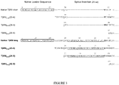
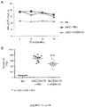

Classifications
-
- A—HUMAN NECESSITIES
- A61—MEDICAL OR VETERINARY SCIENCE; HYGIENE
- A61K—PREPARATIONS FOR MEDICAL, DENTAL OR TOILETRY PURPOSES
- A61K38/00—Medicinal preparations containing peptides
- A61K38/16—Peptides having more than 20 amino acids; Gastrins; Somatostatins; Melanotropins; Derivatives thereof
- A61K38/17—Peptides having more than 20 amino acids; Gastrins; Somatostatins; Melanotropins; Derivatives thereof from animals; from humans
- A61K38/1703—Peptides having more than 20 amino acids; Gastrins; Somatostatins; Melanotropins; Derivatives thereof from animals; from humans from vertebrates
- A61K38/1709—Peptides having more than 20 amino acids; Gastrins; Somatostatins; Melanotropins; Derivatives thereof from animals; from humans from vertebrates from mammals
-
- A—HUMAN NECESSITIES
- A61—MEDICAL OR VETERINARY SCIENCE; HYGIENE
- A61K—PREPARATIONS FOR MEDICAL, DENTAL OR TOILETRY PURPOSES
- A61K31/00—Medicinal preparations containing organic active ingredients
- A61K31/33—Heterocyclic compounds
- A61K31/395—Heterocyclic compounds having nitrogen as a ring hetero atom, e.g. guanethidine or rifamycins
- A61K31/495—Heterocyclic compounds having nitrogen as a ring hetero atom, e.g. guanethidine or rifamycins having six-membered rings with two or more nitrogen atoms as the only ring heteroatoms, e.g. piperazine or tetrazines
- A61K31/505—Pyrimidines; Hydrogenated pyrimidines, e.g. trimethoprim
- A61K31/519—Pyrimidines; Hydrogenated pyrimidines, e.g. trimethoprim ortho- or peri-condensed with heterocyclic rings
-
- A—HUMAN NECESSITIES
- A61—MEDICAL OR VETERINARY SCIENCE; HYGIENE
- A61K—PREPARATIONS FOR MEDICAL, DENTAL OR TOILETRY PURPOSES
- A61K45/00—Medicinal preparations containing active ingredients not provided for in groups A61K31/00 - A61K41/00
- A61K45/06—Mixtures of active ingredients without chemical characterisation, e.g. antiphlogistics and cardiaca
-
- A—HUMAN NECESSITIES
- A61—MEDICAL OR VETERINARY SCIENCE; HYGIENE
- A61P—SPECIFIC THERAPEUTIC ACTIVITY OF CHEMICAL COMPOUNDS OR MEDICINAL PREPARATIONS
- A61P1/00—Drugs for disorders of the alimentary tract or the digestive system
-
- A—HUMAN NECESSITIES
- A61—MEDICAL OR VETERINARY SCIENCE; HYGIENE
- A61P—SPECIFIC THERAPEUTIC ACTIVITY OF CHEMICAL COMPOUNDS OR MEDICINAL PREPARATIONS
- A61P1/00—Drugs for disorders of the alimentary tract or the digestive system
- A61P1/08—Drugs for disorders of the alimentary tract or the digestive system for nausea, cinetosis or vertigo; Antiemetics
-
- A—HUMAN NECESSITIES
- A61—MEDICAL OR VETERINARY SCIENCE; HYGIENE
- A61P—SPECIFIC THERAPEUTIC ACTIVITY OF CHEMICAL COMPOUNDS OR MEDICINAL PREPARATIONS
- A61P1/00—Drugs for disorders of the alimentary tract or the digestive system
- A61P1/14—Prodigestives, e.g. acids, enzymes, appetite stimulants, antidyspeptics, tonics, antiflatulents
-
- A—HUMAN NECESSITIES
- A61—MEDICAL OR VETERINARY SCIENCE; HYGIENE
- A61P—SPECIFIC THERAPEUTIC ACTIVITY OF CHEMICAL COMPOUNDS OR MEDICINAL PREPARATIONS
- A61P1/00—Drugs for disorders of the alimentary tract or the digestive system
- A61P1/16—Drugs for disorders of the alimentary tract or the digestive system for liver or gallbladder disorders, e.g. hepatoprotective agents, cholagogues, litholytics
-
- A—HUMAN NECESSITIES
- A61—MEDICAL OR VETERINARY SCIENCE; HYGIENE
- A61P—SPECIFIC THERAPEUTIC ACTIVITY OF CHEMICAL COMPOUNDS OR MEDICINAL PREPARATIONS
- A61P11/00—Drugs for disorders of the respiratory system
-
- A—HUMAN NECESSITIES
- A61—MEDICAL OR VETERINARY SCIENCE; HYGIENE
- A61P—SPECIFIC THERAPEUTIC ACTIVITY OF CHEMICAL COMPOUNDS OR MEDICINAL PREPARATIONS
- A61P17/00—Drugs for dermatological disorders
-
- A—HUMAN NECESSITIES
- A61—MEDICAL OR VETERINARY SCIENCE; HYGIENE
- A61P—SPECIFIC THERAPEUTIC ACTIVITY OF CHEMICAL COMPOUNDS OR MEDICINAL PREPARATIONS
- A61P17/00—Drugs for dermatological disorders
- A61P17/04—Antipruritics
-
- A—HUMAN NECESSITIES
- A61—MEDICAL OR VETERINARY SCIENCE; HYGIENE
- A61P—SPECIFIC THERAPEUTIC ACTIVITY OF CHEMICAL COMPOUNDS OR MEDICINAL PREPARATIONS
- A61P19/00—Drugs for skeletal disorders
-
- A—HUMAN NECESSITIES
- A61—MEDICAL OR VETERINARY SCIENCE; HYGIENE
- A61P—SPECIFIC THERAPEUTIC ACTIVITY OF CHEMICAL COMPOUNDS OR MEDICINAL PREPARATIONS
- A61P19/00—Drugs for skeletal disorders
- A61P19/02—Drugs for skeletal disorders for joint disorders, e.g. arthritis, arthrosis
-
- A—HUMAN NECESSITIES
- A61—MEDICAL OR VETERINARY SCIENCE; HYGIENE
- A61P—SPECIFIC THERAPEUTIC ACTIVITY OF CHEMICAL COMPOUNDS OR MEDICINAL PREPARATIONS
- A61P19/00—Drugs for skeletal disorders
- A61P19/08—Drugs for skeletal disorders for bone diseases, e.g. rachitism, Paget's disease
-
- A—HUMAN NECESSITIES
- A61—MEDICAL OR VETERINARY SCIENCE; HYGIENE
- A61P—SPECIFIC THERAPEUTIC ACTIVITY OF CHEMICAL COMPOUNDS OR MEDICINAL PREPARATIONS
- A61P21/00—Drugs for disorders of the muscular or neuromuscular system
-
- A—HUMAN NECESSITIES
- A61—MEDICAL OR VETERINARY SCIENCE; HYGIENE
- A61P—SPECIFIC THERAPEUTIC ACTIVITY OF CHEMICAL COMPOUNDS OR MEDICINAL PREPARATIONS
- A61P25/00—Drugs for disorders of the nervous system
- A61P25/02—Drugs for disorders of the nervous system for peripheral neuropathies
-
- A—HUMAN NECESSITIES
- A61—MEDICAL OR VETERINARY SCIENCE; HYGIENE
- A61P—SPECIFIC THERAPEUTIC ACTIVITY OF CHEMICAL COMPOUNDS OR MEDICINAL PREPARATIONS
- A61P27/00—Drugs for disorders of the senses
- A61P27/02—Ophthalmic agents
-
- A—HUMAN NECESSITIES
- A61—MEDICAL OR VETERINARY SCIENCE; HYGIENE
- A61P—SPECIFIC THERAPEUTIC ACTIVITY OF CHEMICAL COMPOUNDS OR MEDICINAL PREPARATIONS
- A61P29/00—Non-central analgesic, antipyretic or antiinflammatory agents, e.g. antirheumatic agents; Non-steroidal antiinflammatory drugs [NSAID]
- A61P29/02—Non-central analgesic, antipyretic or antiinflammatory agents, e.g. antirheumatic agents; Non-steroidal antiinflammatory drugs [NSAID] without antiinflammatory effect
-
- A—HUMAN NECESSITIES
- A61—MEDICAL OR VETERINARY SCIENCE; HYGIENE
- A61P—SPECIFIC THERAPEUTIC ACTIVITY OF CHEMICAL COMPOUNDS OR MEDICINAL PREPARATIONS
- A61P3/00—Drugs for disorders of the metabolism
-
- A—HUMAN NECESSITIES
- A61—MEDICAL OR VETERINARY SCIENCE; HYGIENE
- A61P—SPECIFIC THERAPEUTIC ACTIVITY OF CHEMICAL COMPOUNDS OR MEDICINAL PREPARATIONS
- A61P3/00—Drugs for disorders of the metabolism
- A61P3/02—Nutrients, e.g. vitamins, minerals
-
- A—HUMAN NECESSITIES
- A61—MEDICAL OR VETERINARY SCIENCE; HYGIENE
- A61P—SPECIFIC THERAPEUTIC ACTIVITY OF CHEMICAL COMPOUNDS OR MEDICINAL PREPARATIONS
- A61P35/00—Antineoplastic agents
-
- A—HUMAN NECESSITIES
- A61—MEDICAL OR VETERINARY SCIENCE; HYGIENE
- A61P—SPECIFIC THERAPEUTIC ACTIVITY OF CHEMICAL COMPOUNDS OR MEDICINAL PREPARATIONS
- A61P39/00—General protective or antinoxious agents
-
- A—HUMAN NECESSITIES
- A61—MEDICAL OR VETERINARY SCIENCE; HYGIENE
- A61P—SPECIFIC THERAPEUTIC ACTIVITY OF CHEMICAL COMPOUNDS OR MEDICINAL PREPARATIONS
- A61P43/00—Drugs for specific purposes, not provided for in groups A61P1/00-A61P41/00
-
- A—HUMAN NECESSITIES
- A61—MEDICAL OR VETERINARY SCIENCE; HYGIENE
- A61P—SPECIFIC THERAPEUTIC ACTIVITY OF CHEMICAL COMPOUNDS OR MEDICINAL PREPARATIONS
- A61P7/00—Drugs for disorders of the blood or the extracellular fluid
-
- A—HUMAN NECESSITIES
- A61—MEDICAL OR VETERINARY SCIENCE; HYGIENE
- A61P—SPECIFIC THERAPEUTIC ACTIVITY OF CHEMICAL COMPOUNDS OR MEDICINAL PREPARATIONS
- A61P7/00—Drugs for disorders of the blood or the extracellular fluid
- A61P7/02—Antithrombotic agents; Anticoagulants; Platelet aggregation inhibitors
-
- A—HUMAN NECESSITIES
- A61—MEDICAL OR VETERINARY SCIENCE; HYGIENE
- A61P—SPECIFIC THERAPEUTIC ACTIVITY OF CHEMICAL COMPOUNDS OR MEDICINAL PREPARATIONS
- A61P7/00—Drugs for disorders of the blood or the extracellular fluid
- A61P7/04—Antihaemorrhagics; Procoagulants; Haemostatic agents; Antifibrinolytic agents
-
- A—HUMAN NECESSITIES
- A61—MEDICAL OR VETERINARY SCIENCE; HYGIENE
- A61P—SPECIFIC THERAPEUTIC ACTIVITY OF CHEMICAL COMPOUNDS OR MEDICINAL PREPARATIONS
- A61P7/00—Drugs for disorders of the blood or the extracellular fluid
- A61P7/06—Antianaemics
-
- A—HUMAN NECESSITIES
- A61—MEDICAL OR VETERINARY SCIENCE; HYGIENE
- A61P—SPECIFIC THERAPEUTIC ACTIVITY OF CHEMICAL COMPOUNDS OR MEDICINAL PREPARATIONS
- A61P9/00—Drugs for disorders of the cardiovascular system
-
- A—HUMAN NECESSITIES
- A61—MEDICAL OR VETERINARY SCIENCE; HYGIENE
- A61P—SPECIFIC THERAPEUTIC ACTIVITY OF CHEMICAL COMPOUNDS OR MEDICINAL PREPARATIONS
- A61P9/00—Drugs for disorders of the cardiovascular system
- A61P9/10—Drugs for disorders of the cardiovascular system for treating ischaemic or atherosclerotic diseases, e.g. antianginal drugs, coronary vasodilators, drugs for myocardial infarction, retinopathy, cerebrovascula insufficiency, renal arteriosclerosis
-
- A—HUMAN NECESSITIES
- A61—MEDICAL OR VETERINARY SCIENCE; HYGIENE
- A61P—SPECIFIC THERAPEUTIC ACTIVITY OF CHEMICAL COMPOUNDS OR MEDICINAL PREPARATIONS
- A61P9/00—Drugs for disorders of the cardiovascular system
- A61P9/12—Antihypertensives
-
- C—CHEMISTRY; METALLURGY
- C07—ORGANIC CHEMISTRY
- C07K—PEPTIDES
- C07K14/00—Peptides having more than 20 amino acids; Gastrins; Somatostatins; Melanotropins; Derivatives thereof
- C07K14/435—Peptides having more than 20 amino acids; Gastrins; Somatostatins; Melanotropins; Derivatives thereof from animals; from humans
- C07K14/475—Growth factors; Growth regulators
- C07K14/495—Transforming growth factor [TGF]
-
- C—CHEMISTRY; METALLURGY
- C07—ORGANIC CHEMISTRY
- C07K—PEPTIDES
- C07K14/00—Peptides having more than 20 amino acids; Gastrins; Somatostatins; Melanotropins; Derivatives thereof
- C07K14/435—Peptides having more than 20 amino acids; Gastrins; Somatostatins; Melanotropins; Derivatives thereof from animals; from humans
- C07K14/705—Receptors; Cell surface antigens; Cell surface determinants
- C07K14/71—Receptors; Cell surface antigens; Cell surface determinants for growth factors; for growth regulators
-
- A—HUMAN NECESSITIES
- A61—MEDICAL OR VETERINARY SCIENCE; HYGIENE
- A61K—PREPARATIONS FOR MEDICAL, DENTAL OR TOILETRY PURPOSES
- A61K2300/00—Mixtures or combinations of active ingredients, wherein at least one active ingredient is fully defined in groups A61K31/00 - A61K41/00
-
- C—CHEMISTRY; METALLURGY
- C07—ORGANIC CHEMISTRY
- C07K—PEPTIDES
- C07K2319/00—Fusion polypeptide
- C07K2319/30—Non-immunoglobulin-derived peptide or protein having an immunoglobulin constant or Fc region, or a fragment thereof, attached thereto
Definitions
- MPDs Myeloproliferative disorders
- MPNs neoplasms
- PV polycythemia vera
- ET essential thrombocythemia
- myelofibrosis e.g., primary myelofibrosis, post-polycythemia vera myelofibrosis, and post-essential thrombocythemia myelofibrosis
- CML chronic myeloid leukemia
- PV is characterized by increased production of all 3 types of blood cells
- ET is manifest in the elevation of platelets.
- Myelofibrosis (MF) is a disease in which fibrous (scar-like) tissues develop in the bone marrow as a result of abnormal production of red cells, white cells, and/or platelets.
- CML is characterized by the increased and unregulated growth of predominantly myeloid cells in the bone marrow and the accumulation of these cells in the peripheral blood.
- CML is now defined by its causative molecular lesion, the BCR-ABL fusion gene, which most commonly results from the Philadelphia translocation (Ph). Accordingly, CML is characterized as a BCR-ABL positive (+) myeloproliferative disorder. Discovery of this defined molecular defect lead to the development of the drug imatinib mesylate (Gleevec; Novartis, Basel, Switzerland) to treat CML [ Druker et al. (2001) N Engl J Med 344:1031-1037 ].
- the other three myeloproliferative neoplasms are characterized as BCR-ABL -negative myeloproliferative disorder.
- JAK2V617F tyrosine kinase JAK2
- WO 1998/048024 describes fusion proteins comprising the TGF-beta Type II receptor linked to a portion of an immunoglobulin constant chain and their use to treat a variety of fibroproliferative disorders.
- Allogeneic hematopoietic stem cell transplantation is the only known cure for BCR-ABL- negative MPDs [ Gupta et al. (2012) Blood 120:1367-1379 ].
- stem cell treatment-related mortality is high and only a minority of patients qualify for transplantation. While the development and use of JAK inhibitors represents a significant therapeutic advancement, there are clear limitations to their use in the treatment of BCR-ABL- negative MPDs.
- JAK inhibitors appear to be useful for reducing splenomegaly in myelofibrosis patients; however, their effects on the disease are otherwise largely palliative [ Gupta et al. (2012) Blood 120:1367-1379 ].
- Gäbler et al. (2013), JAK-STAT, 2(3):e25025 is a review artcle on JAK2 mutants as drug targets in myeloproliferative neoplasms.
- JAK inhibitors have little to no effect on many manifestations (complications) of the disease including, for example, cytopenia, transfusion dependence, accelerated or blast phase disease, and fibrosis.
- JAK inhibitors have been shown to promote or worsen thrombocytopenia, anemia, and neutropenia in some patients.
- the present invention relates to a composition for use in a method for treating, preventing, or reducing the progression rate and/or severity of myelofibrosis or one or more complications of myelofibrosis in a patient in need thereof, wherein the composition comprises an effective amount of a transforming growth factor beta type II receptor (T ⁇ RII) antagonist, wherein the T ⁇ RII antagonist is a fusion protein comprising:
- the patient is intolerant or refractory to treatment with a Janus kinase inhibitor.
- the T ⁇ RII antagonist inhibits TGF ⁇ 1 and TGF ⁇ 3.
- the T ⁇ RII antagonist comprises an amino acid sequence that is 90%, 91%, 92%, 93%, 94% 95%, 96%, 97%, 98%, 99%, or 100% identical to any one of SEQ ID NOs: 19, 20 and 21.
- the amino acid sequence of the T ⁇ RII polypeptide consists of an amino acid sequence at least 95%, at least 96%, at least 97%, at least 98%, at least 99% identical to the amino acid sequence of SEQ ID NO: 13.
- amino acid sequence of the T ⁇ RII polypeptide consists of the amino acid sequence of SEQ ID NO: 13.
- the linker is positioned between the T ⁇ RII polypeptide and the immunoglobulin Fc amino acid sequence.
- the fusion protein comprises an amino acid sequence that is at least 90%, 95%, 99%, or 100% identical to the amino acid sequence of SEQ ID NO: 103.
- the patient has a disorder associated with a gain-of-function mutation in JAK2.
- the patient has a disorder associated with elevated or constitutive kinase activity of JAK2, preferably wherein the elevated kinase activity of JAK2 is as compared to healthy patients of the same age and sex.
- the patient has a JAK2V617F-associated disorder.
- the patient has primary myelofibrosis, post-polycythemia vera myelofibrosis or post essential thrombocythemia myelofibrosis.
- the patient has primary myelofibrosis.
- the Janus kinase inhibitor is selected from the group consisting of: ruxolitinib, fedratinib (SAR302503), monoelotinib (CYT387), pacritinib, lestaurtinib, AZD-1480, BMS-911543, NS-018, LY2784544, SEP-701, XL019, and AT-9283.
- the Janus kinase inhibitor is ruxolitinib or fedratinib.
- Proteins described herein are the human forms, unless otherwise specified. NCBI references for the proteins are as follows: human T ⁇ PII isoform A (hT ⁇ RII long ), NP_001020018.1 and human T ⁇ PII isoform B (hT ⁇ RII short ), NP_003233.4. Sequences of native human T ⁇ RII proteins are set forth in Figures 1-2 .
- the TGF ⁇ superfamily contains a variety of growth factors that share common sequence elements and structural motifs. These proteins are known to exert biological effects on a large variety of cell types in both vertebrates and invertebrates. Members of the superfamily perform important functions during embryonic development in pattern formation and tissue specification and can influence a variety of differentiation processes, including adipogenesis, myogenesis, chondrogenesis, cardiogenesis, hematopoiesis, neurogenesis, and epithelial cell differentiation. By manipulating the activity of a member of the TGF ⁇ family, it is often possible to cause significant physiological changes in an organism.
- GDF8 also called myostatin
- inactive alleles of GDF8 are associated with increased muscle mass and, reportedly, exceptional strength [Schuelke et al. (2004) N Engl J Med 350:2682-8 ].
- TGF ⁇ signals are mediated by heteromeric complexes of type I (e.g. T ⁇ RI) and type II (e.g. T ⁇ RII) serine/ threonine kinase receptors, which phosphorylate and activate downstream SMAD proteins upon ligand stimulation [ Massagué (2000) Nat. Rev. Mol. Cell Biol. 1:169-178 ].
- type I and type II receptors are transmembrane proteins, composed of a ligand-binding extracellular domain with cysteine-rich region, a transmembrane domain, and a cytoplasmic domain with predicted serine/threonine specificity.
- Type I receptors are essential for signaling; and type II receptors are required for binding ligands and for expression of type I receptors.
- Type I and II receptors form a stable complex after ligand binding, resulting in phosphorylation of type I receptors by type II receptors.
- TGF ⁇ has three mammalian isoforms, TGF ⁇ 1, TGF ⁇ 2 and TGF ⁇ 3, each with distinct functions in vivo.
- the binding of TGF ⁇ s to T ⁇ RII is a crucial step in initiating activation of the TGF ⁇ signaling pathway, leading to phosphorylation of SMAD2, and translocation of the activated SMAD2/SMAD4 complex to the nucleus to modulate gene expression.
- T ⁇ RII is the known type II receptor for TGF ⁇ and binds with high affinity to TGF ⁇ 1 and TGF ⁇ 3.
- Human T ⁇ PII occurs naturally in at least two isoforms - A (long) and B (short) - generated by alternative splicing in the extracellular domain (ECD) ( Figures 2 and 1 and SEQ ID NOS: 6 and 5).
- the long isoform has a 25-amino-acid insertion and the splicing process causes the valine flanking the insertion site in the short isoform to be replaced by an isoleucine in the long isoform.
- Soluble receptor ectodomains can function as scavengers or ligand traps to inhibit ligand-receptor interactions.
- Ligand traps such as soluble T ⁇ RII-Fc fusion proteins incorporating the native T ⁇ RII extracellular domain (ectodomain) will function as pan-inhibitors against T ⁇ RII ligands, including, TGF ⁇ 1 and TGF ⁇ 3. While in some therapeutic settings this broader spectrum of ligand-binding and signal inhibition may be advantageous, in other settings a more selective molecule may be superior. It is highly desirable for ligand traps such as T ⁇ PII ectodomain polypeptides to exhibit selective ligand-binding profiles.
- the disclosure generally provides T ⁇ RII polypeptides as antagonists of TGF ⁇ 1 or TGF ⁇ 3 for use in treating various TGF ⁇ 1- or TGF ⁇ 3-associated disorders. While not wishing to be bound to any particular mechanism of action, it is expected that such polypeptides act by binding to TGF ⁇ 1 or TGF ⁇ 3 and inhibiting the ability of these ligands to form ternary signaling complexes.
- Myeloproliferative disorders are a group of conditions characterized, in part, by chronic increases in some or all of blood cells (platelets, white blood cells, and red blood cells).
- This group of blood disorders includes polycythemia vera (PV), essential thrombocythemia (ET), myelofibrosis (e.g., primary myelofibrosis (PMF), post-polycythemia vera myelofibrosis, and post-essential thrombocythemia myelofibrosis), and chronic myeloid leukemia (CML). It is generally thought that myeloproliferative disorders arise from a transformation in a hematopoietic stem cell.
- CML is defined by its causative molecular lesion, the BCR-ABL fusion, which most commonly results from the Philadelphia translocation.
- JAK2V617F tyrosine kinase JAK2 mice develop pathology that closely resembles human essential thrombocythemia and polycythemia vera [ Xing et al. (2008) Blood 111: 5109-5117 ].
- JAK2V617F mice As they age, these JAK2V617F mice also develop primary myelofibrosis-like pathology. As disclosed herein, it has been discovered that T ⁇ RII-Fc treatment can reduce splenomegaly, fibrosis, and other morbidities in a JAK2 V617F disease model.
- T ⁇ RII antagonist may be used to treat or prevent complications resultant from the JAK2V617F mutation, which indicates that such therapeutics may be used to treat myeloproliferative disorders (e.g., polycythemia vera, essential thrombocythaemia, and myelofibrosis) as well as Janus kinase-associated disorder (e.g., a JAK2 kinase-associated disorder).
- myeloproliferative disorders e.g., polycythemia vera, essential thrombocythaemia, and myelofibrosis
- Janus kinase-associated disorder e.g., a JAK2 kinase-associated disorder
- T ⁇ RII antagonists may be particular well suited for treatment of polycythemia vera and essential thrombocythaemia to prevent/delay the onset or reduce the severity of fibrosis and other late stage disease complications and thus prevent/delay the transition into secondary myelofibrosis disease (post-polycythemia vera myelofibrosis and post-essential thrombocythaemia myelofibrosis, respectively).
- T ⁇ RII antagonists clearly demonstrate positive effects in treated last stage fibrosis and inflammation in myelofibrosis patients.
- sequence similarity in all its grammatical forms, refers to the degree of identity or correspondence between nucleic acid or amino acid sequences that may or may not share a common evolutionary origin.
- the term “homologous,” when modified with an adverb such as "highly,” may refer to sequence similarity and may or may not relate to a common evolutionary origin.
- Percent (%) sequence identity with respect to a reference polypeptide (or nucleotide) sequence is defined as the percentage of amino acid residues (or nucleic acids) in a candidate sequence that are identical to the amino acid residues (or nucleic acids) in the reference polypeptide (nucleotide) sequence, after aligning the sequences and introducing gaps, if necessary, to achieve the maximum percent sequence identity, and not considering any conservative substitutions as part of the sequence identity. Alignment for purposes of determining percent amino acid sequence identity can be achieved in various ways that are within the skill in the art, for instance, using publicly available computer software such as BLAST, BLAST-2, ALIGN or Megalign (DNASTAR) software.
- ALIGN-2 sequence comparison computer program
- the ALIGN-2 sequence comparison computer program was authored by Genentech, Inc., and the source code has been filed with user documentation in the U.S. Copyright Office, Washington D.C., 20559, where it is registered under U.S. Copyright Registration No. TXU510087.
- the ALIGN-2 program is publicly available from Genentech, Inc., South San Francisco, Calif., or may be compiled from the source code.
- the ALIGN-2 program should be compiled for use on a UNIX operating system, including digital UNIX V4.0D. All sequence comparison parameters are set by the ALIGN-2 program and do not vary.
- Alternize in all its grammatical forms, refers to the process of activating a protein and/or gene ( e.g., by activating or amplifying that protein's gene expression or by inducing an inactive protein to enter an active state) or increasing a protein's and/or gene's activity.
- “Antagonize”, in all its grammatical forms, refers to the process of inhibiting a protein and/or gene (e.g., by inhibiting or decreasing that protein's gene expression or by inducing an active protein to enter an inactive state) or decreasing a protein's and/or gene's activity.
- a T ⁇ RII antagonist can be used to treat a myeloproliferative disorder (e.g., polycythemia vera, essential thrombocythemia, and myelofibrosis) and/or a patient with a Janus kinase-associated disorder (e.g., a JAK2 kinase-associated disorder).
- a T ⁇ RII polypeptide was shown to be effective in improving various myeloproliferative disease complications including, for example, splenomegaly, high inflammatory cytokine levels, and fibrosis.
- the disclosure provides, in part, various T ⁇ RII antagonists that can be used, alone or in combination with one or more additional active agents and/or supportive therapies, to treat, prevent, reduce the progression rate and/or severity of a myeloproliferative disorder and/or a patient with a Janus kinase-associated disorder or one or more complications of a myeloproliferative disorder and/or a Janus kinase-associated disorder.
- T ⁇ RII polypeptides may affect myeloproliferative disease and/or a patient with a Janus kinase-associated disorder through a mechanism other than inhibition of T ⁇ RII ligands
- inhibition of one or more of TGF ⁇ 1, TGF ⁇ 2, and/or TGF ⁇ 3 may be an indicator of the tendency of an agent to inhibit the activities of a spectrum of additional agents, including, perhaps, other members of the TGF-beta superfamily and such collective inhibition may lead to the desired effect on, for example, a myeloproliferative disease and/or a Janus kinase-associated disorder]
- other types of TGFP antagonists e.g., antagonists of the T ⁇ RII receptor, antagonists of one or more T ⁇ RII ligands (e.g., TGF ⁇ 1, TGF ⁇ 2, and TGF ⁇ 3), antagonists of one or more T ⁇ RII-associated type I receptors (e.g., ALK5), antagonists of one or more T ⁇ RII-
- Such antagonists include, for example, for example, T ⁇ PII polypeptides and variants thereof, anti-TGF ⁇ antibodies, anti-ALK5 antibodies, anti-betaglycan antibodies, and anti-T ⁇ RII antibodies; nucleic acids that inhibit the activity or expression (e.g., transcription, translation, secretion from a cell, or combinations thereof) of one or more of TGF ⁇ 1, TGF ⁇ 2, TGF ⁇ 3, ALK5, betaglycan, and T ⁇ RII; as well as small molecules that inhibit the activity or expression (e.g., transcription, translation, secretion from a cell, or combinations thereof) of one or more of TGF ⁇ 1, TGF ⁇ 2, TGF ⁇ 3, ALK5, betaglycan and T ⁇ RII.
- nucleic acids that inhibit the activity or expression (e.g., transcription, translation, secretion from a cell, or combinations thereof) of one or more of TGF ⁇ 1, TGF ⁇ 2, TGF ⁇ 3, ALK5, betaglycan and T ⁇ RII
- small molecules that inhibit the activity or expression
- a T ⁇ RII polypeptide, or combination of polypeptides may inhibit, for example, one or more T ⁇ RII ligands (e.g., TGF ⁇ 1, TGF ⁇ 2, and TGF ⁇ 3), T ⁇ RII receptor, T ⁇ RII-associated type I receptor (e.g., ALK5), and/or T ⁇ RII-associated co-receptor (e.g., betaglycan).
- T ⁇ RII ligands e.g., TGF ⁇ 1, TGF ⁇ 2, and TGF ⁇ 3
- T ⁇ RII receptor e.g., TGF ⁇ 1, TGF ⁇ 2, and TGF ⁇ 3
- T ⁇ RII receptor e.g., T ⁇ RII-associated type I receptor
- T ⁇ RII-associated co-receptor e.g., betaglycan.
- the ability for a T ⁇ RII polypeptide, or combination of polypeptides, to inhibit signaling may be determined in a cell-based assay including, for example, those described herein.
- a T ⁇ RII polypeptide, or combination of polypeptides may be used alone or in combination with one or more additional supportive therapies or active agents to treat, prevent, or reduce the progression rate and/or severity of a myeloproliferative disorder (e.g., myelofibrosis, polycythemia vera, and essential thrombocythaemia) and/or a Janus kinase-associated disorder or one or more complications of a myeloproliferative disorder and/or a Janus kinase-associated disorder (e.g., a JAK2 kinase-associated disorder).
- a myeloproliferative disorder e.g., myelofibrosis, polycythemia vera, and essential thrombocythaemia
- a Janus kinase-associated disorder e.g., a Janus kinase-associated disorder.
- Naturally occurring T ⁇ RII proteins are transmembrane proteins, with a portion of the protein positioned outside the cell (the extracellular portion) and a portion of the protein positioned inside the cell (the intracellular portion).
- Variant T ⁇ RII polypeptides may comprise mutations within the extracellular domain and/or truncated portions of the extracellular domain of T ⁇ RII.
- human T ⁇ RII occurs naturally in at least two isoforms - A (long) and B (short) - generated by alternative splicing in the extracellular domain (ECD) ( Figures 2 and 1 and SEQ ID NOS: 6 and 5).
- SEQ ID NO: 7 which corresponds to residues 23-159 of SEQ ID NO: 5, depicts the native full-length extracellular domain of the short isoform of T ⁇ RII.
- SEQ ID NO: 13 which corresponds to residues 23-184 of SEQ ID NO: 6, depicts the native full-length extracellular domain of the long isoform of T ⁇ RII.
- amino acid position numbering with regard to variants based on the T ⁇ RII short and long isoforms refers to the corresponding position in the native precursors, SEQ ID NO: 5 and SEQ ID NO:6, respectively.
- a T ⁇ RII polypeptide of the disclosure may bind to and inhibit the function of a TGF ⁇ superfamily member, such as but not limited to, TGF ⁇ 1 or TGF ⁇ 3.
- a T ⁇ PII polypeptide as defined in accordance with the claimed invention consists of an amino acid sequence from the extracellular domain of T ⁇ RII that is at least 90% identical to the amino acid sequence of SEQ ID NO: 13. It will be understood by one of skill in the art that corresponding variants based on the long isoform of T ⁇ RII will include nucleotide sequences encoding the 25-amino acid insertion along with a conservative Val-Ile substitution at the flanking position C-terminal to the insertion.. Generally, a T ⁇ RII polypeptide will be designed to be soluble in aqueous solutions at biologically relevant temperatures, pH levels, and osmolarity.
- T ⁇ RII polypeptides as defined by the claimed invention may comprise one or more mutations in the extracellular domain that confer an altered ligand binding profile.
- T ⁇ RII polypeptides can be modified to selectively antagonize T ⁇ RII ligands.
- Data presented here show that Fc fusion proteins comprising shorter N-terminally and C-terminally truncated variants of T ⁇ RII polypeptides display differential inhibitory effects on cellular signaling mediated by TGF ⁇ 1 and TGF ⁇ 3.
- N-terminally truncated variants beginning at amino acids 29 or 35 of SEQ ID NO: 5 and carrying, respectively, a 6- or 12-amino acid N-terminal truncation of the extracellular domain were found to significantly reduced potency for inhibiting TGF ⁇ 3 while maintaining an intermediate degree of TGF ⁇ 1 inhibition, compared to the full length extracellular domain of the short isoform of T ⁇ RII.
- C-terminally truncated variants, ending at amino acid 153 of SEQ ID NO: 5 and carrying a 6-amino acid C-terminal truncation of the extracellular domain had no substantial effect on ligand binding and may therefore be used interchangeably with full length versions.
- N to D substitution at the position corresponding to position 70 of SEQ ID NO: 5 was found to inhibit TGF ⁇ 3 potently and negligible effect on TGF ⁇ 1.
- the N70 residue represents a potential glycosylation site.
- an Fc fusion protein comprising a D to K substitution at the position corresponding to position 110 of SEQ ID NO: 5 was found to significantly reduced potency for inhibiting TGF ⁇ 1 while maintaining an intermediate degree of TGFP3 inhibition compared to the full length extracellular domain of the short isoform of T ⁇ RII.
- T ⁇ RII polypeptides that contain mutations in the ECD such as but not limited to, N70D and D110K (the numbering of the residues corresponds to that of SEQ ID NO: 5) and/or begin between amino acids 29 and 35 and/or terminate between amino acid 153 and amino acid 159 are all expected to be active and exhibit widely different inhibitory potencies towards the different ligands. Any of these truncated variant forms may be desirable to use, depending on the clinical or experimental setting.
- a T ⁇ RII polypeptide may bind to TGF ⁇ 1, and the T ⁇ RII polypeptide may not show substantial binding to TGF ⁇ 3.
- a T ⁇ RII polypeptide may bind to TGF ⁇ 3, and the T ⁇ RII polypeptide may not show substantial binding to TGF ⁇ 1. Binding may be assessed, e.g., using purified proteins in solution or in a surface plasmon resonance system, such as a Biacore TM system.
- a T ⁇ RII polypeptide may inhibit TGF ⁇ 1 cellular signaling, and the T ⁇ RII polypeptide has an intermediate or limited inhibitory effect on TGF ⁇ 3.
- a T ⁇ RII polypeptide may inhibit TGF ⁇ 3 cellular signaling, and the T ⁇ RII polypeptide may have an intermediate or limited inhibitory effect on TGF ⁇ 1. Inhibitory effect on cell signaling can be assayed by methods known in the art.
- the disclosure provides T ⁇ RII polypeptides sharing a specified degree of sequence identity or similarity to a naturally occurring T ⁇ RII polypeptide.
- the sequences are aligned for optimal comparison purposes (e.g ., gaps can be introduced in one or both of a first and a second amino acid or nucleic acid sequence for optimal alignment and non-homologous sequences can be disregarded for comparison purposes).
- the amino acid residues at corresponding amino acid positions are then compared. When a position in the first sequence is occupied by the same amino acid residue as the corresponding position in the second sequence, then the molecules are identical at that position (as used herein amino acid "identity" is equivalent to amino acid "homology”).
- the percent identity between the two sequences is a function of the number of identical positions shared by the sequences, taking into account the number of gaps, and the length of each gap, which need to be introduced for optimal alignment of the two sequences.
- the percent identity between two amino acid sequences can be determined using the Needleman and Wunsch (J Mol. Biol. (48):444-453 (1970 )) algorithm which has been incorporated into the GAP program in the GCG software package (available at http://www.gcg.com).
- the following parameters may be used in the GAP program: either a Blosum 62 matrix or a PAM250 matrix, and a gap weight of 16, 14, 12, 10, 8, 6, or 4 and a length weight of 1, 2, 3, 4, 5, or 6.
- the percent identity between two nucleotide sequences may alternatively be determined using the GAP program in the GCG software package ( Devereux, J., et al., Nucleic Acids Res.
- Exemplary parameters include using a NWSgapdna.CMP matrix and a gap weight of 40, 50, 60, 70, or 80 and a length weight of 1, 2, 3, 4, 5, or 6. Unless otherwise specified, percent identity between two amino acid sequences is to be determined using the GAP program using a Blosum 62 matrix, a GAP weight of 10 and a length weight of 3, and if such algorithm cannot compute the desired percent identity, a suitable alternative disclosed herein should be selected.
- the percent identity between two amino acid sequences can be determined using the algorithm of E. Myers and W. Miller (CABIOS, 4:11-17 (1989 )) which has been incorporated into the ALIGN program (version 2.0), using a PAM120 weight residue table, a gap length penalty of 12 and a gap penalty of 4.
- T ⁇ RII polypeptides may additionally include any of various leader sequences at the N-terminus. Such a sequence would allow the peptides to be expressed and targeted to the secretion pathway in a eukaryotic system. See, e.g., Ernst et al., U.S. Pat. No. 5,082,783 (1992 ).
- a native T ⁇ RII signal sequence may be used to effect extrusion from the cell.
- Possible leader sequences include native leaders, tissue plasminogen activator (TPA) and honeybee mellitin (SEQ ID NOs. 22-24, respectively).
- T ⁇ RII-Fc fusion proteins incorporating a TPA leader sequence examples include SEQ ID NOs: 25, 27, 29, 31, 33, 35, 37, 39, 41, 43, 101 and 103. Processing of signal peptides may vary depending on the leader sequence chosen, the cell type used and culture conditions, among other variables, and therefore actual N-terminal start sites for mature T ⁇ PII polypeptides may shift by 1, 2, 3, 4 or 5 amino acids in either the N-terminal or C-terminal direction.
- T ⁇ RII-Fc fusion proteins include SEQ ID NOs: 25, 27, 29, 31, 33, 35, 37, 39, 41, 43, 50, 53, 54, 55, 56, 57, 58, 59, 60, 61, 62, 101, and 103, as shown herein with the T ⁇ RII polypeptide portion underlined (see Examples). It will be understood by one of skill in the art that corresponding variants based on the long isoform of TPRII will include the 25-amino acid insertion along with a conservative Val-Ile substitution at the flanking position C-terminal to the insertion.
- the present disclosure also contemplates specific mutations of the T ⁇ PII polypeptides so as to alter the glycosylation of the polypeptide.
- Such mutations may be selected so as to introduce or eliminate one or more glycosylation sites, such as O-linked or N-linked glycosylation sites.
- Asparagine-linked glycosylation recognition sites generally comprise a tripeptide sequence, asparagine-X-threonine (or asparagine-X-serine) (where "X" is any amino acid) which is specifically recognized by appropriate cellular glycosylation enzymes.
- the alteration may also be made by the addition of, or substitution by, one or more serine or threonine residues to the sequence of the wild-type T ⁇ RII polypeptide (for O-linked glycosylation sites).
- a variety of amino acid substitutions or deletions at one or both of the first or third amino acid positions of a glycosylation recognition site (and/or amino acid deletion at the second position) results in non-glycosylation at the modified tripeptide sequence.
- Another means of increasing the number of carbohydrate moieties on a T ⁇ RII polypeptide is by chemical or enzymatic coupling of glycosides to the T ⁇ PII polypeptide.
- the sugar(s) may be attached to (a) arginine and histidine; (b) free carboxyl groups; (c) free sulfhydryl groups such as those of cysteine; (d) free hydroxyl groups such as those of serine, threonine, or hydroxyproline; (e) aromatic residues such as those of phenylalanine, tyrosine, or tryptophan; or (f) the amide group of glutamine.
- Removal of one or more carbohydrate moieties present on a T ⁇ RII polypeptide may be accomplished chemically and/or enzymatically.
- Chemical deglycosylation may involve, for example, exposure of the T ⁇ PII polypeptide to the compound trifluoromethanesulfonic acid, or an equivalent compound. This treatment results in the cleavage of most or all sugars except the linking sugar (N-acetylglucosamine or N-acetylgalactosamine), while leaving the amino acid sequence intact.
- Chemical deglycosylation is further described by Hakimuddin et al. (1987) Arch. Biochem. Biophys. 259:52 and by Edge et al. (1981) Anal. Biochem. 118:131 .
- Enzymatic cleavage of carbohydrate moieties on T ⁇ RII polypeptides can be achieved by the use of a variety of endo- and exo-glycosidases as described by Thotakura et al. (1987) Meth. Enzymol. 138:350 .
- the sequence of a T ⁇ RII polypeptide may be adjusted, as appropriate, depending on the type of expression system used, as mammalian, yeast, insect and plant cells may all introduce differing glycosylation patterns that can be affected by the amino acid sequence of the peptide.
- T ⁇ RII polypeptides for use in humans will be expressed in a mammalian cell line that provides proper glycosylation, such as HEK293 or CHO cell lines, although other mammalian expression cell lines, yeast cell lines with engineered glycosylation enzymes, and insect cells are expected to be useful as well.
- This disclosure further contemplates a method, which is not covered by the claimed invention, of generating mutants, particularly sets of combinatorial mutants of a T ⁇ RII polypeptide, as well as truncation mutants; pools of combinatorial mutants are especially useful for identifying functional variant sequences.
- the purpose of screening such combinatorial libraries may be to generate, for example, T ⁇ RII polypeptide variants which can act as either agonists or antagonist, or alternatively, which possess novel activities all together.
- a variety of screening assays are provided below, and such assays may be used to evaluate variants.
- a T ⁇ RII polypeptide variant may be screened for ability to bind to a T ⁇ RII ligand, to prevent binding of a T ⁇ RII ligand to a T ⁇ RII polypeptide or to interfere with signaling caused by a T ⁇ RII ligand.
- the activity of a T ⁇ RII polypeptide or its variants may also be tested in a cell-based or in vivo assay, particularly any of the assays disclosed in the Examples.
- Combinatorially-derived variants can be generated which have a selective or generally increased potency relative to a T ⁇ RII polypeptide comprising an extracellular domain of a naturally occurring T ⁇ RII polypeptide.
- mutagenesis can give rise to variants which have serum half-lives dramatically different than the corresponding wild-type T ⁇ RII polypeptide.
- the altered protein can be rendered either more stable or less stable to proteolytic degradation or other processes which result in destruction of, or otherwise elimination or inactivation of, a native T ⁇ RII polypeptide.
- Such variants, and the genes which encode them can be utilized to alter T ⁇ RII polypeptide levels by modulating the half-life of the T ⁇ RII polypeptides.
- a short half-life can give rise to more transient biological effects and can allow tighter control of recombinant T ⁇ RII polypeptide levels within the patient.
- mutations may be made in the linker (if any) and/or the Fc portion to alter the half-life of the protein.
- a combinatorial library may be produced by way of a degenerate library of genes encoding a library of polypeptides which each include at least a portion of potential T ⁇ RII polypeptide sequences.
- a mixture of synthetic oligonucleotides can be enzymatically ligated into gene sequences such that the degenerate set of potential T ⁇ RII polypeptide nucleotide sequences are expressible as individual polypeptides, or alternatively, as a set of larger fusion proteins (e.g., for phage display).
- the library of potential T ⁇ PII polypeptide variants can be generated from a degenerate oligonucleotide sequence.
- Chemical synthesis of a degenerate gene sequence can be carried out in an automatic DNA synthesizer, and the synthetic genes then be ligated into an appropriate vector for expression.
- the synthesis of degenerate oligonucleotides is well known in the art (see for example, Narang, SA (1983) Tetrahedron 39:3 ; Itakura et al., (1981) Recombinant DNA, Proc. 3rd Cleveland Sympos. Macromolecules, ed.
- T ⁇ RII polypeptide variants can be generated and isolated from a library by screening using, for example, alanine scanning mutagenesis and the like ( Ruf et al., (1994) Biochemistry 33:1565-1572 ; Wang et al., (1994) J. Biol. Chem. 269:3095-3099 ; Balint et al., (1993) Gene 137:109-118 ; Grodberg et al., (1993) Eur. J. Biochem. 218:597-601 ; Nagashima et al., (1993) J. Biol. Chem.
- a wide range of techniques are known in the art for screening gene products of combinatorial libraries made by point mutations and truncations, and, for that matter, for screening cDNA libraries for gene products having a certain property. Such techniques will be generally adaptable for rapid screening of the gene libraries generated by the combinatorial mutagenesis of T ⁇ RII polypeptides.
- the most widely used techniques for screening large gene libraries typically comprises cloning the gene library into replicable expression vectors, transforming appropriate cells with the resulting library of vectors, and expressing the combinatorial genes under conditions in which detection of a desired activity facilitates relatively easy isolation of the vector encoding the gene whose product was detected.
- Preferred assays include T ⁇ RII ligand binding assays and ligand-mediated cell signaling assays.
- the T ⁇ PII polypeptides may further comprise post-translational modifications in addition to any that are naturally present in the T ⁇ PII polypeptides.
- modifications include, but are not limited to, acetylation, carboxylation, glycosylation, phosphorylation, lipidation, pegylation (polyethylene glycol) and acylation.
- the modified T ⁇ RII polypeptides may contain non-amino acid elements, such as polyethylene glycols, lipids, mono- or polysaccharides, and phosphates. Effects of such non-amino acid elements on the functionality of a T ⁇ RII polypeptide may be tested as described herein for other T ⁇ RII polypeptide variants.
- T ⁇ PII polypeptide When a T ⁇ PII polypeptide is produced in cells by cleaving a nascent form of the T ⁇ RII polypeptide, post-translational processing may also be important for correct folding and/or function of the protein.
- Different cells such as CHO, HeLa, MDCK, 293, WI38, NIH-3T3 or HEK-293 have specific cellular machinery and characteristic mechanisms for such post-translational activities and may be chosen to ensure the correct modification and processing of the T ⁇ RII polypeptides.
- T ⁇ RII polypeptides may include fusion proteins having at least a portion of the T ⁇ RII polypeptides and one or more fusion domains, whereby only such variants or modified forms of the T ⁇ PII polypeptide are covered by the claimed invention for which the condition applies to consist of an amino acid sequence from the extracellular domain of T ⁇ RII that is at least 90% identical to the amino acid sequence of SEQ ID NO: 13 .
- fusion domains include, but are not limited to, polyhistidine, Glu-Glu, glutathione S transferase (GST), thioredoxin, protein A, protein G, an immunoglobulin heavy chain constant region (Fc), maltose binding protein (MBP), or human serum albumin.
- a fusion domain may be selected so as to confer a desired property.
- some fusion domains are particularly useful for isolation of the fusion proteins by affinity chromatography.
- relevant matrices for affinity chromatography such as glutathione-, amylase-, and nickel- or cobalt-conjugated resins are used.
- fusion domain may be selected so as to facilitate detection of the T ⁇ RII polypeptides.
- detection domains include the various fluorescent proteins (e.g., GFP) as well as "epitope tags," which are usually short peptide sequences for which a specific antibody is available.
- epitope tags for which specific monoclonal antibodies are readily available include FLAG, influenza virus haemagglutinin (HA), and c-myc tags.
- the fusion domains may have a protease cleavage site, such as for Factor Xa or Thrombin, which allows the relevant protease to partially digest the fusion proteins and thereby liberate the recombinant proteins therefrom. The liberated proteins can then be isolated from the fusion domain by subsequent chromatographic separation.
- a T ⁇ RII polypeptide may be fused with a domain that stabilizes the T ⁇ RII polypeptide in vivo (a "stabilizer” domain).
- stabilizing is meant anything that increases serum half-life, regardless of whether this is because of decreased destruction, decreased clearance by the kidney, or other pharmacokinetic effect. Fusions with the Fc portion of an immunoglobulin are known to confer desirable pharmacokinetic properties on a wide range of proteins. Likewise, fusions to human serum albumin can confer desirable properties.
- Other types of fusion domains that may be selected include multimerizing (e.g., dimerizing, tetramerizing) domains and functional domains.
- the present disclosure provides fusion proteins comprising variants of T ⁇ RII polypeptides fused to one of three Fc domain sequences (e.g., SEQ ID NOs: 19, 20, and 21 as well as sequences 90%, 91%, 92%, 93%, 94% 95%, 96%, 97%, 98%, 99%, or 100% identical to SEQ ID NOs: 19, 20, and 21).
- the Fc domain has one or more mutations at residues such as Asp-265, Lys-322, and Asn-434 (numbered in accordance with the corresponding full-length IgG).
- the mutant Fc domain having one or more of these mutations may have reduced ability of binding to the Fc ⁇ receptor relative to a wildtype Fc domain.
- the mutant Fc domain having one or more of these mutations may have increased ability of binding to the MHC class I-related Fc-receptor (FcRN) relative to a wildtype Fc domain.
- a T ⁇ RII polypeptide may be placed C-terminal to a heterologous domain, or, alternatively, a heterologous domain may be placed C-terminal to a T ⁇ RII polypeptide.
- the T ⁇ RII polypeptide domain and the heterologous domain need not be adjacent in a fusion protein, and additional domains or amino acid sequences may be included C- or N-terminal to either domain or between the domains.
- an immunoglobulin Fc domain or simply “Fc” is understood to mean the carboxyl-terminal portion of an immunoglobulin chain constant region, preferably an immunoglobulin heavy chain constant region, or a portion thereof.
- an immunoglobulin Fc region may comprise 1) a CH1 domain, a CH2 domain, and a CH3 domain, 2) a CH1 domain and a CH2 domain, 3) a CH1 domain and a CH3 domain, 4) a CH2 domain and a CH3 domain, or 5) a combination of two or more domains and an immunoglobulin hinge region.
- the immunoglobulin Fc region may comprise at least an immunoglobulin hinge region a CH2 domain and a CH3 domain, and preferably lacks the CH1 domain.
- the class of immunoglobulin from which the heavy chain constant region is derived may be IgG (Igy) (y subclasses 1, 2, 3, or 4).
- Other classes of immunoglobulin, IgA (Ig ⁇ ), IgD (Ig ⁇ ), IgE (Ig ⁇ ) and IgM (Ig ⁇ ) may be used.
- the choice of appropriate immunoglobulin heavy chain constant region is discussed in detail in U.S. Pat. Nos. 5,541,087 and 5,726,044 .
- the choice of particular immunoglobulin heavy chain constant region sequences from certain immunoglobulin classes and subclasses to achieve a particular result is considered to be within the level of skill in the art.
- the portion of the DNA construct encoding the immunoglobulin Fc region preferably comprises at least a portion of a hinge domain, and preferably at least a portion of a CH3 domain of Fc gamma or the homologous domains in any of IgA, IgD, IgE, or IgM.
- substitution or deletion of amino acids within the immunoglobulin heavy chain constant regions may be useful in the practice of the compositions disclosed herein, as well as in the unclaimed methods disclosed herein.
- One example would be to introduce amino acid substitutions in the upper CH2 region to create an Fc variant with reduced affinity for Fc receptors ( Cole et al. (1997) J. Immunol. 159:3613 ).
- the application further provides T ⁇ RII-Fc fusion proteins with engineered or variant Fc regions.
- Such antibodies and Fc fusion proteins may be useful, for example, in modulating effector functions, such as, antigen-dependent cytotoxicity (ADCC) and complement-dependent cytotoxicity (CDC). Additionally, the modifications may improve the stability of the antibodies and Fc fusion proteins.
- Amino acid sequence variants of the antibodies and Fc fusion proteins are prepared by introducing appropriate nucleotide changes into the DNA, or by peptide synthesis. Such variants include, for example, deletions from, and/or insertions into and/or substitutions of, residues within the amino acid sequences of the antibodies and Fc fusion proteins disclosed herein.
- amino acid changes also may alter post-translational processes of the antibodies and Fc fusion proteins, such as changing the number or position of glycosylation sites.
- Antibodies and Fc fusion proteins with reduced effector function may be produced by introducing changes in the amino acid sequence, including, but are not limited to, the Ala-Ala mutation described by Bluestone et al. (see WO 94/28027 and WO 98/47531 ; also see Xu et al. 2000 Cell Immunol 200; 16-26 ). Thus, antibodies and Fc fusion proteins with mutations within the constant region including the Ala-Ala mutation may be used to reduce or abolish effector function.
- Antibodies and Fc fusion proteins may comprise a mutation to an alanine at position 234 or a mutation to an alanine at position 235, or a combination thereof.
- the antibody or Fc fusion protein may comprise an IgG4 framework, wherein the Ala-Ala mutation would describe a mutation(s) from phenylalanine to alanine at position 234 and/or a mutation from leucine to alanine at position 235.
- the antibody or Fc fusion protein may comprise an IgG1 framework, wherein the Ala-Ala mutation would describe a mutation(s) from leucine to alanine at position 234 and/or a mutation from leucine to alanine at position 235.
- the antibody or Fc fusion protein may alternatively or additionally carry other mutations, including the point mutation K322A in the CH2 domain ( Hezareh et al. 2001 J Virol. 75: 12161-8 ).
- the antibody or Fc fusion protein may be modified to either enhance or inhibit complement dependent cytotoxicity (CDC).
- Modulated CDC activity may be achieved by introducing one or more amino acid substitutions, insertions, or deletions in an Fc region (see, e.g., U.S. Pat. No. 6,194,551 ).
- cysteine residue(s) may be introduced in the Fc region, thereby allowing interchain disulfide bond formation in this region.
- the homodimeric antibody thus generated may have improved or reduced internalization capability and/or increased or decreased complement-mediated cell killing. See Caron et al., J. Exp Med. 176:1191-1195 (1992 ) and Shopes, B. J. Immunol.
- the present disclosure makes available isolated and/or purified forms of the T ⁇ RII polypeptides, which are isolated from, or otherwise substantially free of (e.g., at least 80%, 90%, 95%, 96%, 97%, 98%, or 99% free of), other proteins and/or other T ⁇ RII polypeptide species.
- T ⁇ RII polypeptides will generally be produced by expression from recombinant nucleic acids.
- the disclosure includes nucleic acids encoding soluble T ⁇ RII polypeptides comprising the coding sequence for an extracellular portion of a T ⁇ RII protein.
- this disclosure also pertains to a host cell comprising such nucleic acids.
- the host cell may be any prokaryotic or eukaryotic cell.
- a polypeptide of the present disclosure may be expressed in bacterial cells such as E. coli, insect cells (e.g., using a baculovirus expression system), yeast, or mammalian cells. Other suitable host cells are known to those skilled in the art. Accordingly, some embodiments of the present disclosure not covered by the claimed invention further pertain to methods of producing the T ⁇ RII polypeptides.
- the disclosure provides isolated and/or recombinant nucleic acids encoding any of the T ⁇ RII polypeptides, including fragments, functional variants and fusion proteins disclosed herein.
- SEQ ID NO: 26, 28, 30, 32, 34, 36, 38, 40, 42, 44, and 102 encode variants of T ⁇ RII extracellular domain fused to an IgG2 Fc or an N-terminally truncated IgG1 Fc domain.
- the subject nucleic acids may be single-stranded or double stranded.
- Such nucleic acids may be DNA or RNA molecules. These nucleic acids may be used, for example, in methods for making T ⁇ RII polypeptides or as direct therapeutic agents (e.g., in an antisense, RNAi or gene therapy approach).
- the subject nucleic acids encoding T ⁇ PII polypeptides are further understood to include nucleic acids that are variants of SEQ ID NOs: 26, 28, 30, 32, 34, 36, 38, 40, 42, 44 and 102.
- Variant nucleotide sequences include sequences that differ by one or more nucleotide substitutions, additions or deletions, such as allelic variants.
- the disclosure provides isolated or recombinant nucleic acid sequences that are at least 80%, 85%, 90%, 91%, 92%, 93%, 94% 95%, 96%, 97%, 98%, 99%, or 100% identical to SEQ ID NOs: 26, 28, 30, 32, 34, 36, 38, 40, 42, 44, and 102.
- nucleic acid sequences complementary to SEQ ID NOs: 26, 28, 30, 32, 34, 36, 38, 40, 42, 44, and 102, and variants of SEQ ID NOs: 26, 28, 30, 32, 34, 36, 38, 40, 42, 44, and 102 are also within the scope of this disclosure.
- the nucleic acid sequences of the disclosure can be isolated, recombinant, and/or fused with a heterologous nucleotide sequence, or in a DNA library.
- nucleic acids of the disclosure also include nucleotide sequences that hybridize under highly stringent conditions to the nucleotide sequences designated in SEQ ID NOs: 26, 28, 30, 32, 34, 36, 38, 40, 42, 44, and 102 complement sequences of SEQ ID NOs: 26, 28, 30, 32, 34, 36, 38, 40, 42, 44, and 102, or fragments thereof.
- appropriate stringency conditions which promote DNA hybridization can be varied. For example, one could perform the hybridization at 6.0 ⁇ sodium chloride/sodium citrate (SSC) at about 45°C, followed by a wash of 2.0 ⁇ SSC at 50°C.
- the salt concentration in the wash step can be selected from a low stringency of about 2.0 ⁇ SSC at 50°C to a high stringency of about 0.2 ⁇ SSC at 50°C.
- the temperature in the wash step can be increased from low stringency conditions at room temperature, about 22°C, to high stringency conditions at about 65°C. Both temperature and salt may be varied, or temperature or salt concentration may be held constant while the other variable is changed.
- the disclosure provides nucleic acids which hybridize under low stringency conditions of 6 ⁇ SSC at room temperature followed by a wash at 2 ⁇ SSC at room temperature.
- Isolated nucleic acids which differ from the nucleic acids as set forth in SEQ ID NOs: 26, 28, 30, 32, 34, 36, 38, 40, 42, 44, and 102 due to degeneracy in the genetic code are also within the scope of the disclosure.
- a number of amino acids are designated by more than one triplet. Codons that specify the same amino acid, or synonyms (for example, CAU and CAC are synonyms for histidine) may result in "silent" mutations which do not affect the amino acid sequence of the protein.
- CAU and CAC are synonyms for histidine
- nucleotides up to about 3-5% of the nucleotides
- nucleic acids encoding a particular protein may exist among individuals of a given species due to natural allelic variation. Any and all such nucleotide variations and resulting amino acid polymorphisms are within the scope of this disclosure.
- corresponding variants based on the long isoform of T ⁇ RII will include nucleotide sequences encoding the 25-amino acid insertion along with a conservative Val-Ile substitution at the flanking position C-terminal to the insertion. It will also be appreciated that corresponding variants based on either the long (A) or short (B) isoforms of T ⁇ RII will include variant nucleotide sequences comprising an insertion of 108 nucleotides, encoding a 36-amino-acid insertion (SEQ ID NO: 18), at the same location described for naturally occurring T ⁇ RII isoform C (see Exemplification).
- the recombinant nucleic acids of the disclosure may be operably linked to one or more regulatory nucleotide sequences in an expression construct.
- Regulatory nucleotide sequences will generally be appropriate to the host cell used for expression.
- suitable regulatory sequences are known in the art for a variety of host cells.
- said one or more regulatory nucleotide sequences may include, but are not limited to, promoter sequences, leader or signal sequences, ribosomal binding sites, transcriptional start and termination sequences, translational start and termination sequences, and enhancer or activator sequences. Constitutive or inducible promoters as known in the art are contemplated by the disclosure.
- the promoters may be either naturally occurring promoters, or hybrid promoters that combine elements of more than one promoter.
- An expression construct may be present in a cell on an episome, such as a plasmid, or the expression construct may be inserted in a chromosome.
- the expression vector contains a selectable marker gene to allow the selection of transformed host cells. Selectable marker genes are well known in the art and will vary with the host cell used.
- the subject nucleic acid is provided in an expression vector comprising a nucleotide sequence encoding a T ⁇ PII polypeptide and operably linked to at least one regulatory sequence.
- Regulatory sequences are art-recognized and are selected to direct expression of the T ⁇ RII polypeptide. Accordingly, the term regulatory sequence includes promoters, enhancers, and other expression control elements. Exemplary regulatory sequences are described in Goeddel; Gene Expression Technology: Methods in Enzymology, Academic Press, San Diego, CA (1990 ).
- any of a wide variety of expression control sequences that control the expression of a DNA sequence when operatively linked to it may be used in these vectors to express DNA sequences encoding a T ⁇ RII polypeptide.
- useful expression control sequences include, for example, the early and late promoters of SV40, tet promoter, adenovirus or cytomegalovirus immediate early promoter, RSV promoters, the lac system, the trp system, the TAC or TRC system, T7 promoter whose expression is directed by T7 RNA polymerase, the major operator and promoter regions of phage lambda , the control regions for fd coat protein, the promoter for 3-phosphoglycerate kinase or other glycolytic enzymes, the promoters of acid phosphatase, e.g., Pho5, the promoters of the yeast ⁇ -mating factors, the polyhedron promoter of the baculovirus system and other sequences known to control the expression of genes of
- the design of the expression vector may depend on such factors as the choice of the host cell to be transformed and/or the type of protein desired to be expressed. Moreover, the vector's copy number, the ability to control that copy number and the expression of any other protein encoded by the vector, such as antibiotic markers, should also be considered.
- a recombinant nucleic acid can be produced by ligating the cloned gene, or a portion thereof, into a vector suitable for expression in either prokaryotic cells, eukaryotic cells (yeast, avian, insect or mammalian), or both.
- Expression vehicles for production of a recombinant T ⁇ RII polypeptide include plasmids and other vectors.
- suitable vectors include plasmids of the types: pBR322-derived plasmids, pEMBL-derived plasmids, pEX-derived plasmids, pBTac-derived plasmids and pUC-derived plasmids for expression in prokaryotic cells, such as E. coli.
- Some mammalian expression vectors contain both prokaryotic sequences to facilitate the propagation of the vector in bacteria, and one or more eukaryotic transcription units that are expressed in eukaryotic cells.
- the pcDNAI/amp, pcDNAI/neo, pRc/CMV, pSV2gpt, pSV2neo, pSV2-dhfr, pTk2, pRSVneo, pMSG, pSVT7, pko-neo and pHyg derived vectors are examples of mammalian expression vectors suitable for transfection of eukaryotic cells.
- vectors are modified with sequences from bacterial plasmids, such as pBR322, to facilitate replication and drug resistance selection in both prokaryotic and eukaryotic cells.
- bacterial plasmids such as pBR322
- derivatives of viruses such as the bovine papilloma virus (BPV-1), or Epstein-Barr virus (pHEBo, pREP-derived and p205) can be used for transient expression of proteins in eukaryotic cells.
- BBV-1 bovine papilloma virus
- pHEBo Epstein-Barr virus
- pREP-derived and p205 Epstein-Barr virus
- examples of other viral (including retroviral) expression systems can be found below in the description of gene therapy delivery systems.
- the various methods employed in the preparation of the plasmids and in transformation of host organisms are well known in the art.
- baculovirus expression systems include pVL-derived vectors (such as pVL1392, pVL1393 and pVL941), pAcUW-derived vectors (such as pAcUW1), and pBlueBac-derived vectors (such as the ⁇ -gal containing pBlueBac III).
- pVL-derived vectors such as pVL1392, pVL1393 and pVL941
- pAcUW-derived vectors such as pAcUW1
- pBlueBac-derived vectors such as the ⁇ -gal containing pBlueBac III.
- a vector will be designed for production of the subject T ⁇ PII polypeptides in CHO cells, such as a Pcmv-Script vector (Stratagene, La Jolla, Calif.), pcDNA4 vectors (Invitrogen, Carlsbad, Calif.) and pCI-neo vectors (Promega, Madison, Wisc.).
- a vector will be designed for production of the subject T ⁇ PII polypeptides in HEK-293 cells.
- the subject gene constructs can be used to cause expression of the subject T ⁇ RII polypeptides in cells propagated in culture, e.g., to produce proteins, including fusion proteins or variant proteins, for purification.
- a host cell transfected with a recombinant gene including a coding sequence (e.g., SEQ ID NOs: 26, 28, 30, 32, 34, 36, 38, 40, 42, 44, and 102) for one or more of the subject T ⁇ PII polypeptides.
- the host cell may be any prokaryotic or eukaryotic cell.
- a T ⁇ RII polypeptide disclosed herein may be expressed in bacterial cells such as E. coli, insect cells (e.g., using a baculovirus expression system), yeast, or mammalian cells. Other suitable host cells are known to those skilled in the art.
- a host cell transfected with an expression vector encoding a T ⁇ RII polypeptide can be cultured under appropriate conditions to allow expression of the T ⁇ PII polypeptide to occur.
- the T ⁇ RII polypeptide may be secreted and isolated from a mixture of cells and medium containing the T ⁇ PII polypeptide.
- the T ⁇ PII polypeptide may be retained cytoplasmically or in a membrane fraction and the cells harvested, lysed and the protein isolated.
- a cell culture includes host cells, and media. Suitable media for cell culture are well known in the art.
- the subject T ⁇ PII polypeptides can be isolated from cell culture medium, host cells, or both, using techniques known in the art for purifying proteins, including ion-exchange chromatography, gel filtration chromatography, ultrafiltration, electrophoresis, immunoaffinity purification with antibodies specific for particular epitopes of the T ⁇ PII polypeptides and affinity purification with an agent that binds to a domain fused to the T ⁇ PII polypeptide (e.g., a protein A column may be used to purify a T ⁇ PII-Fc fusion).
- the T ⁇ PII polypeptide is a fusion protein containing a domain which facilitates its purification.
- purification may be achieved by a series of column chromatography steps, including, for example, three or more of the following, in any order: protein A chromatography, Q sepharose chromatography, phenylsepharose chromatography, size exclusion chromatography, and cation exchange chromatography.
- the purification could be completed with viral filtration and buffer exchange.
- a fusion gene coding for a purification leader sequence such as a poly-(His)/enterokinase cleavage site sequence at the N-terminus of the desired portion of the recombinant T ⁇ RII polypeptide, can allow purification of the expressed fusion protein by affinity chromatography using a Ni 2+ metal resin.
- the purification leader sequence can then be subsequently removed by treatment with enterokinase to provide the purified T ⁇ RII polypeptide (e.g., see Hochuli et al., (1987) J. Chromatography 411:177 ; and Janknecht et al., PNAS USA 88:8972 ).
- fusion genes are well known. Essentially, the joining of various DNA fragments coding for different polypeptide sequences is performed in accordance with conventional techniques, employing blunt-ended or stagger-ended termini for ligation, restriction enzyme digestion to provide for appropriate termini, filling-in of cohesive ends as appropriate, alkaline phosphatase treatment to avoid undesirable joining, and enzymatic ligation.
- the fusion gene may be synthesized by conventional techniques including automated DNA synthesizers.
- PCR amplification of gene fragments can be carried out using anchor primers which give rise to complementary overhangs between two consecutive gene fragments which can subsequently be annealed to generate a chimeric gene sequence (see, for example, Current Protocols in Molecular Biology, eds. Ausubel et al., John Wiley & Sons: 1992 ).
- a nucleic acid compound may be single or double stranded.
- a double stranded compound may also include regions of overhang or non-complementarity, where one or the other of the strands is single-stranded.
- a single-stranded compound may include regions of self-complementarity, meaning that the compound forms a so-called "hairpin” or "stem-loop" structure, with a region of double helical structure.
- a nucleic acid compound may comprise a nucleotide sequence that is complementary to a region consisting of no more than 1000, no more than 500, no more than 250, no more than 100 or no more than 50, 35, 30, 25, 22, 20 or 18 nucleotides of the full-length T ⁇ PII nucleic acid sequence or ligand nucleic acid sequence.
- the region of complementarity will preferably be at least 8 nucleotides, and optionally at least 10 or at least 15 nucleotides, such as between 15 and 25 nucleotides.
- a region of complementarity may fall within an intron, a coding sequence, or a noncoding sequence of the target transcript, such as the coding sequence portion.
- a nucleic acid compound will have a length of about 8 to about 500 nucleotides or base pairs in length, such as about 14 to about 50 nucleotides.
- a nucleic acid may be a DNA (particularly for use as an antisense), RNA, or RNA:DNA hybrid. Any one strand may include a mixture of DNA and RNA, as well as modified forms that cannot readily be classified as either DNA or RNA.
- a double-stranded compound may be DNA:DNA, DNA:RNA or RNA:RNA, and any one strand may also include a mixture of DNA and RNA, as well as modified forms that cannot readily be classified as either DNA or RNA.
- a nucleic acid compound may include any of a variety of modifications, including one or modifications to the backbone (the sugar-phosphate portion in a natural nucleic acid, including internucleotide linkages) or the base portion (the purine or pyrimidine portion of a natural nucleic acid).
- An antisense nucleic acid compound will preferably have a length of about 15 to about 30 nucleotides and will often contain one or more modifications to improve characteristics such as stability in the serum, in a cell or in a place where the compound is likely to be delivered, such as the stomach in the case of orally delivered compounds and the lung for inhaled compounds.
- the strand complementary to the target transcript will generally be RNA or modifications thereof.
- the other strand may be RNA, DNA, or any other variation.
- the duplex portion of double-stranded or single-stranded "hairpin" RNAi construct will preferably have a length of 18 to 40 nucleotides in length and optionally about 21 to 23 nucleotides in length, so long as it serves as a Dicer substrate.
- Catalytic or enzymatic nucleic acids may be ribozymes or DNA enzymes and may also contain modified forms.
- Nucleic acid compounds may inhibit expression of the target by about 50%, 75%, 90%, or more when contacted with cells under physiological conditions and at a concentration where a nonsense or sense control has little or no effect. Preferred concentrations for testing the effect of nucleic acid compounds are 1, 5 and 10 micromolar. Nucleic acid compounds may also be tested for effects on, for example, angiogenesis.
- a T ⁇ PII antagonist to be used in accordance with the methods and uses disclosed herein is an antibody (T ⁇ RII antagonist antibody) or combination of antibodies.
- a T ⁇ RII antagonist antibody, or combination of antibodies may inhibit and/or bind to, for example, one or more T ⁇ PII ligands (e.g., TGF ⁇ 1, TGF ⁇ 2, and TGF ⁇ 3), T ⁇ PII receptor, T ⁇ RII-associated type I receptor (e.g., ALK5), and/or T ⁇ PII co-receptor (e.g., betaglycan).
- the ability for a T ⁇ RII antagonist antibody, or combination of antibody, to inhibit signaling (e.g., Smad signaling) and/or bind to a target is determined in an in vitro or cell-based assay including, for example, those described herein.
- a T ⁇ RII antagonist antibody may be used alone or in combination with one or more additional supportive therapies or active agents to treat, prevent, or reduce the progression rate and/or severity of a myeloproliferative disorder (e.g., myelofibrosis, polycythemia vera, and essential thrombocythaemia) and/or a Janus kinase-associated disorder (e.g., a JAK2 kinase-associated disorder) or one or more complications of a myeloproliferative disorders and/or a Janus kinase-associated disorder.
- a myeloproliferative disorder e.g., myelofibrosis, polycythemia vera, and essential thrombocythaemia
- a Janus kinase-associated disorder e.g., a JAK2 kinase-associated disorder
- a T ⁇ RII antagonist antibody, or combination of antibodies is an antibody that inhibits at least TGF ⁇ 1. Therefore, in some embodiments not covered by the claimed invention, a T ⁇ PII antagonist antibody, or combination of antibodies, binds to at least TGF ⁇ 1.
- a TGF ⁇ 1 antibody (anti-TGF ⁇ 1 antibody) generally refers to an antibody that is capable of binding to TGF ⁇ 1 with sufficient affinity such that the antibody is useful as a diagnostic and/or therapeutic agent in targeting TGF ⁇ 1.
- the extent of binding of an anti-TGF ⁇ 1 antibody to an unrelated, non-TGF ⁇ 1 protein is less than about 10%, 9%, 8%, 7%, 6%, 5%, 4%, 3%, 2%, or less than 1% of the binding of the antibody to TGF ⁇ 1 as measured, for example, by a radioimmunoassay (RIA).
- an anti-TGF ⁇ 1 antibody binds to an epitope of TGF ⁇ 1 that is conserved among TGF ⁇ 1 from different species.
- an anti-TGF ⁇ 1 antibody binds to human TGF ⁇ 1.
- a TGF ⁇ 1 antibody may inhibit TGF ⁇ 1 from binding to a type I, type II, and/or co-receptor (e.g., T ⁇ RII, ALK5, and/or betaglycan) and thus inhibit TGF ⁇ 1 signaling (e.g., Smad signaling).
- TGF ⁇ 1 shares some sequence homology to TGF ⁇ 2 and TGF ⁇ 3.
- antibodies that bind TGF ⁇ 1 may also bind to TGF ⁇ 2 and/or TGF ⁇ 3.
- the disclosure relates to a multispecific antibody (e.g., bi-specific antibody), and uses thereof, that binds to TGF ⁇ 1 and further binds to, for example, one or more additional T ⁇ PII ligands (e.g., TGF ⁇ 2, TGF ⁇ 3, or TGF ⁇ 2 and TGF ⁇ 3), one or more type I and/or type II receptors (e.g., T ⁇ RII and ALK5), and/or one or more co-receptors (e.g., betaglycan).
- T ⁇ PII ligands e.g., TGF ⁇ 2, TGF ⁇ 3, or TGF ⁇ 2 and TGF ⁇ 3
- type I and/or type II receptors e.g., T ⁇ RII and ALK5
- co-receptors e.g., betaglycan
- a multispecific antibody that binds to TGF ⁇ 1 may not bind or does not substantially bind to TGF ⁇ 2 (e.g., binds to TGF ⁇ 2 with a K D of greater than 1 ⁇ 10 -7 M or has relatively modest binding, e.g., about 1 ⁇ 10 -8 M or about 1 ⁇ 10 -9 M).
- a multispecific antibody that binds to TGF ⁇ 1 may further bind to TGF ⁇ 3 but does not bind or does not substantially bind to TGF ⁇ 1 (e.g., binds to TGF ⁇ 2 with a K D of greater than 1 ⁇ 10 -7 M or has relatively modest binding, e.g., about 1 ⁇ 10 -8 M or about 1 ⁇ 10 -9 M).
- the disclosure relates to combinations of antibodies, and uses thereof, wherein the combination of antibodies comprises a TGF ⁇ 1 antibody and one or more additional antibodies that bind to, for example, one or more additional T ⁇ PII ligands (e.g., TGF ⁇ 2, TGF ⁇ 3, or TGF ⁇ 2 and TGF ⁇ 3), one or more type I and/or type II receptors (e.g., T ⁇ RII and ALK5), and/or one or more co-receptors (e.g., betaglycan).
- a combination of antibodies that comprises a TGF ⁇ 1 antibody may not comprise a TGF ⁇ 2 antibody.
- a combination of antibodies that comprises a TGF ⁇ 1 antibody may further compris a TGF ⁇ 3 antibody but does not comprise a TGF ⁇ 2 antibody.
- a T ⁇ RII antagonist antibody, or combination of antibodies may be an antibody that inhibits at least TGF ⁇ 2. Therefore, a T ⁇ PII antagonist antibody, or combination of antibodies, may bind to at least TGF ⁇ 2.
- a TGF ⁇ 2 antibody (anti-TGF ⁇ 2 antibody) generally refers to an antibody that is capable of binding to TGF ⁇ 2 with sufficient affinity such that the antibody is useful as a diagnostic and/or therapeutic agent in targeting TGF ⁇ 2.
- the extent of binding of an anti-TGF ⁇ 2 antibody to an unrelated, non-TGF ⁇ 2 protein may be less than about 10%, 9%, 8%, 7%, 6%, 5%, 4%, 3%, 2%, or less than 1% of the binding of the antibody to TGF ⁇ 1 as measured, for example, by a radioimmunoassay (RIA).
- An anti-TGF ⁇ 2 antibody may bind to an epitope of TGFP2 that is conserved among TGF ⁇ 2 from different species.
- An anti-TGF ⁇ 2 antibody may bind to human TGF ⁇ 2.
- a TGF ⁇ 2 antibody may inhibit TGF ⁇ 2 from binding to a type I, type II, and/or co-receptor (e.g., T ⁇ RII, ALK5, and/or betaglycan) and may thus inhibit TGF ⁇ 2 signaling (e.g., Smad signaling).
- TGF ⁇ 2 shares some sequence homology to TGF ⁇ 1 and TGF ⁇ 3. Therefore, antibodies that bind TGF ⁇ 2 may also bind to TGF ⁇ 1 and/or TGF ⁇ 3.
- the disclosure relates to a multispecific antibody (e.g., bi-specific antibody), and uses thereof, that binds to TGF ⁇ 2 and further binds to, for example, one or more additional T ⁇ RII ligands (e.g., TGF ⁇ 1, TGF ⁇ 3, or TGF ⁇ 1 and TGF ⁇ 3), one or more type I and/or type II receptors (e.g., T ⁇ RII and ALK5), and/or one or more co-receptors (e.g., betaglycan)
- the disclosure relates to combinations of antibodies, and uses thereof, wherein the combination of antibodies comprises a TGF ⁇ 2 antibody and one or more additional antibodies that bind to, for example, one or more additional T ⁇ RII ligands (e.g., TGF ⁇ 1, TGF ⁇ 3, or TGF ⁇ 1 and TGF ⁇ 3), one or more type I and/or type II receptors (e.g., T ⁇ RII and A
- a T ⁇ RII antagonist antibody, or combination of antibodies may be an antibody that inhibits at least TGF ⁇ 3.Therefore a T ⁇ RII antagonist antibody, or combination of antibodies, may bind to at least TGF ⁇ 3.
- a TGF ⁇ 3 antibody (anti-TGF ⁇ 3 antibody) generally refers to an antibody that is capable of binding to TGF ⁇ 3 with sufficient affinity such that the antibody is useful as a diagnostic and/or therapeutic agent in targeting TGF ⁇ 3.
- the extent of binding of an anti-TGF ⁇ 3 antibody to an unrelated, non-TGF ⁇ 3 protein may be less than about 10%, 9%, 8%, 7%, 6%, 5%, 4%, 3%, 2%, or less than 1% of the binding of the antibody to TGF ⁇ 3 as measured, for example, by a radioimmunoassay (RIA).
- RIA radioimmunoassay
- An anti-TGF ⁇ 3 antibody may bind to an epitope of TGF ⁇ 3 that is conserved among TGF ⁇ 3 from different species.
- An anti-TGF ⁇ 3 antibody may bind to human TGF ⁇ 3.
- a TGF ⁇ 3 antibody may inhibit TGF ⁇ 3 from binding to a type I, type II, and/or co-receptor (e.g., T ⁇ RII, ALK5, and/or betaglycan) and thus inhibit TGF ⁇ 3 signaling (e.g., Smad signaling).
- TGF ⁇ 3 shares some sequence homology to TGF ⁇ 2 and TGF ⁇ 1. Therefore, antibodies that bind TGF ⁇ 3 may also bind to TGF ⁇ 2 and/or TGF ⁇ 1.
- the disclosure relates to a multispecific antibody (e.g., bi-specific antibody), and uses thereof, that binds to TGF ⁇ 3 and further binds to, for example, one or more additional T ⁇ RII ligands (e.g., TGF ⁇ 2, TGF ⁇ 1, or TGF ⁇ 2 and TGF ⁇ 1), one or more type I and/or type II receptors (e.g., T ⁇ RII and ALK5), and/or one or more co-receptors (e.g., betaglycan).
- T ⁇ RII ligands e.g., TGF ⁇ 2, TGF ⁇ 1, or TGF ⁇ 2 and TGF ⁇ 1
- type I and/or type II receptors e.g., T ⁇ RII and ALK5
- co-receptors e.g., betaglycan
- a multispecific antibody that binds to TGF ⁇ 3 may not bind or does not substantially bind to TGF ⁇ 2 (e.g., binds to TGF ⁇ 2 with a K D of greater than 1 ⁇ 10 -7 M or has relatively modest binding, e.g., about 1 ⁇ 10 -8 M or about 1 ⁇ 10 -9 M).
- a multispecific antibody that binds to TGF ⁇ 3 further binds to TGF ⁇ 1 but does not bind or does not substantially bind to TGF ⁇ 2 (e.g., binds to TGF ⁇ 2 with a K D of greater than 1 ⁇ 10 -7 M or has relatively modest binding, e.g., about 1 ⁇ 10 -8 M or about 1 ⁇ 10 -9 M).
- the disclosure relates to combinations of antibodies, and uses thereof, wherein the combination of antibodies comprises a TGF ⁇ 3 antibody and one or more additional antibodies that bind to, for example, one or more additional T ⁇ PII ligands (e.g., TGF ⁇ 2, TGF ⁇ 1, or TGF ⁇ 2 and TGF ⁇ 1), one or more type I and/or type II receptors (e.g., T ⁇ PII and ALK5), and/or one or more co-receptors (e.g., betaglycan).
- TGF ⁇ PII ligands e.g., TGF ⁇ 2, TGF ⁇ 1, or TGF ⁇ 2 and TGF ⁇ 1
- co-receptors e.g., betaglycan.
- a combination of antibodies that comprises a TGF ⁇ 3 antibody may not comprise a TGF ⁇ 2 antibody.
- a combination of antibodies that comprises a TGF ⁇ 3 antibody may further comprise a TGF ⁇ 1 antibody but may not comprise a TGF ⁇ 2 antibody.
- a T ⁇ RII antagonist antibody, or combination of antibodies may be an antibody that inhibits at least T ⁇ RII. Therefore, a T ⁇ RII antagonist antibody, or combination of antibodies, may bind to at least T ⁇ RII.
- a T ⁇ RII antibody (anti-T ⁇ RII antibody) generally refers to an antibody that binds to T ⁇ RII with sufficient affinity such that the antibody is useful as a diagnostic and/or therapeutic agent in targeting T ⁇ RII.
- the extent of binding of an anti-T ⁇ RII antibody to an unrelated, non-T ⁇ RII protein may be less than about 10%, 9%, 8%, 7%, 6%, 5%, 4%, 3%, 2%, or less than about 1% of the binding of the antibody to T ⁇ RII as measured, for example, by a radioimmunoassay (RIA), Biacore, or other protein-protein interaction or binding affinity assay.
- An anti-T ⁇ RII antibody may bind to an epitope of T ⁇ RII that is conserved among T ⁇ RII from different species.
- An anti-T ⁇ RII antibody may bind to human T ⁇ RII.
- An anti-T ⁇ RII antibody may inhibit one or more T ⁇ RII ligands [e.g., TGF ⁇ 1; TGF ⁇ 2; TGF ⁇ 3; TGF ⁇ 1 and TGF ⁇ 3; TGF ⁇ 1 and TGF ⁇ 2; TGF ⁇ 2 and TGF ⁇ 3; or TGF ⁇ 1, TGF ⁇ 2, and TGF ⁇ 3] from binding to T ⁇ RII.
- T ⁇ RII ligands e.g., TGF ⁇ 1; TGF ⁇ 2; TGF ⁇ 3; TGF ⁇ 1 and TGF ⁇ 3; TGF ⁇ 1 and TGF ⁇ 2; TGF ⁇ 2 and TGF ⁇ 3; or TGF ⁇ 1, TGF ⁇ 2, and TGF ⁇ 3
- An anti-T ⁇ RII antibody may be a multispecific antibody (e.g., bi-specific antibody) that binds to T ⁇ RII and one or more T ⁇ RII ligands [e.g., TGF ⁇ 1, TGF ⁇ 2, and TGF ⁇ 3], type I receptor (e.g., ALK5), and/or co-receptor (e.g., betaglycan).
- T ⁇ RII ligands e.g., TGF ⁇ 1, TGF ⁇ 2, and TGF ⁇ 3
- type I receptor e.g., ALK5
- co-receptor e.g., betaglycan
- the disclosure relates to combinations of antibodies, and uses thereof, wherein the combination of antibodies comprises an anti-T ⁇ RII antibody and one or more additional antibodies that bind to, for example, one or more T ⁇ RII ligands [e.g., TGF ⁇ 1, TGF ⁇ 2, and TGF ⁇ 3], type I receptors (e.g., ALK5), and/or co-receptor (e.g., betaglycan).
- T ⁇ RII ligands e.g., TGF ⁇ 1, TGF ⁇ 2, and TGF ⁇ 3
- type I receptors e.g., ALK5
- co-receptor e.g., betaglycan
- a T ⁇ RII antagonist antibody, or combination of antibodies may be an antibody that inhibits at least ALK5. Therefore, a T ⁇ RII antagonist antibody, or combination of antibodies, may bind to at least ALK5.
- an ALK5 antibody (anti-ALK5antibody) generally refers to an antibody that binds to ALK5 with sufficient affinity such that the antibody is useful as a diagnostic and/or therapeutic agent in targeting ALK5.
- the extent of binding of an anti-ALK5 antibody to an unrelated, non-ALK5 protein may be less than about 10%, 9%, 8%, 7%, 6%, 5%, 4%, 3%, 2%, or less than about 1% of the binding of the antibody to ALK5 as measured, for example, by a radioimmunoassay (RIA), Biacore, or other protein-protein interaction or binding affinity assay.
- An anti-ALK5 antibody may bind to an epitope of ALK5 that is conserved among ALK5 from different species.
- An anti-ALK5 antibody may bind to human ALK5.
- An anti-ALK5 antibody may inhibit one or more T ⁇ RII ligands [ e .
- An anti-ALK5 antibody may be a multispecific antibody (e.g., bi-specific antibody) that binds to ALK5 and one or more T ⁇ PII ligands [ e.g., TGF ⁇ 1, TGF ⁇ 2, and TGF ⁇ 3], type II receptor (e.g., T ⁇ RII), and/or co-receptor (e.g., betaglycan).
- T ⁇ PII ligands e.g., TGF ⁇ 1, TGF ⁇ 2, and TGF ⁇ 3
- type II receptor e.g., T ⁇ RII
- co-receptor e.g., betaglycan
- the disclosure relates to combinations of antibodies, and uses thereof, wherein the combination of antibodies comprises an anti-ALK5 antibody and one or more additional antibodies that bind to, for example, one or more T ⁇ RII ligands [ e . g ., TGF ⁇ 1, TGF ⁇ 2, and TGF ⁇ 3], type II receptors (e.g., T ⁇ RII), and/or co-receptor (e.g., betaglycan).
- T ⁇ RII ligands e . g ., TGF ⁇ 1, TGF ⁇ 2, and TGF ⁇ 3
- type II receptors e.g., T ⁇ RII
- co-receptor e.g., betaglycan
- a T ⁇ RII antagonist antibody, or combination of antibodies is an antibody that inhibits at least betaglycan. Therefore, a T ⁇ RII antagonist antibody, or combination of antibodies, may bind to at least betaglycan.
- a betaglycan antibody (anti-betaglycan antibody) generally refers to an antibody that binds to betaglycan with sufficient affinity such that the antibody is useful as a diagnostic and/or therapeutic agent in targeting betaglycan.
- the extent of binding of an anti-betaglycan antibody to an unrelated, non-betaglycan protein may be less than about 10%, 9%, 8%, 7%, 6%, 5%, 4%, 3%, 2%, or less than about 1% of the binding of the antibody to betaglycan as measured, for example, by a radioimmunoassay (RIA), Biacore, or other protein-protein interaction or binding affinity assay.
- An anti-betaglycan antibody may bind to an epitope of betaglycan that is conserved among betaglycan from different species.
- An anti-betaglycan antibody may bind to human betaglycan.
- An anti-betaglycan antibody may inhibit one or more T ⁇ RII ligands [e.g., TGF ⁇ 1; TGF ⁇ 2; TGF ⁇ 3; TGF ⁇ 1 and TGF ⁇ 3; TGF ⁇ 1 and TGF ⁇ 2; TGF ⁇ 2 and TGF ⁇ 3; or TGF ⁇ 1, TGF ⁇ 2, and TGF ⁇ 3] from binding to betaglycan.
- T ⁇ RII ligands e.g., TGF ⁇ 1; TGF ⁇ 2; TGF ⁇ 3; TGF ⁇ 1 and TGF ⁇ 3; TGF ⁇ 1 and TGF ⁇ 2; TGF ⁇ 2 and TGF ⁇ 3; or TGF ⁇ 1, TGF ⁇ 2, and TGF ⁇ 3
- An anti-betaglycan antibody may be a multispecific antibody (e.g., bi-specific antibody) that binds to betaglycan and one or more T ⁇ RII ligands [ e.g., TGF ⁇ 1, TGF ⁇ 2, and TGF ⁇ 3], type I receptor (e.g., ALK5), and/or type II receptors (e.g., T ⁇ KII).
- T ⁇ RII ligands e.g., TGF ⁇ 1, TGF ⁇ 2, and TGF ⁇ 3
- type I receptor e.g., ALK5
- type II receptors e.g., T ⁇ KII
- the disclosure relates to combinations of antibodies, and uses thereof, wherein the combination of antibodies comprises an anti-betaglycan antibody and one or more additional antibodies that bind to, for example, one or more T ⁇ RII ligands [ e.g., TGF ⁇ 1, TGF ⁇ 2, and TGF ⁇ 3], type I receptors (e.g., ALK5), and/or type II receptors (e.g., T ⁇ RII).
- T ⁇ RII ligands e.g., TGF ⁇ 1, TGF ⁇ 2, and TGF ⁇ 3
- type I receptors e.g., ALK5
- type II receptors e.g., T ⁇ RII
- antibody is used herein in the broadest sense and encompasses various antibody structures, including but not limited to monoclonal antibodies, polyclonal antibodies, multispecific antibodies (e.g., bispecific antibodies), and antibody fragments so long as they exhibit the desired antigen-binding activity.
- An antibody fragment refers to a molecule other than an intact antibody that comprises a portion of an intact antibody that binds the antigen to which the intact antibody binds. Examples of antibody fragments include but are not limited to Fv, Fab, Fab', Fab'-SH, F(ab') 2 ; diabodies; linear antibodies; single-chain antibody molecules ( e.g ., scFv); and multispecific antibodies formed from antibody fragments.
- Antibodies disclosed herein, but not covered by the claimed invention may be polyclonal antibodies or monoclonal antibodies.
- the antibodies may comprise a label attached thereto and able to be detected (e.g., the label can be a radioisotope, fluorescent compound, enzyme, or enzyme co-factor).
- the antibodies may be isolated antibodies.
- Diabodies are antibody fragments with two antigen-binding sites that may be bivalent or bispecific. See, e.g., EP 404,097 ; WO 1993/01161 ; Hudson et al. (2003) Nat. Med. 9:129-134 (2003 ); and Hollinger et al. (1993) Proc. Natl. Acad. Sci. USA 90: 6444-6448 . Triabodies and tetrabodies are also described in Hudson et al. (2003) Nat. Med. 9:129-134 .
- Single-domain antibodies are antibody fragments comprising all or a portion of the heavy-chain variable domain or all or a portion of the light-chain variable domain of an antibody.
- a single-domain antibody may be a human single-domain antibody. See, e.g., U.S. Pat. No. 6,248,516 .
- Antibody fragments can be made by various techniques, including but not limited to proteolytic digestion of an intact antibody as well as production by recombinant host cells (e.g., E. coli or phage), as described herein.
- the antibodies herein may be of any class.
- the class of an antibody refers to the type of constant domain or constant region possessed by its heavy chain.
- the heavy-chain constant domains that correspond to the different classes of immunoglobulins are called alpha, delta, epsilon, gamma, and mu.
- an antibody for use in the herein described unclaimed methods specifically binds to its target antigen, preferably with high binding affinity.
- Affinity may be expressed as a K D value and reflects the intrinsic binding affinity (e.g ., with minimized avidity effects).
- binding affinity is measured in vitro, whether in a cell-free or cell-associated setting. Any of a number of assays known in the art, including those disclosed herein, can be used to obtain binding affinity measurements including, for example, surface plasmon resonance (Biacore TM assay), radiolabeled antigen binding assay (RIA), and ELISA.
- Antibodies may bind to their target antigens (e . g .
- K D may be measured by RIA performed with the Fab version of an antibody of interest and its target antigen as described by the following assay.
- Solution binding affinity of Fabs for the antigen is measured by equilibrating Fab with a minimal concentration of radiolabeled antigen (e.g ., 125 I-labeled) in the presence of a titration series of unlabeled antigen, then capturing bound antigen with an anti-Fab antibody-coated plate [see, e.g., Chen et al. (1999) J. Mol. Biol. 293:865-881 ].
- multi-well plates e.g., MICROTITER ® from Thermo Scientific
- a capturing anti-Fab antibody e.g ., from Cappel Labs
- bovine serum albumin preferably at room temperature (e.g., approximately 23°C).
- radiolabeled antigen are mixed with serial dilutions of a Fab of interest [e.g., consistent with assessment of the anti-VEGF antibody, Fab-12, in Presta et al., (1997) Cancer Res. 57:4593-4599 ].
- the Fab of interest is then incubated, preferably overnight but the incubation may continue for a longer period (e.g ., about 65 hours) to ensure that equilibrium is reached. Thereafter, the mixtures are transferred to the capture plate for incubation, preferably at room temperature for about one hour. The solution is then removed and the plate is washed times several times, preferably with polysorbate 20 and PBS mixture. When the plates have dried, scintillant (e.g ., MICROSCINT ® from Packard) is added, and the plates are counted on a gamma counter (e.g ., TOPCOUNT ® from Packard).
- a gamma counter e.g ., TOPCOUNT ® from Packard
- K D may alternatively be measured using surface plasmon resonance assays using, for example a BIACORE ® 2000 or a BIACORE ® 3000 (Biacore, Inc., Piscataway, N.J.) with immobilized antigen CM5 chips at about 10 response units (RU).
- CM5 carboxymethylated dextran biosensor chips
- EDC N-ethyl-N'-(3-dimethylaminopropyl)-carbodiimide hydrochloride
- NHS N-hydroxysuccinimide
- an antigen can be diluted with 10 mM sodium acetate, pH 4.8, to 5 ⁇ g/ml (about 0.2 ⁇ M) before injection at a flow rate of 5 ⁇ l/minute to achieve approximately 10 response units (RU) of coupled protein.
- 1 M ethanolamine is injected to block unreacted groups.
- two-fold serial dilutions of Fab (0.78 nM to 500 nM) are injected in PBS with 0.05% polysorbate 20 (TWEEN-20 ® ) surfactant (PBST) at at a flow rate of approximately 25 ⁇ l/min.
- association rates (k on ) and dissociation rates (k off ) are calculated using, for example, a simple one-to-one Langmuir binding model (BIACORE ® Evaluation Software version 3.2) by simultaneously fitting the association and dissociation sensorgrams.
- the equilibrium dissociation constant (K D ) is calculated as the ratio k off / k on [see, e.g., Chen et al., (1999) J. Mol. Biol. 293:865-881 ].
- T ⁇ RII T ⁇ RII
- ALK5 betaglycan
- TGF ⁇ 1, TGF ⁇ 2, and TGF ⁇ 3 particularly human sequences
- antibody antagonists for use in accordance with this disclosure may be routinely made by the skilled artisan based on the knowledge in the art and teachings provided herein.
- an antibody described herein is a chimeric antibody.
- a chimeric antibody refers to an antibody in which a portion of the heavy and/or light chain is derived from a particular source or species, while the remainder of the heavy and/or light chain is derived from a different source or species. Certain chimeric antibodies are described, for example, in U.S. Pat. No. 4,816,567 ; and Morrison et al., (1984) Proc. Natl. Acad. Sci. USA, 81:6851-6855 .
- a chimeric antibody may comprise a non-human variable region (e.g., a variable region derived from a mouse, rat, hamster, rabbit, or non-human primate, such as a monkey) and a human constant region.
- a chimeric antibody may be a "class switched" antibody in which the class or subclass has been changed from that of the parent antibody.
- chimeric antibodies include antigen-binding fragments thereof.
- a chimeric antibody may be a humanized antibody.
- a humanized antibody refers to a chimeric antibody comprising amino acid residues from non-human hypervariable regions (HVRs) and amino acid residues from human framework regions (FRs).
- a humanized antibody may comprise substantially all of at least one, and typically two, variable domains, in which all or substantially all of the HVRs (e.g., CDRs) correspond to those of a non-human antibody, and all or substantially all of the FRs correspond to those of a human antibody.
- a humanized antibody optionally may comprise at least a portion of an antibody constant region derived from a human antibody.
- a "humanized form" of an antibody, e.g., a non-human antibody refers to an antibody that has undergone humanization.
- Human framework regions that may be used for humanization include but are not limited to: framework regions selected using the "best-fit" method [see, e.g., Sims et al. (1993) J. Immunol. 151:2296 ]; framework regions derived from the consensus sequence of human antibodies of a particular subgroup of light-chain or heavy-chain variable regions [see, e.g., Carter et al. (1992) Proc. Natl. Acad. Sci. USA, 89:4285 ; and Presta et al. (1993) J. Immunol., 151:2623 ]; human mature (somatically mutated) framework regions or human germline framework regions [see, e.g ., Almagro and Fransson (2008) Front.
- An antibody may be a human antibody.
- Human antibodies can be produced using various techniques known in the art. Human antibodies are described generally in van Dijk and van de Winkel (2001) Curr. Opin. Pharmacol. 5: 368-74 and Lonberg (2008) Curr. Opin. Immunol. 20:450-459 .
- Human antibodies may be prepared by administering an immunogen (e.g a T ⁇ RII, ALK5, betaglycan, TGF ⁇ 1, TGF ⁇ 2, or TGF ⁇ 3 polypeptide) to a transgenic animal that has been modified to produce intact human antibodies or intact antibodies with human variable regions in response to antigenic challenge.
- an immunogen e.g a T ⁇ RII, ALK5, betaglycan, TGF ⁇ 1, TGF ⁇ 2, or TGF ⁇ 3 polypeptide
- Such animals typically contain all or a portion of the human immunoglobulin loci, which replace the endogenous immunoglobulin loci, or which are present extrachromosomally or integrated randomly into the animal's chromosomes. In such transgenic animals, the endogenous immunoglobulin loci have generally been inactivated.
- Human antibodies provided herein can also be made by hybridoma-based methods. Human myeloma and mouse-human heteromyeloma cell lines for the production of human monoclonal antibodies have been described [see, e . g ., Kozbor J. Immunol., (1984) 133: 3001 ; Brodeur et al. (1987) Monoclonal Antibody Production Techniques and Applications, pp. 51-63, Marcel Dekker, Inc., New York ; and Boerner et al. (1991) J. Immunol., 147: 86 ]. Human antibodies generated via human B-cell hybridoma technology are also described in Li et al., (2006) Proc. Natl. Acad. Sci.
- Human antibodies provided herein may also be generated by isolating Fv clone variable-domain sequences selected from human-derived phage display libraries. Such variable-domain sequences may then be combined with a desired human constant domain. Techniques for selecting human antibodies from antibody libraries are described herein.
- antibodies of the present disclosure may be isolated by screening combinatorial libraries for antibodies with the desired activity or activities.
- a variety of methods are known in the art for generating phage-display libraries and screening such libraries for antibodies possessing the desired binding characteristics. Such methods are reviewed, for example, in Hoogenboom et al. (2001) in Methods in Molecular Biology 178:1-37, O'Brien et al., ed., Human Press, Totowa, N.J . and further described, for example, in the McCafferty et al. (1991) Nature 348:552-554 ; Clackson et al., (1991) Nature 352: 624-628 ; Marks et al. (1992) J. Mol. Biol.
- repertoires of VH and VL genes are separately cloned by polymerase chain reaction (PCR) and recombined randomly in phage libraries, which can then be screened for antigen-binding phage as described in Winter et al. (1994) Ann. Rev. Immunol., 12: 433-455 .
- Phage typically display antibody fragments, either as single-chain Fv (scFv) fragments or as Fab fragments.
- Libraries from immunized sources provide high-affinity antibodies to the immunogen (e.g., a T ⁇ RII, TGF ⁇ 1, TGF ⁇ 2, or TGF ⁇ 1 polypeptide) without the requirement of constructing hybridomas.
- naive repertoire can be cloned (e.g ., from human) to provide a single source of antibodies directed against a wide range of non-self and also self-antigens without any immunization as described by Griffiths et al. (1993) EMBO J, 12: 725-734 .
- naive libraries can also be made synthetically by cloning un-rearranged V-gene segments from stem cells and using PCR primers containing random sequence to encode the highly variable CDR3 regions and to accomplish rearrangement in vitro, as described by Hoogenboom and Winter (1992) J. Mol. Biol., 227: 381-388 .
- Patent publications describing human antibody phage libraries include, for example: U.S. Pat.
- An antibody may be a multispecific antibody, for example, a bispecific antibody.
- Multispecific antibodies typically monoclonal antibodies
- the antibodies may be monoclonal antibodies.
- Monoclonal antibody refers to an antibody obtained from a population of substantially homogeneous antibodies, i.e., the individual antibodies comprising the population are identical and/or bind the same epitope, except for possible variant antibodies, e.g ., containing naturally occurring mutations or arising during production of a monoclonal antibody preparation, such variants generally being present in minor amounts.
- polyclonal antibody preparations which typically include different antibodies directed against different epitopes
- each monoclonal antibody of a monoclonal antibody preparation is directed against a single epitope on an antigen.
- the modifier "monoclonal” indicates the character of the antibody as being obtained from a substantially homogeneous population of antibodies and is not to be construed as requiring production of the antibody by any particular method.
- the monoclonal antibodies to be used in accordance with the present methods may be made by a variety of techniques, including but not limited to the hybridoma method, recombinant DNA methods, phage-display methods, and methods utilizing transgenic animals containing all or part of the human immunoglobulin loci, such methods and other exemplary methods for making monoclonal antibodies being described herein.
- anti-protein/anti-peptide antisera or monoclonal antibodies can be made by standard protocols [see, e.g ., Antibodies: A Laboratory Manual (1988) ed. by Harlow and Lane, Cold Spring Harbor Press ].
- a mammal such as a mouse, hamster, or rabbit can be immunized with an immunogenic form of the T ⁇ RII polypeptide, an antigenic fragment which is capable of eliciting an antibody response, or a fusion protein.
- Techniques for conferring immunogenicity on a protein or peptide include conjugation to carriers or other techniques well known in the art.
- An immunogenic portion of a T ⁇ RII polypeptide can be administered in the presence of adjuvant. The progress of immunization can be monitored by detection of antibody titers in plasma or serum. Standard ELISA or other immunoassays can be used with the immunogen as antigen to assess the levels of antibody production and/or level of binding affinity.
- antibody-producing cells can be harvested from an immunized animal and fused by standard somatic cell fusion procedures with immortalizing cells such as myeloma cells to yield hybridoma cells.
- immortalizing cells such as myeloma cells.
- Hybridoma cells can be screened immunochemically for production of antibodies specifically reactive with a T ⁇ RII polypeptide, and monoclonal antibodies isolated from a culture comprising such hybridoma cells.
- the Fc-region variant may comprise a human Fc-region sequence (e.g ., a human IgG1, IgG2, IgG3 or IgG4 Fc region) comprising an amino acid modification (e.g ., a substitution, deletion, and/or addition) at one or more amino acid positions.
- a human Fc-region sequence e.g ., a human IgG1, IgG2, IgG3 or IgG4 Fc region
- an amino acid modification e.g ., a substitution, deletion, and/or addition
- the present disclosure contemplates an antibody variant not covered by the claimed invention that possesses some but not all effector functions, which make it a desirable candidate for applications in which the half-life of the antibody in vivo is important yet for which certain effector functions [ e.g. , complement-dependent cytotoxicity (CDC) and antibody-dependent cellular cytotoxicity (ADCC)] are unnecessary or deleterious.
- CDC complement-dependent cytotoxicity
- ADCC antibody-dependent cellular cytotoxicity
- In vitro and/or in vivo cytotoxicity assays can be conducted to confirm the reduction/depletion of CDC and/or ADCC activities.
- Fc receptor (FcR) binding assays can be conducted to ensure that the antibody lacks FcyR binding (hence likely lacking ADCC activity), but retains FcRn binding ability.
- NK cells express FcyRIII only, whereas monocytes express FcyRI, FcyRII and Fc ⁇ RIII.
- FcR expression on hematopoietic cells is summarized in, for example, Ravetch and Kinet (1991) Annu. Rev. Immunol. 9:457-492 .
- Non-limiting examples of in vitro assays to assess ADCC activity of a molecule of interest are described in U.S. Pat. No. 5,500,362 ; Hellstrom, I. et al. (1986) Proc. Nat'l Acad. Sci. USA 83:7059-7063 ; Hellstrom, I et al. (1985) Proc. Nat'l Acad. Sci.
- non-radioactive assay methods may be employed (e.g., ACTI TM , non-radioactive cytotoxicity assay for flow cytometry; CellTechnology, Inc. Mountain View, Calif.; and CytoTox 96 ® non-radioactive cytotoxicity assay, Promega, Madison, Wis.).
- Useful effector cells for such assays include peripheral blood mononuclear cells (PBMC) and natural killer (NK) cells.
- ADCC activity of the molecule of interest may be assessed in vivo, for example, in an animal model such as that disclosed in Clynes et al. (1998) Proc. Nat'l Acad. Sci. USA 95:652-656 .
- C1q binding assays may also be carried out to confirm that the antibody is unable to bind C1q and hence lacks CDC activity [see, e.g., C1q and C3c binding ELISA in WO 2006/029879 and WO 2005/100402 ].
- a CDC assay may be performed [see, e.g., Gazzano-Santoro et al. (1996) J. Immunol.
- FcRn binding and in vivo clearance/half-life determinations can also be performed using methods known in the art [see, e.g., Petkova, S. B. et al. (2006) Int. Immunol. 18(12):1759-1769 ].
- Antibodies with reduced effector function include those with substitution of one or more of Fc region residues 238, 265, 269, 270, 297, 327 and 329 ( U.S. Pat. No. 6,737,056 ).
- Such Fc mutants include Fc mutants with substitutions at two or more of amino acid positions 265, 269, 270, 297 and 327, including the so-called "DANA" Fc mutant with substitution of residues 265 and 297 to alanine ( U.S. Pat. No. 7,332,581 ).
- cysteine-engineered antibodies e.g., "thioMAbs”
- one or more residues of an antibody are substituted with cysteine residues.
- the substituted residues may occur at accessible sites of the antibody.
- reactive thiol groups are thereby positioned at accessible sites of the antibody and may be used to conjugate the antibody to other moieties, such as drug moieties or linker-drug moieties, to create an immunoconjugate, as described further herein.
- Cysteine engineered antibodies may be generated as described, for example., in U.S. Pat. No. 7,521,541 .
- the techniques used to screen antibodies in order to identify a desirable antibody may influence the properties of the antibody obtained. For example, if an antibody is to be used for binding an antigen in solution, it may be desirable to test solution binding.
- a variety of different techniques are available for testing interaction between antibodies and antigens to identify particularly desirable antibodies. Such techniques include ELISAs, surface plasmon resonance binding assays (e.g., the Biacore TM binding assay, Biacore AB, Uppsala, Sweden), sandwich assays (e.g ., the paramagnetic bead system of IGEN International, Inc., Gaithersburg, Maryland), western blots, immunoprecipitation assays, and immunohistochemistry.
- amino acid sequence variants of the binding polypeptides provided herein are contemplated. For example, it may be desirable to improve the binding affinity and/or other biological properties of the antibody and/or binding polypeptide.
- Amino acid sequence variants of an antibody and/or binding polypeptides may be prepared by introducing appropriate modifications into the nucleotide sequence encoding the antibody and/or binding polypeptide, or by peptide synthesis. Such modifications include, for example, deletions from, and/or insertions into, and/or substitutions of residues within, the amino acid sequences of the antibody and/or binding polypeptide.
- T ⁇ PII target-binding
- ALK5 ALK5
- betaglycan TGF ⁇ 1, TGF ⁇ 2, and/or TGF ⁇ 3
- Alterations may be made in HVRs, for example, to improve antibody affinity.
- Such alterations may be made in HVR "hotspots," i.e., residues encoded by codons that undergo mutation at high frequency during the somatic maturation process (see, e.g., Chowdhury (2008) Methods Mol. Biol. 207:179-196 (2008 )), and/or SDRs (a-CDRs), with the resulting variant VH or VL being tested for binding affinity.
- Affinity maturation by constructing and reselecting from secondary libraries has been described in the art [see, e.g., Hoogenboom et al., in Methods in Molecular Biology 178:1-37, O'Brien et al., ed., Human Press, Totowa, N.J., (2001 )].
- affinity maturation diversity may be introduced into the variable genes chosen for maturation by any of a variety of methods (e.g ., error-prone PCR, chain shuffling, or oligonucleotide-directed mutagenesis).
- a secondary library is then created. The library is then screened to identify any antibody variants with the desired affinity.
- HVR-directed approaches in which several HVR residues (e.g., 4-6 residues at a time) are randomized.
- HVR residues involved in antigen binding may be specifically identified, e.g., using alanine scanning mutagenesis or modeling.
- CDR-H3 and CDR-L3 in particular are often targeted.
- Substitutions, insertions, or deletions may occur within one or more HVRs so long as such alterations do not substantially reduce the ability of the antibody to bind to the antigen.
- conservative alterations e.g ., conservative substitutions as provided herein
- Such alterations may be outside of HVR "hotspots" or SDRs.
- each HVR either may unaltered, or contains no more than one, two, or three amino acid substitutions.
- a useful method for identification of residues or regions of the antibody and/or the binding polypeptide that may be targeted for mutagenesis is called “alanine scanning mutagenesis", as described by Cunningham and Wells (1989) Science, 244:1081-1085 .
- a residue or group of target residues e.g ., charged residues such as arg, asp, his, lys, and glu
- a neutral or negatively charged amino acid e.g., alanine or polyalanine
- a crystal structure of an antigen-antibody complex can be used to identify contact points between the antibody and antigen. Such contact residues and neighboring residues may be targeted or eliminated as candidates for substitution. Variants may be screened to determine whether they contain the desired properties.
- Amino-acid sequence insertions include amino- and/or carboxyl-terminal fusions ranging in length from one residue to polypeptides containing a hundred or more residues, as well as intrasequence insertions of single or multiple amino acid residues.
- terminal insertions include an antibody with an N-terminal methionyl residue.
- Other insertional variants of the antibody molecule include fusion of the N- or C-terminus of the antibody to an enzyme (e.g ., for ADEPT) or a polypeptide which increases the serum half-life of the antibody.
- An antibody and/or binding polypeptide provided herein may be further modified to contain additional non-proteinaceous moieties that are known in the art and readily available.
- the moieties suitable for derivatization of the antibody and/or binding polypeptide include but are not limited to water-soluble polymers.
- Non-limiting examples of water-soluble polymers include, but are not limited to, polyethylene glycol (PEG), copolymers of ethylene glycol/propylene glycol, carboxymethylcellulose, dextran, polyvinyl alcohol, polyvinyl pyrrolidone, poly-1,3-dioxolane, poly-1,3,6-trioxane, ethylene/maleic anhydride copolymer, polyaminoacids (either homopolymers or random copolymers), and dextran or poly(n-vinyl pyrrolidone)polyethylene glycol, propropylene glycol homopolymers, prolypropylene oxide/ethylene oxide co-polymers, polyoxyethylated polyols (e.g., glycerol), polyvinyl alcohol, and mixtures thereof.
- PEG polyethylene glycol
- copolymers of ethylene glycol/propylene glycol carboxymethylcellulose
- dextran polyvinyl alcohol
- Polyethylene glycol propionaldehyde may have advantages in manufacturing due to its stability in water.
- the polymer may be of any molecular weight, and may be branched or unbranched.
- the number of polymers attached to the antibody and/or binding polypeptide may vary, and if more than one polymer are attached, they can be the same or different molecules. In general, the number and/or type of polymers used for derivatization can be determined based on considerations including, but not limited to, the particular properties or functions of the antibody and/or binding polypeptide to be improved, whether the antibody derivative and/or binding polypeptide derivative will be used in a therapy under defined conditions.
- T ⁇ RII antagonist antibodies can be combined with one or more additional T ⁇ RII antagonists to achieve the desired effect [to treat, prevent, or reduce the progression rate and/or severity of a myelodysplastic disorder or one or more complications of a myelodysplastic disorder].
- a T ⁇ RII antagonist antibody can be used in combination with i) one or more additional T ⁇ RII antagonist antibodies, ii) one or more T ⁇ RII polypeptides including variants thereof; iii) one or more T ⁇ RII antagonist small molecules; and iv) one or more T ⁇ RII antagonist polynucleotides.
- a T ⁇ RII antagonist to be used in accordance with the methods and uses disclosed herein is a small molecule (T ⁇ RII antagonist small molecule) or combination of small molecules.
- a T ⁇ RII antagonist small molecule, or combination of small molecules may inhibit, for example, one or more T ⁇ RII ligands (e.g., TGF ⁇ 1, TGF ⁇ 2, and TGF ⁇ 3), T ⁇ RII receptor, T ⁇ RII-associated type I receptor (e.g., ALK5), T ⁇ RII-associated co-receptor (e.g., betaglycan), and/or downstream signaling component (e.g., Smads).
- T ⁇ RII ligands e.g., TGF ⁇ 1, TGF ⁇ 2, and TGF ⁇ 3
- T ⁇ RII receptor e.g., TGF ⁇ 1, TGF ⁇ 2, and TGF ⁇ 3
- T ⁇ RII receptor e.g., TGF ⁇ 1, TGF ⁇ 2, and TGF ⁇ 3
- T ⁇ RII receptor e.g.
- T ⁇ RII antagonist small molecule, or combination of small molecules, to inhibit signaling may be determined in a cell-based assay including, for example, those described herein.
- a T ⁇ PII antagonist small molecule, or combination of small molecules may be used alone or in combination with one or more additional supportive therapies or active agents to treat, prevent, or reduce the progression rate and/or severity of a myeloproliferative disorder (e.g., myelofibrosis, polycythemia vera, and essential thrombocythaemia) and/or a Janus kinase-associated disorder (e.g., a JAK2 kinase-associated disorder) or one or more complications of a myeloproliferative disorder and/or a Janus kinase-associated disorder.
- a myeloproliferative disorder e.g., myelofibrosis, polycythemia vera, and essential thrombocythaemia
- a T ⁇ RII antagonist small molecule, or combination of small molecules may inhibit at least TGF ⁇ 1 (e.g., inhibition of Smad signaling). Therefore, a small molecule inhibitor of TGF ⁇ 1 may bind to TGF ⁇ 1.
- a small molecule inhibitor of TGF ⁇ 1 may inhibit expression (e.g., transcription, translation, secretion, or combinations thereof) of TGF ⁇ 1.
- a small molecule inhibitor of TGF ⁇ 1 may further inhibit one or more of TGF ⁇ 2, TGF ⁇ 3, T ⁇ RII, ALK5, and betaglycan.
- a small molecule inhibitor of TGF ⁇ 1 may not inhibit or may not substantially inhibit TGF ⁇ 2.
- a small molecule inhibitor of TGF ⁇ 1 may further inhibit TGF ⁇ 3 but may not inhibit or may not substantially inhibit TGF ⁇ 2.
- a T ⁇ RII antagonist small molecule, or combination of small molecules may inhibit at least TGF ⁇ 2 (e.g., inhibition of Smad signaling). Therefore, a small molecule inhibitor of TGF ⁇ 2 may bind to TGF ⁇ 2.
- a small molecule inhibitor of TGF ⁇ 2 may inhibit expression (e.g., transcription, translation, secretion, or combinations thereof) of TGF ⁇ 2.
- a small molecule inhibitor of TGF ⁇ 2 may further inhibit one or more of TGF ⁇ 3, TGF ⁇ 1, T ⁇ RII, ALK5, and betaglycan.
- a T ⁇ RII antagonist small molecule, or combination of small molecules may inhibit at least TGF ⁇ 3 (e.g., inhibition of Smad signaling). Therefore, a small molecule inhibitor of TGF ⁇ 3 may bind to TGF ⁇ 3.
- a small molecule inhibitor of TGF ⁇ 3 may inhibit expression (e.g., transcription, translation, secretion, or combinations thereof) of TGF ⁇ 3.
- a small molecule inhibitor of TGF ⁇ 1 may further inhibit one or more of TGF ⁇ 2, TGF ⁇ 1, T ⁇ RII, ALK5, and betaglycan.
- a small molecule inhibitor of TGF ⁇ 3 may not inhibit or may not substantially inhibit TGF ⁇ 2.
- a small molecule inhibitor of TGF ⁇ 3 may further inhibit TGF ⁇ 1 but may not inhibit or may not substantially inhibit TGF ⁇ 2.
- a T ⁇ RII antagonist small molecule, or combination of small molecules may inhibit at least T ⁇ RII (e.g., inhibition of Smad signaling). Therefore, a small molecule inhibitor of T ⁇ RII may bind to T ⁇ RII.
- a small molecule inhibitor of T ⁇ RII may inhibit expression (e.g., transcription, translation, secretion, or combinations thereof) of T ⁇ RII.
- a small molecule inhibitor of T ⁇ RII may further inhibit one or more of TGF ⁇ 1, TGF ⁇ 2, TGF ⁇ 3, ALK5, and betaglycan.
- a T ⁇ RII antagonist small molecule, or combination of small molecules may inhibit TGF ⁇ 1 from binding to T ⁇ RII.
- a T ⁇ RII antagonist small molecule, or combination of small molecules may inhibit TGF ⁇ 2 from binding to T ⁇ RII.
- a T ⁇ RII antagonist small molecule, or combination of small molecules may inhibit TGF ⁇ 1 from binding to T ⁇ RII.
- a T ⁇ RII antagonist small molecule, or combination of small molecules may inhibit TGF ⁇ 1 and TGF ⁇ 3 from binding to T ⁇ RII.
- a T ⁇ RII antagonist small molecule, or combination of small molecules may inhibit TGF ⁇ 1, TGF ⁇ 2, and TGF ⁇ 3 from binding to T ⁇ RII.
- a T ⁇ RII antagonist small molecule, or combination of small molecules may inhibit TGF ⁇ 1 from binding to T ⁇ RII but may not inhibit or does not substantially inhibit TGF ⁇ 2 from binding to T ⁇ RII.
- a T ⁇ RII antagonist small molecule, or combination of small molecules may inhibit TGF ⁇ 3 from binding to T ⁇ RII but may not inhibit or may not substantially inhibit TGF ⁇ 2 from binding to T ⁇ RII.
- a T ⁇ RII antagonist small molecule, or combination of small molecules may inhibit TGF ⁇ 1 and TGF ⁇ 3 from binding to T ⁇ RII but may not inhibit or may not substantially inhibit TGF ⁇ 2 from binding to T ⁇ RII.
- a T ⁇ RII antagonist small molecule, or combination of small molecules may inhibit at least ALK5 (e.g., inhibition of Smad signaling). Therefore, a small molecule inhibitor of ALK5 may bind to ALK5.
- a small molecule inhibitor of ALK5 may inhibit expression (e.g., transcription, translation, secretion, or combinations thereof) of ALK5.
- a small molecule inhibitor of ALK5 may further inhibit one or more of TGF ⁇ 1, TGF ⁇ 2, TGF ⁇ 3, T ⁇ RII, and betaglycan.
- a T ⁇ RII antagonist small molecule, or combination of small molecules may inhibit TGF ⁇ 1 from binding to ALK5.
- a T ⁇ RII antagonist small molecule, or combination of small molecules may inhibit TGF ⁇ 2 from binding to ALK5.
- a T ⁇ RII antagonist small molecule, or combination of small molecules may inhibit TGF ⁇ 3 from binding to ALK5.
- a T ⁇ RII antagonist small molecule, or combination of small molecules may inhibit TGF ⁇ 1 and TGF ⁇ 3 from binding to ALK5.
- a T ⁇ RII antagonist small molecule, or combination of small molecules may inhibit TGF ⁇ 1, TGF ⁇ 2, and TGF ⁇ 3 from binding to ALK5.
- a T ⁇ RII antagonist small molecule, or combination of small molecules may inhibit TGF ⁇ 1 from binding to ALK5 but may not inhibit or may not substantially inhibit TGF ⁇ 2 from binding to ALK5.
- a T ⁇ RII antagonist small molecule, or combination of small molecules may inhibit TGF ⁇ 3 from binding to ALK5 but may not inhibit or may not substantially inhibit TGF ⁇ 2 from binding to ALK5.
- a T ⁇ RII antagonist small molecule, or combination of small molecules may inhibit TGF ⁇ 1 and TGF ⁇ 3 from binding to ALK5 but may not inhibit or may not substantially inhibit TGF ⁇ 2 from binding to ALK5.
- a T ⁇ RII antagonist small molecule, or combination of small molecules may inhibit at least betaglycan (e.g., inhibition of Smad signaling).
- a small molecule inhibitor of betaglycan may bind to betaglycan.
- a small molecule inhibitor of betaglycan may inhibit expression (e.g., transcription, translation, secretion, or combinations thereof) of betaglycan.
- a small molecule inhibitor of betaglycan further may inhibit one or more of TGF ⁇ 1, TGF ⁇ 2, TGF ⁇ 3, T ⁇ RII, and ALK5.
- a T ⁇ RII antagonist small molecule, or combination of small molecules may inhibit TGF ⁇ 1 from binding to betaglycan.
- a T ⁇ RII antagonist small molecule, or combination of small molecules may inhibit TGF ⁇ 2 from binding to betaglycan.
- a T ⁇ RII antagonist small molecule, or combination of small molecules may inhibit TGF ⁇ 3 from binding to betaglycan.
- a T ⁇ RII antagonist small molecule, or combination of small molecules may inhibit TGF ⁇ 1 and TGF ⁇ 3 from binding to betaglycan.
- a T ⁇ RII antagonist small molecule, or combination of small molecules may inhibit TGF ⁇ 1, TGF ⁇ 2, and TGF ⁇ 3 from binding to betaglycan.
- a T ⁇ RII antagonist small molecule, or combination of small molecules may inhibit TGF ⁇ 1 from binding to betaglycan but may not inhibit or may not substantially inhibit TGF ⁇ 2 from binding to betaglycan.
- a T ⁇ RII antagonist small molecule, or combination of small molecules may inhibit TGF ⁇ 3 from binding to betaglycan but may not inhibit or may not substantially inhibit TGF ⁇ 2 from binding to betaglycan.
- a T ⁇ RII antagonist small molecule, or combination of small molecules may inhibit TGF ⁇ 1 and TGF ⁇ 3 from binding to betaglycan but may not inhibit or may not substantially inhibit TGF ⁇ 2 from binding to betaglycan.
- T ⁇ RII antagonist small molecules can be direct or indirect inhibitors.
- a T ⁇ RII antagonist small molecule, or combination of small molecules may inhibit the expression (e.g., transcription, translation, cellular secretion, or combinations thereof) of at least one or more of T ⁇ RII, ALK5, betaglycan, TGF ⁇ 1, TGF ⁇ 2, TGF ⁇ 3, and/or one or more downstream T ⁇ RII signaling factors (Smads).
- a direct T ⁇ RII antagonist small molecule, or combination of small molecules may directly bind to, for example, one or more of T ⁇ RII, ALK5, betaglycan, TGF ⁇ 1, TGF ⁇ 2, and TGF ⁇ 3 or one or more downstream T ⁇ RII signaling factors. Combinations of one or more indirect and one or more direct T ⁇ RII antagonist small molecule may be used in accordance with the methods disclosed herein.
- Binding organic small molecule antagonists may be identified and chemically synthesized using known methodology (see, e.g., PCT Publication Nos. WO 00/00823 and WO 00/39585 ).
- small molecule antagonists are usually less than about 2000 daltons in size, alternatively less than about 1500, 750, 500, 250 or 200 daltons in size, wherein such organic small molecules that are capable of binding, preferably specifically, to a polypeptide as described herein (e.g., T ⁇ RII, ALK5, betaglycan, TGF ⁇ 1, TGF ⁇ 2, and TGF ⁇ 3).
- a polypeptide as described herein e.g., T ⁇ RII, ALK5, betaglycan, TGF ⁇ 1, TGF ⁇ 2, and TGF ⁇ 3
- Binding organic small molecules may be, for example, aldehydes, ketones, oximes, hydrazones, semicarbazones, carbazides, primary amines, secondary amines, tertiary amines, N-substituted hydrazines, hydrazides, alcohols, ethers, thiols, thioethers, disulfides, carboxylic acids, esters, amides, ureas, carbamates, carbonates, ketals, thioketals, acetals, thioacetals, aryl halides, aryl sulfonates, alkyl halides, alkyl sulfonates, aromatic compounds, heterocyclic compounds, anilines, alkenes, alkynes, diols, amino alcohols, oxazolidines, oxazolines, thiazolidines, thiazolines, enamines, sulfonamides, e
- T ⁇ RII antagonist small molecules can be combined with one or more additional T ⁇ RII antagonists to achieve the desired.
- a T ⁇ RII antagonist small molecule can be used in combination with i) one or more additional T ⁇ RII antagonist small molecules, ii) one or more T ⁇ RII antagonist antibodies disclosed herein; iii) one or more T ⁇ RII polypeptides including variants thereof; and/or iv) one or more T ⁇ RII antagonist polynucleotides.
- a T ⁇ RII antagonist to be used in accordance with the methods and uses disclosed herein is a polynucleotide (T ⁇ RII antagonist polynucleotide) or combination of polynucleotides.
- a T ⁇ RII antagonist polynucleotide, or combination of polynucleotides may inhibit, for example, one or more T ⁇ RII ligands (e.g., TGF ⁇ 1, TGF ⁇ 2, and TGF ⁇ 3), T ⁇ RII receptor, T ⁇ RII-associated type I receptor (e.g., ALK5), T ⁇ RII-associated co-receptor (e.g., betaglycan), and/or downstream signaling component (e.g., Smads).
- T ⁇ RII ligands e.g., TGF ⁇ 1, TGF ⁇ 2, and TGF ⁇ 3
- T ⁇ RII receptor e.g., TGF ⁇ 1, TGF ⁇ 2, and TGF ⁇ 3
- T ⁇ RII receptor e.g., T ⁇ RII-associated type I receptor (e.g., ALK5)
- T ⁇ RII-associated co-receptor e.g., betaglycan
- downstream signaling component e.g., Smads
- a T ⁇ RII antagonist polynucleotide, or combination of polynucleotide may be used alone or in combination with one or more additional supportive therapies or active agents to treat, prevent, or reduce the progression rate and/or severity of a myeloproliferative disorder (e.g., myelofibrosis, polycythemia vera, and essential thrombocythaemia) and/or a Janus kinase-associated disorder (e.g., a JAK2 kinase-associated disorder) or one or more complications of a myeloproliferative disorder and/or a Janus kinase-associated disorder.
- a myeloproliferative disorder e.g., myelofibrosis, polycythemia vera, and essential thrombocythaemia
- a Janus kinase-associated disorder e.g., a JAK2 kinase-associated disorder
- a T ⁇ RII antagonist polynucleotide, or combination of polynucleotides may inhibit at least TGF ⁇ 1 (e.g., inhibition of Smad signaling). Therefore, a polynucleotide inhibitor of TGF ⁇ 1 may bind to TGF ⁇ 1.
- a polynucleotide inhibitor of TGF ⁇ 1 may inhibit expression (e.g., transcription, translation, secretion, or combinations thereof) of TGF ⁇ 1.
- a polynucleotide inhibitor of TGF ⁇ 1 may further inhibit one or more of TGF ⁇ 2, TGF ⁇ 3, T ⁇ RII, ALK5, and betaglycan.
- a polynucleotide inhibitor of TGF ⁇ 1 may not inhibit or may not substantially inhibit TGF ⁇ 2.
- a polynucleotide inhibitor of TGF ⁇ 1 may further inhibit TGF ⁇ 3 but may not inhibit or may not substantially inhibit TGF ⁇ 2.
- a T ⁇ RII antagonist polynucleotide, or combination of polynucleotides may inhibit at least TGF ⁇ 2 (e.g., inhibition of Smad signaling). Therefore, a polynucleotide inhibitor of TGF ⁇ 2 may bind to TGF ⁇ 2.
- a polynucleotide inhibitor of TGF ⁇ 2 may inhibit expression (e.g., transcription, translation, secretion, or combinations thereof) of TGF ⁇ 2.
- a polynucleotide inhibitor of TGF ⁇ 2 may further inhibit one or more of TGF ⁇ 3, TGF ⁇ 1, T ⁇ RII, ALK5, and betaglycan.
- a T ⁇ RII antagonist polynucleotide, or combination of polynucleotides may inhibit at least TGF ⁇ 3 (e.g., inhibition of Smad signaling). Therefore, a polynucleotide inhibitor of TGF ⁇ 3 may bind to TGF ⁇ 3.
- a polynucleotide inhibitor of TGF ⁇ 3 may inhibit expression (e.g., transcription, translation, secretion, or combinations thereof) of TGF ⁇ 3.
- a polynucleotide inhibitor of TGF ⁇ 3 may further inhibit one or more of TGF ⁇ 2, TGF ⁇ 1, T ⁇ RII, ALK5, and betaglycan.
- a polynucleotide inhibitor of TGFP3 may not inhibit or may not substantially inhibit TGF ⁇ 2.
- a polynucleotide inhibitor of TGFP3 may further inhibit TGF ⁇ 1 but may not inhibit or may not substantially inhibit TGF ⁇ 2.
- a T ⁇ RII antagonist polynucleotide, or combination of polynucleotides may inhibit at least T ⁇ RII (e.g., inhibition of Smad signaling). Therefore, a polynucleotide inhibitor of T ⁇ RII may bind to T ⁇ RII.
- a polynucleotide inhibitor of T ⁇ RII may inhibit expression (e.g., transcription, translation, secretion, or combinations thereof) of T ⁇ RII.
- a polynucleotide inhibitor of T ⁇ RII may further inhibit one or more of TGF ⁇ 1, TGF ⁇ 2, TGF ⁇ 3, ALK5, and betaglycan.
- a T ⁇ RII antagonist polynucleotide, or combination of polynucleotides may inhibit TGF ⁇ 1 from binding to T ⁇ RII.
- a T ⁇ RII antagonist polynucleotide, or combination of polynucleotides may inhibit TGF ⁇ 2 from binding to T ⁇ RII.
- a T ⁇ RII antagonist polynucleotide, or combination of polynucleotides may inhibit TGF ⁇ 3 from binding to T ⁇ RII.
- a T ⁇ RII antagonist polynucleotide, or combination of polynucleotides may inhibit TGF ⁇ 1 and TGF ⁇ 3 from binding to T ⁇ RII.
- a T ⁇ RII antagonist polynucleotide, or combination of polynucleotides may inhibit TGF ⁇ 1, TGF ⁇ 2, and TGF ⁇ 3 from binding to T ⁇ RII.
- a T ⁇ RII antagonist polynucleotide, or combination of polynucleotides may inhibit TGF ⁇ 1 from binding to T ⁇ RII but may not inhibit or may not substantially inhibit TGF ⁇ 2 from binding to T ⁇ RII.
- a T ⁇ RII antagonist polynucleotide, or combination of polynucleotides may inhibit TGF ⁇ 3 from binding to T ⁇ RII but may not inhibit or may not substantially inhibit TGF ⁇ 2 from binding to T ⁇ RII.
- a T ⁇ RII antagonist polynucleotide, or combination of polynucleotides may inhibit TGF ⁇ 1 and TGF ⁇ 3 from binding to T ⁇ RII but may not inhibit or may not substantially inhibit TGF ⁇ 2 from binding to T ⁇ RII.
- a T ⁇ RII antagonist polynucleotide, or combination of polynucleotides may inhibit at least ALK5 (e.g., inhibition of Smad signaling). Therefore, a polynucleotide inhibitor of ALK5 may bind to ALK5.
- a polynucleotide inhibitor of ALK5 may inhibit expression (e.g., transcription, translation, secretion, or combinations thereof) of ALK5.
- a polynucleotide inhibitor of ALK5 may further inhibit one or more of TGF ⁇ 1, TGF ⁇ 2, TGF ⁇ 3, T ⁇ RII, and betaglycan.
- a T ⁇ RII antagonist polynucleotide, or combination of polynucleotides may inhibit TGF ⁇ 1 from binding to ALK5.
- a T ⁇ RII antagonist polynucleotide, or combination of polynucleotides may inhibit TGF ⁇ 2 from binding to ALK5.
- a T ⁇ RII antagonist polynucleotide, or combination of polynucleotides may inhibit TGF ⁇ 3 from binding to ALK5.
- a T ⁇ RII antagonist polynucleotide, or combination of polynucleotides may inhibit TGF ⁇ 1 and TGF ⁇ 3 from binding to ALK5.
- a T ⁇ RII antagonist polynucleotide, or combination of polynucleotides may inhibit TGF ⁇ 1, TGF ⁇ 2, and TGF ⁇ 3 from binding to ALK5.
- a T ⁇ RII antagonist polynucleotide, or combination of polynucleotides may inhibit TGF ⁇ 1 from binding to ALK5 but may not inhibit or may not substantially inhibit TGF ⁇ 2 from binding to ALK5.
- a T ⁇ RII antagonist polynucleotide, or combination of polynucleotides may inhibit TGF ⁇ 3 from binding to ALK5 but may not inhibit or may not substantially inhibit TGF ⁇ 2 from binding to ALK5.
- a T ⁇ RII antagonist polynucleotide, or combination of polynucleotides may inhibit TGF ⁇ 1 and TGF ⁇ 3 from binding to ALK5 but may not inhibit or may not substantially inhibit TGF ⁇ 2 from binding to ALK5.
- a T ⁇ RII antagonist polynucleotide, or combination of polynucleotides may inhibit at least betaglycan (e.g., inhibition of Smad signaling). Therefore, a polynucleotide inhibitor of betaglycan may bind to betaglycan.
- a polynucleotide inhibitor of betaglycan may inhibit expression (e.g., transcription, translation, secretion, or combinations thereof) of betaglycan.
- a polynucleotide inhibitor of betaglycan may further inhibit one or more of TGF ⁇ 1, TGF ⁇ 2, TGF ⁇ 3, T ⁇ RII, and ALK5.
- a T ⁇ RII antagonist polynucleotide, or combination of polynucleotides may inhibit TGF ⁇ 1 from binding to betaglycan.
- a T ⁇ RII antagonist polynucleotide, or combination of polynucleotides may inhibit TGF ⁇ 2 from binding to betaglycan.
- a T ⁇ RII antagonist polynucleotide, or combination of polynucleotides may inhibit TGF ⁇ 3 from binding to betaglycan.
- a T ⁇ RII antagonist polynucleotide, or combination of polynucleotides may inhibit TGF ⁇ 1 and TGF ⁇ 3 from binding to betaglycan.
- a T ⁇ RII antagonist polynucleotide, or combination of polynucleotides may inhibit TGF ⁇ 1, TGF ⁇ 2, and TGF ⁇ 3 from binding to betaglycan.
- a T ⁇ RII antagonist polynucleotide, or combination of polynucleotides may inhibit TGF ⁇ 1 from binding to betaglycan but may not inhibit or may not substantially inhibit TGF ⁇ 2 from binding to betaglycan.
- a T ⁇ RII antagonist polynucleotide, or combination of polynucleotides may inhibit TGF ⁇ 3 from binding to betaglycan but may not inhibit or may not substantially inhibit TGF ⁇ 2 from binding to betaglycan.
- a T ⁇ RII antagonist polynucleotide, or combination of polynucleotides may inhibit TGF ⁇ 1 and TGF ⁇ 3 from binding to betaglycan but may not inhibit or may not substantially inhibit TGF ⁇ 2 from binding to betaglycan.
- the herein disclosed polynucleotide antagonists may be an antisense nucleic acid, an RNAi molecule [ e.g., small interfering RNA (siRNA), small-hairpin RNA (shRNA), microRNA (miRNA)], an aptamer and/or a ribozyme.
- RNAi molecule e.g., small interfering RNA (siRNA), small-hairpin RNA (shRNA), microRNA (miRNA)
- siRNA small interfering RNA
- shRNA small-hairpin RNA
- miRNA microRNA
- the nucleic acid and amino acid sequences of human T ⁇ RII, ALK5, betaglycan, TGF ⁇ 1, TGF ⁇ 2, and TGF ⁇ 3 are known in the art and thus polynucleotide antagonists for use in accordance with methods of the present disclosure may be routinely made by the skilled artisan based on the knowledge in the art and teachings provided herein.
- antisense technology can be used to control gene expression through antisense DNA or RNA, or through triple-helix formation.
- Antisense techniques are discussed, for example, in Okano (1991) J. Neurochem. 56:560 ; Oligodeoxynucleotides as Antisense Inhibitors of Gene Expression, CRC Press, Boca Raton, Fla. (1988 ).
- Triple helix formation is discussed in, for instance, Cooney et al. (1988) Science 241:456 ; and Dervan et al., (1991) Science 251:1300 .
- the methods are based on binding of a polynucleotide to a complementary DNA or RNA.
- the antisense nucleic acids may comprise a single-stranded RNA or DNA sequence that is complementary to at least a portion of an RNA transcript of a desired gene. However, absolute complementarity, although preferred, is not required.
- a sequence "complementary to at least a portion of an RNA,” referred to herein, means a sequence having sufficient complementarity to be able to hybridize with the RNA, forming a stable duplex; in the case of double-stranded antisense nucleic acids of a gene disclosed herein, a single strand of the duplex DNA may thus be tested, or triplex formation may be assayed.
- the ability to hybridize will depend on both the degree of complementarity and the length of the antisense nucleic acid. Generally, the larger the hybridizing nucleic acid, the more base mismatches with an RNA it may contain and still form a stable duplex (or triplex as the case may be).
- One skilled in the art can ascertain a tolerable degree of mismatch by use of standard procedures to determine the melting point of the hybridized complex.
- Polynucleotides that are complementary to the 5' end of the message should work most efficiently at inhibiting translation.
- sequences complementary to the 3'-untranslated sequences of mRNAs have been shown to be effective at inhibiting translation of mRNAs as well [see, e.g., Wagner, R., (1994) Nature 372:333-335 ].
- oligonucleotides complementary to either the 5'- or 3'-untranslated, noncoding regions of a gene of the disclosure could be used in an antisense approach to inhibit translation of an endogenous mRNA.
- Polynucleotides complementary to the 5'-untranslated region of the mRNA should include the complement of the AUG start codon.
- Antisense polynucleotides complementary to mRNA coding regions are less efficient inhibitors of translation but could be used in accordance with the herein disclosed, however unclaimed methods. Whether designed to hybridize to the 5'-untranslated, 3'-untranslated, or coding regions of an mRNA of the disclosure, antisense nucleic acids should be at least six nucleotides in length,and are preferably oligonucleotides ranging from 6 to about 50 nucleotides in length.
- the oligonucleotide may be at least 10 nucleotides, at least 17 nucleotides, at least 25 nucleotides, or at least 50 nucleotides.
- the antisense nucleic acid of the present disclosure which is also not covered by the claimed invention is produced intracellularly by transcription from an exogenous sequence.
- a vector or a portion thereof is transcribed, producing an antisense nucleic acid (RNA) of a gene of the disclosure.
- RNA antisense nucleic acid
- Such a vector would contain a sequence encoding the desired antisense nucleic acid.
- Such a vector can remain episomal or become chromosomally integrated, as long as it can be transcribed to produce the desired antisense RNA.
- Such vectors can be constructed by recombinant DNA technology methods standard in the art. Vectors can be plasmid, viral, or others known in the art, used for replication and expression in vertebrate cells.
- Expression of the sequence encoding desired genes of the instant disclosure, or fragments thereof, can be by any promoter known in the art to act in vertebrate, preferably human cells.
- Such promoters can be inducible or constitutive.
- Such promoters include, but are not limited to, the SV40 early promoter region [see, e.g., Benoist and Chambon (1981) Nature 29:304-310 ], the promoter contained in the 3' long terminal repeat of Rous sarcoma virus [see, e.g., Yamamoto et al. (1980) Cell 22:787-797 ], the herpes thymidine promoter [see, e.g., Wagner et al. (1981) Proc. Natl. Acad. Sci. U.S.A. 78:1441-1445 ], and the regulatory sequences of the metallothionein gene [see, e.g., Brinster, et al. (1982) Nature 296:39-42 ].
- the polynucleotide antagonists are interfering RNA or RNAi molecules that target the expression of one or more genes.
- RNAi refers to the expression of an RNA which interferes with the expression of the targeted mRNA.
- siRNA small interfering RNA
- the ds RNA complex is then targeted for degradation by the cell.
- An siRNA molecule is a double-stranded RNA duplex of 10 to 50 nucleotides in length, which interferes with the expression of a target gene which is sufficiently complementary ( e.g. at least 80% identity to the gene).
- the siRNA molecule comprises a nucleotide sequence that is at least 85, 90, 95, 96, 97, 98, 99, or 100% identical to the nucleotide sequence of the target gene.
- RNAi molecules include short-hairpin RNA (shRNA); also short-interfering hairpin and microRNA (miRNA).
- shRNA short-hairpin RNA
- miRNA microRNA
- the shRNA molecule contains sense and antisense sequences from a target gene connected by a loop. The shRNA is transported from the nucleus into the cytoplasm, and it is degraded along with the mRNA.
- Pol III or U6 promoters can be used to express RNAs for RNAi.
- Paddison et al. [Genes & Dev. (2002) 16:948-958, 2002 ] have used small RNA molecules folded into hairpins as a means to effect RNAi. Accordingly, such short hairpin RNA (shRNA) molecules are also advantageously used in the methods described herein.
- stem lengths can range anywhere from about 25 to about 30 nt, and loop size can range between 4 to about 25 nt without affecting silencing activity. While not wishing to be bound by any particular theory, it is believed that these shRNAs resemble the double-stranded RNA (dsRNA) products of the DICER RNase and, in any event, have the same capacity for inhibiting expression of a specific gene.
- the shRNA can be expressed from a lentiviral vector.
- An miRNA is a single-stranded RNA of about 10 to 70 nucleotides in length that are initially transcribed as pre-miRNA characterized by a "stem-loop" structure and which are subsequently processed into mature miRNA after further processing through the RISC.
- RNAi molecules that mediate RNAi, including without limitation siRNA
- chemical synthesis Hohjoh, FEBS Lett 521:195-199, 2002
- hydrolysis of dsRNA Yang et al., Proc Natl Acad Sci USA 99:9942-9947, 2002
- T7 RNA polymerase Donzeet et al., Nucleic Acids Res 30:e46, 2002 ; Yu et al., Proc Natl Acad Sci USA 99:6047-6052, 2002
- hydrolysis of double-stranded RNA using a nuclease such as E. coli RNase III ( Yang et al., Proc Natl Acad Sci USA 99:9942-9947, 2002 ).
- polynucleotide antagonists which are not covered by the claimed invention, including but not limited to, a decoy DNA, a double-stranded DNA, a single-stranded DNA, a complexed DNA, an encapsulated DNA, a viral DNA, a plasmid DNA, a naked RNA, an encapsulated RNA, a viral RNA, a double-stranded RNA, a molecule capable of generating RNA interference, or combinations thereof.
- the polynucleotide antagonists may be aptamers.
- Aptamers are nucleic acid molecules, including double-stranded DNA and single-stranded RNA molecules, which bind to and form tertiary structures that specifically bind to a target molecule, such as a T ⁇ RII, TGF ⁇ 1, TGF ⁇ 2, and TGF ⁇ 3 polypeptide.
- T ⁇ RII a target molecule
- TGF ⁇ 1, TGF ⁇ 2, and TGF ⁇ 3 polypeptide such as a T ⁇ RII, TGF ⁇ 1, TGF ⁇ 2, and TGF ⁇ 3 polypeptide.
- the generation and therapeutic use of aptamers are well established in the art. See, e.g., U.S. Pat. No. 5,475,096 . Additional information on aptamers can be found in U.S. Patent Application Publication No. 20060148748 .
- Nucleic acid aptamers are selected using methods known in the art, for example via the Systematic Evolution of Ligands by Exponential Enrichment (SELEX) process.
- SELEX is a method for the in vitro evolution of nucleic acid molecules with highly specific binding to target molecules as described in, e.g., U.S. Pat. Nos. 5,475,096 , 5,580,737 , 5,567,588 , 5,707,796 , 5,763,177 , 6,011,577 , and 6,699,843 .
- Another screening method to identify aptamers is described in U.S. Pat. No. 5,270,163 .
- the SELEX process is based on the capacity of nucleic acids for forming a variety of two- and three-dimensional structures, as well as the chemical versatility available within the nucleotide monomers to act as ligands (form specific binding pairs) with virtually any chemical compound, whether monomeric or polymeric, including other nucleic acid molecules and polypeptides. Molecules of any size or composition can serve as targets.
- the SELEX method involves selection from a mixture of candidate oligonucleotides and step-wise iterations of binding, partitioning and amplification, using the same general selection scheme, to achieve desired binding affinity and selectivity.
- the SELEX method includes steps of contacting the mixture with the target under conditions favorable for binding; partitioning unbound nucleic acids from those nucleic acids which have bound specifically to target molecules; dissociating the nucleic acid-target complexes; amplifying the nucleic acids dissociated from the nucleic acid-target complexes to yield a ligand enriched mixture of nucleic acids.
- the steps of binding, partitioning, dissociating and amplifying are repeated through as many cycles as desired to yield highly specific high affinity nucleic acid ligands to the target molecule.
- binding molecules are separately administered to the animal [see, e.g., O'Connor (1991) J. Neurochem. 56:560 ], but such binding molecules can also be expressed in vivo from polynucleotides taken up by a host cell and expressed in vivo [see, e.g., Oligodeoxynucleotides as Antisense Inhibitors of Gene Expression, CRC Press, Boca Raton, Fla. (1988 )].
- T ⁇ RII antagonist polynucleotides can be combined with one or more additional T ⁇ RII antagonists to achieve the desired effect.
- a T ⁇ RII antagonist polynucleotide can be used in combination with i) one or more additional T ⁇ RII antagonist polynucleotides, ii) one or more T ⁇ RII polypeptides including variants thereof; iii) one or more T ⁇ RII antagonist antibodies; and/or iv) one or more T ⁇ RII antagonist small molecules.
- the present disclosure relates to the use of T ⁇ RII polypeptides (e.g., soluble T ⁇ RII polypeptides) to identify compounds (agents) which are agonist or antagonists of the TGF ⁇ -T ⁇ RII signaling pathway.
- T ⁇ RII polypeptides e.g., soluble T ⁇ RII polypeptides
- Compounds identified through this screening can be tested to assess their ability to modulate TGF ⁇ signaling activity in vitro.
- these compounds can further be tested in animal models to assess their ability to modulate tissue growth in vivo.
- TGF ⁇ and T ⁇ RII polypeptides There are numerous approaches to screening for therapeutic agents for modulating tissue growth by targeting TGF ⁇ and T ⁇ RII polypeptides.
- High-throughput screening of compounds can be carried out to identify agents that perturb TGF ⁇ or T ⁇ RII-mediated cell signaling.
- the assay may be carried out to screen and identify compounds that specifically inhibit or reduce binding of a T ⁇ RII polypeptide to TGF ⁇ .
- the assay can be used to identify compounds that enhance binding of a T ⁇ RII polypeptide to TGF ⁇ .
- the compounds can be identified by their ability to interact with a TGF ⁇ or T ⁇ RII polypeptide.
- test compounds (agents) of the disclosure may be created by any combinatorial chemical method.
- the subject compounds may be naturally occurring biomolecules synthesized in vivo or in vitro.
- Compounds (agents) to be tested for their ability to act as modulators of tissue growth can be produced, for example, by bacteria, yeast, plants or other organisms (e.g., natural products), produced chemically (e.g., small molecules, including peptidomimetics), or produced recombinantly.
- Test compounds contemplated by the present disclosure but not covered by the claimed invention include non-peptidyl organic molecules, peptides, polypeptides, peptidomimetics, sugars, hormones, and nucleic acid molecules.
- the test agent may be a small organic molecule having a molecular weight of less than about 2,000 daltons.
- test compounds may be provided as single, discrete entities, or provided in libraries of greater complexity, such as made by combinatorial chemistry.
- libraries can comprise, for example, alcohols, alkyl halides, amines, amides, esters, aldehydes, ethers and other classes of organic compounds.
- Presentation of test compounds to the test system can be in either an isolated form or as mixtures of compounds, especially in initial screening steps.
- the compounds may be optionally derivatized with other compounds and have derivatizing groups that facilitate isolation of the compounds.
- Non-limiting examples of derivatizing groups include biotin, fluorescein, digoxygenin, green fluorescent protein, isotopes, polyhistidine, magnetic beads, glutathione S transferase (GST), photoactivatible crosslinkers or any combinations thereof.
- the compound of interest is contacted with an isolated and purified T ⁇ RII polypeptide which is ordinarily capable of binding to TGF ⁇ .
- a composition containing a T ⁇ RII ligand is then added to the mixture of the compound and T ⁇ RII polypeptide.
- Detection and quantification of T ⁇ RII/ TGF ⁇ complexes provides a means for determining the compound's efficacy at inhibiting (or potentiating) complex formation between the T ⁇ RII polypeptide and TGF ⁇ .
- the efficacy of the compound can be assessed by generating dose response curves from data obtained using various concentrations of the test compound.
- a control assay can also be performed to provide a baseline for comparison.
- TGF ⁇ is added to a composition containing the T ⁇ RII polypeptide, and the formation of T ⁇ RII/ TGF ⁇ complex is quantitated in the absence of the test compound.
- reactants may be admixed can be varied, and can be admixed simultaneously.
- cellular extracts and lysates may be used to render a suitable cell-free assay system.
- T ⁇ RII polypeptide and TGF ⁇ may be detected by a variety of techniques. For instance, modulation of the formation of complexes can be quantitated using, for example, detectably labeled proteins such as radiolabeled (e.g., 32 P, 35 S, 14 C or 3 H), fluorescently labeled (e.g., FITC), or enzymatically labeled T ⁇ RII polypeptide or TGF ⁇ , by immunoassay, or by chromatographic detection.
- detectably labeled proteins such as radiolabeled (e.g., 32 P, 35 S, 14 C or 3 H), fluorescently labeled (e.g., FITC), or enzymatically labeled T ⁇ RII polypeptide or TGF ⁇
- detectably labeled proteins such as radiolabeled (e.g., 32 P, 35 S, 14 C or 3 H), fluorescently labeled (e.g., FITC), or enzymatically labeled T ⁇ RII polypeptide or T
- the present disclosure contemplates the use of fluorescence polarization assays and fluorescence resonance energy transfer (FRET) assays in measuring, either directly or indirectly, the degree of interaction between a T ⁇ RII polypeptide and its binding protein.
- FRET fluorescence resonance energy transfer
- other modes of detection such as those based on optical waveguides ( PCT Publication WO 96/26432 and U.S. Pat. No. 5,677,196 ), surface plasmon resonance (SPR), surface charge sensors, and surface force sensors, are compatible with many embodiments of the invention.
- an interaction trap assay also known as the "two hybrid assay” for identifying agents that disrupt or potentiate interaction between a T ⁇ RII polypeptide and its binding protein. See for example, U.S. Pat. No. 5,283,317 ; Zervos et al. (1993) Cell 72:223-232 ; Madura et al. (1993) J Biol Chem 268:12046-12054 ; Bartel et al. (1993) Biotechniques 14:920-924 ; and Iwabuchi et al. (1993) Oncogene 8:1693-1696 ).
- the present disclosure contemplates the use of reverse two hybrid systems to identify compounds (e.g., small molecules or peptides) that dissociate interactions between a T ⁇ RII polypeptide and its binding protein. See for example, Vidal and Legrain, (1999) Nucleic Acids Res 27:919-29 ; Vidal and Legrain, (1999) Trends Biotechnol 17:374-81 ; and U.S. Pat. Nos. 5,525,490 ; 5,955,280 ; and 5,965,368 .
- the subject compounds may be identified by their ability to interact with a T ⁇ RII or TGF ⁇ polypeptide of the disclosure.
- the interaction between the compound and the T ⁇ RII or TGF ⁇ polypeptide may be covalent or non-covalent.
- such interaction can be identified at the protein level using in vitro biochemical methods, including photo-crosslinking, radiolabeled ligand binding, and affinity chromatography ( Jakoby WB et al., 1974, Methods in Enzymology 46: 1 ).
- the compounds may be screened in a mechanism-based assay, such as an assay to detect compounds which bind to a TGF ⁇ or T ⁇ RII polypeptide. This may include a solid-phase or fluid-phase binding event.
- the gene encoding a TGF ⁇ or T ⁇ RII polypeptide can be transfected with a reporter system (e.g., ⁇ -galactosidase, luciferase, or green fluorescent protein) into a cell and screened against the library preferably by a high-throughput screening or with individual members of the library.
- a reporter system e.g., ⁇ -galactosidase, luciferase, or green fluorescent protein
- Other mechanism-based binding assays may be used, for example, binding assays which detect changes in free energy. Binding assays can be performed with the target fixed to a well, bead or chip or captured by an immobilized antibody or resolved by capillary electrophoresis. The bound compounds may be detected usually using colorimetric or fluorescence or surface plasmon resonance.
- the present disclosure provides methods and agents for modulating (stimulating or inhibiting) TGF ⁇ -mediated cell signaling. Therefore, any compound identified can be tested in whole cells or tissues, in vitro or in vivo, to confirm their ability to modulate TGF ⁇ signaling. Various methods known in the art can be utilized for this purpose.
- the myeloproliferative neoplasms arise from aberrant hematopoietic stem cell proliferation and include myelofibrosis (PMF), polycythemia vera (PV) and essential thrombocythemia (ET) [ Mesa, R.A. (2013 Leuk Lymphoma 54(2):242-251 ].
- PMF myelofibrosis
- PV polycythemia vera
- ET essential thrombocythemia
- ET and PV are capable of evolving into myelofibrosis (post ET-related myelofibrosis and post PV-related myelofibrosis, respectively) [ Thiele et al. (2008) WHO Classification of Tumours of Haematopoietic and Lymphoid Tissues. IARC Lyon: World Health Organization, 44-7 ; and Cervantes et al.
- ruxolitinib e.g., Jakafi
- FDA Food and Drug Administration
- ruxolitinib e.g., Jakafi
- HSCT hematopoietic stem cell transplant
- the present disclosure provides methods for treating, preventing, or reducing the progression rate and/or severity of a myeloproliferative disorder (e.g., BCR-ABL-negative myeloproliferative neoplasms including primary myelofibrosis, polycythemia vera, masked polycythemia, essential thrombocythemia, post-polycythemia vera myelofibrosis, and post-essential thrombocythaemia myelofibrosis) or one or more complications of a myeloproliferative disorder (e.g., fibrosis, splenomegaly, inflammation, anemia, and extramedullary hematopoiesis) comprising administering to a patient in need thereof an effective amount of one or more T ⁇ RII antagonists (e.g., a T ⁇ RII polypeptide comprising, consisting essential of, or consisting of the amino acid sequence that is at
- the present disclosure provides methods of treating, preventing, or reducing the progression rate and/or severity of a Janus kinase-associated disorder (e.g., a JAK2 kinase-associated disorder) or one or more complications of a Janus kinase-associated disorder comprising administering to a patient in need thereof an effective amount of one or more T ⁇ RII antagonists (e.g., a T ⁇ RII polypeptide comprising, consisting essential of, or consisting of the amino acid sequence that is at least 85%, 90%, 91%, 92%, 93%, 94% 95%, 96%, 97%, 98%, 99%, or 100% identical to the amino acid sequence of any one of SEQ ID NOs: 5-17, 25, 27, 29, 31, 33, 35, 37, 39, 41, 43, 47-49, 53-62, 101, and 103-105), optionally in combination with one or more additional supportive therapies and/or active agents for treating a Janus kinase-associated disorder (e.g., a J
- Janus kinase-associated disorders that may be treated or prevented by the methods of the disclosure include, for example, myeloproliferative disorders (e.g. primary myelofibrosis, polycythemia vera, masked polycythemia vera essential thrombocythaemia, post-polycythemia vera myelofibrosis, post-essential thrombocythaemia myelofibrosis, and CML), other hematological disorders associated with a Janus kinase-associated disorder, and other clonal disorders associated with a Janus kinase-associated disorder as well as complications arising therefrom.
- myeloproliferative disorders e.g. primary myelofibrosis, polycythemia vera, masked polycythemia vera essential thrombocythaemia, post-polycythemia vera myelofibrosis, post-essential thrombocythaemia mye
- a therapeutic that "prevents" a disorder or condition refers to a compound that, in a statistical sample, reduces the occurrence of the disorder or condition in the treated sample relative to an untreated control sample, or delays the onset or reduces the severity of one or more symptoms of the disorder or condition relative to the untreated control sample.
- treating includes amelioration or elimination of the condition once it has been established. In either case, prevention or treatment may be discerned in the diagnosis provided by a physician or other health care provider and the intended result of administration of the therapeutic agent.
- treatment or prevention of a disease or condition as described in the present disclosure is achieved by administering a T ⁇ RII antagonist in an effective amount.
- An effective amount of an agent refers to an amount effective, at dosages and for periods of time necessary, to achieve the desired therapeutic or prophylactic result.
- a therapeutically effective amount of an agent of the present disclosure may vary according to factors such as the disease state, age, sex, and weight of the individual, and the ability of the agent to elicit a desired response in the individual.
- a prophylactically effective amount refers to an amount effective, at dosages and for periods of time necessary, to achieve the desired prophylactic result.
- Myelofibrosis is a clonal neoplastic disorder of hematopoiesis, generally characterized by progressive bone marrow fibrosis resulting in increasingly ineffective hematopoiesis, extramedullary hematopoiesis, a variety of inflammatory complications, and shortened survival [ Mascarenhas et al. (2012) Curr Med Chem 19:4399-4413 ; and Vannucchi et al. (2011) Hematol Am Soc Hematol Educ Prog 2011:222-230 ]. It is one of the myeloproliferative disorders of the bone marrow in which excess cells are produced.
- cytokines such fibroblast growth factor by the abnormal hematopoietic cell clone leads to replacement of the hematopoietic tissue of the bone marrow by connective tissue via collagen fibrosis.
- the decrease in hematopoietic tissue impairs the patient's ability to generate new blood cells, resulting in progressive pancytopenia, a shortage of all blood types.
- the proliferation and fibroblasts and deposition of collagen is a secondary phenomenon, and the fibroblasts themselves are not part of the abnormal cell clone.
- myelofibrosis Common clinical manifestations of myelofibrosis include progressive hepatosplenomegaly, abnormal blood counts, and debilitating symptoms such as fatigue, weight loss, night sweats, fever, pruritus, bone pain, early satiety, abdominal pain or discomfort, arthralgias, myalgias, parasthesias, cachexia, splenic infarct and bleeding.
- arthralgias myalgias, parasthesias, cachexia, splenic infarct and bleeding.
- the only treatment with a clearly demonstrated impact on disease progression has been allogeneic hematopoietic stem cell transplantation alloHSCT, but treatment-related mortality is high and only a minority of patients qualify for this intensive therapy [ Gupta et al. (2012) Blood 120: 1367-1379 ].
- a T ⁇ RII antagonist is used alone to treat, prevent, or reduce the progression rate and/or severity of myelofibrosis (e.g., primary myelofibrosis, post-polycythemia vera myelofibrosis, and post-essential thrombocythemia myelofibrosis).
- myelofibrosis e.g., primary myelofibrosis, post-polycythemia vera myelofibrosis, and post-essential thrombocythemia myelofibrosis.
- T ⁇ RII antagonists not covered by the claimed invention may be used alone to treat, prevent, or reduce the progression rate and/or severity of one or more complications of myelofibrosis including, for example, ineffective hematopoiesis, anemia, inflammation, fibrosis (e.g., bone marrow fibrosis, spleen fibrosis, and liver fibrosis), pancytopenia, neutropenia, elevated cytokines, coagulopathy, an inflammatory complication, IL-6-mediated inflammation or inflammatory complication, thrombocytopenia, extramedullary hematopoiesis (e.g., splenic extramedullary hematopoiesis, hepatic extramedullary hematopoiesis, pulmonary extramedullary hematopoiesis, and lymphatic extramedullary hematopoiesis), hepatomegaly, splenomegaly, osteosclerosis, osteomyelofibro
- Bone marrow fibrosis in PMF is usually associated with JAK2V617F or mutant CALR, or MPL, trisomy 9, or del(13q) [ Hussein et al. (2009) Eur J Haematol. 82:329-338 ].
- the presence of these genetic markers therefore, strongly supports a diagnosis of PMF, in the presence of a myeloid neoplasm associated with bone marrow fibrosis.
- the disclosure relates to methods of T ⁇ RII antagonists to treat, prevent, or reduce the progression rate and/or severity of primary myelofibrosis, particularly treating, preventing, or reducing the progression rate and/or severity of one or more complications of primary myelofibrosis.
- post-PV MF post-polycythemia vera myelofibrosis
- post-ET MF post-essential thrombocythemia myelofibrosis
- IWG-MRT International Working Group for MPN Research and Treatment
- Grade 2-3 according to the European classification: diffuse, often coarse fiber network with no evidence of collagenization (negative trichrome stain) or diffuse, coarse fiber network with areas of collagenization (positive trichrome stain) [ Thiele et al. (2005) Haematologica. 90:1128-1132 ].
- Grade 3-4 according to the standard classification: diffuse and dense increase in reticulin with extensive intersections, occasionally with only focal bundles of collagen and/or focal osteosclerosis or diffuse and dense increase in reticulin with extensive intersections with coarse bundles of collagen, often associated with significant osteosclerosis [ Manoharan et al. (1979) Br J Haematol 43:185-190 ].
- IWG-MRT diagnostic secondary criteria of which two must be detected in a patient along with the IWG-MRT primary criteria for a post-PV MF diagnosis: 1) anemia or sustained loss of requirement for phlebotomy in the absence of cytoreductive therapy, 2) a leukoerythroblastic peripheral blood picture, 3) increasing splenomegaly defined as either an increase in palpable splenomegaly of ⁇ 5 cm or the appearance of a newly palpable splenomegaly, 4) development of ⁇ 1 of three constitutional symptoms: > 10% weight loss in six months, night sweats, unexplained fever.
- IWG-MRT primary criteria for post-ET MF There are two IWG-MRT primary criteria for post-ET MF: 1) documentation of a previous diagnosis of polycythemia vera as defined by the WHO criteria, 2) bone marrow fibrosis grade 2-3 (on 0-3 scale) or grade 3-4 (on 0-4 scale).
- IWG-MRT diagnostic secondary criteria of which two must be detected in a patient along with the IWG-MRT primary criteria for a post-ET MF diagnosis: 1) anemia and a ⁇ 2 g/dL decrease from baseline hemoglobin levels, 2) a leukoerythroblastic peripheral blood picture, 3) increasing splenomegaly defined as either an increase in palpable splenomegaly of ⁇ 5 cm or the appearance of a newly palpable splenomegaly, 4) increased lactate dehydrogenase, and 5) development of ⁇ 1 of three constitutional symptoms: > 10% weight loss in six months, night sweats, unexplained fever.
- the disclosure relates to methods and uses of T ⁇ RII antagonists to treat, prevent, or reduce the progression rate and/or severity of post-polycythemia vera myelofibrosis, particularly treating, preventing, or reducing the progression rate and/or severity of one or more complications of post-polycythemia vera myelofibrosis.
- the disclosure relates to methods of T ⁇ RII antagonists to treat, prevent, or reduce the progression rate and/or severity of post-essential thrombocythemia myelofibrosis, particularly treating, preventing, or reducing the progression rate and/or severity of one or more complications of post-essential thrombocythemia myelofibrosis.
- the disclosure relates to methods of T ⁇ RII antagonists to treat, prevent, or reduce the progression rate and/or severity of myelofibrosis in a patient that has low, intermediate-1, intermediate-2, and/or high-risk myelofibrosis according to the IPSS.
- the disclosure relates to methods of T ⁇ RII antagonists to prevent or delay myelofibrosis risk progression according to the IPSS (e.g., prevents or delays risk progression from low to intermediate-1 risk, intermediate-1 to intermediate-2 risk, and/or intermediate-2 to high risk according to the IPSS).
- the disclosure relates to methods of T ⁇ RII antagonists to promote or increase myelofibrosis risk regression according to the IPSS (e.g., promotes or increase regression from high to intermediate-2 risk, intermediate-2 to intermediate-1 risk, and/or intermediate-1 to low risk according to the IPSS).
- the IWG-MRT subsequently developed a dynamic prognostic model (dynamic international prognostic scoring system [DIPSS]) that uses the same prognostic variables used in IPSS but can be applied at any time during the disease course [ Passamonti F et al. (2010) Blood. 115:1703-1708 ].
- DIPSS assigns two, instead of one, adverse points for hemoglobin ⁇ 10 g/dL and risk categorization is accordingly modified: low (0 adverse points), intermediate-1 (1 or 2 points), intermediate-2 (3 or 4 points), and high (5 or 6 points). The corresponding median survivals were not reached, 14.2, 4, and 1.5 years.
- the disclosure relates to methods and uses of T ⁇ RII antagonists to treat, prevent, or reduce the progression rate and/or severity of myelofibrosis in a patient that has low, intermediate-1, intermediate-2, and/or high-risk myelofibrosis according to the DIPSS.
- the disclosure relates to methods of T ⁇ RII antagonists to prevent or delay myelofibrosis risk progression according to the DIPSS (e.g., prevents or delays risk progression from low to intermediate-1 risk, intermediate-1 to intermediate-2 risk, and/or intermediate-2 to high risk according to the DIPSS).
- the disclosure relates to methods of T ⁇ RII antagonists to promote or increase myelofibrosis risk regression according to the DIPSS (e.g., promotes or increase regression from high to intermediate-2 risk, intermediate-2 to intermediate-1 risk, and/or intennediate-1 to low risk according to the DIPSS).
- IPSS- and DIPSS-independent risk factors for survival in myelofibrosis were subsequently identified and included unfavorable karyotype (i.e., complex karyotype or sole or two abnormalities that include +8, -7/7q-, i(17q), inv(3), -5/5q-, 12p-, or 11q23 rearrangement) [ Hussein et al. (2010) Blood. 115:496-499 ], red cell transfusion need [ Tefferi et al. (2009) Am J Hematol. 85:14-17 ], and platelet count ⁇ 100 ⁇ 109/L [ Patnaik et al. (2010) Eur J Haematol. 84:105-108 ].
- unfavorable karyotype i.e., complex karyotype or sole or two abnormalities that include +8, -7/7q-, i(17q), inv(3), -5/5q-, 12p-, or 11q23 rearrangement
- DIPSS was modified into DIPSS-plus by incorporating these three additional DIPSS-independent risk factors: platelet count ⁇ 100 ⁇ 109/L, red cell transfusion need, and unfavorable karyotype.
- the four DIPSS-plus risk categories based on the aforementioned eight risk factors are low (no risk factors), intermediate-1 (one risk factor), intermediate-2 (two or three risk factors), and high (four or more risk factors) with respective median survivals of 15.4, 6.5, 2.9, and 1.3 years.
- the disclosure relates to methods and uses of T ⁇ RII antagonists to treat, prevent, or reduce the progression rate and/or severity of myelofibrosis in a patient that has low, intermediate-1, intermediate-2, and/or high-risk myelofibrosis according to the DIPSS-plus.
- the disclosure relates to methods of T ⁇ RII antagonists to prevent or delay myelofibrosis risk progression according to the DIPSS-plus (e.g., prevents or delays risk progression from low to intermediate-1 risk, intermediate-1 to intermediate-2 risk, and/or intermediate-2 to high risk according to the DIPSS-plus).
- the disclosure relates to methods of T ⁇ RII antagonists to promote or increase myelofibrosis risk regression according to the DIPSS-plus (e.g., promotes or increase regression from high to intermediate-2 risk, intermediate-2 to intermediate-1 risk, and/or intermediate-1 to low risk according to the DIPSS-plus).
- CALR mutational frequency in JAK2/MPL-unmutated cases was 74%.
- CALR mutations were associated with younger age, higher platelet count, and lower DIPSS-plus score.
- CALR-mutated patients were also less likely to be anemic, require transfusions, or display leukocytosis. Spliceosome mutations were infrequent in CALR-mutated patients.
- CALR+ASXL1- patients In a subsequent international study of 570 patients, the authors reported the longest survival in CALR+ASXL1- patients (median 10.4 years) and shortest in CALR-ASXL1+ patients (median 2.3 years) [ Tefferi et al. (2014) Leukemia. prepublished as DOI 10.1038/leu.2014.57 ].
- CALR+ASXL1+ and CALR-ASXL1- patients had similar survival and were grouped together in an intermediate risk category (median survival 5.8 years).
- leukemia-free survival is also significantly compromised in patients carrying certain mutations including IDH and SRSF2 [ Tefferi et al. (2012) Leukemia. 26:475-480 ; Lasho et al. (2012) Blood. 120:4168-4171 ].
- mutations in LNK and THPO have also been associated with myelofibrosis.
- the disclosure relates to methods of T ⁇ RII antagonists to treat, prevent, or reduce the progression rate and/or severity of myelofibrosis in a patient that has one or more of: monosomal karyotype, inv(3)/i(17q) abnormalities, circulating blasts >9% and/or leukocytes ⁇ 40 ⁇ 109/L, nullizygosity for JAK2 46/1 haplotype, JAK2V617F mutation, IDH1 mutation, IDH2 mutation, EZH2 mutation, SRSF2 mutation, ASXL1 mutation, increased serum IL-8 levels, increased serum 1L-2R levels, increased free light chain levels, JAK1 mutation, JAK2 mutation, JAK3 mutation, TYK2 mutation, MPL mutation, CALR mutation, CALR+ASXL1-, CALR-ASKL1+, CALR+ASKL1+, CALR-ASKL1-, TET2 mutation, THPO mutation, and LNK mutation.
- PV is characterized by an increased red blood cell mass [ Vardiman et al. (2009) Blood. 114:937-951 ; Stuart et al. (2004) Am Fam Physician 69:2139-2144 ; Hensley et al. (2013) Expert Opin Pharmacother 14:609-617 ; Passamonti F. (2012) Blood 120:275-2845 ; and Vannucchi A.M. (2010) Intern Emerg Med. 5:177-184 ].
- Patients with PV typically have excessive proliferation of not only erythroid but also myeloid and megakaryocytic components in the bone marrow, resulting in high red blood cell, white blood cell (WBC), and platelet counts.
- WBC white blood cell
- PV patients generally have a reduced quality of life and are at risk of transformation to secondary MF and acute myeloid leukemia (AML).
- Therapeutic options are limited ( e.g., low-dose aspirin, phlebotomy, and hydroxyurea) and are mainly palliative, focusing on preventing the occurrence of thrombosis and improving symptoms associated with elevated erythroid, myeloid, and/or megakaryocytic levels.
- PV has a much higher prevalence than MF (44-57 per 100,000 persons vs 4-6 per 100,000, respectively), and PV patients have a higher risk of death than the general population [ Mehta et al. (2014) Leuk Lymphoma 55:595-600 ; and Passamonti et al. (2004) Am J Med. 117:755-761 ].
- PV affects more men than women, with the median age of diagnosis being 60 years; however, approximately 20-25 % of patients are younger than 40 years [ Tibes et al. (2013) Expert Opin Emerg Drugs. 18:393-404 ].
- the median survival in patients with PV is around 14 years, but it is much lower in those older than 60 years and/or with a history of thrombosis (around 8 years) [ Tefferi et al. (2013) Leukemia 27:1874-1881 ].
- JAK2V617F mutation is a major criterion in the diagnosis of PV, and several JAK2 inhibitors are in development as targeted molecular therapies for PV [ Hensley et al. (2013) Expert Opin Pharmacother 14:609-617 ].
- PV diagnosis is currently based on the 2008 World Health Organization (WHO) diagnostic criteria [ Vardiman et al. (2009) Blood. 114:937-951 ].
- the WHO diagnostic criteria emphasize laboratory values, morphologic features, and genetic data, with erythrocytosis being the first major criterion.
- Hgb hemoglobin
- Hct elevated hematocrit
- the disclosure relates to methods of T ⁇ RII antagonists to treat, prevent, or reduce the progression rate and/or severity of PV or one or more complications of PV in a patient that has low or high risk PV.
- the disclosure relates to methods of T ⁇ RII antagonists to prevent or delay PV risk progression (e.g., prevent or delay risk progression from low to high risk PV).
- the disclosure relates to methods of T ⁇ RII antagonists to promote or increase PV risk regression (e.g., promotes or increases regression from high to low risk PV).
- the disclosure relates to methods of T ⁇ RII antagonists to prevent or delay PV progression into myelofibrosis (post-PV myelofibrosis).
- PV also carries a risk of transformation into acute leukemia [ Vannucchi A.M. (2010) Intern Emerg Med. 5:177-184 ].
- the incidence of transformation to AML in patients with PV ranges from 5 to 15 % after 10 years of disease, with progressive risk over time [ Finazzi et al. (2005) Blood 105:2664-2670 ].
- the disclosure relates to methods of T ⁇ RII antagonists to prevent or delay PV progression into AML.
- Symptomatic burden in PV is generally severe and present in most patients with the disease [ Scherber et al. (2011) Blood 118:401-408 ; and Hensley et al. (2013) Expert Opin Pharmacother 14:609-617 ].
- the most common complaints are fatigue (reported by 88 % of patients), pruritus (62 %), night sweats (52 %), bone pain (50 %), fever (18 %), and weight loss (31 %), with pruritus and fatigue being the most prevalent and troublesome symptoms.
- Pruritus presents as generalized burning, pricking, tingling, or itching and is frequently reported after water contact (aquagenic pruritus), large temperature shifts, alcohol consumption, or exercise may induce comparable symptoms.
- Fatigue has been identified as the consequence of elevated circulating cytokines (e.g., tumor necrosis factor alpha, interleukin-1, and interleukin-6). Additionally, approximately 35 to 45 % of patients develop splenomegaly, although its presence is usually indicative of advanced disease [ Tefferi et al. (2013) Leukemia 27:1874-1881 ]. Splenomegaly usually results in secondary symptoms, including abdominal pain, early satiety, weight loss, and nausea, and complications can lead to abdominal organ compression and portal hypertension. PV-associated constitutional symptoms and symptoms associated with splenomegaly are present in 70% of patients and compromise quality of life [ Scherber et al. (2011) Blood 118:401-408 ; Hensley et al.
- the MPN-SAF Total Symptom Score is a ten-item scoring instrument focusing on fatigue, concentration, early satiety, inactivity, night sweats, itching, bone pain, abdominal discomfort, weight loss, and fevers. Based on these tools, the symptom burden in patients with PV at diagnosis has been found to be comparable to or worse than that observed in patients with newly diagnosed primary MF.
- Thrombosis is a prominent symptom observed in up to 39% of patients with PV at diagnosis [ Tefferi et al. (2007) Semin Thromb Hemost 33:313-320 ; and Barbui T et al. (2012) Blood Rev 26:205-211 ].
- thrombosis The most frequent types of major thrombosis include stroke, transient ischemic attack, myocardial infarction, peripheral arterial thrombosis, deep venous thrombosis, portal vein thrombosis, and thrombosis of the hepatic veins causing Budd-Chiari syndrome.
- patients may experience microvascular symptoms (e.g., headaches, dizziness, visual disturbances, distal paresthesia, acrocyanosis), with erythromelalgia being the most characteristic disturbance and consisting of congestion, redness, and burning pain in the extremities.
- a T ⁇ RII antagonist e.g., a T ⁇ RII polypeptide comprising, consisting essential of, or consisting of an amino acid sequence that is at least 85%, 90%, 91%, 92%, 93%, 94% 95%, 96%, 97%, 98%, 99%, or 100% identical to the amino acid sequence of any one of SEQ ID NOs: 5-17, 25, 27, 29, 31, 33, 35, 37, 39, 41, 43, 47-49, 53-62, 101, and 103-105) may be used, alone to treat, prevent, or reduce the progression rate and/or severity of polycythemia vera.
- T ⁇ RII antagonists may be used, alone or in combination with one or more supportive therapies or active agents, to treat, prevent, or reduce the progression rate and/or severity of one or more complications of polycythemia vera.
- Complications of polycythemia vera that may be treated in accordance with the methods disclosed herein include, for example, excessive proliferation of erythroid cells, excessive proliferation of myeloid cells, excessive proliferation of megakaryocytic cells, high red blood cell levels, high white blood cell levels, high platelet levels, fatigue, pruritus, night sweats, bone pain, fever, and weight loss, elevated inflammatory cytokines (e.g., IL-6), inflammatory complications, IL-6-mediated inflammatory complications, splenomegaly, abdominal pain, early satiety, weight loss, nausea, abdominal organ compression, portal hypertension, constitutional symptoms, vascular complications, thrombosis, microvascular complications, and macrovascular complications.
- IL-6 inflammatory cytokines
- the new World Health Organization (WHO) diagnostic criteria for essential thrombocythemia (ET) issued in 2008 made an important distinction between true ET and early myelofibrosis and thus helped to identify a more homogenous population for diagnosis, which are characterized, in part, by longer survival and less transformation to overt MF.
- Thrombocytosis is a prerequisite for the diagnosis of ET, but such events may also manifest in PV and PMF patients, although more seldom [ Birgegard G. (2015) Ther Adv Hematol 6(3):142-156 ].
- ET is characterized by lower white blood cell counts, lower hemoglobin levels, low lactate dehydrogenase (LDH) levels in plasma and, importantly, a better prognosis, that early myelofibrosis patients [ Barbui et al. (2012) Blood 120: 569-571 .].
- LDH lactate dehydrogenase
- a recent prognostic model for WHO-classified ET indicates that expected survival from diagnosis is 13.7 years for high risk patients, 24.5 years for an intermediate group and >25 years for low risk patients [ Passamonti et al. (2012) Blood 120: 1197-1201 ].
- JAK2V617F + ET patients manifest a slightly different phenotypic pattern than JAK2V617F - ET patients, presenting with higher Hb and WBC levels, lower serum erythropoietin, and lower platelets.
- JAK2V617F + ET patients manifest a slightly different phenotypic pattern than JAK2V617F - ET patients, presenting with higher Hb and WBC levels, lower serum erythropoietin, and lower platelets.
- CALR a driver mutation of CALR, a highly conserved multifunctional endoplasmic reticulum protein with partly unknown functions.
- the mutation is found in ET and PMF patients, but almost exclusively in ones that do not have mutations in either JAK2 or MPL [ Klampfl et al. (2013) N Engl J Med 369: 2379-2390 ; Nangalia et al. (2013) N Engl J Med 369: 2391-2405 .].
- the frequency is around 20% in both ET and PMF, which means that about 85% of ET patients can now be diagnosed with a molecular marker.
- the CALR mutation in ET produces a phenotype profile with clear differences from JAK2V617F + patients.
- CALR+ patients are younger, more commonly male, have higher platelet levels, and lower leukocyte levels [ Gangat et al. (2014) Eur J Haematol 94: 31-36 ; Rotunno et al. (2014) Blood 123: 1552-1555 ; Rumi et al. (2014) Blood 123: 2416-2419 ; Tefferi et al. (2014c) Leukemia 28: 2300-2303 ].
- the difference in thrombotic rate is quite marked: the 10-year cumulative incidence being 5.1% (JAK2V617F + patients), and 14.5% (CALR + patients), respectively, and correspondingly, the 15-year rate 10.5% (JAK2V617F + patients) and 25.1% (CALR + patients).
- the disclosure relates to methods and uses of T ⁇ RII antagonists to treat, prevent, or reduce the progression rate and/or severity of ET or one or more complications of ET in a patient that has low or high risk ET.
- the disclosure relates to methods and uses of T ⁇ RII antagonists to prevent or delay ET risk progression (e.g., prevent or delay risk progression from low to high risk ET).
- the disclosure relates to methods and uses of T ⁇ RII antagonists to promote or increase ET risk regression (e.g., promotes or increases regression from high to low risk ET).
- the disclosure relates to methods and uses of T ⁇ RII antagonists to prevent or delay ET progression into myelofibrosis (post-ET myelofibrosis). ET also carries a risk of transformation into acute leukemia [ Vannucchi A.M. (2010) Intern Emerg Med. 5:177-184 ].
- the disclosure relates to methods and uses of T ⁇ RII antagonists to prevent or delay ET progression into AML.
- ET patients have a significant symptom burden with an effect on quality of life.
- a symptom assessment tool specific to the MPN population was developed and validated [ Emanuel et al. (2012) J Clin Oncol 30: 4098-4103 ; and Scherber et al. (2011) Blood 118: 401-408 ].
- Symptoms/complications of ET include, for example: fatigue, night sweats, nausea, numbness, visions disturbances, and weight loss, as well as resulting from microvascular complications like headache, chest pain, dizziness and erythromelalgia. Additionally, about 20% of ET patients have experienced thrombosis before or at diagnosis. Mild splenomegaly is also frequently observed in ET patients.
- a T ⁇ RII antagonist e.g., a T ⁇ RII polypeptide comprising, consisting essential of, or consisting of an amino acid sequence that is at least 85%, 90%, 91%, 92%, 93%, 94% 95%, 96%, 97%, 98%, 99%, or 100% identical to the amino acid sequence of any one of SEQ ID NOs: 5-17, 25, 27, 29, 31, 33, 35, 37, 39, 41, 43, 47-49, 53-62, 101, and 103-105) may be used, alone to treat, prevent, or reduce the progression rate and/or severity of essential thrombocythemia.
- T ⁇ RII antagonists may be used alone to treat, prevent, or reduce the progression rate and/or severity of one or more complications of essential thrombocythemia.
- Complications of essential thrombocythemia that may be treated in accordance with the methods disclosed herein include, for example, thrombocytosis, fatigue, night sweats, nausea, numbness, visions disturbances, weight loss, microvascular complications, macrovascular complications, headache, chest pain, dizziness, erythromelalgia, thrombosis, splenomegaly, elevated inflammatory cytokines, inflammatory complication, IL-6 inflammatory complication, elevated inflammatory cytokine levels, elevated IL-6 levels, vasculature complications, and hemorrhage.
- JAK2 Janus kinase 2
- JAK2V617F Janus kinase 2
- ruxolitinib a JAK2 inhibitor which is the first drug approved by the FDA for the treatment of myelofibrosis
- JAK2V617F is present in the majority of myelofibrosis (50-60%) and PV (95%) patients and about half ET ( ⁇ 50%) patients [ Baxter et al. (2005) Lancet 365:1054-1061 ; James C. et al. (2005) Nature 434:1144-1148 ; Kralovics et al. (2005) N Engl J Med. 352:1779-1790 ; Levine et al. (2005) Cancer Cell 7:387-397 ; and Quintas-Cardama et al. (2011) Nat Rev Drug Discov 10:127-140 ].
- JAK2V617F and other mutations may occur in the same patient at the same time, and multiple clones with different mutational profiles can occur in a single patient.
- JAK2V617F has been correlated to worsening of symptoms and progression stage of myeloproliferative disease to advanced stages [ Barosi et al. (2007) Blood 110:4030-4036 ; and Tefferi et al. (2005) Br J Haematol 131:320-328
- allele burden is generally defined as the ratio of mutant copy number (e.g., JAK2V617F ) to total gene copy number a given patient (e.g., JAK2V617F / JAK2V617F + wild-type JAK2 ]) .
- mutant copy number e.g., JAK2V617F
- total gene copy number a given patient e.g., JAK2V617F / JAK2V617F + wild-type JAK2 ]
- the present disclosure provides methods for treating, preventing, or reducing the progression rate and/or severity of a myeloproliferative disorder [e.g., myelofibrosis (e.g., primary myelofibrosis, post-polycythemia vera myelofibrosis, and post-essential thrombocythemia myelofibrosis), polycythemia vera (e.g., masked polycythemia vera), and essential thrombocythemia] or one or more complications of a myeloproliferative disorder (e.g., fibrosis, splenomegaly, inflammation, anemia, and extramedullary hematopoiesis) by administering to a patient in need thereof and effective amount of a T ⁇ RII antagonist (e.g., a T ⁇ RII polypeptide comprising, consisting essential of, or consisting of an amino acid sequence that is at least 85%, 90%
- a T ⁇ RII antagonist
- the method may reduce allele burden of one or more myeloproliferative-associated alleles (e.g., JAK2, MPL, CALR, LNK, TET2, IDH1, IDH2, THPO, and ASXL1) in the patient.
- the patient may have one or more myeloproliferative-associated JAK2 alleles.
- the method reduces allele burden of one or more myeloproliferative-associated JAK2 alleles.
- the patient may have the JAK2V617F myeloproliferative-associated allele.
- the method may reduce JAK2V617F myeloproliferative-associated allele burden.
- the present disclosure provides methods for reducing myeloproliferative-associated allele burden in a patient having a myeloproliferative disorder [e.g., myelofibrosis (e.g., primary myelofibrosis, post-polycythemia vera myelofibrosis, and post-essential thrombocythemia myelofibrosis), polycythemia vera (e.g., masked polycythemia vera), and essential thrombocythemia] by administering to a patient in need thereof and effective amount of a T ⁇ RII antagonist (e.g., a T ⁇ RII polypeptide comprising, consisting essential of, or consisting of an amino acid sequence that is at least 85%, 90%, 91%, 92%, 93%, 94% 95%, 96%, 97%, 98%, 99%, or 100% identical to the amino acid sequence of any one of SEQ ID NOs: 5-17, 25, 27,
- a T ⁇ RII antagonist
- the patient may have one or more myeloproliferative-associated alleles selected from the group consisting of: JAK2, MPL, CALR, LNK, TET2, IDH1, IDH2, THPO, and ASXL1).
- the patient may have one or more JAK2 myeloproliferative-associated alleles.
- the patient has the JAK2V617F myeloproliferative-associated allele.
- a T ⁇ RII antagonist e.g., a T ⁇ RII polypeptide comprising, consisting essential of, or consisting of an amino acid sequence that is at least 85%, 90%, 91 %, 92%, 93%, 94% 95%, 96%, 97%, 98%, 99%, or 100% identical to the amino acid sequence of any one of SEQ ID NOs: 5-17, 25, 27, 29, 31, 33, 35, 37, 39, 41, 43, 47-49, 53-62, 101, and 103-105) can be administered to the subject alone, or in combination with one or more additional active agents or supportive therapies, for example, active agents or supportive therapies that are useful for treating myeloproliferative disorders (e.g., primary myelofibrosis polycythemia vera, masked polycythemia, essential thrombocythemia, post-polycythemia vera myelofibrosis, and post-essential thro
- myeloproliferative disorders e.g., primary mye
- in combination with refers to any form of administration such that additional therapies (e.g., second, third, fourth, etc.) are still effective in the body (e.g., multiple compounds are simultaneously effective in the patient, which may include synergistic effects of those compounds). Effectiveness may not correlate to measurable concentration of the agent in blood, serum, or plasma.
- the different therapeutic compounds can be administered either in the same formulation or in separate formulations, either concomitantly or sequentially, and on different schedules. Thus, an individual who receives such treatment can benefit from a combined effect of different therapies.
- T ⁇ RII antagonists of the disclosure can be administered concurrently with, prior to, or subsequent to, one or more other additional agents or supportive therapies.
- each therapeutic agent will be administered at a dose and/or on a time schedule determined for that particular agent.
- the particular combination to employ in a regimen will take into account compatibility of the T ⁇ RII antagonist of the present disclosure with the therapy and/or the desired effect.
- erythropoiesis-stimulating agents e.g., ESAs such as erythropoietin (EPO) and derivatives thereof
- ESAs erythropoietin
- androgens e.g., testosterone enanthate and fluoxymesterone
- prednisone danazol
- thalidomide e.g., prednisone
- lenalidomide e.g., prednisone
- ESAs e.g., testosterone enanthate and fluoxymesterone
- prednisone e.g., danazol
- thalidomide e.g., prednisone
- lenalidomide e.g., prednisone, danazol
- lenalidomide e.g., prednisone, danazol
- ESAs e.g., testosterone enanthate and fluoxymesterone
- ESAs responses are usually short-lived (around 1 year). If ESAs do not work or have poor efficacy, danazol or androgen preparations are typically used to treat anemic patients with a response rate around 20%. Low-dose thalidomide in association with tapering prednisone has produced responses in anemia in approximately 20-40% of patients [ Thapaliya et al. (2011) Am J Hematol 86(1):86-98 ]. However, thalidomide treatment is often poorly tolerated with peripheral neuropathies, constipation, and somnolence leading to discontinuation of the drug in most patients.
- lenalidomide is the recommended first line therapy because significant improvement, with resolution of anemia and occasionally evidenced of molecular remission, has been reported [ Tefferi et al. (2007) Leukemia 21(8):1827-1828 ].
- the disclosure relates to methods of T ⁇ RII antagonists to treat, prevent, or reduce the progression rate and/or severity of myelofibrosis in a patient that has anemia.
- the disclosure relates to methods and uses of T ⁇ RII antagonists to treat, prevent, or reduce the progression rate and/or severity of anemia in a myelofibrosis patient.
- the disclosure relates to a method of treating, preventing, or reducing the progression rate and/or severity myelofibrosis or one or more complications of myelofibrosis (e.g., anemia) in a patient in need thereof that comprises administration of one or more T ⁇ RII antagonists conjointly with one or more additional active agents selected from the group consisting of: an erythropoiesis-stimulating agent [e.g., ESAs such as erythropoietin (EPO) and derivatives thereof], androgen (e.g., testosterone enanthate and fluoxymesterone), prednisone, danazol, thalidomide, prednisone, and lenalidomide.
- ESAs erythropoiesis-stimulating agent
- EPO erythropoietin
- the disclosure relates to a method of treating, preventing, or reducing the progression rate and/or severity of anemia in a myelofibrosis patient in need thereof of comprises administration of one or more T ⁇ RII antagonists conjointly with one or more additional active agents selected from the group consisting of: an erythropoiesis-stimulating agent [e.g., ESAs such as erythropoietin (EPO) and derivatives thereof], androgen (e.g., testosterone enanthate and fluoxymesterone), prednisone, danazol, thalidomide, prednisone, and lenalidomide.
- ESAs erythropoiesis-stimulating agent
- EPO erythropoietin
- the disclosure relates to a method of treating, preventing, or reducing the progression rate and/or severity anemia in a myelofibrosis patient in need thereof of comprises administration of one or more T ⁇ RII antagonists conjointly with a blood transfusion (whole blood or red blood cell transfusion).
- One or more T ⁇ RII antagonists of the disclosure may be used in combination with an EPO receptor activator to achieve an increase in red blood cells, particularly at lower dose ranges. This may be beneficial in reducing the known off-target effects and risks associated with high doses of EPO receptor activators.
- the primary adverse effects of ESAs include, for example, an excessive increase in the hematocrit or hemoglobin levels and polycythemia. Elevated hematocrit levels can lead to hypertension (more particularly aggravation of hypertension).
- the present disclosure provides methods of treating or preventing anemia in a myelofibrosis patient by administering to the patient a therapeutically effective amount of one or more T ⁇ RII antagonists and an EPO receptor activator.
- a T ⁇ RII antagonist of the disclosure may be used in combination with EPO receptor activators to reduce the required dose of these activators in patients that are susceptible to adverse effects of ESAs. These methods (not covered by the claimed invention) may be used for therapeutic and prophylactic treatments of a patient.
- a level of less than normal for the appropriate age and gender category may be indicative of anemia, although individual variations are taken into account.
- a hemoglobin level from 10-12.5 g/dl, and typically about 11.0 g/dl is considered to be within the normal range in healthy adults, although, in terms of therapy, a lower target level may cause fewer cardiovascular side effects. See, e.g., Jacobs et al. (2000) Nephrol Dial Transplant 15, 15-19 .
- hematocrit levels (percentage of the volume of a blood sample occupied by the cells) can be used as a measure of anemia.
- Hematocrit levels for healthy individuals range from about 41-51% for adult males and from 35-45% for adult females.
- a patient may be treated with a dosing regimen (e.g., T ⁇ RII antagonist optionally in combination with one or more additiona active agents or supportive therapies) intended to restore the patient to a target level of red blood cells, hemoglobin, and/or hematocrit or allow the reduction or elimination of red blood cell transfusions (reduce transfusion burden) while maintaining an acceptable level of red blood cells, hemoglobin and/or hematocrit.
- a dosing regimen e.g., T ⁇ RII antagonist optionally in combination with one or more additiona active agents or supportive therapies
- red blood cell transfusions reduce transfusion burden
- the target hemoglobin and/or hematocrit level can be individualized for each patient.
- red blood cell transfusions require exposure to various donor units of blood and hence a higher risk of alloimmunization. Difficulties with vascular access, availability of and compliance with iron chelation, and high cost are some of the reasons why it can be beneficial to limit the number of red blood cell transfusions.
- T ⁇ RII antagonists may be used in combination with one or more iron-chelating agents to treat, prevent, or reduce the progression rate and/or severity of iron overload in a myelofibrosis patient.
- One or more T ⁇ RII antagonists, optionally combined with an EPO receptor activator may be used in combination with one or more iron-chelating agents to treat, prevent, or reduce the progression rate and/or severity of tissue iron overload [e.g., spleen (splenic), liver (hepatic), heart (cardiac) iron overload] in a myelofibrosis patient.
- tissue iron overload e.g., spleen (splenic), liver (hepatic), heart (cardiac) iron overload
- Effective iron-chelating agents should be able to selectively bind and neutralize ferric iron, the oxidized form of non-transferrin bound iron which likely accounts for most iron toxicity through catalytic production of hydroxyl radicals and oxidation products [see, e.g., Esposito et al. (2003) Blood 102:2670-2677 ]. These agents are structurally diverse, but all possess oxygen or nitrogen donor atoms able to form neutralizing octahedral coordination complexes with individual iron atoms in stoichiometries of 1:1 (hexadentate agents), 2:1 (tridentate), or 3:1 (bidentate) [ Kalinowski et al. (2005) Pharmacol Rev 57:547-583 ].
- iron-chelating agents also are relatively low molecular weight (e.g., less than 700 daltons), with solubility in both water and lipids to enable access to affected tissues.
- Specific examples of iron-chelating molecules include deferoxamine, a hexadentate agent of bacterial origin requiring daily parenteral administration, and the orally active synthetic agents deferiprone (bidentate) and deferasirox (tridentate).
- Combination therapy consisting of same-day administration of two iron-chelating agents shows promise in patients unresponsive to chelation monotherapy and also in overcoming issues of poor patient compliance with dereroxamine alone [ Cao et al. (2011) Pediatr Rep 3(2):e17 ; and Galanello et al. (2010) Ann NY Acad Sci 1202:79-86 ].
- cytoreductive agents have been the treatment of choice for most patients with symptomatic splenomegaly.
- Hydroxycarbamide hydroxyurea, HC
- HC hydroxyurea
- Reductions in spleen size from 25%-50% have been reported in up to 35% of the patients treated with HC [ Martinez-Trillos et al. (2010) Ann Hematol. 89(12):1233-1237 ].
- busulfan or melphalan may be used, especially in older patients, since there is evidence that these agents can increase the frequency of leukemic transformation.
- Spleen responses with low-dose thalidomide are low ( ⁇ 20%).
- lenalidomide has been shown to result in a 33% response rate in a study that included some patients who had failed prior thalidomide therapy.
- monthly intravenous cladribine courses have produced responses up to 50%, with severe but reversible cytopenias being the primary toxicity [ Faoro et al. (2005) Eur J Haematol 74(2):117-120 ].
- Ruxolitinib has proven superior to HC in recent studies and thus is becoming first line agent to control symptomatic or progressive splenomegaly.
- Other JAK inhibitors e.g., SAR302503, CYT387, pacritinib, AZD-1480, BMS-911543, NS-018, LY2784544, lestaurtinib, SEP-701, and AT-9283 are being evaluated for use in treating MF, ET, and PV and thus may be useful in reducing splenomegaly such patients.
- splenectomy While the management of myeloproliferative disease-associated splenomegaly with splenectomy is well established, the procedure is associated with morbidity and mortality rates of approximately 31% and 9%, respectively [ Mesa RA. (2009) Blood 113(22):5394-5400 ]. Hepatic extramedullary hematopoiesis, which sometimes leads to rapid hepatic enlargement, is an unusual but well recognized complication following splenectomy, as is the increased thrombotic risk. As a result, splenectomy is generally restricted to selected patients with refractory hemolysis or anemia, symptomatic splenomegaly, significant splenic infarction, severe portal hypertension, and/or severe hypercatabolic symptoms.
- Radiotherapy can be an alternative to splenectomy in patients with symptomatic splenomegaly and an adequate platelet count.
- studies have shown that 44% of patients experienced cytopenias, following radiotherapy, of which 13% were fatal [ Elliott MA et al. (1999) Blood Rev. 13(3):163-170 ].
- Low-dose radiotherapy remains a preferred treatment for nonsplenic extramedullary hematopoiesis, including involvement of the peritoneum and pleura with resultant ascites and pleural effusions.
- the present disclosure provides methods for treating, preventing, or reducing the progression rate and/or severity of a myeloproliferative disorder (e.g., primary myelofibrosis, polycythemia vera, masked polycythemia, essential thrombocythemia, post-polycythemia vera myelofibrosis, and post-essential thrombocythemia myelofibrosis) or one or more complications of a myeloproliferative disorder comprising administering to a patient in need thereof an effective amount of a T ⁇ RII antagonist (e.g., a T ⁇ RII polypeptide comprising, consisting essential of, or consisting of an amino acid sequence that is at least 85%, 90%, 91%, 92%, 93%, 94% 95%, 96%, 97%, 98%, 99%, or 100% identical to the amino acid sequence of any one of SEQ ID NOs: 5-17, 25, 27, 29, 31, 33, 35, 37
- a T ⁇ RII antagonist
- Active agents or supportive therapies for treating splenomegaly and/or extramedullary hematopoiesis include, for example, cytoreductive agents, thalidomide (e.g., Immunoprin, Talidex, Talizer, and Thalomid), lenalidomide (e.g., Revlimid), hydroxyurea (e.g., Apo-Hydroxyurea, Droxia, and Hydrea), busulfan (e.g., Myleran and Busulfex IV), melphalan (e.g., Alkeran and Sarcolysin), cladribine (e.g., Leustatin, Litak, and Movectro), splenectomy, radiotherapy, JAK inhibitors (e.g., ruxolitinib, SAR302503, CYT387, pacritinib, AZD-1480, BMS-911543, NS-018, LY2784544, lestaurtinib,
- myeloproliferative disorders e.g., primary myelofibrosis polycythemia vera, masked polycythemia, essential thrombocythemia, post-polycythemia vera myelofibrosis, and post-essential thrombocythaemia myelofibrosis
- myeloproliferative disorders e.g., primary myelofibrosis polycythemia vera, masked polycythemia, essential thrombocythemia, post-polycythemia vera myelofibrosis, and post-essential thrombocythaemia myelofibrosis
- Fatal and nonfatal thromboses were reported in 7.2% PMF patients included in a multi-institutional series, with a rate of 1.75% patient-years not dissimilar from that reported in ET (1%-3% patient-year).
- thrombosis Risk factors for thrombosis were age > 60 years and a JAK2V617F mutational status, particularly if the latter was associated with leukocytosis. Hydroxyurea and low-dose aspirin is generally prescribed to treat or prevent thrombosis in MF patients.
- the present disclosure provides methods for treating, preventing, or reducing the progression rate and/or severity of a myeloproliferative disorder (e.g., primary myelofibrosis, polycythemia vera, masked polycythemia, essential thrombocythemia, post-polycythemia vera myelofibrosis, and post-essential thrombocythemia myelofibrosis) or one or more complications of a myeloproliferative disorder comprising administering to a patient in need thereof an effective amount of a T ⁇ RII antagonist (e.g., a T ⁇ RII polypeptide comprising, consisting essential of, or consisting of an amino acid sequence that is at least 85%, 90%, 91%, 92%, 93%, 94% 95%, 96%, 97%, 98%, 99%, or 100% identical to the amino acid sequence of any one of SEQ ID NOs: 5-17, 25, 27, 29, 31, 33, 35, 37
- a T ⁇ RII antagonist
- JAK inhibitors that may be used in accordance with the methods of the disclosure include, for example, ruxolitinib, fedratinib (SAR302503), monoelotinib (CYT387), pacritinib, lestaurtinib, AZD-1480, BMS-911543, NS-018, LY2784544, SEP-701, XL019, and AT-9283.
- IMDs immunomodulating drugs
- mTOR mammalian target of rapamycin pathway
- epigenetic factors modulators [ Mascarenhas et al. (2013) Haematologica 98(10):1499-1509 ].
- Pomalidomide is a second-generation immunomodulatory drug under evaluation in a range of doses for treatment of MF, PV, and ET [ Begna et al. (2011) Leukemia 25:301-304 ; and Mesa et al. (2010) Am J Hematol 85:129-130 ].
- a phase 2 trial evaluating low-dose pomalidomide and prednisone in 58 patients reported anemia responses in 24% of the JAK2V617F-positive patients. Anemia responses were absent in those without the mutation, and predicted by pomalidomide-induced basophilia and the absence of marked splenomegaly.
- the present disclosure provides methods for treating, preventing, or reducing the progression rate and/or severity of a myeloproliferative disorder (e.g., primary myelofibrosis, polycythemia vera, masked polycythemia, essential thrombocythemia, post-polycythemia vera myelofibrosis, and post-essential thrombocythemia myelofibrosis) or one or more complications of a myeloproliferative disorder comprising administering to a patient in need thereof an effective amount of a T ⁇ RII antagonist (e.g., a T ⁇ RII polypeptide comprising, consisting essential of, or consisting of an amino acid sequence that is at least 85%, 90%, 91%, 92%, 93%, 94% 95%, 96%, 97%, 98%, 99%, or 100% identical to the amino acid sequence of any one of SEQ ID NOs: 5-17, 25, 27, 29, 31, 33, 35, 37
- a T ⁇ RII antagonist
- JAK/STAT phosphatidylinositol 3-kinase/mammalian target of rapamycin
- PI3K/mTOR phosphatidylinositol 3-kinase/mammalian target of rapamycin
- the present disclosure provides methods for treating, preventing, or reducing the progression rate and/or severity of a myeloproliferative disorder (e.g., primary myelofibrosis, polycythemia vera, masked polycythemia, essential thrombocythemia, post-polycythemia vera myelofibrosis, and post-essential thrombocythemia myelofibrosis) or one or more complications of a myeloproliferative disorder comprising administering to a patient in need thereof an effective amount of a T ⁇ RII antagonist (e.g., a T ⁇ RII polypeptide comprising, consisting essential of, or consisting of an amino acid sequence that is at least 85%, 90%, 91 %, 92%, 93%, 94% 95%, 96%, 97%, 98%, 99%, or 100% identical to the amino acid sequence of any one of SEQ ID NOs: 5-17, 25, 27, 29, 31, 33, 35
- a T ⁇ RII antagonist
- mTOR inhibitors that may be used in accordance with the methods of the disclosure include, for example, rapamycin, sirolimus, deforolimus, everolimus, temsirolimus, NVP-BEZ235, BGT226, SF1126, PK1-587, INK128, AZD8055, and AZD2014.
- Histone deacetylase (HDAC) inhibition has an inhibitory effect on the JAK-STAT pathway, and HDAC inhibitors givinostat (ITF2357), panobinostat (LBH589) and pracinostat (SB939) are all currently under investigation for MF [ Quintas-Cardama et al. (2011) Therapy Blood 118(Suppl. 1 ); Mascarenhas et al. (2011) Blood. 118(Suppl. 1 ); and Rambaldi et al. (2011) Blood 118(Suppl. 1 )]. It has been reported that treatment with pracinostat results in a reduction in spleen size in 27% of patients, and IWG criteria anemia responses in approximately 10% of MF patients.
- Panobinostat in doses of 20, 25 and 30 mg three times daily also demonstrated encouraging activity in results from a phase 1/2 trial.
- Durable (> 6 months) responses in splenomegaly, decreases in leukoerythroblastosis, and responses in anemia were reported by IWG criteria.
- Givinostat also shows promise based on results of a randomized phase 2 trial in patients with PV that did not respond to hydroxyurea. A complete or partial response was observed in approximately 50% of patients, and the combination was generally well tolerated.
- the present disclosure provides methods for treating, preventing, or reducing the progression rate and/or severity of a myeloproliferative disorder (e.g., primary myelofibrosis, polycythemia vera, masked polycythemia, essential thrombocythemia, post-polycythemia vera myelofibrosis, and post-essential thrombocythemia myelofibrosis) or one or more complications of a myeloproliferative disorder comprising administering to a patient in need thereof an effective amount of a T ⁇ RII antagonist (e.g., a T ⁇ RII polypeptide comprising, consisting essential of, or consisting of an amino acid sequence that is at least 85%, 90%, 91%, 92%, 93%, 94% 95%, 96%, 97%, 98%, 99%, or 100% identical to the amino acid sequence of any one of SEQ ID NOs: 5-17, 25, 27, 29, 31, 33, 35, 37
- a T ⁇ RII antagonist
- T ⁇ RII antagonists in combination with one or more therapies for treating fibrotic disorders.
- T ⁇ RII antagonist can be administered in combination with (i.e., together with) cytotoxins, immunosuppressive agents, radiotoxic agents, and/or therapeutic antibodies.
- Particular co-therapeutics contemplated by the present disclosure include, but are not limited to, steroids (e.g., corticosteroids, such as Prednisone), immune-suppressing and/or anti-inflammatory agents (e.g., gamma-interferon, cyclophosphamide, azathioprine, methotrexate, penicillamine, cyclosporine, colchicine, antithymocyte globulin, mycophenolate mofetil, and hydroxychloroquine), cytotoxic drugs, calcium channel blockers (e.g., nifedipine), angiotensin converting enzyme inhibitors (ACE) inhibitors, para-aminobenzoic acid (PABA), dimethyl sulfoxide, transforming growth factor beta (TGF ⁇ ) inhibitors, interleukin-5 (IL-5) inhibitors, and pan caspase inhibitors.
- steroids e.g., corticosteroids, such as Prednisone
- Additional anti-fibrotic agents that may be used in combination with T ⁇ RII antagonist include, but are not limited to, lectins (as described in, for example, U.S. Patent No.: 7,026,283 ), as well as the anti-fibrotic agents described by Wynn et al (2007, J Clin Invest 117:524-529 ).
- additional anti-fibrotic agents and therapies include, but are not limited to, various anti-inflammatory/immunosuppressive/cytotoxic drugs (including colchicine, azathioprine, cyclophosphamide, prednisone, thalidomide, pentoxifylline and theophylline), TGF ⁇ signaling modifiers (including relaxin, SMAD7, HGF, and BMP7, as well as TGF ⁇ 1, T ⁇ RI, T ⁇ RII, EGR-I, and CTGF inhibitors), cytokine and cytokine receptor antagonists (inhibitors of IL-l ⁇ , IL-5, IL-6, IL- 13, IL-21, IL-4R, IL-13R ⁇ 1, GM-CSF, TNF- ⁇ , oncostatin M, WlSP-I, and PDGFs), cytokines and chemokincs (lFN- ⁇ , IFN- ⁇ / ⁇ , IL-12, IL-10, HGF, CXCL10, and C
- the subject methods may be used in combination with other conventional anti-cancer therapeutic approaches directed to treatment or prevention of proliferative disorders (e.g., tumor).
- proliferative disorders e.g., tumor
- such methods can be used in prophylactic cancer prevention, prevention of cancer recurrence and metastases after surgery, and as an adjuvant of other conventional cancer therapy.
- conventional cancer therapies e.g., chemotherapy, radiation therapy, phototherapy, immunotherapy, and surgery
- a therapeutic agent disclosed herein When a therapeutic agent disclosed herein is administered in combination with another conventional anti-neoplastic agent, either concomitantly or sequentially, such therapeutic agent may enhance the therapeutic effect of the anti-neoplastic agent or overcome cellular resistance to such anti-neoplastic agent. This allows decrease of dosage of an anti-neoplastic agent, thereby reducing the undesirable side effects, or restores the effectiveness of an anti-neoplastic agent in resistant cells.
- the T ⁇ RII antagonists described herein may be used in combination with other compositions and procedures for the treatment of diseases. For example, a tumor may be treated conventionally with surgery, radiation or chemotherapy combined with the T ⁇ RII antagonist, and then the T ⁇ RII antagonist may be subsequently administered to the patient to extend the dormancy of micrometastases and to stabilize any residual primary tumor.
- therapeutic agents useful for combination therapy with a T ⁇ RII antagonist and one or more cancer therapies e.g., surgery, cytotoxic agents, radiological treatments involving irradiation or administration of radioactive substances, chemotherapeutic agents, anti-hormonal agents, growth inhibitory agents, anti-neoplastic compositions, and treatment with anti-cancer agents listed herein and known in the art, or combinations thereof.
- cytotoxic agent refers to a substance that inhibits or prevents the function of cells and/or causes destruction of cells.
- the term is intended to include radioactive isotopes (e.g., At211, 1131, 1125, Y90, Re186, Re188, Sm153, Bi212, P32 and radioactive isotopes of Lu), chemotherapeutic agents e.g.
- methotrexate adriamicin, vinca alkaloids (vincristine, vinblastine, etoposide), doxorubicin, melphalan, mitomycin C, chlorambucil, daunorubicin or other intercalating agents, enzymes and fragments thereof such as nucleolytic enzymes, antibiotics, and toxins such as small molecule toxins or enzymatically active toxins of bacterial, fungal, plant or animal origin, including fragments and/or variants thereof, and the various antitumor or anticancer agents disclosed below. Other cytotoxic agents are described below.
- a tumoricidal agent causes destruction of tumor cells.
- chemotherapeutic agent is a chemical compound useful in the treatment of cancer.
- examples of chemotherapeutic agents include alkylating agents such as thiotepa and CYTOXAN ® cyclosphosphamide; alkyl sulfonates such as busulfan, improsulfan and piposulfan; aziridines such as benzodopa, carboquone, meturedopa, and uredopa; ethylenimines and methylamelamines including altretamine, triethylenemelamine, trietylenephosphoramide, triethiylenethiophosphoramide and trimethylolomelamine; acetogenins (especially bullatacin and bullatacinone); delta-9-tetrahydrocannabinol (dronabinol, MARINOL ® ); beta-lapachone; lapachol; colchicines; betulinic acid; a camptothecin (including the synthetic analogue
- dynemicin including dynemicin A; an esperamicin; as well as neocarzinostatin chromophore and related chromoprotein enediyne antiobiotic chromophores), aclacinomysins, actinomycin, authramycin, azaserine, bleomycins, cactinomycin, carabicin, carminomycin, carzinophilin, chromomycinis, dactinomycin, daunorubicin, detorubicin, 6-diazo-5-oxo-L-norleucine, ADRIAMYCIN ® doxorubicin (including morpholino-doxorubicin, cyanomorpholino-doxorubicin, 2-pyrrolino-doxorubicin and deoxydoxorubicin), epirubicin, esorubicin, i
- anti-hormonal agents that act to regulate, reduce, block, or inhibit the effects of hormones that can promote the growth of cancer, and are often in the form of systemic, or whole-body treatment. They may be hormones themselves.
- SERMs selective estrogen receptor modulators
- tamoxifen including NOLVADEX ® tamoxifen
- EVISTA ® raloxifene droloxifene
- 4-hydroxytamoxifen 4-hydroxytamoxifen
- trioxifene keoxifene
- LY1 17018, onapristone and FARESTON ® toremifene
- anti-progesterones anti-progesterones
- estrogen receptor down-regulators (ERDs) agents that function to suppress or shut down the ovaries, for example, luteinizing hormone-releasing hormone (LHRH) agonists such as LUPRON ® and ELIGARD ® leuprolide acetate, goserelin acetate, buserelin
- chemotherapeutic agents includes bisphosphonates such as clodronate (for example, BONEFOS ® or OSTAC ® ), DIDROC AL ® etidronate, NE-58095, ZOMET A ® zoledronic acid/zoledronate, FOSAMAX ® alendronate, AREDIA ® pamidronate, SKELID ® tiludronate, or ACTONEL ® risedronate; as well as troxacitabine (a 1,3-dioxolane nucleoside cytosine analog); antisense oligonucleotides, particularly those that inhibit expression of genes in signaling pathways implicated in aberrant cell proliferation, such as, for example, PKC-alpha, Raf, H-Ras, and epidermal growth factor receptor (EGF-R); vaccines such as THERATOPE ® vaccine and gene therapy vaccines, for example, ALLOVECTIN ® vaccine, LEUVECTIN ® vaccine,
- a “growth inhibitory agent” when used herein refers to a compound or composition which inhibits growth of a cell either in vitro or in vivo.
- the growth inhibitory agent may be one which significantly reduces the percentage of cells in S phase.
- growth inhibitory agents include agents that block cell cycle progression (at a place other than S phase), such as agents that induce Gl arrest and M-phase arrest.
- Classical M-phase blockers include the vincas (vincristine and vinblastine), taxanes, and topoisomerase II inhibitors such as doxorubicin, epirubicin, daunorubicin, etoposide, and bleomycin.
- DNA alkylating agents such as tamoxifen, prednisone, dacarbazine, mechlorethamine, cisplatin, methotrexate, 5-fluorouracil, and ara-C.
- DNA alkylating agents such as tamoxifen, prednisone, dacarbazine, mechlorethamine, cisplatin, methotrexate, 5-fluorouracil, and ara-C.
- Docetaxel (TAXOTERE ® , Rhone -Poulenc Rorer), derived from the European yew, is a semisynthetic analogue of paclitaxel (TAXOL ® , Bristol-Myers Squibb). Paclitaxel and docetaxel promote the assembly of microtubules from tubulin dimers and stabilize microtubules by preventing depolymerization, which results in the inhibition of mitosis in cells.
- Therapeutic agents described herein may be formulated into pharmaceutical compositions.
- Pharmaceutical compositions for use in accordance with the present disclosure may be formulated in conventional manner using one or more physiologically acceptable carriers or excipients. Such formulations will generally be substantially pyrogen-free, in compliance with most regulatory requirements.
- the therapeutic method of the disclosure includes administering the composition systemically, or locally as an implant or device.
- the therapeutic composition for use in this disclosure is in a pyrogen-free, physiologically acceptable form.
- Therapeutically useful agents other than the T ⁇ RII signaling antagonists which may also optionally be included in the composition as described above, may be administered simultaneously or sequentially with the subject compounds (e.g., T ⁇ RII polypeptides) in the unclaimed methods disclosed herein.
- protein therapeutic agents may be administered parentally, and particularly intravenously or subcutaneously.
- Pharmaceutical compositions suitable for parenteral administration may comprise one or more T ⁇ RII antagonist in combination with one or more pharmaceutically acceptable sterile isotonic aqueous or nonaqueous solutions, dispersions, suspensions or emulsions, or sterile powders which may be reconstituted into sterile injectable solutions or dispersions just prior to use, which may contain antioxidants, buffers, bacteriostats, solutes which render the formulation isotonic with the blood of the intended recipient or suspending or thickening agents.
- aqueous and nonaqueous carriers examples include water, ethanol, polyols (such as glycerol, propylene glycol, polyethylene glycol, and the like), and suitable mixtures thereof, vegetable oils, such as olive oil, and injectable organic esters, such as ethyl oleate.
- polyols such as glycerol, propylene glycol, polyethylene glycol, and the like
- vegetable oils such as olive oil
- injectable organic esters such as ethyl oleate.
- Proper fluidity can be maintained, for example, by the use of coating materials, such as lecithin, by the maintenance of the required particle size in the case of dispersions, and by the use of surfactants.
- compositions and formulations may, if desired, be presented in a pack or dispenser device which may contain one or more unit dosage forms containing the active ingredient.
- the pack may for example comprise metal or plastic foil, such as a blister pack.
- the pack or dispenser device may be accompanied by instructions for administration
- compositions of the present disclosure may include a matrix capable of delivering one or more therapeutic compounds (e.g., T ⁇ RII polypeptides) to a target tissue site, providing a structure for the developing tissue and optimally capable of being resorbed into the body.
- the matrix may provide slow release of the T ⁇ RII antagonist.
- Such matrices may be formed of materials presently in use for other implanted medical applications.
- matrix material is based on biocompatibility, biodegradability, mechanical properties, cosmetic appearance and interface properties.
- the particular application of the subject compositions will define the appropriate formulation.
- Potential matrices for the compositions may be biodegradable and chemically defined calcium sulfate, tricalcium phosphate, hydroxyapatite, polylactic acid and polyanhydrides.
- Other potential materials are biodegradable and biologically well defined, such as bone or dermal collagen.
- Further matrices are comprised of pure proteins or extracellular matrix components.
- Other potential matrices are non-biodegradable and chemically defined, such as sintered hydroxyapatite, bioglass, aluminates, or other ceramics.
- Matrices may be comprised of combinations of any of the above-mentioned types of material, such as polylactic acid and hydroxyapatite or collagen and tricalcium phosphate.
- the bioceramics may be altered in composition, such as in calcium-aluminate-phosphate and processing to alter pore size, particle size, particle shape, and biodegradability.
- methods of the disclosure can be administered for orally, e.g., in the form of capsules, cachets, pills, tablets, lozenges (using a flavored basis, usually sucrose and acacia or tragacanth), powders, granules, or as a solution or a suspension in an aqueous or non-aqueous liquid, or as an oil-in-water or water-in-oil liquid emulsion, or as an elixir or syrup, or as pastilles (using an inert base, such as gelatin and glycerin, or sucrose and acacia) and/or as mouth washes and the like, each containing a predetermined amount of an agent as an active ingredient.
- An agent may also be administered as a bolus, electuary or paste.
- one or more therapeutic compounds of the present disclosure may be mixed with one or more pharmaceutically acceptable carriers, such as sodium citrate or dicalcium phosphate, and/or any of the following: (1) fillers or extenders, such as starches, lactose, sucrose, glucose, mannitol, and/or silicic acid; (2) binders, such as, for example, carboxymethylcellulose, alginates, gelatin, polyvinyl pyrrolidone, sucrose, and/or acacia; (3) humectants, such as glycerol; (4) disintegrating agents, such as agar-agar, calcium carbonate, potato or tapioca starch, alginic acid, certain silicates, and sodium carbonate; (5) solution retarding agents, such as paraffin; (6) absorption accelerators, such as quaternary ammonium compounds; (7) wetting agents, such as
- compositions may also comprise buffering agents.
- Solid compositions of a similar type may also be employed as fillers in soft and hard-filled gelatin capsules using such excipients as lactose or milk sugars, as well as high molecular weight polyethylene glycols and the like.
- Liquid dosage forms for oral administration include pharmaceutically acceptable emulsions, microemulsions, solutions, suspensions, syrups, and elixirs.
- the liquid dosage forms may contain inert diluents commonly used in the art, such as water or other solvents, solubilizing agents and emulsifiers, such as ethyl alcohol, isopropyl alcohol, ethyl carbonate, ethyl acetate, benzyl alcohol, benzyl benzoate, propylene glycol, 1,3-butylene glycol, oils (in particular, cottonseed, groundnut, corn, germ, olive, castor, and sesame oils), glycerol, tetrahydrofuryl alcohol, polyethylene glycols and fatty acid esters of sorbitan, and mixtures thereof.
- the oral compositions can also include adjuvants such as wetting agents, emulsifying and suspending agents,
- Suspensions in addition to the active compounds, may contain suspending agents such as ethoxylated isostearyl alcohols, polyoxyethylene sorbitol, and sorbitan esters, microcrystalline cellulose, aluminum metahydroxide, bentonite, agar-agar and tragacanth, and mixtures thereof.
- suspending agents such as ethoxylated isostearyl alcohols, polyoxyethylene sorbitol, and sorbitan esters, microcrystalline cellulose, aluminum metahydroxide, bentonite, agar-agar and tragacanth, and mixtures thereof.
- compositions of the disclosure may also contain adjuvants, such as preservatives, wetting agents, emulsifying agents and dispersing agents. Prevention of the action of microorganisms may be ensured by the inclusion of various antibacterial and antifungal agents, for example, paraben, chlorobutanol, phenol sorbic acid, and the like. It may also be desirable to include isotonic agents, such as sugars, sodium chloride, and the like into the compositions. In addition, prolonged absorption of the injectable pharmaceutical form may be brought about by the inclusion of agents which delay absorption, such as aluminum monostearate and gelatin.
- the dosage regimen will be determined by the attending physician considering various factors which modify the action of the subject compounds of the disclosure (e.g., T ⁇ RII polypeptides).
- the various factors include, but are not limited to, the patient's age, sex, and diet, the severity disease, time of administration, and other clinical factors.
- the dosage may vary with the type of matrix used in the reconstitution and the types of compounds in the composition.
- the addition of other known growth factors to the final composition may also affect the dosage. Progress can be monitored by periodic assessment of bone growth and/or repair, for example, X-rays (including DEXA), histomorphometric determinations, and tetracycline labeling.
- the present disclosure also provides gene therapy for the in vivo production of T ⁇ RII antagonist.
- Such therapy would achieve its therapeutic effect by introduction of the T ⁇ RII polynucleotide sequences into cells or tissues having the disorders as listed above.
- Delivery of T ⁇ RII polynucleotide sequences can be achieved using a recombinant expression vector such as a chimeric virus or a colloidal dispersion system.
- Preferred for therapeutic delivery of T ⁇ RII polynucleotide sequences is the use of targeted liposomes.
- RNA virus such as a retrovirus
- retroviral vector is a derivative of a murine or avian retrovirus.
- retroviral vectors in which a single foreign gene can be inserted include, but are not limited to: Moloney murine leukemia virus (MoMuLV), Harvey murine sarcoma virus (HaMuSV), murine mammary tumor virus (MuMTV), and Rous Sarcoma Virus (RSV).
- MoMuLV Moloney murine leukemia virus
- HaMuSV Harvey murine sarcoma virus
- MuMTV murine mammary tumor virus
- RSV Rous Sarcoma Virus
- Retroviral vectors can transfer or incorporate a gene for a selectable marker so that transduced cells can be identified and generated.
- Retroviral vectors can be made target-specific by attaching, for example, a sugar, a glycolipid, or a protein. Preferred targeting is accomplished by using an antibody.
- specific polynucleotide sequences can be inserted into the retroviral genome or attached to a viral envelope to allow target specific delivery of the retroviral vector containing the T ⁇ RII polynucleotide.
- the vector may be targeted to bone or cartilage.
- tissue culture cells can be directly transfected with plasmids encoding the retroviral structural genes gag, pol and env, by conventional calcium phosphate transfection. These cells are then transfected with the vector plasmid containing the genes of interest. The resulting cells release the retroviral vector into the culture medium.
- colloidal dispersion systems include macromolecule complexes, nanocapsules, microspheres, beads, and lipid-based systems including oil-in-water emulsions, micelles, mixed micelles, and liposomes.
- the preferred colloidal system of this disclosure is a liposome.
- Liposomes are artificial membrane vesicles which are useful as delivery vehicles in vitro and in vivo. RNA, DNA and intact virions can be encapsulated within the aqueous interior and be delivered to cells in a biologically active form (see e.g., Fraley, et al., Trends Biochem. Sci., 6:77, 1981 ).
- compositions of the liposome is usually a combination of phospholipids, usually in combination with steroids, especially cholesterol. Other phospholipids or other lipids may also be used.
- the physical characteristics of liposomes depend on pH, ionic strength, and the presence of divalent cations.
- lipids useful in liposome production include phosphatidyl compounds, such as phosphatidylglycerol, phosphatidylcholine, phosphatidylserine, phosphatidylethanolamine, sphingolipids, cerebrosides, and gangliosides.
- Illustrative phospholipids include egg phosphatidylcholine, dipalmitoylphosphatidylcholine, and distearoylphosphatidylcholine.
- the targeting of liposomes is also possible based on, for example, organ-specificity, cell-specificity, and organelle-specificity and is known in the art.
- the disclosure provides formulations that may be varied to include acids and bases to adjust the pH; and buffering agents to keep the pH within a narrow range.
- T ⁇ RII occurs naturally in at least two isoforms - A (long) and B (short) - generated by alternative splicing in the extracellular domain (ECD) ( Figures 1 and 2 ).
- T ⁇ RII binds with high affinity to TGF ⁇ 1 and TGF ⁇ 3. While in some therapeutic settings this spectrum of ligand binding may be advantageous, in other settings a more selective molecule may be superior.
- the wild-type hT ⁇ RII long (23-184) sequence is shown below (SEQ ID NO: 13), in which the 25 amino-acid insertion is underlined. Note that splicing results in a conservative amino acid substitution (Val ⁇ Ile) at the flanking position C-terminal to the insertion. Sequence relationships among several hT ⁇ RII short variants and their hT ⁇ RII long counterparts are indicated in Figure 3 .
- Applicant generated a hT ⁇ RII long (23-184)-Fc fusion protein in which the hT ⁇ RII long (23-184) domain was fused at the C-terminus (via a minimal linker) to a human IgG1 Fc domain and fused at the N-terminus to a TPA leader sequence, which has the following amino acid sequence (SEQ ID NO: 101):
- N-tenninal leader sequence and C-tenninal Fc domain are represented by a single underline and the linker domain is indicated by double underline .
- a nucleotide sequence encoding the_hT ⁇ RII long (23-184)-Fc fusion protein has the following nucleotide sequence (SEQ ID NO: 102):
- a processed version of the_hT ⁇ RII long (23-184)-Fc fusion protein has the following amino acid sequence (SEQ ID NO: 103):
- Applicants generated an Fc fusion protein comprising the mature, full-length ECD from the mouse T ⁇ RII isoform 1, which is designated herein as mT ⁇ RII-Fc.
- the mouse T ⁇ RII isoform 1 is homologous to the human T ⁇ RII isoform A (long form) and thus is a mouse equivalent version of the hT ⁇ RII long (23-184)-Fc fusion protein above described.
- mT ⁇ RII-Fc binds with high affinity (picomolar) to TGF ⁇ 1 and TGF ⁇ 3, but does not bind to TGF ⁇ 2.
- hT ⁇ RII long (23-184)-Fc fusion protein and mT ⁇ RII-Fc are potent inhibitors of TGF ⁇ 1 and TGF ⁇ 3 activity, but does not inhibit TGF ⁇ 2 activity, in a cell-based assay as described below.
- the wild-type hT ⁇ RII short (23-159) sequence shown below (SEQ ID NO: 7) served as the basis for five receptor ECD variants listed below (SEQ ID NO: 8-12).
- a wild type hT ⁇ RII short (23-159) was fused to an Fc portion of IgG2 to generate a novel, base Fc fusion construct. See SEQ ID Nos. 50, 51 and 52, below.
- Additional T ⁇ RII ECD variants include:
- any of the above variants could incorporate an insertion of 36 amino acids (SEQ ID NO: 18) between the pair of glutamate residues (positions 151 and 152 of SEQ ID NO: 5, or positions 176 and 177 of SEQ ID NO: 6) located near the C-terminus of the IIT ⁇ RII ECD, as occurs naturally in the hT ⁇ RII isoform C [ Konrad et al. (2007) BMC Genomics 8:318 ].
- GRCKIRHIGS NNRLQRSTCQ NTGWESAHVM KTPGFR SEQ ID NO: 18
- the paired glutamate residues flanking the optional insertion site are denoted below (underlined) for the hT ⁇ RII short (29-159) variant (SEQ ID NO: 105).
- the wild-type hT ⁇ RII short (23-159) sequence shown below (SEQ ID NO: 7) served as the basis for five receptor ECD variants listed below (SEQ ID NO: 8-12).
- a wild type hT ⁇ RII short (23-159) was fused to an Fc portion of IgG2 to generate a novel, base Fc fusion construct. See SEQ ID Nos. 50, 51 and 52, below.
- hT ⁇ RII-hFc fusion proteins were generated in which five hT ⁇ RII short variants described above were each fused at their C-terminus (via a minimal linker) to a human IgG2 Fc domain, which has the following amino acid sequence (SEQ ID NO: 19):
- hT ⁇ RII( short and long) -hFc fusion proteins comprising Fc domains, including full-length human IgG2 Fc domain above, the lgG1 Fc (hGlFc) (SEQ ID NO: 20, below) and N-terminally truncated human IgG1 Fc (hG1Fc short ) (SEQ ID NO: 21, below).
- a polypeptide unrelated to an Fc domain could be attached in place of the Fc domain.
- the selected hT ⁇ RII-hFc protein variants incorporate the TPA leader and have the unprocessed amino-acid sequences shown in SEQ ID NOs: 25, 29, 33, 37, 41, and 101 (see Example 3). Corresponding nucleotide sequences for these variants are SEQ ID NOs: 26, 30, 34, 38, 42, and 102.
- Selected hT ⁇ RII-hFc variants, each with a G2Fc domain SEQ ID NO: 19
- This protein was expressed from a construct including a TPA leader sequence, as shown below (SEQ ID NO:52). Dotted underline denotes leader, and solid underline denotes linker.
- SEQ ID NO:52 The nucleic acid sequence encoding SEQ ID NO:52 is shown below:
- a reporter gene assay in A549 cells was used to determine the ability of hT ⁇ RII-hFc variants to inhibit activity of TGF ⁇ 1, TGF ⁇ 2, and TGF ⁇ 3.
- This assay is based on a human lung carcinoma cell line transfected with a pGL3(CAGA) 12 reporter plasmid ( Dennler et al, 1998, EMBO 17: 3091-3100 ) as well as a Renilla reporter plasmid (pRLCMV) to control for transfection efficiency.
- the CAGA motif is present in the promoters of TGF ⁇ -responsive genes (for example, PAI-1), so this vector is of general use for factors signaling through SMAD2 and SMAD3.
- A549 cells (ATCC ® : CCL-185 TM ) were distributed in 48-well plates at 6.5 ⁇ 10 4 cells per well.
- EMEM Eagle's minimum essential medium
- BSA medium was removed, and cells were incubated overnight at 37°C with a mixture of ligands and inhibitors prepared as described below.
- test articles Serial dilutions of test articles were made in a 48-well plate in a 200 ⁇ l volume of assay buffer (EMEM + 0.1 % BSA). An equal volume of assay buffer containing the test ligand was added to obtain a final ligand concentration equal to the EC50 determined previously.
- Human TOPP ⁇ 1 human TGF ⁇ 2, and human TGF ⁇ 3 were obtained from PeproTech. Test solutions were incubated at 37°C for 30 minutes, then 250 ⁇ 1 of the mixture was added to all wells. Each concentration of test article was determined in duplicate. After incubation with test solutions overnight, cells were rinsed with phosphate-buffered saline, then lysed with passive lysis buffer (Promega E1941) and stored overnight at -70°C.
- This assay was used to screen receptor fusion protein variants for potential inhibitory effects on cell signaling by T ⁇ RII ligands. Consistent with previous reports concerning wild-type T ⁇ RII short -Fc and T ⁇ RII long -Fc [del Re et al. (2004) J BiolChem 279:22765 ], none of the variants tested were able to inhibit TGF ⁇ 2, even at high concentrations. However, hT ⁇ RII-hFc variants unexpectedly showed differential inhibition of cellular signaling mediated by TGF ⁇ 1 and TGF ⁇ 3.
- the T ⁇ RII short )(23-159/D110K.)-G2Fc variant exhibited loss of inhibition of TGF ⁇ 1 but maintained intermediate inhibition of TGFP3 (see table below).
- Position 110 is located in the "hook" region of T ⁇ RII [ Radaev et al. (2010) J Biol Chem 285:14806 ] but has not been suggested to confer selectivity among the recognized T ⁇ RII ligands TGF ⁇ 1, TGF ⁇ 2, and TGF ⁇ 3.
- this variant displays a profile of differential ligand inhibition in which some TGF ⁇ 3 inhibition is maintained the variant has little to no effect on TGF ⁇ 1 activity.
- T ⁇ RII short (29-159)-G2Fc and T ⁇ RII short (35-159)-G2Fc displayed a greatly diminished ability to inhibit TGF ⁇ 3 but maintains some TGF ⁇ 1 inhibitor effects.
- T ⁇ RII short (23-153/N70D)-G2Fc displayed greatly diminished ability to inhibit TGF ⁇ 1 and virtually undiminished ability to inhibit TGF ⁇ 3 compared to T ⁇ RII short (23-153)-G2Fc.
- the C-terminally truncated variant with N70D substitution exhibits a profile of differential ligand inhibition in which TGF ⁇ 3 is inhibited most potently but greatly diminished potency for TGF ⁇ 1 inhibition.
- a C' ⁇ 6 truncated ECD (such as SEQ ID NOs: 11 and 16 for the T ⁇ RII short and T ⁇ RII long isoforms, respectively) can be used as a base sequence for T ⁇ RII short or T ⁇ RII long in which to introduce mutations and N-terminal truncations.
- Item 1 shows the amino acid sequence of hT ⁇ RII short (23-159/D110K)-hG2Fc (SEQ ID NO: 25). Double underline indicates D1 10K substitution. Dotted underline denotes leader, and solid underline denotes linker.
- Item 2 shows a nucleotide sequence encoding hT ⁇ RII short (23-159/D110K)-hG2Fc (SEQ ID NO: 26). Double underline indicates D110K substitution. Dotted underline denotes leader, and solid underline denotes linker.
- Item 3 shows the amino acid sequence of hT ⁇ RII short (23-159/D110K)-hG1Fc short (SEQ ID NO: 27). Double underline indicates D110K substitution. Dotted underline denotes leader, and solid underline denotes linker.
- Item 4 shows a nucleotide sequence encoding hT ⁇ RII short (23-159/D110K)-hG1 Fc short (SEQ ID NO: 28). Double underline indicates DI 10K substitution. Dotted underline denotes leader, and solid underline denotes linker.
- Item 5 shows the amino acid sequence of hT ⁇ RII short (29-159)-hG2Fc (SEQ ID NO: 29). Dotted underline denotes leader, and solid underline denotes linker.
- Item 6 shows a nucleotide sequence encoding hT ⁇ RII short (29-159)-hG2Fc (SEQ ID NO: 30). Dotted underline denotes leader, and solid underline denotes linker.
- Item 7 shows the amino acid sequence of hT ⁇ RII short (29-159)-hG1Fc short (SEQ ID NO: 31). Dotted underline denotes leader, and solid underline denotes linker.
- Item 8 shows a nucleotide sequence encoding hT ⁇ RII short (29-159)-hG1Fc short (SEQ ID NO: 32). Dotted underline denotes leader, and solid underline denotes linker.
- Item 9 shows the amino acid sequence of hT ⁇ RII short (35-159)-hG2Fc (SEQ ID NO: 33). Dotted underline denotes leader, and solid underline denotes linker.
- Item 10 shows a nucleotide sequence encoding hT ⁇ RII short (35-159)-hG2Fc (SEQ ID NO: 34). Dotted underline denotes leader, and solid underline denotes linker.
- Item 11 shows the amino acid sequence of hT ⁇ RII short (35-159)-hG1Fc short (SEQ ID NO: 35). Dotted underline denotes leader, and solid underline denotes linker.
- Item 12 shows a nucleotide sequence encoding hTpRII short (35-159)-hG1Fc short (SEQ ID NO: 36). Dotted underline denotes leader, and solid underline denotes linker.
- Item 13 shows the amino acid sequence of hT ⁇ RII short (23-153)-hG2Fc (SEQ ID NO: 37). Dotted underline denotes leader, and solid underline denotes linker.
- Item 14 shows a nucleotide sequence encoding hT ⁇ RII short (23-153)-hG2Fc (SEQ ID NO: 38). Dotted underline denotes leader, and solid underline denotes linker.
- Item 15 shows the amino acid sequence of hT ⁇ RII short (23-153)-hG1Fc short (SEQ ID NO: 39). Dotted underline denotes leader, and solid underline denotes linker.
- Item 16 shows a nucleotide sequence encoding hT ⁇ RII short (23-153)-hG1Fc short (SEQ ID NO: 40). Dotted underline denotes leader, and solid underline denotes linker.
- Item 17 shows the amino acid sequence of hT ⁇ RII short (23-153/N70D)-hG2Fc (SEQ ID NO: 41). Double underline indicates N70D substitution. Dotted underline denotes leader, and solid underline denotes linker.
- Item 18 shows a nucleotide sequence encoding hT ⁇ RII short (23-153/N70D)-hG2Fc (SEQ ID NO: 42). Double underline indicates N70D substitution. Dotted underline denotes leader, and solid underline denotes linker.
- Item 19 shows the amino acid sequence of hT ⁇ RII short (23-153/N70D)-hG1Fc short (SEQ ID NO: 43). Double underline indicates N70D substitution. Dotted underline denotes leader, and solid underline denotes linker.
- Item 20 shows a nucleotide sequence encoding hT ⁇ RII short (23-153/N70D)-hG1Fc short (SEQ ID NO: 44). Double underline indicates N70D substitution. Dotted underline denotes leader, and solid underline denotes linker.
- Item 21 shows the mature amino acid sequence (i.e., without the leader sequence) of hT ⁇ RII short (23-159/D110K)-hG2Fc (SEQ ID NO: 53). Double underline indicates D110K substitution. Single underline denotes linker.
- Item 22 shows the mature amino acid sequence (i.e., without the leader sequence) of hT ⁇ RII short (23-159/D110K)-hG1Fc short (SEQ ID NO: 54). Double underline indicates D110K substitution. Single underline denotes linker.
- Item 23 shows the mature amino acid sequence (i.e., without the leader sequence) of hT ⁇ RII short (29-1S9)-hG2Fc (SEQ ID NO: 55). Single underline denotes linker.
- Item 24 shows the mature amino acid sequence (i.e., without the leader sequence) of hT ⁇ RII short (29-159)-hG1Fc short (SEQ ID NO: 56). Single underline denotes linker.
- Item 25 shows the mature amino acid sequence (i.e., without the leader sequence) of hT ⁇ RII short (35-159)-hG2Fc (SEQ ID NO: 57).
- Single underline denotes linker.
- Item 26 shows the mature amino acid sequence (i.e., without the leader sequence) of hT ⁇ RII short (35-159)-hG1Fc short (SEQ ID NO: 58).
- Single underline denotes linker.
- Item 27 shows the mature amino acid sequence (i.e., without the leader sequence) of hT ⁇ RII short (23-153)-hG2Fc (SEQ ID NO: 59).
- Single underline denotes linker.
- Item 28 shows the mature amino acid sequence (i.e., without the leader sequence) of hT ⁇ RII short (23-153)-hG1Fc short (SEQ ID NO: 60).
- Single underline denotes linker.
- Item 29 shows the mature amino acid sequence (i.e., without the leader sequence) of hT ⁇ RII short (23-153/N70D)-hG2Fc (SEQ ID NO: 61). Double underline indicates N70D substitution. Single underline denotes linker.
- Item 30 shows the mature amino acid sequence (i.e., without the leader sequence) of hT ⁇ RII short (23-153/N70D)-hG1Fc short (SEQ ID NO: 62). Double underline indicates N70D substitution. Single underline denotes linker.
- Myeloproliferative disorders are a group of conditions characterized, in part, by blood cells abnormalities (e.g., abnormal levels of one or more of platelets, white blood cells, and red blood cells).
- This group of disorders includes polycythemia vera (PV), essential thrombocythemia (ET), and myelofibrosis (MF).
- PV polycythemia vera
- ET essential thrombocythemia
- MF myelofibrosis
- JAK2V617F myeloproliferative disease model has been developed that closely resembles human neoplasms [ Xing et al.(2008) Blood 111: 5109-5117 ].
- JAK2V617F mice develop pathology that closely resembles human PV or ET at a young age, and the disease progresses with advanced age, resulting in myelofibrosis.
- Applicants have evaluated the effects of a T ⁇ RII long -Fc fusion protein in this JAK2V617F model of myeloproliferative disease.
- JAK2V617F mutant mice [the A line as described in Xing et al. (2008) Blood 111: 5109-5117 ] and age-matched wild-type (control) mice (C57BL/6 mice) were used in these studies.
- C57BL/6 mice age-matched wild-type mice
- red blood cell (RBC) and platelet levels were elevated in JAK2V617F mice at all ages compared to wild-type, with a trend toward increased levels in mutant animals between 2 to 5 months followed by a progressive decrease between 8 to 14 months.
- Fibrosis was detectable in bone marrow of JAK2V617F mice starting around 5 months, which worsened with age. JAK2V617F mice also displayed splenomegaly, which also worsened with age. Interestingly, serum levels of TGF ⁇ 1, TGF ⁇ 2, and various bone metabolic cytokines (e.g., OPG, OPN, aFGF, and Trance) were more elevated in younger JAK2V617F mice (2-5 months) than in older JAK2V617F mice, which coincides with the observed increase in RBC and platelet levels. Peak serum levels of these proteins were observed at the onset of fibrosis (around 5 months).
- TGF ⁇ 1, TGF ⁇ 2, and various bone metabolic cytokines e.g., OPG, OPN, aFGF, and Trance
- JAK2V617F mice display PV or ET pathology at an early age (approximately 2-5 months of age) and develop MF pathology as they age (after approximately 5-8 months of age).
- mice were placed into one of three groups: i) treatment of JAK2V617F mice with murine T ⁇ RII long -Fc(mT ⁇ RII-Fc), as described above, on a dosing schedule of 10 mg/kg twice weekly; ii) treatment of JAK2V617F mice with vehicle (TBS) twice weekly; and iii) vehicle treated wild-type mice (C57BL/6 mice).
- vehicle treated JAK2V617F mice displayed elevated RBC levels (+37.1%, P ⁇ 0.001), elevated platelets (+74.5%, P ⁇ 0.001), and increased spleen weights (+9.5-fold, P ⁇ 0.001) compared to wild-type mice.
- bone marrow sections from vehicle treated JAK2V617F mice revealed severe fibrosis compared to wild-type mice (see Figure 4A ).
- mT ⁇ RII-Fc JAK2V617F mice trended toward lower RBC levels (see Figure 5A ), and flow-cytometric analysis revealed reduced levels of Ter119+ erythroid precursor cells in bone marrow and spleen samples (-15%, P ⁇ 0.05).
- Treatment also had a significant effect on splenomegaly, with mT ⁇ RII-Fc treated JAK2V617F mice having reduced spleen weights (-29%, P ⁇ 0.01) compared to vehicle treatment ( Figure 5B ), and fibrosis, with mT ⁇ RII-Fc treated JAK2V617F mice having reduced bone marrow fibrosis (see Figure 4C ) and spleen fibrosis compared to vehicle treatment (see Figure 6A and 6B ). Consistent with the reduction in fibrosis, mT ⁇ RII-Fc treated JAK2V617F mice displayed reduced IL-6 levels (-48.9%, P ⁇ 0.05) compared to vehicle treated mice.
- the data show that high serum TGF ⁇ levels are correlated with the onset fibrosis in this JAK2V617F disease model (around 5 months of age) and that treatment with a T ⁇ RII-Fc fusion protein results in reduction of fibrosis and splenomegaly as well as other morbidities associated with the disease (e.g., reduced levels of inflammatory cytokines).
- T ⁇ RII-Fc polypeptides may be used to treat or prevent disease resultant from the JAK2V617F mutation, which indicates that such therapeutics may be used to treat myeloproliferative disorders (e.g., polycythemia vera, essential thrombocythaemia, and primary myelofibrosis, post-polycythemia vera, and post-essential thrombocythaemia) and other Janus-kinase associated disorders.
- myeloproliferative disorders e.g., polycythemia vera, essential thrombocythaemia, and primary myelofibrosis, post-polycythemia vera, and post-essential thrombocythaemia
- Janus-kinase associated disorders eloproliferative disorders
- T ⁇ RII-Fc polypeptides may be particular well suited for treatment ofPV, ET, and MF patients.
- T ⁇ RII-Fc treatment mitigates PV and ET pathologies as well as prevents the onset/delays or reduces the severity of fibrosis and other late stage disease complications.
- T ⁇ RII-Fc treatment may also be useful in preventing/delaying the transition of PV and/or ET into secondary myelofibrosis disease (post-polycythemia vera myelofibrosis and post-essential thrombocythaemia myelofibrosis).
- transgenic JAK2V617F mutant mice model described above was further used to compare the effects of mT ⁇ RII-Fc and ruxolitinib (rux), separately and in combination, on myelofibrosis.
- treatment in this study was initiated at 12 months of age, which corresponds to a late stage of myelofibrosis in which mice generally have increased levels of fibrosis in various tissues (e.g., the bone marrow and spleen) and reduced red blood cell levels in comparison to younger JAK2V617 mice (e.g., 2-5 months old).
- mice were placed into one of four groups: i) treatment of JAK2V617F mice with vehicle (TBS) twice weekly; ii) treatment of JAK2V617F mice with mT ⁇ RII-Fc, as described above, on a dosing schedule of 10 mg/kg twice weekly; iii) treatment of JAK2V617F mice with rux on a dosing schedule of 60 mg/kg twice daily; and iv) treatment of JAK2V617F mice with mT ⁇ RII-Fc at 10 mg/kg twice weekly and rux at 60 mg/kg daily.
- rux treatment alone or in combination with mT ⁇ RII-Fc significantly lowered RBC levels and reduced spleen weights compared to vehicle treated mice (see Figure 7A and 7B ).
- mT ⁇ RII-Fc treatment alone had a more modest effect on RBC levels and spleen weights in the older JAK2V617F mice.
- Bone marrow fibrosis was also assessed after three weeks of treatment. Rux and vehicle treated mice had similar, high levels of bone marrow fibrosis, which is consistent with the observation that rux does not appear to improve bone marrow fibrosis in human patients (see Figure 8B and 8A , respectively).
- mT ⁇ RII-Fc treatment alone or in combination with rux appears to reduce bone marrow fibrosis within at least as early as three weeks after starting therapy (see Figure 8C and 8D , respectively), suggesting that mT ⁇ RII-Fc treatment may actually reverse fibrotic scaring in patients with advanced myelofibrosis.
- myelofibrosis is a complex disease in which patients are afflicted with many different complications including, for example, erythroid hyperplasia, splenomegaly, increased inflammatory cytokine levels, and tissue fibrosis.
- Rux is currently an approved therapeutic for treating myelofibrosis. While rux has been shown to reduce splenomegaly, it does not treat other complications of the disease including, for example, bone marrow fibrosis.
- T ⁇ RII-Fc fusion protein may be used to decrease tissue fibrosis and have other positive effects on myeloproliferative disease (e.g., reduction of splenomegaly and blood cell levels, particularly in PV and ET stages of the disease).
- T ⁇ RII antagonist treatment therefore has beneficial effects on treating certain complications of myelofibrosis which are not affected by rux.
- a T ⁇ RII-Fc fusion protein may be used as a monotherapy, as well as a co-therapy with rux, to treat myelofibrosis as well as Janus kinase-associated disorders.
- T ⁇ RII antagonist may have various beneficial effects in treating, preventing, or reducing the progression rate and/or severity of myeloproliferative disorders, particularly in treating, preventing, or reducing the progression rate and/or severity of one or more complications of a MF, PV, and/or ET.
Landscapes
- Health & Medical Sciences (AREA)
- Life Sciences & Earth Sciences (AREA)
- Chemical & Material Sciences (AREA)
- General Health & Medical Sciences (AREA)
- Medicinal Chemistry (AREA)
- Organic Chemistry (AREA)
- Animal Behavior & Ethology (AREA)
- Public Health (AREA)
- Veterinary Medicine (AREA)
- Pharmacology & Pharmacy (AREA)
- General Chemical & Material Sciences (AREA)
- Chemical Kinetics & Catalysis (AREA)
- Nuclear Medicine, Radiotherapy & Molecular Imaging (AREA)
- Bioinformatics & Cheminformatics (AREA)
- Engineering & Computer Science (AREA)
- Epidemiology (AREA)
- Gastroenterology & Hepatology (AREA)
- Diabetes (AREA)
- Hematology (AREA)
- Proteomics, Peptides & Aminoacids (AREA)
- Zoology (AREA)
- Immunology (AREA)
- Physical Education & Sports Medicine (AREA)
- Toxicology (AREA)
- Rheumatology (AREA)
- Molecular Biology (AREA)
- Genetics & Genomics (AREA)
- Biophysics (AREA)
- Biochemistry (AREA)
- Orthopedic Medicine & Surgery (AREA)
- Heart & Thoracic Surgery (AREA)
- Cardiology (AREA)
- Cell Biology (AREA)
- Neurology (AREA)
- Nutrition Science (AREA)
- Pain & Pain Management (AREA)
- Dermatology (AREA)
- Marine Sciences & Fisheries (AREA)
- Hospice & Palliative Care (AREA)
- Otolaryngology (AREA)
Description
- Myeloproliferative disorders (MPDs), or neoplasms (MPNs), are a group of conditions generally characterized by chronic increases in some or all of the blood cells (platelets, white blood cells, and red blood cells) [Talarico et al. (1998) Patient Care 30:37-57; Yavorkovsky et al. (2001) J Clin Oncol 19:3790-3792; and Campbell et al (2006) N Engl J Med 355:2452-2-466]. This group of blood disorders includes polycythemia vera (PV), essential thrombocythemia (ET), myelofibrosis (e.g., primary myelofibrosis, post-polycythemia vera myelofibrosis, and post-essential thrombocythemia myelofibrosis), and chronic myeloid leukemia (CML). PV is characterized by increased production of all 3 types of blood cells, whereas ET is manifest in the elevation of platelets. Myelofibrosis (MF) is a disease in which fibrous (scar-like) tissues develop in the bone marrow as a result of abnormal production of red cells, white cells, and/or platelets. CML is characterized by the increased and unregulated growth of predominantly myeloid cells in the bone marrow and the accumulation of these cells in the peripheral blood.
- It is generally thought that MPDs arise from a transformation in a hematopoietic stem cell. Indeed, CML is now defined by its causative molecular lesion, the BCR-ABL fusion gene, which most commonly results from the Philadelphia translocation (Ph). Accordingly, CML is characterized as a BCR-ABL positive (+) myeloproliferative disorder. Discovery of this defined molecular defect lead to the development of the drug imatinib mesylate (Gleevec; Novartis, Basel, Switzerland) to treat CML [Druker et al. (2001) N Engl J Med 344:1031-1037].
- The other three myeloproliferative neoplasms (PV, ET, and MF) are characterized as BCR-ABL-negative myeloproliferative disorder. Recently, several groups identified a gain-of-function mutation of tyrosine kinase JAK2 (JAK2V617F) as a major molecular defect in approximately 90% patients with PV, approximately 50% of patients with ET, and approximately 50-60% of patients with MF [Baxter et al. (2005) Lancet 365:1054-1061; James et al (2005) Nature 434:1144-1148; Kralovics (2005) N. Engl. J. Med. 352:1770-1790]. Interestingly, recent studies have demonstrated that nearly 1% of blood samples collected from hospital patients test positive for the JAK2V617F mutation [Xu et al. (2007) Blood 109:339-342]. Most of these JAKV617F-positive patients do not meet the criteria for diagnosis of MPDs but developed vascular diseases, including thrombosis, coronary heart disease, arteriosclerosis, cerebral ischemia, and cerebral infarction at a higher rate than JAKV617F-negative patients. These data suggest that MPDs and pre-MPDs conditions may represent a more profound public health problem than originally anticipated and further emphasizes the pathologic importance of the JAK2 V617F as well as other Janus kinase mutations.
-
WO 1998/048024 describes fusion proteins comprising the TGF-beta Type II receptor linked to a portion of an immunoglobulin constant chain and their use to treat a variety of fibroproliferative disorders. Allogeneic hematopoietic stem cell transplantation is the only known cure for BCR-ABL-negative MPDs [Gupta et al. (2012) Blood 120:1367-1379]. However, stem cell treatment-related mortality is high and only a minority of patients qualify for transplantation. While the development and use of JAK inhibitors represents a significant therapeutic advancement, there are clear limitations to their use in the treatment of BCR-ABL-negative MPDs. In particular, JAK inhibitors appear to be useful for reducing splenomegaly in myelofibrosis patients; however, their effects on the disease are otherwise largely palliative [Gupta et al. (2012) Blood 120:1367-1379]. Gäbler et al. (2013), JAK-STAT, 2(3):e25025 is a review artcle on JAK2 mutants as drug targets in myeloproliferative neoplasms. In particular, JAK inhibitors have little to no effect on many manifestations (complications) of the disease including, for example, cytopenia, transfusion dependence, accelerated or blast phase disease, and fibrosis. Moreover, JAK inhibitors have been shown to promote or worsen thrombocytopenia, anemia, and neutropenia in some patients. - Thus, there is a high, unmet need for effective therapies treating MPDs and Janus kinase-associated disorders. Accordingly, it is an object of the present disclosure to provide methods for treating, preventing, or reducing the progression rate and/or severity of MPDs or Janus kinase-associated disorders or one or more complications of MPDs or Janus kinase-associated disorders.
- The present invention is defined by the claims. Accordingly, the present invention relates to a composition for use in a method for treating, preventing, or reducing the progression rate and/or severity of myelofibrosis or one or more complications of myelofibrosis in a patient in need thereof, wherein the composition comprises an effective amount of a transforming growth factor beta type II receptor (TβRII) antagonist, wherein the TβRII antagonist is a fusion protein comprising:
- (a) a TβRII polypeptide consisting of an amino acid sequence from the extracellular domain of TβRII that is at least 90% identical to the amino acid sequence of SEQ ID NO: 13;
- (b) a linker; and
- (c) an immunoglobulin Fc domain,
- In accordance with a preferred embodiment the patient is intolerant or refractory to treatment with a Janus kinase inhibitor.
- In accordance with a further preferred embodiment the TβRII antagonist inhibits TGFβ1 and TGFβ3.
- In accordance with another preferred embodiment the TβRII antagonist comprises an amino acid sequence that is 90%, 91%, 92%, 93%, 94% 95%, 96%, 97%, 98%, 99%, or 100% identical to any one of SEQ ID NOs: 19, 20 and 21.
- In accordance with a related preferred embodiment the amino acid sequence of the TβRII polypeptide consists of an amino acid sequence at least 95%, at least 96%, at least 97%, at least 98%, at least 99% identical to the amino acid sequence of SEQ ID NO: 13.
- In accordance with a more preferred embodiment the amino acid sequence of the TβRII polypeptide consists of the amino acid sequence of SEQ ID NO: 13.
- In accordance with a further more preferred embodiment the linker is positioned between the TβRII polypeptide and the immunoglobulin Fc amino acid sequence.
- In accordance with another more preferred embodiment the fusion protein comprises an amino acid sequence that is at least 90%, 95%, 99%, or 100% identical to the amino acid sequence of SEQ ID NO: 103.
- In accordance with a preferred embodiment the patient has a disorder associated with a gain-of-function mutation in JAK2.
- In accordance with a further preferred embodiment the patient has a disorder associated with elevated or constitutive kinase activity of JAK2, preferably wherein the elevated kinase activity of JAK2 is as compared to healthy patients of the same age and sex.
- In accordance with a yet further preferred embodiment the patient has a JAK2V617F-associated disorder.
- In accordance with another further preferred embodiment the patient has primary myelofibrosis, post-polycythemia vera myelofibrosis or post essential thrombocythemia myelofibrosis.
- In accordance with a more preferred embodiment the patient has primary myelofibrosis.
- In accordance with a preferred embodiment the Janus kinase inhibitor is selected from the group consisting of: ruxolitinib, fedratinib (SAR302503), monoelotinib (CYT387), pacritinib, lestaurtinib, AZD-1480, BMS-911543, NS-018, LY2784544, SEP-701, XL019, and AT-9283.
- In accordance with a more preferred embodiment the Janus kinase inhibitor is ruxolitinib or fedratinib.
- The patent or application file contains at least one drawing executed in color. Copies of this patent or patent application publication with color drawing(s) will be provided by the Office upon request and payment of the necessary fee.
-
Figure 1 shows the amino acid sequence of native precursor for the B (short) isoform of human TGFβ receptor type II (hTβRII) (NP_003233.4). Solid underline indicates the mature extracellular domain (ECD) (residues 23-159), and double underline indicates valine that is replaced in the A (long) isoform. Dotted underline denotes leader (residues 1-22). -
Figure 2 shows the amino acid sequence of native precursor for the A (long) isoform of human TβRII (NP_001020018.1). Solid underline indicates the mature ECD (residues 23-184), and double underline indicates the splice-generated isoleucine substitution. Dotted underline denotes leader (residues 1-22). -
Figure 3 shows N-terminal alignment of hTβRIIshort truncations and their hTβRIIlong counterparts. The 25-amino-acid insertion present in hTβRIIlong truncations is underlined. Note that the splicing process causes the valine flanking the insertion site in the short isoform to be replaced by an isoleucine in the long isoform. Boxed sequence denotes leader. -
Figures 4A, 4B, and 4C show bone biopsy from vehicle treated JAK2 V617F mice (Figure 4B ), mTβRII-Fc treated JAK2V617F mice (Figure 4C ), and age matched wild-type mice (Figure 4A ). Fibrosis was reduced in bone marrow samples from mTβRII-Fc treated JAK2 V617F mice compared to control JAK2V617F mice. -
Figures 5A and 5B show the effect of mTβRII-Fc on red blood cell levels (Figure 5A ) and spleen weight (Figure 5B ) in JAK2V617F mice. mTβRII-Fc treatment had a modest effect on RBC levels and significantly decreased spleen weight (-29%; p<0.01) compared to vehicle treated mice. -
Figures 6A and 6B show spleen biopsy from vehicle treated JAK2V617F mice (Figure 6A ) and mTβRII-Fc treated JAK2V617F mice (Figure 6B ). Fibrosis was reduced in spleen samples from mTβRII-Fc treated JAK2V617F mice compared to control JAK2V617F mice. -
Figures 7A and 7B show the effect of mTβRII-Fc and ruxolitinib on red blood cell levels (Figure 7A ) and spleen weight (Figure 7B ) in JAK2V617F mice. Rux treatment alone or in combination with mTβRII-Fc significantly lowered RBC levels and reduced spleen weights compared to vehicle treated mice (∗ P<0.05; ∗∗ P<0.01; ∗∗∗P<0.001 vs vehicle). mTβRII-Fc treatment alone had a more modest effect on RBC levels and spleen weights (∗ P<0.05 and ∗∗ P<0.01 vs vehicle). -
Figures 8A, 8B, 8C, and 8D show bone biopsy from vehicle treated JAK2V617F mice (Figure 8A ), ruxolitinib treated JAK2V617F mice (Figure 8B ), mTβRII-Fc treated JAK2V617F mice (Figure 8C ), and JAK2V617F mice treated with both mTβRII-Fc and ruxolitinib (Figure 8D ). Fibrosis was reduced in bone marrow samples from mTβRII-Fc treated JAK2V617F mice as well as JAK2V617F mice treated with both mTβRII-Fc and ruxolitinib compared to control JAK2 V617F mice. - Proteins described herein are the human forms, unless otherwise specified. NCBI references for the proteins are as follows: human TβPII isoform A (hTβRIIlong), NP_001020018.1 and human TβPII isoform B (hTβRIIshort), NP_003233.4. Sequences of native human TβRII proteins are set forth in
Figures 1-2 . - The TGFβ superfamily contains a variety of growth factors that share common sequence elements and structural motifs. These proteins are known to exert biological effects on a large variety of cell types in both vertebrates and invertebrates. Members of the superfamily perform important functions during embryonic development in pattern formation and tissue specification and can influence a variety of differentiation processes, including adipogenesis, myogenesis, chondrogenesis, cardiogenesis, hematopoiesis, neurogenesis, and epithelial cell differentiation. By manipulating the activity of a member of the TGFβ family, it is often possible to cause significant physiological changes in an organism. For example, the Piedmontese and Belgian Blue cattle breeds carry a loss-of-function mutation in the GDF8 (also called myostatin) gene that causes a marked increase in muscle mass [Grobet et al. (1997) Nat Genet 17(1):71-4]. Similarly, in humans, inactive alleles of GDF8 are associated with increased muscle mass and, reportedly, exceptional strength [Schuelke et al. (2004) N Engl J Med 350:2682-8].
- TGFβ signals are mediated by heteromeric complexes of type I (e.g. TβRI) and type II (e.g. TβRII) serine/ threonine kinase receptors, which phosphorylate and activate downstream SMAD proteins upon ligand stimulation [Massagué (2000) Nat. Rev. Mol. Cell Biol. 1:169-178]. These type I and type II receptors are transmembrane proteins, composed of a ligand-binding extracellular domain with cysteine-rich region, a transmembrane domain, and a cytoplasmic domain with predicted serine/threonine specificity. Type I receptors are essential for signaling; and type II receptors are required for binding ligands and for expression of type I receptors. Type I and II receptors form a stable complex after ligand binding, resulting in phosphorylation of type I receptors by type II receptors. TGFβ has three mammalian isoforms, TGFβ1, TGFβ2 and TGFβ3, each with distinct functions in vivo. The binding of TGFβs to TβRII is a crucial step in initiating activation of the TGFβ signaling pathway, leading to phosphorylation of SMAD2, and translocation of the activated SMAD2/SMAD4 complex to the nucleus to modulate gene expression.
- TβRII is the known type II receptor for TGFβ and binds with high affinity to
TGFβ 1 and TGFβ3. Human TβPII occurs naturally in at least two isoforms - A (long) and B (short) - generated by alternative splicing in the extracellular domain (ECD) (Figures 2 and1 and SEQ ID NOS: 6 and 5). The long isoform has a 25-amino-acid insertion and the splicing process causes the valine flanking the insertion site in the short isoform to be replaced by an isoleucine in the long isoform. Soluble receptor ectodomains can function as scavengers or ligand traps to inhibit ligand-receptor interactions. Ligand traps such as soluble TβRII-Fc fusion proteins incorporating the native TβRII extracellular domain (ectodomain) will function as pan-inhibitors against TβRII ligands, including, TGFβ1 and TGFβ3. While in some therapeutic settings this broader spectrum of ligand-binding and signal inhibition may be advantageous, in other settings a more selective molecule may be superior. It is highly desirable for ligand traps such as TβPII ectodomain polypeptides to exhibit selective ligand-binding profiles. Thus, the disclosure generally provides TβRII polypeptides as antagonists of TGFβ1 or TGFβ3 for use in treating various TGFβ1- or TGFβ3-associated disorders. While not wishing to be bound to any particular mechanism of action, it is expected that such polypeptides act by binding to TGFβ1 or TGFβ3 and inhibiting the ability of these ligands to form ternary signaling complexes. - Myeloproliferative disorders are a group of conditions characterized, in part, by chronic increases in some or all of blood cells (platelets, white blood cells, and red blood cells). This group of blood disorders includes polycythemia vera (PV), essential thrombocythemia (ET), myelofibrosis (e.g., primary myelofibrosis (PMF), post-polycythemia vera myelofibrosis, and post-essential thrombocythemia myelofibrosis), and chronic myeloid leukemia (CML). It is generally thought that myeloproliferative disorders arise from a transformation in a hematopoietic stem cell. Indeed, CML is defined by its causative molecular lesion, the BCR-ABL fusion, which most commonly results from the Philadelphia translocation. Recently, several groups identified a gain-of-function of tyrosine kinase JAK2 (JAK2V617F) as a major molecular defect in patients with in the BCR-ABL-negative myeloproliferative disorders PV, ET, and myelofibrosis (MF). JAK2V617F mice develop pathology that closely resembles human essential thrombocythemia and polycythemia vera [Xing et al. (2008) Blood 111: 5109-5117]. As they age, these JAK2V617F mice also develop primary myelofibrosis-like pathology. As disclosed herein, it has been discovered that TβRII-Fc treatment can reduce splenomegaly, fibrosis, and other morbidities in a JAK2 V617F disease model.
- The data presented herein demonstrates that a TβRII antagonist may be used to treat or prevent complications resultant from the JAK2V617F mutation, which indicates that such therapeutics may be used to treat myeloproliferative disorders (e.g., polycythemia vera, essential thrombocythaemia, and myelofibrosis) as well as Janus kinase-associated disorder (e.g., a JAK2 kinase-associated disorder). In view of the effects on early stage (e.g., splenomegaly) and reduction/delay of onset of late stage disease pathology (e.g., fibrosis and pro-inflammatory cytokines), TβRII antagonists may be particular well suited for treatment of polycythemia vera and essential thrombocythaemia to prevent/delay the onset or reduce the severity of fibrosis and other late stage disease complications and thus prevent/delay the transition into secondary myelofibrosis disease (post-polycythemia vera myelofibrosis and post-essential thrombocythaemia myelofibrosis, respectively). Also, TβRII antagonists clearly demonstrate positive effects in treated last stage fibrosis and inflammation in myelofibrosis patients.
- The terms used in this specification generally have their ordinary meanings in the art, within the context of this invention and in the specific context where each term is used. Certain terms are discussed below or elsewhere in the specification, to provide additional guidance to the practitioner in describing the compositions and methods of the invention and how to make and use them. The scope or meaning of any use of a term will be apparent from the specific context in which the term is used.
- "Homologous," in all its grammatical forms and spelling variations, refers to the relationship between two proteins that possess a "common evolutionary origin," including proteins from superfamilies in the same species of organism, as well as homologous proteins from different species of organism. Such proteins (and their encoding nucleic acids) have sequence homology, as reflected by their sequence similarity, whether in terms of percent identity or by the presence of specific residues or motifs and conserved positions.The term "sequence similarity," in all its grammatical forms, refers to the degree of identity or correspondence between nucleic acid or amino acid sequences that may or may not share a common evolutionary origin. However, in common usage and in the instant application, the term "homologous," when modified with an adverb such as "highly," may refer to sequence similarity and may or may not relate to a common evolutionary origin.
- "Percent (%) sequence identity" with respect to a reference polypeptide (or nucleotide) sequence is defined as the percentage of amino acid residues (or nucleic acids) in a candidate sequence that are identical to the amino acid residues (or nucleic acids) in the reference polypeptide (nucleotide) sequence, after aligning the sequences and introducing gaps, if necessary, to achieve the maximum percent sequence identity, and not considering any conservative substitutions as part of the sequence identity. Alignment for purposes of determining percent amino acid sequence identity can be achieved in various ways that are within the skill in the art, for instance, using publicly available computer software such as BLAST, BLAST-2, ALIGN or Megalign (DNASTAR) software. Those skilled in the art can determine appropriate parameters for aligning sequences, including any algorithms needed to achieve maximal alignment over the full length of the sequences being compared. For purposes herein, however, % amino acid (nucleic acid) sequence identity values are generated using the sequence comparison computer program ALIGN-2. The ALIGN-2 sequence comparison computer program was authored by Genentech, Inc., and the source code has been filed with user documentation in the U.S. Copyright Office, Washington D.C., 20559, where it is registered under U.S. Copyright Registration No. TXU510087. The ALIGN-2 program is publicly available from Genentech, Inc., South San Francisco, Calif., or may be compiled from the source code. The ALIGN-2 program should be compiled for use on a UNIX operating system, including digital UNIX V4.0D. All sequence comparison parameters are set by the ALIGN-2 program and do not vary.
- "Agonize", in all its grammatical forms, refers to the process of activating a protein and/or gene (e.g., by activating or amplifying that protein's gene expression or by inducing an inactive protein to enter an active state) or increasing a protein's and/or gene's activity.
- "Antagonize", in all its grammatical forms, refers to the process of inhibiting a protein and/or gene (e.g., by inhibiting or decreasing that protein's gene expression or by inducing an active protein to enter an inactive state) or decreasing a protein's and/or gene's activity.
- The terms "about" and "approximately" as used in connection with a numerical value throughout the specification and the claims denotes an interval of accuracy, familiar and acceptable to a person skilled in the art. In general, such interval of accuracy is ± 10%. Alternatively, and particularly in biological systems, the terms "about" and "approximately" may mean values that are within an order of magnitude, preferably ≤ 5 -fold and more preferably ≤ 2-fold of a given value.
- Numeric ranges disclosed herein are inclusive of the numbers defining the ranges.
- The terms "a" and "an" include plural referents unless the context in which the term is used clearly dictates otherwise. The terms "a" (or "an"), as well as the terms "one or more," and "at least one" can be used interchangeably herein. Furthermore, "and/or" where used herein is to be taken as specific disclosure of each of the two or more specified features or components with or without the other. Thus, the term "and/or" as used in a phrase such as "A and/or B" herein is intended to include "A and B," "A or B," "A" (alone), and "B" (alone). Likewise, the term "and/or" as used in a phrase such as "A, B, and/or C" is intended to encompass each of the following aspects: A, B, and C; A, B, or C; A or C; A or B; B or C; A and C; A and B; B and C; A (alone); B (alone); and C (alone).
- Throughout this specification, the word "comprise" or variations such as "comprises" or "comprising" will be understood to imply the inclusion of a stated integer or groups of integers but not the exclusion of any other integer or group of integers.
- In part, the data presented herein demonstrates that a TβRII antagonist (inhibitor) can be used to treat a myeloproliferative disorder (e.g., polycythemia vera, essential thrombocythemia, and myelofibrosis) and/or a patient with a Janus kinase-associated disorder (e.g., a JAK2 kinase-associated disorder). In particular, a TβRII polypeptide was shown to be effective in improving various myeloproliferative disease complications including, for example, splenomegaly, high inflammatory cytokine levels, and fibrosis. Accordingly, the disclosure provides, in part, various TβRII antagonists that can be used, alone or in combination with one or more additional active agents and/or supportive therapies, to treat, prevent, reduce the progression rate and/or severity of a myeloproliferative disorder and/or a patient with a Janus kinase-associated disorder or one or more complications of a myeloproliferative disorder and/or a Janus kinase-associated disorder. Although TβRII polypeptides may affect myeloproliferative disease and/or a patient with a Janus kinase-associated disorder through a mechanism other than inhibition of TβRII ligands [e.g., inhibition of one or more of TGFβ1, TGFβ2, and/or TGFβ3 may be an indicator of the tendency of an agent to inhibit the activities of a spectrum of additional agents, including, perhaps, other members of the TGF-beta superfamily and such collective inhibition may lead to the desired effect on, for example, a myeloproliferative disease and/or a Janus kinase-associated disorder], other types of TGFP antagonists [e.g., antagonists of the TβRII receptor, antagonists of one or more TβRII ligands (e.g., TGFβ1, TGFβ2, and TGFβ3), antagonists of one or more TβRII-associated type I receptors (e.g., ALK5), antagonists of one or more TβRII-associated co-receptor (e.g., betaglycan), antagonists or one or more TβRII downstream signaling components (Smads), or combinations of such antagonists] will be useful in the treatment of myeloproliferative disorders and/or Janus kinase-associated disorders, particularly in the treatment, prevention, or reduction in progression rate and/or severity of one or more complications of a myeloproliferative disorder and/or a Janus kinase-associated disorder. Such antagonists include, for example, for example, TβPII polypeptides and variants thereof, anti-TGFβ antibodies, anti-ALK5 antibodies, anti-betaglycan antibodies, and anti-TβRII antibodies; nucleic acids that inhibit the activity or expression (e.g., transcription, translation, secretion from a cell, or combinations thereof) of one or more of TGFβ1, TGFβ2, TGFβ3, ALK5, betaglycan, and TβRII; as well as small molecules that inhibit the activity or expression (e.g., transcription, translation, secretion from a cell, or combinations thereof) of one or more of TGFβ1, TGFβ2, TGFβ3, ALK5, betaglycan and TβRII.
- A TβRII polypeptide, or combination of polypeptides, may inhibit, for example, one or more TβRII ligands (e.g., TGFβ1, TGFβ2, and TGFβ3), TβRII receptor, TβRII-associated type I receptor (e.g., ALK5), and/or TβRII-associated co-receptor (e.g., betaglycan). The ability for a TβRII polypeptide, or combination of polypeptides, to inhibit signaling (e.g., Smad signaling) may be determined in a cell-based assay including, for example, those described herein. A TβRII polypeptide, or combination of polypeptides, may be used alone or in combination with one or more additional supportive therapies or active agents to treat, prevent, or reduce the progression rate and/or severity of a myeloproliferative disorder (e.g., myelofibrosis, polycythemia vera, and essential thrombocythaemia) and/or a Janus kinase-associated disorder or one or more complications of a myeloproliferative disorder and/or a Janus kinase-associated disorder (e.g., a JAK2 kinase-associated disorder).
- Naturally occurring TβRII proteins are transmembrane proteins, with a portion of the protein positioned outside the cell (the extracellular portion) and a portion of the protein positioned inside the cell (the intracellular portion). Variant TβRII polypeptides may comprise mutations within the extracellular domain and/or truncated portions of the extracellular domain of TβRII. As described above, human TβRII occurs naturally in at least two isoforms - A (long) and B (short) - generated by alternative splicing in the extracellular domain (ECD) (
Figures 2 and1 and SEQ ID NOS: 6 and 5). SEQ ID NO: 7, which corresponds to residues 23-159 of SEQ ID NO: 5, depicts the native full-length extracellular domain of the short isoform of TβRII. SEQ ID NO: 13, which corresponds to residues 23-184 of SEQ ID NO: 6, depicts the native full-length extracellular domain of the long isoform of TβRII. Unless noted otherwise, amino acid position numbering with regard to variants based on the TβRII short and long isoforms refers to the corresponding position in the native precursors, SEQ ID NO: 5 and SEQ ID NO:6, respectively. - A TβRII polypeptide of the disclosure may bind to and inhibit the function of a TGFβ superfamily member, such as but not limited to, TGFβ1 or TGFβ3. A TβPII polypeptide as defined in accordance with the claimed invention consists of an amino acid sequence from the extracellular domain of TβRII that is at least 90% identical to the amino acid sequence of SEQ ID NO: 13. It will be understood by one of skill in the art that corresponding variants based on the long isoform of TβRII will include nucleotide sequences encoding the 25-amino acid insertion along with a conservative Val-Ile substitution at the flanking position C-terminal to the insertion.. Generally, a TβRII polypeptide will be designed to be soluble in aqueous solutions at biologically relevant temperatures, pH levels, and osmolarity.
- TβRII polypeptides as defined by the claimed invention may comprise one or more mutations in the extracellular domain that confer an altered ligand binding profile.
- The disclosure further demonstrates that TβRII polypeptides can be modified to selectively antagonize TβRII ligands. Data presented here show that Fc fusion proteins comprising shorter N-terminally and C-terminally truncated variants of TβRII polypeptides display differential inhibitory effects on cellular signaling mediated by TGFβ1 and TGFβ3. Specifically, N-terminally truncated variants beginning at
amino acids amino acids - A TβRII polypeptide may bind to TGFβ1, and the TβRII polypeptide may not show substantial binding to TGFβ3.A TβRII polypeptide may bind to TGFβ3, and the TβRII polypeptide may not show substantial binding to TGFβ1. Binding may be assessed, e.g., using purified proteins in solution or in a surface plasmon resonance system, such as a Biacore™ system.
- A TβRII polypeptide may inhibit TGFβ1 cellular signaling, and the TβRII polypeptide has an intermediate or limited inhibitory effect on TGFβ3. A TβRII polypeptide may inhibit TGFβ3 cellular signaling, and the TβRII polypeptide may have an intermediate or limited inhibitory effect on TGFβ1. Inhibitory effect on cell signaling can be assayed by methods known in the art.
- As described above, the disclosure provides TβRII polypeptides sharing a specified degree of sequence identity or similarity to a naturally occurring TβRII polypeptide. To determine the percent identity of two amino acid sequences, the sequences are aligned for optimal comparison purposes (e.g., gaps can be introduced in one or both of a first and a second amino acid or nucleic acid sequence for optimal alignment and non-homologous sequences can be disregarded for comparison purposes). The amino acid residues at corresponding amino acid positions are then compared. When a position in the first sequence is occupied by the same amino acid residue as the corresponding position in the second sequence, then the molecules are identical at that position (as used herein amino acid "identity" is equivalent to amino acid "homology"). The percent identity between the two sequences is a function of the number of identical positions shared by the sequences, taking into account the number of gaps, and the length of each gap, which need to be introduced for optimal alignment of the two sequences.
- The comparison of sequences and determination of percent identity and similarity between two sequences can be accomplished using a mathematical algorithm (Computational Molecular Biology, Lesk, A. M., ed., Oxford University Press, New York, 1988; Biocomputing: Informatics and Genome Projects, Smith, D. W., ed., Academic Press, New York, 1993; Computer Analysis of Sequence Data, ; Sequence Analysis in Molecular Biology, von Heinje, G., Academic Press, 1987; and Sequence Analysis Primer, Gribskov, M. and Devereux, J., eds., M Stockton Press, New York, 1991).
- The percent identity between two amino acid sequences can be determined using the Needleman and Wunsch (J Mol. Biol. (48):444-453 (1970)) algorithm which has been incorporated into the GAP program in the GCG software package (available at http://www.gcg.com). The following parameters may be used in the GAP program: either a Blosum 62 matrix or a PAM250 matrix, and a gap weight of 16, 14, 12, 10, 8, 6, or 4 and a length weight of 1, 2, 3, 4, 5, or 6. The percent identity between two nucleotide sequences may alternatively be determined using the GAP program in the GCG software package (Devereux, J., et al., Nucleic Acids Res. 12(1):387 (1984)) (available at http://www.gcg.com). Exemplary parameters include using a NWSgapdna.CMP matrix and a gap weight of 40, 50, 60, 70, or 80 and a length weight of 1, 2, 3, 4, 5, or 6. Unless otherwise specified, percent identity between two amino acid sequences is to be determined using the GAP program using a Blosum 62 matrix, a GAP weight of 10 and a length weight of 3, and if such algorithm cannot compute the desired percent identity, a suitable alternative disclosed herein should be selected.
- The percent identity between two amino acid sequences can be determined using the algorithm of E. Myers and W. Miller (CABIOS, 4:11-17 (1989)) which has been incorporated into the ALIGN program (version 2.0), using a PAM120 weight residue table, a gap length penalty of 12 and a gap penalty of 4.
- For determining the best overall alignment between two amino acid sequences can be determined using the FASTDB computer program based on the algorithm of Brutlag et al. (Comp. App. Biosci., 6:237-245 (1990)). In a sequence alignment the query and subject sequences are both amino acid sequences. The result of said global sequence alignment is presented in terms of percent identity. Amino acid sequence identity can be performed using the FASTDB computer program based on the algorithm of Brutlag et al. (Comp. App. Biosci., 6:237-245 (1990)). Parameters employed to calculate percent identity and similarity of an amino acid alignment may comprise: Matrix=PAM 150, k-tuple=2, Mismatch Penalty=1, Joining Penalty=20, Randomization Group Length=0, Cutoff Score=1, Gap Penalty=5 and Gap Size Penalty=0.05.
- TβRII polypeptides may additionally include any of various leader sequences at the N-terminus. Such a sequence would allow the peptides to be expressed and targeted to the secretion pathway in a eukaryotic system. See, e.g.,
Ernst et al., U.S. Pat. No. 5,082,783 (1992 ). Alternatively, a native TβRII signal sequence may be used to effect extrusion from the cell. Possible leader sequences include native leaders, tissue plasminogen activator (TPA) and honeybee mellitin (SEQ ID NOs. 22-24, respectively). Examples of TβRII-Fc fusion proteins incorporating a TPA leader sequence include SEQ ID NOs: 25, 27, 29, 31, 33, 35, 37, 39, 41, 43, 101 and 103. Processing of signal peptides may vary depending on the leader sequence chosen, the cell type used and culture conditions, among other variables, and therefore actual N-terminal start sites for mature TβPII polypeptides may shift by 1, 2, 3, 4 or 5 amino acids in either the N-terminal or C-terminal direction. Examples of TβRII-Fc fusion proteins include SEQ ID NOs: 25, 27, 29, 31, 33, 35, 37, 39, 41, 43, 50, 53, 54, 55, 56, 57, 58, 59, 60, 61, 62, 101, and 103, as shown herein with the TβRII polypeptide portion underlined (see Examples). It will be understood by one of skill in the art that corresponding variants based on the long isoform of TPRII will include the 25-amino acid insertion along with a conservative Val-Ile substitution at the flanking position C-terminal to the insertion. - The present disclosure also contemplates specific mutations of the TβPII polypeptides so as to alter the glycosylation of the polypeptide. Such mutations may be selected so as to introduce or eliminate one or more glycosylation sites, such as O-linked or N-linked glycosylation sites. Asparagine-linked glycosylation recognition sites generally comprise a tripeptide sequence, asparagine-X-threonine (or asparagine-X-serine) (where "X" is any amino acid) which is specifically recognized by appropriate cellular glycosylation enzymes. The alteration may also be made by the addition of, or substitution by, one or more serine or threonine residues to the sequence of the wild-type TβRII polypeptide (for O-linked glycosylation sites). A variety of amino acid substitutions or deletions at one or both of the first or third amino acid positions of a glycosylation recognition site (and/or amino acid deletion at the second position) results in non-glycosylation at the modified tripeptide sequence. Another means of increasing the number of carbohydrate moieties on a TβRII polypeptide is by chemical or enzymatic coupling of glycosides to the TβPII polypeptide. Depending on the coupling mode used, the sugar(s) may be attached to (a) arginine and histidine; (b) free carboxyl groups; (c) free sulfhydryl groups such as those of cysteine; (d) free hydroxyl groups such as those of serine, threonine, or hydroxyproline; (e) aromatic residues such as those of phenylalanine, tyrosine, or tryptophan; or (f) the amide group of glutamine. These methods are described in
WO 87/05330 published Sep. 11, 1987 - This disclosure further contemplates a method, which is not covered by the claimed invention, of generating mutants, particularly sets of combinatorial mutants of a TβRII polypeptide, as well as truncation mutants; pools of combinatorial mutants are especially useful for identifying functional variant sequences. The purpose of screening such combinatorial libraries may be to generate, for example, TβRII polypeptide variants which can act as either agonists or antagonist, or alternatively, which possess novel activities all together. A variety of screening assays are provided below, and such assays may be used to evaluate variants. For example, a TβRII polypeptide variant may be screened for ability to bind to a TβRII ligand, to prevent binding of a TβRII ligand to a TβRII polypeptide or to interfere with signaling caused by a TβRII ligand. The activity of a TβRII polypeptide or its variants may also be tested in a cell-based or in vivo assay, particularly any of the assays disclosed in the Examples.
- Combinatorially-derived variants can be generated which have a selective or generally increased potency relative to a TβRII polypeptide comprising an extracellular domain of a naturally occurring TβRII polypeptide. Likewise, mutagenesis can give rise to variants which have serum half-lives dramatically different than the corresponding wild-type TβRII polypeptide. For example, the altered protein can be rendered either more stable or less stable to proteolytic degradation or other processes which result in destruction of, or otherwise elimination or inactivation of, a native TβRII polypeptide. Such variants, and the genes which encode them, can be utilized to alter TβRII polypeptide levels by modulating the half-life of the TβRII polypeptides. For instance, a short half-life can give rise to more transient biological effects and can allow tighter control of recombinant TβRII polypeptide levels within the patient. In an Fc fusion protein, mutations may be made in the linker (if any) and/or the Fc portion to alter the half-life of the protein.
- A combinatorial library may be produced by way of a degenerate library of genes encoding a library of polypeptides which each include at least a portion of potential TβRII polypeptide sequences. For instance, a mixture of synthetic oligonucleotides can be enzymatically ligated into gene sequences such that the degenerate set of potential TβRII polypeptide nucleotide sequences are expressible as individual polypeptides, or alternatively, as a set of larger fusion proteins (e.g., for phage display).
- There are many ways by which the library of potential TβPII polypeptide variants can be generated from a degenerate oligonucleotide sequence. Chemical synthesis of a degenerate gene sequence can be carried out in an automatic DNA synthesizer, and the synthetic genes then be ligated into an appropriate vector for expression. The synthesis of degenerate oligonucleotides is well known in the art (see for example, Narang, SA (1983) Tetrahedron 39:3; Itakura et al., (1981) Recombinant DNA, Proc. 3rd Cleveland Sympos. Macromolecules, ed. AG Walton, Amsterdam: Elsevier pp273-289; Itakura et al., (1984) Annu. Rev. Biochem. 53:323; Itakura et al., (1984) Science 198:1056; Ike et al., (1983) Nucleic Acid Res. 11:477). Such techniques have been employed in the directed evolution of other proteins (see, for example, Scott et al., (1990) Science 249:386-390; Roberts et al., (1992) PNAS USA 89:2429-2433; Devlin et al., (1990) Science 249: 404-406; Cwirla et al., (1990) PNAS USA 87: 6378-6382; as well as
U.S. Patent Nos: 5,223,409 ,5,198,346 , and5,096,815 ). - Alternatively, other forms of mutagenesis can be utilized to generate a combinatorial library. For example, TβRII polypeptide variants can be generated and isolated from a library by screening using, for example, alanine scanning mutagenesis and the like (Ruf et al., (1994) Biochemistry 33:1565-1572; Wang et al., (1994) J. Biol. Chem. 269:3095-3099; Balint et al., (1993) Gene 137:109-118; Grodberg et al., (1993) Eur. J. Biochem. 218:597-601; Nagashima et al., (1993) J. Biol. Chem. 268:2888-2892; Lowman et al., (1991) Biochemistry 30:10832-10838; and Cunningham et al., (1989) Science 244:1081-1085), by linker scanning mutagenesis (Gustin et al., (1993) Virology 193:653-660; Brown et al., (1992) Mol. Cell Biol. 12:2644-2652; McKnight et al., (1982) Science 232:316); by saturation mutagenesis (Meyers et al., (1986) Science 232:613); by PCR mutagenesis (Leung et al., (1989) Method Cell Mol Biol 1:11-19); or by random mutagenesis, including chemical mutagenesis, etc. (Miller et al., (1992) A Short Course in Bacterial Genetics, CSHL Press, Cold Spring Harbor, NY; and Greener et al., (1994) Strategies in Mol Biol 7:32-34). Linker scanning mutagenesis, particularly in a combinatorial setting, is an attractive method for identifying truncated (bioactive) forms of TβRII polypeptides.
- A wide range of techniques are known in the art for screening gene products of combinatorial libraries made by point mutations and truncations, and, for that matter, for screening cDNA libraries for gene products having a certain property. Such techniques will be generally adaptable for rapid screening of the gene libraries generated by the combinatorial mutagenesis of TβRII polypeptides. The most widely used techniques for screening large gene libraries typically comprises cloning the gene library into replicable expression vectors, transforming appropriate cells with the resulting library of vectors, and expressing the combinatorial genes under conditions in which detection of a desired activity facilitates relatively easy isolation of the vector encoding the gene whose product was detected. Preferred assays include TβRII ligand binding assays and ligand-mediated cell signaling assays.
- The TβPII polypeptides may further comprise post-translational modifications in addition to any that are naturally present in the TβPII polypeptides. Such modifications include, but are not limited to, acetylation, carboxylation, glycosylation, phosphorylation, lipidation, pegylation (polyethylene glycol) and acylation. As a result, the modified TβRII polypeptides may contain non-amino acid elements, such as polyethylene glycols, lipids, mono- or polysaccharides, and phosphates. Effects of such non-amino acid elements on the functionality of a TβRII polypeptide may be tested as described herein for other TβRII polypeptide variants. When a TβPII polypeptide is produced in cells by cleaving a nascent form of the TβRII polypeptide, post-translational processing may also be important for correct folding and/or function of the protein. Different cells (such as CHO, HeLa, MDCK, 293, WI38, NIH-3T3 or HEK-293) have specific cellular machinery and characteristic mechanisms for such post-translational activities and may be chosen to ensure the correct modification and processing of the TβRII polypeptides.
- Functional variants or modified forms of TβRII polypeptides may include fusion proteins having at least a portion of the TβRII polypeptides and one or more fusion domains, whereby only such variants or modified forms of the TβPII polypeptide are covered by the claimed invention for which the condition applies to consist of an amino acid sequence from the extracellular domain of TβRII that is at least 90% identical to the amino acid sequence of SEQ ID NO: 13 . Well-known examples of such fusion domains include, but are not limited to, polyhistidine, Glu-Glu, glutathione S transferase (GST), thioredoxin, protein A, protein G, an immunoglobulin heavy chain constant region (Fc), maltose binding protein (MBP), or human serum albumin. A fusion domain may be selected so as to confer a desired property. For example, some fusion domains are particularly useful for isolation of the fusion proteins by affinity chromatography. For the purpose of affinity purification, relevant matrices for affinity chromatography, such as glutathione-, amylase-, and nickel- or cobalt-conjugated resins are used. Many of such matrices are available in "kit" form, such as the Pharmacia GST purification system and the QIAexpress™ system (Qiagen) useful with (HIS6) fusion partners. As another example, a fusion domain may be selected so as to facilitate detection of the TβRII polypeptides. Examples of such detection domains include the various fluorescent proteins (e.g., GFP) as well as "epitope tags," which are usually short peptide sequences for which a specific antibody is available. Well known epitope tags for which specific monoclonal antibodies are readily available include FLAG, influenza virus haemagglutinin (HA), and c-myc tags. The fusion domains may have a protease cleavage site, such as for Factor Xa or Thrombin, which allows the relevant protease to partially digest the fusion proteins and thereby liberate the recombinant proteins therefrom. The liberated proteins can then be isolated from the fusion domain by subsequent chromatographic separation. A TβRII polypeptide may be fused with a domain that stabilizes the TβRII polypeptide in vivo (a "stabilizer" domain). By "stabilizing" is meant anything that increases serum half-life, regardless of whether this is because of decreased destruction, decreased clearance by the kidney, or other pharmacokinetic effect. Fusions with the Fc portion of an immunoglobulin are known to confer desirable pharmacokinetic properties on a wide range of proteins. Likewise, fusions to human serum albumin can confer desirable properties. Other types of fusion domains that may be selected include multimerizing (e.g., dimerizing, tetramerizing) domains and functional domains.
- As specific examples, the present disclosure provides fusion proteins comprising variants of TβRII polypeptides fused to one of three Fc domain sequences (e.g., SEQ ID NOs: 19, 20, and 21 as well as sequences 90%, 91%, 92%, 93%, 94% 95%, 96%, 97%, 98%, 99%, or 100% identical to SEQ ID NOs: 19, 20, and 21). Optionally, the Fc domain has one or more mutations at residues such as Asp-265, Lys-322, and Asn-434 (numbered in accordance with the corresponding full-length IgG). The mutant Fc domain having one or more of these mutations (e.g., Asp-265 mutation) may have reduced ability of binding to the Fcγ receptor relative to a wildtype Fc domain. The mutant Fc domain having one or more of these mutations (e.g., Asn-434 mutation) may have increased ability of binding to the MHC class I-related Fc-receptor (FcRN) relative to a wildtype Fc domain.
- It is understood that different elements of the fusion proteins may be arranged in any manner that is consistent with the desired functionality. For example, a TβRII polypeptide may be placed C-terminal to a heterologous domain, or, alternatively, a heterologous domain may be placed C-terminal to a TβRII polypeptide. The TβRII polypeptide domain and the heterologous domain need not be adjacent in a fusion protein, and additional domains or amino acid sequences may be included C- or N-terminal to either domain or between the domains.
- As used herein, the term "immunoglobulin Fc domain" or simply "Fc" is understood to mean the carboxyl-terminal portion of an immunoglobulin chain constant region, preferably an immunoglobulin heavy chain constant region, or a portion thereof. For example, an immunoglobulin Fc region may comprise 1) a CH1 domain, a CH2 domain, and a CH3 domain, 2) a CH1 domain and a CH2 domain, 3) a CH1 domain and a CH3 domain, 4) a CH2 domain and a CH3 domain, or 5) a combination of two or more domains and an immunoglobulin hinge region. The immunoglobulin Fc region may comprise at least an immunoglobulin hinge region a CH2 domain and a CH3 domain, and preferably lacks the CH1 domain.
- The class of immunoglobulin from which the heavy chain constant region is derived may be IgG (Igy) (
y subclasses 1, 2, 3, or 4). Other classes of immunoglobulin, IgA (Igα), IgD (Igδ), IgE (Igε) and IgM (Igµ), may be used. The choice of appropriate immunoglobulin heavy chain constant region is discussed in detail inU.S. Pat. Nos. 5,541,087 and5,726,044 . The choice of particular immunoglobulin heavy chain constant region sequences from certain immunoglobulin classes and subclasses to achieve a particular result is considered to be within the level of skill in the art. The portion of the DNA construct encoding the immunoglobulin Fc region preferably comprises at least a portion of a hinge domain, and preferably at least a portion of a CH3 domain of Fc gamma or the homologous domains in any of IgA, IgD, IgE, or IgM. - Furthermore, it is contemplated that substitution or deletion of amino acids within the immunoglobulin heavy chain constant regions may be useful in the practice of the compositions disclosed herein, as well as in the unclaimed methods disclosed herein. One example would be to introduce amino acid substitutions in the upper CH2 region to create an Fc variant with reduced affinity for Fc receptors (Cole et al. (1997) J. Immunol. 159:3613).
- The application further provides TβRII-Fc fusion proteins with engineered or variant Fc regions. Such antibodies and Fc fusion proteins may be useful, for example, in modulating effector functions, such as, antigen-dependent cytotoxicity (ADCC) and complement-dependent cytotoxicity (CDC). Additionally, the modifications may improve the stability of the antibodies and Fc fusion proteins. Amino acid sequence variants of the antibodies and Fc fusion proteins are prepared by introducing appropriate nucleotide changes into the DNA, or by peptide synthesis. Such variants include, for example, deletions from, and/or insertions into and/or substitutions of, residues within the amino acid sequences of the antibodies and Fc fusion proteins disclosed herein. Any combination of deletion, insertion, and substitution is made to arrive at the final construct, provided that the final construct possesses the desired characteristics. The amino acid changes also may alter post-translational processes of the antibodies and Fc fusion proteins, such as changing the number or position of glycosylation sites.
- Antibodies and Fc fusion proteins with reduced effector function may be produced by introducing changes in the amino acid sequence, including, but are not limited to, the Ala-Ala mutation described by
Bluestone et al. (see WO 94/28027 WO 98/47531 - The antibody or Fc fusion protein may be modified to either enhance or inhibit complement dependent cytotoxicity (CDC). Modulated CDC activity may be achieved by introducing one or more amino acid substitutions, insertions, or deletions in an Fc region (see, e.g.,
U.S. Pat. No. 6,194,551 ). Alternatively or additionally, cysteine residue(s) may be introduced in the Fc region, thereby allowing interchain disulfide bond formation in this region. The homodimeric antibody thus generated may have improved or reduced internalization capability and/or increased or decreased complement-mediated cell killing. See Caron et al., J. Exp Med. 176:1191-1195 (1992) and Shopes, B. J. Immunol. 148:2918-2922 (1992),WO99/51642 U.S. Pat. No. 5,648,260 ;U.S. Pat. No. 5,624,821 ; andWO94/29351 - In certain embodiments not covered by the claimed invention, the present disclosure makes available isolated and/or purified forms of the TβRII polypeptides, which are isolated from, or otherwise substantially free of (e.g., at least 80%, 90%, 95%, 96%, 97%, 98%, or 99% free of), other proteins and/or other TβRII polypeptide species. TβRII polypeptides will generally be produced by expression from recombinant nucleic acids.
- In certain embodiments not covered by the claimed invention, the disclosure includes nucleic acids encoding soluble TβRII polypeptides comprising the coding sequence for an extracellular portion of a TβRII protein. In further embodiments not covered by the claimed invention, this disclosure also pertains to a host cell comprising such nucleic acids. The host cell may be any prokaryotic or eukaryotic cell. For example, a polypeptide of the present disclosure may be expressed in bacterial cells such as E. coli, insect cells (e.g., using a baculovirus expression system), yeast, or mammalian cells. Other suitable host cells are known to those skilled in the art. Accordingly, some embodiments of the present disclosure not covered by the claimed invention further pertain to methods of producing the TβRII polypeptides.
- In certain aspects not covered by the claimed invention, the disclosure provides isolated and/or recombinant nucleic acids encoding any of the TβRII polypeptides, including fragments, functional variants and fusion proteins disclosed herein. SEQ ID NO: 26, 28, 30, 32, 34, 36, 38, 40, 42, 44, and 102 encode variants of TβRII extracellular domain fused to an IgG2 Fc or an N-terminally truncated IgG1 Fc domain. The subject nucleic acids may be single-stranded or double stranded. Such nucleic acids may be DNA or RNA molecules. These nucleic acids may be used, for example, in methods for making TβRII polypeptides or as direct therapeutic agents (e.g., in an antisense, RNAi or gene therapy approach).
- In certain aspects not covered by the claimed invention, the subject nucleic acids encoding TβPII polypeptides are further understood to include nucleic acids that are variants of SEQ ID NOs: 26, 28, 30, 32, 34, 36, 38, 40, 42, 44 and 102. Variant nucleotide sequences include sequences that differ by one or more nucleotide substitutions, additions or deletions, such as allelic variants.
- In certain embodiments not covered by the claimed invention, the disclosure provides isolated or recombinant nucleic acid sequences that are at least 80%, 85%, 90%, 91%, 92%, 93%, 94% 95%, 96%, 97%, 98%, 99%, or 100% identical to SEQ ID NOs: 26, 28, 30, 32, 34, 36, 38, 40, 42, 44, and 102. One of ordinary skill in the art will appreciate that nucleic acid sequences complementary to SEQ ID NOs: 26, 28, 30, 32, 34, 36, 38, 40, 42, 44, and 102, and variants of SEQ ID NOs: 26, 28, 30, 32, 34, 36, 38, 40, 42, 44, and 102 are also within the scope of this disclosure. In further embodiments not covered by the claimed invention, the nucleic acid sequences of the disclosure can be isolated, recombinant, and/or fused with a heterologous nucleotide sequence, or in a DNA library.
- In other embodiments not covered by the claimed invention, nucleic acids of the disclosure also include nucleotide sequences that hybridize under highly stringent conditions to the nucleotide sequences designated in SEQ ID NOs: 26, 28, 30, 32, 34, 36, 38, 40, 42, 44, and 102 complement sequences of SEQ ID NOs: 26, 28, 30, 32, 34, 36, 38, 40, 42, 44, and 102, or fragments thereof. As discussed above, one of ordinary skill in the art will understand readily that appropriate stringency conditions which promote DNA hybridization can be varied. For example, one could perform the hybridization at 6.0 × sodium chloride/sodium citrate (SSC) at about 45°C, followed by a wash of 2.0 × SSC at 50°C. For example, the salt concentration in the wash step can be selected from a low stringency of about 2.0 × SSC at 50°C to a high stringency of about 0.2 × SSC at 50°C. In addition, the temperature in the wash step can be increased from low stringency conditions at room temperature, about 22°C, to high stringency conditions at about 65°C. Both temperature and salt may be varied, or temperature or salt concentration may be held constant while the other variable is changed. In some embodiments not covered by the claimed invention, the disclosure provides nucleic acids which hybridize under low stringency conditions of 6 × SSC at room temperature followed by a wash at 2 × SSC at room temperature.
- Isolated nucleic acids which differ from the nucleic acids as set forth in SEQ ID NOs: 26, 28, 30, 32, 34, 36, 38, 40, 42, 44, and 102 due to degeneracy in the genetic code are also within the scope of the disclosure. For example, a number of amino acids are designated by more than one triplet. Codons that specify the same amino acid, or synonyms (for example, CAU and CAC are synonyms for histidine) may result in "silent" mutations which do not affect the amino acid sequence of the protein. However, it is expected that DNA sequence polymorphisms that do lead to changes in the amino acid sequences of the subject proteins will exist among mammalian cells. One skilled in the art will appreciate that these variations in one or more nucleotides (up to about 3-5% of the nucleotides) of the nucleic acids encoding a particular protein may exist among individuals of a given species due to natural allelic variation. Any and all such nucleotide variations and resulting amino acid polymorphisms are within the scope of this disclosure.
- It will be appreciated by one of skill in the art that corresponding variants based on the long isoform of TβRII will include nucleotide sequences encoding the 25-amino acid insertion along with a conservative Val-Ile substitution at the flanking position C-terminal to the insertion. It will also be appreciated that corresponding variants based on either the long (A) or short (B) isoforms of TβRII will include variant nucleotide sequences comprising an insertion of 108 nucleotides, encoding a 36-amino-acid insertion (SEQ ID NO: 18), at the same location described for naturally occurring TβRII isoform C (see Exemplification).
- In certain embodiments not covered by the claimed invention, the recombinant nucleic acids of the disclosure may be operably linked to one or more regulatory nucleotide sequences in an expression construct. Regulatory nucleotide sequences will generally be appropriate to the host cell used for expression. Numerous types of appropriate expression vectors and suitable regulatory sequences are known in the art for a variety of host cells. Typically, said one or more regulatory nucleotide sequences may include, but are not limited to, promoter sequences, leader or signal sequences, ribosomal binding sites, transcriptional start and termination sequences, translational start and termination sequences, and enhancer or activator sequences. Constitutive or inducible promoters as known in the art are contemplated by the disclosure. The promoters may be either naturally occurring promoters, or hybrid promoters that combine elements of more than one promoter. An expression construct may be present in a cell on an episome, such as a plasmid, or the expression construct may be inserted in a chromosome. In an embodiment not covered by the claimed invention, the expression vector contains a selectable marker gene to allow the selection of transformed host cells. Selectable marker genes are well known in the art and will vary with the host cell used.
- In certain aspects disclosed herein not covered by the claimed invention, the subject nucleic acid is provided in an expression vector comprising a nucleotide sequence encoding a TβPII polypeptide and operably linked to at least one regulatory sequence. Regulatory sequences are art-recognized and are selected to direct expression of the TβRII polypeptide. Accordingly, the term regulatory sequence includes promoters, enhancers, and other expression control elements. Exemplary regulatory sequences are described in Goeddel; Gene Expression Technology: Methods in Enzymology, Academic Press, San Diego, CA (1990). For instance, any of a wide variety of expression control sequences that control the expression of a DNA sequence when operatively linked to it may be used in these vectors to express DNA sequences encoding a TβRII polypeptide. Such useful expression control sequences, include, for example, the early and late promoters of SV40, tet promoter, adenovirus or cytomegalovirus immediate early promoter, RSV promoters, the lac system, the trp system, the TAC or TRC system, T7 promoter whose expression is directed by T7 RNA polymerase, the major operator and promoter regions of phage lambda , the control regions for fd coat protein, the promoter for 3-phosphoglycerate kinase or other glycolytic enzymes, the promoters of acid phosphatase, e.g., Pho5, the promoters of the yeast α-mating factors, the polyhedron promoter of the baculovirus system and other sequences known to control the expression of genes of prokaryotic or eukaryotic cells or their viruses, and various combinations thereof. It should be understood that the design of the expression vector may depend on such factors as the choice of the host cell to be transformed and/or the type of protein desired to be expressed. Moreover, the vector's copy number, the ability to control that copy number and the expression of any other protein encoded by the vector, such as antibiotic markers, should also be considered.
- A recombinant nucleic acid can be produced by ligating the cloned gene, or a portion thereof, into a vector suitable for expression in either prokaryotic cells, eukaryotic cells (yeast, avian, insect or mammalian), or both. Expression vehicles for production of a recombinant TβRII polypeptide include plasmids and other vectors. For instance, suitable vectors include plasmids of the types: pBR322-derived plasmids, pEMBL-derived plasmids, pEX-derived plasmids, pBTac-derived plasmids and pUC-derived plasmids for expression in prokaryotic cells, such as E. coli.
- Some mammalian expression vectors contain both prokaryotic sequences to facilitate the propagation of the vector in bacteria, and one or more eukaryotic transcription units that are expressed in eukaryotic cells. The pcDNAI/amp, pcDNAI/neo, pRc/CMV, pSV2gpt, pSV2neo, pSV2-dhfr, pTk2, pRSVneo, pMSG, pSVT7, pko-neo and pHyg derived vectors are examples of mammalian expression vectors suitable for transfection of eukaryotic cells. Some of these vectors are modified with sequences from bacterial plasmids, such as pBR322, to facilitate replication and drug resistance selection in both prokaryotic and eukaryotic cells. Alternatively, derivatives of viruses such as the bovine papilloma virus (BPV-1), or Epstein-Barr virus (pHEBo, pREP-derived and p205) can be used for transient expression of proteins in eukaryotic cells. Examples of other viral (including retroviral) expression systems can be found below in the description of gene therapy delivery systems. The various methods employed in the preparation of the plasmids and in transformation of host organisms are well known in the art. For other suitable expression systems for both prokaryotic and eukaryotic cells, as well as general recombinant procedures, see Molecular Cloning A Laboratory Manual, 3rd Ed., ed. by Sambrook, Fritsch and Maniatis (Cold Spring Harbor Laboratory Press, 2001). In some instances, it may be desirable to express the recombinant polypeptides by the use of a baculovirus expression system. Examples of such baculovirus expression systems include pVL-derived vectors (such as pVL1392, pVL1393 and pVL941), pAcUW-derived vectors (such as pAcUW1), and pBlueBac-derived vectors (such as the β-gal containing pBlueBac III).
- In certain embodiments not covered by the claimed invention, a vector will be designed for production of the subject TβPII polypeptides in CHO cells, such as a Pcmv-Script vector (Stratagene, La Jolla, Calif.), pcDNA4 vectors (Invitrogen, Carlsbad, Calif.) and pCI-neo vectors (Promega, Madison, Wisc.). In a preferred embodiment not covered by the claimed invention, a vector will be designed for production of the subject TβPII polypeptides in HEK-293 cells. As will be apparent, the subject gene constructs can be used to cause expression of the subject TβRII polypeptides in cells propagated in culture, e.g., to produce proteins, including fusion proteins or variant proteins, for purification.
- Also disclosed herein but not covered by the claimed invention is a host cell transfected with a recombinant gene including a coding sequence (e.g., SEQ ID NOs: 26, 28, 30, 32, 34, 36, 38, 40, 42, 44, and 102) for one or more of the subject TβPII polypeptides. The host cell may be any prokaryotic or eukaryotic cell. For example, a TβRII polypeptide disclosed herein may be expressed in bacterial cells such as E. coli, insect cells (e.g., using a baculovirus expression system), yeast, or mammalian cells. Other suitable host cells are known to those skilled in the art.
- Accordingly, the present disclosure further pertains to methods not covered by the claimed invention of producing the subject TβRII polypeptides. For example, a host cell transfected with an expression vector encoding a TβRII polypeptide can be cultured under appropriate conditions to allow expression of the TβPII polypeptide to occur. The TβRII polypeptide may be secreted and isolated from a mixture of cells and medium containing the TβPII polypeptide. Alternatively, the TβPII polypeptide may be retained cytoplasmically or in a membrane fraction and the cells harvested, lysed and the protein isolated. A cell culture includes host cells, and media. Suitable media for cell culture are well known in the art. The subject TβPII polypeptides can be isolated from cell culture medium, host cells, or both, using techniques known in the art for purifying proteins, including ion-exchange chromatography, gel filtration chromatography, ultrafiltration, electrophoresis, immunoaffinity purification with antibodies specific for particular epitopes of the TβPII polypeptides and affinity purification with an agent that binds to a domain fused to the TβPII polypeptide (e.g., a protein A column may be used to purify a TβPII-Fc fusion). In a preferred embodiment not covered by the claimed invention, the TβPII polypeptide is a fusion protein containing a domain which facilitates its purification. As an example, purification may be achieved by a series of column chromatography steps, including, for example, three or more of the following, in any order: protein A chromatography, Q sepharose chromatography, phenylsepharose chromatography, size exclusion chromatography, and cation exchange chromatography. The purification could be completed with viral filtration and buffer exchange.
- In another embodiment not covered by the claimed invention, a fusion gene coding for a purification leader sequence, such as a poly-(His)/enterokinase cleavage site sequence at the N-terminus of the desired portion of the recombinant TβRII polypeptide, can allow purification of the expressed fusion protein by affinity chromatography using a Ni2+ metal resin. The purification leader sequence can then be subsequently removed by treatment with enterokinase to provide the purified TβRII polypeptide (e.g., see Hochuli et al., (1987) J. Chromatography 411:177; and Janknecht et al., PNAS USA 88:8972).
- Techniques for making fusion genes are well known. Essentially, the joining of various DNA fragments coding for different polypeptide sequences is performed in accordance with conventional techniques, employing blunt-ended or stagger-ended termini for ligation, restriction enzyme digestion to provide for appropriate termini, filling-in of cohesive ends as appropriate, alkaline phosphatase treatment to avoid undesirable joining, and enzymatic ligation. The fusion gene may be synthesized by conventional techniques including automated DNA synthesizers. Alternatively, PCR amplification of gene fragments can be carried out using anchor primers which give rise to complementary overhangs between two consecutive gene fragments which can subsequently be annealed to generate a chimeric gene sequence (see, for example, Current Protocols in Molecular Biology, eds. Ausubel et al., John Wiley & Sons: 1992).
- Examples of categories of nucleic acid compounds that are antagonists of TβRII, TGFβ1, and TGFβ3 include antisense nucleic acids, RNAi constructs and catalytic nucleic acid constructs. A nucleic acid compound may be single or double stranded. A double stranded compound may also include regions of overhang or non-complementarity, where one or the other of the strands is single-stranded. A single-stranded compound may include regions of self-complementarity, meaning that the compound forms a so-called "hairpin" or "stem-loop" structure, with a region of double helical structure. A nucleic acid compound may comprise a nucleotide sequence that is complementary to a region consisting of no more than 1000, no more than 500, no more than 250, no more than 100 or no more than 50, 35, 30, 25, 22, 20 or 18 nucleotides of the full-length TβPII nucleic acid sequence or ligand nucleic acid sequence. The region of complementarity will preferably be at least 8 nucleotides, and optionally at least 10 or at least 15 nucleotides, such as between 15 and 25 nucleotides. A region of complementarity may fall within an intron, a coding sequence, or a noncoding sequence of the target transcript, such as the coding sequence portion. Generally, a nucleic acid compound will have a length of about 8 to about 500 nucleotides or base pairs in length, such as about 14 to about 50 nucleotides. A nucleic acid may be a DNA (particularly for use as an antisense), RNA, or RNA:DNA hybrid. Any one strand may include a mixture of DNA and RNA, as well as modified forms that cannot readily be classified as either DNA or RNA. Likewise, a double-stranded compound may be DNA:DNA, DNA:RNA or RNA:RNA, and any one strand may also include a mixture of DNA and RNA, as well as modified forms that cannot readily be classified as either DNA or RNA. A nucleic acid compound may include any of a variety of modifications, including one or modifications to the backbone (the sugar-phosphate portion in a natural nucleic acid, including internucleotide linkages) or the base portion (the purine or pyrimidine portion of a natural nucleic acid). An antisense nucleic acid compound will preferably have a length of about 15 to about 30 nucleotides and will often contain one or more modifications to improve characteristics such as stability in the serum, in a cell or in a place where the compound is likely to be delivered, such as the stomach in the case of orally delivered compounds and the lung for inhaled compounds. In the case of an RNAi construct, the strand complementary to the target transcript will generally be RNA or modifications thereof. The other strand may be RNA, DNA, or any other variation. The duplex portion of double-stranded or single-stranded "hairpin" RNAi construct will preferably have a length of 18 to 40 nucleotides in length and optionally about 21 to 23 nucleotides in length, so long as it serves as a Dicer substrate. Catalytic or enzymatic nucleic acids may be ribozymes or DNA enzymes and may also contain modified forms. Nucleic acid compounds may inhibit expression of the target by about 50%, 75%, 90%, or more when contacted with cells under physiological conditions and at a concentration where a nonsense or sense control has little or no effect. Preferred concentrations for testing the effect of nucleic acid compounds are 1, 5 and 10 micromolar. Nucleic acid compounds may also be tested for effects on, for example, angiogenesis.
- In certain aspects not covered by the claimed invention, a TβPII antagonist to be used in accordance with the methods and uses disclosed herein is an antibody (TβRII antagonist antibody) or combination of antibodies. A TβRII antagonist antibody, or combination of antibodies, may inhibit and/or bind to, for example, one or more TβPII ligands (e.g., TGFβ1, TGFβ2, and TGFβ3), TβPII receptor, TβRII-associated type I receptor (e.g., ALK5), and/or TβPII co-receptor (e.g., betaglycan). In some embodiments not covered by the claimed invention, the ability for a TβRII antagonist antibody, or combination of antibody, to inhibit signaling (e.g., Smad signaling) and/or bind to a target is determined in an in vitro or cell-based assay including, for example, those described herein. As described herein, a TβRII antagonist antibody, or combination of antagonist antibodies, may be used alone or in combination with one or more additional supportive therapies or active agents to treat, prevent, or reduce the progression rate and/or severity of a myeloproliferative disorder (e.g., myelofibrosis, polycythemia vera, and essential thrombocythaemia) and/or a Janus kinase-associated disorder (e.g., a JAK2 kinase-associated disorder) or one or more complications of a myeloproliferative disorders and/or a Janus kinase-associated disorder.
- In certain embodiments not covered by the claimed invention, a TβRII antagonist antibody, or combination of antibodies, is an antibody that inhibits at least TGFβ1. Therefore, in some embodiments not covered by the claimed invention, a TβPII antagonist antibody, or combination of antibodies, binds to at least TGFβ1. As used herein, a TGFβ1 antibody (anti-TGFβ1 antibody) generally refers to an antibody that is capable of binding to TGFβ1 with sufficient affinity such that the antibody is useful as a diagnostic and/or therapeutic agent in targeting TGFβ1. In certain embodiments not covered by the claimed invention, the extent of binding of an anti-TGFβ1 antibody to an unrelated, non-TGFβ1 protein is less than about 10%, 9%, 8%, 7%, 6%, 5%, 4%, 3%, 2%, or less than 1% of the binding of the antibody to TGFβ1 as measured, for example, by a radioimmunoassay (RIA). In certain embodiments not covered by the claimed invention, an anti-TGFβ1 antibody binds to an epitope of TGFβ1 that is conserved among TGFβ1 from different species. In certain preferred embodiments not covered by the claimed invention, an anti-TGFβ1 antibody binds to human TGFβ1. In some embodiments not covered by the claimed invention, a TGFβ1 antibody may inhibit TGFβ1 from binding to a type I, type II, and/or co-receptor (e.g., TβRII, ALK5, and/or betaglycan) and thus inhibit TGFβ1 signaling (e.g., Smad signaling). It should be noted that TGFβ1 shares some sequence homology to TGFβ2 and TGFβ3.Therefore, antibodies that bind TGFβ1 may also bind to TGFβ2 and/or TGFβ3. In some embodiments not covered by the claimed invention, the disclosure relates to a multispecific antibody (e.g., bi-specific antibody), and uses thereof, that binds to TGFβ1 and further binds to, for example, one or more additional TβPII ligands (e.g., TGFβ2, TGFβ3, or TGFβ2 and TGFβ3), one or more type I and/or type II receptors (e.g., TβRII and ALK5), and/or one or more co-receptors (e.g., betaglycan). A multispecific antibody that binds to TGFβ1 may not bind or does not substantially bind to TGFβ2 (e.g., binds to TGFβ2 with a KD of greater than 1 × 10⁻⁷ M or has relatively modest binding, e.g., about 1 × 10⁻⁸ M or about 1 × 10⁻⁹ M). A multispecific antibody that binds to TGFβ1 may further bind to TGFβ3 but does not bind or does not substantially bind to TGFβ1 (e.g., binds to TGFβ2 with a KD of greater than 1 × 10⁻⁷ M or has relatively modest binding, e.g., about 1 × 10⁻⁸ M or about 1 × 10⁻⁹ M). In some embodiments not covered by the claimed invention, the disclosure relates to combinations of antibodies, and uses thereof, wherein the combination of antibodies comprises a TGFβ1 antibody and one or more additional antibodies that bind to, for example, one or more additional TβPII ligands (e.g., TGFβ2, TGFβ3, or TGFβ2 and TGFβ3), one or more type I and/or type II receptors (e.g., TβRII and ALK5), and/or one or more co-receptors (e.g., betaglycan). A combination of antibodies that comprises a TGFβ1 antibody may not comprise a TGFβ2 antibody. A combination of antibodies that comprises a TGFβ1 antibody may further compris a TGFβ3 antibody but does not comprise a TGFβ2 antibody.
- A TβRII antagonist antibody, or combination of antibodies, may be an antibody that inhibits at least TGFβ2. Therefore, a TβPII antagonist antibody, or combination of antibodies, may bind to at least TGFβ2. As used herein, a TGFβ2 antibody (anti-TGFβ2 antibody) generally refers to an antibody that is capable of binding to TGFβ2 with sufficient affinity such that the antibody is useful as a diagnostic and/or therapeutic agent in targeting TGFβ2. The extent of binding of an anti-TGFβ2 antibody to an unrelated, non-TGFβ2 protein may be less than about 10%, 9%, 8%, 7%, 6%, 5%, 4%, 3%, 2%, or less than 1% of the binding of the antibody to TGFβ1 as measured, for example, by a radioimmunoassay (RIA). An anti-TGFβ2 antibody may bind to an epitope of TGFP2 that is conserved among TGFβ2 from different species. An anti-TGFβ2 antibody may bind to human TGFβ2. A TGFβ2 antibody may inhibit TGFβ2 from binding to a type I, type II, and/or co-receptor (e.g., TβRII, ALK5, and/or betaglycan) and may thus inhibit TGFβ2 signaling (e.g., Smad signaling). It should be noted that TGFβ2 shares some sequence homology to TGFβ1 and TGFβ3. Therefore, antibodies that bind TGFβ2 may also bind to TGFβ1 and/or TGFβ3. In some embodiments not covered by the claimed invention, the disclosure relates to a multispecific antibody (e.g., bi-specific antibody), and uses thereof, that binds to TGFβ2 and further binds to, for example, one or more additional TβRII ligands (e.g., TGFβ1, TGFβ3, or TGFβ1 and TGFβ3), one or more type I and/or type II receptors (e.g., TβRII and ALK5), and/or one or more co-receptors (e.g., betaglycan) In some embodiments not covered by the claimed invention, the disclosure relates to combinations of antibodies, and uses thereof, wherein the combination of antibodies comprises a TGFβ2 antibody and one or more additional antibodies that bind to, for example, one or more additional TβRII ligands (e.g., TGFβ1, TGFβ3, or TGFβ1 and TGFβ3), one or more type I and/or type II receptors (e.g., TβRII and ALK5), and/or one or more co-receptors (e.g., betaglycan).
- A TβRII antagonist antibody, or combination of antibodies, may be an antibody that inhibits at least TGFβ3.Therefore a TβRII antagonist antibody, or combination of antibodies, may bind to at least TGFβ3. As used herein, a TGFβ3 antibody (anti-TGFβ3 antibody) generally refers to an antibody that is capable of binding to TGFβ3 with sufficient affinity such that the antibody is useful as a diagnostic and/or therapeutic agent in targeting TGFβ3.The extent of binding of an anti-TGFβ3 antibody to an unrelated, non-TGFβ3 protein may be less than about 10%, 9%, 8%, 7%, 6%, 5%, 4%, 3%, 2%, or less than 1% of the binding of the antibody to TGFβ3 as measured, for example, by a radioimmunoassay (RIA). An anti-TGFβ3 antibody may bind to an epitope of TGFβ3 that is conserved among TGFβ3 from different species. An anti-TGFβ3 antibody may bind to human TGFβ3.A TGFβ3 antibody may inhibit TGFβ3 from binding to a type I, type II, and/or co-receptor (e.g., TβRII, ALK5, and/or betaglycan) and thus inhibit TGFβ3 signaling (e.g., Smad signaling). It should be noted that TGFβ3 shares some sequence homology to TGFβ2 and TGFβ1. Therefore, antibodies that bind TGFβ3 may also bind to TGFβ2 and/or TGFβ1. In some embodiments not covered by the claimed invention, the disclosure relates to a multispecific antibody (e.g., bi-specific antibody), and uses thereof, that binds to TGFβ3 and further binds to, for example, one or more additional TβRII ligands (e.g., TGFβ2, TGFβ1, or TGFβ2 and TGFβ1), one or more type I and/or type II receptors (e.g., TβRII and ALK5), and/or one or more co-receptors (e.g., betaglycan). A multispecific antibody that binds to TGFβ3 may not bind or does not substantially bind to TGFβ2 (e.g., binds to TGFβ2 with a KD of greater than 1 × 10⁻⁷ M or has relatively modest binding, e.g., about 1 × 10⁻⁸ M or about 1 × 10⁻⁹ M). A multispecific antibody that binds to TGFβ3 further binds to TGFβ1 but does not bind or does not substantially bind to TGFβ2 (e.g., binds to TGFβ2 with a KD of greater than 1 × 10⁻⁷ M or has relatively modest binding, e.g., about 1 × 10⁻⁸ M or about 1 × 10⁻⁹ M). In some embodiments not covered by the claimed invention, the disclosure relates to combinations of antibodies, and uses thereof, wherein the combination of antibodies comprises a TGFβ3 antibody and one or more additional antibodies that bind to, for example, one or more additional TβPII ligands (e.g., TGFβ2, TGFβ1, or TGFβ2 and TGFβ1), one or more type I and/or type II receptors (e.g., TβPII and ALK5), and/or one or more co-receptors (e.g., betaglycan). A combination of antibodies that comprises a TGFβ3 antibody may not comprise a TGFβ2 antibody. A combination of antibodies that comprises a TGFβ3 antibody may further comprise a TGFβ1 antibody but may not comprise a TGFβ2 antibody.
- A TβRII antagonist antibody, or combination of antibodies, may be an antibody that inhibits at least TβRII. Therefore, a TβRII antagonist antibody, or combination of antibodies, may bind to at least TβRII. As used herein, a TβRII antibody (anti-TβRII antibody) generally refers to an antibody that binds to TβRII with sufficient affinity such that the antibody is useful as a diagnostic and/or therapeutic agent in targeting TβRII. The extent of binding of an anti-TβRII antibody to an unrelated, non-TβRII protein may be less than about 10%, 9%, 8%, 7%, 6%, 5%, 4%, 3%, 2%, or less than about 1% of the binding of the antibody to TβRII as measured, for example, by a radioimmunoassay (RIA), Biacore, or other protein-protein interaction or binding affinity assay. An anti-TβRII antibody may bind to an epitope of TβRII that is conserved among TβRII from different species. An anti-TβRII antibody may bind to human TβRII. An anti-TβRII antibody may inhibit one or more TβRII ligands [e.g., TGFβ1; TGFβ2; TGFβ3; TGFβ1 and TGFβ3; TGFβ1 and TGFβ2; TGFβ2 and TGFβ3; or TGFβ1, TGFβ2, and TGFβ3] from binding to TβRII. An anti-TβRII antibody may be a multispecific antibody (e.g., bi-specific antibody) that binds to TβRII and one or more TβRII ligands [e.g., TGFβ1, TGFβ2, and TGFβ3], type I receptor (e.g., ALK5), and/or co-receptor (e.g., betaglycan). In some embodiments not covered by the claimed invention, the disclosure relates to combinations of antibodies, and uses thereof, wherein the combination of antibodies comprises an anti-TβRII antibody and one or more additional antibodies that bind to, for example, one or more TβRII ligands [e.g., TGFβ1, TGFβ2, and TGFβ3], type I receptors (e.g., ALK5), and/or co-receptor (e.g., betaglycan).
- A TβRII antagonist antibody, or combination of antibodies, may be an antibody that inhibits at least ALK5. Therefore, a TβRII antagonist antibody, or combination of antibodies, may bind to at least ALK5. As used herein, an ALK5 antibody (anti-ALK5antibody) generally refers to an antibody that binds to ALK5 with sufficient affinity such that the antibody is useful as a diagnostic and/or therapeutic agent in targeting ALK5. The extent of binding of an anti-ALK5 antibody to an unrelated, non-ALK5 protein may be less than about 10%, 9%, 8%, 7%, 6%, 5%, 4%, 3%, 2%, or less than about 1% of the binding of the antibody to ALK5 as measured, for example, by a radioimmunoassay (RIA), Biacore, or other protein-protein interaction or binding affinity assay. An anti-ALK5 antibody may bind to an epitope of ALK5 that is conserved among ALK5 from different species. An anti-ALK5 antibody may bind to human ALK5. An anti-ALK5 antibody may inhibit one or more TβRII ligands [e.g., TGFβ1; TGFβ2; TGFβ3; TGFβ1 and TGFβ3; TGFβ1 and TGFβ2; TGFβ2 and TGFβ3; or TGFβ1, TGFβ2, and TGFβ3] from binding to ALK5. An anti-ALK5 antibody may be a multispecific antibody (e.g., bi-specific antibody) that binds to ALK5 and one or more TβPII ligands [e.g., TGFβ1, TGFβ2, and TGFβ3], type II receptor (e.g., TβRII), and/or co-receptor (e.g., betaglycan). In some embodiments not covered by the claimed invention, the disclosure relates to combinations of antibodies, and uses thereof, wherein the combination of antibodies comprises an anti-ALK5 antibody and one or more additional antibodies that bind to, for example, one or more TβRII ligands [e.g., TGFβ1, TGFβ2, and TGFβ3], type II receptors (e.g., TβRII), and/or co-receptor (e.g., betaglycan).
- In certain aspects not covered by the claimed invention, a TβRII antagonist antibody, or combination of antibodies, is an antibody that inhibits at least betaglycan. Therefore, a TβRII antagonist antibody, or combination of antibodies, may bind to at least betaglycan. As used herein, a betaglycan antibody (anti-betaglycan antibody) generally refers to an antibody that binds to betaglycan with sufficient affinity such that the antibody is useful as a diagnostic and/or therapeutic agent in targeting betaglycan. The extent of binding of an anti-betaglycan antibody to an unrelated, non-betaglycan protein may be less than about 10%, 9%, 8%, 7%, 6%, 5%, 4%, 3%, 2%, or less than about 1% of the binding of the antibody to betaglycan as measured, for example, by a radioimmunoassay (RIA), Biacore, or other protein-protein interaction or binding affinity assay. An anti-betaglycan antibody may bind to an epitope of betaglycan that is conserved among betaglycan from different species. An anti-betaglycan antibody may bind to human betaglycan. An anti-betaglycan antibody may inhibit one or more TβRII ligands [e.g., TGFβ1; TGFβ2; TGFβ3; TGFβ1 and TGFβ3; TGFβ1 and TGFβ2; TGFβ2 and TGFβ3; or TGFβ1, TGFβ2, and TGFβ3] from binding to betaglycan. An anti-betaglycan antibody may be a multispecific antibody (e.g., bi-specific antibody) that binds to betaglycan and one or more TβRII ligands [e.g., TGFβ1, TGFβ2, and TGFβ3], type I receptor (e.g., ALK5), and/or type II receptors (e.g., TβKII). In some embodiments not covered by the claimed invention, the disclosure relates to combinations of antibodies, and uses thereof, wherein the combination of antibodies comprises an anti-betaglycan antibody and one or more additional antibodies that bind to, for example, one or more TβRII ligands [e.g., TGFβ1, TGFβ2, and TGFβ3], type I receptors (e.g., ALK5), and/or type II receptors (e.g., TβRII).
- The term antibody is used herein in the broadest sense and encompasses various antibody structures, including but not limited to monoclonal antibodies, polyclonal antibodies, multispecific antibodies (e.g., bispecific antibodies), and antibody fragments so long as they exhibit the desired antigen-binding activity. An antibody fragment refers to a molecule other than an intact antibody that comprises a portion of an intact antibody that binds the antigen to which the intact antibody binds. Examples of antibody fragments include but are not limited to Fv, Fab, Fab', Fab'-SH, F(ab')2; diabodies; linear antibodies; single-chain antibody molecules (e.g., scFv); and multispecific antibodies formed from antibody fragments. See, e.g., Hudson et al. (2003) Nat. Med. 9:129-134; Plückthun, in The Pharmacology of Monoclonal Antibodies, vol. 113, Rosenburg and Moore eds., (Springer-Verlag, New York), pp. 269-315 (1994);
WO 93/16185 U.S. Pat. Nos. 5,571,894 ,5,587,458 , and5,869,046 . Antibodies disclosed herein, but not covered by the claimed invention, may be polyclonal antibodies or monoclonal antibodies. The antibodies may comprise a label attached thereto and able to be detected (e.g., the label can be a radioisotope, fluorescent compound, enzyme, or enzyme co-factor). The antibodies may be isolated antibodies. Diabodies are antibody fragments with two antigen-binding sites that may be bivalent or bispecific. See, e.g.,EP 404,097 WO 1993/01161 U.S. Pat. No. 6,248,516 . Antibody fragments can be made by various techniques, including but not limited to proteolytic digestion of an intact antibody as well as production by recombinant host cells (e.g., E. coli or phage), as described herein. - The antibodies herein may be of any class. The class of an antibody refers to the type of constant domain or constant region possessed by its heavy chain. There are five major classes of antibodies: IgA, IgD, IgE, IgG, and IgM, and several of these may be further divided into subclasses (isotypes), for example, IgG1, IgG2, IgG3, IgG4, IgA1, and IgA2. The heavy-chain constant domains that correspond to the different classes of immunoglobulins are called alpha, delta, epsilon, gamma, and mu.
- In general, an antibody for use in the herein described unclaimed methods specifically binds to its target antigen, preferably with high binding affinity. Affinity may be expressed as a KD value and reflects the intrinsic binding affinity (e.g., with minimized avidity effects). Typically, binding affinity is measured in vitro, whether in a cell-free or cell-associated setting. Any of a number of assays known in the art, including those disclosed herein, can be used to obtain binding affinity measurements including, for example, surface plasmon resonance (Biacore™ assay), radiolabeled antigen binding assay (RIA), and ELISA. Antibodies may bind to their target antigens (e.g. TGFβ1, TGFβ2, TGFβ2, ALK5, betaglycan, and TβRII.) with at least a KD of 1× 10⁻⁷ or stronger, 1×10⁻⁸ or stronger, 1×10⁻⁹ or stronger, 1×10⁻¹⁰ or stronger, 1×10-11 or stronger, 1×10-12 or stronger, 1×10-13 or stronger, or 1×10-14 or stronger.
- KD may be measured by RIA performed with the Fab version of an antibody of interest and its target antigen as described by the following assay. Solution binding affinity of Fabs for the antigen is measured by equilibrating Fab with a minimal concentration of radiolabeled antigen (e.g., 125I-labeled) in the presence of a titration series of unlabeled antigen, then capturing bound antigen with an anti-Fab antibody-coated plate [see, e.g., Chen et al. (1999) J. Mol. Biol. 293:865-881]. To establish conditions for the assay, multi-well plates (e.g., MICROTITER® from Thermo Scientific) are coated (e.g., overnight) with a capturing anti-Fab antibody (e.g., from Cappel Labs) and subsequently blocked with bovine serum albumin, preferably at room temperature (e.g., approximately 23°C). In a non-adsorbent plate, radiolabeled antigen are mixed with serial dilutions of a Fab of interest [e.g., consistent with assessment of the anti-VEGF antibody, Fab-12, in Presta et al., (1997) Cancer Res. 57:4593-4599]. The Fab of interest is then incubated, preferably overnight but the incubation may continue for a longer period (e.g., about 65 hours) to ensure that equilibrium is reached. Thereafter, the mixtures are transferred to the capture plate for incubation, preferably at room temperature for about one hour. The solution is then removed and the plate is washed times several times, preferably with
polysorbate 20 and PBS mixture. When the plates have dried, scintillant (e.g., MICROSCINT® from Packard) is added, and the plates are counted on a gamma counter (e.g., TOPCOUNT® from Packard). - KD may alternatively be measured using surface plasmon resonance assays using, for example a BIACORE® 2000 or a BIACORE® 3000 (Biacore, Inc., Piscataway, N.J.) with immobilized antigen CM5 chips at about 10 response units (RU). Briefly, carboxymethylated dextran biosensor chips (CM5, Biacore, Inc.) are activated with N-ethyl-N'-(3-dimethylaminopropyl)-carbodiimide hydrochloride (EDC) and N-hydroxysuccinimide (NHS) according to the supplier's instructions. For example, an antigen can be diluted with 10 mM sodium acetate, pH 4.8, to 5 µg/ml (about 0.2 µM) before injection at a flow rate of 5 µl/minute to achieve approximately 10 response units (RU) of coupled protein. Following the injection of antigen, 1 M ethanolamine is injected to block unreacted groups. For kinetics measurements, two-fold serial dilutions of Fab (0.78 nM to 500 nM) are injected in PBS with 0.05% polysorbate 20 (TWEEN-20®) surfactant (PBST) at at a flow rate of approximately 25 µl/min. Association rates (kon) and dissociation rates (koff) are calculated using, for example, a simple one-to-one Langmuir binding model (BIACORE® Evaluation Software version 3.2) by simultaneously fitting the association and dissociation sensorgrams. The equilibrium dissociation constant (KD) is calculated as the ratio koff / kon [see, e.g., Chen et al., (1999) J. Mol. Biol. 293:865-881]. If the on-rate exceeds, for example, 106 M-1 s-1 by the surface plasmon resonance assay above, then the on-rate can be determined by using a fluorescent quenching technique that measures the increase or decrease in fluorescence emission intensity (e.g., excitation=295 nm; emission=340 nm, 16 nm band-pass) of a 20 nM anti-antigen antibody (Fab form) in PBS in the presence of increasing concentrations of antigen as measured in a spectrometer, such as a stop-flow equipped spectrophometer (Aviv Instruments) or a 8000-series SLM-AMINCO® spectrophotometer (ThermoSpectronic) with a stirred cuvette.
- The nucleic acid and amino acid sequences of TβRII, ALK5, betaglycan, TGFβ1, TGFβ2, and TGFβ3; particularly human sequences, are well known in the art and thus antibody antagonists for use in accordance with this disclosure may be routinely made by the skilled artisan based on the knowledge in the art and teachings provided herein.
- In certain embodiments not covered by the claimed invention, an antibody described herein is a chimeric antibody. A chimeric antibody refers to an antibody in which a portion of the heavy and/or light chain is derived from a particular source or species, while the remainder of the heavy and/or light chain is derived from a different source or species. Certain chimeric antibodies are described, for example, in
U.S. Pat. No. 4,816,567 ; and Morrison et al., (1984) Proc. Natl. Acad. Sci. USA, 81:6851-6855. A chimeric antibody may comprise a non-human variable region (e.g., a variable region derived from a mouse, rat, hamster, rabbit, or non-human primate, such as a monkey) and a human constant region. A chimeric antibody may be a "class switched" antibody in which the class or subclass has been changed from that of the parent antibody. In general, chimeric antibodies include antigen-binding fragments thereof. - A chimeric antibody may be a humanized antibody. A humanized antibody refers to a chimeric antibody comprising amino acid residues from non-human hypervariable regions (HVRs) and amino acid residues from human framework regions (FRs). A humanized antibody may comprise substantially all of at least one, and typically two, variable domains, in which all or substantially all of the HVRs (e.g., CDRs) correspond to those of a non-human antibody, and all or substantially all of the FRs correspond to those of a human antibody. A humanized antibody optionally may comprise at least a portion of an antibody constant region derived from a human antibody. A "humanized form" of an antibody, e.g., a non-human antibody, refers to an antibody that has undergone humanization.
- Humanized antibodies and methods of making them are reviewed, for example, in Almagro and Fransson (2008) Front. Biosci. 13:1619-1633 and are further described, for example, in Riechmann et al., (1988) Nature 332:323-329; Queen et al. (1989) Proc. Nat'l Acad. Sci. USA 86:10029-10033;
U.S. Pat. Nos. 5,821,337 ,7,527,791 ,6,982,321 , and7,087,409 ; Kashmiri et al., (2005) Methods 36:25-34 [describing SDR (a-CDR) grafting]; Padlan, Mol. Immunol. (1991) 28:489-498 (describing "resurfacing"); Dall'Acqua et al. (2005) Methods 36:43-60 (describing "FR shuffling"); Osbourn et al. (2005) Methods 36:61-68; and Klimka et al. Br. J. Cancer (2000) 83:252-260 (describing the "guided selection" approach to FR shuffling). - Human framework regions that may be used for humanization include but are not limited to: framework regions selected using the "best-fit" method [see, e.g., Sims et al. (1993) J. Immunol. 151:2296 ]; framework regions derived from the consensus sequence of human antibodies of a particular subgroup of light-chain or heavy-chain variable regions [see, e.g., Carter et al. (1992) Proc. Natl. Acad. Sci. USA, 89:4285; and Presta et al. (1993) J. Immunol., 151:2623]; human mature (somatically mutated) framework regions or human germline framework regions [see, e.g., Almagro and Fransson (2008) Front. Biosci. 13:1619-1633]; and framework regions derived from screening FR libraries [see, e.g., Baca et cd., (1997) J. Biol. Chem. 272:10678-10684; and Rosok et cd., (1996) J. Biol. Chem. 271:22611-22618].
- An antibody may be a human antibody. Human antibodies can be produced using various techniques known in the art. Human antibodies are described generally in van Dijk and van de Winkel (2001) Curr. Opin. Pharmacol. 5: 368-74 and Lonberg (2008) Curr. Opin. Immunol. 20:450-459.
- Human antibodies may be prepared by administering an immunogen (e.g a TβRII, ALK5, betaglycan, TGFβ1, TGFβ2, or TGFβ3 polypeptide) to a transgenic animal that has been modified to produce intact human antibodies or intact antibodies with human variable regions in response to antigenic challenge. Such animals typically contain all or a portion of the human immunoglobulin loci, which replace the endogenous immunoglobulin loci, or which are present extrachromosomally or integrated randomly into the animal's chromosomes. In such transgenic animals, the endogenous immunoglobulin loci have generally been inactivated. For a review of methods for obtaining human antibodies from transgenic animals, see, for example, Lonberg (2005) Nat. Biotechnol. 23:1117-1125;
U.S. Pat. Nos. 6,075,181 and6,150,584 (describing XENOMOUSE™ technology);U.S. Pat. No. 5,770,429 (describing HuMab® technology);U.S. Pat. No. 7,041,870 (describing K-M MOUSE® technology); andU.S. Patent Application Publication No. 2007/0061900 (describing VelociMouse® technology). Human variable regions from intact antibodies generated by such animals may be further modified, for example, by combining with a different human constant region. - Human antibodies provided herein can also be made by hybridoma-based methods. Human myeloma and mouse-human heteromyeloma cell lines for the production of human monoclonal antibodies have been described [see, e.g., Kozbor J. Immunol., (1984) 133: 3001; Brodeur et al. (1987) Monoclonal Antibody Production Techniques and Applications, pp. 51-63, Marcel Dekker, Inc., New York; and Boerner et al. (1991) J. Immunol., 147: 86]. Human antibodies generated via human B-cell hybridoma technology are also described in Li et al., (2006) Proc. Natl. Acad. Sci. USA, 103:3557-3562. Additional methods include those described, for example, in
U.S. Pat. No. 7,189,826 (describing production of monoclonal human IgM antibodies from hybridoma cell lines) and Ni, Xiandai Mianyixue (2006) 26(4):265-268 (2006) (describing human-human hybridomas). Human hybridoma technology (Trioma technology) is also described in Vollmers and Brandlein (2005) Histol. Histopathol., 20(3):927-937 (2005) and Vollmers and Brandlein (2005) Methods Find Exp. Clin. Pharmacol., 27(3):185-91. - Human antibodies provided herein may also be generated by isolating Fv clone variable-domain sequences selected from human-derived phage display libraries. Such variable-domain sequences may then be combined with a desired human constant domain. Techniques for selecting human antibodies from antibody libraries are described herein.
- For example, antibodies of the present disclosure may be isolated by screening combinatorial libraries for antibodies with the desired activity or activities. A variety of methods are known in the art for generating phage-display libraries and screening such libraries for antibodies possessing the desired binding characteristics. Such methods are reviewed, for example, in Hoogenboom et al. (2001) in Methods in Molecular Biology 178:1-37, O'Brien et al., ed., Human Press, Totowa, N.J. and further described, for example, in the McCafferty et al. (1991) Nature 348:552-554; Clackson et al., (1991) Nature 352: 624-628; Marks et al. (1992) J. Mol. Biol. 222:581-597; Marks and Bradbury (2003) in Methods in Molecular Biology 248:161-175, Lo, ed., Human Press, Totowa, N.J.; Sidhu et al. (2004) J. Mol. Biol. 338(2):299-310; Lee et al. (2004) J. Mol. Biol. 340(5):1073-1093; Fellouse (2004) Proc. Natl. Acad. Sci. USA 101(34):12467-12472; and Lee et al. (2004) J. Immunol. Methods 284(1-2): 119-132.
- In certain phage display methods, repertoires of VH and VL genes are separately cloned by polymerase chain reaction (PCR) and recombined randomly in phage libraries, which can then be screened for antigen-binding phage as described in Winter et al. (1994) Ann. Rev. Immunol., 12: 433-455. Phage typically display antibody fragments, either as single-chain Fv (scFv) fragments or as Fab fragments. Libraries from immunized sources provide high-affinity antibodies to the immunogen (e.g., a TβRII, TGFβ1, TGFβ2, or TGFβ1 polypeptide) without the requirement of constructing hybridomas. Alternatively, the naive repertoire can be cloned (e.g., from human) to provide a single source of antibodies directed against a wide range of non-self and also self-antigens without any immunization as described by Griffiths et al. (1993) EMBO J, 12: 725-734. Finally, naive libraries can also be made synthetically by cloning un-rearranged V-gene segments from stem cells and using PCR primers containing random sequence to encode the highly variable CDR3 regions and to accomplish rearrangement in vitro, as described by Hoogenboom and Winter (1992) J. Mol. Biol., 227: 381-388. Patent publications describing human antibody phage libraries include, for example:
U.S. Pat. No. 5,750,373 , andU.S. Patent Publication Nos. 2005/0079574 ,2005/0119455 ,2005/0266000 ,2007/0117126 ,2007/0160598 ,2007/0237764 ,2007/0292936 , and2009/0002360 . - An antibody may be a multispecific antibody, for example, a bispecific antibody. Multispecific antibodies (typically monoclonal antibodies) have binding specificities for at least two different epitopes (e.g., two, three, four, five, or six or more) on one or more (e.g., two, three, four, five, six or more) antigens.
- Engineered antibodies with three or more functional antigen binding sites, including "octopus antibodies," are also included herein (see, e.g.,
US 2006/0025576A1 ). - The antibodies may be monoclonal antibodies. Monoclonal antibody refers to an antibody obtained from a population of substantially homogeneous antibodies, i.e., the individual antibodies comprising the population are identical and/or bind the same epitope, except for possible variant antibodies, e.g., containing naturally occurring mutations or arising during production of a monoclonal antibody preparation, such variants generally being present in minor amounts. In contrast to polyclonal antibody preparations, which typically include different antibodies directed against different epitopes, each monoclonal antibody of a monoclonal antibody preparation is directed against a single epitope on an antigen. Thus, the modifier "monoclonal" indicates the character of the antibody as being obtained from a substantially homogeneous population of antibodies and is not to be construed as requiring production of the antibody by any particular method. For example, the monoclonal antibodies to be used in accordance with the present methods may be made by a variety of techniques, including but not limited to the hybridoma method, recombinant DNA methods, phage-display methods, and methods utilizing transgenic animals containing all or part of the human immunoglobulin loci, such methods and other exemplary methods for making monoclonal antibodies being described herein.
- For example, by using immunogens derived from TβRII, anti-protein/anti-peptide antisera or monoclonal antibodies can be made by standard protocols [see, e.g., Antibodies: A Laboratory Manual (1988) ed. by Harlow and Lane, Cold Spring Harbor Press]. A mammal, such as a mouse, hamster, or rabbit can be immunized with an immunogenic form of the TβRII polypeptide, an antigenic fragment which is capable of eliciting an antibody response, or a fusion protein. Techniques for conferring immunogenicity on a protein or peptide include conjugation to carriers or other techniques well known in the art. An immunogenic portion of a TβRII polypeptide can be administered in the presence of adjuvant. The progress of immunization can be monitored by detection of antibody titers in plasma or serum. Standard ELISA or other immunoassays can be used with the immunogen as antigen to assess the levels of antibody production and/or level of binding affinity.
- Following immunization of an animal with an antigenic preparation of TβRII, antisera can be obtained and, if desired, polyclonal antibodies can be isolated from the serum. To produce monoclonal antibodies, antibody-producing cells (lymphocytes) can be harvested from an immunized animal and fused by standard somatic cell fusion procedures with immortalizing cells such as myeloma cells to yield hybridoma cells. Such techniques are well known in the art, and include, for example, the hybridoma technique [see, e.g., Kohler and Milstein (1975) Nature, 256: 495-497], the human B cell hybridoma technique [see, e.g., Kozbar et al. (1983) Immunology Today, 4:72], and the EBV-hybridoma technique to produce human monoclonal antibodies [Cole et al. (1985) Monoclonal Antibodies and Cancer Therapy, Alan R. Liss, Inc. pp. 77-96]. Hybridoma cells can be screened immunochemically for production of antibodies specifically reactive with a TβRII polypeptide, and monoclonal antibodies isolated from a culture comprising such hybridoma cells.
- One or more amino acid modifications may be introduced into the Fc region of an antibody provided herein thereby generating an Fc-region variant. The Fc-region variant may comprise a human Fc-region sequence (e.g., a human IgG1, IgG2, IgG3 or IgG4 Fc region) comprising an amino acid modification (e.g., a substitution, deletion, and/or addition) at one or more amino acid positions.
- For example, the present disclosure contemplates an antibody variant not covered by the claimed invention that possesses some but not all effector functions, which make it a desirable candidate for applications in which the half-life of the antibody in vivo is important yet for which certain effector functions [e.g., complement-dependent cytotoxicity (CDC) and antibody-dependent cellular cytotoxicity (ADCC)] are unnecessary or deleterious. In vitro and/or in vivo cytotoxicity assays can be conducted to confirm the reduction/depletion of CDC and/or ADCC activities. For example, Fc receptor (FcR) binding assays can be conducted to ensure that the antibody lacks FcyR binding (hence likely lacking ADCC activity), but retains FcRn binding ability. The primary cells for mediating ADCC, NK cells, express FcyRIII only, whereas monocytes express FcyRI, FcyRII and FcγRIII. FcR expression on hematopoietic cells is summarized in, for example, Ravetch and Kinet (1991) Annu. Rev. Immunol. 9:457-492. Non-limiting examples of in vitro assays to assess ADCC activity of a molecule of interest are described in
U.S. Pat. No. 5,500,362 ; Hellstrom, I. et al. (1986) Proc. Nat'l Acad. Sci. USA 83:7059-7063; Hellstrom, I et al. (1985) Proc. Nat'l Acad. Sci. USA 82:1499-1502;U.S. Pat. No. 5,821,337 ; and Bruggemann, M. et al. (1987) J. Exp. Med. 166:1351-1361. Alternatively, non-radioactive assay methods may be employed (e.g., ACTI™, non-radioactive cytotoxicity assay for flow cytometry; CellTechnology, Inc. Mountain View, Calif.; and CytoTox 96® non-radioactive cytotoxicity assay, Promega, Madison, Wis.). Useful effector cells for such assays include peripheral blood mononuclear cells (PBMC) and natural killer (NK) cells. Alternatively, or additionally, ADCC activity of the molecule of interest may be assessed in vivo, for example, in an animal model such as that disclosed in Clynes et al. (1998) Proc. Nat'l Acad. Sci. USA 95:652-656. C1q binding assays may also be carried out to confirm that the antibody is unable to bind C1q and hence lacks CDC activity [see, e.g., C1q and C3c binding ELISA inWO 2006/029879 andWO 2005/100402 ]. To assess complement activation, a CDC assay may be performed [see, e.g., Gazzano-Santoro et al. (1996) J. Immunol. Methods 202:163; Cragg, M. S. et al. (2003) Blood 101:1045-1052; and Cragg, M. S, and M. J. Glennie (2004) Blood 103:2738-2743]. FcRn binding and in vivo clearance/half-life determinations can also be performed using methods known in the art [see, e.g., Petkova, S. B. et al. (2006) Int. Immunol. 18(12):1759-1769]. - Antibodies with reduced effector function include those with substitution of one or more of Fc region residues 238, 265, 269, 270, 297, 327 and 329 (
U.S. Pat. No. 6,737,056 ). Such Fc mutants include Fc mutants with substitutions at two or more of amino acid positions 265, 269, 270, 297 and 327, including the so-called "DANA" Fc mutant with substitution of residues 265 and 297 to alanine (U.S. Pat. No. 7,332,581 ). - It may be desirable to create cysteine-engineered antibodies, e.g., "thioMAbs," in which one or more residues of an antibody are substituted with cysteine residues. The substituted residues may occur at accessible sites of the antibody. By substituting those residues with cysteine, reactive thiol groups are thereby positioned at accessible sites of the antibody and may be used to conjugate the antibody to other moieties, such as drug moieties or linker-drug moieties, to create an immunoconjugate, as described further herein. Any one or more of the following residues may be substituted with cysteine: V205 (Kabat numbering) of the light chain; A118 (EU numbering) of the heavy chain; and S400 (EU numbering) of the heavy-chain Fc region. Cysteine engineered antibodies may be generated as described, for example., in
U.S. Pat. No. 7,521,541 . - In addition, the techniques used to screen antibodies in order to identify a desirable antibody may influence the properties of the antibody obtained. For example, if an antibody is to be used for binding an antigen in solution, it may be desirable to test solution binding. A variety of different techniques are available for testing interaction between antibodies and antigens to identify particularly desirable antibodies. Such techniques include ELISAs, surface plasmon resonance binding assays (e.g., the Biacore™ binding assay, Biacore AB, Uppsala, Sweden), sandwich assays (e.g., the paramagnetic bead system of IGEN International, Inc., Gaithersburg, Maryland), western blots, immunoprecipitation assays, and immunohistochemistry.
- In certain embodiments, amino acid sequence variants of the binding polypeptides provided herein are contemplated. For example, it may be desirable to improve the binding affinity and/or other biological properties of the antibody and/or binding polypeptide. Amino acid sequence variants of an antibody and/or binding polypeptides may be prepared by introducing appropriate modifications into the nucleotide sequence encoding the antibody and/or binding polypeptide, or by peptide synthesis. Such modifications include, for example, deletions from, and/or insertions into, and/or substitutions of residues within, the amino acid sequences of the antibody and/or binding polypeptide. Any combination of deletion, insertion, and substitution can be made to arrive at the final construct, provided that the final construct possesses the desired characteristics, e.g., target-binding (TβPII, ALK5, betaglycan, TGFβ1, TGFβ2, and/or TGFβ3)
- Alterations (e.g., substitutions) may be made in HVRs, for example, to improve antibody affinity. Such alterations may be made in HVR "hotspots," i.e., residues encoded by codons that undergo mutation at high frequency during the somatic maturation process (see, e.g., Chowdhury (2008) Methods Mol. Biol. 207:179-196 (2008)), and/or SDRs (a-CDRs), with the resulting variant VH or VL being tested for binding affinity. Affinity maturation by constructing and reselecting from secondary libraries has been described in the art [see, e.g., Hoogenboom et al., in Methods in Molecular Biology 178:1-37, O'Brien et al., ed., Human Press, Totowa, N.J., (2001)]. In affinity maturation, diversity may be introduced into the variable genes chosen for maturation by any of a variety of methods (e.g., error-prone PCR, chain shuffling, or oligonucleotide-directed mutagenesis). A secondary library is then created. The library is then screened to identify any antibody variants with the desired affinity. Another method to introduce diversity involves HVR-directed approaches, in which several HVR residues (e.g., 4-6 residues at a time) are randomized. HVR residues involved in antigen binding may be specifically identified, e.g., using alanine scanning mutagenesis or modeling. CDR-H3 and CDR-L3 in particular are often targeted.
- Substitutions, insertions, or deletions may occur within one or more HVRs so long as such alterations do not substantially reduce the ability of the antibody to bind to the antigen. For example, conservative alterations (e.g., conservative substitutions as provided herein) that do not substantially reduce binding affinity may be made in HVRs. Such alterations may be outside of HVR "hotspots" or SDRs. In the variant VH and VL sequences described above, each HVR either may unaltered, or contains no more than one, two, or three amino acid substitutions.
- A useful method for identification of residues or regions of the antibody and/or the binding polypeptide that may be targeted for mutagenesis is called "alanine scanning mutagenesis", as described by Cunningham and Wells (1989) Science, 244:1081-1085. In this method, a residue or group of target residues (e.g., charged residues such as arg, asp, his, lys, and glu) are identified and replaced by a neutral or negatively charged amino acid (e.g., alanine or polyalanine) to determine whether the interaction of the antibody or binding polypeptide with antigen is affected. Further substitutions may be introduced at the amino acid locations demonstrating functional sensitivity to the initial substitutions. Alternatively, or additionally, a crystal structure of an antigen-antibody complex can be used to identify contact points between the antibody and antigen. Such contact residues and neighboring residues may be targeted or eliminated as candidates for substitution. Variants may be screened to determine whether they contain the desired properties.
- Amino-acid sequence insertions include amino- and/or carboxyl-terminal fusions ranging in length from one residue to polypeptides containing a hundred or more residues, as well as intrasequence insertions of single or multiple amino acid residues. Examples of terminal insertions include an antibody with an N-terminal methionyl residue. Other insertional variants of the antibody molecule include fusion of the N- or C-terminus of the antibody to an enzyme (e.g., for ADEPT) or a polypeptide which increases the serum half-life of the antibody.
- An antibody and/or binding polypeptide provided herein may be further modified to contain additional non-proteinaceous moieties that are known in the art and readily available. The moieties suitable for derivatization of the antibody and/or binding polypeptide include but are not limited to water-soluble polymers. Non-limiting examples of water-soluble polymers include, but are not limited to, polyethylene glycol (PEG), copolymers of ethylene glycol/propylene glycol, carboxymethylcellulose, dextran, polyvinyl alcohol, polyvinyl pyrrolidone, poly-1,3-dioxolane, poly-1,3,6-trioxane, ethylene/maleic anhydride copolymer, polyaminoacids (either homopolymers or random copolymers), and dextran or poly(n-vinyl pyrrolidone)polyethylene glycol, propropylene glycol homopolymers, prolypropylene oxide/ethylene oxide co-polymers, polyoxyethylated polyols (e.g., glycerol), polyvinyl alcohol, and mixtures thereof. Polyethylene glycol propionaldehyde may have advantages in manufacturing due to its stability in water. The polymer may be of any molecular weight, and may be branched or unbranched. The number of polymers attached to the antibody and/or binding polypeptide may vary, and if more than one polymer are attached, they can be the same or different molecules. In general, the number and/or type of polymers used for derivatization can be determined based on considerations including, but not limited to, the particular properties or functions of the antibody and/or binding polypeptide to be improved, whether the antibody derivative and/or binding polypeptide derivative will be used in a therapy under defined conditions.
- Any of the TβRII antagonist antibodies disclosed, however not claimed herein can be combined with one or more additional TβRII antagonists to achieve the desired effect [to treat, prevent, or reduce the progression rate and/or severity of a myelodysplastic disorder or one or more complications of a myelodysplastic disorder]. For example, a TβRII antagonist antibody can be used in combination with i) one or more additional TβRII antagonist antibodies, ii) one or more TβRII polypeptides including variants thereof; iii) one or more TβRII antagonist small molecules; and iv) one or more TβRII antagonist polynucleotides.
- In certain aspects not covered by the claimed invention, a TβRII antagonist to be used in accordance with the methods and uses disclosed herein is a small molecule (TβRII antagonist small molecule) or combination of small molecules. A TβRII antagonist small molecule, or combination of small molecules, may inhibit, for example, one or more TβRII ligands (e.g., TGFβ1, TGFβ2, and TGFβ3), TβRII receptor, TβRII-associated type I receptor (e.g., ALK5), TβRII-associated co-receptor (e.g., betaglycan), and/or downstream signaling component (e.g., Smads). The ability for a TβRII antagonist small molecule, or combination of small molecules, to inhibit signaling (e.g., Smad signaling) may be determined in a cell-based assay including, for example, those described herein. A TβPII antagonist small molecule, or combination of small molecules, may be used alone or in combination with one or more additional supportive therapies or active agents to treat, prevent, or reduce the progression rate and/or severity of a myeloproliferative disorder (e.g., myelofibrosis, polycythemia vera, and essential thrombocythaemia) and/or a Janus kinase-associated disorder (e.g., a JAK2 kinase-associated disorder) or one or more complications of a myeloproliferative disorder and/or a Janus kinase-associated disorder.
- A TβRII antagonist small molecule, or combination of small molecules, may inhibit at least TGFβ1 (e.g., inhibition of Smad signaling). Therefore, a small molecule inhibitor of TGFβ1 may bind to TGFβ1. A small molecule inhibitor of TGFβ1 may inhibit expression (e.g., transcription, translation, secretion, or combinations thereof) of TGFβ1. A small molecule inhibitor of TGFβ1 may further inhibit one or more of TGFβ2, TGFβ3, TβRII, ALK5, and betaglycan. A small molecule inhibitor of TGFβ1 may not inhibit or may not substantially inhibit TGFβ2. A small molecule inhibitor of TGFβ1 may further inhibit TGFβ3 but may not inhibit or may not substantially inhibit TGFβ2. A TβRII antagonist small molecule, or combination of small molecules, may inhibit at least TGFβ2 (e.g., inhibition of Smad signaling). Therefore, a small molecule inhibitor of TGFβ2 may bind to TGFβ2. A small molecule inhibitor of TGFβ2 may inhibit expression (e.g., transcription, translation, secretion, or combinations thereof) of TGFβ2. A small molecule inhibitor of TGFβ2 may further inhibit one or more of TGFβ3, TGFβ1, TβRII, ALK5, and betaglycan. A TβRII antagonist small molecule, or combination of small molecules, may inhibit at least TGFβ3 (e.g., inhibition of Smad signaling). Therefore, a small molecule inhibitor of TGFβ3 may bind to TGFβ3. A small molecule inhibitor of TGFβ3 may inhibit expression (e.g., transcription, translation, secretion, or combinations thereof) of TGFβ3. A small molecule inhibitor of TGFβ1 may further inhibit one or more of TGFβ2, TGFβ1, TβRII, ALK5, and betaglycan. A small molecule inhibitor of TGFβ3 may not inhibit or may not substantially inhibit TGFβ2. A small molecule inhibitor of TGFβ3 may further inhibit TGFβ1 but may not inhibit or may not substantially inhibit TGFβ2. A TβRII antagonist small molecule, or combination of small molecules, may inhibit at least TβRII (e.g., inhibition of Smad signaling). Therefore, a small molecule inhibitor of TβRII may bind to TβRII. A small molecule inhibitor of TβRII may inhibit expression (e.g., transcription, translation, secretion, or combinations thereof) of TβRII. A small molecule inhibitor of TβRII may further inhibit one or more of TGFβ1, TGFβ2, TGFβ3, ALK5, and betaglycan. A TβRII antagonist small molecule, or combination of small molecules, may inhibit TGFβ1 from binding to TβRII. A TβRII antagonist small molecule, or combination of small molecules, may inhibit TGFβ2 from binding to TβRII. A TβRII antagonist small molecule, or combination of small molecules, may inhibit TGFβ1 from binding to TβRII. A TβRII antagonist small molecule, or combination of small molecules, may inhibit TGFβ1 and TGFβ3 from binding to TβRII. A TβRII antagonist small molecule, or combination of small molecules, may inhibit TGFβ1, TGFβ2, and TGFβ3 from binding to TβRII. A TβRII antagonist small molecule, or combination of small molecules, may inhibit TGFβ1 from binding to TβRII but may not inhibit or does not substantially inhibit TGFβ2 from binding to TβRII. A TβRII antagonist small molecule, or combination of small molecules, may inhibit TGFβ3 from binding to TβRII but may not inhibit or may not substantially inhibit TGFβ2 from binding to TβRII. A TβRII antagonist small molecule, or combination of small molecules, may inhibit TGFβ1 and TGFβ3 from binding to TβRII but may not inhibit or may not substantially inhibit TGFβ2 from binding to TβRII. A TβRII antagonist small molecule, or combination of small molecules, may inhibit at least ALK5 (e.g., inhibition of Smad signaling). Therefore, a small molecule inhibitor of ALK5 may bind to ALK5. A small molecule inhibitor of ALK5 may inhibit expression (e.g., transcription, translation, secretion, or combinations thereof) of ALK5. A small molecule inhibitor of ALK5 may further inhibit one or more of TGFβ1, TGFβ2, TGFβ3, TβRII, and betaglycan. A TβRII antagonist small molecule, or combination of small molecules, may inhibit TGFβ1 from binding to ALK5. A TβRII antagonist small molecule, or combination of small molecules, may inhibit TGFβ2 from binding to ALK5. A TβRII antagonist small molecule, or combination of small molecules, may inhibit TGFβ3 from binding to ALK5. A TβRII antagonist small molecule, or combination of small molecules may inhibit TGFβ1 and TGFβ3 from binding to ALK5. A TβRII antagonist small molecule, or combination of small molecules, may inhibit TGFβ1, TGFβ2, and TGFβ3 from binding to ALK5. A TβRII antagonist small molecule, or combination of small molecules, may inhibit TGFβ1 from binding to ALK5 but may not inhibit or may not substantially inhibit TGFβ2 from binding to ALK5. A TβRII antagonist small molecule, or combination of small molecules, may inhibit TGFβ3 from binding to ALK5 but may not inhibit or may not substantially inhibit TGFβ2 from binding to ALK5. A TβRII antagonist small molecule, or combination of small molecules, may inhibit TGFβ1 and TGFβ3 from binding to ALK5 but may not inhibit or may not substantially inhibit TGFβ2 from binding to ALK5. A TβRII antagonist small molecule, or combination of small molecules, may inhibit at least betaglycan (e.g., inhibition of Smad signaling). Therefore, a small molecule inhibitor of betaglycan may bind to betaglycan. A small molecule inhibitor of betaglycan may inhibit expression (e.g., transcription, translation, secretion, or combinations thereof) of betaglycan. A small molecule inhibitor of betaglycan further may inhibit one or more of TGFβ1, TGFβ2, TGFβ3, TβRII, and ALK5. A TβRII antagonist small molecule, or combination of small molecules, may inhibit TGFβ1 from binding to betaglycan. A TβRII antagonist small molecule, or combination of small molecules, may inhibit TGFβ2 from binding to betaglycan. A TβRII antagonist small molecule, or combination of small molecules, may inhibit TGFβ3 from binding to betaglycan. A TβRII antagonist small molecule, or combination of small molecules, may inhibit TGFβ1 and TGFβ3 from binding to betaglycan. A TβRII antagonist small molecule, or combination of small molecules, may inhibit TGFβ1, TGFβ2, and TGFβ3 from binding to betaglycan. A TβRII antagonist small molecule, or combination of small molecules, may inhibit TGFβ1 from binding to betaglycan but may not inhibit or may not substantially inhibit TGFβ2 from binding to betaglycan. A TβRII antagonist small molecule, or combination of small molecules, may inhibit TGFβ3 from binding to betaglycan but may not inhibit or may not substantially inhibit TGFβ2 from binding to betaglycan. A TβRII antagonist small molecule, or combination of small molecules, may inhibit TGFβ1 and TGFβ3 from binding to betaglycan but may not inhibit or may not substantially inhibit TGFβ2 from binding to betaglycan.
- TβRII antagonist small molecules can be direct or indirect inhibitors. For example, a TβRII antagonist small molecule, or combination of small molecules, may inhibit the expression (e.g., transcription, translation, cellular secretion, or combinations thereof) of at least one or more of TβRII, ALK5, betaglycan, TGFβ1, TGFβ2, TGFβ3, and/or one or more downstream TβRII signaling factors (Smads). Alternatively, a direct TβRII antagonist small molecule, or combination of small molecules, may directly bind to, for example, one or more of TβRII, ALK5, betaglycan, TGFβ1, TGFβ2, and TGFβ3 or one or more downstream TβRII signaling factors. Combinations of one or more indirect and one or more direct TβRII antagonist small molecule may be used in accordance with the methods disclosed herein.
- Binding organic small molecule antagonists may be identified and chemically synthesized using known methodology (see, e.g.,
PCT Publication Nos. WO 00/00823 WO 00/39585 WO00/00823 WO00/39585 - Binding organic small molecules may be, for example, aldehydes, ketones, oximes, hydrazones, semicarbazones, carbazides, primary amines, secondary amines, tertiary amines, N-substituted hydrazines, hydrazides, alcohols, ethers, thiols, thioethers, disulfides, carboxylic acids, esters, amides, ureas, carbamates, carbonates, ketals, thioketals, acetals, thioacetals, aryl halides, aryl sulfonates, alkyl halides, alkyl sulfonates, aromatic compounds, heterocyclic compounds, anilines, alkenes, alkynes, diols, amino alcohols, oxazolidines, oxazolines, thiazolidines, thiazolines, enamines, sulfonamides, epoxides, aziridines, isocyanates, sulfonyl chlorides, diazo compounds, and acid chlorides.
- Any of the TβRII antagonist small molecules disclosed, however not claimed herein can be combined with one or more additional TβRII antagonists to achieve the desired. For example, a TβRII antagonist small molecule can be used in combination with i) one or more additional TβRII antagonist small molecules, ii) one or more TβRII antagonist antibodies disclosed herein; iii) one or more TβRII polypeptides including variants thereof; and/or iv) one or more TβRII antagonist polynucleotides.
- In certain aspects not covered by the claimed invention, a TβRII antagonist to be used in accordance with the methods and uses disclosed herein is a polynucleotide (TβRII antagonist polynucleotide) or combination of polynucleotides. A TβRII antagonist polynucleotide, or combination of polynucleotides, may inhibit, for example, one or more TβRII ligands (e.g., TGFβ1, TGFβ2, and TGFβ3), TβRII receptor, TβRII-associated type I receptor (e.g., ALK5), TβRII-associated co-receptor (e.g., betaglycan), and/or downstream signaling component (e.g., Smads). The ability for a TβRII antagonist polynucleotide, or combination of polynucleotides, to inhibit signaling (e.g., Smad signaling) may be determined in a cell-based assay including, for example, those described herein. A TβRII antagonist polynucleotide, or combination of polynucleotide, may be used alone or in combination with one or more additional supportive therapies or active agents to treat, prevent, or reduce the progression rate and/or severity of a myeloproliferative disorder (e.g., myelofibrosis, polycythemia vera, and essential thrombocythaemia) and/or a Janus kinase-associated disorder (e.g., a JAK2 kinase-associated disorder) or one or more complications of a myeloproliferative disorder and/or a Janus kinase-associated disorder.
- A TβRII antagonist polynucleotide, or combination of polynucleotides, may inhibit at least TGFβ1 (e.g., inhibition of Smad signaling). Therefore, a polynucleotide inhibitor of TGFβ1 may bind to TGFβ1. A polynucleotide inhibitor of TGFβ1 may inhibit expression (e.g., transcription, translation, secretion, or combinations thereof) of TGFβ1. A polynucleotide inhibitor of TGFβ1 may further inhibit one or more of TGFβ2, TGFβ3, TβRII, ALK5, and betaglycan. A polynucleotide inhibitor of TGFβ1 may not inhibit or may not substantially inhibit TGFβ2. A polynucleotide inhibitor of TGFβ1 may further inhibit TGFβ3 but may not inhibit or may not substantially inhibit TGFβ2. A TβRII antagonist polynucleotide, or combination of polynucleotides, may inhibit at least TGFβ2 (e.g., inhibition of Smad signaling). Therefore, a polynucleotide inhibitor of TGFβ2 may bind to TGFβ2. A polynucleotide inhibitor of TGFβ2 may inhibit expression (e.g., transcription, translation, secretion, or combinations thereof) of TGFβ2. A polynucleotide inhibitor of TGFβ2 may further inhibit one or more of TGFβ3, TGFβ1, TβRII, ALK5, and betaglycan. A TβRII antagonist polynucleotide, or combination of polynucleotides, may inhibit at least TGFβ3 (e.g., inhibition of Smad signaling). Therefore, a polynucleotide inhibitor of TGFβ3 may bind to TGFβ3. A polynucleotide inhibitor of TGFβ3 may inhibit expression (e.g., transcription, translation, secretion, or combinations thereof) of TGFβ3. A polynucleotide inhibitor of TGFβ3 may further inhibit one or more of TGFβ2, TGFβ1, TβRII, ALK5, and betaglycan. A polynucleotide inhibitor of TGFP3 may not inhibit or may not substantially inhibit TGFβ2. A polynucleotide inhibitor of TGFP3 may further inhibit TGFβ1 but may not inhibit or may not substantially inhibit TGFβ2. A TβRII antagonist polynucleotide, or combination of polynucleotides, may inhibit at least TβRII (e.g., inhibition of Smad signaling). Therefore, a polynucleotide inhibitor of TβRII may bind to TβRII. A polynucleotide inhibitor of TβRII may inhibit expression (e.g., transcription, translation, secretion, or combinations thereof) of TβRII. A polynucleotide inhibitor of TβRII may further inhibit one or more of TGFβ1, TGFβ2, TGFβ3, ALK5, and betaglycan. A TβRII antagonist polynucleotide, or combination of polynucleotides, may inhibit TGFβ1 from binding to TβRII. A TβRII antagonist polynucleotide, or combination of polynucleotides, may inhibit TGFβ2 from binding to TβRII. A TβRII antagonist polynucleotide, or combination of polynucleotides, may inhibit TGFβ3 from binding to TβRII. A TβRII antagonist polynucleotide, or combination of polynucleotides, may inhibit TGFβ1 and TGFβ3 from binding to TβRII. A TβRII antagonist polynucleotide, or combination of polynucleotides, may inhibit TGFβ1, TGFβ2, and TGFβ3 from binding to TβRII. A TβRII antagonist polynucleotide, or combination of polynucleotides, may inhibit TGFβ1 from binding to TβRII but may not inhibit or may not substantially inhibit TGFβ2 from binding to TβRII. A TβRII antagonist polynucleotide, or combination of polynucleotides, may inhibit TGFβ3 from binding to TβRII but may not inhibit or may not substantially inhibit TGFβ2 from binding to TβRII. A TβRII antagonist polynucleotide, or combination of polynucleotides, may inhibit TGFβ1 and TGFβ3 from binding to TβRII but may not inhibit or may not substantially inhibit TGFβ2 from binding to TβRII. A TβRII antagonist polynucleotide, or combination of polynucleotides, may inhibit at least ALK5 (e.g., inhibition of Smad signaling). Therefore, a polynucleotide inhibitor of ALK5 may bind to ALK5. A polynucleotide inhibitor of ALK5 may inhibit expression (e.g., transcription, translation, secretion, or combinations thereof) of ALK5. A polynucleotide inhibitor of ALK5 may further inhibit one or more of TGFβ1, TGFβ2, TGFβ3, TβRII, and betaglycan. A TβRII antagonist polynucleotide, or combination of polynucleotides, may inhibit TGFβ1 from binding to ALK5. A TβRII antagonist polynucleotide, or combination of polynucleotides, may inhibit TGFβ2 from binding to ALK5. A TβRII antagonist polynucleotide, or combination of polynucleotides, may inhibit TGFβ3 from binding to ALK5. A TβRII antagonist polynucleotide, or combination of polynucleotides, may inhibit TGFβ1 and TGFβ3 from binding to ALK5. A TβRII antagonist polynucleotide, or combination of polynucleotides, may inhibit TGFβ1, TGFβ2, and TGFβ3 from binding to ALK5. A TβRII antagonist polynucleotide, or combination of polynucleotides, may inhibit TGFβ1 from binding to ALK5 but may not inhibit or may not substantially inhibit TGFβ2 from binding to ALK5. A TβRII antagonist polynucleotide, or combination of polynucleotides, may inhibit TGFβ3 from binding to ALK5 but may not inhibit or may not substantially inhibit TGFβ2 from binding to ALK5. A TβRII antagonist polynucleotide, or combination of polynucleotides, may inhibit TGFβ1 and TGFβ3 from binding to ALK5 but may not inhibit or may not substantially inhibit TGFβ2 from binding to ALK5. A TβRII antagonist polynucleotide, or combination of polynucleotides, may inhibit at least betaglycan (e.g., inhibition of Smad signaling). Therefore, a polynucleotide inhibitor of betaglycan may bind to betaglycan. A polynucleotide inhibitor of betaglycan may inhibit expression (e.g., transcription, translation, secretion, or combinations thereof) of betaglycan. A polynucleotide inhibitor of betaglycan may further inhibit one or more of TGFβ1, TGFβ2, TGFβ3, TβRII, and ALK5. A TβRII antagonist polynucleotide, or combination of polynucleotides, may inhibit TGFβ1 from binding to betaglycan. A TβRII antagonist polynucleotide, or combination of polynucleotides, may inhibit TGFβ2 from binding to betaglycan. A TβRII antagonist polynucleotide, or combination of polynucleotides, may inhibit TGFβ3 from binding to betaglycan. A TβRII antagonist polynucleotide, or combination of polynucleotides, may inhibit TGFβ1 and TGFβ3 from binding to betaglycan. A TβRII antagonist polynucleotide, or combination of polynucleotides, may inhibit TGFβ1, TGFβ2, and TGFβ3 from binding to betaglycan. A TβRII antagonist polynucleotide, or combination of polynucleotides, may inhibit TGFβ1 from binding to betaglycan but may not inhibit or may not substantially inhibit TGFβ2 from binding to betaglycan. A TβRII antagonist polynucleotide, or combination of polynucleotides, may inhibit TGFβ3 from binding to betaglycan but may not inhibit or may not substantially inhibit TGFβ2 from binding to betaglycan. A TβRII antagonist polynucleotide, or combination of polynucleotides, may inhibit TGFβ1 and TGFβ3 from binding to betaglycan but may not inhibit or may not substantially inhibit TGFβ2 from binding to betaglycan.
- The herein disclosed polynucleotide antagonists (not covered by the claimed invention), may be an antisense nucleic acid, an RNAi molecule [e.g., small interfering RNA (siRNA), small-hairpin RNA (shRNA), microRNA (miRNA)], an aptamer and/or a ribozyme. The nucleic acid and amino acid sequences of human TβRII, ALK5, betaglycan, TGFβ1, TGFβ2, and TGFβ3 are known in the art and thus polynucleotide antagonists for use in accordance with methods of the present disclosure may be routinely made by the skilled artisan based on the knowledge in the art and teachings provided herein.
- For example, antisense technology can be used to control gene expression through antisense DNA or RNA, or through triple-helix formation. Antisense techniques are discussed, for example, in Okano (1991) J. Neurochem. 56:560; Oligodeoxynucleotides as Antisense Inhibitors of Gene Expression, CRC Press, Boca Raton, Fla. (1988). Triple helix formation is discussed in, for instance, Cooney et al. (1988) Science 241:456; and Dervan et al., (1991) Science 251:1300. The methods are based on binding of a polynucleotide to a complementary DNA or RNA. The antisense nucleic acids may comprise a single-stranded RNA or DNA sequence that is complementary to at least a portion of an RNA transcript of a desired gene. However, absolute complementarity, although preferred, is not required.
- A sequence "complementary to at least a portion of an RNA," referred to herein, means a sequence having sufficient complementarity to be able to hybridize with the RNA, forming a stable duplex; in the case of double-stranded antisense nucleic acids of a gene disclosed herein, a single strand of the duplex DNA may thus be tested, or triplex formation may be assayed. The ability to hybridize will depend on both the degree of complementarity and the length of the antisense nucleic acid. Generally, the larger the hybridizing nucleic acid, the more base mismatches with an RNA it may contain and still form a stable duplex (or triplex as the case may be). One skilled in the art can ascertain a tolerable degree of mismatch by use of standard procedures to determine the melting point of the hybridized complex.
- Polynucleotides that are complementary to the 5' end of the message, for example, the 5'-untranslated sequence up to and including the AUG initiation codon, should work most efficiently at inhibiting translation. However, sequences complementary to the 3'-untranslated sequences of mRNAs have been shown to be effective at inhibiting translation of mRNAs as well [see, e.g., Wagner, R., (1994) Nature 372:333-335]. Thus, oligonucleotides complementary to either the 5'- or 3'-untranslated, noncoding regions of a gene of the disclosure, could be used in an antisense approach to inhibit translation of an endogenous mRNA. Polynucleotides complementary to the 5'-untranslated region of the mRNA should include the complement of the AUG start codon. Antisense polynucleotides complementary to mRNA coding regions are less efficient inhibitors of translation but could be used in accordance with the herein disclosed, however unclaimed methods. Whether designed to hybridize to the 5'-untranslated, 3'-untranslated, or coding regions of an mRNA of the disclosure, antisense nucleic acids should be at least six nucleotides in length,and are preferably oligonucleotides ranging from 6 to about 50 nucleotides in length. The oligonucleotide may be at least 10 nucleotides, at least 17 nucleotides, at least 25 nucleotides, or at least 50 nucleotides.
- In one embodiment not covered by the claimed invention, the antisense nucleic acid of the present disclosure which is also not covered by the claimed invention is produced intracellularly by transcription from an exogenous sequence. For example, a vector or a portion thereof, is transcribed, producing an antisense nucleic acid (RNA) of a gene of the disclosure. Such a vector would contain a sequence encoding the desired antisense nucleic acid. Such a vector can remain episomal or become chromosomally integrated, as long as it can be transcribed to produce the desired antisense RNA. Such vectors can be constructed by recombinant DNA technology methods standard in the art. Vectors can be plasmid, viral, or others known in the art, used for replication and expression in vertebrate cells. Expression of the sequence encoding desired genes of the instant disclosure, or fragments thereof, can be by any promoter known in the art to act in vertebrate, preferably human cells. Such promoters can be inducible or constitutive. Such promoters include, but are not limited to, the SV40 early promoter region [see, e.g., Benoist and Chambon (1981) Nature 29:304-310], the promoter contained in the 3' long terminal repeat of Rous sarcoma virus [see, e.g., Yamamoto et al. (1980) Cell 22:787-797], the herpes thymidine promoter [see, e.g., Wagner et al. (1981) Proc. Natl. Acad. Sci. U.S.A. 78:1441-1445], and the regulatory sequences of the metallothionein gene [see, e.g., Brinster, et al. (1982) Nature 296:39-42].
- In some embodiments described herein but not covered by the claimed invention, the polynucleotide antagonists are interfering RNA or RNAi molecules that target the expression of one or more genes. RNAi refers to the expression of an RNA which interferes with the expression of the targeted mRNA. Specifically, RNAi silences a targeted gene via interacting with the specific mRNA through a siRNA (small interfering RNA). The ds RNA complex is then targeted for degradation by the cell. An siRNA molecule is a double-stranded RNA duplex of 10 to 50 nucleotides in length, which interferes with the expression of a target gene which is sufficiently complementary (e.g. at least 80% identity to the gene). In some embodiments not covered by the claimed invention, the siRNA molecule comprises a nucleotide sequence that is at least 85, 90, 95, 96, 97, 98, 99, or 100% identical to the nucleotide sequence of the target gene.
- Additional RNAi molecules include short-hairpin RNA (shRNA); also short-interfering hairpin and microRNA (miRNA). The shRNA molecule contains sense and antisense sequences from a target gene connected by a loop. The shRNA is transported from the nucleus into the cytoplasm, and it is degraded along with the mRNA. Pol III or U6 promoters can be used to express RNAs for RNAi. Paddison et al. [Genes & Dev. (2002) 16:948-958, 2002] have used small RNA molecules folded into hairpins as a means to effect RNAi. Accordingly, such short hairpin RNA (shRNA) molecules are also advantageously used in the methods described herein. The length of the stem and loop of functional shRNAs varies; stem lengths can range anywhere from about 25 to about 30 nt, and loop size can range between 4 to about 25 nt without affecting silencing activity. While not wishing to be bound by any particular theory, it is believed that these shRNAs resemble the double-stranded RNA (dsRNA) products of the DICER RNase and, in any event, have the same capacity for inhibiting expression of a specific gene. The shRNA can be expressed from a lentiviral vector. An miRNA is a single-stranded RNA of about 10 to 70 nucleotides in length that are initially transcribed as pre-miRNA characterized by a "stem-loop" structure and which are subsequently processed into mature miRNA after further processing through the RISC.
- Molecules that mediate RNAi, including without limitation siRNA, can be produced in vitro by chemical synthesis (Hohjoh, FEBS Lett 521:195-199, 2002), hydrolysis of dsRNA (Yang et al., Proc Natl Acad Sci USA 99:9942-9947, 2002), by in vitro transcription with T7 RNA polymerase (Donzeet et al., Nucleic Acids Res 30:e46, 2002; Yu et al., Proc Natl Acad Sci USA 99:6047-6052, 2002), and by hydrolysis of double-stranded RNA using a nuclease such as E. coli RNase III (Yang et al., Proc Natl Acad Sci USA 99:9942-9947, 2002).
- The disclosure provides polynucleotide antagonists, which are not covered by the claimed invention, including but not limited to, a decoy DNA, a double-stranded DNA, a single-stranded DNA, a complexed DNA, an encapsulated DNA, a viral DNA, a plasmid DNA, a naked RNA, an encapsulated RNA, a viral RNA, a double-stranded RNA, a molecule capable of generating RNA interference, or combinations thereof.
- The polynucleotide antagonists may be aptamers. Aptamers are nucleic acid molecules, including double-stranded DNA and single-stranded RNA molecules, which bind to and form tertiary structures that specifically bind to a target molecule, such as a TβRII, TGFβ1, TGFβ2, and TGFβ3 polypeptide. The generation and therapeutic use of aptamers are well established in the art. See, e.g.,
U.S. Pat. No. 5,475,096 . Additional information on aptamers can be found inU.S. Patent Application Publication No. 20060148748 . Nucleic acid aptamers are selected using methods known in the art, for example via the Systematic Evolution of Ligands by Exponential Enrichment (SELEX) process. SELEX is a method for the in vitro evolution of nucleic acid molecules with highly specific binding to target molecules as described in, e.g.,U.S. Pat. Nos. 5,475,096 ,5,580,737 ,5,567,588 ,5,707,796 ,5,763,177 ,6,011,577 , and6,699,843 . Another screening method to identify aptamers is described inU.S. Pat. No. 5,270,163 . The SELEX process is based on the capacity of nucleic acids for forming a variety of two- and three-dimensional structures, as well as the chemical versatility available within the nucleotide monomers to act as ligands (form specific binding pairs) with virtually any chemical compound, whether monomeric or polymeric, including other nucleic acid molecules and polypeptides. Molecules of any size or composition can serve as targets. The SELEX method involves selection from a mixture of candidate oligonucleotides and step-wise iterations of binding, partitioning and amplification, using the same general selection scheme, to achieve desired binding affinity and selectivity. Starting from a mixture of nucleic acids, which can comprise a segment of randomized sequence, the SELEX method includes steps of contacting the mixture with the target under conditions favorable for binding; partitioning unbound nucleic acids from those nucleic acids which have bound specifically to target molecules; dissociating the nucleic acid-target complexes; amplifying the nucleic acids dissociated from the nucleic acid-target complexes to yield a ligand enriched mixture of nucleic acids. The steps of binding, partitioning, dissociating and amplifying are repeated through as many cycles as desired to yield highly specific high affinity nucleic acid ligands to the target molecule. - Typically, such binding molecules are separately administered to the animal [see, e.g., O'Connor (1991) J. Neurochem. 56:560], but such binding molecules can also be expressed in vivo from polynucleotides taken up by a host cell and expressed in vivo [see, e.g., Oligodeoxynucleotides as Antisense Inhibitors of Gene Expression, CRC Press, Boca Raton, Fla. (1988)].
- Any of the TβRII antagonist polynucleotides can be combined with one or more additional TβRII antagonists to achieve the desired effect. For example, a TβRII antagonist polynucleotide can be used in combination with i) one or more additional TβRII antagonist polynucleotides, ii) one or more TβRII polypeptides including variants thereof; iii) one or more TβRII antagonist antibodies; and/or iv) one or more TβRII antagonist small molecules.
- In certain aspects not covered by the claimed invention, the present disclosure relates to the use of TβRII polypeptides (e.g., soluble TβRII polypeptides) to identify compounds (agents) which are agonist or antagonists of the TGFβ-TβRII signaling pathway. Compounds identified through this screening can be tested to assess their ability to modulate TGFβ signaling activity in vitro. Optionally, these compounds can further be tested in animal models to assess their ability to modulate tissue growth in vivo.
- There are numerous approaches to screening for therapeutic agents for modulating tissue growth by targeting TGFβ and TβRII polypeptides. High-throughput screening of compounds can be carried out to identify agents that perturb TGFβ or TβRII-mediated cell signaling. The assay may be carried out to screen and identify compounds that specifically inhibit or reduce binding of a TβRII polypeptide to TGFβ. Alternatively, the assay can be used to identify compounds that enhance binding of a TβRII polypeptide to TGFβ. The compounds can be identified by their ability to interact with a TGFβ or TβRII polypeptide.
- A variety of assay formats will suffice and, in light of the present disclosure, those not expressly described herein will nevertheless be comprehended by one of ordinary skill in the art. As described herein, the test compounds (agents) of the disclosure may be created by any combinatorial chemical method. Alternatively, the subject compounds may be naturally occurring biomolecules synthesized in vivo or in vitro. Compounds (agents) to be tested for their ability to act as modulators of tissue growth can be produced, for example, by bacteria, yeast, plants or other organisms (e.g., natural products), produced chemically (e.g., small molecules, including peptidomimetics), or produced recombinantly. Test compounds contemplated by the present disclosure but not covered by the claimed invention include non-peptidyl organic molecules, peptides, polypeptides, peptidomimetics, sugars, hormones, and nucleic acid molecules. The test agent may be a small organic molecule having a molecular weight of less than about 2,000 daltons.
- The test compounds may be provided as single, discrete entities, or provided in libraries of greater complexity, such as made by combinatorial chemistry. These libraries can comprise, for example, alcohols, alkyl halides, amines, amides, esters, aldehydes, ethers and other classes of organic compounds. Presentation of test compounds to the test system can be in either an isolated form or as mixtures of compounds, especially in initial screening steps. Optionally, the compounds may be optionally derivatized with other compounds and have derivatizing groups that facilitate isolation of the compounds. Non-limiting examples of derivatizing groups include biotin, fluorescein, digoxygenin, green fluorescent protein, isotopes, polyhistidine, magnetic beads, glutathione S transferase (GST), photoactivatible crosslinkers or any combinations thereof.
- In many drug screening programs which test libraries of compounds and natural extracts, high throughput assays are desirable in order to maximize the number of compounds surveyed in a given period of time. Assays which are performed in cell-free systems, such as may be derived with purified or semi-purified proteins, are often preferred as "primary" screens in that they can be generated to permit rapid development and relatively easy detection of an alteration in a molecular target which is mediated by a test compound. Moreover, the effects of cellular toxicity or bioavailability of the test compound can be generally ignored in the in vitro system, the assay instead being focused primarily on the effect of the drug on the molecular target as may be manifest in an alteration of binding affinity between a TβRII polypeptide and TGFβ.
- Merely to illustrate, in an exemplary screening assay of the present disclosure, the compound of interest is contacted with an isolated and purified TβRII polypeptide which is ordinarily capable of binding to TGFβ. To the mixture of the compound and TβRII polypeptide is then added a composition containing a TβRII ligand. Detection and quantification of TβRII/ TGFβ complexes provides a means for determining the compound's efficacy at inhibiting (or potentiating) complex formation between the TβRII polypeptide and TGFβ. The efficacy of the compound can be assessed by generating dose response curves from data obtained using various concentrations of the test compound. Moreover, a control assay can also be performed to provide a baseline for comparison. For example, in a control assay, isolated and a purified TGFβ is added to a composition containing the TβRII polypeptide, and the formation of TβRII/ TGFβ complex is quantitated in the absence of the test compound. It will be understood that, in general, the order in which the reactants may be admixed can be varied, and can be admixed simultaneously. Moreover, in place of purified proteins, cellular extracts and lysates may be used to render a suitable cell-free assay system.
- Complex formation between the TβRII polypeptide and TGFβ may be detected by a variety of techniques. For instance, modulation of the formation of complexes can be quantitated using, for example, detectably labeled proteins such as radiolabeled (e.g., 32P, 35S, 14C or 3H), fluorescently labeled (e.g., FITC), or enzymatically labeled TβRII polypeptide or TGFβ, by immunoassay, or by chromatographic detection.
- In certain embodiments not covered by the claimed invention, the present disclosure contemplates the use of fluorescence polarization assays and fluorescence resonance energy transfer (FRET) assays in measuring, either directly or indirectly, the degree of interaction between a TβRII polypeptide and its binding protein. Further, other modes of detection, such as those based on optical waveguides (
PCT Publication WO 96/26432 U.S. Pat. No. 5,677,196 ), surface plasmon resonance (SPR), surface charge sensors, and surface force sensors, are compatible with many embodiments of the invention. - Moreover, the present disclosure contemplates, in embodiments not covered by the claimed invention, the use of an interaction trap assay, also known as the "two hybrid assay," for identifying agents that disrupt or potentiate interaction between a TβRII polypeptide and its binding protein. See for example,
U.S. Pat. No. 5,283,317 ; Zervos et al. (1993) Cell 72:223-232; Madura et al. (1993) J Biol Chem 268:12046-12054; Bartel et al. (1993) Biotechniques 14:920-924; and Iwabuchi et al. (1993) Oncogene 8:1693-1696). In a specific embodiment not covered by the claimed invention, the present disclosure contemplates the use of reverse two hybrid systems to identify compounds (e.g., small molecules or peptides) that dissociate interactions between a TβRII polypeptide and its binding protein. See for example, Vidal and Legrain, (1999) Nucleic Acids Res 27:919-29; Vidal and Legrain, (1999) Trends Biotechnol 17:374-81; andU.S. Pat. Nos. 5,525,490 ;5,955,280 ; and5,965,368 . - The subject compounds (not covered by the claimed invention) may be identified by their ability to interact with a TβRII or TGFβ polypeptide of the disclosure. The interaction between the compound and the TβRII or TGFβ polypeptide may be covalent or non-covalent. For example, such interaction can be identified at the protein level using in vitro biochemical methods, including photo-crosslinking, radiolabeled ligand binding, and affinity chromatography (Jakoby WB et al., 1974, Methods in Enzymology 46: 1). The compounds may be screened in a mechanism-based assay, such as an assay to detect compounds which bind to a TGFβ or TβRII polypeptide. This may include a solid-phase or fluid-phase binding event. Alternatively, the gene encoding a TGFβ or TβRII polypeptide can be transfected with a reporter system (e.g., β-galactosidase, luciferase, or green fluorescent protein) into a cell and screened against the library preferably by a high-throughput screening or with individual members of the library. Other mechanism-based binding assays may be used, for example, binding assays which detect changes in free energy. Binding assays can be performed with the target fixed to a well, bead or chip or captured by an immobilized antibody or resolved by capillary electrophoresis. The bound compounds may be detected usually using colorimetric or fluorescence or surface plasmon resonance.
- In certain aspects not covered by the claimed invention, the present disclosure provides methods and agents for modulating (stimulating or inhibiting) TGFβ -mediated cell signaling. Therefore, any compound identified can be tested in whole cells or tissues, in vitro or in vivo, to confirm their ability to modulate TGFβ signaling. Various methods known in the art can be utilized for this purpose.
- The myeloproliferative neoplasms (MPNs) arise from aberrant hematopoietic stem cell proliferation and include myelofibrosis (PMF), polycythemia vera (PV) and essential thrombocythemia (ET) [Mesa, R.A. (2013 Leuk Lymphoma 54(2):242-251]. ET and PV are capable of evolving into myelofibrosis (post ET-related myelofibrosis and post PV-related myelofibrosis, respectively) [Thiele et al. (2008) WHO Classification of Tumours of Haematopoietic and Lymphoid Tissues. IARC Lyon: World Health Organization, 44-7; and Cervantes et al. (2009) Blood 113(13):2895-901]. Despite certain idiosyncratic features, these myelofibrotic diseases have remarkable phenotypic and clinical commonalities, such as their proclivity to develop thrombotic and hemorrhagic complications and to progress to acute myeloid leukemia [Spivak, J.L. (2002) Blood 100:4272-4290; Finazzi et al. (2007) Blood 190:5104-5111; and Passamnoti et al. (2010) Blood 115:1703-1708]. Prior to the Food and Drug Administration (FDA) approval of ruxolitinib (e.g., Jakafi), there were no approved treatments for the treatment of MF other than hematopoietic stem cell transplant (HSCT). Improved understanding of the disease biology has led to an increase in clinical trials evaluating potential new classes of therapeutics. However, a significant unmet need still remains for the treatment of myeloproliferative disorders.
- In certain aspects not covered by the claimed invention, the present disclosure provides methods for treating, preventing, or reducing the progression rate and/or severity of a myeloproliferative disorder (e.g., BCR-ABL-negative myeloproliferative neoplasms including primary myelofibrosis, polycythemia vera, masked polycythemia, essential thrombocythemia, post-polycythemia vera myelofibrosis, and post-essential thrombocythaemia myelofibrosis) or one or more complications of a myeloproliferative disorder (e.g., fibrosis, splenomegaly, inflammation, anemia, and extramedullary hematopoiesis) comprising administering to a patient in need thereof an effective amount of one or more TβRII antagonists (e.g., a TβRII polypeptide comprising, consisting essential of, or consisting of the amino acid sequence that is at least 85%, 90%, 91%, 92%, 93%, 94% 95%, 96%, 97%, 98%, 99%, or 100% identical to the amino acid sequence of any one of SEQ ID NOs: 5-17, 25, 27, 29, 31, 33, 35, 37, 39, 41, 43, 47-49, 53-62, 101, and 103-105), optionally in combination with one or more additional supportive therapies and/or active agents for treating a myeloproliferative disorder (e.g., a Janus kinase inhibitor such as ruxolitinib).
- In certain aspects not covered by the claimed invention, the present disclosure provides methods of treating, preventing, or reducing the progression rate and/or severity of a Janus kinase-associated disorder (e.g., a JAK2 kinase-associated disorder) or one or more complications of a Janus kinase-associated disorder comprising administering to a patient in need thereof an effective amount of one or more TβRII antagonists (e.g., a TβRII polypeptide comprising, consisting essential of, or consisting of the amino acid sequence that is at least 85%, 90%, 91%, 92%, 93%, 94% 95%, 96%, 97%, 98%, 99%, or 100% identical to the amino acid sequence of any one of SEQ ID NOs: 5-17, 25, 27, 29, 31, 33, 35, 37, 39, 41, 43, 47-49, 53-62, 101, and 103-105), optionally in combination with one or more additional supportive therapies and/or active agents for treating a Janus kinase-associated disorder (e.g., a Janus kinase inhibitor such as ruxolitinib). Janus kinase-associated disorders that may be treated or prevented by the methods of the disclosure include, for example, myeloproliferative disorders (e.g. primary myelofibrosis, polycythemia vera, masked polycythemia vera essential thrombocythaemia, post-polycythemia vera myelofibrosis, post-essential thrombocythaemia myelofibrosis, and CML), other hematological disorders associated with a Janus kinase-associated disorder, and other clonal disorders associated with a Janus kinase-associated disorder as well as complications arising therefrom.
- As used herein, a therapeutic that "prevents" a disorder or condition refers to a compound that, in a statistical sample, reduces the occurrence of the disorder or condition in the treated sample relative to an untreated control sample, or delays the onset or reduces the severity of one or more symptoms of the disorder or condition relative to the untreated control sample.
- The term "treating" as used herein includes amelioration or elimination of the condition once it has been established. In either case, prevention or treatment may be discerned in the diagnosis provided by a physician or other health care provider and the intended result of administration of the therapeutic agent.
- In general, treatment or prevention of a disease or condition as described in the present disclosure is achieved by administering a TβRII antagonist in an effective amount. An effective amount of an agent refers to an amount effective, at dosages and for periods of time necessary, to achieve the desired therapeutic or prophylactic result. A therapeutically effective amount of an agent of the present disclosure may vary according to factors such as the disease state, age, sex, and weight of the individual, and the ability of the agent to elicit a desired response in the individual. A prophylactically effective amount refers to an amount effective, at dosages and for periods of time necessary, to achieve the desired prophylactic result.
- Myelofibrosis is a clonal neoplastic disorder of hematopoiesis, generally characterized by progressive bone marrow fibrosis resulting in increasingly ineffective hematopoiesis, extramedullary hematopoiesis, a variety of inflammatory complications, and shortened survival [Mascarenhas et al. (2012) Curr Med Chem 19:4399-4413; and Vannucchi et al. (2011) Hematol Am Soc Hematol Educ Prog 2011:222-230]. It is one of the myeloproliferative disorders of the bone marrow in which excess cells are produced. Production of cytokines such fibroblast growth factor by the abnormal hematopoietic cell clone leads to replacement of the hematopoietic tissue of the bone marrow by connective tissue via collagen fibrosis. The decrease in hematopoietic tissue impairs the patient's ability to generate new blood cells, resulting in progressive pancytopenia, a shortage of all blood types. However, the proliferation and fibroblasts and deposition of collagen is a secondary phenomenon, and the fibroblasts themselves are not part of the abnormal cell clone. As a result of progressive scarring, or fibrosis, of the bone marrow, patients develop extramedullary hematopoiesis as the haemopoetic cells are forced to migrate to other areas, particularly the liver and spleen. This causes an enlargement of these organs. In the liver, the condition is call hepatomegaly. Enlargement of the spleen is called splenomegaly, which also contributes pancytopenia, particularly thrombocytopenia and anemia. There are also reports of extramedullary hematopoiesis occurring in the lungs and lymph nodes. Another complication of extramedullary hematopoiesis is poikilocytosis, of the presence of abnormally shaped red blood cells. Common clinical manifestations of myelofibrosis include progressive hepatosplenomegaly, abnormal blood counts, and debilitating symptoms such as fatigue, weight loss, night sweats, fever, pruritus, bone pain, early satiety, abdominal pain or discomfort, arthralgias, myalgias, parasthesias, cachexia, splenic infarct and bleeding. Until recently, the only treatment with a clearly demonstrated impact on disease progression has been allogeneic hematopoietic stem cell transplantation alloHSCT, but treatment-related mortality is high and only a minority of patients qualify for this intensive therapy [Gupta et al. (2012) Blood 120: 1367-1379].
- In certain aspects not covered by the claimed invention, a TβRII antagonist is used alone to treat, prevent, or reduce the progression rate and/or severity of myelofibrosis (e.g., primary myelofibrosis, post-polycythemia vera myelofibrosis, and post-essential thrombocythemia myelofibrosis). In particular, TβRII antagonists not covered by the claimed invention may be used alone to treat, prevent, or reduce the progression rate and/or severity of one or more complications of myelofibrosis including, for example, ineffective hematopoiesis, anemia, inflammation, fibrosis (e.g., bone marrow fibrosis, spleen fibrosis, and liver fibrosis), pancytopenia, neutropenia, elevated cytokines, coagulopathy, an inflammatory complication, IL-6-mediated inflammation or inflammatory complication, thrombocytopenia, extramedullary hematopoiesis (e.g., splenic extramedullary hematopoiesis, hepatic extramedullary hematopoiesis, pulmonary extramedullary hematopoiesis, and lymphatic extramedullary hematopoiesis), hepatomegaly, splenomegaly, osteosclerosis, osteomyelofibrosis, poikilocytosis, fatigue, weight loss, night sweats, fever, pruritus, bone pain, early satiety, abdominal pain or discomfort, arthralgias, myalgias, parasthesias, cachexia, splenic infarct, and bleeding.
- Current diagnosis of primary myelofibrosis (PMF) is based on World Health Organization (WHO)-criteria and involves a composite assessment of clinical and laboratory features [Tefferi A et al. (2007) Blood. 110:1092-1097]. There are three WHO diagnostic primary criteria: 1) megakaryocyte proliferation and atypia (small to large megakaryocytes with aberrant nuclear/cytoplasmic ratio and hyperchromatic and irregularly folded nuclei and dense clustering) accompanied by either reticulin and/or collagen fibrosis or, in the absence of reticulin fibrosis, the megakaryocyte changes must be accompanied by increased marrow cellularity, granulocytic proliferation, and often decreased erythropoiesis (i.e., pre-fibrotic primary myelofibrosis), 2) not meeting WHO criteria for chronic myelogenous leukemia, polycythemia vera, myelodysplastic syndrome, or other myeloid neoplasm, and 3) demonstration of JAK2V617F or other clonal marker or no evidence of reactive bone marrow fibrosis. In addition, there are four WHO diagnostic minor criteria: 1) leukoerythroblastosis, 2) increased serum LDH levels, 3) anemia, and 4) palpable splenomegaly. Peripheral blood leukoerythroblastosis (i.e., presence of nucleated red cells, immature granulocytes, and dacryocytes) is a typical but not invariable feature of PMF; prefibrotic PMF might not display overt leukoerythroblastosis [Kvasnicka et al. (2010) Am J Hematol. 85:62-69]. Bone marrow fibrosis in PMF is usually associated with JAK2V617F or mutant CALR, or MPL, trisomy 9, or del(13q) [Hussein et al. (2009) Eur J Haematol. 82:329-338]. The presence of these genetic markers, therefore, strongly supports a diagnosis of PMF, in the presence of a myeloid neoplasm associated with bone marrow fibrosis. In certain aspects not covered by the claimed invention, the disclosure relates to methods of TβRII antagonists to treat, prevent, or reduce the progression rate and/or severity of primary myelofibrosis, particularly treating, preventing, or reducing the progression rate and/or severity of one or more complications of primary myelofibrosis.
- Current diagnosis of post-polycythemia vera myelofibrosis (post-PV MF) and post-essential thrombocythemia myelofibrosis (post-ET MF) are based on the criteria published by the International Working Group for MPN Research and Treatment (IWG-MRT) [Barosi G et al. (2008) Leukemia. 22:437-438]. There are two IWG-MRT primary criteria for post-PV MF: 1) documentation of previous diagnosis of polycythemia vera as defined by the WHO criteria, and 2) bone marrow fibrosis grade 2-3 (on 0-3 scale) or grade 3-4 (on 0-4 scale). Grade 2-3 according to the European classification: diffuse, often coarse fiber network with no evidence of collagenization (negative trichrome stain) or diffuse, coarse fiber network with areas of collagenization (positive trichrome stain) [Thiele et al. (2005) Haematologica. 90:1128-1132]. Grade 3-4 according to the standard classification: diffuse and dense increase in reticulin with extensive intersections, occasionally with only focal bundles of collagen and/or focal osteosclerosis or diffuse and dense increase in reticulin with extensive intersections with coarse bundles of collagen, often associated with significant osteosclerosis [Manoharan et al. (1979) Br J Haematol 43:185-190]. In addition, there are four IWG-MRT diagnostic secondary criteria, of which two must be detected in a patient along with the IWG-MRT primary criteria for a post-PV MF diagnosis: 1) anemia or sustained loss of requirement for phlebotomy in the absence of cytoreductive therapy, 2) a leukoerythroblastic peripheral blood picture, 3) increasing splenomegaly defined as either an increase in palpable splenomegaly of ≥ 5 cm or the appearance of a newly palpable splenomegaly, 4) development of ≥ 1 of three constitutional symptoms: > 10% weight loss in six months, night sweats, unexplained fever. There are two IWG-MRT primary criteria for post-ET MF: 1) documentation of a previous diagnosis of polycythemia vera as defined by the WHO criteria, 2) bone marrow fibrosis grade 2-3 (on 0-3 scale) or grade 3-4 (on 0-4 scale). In addition, there are five IWG-MRT diagnostic secondary criteria, of which two must be detected in a patient along with the IWG-MRT primary criteria for a post-ET MF diagnosis: 1) anemia and a ≥ 2 g/dL decrease from baseline hemoglobin levels, 2) a leukoerythroblastic peripheral blood picture, 3) increasing splenomegaly defined as either an increase in palpable splenomegaly of ≥ 5 cm or the appearance of a newly palpable splenomegaly, 4) increased lactate dehydrogenase, and 5) development of ≥ 1 of three constitutional symptoms: > 10% weight loss in six months, night sweats, unexplained fever. In certain aspects, the disclosure relates to methods and uses of TβRII antagonists to treat, prevent, or reduce the progression rate and/or severity of post-polycythemia vera myelofibrosis, particularly treating, preventing, or reducing the progression rate and/or severity of one or more complications of post-polycythemia vera myelofibrosis. In certain aspects not covered by the claimed invention, the disclosure relates to methods of TβRII antagonists to treat, prevent, or reduce the progression rate and/or severity of post-essential thrombocythemia myelofibrosis, particularly treating, preventing, or reducing the progression rate and/or severity of one or more complications of post-essential thrombocythemia myelofibrosis.
- Robust prognostic modeling in myelofibrosis started with the development of the International Prognostic Scoring System (IPSS) in 2009 [Cervantes F et al. (2009) Blood 113:2895-2901]. The IPSS for myelofibrosis is applicable to patients being evaluated at time of initial diagnosis and uses five independent predictors of inferior survival: age >65 years, hemoglobin <10 g/dL, leukocyte count >25 × 109/L, circulating blasts ≥1%, and presence of constitutional symptoms. The presence of 0, 1, 2, and ≥3 adverse factors defines low, intermediate-1, intermediate-2, and high-risk disease, respectively. The corresponding median survivals were 11.3, 7.9, 4, and 2.3 years, respectively. In certain aspects not covered by the claimed invention, the disclosure relates to methods of TβRII antagonists to treat, prevent, or reduce the progression rate and/or severity of myelofibrosis in a patient that has low, intermediate-1, intermediate-2, and/or high-risk myelofibrosis according to the IPSS. In some embodiments not covered by the claimed invention, the disclosure relates to methods of TβRII antagonists to prevent or delay myelofibrosis risk progression according to the IPSS (e.g., prevents or delays risk progression from low to intermediate-1 risk, intermediate-1 to intermediate-2 risk, and/or intermediate-2 to high risk according to the IPSS). In some embodiments not covered by the claimed invention, the disclosure relates to methods of TβRII antagonists to promote or increase myelofibrosis risk regression according to the IPSS (e.g., promotes or increase regression from high to intermediate-2 risk, intermediate-2 to intermediate-1 risk, and/or intermediate-1 to low risk according to the IPSS).
- The IWG-MRT subsequently developed a dynamic prognostic model (dynamic international prognostic scoring system [DIPSS]) that uses the same prognostic variables used in IPSS but can be applied at any time during the disease course [Passamonti F et al. (2010) Blood. 115:1703-1708]. DIPSS assigns two, instead of one, adverse points for hemoglobin <10 g/dL and risk categorization is accordingly modified: low (0 adverse points), intermediate-1 (1 or 2 points), intermediate-2 (3 or 4 points), and high (5 or 6 points). The corresponding median survivals were not reached, 14.2, 4, and 1.5 years. In certain aspects, the disclosure relates to methods and uses of TβRII antagonists to treat, prevent, or reduce the progression rate and/or severity of myelofibrosis in a patient that has low, intermediate-1, intermediate-2, and/or high-risk myelofibrosis according to the DIPSS. In some embodiments not covered by the claimed invention, the disclosure relates to methods of TβRII antagonists to prevent or delay myelofibrosis risk progression according to the DIPSS (e.g., prevents or delays risk progression from low to intermediate-1 risk, intermediate-1 to intermediate-2 risk, and/or intermediate-2 to high risk according to the DIPSS). In some embodiments not covered by the claimed invention, the disclosure relates to methods of TβRII antagonists to promote or increase myelofibrosis risk regression according to the DIPSS (e.g., promotes or increase regression from high to intermediate-2 risk, intermediate-2 to intermediate-1 risk, and/or intennediate-1 to low risk according to the DIPSS).
- IPSS- and DIPSS-independent risk factors for survival in myelofibrosis were subsequently identified and included unfavorable karyotype (i.e., complex karyotype or sole or two abnormalities that include +8, -7/7q-, i(17q), inv(3), -5/5q-, 12p-, or 11q23 rearrangement) [Hussein et al. (2010) Blood. 115:496-499], red cell transfusion need [Tefferi et al. (2009) Am J Hematol. 85:14-17], and platelet count <100 × 109/L [Patnaik et al. (2010) Eur J Haematol. 84:105-108]. Accordingly, DIPSS was modified into DIPSS-plus by incorporating these three additional DIPSS-independent risk factors: platelet count <100 × 109/L, red cell transfusion need, and unfavorable karyotype. The four DIPSS-plus risk categories based on the aforementioned eight risk factors are low (no risk factors), intermediate-1 (one risk factor), intermediate-2 (two or three risk factors), and high (four or more risk factors) with respective median survivals of 15.4, 6.5, 2.9, and 1.3 years. In certain aspects not covered by the claimed invention, the disclosure relates to methods and uses of TβRII antagonists to treat, prevent, or reduce the progression rate and/or severity of myelofibrosis in a patient that has low, intermediate-1, intermediate-2, and/or high-risk myelofibrosis according to the DIPSS-plus. In some embodiments not covered by the claimed invention, the disclosure relates to methods of TβRII antagonists to prevent or delay myelofibrosis risk progression according to the DIPSS-plus (e.g., prevents or delays risk progression from low to intermediate-1 risk, intermediate-1 to intermediate-2 risk, and/or intermediate-2 to high risk according to the DIPSS-plus). In some embodiments not covered by the claimed invention, the disclosure relates to methods of TβRII antagonists to promote or increase myelofibrosis risk regression according to the DIPSS-plus (e.g., promotes or increase regression from high to intermediate-2 risk, intermediate-2 to intermediate-1 risk, and/or intermediate-1 to low risk according to the DIPSS-plus).
- Since the publication of DIPSS-plus, several studies that suggest additional prognostic information have been published. For example, a >80% 2-year mortality in myelofibrosis was predicted by monosomal karyotype, inv(3)/i(17q) abnormalities, or any two of circulating blasts >9%, leukocytes ≥40 × 109/L or other unfavorable karyotype [Tefferi et al. (2011) Blood. 118:4595-4598.]. Similarly, inferior survival in myelofibrosis has been associated with nullizygosity for JAK2 46/1 haplotype, low JAK2V617F allele burden, or presence of IDH, EZH2, SRSF2, or ASXL1 mutations [Tefferi, Ayalew (2014) Am. J. Hematol. 89:916-925]. In contrast, the prcscncc or absence of JAK2V617F, MPL, or TET2 mutations did not appear to affect survival. Survival in myelofibrosis was also affected by increased serum IL-8 and IL-2R levels as well as serum free light chain levels, both independent of DIPSS-plus. Most recently, Tefferi et al. studied 254 patients with myelofibrosis and reported mutational frequencies of 58% for JAK2, 25% CALR, 8% MPL, and 9% wild-type for all three mutations (i.e., triple-negative) [Tefferi et al. (2014) Leukemia. prepublished as DOI 10.1038/leu.2014.3]. CALR mutational frequency in JAK2/MPL-unmutated cases was 74%. CALR mutations were associated with younger age, higher platelet count, and lower DIPSS-plus score. CALR-mutated patients were also less likely to be anemic, require transfusions, or display leukocytosis. Spliceosome mutations were infrequent in CALR-mutated patients. In a subsequent international study of 570 patients, the authors reported the longest survival in CALR+ASXL1- patients (median 10.4 years) and shortest in CALR-ASXL1+ patients (median 2.3 years) [Tefferi et al. (2014) Leukemia. prepublished as DOI 10.1038/leu.2014.57]. CALR+ASXL1+ and CALR-ASXL1- patients had similar survival and were grouped together in an intermediate risk category (median survival 5.8 years). As is becoming evident for overall survival, leukemia-free survival is also significantly compromised in patients carrying certain mutations including IDH and SRSF2 [Tefferi et al. (2012) Leukemia. 26:475-480; Lasho et al. (2012) Blood. 120:4168-4171]. In addition, mutations in LNK and THPO have also been associated with myelofibrosis.
- In certain aspects not covered by the claimed invention, the disclosure relates to methods of TβRII antagonists to treat, prevent, or reduce the progression rate and/or severity of myelofibrosis in a patient that has one or more of: monosomal karyotype, inv(3)/i(17q) abnormalities, circulating blasts >9% and/or leukocytes ≥40 × 109/L, nullizygosity for JAK2 46/1 haplotype, JAK2V617F mutation, IDH1 mutation, IDH2 mutation, EZH2 mutation, SRSF2 mutation, ASXL1 mutation, increased serum IL-8 levels, increased serum 1L-2R levels, increased free light chain levels, JAK1 mutation, JAK2 mutation, JAK3 mutation, TYK2 mutation, MPL mutation, CALR mutation, CALR+ASXL1-, CALR-ASKL1+, CALR+ASKL1+, CALR-ASKL1-, TET2 mutation, THPO mutation, and LNK mutation.
- In general, PV is characterized by an increased red blood cell mass [Vardiman et al. (2009) Blood. 114:937-951; Stuart et al. (2004) Am Fam Physician 69:2139-2144; Hensley et al. (2013) Expert Opin Pharmacother 14:609-617; Passamonti F. (2012) Blood 120:275-2845; and Vannucchi A.M. (2010) Intern Emerg Med. 5:177-184]. Patients with PV typically have excessive proliferation of not only erythroid but also myeloid and megakaryocytic components in the bone marrow, resulting in high red blood cell, white blood cell (WBC), and platelet counts. PV patients generally have a reduced quality of life and are at risk of transformation to secondary MF and acute myeloid leukemia (AML). Therapeutic options are limited (e.g., low-dose aspirin, phlebotomy, and hydroxyurea) and are mainly palliative, focusing on preventing the occurrence of thrombosis and improving symptoms associated with elevated erythroid, myeloid, and/or megakaryocytic levels.
- PV has a much higher prevalence than MF (44-57 per 100,000 persons vs 4-6 per 100,000, respectively), and PV patients have a higher risk of death than the general population [Mehta et al. (2014) Leuk Lymphoma 55:595-600; and Passamonti et al. (2004) Am J Med. 117:755-761]. PV affects more men than women, with the median age of diagnosis being 60 years; however, approximately 20-25 % of patients are younger than 40 years [Tibes et al. (2013) Expert Opin Emerg Drugs. 18:393-404]. The median survival in patients with PV is around 14 years, but it is much lower in those older than 60 years and/or with a history of thrombosis (around 8 years) [Tefferi et al. (2013) Leukemia 27:1874-1881].
- The understanding of the pathogenesis of PV has greatly improved after the discovery of activating mutations in JAK2 in most patients with PV [Levine RL, Pardanani A, Tefferi et al. (2007) Nat Rev Cancer 7:673-683; Tefferi A. (2011) Leukemia 25:1059-1063; and Sever et al. (2014) Leuk Lymphoma 55:2685-2690]. The classical JAK2 V617F mutation present in approximately 96 % of patients with PV and
JAK2 exon 12 mutations in approximately 3 % of patients with PV. Over-activation of JAK2 autonomously activates downstream pathways, including JAK-STAT, leading to unregulated hematopoiesis. These findings have been instrumental in shaping criteria for diagnosis and treatment. The presence of the JAK2V617F mutation is a major criterion in the diagnosis of PV, and several JAK2 inhibitors are in development as targeted molecular therapies for PV [Hensley et al. (2013) Expert Opin Pharmacother 14:609-617]. - PV diagnosis is currently based on the 2008 World Health Organization (WHO) diagnostic criteria [Vardiman et al. (2009) Blood. 114:937-951]. The WHO diagnostic criteria emphasize laboratory values, morphologic features, and genetic data, with erythrocytosis being the first major criterion. According to the WHO, evidence of erythrocytosis includes elevated hemoglobin (Hgb) levels (>18.5 g/dL in men and >16.5 g/dL in women), but other groups, such as the British Committee for Standards in Haematology and the Polycythemia Vera Study Group, emphasize the use of elevated hematocrit (Hct) value (>48 % in women and >52 % in men) or red cell mass measurement, respectively [McMullin et al. (2007) Br J Haematol 138:821-822; and Murphy S. (1999) Semin Hematol 36:9-13]. Recently, some investigators have proposed revising the WHO criteria, especially following the identification of masked PV (mPV) in a subgroup of patients with PV [Barbui T et al. (2014) Leukemia 28:1191-1195; and Barbui et al. (2014) Am J Hematol 89:199-202] Unlike patients with overt PV, patients with mPV tend to have normal or borderline Hgb and Hct values but are usually positive for JAK2 mutations, have bone marrow features consistent with PV, and have low serum erythropoietin levels. It has been proposed that a revision to the current WHO diagnostic criteria with emphasis on a lower Hgb threshold and/or the use of Hct threshold values may be helpful in accurately diagnosing those with mPV and could allow for appropriate and prompt treatment of these patients.
- In the current risk stratification model, age ≥ 60 years of age and history of thrombosis are the two risk factors used to classify patients with PV into low (0 risk factors) and high (one or two risk factors) risk groups. In certain aspects not covered by the claimed invention, the disclosure relates to methods of TβRII antagonists to treat, prevent, or reduce the progression rate and/or severity of PV or one or more complications of PV in a patient that has low or high risk PV. In some embodiments not covered by the claimed invention, the disclosure relates to methods of TβRII antagonists to prevent or delay PV risk progression (e.g., prevent or delay risk progression from low to high risk PV). In some embodiments not covered by the claimed invention, the disclosure relates to methods of TβRII antagonists to promote or increase PV risk regression (e.g., promotes or increases regression from high to low risk PV). In some embodiments not covered by the claimed invention, the disclosure relates to methods of TβRII antagonists to prevent or delay PV progression into myelofibrosis (post-PV myelofibrosis). PV also carries a risk of transformation into acute leukemia [Vannucchi A.M. (2010) Intern Emerg Med. 5:177-184]. The incidence of transformation to AML in patients with PV ranges from 5 to 15 % after 10 years of disease, with progressive risk over time [Finazzi et al. (2005) Blood 105:2664-2670]. Advanced age, female sex, and the use of alkylating drugs, radiation, or a combination of cytoreductive drugs are associated with a higher risk of leukemic transformation. In some embodiments not covered by the claimed invention, the disclosure relates to methods of TβRII antagonists to prevent or delay PV progression into AML.
- Symptomatic burden in PV is generally severe and present in most patients with the disease [Scherber et al. (2011) Blood 118:401-408; and Hensley et al. (2013) Expert Opin Pharmacother 14:609-617]. The most common complaints are fatigue (reported by 88 % of patients), pruritus (62 %), night sweats (52 %), bone pain (50 %), fever (18 %), and weight loss (31 %), with pruritus and fatigue being the most prevalent and troublesome symptoms. Pruritus presents as generalized burning, pricking, tingling, or itching and is frequently reported after water contact (aquagenic pruritus), large temperature shifts, alcohol consumption, or exercise may induce comparable symptoms. Fatigue has been identified as the consequence of elevated circulating cytokines (e.g., tumor necrosis factor alpha, interleukin-1, and interleukin-6). Additionally, approximately 35 to 45 % of patients develop splenomegaly, although its presence is usually indicative of advanced disease [Tefferi et al. (2013) Leukemia 27:1874-1881]. Splenomegaly usually results in secondary symptoms, including abdominal pain, early satiety, weight loss, and nausea, and complications can lead to abdominal organ compression and portal hypertension. PV-associated constitutional symptoms and symptoms associated with splenomegaly are present in 70% of patients and compromise quality of life [Scherber et al. (2011) Blood 118:401-408; Hensley et al. (2013) Expert Opin Pharmacother 14:609-617; and Abelsson et al. (2013) Leuk Lymphoma 54:2226-2230], as assessed by tools such as the European Organisation for Research and Treatment of Cancer Quality of
Life Questionnaire Core 30 and/or the MPN-Symptom Assessment Form (SAF) questionnaires. The MPN-SAF Total Symptom Score is a ten-item scoring instrument focusing on fatigue, concentration, early satiety, inactivity, night sweats, itching, bone pain, abdominal discomfort, weight loss, and fevers. Based on these tools, the symptom burden in patients with PV at diagnosis has been found to be comparable to or worse than that observed in patients with newly diagnosed primary MF. - Some of the most frequent complications of PV are vascular and thromboembolic events and hemorrhages [Vannucchi A.M. (2010) Intern Emerg Med. 5:177-184]. Thrombosis is a prominent symptom observed in up to 39% of patients with PV at diagnosis [Tefferi et al. (2007) Semin Thromb Hemost 33:313-320; and Barbui T et al. (2012) Blood Rev 26:205-211]. The most frequent types of major thrombosis include stroke, transient ischemic attack, myocardial infarction, peripheral arterial thrombosis, deep venous thrombosis, portal vein thrombosis, and thrombosis of the hepatic veins causing Budd-Chiari syndrome. In addition to macrovascular complications, patients may experience microvascular symptoms (e.g., headaches, dizziness, visual disturbances, distal paresthesia, acrocyanosis), with erythromelalgia being the most characteristic disturbance and consisting of congestion, redness, and burning pain in the extremities. In cases of extreme thrombocytosis (e.g., >1500 × 109/L), patients may be at risk for developing acquired von Willebrand syndrome, which causes a bleeding diathesis [Chou YS et al. (2013) Eur J Haematol 90:228-236]. Hemorrhage is also a significant cause of morbidity and mortality in patients with PV, with a cumulative incidence of 39.6 % (6.2 % per person-year). Additionally, overall survival has been found to be significantly shorter among patients with hemorrhage than among those without this complication (median overall survival, around 95 months).
- In certain aspects not covered by the claimed invention, a TβRII antagonist (e.g., a TβRII polypeptide comprising, consisting essential of, or consisting of an amino acid sequence that is at least 85%, 90%, 91%, 92%, 93%, 94% 95%, 96%, 97%, 98%, 99%, or 100% identical to the amino acid sequence of any one of SEQ ID NOs: 5-17, 25, 27, 29, 31, 33, 35, 37, 39, 41, 43, 47-49, 53-62, 101, and 103-105) may be used, alone to treat, prevent, or reduce the progression rate and/or severity of polycythemia vera. In particular, TβRII antagonists may be used, alone or in combination with one or more supportive therapies or active agents, to treat, prevent, or reduce the progression rate and/or severity of one or more complications of polycythemia vera. Complications of polycythemia vera that may be treated in accordance with the methods disclosed herein include, for example, excessive proliferation of erythroid cells, excessive proliferation of myeloid cells, excessive proliferation of megakaryocytic cells, high red blood cell levels, high white blood cell levels, high platelet levels, fatigue, pruritus, night sweats, bone pain, fever, and weight loss, elevated inflammatory cytokines (e.g., IL-6), inflammatory complications, IL-6-mediated inflammatory complications, splenomegaly, abdominal pain, early satiety, weight loss, nausea, abdominal organ compression, portal hypertension, constitutional symptoms, vascular complications, thrombosis, microvascular complications, and macrovascular complications.
- The new World Health Organization (WHO) diagnostic criteria for essential thrombocythemia (ET) issued in 2008 made an important distinction between true ET and early myelofibrosis and thus helped to identify a more homogenous population for diagnosis, which are characterized, in part, by longer survival and less transformation to overt MF. Thrombocytosis is a prerequisite for the diagnosis of ET, but such events may also manifest in PV and PMF patients, although more seldom [Birgegard G. (2015) Ther Adv Hematol 6(3):142-156]. In addition, ET is characterized by lower white blood cell counts, lower hemoglobin levels, low lactate dehydrogenase (LDH) levels in plasma and, importantly, a better prognosis, that early myelofibrosis patients [Barbui et al. (2012) Blood 120: 569-571.]. A recent prognostic model for WHO-classified ET indicates that expected survival from diagnosis is 13.7 years for high risk patients, 24.5 years for an intermediate group and >25 years for low risk patients [Passamonti et al. (2012) Blood 120: 1197-1201]. The distinction between true ET and early MF, so far, has no great consequences for the pharmacological treatment, since this is directed by risk stratification for thrombohemorrhagic events, but it is of course important in communications with the patients and may soon be very important for treatment decisions with the new drugs under development.
- The detection of driver mutations in ET and other myelofibrosis conditions has greatly increased understanding of the pathophysiology of the disease. As discussed previously, a breakthrough in myelofibrosis research was made when a mutation in the pseudokinase domain of the JAK2 gene was discovered to be present in a large percentage of myelofibrosis patients. Interestingly, while around 95% of PV patients carry the JAK2V617F mutation, only around 50% of ET patients are JAK2 mutation positive [Campbell et al. (2005) Lancet 366:1945-1953]. Moreover, JAK2V617F + ET patients manifest a slightly different phenotypic pattern than JAK2V617F - ET patients, presenting with higher Hb and WBC levels, lower serum erythropoietin, and lower platelets. Several studies demonstrate that there is an increased risk of thrombosis in JAK2V617F + ET patients compared to JAK2V617F - ET patients [Ziakas P. (2008) Haematologica 93:1412-1414; Dahabreh et al. (2009) Leuk Res 33: 67-73; and Lussana et al. (2009) Thromb Res 124: 409-417]. A recent study showed that progression of JAK2V617F allele burden, particularly at high stable levels, was significantly correlated to development of MF [Alvarez-Larran A et al. (2014b) Am J Hematol 89: 517-523].
- A recent addition to the mutational pattern in myelofibrosis is the detection of a driver mutation of CALR, a highly conserved multifunctional endoplasmic reticulum protein with partly unknown functions. The mutation is found in ET and PMF patients, but almost exclusively in ones that do not have mutations in either JAK2 or MPL [Klampfl et al. (2013) N Engl J Med 369: 2379-2390; Nangalia et al. (2013) N Engl J Med 369: 2391-2405.]. The frequency is around 20% in both ET and PMF, which means that about 85% of ET patients can now be diagnosed with a molecular marker. Interestingly, the CALR mutation in ET produces a phenotype profile with clear differences from JAK2V617F + patients. Compared with JAK2V617F + patients, CALR+ patients are younger, more commonly male, have higher platelet levels, and lower leukocyte levels [Gangat et al. (2014) Eur J Haematol 94: 31-36; Rotunno et al. (2014) Blood 123: 1552-1555; Rumi et al. (2014) Blood 123: 2416-2419; Tefferi et al. (2014c) Leukemia 28: 2300-2303]. In addition, the difference in thrombotic rate is quite marked: the 10-year cumulative incidence being 5.1% (JAK2V617F + patients), and 14.5% (CALR + patients), respectively, and correspondingly, the 15-year rate 10.5% (JAK2V617F + patients) and 25.1% (CALR + patients).
- In the current risk stratification model, age ≥ 60 years of age and history of thrombosis are the two risk factors used to classify patients with ET into low (0 risk factors) and high (one or two risk factors) risk groups. In certain aspects, the disclosure relates to methods and uses of TβRII antagonists to treat, prevent, or reduce the progression rate and/or severity of ET or one or more complications of ET in a patient that has low or high risk ET. In some embodiments not covered by the claimed invention, the disclosure relates to methods and uses of TβRII antagonists to prevent or delay ET risk progression (e.g., prevent or delay risk progression from low to high risk ET). In some embodiments not covered by the claimed invention, the disclosure relates to methods and uses of TβRII antagonists to promote or increase ET risk regression (e.g., promotes or increases regression from high to low risk ET). In some embodiments not covered by the claimed invention, the disclosure relates to methods and uses of TβRII antagonists to prevent or delay ET progression into myelofibrosis (post-ET myelofibrosis). ET also carries a risk of transformation into acute leukemia [Vannucchi A.M. (2010) Intern Emerg Med. 5:177-184]. In some embodiments not covered by the claimed invention, the disclosure relates to methods and uses of TβRII antagonists to prevent or delay ET progression into AML.
- Studies have shown that ET patients have a significant symptom burden with an effect on quality of life. In an international effort, a symptom assessment tool specific to the MPN population was developed and validated [Emanuel et al. (2012) J Clin Oncol 30: 4098-4103; and Scherber et al. (2011) Blood 118: 401-408]. Symptoms/complications of ET include, for example: fatigue, night sweats, nausea, numbness, visions disturbances, and weight loss, as well as resulting from microvascular complications like headache, chest pain, dizziness and erythromelalgia. Additionally, about 20% of ET patients have experienced thrombosis before or at diagnosis. Mild splenomegaly is also frequently observed in ET patients.
- In certain aspects not covered by the claimed invention, a TβRII antagonist (e.g., a TβRII polypeptide comprising, consisting essential of, or consisting of an amino acid sequence that is at least 85%, 90%, 91%, 92%, 93%, 94% 95%, 96%, 97%, 98%, 99%, or 100% identical to the amino acid sequence of any one of SEQ ID NOs: 5-17, 25, 27, 29, 31, 33, 35, 37, 39, 41, 43, 47-49, 53-62, 101, and 103-105) may be used, alone to treat, prevent, or reduce the progression rate and/or severity of essential thrombocythemia. In such unclaimed aspects, TβRII antagonists may be used alone to treat, prevent, or reduce the progression rate and/or severity of one or more complications of essential thrombocythemia. Complications of essential thrombocythemia that may be treated in accordance with the methods disclosed herein include, for example, thrombocytosis, fatigue, night sweats, nausea, numbness, visions disturbances, weight loss, microvascular complications, macrovascular complications, headache, chest pain, dizziness, erythromelalgia, thrombosis, splenomegaly, elevated inflammatory cytokines, inflammatory complication, IL-6 inflammatory complication, elevated inflammatory cytokine levels, elevated IL-6 levels, vasculature complications, and hemorrhage.
- As discussed above, the discovery of a Janus kinase 2 (JAK2) gain-of-function mutation, JAK2V617F, has led to significant improvements in the understanding of the biology underlying myelofibrosis, as well as the development of ruxolitinib, a JAK2 inhibitor which is the first drug approved by the FDA for the treatment of myelofibrosis [Baxter et al. (2005) Lancet 365:1054-1061; James C. et al. (2005) Nature 434:1144-1148; Kralovics et al. (2005) N Engl J Med. 352:1779-1790; and Levine et al. (2005) Cancer Cell 7:387-397]. JAK2V617F is present in the majority of myelofibrosis (50-60%) and PV (95%) patients and about half ET (~50%) patients [Baxter et al. (2005) Lancet 365:1054-1061; James C. et al. (2005) Nature 434:1144-1148; Kralovics et al. (2005) N Engl J Med. 352:1779-1790; Levine et al. (2005) Cancer Cell 7:387-397; and Quintas-Cardama et al. (2011) Nat Rev Drug Discov 10:127-140]. Additional mutations relevant to the JAK-STAT pathway have been identified in patients with myeloproliferative disorders including, for example, MPL, CALR, LNK, TET2, IDH1, IDH2, THPO, and ASXL1 [Pikman et al. (2006) PLoS Med 3:e270; Oh et al. (2010) Blood 116:988-992; Delhommeau et al. (2009) N Engl J Med 360:2289-2301; and Carbuccia et al. (2009) Leukemia 23:2183-2186]. JAK2V617F and other mutations may occur in the same patient at the same time, and multiple clones with different mutational profiles can occur in a single patient. The presence of JAK2V617F has been correlated to worsening of symptoms and progression stage of myeloproliferative disease to advanced stages [Barosi et al. (2007) Blood 110:4030-4036; and Tefferi et al. (2005) Br J Haematol 131:320-328
- With the discovery of these genetic markers, newer therapeutic strategies are focusing, in part, on achieving a reduction in myeloproliferative disease-associated allele burden. In the context of myeloproliferative diseases, allele burden is generally defined as the ratio of mutant copy number (e.g., JAK2V617F) to total gene copy number a given patient (e.g., JAK2V617F/JAK2V617F + wild-type JAK2]). In some studies, patients with lower allele burden have demonstrated a better prognosis than those with higher allele burdens. Several MF studies have linked high allele burden to progression of disease, for example, one study has shown higher allele burden of certain mutant genes is associated with progressing to AML which is consistent with the observation that a majority of patients with leukemia are homozygous for JAK2V617F [Barosi et al. (2007) Blood 110:4030-4036; and Passamonti et al. (2010) Leukemia 24:1574-1579]. In addition, studies have consistently demonstrated a link between increasing allele burden and worsening splenomegaly and increased myelopoiesis [Passamonti et al. 2009) Haematologica 94:7-10].
- In certain aspects not covered by the claimed invention, the present disclosure provides methods for treating, preventing, or reducing the progression rate and/or severity of a myeloproliferative disorder [e.g., myelofibrosis (e.g., primary myelofibrosis, post-polycythemia vera myelofibrosis, and post-essential thrombocythemia myelofibrosis), polycythemia vera (e.g., masked polycythemia vera), and essential thrombocythemia] or one or more complications of a myeloproliferative disorder (e.g., fibrosis, splenomegaly, inflammation, anemia, and extramedullary hematopoiesis) by administering to a patient in need thereof and effective amount of a TβRII antagonist (e.g., a TβRII polypeptide comprising, consisting essential of, or consisting of an amino acid sequence that is at least 85%, 90%, 91%, 92%, 93%, 94% 95%, 96%, 97%, 98%, 99%, or 100% identical to the amino acid sequence of any one of SEQ ID NOs: 5-17, 25, 27, 29, 31, 33, 35, 37, 39, 41, 43, 47-49, 53-62, 101, and 103-105), optionally in combination with one or more additional supportive therapies and/or active agents for treating a myeloproliferative disorder (e.g., a Janus kinase inhibitor such as ruxolitinib) wherein the patient has one or more myeloproliferative-associated alleles (e.g., JAK2, MPL, CALR, LNK, TET2, IDH1, IDH2, THPO, and ASXL1). The method may reduce allele burden of one or more myeloproliferative-associated alleles (e.g., JAK2, MPL, CALR, LNK, TET2, IDH1, IDH2, THPO, and ASXL1) in the patient. The patient may have one or more myeloproliferative-associated JAK2 alleles. The method reduces allele burden of one or more myeloproliferative-associated JAK2 alleles. The patient may have the JAK2V617F myeloproliferative-associated allele. The method may reduce JAK2V617F myeloproliferative-associated allele burden.
- In certain aspects not covered by the claimed invention, the present disclosure provides methods for reducing myeloproliferative-associated allele burden in a patient having a myeloproliferative disorder [e.g., myelofibrosis (e.g., primary myelofibrosis, post-polycythemia vera myelofibrosis, and post-essential thrombocythemia myelofibrosis), polycythemia vera (e.g., masked polycythemia vera), and essential thrombocythemia] by administering to a patient in need thereof and effective amount of a TβRII antagonist (e.g., a TβRII polypeptide comprising, consisting essential of, or consisting of an amino acid sequence that is at least 85%, 90%, 91%, 92%, 93%, 94% 95%, 96%, 97%, 98%, 99%, or 100% identical to the amino acid sequence of any one of SEQ ID NOs: 5-17, 25, 27, 29, 31, 33, 35, 37, 39, 41, 43, 47-49, 53-62, 101, and 103-105), optionally in combination with one or more additional supportive therapies and/or active agents for treating a myeloproliferative disorder (e.g., a Janus kinase inhibitor such as ruxolitinib). The patient may have one or more myeloproliferative-associated alleles selected from the group consisting of: JAK2, MPL, CALR, LNK, TET2, IDH1, IDH2, THPO, and ASXL1). The patient may have one or more JAK2 myeloproliferative-associated alleles. In some embodiments, the patient has the JAK2V617F myeloproliferative-associated allele.
- In accordance with the methods described herein, which methods are not covered by the claimed invention, a TβRII antagonist (e.g., a TβRII polypeptide comprising, consisting essential of, or consisting of an amino acid sequence that is at least 85%, 90%, 91 %, 92%, 93%, 94% 95%, 96%, 97%, 98%, 99%, or 100% identical to the amino acid sequence of any one of SEQ ID NOs: 5-17, 25, 27, 29, 31, 33, 35, 37, 39, 41, 43, 47-49, 53-62, 101, and 103-105) can be administered to the subject alone, or in combination with one or more additional active agents or supportive therapies, for example, active agents or supportive therapies that are useful for treating myeloproliferative disorders (e.g., primary myelofibrosis polycythemia vera, masked polycythemia, essential thrombocythemia, post-polycythemia vera myelofibrosis, and post-essential thrombocythaemia myelofibrosis) as well as one or more complications thereof a myeloproliferative disorders.
- As used herein, "in combination with", "combinations of', "combined with", or "conjoint" administration refers to any form of administration such that additional therapies (e.g., second, third, fourth, etc.) are still effective in the body (e.g., multiple compounds are simultaneously effective in the patient, which may include synergistic effects of those compounds). Effectiveness may not correlate to measurable concentration of the agent in blood, serum, or plasma. For example, the different therapeutic compounds can be administered either in the same formulation or in separate formulations, either concomitantly or sequentially, and on different schedules. Thus, an individual who receives such treatment can benefit from a combined effect of different therapies. One or more TβRII antagonists of the disclosure can be administered concurrently with, prior to, or subsequent to, one or more other additional agents or supportive therapies. In general, each therapeutic agent will be administered at a dose and/or on a time schedule determined for that particular agent. The particular combination to employ in a regimen will take into account compatibility of the TβRII antagonist of the present disclosure with the therapy and/or the desired effect.
- The management of anemia can be one of the most challenging aspects of treating patients with myelofibrosis [Tefferi A. (2011) Blood 117(13):3949-3504; Barosi et al. (2011) Expert Opin Pharmacother 12(10):1597-1611], Blood transfusion (whole blood or red blood cell transfusion) is a standard therapy for symptomatically anemic myelofibrosis patients. In addition to transfusion, there are a variety of conventional agents used to treat anemia in these patients. For example, erythropoiesis-stimulating agents [e.g., ESAs such as erythropoietin (EPO) and derivatives thereof], androgens (e.g., testosterone enanthate and fluoxymesterone), prednisone, danazol, thalidomide, prednisone, and lenalidomide are commonly used to treat anemia in myelofibrosis patients. In general, ESAs are used in patients with moderate, non-transfusion-dependent anemia and low serum erythropoietin levels. Response rates vary from 20-60% with no clear support for darbepoetin-alpha versus conventional recombinant erythropoietin. ESAs responses are usually short-lived (around 1 year). If ESAs do not work or have poor efficacy, danazol or androgen preparations are typically used to treat anemic patients with a response rate around 20%. Low-dose thalidomide in association with tapering prednisone has produced responses in anemia in approximately 20-40% of patients [Thapaliya et al. (2011) Am J Hematol 86(1):86-98]. However, thalidomide treatment is often poorly tolerated with peripheral neuropathies, constipation, and somnolence leading to discontinuation of the drug in most patients. In myelofibrosis patients with del(5q31)-associated anemia, lenalidomide is the recommended first line therapy because significant improvement, with resolution of anemia and occasionally evidenced of molecular remission, has been reported [Tefferi et al. (2007) Leukemia 21(8):1827-1828].
- In certain aspects not covered by the claimed invention, the disclosure relates to methods of TβRII antagonists to treat, prevent, or reduce the progression rate and/or severity of myelofibrosis in a patient that has anemia. The disclosure relates to methods and uses of TβRII antagonists to treat, prevent, or reduce the progression rate and/or severity of anemia in a myelofibrosis patient. In some embodiments not covered by the claimed invention, the disclosure relates to a method of treating, preventing, or reducing the progression rate and/or severity myelofibrosis or one or more complications of myelofibrosis (e.g., anemia) in a patient in need thereof that comprises administration of one or more TβRII antagonists conjointly with one or more additional active agents selected from the group consisting of: an erythropoiesis-stimulating agent [e.g., ESAs such as erythropoietin (EPO) and derivatives thereof], androgen (e.g., testosterone enanthate and fluoxymesterone), prednisone, danazol, thalidomide, prednisone, and lenalidomide. In some embodiments not covered by the claimed invention, the disclosure relates to a method of treating, preventing, or reducing the progression rate and/or severity of anemia in a myelofibrosis patient in need thereof of comprises administration of one or more TβRII antagonists conjointly with one or more additional active agents selected from the group consisting of: an erythropoiesis-stimulating agent [e.g., ESAs such as erythropoietin (EPO) and derivatives thereof], androgen (e.g., testosterone enanthate and fluoxymesterone), prednisone, danazol, thalidomide, prednisone, and lenalidomide. In some embodiments not covered by the claimed invention, the disclosure relates to a method of treating, preventing, or reducing the progression rate and/or severity anemia in a myelofibrosis patient in need thereof of comprises administration of one or more TβRII antagonists conjointly with a blood transfusion (whole blood or red blood cell transfusion).
- One or more TβRII antagonists of the disclosure may be used in combination with an EPO receptor activator to achieve an increase in red blood cells, particularly at lower dose ranges. This may be beneficial in reducing the known off-target effects and risks associated with high doses of EPO receptor activators. The primary adverse effects of ESAs include, for example, an excessive increase in the hematocrit or hemoglobin levels and polycythemia. Elevated hematocrit levels can lead to hypertension (more particularly aggravation of hypertension). Other adverse effects of ESAs which have been reported, some of which relate to hypertension, are headaches, influenza-like syndrome, obstruction of shunts, myocardial infarctions and cerebral convulsions due to thrombosis, hypertensive encephalopathy, and red cell blood cell aplasia. See, e.g., Singibarti (1994) J. Clin Investig 72(suppl 6), S36-S43; Horl et al. (2000) Nephrol Dial Transplant 15(suppl 4), 51-56; Delanty et al. (1997) Neurology 49, 686-689; and Bunn (2002) N Engl J Med 346(7), 522-523). In certain embodiments not covered by the claimed invention, the present disclosure provides methods of treating or preventing anemia in a myelofibrosis patient by administering to the patient a therapeutically effective amount of one or more TβRII antagonists and an EPO receptor activator. A TβRII antagonist of the disclosure may be used in combination with EPO receptor activators to reduce the required dose of these activators in patients that are susceptible to adverse effects of ESAs. These methods (not covered by the claimed invention) may be used for therapeutic and prophylactic treatments of a patient.
- When monitoring hemoglobin and/or hematocrit levels in humans, a level of less than normal for the appropriate age and gender category may be indicative of anemia, although individual variations are taken into account. For example, a hemoglobin level from 10-12.5 g/dl, and typically about 11.0 g/dl is considered to be within the normal range in healthy adults, although, in terms of therapy, a lower target level may cause fewer cardiovascular side effects. See, e.g., Jacobs et al. (2000) Nephrol Dial Transplant 15, 15-19. Alternatively, hematocrit levels (percentage of the volume of a blood sample occupied by the cells) can be used as a measure of anemia. Hematocrit levels for healthy individuals range from about 41-51% for adult males and from 35-45% for adult females. A patient may be treated with a dosing regimen (e.g., TβRII antagonist optionally in combination with one or more additiona active agents or supportive therapies) intended to restore the patient to a target level of red blood cells, hemoglobin, and/or hematocrit or allow the reduction or elimination of red blood cell transfusions (reduce transfusion burden) while maintaining an acceptable level of red blood cells, hemoglobin and/or hematocrit. As hemoglobin and hematocrit levels vary from person to person, optimally, the target hemoglobin and/or hematocrit level can be individualized for each patient.
- In patients who receive frequent transfusions of whole blood or red blood cells, normal mechanisms of iron homeostasis can be overwhelmed, eventually leading to toxic and potentially fatal accumulation of iron in vital tissues such as heart, liver, and endocrine glands. Regular red blood cell transfusions require exposure to various donor units of blood and hence a higher risk of alloimmunization. Difficulties with vascular access, availability of and compliance with iron chelation, and high cost are some of the reasons why it can be beneficial to limit the number of red blood cell transfusions.
- One or more TβRII antagonists, optionally combined with an EPO receptor activator, may be used in combination with one or more iron-chelating agents to treat, prevent, or reduce the progression rate and/or severity of iron overload in a myelofibrosis patient. One or more TβRII antagonists, optionally combined with an EPO receptor activator, may be used in combination with one or more iron-chelating agents to treat, prevent, or reduce the progression rate and/or severity of tissue iron overload [e.g., spleen (splenic), liver (hepatic), heart (cardiac) iron overload] in a myelofibrosis patient. Effective iron-chelating agents should be able to selectively bind and neutralize ferric iron, the oxidized form of non-transferrin bound iron which likely accounts for most iron toxicity through catalytic production of hydroxyl radicals and oxidation products [see, e.g., Esposito et al. (2003) Blood 102:2670-2677]. These agents are structurally diverse, but all possess oxygen or nitrogen donor atoms able to form neutralizing octahedral coordination complexes with individual iron atoms in stoichiometries of 1:1 (hexadentate agents), 2:1 (tridentate), or 3:1 (bidentate) [Kalinowski et al. (2005) Pharmacol Rev 57:547-583]. In general, effective iron-chelating agents also are relatively low molecular weight (e.g., less than 700 daltons), with solubility in both water and lipids to enable access to affected tissues. Specific examples of iron-chelating molecules include deferoxamine, a hexadentate agent of bacterial origin requiring daily parenteral administration, and the orally active synthetic agents deferiprone (bidentate) and deferasirox (tridentate). Combination therapy consisting of same-day administration of two iron-chelating agents shows promise in patients unresponsive to chelation monotherapy and also in overcoming issues of poor patient compliance with dereroxamine alone [Cao et al. (2011) Pediatr Rep 3(2):e17; and Galanello et al. (2010) Ann NY Acad Sci 1202:79-86].
- In the management of patients with a myeloproliferative disorder (e.g., myelofibrosis, polycythemia vera, and essential thrombocytopenia), cytoreductive agents have been the treatment of choice for most patients with symptomatic splenomegaly. Hydroxycarbamide (hydroxyurea, HC) is the most commonly used cytoreductive agent, which usually produces modest responses at higher doses. However, HC can often exacerbate cytopenias and therefore is often not well tolerated. Reductions in spleen size from 25%-50% have been reported in up to 35% of the patients treated with HC [Martinez-Trillos et al. (2010) Ann Hematol. 89(12):1233-1237]. In patients who do not respond to HC, busulfan or melphalan may be used, especially in older patients, since there is evidence that these agents can increase the frequency of leukemic transformation. Spleen responses with low-dose thalidomide are low (<20%). However, lenalidomide has been shown to result in a 33% response rate in a study that included some patients who had failed prior thalidomide therapy. In cases of massive refractory splenomegaly, monthly intravenous cladribine courses have produced responses up to 50%, with severe but reversible cytopenias being the primary toxicity [Faoro et al. (2005) Eur J Haematol 74(2):117-120]. Ruxolitinib has proven superior to HC in recent studies and thus is becoming first line agent to control symptomatic or progressive splenomegaly. Other JAK inhibitors (e.g., SAR302503, CYT387, pacritinib, AZD-1480, BMS-911543, NS-018, LY2784544, lestaurtinib, SEP-701, and AT-9283) are being evaluated for use in treating MF, ET, and PV and thus may be useful in reducing splenomegaly such patients.
- While the management of myeloproliferative disease-associated splenomegaly with splenectomy is well established, the procedure is associated with morbidity and mortality rates of approximately 31% and 9%, respectively [Mesa RA. (2009) Blood 113(22):5394-5400]. Hepatic extramedullary hematopoiesis, which sometimes leads to rapid hepatic enlargement, is an unusual but well recognized complication following splenectomy, as is the increased thrombotic risk. As a result, splenectomy is generally restricted to selected patients with refractory hemolysis or anemia, symptomatic splenomegaly, significant splenic infarction, severe portal hypertension, and/or severe hypercatabolic symptoms. Radiotherapy can be an alternative to splenectomy in patients with symptomatic splenomegaly and an adequate platelet count. However, studies have shown that 44% of patients experienced cytopenias, following radiotherapy, of which 13% were fatal [Elliott MA et al. (1999) Blood Rev. 13(3):163-170]. Low-dose radiotherapy remains a preferred treatment for nonsplenic extramedullary hematopoiesis, including involvement of the peritoneum and pleura with resultant ascites and pleural effusions.
- In certain aspects not covered by the claimed invention, the present disclosure provides methods for treating, preventing, or reducing the progression rate and/or severity of a myeloproliferative disorder (e.g., primary myelofibrosis, polycythemia vera, masked polycythemia, essential thrombocythemia, post-polycythemia vera myelofibrosis, and post-essential thrombocythemia myelofibrosis) or one or more complications of a myeloproliferative disorder comprising administering to a patient in need thereof an effective amount of a TβRII antagonist (e.g., a TβRII polypeptide comprising, consisting essential of, or consisting of an amino acid sequence that is at least 85%, 90%, 91%, 92%, 93%, 94% 95%, 96%, 97%, 98%, 99%, or 100% identical to the amino acid sequence of any one of SEQ ID NOs: 5-17, 25, 27, 29, 31, 33, 35, 37, 39, 41, 43, 47-49, 53-62, 101, and 103-105) in combination with one or more active agents or supportive therapies for treating splenomegaly and/or extramedullary hematopoiesis. Active agents or supportive therapies for treating splenomegaly and/or extramedullary hematopoiesis include, for example, cytoreductive agents, thalidomide (e.g., Immunoprin, Talidex, Talizer, and Thalomid), lenalidomide (e.g., Revlimid), hydroxyurea (e.g., Apo-Hydroxyurea, Droxia, and Hydrea), busulfan (e.g., Myleran and Busulfex IV), melphalan (e.g., Alkeran and Sarcolysin), cladribine (e.g., Leustatin, Litak, and Movectro), splenectomy, radiotherapy, JAK inhibitors (e.g., ruxolitinib, SAR302503, CYT387, pacritinib, AZD-1480, BMS-911543, NS-018, LY2784544, lestaurtinib, SEP-701, and AT-9283).
- Patients with myeloproliferative disorders (e.g., primary myelofibrosis polycythemia vera, masked polycythemia, essential thrombocythemia, post-polycythemia vera myelofibrosis, and post-essential thrombocythaemia myelofibrosis) suffer from an increased risk of major cardiovascular events [Barbui et al. (2010) Blood 115(4):778-782]. Fatal and nonfatal thromboses were reported in 7.2% PMF patients included in a multi-institutional series, with a rate of 1.75% patient-years not dissimilar from that reported in ET (1%-3% patient-year). Risk factors for thrombosis were age > 60 years and a JAK2V617F mutational status, particularly if the latter was associated with leukocytosis. Hydroxyurea and low-dose aspirin is generally prescribed to treat or prevent thrombosis in MF patients.
- In certain aspects not covered by the claimed invention, the present disclosure provides methods for treating, preventing, or reducing the progression rate and/or severity of a myeloproliferative disorder (e.g., primary myelofibrosis, polycythemia vera, masked polycythemia, essential thrombocythemia, post-polycythemia vera myelofibrosis, and post-essential thrombocythemia myelofibrosis) or one or more complications of a myeloproliferative disorder comprising administering to a patient in need thereof an effective amount of a TβRII antagonist (e.g., a TβRII polypeptide comprising, consisting essential of, or consisting of an amino acid sequence that is at least 85%, 90%, 91%, 92%, 93%, 94% 95%, 96%, 97%, 98%, 99%, or 100% identical to the amino acid sequence of any one of SEQ ID NOs: 5-17, 25, 27, 29, 31, 33, 35, 37, 39, 41, 43, 47-49, 53-62, 101, and 103-105) in combination with one or more active agents or supportive therapies for treating thrombosis. Active agents or supportive therapies for treating thrombosis include, for example, hydroxyurea (e.g., Apo-Hydroxyurea, Droxia, and Hydrea) and aspirin
- In certain aspects not covered by the claimed invention, the present disclosure provides methods for treating, preventing, or reducing the progression rate and/or severity of a myeloproliferative disorder (e.g., primary myelofibrosis, polycythemia vera, masked polycythemia, essential thrombocythemia, post-polycythemia vera myelofibrosis, and post-essential thrombocythemia myelofibrosis) or one or more complications of a myeloproliferative disorder comprising administering to a patient in need thereof an effective amount of a TβRII antagonist (e.g., a TβRII polypeptide comprising, consisting essential of, or consisting of an amino acid sequence that is at least 85%, 90%, 91%, 92%, 93%, 94% 95%, 96%, 97%, 98%, 99%, or 100% identical to the amino acid sequence of any one of SEQ ID NOs: 5-17, 25, 27, 29, 31, 33, 35, 37, 39, 41, 43, 47-49, 53-62, 101, and 103-105) in combination with one or more JAK inhibitors. JAK inhibitors that may be used in accordance with the methods of the disclosure include, for example, ruxolitinib, fedratinib (SAR302503), monoelotinib (CYT387), pacritinib, lestaurtinib, AZD-1480, BMS-911543, NS-018, LY2784544, SEP-701, XL019, and AT-9283.
- In addition to JAK2 inhibition, several other treatment strategies are under investigation for the treatment of myeloproliferative disorders, including immunomodulating drugs (IMiDs), inhibitors of the mammalian target of rapamycin (mTOR) pathway, and epigenetic factors modulators [Mascarenhas et al. (2013) Haematologica 98(10):1499-1509].
- Pomalidomide is a second-generation immunomodulatory drug under evaluation in a range of doses for treatment of MF, PV, and ET [Begna et al. (2011) Leukemia 25:301-304; and Mesa et al. (2010) Am J Hematol 85:129-130]. A phase 2 trial evaluating low-dose pomalidomide and prednisone in 58 patients reported anemia responses in 24% of the JAK2V617F-positive patients. Anemia responses were absent in those without the mutation, and predicted by pomalidomide-induced basophilia and the absence of marked splenomegaly. An analysis of 82 evaluable patients with MF enrolled in three
consecutive phase 1 and 2 clinical trials from 2007 to 2010 demonstrated an anemia response in 27% of patients according to IWG-MRT criteria. Anemia response occurred most often in the first 6 months of treatment, in the presence of JAK2V617F, and in the absence of marked splenomegaly. - In certain aspects not covered by the claimed invention, the present disclosure provides methods for treating, preventing, or reducing the progression rate and/or severity of a myeloproliferative disorder (e.g., primary myelofibrosis, polycythemia vera, masked polycythemia, essential thrombocythemia, post-polycythemia vera myelofibrosis, and post-essential thrombocythemia myelofibrosis) or one or more complications of a myeloproliferative disorder comprising administering to a patient in need thereof an effective amount of a TβRII antagonist (e.g., a TβRII polypeptide comprising, consisting essential of, or consisting of an amino acid sequence that is at least 85%, 90%, 91%, 92%, 93%, 94% 95%, 96%, 97%, 98%, 99%, or 100% identical to the amino acid sequence of any one of SEQ ID NOs: 5-17, 25, 27, 29, 31, 33, 35, 37, 39, 41, 43, 47-49, 53-62, 101, and 103-105) in combination with one or more immunomodulatory agents. Immunomodulatory agents that may be used in accordance with the methods of the disclosure include, for example, pomalidomide (e.g., Pomalyst and Imnovid).
- In addition to JAK/STAT, other related pathways, such as the phosphatidylinositol 3-kinase/mammalian target of rapamycin (PI3K/mTOR) pathway, have been found to be dysregulated in myeloproliferative disorder patients [Tefferi A. (2011) Am J Hematol 86(12):1017-1026]. In several studies, it has been shown that the proliferation of JAK2V617F-positive cells decreased when treated with the mTOR inhibitor everolimus [Guglielmelli et al. (2011) Blood 118(8):2069-2076; and Vannucchi et al. (2009) Blood 114(22):2914]. Results from a Phase I/II study of 39 high- or intermediate-risk patients with PMF or post-PV/ET MF treated with everolimus have also been reported. Of 30 evaluable patients, 69% and 80% experienced complete resolution of systemic symptoms and pruritus, respectively. The response rate was 60% when European Myelofibrosis Network criteria were applied (eight major, seven moderate, and three minor responses) or 23% when International Working Group for Myelofibrosis Research and Treatment criteria were used (one partial response, six clinical improvements). These results provide proof of concept that targeting the mTOR pathway may be clinically relevant in patients with myeloproliferative neoplasms.
- In certain aspects not covered by the claimed invention, the present disclosure provides methods for treating, preventing, or reducing the progression rate and/or severity of a myeloproliferative disorder (e.g., primary myelofibrosis, polycythemia vera, masked polycythemia, essential thrombocythemia, post-polycythemia vera myelofibrosis, and post-essential thrombocythemia myelofibrosis) or one or more complications of a myeloproliferative disorder comprising administering to a patient in need thereof an effective amount of a TβRII antagonist (e.g., a TβRII polypeptide comprising, consisting essential of, or consisting of an amino acid sequence that is at least 85%, 90%, 91 %, 92%, 93%, 94% 95%, 96%, 97%, 98%, 99%, or 100% identical to the amino acid sequence of any one of SEQ ID NOs: 5-17, 25, 27, 29, 31, 33, 35, 37, 39, 41, 43, 47-49, 53-62, 101, and 103-105) in combination with one or more mTOR inhibitors. mTOR inhibitors that may be used in accordance with the methods of the disclosure include, for example, rapamycin, sirolimus, deforolimus, everolimus, temsirolimus, NVP-BEZ235, BGT226, SF1126, PK1-587, INK128, AZD8055, and AZD2014.
- Histone deacetylase (HDAC) inhibition has an inhibitory effect on the JAK-STAT pathway, and HDAC inhibitors givinostat (ITF2357), panobinostat (LBH589) and pracinostat (SB939) are all currently under investigation for MF [Quintas-Cardama et al. (2011) Therapy Blood 118(Suppl. 1); Mascarenhas et al. (2011) Blood. 118(Suppl. 1); and Rambaldi et al. (2011) Blood 118(Suppl. 1)]. It has been reported that treatment with pracinostat results in a reduction in spleen size in 27% of patients, and IWG criteria anemia responses in approximately 10% of MF patients. Panobinostat in doses of 20, 25 and 30 mg three times daily also demonstrated encouraging activity in results from a
phase 1/2 trial. Durable (> 6 months) responses in splenomegaly, decreases in leukoerythroblastosis, and responses in anemia were reported by IWG criteria. Givinostat also shows promise based on results of a randomized phase 2 trial in patients with PV that did not respond to hydroxyurea. A complete or partial response was observed in approximately 50% of patients, and the combination was generally well tolerated. - In certain aspects not covered by the claimed invention, the present disclosure provides methods for treating, preventing, or reducing the progression rate and/or severity of a myeloproliferative disorder (e.g., primary myelofibrosis, polycythemia vera, masked polycythemia, essential thrombocythemia, post-polycythemia vera myelofibrosis, and post-essential thrombocythemia myelofibrosis) or one or more complications of a myeloproliferative disorder comprising administering to a patient in need thereof an effective amount of a TβRII antagonist (e.g., a TβRII polypeptide comprising, consisting essential of, or consisting of an amino acid sequence that is at least 85%, 90%, 91%, 92%, 93%, 94% 95%, 96%, 97%, 98%, 99%, or 100% identical to the amino acid sequence of any one of SEQ ID NOs: 5-17, 25, 27, 29, 31, 33, 35, 37, 39, 41, 43, 47-49, 53-62, 101, and 103-105) in combination with one or more histone deacetylase inhibitors. Histone deacetylase inhibitors that may be used in accordance with the methods of the disclosure include, for example, givinostat, panobinostat, and pracinostat.
- The present disclosure contemplates the use of TβRII antagonists in combination with one or more therapies for treating fibrotic disorders. For example, TβRII antagonist can be administered in combination with (i.e., together with) cytotoxins, immunosuppressive agents, radiotoxic agents, and/or therapeutic antibodies. Particular co-therapeutics contemplated by the present disclosure include, but are not limited to, steroids (e.g., corticosteroids, such as Prednisone), immune-suppressing and/or anti-inflammatory agents (e.g., gamma-interferon, cyclophosphamide, azathioprine, methotrexate, penicillamine, cyclosporine, colchicine, antithymocyte globulin, mycophenolate mofetil, and hydroxychloroquine), cytotoxic drugs, calcium channel blockers (e.g., nifedipine), angiotensin converting enzyme inhibitors (ACE) inhibitors, para-aminobenzoic acid (PABA), dimethyl sulfoxide, transforming growth factor beta (TGFβ) inhibitors, interleukin-5 (IL-5) inhibitors, and pan caspase inhibitors. Additional anti-fibrotic agents that may be used in combination with TβRII antagonist include, but are not limited to, lectins (as described in, for example,
U.S. Patent No.: 7,026,283 ), as well as the anti-fibrotic agents described by Wynn et al (2007, J Clin Invest 117:524-529). For example, additional anti-fibrotic agents and therapies include, but are not limited to, various anti-inflammatory/immunosuppressive/cytotoxic drugs (including colchicine, azathioprine, cyclophosphamide, prednisone, thalidomide, pentoxifylline and theophylline), TGFβ signaling modifiers (including relaxin, SMAD7, HGF, and BMP7, as well as TGFβ1, TβRI, TβRII, EGR-I, and CTGF inhibitors), cytokine and cytokine receptor antagonists (inhibitors of IL-lβ, IL-5, IL-6, IL- 13, IL-21, IL-4R, IL-13Rα1, GM-CSF, TNF-α, oncostatin M, WlSP-I, and PDGFs), cytokines and chemokincs (lFN-γ, IFN-α/β, IL-12, IL-10, HGF, CXCL10, and CXCL11), chemokine antagonists (inhibitors of CXCL1, CXCL2, CXCL12, CCL2, CCL3, CCL6, CCL17, and CCL18), chemokine receptor antagonists (inhibitors of CCR2, CCR3, CCR5, CCR7, CXCR2, and CXCR4), TLR antagonists (inhibitors of TLR3, TLR4, and TLR9), angiogenesis antagonists (VEGF-specific antibodies and adenosine deaminase replacement therapy), antihypertensive drugs (beta blockers and inhibitors of ANG 11, ACE, and aldosterone), vasoactive substances (ET-1 receptor antagonists and bosetan), inhibitors of the enzymes that synthesize and process collagen (inhibitors of prolyl hydroxylase), B cell antagonists (rituximab), integrin/adhesion molecule antagonists (molecules that block αlβl and αvβ6 integrins, as well as inhibitors of integrin-linked kinase, and antibodies specific for ICAM-I and VCAM-I), proapoptotic drugs that target myofibroblasts, MMP inhibitors (inhibitors of MMP2, MMP9, and MMP12), and TIMP inhibitors (antibodies specific for TIMP-1). - The subject methods (not covered by the claimed invention) may be used in combination with other conventional anti-cancer therapeutic approaches directed to treatment or prevention of proliferative disorders (e.g., tumor). For example, in embodiments not covered by the claimed invention, such methods can be used in prophylactic cancer prevention, prevention of cancer recurrence and metastases after surgery, and as an adjuvant of other conventional cancer therapy. The present disclosure recognizes that the effectiveness of conventional cancer therapies (e.g., chemotherapy, radiation therapy, phototherapy, immunotherapy, and surgery) can be enhanced through the use of a subject antagonist therapeutic agent.
- When a therapeutic agent disclosed herein is administered in combination with another conventional anti-neoplastic agent, either concomitantly or sequentially, such therapeutic agent may enhance the therapeutic effect of the anti-neoplastic agent or overcome cellular resistance to such anti-neoplastic agent. This allows decrease of dosage of an anti-neoplastic agent, thereby reducing the undesirable side effects, or restores the effectiveness of an anti-neoplastic agent in resistant cells. The TβRII antagonists described herein may be used in combination with other compositions and procedures for the treatment of diseases. For example, a tumor may be treated conventionally with surgery, radiation or chemotherapy combined with the TβRII antagonist, and then the TβRII antagonist may be subsequently administered to the patient to extend the dormancy of micrometastases and to stabilize any residual primary tumor.
- Other therapeutic agents useful for combination therapy with a TβRII antagonist and one or more cancer therapies: e.g., surgery, cytotoxic agents, radiological treatments involving irradiation or administration of radioactive substances, chemotherapeutic agents, anti-hormonal agents, growth inhibitory agents, anti-neoplastic compositions, and treatment with anti-cancer agents listed herein and known in the art, or combinations thereof.
- The term "cytotoxic agent" as used herein refers to a substance that inhibits or prevents the function of cells and/or causes destruction of cells. The term is intended to include radioactive isotopes (e.g., At211, 1131, 1125, Y90, Re186, Re188, Sm153, Bi212, P32 and radioactive isotopes of Lu), chemotherapeutic agents e.g. methotrexate, adriamicin, vinca alkaloids (vincristine, vinblastine, etoposide), doxorubicin, melphalan, mitomycin C, chlorambucil, daunorubicin or other intercalating agents, enzymes and fragments thereof such as nucleolytic enzymes, antibiotics, and toxins such as small molecule toxins or enzymatically active toxins of bacterial, fungal, plant or animal origin, including fragments and/or variants thereof, and the various antitumor or anticancer agents disclosed below. Other cytotoxic agents are described below. A tumoricidal agent causes destruction of tumor cells.
- A "chemotherapeutic agent" is a chemical compound useful in the treatment of cancer. Examples of chemotherapeutic agents include alkylating agents such as thiotepa and CYTOXAN® cyclosphosphamide; alkyl sulfonates such as busulfan, improsulfan and piposulfan; aziridines such as benzodopa, carboquone, meturedopa, and uredopa; ethylenimines and methylamelamines including altretamine, triethylenemelamine, trietylenephosphoramide, triethiylenethiophosphoramide and trimethylolomelamine; acetogenins (especially bullatacin and bullatacinone); delta-9-tetrahydrocannabinol (dronabinol, MARINOL®); beta-lapachone; lapachol; colchicines; betulinic acid; a camptothecin (including the synthetic analogue topotecan (HYCAMTIN®), CPT-11 (irinotecan, CAMPTOSAR®), acetylcamptothecin, scopolectin, and 9-aminocamptothecin); bryostatin; callystatin; CC-1065 (including its adozelesin, carzelesin and bizelesin synthetic analogues); podophyllotoxin; podophyllinic acid; teniposide; cryptophycins (particularly cryptophycin 1 and cryptophycin 8); dolastatin; duocarmycin (including the synthetic analogues, KW-2189 and CBl-TMl); eleutherobin; pancratistatin; a sarcodictyin; spongistatin; nitrogen mustards such as chlorambucil, chlornaphazine, cyclophosphamide, estramustine, ifosfamide, mechlorethamine, mechlorethamine oxide hydrochloride, melphalan, novembichin, phenesterine, prednimustine, trofosfamide, uracil mustard; nitrosureas such as carmustine, chlorozotocin, fotemustine, lomustine, nimustine, and ranimnustine; antibiotics such as the enediyne antibiotics (e.g., calicheamicin, especially calicheamicin gammall and calicheamicin omegall (see, e.g., Agnew, Chem Intl. Ed. Engl., 33: 183-186 (1994)); dynemicin, including dynemicin A; an esperamicin; as well as neocarzinostatin chromophore and related chromoprotein enediyne antiobiotic chromophores), aclacinomysins, actinomycin, authramycin, azaserine, bleomycins, cactinomycin, carabicin, carminomycin, carzinophilin, chromomycinis, dactinomycin, daunorubicin, detorubicin, 6-diazo-5-oxo-L-norleucine, ADRIAMYCIN® doxorubicin (including morpholino-doxorubicin, cyanomorpholino-doxorubicin, 2-pyrrolino-doxorubicin and deoxydoxorubicin), epirubicin, esorubicin, idarubicin, marcellomycin, mitomycins such as mitomycin C, mycophenolic acid, nogalamycin, olivomycins, peplomycin, porfiromycin, puromycin, quelamycin, rodorubicin, streptonigrin, streptozocin, tubercidin, ubenimex, zinostatin, zorubicin; anti-metabolites such as methotrexate and 5-fluorouracil (5-FU); folic acid analogues such as denopterin, methotrexate, pteropterin, trimetrexate; purine analogs such as fludarabine, 6-mercaptopurine, thiamiprine, thioguanine; pyrimidine analogs such as ancitabine, azacitidine, 6-azauridine, carmofur, cytarabine, dideoxyuridine, doxifluridine, enocitabine, floxuridine; androgens such as calusterone, dromostanolone propionate, epitiostanol, mepitiostane, testolactone; anti-adrenals such as aminoglutethimide, mitotane, trilostane; folic acid replenisher such as folinic acid; aceglatone; aldophosphamide glycoside; aminolevulinic acid; eniluracil; amsacrine; bestrabucil; bisantrene; edatraxate; demecolcine; diaziquone; elfornithine; elliptinium acetate; an epothilone; etoglucid; gallium nitrate; hydroxyurea; lentinan; lonidamine; maytansinoids such as maytansine and ansamitocins; mitoguazone; mitoxantrone; mopidanmol; nitraerine; pentostatin; phenamet; pirarubicin; losoxantrone; 2-ethylhydrazide; procarbazine; PSK® polysaccharide complex (JHS Natural Products, Eugene, OR); razoxane; rhizoxin; sizofiran; spirogermanium; tenuazonic acid; triaziquone; 2,2',2"-trichlorotriethylamine; trichothecenes (especially T-2 toxin, verracurin A, roridin A and anguidine); urethan; vindesine (ELDISINE®, FILDESIN®); dacarbazine; mannomustine; mitobronitol; mitolactol; pipobroman; gacytosine; arabinoside ("Ara-C"); thiotepa; taxoids, e.g., TAXOL® paclitaxel (Bristol-Myers Squibb Oncology, Princeton, N.J.), ABRAXANE™ Cremophor-free, albumin-engineered nanoparticle formulation of paclitaxel (American Pharmaceutical Partners, Schaumberg, Illinois), and TAXOTERE® doxetaxel (Rhone-Poulenc Rorer, Antony, France); chlorambucil; gemcitabine (GEMZAR®); 6-thioguanine; mercaptopurine; methotrexate; platinum analogs such as cisplatin and carboplatin; vinblastine (VELBAN®); platinum; etoposide (VP- 16); ifosfamide; mitoxantrone; vincristine (ONCOVIN®); oxaliplatin; leucovovin; vinorelbine (NAVELBINE®); novantrone; edatrexate; daunomycin; aminopterin; ibandronate; topoisomerase inhibitor RFS 2000; difluoromethylornithine (DMFO); retinoids such as retinoic acid; capecitabine (XELODA®); pharmaceutically acceptable salts, acids or derivatives of any of the above; as well as combinations of two or more of the above such as CHOP, an abbreviation for a combined therapy of cyclophosphamide, doxorubicin, vincristine, and prednisolone, and FOLFOX, an abbreviation for a treatment regimen with oxaliplatin (ELOXATIN™) combined with 5-FU and leucovovin.
- Also included in this definition are anti-hormonal agents that act to regulate, reduce, block, or inhibit the effects of hormones that can promote the growth of cancer, and are often in the form of systemic, or whole-body treatment. They may be hormones themselves. Examples include anti-estrogens and selective estrogen receptor modulators (SERMs), including, for example, tamoxifen (including NOLVADEX® tamoxifen), EVISTA® raloxifene, droloxifene, 4-hydroxytamoxifen, trioxifene, keoxifene, LY1 17018, onapristone, and FARESTON® toremifene; anti-progesterones; estrogen receptor down-regulators (ERDs); agents that function to suppress or shut down the ovaries, for example, luteinizing hormone-releasing hormone (LHRH) agonists such as LUPRON® and ELIGARD® leuprolide acetate, goserelin acetate, buserelin acetate and tripterelin; other anti-androgens such as flutamide, nilutamide and bicalutamide; and aromatase inhibitors that inhibit the enzyme aromatase, which regulates estrogen production in the adrenal glands, such as, for example, 4(5)-imidazoles, aminoglutethimide, MEGASE® megestrol acetate, AROMASIN® exemestane, formestane, fadrozole, RIVIS OR® vorozole, FEMARA® letrozole, and ARIMIDEX® anastrozole. In addition, such definition of chemotherapeutic agents includes bisphosphonates such as clodronate (for example, BONEFOS® or OSTAC®), DIDROC AL® etidronate, NE-58095, ZOMET A® zoledronic acid/zoledronate, FOSAMAX® alendronate, AREDIA® pamidronate, SKELID® tiludronate, or ACTONEL® risedronate; as well as troxacitabine (a 1,3-dioxolane nucleoside cytosine analog); antisense oligonucleotides, particularly those that inhibit expression of genes in signaling pathways implicated in aberrant cell proliferation, such as, for example, PKC-alpha, Raf, H-Ras, and epidermal growth factor receptor (EGF-R); vaccines such as THERATOPE® vaccine and gene therapy vaccines, for example, ALLOVECTIN® vaccine, LEUVECTIN® vaccine, and VAXID® vaccine; LURTOTECAN® topoisomerase 1 inhibitor; ABARELIX® rmRH; lapatinib ditosylate (an ErbB-2 and EGFR dual tyrosine kinase small-molecule inhibitor also known as GW572016); and pharmaceutically acceptable salts, acids or derivatives of any of the above.
- A "growth inhibitory agent" when used herein refers to a compound or composition which inhibits growth of a cell either in vitro or in vivo. Thus, the growth inhibitory agent may be one which significantly reduces the percentage of cells in S phase. Examples of growth inhibitory agents include agents that block cell cycle progression (at a place other than S phase), such as agents that induce Gl arrest and M-phase arrest. Classical M-phase blockers include the vincas (vincristine and vinblastine), taxanes, and topoisomerase II inhibitors such as doxorubicin, epirubicin, daunorubicin, etoposide, and bleomycin. Those agents that arrest Gl also spill over into S-phase arrest, for example, DNA alkylating agents such as tamoxifen, prednisone, dacarbazine, mechlorethamine, cisplatin, methotrexate, 5-fluorouracil, and ara-C. Further information can be found in The Molecular Basis of Cancer, Mendelsohn and Israel, eds., ), especially p. 13. The taxanes (paclitaxel and docetaxel) are anticancer drugs both derived from the yew tree. Docetaxel (TAXOTERE®, Rhone -Poulenc Rorer), derived from the European yew, is a semisynthetic analogue of paclitaxel (TAXOL®, Bristol-Myers Squibb). Paclitaxel and docetaxel promote the assembly of microtubules from tubulin dimers and stabilize microtubules by preventing depolymerization, which results in the inhibition of mitosis in cells.
- Therapeutic agents described herein (e.g., a TβRII polypeptide comprising, consisting essential of, or consisting of the amino acid sequence of any one of SEQ ID NOs: 7-17, 25, 27, 29, 31, 33, 35, 37, 39, 41, 43, 47-50, 52-62, 101, and 103-105) may be formulated into pharmaceutical compositions. Pharmaceutical compositions for use in accordance with the present disclosure may be formulated in conventional manner using one or more physiologically acceptable carriers or excipients. Such formulations will generally be substantially pyrogen-free, in compliance with most regulatory requirements.
- In certain embodiments not covered by the claimed invention, the therapeutic method of the disclosure includes administering the composition systemically, or locally as an implant or device. When administered, the therapeutic composition for use in this disclosure is in a pyrogen-free, physiologically acceptable form. Therapeutically useful agents other than the TβRII signaling antagonists which may also optionally be included in the composition as described above, may be administered simultaneously or sequentially with the subject compounds (e.g., TβRII polypeptides) in the unclaimed methods disclosed herein.
- Typically, protein therapeutic agents may be administered parentally, and particularly intravenously or subcutaneously. Pharmaceutical compositions suitable for parenteral administration may comprise one or more TβRII antagonist in combination with one or more pharmaceutically acceptable sterile isotonic aqueous or nonaqueous solutions, dispersions, suspensions or emulsions, or sterile powders which may be reconstituted into sterile injectable solutions or dispersions just prior to use, which may contain antioxidants, buffers, bacteriostats, solutes which render the formulation isotonic with the blood of the intended recipient or suspending or thickening agents. Examples of suitable aqueous and nonaqueous carriers which may be employed in the pharmaceutical compositions of the disclosure include water, ethanol, polyols (such as glycerol, propylene glycol, polyethylene glycol, and the like), and suitable mixtures thereof, vegetable oils, such as olive oil, and injectable organic esters, such as ethyl oleate. Proper fluidity can be maintained, for example, by the use of coating materials, such as lecithin, by the maintenance of the required particle size in the case of dispersions, and by the use of surfactants.
- The compositions and formulations may, if desired, be presented in a pack or dispenser device which may contain one or more unit dosage forms containing the active ingredient. The pack may for example comprise metal or plastic foil, such as a blister pack. The pack or dispenser device may be accompanied by instructions for administration
- Further, the composition may be encapsulated or injected in a form for delivery to a target tissue site. Compositions of the present disclosure may include a matrix capable of delivering one or more therapeutic compounds (e.g., TβRII polypeptides) to a target tissue site, providing a structure for the developing tissue and optimally capable of being resorbed into the body. For example, the matrix may provide slow release of the TβRII antagonist. Such matrices may be formed of materials presently in use for other implanted medical applications.
- The choice of matrix material is based on biocompatibility, biodegradability, mechanical properties, cosmetic appearance and interface properties. The particular application of the subject compositions will define the appropriate formulation. Potential matrices for the compositions may be biodegradable and chemically defined calcium sulfate, tricalcium phosphate, hydroxyapatite, polylactic acid and polyanhydrides. Other potential materials are biodegradable and biologically well defined, such as bone or dermal collagen. Further matrices are comprised of pure proteins or extracellular matrix components. Other potential matrices are non-biodegradable and chemically defined, such as sintered hydroxyapatite, bioglass, aluminates, or other ceramics. Matrices may be comprised of combinations of any of the above-mentioned types of material, such as polylactic acid and hydroxyapatite or collagen and tricalcium phosphate. The bioceramics may be altered in composition, such as in calcium-aluminate-phosphate and processing to alter pore size, particle size, particle shape, and biodegradability.
- In certain embodiments not covered by the claimed invention, methods of the disclosure can be administered for orally, e.g., in the form of capsules, cachets, pills, tablets, lozenges (using a flavored basis, usually sucrose and acacia or tragacanth), powders, granules, or as a solution or a suspension in an aqueous or non-aqueous liquid, or as an oil-in-water or water-in-oil liquid emulsion, or as an elixir or syrup, or as pastilles (using an inert base, such as gelatin and glycerin, or sucrose and acacia) and/or as mouth washes and the like, each containing a predetermined amount of an agent as an active ingredient. An agent may also be administered as a bolus, electuary or paste.
- In solid dosage forms for oral administration (capsules, tablets, pills, dragees, powders, granules, and the like), one or more therapeutic compounds of the present disclosure may be mixed with one or more pharmaceutically acceptable carriers, such as sodium citrate or dicalcium phosphate, and/or any of the following: (1) fillers or extenders, such as starches, lactose, sucrose, glucose, mannitol, and/or silicic acid; (2) binders, such as, for example, carboxymethylcellulose, alginates, gelatin, polyvinyl pyrrolidone, sucrose, and/or acacia; (3) humectants, such as glycerol; (4) disintegrating agents, such as agar-agar, calcium carbonate, potato or tapioca starch, alginic acid, certain silicates, and sodium carbonate; (5) solution retarding agents, such as paraffin; (6) absorption accelerators, such as quaternary ammonium compounds; (7) wetting agents, such as, for example, cetyl alcohol and glycerol monostearate; (8) absorbents, such as kaolin and bentonite clay; (9) lubricants, such a talc, calcium stearate, magnesium stearate, solid polyethylene glycols, sodium lauryl sulfate, and mixtures thereof; and (10) coloring agents. In the case of capsules, tablets and pills, the pharmaceutical compositions may also comprise buffering agents. Solid compositions of a similar type may also be employed as fillers in soft and hard-filled gelatin capsules using such excipients as lactose or milk sugars, as well as high molecular weight polyethylene glycols and the like.
- Liquid dosage forms for oral administration include pharmaceutically acceptable emulsions, microemulsions, solutions, suspensions, syrups, and elixirs. In addition to the active ingredient, the liquid dosage forms may contain inert diluents commonly used in the art, such as water or other solvents, solubilizing agents and emulsifiers, such as ethyl alcohol, isopropyl alcohol, ethyl carbonate, ethyl acetate, benzyl alcohol, benzyl benzoate, propylene glycol, 1,3-butylene glycol, oils (in particular, cottonseed, groundnut, corn, germ, olive, castor, and sesame oils), glycerol, tetrahydrofuryl alcohol, polyethylene glycols and fatty acid esters of sorbitan, and mixtures thereof. Besides inert diluents, the oral compositions can also include adjuvants such as wetting agents, emulsifying and suspending agents, sweetening, flavoring, coloring, perfuming, and preservative agents.
- Suspensions, in addition to the active compounds, may contain suspending agents such as ethoxylated isostearyl alcohols, polyoxyethylene sorbitol, and sorbitan esters, microcrystalline cellulose, aluminum metahydroxide, bentonite, agar-agar and tragacanth, and mixtures thereof.
- The compositions of the disclosure may also contain adjuvants, such as preservatives, wetting agents, emulsifying agents and dispersing agents. Prevention of the action of microorganisms may be ensured by the inclusion of various antibacterial and antifungal agents, for example, paraben, chlorobutanol, phenol sorbic acid, and the like. It may also be desirable to include isotonic agents, such as sugars, sodium chloride, and the like into the compositions. In addition, prolonged absorption of the injectable pharmaceutical form may be brought about by the inclusion of agents which delay absorption, such as aluminum monostearate and gelatin.
- It is understood that the dosage regimen will be determined by the attending physician considering various factors which modify the action of the subject compounds of the disclosure (e.g., TβRII polypeptides). The various factors include, but are not limited to, the patient's age, sex, and diet, the severity disease, time of administration, and other clinical factors. Optionally, the dosage may vary with the type of matrix used in the reconstitution and the types of compounds in the composition. The addition of other known growth factors to the final composition, may also affect the dosage. Progress can be monitored by periodic assessment of bone growth and/or repair, for example, X-rays (including DEXA), histomorphometric determinations, and tetracycline labeling.
- In certain embodiments not covered by the claimed invention, the present disclosure also provides gene therapy for the in vivo production of TβRII antagonist. Such therapy would achieve its therapeutic effect by introduction of the TβRII polynucleotide sequences into cells or tissues having the disorders as listed above. Delivery of TβRII polynucleotide sequences can be achieved using a recombinant expression vector such as a chimeric virus or a colloidal dispersion system. Preferred for therapeutic delivery of TβRII polynucleotide sequences is the use of targeted liposomes.
- Various viral vectors which can be utilized for gene therapy as taught herein include adenovirus, herpes virus, vaccinia, or, preferably, an RNA virus such as a retrovirus. Preferably, the retroviral vector is a derivative of a murine or avian retrovirus. Examples of retroviral vectors in which a single foreign gene can be inserted include, but are not limited to: Moloney murine leukemia virus (MoMuLV), Harvey murine sarcoma virus (HaMuSV), murine mammary tumor virus (MuMTV), and Rous Sarcoma Virus (RSV). A number of additional retroviral vectors can incorporate multiple genes. All of these vectors can transfer or incorporate a gene for a selectable marker so that transduced cells can be identified and generated. Retroviral vectors can be made target-specific by attaching, for example, a sugar, a glycolipid, or a protein. Preferred targeting is accomplished by using an antibody. Those of skill in the art will recognize that specific polynucleotide sequences can be inserted into the retroviral genome or attached to a viral envelope to allow target specific delivery of the retroviral vector containing the TβRII polynucleotide. The vector may be targeted to bone or cartilage.
- Alternatively, tissue culture cells can be directly transfected with plasmids encoding the retroviral structural genes gag, pol and env, by conventional calcium phosphate transfection. These cells are then transfected with the vector plasmid containing the genes of interest. The resulting cells release the retroviral vector into the culture medium.
- Another targeted delivery system for TβRII antagonist polynucleotides is a colloidal dispersion system. Colloidal dispersion systems include macromolecule complexes, nanocapsules, microspheres, beads, and lipid-based systems including oil-in-water emulsions, micelles, mixed micelles, and liposomes. The preferred colloidal system of this disclosure is a liposome. Liposomes are artificial membrane vesicles which are useful as delivery vehicles in vitro and in vivo. RNA, DNA and intact virions can be encapsulated within the aqueous interior and be delivered to cells in a biologically active form (see e.g., Fraley, et al., Trends Biochem. Sci., 6:77, 1981). Methods for efficient gene transfer using a liposome vehicle, are known in the art, see e.g., Mannino, et al., Biotechniques, 6:682, 1988. The composition of the liposome is usually a combination of phospholipids, usually in combination with steroids, especially cholesterol. Other phospholipids or other lipids may also be used. The physical characteristics of liposomes depend on pH, ionic strength, and the presence of divalent cations.
- Examples of lipids useful in liposome production include phosphatidyl compounds, such as phosphatidylglycerol, phosphatidylcholine, phosphatidylserine, phosphatidylethanolamine, sphingolipids, cerebrosides, and gangliosides. Illustrative phospholipids include egg phosphatidylcholine, dipalmitoylphosphatidylcholine, and distearoylphosphatidylcholine. The targeting of liposomes is also possible based on, for example, organ-specificity, cell-specificity, and organelle-specificity and is known in the art.
- The disclosure provides formulations that may be varied to include acids and bases to adjust the pH; and buffering agents to keep the pH within a narrow range.
- The invention now being generally described, it will be more readily understood by reference to the following examples, which are included merely for purposes of illustration of certain embodiments of the present invention, and are not intended to limit the invention.
- Human TβRII occurs naturally in at least two isoforms - A (long) and B (short) - generated by alternative splicing in the extracellular domain (ECD) (
Figures 1 and2 ). TβRII binds with high affinity to TGFβ1 and TGFβ3. While in some therapeutic settings this spectrum of ligand binding may be advantageous, in other settings a more selective molecule may be superior. As detailed below, Applicants generated and have envisioned various TβRII polypeptides and Fc fusion proteins comprising an extracellular domain of the TβRII long or short isoforms (TβRIIlong or TβRIIshort) as well as variants thereof. - The wild-type hTβRIIlong(23-184) sequence is shown below (SEQ ID NO: 13), in which the 25 amino-acid insertion is underlined. Note that splicing results in a conservative amino acid substitution (Val→Ile) at the flanking position C-terminal to the insertion. Sequence relationships among several hTβRIIshort variants and their hTβRIIlong counterparts are indicated in
Figure 3 .
- Applicant generated a hTβRIIlong(23-184)-Fc fusion protein in which the hTβRIIlong(23-184) domain was fused at the C-terminus (via a minimal linker) to a human IgG1 Fc domain and fused at the N-terminus to a TPA leader sequence, which has the following amino acid sequence (SEQ ID NO: 101):

- The N-tenninal leader sequence and C-tenninal Fc domain are represented by a single underline and the linker domain is indicated by double underline. A nucleotide sequence encoding the_hTβRIIlong(23-184)-Fc fusion protein has the following nucleotide sequence (SEQ ID NO: 102):


- A processed version of the_hTβRIIlong(23-184)-Fc fusion protein has the following amino acid sequence (SEQ ID NO: 103):

- For use in certain animal models described herein, Applicants generated an Fc fusion protein comprising the mature, full-length ECD from the
mouse TβRII isoform 1, which is designated herein as mTβRII-Fc. Themouse TβRII isoform 1 is homologous to the human TβRII isoform A (long form) and thus is a mouse equivalent version of the hTβRIIlong(23-184)-Fc fusion protein above described. As with the human version, it was determined that mTβRII-Fc binds with high affinity (picomolar) to TGFβ1 and TGFβ3, but does not bind to TGFβ2. In addition, it was determined that the hTβRIIlong(23-184)-Fc fusion protein and mTβRII-Fc are potent inhibitors of TGFβ1 and TGFβ3 activity, but does not inhibit TGFβ2 activity, in a cell-based assay as described below. - Applicants also envision five corresponding variants (SEQ ID NO: 14-17) based on the wild-type hTβRIIlong(23-184) sequence shown in (SEQ ID NO: 13) and Fc fusion proteins thereof.
- (1) The hTβRIIlong(23-184/D135K) amino acid sequence shown below (SEQ ID NO: 14), in which the substituted residue is double underlined.

- (2) The N-terminally truncated hTβRIIlong(29-184) amino acid sequence shown below (SEQ ID NO: 15).

- (3) The N-terminally truncated hTβRIIlong(60-184) amino acid sequence shown below (SEQ ID NO: 104).

- (4) The C-terminally truncated hTβRIIlong(23-178) amino acid sequence shown below (SEQ ID NO: 16).

- (5) The C-terminally truncated hTβRIIlong(23-178/N95D) amino acid sequence shown below (SEQ ID NO: 17), in which the substituted residue is double underlined.


- The wild-type hTβRIIshort(23-159) sequence shown below (SEQ ID NO: 7) served as the basis for five receptor ECD variants listed below (SEQ ID NO: 8-12). A wild type hTβRIIshort(23-159) was fused to an Fc portion of IgG2 to generate a novel, base Fc fusion construct. See SEQ ID Nos. 50, 51 and 52, below.

- (1) The hTβRIIshort(23-159/Dl 10K) amino acid sequence shown below (SEQ ID NO: 8), in which the substituted residue is underlined.

- (2) The N-terminally truncated hTβRIIshort(29-159) amino acid sequence shown below (SEQ ID NO: 9).

- (3) The N-tenninally truncated hTβRIIshort(35-159) amino acid sequence shown below (SEQ ID NO: 10).

- (4) The C-terminally truncated hTpRIIshort(23-153) amino acid sequence shown below (SEQ ID NO: 11).

- (5) The C-terminally truncated hTβRIIshort(23-153/N70D) amino acid sequence shown below (SEQ ID NO: 12), in which the substituted residue is underlined.

- Additional TβRII ECD variants include:
- (A) The N- and C-terminally truncated hTβRIIshort(35-153) or hTβIIshort(60-178) amino acid sequence shown below (SEQ ID NO: 47).

- (B) The N- and C-terminally truncated hTβRIIshort(29-153) amino acid sequence shown below (SEQ ID NO: 48).

- (C) The N- and C-terminally truncated hTβRIIlong(29-178) amino acid sequence shown below (SEQ ID NO: 49).

- Any of the above variants (e.g., SEQ ID NO: 8-12, 14-17, and 47-49) could incorporate an insertion of 36 amino acids (SEQ ID NO: 18) between the pair of glutamate residues (positions 151 and 152 of SEQ ID NO: 5, or positions 176 and 177 of SEQ ID NO: 6) located near the C-terminus of the IITβRII ECD, as occurs naturally in the hTβRII isoform C [Konrad et al. (2007) BMC Genomics 8:318].
GRCKIRHIGS NNRLQRSTCQ NTGWESAHVM KTPGFR (SEQ ID NO: 18) - As an example, the paired glutamate residues flanking the optional insertion site are denoted below (underlined) for the hTβRIIshort(29-159) variant (SEQ ID NO: 105).

- In part, Applicants sought polypeptides with enhanced or reduced selectivity for TGFβ1 or TGFβ3 by generating fusion proteins comprising variants of human TβRII ECD. The wild-type hTβRIIshort(23-159) sequence shown below (SEQ ID NO: 7) served as the basis for five receptor ECD variants listed below (SEQ ID NO: 8-12). A wild type hTβRIIshort(23-159) was fused to an Fc portion of IgG2 to generate a novel, base Fc fusion construct. See SEQ ID Nos. 50, 51 and 52, below.
- Applicants envision five corresponding variants (SEQ ID NO: 14-17) based on the wild-type hTβRIIlong(23-184) sequence shown below (SEQ ID NO: 13), in which the 25 amino-acid insertion is underlined. Note that splicing results in a conservative amino acid substitution (Val→Ile) at the flanking position C-terminal to the insertion. Sequence relationships among several hTβRIIshort variants and their hTβRIIlongcounterparts are indicated in
Figure 3 . - hTβRII-hFc fusion proteins were generated in which five hTβRIIshort variants described above were each fused at their C-terminus (via a minimal linker) to a human IgG2 Fc domain, which has the following amino acid sequence (SEQ ID NO: 19):

- Applicants envision hTβRII(short and long)-hFc fusion proteins comprising Fc domains, including full-length human IgG2 Fc domain above, the lgG1 Fc (hGlFc) (SEQ ID NO: 20, below) and N-terminally truncated human IgG1 Fc (hG1Fcshort) (SEQ ID NO: 21, below). Optionally, a polypeptide unrelated to an Fc domain could be attached in place of the Fc domain.


- The following three leader sequences were considered:
- (1) Native: MGRGLLRGLWPLHIVLWTRIAS (SEQ ID NO: 22)
- (2) Tissue plasminogen activator (TPA): MDAMKRGLCCVLLLCGAVFVSP (SEQ ID NO: 23)
- (3) Honey bee melittin (HBML): MKFLVNVALVFMVVYISYIYA (SEQ ID NO: 24)
- The selected hTβRII-hFc protein variants incorporate the TPA leader and have the unprocessed amino-acid sequences shown in SEQ ID NOs: 25, 29, 33, 37, 41, and 101 (see Example 3). Corresponding nucleotide sequences for these variants are SEQ ID NOs: 26, 30, 34, 38, 42, and 102. Selected hTβRII-hFc variants, each with a G2Fc domain (SEQ ID NO: 19), were expressed in HEK-293 cells and purified from conditioned media by filtration and protein A chromatography. Purity of samples for reporter gene assays was evaluated by SDS-PAGE and Western blot analysis.
- Applicants envision additional hTβRII-hFc protein variants with the unprocessed amino-acid sequences shown in SEQ ID NOs: 27, 31, 35, 39,43, and 101 and corresponding nucleotide sequences shown in SEQ ID NOs: 28, 32, 36, 40, 44, and 101.
- The amino acid sequence of the wild-type short construct hTβRIIshort(23-159)-hG2Fc (SEQ ID NO: 50 is shown below.

- This protein was expressed from a construct including a TPA leader sequence, as shown below (SEQ ID NO:52). Dotted underline denotes leader, and solid underline denotes linker.

- The nucleic acid sequence encoding SEQ ID NO:52 is shown below:


- A reporter gene assay in A549 cells was used to determine the ability of hTβRII-hFc variants to inhibit activity of TGFβ1, TGFβ2, and TGFβ3. This assay is based on a human lung carcinoma cell line transfected with a pGL3(CAGA) 12 reporter plasmid (Dennler et al, 1998, EMBO 17: 3091-3100) as well as a Renilla reporter plasmid (pRLCMV) to control for transfection efficiency. The CAGA motif is present in the promoters of TGFβ-responsive genes (for example, PAI-1), so this vector is of general use for factors signaling through SMAD2 and SMAD3.
- On the first day of the assay, A549 cells (ATCC®: CCL-185TM) were distributed in 48-well plates at 6.5×10⁴ cells per well. On the second day, a solution containing 10 µg pGL3(CAGA)12, 100ng pRLCMV, 30 µl X-tremeGENE 9 (Roche Applied Science), and 970 µl OptiMEM (Invitrogen) was preincubated for 30 min, then added to Eagle's minimum essential medium (EMEM, ATCC®) supplemented with 0.1% BSA, which was applied to the plated cells (500 µl/well) for incubation overnight at room temperature. On the third day, medium was removed, and cells were incubated overnight at 37°C with a mixture of ligands and inhibitors prepared as described below.
- Serial dilutions of test articles were made in a 48-well plate in a 200 µl volume of assay buffer (EMEM + 0.1 % BSA). An equal volume of assay buffer containing the test ligand was added to obtain a final ligand concentration equal to the EC50 determined previously. Human TOPPβ1 human TGFβ2, and human TGFβ3 were obtained from PeproTech. Test solutions were incubated at 37°C for 30 minutes, then 250 µ1 of the mixture was added to all wells. Each concentration of test article was determined in duplicate. After incubation with test solutions overnight, cells were rinsed with phosphate-buffered saline, then lysed with passive lysis buffer (Promega E1941) and stored overnight at -70°C. On the fourth and final day, plates were warmed to room temperature with gentle shaking. Cell lysates were transferred in duplicate to a chemiluminescence plate (96-well) and analyzed in a luminometer with reagents from a Dual-Luciferase Reporter Assay system (Promega E 1980) to determine normalized luciferase activity.
- This assay was used to screen receptor fusion protein variants for potential inhibitory effects on cell signaling by TβRII ligands. Consistent with previous reports concerning wild-type TβRIIshort-Fc and TβRIIlong-Fc [del Re et al. (2004) J BiolChem 279:22765], none of the variants tested were able to inhibit TGFβ2, even at high concentrations. However, hTβRII-hFc variants unexpectedly showed differential inhibition of cellular signaling mediated by TGFβ1 and TGFβ3. Compared with wild-type TβRIIshort(23-159)-G2Fc, the TβRIIshort)(23-159/D110K.)-G2Fc variant exhibited loss of inhibition of TGFβ1 but maintained intermediate inhibition of TGFP3 (see table below). Position 110 is located in the "hook" region of TβRII [Radaev et al. (2010) J Biol Chem 285:14806] but has not been suggested to confer selectivity among the recognized TβRII ligands TGFβ1, TGFβ2, and TGFβ3. Thus, this variant displays a profile of differential ligand inhibition in which some TGFβ3 inhibition is maintained the variant has little to no effect on TGFβ1 activity.
Construct IC50 (nM) hTGFβ1 (640 pg/ml) hTGFβ3 (270 pg/ml) Full-length wild-type ECD TβRIIshort(23-159)-G2Fc 1.73 0.14 Full-length ECD with D110K substitution TβRIIshort(23-159/D110K)-G2Fc ND (> 73.6) ~ 6.9 ND, not determined - In a second experiment, potencies of variants with N-terminally truncated TβRII ECD were compared with that of full-length wild-type TβRII ECD. As shown in the table below, TβRIIshort(29-159)-G2Fc and TβRIIshort(35-159)-G2Fc displayed a greatly diminished ability to inhibit TGFβ3 but maintains some TGFβ1 inhibitor effects.
Construct IC50 (nM) hTGFβ1 (640 pg/ml) hTGFβ3 (270 pg/ml) Full-length wild-type ECD TβRIIshort(23-159)-G2Fc 0.52 0.37 N'Δ6 ECD TβRIIshort(29-159)-G2Fc 2.05 ND (> 7.5) N'Δ12 ECD TβRIIshort(35-159)-G2Fc 2.51 ND (> 7.5) ND, not determined - In a third experiment, we determined the effect on potency of a N70D substitution in a C-terminally truncated TβRII ECD. This aspartate residue represents a potential glycosylation site. As shown in the table below, TβRIIshort(23-153/N70D)-G2Fc displayed greatly diminished ability to inhibit TGFβ1 and virtually undiminished ability to inhibit TGFβ3 compared to TβRIIshort(23-153)-G2Fc. Thus, the C-terminally truncated variant with N70D substitution exhibits a profile of differential ligand inhibition in which TGFβ3 is inhibited most potently but greatly diminished potency for TGFβ1 inhibition.
Construct IC50 (nM) hTGFβl (640 pg/ml) hTGFβ3 (270 pg/ml) Full-length wild-type ECD TβRIIshort(23-159)-G2Fc ND ND C'Δ6 ECD TβRIIshort(23-153)-G2Fc 2.62 0.14 C'Δ6 ECD with N70D substitution TβRIIshort(23-153/N70D)-G2Fc 17.7 0.28 - Together, these results demonstrate that Applicants have generated truncations and mutations of the TβRII ECD that exhibit widely different ligand binding profiles. Activity profiles of these variants can be summarized in the following table.
Summary of Ligand Selectivity Construct Degree of Ligand Inhibition Potent Moderate Negligible Full-length wild-type ECD TβRIIshort(23-159)-G2Fc TGFβ1 TGFβ3 --- TGFβ2 Full-length ECD with D110K substitution TβRIIshort(23-159/D110K)-G2Fc --- TGFβ3 TGFβ1 TGFβ2 N'Δ6 ECD TβRIIshort(29-159)-G2Fc --- TGFβ1 TGFβ2 TGFβ3 N'Δ12 ECD TβRIIshort(35-159)-G2Fc --- TGFβ1 TGFβ2 TGFβ3 C'Δ6 ECD with N70D substitution TβRIIshort(23-153/N70D)-G2Fc TGFβ3 --- TGFβ1 TGFβ2 - This example summarizes nucleic acid constructs that can be used to express TβRII constructs in HEK-293 or CHO cells, according to the methods provided herein in order to provide the proteins isolated from cell culture. In each case the mature protein isolated from cell culture will have the leader sequence (dotted underline in each sequence below) removed.
-
Item 1 shows the amino acid sequence of hTβRIIshort(23-159/D110K)-hG2Fc (SEQ ID NO: 25). Double underline indicates D1 10K substitution. Dotted underline denotes leader, and solid underline denotes linker.
- Item 2 shows a nucleotide sequence encoding hTβRIIshort(23-159/D110K)-hG2Fc (SEQ ID NO: 26). Double underline indicates D110K substitution. Dotted underline denotes leader, and solid underline denotes linker.


- Item 3 shows the amino acid sequence of hTβRIIshort(23-159/D110K)-hG1Fcshort (SEQ ID NO: 27). Double underline indicates D110K substitution. Dotted underline denotes leader, and solid underline denotes linker.

- Item 4 shows a nucleotide sequence encoding hTβRIIshort(23-159/D110K)-hG1 Fcshort (SEQ ID NO: 28). Double underline indicates DI 10K substitution. Dotted underline denotes leader, and solid underline denotes linker.

- Item 5 shows the amino acid sequence of hTβRIIshort(29-159)-hG2Fc (SEQ ID NO: 29). Dotted underline denotes leader, and solid underline denotes linker.

-
Item 6 shows a nucleotide sequence encoding hTβRIIshort(29-159)-hG2Fc (SEQ ID NO: 30). Dotted underline denotes leader, and solid underline denotes linker.
- Item 7 shows the amino acid sequence of hTβRIIshort(29-159)-hG1Fcshort (SEQ ID NO: 31). Dotted underline denotes leader, and solid underline denotes linker.


- Item 8 shows a nucleotide sequence encoding hTβRIIshort(29-159)-hG1Fcshort (SEQ ID NO: 32). Dotted underline denotes leader, and solid underline denotes linker.

- Item 9 shows the amino acid sequence of hTβRIIshort(35-159)-hG2Fc (SEQ ID NO: 33). Dotted underline denotes leader, and solid underline denotes linker.


-
Item 10 shows a nucleotide sequence encoding hTβRIIshort(35-159)-hG2Fc (SEQ ID NO: 34). Dotted underline denotes leader, and solid underline denotes linker.
- Item 11 shows the amino acid sequence of hTβRIIshort(35-159)-hG1Fcshort (SEQ ID NO: 35). Dotted underline denotes leader, and solid underline denotes linker.


-
Item 12 shows a nucleotide sequence encoding hTpRIIshort(35-159)-hG1Fcshort (SEQ ID NO: 36). Dotted underline denotes leader, and solid underline denotes linker.
- Item 13 shows the amino acid sequence of hTβRIIshort(23-153)-hG2Fc (SEQ ID NO: 37). Dotted underline denotes leader, and solid underline denotes linker.


-
Item 14 shows a nucleotide sequence encoding hTβRIIshort(23-153)-hG2Fc (SEQ ID NO: 38). Dotted underline denotes leader, and solid underline denotes linker.
- Item 15 shows the amino acid sequence of hTβRIIshort(23-153)-hG1Fcshort (SEQ ID NO: 39). Dotted underline denotes leader, and solid underline denotes linker.


-
Item 16 shows a nucleotide sequence encoding hTβRIIshort(23-153)-hG1Fcshort (SEQ ID NO: 40). Dotted underline denotes leader, and solid underline denotes linker.
- Item 17 shows the amino acid sequence of hTβRIIshort(23-153/N70D)-hG2Fc (SEQ ID NO: 41). Double underline indicates N70D substitution. Dotted underline denotes leader, and solid underline denotes linker.


- Item 18 shows a nucleotide sequence encoding hTβRIIshort(23-153/N70D)-hG2Fc (SEQ ID NO: 42). Double underline indicates N70D substitution. Dotted underline denotes leader, and solid underline denotes linker.

- Item 19 shows the amino acid sequence of hTβRIIshort(23-153/N70D)-hG1Fcshort (SEQ ID NO: 43). Double underline indicates N70D substitution. Dotted underline denotes leader, and solid underline denotes linker.

-
Item 20 shows a nucleotide sequence encoding hTβRIIshort(23-153/N70D)-hG1Fcshort (SEQ ID NO: 44). Double underline indicates N70D substitution. Dotted underline denotes leader, and solid underline denotes linker.

- Item 21 shows the mature amino acid sequence (i.e., without the leader sequence) of hTβRIIshort(23-159/D110K)-hG2Fc (SEQ ID NO: 53). Double underline indicates D110K substitution. Single underline denotes linker.

- Item 22 shows the mature amino acid sequence (i.e., without the leader sequence) of hTβRIIshort(23-159/D110K)-hG1Fcshort (SEQ ID NO: 54). Double underline indicates D110K substitution. Single underline denotes linker.

-
Item 23 shows the mature amino acid sequence (i.e., without the leader sequence) of hTβRIIshort(29-1S9)-hG2Fc (SEQ ID NO: 55). Single underline denotes linker.

Item 24 shows the mature amino acid sequence (i.e., without the leader sequence) of hTβRIIshort(29-159)-hG1Fcshort (SEQ ID NO: 56). Single underline denotes linker.
-
Item 25 shows the mature amino acid sequence (i.e., without the leader sequence) of hTβRIIshort(35-159)-hG2Fc (SEQ ID NO: 57). Single underline denotes linker.
- Item 26 shows the mature amino acid sequence (i.e., without the leader sequence) of hTβRIIshort(35-159)-hG1Fcshort (SEQ ID NO: 58). Single underline denotes linker.


- Item 27 shows the mature amino acid sequence (i.e., without the leader sequence) of hTβRIIshort(23-153)-hG2Fc (SEQ ID NO: 59). Single underline denotes linker.

- Item 28 shows the mature amino acid sequence (i.e., without the leader sequence) of hTβRIIshort(23-153)-hG1Fcshort (SEQ ID NO: 60). Single underline denotes linker.

-
Item 29 shows the mature amino acid sequence (i.e., without the leader sequence) of hTβRIIshort(23-153/N70D)-hG2Fc (SEQ ID NO: 61). Double underline indicates N70D substitution. Single underline denotes linker.

-
Item 30 shows the mature amino acid sequence (i.e., without the leader sequence) of hTβRIIshort(23-153/N70D)-hG1Fcshort (SEQ ID NO: 62). Double underline indicates N70D substitution. Single underline denotes linker.
- Myeloproliferative disorders are a group of conditions characterized, in part, by blood cells abnormalities (e.g., abnormal levels of one or more of platelets, white blood cells, and red blood cells). This group of disorders includes polycythemia vera (PV), essential thrombocythemia (ET), and myelofibrosis (MF). Recently, several groups identified a gain-of-function mutation of tyrosine kinase JAK2 (JAK2V617F) as a major molecular defect in patients with PV, ET, and MF [e.g., Kralovics et al (2005) N Engl J Med 2005;352:1779-1790]. The mutation results in constitutive activation of JAK2, which has been associated with development or worsening of myeloproliferative disease. Recently a JAK2 inhibitor (ruxolitinib) has been approved for treatment ofMF and PV.
- A JAK2V617F myeloproliferative disease model has been developed that closely resembles human neoplasms [Xing et al.(2008) Blood 111: 5109-5117]. In particular, JAK2V617F mice develop pathology that closely resembles human PV or ET at a young age, and the disease progresses with advanced age, resulting in myelofibrosis. Applicants have evaluated the effects of a TβRIIlong-Fc fusion protein in this JAK2V617F model of myeloproliferative disease.
- Transgenic JAK2V617F mutant mice [the A line as described in Xing et al. (2008) Blood 111: 5109-5117] and age-matched wild-type (control) mice (C57BL/6 mice) were used in these studies. To understand the onset and progression of myelofibrosis disease, the complete blood counts and degree of fibrosis in JAK2V617F mice was compared, at various ages, to data obtained from control animals. Red blood cell (RBC) and platelet levels were elevated in JAK2V617F mice at all ages compared to wild-type, with a trend toward increased levels in mutant animals between 2 to 5 months followed by a progressive decrease between 8 to 14 months. Fibrosis was detectable in bone marrow of JAK2V617F mice starting around 5 months, which worsened with age. JAK2V617F mice also displayed splenomegaly, which also worsened with age. Interestingly, serum levels of TGFβ1, TGFβ2, and various bone metabolic cytokines (e.g., OPG, OPN, aFGF, and Trance) were more elevated in younger JAK2V617F mice (2-5 months) than in older JAK2V617F mice, which coincides with the observed increase in RBC and platelet levels. Peak serum levels of these proteins were observed at the onset of fibrosis (around 5 months). Similarly, inflammatory cytokines (e.g., IL-6, IL-1b, and TNFa) were elevated at older ages. Accordingly, JAK2V617F mice display PV or ET pathology at an early age (approximately 2-5 months of age) and develop MF pathology as they age (after approximately 5-8 months of age).
- For the TβRII-Fc study, treatment was initiated at 4 months of age, which corresponds to elevated serum TGFβ1 but prior to the onset of fibrosis (at around 5 months of age). Mice were placed into one of three groups: i) treatment of JAK2V617F mice with murine TβRIIlong-Fc(mTβRII-Fc), as described above, on a dosing schedule of 10 mg/kg twice weekly; ii) treatment of JAK2V617F mice with vehicle (TBS) twice weekly; and iii) vehicle treated wild-type mice (C57BL/6 mice). Following 6 months of treatment, vehicle treated JAK2V617F mice displayed elevated RBC levels (+37.1%, P<0.001), elevated platelets (+74.5%, P<0.001), and increased spleen weights (+9.5-fold, P<0.001) compared to wild-type mice. In addition, bone marrow sections from vehicle treated JAK2V617F mice (see
Figure 4B ) revealed severe fibrosis compared to wild-type mice (seeFigure 4A ). In comparison to vehicle, mTβRII-Fc JAK2V617F mice trended toward lower RBC levels (seeFigure 5A ), and flow-cytometric analysis revealed reduced levels of Ter119+ erythroid precursor cells in bone marrow and spleen samples (-15%, P<0.05). Treatment also had a significant effect on splenomegaly, with mTβRII-Fc treated JAK2V617F mice having reduced spleen weights (-29%, P<0.01) compared to vehicle treatment (Figure 5B ), and fibrosis, with mTβRII-Fc treated JAK2V617F mice having reduced bone marrow fibrosis (seeFigure 4C ) and spleen fibrosis compared to vehicle treatment (seeFigure 6A and 6B ). Consistent with the reduction in fibrosis, mTβRII-Fc treated JAK2V617F mice displayed reduced IL-6 levels (-48.9%, P<0.05) compared to vehicle treated mice. - Accordingly, the data show that high serum TGFβ levels are correlated with the onset fibrosis in this JAK2V617F disease model (around 5 months of age) and that treatment with a TβRII-Fc fusion protein results in reduction of fibrosis and splenomegaly as well as other morbidities associated with the disease (e.g., reduced levels of inflammatory cytokines). Together, these data demonstrate that TβRII-Fc polypeptides may be used to treat or prevent disease resultant from the JAK2V617F mutation, which indicates that such therapeutics may be used to treat myeloproliferative disorders (e.g., polycythemia vera, essential thrombocythaemia, and primary myelofibrosis, post-polycythemia vera, and post-essential thrombocythaemia) and other Janus-kinase associated disorders. In view of the positive effects on early (e.g., splenomegaly and elevated blood cell levels) and late (e.g., fibrosis and proinflammatory cytokines) stage disease pathology, TβRII-Fc polypeptides may be particular well suited for treatment ofPV, ET, and MF patients. For example, TβRII-Fc treatment mitigates PV and ET pathologies as well as prevents the onset/delays or reduces the severity of fibrosis and other late stage disease complications. TβRII-Fc treatment may also be useful in preventing/delaying the transition of PV and/or ET into secondary myelofibrosis disease (post-polycythemia vera myelofibrosis and post-essential thrombocythaemia myelofibrosis).
- The transgenic JAK2V617F mutant mice model described above was further used to compare the effects of mTβRII-Fc and ruxolitinib (rux), separately and in combination, on myelofibrosis. In contrast to Example 4, treatment in this study was initiated at 12 months of age, which corresponds to a late stage of myelofibrosis in which mice generally have increased levels of fibrosis in various tissues (e.g., the bone marrow and spleen) and reduced red blood cell levels in comparison to younger JAK2V617 mice (e.g., 2-5 months old). Mice were placed into one of four groups: i) treatment of JAK2V617F mice with vehicle (TBS) twice weekly; ii) treatment of JAK2V617F mice with mTβRII-Fc, as described above, on a dosing schedule of 10 mg/kg twice weekly; iii) treatment of JAK2V617F mice with rux on a dosing schedule of 60 mg/kg twice daily; and iv) treatment of JAK2V617F mice with mTβRII-Fc at 10 mg/kg twice weekly and rux at 60 mg/kg daily. After three weeks of treatment, rux treatment alone or in combination with mTβRII-Fc significantly lowered RBC levels and reduced spleen weights compared to vehicle treated mice (see
Figure 7A and 7B ). mTβRII-Fc treatment alone had a more modest effect on RBC levels and spleen weights in the older JAK2V617F mice. Bone marrow fibrosis was also assessed after three weeks of treatment. Rux and vehicle treated mice had similar, high levels of bone marrow fibrosis, which is consistent with the observation that rux does not appear to improve bone marrow fibrosis in human patients (seeFigure 8B and 8A , respectively). Surprisingly, mTβRII-Fc treatment alone or in combination with rux appears to reduce bone marrow fibrosis within at least as early as three weeks after starting therapy (seeFigure 8C and 8D , respectively), suggesting that mTβRII-Fc treatment may actually reverse fibrotic scaring in patients with advanced myelofibrosis. - As demonstrated by the data, myelofibrosis is a complex disease in which patients are afflicted with many different complications including, for example, erythroid hyperplasia, splenomegaly, increased inflammatory cytokine levels, and tissue fibrosis. Rux is currently an approved therapeutic for treating myelofibrosis. While rux has been shown to reduce splenomegaly, it does not treat other complications of the disease including, for example, bone marrow fibrosis. In both the early and late stage myelofibrosis studies, Applicants have demonstrated that treatment with a TβRII-Fc fusion protein alone may be used to decrease tissue fibrosis and have other positive effects on myeloproliferative disease (e.g., reduction of splenomegaly and blood cell levels, particularly in PV and ET stages of the disease). TβRII antagonist treatment therefore has beneficial effects on treating certain complications of myelofibrosis which are not affected by rux. Accordingly, Applicants have demonstrated that a TβRII-Fc fusion protein may be used as a monotherapy, as well as a co-therapy with rux, to treat myelofibrosis as well as Janus kinase-associated disorders. Moreover, these data suggest that TβRII antagonist may have various beneficial effects in treating, preventing, or reducing the progression rate and/or severity of myeloproliferative disorders, particularly in treating, preventing, or reducing the progression rate and/or severity of one or more complications of a MF, PV, and/or ET.
Claims (15)
- A composition for use in a method for treating, preventing, or reducing the progression rate and/or severity of myelofibrosis or one or more complications of myelofibrosis in a patient in need thereof, wherein the composition comprises an effective amount of a transforming growth factor beta type || receptor (TβRII) antagonist, wherein the TβRll antagonist is a fusion protein comprising:(a) a TβRII) polypeptide consisting of an amino acid sequence from the extracellular domain of TβRII that is at least 90% identical to the amino acid sequence of SEQ ID NO: 13;(b) a linker; and(c) an immunoglobulin Fc domain,and wherein the patient is being treated with a Janus kinase inhibitor.
- The composition for use according to claim 1, wherein the patient is intolerant or refractory to treatment with a Janus kinase inhibitor.
- The composition for use according to claim 1 or 2, wherein the TβRll antagonist inhibits TGFβ1 and TGFβ3.
- The composition for use according to any one of claims 1-3, wherein the TβRll antagonist comprises an amino acid sequence that is 90%, 91%, 92%, 93%, 94% 95%, 96%, 97%, 98%, 99%, or 100% identical to any one of SEQ ID NOs: 19, 20 and 21.
- The composition for use according to any one of claims 1-3, wherein the amino acid sequence of the TβRll polypeptide consists of an amino acid sequence at least 95%, at least 96%, at least 97%, at least 98%, at least 99% identical to the amino acid sequence of SEQ ID NO: 13.
- The composition for use according to claim 5, wherein the amino acid sequence of the TβRll polypeptide consists of the amino acid sequence of SEQ ID NO: 13.
- The composition for use according to claim 5 or claim 6, wherein the linker is positioned between the TβRll polypeptide and the immunoglobulin Fc amino acid sequence.
- The composition for use according to any one of claims 3-7, wherein the fusion protein comprises an amino acid sequence that is at least 90%, 95%, 99%, or 100% identical to the amino acid sequence of SEQ ID NO: 103.
- The composition for use according to any one of claims 1-8, wherein the patient has a disorder associated with a gain-of-function mutation in JAK2.
- The composition for use according to any one of claims 1-9, wherein the patient has a disorder associated with elevated or constitutive kinase activity of JAK2, preferably wherein the elevated kinase activity of JAK2 is as compared to healthy patients of the same age and sex.
- The composition for use according to any one of claims 1-10, wherein the patient has a JAK2V617F-associated disorder.
- The composition for use according to any one of claims 1-11, wherein the patient has primary myelofibrosis, post-polycythemia vera myelofibrosis or post essential thrombocythemia myelofibrosis.
- The composition for use according to claim 12, wherein the patient has primary myelofibrosis.
- The composition for use according to any one of claims 1-13, wherein the Janus kinase inhibitor is selected from the group consisting of: ruxolitinib, fedratinib (SAR302503), monoelotinib (CYT387), pacritinib, lestaurtinib, AZD-1480, BMS-911543, NS-018, LY2784544, SEP-701, XL019, and AT-9283.
- The composition for use according to claim 14, wherein the Janus kinase inhibitor is ruxolitinib or fedratinib.
Priority Applications (1)
| Application Number | Priority Date | Filing Date | Title |
|---|---|---|---|
| EP22204672.4A EP4218792A1 (en) | 2015-08-04 | 2016-08-04 | Composition for treating myeloproliferative disorders |
Applications Claiming Priority (3)
| Application Number | Priority Date | Filing Date | Title |
|---|---|---|---|
| US201562201058P | 2015-08-04 | 2015-08-04 | |
| US201562263603P | 2015-12-04 | 2015-12-04 | |
| PCT/US2016/045631 WO2017024171A1 (en) | 2015-08-04 | 2016-08-04 | Methods for treating myeloproliferative disorders |
Related Child Applications (1)
| Application Number | Title | Priority Date | Filing Date |
|---|---|---|---|
| EP22204672.4A Division EP4218792A1 (en) | 2015-08-04 | 2016-08-04 | Composition for treating myeloproliferative disorders |
Publications (3)
| Publication Number | Publication Date |
|---|---|
| EP3331550A1 EP3331550A1 (en) | 2018-06-13 |
| EP3331550A4 EP3331550A4 (en) | 2019-04-10 |
| EP3331550B1 true EP3331550B1 (en) | 2022-11-02 |
Family
ID=57943723
Family Applications (2)
| Application Number | Title | Priority Date | Filing Date |
|---|---|---|---|
| EP16833893.7A Active EP3331550B1 (en) | 2015-08-04 | 2016-08-04 | Composition for treating myeloproliferative disorders |
| EP22204672.4A Pending EP4218792A1 (en) | 2015-08-04 | 2016-08-04 | Composition for treating myeloproliferative disorders |
Family Applications After (1)
| Application Number | Title | Priority Date | Filing Date |
|---|---|---|---|
| EP22204672.4A Pending EP4218792A1 (en) | 2015-08-04 | 2016-08-04 | Composition for treating myeloproliferative disorders |
Country Status (9)
| Country | Link |
|---|---|
| US (3) | US9884900B2 (en) |
| EP (2) | EP3331550B1 (en) |
| JP (3) | JP7320350B2 (en) |
| KR (1) | KR20180035884A (en) |
| CN (2) | CN116327952A (en) |
| AU (2) | AU2016301380B2 (en) |
| CA (1) | CA2994413A1 (en) |
| HK (2) | HK1256638A1 (en) |
| WO (1) | WO2017024171A1 (en) |
Families Citing this family (18)
| Publication number | Priority date | Publication date | Assignee | Title |
|---|---|---|---|---|
| US8216997B2 (en) | 2008-08-14 | 2012-07-10 | Acceleron Pharma, Inc. | Methods for increasing red blood cell levels and treating anemia using a combination of GDF traps and erythropoietin receptor activators |
| AU2013337677B2 (en) | 2012-11-02 | 2018-06-28 | Celgene Corporation | Activin-ActRII antagonists and uses for treating bone and other disorders |
| EP3705498A1 (en) | 2013-08-22 | 2020-09-09 | Acceleron Pharma Inc. | Tgf-beta receptor type ii variants and uses thereof |
| EP4417255A2 (en) | 2013-11-21 | 2024-08-21 | The Brigham and Women's Hospital Inc. | Compositions and methods for treating pulmonary hypertension |
| PT3227675T (en) | 2014-12-03 | 2023-05-30 | Celgene Corp | Activin-actrii antagonists and uses for treating anemia |
| MA41119A (en) | 2014-12-03 | 2017-10-10 | Acceleron Pharma Inc | METHODS OF TREATMENT OF MYELODYSPLASIC SYNDROMES AND SIDEROBLASTIC ANEMIA |
| KR20180035884A (en) | 2015-08-04 | 2018-04-06 | 악셀레론 파마 인코포레이티드 | Methods for treating myeloproliferative disorders |
| EP3628049B1 (en) * | 2017-05-04 | 2023-05-10 | Acceleron Pharma Inc. | Tgf-beta receptor type ii fusion proteins and uses thereof |
| KR102629503B1 (en) * | 2017-05-12 | 2024-01-24 | 지앙수 헨그루이 파마슈티컬스 컴퍼니 리미티드 | Fusion protein containing TGF-β receptor and pharmaceutical use thereof |
| US20200101134A1 (en) * | 2017-06-14 | 2020-04-02 | Celgene Corporation | Methods for treating myeloproliferative neoplasm-associated myelofibrosis and anemia |
| AU2019231791B2 (en) | 2018-03-09 | 2022-08-11 | Agenus Inc. | Anti-CD73 antibodies and methods of use thereof |
| WO2019231819A1 (en) * | 2018-05-31 | 2019-12-05 | The Henry M. Jackson Foundation For The Advancement Of Military Medicine, Inc. | Methods of treating myeloproliferative disorders |
| BR112021005518A2 (en) * | 2018-09-25 | 2021-06-29 | Impact Biomedicines, Inc. | methods for treating myeloproliferative disorders |
| WO2021021528A1 (en) * | 2019-07-26 | 2021-02-04 | Board Of Trustees Of Michigan State University | Methods of preventing or treating fatty degeneration of skeletal muscle |
| WO2021067749A2 (en) * | 2019-10-02 | 2021-04-08 | The Board Of Trustees Of The Leland Stanford Junior University | Methods and compositions for the treatment of osteoarthritis |
| MX2022005491A (en) * | 2019-11-05 | 2022-07-27 | Acceleron Pharma Inc | Treatments for systemic sclerosis. |
| CA3206030A1 (en) * | 2020-12-18 | 2022-06-23 | Nuvamid Sa | Nicotinamide mononucleotide derivatives and use thereof in the treatment and prevention of a red blood cell disorder |
| WO2023022520A1 (en) * | 2021-08-18 | 2023-02-23 | 주식회사 삼양홀딩스 | Oral tablet composition of ruxolitinib and method for preparing same |
Family Cites Families (146)
| Publication number | Priority date | Publication date | Assignee | Title |
|---|---|---|---|---|
| US4816567A (en) | 1983-04-08 | 1989-03-28 | Genentech, Inc. | Recombinant immunoglobin preparations |
| GB8529014D0 (en) | 1985-11-25 | 1986-01-02 | Biogen Nv | Enhanced secretion of heterologous proteins |
| JPS63502716A (en) | 1986-03-07 | 1988-10-13 | マサチューセッツ・インステチュート・オブ・テクノロジー | How to enhance glycoprotein stability |
| US6548640B1 (en) | 1986-03-27 | 2003-04-15 | Btg International Limited | Altered antibodies |
| IL85035A0 (en) | 1987-01-08 | 1988-06-30 | Int Genetic Eng | Polynucleotide molecule,a chimeric antibody with specificity for human b cell surface antigen,a process for the preparation and methods utilizing the same |
| DE3883899T3 (en) | 1987-03-18 | 1999-04-22 | Sb2, Inc., Danville, Calif. | CHANGED ANTIBODIES. |
| US5080891A (en) | 1987-08-03 | 1992-01-14 | Ddi Pharmaceuticals, Inc. | Conjugates of superoxide dismutase coupled to high molecular weight polyalkylene glycols |
| US5223409A (en) | 1988-09-02 | 1993-06-29 | Protein Engineering Corp. | Directed evolution of novel binding proteins |
| WO1990005144A1 (en) | 1988-11-11 | 1990-05-17 | Medical Research Council | Single domain ligands, receptors comprising said ligands, methods for their production, and use of said ligands and receptors |
| US5571714A (en) | 1988-12-22 | 1996-11-05 | Celtrix Pharmaceuticals, Inc. | Monoclonal antibodies which bind both transforming growth factors β1 and β2 and methods of use |
| US5198346A (en) | 1989-01-06 | 1993-03-30 | Protein Engineering Corp. | Generation and selection of novel DNA-binding proteins and polypeptides |
| US5096815A (en) | 1989-01-06 | 1992-03-17 | Protein Engineering Corporation | Generation and selection of novel dna-binding proteins and polypeptides |
| DE3920358A1 (en) | 1989-06-22 | 1991-01-17 | Behringwerke Ag | BISPECIFIC AND OLIGO-SPECIFIC, MONO- AND OLIGOVALENT ANTI-BODY CONSTRUCTS, THEIR PRODUCTION AND USE |
| US6150584A (en) | 1990-01-12 | 2000-11-21 | Abgenix, Inc. | Human antibodies derived from immunized xenomice |
| US6075181A (en) | 1990-01-12 | 2000-06-13 | Abgenix, Inc. | Human antibodies derived from immunized xenomice |
| RU2198931C2 (en) | 1990-06-11 | 2003-02-20 | Нексстар Фармасьютикалс Инк. | Method of identification of ligands, method of antibody preparing, method of selection of nucleic acid, method of nucleic acids mixture preparing, mixture of nucleic acids, nucleic acid ligand no occurring in nature |
| US5707796A (en) | 1990-06-11 | 1998-01-13 | Nexstar Pharmaceuticals, Inc. | Method for selecting nucleic acids on the basis of structure |
| US5580737A (en) | 1990-06-11 | 1996-12-03 | Nexstar Pharmaceuticals, Inc. | High-affinity nucleic acid ligands that discriminate between theophylline and caffeine |
| US5270163A (en) | 1990-06-11 | 1993-12-14 | University Research Corporation | Methods for identifying nucleic acid ligands |
| US5763177A (en) | 1990-06-11 | 1998-06-09 | Nexstar Pharmaceuticals, Inc. | Systematic evolution of ligands by exponential enrichment: photoselection of nucleic acid ligands and solution selex |
| US5567588A (en) | 1990-06-11 | 1996-10-22 | University Research Corporation | Systematic evolution of ligands by exponential enrichment: Solution SELEX |
| IL98569A0 (en) | 1990-06-20 | 1992-07-15 | Bristol Myers Squibb Co | Pharmaceutical compositions containing tgf-b and antagonists thereof for modulating blood pressure |
| US5770429A (en) | 1990-08-29 | 1998-06-23 | Genpharm International, Inc. | Transgenic non-human animals capable of producing heterologous antibodies |
| ATE164395T1 (en) | 1990-12-03 | 1998-04-15 | Genentech Inc | METHOD FOR ENRICHMENT OF PROTEIN VARIANTS WITH MODIFIED BINDING PROPERTIES |
| US5571894A (en) | 1991-02-05 | 1996-11-05 | Ciba-Geigy Corporation | Recombinant antibodies specific for a growth factor receptor |
| GB9106678D0 (en) | 1991-03-28 | 1991-05-15 | Ferguson Mark W J | Wound healing |
| EP1400536A1 (en) | 1991-06-14 | 2004-03-24 | Genentech Inc. | Method for making humanized antibodies |
| GB9114948D0 (en) | 1991-07-11 | 1991-08-28 | Pfizer Ltd | Process for preparing sertraline intermediates |
| US5587458A (en) | 1991-10-07 | 1996-12-24 | Aronex Pharmaceuticals, Inc. | Anti-erbB-2 antibodies, combinations thereof, and therapeutic and diagnostic uses thereof |
| JP4124815B2 (en) | 1991-10-31 | 2008-07-23 | ホワイトヘッド インスティチュート フォー バイオメディカル リサーチ | TGF-β type receptor cDNA and use thereof |
| US6008011A (en) | 1991-10-31 | 1999-12-28 | Whitehead Institute For Biomedical Research | TGF-β type II receptor cDNAs |
| AU675929B2 (en) | 1992-02-06 | 1997-02-27 | Curis, Inc. | Biosynthetic binding protein for cancer marker |
| US5543143A (en) | 1992-03-06 | 1996-08-06 | Corixa Corporation | Method for activating macrophages/monocytes |
| JPH08504763A (en) | 1992-10-29 | 1996-05-21 | セルトリックス ファーマシューティカルズ,インコーポレイテッド | Use of TGF-β receptor fragment as a therapeutic agent |
| US5677196A (en) | 1993-05-18 | 1997-10-14 | University Of Utah Research Foundation | Apparatus and methods for multi-analyte homogeneous fluoro-immunoassays |
| US5885573A (en) | 1993-06-01 | 1999-03-23 | Arch Development Corporation | Methods and materials for modulation of the immunosuppressive activity and toxicity of monoclonal antibodies |
| WO1994029351A2 (en) | 1993-06-16 | 1994-12-22 | Celltech Limited | Antibodies |
| ATE355369T1 (en) | 1993-10-14 | 2006-03-15 | Harvard College | METHOD FOR INDUCING AND MAINTAINING NEURONAL CELLS |
| US5470952A (en) | 1993-10-20 | 1995-11-28 | Regeneron Pharmaceuticals, Inc. | CNTF and IL-6 antagonists |
| US5525490A (en) | 1994-03-29 | 1996-06-11 | Onyx Pharmaceuticals, Inc. | Reverse two-hybrid method |
| US5541087A (en) | 1994-09-14 | 1996-07-30 | Fuji Immunopharmaceuticals Corporation | Expression and export technology of proteins as immunofusins |
| US5814565A (en) | 1995-02-23 | 1998-09-29 | University Of Utah Research Foundation | Integrated optic waveguide immunosensor |
| US5866323A (en) | 1995-04-07 | 1999-02-02 | Case Western Reserve University | Cancer diagnosis prognosis and therapy based on mutation of receptors for transforming growth factor β and homologous growth controlling factors |
| DE69636866D1 (en) | 1995-04-11 | 2007-03-15 | Gen Hospital Corp | Reverse "two-hybrid"-systeme |
| US5869046A (en) | 1995-04-14 | 1999-02-09 | Genentech, Inc. | Altered polypeptides with increased half-life |
| US6699843B2 (en) | 1995-06-07 | 2004-03-02 | Gilead Sciences, Inc. | Method for treatment of tumors using nucleic acid ligands to PDGF |
| US6423501B2 (en) | 1996-12-13 | 2002-07-23 | Beth Israel Deaconess Medical Center | Calcium-independent negative regulation by CD81 of receptor signaling |
| EP0975771B1 (en) | 1997-04-18 | 2007-07-11 | Biogen Idec MA Inc. | Type ii tgf-beta receptor/immunoglobulin constant region fusion proteins |
| WO1998047531A2 (en) | 1997-04-21 | 1998-10-29 | Arch Development Corporation | Fc receptor non-binding anti-cd3 monoclonal antibodies deliver a partial tcr signal and induce clonal anergy |
| US6294350B1 (en) | 1997-06-05 | 2001-09-25 | Dalhousie University | Methods for treating fibroproliferative diseases |
| US6011577A (en) | 1997-06-30 | 2000-01-04 | Polaroid Corporation | Modular optical print head assembly |
| MY131805A (en) | 1997-09-18 | 2007-09-28 | Biogen Idec Inc | Synergistic composition and methods for treating neoplastic or cancerous growths and for restoring or boosting hematopoiesis. |
| US6610833B1 (en) | 1997-11-24 | 2003-08-26 | The Institute For Human Genetics And Biochemistry | Monoclonal human natural antibodies |
| WO1999029888A1 (en) | 1997-12-05 | 1999-06-17 | The Scripps Research Institute | Humanization of murine antibody |
| US6194551B1 (en) | 1998-04-02 | 2001-02-27 | Genentech, Inc. | Polypeptide variants |
| ES2532910T3 (en) | 1998-04-02 | 2015-04-01 | Genentech, Inc. | Antibody variants and fragments thereof |
| EP1087998A1 (en) | 1998-06-16 | 2001-04-04 | Biogen, Inc. | Variant type ii tgf-beta receptor fusion proteins and methods |
| US6335155B1 (en) | 1998-06-26 | 2002-01-01 | Sunesis Pharmaceuticals, Inc. | Methods for rapidly identifying small organic molecule ligands for binding to biological target molecules |
| ES2146552B1 (en) | 1998-11-24 | 2001-04-16 | Inst Cientifico Tecnol Navarra | INHIBITING PEPTIDES OF TGF / 31 |
| EP1141708A1 (en) | 1998-12-28 | 2001-10-10 | Sunesis Pharmaceuticals Inc. | Identifying small organic molecule ligands for binding |
| US6737056B1 (en) | 1999-01-15 | 2004-05-18 | Genentech, Inc. | Polypeptide variants with altered effector function |
| ATE428719T1 (en) | 1999-07-29 | 2009-05-15 | Gilead Sciences Inc | NUCLEIC ACID LIGANDS FOR HEPATOCYTIC GROWTH FACTOR/DISPERSION FACTOR (HGF/SF) AND ITS C-MET RECEPTOR |
| US20030180714A1 (en) | 1999-12-15 | 2003-09-25 | Genentech, Inc. | Shotgun scanning |
| PT1263464E (en) | 2000-03-09 | 2012-12-17 | Genzyme Corp | Use of antagonist anti-tgf-beta antibodies to treat or to prevent loss of renal function |
| AU2001249446A1 (en) | 2000-03-24 | 2001-10-08 | Dzgenes, L.L.C. | Diagnostic polymorphisms of tgf-beta-rii promoter |
| AU4761601A (en) | 2000-04-11 | 2001-10-23 | Genentech Inc | Multivalent antibodies and uses therefor |
| US6596541B2 (en) | 2000-10-31 | 2003-07-22 | Regeneron Pharmaceuticals, Inc. | Methods of modifying eukaryotic cells |
| DE60131456T2 (en) | 2000-11-30 | 2008-07-10 | Medarex, Inc., Milpitas | TRANCHROMOSOMAL TRANSGEN RODENTS FOR THE MANUFACTURE OF HUMAN ANTIBODIES |
| US20030028905A1 (en) | 2001-07-31 | 2003-02-06 | Petra Knaus | Mutant forms of the TGF-beta type II receptor which bind all TGF-beta isoforms |
| WO2003061587A2 (en) | 2002-01-22 | 2003-07-31 | Genzyme Corporation | USE OF TGF-β ANTAGONISTS TO TREAT OR TO PREVENT CHRONIC TRANSPLANT REJECTION |
| JP4753578B2 (en) | 2002-06-03 | 2011-08-24 | ジェネンテック, インコーポレイテッド | Synthetic antibody phage library |
| US7919084B2 (en) | 2002-06-17 | 2011-04-05 | St. Vincent's Hospital Sydney Limited | Methods of diagnosis, prognosis and treatment of cardiovascular disease |
| US20040192583A1 (en) | 2002-12-19 | 2004-09-30 | Satyanarayana Medicherla | Treatment of obesity and associated conditions with TGF-beta inhibitors |
| CA2510003A1 (en) | 2003-01-16 | 2004-08-05 | Genentech, Inc. | Synthetic antibody phage libraries |
| EP1620128A1 (en) | 2003-04-30 | 2006-02-01 | Genzyme Corporation | Tgf-beta antagonists combined with renin-angiotensin-aldosteron-system antagonists for treating renal insufficiency |
| WO2005019258A2 (en) | 2003-08-11 | 2005-03-03 | Genentech, Inc. | Compositions and methods for the treatment of immune related diseases |
| WO2005019422A2 (en) | 2003-08-13 | 2005-03-03 | The Board Of Trustees Of The University Of Illinois | Silencing of tgf-beta receptor type ii expression by sirna |
| JP2007503393A (en) | 2003-08-22 | 2007-02-22 | ベーリンガー インゲルハイム ファーマシューティカルズ インコーポレイテッド | COPD and pulmonary hypertension treatment method |
| ZA200608130B (en) | 2004-03-31 | 2008-12-31 | Genentech Inc | Humanized anti-TGF-beta antibodies |
| US7785903B2 (en) | 2004-04-09 | 2010-08-31 | Genentech, Inc. | Variable domain library and uses |
| PT1737891E (en) | 2004-04-13 | 2013-04-16 | Hoffmann La Roche | Anti-p-selectin antibodies |
| US20090042780A1 (en) | 2004-05-20 | 2009-02-12 | Acceleron Pharma Inc | Modified TGF-Beta Superfamily Polypeptides and Related Methods |
| US7786261B2 (en) | 2004-09-02 | 2010-08-31 | National Research Council Of Canada | Coiled-coil fusion proteins comprising cell receptor domains |
| TWI380996B (en) | 2004-09-17 | 2013-01-01 | Hoffmann La Roche | Anti-ox40l antibodies |
| WO2006036729A2 (en) | 2004-09-22 | 2006-04-06 | Genzyme Corporation | USE OF TGF-β ANTAGONISTS TO LIMIT NEPHROTOXICITY OF IMMUNOSUPPRESSIVE AGENTS |
| CA2580141C (en) | 2004-09-23 | 2013-12-10 | Genentech, Inc. | Cysteine engineered antibodies and conjugates |
| US7795389B2 (en) | 2004-09-28 | 2010-09-14 | The Board Of Regents Of The University Of Texas System | Antagonizing TGF-beta activity with various ectodomains TGF-beta receptors used in combination or as fusion proteins |
| JP5481028B2 (en) | 2004-10-13 | 2014-04-23 | ザ オハイオ ステート ユニバーシティー リサーチ ファウンデーション | Methods for treating or preventing virus-related lymphoproliferative disorders |
| FR2877013A1 (en) * | 2004-10-27 | 2006-04-28 | Assist Publ Hopitaux De Paris | INDENTIFICATION OF A JAK2 MUTATION INVOLVED IN THE VAQUEZ POLYGLOBULIA |
| SI1850873T1 (en) | 2005-02-08 | 2019-05-31 | Genzyme Corporation | Antibodies to tgfbeta |
| US8679490B2 (en) | 2005-11-07 | 2014-03-25 | Genentech, Inc. | Binding polypeptides with diversified and consensus VH/VL hypervariable sequences |
| US20070237764A1 (en) | 2005-12-02 | 2007-10-11 | Genentech, Inc. | Binding polypeptides with restricted diversity sequences |
| JP5342834B2 (en) | 2008-09-05 | 2013-11-13 | 日東電工株式会社 | Treatment agent for myelofibrosis |
| ITMI20060181A1 (en) | 2006-02-03 | 2007-08-04 | Univ Padova | TGF-B MODULATORS AND THEIR USE |
| EP1998810A4 (en) | 2006-03-13 | 2009-09-02 | Univ Johns Hopkins | Augmentation of endothelial thromboresistance |
| CA2651567A1 (en) | 2006-05-09 | 2007-11-22 | Genentech, Inc. | Binding polypeptides with optimized scaffolds |
| US20100003256A1 (en) | 2006-06-05 | 2010-01-07 | Novartis Ag | Use of TGF-Beta Antagonists in Treatment of Parathyroid-Related Disorders |
| US8642034B2 (en) | 2006-10-03 | 2014-02-04 | Genzyme Corporation | Use of TGF-β antagonists to treat infants at risk of developing bronchopulmonary dysplasia |
| EP2574671B1 (en) | 2007-03-19 | 2016-02-03 | National Research Council Of Canada | Antagonists of ligands and uses thereof |
| CN100592373C (en) | 2007-05-25 | 2010-02-24 | 群康科技(深圳)有限公司 | Liquid crystal panel drive device and its drive method |
| WO2008157367A1 (en) | 2007-06-15 | 2008-12-24 | Genzyme Corporation | Fusion proteins containing two tgf-beta binding domains of tgf-beta type ii receptor |
| US8288540B2 (en) | 2007-08-22 | 2012-10-16 | Irm Llc | 2-heteroarylamino-pyrimidine derivatives as kinase inhibitors |
| WO2009092087A2 (en) | 2008-01-18 | 2009-07-23 | The Brigham And Women's Hospital, Inc. | Selective differentiation, identification, and modulation of human th17 cells |
| US8658135B2 (en) | 2008-03-19 | 2014-02-25 | National Research Council Of Canada | Antagonists of ligands and uses thereof |
| WO2009143309A2 (en) | 2008-05-21 | 2009-11-26 | Trustees Of Dartmouth College | Female reproductive tract and anal prophylaxes |
| WO2009152610A1 (en) | 2008-06-20 | 2009-12-23 | The Royal Institution For The Advancement Of Learning/Mcgill University | Interleukin-2/soluble tgf-beta type ii receptor b conjugates and methods and uses thereof |
| US20110177070A1 (en) | 2008-07-02 | 2011-07-21 | Emergent Product Development Seatlle, LLC | TGF-Beta Antagonist Multi-Target Binding Proteins |
| WO2010048273A2 (en) | 2008-10-21 | 2010-04-29 | President And Fellows Of Harvard College | Methods and compounds for treatment of neurodegenerative disorders |
| CA2749544A1 (en) | 2009-01-13 | 2010-07-22 | Acceleron Pharma Inc. | Methods for increasing adiponectin |
| WO2010129515A1 (en) | 2009-05-08 | 2010-11-11 | The United Satates Of America As Represented By The Secretary, Department Of Health & Human Services | Modulation of the tgf-beta and pi3k/akt pathways in the diagnosis and treatment of squamous cell carcinoma |
| WO2011088148A1 (en) | 2010-01-12 | 2011-07-21 | Isis Pharmaceuticals, Inc. | Modulation of transforming growth factor-beta 1 expression |
| EP2542590B2 (en) | 2010-03-05 | 2020-04-01 | The Johns Hopkins University | Compositions and methods for targeted immunomodulatory antibodies and fusion proteins |
| CN101852804B (en) | 2010-03-29 | 2013-06-12 | 中国医学科学院病原生物学研究所 | New applications of antibody of GDF15 (Growth differentiation factor 15) protein |
| US8080568B1 (en) | 2010-06-29 | 2011-12-20 | Ewha University - Industry Collaboration Foundation | 2-pyridyl substituted imidazoles as therapeutic ALK5 and/or ALK4 inhibitors |
| PL2611831T3 (en) | 2010-09-01 | 2019-05-31 | Genzyme Corp | Treatment of myocardial infarction using tgf-beta antagonists |
| US20130287688A1 (en) | 2010-11-18 | 2013-10-31 | Xtuit Pharmaceuticals, Inc. | Novel compositions and uses of anti-hypertension agents for cancer therapy |
| US8800906B2 (en) | 2010-12-27 | 2014-08-12 | The Board Of Trustees Of The Leland Stanford Junior University | Use of transforming growth factor-beta neutralizing antibodies and fusion proteins thereof in treating pain |
| SG191762A1 (en) | 2011-01-06 | 2013-08-30 | Glaxo Group Ltd | Ligands that bind tgf-beta receptor ii |
| CN108341863A (en) | 2011-04-20 | 2018-07-31 | 阿塞勒隆制药公司 | Endoglin polypeptide and application thereof |
| CN102850458B (en) * | 2011-06-28 | 2014-10-01 | 华博生物医药技术(上海)有限公司 | Novel recombined dual-function fusion protein and its preparation method and application |
| WO2013012648A1 (en) | 2011-07-15 | 2013-01-24 | Emory University | Gdf15 in diagnostic and therapeutic applications |
| US9468666B2 (en) | 2011-08-01 | 2016-10-18 | Tufts Medical Center, Inc. | Treatment of heart failure and related conditions |
| AU2013202269A1 (en) | 2011-10-26 | 2013-05-16 | Howard Florey Institute Of Experimental Physiology And Medicine | Compositions and methods for the treatment of fibrosis and fibrotic diseases |
| JP6357467B2 (en) | 2012-04-30 | 2018-07-11 | バイオコン・リミテッド | Targeted / immunomodulatory fusion proteins and methods for making the same |
| EP3424511A1 (en) | 2012-05-16 | 2019-01-09 | Aadigen, LLC | Multi-target modulation for treating fibrosis and inflammatory conditions |
| RU2681502C2 (en) | 2013-03-11 | 2019-03-06 | Джензим Корпорейшн | Structured anti-tgf-beta antibodies and antigen-binding fragments |
| PT2970512T (en) | 2013-03-12 | 2019-01-17 | Biocon Ltd | Fusion immunomodulatory proteins and methods for making same |
| EP2986636B1 (en) | 2013-04-17 | 2018-11-14 | Baylor College Of Medicine | Immunosuppressive tgf-beta signal converter |
| EP3705498A1 (en) | 2013-08-22 | 2020-09-09 | Acceleron Pharma Inc. | Tgf-beta receptor type ii variants and uses thereof |
| EP4417255A2 (en) | 2013-11-21 | 2024-08-21 | The Brigham and Women's Hospital Inc. | Compositions and methods for treating pulmonary hypertension |
| EP3889172B1 (en) | 2014-02-10 | 2024-05-01 | Merck Patent GmbH | Targeted tgf beta inhibition |
| KR20170005854A (en) | 2014-05-18 | 2017-01-16 | 칠드런'즈 메디컬 센터 코포레이션 | Methods and compositions relating to exosomes |
| EP3154559B1 (en) | 2014-06-13 | 2021-09-01 | Novartis AG | Use of serelaxin to reduce gdf-15 |
| EP3693004A1 (en) | 2014-08-01 | 2020-08-12 | The Brigham and Women's Hospital, Inc. | An inhibitor of gdf-15 for use in treating fibrosis |
| JP6631865B2 (en) * | 2014-08-11 | 2020-01-15 | 日本化薬株式会社 | Chimeric protein with TGFβ inhibitory function |
| US10227393B2 (en) | 2015-04-06 | 2019-03-12 | Acceleron Pharma Inc. | TGF-beta superfamily type I and type II receptor heteromultimers and uses thereof |
| MA53883A (en) | 2015-04-06 | 2021-08-18 | Acceleron Pharma Inc | ONE-ARM TYPE I AND TYPE II RECEPTOR FUSION PROTEINS AND THEIR USES |
| KR20180035884A (en) | 2015-08-04 | 2018-04-06 | 악셀레론 파마 인코포레이티드 | Methods for treating myeloproliferative disorders |
| CA2996996A1 (en) | 2015-08-31 | 2017-03-09 | National Research Council Of Canada | Tgf-.beta.-receptor ectodomain fusion molecules and uses thereof |
| WO2017134592A1 (en) | 2016-02-03 | 2017-08-10 | Biocon Limited | Anti-cd20/immunomodulatory fusion proteins and methods for making same |
| KR20190039200A (en) | 2016-08-12 | 2019-04-10 | 메르크 파텐트 게엠베하 | Combination therapy for cancer |
| AU2017336546A1 (en) | 2016-09-27 | 2019-05-02 | Epicentrx, Inc. | Immunomodulatory fusion proteins |
| RU2019124875A (en) | 2017-01-07 | 2021-02-08 | Мерк Патент Гмбх | DOSING SCHEDULES AND DOSAGE FORMS FOR TARGETED TGF-B INHIBITION |
| WO2018158727A1 (en) | 2017-03-02 | 2018-09-07 | National Research Council Of Canada | Tgf-β-receptor ectodomain fusion molecules and uses thereof |
| EP3628049B1 (en) | 2017-05-04 | 2023-05-10 | Acceleron Pharma Inc. | Tgf-beta receptor type ii fusion proteins and uses thereof |
| JP2021512919A (en) | 2018-02-09 | 2021-05-20 | アクセルロン ファーマ インコーポレイテッド | Methods for treating ectopic ossification |
-
2016
- 2016-08-04 KR KR1020187006105A patent/KR20180035884A/en not_active Application Discontinuation
- 2016-08-04 CN CN202210859131.XA patent/CN116327952A/en active Pending
- 2016-08-04 CA CA2994413A patent/CA2994413A1/en active Pending
- 2016-08-04 WO PCT/US2016/045631 patent/WO2017024171A1/en active Application Filing
- 2016-08-04 AU AU2016301380A patent/AU2016301380B2/en active Active
- 2016-08-04 EP EP16833893.7A patent/EP3331550B1/en active Active
- 2016-08-04 CN CN201680057745.9A patent/CN108348578B/en active Active
- 2016-08-04 EP EP22204672.4A patent/EP4218792A1/en active Pending
- 2016-08-04 US US15/228,956 patent/US9884900B2/en active Active
- 2016-08-04 JP JP2018505460A patent/JP7320350B2/en active Active
-
2017
- 2017-12-14 US US15/842,300 patent/US11203624B2/en active Active
-
2018
- 2018-12-10 HK HK18115773.1A patent/HK1256638A1/en unknown
-
2019
- 2019-01-31 HK HK19101702.6A patent/HK1259336A1/en unknown
-
2021
- 2021-02-05 JP JP2021017312A patent/JP2021066753A/en active Pending
- 2021-09-24 AU AU2021236561A patent/AU2021236561A1/en not_active Abandoned
- 2021-11-24 US US17/535,100 patent/US20220332778A1/en not_active Abandoned
-
2023
- 2023-05-22 JP JP2023083682A patent/JP2023096173A/en active Pending
Non-Patent Citations (1)
| Title |
|---|
| RAGHAVA GPS ET AL: "Quantification of the variation in percentage identity for protein sequence alignments", BMC BIOINFORMATICS, BIOMED CENTRAL , LONDON, GB, vol. 7, no. 1, 19 September 2006 (2006-09-19), pages 415, XP021013928, ISSN: 1471-2105, DOI: 10.1186/1471-2105-7-415 * |
Also Published As
| Publication number | Publication date |
|---|---|
| AU2021236561A1 (en) | 2021-10-28 |
| JP2021066753A (en) | 2021-04-30 |
| AU2016301380A1 (en) | 2018-02-15 |
| JP7320350B2 (en) | 2023-08-03 |
| US11203624B2 (en) | 2021-12-21 |
| US9884900B2 (en) | 2018-02-06 |
| US20180222956A1 (en) | 2018-08-09 |
| JP2023096173A (en) | 2023-07-06 |
| KR20180035884A (en) | 2018-04-06 |
| EP3331550A4 (en) | 2019-04-10 |
| US20170037100A1 (en) | 2017-02-09 |
| US20220332778A1 (en) | 2022-10-20 |
| CN108348578A (en) | 2018-07-31 |
| EP4218792A1 (en) | 2023-08-02 |
| EP3331550A1 (en) | 2018-06-13 |
| WO2017024171A1 (en) | 2017-02-09 |
| AU2016301380B2 (en) | 2021-07-01 |
| HK1256638A1 (en) | 2019-09-27 |
| CA2994413A1 (en) | 2017-02-09 |
| HK1259336A1 (en) | 2019-11-29 |
| JP2018522039A (en) | 2018-08-09 |
| CN108348578B (en) | 2022-08-09 |
| CN116327952A (en) | 2023-06-27 |
Similar Documents
| Publication | Publication Date | Title |
|---|---|---|
| EP3331550B1 (en) | Composition for treating myeloproliferative disorders | |
| EP3490582B1 (en) | Compositions for use in treating myelofibrosis | |
| US20210009705A1 (en) | Anti-ox40 antibodies and methods of use | |
| JP6737786B2 (en) | Methods for treating myelodysplastic syndrome and sideroblastic anemia | |
| JP7503887B2 (en) | Methods for monitoring and treating cancer - Patents.com | |
| EP3126394B1 (en) | Anti-ox40 antibodies and methods of use | |
| JP2018518483A (en) | Methods of treating cancer using anti-OX40 antibodies and PD-1 axis binding antagonists | |
| JP2017509647A (en) | Methods of treating red blood cell production by increasing red blood cell levels by inhibiting activin B and / or GDF11 | |
| EA043914B1 (en) | WAYS TO INCREASE RED CYTE AND/OR HEMOGLOBIN LEVELS AND TREAT MYELOFIBROSIS |
Legal Events
| Date | Code | Title | Description |
|---|---|---|---|
| STAA | Information on the status of an ep patent application or granted ep patent |
Free format text: STATUS: THE INTERNATIONAL PUBLICATION HAS BEEN MADE |
|
| PUAI | Public reference made under article 153(3) epc to a published international application that has entered the european phase |
Free format text: ORIGINAL CODE: 0009012 |
|
| STAA | Information on the status of an ep patent application or granted ep patent |
Free format text: STATUS: REQUEST FOR EXAMINATION WAS MADE |
|
| 17P | Request for examination filed |
Effective date: 20180301 |
|
| AK | Designated contracting states |
Kind code of ref document: A1 Designated state(s): AL AT BE BG CH CY CZ DE DK EE ES FI FR GB GR HR HU IE IS IT LI LT LU LV MC MK MT NL NO PL PT RO RS SE SI SK SM TR |
|
| AX | Request for extension of the european patent |
Extension state: BA ME |
|
| DAV | Request for validation of the european patent (deleted) | ||
| DAX | Request for extension of the european patent (deleted) | ||
| A4 | Supplementary search report drawn up and despatched |
Effective date: 20190314 |
|
| RIC1 | Information provided on ipc code assigned before grant |
Ipc: A61P 19/00 20060101ALI20190307BHEP Ipc: A61P 35/00 20060101ALI20190307BHEP Ipc: C07K 19/00 20060101ALI20190307BHEP Ipc: A61K 38/17 20060101AFI20190307BHEP Ipc: C07K 14/735 20060101ALI20190307BHEP Ipc: C07K 14/71 20060101ALI20190307BHEP |
|
| RIN1 | Information on inventor provided before grant (corrected) |
Inventor name: SURAGANI, NAGA, VENKATA SAI RAJASEKHAR Inventor name: KUMAR, RAVINDRA |
|
| REG | Reference to a national code |
Ref country code: HK Ref legal event code: DE Ref document number: 1256638 Country of ref document: HK |
|
| STAA | Information on the status of an ep patent application or granted ep patent |
Free format text: STATUS: EXAMINATION IS IN PROGRESS |
|
| 17Q | First examination report despatched |
Effective date: 20200605 |
|
| STAA | Information on the status of an ep patent application or granted ep patent |
Free format text: STATUS: EXAMINATION IS IN PROGRESS |
|
| STAA | Information on the status of an ep patent application or granted ep patent |
Free format text: STATUS: EXAMINATION IS IN PROGRESS |
|
| GRAP | Despatch of communication of intention to grant a patent |
Free format text: ORIGINAL CODE: EPIDOSNIGR1 |
|
| STAA | Information on the status of an ep patent application or granted ep patent |
Free format text: STATUS: GRANT OF PATENT IS INTENDED |
|
| INTG | Intention to grant announced |
Effective date: 20220523 |
|
| GRAS | Grant fee paid |
Free format text: ORIGINAL CODE: EPIDOSNIGR3 |
|
| GRAA | (expected) grant |
Free format text: ORIGINAL CODE: 0009210 |
|
| STAA | Information on the status of an ep patent application or granted ep patent |
Free format text: STATUS: THE PATENT HAS BEEN GRANTED |
|
| AK | Designated contracting states |
Kind code of ref document: B1 Designated state(s): AL AT BE BG CH CY CZ DE DK EE ES FI FR GB GR HR HU IE IS IT LI LT LU LV MC MK MT NL NO PL PT RO RS SE SI SK SM TR |
|
| REG | Reference to a national code |
Ref country code: GB Ref legal event code: FG4D |
|
| REG | Reference to a national code |
Ref country code: CH Ref legal event code: EP Ref country code: AT Ref legal event code: REF Ref document number: 1528267 Country of ref document: AT Kind code of ref document: T Effective date: 20221115 |
|
| REG | Reference to a national code |
Ref country code: DE Ref legal event code: R096 Ref document number: 602016076107 Country of ref document: DE |
|
| REG | Reference to a national code |
Ref country code: IE Ref legal event code: FG4D |
|
| REG | Reference to a national code |
Ref country code: LT Ref legal event code: MG9D |
|
| REG | Reference to a national code |
Ref country code: NL Ref legal event code: MP Effective date: 20221102 |
|
| REG | Reference to a national code |
Ref country code: AT Ref legal event code: MK05 Ref document number: 1528267 Country of ref document: AT Kind code of ref document: T Effective date: 20221102 |
|
| PG25 | Lapsed in a contracting state [announced via postgrant information from national office to epo] |
Ref country code: SE Free format text: LAPSE BECAUSE OF FAILURE TO SUBMIT A TRANSLATION OF THE DESCRIPTION OR TO PAY THE FEE WITHIN THE PRESCRIBED TIME-LIMIT Effective date: 20221102 Ref country code: PT Free format text: LAPSE BECAUSE OF FAILURE TO SUBMIT A TRANSLATION OF THE DESCRIPTION OR TO PAY THE FEE WITHIN THE PRESCRIBED TIME-LIMIT Effective date: 20230302 Ref country code: NO Free format text: LAPSE BECAUSE OF FAILURE TO SUBMIT A TRANSLATION OF THE DESCRIPTION OR TO PAY THE FEE WITHIN THE PRESCRIBED TIME-LIMIT Effective date: 20230202 Ref country code: LT Free format text: LAPSE BECAUSE OF FAILURE TO SUBMIT A TRANSLATION OF THE DESCRIPTION OR TO PAY THE FEE WITHIN THE PRESCRIBED TIME-LIMIT Effective date: 20221102 Ref country code: FI Free format text: LAPSE BECAUSE OF FAILURE TO SUBMIT A TRANSLATION OF THE DESCRIPTION OR TO PAY THE FEE WITHIN THE PRESCRIBED TIME-LIMIT Effective date: 20221102 Ref country code: ES Free format text: LAPSE BECAUSE OF FAILURE TO SUBMIT A TRANSLATION OF THE DESCRIPTION OR TO PAY THE FEE WITHIN THE PRESCRIBED TIME-LIMIT Effective date: 20221102 Ref country code: AT Free format text: LAPSE BECAUSE OF FAILURE TO SUBMIT A TRANSLATION OF THE DESCRIPTION OR TO PAY THE FEE WITHIN THE PRESCRIBED TIME-LIMIT Effective date: 20221102 |
|
| PG25 | Lapsed in a contracting state [announced via postgrant information from national office to epo] |
Ref country code: RS Free format text: LAPSE BECAUSE OF FAILURE TO SUBMIT A TRANSLATION OF THE DESCRIPTION OR TO PAY THE FEE WITHIN THE PRESCRIBED TIME-LIMIT Effective date: 20221102 Ref country code: PL Free format text: LAPSE BECAUSE OF FAILURE TO SUBMIT A TRANSLATION OF THE DESCRIPTION OR TO PAY THE FEE WITHIN THE PRESCRIBED TIME-LIMIT Effective date: 20221102 Ref country code: LV Free format text: LAPSE BECAUSE OF FAILURE TO SUBMIT A TRANSLATION OF THE DESCRIPTION OR TO PAY THE FEE WITHIN THE PRESCRIBED TIME-LIMIT Effective date: 20221102 Ref country code: IS Free format text: LAPSE BECAUSE OF FAILURE TO SUBMIT A TRANSLATION OF THE DESCRIPTION OR TO PAY THE FEE WITHIN THE PRESCRIBED TIME-LIMIT Effective date: 20230302 Ref country code: HR Free format text: LAPSE BECAUSE OF FAILURE TO SUBMIT A TRANSLATION OF THE DESCRIPTION OR TO PAY THE FEE WITHIN THE PRESCRIBED TIME-LIMIT Effective date: 20221102 Ref country code: GR Free format text: LAPSE BECAUSE OF FAILURE TO SUBMIT A TRANSLATION OF THE DESCRIPTION OR TO PAY THE FEE WITHIN THE PRESCRIBED TIME-LIMIT Effective date: 20230203 |
|
| PG25 | Lapsed in a contracting state [announced via postgrant information from national office to epo] |
Ref country code: NL Free format text: LAPSE BECAUSE OF FAILURE TO SUBMIT A TRANSLATION OF THE DESCRIPTION OR TO PAY THE FEE WITHIN THE PRESCRIBED TIME-LIMIT Effective date: 20221102 |
|
| P01 | Opt-out of the competence of the unified patent court (upc) registered |
Effective date: 20230525 |
|
| PG25 | Lapsed in a contracting state [announced via postgrant information from national office to epo] |
Ref country code: SM Free format text: LAPSE BECAUSE OF FAILURE TO SUBMIT A TRANSLATION OF THE DESCRIPTION OR TO PAY THE FEE WITHIN THE PRESCRIBED TIME-LIMIT Effective date: 20221102 Ref country code: RO Free format text: LAPSE BECAUSE OF FAILURE TO SUBMIT A TRANSLATION OF THE DESCRIPTION OR TO PAY THE FEE WITHIN THE PRESCRIBED TIME-LIMIT Effective date: 20221102 Ref country code: EE Free format text: LAPSE BECAUSE OF FAILURE TO SUBMIT A TRANSLATION OF THE DESCRIPTION OR TO PAY THE FEE WITHIN THE PRESCRIBED TIME-LIMIT Effective date: 20221102 Ref country code: DK Free format text: LAPSE BECAUSE OF FAILURE TO SUBMIT A TRANSLATION OF THE DESCRIPTION OR TO PAY THE FEE WITHIN THE PRESCRIBED TIME-LIMIT Effective date: 20221102 Ref country code: CZ Free format text: LAPSE BECAUSE OF FAILURE TO SUBMIT A TRANSLATION OF THE DESCRIPTION OR TO PAY THE FEE WITHIN THE PRESCRIBED TIME-LIMIT Effective date: 20221102 |
|
| REG | Reference to a national code |
Ref country code: DE Ref legal event code: R097 Ref document number: 602016076107 Country of ref document: DE |
|
| PG25 | Lapsed in a contracting state [announced via postgrant information from national office to epo] |
Ref country code: SK Free format text: LAPSE BECAUSE OF FAILURE TO SUBMIT A TRANSLATION OF THE DESCRIPTION OR TO PAY THE FEE WITHIN THE PRESCRIBED TIME-LIMIT Effective date: 20221102 Ref country code: AL Free format text: LAPSE BECAUSE OF FAILURE TO SUBMIT A TRANSLATION OF THE DESCRIPTION OR TO PAY THE FEE WITHIN THE PRESCRIBED TIME-LIMIT Effective date: 20221102 |
|
| PLBE | No opposition filed within time limit |
Free format text: ORIGINAL CODE: 0009261 |
|
| STAA | Information on the status of an ep patent application or granted ep patent |
Free format text: STATUS: NO OPPOSITION FILED WITHIN TIME LIMIT |
|
| 26N | No opposition filed |
Effective date: 20230803 |
|
| PGFP | Annual fee paid to national office [announced via postgrant information from national office to epo] |
Ref country code: GB Payment date: 20230712 Year of fee payment: 8 |
|
| PG25 | Lapsed in a contracting state [announced via postgrant information from national office to epo] |
Ref country code: SI Free format text: LAPSE BECAUSE OF FAILURE TO SUBMIT A TRANSLATION OF THE DESCRIPTION OR TO PAY THE FEE WITHIN THE PRESCRIBED TIME-LIMIT Effective date: 20221102 |
|
| PGFP | Annual fee paid to national office [announced via postgrant information from national office to epo] |
Ref country code: FR Payment date: 20230710 Year of fee payment: 8 Ref country code: DE Payment date: 20230711 Year of fee payment: 8 |
|
| PG25 | Lapsed in a contracting state [announced via postgrant information from national office to epo] |
Ref country code: MC Free format text: LAPSE BECAUSE OF FAILURE TO SUBMIT A TRANSLATION OF THE DESCRIPTION OR TO PAY THE FEE WITHIN THE PRESCRIBED TIME-LIMIT Effective date: 20221102 |
|
| REG | Reference to a national code |
Ref country code: CH Ref legal event code: PL |
|
| PG25 | Lapsed in a contracting state [announced via postgrant information from national office to epo] |
Ref country code: MC Free format text: LAPSE BECAUSE OF FAILURE TO SUBMIT A TRANSLATION OF THE DESCRIPTION OR TO PAY THE FEE WITHIN THE PRESCRIBED TIME-LIMIT Effective date: 20221102 |
|
| PG25 | Lapsed in a contracting state [announced via postgrant information from national office to epo] |
Ref country code: LU Free format text: LAPSE BECAUSE OF NON-PAYMENT OF DUE FEES Effective date: 20230804 |
|
| PG25 | Lapsed in a contracting state [announced via postgrant information from national office to epo] |
Ref country code: LU Free format text: LAPSE BECAUSE OF NON-PAYMENT OF DUE FEES Effective date: 20230804 Ref country code: CH Free format text: LAPSE BECAUSE OF NON-PAYMENT OF DUE FEES Effective date: 20230831 |
|
| REG | Reference to a national code |
Ref country code: BE Ref legal event code: MM Effective date: 20230831 |
|
| REG | Reference to a national code |
Ref country code: IE Ref legal event code: MM4A |
|
| PG25 | Lapsed in a contracting state [announced via postgrant information from national office to epo] |
Ref country code: IT Free format text: LAPSE BECAUSE OF FAILURE TO SUBMIT A TRANSLATION OF THE DESCRIPTION OR TO PAY THE FEE WITHIN THE PRESCRIBED TIME-LIMIT Effective date: 20221102 |
|
| PG25 | Lapsed in a contracting state [announced via postgrant information from national office to epo] |
Ref country code: IE Free format text: LAPSE BECAUSE OF NON-PAYMENT OF DUE FEES Effective date: 20230804 |
|
| PG25 | Lapsed in a contracting state [announced via postgrant information from national office to epo] |
Ref country code: IE Free format text: LAPSE BECAUSE OF NON-PAYMENT OF DUE FEES Effective date: 20230804 |
|
| PG25 | Lapsed in a contracting state [announced via postgrant information from national office to epo] |
Ref country code: BE Free format text: LAPSE BECAUSE OF NON-PAYMENT OF DUE FEES Effective date: 20230831 |