EP3328453B1 - Method of preparing an osteogenic bone graft - Google Patents
Method of preparing an osteogenic bone graft Download PDFInfo
- Publication number
- EP3328453B1 EP3328453B1 EP16753497.3A EP16753497A EP3328453B1 EP 3328453 B1 EP3328453 B1 EP 3328453B1 EP 16753497 A EP16753497 A EP 16753497A EP 3328453 B1 EP3328453 B1 EP 3328453B1
- Authority
- EP
- European Patent Office
- Prior art keywords
- effluent
- bone graft
- anticoagulant
- osteogenic
- cell fraction
- Prior art date
- Legal status (The legal status is an assumption and is not a legal conclusion. Google has not performed a legal analysis and makes no representation as to the accuracy of the status listed.)
- Active
Links
- 210000000988 bone and bone Anatomy 0.000 title claims description 141
- 230000002188 osteogenic effect Effects 0.000 title claims description 65
- 238000000034 method Methods 0.000 title claims description 63
- 210000004027 cell Anatomy 0.000 claims description 114
- 239000003146 anticoagulant agent Substances 0.000 claims description 104
- 229940127219 anticoagulant drug Drugs 0.000 claims description 104
- 210000005087 mononuclear cell Anatomy 0.000 claims description 60
- 150000001875 compounds Chemical class 0.000 claims description 54
- 239000000463 material Substances 0.000 claims description 45
- 230000001413 cellular effect Effects 0.000 claims description 38
- 239000011159 matrix material Substances 0.000 claims description 31
- 239000000203 mixture Substances 0.000 claims description 25
- 230000000278 osteoconductive effect Effects 0.000 claims description 21
- 230000002138 osteoinductive effect Effects 0.000 claims description 19
- 238000001914 filtration Methods 0.000 claims description 16
- 238000012545 processing Methods 0.000 claims description 14
- 239000000316 bone substitute Substances 0.000 claims description 13
- 238000002156 mixing Methods 0.000 claims description 12
- 238000011144 upstream manufacturing Methods 0.000 claims description 5
- HTTJABKRGRZYRN-UHFFFAOYSA-N Heparin Chemical compound OC1C(NC(=O)C)C(O)OC(COS(O)(=O)=O)C1OC1C(OS(O)(=O)=O)C(O)C(OC2C(C(OS(O)(=O)=O)C(OC3C(C(O)C(O)C(O3)C(O)=O)OS(O)(=O)=O)C(CO)O2)NS(O)(=O)=O)C(C(O)=O)O1 HTTJABKRGRZYRN-UHFFFAOYSA-N 0.000 claims description 3
- 229960002897 heparin Drugs 0.000 claims description 3
- 229920000669 heparin Polymers 0.000 claims description 3
- BZUNJUAMQZRJIP-UHFFFAOYSA-N CPDA Natural products OCCCCCCCCCCCCCCC(O)=O BZUNJUAMQZRJIP-UHFFFAOYSA-N 0.000 claims 1
- 238000000338 in vitro Methods 0.000 claims 1
- GISJHCLTIVIGLX-UHFFFAOYSA-N n-[4-[(4-chlorophenyl)methoxy]pyridin-2-yl]-2-(2,6-difluorophenyl)acetamide Chemical compound FC1=CC=CC(F)=C1CC(=O)NC1=CC(OCC=2C=CC(Cl)=CC=2)=CC=N1 GISJHCLTIVIGLX-UHFFFAOYSA-N 0.000 claims 1
- 238000003306 harvesting Methods 0.000 description 26
- 210000003743 erythrocyte Anatomy 0.000 description 21
- 239000000523 sample Substances 0.000 description 18
- 230000002262 irrigation Effects 0.000 description 16
- 238000003973 irrigation Methods 0.000 description 16
- 210000002381 plasma Anatomy 0.000 description 16
- 229920000642 polymer Polymers 0.000 description 15
- 238000011084 recovery Methods 0.000 description 15
- -1 cholesteryl ester Chemical class 0.000 description 14
- 239000012530 fluid Substances 0.000 description 14
- 239000011148 porous material Substances 0.000 description 14
- 102100031573 Hematopoietic progenitor cell antigen CD34 Human genes 0.000 description 13
- 101000777663 Homo sapiens Hematopoietic progenitor cell antigen CD34 Proteins 0.000 description 13
- 238000012360 testing method Methods 0.000 description 13
- 238000004458 analytical method Methods 0.000 description 12
- 230000008569 process Effects 0.000 description 12
- YXHLJMWYDTXDHS-IRFLANFNSA-N 7-aminoactinomycin D Chemical compound C[C@H]1OC(=O)[C@H](C(C)C)N(C)C(=O)CN(C)C(=O)[C@@H]2CCCN2C(=O)[C@@H](C(C)C)NC(=O)[C@H]1NC(=O)C1=C(N)C(=O)C(C)=C2OC(C(C)=C(N)C=C3C(=O)N[C@@H]4C(=O)N[C@@H](C(N5CCC[C@H]5C(=O)N(C)CC(=O)N(C)[C@@H](C(C)C)C(=O)O[C@@H]4C)=O)C(C)C)=C3N=C21 YXHLJMWYDTXDHS-IRFLANFNSA-N 0.000 description 11
- 108700012813 7-aminoactinomycin D Proteins 0.000 description 11
- 229920001577 copolymer Polymers 0.000 description 11
- 210000001519 tissue Anatomy 0.000 description 11
- 239000008187 granular material Substances 0.000 description 10
- 210000000130 stem cell Anatomy 0.000 description 10
- 239000011230 binding agent Substances 0.000 description 9
- 210000001772 blood platelet Anatomy 0.000 description 9
- 238000000684 flow cytometry Methods 0.000 description 9
- BWGVNKXGVNDBDI-UHFFFAOYSA-N Fibrin monomer Chemical compound CNC(=O)CNC(=O)CN BWGVNKXGVNDBDI-UHFFFAOYSA-N 0.000 description 8
- 238000005119 centrifugation Methods 0.000 description 8
- 230000007547 defect Effects 0.000 description 8
- 230000003068 static effect Effects 0.000 description 8
- 102000004121 Annexin A5 Human genes 0.000 description 7
- 108090000672 Annexin A5 Proteins 0.000 description 7
- 206010053567 Coagulopathies Diseases 0.000 description 7
- 239000000178 monomer Substances 0.000 description 7
- QORWJWZARLRLPR-UHFFFAOYSA-H tricalcium bis(phosphate) Chemical compound [Ca+2].[Ca+2].[Ca+2].[O-]P([O-])([O-])=O.[O-]P([O-])([O-])=O QORWJWZARLRLPR-UHFFFAOYSA-H 0.000 description 7
- 108010073385 Fibrin Proteins 0.000 description 6
- 102000009123 Fibrin Human genes 0.000 description 6
- 210000001185 bone marrow Anatomy 0.000 description 6
- 239000001506 calcium phosphate Substances 0.000 description 6
- 235000011010 calcium phosphates Nutrition 0.000 description 6
- 230000035602 clotting Effects 0.000 description 6
- 229950003499 fibrin Drugs 0.000 description 6
- 239000002245 particle Substances 0.000 description 6
- 238000000926 separation method Methods 0.000 description 6
- 101000738771 Homo sapiens Receptor-type tyrosine-protein phosphatase C Proteins 0.000 description 5
- 102100037422 Receptor-type tyrosine-protein phosphatase C Human genes 0.000 description 5
- 230000002776 aggregation Effects 0.000 description 5
- 229910000389 calcium phosphate Inorganic materials 0.000 description 5
- 230000003833 cell viability Effects 0.000 description 5
- 238000001356 surgical procedure Methods 0.000 description 5
- 230000035899 viability Effects 0.000 description 5
- JVTAAEKCZFNVCJ-REOHCLBHSA-N L-lactic acid Chemical compound C[C@H](O)C(O)=O JVTAAEKCZFNVCJ-REOHCLBHSA-N 0.000 description 4
- 230000023555 blood coagulation Effects 0.000 description 4
- 210000004204 blood vessel Anatomy 0.000 description 4
- 230000015271 coagulation Effects 0.000 description 4
- 238000005345 coagulation Methods 0.000 description 4
- 230000001332 colony forming effect Effects 0.000 description 4
- 239000000470 constituent Substances 0.000 description 4
- 230000003511 endothelial effect Effects 0.000 description 4
- 230000035876 healing Effects 0.000 description 4
- 210000002901 mesenchymal stem cell Anatomy 0.000 description 4
- 229910052751 metal Inorganic materials 0.000 description 4
- 239000002184 metal Substances 0.000 description 4
- 230000008439 repair process Effects 0.000 description 4
- 239000007787 solid Substances 0.000 description 4
- 229920001059 synthetic polymer Polymers 0.000 description 4
- TZCPCKNHXULUIY-RGULYWFUSA-N 1,2-distearoyl-sn-glycero-3-phosphoserine Chemical compound CCCCCCCCCCCCCCCCCC(=O)OC[C@H](COP(O)(=O)OC[C@H](N)C(O)=O)OC(=O)CCCCCCCCCCCCCCCCC TZCPCKNHXULUIY-RGULYWFUSA-N 0.000 description 3
- RSGFPIWWSCWCFJ-VAXZQHAWSA-N 2-hydroxypropane-1,2,3-tricarboxylic acid;(2r,3s,4r,5r)-2,3,4,5,6-pentahydroxyhexanal;phosphoric acid Chemical compound OP(O)(O)=O.OC[C@@H](O)[C@@H](O)[C@H](O)[C@@H](O)C=O.OC(=O)CC(O)(C(O)=O)CC(O)=O RSGFPIWWSCWCFJ-VAXZQHAWSA-N 0.000 description 3
- JJTUDXZGHPGLLC-IMJSIDKUSA-N 4511-42-6 Chemical compound C[C@@H]1OC(=O)[C@H](C)OC1=O JJTUDXZGHPGLLC-IMJSIDKUSA-N 0.000 description 3
- ZWZWYGMENQVNFU-UHFFFAOYSA-N Glycerophosphorylserin Natural products OC(=O)C(N)COP(O)(=O)OCC(O)CO ZWZWYGMENQVNFU-UHFFFAOYSA-N 0.000 description 3
- AEMRFAOFKBGASW-UHFFFAOYSA-N Glycolic acid Chemical compound OCC(O)=O AEMRFAOFKBGASW-UHFFFAOYSA-N 0.000 description 3
- 229910019142 PO4 Inorganic materials 0.000 description 3
- FAPWRFPIFSIZLT-UHFFFAOYSA-M Sodium chloride Chemical compound [Na+].[Cl-] FAPWRFPIFSIZLT-UHFFFAOYSA-M 0.000 description 3
- 108090000190 Thrombin Proteins 0.000 description 3
- 210000000577 adipose tissue Anatomy 0.000 description 3
- 238000004220 aggregation Methods 0.000 description 3
- 239000011324 bead Substances 0.000 description 3
- 230000008901 benefit Effects 0.000 description 3
- 210000004369 blood Anatomy 0.000 description 3
- 239000008280 blood Substances 0.000 description 3
- 239000011575 calcium Substances 0.000 description 3
- 239000000919 ceramic Substances 0.000 description 3
- KRKNYBCHXYNGOX-UHFFFAOYSA-N citric acid Chemical compound OC(=O)CC(O)(C(O)=O)CC(O)=O KRKNYBCHXYNGOX-UHFFFAOYSA-N 0.000 description 3
- 238000004891 communication Methods 0.000 description 3
- 239000012141 concentrate Substances 0.000 description 3
- 238000005520 cutting process Methods 0.000 description 3
- 238000010790 dilution Methods 0.000 description 3
- 239000012895 dilution Substances 0.000 description 3
- 210000003714 granulocyte Anatomy 0.000 description 3
- 208000014674 injury Diseases 0.000 description 3
- 230000000670 limiting effect Effects 0.000 description 3
- 239000012139 lysis buffer Substances 0.000 description 3
- 102000004169 proteins and genes Human genes 0.000 description 3
- 108090000623 proteins and genes Proteins 0.000 description 3
- 239000011780 sodium chloride Substances 0.000 description 3
- 229960004072 thrombin Drugs 0.000 description 3
- 210000000689 upper leg Anatomy 0.000 description 3
- YBJHBAHKTGYVGT-ZKWXMUAHSA-N (+)-Biotin Chemical compound N1C(=O)N[C@@H]2[C@H](CCCCC(=O)O)SC[C@@H]21 YBJHBAHKTGYVGT-ZKWXMUAHSA-N 0.000 description 2
- VPVXHAANQNHFSF-UHFFFAOYSA-N 1,4-dioxan-2-one Chemical compound O=C1COCCO1 VPVXHAANQNHFSF-UHFFFAOYSA-N 0.000 description 2
- RKDVKSZUMVYZHH-UHFFFAOYSA-N 1,4-dioxane-2,5-dione Chemical compound O=C1COC(=O)CO1 RKDVKSZUMVYZHH-UHFFFAOYSA-N 0.000 description 2
- GHPVDCPCKSNJDR-UHFFFAOYSA-N 2-hydroxydecanoic acid Chemical compound CCCCCCCCC(O)C(O)=O GHPVDCPCKSNJDR-UHFFFAOYSA-N 0.000 description 2
- JYZJYKOZGGEXSX-UHFFFAOYSA-N 2-hydroxymyristic acid Chemical compound CCCCCCCCCCCCC(O)C(O)=O JYZJYKOZGGEXSX-UHFFFAOYSA-N 0.000 description 2
- WHBMMWSBFZVSSR-UHFFFAOYSA-N 3-hydroxybutyric acid Chemical compound CC(O)CC(O)=O WHBMMWSBFZVSSR-UHFFFAOYSA-N 0.000 description 2
- REKYPYSUBKSCAT-UHFFFAOYSA-N 3-hydroxypentanoic acid Chemical compound CCC(O)CC(O)=O REKYPYSUBKSCAT-UHFFFAOYSA-N 0.000 description 2
- ALRHLSYJTWAHJZ-UHFFFAOYSA-N 3-hydroxypropionic acid Chemical compound OCCC(O)=O ALRHLSYJTWAHJZ-UHFFFAOYSA-N 0.000 description 2
- YEJRWHAVMIAJKC-UHFFFAOYSA-N 4-Butyrolactone Chemical compound O=C1CCCO1 YEJRWHAVMIAJKC-UHFFFAOYSA-N 0.000 description 2
- 229930182843 D-Lactic acid Natural products 0.000 description 2
- JVTAAEKCZFNVCJ-UWTATZPHSA-N D-lactic acid Chemical compound C[C@@H](O)C(O)=O JVTAAEKCZFNVCJ-UWTATZPHSA-N 0.000 description 2
- 241000473945 Theria <moth genus> Species 0.000 description 2
- 208000027418 Wounds and injury Diseases 0.000 description 2
- 239000000853 adhesive Substances 0.000 description 2
- 230000001070 adhesive effect Effects 0.000 description 2
- 210000001789 adipocyte Anatomy 0.000 description 2
- 238000005054 agglomeration Methods 0.000 description 2
- 238000003556 assay Methods 0.000 description 2
- 230000000740 bleeding effect Effects 0.000 description 2
- 210000002449 bone cell Anatomy 0.000 description 2
- 210000004271 bone marrow stromal cell Anatomy 0.000 description 2
- WERYXYBDKMZEQL-UHFFFAOYSA-N butane-1,4-diol Chemical compound OCCCCO WERYXYBDKMZEQL-UHFFFAOYSA-N 0.000 description 2
- 230000030833 cell death Effects 0.000 description 2
- 210000000170 cell membrane Anatomy 0.000 description 2
- 230000008878 coupling Effects 0.000 description 2
- 238000010168 coupling process Methods 0.000 description 2
- 238000005859 coupling reaction Methods 0.000 description 2
- 229940022769 d- lactic acid Drugs 0.000 description 2
- 230000006378 damage Effects 0.000 description 2
- 239000012470 diluted sample Substances 0.000 description 2
- 238000009826 distribution Methods 0.000 description 2
- JBFHTYHTHYHCDJ-UHFFFAOYSA-N gamma-caprolactone Chemical compound CCC1CCC(=O)O1 JBFHTYHTHYHCDJ-UHFFFAOYSA-N 0.000 description 2
- 230000005484 gravity Effects 0.000 description 2
- 230000003394 haemopoietic effect Effects 0.000 description 2
- 229920001519 homopolymer Polymers 0.000 description 2
- 239000012528 membrane Substances 0.000 description 2
- 229920001610 polycaprolactone Polymers 0.000 description 2
- 239000004632 polycaprolactone Substances 0.000 description 2
- XJMOSONTPMZWPB-UHFFFAOYSA-M propidium iodide Chemical compound [I-].[I-].C12=CC(N)=CC=C2C2=CC=C(N)C=C2[N+](CCC[N+](C)(CC)CC)=C1C1=CC=CC=C1 XJMOSONTPMZWPB-UHFFFAOYSA-M 0.000 description 2
- 230000004044 response Effects 0.000 description 2
- 210000002966 serum Anatomy 0.000 description 2
- 238000010186 staining Methods 0.000 description 2
- 238000013517 stratification Methods 0.000 description 2
- 230000001225 therapeutic effect Effects 0.000 description 2
- 210000002303 tibia Anatomy 0.000 description 2
- 230000000451 tissue damage Effects 0.000 description 2
- 231100000827 tissue damage Toxicity 0.000 description 2
- 230000008733 trauma Effects 0.000 description 2
- YFHICDDUDORKJB-UHFFFAOYSA-N trimethylene carbonate Chemical compound O=C1OCCCO1 YFHICDDUDORKJB-UHFFFAOYSA-N 0.000 description 2
- XLYOFNOQVPJJNP-UHFFFAOYSA-N water Chemical compound O XLYOFNOQVPJJNP-UHFFFAOYSA-N 0.000 description 2
- PAPBSGBWRJIAAV-UHFFFAOYSA-N ε-Caprolactone Chemical compound O=C1CCCCCO1 PAPBSGBWRJIAAV-UHFFFAOYSA-N 0.000 description 2
- JIABEENURMZTTI-UHFFFAOYSA-N 1-isocyanato-2-[(2-isocyanatophenyl)methyl]benzene Chemical class O=C=NC1=CC=CC=C1CC1=CC=CC=C1N=C=O JIABEENURMZTTI-UHFFFAOYSA-N 0.000 description 1
- CNDCQWGRLNGNNO-UHFFFAOYSA-N 2-(2-sulfanylethoxy)ethanethiol Chemical compound SCCOCCS CNDCQWGRLNGNNO-UHFFFAOYSA-N 0.000 description 1
- JCBPETKZIGVZRE-UHFFFAOYSA-N 2-aminobutan-1-ol Chemical compound CCC(N)CO JCBPETKZIGVZRE-UHFFFAOYSA-N 0.000 description 1
- AFENDNXGAFYKQO-UHFFFAOYSA-N 2-hydroxybutyric acid Chemical compound CCC(O)C(O)=O AFENDNXGAFYKQO-UHFFFAOYSA-N 0.000 description 1
- RGMMREBHCYXQMA-UHFFFAOYSA-N 2-hydroxyheptanoic acid Chemical compound CCCCCC(O)C(O)=O RGMMREBHCYXQMA-UHFFFAOYSA-N 0.000 description 1
- NYHNVHGFPZAZGA-UHFFFAOYSA-N 2-hydroxyhexanoic acid Chemical compound CCCCC(O)C(O)=O NYHNVHGFPZAZGA-UHFFFAOYSA-N 0.000 description 1
- BWLBGMIXKSTLSX-UHFFFAOYSA-N 2-hydroxyisobutyric acid Chemical compound CC(C)(O)C(O)=O BWLBGMIXKSTLSX-UHFFFAOYSA-N 0.000 description 1
- KIHBGTRZFAVZRV-UHFFFAOYSA-N 2-hydroxyoctadecanoic acid Chemical compound CCCCCCCCCCCCCCCCC(O)C(O)=O KIHBGTRZFAVZRV-UHFFFAOYSA-N 0.000 description 1
- JKRDADVRIYVCCY-UHFFFAOYSA-N 2-hydroxyoctanoic acid Chemical compound CCCCCCC(O)C(O)=O JKRDADVRIYVCCY-UHFFFAOYSA-N 0.000 description 1
- JRHWHSJDIILJAT-UHFFFAOYSA-N 2-hydroxypentanoic acid Chemical compound CCCC(O)C(O)=O JRHWHSJDIILJAT-UHFFFAOYSA-N 0.000 description 1
- GZYXPXGNODDCBD-UHFFFAOYSA-N 3,3,6,6-tetramethyl-1,4-dioxane-2,5-dione Chemical compound CC1(C)OC(=O)C(C)(C)OC1=O GZYXPXGNODDCBD-UHFFFAOYSA-N 0.000 description 1
- ULKFLOVGORAZDI-UHFFFAOYSA-N 3,3-dimethyloxetan-2-one Chemical compound CC1(C)COC1=O ULKFLOVGORAZDI-UHFFFAOYSA-N 0.000 description 1
- SJZRECIVHVDYJC-UHFFFAOYSA-M 4-hydroxybutyrate Chemical compound OCCCC([O-])=O SJZRECIVHVDYJC-UHFFFAOYSA-M 0.000 description 1
- VLGDSNWNOFYURG-UHFFFAOYSA-N 4-propyloxetan-2-one Chemical compound CCCC1CC(=O)O1 VLGDSNWNOFYURG-UHFFFAOYSA-N 0.000 description 1
- 229930024421 Adenine Natural products 0.000 description 1
- 108010088751 Albumins Proteins 0.000 description 1
- 102000009027 Albumins Human genes 0.000 description 1
- 108010039209 Blood Coagulation Factors Proteins 0.000 description 1
- 102000015081 Blood Coagulation Factors Human genes 0.000 description 1
- 208000010392 Bone Fractures Diseases 0.000 description 1
- 229910014497 Ca10(PO4)6(OH)2 Inorganic materials 0.000 description 1
- BVKZGUZCCUSVTD-UHFFFAOYSA-L Carbonate Chemical compound [O-]C([O-])=O BVKZGUZCCUSVTD-UHFFFAOYSA-L 0.000 description 1
- 229920002134 Carboxymethyl cellulose Polymers 0.000 description 1
- 239000004970 Chain extender Substances 0.000 description 1
- KRKNYBCHXYNGOX-UHFFFAOYSA-K Citrate Chemical compound [O-]C(=O)CC(O)(CC([O-])=O)C([O-])=O KRKNYBCHXYNGOX-UHFFFAOYSA-K 0.000 description 1
- 108010035532 Collagen Proteins 0.000 description 1
- 102000008186 Collagen Human genes 0.000 description 1
- PIICEJLVQHRZGT-UHFFFAOYSA-N Ethylenediamine Chemical compound NCCN PIICEJLVQHRZGT-UHFFFAOYSA-N 0.000 description 1
- 108010071289 Factor XIII Proteins 0.000 description 1
- 108010049003 Fibrinogen Proteins 0.000 description 1
- 102000008946 Fibrinogen Human genes 0.000 description 1
- 239000005057 Hexamethylene diisocyanate Substances 0.000 description 1
- 239000005058 Isophorone diisocyanate Substances 0.000 description 1
- 229920000106 Liquid crystal polymer Polymers 0.000 description 1
- 239000004977 Liquid-crystal polymers (LCPs) Substances 0.000 description 1
- 241001465754 Metazoa Species 0.000 description 1
- OMRDSWJXRLDPBB-UHFFFAOYSA-N N=C=O.N=C=O.C1CCCCC1 Chemical compound N=C=O.N=C=O.C1CCCCC1 OMRDSWJXRLDPBB-UHFFFAOYSA-N 0.000 description 1
- BPQQTUXANYXVAA-UHFFFAOYSA-N Orthosilicate Chemical compound [O-][Si]([O-])([O-])[O-] BPQQTUXANYXVAA-UHFFFAOYSA-N 0.000 description 1
- 229920003171 Poly (ethylene oxide) Polymers 0.000 description 1
- 229920001244 Poly(D,L-lactide) Polymers 0.000 description 1
- 229920000562 Poly(ethylene adipate) Polymers 0.000 description 1
- 229920000954 Polyglycolide Polymers 0.000 description 1
- 229920000331 Polyhydroxybutyrate Polymers 0.000 description 1
- 210000001744 T-lymphocyte Anatomy 0.000 description 1
- 102000003790 Thrombin receptors Human genes 0.000 description 1
- 108090000166 Thrombin receptors Proteins 0.000 description 1
- 230000002159 abnormal effect Effects 0.000 description 1
- 230000001133 acceleration Effects 0.000 description 1
- 239000002253 acid Substances 0.000 description 1
- 229960000643 adenine Drugs 0.000 description 1
- 229910052586 apatite Inorganic materials 0.000 description 1
- 230000001640 apoptogenic effect Effects 0.000 description 1
- 230000006907 apoptotic process Effects 0.000 description 1
- 230000009286 beneficial effect Effects 0.000 description 1
- 229960002685 biotin Drugs 0.000 description 1
- 235000020958 biotin Nutrition 0.000 description 1
- 239000011616 biotin Substances 0.000 description 1
- 210000000601 blood cell Anatomy 0.000 description 1
- 239000003114 blood coagulation factor Substances 0.000 description 1
- 230000036772 blood pressure Effects 0.000 description 1
- 230000008468 bone growth Effects 0.000 description 1
- 210000002798 bone marrow cell Anatomy 0.000 description 1
- 230000010478 bone regeneration Effects 0.000 description 1
- 229910052828 bromapatite Inorganic materials 0.000 description 1
- 239000000872 buffer Substances 0.000 description 1
- BMRWNKZVCUKKSR-UHFFFAOYSA-N butane-1,2-diol Chemical compound CCC(O)CO BMRWNKZVCUKKSR-UHFFFAOYSA-N 0.000 description 1
- 229940043430 calcium compound Drugs 0.000 description 1
- 150000001674 calcium compounds Chemical class 0.000 description 1
- 150000001720 carbohydrates Chemical class 0.000 description 1
- 239000001768 carboxy methyl cellulose Substances 0.000 description 1
- 235000010948 carboxy methyl cellulose Nutrition 0.000 description 1
- 239000008112 carboxymethyl-cellulose Substances 0.000 description 1
- 210000003855 cell nucleus Anatomy 0.000 description 1
- 238000003320 cell separation method Methods 0.000 description 1
- 239000001913 cellulose Substances 0.000 description 1
- 229920002678 cellulose Polymers 0.000 description 1
- 229920002301 cellulose acetate Polymers 0.000 description 1
- 239000007795 chemical reaction product Substances 0.000 description 1
- 239000003153 chemical reaction reagent Substances 0.000 description 1
- 229910052589 chlorapatite Inorganic materials 0.000 description 1
- 229920001436 collagen Polymers 0.000 description 1
- 230000001010 compromised effect Effects 0.000 description 1
- 238000011109 contamination Methods 0.000 description 1
- 238000007796 conventional method Methods 0.000 description 1
- 230000001054 cortical effect Effects 0.000 description 1
- 238000004132 cross linking Methods 0.000 description 1
- 238000009295 crossflow filtration Methods 0.000 description 1
- 150000005676 cyclic carbonates Chemical class 0.000 description 1
- 150000004292 cyclic ethers Chemical class 0.000 description 1
- 230000002939 deleterious effect Effects 0.000 description 1
- 230000000779 depleting effect Effects 0.000 description 1
- 238000001514 detection method Methods 0.000 description 1
- 238000006073 displacement reaction Methods 0.000 description 1
- 210000000704 endosteal cell Anatomy 0.000 description 1
- 210000002889 endothelial cell Anatomy 0.000 description 1
- 210000003038 endothelium Anatomy 0.000 description 1
- 230000002255 enzymatic effect Effects 0.000 description 1
- PROQIPRRNZUXQM-ZXXIGWHRSA-N estriol Chemical compound OC1=CC=C2[C@H]3CC[C@](C)([C@H]([C@H](O)C4)O)[C@@H]4[C@@H]3CCC2=C1 PROQIPRRNZUXQM-ZXXIGWHRSA-N 0.000 description 1
- 229940012444 factor xiii Drugs 0.000 description 1
- 229940012952 fibrinogen Drugs 0.000 description 1
- 229910052587 fluorapatite Inorganic materials 0.000 description 1
- 239000012634 fragment Substances 0.000 description 1
- 230000008014 freezing Effects 0.000 description 1
- 238000007710 freezing Methods 0.000 description 1
- 238000000227 grinding Methods 0.000 description 1
- 210000003958 hematopoietic stem cell Anatomy 0.000 description 1
- KDOWHHULNTXTNS-UHFFFAOYSA-N hex-3-yne-2,5-diol Chemical compound CC(O)C#CC(C)O KDOWHHULNTXTNS-UHFFFAOYSA-N 0.000 description 1
- RRAMGCGOFNQTLD-UHFFFAOYSA-N hexamethylene diisocyanate Chemical compound O=C=NCCCCCCN=C=O RRAMGCGOFNQTLD-UHFFFAOYSA-N 0.000 description 1
- 239000008240 homogeneous mixture Substances 0.000 description 1
- 125000004356 hydroxy functional group Chemical group O* 0.000 description 1
- 229910052588 hydroxylapatite Inorganic materials 0.000 description 1
- 239000001866 hydroxypropyl methyl cellulose Substances 0.000 description 1
- 229920003088 hydroxypropyl methyl cellulose Polymers 0.000 description 1
- 235000010979 hydroxypropyl methyl cellulose Nutrition 0.000 description 1
- UFVKGYZPFZQRLF-UHFFFAOYSA-N hydroxypropyl methyl cellulose Chemical compound OC1C(O)C(OC)OC(CO)C1OC1C(O)C(O)C(OC2C(C(O)C(OC3C(C(O)C(O)C(CO)O3)O)C(CO)O2)O)C(CO)O1 UFVKGYZPFZQRLF-UHFFFAOYSA-N 0.000 description 1
- 239000007943 implant Substances 0.000 description 1
- 238000002513 implantation Methods 0.000 description 1
- 238000001727 in vivo Methods 0.000 description 1
- 239000007972 injectable composition Substances 0.000 description 1
- 229910052500 inorganic mineral Inorganic materials 0.000 description 1
- 229910052909 inorganic silicate Inorganic materials 0.000 description 1
- 238000003780 insertion Methods 0.000 description 1
- 230000037431 insertion Effects 0.000 description 1
- 230000003993 interaction Effects 0.000 description 1
- 230000002427 irreversible effect Effects 0.000 description 1
- NIMLQBUJDJZYEJ-UHFFFAOYSA-N isophorone diisocyanate Chemical compound CC1(C)CC(N=C=O)CC(C)(CN=C=O)C1 NIMLQBUJDJZYEJ-UHFFFAOYSA-N 0.000 description 1
- 238000013150 knee replacement Methods 0.000 description 1
- 150000003951 lactams Chemical class 0.000 description 1
- 150000002596 lactones Chemical class 0.000 description 1
- 210000000265 leukocyte Anatomy 0.000 description 1
- 239000007788 liquid Substances 0.000 description 1
- 239000004973 liquid crystal related substance Substances 0.000 description 1
- 210000004698 lymphocyte Anatomy 0.000 description 1
- 239000002906 medical waste Substances 0.000 description 1
- AKWHOGIYEOZALP-UHFFFAOYSA-N methyl 2-methoxy-2-methylpropanoate Chemical compound COC(=O)C(C)(C)OC AKWHOGIYEOZALP-UHFFFAOYSA-N 0.000 description 1
- 238000003801 milling Methods 0.000 description 1
- 239000011707 mineral Substances 0.000 description 1
- 210000001616 monocyte Anatomy 0.000 description 1
- 230000001338 necrotic effect Effects 0.000 description 1
- 230000000399 orthopedic effect Effects 0.000 description 1
- 239000008188 pellet Substances 0.000 description 1
- VSIIXMUUUJUKCM-UHFFFAOYSA-D pentacalcium;fluoride;triphosphate Chemical compound [F-].[Ca+2].[Ca+2].[Ca+2].[Ca+2].[Ca+2].[O-]P([O-])([O-])=O.[O-]P([O-])([O-])=O.[O-]P([O-])([O-])=O VSIIXMUUUJUKCM-UHFFFAOYSA-D 0.000 description 1
- XYJRXVWERLGGKC-UHFFFAOYSA-D pentacalcium;hydroxide;triphosphate Chemical compound [OH-].[Ca+2].[Ca+2].[Ca+2].[Ca+2].[Ca+2].[O-]P([O-])([O-])=O.[O-]P([O-])([O-])=O.[O-]P([O-])([O-])=O XYJRXVWERLGGKC-UHFFFAOYSA-D 0.000 description 1
- UQGPCEVQKLOLLM-UHFFFAOYSA-N pentaneperoxoic acid Chemical compound CCCCC(=O)OO UQGPCEVQKLOLLM-UHFFFAOYSA-N 0.000 description 1
- 230000002572 peristaltic effect Effects 0.000 description 1
- 125000002467 phosphate group Chemical group [H]OP(=O)(O[H])O[*] 0.000 description 1
- 229920001432 poly(L-lactide) Polymers 0.000 description 1
- 229920006210 poly(glycolide-co-caprolactone) Polymers 0.000 description 1
- 239000005015 poly(hydroxybutyrate) Substances 0.000 description 1
- 229920000980 poly(hydroxybutyrate-co-hydroxyvalerate) Polymers 0.000 description 1
- 229920000218 poly(hydroxyvalerate) Polymers 0.000 description 1
- 229920001306 poly(lactide-co-caprolactone) Polymers 0.000 description 1
- 229920002463 poly(p-dioxanone) polymer Polymers 0.000 description 1
- 239000000622 polydioxanone Substances 0.000 description 1
- 229920001223 polyethylene glycol Polymers 0.000 description 1
- 239000004814 polyurethane Substances 0.000 description 1
- 229920002635 polyurethane Polymers 0.000 description 1
- 239000002243 precursor Substances 0.000 description 1
- 238000002360 preparation method Methods 0.000 description 1
- 230000001172 regenerating effect Effects 0.000 description 1
- 230000008929 regeneration Effects 0.000 description 1
- 238000011069 regeneration method Methods 0.000 description 1
- 238000009877 rendering Methods 0.000 description 1
- 230000000717 retained effect Effects 0.000 description 1
- 230000000250 revascularization Effects 0.000 description 1
- 238000005070 sampling Methods 0.000 description 1
- 239000008247 solid mixture Substances 0.000 description 1
- 210000002536 stromal cell Anatomy 0.000 description 1
- 238000006467 substitution reaction Methods 0.000 description 1
- 239000006228 supernatant Substances 0.000 description 1
- 238000003786 synthesis reaction Methods 0.000 description 1
- 238000011541 total hip replacement Methods 0.000 description 1
- 150000003626 triacylglycerols Chemical class 0.000 description 1
- 229910000391 tricalcium phosphate Inorganic materials 0.000 description 1
- 238000009827 uniform distribution Methods 0.000 description 1
- 210000005167 vascular cell Anatomy 0.000 description 1
Images


Classifications
-
- A—HUMAN NECESSITIES
- A61—MEDICAL OR VETERINARY SCIENCE; HYGIENE
- A61L—METHODS OR APPARATUS FOR STERILISING MATERIALS OR OBJECTS IN GENERAL; DISINFECTION, STERILISATION OR DEODORISATION OF AIR; CHEMICAL ASPECTS OF BANDAGES, DRESSINGS, ABSORBENT PADS OR SURGICAL ARTICLES; MATERIALS FOR BANDAGES, DRESSINGS, ABSORBENT PADS OR SURGICAL ARTICLES
- A61L27/00—Materials for grafts or prostheses or for coating grafts or prostheses
- A61L27/36—Materials for grafts or prostheses or for coating grafts or prostheses containing ingredients of undetermined constitution or reaction products thereof, e.g. transplant tissue, natural bone, extracellular matrix
- A61L27/38—Materials for grafts or prostheses or for coating grafts or prostheses containing ingredients of undetermined constitution or reaction products thereof, e.g. transplant tissue, natural bone, extracellular matrix containing added animal cells
- A61L27/3804—Materials for grafts or prostheses or for coating grafts or prostheses containing ingredients of undetermined constitution or reaction products thereof, e.g. transplant tissue, natural bone, extracellular matrix containing added animal cells characterised by specific cells or progenitors thereof, e.g. fibroblasts, connective tissue cells, kidney cells
-
- A—HUMAN NECESSITIES
- A61—MEDICAL OR VETERINARY SCIENCE; HYGIENE
- A61L—METHODS OR APPARATUS FOR STERILISING MATERIALS OR OBJECTS IN GENERAL; DISINFECTION, STERILISATION OR DEODORISATION OF AIR; CHEMICAL ASPECTS OF BANDAGES, DRESSINGS, ABSORBENT PADS OR SURGICAL ARTICLES; MATERIALS FOR BANDAGES, DRESSINGS, ABSORBENT PADS OR SURGICAL ARTICLES
- A61L27/00—Materials for grafts or prostheses or for coating grafts or prostheses
- A61L27/50—Materials characterised by their function or physical properties, e.g. injectable or lubricating compositions, shape-memory materials, surface modified materials
- A61L27/54—Biologically active materials, e.g. therapeutic substances
-
- A—HUMAN NECESSITIES
- A61—MEDICAL OR VETERINARY SCIENCE; HYGIENE
- A61L—METHODS OR APPARATUS FOR STERILISING MATERIALS OR OBJECTS IN GENERAL; DISINFECTION, STERILISATION OR DEODORISATION OF AIR; CHEMICAL ASPECTS OF BANDAGES, DRESSINGS, ABSORBENT PADS OR SURGICAL ARTICLES; MATERIALS FOR BANDAGES, DRESSINGS, ABSORBENT PADS OR SURGICAL ARTICLES
- A61L2300/00—Biologically active materials used in bandages, wound dressings, absorbent pads or medical devices
- A61L2300/20—Biologically active materials used in bandages, wound dressings, absorbent pads or medical devices containing or releasing organic materials
- A61L2300/23—Carbohydrates
- A61L2300/236—Glycosaminoglycans, e.g. heparin, hyaluronic acid, chondroitin
-
- A—HUMAN NECESSITIES
- A61—MEDICAL OR VETERINARY SCIENCE; HYGIENE
- A61L—METHODS OR APPARATUS FOR STERILISING MATERIALS OR OBJECTS IN GENERAL; DISINFECTION, STERILISATION OR DEODORISATION OF AIR; CHEMICAL ASPECTS OF BANDAGES, DRESSINGS, ABSORBENT PADS OR SURGICAL ARTICLES; MATERIALS FOR BANDAGES, DRESSINGS, ABSORBENT PADS OR SURGICAL ARTICLES
- A61L2300/00—Biologically active materials used in bandages, wound dressings, absorbent pads or medical devices
- A61L2300/40—Biologically active materials used in bandages, wound dressings, absorbent pads or medical devices characterised by a specific therapeutic activity or mode of action
- A61L2300/412—Tissue-regenerating or healing or proliferative agents
-
- A—HUMAN NECESSITIES
- A61—MEDICAL OR VETERINARY SCIENCE; HYGIENE
- A61L—METHODS OR APPARATUS FOR STERILISING MATERIALS OR OBJECTS IN GENERAL; DISINFECTION, STERILISATION OR DEODORISATION OF AIR; CHEMICAL ASPECTS OF BANDAGES, DRESSINGS, ABSORBENT PADS OR SURGICAL ARTICLES; MATERIALS FOR BANDAGES, DRESSINGS, ABSORBENT PADS OR SURGICAL ARTICLES
- A61L2300/00—Biologically active materials used in bandages, wound dressings, absorbent pads or medical devices
- A61L2300/40—Biologically active materials used in bandages, wound dressings, absorbent pads or medical devices characterised by a specific therapeutic activity or mode of action
- A61L2300/42—Anti-thrombotic agents, anticoagulants, anti-platelet agents
-
- A—HUMAN NECESSITIES
- A61—MEDICAL OR VETERINARY SCIENCE; HYGIENE
- A61L—METHODS OR APPARATUS FOR STERILISING MATERIALS OR OBJECTS IN GENERAL; DISINFECTION, STERILISATION OR DEODORISATION OF AIR; CHEMICAL ASPECTS OF BANDAGES, DRESSINGS, ABSORBENT PADS OR SURGICAL ARTICLES; MATERIALS FOR BANDAGES, DRESSINGS, ABSORBENT PADS OR SURGICAL ARTICLES
- A61L2430/00—Materials or treatment for tissue regeneration
- A61L2430/02—Materials or treatment for tissue regeneration for reconstruction of bones; weight-bearing implants
Definitions
- the present disclosure relates to osteogenic bone grafts including concentrated cell fractions of mononuclear cell populations and an anticoagulant, as well as processes for preparing the same.
- Non-unions in particular those involving large segmental defects in long bones, present a significant healing challenge.
- One method of healing these defects involves the use of autograft obtained from the medullary canal of a long bone, for example, a femur or tibia, using a surgical reamer irrigator aspirator (RIA) system.
- RIA surgical reamer irrigator aspirator
- This system has the advantages of being able to harvest a larger quantity of autograft, with lower morbidity and greater rapidity, as compared to, for example, iliac crest bone grafting.
- RIA bone grafts are emerging as the new "gold standard" as it relates to the use of bone grafts, either in repair of large segmental defects or other clinical situations requiring bone grafts.
- One problem that remains is that a large quantity of cells including mesenchymal stem cells, endothelial progenitors, and other progenitor cells, which become available during harvesting are currently lost or under
- surgical reamers utilize a rotating cutting head similar to a drill displaced at the distal end of a drive shaft.
- Bone cutting devices for use in intramedullary reaming typically use a flexible drive shaft because the medullary canals of bones are seldom straight and usually will have some degree of curvature.
- Most reamers also have a central longitudinal bore through both the reamer and the drive shaft. The central bore is intended to receive a long, small diameter guide pin or wire which is initially inserted into the medullary canal to act as a track for the advancing reamer.
- Reamers are used in orthopedic surgery to prepare the medullary canals of bone for surgical procedures such as total hip and knee replacement, nail insertion to stabilize a long bone fracture, an intramedullary osteotomy, and bone harvesting for grafting purposes.
- Blood coagulation or clotting
- Blood coagulation is one of the body's natural means of stopping bleeding. It comprises a complex cascade of many clotting factors, some always present in the blood and some released from damaged tissue and activated platelets.
- platelets When the lining of a blood vessel is broken, platelets are attracted forming a platelet plug.
- platelets have thrombin receptors on their surfaces that bind serum thrombin molecules, which in turn convert soluble fibrinogen in the serum into fibrin at the wound site. Fibrin forms long strands of tough insoluble protein that are bound to the platelets.
- Factor XIII completes the cross-linking of fibrin so that it hardens and contracts.
- the cross-linked fibrin forms a mesh atop the platelet plug that completes the clot.
- the end-product is a mesh of fibrin strands in which blood cells are trapped to form a solid mass that seeks to stop bleeding. The entrapment of cells that promote bone growth by fibrin clots can be deleterious to the regeneration of bone tissue at the site of the trauma injury.
- the blood clotting cascade begins almost instantly after a reamer head damages the endothelium lining of any blood vessels or other tissue in the medullary canal of the femur or tibia. Once intiated, this process is normally irreversible and in certain instances all useful cellular graft material, as well as fatty tissues residing in the medullary canal that becomes atomized by the cutting of the reaming head, can become trapped in the clot matrix, rendering it unusable as a bone graft.
- US2007036766 relates to "a method for preparation of an autologous tissue graft composition
- Bone marrow is harvested from the patient.
- the nucleated cells of the bone marrow are subsequently concentrated.
- Autologous thrombin is purified from a volume of whole blood also taken from the patient.
- the bone marrow aspirate or bone marrow concentrate is then combined with the purified autologous thrombin to form a coagulated tissue graft material that may be used alone or in conjunction with other graft materials”.
- the present invention relates to methods of preparing an osteogenic bone graft according to the appended claims 1-11 and to an osteogenic bone graft composition according to claims 12-15.
- the methods can include:
- Methods of the disclosure can include:
- the present disclosure also describes methods of preparing an osteogenic bone graft including:
- harvesting includes aspirating the intramedullary canal of a long bone with a surgical reaming device.
- the anticoagulant is continuously and proportionately introduced into the effluent.
- the continuous and proportionate introduction of the anticoagulant is advantageous as a way in which to arrest the clotting and protect the regenerative potential of the bone forming and blood vessel forming cells. Suffusing the harvested effluent with quick-acting anticoagulants can arrest the clotting cascade and provide enhanced cell viability of the harvested material.
- the clotting cascade begins as soon as the tissue damage occurs, the sooner the anticoagulant can be proportionately distributed into the effluent after it emerges from the medullary canal, to interrupt the clotting cascade, the more useful the harvested cellular material and mononuclear cell (MNC) population will be for bone regeneration.
- MNC mononuclear cell
- preparing the osteogenic bone graft includes combining the concentrated cell fraction with an osteoconductive or osteoinductive matrix.
- the effluent is filtered prior to collecting, where the step of filtering removes coarse residue from the effluent.
- the method can include retaining autologous bone graft from the filtered coarse residue.
- preparing the osteogenic bone graft can include combining the concentrated cell fraction with the autologous bone graft obtained from the coarse residue.
- preparing the osteogenic bone graft can include combining the concentrated cell fraction with a secondary autologous bone graft, where the secondary autologous bone graft is obtained from an anatomical site distinct from the harvesting site.
- preparing the osteogenic bone graft can include combining the concentrated cell fraction with an allograft, or a synthetic bone substitute.
- the present disclosure is also directed to osteogenic bone grafts according to claim 12.
- the osteogenic bone grafts can include a concentrated cell fraction including a medullary canal derived mononuclear cell population; and an anticoagulant compound.
- the osteogenic bone graft includes a homogenous mixture of the concentrated cell fraction and the anticoagulant compound.
- the osteogenic bone graft can be further combined with an osteoinductive or an osteoconductive matrix.
- the osteoconductive or osteoinductive matrix is an allograft, a synthetic bone substitute, or an autologous bone graft, or a combination thereof.
- the concentration of the mononuclear cell population in the bone graft composition is the range of about 1.0 x 10 6 per ml to about 1000.0 x 10 6 per ml.
- aspiration stream As used herein "aspiration stream,” “aspirant,” and “aspirating stream,” and derivatives thereof are defined as any and all material that is harvested from the intramedullary canal of a long bone during a surgical reaming or equivalent surgical procedure.
- Such material can include, without limitation, irrigation fluids, bone tissue and cells, endosteal cells and tissue, fat tissue and cells, bone marrow and marrow stroma, vascular cells and tissue, stem and/or progenitor cells (such as mesenchymal, hematopoietic, and endothelial stem/progenitor cells) and blood (including its individual constituents such as platelets, red blood cells, plasma, granulocytes, lymphocytes, and monocytes).
- effluent stream and "effluent” and derivatives thereof are defined as constituents of the aspiration stream that can be separated, when desired, from the aspiration stream by some process.
- the separation process may include but is not limited to filtration. Where filtration is utilized to separate the effluent from the aspiration stream, the effluent can constitute anything that passes through the filter. Therefore, as defined herein, "effluent stream” and “effluent” and derivatives thereof are considered to be a constituent of the "aspiration stream” such that any reference to a process or processes performed upon or to the "aspiration stream" prior to a separation/filtration process or step would necessarily include the "effluent stream.”
- coarse residue is defined as the remainder of the aspiration stream that is not part of the effluent.
- the "coarse residue” is anything in the effluent that does not pass through a filter.
- the components of the “coarse residue” can vary depending upon the average pore size of the filter, but will include, typically, components having a cross-sectional dimension larger than the average pore size of the filter.
- Typical components of the "coarse residue” can include, for example, bone particles, agglomerations or aggregations of fat, and clots, as well as combinations of the foregoing.
- the present disclosure describes methods of preparing an osteogenic bone graft.
- the methods can include:
- the methods disclosed herein can, according to one embodiment, isolate mononuclear cells (MNCs), and by extension, subsets of this cell population (e.g. mesenchymal stem cells) from effluent obtained intraoperatively from a surgical procedure, for example, reaming of the intramedullary canal of long bone; e.g., a femur.
- the effluent can be considered a heterogeneous biologic fluid that includes, among other things, several biologic components that are aspirated from the intramedullary canal.
- An exemplary non-limiting list can include, for example, bone marrow and marrow stroma, endosteal tissue, adipose (yellow marrow), endo cortical bone chips, metaphyseal bone chips, platelets, and other commonly known bone or marrow related tissues.
- the cells that can be contained within the effluent includes the following: red blood cells, granulocytes, and mononuclear cells. Many cell types comprise the mononuclear cell population.
- An exemplary, and non-limiting list can include, for example: CD45+ (white blood cells), CD45-, CD 34+ and CD133+ (hematopoietic stem cells), C105+, CD 271+, mesenchymal stem cells (MSCs), endothelial cells, endothelial progenitor cells, bone cells, bone lining cells, t-cells, stromal cells and adipocytes.
- CD45+ white blood cells
- CD45-, CD 34+ and CD133+ hematopoietic stem cells
- C105+ CD 271+
- MSCs mesenchymal stem cells
- endothelial cells endothelial progenitor cells
- bone cells bone lining cells
- t-cells stromal cells
- adipocytes adipocytes.
- the overall mononuclear cell population contained within a typically harvested effluent volume constitutes a very small fraction, in certain instances, less than 1%, of the total volume of the effluent.
- the remainder volume of the effluent is mainly red blood cells, irrigant, other fluids, plasma, fat and fatty tissues.
- methods for preparing an osteogenic bone graft include harvesting a volume of cellular material from the intramedullary canal of a long bone.
- the harvesting can be accomplished utilizing a surgical reamer in combination with an aspiration stream.
- the harvesting further includes an irrigation stream.
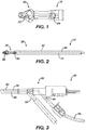
- Surgical reamers are well known in the art. An exemplary reamer is shown and described in U.S. Pat. No. 6,332,886 .
- a surgical reaming device 10 incudes a reamer head 20, located at a distal end 30 of the reaming device.
- a flexible cannulated tube 40 is connected to the reamer head 20 at a distal end of the tube 40 and extends proximally between the reamer head 20 and the proximal end 50 of the device 10.
- Either the reamer head 20, the distal end of the tube 40, or both can have one or more openings 22, 24 that are in fluid communication with the cannulation of the tube 40.
- the cannulated tube 40 can have more than one cannulation, for example, two cannulations within the tube.
- multiple cannulations can extend separately and coextensively from one another within the tube 40 such that each of the cannulations can maintain an independent and distinct fluid flow.
- One or more ports 52, 54 such as, for example, two ports, are located at the proximal end 50 of the device 10 and are in fluid communication with the cannulation of the tube 40. Accordingly, in one embodiment, each of the one or more ports 52, 54 can connect to each of the one or more of the cannulations within the tube 40, respectively, such that each of the one or more ports 52, 54 can be in separate and independent fluid communication with the each of the one or more openings 22, 24 at distal end of the device.
- a surgical reaming device 10 includes two proximal ports 52, 54, an irrigation port 54 and an aspiration port 52.
- the surgical reaming device 10 further includes a flexible tube 40 including two independent coextensive cannulations; an irrigation stream cannulation, and an aspiration stream cannulation.
- the device 10 includes one or more irrigation stream openings 22 connected to the irrigation stream cannulation, and separately, one or more aspiration stream openings 24 connected to the aspiration stream cannulation.
- an irrigation stream 44 is introduced by coupling a fluid supply 56 to the irrigation port 54 of surgical reamer 10, and an aspiration stream 42 is formed by coupling a vacuum source to the aspiration port 52.
- the irrigation stream 44 can flow in a direction from the fluid supply 56, through the irrigation port 54, further into the irrigation stream cannulation, and exit the device through the irrigation stream opening 22 into the intramedullary canal.
- the aspiration stream 42 can flow in a direction from the intramedullary canal into the aspiration stream cannulation through the aspiration stream openings 24, and exit the surgical reaming device through the aspiration stream port 52.
- access to the intramedullary canal is gained via an opening made with a surgical awl, drill, or other like device.
- the fluid supply 56 is opened, starting the irrigation stream 44 through the device 10 as detailed above.
- the reamer head 20 is activated and begins rotation.
- the vacuum source is activated and the reamer head 20 is inserted into the canal.
- the irrigation stream 44 begins to flow into the intramedullary canal space.
- the aspiration stream 42 will begin to flow as previously described, harvesting a volume of cellular material including a mononuclear cell population.
- the aspiration stream 42 can, according to one embodiment, define the contents of the effluent as well as include other components including the coarse residue.
- the aspiration stream 42 includes the volume of harvested cellular material including the mononuclear cell population of the effluent as described above.
- the methods for preparing an osteogenic bone graft can further include filtering the effluent prior to the collecting of the effluent.
- filtering the effluent includes filtering in order to remove a coarse residue, as well as other large tissue fragments, from the effluent in order to separate the effluent from the remainder of the aspiration stream.
- the aspiration stream exiting from the surgical reamer flows to a filter assembly that can include a filter, and also can include an intermediate collection point disposed at a flow point downstream of the filter, and which can collect the effluent.
- the filter can have an average pore size in the range of about 500 microns.
- the step of filtering captures an amount of coarse residue at a surface of the filter.
- the step of filtering retains an amount of autologous bone graft contained within the coarse residue. The retained autologous bone graft can be subsequently used in preparing the osteogenic bone graft as will be more fully described below.
- methods for preparing an osteogenic bone graft include introducing an anticoagulant compound into a volume of cellular material.
- Methods for preparing an osteogenic bone graft are disclosed which include introducing an anticoagulant compound into a harvested volume of cellular material.
- methods of preparing an osteogenic bone graft include:
- methods of preparing an osteogenic bone graft include:
- Anticoagulant compounds are well known in the art.
- An exemplary, non-exclusive list of anticoagulants can include for example, heparin, anticoagulant citrate dextrose-A (ACDA), citrate phosphate dextrose (CPD), citrate phosphate dextrose adenine (CPDA), and bivalarudin, and combinations thereof.
- the anticoagulant compound is introduced into the volume of cellular material in a range of about 2 parts to about 10 parts (by weight) of effluent to anticoagulant compound.
- the anticoagulant compound is introduced into the volume of cellular material in a range of about 6 parts to about 8 parts (by weight) of effluent to anticoagulant compound.
- the anticoagulant compound is introduced into the effluent in an amount sufficient to arrest coagulation for an amount of time sufficient to obtain the concentrated cell fraction.
- the amount of time sufficient to arrest coagulation with the anti-coagulant can vary, and can be for example, less than about 8 hours, less than about 7 hours, less than about 6 hours, less than about 5 hours. less than about 4 hours, less than about 3 hours, less than about 2 hours, or less than about 1 hour.
- the anticoagulant compound is continuously and proportionately introduced into the effluent from an anticoagulant container .
- a proportional introduction would allow for a sample of a given volume of effluent that has been mixed with anticoagulant to have approximately the same ratio of effluent to anticoagulant than another such sample taken at another point in the process.
- the proportionate introducing of the anticoagulant compound is by way of static mixing of the effluent and the anticoagulant, such as for example, a proportionate introduction of the anticoagulant compound and the aspirant accomplished through static mixing of the aspirant and anticoagulant.
- static mixing chambers are called "static mixers" in that the chamber remains fixed at an in-line location and the mixing is caused by the simultaneous, tortuous passage of the two liquids.
- the static mixing is a tortuous mixing.
- the introduction of the anticoagulant compound to the effluent occurs as they pass through an in-line mixing chamber with an interior geometry that causes tortuous mixing of the anticoagulant compound and the effluent such that by the time these two fluids passage through the in-line mixing chamber a homogenous distribution of the anticoagulant compound within the effluent is completed.
- the anticoagulant can be metered into the effluent, upstream of the filter assembly (i.e., into the aspiration stream), and mixed with the effluent during passage through a static mixer.
- the anticoagulant can mix thoroughly upstream of the filter assembly with both the cellular material, containing the mononuclear cell population, and the coarse residue, containing the autologous bone graft, in in an amount sufficient to arrest coagulation.
- the anticoagulant may be introduced within the filter assembly, and can also mix thoroughly with both the cellular material, containing the mononuclear cell population, and the coarse residue, containing the autologous bone graft.
- the anticoagulant can be metered into the effluent downstream of the filter assembly and mixed with the effluent during passage through a static mixer.
- the anticoagulant container includes a 0.2 micron pore size filter to prevent airborne microbes entering the container to replace the volume of anticoagulant dispensed through a one-way valve, such as a duckbill valve, that opens only in response to the vacuum that generates, and pulls the aspiration stream through the filter, which has been previously described.
- the aspiration stream can be pulled into a suction canister positioned downstream of the filter, and in certain embodiments, the suction canister and filter can be considered to form a filter assembly.
- the anticoagulant compound is introduced in a manner that prevents it from travelling upstream to the surgical reaming device and entering the medullary canal of the patient.
- the anticoagulant is introduced into the irrigation stream; for example, continuously introduced into the irrigation stream, such that the anticoagulant can be introduced into the volume of cellular material within the medullary canal and harvested in the aspiration stream.
- the anticoagulant is contained in a rigid container of a fixed volume (and equipped with an air displacement filter as desired) that is plumbed into the effluent line.
- the anticoagulant is contained in a flexible container such as a standard IV bag, which is then connected to the vacuum line at the static mixer via a standard barb fitting.
- the anticoagulant line may be connected to the primary (vacuum line) via a Y shaped fitting.
- the anticoagulant and effluent are combined in a manner designed to induce turbulent mixing, for example a nozzle designed for this purpose.
- the anticoagulant is pumped into the effluent using a peristaltic pump to induce positive pressure.
- the anticoagulant is drip fed into the effluent line under the influence of gravity.
- the anticoagulant is pumped under the influence of pressure applied to the walls of a flexible container (e.g. blood pressure cuff).
- methods for preparing an osteogenic bone graft include collecting the effluent including the volume of cellular material containing the anticoagulant compound at a collection point.
- collecting the effluent includes a first step of collecting at an intermediate collection point.
- the intermediate collection point can include a structure, such as, for example, a suction canister, that is part of the filter assembly, and collects the effluent after it has been filtered.
- the anticoagulant compound and the effluent have been proportionately mixed prior to collecting at the intermediate collection point.
- the effluent is subsequently collected at a processing collection point.
- the processing collection point can include a device capable of processing the effluent such that a desired concentrated cell fraction can be separated from the effluent. Such separation devices are known in the art, and can include, for example, centrifuges.
- methods for preparing an osteogenic bone graft include separating a concentrated cell fraction from the effluent, the concentrated cell fraction including the mononuclear cell population and the anticoagulant compound.
- a device can separate the concentrated cell fraction from the effluent, such as for example, through centrifugation.
- Exemplary separation devices are shown and described, for example in U.S. Pat. No. 8,747,289 .
- the individual cellular components of the cellular material can have varying densities.
- the effluent can be processed through multiple spin cycles. After the completion of each spin cycle, the undesired fractions of the processed effluent can be decanted or otherwise removed from the separation device. In such a manner, a concentrated cell fraction is separated from the effluent for use in preparing the osteogenic bone graft; the concentrated cell fraction including the mononuclear cell population and the anticoagulant compound.
- the concentrated cell fraction can be separated from the effluent during a centrifugation process, such as, for example, by removing the remainder fractions of the effluent during centrifugation.
- the original cellular material has a total mononuclear cell population and the concentrated cell fraction contains at least 80% of the mononuclear cell population of the cellular material.
- the concentrated cell fraction contains at least 90% of the mononuclear cell population of the cellular material.
- the osteogenic bone grafts include a concentrated cell fraction including a medullary canal derived mononuclear cell population; and an anticoagulant compound.
- the osteogenic bone graft can be further combined with an osteoinductive or an osteoconductive matrix.
- the osteoconductive or osteoinductive matrix is an allograft, a synthetic bone substitute, or an autologous bone graft, or a combination thereof.
- the concentration of the mononuclear cell population in the bone graft composition is the range of about 1.0 x 10 6 per ml to about 1000.0 x 10 6 per ml (prior to combining the osteogenic bone graft with an osteoconductive or osteoinductive matrix).
- the osteogenic bone graft can be an injectable composition, a moldable or pliable composition, or a rigid solid composition.
- the osteogenic bone graft can include a secondary amount of anticoagulant, where the secondary amount of anticoagulant can provide a therapeutic effect at the bone graft implantation site.
- the matrix can be a porous matrix.
- the porous nature of the matrix can allow for uptake of the concentrated cell fraction and anticoagulant within the matrix.
- the matrix can be non-porous.
- the concentrated cell fraction and anticoagulant can be blended or otherwise mixed with the matrix to provide a uniform distribution with the matrix.
- preparing the osteogenic bone graft includes combining the concentrated cell fraction with an osteoconductive or osteoinductive matrix.
- preparing the osteogenic bone graft can include combining the concentrated cell fraction with the autologous bone graft obtained from the coarse residue.
- preparing the osteogenic bone graft can include combining the concentrated cell fraction with a secondary autologous bone graft, where the secondary autologous bone graft is obtained from an anatomical site distinct from the harvesting site.
- preparing the osteogenic bone graft can include combining the concentrated cell fraction with an allograft, or a synthetic bone substitute.
- preparing the osteogenic bone graft includes adding an additional amount of anticoagulant to the bone graft, where the amount added is sufficient to increase the overall concentration of anticoagulant in the bone graft to a desired therapeutic level.
- a method of preparing the osteogenic bone graft includes applying the osteogenic bone graft to a bone defect site is also disclosed.
- the method includes performing all of the disclosed steps intraoperatively in a single procedure. This has the advantage of reducing the number of surgical procedures, as well as reducing the need to store the cells, which could result in contamination and loss of cell viability.
- the intraoperative performance of the steps is within 6 hours.
- the method of preparing the osteogenic bone graft can further include storing an excess amount of the concentrated cell fraction and anticoagulant, and can further include preparing a second, third, or more, osteogenic bone grafts.
- the method of preparing the osteogenic bone graft can include cryoprotecting, freezing, and cryogenically storing one or more excess portions of the concentrated cell fraction and anticoagulant, and can include preparing a second, third, or more, osteogenic bone grafts from the cryopreserved excess portions.
- the osteoconductive or osteoinductive matrix according to the present disclosure can include autologous bone (autograft), allogenic bone (allograft), as well as synthetic bone substitutes.
- Autologous bone can be harvested from the medullary canal as previously described above, and can also be taken from a secondary anatomical site such as the iliac crest. Autologous bone offers less risk of rejection because it has originated from the patient's own body. Additionally, autologous bone can also provide osteoinductive and osteogenic properties in addition to having osteoconductive properties.
- Autologous bone scaffolds with high solid bone content has a higher osteoconductive potential than autologous bone that contains a lower solid bone content.
- Allogenic bone scaffolds offer the same osteoconductive properties as autologous scaffolds. Allogenic scaffolds can be obtained from cadaveric samples, for example, from a tissue bank.
- the osteoconductive or osteoinductive matrix includes a synthetic bone substitute.
- the synthetic bone substitute can be porous or non-porous.
- the term "porous" includes, but is not limited to, macroporosity (mean pore diameter greater than or equal to 100 um), mesoporosity (mean pore diameter less than 100 um but greater than or equal to 10 um) and microporosity (mean pore diameter less than 10 um).
- the pores may be of any size, shape or distribution, or within a predetermined tolerance.
- the pores can be interconnecting or non-interconnecting.
- the diameter of the pores can range in size up to about 750 um.
- the pore sizes range up to about 500 um, with approximately 75% of the pores being at least 100 um in size and the remaining 25% of the pores being no more than 10 um in size.
- the synthetic bone substitute includes a ceramic bone substitute, such as, for example, a calcium phosphate based compound.
- a ceramic bone substitute such as, for example, a calcium phosphate based compound.
- Suitable examples of calcium phosphates include amorphous calcium phosphate, crystalline calcium phosphate, or any combination thereof.
- the calcium phosphate compound can be an apatite.
- Apatites are a group of calcium phosphate minerals, usually referring to hydroxyapatite Ca 10 (PO 4 ) 6 (OH) 2 , fluoroapatite Ca 10 (PO 4 ) 6 (F) 2 , chlorapatite Ca 10 (PO 4 ) 6 (Cl) 2 and bromapatite Ca 10 (PO 4 ) 6 (Br) 2 and can further include both silicate (SiO 4 4- ) and carbonate (CO 3 2- ) substituted hydroxyapatites, where the substitution is for one or more of the hydroxy and/or phosphate groups.
- the ceramic bone substitute includes beta-tricalcium phosphate Ca 3 (PO 4 ) 2 , (b-TCP).
- the osteoconductive or osteoinductive matrix can be of any shape as desired for the particular bone defect to be repaired.
- the matrix is a monolithic composition that can be either porous or non-porous. Suitable shapes can include, for example, spherical, cubic, wedge-shaped, oblong, cylindrical, or combinations thereof.
- the matrix can be a plurality of porous or non-porous granules.
- the specific surface area of the matrix can vary. For example, when the matrix is a porous granule, the specific surface area can range from about 0.1m 2 /g to about 100m 2 /g.
- the osteoconductive or osteoinductive matrix may be ceramic bone substitute particles or granules of any size or shape.
- the granules can be obtained by grinding or milling a calcium compound to a desired particle size or particle diameter.
- the mean diameter of the granules range in size from about 0.05 mm to about 10 mm.
- the mean diameter of the granules range in size from about 0.075 mm to about 5 mm.
- the mean diameter of the granules range in size from about 0.075 mm to about 1 mm.
- the mean diameter of the granules range in size from about 1.4 mm to about 2.8 mm.
- the mean diameter of the granules range in size from about 2.8 mm to about 5.6 mm.
- the mean diameter of the granules range in size from about 0.1 mm to about 0.750 mm
- the osteoconductive or osteoinductive matrix can be further combined with a polymeric binder, such that the osteogenic bone graft could be formed, for example, into a moldable or pliable implant that could be shaped as desired to fit the area of the bone to be repaired.
- the polymeric binder can include polymers such as homopolymers and copolymers (i.e., polymers including two or more different monomeric units), as well as polymer and copolymer blends, mixtures and combinations.
- the polymer can be a resorbable polymer, a non-resorbable polymer, or a combination thereof.
- the polymeric binder includes a resorbable polymer, and the polymeric binder is substantially free of a non-resorbable polymer.
- the polymeric binder is resorbable in vivo and includes a resorbable polymer.
- the polymer(s) of the polymeric binder can also include a synthetic polymer, a non-synthetic polymer (i.e., a polymer obtained from a plant or animal), or a combination thereof.
- Suitable polymers useful for preparing the polymeric binder include, but are not limited to, homopolymers or copolymers of monomers selected from L-lactide; L-lactic acid; D-lactide; D-lactic acid; glycolide; alpha-hydroxybutyric acid; alpha-hydroxyvaleric acid; alpha-hydroxyacetic acid; alpha-hydroxycaproic acid; alpha-hydroxyheptanoic acid; alpha-hydroxydecanoic acid; alpha-hydroxymyristic acid; alpha-hydroxyoctanoic acid; alpha-hydroxystearic acid; hydroxybutyrate; hydroxyvalerate; beta-propiolactide; beta-propiolactic acid; gamma-caprolactone; beta-caprolactone; epsilon-caprolactone; gamma-butyrolactone; pivalolactone; tetramethylglycolide; tetramethylglycolic acid; dimethylglycolic acid; trimethylene carbonate
- the polymeric binder includes resorbable polymers.
- resorbable polymers include, e.g., polymers derived from monomers selected from L-lactic acid, D-lactic acid, L-lactide, D-lactide, D,L-lactide, glycolide, a lactone, a lactam, epsilon-caprolactone, trimethylene carbonate, a cyclic carbonate, a cyclic ether, para-dioxanone, beta-hydroxybutyric acid, beta-hydroxypropionic acid, beta-hydroxyvaleric acid, saccharides, collagen, fibrin, albumin; and any combination of two or more of the foregoing.
- the polymeric binder includes a resorbable synthetic polymer.
- resorbable synthetic polymers include, e.g., a poly(L-lactide) (co)polymer, a poly(D,L-lactide) (co)polymer, a polyglycolide (co)polymer, a polycaprolactone (co)polymer, a poly(tetramethylglycolic acid) (co)polymer, a polydioxanone (co) polymer, a polyhydroxybutyrate (co)polymer, a polyhydroxyvalerate (co)polymer, a poly(L-lactide-co-glycolide) copolymer, a poly(glycolide-co-trimethylene carbonate) copolymer, a poly(glycolide-co-caprolactone) copolymer, a poly(glycolide-co-dioxanone-co-trim
- the osteogenic bone graft can consist essentially of the concentrated cell fraction including a medullary canal derived mononuclear cell population and an anticoagulant compound, such that the bone graft does not include any components that materially alter the function of the osteogenic bone graft to therapeutically affect the repair of a bone defect.
- the osteogenic bone graft can consist essentially of the concentrated cell fraction including a medullary canal derived mononuclear cell population, an anticoagulant compound, and an osteoinductive or an osteoconductive matrix, such that the bone graft does not include any components that materially alter the function of the osteogenic bone graft to therapeutically affect the repair of a bone defect.
- Samples of cellular material were harvested in procedures (labeled Run 1 through 10 or Patient 1 through 10) from human long bones utilizing a commercially available surgical reamer from Depuy Synthes (Reamer/Irrigator/Aspirator (RIA)). The amount of each harvested effluent that was processed is shown in Table 3 below.
- the effluent was collected intraoperatively from a line placed in between the filter assembly and a vacuum source. Additional filters may optionally be used in order to filter out debris that would otherwise cause fouling of the processing system. Additional filters may also be added in order to collect a further population of cells that may otherwise be clinically valuable.
- An anticoagulant in this case, Heparin, was introduced into the effluent in a controlled manner at a ratio of about 6-8 parts effluent to anticoagulant (by weight). The anticoagulant was introduced downstream of the filter assembly. The anticoagulant was proportionately introduced to the effluent through a static mixer in a manner that prevented it from travelling upstream into the surgical reaming device.
- the effluent and anticoagulant were collected in an intermediate collection chamber (e.g. a suction canister) positioned downstream of the filter prior to being decanted into a processing collection point. Samples of the pre-processed effluent were taken to be used in later testing and analysis.
- an intermediate collection chamber e.g. a suction canister
- the processing collection point was the single use disposable cartridge, available with the commercially available SynGenX-Lab System. Each of the cartridges is capable of processing up to 240 ml of collected effluent stream and up to 4 cartridges may be placed in a SynGenX-Lab centrifuge (total effluent volume that can be processed in a single cycle is approximately 1000 ml). The harvested effluent from each of the runs was separately processed and was evenly mixed and partitioned into separate cartridges.
- the centrifuge cycle is programmable and may include a "fast spin cycle” (during which the stratification of cells according to density in the main compartment occurs) and “slow spin cycle” (during which depletion of red cells to a dedicated compartment and harvest of clinically relevant cells to a harvest compartment occurs).
- the SyngenX-Lab System includes the cartridge and a reusable battery operated, firmware instructed control module, which contains an accelerometer and four infrared (IR) sensors.
- the accelerometer assures that the stratification of cell fractions according to density occurs at the correct gravity and the IR sensors assure that the predetermined volume of RBCs are transferred to the depletion compartment of the cartridge and the predetermined volume of MNCs, platelets and plasma are transferred to the harvest compartment of the cartridge.
- the processing system can be capable of depleting the excess red blood cells, irrigant, plasma, and granulocytes from the effluent. It is also capable of separating a concentrated cell fraction (as shown in the data below) including over 80% of the MNCs from the effluent.
- the plasma fraction, the RBC fraction and the concentrated cell fraction containing the mononuclear cell population (commonly referred to as the "buffy coat") were removed from each of the cartridges into a tube and labelled for further testing and analysis.
- mass balance is defined as the number of target cells recovered in the harvest chamber, red cell depletion chamber and main processing chamber (of the single-use disposable processing cartridge described in U.S. Pat. No. 8,747,289 ), at the end of a run cycle, compared, as a percentage, to the total number of target cells residing in the full volume of collected RIA effluent.
- 7-Aminoactinomycin D is a fluorescent chemical compound used as a cell viability stain. 7-AAD does not readily pass through intact cell membranes, so upon testing cells with compromised membranes will stain with 7-AAD, while live cells with intact cell membranes will remain dark.
- Annexin A5 is used as a non-quantitative probe to detect cells that have expressed phosphatidylserine (PS) on the cell surface, an event found in apoptosis as well as other forms of cell death.
- the annexin A5 affinity assay typically uses a conjugate of annexin V and a fluorescent or enzymatic label, biotin or other tags, or a radioelement, in a suitable buffer.
- the assay combines annexin V staining of PS and PE membrane events with the staining of proteins in the cell nucleus with propidium iodide (PI) or 7-Aminoactinomycin D (AAD-7), distinguishing viable cells from apoptotic cells and necrotic cells. Detection occurs by flow cytometry.
- Test data from Table 2 demonstrates that the viability of the harvested cells is maintained throughout the processing of the effluent.
- Test data demonstrates mononuclear cell concentrations having values in the 10 6 /ml range.
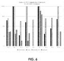
- Table 3 Flow Cytometry analysis of cell recovery Run RIA Vol. (mL) Harvest Vol. (mL) Harvest MNC count (10 9 ) MNC (%) Recovery Harvest CD34+ count (10 9 ) CD34+ (%) Recovery 1 960.0 39.2 N/A N/A N/A N/A 2 958.1 41.2 0.40 89.4 0.0262 97.1 3 225.3 10.5 0.17 101.4 0.0072 103.1 4 953.3 38.5 0.10 95.8 0.0016 99.0 5 956.9 35.9 7.54 95.5 0.5518 96.8 6 1000.6 38.8 0.19 102.6 0.0019 103.1 7 757.6 33.0 1.66 83.1 0.5464 94.2 8 757.6 24.9 0.41 93.7 0.0081 98.1 9 548.9 27.9 0.75 96.0 0.0361 96.9 10 951.3 20.7 0.84 94.5 0.0126 97.6
- test data in Table 3 demonstrates MNC recovery of greater than 90% and greater than 94% CD34+ recovery in runs 2-10.
- the effluent from Patient 1 was not analyzed.
- the data demonstrate that the colony forming capacity of the harvested MNC concentrate was greater ( ⁇ 1.5 to 35 times) than RIA effluent itself. Further, the data also demonstrate that the colony forming cells reside almost entirely within the concentrated cell fraction rather than other constituent layers (e.g., RBC layer and Supernatent layer), except for run 7 where cell separation was incomplete (below average MNC recovery).
Description
- This application claims the benefit of
U.S. Provisional Appl. No. 62/199,480, filed on July 31, 2015 - The present disclosure relates to osteogenic bone grafts including concentrated cell fractions of mononuclear cell populations and an anticoagulant, as well as processes for preparing the same.
- Non-unions, in particular those involving large segmental defects in long bones, present a significant healing challenge. One method of healing these defects involves the use of autograft obtained from the medullary canal of a long bone, for example, a femur or tibia, using a surgical reamer irrigator aspirator (RIA) system. This system has the advantages of being able to harvest a larger quantity of autograft, with lower morbidity and greater rapidity, as compared to, for example, iliac crest bone grafting. RIA bone grafts are emerging as the new "gold standard" as it relates to the use of bone grafts, either in repair of large segmental defects or other clinical situations requiring bone grafts. One problem that remains is that a large quantity of cells including mesenchymal stem cells, endothelial progenitors, and other progenitor cells, which become available during harvesting are currently lost or underutilized.
- In general, surgical reamers utilize a rotating cutting head similar to a drill displaced at the distal end of a drive shaft. Bone cutting devices for use in intramedullary reaming typically use a flexible drive shaft because the medullary canals of bones are seldom straight and usually will have some degree of curvature. Most reamers also have a central longitudinal bore through both the reamer and the drive shaft. The central bore is intended to receive a long, small diameter guide pin or wire which is initially inserted into the medullary canal to act as a track for the advancing reamer. Reamers are used in orthopedic surgery to prepare the medullary canals of bone for surgical procedures such as total hip and knee replacement, nail insertion to stabilize a long bone fracture, an intramedullary osteotomy, and bone harvesting for grafting purposes.
- Conventional methods of isolating and harvesting certain cell types from an intramedullary canal aspirate sample generally involve centrifugation of the sample. During centrifugation, populations of cells migrate to a relative position along the axis of lesser to greater acceleration, and concentrate in layers according to their density, displacing other higher and lower density cell types and plasma during the process.
- Currently most of the effluent harvested during an intramedullary reaming is disposed of as medical waste. There is no practical way to isolate the therapeutically beneficial cells due to the large volumes present (∼250cc - 3000cc) and the presence of extraneous fluid, cells, and tissues, that do not promote bone healing (such as, RBCs, plasma, irrigant fluid, adipocytes, triglycerides, cholesteryl ester, certain proteins acting as adpokines, etc.). Existing cell separation methods are typically based on density gradation (e.g. centrifugation), filtration (e.g. tangential flow filtration) and other methods. Filtration of effluent at the cellular level is impractical due to the very large filter surface areas required, and complications associated with clogging, coagulation, and cell death. Centrifugation has been used as a method to separate and collect cells from the total effluent volume; however, existing systems suffer from drawbacks. Most commercially available systems cannot process more than about 50 cc of fluid per individual container, or 200 cc total for each centrifugation cycle in a four-container centrifuge system, making them impractical for processing the large volumes of effluent present in most intramedullary reaming procedures.
- Additionally, problems can arise with the usefulness of harvested graft material due to the interaction of blood coagulation and the aggregation of adhesive fat particles created by the reaming tool. Blood coagulation, or clotting, is one of the body's natural means of stopping bleeding. It comprises a complex cascade of many clotting factors, some always present in the blood and some released from damaged tissue and activated platelets. When the lining of a blood vessel is broken, platelets are attracted forming a platelet plug. These platelets have thrombin receptors on their surfaces that bind serum thrombin molecules, which in turn convert soluble fibrinogen in the serum into fibrin at the wound site. Fibrin forms long strands of tough insoluble protein that are bound to the platelets. Factor XIII completes the cross-linking of fibrin so that it hardens and contracts. The cross-linked fibrin forms a mesh atop the platelet plug that completes the clot. The end-product is a mesh of fibrin strands in which blood cells are trapped to form a solid mass that seeks to stop bleeding. The entrapment of cells that promote bone growth by fibrin clots can be deleterious to the regeneration of bone tissue at the site of the trauma injury.
- The blood clotting cascade begins almost instantly after a reamer head damages the endothelium lining of any blood vessels or other tissue in the medullary canal of the femur or tibia. Once intiated, this process is normally irreversible and in certain instances all useful cellular graft material, as well as fatty tissues residing in the medullary canal that becomes atomized by the cutting of the reaming head, can become trapped in the clot matrix, rendering it unusable as a bone graft. These entrapped cells often die because the fibrin clot, interwoven with the highly adhesive fat particles, entomb and prevent the revascularization of the cells by new blood vessels formed in response to the tissue damage incurred both by the original trauma and the reaming of the intramedullary canal. Existing methods described in the prior art rely on placing anticoagulant with the generated effluent at the point of final collection or sampling a quantity of effluent (typically 50 cc or less) and adding it to a separate container with a fixed volume of anticoagulant. The principle disadvantage of these methods is that they do not facilitate a rapid proportionate mixing of the anticoagulant into the effluent and therefore are suboptimal in terms of both arresting the onset of the clotting cascade, and cell recovery and viability.
-
US2007036766 relates to "a method for preparation of an autologous tissue graft composition (...) Bone marrow is harvested from the patient. In one embodiment, the nucleated cells of the bone marrow are subsequently concentrated. Autologous thrombin is purified from a volume of whole blood also taken from the patient. The bone marrow aspirate or bone marrow concentrate is then combined with the purified autologous thrombin to form a coagulated tissue graft material that may be used alone or in conjunction with other graft materials". - Consequently, there is a need in the art to find a method for improving the use of harvested biologic components of the intramedullary canal as osteogenic bone graft, and further a need to arrest the clotting cascade within the harvested material in order to prevent the harvested material from becoming irreversibly bound in a clot matrix.
- The invention is defined by the claims. Any subject-matter falling outside the scope of the claims is provided for information purposes only.
- The present invention relates to methods of preparing an osteogenic bone graft according to the appended claims 1-11 and to an osteogenic bone graft composition according to claims 12-15. The methods can include:
- introducing an anticoagulant compound into a volume of cellular material including a mononuclear cell population from the intramedullary canal of a long bone;
- collecting an effluent including the volume of cellular material containing the anticoagulant compound at a collection point;
- separating a concentrated cell fraction from the effluent, the concentrated cell fraction including the mononuclear cell population and the anticoagulant compound; and
- preparing an osteogenic bone graft from the concentrated cell fraction.
- Methods of the disclosure can include:
- harvesting a volume of cellular material including a mononuclear cell population from the intramedullary canal of a long bone; introducing an anticoagulant compound into the volume of cellular material;
- collecting an effluent including the harvested volume of cellular material containing the anticoagulant compound at a collection point;
- separating a concentrated cell fraction from the effluent, the concentrated cell fraction including the mononuclear cell population and the anticoagulant compound; and
- preparing an osteogenic bone graft from the concentrated cell fraction.
- The present disclosure also describes methods of preparing an osteogenic bone graft including:
- introducing an anticoagulant compound into a volume of cellular material including a mononuclear cell population harvested from the intramedullary canal of a long bone;
- collecting an effluent including the harvested volume of cellular material containing the anticoagulant compound at a collection point;
- separating a concentrated cell fraction from the effluent, the concentrated cell fraction including the mononuclear cell population and the anticoagulant compound; and
- preparing an osteogenic bone graft from the concentrated cell fraction.
- According to one embodiment, harvesting includes aspirating the intramedullary canal of a long bone with a surgical reaming device. The anticoagulant is continuously and proportionately introduced into the effluent. The continuous and proportionate introduction of the anticoagulant is advantageous as a way in which to arrest the clotting and protect the regenerative potential of the bone forming and blood vessel forming cells. Suffusing the harvested effluent with quick-acting anticoagulants can arrest the clotting cascade and provide enhanced cell viability of the harvested material. As the clotting cascade begins as soon as the tissue damage occurs, the sooner the anticoagulant can be proportionately distributed into the effluent after it emerges from the medullary canal, to interrupt the clotting cascade, the more useful the harvested cellular material and mononuclear cell (MNC) population will be for bone regeneration.
- According to one embodiment, preparing the osteogenic bone graft includes combining the concentrated cell fraction with an osteoconductive or osteoinductive matrix. In one embodiment, the effluent is filtered prior to collecting, where the step of filtering removes coarse residue from the effluent. In a further embodiment, the method can include retaining autologous bone graft from the filtered coarse residue. According to one embodiment of the disclosure, preparing the osteogenic bone graft can include combining the concentrated cell fraction with the autologous bone graft obtained from the coarse residue. According to another embodiment of
the disclosure, preparing the osteogenic bone graft can include combining the concentrated cell fraction with a secondary autologous bone graft, where the secondary autologous bone graft is obtained from an anatomical site distinct from the harvesting site. In a further embodiment, preparing the osteogenic bone graft can include combining the concentrated cell fraction with an allograft, or a synthetic bone substitute. - The present disclosure is also directed to osteogenic bone grafts according to claim 12. The osteogenic bone grafts, according to the present disclosure, can include a concentrated cell fraction including a medullary canal derived mononuclear cell population; and an anticoagulant compound. In one embodiment, the osteogenic bone graft includes a homogenous mixture of the concentrated cell fraction and the anticoagulant compound. According to one embodiment, the osteogenic bone graft can be further combined with an osteoinductive or an osteoconductive matrix. According to another embodiment, the osteoconductive or osteoinductive matrix is an allograft, a synthetic bone substitute, or an autologous bone graft, or a combination thereof. In a further embodiment, the concentration of the mononuclear cell population in the bone graft composition is the range of about 1.0 x 10⁶ per ml to about 1000.0 x 106 per ml.
- The drawings illustrate generally, by way of example, but not by way of limitation, various embodiments discussed in the present document. The foregoing summary, as well as the following detailed description of preferred embodiments of the application, will be better understood when read in conjunction with the appended drawings.
-
Fig. 1 is a side view of a reaming head of commercially available surgical reaming device; -
Fig. 2 is a side view of the distal end of the surgical reaming device; -
Fig. 3 is a side view of the proximal end of the surgical reaming device; -
Fig. 4 is a bottom view of the surgical reaming device; -
Fig. 5 is a graphical representation of the mononuclear cell and CD34+ cell content and percent recovery from processing according to an embodiment of the disclosure; and, -
Fig. 6 is a graphical representation of data measuring colony forming units of the various components of the effluent obtained according to another embodiment of the disclosure. - In this document, the terms "a" or "an" are used to include one or more than one and the term "or" is used to refer to a nonexclusive "or" unless otherwise indicated. In addition, it is to be understood that the phraseology or terminology employed herein, and not otherwise defined, is for the purpose of description only and not of limitation. When a range of values is expressed, another embodiment includes from the one particular value and/or to the other particular value. Similarly, when values are expressed as approximations, by use of the antecedent "about," it will be understood that the particular value forms another embodiment. All ranges are inclusive and combinable. Further, reference to values stated in ranges includes each and every value within that range. It is also to be appreciated that certain features of the invention, which are, for clarity, described herein in the context of separate embodiments, may also be provided in combination in a single embodiment. Conversely, various features of the invention that are, for brevity, described in the context of a single embodiment, may also be provided separately or in any subcombination.
- As used herein "aspiration stream," "aspirant," and "aspirating stream," and derivatives thereof are defined as any and all material that is harvested from the intramedullary canal of a long bone during a surgical reaming or equivalent surgical procedure. Such material can include, without limitation, irrigation fluids, bone tissue and cells, endosteal cells and tissue, fat tissue and cells, bone marrow and marrow stroma, vascular cells and tissue, stem and/or progenitor cells (such as mesenchymal, hematopoietic, and endothelial stem/progenitor cells) and blood (including its individual constituents such as platelets, red blood cells, plasma, granulocytes, lymphocytes, and monocytes).
- As used herein "effluent stream" and "effluent" and derivatives thereof are defined as constituents of the aspiration stream that can be separated, when desired, from the aspiration stream by some process. The separation process may include but is not limited to filtration. Where filtration is utilized to separate the effluent from the aspiration stream, the effluent can constitute anything that passes through the filter. Therefore, as defined herein, "effluent stream" and "effluent" and derivatives thereof are considered to be a constituent of the "aspiration stream" such that any reference to a process or processes performed upon or to the "aspiration stream" prior to a separation/filtration process or step would necessarily include the "effluent stream."
- As used herein, "coarse residue" is defined as the remainder of the aspiration stream that is not part of the effluent. For example, where filtration is used, the "coarse residue" is anything in the effluent that does not pass through a filter. The components of the "coarse residue" can vary depending upon the average pore size of the filter, but will include, typically, components having a cross-sectional dimension larger than the average pore size of the filter. However, it should be understood that components in the effluent smaller than the average pore size of the filter, which would be expected to pass through the filter, can become trapped on the filter or within an aggregation or agglomeration of the larger components at the filter surface during normal operating processes utilizing the filter, such that the coarse residue can, in certain embodiments, include a small percentage of components that would also be contained within the filtered effluent. Typical components of the "coarse residue" can include, for example, bone particles, agglomerations or aggregations of fat, and clots, as well as combinations of the foregoing.
- The present disclosure describes methods of preparing an osteogenic bone graft. The methods can include:
- harvesting a volume of cellular material including a mononuclear cell population from the intramedullary canal of a long bone;
- introducing an anticoagulant compound into the volume of cellular material;
- collecting an effluent including the harvested volume of cellular material containing the anticoagulant compound at a collection point;
- separating a concentrated cell fraction from the effluent, the concentrated cell fraction including the mononuclear cell population and the anticoagulant compound; and
- preparing an osteogenic bone graft from the concentrated cell fraction.
- The methods disclosed herein can, according to one embodiment, isolate mononuclear cells (MNCs), and by extension, subsets of this cell population (e.g. mesenchymal stem cells) from effluent obtained intraoperatively from a surgical procedure, for example, reaming of the intramedullary canal of long bone; e.g., a femur. Typically, the effluent can be considered a heterogeneous biologic fluid that includes, among other things, several biologic components that are aspirated from the intramedullary canal. An exemplary non-limiting list can include, for example, bone marrow and marrow stroma, endosteal tissue, adipose (yellow marrow), endo cortical bone chips, metaphyseal bone chips, platelets, and other commonly known bone or marrow related tissues. Among the cells that can be contained within the effluent includes the following: red blood cells, granulocytes, and mononuclear cells. Many cell types comprise the mononuclear cell population. An exemplary, and non-limiting list can include, for example: CD45+ (white blood cells), CD45-, CD 34+ and CD133+ (hematopoietic stem cells), C105+, CD 271+, mesenchymal stem cells (MSCs), endothelial cells, endothelial progenitor cells, bone cells, bone lining cells, t-cells, stromal cells and adipocytes. Within the mononuclear cell population, are cells that can contribute to bone healing such as, for example, mesenchymal, hematopoietic and endothelial stem and progenitor cells. However, the overall mononuclear cell population contained within a typically harvested effluent volume constitutes a very small fraction, in certain instances, less than 1%, of the total volume of the effluent. The remainder volume of the effluent is mainly red blood cells, irrigant, other fluids, plasma, fat and fatty tissues.
- According to the present disclosure, methods for preparing an osteogenic bone graft include harvesting a volume of cellular material from the intramedullary canal of a long bone. According to one embodiment, the harvesting can be accomplished utilizing a surgical reamer in combination with an aspiration stream. In an exemplary embodiment, the harvesting further includes an irrigation stream. Surgical reamers are well known in the art. An exemplary reamer is shown and described in
U.S. Pat. No. 6,332,886 . - As shown in
Figs. 1-4 , asurgical reaming device 10 incudes areamer head 20, located at a distal end 30 of the reaming device. A flexible cannulatedtube 40 is connected to thereamer head 20 at a distal end of thetube 40 and extends proximally between thereamer head 20 and theproximal end 50 of thedevice 10. Either thereamer head 20, the distal end of thetube 40, or both can have one ormore openings tube 40. The cannulatedtube 40 can have more than one cannulation, for example, two cannulations within the tube. In such an embodiment, multiple cannulations can extend separately and coextensively from one another within thetube 40 such that each of the cannulations can maintain an independent and distinct fluid flow. One ormore ports proximal end 50 of thedevice 10 and are in fluid communication with the cannulation of thetube 40. Accordingly, in one embodiment, each of the one ormore ports tube 40, respectively, such that each of the one ormore ports more openings surgical reaming device 10 includes twoproximal ports irrigation port 54 and anaspiration port 52. Thesurgical reaming device 10 further includes aflexible tube 40 including two independent coextensive cannulations; an irrigation stream cannulation, and an aspiration stream cannulation. Further, at the distal end 30, thedevice 10 includes one or moreirrigation stream openings 22 connected to the irrigation stream cannulation, and separately, one or moreaspiration stream openings 24 connected to the aspiration stream cannulation. In such an embodiment, anirrigation stream 44 is introduced by coupling afluid supply 56 to theirrigation port 54 ofsurgical reamer 10, and anaspiration stream 42 is formed by coupling a vacuum source to theaspiration port 52. Theirrigation stream 44 can flow in a direction from thefluid supply 56, through theirrigation port 54, further into the irrigation stream cannulation, and exit the device through the irrigation stream opening 22 into the intramedullary canal. Theaspiration stream 42 can flow in a direction from the intramedullary canal into the aspiration stream cannulation through theaspiration stream openings 24, and exit the surgical reaming device through theaspiration stream port 52. - Typically, in reaming procedures, access to the intramedullary canal is gained via an opening made with a surgical awl, drill, or other like device. Once access is gained, the
fluid supply 56 is opened, starting theirrigation stream 44 through thedevice 10 as detailed above. Thereamer head 20 is activated and begins rotation. The vacuum source is activated and thereamer head 20 is inserted into the canal. Upon entry into the medullary canal, theirrigation stream 44 begins to flow into the intramedullary canal space. Once thereamer head 20 has been advanced within the intramedullary canal far enough that the aspiration stream holes 24 are within the canal, theaspiration stream 42 will begin to flow as previously described, harvesting a volume of cellular material including a mononuclear cell population. Theaspiration stream 42 can, according to one embodiment, define the contents of the effluent as well as include other components including the coarse residue. As such, theaspiration stream 42 includes the volume of harvested cellular material including the mononuclear cell population of the effluent as described above. - According to the present disclosure, the methods for preparing an osteogenic bone graft can further include filtering the effluent prior to the collecting of the effluent. According to one embodiment, filtering the effluent includes filtering in order to remove a coarse residue, as well as other large tissue fragments, from the effluent in order to separate the effluent from the remainder of the aspiration stream. In most instances, the aspiration stream exiting from the surgical reamer flows to a filter assembly that can include a filter, and also can include an intermediate collection point disposed at a flow point downstream of the filter, and which can collect the effluent. According to one embodiment, the filter can have an average pore size in the range of about 500 microns.
- According to one embodiment, the step of filtering captures an amount of coarse residue at a surface of the filter. In one embodiment, the step of filtering retains an amount of autologous bone graft contained within the coarse residue. The retained autologous bone graft can be subsequently used in preparing the osteogenic bone graft as will be more fully described below.
- According to the present disclosure, methods for preparing an osteogenic bone graft include introducing an anticoagulant compound into a volume of cellular material. Methods for preparing an osteogenic bone graft are disclosed which include introducing an anticoagulant compound into a harvested volume of cellular material.
- For example, according to the present disclosure, methods of preparing an osteogenic bone graft include:
- introducing an anticoagulant compound into a volume of cellular material including a mononuclear cell population from the intramedullary canal of a long bone;
- collecting an effluent including the volume of cellular material containing the anticoagulant compound at a collection point;
- separating a concentrated cell fraction from the effluent, the concentrated cell fraction including the mononuclear cell population and the anticoagulant compound; and
- preparing an osteogenic bone graft from the concentrated cell fraction
- According to another embodiment of the present disclosure, methods of preparing an osteogenic bone graft include:
- introducing an anticoagulant compound into a volume of cellular material including a mononuclear cell population harvested from the intramedullary canal of a long bone;
- collecting an effluent including the harvested volume of cellular material containing the anticoagulant compound at a collection point;
- separating a concentrated cell fraction from the effluent, the concentrated cell fraction including the mononuclear cell population and the anticoagulant compound; and
- preparing an osteogenic bone graft from the concentrated cell fraction.
- Anticoagulant compounds are well known in the art. An exemplary, non-exclusive list of anticoagulants can include for example, heparin, anticoagulant citrate dextrose-A (ACDA), citrate phosphate dextrose (CPD), citrate phosphate dextrose adenine (CPDA), and bivalarudin, and combinations thereof. According to one embodiment, the anticoagulant compound is introduced into the volume of cellular material in a range of about 2 parts to about 10 parts (by weight) of effluent to anticoagulant compound. According to another embodiment, the anticoagulant compound is introduced into the volume of cellular material in a range of about 6 parts to about 8 parts (by weight) of effluent to anticoagulant compound. According to a further embodiment, the anticoagulant compound is introduced into the effluent in an amount sufficient to arrest coagulation for an amount of time sufficient to obtain the concentrated cell fraction. The amount of time sufficient to arrest coagulation with the anti-coagulant can vary, and can be for example, less than about 8 hours, less than about 7 hours, less than about 6 hours, less than about 5 hours. less than about 4 hours, less than about 3 hours, less than about 2 hours, or less than about 1 hour.
- The anticoagulant compound is continuously and proportionately introduced into the effluent from an anticoagulant container . A proportional introduction, for example, would allow for a sample of a given volume of effluent that has been mixed with anticoagulant to have approximately the same ratio of effluent to anticoagulant than another such sample taken at another point in the process.
- In an exemplary embodiment, the proportionate introducing of the anticoagulant compound is by way of static mixing of the effluent and the anticoagulant, such as for example, a proportionate introduction of the anticoagulant compound and the aspirant accomplished through static mixing of the aspirant and anticoagulant. Such in-line mixing chambers are called "static mixers" in that the chamber remains fixed at an in-line location and the mixing is caused by the simultaneous, tortuous passage of the two liquids. According to a further embodiment, the static mixing is a tortuous mixing. According to a particular embodiment, the introduction of the anticoagulant compound to the effluent occurs as they pass through an in-line mixing chamber with an interior geometry that causes tortuous mixing of the anticoagulant compound and the effluent such that by the time these two fluids passage through the in-line mixing chamber a homogenous distribution of the anticoagulant compound within the effluent is completed.
- According to one embodiment, the anticoagulant can be metered into the effluent, upstream of the filter assembly (i.e., into the aspiration stream), and mixed with the effluent during passage through a static mixer. In this way, the anticoagulant can mix thoroughly upstream of the filter assembly with both the cellular material, containing the mononuclear cell population, and the coarse residue, containing the autologous bone graft, in in an amount sufficient to arrest coagulation. In another embodiment, the anticoagulant may be introduced within the filter assembly, and can also mix thoroughly with both the cellular material, containing the mononuclear cell population, and the coarse residue, containing the autologous bone graft. According to another embodiment, the anticoagulant can be metered into the effluent downstream of the filter assembly and mixed with the effluent during passage through a static mixer. According to a further embodiment, the anticoagulant container includes a 0.2 micron pore size filter to prevent airborne microbes entering the container to replace the volume of anticoagulant dispensed through a one-way valve, such as a duckbill valve, that opens only in response to the vacuum that generates, and pulls the aspiration stream through the filter, which has been previously described. According to one embodiment, the aspiration stream can be pulled into a suction canister positioned downstream of the filter, and in certain embodiments, the suction canister and filter can be considered to form a filter assembly.
- According to one embodiment, the anticoagulant compound is introduced in a manner that prevents it from travelling upstream to the surgical reaming device and entering the medullary canal of the patient. According to another embodiment, the anticoagulant is introduced into the irrigation stream; for example, continuously introduced into the irrigation stream, such that the anticoagulant can be introduced into the volume of cellular material within the medullary canal and harvested in the aspiration stream. According to one embodiment, the anticoagulant is contained in a rigid container of a fixed volume (and equipped with an air displacement filter as desired) that is plumbed into the effluent line. According to one embodiment, the anticoagulant is contained in a flexible container such as a standard IV bag, which is then connected to the vacuum line at the static mixer via a standard barb fitting. Alternatively, the anticoagulant line may be connected to the primary (vacuum line) via a Y shaped fitting. In another embodiment, the anticoagulant and effluent are combined in a manner designed to induce turbulent mixing, for example a nozzle designed for this purpose. According to one embodiment, the anticoagulant is pumped into the effluent using a peristaltic pump to induce positive pressure. According to one embodiment, the anticoagulant is drip fed into the effluent line under the influence of gravity. According to one embodiment, the anticoagulant is pumped under the influence of pressure applied to the walls of a flexible container (e.g. blood pressure cuff).
- According to the present disclosure, methods for preparing an osteogenic bone graft include collecting the effluent including the volume of cellular material containing the anticoagulant compound at a collection point. According to one embodiment, collecting the effluent includes a first step of collecting at an intermediate collection point. According to one embodiment, the intermediate collection point can include a structure, such as, for example, a suction canister, that is part of the filter assembly, and collects the effluent after it has been filtered. The anticoagulant compound and the effluent have been proportionately mixed prior to collecting at the intermediate collection point. According to a further embodiment, the effluent is subsequently collected at a processing collection point. The processing collection point can include a device capable of processing the effluent such that a desired concentrated cell fraction can be separated from the effluent. Such separation devices are known in the art, and can include, for example, centrifuges.
- According to the present disclosure, methods for preparing an osteogenic bone graft include separating a concentrated cell fraction from the effluent, the concentrated cell fraction including the mononuclear cell population and the anticoagulant compound. According to one embodiment, a device can separate the concentrated cell fraction from the effluent, such as for example, through centrifugation.
- Exemplary separation devices are shown and described, for example in
U.S. Pat. No. 8,747,289 . As is known in the art, the individual cellular components of the cellular material can have varying densities. According to one embodiment, the effluent can be processed through multiple spin cycles. After the completion of each spin cycle, the undesired fractions of the processed effluent can be decanted or otherwise removed from the separation device. In such a manner, a concentrated cell fraction is separated from the effluent for use in preparing the osteogenic bone graft; the concentrated cell fraction including the mononuclear cell population and the anticoagulant compound. According to one embodiment, the concentrated cell fraction can be separated from the effluent during a centrifugation process, such as, for example, by removing the remainder fractions of the effluent during centrifugation. According to one embodiment, the original cellular material has a total mononuclear cell population and the concentrated cell fraction contains at least 80% of the mononuclear cell population of the cellular material. According to a further embodiment, the concentrated cell fraction contains at least 90% of the mononuclear cell population of the cellular material. - The osteogenic bone grafts, according to the present disclosure, include a concentrated cell fraction including a medullary canal derived mononuclear cell population; and an anticoagulant compound. According to one embodiment, the osteogenic bone graft can be further combined with an osteoinductive or an osteoconductive matrix. According to another embodiment, the osteoconductive or osteoinductive matrix is an allograft, a synthetic bone substitute, or an autologous bone graft, or a combination thereof. In a further embodiment, the concentration of the mononuclear cell population in the bone graft composition is the range of about 1.0 x 10⁶ per ml to about 1000.0 x 106 per ml (prior to combining the osteogenic bone graft with an osteoconductive or osteoinductive matrix). According to one embodiment, the osteogenic bone graft can be an injectable composition, a moldable or pliable composition, or a rigid solid composition. According to one embodiment, the osteogenic bone graft can include a secondary amount of anticoagulant, where the secondary amount of anticoagulant can provide a therapeutic effect at the bone graft implantation site.
- According to one embodiment, the matrix can be a porous matrix. In such an embodiment, the porous nature of the matrix can allow for uptake of the concentrated cell fraction and anticoagulant within the matrix. According to another embodiment, the matrix can be non-porous. In a further embodiment, the concentrated cell fraction and anticoagulant can be blended or otherwise mixed with the matrix to provide a uniform distribution with the matrix.
- According to the present disclosure, methods for preparing an osteogenic bone graft are described. According to one embodiment, preparing the osteogenic bone graft includes combining the concentrated cell fraction with an osteoconductive or osteoinductive matrix. According to one embodiment, preparing the osteogenic bone graft can include combining the concentrated cell fraction with the autologous bone graft obtained from the coarse residue. According to another embodiment, preparing the osteogenic bone graft can include combining the concentrated cell fraction with a secondary autologous bone graft, where the secondary autologous bone graft is obtained from an anatomical site distinct from the harvesting site. In a further embodiment, preparing the osteogenic bone graft can include combining the concentrated cell fraction with an allograft, or a synthetic bone substitute. According to one embodiment, preparing the osteogenic bone graft includes adding an additional amount of anticoagulant to the bone graft, where the amount added is sufficient to increase the overall concentration of anticoagulant in the bone graft to a desired therapeutic level.
- A method of preparing the osteogenic bone graft includes applying the osteogenic bone graft to a bone defect site is also disclosed. According to one non exemplary embodiment, the method includes performing all of the disclosed steps intraoperatively in a single procedure. This has the advantage of reducing the number of surgical procedures, as well as reducing the need to store the cells, which could result in contamination and loss of cell viability. According to one embodiment, the intraoperative performance of the steps is within 6 hours.
- According to a further embodiment, where an amount of harvested cells exceeds the amount clinically necessary for the bone defect repair, the method of preparing the osteogenic bone graft can further include storing an excess amount of the concentrated cell fraction and anticoagulant, and can further include preparing a second, third, or more, osteogenic bone grafts. For example, the method of preparing the osteogenic bone graft can include cryoprotecting, freezing, and cryogenically storing one or more excess portions of the concentrated cell fraction and anticoagulant, and can include preparing a second, third, or more, osteogenic bone grafts from the cryopreserved excess portions.
- The osteoconductive or osteoinductive matrix according to the present disclosure can include autologous bone (autograft), allogenic bone (allograft), as well as synthetic bone substitutes. Autologous bone can be harvested from the medullary canal as previously described above, and can also be taken from a secondary anatomical site such as the iliac crest. Autologous bone offers less risk of rejection because it has originated from the patient's own body. Additionally, autologous bone can also provide osteoinductive and osteogenic properties in addition to having osteoconductive properties. Autologous bone scaffolds with high solid bone content has a higher osteoconductive potential than autologous bone that contains a lower solid bone content. Allogenic bone scaffolds offer the same osteoconductive properties as autologous scaffolds. Allogenic scaffolds can be obtained from cadaveric samples, for example, from a tissue bank.
- According to one embodiment, the osteoconductive or osteoinductive matrix includes a synthetic bone substitute. The synthetic bone substitute can be porous or non-porous. The term "porous" includes, but is not limited to, macroporosity (mean pore diameter greater than or equal to 100 um), mesoporosity (mean pore diameter less than 100 um but greater than or equal to 10 um) and microporosity (mean pore diameter less than 10 um). The pores may be of any size, shape or distribution, or within a predetermined tolerance. In addition, the pores can be interconnecting or non-interconnecting. In one embodiment, the diameter of the pores can range in size up to about 750 um. In another embodiment, the pore sizes range up to about 500 um, with approximately 75% of the pores being at least 100 um in size and the remaining 25% of the pores being no more than 10 um in size.
- In one embodiment, the synthetic bone substitute includes a ceramic bone substitute, such as, for example, a calcium phosphate based compound. Suitable examples of calcium phosphates include amorphous calcium phosphate, crystalline calcium phosphate, or any combination thereof. For example, the calcium phosphate compound can be an apatite. Apatites are a group of calcium phosphate minerals, usually referring to hydroxyapatite Ca10(PO4)6(OH)2, fluoroapatite Ca10(PO4)6(F)2, chlorapatite Ca10(PO4)6(Cl)2 and bromapatite Ca10(PO4)6(Br)2 and can further include both silicate (SiO4 4-) and carbonate (CO3 2-) substituted hydroxyapatites, where the substitution is for one or more of the hydroxy and/or phosphate groups. In another embodiment, the ceramic bone substitute includes beta-tricalcium phosphate Ca3(PO4)2, (b-TCP).
- The osteoconductive or osteoinductive matrix can be of any shape as desired for the particular bone defect to be repaired. According to one embodiment, the matrix is a monolithic composition that can be either porous or non-porous. Suitable shapes can include, for example, spherical, cubic, wedge-shaped, oblong, cylindrical, or combinations thereof. In another embodiment, the matrix can be a plurality of porous or non-porous granules. The specific surface area of the matrix can vary. For example, when the matrix is a porous granule, the specific surface area can range from about 0.1m2/g to about 100m2/g.
- The osteoconductive or osteoinductive matrix may be ceramic bone substitute particles or granules of any size or shape. The granules can be obtained by grinding or milling a calcium compound to a desired particle size or particle diameter. In one embodiment, the mean diameter of the granules range in size from about 0.05 mm to about 10 mm. In another embodiment, the mean diameter of the granules range in size from about 0.075 mm to about 5 mm. In another embodiment, the mean diameter of the granules range in size from about 0.075 mm to about 1 mm. In another embodiment, the mean diameter of the granules range in size from about 1.4 mm to about 2.8 mm. In another embodiment, the mean diameter of the granules range in size from about 2.8 mm to about 5.6 mm. In another embodiment, the mean diameter of the granules range in size from about 0.1 mm to about 0.750 mm
- According to another embodiment of the present disclosure, the osteoconductive or osteoinductive matrix can be further combined with a polymeric binder, such that the osteogenic bone graft could be formed, for example, into a moldable or pliable implant that could be shaped as desired to fit the area of the bone to be repaired.
- The polymeric binder can include polymers such as homopolymers and copolymers (i.e., polymers including two or more different monomeric units), as well as polymer and copolymer blends, mixtures and combinations. The polymer can be a resorbable polymer, a non-resorbable polymer, or a combination thereof. In one embodiment, the polymeric binder includes a resorbable polymer, and the polymeric binder is substantially free of a non-resorbable polymer. According to one embodiment, the polymeric binder is resorbable in vivo and includes a resorbable polymer. The polymer(s) of the polymeric binder can also include a synthetic polymer, a non-synthetic polymer (i.e., a polymer obtained from a plant or animal), or a combination thereof.
- Suitable polymers useful for preparing the polymeric binder include, but are not limited to, homopolymers or copolymers of monomers selected from L-lactide; L-lactic acid; D-lactide; D-lactic acid; glycolide; alpha-hydroxybutyric acid; alpha-hydroxyvaleric acid; alpha-hydroxyacetic acid; alpha-hydroxycaproic acid; alpha-hydroxyheptanoic acid; alpha-hydroxydecanoic acid; alpha-hydroxymyristic acid; alpha-hydroxyoctanoic acid; alpha-hydroxystearic acid; hydroxybutyrate; hydroxyvalerate; beta-propiolactide; beta-propiolactic acid; gamma-caprolactone; beta-caprolactone; epsilon-caprolactone; gamma-butyrolactone; pivalolactone; tetramethylglycolide; tetramethylglycolic acid; dimethylglycolic acid; trimethylene carbonate; dioxanone; those monomers that form liquid crystal polymers; those monomers that form cellulose; those monomers that form cellulose acetate; those monomers that form carboxymethylcellulose; those monomers that form hydroxypropylmethyl-cellulose; polyurethane precursors including macrodiols selected from polycaprolactone, poly(ethylene oxide), poly(ethylene glycol), poly(ethylene adipate), poly(butylene oxide), and a mixture thereof, isocyanate-functional compounds selected from hexamethylene diisocyanate, isophorone diisocyanate, cyclohexane diisocyanate, hydrogenated methylene diphenylene diisocyanate, and a mixture thereof, and chain extenders selected from ethylenediamine, 1,4-butanediol, 1,2-butanediol, 2-amino-1-butanol, thiodiethylene diol, 2-mercaptoethyl ether, 3-hexyne-2,5-diol, citric acid, and a mixture thereof, and any combination of two or more of the foregoing.
- In one embodiment, the polymeric binder includes resorbable polymers. Suitable examples of resorbable polymers include, e.g., polymers derived from monomers selected from L-lactic acid, D-lactic acid, L-lactide, D-lactide, D,L-lactide, glycolide, a lactone, a lactam, epsilon-caprolactone, trimethylene carbonate, a cyclic carbonate, a cyclic ether, para-dioxanone, beta-hydroxybutyric acid, beta-hydroxypropionic acid, beta-hydroxyvaleric acid, saccharides, collagen, fibrin, albumin; and any combination of two or more of the foregoing.
- In another embodiment, the polymeric binder includes a resorbable synthetic polymer. Non-limiting examples of resorbable synthetic polymers include, e.g., a poly(L-lactide) (co)polymer, a poly(D,L-lactide) (co)polymer, a polyglycolide (co)polymer, a polycaprolactone (co)polymer, a poly(tetramethylglycolic acid) (co)polymer, a polydioxanone (co) polymer, a polyhydroxybutyrate (co)polymer, a polyhydroxyvalerate (co)polymer, a poly(L-lactide-co-glycolide) copolymer, a poly(glycolide-co-trimethylene carbonate) copolymer, a poly(glycolide-co-caprolactone) copolymer, a poly(glycolide-co-dioxanone-co-trimethylene carbonate) copolymer, a poly(tetramethylglycolic acid-co-dioxanone-co-trimethylene carbonate) copolymer, a poly(glycolide-co-caprolactone-co-L-lactide-co-trimethylene carbonate) copolymer, a poly(lactide-co-caprolactone) copolymer, a poly (hydroxybutyrate-co-hydroxyvalerate) copolymer, a liquid crystal (co)polymer, a combination thereof, or a copolymer thereof.
- According to one embodiment, the osteogenic bone graft can consist essentially of the concentrated cell fraction including a medullary canal derived mononuclear cell population and an anticoagulant compound, such that the bone graft does not include any components that materially alter the function of the osteogenic bone graft to therapeutically affect the repair of a bone defect. According to a further embodiment, the osteogenic bone graft can consist essentially of the concentrated cell fraction including a medullary canal derived mononuclear cell population, an anticoagulant compound, and an osteoinductive or an osteoconductive matrix, such that the bone graft does not include any components that materially alter the function of the osteogenic bone graft to therapeutically affect the repair of a bone defect.
- Samples of cellular material were harvested in procedures (labeled Run 1 through 10 or Patient 1 through 10) from human long bones utilizing a commercially available surgical reamer from Depuy Synthes (Reamer/Irrigator/Aspirator (RIA)). The amount of each harvested effluent that was processed is shown in Table 3 below.
- The effluent was collected intraoperatively from a line placed in between the filter assembly and a vacuum source. Additional filters may optionally be used in order to filter out debris that would otherwise cause fouling of the processing system. Additional filters may also be added in order to collect a further population of cells that may otherwise be clinically valuable. An anticoagulant, in this case, Heparin, was introduced into the effluent in a controlled manner at a ratio of about 6-8 parts effluent to anticoagulant (by weight). The anticoagulant was introduced downstream of the filter assembly. The anticoagulant was proportionately introduced to the effluent through a static mixer in a manner that prevented it from travelling upstream into the surgical reaming device. The effluent and anticoagulant were collected in an intermediate collection chamber (e.g. a suction canister) positioned downstream of the filter prior to being decanted into a processing collection point. Samples of the pre-processed effluent were taken to be used in later testing and analysis.
- The processing collection point was the single use disposable cartridge, available with the commercially available SynGenX-Lab System. Each of the cartridges is capable of processing up to 240 ml of collected effluent stream and up to 4 cartridges may be placed in a SynGenX-Lab centrifuge (total effluent volume that can be processed in a single cycle is approximately 1000 ml). The harvested effluent from each of the runs was separately processed and was evenly mixed and partitioned into separate cartridges. The centrifuge cycle is programmable and may include a "fast spin cycle" (during which the stratification of cells according to density in the main compartment occurs) and "slow spin cycle" (during which depletion of red cells to a dedicated compartment and harvest of clinically relevant cells to a harvest compartment occurs). The SyngenX-Lab System includes the cartridge and a reusable battery operated, firmware instructed control module, which contains an accelerometer and four infrared (IR) sensors. The accelerometer assures that the stratification of cell fractions according to density occurs at the correct gravity and the IR sensors assure that the predetermined volume of RBCs are transferred to the depletion compartment of the cartridge and the predetermined volume of MNCs, platelets and plasma are transferred to the harvest compartment of the cartridge. The processing system can be capable of depleting the excess red blood cells, irrigant, plasma, and granulocytes from the effluent. It is also capable of separating a concentrated cell fraction (as shown in the data below) including over 80% of the MNCs from the effluent. The plasma fraction, the RBC fraction and the concentrated cell fraction containing the mononuclear cell population (commonly referred to as the "buffy coat") were removed from each of the cartridges into a tube and labelled for further testing and analysis.
- An exemplary, non-limiting, testing protocol is described as follows:
- 1) Decant up to 480 mL (2 cartridge procedure) or 960 ml (4 cartridge procedure) of saline-diluted, anticoagulated effluent from the RIA suction canister.
- 2) Thoroughly mix the effluent sample and collect ≥10 mL in a labeled tube (set aside for testing)
- 3) Collect another three 1 mL samples for in-house analysis.
- 4) Obtain 2 Sysmex reads of one 1 mL tube of effluent (the reads may be abnormal due to the fat content).
- 5) Spin the other two 1 mL tubes of effluent at 400 x g for 5 minutes. Remove the supernatants, flick the pellets, and suspend the cells in 1 mL of saline. Obtain 2 Sysmex reads of each tube and compare to the non-spun effluent reads.
- 6) Obtain duplicate flow cytometry analyses of the non-spun effluent (see below for instructions).
- 7) Thoroughly mix the effluent sample and load either 2 or 4 SynGenX-2000 DCs with 200-240 mL of effluent.
- 8) Attach the cartridges to programmed control modules and process the cartridges in the centrifuge.
- 9) Harvest the buffy coats from both cartridges (approximately 7-10 mL each) and combine in a labeled conical tube.
- 10) Thoroughly mix the buffy coat tube and collect a 1 mL sample for further analysis.
- 11) Set aside the remaining buffy coat for testing
- 12) Obtain 2 Sysmex reads of the buffy coat. If the total nucleated cell (TNC) concentration is too high to obtain normal reads, dilute the buffy coat as necessary with saline and obtain 2 additional Sysmex reads. Record the dilution factor (X) in the table below.
- 13) Obtain duplicate flow cytometry analyses of the buffy coat (see below for instructions). If the buffy coat was diluted for Sysmex reads, use the diluted sample. If the buffy coat was not diluted, use the sample as is.
- 14) Collect the plasma from both cartridges and combine in a labeled conical tube.
- 15) Thoroughly mix the plasma and collect a 1 mL sample for in-house analysis.
- 16) Set aside ≥ 12 mL of the remaining plasma in a labeled tube for testing
- 17) Obtain 2 Sysmex reads of the plasma.
- 18) Obtain duplicate flow cytometry analyses of the plasma (see below for instructions).
- 19) Collect the RBCs from both cartridges and combine in a labeled conical tube.
- 20) Thoroughly mix the RBC tube and collect a 1 mL sample for in-house analysis.
- 21) Set aside ≥1 mL of the remaining RBC in a labeled tube for testing
- 22) Obtain 2 Sysmex reads of the RBCs. If the TNC concentration is too high to obtain normal reads, dilute the RBCs as necessary with saline and obtain 2 additional Sysmex reads. Record the dilution factor (X) in the table below.
- 23) Obtain duplicate flow cytometry analyses of the RBCs. If the RBCs were diluted for Sysmex reads, use the diluted sample. If the RBCs were not diluted, use the sample as is.
- A protocol for the flow cytometry analysis of the wanted concentrated cell fraction, and the unwanted cell fractions is as follows:
- 1) Obtain 10 TruCount tubes from the Becton Dickenson (BD) Stem Cell Enumeration Kit.
- 2) Label the tubes "Pre-1," "Pre-2," "Post-1," "Post-2," "Plasma-1," "Plasma-2," "RBC-1," "RBC-2," and "Isotype Control-Pre," and "Isotype Control-Post."
- 3) Record the TruCount bead count listed on the side of each tube in the table below.
- 4) Prepare the tubes only when the sample to be tested is available and ready.
- 5) When ready to analyze samples, add 20 uL of the Stem Cell Reagents bottle and 20 uL of 7-AAD from the kit to each tube (except the isotype control tubes), right above the metal plate at the bottom.
- 6) To the isotype control tubes, add 20 uL of PE-anti-mouse IgG antibody, 20 uL of FITC-anti-human CD45 antibody, and 20 uL of 7-AAD, right above the metal plate at the bottom.
- 7) Thoroughly mix the test sample ("Pre" effluent, "Post" buffy coat, plasma, or RBC samples) and add 100 uL of sample to the 2 corresponding tubes, just at the metal plate, making sure the tip does not touch the metal plate or sides of the tube. For the "Isotype Control-Pre" tube, add the "Pre" effluent and for the "Isotype Control-Post" tube, add the "Post" buffy coat.
- 8) Briefly vortex the tubes and place in the dark at room temperature for 20 minutes.
- 9) While the tubes are incubating, prepare 30 mL of 1X lysis buffer in a conical tube by adding 3 mL of 10X lysis buffer from the kit to 27 mL of DI water. Mix the tube.
- 10) Add 2 mL of 1X lysis buffer to each tube, make sure the tube is recapped, and vortex briefly to mix.
- 11) Place in the dark at room temperature for 10 minutes.
- 12) Immediately place on ice in the dark until ready to analyze; analyze within 1 hour.
- 13) Using the CD34/45 analysis workspace on the BD Accuri C6 computer, analyze each sample.
- 14) Record the TNC count, MNC, count, CD34 count, percent viability, bead count through the flow cytometer, and volume processed through the flow cytometer in the table below.
- 15) Using the following formula, determine the concentration of TNC, MNC, and CD34 cells per mL:

- 16) Determine the total number of TNC, MNC, CD34 cells, and CD45 cells by multiplying the total volume of the sample by the concentration determined above.
- 17) Determine the average of the duplicate "Pre," "Post," "Plasma," and "RBC" tubes.
- 18) Determine the percent recovery of TNC, MNC, CD34, and CD45 cells in the "Post" average versus the "Pre" average, the "Plasma" average versus the "Pre" average, and the "RBC" average versus the "Pre" average.
- Samples of each of the pre-processed effluent, plasma, RBC, and buffy coat were subject to flow cytometry analysis, in addition to other testing, and the data appears below.
- The data from Table 1 demonstrates that on average, over 92% of the MNCs and almost 99% of the CD34+ cells that were resident in the effluent were recovered in the concentrated cell fraction. These values are even higher for Runs 2-10, (94.6% and ∼ 100%, respectively) which includes only runs with mass balance values greater than 90.0%. As used herein, "mass balance" is defined as the number of target cells recovered in the harvest chamber, red cell depletion chamber and main processing chamber (of the single-use disposable processing cartridge described in
U.S. Pat. No. 8,747,289 ), at the end of a run cycle, compared, as a percentage, to the total number of target cells residing in the full volume of collected RIA effluent. Reported recovery values are credible within a range of 95% - 105% of the initial number of target cells in the RIA effluent located within the chambers.Table 1 MNC Recovery MNC Mass Balance CD34 Recovery CD34 Mass Balance Harvest HCT Run 1 75.70% 87.10% 75.70% 86.00% 8.00 % Run 2 89.40% 97.50% 97.10% 101.10% 13.60 % Run 3 101.40% 102.10% 103.10% 104.70% 4.50 % Run 4 95.80% 101.10% 99.00% 102.50% 12.00 % Run 5 95.50% 98.40% 96.80% 97.60% 10.80 % Run 6 102.60% 104.10% 103.10% 103.90% 12.00 % Run 7 83.10% 99.70% 94.20% 101.70% 7.60 % Run 8 93.00% 100.70% 98.10% 98.10% 2.50 % Run 9 96.00% 102.50% 96.90% 100.30% 2.15 % Run 10 94.50% 95.50% 97.60% 97.60% 2.55% Average 92.70% 98.90% 96.20% 99.40% 7.57% Avg. (Run 2-10) 94.60% 100.20% 98.40% 100.80% 7.52% - 7-Aminoactinomycin D (7-AAD) is a fluorescent chemical compound used as a cell viability stain. 7-AAD does not readily pass through intact cell membranes, so upon testing cells with compromised membranes will stain with 7-AAD, while live cells with intact cell membranes will remain dark.
- Annexin A5 is used as a non-quantitative probe to detect cells that have expressed phosphatidylserine (PS) on the cell surface, an event found in apoptosis as well as other forms of cell death. The annexin A5 affinity assay typically uses a conjugate of annexin V and a fluorescent or enzymatic label, biotin or other tags, or a radioelement, in a suitable buffer. The assay combines annexin V staining of PS and PE membrane events with the staining of proteins in the cell nucleus with propidium iodide (PI) or 7-Aminoactinomycin D (AAD-7), distinguishing viable cells from apoptotic cells and necrotic cells. Detection occurs by flow cytometry.
- Samples from the pre-processed effluent were compared to samples of the buffy coat.
Table 2: Flow Cytometry analysis of cell viability MEAN Viability CD45+ MNC CD34+ Pre-Process 7-AAD % 91.8% 90.7% 97.6% Post Process Harvest 7-AAD % 92.3% 88.3% 96.8% Mean delta 7-AAD% -1.1% 2.0% 1.1% Pre-Process Annexin V % 91.2% 88.4% 98.8% Post Process Harvest Annexin V % 96.0% 95.1% 98.2% Mean delta Annexin V% -4.8% -6.7% 0.6% - Test data from Table 2 demonstrates that the viability of the harvested cells is maintained throughout the processing of the effluent.
- The data in Table 3 demonstrate that the harvest volumes and MNC recoveries are consistent from patient to patient, thus the cell concentrations result only from patient variability.
- Test data demonstrates mononuclear cell concentrations having values in the 106/ml range.
Table 3: Flow Cytometry analysis of cell recovery Run RIA Vol. (mL) Harvest Vol. (mL) Harvest MNC count (109) MNC (%) Recovery Harvest CD34+ count (109) CD34+ (%) Recovery 1 960.0 39.2 N/A N/A N/A N/ A 2 958.1 41.2 0.40 89.4 0.0262 97.1 3 225.3 10.5 0.17 101.4 0.0072 103.1 4 953.3 38.5 0.10 95.8 0.0016 99.0 5 956.9 35.9 7.54 95.5 0.5518 96.8 6 1000.6 38.8 0.19 102.6 0.0019 103.1 7 757.6 33.0 1.66 83.1 0.5464 94.2 8 757.6 24.9 0.41 93.7 0.0081 98.1 9 548.9 27.9 0.75 96.0 0.0361 96.9 10 951.3 20.7 0.84 94.5 0.0126 97.6 - As shown in
Fig. 2 , the test data in Table 3 demonstrates MNC recovery of greater than 90% and greater than 94% CD34+ recovery in runs 2-10. The effluent from Patient 1 was not analyzed. - Autologous bone grafts were obtained from the coarse residue at the filter for Runs 1 through 5. Samples of the autologous bone graft (with and without anticoagulant) were tested for cell viability. The data is shown in Table 4 below. Samples of the autologous bone graft including the anticoagulant were shown to have significantly higher cell counts than the sample without anticoagulant.
Table 4: Coarse Residue analysis Run Anticoagulant Weight (g) MSC count #1 With N.D. 2.3 x 10⁶ / 5 ml after 12 days. Without N.D. 0.9 x 10⁶ / 5 ml after 12 days. #2 With 106 2.4 x 10⁶ / 5ml after 14 days. Without 36 0.6 x 10⁶ / 5ml after 14 days. #3 With 95 Not plated, but filtered/centrifuged to generate "scaffold Without 53 (solid) material" #4 With 141 - Without 143 #5 With 142.8 Without 138.7 - The data, as shown in
Fig. 3 , as well as the data shown in Table 5 below, demonstrate that the colony forming capacity of the harvested MNC concentrate was greater (∼ 1.5 to 35 times) than RIA effluent itself. Further, the data also demonstrate that the colony forming cells reside almost entirely within the concentrated cell fraction rather than other constituent layers (e.g., RBC layer and Supernatent layer), except forrun 7 where cell separation was incomplete (below average MNC recovery).Table 5 Number of CFU-F normalized per 250µl plated Effluent Harvest RBCs Supernatent Run 2 39.0 60.0 2.6 2.9 Run 3Confluence Confluence 1.7 4.2 Run 41.2 30.3 0.3 0.0 Run 525.2 Confluence 0.3 2.0 Run 60.0 44.2 0.0 0.0 Run 7Confluence Confluence 345.0 151.7 Run 81.7 59.0 0.0 0.0 Run 96.0 64.0 0.0 0.0 Run 1052.0 Confluence 0.0 2.8
Claims (15)
- An in-vitro method of preparing an osteogenic bone graft comprising: introducing an anticoagulant compound into an effluent, wherein the effluent includes a volume of cellular material including a mononuclear cell population from the intramedullary canal of a long bone, wherein the anticoagulant compound is continuously and proportionately introduced into the effluent;
collecting the effluent including the volume of cellular material containing the anticoagulant compound at a collection point;
separating a concentrated cell fraction from the effluent, the concentrated cell fraction including the mononuclear cell population and the anticoagulant compound; and
preparing an osteogenic bone graft from the concentrated cell fraction. - The method of claim 1, wherein the anticoagulant compound is introduced at a ratio in the range of about 2 parts to about 10 parts, by weight, effluent to anticoagulant compound.
- The method of claim 1, wherein the proportionate introduction of the anticoagulant compound is tortuous mixing of the effluent and the anticoagulant.
- The method of any preceding claim, wherein the method further includes filtering the effluent prior to collecting the effluent, preferably, wherein filtering removes a coarse residue from the effluent.
- The method of claim 4, further comprising the step of retaining an amount of autologous bone graft from the coarse residue.
- The method of any preceding claim, wherein collecting the effluent includes first collecting at an intermediate collection point, and subsequently collecting at a processing collection point.
- The method of any preceding claim, wherein the concentrated cell fraction contains at least 80%, and preferably at least 90%, of the mononuclear cell population of the cellular material.
- The method of any preceding claim, wherein the step of preparing the osteogenic bone graft includes combining the concentrated cell fraction with an osteoconductive or osteoinductive matrix, preferably, wherein the osteoconductive or osteoinductive matrix is an allograft or a synthetic bone substitute.
- The method of claim 5, wherein the step of preparing the osteogenic bone graft includes combining the concentrated cell fraction with an osteoconductive or osteoinductive matrix including the autologous bone graft.
- The method of any preceding claim, further comprising adding an additional amount of anticoagulant to the osteogenic bone graft.
- The method of claim 4, wherein the introduction of the anticoagulant is upstream of the filtering, or
wherein the introduction of the anticoagulant is downstream of the filtering. - An osteogenic bone graft composition prepared according to the method of any of claims 1 to 11, comprising:a concentrated cell fraction including a medullary canal derived mononuclear cell population; andan anticoagulant compound.
- The osteogenic bone graft composition of claim 12, further comprising an osteoconductive or osteoinductive matrix, preferably, wherein the osteoconductive or osteoinductive matrix is an allograft, a synthetic bone substitute, or an autologous bone graft, or a combination thereof.
- The osteogenic bone graft composition of any of claims 12 to 13, wherein the concentration of the mononuclear cell population in the bone graft composition is the range of 1.0 x 10⁶ /mL to about 1000.0 x 106 per ml.
- The osteogenic bone graft composition of any of claims 12 to 14, wherein the anticoagulant compound is selected from the group consisting of: heparin, ACDA, CPD, CPDA, and bivalarudin, and combinations thereof.
Applications Claiming Priority (2)
| Application Number | Priority Date | Filing Date | Title |
|---|---|---|---|
| US201562199480P | 2015-07-31 | 2015-07-31 | |
| PCT/US2016/043048 WO2017023542A1 (en) | 2015-07-31 | 2016-07-20 | Method of preparing an osteogenic bone graft |
Publications (2)
| Publication Number | Publication Date |
|---|---|
| EP3328453A1 EP3328453A1 (en) | 2018-06-06 |
| EP3328453B1 true EP3328453B1 (en) | 2021-03-24 |
Family
ID=56694213
Family Applications (1)
| Application Number | Title | Priority Date | Filing Date |
|---|---|---|---|
| EP16753497.3A Active EP3328453B1 (en) | 2015-07-31 | 2016-07-20 | Method of preparing an osteogenic bone graft |
Country Status (8)
| Country | Link |
|---|---|
| US (2) | US11278644B2 (en) |
| EP (1) | EP3328453B1 (en) |
| JP (1) | JP6812430B2 (en) |
| CN (1) | CN108025113B (en) |
| AU (1) | AU2016303310B2 (en) |
| BR (1) | BR112018002009B1 (en) |
| CA (1) | CA2994125C (en) |
| WO (1) | WO2017023542A1 (en) |
Families Citing this family (6)
| Publication number | Priority date | Publication date | Assignee | Title |
|---|---|---|---|---|
| US10646235B2 (en) | 2017-10-26 | 2020-05-12 | DePuy Synthes Products, Inc. | Guide wire seal for reamer irrigator aspirator system |
| CN111388027B (en) * | 2020-03-25 | 2023-07-25 | 中国人民解放军总医院第六医学中心 | Bone marrow harvesting device |
| CN114259276A (en) * | 2021-12-24 | 2022-04-01 | 中国人民解放军陆军军医大学第一附属医院 | High-activity bone construction method based on bone marrow aspiration enrichment system |
| CN114177203B (en) * | 2021-12-27 | 2024-02-13 | 博雅干细胞科技有限公司 | Cell therapeutic agent for treating premature ovarian failure by umbilical cord blood concentrated cells |
| CN114196619B (en) * | 2021-12-27 | 2023-11-07 | 深圳博雅感知医疗科技有限公司 | Mobilized peripheral blood concentrated cell therapeutic agent for treating premature ovarian failure |
| US20230390070A1 (en) * | 2022-06-07 | 2023-12-07 | Warsaw Orthopedic, Inc. | Methods and apparatus for coating bone particles using a mesh |
Family Cites Families (11)
| Publication number | Priority date | Publication date | Assignee | Title |
|---|---|---|---|---|
| WO2000045714A1 (en) | 1999-02-03 | 2000-08-10 | Synthes Ag Chur | Surgical reamer and method of using same |
| US20060278588A1 (en) | 2002-05-24 | 2006-12-14 | Woodell-May Jennifer E | Apparatus and method for separating and concentrating fluids containing multiple components |
| US20070276352A1 (en) | 2002-06-04 | 2007-11-29 | Stemcor Systems, Inc. | Removable device and method for tissue disruption |
| AU2003248714A1 (en) * | 2002-06-17 | 2003-12-31 | Trimedyne, Inc. | Devices and methods for minimally invasive treatment of degenerated spinal discs |
| US7291450B2 (en) | 2003-03-28 | 2007-11-06 | Smith & Nephew, Inc. | Preparation of a cell concentrate from a physiological solution |
| US20070055282A1 (en) * | 2003-03-31 | 2007-03-08 | The Cleveland Clinic Foundation | Apparatus and method for harvesting bone marrow |
| US20070036766A1 (en) * | 2005-08-09 | 2007-02-15 | Sherwin Kevy | Tissue graft composition comprising autologous bone marrow and purified autologous thrombin |
| US8785191B2 (en) | 2007-08-27 | 2014-07-22 | University Of Connecticut | Concentration of stem cells obtained during orthopaedic surgeries |
| MY160835A (en) | 2010-03-18 | 2017-03-31 | Syngen Inc | System for purifying certain cell populations in blood or bone marrow by depleting others |
| EP2943206A4 (en) * | 2013-01-12 | 2017-01-04 | Cesca Therapeutics Inc. | Rapid infusion of autologous bone marrow derived stem cells |
| JP6557659B2 (en) | 2013-07-16 | 2019-08-07 | ザ ボード オブ トラスティーズ オブ ザ レランド スタンフォード ジュニア ユニバーシティー | Increased bone formation potential of bone grafts |
-
2016
- 2016-07-20 CA CA2994125A patent/CA2994125C/en active Active
- 2016-07-20 EP EP16753497.3A patent/EP3328453B1/en active Active
- 2016-07-20 WO PCT/US2016/043048 patent/WO2017023542A1/en active Application Filing
- 2016-07-20 AU AU2016303310A patent/AU2016303310B2/en active Active
- 2016-07-20 CN CN201680045112.6A patent/CN108025113B/en active Active
- 2016-07-20 JP JP2018525502A patent/JP6812430B2/en active Active
- 2016-07-20 BR BR112018002009-5A patent/BR112018002009B1/en active IP Right Grant
- 2016-07-20 US US15/747,942 patent/US11278644B2/en active Active
-
2022
- 2022-03-21 US US17/699,765 patent/US20220202992A1/en active Pending
Non-Patent Citations (1)
| Title |
|---|
| None * |
Also Published As
| Publication number | Publication date |
|---|---|
| JP2018525193A (en) | 2018-09-06 |
| US20220202992A1 (en) | 2022-06-30 |
| CA2994125C (en) | 2023-08-08 |
| WO2017023542A1 (en) | 2017-02-09 |
| AU2016303310B2 (en) | 2021-02-18 |
| BR112018002009A2 (en) | 2018-09-18 |
| BR112018002009B1 (en) | 2021-06-08 |
| EP3328453A1 (en) | 2018-06-06 |
| CN108025113B (en) | 2022-06-10 |
| US11278644B2 (en) | 2022-03-22 |
| JP6812430B2 (en) | 2021-01-13 |
| US20180221539A1 (en) | 2018-08-09 |
| CA2994125A1 (en) | 2017-02-09 |
| AU2016303310A1 (en) | 2018-02-22 |
| CN108025113A (en) | 2018-05-11 |
Similar Documents
| Publication | Publication Date | Title |
|---|---|---|
| US20220202992A1 (en) | Method of preparing an osteogenic bone graft | |
| EP3294306B1 (en) | Bone fragment and tissue processing system | |
| US20080269762A1 (en) | Method and device for repair of cartilage defects | |
| CA2439747C (en) | Composite bone marrow graft material with method and kit | |
| EP2806914B1 (en) | A system and method for obtaining a cellular sample enriched with defined cells such as platelet rich plasma(prp) | |
| DE60302820T2 (en) | Autologous bone replacement material | |
| US20130226149A1 (en) | Apparatus and method for separating and concentrating fluids containing multiple components | |
| US6872184B2 (en) | Tissue collection apparatus | |
| AU2002250173A1 (en) | Composite bone marrow graft material with method and kit | |
| US20070036766A1 (en) | Tissue graft composition comprising autologous bone marrow and purified autologous thrombin | |
| US20230079824A1 (en) | Bone fragment and tissue processing system | |
| EP2644208B1 (en) | Method for producing implant material | |
| US11278336B2 (en) | Osteomedullary tissue processing system | |
| DE102004036840A1 (en) | Method and means for obtaining platelet rich plasma | |
| Harrell et al. | Novel Technology to Increase Concentrations of Stem and Progenitor Cells in Marrow Aspiration | |
| Ivanova et al. | Characteristics of autologous platelet rich plasma concentrates obtained by the buffy coat technique using different protocols | |
| Kirimlioglu et al. | Determination of the optimum platelet-rich plasma method for the essential amount of platelets | |
| Muschler et al. | 2 Bone Marrow as for Musculoskeletal Cellular Therapies | |
| Harrell et al. | Marrow Cellution Bone Marrow Aspiration System and Related Concentrations of Stem and Progenitor Cells | |
| WO2020161747A1 (en) | Osteoblast cell-mixture, and implementations thereof | |
| Harrell et al. | COMPARATIVE ANALYSIS OF MARROW CELLUTIONS AND THE BMAC® HARVEST®/TERUMO® SYSTEM | |
| JPH0658924A (en) | Method for sampling serum from blood |
Legal Events
| Date | Code | Title | Description |
|---|---|---|---|
| STAA | Information on the status of an ep patent application or granted ep patent |
Free format text: STATUS: THE INTERNATIONAL PUBLICATION HAS BEEN MADE |
|
| PUAI | Public reference made under article 153(3) epc to a published international application that has entered the european phase |
Free format text: ORIGINAL CODE: 0009012 |
|
| STAA | Information on the status of an ep patent application or granted ep patent |
Free format text: STATUS: REQUEST FOR EXAMINATION WAS MADE |
|
| 17P | Request for examination filed |
Effective date: 20180206 |
|
| AK | Designated contracting states |
Kind code of ref document: A1 Designated state(s): AL AT BE BG CH CY CZ DE DK EE ES FI FR GB GR HR HU IE IS IT LI LT LU LV MC MK MT NL NO PL PT RO RS SE SI SK SM TR |
|
| AX | Request for extension of the european patent |
Extension state: BA ME |
|
| DAV | Request for validation of the european patent (deleted) | ||
| DAX | Request for extension of the european patent (deleted) | ||
| STAA | Information on the status of an ep patent application or granted ep patent |
Free format text: STATUS: EXAMINATION IS IN PROGRESS |
|
| 17Q | First examination report despatched |
Effective date: 20190529 |
|
| GRAP | Despatch of communication of intention to grant a patent |
Free format text: ORIGINAL CODE: EPIDOSNIGR1 |
|
| STAA | Information on the status of an ep patent application or granted ep patent |
Free format text: STATUS: GRANT OF PATENT IS INTENDED |
|
| INTG | Intention to grant announced |
Effective date: 20200506 |
|
| GRAJ | Information related to disapproval of communication of intention to grant by the applicant or resumption of examination proceedings by the epo deleted |
Free format text: ORIGINAL CODE: EPIDOSDIGR1 |
|
| STAA | Information on the status of an ep patent application or granted ep patent |
Free format text: STATUS: EXAMINATION IS IN PROGRESS |
|
| INTC | Intention to grant announced (deleted) | ||
| GRAP | Despatch of communication of intention to grant a patent |
Free format text: ORIGINAL CODE: EPIDOSNIGR1 |
|
| STAA | Information on the status of an ep patent application or granted ep patent |
Free format text: STATUS: GRANT OF PATENT IS INTENDED |
|
| INTG | Intention to grant announced |
Effective date: 20201028 |
|
| GRAS | Grant fee paid |
Free format text: ORIGINAL CODE: EPIDOSNIGR3 |
|
| GRAA | (expected) grant |
Free format text: ORIGINAL CODE: 0009210 |
|
| STAA | Information on the status of an ep patent application or granted ep patent |
Free format text: STATUS: THE PATENT HAS BEEN GRANTED |
|
| AK | Designated contracting states |
Kind code of ref document: B1 Designated state(s): AL AT BE BG CH CY CZ DE DK EE ES FI FR GB GR HR HU IE IS IT LI LT LU LV MC MK MT NL NO PL PT RO RS SE SI SK SM TR |
|
| REG | Reference to a national code |
Ref country code: GB Ref legal event code: FG4D |
|
| REG | Reference to a national code |
Ref country code: CH Ref legal event code: EP |
|
| REG | Reference to a national code |
Ref country code: IE Ref legal event code: FG4D |
|
| REG | Reference to a national code |
Ref country code: AT Ref legal event code: REF Ref document number: 1373814 Country of ref document: AT Kind code of ref document: T Effective date: 20210415 Ref country code: DE Ref legal event code: R096 Ref document number: 602016054841 Country of ref document: DE Ref country code: CH Ref legal event code: NV Representative=s name: E. BLUM AND CO. AG PATENT- UND MARKENANWAELTE V, CH |
|
| REG | Reference to a national code |
Ref country code: LT Ref legal event code: MG9D |
|
| PG25 | Lapsed in a contracting state [announced via postgrant information from national office to epo] |
Ref country code: NO Free format text: LAPSE BECAUSE OF FAILURE TO SUBMIT A TRANSLATION OF THE DESCRIPTION OR TO PAY THE FEE WITHIN THE PRESCRIBED TIME-LIMIT Effective date: 20210624 Ref country code: HR Free format text: LAPSE BECAUSE OF FAILURE TO SUBMIT A TRANSLATION OF THE DESCRIPTION OR TO PAY THE FEE WITHIN THE PRESCRIBED TIME-LIMIT Effective date: 20210324 Ref country code: GR Free format text: LAPSE BECAUSE OF FAILURE TO SUBMIT A TRANSLATION OF THE DESCRIPTION OR TO PAY THE FEE WITHIN THE PRESCRIBED TIME-LIMIT Effective date: 20210625 Ref country code: FI Free format text: LAPSE BECAUSE OF FAILURE TO SUBMIT A TRANSLATION OF THE DESCRIPTION OR TO PAY THE FEE WITHIN THE PRESCRIBED TIME-LIMIT Effective date: 20210324 Ref country code: BG Free format text: LAPSE BECAUSE OF FAILURE TO SUBMIT A TRANSLATION OF THE DESCRIPTION OR TO PAY THE FEE WITHIN THE PRESCRIBED TIME-LIMIT Effective date: 20210624 |
|
| PG25 | Lapsed in a contracting state [announced via postgrant information from national office to epo] |
Ref country code: LV Free format text: LAPSE BECAUSE OF FAILURE TO SUBMIT A TRANSLATION OF THE DESCRIPTION OR TO PAY THE FEE WITHIN THE PRESCRIBED TIME-LIMIT Effective date: 20210324 Ref country code: RS Free format text: LAPSE BECAUSE OF FAILURE TO SUBMIT A TRANSLATION OF THE DESCRIPTION OR TO PAY THE FEE WITHIN THE PRESCRIBED TIME-LIMIT Effective date: 20210324 Ref country code: SE Free format text: LAPSE BECAUSE OF FAILURE TO SUBMIT A TRANSLATION OF THE DESCRIPTION OR TO PAY THE FEE WITHIN THE PRESCRIBED TIME-LIMIT Effective date: 20210324 |
|
| REG | Reference to a national code |
Ref country code: NL Ref legal event code: MP Effective date: 20210324 |
|
| REG | Reference to a national code |
Ref country code: AT Ref legal event code: MK05 Ref document number: 1373814 Country of ref document: AT Kind code of ref document: T Effective date: 20210324 |
|
| PG25 | Lapsed in a contracting state [announced via postgrant information from national office to epo] |
Ref country code: NL Free format text: LAPSE BECAUSE OF FAILURE TO SUBMIT A TRANSLATION OF THE DESCRIPTION OR TO PAY THE FEE WITHIN THE PRESCRIBED TIME-LIMIT Effective date: 20210324 |
|
| PG25 | Lapsed in a contracting state [announced via postgrant information from national office to epo] |
Ref country code: LT Free format text: LAPSE BECAUSE OF FAILURE TO SUBMIT A TRANSLATION OF THE DESCRIPTION OR TO PAY THE FEE WITHIN THE PRESCRIBED TIME-LIMIT Effective date: 20210324 Ref country code: CZ Free format text: LAPSE BECAUSE OF FAILURE TO SUBMIT A TRANSLATION OF THE DESCRIPTION OR TO PAY THE FEE WITHIN THE PRESCRIBED TIME-LIMIT Effective date: 20210324 Ref country code: EE Free format text: LAPSE BECAUSE OF FAILURE TO SUBMIT A TRANSLATION OF THE DESCRIPTION OR TO PAY THE FEE WITHIN THE PRESCRIBED TIME-LIMIT Effective date: 20210324 Ref country code: AT Free format text: LAPSE BECAUSE OF FAILURE TO SUBMIT A TRANSLATION OF THE DESCRIPTION OR TO PAY THE FEE WITHIN THE PRESCRIBED TIME-LIMIT Effective date: 20210324 Ref country code: SM Free format text: LAPSE BECAUSE OF FAILURE TO SUBMIT A TRANSLATION OF THE DESCRIPTION OR TO PAY THE FEE WITHIN THE PRESCRIBED TIME-LIMIT Effective date: 20210324 |
|
| PG25 | Lapsed in a contracting state [announced via postgrant information from national office to epo] |
Ref country code: IS Free format text: LAPSE BECAUSE OF FAILURE TO SUBMIT A TRANSLATION OF THE DESCRIPTION OR TO PAY THE FEE WITHIN THE PRESCRIBED TIME-LIMIT Effective date: 20210724 Ref country code: PL Free format text: LAPSE BECAUSE OF FAILURE TO SUBMIT A TRANSLATION OF THE DESCRIPTION OR TO PAY THE FEE WITHIN THE PRESCRIBED TIME-LIMIT Effective date: 20210324 Ref country code: PT Free format text: LAPSE BECAUSE OF FAILURE TO SUBMIT A TRANSLATION OF THE DESCRIPTION OR TO PAY THE FEE WITHIN THE PRESCRIBED TIME-LIMIT Effective date: 20210726 Ref country code: RO Free format text: LAPSE BECAUSE OF FAILURE TO SUBMIT A TRANSLATION OF THE DESCRIPTION OR TO PAY THE FEE WITHIN THE PRESCRIBED TIME-LIMIT Effective date: 20210324 Ref country code: SK Free format text: LAPSE BECAUSE OF FAILURE TO SUBMIT A TRANSLATION OF THE DESCRIPTION OR TO PAY THE FEE WITHIN THE PRESCRIBED TIME-LIMIT Effective date: 20210324 |
|
| REG | Reference to a national code |
Ref country code: DE Ref legal event code: R097 Ref document number: 602016054841 Country of ref document: DE |
|
| PG25 | Lapsed in a contracting state [announced via postgrant information from national office to epo] |
Ref country code: ES Free format text: LAPSE BECAUSE OF FAILURE TO SUBMIT A TRANSLATION OF THE DESCRIPTION OR TO PAY THE FEE WITHIN THE PRESCRIBED TIME-LIMIT Effective date: 20210324 Ref country code: AL Free format text: LAPSE BECAUSE OF FAILURE TO SUBMIT A TRANSLATION OF THE DESCRIPTION OR TO PAY THE FEE WITHIN THE PRESCRIBED TIME-LIMIT Effective date: 20210324 Ref country code: DK Free format text: LAPSE BECAUSE OF FAILURE TO SUBMIT A TRANSLATION OF THE DESCRIPTION OR TO PAY THE FEE WITHIN THE PRESCRIBED TIME-LIMIT Effective date: 20210324 |
|
| PLBE | No opposition filed within time limit |
Free format text: ORIGINAL CODE: 0009261 |
|
| STAA | Information on the status of an ep patent application or granted ep patent |
Free format text: STATUS: NO OPPOSITION FILED WITHIN TIME LIMIT |
|
| PG25 | Lapsed in a contracting state [announced via postgrant information from national office to epo] |
Ref country code: SI Free format text: LAPSE BECAUSE OF FAILURE TO SUBMIT A TRANSLATION OF THE DESCRIPTION OR TO PAY THE FEE WITHIN THE PRESCRIBED TIME-LIMIT Effective date: 20210324 |
|
| 26N | No opposition filed |
Effective date: 20220104 |
|
| PG25 | Lapsed in a contracting state [announced via postgrant information from national office to epo] |
Ref country code: MC Free format text: LAPSE BECAUSE OF FAILURE TO SUBMIT A TRANSLATION OF THE DESCRIPTION OR TO PAY THE FEE WITHIN THE PRESCRIBED TIME-LIMIT Effective date: 20210324 |
|
| REG | Reference to a national code |
Ref country code: BE Ref legal event code: MM Effective date: 20210731 |
|
| PG25 | Lapsed in a contracting state [announced via postgrant information from national office to epo] |
Ref country code: IS Free format text: LAPSE BECAUSE OF FAILURE TO SUBMIT A TRANSLATION OF THE DESCRIPTION OR TO PAY THE FEE WITHIN THE PRESCRIBED TIME-LIMIT Effective date: 20210724 Ref country code: LU Free format text: LAPSE BECAUSE OF NON-PAYMENT OF DUE FEES Effective date: 20210720 |
|
| PG25 | Lapsed in a contracting state [announced via postgrant information from national office to epo] |
Ref country code: IE Free format text: LAPSE BECAUSE OF NON-PAYMENT OF DUE FEES Effective date: 20210720 Ref country code: BE Free format text: LAPSE BECAUSE OF NON-PAYMENT OF DUE FEES Effective date: 20210731 |
|
| PG25 | Lapsed in a contracting state [announced via postgrant information from national office to epo] |
Ref country code: HU Free format text: LAPSE BECAUSE OF FAILURE TO SUBMIT A TRANSLATION OF THE DESCRIPTION OR TO PAY THE FEE WITHIN THE PRESCRIBED TIME-LIMIT; INVALID AB INITIO Effective date: 20160720 |
|
| PG25 | Lapsed in a contracting state [announced via postgrant information from national office to epo] |
Ref country code: CY Free format text: LAPSE BECAUSE OF FAILURE TO SUBMIT A TRANSLATION OF THE DESCRIPTION OR TO PAY THE FEE WITHIN THE PRESCRIBED TIME-LIMIT Effective date: 20210324 |
|
| PGFP | Annual fee paid to national office [announced via postgrant information from national office to epo] |
Ref country code: IT Payment date: 20230612 Year of fee payment: 8 Ref country code: FR Payment date: 20230620 Year of fee payment: 8 |
|
| PGFP | Annual fee paid to national office [announced via postgrant information from national office to epo] |
Ref country code: GB Payment date: 20230601 Year of fee payment: 8 Ref country code: CH Payment date: 20230801 Year of fee payment: 8 |
|
| PGFP | Annual fee paid to national office [announced via postgrant information from national office to epo] |
Ref country code: DE Payment date: 20230531 Year of fee payment: 8 |