EP3324852B1 - Führung für lungenkrebsbestrahlung - Google Patents
Führung für lungenkrebsbestrahlung Download PDFInfo
- Publication number
- EP3324852B1 EP3324852B1 EP16742221.1A EP16742221A EP3324852B1 EP 3324852 B1 EP3324852 B1 EP 3324852B1 EP 16742221 A EP16742221 A EP 16742221A EP 3324852 B1 EP3324852 B1 EP 3324852B1
- Authority
- EP
- European Patent Office
- Prior art keywords
- lung tumor
- images
- ultrasound
- diaphragm
- tumor
- Prior art date
- Legal status (The legal status is an assumption and is not a legal conclusion. Google has not performed a legal analysis and makes no representation as to the accuracy of the status listed.)
- Active
Links
Images


Classifications
-
- A—HUMAN NECESSITIES
- A61—MEDICAL OR VETERINARY SCIENCE; HYGIENE
- A61N—ELECTROTHERAPY; MAGNETOTHERAPY; RADIATION THERAPY; ULTRASOUND THERAPY
- A61N5/00—Radiation therapy
- A61N5/10—X-ray therapy; Gamma-ray therapy; Particle-irradiation therapy
- A61N5/103—Treatment planning systems
- A61N5/1037—Treatment planning systems taking into account the movement of the target, e.g. 4D-image based planning
-
- A—HUMAN NECESSITIES
- A61—MEDICAL OR VETERINARY SCIENCE; HYGIENE
- A61B—DIAGNOSIS; SURGERY; IDENTIFICATION
- A61B8/00—Diagnosis using ultrasonic, sonic or infrasonic waves
- A61B8/08—Clinical applications
- A61B8/0833—Clinical applications involving detecting or locating foreign bodies or organic structures
- A61B8/085—Clinical applications involving detecting or locating foreign bodies or organic structures for locating body or organic structures, e.g. tumours, calculi, blood vessels, nodules
-
- A—HUMAN NECESSITIES
- A61—MEDICAL OR VETERINARY SCIENCE; HYGIENE
- A61N—ELECTROTHERAPY; MAGNETOTHERAPY; RADIATION THERAPY; ULTRASOUND THERAPY
- A61N5/00—Radiation therapy
- A61N5/10—X-ray therapy; Gamma-ray therapy; Particle-irradiation therapy
- A61N5/1048—Monitoring, verifying, controlling systems and methods
- A61N5/1049—Monitoring, verifying, controlling systems and methods for verifying the position of the patient with respect to the radiation beam
-
- A—HUMAN NECESSITIES
- A61—MEDICAL OR VETERINARY SCIENCE; HYGIENE
- A61N—ELECTROTHERAPY; MAGNETOTHERAPY; RADIATION THERAPY; ULTRASOUND THERAPY
- A61N5/00—Radiation therapy
- A61N5/10—X-ray therapy; Gamma-ray therapy; Particle-irradiation therapy
- A61N5/1048—Monitoring, verifying, controlling systems and methods
- A61N5/1064—Monitoring, verifying, controlling systems and methods for adjusting radiation treatment in response to monitoring
- A61N5/1065—Beam adjustment
- A61N5/1067—Beam adjustment in real time, i.e. during treatment
-
- A—HUMAN NECESSITIES
- A61—MEDICAL OR VETERINARY SCIENCE; HYGIENE
- A61B—DIAGNOSIS; SURGERY; IDENTIFICATION
- A61B8/00—Diagnosis using ultrasonic, sonic or infrasonic waves
- A61B8/48—Diagnostic techniques
- A61B8/483—Diagnostic techniques involving the acquisition of a 3D volume of data
-
- A—HUMAN NECESSITIES
- A61—MEDICAL OR VETERINARY SCIENCE; HYGIENE
- A61B—DIAGNOSIS; SURGERY; IDENTIFICATION
- A61B8/00—Diagnosis using ultrasonic, sonic or infrasonic waves
- A61B8/52—Devices using data or image processing specially adapted for diagnosis using ultrasonic, sonic or infrasonic waves
- A61B8/5215—Devices using data or image processing specially adapted for diagnosis using ultrasonic, sonic or infrasonic waves involving processing of medical diagnostic data
- A61B8/5238—Devices using data or image processing specially adapted for diagnosis using ultrasonic, sonic or infrasonic waves involving processing of medical diagnostic data for combining image data of patient, e.g. merging several images from different acquisition modes into one image
- A61B8/5246—Devices using data or image processing specially adapted for diagnosis using ultrasonic, sonic or infrasonic waves involving processing of medical diagnostic data for combining image data of patient, e.g. merging several images from different acquisition modes into one image combining images from the same or different imaging techniques, e.g. color Doppler and B-mode
-
- A—HUMAN NECESSITIES
- A61—MEDICAL OR VETERINARY SCIENCE; HYGIENE
- A61B—DIAGNOSIS; SURGERY; IDENTIFICATION
- A61B8/00—Diagnosis using ultrasonic, sonic or infrasonic waves
- A61B8/52—Devices using data or image processing specially adapted for diagnosis using ultrasonic, sonic or infrasonic waves
- A61B8/5215—Devices using data or image processing specially adapted for diagnosis using ultrasonic, sonic or infrasonic waves involving processing of medical diagnostic data
- A61B8/5238—Devices using data or image processing specially adapted for diagnosis using ultrasonic, sonic or infrasonic waves involving processing of medical diagnostic data for combining image data of patient, e.g. merging several images from different acquisition modes into one image
- A61B8/5261—Devices using data or image processing specially adapted for diagnosis using ultrasonic, sonic or infrasonic waves involving processing of medical diagnostic data for combining image data of patient, e.g. merging several images from different acquisition modes into one image combining images from different diagnostic modalities, e.g. ultrasound and X-ray
-
- A—HUMAN NECESSITIES
- A61—MEDICAL OR VETERINARY SCIENCE; HYGIENE
- A61N—ELECTROTHERAPY; MAGNETOTHERAPY; RADIATION THERAPY; ULTRASOUND THERAPY
- A61N5/00—Radiation therapy
- A61N5/10—X-ray therapy; Gamma-ray therapy; Particle-irradiation therapy
- A61N5/1048—Monitoring, verifying, controlling systems and methods
- A61N5/1049—Monitoring, verifying, controlling systems and methods for verifying the position of the patient with respect to the radiation beam
- A61N2005/1058—Monitoring, verifying, controlling systems and methods for verifying the position of the patient with respect to the radiation beam using ultrasound imaging
Definitions
- the invention relates to tumor detection, which can be used to provide guidance for lung cancer radiation.
- WO 2012/142031 A1 describes tracking the position of a region in a subject using an ultrasound system by constructing a position mapping table based on the relationship of the ultrasound echo signatures of the ultrasound data acquired in both the training and motion tracking stages.
- US 2014/046172 A1 provides tracking a tumor position, which changes by the movement of a body. According to various aspects, a location of a tumor position of a target organ may be estimated using images of one or more surrounding.
- WO 2012/019162 A1 relates to tracking a target volume, e.g., tumor, in real-time during radiation treatment, wherein an x-ray imager and ultrasound probe are configured to substantially simultaneously obtain inherently registered x-ray and set-up ultrasound images of the tumor in a second reference frame.
- US 2014/31 6247 A1 relates to tracking a change in a region of interest in a subject according to respiration by using ultrasound images including the region of interest obtained during the respiration cycle of the subject, and an information obtainer configured to obtain information regarding the region of interest at a time when the ultrasound images are obtained, from the selected model, by using the obtained respiration signal.
- RT radiation therapy
- CT computed tomography
- MR magnetic resonance
- the radiation therapy planning (sometimes called RT "simulation”) computes a simulated absorbed radiation dose distribution in the tumor and surrounding tissue, and parameters of the radiation therapy (e.g. beam positions, multi-leaf collimator settings, tomography parameters, or so forth) are adjusted in the simulation to obtain a simulated radiation therapy dose that meets the planning criteria (e.g.
- a variant approach is to design the margin for a particular respiratory phase (e.g. exhalation) and perform respiratory gating using a spirometer or other respiration monitor to determine what phase of breathing a patient is in.
- the radiation beam may be shuttered off during parts of the respiratory cycle while the tumor is known to be in a certain position.
- the correlation between respiratory phase and lung tumor position is indirect and may be weak.
- One advantage resides in, during radiation therapy of a lung tumor, a lesser amount of radiation being delivered to vital organs.
- Another advantage resides in improved targeting of a moving tumor during radiation therapy.
- the invention may take form in various components and arrangements of components, and in various steps and arrangements of steps.
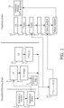
- the imaged position of a thoracic diaphragm is leveraged to determine and track the position of a lung tumor during radiation therapy (RT). It is recognized herein that the position of the thoracic diaphragm provides a good surrogate for tumor position. Respiration is driven by the thoracic diaphragm, which contracts and moves in the inferior direction during inhalation to cause the lungs to expand. Motion of the lung tumor is caused by, and therefore generally correlates with, this motion of the diaphragm.
- the diaphragm position may be monitored by ultrasound imaging.
- ultrasound imaging to directly measure and track the position of a lung tumor during RT is not practical because of air in the lungs.
- lung tumor position is correlated with ultrasound images of the diaphragm.
- a respiratory monitor 2 monitors respiratory phase 4 so that tumor position over the respiratory cycle is captured; however, in some embodiments it is contemplated to omit the respiratory monitoring.
- computed tomography (CT) images 8 of a patient may be taken in using a computed tomography (CT) imaging system 6.
- CT images for radiation therapy planning are usually three-dimensional images.
- CT images are acquired at various times (sometimes referred to as 4DCT).
- respiratory gating based on the respiratory phase 4 is employed to assign CT imaging data to different respiratory phase "bins" which are each reconstructed to generate an image of the tumor at a different respiratory phase.
- the CT images 8 inform on the position of a lung tumor, and may be used to generate a density map 9 for use in the radiation therapy planning.
- the CT image 8 may be adjusted for differences in absorption coefficient of the therapeutic radiation used in RT as compared with the x-rays used in CT imaging.
- the CT imaging system 6 may be replaced by another imaging modality that provides contrast for both the lung tumor and the thoracic diaphragm.
- MRI magnetic resonance imaging
- the density map may be generated by an approach such as segmenting the MRI image to identify regions of different tissue types (e.g. bone, fat, muscle, air in lungs) and assigning appropriate radiation absorption values for each tissue region.
- ultrasound images of patient are acquired using an ultrasound imaging device or system 10.
- the ultrasound images do not generally provide good contrast for the lung tumor, but the ultrasound images do image the thoracic diaphragm.
- a geometry of the thoracic diaphragm of the patient is captured in ultrasound images 12 acquired by the ultrasound imaging system 10.
- the thoracic diaphragm contracts to expand the lungs during inhalation, and relaxes during exhalation.
- the detailed motion of the thoracic diaphragm can be complex, and may include aspects of non-rigid shape deformation, translation, rotation, or various combinations thereof for various portions of the thoracic diaphragm.
- the term "geometry" of the thoracic diaphragm and similar phraseology as used herein encompasses all such movement, rotation, deformation or other geometric change of the thoracic diaphragm.
- the ultrasound images 12 may be either two-dimensional or three-dimensional. If two-dimensional ultrasound images 12 are employed, then the orientation of the image should be chosen to capture the principal inferior/superior (inhalation/exhalation) motion of the diaphragm. For example, by positioning the ultrasound probe generally below (i.e. inferior with respect to) the diaphragm (e.g. a subcostal or intercostal oblique probe position), a two-dimensional US image can be obtained in a slanted coronal plane that intersects the generally planar diaphragm muscle sheet so as to effectively capture this motion.
- the ultrasound probe generally below (i.e. inferior with respect to) the diaphragm (e.g. a subcostal or intercostal oblique probe position)
- a two-dimensional US image can be obtained in a slanted coronal plane that intersects the generally planar diaphragm muscle sheet so as to effectively capture this motion.
- the ultrasound probe should also be positioned so as to not interfere with (e.g. block) the RT beam(s).
- the subcostal probe position is generally effective to locate the ultrasound probe inferior to the RT beam(s).
- the position T(i) of the lung tumor is obtained by manual, semi-automated, or automated segmentation of the CT image 8.
- This tumor position T(i) is correlated with corresponding ultrasound images US(i) to generate a relationship 14 denoted T(i)-US(i) between location T(i) of the malignant tumor and the ultrasound images US(i) of the thoracic diaphragm acquired during the simulation phase.
- a position of the diaphragm may be correlated with a position of the tumor.
- To correlate CT information with ultrasound information many different techniques are possible. One such technique is to measure the CT information and ultrasound information simultaneously. That way, time stamps from the CT information may be matched with time stamps from the ultrasound information to achieve the correlation.
- the index i suitably denotes the timestamp.
- the respiratory monitor 2 is used to perform respiratory gating to generate the 4DCT images.
- Another such technique is to use the breathing signal 4 to correlate the CT information with the ultrasound information. If this technique is used, the CT information and the ultrasound information do not need to be collected at the same time, and here the index i denotes respiratory phase.
- yet another illustrative technique is to correlate the ultrasound and CT images/volumes with each other based on the position of the diaphragm in the two modalities.
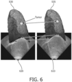
- CT and ultrasound images/volumes are spatially co-registered, for each CT volume in a "4-dimensional" CT (4DCT) series (time is the fourth dimension; and 4DCT will be described below), an ultrasound volume may be found in which the diaphragm positon in the ultrasound volume has the best overlap with the diaphragm position in the CT (see, e.g., FIGURE 4 ). Therefore, for each CT volume CT i there will be a corresponding ultrasound image/volume sim US(i) that is acquired at the similar breathing phase.
- FIGURE 4 shows an overlay of an ultrasound volume on a CT volume in sagittal view 410, and coronal view 420.
- FIGURE 4 shows a good overlap of diaphragm in the two modalities.
- the index I is suitably an arbitrary index indicating that the malignant tumor position T(i) was obtained from a CT image for which the diaphragm was in the same position (within some fitting tolerance) as the diaphragm position in the corresponding ultrasound image sim US(i).
- the respiratory monitor 2 is optionally omitted unless needed to perform respiratory gating to generate the 4DCT images).
- This adjustment may, for example, include shuttering the therapeutic radiation beam of the RT apparatus 16 for a time interval during which the tumor has moved out of its target position due to respiration, or may include operating the RT apparatus 16 to move the therapeutic beam in real-time to track respiration-related motion of the tumor.
- the ultrasound probe 18 used in the ultrasound imaging during the radiation treatment should be spatially co-registered to the linear accelerator (LINAC) coordinate system (or, more generally, to the coordinate system of the radiation therapy apparatus 16 delivering the radiation therapy, which may be a LINAC or some other radiation therapy system depending upon the type of therapeutic radiation); and with the simulation CT coordinate system (i.e. the coordinate system of the CT images 8) and simulation ultrasound images 12.
- LINAC linear accelerator
- the simulation CT coordinate system i.e. the coordinate system of the CT images 8
- Registration between the CT coordinate system of the CT images 8 and the radiation therapy apparatus 16 can employ any registration technique used in registering the planning coordinate system with the therapy coordinate system, such as using applied fiducial markers, and/or relying on intrinsic anatomical markers, and/or employing a scout scan acquired by an auxiliary imaging system, or so forth.
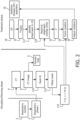
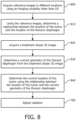
- FIGURE 2 diagrammatically shows a second preferred embodiment.
- no ultrasound images are acquired during the simulation phase (thus, the simulation-phase ultrasound imaging system 10 is omitted), which simplifies the simulation phase as the radiologist no longer needs to coordinate acquisition of the simulation ultrasound images 12 of the illustrative embodiment of FIGURE 1 .
- the illustrated CT imaging system 6 (or an MRI or other imaging system capable of imaging both the tumor and the diaphragm) acquires images of the patient which show both the lung tumor and the diaphragm.
- the 4DCT images 8 include both the lung and the thoracic diaphragm. As in the embodiment of FIGURE 1 , the CT images 8 are used to generate the density map 9.
- the position T(i) of the lung tumor is obtained by manual, semi-automated, or automated segmentation of the CT image 8. It is also contemplated that the density map and tumor position may be generated using MRI images rather than CT images.
- a mesh fit operation 110 is performed to fit a mesh to the lung in the CT image 8 to generate a fitted mesh M(i) 112 representing the geometry of the diaphragm. Because the lung and diaphragm are closely connected, for improved accuracy the mesh fit operation 110 may fit both the lungs and diaphragm to generate a fitted lung/diagraphm mesh M(i) 112.
- a relationship 114 that relates tumor position T(i) obtained from a CT image with the fitted mesh M(i) 112 obtained from the same CT image replaces the T(i) ⁇ ->US(i) relationship 14 of the embodiment of FIGURE 1 .
- the respiratory phase 4 is not used as an index and can be omitted unless (as in illustrative FIGURE 2 ) it is used for respiratory gating of the CT imaging system 6.
- the RT apparatus 16 delivers therapeutic radiation to the patient while ultrasound images of the patient are acquired using the treatment ultrasound device or system 18 as in the embodiment of FIGURE 1 ; this again may be represented by real-time ultrasound images T x US(t).
- the ultrasound images are acquired in real-time (e.g. every 0.5 seconds or faster) as the radiation therapy beam is being applied to the patient to perform RT treatment.
- the ultrasound probe 18 is spatially registered to the linear accelerator (LINAC) coordinate system (or other RT apparatus 16 coordinate system) using a localization technology such as electromagnetic transponders (EM), OSS, optical fiducial markers, or so forth and therefore, the ultrasound probe 18 is also registered to the CT coordinate system.
- LINAC linear accelerator
- EM electromagnetic transponders
- OSS optical fiducial markers
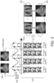
- a best lung/diaphragm mesh is found such that the overlap between the diaphragm part of the lung/diaphragm mesh 112 and the diaphragm image in the ultrasound image/volume is maximized.
- the overlap can be measured using a point-set to image/volume similarity metric on a processed ultrasound images/volumes without segmenting the diaphragm or by measuring the point-set to point-set similarity metric by segmenting the diaphragm in the ultrasound images/volumes. These are merely illustrative examples.
- the positon of the target can be estimated based on the selected CT volume.
- the diaphragm is identified (e.g. a point set may identify the diaphragm).
- a diaphragm overlap metric is employed which measures overlap of the CT/MRI diaphragm mesh and the image of the diaphragm in the real-time ultrasound images.
- a tracking system to track the probe is not used and the ultrasound (US) probe 18 is not localized respective to the RT system.
- US ultrasound
- each real-time US image/volume is registered to all the instances of CT lung mesh based on the diaphragm feature in the US image using any registration method.
- the mesh with the best registration result can be used to estimate the target positon.
Landscapes
- Health & Medical Sciences (AREA)
- Engineering & Computer Science (AREA)
- Biomedical Technology (AREA)
- Life Sciences & Earth Sciences (AREA)
- Veterinary Medicine (AREA)
- Radiology & Medical Imaging (AREA)
- Nuclear Medicine, Radiotherapy & Molecular Imaging (AREA)
- Animal Behavior & Ethology (AREA)
- General Health & Medical Sciences (AREA)
- Public Health (AREA)
- Pathology (AREA)
- Biophysics (AREA)
- Physics & Mathematics (AREA)
- Vascular Medicine (AREA)
- Heart & Thoracic Surgery (AREA)
- Medical Informatics (AREA)
- Molecular Biology (AREA)
- Surgery (AREA)
- Ultra Sonic Daignosis Equipment (AREA)
- Apparatus For Radiation Diagnosis (AREA)
- Radiation-Therapy Devices (AREA)
Claims (7)
- Lungentumorerfassungsvorrichtung umfassend:
eine elektronische Vorrichtung, programmiert zum:Empfangen einer Vielzahl von Bildern (8) eines Patienten, wobei jedes Bild einen Lungentumor und ein Diaphragma an verschiedenen Positionen zeigt, wobei die Vielzahl von Bildern unter Verwendung einer Bildgebungsmodalität, die von Ultraschallbildgebung verschieden ist, aufgenommen worden sind;Durchführen einer Netzanpassungsschritts (110), um in jedem der Vielzahl von Bildern (8) des Patienten ein Netz an die Lunge anzupassen, um eine Vielzahl von Simulationsphase-angepassten Netzen (112) zu erzeugen, die die Geometrie des Diaphragmas an verschiedenen Positionen darstellen;Bestimmen einer Beziehung (114), die jedem Netz der Vielzahl von Simulationsphase-angepassten Netzen eine Position des Lungentumors zuordnet;Empfangen eines Ultraschallbilds (19); undBestimmen einer aktuellen Position (24) eines Lungentumors unter Verwendung des empfangenen Ultraschallbilds (19), ohne den Lungentumor in dem empfangenen Ultraschallbild zu erfassen, durch Vergleichen des in dem Ultraschallbild (19) abgebildeten Diaphragmas mit der Vielzahl von Simulationsphase-angepassten Netzen (112), um ein ähnlichstes Simulationsphase-angepasstes Netz zu identifizieren, und Bestimmen der aktuellen Position (24) des Lungentumors unter Verwendung der bestimmten Beziehung (114), die jedem Netz der Vielzahl von Simulationsphase-angepassten Netzen eine Position des Lungentumors zuordnet. - Lungentumorerfassungsvorrichtung nach Anspruch 1, ferner umfassend:
eine Ultraschallvorrichtung (18), angeordnet zum Aufnehmen des Ultraschallbilds (19), das von der elektronischen Vorrichtung empfangen wird. - Lungentumorerfassungsvorrichtung nach Anspruch 2, wobei:die Ultraschallvorrichtung (18) zum Aufnehmen wenigstens eines Ultraschallbilds (19) eines Thoraxdiaphragmas angeordnet ist; unddie elektronische Vorrichtung eine elektronische Datenverarbeitungsvorrichtung ist, die programmiert ist, die aktuelle Position (24) des Lungentumors unter Verwendung des Ultraschallbilds (19) des Thoraxdiaphragmas und der Beziehung (14) zu bestimmen, wobei die Beziehung (14) ferner einem Satz von Simulationsphase-angepassten Netzen (112) des Thoraxdiaphragmas in verschiedenen Geometrien Lungentumorpositionen zuordnet, ohne den Lungentumor in den Ultraschallbildern (19) zu erfassen,wobei die elektronische Datenverarbeitungsvorrichtung dafür gestaltet ist, die Beziehung (14) in einer Simulationsphase zu erzeugen durch:Bestimmen von Lungentumorpositionen in Computertomographie(CT)-Bildern des Lungentumors; undKorrelieren der bestimmten Lungentumorpositionen mit den Simulationsphase-angepassten Netzen (sim M (i)) des Thoraxdiaphragmas.
- Lungentumorerfassungsvorrichtung nach Anspruch 3, wobei die elektronische Simulationsphasen-Datenverarbeitungsvorrichtung ferner gestaltet ist zum: Erzeugen einer Dichtekarte (9) eines Bereichs, der den Lungentumor enthält, aus einem oder mehreren der CT-Bilder des Lungentumors.
- Lungentumorerfassungsvorrichtung nach einem der Ansprüche 3-4, wobei die elektronische Simulationsphasen-Datenverarbeitungsvorrichtung ferner gestaltet ist zum:
Erzeugen eines Strahlentherapieplans. - Lungentumorerfassungsvorrichtung nach Anspruch 3, wobei die elektronische Datenverarbeitungsvorrichtung programmiert ist, die aktuelle Position (24) des Lungentumors durch Arbeitsschritte zu bestimmen, die enthalten:Berechnen eines Simulationsphase-angepassten Netzindex i* (22) gemäß:
 wobei simMi das Simulationsphase-angepasste Netz (112) bezeichnet und TxUS(t) das Ultraschallbild (19) des Thoraxdiaphragmas ist, t die Zeit darstellt und L die Ähnlichkeit zwischen simMi und Tx US(t) misst;und Bestimmen der aktuellen Position (24) des Lungentumors gemäß
 wobei simTii die Position des Lungentumors ist, die dem Simulationsphase-angepassten Netz sim M i* entspricht, gemäß der vorgegebenen Beziehung (14).
 - Lungentumorerfassungsverfahren umfassend:Aufnehmen eines Ultraschallbilds (19) eines Thoraxdiaphragmas eines Patienten; und Bestimmen einer aktuellen Position (24) eines Lungentumors des Patienten unter Verwendung des aufgenommenen Ultraschallbilds (19) und einer Tumorlokalisierungsbeziehung (14);wobei das Bestimmen nicht Erfassen des Lungentumors in dem aufgenommenen Ultraschallbild (19) enthält;wobei das Verfahren ferner während einer Simulationsphase, die den Aufnahme- und Bestimmungsschritten vorausgeht, Erzeugen der Tumorlokalisierungsbeziehung (14) umfasst durch Aufnehmen von Bildern (8) des Lungentumors und des Diaphragmas an verschiedenen Positionen unter Verwendung einer Bildgebungsmodalität (6), die von Ultraschallbildgebung verschieden ist;Durchführen eines Netzanpassungsschritts (110) zum Anpassen eines Netzes an die Lunge in jedem der Bilder (8), um eine Vielzahl von Simulationsphase-angepassten Netze (112) zu erzeugen, die die Geometrie des Diaphragmas an verschiedenen Positionen darstellen; undBestimmen der Tumorlokalisierungsbeziehung (14), die zu jedem Netz der Vielzahl von Simulationsphase-angepassten Netzen (112) unter Verwendung der Bilder des Lungentumors an verschiedenen Positionen, die während der Simulationsphase erfasst wurden, eine Position des Lungentumors zuordnet;wobei Bestimmen der aktuellen Position des Lungentumors des Patienten ferner Vergleichen des in dem aufgenommenen Ultraschallbild (19) abgebildeten Diaphragmas mit der Vielzahl von Simulationsphase-angepassten Netzen (112) umfasst, um ein ähnlichstes Netz zu identifizieren.
Applications Claiming Priority (2)
| Application Number | Priority Date | Filing Date | Title |
|---|---|---|---|
| US201562193617P | 2015-07-17 | 2015-07-17 | |
| PCT/EP2016/066912 WO2017013019A1 (en) | 2015-07-17 | 2016-07-15 | Guidance for lung cancer radiation |
Publications (2)
| Publication Number | Publication Date |
|---|---|
| EP3324852A1 EP3324852A1 (de) | 2018-05-30 |
| EP3324852B1 true EP3324852B1 (de) | 2025-06-25 |
Family
ID=56550205
Family Applications (1)
| Application Number | Title | Priority Date | Filing Date |
|---|---|---|---|
| EP16742221.1A Active EP3324852B1 (de) | 2015-07-17 | 2016-07-15 | Führung für lungenkrebsbestrahlung |
Country Status (5)
| Country | Link |
|---|---|
| US (1) | US11135447B2 (de) |
| EP (1) | EP3324852B1 (de) |
| JP (2) | JP7122115B2 (de) |
| CN (1) | CN107847216B (de) |
| WO (1) | WO2017013019A1 (de) |
Families Citing this family (7)
| Publication number | Priority date | Publication date | Assignee | Title |
|---|---|---|---|---|
| JP2021503364A (ja) | 2017-11-16 | 2021-02-12 | エバメッド・エセアー | 心臓不整脈非侵襲的治療装置及び方法 |
| CN110433398B (zh) * | 2019-09-03 | 2024-05-03 | 广西医大开元埌东医院有限责任公司 | 一种模拟放疗时呼吸运动对肺部肿瘤位置影响的装置 |
| US12156760B2 (en) | 2019-11-14 | 2024-12-03 | Ebamed Sa | Cardiac phase gating system for radiation therapy |
| EP3970620A1 (de) * | 2020-09-21 | 2022-03-23 | Koninklijke Philips N.V. | Verarbeitung von dunkelfeldröntgenbilddateninformationen |
| CN116669633A (zh) | 2020-12-23 | 2023-08-29 | 艾巴麦德有限公司 | 多平面运动管理系统 |
| US20230071643A1 (en) * | 2021-07-20 | 2023-03-09 | Daniel Nathan Maxwell | Ultrasound Diaphragmography Device and Method |
| KR20250026100A (ko) | 2023-08-16 | 2025-02-25 | 한국표준과학연구원 | 호흡 운동에 의한 장기 움직임 시뮬레이터 |
Family Cites Families (55)
| Publication number | Priority date | Publication date | Assignee | Title |
|---|---|---|---|---|
| JPS62186381A (ja) * | 1986-02-12 | 1987-08-14 | Hitachi Ltd | 画像位置合わせ方式 |
| FR2694881B1 (fr) * | 1992-07-31 | 1996-09-06 | Univ Joseph Fourier | Procede de determination de la position d'un organe. |
| US5672877A (en) * | 1996-03-27 | 1997-09-30 | Adac Laboratories | Coregistration of multi-modality data in a medical imaging system |
| US5846513B1 (en) * | 1997-07-08 | 2000-11-28 | Carewise Medical Products Corp | Tumor localization and removal system using penetratable detection probe and removal instrument |
| US6325758B1 (en) * | 1997-10-27 | 2001-12-04 | Nomos Corporation | Method and apparatus for target position verification |
| US6560354B1 (en) * | 1999-02-16 | 2003-05-06 | University Of Rochester | Apparatus and method for registration of images to physical space using a weighted combination of points and surfaces |
| CA2314794A1 (en) | 2000-08-01 | 2002-02-01 | Dimitre Hristov | Apparatus for lesion or organ localization |
| JP2004000499A (ja) * | 2002-03-27 | 2004-01-08 | Aloka Co Ltd | 超音波医療システム |
| AU2003282690B2 (en) * | 2002-10-07 | 2008-12-18 | Best Medical International, Inc. | Method and apparatus for target position verification |
| EP1599833B1 (de) * | 2003-02-18 | 2007-03-21 | Philips Intellectual Property & Standards GmbH | Bildsegmentierung durch zuweisen von klassen zu adaptivenmesh-primitiven |
| US7570791B2 (en) * | 2003-04-25 | 2009-08-04 | Medtronic Navigation, Inc. | Method and apparatus for performing 2D to 3D registration |
| US7103399B2 (en) * | 2003-09-08 | 2006-09-05 | Vanderbilt University | Apparatus and methods of cortical surface registration and deformation tracking for patient-to-image alignment in relation to image-guided surgery |
| EP1694208A2 (de) * | 2003-11-26 | 2006-08-30 | Viatronix Incorporated | Systeme und verfahren für die automatische segmentierung, visualisierung und analyse von medizinischen bildern |
| US7853308B2 (en) * | 2004-02-17 | 2010-12-14 | Siemens Medical Solutions Usa, Inc. | System and method for patient positioning for radiotherapy in the presence of respiratory motion |
| US8989349B2 (en) * | 2004-09-30 | 2015-03-24 | Accuray, Inc. | Dynamic tracking of moving targets |
| US20060241443A1 (en) * | 2004-11-22 | 2006-10-26 | Whitmore Willet F Iii | Real time ultrasound monitoring of the motion of internal structures during respiration for control of therapy delivery |
| US10492749B2 (en) * | 2005-05-03 | 2019-12-03 | The Regents Of The University Of California | Biopsy systems for breast computed tomography |
| US8108072B2 (en) * | 2007-09-30 | 2012-01-31 | Intuitive Surgical Operations, Inc. | Methods and systems for robotic instrument tool tracking with adaptive fusion of kinematics information and image information |
| US7889905B2 (en) * | 2005-05-23 | 2011-02-15 | The Penn State Research Foundation | Fast 3D-2D image registration method with application to continuously guided endoscopy |
| US7713205B2 (en) * | 2005-06-29 | 2010-05-11 | Accuray Incorporated | Dynamic tracking of soft tissue targets with ultrasound images, without using fiducial markers |
| US7831073B2 (en) * | 2005-06-29 | 2010-11-09 | Accuray Incorporated | Precision registration of X-ray images to cone-beam CT scan for image-guided radiation treatment |
| EP1915738B1 (de) * | 2005-08-09 | 2018-09-26 | Koninklijke Philips N.V. | System und verfahren zum selektiven mischen von zweidimensionalen röntgenbildern und dreidimensionalen ultraschallbildern |
| US8406851B2 (en) * | 2005-08-11 | 2013-03-26 | Accuray Inc. | Patient tracking using a virtual image |
| CA2651437C (en) * | 2006-05-18 | 2016-01-05 | Resonant Medical Inc. | Methods and systems for segmentation using boundary reparameterization |
| US20070286342A1 (en) * | 2006-06-07 | 2007-12-13 | Fuller Donald B | Systems and methods for performing radiosurgery using stereotactic techniques |
| CN101490716B (zh) | 2006-07-17 | 2012-07-04 | 皇家飞利浦电子股份有限公司 | 用于医学图像分割的与多边形网格的高效用户交互 |
| US7812543B2 (en) | 2006-11-15 | 2010-10-12 | Budike Jr Lothar E S | Modular wireless lighting control system using a common ballast control interface |
| US8358818B2 (en) * | 2006-11-16 | 2013-01-22 | Vanderbilt University | Apparatus and methods of compensating for organ deformation, registration of internal structures to images, and applications of same |
| US20080119725A1 (en) * | 2006-11-20 | 2008-05-22 | General Electric Company | Systems and Methods for Visual Verification of CT Registration and Feedback |
| JP2008154861A (ja) | 2006-12-25 | 2008-07-10 | Univ Of Tokyo | 放射線治療システム |
| WO2008086434A2 (en) * | 2007-01-09 | 2008-07-17 | Cyberheart, Inc. | Depositing radiation in heart muscle under ultrasound guidance |
| CN101053531A (zh) * | 2007-05-17 | 2007-10-17 | 上海交通大学 | 基于多模式增敏成像融合的早期肿瘤定位跟踪方法 |
| US10292619B2 (en) * | 2007-07-09 | 2019-05-21 | Covidien Lp | Patient breathing modeling |
| US8111892B2 (en) * | 2008-06-04 | 2012-02-07 | Medison Co., Ltd. | Registration of CT image onto ultrasound images |
| EP2131212A3 (de) * | 2008-06-05 | 2011-10-05 | Medison Co., Ltd. | Nicht starre Registrierung zwischen CT-Bildern und Ultraschallbildern |
| JP5241357B2 (ja) * | 2008-07-11 | 2013-07-17 | 三菱プレシジョン株式会社 | 生体データモデル作成方法及びその装置 |
| US8942342B2 (en) | 2008-12-29 | 2015-01-27 | Analogic Corporation | Multi-modality image acquisition |
| JP5806448B2 (ja) * | 2009-05-13 | 2015-11-10 | 株式会社東芝 | 核医学イメージング装置、画像処理装置および画像処理方法 |
| KR101121353B1 (ko) * | 2009-08-03 | 2012-03-09 | 한국과학기술원 | 2차원 초음파 영상에 대응하는 2차원 ct 영상을 제공하는 시스템 및 방법 |
| JP5702572B2 (ja) * | 2009-10-29 | 2015-04-15 | 株式会社東芝 | X線撮影装置 |
| JP5631698B2 (ja) * | 2009-12-07 | 2014-11-26 | 株式会社東芝 | 医用画像処理装置及び医用画像処理方法 |
| US8311303B2 (en) * | 2010-01-12 | 2012-11-13 | Siemens Corporation | Method and system for semantics driven image registration |
| JP2012030048A (ja) | 2010-07-02 | 2012-02-16 | Toshiba Corp | 超音波診断装置、画像処理装置、及び解析プログラム |
| WO2012019162A1 (en) * | 2010-08-06 | 2012-02-09 | Accuray, Inc. | Systems and methods for real-time tumor tracking during radiation treatment using ultrasound imaging |
| US9129426B2 (en) * | 2010-08-31 | 2015-09-08 | General Electric Company | Motion compensation in image processing |
| US20120071757A1 (en) * | 2010-09-17 | 2012-03-22 | University Of British Columbia | Ultrasound Registration |
| JP5872323B2 (ja) * | 2011-03-29 | 2016-03-01 | 株式会社東芝 | X線ct装置及び画像処理方法 |
| US20120253170A1 (en) * | 2011-03-29 | 2012-10-04 | Samsung Electronics Co., Ltd. | Method and apparatus for generating medical image of body organ by using 3-d model |
| US8358823B2 (en) * | 2011-03-30 | 2013-01-22 | Mitsubishi Electric Research Laboratories, Inc. | Method for tracking tumors in bi-plane images |
| WO2012142031A1 (en) * | 2011-04-12 | 2012-10-18 | Brigham And Women's Hospital, Inc. | System and method for motion tracking using unique ultrasound echo signatures |
| WO2013028762A1 (en) | 2011-08-22 | 2013-02-28 | Siemens Corporation | Method and system for integrated radiological and pathological information for diagnosis, therapy selection, and monitoring |
| US9873003B2 (en) * | 2012-07-13 | 2018-01-23 | Mitsubishi Electric Corporation | X-ray positioning apparatus, X-ray positioning method, and attentional image photographing method |
| KR102070427B1 (ko) * | 2012-08-08 | 2020-01-28 | 삼성전자주식회사 | 종양의 위치를 추적하는 방법 및 장치 |
| KR20140126815A (ko) * | 2013-04-22 | 2014-11-03 | 삼성전자주식회사 | 호흡 주기 동안 체내 장기의 변화를 추적하는 방법, 장치 및 시스템. |
| US9950194B2 (en) * | 2014-09-09 | 2018-04-24 | Mevion Medical Systems, Inc. | Patient positioning system |
-
2016
- 2016-07-15 EP EP16742221.1A patent/EP3324852B1/de active Active
- 2016-07-15 WO PCT/EP2016/066912 patent/WO2017013019A1/en not_active Ceased
- 2016-07-15 US US15/743,005 patent/US11135447B2/en active Active
- 2016-07-15 JP JP2017567330A patent/JP7122115B2/ja active Active
- 2016-07-15 CN CN201680042033.XA patent/CN107847216B/zh active Active
-
2022
- 2022-05-23 JP JP2022083542A patent/JP7397909B2/ja active Active
Also Published As
| Publication number | Publication date |
|---|---|
| JP7397909B2 (ja) | 2023-12-13 |
| JP2018519913A (ja) | 2018-07-26 |
| EP3324852A1 (de) | 2018-05-30 |
| WO2017013019A1 (en) | 2017-01-26 |
| US20180214713A1 (en) | 2018-08-02 |
| JP7122115B2 (ja) | 2022-08-19 |
| JP2022117992A (ja) | 2022-08-12 |
| CN107847216A (zh) | 2018-03-27 |
| US11135447B2 (en) | 2021-10-05 |
| CN107847216B (zh) | 2024-01-23 |
Similar Documents
| Publication | Publication Date | Title |
|---|---|---|
| JP7397909B2 (ja) | 肺癌放射線のためのガイダンス | |
| EP3407791B1 (de) | Darstellung einer bildsequenz im zusammenhang mit einem bewegungsmodell | |
| US8831706B2 (en) | Fiducial-less tracking of a volume of interest | |
| US8422631B2 (en) | Radiation therapy planning apparatus and radiation therapy planning method | |
| US7720196B2 (en) | Target tracking using surface scanner and four-dimensional diagnostic imaging data | |
| McClelland et al. | Inter-fraction variations in respiratory motion models | |
| US20150080634A1 (en) | Tracking external markers to internal bodily structures | |
| US20130035588A1 (en) | Magnetic resonance imaging for therapy planning | |
| EP2506216A2 (de) | Röntgen-CT-Diagnosevorrichtung und Bildverarbeitungsverfahren | |
| CN107106867A (zh) | 磁共振投影成像 | |
| US8358738B2 (en) | Respiration-correlated radiotherapy | |
| CN107613873B (zh) | 用于物体的原地靶向的方法和系统 | |
| JP2014104360A (ja) | 同期放射線治療の方法及びシステム | |
| KR20140100648A (ko) | 일 호흡 주기에 따른 장기의 형상 및 위치의 변화를 나타내는 모델을 생성하는 방법, 장치 및 시스템. | |
| JP2011200542A (ja) | 患者位置決め方法および患者位置決めシステム | |
| JPWO2020012785A1 (ja) | 放射線治療装置および放射線治療方法 | |
| JP6692817B2 (ja) | 対象物体の変位を計算する方法及びシステム | |
| US20220401758A1 (en) | Patient anatomical structure change detection method, patient anatomical structure change detection device, and computer program | |
| KR20150119695A (ko) | 방사선 치료시 자연호흡법을 이용한 환자 호흡동조 시스템 및 이에 의한 방사선 조사방법 | |
| CN102440789B (zh) | 一种基于双能x射线图像的软组织病灶定位系统 | |
| CN116407779A (zh) | 放射治疗系统及计算机可读存储介质 | |
| CN116472090A (zh) | 患者身体区域的优化跟踪 | |
| Miura et al. | 4D modeling in a gimbaled linear accelerator by using gold anchor markers | |
| US20250232447A1 (en) | Irradiated position confirmation support device, irradiated position confirmation support method, and irradiated position confirmation support program | |
| Ranjbar | Simulating the breathing of lung cancer patients to estimate tumor motion and deformation at the time of radiation treatment |
Legal Events
| Date | Code | Title | Description |
|---|---|---|---|
| STAA | Information on the status of an ep patent application or granted ep patent |
Free format text: STATUS: THE INTERNATIONAL PUBLICATION HAS BEEN MADE |
|
| PUAI | Public reference made under article 153(3) epc to a published international application that has entered the european phase |
Free format text: ORIGINAL CODE: 0009012 |
|
| STAA | Information on the status of an ep patent application or granted ep patent |
Free format text: STATUS: REQUEST FOR EXAMINATION WAS MADE |
|
| 17P | Request for examination filed |
Effective date: 20180219 |
|
| AK | Designated contracting states |
Kind code of ref document: A1 Designated state(s): AL AT BE BG CH CY CZ DE DK EE ES FI FR GB GR HR HU IE IS IT LI LT LU LV MC MK MT NL NO PL PT RO RS SE SI SK SM TR |
|
| AX | Request for extension of the european patent |
Extension state: BA ME |
|
| DAV | Request for validation of the european patent (deleted) | ||
| DAX | Request for extension of the european patent (deleted) | ||
| RAP1 | Party data changed (applicant data changed or rights of an application transferred) |
Owner name: KONINKLIJKE PHILIPS N.V. |
|
| STAA | Information on the status of an ep patent application or granted ep patent |
Free format text: STATUS: EXAMINATION IS IN PROGRESS |
|
| 17Q | First examination report despatched |
Effective date: 20200724 |
|
| RAP1 | Party data changed (applicant data changed or rights of an application transferred) |
Owner name: ELEKTA, INC. |
|
| 111L | Licence recorded |
Designated state(s): AL AT BE BG CH CY CZ DE DK EE ES FI FR GB GR HR HU IE IS IT LT LU LV MC MK MT NL NO PL PT RO RS SE SI SK SM TR Name of requester: KONINKLIJKE PHILIPS N.V., NL Effective date: 20240723 |
|
| GRAP | Despatch of communication of intention to grant a patent |
Free format text: ORIGINAL CODE: EPIDOSNIGR1 |
|
| STAA | Information on the status of an ep patent application or granted ep patent |
Free format text: STATUS: GRANT OF PATENT IS INTENDED |
|
| INTG | Intention to grant announced |
Effective date: 20250121 |
|
| GRAS | Grant fee paid |
Free format text: ORIGINAL CODE: EPIDOSNIGR3 |
|
| GRAA | (expected) grant |
Free format text: ORIGINAL CODE: 0009210 |
|
| STAA | Information on the status of an ep patent application or granted ep patent |
Free format text: STATUS: THE PATENT HAS BEEN GRANTED |
|
| 111L | Licence recorded |
Designated state(s): AL AT BE BG CH CY CZ DE DK EE ES FI FR GB GR HR HU IE IS IT LT LU LV MC MK MT NL NO PL PT RO RS SE SI SK SM TR Name of requester: KONINKLIJKE PHILIPS N.V., NL Effective date: 20240723 |
|
| AK | Designated contracting states |
Kind code of ref document: B1 Designated state(s): AL AT BE BG CH CY CZ DE DK EE ES FI FR GB GR HR HU IE IS IT LI LT LU LV MC MK MT NL NO PL PT RO RS SE SI SK SM TR |
|
| REG | Reference to a national code |
Ref country code: GB Ref legal event code: FG4D |
|
| REG | Reference to a national code |
Ref country code: CH Ref legal event code: EP |
|
| REG | Reference to a national code |
Ref country code: CH Ref legal event code: EP |
|
| REG | Reference to a national code |
Ref country code: IE Ref legal event code: FG4D |
|
| REG | Reference to a national code |
Ref country code: DE Ref legal event code: R096 Ref document number: 602016092656 Country of ref document: DE |
|
| PGFP | Annual fee paid to national office [announced via postgrant information from national office to epo] |
Ref country code: FR Payment date: 20250610 Year of fee payment: 10 |
|
| P01 | Opt-out of the competence of the unified patent court (upc) registered |
Free format text: CASE NUMBER: APP_30188/2025 Effective date: 20250624 |
|
| PG25 | Lapsed in a contracting state [announced via postgrant information from national office to epo] |
Ref country code: FI Free format text: LAPSE BECAUSE OF FAILURE TO SUBMIT A TRANSLATION OF THE DESCRIPTION OR TO PAY THE FEE WITHIN THE PRESCRIBED TIME-LIMIT Effective date: 20250625 |
|
| PGFP | Annual fee paid to national office [announced via postgrant information from national office to epo] |
Ref country code: DE Payment date: 20250611 Year of fee payment: 10 |
|
| REG | Reference to a national code |
Ref country code: LT Ref legal event code: MG9D |
|
| PG25 | Lapsed in a contracting state [announced via postgrant information from national office to epo] |
Ref country code: GR Free format text: LAPSE BECAUSE OF FAILURE TO SUBMIT A TRANSLATION OF THE DESCRIPTION OR TO PAY THE FEE WITHIN THE PRESCRIBED TIME-LIMIT Effective date: 20250926 Ref country code: NO Free format text: LAPSE BECAUSE OF FAILURE TO SUBMIT A TRANSLATION OF THE DESCRIPTION OR TO PAY THE FEE WITHIN THE PRESCRIBED TIME-LIMIT Effective date: 20250925 |
|
| PG25 | Lapsed in a contracting state [announced via postgrant information from national office to epo] |
Ref country code: BG Free format text: LAPSE BECAUSE OF FAILURE TO SUBMIT A TRANSLATION OF THE DESCRIPTION OR TO PAY THE FEE WITHIN THE PRESCRIBED TIME-LIMIT Effective date: 20250625 |
|
| PGFP | Annual fee paid to national office [announced via postgrant information from national office to epo] |
Ref country code: GB Payment date: 20250723 Year of fee payment: 10 |
|
| PG25 | Lapsed in a contracting state [announced via postgrant information from national office to epo] |
Ref country code: HR Free format text: LAPSE BECAUSE OF FAILURE TO SUBMIT A TRANSLATION OF THE DESCRIPTION OR TO PAY THE FEE WITHIN THE PRESCRIBED TIME-LIMIT Effective date: 20250625 |
|
| PG25 | Lapsed in a contracting state [announced via postgrant information from national office to epo] |
Ref country code: RS Free format text: LAPSE BECAUSE OF FAILURE TO SUBMIT A TRANSLATION OF THE DESCRIPTION OR TO PAY THE FEE WITHIN THE PRESCRIBED TIME-LIMIT Effective date: 20250925 |
|
| PG25 | Lapsed in a contracting state [announced via postgrant information from national office to epo] |
Ref country code: LV Free format text: LAPSE BECAUSE OF FAILURE TO SUBMIT A TRANSLATION OF THE DESCRIPTION OR TO PAY THE FEE WITHIN THE PRESCRIBED TIME-LIMIT Effective date: 20250625 |
|
| REG | Reference to a national code |
Ref country code: NL Ref legal event code: MP Effective date: 20250625 |
|
| PG25 | Lapsed in a contracting state [announced via postgrant information from national office to epo] |
Ref country code: NL Free format text: LAPSE BECAUSE OF FAILURE TO SUBMIT A TRANSLATION OF THE DESCRIPTION OR TO PAY THE FEE WITHIN THE PRESCRIBED TIME-LIMIT Effective date: 20250625 |
|
| PG25 | Lapsed in a contracting state [announced via postgrant information from national office to epo] |
Ref country code: PT Free format text: LAPSE BECAUSE OF FAILURE TO SUBMIT A TRANSLATION OF THE DESCRIPTION OR TO PAY THE FEE WITHIN THE PRESCRIBED TIME-LIMIT Effective date: 20251027 |
|
| REG | Reference to a national code |
Ref country code: AT Ref legal event code: MK05 Ref document number: 1805619 Country of ref document: AT Kind code of ref document: T Effective date: 20250625 |
|
| PG25 | Lapsed in a contracting state [announced via postgrant information from national office to epo] |
Ref country code: IS Free format text: LAPSE BECAUSE OF FAILURE TO SUBMIT A TRANSLATION OF THE DESCRIPTION OR TO PAY THE FEE WITHIN THE PRESCRIBED TIME-LIMIT Effective date: 20251025 |
|
| PG25 | Lapsed in a contracting state [announced via postgrant information from national office to epo] |
Ref country code: AT Free format text: LAPSE BECAUSE OF FAILURE TO SUBMIT A TRANSLATION OF THE DESCRIPTION OR TO PAY THE FEE WITHIN THE PRESCRIBED TIME-LIMIT Effective date: 20250625 Ref country code: SM Free format text: LAPSE BECAUSE OF FAILURE TO SUBMIT A TRANSLATION OF THE DESCRIPTION OR TO PAY THE FEE WITHIN THE PRESCRIBED TIME-LIMIT Effective date: 20250625 |
|
| PG25 | Lapsed in a contracting state [announced via postgrant information from national office to epo] |
Ref country code: CZ Free format text: LAPSE BECAUSE OF FAILURE TO SUBMIT A TRANSLATION OF THE DESCRIPTION OR TO PAY THE FEE WITHIN THE PRESCRIBED TIME-LIMIT Effective date: 20250625 |
|
| PG25 | Lapsed in a contracting state [announced via postgrant information from national office to epo] |
Ref country code: PL Free format text: LAPSE BECAUSE OF FAILURE TO SUBMIT A TRANSLATION OF THE DESCRIPTION OR TO PAY THE FEE WITHIN THE PRESCRIBED TIME-LIMIT Effective date: 20250625 |
|
| PG25 | Lapsed in a contracting state [announced via postgrant information from national office to epo] |
Ref country code: EE Free format text: LAPSE BECAUSE OF FAILURE TO SUBMIT A TRANSLATION OF THE DESCRIPTION OR TO PAY THE FEE WITHIN THE PRESCRIBED TIME-LIMIT Effective date: 20250625 |
|
| PG25 | Lapsed in a contracting state [announced via postgrant information from national office to epo] |
Ref country code: SK Free format text: LAPSE BECAUSE OF FAILURE TO SUBMIT A TRANSLATION OF THE DESCRIPTION OR TO PAY THE FEE WITHIN THE PRESCRIBED TIME-LIMIT Effective date: 20250625 Ref country code: RO Free format text: LAPSE BECAUSE OF FAILURE TO SUBMIT A TRANSLATION OF THE DESCRIPTION OR TO PAY THE FEE WITHIN THE PRESCRIBED TIME-LIMIT Effective date: 20250625 |
|
| PG25 | Lapsed in a contracting state [announced via postgrant information from national office to epo] |
Ref country code: ES Free format text: LAPSE BECAUSE OF FAILURE TO SUBMIT A TRANSLATION OF THE DESCRIPTION OR TO PAY THE FEE WITHIN THE PRESCRIBED TIME-LIMIT Effective date: 20250625 |
|
| REG | Reference to a national code |
Ref country code: CH Ref legal event code: H13 Free format text: ST27 STATUS EVENT CODE: U-0-0-H10-H13 (AS PROVIDED BY THE NATIONAL OFFICE) Effective date: 20260224 |
|
| PG25 | Lapsed in a contracting state [announced via postgrant information from national office to epo] |
Ref country code: LU Free format text: LAPSE BECAUSE OF NON-PAYMENT OF DUE FEES Effective date: 20250715 |