EP3312608B1 - Procédés d'identification d'une plaie cutanée non cicatrisante et de surveillance de la cicatrisation d'une plaie cutanée - Google Patents
Procédés d'identification d'une plaie cutanée non cicatrisante et de surveillance de la cicatrisation d'une plaie cutanée Download PDFInfo
- Publication number
- EP3312608B1 EP3312608B1 EP16002266.1A EP16002266A EP3312608B1 EP 3312608 B1 EP3312608 B1 EP 3312608B1 EP 16002266 A EP16002266 A EP 16002266A EP 3312608 B1 EP3312608 B1 EP 3312608B1
- Authority
- EP
- European Patent Office
- Prior art keywords
- wound
- time point
- marker
- skin wound
- cells
- Prior art date
- Legal status (The legal status is an assumption and is not a legal conclusion. Google has not performed a legal analysis and makes no representation as to the accuracy of the status listed.)
- Active
Links
- 206010072170 Skin wound Diseases 0.000 title claims description 463
- 230000035876 healing Effects 0.000 title claims description 265
- 238000000034 method Methods 0.000 title claims description 205
- 238000012544 monitoring process Methods 0.000 title claims description 29
- 208000027418 Wounds and injury Diseases 0.000 claims description 480
- 206010052428 Wound Diseases 0.000 claims description 478
- 210000000416 exudates and transudate Anatomy 0.000 claims description 369
- 210000002950 fibroblast Anatomy 0.000 claims description 315
- 210000004027 cell Anatomy 0.000 claims description 298
- 239000003550 marker Substances 0.000 claims description 252
- 239000000523 sample Substances 0.000 claims description 233
- 210000002540 macrophage Anatomy 0.000 claims description 198
- 108020004999 messenger RNA Proteins 0.000 claims description 133
- 150000001875 compounds Chemical class 0.000 claims description 122
- 230000035755 proliferation Effects 0.000 claims description 107
- 239000002458 cell surface marker Substances 0.000 claims description 94
- 239000011159 matrix material Substances 0.000 claims description 85
- 230000015572 biosynthetic process Effects 0.000 claims description 82
- 210000002510 keratinocyte Anatomy 0.000 claims description 65
- 230000029663 wound healing Effects 0.000 claims description 54
- 239000011230 binding agent Substances 0.000 claims description 45
- 238000003501 co-culture Methods 0.000 claims description 43
- 230000002500 effect on skin Effects 0.000 claims description 37
- 238000012258 culturing Methods 0.000 claims description 36
- 230000004069 differentiation Effects 0.000 claims description 36
- 102000004127 Cytokines Human genes 0.000 claims description 34
- 108090000695 Cytokines Proteins 0.000 claims description 34
- 102100031585 ADP-ribosyl cyclase/cyclic ADP-ribose hydrolase 1 Human genes 0.000 claims description 33
- 102100026122 High affinity immunoglobulin gamma Fc receptor I Human genes 0.000 claims description 33
- 101000777636 Homo sapiens ADP-ribosyl cyclase/cyclic ADP-ribose hydrolase 1 Proteins 0.000 claims description 33
- 101000913074 Homo sapiens High affinity immunoglobulin gamma Fc receptor I Proteins 0.000 claims description 33
- 102100036301 C-C chemokine receptor type 7 Human genes 0.000 claims description 32
- 101000716065 Homo sapiens C-C chemokine receptor type 7 Proteins 0.000 claims description 32
- 108020003175 receptors Proteins 0.000 claims description 32
- 102000005962 receptors Human genes 0.000 claims description 32
- 239000006228 supernatant Substances 0.000 claims description 32
- 102100023701 C-C motif chemokine 18 Human genes 0.000 claims description 31
- 102100025248 C-X-C motif chemokine 10 Human genes 0.000 claims description 31
- 101000978371 Homo sapiens C-C motif chemokine 18 Proteins 0.000 claims description 31
- 101000858088 Homo sapiens C-X-C motif chemokine 10 Proteins 0.000 claims description 31
- 108010076561 Interleukin-23 Subunit p19 Proteins 0.000 claims description 31
- 210000001616 monocyte Anatomy 0.000 claims description 30
- 238000002560 therapeutic procedure Methods 0.000 claims description 30
- 239000003795 chemical substances by application Substances 0.000 claims description 29
- 238000012605 2D cell culture Methods 0.000 claims description 27
- 208000025865 Ulcer Diseases 0.000 claims description 27
- 108090000623 proteins and genes Proteins 0.000 claims description 27
- 102000004169 proteins and genes Human genes 0.000 claims description 27
- 231100000397 ulcer Toxicity 0.000 claims description 26
- 238000012216 screening Methods 0.000 claims description 23
- 239000007787 solid Substances 0.000 claims description 23
- 229940079593 drug Drugs 0.000 claims description 21
- 239000003814 drug Substances 0.000 claims description 21
- 208000015181 infectious disease Diseases 0.000 claims description 21
- 238000001804 debridement Methods 0.000 claims description 20
- 210000003491 skin Anatomy 0.000 claims description 20
- 238000000338 in vitro Methods 0.000 claims description 19
- 239000013589 supplement Substances 0.000 claims description 18
- 230000002708 enhancing effect Effects 0.000 claims description 17
- 108090000765 processed proteins & peptides Proteins 0.000 claims description 17
- 230000001737 promoting effect Effects 0.000 claims description 17
- 239000000203 mixture Substances 0.000 claims description 16
- 108010037362 Extracellular Matrix Proteins Proteins 0.000 claims description 15
- 102000010834 Extracellular Matrix Proteins Human genes 0.000 claims description 15
- 108010010803 Gelatin Proteins 0.000 claims description 15
- 210000002744 extracellular matrix Anatomy 0.000 claims description 15
- 229920000159 gelatin Polymers 0.000 claims description 15
- 239000008273 gelatin Substances 0.000 claims description 15
- 235000019322 gelatine Nutrition 0.000 claims description 15
- 235000011852 gelatine desserts Nutrition 0.000 claims description 15
- 239000003102 growth factor Substances 0.000 claims description 15
- 238000012413 Fluorescence activated cell sorting analysis Methods 0.000 claims description 14
- 238000010324 immunological assay Methods 0.000 claims description 13
- 235000000346 sugar Nutrition 0.000 claims description 13
- 230000003115 biocidal effect Effects 0.000 claims description 12
- 239000007850 fluorescent dye Substances 0.000 claims description 12
- 238000012604 3D cell culture Methods 0.000 claims description 11
- 206010040943 Skin Ulcer Diseases 0.000 claims description 11
- 210000004369 blood Anatomy 0.000 claims description 11
- 239000008280 blood Substances 0.000 claims description 11
- 208000037265 diseases, disorders, signs and symptoms Diseases 0.000 claims description 11
- 102000004196 processed proteins & peptides Human genes 0.000 claims description 11
- 239000000047 product Substances 0.000 claims description 11
- 230000027455 binding Effects 0.000 claims description 10
- 239000000470 constituent Substances 0.000 claims description 10
- 206010012601 diabetes mellitus Diseases 0.000 claims description 10
- 201000010099 disease Diseases 0.000 claims description 10
- 238000001415 gene therapy Methods 0.000 claims description 10
- 241000411851 herbal medicine Species 0.000 claims description 10
- 238000012986 modification Methods 0.000 claims description 10
- 230000004048 modification Effects 0.000 claims description 10
- 239000000419 plant extract Substances 0.000 claims description 10
- 150000008163 sugars Chemical class 0.000 claims description 10
- 238000004113 cell culture Methods 0.000 claims description 9
- 238000007906 compression Methods 0.000 claims description 9
- 230000006835 compression Effects 0.000 claims description 9
- 239000000463 material Substances 0.000 claims description 9
- 238000002493 microarray Methods 0.000 claims description 8
- 238000011301 standard therapy Methods 0.000 claims description 8
- 108091032973 (ribonucleotides)n+m Proteins 0.000 claims description 7
- 238000005469 granulation Methods 0.000 claims description 7
- 230000003179 granulation Effects 0.000 claims description 7
- 238000010899 nucleation Methods 0.000 claims description 7
- 239000004033 plastic Substances 0.000 claims description 7
- 238000012286 ELISA Assay Methods 0.000 claims description 6
- 208000000558 Varicose Ulcer Diseases 0.000 claims description 6
- 238000003271 compound fluorescence assay Methods 0.000 claims description 6
- 238000009396 hybridization Methods 0.000 claims description 6
- 230000017074 necrotic cell death Effects 0.000 claims description 6
- 201000002282 venous insufficiency Diseases 0.000 claims description 6
- 241000124008 Mammalia Species 0.000 claims description 5
- 208000004210 Pressure Ulcer Diseases 0.000 claims description 5
- 239000012228 culture supernatant Substances 0.000 claims description 5
- 208000002847 Surgical Wound Diseases 0.000 claims description 4
- 239000003242 anti bacterial agent Substances 0.000 claims description 4
- 239000011324 bead Substances 0.000 claims description 4
- 210000005260 human cell Anatomy 0.000 claims description 4
- 229920000742 Cotton Polymers 0.000 claims description 3
- 206010011985 Decubitus ulcer Diseases 0.000 claims description 3
- 206010056340 Diabetic ulcer Diseases 0.000 claims description 3
- 230000001028 anti-proliverative effect Effects 0.000 claims description 3
- 230000036770 blood supply Effects 0.000 claims description 3
- 230000002950 deficient Effects 0.000 claims description 3
- 230000023753 dehiscence Effects 0.000 claims description 3
- 229940088597 hormone Drugs 0.000 claims description 3
- 239000005556 hormone Substances 0.000 claims description 3
- 230000000302 ischemic effect Effects 0.000 claims description 3
- 239000012528 membrane Substances 0.000 claims description 3
- 244000005700 microbiome Species 0.000 claims description 3
- 230000001613 neoplastic effect Effects 0.000 claims description 3
- 230000002981 neuropathic effect Effects 0.000 claims description 3
- 108020004707 nucleic acids Proteins 0.000 claims description 3
- 102000039446 nucleic acids Human genes 0.000 claims description 3
- 150000007523 nucleic acids Chemical class 0.000 claims description 3
- 229920000642 polymer Polymers 0.000 claims description 3
- 150000003384 small molecules Chemical class 0.000 claims description 3
- 230000004936 stimulating effect Effects 0.000 claims description 3
- 239000000126 substance Substances 0.000 claims description 3
- 230000003612 virological effect Effects 0.000 claims description 3
- 230000002924 anti-infective effect Effects 0.000 claims description 2
- 230000002519 immonomodulatory effect Effects 0.000 claims description 2
- 239000003018 immunosuppressive agent Substances 0.000 claims description 2
- 229940125721 immunosuppressive agent Drugs 0.000 claims description 2
- 230000001771 impaired effect Effects 0.000 claims description 2
- 102000011718 Interleukin-23 Subunit p19 Human genes 0.000 claims 22
- 238000003556 assay Methods 0.000 description 65
- 230000000694 effects Effects 0.000 description 31
- 230000001684 chronic effect Effects 0.000 description 26
- 239000002609 medium Substances 0.000 description 18
- NOESYZHRGYRDHS-UHFFFAOYSA-N insulin Chemical compound N1C(=O)C(NC(=O)C(CCC(N)=O)NC(=O)C(CCC(O)=O)NC(=O)C(C(C)C)NC(=O)C(NC(=O)CN)C(C)CC)CSSCC(C(NC(CO)C(=O)NC(CC(C)C)C(=O)NC(CC=2C=CC(O)=CC=2)C(=O)NC(CCC(N)=O)C(=O)NC(CC(C)C)C(=O)NC(CCC(O)=O)C(=O)NC(CC(N)=O)C(=O)NC(CC=2C=CC(O)=CC=2)C(=O)NC(CSSCC(NC(=O)C(C(C)C)NC(=O)C(CC(C)C)NC(=O)C(CC=2C=CC(O)=CC=2)NC(=O)C(CC(C)C)NC(=O)C(C)NC(=O)C(CCC(O)=O)NC(=O)C(C(C)C)NC(=O)C(CC(C)C)NC(=O)C(CC=2NC=NC=2)NC(=O)C(CO)NC(=O)CNC2=O)C(=O)NCC(=O)NC(CCC(O)=O)C(=O)NC(CCCNC(N)=N)C(=O)NCC(=O)NC(CC=3C=CC=CC=3)C(=O)NC(CC=3C=CC=CC=3)C(=O)NC(CC=3C=CC(O)=CC=3)C(=O)NC(C(C)O)C(=O)N3C(CCC3)C(=O)NC(CCCCN)C(=O)NC(C)C(O)=O)C(=O)NC(CC(N)=O)C(O)=O)=O)NC(=O)C(C(C)CC)NC(=O)C(CO)NC(=O)C(C(C)O)NC(=O)C1CSSCC2NC(=O)C(CC(C)C)NC(=O)C(NC(=O)C(CCC(N)=O)NC(=O)C(CC(N)=O)NC(=O)C(NC(=O)C(N)CC=1C=CC=CC=1)C(C)C)CC1=CN=CN1 NOESYZHRGYRDHS-UHFFFAOYSA-N 0.000 description 16
- IAZDPXIOMUYVGZ-UHFFFAOYSA-N Dimethylsulphoxide Chemical compound CS(C)=O IAZDPXIOMUYVGZ-UHFFFAOYSA-N 0.000 description 15
- 108060008682 Tumor Necrosis Factor Proteins 0.000 description 15
- 102000000852 Tumor Necrosis Factor-alpha Human genes 0.000 description 15
- 239000000243 solution Substances 0.000 description 15
- MZOFCQQQCNRIBI-VMXHOPILSA-N (3s)-4-[[(2s)-1-[[(2s)-1-[[(1s)-1-carboxy-2-hydroxyethyl]amino]-4-methyl-1-oxopentan-2-yl]amino]-5-(diaminomethylideneamino)-1-oxopentan-2-yl]amino]-3-[[2-[[(2s)-2,6-diaminohexanoyl]amino]acetyl]amino]-4-oxobutanoic acid Chemical compound OC[C@@H](C(O)=O)NC(=O)[C@H](CC(C)C)NC(=O)[C@H](CCCN=C(N)N)NC(=O)[C@H](CC(O)=O)NC(=O)CNC(=O)[C@@H](N)CCCCN MZOFCQQQCNRIBI-VMXHOPILSA-N 0.000 description 14
- CIWBSHSKHKDKBQ-JLAZNSOCSA-N Ascorbic acid Chemical compound OC[C@H](O)[C@H]1OC(=O)C(O)=C1O CIWBSHSKHKDKBQ-JLAZNSOCSA-N 0.000 description 14
- 102000003896 Myeloperoxidases Human genes 0.000 description 14
- 108090000235 Myeloperoxidases Proteins 0.000 description 14
- 239000007788 liquid Substances 0.000 description 14
- 102000004190 Enzymes Human genes 0.000 description 13
- 108090000790 Enzymes Proteins 0.000 description 13
- 210000004322 M2 macrophage Anatomy 0.000 description 13
- 102000002274 Matrix Metalloproteinases Human genes 0.000 description 13
- 108010000684 Matrix Metalloproteinases Proteins 0.000 description 13
- 238000010790 dilution Methods 0.000 description 13
- 239000012895 dilution Substances 0.000 description 13
- 238000001516 cell proliferation assay Methods 0.000 description 12
- 210000001519 tissue Anatomy 0.000 description 12
- 210000003690 classically activated macrophage Anatomy 0.000 description 11
- 210000002889 endothelial cell Anatomy 0.000 description 11
- 238000002965 ELISA Methods 0.000 description 9
- 102100036705 Interleukin-23 subunit alpha Human genes 0.000 description 9
- LOKCTEFSRHRXRJ-UHFFFAOYSA-I dipotassium trisodium dihydrogen phosphate hydrogen phosphate dichloride Chemical compound P(=O)(O)(O)[O-].[K+].P(=O)(O)([O-])[O-].[Na+].[Na+].[Cl-].[K+].[Cl-].[Na+] LOKCTEFSRHRXRJ-UHFFFAOYSA-I 0.000 description 9
- 239000000975 dye Substances 0.000 description 9
- 239000002953 phosphate buffered saline Substances 0.000 description 9
- 238000012360 testing method Methods 0.000 description 9
- 102000004877 Insulin Human genes 0.000 description 8
- 108090001061 Insulin Proteins 0.000 description 8
- 102000016387 Pancreatic elastase Human genes 0.000 description 8
- 108010067372 Pancreatic elastase Proteins 0.000 description 8
- 101710098940 Pro-epidermal growth factor Proteins 0.000 description 8
- FAPWRFPIFSIZLT-UHFFFAOYSA-M Sodium chloride Chemical compound [Na+].[Cl-] FAPWRFPIFSIZLT-UHFFFAOYSA-M 0.000 description 8
- 229940125396 insulin Drugs 0.000 description 8
- 150000003839 salts Chemical class 0.000 description 8
- ZZZCUOFIHGPKAK-UHFFFAOYSA-N D-erythro-ascorbic acid Natural products OCC1OC(=O)C(O)=C1O ZZZCUOFIHGPKAK-UHFFFAOYSA-N 0.000 description 7
- 229930003268 Vitamin C Natural products 0.000 description 7
- 238000002835 absorbance Methods 0.000 description 7
- 239000012131 assay buffer Substances 0.000 description 7
- 238000011156 evaluation Methods 0.000 description 7
- 238000011534 incubation Methods 0.000 description 7
- 239000011718 vitamin C Substances 0.000 description 7
- 235000019154 vitamin C Nutrition 0.000 description 7
- QTBSBXVTEAMEQO-UHFFFAOYSA-N Acetic acid Chemical compound CC(O)=O QTBSBXVTEAMEQO-UHFFFAOYSA-N 0.000 description 6
- 239000006144 Dulbecco’s modified Eagle's medium Substances 0.000 description 6
- 239000013543 active substance Substances 0.000 description 6
- 238000007792 addition Methods 0.000 description 6
- 238000000423 cell based assay Methods 0.000 description 6
- 230000001413 cellular effect Effects 0.000 description 6
- 238000003018 immunoassay Methods 0.000 description 6
- 230000001965 increasing effect Effects 0.000 description 6
- 230000006698 induction Effects 0.000 description 6
- 230000000770 proinflammatory effect Effects 0.000 description 6
- 230000009870 specific binding Effects 0.000 description 6
- UCSJYZPVAKXKNQ-HZYVHMACSA-N streptomycin Chemical compound CN[C@H]1[C@H](O)[C@@H](O)[C@H](CO)O[C@H]1O[C@@H]1[C@](C=O)(O)[C@H](C)O[C@H]1O[C@@H]1[C@@H](NC(N)=N)[C@H](O)[C@@H](NC(N)=N)[C@H](O)[C@H]1O UCSJYZPVAKXKNQ-HZYVHMACSA-N 0.000 description 6
- 108010081589 Becaplermin Proteins 0.000 description 5
- 206010061218 Inflammation Diseases 0.000 description 5
- 230000001464 adherent effect Effects 0.000 description 5
- 230000003247 decreasing effect Effects 0.000 description 5
- 230000012010 growth Effects 0.000 description 5
- 230000002757 inflammatory effect Effects 0.000 description 5
- 230000004054 inflammatory process Effects 0.000 description 5
- 230000005764 inhibitory process Effects 0.000 description 5
- SXQCTESRRZBPHJ-UHFFFAOYSA-M lissamine rhodamine Chemical compound [Na+].C=12C=CC(=[N+](CC)CC)C=C2OC2=CC(N(CC)CC)=CC=C2C=1C1=CC=C(S([O-])(=O)=O)C=C1S([O-])(=O)=O SXQCTESRRZBPHJ-UHFFFAOYSA-M 0.000 description 5
- 238000002156 mixing Methods 0.000 description 5
- 230000008569 process Effects 0.000 description 5
- 230000035945 sensitivity Effects 0.000 description 5
- 239000000725 suspension Substances 0.000 description 5
- 230000035899 viability Effects 0.000 description 5
- YBJHBAHKTGYVGT-ZKWXMUAHSA-N (+)-Biotin Chemical compound N1C(=O)N[C@@H]2[C@H](CCCCC(=O)O)SC[C@@H]21 YBJHBAHKTGYVGT-ZKWXMUAHSA-N 0.000 description 4
- 108700039887 Essential Genes Proteins 0.000 description 4
- 108010002352 Interleukin-1 Proteins 0.000 description 4
- MIJPAVRNWPDMOR-ZAFYKAAXSA-N L-ascorbic acid 2-phosphate Chemical compound OC[C@H](O)[C@H]1OC(=O)C(OP(O)(O)=O)=C1O MIJPAVRNWPDMOR-ZAFYKAAXSA-N 0.000 description 4
- 102000007651 Macrophage Colony-Stimulating Factor Human genes 0.000 description 4
- 108010046938 Macrophage Colony-Stimulating Factor Proteins 0.000 description 4
- 102000005741 Metalloproteases Human genes 0.000 description 4
- 108010006035 Metalloproteases Proteins 0.000 description 4
- 229930040373 Paraformaldehyde Natural products 0.000 description 4
- 238000010256 biochemical assay Methods 0.000 description 4
- 230000010261 cell growth Effects 0.000 description 4
- 238000006243 chemical reaction Methods 0.000 description 4
- 230000006378 damage Effects 0.000 description 4
- 231100000673 dose–response relationship Toxicity 0.000 description 4
- 238000007876 drug discovery Methods 0.000 description 4
- 238000006911 enzymatic reaction Methods 0.000 description 4
- 239000012530 fluid Substances 0.000 description 4
- ZDXPYRJPNDTMRX-UHFFFAOYSA-N glutamine Natural products OC(=O)C(N)CCC(N)=O ZDXPYRJPNDTMRX-UHFFFAOYSA-N 0.000 description 4
- 230000006872 improvement Effects 0.000 description 4
- 238000005259 measurement Methods 0.000 description 4
- 229920002866 paraformaldehyde Polymers 0.000 description 4
- 230000001717 pathogenic effect Effects 0.000 description 4
- 210000003819 peripheral blood mononuclear cell Anatomy 0.000 description 4
- 239000012474 protein marker Substances 0.000 description 4
- 230000028327 secretion Effects 0.000 description 4
- 239000011780 sodium chloride Substances 0.000 description 4
- 231100000331 toxic Toxicity 0.000 description 4
- 230000002588 toxic effect Effects 0.000 description 4
- 206010016654 Fibrosis Diseases 0.000 description 3
- 208000002260 Keloid Diseases 0.000 description 3
- 108010028275 Leukocyte Elastase Proteins 0.000 description 3
- 102000016799 Leukocyte elastase Human genes 0.000 description 3
- 229930182555 Penicillin Natural products 0.000 description 3
- JGSARLDLIJGVTE-MBNYWOFBSA-N Penicillin G Chemical compound N([C@H]1[C@H]2SC([C@@H](N2C1=O)C(O)=O)(C)C)C(=O)CC1=CC=CC=C1 JGSARLDLIJGVTE-MBNYWOFBSA-N 0.000 description 3
- 239000006146 Roswell Park Memorial Institute medium Substances 0.000 description 3
- 239000007983 Tris buffer Substances 0.000 description 3
- 239000007864 aqueous solution Substances 0.000 description 3
- 230000008901 benefit Effects 0.000 description 3
- 230000004663 cell proliferation Effects 0.000 description 3
- 231100000749 chronicity Toxicity 0.000 description 3
- 230000003111 delayed effect Effects 0.000 description 3
- 238000002474 experimental method Methods 0.000 description 3
- 210000003722 extracellular fluid Anatomy 0.000 description 3
- 230000004761 fibrosis Effects 0.000 description 3
- 238000011049 filling Methods 0.000 description 3
- 230000004941 influx Effects 0.000 description 3
- 230000002401 inhibitory effect Effects 0.000 description 3
- 210000001117 keloid Anatomy 0.000 description 3
- 238000011528 liquid biopsy Methods 0.000 description 3
- 238000004519 manufacturing process Methods 0.000 description 3
- 239000013642 negative control Substances 0.000 description 3
- 210000000440 neutrophil Anatomy 0.000 description 3
- 229940049954 penicillin Drugs 0.000 description 3
- 239000013641 positive control Substances 0.000 description 3
- 231100000241 scar Toxicity 0.000 description 3
- 230000000638 stimulation Effects 0.000 description 3
- 229960005322 streptomycin Drugs 0.000 description 3
- 239000000758 substrate Substances 0.000 description 3
- 238000011282 treatment Methods 0.000 description 3
- LENZDBCJOHFCAS-UHFFFAOYSA-N tris Chemical compound OCC(N)(CO)CO LENZDBCJOHFCAS-UHFFFAOYSA-N 0.000 description 3
- XLYOFNOQVPJJNP-UHFFFAOYSA-N water Chemical compound O XLYOFNOQVPJJNP-UHFFFAOYSA-N 0.000 description 3
- MAZUXGSAOIEIIG-UHFFFAOYSA-N 2-hydroxyimino-1-pyridin-3-ylethanone Chemical compound ON=CC(=O)C1=CC=CN=C1 MAZUXGSAOIEIIG-UHFFFAOYSA-N 0.000 description 2
- IJGRMHOSHXDMSA-UHFFFAOYSA-N Atomic nitrogen Chemical compound N#N IJGRMHOSHXDMSA-UHFFFAOYSA-N 0.000 description 2
- 108091003079 Bovine Serum Albumin Proteins 0.000 description 2
- OYPRJOBELJOOCE-UHFFFAOYSA-N Calcium Chemical compound [Ca] OYPRJOBELJOOCE-UHFFFAOYSA-N 0.000 description 2
- 208000032544 Cicatrix Diseases 0.000 description 2
- 102000008186 Collagen Human genes 0.000 description 2
- 108010035532 Collagen Proteins 0.000 description 2
- 208000008960 Diabetic foot Diseases 0.000 description 2
- 239000012981 Hank's balanced salt solution Substances 0.000 description 2
- 101000946889 Homo sapiens Monocyte differentiation antigen CD14 Proteins 0.000 description 2
- 206010023330 Keloid scar Diseases 0.000 description 2
- 102100035877 Monocyte differentiation antigen CD14 Human genes 0.000 description 2
- 239000004793 Polystyrene Substances 0.000 description 2
- 241000700159 Rattus Species 0.000 description 2
- 208000028990 Skin injury Diseases 0.000 description 2
- QAOWNCQODCNURD-UHFFFAOYSA-N Sulfuric acid Chemical compound OS(O)(=O)=O QAOWNCQODCNURD-UHFFFAOYSA-N 0.000 description 2
- 102000046299 Transforming Growth Factor beta1 Human genes 0.000 description 2
- 101800002279 Transforming growth factor beta-1 Proteins 0.000 description 2
- 230000004913 activation Effects 0.000 description 2
- 230000032683 aging Effects 0.000 description 2
- 238000004458 analytical method Methods 0.000 description 2
- 230000003110 anti-inflammatory effect Effects 0.000 description 2
- 229940088710 antibiotic agent Drugs 0.000 description 2
- 239000000427 antigen Substances 0.000 description 2
- 210000000612 antigen-presenting cell Anatomy 0.000 description 2
- 108091007433 antigens Proteins 0.000 description 2
- 102000036639 antigens Human genes 0.000 description 2
- 239000012736 aqueous medium Substances 0.000 description 2
- 229960002685 biotin Drugs 0.000 description 2
- 235000020958 biotin Nutrition 0.000 description 2
- 239000011616 biotin Substances 0.000 description 2
- 239000011575 calcium Substances 0.000 description 2
- 229910052791 calcium Inorganic materials 0.000 description 2
- 230000020411 cell activation Effects 0.000 description 2
- 238000012512 characterization method Methods 0.000 description 2
- 229920001436 collagen Polymers 0.000 description 2
- 239000002299 complementary DNA Substances 0.000 description 2
- 230000000875 corresponding effect Effects 0.000 description 2
- 238000001514 detection method Methods 0.000 description 2
- 238000011161 development Methods 0.000 description 2
- 230000018109 developmental process Effects 0.000 description 2
- 238000003745 diagnosis Methods 0.000 description 2
- 238000011984 electrochemiluminescence immunoassay Methods 0.000 description 2
- 230000005284 excitation Effects 0.000 description 2
- 239000012634 fragment Substances 0.000 description 2
- 239000012737 fresh medium Substances 0.000 description 2
- 230000009422 growth inhibiting effect Effects 0.000 description 2
- 230000009036 growth inhibition Effects 0.000 description 2
- 230000036541 health Effects 0.000 description 2
- 230000001969 hypertrophic effect Effects 0.000 description 2
- 238000000099 in vitro assay Methods 0.000 description 2
- 238000001727 in vivo Methods 0.000 description 2
- 238000005462 in vivo assay Methods 0.000 description 2
- 239000003112 inhibitor Substances 0.000 description 2
- 208000014674 injury Diseases 0.000 description 2
- 238000011835 investigation Methods 0.000 description 2
- 210000000265 leukocyte Anatomy 0.000 description 2
- 208000030159 metabolic disease Diseases 0.000 description 2
- CMEUDEVBFFPSEI-NFHWZJRKSA-N methyl 4-[[(2s)-1-[[(2s)-1-[(2s)-2-[[(2s)-3-methyl-1-[(4-methyl-2-oxochromen-7-yl)amino]-1-oxobutan-2-yl]carbamoyl]pyrrolidin-1-yl]-1-oxopropan-2-yl]amino]-1-oxopropan-2-yl]amino]-4-oxobutanoate Chemical compound COC(=O)CCC(=O)N[C@@H](C)C(=O)N[C@@H](C)C(=O)N1CCC[C@H]1C(=O)N[C@@H](C(C)C)C(=O)NC1=CC=C(C(C)=CC(=O)O2)C2=C1 CMEUDEVBFFPSEI-NFHWZJRKSA-N 0.000 description 2
- 230000000813 microbial effect Effects 0.000 description 2
- 210000004925 microvascular endothelial cell Anatomy 0.000 description 2
- 238000004264 monolayer culture Methods 0.000 description 2
- UPSFMJHZUCSEHU-JYGUBCOQSA-N n-[(2s,3r,4r,5s,6r)-2-[(2r,3s,4r,5r,6s)-5-acetamido-4-hydroxy-2-(hydroxymethyl)-6-(4-methyl-2-oxochromen-7-yl)oxyoxan-3-yl]oxy-4,5-dihydroxy-6-(hydroxymethyl)oxan-3-yl]acetamide Chemical compound CC(=O)N[C@@H]1[C@@H](O)[C@H](O)[C@@H](CO)O[C@H]1O[C@H]1[C@H](O)[C@@H](NC(C)=O)[C@H](OC=2C=C3OC(=O)C=C(C)C3=CC=2)O[C@@H]1CO UPSFMJHZUCSEHU-JYGUBCOQSA-N 0.000 description 2
- 210000000056 organ Anatomy 0.000 description 2
- 210000001539 phagocyte Anatomy 0.000 description 2
- 229920002223 polystyrene Polymers 0.000 description 2
- 238000002810 primary assay Methods 0.000 description 2
- 238000002731 protein assay Methods 0.000 description 2
- 230000002285 radioactive effect Effects 0.000 description 2
- 125000006853 reporter group Chemical group 0.000 description 2
- 230000037387 scars Effects 0.000 description 2
- 238000002805 secondary assay Methods 0.000 description 2
- 159000000000 sodium salts Chemical class 0.000 description 2
- YEENEYXBHNNNGV-XEHWZWQGSA-M sodium;3-acetamido-5-[acetyl(methyl)amino]-2,4,6-triiodobenzoate;(2r,3r,4s,5s,6r)-2-[(2r,3s,4s,5r)-3,4-dihydroxy-2,5-bis(hydroxymethyl)oxolan-2-yl]oxy-6-(hydroxymethyl)oxane-3,4,5-triol Chemical compound [Na+].CC(=O)N(C)C1=C(I)C(NC(C)=O)=C(I)C(C([O-])=O)=C1I.O[C@H]1[C@H](O)[C@@H](CO)O[C@]1(CO)O[C@@H]1[C@H](O)[C@@H](O)[C@H](O)[C@@H](CO)O1 YEENEYXBHNNNGV-XEHWZWQGSA-M 0.000 description 2
- 238000010186 staining Methods 0.000 description 2
- 235000011149 sulphuric acid Nutrition 0.000 description 2
- 230000002459 sustained effect Effects 0.000 description 2
- 230000001225 therapeutic effect Effects 0.000 description 2
- 230000007838 tissue remodeling Effects 0.000 description 2
- 238000012546 transfer Methods 0.000 description 2
- UAIUNKRWKOVEES-UHFFFAOYSA-N 3,3',5,5'-tetramethylbenzidine Chemical compound CC1=C(N)C(C)=CC(C=2C=C(C)C(N)=C(C)C=2)=C1 UAIUNKRWKOVEES-UHFFFAOYSA-N 0.000 description 1
- UMCMPZBLKLEWAF-BCTGSCMUSA-N 3-[(3-cholamidopropyl)dimethylammonio]propane-1-sulfonate Chemical compound C([C@H]1C[C@H]2O)[C@H](O)CC[C@]1(C)[C@@H]1[C@@H]2[C@@H]2CC[C@H]([C@@H](CCC(=O)NCCC[N+](C)(C)CCCS([O-])(=O)=O)C)[C@@]2(C)[C@@H](O)C1 UMCMPZBLKLEWAF-BCTGSCMUSA-N 0.000 description 1
- 102000007469 Actins Human genes 0.000 description 1
- 108010085238 Actins Proteins 0.000 description 1
- 208000031104 Arterial Occlusive disease Diseases 0.000 description 1
- 206010062542 Arterial insufficiency Diseases 0.000 description 1
- 108090001008 Avidin Proteins 0.000 description 1
- 238000000035 BCA protein assay Methods 0.000 description 1
- 241000894006 Bacteria Species 0.000 description 1
- 208000004434 Calcinosis Diseases 0.000 description 1
- UXVMQQNJUSDDNG-UHFFFAOYSA-L Calcium chloride Chemical compound [Cl-].[Cl-].[Ca+2] UXVMQQNJUSDDNG-UHFFFAOYSA-L 0.000 description 1
- 241000283707 Capra Species 0.000 description 1
- 241000700198 Cavia Species 0.000 description 1
- 102000012422 Collagen Type I Human genes 0.000 description 1
- 108010022452 Collagen Type I Proteins 0.000 description 1
- 102000001187 Collagen Type III Human genes 0.000 description 1
- 108010069502 Collagen Type III Proteins 0.000 description 1
- 206010051055 Deep vein thrombosis Diseases 0.000 description 1
- 201000004624 Dermatitis Diseases 0.000 description 1
- 238000008157 ELISA kit Methods 0.000 description 1
- 206010063560 Excessive granulation tissue Diseases 0.000 description 1
- 102000009123 Fibrin Human genes 0.000 description 1
- 108010073385 Fibrin Proteins 0.000 description 1
- BWGVNKXGVNDBDI-UHFFFAOYSA-N Fibrin monomer Chemical compound CNC(=O)CNC(=O)CN BWGVNKXGVNDBDI-UHFFFAOYSA-N 0.000 description 1
- 108010067306 Fibronectins Proteins 0.000 description 1
- 102000016359 Fibronectins Human genes 0.000 description 1
- 208000003790 Foot Ulcer Diseases 0.000 description 1
- ZWQVYZXPYSYPJD-RYUDHWBXSA-N Glu-Gly-Phe Chemical compound OC(=O)CC[C@H](N)C(=O)NCC(=O)N[C@H](C(O)=O)CC1=CC=CC=C1 ZWQVYZXPYSYPJD-RYUDHWBXSA-N 0.000 description 1
- 102100031181 Glyceraldehyde-3-phosphate dehydrogenase Human genes 0.000 description 1
- 101000976075 Homo sapiens Insulin Proteins 0.000 description 1
- 101000990902 Homo sapiens Matrix metalloproteinase-9 Proteins 0.000 description 1
- 101000851176 Homo sapiens Pro-epidermal growth factor Proteins 0.000 description 1
- 206010021143 Hypoxia Diseases 0.000 description 1
- HEFNNWSXXWATRW-UHFFFAOYSA-N Ibuprofen Chemical compound CC(C)CC1=CC=C(C(C)C(O)=O)C=C1 HEFNNWSXXWATRW-UHFFFAOYSA-N 0.000 description 1
- 102000001706 Immunoglobulin Fab Fragments Human genes 0.000 description 1
- 108010054477 Immunoglobulin Fab Fragments Proteins 0.000 description 1
- 102000008394 Immunoglobulin Fragments Human genes 0.000 description 1
- 108010021625 Immunoglobulin Fragments Proteins 0.000 description 1
- 102100037850 Interferon gamma Human genes 0.000 description 1
- 108010074328 Interferon-gamma Proteins 0.000 description 1
- 108090000978 Interleukin-4 Proteins 0.000 description 1
- 108090001005 Interleukin-6 Proteins 0.000 description 1
- 108010074338 Lymphokines Proteins 0.000 description 1
- 102000008072 Lymphokines Human genes 0.000 description 1
- 229940124761 MMP inhibitor Drugs 0.000 description 1
- 108010078689 Mca-Lys-Pro-Leu-Gly-Leu-Dpa-Ala-Arg-NH2 Proteins 0.000 description 1
- 102000018697 Membrane Proteins Human genes 0.000 description 1
- 108010052285 Membrane Proteins Proteins 0.000 description 1
- 241001465754 Metazoa Species 0.000 description 1
- 241000283973 Oryctolagus cuniculus Species 0.000 description 1
- 208000012868 Overgrowth Diseases 0.000 description 1
- 102000035195 Peptidases Human genes 0.000 description 1
- 108091005804 Peptidases Proteins 0.000 description 1
- 108010038512 Platelet-Derived Growth Factor Proteins 0.000 description 1
- 102000010780 Platelet-Derived Growth Factor Human genes 0.000 description 1
- 239000004365 Protease Substances 0.000 description 1
- 239000012979 RPMI medium Substances 0.000 description 1
- 108010090804 Streptavidin Proteins 0.000 description 1
- 102000004887 Transforming Growth Factor beta Human genes 0.000 description 1
- 108090001012 Transforming Growth Factor beta Proteins 0.000 description 1
- 206010047249 Venous thrombosis Diseases 0.000 description 1
- 241000251539 Vertebrata <Metazoa> Species 0.000 description 1
- 206010053692 Wound complication Diseases 0.000 description 1
- 230000001154 acute effect Effects 0.000 description 1
- 238000013459 approach Methods 0.000 description 1
- 239000007900 aqueous suspension Substances 0.000 description 1
- 208000021328 arterial occlusion Diseases 0.000 description 1
- 238000002820 assay format Methods 0.000 description 1
- 230000008236 biological pathway Effects 0.000 description 1
- 230000031018 biological processes and functions Effects 0.000 description 1
- 210000001124 body fluid Anatomy 0.000 description 1
- 239000010839 body fluid Substances 0.000 description 1
- 229940098773 bovine serum albumin Drugs 0.000 description 1
- 239000007975 buffered saline Substances 0.000 description 1
- 239000001110 calcium chloride Substances 0.000 description 1
- 229910001628 calcium chloride Inorganic materials 0.000 description 1
- 238000004364 calculation method Methods 0.000 description 1
- 239000006143 cell culture medium Substances 0.000 description 1
- 230000006041 cell recruitment Effects 0.000 description 1
- 238000005119 centrifugation Methods 0.000 description 1
- 239000007795 chemical reaction product Substances 0.000 description 1
- 201000002816 chronic venous insufficiency Diseases 0.000 description 1
- 239000011248 coating agent Substances 0.000 description 1
- 238000000576 coating method Methods 0.000 description 1
- 239000012141 concentrate Substances 0.000 description 1
- 210000002808 connective tissue Anatomy 0.000 description 1
- 230000002596 correlated effect Effects 0.000 description 1
- 239000003246 corticosteroid Substances 0.000 description 1
- 229960001334 corticosteroids Drugs 0.000 description 1
- 238000010168 coupling process Methods 0.000 description 1
- 239000013078 crystal Substances 0.000 description 1
- 230000007547 defect Effects 0.000 description 1
- 230000001419 dependent effect Effects 0.000 description 1
- 210000004207 dermis Anatomy 0.000 description 1
- BNIILDVGGAEEIG-UHFFFAOYSA-L disodium hydrogen phosphate Chemical compound [Na+].[Na+].OP([O-])([O-])=O BNIILDVGGAEEIG-UHFFFAOYSA-L 0.000 description 1
- 229910000397 disodium phosphate Inorganic materials 0.000 description 1
- 235000019800 disodium phosphate Nutrition 0.000 description 1
- 208000035475 disorder Diseases 0.000 description 1
- CETRZFQIITUQQL-UHFFFAOYSA-N dmso dimethylsulfoxide Chemical compound CS(C)=O.CS(C)=O CETRZFQIITUQQL-UHFFFAOYSA-N 0.000 description 1
- 239000003937 drug carrier Substances 0.000 description 1
- 239000003596 drug target Substances 0.000 description 1
- 238000001035 drying Methods 0.000 description 1
- 238000005516 engineering process Methods 0.000 description 1
- 210000002615 epidermis Anatomy 0.000 description 1
- SFNALCNOMXIBKG-UHFFFAOYSA-N ethylene glycol monododecyl ether Chemical compound CCCCCCCCCCCCOCCO SFNALCNOMXIBKG-UHFFFAOYSA-N 0.000 description 1
- 239000012894 fetal calf serum Substances 0.000 description 1
- 229950003499 fibrin Drugs 0.000 description 1
- 238000001943 fluorescence-activated cell sorting Methods 0.000 description 1
- 230000002068 genetic effect Effects 0.000 description 1
- 108020004445 glyceraldehyde-3-phosphate dehydrogenase Proteins 0.000 description 1
- 210000001126 granulation tissue Anatomy 0.000 description 1
- 102000054439 human MMP9 Human genes 0.000 description 1
- 208000018875 hypoxemia Diseases 0.000 description 1
- 229960001680 ibuprofen Drugs 0.000 description 1
- 210000002865 immune cell Anatomy 0.000 description 1
- 239000012642 immune effector Substances 0.000 description 1
- 229940121354 immunomodulator Drugs 0.000 description 1
- 239000003547 immunosorbent Substances 0.000 description 1
- 238000012606 in vitro cell culture Methods 0.000 description 1
- 238000010874 in vitro model Methods 0.000 description 1
- 238000011065 in-situ storage Methods 0.000 description 1
- PBGKTOXHQIOBKM-FHFVDXKLSA-N insulin (human) Chemical compound C([C@@H](C(=O)N[C@@H](CC(C)C)C(=O)N[C@H]1CSSC[C@H]2C(=O)N[C@H](C(=O)N[C@@H](CO)C(=O)N[C@H](C(=O)N[C@H](C(N[C@@H](CO)C(=O)N[C@@H](CC(C)C)C(=O)N[C@@H](CC=3C=CC(O)=CC=3)C(=O)N[C@@H](CCC(N)=O)C(=O)N[C@@H](CC(C)C)C(=O)N[C@@H](CCC(O)=O)C(=O)N[C@@H](CC(N)=O)C(=O)N[C@@H](CC=3C=CC(O)=CC=3)C(=O)N[C@@H](CSSC[C@H](NC(=O)[C@H](C(C)C)NC(=O)[C@H](CC(C)C)NC(=O)[C@H](CC=3C=CC(O)=CC=3)NC(=O)[C@H](CC(C)C)NC(=O)[C@H](C)NC(=O)[C@H](CCC(O)=O)NC(=O)[C@H](C(C)C)NC(=O)[C@H](CC(C)C)NC(=O)[C@H](CC=3NC=NC=3)NC(=O)[C@H](CO)NC(=O)CNC1=O)C(=O)NCC(=O)N[C@@H](CCC(O)=O)C(=O)N[C@@H](CCCNC(N)=N)C(=O)NCC(=O)N[C@@H](CC=1C=CC=CC=1)C(=O)N[C@@H](CC=1C=CC=CC=1)C(=O)N[C@@H](CC=1C=CC(O)=CC=1)C(=O)N[C@@H]([C@@H](C)O)C(=O)N1[C@@H](CCC1)C(=O)N[C@@H](CCCCN)C(=O)N[C@@H]([C@@H](C)O)C(O)=O)C(=O)N[C@@H](CC(N)=O)C(O)=O)=O)CSSC[C@@H](C(N2)=O)NC(=O)[C@H](CCC(N)=O)NC(=O)[C@H](CCC(O)=O)NC(=O)[C@H](C(C)C)NC(=O)[C@@H](NC(=O)CN)[C@@H](C)CC)[C@@H](C)CC)[C@@H](C)O)NC(=O)[C@H](CCC(N)=O)NC(=O)[C@H](CC(N)=O)NC(=O)[C@@H](NC(=O)[C@@H](N)CC=1C=CC=CC=1)C(C)C)C1=CN=CN1 PBGKTOXHQIOBKM-FHFVDXKLSA-N 0.000 description 1
- 230000016507 interphase Effects 0.000 description 1
- 238000002955 isolation Methods 0.000 description 1
- 210000003734 kidney Anatomy 0.000 description 1
- 238000011542 limb amputation Methods 0.000 description 1
- 210000003141 lower extremity Anatomy 0.000 description 1
- 230000003278 mimic effect Effects 0.000 description 1
- 235000019799 monosodium phosphate Nutrition 0.000 description 1
- GVUGOAYIVIDWIO-UFWWTJHBSA-N nepidermin Chemical compound C([C@@H](C(=O)N[C@@H]([C@@H](C)CC)C(=O)NCC(=O)N[C@@H](CCC(O)=O)C(=O)N[C@@H](CCCNC(N)=N)C(=O)N[C@@H](CS)C(=O)N[C@@H](CCC(N)=O)C(=O)N[C@@H](CC=1C=CC(O)=CC=1)C(=O)N[C@@H](CCCNC(N)=N)C(=O)N[C@@H](CC(O)=O)C(=O)N[C@@H](CC(C)C)C(=O)N[C@@H](CCCCN)C(=O)N[C@@H](CC=1C2=CC=CC=C2NC=1)C(=O)N[C@@H](CC=1C2=CC=CC=C2NC=1)C(=O)N[C@@H](CCC(O)=O)C(=O)N[C@@H](CC(C)C)C(=O)N[C@@H](CCCNC(N)=N)C(O)=O)NC(=O)CNC(=O)[C@@H](NC(=O)[C@@H](NC(=O)[C@H](CS)NC(=O)[C@H](CC(N)=O)NC(=O)[C@H](CS)NC(=O)[C@H](C)NC(=O)[C@H](CC=1C=CC(O)=CC=1)NC(=O)[C@H](CCCCN)NC(=O)[C@H](CC(O)=O)NC(=O)[C@H](CC(C)C)NC(=O)[C@H](C)NC(=O)[C@H](CCC(O)=O)NC(=O)[C@@H](NC(=O)[C@H](CC=1C=CC(O)=CC=1)NC(=O)[C@H](CCSC)NC(=O)[C@H](CS)NC(=O)[C@@H](NC(=O)CNC(=O)[C@H](CC(O)=O)NC(=O)[C@H](CC=1NC=NC=1)NC(=O)[C@H](CC(C)C)NC(=O)[C@H](CS)NC(=O)[C@H](CC=1C=CC(O)=CC=1)NC(=O)CNC(=O)[C@H](CC(O)=O)NC(=O)[C@H](CC=1NC=NC=1)NC(=O)[C@H](CO)NC(=O)[C@H](CC(C)C)NC(=O)[C@H]1N(CCC1)C(=O)[C@H](CS)NC(=O)[C@H](CCC(O)=O)NC(=O)[C@H](CO)NC(=O)[C@H](CC(O)=O)NC(=O)[C@H](CO)NC(=O)[C@@H](N)CC(N)=O)C(C)C)[C@@H](C)CC)C(C)C)C(C)C)C1=CC=C(O)C=C1 GVUGOAYIVIDWIO-UFWWTJHBSA-N 0.000 description 1
- 229910052757 nitrogen Inorganic materials 0.000 description 1
- 229940021182 non-steroidal anti-inflammatory drug Drugs 0.000 description 1
- 235000003715 nutritional status Nutrition 0.000 description 1
- 230000003647 oxidation Effects 0.000 description 1
- 238000007254 oxidation reaction Methods 0.000 description 1
- 239000001301 oxygen Substances 0.000 description 1
- 229910052760 oxygen Inorganic materials 0.000 description 1
- 230000003950 pathogenic mechanism Effects 0.000 description 1
- 230000007170 pathology Effects 0.000 description 1
- 230000007310 pathophysiology Effects 0.000 description 1
- 230000037361 pathway Effects 0.000 description 1
- 239000008188 pellet Substances 0.000 description 1
- 210000005259 peripheral blood Anatomy 0.000 description 1
- 239000011886 peripheral blood Substances 0.000 description 1
- 239000008194 pharmaceutical composition Substances 0.000 description 1
- 239000000546 pharmaceutical excipient Substances 0.000 description 1
- 238000011458 pharmacological treatment Methods 0.000 description 1
- 229920001184 polypeptide Polymers 0.000 description 1
- 230000008092 positive effect Effects 0.000 description 1
- 238000012910 preclinical development Methods 0.000 description 1
- 230000002062 proliferating effect Effects 0.000 description 1
- 229940116157 regranex Drugs 0.000 description 1
- 230000001105 regulatory effect Effects 0.000 description 1
- 238000007634 remodeling Methods 0.000 description 1
- 238000011160 research Methods 0.000 description 1
- 238000012552 review Methods 0.000 description 1
- 238000003345 scintillation counting Methods 0.000 description 1
- 238000000926 separation method Methods 0.000 description 1
- 238000013207 serial dilution Methods 0.000 description 1
- 239000012679 serum free medium Substances 0.000 description 1
- -1 small molecule compound Chemical class 0.000 description 1
- AJPJDKMHJJGVTQ-UHFFFAOYSA-M sodium dihydrogen phosphate Chemical compound [Na+].OP(O)([O-])=O AJPJDKMHJJGVTQ-UHFFFAOYSA-M 0.000 description 1
- 229910000162 sodium phosphate Inorganic materials 0.000 description 1
- 241000894007 species Species 0.000 description 1
- 238000004611 spectroscopical analysis Methods 0.000 description 1
- 238000007619 statistical method Methods 0.000 description 1
- 239000008223 sterile water Substances 0.000 description 1
- 210000004003 subcutaneous fat Anatomy 0.000 description 1
- 238000010998 test method Methods 0.000 description 1
- ZRKFYGHZFMAOKI-QMGMOQQFSA-N tgfbeta Chemical compound C([C@H](NC(=O)[C@H](C(C)C)NC(=O)CNC(=O)[C@H](CCC(O)=O)NC(=O)[C@H](CCCNC(N)=N)NC(=O)[C@H](CC(N)=O)NC(=O)[C@H](CC(C)C)NC(=O)[C@H]([C@@H](C)O)NC(=O)[C@H](CCC(O)=O)NC(=O)[C@H]([C@@H](C)O)NC(=O)[C@H](CC(C)C)NC(=O)CNC(=O)[C@H](C)NC(=O)[C@H](CO)NC(=O)[C@H](CCC(N)=O)NC(=O)[C@@H](NC(=O)[C@H](C)NC(=O)[C@H](C)NC(=O)[C@@H](NC(=O)[C@H](CC(C)C)NC(=O)[C@@H](N)CCSC)C(C)C)[C@@H](C)CC)C(=O)N[C@@H]([C@@H](C)O)C(=O)N[C@@H](C(C)C)C(=O)N[C@@H](CC=1C=CC=CC=1)C(=O)N[C@@H](C)C(=O)N1[C@@H](CCC1)C(=O)N[C@@H]([C@@H](C)O)C(=O)N[C@@H](CC(N)=O)C(=O)N[C@@H](CCC(O)=O)C(=O)N[C@@H](C)C(=O)N[C@@H](CC=1C=CC=CC=1)C(=O)N[C@@H](CCCNC(N)=N)C(=O)N[C@@H](C)C(=O)N[C@@H](CC(C)C)C(=O)N1[C@@H](CCC1)C(=O)N1[C@@H](CCC1)C(=O)N[C@@H](CCCNC(N)=N)C(=O)N[C@@H](CCC(O)=O)C(=O)N[C@@H](CCCNC(N)=N)C(=O)N[C@@H](CO)C(=O)N[C@@H](CCCNC(N)=N)C(=O)N[C@@H](CC(C)C)C(=O)N[C@@H](CC(C)C)C(O)=O)C1=CC=C(O)C=C1 ZRKFYGHZFMAOKI-QMGMOQQFSA-N 0.000 description 1
- 230000009772 tissue formation Effects 0.000 description 1
- 230000017423 tissue regeneration Effects 0.000 description 1
- 230000000699 topical effect Effects 0.000 description 1
- 231100000041 toxicology testing Toxicity 0.000 description 1
- 229940043263 traditional drug Drugs 0.000 description 1
- 238000002054 transplantation Methods 0.000 description 1
- 230000036269 ulceration Effects 0.000 description 1
- 238000007631 vascular surgery Methods 0.000 description 1
- 238000011179 visual inspection Methods 0.000 description 1
- 229940088594 vitamin Drugs 0.000 description 1
- 229930003231 vitamin Natural products 0.000 description 1
- 239000011782 vitamin Substances 0.000 description 1
- 235000013343 vitamin Nutrition 0.000 description 1
- 150000003722 vitamin derivatives Chemical class 0.000 description 1
- 238000001086 yeast two-hybrid system Methods 0.000 description 1
- 239000011592 zinc chloride Substances 0.000 description 1
- JIAARYAFYJHUJI-UHFFFAOYSA-L zinc dichloride Chemical compound [Cl-].[Cl-].[Zn+2] JIAARYAFYJHUJI-UHFFFAOYSA-L 0.000 description 1
Images


Classifications
-
- C—CHEMISTRY; METALLURGY
- C12—BIOCHEMISTRY; BEER; SPIRITS; WINE; VINEGAR; MICROBIOLOGY; ENZYMOLOGY; MUTATION OR GENETIC ENGINEERING
- C12Q—MEASURING OR TESTING PROCESSES INVOLVING ENZYMES, NUCLEIC ACIDS OR MICROORGANISMS; COMPOSITIONS OR TEST PAPERS THEREFOR; PROCESSES OF PREPARING SUCH COMPOSITIONS; CONDITION-RESPONSIVE CONTROL IN MICROBIOLOGICAL OR ENZYMOLOGICAL PROCESSES
- C12Q1/00—Measuring or testing processes involving enzymes, nucleic acids or microorganisms; Compositions therefor; Processes of preparing such compositions
- C12Q1/68—Measuring or testing processes involving enzymes, nucleic acids or microorganisms; Compositions therefor; Processes of preparing such compositions involving nucleic acids
- C12Q1/6876—Nucleic acid products used in the analysis of nucleic acids, e.g. primers or probes
- C12Q1/6883—Nucleic acid products used in the analysis of nucleic acids, e.g. primers or probes for diseases caused by alterations of genetic material
-
- G—PHYSICS
- G01—MEASURING; TESTING
- G01N—INVESTIGATING OR ANALYSING MATERIALS BY DETERMINING THEIR CHEMICAL OR PHYSICAL PROPERTIES
- G01N33/00—Investigating or analysing materials by specific methods not covered by groups G01N1/00 - G01N31/00
- G01N33/48—Biological material, e.g. blood, urine; Haemocytometers
- G01N33/50—Chemical analysis of biological material, e.g. blood, urine; Testing involving biospecific ligand binding methods; Immunological testing
- G01N33/5005—Chemical analysis of biological material, e.g. blood, urine; Testing involving biospecific ligand binding methods; Immunological testing involving human or animal cells
- G01N33/5091—Chemical analysis of biological material, e.g. blood, urine; Testing involving biospecific ligand binding methods; Immunological testing involving human or animal cells for testing the pathological state of an organism
-
- G—PHYSICS
- G01—MEASURING; TESTING
- G01N—INVESTIGATING OR ANALYSING MATERIALS BY DETERMINING THEIR CHEMICAL OR PHYSICAL PROPERTIES
- G01N33/00—Investigating or analysing materials by specific methods not covered by groups G01N1/00 - G01N31/00
- G01N33/48—Biological material, e.g. blood, urine; Haemocytometers
- G01N33/50—Chemical analysis of biological material, e.g. blood, urine; Testing involving biospecific ligand binding methods; Immunological testing
- G01N33/5005—Chemical analysis of biological material, e.g. blood, urine; Testing involving biospecific ligand binding methods; Immunological testing involving human or animal cells
- G01N33/5008—Chemical analysis of biological material, e.g. blood, urine; Testing involving biospecific ligand binding methods; Immunological testing involving human or animal cells for testing or evaluating the effect of chemical or biological compounds, e.g. drugs, cosmetics
- G01N33/502—Chemical analysis of biological material, e.g. blood, urine; Testing involving biospecific ligand binding methods; Immunological testing involving human or animal cells for testing or evaluating the effect of chemical or biological compounds, e.g. drugs, cosmetics for testing non-proliferative effects
- G01N33/5023—Chemical analysis of biological material, e.g. blood, urine; Testing involving biospecific ligand binding methods; Immunological testing involving human or animal cells for testing or evaluating the effect of chemical or biological compounds, e.g. drugs, cosmetics for testing non-proliferative effects on expression patterns
-
- G—PHYSICS
- G01—MEASURING; TESTING
- G01N—INVESTIGATING OR ANALYSING MATERIALS BY DETERMINING THEIR CHEMICAL OR PHYSICAL PROPERTIES
- G01N33/00—Investigating or analysing materials by specific methods not covered by groups G01N1/00 - G01N31/00
- G01N33/48—Biological material, e.g. blood, urine; Haemocytometers
- G01N33/50—Chemical analysis of biological material, e.g. blood, urine; Testing involving biospecific ligand binding methods; Immunological testing
- G01N33/5005—Chemical analysis of biological material, e.g. blood, urine; Testing involving biospecific ligand binding methods; Immunological testing involving human or animal cells
- G01N33/5008—Chemical analysis of biological material, e.g. blood, urine; Testing involving biospecific ligand binding methods; Immunological testing involving human or animal cells for testing or evaluating the effect of chemical or biological compounds, e.g. drugs, cosmetics
- G01N33/5044—Chemical analysis of biological material, e.g. blood, urine; Testing involving biospecific ligand binding methods; Immunological testing involving human or animal cells for testing or evaluating the effect of chemical or biological compounds, e.g. drugs, cosmetics involving specific cell types
- G01N33/5047—Cells of the immune system
- G01N33/5055—Cells of the immune system involving macrophages
-
- G—PHYSICS
- G16—INFORMATION AND COMMUNICATION TECHNOLOGY [ICT] SPECIALLY ADAPTED FOR SPECIFIC APPLICATION FIELDS
- G16H—HEALTHCARE INFORMATICS, i.e. INFORMATION AND COMMUNICATION TECHNOLOGY [ICT] SPECIALLY ADAPTED FOR THE HANDLING OR PROCESSING OF MEDICAL OR HEALTHCARE DATA
- G16H50/00—ICT specially adapted for medical diagnosis, medical simulation or medical data mining; ICT specially adapted for detecting, monitoring or modelling epidemics or pandemics
- G16H50/30—ICT specially adapted for medical diagnosis, medical simulation or medical data mining; ICT specially adapted for detecting, monitoring or modelling epidemics or pandemics for calculating health indices; for individual health risk assessment
-
- G—PHYSICS
- G16—INFORMATION AND COMMUNICATION TECHNOLOGY [ICT] SPECIALLY ADAPTED FOR SPECIFIC APPLICATION FIELDS
- G16H—HEALTHCARE INFORMATICS, i.e. INFORMATION AND COMMUNICATION TECHNOLOGY [ICT] SPECIALLY ADAPTED FOR THE HANDLING OR PROCESSING OF MEDICAL OR HEALTHCARE DATA
- G16H50/00—ICT specially adapted for medical diagnosis, medical simulation or medical data mining; ICT specially adapted for detecting, monitoring or modelling epidemics or pandemics
- G16H50/50—ICT specially adapted for medical diagnosis, medical simulation or medical data mining; ICT specially adapted for detecting, monitoring or modelling epidemics or pandemics for simulation or modelling of medical disorders
-
- C—CHEMISTRY; METALLURGY
- C12—BIOCHEMISTRY; BEER; SPIRITS; WINE; VINEGAR; MICROBIOLOGY; ENZYMOLOGY; MUTATION OR GENETIC ENGINEERING
- C12Q—MEASURING OR TESTING PROCESSES INVOLVING ENZYMES, NUCLEIC ACIDS OR MICROORGANISMS; COMPOSITIONS OR TEST PAPERS THEREFOR; PROCESSES OF PREPARING SUCH COMPOSITIONS; CONDITION-RESPONSIVE CONTROL IN MICROBIOLOGICAL OR ENZYMOLOGICAL PROCESSES
- C12Q1/00—Measuring or testing processes involving enzymes, nucleic acids or microorganisms; Compositions therefor; Processes of preparing such compositions
- C12Q1/68—Measuring or testing processes involving enzymes, nucleic acids or microorganisms; Compositions therefor; Processes of preparing such compositions involving nucleic acids
- C12Q1/6876—Nucleic acid products used in the analysis of nucleic acids, e.g. primers or probes
- C12Q1/6883—Nucleic acid products used in the analysis of nucleic acids, e.g. primers or probes for diseases caused by alterations of genetic material
- C12Q1/6886—Nucleic acid products used in the analysis of nucleic acids, e.g. primers or probes for diseases caused by alterations of genetic material for cancer
-
- C—CHEMISTRY; METALLURGY
- C12—BIOCHEMISTRY; BEER; SPIRITS; WINE; VINEGAR; MICROBIOLOGY; ENZYMOLOGY; MUTATION OR GENETIC ENGINEERING
- C12Q—MEASURING OR TESTING PROCESSES INVOLVING ENZYMES, NUCLEIC ACIDS OR MICROORGANISMS; COMPOSITIONS OR TEST PAPERS THEREFOR; PROCESSES OF PREPARING SUCH COMPOSITIONS; CONDITION-RESPONSIVE CONTROL IN MICROBIOLOGICAL OR ENZYMOLOGICAL PROCESSES
- C12Q2600/00—Oligonucleotides characterized by their use
- C12Q2600/118—Prognosis of disease development
-
- C—CHEMISTRY; METALLURGY
- C12—BIOCHEMISTRY; BEER; SPIRITS; WINE; VINEGAR; MICROBIOLOGY; ENZYMOLOGY; MUTATION OR GENETIC ENGINEERING
- C12Q—MEASURING OR TESTING PROCESSES INVOLVING ENZYMES, NUCLEIC ACIDS OR MICROORGANISMS; COMPOSITIONS OR TEST PAPERS THEREFOR; PROCESSES OF PREPARING SUCH COMPOSITIONS; CONDITION-RESPONSIVE CONTROL IN MICROBIOLOGICAL OR ENZYMOLOGICAL PROCESSES
- C12Q2600/00—Oligonucleotides characterized by their use
- C12Q2600/136—Screening for pharmacological compounds
-
- C—CHEMISTRY; METALLURGY
- C12—BIOCHEMISTRY; BEER; SPIRITS; WINE; VINEGAR; MICROBIOLOGY; ENZYMOLOGY; MUTATION OR GENETIC ENGINEERING
- C12Q—MEASURING OR TESTING PROCESSES INVOLVING ENZYMES, NUCLEIC ACIDS OR MICROORGANISMS; COMPOSITIONS OR TEST PAPERS THEREFOR; PROCESSES OF PREPARING SUCH COMPOSITIONS; CONDITION-RESPONSIVE CONTROL IN MICROBIOLOGICAL OR ENZYMOLOGICAL PROCESSES
- C12Q2600/00—Oligonucleotides characterized by their use
- C12Q2600/158—Expression markers
-
- G—PHYSICS
- G01—MEASURING; TESTING
- G01N—INVESTIGATING OR ANALYSING MATERIALS BY DETERMINING THEIR CHEMICAL OR PHYSICAL PROPERTIES
- G01N2800/00—Detection or diagnosis of diseases
- G01N2800/20—Dermatological disorders
-
- G—PHYSICS
- G01—MEASURING; TESTING
- G01N—INVESTIGATING OR ANALYSING MATERIALS BY DETERMINING THEIR CHEMICAL OR PHYSICAL PROPERTIES
- G01N2800/00—Detection or diagnosis of diseases
- G01N2800/52—Predicting or monitoring the response to treatment, e.g. for selection of therapy based on assay results in personalised medicine; Prognosis
-
- G—PHYSICS
- G01—MEASURING; TESTING
- G01N—INVESTIGATING OR ANALYSING MATERIALS BY DETERMINING THEIR CHEMICAL OR PHYSICAL PROPERTIES
- G01N2800/00—Detection or diagnosis of diseases
- G01N2800/60—Complex ways of combining multiple protein biomarkers for diagnosis
Definitions
- the present invention relates to an in vitro method for identifying a skin wound in an individual as being a non-healing skin wound or healing skin wound, in vitro methods for monitoring the healing of a skin wound in an individual, methods for screening for compounds suitable for modulating skin wound healing, as well as kits related thereto.
- Chronic wounds have a multifactorial etiology and are dependent on different variables: a) underlying disease, e.g. diabetes, arterial or venous insufficiency, b) pressure, c) age and nutritional status and d) microbial environment.
- Chronic wounds are generally understood as those wounds that have not healed within 2 months. They are a major health issue worldwide. In developed countries, including the US and the EU, it has been estimated that 1 to 2% of the total population will experience a chronic wound during their lifetime [ Gottrup F (2004) Am J Surg 187:38S-43S ]. In the US alone, approximately 5.7 million patients are affected. This number is expected to increase due to the aging population and growing incidence of metabolic diseases.
- Venous ulcers are defects in pathologically altered tissue on the lower leg based on chronic venous insufficiency, often accompanied by deep venous thrombosis.
- Pressure ulcers are the results of severe tissue hypoxemia in immobilized patients. Diabetic foot ulceration can affect up to 25% of patients with diabetes throughout their lifetime and often results in lower limb amputation.
- the standard of care for all of these wounds includes wound dressings, surgical and biological (maggot) debridement, infection control and negative pressure therapy.
- Regranex® (PDGF: platelet-derived growth factor) is the only registered pharmacological treatment, but its therapeutic efficacy is minor, as is the success of cell-based therapies. Recurrence is a problem in one third of all chronic wounds, regardless of their treatment.
- Non-steroidal anti-inflammatory drugs e.g. ibuprofen, are only effective in ameliorating wound pain [Dissemond j et al (2014)].
- Mendez et al., J Vascular Surgery (1999), 30(4):734-743 discloses that venous ulcer wound fluids inhibit the proliferation of fibroblasts.
- markers are known in the context of wound healing. Furthermore, it has been described in the past for certain markers, that some markers are elevated or decreased in groups of non-healing wounds in general (either pooled or separately, determining average values) as compared to healing wounds in general (again either pooled or separately, determining average values).
- the absolute values of such markers in single individuals often vary tremendously and do not allow for monitoring or diagnosing a single skin wound in a single individual reliably.
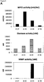
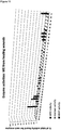
- Myeloperoxidase, Matrix-Metalloproteinase and Elastase enzyme activities are found to be generally lower in healing skin wound exudates as compared to the non-healing wounds.
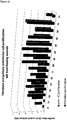
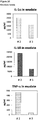
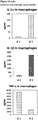
- IL-1 ⁇ , IL-1 ⁇ and TNF- ⁇ cytokine levels are elevated in general in non-healing skin as compared to healing skin wounds ( Figure 11 ).
- the individual values again vary strongly.
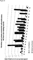
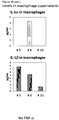
- cytokine levels are in the same range as in non-healing wound exudates for some of the healing wound exudates ( Figure 13 ), and only about 50% of the wound exudate samples from non-healing wounds induce cytokine secretion by macrophages ( Figure 15 ).
- Methods for reliably identifying non-healing skin wounds and/or for identifying that the healing of a skin wound worsens would provide an opportunity to further treat such skin wounds in time for the individual affected, thereby avoiding or attenuating recurrence and/or worsening of the healing status.
- pathogenic phagocytes release a variety of factors, including cytokines, proteases and toxic oxygen radicals into the wound tissue to destroy tissue cells, extracellular matrix and growth factors [ Clark RAF et al (2007) J Invest Dermatol 127:1018-1029 ]. These pathogenic factors are contained in the wound itself, e.g. in the fibrin clot or in the wound exudate (wound fluid).
- the wound fluid called wound exudate (WE) is the extracellular fluid containing a molecular fingerprint of wound cells and can be referred to as a "liquid biopsy”.
- the methods developed and established are particularly suitable for the identifying a skin wound in an individual as healing or non-healing skin wound, and/or for monitoring the healing of a skin wound in an individual and/or for evaluating the efficacy of known and unknown compounds, and/or for compound screening, in the context of skin would healing.
- the present invention relates to an in vitro method for identifying a skin wound in an individual as being a non-healing skin wound or healing skin wound, the method comprising:
- one skin wound may be a healing skin wound
- another skin wound of the same individual at the same or a different time point may be a non-healing skin wound, e.g. due to infections or an underlying disease affecting a specific skin wound.
- the present invention relates to an in vitro method which allows for identifying a skin wound in an individual as being a non-healing skin wound or healing skin wound. Therefore, the present invention allows, for example, for assessing specifically different skin wounds of the same individual.
- Identifying a skin wound of an individual as a non-healing skin wound allows for patient surveillance, monitoring of the wound healing and specific therapeutic interventions to stabilize, ameliorate and/or improve the healing of the skin wound.
- the skin wound identified as non-healing skin wound using a method of the invention may be treated with one or more of the following: compression, wound dressings, surgical debridement, biological debridement, infection control, antibiotic therapy, negative pressure therapy, proteins, in particular growth factors, antibodies, peptides, sugars, cells or cell constituents, artificial skin, human blood-derived products, gene therapy or genetically engineered wound bed modifications, drugs, herbal medicines, plant extracts.
- a wound is understood as damage to a tissue of a living individual, such as cuts, tears, burns, or breaks, preferably a wound is understood as open injury of a tissue of a living individual.
- a skin wound is understood as a damage to a skin of a living individual, such as cuts, tears, burns, or breaks.
- a skin wound is understood as open injury of the skin of a living individual.
- the skin may be located at any area of an individual, such as for example the head, the arms, the legs, the chest, or the back. Further, the individual may have one, two, three, four or more skin wounds. Further, the area of a skin wound may differ. In a preferred embodiment, the skin wound forms wound exudate.
- the skin wound may for example be selected from a wound of a diabetic patient, a wound which is infected by at least one microorganism, an ischemic wound, a wound in a patient suffering from deficient blood supply or venous stasis, an ulcer, such a diabetic ulcer, venous ulcer, arterial ulcer (e.g. ulcus cruris arteriosum), mixed ulcer, or pressure ulcer, a neuropathic wound, ulcus cruris, surgical wound, burn, dehiscence, neoplastic ulcer and rare ulcer.
- an individual's skin wound is not affected by a further disease mechanically preventing wound closure, such as calcinosis, where calcium crystals in the wound mechanically prevent wound closure, or exudative dermatitis.
- Ulcers can result in complete loss of the epidermis and often portions of the dermis and even subcutaneous fat.
- a non-healing skin wound refers to a skin wound which does not heal at an expected rate, in particular, as a skin wound which does not close within 2 months under standard therapy, preferably within 3 or more months under standard therapy.
- a non-healing skin wound is characterized by a lack of wound closure, an increase of the area and/or depth of the wound, necrosis and/or infections of the skin wound, and/or lack of granulation.
- a "healing skin wound” is understood as a skin wound which heals at an expected rate, in particular, as a skin wound which closes within 2 months under standard therapy.
- a healing skin wound is characterized by ongoing wound closure, granulation, absence of necrosis and/or absence of infections.
- Standard therapy is understood as a treatment recommended in general by physicians for skin wounds, in particular one or more selected from wound dressings, surgical and biological (maggot) debridement, infection control, negative pressure therapy, and therapy with a biological or cell treatment.
- the skin wound is untreated or treated with standard therapy or with one or more of the following: compression, wound dressings, surgical debridement, biological debridement, infection control, antibiotic therapy, negative pressure therapy, proteins, in particular growth factors, antibodies, peptides, sugars, cells or cell constituents, artificial skin, human blood-derived products, gene therapy or genetically engineered wound bed modifications, drugs, herbal medicines, plant extracts.
- the individual is an animal, preferably the individual is a vertebrate, in particular a mammal, more preferably a human.
- the individual may be an otherwise healthy individual or may exhibit further diseases and/or co-morbidities, and/or is treated with medication(s) for further diseases and/or co-morbidities.
- the skin wound of the individual may be untreated or treated with standard therapy or with one or more of the following: compression, wound dressings, surgical debridement, biological debridement, infection control, antibiotic therapy, negative pressure therapy, proteins, in particular growth factors, antibodies, peptides, sugars, cells or cell constituents, artificial skin, human blood-derived products, gene therapy or genetically engineered wound bed modifications, drugs, herbal medicines, plant extracts.
- step a) the method of the invention includes measuring
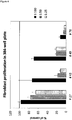
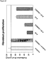
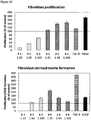
- Measuring the proliferation of primary fibroblast cells in the presence of a wound exudate sample obtained from said skin wound may be performed as shown in the examples, in particular in Example 3.1.1.
- primary fibroblast cells are used, which may be primary mammal dermal fibroblasts, preferably primary human dermal fibroblasts.
- Methods for obtaining cultured primary human dermal fibroblast cells are known in the art and are for example described in the examples.
- the cells may be cultured using DMEM medium containing FCS.
- the cells are incubated on a solid support, thereby allowing the cells to adhere to the support, as for example described in the Examples, where multiwell plates were used.
- the cells are contacted with the wound exudate sample, which is optionally diluted, e.g. diluted with medium or a saline aqueous liquid.
- the contacting may be performed before or after adherence of the cells occurs.
- the contacting may be achieved by adding the optionally diluted, liquid wound exudate sample, to the cells either prior to adherence, for example at the seeding of the cells, or after adherence.
- the contacting may be achieved e.g. by pipetting, and optionally gentle mixing.
- the cells are incubated for an appropriate time, such as for 6 hours to 300 hours, more preferably 12 hours to 200 hours, even more preferably 24 hours to 120 hours. In the examples, 72 hours were successfully used.
- a corresponding liquid without wound exudate such as medium or a saline aqueous liquid may be added or no liquid is added.
- the amount, preferably the cell number, including the formation of extracellular matrix, of the primary fibroblast cells is determined, such as by fixing cells and determining total protein content.
- the cells may for example be fixed using paraformaldehyde.
- a suitable dye, such as sulforhodamine B may be used for determining the amount, preferably the cell number, including the formation of extracellular matrix, of the primary fibroblast cells.
- the stained cells including the extracellular matrix formed may then be quantified e.g. by determining absorbance or fluorescence at a suitable wavelength, depending on the dye.
- the method is performed in 2D cell culture, which allows for culturing the cells adherently on a solid support.
- the method step includes the following steps:
- the culturing of cells in methods of the present invention is preferably performed at about 20°C to 40°C, more preferably 25°C to 38°C, even more preferably at about 37°C.
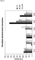
- Measuring the fibroblast-derived matrix formation by primary fibroblast cells in the presence of a wound exudate sample obtained from a skin wound may be performed as shown in the examples, in particular in Example 3.1.2.
- primary fibroblast cells are used, which may be primary mammal dermal fibroblasts, preferably primary human dermal fibroblasts.
- primary human dermal fibroblast cells are seeded on a support, which is preferably pre-coated with an adhesion enhancing agent, such as gelatin.
- the coating may be achieved by incubating the support with a solution or suspension containing the adhesion enhancing agent, such as gelatin. In the examples, a 0,2% gelatin solution was successfully used.
- the cells are cultured until confluence is reached. Subsequently, the cells are contacted with (i) a matrix promoting supplement, and (ii) the wound exudate sample, which is optionally diluted, wherein (i) and (ii) may be contacted simultaneously or sequentially.
- the matrix promoting supplement which is preferably selected from a solution comprising Vitamin C or a physiologically acceptable salt thereof, such the sodium salt, or 2-phospho-L-ascorbic acid or a physiologically acceptable salt thereof, and a combination of EGF and insulin, is added to the cells, e.g. by pipetting, and optionally gentle mixing.
- the wound exudate sample, which is optionally diluted may be contacted simultaneously or sequentially.
- the optionally diluted wound exudate sample may be mixed with the matrix promoting supplement, and the mixture may be added to the cells.
- the optionally diluted wound exudate sample may be added separately, but simultaneously, or separately, but subsequent to or prior to the matrix promoting supplement.
- the components (i) and (ii) are preferably contacted within 1 hour.
- the cells are subsequently incubated, preferably for 12 hours to 20 days, wherein the medium is optionally replaced at least one time with fresh medium supplemented with optionally diluted wound exudate and matrix promoting supplement. In the example, the medium was replaced once after 4 days of incubation, and the total incubation was 8 days.
- the solid support preferably contains at least one cavity which allows for filling of the space and therefore allows for a 3D cell culture.
- the amount of the fibroblast-derived matrix is determined, such as by fixing cells and determining total protein content.
- the cells may for example be fixed using paraformaldehyde.
- a suitable dye such as sulforhodamine B may be used for determining the amount, preferably the cell number, including the formation of extracellular matrix, of the primary fibroblast cells.
- the stained cells including the formation of extracellular matrix may then be quantified e.g. by determining absorbance or fluorescence at a suitable wavelength, depending on the dye.
- the method step preferably includes the following steps:
- fibroblast-derived matrix or “FDM” is understood as the extracellular matrix (ECM) formed by living fibroblast cells in an environment conducive for matrix formation, e.g. in the presence of a matrix promoting supplement.
- FDM is obtainable as described in the examples.
- FDM is obtainable by (i) seeding primary human dermal fibroblast cells on a support, which is pre-coated with an adhesion enhancing agent, such as gelatin, (ii) culturing the cells on the support, preferably until confluence is reached and (iii) contacting the cells with a matrix promoting supplement, such as Vitamin C or a physiologically acceptable salt thereof, or 2-phospho-L-ascorbic acid or a physiologically acceptable salt thereof, or a combination of EGF and insulin.
- an adhesion enhancing agent such as gelatin
- a "matrix promoting supplement” is understood as a compound or composition which promotes the formation of fibroblast-derived matrix by living fibroblast cells in an in vitro cell culture.
- Suitable matrix promoting supplements are Vitamin C or a physiologically acceptable salt thereof, such the sodium salt, or 2-phospho-L-ascorbic acid or a physiologically acceptable salt thereof, and a combination of EGF and insulin, as well as compositions comprising the compounds, such as solutions or suspensions.
- a combination of EGF and insulin may be provided to the cell culture separately, e.g. as separate solutions comprising EGF or insulin respectively, or together, e.g. as solution comprising EGF and insulin.
- an "adhesion enhancing agent” is an agent which enhances adhesion of cells to a solid support, such as a plastic support, but which does not substantially interfere with the viability of the cells.
- the adhesion enhancing agent is gelatin or fibronectin, more preferably gelatin.
- 2D cell culture is understood as a cell culture wherein the cells are cultured in a planar or substantially planar surface. In a preferred embodiment, the 2D cell culture is culturing of adherent cells.
- 3D cell culture is understood as a cell culture wherein the cells are cultured on a non-planar or substantially non-planar surface.
- the 3D cell culture is culturing of adherent cells and/or culturing of cells within a matrix, such as ECM, in particular FDM.
- a “support” or “solid support” is preferably selected from a chip, array, such as a microarray or nanoarray, a plate, such as a multiwell plate, or a dish.
- the solid support is preferably suitable for culturing cells, for example the support may be a plastic support.
- “Wound exudate” is understood as the extracellular fluid located within and above a skin wound. The wound exudate is also referred to a "liquid biopsy".
- wound exudate sample or "WE” is understood as a sample of wound exudate obtained from a skin wound of an individual.
- Methods for obtaining a wound exudate sample are known in the art.
- a wound exudate sample may be obtained by a physical or chemical method, in particular by applying negative pressure to the skin wound, such as by using a negative pressure drainage device, a method using capillary forces, collecting wound exudate in a film dressing or membrane, collecting wound exudate in a syringe, applying an absorptive material, such as absorptive beads, or a filter, or by using a swab, such as a cotton swab.
- the volume of wound exudate sample may vary and may be in the range of 1 nl to 1 l, 10 nl to 10 1 l, or 100 nl to 1 l, such as 1 ⁇ l to 1 l, 1 ml to 1 l or 10 ml to 1 l.
- wound exudate samples investigated in the examples had a volume of up to 400 ml and typically had a volume of 10 to 100 ml, in particular 10 to 50 ml.
- the wound exudate sample may be used the methods of the invention directly after obtaining the sample or may be stored, in particular stored at ⁇ 4°C, ⁇ 0°C or ⁇ 10°C before usage in the methods of the invention.
- the above assays relating to measuring the proliferation of primary fibroblast cells and the fibroblast-derived matrix formation by primary fibroblast cells can reliably identify skin wounds as healing skin wounds, or non-healing skin wounds, respectively. In particular, it is possible to reliably identify skin wounds as non-healing skin wounds. Therefore, one or both of these assays may be used for the method of the invention.
- the wound exudate of a healing wound has a dose-dependent positive effect on the proliferation of primary fibroblast cells and the fibroblast-derived matrix formation by primary fibroblast cells, as compared to a control in absence of wound exudate.
- value(s) obtained in the assay(s) which is/are equal to or above a control value established in the absence of wound exudate are indicative of a healing wound. Accordingly, value(s) obtained in the assay(s) which is/are below a control value established in the absence of wound exudate are indicative of a non-healing wound.
- control value(s) may be determined in parallel or may be established independently, preferably in parallel.
- the skin wound is identified as being a non-healing skin wound in case the value(s) obtained in i) and/or ii) is/are below a control value established in the absence of wound exudate, preferably at least 10% below the respective control value(s), more preferably at least 15%, even more preferably are at least 20% below the respective control value(s), or the skin wound is identified as being a healing skin wound in case the value(s) obtained in i) and/or ii) is/are equal to or above a control value established in the absence of wound exudate, at least 10% above the respective control value(s), more preferably at least 15%, even more preferably are at least 20% above the respective control value(s), more preferably wherein the value obtained in i) is at least 10%, more preferably at least 15%, above the respective control value and/or the value obtained in ii) is at least 50%, more preferably at least 100%, above the respective control value.
- the measurements at least in triplicate and/or to establish a statistical significance to improve the accuracy of the diagnosis.
- Statistical methods are known in the art. For example, the standard deviation may be determined. Further, the statistical significance may be p ⁇ 0,05, p ⁇ 0,001 or p ⁇ 0,001 for identifying a skin wound as a healing or non-healing skin wound.
- the value(s) in step a) is/are measured at least in triplicate and/or a statistical significance is established.
- a skin wound can be identified reliably as a non-healing skin wound in case the values obtained in both assays are below a control value established in the absence of wound exudate. Further, a skin wound can be identified reliably as a healing skin wound in case the values obtained in both assays are equal to or above a control value established in the absence of wound exudate.
- values in both assays which are each at least 10% above the respective control values, more preferably wherein the values obtained in the assay are each at least 15%, even more preferably are at least 20%, 30%, 40% or 50% above the respective control values are identifying the skin wound as healing skin wound.
- values in both assays which are each at least 10% below the respective control values, more preferably wherein the values obtained in the assay are each at least 15%, even more preferably are at least 20%, 30%, 40% or 50% below the respective control values are identifying the skin wound as non-healing skin wound.
- the method of the invention comprises
- the accuracy and reliability can be further increased by including a further assay in the method, which measures the proliferation of keratinocyte cells, such as primary keratinocyte cells or HaCaT cells. Further, it was found in the Examples that the use of HaCaT cells is to be preferred as compared to primary keratinocytes and allows for reliable prediction, in combination with the fibroblast-based assays described above.
- a skin wound can be identified reliably as a healing skin wound in case at least two, preferably all three of the values obtained with the assays measuring the proliferation of primary fibroblast cells, measuring the fibroblast-derived matrix formation by primary fibroblast cells and measuring the proliferation of keratinocyte cells are equal to or above the respective control values established in the absence of wound exudate, more preferably wherein the values obtained in the assays measuring the proliferation of primary fibroblast cells and/or the fibroblast-derived matrix formation by primary fibroblast cells, and the assay measuring the proliferation of keratinocyte cells are at least 10%, more preferably at least 15%, 20%, 30%, 40% or 50% above the respective control value.
- a skin wound can be identified reliably as a non-healing skin wound in case at least two, preferably all three of the values obtained with the assays measuring the proliferation of primary fibroblast cells, measuring the fibroblast-derived matrix formation by primary fibroblast cells and measuring the proliferation of keratinocyte cells are below the respective control values established in the absence of wound exudate, more preferably wherein the values obtained in the assays measuring the proliferation of primary fibroblast cells and/or the fibroblast-derived matrix formation by primary fibroblast cells, and the assay measuring the proliferation of keratinocyte cells are at least 10%, more preferably at least 15%, 20%, 30%, 40% or 50% below the respective control value.
- step a) further comprises the following step: iiia) measuring the proliferation of keratinocyte cells in the presence of a wound exudate sample obtained from said skin wound, and wherein step b) comprises: b) identifying the skin wound as being a non-healing skin wound in case at least two, preferably three of the values obtained in i) to iiia) are below the respective control values established in the absence of wound exudate, more preferably wherein the values obtained in i) and/or ii) and iiia) are at least 10%, more preferably at least 15%, below the respective control value, or identifying the skin wound as being a healing skin wound in case at least two, preferably three of the values obtained in i) to iii) are equal to or above the respective control values established in the absence of wound exudate, more preferably wherein the values obtained in i) and/or ii) and iiia) are at least 10%,
- the proliferation of keratinocyte cells is measured in the presence of a wound exudate sample obtained from said skin wound.
- the keratinocyte proliferation assay preferably includes culturing human primary keratinocyte cells, or HaCaT cells, which is a standard keratinocyte cell line, under standard conditions, such as by using DMEM containing FCS as medium, as for example described in the Examples.
- the cells are subsequently incubated on a solid support, thereby allowing the cells to adhere to the support. Further, the cells are contacted with the wound exudate sample, which is optionally diluted, wherein the contacting may be performed before or after adherence of the cells occurs.
- the optionally diluted wound exudate sample may be added to the adherent cells, for example by pipetting or otherwise adding the liquid, or the optionally diluted wound exudate sample may be added to the non-adherent cells, for example by pipetting or otherwise adding the liquid to the cells, followed by allowing the keratinocyte cells to adhere.
- the cells are subsequently incubated, preferably for 6 hours to 200 hours, preferably 24 hours to 100 hours. In the examples, the cells are incubated for 72 hours.
- the amount, preferably the cell number, of the keratinocyte cells is determined, such as by fixing cells and determining total protein content.
- the cells may for example be fixed using paraformaldehyde.
- a suitable dye such as sulforhodamine B may be used for determining the amount, preferably the cell number, of the keratinocyte cells.
- the stained cells may then be quantified e.g. by determining absorbance or fluorescence at a suitable wavelength, depending on the dye.
- the method is performed in 2D cell culture, which allows for culturing the cells adherently on a solid support.
- a keratinocyte cell may be a primary keratinocyte cell or a keratinocyte cell line, in particular a human primary keratinocyte cell or a human keratinocyte cell line.
- the keratinocyte cells used in the present invention are selected from HaCaT cells and primary keratinocyte cells.
- HaCaT cells represent an established and widely used human keratinocyte cell line.
- the keratinocyte cells used in the present invention are HaCaT cells.
- measuring the proliferation of keratinocyte cells in the presence of a wound exudate sample obtained from a skin wound includes the following steps:
- the accuracy and reliability can be further increased by including one or more further assays in the method which determine macrophage M1 and M2 markers in the context of wound exudate.
- markers may be cell surface protein markers, protein markers in the supernatant of macrophages or marker mRNAs in macrophages.
- Macrophages are tissue-resident professional phagocytes and antigen-presenting cells (APC), which differentiate from circulating peripheral blood monocytes. Activated macrophages of different phenotypes are classified by skilled persons into M1-macrophages and M2 macrophages. M1-macrophages are activated macrophages which comprise immune effector cells with an acute inflammatory phenotype. These are highly aggressive against bacteria and produce large amounts of lymphokines. The M2-macrophages are alternatively activated and anti-inflammatory.
- APC antigen-presenting cells
- M2 marker is understood as a protein marker which is specific for M2 macrophages. Preferably, the marker is secreted by the macrophages.
- the M2 markers are known in the art and are selected from CCL22 and CCL18. The markers may determined by methods known in the art, e.g. by using an immunological assay, even more preferably by using an ELISA assay.
- M1 marker is understood as a protein marker which is specific for M1 macrophages. Preferably, the marker is secreted by the macrophages.
- the M1 markers are known in the art and are selected from CXCL10 and IL-23p19. The markers may determined by methods known in the art, e.g. by using an immunological assay, even more preferably by using an ELISA assay.
- M1 cell surface marker is understood as a protein marker which is expressed at the surface of macrophages, and which is specific for M1 macrophages.
- the M1 cell surface markers are known in the art and are selected from CD38, CD64 and CD197.
- the amount(s) and/or frequency distribution(s) of the cell surface markers may be determined by an immunological assay and/or a fluorescence assay, in particular by FACS analysis, whereby typically a frequency distribution is determined.
- M2 cell surface marker is understood as a protein marker which is expressed at the surface of macrophages, and which is specific for M2 macrophages.
- the M2 cell surface markers are known in the art and are selected from CD200 receptor (CD200R), CD206 and CD209.
- CD200R CD200 receptor
- CD206 CD206
- CD209 CD200 receptor
- the amount(s) and/or frequency distribution(s) of the cell surface markers may be determined by an immunological assay and/or a fluorescence assay, in particular by FACS analysis, whereby typically a frequency distribution is determined.
- M2 marker mRNA is understood as an mRNA which is expressed by macrophages, and which is specific for M2 macrophages.
- the M2 marker mRNAs are known in the art and are selected CD200 receptor (CD200R), CD206, CD209, CCL22 and CCL18.
- the marker mRNAs may be determined by methods known in the art. Preferably, the amount may be determined by contacting a probe which specifically binds to a marker mRNA, wherein the probe is optionally labelled, with the macrophage RNA under conditions which are conducive to hybridization, and detecting the hybridized probe. For example, the mRNA may be reversely transcribed into cDNA prior to detection.
- M1 marker mRNA is understood as an mRNA which is expressed by macrophages, and which is specific for M1 macrophages.
- the M1 marker mRNAs are known in the art and are selected from CD38, CD64, CD197, CXCL10 and IL-23p19.
- the amount may be determined by contacting a probe which specifically binds to a marker mRNA, wherein the probe is optionally labelled, with the macrophage RNA under conditions which are conducive to hybridization, and detecting the hybridized probe.
- the mRNA may be reversely transcribed into cDNA prior to detection.
- the ratio of M1/M2 markers is indicative of a healing or non-healing skin wound, in combination with one or more cellular assays described above relating to measuring the proliferation of primary fibroblast cells, measuring the fibroblast-derived matrix (FDM) formation by primary fibroblast cells and measuring the proliferation of keratinocyte cells.
- FDM fibroblast-derived matrix
- an elevated ratio of M1/M2 markers, M1/M2 cell surface markers or M1/M2 marker mRNAs is indicative of a non-healing skin wound
- a low ratio of M1/M2 markers, M1/M2 cell surface markers or M1/M2 marker mRNAs is indicative of a healing skin wound.
- step a) of the method of the invention further comprises one, two or three of the following steps iiib) to iiid):
- the frequency distribution may be determined by determining the %age of cells which are positive for a given marker within a population, which is the most commonly used readout in FACS analysis.
- the amount may be determined by determining the quantity of cell surface expression, as a surrogate for the number of labelled molecules on the cell surface per individual cell when using labelled binding agents for the markers, as for example measured by the mean fluorescence intensity.
- measuring the amount(s) of one or more M1 marker(s) and one or more M2 marker(s) in the supernatant of macrophages incubated with a wound exudate sample obtained from a skin wound includes the following steps:
- primary human monocyte cells may be co-cultured with human dermal fibroblast cells in 2D cell culture, or with fibroblast-derived matrices. Methods for generating fibroblast-derived matrices are described above, as well as in the examples. Subsequently, the cells are incubated until macrophage differentiation is reached. For example, CD163 can be used as a cell surface marker of macrophage differentiation. Further, the cells are contacted with a wound exudate sample, which is optionally diluted, for example by pipetting the sample to the cells, and optionally gentle mixing. Further, the cells are incubated, preferably for 1 hour 100 hours, e.g. 4 hours to 100 hours.
- measuring the amount(s) and/or frequency distribution(s) of one or more M1 cell surface marker(s) and one or more M2 cell surface marker(s) on macrophages incubated with a wound exudate sample obtained from a skin wound includes the following steps:
- primary human monocyte cells may be co-cultured with human dermal fibroblast cells in 2D cell culture, or with fibroblast-derived matrices. Methods for generating fibroblast-derived matrices are described above, as well as in the examples. Subsequently, the cells are incubated until macrophage differentiation is reached. For example, CD163 can be used as a cell surface marker of macrophage differentiation. Further, the cells are contacted with a wound exudate sample, which is optionally diluted, for example by pipetting the sample to the cells, and optionally gentle mixing. Further, the cells are incubated, preferably for 1 hour 100 hours, e.g. 4 hours to 100 hours.
- the amount(s) and/or frequency distribution(s) of one or more M1 marker(s) and one or more M2 marker(s) on the cell surface of macrophages is determined.
- the cells may be harvested and subjected to FACS analysis, gating on the monocyte/macrophage population. Geometric means of mean fluorescence intensities can be used to quantify surface marker expression.
- the one or more M1 cell surface markers are selected from CD38, CD64 and CD197, and/or the one or more M2 cell surface markers are selected from CD200 receptor (CD200R), CD206 and CD209, more preferably wherein the amount(s) and/or frequency distribution(s) of the cell surface markers are determined by an immunological assay and/or a fluorescence assay, in particular by FACS analysis.
- step (iv) comprises contacting the macrophages with binding agents, preferably antibodies, which specifically recognize one or more M1 surface marker(s) and one or more M2 surface marker(s), wherein the binding agents are optionally labelled, in particular labelled with a fluorescent label, and determining the amount of binding molecules bound to the macrophages, in particular by determining mean fluorescence intensity, thereby determining the amount(s) of the cell surface markers.
- binding agents preferably antibodies, which specifically recognize one or more M1 surface marker(s) and one or more M2 surface marker(s)
- the binding agents are optionally labelled, in particular labelled with a fluorescent label
- determining the amount of binding molecules bound to the macrophages in particular by determining mean fluorescence intensity, thereby determining the amount(s) of the cell surface markers.
- antibodies specifically recognizing the surface markers and which contain a fluorescent label may be used.
- step (iv) comprises contacting the macrophages with binding agents, preferably antibodies, which specifically recognize one or more M1 surface marker(s) and one or more M2 surface marker(s), wherein the binding agents are optionally labelled, in particular labelled with a fluorescent label, and determining the percentages of cells which are positive for the one or more M1 surface marker(s) and the one or more M2 surface marker(s), respectively, within a cell population, in particular wherein FACS analysis is performed, thereby determining the frequency distribution(s) of the cell surface markers.
- binding agents preferably antibodies, which specifically recognize one or more M1 surface marker(s) and one or more M2 surface marker(s), wherein the binding agents are optionally labelled, in particular labelled with a fluorescent label, and determining the percentages of cells which are positive for the one or more M1 surface marker(s) and the one or more M2 surface marker(s), respectively, within a cell population, in particular wherein FACS analysis is performed, thereby determining the frequency distribution(s) of the cell
- Determination of proteins as binding agents of a marker protein can be performed using any of a number of known methods for identifying and obtaining proteins that specifically interact with proteins or polypeptides, for example, a yeast two-hybrid screening system such as that described in U.S. Pat. No. 5,283,173 and U.S. Pat. No. 5,468,614 , or the equivalent.
- a binding agent which specifically recognizes a marker has preferably at least an affinity of 10 7 l/mol for its corresponding target molecule.
- the binding agent which specifically recognizes a marker preferably has an affinity of 10 8 l/mol or even more preferred of 10 9 l/mol for its target marker molecule.
- the term specific is used to indicate that other biomolecules present in the sample do not significantly bind to the binding agent which specifically recognizes the marker.
- the level of binding to a biomolecule other than the target marker molecule results in a binding affinity which is only 10% or less, more preferably only 5% or less of the affinity to the target marker molecule, respectively.
- a preferred specific binding agent will fulfill both the above minimum criteria for affinity as well as for specificity.
- a binding agent which specifically recognizes a marker preferably is an antibody reactive with the marker.
- the term antibody refers to a polyclonal antibody, a monoclonal antibody, antigen binding fragments of such antibodies, single chain antibodies as well as to genetic constructs comprising the binding domain of an antibody.
- antibody includes polyclonal antibodies, monoclonal antibodies, fragments thereof such as F(ab')2, and Fab fragments, as well as any naturally occurring or recombinantly produced binding partners, which are molecules that specifically bind to a marker protein. Any antibody fragment retaining the above criteria of a specific binding agent can be used. Antibodies are generated by state of the art procedures, e.g., as described in Tijssen ( Tijssen, P., Practice and theory of enzyme immunoassays, Elsevier Science Publishers B.V., Amsterdam (1990 ), the whole book, especially pages 43-78). In addition, the skilled person is well aware of methods based on immunosorbents that can be used for the specific isolation of antibodies. By these means the quality of polyclonal antibodies and hence their performance in immunoassays can be enhanced (Tijssen, P., supra , pages 108-115).
- polyclonal antibodies raised in e.g. goats may be used.
- polyclonal antibodies from different species e.g., rats, rabbits or guinea pigs, as well as monoclonal antibodies can be used. Since monoclonal antibodies can be produced in any amount required with constant properties, they represent ideal tools in development of an assay for clinical routine.
- the sample obtained from an individual is incubated with the binding agent that specifically recognizes the marker in question under conditions appropriate for formation of a binding agent marker-complex. Such conditions need not be specified, since the skilled artisan without any inventive effort can easily identify such appropriate incubation conditions.
- the amount of binding agent marker-complex is measured and used in the methods and uses of the invention. As the skilled artisan will appreciate there are numerous methods to measure the amount of the specific binding agent marker-complex all described in detail in relevant textbooks (cf., e.g., Tijssen P., supra, or Diamandis, E.P. and Christopoulos, T.K. (eds.), Immunoassay, Academic Press, Boston (1996 )).
- monoclonal antibodies to the marker(s) are used in a quantitative (amount or concentration of the marker(s) is determined) immunoassay.
- the marker may be detected in a sandwich type assay format.
- a first specific binding agent is used to capture the marker in question on the one side and a second specific binding agent (e.g. a second antibody), which is labeled to be directly or indirectly detectable, is used on the other side.
- the second specific binding agent may contain a detectable reporter moiety or label such as an enzyme, dye, radionuclide, luminescent group, fluorescent group or biotin, or the like. Any reporter moiety or label could be used with the methods disclosed herein so long as the signal of such is directly related or proportional to the quantity of binding agent remaining on the support after wash.
- the amount of the second binding agent that remains bound to the solid support is then determined using a method appropriate for the specific detectable reporter moiety or label.
- a method appropriate for the specific detectable reporter moiety or label For radioactive groups, scintillation counting or autoradiographic methods are generally appropriate.
- Antibody-enzyme conjugates can be prepared using a variety of coupling techniques (for review see, e.g., Scouten, W. H., Methods in Enzymology 135:30-65, 1987 ). Spectroscopic methods can be used to detect dyes (including, for example, colorimetric products of enzyme reactions), luminescent groups and fluorescent groups. Biotin can be detected using avidin or streptavidin, coupled to a different reporter group (commonly a radioactive or fluorescent group or an enzyme).
- Enzyme reporter groups can generally be detected by the addition of substrate (generally for a specific period of time), followed by spectroscopic, spectrophotometric or other analysis of the reaction products. Standards and standard additions can be used to determine the level of antigen in a sample, using well known techniques.
- Immunoassays for measuring marker proteins of the invention include for example ELISA, enzyme immunoassay (EIA) and electro-chemiluminescence immunoassay (ECLIA) for the quantitative determination of a marker protein described herein.
- EIA enzyme immunoassay
- ELIA electro-chemiluminescence immunoassay
- measuring the expression level(s) of one or more M1 marker mRNA(s) and one or more M2 marker mRNA(s) in macrophages incubated with a wound exudate sample obtained from a skin wound includes the following steps:
- the one or more M1 marker mRNA(s) are selected from CD38, CD64, CD197, CXCL10 and IL-23p19, and/or the one or more M2 marker mRNA(s) are selected from CD200 receptor (CD200R), CD206, CD209, CCL22 and CCL18, more preferably wherein the method comprises contacting a probe which specifically binds to a marker mRNA, wherein the probe is optionally labelled, with the macrophage RNA under conditions which are conducive to hybridization, and detecting the hybridized probe.
- primary human monocyte cells may be co-cultured with human dermal fibroblast cells in 2D cell culture, or with fibroblast-derived matrices. Methods for generating fibroblast-derived matrices are described above, as well as in the examples. Subsequently, the cells are incubated until macrophage differentiation is reached. For example, CD163 can be used as a cell surface marker of macrophage differentiation. Further, the cells are contacted with a wound exudate sample, which is optionally diluted, for example by pipetting the sample to the cells, and optionally gentle mixing. Further, the cells are incubated, preferably for 1 hour 100 hours, e.g. 4 hours to 100 hours.
- the expression level(s) of one or more M1 marker mRNA(s) and one or more M2 marker mRNA(s) in the macrophages is determined.
- the cells may be harvested and mRNA expression level(s) may be determined using suitable probes.
- the expression level of a housekeeping gene such as actin or GAPDH may be determined and the expression level(s) of M1 or M2 marker RNA(s) may be determined as expression level relative to a housekeeping gene.
- Sensitivity and specificity are statistical measures of the performance of a binary classification test, also known in statistics as classification function: Sensitivity (also called the true positive rate) measures the proportion of positives that are correctly identified as such (e.g., the percentage of healing skin wounds identified as healing skin wound, respectively).
- Specificity measures the proportion of negatives that are correctly identified as such (e.g., the percentage of healing skin wounds identified as being a non-healing skin wound).
- scanners may be set to trigger on low-risk items like belt buckles and keys (low specificity), in order to reduce the risk of missing objects that do pose a threat to the aircraft and those aboard (high sensitivity).
- This trade-off can be represented graphically as a receiver operating characteristic curve.
- a perfect predictor would be described as 100% sensitive (e.g., all non-healing skin wounds are identified as non-healing skin wounds) and 100% specific (e.g., all healing skin wounds are not identified as non-healing skin wounds); however, theoretically any predictor will possess a minimum error bound known as the Bayes error rate.
- the cut-off can be set in order to either increase sensitivity or specificity.
- a combined value may be calculated using the two or more values obtained in the assays.
- the combined value is compared to the combined value of the respective control, which has been obtained using the same mathematical procedure.
- the combined value is obtained by weighted calculation of the values. This means that one of the assays is given a higher weighting that the other.
- the weighting factors have been obtained by analyzing a reference population of skin wounds.
- identifying a skin wound as non-healing skin wound preferably requires that at least the value(s) obtained in the assays measuring the proliferation of primary fibroblast cells and/or measuring the fibroblast-derived matrix (FDM) formation by primary fibroblast cells is/are below the respective control value(s) established in the absence of wound exudate.
- identifying a skin wound as healing skin wound preferably requires that at least the value(s) obtained in the assays measuring the proliferation of primary fibroblast cells and/or measuring the fibroblast-derived matrix (FDM) formation by primary fibroblast cells is/are equal or above the respective control value(s) established in the absence of wound exudate.
- FDM fibroblast-derived matrix
- step b) comprises:
- the skin wound as identified as being a healing skin wound in case at least two, preferably three, four, five or six of (1) to (6) above for a healing skin wound are fulfilled, with the proviso that at least the value(s) obtained in i) and/or ii) is/are at least 10% above the respective control values, more preferably wherein the values obtained in i) and/or ii) is/are at least 15%, even more preferably at least 20%, 30%, 40% or 50% above the respective control values, even more preferably wherein the value obtained in i) is at least 15%, even more preferably at least 20%, 30%, 40% or 50% above the respective control value and the value obtained in ii) is at least 50%, more preferably at least 100%, even more preferably 110%, 120%, 130% or 140% above the respective control value.
- the skin wound as identified as being a non-healing skin wound in case at least two, preferably three, four, five or six of (1) to (6) above for a non-healing skin wound are fulfilled, with the proviso that at least the value(s) obtained in i) and/or ii) is/are at least 10% below the respective control values, more preferably wherein the values obtained in i) and/or ii) is/are at least 15%, even more preferably at least 20%, 30%, 40% or 50% below the respective control values.
- the cellular assays are performed as described above for the methods of identifying.
- the method of the invention allows for reliably monitoring and predicting the wound healing of skin wounds in a patient.
- the method of the invention correctly predicted the subsequent worsening of skin wound healing in a patient, at a time point where visual inspection concluded an improvement in wound healing.
- the present invention relates to an in vitro method for monitoring the healing of a skin wound in an individual, the method comprising:
- the second time point is understood to be later than the first time point.
- the first time point and the second time point are separated by at least 6 hours, in particular by between 6 hours and 12 months.
- the first time point and the second time point are separated by 12 hours to 3, 6 or 12 months, or 1 day, 1 week or 1 month to 12 months.
- the monitoring is surprisingly reliable for determining a skin wound at a second time point to exhibit improved healing as compared to the first time point when an increase in the value(s) for one or both of the fibroblast-based assays is determined at the later time point as compared to the earlier time point, in cases where the value obtained in one or both of the assays measuring the proliferation of primary fibroblast cells in the presence of wound exudate samples obtained from a skin wound, and measuring the fibroblast-derived matrix formation by primary fibroblast cells in the presence of a wound exudate sample is/are equal to or below a control value established in the absence of wound exudate at the first, earlier time point. Accordingly, the monitoring of an improved healing is particularly reliable in case the wound is a non-healing wound at the first, earlier time point, as determined by a method of identifying the present invention.
- the method for monitoring the healing of a skin wound in an individual of the invention includes identifying the skin wound as being a non-healing skin wound or healing skin wound, respectively, at the respective time point, by performing the methods of the invention.
- the monitoring is surprisingly reliable for determining a skin wound at a second time point to exhibit worsened healing when a decrease in the value(s) for the one or both of the fibroblast-based assays is determined at the later time point as compared to the earlier time point, in case the value obtained in one or both of the assays measuring the proliferation of primary fibroblast cells in the presence of wound exudate samples obtained from a skin wound, and measuring the fibroblast-derived matrix formation by primary fibroblast cells in the presence of a wound exudate sample at the second later time point is/are equal to or below 100% of a control value established in the absence of wound exudate at the first, earlier time point. Accordingly, the monitoring of a worsened healing is particularly reliable in case the wound is a non-healing wound at the second, later time point, as determined by a method for identifying of the present invention.
- the present invention relates to an in vitro method for monitoring the healing of a skin wound in an individual, the method comprising:
- the method of the invention comprises:
- steps a) to e) may be repeated at two, three, four, five, six, seven, eight, nine, 10 or more later time points.
- a skin wound at a second time point is identified to exhibit improved healing in case the value obtained in c)i) at said second time point is at least at least 10%, more preferably 15%, even more preferably 20%, 25%, 30%, 40% or 50% higher than the value obtained in a)i) at said first time point, and the value obtained in c)ii) at said second time point is at least 10%, more preferably 15%, even more preferably 20%, 25%, 30%, 40% or 50% higher than the value obtained in a)ii) at said first time point, with the proviso that the values obtained in a)i) at said first time point and a)ii) at said first time point are equal to or below a respective control value established in the absence of wound exudate, preferably wherein the value(s) at said first time point is/are at least 10% below the respective control value, more preferably at least 15%, even more preferably at least 20%, 30%, 40% or 50% below the respective control value established in the absence of wound exudate at said first time point.
- a skin wound at a second time point is identified to exhibit worsened healing in case the value obtained in c)i) at said second time point is at least at least 10%, more preferably 15%, even more preferably 20%, 25%, 30%, 40% or 50% lower than the value obtained in a)i) at said first time point, and the value obtained in c)ii) at said second time point is at least 10%, more preferably 15%, even more preferably 20%, 25%, 30%, 40% or 50% lower than the value obtained in a)ii) at said first time point, with the proviso that the values obtained in c)i) and c)ii) at said second time point are equal to or below 100% of a respective control value established in the absence of wound exudate, preferably wherein the value(s) at said second time point is/are at least 10% below the respective control value, more preferably at least 15%, even more preferably at least 20%, 30%, 40% or 50% below the respective control value established in the absence of wound exudate at said second time point.
- the method of the present invention comprises:
- a skin wound at a second time point is identified to exhibit improved healing in case at least two, preferably three, four, five or six of (1) to (6) for improved healing are fulfilled, preferably wherein the value obtained in c)i) at said second time point is at least at least 10%, more preferably 15%, even more preferably 20%, 25%, 30%, 40% or 50% higher than the value obtained in a)i) at said first time point, and/or the value obtained in c)ii) at said second time point is at least 10%, more preferably 15%, even more preferably 20%, 25%, 30%, 40% or 50% higher than the value obtained in a)ii) at said first time point, with the proviso that the values obtained in a)i) at said first time point and/or a)ii) at said first time point are equal to or below a respective control value established in the absence of wound exudate, preferably wherein the value(s) at said first time point is/are at least 10% below the respective control value, more preferably at least 15%, even more
- a skin wound at a second time point is identified to exhibit improved healing in case at least two, preferably three, four, five or six of (1) to (6) for improved healing are fulfilled, preferably wherein the value obtained in c)i) at said second time point is at least at least 10%, more preferably 15%, even more preferably 20%, 25%, 30%, 40% or 50% higher than the value obtained in a)i) at said first time point, and the value obtained in c)ii) at said second time point is at least 10%, more preferably 15%, even more preferably 20%, 25%, 30%, 40% or 50% higher than the value obtained in a)ii) at said first time point, with the proviso that the values obtained in a)i) at said first time point and a)ii) at said first time point are equal to or below a respective control value established in the absence of wound exudate, preferably wherein the values at said first time point are at least 10% below the respective control value, more preferably at least 15%, even more preferably at least 20%, 30%,
- a skin wound at a second time point is identified to exhibit worsened healing in case healing in case at least two, preferably three, four, five or six of (1) to (6) for worsened wound healing are fulfilled, preferably wherein the value obtained in c)i) at said second time point is at least at least 10%, more preferably 15%, even more preferably 20%, 25%, 30%, 40% or 50% lower than the value obtained in a)i) at said first time point, and/or the value obtained in c)ii) at said second time point is at least 10%, more preferably 15%, even more preferably 20%, 25%, 30%, 40% or 50% lower than the value obtained in a)ii) at said first time point, with the proviso that the values obtained in c)i) and/or c)ii) at said second time point are equal to or below 100% of a respective control value established in the absence of wound exudate, preferably wherein the value(s) at said second time point is/are at least 10% below a respective control value, more
- a skin wound at a second time point is identified to exhibit worsened healing in case healing in case at least two, preferably three, four, five or six of (1) to (6) for worsened wound healing are fulfilled, preferably wherein the value obtained in c)i) at said second time point is at least at least 10%, more preferably 15%, even more preferably 20%, 25%, 30%, 40% or 50% lower than the value obtained in a)i) at said first time point, and the value obtained in c)ii) at said second time point is at least 10%, more preferably 15%, even more preferably 20%, 25%, 30%, 40% or 50% lower than the value obtained in a)ii) at said first time point, with the proviso that the values obtained in c)i) and c)ii) at said second time point are equal to or below 100% of a respective control value established in the absence of wound exudate, preferably wherein the values at said second time point are at least 10% below a respective control value, more preferably at least 15%, even more
- the method steps a)i), a)ii) and a)iii) at said first time point, and c)i), c)ii) and c)iii) at said second time point are performed.
- a skin wound at a second time point is identified to exhibit improved healing in case at least two, preferably three of (1) to (3) for improved healing are fulfilled, preferably wherein the value obtained in c)i) at said second time point is at least at least 10%, more preferably 15%, even more preferably 20%, 25%, 30%, 40% or 50% higher than the value obtained in a)i) at said first time point, and/or the value obtained in c)ii) at said second time point is at least 10%, more preferably 15%, even more preferably 20%, 25%, 30%, 40% or 50% higher than the value obtained in a)ii) at said first time point, and/or the value obtained in c)iiia) at said second time point is at least 10%, more preferably 15%, even more preferably 20%, 25%, 30%, 40% or 50% higher than the value obtained in a)iiia) at said first time point, with the proviso that the values obtained in a)i) at said first time point and/or a)ii) at said first time point
- a skin wound at a second time point is identified to exhibit improved healing in case at least two, preferably three of (1) to (3) for improved healing are fulfilled, preferably wherein the value obtained in c)i) at said second time point is at least at least 10%, more preferably 15%, even more preferably 20%, 25%, 30%, 40% or 50% higher than the value obtained in a)i) at said first time point, and the value obtained in c)ii) at said second time point is at least 10%, more preferably 15%, even more preferably 20%, 25%, 30%, 40% or 50% higher than the value obtained in a)ii) at said first time point, and/or the value obtained in c)iiia) at said second time point is at least 10%, more preferably 15%, even more preferably 20%, 25%, 30%, 40% or 50% higher than the value obtained in a)iia) at said first time point with the proviso that the values obtained in a)i) at said first time point and a)ii) at said first time point and/or
- a skin wound at a second time point is identified to exhibit worsened healing in case healing in case at least two, preferably three of (1) to (6) for worsened wound healing are fulfilled, preferably wherein the value obtained in c)i) at said second time point is at least at least 10%, more preferably 15%, even more preferably 20%, 25%, 30%, 40% or 50% lower than the value obtained in a)i) at said first time point, and/or the value obtained in c)ii) at said second time point is at least 10%, more preferably 15%, even more preferably 20%, 25%, 30%, 40% or 50% lower than the value obtained in a)ii) at said first time point, and/or the value obtained in c)iiia) at said second time point is at least 10%, more preferably 15%, even more preferably 20%, 25%, 30%, 40% or 50% lower than the value obtained in a)iia) at said first time point, with the proviso that the values obtained in c)i) and/or c)i
- a skin wound at a second time point is identified to exhibit worsened healing in case healing in case at least two, preferably three of (1) to (3) for worsened wound healing are fulfilled, preferably wherein the value obtained in c)i) at said second time point is at least at least 10%, more preferably 15%, even more preferably 20%, 25%, 30%, 40% or 50% lower than the value obtained in a)i) at said first time point, and the value obtained in c)ii) at said second time point is at least 10%, more preferably 15%, even more preferably 20%, 25%, 30%, 40% or 50% lower than the value obtained in a)ii) at said first time point, and/or the value obtained in c)iiia) at said second time point is at least 10%, more preferably 15%, even more preferably 20%, 25%, 30%, 40% or 50% lower than the value obtained in a)iia) at said first time point, with the proviso that the values obtained in c)i) and c)ii) and/
- the individual is a mammal, more preferably a human.
- a skin wound is selected from a wound of a diabetic patient, a wound which is infected by at least one microorganism, an ischemic wound, a wound in a patient suffering from deficient blood supply or venous stasis, an ulcer, such a diabetic ulcer, venous ulcer, arterial ulcer (e.g. ulcus cruris arteriosum), mixed ulcer, or pressure ulcer, a neuropathic wound, ulcus cruris, surgical wound, burn, dehiscence, neoplastic ulcer and rare ulcer.
- a wound of a diabetic patient a wound which is infected by at least one microorganism, an ischemic wound, a wound in a patient suffering from deficient blood supply or venous stasis, an ulcer, such a diabetic ulcer, venous ulcer, arterial ulcer (e.g. ulcus cruris arteriosum), mixed ulcer, or pressure ulcer, a neuropathic wound, ulcus cruris, surgical wound, burn, de
- a non-healing skin wound is understood as a wound which does not close within 2 months under standard therapy.
- the individual exhibits further diseases and/or co-morbidities, and/or is treated with medication(s) for further diseases and/or co-morbidities.
- such individuals may have a risk for developing chronic skin wounds and/or non-healing skin wounds and/or to exhibit worsened wound healing.
- skin wounds can in particular benefit from the methods of the present invention.
- the skin wound is untreated or treated with one or more of the following: compression, wound dressings, surgical debridement, biological debridement, infection control, antibiotic therapy, negative pressure therapy, proteins, in particular growth factors, antibodies, peptides, sugars, cells or cell constituents, artificial skin, human blood-derived products, gene therapy or genetically engineered wound bed modifications, drugs, herbal medicines, plant extracts.
- the individual is identified to be treated with one or more of the therapies selected from compression, wound dressings, surgical debridement, biological debridement, infection control, antibiotic therapy, negative pressure therapy, proteins, in particular growth factors, antibodies, peptides, sugars, cells or cell constituents, artificial skin, human blood-derived products, gene therapy or genetically engineered wound bed modifications, drugs, herbal medicines, plant extracts, in case (i) a skin wound of the individual is identified as being a non-healing skin wound by a method for identifying a skin wound in an individual as being a non-healing skin wound or healing skin wound of the present invention, and/or a skin wound of the individual is identified to exhibit worsened healing at a second time point as compared to a first time point by a method for monitoring skin wound healing of the present invention.
- the therapies selected from compression, wound dressings, surgical debridement, biological debridement, infection control, antibiotic therapy, negative pressure therapy, proteins, in particular growth factors, antibodies, peptides, sugars,
- the present invention relates to a method of treating a non-healing skin wound in an individual, comprising administering to an individual one or more of the therapies selected from compression, wound dressings, surgical debridement, biological debridement, infection control, antibiotic therapy, negative pressure therapy, proteins, in particular growth factors, antibodies, peptides, sugars, cells or cell constituents, artificial skin, human blood-derived products, gene therapy or genetically engineered wound bed modifications, drugs, herbal medicines, plant extracts, wherein the individual is identified to have at least one non-healing skin wound by performing a method of the present invention.
- the therapies selected from compression, wound dressings, surgical debridement, biological debridement, infection control, antibiotic therapy, negative pressure therapy, proteins, in particular growth factors, antibodies, peptides, sugars, cells or cell constituents, artificial skin, human blood-derived products, gene therapy or genetically engineered wound bed modifications, drugs, herbal medicines, plant extracts, wherein the individual is identified to have at least one non-healing skin wound by performing
- the present invention relates to a method of treating a skin wound in an individual which exhibits worsened healing, comprising administering to an individual one or more of the therapies selected from compression, wound dressings, surgical debridement, biological debridement, infection control, antibiotic therapy, negative pressure therapy, proteins, in particular growth factors, antibodies, peptides, sugars, cells or cell constituents, artificial skin, human blood-derived products, gene therapy or genetically engineered wound bed modifications, drugs, herbal medicines, plant extracts, wherein the individual is identified to have at least one skin wound which exhibits worsened wound healing by performing a method of the present invention.
- the therapies selected from compression, wound dressings, surgical debridement, biological debridement, infection control, antibiotic therapy, negative pressure therapy, proteins, in particular growth factors, antibodies, peptides, sugars, cells or cell constituents, artificial skin, human blood-derived products, gene therapy or genetically engineered wound bed modifications, drugs, herbal medicines, plant extracts, wherein the individual is identified to have at least one skin wound which
- a therapeutically effective amount is administered to the individual.
- at least one active agent such as antibiotic therapy, proteins, in particular growth factors, antibodies, peptides, sugars, cells or cell constituents, artificial skin, human blood-derived products, gene therapy or genetically engineered wound bed modifications, drugs, herbal medicines, and plant extracts
- a therapeutically effective amount is administered to the individual.
- at least one active agent is comprised in a pharmaceutical composition which may further comprise at least one pharmaceutically acceptable carrier or excipient.
- the therapeutically effective amount depends on the nature of the agent and is known to a skilled person. Further, at least one active agent may be administered once or repeatedly.
- the wound exudate sample is obtained by a physical or chemical method, in particular by applying negative pressure to the skin wound, in particular by using a negative pressure drainage device, a method using capillary forces, collecting wound exudate in a film dressing or membrane, collecting wound exudate in a syringe, applying an absorptive material, such as absorptive beads, or a filter, or by using a swab, such as a cotton swab.
- negative pressure was applied to the skin wound in order to obtain a wound exudate sample.
- a healing skin wound is characterized by ongoing wound closure, granulation, absence of necrosis and/or absence of infections.
- a non-healing skin wound is characterized by a lack of wound closure, an increase of the area and/or depth of the wound, necrosis and/or infections of the skin wound, and/or lack of granulation.
- the fibroblast and/or monocyte cells used in the methods are human cells, preferably human cells and/or primary cells obtained from healthy human individuals, from patients with comorbidities associated with impaired wound healing, such as diabetes, and/or from the individual patients providing the wound exudates.
- the wound exudate sample is diluted between 1:2 to 1:1000, preferably between 1:10 and 1:200 or 1:20 to 1:100. For example, dilutions of 1:25 or 1:50, such as dilutions in medium were successfully used.
- the wound exudate sample is preferably diluted in an aqueous liquid, such as aqueous solution or suspension.
- the wound exudate sample is diluted in a cell medium, such as DMEM containing FCS.
- an aqueous saline solution such as an aqueous buffered saline solution may be used.
- the wound exudate sample is diluted in an aqueous liquid, which does not substantially interfere with the viability of the cells used in the assays of the methods of the invention.
- measuring the proliferation of primary fibroblast cells in the presence of a wound exudate sample obtained from a skin wound includes the following steps:
- measuring the fibroblast-derived matrix formation by primary fibroblast cells in the presence of a wound exudate sample obtained from a skin wound includes the following steps:
- measuring the proliferation of keratinocyte cells in the presence of a wound exudate sample obtained from a skin wound includes the following steps:
- measuring the amount(s) of one or more M1 marker(s) and one or more M2 marker(s) in the supernatant of macrophages incubated with a wound exudate sample obtained from a skin wound includes the following steps:
- measuring the amount(s) and/or frequency distribution(s) of one or more M1 cell surface marker(s) and one or more M2 cell surface marker(s) on macrophages incubated with a wound exudate sample obtained from a skin wound includes the following steps:
- measuring the expression level(s) of one or more M1 marker mRNA(s) and one or more M2 marker mRNA(s) in macrophages incubated with a wound exudate sample obtained from a skin wound includes the following steps:
- two or more assays performed in the methods of the present invention are performed simultaneously, even more preferably wherein the method steps are performed on a single support.
- a wound-on-a-chip test method is provided. Thereby, the demands of clinicians are met.
- the method steps of any method of the invention are performed on a single support, more preferably wherein the support is a chip, array, such as a microarray or nanoarray, a plate, such as a multiwell plate, or a dish.
- the solid support preferably contains at least one, preferably a plurality of defined areas or cavities, more preferably at least one, even more preferably a plurality of cavities which allow for filling of the space and therefore allow for a 3D cell culture.
- a multiwell plate or a microarray or nanoarray comprising a plurality of defined cavities may be used. In the examples, a multiwell plate was successfully used.
- the solid support does not substantially interfere with the viability of the cells and/or is suitable for culturing cells, for example the support may be a plastic support.
- the solid support preferably contains at least one cavity, such as a well, more preferably a plurality of defined cavities.
- the solid support preferably contains at least one cavity, such as a well, more preferably a plurality of defined cavities.
- multi-well plates may be used. Such multi-well plates comprise a plurality of defined cavities.
- the present invention relates to a kit comprising the agents for performing the method steps i) to iiid) described above, preferably wherein the kit comprises:
- M1 and M2 marker(s), cell surface marker(s) and/or marker mRNA(s) are described above for the methods of the present invention.
- the keratinocyte cells are selected from HaCaT cells and primary keratinocyte cells, in particular human primary keratinocyte cells.
- the keratinocyte cells used in the present invention are HaCaT cells.
- Fibroblast-derived matrix is obtainable by (i) seeding primary human dermal fibroblast cells on a support, which is pre-coated with an adhesion enhancing agent, such as gelatin, (ii) culturing the cells on the support, preferably until confluence is reached and (iii) contacting the cells with a matrix promoting supplement, such as Vitamin C or a physiologically acceptable salt thereof, or 2-phospho-L-ascorbic acid or a physiologically acceptable salt thereof, or a combination of EGF and insulin.
- FDM may be formed in situ or may be transferred to the support after formation.
- a wound-on-a-chip may be provided, which allows for diagnosing, monitoring and predicting aspects of skin wound healing, such as identifying a non-healing skin wound and predicting worsened healing of a skin wound.
- the present invention relates to a support suitable for performing a method of the present invention, wherein the support comprises a plurality of defined areas or cavities and wherein:
- the solid support preferably contains a plurality of defined cavities. Cavities allow for filling of the space and therefore allow for a 3D cell culture.
- a multiwell plate or a microarray or nanoarray comprising a plurality of defined cavities may be used. In the examples, a multiwell plate was successfully used.
- the solid support does not substantially interfere with the viability of the cells and/or is suitable for culturing cells, for example the support may be a plastic support.
- the solid support may contain a plurality of defined wells. For example, multi-well plates may be used.
- the support comprises 2, 3, 4, 5, 6, 7, 8, 9, 10 or more defined areas or cavities, such as 2, 3, 4, 5, 6, 7, 8, 9 or 10 to 10 5 , 2, 3, 4, 5, 6, 7, 8, 9 or 10 to 10 4 , 2, 3, 4, 5, 6, 7, 8, 9 or 10 to 10 3 , or 2, 3, 4, 5, 6, 7, 8, 9 or 10 to 10 2 defined areas or cavities.
- Macrophages, keratinocytes and fibroblasts are perceived as pivotal cells in sustained wound inflammation and resulting wound chronicity.
- WE from chronic wounds we established new test systems in these cell types that are suitable for compound screening. They form the basis for our investigations aimed at the identification of inhibitors of WE-induced cell activation for therapy of non-healing chronic ulcers ( Figure 2 ).
- Example 4.3 the method makes use of the assay steps described above for the further methods of the invention.
- the proliferation of primary fibroblast cells in the presence of (i) a wound exudate sample obtained from a skin wound of at least one individual, and (ii) at least one candidate compound is measured as primary assay.
- at least one of the further assays described herein are performed.
- the candidate compound is found to be active in the proliferation of primary fibroblast cells assay in case the value obtained in the assay is at least 10% above or at least 10% below a control value established in the absence of the at least one candidate compound, or is above a cut-off value.
- the present invention relates to a method for screening for compounds suitable for modulating skin wound healing, comprising the following steps:
- the method may be repeated one or more times.
- step B) includes performing one, two, three, four or five of said method steps B1) to B5) in case the value obtained in A) is above or below a cut-off value.
- the cut-off value may be the control value established in the absence of the candidate compound + X * standard deviation, wherein X is 0,5 or more, such as 1, 1.5, 2, 2.5 3, 4 or more, such as up to 5, 6, 7, 8, 9 or 10.
- X is 3.
- the cut-off value may be the control value established in the absence of the candidate compound - X * standard deviation, wherein X is 0,5 or more, such as 1, 1.5, 2, 2.5 3, 4 or more, such as up to 5, 6, 7, 8, 9 or 10.
- the compound is identified as being suitable for modulating skin wound healing, in case at least one value obtained in B1) to B5) above or 10% below a respective cut-off value.
- the wound exudate sample is diluted, in particular as described above.
- one, two, three, four or five of the method steps B1) to B5) are performed in case the value obtained in A) above is at least 15%, 20%, 25%, 30%, 40% or 50% above a control value established in the absence of the at least one candidate compound.
- the compound is identified as being suitable for modulating skin wound healing, in case at least one value obtained in B1) and B2) is at least 10%,15%, 20%, 25%, 30%, 40% or 50% above a control value established in the absence of the at least one candidate compound.
- modulating wound healing is improving wound healing.
- one, two, three, four or five of the method steps B1) to B5) are performed in case the value obtained in A) above is at least 15%, 20%, 25%, 30%, 40% or 50% below a control value established in the absence of the at least one candidate compound.
- the compound is identified as being suitable for modulating skin wound healing, in case at least one value obtained in B1) and B2) is at least 10%,15%, 20%, 25%, 30%, 40% or 50% below a control value established in the absence of the at least one candidate compound.
- modulating wound healing is worsening wound healing.
- Compounds identified as being suitable for worsening skin wound healing are suitable for treating and/or preventing hypertrophic scars, keloids or fibrosis.
- the (i) wound exudate sample obtained from a skin wound of at least one individual, and (ii) the at least one candidate compound may be added for incubation simultaneously, or sequentially.
- (i) and (ii) may be added as separate compositions, or as a single composition.
- the optionally diluted wound exudate sample may be added to an assay, e.g. by pipetting the liquid into a well of a support, and the candidate compound is added separately, e.g. by pipetting the liquid containing such compound into a well of a support, either before, simultaneous with, or after the optionally diluted wound exudate sample.
- the optionally diluted wound exudate sample may be mixed with the at least one candidate compound or composition comprising at least one candidate compound, and be added to the assay subsequently.
- the method steps include contacting of a wound exudate sample obtained from a skin wound of at least one individual.
- a wound exudate sample obtained from a skin wound of one individual may be used, or mixtures of wound exudate samples obtained from a skin wound of two or more individuals, such as 2, 3, 4, 5, 6, 7, 8, 9, 10 or more individuals may be used.
- the wound exudate samples may be samples from healing wounds or non-healing wounds, such as ulcers, or mixtures thereof.
- measuring the proliferation of primary fibroblast cells in the presence of (i) a wound exudate sample obtained from a skin wound of at least one individual, and (ii) at least one candidate compound includes the following steps:
- measuring the fibroblast-derived matrix formation by primary fibroblast cells in the presence of (i) a wound exudate sample obtained from a skin wound of at least one individual and (ii) said at least one candidate compound includes the following steps:
- measuring the proliferation of keratinocyte cells in the presence of (i) a wound exudate sample obtained from a skin wound of at least one individual and (ii) said at least one candidate compound includes the following steps:
- measuring the amount(s) of one or more M1 marker(s) and one or more M2 marker(s) in the supernatant of macrophages incubated with (i) a wound exudate sample obtained from a skin wound of at least one individual and (ii) said at least one candidate compound, wherein the macrophages are in co-culture with fibroblasts includes the following steps:
- measuring the amount(s) and/or frequency distribution(s) of one or more M1 cell surface marker(s) and one or more M2 cell surface marker(s) on macrophages incubated with (i) a wound exudate sample obtained from a skin wound of at least one individual and (ii) said at least one candidate compound, wherein the macrophages are in co-culture with fibroblasts includes the following steps:
- measuring the expression level(s) of one or more M1 marker mRNA(s) and one or more M2 marker mRNA(s) in macrophages incubated with (i) a wound exudate sample obtained from a skin wound of at least one individual and (ii) said at least one candidate compound, wherein the macrophages are in co-culture with fibroblasts includes the following steps:
- a wide variety of compounds may be used in the methods for screening.
- a small molecule, a hormone, sugar, protein, peptide, polymer, biological, such as a protein, a peptide, an antibody or derivative thereof, or a conjugate thereof, a nucleic acid, such a viral agent, a wound dressing which optionally comprises a therapeutically active agent, or one or more cell(s), such as one or more genetically modified cell may be used.
- a plurality of compounds may be tested in parallel. In case 2, 3, 4, 5, 6, 7, 8, 9, 10 or more candidate compounds are to be tested in parallel, such compounds may be added separately or as at least one composition comprising two or more candidate compounds. Thereby, a huge number of candidate compounds may be tested.
- compositions such as 1, 2, 3, 4, 5, 6, 7, 8, 9, 10 or more compositions, each comprising at least one, such as 1, 2, 3, 4, 5, 6, 7, 8, 9, 10 or more candidate compounds, may be added.
- the compounds or composition comprising compounds may be provided as a liquid, such as a solution, in particular aqueous solution or medium or suspension, such as a suspension in an aqueous solution or medium.
- the liquid, in particular solution or suspension, without candidate compound(s) preferably does not substantially interfere with the viability of cells used in the assay(s).
- the at least one compound is selected from a small molecule, a hormone, sugar, protein, peptide, polymer, biological, such as a protein, a peptide, an antibody or derivative thereof, or a conjugate thereof, a nucleic acid, such a viral agent, or one or more cell(s), such as one or more genetically modified cell.
- the at least one compound is selected from an immunomodulatory agent, more preferably an immunosuppressive agent, an antibiotic, an antiinfective, a growth factor, a cytokine, an antiproliferative agent and an agent stimulating proliferation.
- known approved active agents may be tested, such as active agents approved in the field of wound healing or for other indications, such as for inflammatory and/or proliferative disorders.
- novel compounds and/or compound libraries may be tested.
- combinatorial libraries or other small molecule compound libraries, or antibody libraries may be tested.
- the at least one compound is a single compound, or 2, 3, 4, 5, or more different compounds, wherein the 2, 3, 4, 5, or more different compounds may be present in a single composition or in 2 or more separate compositions.
- the values are measured at least in triplicate and/or a statistical significance is established in B), more preferably, wherein p ⁇ 0,05, p ⁇ 0,001 or p ⁇ 0,001, and/or the compound is identified as being suitable for modulating skin wound healing, in case at least one value obtained in B1) to B5) is at least 10% above, such as at least 10%,15%, 20%, 25%, 30%, 40% or 50% above, or at least 10% below, such as at least 10%,15%, 20%, 25%, 30%, 40% or 50% below a control value established in the absence of the candidate compound with statistical significance, more preferably, wherein p ⁇ 0,05, p ⁇ 0,001 or p ⁇ 0,001.
- a compound that is identified to be suitable for modulating, in particular improving skin wound healing can be tested in in vitro, ex vivo and/or in vivo models for efficacy and safety in the context of wound healing.
- a compound that is identified to be suitable for modulating, in particular worsening skin wound healing can be tested in in vitro, ex vivo and/or in vivo models for efficacy and safety in the context of hypertrophic scars, keloids and fibrosis.
- a keloid is a type of scar of the skin which, depending on its maturity, is composed mainly of either type III or type I collagen. It is a result of an overgrowth of granulation tissue at the site of a healed skin injury which is then slowly replaced by collagen type 1.
- a keloid scar is benign.
- Fibrosis is the formation of excess fibrous connective tissue in an organ or tissue in a reparative or reactive process.
- Wound exudate is the extracellular fluid containing a molecular fingerprint of wound cells and can be referred to as a "liquid biopsy”. Since removal of wound exudates (WE) can improve wound healing, we reasoned that factors contained within WE critically impede wound healing, e.g. through activation of innate immune cells.
- Macrophages, keratinocytes and fibroblasts are perceived as pivotal cells in sustained wound inflammation and resulting wound chronicity.
- WE from chronic wounds we established new test systems in these cell types that are suitable for compound screening, monitoring the healing of a skin wound and/or for identifying a skin wound in an individual as being a non-healing skin wound or healing skin wound.
- the assays form the basis for our investigations aimed at the identification of inhibitors of WE-induced cell activation for therapy of non-healing chronic ulcers ( Figure 2 ) as well as for the methods for monitoring the healing of a skin wound and/or for identifying a skin wound in an individual as being a non-healing skin wound or healing skin wound.
- HDF human dermal fibroblast proliferation assay: measuring the proliferation of primary fibroblast cells in the presence of a wound exudate sample obtained from a skin wound of an individual
- HDF Primary human dermal fibroblasts
- the cells were allowed to adhere overnight and then incubated for 72 hours at 37°C under the following conditions: graded compound concentrations or 20ng/ml PDGF-BB (Tonbo Biosciences # TO-21-8501-U010) in the absence or presence of different dilutions of sterile-filtered WE in medium. For negative control samples, 200 ⁇ l medium was added instead of specific stimuli. Alternatively, the cells were seeded into 384-well plates at 2500 cells/well together with test compounds or growth factors and WE or medium in a total volume of 50 ⁇ l.
- the cells were fixed with 4% paraformaldehyde (Morphisto) for 15 minutes at room temperature and washed 3 times with PBS.
- a control plate was fixed after the overnight adherence of the cells (day 1) to determine the starting cell number.
- Total cellular protein was determined as a measure of cell number by staining the fixed cells with sulforhodamine B (SRB, Sigma). A 0.4% SRB solution in 1% acetic acid was added to the wells for 30 minutes. The wells were then washed with 1% acetic acid until the wash solution remained colorless. After drying, the dye was eluted with 10mM Tris ⁇ HCl, pH8.5, and absorbance was measured either at 550 or 492nm for lower and higher cell densities, respectively. The average absorbance of the sample representing the day 1 starting cell number (for 96-well plates) was subtracted from the absorbance values of the WE-treated cells.
- SRB sulforhodamine B
- HDF primary human fibroblasts
- FDM fibroblast-derived matrices
- HDF cells were seeded at 5000 cells/well on day -3 into 96-well tissue culture plates (1250 cells/well for 384-well plates), which had been pre-coated for 1 hour at 37°C with 100 ⁇ l of a 0.2% gelatin solution (Sigma).
- a matrix promoting supplement vitamin C: 2-phospho-L-ascorbic acid trisodium salt, 100 ⁇ g/ml; Sigma
- test samples containing PDGF-BB, TGF- ⁇ 1 or graded concentrations of compounds - /+ WE as described for the HDF proliferation assay.
- medium was replaced by fresh vitamin C- and stimulus- as well as compound-containing medium, maintaining the conditions initiated on day 0.
- TGF- ⁇ 1 and PDGF-BB were included as positive controls to promote FDM formation and cell growth, respectively.
- FDM production was measured in fixed cultures via SRB staining and evaluated as described above.
- Keratinocvte proliferation assay measuring the proliferation of keratinocyte cells in the presence of a wound exudate sample obtained from a skin wound
- the HaCaT keratinocyte cell line was routinely cultured in DMEM containing 10% FCS, 2mM glutamine, and 100 U/ml penicillin/100 ⁇ g/ml streptomycin.
- the proliferation assay was carried out as described for HDF cells.
- Primary human keratinocytes were grown in KBM medium (Lonza CC-3104) containing 0.06mM calcium and supplemented with growth factors (Lonza CC-4131) on plastic coated with rat tail collagen (40 ⁇ g/ml; Gibco) or gelatin (0.2%; Sigma). No antibiotics were used.
- the proliferation assay was carried out as described for HDF cells.
- the primary human endothelial cells HMVEC-d- (Lonza, CC-2543) were cultured in EGM-2-MV BulletKit medium (Lonza CC-3156 & CC-4147).
- the proliferation assay was carried out as described for HDF cells.
- PBMC peripheral blood mononuclear cells
- the cell pellet was washed 3 times with PBS, resuspended in RPMI medium containing 20% FCS and 10% DMSO and frozen in liquid nitrogen.
- Monocytes were generated from frozen aliquots using positive selection with the CD14 Beads-Kit (Miltenyi) on an autoMACS-Sorter (Miltenyi) according to the manufacturer's instructions.
- monocytes were seeded at 4 x 10 6 monocytes/well in 6-well-plates (Nunc) and incubated with 20 ng/ml M-CSF (R&D Systems) in RPMI supplemented with 10% FCS, 2mM glutamine, and 100 U/ml penicillin / 100 ⁇ g/ml streptomycin in a total volume of 5ml per well. After 2 days, 2ml of the supernatant were removed and replaced by 2.5ml/well of fresh medium containing 20ng/ml M-CSF. On the third day, microscopic examination revealed differentiation into adherent, frequently elongated cells.
- M-CSF R&D Systems
- the macrophages were harvested with a rubber scraper, centrifuged at 1200 rpm for 5 minutes, resuspended in serum-free medium and seeded at 2 x 10 5 cells/well in 100 ⁇ l. After 1 hour at 37°C, 2-fold concentrated stimuli were added in a volume of 100 ⁇ l to give the indicated final concentrations.
- a combination of 100ng/ml LPS (Sigma) and 50ng/ml IFN- ⁇ (PeproTech) served as positive control for the induction of cytokine secretion.
- Graded concentrations of test compounds were prepared in the absence or presence of a 1:100 dilution of sterile-filtered WE. For negative control samples, 100 ⁇ l medium was added instead of specific stimuli.
- Human monocvte-dermal fibroblast co-cultures as in vitro models that reflect macrophage behavior in human skin measuring (a) the amount(s) of one or more M1 marker(s) and one or more M2 marker(s) in the supernatant of macrophages incubated with a wound exudate sample obtained from a skin wound, wherein the macrophages are in co-culture with fibroblasts, and (b) measuring the amount(s) and/or frequency distribution(s) of one or more M1 cell surface marker(s) and one or more M2 cell surface marker(s) on macrophages incubated with a wound exudate sample obtained from a skin wound, wherein the macrophages are in co-culture with fibroblasts, and (b) measuring the expression level(s) of one or more M1 marker mRNA(s) and one or more M2 marker mRNA(s) in macrophages incubated with a wound exudate sample obtained from a skin wound, wherein
- CD14 + monocytes isolated from PBMC of healthy donors by magnetic bead separation were incubated either alone or in the presence of primary human dermal fibroblasts (CellNTec) or fibroblast-derived matrices (FDM). FDM had been generated from primary human dermal fibroblasts by a 3-week incubation with the growth supplements vitamin C or insulin and EGF (vitamin C: 2-phospho-L-ascorbic acid trisodium salt, 100 ⁇ g/ml; human EGF, 5ng/ml; human insulin, 5 ⁇ g/ml).
- EGF vitamin C: 2-phospho-L-ascorbic acid trisodium salt, 100 ⁇ g/ml; human EGF, 5ng/ml; human insulin, 5 ⁇ g/ml).
- the cultures were stimulated overnight with IFN-g (50ng/ml), LPS (100ng/ml) and IL-4 (25ng/ml) or combinations thereof as controls for M1 and M2 macrophage induction.
- Specific mRNA levels are determined as ratios compared to a housekeeping gene; the values obtained are "expression relative to housekeeping gene".
- Wound exudates were diluted in phosphate buffered saline (PBS) and subjected to a commercial protein assay, Pierce BCA Protein Assay Kit (Thermo Scientific #23225), using bovine serum albumin as a standard in the range of 20 - 2000 ⁇ g/ml.
- the assay was performed in 96-well ELISA microplates (Greiner # 655101) and measured on a TECAN Infinite M200 Pro microtiter plate reader at 562nm, using Magellan 7.2 software for evaluation.
- MPO Myeloperoxidase
- MPO activity in PBS-prediluted WE samples was determined by oxidation of 3,3',5,5'-Tetramethylbenzidine (TMB) in 96-well ELISA microplates. 10 ⁇ l of diluted WE samples were added to 40 ⁇ l of assay buffer (78mM NaH2PO4 ⁇ H2O, 1.67mM Na2HPO4, 0.5% HTAB pH 5.40) and then incubated with 40 ⁇ l 1X TMB ELISA Substrate Solution (eBioscience # 00-4201-56) until blue color developed (up to 30 minutes). MPO from human leukocytes (Sigma M6908-5UN) in assay buffer was used as standard at concentrations from 0.12 to 120mU/ml. The reaction was stopped by the addition of 45 ⁇ l 2N H2SO4, and the samples were measured on a TECAN Infinite M200 Pro microtiter plate reader at 450nm, using Magellan 7.2 software for evaluation.
- TMB 3,3',5,5'-Tetramethylbenzidine
- Elastase activity in WE was determined by measuring the fluorescence of the fluorogenic substrate MeOSuc-Ala-Ala-Pro-Val-AMC.
- 5 ⁇ l WE pre-dilutions were added to 20 ⁇ l assay buffer (50mM Tris pH7.4, 600mM NaCl, 0.05% CHAPS) in 384-well black polystyrene plates (Corning #3573).
- 25 ⁇ l of elastase substrate V Merck-Millipore/Calbiochem Cat# 324740
- the reaction was allowed to proceed for 1 hour.
- MMP Matrix metalloproteinase
- the activity of all matrix metalloproteinases was determined in WE by measuring the fluorescence of the fluorogenic substrate MCA-Lys-Pro-Leu-Gly-Leu-DPA-Ala-Arg-NH2.
- Active human MMP9 full length protein (Abcam #ab168863) in assay buffer was used as standard at concentrations from 0.31 to 20nM.
- IL-1 ⁇ in WE and in macrophage supernatants was determined in F96 Maxisorp Nunc Immuno plates (Nunc, #439454) using the hIL1-alpha DuoSet ELISA Kit from R&D Systems (# DY200) according to the manufacturer's instructions.
- the enzyme reaction was detected with TMB Solution (eBioscience) and stopped by the addition of 50 ⁇ l/well 2N H2SO4. Absorbance was read on a Tecan Infinite M200 Pro at 450nm.
- IL-1 ⁇ in WE and in macrophage supernatants was determined in F96 Maxisorp Nunc Immuno plates (Nunc, #439454) using the IL1-beta ELISA ReadySet-Go Kit from eBioscience (#88-7261-88) according to the manufacturer's instructions. Enzyme reaction and measurement were carried out as described for IL-1 ⁇ .
- TNF- ⁇ in WE and in macrophage supernatants was determined in F96 Maxisorp Nunc Immuno plates (Nunc, #439454) using the TNF-alpha ELISA ReadySet-Go Kit from eBioscience (# 88-7346-88) according to the manufacturer's instructions. Enzyme reaction and measurement were performed as described for IL-1 ⁇ .
- Table 1 Characteristics of the patients who provided the screening exudates WE number # 49 # 27 # 43 # 78 Wound Foot ulcer Ulcus cruris arteriosus "Gamaschen-Ulcus", diabetic patient Operation wound (after kidney transplantation), diabetic patient
- the methods of the present invention surprisingly allowed the prediction of whether a defined skin wound in a defined individual exhibits improved or worsened wound healing in the future.
- the assays are performed in the presence of wound exudate from at least one individual.
- a compound is identified as being suitable for modulating skin wound healing in case if the compound is further active in at least one of the five secondary assays.
- the method for screening for compounds suitable for modulating skin wound healing comprises the following steps:
Landscapes
- Health & Medical Sciences (AREA)
- Life Sciences & Earth Sciences (AREA)
- Engineering & Computer Science (AREA)
- Immunology (AREA)
- Biomedical Technology (AREA)
- Chemical & Material Sciences (AREA)
- Molecular Biology (AREA)
- Hematology (AREA)
- Urology & Nephrology (AREA)
- General Health & Medical Sciences (AREA)
- Pathology (AREA)
- Cell Biology (AREA)
- Analytical Chemistry (AREA)
- Physics & Mathematics (AREA)
- Microbiology (AREA)
- Biotechnology (AREA)
- Biochemistry (AREA)
- Medicinal Chemistry (AREA)
- Food Science & Technology (AREA)
- General Physics & Mathematics (AREA)
- Tropical Medicine & Parasitology (AREA)
- Bioinformatics & Cheminformatics (AREA)
- Toxicology (AREA)
- Medical Informatics (AREA)
- Public Health (AREA)
- Proteomics, Peptides & Aminoacids (AREA)
- Organic Chemistry (AREA)
- Epidemiology (AREA)
- Primary Health Care (AREA)
- Databases & Information Systems (AREA)
- Data Mining & Analysis (AREA)
- Physiology (AREA)
- Zoology (AREA)
- Wood Science & Technology (AREA)
- Genetics & Genomics (AREA)
- Biophysics (AREA)
- General Engineering & Computer Science (AREA)
- Measuring Or Testing Involving Enzymes Or Micro-Organisms (AREA)
- Investigating Or Analysing Biological Materials (AREA)
- Immobilizing And Processing Of Enzymes And Microorganisms (AREA)
Claims (15)
- Procédé in vitro pour identifier une plaie cutanée chez un individu comme étant une plaie cutanée ne cicatrisant pas ou une plaie cutanée en cours de cicatrisation, le procédé comprenant les étapes consistant à :a) mesureri) la prolifération de cellules fibroblastes primaires en présence d'un échantillon d'exsudat de plaie obtenu à partir de ladite plaie cutanée, et/ouii) la formation de matrice dérivée de fibroblastes par des cellules fibroblastes primaires en présence d'un échantillon d'exsudat de plaie obtenu à partir de ladite plaie cutanée, etb) identifier la plaie cutanée comme étant une plaie cutanée ne cicatrisant pas dans le cas où la ou les valeurs obtenues en i) et/ou ii) sont inférieures à une valeur témoin établie en l'absence d'exsudat de plaie, ou
identifier la plaie cutanée comme étant une plaie cutanée en cours de cicatrisation dans le cas où la ou les valeurs obtenues en i) et/ou ii) sont égales ou supérieures à une valeur témoin établie en l'absence d'exsudat de plaie,
de préférence dans lequel la ou les valeurs en a) sont mesurées au moins en triple et/ou une signification statistique est établie. - Procédé in vitro pour surveiller la cicatrisation d'une plaie cutanée chez un individu, le procédé comprenant les étapes consistant à :a) mesureri) la prolifération de cellules fibroblastes primaires en présence d'un échantillon d'exsudat de plaie obtenu à partir de ladite plaie cutanée à un premier point de temps, et/ouii) la formation de matrice dérivée de fibroblastes par des cellules fibroblastes primaires en présence d'un échantillon d'exsudat de plaie obtenu à partir de ladite plaie cutanée à un premier point de temps,b) éventuellement identifier la plaie cutanée comme étant une plaie cutanée ne cicatrisant pas audit premier point de temps dans le cas où la ou les valeurs obtenues en a)i) et/ou a)ii) sont inférieures à une valeur témoin établie en l'absence d'exsudat de plaie, ou identifier la plaie cutanée comme étant une plaie cutanée en cours de cicatrisation à un premier point de temps dans le cas où la ou les valeurs obtenues en a)i) et/ou a)ii) sont égales ou supérieures à une valeur témoin établie en l'absence d'exsudat de plaie,c) mesureri) la prolifération de cellules fibroblastes primaires en présence d'un échantillon d'exsudat de plaie obtenu à partir de ladite plaie cutanée à un deuxième point de temps, et/ouii) la formation de matrice dérivée de fibroblastes par des cellules fibroblastes primaires en présence d'un échantillon d'exsudat de plaie obtenu à partir de ladite plaie cutanée à un deuxième point de temps,d) éventuellement identifier la plaie cutanée comme étant une plaie cutanée ne cicatrisant pas audit deuxième point de temps dans le cas où la ou les valeurs obtenues en c)i) et/ou c)ii) sont inférieures à une valeur témoin établie en l'absence d'exsudat de plaie, ou identifier la plaie cutanée comme étant une plaie cutanée en cours de cicatrisation dudit deuxième point de temps dans le cas où la ou les valeurs obtenues en c)i) et/ou c)ii) sont égales ou supérieures à une valeur témoin établie en l'absence d'exsudat de plaie,e)A) identifier une plaie cutanée à un deuxième point de temps comme présentant une meilleure cicatrisation dans le cas où la valeur obtenue en c)i) audit deuxième point de temps est supérieure à la valeur obtenue en a)i) audit premier point de temps, et/ou la valeur obtenue en c)ii) audit deuxième point de temps est supérieure à la valeur obtenue en a)ii) audit deuxième point de temps, sous réserve que la ou les valeurs obtenues en a)i) audit premier point de temps et/ou en a)ii) audit premier point de temps soient égales ou inférieures à une valeur témoin établie en l'absence d'exsudat de plaie,
ouB) identifier une plaie cutanée à un deuxième point de temps comme présentant une cicatrisation dégradée dans le cas où la valeur obtenue en c)i) audit deuxième point de temps est inférieure à la valeur obtenue en a)i) audit premier point de temps, et/ou la valeur obtenue en c)ii) audit deuxième point de temps est inférieure à la valeur obtenue en a)ii) audit deuxième point de temps,et
sous réserve que la ou les valeurs obtenues en c)i) et/ou c)ii) audit deuxième point de temps soient égales ou inférieures à 100 % d'une valeur témoin établie en l'absence d'exsudat de plaie,f) éventuellement répéter les étapes a) à e) à un ou plusieurs points de temps ultérieurs,
en surveillant ainsi la cicatrisation de la plaie cutanée,
de préférence
dans lequel le premier point de temps et le deuxième point de temps sont séparés de 6 heures à 12 mois, et/ou
les valeurs sont mesurées au moins en triple et/ou une signification statistique est établie. - Procédé selon la revendication 1, où le procédé comprend les étapes consistant àa) mesureri) la prolifération de cellules fibroblastes primaires en présence d'un échantillon d'exsudat de plaie obtenu à partir de ladite plaie cutanée, et/ouii) la formation de matrice dérivée de fibroblastes par des cellules fibroblastes primaires en présence d'un échantillon d'exsudat de plaie obtenu à partir de ladite plaie cutanée, etb) identifier la plaie cutanée comme étant une plaie cutanée ne cicatrisant pas dans le cas où la valeur obtenue en a)i) est inférieure à une valeur témoin établie en l'absence d'exsudat de plaie, et la valeur obtenue en a)ii) est inférieure à une valeur témoin établie en l'absence d'exsudat de plaie, de préférence où les valeurs obtenues en a)i) et a)ii) sont inférieures d'au moins 10 % aux valeurs témoins respectives, plus préférablement où les valeurs obtenues en a)i) et a)ii) sont inférieures d'au moins 15 %, plus préférablement encore d'au moins 20 %, aux valeurs témoins respectives, ou
identifier la plaie cutanée comme étant une plaie cutanée en cours de cicatrisation dans le cas où la valeur obtenue en a)i) est égale ou supérieure à une valeur témoin établie en l'absence d'exsudat de plaie, et la valeur obtenue en a)ii) est égale ou supérieure à une valeur témoin établie en l'absence d'exsudat de plaie,
de préférence où les valeurs obtenues en a)i) et a)ii) sont supérieures d'au moins 10 %, plus préférablement d'au moins 15 %, plus préférablement encore d'au moins 20 %, aux valeurs témoins respectives,
de préférence dans lequel une valeur témoin est établie pour les valeurs obtenues en a)i) et a)ii) et/ou les valeurs en a) sont mesurées au moins en triple et/ou une signification statistique est établie. - Procédé selon la revendication 1 ou 3, dans lequell'étape a) comprend en outre l'étape suivante consistant à :
iiia) mesurer la prolifération de cellules HaCaT en présence d'un échantillon d'exsudat de plaie obtenu à partir de ladite plaie cutanée,et dans lequel l'étape b) consiste à :de préférence dans lequel une valeur combinée est établie pour les valeurs obtenues en i) et/ou ii) et iiia).
b) identifier la plaie cutanée comme étant une plaie cutanée ne cicatrisant pas dans le cas où au moins deux, de préférence trois des valeurs obtenues en i) à iiia) sont inférieures aux valeurs témoins respectives établies en l'absence d'exsudat de plaie, plus préférablement où les valeurs obtenues en i) et/ou ii) et iiia) sont inférieures d'au moins 10 %, plus préférablement d'au moins 15 %, à la valeur témoin respective, ou
identifier la plaie cutanée comme étant une plaie cutanée en cours de cicatrisation dans le cas où au moins deux, de préférence trois des valeurs obtenues en i) à iii) sont égales ou supérieures aux valeurs témoins respectives établies en l'absence d'exsudat de plaie, plus préférablement où les valeurs obtenues en i) et/ou ii) et iiia) sont supérieures d'au moins 10 %, plus préférablement d'au moins 15 %, à la valeur témoin respective, - Procédé selon la revendication 1, 3 ou 4, dans lequell'étape a) comprend en outre une, deux ou trois des étapes iiib) à iiid) suivantes, consistant à :iiib) mesurer la ou les quantités d'un ou plusieurs marqueurs M1 et d'un ou plusieurs marqueurs M2 dans le surnageant de macrophages incubés avec un échantillon d'exsudat de plaie obtenu à partir de ladite plaie cutanée, où les macrophages sont en co-culture avec des fibroblastes, où les un ou plusieurs marqueurs M1 sont choisis parmi CXCL10 et IL-23p19, et le ou les un ou plusieurs marqueurs M2 sont choisis parmi CCL22 et CCL18,iiic) mesurer la ou les quantités et/ou la ou les distributions de fréquence d'un ou plusieurs marqueurs de surface cellulaire M1 et d'un ou plusieurs marqueurs de surface cellulaire M2 sur des macrophages incubés avec un échantillon d'exsudat de plaie obtenu à partir de ladite plaie cutanée, où les macrophages sont en co-culture avec des fibroblastes, les un ou plusieurs marqueurs de surface cellulaire M1 sont choisis parmi CD38, CD64 et CD197, et les un ou plusieurs marqueurs de surface cellulaire M2 sont choisis parmi le récepteur de CD200, CD206 et CD209,iiid) mesurer le ou les niveaux d'expression d'un ou plusieurs ARNm de marqueur M1 et d'un ou plusieurs ARNm de marqueur M2 dans des macrophages incubés avec un échantillon d'exsudat de plaie obtenu à partir de ladite plaie cutanée, où les macrophages sont en co-culture avec des fibroblastes, où les un ou plusieurs ARNm de marqueur M1 sont choisis parmi CD38, CD64, CD197, CXCL10 et IL-23p19, et les un ou plusieurs ARNm de marqueur M2 sont choisis parmi le récepteur de CD200 (CD200R), CD206, CD209, CCL22 et CCL18,et dans lequel l'étape b) consiste à :de préférence dans lequel une valeur combinée est établie pour les valeurs obtenues en i), ii), iiia), iiib), iiic) et/ou iiid).
b) identifier la plaie cutanée comme étant une plaie cutanée ne cicatrisant pas dans le cas où au moins deux, de préférence trois, quatre, cinq ou six parmi (1) à (6) sont satisfaits :(1) la valeur obtenue en i) est inférieure à la valeur témoin respective établie en l'absence d'exsudat de plaie,(2) la valeur obtenue en ii) est inférieure à la valeur témoin respective établie en l'absence d'exsudat de plaie,(3) la valeur obtenue en iiia) est inférieure à la valeur témoin respective établie en l'absence d'exsudat de plaie,(4) le rapport de la ou des quantités d'un ou plusieurs marqueurs M1 à la ou aux quantités d'un ou plusieurs marqueurs M2 obtenues en iiib) est supérieur à une valeur de seuil,(5) le rapport de la ou des quantités et/ou de la ou des distributions de fréquence d'un ou plusieurs marqueurs de surface cellulaire M1 à la ou aux quantités et/ou à la ou aux distributions de fréquence d'un ou plusieurs marqueurs de surface cellulaire M2 obtenues en iiic) est supérieur à une valeur de seuil,(6) le rapport du ou des niveaux d'expression d'un ou plusieurs ARNm de marqueur M1 au ou aux niveaux d'expression d'un ou plusieurs ARNm de marqueurs M2 obtenus en iiid) est supérieur à une valeur de seuil, ouidentifier la plaie cutanée comme étant une plaie cutanée en cours de cicatrisation dans le cas où au moins deux, de préférence trois, quatre, cinq ou six parmi (1) à (6) sont satisfaits :(1) la valeur obtenue en i) est égale ou supérieure à la valeur témoin respective établie en l'absence d'exsudat de plaie,(2) la valeur obtenue en ii) est égale ou supérieure à la valeur témoin respective établie en l'absence d'exsudat de plaie,(3) la valeur obtenue en iiia) est égale ou supérieure à la valeur témoin respective établie en l'absence d'exsudat de plaie,(4) le rapport de la ou des quantités d'un ou plusieurs marqueurs M1 à la ou aux quantités d'un ou plusieurs marqueurs M2 obtenues en iiib) est inférieur à une valeur de seuil,(5) le rapport de la ou des quantités et/ou de la ou des distributions de fréquence d'un ou plusieurs marqueurs de surface cellulaire M1 à la ou aux quantités et/ou à la ou aux distributions de fréquence d'un ou plusieurs marqueurs de surface cellulaire M2 obtenues en iiic) est inférieur à une valeur de seuil,(6) le rapport du ou des niveaux d'expression d'un ou plusieurs ARNm de marqueur M1 au ou aux niveaux d'expression d'un ou plusieurs ARNm de marqueurs M2 obtenus en iiid) est inférieur à une valeur de seuil, - Procédé selon la revendication 4 ou 5,
dans lequel l'étape b) consiste à :
b) identifier la plaie cutanée comme étant une plaie cutanée ne cicatrisant pas dans le cas où au moins deux, de préférence trois, quatre, cinq ou six parmi (1) à (6) sont satisfaits :(1) la valeur obtenue en i) est inférieure à la valeur témoin respective établie en l'absence d'exsudat de plaie,(2) la valeur obtenue en ii) est inférieure à la valeur témoin respective établie en l'absence d'exsudat de plaie,(3) la valeur obtenue en iiia) est inférieure à la valeur témoin respective établie en l'absence d'exsudat de plaie,(4) le rapport de la ou des quantités d'un ou plusieurs marqueurs M1 à la ou aux quantités d'un ou plusieurs marqueurs M2 obtenues en iiib) est supérieur à une valeur de seuil, où les un ou plusieurs marqueurs M1 sont choisis parmi CXCL10 et IL-23p19, et les un ou plusieurs marqueurs M2 sont choisis parmi CCL22 et CCL18,(5) le rapport de la ou des quantités et/ou de la ou des distributions de fréquence d'un ou plusieurs marqueurs de surface cellulaire M1 à la ou aux quantités et/ou à la ou aux distributions de fréquence d'un ou plusieurs marqueurs de surface cellulaire M2 obtenues en iiic) est supérieur à une valeur de seuil, où les un ou plusieurs marqueurs de surface cellulaire M1 sont choisis parmi CD38, CD64 et CD197, et les un ou plusieurs marqueurs de surface cellulaire M2 sont choisis parmi le récepteur de CD200, CD206 et CD209,(6) le rapport du ou des niveaux d'expression d'un ou plusieurs ARNm de marqueur M1 au ou aux niveaux d'expression d'un ou plusieurs ARNm de marqueurs M2 obtenus en iiid) est supérieur à une valeur de seuil, où les un ou plusieurs ARNm de marqueur M1 sont choisis parmi CD38, CD64, CD197, CXCL10 et IL-23p19, et les un ou plusieurs ARNm de marqueur M2 sont choisis parmi le récepteur de CD200 (CD200R), CD206, CD209, CCL22 et CCL18,sous réserve qu'au moins la ou les valeurs obtenues en i) et/ou ii) soient inférieures à la ou aux valeurs témoins respectives établies en l'absence d'exsudat de plaie,
et/ou
identifier la plaie cutanée comme étant une plaie cutanée en cours de cicatrisation dans le cas où au moins deux, de préférence trois, quatre, cinq ou six parmi (1) à (6) sont satisfaits :(1) la valeur obtenue en i) est supérieure à la valeur témoin respective établie en l'absence d'exsudat de plaie,(2) la valeur obtenue en ii) est supérieure à la valeur témoin respective établie en l'absence d'exsudat de plaie,(3) la valeur obtenue en iiia) est supérieure à la valeur témoin respective établie en l'absence d'exsudat de plaie,(4) le rapport de la ou des quantités d'un ou plusieurs marqueurs M1 à la ou aux quantités d'un ou plusieurs marqueurs M2 obtenues en iiib) est inférieur à une valeur de seuil,(5) le rapport de la ou des quantités et/ou de la ou des distributions de fréquence d'un ou plusieurs marqueurs de surface cellulaire M1 à la ou aux quantités et/ou à la ou aux distributions de fréquence d'un ou plusieurs marqueurs de surface cellulaire M2 obtenues en iiic) est inférieur à une valeur de seuil,(6) le rapport du ou des niveaux d'expression d'un ou plusieurs ARNm de marqueur M1 au ou aux niveaux d'expression d'un ou plusieurs ARNm de marqueurs M2 obtenus en iiid) est inférieur à une valeur de seuil,sous réserve qu'au moins la ou les valeurs obtenues en i) et/ou ii) soient égales ou supérieures à la ou aux valeurs témoins respectives établies en l'absence d'exsudat de plaie. - Procédé selon la revendication 2, comprenant les étapes consistant à :a) mesureri) la prolifération de cellules fibroblastes primaires en présence d'un échantillon d'exsudat de plaie obtenu à partir de ladite plaie cutanée à un premier point de temps, etii) la formation de matrice dérivée de fibroblastes par des cellules fibroblastes primaires en présence d'un échantillon d'exsudat de plaie obtenu à partir de ladite plaie cutanée à un premier point de temps,b) éventuellement identifier la plaie cutanée comme étant une plaie cutanée ne cicatrisant pas audit premier point de temps dans le cas où la valeur obtenue en a)i) est inférieure à une valeur témoin établie en l'absence d'exsudat de plaie, et la valeur obtenue en a)ii) est inférieure à une valeur témoin établie en l'absence d'exsudat de plaie, ou
identifier la plaie cutanée comme étant une plaie cutanée en cours de cicatrisation audit premier point de temps dans le cas où la valeur obtenue en a)i) est égale ou supérieure à une valeur témoin établie en l'absence d'exsudat de plaie, et la valeur obtenue en a)ii) est égale ou supérieure à une valeur témoin établie en l'absence d'exsudat de plaie,c) mesureri) la prolifération de cellules fibroblastes primaires en présence d'un échantillon d'exsudat de plaie obtenu à partir de ladite plaie cutanée à un deuxième point de temps, etii) la formation de matrice dérivée de fibroblastes par des cellules fibroblastes primaires en présence d'un échantillon d'exsudat de plaie obtenu à partir de ladite plaie cutanée à un deuxième point de temps,d) éventuellement identifier la plaie cutanée comme étant une plaie cutanée ne cicatrisant pas audit deuxième point de temps dans le cas où la valeur obtenue en c)i) est inférieure à une valeur témoin établie en l'absence d'exsudat de plaie, et la valeur obtenue en c)ii) est inférieure à une valeur témoin établie en l'absence d'exsudat de plaie, ou
identifier la plaie cutanée comme étant une plaie cutanée en cours de cicatrisation audit deuxième point de temps dans le cas où la valeur obtenue en c)i) est égale ou supérieure à une valeur témoin établie en l'absence d'exsudat de plaie, et la valeur obtenue en c)ii) est égale ou supérieure à une valeur témoin établie en l'absence d'exsudat de plaie,e)A) identifier une plaie cutanée à un deuxième point de temps comme présentant une meilleure cicatrisation dans le cas où la valeur obtenue en c)i) audit deuxième point de temps est supérieure à la valeur obtenue en a)i) audit premier point de temps, et la valeur obtenue en c)ii) audit deuxième point de temps est supérieure à la valeur obtenue en a)ii) audit deuxième point de temps, sous réserve que les valeurs obtenues en a)i) audit premier point de temps et en a)ii) audit premier point de temps soient égales ou inférieures à une valeur témoin respective établie en l'absence d'exsudat de plaie,
ouB) identifier une plaie cutanée à un deuxième point de temps comme présentant une cicatrisation dégradée dans le cas où la valeur obtenue en c)i) audit deuxième point de temps est inférieure à la valeur obtenue en a)i) audit premier point de temps, et la valeur obtenue en c)ii) audit deuxième point de temps est inférieure à la valeur obtenue en a)ii) audit deuxième point de temps, sous réserve que les valeurs obtenues en c)i) et c)ii) audit deuxième point de temps soient égales ou inférieures à 100 % d'une valeur témoin respective établie en l'absence d'exsudat de plaie, etf) éventuellement répéter les étapes a) à e) à un ou plusieurs points de temps ultérieurs. - Procédé selon la revendication 2, comprenant les étapes consistant à :a) mesureri) la prolifération de cellules fibroblastes primaires en présence d'un échantillon d'exsudat de plaie obtenu à partir de ladite plaie cutanée à un premier point de temps, etii) la formation de matrice dérivée de fibroblastes par des cellules fibroblastes primaires en présence d'un échantillon d'exsudat de plaie obtenu à partir de ladite plaie cutanée à un premier point de temps, etun, deux, trois ou quatre parmi iiia), iiib), iiic) et iiid) :iiia) la prolifération de cellules HaCaT en présence d'un échantillon d'exsudat de plaie obtenu à partir de ladite plaie cutanée à un premier point de temps,iiib) la ou les quantités d'un ou plusieurs marqueurs M1 et d'un ou plusieurs marqueurs M2 dans le surnageant de macrophages incubés avec un échantillon d'exsudat de plaie obtenu à partir de ladite plaie cutanée à un premier point de temps, où les macrophages sont en co-culture avec des fibroblastes, où les un ou plusieurs marqueurs M1 sont choisis parmi CXCL10 et IL-23p19, et où les un ou plusieurs marqueurs M2 sont choisis parmi CCL22 et CCL18,iiic) la ou les quantités et/ou la ou les distributions de fréquence d'un ou plusieurs marqueurs de surface cellulaire M1 et d'un ou plusieurs marqueurs de surface cellulaire M2 sur des macrophages incubés avec un échantillon d'exsudat de plaie obtenu à partir de ladite plaie cutanée à un premier point de temps, où les macrophages sont en co-culture avec des fibroblastes, où les un ou plusieurs marqueurs de surface cellulaire M1 sont choisis parmi CD38, CD64 et CD197, et où les un ou plusieurs marqueurs de surface cellulaire M2 sont choisis parmi le récepteur de CD200, CD206 et CD209,iiid) le ou les niveaux d'expression d'un ou plusieurs ARNm de marqueur M1 et d'un ou plusieurs ARNm de marqueur M2 dans des macrophages incubés avec un échantillon d'exsudat de plaie obtenu à partir de ladite plaie cutanée à un premier point de temps, où les macrophages sont en co-culture avec des fibroblastes, où les un ou plusieurs ARNm de marqueur M1 sont choisis parmi CD38, CD64, CD197, CXCL10 et IL-23p19, et où les un ou plusieurs ARNm de marqueur M2 sont choisis parmi le récepteur de CD200 (CD200R), CD206, CD209, CCL22 et CCL18,b) éventuellement identifier la plaie cutanée comme étant une plaie cutanée ne cicatrisant pas audit premier point de temps ou comme étant une plaie cutanée en cours de cicatrisation à un premier point de temps conformément à la revendication 3, 4 ou 5,c) mesureri) la prolifération de cellules fibroblastes primaires en présence d'un échantillon d'exsudat de plaie obtenu à partir de ladite plaie cutanée à un deuxième point de temps, etii) la formation de matrice dérivée de fibroblastes par des cellules fibroblastes primaires en présence d'un échantillon d'exsudat de plaie obtenu à partir de ladite plaie cutanée à un deuxième point de temps,et un, deux, trois ou quatre parmi iiia), iiib), iiic) et iiid) :iiia) la prolifération de cellules HaCaT en présence d'un échantillon d'exsudat de plaie obtenu à partir de ladite plaie cutanée à un deuxième point de temps,iiib) la ou les quantités d'un ou plusieurs marqueurs M1 et d'un ou plusieurs marqueurs M2 dans le surnageant de macrophages incubés avec un échantillon d'exsudat de plaie obtenu à partir de ladite plaie cutanée à un deuxième point de temps, où les macrophages sont en co-culture avec des fibroblastes, où les un ou plusieurs marqueurs M1 sont choisis parmi CXCL10 et IL-23p19, et les un ou plusieurs marqueurs M2 sont choisis parmi CCL22 et CCL18,iiic) la ou les quantités et/ou la ou les distributions de fréquence d'un ou plusieurs marqueurs de surface cellulaire M1 et d'un ou plusieurs marqueurs de surface cellulaire M2 sur des macrophages incubés avec un échantillon d'exsudat de plaie obtenu à partir de ladite plaie cutanée à un deuxième point de temps, où les macrophages sont en co-culture avec des fibroblastes, où les un ou plusieurs marqueurs de surface cellulaire M1 sont choisis parmi CD38, CD64 et CD197, et où les un ou plusieurs marqueurs de surface cellulaire M2 sont choisis parmi le récepteur de CD200, CD206 et CD209,iiid) le ou les niveaux d'expression d'un ou plusieurs ARNm de marqueur M1 et d'un ou plusieurs ARNm de marqueur M2 dans des macrophages incubés avec un échantillon d'exsudat de plaie obtenu à partir de ladite plaie cutanée à un deuxième point de temps, où les macrophages sont en co-culture avec des fibroblastes, où les un ou plusieurs ARNm de marqueur M1 sont choisis parmi CD38, CD64, CD197, CXCL10 et IL-23p19, et où les un ou plusieurs ARNm de marqueur M2 sont choisis parmi le récepteur de CD200 (CD200R), CD206, CD209, CCL22 et CCL18,d) éventuellement identifier la plaie cutanée comme étant une plaie cutanée ne cicatrisant pas audit deuxième point de temps ou comme étant une plaie cutanée en cours de cicatrisation audit deuxième point de temps conformément à la revendication 3, 4 ou 5,e)A) identifier une plaie cutanée à un deuxième point de temps comme présentant une meilleure cicatrisation dans le cas où au moins deux, de préférence trois, quatre, cinq ou six de (1) à (6) sont satisfaits :(1) la valeur obtenue en c)i) audit deuxième point de temps est supérieure à la valeur obtenue en a)i) audit premier point de temps,(2) la valeur obtenue en c)ii) audit deuxième point de temps est supérieure à la valeur obtenue en a)ii) audit premier point de temps,(3) la valeur obtenue en c)iiia) audit deuxième point de temps est supérieure à la valeur obtenue en a)iiia) audit premier point de temps,(4) le rapport de la ou des quantités d'un ou plusieurs marqueurs M1 à la ou aux quantités d'un ou plusieurs marqueurs M2 obtenues en c)iiib) audit deuxième point de temps est inférieur au rapport de la ou des quantités d'un ou plusieurs marqueurs M1 à la ou aux quantités d'un ou plusieurs marqueurs M2 obtenues en a)iiib) audit premier point de temps, où les un ou plusieurs marqueurs M1 sont choisis parmi CXCL10 et IL-23p19, et les un ou plusieurs marqueurs M2 sont choisis parmi CCL22 et CCL18,(5) le rapport de la ou des quantités et/ou de la ou des distributions de fréquence d'un ou plusieurs marqueurs de surface cellulaire M1 à la ou aux quantités et/ou à la ou aux distributions de fréquence d'un ou plusieurs marqueurs de surface cellulaire M2 obtenues en c)iiic) audit deuxième point de temps est inférieur au rapport de la ou des quantités et/ou de la ou des distributions de fréquence d'un ou plusieurs marqueurs de surface cellulaire M1 à la ou aux quantités et/ou à la ou aux distributions de fréquence d'un ou plusieurs marqueurs de surface cellulaire M2 obtenues en a)iiic) audit premier point de temps, où les un ou plusieurs marqueurs de surface cellulaire M1 sont choisis parmi CD38, CD64 et CD197, et où les un ou plusieurs marqueurs de surface cellulaire M2 sont choisis parmi le récepteur de CD200, CD206 et CD209,(6) le rapport du ou des niveaux d'expression d'un ou plusieurs ARNm de marqueur M1 au ou aux niveaux d'expression d'un ou plusieurs ARNm de marqueurs M2 obtenus en c)iiid) audit deuxième point de temps est inférieur au rapport du ou des niveaux d'expression d'un ou plusieurs ARNm de marqueur M1 au ou aux niveaux d'expression d'un ou plusieurs ARNm de marqueurs M2 obtenus en a)iiid) audit premier point de temps, où les un ou plusieurs ARNm de marqueur M1 sont choisis parmi CD38, CD64, CD197, CXCL10 et IL-23p19, et les un ou plusieurs ARNm de marqueur M2 sont choisis parmi le récepteur de CD200 (CD200R), CD206, CD209, CCL22 et CCL18,sous réserve qu'au moins la ou les valeurs obtenues en c)i) et/ou c)ii) audit deuxième point de temps soient supérieures à la ou aux valeurs obtenues en a)i) et/ou a)ii) audit premier point de temps, et
sous réserve que la ou les valeurs obtenues en a)i) audit premier point de temps et/ou en a)ii) audit premier point de temps soient égales ou inférieures à une valeur témoin établie en l'absence d'exsudat de plaie,
ouB) identifier une plaie cutanée à un deuxième point de temps comme présentant une cicatrisation dégradée dans le cas où au moins deux, de préférence trois, quatre, cinq ou six de (1) à (6) sont satisfaits :et(1) la valeur obtenue en c)i) audit deuxième point de temps est inférieure à la valeur obtenue en a)i) audit premier point de temps,(2) la valeur obtenue en c)ii) audit deuxième point de temps est inférieure à la valeur obtenue en a)ii) audit premier point de temps,(3) la valeur obtenue en c)iiia) audit deuxième point de temps est inférieure à la valeur obtenue en a)iiia) audit premier point de temps,(4) le rapport de la ou des quantités d'un ou plusieurs marqueurs M1 à la ou aux quantités d'un ou plusieurs marqueurs M2 obtenues en c)iiib) audit deuxième point de temps est supérieur au rapport de la ou des quantités d'un ou plusieurs marqueurs M1 à la ou aux quantités d'un ou plusieurs marqueurs M2 obtenues en a)iiib) audit premier point de temps, où les un ou plusieurs marqueurs M1 sont choisis parmi CXCL10 et IL-23p19, et les un ou plusieurs marqueurs M2 sont choisis parmi CCL22 et CCL18,(5) le rapport de la ou des quantités et/ou de la ou des distributions de fréquence d'un ou plusieurs marqueurs de surface cellulaire M1 à la ou aux quantités et/ou à la ou aux distributions de fréquence d'un ou plusieurs marqueurs de surface cellulaire M2 obtenues en c)iiic) audit deuxième point de temps est supérieur au rapport de la ou des quantités et/ou de la ou des distributions de fréquence d'un ou plusieurs marqueurs de surface cellulaire M1 à la ou aux quantités et/ou à la ou aux distributions de fréquence d'un ou plusieurs marqueurs de surface cellulaire M2 obtenues en a)iiic) audit premier point de temps, où les un ou plusieurs marqueurs de surface cellulaire M1 sont choisis parmi CD38, CD64 et CD197, et où les un ou plusieurs marqueurs de surface cellulaire M2 sont choisis parmi le récepteur de CD200, CD206 et CD209,(6) le rapport du ou des niveaux d'expression d'un ou plusieurs ARNm de marqueur M1 au ou aux niveaux d'expression d'un ou plusieurs ARNm de marqueurs M2 obtenus en c)iiid) audit deuxième point de temps est supérieur au rapport du ou des niveaux d'expression d'un ou plusieurs ARNm de marqueur M1 au ou aux niveaux d'expression d'un ou plusieurs ARNm de marqueurs M2 obtenus en a)iiid) audit premier point de temps, où les un ou plusieurs ARNm de marqueur M1 sont choisis parmi CD38, CD64, CD197, CXCL10 et IL-23p19, et les un ou plusieurs ARNm de marqueur M2 sont choisis parmi le récepteur de CD200 (CD200R), CD206, CD209, CCL22 et CCL18, sous réserve qu'au moins la ou les valeurs obtenues en c)i) et/ou c)ii) audit deuxième point de temps soient inférieures à la ou aux valeurs obtenues en a)i) et/ou a)ii) audit premier point de temps, et
sous réserve que la ou les valeurs obtenues en c)i) et/ou c)ii) audit deuxième point de temps soient égales ou inférieures à 100 % d'une valeur témoin établie en l'absence d'exsudat de plaie,f) éventuellement répéter les étapes a) à e) à un ou plusieurs points de temps ultérieurs. - Procédé selon l'une quelconque des revendications précédentes, dans lequel- l'individu est un mammifère, de préférence un humain, et/ou- la plaie cutanée est choisie parmi une plaie d'un patient diabétique, une plaie qui est infectée par au moins un microorganisme, une plaie ischémique, une plaie chez un patient souffrant d'une irrigation sanguine insuffisante ou d'une stase veineuse, un ulcère, tel qu'un ulcère diabétique, un ulcère veineux, un ulcère artériel (par exemple un ulcus cruris arteriosum), un ulcère mixte, ou une escarre de décubitus, une plaie neuropathique, un ulcus cruris, une plaie chirurgicale, une brûlure, une déhiscence, un ulcère néoplasique et un ulcère rare, et/ou- la plaie cutanée ne cicatrisant pas est comprise comme une plaie qui ne se referme pas en 2 mois avec un traitement standard, et/ou- l'individu présente d'autres maladies et/ou co-morbidités, et/ou est traité avec une ou plusieurs médications pour d'autres maladies et/ou co-morbidités, et/ou- la plaie cutanée n'est pas traitée ou est traitée avec un ou plusieurs des suivants : une compression, des pansements, un débridement chirurgical, un débridement biologique, un contrôle de l'infection, une antibiothérapie, un traitement par pression négative, des protéines, en particulier des facteurs de croissance, des anticorps, des peptides, des sucres, des cellules ou des constituants cellulaires, une peau artificielle, des produits dérivés du sang humain, une thérapie génique ou des modifications du lit de la plaie par génie génétique, des médicaments, une phytothérapie, des extraits végétaux, et/ou- l'individu est identifié comme étant traité par un ou plusieurs des thérapies choisies parmi une compression, des pansements, un débridement chirurgical, un débridement biologique, un contrôle de l'infection, une antibiothérapie, un traitement par pression négative, des protéines, en particulier des facteurs de croissance, des anticorps, des peptides, des sucres, des cellules ou des constituants cellulaires, une peau artificielle, des produits dérivés du sang humain, une thérapie génique ou des modifications du lit de la plaie par génie génétique, des médicaments, une phytothérapie, des extraits végétaux, dans le cas où (i) une plaie cutanée de l'individu est identifiée comme étant une plaie cutanée ne cicatrisant pas par un procédé selon les revendications 1 ou 3 à 6, et/ou une plaie cutanée de l'individu est identifiée comme présentant une cicatrisation dégradée à un deuxième point de temps en comparaison avec un premier point de temps par un procédé selon les revendications 2, 7 ou 8, et/ou- l'échantillon d'exsudat de plaie est obtenu par un procédé physique ou chimique, en particulier par application d'une pression négative à la plaie cutanée, en particulier par utilisation d'un dispositif de drainage par pression négative, d'un procédé utilisant des forces capillaires, d'une collecte d'exsudat de plaie dans une membrane ou un pansement pelliculaire, d'une collecte d'exsudat de plaie dans une seringue, de l'application d'un matériau absorbant, tel que des perles absorbantes, ou d'un filtre, ou par utilisation d'un écouvillon, tel qu'un écouvillon en coton, et/ou- une plaie cutanée en cours de cicatrisation est caractérisée par une fermeture en cours de la plaie, une granulation, une absence de nécrose et/ou une absence d'infections, et/ou- une plaie cutanée ne cicatrisant pas est caractérisée par une absence de fermeture de la plaie, une augmentation de la surface et/ou de la profondeur de la plaie, une nécrose et/ou des infections de la plaie cutanée, et/ou une absence de granulation, et/ou- les cellules fibroblastes et/ou monocytes utilisées dans les procédés sont des cellules humaines, de préférence des cellules humaines obtenues auprès d'individus humains en bonne santé, auprès de patients souffrant de co-morbidités associées à une détérioration de la cicatrisation des plaies, telles que le diabète, et/ou auprès de patients individuels fournissant les exsudats de plaie, et/ou- l'échantillon d'exsudat de plaie est dilué entre 1:2 et 1:1000, de préférence entre 1:10 et 1:200.
- Procédé selon l'une quelconque des revendications précédentes, dans lequeli) la mesure de la prolifération de cellules fibroblastes primaires en présence d'un échantillon d'exsudat de plaie obtenu à partir d'une plaie cutanée comprend les étapes suivantes consistant à :(i) cultiver des cellules fibroblastes dermiques humaines,(ii) incuber les cellules sur un support solide, en permettant ainsi aux cellules d'adhérer au support,(iii) mettre les cellules en contact avec l'échantillon d'exsudat de plaie, qui est éventuellement dilué, où la mise en contact peut être effectuée avant ou après la survenue de l'adhésion des cellules,(iv) déterminer la quantité, de préférence le nombre de cellules, y compris la formation de matrice extracellulaire, des cellules fibroblastes primaires,de préférence où le procédé est effectué en culture cellulaire 2D, et/ouii) la mesure de la formation de matrice dérivée de fibroblastes par des cellules fibroblastes primaires en présence d'un échantillon d'exsudat de plaie obtenu à partir d'une plaie cutanée comprend les étapes suivantes consistant à :(i) ensemencer des cellules fibroblastes dermiques humaines primaires sur un support, qui est de préférence pré-revêtu d'un agent amplifiant l'adhérence, tel que la gélatine,(ii) cultiver les cellules sur le support, de préférence jusqu'à ce que la confluence soit atteinte,(iii) mettre les cellules en contact avec (i) un supplément promoteur de matrice, et (ii) l'échantillon d'exsudat de plaie, qui est éventuellement dilué, où (i) et (ii) peuvent être mis en contact simultanément ou successivement,(iv) déterminer la quantité de la matrice dérivée de fibroblastes,de préférence où le procédé est effectué en culture cellulaire 3D, et/ouiii) la mesure de la prolifération de cellules kératinocytes en présence d'un échantillon d'exsudat de plaie obtenu à partir d'une plaie cutanée comprend les étapes suivantes consistant à :(i) cultiver des cellules kératinocytes,(ii) incuber les cellules sur un support solide, en permettant ainsi aux cellules d'adhérer au support,(iii) mettre les cellules en contact avec l'échantillon d'exsudat de plaie, qui est éventuellement dilué, où la mise en contact peut être effectuée avant ou après la survenue de l'adhésion des cellules,(iv) déterminer la quantité, de préférence le nombre de cellules, des cellules kératinocytes,de préférence où le procédé est effectué en culture cellulaire 2D, et/ouiv) la mesure de la ou des quantités d'un ou plusieurs marqueurs M1 et d'un ou plusieurs marqueurs M2 dans le surnageant de macrophages incubés avec un échantillon d'exsudat de plaie obtenu à partir d'une plaie cutanée comprend les étapes suivantes consistant à :(i) co-cultiver des cellules monocytes humaines primaires avec (a) des cellules fibroblastes dermiques humaines en culture cellulaire 2D ou (b) des matrices dérivées de fibroblastes,(ii) incuber les cellules jusqu'à ce que la différenciation des macrophages soit atteinte, éventuellement où CD163 est utilisé en tant que marqueur de surface cellulaire de la différenciation des macrophages,(iii) mettre les cellules en contact avec un échantillon d'exsudat de plaie, qui est éventuellement dilué,(iv) déterminer la quantité du ou des marqueurs M1 et du ou des marqueurs M2 dans le surnageant de culture cellulaire,
où les un ou plusieurs marqueurs M1 sont choisis parmi CXCL10 et IL-23p19, et les un ou plusieurs marqueurs M2 sont choisis parmi CCL22 et CCL18, de préférence les marqueurs sont déterminés par utilisation d'un dosage immunologique, plus préférablement par utilisation d'un dosage ELISA, et/ouv) la mesure de la ou des quantités et/ou la ou les distributions de fréquence d'un ou plusieurs marqueurs de surface cellulaire M1 et d'un ou plusieurs marqueurs de surface cellulaire M2 sur des macrophages incubés avec un échantillon d'exsudat de plaie obtenu à partir de ladite plaie cutanée comprend les étapes suivantes consistant à :(i) co-cultiver des cellules monocytes humaines primaires avec (a) des cellules fibroblastes dermiques humaines en culture cellulaire 2D ou (b) des matrices dérivées de fibroblastes,(ii) incuber les cellules jusqu'à ce que la différenciation des macrophages soit atteinte, éventuellement où CD163 est utilisé en tant que marqueur de surface cellulaire de la différenciation des macrophages,(iii) mettre les cellules en contact avec un échantillon d'exsudat de plaie, qui est éventuellement dilué,(iv) déterminer la ou les quantités et/ou la ou les distributions de fréquence d'un ou plusieurs marqueurs M1 et d'un ou plusieurs marqueurs M2 sur la surface cellulaire de macrophages,
où les un ou plusieurs marqueurs de surface cellulaire M1 sont choisis parmi CD38, CD64 et CD197, et les un ou plusieurs marqueurs de surface cellulaire M2 sont choisis parmi le récepteur de CD200 (CD200R), CD206 et CD209, de préférence où la ou les quantités et/ou la ou les distributions de fréquence des marqueurs de surface cellulaire sont déterminées par un dosage immunologique et/ou un dosage de fluorescence, en particulier par analyse FACS,
plus préférablement encore où l'étape iv) consiste à :mettre des macrophages en contact avec des agents de liaison, de préférence des anticorps, qui reconnaissent spécifiquement un ou plusieurs marqueurs de surface cellulaire M1 et un ou plusieurs marqueurs de surface cellulaire M2, où les agents de liaison sont éventuellement marqués, en particulier marqués avec un marqueur fluorescent, et déterminer la quantité de molécules de liaison liées aux macrophages, en particulier en déterminant l'intensité de fluorescence moyenne, et en déterminant ainsi la ou les quantités des marqueurs de surface cellulaire,
et/oumettre les macrophages en contact avec des agents de liaison, de préférence des anticorps, qui reconnaissent spécifiquement un ou plusieurs marqueurs de surface cellulaire M1 et un ou plusieurs marqueurs de surface cellulaire M2, où les agents de liaison sont éventuellement marqués, en particulier marqués avec un marqueur fluorescent, et déterminer les pourcentages de cellules qui sont positives pour les un ou plusieurs marqueurs de surface cellulaire M1 et les un ou plusieurs marqueurs de surface cellulaire M2, respectivement, dans une population de cellules, en particulier où une analyse FACS est effectuée, en déterminant ainsi la ou les distributions de fréquence des marqueurs de surface cellulaire, et/ouvi) la mesure du ou des niveaux d'expression d'un ou plusieurs ARNm de marqueur M1 et d'un ou plusieurs ARNm de marqueur M2 dans des macrophages incubés avec un échantillon d'exsudat de plaie obtenu à partir de ladite plaie cutanée comprend les étapes suivantes consistant à :(i) co-cultiver des cellules monocytes humaines primaires avec (a) des cellules fibroblastes dermiques humaines en culture cellulaire 2D ou (b) des matrices dérivées de fibroblastes,(ii) incuber les cellules jusqu'à ce que la différenciation des macrophages soit atteinte, éventuellement où CD163 est utilisé en tant que marqueur de surface cellulaire de la différenciation des macrophages,(iii) mettre les cellules en contact avec un échantillon d'exsudat de plaie, qui est éventuellement dilué,(iv) déterminer le ou les niveaux d'expression d'un ou plusieurs ARNm de marqueur M1 et d'un ou plusieurs ARNm de marqueur M2 dans les macrophages,
où les un ou plusieurs ARNm de marqueur M1 sont choisis parmi CD38, CD64, CD197, CXCL10 et IL-23p19, et les un ou plusieurs ARNm de marqueur M2 sont choisis parmi le récepteur de CD200 (CD200R), CD206, CD209, CCL22 et CCL18,
de préférence où le procédé comprend les étapes consistant à mettre en contact une sonde qui se lie spécifiquement à un ARNm marqueur, où la sonde est éventuellement marquée, avec l'ARN de macrophage dans des conditions qui sont propices à une hybridation, et à détecter la sonde hybridée. - Procédé selon l'une quelconque des revendications précédentes, dans lequel les étapes de procédés suivantes sont effectuées simultanément :i) mesure de la prolifération de cellules fibroblastes primaires en présence d'un échantillon d'exsudat de plaie obtenu à partir d'une plaie cutanée, etii) mesure de la formation de matrice dérivée de fibroblastes par des cellules fibroblastes primaires en présence d'un échantillon d'exsudat de plaie obtenu à partir d'une plaie cutanée,et éventuellementiiia) mesure de la prolifération de cellules kératinocytes en présence d'un échantillon d'exsudat de plaie obtenu à partir d'une plaie cutanée, et/ouiiib) mesure de la ou des quantités d'un ou plusieurs marqueurs M1 et d'un ou plusieurs marqueurs M2 dans le surnageant de macrophages incubés avec un échantillon d'exsudat de plaie obtenu à partir d'une plaie cutanée, où les macrophages sont en co-culture avec des fibroblastes, où les un ou plusieurs marqueurs M1 sont choisis parmi CXCL10 et IL-23p19, et les un ou plusieurs marqueurs M2 sont choisis parmi CCL22 et CCL18, et/ouiiic) mesure de la ou des quantités et/ou de la ou des distributions de fréquence d'un ou plusieurs marqueurs de surface cellulaire M1 et d'un ou plusieurs marqueurs de surface cellulaire M2 sur des macrophages incubés avec un échantillon d'exsudat de plaie obtenu à partir d'une plaie cutanée, où les macrophages sont en co-culture avec des fibroblastes, où les un ou plusieurs marqueurs de surface cellulaire M1 sont choisis parmi CD38, CD64 et CD197, et où les un ou plusieurs marqueurs de surface cellulaire M2 sont choisis parmi le récepteur de CD200, CD206 et CD209, et/ouiiid) mesure du ou des niveaux d'expression d'un ou plusieurs ARNm de marqueur M1 et d'un ou plusieurs ARNm de marqueur M2 dans des macrophages incubés avec un échantillon d'exsudat de plaie obtenu à partir d'une plaie cutanée, où les macrophages sont en co-culture avec des fibroblastes, où les un ou plusieurs ARNm de marqueur M1 sont choisis parmi CD38, CD64, CD197, CXCL10 et IL-23p19, et les un ou plusieurs ARNm de marqueur M2 sont choisis parmi le récepteur de CD200 (CD200R), CD206, CD209, CCL22 et CCL18,de préférence dans lequel les étapes du procédé sont effectuées sur un seul support, plus préférablement dans lequel le support est une puce, un réseau, tel qu'un microréseau ou un nanoréseau, une plaque, telle qu'une plaque à puits multiples, ou une boîte.
- Kit comprenant les agents pour effectuer les étapes de procédé i) à iiid) de la revendication 11, où le kit comprend :a) des cellules fibroblastes primaires,b) des cellules kératinocytes,c) un support ayant une pluralité de zones ou cavités définies, où un sous-ensemble de zones ou de cavités (i) sont revêtues d'un agent amplifiant l'adhérence, de préférence de gélatine, et/ou (ii) sont remplies de matrice dérivée de fibroblastes (FDM),d) éventuellement un supplément promoteur de matrice, ete) des cellules monocytes, etf) des agents de liaison, de préférence des anticorps, qui reconnaissent spécifiquement un ou plusieurs marqueurs M1 et un ou plusieurs marqueurs M2, et/ou des agents de liaison, de préférence des anticorps, qui reconnaissent spécifiquement un ou plusieurs marqueurs de surface cellulaire M1 et un ou plusieurs marqueurs de surface cellulaire M2, et/ou des sondes qui reconnaissent spécifiquement un ou plusieurs ARNm de marqueur M1 et un ou plusieurs ARNm de marqueur M2, où les un ou plusieurs marqueurs de surface cellulaire M1 sont choisis parmi CD38, CD64 et CD197, et où les un ou plusieurs marqueurs de surface cellulaire M2 sont choisis parmi le récepteur de CD200, CD206 et CD209, et où les un ou plusieurs marqueurs M1 sont choisis parmi CXCL10 et IL-23p19, et les un ou plusieurs marqueurs M2 sont choisis parmi CCL22 et CCL18, et où les un ou plusieurs ARNm de marqueur M1 sont choisis parmi CD38, CD64, CD197, CXCL10 et IL-23p19, et les un ou plusieurs ARNm de marqueur M2 sont choisis parmi le récepteur de CD200 (CD200R), CD206, CD209, CCL22 et CCL18.
- Support adapté pour mettre en oeuvre les procédés selon les revendications 1 à 11, où le support comprend une pluralité de zones ou cavités définies, dans lequel :a) un sous-ensemble des zones ou cavités sont revêtues d'un agent amplifiant l'adhérence,b) un sous-ensemble des zones ou cavités sont revêtues d'un agent amplifiant l'adhérence et/ou remplies de matrice dérivées de fibroblastes (FDM),c) un sous-ensemble des zones ou cavités ne sont pas traitées,d) éventuellement :d1) un sous-ensemble des zones ou cavités contiennent des agents de liaison, de préférence des anticorps, qui reconnaissent spécifiquement un ou plusieurs marqueurs M1, etd2) un sous-ensemble des zones ou cavités contiennent des agents de liaison, de préférence des anticorps, qui reconnaissent spécifiquement un ou plusieurs marqueurs M2,où les un ou plusieurs marqueurs M1 sont choisis parmi CXCL10 et IL-23p19, et les un ou plusieurs marqueurs M2 sont choisis parmi CCL22 et CCL18,e)e1) un sous-ensemble des zones ou cavités contiennent des agents de liaison, de préférence des anticorps, qui reconnaissent spécifiquement un ou plusieurs marqueurs de surface cellulaire M1, ete2) un sous-ensemble des zones ou cavités contiennent des agents de liaison, de préférence des anticorps, qui reconnaissent spécifiquement un ou plusieurs marqueurs de surface cellulaire M2,où les un ou plusieurs marqueurs de surface cellulaire M1 sont choisis parmi CD38, CD64 et CD197, et où les un ou plusieurs marqueurs de surface cellulaire M2 sont choisis parmi le récepteur de CD200, CD206 et CD209,
etf) éventuellement :dans lequel les sous-ensemble a) à f) ne se chevauchent pas,f1) un sous-ensemble des zones ou cavités contiennent des sondes qui reconnaissent spécifiquement un ou plusieurs ARNm de marqueur M1, etf2) un sous-ensemble des zones ou cavités contiennent des sondes qui reconnaissent spécifiquement un ou plusieurs ARNm de marqueur M2,où les un ou plusieurs ARNm de marqueur M1 sont choisis parmi CD38, CD64, CD197, CXCL10 et IL-23p19, et les un ou plusieurs ARNm de marqueur M2 sont choisis parmi le récepteur de CD200 (CD200R), CD206, CD209, CCL22 et CCL18,
de préférence(x) au moins certaines des zones ou cavités conformément à a) contiennent en outre des cellules fibroblastes primaires, et/ou(xi) au moins certaines des zones ou cavités conformément à (x) ou b) contiennent en outre des cellules monocytes, et/ou(xii) au moins certaines des zones ou cavités conformément à c) contiennent des cellules fibroblastes primaires, et/ou(xiii) au moins certaines des zones ou cavités conformément à c) contiennent en outre des cellules kératinocytes,dans lequel les zones ou cavités conformément à (xii) et (xiii) ne se chevauchent pas,
plus préférablement où le support est une puce, un réseau, tel qu'un microréseau ou un nanoréseau, une plaque, telle qu'une plaque à puits multiples, ou une boîte, et/ou le support est un support en matière plastique. - Procédé de criblage de composés convenant pour moduler la cicatrisation d'une plaie cutanée, comprenant les étapes suivantes consistant à :A) mesurer la prolifération de cellules fibroblastes primaires en présence de (i) un échantillon d'exsudat de plaie obtenu à partir d'une plaie cutanée d'au moins un individu, et (ii) au moins un composé candidat, etB) effectuer une, deux, trois, quatre ou cinq des étapes de procédé B1) à B5) qui suivent dans le cas où la valeur obtenue en A) est supérieure d'au moins 10 % ou inférieure d'au moins 10 % à une valeur témoin établie en l'absence de l'au moins un composé candidat :dans lequel le composé est identifié comme convenant pour moduler la cicatrisation d'une plaie cutanée dans le cas où au moins une valeur obtenue en B1) à B5) est supérieure d'au moins 10 % ou inférieure d'au moins 10 % à une valeur témoin établie en l'absence du composé candidat,B1) mesure de la formation de matrice dérivée de fibroblastes par des cellules fibroblastes primaires en présence de (i) un échantillon d'exsudat de plaie obtenu à partir d'une plaie cutanée d'au moins un individu, et (ii) ledit au moins un composé candidat,B2) mesure de la prolifération de cellules kératinocytes en présence de (i) un échantillon d'exsudat de plaie obtenu à partir d'une plaie cutanée d'au moins un individu, et (ii) ledit au moins un composé candidat,B3) mesure de la ou des quantités d'un ou plusieurs marqueurs M1 et d'un ou plusieurs marqueurs M2 dans le surnageant de macrophages incubés avec (i) un échantillon d'exsudat de plaie obtenu à partir d'une plaie cutanée d'au moins un individu, et (ii) ledit au moins un composé candidat, où les macrophages sont en co-culture avec des fibroblastes, où les un ou plusieurs marqueurs M1 sont choisis parmi CXCL10 et IL-23p19, et les un ou plusieurs marqueurs M2 sont choisis parmi CCL22 et CCL18,B4) mesure de la ou des quantités et/ou de la ou des distributions de fréquence d'un ou plusieurs marqueurs de surface cellulaire M1 et d'un ou plusieurs marqueurs de surface cellulaire M2 sur des macrophages incubés avec (i) un échantillon d'exsudat de plaie obtenu à partir d'une plaie cutanée d'au moins un individu, et (ii) ledit au moins un composé candidat, où les macrophages sont en co-culture avec des fibroblastes, où les un ou plusieurs marqueurs de surface cellulaire M1 sont choisis parmi CD38, CD64 et CD197, et où les un ou plusieurs marqueurs de surface cellulaire M2 sont choisis parmi le récepteur de CD200, CD206 et CD209,B5) mesure du ou des niveaux d'expression d'un ou plusieurs ARNm de marqueur M1 et d'un ou plusieurs ARNm de marqueur M2 dans des macrophages incubés avec (i) un échantillon d'exsudat de plaie obtenu à partir d'une plaie cutanée d'au moins un individu, et (ii) ledit au moins un composé candidat, où les macrophages sont en co-culture avec des fibroblastes, où les un ou plusieurs ARNm de marqueur M1 sont choisis parmi CD38, CD64, CD197, CXCL10 et IL-23p19, et les un ou plusieurs ARNm de marqueur M2 sont choisis parmi le récepteur de CD200 (CD200R), CD206, CD209, CCL22 et CCL18,
de préférence dans lequel les étapes de procédé conformément à A) et B1) à B5) sont effectuées conformément à la revendication 10. - Procédé de criblage de composés selon la revendication 14, dans lequela) l'au moins un composé est choisi parmi une petite molécule, une hormone, un sucre, une protéine, un peptide, un polymère, un agent biologique tel qu'une protéine, un peptide, un anticorps ou un dérivé de celui-ci, ou un conjugué de celui-ci, un acide nucléique tel qu'un agent viral, ou une ou plusieurs cellules telles qu'une ou plusieurs cellules génétiquement modifiées, et/oub) l'au moins un composé est choisi parmi un agent immunomodulateur, plus préférablement un agent immunodépresseur, un antibiotique, un anti-infectieux, un facteur de croissance, une cytokine, un agent antiprolifératif et un agent stimulant la prolifération, et/ouc) l'au moins un composé est un seul composé, ou 2, 3, 4, 5 ou plus de 5 composés différents, où les 2, 3, 4, 5 ou plus de 5 composés différents peuvent être présents dans une seule composition ou dans 2 ou plus de 2 compositions séparées, et/oud) les valeurs sont mesurées au moins en triple et/ou une signification statistique est établie en B), plus préférablement, où p < 0,05, p < 0,001 ou p ≤ 0,001,
et/ou le composé est identifié comme convenant pour moduler la cicatrisation d'une plaie cutanée dans le cas où au moins une valeur obtenue en B1) à B5) est supérieure d'au moins 10 % ou inférieure d'au moins 10 % à une valeur témoin établie en l'absence du composé candidat avec une signification statistique, plus préférablement, où p < 0,05, p ≤ 0,001 ou p ≤ 0,001.
Priority Applications (8)
| Application Number | Priority Date | Filing Date | Title |
|---|---|---|---|
| EP16002266.1A EP3312608B1 (fr) | 2016-10-24 | 2016-10-24 | Procédés d'identification d'une plaie cutanée non cicatrisante et de surveillance de la cicatrisation d'une plaie cutanée |
| AU2017350134A AU2017350134B2 (en) | 2016-10-24 | 2017-10-23 | Methods for identifying a non-healing skin wound and for monitoring the healing of a skin wound |
| CA3040053A CA3040053A1 (fr) | 2016-10-24 | 2017-10-23 | Procedes d'identification d'une blessure cutanee ne guerissant pas et de controle de la guerison d'une blessure cutanee |
| US16/344,356 US11579141B2 (en) | 2016-10-24 | 2017-10-23 | Methods for identifying a non-healing skin wound and for monitoring the healing of a skin wound |
| EP17793891.7A EP3529614A1 (fr) | 2016-10-24 | 2017-10-23 | Procédés d'identification d'une blessure cutanée ne guérissant pas et de contrôle de la guérison d'une blessure cutanée |
| JP2019542796A JP7153656B2 (ja) | 2016-10-24 | 2017-10-23 | 非治癒性皮膚創傷の同定方法および皮膚創傷の治癒のモニター方法 |
| PCT/EP2017/076983 WO2018077792A1 (fr) | 2016-10-24 | 2017-10-23 | Procédés d'identification d'une blessure cutanée ne guérissant pas et de contrôle de la guérison d'une blessure cutanée |
| CN201780065209.8A CN109844529B (zh) | 2016-10-24 | 2017-10-23 | 用于识别未愈合的皮肤伤口以及用于监测皮肤伤口愈合的方法 |
Applications Claiming Priority (1)
| Application Number | Priority Date | Filing Date | Title |
|---|---|---|---|
| EP16002266.1A EP3312608B1 (fr) | 2016-10-24 | 2016-10-24 | Procédés d'identification d'une plaie cutanée non cicatrisante et de surveillance de la cicatrisation d'une plaie cutanée |
Publications (2)
| Publication Number | Publication Date |
|---|---|
| EP3312608A1 EP3312608A1 (fr) | 2018-04-25 |
| EP3312608B1 true EP3312608B1 (fr) | 2019-10-02 |
Family
ID=57240776
Family Applications (2)
| Application Number | Title | Priority Date | Filing Date |
|---|---|---|---|
| EP16002266.1A Active EP3312608B1 (fr) | 2016-10-24 | 2016-10-24 | Procédés d'identification d'une plaie cutanée non cicatrisante et de surveillance de la cicatrisation d'une plaie cutanée |
| EP17793891.7A Pending EP3529614A1 (fr) | 2016-10-24 | 2017-10-23 | Procédés d'identification d'une blessure cutanée ne guérissant pas et de contrôle de la guérison d'une blessure cutanée |
Family Applications After (1)
| Application Number | Title | Priority Date | Filing Date |
|---|---|---|---|
| EP17793891.7A Pending EP3529614A1 (fr) | 2016-10-24 | 2017-10-23 | Procédés d'identification d'une blessure cutanée ne guérissant pas et de contrôle de la guérison d'une blessure cutanée |
Country Status (7)
| Country | Link |
|---|---|
| US (1) | US11579141B2 (fr) |
| EP (2) | EP3312608B1 (fr) |
| JP (1) | JP7153656B2 (fr) |
| CN (1) | CN109844529B (fr) |
| AU (1) | AU2017350134B2 (fr) |
| CA (1) | CA3040053A1 (fr) |
| WO (1) | WO2018077792A1 (fr) |
Families Citing this family (3)
| Publication number | Priority date | Publication date | Assignee | Title |
|---|---|---|---|---|
| AU2020211303A1 (en) * | 2019-01-22 | 2021-07-15 | Akribes Biomedical Gmbh | Selective Glucocorticoid Receptor Modifiers for treating impaired skin wound healing |
| WO2024010890A1 (fr) * | 2022-07-08 | 2024-01-11 | Drexel University | Méthodes de traitement, d'amélioration et/ou de prévention de plaies |
| CN115418386B (zh) * | 2022-11-03 | 2023-03-24 | 成都诺医德医学检验实验室有限公司 | 一种基于皮肤类器官的损伤愈合评价方法及模型和应用 |
Family Cites Families (16)
| Publication number | Priority date | Publication date | Assignee | Title |
|---|---|---|---|---|
| US5283173A (en) | 1990-01-24 | 1994-02-01 | The Research Foundation Of State University Of New York | System to detect protein-protein interactions |
| AU7543098A (en) * | 1997-05-27 | 1998-12-30 | University Of Wales College Of Medicine | Method for assessing wound healing |
| AU2007203472B2 (en) * | 2000-07-29 | 2011-07-14 | Smith & Nephew Plc | Tissue implant for cartilage repair |
| US7144729B2 (en) * | 2000-09-01 | 2006-12-05 | Dfb Pharmaceuticals, Inc. | Methods and compositions for tissue regeneration |
| WO2002043758A2 (fr) | 2000-12-01 | 2002-06-06 | Schering Corporation | Utilisation de genes et reactifs associes |
| US20070233391A1 (en) | 2002-03-07 | 2007-10-04 | Cambridge University Technical Service Limited (CUTS) | Scd fingerprints |
| AU2011252883B2 (en) * | 2010-05-14 | 2015-09-10 | Abbvie Inc. | IL-1 binding proteins |
| WO2012052356A2 (fr) * | 2010-10-19 | 2012-04-26 | Cutech S.R.L. | Extraits de micro-algues et leur application |
| AU2011332864A1 (en) * | 2010-11-23 | 2013-01-31 | Kci Licensing, Inc. | Devices and methods for the diagnosis and treatment of wounds using biomarkers |
| DE102011117136A1 (de) | 2011-10-25 | 2013-04-25 | JeNaCell GmbH | Verfahren zur Generierung getrockneter Cellulose und cellulosehaltigen Materials sowie nach diesem Verfahren hergestellte requellbare Celluloseprodukte |
| MY173174A (en) * | 2013-02-14 | 2020-01-02 | Helix Biomedix Inc | Short bio-active peptides for promoting wound healing |
| US20170157296A1 (en) * | 2014-07-21 | 2017-06-08 | The Board Of Trustees Of The Leland Stanford Junior University | Macrophage or monocyte enhanced wound healing |
| US20170247758A1 (en) * | 2014-08-18 | 2017-08-31 | Drexel University | Methods, computer-readable media, and systems for assessing samples and wounds, predicting whether a wound will heal, and monitoring effectiveness of a treatment |
| US20190030125A1 (en) * | 2014-12-07 | 2019-01-31 | Vessl Therapeutics Ltd | Use of fibulin-5 for the treatment of keloid scars |
| MX2017009180A (es) | 2015-01-15 | 2018-03-28 | JeNaCell GmbH | Biomateriales de nanocelulosa sintetizada bacterialmente y de fases múltiples, y método para producirlos. |
| CN113855589B (zh) | 2015-11-25 | 2024-01-23 | 耶拿细胞有限公司 | 用于皮肤病学用途的生物技术生产的含纤维素的制品 |
-
2016
- 2016-10-24 EP EP16002266.1A patent/EP3312608B1/fr active Active
-
2017
- 2017-10-23 US US16/344,356 patent/US11579141B2/en active Active
- 2017-10-23 CN CN201780065209.8A patent/CN109844529B/zh active Active
- 2017-10-23 JP JP2019542796A patent/JP7153656B2/ja active Active
- 2017-10-23 CA CA3040053A patent/CA3040053A1/fr active Pending
- 2017-10-23 WO PCT/EP2017/076983 patent/WO2018077792A1/fr active Application Filing
- 2017-10-23 EP EP17793891.7A patent/EP3529614A1/fr active Pending
- 2017-10-23 AU AU2017350134A patent/AU2017350134B2/en active Active
Non-Patent Citations (1)
| Title |
|---|
| None * |
Also Published As
| Publication number | Publication date |
|---|---|
| EP3312608A1 (fr) | 2018-04-25 |
| CA3040053A1 (fr) | 2018-05-03 |
| US20200025746A1 (en) | 2020-01-23 |
| US11579141B2 (en) | 2023-02-14 |
| CN109844529B (zh) | 2022-08-02 |
| AU2017350134B2 (en) | 2023-02-09 |
| CN109844529A (zh) | 2019-06-04 |
| JP7153656B2 (ja) | 2022-10-14 |
| AU2017350134A1 (en) | 2019-04-11 |
| EP3529614A1 (fr) | 2019-08-28 |
| JP2019537452A (ja) | 2019-12-26 |
| WO2018077792A1 (fr) | 2018-05-03 |
Similar Documents
| Publication | Publication Date | Title |
|---|---|---|
| Kyne et al. | Neutrophilia and congestive heart failure after acute myocardial infarction | |
| Shpacovitch et al. | Agonists of proteinase-activated receptor 2 induce cytokine release and activation of nuclear transcription factor κB in human dermal microvascular endothelial cells | |
| Aden et al. | Epithelial cells promote fibroblast activation via IL-1α in systemic sclerosis | |
| Li et al. | Chemokine CCL2 enhances survival and invasiveness of endometrial stromal cells in an autocrine manner by activating Akt and MAPK/Erk1/2 signal pathway | |
| CA2788636C (fr) | Diagnostic et prediction precoces, sur la base d'une detection il-6, d'un syndrome de reponse inflammatoire systemique et de septicemie chez des patients asymptomatiques | |
| EP3312608B1 (fr) | Procédés d'identification d'une plaie cutanée non cicatrisante et de surveillance de la cicatrisation d'une plaie cutanée | |
| Coupes et al. | TRANSFORMING GROWTH FACTOR β1 IN RENAL ALLOGRAFT RECIPIENTS1 | |
| Altay et al. | Does treatment affect the levels of serum interleukin-6, interleukin-8 and procalcitonin in diabetic foot infection? A pilot study | |
| Bernardi et al. | Circulating osteoprotegerin is associated with chronic kidney disease in hypertensive patients | |
| EP3615034B1 (fr) | Rucaparib, talazoparib, veliparib, olaparib et azd 2461 pour améliorer la cicatrisation de plaies cutanées | |
| CA2687776A1 (fr) | Reconnaissance de profils basee sur ilcs pour le diagnostic de la sepsie | |
| Xu et al. | Constitutive release of cytokines by human oral keratinocytes in an organotypic culture | |
| US10739352B2 (en) | Diagnosis and treatment of wound infection with procalcitonin as diagnostic marker | |
| Pollins et al. | Proteomic investigation of human burn wounds by 2D-difference gel electrophoresis and mass spectrometry | |
| Ortega et al. | Chronic venous disease patients showed altered expression of IGF-1/PAPP-A/STC-2 axis in the vein wall | |
| Kumar et al. | Dynamic alteration in the vaginal secretory proteome across the early and mid-trimesters of pregnancy | |
| US11333662B2 (en) | Potency assay of secretomes | |
| RU2790567C2 (ru) | Анализ активности секретомов | |
| US20200132673A1 (en) | Proteoglycan irregularities in abnormal fibroblasts and therapies based therefrom | |
| CA2511485C (fr) | Procede elisa pour la detection de la proteine 1 de liaison au guanylate (gbp-1) | |
| Fernandez Davila et al. | Pulmonary Matrix Derived Hydrogels from Patients with Idiopathic Pulmonary Fibrosis Induce a Proinflammatory State in Lung Fibroblasts In Vitro | |
| Schubkegel | Protein tyrosine phosphatase interacting protein 51 during migration in HaCat cells | |
| RU2503000C2 (ru) | Способ прогнозирования наступления беременности | |
| CA2335829C (fr) | Procede de mesure quantitative du tgf-beta | |
| EP1746420A1 (fr) | Méthode pour détecter rapidement et facilement une substance cible et kit enzymoimmunologique |
Legal Events
| Date | Code | Title | Description |
|---|---|---|---|
| PUAI | Public reference made under article 153(3) epc to a published international application that has entered the european phase |
Free format text: ORIGINAL CODE: 0009012 |
|
| STAA | Information on the status of an ep patent application or granted ep patent |
Free format text: STATUS: THE APPLICATION HAS BEEN PUBLISHED |
|
| AK | Designated contracting states |
Kind code of ref document: A1 Designated state(s): AL AT BE BG CH CY CZ DE DK EE ES FI FR GB GR HR HU IE IS IT LI LT LU LV MC MK MT NL NO PL PT RO RS SE SI SK SM TR |
|
| AX | Request for extension of the european patent |
Extension state: BA ME |
|
| STAA | Information on the status of an ep patent application or granted ep patent |
Free format text: STATUS: REQUEST FOR EXAMINATION WAS MADE |
|
| 17P | Request for examination filed |
Effective date: 20181022 |
|
| RBV | Designated contracting states (corrected) |
Designated state(s): AL AT BE BG CH CY CZ DE DK EE ES FI FR GB GR HR HU IE IS IT LI LT LU LV MC MK MT NL NO PL PT RO RS SE SI SK SM TR |
|
| GRAP | Despatch of communication of intention to grant a patent |
Free format text: ORIGINAL CODE: EPIDOSNIGR1 |
|
| STAA | Information on the status of an ep patent application or granted ep patent |
Free format text: STATUS: GRANT OF PATENT IS INTENDED |
|
| RIC1 | Information provided on ipc code assigned before grant |
Ipc: G01N 33/50 20060101AFI20190405BHEP |
|
| INTG | Intention to grant announced |
Effective date: 20190426 |
|
| RIN1 | Information on inventor provided before grant (corrected) |
Inventor name: STUETZ, ANTON Inventor name: WOLFF-WINISKI, BARBARA Inventor name: DOERFLER, PETRA |
|
| GRAS | Grant fee paid |
Free format text: ORIGINAL CODE: EPIDOSNIGR3 |
|
| GRAA | (expected) grant |
Free format text: ORIGINAL CODE: 0009210 |
|
| STAA | Information on the status of an ep patent application or granted ep patent |
Free format text: STATUS: THE PATENT HAS BEEN GRANTED |
|
| AK | Designated contracting states |
Kind code of ref document: B1 Designated state(s): AL AT BE BG CH CY CZ DE DK EE ES FI FR GB GR HR HU IE IS IT LI LT LU LV MC MK MT NL NO PL PT RO RS SE SI SK SM TR |
|
| REG | Reference to a national code |
Ref country code: GB Ref legal event code: FG4D |
|
| REG | Reference to a national code |
Ref country code: CH Ref legal event code: EP Ref country code: AT Ref legal event code: REF Ref document number: 1186763 Country of ref document: AT Kind code of ref document: T Effective date: 20191015 |
|
| REG | Reference to a national code |
Ref country code: DE Ref legal event code: R096 Ref document number: 602016021485 Country of ref document: DE |
|
| REG | Reference to a national code |
Ref country code: IE Ref legal event code: FG4D |
|
| REG | Reference to a national code |
Ref country code: NL Ref legal event code: MP Effective date: 20191002 |
|
| REG | Reference to a national code |
Ref country code: LT Ref legal event code: MG4D |
|
| REG | Reference to a national code |
Ref country code: CH Ref legal event code: NV Representative=s name: VALIPAT S.A. C/O BOVARD SA NEUCHATEL, CH |
|
| REG | Reference to a national code |
Ref country code: AT Ref legal event code: MK05 Ref document number: 1186763 Country of ref document: AT Kind code of ref document: T Effective date: 20191002 |
|
| PG25 | Lapsed in a contracting state [announced via postgrant information from national office to epo] |
Ref country code: FI Free format text: LAPSE BECAUSE OF FAILURE TO SUBMIT A TRANSLATION OF THE DESCRIPTION OR TO PAY THE FEE WITHIN THE PRESCRIBED TIME-LIMIT Effective date: 20191002 Ref country code: PT Free format text: LAPSE BECAUSE OF FAILURE TO SUBMIT A TRANSLATION OF THE DESCRIPTION OR TO PAY THE FEE WITHIN THE PRESCRIBED TIME-LIMIT Effective date: 20200203 Ref country code: LV Free format text: LAPSE BECAUSE OF FAILURE TO SUBMIT A TRANSLATION OF THE DESCRIPTION OR TO PAY THE FEE WITHIN THE PRESCRIBED TIME-LIMIT Effective date: 20191002 Ref country code: SE Free format text: LAPSE BECAUSE OF FAILURE TO SUBMIT A TRANSLATION OF THE DESCRIPTION OR TO PAY THE FEE WITHIN THE PRESCRIBED TIME-LIMIT Effective date: 20191002 Ref country code: BG Free format text: LAPSE BECAUSE OF FAILURE TO SUBMIT A TRANSLATION OF THE DESCRIPTION OR TO PAY THE FEE WITHIN THE PRESCRIBED TIME-LIMIT Effective date: 20200102 Ref country code: PL Free format text: LAPSE BECAUSE OF FAILURE TO SUBMIT A TRANSLATION OF THE DESCRIPTION OR TO PAY THE FEE WITHIN THE PRESCRIBED TIME-LIMIT Effective date: 20191002 Ref country code: LT Free format text: LAPSE BECAUSE OF FAILURE TO SUBMIT A TRANSLATION OF THE DESCRIPTION OR TO PAY THE FEE WITHIN THE PRESCRIBED TIME-LIMIT Effective date: 20191002 Ref country code: NO Free format text: LAPSE BECAUSE OF FAILURE TO SUBMIT A TRANSLATION OF THE DESCRIPTION OR TO PAY THE FEE WITHIN THE PRESCRIBED TIME-LIMIT Effective date: 20200102 Ref country code: GR Free format text: LAPSE BECAUSE OF FAILURE TO SUBMIT A TRANSLATION OF THE DESCRIPTION OR TO PAY THE FEE WITHIN THE PRESCRIBED TIME-LIMIT Effective date: 20200103 Ref country code: AT Free format text: LAPSE BECAUSE OF FAILURE TO SUBMIT A TRANSLATION OF THE DESCRIPTION OR TO PAY THE FEE WITHIN THE PRESCRIBED TIME-LIMIT Effective date: 20191002 Ref country code: ES Free format text: LAPSE BECAUSE OF FAILURE TO SUBMIT A TRANSLATION OF THE DESCRIPTION OR TO PAY THE FEE WITHIN THE PRESCRIBED TIME-LIMIT Effective date: 20191002 Ref country code: NL Free format text: LAPSE BECAUSE OF FAILURE TO SUBMIT A TRANSLATION OF THE DESCRIPTION OR TO PAY THE FEE WITHIN THE PRESCRIBED TIME-LIMIT Effective date: 20191002 |
|
| PG25 | Lapsed in a contracting state [announced via postgrant information from national office to epo] |
Ref country code: CZ Free format text: LAPSE BECAUSE OF FAILURE TO SUBMIT A TRANSLATION OF THE DESCRIPTION OR TO PAY THE FEE WITHIN THE PRESCRIBED TIME-LIMIT Effective date: 20191002 Ref country code: HR Free format text: LAPSE BECAUSE OF FAILURE TO SUBMIT A TRANSLATION OF THE DESCRIPTION OR TO PAY THE FEE WITHIN THE PRESCRIBED TIME-LIMIT Effective date: 20191002 Ref country code: IS Free format text: LAPSE BECAUSE OF FAILURE TO SUBMIT A TRANSLATION OF THE DESCRIPTION OR TO PAY THE FEE WITHIN THE PRESCRIBED TIME-LIMIT Effective date: 20200224 Ref country code: RS Free format text: LAPSE BECAUSE OF FAILURE TO SUBMIT A TRANSLATION OF THE DESCRIPTION OR TO PAY THE FEE WITHIN THE PRESCRIBED TIME-LIMIT Effective date: 20191002 |
|
| PG25 | Lapsed in a contracting state [announced via postgrant information from national office to epo] |
Ref country code: AL Free format text: LAPSE BECAUSE OF FAILURE TO SUBMIT A TRANSLATION OF THE DESCRIPTION OR TO PAY THE FEE WITHIN THE PRESCRIBED TIME-LIMIT Effective date: 20191002 |
|
| REG | Reference to a national code |
Ref country code: DE Ref legal event code: R097 Ref document number: 602016021485 Country of ref document: DE |
|
| PG2D | Information on lapse in contracting state deleted |
Ref country code: IS |
|
| PG25 | Lapsed in a contracting state [announced via postgrant information from national office to epo] |
Ref country code: EE Free format text: LAPSE BECAUSE OF FAILURE TO SUBMIT A TRANSLATION OF THE DESCRIPTION OR TO PAY THE FEE WITHIN THE PRESCRIBED TIME-LIMIT Effective date: 20191002 Ref country code: DK Free format text: LAPSE BECAUSE OF FAILURE TO SUBMIT A TRANSLATION OF THE DESCRIPTION OR TO PAY THE FEE WITHIN THE PRESCRIBED TIME-LIMIT Effective date: 20191002 Ref country code: RO Free format text: LAPSE BECAUSE OF FAILURE TO SUBMIT A TRANSLATION OF THE DESCRIPTION OR TO PAY THE FEE WITHIN THE PRESCRIBED TIME-LIMIT Effective date: 20191002 Ref country code: LU Free format text: LAPSE BECAUSE OF NON-PAYMENT OF DUE FEES Effective date: 20191024 Ref country code: MC Free format text: LAPSE BECAUSE OF FAILURE TO SUBMIT A TRANSLATION OF THE DESCRIPTION OR TO PAY THE FEE WITHIN THE PRESCRIBED TIME-LIMIT Effective date: 20191002 Ref country code: IS Free format text: LAPSE BECAUSE OF FAILURE TO SUBMIT A TRANSLATION OF THE DESCRIPTION OR TO PAY THE FEE WITHIN THE PRESCRIBED TIME-LIMIT Effective date: 20200202 |
|
| PLBE | No opposition filed within time limit |
Free format text: ORIGINAL CODE: 0009261 |
|
| STAA | Information on the status of an ep patent application or granted ep patent |
Free format text: STATUS: NO OPPOSITION FILED WITHIN TIME LIMIT |
|
| REG | Reference to a national code |
Ref country code: BE Ref legal event code: MM Effective date: 20191031 |
|
| PG25 | Lapsed in a contracting state [announced via postgrant information from national office to epo] |
Ref country code: SM Free format text: LAPSE BECAUSE OF FAILURE TO SUBMIT A TRANSLATION OF THE DESCRIPTION OR TO PAY THE FEE WITHIN THE PRESCRIBED TIME-LIMIT Effective date: 20191002 Ref country code: IT Free format text: LAPSE BECAUSE OF FAILURE TO SUBMIT A TRANSLATION OF THE DESCRIPTION OR TO PAY THE FEE WITHIN THE PRESCRIBED TIME-LIMIT Effective date: 20191002 Ref country code: BE Free format text: LAPSE BECAUSE OF NON-PAYMENT OF DUE FEES Effective date: 20191031 Ref country code: SK Free format text: LAPSE BECAUSE OF FAILURE TO SUBMIT A TRANSLATION OF THE DESCRIPTION OR TO PAY THE FEE WITHIN THE PRESCRIBED TIME-LIMIT Effective date: 20191002 |
|
| 26N | No opposition filed |
Effective date: 20200703 |
|
| PG25 | Lapsed in a contracting state [announced via postgrant information from national office to epo] |
Ref country code: IE Free format text: LAPSE BECAUSE OF NON-PAYMENT OF DUE FEES Effective date: 20191024 |
|
| PG25 | Lapsed in a contracting state [announced via postgrant information from national office to epo] |
Ref country code: SI Free format text: LAPSE BECAUSE OF FAILURE TO SUBMIT A TRANSLATION OF THE DESCRIPTION OR TO PAY THE FEE WITHIN THE PRESCRIBED TIME-LIMIT Effective date: 20191002 |
|
| PG25 | Lapsed in a contracting state [announced via postgrant information from national office to epo] |
Ref country code: CY Free format text: LAPSE BECAUSE OF FAILURE TO SUBMIT A TRANSLATION OF THE DESCRIPTION OR TO PAY THE FEE WITHIN THE PRESCRIBED TIME-LIMIT Effective date: 20191002 |
|
| PG25 | Lapsed in a contracting state [announced via postgrant information from national office to epo] |
Ref country code: MT Free format text: LAPSE BECAUSE OF FAILURE TO SUBMIT A TRANSLATION OF THE DESCRIPTION OR TO PAY THE FEE WITHIN THE PRESCRIBED TIME-LIMIT Effective date: 20191002 Ref country code: HU Free format text: LAPSE BECAUSE OF FAILURE TO SUBMIT A TRANSLATION OF THE DESCRIPTION OR TO PAY THE FEE WITHIN THE PRESCRIBED TIME-LIMIT; INVALID AB INITIO Effective date: 20161024 |
|
| REG | Reference to a national code |
Ref country code: DE Ref legal event code: R082 Ref document number: 602016021485 Country of ref document: DE Representative=s name: SIMMONS & SIMMONS LLP, DE |
|
| PG25 | Lapsed in a contracting state [announced via postgrant information from national office to epo] |
Ref country code: TR Free format text: LAPSE BECAUSE OF FAILURE TO SUBMIT A TRANSLATION OF THE DESCRIPTION OR TO PAY THE FEE WITHIN THE PRESCRIBED TIME-LIMIT Effective date: 20191002 |
|
| PG25 | Lapsed in a contracting state [announced via postgrant information from national office to epo] |
Ref country code: MK Free format text: LAPSE BECAUSE OF FAILURE TO SUBMIT A TRANSLATION OF THE DESCRIPTION OR TO PAY THE FEE WITHIN THE PRESCRIBED TIME-LIMIT Effective date: 20191002 |
|
| P01 | Opt-out of the competence of the unified patent court (upc) registered |
Effective date: 20230413 |
|
| PGFP | Annual fee paid to national office [announced via postgrant information from national office to epo] |
Ref country code: GB Payment date: 20230925 Year of fee payment: 8 |
|
| PGFP | Annual fee paid to national office [announced via postgrant information from national office to epo] |
Ref country code: FR Payment date: 20231023 Year of fee payment: 8 Ref country code: DE Payment date: 20230912 Year of fee payment: 8 Ref country code: CH Payment date: 20231101 Year of fee payment: 8 |