EP3311168B1 - Novel tnf structure for use in therapy - Google Patents
Novel tnf structure for use in therapy Download PDFInfo
- Publication number
- EP3311168B1 EP3311168B1 EP15784067.9A EP15784067A EP3311168B1 EP 3311168 B1 EP3311168 B1 EP 3311168B1 EP 15784067 A EP15784067 A EP 15784067A EP 3311168 B1 EP3311168 B1 EP 3311168B1
- Authority
- EP
- European Patent Office
- Prior art keywords
- tnfα
- compound
- trimer
- binding
- tnfα trimer
- Prior art date
- Legal status (The legal status is an assumption and is not a legal conclusion. Google has not performed a legal analysis and makes no representation as to the accuracy of the status listed.)
- Active
Links
- 238000002560 therapeutic procedure Methods 0.000 title claims description 13
- 108060008682 Tumor Necrosis Factor Proteins 0.000 claims description 854
- 102000000852 Tumor Necrosis Factor-alpha Human genes 0.000 claims description 854
- 150000001875 compounds Chemical class 0.000 claims description 730
- 239000013638 trimer Substances 0.000 claims description 529
- 238000009739 binding Methods 0.000 claims description 343
- 230000027455 binding Effects 0.000 claims description 342
- 238000000034 method Methods 0.000 claims description 121
- 230000011664 signaling Effects 0.000 claims description 73
- 108090000623 proteins and genes Proteins 0.000 claims description 61
- 102000004169 proteins and genes Human genes 0.000 claims description 60
- 239000003112 inhibitor Substances 0.000 claims description 39
- 102100033732 Tumor necrosis factor receptor superfamily member 1A Human genes 0.000 claims description 36
- 125000004429 atom Chemical group 0.000 claims description 29
- 239000012528 membrane Substances 0.000 claims description 29
- 125000003275 alpha amino acid group Chemical group 0.000 claims description 25
- 150000001413 amino acids Chemical class 0.000 claims description 24
- IJGRMHOSHXDMSA-UHFFFAOYSA-N Atomic nitrogen Chemical compound N#N IJGRMHOSHXDMSA-UHFFFAOYSA-N 0.000 claims description 23
- JQUCWIWWWKZNCS-LESHARBVSA-N C(C1=CC=CC=C1)(=O)NC=1SC[C@H]2[C@@](N1)(CO[C@H](C2)C)C=2SC=C(N2)NC(=O)C2=NC=C(C=C2)OC(F)F Chemical compound C(C1=CC=CC=C1)(=O)NC=1SC[C@H]2[C@@](N1)(CO[C@H](C2)C)C=2SC=C(N2)NC(=O)C2=NC=C(C=C2)OC(F)F JQUCWIWWWKZNCS-LESHARBVSA-N 0.000 claims description 23
- 101710187743 Tumor necrosis factor receptor superfamily member 1A Proteins 0.000 claims description 21
- SMNRFWMNPDABKZ-WVALLCKVSA-N [[(2R,3S,4R,5S)-5-(2,6-dioxo-3H-pyridin-3-yl)-3,4-dihydroxyoxolan-2-yl]methoxy-hydroxyphosphoryl] [[[(2R,3S,4S,5R,6R)-4-fluoro-3,5-dihydroxy-6-(hydroxymethyl)oxan-2-yl]oxy-hydroxyphosphoryl]oxy-hydroxyphosphoryl] hydrogen phosphate Chemical compound OC[C@H]1O[C@H](OP(O)(=O)OP(O)(=O)OP(O)(=O)OP(O)(=O)OC[C@H]2O[C@H]([C@H](O)[C@@H]2O)C2C=CC(=O)NC2=O)[C@H](O)[C@@H](F)[C@@H]1O SMNRFWMNPDABKZ-WVALLCKVSA-N 0.000 claims description 19
- 230000003247 decreasing effect Effects 0.000 claims description 19
- 125000001072 heteroaryl group Chemical group 0.000 claims description 18
- 125000004435 hydrogen atom Chemical group [H]* 0.000 claims description 16
- 241001465754 Metazoa Species 0.000 claims description 14
- 125000003118 aryl group Chemical group 0.000 claims description 14
- 238000013461 design Methods 0.000 claims description 14
- 206010028980 Neoplasm Diseases 0.000 claims description 13
- 201000011510 cancer Diseases 0.000 claims description 13
- 229910052757 nitrogen Inorganic materials 0.000 claims description 13
- 208000024827 Alzheimer disease Diseases 0.000 claims description 12
- 208000023275 Autoimmune disease Diseases 0.000 claims description 12
- 208000015181 infectious disease Diseases 0.000 claims description 12
- 201000000596 systemic lupus erythematosus Diseases 0.000 claims description 12
- 208000018737 Parkinson disease Diseases 0.000 claims description 11
- 229910052799 carbon Inorganic materials 0.000 claims description 11
- 208000037265 diseases, disorders, signs and symptoms Diseases 0.000 claims description 11
- 238000011282 treatment Methods 0.000 claims description 11
- 206010039073 rheumatoid arthritis Diseases 0.000 claims description 10
- 238000002424 x-ray crystallography Methods 0.000 claims description 10
- AICOOMRHRUFYCM-ZRRPKQBOSA-N oxazine, 1 Chemical compound C([C@@H]1[C@H](C(C[C@]2(C)[C@@H]([C@H](C)N(C)C)[C@H](O)C[C@]21C)=O)CC1=CC2)C[C@H]1[C@@]1(C)[C@H]2N=C(C(C)C)OC1 AICOOMRHRUFYCM-ZRRPKQBOSA-N 0.000 claims description 9
- 208000011231 Crohn disease Diseases 0.000 claims description 8
- 201000004681 Psoriasis Diseases 0.000 claims description 8
- 208000035475 disorder Diseases 0.000 claims description 8
- 206010015037 epilepsy Diseases 0.000 claims description 8
- 230000036210 malignancy Effects 0.000 claims description 8
- 238000012216 screening Methods 0.000 claims description 8
- 150000003384 small molecules Chemical class 0.000 claims description 8
- 238000013459 approach Methods 0.000 claims description 7
- 208000027866 inflammatory disease Diseases 0.000 claims description 7
- OKTJSMMVPCPJKN-UHFFFAOYSA-N Carbon Chemical group [C] OKTJSMMVPCPJKN-UHFFFAOYSA-N 0.000 claims description 6
- 208000024172 Cardiovascular disease Diseases 0.000 claims description 6
- 208000012902 Nervous system disease Diseases 0.000 claims description 6
- 208000025966 Neurological disease Diseases 0.000 claims description 6
- 208000002193 Pain Diseases 0.000 claims description 6
- 108010001267 Protein Subunits Proteins 0.000 claims description 6
- 102000002067 Protein Subunits Human genes 0.000 claims description 6
- 230000002238 attenuated effect Effects 0.000 claims description 6
- 230000001363 autoimmune Effects 0.000 claims description 6
- 208000015122 neurodegenerative disease Diseases 0.000 claims description 6
- 230000002265 prevention Effects 0.000 claims description 6
- 230000010512 thermal transition Effects 0.000 claims description 6
- 206010017533 Fungal infection Diseases 0.000 claims description 5
- 206010025323 Lymphomas Diseases 0.000 claims description 5
- 208000031888 Mycoses Diseases 0.000 claims description 5
- QVGXLLKOCUKJST-UHFFFAOYSA-N atomic oxygen Chemical compound [O] QVGXLLKOCUKJST-UHFFFAOYSA-N 0.000 claims description 5
- 201000005008 bacterial sepsis Diseases 0.000 claims description 5
- 238000005094 computer simulation Methods 0.000 claims description 5
- 230000003040 nociceptive effect Effects 0.000 claims description 5
- 229910052760 oxygen Inorganic materials 0.000 claims description 5
- 239000001301 oxygen Substances 0.000 claims description 5
- 238000000338 in vitro Methods 0.000 claims description 4
- 125000004430 oxygen atom Chemical group O* 0.000 claims description 4
- 235000018290 Musa x paradisiaca Nutrition 0.000 claims description 3
- 238000000126 in silico method Methods 0.000 claims description 3
- 230000000926 neurological effect Effects 0.000 claims description 3
- 240000005561 Musa balbisiana Species 0.000 claims 1
- 108060008683 Tumor Necrosis Factor Receptor Proteins 0.000 description 139
- 102000003298 tumor necrosis factor receptor Human genes 0.000 description 139
- 238000012360 testing method Methods 0.000 description 137
- 239000000523 sample Substances 0.000 description 95
- 102000005962 receptors Human genes 0.000 description 92
- 108020003175 receptors Proteins 0.000 description 92
- 238000003556 assay Methods 0.000 description 80
- 229920002562 Polyethylene Glycol 3350 Polymers 0.000 description 64
- 229960001760 dimethyl sulfoxide Drugs 0.000 description 61
- 210000004027 cell Anatomy 0.000 description 59
- IAZDPXIOMUYVGZ-UHFFFAOYSA-N Dimethylsulphoxide Chemical compound CS(C)=O IAZDPXIOMUYVGZ-UHFFFAOYSA-N 0.000 description 57
- 102000018594 Tumour necrosis factor Human genes 0.000 description 55
- 108050007852 Tumour necrosis factor Proteins 0.000 description 55
- 235000018102 proteins Nutrition 0.000 description 55
- 230000000694 effects Effects 0.000 description 44
- JKMHFZQWWAIEOD-UHFFFAOYSA-N 2-[4-(2-hydroxyethyl)piperazin-1-yl]ethanesulfonic acid Chemical compound OCC[NH+]1CCN(CCS([O-])(=O)=O)CC1 JKMHFZQWWAIEOD-UHFFFAOYSA-N 0.000 description 40
- 239000007983 Tris buffer Substances 0.000 description 40
- LENZDBCJOHFCAS-UHFFFAOYSA-N tris Chemical compound OCC(N)(CO)CO LENZDBCJOHFCAS-UHFFFAOYSA-N 0.000 description 40
- 239000000243 solution Substances 0.000 description 38
- 101000611183 Homo sapiens Tumor necrosis factor Proteins 0.000 description 37
- 239000013078 crystal Substances 0.000 description 37
- 239000000203 mixture Substances 0.000 description 35
- 239000003795 chemical substances by application Substances 0.000 description 34
- 239000005557 antagonist Substances 0.000 description 33
- 102000057041 human TNF Human genes 0.000 description 33
- 101710187830 Tumor necrosis factor receptor superfamily member 1B Proteins 0.000 description 30
- 102100033733 Tumor necrosis factor receptor superfamily member 1B Human genes 0.000 description 30
- 238000004949 mass spectrometry Methods 0.000 description 29
- FAPWRFPIFSIZLT-UHFFFAOYSA-M Sodium chloride Chemical compound [Na+].[Cl-] FAPWRFPIFSIZLT-UHFFFAOYSA-M 0.000 description 27
- 239000013068 control sample Substances 0.000 description 26
- 230000003993 interaction Effects 0.000 description 26
- LYCAIKOWRPUZTN-UHFFFAOYSA-N Ethylene glycol Chemical compound OCCO LYCAIKOWRPUZTN-UHFFFAOYSA-N 0.000 description 25
- 210000004379 membrane Anatomy 0.000 description 25
- 239000000872 buffer Substances 0.000 description 24
- WEVYAHXRMPXWCK-UHFFFAOYSA-N Acetonitrile Chemical compound CC#N WEVYAHXRMPXWCK-UHFFFAOYSA-N 0.000 description 23
- 235000001014 amino acid Nutrition 0.000 description 23
- 239000003446 ligand Substances 0.000 description 23
- TVXBFESIOXBWNM-UHFFFAOYSA-N Xylitol Natural products OCCC(O)C(O)C(O)CCO TVXBFESIOXBWNM-UHFFFAOYSA-N 0.000 description 22
- HEBKCHPVOIAQTA-UHFFFAOYSA-N meso ribitol Natural products OCC(O)C(O)C(O)CO HEBKCHPVOIAQTA-UHFFFAOYSA-N 0.000 description 22
- 239000000178 monomer Substances 0.000 description 22
- 239000000811 xylitol Substances 0.000 description 22
- HEBKCHPVOIAQTA-SCDXWVJYSA-N xylitol Chemical compound OC[C@H](O)[C@@H](O)[C@H](O)CO HEBKCHPVOIAQTA-SCDXWVJYSA-N 0.000 description 22
- 235000010447 xylitol Nutrition 0.000 description 22
- 229960002675 xylitol Drugs 0.000 description 22
- 238000002474 experimental method Methods 0.000 description 21
- 238000002875 fluorescence polarization Methods 0.000 description 20
- 230000005764 inhibitory process Effects 0.000 description 20
- 238000007792 addition Methods 0.000 description 19
- 230000015572 biosynthetic process Effects 0.000 description 19
- 229920002523 polyethylene Glycol 1000 Polymers 0.000 description 19
- 241000894007 species Species 0.000 description 19
- 238000004458 analytical method Methods 0.000 description 18
- 230000007115 recruitment Effects 0.000 description 18
- 238000010494 dissociation reaction Methods 0.000 description 17
- 230000005593 dissociations Effects 0.000 description 17
- 238000002347 injection Methods 0.000 description 17
- 239000007924 injection Substances 0.000 description 17
- 101000801228 Homo sapiens Tumor necrosis factor receptor superfamily member 1A Proteins 0.000 description 15
- 150000005829 chemical entities Chemical class 0.000 description 15
- 229920001223 polyethylene glycol Polymers 0.000 description 14
- 239000011780 sodium chloride Substances 0.000 description 14
- 101000648740 Mus musculus Tumor necrosis factor Proteins 0.000 description 13
- 241000699670 Mus sp. Species 0.000 description 13
- 108090000765 processed proteins & peptides Proteins 0.000 description 13
- 238000004895 liquid chromatography mass spectrometry Methods 0.000 description 12
- MZOFCQQQCNRIBI-VMXHOPILSA-N (3s)-4-[[(2s)-1-[[(2s)-1-[[(1s)-1-carboxy-2-hydroxyethyl]amino]-4-methyl-1-oxopentan-2-yl]amino]-5-(diaminomethylideneamino)-1-oxopentan-2-yl]amino]-3-[[2-[[(2s)-2,6-diaminohexanoyl]amino]acetyl]amino]-4-oxobutanoic acid Chemical compound OC[C@@H](C(O)=O)NC(=O)[C@H](CC(C)C)NC(=O)[C@H](CCCN=C(N)N)NC(=O)[C@H](CC(O)=O)NC(=O)CNC(=O)[C@@H](N)CCCCN MZOFCQQQCNRIBI-VMXHOPILSA-N 0.000 description 11
- 241000699666 Mus <mouse, genus> Species 0.000 description 11
- LOKCTEFSRHRXRJ-UHFFFAOYSA-I dipotassium trisodium dihydrogen phosphate hydrogen phosphate dichloride Chemical compound P(=O)(O)(O)[O-].[K+].P(=O)(O)([O-])[O-].[Na+].[Na+].[Cl-].[K+].[Cl-].[Na+] LOKCTEFSRHRXRJ-UHFFFAOYSA-I 0.000 description 11
- 238000009510 drug design Methods 0.000 description 11
- 210000000440 neutrophil Anatomy 0.000 description 11
- 239000002953 phosphate buffered saline Substances 0.000 description 11
- 230000006641 stabilisation Effects 0.000 description 11
- DHMQDGOQFOQNFH-UHFFFAOYSA-N Glycine Chemical compound NCC(O)=O DHMQDGOQFOQNFH-UHFFFAOYSA-N 0.000 description 10
- ZRALSGWEFCBTJO-UHFFFAOYSA-N Guanidine Chemical compound NC(N)=N ZRALSGWEFCBTJO-UHFFFAOYSA-N 0.000 description 10
- 239000007995 HEPES buffer Substances 0.000 description 10
- 108091028043 Nucleic acid sequence Proteins 0.000 description 10
- XSQUKJJJFZCRTK-UHFFFAOYSA-N Urea Chemical compound NC(N)=O XSQUKJJJFZCRTK-UHFFFAOYSA-N 0.000 description 10
- 239000000126 substance Substances 0.000 description 10
- 108020004414 DNA Proteins 0.000 description 9
- 230000007423 decrease Effects 0.000 description 9
- 238000001943 fluorescence-activated cell sorting Methods 0.000 description 9
- 238000011534 incubation Methods 0.000 description 9
- 238000007912 intraperitoneal administration Methods 0.000 description 9
- 238000005259 measurement Methods 0.000 description 9
- 201000006417 multiple sclerosis Diseases 0.000 description 9
- 230000004044 response Effects 0.000 description 9
- XLYOFNOQVPJJNP-UHFFFAOYSA-N water Substances O XLYOFNOQVPJJNP-UHFFFAOYSA-N 0.000 description 9
- ZMXDDKWLCZADIW-UHFFFAOYSA-N N,N-Dimethylformamide Chemical compound CN(C)C=O ZMXDDKWLCZADIW-UHFFFAOYSA-N 0.000 description 8
- 239000011324 bead Substances 0.000 description 8
- 238000006243 chemical reaction Methods 0.000 description 8
- 239000012634 fragment Substances 0.000 description 8
- 230000002401 inhibitory effect Effects 0.000 description 8
- 238000000670 ligand binding assay Methods 0.000 description 8
- 210000003819 peripheral blood mononuclear cell Anatomy 0.000 description 8
- 150000003839 salts Chemical class 0.000 description 8
- 238000002198 surface plasmon resonance spectroscopy Methods 0.000 description 8
- 230000001225 therapeutic effect Effects 0.000 description 8
- -1 Cy5 Chemical compound 0.000 description 7
- 238000002965 ELISA Methods 0.000 description 7
- 210000001744 T-lymphocyte Anatomy 0.000 description 7
- ZMANZCXQSJIPKH-UHFFFAOYSA-N Triethylamine Chemical compound CCN(CC)CC ZMANZCXQSJIPKH-UHFFFAOYSA-N 0.000 description 7
- 230000000903 blocking effect Effects 0.000 description 7
- 239000003153 chemical reaction reagent Substances 0.000 description 7
- 239000003814 drug Substances 0.000 description 7
- 230000006870 function Effects 0.000 description 7
- 239000008194 pharmaceutical composition Substances 0.000 description 7
- 239000012071 phase Substances 0.000 description 7
- 230000034190 positive regulation of NF-kappaB transcription factor activity Effects 0.000 description 7
- 230000008569 process Effects 0.000 description 7
- 239000007787 solid Substances 0.000 description 7
- 208000009304 Acute Kidney Injury Diseases 0.000 description 6
- IAZDPXIOMUYVGZ-WFGJKAKNSA-N Dimethyl sulfoxide Chemical group [2H]C([2H])([2H])S(=O)C([2H])([2H])[2H] IAZDPXIOMUYVGZ-WFGJKAKNSA-N 0.000 description 6
- XEKOWRVHYACXOJ-UHFFFAOYSA-N Ethyl acetate Chemical compound CCOC(C)=O XEKOWRVHYACXOJ-UHFFFAOYSA-N 0.000 description 6
- 208000001132 Osteoporosis Diseases 0.000 description 6
- 239000012980 RPMI-1640 medium Substances 0.000 description 6
- 208000033626 Renal failure acute Diseases 0.000 description 6
- VYPSYNLAJGMNEJ-UHFFFAOYSA-N Silicium dioxide Chemical compound O=[Si]=O VYPSYNLAJGMNEJ-UHFFFAOYSA-N 0.000 description 6
- 201000011040 acute kidney failure Diseases 0.000 description 6
- 208000006673 asthma Diseases 0.000 description 6
- OWMVSZAMULFTJU-UHFFFAOYSA-N bis-tris Chemical compound OCCN(CCO)C(CO)(CO)CO OWMVSZAMULFTJU-UHFFFAOYSA-N 0.000 description 6
- 230000008859 change Effects 0.000 description 6
- 239000000470 constituent Substances 0.000 description 6
- 230000009260 cross reactivity Effects 0.000 description 6
- 230000007783 downstream signaling Effects 0.000 description 6
- 239000007850 fluorescent dye Substances 0.000 description 6
- 239000000499 gel Substances 0.000 description 6
- 239000001963 growth medium Substances 0.000 description 6
- 238000002334 isothermal calorimetry Methods 0.000 description 6
- 239000003550 marker Substances 0.000 description 6
- 238000012900 molecular simulation Methods 0.000 description 6
- 210000001616 monocyte Anatomy 0.000 description 6
- 230000002829 reductive effect Effects 0.000 description 6
- 238000010183 spectrum analysis Methods 0.000 description 6
- 238000003786 synthesis reaction Methods 0.000 description 6
- 239000012130 whole-cell lysate Substances 0.000 description 6
- VVCMGAUPZIKYTH-VGHSCWAPSA-N 2-acetyloxybenzoic acid;[(2s,3r)-4-(dimethylamino)-3-methyl-1,2-diphenylbutan-2-yl] propanoate;1,3,7-trimethylpurine-2,6-dione Chemical compound CC(=O)OC1=CC=CC=C1C(O)=O.CN1C(=O)N(C)C(=O)C2=C1N=CN2C.C([C@](OC(=O)CC)([C@H](C)CN(C)C)C=1C=CC=CC=1)C1=CC=CC=C1 VVCMGAUPZIKYTH-VGHSCWAPSA-N 0.000 description 5
- RTZKZFJDLAIYFH-UHFFFAOYSA-N Diethyl ether Chemical compound CCOCC RTZKZFJDLAIYFH-UHFFFAOYSA-N 0.000 description 5
- 102100031111 Disintegrin and metalloproteinase domain-containing protein 17 Human genes 0.000 description 5
- 206010018364 Glomerulonephritis Diseases 0.000 description 5
- 239000004471 Glycine Substances 0.000 description 5
- 208000022559 Inflammatory bowel disease Diseases 0.000 description 5
- CHJJGSNFBQVOTG-UHFFFAOYSA-N N-methyl-guanidine Natural products CNC(N)=N CHJJGSNFBQVOTG-UHFFFAOYSA-N 0.000 description 5
- 206010002026 amyotrophic lateral sclerosis Diseases 0.000 description 5
- 239000000427 antigen Substances 0.000 description 5
- 108091007433 antigens Proteins 0.000 description 5
- 102000036639 antigens Human genes 0.000 description 5
- 239000000090 biomarker Substances 0.000 description 5
- 239000004202 carbamide Substances 0.000 description 5
- 238000013480 data collection Methods 0.000 description 5
- SWSQBOPZIKWTGO-UHFFFAOYSA-N dimethylaminoamidine Natural products CN(C)C(N)=N SWSQBOPZIKWTGO-UHFFFAOYSA-N 0.000 description 5
- 231100000673 dose–response relationship Toxicity 0.000 description 5
- 229940079593 drug Drugs 0.000 description 5
- 238000009472 formulation Methods 0.000 description 5
- 150000002500 ions Chemical class 0.000 description 5
- 238000001819 mass spectrum Methods 0.000 description 5
- 230000010287 polarization Effects 0.000 description 5
- 229920000136 polysorbate Polymers 0.000 description 5
- 238000001542 size-exclusion chromatography Methods 0.000 description 5
- 239000002904 solvent Substances 0.000 description 5
- 229940046728 tumor necrosis factor alpha inhibitor Drugs 0.000 description 5
- 239000003981 vehicle Substances 0.000 description 5
- 239000003643 water by type Substances 0.000 description 5
- QGZKDVFQNNGYKY-UHFFFAOYSA-N Ammonia Chemical compound N QGZKDVFQNNGYKY-UHFFFAOYSA-N 0.000 description 4
- 239000004475 Arginine Substances 0.000 description 4
- 208000007342 Diabetic Nephropathies Diseases 0.000 description 4
- KCXVZYZYPLLWCC-UHFFFAOYSA-N EDTA Chemical compound OC(=O)CN(CC(O)=O)CCN(CC(O)=O)CC(O)=O KCXVZYZYPLLWCC-UHFFFAOYSA-N 0.000 description 4
- 101000946889 Homo sapiens Monocyte differentiation antigen CD14 Proteins 0.000 description 4
- 208000005777 Lupus Nephritis Diseases 0.000 description 4
- 102100035877 Monocyte differentiation antigen CD14 Human genes 0.000 description 4
- IMNFDUFMRHMDMM-UHFFFAOYSA-N N-Heptane Chemical compound CCCCCCC IMNFDUFMRHMDMM-UHFFFAOYSA-N 0.000 description 4
- 208000036576 Obstructive uropathy Diseases 0.000 description 4
- 241000700159 Rattus Species 0.000 description 4
- WYURNTSHIVDZCO-UHFFFAOYSA-N Tetrahydrofuran Chemical compound C1CCOC1 WYURNTSHIVDZCO-UHFFFAOYSA-N 0.000 description 4
- 230000009471 action Effects 0.000 description 4
- 230000003213 activating effect Effects 0.000 description 4
- ODKSFYDXXFIFQN-UHFFFAOYSA-N arginine Natural products OC(=O)C(N)CCCNC(N)=N ODKSFYDXXFIFQN-UHFFFAOYSA-N 0.000 description 4
- 235000009697 arginine Nutrition 0.000 description 4
- 210000004369 blood Anatomy 0.000 description 4
- 239000008280 blood Substances 0.000 description 4
- 230000037396 body weight Effects 0.000 description 4
- BFPSDSIWYFKGBC-UHFFFAOYSA-N chlorotrianisene Chemical compound C1=CC(OC)=CC=C1C(Cl)=C(C=1C=CC(OC)=CC=1)C1=CC=C(OC)C=C1 BFPSDSIWYFKGBC-UHFFFAOYSA-N 0.000 description 4
- 238000010367 cloning Methods 0.000 description 4
- 238000004925 denaturation Methods 0.000 description 4
- 230000036425 denaturation Effects 0.000 description 4
- 230000001419 dependent effect Effects 0.000 description 4
- 238000001514 detection method Methods 0.000 description 4
- 208000033679 diabetic kidney disease Diseases 0.000 description 4
- YMWUJEATGCHHMB-UHFFFAOYSA-N dichloromethane Natural products ClCCl YMWUJEATGCHHMB-UHFFFAOYSA-N 0.000 description 4
- 239000003937 drug carrier Substances 0.000 description 4
- 239000000975 dye Substances 0.000 description 4
- 230000009881 electrostatic interaction Effects 0.000 description 4
- 108020001507 fusion proteins Proteins 0.000 description 4
- 102000037865 fusion proteins Human genes 0.000 description 4
- 238000003018 immunoassay Methods 0.000 description 4
- 239000007788 liquid Substances 0.000 description 4
- 239000012139 lysis buffer Substances 0.000 description 4
- 230000014759 maintenance of location Effects 0.000 description 4
- 238000002156 mixing Methods 0.000 description 4
- 230000035772 mutation Effects 0.000 description 4
- 239000000047 product Substances 0.000 description 4
- 238000000746 purification Methods 0.000 description 4
- 238000003571 reporter gene assay Methods 0.000 description 4
- 238000009738 saturating Methods 0.000 description 4
- 238000013207 serial dilution Methods 0.000 description 4
- 238000011105 stabilization Methods 0.000 description 4
- 238000010561 standard procedure Methods 0.000 description 4
- 239000000758 substrate Substances 0.000 description 4
- 239000006228 supernatant Substances 0.000 description 4
- 239000004094 surface-active agent Substances 0.000 description 4
- 208000024891 symptom Diseases 0.000 description 4
- 201000002327 urinary tract obstruction Diseases 0.000 description 4
- 239000011534 wash buffer Substances 0.000 description 4
- 238000005160 1H NMR spectroscopy Methods 0.000 description 3
- BZTDTCNHAFUJOG-UHFFFAOYSA-N 6-carboxyfluorescein Chemical compound C12=CC=C(O)C=C2OC2=CC(O)=CC=C2C11OC(=O)C2=CC=C(C(=O)O)C=C21 BZTDTCNHAFUJOG-UHFFFAOYSA-N 0.000 description 3
- HJCMDXDYPOUFDY-WHFBIAKZSA-N Ala-Gln Chemical compound C[C@H](N)C(=O)N[C@H](C(O)=O)CCC(N)=O HJCMDXDYPOUFDY-WHFBIAKZSA-N 0.000 description 3
- NLXLAEXVIDQMFP-UHFFFAOYSA-N Ammonia chloride Chemical compound [NH4+].[Cl-] NLXLAEXVIDQMFP-UHFFFAOYSA-N 0.000 description 3
- USFZMSVCRYTOJT-UHFFFAOYSA-N Ammonium acetate Chemical compound N.CC(O)=O USFZMSVCRYTOJT-UHFFFAOYSA-N 0.000 description 3
- 239000005695 Ammonium acetate Substances 0.000 description 3
- OJRUSAPKCPIVBY-KQYNXXCUSA-N C1=NC2=C(N=C(N=C2N1[C@H]3[C@@H]([C@@H]([C@H](O3)COP(=O)(CP(=O)(O)O)O)O)O)I)N Chemical compound C1=NC2=C(N=C(N=C2N1[C@H]3[C@@H]([C@@H]([C@H](O3)COP(=O)(CP(=O)(O)O)O)O)O)I)N OJRUSAPKCPIVBY-KQYNXXCUSA-N 0.000 description 3
- 108010029697 CD40 Ligand Proteins 0.000 description 3
- 102100032937 CD40 ligand Human genes 0.000 description 3
- 239000006144 Dulbecco’s modified Eagle's medium Substances 0.000 description 3
- 102000004190 Enzymes Human genes 0.000 description 3
- 108090000790 Enzymes Proteins 0.000 description 3
- LFQSCWFLJHTTHZ-UHFFFAOYSA-N Ethanol Chemical compound CCO LFQSCWFLJHTTHZ-UHFFFAOYSA-N 0.000 description 3
- PEDCQBHIVMGVHV-UHFFFAOYSA-N Glycerine Chemical compound OCC(O)CO PEDCQBHIVMGVHV-UHFFFAOYSA-N 0.000 description 3
- 206010059176 Juvenile idiopathic arthritis Diseases 0.000 description 3
- OKKJLVBELUTLKV-UHFFFAOYSA-N Methanol Chemical compound OC OKKJLVBELUTLKV-UHFFFAOYSA-N 0.000 description 3
- 241001529936 Murinae Species 0.000 description 3
- 108010057466 NF-kappa B Proteins 0.000 description 3
- 102000003945 NF-kappa B Human genes 0.000 description 3
- 108091005804 Peptidases Proteins 0.000 description 3
- 102000035195 Peptidases Human genes 0.000 description 3
- 229920001213 Polysorbate 20 Polymers 0.000 description 3
- DNIAPMSPPWPWGF-UHFFFAOYSA-N Propylene glycol Chemical compound CC(O)CO DNIAPMSPPWPWGF-UHFFFAOYSA-N 0.000 description 3
- 206010037660 Pyrexia Diseases 0.000 description 3
- 206010040047 Sepsis Diseases 0.000 description 3
- HEMHJVSKTPXQMS-UHFFFAOYSA-M Sodium hydroxide Chemical compound [OH-].[Na+] HEMHJVSKTPXQMS-UHFFFAOYSA-M 0.000 description 3
- 208000027520 Somatoform disease Diseases 0.000 description 3
- YXFVVABEGXRONW-UHFFFAOYSA-N Toluene Chemical compound CC1=CC=CC=C1 YXFVVABEGXRONW-UHFFFAOYSA-N 0.000 description 3
- 238000002441 X-ray diffraction Methods 0.000 description 3
- 230000004913 activation Effects 0.000 description 3
- 239000004480 active ingredient Substances 0.000 description 3
- 239000000654 additive Substances 0.000 description 3
- 229940043376 ammonium acetate Drugs 0.000 description 3
- 235000019257 ammonium acetate Nutrition 0.000 description 3
- 210000003719 b-lymphocyte Anatomy 0.000 description 3
- 239000012267 brine Substances 0.000 description 3
- 230000030833 cell death Effects 0.000 description 3
- 230000022534 cell killing Effects 0.000 description 3
- 229940125904 compound 1 Drugs 0.000 description 3
- 229940125758 compound 15 Drugs 0.000 description 3
- 238000002425 crystallisation Methods 0.000 description 3
- 230000008025 crystallization Effects 0.000 description 3
- 239000011549 crystallization solution Substances 0.000 description 3
- 238000011161 development Methods 0.000 description 3
- 230000018109 developmental process Effects 0.000 description 3
- 238000002050 diffraction method Methods 0.000 description 3
- 238000010790 dilution Methods 0.000 description 3
- 239000012895 dilution Substances 0.000 description 3
- 201000010099 disease Diseases 0.000 description 3
- 235000019439 ethyl acetate Nutrition 0.000 description 3
- 230000007717 exclusion Effects 0.000 description 3
- 238000000684 flow cytometry Methods 0.000 description 3
- 239000012530 fluid Substances 0.000 description 3
- 238000002523 gelfiltration Methods 0.000 description 3
- ZDXPYRJPNDTMRX-UHFFFAOYSA-N glutamine Natural products OC(=O)C(N)CCC(N)=O ZDXPYRJPNDTMRX-UHFFFAOYSA-N 0.000 description 3
- 238000004128 high performance liquid chromatography Methods 0.000 description 3
- 239000001257 hydrogen Substances 0.000 description 3
- 229910052739 hydrogen Inorganic materials 0.000 description 3
- RAXXELZNTBOGNW-UHFFFAOYSA-N imidazole Natural products C1=CNC=N1 RAXXELZNTBOGNW-UHFFFAOYSA-N 0.000 description 3
- 230000028993 immune response Effects 0.000 description 3
- 238000001727 in vivo Methods 0.000 description 3
- 230000002757 inflammatory effect Effects 0.000 description 3
- 238000001802 infusion Methods 0.000 description 3
- 230000021995 interleukin-8 production Effects 0.000 description 3
- 201000002215 juvenile rheumatoid arthritis Diseases 0.000 description 3
- 229920002521 macromolecule Polymers 0.000 description 3
- 238000004519 manufacturing process Methods 0.000 description 3
- 230000001404 mediated effect Effects 0.000 description 3
- 239000002609 medium Substances 0.000 description 3
- 239000011325 microbead Substances 0.000 description 3
- 230000010807 negative regulation of binding Effects 0.000 description 3
- 238000006386 neutralization reaction Methods 0.000 description 3
- 230000003287 optical effect Effects 0.000 description 3
- 210000000056 organ Anatomy 0.000 description 3
- 208000027753 pain disease Diseases 0.000 description 3
- 210000003200 peritoneal cavity Anatomy 0.000 description 3
- 239000000256 polyoxyethylene sorbitan monolaurate Substances 0.000 description 3
- 235000010486 polyoxyethylene sorbitan monolaurate Nutrition 0.000 description 3
- 239000013641 positive control Substances 0.000 description 3
- 238000002360 preparation method Methods 0.000 description 3
- 230000000069 prophylactic effect Effects 0.000 description 3
- 238000000159 protein binding assay Methods 0.000 description 3
- 230000009467 reduction Effects 0.000 description 3
- 238000011160 research Methods 0.000 description 3
- 239000000377 silicon dioxide Substances 0.000 description 3
- 238000002415 sodium dodecyl sulfate polyacrylamide gel electrophoresis Methods 0.000 description 3
- HPALAKNZSZLMCH-UHFFFAOYSA-M sodium;chloride;hydrate Chemical compound O.[Na+].[Cl-] HPALAKNZSZLMCH-UHFFFAOYSA-M 0.000 description 3
- 230000003019 stabilising effect Effects 0.000 description 3
- 238000003756 stirring Methods 0.000 description 3
- 238000004809 thin layer chromatography Methods 0.000 description 3
- 210000001519 tissue Anatomy 0.000 description 3
- 239000002452 tumor necrosis factor alpha inhibitor Substances 0.000 description 3
- YBJHBAHKTGYVGT-ZKWXMUAHSA-N (+)-Biotin Chemical compound N1C(=O)N[C@@H]2[C@H](CCCCC(=O)O)SC[C@@H]21 YBJHBAHKTGYVGT-ZKWXMUAHSA-N 0.000 description 2
- ASGMFNBUXDJWJJ-JLCFBVMHSA-N (1R,3R)-3-[[3-bromo-1-[4-(5-methyl-1,3,4-thiadiazol-2-yl)phenyl]pyrazolo[3,4-d]pyrimidin-6-yl]amino]-N,1-dimethylcyclopentane-1-carboxamide Chemical compound BrC1=NN(C2=NC(=NC=C21)N[C@H]1C[C@@](CC1)(C(=O)NC)C)C1=CC=C(C=C1)C=1SC(=NN=1)C ASGMFNBUXDJWJJ-JLCFBVMHSA-N 0.000 description 2
- NRDQNNGIVPTOQR-UHFFFAOYSA-N (6-bromo-7-fluoro-2-methylimidazo[1,2-a]pyridin-3-yl)-[2-(difluoromethoxy)phenyl]methanol Chemical compound BrC=1C(=CC=2N(C=1)C(=C(N=2)C)C(O)C1=C(C=CC=C1)OC(F)F)F NRDQNNGIVPTOQR-UHFFFAOYSA-N 0.000 description 2
- BGAJNPLDJJBRHK-UHFFFAOYSA-N 3-[2-[5-(3-chloro-4-propan-2-yloxyphenyl)-1,3,4-thiadiazol-2-yl]-3-methyl-6,7-dihydro-4h-pyrazolo[4,3-c]pyridin-5-yl]propanoic acid Chemical compound C1=C(Cl)C(OC(C)C)=CC=C1C1=NN=C(N2C(=C3CN(CCC(O)=O)CCC3=N2)C)S1 BGAJNPLDJJBRHK-UHFFFAOYSA-N 0.000 description 2
- FWMNVWWHGCHHJJ-SKKKGAJSSA-N 4-amino-1-[(2r)-6-amino-2-[[(2r)-2-[[(2r)-2-[[(2r)-2-amino-3-phenylpropanoyl]amino]-3-phenylpropanoyl]amino]-4-methylpentanoyl]amino]hexanoyl]piperidine-4-carboxylic acid Chemical compound C([C@H](C(=O)N[C@H](CC(C)C)C(=O)N[C@H](CCCCN)C(=O)N1CCC(N)(CC1)C(O)=O)NC(=O)[C@H](N)CC=1C=CC=CC=1)C1=CC=CC=C1 FWMNVWWHGCHHJJ-SKKKGAJSSA-N 0.000 description 2
- 108091007505 ADAM17 Proteins 0.000 description 2
- QTBSBXVTEAMEQO-UHFFFAOYSA-N Acetic acid Chemical compound CC(O)=O QTBSBXVTEAMEQO-UHFFFAOYSA-N 0.000 description 2
- 239000012099 Alexa Fluor family Substances 0.000 description 2
- 102000002260 Alkaline Phosphatase Human genes 0.000 description 2
- 108020004774 Alkaline Phosphatase Proteins 0.000 description 2
- 206010002556 Ankylosing Spondylitis Diseases 0.000 description 2
- 206010006895 Cachexia Diseases 0.000 description 2
- 206010009900 Colitis ulcerative Diseases 0.000 description 2
- 229940127007 Compound 39 Drugs 0.000 description 2
- 102000004127 Cytokines Human genes 0.000 description 2
- 108090000695 Cytokines Proteins 0.000 description 2
- 208000017701 Endocrine disease Diseases 0.000 description 2
- 108010008165 Etanercept Proteins 0.000 description 2
- 208000009386 Experimental Arthritis Diseases 0.000 description 2
- 201000008808 Fibrosarcoma Diseases 0.000 description 2
- 206010018374 Glomerulonephritis minimal lesion Diseases 0.000 description 2
- 108010002350 Interleukin-2 Proteins 0.000 description 2
- 108090001007 Interleukin-8 Proteins 0.000 description 2
- 208000032382 Ischaemic stroke Diseases 0.000 description 2
- 208000003456 Juvenile Arthritis Diseases 0.000 description 2
- 208000004883 Lipoid Nephrosis Diseases 0.000 description 2
- 241000186781 Listeria Species 0.000 description 2
- 239000007993 MOPS buffer Substances 0.000 description 2
- CSNNHWWHGAXBCP-UHFFFAOYSA-L Magnesium sulfate Chemical compound [Mg+2].[O-][S+2]([O-])([O-])[O-] CSNNHWWHGAXBCP-UHFFFAOYSA-L 0.000 description 2
- 241000124008 Mammalia Species 0.000 description 2
- 102000007474 Multiprotein Complexes Human genes 0.000 description 2
- 108010085220 Multiprotein Complexes Proteins 0.000 description 2
- 241000234295 Musa Species 0.000 description 2
- 206010062207 Mycobacterial infection Diseases 0.000 description 2
- 239000000020 Nitrocellulose Substances 0.000 description 2
- 229940122907 Phosphatase inhibitor Drugs 0.000 description 2
- 239000004698 Polyethylene Substances 0.000 description 2
- 101000648290 Rattus norvegicus Tumor necrosis factor Proteins 0.000 description 2
- 206010039710 Scleroderma Diseases 0.000 description 2
- 229920002684 Sepharose Polymers 0.000 description 2
- 208000021386 Sjogren Syndrome Diseases 0.000 description 2
- CDBYLPFSWZWCQE-UHFFFAOYSA-L Sodium Carbonate Chemical compound [Na+].[Na+].[O-]C([O-])=O CDBYLPFSWZWCQE-UHFFFAOYSA-L 0.000 description 2
- PMZURENOXWZQFD-UHFFFAOYSA-L Sodium Sulfate Chemical compound [Na+].[Na+].[O-]S([O-])(=O)=O PMZURENOXWZQFD-UHFFFAOYSA-L 0.000 description 2
- VMHLLURERBWHNL-UHFFFAOYSA-M Sodium acetate Chemical compound [Na+].CC([O-])=O VMHLLURERBWHNL-UHFFFAOYSA-M 0.000 description 2
- DBMJMQXJHONAFJ-UHFFFAOYSA-M Sodium laurylsulphate Chemical compound [Na+].CCCCCCCCCCCCOS([O-])(=O)=O DBMJMQXJHONAFJ-UHFFFAOYSA-M 0.000 description 2
- 108010090804 Streptavidin Proteins 0.000 description 2
- DTQVDTLACAAQTR-UHFFFAOYSA-N Trifluoroacetic acid Chemical compound OC(=O)C(F)(F)F DTQVDTLACAAQTR-UHFFFAOYSA-N 0.000 description 2
- 206010067584 Type 1 diabetes mellitus Diseases 0.000 description 2
- 201000006704 Ulcerative Colitis Diseases 0.000 description 2
- 206010046851 Uveitis Diseases 0.000 description 2
- 239000002253 acid Substances 0.000 description 2
- RJURFGZVJUQBHK-UHFFFAOYSA-N actinomycin D Natural products CC1OC(=O)C(C(C)C)N(C)C(=O)CN(C)C(=O)C2CCCN2C(=O)C(C(C)C)NC(=O)C1NC(=O)C1=C(N)C(=O)C(C)=C2OC(C(C)=CC=C3C(=O)NC4C(=O)NC(C(N5CCCC5C(=O)N(C)CC(=O)N(C)C(C(C)C)C(=O)OC4C)=O)C(C)C)=C3N=C21 RJURFGZVJUQBHK-UHFFFAOYSA-N 0.000 description 2
- 239000000556 agonist Substances 0.000 description 2
- 230000001668 ameliorated effect Effects 0.000 description 2
- 229910021529 ammonia Inorganic materials 0.000 description 2
- 239000008365 aqueous carrier Substances 0.000 description 2
- 239000007864 aqueous solution Substances 0.000 description 2
- 206010003246 arthritis Diseases 0.000 description 2
- 238000002820 assay format Methods 0.000 description 2
- 230000005540 biological transmission Effects 0.000 description 2
- 210000001124 body fluid Anatomy 0.000 description 2
- 239000010839 body fluid Substances 0.000 description 2
- 239000000337 buffer salt Substances 0.000 description 2
- 210000004899 c-terminal region Anatomy 0.000 description 2
- XJHCXCQVJFPJIK-UHFFFAOYSA-M caesium fluoride Chemical compound [F-].[Cs+] XJHCXCQVJFPJIK-UHFFFAOYSA-M 0.000 description 2
- 238000004422 calculation algorithm Methods 0.000 description 2
- 238000004364 calculation method Methods 0.000 description 2
- 238000004113 cell culture Methods 0.000 description 2
- 238000005119 centrifugation Methods 0.000 description 2
- 239000013522 chelant Substances 0.000 description 2
- DQLATGHUWYMOKM-UHFFFAOYSA-L cisplatin Chemical compound N[Pt](N)(Cl)Cl DQLATGHUWYMOKM-UHFFFAOYSA-L 0.000 description 2
- 229960004316 cisplatin Drugs 0.000 description 2
- 229910052681 coesite Inorganic materials 0.000 description 2
- 229940125782 compound 2 Drugs 0.000 description 2
- 229940126214 compound 3 Drugs 0.000 description 2
- 238000004590 computer program Methods 0.000 description 2
- 229910052906 cristobalite Inorganic materials 0.000 description 2
- 239000012228 culture supernatant Substances 0.000 description 2
- 210000004748 cultured cell Anatomy 0.000 description 2
- 230000001086 cytosolic effect Effects 0.000 description 2
- 230000006378 damage Effects 0.000 description 2
- 238000003795 desorption Methods 0.000 description 2
- 238000000113 differential scanning calorimetry Methods 0.000 description 2
- 238000009792 diffusion process Methods 0.000 description 2
- 238000007865 diluting Methods 0.000 description 2
- 238000000132 electrospray ionisation Methods 0.000 description 2
- 238000010828 elution Methods 0.000 description 2
- 210000003743 erythrocyte Anatomy 0.000 description 2
- 150000002148 esters Chemical class 0.000 description 2
- 239000012997 ficoll-paque Substances 0.000 description 2
- GNBHRKFJIUUOQI-UHFFFAOYSA-N fluorescein Chemical compound O1C(=O)C2=CC=CC=C2C21C1=CC=C(O)C=C1OC1=CC(O)=CC=C21 GNBHRKFJIUUOQI-UHFFFAOYSA-N 0.000 description 2
- MHMNJMPURVTYEJ-UHFFFAOYSA-N fluorescein-5-isothiocyanate Chemical compound O1C(=O)C2=CC(N=C=S)=CC=C2C21C1=CC=C(O)C=C1OC1=CC(O)=CC=C21 MHMNJMPURVTYEJ-UHFFFAOYSA-N 0.000 description 2
- 125000000524 functional group Chemical group 0.000 description 2
- 238000003119 immunoblot Methods 0.000 description 2
- 230000001771 impaired effect Effects 0.000 description 2
- 230000015788 innate immune response Effects 0.000 description 2
- 238000007918 intramuscular administration Methods 0.000 description 2
- 238000001990 intravenous administration Methods 0.000 description 2
- 239000007951 isotonicity adjuster Substances 0.000 description 2
- 230000000366 juvenile effect Effects 0.000 description 2
- 210000003734 kidney Anatomy 0.000 description 2
- 239000006166 lysate Substances 0.000 description 2
- 239000000463 material Substances 0.000 description 2
- 238000002844 melting Methods 0.000 description 2
- 230000008018 melting Effects 0.000 description 2
- 229920000609 methyl cellulose Polymers 0.000 description 2
- 239000001923 methylcellulose Substances 0.000 description 2
- 235000013336 milk Nutrition 0.000 description 2
- 239000008267 milk Substances 0.000 description 2
- 210000004080 milk Anatomy 0.000 description 2
- 230000004048 modification Effects 0.000 description 2
- 238000012986 modification Methods 0.000 description 2
- 238000003032 molecular docking Methods 0.000 description 2
- 208000027531 mycobacterial infectious disease Diseases 0.000 description 2
- 239000013642 negative control Substances 0.000 description 2
- 229920001220 nitrocellulos Polymers 0.000 description 2
- 238000007911 parenteral administration Methods 0.000 description 2
- 230000036961 partial effect Effects 0.000 description 2
- 230000001717 pathogenic effect Effects 0.000 description 2
- 230000001575 pathological effect Effects 0.000 description 2
- 238000011533 pre-incubation Methods 0.000 description 2
- 238000002953 preparative HPLC Methods 0.000 description 2
- 235000019833 protease Nutrition 0.000 description 2
- 239000011541 reaction mixture Substances 0.000 description 2
- 230000000717 retained effect Effects 0.000 description 2
- PYWVYCXTNDRMGF-UHFFFAOYSA-N rhodamine B Chemical compound [Cl-].C=12C=CC(=[N+](CC)CC)C=C2OC2=CC(N(CC)CC)=CC=C2C=1C1=CC=CC=C1C(O)=O PYWVYCXTNDRMGF-UHFFFAOYSA-N 0.000 description 2
- 229920006395 saturated elastomer Polymers 0.000 description 2
- 230000028327 secretion Effects 0.000 description 2
- 230000035945 sensitivity Effects 0.000 description 2
- 239000011734 sodium Substances 0.000 description 2
- 239000001632 sodium acetate Substances 0.000 description 2
- 235000017281 sodium acetate Nutrition 0.000 description 2
- 235000019333 sodium laurylsulphate Nutrition 0.000 description 2
- 238000001228 spectrum Methods 0.000 description 2
- 230000000638 stimulation Effects 0.000 description 2
- 229910052682 stishovite Inorganic materials 0.000 description 2
- 239000011550 stock solution Substances 0.000 description 2
- 238000003860 storage Methods 0.000 description 2
- UCSJYZPVAKXKNQ-HZYVHMACSA-N streptomycin Chemical compound CN[C@H]1[C@H](O)[C@@H](O)[C@H](CO)O[C@H]1O[C@@H]1[C@](C=O)(O)[C@H](C)O[C@H]1O[C@@H]1[C@@H](NC(N)=N)[C@H](O)[C@@H](NC(N)=N)[C@H](O)[C@H]1O UCSJYZPVAKXKNQ-HZYVHMACSA-N 0.000 description 2
- 238000007920 subcutaneous administration Methods 0.000 description 2
- 235000000346 sugar Nutrition 0.000 description 2
- 230000009885 systemic effect Effects 0.000 description 2
- 230000004797 therapeutic response Effects 0.000 description 2
- 230000000699 topical effect Effects 0.000 description 2
- 229910052905 tridymite Inorganic materials 0.000 description 2
- 201000008827 tuberculosis Diseases 0.000 description 2
- 239000002451 tumor necrosis factor inhibitor Substances 0.000 description 2
- 238000011179 visual inspection Methods 0.000 description 2
- 239000003039 volatile agent Substances 0.000 description 2
- 238000001262 western blot Methods 0.000 description 2
- QYYZXEPEVBXNNA-QGZVFWFLSA-N (1R)-2-acetyl-N-[4-(1,1,1,3,3,3-hexafluoro-2-hydroxypropan-2-yl)phenyl]-5-methylsulfonyl-1,3-dihydroisoindole-1-carboxamide Chemical compound C(C)(=O)N1[C@H](C2=CC=C(C=C2C1)S(=O)(=O)C)C(=O)NC1=CC=C(C=C1)C(C(F)(F)F)(C(F)(F)F)O QYYZXEPEVBXNNA-QGZVFWFLSA-N 0.000 description 1
- UAOUIVVJBYDFKD-XKCDOFEDSA-N (1R,9R,10S,11R,12R,15S,18S,21R)-10,11,21-trihydroxy-8,8-dimethyl-14-methylidene-4-(prop-2-enylamino)-20-oxa-5-thia-3-azahexacyclo[9.7.2.112,15.01,9.02,6.012,18]henicosa-2(6),3-dien-13-one Chemical compound C([C@@H]1[C@@H](O)[C@@]23C(C1=C)=O)C[C@H]2[C@]12C(N=C(NCC=C)S4)=C4CC(C)(C)[C@H]1[C@H](O)[C@]3(O)OC2 UAOUIVVJBYDFKD-XKCDOFEDSA-N 0.000 description 1
- AOSZTAHDEDLTLQ-AZKQZHLXSA-N (1S,2S,4R,8S,9S,11S,12R,13S,19S)-6-[(3-chlorophenyl)methyl]-12,19-difluoro-11-hydroxy-8-(2-hydroxyacetyl)-9,13-dimethyl-6-azapentacyclo[10.8.0.02,9.04,8.013,18]icosa-14,17-dien-16-one Chemical compound C([C@@H]1C[C@H]2[C@H]3[C@]([C@]4(C=CC(=O)C=C4[C@@H](F)C3)C)(F)[C@@H](O)C[C@@]2([C@@]1(C1)C(=O)CO)C)N1CC1=CC=CC(Cl)=C1 AOSZTAHDEDLTLQ-AZKQZHLXSA-N 0.000 description 1
- ABJSOROVZZKJGI-OCYUSGCXSA-N (1r,2r,4r)-2-(4-bromophenyl)-n-[(4-chlorophenyl)-(2-fluoropyridin-4-yl)methyl]-4-morpholin-4-ylcyclohexane-1-carboxamide Chemical compound C1=NC(F)=CC(C(NC(=O)[C@H]2[C@@H](C[C@@H](CC2)N2CCOCC2)C=2C=CC(Br)=CC=2)C=2C=CC(Cl)=CC=2)=C1 ABJSOROVZZKJGI-OCYUSGCXSA-N 0.000 description 1
- SZUVGFMDDVSKSI-WIFOCOSTSA-N (1s,2s,3s,5r)-1-(carboxymethyl)-3,5-bis[(4-phenoxyphenyl)methyl-propylcarbamoyl]cyclopentane-1,2-dicarboxylic acid Chemical compound O=C([C@@H]1[C@@H]([C@](CC(O)=O)([C@H](C(=O)N(CCC)CC=2C=CC(OC=3C=CC=CC=3)=CC=2)C1)C(O)=O)C(O)=O)N(CCC)CC(C=C1)=CC=C1OC1=CC=CC=C1 SZUVGFMDDVSKSI-WIFOCOSTSA-N 0.000 description 1
- WJYLSDLKLWXYML-CHQPKSDDSA-N (2S)-2-cyclohexyl-2-[[4-[2-[(3-phenylphenyl)methylcarbamoyl]-6-[(2S)-2-(pyrrolidin-1-ylmethyl)pyrrolidine-1-carbonyl]pyridin-4-yl]-6-[(2S)-2-(pyrrolidin-1-ylmethyl)pyrrolidine-1-carbonyl]pyridine-2-carbonyl]amino]acetic acid Chemical compound C([C@@H]1CCCN1C(=O)C=1C=C(C=C(N=1)C(=O)N[C@H](C(=O)O)C1CCCCC1)C=1C=C(N=C(C=1)C(=O)NCC=1C=C(C=CC=1)C=1C=CC=CC=1)C(=O)N1[C@@H](CCC1)CN1CCCC1)N1CCCC1 WJYLSDLKLWXYML-CHQPKSDDSA-N 0.000 description 1
- GHYOCDFICYLMRF-UTIIJYGPSA-N (2S,3R)-N-[(2S)-3-(cyclopenten-1-yl)-1-[(2R)-2-methyloxiran-2-yl]-1-oxopropan-2-yl]-3-hydroxy-3-(4-methoxyphenyl)-2-[[(2S)-2-[(2-morpholin-4-ylacetyl)amino]propanoyl]amino]propanamide Chemical compound C1(=CCCC1)C[C@@H](C(=O)[C@@]1(OC1)C)NC([C@H]([C@@H](C1=CC=C(C=C1)OC)O)NC([C@H](C)NC(CN1CCOCC1)=O)=O)=O GHYOCDFICYLMRF-UTIIJYGPSA-N 0.000 description 1
- GCTFTMWXZFLTRR-GFCCVEGCSA-N (2r)-2-amino-n-[3-(difluoromethoxy)-4-(1,3-oxazol-5-yl)phenyl]-4-methylpentanamide Chemical compound FC(F)OC1=CC(NC(=O)[C@H](N)CC(C)C)=CC=C1C1=CN=CO1 GCTFTMWXZFLTRR-GFCCVEGCSA-N 0.000 description 1
- IUSARDYWEPUTPN-OZBXUNDUSA-N (2r)-n-[(2s,3r)-4-[[(4s)-6-(2,2-dimethylpropyl)spiro[3,4-dihydropyrano[2,3-b]pyridine-2,1'-cyclobutane]-4-yl]amino]-3-hydroxy-1-[3-(1,3-thiazol-2-yl)phenyl]butan-2-yl]-2-methoxypropanamide Chemical compound C([C@H](NC(=O)[C@@H](C)OC)[C@H](O)CN[C@@H]1C2=CC(CC(C)(C)C)=CN=C2OC2(CCC2)C1)C(C=1)=CC=CC=1C1=NC=CS1 IUSARDYWEPUTPN-OZBXUNDUSA-N 0.000 description 1
- YJLIKUSWRSEPSM-WGQQHEPDSA-N (2r,3r,4s,5r)-2-[6-amino-8-[(4-phenylphenyl)methylamino]purin-9-yl]-5-(hydroxymethyl)oxolane-3,4-diol Chemical compound C=1C=C(C=2C=CC=CC=2)C=CC=1CNC1=NC=2C(N)=NC=NC=2N1[C@@H]1O[C@H](CO)[C@@H](O)[C@H]1O YJLIKUSWRSEPSM-WGQQHEPDSA-N 0.000 description 1
- VIJSPAIQWVPKQZ-BLECARSGSA-N (2s)-2-[[(2s)-2-[[(2s)-2-[[(2s)-2-[[(2s)-2-[[(2s)-2-acetamido-5-(diaminomethylideneamino)pentanoyl]amino]-4-methylpentanoyl]amino]-4,4-dimethylpentanoyl]amino]-4-methylpentanoyl]amino]propanoyl]amino]-5-(diaminomethylideneamino)pentanoic acid Chemical compound NC(=N)NCCC[C@@H](C(O)=O)NC(=O)[C@H](C)NC(=O)[C@H](CC(C)C)NC(=O)[C@H](CC(C)(C)C)NC(=O)[C@H](CC(C)C)NC(=O)[C@H](CCCNC(N)=N)NC(C)=O VIJSPAIQWVPKQZ-BLECARSGSA-N 0.000 description 1
- WWTBZEKOSBFBEM-SPWPXUSOSA-N (2s)-2-[[2-benzyl-3-[hydroxy-[(1r)-2-phenyl-1-(phenylmethoxycarbonylamino)ethyl]phosphoryl]propanoyl]amino]-3-(1h-indol-3-yl)propanoic acid Chemical compound N([C@@H](CC=1C2=CC=CC=C2NC=1)C(=O)O)C(=O)C(CP(O)(=O)[C@H](CC=1C=CC=CC=1)NC(=O)OCC=1C=CC=CC=1)CC1=CC=CC=C1 WWTBZEKOSBFBEM-SPWPXUSOSA-N 0.000 description 1
- STBLNCCBQMHSRC-BATDWUPUSA-N (2s)-n-[(3s,4s)-5-acetyl-7-cyano-4-methyl-1-[(2-methylnaphthalen-1-yl)methyl]-2-oxo-3,4-dihydro-1,5-benzodiazepin-3-yl]-2-(methylamino)propanamide Chemical compound O=C1[C@@H](NC(=O)[C@H](C)NC)[C@H](C)N(C(C)=O)C2=CC(C#N)=CC=C2N1CC1=C(C)C=CC2=CC=CC=C12 STBLNCCBQMHSRC-BATDWUPUSA-N 0.000 description 1
- QFLWZFQWSBQYPS-AWRAUJHKSA-N (3S)-3-[[(2S)-2-[[(2S)-2-[5-[(3aS,6aR)-2-oxo-1,3,3a,4,6,6a-hexahydrothieno[3,4-d]imidazol-4-yl]pentanoylamino]-3-methylbutanoyl]amino]-3-(4-hydroxyphenyl)propanoyl]amino]-4-[1-bis(4-chlorophenoxy)phosphorylbutylamino]-4-oxobutanoic acid Chemical compound CCCC(NC(=O)[C@H](CC(O)=O)NC(=O)[C@H](Cc1ccc(O)cc1)NC(=O)[C@@H](NC(=O)CCCCC1SC[C@@H]2NC(=O)N[C@H]12)C(C)C)P(=O)(Oc1ccc(Cl)cc1)Oc1ccc(Cl)cc1 QFLWZFQWSBQYPS-AWRAUJHKSA-N 0.000 description 1
- IWZSHWBGHQBIML-ZGGLMWTQSA-N (3S,8S,10R,13S,14S,17S)-17-isoquinolin-7-yl-N,N,10,13-tetramethyl-2,3,4,7,8,9,11,12,14,15,16,17-dodecahydro-1H-cyclopenta[a]phenanthren-3-amine Chemical compound CN(C)[C@H]1CC[C@]2(C)C3CC[C@@]4(C)[C@@H](CC[C@@H]4c4ccc5ccncc5c4)[C@@H]3CC=C2C1 IWZSHWBGHQBIML-ZGGLMWTQSA-N 0.000 description 1
- UDQTXCHQKHIQMH-KYGLGHNPSA-N (3ar,5s,6s,7r,7ar)-5-(difluoromethyl)-2-(ethylamino)-5,6,7,7a-tetrahydro-3ah-pyrano[3,2-d][1,3]thiazole-6,7-diol Chemical compound S1C(NCC)=N[C@H]2[C@@H]1O[C@H](C(F)F)[C@@H](O)[C@@H]2O UDQTXCHQKHIQMH-KYGLGHNPSA-N 0.000 description 1
- HUWSZNZAROKDRZ-RRLWZMAJSA-N (3r,4r)-3-azaniumyl-5-[[(2s,3r)-1-[(2s)-2,3-dicarboxypyrrolidin-1-yl]-3-methyl-1-oxopentan-2-yl]amino]-5-oxo-4-sulfanylpentane-1-sulfonate Chemical compound OS(=O)(=O)CC[C@@H](N)[C@@H](S)C(=O)N[C@@H]([C@H](C)CC)C(=O)N1CCC(C(O)=O)[C@H]1C(O)=O HUWSZNZAROKDRZ-RRLWZMAJSA-N 0.000 description 1
- MPDDTAJMJCESGV-CTUHWIOQSA-M (3r,5r)-7-[2-(4-fluorophenyl)-5-[methyl-[(1r)-1-phenylethyl]carbamoyl]-4-propan-2-ylpyrazol-3-yl]-3,5-dihydroxyheptanoate Chemical compound C1([C@@H](C)N(C)C(=O)C2=NN(C(CC[C@@H](O)C[C@@H](O)CC([O-])=O)=C2C(C)C)C=2C=CC(F)=CC=2)=CC=CC=C1 MPDDTAJMJCESGV-CTUHWIOQSA-M 0.000 description 1
- YQOLEILXOBUDMU-KRWDZBQOSA-N (4R)-5-[(6-bromo-3-methyl-2-pyrrolidin-1-ylquinoline-4-carbonyl)amino]-4-(2-chlorophenyl)pentanoic acid Chemical compound CC1=C(C2=C(C=CC(=C2)Br)N=C1N3CCCC3)C(=O)NC[C@H](CCC(=O)O)C4=CC=CC=C4Cl YQOLEILXOBUDMU-KRWDZBQOSA-N 0.000 description 1
- KKHFRAFPESRGGD-UHFFFAOYSA-N 1,3-dimethyl-7-[3-(n-methylanilino)propyl]purine-2,6-dione Chemical compound C1=NC=2N(C)C(=O)N(C)C(=O)C=2N1CCCN(C)C1=CC=CC=C1 KKHFRAFPESRGGD-UHFFFAOYSA-N 0.000 description 1
- RYHBNJHYFVUHQT-UHFFFAOYSA-N 1,4-Dioxane Chemical compound C1COCCO1 RYHBNJHYFVUHQT-UHFFFAOYSA-N 0.000 description 1
- MHSLDASSAFCCDO-UHFFFAOYSA-N 1-(5-tert-butyl-2-methylpyrazol-3-yl)-3-(4-pyridin-4-yloxyphenyl)urea Chemical compound CN1N=C(C(C)(C)C)C=C1NC(=O)NC(C=C1)=CC=C1OC1=CC=NC=C1 MHSLDASSAFCCDO-UHFFFAOYSA-N 0.000 description 1
- KQZLRWGGWXJPOS-NLFPWZOASA-N 1-[(1R)-1-(2,4-dichlorophenyl)ethyl]-6-[(4S,5R)-4-[(2S)-2-(hydroxymethyl)pyrrolidin-1-yl]-5-methylcyclohexen-1-yl]pyrazolo[3,4-b]pyrazine-3-carbonitrile Chemical compound ClC1=C(C=CC(=C1)Cl)[C@@H](C)N1N=C(C=2C1=NC(=CN=2)C1=CC[C@@H]([C@@H](C1)C)N1[C@@H](CCC1)CO)C#N KQZLRWGGWXJPOS-NLFPWZOASA-N 0.000 description 1
- FVHCSISUKCWWMA-UHFFFAOYSA-N 1-[(2,5-dimethylphenyl)methyl]-6-[4-(piperazin-1-ylmethyl)phenyl]-2-(pyridin-4-ylmethyl)benzimidazole Chemical compound CC1=C(CN2C(=NC3=C2C=C(C=C3)C3=CC=C(C=C3)CN3CCNCC3)CC3=CC=NC=C3)C=C(C=C1)C FVHCSISUKCWWMA-UHFFFAOYSA-N 0.000 description 1
- WZZBNLYBHUDSHF-DHLKQENFSA-N 1-[(3s,4s)-4-[8-(2-chloro-4-pyrimidin-2-yloxyphenyl)-7-fluoro-2-methylimidazo[4,5-c]quinolin-1-yl]-3-fluoropiperidin-1-yl]-2-hydroxyethanone Chemical compound CC1=NC2=CN=C3C=C(F)C(C=4C(=CC(OC=5N=CC=CN=5)=CC=4)Cl)=CC3=C2N1[C@H]1CCN(C(=O)CO)C[C@@H]1F WZZBNLYBHUDSHF-DHLKQENFSA-N 0.000 description 1
- CWZUTHDJLNZLCM-DFBGVHRSSA-N 1-[2-[(1r,3s,5r)-3-[(6-bromopyridin-2-yl)carbamoyl]-2-azabicyclo[3.1.0]hexan-2-yl]-2-oxoethyl]indazole-3-carboxamide Chemical compound O=C([C@@H]1C[C@H]2C[C@H]2N1C(=O)CN1N=C(C2=CC=CC=C21)C(=O)N)NC1=CC=CC(Br)=N1 CWZUTHDJLNZLCM-DFBGVHRSSA-N 0.000 description 1
- ONBQEOIKXPHGMB-VBSBHUPXSA-N 1-[2-[(2s,3r,4s,5r)-3,4-dihydroxy-5-(hydroxymethyl)oxolan-2-yl]oxy-4,6-dihydroxyphenyl]-3-(4-hydroxyphenyl)propan-1-one Chemical compound O[C@@H]1[C@H](O)[C@@H](CO)O[C@H]1OC1=CC(O)=CC(O)=C1C(=O)CCC1=CC=C(O)C=C1 ONBQEOIKXPHGMB-VBSBHUPXSA-N 0.000 description 1
- VQEIRRLURBLUOL-FQEVSTJZSA-N 1-[5-[3-[(S)-[2-(difluoromethoxy)phenyl]-hydroxymethyl]-7-fluoro-2-methylimidazo[1,2-a]pyridin-6-yl]pyrimidin-2-yl]-3-(trifluoromethyl)azetidin-3-ol Chemical compound FC(OC1=C(C=CC=C1)[C@@H](C1=C(N=C2N1C=C(C(=C2)F)C=1C=NC(=NC=1)N1CC(C1)(O)C(F)(F)F)C)O)F VQEIRRLURBLUOL-FQEVSTJZSA-N 0.000 description 1
- UNILWMWFPHPYOR-KXEYIPSPSA-M 1-[6-[2-[3-[3-[3-[2-[2-[3-[[2-[2-[[(2r)-1-[[2-[[(2r)-1-[3-[2-[2-[3-[[2-(2-amino-2-oxoethoxy)acetyl]amino]propoxy]ethoxy]ethoxy]propylamino]-3-hydroxy-1-oxopropan-2-yl]amino]-2-oxoethyl]amino]-3-[(2r)-2,3-di(hexadecanoyloxy)propyl]sulfanyl-1-oxopropan-2-yl Chemical compound O=C1C(SCCC(=O)NCCCOCCOCCOCCCNC(=O)COCC(=O)N[C@@H](CSC[C@@H](COC(=O)CCCCCCCCCCCCCCC)OC(=O)CCCCCCCCCCCCCCC)C(=O)NCC(=O)N[C@H](CO)C(=O)NCCCOCCOCCOCCCNC(=O)COCC(N)=O)CC(=O)N1CCNC(=O)CCCCCN\1C2=CC=C(S([O-])(=O)=O)C=C2CC/1=C/C=C/C=C/C1=[N+](CC)C2=CC=C(S([O-])(=O)=O)C=C2C1 UNILWMWFPHPYOR-KXEYIPSPSA-M 0.000 description 1
- MEKOFIRRDATTAG-UHFFFAOYSA-N 2,2,5,8-tetramethyl-3,4-dihydrochromen-6-ol Chemical compound C1CC(C)(C)OC2=C1C(C)=C(O)C=C2C MEKOFIRRDATTAG-UHFFFAOYSA-N 0.000 description 1
- PYRKKGOKRMZEIT-UHFFFAOYSA-N 2-[6-(2-cyclopropylethoxy)-9-(2-hydroxy-2-methylpropyl)-1h-phenanthro[9,10-d]imidazol-2-yl]-5-fluorobenzene-1,3-dicarbonitrile Chemical compound C1=C2C3=CC(CC(C)(O)C)=CC=C3C=3NC(C=4C(=CC(F)=CC=4C#N)C#N)=NC=3C2=CC=C1OCCC1CC1 PYRKKGOKRMZEIT-UHFFFAOYSA-N 0.000 description 1
- FMKGJQHNYMWDFJ-CVEARBPZSA-N 2-[[4-(2,2-difluoropropoxy)pyrimidin-5-yl]methylamino]-4-[[(1R,4S)-4-hydroxy-3,3-dimethylcyclohexyl]amino]pyrimidine-5-carbonitrile Chemical compound FC(COC1=NC=NC=C1CNC1=NC=C(C(=N1)N[C@H]1CC([C@H](CC1)O)(C)C)C#N)(C)F FMKGJQHNYMWDFJ-CVEARBPZSA-N 0.000 description 1
- YSUIQYOGTINQIN-UZFYAQMZSA-N 2-amino-9-[(1S,6R,8R,9S,10R,15R,17R,18R)-8-(6-aminopurin-9-yl)-9,18-difluoro-3,12-dihydroxy-3,12-bis(sulfanylidene)-2,4,7,11,13,16-hexaoxa-3lambda5,12lambda5-diphosphatricyclo[13.2.1.06,10]octadecan-17-yl]-1H-purin-6-one Chemical compound NC1=NC2=C(N=CN2[C@@H]2O[C@@H]3COP(S)(=O)O[C@@H]4[C@@H](COP(S)(=O)O[C@@H]2[C@@H]3F)O[C@H]([C@H]4F)N2C=NC3=C2N=CN=C3N)C(=O)N1 YSUIQYOGTINQIN-UZFYAQMZSA-N 0.000 description 1
- TVTJUIAKQFIXCE-HUKYDQBMSA-N 2-amino-9-[(2R,3S,4S,5R)-4-fluoro-3-hydroxy-5-(hydroxymethyl)oxolan-2-yl]-7-prop-2-ynyl-1H-purine-6,8-dione Chemical compound NC=1NC(C=2N(C(N(C=2N=1)[C@@H]1O[C@@H]([C@H]([C@H]1O)F)CO)=O)CC#C)=O TVTJUIAKQFIXCE-HUKYDQBMSA-N 0.000 description 1
- VLAPDEKXZLRRKV-UHFFFAOYSA-N 2-chloro-5-(4,4,5,5-tetramethyl-1,3,2-dioxaborolan-2-yl)pyrimidine Chemical compound O1C(C)(C)C(C)(C)OB1C1=CN=C(Cl)N=C1 VLAPDEKXZLRRKV-UHFFFAOYSA-N 0.000 description 1
- LFOIDLOIBZFWDO-UHFFFAOYSA-N 2-methoxy-6-[6-methoxy-4-[(3-phenylmethoxyphenyl)methoxy]-1-benzofuran-2-yl]imidazo[2,1-b][1,3,4]thiadiazole Chemical compound N1=C2SC(OC)=NN2C=C1C(OC1=CC(OC)=C2)=CC1=C2OCC(C=1)=CC=CC=1OCC1=CC=CC=C1 LFOIDLOIBZFWDO-UHFFFAOYSA-N 0.000 description 1
- AZKSAVLVSZKNRD-UHFFFAOYSA-M 3-(4,5-dimethylthiazol-2-yl)-2,5-diphenyltetrazolium bromide Chemical compound [Br-].S1C(C)=C(C)N=C1[N+]1=NC(C=2C=CC=CC=2)=NN1C1=CC=CC=C1 AZKSAVLVSZKNRD-UHFFFAOYSA-M 0.000 description 1
- BYHQTRFJOGIQAO-GOSISDBHSA-N 3-(4-bromophenyl)-8-[(2R)-2-hydroxypropyl]-1-[(3-methoxyphenyl)methyl]-1,3,8-triazaspiro[4.5]decan-2-one Chemical compound C[C@H](CN1CCC2(CC1)CN(C(=O)N2CC3=CC(=CC=C3)OC)C4=CC=C(C=C4)Br)O BYHQTRFJOGIQAO-GOSISDBHSA-N 0.000 description 1
- MTIONBCSUYRIEM-UHFFFAOYSA-N 3-(trifluoromethyl)azetidin-3-ol Chemical compound FC(F)(F)C1(O)CNC1 MTIONBCSUYRIEM-UHFFFAOYSA-N 0.000 description 1
- QBWKPGNFQQJGFY-QLFBSQMISA-N 3-[(1r)-1-[(2r,6s)-2,6-dimethylmorpholin-4-yl]ethyl]-n-[6-methyl-3-(1h-pyrazol-4-yl)imidazo[1,2-a]pyrazin-8-yl]-1,2-thiazol-5-amine Chemical compound N1([C@H](C)C2=NSC(NC=3C4=NC=C(N4C=C(C)N=3)C3=CNN=C3)=C2)C[C@H](C)O[C@H](C)C1 QBWKPGNFQQJGFY-QLFBSQMISA-N 0.000 description 1
- WYFCZWSWFGJODV-MIANJLSGSA-N 4-[[(1s)-2-[(e)-3-[3-chloro-2-fluoro-6-(tetrazol-1-yl)phenyl]prop-2-enoyl]-5-(4-methyl-2-oxopiperazin-1-yl)-3,4-dihydro-1h-isoquinoline-1-carbonyl]amino]benzoic acid Chemical compound O=C1CN(C)CCN1C1=CC=CC2=C1CCN(C(=O)\C=C\C=1C(=CC=C(Cl)C=1F)N1N=NN=C1)[C@@H]2C(=O)NC1=CC=C(C(O)=O)C=C1 WYFCZWSWFGJODV-MIANJLSGSA-N 0.000 description 1
- DCVGCQPXTOSWEA-UHFFFAOYSA-N 4-[[4-[2-(2,3-dihydro-1H-inden-2-ylamino)pyrimidin-5-yl]-1-[2-oxo-2-(2,4,6,7-tetrahydrotriazolo[4,5-c]pyridin-5-yl)ethyl]pyrazol-3-yl]methyl]-1-methylpiperazin-2-one Chemical compound CN1CCN(CC2=NN(CC(=O)N3CCC4=C(C3)N=NN4)C=C2C2=CN=C(NC3CC4=C(C3)C=CC=C4)N=C2)CC1=O DCVGCQPXTOSWEA-UHFFFAOYSA-N 0.000 description 1
- XFJBGINZIMNZBW-CRAIPNDOSA-N 5-chloro-2-[4-[(1r,2s)-2-[2-(5-methylsulfonylpyridin-2-yl)oxyethyl]cyclopropyl]piperidin-1-yl]pyrimidine Chemical compound N1=CC(S(=O)(=O)C)=CC=C1OCC[C@H]1[C@@H](C2CCN(CC2)C=2N=CC(Cl)=CN=2)C1 XFJBGINZIMNZBW-CRAIPNDOSA-N 0.000 description 1
- JZNXLPPJRFFECJ-UHFFFAOYSA-N 6,7-dimethyl-3-[[methyl-[2-[methyl-[[1-[3-(trifluoromethyl)phenyl]indol-3-yl]methyl]amino]ethyl]amino]methyl]chromen-4-one Chemical compound C=1OC2=CC(C)=C(C)C=C2C(=O)C=1CN(C)CCN(C)CC(C1=CC=CC=C11)=CN1C1=CC=CC(C(F)(F)F)=C1 JZNXLPPJRFFECJ-UHFFFAOYSA-N 0.000 description 1
- HCCNBKFJYUWLEX-UHFFFAOYSA-N 7-(6-methoxypyridin-3-yl)-1-(2-propoxyethyl)-3-(pyrazin-2-ylmethylamino)pyrido[3,4-b]pyrazin-2-one Chemical compound O=C1N(CCOCCC)C2=CC(C=3C=NC(OC)=CC=3)=NC=C2N=C1NCC1=CN=CC=N1 HCCNBKFJYUWLEX-UHFFFAOYSA-N 0.000 description 1
- HBAQYPYDRFILMT-UHFFFAOYSA-N 8-[3-(1-cyclopropylpyrazol-4-yl)-1H-pyrazolo[4,3-d]pyrimidin-5-yl]-3-methyl-3,8-diazabicyclo[3.2.1]octan-2-one Chemical class C1(CC1)N1N=CC(=C1)C1=NNC2=C1N=C(N=C2)N1C2C(N(CC1CC2)C)=O HBAQYPYDRFILMT-UHFFFAOYSA-N 0.000 description 1
- 108091007504 ADAM10 Proteins 0.000 description 1
- 206010001052 Acute respiratory distress syndrome Diseases 0.000 description 1
- 208000026872 Addison Disease Diseases 0.000 description 1
- 208000030760 Anaemia of chronic disease Diseases 0.000 description 1
- 108010032595 Antibody Binding Sites Proteins 0.000 description 1
- 206010003827 Autoimmune hepatitis Diseases 0.000 description 1
- 108090001008 Avidin Proteins 0.000 description 1
- 208000035143 Bacterial infection Diseases 0.000 description 1
- 208000023328 Basedow disease Diseases 0.000 description 1
- 208000009137 Behcet syndrome Diseases 0.000 description 1
- 208000008439 Biliary Liver Cirrhosis Diseases 0.000 description 1
- 208000033222 Biliary cirrhosis primary Diseases 0.000 description 1
- 208000020084 Bone disease Diseases 0.000 description 1
- 241000283690 Bos taurus Species 0.000 description 1
- 108091003079 Bovine Serum Albumin Proteins 0.000 description 1
- JICUWMRJSAMXQK-UHFFFAOYSA-N CC1(C)OB(OC1(C)C)C1=CN=C(N=C1)N1CC(O)(C1)C(F)(F)F Chemical compound CC1(C)OB(OC1(C)C)C1=CN=C(N=C1)N1CC(O)(C1)C(F)(F)F JICUWMRJSAMXQK-UHFFFAOYSA-N 0.000 description 1
- BQXUPNKLZNSUMC-YUQWMIPFSA-N CCN(CCCCCOCC(=O)N[C@H](C(=O)N1C[C@H](O)C[C@H]1C(=O)N[C@@H](C)c1ccc(cc1)-c1scnc1C)C(C)(C)C)CCOc1ccc(cc1)C(=O)c1c(sc2cc(O)ccc12)-c1ccc(O)cc1 Chemical compound CCN(CCCCCOCC(=O)N[C@H](C(=O)N1C[C@H](O)C[C@H]1C(=O)N[C@@H](C)c1ccc(cc1)-c1scnc1C)C(C)(C)C)CCOc1ccc(cc1)C(=O)c1c(sc2cc(O)ccc12)-c1ccc(O)cc1 BQXUPNKLZNSUMC-YUQWMIPFSA-N 0.000 description 1
- 101100506090 Caenorhabditis elegans hil-2 gene Proteins 0.000 description 1
- OYPRJOBELJOOCE-UHFFFAOYSA-N Calcium Chemical compound [Ca] OYPRJOBELJOOCE-UHFFFAOYSA-N 0.000 description 1
- 241000282472 Canis lupus familiaris Species 0.000 description 1
- 241000283707 Capra Species 0.000 description 1
- 206010007572 Cardiac hypertrophy Diseases 0.000 description 1
- 208000006029 Cardiomegaly Diseases 0.000 description 1
- 206010062746 Carditis Diseases 0.000 description 1
- 206010008685 Chondritis Diseases 0.000 description 1
- 229940126657 Compound 17 Drugs 0.000 description 1
- 229940126639 Compound 33 Drugs 0.000 description 1
- 206010010904 Convulsion Diseases 0.000 description 1
- 241000938605 Crocodylia Species 0.000 description 1
- FBPFZTCFMRRESA-FSIIMWSLSA-N D-Glucitol Natural products OC[C@H](O)[C@H](O)[C@@H](O)[C@H](O)CO FBPFZTCFMRRESA-FSIIMWSLSA-N 0.000 description 1
- FBPFZTCFMRRESA-KVTDHHQDSA-N D-Mannitol Chemical compound OC[C@@H](O)[C@@H](O)[C@H](O)[C@H](O)CO FBPFZTCFMRRESA-KVTDHHQDSA-N 0.000 description 1
- FBPFZTCFMRRESA-JGWLITMVSA-N D-glucitol Chemical compound OC[C@H](O)[C@@H](O)[C@H](O)[C@H](O)CO FBPFZTCFMRRESA-JGWLITMVSA-N 0.000 description 1
- 108010092160 Dactinomycin Proteins 0.000 description 1
- 206010012438 Dermatitis atopic Diseases 0.000 description 1
- 206010012688 Diabetic retinal oedema Diseases 0.000 description 1
- 206010012689 Diabetic retinopathy Diseases 0.000 description 1
- 102100039673 Disintegrin and metalloproteinase domain-containing protein 10 Human genes 0.000 description 1
- 102100031116 Disintegrin and metalloproteinase domain-containing protein 19 Human genes 0.000 description 1
- 206010013801 Duchenne Muscular Dystrophy Diseases 0.000 description 1
- 239000012591 Dulbecco’s Phosphate Buffered Saline Substances 0.000 description 1
- 208000032928 Dyslipidaemia Diseases 0.000 description 1
- 231100000491 EC50 Toxicity 0.000 description 1
- 238000012286 ELISA Assay Methods 0.000 description 1
- LVGKNOAMLMIIKO-UHFFFAOYSA-N Elaidinsaeure-aethylester Natural products CCCCCCCCC=CCCCCCCCC(=O)OCC LVGKNOAMLMIIKO-UHFFFAOYSA-N 0.000 description 1
- 241000283086 Equidae Species 0.000 description 1
- 208000010228 Erectile Dysfunction Diseases 0.000 description 1
- 241000588724 Escherichia coli Species 0.000 description 1
- 241000282326 Felis catus Species 0.000 description 1
- 206010057671 Female sexual dysfunction Diseases 0.000 description 1
- 229920001917 Ficoll Polymers 0.000 description 1
- 241000287828 Gallus gallus Species 0.000 description 1
- 208000024869 Goodpasture syndrome Diseases 0.000 description 1
- 208000015023 Graves' disease Diseases 0.000 description 1
- 206010019196 Head injury Diseases 0.000 description 1
- 206010019280 Heart failures Diseases 0.000 description 1
- HTTJABKRGRZYRN-UHFFFAOYSA-N Heparin Chemical compound OC1C(NC(=O)C)C(O)OC(COS(O)(=O)=O)C1OC1C(OS(O)(=O)=O)C(O)C(OC2C(C(OS(O)(=O)=O)C(OC3C(C(O)C(O)C(O3)C(O)=O)OS(O)(=O)=O)C(CO)O2)NS(O)(=O)=O)C(C(O)=O)O1 HTTJABKRGRZYRN-UHFFFAOYSA-N 0.000 description 1
- 206010019668 Hepatic fibrosis Diseases 0.000 description 1
- 241000282412 Homo Species 0.000 description 1
- 101000777464 Homo sapiens Disintegrin and metalloproteinase domain-containing protein 19 Proteins 0.000 description 1
- 101001055222 Homo sapiens Interleukin-8 Proteins 0.000 description 1
- 101000979342 Homo sapiens Nuclear factor NF-kappa-B p105 subunit Proteins 0.000 description 1
- 208000023105 Huntington disease Diseases 0.000 description 1
- 206010020772 Hypertension Diseases 0.000 description 1
- 206010021245 Idiopathic thrombocytopenic purpura Diseases 0.000 description 1
- DGAQECJNVWCQMB-PUAWFVPOSA-M Ilexoside XXIX Chemical compound C[C@@H]1CC[C@@]2(CC[C@@]3(C(=CC[C@H]4[C@]3(CC[C@@H]5[C@@]4(CC[C@@H](C5(C)C)OS(=O)(=O)[O-])C)C)[C@@H]2[C@]1(C)O)C)C(=O)O[C@H]6[C@@H]([C@H]([C@@H]([C@H](O6)CO)O)O)O.[Na+] DGAQECJNVWCQMB-PUAWFVPOSA-M 0.000 description 1
- 108010054477 Immunoglobulin Fab Fragments Proteins 0.000 description 1
- 102000001706 Immunoglobulin Fab Fragments Human genes 0.000 description 1
- 206010061218 Inflammation Diseases 0.000 description 1
- 102100026720 Interferon beta Human genes 0.000 description 1
- 108090000467 Interferon-beta Proteins 0.000 description 1
- 102000003777 Interleukin-1 beta Human genes 0.000 description 1
- 108090000193 Interleukin-1 beta Proteins 0.000 description 1
- ODKSFYDXXFIFQN-BYPYZUCNSA-N L-arginine Chemical compound OC(=O)[C@@H](N)CCCN=C(N)N ODKSFYDXXFIFQN-BYPYZUCNSA-N 0.000 description 1
- 229930064664 L-arginine Natural products 0.000 description 1
- 235000014852 L-arginine Nutrition 0.000 description 1
- 238000012897 Levenberg–Marquardt algorithm Methods 0.000 description 1
- 208000017170 Lipid metabolism disease Diseases 0.000 description 1
- 108060001084 Luciferase Proteins 0.000 description 1
- 239000005089 Luciferase Substances 0.000 description 1
- 206010025415 Macular oedema Diseases 0.000 description 1
- FYYHWMGAXLPEAU-UHFFFAOYSA-N Magnesium Chemical compound [Mg] FYYHWMGAXLPEAU-UHFFFAOYSA-N 0.000 description 1
- 229930195725 Mannitol Natural products 0.000 description 1
- 102000004318 Matrilysin Human genes 0.000 description 1
- 108090000855 Matrilysin Proteins 0.000 description 1
- 238000007476 Maximum Likelihood Methods 0.000 description 1
- 208000001145 Metabolic Syndrome Diseases 0.000 description 1
- 208000029725 Metabolic bone disease Diseases 0.000 description 1
- 208000009525 Myocarditis Diseases 0.000 description 1
- LVDRREOUMKACNJ-BKMJKUGQSA-N N-[(2R,3S)-2-(4-chlorophenyl)-1-(1,4-dimethyl-2-oxoquinolin-7-yl)-6-oxopiperidin-3-yl]-2-methylpropane-1-sulfonamide Chemical compound CC(C)CS(=O)(=O)N[C@H]1CCC(=O)N([C@@H]1c1ccc(Cl)cc1)c1ccc2c(C)cc(=O)n(C)c2c1 LVDRREOUMKACNJ-BKMJKUGQSA-N 0.000 description 1
- AFCARXCZXQIEQB-UHFFFAOYSA-N N-[3-oxo-3-(2,4,6,7-tetrahydrotriazolo[4,5-c]pyridin-5-yl)propyl]-2-[[3-(trifluoromethoxy)phenyl]methylamino]pyrimidine-5-carboxamide Chemical compound O=C(CCNC(=O)C=1C=NC(=NC=1)NCC1=CC(=CC=C1)OC(F)(F)F)N1CC2=C(CC1)NN=N2 AFCARXCZXQIEQB-UHFFFAOYSA-N 0.000 description 1
- OPFJDXRVMFKJJO-ZHHKINOHSA-N N-{[3-(2-benzamido-4-methyl-1,3-thiazol-5-yl)-pyrazol-5-yl]carbonyl}-G-dR-G-dD-dD-dD-NH2 Chemical compound S1C(C=2NN=C(C=2)C(=O)NCC(=O)N[C@H](CCCN=C(N)N)C(=O)NCC(=O)N[C@H](CC(O)=O)C(=O)N[C@H](CC(O)=O)C(=O)N[C@H](CC(O)=O)C(N)=O)=C(C)N=C1NC(=O)C1=CC=CC=C1 OPFJDXRVMFKJJO-ZHHKINOHSA-N 0.000 description 1
- 238000005481 NMR spectroscopy Methods 0.000 description 1
- QOVYHDHLFPKQQG-NDEPHWFRSA-N N[C@@H](CCC(=O)N1CCC(CC1)NC1=C2C=CC=CC2=NC(NCC2=CN(CCCNCCCNC3CCCCC3)N=N2)=N1)C(O)=O Chemical compound N[C@@H](CCC(=O)N1CCC(CC1)NC1=C2C=CC=CC2=NC(NCC2=CN(CCCNCCCNC3CCCCC3)N=N2)=N1)C(O)=O QOVYHDHLFPKQQG-NDEPHWFRSA-N 0.000 description 1
- 206010029113 Neovascularisation Diseases 0.000 description 1
- 102100023050 Nuclear factor NF-kappa-B p105 subunit Human genes 0.000 description 1
- 206010049088 Osteopenia Diseases 0.000 description 1
- 208000030852 Parasitic disease Diseases 0.000 description 1
- 241001494479 Pecora Species 0.000 description 1
- 241000721454 Pemphigus Species 0.000 description 1
- 229930182555 Penicillin Natural products 0.000 description 1
- JGSARLDLIJGVTE-MBNYWOFBSA-N Penicillin G Chemical compound N([C@H]1[C@H]2SC([C@@H](N2C1=O)C(O)=O)(C)C)C(=O)CC1=CC=CC=C1 JGSARLDLIJGVTE-MBNYWOFBSA-N 0.000 description 1
- 208000031845 Pernicious anaemia Diseases 0.000 description 1
- 206010035664 Pneumonia Diseases 0.000 description 1
- 206010035742 Pneumonitis Diseases 0.000 description 1
- 239000002202 Polyethylene glycol Substances 0.000 description 1
- ZLMJMSJWJFRBEC-UHFFFAOYSA-N Potassium Chemical compound [K] ZLMJMSJWJFRBEC-UHFFFAOYSA-N 0.000 description 1
- 208000002389 Pouchitis Diseases 0.000 description 1
- 208000012654 Primary biliary cholangitis Diseases 0.000 description 1
- 239000004365 Protease Substances 0.000 description 1
- 229940124158 Protease/peptidase inhibitor Drugs 0.000 description 1
- 238000004617 QSAR study Methods 0.000 description 1
- 108700008625 Reporter Genes Proteins 0.000 description 1
- 208000013616 Respiratory Distress Syndrome Diseases 0.000 description 1
- 206010038933 Retinopathy of prematurity Diseases 0.000 description 1
- 206010038934 Retinopathy proliferative Diseases 0.000 description 1
- 239000006146 Roswell Park Memorial Institute medium Substances 0.000 description 1
- PNUZDKCDAWUEGK-CYZMBNFOSA-N Sitafloxacin Chemical compound C([C@H]1N)N(C=2C(=C3C(C(C(C(O)=O)=CN3[C@H]3[C@H](C3)F)=O)=CC=2F)Cl)CC11CC1 PNUZDKCDAWUEGK-CYZMBNFOSA-N 0.000 description 1
- 208000006011 Stroke Diseases 0.000 description 1
- 201000009594 Systemic Scleroderma Diseases 0.000 description 1
- 206010042953 Systemic sclerosis Diseases 0.000 description 1
- 208000031981 Thrombocytopenic Idiopathic Purpura Diseases 0.000 description 1
- 208000007536 Thrombosis Diseases 0.000 description 1
- 101710120037 Toxin CcdB Proteins 0.000 description 1
- 206010052779 Transplant rejections Diseases 0.000 description 1
- YZCKVEUIGOORGS-NJFSPNSNSA-N Tritium Chemical compound [3H] YZCKVEUIGOORGS-NJFSPNSNSA-N 0.000 description 1
- 102000004142 Trypsin Human genes 0.000 description 1
- 108090000631 Trypsin Proteins 0.000 description 1
- 206010047115 Vasculitis Diseases 0.000 description 1
- 241000251539 Vertebrata <Metazoa> Species 0.000 description 1
- 208000036142 Viral infection Diseases 0.000 description 1
- 208000027418 Wounds and injury Diseases 0.000 description 1
- LJOOWESTVASNOG-UFJKPHDISA-N [(1s,3r,4ar,7s,8s,8as)-3-hydroxy-8-[2-[(4r)-4-hydroxy-6-oxooxan-2-yl]ethyl]-7-methyl-1,2,3,4,4a,7,8,8a-octahydronaphthalen-1-yl] (2s)-2-methylbutanoate Chemical compound C([C@H]1[C@@H](C)C=C[C@H]2C[C@@H](O)C[C@@H]([C@H]12)OC(=O)[C@@H](C)CC)CC1C[C@@H](O)CC(=O)O1 LJOOWESTVASNOG-UFJKPHDISA-N 0.000 description 1
- SPXSEZMVRJLHQG-XMMPIXPASA-N [(2R)-1-[[4-[(3-phenylmethoxyphenoxy)methyl]phenyl]methyl]pyrrolidin-2-yl]methanol Chemical compound C(C1=CC=CC=C1)OC=1C=C(OCC2=CC=C(CN3[C@H](CCC3)CO)C=C2)C=CC=1 SPXSEZMVRJLHQG-XMMPIXPASA-N 0.000 description 1
- LNUFLCYMSVYYNW-ZPJMAFJPSA-N [(2r,3r,4s,5r,6r)-2-[(2r,3r,4s,5r,6r)-6-[(2r,3r,4s,5r,6r)-6-[(2r,3r,4s,5r,6r)-6-[[(3s,5s,8r,9s,10s,13r,14s,17r)-10,13-dimethyl-17-[(2r)-6-methylheptan-2-yl]-2,3,4,5,6,7,8,9,11,12,14,15,16,17-tetradecahydro-1h-cyclopenta[a]phenanthren-3-yl]oxy]-4,5-disulfo Chemical compound O([C@@H]1[C@@H](COS(O)(=O)=O)O[C@@H]([C@@H]([C@H]1OS(O)(=O)=O)OS(O)(=O)=O)O[C@@H]1[C@@H](COS(O)(=O)=O)O[C@@H]([C@@H]([C@H]1OS(O)(=O)=O)OS(O)(=O)=O)O[C@@H]1[C@@H](COS(O)(=O)=O)O[C@H]([C@@H]([C@H]1OS(O)(=O)=O)OS(O)(=O)=O)O[C@@H]1C[C@@H]2CC[C@H]3[C@@H]4CC[C@@H]([C@]4(CC[C@@H]3[C@@]2(C)CC1)C)[C@H](C)CCCC(C)C)[C@H]1O[C@H](COS(O)(=O)=O)[C@@H](OS(O)(=O)=O)[C@H](OS(O)(=O)=O)[C@H]1OS(O)(=O)=O LNUFLCYMSVYYNW-ZPJMAFJPSA-N 0.000 description 1
- PSLUFJFHTBIXMW-WYEYVKMPSA-N [(3r,4ar,5s,6s,6as,10s,10ar,10bs)-3-ethenyl-10,10b-dihydroxy-3,4a,7,7,10a-pentamethyl-1-oxo-6-(2-pyridin-2-ylethylcarbamoyloxy)-5,6,6a,8,9,10-hexahydro-2h-benzo[f]chromen-5-yl] acetate Chemical compound O([C@@H]1[C@@H]([C@]2(O[C@](C)(CC(=O)[C@]2(O)[C@@]2(C)[C@@H](O)CCC(C)(C)[C@@H]21)C=C)C)OC(=O)C)C(=O)NCCC1=CC=CC=N1 PSLUFJFHTBIXMW-WYEYVKMPSA-N 0.000 description 1
- 201000000690 abdominal obesity-metabolic syndrome Diseases 0.000 description 1
- 238000002835 absorbance Methods 0.000 description 1
- 239000002250 absorbent Substances 0.000 description 1
- 230000002745 absorbent Effects 0.000 description 1
- 239000003070 absorption delaying agent Substances 0.000 description 1
- 238000010521 absorption reaction Methods 0.000 description 1
- 150000007513 acids Chemical class 0.000 description 1
- RJURFGZVJUQBHK-IIXSONLDSA-N actinomycin D Chemical compound C[C@H]1OC(=O)[C@H](C(C)C)N(C)C(=O)CN(C)C(=O)[C@@H]2CCCN2C(=O)[C@@H](C(C)C)NC(=O)[C@H]1NC(=O)C1=C(N)C(=O)C(C)=C2OC(C(C)=CC=C3C(=O)N[C@@H]4C(=O)N[C@@H](C(N5CCC[C@H]5C(=O)N(C)CC(=O)N(C)[C@@H](C(C)C)C(=O)O[C@@H]4C)=O)C(C)C)=C3N=C21 RJURFGZVJUQBHK-IIXSONLDSA-N 0.000 description 1
- 229960002964 adalimumab Drugs 0.000 description 1
- 206010064930 age-related macular degeneration Diseases 0.000 description 1
- 230000003281 allosteric effect Effects 0.000 description 1
- 150000001412 amines Chemical class 0.000 description 1
- 125000000539 amino acid group Chemical group 0.000 description 1
- 235000019270 ammonium chloride Nutrition 0.000 description 1
- VZTDIZULWFCMLS-UHFFFAOYSA-N ammonium formate Chemical compound [NH4+].[O-]C=O VZTDIZULWFCMLS-UHFFFAOYSA-N 0.000 description 1
- 229940124326 anaesthetic agent Drugs 0.000 description 1
- 230000003444 anaesthetic effect Effects 0.000 description 1
- 230000000202 analgesic effect Effects 0.000 description 1
- 208000007502 anemia Diseases 0.000 description 1
- 230000008485 antagonism Effects 0.000 description 1
- 239000000730 antalgic agent Substances 0.000 description 1
- 239000003242 anti bacterial agent Substances 0.000 description 1
- 230000000844 anti-bacterial effect Effects 0.000 description 1
- 229940121363 anti-inflammatory agent Drugs 0.000 description 1
- 239000002260 anti-inflammatory agent Substances 0.000 description 1
- 239000003429 antifungal agent Substances 0.000 description 1
- 229940121375 antifungal agent Drugs 0.000 description 1
- 239000008346 aqueous phase Substances 0.000 description 1
- PYMYPHUHKUWMLA-WDCZJNDASA-N arabinose Chemical compound OC[C@@H](O)[C@@H](O)[C@H](O)C=O PYMYPHUHKUWMLA-WDCZJNDASA-N 0.000 description 1
- PYMYPHUHKUWMLA-UHFFFAOYSA-N arabinose Natural products OCC(O)C(O)C(O)C=O PYMYPHUHKUWMLA-UHFFFAOYSA-N 0.000 description 1
- 239000012911 assay medium Substances 0.000 description 1
- XRWSZZJLZRKHHD-WVWIJVSJSA-N asunaprevir Chemical compound O=C([C@@H]1C[C@H](CN1C(=O)[C@@H](NC(=O)OC(C)(C)C)C(C)(C)C)OC1=NC=C(C2=CC=C(Cl)C=C21)OC)N[C@]1(C(=O)NS(=O)(=O)C2CC2)C[C@H]1C=C XRWSZZJLZRKHHD-WVWIJVSJSA-N 0.000 description 1
- 201000008937 atopic dermatitis Diseases 0.000 description 1
- 201000003710 autoimmune thrombocytopenic purpura Diseases 0.000 description 1
- 230000001580 bacterial effect Effects 0.000 description 1
- 208000022362 bacterial infectious disease Diseases 0.000 description 1
- 230000009286 beneficial effect Effects 0.000 description 1
- 125000003785 benzimidazolyl group Chemical group N1=C(NC2=C1C=CC=C2)* 0.000 description 1
- KGNDCEVUMONOKF-UGPLYTSKSA-N benzyl n-[(2r)-1-[(2s,4r)-2-[[(2s)-6-amino-1-(1,3-benzoxazol-2-yl)-1,1-dihydroxyhexan-2-yl]carbamoyl]-4-[(4-methylphenyl)methoxy]pyrrolidin-1-yl]-1-oxo-4-phenylbutan-2-yl]carbamate Chemical compound C1=CC(C)=CC=C1CO[C@H]1CN(C(=O)[C@@H](CCC=2C=CC=CC=2)NC(=O)OCC=2C=CC=CC=2)[C@H](C(=O)N[C@@H](CCCCN)C(O)(O)C=2OC3=CC=CC=C3N=2)C1 KGNDCEVUMONOKF-UGPLYTSKSA-N 0.000 description 1
- SRBFZHDQGSBBOR-UHFFFAOYSA-N beta-D-Pyranose-Lyxose Natural products OC1COC(O)C(O)C1O SRBFZHDQGSBBOR-UHFFFAOYSA-N 0.000 description 1
- 230000004071 biological effect Effects 0.000 description 1
- 230000033228 biological regulation Effects 0.000 description 1
- 229960002685 biotin Drugs 0.000 description 1
- 235000020958 biotin Nutrition 0.000 description 1
- 239000011616 biotin Substances 0.000 description 1
- 229940098773 bovine serum albumin Drugs 0.000 description 1
- 239000007853 buffer solution Substances 0.000 description 1
- 239000011575 calcium Substances 0.000 description 1
- 229910052791 calcium Inorganic materials 0.000 description 1
- 239000000969 carrier Substances 0.000 description 1
- 210000000845 cartilage Anatomy 0.000 description 1
- 230000015556 catabolic process Effects 0.000 description 1
- 239000006285 cell suspension Substances 0.000 description 1
- 208000015114 central nervous system disease Diseases 0.000 description 1
- 229960003115 certolizumab pegol Drugs 0.000 description 1
- 235000013330 chicken meat Nutrition 0.000 description 1
- 238000013375 chromatographic separation Methods 0.000 description 1
- 238000011210 chromatographic step Methods 0.000 description 1
- 238000004587 chromatography analysis Methods 0.000 description 1
- 230000001684 chronic effect Effects 0.000 description 1
- 208000025302 chronic primary adrenal insufficiency Diseases 0.000 description 1
- 229940090100 cimzia Drugs 0.000 description 1
- 238000003776 cleavage reaction Methods 0.000 description 1
- 238000000749 co-immunoprecipitation Methods 0.000 description 1
- 238000000576 coating method Methods 0.000 description 1
- 238000002648 combination therapy Methods 0.000 description 1
- 238000012875 competitive assay Methods 0.000 description 1
- 230000000295 complement effect Effects 0.000 description 1
- 230000009918 complex formation Effects 0.000 description 1
- 229940125773 compound 10 Drugs 0.000 description 1
- 229940125797 compound 12 Drugs 0.000 description 1
- 229940126543 compound 14 Drugs 0.000 description 1
- 229940126142 compound 16 Drugs 0.000 description 1
- 229940125810 compound 20 Drugs 0.000 description 1
- 229940126086 compound 21 Drugs 0.000 description 1
- 229940126208 compound 22 Drugs 0.000 description 1
- 229940125833 compound 23 Drugs 0.000 description 1
- 229940125961 compound 24 Drugs 0.000 description 1
- 229940125846 compound 25 Drugs 0.000 description 1
- 229940125851 compound 27 Drugs 0.000 description 1
- 229940127204 compound 29 Drugs 0.000 description 1
- 229940125877 compound 31 Drugs 0.000 description 1
- 229940125878 compound 36 Drugs 0.000 description 1
- 229940125807 compound 37 Drugs 0.000 description 1
- 229940127573 compound 38 Drugs 0.000 description 1
- 229940126540 compound 41 Drugs 0.000 description 1
- 229940125936 compound 42 Drugs 0.000 description 1
- 229940125844 compound 46 Drugs 0.000 description 1
- 229940127271 compound 49 Drugs 0.000 description 1
- 229940125898 compound 5 Drugs 0.000 description 1
- 229940126545 compound 53 Drugs 0.000 description 1
- 229940127113 compound 57 Drugs 0.000 description 1
- 229940125900 compound 59 Drugs 0.000 description 1
- 238000001816 cooling Methods 0.000 description 1
- 238000012937 correction Methods 0.000 description 1
- 239000006184 cosolvent Substances 0.000 description 1
- 230000008878 coupling Effects 0.000 description 1
- 238000010168 coupling process Methods 0.000 description 1
- 238000005859 coupling reaction Methods 0.000 description 1
- 239000006071 cream Substances 0.000 description 1
- 239000010779 crude oil Substances 0.000 description 1
- 238000002447 crystallographic data Methods 0.000 description 1
- NXQGGXCHGDYOHB-UHFFFAOYSA-L cyclopenta-1,4-dien-1-yl(diphenyl)phosphane;dichloropalladium;iron(2+) Chemical compound [Fe+2].Cl[Pd]Cl.[CH-]1C=CC(P(C=2C=CC=CC=2)C=2C=CC=CC=2)=C1.[CH-]1C=CC(P(C=2C=CC=CC=2)C=2C=CC=CC=2)=C1 NXQGGXCHGDYOHB-UHFFFAOYSA-L 0.000 description 1
- 230000016396 cytokine production Effects 0.000 description 1
- 230000009089 cytolysis Effects 0.000 description 1
- 229960000640 dactinomycin Drugs 0.000 description 1
- 230000002950 deficient Effects 0.000 description 1
- 230000007850 degeneration Effects 0.000 description 1
- 239000008367 deionised water Substances 0.000 description 1
- 238000000432 density-gradient centrifugation Methods 0.000 description 1
- 238000009795 derivation Methods 0.000 description 1
- 238000011033 desalting Methods 0.000 description 1
- 230000001627 detrimental effect Effects 0.000 description 1
- 206010012601 diabetes mellitus Diseases 0.000 description 1
- WBKFWQBXFREOFH-UHFFFAOYSA-N dichloromethane;ethyl acetate Chemical compound ClCCl.CCOC(C)=O WBKFWQBXFREOFH-UHFFFAOYSA-N 0.000 description 1
- 239000003085 diluting agent Substances 0.000 description 1
- 239000000539 dimer Substances 0.000 description 1
- 230000006806 disease prevention Effects 0.000 description 1
- 239000002612 dispersion medium Substances 0.000 description 1
- 239000012153 distilled water Substances 0.000 description 1
- 238000009826 distribution Methods 0.000 description 1
- 238000007876 drug discovery Methods 0.000 description 1
- 230000008482 dysregulation Effects 0.000 description 1
- 230000001210 effect on neutrophils Effects 0.000 description 1
- 239000012636 effector Substances 0.000 description 1
- 230000005611 electricity Effects 0.000 description 1
- 238000003821 enantio-separation Methods 0.000 description 1
- 229940073621 enbrel Drugs 0.000 description 1
- RDYMFSUJUZBWLH-UHFFFAOYSA-N endosulfan Chemical compound C12COS(=O)OCC2C2(Cl)C(Cl)=C(Cl)C1(Cl)C2(Cl)Cl RDYMFSUJUZBWLH-UHFFFAOYSA-N 0.000 description 1
- 238000005516 engineering process Methods 0.000 description 1
- 239000002532 enzyme inhibitor Substances 0.000 description 1
- 238000011067 equilibration Methods 0.000 description 1
- 230000003628 erosive effect Effects 0.000 description 1
- 229960000403 etanercept Drugs 0.000 description 1
- VFRSADQPWYCXDG-LEUCUCNGSA-N ethyl (2s,5s)-5-methylpyrrolidine-2-carboxylate;2,2,2-trifluoroacetic acid Chemical compound OC(=O)C(F)(F)F.CCOC(=O)[C@@H]1CC[C@H](C)N1 VFRSADQPWYCXDG-LEUCUCNGSA-N 0.000 description 1
- LVGKNOAMLMIIKO-QXMHVHEDSA-N ethyl oleate Chemical compound CCCCCCCC\C=C/CCCCCCCC(=O)OCC LVGKNOAMLMIIKO-QXMHVHEDSA-N 0.000 description 1
- 229940093471 ethyl oleate Drugs 0.000 description 1
- 238000011156 evaluation Methods 0.000 description 1
- 230000029142 excretion Effects 0.000 description 1
- 230000001747 exhibiting effect Effects 0.000 description 1
- 238000013213 extrapolation Methods 0.000 description 1
- 208000030533 eye disease Diseases 0.000 description 1
- 230000002349 favourable effect Effects 0.000 description 1
- 238000003818 flash chromatography Methods 0.000 description 1
- 108091006047 fluorescent proteins Proteins 0.000 description 1
- 102000034287 fluorescent proteins Human genes 0.000 description 1
- 238000004108 freeze drying Methods 0.000 description 1
- 230000004927 fusion Effects 0.000 description 1
- 239000003193 general anesthetic agent Substances 0.000 description 1
- 102000005396 glutamine synthetase Human genes 0.000 description 1
- 108020002326 glutamine synthetase Proteins 0.000 description 1
- 230000012010 growth Effects 0.000 description 1
- JAXFJECJQZDFJS-XHEPKHHKSA-N gtpl8555 Chemical compound OC(=O)C[C@H](N)C(=O)N[C@@H](CCC(O)=O)C(=O)N[C@@H](C(C)C)C(=O)N[C@@H](C(C)C)C(=O)N1CCC[C@@H]1C(=O)N[C@H](B1O[C@@]2(C)[C@H]3C[C@H](C3(C)C)C[C@H]2O1)CCC1=CC=C(F)C=C1 JAXFJECJQZDFJS-XHEPKHHKSA-N 0.000 description 1
- 230000036541 health Effects 0.000 description 1
- 238000010438 heat treatment Methods 0.000 description 1
- 208000007475 hemolytic anemia Diseases 0.000 description 1
- 230000002949 hemolytic effect Effects 0.000 description 1
- 229960002897 heparin Drugs 0.000 description 1
- 229920000669 heparin Polymers 0.000 description 1
- 238000012203 high throughput assay Methods 0.000 description 1
- 238000013537 high throughput screening Methods 0.000 description 1
- 229940048921 humira Drugs 0.000 description 1
- 210000004408 hybridoma Anatomy 0.000 description 1
- 230000002209 hydrophobic effect Effects 0.000 description 1
- 230000003832 immune regulation Effects 0.000 description 1
- 208000026278 immune system disease Diseases 0.000 description 1
- 238000002649 immunization Methods 0.000 description 1
- 238000010166 immunofluorescence Methods 0.000 description 1
- 238000003364 immunohistochemistry Methods 0.000 description 1
- 229960003444 immunosuppressant agent Drugs 0.000 description 1
- 230000001861 immunosuppressant effect Effects 0.000 description 1
- 239000003018 immunosuppressive agent Substances 0.000 description 1
- 230000008676 import Effects 0.000 description 1
- 201000001881 impotence Diseases 0.000 description 1
- 239000012535 impurity Substances 0.000 description 1
- 208000027138 indeterminate colitis Diseases 0.000 description 1
- 208000000509 infertility Diseases 0.000 description 1
- 230000036512 infertility Effects 0.000 description 1
- 231100000535 infertility Toxicity 0.000 description 1
- 208000021646 inflammation of heart layer Diseases 0.000 description 1
- 230000004054 inflammatory process Effects 0.000 description 1
- 230000028709 inflammatory response Effects 0.000 description 1
- 229960000598 infliximab Drugs 0.000 description 1
- 230000000977 initiatory effect Effects 0.000 description 1
- 208000014674 injury Diseases 0.000 description 1
- 230000010354 integration Effects 0.000 description 1
- 238000012482 interaction analysis Methods 0.000 description 1
- 239000007928 intraperitoneal injection Substances 0.000 description 1
- 238000000752 ionisation method Methods 0.000 description 1
- 230000001788 irregular Effects 0.000 description 1
- 238000002955 isolation Methods 0.000 description 1
- ZLVXBBHTMQJRSX-VMGNSXQWSA-N jdtic Chemical compound C1([C@]2(C)CCN(C[C@@H]2C)C[C@H](C(C)C)NC(=O)[C@@H]2NCC3=CC(O)=CC=C3C2)=CC=CC(O)=C1 ZLVXBBHTMQJRSX-VMGNSXQWSA-N 0.000 description 1
- 235000015110 jellies Nutrition 0.000 description 1
- 239000008274 jelly Substances 0.000 description 1
- 206010023332 keratitis Diseases 0.000 description 1
- 238000003367 kinetic assay Methods 0.000 description 1
- 150000002605 large molecules Chemical class 0.000 description 1
- 239000002502 liposome Substances 0.000 description 1
- RENRQMCACQEWFC-UGKGYDQZSA-N lnp023 Chemical compound C1([C@H]2N(CC=3C=4C=CNC=4C(C)=CC=3OC)CC[C@@H](C2)OCC)=CC=C(C(O)=O)C=C1 RENRQMCACQEWFC-UGKGYDQZSA-N 0.000 description 1
- 238000011068 loading method Methods 0.000 description 1
- 230000033001 locomotion Effects 0.000 description 1
- 239000013627 low molecular weight specie Substances 0.000 description 1
- 208000002780 macular degeneration Diseases 0.000 description 1
- 201000010230 macular retinal edema Diseases 0.000 description 1
- 239000011777 magnesium Substances 0.000 description 1
- 229910052749 magnesium Inorganic materials 0.000 description 1
- 229910052943 magnesium sulfate Inorganic materials 0.000 description 1
- 238000007885 magnetic separation Methods 0.000 description 1
- 239000000594 mannitol Substances 0.000 description 1
- 235000010355 mannitol Nutrition 0.000 description 1
- 239000011159 matrix material Substances 0.000 description 1
- 238000001906 matrix-assisted laser desorption--ionisation mass spectrometry Methods 0.000 description 1
- 230000007246 mechanism Effects 0.000 description 1
- 230000010534 mechanism of action Effects 0.000 description 1
- BUTPBERGMJVRBM-UHFFFAOYSA-N methanol;methylsulfinylmethane Chemical compound OC.CS(C)=O BUTPBERGMJVRBM-UHFFFAOYSA-N 0.000 description 1
- 239000004530 micro-emulsion Substances 0.000 description 1
- 238000000386 microscopy Methods 0.000 description 1
- 238000000329 molecular dynamics simulation Methods 0.000 description 1
- 230000004001 molecular interaction Effects 0.000 description 1
- 238000000324 molecular mechanic Methods 0.000 description 1
- 238000000302 molecular modelling Methods 0.000 description 1
- 238000012544 monitoring process Methods 0.000 description 1
- 201000006938 muscular dystrophy Diseases 0.000 description 1
- 206010028417 myasthenia gravis Diseases 0.000 description 1
- XGZVNVFLUGNOJQ-UHFFFAOYSA-N n,n-dimethylformamide;ethyl acetate Chemical compound CN(C)C=O.CCOC(C)=O XGZVNVFLUGNOJQ-UHFFFAOYSA-N 0.000 description 1
- IOMMMLWIABWRKL-WUTDNEBXSA-N nazartinib Chemical compound C1N(C(=O)/C=C/CN(C)C)CCCC[C@H]1N1C2=C(Cl)C=CC=C2N=C1NC(=O)C1=CC=NC(C)=C1 IOMMMLWIABWRKL-WUTDNEBXSA-N 0.000 description 1
- 239000012299 nitrogen atmosphere Substances 0.000 description 1
- 125000004433 nitrogen atom Chemical group N* 0.000 description 1
- QJGQUHMNIGDVPM-UHFFFAOYSA-N nitrogen group Chemical group [N] QJGQUHMNIGDVPM-UHFFFAOYSA-N 0.000 description 1
- 102000039446 nucleic acids Human genes 0.000 description 1
- 108020004707 nucleic acids Proteins 0.000 description 1
- 150000007523 nucleic acids Chemical class 0.000 description 1
- 230000000414 obstructive effect Effects 0.000 description 1
- 230000009437 off-target effect Effects 0.000 description 1
- 239000003921 oil Substances 0.000 description 1
- 235000019198 oils Nutrition 0.000 description 1
- 235000008390 olive oil Nutrition 0.000 description 1
- 239000004006 olive oil Substances 0.000 description 1
- PIDFDZJZLOTZTM-KHVQSSSXSA-N ombitasvir Chemical compound COC(=O)N[C@@H](C(C)C)C(=O)N1CCC[C@H]1C(=O)NC1=CC=C([C@H]2N([C@@H](CC2)C=2C=CC(NC(=O)[C@H]3N(CCC3)C(=O)[C@@H](NC(=O)OC)C(C)C)=CC=2)C=2C=CC(=CC=2)C(C)(C)C)C=C1 PIDFDZJZLOTZTM-KHVQSSSXSA-N 0.000 description 1
- 239000001048 orange dye Substances 0.000 description 1
- 150000002895 organic esters Chemical class 0.000 description 1
- 239000012044 organic layer Substances 0.000 description 1
- 239000012074 organic phase Substances 0.000 description 1
- 230000010355 oscillation Effects 0.000 description 1
- 230000003071 parasitic effect Effects 0.000 description 1
- 230000037361 pathway Effects 0.000 description 1
- 239000008188 pellet Substances 0.000 description 1
- 229940049954 penicillin Drugs 0.000 description 1
- 239000000137 peptide hydrolase inhibitor Substances 0.000 description 1
- 230000008447 perception Effects 0.000 description 1
- 230000026731 phosphorylation Effects 0.000 description 1
- 238000006366 phosphorylation reaction Methods 0.000 description 1
- 210000002381 plasma Anatomy 0.000 description 1
- 230000004983 pleiotropic effect Effects 0.000 description 1
- 208000005987 polymyositis Diseases 0.000 description 1
- 229920005862 polyol Polymers 0.000 description 1
- 150000003077 polyols Chemical class 0.000 description 1
- 229920001184 polypeptide Polymers 0.000 description 1
- 229910052700 potassium Inorganic materials 0.000 description 1
- 239000011736 potassium bicarbonate Substances 0.000 description 1
- 229910000028 potassium bicarbonate Inorganic materials 0.000 description 1
- TYJJADVDDVDEDZ-UHFFFAOYSA-M potassium hydrogencarbonate Chemical compound [K+].OC([O-])=O TYJJADVDDVDEDZ-UHFFFAOYSA-M 0.000 description 1
- LWIHDJKSTIGBAC-UHFFFAOYSA-K potassium phosphate Substances [K+].[K+].[K+].[O-]P([O-])([O-])=O LWIHDJKSTIGBAC-UHFFFAOYSA-K 0.000 description 1
- 235000011009 potassium phosphates Nutrition 0.000 description 1
- 210000004986 primary T-cell Anatomy 0.000 description 1
- 125000002924 primary amino group Chemical group [H]N([H])* 0.000 description 1
- 102000004196 processed proteins & peptides Human genes 0.000 description 1
- 238000012545 processing Methods 0.000 description 1
- 230000002062 proliferating effect Effects 0.000 description 1
- 235000019419 proteases Nutrition 0.000 description 1
- 230000012846 protein folding Effects 0.000 description 1
- 230000006337 proteolytic cleavage Effects 0.000 description 1
- 230000001185 psoriatic effect Effects 0.000 description 1
- 230000002685 pulmonary effect Effects 0.000 description 1
- 208000005069 pulmonary fibrosis Diseases 0.000 description 1
- 238000010791 quenching Methods 0.000 description 1
- 230000000171 quenching effect Effects 0.000 description 1
- 230000002285 radioactive effect Effects 0.000 description 1
- 238000003127 radioimmunoassay Methods 0.000 description 1
- 230000009257 reactivity Effects 0.000 description 1
- 238000003753 real-time PCR Methods 0.000 description 1
- 230000008929 regeneration Effects 0.000 description 1
- 238000011069 regeneration method Methods 0.000 description 1
- 230000001105 regulatory effect Effects 0.000 description 1
- 229940116176 remicade Drugs 0.000 description 1
- 239000011369 resultant mixture Substances 0.000 description 1
- 208000004644 retinal vein occlusion Diseases 0.000 description 1
- 238000010839 reverse transcription Methods 0.000 description 1
- 230000002441 reversible effect Effects 0.000 description 1
- 238000012552 review Methods 0.000 description 1
- 206010039083 rhinitis Diseases 0.000 description 1
- 239000012146 running buffer Substances 0.000 description 1
- 239000012723 sample buffer Substances 0.000 description 1
- 230000007017 scission Effects 0.000 description 1
- 230000000405 serological effect Effects 0.000 description 1
- 210000002966 serum Anatomy 0.000 description 1
- 208000012201 sexual and gender identity disease Diseases 0.000 description 1
- 208000015891 sexual disease Diseases 0.000 description 1
- 230000019491 signal transduction Effects 0.000 description 1
- 238000002922 simulated annealing Methods 0.000 description 1
- 229940126586 small molecule drug Drugs 0.000 description 1
- 238000002791 soaking Methods 0.000 description 1
- 229910052708 sodium Inorganic materials 0.000 description 1
- 229910000029 sodium carbonate Inorganic materials 0.000 description 1
- 239000001488 sodium phosphate Substances 0.000 description 1
- 235000011008 sodium phosphates Nutrition 0.000 description 1
- 229910052938 sodium sulfate Inorganic materials 0.000 description 1
- 235000011152 sodium sulphate Nutrition 0.000 description 1
- 239000011537 solubilization buffer Substances 0.000 description 1
- 239000000600 sorbitol Substances 0.000 description 1
- 230000009870 specific binding Effects 0.000 description 1
- 208000020431 spinal cord injury Diseases 0.000 description 1
- 230000002269 spontaneous effect Effects 0.000 description 1
- 238000012453 sprague-dawley rat model Methods 0.000 description 1
- 230000000087 stabilizing effect Effects 0.000 description 1
- 238000010186 staining Methods 0.000 description 1
- 239000007858 starting material Substances 0.000 description 1
- 230000004936 stimulating effect Effects 0.000 description 1
- 229960005322 streptomycin Drugs 0.000 description 1
- 238000002910 structure generation Methods 0.000 description 1
- 230000002311 subsequent effect Effects 0.000 description 1
- 238000006467 substitution reaction Methods 0.000 description 1
- 150000005846 sugar alcohols Polymers 0.000 description 1
- 150000008163 sugars Chemical class 0.000 description 1
- 125000000472 sulfonyl group Chemical group *S(*)(=O)=O 0.000 description 1
- 230000008093 supporting effect Effects 0.000 description 1
- 238000001920 surface-enhanced laser desorption--ionisation mass spectrometry Methods 0.000 description 1
- 230000004083 survival effect Effects 0.000 description 1
- 239000000725 suspension Substances 0.000 description 1
- 238000013268 sustained release Methods 0.000 description 1
- 239000012730 sustained-release form Substances 0.000 description 1
- VMKIXWAFFVLJCK-UHFFFAOYSA-N tert-butyl 3-oxoazetidine-1-carboxylate Chemical compound CC(C)(C)OC(=O)N1CC(=O)C1 VMKIXWAFFVLJCK-UHFFFAOYSA-N 0.000 description 1
- YLQBMQCUIZJEEH-UHFFFAOYSA-N tetrahydrofuran Natural products C=1C=COC=1 YLQBMQCUIZJEEH-UHFFFAOYSA-N 0.000 description 1
- 229940124597 therapeutic agent Drugs 0.000 description 1
- 206010043778 thyroiditis Diseases 0.000 description 1
- 238000001269 time-of-flight mass spectrometry Methods 0.000 description 1
- 238000000954 titration curve Methods 0.000 description 1
- 238000011200 topical administration Methods 0.000 description 1
- 231100000331 toxic Toxicity 0.000 description 1
- 230000002588 toxic effect Effects 0.000 description 1
- 230000002110 toxicologic effect Effects 0.000 description 1
- 231100000759 toxicological effect Toxicity 0.000 description 1
- 230000005945 translocation Effects 0.000 description 1
- MWKJTNBSKNUMFN-UHFFFAOYSA-N trifluoromethyltrimethylsilane Chemical compound C[Si](C)(C)C(F)(F)F MWKJTNBSKNUMFN-UHFFFAOYSA-N 0.000 description 1
- RYFMWSXOAZQYPI-UHFFFAOYSA-K trisodium phosphate Chemical class [Na+].[Na+].[Na+].[O-]P([O-])([O-])=O RYFMWSXOAZQYPI-UHFFFAOYSA-K 0.000 description 1
- 229910052722 tritium Inorganic materials 0.000 description 1
- 239000012588 trypsin Substances 0.000 description 1
- 230000006433 tumor necrosis factor production Effects 0.000 description 1
- 208000035408 type 1 diabetes mellitus 1 Diseases 0.000 description 1
- 238000010798 ubiquitination Methods 0.000 description 1
- 230000034512 ubiquitination Effects 0.000 description 1
- 238000004704 ultra performance liquid chromatography Methods 0.000 description 1
- 210000002700 urine Anatomy 0.000 description 1
- 238000010200 validation analysis Methods 0.000 description 1
- LSGOVYNHVSXFFJ-UHFFFAOYSA-N vanadate(3-) Chemical compound [O-][V]([O-])([O-])=O LSGOVYNHVSXFFJ-UHFFFAOYSA-N 0.000 description 1
- 235000015112 vegetable and seed oil Nutrition 0.000 description 1
- 239000008158 vegetable oil Substances 0.000 description 1
- 230000009385 viral infection Effects 0.000 description 1
- 238000003041 virtual screening Methods 0.000 description 1
Images

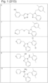

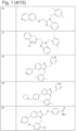


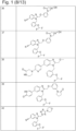

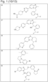


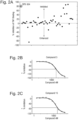
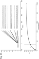
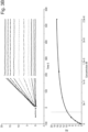
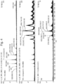

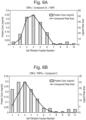
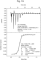
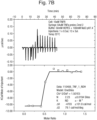
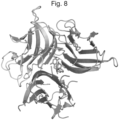
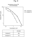

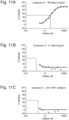

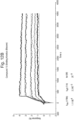
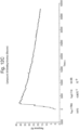
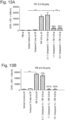
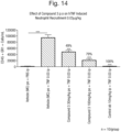
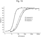
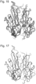

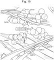

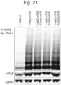
Classifications
-
- A—HUMAN NECESSITIES
- A61—MEDICAL OR VETERINARY SCIENCE; HYGIENE
- A61P—SPECIFIC THERAPEUTIC ACTIVITY OF CHEMICAL COMPOUNDS OR MEDICINAL PREPARATIONS
- A61P35/00—Antineoplastic agents
-
- A—HUMAN NECESSITIES
- A61—MEDICAL OR VETERINARY SCIENCE; HYGIENE
- A61K—PREPARATIONS FOR MEDICAL, DENTAL OR TOILETRY PURPOSES
- A61K47/00—Medicinal preparations characterised by the non-active ingredients used, e.g. carriers or inert additives; Targeting or modifying agents chemically bound to the active ingredient
- A61K47/50—Medicinal preparations characterised by the non-active ingredients used, e.g. carriers or inert additives; Targeting or modifying agents chemically bound to the active ingredient the non-active ingredient being chemically bound to the active ingredient, e.g. polymer-drug conjugates
- A61K47/51—Medicinal preparations characterised by the non-active ingredients used, e.g. carriers or inert additives; Targeting or modifying agents chemically bound to the active ingredient the non-active ingredient being chemically bound to the active ingredient, e.g. polymer-drug conjugates the non-active ingredient being a modifying agent
- A61K47/62—Medicinal preparations characterised by the non-active ingredients used, e.g. carriers or inert additives; Targeting or modifying agents chemically bound to the active ingredient the non-active ingredient being chemically bound to the active ingredient, e.g. polymer-drug conjugates the non-active ingredient being a modifying agent the modifying agent being a protein, peptide or polyamino acid
- A61K47/64—Drug-peptide, drug-protein or drug-polyamino acid conjugates, i.e. the modifying agent being a peptide, protein or polyamino acid which is covalently bonded or complexed to a therapeutically active agent
- A61K47/6425—Drug-peptide, drug-protein or drug-polyamino acid conjugates, i.e. the modifying agent being a peptide, protein or polyamino acid which is covalently bonded or complexed to a therapeutically active agent the peptide or protein in the drug conjugate being a receptor, e.g. CD4, a cell surface antigen, i.e. not a peptide ligand targeting the antigen, or a cell surface determinant, i.e. a part of the surface of a cell
-
- A—HUMAN NECESSITIES
- A61—MEDICAL OR VETERINARY SCIENCE; HYGIENE
- A61P—SPECIFIC THERAPEUTIC ACTIVITY OF CHEMICAL COMPOUNDS OR MEDICINAL PREPARATIONS
- A61P37/00—Drugs for immunological or allergic disorders
-
- C—CHEMISTRY; METALLURGY
- C07—ORGANIC CHEMISTRY
- C07D—HETEROCYCLIC COMPOUNDS
- C07D213/00—Heterocyclic compounds containing six-membered rings, not condensed with other rings, with one nitrogen atom as the only ring hetero atom and three or more double bonds between ring members or between ring members and non-ring members
- C07D213/02—Heterocyclic compounds containing six-membered rings, not condensed with other rings, with one nitrogen atom as the only ring hetero atom and three or more double bonds between ring members or between ring members and non-ring members having three double bonds between ring members or between ring members and non-ring members
- C07D213/04—Heterocyclic compounds containing six-membered rings, not condensed with other rings, with one nitrogen atom as the only ring hetero atom and three or more double bonds between ring members or between ring members and non-ring members having three double bonds between ring members or between ring members and non-ring members having no bond between the ring nitrogen atom and a non-ring member or having only hydrogen or carbon atoms directly attached to the ring nitrogen atom
- C07D213/60—Heterocyclic compounds containing six-membered rings, not condensed with other rings, with one nitrogen atom as the only ring hetero atom and three or more double bonds between ring members or between ring members and non-ring members having three double bonds between ring members or between ring members and non-ring members having no bond between the ring nitrogen atom and a non-ring member or having only hydrogen or carbon atoms directly attached to the ring nitrogen atom with hetero atoms or with carbon atoms having three bonds to hetero atoms with at the most one bond to halogen, e.g. ester or nitrile radicals, directly attached to ring carbon atoms
- C07D213/72—Nitrogen atoms
-
- C—CHEMISTRY; METALLURGY
- C07—ORGANIC CHEMISTRY
- C07D—HETEROCYCLIC COMPOUNDS
- C07D235/00—Heterocyclic compounds containing 1,3-diazole or hydrogenated 1,3-diazole rings, condensed with other rings
- C07D235/02—Heterocyclic compounds containing 1,3-diazole or hydrogenated 1,3-diazole rings, condensed with other rings condensed with carbocyclic rings or ring systems
- C07D235/04—Benzimidazoles; Hydrogenated benzimidazoles
-
- C—CHEMISTRY; METALLURGY
- C07—ORGANIC CHEMISTRY
- C07D—HETEROCYCLIC COMPOUNDS
- C07D239/00—Heterocyclic compounds containing 1,3-diazine or hydrogenated 1,3-diazine rings
- C07D239/02—Heterocyclic compounds containing 1,3-diazine or hydrogenated 1,3-diazine rings not condensed with other rings
- C07D239/24—Heterocyclic compounds containing 1,3-diazine or hydrogenated 1,3-diazine rings not condensed with other rings having three or more double bonds between ring members or between ring members and non-ring members
- C07D239/26—Heterocyclic compounds containing 1,3-diazine or hydrogenated 1,3-diazine rings not condensed with other rings having three or more double bonds between ring members or between ring members and non-ring members with only hydrogen atoms, hydrocarbon or substituted hydrocarbon radicals, directly attached to ring carbon atoms
-
- C—CHEMISTRY; METALLURGY
- C07—ORGANIC CHEMISTRY
- C07D—HETEROCYCLIC COMPOUNDS
- C07D401/00—Heterocyclic compounds containing two or more hetero rings, having nitrogen atoms as the only ring hetero atoms, at least one ring being a six-membered ring with only one nitrogen atom
- C07D401/14—Heterocyclic compounds containing two or more hetero rings, having nitrogen atoms as the only ring hetero atoms, at least one ring being a six-membered ring with only one nitrogen atom containing three or more hetero rings
-
- C—CHEMISTRY; METALLURGY
- C07—ORGANIC CHEMISTRY
- C07D—HETEROCYCLIC COMPOUNDS
- C07D471/00—Heterocyclic compounds containing nitrogen atoms as the only ring hetero atoms in the condensed system, at least one ring being a six-membered ring with one nitrogen atom, not provided for by groups C07D451/00 - C07D463/00
- C07D471/02—Heterocyclic compounds containing nitrogen atoms as the only ring hetero atoms in the condensed system, at least one ring being a six-membered ring with one nitrogen atom, not provided for by groups C07D451/00 - C07D463/00 in which the condensed system contains two hetero rings
- C07D471/04—Ortho-condensed systems
-
- C—CHEMISTRY; METALLURGY
- C07—ORGANIC CHEMISTRY
- C07K—PEPTIDES
- C07K14/00—Peptides having more than 20 amino acids; Gastrins; Somatostatins; Melanotropins; Derivatives thereof
- C07K14/435—Peptides having more than 20 amino acids; Gastrins; Somatostatins; Melanotropins; Derivatives thereof from animals; from humans
- C07K14/52—Cytokines; Lymphokines; Interferons
- C07K14/525—Tumour necrosis factor [TNF]
-
- C—CHEMISTRY; METALLURGY
- C07—ORGANIC CHEMISTRY
- C07K—PEPTIDES
- C07K14/00—Peptides having more than 20 amino acids; Gastrins; Somatostatins; Melanotropins; Derivatives thereof
- C07K14/435—Peptides having more than 20 amino acids; Gastrins; Somatostatins; Melanotropins; Derivatives thereof from animals; from humans
- C07K14/705—Receptors; Cell surface antigens; Cell surface determinants
- C07K14/70575—NGF/TNF-superfamily, e.g. CD70, CD95L, CD153, CD154
-
- C—CHEMISTRY; METALLURGY
- C07—ORGANIC CHEMISTRY
- C07K—PEPTIDES
- C07K16/00—Immunoglobulins [IGs], e.g. monoclonal or polyclonal antibodies
- C07K16/18—Immunoglobulins [IGs], e.g. monoclonal or polyclonal antibodies against material from animals or humans
- C07K16/24—Immunoglobulins [IGs], e.g. monoclonal or polyclonal antibodies against material from animals or humans against cytokines, lymphokines or interferons
- C07K16/241—Tumor Necrosis Factors
-
- G—PHYSICS
- G01—MEASURING; TESTING
- G01N—INVESTIGATING OR ANALYSING MATERIALS BY DETERMINING THEIR CHEMICAL OR PHYSICAL PROPERTIES
- G01N33/00—Investigating or analysing materials by specific methods not covered by groups G01N1/00 - G01N31/00
- G01N33/48—Biological material, e.g. blood, urine; Haemocytometers
- G01N33/50—Chemical analysis of biological material, e.g. blood, urine; Testing involving biospecific ligand binding methods; Immunological testing
- G01N33/68—Chemical analysis of biological material, e.g. blood, urine; Testing involving biospecific ligand binding methods; Immunological testing involving proteins, peptides or amino acids
- G01N33/6803—General methods of protein analysis not limited to specific proteins or families of proteins
- G01N33/6845—Methods of identifying protein-protein interactions in protein mixtures
-
- G—PHYSICS
- G01—MEASURING; TESTING
- G01N—INVESTIGATING OR ANALYSING MATERIALS BY DETERMINING THEIR CHEMICAL OR PHYSICAL PROPERTIES
- G01N33/00—Investigating or analysing materials by specific methods not covered by groups G01N1/00 - G01N31/00
- G01N33/48—Biological material, e.g. blood, urine; Haemocytometers
- G01N33/50—Chemical analysis of biological material, e.g. blood, urine; Testing involving biospecific ligand binding methods; Immunological testing
- G01N33/68—Chemical analysis of biological material, e.g. blood, urine; Testing involving biospecific ligand binding methods; Immunological testing involving proteins, peptides or amino acids
- G01N33/6854—Immunoglobulins
-
- G—PHYSICS
- G01—MEASURING; TESTING
- G01N—INVESTIGATING OR ANALYSING MATERIALS BY DETERMINING THEIR CHEMICAL OR PHYSICAL PROPERTIES
- G01N33/00—Investigating or analysing materials by specific methods not covered by groups G01N1/00 - G01N31/00
- G01N33/48—Biological material, e.g. blood, urine; Haemocytometers
- G01N33/50—Chemical analysis of biological material, e.g. blood, urine; Testing involving biospecific ligand binding methods; Immunological testing
- G01N33/68—Chemical analysis of biological material, e.g. blood, urine; Testing involving biospecific ligand binding methods; Immunological testing involving proteins, peptides or amino acids
- G01N33/6863—Cytokines, i.e. immune system proteins modifying a biological response such as cell growth proliferation or differentiation, e.g. TNF, CNF, GM-CSF, lymphotoxin, MIF or their receptors
-
- C—CHEMISTRY; METALLURGY
- C07—ORGANIC CHEMISTRY
- C07D—HETEROCYCLIC COMPOUNDS
- C07D401/00—Heterocyclic compounds containing two or more hetero rings, having nitrogen atoms as the only ring hetero atoms, at least one ring being a six-membered ring with only one nitrogen atom
- C07D401/02—Heterocyclic compounds containing two or more hetero rings, having nitrogen atoms as the only ring hetero atoms, at least one ring being a six-membered ring with only one nitrogen atom containing two hetero rings
- C07D401/04—Heterocyclic compounds containing two or more hetero rings, having nitrogen atoms as the only ring hetero atoms, at least one ring being a six-membered ring with only one nitrogen atom containing two hetero rings directly linked by a ring-member-to-ring-member bond
-
- C—CHEMISTRY; METALLURGY
- C07—ORGANIC CHEMISTRY
- C07K—PEPTIDES
- C07K2317/00—Immunoglobulins specific features
- C07K2317/20—Immunoglobulins specific features characterized by taxonomic origin
- C07K2317/24—Immunoglobulins specific features characterized by taxonomic origin containing regions, domains or residues from different species, e.g. chimeric, humanized or veneered
-
- C—CHEMISTRY; METALLURGY
- C07—ORGANIC CHEMISTRY
- C07K—PEPTIDES
- C07K2317/00—Immunoglobulins specific features
- C07K2317/30—Immunoglobulins specific features characterized by aspects of specificity or valency
- C07K2317/32—Immunoglobulins specific features characterized by aspects of specificity or valency specific for a neo-epitope on a complex, e.g. antibody-antigen or ligand-receptor
-
- C—CHEMISTRY; METALLURGY
- C07—ORGANIC CHEMISTRY
- C07K—PEPTIDES
- C07K2317/00—Immunoglobulins specific features
- C07K2317/30—Immunoglobulins specific features characterized by aspects of specificity or valency
- C07K2317/34—Identification of a linear epitope shorter than 20 amino acid residues or of a conformational epitope defined by amino acid residues
-
- C—CHEMISTRY; METALLURGY
- C07—ORGANIC CHEMISTRY
- C07K—PEPTIDES
- C07K2317/00—Immunoglobulins specific features
- C07K2317/50—Immunoglobulins specific features characterized by immunoglobulin fragments
- C07K2317/55—Fab or Fab'
-
- C—CHEMISTRY; METALLURGY
- C07—ORGANIC CHEMISTRY
- C07K—PEPTIDES
- C07K2317/00—Immunoglobulins specific features
- C07K2317/50—Immunoglobulins specific features characterized by immunoglobulin fragments
- C07K2317/56—Immunoglobulins specific features characterized by immunoglobulin fragments variable (Fv) region, i.e. VH and/or VL
- C07K2317/565—Complementarity determining region [CDR]
-
- C—CHEMISTRY; METALLURGY
- C07—ORGANIC CHEMISTRY
- C07K—PEPTIDES
- C07K2317/00—Immunoglobulins specific features
- C07K2317/90—Immunoglobulins specific features characterized by (pharmaco)kinetic aspects or by stability of the immunoglobulin
- C07K2317/92—Affinity (KD), association rate (Ka), dissociation rate (Kd) or EC50 value
-
- G—PHYSICS
- G01—MEASURING; TESTING
- G01N—INVESTIGATING OR ANALYSING MATERIALS BY DETERMINING THEIR CHEMICAL OR PHYSICAL PROPERTIES
- G01N2333/00—Assays involving biological materials from specific organisms or of a specific nature
- G01N2333/435—Assays involving biological materials from specific organisms or of a specific nature from animals; from humans
- G01N2333/52—Assays involving cytokines
- G01N2333/525—Tumor necrosis factor [TNF]
-
- G—PHYSICS
- G01—MEASURING; TESTING
- G01N—INVESTIGATING OR ANALYSING MATERIALS BY DETERMINING THEIR CHEMICAL OR PHYSICAL PROPERTIES
- G01N2333/00—Assays involving biological materials from specific organisms or of a specific nature
- G01N2333/435—Assays involving biological materials from specific organisms or of a specific nature from animals; from humans
- G01N2333/705—Assays involving receptors, cell surface antigens or cell surface determinants
- G01N2333/715—Assays involving receptors, cell surface antigens or cell surface determinants for cytokines; for lymphokines; for interferons
- G01N2333/7151—Assays involving receptors, cell surface antigens or cell surface determinants for cytokines; for lymphokines; for interferons for tumor necrosis factor [TNF]; for lymphotoxin [LT]
-
- G—PHYSICS
- G01—MEASURING; TESTING
- G01N—INVESTIGATING OR ANALYSING MATERIALS BY DETERMINING THEIR CHEMICAL OR PHYSICAL PROPERTIES
- G01N2500/00—Screening for compounds of potential therapeutic value
- G01N2500/02—Screening involving studying the effect of compounds C on the interaction between interacting molecules A and B (e.g. A = enzyme and B = substrate for A, or A = receptor and B = ligand for the receptor)
-
- G—PHYSICS
- G01—MEASURING; TESTING
- G01N—INVESTIGATING OR ANALYSING MATERIALS BY DETERMINING THEIR CHEMICAL OR PHYSICAL PROPERTIES
- G01N2500/00—Screening for compounds of potential therapeutic value
- G01N2500/04—Screening involving studying the effect of compounds C directly on molecule A (e.g. C are potential ligands for a receptor A, or potential substrates for an enzyme A)
Definitions
- This invention relates to novel stable, asymmetric, trimeric TNF ⁇ structures, and their use in therapy.
- the invention in particular relates to complexes of trimeric TNF ⁇ structures with small molecules which can bind to the TNFR1 receptor, but attenuate signalling therefrom, which can be used in the treatment and/or prevention of disease.
- the disclosure further relates to crystals of the asymmetric trimeric TNF ⁇ structures and complexes.
- the invention also relates to the use of 3-D structure models obtained from such crystals in methods for the determination of novel trimeric TNF ⁇ inhibitors.
- TNF Tumour Necrosis Factor
- TNF Tumour Necrosis Factor
- a common core motif which consists of two antiparallel ⁇ -pleated sheets with antiparallel ⁇ -strands, forming a " jelly roll " ⁇ -structure.
- Another common feature shared by members of the TNF superfamily is the formation of homo- or heterotrimeric complexes. It is these trimeric forms of the TNF superfamily members that bind to, and activate, specific TNF superfamily receptors.
- TNF ⁇ is the archetypal member of the TNF superfamily - forming a symmetrical homotrimer. Dysregulation of TNF ⁇ production has been implicated in a number of pathological conditions of significant medical importance. For example, TNF ⁇ has been implicated in rheumatoid arthritis, inflammatory bowel diseases (including Crohn's disease), psoriasis, Alzheimer's disease (AD), Parkinson's disease (PD), pain, epilepsy, osteoporosis, asthma, systemic lupus erythematosus (SLE) and multiple sclerosis (MS). Other members of the TNF superfamily have also been implicated in pathological conditions, including autoimmune disease.
- TNF superfamily members are macromolecular and act by inhibiting the binding of the TNF superfamily member to its receptor.
- conventional antagonists include anti-TNF ⁇ antibodies, particularly monoclonal antibodies, such as infliximab (Remicade ® ), adalimumab (Humira ® ) and certolizumab pegol (Cimzia ® ), or soluble TNF ⁇ receptor fusion proteins, such as etanercept (Enbrel ® ). These both inhibit soluble TNF ⁇ and its interaction with the receptor TNFR1 (responsible for inflammation) and membrane-bound TNF ⁇ and its interaction with the receptor TNFR2 (involved in the immune response).
- the present inventors have obtained extensive structural understanding of a new, stable, asymmetric trimeric structure of TNF ⁇ .
- This TNF ⁇ trimer acts by binding to TNFR1, but is less able, or unable, to initiate signalling downstream of TNFR1.
- This TNF ⁇ trimer may adopt this structure through forming a complex with a class of small molecular entities (SME) at the centre of the trimer.
- SME small molecular entities
- the present inventors have also obtained an extensive structural understanding of such compounds, the pharmacophore which can induce the asymmetric TNF ⁇ trimeric structure, and the key interactions of residues within the TNF ⁇ trimer which contact the pharmacophore to induce the asymmetric TNF ⁇ trimer.
- asymmetric TNF ⁇ trimers and complexes can be used in the treatment of conditions mediated by TNF ⁇ .
- the present inventors have also found that membrane-bound TNF ⁇ trimers and complexes do not disrupt the signalling downstream of TNFR2.
- the present inventors have identified novel asymmetric TNF ⁇ trimer crystal forms which may be used in structural-based methods of rational drug design to determine novel compounds which may induce the asymmetric TNF ⁇ trimer.
- the present invention provides a complex comprising an asymmetrical TNF ⁇ trimer of a protein subunit comprising the amino-acid sequence of SEQ ID NO: 36, or corresponding sequence, and a compound that is capable of binding to the TNF ⁇ trimer, wherein:
- asymmetrical TNF ⁇ trimer asymmetric TNF ⁇ trimer
- TNF ⁇ trimer of the invention is non-naturally occurring (given the nature of its asymmetric conformation).
- Antagonism of the TNFR1 receptor
- TNFR1 asymmetrical TNF ⁇ trimer
- asymmetric TNF ⁇ trimer asymmetric TNF ⁇ trimer of the invention
- TNFR1 asymmetric TNF ⁇ trimer of the invention
- the invention also provides a computer comprising executable code for:
- the invention provides a method of identifying a potential inhibitor of an apo TNF ⁇ trimer, comprising the steps of:
- the invention provides a method of designing a compound which binds to an apo TNF ⁇ trimer comprising the steps of:
- the invention relates to an asymmetrical TNF ⁇ trimer of a protein subunit comprising (or consisting of) the amino-acid sequence of SEQ ID NO: 36, or corresponding sequence, wherein said TNF ⁇ trimer adopts a conformation, when determined by x-ray crystallography, with the C ⁇ atoms of residues 12-18, 47-50, 54-64, 76-82, 91-97, 117-125, 129-137, and 150-156 of SEQ ID NO: 36, or corresponding sequence, for all subunits within 0.9 ⁇ RMSD [root mean square deviation] of the same atoms of the Reference Structure Compound34.pdb, said TNF- ⁇ trimer being able to bind TNFR1, but wherein signalling from said bound TNFR1 is attenuated or antagonised, for use in a method of therapy practised on the human or animal body.
- the asymmetrical TNF ⁇ trimer or distorted TNF ⁇ trimer or TNF ⁇ trimer of the invention herein are novel structures of TNF ⁇ where the normal 3-fold axis of symmetry between the subunits in symmetrical or apo TNF ⁇ trimer (Eck and Sprang 1989 JBC 264:17595-605) is disrupted such that the trimer still binds TNFR1, but wherein signalling from said bound TNFR1 is attenuated or antagonised or completely inhibited.
- Structures of apo TNF ⁇ trimer are well known in the art such as 1A8M from the Protein Data Bank (PDB).
- TNF ⁇ trimer of the invention may have the wild-type amino sequence of any known animal or human TNF ⁇ , in particular human TNF ⁇ , for instance SEQ ID NO:36. It may be soluble TNF ⁇ (sTNF ⁇ ) or membrane-bound TNF ⁇ , or both.
- sTNF ⁇ soluble TNF ⁇
- membrane-bound TNF ⁇ membrane-bound TNF ⁇
- Soluble homotrimeric TNF ⁇ is released from membrane-bound homotrimeric TNF ⁇ (mTNF) via proteolytic cleavage by the metalloprotease TNF alpha converting enzyme (TACE / ADAM17; though other proteinases can also release sTNF such as ADAM10, ADAM19, matrix metalloproteinase 7 and proteinase 3 which may yield corresponding soluble TNF ⁇ sequences that may be extended or truncated by 1, 2, 3, 4, or 5 amino acids relative to a TACE cleaved sTNF ⁇ such as SEQ ID NO: 36).
- TACE / ADAM17 metalloprotease TNF alpha converting enzyme
- sTNF such as ADAM10, ADAM19, matrix metalloproteinase 7 and proteinase 3 which may yield corresponding soluble TNF ⁇ sequences that may be extended or truncated by 1, 2, 3, 4, or 5 amino acids relative to a TACE cleaved sTNF ⁇ such as SEQ ID NO:
- a human sequence encompassed by the term mTNF is shown in SEQ ID NO: 34
- a human sequence encompassed by the term sTNF (the product of the action of TACE on SEQ ID NO: 34) is shown in SEQ ID NO: 36.
- Corresponding sequences of rat and mouse mTNF ⁇ are presented in SEQ ID NO:32 and 33, respectively.
- Corresponding sequences of TNF ⁇ from other animals may be readily overlaid with the SEQ ID NO:36 sequence and given the same amino acid numbering as for SEQ ID NO:36 (used in the numbering of TNF ⁇ amino acids herein).
- the sequence from various animals may be found within the Uniprot database (www.uniprot.org) including human sequences P01375 and Q5STB3.
- the corresponding sTNF ⁇ sequences may be the 157 amino acid C-terminal end of the mTNF ⁇ sequence (as SEQ ID NO:36) or may be longer or shorter by one, two or three amino acids (the rat and mouse sequences being 156 amino acids).
- the corresponding sTNF ⁇ sequence may have 1, 2, 3, 4, 5, 6, 7, 8, 9, 10, 11, 12, 13, 14, 15, 16, 17, 18, 19, 20, 21, 22, 23, 24, 25, 26, 27, 28, 29, 30, 31, 32, 33, 34, 35, 36, 37, 38, 39, or 40 amino acid substitutions relative to SEQ ID NO:36.
- the corresponding sTNF ⁇ sequence may have 80, 90, 95, 96, 97, 98, or 99% amino acid sequence identity to SEQ ID NO:36 over the length of SEQ ID NO:36.
- TNF ⁇ trimers of the invention have a common asymmetric structure, as determined by x-ray crystallography (for instance by the methods detailed in Examples 18 and 19). TNF ⁇ trimers of the invention may be confirmed by overlaying the atomic coordinates of the C ⁇ atoms of residues 12-18, 47-50, 54-64, 76-82, 91-97, 117-125, 129-137, and 150-156 (the ⁇ -sheet residues) of SEQ ID NO: 36, or corresponding sequence, for all subunits and establishing that they are within 0.9 ⁇ RMSD [root mean square deviation] of the same atoms of the Reference Structure Compound34.pdb (for instance as described in Example 19).
- the rat and mouse sTNF ⁇ sequences are the C-terminal 156 amino acids of mTNF ⁇ rather than the 157 of SEQ ID NO:36. It may readily be determined that the corresponding ⁇ -sheet residues for overlay in, for instance, the rat sTNF ⁇ sequence (of mTNF ⁇ sequence SEQ ID NO:32) are residues 12-18, 47-50, 54-64, 75-81, 90-96, 116-124, 128-136, and 149-155.
- Atomic coordinates for 64 structures are given in the Supplementary Data of the present invention, each structure given the name "Compound X” which is equivalent to the term “CompoundX.pdb” with X being the formula number (X) of the Compound of Figure 1 sitting at the centre of each trimer.
- the atomic coordinates for Reference Structure Compound34.pdb (the structure that is most average of the 64) may thus readily be loaded into appropriate software for the overlay to take place.
- trimer subunit chains will need to be assigned with subunit labels A, B, and C.
- Which chain is A, B or C may be ascertained by measuring three distances in the x-ray crystallography determined structure between three C-alpha atoms of three identical residues - e.g. P117 in each chain (P116 in the mouse sTNF ⁇ sequence); (G121 is also appropriate).
- the three distances form a triangle which is equilateral in apo TNF ⁇ but distorted in the asymmetric TNF ⁇ structures of the invention.
- TNF- ⁇ trimers of the invention are able to bind TNFR1, but signalling from said bound TNFR1 is attenuated or antagonised, may be carried out by any of the appropriate methods discussed herein (for instance in Examples 6, 7, 8, 9, 10 [the Reporter Gene Assay], 12).
- TNF- ⁇ trimers of the invention are for use in a method of therapy practised on the human or animal body.
- TNF- ⁇ trimers of the invention may be administered to the human or animal body either directly or indirectly.
- directly it is meant that symmetrical TNF- ⁇ trimer already within the animal or human may be rendered asymmetrical TNF- ⁇ trimers of the invention which may be used for therapy.
- TNF- ⁇ trimers of the invention may be administered to the human or animal body indirectly by administering to the animal or human a compound described herein [for instance Compounds (1)-(65)], which may, for instance, cause the formation of a complex of the invention as described herein which may be used in therapy.
- the TNF ⁇ trimer of the invention may be for use in the treatment and/or prevention of one or more of autoimmune disorders, inflammatory disorders, neurological disorders, neurodegenerative disorders, pain disorders, nociceptive disorders or cardiovascular disorders.
- autoimmune disorders inflammatory disorders
- neurological disorders neurodegenerative disorders
- pain disorders nociceptive disorders or cardiovascular disorders.
- psoriasis systemic lupus erythematosus
- Alzheimer's disease Parkinson's disease and epilepsy.
- the RMSD is within 0.85, 0.8, 0.7, 0.65, 0.6, 0.5, or 0.47 ⁇ .
- asymmetric TNF ⁇ trimer of a protein subunit comprising or consisting of the amino-acid sequence of SEQ ID NO: 36, or corresponding sequence, wherein said TNF ⁇ trimer adopts a conformation, when determined by x-ray crystallography, with the C ⁇ atoms of residues 12-18, 47-50, 54-64, 76-82, 91-97, 117-125, 129-137, and 150-156 of SEQ ID NO: 36, or corresponding sequence, for all subunits within 0.9 ⁇ RMSD of the same atoms of the Reference Structure Compound34.pdb, said TNF- ⁇ trimer being able to bind TNFR1, but wherein signalling from said bound TNFR1 is attenu
- the conformation of the TNF ⁇ trimer of the invention is obtainable or obtained through the TNF ⁇ trimer forming a complex with any one of Compounds (1)-(65).
- the TNF ⁇ trimer antagonises the signalling of the TNFR1 receptor (measured, for instance, by methods disclosed herein); the TNF ⁇ trimer has increased stability compared to the stability of a symmetrical TNF ⁇ trimer (measured, for instance, by methods disclosed herein), for instance the increase in stability results in an increase in the thermal transition midpoint (T m ) of the TNF ⁇ trimer of at least 1 ⁇ C, of at least 10 ⁇ C, or is between 10 ⁇ C and 20 ⁇ C (measured, for instance, by methods disclosed herein; the TNF ⁇ trimer has an increased binding affinity to the TNFR1 receptor compared to the binding affinity of a symmetrical TNF ⁇ trimer to the TNFR1 receptor (measured, for instance, by methods disclosed herein); the TNF ⁇ trimer has an increased binding affinity to the TNFR1 receptor by increasing the on rate (k on-r ) and/or decreasing the off rate (k off-r ) compared to the k
- the K D-r of the TNF ⁇ trimer to the TNFR1 receptor is decreased compared to the K D-r of the symmetrical TNF ⁇ trimer to the TNFR1 receptor, wherein:
- the K D-r of the TNF ⁇ trimer to the TNFR1 receptor is decreased compared to the K D-r of the symmetrical TNF ⁇ trimer to the TNFR1 receptor, wherein:
- the TNF ⁇ trimer of the invention binds to either of the following antibodies with a K D-ab of 1 nM or less: CA185_1979 with a light chain of sequence SEQ ID NO: 26 and heavy chain of sequence SEQ ID NO:27; or CA185_1974 with a light chain of sequence SEQ ID NO: 11 and heavy chain of sequence SEQ ID NO: 12 (see herein, for methods of measurement and further information on these antibodies which are specific for the TNF ⁇ trimer of the invention).
- the TNF ⁇ trimer of the invention may be said to have adopted a "stable" asymmetric conformation.
- Example 20 describe the residues of TNF ⁇ trimer that are always involved in making ⁇ 4 ⁇ contacts with such compounds (for instance Compounds (1)-(64)), and Example 21 describes a pharmacophore that may be comprised within a compound that is understood to be needed for making these contacts. This understanding readily allows the skilled person to use the pharmacophore as a basis to design many different compounds which can stabilise the complexes of the present invention.
- the invention provides a complex comprising a TNF ⁇ trimer of a protein subunit comprising or consisting of the amino-acid sequence of SEQ ID NO: 36, or corresponding sequence, and a compound that is capable of binding to the TNF ⁇ trimer, whereby the compound-trimer complex binds to TNFR1 and attenuates or antagonises the signalling induced by the trimer through TNFR1, wherein the trimer is as defined in the claims and wherein the compound, as determined by x-ray crystallography, comprises a pharmacophore which is bound within the TNF ⁇ trimer such that it is within 4 ⁇ of all of the following residues: Leu57 of subunit A; Tyr119 of subunit B; Gly121 of subunit B; Gly122 of subunit B; Tyr59 of subunit C; Leu120 of subunit C; and Tyr 15 1 of subunit C, and wherein the pharmacophore consists of 2 fused rings ( "R3" and "R2”), wherein
- the pharmacophore may have one or more of: the R4 ring being 5- or 6-membered; the R3 ring being aromatic; the R4 ring being aromatic; the R2 ring being heteroaromatic; the R3 ring being heteroaromatic; the R4 ring being heteroaromatic; the fused rings sharing 2 atoms; the linking non-hydrogen atom being Carbon, Nitrogen, or Oxygen (which may itself be part of a further ring of the compound that links the R2 ring with the R4 ring to form for instance a tricyclic compound comprising three fused rings - see Compounds 48, 49, 51, 52, 60, 61 or 63 for example); the A1 feature being through a nitrogen or oxygen atom in the R2 ring (which may hydrogen-bond to the sidechain of Tyr151 on subunit C of the TNF ⁇ trimer).
- the pi system of the heteroaromatic R2 ring forms a CH-pi interaction with (suitably the C-Beta CH2 group of) the sidechain of Tyr59 on subunit C of the TNF ⁇ trimer.
- the pi system of the aromatic/heteroaromatic R3 ring forms a pi stacking interaction with the aromatic ring of the sidechain of Tyr59 on subunit C of the TNF ⁇ trimer.
- the pi system of the aromatic/heteroaromatic R3 ring forms a CH-pi interaction with (suitably the C-Delta CH2 group of) the sidechain of Leu57 on subunit A of the TNF ⁇ trimer.
- R4 is a 5- or 6-membered ring.
- R4 is a 5-membered ring. In one aspect of that embodiment, R4 is a 5-membered aromatic ring. In another aspect of that embodiment R4 is a 5-membered heteroaromatic ring.
- R4 is a 6-membered ring. In one aspect of that embodiment, R4 is a 6-membered aromatic ring. In another aspect of that embodiment R4 is a 6-membered heteroaromatic ring.
- the pi system of the aromatic/heteroaromatic R4 ring forms a CH-pi interaction with (suitably the C-Delta CH2 group of) the sidechain of Leu57 on subunit A of the TNF ⁇ trimer.
- the linking non-hydrogen atom (the single atom linking [and thus not part of] R2 and R4 rings) is Carbon, Nitrogen, or Oxygen.
- the linking non-hydrogen atom is Carbon.
- the linking non-hydrogen atom is Nitrogen.
- the linking non-hydrogen atom is Oxygen.
- A1 is a nitrogen or oxygen atom in (i.e. part of) the R2 ring.
- A1 hydrogen-bonds to the sidechain of Tyr151 on subunit C of the TNF ⁇ trimer.
- the pharmacophore represents the minimum structural and/or chemical features of the compound-trimer complex and therefore that the compound may include additional structural features.
- the compound of the compound-trimer complex may comprise the pharmacophore as defined above wherein the linking non-hydrogen atom is incorporated into an additional ring that links the R2 ring with the R4 ring, thereby forming a fused tricyclic compound (see Compounds 48, 49, 51, 52, 60, 61 or 63 for example).
- the pharmacophore may have the R2, R3 and R4 ring, and A1 features arranged within the TNF ⁇ trimer structure according to the following Table: TNF ⁇ trimer amino acid Pharmacophore Feature within 4 ⁇ of the TNF ⁇ trimer amino acid Tyr151 subunit C A1 Hydrogen-bond acceptor Tyr59 subunit C R2, R3 rings Leu120 subunit C R2, R3 rings Leu57 subunit A R3, R4 rings Tyr119 subunit B R4 ring Gly121 subunit B R4 ring Gly122 subunit B R4 ring
- the pharmacophore may have one or more of the distances between the R2, R3, R4 and A1 features about, exactly or within 10% of the value according to the following Table: Site 1 feature Site 2 feature Distance ( ⁇ ) A1 R2 1.187 A1 R3 2.737 A1 R4 5.661 R2 R3 2.107 R2 R4 4.650 R3 R4 5.088
- the pharmacophore may have one or more of the angles between the R2, R3, R4 and A1 features about, exactly or within 10% of the value according to the following Table: Site 1 feature Site 2 feature Site 3 feature Angle (degrees) R2 A1 R3 46.6 R2 A1 R4 28.2 R3 A1 R4 63.9 A1 R2 R3 109.2 A1 R2 R4 144.9 R3 R2 R4 89.5 A1 R3 R2 24.2 A1 R3 R4 87.3 R2 R3 R4 66.0 A1 R4 R2 6.9 A1 R4 R3 28.9 R2 R4 R3 24.5
- the R3-R2-R4 angle of the pharmacophore defines a banana shape which may be involved in the desymmetrisation of the TNF ⁇ trimer of the invention.
- the compound comprising the pharmacophore may have 20-41 non-hydrogen atoms, and for instance may be any one of Compounds (1)-(65).
- the complex of the invention may similarly be for use in a method of therapy practised on the human or animal body (through direct [administration of the complex] or indirect [administration of the compound] administration), or may be directly or indirectly administered to a patient in need thereof in a method of treating and/or preventing one or more of autoimmune disorders, inflammatory disorders, neurological disorders, neurodegenerative disorders, pain disorders, nociceptive disorders, and/or cardiovascular disorders; for instance in the treatment and/or prevention of one or more of rheumatoid arthritis, Crohn's disease, psoriasis, systemic lupus erythematosus, Alzheimer's disease, Parkinson's disease and/or epilepsy.
- the complex of the invention may similarly comprise protein subunits comprising or consisting of the amino-acid sequence of SEQ ID NO: 36, or corresponding sequence.
- the complex of the invention comprises the TNF ⁇ trimer of the invention.
- the compound within the complex antagonises the signalling induced by the TNF ⁇ trimer through the TNFR1 receptor (measured, for instance, by methods disclosed herein): the compound increases the stability of the TNF ⁇ trimer compared to the stability of the TNF ⁇ trimer in the absence of the compound (measured, for instance, by methods disclosed herein), for instance the increase in stability results in an increase in the thermal transition midpoint (T m ) of the TNF ⁇ trimer of at least 1 ⁇ C, of at least 10 ⁇ C, or is between 10 ⁇ C and 20 ⁇ C; the compound increases the binding affinity of the TNF ⁇ trimer to the TNFR1 receptor compared to the binding affinity of the TNF ⁇ trimer to the TNFR1 receptor in the absence of the compound (measured, for instance, by methods disclosed herein), for instance the compound increases the binding affinity of the TNF ⁇ trimer to the TNFR1 receptor by increasing the on rate (k on-r ) and/or decreasing the off rate (k off)
- the compound decreases the K D-r of the TNF ⁇ trimer to the TNFR1 receptor compared to the K D-r of the TNF ⁇ trimer to the TNFR1 receptor in the absence of the compound, wherein:
- the compound decreases the K D-r of the TNF ⁇ trimer to the TNFR1 receptor compared to the K D-r of the TNF ⁇ trimer to the TNFR1 receptor in the absence of the compound, wherein:
- the compound has an IC 50 value of 500nM or less (measured, for instance, by methods disclosed herein).
- the complex binds to either of the following antibodies with a K D-ab of 1 nM or less: CA185_1979 with a light chain of sequence SEQ ID NO: 26 and heavy chain of sequence SEQ ID NO:27; or CA185_1974 with a light chain of sequence SEQ ID NO: 11 and heavy chain of sequence SEQ ID NO:12 (see herein, for methods of measurement and further information on these antibodies which are specific for the complexes and TNF ⁇ trimer of the invention).
- the complex of the invention may be said to adopt a "stable" asymmetric conformation.
- a pharmaceutical composition comprising the complex of the invention and a pharmaceutically acceptable carrier.
- the inventors present data supporting that the TNF ⁇ trimer of the invention and the complex of the invention may be used in therapy without a significant side effect of existing TNF ⁇ therapeutics.
- sTNF ⁇ has specificity for the TNFR1 receptor, and mTNF ⁇ for the TNFR2 receptor (Grell et al. Cell 1995 83:793-802).
- a TNF ⁇ inhibitor in development that selectively inhibits sTNF ⁇ but not mTNF ⁇ has the characteristics of attenuating experimental arthritis without suppressing innate immunity to infection ( Zalevsky et al. J of Immunology 2007 179:1872-1883 ).
- Example 22 investigates how the mTNF ⁇ conformation upon binding Compounds described herein does not affect TNFR2 function; TNFR2 proximal and downstream signalling is not impaired.
- the TNF ⁇ trimer of the invention or the complex of the invention may be for use in a method of therapy practised on the human or animal body, wherein the use does not induce or cause to worsen an infection and/or malignancy.
- the infection is TB and/or bacterial sepsis and/or fungal infection and/or the malignancy is lymphoma.
- the inventors provide in the methods that the administration to the patient in need thereof does not induce or cause to worsen an infection and/or malignancy in said patient.
- the infection is TB and/or bacterial sepsis and/or fungal infection and/or the malignancy is lymphoma.
- the present inventors have developed assays for identifying antagonists of TNF ⁇ . Specifically, the present inventors have developed assays that can be used to identify compounds that bind to trimeric apo forms of TNF ⁇ , and that stabilise these trimers in a conformation that is capable of binding to the requisite TNF receptor (TNFR1), and so antagonise signalling through said receptor. Accordingly, assays that are useful for identifying antagonists of TNF ⁇ are disclosed.
- the assays described herein may be used to identify compounds that bind to trimeric apo forms of TNF ⁇ , and which form a compound-trimer complex which binds to the TNFR1.
- the assays identify compounds that bind to the trimeric form of TNF ⁇ , but not to the monomeric form.
- the compounds bind to and stabilise the trimeric form of TNF ⁇ , do not bind to the monomeric form and do not stabilise the dimeric form of TNF ⁇ .
- the stabilisation of TNF ⁇ trimers by test compounds may occur by the test compound inhibiting the exchange of monomer units between trimers.
- Assays may comprise determining whether a test compound enhances the binding of the TNF ⁇ (TNF ⁇ trimers and complexes of the invention) to its receptor, and hence identify TNF ⁇ antagonists.
- assays of the disclosure may comprise determining whether a test compound enhances the binding of the TNF ⁇ to TNFR1, and hence identify TNF ⁇ antagonists which act by increasing the binding of reduced signalling, or non-signalling, forms of TNF ⁇ (TNF ⁇ trimers and complexes of the invention) to TNFR1.
- Assays for identifying TNF ⁇ antagonists according to the invention may comprise incubating a sample of TNF ⁇ under conditions that destabilise the formation of trimers of TNF ⁇ , for example in the presence of DMSO, and measuring the extent to which a test compound stabilises the formation of TNF ⁇ trimers.
- assays for identifying TNF ⁇ antagonists according to the invention may involve binding of TNF ⁇ to a test compound, and measuring the extent of binding of the TNF ⁇ trimer of the invention or compound-trimer complex to the TNFR1.
- TNF ⁇ and TNFR1 may be purified or present in mixtures, such as in cultured cells, tissue samples, body fluids or culture medium. Assays may be developed that are qualitative or quantitative, with the latter being useful for determining the binding parameters (affinity constants and kinetics) of the test compound to trimeric forms of TNF ⁇ , and also of the binding parameters of the TNF ⁇ trimer of the invention or compound-trimer complex to the requisite TNF receptor.
- the amount of the monomeric, dimeric and trimeric forms of TNF ⁇ may be determined by measuring the mass of the monomeric, dimeric and trimeric forms, the molar amount of the monomeric, dimeric and trimeric forms, the concentration of the monomeric, dimeric and trimeric forms, and the molarity of the monomeric, dimeric and trimeric forms. This amount may be given in any appropriate units. For example, the concentration of the monomeric, dimeric and trimeric forms may be given in pg/ml, ng/ml or ⁇ g/ml. The mass of the monomeric, dimeric and trimeric forms may be given in pg, ng or ⁇ g.
- the amount of the monomeric, dimeric or trimeric forms of TNF ⁇ in a sample of interest may be compared with the level of the monomeric, dimeric or trimeric forms of TNF ⁇ in another sample, such as a control sample, as described herein.
- the actual amount of the monomeric, dimeric or trimeric forms of TNF ⁇ such as the mass, molar amount, concentration or molarity of the monomeric, dimeric or trimeric forms of TNF ⁇ in the samples may be assessed.
- the amount of the monomeric, dimeric or trimeric forms of TNF ⁇ may be compared with that in another sample without quantifying the mass, molar amount, concentration or molarity of the monomeric, dimeric or trimeric forms of TNF ⁇ .
- the amount of the monomeric, dimeric or trimeric forms of TNF ⁇ in a sample according to the invention may be assessed as a relative amount, such as a relative mass, relative molar amount, relative concentration or relative molarity of the monomeric, dimeric or trimeric forms of TNF ⁇ based on a comparison between two or more samples.
- libraries of compounds may be screened in order to identify antagonists of TNF ⁇ (i.e. using the assays disclosed herein).
- Such libraries typically comprise at least 260 compounds.
- such libraries comprise at least 300, at least 500 or even at least 1000 compounds.
- the present inventors have found that mass spectrometry may be used to identify compounds that bind to trimeric forms of TNF ⁇ and that stabilise these trimers in a conformation (TNF ⁇ trimer or complex of the invention) that is capable of binding to TNFR1.
- mass spectrometry may be used to assess whether a compound stabilises the trimeric form of TNF ⁇ .
- the disclosure provides an assay for identifying a compound that is capable of binding to a trimeric TNF ⁇ protein, whereby the compound-trimer complex (or TNF ⁇ trimer of the invention) binds to TNFR1 and antagonises the signalling of the receptor comprising the steps of identifying the binding of a test compound to the trimeric form of TNF ⁇ in a sample and comparing the binding of the compound to the trimeric form of TNF ⁇ to corresponding values from control samples, which comprises conducting a mass spectrometric analysis on a sample containing the TNF ⁇ and the compound to detect the amount of the TNF ⁇ trimer and comparing the amount of TNF ⁇ trimer in the sample with a control sample and selecting a compound that is capable of binding to the trimeric form of TNF ⁇ , whereby the compound-trimer complex (or TNF ⁇ trimer of the invention) binds to TNFR1 antagonises the signalling of the receptor.
- the control sample may be identical to the sample being assayed, except that it lacks
- a test compound may be added to a solution of TNF ⁇ in the presence of a destabilising agent.
- destabilising agents also known as chaotropes, include low molar concentrations (e.g. 1M) of urea, guanidine or acetonitrile, high concentrations (e.g. 6M or higher) of these reagents will result in complete dissociation of the TNF ⁇ trimer and unfolding of the constituent TNF ⁇ monomeric subunits.
- the destabilising agent is preferably DMSO, typically at a concentration of 5%, 10% or higher. The resulting solution may be analysed using mass spectrometry.
- Non-covalent complexes formed between TNF ⁇ and test compounds with binding affinities as weak as 1mM can be detected. Binding stoichiometry may be obtained directly from presence or absence of complexes in which multiple molecules of the test compound are bound. Binding affinities (K D values) can be determined by measuring the TNF ⁇ - test compound complex (compound-trimer complex) / TNF ⁇ concentration ratio at known test compound concentrations.
- the test compound stabilises the trimeric form of TNF ⁇ if it increases the proportion of trimer compared to the amount of trimer observed for a sample containing the TNF ⁇ and the destabilising agent in the absence of the test compound.
- the test compound may increase the amount of trimer by 10%, 15%, 20%, 25%, 30%, 35%, 40%, 45%, 50%, 55%, 60%, 65%, 70%, 75%, 80%, 85%, 90%, 95%, 100%, 150%, 200%, 300%, 400% or more compared to the amount of trimer present in a sample containing the TNF ⁇ and the destabilising agent in the absence of the test compound.
- the test compound may also increase the amount of trimer compared to that observed for a sample of the TNF ⁇ in the absence of both the destabilising agent and the test compound.
- the test compound may increase the amount of trimer by 10%, 15%, 20%, 25%, 30%, 35%, 40%, 45%, 50%, 55%, 60%, 65%, 70%, 75%, 80%, 85%, 90%, 95%, 100%, 150%, 200%, 300%, 400% or more compared to the amount of trimer present in a sample containing TNF ⁇ in the absence of both the destabilising agent and the test compound.
- Trimer stabilisation is evidenced in two ways in the mass spectrometric study.
- TNF ⁇ trimer complex or TNF ⁇ trimer of the invention
- TNF ⁇ trimer complex can be measured by the ratio of monomer and trimer observed in the mass spectrum.
- the dimeric species is not observed in our studies.
- This dissociation may be an artefact of the high energy process used to introduce molecules into the mass spectrometer. None-the-less it can be used to assess the ability of the test compounds to stabilise the trimeric complex during the nebulisation and ionisation processes and thereby reduce the amount of monomer observed in the mass spectrum, the monomer/trimer ratio being used to determine the degree of stabilisation.
- Mass spectrometry may also be used to determine whether the test compound binds to the monomeric, dimeric and trimeric forms of TNF ⁇ .
- Mass spectrometry may also be used to determine the stoichiometry of the test compounds with TNF ⁇ , i.e. how many molecules of the test compound bind to TNF ⁇ .
- Mass spectrometry may also be used to determine whether the compound - TNF ⁇ trimer complex (or TNF ⁇ trimer of the invention) binds to TNFR1.
- Mass spectrometry may also be used to measure the rates at which a test compound binds to TNF ⁇ (the "on” rate” k on-c ) and rate at which the test compound dissociates from the TNF ⁇ (the “ off " rate or k off-c ).
- the symbol “K D-c ,” denotes the binding affinity (dissociation constant) of a test compound for TNF ⁇ .
- K D-c is defined as k off-c /k on-c .
- Test compounds may have slow "on” rates, which can be measured in minutes by mass spectral analysis of the TNF ⁇ and compound-trimer complex (or TNF ⁇ trimer of the invention) peak intensities.
- K D-c values for a test compound can be estimated by repeating this measurement at different TNF ⁇ : compound-trimer complex ratios.
- binding of compounds of the invention to TNF ⁇ trimers is characterized by fast "on” rates, ideally about 10 7 M -1 s -1 , with slow " off” rate, for example values typically of 10 -3 s -1 , 10 -4 s -1 , or no measurable " off " rate.
- Mass spectrometry may also be used to determine whether the test compound binds to the TNF ⁇ in the presence of TNFR1. This may involve incubating the test compound with TNF ⁇ that has been pre-incubated with its receptor. The sample containing the test compound, and pre-incubated TNF ⁇ and receptor can then be fractionated to separate molecules according to their molecular size, for example by analytical gel filtration. The resulting fractions may be analysed using mass spectrometry to determine whether the test compound binds to TNF ⁇ in the presence of the requisite receptor. The compound will elute in the same fraction as the TNF ⁇ if it is bound to the TNF ⁇ . The compound will elute in a different fraction than the TNF ⁇ if it is not bound to the TNF ⁇ . In this case the compound will typically elute in a later gel filtration fraction than the TNF ⁇ .
- Mass spectrometric methods may include, for example, matrix-assisted laser desorption/ionization mass spectrometry (MALDI MS), surface-enhanced laser desorption/ionization mass spectrometry (SELDI MS), time of flight mass spectrometry (TOF MS) and liquid chromatography mass spectrometry (LC MS).
- MALDI MS matrix-assisted laser desorption/ionization mass spectrometry
- SELDI MS surface-enhanced laser desorption/ionization mass spectrometry
- TOF MS time of flight mass spectrometry
- LC MS liquid chromatography mass spectrometry
- TNF ⁇ trimer of the invention Conventional antagonists act by inhibiting the binding of the TNF ⁇ to its receptor.
- the present inventors have used receptor-ligand binding assays to determine whether a test compound enhances the binding of TNF ⁇ trimer of the invention or complex of the invention to its receptor.
- Such receptor-ligand binding assays may be used to hence identify TNF ⁇ antagonists which act by increasing the binding of reduced-signalling, or non-signalling, forms of TNF ⁇ to TNFR1.
- the disclosure provides an assay for identifying a compound that is capable of binding to a trimeric protein that is TNF ⁇ , whereby the compound-trimer complex binds to the TNFR1 and antagonises the signalling of the receptor comprising the step of measuring the level of trimeric TNF ⁇ bound to the requisite receptor in a sample comprising a test compound and comparing the level of trimeric TNF ⁇ bound to the requisite receptor to corresponding values from control samples, which comprises performing a receptor-ligand binding assay in which a sample of TNF ⁇ and the compound, is applied to TNFR1 that has been bound to a surface and comparing the amount of TNF ⁇ trimer bound to TNFR1 with a control sample and selecting a compound that is capable of binding to the trimeric form of TNF ⁇ , whereby the compound-trimer complex binds to the TNFR1 and antagonises the signalling of the receptor.
- the control sample may be identical to the sample being assayed, except that it lacks the test compound and/or it
- a test compound may be added to a solution comprising TNF ⁇ and destabilising agent.
- the level of binding of TNFR1 in the presence of the destabilising agent alone (in a control sample) can be compared with the level of binding of TNF ⁇ to its receptor in the presence of the destabilising agent and the test compound.
- the test compound enhances the binding of TNF ⁇ to its receptor if it increases the proportion of TNF ⁇ bound to its receptor compared to the level of binding of TNF ⁇ to its receptor observed for a sample containing TNF ⁇ and the destabilising agent in the absence of the test compound.
- the test compound may increase the amount of TNF ⁇ bound to its receptor by 10%, 15%, 20%, 25%, 30%, 35%, 40%, 45%, 50%, 55%, 60%, 65%, 70%, 75%, 80%, 85%, 90%, 95%, 100%, 150%, 200%, 300%, 400% or more compared to the amount of TNF ⁇ bound to its receptor in a sample containing TNF ⁇ in the absence of the test compound.
- the receptor-ligand binding assay typically requires TNF ⁇ receptor bound to a support.
- the TNF ⁇ receptor may be bound directly to the support, or indirectly, using a linker molecule, such as avidin or streptavidin.
- the level of binding of TNF ⁇ to its receptor can then be assayed by adding a solution of TNF ⁇ with a destabilising agent.
- Destabilising agents also known as chaotropes, include low molar concentrations (e.g. 1M) of urea, guanidine or acetonitrile, high concentrations (e.g. 6M or higher) of these reagents will result in complete dissociation of the TNF ⁇ trimer and unfolding of the constituent TNF ⁇ monomeric subunits.
- the destabilising agent is preferably DMSO, typically at a concentration of 5%, 10% or higher.
- Binding of TNF ⁇ to its receptor is typically determined using an antibody that is specific to TNF ⁇ and which is bound to a marker.
- the marker can be any molecule that can be detected.
- the marker can be a radioactive isotope (for example 125 I, 32 P, 35 S and 3 H), fluorescent dye (for example fluorescein, rhodamine), enzyme-conjugate and the like.
- a substrate for the enzyme is used to quantify the amount of TNF ⁇ bound to the surface-bound receptor.
- Other markers include molecular labels that can be activated to produce light on exposure to certain stimuli, such as electricity. The choice of a marker will depend upon the detection system used.
- Receptor-ligand binding assays may be carried out in several formats, including cell-based binding assays, solution-phase assays and immunoassays.
- the solid supports for receptor-ligand binding reactions preferably contain wells.
- test compound-trimer complexes or TNF ⁇ trimers of the invention are incubated with the TNF ⁇ receptor for a specified period of time followed by measurement of the amount of the compound-trimer complex that is bound to the receptor.
- the level of bound compound-trimer complex (or TNF ⁇ trimer of the invention) may be calculated by measuring the marker using microscopy, fluorimetry, a scintillation counter, or any available immunoassay.
- the symbol “k on-r” denotes the rate (the “on” rate) at which a compound-trimer complex (or TNF ⁇ trimer of the invention) binds to a TNF ⁇ receptor.
- the symbol “k off-r” denotes the rate (the " off” rate) at which a compound-trimer complex (or TNF ⁇ trimer of the invention) dissociates from a TNF ⁇ receptor.
- the symbol “K D-r” denotes the binding affinity (dissociation constant) of a compound-trimer complex (or TNF ⁇ of the invention) for its receptor.
- K D-r is defined as k off-r /k on-r .
- Receptor-ligand binding assays may be used to measure the binding affinity of the compound-trimer complexes of the invention (or TNF ⁇ trimers of the invention) to TNFR1.
- competition assays may be used to compare the k on-r and k off-r values for compound-trimer complexes of the invention (or TNF ⁇ trimers of the invention) to TNFR1 and the k on-r and k off-r values of the TNF ⁇ binding to its receptor in the absence of the test compound (or apo TNF ⁇ ), and to determine K D-r values for binding of compound-trimer complexes of the invention (or TNF ⁇ trimer of the invention) to the receptor.
- the disclosure provides an assay for identifying a compound that is capable of binding to a trimeric TNF ⁇ protein, whereby the compound-trimer complex binds to the requisite TNF ⁇ receptor and modulates the signalling of the receptor comprising the step of measuring the stability of the trimeric TNF ⁇ in a sample comprising the compound and comparing the stability of the trimeric TNF ⁇ to corresponding values from control samples, which comprises performing an assay to determine the T m of the trimeric form of TNF ⁇ in a sample of the TNF ⁇ and the compound, comparing the T m of the trimeric form of TNF ⁇ with a control sample and selecting a compound that is capable of binding to the trimeric form of TNF ⁇ , whereby the compound-trimer complex binds to the TNF ⁇ receptor and antagonises the signalling of the receptor.
- the control sample may be
- a test compound may be added to a solution comprising TNF ⁇ and destabilising agent.
- the stability of the trimeric form of TNF ⁇ in the presence of the destabilising agent alone (in a control sample) can be compared with the stability of the trimeric form of TNF ⁇ in the presence of the destabilising agent and the test compound.
- the test compound enhances the stability of the trimeric form of TNF ⁇ if it increases the thermal transition midpoint (T m ) of the trimeric form of TNF ⁇ compared to the T m of the trimeric form of TNF ⁇ observed for a sample containing the TNF ⁇ and the destabilising agent in the absence of the test compound (or asymmetric TNF ⁇ trimer of the invention vs. symmetric TNF ⁇ ).
- the T m of the trimeric form of TNF ⁇ is the temperature at which 50% of the biomolecules are unfolded.
- the T m of the trimeric form of TNF ⁇ in the presence and/or absence of the test compound may be measured using any appropriate technique known in the art, for example using differential scanning calorimetry (DSC) or fluorescence probed thermal denaturation assays.
- the test compound may increase the T m of the trimeric form of TNF ⁇ by at least 1 ⁇ C, at least 2 ⁇ C, at least 5 ⁇ C, at least 10 ⁇ C, at least 15 ⁇ C, at least 20 ⁇ C or more compared to the T m of the trimeric form of TNF ⁇ in a sample containing TNF ⁇ in the absence of the test compound (or asymmetric TNF ⁇ trimer of the invention vs. symmetric TNF ⁇ ).
- the test compound increases the T m of the trimeric form of TNF ⁇ by at least 1 °C, more preferably by at least 10 °C and even more preferably by between 10 ⁇ C and 20 C.
- Destabilising agents also known as chaotropes, include low molar concentrations (e.g. 1M) of urea, guanidine or acetonitrile, high concentrations (e.g. 6M or higher) of these reagents will result in complete dissociation of the TNF ⁇ trimer and unfolding of the constituent TNF ⁇ monomeric subunits.
- the destabilising agent is preferably DMSO, typically at a concentration of 5%, 10% or higher.
- the present inventors have developed isothermal calorimetry methods for determining the effect of test compounds on the binding affinity of TNF ⁇ for their receptors (or the effect of asymmetric TNF ⁇ trimers of the invention vs symmetric TNF ⁇ trimers on receptor binding).
- the disclosure provides an assay for identifying a compound capable of binding to a trimeric TNF ⁇ , whereby the compound-trimer complex binds to TNFR1 and antagonises the signalling of the receptor comprising the step of measuring the level of trimeric TNF ⁇ bound to the receptor in a sample comprising the compound and comparing the level of trimeric TNF ⁇ bound to the receptor to corresponding values from control samples (or comparing the binding of asymmetric TNF ⁇ trimer of the invention with symmetric TNF ⁇ ), which comprises performing an isothermal calorimetric analysis to measure the binding affinity of TNF ⁇ for the receptor in the presence of the compound; and comparing the binding affinity of TNF ⁇ for the receptor with a control sample and selecting a compound capable of binding to a trimeric TNF ⁇ , whereby the compound-trimer complex binds to the receptor and antagonises the signalling of the receptor.
- the control sample may be identical to the sample being assayed, except that it lacks the test compound and/or it contains a known compound.
- Aliquots of TNF ⁇ may be added sequentially to a reservoir of the receptor.
- the volume of the aliquots may be in any appropriate range.
- the aliquots may be of any appropriate volume, such as from 0.1 ⁇ l to 10 ⁇ l .
- the aliquots may be 0.5 ⁇ l, 1.0 ⁇ l, or 3.0 ⁇ l in volume. It may be possible to use larger volumes depending on the syringe volume.
- TNF ⁇ will result in the release or absorption of a small amount of heat as the TNF ⁇ trimers bind to the receptor.
- each addition of TNF ⁇ will result in the release of a small amount of heat as the TNF ⁇ trimers bind to the receptor.
- the amount of heat release can be measured using isothermal calorimetry, and this information used to obtain the binding affinity of TNF ⁇ with its receptor.
- TNF ⁇ and test compound will be in the form of a compound-trimer complex (or an asymmetric TNF ⁇ trimer of the invention).
- the amount of heat release can be measured using isothermal calorimetry, and this information used to obtain the binding affinity of the TNF ⁇ with its receptor.
- the binding affinities of TNF ⁇ and compound-trimer complex may be compared to determine whether the compound increases the binding affinity of TNF ⁇ to its receptor.
- the test compound may increase the binding affinity of TNF ⁇ to its receptor by 2 times, 3 times, 4 times, 5 times, 10 times, 20 times, 30 times, 40 times, 50 times, 60 times, 70 times, 80 times, 90 times, 100 times or more compared to the binding affinity of TNF ⁇ to its receptor in the absence of the test compound (or asymmetric TNF ⁇ trimer of the invention vs. symmetric TNF ⁇ trimer).
- the binding affinity may be given in terms of binding affinities (K D-r ) and may be given in any appropriate units, such as ⁇ M, nM or pM.
- K D-r binding affinities
- the K D-r value of TNF ⁇ for binding to its receptor in the presence of the test compound may be at least 1.5 times, 2 times, 3 times, 4 times, 5 times, 10 times, 20 times, 30 times, 40 times, 50 times, 60 times, 70 times, 80 times, 90 times, 100 times lower than the K D-r value of the TNF ⁇ for binding to its receptor in the absence of the test compound (or asymmetric TNF ⁇ trimer of the invention vs. symmetric TNF ⁇ trimer).
- the K D-r value of TNF ⁇ for binding to its receptor in the presence of the test compound may be 1 ⁇ M, 100nM, 10nM, 5nM, 1nM, 100pM, 10pM or less.
- the K D-r value of TNF ⁇ for binding to its receptor in the presence of the test compound is 1nM or less (or asymmetric TNF ⁇ trimer of the invention vs. symmetric TNF ⁇ trimer).
- the present inventors have developed methods for identifying compounds that are capable of binding to a trimeric TNF ⁇ , whereby the compound-trimer complex binds to TNFR1 and antagonises the signalling of the receptor by investigating the ability of a test compound to compete with a probe compound for binding to a trimeric TNF ⁇ .
- the disclosure provides an assay which comprises measuring the competition of a test compound with a probe compound for binding to the trimeric form of TNF ⁇ and comparing the level of competition thereby observed to corresponding values from control samples and selecting a compound that is capable of binding to a trimeric TNF ⁇ protein, whereby the compound-trimer complex binds to TNFR1 receptor and antagonises the signalling of the receptor.
- the probe compound may comprise a compound in accordance with the invention that is radiolabelled.
- Radionuclei that may be used in the probes of the present invention include tritium ( 3 H), 14 C, 18 F, 22 Na, 32 P, 33 P, 35 S, 36 Cl, 125 I, 131 I and 99m Tc.
- the competition assay may be a fluorescence polarization (FP) assay, where the degree of fluorescence polarization is related to the rotational relaxation time of a fluorescent molecule, and hence, molecular size. Large molecules exhibit a greater degree of polarization than small molecules.
- FP assays may be used to measure the interaction of a small fluorescent ligand or probe, with a larger protein, such as TNF ⁇ . The degree of polarization provides a direct measure of the bound/free ratio of the fluorescent ligand.
- the disclosure therefore provides a method for identifying a compound that is capable of binding to a trimeric TNF ⁇ protein, whereby the compound-trimer complex binds to the TNFR1 receptor and antagonises the signalling of the receptor comprising the steps of measuring the competition of the compound with a probe compound for binding to the trimeric form of TNF ⁇ , comparing the level of competition observed to corresponding values from a control sample and selecting a compound that is capable of binding to a trimeric TNF ⁇ protein, whereby the compound-trimer complex binds to TNFR1 and antagonises the signalling of the receptor, wherein said method comprises performing a fluorescence polarization assay using the compound and a probe compound, comparing the degree of polarization of the probe compound in the presence of the compound with the degree of polarization in a control sample.
- IC 50 values represent the concentration of a compound that is required to result in a 50% inhibition of binding of the probe to the trimeric TNF ⁇ .
- the test compounds may have IC 50 values of 500nM, 400nM, 300nM, 200nM, 100nM, 90nM, 80nM, 70nM, 60nM, 50nM, 40nM, 30nM, 20nM, 10nM, 5nM, 1nM, 100pM or less.
- the test compounds have an IC 50 value of 200nM or less. More preferably, the test compounds have an IC 50 value of 150nM or less or an IC 50 value of 100nM or less.
- a library of compounds is typically subjected to one or more of the assays described herein in order to identify antagonists of TNF ⁇ .
- Such libraries which may comprise at least 260 compounds, at least 300, at least 500 or even at least 1000 compounds, may be screened using fluorescence polarization.
- the method may comprise selecting a compound as an antagonist of the TNF ⁇ if the compound results in a particular IC 50 value.
- a compound may be identified as a antagonist of TNF ⁇ if the compound results in an IC 50 value of less than 50 ⁇ M.
- compounds are identified where they result in an IC 50 value of less than 500 nM, less than 200 nM or even less than 100 nM.
- a compound from a library may also be identified as an antagonist of TNF ⁇ if it has the lowest IC 50 value out of all the compounds of the library that are tested.
- a compound may be identified as an antagonist of TNF ⁇ where it has a low IC 50 value (i.e. a better IC 50 value) compared with other compounds of the library.
- the 50% of compounds of the library which result in the lowest IC 50 values may be identified as antagonists.
- the 25% or even 10% of compounds of the library which result in the lowest IC 50 values may be identified as antagonists.
- the probe compound comprises a compound in accordance with the invention conjugated to a fluorescent ligand.
- the fluorescent ligand is a fluorescent dye having a fluorescence lifetime of 10 ns or less.
- suitable fluorescent dyes include fluorescein, rhodamine, a Cy dye (for example Cy2, Cy3, Cy3B, Cy3.5, Cy5, Cy5.5 or Cy7), an Alexa Fluor ® dye (for example Alexa Fluor ® 350, 405, 430, 488, 532, 546, 555, 568, 594, 610, 633, 635, 647, 660, 680, 700, 750 or 790) or a BODIPY ® dye (for example BODIPY FL, BODIPY R6G, BODIPY TMR or BODIPY TR).
- a specific example of a probe compound is described in Example 14.
- control sample may be identical to the sample being assayed, except that it lacks the test compound and/or it contains a known compound.
- the sample comprising TNF ⁇ and the compound may further comprise a destabilising agent.
- destabilising agents also known as chaotropes, include low molar concentrations (e.g. 1M) of urea, guanidine or acetonitrile, high concentrations (e.g. 6M or higher) of these reagents will result in complete dissociation of the TNF ⁇ trimer and unfolding of the constituent TNF ⁇ monomeric subunits.
- the destabilising agent is preferably DMSO, typically at a concentration of 5%, 10% or higher.
- fluorescence polarization may be used to identify antagonists of TNF ⁇
- such antagonists may be identified by any assay described herein excluding fluorescence polarization (i.e. by a method that is not fluorescence polarization).
- binding of a compound to a trimeric TNF ⁇ , and competition of a compound with a probe compound for binding to the trimeric form of TNF ⁇ may be determined by any method other than by fluorescence polarization.
- the invention may involve a method for identifying a compound that can antagonise (i.e. prevent or reduce) signalling by TNF ⁇ -bound TNFR1.
- the invention may involve a method for identifying a compound that can prevent or reduce signalling by TNF ⁇ -bound TNFR1.
- Such a method may comprise contacting TNFR1 with both TNF ⁇ and a compound-trimer complex and detecting whether the test compound prevents or reduces the TNF ⁇ trimer signalling through the TNFR1.
- the amount of signalling from TNFR1 treated with the compound-trimer complex can be compared to the amount of signalling from TNFR1 treated with TNF ⁇ only (or asymmetric TNF ⁇ trimer of the invention vs. symmetric TNF ⁇ trimer).
- a L929 murine fibrosarcoma cell-killing assay can be used to assess the stimulation of cell death by TNF ⁇ .
- Inhibition of TNF ⁇ -induced IL-8 production by human monocytes may also be used to assess whether a test compound inhibits TNF ⁇ signalling via its receptor.
- test compounds that bind to trimeric forms of TNF ⁇ .
- These compounds are small molecular entities (SMEs) that have a molecular weight of 1000Da or less, 750 Da or less, or 600Da or less. These compounds stabilise a conformation of the trimeric TNF ⁇ that binds to TNFR1 and antagonises the signalling of the receptor.
- SMEs small molecular entities
- Tm thermal transition midpoint
- DSC differential scanning calorimetry
- the compounds may bind inside the central space present within the TNF ⁇ trimer (i.e. the core of the trimer).
- These compounds may turn the TNF ⁇ into a TNFR1 antagonist. These compounds are therefore capable of blocking the TNF ⁇ signalling without having to compete with the high affinity interaction between the TNF ⁇ and its receptor.
- the compounds identified by the methods described herein are allosteric antagonists that bind to the natural agonists of the TNFR1 receptor, i.e. to trimeric forms of TNF ⁇ and drive these trimers to adopt a conformation that still binds to the TNFR1 and antagonises signalling by the receptor.
- the compounds identified by the methods described herein can convert the natural TNF ⁇ agonists into antagonists.
- conventional TNF ⁇ antagonists bind to the TNF ⁇ or the TNF receptor and prevent the binding of the TNF ⁇ to the receptor.
- the compounds identified by the methods described herein are not limited in terms of their chemical formula or structure (except as provided herein), provided that they bind to TNF ⁇ and stabilise a conformation of the trimeric TNF ⁇ (e.g. a TNF ⁇ trimer of the invention) that binds to TNFR1 and antagonises the signalling of the receptor.
- the compounds identified by the methods described herein can therefore be identified using the assays and methods described herein.
- the compounds identified by the methods described herein may comprise a benzimidazole moiety or an isostere thereof, for example the compounds of formulae (1)-(65).
- the compounds identified by the methods described herein may increase the binding affinity of TNF ⁇ (in the form of a compound-trimer complex) to TNFR1 compared to the binding affinity of the TNF ⁇ to the receptor in the absence of the compounds.
- the compounds identified by the methods described herein bind to the trimeric forms of TNF ⁇ .Such compounds may bind specifically to the trimeric forms of one or more TNF ⁇ . A compound identified by the methods described herein may bind specifically to only one of the TNF superfamily members (e.g. TNF ⁇ ), but not to any other TNF superfamily members. A compound identified by the methods described herein may also bind specifically to two, three, four or up to all of the TNF superfamily members. By specific, it will be understood that the compounds bind to the molecule or molecules of interest, in this case the trimeric form of TNF ⁇ , with no significant cross-reactivity to any other molecule, which may include other members of the TNF superfamily.
- TNF ⁇ TNF superfamily members
- Cross-reactivity may be assessed by any suitable method, for example surface plasmon resonance.
- Cross-reactivity of a compound for the trimeric form of TNF ⁇ with a molecule other than the trimeric form of that particular TNF superfamily member may be considered significant if the compound binds to the other molecule at least 5%, 10%, 15%, 20%, 25%, 30%, 35%, 40%, 45%, 50%, 55%, 60%, 65%, 70%, 75%, 80%, 85%, 90% or 100% as strongly as it binds to the trimeric form of TNF ⁇ .
- a compound that is specific for the trimeric form of TNF ⁇ may bind to another molecule at less than 90%, 85%, 80%, 75%, 70%, 65%, 60%, 55%, 50%, 45%, 40%, 35%, 30%, 25% or 20% the strength that it binds to the trimeric form of TNF ⁇ .
- the compound binds to the other molecule at less than 20%, less than 15%, less than 10% or less than 5%, less than 2% or less than 1% the strength that it binds to the trimeric form of TNF ⁇ .
- the K D-r value of TNF ⁇ for binding to its receptor in the presence of the test compound may be at least 1.5 times, 2 times, 3 times, 4 times, 5 times, 10 times, 20 times, 30 times, 40 times, 50 times, 60 times, 70 times, 80 times, 90 times, 100 times lower than the K D-r value of TNF ⁇ for binding to its receptor in the absence of the test compound.
- the K D-r value of the compound-trimer complex for binding to TNF ⁇ is decreased at least 1.5 times, preferably at least 3 times, more preferably at least 4 times the K D-r value of the TNF ⁇ trimer binding to the TNFR1 in the absence of the test compound, i.e.
- the binding affinity of the compound-trimer complex for the TNFR1 is preferably increased at least 1.5-fold, preferably at least three-fold, more preferably at least four-fold compared to the binding affinity of TNF ⁇ trimer to TNFR1 in the absence of test compound.
- the decrease in the K D-r value of the compound-trimer complex for binding to TNFR1 compared to the K D-r value of the TNF ⁇ trimer alone binding to the receptor may result from an increase in the on rate (k on-r ) of the compound-trimer complex binding to the receptor compared to TNF ⁇ trimer alone, and/or a decrease in the off rate (k off-r ) compared to the TNF ⁇ trimer alone.
- the on rate (k on-r ) of the compound-trimer complex binding to the receptor is increased compared to the TNF ⁇ trimer alone.
- the off rate (k off-r ) of the compound-trimer complex binding to the receptor is decreased compared to the TNF ⁇ trimer alone.
- the on rate (k on-r ) of the compound-trimer complex binding to the receptor is increased, and the off-rate (k off-r ) of the compound-trimer complex binding to the receptor is decreased, compared to the TNF ⁇ trimer alone.
- the k on-r value of the compound-trimer complex to TNFR1 may be increased by at least 1.5-fold or at least two-fold and preferably at least three fold compared to the k on-r value of the TNF ⁇ trimer binding to its receptor in the absence of the compound and/or the k off-r value of the compound-trimer complex to TNFR1 may be decreased by at least 1.2-fold, at least 1.6-fold, at least two-fold, more preferably at least 2.4-fold compared to the k off-r value of the TNF ⁇ trimer binding to its receptor in the absence of the compound.
- asymmetric TNF ⁇ trimers of the invention may be compared with symmetric TNF ⁇ trimers.
- the on-rate for compound binding to TNF ⁇ trimer (k on-c ) is faster than the on-rate for compound-trimer complex binding to TNFR1 (k on-r ).
- the off-rate for compound-trimer complex binding to TNFR1 (k off-r ) is faster than the off-rate for compound binding to TNF ⁇ trimer (k off-c ).
- the on-rate for compound binding to TNF ⁇ trimer (k on-c ) is faster than the on-rate for compound-trimer complex binding to TNFR1 (k on-r ), and the off-rate for compound-trimer complex binding to TNFR1 (k off-r ) is faster than the off-rate for compound binding to TNF ⁇ trimer (k off-c ).
- the K D-c value of the compound for binding to TNF ⁇ trimer is lower than the K D-r value of the compound-trimer complex for binding to TNFR1, i.e. the compound has a higher affinity for the trimer than the compound-trimer complex has for the receptor.
- the k on-r , k off-r , and K D-r values for both the compound-trimer complex and the TNF ⁇ trimer to TNFR1 may be determined using any appropriate technique, for example surface plasmon resonance, mass spectrometry and isothermal calorimetry, as described in the Examples herein.
- the K D-r value of TNF ⁇ for binding to its receptor in the presence of the test compound may be 1 ⁇ M, 100nM, 10nM, 5nM, 1nM, 100pM, 10pM or less. In a preferred embodiment the K D-r value of TNF ⁇ for binding to its receptor in the presence of the test compound (i.e. in a compound-trimer complex) is 1nM or less.
- the K D-r value of a compound-trimer complex for binding to TNFR1 is less than 600pM, more preferably less than 500pM, less than 400pM, less than 300pM, less than 200pM, less than 100pM or less than 50pM. In a most preferred embodiment the K D-r value of a compound-trimer complex for binding to TNFR1 is less than 200pM. This may similarly be done for asymmetric TNF ⁇ trimers of the invention vs. symmetric TNF ⁇ trimers.
- Compounds identified by the methods described herein may be identified by an assay which comprises determining the K D-r of the trimeric form of TNF ⁇ in a sample of TNF ⁇ and the compound; comparing the K D-r of the trimeric form of TNF ⁇ in the sample with a control sample; and selecting a compound of the invention.
- the compounds identified by the methods may completely or partially inhibit signalling through TNFR1 when TNF ⁇ in the form of a compound-trimer complex binds to the receptor.
- the compound may act to reduce signalling through TNFR1 by at least 10%, 20%, 30%, 40%, 50%, 60%, 70%, 80%, 90% or 100%. Any change in the level of signalling may be measured by any appropriate technique, including measuring reporter gene activity by alkaline phosphatase or luciferase, NF- ⁇ B translocation using machines such as the Cellomics Arrayscan, phosphorylation of downstream effectors, recruitment of signalling molecules, or cell death.
- the compounds identified by the methods may antagonise at least one of the downstream effects of signalling through TNFR1 when TNF ⁇ in the form of a compound-trimer complex binds to the receptor. Such effects are discussed herein and include TNF ⁇ -induced IL-8, II,17A/F, II,2 and VCAM production, TNF ⁇ -induced NF- ⁇ B activation and neutrophil recruitment. Standard techniques are known in the art for measuring the downstream effects of TNF ⁇ .
- the compounds identified by the methods of the invention may antagonise at least 1, 2, 3, 4, 5, 10 or up to all of the downstream effects of signalling through TNFR1.
- the activity of the compounds identified by the methods may be quantified using standard terminology, such as IC 50 or half maximal effective concentration (EC 50 ) values.
- IC 50 values represent the concentration of a compound that is required for 50% inhibition of a specified biological or biochemical function.
- EC 50 values represent the concentration of a compound that is required for 50% of its maximal effect.
- the compounds identified by the methods may have IC 50 or EC 50 values of 500nM, 400nM, 300nM, 200nM, 100nM, 90nM, 80nM, 70nM, 60nM, 50nM, 40nM, 30nM, 20nM, 10nM, 5nM, 1nM, 100pM or less.
- IC 50 and EC 50 values may be measured using any appropriate technique, for example cytokine production can be quantified using ELISA. IC 50 and EC 50 values can then be generated using a standard 4-parameter logistic model also known as the sigmoidal dose response model.
- Antibodies for use in assays of the present disclosure are:
- Antibodies of the disclosure can be tested for binding to a compound-trimer complex by, for example, standard ELISA or Western blotting.
- An ELISA assay can also be used to screen for hybridomas that show positive reactivity with the target protein.
- the binding selectivity of an antibody may also be determined by monitoring binding of the antibody to cells expressing the target protein, for example by flow cytometry.
- Antibodies of the disclosure selectively (or specifically) recognise at least one compound-trimer complex, i.e. epitopes within a compound-trimer complex (or epitopes within the asymmetric TNF ⁇ trimers of the invention).
- An antibody, or other compound "selectively binds” or “selectively recognises” a protein when it binds with preferential or high affinity to the protein for which it is selective but does not substantially bind, or binds with low affinity, to other proteins.
- the selectivity of an antibody of the invention for a target a compound-trimer complex may be further studied by determining whether or not the antibody binds to other related compound-trimer complexes or whether it discriminates between them.
- An antibody may bind specifically (or selectively) to compound-trimer complexes comprising the trimeric form of TNF ⁇ (or the asymmetric TNF ⁇ trimer of the invention).
- the antibody binds to the compound-trimer complexes (or asymmetric TNF ⁇ trimer of the invention) of interest with no significant cross-reactivity to any other molecule, which may include test compounds in the absence of TNF ⁇ trimer or TNF ⁇ trimers in the absence of a test compound (symmetric TNF ⁇ ). Cross-reactivity may be assessed by any suitable method described herein.
- Cross-reactivity of an antibody for a compound-trimer complex with a molecule other than the compound-trimer complex may be considered significant if the antibody binds to the other molecule at least about 5%, 10%, 15%, 20%, 25%, 30%, 35%, 40%, 45%, 50%, 55%, 60%, 65%, 70%, 75%, 80%, 85%, 90% or 100% as strongly as it binds to the compound-trimer complex of interest.
- An antibody that is specific (or selective) for the compound-trimer complex may bind to another molecule at less than about 90%, 85%, 80%, 75%, 70%, 65%, 60%, 55%, 50%, 45%, 40%, 35%, 30%, 25% or 20% the strength that it binds to the compound-trimer complex.
- the antibody may bind to the other molecule at less than about 20%, less than about 15%, less than about 10% or less than about 5%, less than about 2% or less than about 1% the strength that it binds to the compound-trimer complex.
- the antibody specifically (or selectively) binds to a compound-trimer complex compared with (i) the trimeric form of TNF ⁇ in the absence of the compound and/or (ii) the compound in the absence of TNF ⁇ trimer (or to asymmetric TNF ⁇ trimer of the invention vs. symmetric TNF ⁇ trimer).
- K D-ab denotes the binding affinity (dissociation constant) of an antibody for a compound-trimer complex (or asymmetric TNF ⁇ trimer of the invention).
- K D-ab is defined as k off-ab /k on-ab .
- Antibodies may have slow " on " rates, which can be measured in minutes by mass spectral analysis of the compound-trimer complex and antibody peak intensities. K D-ab values for an antibody can be estimated by repeating this measurement at different antibody: compound-trimer complex ratios.
- the K D-ab value of the antibody for binding to a compound-trimer complex may be at least about 1.5 times, 2 times, 3 times, 4 times, 5 times, 10 times, 20 times, 30 times, 40 times, 50 times, 60 times, 70 times, 80 times, 90 times, 100 times, 200 times, 300 times or 400 times lower, or lower, than the K D-ab value of the antibody for binding to the trimeric TNF ⁇ in the absence of the compound (symmetric TNF ⁇ ) and/or the K D-ab value of the antibody for binding to the compound in the absence of the trimeric TNF ⁇ .
- the K D-ab value of the antibody for binding to a compound-trimer complex may be decreased at least about 10 times, at least about 100 times, at least about 200 times, at least about 300 times the K D-ab value of the TNF ⁇ trimer binding to the TNFR1 receptor in the absence of the test compound, i.e.
- the binding affinity of the antibody for the compound-trimer complex is typically increased at least about 10-fold, suitably at least about 100-fold, more suitably at least about 200-fold, most suitably at least about 300-fold compared to the binding affinity of the antibody to the trimeric TNF ⁇ in the absence of the compound (or symmetric TNF ⁇ ) and/or the binding affinity of the antibody to the compound in the absence of the trimeric TNF ⁇ .
- the binding affinity may be given in terms of binding affinities (K D-ab ) and may be given in any appropriate units, such as ⁇ M, nM or pM.
- K D-ab binding affinities
- the K D-ab value of the antibody for binding to the compound-trimer complex may be at least about 1.5 times, 2 times, 3 times, 4 times, 5 times, 10 times, 20 times, 30 times, 40 times, 50 times, 60 times, 70 times, 80 times, 90 times, 100 times lower, or even lower than the K D-ab value of the antibody for binding to the trimeric TNF ⁇ in the absence of the compound (or symmetric TNF ⁇ ) and/or the K D-ab value of the antibody for binding to the compound in the absence of the trimeric TNF ⁇ .
- the decrease in the K D-ab value of the antibody for binding to the compound-trimer complex (or asymmetric TNF ⁇ trimer of the invention) compared to the K D-ab value of the antibody binding to the trimeric TNF ⁇ in the absence of the compound (or symmetric TNF ⁇ ) and/or the K D-ab value of the antibody for binding to the compound in the absence of the trimeric TNF ⁇ may result from an increase in the on rate (k on-ab ) of the antibody binding to the compound-trimer complex (or asymmetric TNF ⁇ trimer of the invention) compared to the antibody binding to the trimeric TNF ⁇ in the absence of the compound (symmetric TNF ⁇ ) and/or the antibody binding to the compound in the absence of the trimeric TNF ⁇ ; and/or a decrease in the off rate (k off-ab ) compared to the antibody binding to the trimeric TNF ⁇ in the absence of the compound (or symmetric TNF ⁇ trimer) and/or the antibody binding to the compound in the absence of the trimeric TNF ⁇ .
- the on rate (k on-ab ) of the antibody binding to the compound-trimer complex (or asymmetric TNF ⁇ trimer of the invention) is generally increased compared to the on rate of the antibody binding to the trimeric TNF ⁇ in the absence of the compound (symmetric TNF ⁇ ) and/or the antibody binding to the compound in the absence of the trimeric TNF ⁇ .
- the off rate (k off-ab ) of the antibody binding to the compound-trimer complex (or asymmetric TNF ⁇ trimer of the invention) is generally decreased compared to the off rate of the antibody binding to the trimeric TNF ⁇ in the absence of the compound (symmetric TNF ⁇ ) and/or the antibody binding to the compound in the absence of the trimeric TNF ⁇ .
- the on rate (k on-ab ) of the antibody binding to the compound-trimer complex (or asymmetric TNF ⁇ trimer of the invention) is increased, and the off-rate (k off-ab ) of the antibody binding to the compound-trimer complex (or asymmetric TNF ⁇ trimer of the invention) is decreased, compared to the antibody binding to the trimeric TNF ⁇ in the absence of the compound (or symmetric TNF ⁇ ) and/or the antibody binding to the compound in the absence of the trimeric TNF ⁇ .
- the k on-ab value of the antibody binding to the compound-trimer complex may be increased by at least about 1.5-fold or at least two-fold and typically at least about three fold compared to the k on-ab value of the antibody binding to the trimeric TNF ⁇ in the absence of the compound (symmetric TNF ⁇ ) and/or the antibody binding to the compound in the absence of the trimeric TNF ⁇ and/or the k off-ab value of the antibody binding to the compound-trimer complex (or asymmetric TNF ⁇ trimer of the invention) may be decreased by at least about two-fold, at least about 10-fold, at least about 20-fold, at least about 30-fold, at least about 40-fold, at least about 50-fold, at least about 60-fold, at least about 70-fold, at least about 80-fold more suitably at least about 90-fold compared to the k off-ab value of the antibody binding to the trimeric TNF ⁇ in the absence of the compound (symmetric TNF ⁇ ) and/or
- the k on-ab , k off-ab , and K D-ab values may be determined using any appropriate technique, for example surface plasmon resonance, mass spectrometry and isothermal calorimetry.
- the K D-ab value of the antibody binding to a compound-trimer complex may be 1 nM, 900 pM, 700 pM, 500 pM, 100 pM, 10 pM or less (typically down to about 1 pM).
- Antibodies of the disclosure will desirably bind to the compound-trimer complexes of the invention (or asymmetric TNF ⁇ trimers of the invention) with high affinity, for example in the picomolar range.
- the K D-ab value of the antibody binding to a compound-trimer complex may be 1 nM or less, 900 pM or less, 700 pM or less, 500 pM or less, 400 pM or less, 300 pM or less, 200 pM or less, 100 pM or less, 90 pM or less, 80 pM or less, 70 pM or less, 60 pM or less, 50 pM or less, 40 pM or less, 30 pM or less, 20 pM or less, 10 pM or less (again, down to about 1 pM).
- the antibody may be recombinantly produced by routine methods.
- epitopes is a region of an antigen that is bound by an antibody.
- Epitopes may be defined as structural or functional. Functional epitopes are generally a subset of the structural epitopes and have those residues that directly contribute to the affinity of the interaction. Epitopes may also be conformational, that is, composed of non-linear amino acids.
- epitopes may include determinants that are chemically active surface groupings of molecules such as amino acids, sugar side chains, phosphoryl groups, or sulfonyl groups, and, in certain embodiments, may have specific three-dimensional structural characteristics, and/or specific charge characteristics.
- an antibody competes for binding with a reference antibody the above-described binding methodology is performed in two orientations.
- a first orientation the reference antibody is allowed to bind to a protein/peptide under saturating conditions followed by assessment of binding of the test antibody to the protein/peptide molecule.
- the test antibody is allowed to bind to the protein/peptide under saturating conditions followed by assessment of binding of the reference antibody to the protein/peptide. If, in both orientations, only the first (saturating) antibody is capable of binding to the protein/peptide, then it is concluded that the test antibody and the reference antibody compete for binding to the protein/peptide.
- an antibody that competes for binding with a reference antibody may not necessarily bind to the identical epitope as the reference antibody, but may sterically block binding of the reference antibody by binding an overlapping or adjacent epitope.
- Two antibodies bind to the same or overlapping epitope if each competitively inhibits (blocks) binding of the other to the antigen. That is, a 1-, 5-, 10-, 20- or 100-fold excess of one antibody inhibits binding of the other by at least 50%, 75%, 90% or even 99% as measured in a competitive binding assay (see, e.g., Junghans et al., Cancer Res, 1990:50:1495-1502).
- two antibodies have the same epitope if essentially all amino acid mutations in the antigen that reduce or eliminate binding of one antibody reduce or eliminate binding of the other.
- Two antibodies have overlapping epitopes if some amino acid mutations that reduce or eliminate binding of one antibody reduce or eliminate binding of the other.
- Additional routine experimentation e.g., peptide mutation and binding analyses
- peptide mutation and binding analyses can then be carried out to confirm whether the observed lack of binding of the test antibody is in fact due to binding to the same epitope as the reference antibody or if steric blocking (or another phenomenon) is responsible for the lack of observed binding.
- steric blocking or another phenomenon
- this sort can be performed using ELISA, RIA, surface plasmon resonance, flow cytometry or any other quantitative or qualitative antibody-binding assay available in the art.
- the antibodies may be used to identify compounds of the invention (or complexes of the invention or TNF ⁇ trimers of the invention) as described herein.
- the antibodies may also be used as target engagement biomarkers.
- a target engagement biomarker can be used to detect the engagement, i.e. the binding of a ligand to a target of interest.
- the antibodies only bind to the complexes of compounds of the invention with trimeric forms of TNF ⁇ . Therefore, if an antibody of thee disclosure is able to bind to a compound-trimer complex, this is evidence that the ligand (compound) has bound to the target of interest (TNF ⁇ trimer).
- Antibodies of the disclosure can be modified to add a detectable marker as described herein. Therefore, engagement of a compound of the invention with a target TNF ⁇ may be detected using such an antibody.
- a sample may be taken from a subject being treated according to the present invention.
- the sample obtained from the subject may be treated with an antibody of the disclosure in order to determine whether the compound used to treat the subject has bound to the target TNF ⁇ (or that asymmetric TNF ⁇ trimer of the invention has been formed in the subject).
- the sample obtained from the subject may be any appropriate tissue or fluid, such as blood, plasma or urine.
- the subject may be mammalian, typically human.
- the disclosure provides the use of an antibody of the disclosure as a target engagement biomarker for the detection of a compound-trimer complex (or that an asymmetric TNF ⁇ trimer of the invention has been formed) comprising a trimeric TNF ⁇ protein and a compound that is capable of binding to the trimeric TNF ⁇ protein, whereby the compound-trimer complex (or asymmetric TNF ⁇ trimer of the invention) binds to TNFR1 and antagonises the signalling induced by the trimer through the receptor in a sample obtained from a subject.
- the present disclosure provides a method of detecting target engagement of a compound to a trimeric TNF ⁇ , whereby the compound-trimer complex binds to TNFR1 and antagonises the signalling induced by the trimer through the receptor, said method comprising:
- antibodies can be labelled.
- labels include enzymes, biotin/streptavidin, fluorescent proteins and fluorescent dyes.
- Binding of an antibody to a target may be measured, for example, by an immunoassay method.
- Immunoassays include Western Blotting, ELISA, immunofluorescence, immunohistochemistry and flow cytometry. Any appropriate technique may be used to measure binding of the antibody to the TNF ⁇ .
- binding of the detectable antibody to the sample from a subject who has been administered the compound is compared with binding of the antibody to a control sample.
- the control sample may be any appropriate sample.
- the control sample is typically a "negative control" which is representative of binding of the antibody to the TNF ⁇ in the absence of the compound.
- the sample may be obtained from the patient prior to administration of the compound.
- the control may also be based on previously determined measurements e.g. from a number of samples from different subjects in the absence of compound. Measurements from about 5, 10, 20, 50 or 100 subjects may be used in determining the control value.
- the control may be an average value, or a range of all the values obtained.
- the experimental conditions e.g. methods of detection are the same for the sample from a subject administered the compound, and for the control sample.
- the antibody is also the same in both cases.
- Greater binding (increased binding) of the detectable antibody to the sample from the patient administered the compound compared with binding of the antibody to the control sample is indicative of target engagement of the compound to the trimeric TNF ⁇ .
- equivalent or lower binding (decreased binding) for the sample from the patient administered the compound relative to the control indicates that there is no target engagement of said compound.
- no significant difference in the two amounts indicates that there is no target engagement.
- target engagement may be determined based on the spread of the data, the difference between the control data and the detected binding of the antibody in the sample in question, and calculated confidence levels. It is also possible to identify target engagement when the detected binding for the sample in question is higher than the maximum amount of binding detected in any negative control.
- Target engagement may be detected if binding of the antibody is increased by about 30% or more relative to the highest amount in the control range. Target engagement may also be detected if binding of the antibody is increased by about 40% or more, or about 50% or more relative to the control range. The same applies when the control is an average value, or a single value based on a sample from the patient prior to administration of the compound. There is of course no upper limit to the percentage increase relative to the control.
- An antibody of the disclosure may be used to screen for a compound that elicits a conformational change in a trimeric TNF ⁇ (generating an asymmetric TNF ⁇ trimer of the invention), wherein said conformational change antagonises the signalling of TNFR1 on binding of the trimeric TNF ⁇ .
- the present disclosure provides antibodies that selectively bind to at least one compound-trimer complex (or asymmetric TNF ⁇ trimer of the invention) described herein relative to their binding to the compound alone or to TNF ⁇ in the absence of the compound (or symmetric TNF ⁇ ). These antibodies may be used to identify further compounds or classes of compounds having the same properties.
- the disclosure provides an assay for identifying a compound of the invention comprising the steps of:
- the "different" compound-trimer complex referred to in step (b) above will generally be a complex containing the same trimer as the compound-trimer complex of step (a), but a different compound.
- the compound may be any of compounds (1)-(65).
- step (c) is meant that the binding affinity of the compound-trimer complex referred to in step (a) and the binding affinity of the different compound-trimer complex referred to in step (b) are approximately comparable.
- Selective binding of said antibody to said complex is typically measured relative to the binding of said antibody to TNF ⁇ in the absence of the compound or to the compound in the absence of TNF ⁇ .
- the binding affinity of the compound-trimer complex referred to in step (a) will generally be superior to the binding affinity of the different compound-trimer complex referred to in step (b).
- the difference in the binding affinity of the compound-trimer complex referred to in step (a) relative to the binding affinity of the different compound-trimer complex referred to in step (b) will be within limits of 10-fold, 20-fold, 50-fold, 100-fold, 200-fold or 500-fold.
- Libraries of compounds can be assayed using the antibodies of the disclosure.
- the library compounds can be incubated with said antibody in the presence and absence of TNF ⁇ .
- a compound that forms part of a compound-trimer complex that binds to an antibody of the disclosure only in the presence of both the TNF ⁇ and the compound is a likely candidate to have the same activity as the compounds described herein.
- the assays disclosed herein may then be used to verify whether the test compound is a compound as described herein.
- One or more of the antibodies of the disclosure may be used in the assay.
- a generic antibody that is capable of binding to complexes of any compound of the invention with TNF ⁇ may be used in the antibody assay of the disclosure.
- the antibody assay of the present disclosure may be a high throughput assay that is capable of screening a large number of test compounds over a short space of time to identify compounds of the present invention.
- the TNF ⁇ and its receptors may be purified or present in mixtures, such as in cultured cells, tissue samples, body fluids or culture medium. Assays may be developed that are qualitative or quantitative, with the latter being useful for determining the binding parameters (affinity constants and kinetics) of the test compound to trimeric forms of TNF ⁇ , and also of the binding parameters of the compound-trimer complex to TNFR1.
- the sample comprising the TNF ⁇ and the compound may further comprise a destabilising agent.
- destabilising agents also known as chaotropes, include low molar concentrations (e.g. 1M) of urea, guanidine or acetonitrile, high concentrations (e.g. 6M or higher) of these reagents will result in complete dissociation of the TNF ⁇ trimer and unfolding of the constituent TNF ⁇ monomeric subunits.
- the destabilising agent may be DMSO, typically at a concentration of 5%, 10% or higher.
- test compounds may have any/all of the properties discussed above.
- TNF ⁇ is the archetypal member of the TNF superfamily.
- TNF ⁇ is a pleiotropic cytokine that mediates immune regulation and inflammatory responses.
- TNF ⁇ is also known to be involved in responses to bacterial, parasitic and viral infections.
- TNF ⁇ is known to have a role in rheumatoid arthritis (RA), inflammatory bowel diseases (including Crohn's disease), psoriasis, Alzheimer's disease (AD), Parkinson's disease (PD), pain, epilepsy, osteoporosis, asthma, sepsis, fever, Systemic lupus erythematosus (SLE) and Multiple Sclerosis (MS) and cancer.
- RA rheumatoid arthritis
- AD Alzheimer's disease
- PD Parkinson's disease
- SLE Systemic lupus erythematosus
- MS Multiple Sclerosis
- TNF ⁇ is also known to have a role in Amyotrophic Lateral Sclerosis (ALS), ischemic stroke, immune complex-mediated glomerulonephritis, lupus nephritis (LN), antineutrophil cytoplasmic antibodies (ANCA-) associated glomerulonephritis, minimal change disease, diabetic nephropathy (DN), acute kidney injury (AKI), obstructive uropathy, kidney allograft rejection, cisplatin-induced AKI and obstructive uropathy.
- ALS Amyotrophic Lateral Sclerosis
- ischemic stroke immune complex-mediated glomerulonephritis
- LN lupus nephritis
- ANCA- antineutrophil cytoplasmic antibodies
- Complexes of the invention may be used (directly or indirectly) to treat, prevent or ameliorate any condition that that can be treated, prevented or ameliorated by a conventional TNF ⁇ antagonist.
- the complex of the invention may be used alone or in combination with a conventional TNF ⁇ antagonist. Any condition that results, partially or wholly, from pathogenic signalling through a TNF receptor by TNF ⁇ may in principle be treated, prevented or ameliorated according to the present invention.
- Pathogenic signalling through a TNF receptor by TNF ⁇ includes increased signalling through a TNF receptor over and above the normal physiological level of signalling, signalling through a TNF receptor which is initiated normally, but which fails to stop in response to normal physiological signals and signalling through a TNF receptor that is within the normal physiological range of magnitude, but which is initiated by non-physiological means.
- the complexes of the invention are accordingly beneficial in the treatment and/or prevention of various human ailments. These include autoimmune and inflammatory disorders; neurological and neurodegenerative disorders; pain and nociceptive disorders; and cardiovascular disorders.
- Inflammatory and autoimmune disorders include systemic autoimmune disorders, autoimmune endocrine disorders and organ-specific autoimmune disorders.
- Systemic autoimmune disorders include systemic lupus erythematosus (SLE), psoriasis, vasculitis, polymyositis, scleroderma, multiple sclerosis, ankylosing spondylitis, rheumatoid arthritis and Sjögren's syndrome.
- Autoimmune endocrine disorders include thyroiditis.
- Organ-specific autoimmune disorders include Addison's disease, haemolytic or pernicious anaemia, glomerulonephritis (including Goodpasture's syndrome), Graves' disease, idiopathic thrombocytopenic purpura, insulin-dependent diabetes mellitus, juvenile diabetes, uveitis, inflammatory bowel disease (including Crohn's disease and ulcerative colitis), pemphigus, atopic dermatitis, autoimmune hepatitis, primary biliary cirrhosis, autoimmune pneumonitis, autoimmune carditis, myasthenia gravis, spontaneous infertility, osteoporosis, asthma and muscular dystrophy (including Duchenne muscular dystrophy).
- Neurological and neurodegenerative disorders include Alzheimer's disease, Parkinson's disease, Huntington's disease, stroke, amyotrophic lateral sclerosis, spinal cord injury, head trauma, seizures and epilepsy.
- Cardiovascular disorders include thrombosis, cardiac hypertrophy, hypertension, irregular contractility of the heart (e.g. during heart failure), and sexual disorders (including erectile dysfunction and female sexual dysfunction).
- a complex of the invention may be used to treat or prevent inflammatory disorders, CNS disorders, immune disorders and autoimmune diseases, pain, osteoporosis, fever and organ transplant rejection.
- a complex of the invention may be used to treat or prevent rheumatoid arthritis, inflammatory bowel diseases (including Crohn's disease), psoriasis, Alzheimer's disease, Parkinson's disease, epilepsy, asthma, sepsis, systemic lupus erythematosus, multiple sclerosis, asthma, rhinitis, cancer and osteoporosis.
- a complex of the invention may be used to treat or prevent rheumatoid arthritis (RA), non specific inflammatory arthritis, erosive bone disease, chondritis, cartilage degeneration and/or destruction, juvenile inflammatory arthritis, Still's Disease (juvenile and/or adult onset), juvenile idiopathic arthritis, juvenile idiopathic arthritis (both oligoarticular and polyarticular forms), inflammatory bowel diseases (including Crohn's disease, ulcerative colitis, indeterminate colitis, pouchitis), psoriasis, psoriatic arthopathy, ankylosing spondylitis, Sjogren's Disease, Alzheimer's disease (AD), Behcet's Disease, Parkinson's disease (PD), amyotrophic lateral sclerosis (ALS), ischemic stroke, pain, epilepsy, osteoporosis, osteopenia, anaemia of chronic disease, cachexia, diabetes, dyslipidemia, metabolic syndrome, asthma, chronic obstructive airways (or
- Compounds identified by the methods of the disclosure and a compound-trimer complexes of the invention and TNF ⁇ trimers of the invention will typically be formulated into pharmaceutical compositions, together with a pharmaceutically acceptable carrier.
- pharmaceutically acceptable carrier includes any and all solvents, dispersion media, coatings, antibacterial and antifungal agents, isotonic and absorption delaying agents, and the like that are physiologically compatible.
- the carrier may be suitable for parenteral, e.g. intravenous, intramuscular, intradermal, intraocular, intraperitoneal, subcutaneous, spinal or other parenteral routes of administration, for example by injection or infusion.
- the carrier may be suitable for non-parenteral administration, such as a topical, epidermal or mucosal route of administration.
- the carrier is suitable for oral administration.
- the antagonist may be coated in a material to protect the compound from the action of acids and other natural conditions that may inactivate the compound.
- compositions may include one or more pharmaceutically acceptable salts.
- pharmaceutically acceptable salt refers to a salt that retains the desired biological activity of the parent compound and does not impart any undesired toxicological effects. Examples of such salts include acid addition salts and base addition salts.
- Preferred pharmaceutically acceptable carriers comprise aqueous carriers or diluents.
- suitable aqueous carriers that may be employed in the pharmaceutical compositions include water, buffered water and saline.
- suitable aqueous carriers include water, buffered water and saline.
- other carriers include ethanol, polyols (such as glycerol, propylene glycol, polyethylene glycol, and the like), and suitable mixtures thereof, vegetable oils, such as olive oil, and injectable organic esters, such as ethyl oleate.
- isotonic agents for example, sugars, polyalcohols such as mannitol, sorbitol, or sodium chloride in the composition.
- compositions typically must be sterile and stable under the conditions of manufacture and storage.
- the composition can be formulated as a solution, microemulsion, liposome, or other ordered structure suitable to high drug concentration.
- compositions may comprise additional active ingredients.
- kits comprising compounds identified by the methods of the disclosure and complexes of the invention (or TNF ⁇ trimers of the invention) and instructions for use.
- the kit may further contain one or more additional reagents, such as an additional therapeutic or prophylactic agent as discussed above.
- the compounds identified by the methods of the disclosure and the compound-trimer complexes of the present invention (or TNF ⁇ trimers of the invention) or formulations or compositions thereof may be administered for prophylactic and/or therapeutic treatments.
- compound-trimer complexes are administered (directly or indirectly) to a subject already suffering from a disorder or condition as described above, in an amount sufficient to cure, alleviate or partially arrest the condition or one or more of its symptoms.
- Such therapeutic treatment may result in a decrease in severity of disease symptoms, or an increase in frequency or duration of symptom-free periods.
- An amount adequate to accomplish this is defined as a "therapeutically effective amount”.
- formulations are administered to a subject at risk of a disorder or condition as described above, in an amount sufficient to prevent or reduce the subsequent effects of the condition or one or more of its symptoms.
- An amount adequate to accomplish this is defined as a "prophylactically effective amount”. Effective amounts for each purpose will depend on the severity of the disease or injury as well as the weight and general state of the subject.
- a subject for administration may be a human or non-human animal.
- non-human animal includes all vertebrates, e.g., mammals and non-mammals, such as non-human primates, sheep, dogs, cats, horses, cows, chickens, amphibians, reptiles, etc. Administration to humans is preferred.
- a compound-trimer complex of the present invention may be administered via one or more routes of administration using one or more of a variety of methods known in the art.
- routes of administration for compounds or compound-trimer complexes of the invention include intravenous, intramuscular, intradermal, intraocular, intraperitoneal, subcutaneous, spinal or other parenteral routes of administration, for example by injection or infusion.
- parenteral administration as used herein means modes of administration other than enteral and topical administration, usually by injection.
- a compound-trimer complex of the present invention can be administered via a non-parenteral route, such as a topical, epidermal or mucosal route of administration.
- a non-parenteral route such as a topical, epidermal or mucosal route of administration.
- the compound-trimer complex of the invention is for oral administration. Indirect administration may also take place as described herein.
- a suitable dosage of a compound-trimer complex of the invention may be determined by a skilled medical practitioner. Actual dosage levels of the active ingredients in the pharmaceutical compositions may be varied so as to obtain an amount of the active ingredient that is effective to achieve the desired therapeutic response for a particular patient, composition, and mode of administration, without being toxic to the patient.
- the selected dosage level will depend upon a variety of pharmacokinetic factors including the activity of the particular compositions employed, the route of administration, the time of administration, the rate of excretion of the particular compound being employed, the duration of the treatment, other drugs, compounds and/or materials used in combination with the particular compositions employed, the age, sex, weight, condition, general health and prior medical history of the patient being treated, and like factors well known in the medical arts.
- a suitable dose may be, for example, in the range of from about 0.01 ⁇ g/kg to about 1000mg/kg body weight, typically from about 0.1 ⁇ g/kg to about 100mg/kg body weight, of the patient to be treated.
- a suitable dosage may be from about 1 ⁇ g/kg to about 10mg/kg body weight per day or from about 10 ⁇ g/kg to about 5 mg/kg body weight per day.
- Dosage regimens may be adjusted to provide the optimum desired response (e.g., a therapeutic response). For example, a single dose may be administered, several divided doses may be administered over time or the dose may be proportionally reduced or increased as indicated by the exigencies of the therapeutic situation.
- Dosage unit form as used herein refers to physically discrete units suited as unitary dosages for the subjects to be treated; each unit contains a predetermined quantity of active compound calculated to produce the desired therapeutic effect in association with the required pharmaceutical carri er.
- Administration may be in single or multiple doses. Multiple doses may be administered via the same or different routes and to the same or different locations. Alternatively, doses can be via a sustained release formulation, in which case less frequent administration is required. Dosage and frequency may vary depending on the half-life of the antagonist in the patient and the duration of treatment desired.
- compound-trimer complexes of the invention may be co-administered with one or other more other therapeutic agents.
- the other agent may be an analgesic, anaesthetic, immunosuppressant or anti-inflammatory agent.
- Combined administration of two or more agents may be achieved in a number of different ways. Both may be administered together in a single composition, or they may be administered in separate compositions as part of a combined therapy. For example, the one may be administered before, after or concurrently with the other.
- the present inventors have discovered that the TNF ⁇ trimer of the invention (or complexes of the invention) may be crystallised with novel crystals suitable for x-ray diffraction and for structural determination of the trimers.
- novel crystals suitable for x-ray diffraction and for structural determination of the trimers.
- the present invention permits the use of molecular techniques to identify, select and design chemical entities (including inhibitors including those that comprise pharmacophores of the invention) which are capable of binding to the cavity at the centre of the TNF ⁇ trimer of the invention.
- TNF ⁇ trimer crystal is provided with Space Group P 21 21 21 (most common), P 21 21 2, or P 1 21 1.
- the crystals may be made according to any of the methods known in the art, for instance described in Example 18 (or minor variations therefrom).
- the crystals comprise sTNF ⁇ , in particular human sTNF ⁇ , for instance a polypeptide comprising or consisting of amino acid residues according to SEQ ID NO: 35 or 36.
- the crystal comprises any of Compounds (1) - (64).
- the crystals may be used to determine the structure of the complex or TNF ⁇ trimer of the invention therein, and the resulting 3D model may be stored on a computer, and may be used in methods for determining similar-functioning compounds within the complexes of the invention.
- Also provided is a method of designing a compound which binds to an apo TNF ⁇ trimer comprising the steps of:
- a method for identifying a candidate inhibitor that interacts with a binding pocket at the centre of an apo TNF ⁇ trimer comprising the steps of:
- the design of chemical entities that bind to or inhibit the TNF ⁇ binding pocket according to this invention generally involves consideration of two factors.
- the entity must be capable of physically and structurally associating with parts or all of the binding pocket.
- Non-covalent molecular interactions important in this association include hydrogen bonding, van der Waals interactions, hydrophobic interactions and electrostatic interactions.
- the entity must be able to assume a conformation that allows it to associate with the TNF ⁇ binding pocket directly. Although certain portions of the entity will not directly participate in these associations, those portions of the entity may still influence the overall conformation of the molecule. This, in turn, may have an impact on potency.
- conformational requirements include the overall three-dimensional structure and orientation of the chemical entity in relation to all or a portion of the binding pocket, or the spacing between functional groups of an entity comprising several chemical entities that directly interact with the TNF ⁇ binding pocket.
- the potential inhibitory or binding effect of a chemical entity on the TNF ⁇ binding pocket may be analyzed prior to its actual synthesis and tested by the use of computer modeling techniques. If the theoretical structure of the given entity suggests insufficient interaction and association between it and the binding pocket, testing of the entity is obviated. However, if computer modeling indicates a strong interaction, the compound may then be synthesized and tested for its ability to bind to the binding pocket. This may be achieved by testing the ability of the molecule to inhibit TNF ⁇ protein using the assays described herein. In this manner, synthesis of inoperative compounds may be avoided.
- a potential inhibitor of the TNF ⁇ binding pocket may be computationally evaluated by means of a series of steps in which chemical entities or fragments are screened and selected for their ability to associate with the TNF ⁇ binding pocket.
- One skilled in the art may use one of several methods to screen chemical entities or fragments for their ability to associate with the TNF ⁇ binding pocket. This process may begin by visual inspection of, for example, a TNF ⁇ binding pocket on the computer screen based on the TNF ⁇ structure coordinates in Compoundl.pdb to Compound64.pdb, or other coordinates which define a similar shape generated from a machine-readable storage medium. Selected fragments or chemical entities may then be positioned in a variety of orientations, or docked, within that binding pocket. Docking may be accomplished using software such as QUANTA(R) [Molecular Simulations, Inc., San Diego, CA; now part of Accelrys, San Diego, CA] and SYBYL(R) [Tripos Associates, St. Louis, MO], followed by energy minimization and molecular dynamics with standard molecular mechanics force fields, such as CHARMm(R) [Accelrys, San Diego, CA] and AMBER.
- QUANTA(R) Molecular Simulations, Inc.,
- Specialized computer programs may also assist in the process of selecting fragments or chemical entities. These include:
- Useful programs to aid one of skill in the art in building an inhibitor of a TNF ⁇ binding pocket in a step-wise fashion, including one fragment or chemical entity at a time include:
- inhibitory or other TNF ⁇ binding compounds may be designed as a whole or "de novo" using either an empty binding pocket or optionally including some portion(s) of a known inhibitor(s) or comprising the pharmacophore described herein.
- de novo ligand design methods including:
- an effective TNF ⁇ binding pocket inhibitor must preferably demonstrate a relatively small difference in energy between its bound and free states (i.e., a small deformation energy of binding).
- the most efficient TNF ⁇ binding pocket inhibitors should preferably be designed with a deformation energy of binding of not greater than about 10 kcal/mole, more preferably, not greater than 7 kcal/mole.
- TNF ⁇ binding pocket inhibitors may interact with the binding pocket in more than one conformation that is similar in overall binding energy. In those cases, the deformation energy of binding is taken to be the difference between the energy of the free entity and the average energy of the conformations observed when the inhibitor binds to the protein.
- An entity designed or selected as binding to a TNF ⁇ binding pocket may be further computationally optimized so that in its bound state it would preferably lack repulsive electrostatic interaction with the target enzyme and with the surrounding water molecules.
- Such non-complementary electrostatic interactions include repulsive charge-charge, dipole-dipole and charge-dipole interactions.
- Another approach enabled by this invention is the computational screening of small molecule databases for chemical entities or compounds that can bind in whole, or in part, to a TNF ⁇ binding pocket.
- the quality of fit of such entities to the binding pocket may be judged either by shape complementarity or by estimated interaction energy [ E. C. Meng et al., J. Comp. Chem., 13, pp. 505-524 (1992 )].
- the invention provides compounds which associate with a TNF ⁇ binding pocket produced or identified by the method set forth above.
- Iterative drug design is a method for optimizing associations between a protein and a compound by determining and evaluating the three- dimensional structures of successive sets of protein/compound complexes.
- crystals of a series of protein or protein complexes are obtained and then the three-dimensional structures of each crystal is solved.
- Such an approach provides insight into the association between the proteins and compounds of each complex. This is accomplished by selecting compounds with inhibitory activity, obtaining crystals of this new protein/compound complex, solving the three-dimensional structure of the complex, and comparing the associations between the new protein/compound complex and previously solved protein/compound complexes. By observing how changes in the compound affected the protein/compound associations, these associations may be optimized.
- the structures Compound1.pdb-Compound64.pdb are provided for this purpose.
- iterative drug design is carried out by forming successive protein-compound complexes and then crystallizing each new complex.
- a pre-formed protein crystal is soaked in the presence of an inhibitor, thereby forming a protein/compound complex and obviating the need to crystallize each individual protein/compound complex.
- Optical rotations were measured using an Optical Activity PolAAR 2001 polarimeter.
- racemic title compound was prepared following the procedure described in patent application WO 2014/009295 .
- the racemic mixture thus prepared was separated into the constituent enantiomers by chiral chromatography as detailed below:
- Example 2 Screens for compounds that bind to TNF ⁇
- TNFR-ECD Extracellular domain of TNFR
- the compound labelled "SPD-304" which is representative of TNF ⁇ antagonists known in the art, has a % inhibition value of +80%, indicating that this compound inhibits the binding of TNF ⁇ to its receptor. In contrast, several of the compounds tested, have negative % inhibition values, indicating that these compounds enhance the binding of TNF ⁇ to the TNF receptor.
- concentration responses for compounds of formula (3) ( Figure 2B ) and formula (15) ( Figure 2C ) produce negative inhibition curves.
- concentration responses for compounds of formula (3) ( Figure 2B ) and formula (15) ( Figure 2C ) produce negative inhibition curves.
- concentration responses for compounds of formula (3) ( Figure 2B ) and formula (15) ( Figure 2C ) produce negative inhibition curves.
- the binding of TNF ⁇ to the immobilised ECD-TNFR appears to be enhanced as the concentrations of the compounds increase.
- an EC50 concentration of compound giving 50% of total effect
- the EC50 for compound of formula (3) was 4.6 ⁇ M and the EC50 for the compound of formula (15) was 3.7 ⁇ M.
- BIA Biomolecular Interaction Analysis
- surface plasmon resonance can also be used to measure compound induced enhanced binding of TNF ⁇ to TNF receptor.
- a Biacore A100/4000 was used.
- ECD-TNFR extracellular domain of TNF receptor
- HBS-P buffer 10mM HEPES pH 7.4, 0.15 M NaCl, 0.005 % Surfactant P20, BIAcore, GE Healthcare.
- Compounds were serially diluted two fold so that the highest concentration in the assay was 20 ⁇ M.
- a typical assay may use 20 ⁇ M, 10 ⁇ M, 5 ⁇ M, 2.5 ⁇ M, 1.25 ⁇ M, 0.625 ⁇ M, 0.312 ⁇ M, 0.156 ⁇ M, 0.078 ⁇ M, 0.039 ⁇ M solution of compound.
- the compounds were mixed with 0.5-1 nM TNF ⁇ and equilibrated for at least 5 hours. Control compounds were tested every 10-15 cycles.
- the TNF ⁇ /compound mix was flowed over immobilised TNFR for 3 minutes followed by surface regeneration after each cycle with one 30 ml injection of 10mM HCL at a flow rate of 30mL/min.
- EC50s show inter-assay variability and the conditions for the Biacore assays and MSD assays are very different. As a result the measured EC50s are not expected to be identical for the two assay formats.
- Mass spectrometry was typically performed using a Waters LCT-premier Time-of-Flight mass spectrometer or a Waters SynaptG2 Q-TOF mass spectrometer. Samples were introduced using an Advion Triversa Nanomate nanoflow infusion device which replaces the conventional spectrometer source, sample injection was via an "A" series chip with 5 ⁇ M nozzle size at a nominal flow rate of 100 nl/min. Further modifications to the Waters LCT-premier Time-of-Flight mass spectrometer include a customised source cooling device allowing precise control of the source temperature and a commercial pressure regulation device giving precise control over the vacuum conditions in the source region.
- Ions were generated using standard positive ion electrospray conditions resulting in multiple charging of TNF ⁇ .
- Mass spectrometry is very sensitive to the buffer salts present in the protein sample.
- Typical buffer salts such as potassium or sodium phosphates have a severely detrimental affect on ionisation. Accordingly protein samples were pre-treated to remove these salts using a Zeba desalt spin column, the protein being exchanged into a mass spectrometry compatible buffer system, typically 50 mM Ammonium Acetate at pH 6.8.
- test compounds were presented as 10 mM DMSO stock solutions such that when they are incubated with TNF ⁇ in solution the final DMSO concentration is 5%.
- the charge state envelope is observed to shift to higher m/z (lower z) compared not only with the 5% DMSO control spectrum but also with the spectrum acquired under 100% aqueous indicating that the test compounds are able to overcome the destabilising effect of the 5% DMSO and afford stabilisation over and above that observed under native conditions. This is evidenced by the changes in the number of charges acquired by the protein under the various conditions described.
- the measured "on" rate is an arithmetic product of the rate constant k on and the concentration of the test compound, at high concentrations of the test compound the observed rate is larger than at low concentrations.
- Experimental measurement of the observed rate by mass spectrometry at different test compound concentrations allows the value of the rate constant (k on ) to be derived.
- a mixture of the test compound and TNF ⁇ trimer is prepared at the desired concentration using an Advion Triversa Nanomate robot from stock solutions of TNF ⁇ and test compound. The sample is then infused into the mass spectrometer over several minutes during which time the ratio of the free TNF ⁇ and TNF ⁇ /test compound complex signals in the mass spectrum is recorded. This is repeated for several different test compound/TNF ⁇ ratios.
- Test compounds were prepared as 10mM solutions in dimethylsulphoxide (DMSO). Therefore, it was necessary to establish the effect of DMSO on the native TNF ⁇ trimer in the absence of a test compound. DMSO was added to an aqueous solution of TNF ⁇ trimer to give a final concentration of 5% v/v and the mass spectrum acquired.
- DMSO dimethylsulphoxide
- TNF ⁇ In a 100% aqueous environment, i.e. in the absence of DMSO, a large proportion of TNF ⁇ exists in the trimeric form, with a significant proportion of the TNF ⁇ monomer. In a 100% aqueous environment, the trimeric form of TNF ⁇ is observed as a charge state envelope comprising the +12, +13 and +14 ions ( Figure 4 , bottom trace).
- the compound of formula (3) was added to a solution containing TNF ⁇ and 5% v/v DMSO and the mass spectrum acquired. Trimeric TNF ⁇ was found to exist in the solution of 5% v/v DMSO in the presence of the compound of formula (3) ( Figure 4 , middle trace).
- the charge state envelope observed for the compound of formula 3-bound TNF ⁇ shifts to higher m/z values (exclusively +12 and +11), revealing that the compound of formula (3) not only overcame the weak unfolding influence of the DMSO on TNF ⁇ , but also resulted in a stabilization of the trimeric TNF ⁇ complex over and above that observed in the absence of DMSO.
- Example 7 Isothermal calorimetric analysis of TNF ⁇ and the compound of formula (15) - TNF ⁇ trimer complexes binding to TNF-R
- TNF ⁇ (128 ⁇ M) in ITC buffer 50mM HEPES, 150mM NaCl, pH 7.4 was incubated for 60 minutes with a DMSO stock of compound 2 giving a final compound concentration of 300mM in 5% DMSO (test sample).
- a control sample in which DMSO but not compound was added to the TNF ⁇ sample was also incubated for 60 minutes (control).
- the ITC experiments were performed using the ITC injection protocol described on the Isotherm plots ( Figure 7A and B ) at 25 degrees centigrade stirring at 1000 rpm.
- the K D of TNF ⁇ binding to TNF-R in the absence of any test compound was calculated to be 77nM ( Figure 7A ).
- the K D of TNF ⁇ binding to TNF-R in the presence of the compound of formula (15) was below the sensitivity range of the calorimeter and so could not be accurately calculated. However, the calorimeter has a lower sensitivity boundary of about 1 nM. Therefore, the K D of TNF ⁇ binding to TNF-R in the presence of the compound of formula (15) must be 1nM or lower (see Figure 7B ).
- the L929 neutralisation assays were carried out using the protocol disclosed in Baarsch MJJ et al (Immunol Methods 1991; 140: 15-22 ) and Galloway CJ et al J (Immunol Methods 1991; 140: 37-43 ).
- L929 cells (ECACC, 85011425) were cultured in culture medium consisting of RPMI 1640 (Gibco) containing 10% FCS (PAA), 2mM glutamine (Gibco), 50U/ml penicillin (Gibco) and 50 ⁇ g/ml streptomycin (Gibco).
- the cells were washed three times with 10mL Dulbecco's phosphate-buffered saline without calcium and magnesium (Gibco) and 3ml of trypsin-EDTA (Gibco) was then added for 2 minutes to remove the cells from the flask. Culture medium was added to neutralise the trypsin and the cells pipetted up and down to remove any clumps.
- the L929 cells were split 1/2 or 1/3 the day before use and cultured for a further 24 hours.
- the flasks were then trypsinised as above and 2 ⁇ 10 4 cells in 100 ⁇ l were added per well of a 96 well flat-bottomed plate (Becton Dickinson). The plates were cultured for 24 hours before the assay was set up.
- Serial dilutions were made from DMSO stocks of the compounds. Typically a 9 point titration curve would be generated by double diluting from a concentrated solution of compound to give a final assay concentration of 25, 12.5, 6.25, 3.125, 1.56, 0.78, 0.39, 0.2, 0.1 ⁇ M.
- the assay medium was the same as culture medium but also contained 1 ⁇ g/ml actinomycin D (Sigma). The medium was flicked off the plates and the assay samples plus TNF ⁇ , standards and controls were added in 100 ⁇ l volumes in duplicate. Plates were incubated for a further 16 hours and then 10 ⁇ l per well of a 5mg/ml methylthiazoletetrazolium (MTT; Sigma) solution in culture medium was added for a further 4 hours. The reaction was stopped by the addition of 100 ⁇ l of solubilisation buffer containing 20% sodium dodecyl sulphate (SDS, BDH) dissolved in 50% dimethyl formamide (DMF; BDH) and 50% deionised water.
- SDS sodium dodecyl sulphate
- DMF dimethyl formamide
- PBMC Peripheral blood mononuclear cells
- PBMC peripheral blood mononuclear cells
- erythrocyte lysis buffer 1g/L KHCO 3 , 8.3 g/L NH 4 Cl, 0.0372 g/L EDTA.
- Isolation of monocytes from the PBMC was performed using CD14+ Magnetic MicroBeads (Miltenyi Biotec) according to the manufacturer's instructions. Briefly, PBMC were resuspended in Dulbecco's modified Eagle's medium containing 5% BSA (Sigma) and 2mM EDTA (Sigma) at 1 ⁇ 10 7 cells/ml.
- CD14 MicroBeads per 10 7 total cells were incubated for 15 min at room temperature.
- the magnetic separation was performed using a LS column (Miltenyi Biotec). Prior to application of the cell/bead mixture to the column, the column was placed in the magnetic field and washed twice with 5mL buffer. The cell suspension was then applied onto the column, in the magnetic field. Monocytes binding CD14 + MicroBeads were retained on the LS column while the remaining PBMC passed through the column. To isolate monocytes, the column (containing the retained cells) was then removed from the magnet and placed in a collection tube. 5mL buffer were add to the column and the CD14 + cells collected from the column by applying a syringe plunger to the top of the column. The collected cells were washed once in RPMI 1640.
- the plate was spun down (300 g , 5 min) and the supernatants collected for cytokine measurement.
- Human IL-8 was measured in the cell culture supernatants using enzyme-linked immunosorbent assay (ELISA) kits from R&D Systems Ltd. according to the manufacturer's instructions.
- the substrate used for the ELISAs was TM Blue (Serologicals Corporation). Plates were read at a wavelength of 630nm with correction at 470nm.
- the compound of formula (3) inhibited the TNF ⁇ -induced production of IL-8 in a concentration dependent manner ( Figure 10 ), with an IC 50 value of 454.1nM.
- HEK-293 cells Stimulation of HEK-293 cells by TNF-alpha leads to activation of the NF-kB pathway.
- the reporter cell line used to determine TNF alpha activity was purchased from Invivogen.
- HEK-BlueTM CD40L is a stable transfectant expressing SEAP (secreted alkaline phosphatase) under the control of the IFN-beta minimal promoter fused to 5 NF-kB binding sites. Secretion of SEAP by these cells is stimulated in a concentration dependent manner by TNF-alpha (0.5ng/ml), IL-1-beta (0.5ng/ml) and an activating anti-human TNFR1 antibody (300ng/ml).
- Compounds were diluted from 10mM DMSO stocks (final assay concentration 0.3%) to generate a 10 point 3 fold serial dilution curve (30,000nM to 2nM final concentration). They were mixed with stimulating ligand for 1 hour in a 384 well microtitre plate. Freshly thawed and washed cells were added to the compound/stimulus mixture and further incubated for 18 hours. SEAP activity was determined in the supernatant using the colorimetric substrate Quanti-blue TM (Invivogen).
- Percentage inhibitions for compound dilutions were calculated between a DMSO control and maximum inhibition (by excess control compound) and an IC50 calculated using xlfit (4 parameter logistic model) in Activity Base.
- the compound of formula (15) inhibited the activation of NF- ⁇ B by TNF ⁇ in a concentration-dependent manner, with an IC 50 of 113nM ( Figure 11A ).
- the compound of formula (15) did not inhibit the activation of NF- ⁇ B by IL-1 ⁇ ( Figure 11B ) or the activating TNF-R1 antibody ( Figure 11C ). Therefore, the compound of formula (15) specifically inhibits TNF ⁇ -induced signalling through the TNF-R1, but has no effect on NF- ⁇ B activation induced by other signalling pathways (such as by IL-1 ⁇ ), or when the initiation of signalling from the TNF-R1 by TNF ⁇ is bypassed (such as by using an activating TNF-R1 antibody).
- compounds of formulae (1)-(64) were also found to inhibit the activation of NF- ⁇ B by TNF ⁇ in a concentration-dependent manner, exhibiting IC 50 values of 50 ⁇ M or less.
- TNF ⁇ was immobilised at pH5 to a level of 5-8 KRU onto a CMS sensor in HBS-P buffer (10mM HEPES pH 7.4, 0.15 M NaCl, 0.005 % Surfactant P20, BIAcore, GE Healthcare).
- HBS-P buffer 10mM HEPES pH 7.4, 0.15 M NaCl, 0.005 % Surfactant P20, BIAcore, GE Healthcare.
- the TNF ⁇ was then equilibrated in HBS-P with 5% DMSO for at least 5 hours.
- the samples were diluted fromlOmM stocks into DMSO matched buffer and left to solubilise for at least 5 hours.
- the flow rate was 30 ⁇ L/min.
- This assay was performed by adding 4 or 5 concentrations of compound starting from a highest concentration of 25 ⁇ M for compound of formula (3) and 1 ⁇ M for compound of formula (15) and then serially diluting this sample. Background subtraction binding curves were analysed using the BIAevaluation software following standard procedures. Binding, affinity and kinetic parameters were determined using Biacore software. The kinetic data were fitted using the levenberg marquardt algorithm.
- the dissociation constant (K D ) can be calculated from the ratio of the two constants k off /k on .
- the K D values for the compound of formula (3) and for the compound of formula (15) are 35nM and 2nM, respectively. This is significantly lower than the EC 50 s determined on the Biacore shown in Example 3 and is likely to reflect the differences in the format of the assays. Additionally the form of TNF ⁇ differs in that in the kinetic assay of Example 11 the TNF ⁇ is immobilised.
- the experiment was repeated to measure the association rate, dissociation rate and affinity of the compound of formula (39) for TNF ⁇ ( Figure 12C ).
- the compound of formula (39) was found to have a k on of 5470 M -1 s -1 , a dissociation rate constant of 4.067e -5 s -1 and a K D of 7nM.
- Example 12 The compound of formula (3) and the compound of formula (15) antagonise TNF ⁇ activity in vivo
- each group contained 10 male Balb/c mice (Charles River) apart from an anti-human TNF ⁇ antibody positive control, which used a set of 5 mice.
- PBS gel filtered vehicle
- hTNF ⁇ 0.03 ⁇ g/kg
- samples 1, 2 and 3 compound bound to TNF ⁇ at a ratio of 0.1:1, 1:1 and 10:1, respectively.
- mice were also included in the study to assess the effect of compound on neutrophil recruitment.
- FACS buffer 500mL PBS containing 2g bovine serum albumin, 6mL HEPES buffer and 500mL EDTA.
- AFACS cocktail was made up using anti-GR-1 PE (BD cat# 553128 Lot# 75542) at 1 in 39 dilution and anti-CD45 FITC (BD cat# 553080 Lot# 80807) at 1 in 19 dilution in FACS buffer.
- Fc block (BD Cat# 553142 Lot# 87810) was prepared 1 in 10 with FACS buffer and 10 ⁇ L added to each sample 5 minutes before adding the antibody cocktail. 10 ⁇ l of antibody cocktail was added to each tube containing the 100 ⁇ L of sample. Samples were then left for 20mins on ice.
- FACS Lyse solution (BD Cat# 349202 Lot# 29076, diluted 1:10 in dH 2 0) was added to each tube, mixed and left at room temperature for 5 minutes. 1mL of FACS Buffer was then added to each tube and centrifuged at 400g for 5 minutes. The FACS buffer was then carefully poured off and the tip of the tube dabbed on absorbent paper to leave the tube completely dry. Then 300 ⁇ L of 1 in 10 Reference Bead solution (Sigma cat# P2477 Lot # 116K1612) diluted in FACS buffer was added to each tube.
- Figure 13 shows the results for the compound of formula (3) (A) and the compound of formula (15) (B). Vehicle alone had a negligible effect on neutrophil recruitment as did compound alone (slightly higher in (B)).
- Sample 1 from each study ratio compound:TNF ⁇ 0.1:1 was not significantly different from adding TNF ⁇ in the absence of the compound.
- Sample 2 (1:1) and sample 3 (10:1) showed significant inhibition of neutrophil recruitment, (86% and 85%, respectively).
- sample 2 and sample 3 of the compound of formula (15) showed significant inhibition of neutrophil recruitment, (101% and 102%, respectively).
- the antibody control mice showed 100% inhibition of neutrophil recruitment (data not shown).
- mice were treated with hTNF ⁇ (0.3 ⁇ g/ml) and the compound of formula (3) was administered orally (p.o.).
- the compound of formula (3) was made into a suspension in 1% methylcellulose vehicle using a covaris machine.
- anti-human TNF ⁇ monoclonal antibody (anti-hTNF ⁇ , UCB) was also utilised as a positive control in this study.
- mice Ten male Balb/c mice were used per group except in the group that received anti-hTNF ⁇ for which 4 mice were used.
- mice were injected with 100 ⁇ L i.p. of either PBS or hTNF ⁇ at 0.03 ⁇ g/kg.
- Oral administration of 30mg/kg and 100mg/kg of compound of formula (3) reduced TNF ⁇ stimulated neutrophil recruitment into the peritoneal cavity by 49% and 79%, respectively ( Figure 14 ).
- the positive control antibody (10mg/kg) given by i.p. injection completely inhibited neutrophil recruitment.
- the compound of formula (3) can antaeonise TNF ⁇ activity in vivo not only when premixed with the TNF ⁇ and administered by the i.p route but also when it is administered orally.
- a fluorescence probed thermal denaturation assay was performed to assess the effect of the compounds on the thermal stability of TNF ⁇ as a measure of compound binding.
- the reaction mix contained 5 ⁇ l of 30x SYPRO ® Orange dye (Invitrogen) and 5 ⁇ l of TNF ⁇ (at 1.0 mg/ml), 37.5 ⁇ l PBS, pH 7.4 and 2.5 ⁇ l of compound (at 2 mM in DMSO). 10 ⁇ l of the mix was dispensed in quadruplicate into a 384 PCR optical well plate and was run on a 7900HT Fast Real-Time PCR System (Agilent Technologies). PCR System heating device was set at 20°C to 99°C with a ramp rate of 1.
- Tm Thermal transition midpoint
- Compound (A) 1-(2,5-Dimethylbenzyl)-6-[4-(piperazin-1-ylmethyl)phenyl]-2-(pyridin-4-yl-methyl)-1 H -benzimidazole - hereinafter referred to as "Compound (A)" - was prepared by the procedure described in Example 499 of WO 2013/186229 (published 19 December 2013 ).
- the compound of formula (3) was tested at 10 concentrations starting from 100 ⁇ M at a final concentration of 5% DMSO, by pre-incubation with TNF ⁇ for 60 minutes at ambient temperature in 20mM Tris, 150mM NaCl, 0.05% Tween 20, before addition of the fluorescence conjugate and a further incubation at ambient temperature overnight.
- the final concentrations of TNF ⁇ and the fluorescence conjugate were 50nM and 10nM respectively in a total assay volume of 25 ⁇ l. Plates were read on an Analyst HT reader. An IC50 was calculated using xlfit (4 parameter logistic model) in Activity Base.
- the experiment was repeated using the compounds of formula (15) and (39).
- the compound of formula (15) was able to inhibit binding of the fluorescence conjugate to TNF ⁇ with an IC 50 value of 102nM.
- the compound of formula (39) was able to inhibit binding of the fluorescence conjugate to TNF ⁇ with an IC 50 value of 20nM.
- Human TNF ⁇ (+/- compound (15)) was presented on bead surfaces (superavidin-coated Bangs Beads, catalogue number CP01N) by a capture system using a human TNF-Receptor I-Fc fusion protein (R&D Systems catalogue number 372-R1-050), bound with biotinylated anti-human Fc (Jackson catalogue number 109-066-098).
- Antibodies which demonstrated preferential binding to the TNF ⁇ -compound (15) complex were termed 'conformation-selective' and were taken forward for cloning.
- the Fluorescent Foci method US Patent 7993864 / Europe EP1570267B1 ) was used to identify and isolate antigen-specific B cells from positive wells, and specific antibody variable region genes were recovered from single cells by reverse transcription (RT)-PCR.
- the stoichiometry was therefore determined to be 1 Fab : 1 TNF ⁇ trimer, with excess Fab appearing at 1.5x and 2x molar excess.
- a 3 minute injection of human or mouse TNF ⁇ +/- test compound was passed over each captured IgG at a flow rate of 30 ⁇ l/min.
- the surface was regenerated at a flow-rate of 10 ⁇ l/min by a 60 s injection of 40 mM HCl x2 and a 30 s 5 mM NaOH.
- Double referenced background subtracted binding curves were analysed using the T200 Evaluation software (version 1.0) following standard procedures. Kinetic parameters were determined from the fitting algorithm.
- CA185_01974 and CA185_01979 demonstrated >2 log selective binding for compound-distorted human TNF ⁇ , with representative test compounds from two chemical series.
- Table 3 - BIAcore data with mouse TNF ⁇ Antibody Mouse TNF ⁇ ka (M -1 s -1 ) kd (s- 1 ) KD (M) CA185_01974 + compound (39) 6.7x10 4 4.8x10 -5 7.1x10 -10 CA185_01974 + compound (15) 5.8x10 4 8.8x10 -5 1.5x10 -9 CA185_01974 apo 4.2x10 4 4.9x10 -3 1.2x10 -7 CA185_01979 + compound (39) 1.9x10 5 3.5x10 -5 1.9x10 -10 CA185_01979 + compound (15) 1.6x10 5 6.3x10 -5 3.8x10 -10 CA185_01979 apo 7.2x10 4 2.0x10 -3 2.7x10 -8
- CA185_01974 and CA185_01979 demonstrated >1.5 and >2 log selective binding for compound-distorted mouse TNF ⁇ , with representative test compounds from two chemical series.
- the antibodies CA185_01974 and CA185_01989 have been demonstrated specifically to bind to a compound-distorted state of TNF ⁇ , and will be useful target-engagement biomarkers for detecting the distorted trimeric structure of TNF ⁇ of the invention.
- the antibodies have been shown to bind to a conformation of TNF ⁇ , which is specifically stabilised by compounds from different chemical series. It is envisaged that these antibodies will become standards in defining this, and closely related, biologically relevant conformations, of the TNF ⁇ trimer, which are stabilised by a wider range of chemical series than are described here. Based on the data shown, the human TNF ⁇ trimer could be considered to be stabilised in the defined, biologically relevant conformation described if either CA185_01974 or CA185_01989 antibody binds with a K D better than 1 nM in the BIAcore assay format described above.
- Example 18 Crystal structures of trimeric TNF ⁇ bound to the compounds of formulae (1)-(64)
- the soluble form of human TNF ⁇ (VC 2043, UniProt P01375) was expressed as a fusion protein in E. coli and had the final sequence:
- SEQ ID NO: 35 The initial "S” of SEQ ID NO: 35 is a cloning artefact and not part of the native sequence of the TNF.
- the residue numbering of SEQ ID NO: 35 therefore starts from V i.e. V1, R2, S3 etc.
- SEQ ID NO: 36 represents SEQ ID NO: 35, but without this initial "S” residue i.e.
- Cells were pre-cultured at 37 °C in rich media, induced with the addition of 0.1% arabinose and allowed to express overnight at 25 °C in vector pEMB54.
- This vector introduces a cleavable N-terminal His 6 Smt-tag.
- the cells were lysed and purified by Ni-NTA chelate chromatography.
- the fusion protein was eluted with buffer containing imidazole and cleaved by the addition of protease.
- the final cleaved TNF ⁇ protein was purified by a subtractive Ni chelate chromatography step to remove the fusion tag and further purified by size exclusion chromatography to remove the remaining impurities.
- the final TNF ⁇ product was typically concentrated to 20.0 mg/ml and flash frozen in liquid nitrogen.
- Purified human TNF (20.0 mg/ml, VC 2043) was diluted to 4-7 mg/ml in 10mM HEPES pH 7.5, 150mM NaCl buffer followed by overnight incubation at 4°C with 0.3-0.5 mM compound (1-2 molar excess).
- the TNF compound complexes were crystallized by sitting drop vapor diffusion by mixing 0.5 ⁇ l of complex with 0.5 ⁇ l of 21.44% PEG 3350, 100 mM Tris, pH 9.0 (UCB1474433); or 22.0 % PEG 3350, 100 mM glycine, 100 mM Tris, pH 8.5 (UCB1480595); or 25.6 % PEG 3350, 100 mM Tris, pH 8.3, 3% xylitol (UCB5143079) or other formulation with varying precipitant concentration or additives over 100 ⁇ l of the same crystallization solution.
- Other formulations for other compounds include a range of PEG 3350 of 8-28%.
- PEG-like additives include PEG 1000 (8-18%) or PEG 2000 (10-13%).
- Other additives include MPD (5-13%) or 200 mM NaCl.
- Other buffers include 100 mM Hepes (pH 7-8), 100 mM Tris or bis-Tris (pH 6.7-9.0).
- Other non-PEG 3350 crystallization solutions consist of PEG 2000 (14-19%), 100 mM Hepes, pH 8, 25 mM arginine; or 17% PEG 10000, 200 mM ammonium acetate, 100 mM bis-Tris, pH 5.5; or 30% PEG 2000 MME, 100 mM sodium acetate, 100 mM Mes, pH 6.5. More details of crystallization conditions and unit cells for each compound are listed in Table 4.
- Crystals were briefly soaked in ethylene glycol and/or vitrified directly in liquid nitrogen for data collection.
- X-ray diffraction data were collected from a synchrotron source at a wavelength, most frequently, of 0.976484 ⁇ and recorded on CCD detector. Diffraction data were reduced with the XDS package (Kabsch, 2010a).
- the structure of the human TNF ⁇ (VC 2043) complexed with compound was solved by molecular replacement using Phaser with input model of 1TNF. Data were integrated in XDS and scaled using XSCALE (Kabsch, 2010b). Iterative manual model building using Coot (Emsley and Cowtan, 2004) and Refmac (Murshudov et al., 1997) continued until R and R free converged. Model quality was validated using Coot and MolProbity (Chen et al., 2010). The resolution of the structures was most frequently around 2.15 ⁇ .
- Atomic coordinates for all 64 structures are given in the Supplementary Data of the present invention, each structure given the name "Compound X” or “CompoundX.pdb” with X being the formula number (X) of the Compound in Figure 1 sitting at the centre of each trimer.
- the crystal structure of the complex (solved to a resolution of 2.2 ⁇ ) can be seen in Figure 8 .
- the compound can be seen in the middle of the trimer which is no longer symmetrical.
- Which chain is A, B or C may be ascertained by measuring three distances between three C-alpha atoms of three identical residues - e.g. P117 in each chain (G121 is also appropriate).
- the three distances form a triangle which is equilateral in apo TNF but distorted when compound is bound.
- TNF trimer takes on a deformed or distorted conformation when bound by a compound as disclosed herein.
- Example 19 Analysis of Crystal structures of trimeric TNF ⁇ bound to the compounds of formulae (1)-(63), and determination that the distorted TNF ⁇ trimer structure "Compound 64.pdb " is within the TNF ⁇ trimer structures of the invention
- Root mean squared deviation (RMSD) calculations were carried out to ascertain the distance of each of the crystal structures of distorted TNF ⁇ trimer to a reference structure.
- RMSD calculations were done using the program PyMOL (www.pymol.org). In this example version 1.7. was used.
- the cmd.rms PyMOL command was used with defined pairs of atoms.
- the atoms used were the backbone C ⁇ 's of certain residues within the ⁇ -strands of TNF ⁇ . The residue numbers used are shown below: 12-18, 47-50, 54-64, 76-82, 91-97, 117-125, 129-137, 150-156
- residue numbers are those within the published structure of TNF ⁇ with PDB code 1A8M and within the distorted TNF ⁇ trimer structures of this invention.
- the residue numbers are also according to the sequence of SEQ ID NO:36.
- the residues in each of the three chains with the TNF ⁇ structures, (labelled A, B, and C according to our instructions) were used.
- the RMSD was calculated for each structure against all others.
- the "representative structure” or “Reference Structure” was chosen to be the one with the lowest sum RMSD to all others - the structure "Compound 34" or “Compound34.pdb”.
- TNF ⁇ 30.0 mg/ml, VC 2043
- 10mM HEPES pH 7.5 10mM HEPES pH 7.5
- 150mM NaCl buffer 150mM NaCl buffer
- 0.5 mM compound 65 1-2 molar excess
- the TNF ⁇ -compound 65 complex was crystallized by sitting drop vapor diffusion by mixing 0.5 ⁇ l of complex with 0.5 ⁇ l of 19% PEG 2000, 15% PEG 1000, 20 mM L-Arginine, 0.1M HEPES, pH 8.0 over 100 ⁇ l of the same crystallization solution. Crystals were harvested for data collection approximately 2 weeks after initial set up. They were briefly soaked in ethylene glycol and vitrified directly in liquid nitrogen for data collection.
- TNF ⁇ trimer structures were generated with further compounds, and analysed as described above. All had an RMSD less than 0.9 ⁇ compared to Reference Structure (the largest being 0.85 ⁇ .
- the analysis of a crystal structure of an Antibody/Antigen complex involves detailing important interactions at the paratope/epitope interface by looking for contacts within 4 ⁇ .
- the 64 distorted trimeric TNF ⁇ structures may be analysed for those important interactions that are consistently made between residues of TNF ⁇ and the compounds at the centre of the trimer.
- Figure 18 shows a plot illustrating the percent of ligands from the 64 distorted TNF ⁇ trimer structures which are within 4 ⁇ of a specific residue (subunit specific) in the TNF ⁇ trimer. It was observed that 7 residues are always within 4 ⁇ of the ligand: Leu57 from subunit A, Tyr119 from subunit B, Gly121 from subunit B, Gly122 from subunit B, Leu120 from subunit C and Tyr151 from subunit C. It should be noted that residues from all three subunits are always involved in the interaction ; possibly a reason why the trimeric TNF ⁇ structure is stabilised.
- Figure 19 shows a picture of the core of the Compound 1 distorted TNF ⁇ trimer structure highlighting all the above residues surrounding Compound 1.
- a pharmacophore can be described of a ligand which interacts with all 7 TNF ⁇ residues that are always within 4 ⁇ of the ligand using the structures of Compounds (1)-(64) and the program Phase ( Small-Molecule Drug Discovery Suite 2015-1: Phase, version 4.2, Schrödinger, LLC, New York, NY, 2015 ; Dixon, S.L.; Smondyrev, A.M.; Knoll, E.H.; Rao, S.N.; Shaw, D.E.; Friesner, R.A., "PHASE: ANew Engine for Pharmacophore Perception, 3D QSAR Model Development, and 3D Database Screening. 1. Methodology and Preliminary Results," J. Comput. Aided Mol.
- Table 6 Distorted TNF ⁇ trimer amino acid Pharmacophore Feature within 4 ⁇ of the TNF ⁇ trimer amino acid TyrC151 A1 Hydrogen-bond acceptor TyrC59 R2, R3 rings LeuC120 R2, R3 rings LeuA57 R3, R4 rings TyrB119 R4 ring GlyB121 R4 ring GlyB122 R4 ring Table 7 Site 1 feature Site 2 feature Distance ( ⁇ ) A1 R2 1.187 A1 R3 2.737 A1 R4 5.661 R2 R3 2.107 R2 R4 4.650 R3 R4 5.088 Table 8 Site 1 feature Site 2 feature Site 3 feature Angle (degrees) R2 A1 R3 46.6 R2 A1 R4 28.2 R3 A1 R4 63.9 A1 R2 R3 109.2 A1 R2 R4 144.9 R3 R2 R4 89.5 A1 R3 R2 24.2 A1 R3 R4 87.3 R2 R3 R4 66.0 A1 R4 R2 6.9 A1 R4 R3 28.9 R2 R4 R3
- Figure 20 shows an illustrative example of the pharmacophore that may fit within the distorted TNF ⁇ trimer structure of the invention showing the position of the R2, R3 and R4 centres of the three ring features, and the position of the Hydrogen Bond acceptor feature A1 within the R2 ring.
- all the ring features are aromatic (with ring R2 being heteroaromatic), the linking atom between ring R2 and ring R4 is carbon, feature A1 within ring R2 is a nitrogen atom, ring R2 is a five-membered ring, rings R3 and R4 are six-membered, and with distances and angles between features according to Tables 7 and 8.
- sTNF ⁇ has specificity for the TNFR1 receptor, and mTNF ⁇ for the TNFR2 receptor ( Grell et al. Cell 1995 83:793-802 ).
- a TNF ⁇ inhibitor in development that selectively inhibits sTNF ⁇ but not mTNF ⁇ has the characteristics of attenuating experimental arthritis without suppressing innate immunity to infection ( Zalevsky et al. J of Immunology 2007 179:1872-1883 ).
- the present example investigates how the mTNF ⁇ conformation upon binding Compounds described herein does not affect TNFR2 function; TNFR2 proximal and downstream signalling is not impaired.
- a murine B cell line (NS0-mTNF) stably expressing non-cleavable human membrane TNF due to a mutation at the TACE cleavage site was used to trigger TNFR2 signalling in PHA-L and IL-2 expanded primary human T cells.
- T cells were expanded from PBMCs that were initially isolated from whole blood using Ficoll gradient centrifugation and showed increased levels of TNFR2 after activation and expansion with PHA-L and IL-2.
- TNFR1 was also present on these cells but was blocked using a UCB mouse anti-human-TNFR1 antibody 5R16.
- NS0-mTNF cells were cultured with 10 ⁇ M NCEs for 1 hour to allow binding of Compounds to membrane TNF. T cells were then mixed with the NS0-mTNF cells and following a brief spin to allow cell-cell contact, cells were incubated for 5min and then lysed in lysis buffer. The lysate was analysed for two proximal and downstream signalling events demonstrating TNFR2 signalling. First TRAF-2 recruitment to the TNFR2 receptor was measured in Co-immunoprecipitation experiments as a measure of membrane proximal signalling. After lysis of cells TNFR2 was isolated from the lysate using polyclonal goat anti-human TNFR2 antibodies followed by protein G sepharose bead purification.
- TNFR2 associated TRAF-2 was then measured in immunoblots after running the purified TNFR2 preparation on a SDS-PAGE. In addition, the whole cell lysate was also sampled for the presence of pNF ⁇ B using SDS-PAGE and pNF ⁇ B specific immunoblots.
- NSO-wt cells were used as a control and showed very limited TRAF-2 recruitment to the TNFR2 receptor as well as low pNF ⁇ B signalling when mixed with expanded human primary T cells.
- the presence of membrane TNF on NS0 cells lead to strong recruitment of TRAF-2 and potential ubiquitination due to the observed laddering effect, indicating TNFR2 specific membrane proximal signalling.
- increased levels of pNF ⁇ B was measured in the whole cell lysate, showing downstream TNFR2 signalling. The presence of Compounds did not alter the TNFR2 specific proximal and downstream signalling and led to similar recruitment of TRAF-2 to TNFR2 and similar levels of pNF ⁇ B in whole cell lysates.
- Figure 21 is a gel showing the effect of Compounds 15, 43 and 39 bound to membrane TNF ⁇ -induced TNFR2 proximal and downstream signalling; binding of Compounds to membrane TNF ⁇ over-expressed in NS0 cells does not impair TNFR2 specific membrane proximal (TRAF-2 recruitment to TNFR2) and downstream (pNFxB presence in whole cell lysates) signalling.
- C87 was preincubated with TNF ⁇ for 1 hour prior to the addition to HEK-293 cells stably transfected with SEAP under the control of NF ⁇ B.
- An appropriate counter-screen was also tested in order to detect non-TNF related (off target) activity.
- the assay was incubated overnight before inhibition was measured compared to 100 % blocking by a control compound.
- the maximum C87 concentration was 10,000 nM, with a 3-fold serial dilution. No inhibitory effect could be detected that could not be attributed to off-target activity.
- TNF was immobilised using an avi-tag linker and C87 was passed over the chip.
- a dose response of C87 from a highest concentration of 10 ⁇ M was performed. No binding was observed.
- the flow rate of C87 passing over the chip was reduced. A small shift was observed but overall binding was negligible.
- the binding of C87 to TNF described in Ma et al was likely to be superstoichiometric based on the RU value on the Y-axis. At standard TNF density on the chip this value was in the region of thirty times higher than expected for simple 1: 1 binding.
- BIO8898 was tested against the immobilised soluble form of CD40L and the soluble form of TNF ⁇ by SPR on a Biacore 4000 machine.
- a geomean IC50 of 17 ⁇ M was determined for binding against CD40L whereas no binding was detected at a concentration of up to 100 ⁇ M for TNF ⁇ in this assay.
- C87 was preincubated with TNF ⁇ for 1 hour prior to assay against the fluorescent compound (probe). Competition with the fluorescent compound either directly (binding at the same site) or indirectly (disrupting TNF) is detected by a reduction in FP.
- Extrapolation of the inhibition curve produced an IC50 of about 100 ⁇ M. Fluorescence quenching was, however, observed at the highest concentrations of inhibitor which, when subtracted, resulted in negligible inhibition of C87 in this assay.
- Thermofluor measures the change of melting temperature (Tm) of TNF ⁇ due to compound either stabilising or disrupting the protein.
- Tm melting temperature
- Supplementary Data the atomic structure coordinates of the 64 structures of human soluble TNF alpha complexed with Compounds (1) - (64) as derived by X-ray diffraction from the crystals.
- the structures are also called Compound1.pdb - Compound64.pdb herein.
- the coordinates may be found below or in the published PCT specification from which this patent application derives.
Description
- This invention relates to novel stable, asymmetric, trimeric TNFα structures, and their use in therapy. The invention in particular relates to complexes of trimeric TNFα structures with small molecules which can bind to the TNFR1 receptor, but attenuate signalling therefrom, which can be used in the treatment and/or prevention of disease. The disclosure further relates to crystals of the asymmetric trimeric TNFα structures and complexes. The invention also relates to the use of 3-D structure models obtained from such crystals in methods for the determination of novel trimeric TNFα inhibitors.
- The Tumour Necrosis Factor (TNF) superfamily is a family of proteins that share a primary function of regulating cell survival and cell death. Members of the TNF superfamily share a common core motif, which consists of two antiparallel β-pleated sheets with antiparallel β-strands, forming a "jelly roll" β-structure. Another common feature shared by members of the TNF superfamily is the formation of homo- or heterotrimeric complexes. It is these trimeric forms of the TNF superfamily members that bind to, and activate, specific TNF superfamily receptors.
- TNFα is the archetypal member of the TNF superfamily - forming a symmetrical homotrimer. Dysregulation of TNFα production has been implicated in a number of pathological conditions of significant medical importance. For example, TNFα has been implicated in rheumatoid arthritis, inflammatory bowel diseases (including Crohn's disease), psoriasis, Alzheimer's disease (AD), Parkinson's disease (PD), pain, epilepsy, osteoporosis, asthma, systemic lupus erythematosus (SLE) and multiple sclerosis (MS). Other members of the TNF superfamily have also been implicated in pathological conditions, including autoimmune disease.
- Conventional antagonists of TNF superfamily members are macromolecular and act by inhibiting the binding of the TNF superfamily member to its receptor. Examples of conventional antagonists include anti-TNFα antibodies, particularly monoclonal antibodies, such as infliximab (Remicade®), adalimumab (Humira®) and certolizumab pegol (Cimzia®), or soluble TNFα receptor fusion proteins, such as etanercept (Enbrel®). These both inhibit soluble TNFα and its interaction with the receptor TNFR1 (responsible for inflammation) and membrane-bound TNFα and its interaction with the receptor TNFR2 (involved in the immune response).
- He et al (2005) Science, 310, 1022-1025 discloses a small molecule inhibitor of TNFα that promotes subunit disassembly.
US 2002/110868 discloses proteins with TNFα antagonist activity and nucleic acids encoding these proteins.US 7,993,636 discloses TNFα mutants. Vol et al (1989) The Journal of Biological Chemistry, 264, 17595-17605 discloses the structure of TNFα. - The present inventors have obtained extensive structural understanding of a new, stable, asymmetric trimeric structure of TNFα. This TNFα trimer acts by binding to TNFR1, but is less able, or unable, to initiate signalling downstream of TNFR1. This TNFα trimer may adopt this structure through forming a complex with a class of small molecular entities (SME) at the centre of the trimer. The present inventors have also obtained an extensive structural understanding of such compounds, the pharmacophore which can induce the asymmetric TNFα trimeric structure, and the key interactions of residues within the TNFα trimer which contact the pharmacophore to induce the asymmetric TNFα trimer. These asymmetric TNFα trimers and complexes can be used in the treatment of conditions mediated by TNFα. The present inventors have also found that membrane-bound TNFα trimers and complexes do not disrupt the signalling downstream of TNFR2. Last, the present inventors have identified novel asymmetric TNFα trimer crystal forms which may be used in structural-based methods of rational drug design to determine novel compounds which may induce the asymmetric TNFα trimer.
- Accordingly, the present invention provides a complex comprising an asymmetrical TNFα trimer of a protein subunit comprising the amino-acid sequence of SEQ ID NO: 36, or corresponding sequence, and a compound that is capable of binding to the TNFα trimer, wherein:
- said TNFα trimer adopts a conformation, when determined by x-ray crystallography, with the Cα atoms of residues 12-18, 47-50, 54-64, 76-82, 91-97, 117-125, 129-137, and 150-156 of SEQ ID NO: 36, or corresponding sequence, for all subunits within 0.9Å RMSD of the same atoms of the Reference Structure Compound34.pdb; and
- Herein the terms "asymmetrical TNFα trimer", "asymmetric TNFα trimer" or "TNFα trimer of the invention" are used interchangeably to mean the same. Herein, the TNFα trimer of the invention is non-naturally occurring (given the nature of its asymmetric conformation). "Antagonists" or "antagonism" of the TNFR1 receptor can be understood from the use of the terms herein, and, for example, are broadly intended to represent the means that result in a functional prevention or reduction of TNFR1 signalling regardless of the mechanism of action (unless stated otherwise). Herein "and/or" means "and or or".
- The invention also provides a computer comprising executable code for:
- a) using the structural coordinates of a TNFα trimer according to any of Compound1.pdb to Compound64.pdb as a 3-dimensional model of an asymmetric TNFα trimer;
- b) analysing a binding pocket in the centre of the trimer in the 3-dimensional model; and
- c) screening in silico library for small molecules that fit into said binding site.
- Furthermore the invention provides a method of identifying a potential inhibitor of an apo TNFα trimer, comprising the steps of:
- a) using the structural coordinates of a TNFα trimer according to any of Compound1.pdb to Compound64.pdb to generate a 3-dimensional model of an asymmetric TNFα trimer;
- b) identifying residues of a binding pocket in the centre of the trimer in the 3-dimensional model;
- c) generating a specific 3-D target using the binding site residues;
- d) employing the specific 3-D target to design or select a potential inhibitor of TNFα trimer;
- e) obtaining the potential inhibitor; and
- f) contacting the potential inhibitor with an apo TNFα trimer in vitro to determine the ability of said potential inhibitor to interact with said apo TNFα trimer,
- Finally, the invention provides a method of designing a compound which binds to an apo TNFα trimer comprising the steps of:
- a) using the atomic coordinates of a TNFα trimer according to any of Compound1.pdb to Compound64.pdb to build a 3-dimensional computer model of a binding pocket at the centre of the TNFα trimer;
- b) assessing the stereochemical complementarity between a compound and the binding pocket;
- c) optimizing stereochemical complementarity in an iterative approach by observing changes in the protein or compound that affect the protein/compound associations; and
- d) designing a compound which optimize said protein/compound stereochemical complementarity.
-
-
Figure 1 shows the structures of the Compounds of formulae (1) - (64) & (65). -
Figure 2A shows the results of a screen (Mesoscale Discovery assay, MSD) of test compounds that affect the binding of TNFα to the TNF receptor. Multiple test compounds were investigated, and the level of % inhibition of TNFα binding to the TNF receptor calculated.Figure 2B shows a dose response curve for compound of formula (3) using this assay.Figure 2C shows the dose response curve for compound of formula (15). -
Figure 3A shows a receptor-ligand binding assay demonstrating the enhanced binding of TNF to the extracellular domain (ECD) of TNFR1 in the presence of compound of formula (3). Figure 4B shows enhanced binding induced by compound of formula (15) in the same assay. -
Figure 4 (bottom trace) shows the deconvoluted mass spectrogram of TNFα in 100% aqueous solution.Figure 4 (top trace) shows the deconvoluted mass spectrogram of TNFα in a solution containing 10% v/v DMSO.Figure 4 (middle trace) shows the deconvoluted mass spectrogram of TNFα in a solution containing 10% v/v DMSO and compound of formula (3). -
Figure 5 shows the mass spectrogram of TNFα in a solution containing the compound of formula (3). -
Figure 6 shows an overlay of the elution profile of a size exclusion chromatography experiment and subsequent mass spectrometric analysis of (A) a sample of TNFα pre-incubated with the compound of formula (3) and then mixed with TNF-R and (B) a sample of TNFα pre-incubated with TNF-R and then mixed with the compound of formula (3). -
Figure 7 shows (A) the results of isothermal calorimetric analysis of the binding of TNFα to TNF-R and (B) the results of isothermal calorimetric analysis of the binding of TNFα to TNF-R wherein the TNFα has been pre-incubated with the compound of formula (15). -
Figure 8 shows the crystal structure of a compound of formula (3)-trimeric TNFα complex. -
Figure 9 shows a graph of the neutralisation of human TNFα by the compound of formula (3) and the compound of formula (15) as measured in terms of the concentration of the compound of formula (3) and the compound of formula (15) against residual human TNFα concentration (pg/ml) measured using an L929 murine fibrosarcoma cell-killing assay. -
Figure 10 shows a graph of the concentration of the compound of formula (3) (nM) against % relative IL-8 production in TNFα treated human monocytes. -
Figure 11 shows a graph of the concentration of the compound of formula (15) (nM) against % inhibition of NF-κB activation in HEK293 cells in the presence of (A) TNFα (0.5ng/mL), (B) IL-1β (0.5ng/mL) and (C) an activating TNF-R1 antibody (300ng/mL). -
Figure 12A shows the binding kinetics of the compound of formula (3) with TNFα over time as measured using surface plasmon resonance.Figure 12B shows the binding kinetics for the compound of formula (15) with TNFα.Figure 12C shows the binding kinetics for the compound of formula (39) with TNFα. -
Figure 13 shows the level of neutrophil recruitment in response to TNFα alone or TNFα that has been pre-incubated with increasing concentrations of (A) the compound of formula (3) or (B) the compound of formula (15) and administered by intraperitoneal injection (ip.). -
Figure 14 shows the level of neutrophil recruitment in response to TNFα, alone or in the presence of increasing concentrations of the compound of formula (3) administered orally. -
Figure 15 is a graph of the results of a fluorescence polarization (FP) assay using test compounds of formula (3), (15) and (39). Concentrations of the test compound are plotted against the % inhibition of binding of the fluorescence conjugate to TNFα. -
Figure 16 shows an RMSD overlay of the Cα atoms of the β-sheets of the distorted TNFα trimer structures Compound 1-33,35-63 with the emboldenedReference Structure Compound 34. -
Figure 17 shows an RMSD overlay of the Cα atoms of the β-sheets of the distorted TNFαtrimer structure Compound 64 with the emboldenedReference Structure Compound 34. -
Figure 18 shows a plot illustrating the percent of ligands from the 64 distorted TNFα trimer structures which are within 4Å of a specific residue in the TNFα trimer. -
Figure 19 shows a picture of the core of theCompound 1 distorted TNFα trimer structure highlighting all residues which are within 4Å of the ligand in 100% of the 64 structures. -
Figure 20 shows an example of the pharmacophore that may fit within the distorted TNFα trimer structure of the invention showing the position of the R2, R3 and R4 centres of the three ring features, and the position of the Hydrogen Bond acceptor feature A1 within the R2 ring. -
Figure 21 is a gel showing the effect of Compounds described herein (that induce the distorted soluble TNFα trimer structure) on membrane TNFα-induced TNFR2 proximal and downstream signalling; binding of Compounds to membrane TNFα over-expressed in NS0 cells does not impair TNFR2 specific membrane proximal (TRAF-2 recruitment to TNFR2) and downstream (pNFxB presence in whole cell lysates) signalling. -
- SEQ ID NO: 1 shows the LCDR1 of CA185_01974.0.
- SEQ ID NO: 2 shows the LCDR2 of CA185_01974.0.
- SEQ ID NO: 3 shows the LCDR3 of CA185_01974.0.
- SEQ ID NO: 4 shows the HCDR1 of CA185_01974.0.
- SEQ ID NO: 5 shows the HCDR2 of CA185_01974.0.
- SEQ ID NO: 6 shows the HCDR3 of CA185_01974.0.
- SEQ ID NO: 7 shows the amino acid sequence of the LCVR of CA185_01974.0.
- SEQ ID NO: 8 shows the amino acid sequence of the HCVR of CA185_01974.0.
- SEQ ID NO: 9 shows the DNA sequence of the LCVR of CA185_01974.0.
- SEQ ID NO: 10 shows the DNA sequence of the HCVR of CA185_01974.0.
- SEQ ID NO: 11 shows the amino acid sequence of the kappa light chain of CA185_01974.0.
- SEQ ID NO: 12 shows the amino acid sequence of the mIgG1 heavy chain of CA185_01974.0.
- SEQ ID NO: 13 shows the amino acid sequence of the mFab (no hinge) heavy chain of CA185_01974.0.
- SEQ ID NO: 14 shows the DNA sequence of the kappa light chain of CA185_01974.0.
- SEQ ID NO: 15 shows the DNA sequence of the mIgG1 heavy chain of CA185_01974.0.
- SEQ ID NO: 16 shows the DNA sequence of the mFab (no hinge) heavy chain of CA185_01974.0.
- SEQ ID NO: 17 shows the LCDR2 of CA185_01979.0.
- SEQ ID NO: 18 shows the LCDR3 of CA185_01979.0.
- SEQ ID NO: 19 shows the HCDR1 of CA185_01979.0.
- SEQ ID NO: 20 shows the HCDR2 of CA185_01979.0.
- SEQ ID NO: 21 shows the HCDR3 of CA185_01979.0.
- SEQ ID NO: 22 shows the amino acid sequence of the LCVR of CA185_01979.0.
- SEQ ID NO: 23 shows the amino acid sequence of the HCVR of CA185_01979.0.
- SEQ ID NO: 24 shows the DNA sequence of the LCVR of CA185_01979.0.
- SEQ ID NO: 25 shows the DNA sequence of the HCVR of CA185_01979.0.
- SEQ ID NO: 26 shows the amino acid sequence of the kappa light chain of CA185_01979.0.
- SEQ ID NO: 27 shows the amino acid sequence of the mIgG1 heavy chain of CA185_01979.0.
- SEQ ID NO: 28 shows the amino acid sequence of the mFab (no hinge) heavy chain of CA185_01979.0.
- SEQ ID NO: 29 shows the DNA sequence of the kappa light chain of CA185_01979.0.
- SEQ ID NO: 30 shows the DNA sequence of the mIgG1 heavy chain of CA185_01979.0.
- SEQ ID NO: 31 shows the DNA sequence of the mFab (no hinge) heavy chain of CA185_01979.0.
- SEQ ID NO: 32 shows the amino acid sequence of rat TNFα.
- SEQ ID NO: 33 shows the amino acid sequence of mouse TNFα.
- SEQ ID NO: 34 shows the amino acid sequence of human TNFα.
- SEQ ID NO: 35 shows the amino acid sequence of the soluble form of human TNFα.
- SEQ ID NO: 36 shows the amino acid sequence of the soluble form of human TNFα, but without the initial "S" (which is a cloning artefact in SEQ ID NO: 35)
- The invention relates to an asymmetrical TNFα trimer of a protein subunit comprising (or consisting of) the amino-acid sequence of SEQ ID NO: 36, or corresponding sequence, wherein said TNFα trimer adopts a conformation, when determined by x-ray crystallography, with the Cα atoms of residues 12-18, 47-50, 54-64, 76-82, 91-97, 117-125, 129-137, and 150-156 of SEQ ID NO: 36, or corresponding sequence, for all subunits within 0.9Å RMSD [root mean square deviation] of the same atoms of the Reference Structure Compound34.pdb, said TNF-α trimer being able to bind TNFR1, but wherein signalling from said bound TNFR1 is attenuated or antagonised, for use in a method of therapy practised on the human or animal body.
- The asymmetrical TNFα trimer or distorted TNFα trimer or TNFα trimer of the invention herein are novel structures of TNFα where the normal 3-fold axis of symmetry between the subunits in symmetrical or apo TNFα trimer (Eck and Sprang 1989 JBC 264:17595-605) is disrupted such that the trimer still binds TNFR1, but wherein signalling from said bound TNFR1 is attenuated or antagonised or completely inhibited. Structures of apo TNFα trimer are well known in the art such as 1A8M from the Protein Data Bank (PDB).
- The term "corresponding sequence" indicates that the TNFα trimer of the invention may have the wild-type amino sequence of any known animal or human TNFα, in particular human TNFα, for instance SEQ ID NO:36. It may be soluble TNFα (sTNFα) or membrane-bound TNFα, or both. Soluble homotrimeric TNFα (sTNF) is released from membrane-bound homotrimeric TNFα (mTNF) via proteolytic cleavage by the metalloprotease TNF alpha converting enzyme (TACE / ADAM17; though other proteinases can also release sTNF such as ADAM10, ADAM19,
matrix metalloproteinase 7 andproteinase 3 which may yield corresponding soluble TNFα sequences that may be extended or truncated by 1, 2, 3, 4, or 5 amino acids relative to a TACE cleaved sTNFα such as SEQ ID NO: 36). The soluble 52 kDa trimeric sTNF takes on a triangular pyramid shape. A human sequence encompassed by the term mTNF is shown in SEQ ID NO: 34, and a human sequence encompassed by the term sTNF (the product of the action of TACE on SEQ ID NO: 34) is shown in SEQ ID NO: 36. Corresponding sequences of rat and mouse mTNFα are presented in SEQ ID NO:32 and 33, respectively. Corresponding sequences of TNFα from other animals (or known variants of the human sequence) may be readily overlaid with the SEQ ID NO:36 sequence and given the same amino acid numbering as for SEQ ID NO:36 (used in the numbering of TNFα amino acids herein). For instance, the sequence from various animals may be found within the Uniprot database (www.uniprot.org) including human sequences P01375 and Q5STB3. The corresponding sTNFα sequences may be the 157 amino acid C-terminal end of the mTNFα sequence (as SEQ ID NO:36) or may be longer or shorter by one, two or three amino acids (the rat and mouse sequences being 156 amino acids). The corresponding sTNFα sequence may have 1, 2, 3, 4, 5, 6, 7, 8, 9, 10, 11, 12, 13, 14, 15, 16, 17, 18, 19, 20, 21, 22, 23, 24, 25, 26, 27, 28, 29, 30, 31, 32, 33, 34, 35, 36, 37, 38, 39, or 40 amino acid substitutions relative to SEQ ID NO:36. The corresponding sTNFα sequence may have 80, 90, 95, 96, 97, 98, or 99% amino acid sequence identity to SEQ ID NO:36 over the length of SEQ ID NO:36. - TNFα trimers of the invention have a common asymmetric structure, as determined by x-ray crystallography (for instance by the methods detailed in Examples 18 and 19). TNFα trimers of the invention may be confirmed by overlaying the atomic coordinates of the Cα atoms of residues 12-18, 47-50, 54-64, 76-82, 91-97, 117-125, 129-137, and 150-156 (the β-sheet residues) of SEQ ID NO: 36, or corresponding sequence, for all subunits and establishing that they are within 0.9Å RMSD [root mean square deviation] of the same atoms of the Reference Structure Compound34.pdb (for instance as described in Example 19).
- The corresponding sequences of the β-sheet residues from any TNFα sequence may be readily determined. For instance the rat and mouse sTNFα sequences are the C-
terminal 156 amino acids of mTNFα rather than the 157 of SEQ ID NO:36. It may readily be determined that the corresponding β-sheet residues for overlay in, for instance, the rat sTNFα sequence (of mTNFα sequence SEQ ID NO:32) are residues 12-18, 47-50, 54-64, 75-81, 90-96, 116-124, 128-136, and 149-155. - Atomic coordinates for 64 structures are given in the Supplementary Data of the present invention, each structure given the name "Compound X" which is equivalent to the term "CompoundX.pdb" with X being the formula number (X) of the Compound of
Figure 1 sitting at the centre of each trimer. The atomic coordinates for Reference Structure Compound34.pdb (the structure that is most average of the 64) may thus readily be loaded into appropriate software for the overlay to take place. - In carrying out the overlay the trimer subunit chains will need to be assigned with subunit labels A, B, and C. Which chain is A, B or C may be ascertained by measuring three distances in the x-ray crystallography determined structure between three C-alpha atoms of three identical residues - e.g. P117 in each chain (P116 in the mouse sTNFα sequence); (G121 is also appropriate).
- The three distances form a triangle which is equilateral in apo TNFα but distorted in the asymmetric TNFα structures of the invention. The shortest distance is between BC and the longest between AC (for instance AC=13.8 Å, AB=12.3 Å, BC=10.2 Å); thus looking down through the axis of the molecule with N/C termini pointing towards you the longest distance defines C then A chains going anti-clockwise, then B and C again continuing anti-clockwise.
- The determination that TNF-α trimers of the invention are able to bind TNFR1, but signalling from said bound TNFR1 is attenuated or antagonised, may be carried out by any of the appropriate methods discussed herein (for instance in Examples 6, 7, 8, 9, 10 [the Reporter Gene Assay], 12).
- Given the advantageous properties of the TNF-α trimers of the invention, they are for use in a method of therapy practised on the human or animal body.
- The TNF-α trimers of the invention may be administered to the human or animal body either directly or indirectly. By "indirectly" it is meant that symmetrical TNF-α trimer already within the animal or human may be rendered asymmetrical TNF-α trimers of the invention which may be used for therapy. In one embodiment TNF-α trimers of the invention may be administered to the human or animal body indirectly by administering to the animal or human a compound described herein [for instance Compounds (1)-(65)], which may, for instance, cause the formation of a complex of the invention as described herein which may be used in therapy.
- The TNFα trimer of the invention may be for use in the treatment and/or prevention of one or more of autoimmune disorders, inflammatory disorders, neurological disorders, neurodegenerative disorders, pain disorders, nociceptive disorders or cardiovascular disorders. For instance, for use in the treatment and/or prevention of one or more of rheumatoid arthritis, Crohn's disease, psoriasis, systemic lupus erythematosus, Alzheimer's disease, Parkinson's disease and epilepsy.
- In one aspect herein, the RMSD is within 0.85, 0.8, 0.7, 0.65, 0.6, 0.5, or 0.47 Å.
- In a further aspect of the disclosure there is, similarly, provided a method of treating and/or preventing one or more of autoimmune disorders, inflammatory disorders, neurological disorders, neurodegenerative disorders, pain disorders, nociceptive disorders or cardiovascular disorders, by directly or indirectly administering to a patient in need thereof an asymmetric TNFα trimer of a protein subunit comprising or consisting of the amino-acid sequence of SEQ ID NO: 36, or corresponding sequence, wherein said TNFα trimer adopts a conformation, when determined by x-ray crystallography, with the Cα atoms of residues 12-18, 47-50, 54-64, 76-82, 91-97, 117-125, 129-137, and 150-156 of SEQ ID NO: 36, or corresponding sequence, for all subunits within 0.9Å RMSD of the same atoms of the Reference Structure Compound34.pdb, said TNF-α trimer being able to bind TNFR1, but wherein signalling from said bound TNFR1 is attenuated or antagonised.
- In an aspect of the above embodiments, the conformation of the TNFα trimer of the invention is obtainable or obtained through the TNFα trimer forming a complex with any one of Compounds (1)-(65).
- In particular embodiments of the invention: the TNFα trimer antagonises the signalling of the TNFR1 receptor (measured, for instance, by methods disclosed herein); the TNFα trimer has increased stability compared to the stability of a symmetrical TNFα trimer (measured, for instance, by methods disclosed herein), for instance the increase in stability results in an increase in the thermal transition midpoint (Tm) of the TNFα trimer of at least 1 □C, of at least 10 □C, or is between 10 □C and 20 □C (measured, for instance, by methods disclosed herein; the TNFα trimer has an increased binding affinity to the TNFR1 receptor compared to the binding affinity of a symmetrical TNFα trimer to the TNFR1 receptor (measured, for instance, by methods disclosed herein); the TNFα trimer has an increased binding affinity to the TNFR1 receptor by increasing the on rate (kon-r) and/or decreasing the off rate (koff-r) compared to the kon-r and koff-r values for binding of the symmetrical TNFα trimer to the TNFR1 receptor (measured, for instance, by methods disclosed herein); the TNFα trimer has an increased binding affinity to the TNFR1 receptor by increasing the on rate (kon-r) compared to the kon-r value for binding of the symmetrical TNFα trimer to the TNFR1 receptor (measured, for instance, by methods disclosed herein).
- In a further embodiment, and as described further herein, the KD-r of the TNFα trimer to the TNFR1 receptor is decreased compared to the KD-r of the symmetrical TNFα trimer to the TNFR1 receptor, wherein:
- a) the KD-r of the TNFα trimer to the TNFR1 receptor is decreased by at least 10 times compared to the KD-r of the symmetrical TNFα trimer to the TNFR1 receptor;
- b) the KD-r value of the TNFα trimer to the TNFR1 receptor is less than 10nM.
- In a further embodiment, and as described further herein the KD-r of the TNFα trimer to the TNFR1 receptor is decreased compared to the KD-r of the symmetrical TNFα trimer to the TNFR1 receptor, wherein:
- a) the KD-r of the TNFα trimer to the TNFR1 receptor is decreased by at least 4 times compared to the KD-r of the symmetrical TNFα trimer to the TNFR1 receptor;
- b) the KD-r value of the TNFα trimer to the TNFR1 receptor is less than 600pM, for instance less than 200pM.
- In a further embodiment, the TNFα trimer of the invention binds to either of the following antibodies with a KD-ab of 1 nM or less: CA185_1979 with a light chain of sequence SEQ ID NO: 26 and heavy chain of sequence SEQ ID NO:27; or CA185_1974 with a light chain of sequence SEQ ID NO: 11 and heavy chain of sequence SEQ ID NO: 12 (see herein, for methods of measurement and further information on these antibodies which are specific for the TNFα trimer of the invention). By such binding the TNFα trimer of the invention may be said to have adopted a "stable" asymmetric conformation.
- The large number of crystal structures disclosed herein has resulted in the structural understanding of the TNFα trimer of the invention - in particular a clear understanding of the nature of the cleft or cavity or binding site at the centre of the TNFα trimer of the invention (see Example 20 and 21). In particular, the shape of the cavity may be occupied with a compound which forms a complex with the TNFα trimer of the invention to stabilise its advantageous asymmetric conformation. Example 20 describe the residues of TNFα trimer that are always involved in making <4Å contacts with such compounds (for instance Compounds (1)-(64)), and Example 21 describes a pharmacophore that may be comprised within a compound that is understood to be needed for making these contacts. This understanding readily allows the skilled person to use the pharmacophore as a basis to design many different compounds which can stabilise the complexes of the present invention.
- As set out above, the invention provides a complex comprising a TNFα trimer of a protein subunit comprising or consisting of the amino-acid sequence of SEQ ID NO: 36, or corresponding sequence, and a compound that is capable of binding to the TNFα trimer, whereby the compound-trimer complex binds to TNFR1 and attenuates or antagonises the signalling induced by the trimer through TNFR1, wherein the trimer is as defined in the claims and wherein the compound, as determined by x-ray crystallography, comprises a pharmacophore which is bound within the TNFα trimer such that it is within 4 Å of all of the following residues: Leu57 of subunit A; Tyr119 of subunit B; Gly121 of subunit B; Gly122 of subunit B; Tyr59 of subunit C; Leu120 of subunit C; and
Tyr 15 1 of subunit C, and wherein the pharmacophore consists of 2 fused rings ( "R3" and "R2"), wherein R3 is a six-membered aromatic or heteroaromatic ring and R2 is a five-member heteroaromatic ring and R2 has an H bond acceptor ("A1") and which is also substituted through a linking non-hydrogen atom to a further 5- or 6-member ring (with centre at "R4"). The complex is for use in a method of therapy practised on the human or animal body. - The pharmacophore may have one or more of: the R4 ring being 5- or 6-membered; the R3 ring being aromatic; the R4 ring being aromatic; the R2 ring being heteroaromatic; the R3 ring being heteroaromatic; the R4 ring being heteroaromatic; the fused rings sharing 2 atoms; the linking non-hydrogen atom being Carbon, Nitrogen, or Oxygen (which may itself be part of a further ring of the compound that links the R2 ring with the R4 ring to form for instance a tricyclic compound comprising three fused rings - see
Compounds - Suitably the pi system of the heteroaromatic R2 ring forms a CH-pi interaction with (suitably the C-Beta CH2 group of) the sidechain of Tyr59 on subunit C of the TNFα trimer.
- Suitably the pi system of the aromatic/heteroaromatic R3 ring forms a pi stacking interaction with the aromatic ring of the sidechain of Tyr59 on subunit C of the TNFα trimer.
- Suitably the pi system of the aromatic/heteroaromatic R3 ring forms a CH-pi interaction with (suitably the C-Delta CH2 group of) the sidechain of Leu57 on subunit A of the TNFα trimer.
- Generally, R4 is a 5- or 6-membered ring.
- In one embodiment, R4 is a 5-membered ring. In one aspect of that embodiment, R4 is a 5-membered aromatic ring. In another aspect of that embodiment R4 is a 5-membered heteroaromatic ring.
- In another embodiment R4 is a 6-membered ring. In one aspect of that embodiment, R4 is a 6-membered aromatic ring. In another aspect of that embodiment R4 is a 6-membered heteroaromatic ring.
- Suitably the pi system of the aromatic/heteroaromatic R4 ring forms a CH-pi interaction with (suitably the C-Delta CH2 group of) the sidechain of Leu57 on subunit A of the TNFα trimer.
- Generally the fused R3 and R2 rings share 2 atoms.
- Generally the linking non-hydrogen atom (the single atom linking [and thus not part of] R2 and R4 rings) is Carbon, Nitrogen, or Oxygen. In a first embodiment the linking non-hydrogen atom is Carbon. In a second embodiment, the linking non-hydrogen atom is Nitrogen. In a third embodiment, the linking non-hydrogen atom is Oxygen.
- Generally, A1 is a nitrogen or oxygen atom in (i.e. part of) the R2 ring. Suitably A1 hydrogen-bonds to the sidechain of Tyr151 on subunit C of the TNFα trimer.
- It will be apparent to the skilled person of the art that the pharmacophore represents the minimum structural and/or chemical features of the compound-trimer complex and therefore that the compound may include additional structural features.
- For example, the compound of the compound-trimer complex may comprise the pharmacophore as defined above wherein the linking non-hydrogen atom is incorporated into an additional ring that links the R2 ring with the R4 ring, thereby forming a fused tricyclic compound (see
Compounds - The pharmacophore may have the R2, R3 and R4 ring, and A1 features arranged within the TNFα trimer structure according to the following Table:
TNFα trimer amino acid Pharmacophore Feature within 4Å of the TNFα trimer amino acid Tyr151 subunit C A1 Hydrogen-bond acceptor Tyr59 subunit C R2, R3 rings Leu120 subunit C R2, R3 rings Leu57 subunit A R3, R4 rings Tyr119 subunit B R4 ring Gly121 subunit B R4 ring Gly122 subunit B R4 ring - In a further aspect, the pharmacophore may have one or more of the distances between the R2, R3, R4 and A1 features about, exactly or within 10% of the value according to the following Table:
Site 1feature Site 2 feature Distance (Å) A1 R2 1.187 A1 R3 2.737 A1 R4 5.661 R2 R3 2.107 R2 R4 4.650 R3 R4 5.088 - In a yet further aspect, the pharmacophore may have one or more of the angles between the R2, R3, R4 and A1 features about, exactly or within 10% of the value according to the following Table:
Site 1feature Site 2 feature Site 3 feature Angle (degrees) R2 A1 R3 46.6 R2 A1 R4 28.2 R3 A1 R4 63.9 A1 R2 R3 109.2 A1 R2 R4 144.9 R3 R2 R4 89.5 A1 R3 R2 24.2 A1 R3 R4 87.3 R2 R3 R4 66.0 A1 R4 R2 6.9 A1 R4 R3 28.9 R2 R4 R3 24.5 - Advantageously, the R3-R2-R4 angle of the pharmacophore defines a banana shape which may be involved in the desymmetrisation of the TNFα trimer of the invention.
- The compound comprising the pharmacophore may have 20-41 non-hydrogen atoms, and for instance may be any one of Compounds (1)-(65).
- As described herein, the complex of the invention may similarly be for use in a method of therapy practised on the human or animal body (through direct [administration of the complex] or indirect [administration of the compound] administration), or may be directly or indirectly administered to a patient in need thereof in a method of treating and/or preventing one or more of autoimmune disorders, inflammatory disorders, neurological disorders, neurodegenerative disorders, pain disorders, nociceptive disorders, and/or cardiovascular disorders; for instance in the treatment and/or prevention of one or more of rheumatoid arthritis, Crohn's disease, psoriasis, systemic lupus erythematosus, Alzheimer's disease, Parkinson's disease and/or epilepsy.
- As described herein, the complex of the invention may similarly comprise protein subunits comprising or consisting of the amino-acid sequence of SEQ ID NO: 36, or corresponding sequence.
- The complex of the invention comprises the TNFα trimer of the invention.
- In particular embodiments of the invention: the compound within the complex antagonises the signalling induced by the TNFα trimer through the TNFR1 receptor (measured, for instance, by methods disclosed herein): the compound increases the stability of the TNFα trimer compared to the stability of the TNFα trimer in the absence of the compound (measured, for instance, by methods disclosed herein), for instance the increase in stability results in an increase in the thermal transition midpoint (Tm) of the TNFα trimer of at least 1 □C, of at least 10 □C, or is between 10 □C and 20 □C; the compound increases the binding affinity of the TNFα trimer to the TNFR1 receptor compared to the binding affinity of the TNFα trimer to the TNFR1 receptor in the absence of the compound (measured, for instance, by methods disclosed herein), for instance the compound increases the binding affinity of the TNFα trimer to the TNFR1 receptor by increasing the on rate (kon-r) and/or decreasing the off rate (koff-r) compared to the kon-r and koff-r values for binding of the TNFα trimer to the TNFR1 receptor in the absence of the compound (as described herein); the compound increases the binding affinity of the TNFα trimer to the TNFR1 receptor by increasing the on rate (kon-r) compared to the kon-r value for binding of the TNFα trimer to the TNFR1 receptor in the absence of the compound (measured, for instance, by methods disclosed herein).
- In a further embodiment, and as described further herein, the compound decreases the KD-r of the TNFα trimer to the TNFR1 receptor compared to the KD-r of the TNFα trimer to the TNFR1 receptor in the absence of the compound, wherein:
- a) the compound decreases the KD-r of the TNFα trimer to the TNFR1 receptor by at least 10 times compared to the KD-r of the TNFα trimer to the TNFR1 receptor in the absence of the compound;
- b) the KD-r value of the TNFα trimer to the TNFR1 receptor in the presence of the compound is less than 10nM.
- In a further embodiment, and as described further herein, the compound decreases the KD-r of the TNFα trimer to the TNFR1 receptor compared to the KD-r of the TNFα trimer to the TNFR1 receptor in the absence of the compound, wherein:
- a) the compound decreases the KD-r of the TNFα trimer to the TNFR1 receptor by at least 4 times compared to the KD-r of the TNFα trimer to the TNFR1 receptor in the absence of the compound;
- b) the KD-r value of the TNFα trimer to the TNFR1 receptor in the presence of the compound is less than 600pM, for instance less than 200pM.
- In a further embodiment, the compound has an IC50 value of 500nM or less (measured, for instance, by methods disclosed herein).
- In a further embodiment of the invention, the complex binds to either of the following antibodies with a KD-ab of 1 nM or less: CA185_1979 with a light chain of sequence SEQ ID NO: 26 and heavy chain of sequence SEQ ID NO:27; or CA185_1974 with a light chain of sequence SEQ ID NO: 11 and heavy chain of sequence SEQ ID NO:12 (see herein, for methods of measurement and further information on these antibodies which are specific for the complexes and TNFα trimer of the invention). By such binding the complex of the invention may be said to adopt a "stable" asymmetric conformation.
- In a further aspect of the disclosure, a pharmaceutical composition is provided comprising the complex of the invention and a pharmaceutically acceptable carrier.
- The inventors present data supporting that the TNFα trimer of the invention and the complex of the invention may be used in therapy without a significant side effect of existing TNFα therapeutics.
- Existing inhibitors of TNFα bind and neutralise both soluble and membrane-bound TNFα (sTNFα and mTNFα, respectively) (Nesbitt et al. Inflamm Bowel Dis 2007 13:1323-1332).
- It is known that sTNFα has specificity for the TNFR1 receptor, and mTNFα for the TNFR2 receptor (Grell et al. Cell 1995 83:793-802).
- Existing inhibitors have black box warnings on their labels describing potential serious consequences of their use in the context of serious infection (particularly TB [tuberculosis], bacterial sepsis, and fungal infections) and malignancy (including lymphoma).
- The immune response to TB (as well as Listeria) is known to be driven by mTNFα (Garcia et al. ).
- A TNFα inhibitor in development that selectively inhibits sTNFα but not mTNFα has the characteristics of attenuating experimental arthritis without suppressing innate immunity to infection (Zalevsky et al. J of Immunology 2007 179:1872-1883).
- Example 22 investigates how the mTNFα conformation upon binding Compounds described herein does not affect TNFR2 function; TNFR2 proximal and downstream signalling is not impaired.
- Accordingly, the TNFα trimer of the invention or the complex of the invention, may be for use in a method of therapy practised on the human or animal body, wherein the use does not induce or cause to worsen an infection and/or malignancy. For example the infection is TB and/or bacterial sepsis and/or fungal infection and/or the malignancy is lymphoma.
- Similarly, the inventors provide in the methods that the administration to the patient in need thereof does not induce or cause to worsen an infection and/or malignancy in said patient. For example the infection is TB and/or bacterial sepsis and/or fungal infection and/or the malignancy is lymphoma.
- The present inventors have developed assays for identifying antagonists of TNFα. Specifically, the present inventors have developed assays that can be used to identify compounds that bind to trimeric apo forms of TNFα, and that stabilise these trimers in a conformation that is capable of binding to the requisite TNF receptor (TNFR1), and so antagonise signalling through said receptor. Accordingly, assays that are useful for identifying antagonists of TNFα are disclosed.
- In particular, the assays described herein may be used to identify compounds that bind to trimeric apo forms of TNFα, and which form a compound-trimer complex which binds to the TNFR1.
- In a preferred aspect, the assays identify compounds that bind to the trimeric form of TNFα, but not to the monomeric form. In a particularly preferred aspect, the compounds bind to and stabilise the trimeric form of TNFα, do not bind to the monomeric form and do not stabilise the dimeric form of TNFα. The stabilisation of TNFα trimers by test compounds may occur by the test compound inhibiting the exchange of monomer units between trimers.
- Assays may comprise determining whether a test compound enhances the binding of the TNFα (TNFα trimers and complexes of the invention) to its receptor, and hence identify TNFα antagonists. In a preferred aspect, assays of the disclosure may comprise determining whether a test compound enhances the binding of the TNFα to TNFR1, and hence identify TNFα antagonists which act by increasing the binding of reduced signalling, or non-signalling, forms of TNFα (TNFα trimers and complexes of the invention) to TNFR1.
- Assays for identifying TNFα antagonists according to the invention may comprise incubating a sample of TNFα under conditions that destabilise the formation of trimers of TNFα, for example in the presence of DMSO, and measuring the extent to which a test compound stabilises the formation of TNFα trimers. Alternatively, assays for identifying TNFα antagonists according to the invention may involve binding of TNFα to a test compound, and measuring the extent of binding of the TNFα trimer of the invention or compound-trimer complex to the TNFR1.
- TNFα and TNFR1 may be purified or present in mixtures, such as in cultured cells, tissue samples, body fluids or culture medium. Assays may be developed that are qualitative or quantitative, with the latter being useful for determining the binding parameters (affinity constants and kinetics) of the test compound to trimeric forms of TNFα, and also of the binding parameters of the TNFα trimer of the invention or compound-trimer complex to the requisite TNF receptor.
- The amount of the monomeric, dimeric and trimeric forms of TNFα may be determined by measuring the mass of the monomeric, dimeric and trimeric forms, the molar amount of the monomeric, dimeric and trimeric forms, the concentration of the monomeric, dimeric and trimeric forms, and the molarity of the monomeric, dimeric and trimeric forms. This amount may be given in any appropriate units. For example, the concentration of the monomeric, dimeric and trimeric forms may be given in pg/ml, ng/ml or µg/ml. The mass of the monomeric, dimeric and trimeric forms may be given in pg, ng or µg.
- The amount of the monomeric, dimeric or trimeric forms of TNFα in a sample of interest may be compared with the level of the monomeric, dimeric or trimeric forms of TNFα in another sample, such as a control sample, as described herein. In such a method, the actual amount of the monomeric, dimeric or trimeric forms of TNFα, such as the mass, molar amount, concentration or molarity of the monomeric, dimeric or trimeric forms of TNFα in the samples may be assessed. The amount of the monomeric, dimeric or trimeric forms of TNFα may be compared with that in another sample without quantifying the mass, molar amount, concentration or molarity of the monomeric, dimeric or trimeric forms of TNFα. Thus, the amount of the monomeric, dimeric or trimeric forms of TNFα in a sample according to the invention may be assessed as a relative amount, such as a relative mass, relative molar amount, relative concentration or relative molarity of the monomeric, dimeric or trimeric forms of TNFα based on a comparison between two or more samples.
- In the present invention, libraries of compounds may be screened in order to identify antagonists of TNFα (i.e. using the assays disclosed herein). Such libraries typically comprise at least 260 compounds. Preferably, such libraries comprise at least 300, at least 500 or even at least 1000 compounds.
- The present inventors have found that mass spectrometry may be used to identify compounds that bind to trimeric forms of TNFα and that stabilise these trimers in a conformation (TNFα trimer or complex of the invention) that is capable of binding to TNFR1.
- In particular, mass spectrometry may be used to assess whether a compound stabilises the trimeric form of TNFα.
- Accordingly, the disclosure provides an assay for identifying a compound that is capable of binding to a trimeric TNFα protein, whereby the compound-trimer complex (or TNFα trimer of the invention) binds to TNFR1 and antagonises the signalling of the receptor comprising the steps of identifying the binding of a test compound to the trimeric form of TNFα in a sample and comparing the binding of the compound to the trimeric form of TNFα to corresponding values from control samples, which comprises conducting a mass spectrometric analysis on a sample containing the TNFα and the compound to detect the amount of the TNFα trimer and comparing the amount of TNFα trimer in the sample with a control sample and selecting a compound that is capable of binding to the trimeric form of TNFα, whereby the compound-trimer complex (or TNFα trimer of the invention) binds to TNFR1 antagonises the signalling of the receptor.. The control sample may be identical to the sample being assayed, except that it lacks the test compound. The sample comprising the TNFα and the compound may further comprise a destabilising agent.
- A test compound may be added to a solution of TNFα in the presence of a destabilising agent. Destabilising agents, also known as chaotropes, include low molar concentrations (e.g. 1M) of urea, guanidine or acetonitrile, high concentrations (e.g. 6M or higher) of these reagents will result in complete dissociation of the TNFα trimer and unfolding of the constituent TNFα monomeric subunits. The destabilising agent is preferably DMSO, typically at a concentration of 5%, 10% or higher. The resulting solution may be analysed using mass spectrometry.
- Non-covalent complexes formed between TNFα and test compounds with binding affinities as weak as 1mM can be detected. Binding stoichiometry may be obtained directly from presence or absence of complexes in which multiple molecules of the test compound are bound. Binding affinities (KD values) can be determined by measuring the TNFα - test compound complex (compound-trimer complex) / TNFα concentration ratio at known test compound concentrations.
- The test compound stabilises the trimeric form of TNFα if it increases the proportion of trimer compared to the amount of trimer observed for a sample containing the TNFα and the destabilising agent in the absence of the test compound. The test compound may increase the amount of trimer by 10%, 15%, 20%, 25%, 30%, 35%, 40%, 45%, 50%, 55%, 60%, 65%, 70%, 75%, 80%, 85%, 90%, 95%, 100%, 150%, 200%, 300%, 400% or more compared to the amount of trimer present in a sample containing the TNFα and the destabilising agent in the absence of the test compound.
- The test compound may also increase the amount of trimer compared to that observed for a sample of the TNFα in the absence of both the destabilising agent and the test compound. The test compound may increase the amount of trimer by 10%, 15%, 20%, 25%, 30%, 35%, 40%, 45%, 50%, 55%, 60%, 65%, 70%, 75%, 80%, 85%, 90%, 95%, 100%, 150%, 200%, 300%, 400% or more compared to the amount of trimer present in a sample containing TNFα in the absence of both the destabilising agent and the test compound.
- Trimer stabilisation is evidenced in two ways in the mass spectrometric study.
- First there is the physical dissociation of the TNFα trimer complex (or TNFα trimer of the invention) which can be measured by the ratio of monomer and trimer observed in the mass spectrum. The dimeric species is not observed in our studies. This dissociation may be an artefact of the high energy process used to introduce molecules into the mass spectrometer. None-the-less it can be used to assess the ability of the test compounds to stabilise the trimeric complex during the nebulisation and ionisation processes and thereby reduce the amount of monomer observed in the mass spectrum, the monomer/trimer ratio being used to determine the degree of stabilisation.
- Second, under soft ionisation conditions less energy is imparted to the trimeric complex (or TNFα trimer of the invention) resulting in its intact transmission into the spectrometer thereby more closely reflecting the true solution composition. The electrospray ionisation process results in multiple charging of proteins because, in positive ionisation mode, basic functional groups within certain amino acids acquire a positive charge. Mass spectrometers measure the mass/charge ratio. Therefore, for example a nominally 52,000 Da TNFα trimer will appear at an m/z ratio of 5,200 if it is carrying 10 charges. It is this multiple charging effect that permits spectrometers with a limited mass range to be used in the study of multimeric protein complexes (or TNFα trimers of the invention). Software supplied with the spectrometer allow the user to deconvolute the data to give the mass of the protein as it would appear if it was to carry just a single charge (i.e. its true molecular weight based on its atomic composition).
- In folded proteins where many amino acids are buried in the core with only a percentage exposed on the surface typically 6 to 8 positive charges are acquired. No one single charged species predominates, often several species (ions) are observed within a small range, these comprise what is known as the charge state envelope. At the other extreme, where a protein is totally denatured (i.e. unfolded) then many more amino acids are exposed and the typical number of charges acquired may be as high as 20, the charge state envelope also comprises a larger number of charged species as statistically there are now more available sites to accept a charge. Thus the number of charges and the number of charged species comprising the charge state envelope are sensitive readouts on the degree of protein folding. Further, if a folded protein can exist in multiple conformations which differ in the relative number of surface exposed amino acids then shifts in the charge state envelope will reflect these differences.
- Under soft nano-electrospray ionisation conditions, mass spectrometric studies of intact, folded TNFα protein show that almost 100% of the TNFα trimer is detected, very little of the TNFα monomer is detected whilst the dimeric species is completely absent.
- Under harsher ionization conditions, or when a destabilising agent is added to the TNFα sample, increased levels of the monomeric TNFα are observed with a concomitant reduction in the levels of the trimer. Only a very small quantity of dimer is observed.
- Mass spectrometry may also be used to determine whether the test compound binds to the monomeric, dimeric and trimeric forms of TNFα.
- Mass spectrometry may also be used to determine the stoichiometry of the test compounds with TNFα, i.e. how many molecules of the test compound bind to TNFα.
- Mass spectrometry may also be used to determine whether the compound - TNFα trimer complex (or TNFα trimer of the invention) binds to TNFR1.
- Mass spectrometry may also be used to measure the rates at which a test compound binds to TNFα (the "on" rate" kon-c) and rate at which the test compound dissociates from the TNFα (the "off" rate or koff-c). As used herein, the symbol "KD-c," denotes the binding affinity (dissociation constant) of a test compound for TNFα. KD-c is defined as koff-c/kon-c. Test compounds may have slow "on" rates, which can be measured in minutes by mass spectral analysis of the TNFα and compound-trimer complex (or TNFα trimer of the invention) peak intensities. KD-c values for a test compound can be estimated by repeating this measurement at different TNFα: compound-trimer complex ratios. In a preferred embodiment, binding of compounds of the invention to TNFα trimers is characterized by fast "on" rates, ideally about 107 M-1s-1, with slow "off" rate, for example values typically of 10-3 s-1, 10-4 s-1, or no measurable "off" rate.
- Mass spectrometry may also be used to determine whether the test compound binds to the TNFα in the presence of TNFR1. This may involve incubating the test compound with TNFα that has been pre-incubated with its receptor. The sample containing the test compound, and pre-incubated TNFα and receptor can then be fractionated to separate molecules according to their molecular size, for example by analytical gel filtration. The resulting fractions may be analysed using mass spectrometry to determine whether the test compound binds to TNFα in the presence of the requisite receptor. The compound will elute in the same fraction as the TNFα if it is bound to the TNFα. The compound will elute in a different fraction than the TNFα if it is not bound to the TNFα. In this case the compound will typically elute in a later gel filtration fraction than the TNFα.
- Mass spectrometric methods may include, for example, matrix-assisted laser desorption/ionization mass spectrometry (MALDI MS), surface-enhanced laser desorption/ionization mass spectrometry (SELDI MS), time of flight mass spectrometry (TOF MS) and liquid chromatography mass spectrometry (LC MS).
- Conventional antagonists act by inhibiting the binding of the TNFα to its receptor. The present inventors have used receptor-ligand binding assays to determine whether a test compound enhances the binding of TNFα trimer of the invention or complex of the invention to its receptor. Such receptor-ligand binding assays may be used to hence identify TNFα antagonists which act by increasing the binding of reduced-signalling, or non-signalling, forms of TNFα to TNFR1.
- Accordingly, the disclosure provides an assay for identifying a compound that is capable of binding to a trimeric protein that is TNFα, whereby the compound-trimer complex binds to the TNFR1 and antagonises the signalling of the receptor comprising the step of measuring the level of trimeric TNFα bound to the requisite receptor in a sample comprising a test compound and comparing the level of trimeric TNFα bound to the requisite receptor to corresponding values from control samples, which comprises performing a receptor-ligand binding assay in which a sample of TNFα and the compound, is applied to TNFR1 that has been bound to a surface and comparing the amount of TNFα trimer bound to TNFR1 with a control sample and selecting a compound that is capable of binding to the trimeric form of TNFα, whereby the compound-trimer complex binds to the TNFR1 and antagonises the signalling of the receptor. The control sample may be identical to the sample being assayed, except that it lacks the test compound and/or it contains a known compound. The sample comprising the TNFα and the compound may further comprise a destabilising agent.
- A test compound may be added to a solution comprising TNFα and destabilising agent. The level of binding of TNFR1 in the presence of the destabilising agent alone (in a control sample) can be compared with the level of binding of TNFα to its receptor in the presence of the destabilising agent and the test compound. The test compound enhances the binding of TNFα to its receptor if it increases the proportion of TNFα bound to its receptor compared to the level of binding of TNFα to its receptor observed for a sample containing TNFα and the destabilising agent in the absence of the test compound.
- The test compound may increase the amount of TNFα bound to its receptor by 10%, 15%, 20%, 25%, 30%, 35%, 40%, 45%, 50%, 55%, 60%, 65%, 70%, 75%, 80%, 85%, 90%, 95%, 100%, 150%, 200%, 300%, 400% or more compared to the amount of TNFα bound to its receptor in a sample containing TNFα in the absence of the test compound.
- The receptor-ligand binding assay typically requires TNFα receptor bound to a support. The TNFα receptor may be bound directly to the support, or indirectly, using a linker molecule, such as avidin or streptavidin. The level of binding of TNFα to its receptor can then be assayed by adding a solution of TNFα with a destabilising agent. Destabilising agents, also known as chaotropes, include low molar concentrations (e.g. 1M) of urea, guanidine or acetonitrile, high concentrations (e.g. 6M or higher) of these reagents will result in complete dissociation of the TNFα trimer and unfolding of the constituent TNFα monomeric subunits. The destabilising agent is preferably DMSO, typically at a concentration of 5%, 10% or higher.
- Binding of TNFα to its receptor is typically determined using an antibody that is specific to TNFα and which is bound to a marker. The marker can be any molecule that can be detected. For example, the marker can be a radioactive isotope (for example 125I, 32P, 35S and 3H), fluorescent dye (for example fluorescein, rhodamine), enzyme-conjugate and the like. A substrate for the enzyme is used to quantify the amount of TNFα bound to the surface-bound receptor. Other markers include molecular labels that can be activated to produce light on exposure to certain stimuli, such as electricity. The choice of a marker will depend upon the detection system used.
- Receptor-ligand binding assays may be carried out in several formats, including cell-based binding assays, solution-phase assays and immunoassays. The solid supports for receptor-ligand binding reactions preferably contain wells. In general, test compound-trimer complexes (or TNFα trimers of the invention) are incubated with the TNFα receptor for a specified period of time followed by measurement of the amount of the compound-trimer complex that is bound to the receptor. The level of bound compound-trimer complex (or TNFα trimer of the invention) may be calculated by measuring the marker using microscopy, fluorimetry, a scintillation counter, or any available immunoassay.
- As used herein, the symbol "kon-r" denotes the rate (the "on" rate) at which a compound-trimer complex (or TNFα trimer of the invention) binds to a TNFα receptor. As used herein, the symbol "koff-r" denotes the rate (the "off" rate) at which a compound-trimer complex (or TNFα trimer of the invention) dissociates from a TNFα receptor. As used herein, the symbol "KD-r" denotes the binding affinity (dissociation constant) of a compound-trimer complex (or TNFα of the invention) for its receptor. KD-r is defined as koff-r/kon-r.
- Receptor-ligand binding assays may be used to measure the binding affinity of the compound-trimer complexes of the invention (or TNFα trimers of the invention) to TNFR1. In particular, competition assays may be used to compare the kon-r and koff-r values for compound-trimer complexes of the invention (or TNFα trimers of the invention) to TNFR1 and the kon-r and koff-r values of the TNFα binding to its receptor in the absence of the test compound (or apo TNFα), and to determine KD-r values for binding of compound-trimer complexes of the invention (or TNFα trimer of the invention) to the receptor.
- The present inventors have developed methods for determining the effect of test compounds on the stability of TNFα (or the stability of TNFα trimers of the invention relative to apo TNFα trimer). Accordingly, the disclosure provides an assay for identifying a compound that is capable of binding to a trimeric TNFα protein, whereby the compound-trimer complex binds to the requisite TNFα receptor and modulates the signalling of the receptor comprising the step of measuring the stability of the trimeric TNFα in a sample comprising the compound and comparing the stability of the trimeric TNFα to corresponding values from control samples, which comprises performing an assay to determine the Tm of the trimeric form of TNFα in a sample of the TNFα and the compound, comparing the Tm of the trimeric form of TNFα with a control sample and selecting a compound that is capable of binding to the trimeric form of TNFα, whereby the compound-trimer complex binds to the TNFα receptor and antagonises the signalling of the receptor. The control sample may be identical to the sample being assayed, except that it lacks the test compound and/or it contains a known compound. The sample comprising the TNFα and the compound may further comprise a destabilising agent.
- A test compound may be added to a solution comprising TNFα and destabilising agent. The stability of the trimeric form of TNFα in the presence of the destabilising agent alone (in a control sample) can be compared with the stability of the trimeric form of TNFα in the presence of the destabilising agent and the test compound. The test compound enhances the stability of the trimeric form of TNFα if it increases the thermal transition midpoint (Tm) of the trimeric form of TNFα compared to the Tm of the trimeric form of TNFα observed for a sample containing the TNFα and the destabilising agent in the absence of the test compound (or asymmetric TNFα trimer of the invention vs. symmetric TNFα). The Tm of the trimeric form of TNFα is the temperature at which 50% of the biomolecules are unfolded. The Tm of the trimeric form of TNFα in the presence and/or absence of the test compound may be measured using any appropriate technique known in the art, for example using differential scanning calorimetry (DSC) or fluorescence probed thermal denaturation assays.
- The test compound may increase the Tm of the trimeric form of TNFα by at least 1 □C, at least 2 □C, at least 5 □C, at least 10 □C, at least 15 □C, at least 20 □C or more compared to the Tm of the trimeric form of TNFα in a sample containing TNFα in the absence of the test compound (or asymmetric TNFα trimer of the invention vs. symmetric TNFα). Preferably the test compound increases the Tm of the trimeric form of TNFα by at least 1 °C, more preferably by at least 10 °C and even more preferably by between 10 □C and 20 C.
- Destabilising agents, also known as chaotropes, include low molar concentrations (e.g. 1M) of urea, guanidine or acetonitrile, high concentrations (e.g. 6M or higher) of these reagents will result in complete dissociation of the TNFα trimer and unfolding of the constituent TNFα monomeric subunits. The destabilising agent is preferably DMSO, typically at a concentration of 5%, 10% or higher.
- The present inventors have developed isothermal calorimetry methods for determining the effect of test compounds on the binding affinity of TNFα for their receptors (or the effect of asymmetric TNFα trimers of the invention vs symmetric TNFα trimers on receptor binding).
- Accordingly, the disclosure provides an assay for identifying a compound capable of binding to a trimeric TNFα, whereby the compound-trimer complex binds to TNFR1 and antagonises the signalling of the receptor comprising the step of measuring the level of trimeric TNFα bound to the receptor in a sample comprising the compound and comparing the level of trimeric TNFα bound to the receptor to corresponding values from control samples (or comparing the binding of asymmetric TNFα trimer of the invention with symmetric TNFα), which comprises performing an isothermal calorimetric analysis to measure the binding affinity of TNFα for the receptor in the presence of the compound; and comparing the binding affinity of TNFα for the receptor with a control sample and selecting a compound capable of binding to a trimeric TNFα, whereby the compound-trimer complex binds to the receptor and antagonises the signalling of the receptor. The control sample may be identical to the sample being assayed, except that it lacks the test compound and/or it contains a known compound.
- Aliquots of TNFα may be added sequentially to a reservoir of the receptor. The volume of the aliquots may be in any appropriate range. The aliquots may be of any appropriate volume, such as from 0.1µl to 10µl . In a preferred aspect the aliquots may be 0.5µl, 1.0µl, or 3.0µl in volume. It may be possible to use larger volumes depending on the syringe volume.
- Each addition of TNFα will result in the release or absorption of a small amount of heat as the TNFα trimers bind to the receptor. Typically, each addition of TNFα will result in the release of a small amount of heat as the TNFα trimers bind to the receptor. The amount of heat release can be measured using isothermal calorimetry, and this information used to obtain the binding affinity of TNFα with its receptor.
- This process can be repeated using sequential additions of a solution comprising TNFα and a test compound to a reservoir of the receptor. Preferably the TNFα and test compound will be in the form of a compound-trimer complex (or an asymmetric TNFα trimer of the invention). Again, the amount of heat release can be measured using isothermal calorimetry, and this information used to obtain the binding affinity of the TNFα with its receptor.
- The binding affinities of TNFα and compound-trimer complex (or symmetric vs. asymmetric TNFα trimer of the invention) may be compared to determine whether the compound increases the binding affinity of TNFα to its receptor.
- The test compound may increase the binding affinity of TNFα to its receptor by 2 times, 3 times, 4 times, 5 times, 10 times, 20 times, 30 times, 40 times, 50 times, 60 times, 70 times, 80 times, 90 times, 100 times or more compared to the binding affinity of TNFα to its receptor in the absence of the test compound (or asymmetric TNFα trimer of the invention vs. symmetric TNFα trimer).
- The binding affinity may be given in terms of binding affinities (KD-r) and may be given in any appropriate units, such as µM, nM or pM. The smaller the KD-r value, the larger the binding affinity of TNFα to its receptor.
- The KD-r value of TNFα for binding to its receptor in the presence of the test compound may be at least 1.5 times, 2 times, 3 times, 4 times, 5 times, 10 times, 20 times, 30 times, 40 times, 50 times, 60 times, 70 times, 80 times, 90 times, 100 times lower than the KD-r value of the TNFα for binding to its receptor in the absence of the test compound (or asymmetric TNFα trimer of the invention vs. symmetric TNFα trimer).
- The KD-r value of TNFα for binding to its receptor in the presence of the test compound may be 1µM, 100nM, 10nM, 5nM, 1nM, 100pM, 10pM or less. In a preferred embodiment the KD-r value of TNFα for binding to its receptor in the presence of the test compound is 1nM or less (or asymmetric TNFα trimer of the invention vs. symmetric TNFα trimer).
- The present inventors have developed methods for identifying compounds that are capable of binding to a trimeric TNFα, whereby the compound-trimer complex binds to TNFR1 and antagonises the signalling of the receptor by investigating the ability of a test compound to compete with a probe compound for binding to a trimeric TNFα. Accordingly, the disclosure provides an assay which comprises measuring the competition of a test compound with a probe compound for binding to the trimeric form of TNFα and comparing the level of competition thereby observed to corresponding values from control samples and selecting a compound that is capable of binding to a trimeric TNFα protein, whereby the compound-trimer complex binds to TNFR1 receptor and antagonises the signalling of the receptor.
- The probe compound may comprise a compound in accordance with the invention that is radiolabelled. Radionuclei that may be used in the probes of the present invention include tritium (3H), 14C, 18F, 22Na, 32P, 33P, 35S, 36Cl, 125I, 131I and 99mTc.
- In particular, the competition assay may be a fluorescence polarization (FP) assay, where the degree of fluorescence polarization is related to the rotational relaxation time of a fluorescent molecule, and hence, molecular size. Large molecules exhibit a greater degree of polarization than small molecules. Thus, FP assays may be used to measure the interaction of a small fluorescent ligand or probe, with a larger protein, such as TNFα. The degree of polarization provides a direct measure of the bound/free ratio of the fluorescent ligand.
- The disclosure therefore provides a method for identifying a compound that is capable of binding to a trimeric TNFα protein, whereby the compound-trimer complex binds to the TNFR1 receptor and antagonises the signalling of the receptor comprising the steps of measuring the competition of the compound with a probe compound for binding to the trimeric form of TNFα, comparing the level of competition observed to corresponding values from a control sample and selecting a compound that is capable of binding to a trimeric TNFα protein, whereby the compound-trimer complex binds to TNFR1 and antagonises the signalling of the receptor, wherein said method comprises performing a fluorescence polarization assay using the compound and a probe compound, comparing the degree of polarization of the probe compound in the presence of the compound with the degree of polarization in a control sample.
- The ability of a test compound to compete with a probe or ligand may be quantified using standard terminology, such as half maximal inhibitory concentration (IC50). In this context, IC50 values represent the concentration of a compound that is required to result in a 50% inhibition of binding of the probe to the trimeric TNFα. The test compounds may have IC50 values of 500nM, 400nM, 300nM, 200nM, 100nM, 90nM, 80nM, 70nM, 60nM, 50nM, 40nM, 30nM, 20nM, 10nM, 5nM, 1nM, 100pM or less. Preferably, the test compounds have an IC50 value of 200nM or less. More preferably, the test compounds have an IC50 value of 150nM or less or an IC50 value of 100nM or less.
- As mentioned above, in the present invention a library of compounds is typically subjected to one or more of the assays described herein in order to identify antagonists of TNFα. Such libraries, which may comprise at least 260 compounds, at least 300, at least 500 or even at least 1000 compounds, may be screened using fluorescence polarization.
- When a library of compounds is screened using fluorescence polarization, the method may comprise selecting a compound as an antagonist of the TNFα if the compound results in a particular IC50 value. For example, a compound may be identified as a antagonist of TNFα if the compound results in an IC50 value of less than 50 µM. In some aspects, compounds are identified where they result in an IC50 value of less than 500 nM, less than 200 nM or even less than 100 nM.
- A compound from a library may also be identified as an antagonist of TNFα if it has the lowest IC50 value out of all the compounds of the library that are tested. Likewise, a compound may be identified as an antagonist of TNFα where it has a low IC50 value (i.e. a better IC50 value) compared with other compounds of the library. For example, the 50% of compounds of the library which result in the lowest IC50 values may be identified as antagonists. In some aspects, the 25% or even 10% of compounds of the library which result in the lowest IC50 values may be identified as antagonists.
- In one aspect, the probe compound comprises a compound in accordance with the invention conjugated to a fluorescent ligand. Suitably, the fluorescent ligand is a fluorescent dye having a fluorescence lifetime of 10 ns or less. Typical examples of suitable fluorescent dyes include fluorescein, rhodamine, a Cy dye (for example Cy2, Cy3, Cy3B, Cy3.5, Cy5, Cy5.5 or Cy7), an Alexa Fluor® dye (for example Alexa Fluor® 350, 405, 430, 488, 532, 546, 555, 568, 594, 610, 633, 635, 647, 660, 680, 700, 750 or 790) or a BODIPY® dye (for example BODIPY FL, BODIPY R6G, BODIPY TMR or BODIPY TR). A specific example of a probe compound is described in Example 14.
- The control sample may be identical to the sample being assayed, except that it lacks the test compound and/or it contains a known compound.
- The sample comprising TNFα and the compound may further comprise a destabilising agent. Destabilising agents, also known as chaotropes, include low molar concentrations (e.g. 1M) of urea, guanidine or acetonitrile, high concentrations (e.g. 6M or higher) of these reagents will result in complete dissociation of the TNFα trimer and unfolding of the constituent TNFα monomeric subunits. The destabilising agent is preferably DMSO, typically at a concentration of 5%, 10% or higher.
- Although fluorescence polarization may be used to identify antagonists of TNFα, in some aspects such antagonists may be identified by any assay described herein excluding fluorescence polarization (i.e. by a method that is not fluorescence polarization). In particular, binding of a compound to a trimeric TNFα, and competition of a compound with a probe compound for binding to the trimeric form of TNFα, may be determined by any method other than by fluorescence polarization.
- The invention may involve a method for identifying a compound that can antagonise (i.e. prevent or reduce) signalling by TNFα-bound TNFR1.
- In one embodiment, the invention may involve a method for identifying a compound that can prevent or reduce signalling by TNFα-bound TNFR1. Such a method may comprise contacting TNFR1 with both TNFα and a compound-trimer complex and detecting whether the test compound prevents or reduces the TNFα trimer signalling through the TNFR1. The amount of signalling from TNFR1 treated with the compound-trimer complex can be compared to the amount of signalling from TNFR1 treated with TNFα only (or asymmetric TNFα trimer of the invention vs. symmetric TNFα trimer).
- To detect the level of signalling, assays that measure the downstream effects of TNFR1 signalling can be performed. For example, a L929 murine fibrosarcoma cell-killing assay can be used to assess the stimulation of cell death by TNFα. Inhibition of TNFα-induced IL-8 production by human monocytes may also be used to assess whether a test compound inhibits TNFα signalling via its receptor.
- Using the assays described herein, the present inventors have identified test compounds that bind to trimeric forms of TNFα. These compounds are small molecular entities (SMEs) that have a molecular weight of 1000Da or less, 750 Da or less, or 600Da or less. These compounds stabilise a conformation of the trimeric TNFα that binds to TNFR1 and antagonises the signalling of the receptor.
- The stabilising effect of compounds of the invention on trimeric forms of TNFα may be quantified by measuring the thermal transition midpoint (Tm) of the trimers in the presence and absence of the compound (or similarly the asymmetric TNFα trimer of the invention vs. a symmetric TNFα trimer). Tm signifies the temperature at which 50% of the biomolecules are unfolded. Compounds which stabilise TNFα trimers will increase the Tm of the trimers. Tm may be determined using any appropriate technique known in the art, for example using differential scanning calorimetry (DSC) or fluorescence probed thermal denaturation assays.
- The compounds may bind inside the central space present within the TNFα trimer (i.e. the core of the trimer).
- These compounds may turn the TNFα into a TNFR1 antagonist. These compounds are therefore capable of blocking the TNFα signalling without having to compete with the high affinity interaction between the TNFα and its receptor.
- The compounds identified by the methods described herein are allosteric antagonists that bind to the natural agonists of the TNFR1 receptor, i.e. to trimeric forms of TNFα and drive these trimers to adopt a conformation that still binds to the TNFR1 and antagonises signalling by the receptor.
- The compounds identified by the methods described herein can convert the natural TNFα agonists into antagonists. In contrast, conventional TNFα antagonists bind to the TNFα or the TNF receptor and prevent the binding of the TNFα to the receptor.
- The compounds identified by the methods described herein are not limited in terms of their chemical formula or structure (except as provided herein), provided that they bind to TNFα and stabilise a conformation of the trimeric TNFα (e.g. a TNFα trimer of the invention) that binds to TNFR1 and antagonises the signalling of the receptor. The compounds identified by the methods described herein can therefore be identified using the assays and methods described herein. The compounds identified by the methods described herein may comprise a benzimidazole moiety or an isostere thereof, for example the compounds of formulae (1)-(65).
- The compounds identified by the methods described herein may increase the binding affinity of TNFα (in the form of a compound-trimer complex) to TNFR1 compared to the binding affinity of the TNFα to the receptor in the absence of the compounds.
- The compounds identified by the methods described herein bind to the trimeric forms of TNFα.Such compounds may bind specifically to the trimeric forms of one or more TNFα. A compound identified by the methods described herein may bind specifically to only one of the TNF superfamily members (e.g. TNFα), but not to any other TNF superfamily members. A compound identified by the methods described herein may also bind specifically to two, three, four or up to all of the TNF superfamily members. By specific, it will be understood that the compounds bind to the molecule or molecules of interest, in this case the trimeric form of TNFα, with no significant cross-reactivity to any other molecule, which may include other members of the TNF superfamily. Cross-reactivity may be assessed by any suitable method, for example surface plasmon resonance. Cross-reactivity of a compound for the trimeric form of TNFα with a molecule other than the trimeric form of that particular TNF superfamily member may be considered significant if the compound binds to the other molecule at least 5%, 10%, 15%, 20%, 25%, 30%, 35%, 40%, 45%, 50%, 55%, 60%, 65%, 70%, 75%, 80%, 85%, 90% or 100% as strongly as it binds to the trimeric form of TNFα. A compound that is specific for the trimeric form of TNFα may bind to another molecule at less than 90%, 85%, 80%, 75%, 70%, 65%, 60%, 55%, 50%, 45%, 40%, 35%, 30%, 25% or 20% the strength that it binds to the trimeric form of TNFα. Preferably, the compound binds to the other molecule at less than 20%, less than 15%, less than 10% or less than 5%, less than 2% or less than 1% the strength that it binds to the trimeric form of TNFα.
- The KD-r value of TNFα for binding to its receptor in the presence of the test compound (i.e. in the form of a compound-trimer complex) may be at least 1.5 times, 2 times, 3 times, 4 times, 5 times, 10 times, 20 times, 30 times, 40 times, 50 times, 60 times, 70 times, 80 times, 90 times, 100 times lower than the KD-r value of TNFα for binding to its receptor in the absence of the test compound. In a preferred embodiment, the KD-r value of the compound-trimer complex for binding to TNFα is decreased at least 1.5 times, preferably at least 3 times, more preferably at least 4 times the KD-r value of the TNFα trimer binding to the TNFR1 in the absence of the test compound, i.e. the binding affinity of the compound-trimer complex for the TNFR1 is preferably increased at least 1.5-fold, preferably at least three-fold, more preferably at least four-fold compared to the binding affinity of TNFα trimer to TNFR1 in the absence of test compound.
- The decrease in the KD-r value of the compound-trimer complex for binding to TNFR1 compared to the KD-r value of the TNFα trimer alone binding to the receptor may result from an increase in the on rate (kon-r) of the compound-trimer complex binding to the receptor compared to TNFα trimer alone, and/or a decrease in the off rate (koff-r) compared to the TNFα trimer alone. In a preferred embodiment, the on rate (kon-r) of the compound-trimer complex binding to the receptor is increased compared to the TNFα trimer alone. In another embodiment, the off rate (koff-r) of the compound-trimer complex binding to the receptor is decreased compared to the TNFα trimer alone. In a further embodiment, the on rate (kon-r) of the compound-trimer complex binding to the receptor is increased, and the off-rate (koff-r) of the compound-trimer complex binding to the receptor is decreased, compared to the TNFα trimer alone. The kon-r value of the compound-trimer complex to TNFR1 may be increased by at least 1.5-fold or at least two-fold and preferably at least three fold compared to the kon-r value of the TNFα trimer binding to its receptor in the absence of the compound and/or the koff-r value of the compound-trimer complex to TNFR1 may be decreased by at least 1.2-fold, at least 1.6-fold, at least two-fold, more preferably at least 2.4-fold compared to the koff-r value of the TNFα trimer binding to its receptor in the absence of the compound. Similarly asymmetric TNFα trimers of the invention may be compared with symmetric TNFα trimers.
- In one embodiment, the on-rate for compound binding to TNFα trimer (kon-c) is faster than the on-rate for compound-trimer complex binding to TNFR1 (kon-r). In another embodiment, the off-rate for compound-trimer complex binding to TNFR1 (koff-r) is faster than the off-rate for compound binding to TNFα trimer (koff-c). In a further embodiment, the on-rate for compound binding to TNFα trimer (kon-c) is faster than the on-rate for compound-trimer complex binding to TNFR1 (kon-r), and the off-rate for compound-trimer complex binding to TNFR1 (koff-r) is faster than the off-rate for compound binding to TNFα trimer (koff-c). In a preferred embodiment, the KD-c value of the compound for binding to TNFα trimer is lower than the KD-r value of the compound-trimer complex for binding to TNFR1, i.e. the compound has a higher affinity for the trimer than the compound-trimer complex has for the receptor.
- The kon-r, koff-r, and KD-r values for both the compound-trimer complex and the TNFα trimer to TNFR1 may be determined using any appropriate technique, for example surface plasmon resonance, mass spectrometry and isothermal calorimetry, as described in the Examples herein. The KD-r value of TNFα for binding to its receptor in the presence of the test compound may be 1µM, 100nM, 10nM, 5nM, 1nM, 100pM, 10pM or less. In a preferred embodiment the KD-r value of TNFα for binding to its receptor in the presence of the test compound (i.e. in a compound-trimer complex) is 1nM or less. In a more preferred embodiment, the KD-r value of a compound-trimer complex for binding to TNFR1 is less than 600pM, more preferably less than 500pM, less than 400pM, less than 300pM, less than 200pM, less than 100pM or less than 50pM. In a most preferred embodiment the KD-r value of a compound-trimer complex for binding to TNFR1 is less than 200pM. This may similarly be done for asymmetric TNFα trimers of the invention vs. symmetric TNFα trimers.
- Compounds identified by the methods described herein may be identified by an assay which comprises determining the KD-r of the trimeric form of TNFα in a sample of TNFα and the compound; comparing the KD-r of the trimeric form of TNFα in the sample with a control sample; and selecting a compound of the invention.
- The compounds identified by the methods may completely or partially inhibit signalling through TNFR1 when TNFα in the form of a compound-trimer complex binds to the receptor. The compound may act to reduce signalling through TNFR1 by at least 10%, 20%, 30%, 40%, 50%, 60%, 70%, 80%, 90% or 100%. Any change in the level of signalling may be measured by any appropriate technique, including measuring reporter gene activity by alkaline phosphatase or luciferase, NF-κB translocation using machines such as the Cellomics Arrayscan, phosphorylation of downstream effectors, recruitment of signalling molecules, or cell death.
- The compounds identified by the methods may antagonise at least one of the downstream effects of signalling through TNFR1 when TNFα in the form of a compound-trimer complex binds to the receptor. Such effects are discussed herein and include TNFα-induced IL-8, II,17A/F, II,2 and VCAM production, TNFα-induced NF-κB activation and neutrophil recruitment. Standard techniques are known in the art for measuring the downstream effects of TNFα. The compounds identified by the methods of the invention may antagonise at least 1, 2, 3, 4, 5, 10 or up to all of the downstream effects of signalling through TNFR1.
- The activity of the compounds identified by the methods may be quantified using standard terminology, such as IC50 or half maximal effective concentration (EC50) values. IC50 values represent the concentration of a compound that is required for 50% inhibition of a specified biological or biochemical function. EC50 values represent the concentration of a compound that is required for 50% of its maximal effect. The compounds identified by the methods may have IC50 or EC50 values of 500nM, 400nM, 300nM, 200nM, 100nM, 90nM, 80nM, 70nM, 60nM, 50nM, 40nM, 30nM, 20nM, 10nM, 5nM, 1nM, 100pM or less. IC50 and EC50 values may be measured using any appropriate technique, for example cytokine production can be quantified using ELISA. IC50 and EC50 values can then be generated using a standard 4-parameter logistic model also known as the sigmoidal dose response model.
- Antibodies for use in assays of the present disclosure are:
- CA185_1979 with a light chain of sequence SEQ ID NO: 26 and heavy chain of sequence SEQ ID NO:27; or
- CA185_1974 with a light chain of sequence SEQ ID NO: 11 and heavy chain of sequence SEQ ID NO: 12.
- Antibodies of the disclosure can be tested for binding to a compound-trimer complex by, for example, standard ELISA or Western blotting. An ELISA assay can also be used to screen for hybridomas that show positive reactivity with the target protein. The binding selectivity of an antibody may also be determined by monitoring binding of the antibody to cells expressing the target protein, for example by flow cytometry.
- Antibodies of the disclosure selectively (or specifically) recognise at least one compound-trimer complex, i.e. epitopes within a compound-trimer complex (or epitopes within the asymmetric TNFα trimers of the invention). An antibody, or other compound, "selectively binds" or "selectively recognises" a protein when it binds with preferential or high affinity to the protein for which it is selective but does not substantially bind, or binds with low affinity, to other proteins. The selectivity of an antibody of the invention for a target a compound-trimer complex may be further studied by determining whether or not the antibody binds to other related compound-trimer complexes or whether it discriminates between them.
- An antibody may bind specifically (or selectively) to compound-trimer complexes comprising the trimeric form of TNFα (or the asymmetric TNFα trimer of the invention).
- By specific (or selective), it will be understood that the antibody binds to the compound-trimer complexes (or asymmetric TNFα trimer of the invention) of interest with no significant cross-reactivity to any other molecule, which may include test compounds in the absence of TNFα trimer or TNFα trimers in the absence of a test compound (symmetric TNFα). Cross-reactivity may be assessed by any suitable method described herein. Cross-reactivity of an antibody for a compound-trimer complex with a molecule other than the compound-trimer complex may be considered significant if the antibody binds to the other molecule at least about 5%, 10%, 15%, 20%, 25%, 30%, 35%, 40%, 45%, 50%, 55%, 60%, 65%, 70%, 75%, 80%, 85%, 90% or 100% as strongly as it binds to the compound-trimer complex of interest. An antibody that is specific (or selective) for the compound-trimer complex may bind to another molecule at less than about 90%, 85%, 80%, 75%, 70%, 65%, 60%, 55%, 50%, 45%, 40%, 35%, 30%, 25% or 20% the strength that it binds to the compound-trimer complex. The antibody may bind to the other molecule at less than about 20%, less than about 15%, less than about 10% or less than about 5%, less than about 2% or less than about 1% the strength that it binds to the compound-trimer complex. The antibody specifically (or selectively) binds to a compound-trimer complex compared with (i) the trimeric form of TNFα in the absence of the compound and/or (ii) the compound in the absence of TNFα trimer (or to asymmetric TNFα trimer of the invention vs. symmetric TNFα trimer).
- The rates at which an antibody binds to a compound-trimer complex (or asymmetric TNFα trimer of the invention) is referred to herein as the "on" rate" kon-ab and the rate at which the antibody dissociates from the compound-trimer complex (or asymmetric TNFα trimer of the invention) is referred to herein as the "off" rate or koff-ab. As used herein, the symbol "KD-ab " denotes the binding affinity (dissociation constant) of an antibody for a compound-trimer complex (or asymmetric TNFα trimer of the invention). KD-ab is defined as koff-ab/kon-ab. Antibodies may have slow "on" rates, which can be measured in minutes by mass spectral analysis of the compound-trimer complex and antibody peak intensities. KD-ab values for an antibody can be estimated by repeating this measurement at different antibody: compound-trimer complex ratios.
- The KD-ab value of the antibody for binding to a compound-trimer complex (or asymmetric TNFα trimer of the invention) may be at least about 1.5 times, 2 times, 3 times, 4 times, 5 times, 10 times, 20 times, 30 times, 40 times, 50 times, 60 times, 70 times, 80 times, 90 times, 100 times, 200 times, 300 times or 400 times lower, or lower, than the KD-ab value of the antibody for binding to the trimeric TNFα in the absence of the compound (symmetric TNFα) and/or the KD-ab value of the antibody for binding to the compound in the absence of the trimeric TNFα. The KD-ab value of the antibody for binding to a compound-trimer complex (or asymmetric TNFα trimer of the invention) may be decreased at least about 10 times, at least about 100 times, at least about 200 times, at least about 300 times the KD-ab value of the TNFα trimer binding to the TNFR1 receptor in the absence of the test compound, i.e. the binding affinity of the antibody for the compound-trimer complex (or asymmetric TNFα trimer of the invention) is typically increased at least about 10-fold, suitably at least about 100-fold, more suitably at least about 200-fold, most suitably at least about 300-fold compared to the binding affinity of the antibody to the trimeric TNFα in the absence of the compound (or symmetric TNFα ) and/or the binding affinity of the antibody to the compound in the absence of the trimeric TNFα.
- The binding affinity may be given in terms of binding affinities (KD-ab) and may be given in any appropriate units, such as µM, nM or pM. The smaller the KD-ab value, the larger the binding affinity of the antibody to the compound-trimer complex (or asymmetric TNFα trimer of the invention).
- The KD-ab value of the antibody for binding to the compound-trimer complex (or asymmetric TNFα trimer of the invention) may be at least about 1.5 times, 2 times, 3 times, 4 times, 5 times, 10 times, 20 times, 30 times, 40 times, 50 times, 60 times, 70 times, 80 times, 90 times, 100 times lower, or even lower than the KD-ab value of the antibody for binding to the trimeric TNFα in the absence of the compound (or symmetric TNFα) and/or the KD-ab value of the antibody for binding to the compound in the absence of the trimeric TNFα.
- The decrease in the KD-ab value of the antibody for binding to the compound-trimer complex (or asymmetric TNFα trimer of the invention) compared to the KD-ab value of the antibody binding to the trimeric TNFα in the absence of the compound (or symmetric TNFα) and/or the KD-ab value of the antibody for binding to the compound in the absence of the trimeric TNFα may result from an increase in the on rate (kon-ab) of the antibody binding to the compound-trimer complex (or asymmetric TNFα trimer of the invention) compared to the antibody binding to the trimeric TNFα in the absence of the compound (symmetric TNFα) and/or the antibody binding to the compound in the absence of the trimeric TNFα; and/or a decrease in the off rate (koff-ab) compared to the antibody binding to the trimeric TNFα in the absence of the compound (or symmetric TNFα trimer) and/or the antibody binding to the compound in the absence of the trimeric TNFα.
- The on rate (kon-ab) of the antibody binding to the compound-trimer complex (or asymmetric TNFα trimer of the invention) is generally increased compared to the on rate of the antibody binding to the trimeric TNFα in the absence of the compound (symmetric TNFα) and/or the antibody binding to the compound in the absence of the trimeric TNFα. The off rate (koff-ab) of the antibody binding to the compound-trimer complex (or asymmetric TNFα trimer of the invention) is generally decreased compared to the off rate of the antibody binding to the trimeric TNFα in the absence of the compound (symmetric TNFα) and/or the antibody binding to the compound in the absence of the trimeric TNFα. Most typically, the on rate (kon-ab) of the antibody binding to the compound-trimer complex (or asymmetric TNFα trimer of the invention) is increased, and the off-rate (koff-ab) of the antibody binding to the compound-trimer complex (or asymmetric TNFα trimer of the invention) is decreased, compared to the antibody binding to the trimeric TNFα in the absence of the compound (or symmetric TNFα) and/or the antibody binding to the compound in the absence of the trimeric TNFα.
- The kon-ab value of the antibody binding to the compound-trimer complex (or asymmetric TNFα trimer of the invention) may be increased by at least about 1.5-fold or at least two-fold and typically at least about three fold compared to the kon-ab value of the antibody binding to the trimeric TNFα in the absence of the compound (symmetric TNFα) and/or the antibody binding to the compound in the absence of the trimeric TNFα and/or the koff-ab value of the antibody binding to the compound-trimer complex (or asymmetric TNFα trimer of the invention) may be decreased by at least about two-fold, at least about 10-fold, at least about 20-fold, at least about 30-fold, at least about 40-fold, at least about 50-fold, at least about 60-fold, at least about 70-fold, at least about 80-fold more suitably at least about 90-fold compared to the koff-ab value of the antibody binding to the trimeric TNFα in the absence of the compound (symmetric TNFα) and/or the antibody binding to the compound in the absence of the trimeric TNFα.
- The kon-ab, koff-ab, and KD-ab values may be determined using any appropriate technique, for example surface plasmon resonance, mass spectrometry and isothermal calorimetry.
- The KD-ab value of the antibody binding to a compound-trimer complex (or asymmetric TNFα trimer of the invention) may be 1 nM, 900 pM, 700 pM, 500 pM, 100 pM, 10 pM or less (typically down to about 1 pM). Antibodies of the disclosure will desirably bind to the compound-trimer complexes of the invention (or asymmetric TNFα trimers of the invention) with high affinity, for example in the picomolar range. The KD-ab value of the antibody binding to a compound-trimer complex (or asymmetric TNFα trimer of the invention) may be 1 nM or less, 900 pM or less, 700 pM or less, 500 pM or less, 400 pM or less, 300 pM or less, 200 pM or less, 100 pM or less, 90 pM or less, 80 pM or less, 70 pM or less, 60 pM or less, 50 pM or less, 40 pM or less, 30 pM or less, 20 pM or less, 10 pM or less (again, down to about 1 pM).
- The antibody may be recombinantly produced by routine methods.
- The term "epitope" is a region of an antigen that is bound by an antibody. Epitopes may be defined as structural or functional. Functional epitopes are generally a subset of the structural epitopes and have those residues that directly contribute to the affinity of the interaction. Epitopes may also be conformational, that is, composed of non-linear amino acids. In certain embodiments, epitopes may include determinants that are chemically active surface groupings of molecules such as amino acids, sugar side chains, phosphoryl groups, or sulfonyl groups, and, in certain embodiments, may have specific three-dimensional structural characteristics, and/or specific charge characteristics.
- One can easily determine whether an antibody binds to the same epitope as, or competes for binding with, a reference antibody by using routine methods known in the art. For example, to determine if a test antibody binds to the same epitope as a reference antibody of the invention, the reference antibody is allowed to bind to a protein or peptide under saturating conditions. Next, the ability of a test antibody to bind to the protein or peptide is assessed. If the test antibody is able to bind to the protein or peptide following saturation binding with the reference antibody, it can be concluded that the test antibody binds to a different epitope than the reference antibody. On the other hand, if the test antibody is not able to bind to protein or peptide following saturation binding with the reference antibody, then the test antibody may bind to the same epitope as the epitope bound by the reference antibody.
- To determine if an antibody competes for binding with a reference antibody, the above-described binding methodology is performed in two orientations. In a first orientation, the reference antibody is allowed to bind to a protein/peptide under saturating conditions followed by assessment of binding of the test antibody to the protein/peptide molecule. In a second orientation, the test antibody is allowed to bind to the protein/peptide under saturating conditions followed by assessment of binding of the reference antibody to the protein/peptide. If, in both orientations, only the first (saturating) antibody is capable of binding to the protein/peptide, then it is concluded that the test antibody and the reference antibody compete for binding to the protein/peptide. As will be appreciated by the skilled person, an antibody that competes for binding with a reference antibody may not necessarily bind to the identical epitope as the reference antibody, but may sterically block binding of the reference antibody by binding an overlapping or adjacent epitope.
- Two antibodies bind to the same or overlapping epitope if each competitively inhibits (blocks) binding of the other to the antigen. That is, a 1-, 5-, 10-, 20- or 100-fold excess of one antibody inhibits binding of the other by at least 50%, 75%, 90% or even 99% as measured in a competitive binding assay (see, e.g., Junghans et al., Cancer Res, 1990:50:1495-1502). Alternatively, two antibodies have the same epitope if essentially all amino acid mutations in the antigen that reduce or eliminate binding of one antibody reduce or eliminate binding of the other. Two antibodies have overlapping epitopes if some amino acid mutations that reduce or eliminate binding of one antibody reduce or eliminate binding of the other.
- Additional routine experimentation (e.g., peptide mutation and binding analyses) can then be carried out to confirm whether the observed lack of binding of the test antibody is in fact due to binding to the same epitope as the reference antibody or if steric blocking (or another phenomenon) is responsible for the lack of observed binding. Experiments of this sort can be performed using ELISA, RIA, surface plasmon resonance, flow cytometry or any other quantitative or qualitative antibody-binding assay available in the art.
- The antibodies may be used to identify compounds of the invention (or complexes of the invention or TNFα trimers of the invention) as described herein. The antibodies may also be used as target engagement biomarkers. A target engagement biomarker can be used to detect the engagement, i.e. the binding of a ligand to a target of interest. In the present case, the antibodies only bind to the complexes of compounds of the invention with trimeric forms of TNFα. Therefore, if an antibody of thee disclosure is able to bind to a compound-trimer complex, this is evidence that the ligand (compound) has bound to the target of interest (TNFα trimer). Antibodies of the disclosure can be modified to add a detectable marker as described herein. Therefore, engagement of a compound of the invention with a target TNFα may be detected using such an antibody.
- The use of antibodies of the disclosure as target engagement biomarkers is potentially useful in a clinical or pre-clinical environment, where a sample may be taken from a subject being treated according to the present invention. The sample obtained from the subject may be treated with an antibody of the disclosure in order to determine whether the compound used to treat the subject has bound to the target TNFα (or that asymmetric TNFα trimer of the invention has been formed in the subject). The sample obtained from the subject may be any appropriate tissue or fluid, such as blood, plasma or urine. The subject may be mammalian, typically human.
- Accordingly, the disclosure provides the use of an antibody of the disclosure as a target engagement biomarker for the detection of a compound-trimer complex (or that an asymmetric TNFα trimer of the invention has been formed) comprising a trimeric TNFα protein and a compound that is capable of binding to the trimeric TNFα protein, whereby the compound-trimer complex (or asymmetric TNFα trimer of the invention) binds to TNFR1 and antagonises the signalling induced by the trimer through the receptor in a sample obtained from a subject.
- Similarly, the present disclosure provides a method of detecting target engagement of a compound to a trimeric TNFα, whereby the compound-trimer complex binds to TNFR1 and antagonises the signalling induced by the trimer through the receptor, said method comprising:
- (a) obtaining a sample from a subject administered said compound;
- (b) contacting an antibody of the disclosure to said sample and a control sample, wherein said antibody is detectable;
- (c) determining the amount of binding of said detectable antibody to said sample and said control sample,
- Methods of detecting antibodies, and measuring the amount of binding of an antibody to a target, are well known in the art. Typically, antibodies can be labelled. Such labels include enzymes, biotin/streptavidin, fluorescent proteins and fluorescent dyes.
- Binding of an antibody to a target may be measured, for example, by an immunoassay method. Immunoassays include Western Blotting, ELISA, immunofluorescence, immunohistochemistry and flow cytometry. Any appropriate technique may be used to measure binding of the antibody to the TNFα.
- In the method described above, binding of the detectable antibody to the sample from a subject who has been administered the compound is compared with binding of the antibody to a control sample. The control sample may be any appropriate sample. The control sample is typically a "negative control" which is representative of binding of the antibody to the TNFα in the absence of the compound. For example, the sample may be obtained from the patient prior to administration of the compound. The control may also be based on previously determined measurements e.g. from a number of samples from different subjects in the absence of compound. Measurements from about 5, 10, 20, 50 or 100 subjects may be used in determining the control value. The control may be an average value, or a range of all the values obtained.
- The experimental conditions e.g. methods of detection are the same for the sample from a subject administered the compound, and for the control sample. The antibody is also the same in both cases.
- Greater binding (increased binding) of the detectable antibody to the sample from the patient administered the compound compared with binding of the antibody to the control sample is indicative of target engagement of the compound to the trimeric TNFα. In other words, equivalent or lower binding (decreased binding) for the sample from the patient administered the compound relative to the control indicates that there is no target engagement of said compound. In other words, no significant difference in the two amounts indicates that there is no target engagement.
- The skilled person can readily determine when there is increased binding relative to the control. For example when the control is a range of data, target engagement may be determined based on the spread of the data, the difference between the control data and the detected binding of the antibody in the sample in question, and calculated confidence levels. It is also possible to identify target engagement when the detected binding for the sample in question is higher than the maximum amount of binding detected in any negative control.
- Target engagement may be detected if binding of the antibody is increased by about 30% or more relative to the highest amount in the control range. Target engagement may also be detected if binding of the antibody is increased by about 40% or more, or about 50% or more relative to the control range. The same applies when the control is an average value, or a single value based on a sample from the patient prior to administration of the compound. There is of course no upper limit to the percentage increase relative to the control.
- An antibody of the disclosure may be used to screen for a compound that elicits a conformational change in a trimeric TNFα (generating an asymmetric TNFα trimer of the invention), wherein said conformational change antagonises the signalling of TNFR1 on binding of the trimeric TNFα.
- As described herein, the present disclosure provides antibodies that selectively bind to at least one compound-trimer complex (or asymmetric TNFα trimer of the invention) described herein relative to their binding to the compound alone or to TNFα in the absence of the compound (or symmetric TNFα). These antibodies may be used to identify further compounds or classes of compounds having the same properties.
- Accordingly, the disclosure provides an assay for identifying a compound of the invention comprising the steps of:
- a) performing a binding assay to measure the binding affinity of a test compound-trimer complex to an antibody of the disclosure;
- b) comparing the binding affinity as measured in step (a) with the binding affinity of a different compound-trimer complex known to bind with high affinity to the antibody referred to in step (a); and
- c) selecting the compound present in the compound-trimer complex of step (a) if its measured binding affinity is acceptable when considered in the light of the comparison referred to in step (b).
- As will be appreciated, the "different" compound-trimer complex referred to in step (b) above will generally be a complex containing the same trimer as the compound-trimer complex of step (a), but a different compound. The compound may be any of compounds (1)-(65).
- By "acceptable" in step (c) is meant that the binding affinity of the compound-trimer complex referred to in step (a) and the binding affinity of the different compound-trimer complex referred to in step (b) are approximately comparable. Selective binding of said antibody to said complex is typically measured relative to the binding of said antibody to TNFα in the absence of the compound or to the compound in the absence of TNFα.
- The binding affinity of the compound-trimer complex referred to in step (a) will generally be superior to the binding affinity of the different compound-trimer complex referred to in step (b). Suitably, the difference in the binding affinity of the compound-trimer complex referred to in step (a) relative to the binding affinity of the different compound-trimer complex referred to in step (b) will be within limits of 10-fold, 20-fold, 50-fold, 100-fold, 200-fold or 500-fold.
- Libraries of compounds can be assayed using the antibodies of the disclosure. The library compounds can be incubated with said antibody in the presence and absence of TNFα. A compound that forms part of a compound-trimer complex that binds to an antibody of the disclosure only in the presence of both the TNFα and the compound is a likely candidate to have the same activity as the compounds described herein. The assays disclosed herein may then be used to verify whether the test compound is a compound as described herein.
- One or more of the antibodies of the disclosure may be used in the assay. A generic antibody that is capable of binding to complexes of any compound of the invention with TNFα may be used in the antibody assay of the disclosure.
- The antibody assay of the present disclosure may be a high throughput assay that is capable of screening a large number of test compounds over a short space of time to identify compounds of the present invention.
- The TNFα and its receptors may be purified or present in mixtures, such as in cultured cells, tissue samples, body fluids or culture medium. Assays may be developed that are qualitative or quantitative, with the latter being useful for determining the binding parameters (affinity constants and kinetics) of the test compound to trimeric forms of TNFα, and also of the binding parameters of the compound-trimer complex to TNFR1.
- The sample comprising the TNFα and the compound may further comprise a destabilising agent. Destabilising agents, also known as chaotropes, include low molar concentrations (e.g. 1M) of urea, guanidine or acetonitrile, high concentrations (e.g. 6M or higher) of these reagents will result in complete dissociation of the TNFα trimer and unfolding of the constituent TNFα monomeric subunits. The destabilising agent may be DMSO, typically at a concentration of 5%, 10% or higher.
- The test compounds may have any/all of the properties discussed above.
- TNFα is the archetypal member of the TNF superfamily. TNFα is a pleiotropic cytokine that mediates immune regulation and inflammatory responses. In vivo, TNFα is also known to be involved in responses to bacterial, parasitic and viral infections. In particular, TNFα is known to have a role in rheumatoid arthritis (RA), inflammatory bowel diseases (including Crohn's disease), psoriasis, Alzheimer's disease (AD), Parkinson's disease (PD), pain, epilepsy, osteoporosis, asthma, sepsis, fever, Systemic lupus erythematosus (SLE) and Multiple Sclerosis (MS) and cancer. TNFα is also known to have a role in Amyotrophic Lateral Sclerosis (ALS), ischemic stroke, immune complex-mediated glomerulonephritis, lupus nephritis (LN), antineutrophil cytoplasmic antibodies (ANCA-) associated glomerulonephritis, minimal change disease, diabetic nephropathy (DN), acute kidney injury (AKI), obstructive uropathy, kidney allograft rejection, cisplatin-induced AKI and obstructive uropathy.
- Complexes of the invention may be used (directly or indirectly) to treat, prevent or ameliorate any condition that that can be treated, prevented or ameliorated by a conventional TNFα antagonist. The complex of the invention may be used alone or in combination with a conventional TNFα antagonist. Any condition that results, partially or wholly, from pathogenic signalling through a TNF receptor by TNFα may in principle be treated, prevented or ameliorated according to the present invention. Pathogenic signalling through a TNF receptor by TNFα includes increased signalling through a TNF receptor over and above the normal physiological level of signalling, signalling through a TNF receptor which is initiated normally, but which fails to stop in response to normal physiological signals and signalling through a TNF receptor that is within the normal physiological range of magnitude, but which is initiated by non-physiological means.
- The complexes of the invention are accordingly beneficial in the treatment and/or prevention of various human ailments. These include autoimmune and inflammatory disorders; neurological and neurodegenerative disorders; pain and nociceptive disorders; and cardiovascular disorders.
- Inflammatory and autoimmune disorders include systemic autoimmune disorders, autoimmune endocrine disorders and organ-specific autoimmune disorders. Systemic autoimmune disorders include systemic lupus erythematosus (SLE), psoriasis, vasculitis, polymyositis, scleroderma, multiple sclerosis, ankylosing spondylitis, rheumatoid arthritis and Sjögren's syndrome. Autoimmune endocrine disorders include thyroiditis. Organ-specific autoimmune disorders include Addison's disease, haemolytic or pernicious anaemia, glomerulonephritis (including Goodpasture's syndrome), Graves' disease, idiopathic thrombocytopenic purpura, insulin-dependent diabetes mellitus, juvenile diabetes, uveitis, inflammatory bowel disease (including Crohn's disease and ulcerative colitis), pemphigus, atopic dermatitis, autoimmune hepatitis, primary biliary cirrhosis, autoimmune pneumonitis, autoimmune carditis, myasthenia gravis, spontaneous infertility, osteoporosis, asthma and muscular dystrophy (including Duchenne muscular dystrophy).
- Neurological and neurodegenerative disorders include Alzheimer's disease, Parkinson's disease, Huntington's disease, stroke, amyotrophic lateral sclerosis, spinal cord injury, head trauma, seizures and epilepsy.
- Cardiovascular disorders include thrombosis, cardiac hypertrophy, hypertension, irregular contractility of the heart (e.g. during heart failure), and sexual disorders (including erectile dysfunction and female sexual dysfunction).
- In particular, a complex of the invention may be used to treat or prevent inflammatory disorders, CNS disorders, immune disorders and autoimmune diseases, pain, osteoporosis, fever and organ transplant rejection. In a preferred embodiment, a complex of the invention may be used to treat or prevent rheumatoid arthritis, inflammatory bowel diseases (including Crohn's disease), psoriasis, Alzheimer's disease, Parkinson's disease, epilepsy, asthma, sepsis, systemic lupus erythematosus, multiple sclerosis, asthma, rhinitis, cancer and osteoporosis. In another preferred embodiment, a complex of the invention may be used to treat or prevent rheumatoid arthritis (RA), non specific inflammatory arthritis, erosive bone disease, chondritis, cartilage degeneration and/or destruction, juvenile inflammatory arthritis, Still's Disease (juvenile and/or adult onset), juvenile idiopathic arthritis, juvenile idiopathic arthritis (both oligoarticular and polyarticular forms), inflammatory bowel diseases (including Crohn's disease, ulcerative colitis, indeterminate colitis, pouchitis), psoriasis, psoriatic arthopathy, ankylosing spondylitis, Sjogren's Disease, Alzheimer's disease (AD), Behcet's Disease, Parkinson's disease (PD), amyotrophic lateral sclerosis (ALS), ischemic stroke, pain, epilepsy, osteoporosis, osteopenia, anaemia of chronic disease, cachexia, diabetes, dyslipidemia, metabolic syndrome, asthma, chronic obstructive airways (or pulmonary) disease, sepsis, fever, respiratory distress syndrome, systemic lupus erythematosus (SLE), multiple sclerosis (MS) immune complex-mediated glomerulonephritis, lupus nephritis (LN), antineutrophil cytoplasmic antibodies (ANCA-) associated glomerulonephritis, minimal change disease, diabetic nephropathy (DN), acute kidney injury (AKI), obstructive uropathy, kidney allograft rejection, cisplatin-induced AKI and obstructive uropathy, eye diseases (including diabetic retinopathy, diabetic macular oedema, retinopathy of prematurity, age related macular degeneration, macular oedema, proliferative and/or non proliferative retinopathy, corneal vascularisation including neovascularization, retinal vein occlusion, various forms of uveitis and keratitis), thryoiditis, fibrosing disorders including various forms of hepatic fibrosis, various forms of pulmonary fibrosis, systemic sclerosis, scleroderma, cancer and cancer associated complications (including skeletal complications, cachexia and anaemia).
- Compounds identified by the methods of the disclosure and a compound-trimer complexes of the invention and TNFα trimers of the invention will typically be formulated into pharmaceutical compositions, together with a pharmaceutically acceptable carrier.
- As used herein, "pharmaceutically acceptable carrier" includes any and all solvents, dispersion media, coatings, antibacterial and antifungal agents, isotonic and absorption delaying agents, and the like that are physiologically compatible. The carrier may be suitable for parenteral, e.g. intravenous, intramuscular, intradermal, intraocular, intraperitoneal, subcutaneous, spinal or other parenteral routes of administration, for example by injection or infusion. Alternatively, the carrier may be suitable for non-parenteral administration, such as a topical, epidermal or mucosal route of administration. In a preferred embodiment the carrier is suitable for oral administration. Depending on the route of administration, the antagonist may be coated in a material to protect the compound from the action of acids and other natural conditions that may inactivate the compound.
- The pharmaceutical compositions may include one or more pharmaceutically acceptable salts. A "pharmaceutically acceptable salt" refers to a salt that retains the desired biological activity of the parent compound and does not impart any undesired toxicological effects. Examples of such salts include acid addition salts and base addition salts.
- Preferred pharmaceutically acceptable carriers comprise aqueous carriers or diluents. Examples of suitable aqueous carriers that may be employed in the pharmaceutical compositions include water, buffered water and saline. Examples of other carriers include ethanol, polyols (such as glycerol, propylene glycol, polyethylene glycol, and the like), and suitable mixtures thereof, vegetable oils, such as olive oil, and injectable organic esters, such as ethyl oleate. In many cases, it will be preferable to include isotonic agents, for example, sugars, polyalcohols such as mannitol, sorbitol, or sodium chloride in the composition.
- Therapeutic compositions typically must be sterile and stable under the conditions of manufacture and storage. The composition can be formulated as a solution, microemulsion, liposome, or other ordered structure suitable to high drug concentration.
- Pharmaceutical compositions may comprise additional active ingredients.
- Also within the scope of the present disclosure are kits comprising compounds identified by the methods of the disclosure and complexes of the invention (or TNFα trimers of the invention) and instructions for use. The kit may further contain one or more additional reagents, such as an additional therapeutic or prophylactic agent as discussed above.
- The compounds identified by the methods of the disclosure and the compound-trimer complexes of the present invention (or TNFα trimers of the invention) or formulations or compositions thereof may be administered for prophylactic and/or therapeutic treatments.
- In therapeutic applications, compound-trimer complexes are administered (directly or indirectly) to a subject already suffering from a disorder or condition as described above, in an amount sufficient to cure, alleviate or partially arrest the condition or one or more of its symptoms. Such therapeutic treatment may result in a decrease in severity of disease symptoms, or an increase in frequency or duration of symptom-free periods. An amount adequate to accomplish this is defined as a "therapeutically effective amount".
- In prophylactic applications, formulations are administered to a subject at risk of a disorder or condition as described above, in an amount sufficient to prevent or reduce the subsequent effects of the condition or one or more of its symptoms. An amount adequate to accomplish this is defined as a "prophylactically effective amount". Effective amounts for each purpose will depend on the severity of the disease or injury as well as the weight and general state of the subject.
- A subject for administration may be a human or non-human animal. The term "non-human animal" includes all vertebrates, e.g., mammals and non-mammals, such as non-human primates, sheep, dogs, cats, horses, cows, chickens, amphibians, reptiles, etc. Administration to humans is preferred.
- A compound-trimer complex of the present invention may be administered via one or more routes of administration using one or more of a variety of methods known in the art. As will be appreciated by the skilled artisan, the route and/or mode of administration will vary depending upon the desired results. Examples of routes of administration for compounds or compound-trimer complexes of the invention include intravenous, intramuscular, intradermal, intraocular, intraperitoneal, subcutaneous, spinal or other parenteral routes of administration, for example by injection or infusion. The phrase "parenteral administration" as used herein means modes of administration other than enteral and topical administration, usually by injection. Alternatively, a compound-trimer complex of the present invention can be administered via a non-parenteral route, such as a topical, epidermal or mucosal route of administration. In an embodiment the compound-trimer complex of the invention is for oral administration. Indirect administration may also take place as described herein.
- A suitable dosage of a compound-trimer complex of the invention may be determined by a skilled medical practitioner. Actual dosage levels of the active ingredients in the pharmaceutical compositions may be varied so as to obtain an amount of the active ingredient that is effective to achieve the desired therapeutic response for a particular patient, composition, and mode of administration, without being toxic to the patient. The selected dosage level will depend upon a variety of pharmacokinetic factors including the activity of the particular compositions employed, the route of administration, the time of administration, the rate of excretion of the particular compound being employed, the duration of the treatment, other drugs, compounds and/or materials used in combination with the particular compositions employed, the age, sex, weight, condition, general health and prior medical history of the patient being treated, and like factors well known in the medical arts.
- A suitable dose may be, for example, in the range of from about 0.01µg/kg to about 1000mg/kg body weight, typically from about 0.1µg/kg to about 100mg/kg body weight, of the patient to be treated. For example, a suitable dosage may be from about 1µg/kg to about 10mg/kg body weight per day or from about 10 µg/kg to about 5 mg/kg body weight per day.
- Dosage regimens may be adjusted to provide the optimum desired response (e.g., a therapeutic response). For example, a single dose may be administered, several divided doses may be administered over time or the dose may be proportionally reduced or increased as indicated by the exigencies of the therapeutic situation. Dosage unit form as used herein refers to physically discrete units suited as unitary dosages for the subjects to be treated; each unit contains a predetermined quantity of active compound calculated to produce the desired therapeutic effect in association with the required pharmaceutical carri er.
- Administration may be in single or multiple doses. Multiple doses may be administered via the same or different routes and to the same or different locations. Alternatively, doses can be via a sustained release formulation, in which case less frequent administration is required. Dosage and frequency may vary depending on the half-life of the antagonist in the patient and the duration of treatment desired.
- As mentioned above, compound-trimer complexes of the invention may be co-administered with one or other more other therapeutic agents. For example, the other agent may be an analgesic, anaesthetic, immunosuppressant or anti-inflammatory agent.
- Combined administration of two or more agents may be achieved in a number of different ways. Both may be administered together in a single composition, or they may be administered in separate compositions as part of a combined therapy. For example, the one may be administered before, after or concurrently with the other.
- The present inventors have discovered that the TNFα trimer of the invention (or complexes of the invention) may be crystallised with novel crystals suitable for x-ray diffraction and for structural determination of the trimers. With quantity of structural information available, for the first time the present invention permits the use of molecular techniques to identify, select and design chemical entities (including inhibitors including those that comprise pharmacophores of the invention) which are capable of binding to the cavity at the centre of the TNFα trimer of the invention.
- Accordingly a TNFα trimer crystal is provided with
Space Group P 21 21 21 (most common),P 21 21 2, orP 1 21 1. The crystals may be made according to any of the methods known in the art, for instance described in Example 18 (or minor variations therefrom). - In one aspect the crystals comprise sTNFα, in particular human sTNFα, for instance a polypeptide comprising or consisting of amino acid residues according to SEQ ID NO: 35 or 36.
- In aspects of the disclosure, the crystal may have unit cell dimensions a=54 Å ±1-2 Å, b=81 Å ±1-2 Å, c=93 Å ±1-2 Å, alpha = 90 degree, beta=90 degree, and gamma=90 degree (most common) or a=47.7 Å ±1-2 Å, b=95.8 Å ±1-2 Å, c=100.7 Å ±1-2 Å, alpha = 90 degree, beta=99.1 degree, and gamma=90 degree.
- In one aspect the crystal comprises any of Compounds (1) - (64).
- The crystals may be used to determine the structure of the complex or TNFα trimer of the invention therein, and the resulting 3D model may be stored on a computer, and may be used in methods for determining similar-functioning compounds within the complexes of the invention.
- Thus, further provided is a computer comprising executable code for:
- a) using the structural coordinates of a TNFα trimer according to any of Compoundl.pdb to Compound64.pdb as a 3-dimensional model of an asymmetric TNFα trimer;
- b) analysing a binding pocket in the centre of the trimer in the 3-dimensional model; and
- c) screening in silico library for small molecules that fit into said binding site.
- In a further embodiment there is provided a method of identifying a potential inhibitor of an apo TNFα trimer, comprising the steps of:
- a) using the structural coordinates of a TNFα trimer according to any of Compoundl.pdb to Compound64.pdb to generate a 3-dimensional model of an asymmetric TNFα trimer;
- b) identifying residues of a binding pocket in the centre of the trimer in the 3-dimensional model;
- c) generating a specific 3-D target using the binding site residues;
- d) employing the specific 3-D target to design or select a potential inhibitor of TNFα trimer;
- e) obtaining the potential inhibitor; and
- f) contacting the potential inhibitor with an apo TNFα trimer in vitro to determine the ability of said potential inhibitor to interact with said apo TNFα trimer,
- Also provided is a method of designing a compound which binds to an apo TNFα trimer comprising the steps of:
- a) using the atomic coordinates of a TNFα trimer according to any of Compoundl.pdb to Compound64.pdb to build a 3-dimensional computer model of a binding pocket at the centre of the TNFα trimer;
- b) assessing the stereochemical complementarity between a compound and the binding pocket;
- c) optimizing stereochemical complementarity in an iterative approach by observing changes in the protein or compound that affect the protein/compound associations; and
- d) designing a compound which optimize said protein/compound stereochemical complementarity.
- Further provided is a method for identifying a candidate inhibitor that interacts with a binding pocket at the centre of an apo TNFα trimer, comprising the steps of:
- a) obtaining a crystal of the invention;
- b) obtaining the structural coordinates of amino acids of the crystal of step a);
- c) generating a 3-D model of a TNFα trimer using the structural coordinates of the amino acids generated in step b),
- d) determining a binding pocket at the centre of the TNFα trimer from the 3-D model;
- e) performing computer fitting analysis to design or identify the candidate inhibitor which interacts with the binding pocket; and
- f) contacting the designed or identified candidate inhibitor with an apo TNFα trimer in vitro to determine the effect of the inhibitor on TNFα activity.
- Also provided is a method of identifying compounds that bind apo TNFα trimer, comprising the steps of:
- a) obtaining a 3-D molecular model of a TNFα trimer using the crystals of the invention;
- b) using the model of a) in a method of rational drug design to identify candidate compounds that can bind at the centre of the TNFα trimer; and
- c) assaying for TNFα activity in the presence of the binding candidate compounds identified in step b) to thereby identify compounds that bind apo TNFα trimer.
- Generally there is provided a use of the crystals of the invention (or of the structural co-ordinates of any of Compoundl.pdb to Compound64.pdb) in the identification of an inhibitor of an apo TNFα trimer.
- Assays to determine if a compound binds to a complex or TNFα of the invention are described herein.
- The design of chemical entities that bind to or inhibit the TNFα binding pocket according to this invention generally involves consideration of two factors. First, the entity must be capable of physically and structurally associating with parts or all of the binding pocket. Non-covalent molecular interactions important in this association include hydrogen bonding, van der Waals interactions, hydrophobic interactions and electrostatic interactions.
- Second, the entity must be able to assume a conformation that allows it to associate with the TNFα binding pocket directly. Although certain portions of the entity will not directly participate in these associations, those portions of the entity may still influence the overall conformation of the molecule. This, in turn, may have an impact on potency. Such conformational requirements include the overall three-dimensional structure and orientation of the chemical entity in relation to all or a portion of the binding pocket, or the spacing between functional groups of an entity comprising several chemical entities that directly interact with the TNFα binding pocket.
- The potential inhibitory or binding effect of a chemical entity on the TNFα binding pocket may be analyzed prior to its actual synthesis and tested by the use of computer modeling techniques. If the theoretical structure of the given entity suggests insufficient interaction and association between it and the binding pocket, testing of the entity is obviated. However, if computer modeling indicates a strong interaction, the compound may then be synthesized and tested for its ability to bind to the binding pocket. This may be achieved by testing the ability of the molecule to inhibit TNFα protein using the assays described herein. In this manner, synthesis of inoperative compounds may be avoided.
- A potential inhibitor of the TNFα binding pocket may be computationally evaluated by means of a series of steps in which chemical entities or fragments are screened and selected for their ability to associate with the TNFα binding pocket.
- One skilled in the art may use one of several methods to screen chemical entities or fragments for their ability to associate with the TNFα binding pocket. This process may begin by visual inspection of, for example, a TNFα binding pocket on the computer screen based on the TNFα structure coordinates in Compoundl.pdb to Compound64.pdb, or other coordinates which define a similar shape generated from a machine-readable storage medium. Selected fragments or chemical entities may then be positioned in a variety of orientations, or docked, within that binding pocket. Docking may be accomplished using software such as QUANTA(R) [Molecular Simulations, Inc., San Diego, CA; now part of Accelrys, San Diego, CA] and SYBYL(R) [Tripos Associates, St. Louis, MO], followed by energy minimization and molecular dynamics with standard molecular mechanics force fields, such as CHARMm(R) [Accelrys, San Diego, CA] and AMBER.
- Specialized computer programs may also assist in the process of selecting fragments or chemical entities. These include:
- 1. GRID [P. J. Goodford, "A Computational Procedure for Determining Energetically Favorable Binding Sites on Biologically Important Macromolecules", J. Med. Chem., 28, pp. 849-857 (1985)]. GRID is available from Oxford University, Oxford, UK.
- 2. MCSS [A. Miranker et al, "Functionality Maps of Binding Sites: A Multiple Copy Simultaneous Search Method." Proteins: Structure, Function and Genetics, 11, pp. 29-34 (1991)]. MCSS is available from Molecular Simulations, San Diego, CA. 3. AUTODOCK [D. S. Goodsell et al, "Automated Docking of Substrates to Proteins by Simulated Annealing", Proteins: Structure, Function, and Genetics, 8, pp. 195-202 (1990)]. AUTODOCK is available from Scripps Research Institute, La Jolla, CA.
- 4. DOCK [I. D. Kuntz et al, "A Geometric Approach to Macromolecule-Ligand Interactions", J. Mol. Biol, 161, pp. 269-288 (1982)]. DOCK is available from University of California, San Francisco, CA.
- 5. GLIDE [Schrodinger, Portland, Oregon 97204, USA; Thomas A. Halgren, Robert B. Murphy, Richard A. Friesner, Hege S. Beard, Leah L. Frye, W. Thomas Pollard, and Jay L. Banks "Glide: A New Approach for Rapid, Accurate Docking and Scoring. 2. Enrichment Factors in Database Screening", J. Med. Chem., 47 (7), pp. 1750 -1759 (2004)]
- Once suitable chemical entities or fragments have been selected, they can be assembled into a single compound or complex of compounds. Assembly may be preceded by visual inspection of the relationship of the fragments to each other on the three-dimensional image displayed on a computer screen in relation to the structure coordinates of TNFα protein. This would be followed by manual model building using software such as QUANTA(R) and DISCOVERY STUDIO(R) [Molecular Simulations, Inc., San Diego, CA; now part of Accelrys, San Diego, CA], SYBYL(R) [Tripos Associates, St. Louis, MO] or MAESTRO [Schrodinger, Portland, Oregon 97204, USA], or OPENEYE [Copyright (C) 1997-2006, OpenEye Scientific Software, SanteFe, NM 87508, USA].
- Useful programs to aid one of skill in the art in building an inhibitor of a TNFα binding pocket in a step-wise fashion, including one fragment or chemical entity at a time, include:
- 1. CAVEAT [P. A. Bartlett et al, "CAVEAT: A Program to Facilitate the Structure-Derived Design of Biologically Active Molecules", in Molecular Recognition in Chemical and Biological Problems", Special Pub., Royal Chem. Soc, 78, pp. 182-196 (1989); G. Lauri and P. A. Bartlett, "CAVEAT: a Program to Facilitate the Design of Organic Molecules", J. Comput. Aided Mol. Des., 8, pp. 51-66 (1994)]. CAVEAT is available from the University of California, Berkeley, CA.
- 2. 3D Database systems such as ISIS (MDL Information Systems, San Leandro, CA). This area is reviewed in Y. C. Martin, "3D Database Searching in Drug Design", J. Med. Chem., 35, pp. 2145-2154 (1992).
- 3. HOOK [M. B. Eisen et al., "HOOK: A Program for Finding Novel Molecular Architectures that Satisfy the Chemical and Steric Requirements of a Macromolecule Binding Site", Proteins: Struct., Fund, Genet., 19, pp. 199-221 (1994)]. HOOK is available from Molecular Simulations, San Diego, CA.
- Instead of proceeding to build an inhibitor of a TNFα binding pocket in a step-wise fashion one fragment or chemical entity at a time as described above, inhibitory or other TNFα binding compounds may be designed as a whole or "de novo" using either an empty binding pocket or optionally including some portion(s) of a known inhibitor(s) or comprising the pharmacophore described herein. There are many de novo ligand design methods including:
- 1. LUDI [H.-J. Bohm, "The Computer Program LUDI: A New Method for the De Novo Design of Enzyme Inhibitors", J. Comp. Aid. Molec. Design, 6, pp. 61-78 (1992)]. LUDI is available from Molecular Simulations Incorporated, San Diego, CA; now Accelrys, San Diego,CA.
- 2. LEGEND [Y. Nishibata et al, Tetrahedron, 47, p. 8985 (1991)]. LEGEND is available from Molecular Simulations Incorporated, San Diego, CA; now Acclerys, San Diego, CA.
- 3. LEAPFROG(R) [available from Tripos Associates, St. Louis, MO].
- 4. SPROUT [V. Gillet et al, "SPROUT: A Program for Structure Generation)", J. Comput. Aided Mol Design, 7, pp. 127-153 (1993)]. SPROUT is available from the University of Leeds, UK.
- 5. NEWLEAD (V. Tschinke and N.C. Cohen, "The NEWLEAD Program: A New Method for the Design of Candidate Structures from Pharmacophoric Hypotheses", J. Med. Chem., 36, 3863-3870 (1993)).
- Other molecular modeling techniques may also be employed in accordance with this invention [see, e.g., N. C. Cohen et al, "Molecular Modeling Software and Methods for Medicinal Chemistry, J. Med. Chem., 33, pp. 883-894 (1990); see also, M. A. Navia and M. A. Murcko, "The Use of Structural Information in Drug Design", Current Opinions in Structural Biology, 2, pp. 202-210 (1992); L. M. Balbes et al., "A Perspective of Modern Methods in Computer- Aided Drug Design", Reviews in Computational Chemistry, Vol. 5, K. B. Lipkowitz and D. B. Boyd, Eds., VCH, New York, pp. 337-380 (1994); see also, W. C. Guida, "Software For Structure-Based Drug Design", Curr. Opin. Struct. Biology, 4, pp. 777-781 (1994)].
- Once a chemical entity has been designed or selected by the above methods, the efficiency with which that chemical entity may bind to a TNFα binding pocket may be tested and optimized by computational evaluation. For example, an effective TNFα binding pocket inhibitor must preferably demonstrate a relatively small difference in energy between its bound and free states (i.e., a small deformation energy of binding). Thus, the most efficient TNFα binding pocket inhibitors should preferably be designed with a deformation energy of binding of not greater than about 10 kcal/mole, more preferably, not greater than 7 kcal/mole. TNFα binding pocket inhibitors may interact with the binding pocket in more than one conformation that is similar in overall binding energy. In those cases, the deformation energy of binding is taken to be the difference between the energy of the free entity and the average energy of the conformations observed when the inhibitor binds to the protein.
- An entity designed or selected as binding to a TNFα binding pocket may be further computationally optimized so that in its bound state it would preferably lack repulsive electrostatic interaction with the target enzyme and with the surrounding water molecules. Such non-complementary electrostatic interactions include repulsive charge-charge, dipole-dipole and charge-dipole interactions.
- Specific computer software is available in the art to evaluate compound deformation energy and electrostatic interactions. Examples of programs designed for such uses include: Gaussian 94, revision C [M. J. Frisch, Gaussian, Inc., Pittsburgh, PA (C)1995]; AMBER, version 4.1 [P. A. Kollman, University of California at San Francisco, (C)1995]; QUANTA(R)/ CHARMm(R) [Accelrys, San Diego, CA]; Insight II(R)/Discovery Studio(R) [Accelrys, San Diego, CA (C)2001, 2002]; DelPhi [Accelrys, San Diego, CA (C)2001, 2002]; and AMSOL [Quantum Chemistry Program Exchange, Indiana University]. These programs may be implemented, for instance, using a Silicon Graphics(R) workstation such as an INDIG02 with "IMPACT™" graphics. Other hardware systems and software packages will be known to those skilled in the art.
- Another approach enabled by this invention, is the computational screening of small molecule databases for chemical entities or compounds that can bind in whole, or in part, to a TNFα binding pocket. In this screening, the quality of fit of such entities to the binding pocket may be judged either by shape complementarity or by estimated interaction energy [E. C. Meng et al., J. Comp. Chem., 13, pp. 505-524 (1992)].
- According to another embodiment, the invention provides compounds which associate with a TNFα binding pocket produced or identified by the method set forth above.
- Another particularly useful drug design technique enabled by this invention is iterative drug design. Iterative drug design is a method for optimizing associations between a protein and a compound by determining and evaluating the three- dimensional structures of successive sets of protein/compound complexes.
- In iterative drug design, crystals of a series of protein or protein complexes are obtained and then the three-dimensional structures of each crystal is solved. Such an approach provides insight into the association between the proteins and compounds of each complex. This is accomplished by selecting compounds with inhibitory activity, obtaining crystals of this new protein/compound complex, solving the three-dimensional structure of the complex, and comparing the associations between the new protein/compound complex and previously solved protein/compound complexes. By observing how changes in the compound affected the protein/compound associations, these associations may be optimized. The structures Compound1.pdb-Compound64.pdb are provided for this purpose.
- In some cases, iterative drug design is carried out by forming successive protein-compound complexes and then crystallizing each new complex. Alternatively, a pre-formed protein crystal is soaked in the presence of an inhibitor, thereby forming a protein/compound complex and obviating the need to crystallize each individual protein/compound complex.
- The following Examples illustrate the invention.
-
Compound No. Disclosure of synthesis (PCT publication or application No.) 1 WO2013/186229 Example 12 WO2013/186229 Example 33 WO2013/186229 Example 24 WO2013/186229 Example 65 WO2013/186229 Example 36 WO2013/186229 Example 117 WO2013/186229 Example 128 WO2013/186229 Example 149 WO2013/186229 Example 4010 WO2013/186229 Example 2111 WO2013/186229 Example 5512 WO2013/186229 Example 5313 WO2013/186229 Example 5114 WO2013/186229 Example 2715 WO2013/186229 Example 4416 WO2013/186229 Example 2917 WO2013/186229 Example 1718 WO2013/186229 Example 3419 WO2013/186229 Example 33020 WO2013/186229 Example 26021 WO2013/186229 Example 40022 WO2013/186229 Example 70423 WO2013/186229 Example 70524 WO2014/009295 Example 925 WO2013/186229 Example 38426 WO2014/009296 Example 127 WO2013/186229 Example 21628 WO2013/186229 Example 20529 WO2013/186229 Example 20630 WO2013/186229 Example 17731 WO2013/186229 Example 17832 WO2013/186229 Example 49033 WO2013/186229 Example 45734 WO2013/186229 Example 44535 WO2013/186229 Example 21136 WO2013/186229 Example 21937 WO2013/186229 Example 22138 WO2013/186229 Example 49339 WO2013/186229 Example 8940 WO2013/186229 Example 14041 WO2013/186229 Example 14842 WO2013/186229 Example 15643 WO2014/009295 Example 444 WO2013/186229 Example 51345 WO2013/186229 Example 72746 WO2014/009295 Example 2347 WO2014/009295 Example 31448 WO2015/086527 Example 4249 WO2015/086525 Example 150 WO2013/186229 Example 105751 WO2015/086527 Example 1552 WO2015/086525 Example 553 WO2013/186229 Example 105554 WO2014/009295 Example 6955 WO2014/009295 Example 27456 WO2014/009295 Example 22157 WO2014/009295 Example 30758 WO2014/009295 Example 4159 WO2014/009295 Example 4260 WO2015/086527 Example 6461 WO2015/086525 Example 9662 WO2013/186229 Example 58363 WO2015/086526 Example 9465 WO2013/186229 Example 330 - Example 1(B) - Synthesis of the compound of formula (64).
- Compounds were named with the aid of ACD/Name Batch (Network) ver. 12.0 or Accelyrs Draw 4.0
-
DCM: Dichloromethane EtOAc : Ethyl acetate DMF: N,N-Dimethylformamide MeOH: Methanol DMSO: Dimethylsulfoxide SiO2: Silica Et2O: Diethyl ether h: Hour THF: Tetrahydrofuran RT: retention time r.t.: Room temperature MeCN: Acetonitrile br.: Broad M: Mass Brine: Saturated aqueous sodium chloride solution HPLC: High Performance Liquid Chromatography LCMS: Liquid Chromatography Mass Spectrometry ES+: Electrospray Positive Ionisation TEA: Triethylamine TLC: thin layer chromatography - All NMRs were obtained either at 300 MHz or 400 MHz.
- All reactions involving air or moisture-sensitive reagents were performed under a nitrogen atmosphere using dried solvents and glassware.
- All compound LCMS data were determined by using the method below.
- Waters Acquity-SQD, Waters Acquity UPLC BEH C18, 2.1 × 50 mm, 1.7 µm column
- Mobile phase A: 10 mM Ammonium Formate + 0.1 % Ammonia
- Mobile phase B: 95 % MeCN + 5 % H2O + 0.1 % Ammonia
- Gradient program (Flow Rate 1.0 mL/min,
Column Temperature 40°C):Time A% B% 0.00 95 5 0.50 95 5 1.75 5 95 2.00 5 95 2.25 95 5 - It will be apparent to the one skilled in the art that different retention times (RT) may be obtained for LCMS data if different analytical conditions are used.
- Optical rotations were measured using an Optical Activity PolAAR 2001 polarimeter.

- The racemic title compound was prepared following the procedure described in
patent application WO 2014/009295 . The racemic mixture thus prepared was separated into the constituent enantiomers by chiral chromatography as detailed below: - The title compound was isolated by purification of racemic (6-Bromo-7-fluoro-2-methylimidazo[1,2-a]pyridin-3-yl)[2-(difluoromethoxy)phenyl]-methanol under LC conditions on Chiralpak AD (100*500 mm*mm,
flow 300 mL/min, 30°C, 2-PrOH/heptane 1/9, injection of 230 mL solution at a concentration of 7.5 g/L). The first eluting enantiomer (RT 27 min) was collected and the fractions were evaporated to yield enantiomer A. [α] -12.8°. The second eluting enantiomer (RT 50 min) was collected and the fractions were evaporated to yield enantiomer B. [α] + 12.7°
- To a solution of 1-boc-3-azetidinone (11.3 g, 58.4 mmol,) and (trifluoromethyl)trimethylsilane (9.22g, 64.3 mmol) in THF (100 mL) cooled to ~-5°C on an ice/brine bath was added portion wise caesium fluoride (9.77g, 64.3 mmol). The resultant mixture was allowed to stir at r.t, TLC analysis after 4 hours at indicated complete consumption of starting material and a less polar component. The reaction was quenched by the addition of saturated aqueous ammonium chloride solution (100 mL) and the aqueous phase extracted with EtOAc (3 × 100 mL). The organic phase was separated, dried over sodium sulphate, filtered and the volatiles were removed in vacuo to give a crude oil. The oil thus obtained was dissolved in DCM (100 mL) and trifluoroacetic acid (40mL) added. The mixture was stirred at ambient temperature for 4hr. The volatiles were removed in vacuo and the residue azeotroped with toluene (3 × 150 mL) to give the title compound trifluoroacetate salt as a brown solid (15g). 1H NMR (400 MHz, d6 DMSO): δ/ppm 9.48 (s, 2 H), 7.95 (d, J 0.3 Hz, 1 H), 4.28 (d, J 13.1 Hz, 2 H), 4.06 (m, 2 H).
- The compound thus obtained was used in the subsequent reaction without further purification.

- To a solution of Intermediate 2 (12 g) in acetonitrile (150 mL) was added TEA (30 mL) and 2-chloro-5-(4,4,5,5-tetramethyl-1,3,2-dioxaborolan-2-yl)pyrimidine (16 g) and the reaction stirred at 65 °C for 18 hours. The solvents were removed in vacuo and the solid residue triturated and washed with distilled water to give a beige solid and dried under high vacuum to give the title compound as a beige solid (18.5 g). 1H NMR (300 MHz, d6 DMSO): δ/ppm 8.53 (2H, s), 7.46 (1H, s), 4.33-4.31 (2H, m), 4.10-4.08 (2H, m), 1.29 (12H, s). LCMS (ES+) RT 1.14 min, 346.0 (M+H)+.

- A mixture of Intermediate 1 (0.7 g, 2 mmol), Intermediate 3 (0.7 g, 2 mmol), 1,1'-bis(diphenylphosphino)ferrocene-palladium(II)dichloride dichloromethane complex (36 mg, 0.044 mmol) and 2 M sodium carbonate (2 mL) in dioxane (12 mL) was de-gassed and refluxed for 3 h. The cooled reaction mixture was diluted with EtOAc, washed twice with brine, the organic layer was dried (MgSO4) and concentrated in vacuo. The residue was columned flash column chromatography (SiO2, 0-90% EtOAc/heptane), yielding the title compound as a cream solid (500 mg, 50%). 1H NMR (300 MHz, DMSO-d6) : δ 8.51 (m, 3 H), 7.95 (dd, J1 2.3 Hz, J2 6.7 Hz, 1 H), 7.46 (m, 2 H), 7.36 (m, 2 H), 7.12 (m, 2 H), 6.42 (d, J 4.4 Hz, 1 H), 6.18 (d, J 4.4 Hz, 1 H), 4.35 (m, 2 H), 4.13 (d, J 10.2 Hz, 2 H), 2.12 (s, 3 H). LCMS (ES+) RT 1.34 min, 540.0 (M+H)+. [α] + 39.7°.
- The compounds of formulae (3) and (15) have been screened using the following assay.
- 384 well uncoated plates (standard binding) Meso Scale Discovery plates (MSD) were coated overnight with the extracellular domain of TNFR (TNFR-ECD) (10µl, 1ug/mL in PBS). To ensure even distribution plates were centrifuged at 1000 rpm for 2 minutes. The plates were then sealed and stored at +4C overnight.
- The wells of the plates were then washed three times in 50µl phosphate buffered saline pH 6.5 (PB) with 0.05% Tween 20 (wash buffer), and then blocked with
50µl 2% BSA. The plates were then incubated at room temperature on a shaker (600 rpm) for 2 hours. After this incubation plates were washed (3 × 50µl wash buffer per well). - During the blocking incubation, compounds of formulae (3) and (15) were pre-incubated with TNF (R&D Systems) prior to addition to the pre-blocked and washed MSD plates. For a single point assay as shown in
Figure 2A the compounds were assayed at a final concentration of 100µM (5% final v/v DMSO). - For the determination of EC50 values (
Figure 2B and 2C ) compounds of formulae (3) and (15) were double or triple diluted in DMSO such that when added to the assay the highest concentration of the test compound was 50 or 100 µM (5% final v/v DMSO). Prediluted compounds of formulae (3) and (15) were added at a ratio of 1:1 to 4ng/mL TNF (final concentration 2 ng/ml), and then incubated at room temperature on a shaker 600rpm for 1 hour. - 10µl of pre-incubated mixtures of compound of formulae (3) or (15) with TNFα were added to the prepared MSD plate and incubated at room temperature on a shaker for 1 hour.
- The plates were then washed with wash buffer (3 × 50µl per well). Sulfo-tagged anti-TNF polyclonal antibody was then added to each well and the plates incubated for a further 1.5 hours at room temperature on a shaker.
- The plates were then washed (3 × 50µl wash buffer per well), followed by the addition of 50µl MSD Read buffer T plus surfactant (diluted 1 in 2 in H2O) and read on a SECTOR Imager 6000.
- For single point assays percentage inhibition was calculated using a control sample without compound.
- For EC50s determination results were calculated by standard means using a 4 parameter logistic model (sigmoidal dose response model).
- As can be seen from
Figure 2A , the compound labelled "SPD-304", which is representative of TNFα antagonists known in the art, has a % inhibition value of +80%, indicating that this compound inhibits the binding of TNFα to its receptor. In contrast, several of the compounds tested, have negative % inhibition values, indicating that these compounds enhance the binding of TNFα to the TNF receptor. - Likewise, concentration responses for compounds of formula (3) (
Figure 2B ) and formula (15) (Figure 2C ) produce negative inhibition curves. In other words the binding of TNFα to the immobilised ECD-TNFR appears to be enhanced as the concentrations of the compounds increase. For this reason an EC50 (concentration of compound giving 50% of total effect) must be calculated rather than an IC50. In this instance the EC50 for compound of formula (3) was 4.6µM and the EC50 for the compound of formula (15) was 3.7µM. - BIA (Biomolecular Interaction Analysis) using surface plasmon resonance can also be used to measure compound induced enhanced binding of TNFα to TNF receptor. For this purpose a Biacore A100/4000 was used. In what is termed an in-solution competition/enhancement assay the extracellular domain of TNF receptor (ECD-TNFR) was immobilised at pH5 to a level of 1 KRU onto a CM5 sensor in HBS-P buffer (10mM HEPES pH 7.4, 0.15 M NaCl, 0.005 % Surfactant P20, BIAcore, GE Healthcare).
- Compounds were serially diluted two fold so that the highest concentration in the assay was 20 µM. For example a typical assay may use 20 µM, 10 µM, 5 µM, 2.5 µM, 1.25 µM, 0.625 µM, 0.312 µM, 0.156 µM, 0.078 µM, 0.039 µM solution of compound. The compounds were mixed with 0.5-1 nM TNFα and equilibrated for at least 5 hours. Control compounds were tested every 10-15 cycles. The TNFα/compound mix was flowed over immobilised TNFR for 3 minutes followed by surface regeneration after each cycle with one 30 ml injection of 10mM HCL at a flow rate of 30mL/min. Background subtraction binding curves were analysed using the BIAevaluation software following standard procedures. The EC50 data were determined using a four parameter logistic fit.
Figure 3A andFigure 3B shows the progress curves for the compounds of formula (3) and formula (15), respectively. The RU (resonance unit) value for TNFα in the absence of compound was subtracted from the curves so these now show only the increase in binding induced by the compounds. The progress curves plateau at higher RU values as the concentration of compound increases. From this an EC50 value can be calculated by determining the concentration of compound that gives a 50% maximal effect using the 4 parameter logistic fit model. In these experiments the EC50 for the compound of formula (3) was calculated to be 298nM and that for the compound of formula (15) to be 280nM. - It may be noted that EC50s show inter-assay variability and the conditions for the Biacore assays and MSD assays are very different. As a result the measured EC50s are not expected to be identical for the two assay formats.
- Mass spectrometry was typically performed using a Waters LCT-premier Time-of-Flight mass spectrometer or a Waters SynaptG2 Q-TOF mass spectrometer. Samples were introduced using an Advion Triversa Nanomate nanoflow infusion device which replaces the conventional spectrometer source, sample injection was via an "A" series chip with 5µM nozzle size at a nominal flow rate of 100 nl/min. Further modifications to the Waters LCT-premier Time-of-Flight mass spectrometer include a customised source cooling device allowing precise control of the source temperature and a commercial pressure regulation device giving precise control over the vacuum conditions in the source region. Together these modifications help retain the TNFα trimer in a native, folded conformation and facilitate the detection of complexes formed with test compounds of weak affinities. Typical settings were Source temperature: 10°C, source pressure 3.74 e -3mbar, analyser pressure 1.54 e -6 mbar.
- Ions were generated using standard positive ion electrospray conditions resulting in multiple charging of TNFα.
- Mass spectrometry is very sensitive to the buffer salts present in the protein sample. Typical buffer salts such as potassium or sodium phosphates have a severely detrimental affect on ionisation. Accordingly protein samples were pre-treated to remove these salts using a Zeba desalt spin column, the protein being exchanged into a mass spectrometry compatible buffer system, typically 50 mM Ammonium Acetate at pH 6.8.
- Under soft ionisation conditions when 100% transmission of the trimeric species is observed, under native conditions in a 100% aqueous environment the trimeric form is observed as a charge state envelope comprising the +12, +13 and +14 ions, on addition of 5 % v/v DMSO the charge state envelope shifts to lower a m/z (higher z) indicating that, as expected, the organic cosolvent causes partial unfolding in solution of the trimeric species, an increased level of the monomer is also detected. When 10% v/v DMSO is added only the charge state envelope associated with the monomeric form is observed indicating that this level of DMSO disrupts the trimer formation in solution. Typically the test compounds were presented as 10 mM DMSO stock solutions such that when they are incubated with TNFα in solution the final DMSO concentration is 5%. Under soft ionisation conditions the charge state envelope is observed to shift to higher m/z (lower z) compared not only with the 5% DMSO control spectrum but also with the spectrum acquired under 100% aqueous indicating that the test compounds are able to overcome the destabilising effect of the 5% DMSO and afford stabilisation over and above that observed under native conditions. This is evidenced by the changes in the number of charges acquired by the protein under the various conditions described.
- The measured "on" rate is an arithmetic product of the rate constant kon and the concentration of the test compound, at high concentrations of the test compound the observed rate is larger than at low concentrations. Experimental measurement of the observed rate by mass spectrometry at different test compound concentrations allows the value of the rate constant (kon) to be derived. In a typical experiment a mixture of the test compound and TNFα trimer is prepared at the desired concentration using an Advion Triversa Nanomate robot from stock solutions of TNFα and test compound. The sample is then infused into the mass spectrometer over several minutes during which time the ratio of the free TNFα and TNFα/test compound complex signals in the mass spectrum is recorded. This is repeated for several different test compound/TNFα ratios.
- The data recorded for different test compound/TNFα ratios are then fitted to the theoretical one phase logarithmic association curve using Graphpad PRISM v.5 to derive the kon value. This confirmed the low kon value observed on the Biacore.
- Test compounds were prepared as 10mM solutions in dimethylsulphoxide (DMSO). Therefore, it was necessary to establish the effect of DMSO on the native TNFα trimer in the absence of a test compound. DMSO was added to an aqueous solution of TNFα trimer to give a final concentration of 5% v/v and the mass spectrum acquired.
- In a 100% aqueous environment, i.e. in the absence of DMSO, a large proportion of TNFα exists in the trimeric form, with a significant proportion of the TNFα monomer. In a 100% aqueous environment, the trimeric form of TNFα is observed as a charge state envelope comprising the +12, +13 and +14 ions (
Figure 4 , bottom trace). - Less trimeric TNFα was observed on addition of 5 % v/v DMSO. The charge state envelope shifted to a lower mass/charge ratio (m/z) indicating that the DMSO caused partial unfolding of the trimeric species. An increased level of monomeric TNFα was also detected in the presence of 5% v/v DMSO.
- When 10% v/v DMSO was added only the charge state envelope associated with the monomeric form is observed indicating that this level of DMSO disrupts trimer formation of TNFα (
Figure 4 , top trace). - The compound of formula (3) was added to a solution containing TNFα and 5% v/v DMSO and the mass spectrum acquired. Trimeric TNFα was found to exist in the solution of 5% v/v DMSO in the presence of the compound of formula (3) (
Figure 4 , middle trace). The charge state envelope observed for the compound of formula 3-bound TNFα shifts to higher m/z values (exclusively +12 and +11), revealing that the compound of formula (3) not only overcame the weak unfolding influence of the DMSO on TNFα, but also resulted in a stabilization of the trimeric TNFα complex over and above that observed in the absence of DMSO. - To address the concern that it was necessary to have DMSO present in order to weaken the trimeric TNFα complex sufficiently before the test compounds could bind, the experiment was repeated with a water-soluble compound under 100% aqueous conditions. In the absence of DMSO compound bound to the trimeric complex causing the same shift to a higher m/z ratio that was observed when DMSO was present (data not shown). This confirmed that the test compounds do not need DMSO to be present to bind to the TNFα trimer and can exert their stabilizing affect regardless of the presence of a destabilising agent.
- Further evidence for the stabilization of the trimeric form of TNFα by the test compounds was obtained from analyzing the samples under harsher ionization conditions that tend to favour breakdown of the native trimeric form into monomers. When TNFα was bound to the compound of formula (3) the quantity of TNFα monomer detected under these conditions was significantly reduced (data not shown). This suggests that the test compounds protect the TNFα trimer from mass spectrometric disruption.
- Incubation of a library of test compounds, including the compound of formula (3) with TNFα was monitored by mass spectrometry under soft ionization conditions. The data show the stoichiometry of binding as one molecule of the compound of formula (3) per TNFα trimer (
Figure 5 ). The compound of formula (3) was not observed to bind to the monomeric form of TNFα. There was no evidence for stabilization of the dimeric form of TNFα. This confirms that the test compounds, including the compound of formula (3), have a different mode of action to known compounds, which stabilize the dimeric form of TNFα. - Human and mouse homotrimers of TNFα (H3 and M3 respectively) were incubated together and aliquots of the solution monitored by mass spectrometry appearance of the cross species heterotrimers. The mass spectrometric analysis confirmed that monomer exchange between native TNFα trimers was able to occur in solution. The exchange rate was slow and was monitored over a course of 4 hours before full equilibration was achieved (data not shown). The mechanism is unknown, although is it unlikely to involve formation of the dimeric forms as none of these were observed. Monomer exchange is likely to be occurring between pure human and mouse trimers, the mixing of mouse and human trimers simply makes this exchange visible by mass spectrometry.
- In a second series of experiments an excess of the compound of formula (3) was incubated with Human TNFα, the excess compound of formula (3) was then removed. Mass Spectral analysis confirmed that a 1:1 complex had been formed between the compound of formula (3) and h-TNFα. Mouse TNFα was now added to this sample which was then subjected to mass spectral analysis over a number of hours. After 18 hours there was no observed change in the composition of the sample. Notably no monomer subunit exchange had occurred, formation of the mixed heterotrimeric species either free as MH2 and MzH or ligated as MH2L and M2HL were not observed. In addition, there was no evidence of formation of the M3L species and no evidence of formation of the unligated H3 species. This strongly suggests that once the compound of formula (3) is bound to h-TNFα there is no measurable off-rate. Thus, when preincubated with h-TNFα, the compound of formula (3) locked the human trimer, hence no cross species monomer subunit exchange was observed.
- The experiment was then repeated in reverse. Excess compound of
formula 3 was incubated with Mouse TNFα, the excess compound of formula (3) was then removed. Mass Spectral analysis confirmed that a 1:1 complex had been formed between the compound of formula (3) and m-TNFα. Human TNFα was now added to this sample which was then subjected to mass spectral analysis over a number of hours. The data show clearly that monomer subunit exchange can occur, formation of the mixed heterotrimeric species was observed in both the free (MH2 and M2H) and ligated (MH2L and M2HL) state. In addition there was evidence of formation of the ligated human homotrimer (H3L), the unligated mouse homotrimer (M3) and for unbound compound of formula (3) (L). This suggests that although a 1:1 complex was formed between compound of formula (3) and the mouse TNFα homotrimer, there is a measurable off-rate. Once this complex (M3L) has dissociated, monomer subunit exchange between the H3 and M3 species proceeds and the liberated ligand is then able to form complexes with all 4 trimer species present in solution. Thus, when preincubated with m-TNFα, the compound of formula (3) did not prevent monomer subunit exchange and the formation of the mixed heterotrimers was observed. - These two experiments were then repeated with the compound of formula (15) instead of the compound of formula (3). The results when the compound of formula (15) was pre-incubated with h-TNFα to give a 1:1 complex and then mixed with unligated m-TNFα were the same as with the compound of formula (3). No monomer subunit exchange was observed, after 18 hours only the H3L and M3 species were observed in solution confirming that the compound of formula (15) has also no measurable off-rate when complexed with h-TNFα. Thus, when preincubated with h-TNFα, the compound of formula (15) locked the human trimer, hence no cross species monomer subunit exchange was observed.
- However, in contrast to the compound of formula (3), when the compound of formula (15) was preincubated with m-TNFα to form a 1:1 complex and then mixed with unligated h-TNFα no monomer subunit exchange was observed, after 18 hours only the M3L and H3 species were observed in solution. This suggests that the compound of formula (15) has also no measurable off-rate when complexed with m-TNFα. Thus, when preincubated with m-TNFα, the compound of formula (15) locked the mouse trimer, hence no cross species monomer subunit exchange was observed.
- Together these data suggest that while the compound of formula (3) and the compound of formula (15) have similar affinities for the human TNFα, the compounds have different affinities for the mouse TNFα trimer, the compound of formula (15) binding more tightly than the compound of formula (3) to the latter.
- Fractions from size exclusion chromatographic separation of mixtures of TNFα, TNF-R and the compound of formula (3) were analysed by liquid chromatography-mass spectrometry (LC-MS). Two samples were prepared for size exclusion chromatography. In the first sample the compound of formula (3) was pre-incubated with TNFα before the addition of the compound-trimer complex to TNF-R. In the second sample the compound of formula (3) was added to a pre-formed complex of TNFα and TNF-R. The LC-MS analysis revealed that the compound of formula (3) was associated with those fractions that contain the two proteins (
Figure 6 ), suggesting that regardless of the order of addition the compound of formula (3) is still able to bind to TNFα, i.e. that the compound of formula (3) binds to TNFα even in the presence of TNF-R. - TNFα (128µM) in ITC buffer (50mM HEPES, 150mM NaCl, pH 7.4) was incubated for 60 minutes with a DMSO stock of
compound 2 giving a final compound concentration of 300mM in 5% DMSO (test sample). A control sample in which DMSO but not compound was added to the TNFα sample was also incubated for 60 minutes (control). - Following incubation the samples were gel filtered on a
Nap 5 size exclusion column (GE Healthcare). The column was equilibrated with 15ml of ITC buffer prior to the addition of 500µl of sample which was run into the column and then eluted using 1ml of ITC buffer. This process separates the TNF and compound bound TNF from free compound and DMSO. - Absorbance readings at 280nm were used to determine the concentration of TNFα in the test sample or the control following elution from the
NAP 5 column and the samples were diluted to a TNFα concentration of 64µM. - 200µl of the extracellular domain (ECD) of TNFR1 (10mM) was loaded into the sample cell of an AutoITC200 (GE Healthcare) automatically (using the Plates Standard B protocol). In 2 experiments 40µl of either the test sample or the control was loaded into the injection syringe automatically using the same protocol.
- The ITC experiments were performed using the ITC injection protocol described on the Isotherm plots (
Figure 7A andB ) at 25 degrees centigrade stirring at 1000 rpm. - Data was collected and analysed using GE Healthcare ITC applications in Origin 4.0 Software and the results were calculated using a one-site binding algorithm.
- The KD of TNFα binding to TNF-R in the absence of any test compound was calculated to be 77nM (
Figure 7A ). The KD of TNFα binding to TNF-R in the presence of the compound of formula (15) was below the sensitivity range of the calorimeter and so could not be accurately calculated. However, the calorimeter has a lower sensitivity boundary of about 1 nM. Therefore, the KD of TNFα binding to TNF-R in the presence of the compound of formula (15) must be 1nM or lower (seeFigure 7B ). - The L929 neutralisation assays were carried out using the protocol disclosed in Baarsch MJJ et al (Immunol Methods 1991; 140: 15-22) and Galloway CJ et al J (Immunol Methods 1991; 140: 37-43).
- Briefly, L929 cells (ECACC, 85011425) were cultured in culture medium consisting of RPMI 1640 (Gibco) containing 10% FCS (PAA), 2mM glutamine (Gibco), 50U/ml penicillin (Gibco) and 50 µg/ml streptomycin (Gibco). When they were subcultured, the cells were washed three times with 10mL Dulbecco's phosphate-buffered saline without calcium and magnesium (Gibco) and 3ml of trypsin-EDTA (Gibco) was then added for 2 minutes to remove the cells from the flask. Culture medium was added to neutralise the trypsin and the cells pipetted up and down to remove any clumps.
- The L929 cells were split 1/2 or 1/3 the day before use and cultured for a further 24 hours. The flasks were then trypsinised as above and 2×10⁴ cells in 100µl were added per well of a 96 well flat-bottomed plate (Becton Dickinson). The plates were cultured for 24 hours before the assay was set up.
- Serial dilutions were made from DMSO stocks of the compounds. Typically a 9 point titration curve would be generated by double diluting from a concentrated solution of compound to give a final assay concentration of 25, 12.5, 6.25, 3.125, 1.56, 0.78, 0.39, 0.2, 0.1µM.
- The assay medium was the same as culture medium but also contained 1 µg/ml actinomycin D (Sigma). The medium was flicked off the plates and the assay samples plus TNFα, standards and controls were added in 100µl volumes in duplicate. Plates were incubated for a further 16 hours and then 10µl per well of a 5mg/ml methylthiazoletetrazolium (MTT; Sigma) solution in culture medium was added for a further 4 hours. The reaction was stopped by the addition of 100µl of solubilisation buffer containing 20% sodium dodecyl sulphate (SDS, BDH) dissolved in 50% dimethyl formamide (DMF; BDH) and 50% deionised water.
- After overnight incubation at 37°C to allow the dye to dissolve, the plates were read on a Multiskan EX plate reader (Labsystem) at 570nm with subtraction at 630nm. Data were analysed using the Genesis software package.
- Both the compound of formula (3) and the compound of formula (15) inhibited the cell killing activity of human TNFα (
Figure 9 ), indicating that both the compound of formula (3) and the compound of formula (15) were able to inhibit human TNFα-induced signalling through TNF-R. In this instance the compound of formula (3) gave an IC50 value of 306nM and the compound of formula (15) gave an IC50 value of 125nM. The protocol was repeated using the compound of formula (39), which was also found to inhibit human TNFα-induced signalling through TNF-R. Thus, the compound of formula (39) gave an IC50 value of 21 nM. - Venous blood from healthy donors was collected by venupuncture into sodium/heparin containing tubes (BD Biosciences). Peripheral blood mononuclear cells (PBMC) were isolated by density gradient centrifugation with Ficoll Paque (Amersham Biosciences). Briefly, 10 mL of blood was diluted 1:1 (v/v) with RPMI 1640 (Gibco) and carefully layered onto 20 mL Ficoll Paque. Cells were spun down for 30 minutes (min) at 470g, the PBMC collected, washed once in RPMI 1640 and any remaining contaminating erythrocytes lysed in erythrocyte lysis buffer (1g/L KHCO3, 8.3 g/L NH4Cl, 0.0372 g/L EDTA). Isolation of monocytes from the PBMC was performed using CD14+ Magnetic MicroBeads (Miltenyi Biotec) according to the manufacturer's instructions. Briefly, PBMC were resuspended in Dulbecco's modified Eagle's medium containing 5% BSA (Sigma) and 2mM EDTA (Sigma) at 1×10⁷ cells/ml. 25µL of CD14 MicroBeads per 107 total cells were incubated for 15 min at room temperature. The magnetic separation was performed using a LS column (Miltenyi Biotec). Prior to application of the cell/bead mixture to the column, the column was placed in the magnetic field and washed twice with 5mL buffer. The cell suspension was then applied onto the column, in the magnetic field. Monocytes binding CD14+ MicroBeads were retained on the LS column while the remaining PBMC passed through the column. To isolate monocytes, the column (containing the retained cells) was then removed from the magnet and placed in a collection tube. 5mL buffer were add to the column and the CD14+ cells collected from the column by applying a syringe plunger to the top of the column. The collected cells were washed once in RPMI 1640.
- An 11 points 3-fold serial dilution (blank included) of the compounds (stock concentration 10mM) was performed in DMSO in a 96 well round-bottomed plate. Purified monocytes were washed by centrifugation (300g for 5 minutes) and resuspended in complete medium at a concentration of 1×10⁶ cells/mL. 160µL of this cell population was incubated at 37°C in a 96 well round-bottomed plate with 40µL of the compounds and TNFα (final concentration (~1ng/ml) in RPMI 1640 or relevant controls in triplicate.
- After 18 hours the plate was spun down (300g, 5 min) and the supernatants collected for cytokine measurement.
- Human IL-8 was measured in the cell culture supernatants using enzyme-linked immunosorbent assay (ELISA) kits from R&D Systems Ltd. according to the manufacturer's instructions. The substrate used for the ELISAs was TM Blue (Serologicals Corporation). Plates were read at a wavelength of 630nm with correction at 470nm. The compound of formula (3) inhibited the TNFα-induced production of IL-8 in a concentration dependent manner (
Figure 10 ), with an IC50 value of 454.1nM. - Stimulation of HEK-293 cells by TNF-alpha leads to activation of the NF-kB pathway. The reporter cell line used to determine TNF alpha activity was purchased from Invivogen. HEK-BlueTM CD40L, is a stable transfectant expressing SEAP (secreted alkaline phosphatase) under the control of the IFN-beta minimal promoter fused to 5 NF-kB binding sites. Secretion of SEAP by these cells is stimulated in a concentration dependent manner by TNF-alpha (0.5ng/ml), IL-1-beta (0.5ng/ml) and an activating anti-human TNFR1 antibody (300ng/ml).
- Compounds were diluted from 10mM DMSO stocks (final assay concentration 0.3%) to generate a 10
point 3 fold serial dilution curve (30,000nM to 2nM final concentration). They were mixed with stimulating ligand for 1 hour in a 384 well microtitre plate. Freshly thawed and washed cells were added to the compound/stimulus mixture and further incubated for 18 hours. SEAP activity was determined in the supernatant using the colorimetric substrate Quanti-blue TM (Invivogen). - Percentage inhibitions for compound dilutions were calculated between a DMSO control and maximum inhibition (by excess control compound) and an IC50 calculated using xlfit (4 parameter logistic model) in Activity Base.
- The specific activity of the compound of formula (15) against the TNF-alpha response was compared to that seen with the counterscreens (IL-1beta and anti-human TNFR1 antibody).
- The compound of formula (15) inhibited the activation of NF-κB by TNFα in a concentration-dependent manner, with an IC50 of 113nM (
Figure 11A ). In contrast, the compound of formula (15) did not inhibit the activation of NF-κB by IL-1β (Figure 11B ) or the activating TNF-R1 antibody (Figure 11C ). Therefore, the compound of formula (15) specifically inhibits TNFα-induced signalling through the TNF-R1, but has no effect on NF-κB activation induced by other signalling pathways (such as by IL-1β), or when the initiation of signalling from the TNF-R1 by TNFα is bypassed (such as by using an activating TNF-R1 antibody). - Further, when tested in the reporter gene assay, compounds of formulae (1)-(64) were also found to inhibit the activation of NF-κB by TNFα in a concentration-dependent manner, exhibiting IC50 values of 50 µM or less.
- Surface plasmon resonance was used to measure the association rate, the dissociation rate and the affinity of the compounds of formulae (3) and (15) for TNFα (
Figure 12A andB ). For the purpose of this study a Biacore T100/T200 was used. - TNFα was immobilised at pH5 to a level of 5-8 KRU onto a CMS sensor in HBS-P buffer (10mM HEPES pH 7.4, 0.15 M NaCl, 0.005 % Surfactant P20, BIAcore, GE Healthcare). The TNFα was then equilibrated in HBS-P with 5% DMSO for at least 5 hours. The samples were diluted fromlOmM stocks into DMSO matched buffer and left to solubilise for at least 5 hours. The flow rate was 30µL/min.
- This assay was performed by adding 4 or 5 concentrations of compound starting from a highest concentration of 25µM for compound of formula (3) and 1µM for compound of formula (15) and then serially diluting this sample. Background subtraction binding curves were analysed using the BIAevaluation software following standard procedures. Binding, affinity and kinetic parameters were determined using Biacore software. The kinetic data were fitted using the levenberg marquardt algorithm.
- The experiment showed that these compounds bind immobilised TNFα very slowly as evidenced by a kon of 2.668e3 M-1s-1 for compound of formula (3) (
Figure 12A ) and 1.119e3 M-1s-1 for compound of formula (15) (Figure 12B ). They also have remarkably slow dissociation rates which appears to be a characteristic of compounds with this mode of action. The dissociation rate constant (koff) for the compound of formula (3) is 9.46e-5 s-1 and for compound of formula (15) is equal to 2.24e-5 s-1. This equates to a dissociation half-life (t1/2) of over 2 hours and 8 hours, respectively. The dissociation constant (KD) can be calculated from the ratio of the two constants koff/kon. In this experiment the KD values for the compound of formula (3) and for the compound of formula (15) are 35nM and 2nM, respectively. This is significantly lower than the EC50s determined on the Biacore shown in Example 3 and is likely to reflect the differences in the format of the assays. Additionally the form of TNFα differs in that in the kinetic assay of Example 11 the TNFα is immobilised. - The experiment was repeated to measure the association rate, dissociation rate and affinity of the compound of formula (39) for TNFα (
Figure 12C ). The compound of formula (39) was found to have a kon of 5470 M-1s-1, a dissociation rate constant of 4.067e-5 s-1 and a KD of 7nM. - In separate studies, compounds of formula (3) and formula (15) were mixed with 20µM solutions of TNFα dissolved in phosphate buffered saline (PBS) to aconcentration of 2µM, 20µM and 200µM. The ratio of each compound to TNFα was, therefore, 0.1:1 (sample 1), 1: 1 (sample 2) and 10:1 (sample 3). The solutions were incubated at room temperature for 3 hours to allow the compounds to bind to TNFα, prior to gel filtration using a Zeba Spin desalting column (Thermo Scientific). This process separates protein bound compound and free compound. A control sample containing PBS only was processed in the same way to provide a vehicle control for the study. The concentration of eluted protein was determined using a Nanodrop (ND-1000). The TNFα:compound complexes were diluted in PBS to a concentration for injection of 0.03µg/kg.
- For the study, typically, each group contained 10 male Balb/c mice (Charles River) apart from an anti-human TNFα antibody positive control, which used a set of 5 mice. Antibody control mice were administered anti-hTNFα at 10mg/kg (100µL) by intraperitoneal (i.p.) injection five minutes before (t = -5) being given an i.p. injection of either PBS or hTNFα at 0.1µg/kg (t = 0).
- Test mice were injected i.p. at t= 0 with 100µL of either gel filtered vehicle (PBS), hTNFα (0.03µg/kg) or
samples - Compound only mice were also included in the study to assess the effect of compound on neutrophil recruitment.
- All mice were killed by cervical dislocation two hours post-injection of hTNFα (t = 2h) and the peritoneal cavity was lavaged with 3mL of FACS buffer (500mL PBS containing 2g bovine serum albumin, 6mL HEPES buffer and 500mL EDTA). Lavage fluid was aspirated and neutrophil numbers were assessed by staining cells with anti-Gr1 PE and anti-CD45 FITC by FACS as detailed below.
- 100µL of lavage fluid from each sample was aliquoted into FACS tubes. AFACS cocktail was made up using anti-GR-1 PE (BD cat# 553128 Lot# 75542) at 1 in 39 dilution and anti-CD45 FITC (BD cat# 553080 Lot# 80807) at 1 in 19 dilution in FACS buffer. Fc block (BD Cat# 553142 Lot# 87810) was prepared 1 in 10 with FACS buffer and 10µL added to each
sample 5 minutes before adding the antibody cocktail. 10µl of antibody cocktail was added to each tube containing the 100µL of sample. Samples were then left for 20mins on ice. 1mL of FACS Lyse solution (BD Cat# 349202 Lot# 29076, diluted 1:10 in dH20) was added to each tube, mixed and left at room temperature for 5 minutes. 1mL of FACS Buffer was then added to each tube and centrifuged at 400g for 5 minutes. The FACS buffer was then carefully poured off and the tip of the tube dabbed on absorbent paper to leave the tube completely dry. Then 300µL of 1 in 10 Reference Bead solution (Sigma cat# P2477 Lot # 116K1612) diluted in FACS buffer was added to each tube. - Samples were analysed using FACScalibur II and FloJo software.
-
Figure 13 shows the results for the compound of formula (3) (A) and the compound of formula (15) (B). Vehicle alone had a negligible effect on neutrophil recruitment as did compound alone (slightly higher in (B)).Sample 1 from each study (ratio compound:TNFα 0.1:1) was not significantly different from adding TNFα in the absence of the compound. Sample 2 (1:1) and sample 3 (10:1) showed significant inhibition of neutrophil recruitment, (86% and 85%, respectively). Similarly,sample 2 andsample 3 of the compound of formula (15) showed significant inhibition of neutrophil recruitment, (101% and 102%, respectively). The antibody control mice showed 100% inhibition of neutrophil recruitment (data not shown). - In a further experiment, mice were treated with hTNFα (0.3µg/ml) and the compound of formula (3) was administered orally (p.o.).
- The compound of formula (3) was made into a suspension in 1% methylcellulose vehicle using a covaris machine.
- An anti-human TNFα monoclonal antibody (anti-hTNFα, UCB) was also utilised as a positive control in this study.
- Ten male Balb/c mice were used per group except in the group that received anti-hTNFα for which 4 mice were used.
- Mice received 100µL of either vehicle (1% methylcellulose) or compound of formula (3) at 30mg/kg or 100mg/kg p.o. 30 minutes (t = -30) or anti-hTNFα at 10mg/kg i.p. 5 minutes (t = -5) prior to being injected with human TNFα. At t = 0 mice were injected with 100µL i.p. of either PBS or hTNFα at 0.03µg/kg.
- All mice were killed by cervical dislocation two hours post-injection of hTNFα (t = 2h) and the peritoneal cavity was lavaged and neutrophil numbers measured as described above.
Oral administration of 30mg/kg and 100mg/kg of compound of formula (3) reduced TNFα stimulated neutrophil recruitment into the peritoneal cavity by 49% and 79%, respectively (Figure 14 ). The positive control antibody (10mg/kg) given by i.p. injection completely inhibited neutrophil recruitment. - Therefore, the compound of formula (3) can antaeonise TNFα activity in vivo not only when premixed with the TNFα and administered by the i.p route but also when it is administered orally.
- A fluorescence probed thermal denaturation assay was performed to assess the effect of the compounds on the thermal stability of TNFα as a measure of compound binding. The reaction mix contained 5 µl of 30x SYPRO® Orange dye (Invitrogen) and 5 µl of TNFα (at 1.0 mg/ml), 37.5 µl PBS, pH 7.4 and 2.5 µl of compound (at 2 mM in DMSO). 10 µl of the mix was dispensed in quadruplicate into a 384 PCR optical well plate and was run on a 7900HT Fast Real-Time PCR System (Agilent Technologies). PCR System heating device was set at 20°C to 99°C with a ramp rate of 1. 1°C/min; fluorescence changes in the wells were monitored by a Charge-coupled device (CCD). The fluorescence intensity increase was plotted as a function of temperature and the Tm calculated as the midpoint of this denaturation curve (determined as the point of inflection) (Table 1).
- Stabilising TNFα is indicated by an increase in Tm. The compounds of formulae (3) and (15) both increase the Tm of TNFα (as shown in Table 1). Therefore, both the compounds of formulae (3) and (15) increase the stability of the TNFα trimer.
Table 1 shows the thermal transition midpoint (Tm) of TNFα in the presence of either compound (3) or (15). Sample Tm (°C) (mean ± sd) Tm difference (=(TNF+cpd) - (TNF+DMSO)) TNFα + 5% DMSO 61.4 ± 0.86 (n=13) - TNFα + 5% DMSO + compound (3) 73.2 ± 0.6 (n=4) 11.8 TNFα + 5% DMSO + compound (15) 78.5 ± 1.1 (n=4) 17.1 - 1-(2,5-Dimethylbenzyl)-6-[4-(piperazin-1-ylmethyl)phenyl]-2-(pyridin-4-yl-methyl)-1H-benzimidazole - hereinafter referred to as "Compound (A)" - was prepared by the procedure described in Example 499 of
WO 2013/186229 (published 19 December 2013 ). - Compound (A) (27.02 mg, 0.0538 mmol) was dissolved in DMSO (2 mL). 5 (-6) Carboxy-fluorescein succinimyl ester (24.16 mg, 0.0510 mmol) (Invitrogen catalogue number: C1311) was dissolved in DMSO (1 mL) to give a bright yellow solution. The two solutions were mixed at room temperature, the mixture turning red in colour. The mixture was stirred at room temperature. Shortly after mixing a 20 µL aliquot was removed and diluted in a 80:20 mixture of AcOH:HzO for LC-MS analysis on the 1200RR-6140 LC-MS system. The chromatogram showed two closely eluting peaks at retention times of 1.42 and 1.50 minutes, both with mass (M+H)+ = 860.8 amu, corresponding to the two products formed with the 5- and 6-substituted carboxyfluorescein group. A further peak at retention time 2.21 minutes had a mass of (M+H)+ = 502.8 amu, corresponding to Compound (A). No peak was observed for unreacted 5(-6) carboxyfluorescein succinimyl ester. The peak areas were 22.0%, 39.6% and 31.4% for the three signals, indicating a 61.6% conversion to the two isomers of the desired fluorescence conjugate at that time-point. Further 20 µL aliquots were extracted after several hours and then after overnight stirring, diluted as before and subjected to LC-MS analysis. The percentage conversion was determined as 79.8% and 88.6% respectively at these time-points. The mixture was purified on a UV-directed preparative HPLC system. The pooled purified fractions were freeze-dried to remove excess solvent. After freeze-drying, an orange solid (23.3 mg) was recovered, equivalent to 0.027 mmol of fluorescence conjugate, corresponding to an overall yield of 53% for the reaction and preparative HPLC purification.
- Compounds were tested at 10 concentrations starting from 25 µM in a final assay concentration of 5% DMSO, by pre-incubation with TNFα for 60 minutes at ambient temperature in 20 mM Tris, 150 mM NaCl, 0.05
% Tween 20, before addition of the fluorescence conjugate and a further incubation for 20 hours at ambient temperature. The final concentrations of TNFα and the fluorescence conjugate were 10 nM and 10 nM respectively in a total assay volume of 25 µL. Plates were read on a plate reader capable of detecting fluorescence polarisation (e.g. an Analyst HT plate reader; or an Envision plate reader). An IC50 value was calculated using XLfit™ (4 parameter logistic model) in ActivityBase. - When tested in the fluorescence polarisation assay, compounds of formulae (1)-(64) of the accompanying Examples were all found to exhibit IC50 values of 50 µM or less.
- In a further experiment, the compound of formula (3) was tested at 10 concentrations starting from 100µM at a final concentration of 5% DMSO, by pre-incubation with TNFα for 60 minutes at ambient temperature in 20mM Tris, 150mM NaCl, 0.05
% Tween 20, before addition of the fluorescence conjugate and a further incubation at ambient temperature overnight. The final concentrations of TNFα and the fluorescence conjugate were 50nM and 10nM respectively in a total assay volume of 25µl. Plates were read on an Analyst HT reader. An IC50 was calculated using xlfit (4 parameter logistic model) in Activity Base. - The results are illustrated graphically in
Figure 15 . The compound of formula (3) was able to inhibit binding of the fluorescence conjugate to TNFα with an IC50 value of 167nM. - The experiment was repeated using the compounds of formula (15) and (39). The compound of formula (15) was able to inhibit binding of the fluorescence conjugate to TNFα with an IC50 value of 102nM. The compound of formula (39) was able to inhibit binding of the fluorescence conjugate to TNFα with an IC50 value of 20nM.
- Following the immunisation of 5 Sprague Dawley rats with human TNFα in complex with the benzimidazole compound (15), immune B cells were cultured in 96-well plates to induce clonal expansion and antibody secretion (Tickle, S. et al., High throughput screening for high affinity antibodies Journal of Laboratory Automation 2009 14: 303-307). Culture supernatants were screened for IgG antibodies preferentially binding to human TNFα in complex with compound (15) (at a 50 fold molar excess), compared to apo human TNFα, in a homogeneous bead-based FMAT assay. Human TNFα (+/- compound (15)) was presented on bead surfaces (superavidin-coated Bangs Beads, catalogue number CP01N) by a capture system using a human TNF-Receptor I-Fc fusion protein (R&D Systems catalogue number 372-R1-050), bound with biotinylated anti-human Fc (Jackson catalogue number 109-066-098).
- Antibodies which demonstrated preferential binding to the TNFα-compound (15) complex were termed 'conformation-selective' and were taken forward for cloning. The Fluorescent Foci method (
US Patent 7993864 /Europe EP1570267B1 ) was used to identify and isolate antigen-specific B cells from positive wells, and specific antibody variable region genes were recovered from single cells by reverse transcription (RT)-PCR. - The amino acid sequences of two representative antibodies, CA185_01974 and CA185_01979, which demonstrated conformation-selective binding to both human and mouse TNFα + compound are shown below:
- CA185_01974.0 (VR0001837)
- Light chain variable region (LCVR) SEQ ID NO: 7 (CDRs underlined)

- Heavy chain variable region (HCVR) SEQ ID NO:8 (CDRs underlined)

- CA185_01979.0 (VR0001842)
- Light chain variable region (LCVR) SEQ ID NO: 22 (CDRs underlined)

- Heavy chain variable region (HCVR) SEQ ID NO: 23 (CDRs underlined)

- Specific binding of mouse Fab fragments was demonstrated by complex formation between CA185_01974 and human TNFα complexed with compound (15) using size exclusion chromatography (data not shown). With a 0.5x molar excess of Fab the predominant peak corresponds to bound Fab and trimer-compound complex (although there is a small peak showing the presence of some trimer-compound complex not bound to Fab). At a 1.0x molar excess of Fab there is single higher molecular weight peak corresponding to Fab bound to trimer-compound complex. At 1.5x and 2x molar excesses of Fab, there is a growing lower molecular peak corresponding to unbound Fab.
- The stoichiometry was therefore determined to be 1 Fab : 1 TNFα trimer, with excess Fab appearing at 1.5x and 2x molar excess.
- Binding of CA185_01979 to human TNFα complexed with compound (15) was also investigated using size exclusion chromatography (data not shown). As for CA185_01974, the stoichiometry was determined to be 1 Fab : 1 TNFα trimer, with excess Fab appearing at 1.5x and 2x molar excess.
- Surface plasmon resonance was performed at 25 °C using a BIAcore T200 (GE Healthcare). Anti-Mouse Fc (Jackson 115-006-071) was immobilised on a CMS Sensor Chip (GE Healthcare) via amine coupling chemistry to a capture level of ~6000 response units. HBS-EP buffer (10 mM HEPES pH 7.4, 0.15 M NaCl, 3 mM EDTA, 0.05% (v/v) surfactant P20 - GE Healthcare) +1% DMSO was used as the running buffer. A 10 µl injection of each IgG at 1 µg/ml was used for capture by the immobilised anti-mouse Fc to create the TNFα-binding surface. Human or mouse TNFα (in-house) at 50 nM was pre-incubated with 2 µM compound in HBS-EP+ (1% DMSO) for 5 hours.
- A 3 minute injection of human or mouse TNFα +/- test compound was passed over each captured IgG at a flow rate of 30µl/min. The surface was regenerated at a flow-rate of 10µl/min by a 60 s injection of 40 mM HCl x2 and a 30 s 5 mM NaOH. Double referenced background subtracted binding curves were analysed using the T200 Evaluation software (version 1.0) following standard procedures. Kinetic parameters were determined from the fitting algorithm.
- The kinetic binding data for human and mouse TNFα in the presence and absence of test compounds from two chemical series are shown in Tables 2 and 3 below.
Table 2 - BIAcore data with human TNFα Antibody Human TNFα ka (M-1s-1) kd (s-1) KD (M) CA185_01974 + compound (39) 4.2x10⁵ 3.9x10⁻⁵ 9.4x10⁻¹¹ CA185_01974 + compound 153.2x10⁵ 3.8x10⁻⁵ 1.2x10⁻¹⁰ CA185_01974 apo 6.6x10⁴ 1.3x10⁻³ 1.9x10⁻⁸ CA185_01979 + compound (39) 5.7x10⁵ 3.3x10⁻⁵ 5.8x10⁻¹¹ CA185_01979 + compound (15) 4.7x10⁵ 1.6x10⁻⁵ 3.4x10⁻¹¹ CA185_01979 apo 1.1x10⁵ 7.1x10⁻⁴ 6.7x10⁻⁹ - Both CA185_01974 and CA185_01979 demonstrated >2 log selective binding for compound-distorted human TNFα, with representative test compounds from two chemical series.
Table 3 - BIAcore data with mouse TNFα Antibody Mouse TNFα ka (M-1s-1) kd (s-1) KD (M) CA185_01974 + compound (39) 6.7x10⁴ 4.8x10⁻⁵ 7.1x10⁻¹⁰ CA185_01974 + compound (15) 5.8x10⁴ 8.8x10⁻⁵ 1.5x10⁻⁹ CA185_01974 apo 4.2x10⁴ 4.9x10⁻³ 1.2x10⁻⁷ CA185_01979 + compound (39) 1.9x10⁵ 3.5x10⁻⁵ 1.9x10⁻¹⁰ CA185_01979 + compound (15) 1.6x10⁵ 6.3x10⁻⁵ 3.8x10⁻¹⁰ CA185_01979 apo 7.2x10⁴ 2.0x10⁻³ 2.7x10⁻⁸ - Both CA185_01974 and CA185_01979 demonstrated >1.5 and >2 log selective binding for compound-distorted mouse TNFα, with representative test compounds from two chemical series.
- The antibodies CA185_01974 and CA185_01989 have been demonstrated specifically to bind to a compound-distorted state of TNFα, and will be useful target-engagement biomarkers for detecting the distorted trimeric structure of TNFα of the invention.
- The antibodies have been shown to bind to a conformation of TNFα, which is specifically stabilised by compounds from different chemical series. It is envisaged that these antibodies will become standards in defining this, and closely related, biologically relevant conformations, of the TNFα trimer, which are stabilised by a wider range of chemical series than are described here. Based on the data shown, the human TNFα trimer could be considered to be stabilised in the defined, biologically relevant conformation described if either CA185_01974 or CA185_01989 antibody binds with a KD better than 1 nM in the BIAcore assay format described above.
- The soluble form of human TNFα (VC 2043, UniProt P01375) was expressed as a fusion protein in E. coli and had the final sequence:

- The initial "S" of SEQ ID NO: 35 is a cloning artefact and not part of the native sequence of the TNF. The residue numbering of SEQ ID NO: 35 therefore starts from V i.e. V1, R2, S3 etc. SEQ ID NO: 36 represents SEQ ID NO: 35, but without this initial "S" residue i.e.

- Cells were pre-cultured at 37 °C in rich media, induced with the addition of 0.1% arabinose and allowed to express overnight at 25 °C in vector pEMB54. This vector introduces a cleavable N-terminal His6Smt-tag. The cells were lysed and purified by Ni-NTA chelate chromatography. The fusion protein was eluted with buffer containing imidazole and cleaved by the addition of protease. The final cleaved TNFα protein was purified by a subtractive Ni chelate chromatography step to remove the fusion tag and further purified by size exclusion chromatography to remove the remaining impurities. The final TNFα product was typically concentrated to 20.0 mg/ml and flash frozen in liquid nitrogen.
- Purified human TNF (20.0 mg/ml, VC 2043) was diluted to 4-7 mg/ml in 10mM HEPES pH 7.5, 150mM NaCl buffer followed by overnight incubation at 4°C with 0.3-0.5 mM compound (1-2 molar excess). The TNF compound complexes were crystallized by sitting drop vapor diffusion by mixing 0.5 µl of complex with 0.5 µl of 21.44
% PEG 3350, 100 mM Tris, pH 9.0 (UCB1474433); or 22.0% PEG 3350, 100 mM glycine, 100 mM Tris, pH 8.5 (UCB1480595); or 25.6% PEG 3350, 100 mM Tris, pH 8.3, 3% xylitol (UCB5143079) or other formulation with varying precipitant concentration or additives over 100 µl of the same crystallization solution. Other formulations for other compounds include a range of PEG 3350 of 8-28%. Other PEG-like additives include PEG 1000 (8-18%) or PEG 2000 (10-13%). Other additives include MPD (5-13%) or 200 mM NaCl. Other buffers include 100 mM Hepes (pH 7-8), 100 mM Tris or bis-Tris (pH 6.7-9.0). Other non-PEG 3350 crystallization solutions consist of PEG 2000 (14-19%), 100 mM Hepes,pH % PEG % PEG 2000 MME, 100 mM sodium acetate, 100 mM Mes, pH 6.5. More details of crystallization conditions and unit cells for each compound are listed in Table 4. - Crystals were briefly soaked in ethylene glycol and/or vitrified directly in liquid nitrogen for data collection.
- X-ray diffraction data were collected from a synchrotron source at a wavelength, most frequently, of 0.976484 Å and recorded on CCD detector. Diffraction data were reduced with the XDS package (Kabsch, 2010a). The structure of the human TNF□ (VC 2043) complexed with compound was solved by molecular replacement using Phaser with input model of 1TNF. Data were integrated in XDS and scaled using XSCALE (Kabsch, 2010b). Iterative manual model building using Coot (Emsley and Cowtan, 2004) and Refmac (Murshudov et al., 1997) continued until R and Rfree converged. Model quality was validated using Coot and MolProbity (Chen et al., 2010). The resolution of the structures was most frequently around 2.15 Å.
- Atomic coordinates for all 64 structures are given in the Supplementary Data of the present invention, each structure given the name "Compound X" or "CompoundX.pdb" with X being the formula number (X) of the Compound in
Figure 1 sitting at the centre of each trimer. - References:
- Kabsch, W. 2010a. XDS. Acta Crystallogr. D Biol. Crystallogr. Feb; 66 (Pt 2): 1125-32. PMID: 20124692.
- Kabsch W. 2010b. Integration, scaling, space-group assignment and post-refinement. Acta Crystallogr. D Biol. Crystallogr. Feb; 66 (Pt 2): 133-44. PMID: 20124693.
- Emsley, P. and Cowtan, K. 2004. Coot: model-building tools for molecular graphics. Acta Crystallogr. D Biol. Crystallogr. Dec; 60 (.
- Murshudov, G.N., Vagin, A.A., and Dodson, E.J. 1997. Refinement of macromolecular structures by the maximum-likelihood method. Acta Crystallogr. D Biol. Crystallogr. May 1; 53 (Pt 3): 240-55. PMID: 15299926.
- Chen et al. 2010. MolProbity: all-atom structure validation for macromolecular crystallography. Acta Crystallogr. D Biol. Crystallogr. Jan; 66 (Pt 1): 12-21. PMID: 20057044.
- When the TNFα was pre-incubated with the compound of formula (3), the resulting compound-trimer complex crystallised, and the crystal structure of the compound-trimer TNFα complex determined using X-ray crystallography, the crystal structure of the complex (solved to a resolution of 2.2 Å) can be seen in
Figure 8 . The compound can be seen in the middle of the trimer which is no longer symmetrical. - In more detail, when looking at a crystal structure of a TNFα trimer from the side it is approximately shaped like a pyramid/cone. When you look down the trimer axis with the N- and C-termini of the monomer ends pointing towards you then you are looking at the "fat" end of the trimer. In the distorted structure with compound, a cleft opens between A and C subunits in which, without being bound by theory, the CA185_01974 and CA185_01979 Abs bind.
- Which chain is A, B or C may be ascertained by measuring three distances between three C-alpha atoms of three identical residues - e.g. P117 in each chain (G121 is also appropriate).
- The three distances form a triangle which is equilateral in apo TNF but distorted when compound is bound. The shortest distance is between BC and the longest between AC (for instance AC=13.8 Å, AB=12.3 Å, BC=10.2 Å); thus looking down through the axis of the molecule with N/C termini pointing towards you the longest distance defines C then A chains going anti-clockwise, then B and C again continuing anti-clockwise.
- It has been identified that binding of the compounds described herein to trimeric forms of TNF results in a conformational change in the TNF trimer. In particular, the TNF trimer takes on a deformed or distorted conformation when bound by a compound as disclosed herein.
- For example, when compounds (1)-(64) are bound the soluble domain of human TNFα the TNF retains its trimeric structure but the A and C subunits move away from each other and A rotates to generate a cleft between these subunits. Without being bound by theory, it is believed that this distortion accounts for the trimer's ability to bind TNFR1 without the normal activation of the receptor.
- This movement of the subunits also results in an enlargement of the cavity at centre of the trimer where the Compounds bind. Compounds (1)-(64) have between 20 and 41 non-hydrogen atoms. Typically the increase in surface area within the distorted TNFα trimer structures relative to apo TNFα structure 1A8M (using The PYMOL Molecular Graphics System, Version 1.7 Schrödinger, LLC; using the commands and settings: solvent_radius = 1.4Å, dot_solvent = 1, get_area) is between 500-1000 Å2 .
- Root mean squared deviation (RMSD) calculations were carried out to ascertain the distance of each of the crystal structures of distorted TNFα trimer to a reference structure.
- RMSD calculations were done using the program PyMOL (www.pymol.org). In this example version 1.7. was used. The cmd.rms PyMOL command was used with defined pairs of atoms. The atoms used were the backbone Cα's of certain residues within the β-strands of TNFα. The residue numbers used are shown below:
12-18, 47-50, 54-64, 76-82, 91-97, 117-125, 129-137, 150-156 - These residue numbers are those within the published structure of TNFα with PDB code 1A8M and within the distorted TNFα trimer structures of this invention. The residue numbers are also according to the sequence of SEQ ID NO:36. The residues in each of the three chains with the TNFα structures, (labelled A, B, and C according to our instructions) were used.
- The command cmd.rms was used with the following arguments :

- The RMSD was calculated for each structure against all others. The "representative structure" or "Reference Structure" was chosen to be the one with the lowest sum RMSD to all others - the structure "
Compound 34" or "Compound34.pdb". - The RMSD values, measured in Ångströms, for the remaining 63 structures with reference to the
Compound 34 structure are shown below in Table 5.Figure 16 shows an overlay of the structures with the Reference Structure emboldened; the structures cluster closely around the Reference Structure indicating the common distorted TNFα trimeric arrangement for all 64 stuctures.Table 5 Crystal structure RMSD (Å) with Compound 34Compound 10.20 Compound 20.24 Compound 30.33 Compound 40.21 Compound 50.25 Compound 60.21 Compound 70.25 Compound 80.30 Compound 90.28 Compound 100.25 Compound 110.30 Compound 120.27 Compound 130.36 Compound 140.30 Compound 150.24 Compound 160.47 Compound 170.44 Compound 180.31 Compound 190.21 Compound 200.22 Compound 210.22 Compound 220.23 Compound 230.24 Compound 240.28 Compound 250.34 Compound 260.38 Compound 270.24 Compound 280.29 Compound 290.24 Compound 300.23 Compound 310.24 Compound 320.24 Compound 330.14 Compound 350.16 Compound 360.21 Compound 370.22 Compound 380.17 Compound 390.22 Compound 400.17 Compound 410.20 Compound 420.21 Compound 430.36 Compound 440.25 Compound 450.24 Compound 460.27 Compound 470.26 Compound 480.30 Compound 490.28 Compound 500.23 Compound 510.34 Compound 520.26 Compound 530.24 Compound 540.32 Compound 550.26 Compound 560.24 Compound 570.30 Compound 580.24 Compound 590.25 Compound 600.44 Compound 610.28 Compound 620.16 Compound 630.36 Compound 640.22 - The RMSD to the Reference Structure of the apo form of the TNFα trimer published as PDB code 1A8M is 2.35 Å.
- Reference Structure (Compound 34) and Query Structure (Compound 64) were overlaid (see
Figure 17 with Reference Structure emboldened) by: - 1. Reading pdb files for both crystal structures into PyMol
- 2. Creating selections for the identified residue c-alpha atoms for Compound 64 (Compound64.pdb) e.g. "(moving)" and Reference Structure (Compound34.pdb) e.g. "(target)" crystal structures. There should be 183 atoms selected in each structure.
- 3. Calculating the root mean squared deviation (RMSD) in Angstroms using the rms command.
- 4. The RMSD is less than 0.9 Å (0.22 Å) and so the distorted TNFα structure of
Compound 64 falls within the scope of the present invention. - Record of the input and responses from PyMol:
- PyMOL>load Compound34.pdb
- CmdLoad: " Compound34.pdb" loaded as " Compound34".
- PyMOL>load Compound64.pdb
- CmdLoad: " Compound64.pdb" loaded as " Compound64".
- PyMOL>select moving, Compound64 and name CA and (alt A or alt "\") and (resi 12-18 or resi 47-50 or resi 54-64 or resi 76-82 or resi 91-97 or resi 117-125 or resi 129-137 or resi 150-156) and (chain A or chain B or chain C)
- Selector: selection "moving2" defined with 183 atoms.
- PyMOL>select target, Compound34 and name CA and (alt A or alt "\") and (resi 12-18 or resi 47-50 or resi 54-64 or resi 76-82 or resi 91-97 or resi 117-125 or resi 129-137 or resi 150-156) and (chain A or chain B or chain C)
- Selector: selection "target2" defined with 183 atoms.
- PyMOL>from pymol import cmd
- PyMOL>cmd.rms("(moving)","(target)",matchmaker=4, quiet=0, cycles=0)
- Executive: RMS = 0.219 (183 to 183 atoms)
- Purified human TNFα (30.0 mg/ml, VC 2043) [produced as described above] was diluted to 4 mg/ml in 10mM HEPES pH 7.5, 150mM NaCl buffer followed by overnight incubation at 4°C with 0.5 mM compound 65 (1-2 molar excess). The TNFα-
compound 65 complex was crystallized by sitting drop vapor diffusion by mixing 0.5 µl of complex with 0.5 µl of 19% PEG % PEG Table 5b. Data collection and refinement statistics Data collection Dataset 1 Beamline CLSI 08ID-1 Oscillation width (°) 1.0 Frames 135 Exposure (sec) 3 Distance (mm) 250 Wavelength (Å) 0.97949 Data processing (outer shell) Space Group P212121 Unit cell (Å,°) a=53.65, b=81.34, c=93.72 Resolution (Å) 50-2.55 (2.62-2.55) I/σ 21.1 (5.9) Completeness (%) 99.0 (99.7) Rmerge (%) 0.076 (0.439) Reflections (unique) 75,387 (13,765) Multiplicity 5.5 (5.5) Refinement statistics Rwork/Rfree overall 0.171/0.237 RMSD bonds (Å) 0.015 RMSD angles (°) 1.508 Ramachandran outliers (%) 0.25 Ramachandran favored (%) 97.50 Molprobity score 1.67; 99th percentile* (N=7646, 2.55Å ± 0.25Å) * 100th percentile is the best among structures of comparable resolution; 0th percentile is the worst. - Reference Structure (Compound 34) and Query Structure (as determined above for Compound 65) were overlaid as described in Example 19 b). The RMSD was less than 0.9 Å (0.62 Å) and so the distorted TNFα structure of
Compound 65 fell within the scope of the present invention. As for the structures described in Example 21, the pharmacophore of the invention withinCompound 65 was within 4Å of the same 7 TNFα residues. - Further distorted TNFα trimer structures were generated with further compounds, and analysed as described above. All had an RMSD less than 0.9 Å compared to Reference Structure (the largest being 0.85 Å.
- The analysis of a crystal structure of an Antibody/Antigen complex involves detailing important interactions at the paratope/epitope interface by looking for contacts within 4Å. Similarly, the 64 distorted trimeric TNFα structures may be analysed for those important interactions that are consistently made between residues of TNFα and the compounds at the centre of the trimer.
- Maestro from the Schrodinger program ( Schrödinger Release 2014-4: Maestro, version 10.0, Schrödinger, LLC, New York, NY, 2014.) was used to analyse the 64 distorted TNFα trimer crystal structures. For each structure, a 4Å radius around the ligand was selected and all residues from the TNFα trimer, situated within this 4Å radius, were recorded.
-
Figure 18 shows a plot illustrating the percent of ligands from the 64 distorted TNFα trimer structures which are within 4Å of a specific residue (subunit specific) in the TNFα trimer. It was observed that 7 residues are always within 4Å of the ligand: Leu57 from subunit A, Tyr119 from subunit B, Gly121 from subunit B, Gly122 from subunit B, Leu120 from subunit C and Tyr151 from subunit C. It should be noted that residues from all three subunits are always involved in the interaction ; possibly a reason why the trimeric TNFα structure is stabilised. -
Figure 19 shows a picture of the core of theCompound 1 distorted TNFα trimer structure highlighting all the aboveresidues surrounding Compound 1. - A pharmacophore can be described of a ligand which interacts with all 7 TNFα residues that are always within 4Å of the ligand using the structures of Compounds (1)-(64) and the program Phase ( Small-Molecule Drug Discovery Suite 2015-1: Phase, version 4.2, Schrödinger, LLC, New York, NY, 2015; Dixon, S.L.; Smondyrev, A.M.; Knoll, E.H.; Rao, S.N.; Shaw, D.E.; Friesner, R.A., "PHASE: ANew Engine for Pharmacophore Perception, 3D QSAR Model Development, and 3D Database Screening. 1. Methodology and Preliminary Results," J. Comput. Aided Mol. Des., 2006, 20, 647-671; Dixon, S.L.; Smondyrev, A.M.; Rao, S.N., "PHASE: A Novel Approach to Pharmacophore Modeling and 3D Database Searching," Chem. Biol. Drug Des., 2006, 67, 370-372).
- The features of the elucidated pharmacophore were as follows:
1) 2 fused 5- or 6-member rings (with centres at "R3" and "R2"), one ring (with centre at R2) with an H bond acceptor ("A1") [forming a hydrogen-bond with the sidechain of Tyr151 of subunit C] and which is also substituted through a linking non-hydrogen atom to a further 5- or 6-member ring (with centre at "R4");
2) Where the features may be arranged within the distorted TNFα trimer structure according to Table 6;
3) Where one or more of the rings may be aromatic;
4) Where one or more of the rings may be heteroaromatic;
5) Where the fused rings share 2 atoms;
6) Where the R3 ring may be 5 or 6 membered, the R2 ring may be 5 or 6 membered and the R4 ring may be 5 or 6 membered;
7) Where the linking non-hydrogen atom may be Carbon, Nitrogen, or Oxygen;
8) Where A1 may be through a nitrogen or oxygen atom in the R2 ring;
9) Where the pi system of the R2 ring may form a CH-pi interaction with the sidechain of Tyr59 on subunit C of the TNFα trimer;
10) Where the pi system of the R3 ring may form a pi stacking interaction with the sidechain of Tyr59 on subunit C of the TNFα trimer;
11) Where the pi system of the R3 ring may form a CH-pi interaction with the sidechain of Leu57 on subunit A of the TNFα trimer;
12) Where the pi system of the R4 ring may form a CH-pi interaction with the sidechain of Leu57 on subunit A of the TNFα trimer;
13) Where one or more of the distances between the R1, R2, R3 and A1 features may be according to those of Table 7 (about, exactly or within 10%);
14) Where one or more of the angles between the R1, R2, R3 and A1 features may be according to those of Table 8 (about, exactly or within 10%) ; the R3-R2-R4 angle defining a banana shape that may be involved in the desymmetrisation of the TNFα trimer;
15) Where the ligand comprising the pharmacophore may have 20-41 non-hydrogen atoms.Table 6 Distorted TNFα trimer amino acid Pharmacophore Feature within 4Å of the TNFα trimer amino acid TyrC151 A1 Hydrogen-bond acceptor TyrC59 R2, R3 rings LeuC120 R2, R3 rings LeuA57 R3, R4 rings TyrB119 R4 ring GlyB121 R4 ring GlyB122 R4 ring Table 7 Site 1feature Site 2 feature Distance (Å) A1 R2 1.187 A1 R3 2.737 A1 R4 5.661 R2 R3 2.107 R2 R4 4.650 R3 R4 5.088 Table 8 Site 1feature Site 2 feature Site 3 feature Angle (degrees) R2 A1 R3 46.6 R2 A1 R4 28.2 R3 A1 R4 63.9 A1 R2 R3 109.2 A1 R2 R4 144.9 R3 R2 R4 89.5 A1 R3 R2 24.2 A1 R3 R4 87.3 R2 R3 R4 66.0 A1 R4 R2 6.9 A1 R4 R3 28.9 R2 R4 R3 24.5 -
Figure 20 shows an illustrative example of the pharmacophore that may fit within the distorted TNFα trimer structure of the invention showing the position of the R2, R3 and R4 centres of the three ring features, and the position of the Hydrogen Bond acceptor feature A1 within the R2 ring. In this example all the ring features are aromatic (with ring R2 being heteroaromatic), the linking atom between ring R2 and ring R4 is carbon, feature A1 within ring R2 is a nitrogen atom, ring R2 is a five-membered ring, rings R3 and R4 are six-membered, and with distances and angles between features according to Tables 7 and 8. - Existing inhibitors of TNFα bind and neutralise both soluble and membrane-bound TNFα (sTNFα and mTNFα, respectively) (Nesbitt et al. Inflamm Bowel Dis 2007 13:1323-1332).
- It is known that sTNFα has specificity for the TNFR1 receptor, and mTNFα for the TNFR2 receptor (Grell et al. Cell 1995 83:793-802).
- Existing inhibitors have black box warnings on their labels describing potential serious consequences of their use in the context of serious infection (particularly TB [tuberculosis], bacterial sepsis, and fungal infections) and malignancy (including lymphoma).
- The immune response to TB (as well as Listeria) is known to be driven by mTNFα (Garcia et al. ).
- A TNFα inhibitor in development that selectively inhibits sTNFα but not mTNFα has the characteristics of attenuating experimental arthritis without suppressing innate immunity to infection (Zalevsky et al. J of Immunology 2007 179:1872-1883).
- The present example investigates how the mTNFα conformation upon binding Compounds described herein does not affect TNFR2 function; TNFR2 proximal and downstream signalling is not impaired.
- A murine B cell line (NS0-mTNF) stably expressing non-cleavable human membrane TNF due to a mutation at the TACE cleavage site was used to trigger TNFR2 signalling in PHA-L and IL-2 expanded primary human T cells. T cells were expanded from PBMCs that were initially isolated from whole blood using Ficoll gradient centrifugation and showed increased levels of TNFR2 after activation and expansion with PHA-L and IL-2. TNFR1 was also present on these cells but was blocked using a UCB mouse anti-human-TNFR1 antibody 5R16.
- NS0-mTNF cells were cultured with 10µM NCEs for 1 hour to allow binding of Compounds to membrane TNF. T cells were then mixed with the NS0-mTNF cells and following a brief spin to allow cell-cell contact, cells were incubated for 5min and then lysed in lysis buffer. The lysate was analysed for two proximal and downstream signalling events demonstrating TNFR2 signalling. First TRAF-2 recruitment to the TNFR2 receptor was measured in Co-immunoprecipitation experiments as a measure of membrane proximal signalling. After lysis of cells TNFR2 was isolated from the lysate using polyclonal goat anti-human TNFR2 antibodies followed by protein G sepharose bead purification. TNFR2 associated TRAF-2 was then measured in immunoblots after running the purified TNFR2 preparation on a SDS-PAGE. In addition, the whole cell lysate was also sampled for the presence of pNFκB using SDS-PAGE and pNFκB specific immunoblots.
- NSO-wt cells were used as a control and showed very limited TRAF-2 recruitment to the TNFR2 receptor as well as low pNFκB signalling when mixed with expanded human primary T cells. In contrast, the presence of membrane TNF on NS0 cells lead to strong recruitment of TRAF-2 and potential ubiquitination due to the observed laddering effect, indicating TNFR2 specific membrane proximal signalling. In addition increased levels of pNFκB was measured in the whole cell lysate, showing downstream TNFR2 signalling. The presence of Compounds did not alter the TNFR2 specific proximal and downstream signalling and led to similar recruitment of TRAF-2 to TNFR2 and similar levels of pNFκB in whole cell lysates. These results therefore show that binding of Compounds to membrane TNF over-expressed in NS0 cells does not impair TNFR2 specific membrane proximal (TRAF-2 recruitment to TNFR2) and downstream (pNFxB presence in whole cell lysates) signalling.
-
- PBMC from 3 three different donors were cultured in 6 well plates with 10×10^6 cells per well in 5ml of RPMI1640 supplemented with 10% FCS, 1% human serum, HEPES GlutaMAX, 25U/ml hIL-2 (Roche) and 2µg/ml PHA-L (Sigma) to grow out T cells
- after 3 days cells were resuspended and cultured in 75cm^2 flasks with the same media as above, but lacking the PHA-L
- growth of culture was monitored and media was changed every 3 days.
-
- NSO-wt: DMEM with 10% FCS, NEAA and GlutaMAX
- NS0-mTNF: DMEM with 10% FCS, NEAA (cells were cultured without GlutaMAX as membrane TNF was introduced into the cells on a vector expressing glutamine synthetase in order to produce both glutamine and the membrane TNF - using glutamine deficient media is keeping the cells selected for membrane TNF expressing cells.
-
- prepare 500µl media containing 10×10^6 NS0-mTMF cells in RPMI+10 % FCS + HEPES + GlutMAX +- 20µM (
Compound 15,Compound 43 or Compound 39) or DMSO and incubate for 1h at 37°C to allow loading of compound into TNF. This is a 2x solution - prepare 500µl of 10×10^6 NSO-wt cells
- After 1h add 500µl human T cells (50×10^6 cells/test) in human media at 37°C. T cells were preincubated for 15min @37°C with 50µg/ml 5R16 anti-TNFR1 Fab-PEG to block signalling via TNFR1 prior adding to the NS0 cells. Mix, spin briefly to get cells in contact and incubate for 5min at 37°C, spin and immediately lyse by resuspending pellet with 1ml ice cold lysis buffer and lyse for 1h: 1% NP-40, 150mM NaCl, 50mM Tris pH8.0, 25mM NaF 1mM Vanadate -> to 10ml of this buffer 100µl phosphatase inhibitor (Sigma P5726-5ml) and 100µl phosphatase inhibitor (Sigma P2850) was added as well as 20 µl of protease inhibitor cocktail (Sigma- P8430)
- spin 15min at full speed in microcentrifuge and use supernatant. Save some of the supernatant to look for pNFkB in blots
- add 5µg of polyclonal goat-anti-hTNFR2 (R&D) and incubate over night while rotating at 4°C, next morning add 20µl of protG sepharose beads for another 1h @4°C while rotating
- spin, and wash beads 3x with lysis buffer, then spin and take off as much liquid as possible, add SDS-PAGE reducing sample buffer and boil for 10min
- load 15µl on a 4-12% Bis-Tris gel and run for 55min @200V in MOPS buffer
- blot using iBlot 7min protocol on nitrocellulose membrane and block in 5% milk in TBS/0.05%Tween for 2h
- add primary antibody (anti-TRAF-2 - 1:250 sc-136999 - Santa Cruz) in blocking buffer and incubate over night at 4°C on roller in 5ml
- wash 3x with TBS/0.05% Tween
- add secondary anti-mouse-HRP (1:2000) in blocking buffer for 2h at RT while shaking
- wash 5x with TBS/0.05% Tween and develop
-
- run 10µl of sup on 4-12% Bis-Tris gel in MOPS buffer for 1h @ 200V under reducing conditions and blot on nitrocellulose membrane using 7min iBlot program.
- block for 2h at RT while shaking with 5% milk in TBS/0.05% Tween
- add anti-pNFkB antibody 1: 1000 (NEB-3033) and anti-GAPDH (1:4000 NEB) and incubate over night at 4°C in 50ml Falcon on roller
- wash 3x with TBS/0.05% Tween-add secondary anti-rabbit-HRP (1:2000) in blocking buffer for 2h at RT while shaking
- wash 5x with TBS/0.05% Tween and develop
-
Figure 21 is a gel showing the effect ofCompounds - As described on page 12458 of Ma et al. (2014) JBC 289:12457-12466, C87 was discovered through virtual screening by attempting to find molecules which fit the space occupied by a 7 amino-acid peptide from loop2/domain2 of TNFR1 in its interaction with the external surface of TNFβ. The C87 compound from Ma et al. and the BIO8898 compound from Silvian et al. (2011) ACS Chemical Biology 6:636-647 were tested by the present inventors.
-
- The Biacore observations described in Ma et al. for C87 could not be repeated.
- No evidence of TNF specific inhibition in cells was observed.
- Additionally C87 was not observed to bind by mass spectrometry, which is sensitive to millimolar affinities.
- Extensive crystallography trials only produced apo-TNF (TNF without compound).
- In the fluorescence polarisation (FP) assay, C87 showed no significant inhibition above the interference level of the compound with the fluorescent read-out.
- Thermofluor, which measures stabilisation of the thermal melting temperature of TNFα, did show a small stabilisation for C87.
- In summary, no evidence was found that C87 binds in the centre of the trimer. The overwhelming majority of the data suggested no direct interaction with TNFα. BIO8898was also found not to bind to TNFα.
- C87 was preincubated with TNFα for 1 hour prior to the addition to HEK-293 cells stably transfected with SEAP under the control of NFκB. An appropriate counter-screen was also tested in order to detect non-TNF related (off target) activity. The assay was incubated overnight before inhibition was measured compared to 100 % blocking by a control compound. The maximum C87 concentration was 10,000 nM, with a 3-fold serial dilution. No inhibitory effect could be detected that could not be attributed to off-target activity.
- TNF was immobilised using an avi-tag linker and C87 was passed over the chip. In one experiment, a dose response of C87 from a highest concentration of 10 µM was performed. No binding was observed. In a second experiment, the flow rate of C87 passing over the chip was reduced. A small shift was observed but overall binding was negligible. The binding of C87 to TNF described in Ma et al was likely to be superstoichiometric based on the RU value on the Y-axis. At standard TNF density on the chip this value was in the region of thirty times higher than expected for simple 1: 1 binding.
- In another experiment, BIO8898 was tested against the immobilised soluble form of CD40L and the soluble form of TNFα by SPR on a
Biacore 4000 machine. A geomean IC50 of 17 µM was determined for binding against CD40L whereas no binding was detected at a concentration of up to 100 µM for TNFα in this assay. - There was no evidence of C87 binding to human TNFα (20 µM) at a concentration of 400 µM. A species of lower molecular weight (~473 Da appears to bind at less than 5 % occupancy). C87 has a molecular weight of 503 Da. Based on the occupancy at a concentration of 400 µM, an affinity of the low molecular weight species in excess of 1 mM is predicted.
- Overall a large effort was put into crystallising C87 with TNFα, including testing conditions that routinely work with compounds described in the present application. This comprised setting up a large number of crystallization trials at different ligand concentrations, different protein concentrations, and different soaking times. A few crystals were observed that, on analysis, proved to be salt or TNF with no compound.
- C87 was preincubated with TNFα for 1 hour prior to assay against the fluorescent compound (probe). Competition with the fluorescent compound either directly (binding at the same site) or indirectly (disrupting TNF) is detected by a reduction in FP.
- Extrapolation of the inhibition curve produced an IC50 of about 100 µM. Fluorescence quenching was, however, observed at the highest concentrations of inhibitor which, when subtracted, resulted in negligible inhibition of C87 in this assay.
- Thermofluor measures the change of melting temperature (Tm) of TNFα due to compound either stabilising or disrupting the protein. A stabilization effect of 3.8 °C was observed at a concentration of 500 µM C87, suggesting the possibility of weak binding, which may not be specific.
-
- SEQ ID NO: 1 (LCDR1 of 1974)
QASQDIGN - SEQ ID NO: 2 (LCDR2 of 1974)
GATSLAD - SEQ ID NO: 3 (LCDR3 of 1974)
LQGQSTPYT - SEQ ID NO: 4 (HCDR1 of 1974)
AYYMA - SEQ ID NO: 5 (HCDR2 of 1974)
ASINYDGANTFYRDSVKG - SEQ ID NO: 6 (HCDR3 of 1974)
EAYGYNSNWFGY -
SEQ ID NO: 7 (LCVR of 1974)

-
SEQ ID NO: 8 (HCVR of 1974)

-
SEQ ID NO: 9 (LCVR DNA of 1974)

-
SEQ ID NO: 10 (HCVR DNA of 1974)

-
SEQ ID NO: 11 (1974 LC kappa full)

- SEQ ID NO: 12 (1974 HC mIgG1 full)

-
SEQ ID NO: 13 (1974 HC mFabno hinge full)

-
SEQ ID NO: 14 (1974 LC DNA kappa full)

-
SEQ ID NO: 15 (1974 HC DNA mIgG1 full)

-
SEQ ID NO: 16 (1974 HC DNA mFabno hinge full)


- SEQ ID NO: 17 (LCDR2 of 1979)
GTTSLAD - SEQ ID NO: 18 (LCDR3 of 1979)
LQAYSTPFTF - SEQ ID NO: 19 (HCDR1 of 1979)
NSYWD - SEQ ID NO: 20 (HCDR2 of 1979)
YINYSGSTGYNPSLKS - SEQ ID NO: 21 (HCDR3 of 1979)
GTYGYNAYHFDY -
SEQ ID NO: 22 (LCVR of 1979)

-
SEQ ID NO: 23 (HCVR of 1979)

-
SEQ ID NO: 24 (LCVR DNA of 1979)

-
SEQ ID NO: 25 (HCVR DNA of 1979)

-
SEQ ID NO: 26 (1979 LC Kappa full)

-
SEQ ID NO: 27 (1979 HC mIgG1 full)

-
SEQ ID NO: 28 (1979 HC mFabno hinge full)

-
SEQ ID NO: 29 (1979 LC DNA Kappa full)

-
SEQ ID NO: 30 (1979 HC DNA mIgG1 full)

-
SEQ ID NO: 31 (1979 HC DNA mFabno hinge full)


-
SEQ ID NO: 32 - Rat TNFα

-
SEQ ID NO: 33 - Mouse TNFα

-
SEQ ID NO: 34 - Human TNFα

-
SEQ ID NO: 35 - Soluble form of human TNFα

-
SEQ ID NO: 36 - Soluble form of human TNFα, but lacking the "S" cloning artefact of SEQ ID NO: 35

- Supplementary Data: the atomic structure coordinates of the 64 structures of human soluble TNF alpha complexed with Compounds (1) - (64) as derived by X-ray diffraction from the crystals. The structures are also called Compound1.pdb - Compound64.pdb herein. The coordinates may be found below or in the published PCT specification from which this patent application derives.
-


-


-


-


-


-


-


-


-


-


-


-


-


-


-


-


-


-


-


-


-


-


-


-


-


-


-


-


-


-


-


-


-


-


-


-


-


-


-


-


-


-


-


-


-


-


-


-


-


-


-


-


-


-


-


-


-


-


-


-


-


-


-


-


wherein said complex is able to bind TNFR1, but wherein signalling from said bound TNFR1 is attenuated or antagonised, for use in a method of therapy practised on the human or animal body.
| Compound | Crystallization conditions | Unit cell dimensions | Space group |
| 1 | 13% MPD, 13% PEG 1000, 13% PEG 3350, 0.1 M Hepes, pH 8.0, 298K | 54.530 81.600 92.260 90.00 90.00 90.00 | P 21 21 21 |
| 2 | 13% PEG 1000, 15% PEG 3350, 0.1 M Hepes, pH 8.0, 298K | 53.380 81.610 92.230 90.00 90.00 90.00 | P 21 21 21 |
| 3 | 10% PEG 2000, 15% PEG 3350, 3% xylitol, 0.1 M Hepes, pH 8.9, 298K | 53.520 81.170 93.350 90.00 90.00 90.00 | P 21 21 21 |
| 4 | 25% PEG 3350, 0.1 M bis-Tris, pH 6.7, 298K | 54.680 81.760 92.280 90.00 90.00 90.00 | P 21 21 21 |
| 5 | 12% PEG 1000, 15% PEG 3350, 3% xylitol, 0.1 M Hepes, pH 8.0, 298K | 53.820 81.570 93.080 90.00 90.00 90.00 | P 21 21 21 |
| 6 | 11% PEG 1000, 15% PEG 3350, 3% xylitol, 0.1 M Hepes, pH 8.0, 298K | 54.370 81.720 92.710 90.00 90.00 90.00 | P 21 21 21 |
| 7 | 13% PEG 2000, 15% PEG 3350, 3% xylitol, 0.1 M Hepes, pH 8.0, 298K | 53.800 81.320 92.230 90.00 90.00 90.00 | P 21 21 21 |
| 8 | 11% PEG 1000, 15% PEG 3350, 3% xylitol, 0.1 M Hepes, pH 8.0, 298K | 53.540 81.160 93.100 90.00 90.00 90.00 | P 21 21 21 |
| 9 | 13% PEG 1000, 13% PEG 3350, 3% xylitol, 0.1 M Hepes, pH 8.0, 298K | 54.250 82.150 93.800 90.00 90.00 90.00 | P 21 21 21 |
| 10 | 12% PEG 1000, 15% PEG 3350, 3% xylitol, 0.1 M Hepes, pH 8.0, 298K | 53.750 81.280 94.100 90.00 90.00 90.00 | P 21 21 21 |
| 11 | 19% PEG 2000, 15% PEG 1000, 0.025 M arginine, 0.1 M Hepes, pH 8.0, 298K | 53.600 82.060 93.240 90.00 90.00 90.00 | P 21 21 21 |
| 12 | 11% PEG 2000, 15% PEG 3350, 3% xylitol, 0.1 M Hepes, pH 8.0, 298K | 54.037 82.059 93.887 90.00 90.00 90.00 | P 21 21 21 |
| 13 | 11% PEG 1000, 15% PEG 3350, 3% xylitol, 0.1 M Hepes, pH 8.0, 298K | 53.560 80.890 93.490 90.00 90.00 90.00 | P 21 21 21 |
| 14 | 13% PEG 1000, 8% PEG 3350, 5% MPD, 0.1 M Hepes, pH 8.0, 298K | 53.870 81.090 94.600 90.00 90.00 90.00 | P 21 21 21 |
| 15 | 23.2% PEG 3350, 5% MPD, 100 mM Tris, pH 8.0, 298K | 54.170 81.990 94.170 90.00 90.00 90.00 | P 21 21 21 |
| 16 | 12% PEG 1000, 15% PEG 3350, 3% xylitol, 0.1 M Hepes, pH 8.0, 298K | 53.600 80.450 92.760 90.00 90.00 90.00 | P 21 21 21 |
| 17 | 11% PEG 1000, 15% PEG 3350, 3% xylitol, 0.1 M Hepes, pH 8.0, 298K | 53.780 80.570 93.590 90.00 90.00 90.00 | P 21 21 2 |
| 18 | 14% PEG 2000, 15% PEG 1000, 0.025 M arginine, 0.1 M Hepes, pH 8.0, 298K | 53.620 81.660 92.720 90.00 90.00 90.00 | P 21 21 21 |
| 19 | 19% PEG 2000, 15% PEG 1000, 0.025 M arginine, 0.1 M Hepes, pH 8.0, 298K | 53.650 81.340 93.720 90.00 90.00 90.00 | P 21 21 21 |
| 20 | 24.4% PEG 3350, 3% xylitol, 100 mM Tris, pH 8.5, 298K | 54.770 81.700 93.780 90.00 90.00 90.00 | P 21 21 21 |
| 21 | 10% PEG 1000, 15% PEG 3350, 0.1 M Hepes, pH 8.0, 298K | 54.290 81.620 92.730 90.00 90.00 90.00 | P 21 21 21 |
| 22 | 26.8% PEG 3350, 100 mM Tris, pH 8.3, 298K | 54.650 81.680 92.720 90.00 90.00 90.00 | P 21 21 21 |
| 23 | 24.4% PEG 3350, 100 mM Tris, pH 7.7, 298K | 54.490 81.600 93.040 90.00 90.00 90.00 | P 21 21 21 |
| 24 | 17% PEG 10000, 200 mM ammonium acetate, 0.1 M bis-Tris, pH 5.5, 298K | 54.350 81.270 92.600 90.00 90.00 90.00 | P 21 21 21 |
| 25 | 18.6% PEG 3350, 3% xylitol, 100 mM Tris, pH 7.5, 298K | 53.980 81.660 93.470 90.00 90.00 90.00 | P 21 21 21 |
| 26 | 13% PEG 3350, 18% PEG 1000, 5% MPD, 100 mM Hepes, pH 8.0, | 54.540 81.590 92.300 90.00 90.00 90.00 | P 21 21 21 |
| 27 | 28% PEG 3350, 100 mM glycine, 100 mM Tris, pH 8.3, 298K | 54.380 81.640 91.790 90.00 90.00 90.00 | P 21 21 21 |
| 28 | 21.4% PEG 3350, 100 mM Tris, pH 8.0, 298K | 54.160 81.470 93.420 90.00 90.00 90.00 | P 21 21 21 |
| 29 | 26.8% PEG 3350, 100 mM Tris, pH 9.0, 298K | 54.620 81.690 93.000 90.00 90.00 90.00 | P 21 21 21 |
| 30 | 25.0% PEG 3350, 200 mM NaCl, 100 mM Tris, pH 8.5, 298K | 54.320 81.640 92.280 90.00 90.00 90.00 | P 21 21 2 |
| 31 | 23.2% PEG 3350, 100 mM Tris, pH 7.5, 298K | 54.440 82.660 92.150 90.00 90.00 90.00 | P 21 21 21 |
| 32 | 21.44% PEG 3350, 100 mM Tris, pH 9.0, 298K | 54.520 81.960 93.240 90.00 90.00 90.00 | P 21 21 21 |
| 33 | 24.4% PEG 3350, 100 mM Tris, pH 8.3, 298K | 54.220 82.360 92.880 90.00 90.00 90.00 | P 21 21 21 |
| 34 | 22% PEG 3350, 100 mM Hepes, pH 7.5, 298K | 54.260 81.700 92.740 90.00 90.00 90.00 | P 21 21 21 |
| 35 | 23.2% PEG 3350, 100 mM Tris, pH 8.5, 298K | 54.620 81.990 92.090 90.00 90.00 90.00 | P 21 21 21 |
| 36 | 23.2% PEG 3350, 3% xylitol, 100 mM Tris, pH 8.0, 298K | 54.650 81.900 92.850 90.00 90.00 90.00 | P 21 21 21 |
| 37 | 25.6% PEG 3350, 3% xylitol, 100 mM Hepes, pH 7.5, 298K | 54.610 81.860 92.920 90.00 90.00 90.00 | P 21 21 21 |
| 38 | 26.8% PEG 3350, 100 mM Hepes, pH 7.5, 298K | 54.410 82.140 93.160 90.00 90.00 90.00 | P 21 21 21 |
| 39 | 24.4% PEG 3350, 100 mM Hepes, pH 7.0, 298K | 55.120 82.110 93.020 90.00 90.00 90.00 | P 21 21 21 |
| 40 | 23.2% PEG 3350, 100 mM Tris, pH 7.7, 298K | 54.650 81.750 92.060 90.00 90.00 90.00 | P 21 21 21 |
| 41 | 26.8% PEG 3350, 100 mM glycine, 100 mM Tris, pH 8.0, 298K | 54.660 81.820 92.390 90.00 90.00 90.00 | P 21 21 21 |
| 42 | 26.8% PEG 3350, 3% xylitol, 100 mM Hepes, pH 7.5, 298K | 54.340 81.890 92.580 90.00 90.00 90.00 | P 21 21 21 |
| 43 | 22% PEG 3350, 100 mM glycine, 100 mM Tris, pH 8.5, 298K | 54.260 80.990 94.610 90.00 90.00 90.00 | P 21 21 21 |
| 44 | 22% PEG 3350, 5% MPD, 100 mM Tris, pH 8.0, 298K | 54.400 81.410 92.800 90.00 90.00 90.00 | P 21 21 21 |
| 45 | 23.2% PEG 3350, 100 mM Tris, pH 8.5, 298K | 54.760 81.400 93.250 90.00 90.00 90.00 | P 21 21 21 |
| 46 | 23.2% PEG 3350, 3% xylitol, 100 mM Tris, pH 9.0, 298K | 54.370 81.540 92.970 90.00 90.00 90.00 | P 21 21 21 |
| 47 | 25.6% PEG 3350, 3% xylitol, 100 mM Tris, pH 9.0, 298K | 54.640 82.120 93.830 90.00 90.00 90.00 | P 21 21 21 |
| 48 | 23.2% PEG 3350, 100 mM Hepes, pH 7.0, 298K | 54.290 81.510 92.320 90.00 90.00 90.00 | P 21 21 21 |
| 49 | 28% PEG 3350, 3% xylitol, 0.1 M Hepes, pH 7.5, 298K | 54.450 81.380 92.670 90.00 90.00 90.00 | P 21 21 21 |
| 50 | 20.48% PEG 3350, 100 mM Tris, pH 7.5, 298K | 54.400 81.760 92.940 90.00 90.00 90.00 | P 21 21 21 |
| 51 | 25.6% PEG 3350, 3% xylitol, 100 mM Tris, pH 8.3, 298K | 54.530 81.350 93.120 90.00 90.00 90.00 | P 21 21 21 |
| 52 | 23.2% PEG 3350, 100 mM Tris, pH 8.5, 298K | 54.590 81.670 92.830 90.00 90.00 90.00 | P 21 21 21 |
| 53 | 10% PEG 3350, 8% PEG 1000, 5% MPD, 100 mM Hepes, pH 8.0, 298K | 54.470 81.860 93.400 90.00 90.00 90.00 | P 21 21 21 |
| 54 | 24.4% PEG 3350, 100 mM Tris, pH 8.5, 298K | 54.250 81.650 92.740 90.00 90.00 90.00 | P 21 21 21 |
| 55 | 18.56% PEG 3350, 100 mM glycine, 100 mM Tris, pH 7.5, 298K | 54.570 81.470 92.880 90.00 90.00 90.00 | P 21 21 21 |
| 56 | 26.8% PEG 3350, 100 mM Tris, pH 8.5, 298K | 54.250 81.820 93.390 90.00 90.00 90.00 | P 21 21 21 |
| 57 | 21.44% PEG 3350, 100 mM Tris, pH 7.5, 298K | 54.290 81.150 92.740 90.00 90.00 90.00 | P 21 21 21 |
| 58 | 24.4% PEG 3350, 5% MPD, 100 mM Tris, pH 9.0, 298K | 54.520 81.460 93.010 90.00 90.00 90.00 | P 21 21 21 |
| 59 | 23.2% PEG 3350, 100 mM Hepes, pH 7.0, 298K | 54.320 81.810 93.010 90.00 90.00 90.00 | P 21 21 21 |
| 60 | 23.2% PEG 3350, 100 mM Tris, pH 8.5, 298K | 47.700 95.780 100.740 90.00 99.12 90.00 | P 1 21 1 |
| 61 | 23.2% PEG 3350, 100 mM Tris, pH 8.5, 298K | 54.430 80.560 93.190 90.00 90.00 90.00 | P 21 21 21 |
| 62 | 30% PEG PEG 2000 MME, 100 mM sodium acetate, 100 mM Mes, pH 6.5, 298K | 54.400 81.700 92.500 90.00 90.00 90.00 | P 21 21 21 |
| 63 | 23.2% PEG 3350, 100 mM Tris, pH 8.5, 298K | 54.620 81.640 93.330 90.00 90.00 90.00 | P 21 21 21 |
| 64 | 25.6% PEG 3350, 100 mM Tris, 3% xylitol, pH 8.3, 298K | 54.090 82.090 93.230 90.00 90.00 90.00 | P 21 21 21 |
Claims (13)
- A complex comprising an asymmetrical TNFα trimer of a protein subunit comprising the amino-acid sequence of SEQ ID NO: 36, or corresponding sequence, and a compound that is capable of binding to the TNFα trimer, wherein:- said TNFα trimer adopts a conformation, when determined by x-ray crystallography, with the Cα atoms of residues 12-18, 47-50, 54-64, 76-82, 91-97, 117-125, 129-137, and 150-156 of SEQ ID NO: 36, or corresponding sequence, for all subunits within 0.9Å RMSD of the same atoms of the Reference Structure Compound34.pdb; andthe compound, as determined by x-ray crystallography, comprises a pharmacophore which is bound within the TNFα trimer such that it is within 4 Å of all of the following residues of SEQ ID NO: 36: Leu57 of subunit A; Tyr119 of subunit B; Gly121 of subunit B; Gly122 of subunit B; Tyr59 of subunit C; Leu120 of subunit C; and Tyr151 of subunit C, or the corresponding residues of the corresponding sequence, and wherein the pharmacophore consists of 2 fused rings ( "R3" and "R2"), wherein R3 is a six-membered aromatic or heteroaromatic ring and R2 is a five-membered heteroaromatic ring, and R2 has an H bond acceptor ("A1") and which is also substituted through a linking non-hydrogen atom to a further 5- or 6-member ring ( "R4"),
wherein said complex is able to bind TNFR1, but wherein signalling from said bound TNFR1 is attenuated or antagonised, for use in a method of therapy practised on the human or animal body. - The complex for use according to claim 1, wherein the complex is for use in the treatment and/or prevention of one or more of autoimmune and inflammatory disorders; neurological and neurodegenerative disorders; pain and nociceptive disorders; and cardiovascular disorders, such as one or more of rheumatoid arthritis, Crohn's disease, psoriasis, systemic lupus erythematosus, Alzheimer's disease, Parkinson's disease and epilepsy
- The complex for use according to claim 1 or 2, wherein:(a) the RMSD is within 0.85, 0.8, 0.7, 0.65, 0.6, 0.5, or 0.47 Å; or(b) the conformation is obtainable or obtained through the TNFα trimer forming a complex with any one of Compounds (1)-(64), wherein compounds (1)-(64) have the following structures:
Compound STRUCTURE 1 
2 
3 
4 
5 
6 
7 
8 
9 
10 
11 
12 
13 
14 
15 
16 
17 
18 
19 
20 
21 
22 
23 
24 
25 
26 
27 
28 
29 
30 
31 
32 
33 
34 
35 
36 
37 
38 
39 
40 
41 
42 
43 
44 
45 
46 
47 
48 
49 
50 
51 
52 
53 
54 
55 
56 
57 
58 
59 
60 
61 
62 
63 
64 
- The complex for use according to any one of claims 1-3, wherein the corresponding sequence has 80, 90, 95, 96, 97, 98 or 99% amino acid sequence identity to SEQ ID NO: 36 over the length of SEQ ID NO: 36.
- The complex for use according to any one of claims 1-4, wherein:(a) the R4 ring is aromatic or heteroaromatic;(b) the fused rings share 2 atoms;(c) linking non-hydrogen atom is Carbon, Nitrogen, or Oxygen;(d) the A1 feature is through a nitrogen or oxygen atom in the R2 ring;(e) the A1 feature hydrogen-bonds to the sidechain of Tyr151 on subunit C of the TNFα trimer;(f) the R2, R3 and R4 ring, and A1 features are arranged within the TNFα trimer structure according to the following Table:
TNFα trimer amino acid Pharmacophore Feature within 4Å of the TNFα trimer amino acid Tyr151 subunit C A1 Hydrogen-bond acceptor Tyr59 subunit C R2, R3 rings Leu120 subunit C R2, R3 rings Leu57 subunit A R3, R4 rings Tyr119 subunit B R4 ring Gly121 subunit B R4 ring Gly122 subunit B R4 ring (g) one or more of the distances between the R2, R3, R4 and A1 features is/are exactly or within 10% of the value according to the following Table:Site 1 feature Site 2 feature Distance (Å) A1 R2 1.187 A1 R3 2.737 A1 R4 5.661 R2 R3 2.107 R2 R4 4.650 R3 R4 5.088 (h) one or more of the angles between the R2, R3, R4 and A1 features is/are exactly or within 10% of the value according to the following Table:Site 1 feature Site 2 feature Site 3 feature Angle (degrees) R2 A1 R3 46.6 R2 A1 R4 28.2 R3 A1 R4 63.9 A1 R2 R3 109.2 A1 R2 R4 144.9 R3 R2 R4 89.5 A1 R3 R2 24.2 A1 R3 R4 87.3 R2 R3 R4 66.0 A1 R4 R2 6.9 A1 R4 R3 28.9 R2 R4 R3 24.5 (i) the R3-R2-R4 angle defines a banana shape involved in the desymmetrisation of the TNFα trimer; and/or(j) the compound comprising the pharmacophore has 20-41 non-hydrogen atoms. - The complex for use according to any one of claims 1-5, wherein the therapy is on the human body.
- The complex for use according to any one of the claims 1-6, wherein:(a) the TNFα trimer antagonises the signalling of the TNFR1 receptor; and/or(b) the TNFα trimer has increased stability compared to the stability of a symmetrical TNFα trimer, optionally wherein the increase in stability results in an increase in the thermal transition midpoint (Tm) of the TNFα trimer of at least 1 °C, of at least 10 °C, or is between 10 °C and 20 °C; and/or(c) the TNFα trimer has an increased binding affinity to the TNFR1 receptor compared to the binding affinity of a symmetrical TNFα trimer to the TNFR1 receptor, optionally wherein:(i) the TNFα trimer has an increased binding affinity to the TNFR1 receptor by increasing the on rate (kon-r) and/or decreasing the off rate (koff-r) compared to the kon-r and koff-r values for binding of the symmetrical TNFα trimer to the TNFR1 receptor; and/or(ii) the KD-r of the TNFα trimer to the TNFR1 receptor is decreased compared to the KD-r of the symmetrical TNFα trimer to the TNFR1 receptor, wherein:- the KD-r of the TNFα trimer to the TNFR1 receptor is decreased by at least 10 times compared to the KD-r of the symmetrical TNFα trimer to the TNFR1 receptor;- the KD-r value of the TNFα trimer to the TNFR1 receptor is less than 10nM; or- the KD-r of the TNFα trimer to the TNFR1 receptor is decreased by at least 4 times compared to the KD-r of the symmetrical TNFα trimer to the TNFR1 receptor;- the KD-r value of the TNFα trimer to the TNFR1 receptor is less than 600pM, optionally wherein the KD-r value of the TNFα trimer to the TNFR1 receptor is less than 200pM.
- The complex for use according to any one of claims 1-7 wherein:(a) said TNFα trimer binds to either of the following antibodies with a KD-ab of 1 nM or less: CA185_1979 with a light chain of sequence SEQ ID NO: 26 and heavy chain of sequence SEQ ID NO:27; or
CA185_1974 with a light chain of sequence SEQ ID NO: 11 and heavy chain of sequence SEQ ID NO: 12; and/or(b) the TNFα trimer is soluble TNFα, membrane-bound TNFα, or both soluble TNFα and membrane-bound TNFα; and/or(c) the TNFα trimer comprises or consists of the protein sequence of SEQ ID NO:36. - The complex for use according to any one of claims 1-8, wherein the use does not induce or cause to worsen an infection and/or malignancy.
- The complex for use according to claim 9, wherein the infection is TB and/or bacterial sepsis and/or fungal infection or the malignancy is lymphoma.
- A computer comprising executable code for:a) using the structural coordinates of a TNFα trimer according to any of Compound1.pdb to Compound64.pdb as a 3-dimensional model of an asymmetric TNFα trimer;b) analysing a binding pocket in the centre of the trimer in the 3-dimensional model; andc) screening in silico library for small molecules that fit into said binding site.
- A method of identifying a potential inhibitor of an apo TNFα trimer, comprising the steps of:a) using the structural coordinates of a TNFα trimer according to any of Compound1.pdb to Compound64.pdb to generate a 3-dimensional model of an asymmetric TNFα trimer;b) identifying residues of a binding pocket in the centre of the trimer in the 3-dimensional model;c) generating a specific 3-D target using the binding site residues;d) employing the specific 3-D target to design or select a potential inhibitor of TNFα trimer;e) obtaining the potential inhibitor; andf) contacting the potential inhibitor with an apo TNFα trimer in vitro to determine the ability of said potential inhibitor to interact with said apo TNFα trimer,whereby the ability to interact is an indication that said potential inhibitor of the apo TNFα trimer is determined.
- A method of designing a compound which binds to an apo TNFα trimer comprising the steps of:a) using the atomic coordinates of a TNFα trimer according to any of Compound1.pdb to Compound64.pdb to build a 3-dimensional computer model of a binding pocket at the centre of the TNFα trimer;b) assessing the stereochemical complementarity between a compound and the binding pocket;c) optimizing stereochemical complementarity in an iterative approach by observing changes in the protein or compound that affect the protein/compound associations; andd) designing a compound which optimize said protein/compound stereochemical complementarity.
Applications Claiming Priority (2)
| Application Number | Priority Date | Filing Date | Title |
|---|---|---|---|
| GBGB1510758.4A GB201510758D0 (en) | 2015-06-18 | 2015-06-18 | Novel TNFa structure for use in therapy |
| PCT/EP2015/074491 WO2016202412A1 (en) | 2015-06-18 | 2015-10-22 | NOVEL TNFα STRUCTURE FOR USE IN THERAPY |
Publications (2)
| Publication Number | Publication Date |
|---|---|
| EP3311168A1 EP3311168A1 (en) | 2018-04-25 |
| EP3311168B1 true EP3311168B1 (en) | 2023-05-10 |
Family
ID=53784155
Family Applications (8)
| Application Number | Title | Priority Date | Filing Date |
|---|---|---|---|
| EP21201892.3A Withdrawn EP3988936A1 (en) | 2015-06-18 | 2015-10-22 | Antibody epitope |
| EP15784066.1A Active EP3311167B1 (en) | 2015-06-18 | 2015-10-22 | Modulation of members of the tnf-superfamily |
| EP15784069.5A Active EP3311169B1 (en) | 2015-06-18 | 2015-10-22 | Modulator assay |
| EP21192200.0A Pending EP3960759A1 (en) | 2015-06-18 | 2015-10-22 | Modulator complex |
| EP15784368.1A Active EP3311170B1 (en) | 2015-06-18 | 2015-10-22 | Antibody |
| EP15784370.7A Active EP3311171B1 (en) | 2015-06-18 | 2015-10-22 | Antibody epitope |
| EP21194582.9A Withdrawn EP3995831A1 (en) | 2015-06-18 | 2015-10-22 | Antibody |
| EP15784067.9A Active EP3311168B1 (en) | 2015-06-18 | 2015-10-22 | Novel tnf structure for use in therapy |
Family Applications Before (7)
| Application Number | Title | Priority Date | Filing Date |
|---|---|---|---|
| EP21201892.3A Withdrawn EP3988936A1 (en) | 2015-06-18 | 2015-10-22 | Antibody epitope |
| EP15784066.1A Active EP3311167B1 (en) | 2015-06-18 | 2015-10-22 | Modulation of members of the tnf-superfamily |
| EP15784069.5A Active EP3311169B1 (en) | 2015-06-18 | 2015-10-22 | Modulator assay |
| EP21192200.0A Pending EP3960759A1 (en) | 2015-06-18 | 2015-10-22 | Modulator complex |
| EP15784368.1A Active EP3311170B1 (en) | 2015-06-18 | 2015-10-22 | Antibody |
| EP15784370.7A Active EP3311171B1 (en) | 2015-06-18 | 2015-10-22 | Antibody epitope |
| EP21194582.9A Withdrawn EP3995831A1 (en) | 2015-06-18 | 2015-10-22 | Antibody |
Country Status (16)
| Country | Link |
|---|---|
| US (11) | US10883996B2 (en) |
| EP (8) | EP3988936A1 (en) |
| JP (12) | JP6948954B2 (en) |
| KR (2) | KR20180048571A (en) |
| CN (6) | CN107810419B (en) |
| AU (2) | AU2015398984B2 (en) |
| CA (5) | CA2987823C (en) |
| CL (2) | CL2017003189A1 (en) |
| EA (4) | EA201890078A1 (en) |
| ES (5) | ES2948915T3 (en) |
| GB (1) | GB201510758D0 (en) |
| IL (2) | IL256099B1 (en) |
| MX (2) | MX2017015481A (en) |
| PL (4) | PL3311169T3 (en) |
| PT (4) | PT3311170T (en) |
| WO (5) | WO2016202413A1 (en) |
Families Citing this family (11)
| Publication number | Priority date | Publication date | Assignee | Title |
|---|---|---|---|---|
| EA035638B1 (en) | 2015-03-31 | 2020-07-20 | ВиЭйчСКВЕАРД ЛИМИТЕД | Polypeptides |
| TW201702247A (en) | 2015-04-17 | 2017-01-16 | 艾伯維有限公司 | Indazolones as modulators of TNF signaling |
| US20160304496A1 (en) | 2015-04-17 | 2016-10-20 | Abbvie Inc. | Indazolones as modulators of tnf signaling |
| AR104291A1 (en) | 2015-04-17 | 2017-07-12 | Abbvie Inc | TRICYCLIC MODULATORS OF TNF SIGNALING |
| GB201510758D0 (en) | 2015-06-18 | 2015-08-05 | Ucb Biopharma Sprl | Novel TNFa structure for use in therapy |
| GB201522394D0 (en) | 2015-12-18 | 2016-02-03 | Ucb Biopharma Sprl | Antibodies |
| WO2018060453A1 (en) | 2016-09-30 | 2018-04-05 | Vhsquared Limited | Compositions |
| GB201621907D0 (en) | 2016-12-21 | 2017-02-01 | Ucb Biopharma Sprl And Sanofi | Antibody epitope |
| CN116462666A (en) * | 2017-01-10 | 2023-07-21 | 瑞士苏黎世联邦理工学院 | Cytoprotective compounds and uses thereof |
| JP2022538083A (en) | 2019-06-21 | 2022-08-31 | ソリッソ ファーマシューティカルズ,インク. | Polypeptide |
| KR20220066035A (en) | 2019-06-21 | 2022-05-23 | 소리소 파마슈티컬스 인크. | polypeptide |
Family Cites Families (124)
| Publication number | Priority date | Publication date | Assignee | Title |
|---|---|---|---|---|
| JP2647427B2 (en) | 1987-04-24 | 1997-08-27 | 帝人株式会社 | Detection method, monoclonal antibody, and detection kit for determining disease state of subject |
| JP3771253B2 (en) | 1988-09-02 | 2006-04-26 | ダイアックス コープ. | Generation and selection of novel binding proteins |
| US5223409A (en) | 1988-09-02 | 1993-06-29 | Protein Engineering Corp. | Directed evolution of novel binding proteins |
| GB8823869D0 (en) | 1988-10-12 | 1988-11-16 | Medical Res Council | Production of antibodies |
| US4908372A (en) | 1988-10-13 | 1990-03-13 | Merrell Dow Pharmaceuticals Inc. | Antihistaminic piperidinyl benzimidazoles |
| DE3843534A1 (en) | 1988-12-23 | 1990-07-12 | Basf Ag | NEW TNF POLYPEPTIDES |
| US6451983B2 (en) | 1989-08-07 | 2002-09-17 | Peptech Limited | Tumor necrosis factor antibodies |
| GB8928874D0 (en) | 1989-12-21 | 1990-02-28 | Celltech Ltd | Humanised antibodies |
| WO1991010737A1 (en) | 1990-01-11 | 1991-07-25 | Molecular Affinities Corporation | Production of antibodies using gene libraries |
| US5780225A (en) | 1990-01-12 | 1998-07-14 | Stratagene | Method for generating libaries of antibody genes comprising amplification of diverse antibody DNAs and methods for using these libraries for the production of diverse antigen combining molecules |
| CA2050918A1 (en) | 1990-01-12 | 1991-07-13 | Raju Kucherlapati | Generation of xenogeneic antibodies |
| US5427908A (en) | 1990-05-01 | 1995-06-27 | Affymax Technologies N.V. | Recombinant library screening methods |
| GB9015198D0 (en) | 1990-07-10 | 1990-08-29 | Brien Caroline J O | Binding substance |
| EP0542810A1 (en) | 1990-08-02 | 1993-05-26 | B.R. Centre Limited | Methods for the production of proteins with a desired function |
| US5770429A (en) | 1990-08-29 | 1998-06-23 | Genpharm International, Inc. | Transgenic non-human animals capable of producing heterologous antibodies |
| US5661016A (en) | 1990-08-29 | 1997-08-26 | Genpharm International Inc. | Transgenic non-human animals capable of producing heterologous antibodies of various isotypes |
| US5625126A (en) | 1990-08-29 | 1997-04-29 | Genpharm International, Inc. | Transgenic non-human animals for producing heterologous antibodies |
| US5545806A (en) | 1990-08-29 | 1996-08-13 | Genpharm International, Inc. | Ransgenic non-human animals for producing heterologous antibodies |
| US5633425A (en) | 1990-08-29 | 1997-05-27 | Genpharm International, Inc. | Transgenic non-human animals capable of producing heterologous antibodies |
| ES2108048T3 (en) | 1990-08-29 | 1997-12-16 | Genpharm Int | PRODUCTION AND USE OF LOWER TRANSGENIC ANIMALS CAPABLE OF PRODUCING HETEROLOGICAL ANTIBODIES. |
| US5698426A (en) | 1990-09-28 | 1997-12-16 | Ixsys, Incorporated | Surface expression libraries of heteromeric receptors |
| DE69129154T2 (en) | 1990-12-03 | 1998-08-20 | Genentech Inc | METHOD FOR ENRICHING PROTEIN VARIANTS WITH CHANGED BINDING PROPERTIES |
| GB9028123D0 (en) | 1990-12-28 | 1991-02-13 | Erba Carlo Spa | Monoclonal antibodies against human tumor necrosis factor alpha |
| AU662148B2 (en) | 1991-04-10 | 1995-08-24 | Scripps Research Institute, The | Heterodimeric receptor libraries using phagemids |
| GB9113120D0 (en) | 1991-06-18 | 1991-08-07 | Kodak Ltd | Photographic processing apparatus |
| IT1249708B (en) | 1991-09-23 | 1995-03-09 | Tecnogen Scpa | METHOD FOR DETERMINING THE TUMORAL NECROSIS FACTOR (TNF) IN BIOLOGICALLY ACTIVE FORM. |
| WO1993011236A1 (en) | 1991-12-02 | 1993-06-10 | Medical Research Council | Production of anti-self antibodies from antibody segment repertoires and displayed on phage |
| TW231291B (en) | 1992-01-09 | 1994-10-01 | Janssen Pharmaceutica Nv | |
| US5733743A (en) | 1992-03-24 | 1998-03-31 | Cambridge Antibody Technology Limited | Methods for producing members of specific binding pairs |
| AU6001094A (en) | 1993-02-03 | 1994-08-29 | N.V. Innogenetics S.A. | Tnf-alpha muteins and a process for preparing them |
| CA2119089A1 (en) | 1993-03-29 | 1994-09-30 | David Banner | Tumor necrosis factor muteins |
| JPH09506262A (en) | 1993-12-08 | 1997-06-24 | ジェンザイム・コーポレイション | Method for producing specific antibody |
| DE69531148T2 (en) | 1994-01-31 | 2004-04-29 | Trustees Of Boston University, Boston | LIBRARIES MADE OF POLYCLONAL ANTIBODIES |
| US5516637A (en) | 1994-06-10 | 1996-05-14 | Dade International Inc. | Method involving display of protein binding pairs on the surface of bacterial pili and bacteriophage |
| DE4440613C1 (en) | 1994-11-14 | 1996-07-25 | Leica Ag | Device and method for the detection and demodulation of an intensity-modulated radiation field |
| TW453995B (en) | 1995-12-15 | 2001-09-11 | Novartis Ag | Certain alpha-substituted arylsulfonamido acetohydroxamic acids |
| JP2978435B2 (en) | 1996-01-24 | 1999-11-15 | チッソ株式会社 | Method for producing acryloxypropyl silane |
| CN1204230A (en) | 1997-12-08 | 1999-01-06 | 李建安 | Protective cover for hand-held telephone set and its making method |
| US20040009535A1 (en) | 1998-11-27 | 2004-01-15 | Celltech R&D, Inc. | Compositions and methods for increasing bone mineralization |
| US7101974B2 (en) * | 2000-03-02 | 2006-09-05 | Xencor | TNF-αvariants |
| NZ524980A (en) | 2000-09-01 | 2005-10-28 | Biogen Idec Inc | Novel CD40:CD154 binding interruptor compounds and use thereof to treat immunological complications |
| AU2002310156A1 (en) | 2001-06-05 | 2002-12-16 | Boehringer Ingelheim Pharmaceuticals, Inc. | 1,4-disubstituted benzo-fused cycloalkyl urea compounds |
| US6632810B2 (en) | 2001-06-29 | 2003-10-14 | Kowa Co., Ltd. | Cyclic diamine compound with condensed-ring groups |
| JP2003040888A (en) | 2001-07-30 | 2003-02-13 | Sankyo Co Ltd | Imidazole derivative |
| WO2004012673A2 (en) | 2002-08-01 | 2004-02-12 | Wyeth, | Methods and reagents relating to inflammation and apoptosis |
| AU2003285578B2 (en) | 2002-12-03 | 2010-07-15 | Ucb Pharma S.A. | Assay for identifying antibody producing cells |
| WO2004099244A2 (en) | 2003-05-09 | 2004-11-18 | Pharmexa A/S | Immunogenic human tnf alpha analogues with reduced cytotoxicity and methods of their preparation |
| GB0312481D0 (en) | 2003-05-30 | 2003-07-09 | Celltech R&D Ltd | Antibodies |
| PL1644412T5 (en) | 2003-07-01 | 2019-01-31 | Ucb Biopharma Sprl | Modified antibody fab fragments |
| GB0315457D0 (en) | 2003-07-01 | 2003-08-06 | Celltech R&D Ltd | Biological products |
| GB0315450D0 (en) | 2003-07-01 | 2003-08-06 | Celltech R&D Ltd | Biological products |
| TWI503328B (en) | 2003-07-15 | 2015-10-11 | Amgen Inc | Human anti-ngf neutralizing antibodies as selective ngf pathway inhibitors |
| US7268116B2 (en) | 2003-10-02 | 2007-09-11 | Genhunter Corp. | Methods and compositions for producing secreted trimeric receptor analogs and biologically active fusion proteins |
| TW201122103A (en) * | 2004-01-06 | 2011-07-01 | Hayashibara Biochem Lab | Tnf antagonists and tnf inhibitors comprising the same as the active ingredient |
| GB0411186D0 (en) | 2004-05-19 | 2004-06-23 | Celltech R&D Ltd | Biological products |
| CA2962819C (en) | 2005-05-18 | 2021-07-27 | Ablynx Nv | Improved nanobodies against human serum albumin |
| DE102005023617A1 (en) | 2005-05-21 | 2006-11-23 | Aspre Ag | Method for mixing colors in a display |
| WO2007060411A1 (en) | 2005-11-24 | 2007-05-31 | Ucb Pharma S.A. | Anti-tnf alpha antibodies which selectively inhibit tnf alpha signalling through the p55r |
| US20090104187A1 (en) * | 2007-05-21 | 2009-04-23 | Alder Biopharmaceuticals, Inc. | Novel Rabbit Antibody Humanization Methods and Humanized Rabbit Antibodies |
| US9056905B2 (en) | 2007-05-21 | 2015-06-16 | Alderbio Holdings Llc | Antibodies to TNF-α and use thereof |
| US20090035225A1 (en) | 2007-08-03 | 2009-02-05 | Alcon Research, Ltd. | RNAi-RELATED INHIBITION OF TNFa SIGNALING PATHWAY FOR TREATMENT OF GLAUCOMA |
| US8629246B2 (en) | 2007-09-26 | 2014-01-14 | Ucb Pharma S.A. | Dual specificity antibody fusions |
| US9365644B2 (en) | 2008-04-23 | 2016-06-14 | Epitomics, Inc. | Anti-TNFα antibody |
| CN102076716A (en) | 2008-06-25 | 2011-05-25 | 艾斯巴技术,爱尔康生物医药研究装置有限责任公司 | Stable and soluble antibodies inhibiting tnfa |
| JP2012509312A (en) * | 2008-11-20 | 2012-04-19 | パナセア バイオテック リミテッド | Tumor necrosis factor alpha inhibitory peptide and method of use thereof |
| JP2010172307A (en) | 2009-01-30 | 2010-08-12 | Saitama Medical Univ | METHOD AND APPARATUS FOR PREDICTING PHARMACOLOGICAL EFFICACY OF SOLUBLE TNFalpha/LTalpha RECEPTOR PREPARATION ON RHEUMATOID ARTHRITIS |
| US20120094935A1 (en) | 2009-04-09 | 2012-04-19 | West Anthony P | Methods for creating or identifying compounds that bind tumor necrosis factor alpha |
| JP5795759B2 (en) | 2009-04-16 | 2015-10-14 | アッヴィ・バイオセラピューティクス・インコーポレイテッド | Anti-TNF-α antibody and use thereof |
| KR20130010123A (en) | 2010-04-07 | 2013-01-25 | 아비에 인코포레이티드 | TNF-α BINDING PROTEINS |
| US8377441B2 (en) * | 2010-08-03 | 2013-02-19 | National Cheng Kung University | Treating breast cancer with anti-IL-19 antibody |
| ES2829388T3 (en) | 2010-10-30 | 2021-05-31 | Kindex Therapeutics Llc | Cis 3,4-dihydroxy-2- (3-methylbutanoyl) -5 - (- 3-methylbutyl) -4- (4-methylpentanoyl) cyclopent-2-en-1-one derivatives, enantiomerically substantially pure compositions and methods |
| AR084210A1 (en) | 2010-12-08 | 2013-05-02 | Abbott Lab | PROTEINS OF UNION TO TNF-a |
| US20140112929A1 (en) | 2011-06-17 | 2014-04-24 | Glaxo Group Limited | Tumour necrosis factor receptor 1 antagonists |
| CN103930126A (en) | 2011-08-12 | 2014-07-16 | Bsrc亚历山大.弗莱明 | TNF superfamily trimerization inhibitors |
| ES2675583T3 (en) * | 2012-06-11 | 2018-07-11 | Ucb Biopharma Sprl | TNF-alpha benzimidazoles modulators |
| WO2014001557A1 (en) | 2012-06-28 | 2014-01-03 | Ucb Pharma S.A. | A method for identifying compounds of therapeutic interest |
| GB201212513D0 (en) * | 2012-07-13 | 2012-08-29 | Ucb Pharma Sa | Therapeutic agents |
| US9309243B2 (en) * | 2012-07-13 | 2016-04-12 | Ucb Biopharma Sprl | Imidazopyridine derivatives as modulators of TNF activity |
| DK2892547T3 (en) | 2012-09-10 | 2020-10-26 | Xencor Inc | DOMINANT, NEGATIVE TNF-ALPHA INHIBITOR FOR USE IN THE TREATMENT OF NEUROLOGICAL CNS DISORDERS |
| EP2948178A4 (en) | 2013-01-25 | 2016-07-20 | Thymon Llc | Compositions for selective reduction of circulating bioactive soluble tnf and methods for treating tnf-mediated disease |
| WO2014165223A2 (en) | 2013-03-12 | 2014-10-09 | Anellotech, Inc. | Regeneration of catalytic fast pyrolysis catalyst |
| WO2015007727A1 (en) | 2013-07-15 | 2015-01-22 | Novo Nordisk A/S | Antibodies that bind urokinase plasminogen activator |
| GB201321728D0 (en) | 2013-12-09 | 2014-01-22 | Ucb Pharma Sa | Therapeutic agents |
| GB201321733D0 (en) | 2013-12-09 | 2014-01-22 | Ucb Pharma Sa | Therapeutic agents |
| GB201321737D0 (en) | 2013-12-09 | 2014-01-22 | Ucb Pharma Sa | Therapeutic Agents |
| GB201321745D0 (en) | 2013-12-09 | 2014-01-22 | Ucb Pharma Sa | Therapeutic agents |
| GB201321734D0 (en) | 2013-12-09 | 2014-01-22 | Ucb Pharma Sa | Therapeutic Agents |
| GB201321729D0 (en) | 2013-12-09 | 2014-01-22 | Ucb Pharma Sa | Therapeutic agents |
| GB201321742D0 (en) | 2013-12-09 | 2014-01-22 | Ucb Pharma Sa | Therapeutic agents |
| CA2931589C (en) | 2013-12-09 | 2022-01-04 | Ucb Biopharma Sprl | Fused bicyclic heteroaromatic derivatives as modulators of tnf activity |
| GB201321730D0 (en) | 2013-12-09 | 2014-01-22 | Ucb Pharma Sa | Therapeutic agents |
| GB201321746D0 (en) | 2013-12-09 | 2014-01-22 | Ucb Pharma Sa | Therapeutic agents |
| GB201321738D0 (en) | 2013-12-09 | 2014-01-22 | Ucb Pharma Sa | Therapeutic Agents |
| GB201321732D0 (en) | 2013-12-09 | 2014-01-22 | Ucb Pharma Sa | Therapeutic agents |
| GB201321743D0 (en) | 2013-12-09 | 2014-01-22 | Ucb Pharma Sa | Therapeutic agents |
| GB201321748D0 (en) | 2013-12-09 | 2014-01-22 | Ucb Pharma Sa | Therapeutic agents |
| WO2015086519A1 (en) | 2013-12-09 | 2015-06-18 | Ucb Biopharma Sprl | Imidazopyridine derivatives as modulators of tnf activity |
| GB201321744D0 (en) | 2013-12-09 | 2014-01-22 | Ucb Pharma Sa | Therapeutic agents |
| GB201321736D0 (en) | 2013-12-09 | 2014-01-22 | Ucb Pharma Sa | Therapeutic agents |
| GB201321741D0 (en) | 2013-12-09 | 2014-01-22 | Ucb Pharma Sa | Therapeutic agents |
| GB201321749D0 (en) | 2013-12-09 | 2014-01-22 | Ucb Pharma Sa | Therapeutic agents |
| GB201321735D0 (en) | 2013-12-09 | 2014-01-22 | Ucb Pharma Sa | Therapeutic Agents |
| GB201321740D0 (en) | 2013-12-09 | 2014-01-22 | Ucb Pharma Sa | Therapeutic agents |
| GB201321739D0 (en) | 2013-12-09 | 2014-01-22 | Ucb Pharma Sa | Therapeutic agents |
| GB201321731D0 (en) | 2013-12-09 | 2014-01-22 | Ucb Pharma Sa | Therapeutic agents |
| PL3201196T3 (en) | 2014-10-03 | 2022-01-03 | UCB Biopharma SRL | Fused pentacyclic imidazole derivatives |
| CN113980131A (en) | 2015-01-30 | 2022-01-28 | 学校法人埼玉医科大学 | anti-ALK 2 antibodies |
| CA2982446A1 (en) | 2015-03-18 | 2016-09-22 | Bristol-Myers Squibb Company | Tricyclic heterocyclic compounds useful as inhibitors of tnf |
| MX2017011577A (en) | 2015-03-18 | 2017-10-26 | Bristol Myers Squibb Co | Heterocyclic compounds useful as inhibitors of tnf. |
| WO2016149436A1 (en) | 2015-03-18 | 2016-09-22 | Bristol-Myers Squibb Company | Substituted tricyclic heterocyclic compounds |
| US20160304496A1 (en) | 2015-04-17 | 2016-10-20 | Abbvie Inc. | Indazolones as modulators of tnf signaling |
| TW201702247A (en) | 2015-04-17 | 2017-01-16 | 艾伯維有限公司 | Indazolones as modulators of TNF signaling |
| AR104291A1 (en) | 2015-04-17 | 2017-07-12 | Abbvie Inc | TRICYCLIC MODULATORS OF TNF SIGNALING |
| GB201509893D0 (en) | 2015-06-08 | 2015-07-22 | Ucb Biopharma Sprl | Therapeutic agents |
| GB201509885D0 (en) | 2015-06-08 | 2015-07-22 | Ucb Biopharma Sprl | Therapeutic agents |
| GB201509888D0 (en) | 2015-06-08 | 2015-07-22 | Ucb Biopharma Sprl | Therapeutic agents |
| GB201510758D0 (en) | 2015-06-18 | 2015-08-05 | Ucb Biopharma Sprl | Novel TNFa structure for use in therapy |
| EP3331889B1 (en) | 2015-08-03 | 2020-09-23 | Bristol-Myers Squibb Company | Cyclic compounds useful as modulators of tnf alpha |
| BR112018001960A2 (en) | 2015-08-03 | 2018-09-18 | Bristol-Myers Squibb Company | heterocyclic compounds useful as alpha tnf modulators |
| EA201892144A1 (en) | 2016-04-01 | 2019-04-30 | Юсб Байофарма Спрл | CONDENSED HEXACYCLIC DERIVATIVES OF IMIDAZOL AS A TNF ACTIVITY MODULATOR |
| EP3436460B1 (en) | 2016-04-01 | 2021-08-18 | UCB Biopharma SRL | Fused pentacyclic imidazole derivatives as modulators of tnf activity |
| US10669286B2 (en) | 2016-04-01 | 2020-06-02 | UCB Biopharma SRL | Fused pentacyclic imidazole derivatives as modulators of TNF activity |
| US10793578B2 (en) | 2016-04-01 | 2020-10-06 | UCB Biopharma SRL | Fused pentacyclic imidazole derivatives as modulators of TNF activity |
| GB201621907D0 (en) | 2016-12-21 | 2017-02-01 | Ucb Biopharma Sprl And Sanofi | Antibody epitope |
-
2015
- 2015-06-18 GB GBGB1510758.4A patent/GB201510758D0/en not_active Ceased
- 2015-10-22 PL PL15784069T patent/PL3311169T3/en unknown
- 2015-10-22 CN CN201580081026.6A patent/CN107810419B/en active Active
- 2015-10-22 AU AU2015398984A patent/AU2015398984B2/en active Active
- 2015-10-22 WO PCT/EP2015/074524 patent/WO2016202413A1/en active Application Filing
- 2015-10-22 PT PT157843681T patent/PT3311170T/en unknown
- 2015-10-22 PT PT157843707T patent/PT3311171T/en unknown
- 2015-10-22 CN CN201580080985.6A patent/CN108290945B/en active Active
- 2015-10-22 WO PCT/EP2015/074490 patent/WO2016202411A1/en active Application Filing
- 2015-10-22 EP EP21201892.3A patent/EP3988936A1/en not_active Withdrawn
- 2015-10-22 US US15/736,336 patent/US10883996B2/en active Active
- 2015-10-22 US US15/736,535 patent/US10969393B2/en active Active
- 2015-10-22 EP EP15784066.1A patent/EP3311167B1/en active Active
- 2015-10-22 CN CN201580081000.1A patent/CN108055865B/en active Active
- 2015-10-22 EP EP15784069.5A patent/EP3311169B1/en active Active
- 2015-10-22 CN CN202010405385.5A patent/CN111574517B/en active Active
- 2015-10-22 PL PL15784370T patent/PL3311171T3/en unknown
- 2015-10-22 CA CA2987823A patent/CA2987823C/en active Active
- 2015-10-22 US US15/736,558 patent/US11022614B2/en active Active
- 2015-10-22 CN CN201580080990.7A patent/CN108139407B/en active Active
- 2015-10-22 PL PL15784067.9T patent/PL3311168T3/en unknown
- 2015-10-22 ES ES15784067T patent/ES2948915T3/en active Active
- 2015-10-22 KR KR1020187001568A patent/KR20180048571A/en unknown
- 2015-10-22 EA EA201890078A patent/EA201890078A1/en unknown
- 2015-10-22 JP JP2017565189A patent/JP6948954B2/en active Active
- 2015-10-22 US US15/736,614 patent/US10775385B2/en active Active
- 2015-10-22 EP EP21192200.0A patent/EP3960759A1/en active Pending
- 2015-10-22 AU AU2015398985A patent/AU2015398985A1/en not_active Abandoned
- 2015-10-22 CA CA2987698A patent/CA2987698C/en active Active
- 2015-10-22 PT PT157840679T patent/PT3311168T/en unknown
- 2015-10-22 ES ES15784066T patent/ES2836769T3/en active Active
- 2015-10-22 JP JP2017565173A patent/JP6659030B2/en active Active
- 2015-10-22 EP EP15784368.1A patent/EP3311170B1/en active Active
- 2015-10-22 JP JP2017565155A patent/JP6781718B2/en active Active
- 2015-10-22 JP JP2017565114A patent/JP7336178B2/en active Active
- 2015-10-22 CA CA2988723A patent/CA2988723A1/en not_active Abandoned
- 2015-10-22 MX MX2017015481A patent/MX2017015481A/en unknown
- 2015-10-22 WO PCT/EP2015/074491 patent/WO2016202412A1/en active Application Filing
- 2015-10-22 IL IL256099A patent/IL256099B1/en unknown
- 2015-10-22 KR KR1020187001566A patent/KR102599907B1/en active IP Right Grant
- 2015-10-22 EA EA201890082A patent/EA201890082A1/en unknown
- 2015-10-22 ES ES15784069T patent/ES2895486T3/en active Active
- 2015-10-22 JP JP2017565174A patent/JP6951256B2/en active Active
- 2015-10-22 EP EP15784370.7A patent/EP3311171B1/en active Active
- 2015-10-22 EP EP21194582.9A patent/EP3995831A1/en not_active Withdrawn
- 2015-10-22 EA EA201890080A patent/EA201890080A1/en unknown
- 2015-10-22 PL PL15784368T patent/PL3311170T3/en unknown
- 2015-10-22 MX MX2017015757A patent/MX2017015757A/en unknown
- 2015-10-22 EA EA201890081A patent/EA201890081A1/en unknown
- 2015-10-22 CA CA2987827A patent/CA2987827C/en active Active
- 2015-10-22 EP EP15784067.9A patent/EP3311168B1/en active Active
- 2015-10-22 ES ES15784370T patent/ES2902415T3/en active Active
- 2015-10-22 ES ES15784368T patent/ES2895552T3/en active Active
- 2015-10-22 PT PT157840695T patent/PT3311169T/en unknown
- 2015-10-22 US US15/736,520 patent/US10705094B2/en active Active
- 2015-10-22 CA CA2988516A patent/CA2988516A1/en active Pending
- 2015-10-22 WO PCT/EP2015/074527 patent/WO2016202414A1/en active Application Filing
- 2015-10-22 WO PCT/EP2015/074532 patent/WO2016202415A1/en active Application Filing
- 2015-10-22 CN CN201580080984.1A patent/CN107771286B/en active Active
-
2017
- 2017-12-04 IL IL256097A patent/IL256097A/en unknown
- 2017-12-13 CL CL2017003189A patent/CL2017003189A1/en unknown
- 2017-12-18 CL CL2017003246A patent/CL2017003246A1/en unknown
-
2019
- 2019-08-27 JP JP2019154528A patent/JP6752344B2/en active Active
-
2020
- 2020-05-26 US US16/883,859 patent/US11448655B2/en active Active
- 2020-09-04 US US17/013,326 patent/US11674967B2/en active Active
- 2020-10-16 JP JP2020174314A patent/JP2021038222A/en active Pending
- 2020-10-16 US US17/072,665 patent/US20210140972A1/en active Pending
-
2021
- 2021-01-07 US US17/143,719 patent/US20210132079A1/en not_active Abandoned
- 2021-02-12 JP JP2021020352A patent/JP2021109880A/en active Pending
- 2021-05-19 US US17/325,036 patent/US20220074947A1/en active Pending
- 2021-06-08 JP JP2021095909A patent/JP2021153592A/en not_active Abandoned
- 2021-09-24 JP JP2021155537A patent/JP7159420B2/en active Active
-
2022
- 2022-08-30 US US17/823,356 patent/US20230194541A1/en active Pending
-
2023
- 2023-01-06 JP JP2023001089A patent/JP2023052250A/en active Pending
- 2023-03-23 JP JP2023046164A patent/JP2023100010A/en active Pending
Non-Patent Citations (2)
| Title |
|---|
| MUKAI YOHEI ET AL: "Solution of the structure of the TNF-TNFR2 complex", vol. 3, no. 148, 16 November 2010 (2010-11-16), pages ra83, XP009512083, ISSN: 1525-8882, Retrieved from the Internet <URL:http://stke.sciencemag.org/cgi/doi/10.1126/scisignal.2000954> DOI: 10.1126/SCISIGNAL.2000954 * |
| S S CHA ET AL: "High Resolution Crystal Structure of a Human Tumor Necrosis Factor-[alpha] Mutant with Low Systemic Toxicity", THE JOURNAL OF BIOLOGICAL CHEMISTRY, 23 January 1998 (1998-01-23), United States, pages 2153 - 2160, XP055572909, Retrieved from the Internet <URL:http://www.jbc.org/content/273/4/2153.full.pdf> DOI: 10.1074/jbc.273.4.2153 * |
Also Published As
Similar Documents
| Publication | Publication Date | Title |
|---|---|---|
| US11674967B2 (en) | Method of identifying potential inhibitors of APO TNFα trimers | |
| US20220025033A1 (en) | Antibody epitope | |
| EP4201959A1 (en) | Protein, polynucleotide, vector, host cell, composition, method for treating an illness, in-vitro method for predicting multiple sclerosis, and use of a protein or composition | |
| EA045234B1 (en) | ANTIBODIES BINDING TO A COMPLEX INCLUDING TRIMERIC TNFα, THEIR APPLICATION AND ANTIBODY-ENCODING POLYNUCLEOTIDE |
Legal Events
| Date | Code | Title | Description |
|---|---|---|---|
| STAA | Information on the status of an ep patent application or granted ep patent |
Free format text: STATUS: THE INTERNATIONAL PUBLICATION HAS BEEN MADE |
|
| PUAI | Public reference made under article 153(3) epc to a published international application that has entered the european phase |
Free format text: ORIGINAL CODE: 0009012 |
|
| STAA | Information on the status of an ep patent application or granted ep patent |
Free format text: STATUS: REQUEST FOR EXAMINATION WAS MADE |
|
| 17P | Request for examination filed |
Effective date: 20171221 |
|
| AK | Designated contracting states |
Kind code of ref document: A1 Designated state(s): AL AT BE BG CH CY CZ DE DK EE ES FI FR GB GR HR HU IE IS IT LI LT LU LV MC MK MT NL NO PL PT RO RS SE SI SK SM TR |
|
| AX | Request for extension of the european patent |
Extension state: BA ME |
|
| DAV | Request for validation of the european patent (deleted) | ||
| DAX | Request for extension of the european patent (deleted) | ||
| STAA | Information on the status of an ep patent application or granted ep patent |
Free format text: STATUS: EXAMINATION IS IN PROGRESS |
|
| 17Q | First examination report despatched |
Effective date: 20190412 |
|
| RAP1 | Party data changed (applicant data changed or rights of an application transferred) |
Owner name: UCB BIOPHARMA SRL |
|
| STAA | Information on the status of an ep patent application or granted ep patent |
Free format text: STATUS: EXAMINATION IS IN PROGRESS |
|
| STAA | Information on the status of an ep patent application or granted ep patent |
Free format text: STATUS: EXAMINATION IS IN PROGRESS |
|
| GRAP | Despatch of communication of intention to grant a patent |
Free format text: ORIGINAL CODE: EPIDOSNIGR1 |
|
| STAA | Information on the status of an ep patent application or granted ep patent |
Free format text: STATUS: GRANT OF PATENT IS INTENDED |
|
| INTG | Intention to grant announced |
Effective date: 20221118 |
|
| GRAS | Grant fee paid |
Free format text: ORIGINAL CODE: EPIDOSNIGR3 |
|
| GRAA | (expected) grant |
Free format text: ORIGINAL CODE: 0009210 |
|
| STAA | Information on the status of an ep patent application or granted ep patent |
Free format text: STATUS: THE PATENT HAS BEEN GRANTED |
|
| AK | Designated contracting states |
Kind code of ref document: B1 Designated state(s): AL AT BE BG CH CY CZ DE DK EE ES FI FR GB GR HR HU IE IS IT LI LT LU LV MC MK MT NL NO PL PT RO RS SE SI SK SM TR |
|
| REG | Reference to a national code |
Ref country code: GB Ref legal event code: FG4D |
|
| REG | Reference to a national code |
Ref country code: AT Ref legal event code: REF Ref document number: 1567176 Country of ref document: AT Kind code of ref document: T Effective date: 20230515 Ref country code: CH Ref legal event code: EP |
|
| REG | Reference to a national code |
Ref country code: DE Ref legal event code: R096 Ref document number: 602015083527 Country of ref document: DE |
|
| REG | Reference to a national code |
Ref country code: IE Ref legal event code: FG4D |
|
| P01 | Opt-out of the competence of the unified patent court (upc) registered |
Effective date: 20230421 |
|
| REG | Reference to a national code |
Ref country code: PT Ref legal event code: SC4A Ref document number: 3311168 Country of ref document: PT Date of ref document: 20230711 Kind code of ref document: T Free format text: AVAILABILITY OF NATIONAL TRANSLATION Effective date: 20230705 |
|
| REG | Reference to a national code |
Ref country code: NL Ref legal event code: FP |
|
| REG | Reference to a national code |
Ref country code: LT Ref legal event code: MG9D |
|
| REG | Reference to a national code |
Ref country code: ES Ref legal event code: FG2A Ref document number: 2948915 Country of ref document: ES Kind code of ref document: T3 Effective date: 20230921 |
|
| REG | Reference to a national code |
Ref country code: GR Ref legal event code: EP Ref document number: 20230401256 Country of ref document: GR Effective date: 20230908 |
|
| PG25 | Lapsed in a contracting state [announced via postgrant information from national office to epo] |
Ref country code: SE Free format text: LAPSE BECAUSE OF FAILURE TO SUBMIT A TRANSLATION OF THE DESCRIPTION OR TO PAY THE FEE WITHIN THE PRESCRIBED TIME-LIMIT Effective date: 20230510 Ref country code: NO Free format text: LAPSE BECAUSE OF FAILURE TO SUBMIT A TRANSLATION OF THE DESCRIPTION OR TO PAY THE FEE WITHIN THE PRESCRIBED TIME-LIMIT Effective date: 20230810 |
|
| PGFP | Annual fee paid to national office [announced via postgrant information from national office to epo] |
Ref country code: NL Payment date: 20230925 Year of fee payment: 9 Ref country code: GB Payment date: 20230914 Year of fee payment: 9 |
|
| PG25 | Lapsed in a contracting state [announced via postgrant information from national office to epo] |
Ref country code: RS Free format text: LAPSE BECAUSE OF FAILURE TO SUBMIT A TRANSLATION OF THE DESCRIPTION OR TO PAY THE FEE WITHIN THE PRESCRIBED TIME-LIMIT Effective date: 20230510 Ref country code: LV Free format text: LAPSE BECAUSE OF FAILURE TO SUBMIT A TRANSLATION OF THE DESCRIPTION OR TO PAY THE FEE WITHIN THE PRESCRIBED TIME-LIMIT Effective date: 20230510 Ref country code: LT Free format text: LAPSE BECAUSE OF FAILURE TO SUBMIT A TRANSLATION OF THE DESCRIPTION OR TO PAY THE FEE WITHIN THE PRESCRIBED TIME-LIMIT Effective date: 20230510 Ref country code: IS Free format text: LAPSE BECAUSE OF FAILURE TO SUBMIT A TRANSLATION OF THE DESCRIPTION OR TO PAY THE FEE WITHIN THE PRESCRIBED TIME-LIMIT Effective date: 20230910 Ref country code: HR Free format text: LAPSE BECAUSE OF FAILURE TO SUBMIT A TRANSLATION OF THE DESCRIPTION OR TO PAY THE FEE WITHIN THE PRESCRIBED TIME-LIMIT Effective date: 20230510 |
|
| PGFP | Annual fee paid to national office [announced via postgrant information from national office to epo] |
Ref country code: FR Payment date: 20230921 Year of fee payment: 9 Ref country code: BE Payment date: 20230918 Year of fee payment: 9 |
|
| PG25 | Lapsed in a contracting state [announced via postgrant information from national office to epo] |
Ref country code: FI Free format text: LAPSE BECAUSE OF FAILURE TO SUBMIT A TRANSLATION OF THE DESCRIPTION OR TO PAY THE FEE WITHIN THE PRESCRIBED TIME-LIMIT Effective date: 20230510 |
|
| PG25 | Lapsed in a contracting state [announced via postgrant information from national office to epo] |
Ref country code: SK Free format text: LAPSE BECAUSE OF FAILURE TO SUBMIT A TRANSLATION OF THE DESCRIPTION OR TO PAY THE FEE WITHIN THE PRESCRIBED TIME-LIMIT Effective date: 20230510 |
|
| PGFP | Annual fee paid to national office [announced via postgrant information from national office to epo] |
Ref country code: GR Payment date: 20231009 Year of fee payment: 9 |
|
| PGFP | Annual fee paid to national office [announced via postgrant information from national office to epo] |
Ref country code: ES Payment date: 20231108 Year of fee payment: 9 |
|
| PG25 | Lapsed in a contracting state [announced via postgrant information from national office to epo] |
Ref country code: SM Free format text: LAPSE BECAUSE OF FAILURE TO SUBMIT A TRANSLATION OF THE DESCRIPTION OR TO PAY THE FEE WITHIN THE PRESCRIBED TIME-LIMIT Effective date: 20230510 Ref country code: SK Free format text: LAPSE BECAUSE OF FAILURE TO SUBMIT A TRANSLATION OF THE DESCRIPTION OR TO PAY THE FEE WITHIN THE PRESCRIBED TIME-LIMIT Effective date: 20230510 Ref country code: RO Free format text: LAPSE BECAUSE OF FAILURE TO SUBMIT A TRANSLATION OF THE DESCRIPTION OR TO PAY THE FEE WITHIN THE PRESCRIBED TIME-LIMIT Effective date: 20230510 Ref country code: EE Free format text: LAPSE BECAUSE OF FAILURE TO SUBMIT A TRANSLATION OF THE DESCRIPTION OR TO PAY THE FEE WITHIN THE PRESCRIBED TIME-LIMIT Effective date: 20230510 Ref country code: DK Free format text: LAPSE BECAUSE OF FAILURE TO SUBMIT A TRANSLATION OF THE DESCRIPTION OR TO PAY THE FEE WITHIN THE PRESCRIBED TIME-LIMIT Effective date: 20230510 Ref country code: CZ Free format text: LAPSE BECAUSE OF FAILURE TO SUBMIT A TRANSLATION OF THE DESCRIPTION OR TO PAY THE FEE WITHIN THE PRESCRIBED TIME-LIMIT Effective date: 20230510 |
|
| PGFP | Annual fee paid to national office [announced via postgrant information from national office to epo] |
Ref country code: TR Payment date: 20231019 Year of fee payment: 9 Ref country code: PT Payment date: 20231021 Year of fee payment: 9 Ref country code: IT Payment date: 20230926 Year of fee payment: 9 Ref country code: DE Payment date: 20230912 Year of fee payment: 9 Ref country code: CH Payment date: 20231102 Year of fee payment: 9 Ref country code: AT Payment date: 20230925 Year of fee payment: 9 |
|
| REG | Reference to a national code |
Ref country code: DE Ref legal event code: R026 Ref document number: 602015083527 Country of ref document: DE |
|
| REG | Reference to a national code |
Ref country code: AT Ref legal event code: UEP Ref document number: 1567176 Country of ref document: AT Kind code of ref document: T Effective date: 20230510 |
|
| PLBI | Opposition filed |
Free format text: ORIGINAL CODE: 0009260 |
|
| PLAX | Notice of opposition and request to file observation + time limit sent |
Free format text: ORIGINAL CODE: EPIDOSNOBS2 |
|
| PGFP | Annual fee paid to national office [announced via postgrant information from national office to epo] |
Ref country code: PL Payment date: 20230928 Year of fee payment: 9 |
|
| 26 | Opposition filed |
Opponent name: DR. H. ULRICH DOERRIES Effective date: 20240212 |