EP3308162B1 - Verfahren zur in vitro quantifizierung von nanoobjekten von säugetierhautzellen - Google Patents
Verfahren zur in vitro quantifizierung von nanoobjekten von säugetierhautzellen Download PDFInfo
- Publication number
- EP3308162B1 EP3308162B1 EP16728931.3A EP16728931A EP3308162B1 EP 3308162 B1 EP3308162 B1 EP 3308162B1 EP 16728931 A EP16728931 A EP 16728931A EP 3308162 B1 EP3308162 B1 EP 3308162B1
- Authority
- EP
- European Patent Office
- Prior art keywords
- skin
- objects
- microscopy
- cnos
- nano
- Prior art date
- Legal status (The legal status is an assumption and is not a legal conclusion. Google has not performed a legal analysis and makes no representation as to the accuracy of the status listed.)
- Active
Links
Images

Classifications
-
- G—PHYSICS
- G01—MEASURING; TESTING
- G01N—INVESTIGATING OR ANALYSING MATERIALS BY DETERMINING THEIR CHEMICAL OR PHYSICAL PROPERTIES
- G01N33/00—Investigating or analysing materials by specific methods not covered by groups G01N1/00 - G01N31/00
- G01N33/48—Biological material, e.g. blood, urine; Haemocytometers
- G01N33/50—Chemical analysis of biological material, e.g. blood, urine; Testing involving biospecific ligand binding methods; Immunological testing
- G01N33/5005—Chemical analysis of biological material, e.g. blood, urine; Testing involving biospecific ligand binding methods; Immunological testing involving human or animal cells
- G01N33/5008—Chemical analysis of biological material, e.g. blood, urine; Testing involving biospecific ligand binding methods; Immunological testing involving human or animal cells for testing or evaluating the effect of chemical or biological compounds, e.g. drugs, cosmetics
- G01N33/502—Chemical analysis of biological material, e.g. blood, urine; Testing involving biospecific ligand binding methods; Immunological testing involving human or animal cells for testing or evaluating the effect of chemical or biological compounds, e.g. drugs, cosmetics for testing non-proliferative effects
Definitions
- Integrity of the skin barrier is a prerequisite for health.
- the skin barrier protects the organism against exogenous stressors and simultaneously prevents water loss.
- AD atopic dermatitis
- the barrier function is compromised.
- the skin barrier is at dynamic equilibrium due to a proliferating epidermis balanced by permanent abrasion of cornified epithelial cells.
- corneocytes can be regarded as messengers from inside the body and minute changes of their morphology might hint at out-of-balance processes underneath.
- Corneocyte ultrastructure has previously been revealed by electron microscopy, see Dawber, R. P., R. Marks, et al. (1972), "Scanning electron microscopy of the stratum corneum", Br J Dermatol 86(3): 272-281 . Individual appearances have been described, most prominently the so-called “rough corneocytes”(, where the subcellular texture is not smooth, but decorated with multiple small protrusions, see Fredonnet, J., G. Gasc, et al. (2014), “Topographical and nano-mechanical characterization of native corneocytes using atomic force microscopy", J Dermatol Sci 75(1): 63-65 .
- a standard non-invasive technique to obtain samples from the human stratum corneum is the tape stripping procedure, see Lademann, J., U. Jacobi, et al. (2009), "The tape stripping procedure - evaluation of some critical parameters", Eur J Pharm Biopharm 72(2): 317-323 . Combined with chemical analysis methods, it serves in pharmacokinetics for retrieving skin-permeating compounds or to determine the endogenous content of natural moisturising factor (NMF).
- NMF moisturising factor
- the adhering corneocytes may be subjected to all kinds of high-resolution microscopy with or without labelling.
- Various methods of analysing corneocytes on tape strips are subject of patented methods, but they either focus on cell size, cell density, etc., or they use staining / labelling procedures ( EP 1 845 359 A2 ).
- Atomic force microscopy became a versatile tool for biological studies on single biomolecules, viruses or cells. Since no sample processing is necessary, investigation of biological specimen close to physiological conditions is possible with only a minimum of procedure-derived artefacts. Corneocytes have already been investigated by AFM and CNOs have already been observed, see above the articles by Gorzelanny, C. et al. and Fredonnet, J. et al. However, no relevant conclusions have been drawn from these observations regarding any diagnosis.
- the invention is also directed to the use of said in vitro method for various assessments and tests, e.g., for assessing an impaired skin barrier function of an individual, for assessing an elevated inflammatory condition, for giving or determining an individual risk factor for the future development of a skin disease, and/or for allergy testing, including food allergies.
- the method may be used for testing the efficacy or side effects of drugs, including topically and systemically applied drugs, for testing skin irritants, e.g., to exposures at workspaces, and/or for testing the effect of cosmetic formulations, creams, emollients, sun blockers, and skin application agents in general.
- analysing the surface topography of corneocytes at nanoscale resolution may be done by various methods, among which are: atomic force microscopy (AFM); scanning electron microscopy (SEM); digital holographic microscopy (DHM); (confocal) Raman microscopy; confocal reflectance microscopy; (confocal) fluorescence microscopy; and optical interferometry.
- AFM atomic force microscopy
- SEM scanning electron microscopy
- DHM digital holographic microscopy
- Raman microscopy Raman microscopy
- confocal reflectance microscopy confocal fluorescence microscopy
- optical interferometry optical interferometry
- a visualization of the CNOs - which here are defined as subcellular objects of typical size smaller than 500 nm (perimeter and/or height) with a circularity index > 0.5 - may be accomplished by specific labelling and subsequent label-based microscopy techniques, e.g. fluorescence microscopy, or immune-gold SEM or SEM-EDX.
- labelling involves antibodies to biochemical constituents of the CNOs for counting these.
- the analysed area is less than the surface of one corneocyte. Recording many randomly accessed areas is advantageous as this makes it possible to obtain a representative value by averaging the analysis results to cope with biological heterogeneity.
- the invention allows a linking of nanostructure count to skin diagnostics, which has not been known so far.
- the invention provides a possibility to quantify circular nano-objects (CNOs) on corneocytes to yield an in vitro diagnostic parameter for patient skin.
- CNOs circular nano-objects
- the method of the invention may be applied to different anatomical regions of the volunteer or patient to set up an individual map of CNO densities across the body.
- the determination of the CNOs takes place at a part of the cellular surface, wherein the subdivided part of the cell or cell surface comprises a length of preferably 5 to 50 ⁇ m.
- the subdivided part of the cell comprises a deviational volume in the range of 0.2 to 20 ⁇ m in xy-axis and ⁇ 500 nm in z-axis.
- the local deviational volume is defined as in EP 2 435 829 B1 , i.e., as a nanoscale excursion in z-direction over a predefined mask in xy-plane, wherein the local protruding, positive or depressed, negative volume as compared to the mean surface level of said predefined mask is evaluated (see claim 1 of EP 2 435 829 B1 ).
- the number of CNOs per unit area constitute an index number for simple comparison, the so-called Dermal Topographical Index (DTI).
- DTI Dermal Topographical Index
- the data of a calibrated or standard sample are obtained preferably by pooling samples' data of healthy volunteers.
- the healthy groups may be divided into subclasses along classical dermatological qualifiers, as for instance the subjects' age, sex, skin pigmentation, ethnical groups, etc.
- additional nanoscale structures like non-circular objects (circularity index ⁇ 0.5) or ridges or fibers or compositions thereof may be used for detailed sub-classification of the index.
- the method according to the invention is used to produce an in vitro diagnostic marker for skin condition.
- the method according to the invention is preferably used for the detection of a morphometric marker related to diseases, wherein the diseases are chosen from the group of immune, allergic, atopic, tumorous, or other skin diseases.
- diseases are chosen from the group of immune, allergic, atopic, tumorous, or other skin diseases.
- a person skilled in the art will recognize that the present invention is not limited to the listed diseases but also applicable to any disease that involves skin malfunction.
- the method according to the invention is preferably intended for determining corneocyte surface structures as an in vitro diagnostic marker in the prophylaxis, diagnosis, therapy, follow-up and/or aftercare of a therapy in any of the diseases mentioned above. Besides this the method is suitable and intended for determining cellular mechanical or contractile forces.
- the method according to the invention may be used in the production or screening of a drug for the treatment of any of the diseases mentioned above comprising pharmaceutical compositions, antibodies, proteins, peptides, nucleic acids or chemicals, but is not limited to this substances.
- the method according to the invention may also be used for a cell-culture based system of diagnosis. Specific patterns of topographical structures will be related to skin disease induced alteration of the cell and tissue function. Additionally, it is possible to determine the local extension of a disease if cell samples from different regions of the body are used for the method according to the invention.
- the present invention provides a method to determine the number of CNOs irrespective of their biochemical characterisation. There are indications which hint that the CNOs are corneodesmosomes. The present inventor also assumes that f-actin or keratin-enforced anchoring scaffolds function as cell-cell attachment sites. However, at the moment it is sufficient to know that the density of CNOs is defined as Dermal Topographical Index (DTI), which gives a simple scalar value to be used for classification of the skin tissue status.
- DTI Dermal Topographical Index
- the method according to the invention is based on the observation that the surface texture changes within a nanometer range in the z-direction (height), when cells are growing differentiating or are being stressed. Corneocytes are used as reporters from inside the body to integrate cellular stress information of the past few weeks within the individual under investigation.
- the above listed skin conditions exhibited an elevated number of CNOs, i.e., a higher DTI value.
- the advantage of this method is that it delivers information about tissue status without the need for a biopsy. Moreover, it circumvents the problems of optical microscopy on tape strips that arise from turbidity and autofluorescence of the tape material.
- the following methods may be employed: skin biopsies; dandruff shedding; cyanacrylate ruptures; and cultivation of skin cells in vitro (keratinocytes).
- the skin was sampled using a standard method. Clinically unaffected skin on the subjects' volar forearm was used for stratum corneum sampling. Circular adhesive tape strips (3.8 cm 2 , D-Squame, Monaderm, Monaco, France) were attached to the forearm skin and pressed gently for 10 seconds. Ten consecutive tape strips were sampled from the same site. Nine tape strips were discarded and the tenth tape strip was gently removed, placed in a closed vial and stored at room temperature until analysis.
- Corneocytes were analysed by atomic force microscopy (AFM) as described before, see the article by Gorzelanny, C. et al. Briefly, in each case the 10 th tape strip was subjected to AFM measurements carried out using a Multimode AFM equipped with a Nanoscope III controller and software version 5.30sr3 (Digital Instruments, Santa Barbara, CA, USA). Silicon-nitride tips on V-shaped gold-coated cantilevers were used (0.01 N/m, MLCT, VEECO, Mannheim, Germany). Imaging was performed in air at ambient temperature with forces less than 1 nN at 1-3 scan lines per second (1-3 Hz) with 512*512 pixels resolution. For nano-object analysis, subcellular scan areas of (20 ⁇ m) 2 were recorded.
- AFM atomic force microscopy
- Example 1 atopic dermatitis
- Atopic dermatitis is an inflammatory skin condition, which is highly common in the industrialized world.
- Corneocytes of the volar forearm of volunteers with or without AD were sampled by the tape-stripping procedure as depicted in Fig. 1A .
- corneocytes appear as flat sheets with characteristic ridges and small furrows at the border between neighboring corneocytes.
- a zoom in to 20 ⁇ m and 5 ⁇ m reveals circular nano-objects (marked by arrows) often near the ridges as have already been described by others before, still lacking a clear identification.
- AFM was performed on the same sample ( Fig. 1B , lower row).
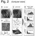
- FIG. 2 A basic scheme of the procedure is depicted in Fig. 2 .
- a subcellular "object of interest” (circular nano-object) is defined (manually) by its protrusion or elevation above mean surface level, here defined by 100 nm ⁇ h ⁇ 750 nm, see the green area under the profile curve of Fig. 2A .
- the in-plane-shape (round or fiber-like) assists in further classification. From a typical raw image of (20 ⁇ m) 2 ( Fig. 2B ) many "objects of interest” were identified by a trained software tool ( Fig. 2C ).
- the automation allowed to analyze a sufficient amount of data in an operator-independent manner and delivered robust results. It is to be noted that the object count was independent of the AFM-instrument manufacturer. The physical repeatability was high: deviation in object count was less than 10% through ten consecutive scans over the same area - including artifacts like scan lines or instrument drift etc. (data not shown). Overall, the recording and evaluation process is objective, stable and sensitive.
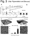
- the test cohort was classified following obvious macroscopic criteria: age, pigmentation and disease ( FIG. 3 ). Since an earlier AFM-report has assigned their appearance onto the age of subjects (see the article by Gorzelanny, C. et al.), this was performed first. Tape strips were collected of 21 healthy individuals that never had experienced any skin disease and subjected to the nano-object counting procedure ( Fig. 3A ). Samples were analyzed as described for Fig. 2 . DTI values were between 8.1 and 108.9. No correlation with age was detected. Three individuals with a higher score turned out to have relatives with a positive history of psoriasis or AD - hinting at a genetic contribution to corneocyte ultrastructure. Disregarding these outlier values yielded a healthy (normal) DTI value of 27 ⁇ 6.3.
- test cohort was grouped according to their pigmentation types A-D according to Fitzpatrick ( Fig. 3B ). No influence of pigmentation could be observed.
- Psoriasis is the cutaneous manifestation of a systemic disease of presumably autoimmune origin.
- One characteristic of Psoriasis is an accelerated turnover of keratinocytes at inflammatory plaques that show a preference for (but not being limited to) regions of thicker epidermis as on the elbow or knee - in contrast to atopic dermatitis, which is rather found where stratum corneum is thinner (inner side of forearm, face, neck etc.).
- Disease appearance and progression of psoriasis show an individually broad variation.
- a psoriatic skin can be characterized by the method of the invention even at clinically unaffected sites.
- Actinic keratosis is characterized by proliferation of keratinocytes, which is a consequence of UV-light exposure. It is controllable but may develop into a squamous cell carcinoma. Therefore, it is regarded as a "precancerosis". AK has just been accepted by German healthcare officials as an occupational disease.
- a volunteer diagnosed actinic keratosis was sampled on 1) a lesional region on the forearm, 2) a non-lesional region in close proximity (light-exposed area), and 3) a non-lesional region from a non-light exposed region (inner side of the arm close to the armpit).
- the DTI values of 1) 400, 2) 300, and 3) 100 demonstrate a clearly elevated value at non-lesional sites. This renders the method of the invention a toll for the early detection of UV-induced skin cancer.
- Contact dermatitis is an inflammatory reaction after exposure to detergents or other irritant compounds or materials. It is a socioeconomic problem for employees being exposed to chemicals at their workplace. In an experiment, healthy volunteers were exposed to a standard detergent solution of 0.1 % sodium dodecyl sulphate (SDS) for 24h.
- SDS sodium dodecyl sulphate
- the volunteer's DTI was as low as 40. After three days of SDS exposure, the many circular nano-objects CNOs arose such that the DTI increased to a value of 370, which is already half maximal. Hence, a toxic influence by detergents or other noxious conditions can be indicated by DTI.
Landscapes
- Health & Medical Sciences (AREA)
- Engineering & Computer Science (AREA)
- Life Sciences & Earth Sciences (AREA)
- Biomedical Technology (AREA)
- Immunology (AREA)
- Hematology (AREA)
- Chemical & Material Sciences (AREA)
- Urology & Nephrology (AREA)
- Molecular Biology (AREA)
- Tropical Medicine & Parasitology (AREA)
- Medicinal Chemistry (AREA)
- Microbiology (AREA)
- Biotechnology (AREA)
- Toxicology (AREA)
- Bioinformatics & Cheminformatics (AREA)
- Food Science & Technology (AREA)
- Cell Biology (AREA)
- Physics & Mathematics (AREA)
- Analytical Chemistry (AREA)
- Biochemistry (AREA)
- General Health & Medical Sciences (AREA)
- General Physics & Mathematics (AREA)
- Pathology (AREA)
- Investigating Or Analysing Biological Materials (AREA)
Claims (7)
- In-vitro-Verfahren zur Quantifizierung von Nanoobjekten von Hautzellen von Säugetieren umfassend die folgenden Schritte:a) Gewinnen von Korneozyten von Säugetieren;b) Analysieren der Oberflächen-Topographie der Korneozyten in nanoskaliger Auflösung;c) Identifizieren subzellulärer Objekte einer Höhe kleiner als 500 nm mit einem Zirkularitätsindex > 0,5 (zirkuläre Nanoobjekte, CNOs) und die eine Fläche kleiner als 1 µm2 aufweisen;d) Zählen dieser CNOs mittels einer automatischen Software-Routine;e) Beziehen der Anzahl an CNOs auf eine Flächeneinheit, um einen Dichteparameter zu erhalten, insbesondere einen strukturellen Index in Form eines dermalen topographischen Index (DTI).
- Verfahren nach Anspruch 1, dadurch gekennzeichnet, dass die Korneozyten gemäß Schritt a) gemäß Anspruch 1 durch eine der folgenden Verfahren gewonnen wurden:a) Abziehen eines Klebestreifens;b) Hautbiopsien;c) Hautschuppenablösung;d) Cyanacrylat-Abrisse;e) Kultivierung von Hautzellen in vitro (Keratinozyten).
- Verfahren nach Anspruch 1 oder 2, dadurch gekennzeichnet, dass die Nanoobjekte gemäß Schritt b) gemäß Anspruch 1 durch eine der folgenden Techniken analysiert werden:a) Rasterkraftmikroskopie (atomic force microscopy, AFM);b) Rasterelektronenmikroskopie (scanning electron microscopy, SEM);c) Digitale Holographie-Mikroskopie (digital holographic microscopy, DHM);d) Raman Mikroskopie;e) Konfokale Reflexionsmikroskopie;f) Fluoreszenz-Mikroskopie;g) Optische Interferometrie.
- Verfahren nach einem der vorhergehenden Ansprüche, dadurch gekennzeichnet, dass auch Oberflächenstrukturen mit einem Zirkularitätsindex von < 0,5 gezählt werden, um die Zählung der CNOs zu unterstützen und/oder zu komplementieren.
- Verwendung des In-vitro-Verfahrens nach einem der vorhergehenden Verfahrensansprüche zur Bestimmung einer gestörten Hautbarrierefunktion eines Individuums oder zur Bestimmung eines erhöhten entzündlichen Zustandes.
- Verwendung des In-vitro-Verfahrens nach einem der vorhergehenden Verfahrensansprüche als diagnostisches Marker-Verfahren für Hauterkrankungen.
- Verwendung des In-vitro-Verfahrens nach einem der vorhergehenden Verfahrensansprüche:a) zur Benennung oder Bestimmung eines individuellen Risikofaktors für die zukünftige Entstehung einer Hauterkrankung;b) zur Allergietestung, vorzugsweise ausgewählt aus Hautkrankheiten sowie Nahrungsmittelallergien;c) zur Bestimmung der Wirksamkeit oder der Nebenwirkungen von Wirkstoffen, einschließlich topisch sowie systemisch angewandter Wirkstoffe;d) zur Testung von hautreizenden Stoffen, z.B. bei Expositionen am Arbeitsplatz.e) zum Testen der Wirkung von kosmetischen Formulierungen, Cremes, Emulsionen, Sonnenschutzmitteln sowie Hautanwendungsmitteln im Allgemeinen.
Applications Claiming Priority (2)
| Application Number | Priority Date | Filing Date | Title |
|---|---|---|---|
| DE102015109345 | 2015-06-11 | ||
| PCT/EP2016/063187 WO2016198535A1 (en) | 2015-06-11 | 2016-06-09 | In vitro method for quantifying nano-objects of mammalian skin cells |
Publications (2)
| Publication Number | Publication Date |
|---|---|
| EP3308162A1 EP3308162A1 (de) | 2018-04-18 |
| EP3308162B1 true EP3308162B1 (de) | 2021-06-02 |
Family
ID=56121074
Family Applications (1)
| Application Number | Title | Priority Date | Filing Date |
|---|---|---|---|
| EP16728931.3A Active EP3308162B1 (de) | 2015-06-11 | 2016-06-09 | Verfahren zur in vitro quantifizierung von nanoobjekten von säugetierhautzellen |
Country Status (4)
| Country | Link |
|---|---|
| EP (1) | EP3308162B1 (de) |
| AU (1) | AU2016274659B2 (de) |
| DK (1) | DK3308162T3 (de) |
| WO (1) | WO2016198535A1 (de) |
Families Citing this family (3)
| Publication number | Priority date | Publication date | Assignee | Title |
|---|---|---|---|---|
| AU2019255069A1 (en) * | 2018-04-19 | 2020-11-26 | Saraya Co., Ltd. | Method and kit for assisting diagnosis of disease in subject |
| EP3877823A4 (de) | 2018-11-07 | 2022-07-27 | Trustees of Tufts College | Atomkraftmikroskopie zur identifizierung von oberflächen |
| EP4127645B1 (de) * | 2020-03-24 | 2025-08-13 | The Procter & Gamble Company | Verfahren zum testen von hautproben |
Family Cites Families (2)
| Publication number | Priority date | Publication date | Assignee | Title |
|---|---|---|---|---|
| KR100458148B1 (ko) | 2001-10-29 | 2004-11-26 | 포라 가세이 고교 가부시키가이샤 | 피부 분석 시스템 |
| GB0909128D0 (en) | 2009-05-28 | 2009-07-01 | Riethmueller Christoph | Imaging method and use thereof |
-
2016
- 2016-06-09 EP EP16728931.3A patent/EP3308162B1/de active Active
- 2016-06-09 AU AU2016274659A patent/AU2016274659B2/en active Active
- 2016-06-09 WO PCT/EP2016/063187 patent/WO2016198535A1/en not_active Ceased
- 2016-06-09 DK DK16728931.3T patent/DK3308162T3/da active
Non-Patent Citations (1)
| Title |
|---|
| None * |
Also Published As
| Publication number | Publication date |
|---|---|
| AU2016274659A1 (en) | 2018-01-18 |
| AU2016274659B2 (en) | 2022-01-06 |
| EP3308162A1 (de) | 2018-04-18 |
| WO2016198535A1 (en) | 2016-12-15 |
| DK3308162T3 (da) | 2021-08-09 |
Similar Documents
| Publication | Publication Date | Title |
|---|---|---|
| Évora et al. | Corneocytes: Relationship between structural and biomechanical properties | |
| Elsner | Methods for the assessment of barrier function | |
| Gambichler et al. | In vivo data of epidermal thickness evaluated by optical coherence tomography: effects of age, gender, skin type, and anatomic site | |
| Tranca et al. | Nanoscale mapping of refractive index by using scattering-type scanning near-field optical microscopy | |
| De Vries et al. | Dermal organization in scleroderma: the fast Fourier transform and the laser scatter method objectify fibrosis in nonlesional as well as lesional skin | |
| Cavalcanti et al. | Application of atomic force microscopy in the analysis of time since deposition (TSD) of red blood cells in bloodstains: A forensic analysis | |
| Franz et al. | Nanoscale alterations of corneocytes indicate skin disease | |
| EP3308162B1 (de) | Verfahren zur in vitro quantifizierung von nanoobjekten von säugetierhautzellen | |
| JP6499823B2 (ja) | 線維状構造分析に基づく肌状態の鑑別法 | |
| Egawa | Raman microscopy for skin evaluation | |
| Blume-Peytavi et al. | Hair growth assessment techniques | |
| Uehara et al. | Transepidermal water loss estimation model for evaluating skin barrier function | |
| Byrne | Bioengineering and subjective approaches to the clinical evaluation of dry skin | |
| Corniani et al. | Sub-surface deformation of individual fingerprint ridges during tactile interactions | |
| Park et al. | Two possible classifications of facial skin type by two parameters in Korean women: sebum excretion rate (SER) and skin surface relief (SSR) | |
| Liang et al. | In vivo multiphoton microscopy for investigating biomechanical properties of human skin | |
| WO2014020173A1 (en) | Surface isotropy as a marker for epidermal maturation | |
| EP3055643B1 (de) | Ein inspektionssystem basierend auf optischer kohärenztomographie im spektralbereich mit nanometer empfindlichkeit | |
| JP5977268B2 (ja) | 肌理の評価方法 | |
| Vargiolu et al. | Hair surface and mechanical properties of Copt mummies from Antinopolis | |
| Xhauflaire et al. | Highlighting the rim of the perifollicular epidermal unit. | |
| JP3667511B2 (ja) | ストレスの鑑別法 | |
| Lee et al. | Quantitative morphological and biochemical studies on human downy hairs using 3-D quantitative phase imaging | |
| JP2015158518A (ja) | 肌理の評価方法 | |
| Li et al. | A modified in vitro stripping method to automate the calculation of geometry of corneocytes imaged with fluorescent microscopy: example of moisturizer treatment |
Legal Events
| Date | Code | Title | Description |
|---|---|---|---|
| STAA | Information on the status of an ep patent application or granted ep patent |
Free format text: STATUS: THE INTERNATIONAL PUBLICATION HAS BEEN MADE |
|
| PUAI | Public reference made under article 153(3) epc to a published international application that has entered the european phase |
Free format text: ORIGINAL CODE: 0009012 |
|
| STAA | Information on the status of an ep patent application or granted ep patent |
Free format text: STATUS: REQUEST FOR EXAMINATION WAS MADE |
|
| 17P | Request for examination filed |
Effective date: 20171221 |
|
| AK | Designated contracting states |
Kind code of ref document: A1 Designated state(s): AL AT BE BG CH CY CZ DE DK EE ES FI FR GB GR HR HU IE IS IT LI LT LU LV MC MK MT NL NO PL PT RO RS SE SI SK SM TR |
|
| AX | Request for extension of the european patent |
Extension state: BA ME |
|
| DAV | Request for validation of the european patent (deleted) | ||
| DAX | Request for extension of the european patent (deleted) | ||
| STAA | Information on the status of an ep patent application or granted ep patent |
Free format text: STATUS: EXAMINATION IS IN PROGRESS |
|
| 17Q | First examination report despatched |
Effective date: 20190517 |
|
| GRAP | Despatch of communication of intention to grant a patent |
Free format text: ORIGINAL CODE: EPIDOSNIGR1 |
|
| STAA | Information on the status of an ep patent application or granted ep patent |
Free format text: STATUS: GRANT OF PATENT IS INTENDED |
|
| INTG | Intention to grant announced |
Effective date: 20201215 |
|
| GRAS | Grant fee paid |
Free format text: ORIGINAL CODE: EPIDOSNIGR3 |
|
| GRAA | (expected) grant |
Free format text: ORIGINAL CODE: 0009210 |
|
| STAA | Information on the status of an ep patent application or granted ep patent |
Free format text: STATUS: THE PATENT HAS BEEN GRANTED |
|
| REG | Reference to a national code |
Ref country code: CH Ref legal event code: EP |
|
| AK | Designated contracting states |
Kind code of ref document: B1 Designated state(s): AL AT BE BG CH CY CZ DE DK EE ES FI FR GB GR HR HU IE IS IT LI LT LU LV MC MK MT NL NO PL PT RO RS SE SI SK SM TR |
|
| REG | Reference to a national code |
Ref country code: GB Ref legal event code: FG4D |
|
| REG | Reference to a national code |
Ref country code: AT Ref legal event code: REF Ref document number: 1398938 Country of ref document: AT Kind code of ref document: T Effective date: 20210615 |
|
| REG | Reference to a national code |
Ref country code: IE Ref legal event code: FG4D |
|
| REG | Reference to a national code |
Ref country code: DE Ref legal event code: R096 Ref document number: 602016058808 Country of ref document: DE |
|
| REG | Reference to a national code |
Ref country code: DK Ref legal event code: T3 Effective date: 20210803 |
|
| REG | Reference to a national code |
Ref country code: NL Ref legal event code: FP |
|
| REG | Reference to a national code |
Ref country code: LT Ref legal event code: MG9D |
|
| PG25 | Lapsed in a contracting state [announced via postgrant information from national office to epo] |
Ref country code: LT Free format text: LAPSE BECAUSE OF FAILURE TO SUBMIT A TRANSLATION OF THE DESCRIPTION OR TO PAY THE FEE WITHIN THE PRESCRIBED TIME-LIMIT Effective date: 20210602 Ref country code: FI Free format text: LAPSE BECAUSE OF FAILURE TO SUBMIT A TRANSLATION OF THE DESCRIPTION OR TO PAY THE FEE WITHIN THE PRESCRIBED TIME-LIMIT Effective date: 20210602 Ref country code: BG Free format text: LAPSE BECAUSE OF FAILURE TO SUBMIT A TRANSLATION OF THE DESCRIPTION OR TO PAY THE FEE WITHIN THE PRESCRIBED TIME-LIMIT Effective date: 20210902 Ref country code: HR Free format text: LAPSE BECAUSE OF FAILURE TO SUBMIT A TRANSLATION OF THE DESCRIPTION OR TO PAY THE FEE WITHIN THE PRESCRIBED TIME-LIMIT Effective date: 20210602 |
|
| REG | Reference to a national code |
Ref country code: AT Ref legal event code: MK05 Ref document number: 1398938 Country of ref document: AT Kind code of ref document: T Effective date: 20210602 |
|
| PG25 | Lapsed in a contracting state [announced via postgrant information from national office to epo] |
Ref country code: RS Free format text: LAPSE BECAUSE OF FAILURE TO SUBMIT A TRANSLATION OF THE DESCRIPTION OR TO PAY THE FEE WITHIN THE PRESCRIBED TIME-LIMIT Effective date: 20210602 Ref country code: SE Free format text: LAPSE BECAUSE OF FAILURE TO SUBMIT A TRANSLATION OF THE DESCRIPTION OR TO PAY THE FEE WITHIN THE PRESCRIBED TIME-LIMIT Effective date: 20210602 Ref country code: NO Free format text: LAPSE BECAUSE OF FAILURE TO SUBMIT A TRANSLATION OF THE DESCRIPTION OR TO PAY THE FEE WITHIN THE PRESCRIBED TIME-LIMIT Effective date: 20210902 Ref country code: PL Free format text: LAPSE BECAUSE OF FAILURE TO SUBMIT A TRANSLATION OF THE DESCRIPTION OR TO PAY THE FEE WITHIN THE PRESCRIBED TIME-LIMIT Effective date: 20210602 Ref country code: LV Free format text: LAPSE BECAUSE OF FAILURE TO SUBMIT A TRANSLATION OF THE DESCRIPTION OR TO PAY THE FEE WITHIN THE PRESCRIBED TIME-LIMIT Effective date: 20210602 Ref country code: GR Free format text: LAPSE BECAUSE OF FAILURE TO SUBMIT A TRANSLATION OF THE DESCRIPTION OR TO PAY THE FEE WITHIN THE PRESCRIBED TIME-LIMIT Effective date: 20210903 |
|
| PG25 | Lapsed in a contracting state [announced via postgrant information from national office to epo] |
Ref country code: CZ Free format text: LAPSE BECAUSE OF FAILURE TO SUBMIT A TRANSLATION OF THE DESCRIPTION OR TO PAY THE FEE WITHIN THE PRESCRIBED TIME-LIMIT Effective date: 20210602 Ref country code: EE Free format text: LAPSE BECAUSE OF FAILURE TO SUBMIT A TRANSLATION OF THE DESCRIPTION OR TO PAY THE FEE WITHIN THE PRESCRIBED TIME-LIMIT Effective date: 20210602 Ref country code: SM Free format text: LAPSE BECAUSE OF FAILURE TO SUBMIT A TRANSLATION OF THE DESCRIPTION OR TO PAY THE FEE WITHIN THE PRESCRIBED TIME-LIMIT Effective date: 20210602 Ref country code: SK Free format text: LAPSE BECAUSE OF FAILURE TO SUBMIT A TRANSLATION OF THE DESCRIPTION OR TO PAY THE FEE WITHIN THE PRESCRIBED TIME-LIMIT Effective date: 20210602 Ref country code: ES Free format text: LAPSE BECAUSE OF FAILURE TO SUBMIT A TRANSLATION OF THE DESCRIPTION OR TO PAY THE FEE WITHIN THE PRESCRIBED TIME-LIMIT Effective date: 20210602 Ref country code: RO Free format text: LAPSE BECAUSE OF FAILURE TO SUBMIT A TRANSLATION OF THE DESCRIPTION OR TO PAY THE FEE WITHIN THE PRESCRIBED TIME-LIMIT Effective date: 20210602 Ref country code: PT Free format text: LAPSE BECAUSE OF FAILURE TO SUBMIT A TRANSLATION OF THE DESCRIPTION OR TO PAY THE FEE WITHIN THE PRESCRIBED TIME-LIMIT Effective date: 20211004 Ref country code: AT Free format text: LAPSE BECAUSE OF FAILURE TO SUBMIT A TRANSLATION OF THE DESCRIPTION OR TO PAY THE FEE WITHIN THE PRESCRIBED TIME-LIMIT Effective date: 20210602 |
|
| REG | Reference to a national code |
Ref country code: DE Ref legal event code: R097 Ref document number: 602016058808 Country of ref document: DE |
|
| REG | Reference to a national code |
Ref country code: BE Ref legal event code: MM Effective date: 20210630 |
|
| PG25 | Lapsed in a contracting state [announced via postgrant information from national office to epo] |
Ref country code: MC Free format text: LAPSE BECAUSE OF FAILURE TO SUBMIT A TRANSLATION OF THE DESCRIPTION OR TO PAY THE FEE WITHIN THE PRESCRIBED TIME-LIMIT Effective date: 20210602 Ref country code: LU Free format text: LAPSE BECAUSE OF NON-PAYMENT OF DUE FEES Effective date: 20210609 |
|
| PLBE | No opposition filed within time limit |
Free format text: ORIGINAL CODE: 0009261 |
|
| STAA | Information on the status of an ep patent application or granted ep patent |
Free format text: STATUS: NO OPPOSITION FILED WITHIN TIME LIMIT |
|
| PG25 | Lapsed in a contracting state [announced via postgrant information from national office to epo] |
Ref country code: IE Free format text: LAPSE BECAUSE OF NON-PAYMENT OF DUE FEES Effective date: 20210609 |
|
| 26N | No opposition filed |
Effective date: 20220303 |
|
| PG25 | Lapsed in a contracting state [announced via postgrant information from national office to epo] |
Ref country code: AL Free format text: LAPSE BECAUSE OF FAILURE TO SUBMIT A TRANSLATION OF THE DESCRIPTION OR TO PAY THE FEE WITHIN THE PRESCRIBED TIME-LIMIT Effective date: 20210602 |
|
| PG25 | Lapsed in a contracting state [announced via postgrant information from national office to epo] |
Ref country code: IT Free format text: LAPSE BECAUSE OF FAILURE TO SUBMIT A TRANSLATION OF THE DESCRIPTION OR TO PAY THE FEE WITHIN THE PRESCRIBED TIME-LIMIT Effective date: 20210602 Ref country code: BE Free format text: LAPSE BECAUSE OF NON-PAYMENT OF DUE FEES Effective date: 20210630 |
|
| PG25 | Lapsed in a contracting state [announced via postgrant information from national office to epo] |
Ref country code: CY Free format text: LAPSE BECAUSE OF FAILURE TO SUBMIT A TRANSLATION OF THE DESCRIPTION OR TO PAY THE FEE WITHIN THE PRESCRIBED TIME-LIMIT Effective date: 20210602 |
|
| PG25 | Lapsed in a contracting state [announced via postgrant information from national office to epo] |
Ref country code: HU Free format text: LAPSE BECAUSE OF FAILURE TO SUBMIT A TRANSLATION OF THE DESCRIPTION OR TO PAY THE FEE WITHIN THE PRESCRIBED TIME-LIMIT; INVALID AB INITIO Effective date: 20160609 |
|
| PG25 | Lapsed in a contracting state [announced via postgrant information from national office to epo] |
Ref country code: MK Free format text: LAPSE BECAUSE OF FAILURE TO SUBMIT A TRANSLATION OF THE DESCRIPTION OR TO PAY THE FEE WITHIN THE PRESCRIBED TIME-LIMIT Effective date: 20210602 |
|
| PG25 | Lapsed in a contracting state [announced via postgrant information from national office to epo] |
Ref country code: MT Free format text: LAPSE BECAUSE OF FAILURE TO SUBMIT A TRANSLATION OF THE DESCRIPTION OR TO PAY THE FEE WITHIN THE PRESCRIBED TIME-LIMIT Effective date: 20210602 |
|
| PGFP | Annual fee paid to national office [announced via postgrant information from national office to epo] |
Ref country code: DE Payment date: 20250516 Year of fee payment: 10 |
|
| PGFP | Annual fee paid to national office [announced via postgrant information from national office to epo] |
Ref country code: GB Payment date: 20250621 Year of fee payment: 10 Ref country code: DK Payment date: 20250620 Year of fee payment: 10 |
|
| PGFP | Annual fee paid to national office [announced via postgrant information from national office to epo] |
Ref country code: NL Payment date: 20250621 Year of fee payment: 10 |
|
| PGFP | Annual fee paid to national office [announced via postgrant information from national office to epo] |
Ref country code: FR Payment date: 20250617 Year of fee payment: 10 |
|
| PGFP | Annual fee paid to national office [announced via postgrant information from national office to epo] |
Ref country code: CH Payment date: 20250701 Year of fee payment: 10 |