EP3055643B1 - Ein inspektionssystem basierend auf optischer kohärenztomographie im spektralbereich mit nanometer empfindlichkeit - Google Patents
Ein inspektionssystem basierend auf optischer kohärenztomographie im spektralbereich mit nanometer empfindlichkeit Download PDFInfo
- Publication number
- EP3055643B1 EP3055643B1 EP14781556.7A EP14781556A EP3055643B1 EP 3055643 B1 EP3055643 B1 EP 3055643B1 EP 14781556 A EP14781556 A EP 14781556A EP 3055643 B1 EP3055643 B1 EP 3055643B1
- Authority
- EP
- European Patent Office
- Prior art keywords
- oct
- axial
- images
- image
- spectra
- Prior art date
- Legal status (The legal status is an assumption and is not a legal conclusion. Google has not performed a legal analysis and makes no representation as to the accuracy of the status listed.)
- Not-in-force
Links
- 238000012014 optical coherence tomography Methods 0.000 title claims description 66
- 238000007689 inspection Methods 0.000 title description 3
- 238000001228 spectrum Methods 0.000 claims description 46
- 238000000034 method Methods 0.000 claims description 25
- 230000035945 sensitivity Effects 0.000 claims description 20
- 230000005855 radiation Effects 0.000 claims description 16
- 238000003384 imaging method Methods 0.000 claims description 13
- 230000003595 spectral effect Effects 0.000 claims description 12
- 238000013507 mapping Methods 0.000 claims description 8
- 238000005286 illumination Methods 0.000 claims description 3
- 238000000354 decomposition reaction Methods 0.000 claims description 2
- 239000000523 sample Substances 0.000 description 15
- 239000002077 nanosphere Substances 0.000 description 14
- 238000013459 approach Methods 0.000 description 8
- 230000003287 optical effect Effects 0.000 description 8
- 210000001519 tissue Anatomy 0.000 description 6
- 238000002474 experimental method Methods 0.000 description 5
- 238000001727 in vivo Methods 0.000 description 5
- 239000000463 material Substances 0.000 description 5
- 230000017531 blood circulation Effects 0.000 description 4
- 238000000386 microscopy Methods 0.000 description 4
- PEDCQBHIVMGVHV-UHFFFAOYSA-N Glycerine Chemical compound OCC(O)CO PEDCQBHIVMGVHV-UHFFFAOYSA-N 0.000 description 3
- 230000004075 alteration Effects 0.000 description 3
- 210000004905 finger nail Anatomy 0.000 description 3
- 239000011521 glass Substances 0.000 description 3
- 229940028435 intralipid Drugs 0.000 description 3
- 238000000149 argon plasma sintering Methods 0.000 description 2
- 238000012512 characterization method Methods 0.000 description 2
- 238000010586 diagram Methods 0.000 description 2
- 238000006073 displacement reaction Methods 0.000 description 2
- 238000011503 in vivo imaging Methods 0.000 description 2
- 238000005259 measurement Methods 0.000 description 2
- 239000002184 metal Substances 0.000 description 2
- 239000004033 plastic Substances 0.000 description 2
- 238000012545 processing Methods 0.000 description 2
- 238000012800 visualization Methods 0.000 description 2
- 230000005653 Brownian motion process Effects 0.000 description 1
- 108010077544 Chromatin Proteins 0.000 description 1
- 229910000530 Gallium indium arsenide Inorganic materials 0.000 description 1
- 241001465754 Metazoa Species 0.000 description 1
- 206010028980 Neoplasm Diseases 0.000 description 1
- 208000037273 Pathologic Processes Diseases 0.000 description 1
- 239000004793 Polystyrene Substances 0.000 description 1
- 241001351225 Sergey Species 0.000 description 1
- 230000005540 biological transmission Effects 0.000 description 1
- 238000005537 brownian motion Methods 0.000 description 1
- 201000011510 cancer Diseases 0.000 description 1
- 210000004027 cell Anatomy 0.000 description 1
- 238000004113 cell culture Methods 0.000 description 1
- 230000004663 cell proliferation Effects 0.000 description 1
- 230000001413 cellular effect Effects 0.000 description 1
- 210000003483 chromatin Anatomy 0.000 description 1
- 238000010276 construction Methods 0.000 description 1
- 230000003247 decreasing effect Effects 0.000 description 1
- 239000003814 drug Substances 0.000 description 1
- 239000000835 fiber Substances 0.000 description 1
- 210000000245 forearm Anatomy 0.000 description 1
- 238000011835 investigation Methods 0.000 description 1
- 238000002372 labelling Methods 0.000 description 1
- 230000004089 microcirculation Effects 0.000 description 1
- 238000004377 microelectronic Methods 0.000 description 1
- 239000002086 nanomaterial Substances 0.000 description 1
- 238000012634 optical imaging Methods 0.000 description 1
- 238000012856 packing Methods 0.000 description 1
- 230000009054 pathological process Effects 0.000 description 1
- 230000010287 polarization Effects 0.000 description 1
- 229920002223 polystyrene Polymers 0.000 description 1
- 238000002360 preparation method Methods 0.000 description 1
- 230000001105 regulatory effect Effects 0.000 description 1
- 239000007787 solid Substances 0.000 description 1
- 239000000725 suspension Substances 0.000 description 1
- 230000002123 temporal effect Effects 0.000 description 1
- 238000012360 testing method Methods 0.000 description 1
- 238000003325 tomography Methods 0.000 description 1
- 210000005166 vasculature Anatomy 0.000 description 1
Images


Classifications
-
- G—PHYSICS
- G01—MEASURING; TESTING
- G01B—MEASURING LENGTH, THICKNESS OR SIMILAR LINEAR DIMENSIONS; MEASURING ANGLES; MEASURING AREAS; MEASURING IRREGULARITIES OF SURFACES OR CONTOURS
- G01B9/00—Measuring instruments characterised by the use of optical techniques
- G01B9/02—Interferometers
- G01B9/02041—Interferometers characterised by particular imaging or detection techniques
- G01B9/02044—Imaging in the frequency domain, e.g. by using a spectrometer
-
- A—HUMAN NECESSITIES
- A61—MEDICAL OR VETERINARY SCIENCE; HYGIENE
- A61B—DIAGNOSIS; SURGERY; IDENTIFICATION
- A61B5/00—Measuring for diagnostic purposes; Identification of persons
- A61B5/0059—Measuring for diagnostic purposes; Identification of persons using light, e.g. diagnosis by transillumination, diascopy, fluorescence
- A61B5/0062—Arrangements for scanning
- A61B5/0066—Optical coherence imaging
-
- A—HUMAN NECESSITIES
- A61—MEDICAL OR VETERINARY SCIENCE; HYGIENE
- A61B—DIAGNOSIS; SURGERY; IDENTIFICATION
- A61B5/00—Measuring for diagnostic purposes; Identification of persons
- A61B5/72—Signal processing specially adapted for physiological signals or for diagnostic purposes
- A61B5/7235—Details of waveform analysis
- A61B5/7253—Details of waveform analysis characterised by using transforms
- A61B5/7257—Details of waveform analysis characterised by using transforms using Fourier transforms
-
- G—PHYSICS
- G01—MEASURING; TESTING
- G01B—MEASURING LENGTH, THICKNESS OR SIMILAR LINEAR DIMENSIONS; MEASURING ANGLES; MEASURING AREAS; MEASURING IRREGULARITIES OF SURFACES OR CONTOURS
- G01B9/00—Measuring instruments characterised by the use of optical techniques
- G01B9/02—Interferometers
- G01B9/02001—Interferometers characterised by controlling or generating intrinsic radiation properties
- G01B9/02002—Interferometers characterised by controlling or generating intrinsic radiation properties using two or more frequencies
- G01B9/02004—Interferometers characterised by controlling or generating intrinsic radiation properties using two or more frequencies using frequency scans
-
- G—PHYSICS
- G01—MEASURING; TESTING
- G01B—MEASURING LENGTH, THICKNESS OR SIMILAR LINEAR DIMENSIONS; MEASURING ANGLES; MEASURING AREAS; MEASURING IRREGULARITIES OF SURFACES OR CONTOURS
- G01B9/00—Measuring instruments characterised by the use of optical techniques
- G01B9/02—Interferometers
- G01B9/02083—Interferometers characterised by particular signal processing and presentation
- G01B9/02087—Combining two or more images of the same region
-
- G—PHYSICS
- G01—MEASURING; TESTING
- G01B—MEASURING LENGTH, THICKNESS OR SIMILAR LINEAR DIMENSIONS; MEASURING ANGLES; MEASURING AREAS; MEASURING IRREGULARITIES OF SURFACES OR CONTOURS
- G01B9/00—Measuring instruments characterised by the use of optical techniques
- G01B9/02—Interferometers
- G01B9/0209—Low-coherence interferometers
- G01B9/02091—Tomographic interferometers, e.g. based on optical coherence
-
- G—PHYSICS
- G01—MEASURING; TESTING
- G01N—INVESTIGATING OR ANALYSING MATERIALS BY DETERMINING THEIR CHEMICAL OR PHYSICAL PROPERTIES
- G01N21/00—Investigating or analysing materials by the use of optical means, i.e. using sub-millimetre waves, infrared, visible or ultraviolet light
- G01N21/17—Systems in which incident light is modified in accordance with the properties of the material investigated
- G01N21/47—Scattering, i.e. diffuse reflection
- G01N21/4795—Scattering, i.e. diffuse reflection spatially resolved investigating of object in scattering medium
-
- G—PHYSICS
- G01—MEASURING; TESTING
- G01N—INVESTIGATING OR ANALYSING MATERIALS BY DETERMINING THEIR CHEMICAL OR PHYSICAL PROPERTIES
- G01N21/00—Investigating or analysing materials by the use of optical means, i.e. using sub-millimetre waves, infrared, visible or ultraviolet light
- G01N21/17—Systems in which incident light is modified in accordance with the properties of the material investigated
- G01N2021/178—Methods for obtaining spatial resolution of the property being measured
- G01N2021/1785—Three dimensional
- G01N2021/1787—Tomographic, i.e. computerised reconstruction from projective measurements
Definitions
- the invention relates to inspection of a variety of objects, including body tissue (also in vivo ), cell cultures, materials for material science, and micro and nano electronics components, for example.
- OCT optical coherence tomography
- OCT provides non-invasive, contactless, depth resolving imaging of the object's internal structure.
- OCT facilitates cellular-level structural and functional imaging of living animal and human tissue as well as micro level imaging of different materials, but the structural sensitivity and resolution are limited to the microscale.
- phase OCT phase OCT
- vibration measurements in the ear at the nanoscale have been demonstrated, and the ability of OCT to sense nanoscale structural alteration in weakly scattering media has been discussed [1].
- the limitation to weakly scattering media generally excludes application to human tissue and to many materials of interest. describes visualization of the dominant structure (which corresponds to just one spatial frequency in depth direction) for each pixel of 2D image as a corresponding colour.
- QPM quantitative phase microscopy
- the invention is directed towards achieving improved sensitivity of 3D imaging for medical diagnostics, material science, nanofabrication, microelectronics.
- an OCT imaging method as set out in claim 1.
- the spectrum of step (a) is a full complex spectrum.
- the spectrum is formed by using quasi-collimated radiation for illumination.
- the spectrum is formed by spectral domain OCT (SDOCT).
- SDOCT spectral domain OCT
- the spectrum is formed by swept source OCT (SSOCT).
- SSOCT swept source OCT
- the step (a) includes decomposition of the spectrum of axial spatial frequencies into zones, performing reconstruction of image depth profiles for each zone, and measuring signals at each point in each reconstructed image depth profile.
- each zone corresponds to a narrow bandwidth of spatial frequencies and is considered as a single spatial frequency.
- reconstruction of corresponding images (axial Z (depth)-profiles) for each zone is performed via an inverse Fourier transform.
- step (b) the controller forms OCT 2D images and step (d) includes mapping spectra of spatial frequencies into each pixel of each of a number of said 2D images.
- the controller selects the information parameters to be calculated in step (e) according to a required application.
- the information parameters include one or more of maximum spatial frequency, dominant spatial period, centre of mass, medium spatial frequency and medium spatial period, correlation between axial spectra.
- step (b) the controller forms 2D or 3D images, and the magnitude of at least some calculated information parameters is matched to colour of each pixel of said 2D images or each voxel of said 3D OCT images.
- a sequence of OCT images in time is recorded.
- step (b) local axial spectra of step (b) are reconstructed and analysed in time.
- time dependences of calculated informative parameters for each pixel of an OCT 2D image or each voxel of a 3D OCT image are calculated and plotted.
- the radiation receiver is a 2D detector and spectral interferograms are formed simultaneously for all image points.
- the invention provides an OCT system comprising a radiation emitter, a radiation detector, an interferometer, and a controller adapted to perform the steps of a method as defined above in any embodiment.
- An OCT imaging system comprising a radiation emitter, a radiation receiver, a controller which controls the emitter and processes received radiation data according to OCT to provide an output image.
- the controller forms a spectrum of spatial frequencies along the depth direction. It calculates from the spectrum local spectra of spatial frequencies or periods along the depth direction for individual volume elements along the depth direction. It translates the local spectra of spatial frequencies or periods along the depth direction into the OCT image domain. It maps the local spectra into the volume elements to provide a sensitivity on the nano-scale whereas the volume elements are in the micro-scale.
- the controller calculates information parameters from the translated and mapped local spectra.
- the system achieves nano-scale sensitivity although the volume elements are at the micro-scale.
- the invention achieves nano-scale sensitivity OCT to structural changes, termed nano-scale sensitivity OCT ("nsOCT").
- the OCT signal is formed by light scattered from high axial spatial frequency components of the object's scattering potential which correspond to submicron structure.
- the axial Fourier spectrum of the object's scattering potential is very informative and highly sensitive to structural changes because the optical system, if the required spectral transmission is provided, does not impose any limitations on the bandwidth of translated axial spatial frequencies.
- the range of axial spatial frequencies is limited by spectral bandwidth and the resolution of spatial frequencies is limited by spectral resolution.
- conventional OCT during the inverse Fourier transform to reconstruct axial profile, the spatial information is integrated and, as a result, the resolution and sensitivity even for the best OCT systems are relatively poor.
- the local axial Fourier spectra are directly translated into each voxel of the 3D OCT image.
- submicron structure can be visualized and nanoscale structural alterations within each voxel can be detected.
- Different informative parameters can be extracted from the local profiles of the axial spatial periods to characterize structure, depending on application, and mapped into the OCT image to form a colour map.
- one of such parameters can be the maximum spatial frequency (period), which is the frequency at the maximal signal, which is directly related to the dominant size of the local structure.
- a spectral domain OCT (SD-OCT) setup such as that of Fig. 6 with low NA optics (see Methods) was used.
- the voxel size in OCT images was 12 ⁇ m x 30 ⁇ m x 30 ⁇ m
- the spatial interval to reconstruct profiles of the axial spatial periods was 4 nm
- the voxel size was 50 ⁇ m x 30 ⁇ m x 30 ⁇ m.
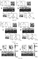
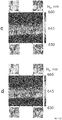
- the top layer contains nanospheres with 650 nm mean diameter and the bottom layer - with 670 nm mean diameter.
- These layers of nanosphere aggregates whose sizes are well beyond the resolution limit of the OCT system, are indistinguishable in the conventional OCT image Fig. 1a and, of course, the difference between them of 20 nm cannot be detected.
- the nsOCT approach shows that in most points in the bottom layer, the maximum of the profiles of the axial spatial periods was shifted to larger values (larger dominant sizes of the structure) relative to corresponding profiles in the top layer.
- Fig. 1a shows the plot of two of these selected points for the bottom and top layers, respectively.
- Fig. 1b the nsOCT image as a map of the maximal axial spatial periods (dominant axial structure sizes) is shown.
- Fig. 1c the same results are presented when the layers were flipped.
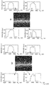
- a second experiment demonstrates the ability of nsOCT to detect nanoscale structural changes in time. Structural changes less than 30 nm within the scattering sample were made as described in Methods. We recorded 50 B-scans before and 50 B-scans after structural changes were made. Images in Fig. 2 demonstrate that there are no detectable structural changes at selected locations between second ( Fig. 2a, e ) and last ( Fig. 2b, f ) frames before, as well as between second ( Fig. 2c , g ) and last ( Fig. 2d , h ) frames after structural changes were made. The profiles of the axial spatial periods ( Fig.
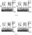
- FIG. 4 an example of nsOCT application to in vivo imaging of human skin is presented.
- the time interval between images Fig. 4a , c and Fig. 4b , d was 20 ms, the same as for our model experiment ( Fig.3 ).
- the most likely reason for such structural changes is blood flow.
- the information about changes in axial spatial period profiles can be used for determination of the blood flow velocity.
- nsOCT setup was based on the SD-OCT setup shown in Fig. 6 .
- a broadband 1310 nm superluminescent diode SLD with bandwidth of 83 nm (SLD, Dense Light, Singapore) was coupled into the interferometer, via an optical coupler OC.
- the spectrometer consisted of a 50 mm focal length collimator, a 1145 lines/mm transmitting grating, an achromatic lens with a 100 mm focal length and a 14-bit, 1024 pixels InGaAs line scan camera (SU1024LDH2, Goodrich Ltd. USA) with a maximum acquisition rate of 91 kHz.
- This spectrometer setup had a spectral resolution of 0.1432 nm, which gave a maximum imaging range of ⁇ 6 mm (in air).
- the measured sensitivity of the system was ⁇ 105 dB near the zero-delay line.
- the sensitivity drop off of the system was ⁇ 20 dB at a depth range ⁇ 3 mm.
- the measured axial imaging resolution of the system was ⁇ 12 ⁇ m in air ( ⁇ 8.6 ⁇ m in human skin) and a lateral resolution was ⁇ 30 ⁇ m.
- nsOCT images were formed as maps of the maximal spatial periods for each voxel.
- the voxel size in nsOCT images was 50 ⁇ m x 30 ⁇ m x 30 ⁇ m and spatial interval to reconstruct profiles of the axial spatial periods was 4 nm.
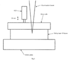
- the sample which consists of ten layers of scattering sticky tape, was placed on the solid stable basis (metal plate), as shown in Fig. 5 .
- Metal plate was rigorously fixed to the bench.
- the distance between low NA Illumination beam from OCT system and PZT tip was about 10 mm.
- Fifty OCT images (B-scans) were taken before and after 30nm shift was applied.
- the invention takes a very different approach from those described in the prior art. For example, referring to documents [10] and [11] these do not describe or suggest forming a depth resolved spectrum of spatial frequencies along the depth direction, calculating from the spectrum local spectra of spatial frequencies or periods along the depth direction for individual volume elements along the depth direction, and mapping the local spectra into the volume elements to provide a sensitivity on a scale smaller by at least one order of magnitude than that of the volume elements, and calculating information parameters from the translated and mapped local spectra.
- the invention provides extracting the local spectra of spatial frequencies in the depth direction (depth profiles) and mapping them to each voxel of the 3D reconstructed image.
- the invention achieves a very large improvement in sensitivity of OCT to structural changes. Improvement of more than 300 times has been demonstrated; using OCT system with resolution 12 ⁇ m x 30 ⁇ m x 30 ⁇ m we were able to detect the size difference between nanosphere aggregates as small as 20 nm and temporal structural changes within scattering samples less than 30 nm. The sensitivity is limited by the spectral resolution and can go far beyond what we have demonstrated here.
Landscapes
- Health & Medical Sciences (AREA)
- Physics & Mathematics (AREA)
- Life Sciences & Earth Sciences (AREA)
- General Physics & Mathematics (AREA)
- Engineering & Computer Science (AREA)
- General Health & Medical Sciences (AREA)
- Pathology (AREA)
- Signal Processing (AREA)
- Radiology & Medical Imaging (AREA)
- Nuclear Medicine, Radiotherapy & Molecular Imaging (AREA)
- Veterinary Medicine (AREA)
- Biophysics (AREA)
- Public Health (AREA)
- Biomedical Technology (AREA)
- Heart & Thoracic Surgery (AREA)
- Medical Informatics (AREA)
- Molecular Biology (AREA)
- Surgery (AREA)
- Animal Behavior & Ethology (AREA)
- Physiology (AREA)
- Psychiatry (AREA)
- Computer Vision & Pattern Recognition (AREA)
- Optics & Photonics (AREA)
- Chemical & Material Sciences (AREA)
- Analytical Chemistry (AREA)
- Biochemistry (AREA)
- Immunology (AREA)
- Artificial Intelligence (AREA)
- Mathematical Physics (AREA)
- Investigating Or Analysing Materials By Optical Means (AREA)
Claims (14)
- OCT-Bildgebungsverfahren, durchgeführt von einem OCT-System, beinhaltend einen Strahlungssender, einen Strahlungsempfänger, ein Interferometer und eine Steuerung, die angepasst ist, um den Sender zu steuern und um empfangene Strahlungsdaten gemäß Fourier-Domain-OCT zu verarbeiten, um ein Ausgabebild eines Objekts bereitzustellen, und wobei das Verfahren die folgenden Schritte der Steuerung umfasst:a(1). Bilden eines herkömmlichen 3D-Fourier-Domain-OCT-Bilds aus axialen FourierSpektren,a(2). Bestimmen eines Spektrums axialer räumlicher Frequenzen entlang einer Tiefenrichtung aus jedem axialen Fourier-Spektrum;b. Berechnen, aus jedem bestimmten Spektrum von Schritt (a(2)), lokaler axialer Spektren räumlicher Frequenzen oder von Perioden entlang der Tiefenrichtung für einzelne Volumenelemente des 3D-OCT-Bilds;c. Übertragen der lokalen axialen Spektren von Schritt (b) in die Fourier-Domain-OCT- Bild-Domain;d. Abbilden der übertragenen lokalen axialen Spektren in den Volumenelementen des 3D-OCT-Bilds, um eine Empfindlichkeit auf einer Skala bereitzustellen, die um mindestens eine Größenordnung kleiner ist als die der Volumenelemente; unde. Berechnen von Informationsparametern auf der Struktur des Objekts aus den übertragenen und abgebildeten lokalen Spektren von Schritt (c) und (d).
- Verfahren nach Anspruch 1, wobei das Spektrum von Schritt (a) ein vollständiges komplexes Spektrum ist.
- Verfahren nach einem der vorangehenden Ansprüche, wobei das Spektrum unter Verwendung von quasi-kollimierter Strahlung zur Beleuchtung gebildet wird.
- Verfahren nach einem der vorangehenden Ansprüche, wobei das Spektrum durch Spectral-Domain-OCT, SDOCT, oder durch Swept-Source-OCT, SSOCT, gebildet wird.
- Verfahren nach einem der vorangehenden Ansprüche, wobei Schritt (a) Folgendes beinhaltet: die Zerlegung des Spektrums axialer räumlicher Frequenzen in Zonen, das Durchführen einer Rekonstruktion von Bildtiefenprofilen für jede Zone und das Messen von Signalen an jedem Punkt in jedem rekonstruierten Bildtiefenprofil, und wobei optional jede Zone einer schmalen Bandbreite räumlicher Frequenzen entspricht und als eine einzelne räumliche Frequenz betrachtet wird.
- Verfahren nach Anspruch 5, wobei die Rekonstruktion entsprechender Bilder, die axiale z-Profile sind, für jede Zone über eine inverse Fourier-Transformation durchgeführt wird.
- Verfahren nach einem der vorangehenden Ansprüche, wobei in Schritt (b) die Steuerung OCT-2D-Bilder bildet und wobei Schritt (d) das Abbilden von Spektren räumlicher Frequenzen in jedem Pixel von jedem von einer Anzahl der 2D-Bilder beinhaltet.
- Verfahren nach einem der vorangehenden Ansprüche, wobei die Steuerung die Informationsparameter, die in Schritt (e) berechnet werden, gemäß einer erforderlichen Anwendung auswählt und wobei die Informationsparameter eines oder mehrere von Folgendem beinhalten: einer maximalen räumlichen Frequenz, einer dominanten räumlichen Periode, einem Masseschwerpunkt, einer mittleren räumlichen Frequenz und einer mittleren räumlichen Periode, einer Korrelation zwischen axialen Spektren.
- Verfahren nach einem der vorangehenden Ansprüche, wobei in Schritt (b) die Steuerung 2D- oder 3D-Bilder bildet und wobei die Größe von mindestens einigen berechneten Informationsparametern mit einer Farbe jedes Pixels der 2D-Bilder oder jedes Voxels der 3D-OCT-Bilder abgestimmt wird.
- Verfahren nach einem der vorangehenden Ansprüche, wobei eine Sequenz von OCT-Bildern zeitlich aufgezeichnet wird.
- Verfahren nach Anspruch 10, wobei lokale axiale Spektren von Schritt (b) rekonstruiert und zeitlich analysiert werden.
- Verfahren nach Anspruch 10 oder 11, wobei Zeitabhängigkeiten berechneter Informationsparameter für jedes Pixel eines OCT-2D-Bilds oder jedes Voxel eines 3D-OCT-Bilds berechnet und dargestellt werden.
- Verfahren nach einem der vorangehenden Ansprüche, wobei der Strahlungsempfänger ein 2D-Detektor ist und spektrale Interferogramme gleichzeitig für alle Bildpunkte gebildet werden.
- OCT-System (OCT = optische Kohärenztomographie), umfassend einen Strahlungssender, einen Strahlungsdetektor, ein Interferometer und eine Steuerung, die konfiguriert ist, um den Sender zu steuern und um anhand der Strahlung empfangene Strahlungsdaten zu verarbeiten, indem die Schritte eines Verfahrens nach einem der vorangehenden Ansprüche durchgeführt werden.
Priority Applications (1)
| Application Number | Priority Date | Filing Date | Title |
|---|---|---|---|
| EP14781556.7A EP3055643B1 (de) | 2013-10-11 | 2014-10-08 | Ein inspektionssystem basierend auf optischer kohärenztomographie im spektralbereich mit nanometer empfindlichkeit |
Applications Claiming Priority (3)
| Application Number | Priority Date | Filing Date | Title |
|---|---|---|---|
| EP13188342 | 2013-10-11 | ||
| EP14781556.7A EP3055643B1 (de) | 2013-10-11 | 2014-10-08 | Ein inspektionssystem basierend auf optischer kohärenztomographie im spektralbereich mit nanometer empfindlichkeit |
| PCT/EP2014/071536 WO2015052232A1 (en) | 2013-10-11 | 2014-10-08 | A nano-sensitive fourier-domain optical coherence tomography inspection system |
Publications (2)
| Publication Number | Publication Date |
|---|---|
| EP3055643A1 EP3055643A1 (de) | 2016-08-17 |
| EP3055643B1 true EP3055643B1 (de) | 2019-07-31 |
Family
ID=49354514
Family Applications (1)
| Application Number | Title | Priority Date | Filing Date |
|---|---|---|---|
| EP14781556.7A Not-in-force EP3055643B1 (de) | 2013-10-11 | 2014-10-08 | Ein inspektionssystem basierend auf optischer kohärenztomographie im spektralbereich mit nanometer empfindlichkeit |
Country Status (3)
| Country | Link |
|---|---|
| US (1) | US10012492B2 (de) |
| EP (1) | EP3055643B1 (de) |
| WO (1) | WO2015052232A1 (de) |
Cited By (1)
| Publication number | Priority date | Publication date | Assignee | Title |
|---|---|---|---|---|
| EP4242580A1 (de) | 2022-03-09 | 2023-09-13 | National University of Ireland Galway | Bildgebungsvorrichtung und -verfahren |
Families Citing this family (3)
| Publication number | Priority date | Publication date | Assignee | Title |
|---|---|---|---|---|
| EP3103386B1 (de) * | 2015-06-08 | 2019-04-10 | Tomey Corporation | Blutflussgeschwindigkeitsmessvorrichtung, programm und verfahren |
| US12207878B2 (en) | 2020-08-05 | 2025-01-28 | LighTopTech Corp. | Apparatus and methods for three dimensional optical imaging of dynamics with reduced motion artifacts |
| CN113281305B (zh) * | 2021-05-17 | 2023-04-14 | 太原理工大学 | 一种基于散射介质实现超分辨显微成像方法及其装置 |
Family Cites Families (4)
| Publication number | Priority date | Publication date | Assignee | Title |
|---|---|---|---|---|
| US7978346B1 (en) * | 2009-02-18 | 2011-07-12 | University Of Central Florida Research Foundation, Inc. | Methods and systems for realizing high resolution three-dimensional optical imaging |
| US9823127B2 (en) * | 2010-01-22 | 2017-11-21 | Duke University | Systems and methods for deep spectroscopic imaging of biological samples with use of an interferometer and spectrometer |
| US9332902B2 (en) * | 2012-01-20 | 2016-05-10 | Carl Zeiss Meditec, Inc. | Line-field holoscopy |
| WO2013169934A1 (en) * | 2012-05-08 | 2013-11-14 | King Abdullah University Of Science And Technology | Submicron resolution spectral-domain optical coherence tomography |
-
2014
- 2014-10-08 US US15/027,056 patent/US10012492B2/en active Active
- 2014-10-08 WO PCT/EP2014/071536 patent/WO2015052232A1/en not_active Ceased
- 2014-10-08 EP EP14781556.7A patent/EP3055643B1/de not_active Not-in-force
Non-Patent Citations (1)
| Title |
|---|
| None * |
Cited By (2)
| Publication number | Priority date | Publication date | Assignee | Title |
|---|---|---|---|---|
| EP4242580A1 (de) | 2022-03-09 | 2023-09-13 | National University of Ireland Galway | Bildgebungsvorrichtung und -verfahren |
| WO2023170243A1 (en) | 2022-03-09 | 2023-09-14 | National University Of Ireland, Galway | Imaging apparatus and method |
Also Published As
| Publication number | Publication date |
|---|---|
| US10012492B2 (en) | 2018-07-03 |
| US20160238370A1 (en) | 2016-08-18 |
| EP3055643A1 (de) | 2016-08-17 |
| WO2015052232A1 (en) | 2015-04-16 |
Similar Documents
| Publication | Publication Date | Title |
|---|---|---|
| Alexandrov et al. | Nano-sensitive optical coherence tomography | |
| Targowski et al. | Optical coherence tomography for artwork diagnostics | |
| US7633627B2 (en) | Methods, systems and computer program products for characterizing structures based on interferometric phase data | |
| De Melo et al. | Evaluation of enamel dental restoration interface<? xpp qa?> by optical coherence tomography | |
| Lee et al. | Fiber-based optical coherence tomography for biomedical imaging, sensing, and precision measurements | |
| EP3055643B1 (de) | Ein inspektionssystem basierend auf optischer kohärenztomographie im spektralbereich mit nanometer empfindlichkeit | |
| Vairagi et al. | Common-path optical coherence tomography using the Bessel beam from negative axicon optical fiber tip | |
| Kennedy et al. | Three-dimensional optical coherence elastography by phase-sensitive comparison of C-scans | |
| Wax et al. | Optical spectroscopy of biological cells | |
| CN102835947A (zh) | 基于散斑相关度的oct图像分析方法 | |
| Alexandrov et al. | Spatial frequency domain correlation mapping optical coherence tomography for nanoscale structural characterization | |
| Gubarkova et al. | Quantitative evaluation of atherosclerotic plaques using cross-polarization optical coherence tomography, nonlinear, and atomic force microscopy | |
| Dey et al. | Skin cancer margin detection using nanosensitive optical coherence tomography and a comparative study with confocal microscopy | |
| US20180172425A1 (en) | High definition optical coherence tomography imaging for non-invasive examination of heritage works | |
| Fischer et al. | Large field optical tomography system | |
| Reyes et al. | Dimensional metrology of lab‐on‐a‐chip internal structures: a comparison of optical coherence tomography with confocal fluorescence microscopy | |
| Faber et al. | Optical coherence tomography | |
| Alexandrov et al. | Nano-sensitive optical coherence tomography (nsOCT) for depth resolved characterization of 3D submicron structure | |
| Alexandrov et al. | Nano-sensitive optical coherence tomography (nsOCT) for depth resolved characterization of 3D submicron structure | |
| Raele et al. | Autocorrelation optical coherence tomography (Au-OCT) of complex morphologies and moving samples | |
| Gomes et al. | Optical Coherence Tomography (OCT): From Basics to General Applications | |
| Zhou | Spectral 3d reconstruction based on macroscopic oct imaging | |
| Liu | Computational Microscopy for Biomedical Imaging With Deep Learning Assisted Image Analysis | |
| Job | Optical Coherence Imaging of Biological Tissues | |
| Alexandrov et al. | Correlation mapping nano-sensitive optical coherence tomography (cm-nsOCT): A novel technique for structural characterization |
Legal Events
| Date | Code | Title | Description |
|---|---|---|---|
| PUAI | Public reference made under article 153(3) epc to a published international application that has entered the european phase |
Free format text: ORIGINAL CODE: 0009012 |
|
| 17P | Request for examination filed |
Effective date: 20160510 |
|
| AK | Designated contracting states |
Kind code of ref document: A1 Designated state(s): AL AT BE BG CH CY CZ DE DK EE ES FI FR GB GR HR HU IE IS IT LI LT LU LV MC MK MT NL NO PL PT RO RS SE SI SK SM TR |
|
| AX | Request for extension of the european patent |
Extension state: BA ME |
|
| DAX | Request for extension of the european patent (deleted) | ||
| STAA | Information on the status of an ep patent application or granted ep patent |
Free format text: STATUS: EXAMINATION IS IN PROGRESS |
|
| 17Q | First examination report despatched |
Effective date: 20180914 |
|
| GRAP | Despatch of communication of intention to grant a patent |
Free format text: ORIGINAL CODE: EPIDOSNIGR1 |
|
| STAA | Information on the status of an ep patent application or granted ep patent |
Free format text: STATUS: GRANT OF PATENT IS INTENDED |
|
| INTG | Intention to grant announced |
Effective date: 20190402 |
|
| RIN1 | Information on inventor provided before grant (corrected) |
Inventor name: ALEXANDROV, SERGEY Inventor name: LEAHY, MARTIN J. Inventor name: ZAM, AZHAR Inventor name: SUBHASH, HREBESH |
|
| GRAS | Grant fee paid |
Free format text: ORIGINAL CODE: EPIDOSNIGR3 |
|
| GRAA | (expected) grant |
Free format text: ORIGINAL CODE: 0009210 |
|
| STAA | Information on the status of an ep patent application or granted ep patent |
Free format text: STATUS: THE PATENT HAS BEEN GRANTED |
|
| AK | Designated contracting states |
Kind code of ref document: B1 Designated state(s): AL AT BE BG CH CY CZ DE DK EE ES FI FR GB GR HR HU IE IS IT LI LT LU LV MC MK MT NL NO PL PT RO RS SE SI SK SM TR |
|
| REG | Reference to a national code |
Ref country code: CH Ref legal event code: EP Ref country code: GB Ref legal event code: FG4D |
|
| REG | Reference to a national code |
Ref country code: DE Ref legal event code: R096 Ref document number: 602014050921 Country of ref document: DE |
|
| REG | Reference to a national code |
Ref country code: AT Ref legal event code: REF Ref document number: 1161378 Country of ref document: AT Kind code of ref document: T Effective date: 20190815 |
|
| REG | Reference to a national code |
Ref country code: IE Ref legal event code: FG4D |
|
| REG | Reference to a national code |
Ref country code: NL Ref legal event code: MP Effective date: 20190731 |
|
| REG | Reference to a national code |
Ref country code: LT Ref legal event code: MG4D |
|
| REG | Reference to a national code |
Ref country code: AT Ref legal event code: MK05 Ref document number: 1161378 Country of ref document: AT Kind code of ref document: T Effective date: 20190731 |
|
| PG25 | Lapsed in a contracting state [announced via postgrant information from national office to epo] |
Ref country code: FI Free format text: LAPSE BECAUSE OF FAILURE TO SUBMIT A TRANSLATION OF THE DESCRIPTION OR TO PAY THE FEE WITHIN THE PRESCRIBED TIME-LIMIT Effective date: 20190731 Ref country code: LT Free format text: LAPSE BECAUSE OF FAILURE TO SUBMIT A TRANSLATION OF THE DESCRIPTION OR TO PAY THE FEE WITHIN THE PRESCRIBED TIME-LIMIT Effective date: 20190731 Ref country code: AT Free format text: LAPSE BECAUSE OF FAILURE TO SUBMIT A TRANSLATION OF THE DESCRIPTION OR TO PAY THE FEE WITHIN THE PRESCRIBED TIME-LIMIT Effective date: 20190731 Ref country code: PT Free format text: LAPSE BECAUSE OF FAILURE TO SUBMIT A TRANSLATION OF THE DESCRIPTION OR TO PAY THE FEE WITHIN THE PRESCRIBED TIME-LIMIT Effective date: 20191202 Ref country code: NO Free format text: LAPSE BECAUSE OF FAILURE TO SUBMIT A TRANSLATION OF THE DESCRIPTION OR TO PAY THE FEE WITHIN THE PRESCRIBED TIME-LIMIT Effective date: 20191031 Ref country code: SE Free format text: LAPSE BECAUSE OF FAILURE TO SUBMIT A TRANSLATION OF THE DESCRIPTION OR TO PAY THE FEE WITHIN THE PRESCRIBED TIME-LIMIT Effective date: 20190731 Ref country code: BG Free format text: LAPSE BECAUSE OF FAILURE TO SUBMIT A TRANSLATION OF THE DESCRIPTION OR TO PAY THE FEE WITHIN THE PRESCRIBED TIME-LIMIT Effective date: 20191031 Ref country code: NL Free format text: LAPSE BECAUSE OF FAILURE TO SUBMIT A TRANSLATION OF THE DESCRIPTION OR TO PAY THE FEE WITHIN THE PRESCRIBED TIME-LIMIT Effective date: 20190731 Ref country code: HR Free format text: LAPSE BECAUSE OF FAILURE TO SUBMIT A TRANSLATION OF THE DESCRIPTION OR TO PAY THE FEE WITHIN THE PRESCRIBED TIME-LIMIT Effective date: 20190731 |
|
| PG25 | Lapsed in a contracting state [announced via postgrant information from national office to epo] |
Ref country code: IS Free format text: LAPSE BECAUSE OF FAILURE TO SUBMIT A TRANSLATION OF THE DESCRIPTION OR TO PAY THE FEE WITHIN THE PRESCRIBED TIME-LIMIT Effective date: 20191130 Ref country code: RS Free format text: LAPSE BECAUSE OF FAILURE TO SUBMIT A TRANSLATION OF THE DESCRIPTION OR TO PAY THE FEE WITHIN THE PRESCRIBED TIME-LIMIT Effective date: 20190731 Ref country code: ES Free format text: LAPSE BECAUSE OF FAILURE TO SUBMIT A TRANSLATION OF THE DESCRIPTION OR TO PAY THE FEE WITHIN THE PRESCRIBED TIME-LIMIT Effective date: 20190731 Ref country code: AL Free format text: LAPSE BECAUSE OF FAILURE TO SUBMIT A TRANSLATION OF THE DESCRIPTION OR TO PAY THE FEE WITHIN THE PRESCRIBED TIME-LIMIT Effective date: 20190731 Ref country code: LV Free format text: LAPSE BECAUSE OF FAILURE TO SUBMIT A TRANSLATION OF THE DESCRIPTION OR TO PAY THE FEE WITHIN THE PRESCRIBED TIME-LIMIT Effective date: 20190731 Ref country code: GR Free format text: LAPSE BECAUSE OF FAILURE TO SUBMIT A TRANSLATION OF THE DESCRIPTION OR TO PAY THE FEE WITHIN THE PRESCRIBED TIME-LIMIT Effective date: 20191101 |
|
| PG25 | Lapsed in a contracting state [announced via postgrant information from national office to epo] |
Ref country code: TR Free format text: LAPSE BECAUSE OF FAILURE TO SUBMIT A TRANSLATION OF THE DESCRIPTION OR TO PAY THE FEE WITHIN THE PRESCRIBED TIME-LIMIT Effective date: 20190731 |
|
| PG25 | Lapsed in a contracting state [announced via postgrant information from national office to epo] |
Ref country code: PL Free format text: LAPSE BECAUSE OF FAILURE TO SUBMIT A TRANSLATION OF THE DESCRIPTION OR TO PAY THE FEE WITHIN THE PRESCRIBED TIME-LIMIT Effective date: 20190731 Ref country code: EE Free format text: LAPSE BECAUSE OF FAILURE TO SUBMIT A TRANSLATION OF THE DESCRIPTION OR TO PAY THE FEE WITHIN THE PRESCRIBED TIME-LIMIT Effective date: 20190731 Ref country code: IT Free format text: LAPSE BECAUSE OF FAILURE TO SUBMIT A TRANSLATION OF THE DESCRIPTION OR TO PAY THE FEE WITHIN THE PRESCRIBED TIME-LIMIT Effective date: 20190731 Ref country code: DK Free format text: LAPSE BECAUSE OF FAILURE TO SUBMIT A TRANSLATION OF THE DESCRIPTION OR TO PAY THE FEE WITHIN THE PRESCRIBED TIME-LIMIT Effective date: 20190731 Ref country code: RO Free format text: LAPSE BECAUSE OF FAILURE TO SUBMIT A TRANSLATION OF THE DESCRIPTION OR TO PAY THE FEE WITHIN THE PRESCRIBED TIME-LIMIT Effective date: 20190731 |
|
| PG25 | Lapsed in a contracting state [announced via postgrant information from national office to epo] |
Ref country code: CZ Free format text: LAPSE BECAUSE OF FAILURE TO SUBMIT A TRANSLATION OF THE DESCRIPTION OR TO PAY THE FEE WITHIN THE PRESCRIBED TIME-LIMIT Effective date: 20190731 Ref country code: SK Free format text: LAPSE BECAUSE OF FAILURE TO SUBMIT A TRANSLATION OF THE DESCRIPTION OR TO PAY THE FEE WITHIN THE PRESCRIBED TIME-LIMIT Effective date: 20190731 Ref country code: IS Free format text: LAPSE BECAUSE OF FAILURE TO SUBMIT A TRANSLATION OF THE DESCRIPTION OR TO PAY THE FEE WITHIN THE PRESCRIBED TIME-LIMIT Effective date: 20200224 Ref country code: MC Free format text: LAPSE BECAUSE OF FAILURE TO SUBMIT A TRANSLATION OF THE DESCRIPTION OR TO PAY THE FEE WITHIN THE PRESCRIBED TIME-LIMIT Effective date: 20190731 Ref country code: SM Free format text: LAPSE BECAUSE OF FAILURE TO SUBMIT A TRANSLATION OF THE DESCRIPTION OR TO PAY THE FEE WITHIN THE PRESCRIBED TIME-LIMIT Effective date: 20190731 |
|
| REG | Reference to a national code |
Ref country code: CH Ref legal event code: PL |
|
| REG | Reference to a national code |
Ref country code: DE Ref legal event code: R097 Ref document number: 602014050921 Country of ref document: DE |
|
| PLBE | No opposition filed within time limit |
Free format text: ORIGINAL CODE: 0009261 |
|
| STAA | Information on the status of an ep patent application or granted ep patent |
Free format text: STATUS: NO OPPOSITION FILED WITHIN TIME LIMIT |
|
| PG2D | Information on lapse in contracting state deleted |
Ref country code: IS |
|
| PG25 | Lapsed in a contracting state [announced via postgrant information from national office to epo] |
Ref country code: LI Free format text: LAPSE BECAUSE OF NON-PAYMENT OF DUE FEES Effective date: 20191031 Ref country code: LU Free format text: LAPSE BECAUSE OF NON-PAYMENT OF DUE FEES Effective date: 20191008 Ref country code: CH Free format text: LAPSE BECAUSE OF NON-PAYMENT OF DUE FEES Effective date: 20191031 Ref country code: IS Free format text: LAPSE BECAUSE OF FAILURE TO SUBMIT A TRANSLATION OF THE DESCRIPTION OR TO PAY THE FEE WITHIN THE PRESCRIBED TIME-LIMIT Effective date: 20191030 |
|
| 26N | No opposition filed |
Effective date: 20200603 |
|
| REG | Reference to a national code |
Ref country code: BE Ref legal event code: MM Effective date: 20191031 |
|
| PG25 | Lapsed in a contracting state [announced via postgrant information from national office to epo] |
Ref country code: SI Free format text: LAPSE BECAUSE OF FAILURE TO SUBMIT A TRANSLATION OF THE DESCRIPTION OR TO PAY THE FEE WITHIN THE PRESCRIBED TIME-LIMIT Effective date: 20190731 Ref country code: BE Free format text: LAPSE BECAUSE OF NON-PAYMENT OF DUE FEES Effective date: 20191031 |
|
| PG25 | Lapsed in a contracting state [announced via postgrant information from national office to epo] |
Ref country code: CY Free format text: LAPSE BECAUSE OF FAILURE TO SUBMIT A TRANSLATION OF THE DESCRIPTION OR TO PAY THE FEE WITHIN THE PRESCRIBED TIME-LIMIT Effective date: 20190731 |
|
| PG25 | Lapsed in a contracting state [announced via postgrant information from national office to epo] |
Ref country code: MT Free format text: LAPSE BECAUSE OF FAILURE TO SUBMIT A TRANSLATION OF THE DESCRIPTION OR TO PAY THE FEE WITHIN THE PRESCRIBED TIME-LIMIT Effective date: 20190731 Ref country code: HU Free format text: LAPSE BECAUSE OF FAILURE TO SUBMIT A TRANSLATION OF THE DESCRIPTION OR TO PAY THE FEE WITHIN THE PRESCRIBED TIME-LIMIT; INVALID AB INITIO Effective date: 20141008 |
|
| PG25 | Lapsed in a contracting state [announced via postgrant information from national office to epo] |
Ref country code: MK Free format text: LAPSE BECAUSE OF FAILURE TO SUBMIT A TRANSLATION OF THE DESCRIPTION OR TO PAY THE FEE WITHIN THE PRESCRIBED TIME-LIMIT Effective date: 20190731 |
|
| PGFP | Annual fee paid to national office [announced via postgrant information from national office to epo] |
Ref country code: FR Payment date: 20221020 Year of fee payment: 9 |
|
| PGFP | Annual fee paid to national office [announced via postgrant information from national office to epo] |
Ref country code: IE Payment date: 20221024 Year of fee payment: 9 Ref country code: GB Payment date: 20221020 Year of fee payment: 9 Ref country code: DE Payment date: 20221020 Year of fee payment: 9 |
|
| REG | Reference to a national code |
Ref country code: DE Ref legal event code: R119 Ref document number: 602014050921 Country of ref document: DE |
|
| GBPC | Gb: european patent ceased through non-payment of renewal fee |
Effective date: 20231008 |
|
| PG25 | Lapsed in a contracting state [announced via postgrant information from national office to epo] |
Ref country code: GB Free format text: LAPSE BECAUSE OF NON-PAYMENT OF DUE FEES Effective date: 20231008 |
|
| PG25 | Lapsed in a contracting state [announced via postgrant information from national office to epo] |
Ref country code: GB Free format text: LAPSE BECAUSE OF NON-PAYMENT OF DUE FEES Effective date: 20231008 Ref country code: FR Free format text: LAPSE BECAUSE OF NON-PAYMENT OF DUE FEES Effective date: 20231031 Ref country code: DE Free format text: LAPSE BECAUSE OF NON-PAYMENT OF DUE FEES Effective date: 20240501 |
|
| PG25 | Lapsed in a contracting state [announced via postgrant information from national office to epo] |
Ref country code: IE Free format text: LAPSE BECAUSE OF NON-PAYMENT OF DUE FEES Effective date: 20231008 |
|
| PG25 | Lapsed in a contracting state [announced via postgrant information from national office to epo] |
Ref country code: IE Free format text: LAPSE BECAUSE OF NON-PAYMENT OF DUE FEES Effective date: 20231008 |