EP3307387B1 - Système de planification de dose - Google Patents
Système de planification de dose Download PDFInfo
- Publication number
- EP3307387B1 EP3307387B1 EP16728060.1A EP16728060A EP3307387B1 EP 3307387 B1 EP3307387 B1 EP 3307387B1 EP 16728060 A EP16728060 A EP 16728060A EP 3307387 B1 EP3307387 B1 EP 3307387B1
- Authority
- EP
- European Patent Office
- Prior art keywords
- biopsy
- tumour
- dose
- locations
- organ
- Prior art date
- Legal status (The legal status is an assumption and is not a legal conclusion. Google has not performed a legal analysis and makes no representation as to the accuracy of the status listed.)
- Active
Links
Images
Classifications
-
- A—HUMAN NECESSITIES
- A61—MEDICAL OR VETERINARY SCIENCE; HYGIENE
- A61B—DIAGNOSIS; SURGERY; IDENTIFICATION
- A61B34/00—Computer-aided surgery; Manipulators or robots specially adapted for use in surgery
- A61B34/10—Computer-aided planning, simulation or modelling of surgical operations
-
- A—HUMAN NECESSITIES
- A61—MEDICAL OR VETERINARY SCIENCE; HYGIENE
- A61B—DIAGNOSIS; SURGERY; IDENTIFICATION
- A61B10/00—Other methods or instruments for diagnosis, e.g. instruments for taking a cell sample, for biopsy, for vaccination diagnosis; Sex determination; Ovulation-period determination; Throat striking implements
- A61B10/02—Instruments for taking cell samples or for biopsy
- A61B10/04—Endoscopic instruments
-
- A—HUMAN NECESSITIES
- A61—MEDICAL OR VETERINARY SCIENCE; HYGIENE
- A61B—DIAGNOSIS; SURGERY; IDENTIFICATION
- A61B18/00—Surgical instruments, devices or methods for transferring non-mechanical forms of energy to or from the body
- A61B18/04—Surgical instruments, devices or methods for transferring non-mechanical forms of energy to or from the body by heating
- A61B18/12—Surgical instruments, devices or methods for transferring non-mechanical forms of energy to or from the body by heating by passing a current through the tissue to be heated, e.g. high-frequency current
-
- A—HUMAN NECESSITIES
- A61—MEDICAL OR VETERINARY SCIENCE; HYGIENE
- A61B—DIAGNOSIS; SURGERY; IDENTIFICATION
- A61B8/00—Diagnosis using ultrasonic, sonic or infrasonic waves
- A61B8/08—Detecting organic movements or changes, e.g. tumours, cysts, swellings
- A61B8/0833—Detecting organic movements or changes, e.g. tumours, cysts, swellings involving detecting or locating foreign bodies or organic structures
- A61B8/0841—Detecting organic movements or changes, e.g. tumours, cysts, swellings involving detecting or locating foreign bodies or organic structures for locating instruments
-
- A—HUMAN NECESSITIES
- A61—MEDICAL OR VETERINARY SCIENCE; HYGIENE
- A61N—ELECTROTHERAPY; MAGNETOTHERAPY; RADIATION THERAPY; ULTRASOUND THERAPY
- A61N5/00—Radiation therapy
- A61N5/10—X-ray therapy; Gamma-ray therapy; Particle-irradiation therapy
-
- A—HUMAN NECESSITIES
- A61—MEDICAL OR VETERINARY SCIENCE; HYGIENE
- A61N—ELECTROTHERAPY; MAGNETOTHERAPY; RADIATION THERAPY; ULTRASOUND THERAPY
- A61N5/00—Radiation therapy
- A61N5/10—X-ray therapy; Gamma-ray therapy; Particle-irradiation therapy
- A61N5/1001—X-ray therapy; Gamma-ray therapy; Particle-irradiation therapy using radiation sources introduced into or applied onto the body; brachytherapy
-
- A—HUMAN NECESSITIES
- A61—MEDICAL OR VETERINARY SCIENCE; HYGIENE
- A61N—ELECTROTHERAPY; MAGNETOTHERAPY; RADIATION THERAPY; ULTRASOUND THERAPY
- A61N5/00—Radiation therapy
- A61N5/10—X-ray therapy; Gamma-ray therapy; Particle-irradiation therapy
- A61N5/103—Treatment planning systems
-
- A—HUMAN NECESSITIES
- A61—MEDICAL OR VETERINARY SCIENCE; HYGIENE
- A61N—ELECTROTHERAPY; MAGNETOTHERAPY; RADIATION THERAPY; ULTRASOUND THERAPY
- A61N5/00—Radiation therapy
- A61N5/10—X-ray therapy; Gamma-ray therapy; Particle-irradiation therapy
- A61N5/103—Treatment planning systems
- A61N5/1031—Treatment planning systems using a specific method of dose optimization
-
- A—HUMAN NECESSITIES
- A61—MEDICAL OR VETERINARY SCIENCE; HYGIENE
- A61N—ELECTROTHERAPY; MAGNETOTHERAPY; RADIATION THERAPY; ULTRASOUND THERAPY
- A61N7/00—Ultrasound therapy
- A61N7/02—Localised ultrasound hyperthermia
-
- G—PHYSICS
- G16—INFORMATION AND COMMUNICATION TECHNOLOGY [ICT] SPECIALLY ADAPTED FOR SPECIFIC APPLICATION FIELDS
- G16H—HEALTHCARE INFORMATICS, i.e. INFORMATION AND COMMUNICATION TECHNOLOGY [ICT] SPECIALLY ADAPTED FOR THE HANDLING OR PROCESSING OF MEDICAL OR HEALTHCARE DATA
- G16H20/00—ICT specially adapted for therapies or health-improving plans, e.g. for handling prescriptions, for steering therapy or for monitoring patient compliance
- G16H20/10—ICT specially adapted for therapies or health-improving plans, e.g. for handling prescriptions, for steering therapy or for monitoring patient compliance relating to drugs or medications, e.g. for ensuring correct administration to patients
-
- A—HUMAN NECESSITIES
- A61—MEDICAL OR VETERINARY SCIENCE; HYGIENE
- A61B—DIAGNOSIS; SURGERY; IDENTIFICATION
- A61B18/00—Surgical instruments, devices or methods for transferring non-mechanical forms of energy to or from the body
- A61B18/02—Surgical instruments, devices or methods for transferring non-mechanical forms of energy to or from the body by cooling, e.g. cryogenic techniques
-
- A—HUMAN NECESSITIES
- A61—MEDICAL OR VETERINARY SCIENCE; HYGIENE
- A61B—DIAGNOSIS; SURGERY; IDENTIFICATION
- A61B18/00—Surgical instruments, devices or methods for transferring non-mechanical forms of energy to or from the body
- A61B18/04—Surgical instruments, devices or methods for transferring non-mechanical forms of energy to or from the body by heating
- A61B18/12—Surgical instruments, devices or methods for transferring non-mechanical forms of energy to or from the body by heating by passing a current through the tissue to be heated, e.g. high-frequency current
- A61B18/14—Probes or electrodes therefor
-
- A—HUMAN NECESSITIES
- A61—MEDICAL OR VETERINARY SCIENCE; HYGIENE
- A61B—DIAGNOSIS; SURGERY; IDENTIFICATION
- A61B18/00—Surgical instruments, devices or methods for transferring non-mechanical forms of energy to or from the body
- A61B18/18—Surgical instruments, devices or methods for transferring non-mechanical forms of energy to or from the body by applying electromagnetic radiation, e.g. microwaves
- A61B18/20—Surgical instruments, devices or methods for transferring non-mechanical forms of energy to or from the body by applying electromagnetic radiation, e.g. microwaves using laser
-
- A—HUMAN NECESSITIES
- A61—MEDICAL OR VETERINARY SCIENCE; HYGIENE
- A61B—DIAGNOSIS; SURGERY; IDENTIFICATION
- A61B10/00—Other methods or instruments for diagnosis, e.g. instruments for taking a cell sample, for biopsy, for vaccination diagnosis; Sex determination; Ovulation-period determination; Throat striking implements
- A61B10/02—Instruments for taking cell samples or for biopsy
- A61B10/04—Endoscopic instruments
- A61B2010/045—Needles
-
- A—HUMAN NECESSITIES
- A61—MEDICAL OR VETERINARY SCIENCE; HYGIENE
- A61B—DIAGNOSIS; SURGERY; IDENTIFICATION
- A61B17/00—Surgical instruments, devices or methods, e.g. tourniquets
- A61B17/34—Trocars; Puncturing needles
- A61B17/3403—Needle locating or guiding means
- A61B2017/3413—Needle locating or guiding means guided by ultrasound
-
- A—HUMAN NECESSITIES
- A61—MEDICAL OR VETERINARY SCIENCE; HYGIENE
- A61B—DIAGNOSIS; SURGERY; IDENTIFICATION
- A61B18/00—Surgical instruments, devices or methods for transferring non-mechanical forms of energy to or from the body
- A61B2018/00571—Surgical instruments, devices or methods for transferring non-mechanical forms of energy to or from the body for achieving a particular surgical effect
- A61B2018/00577—Ablation
-
- A—HUMAN NECESSITIES
- A61—MEDICAL OR VETERINARY SCIENCE; HYGIENE
- A61B—DIAGNOSIS; SURGERY; IDENTIFICATION
- A61B34/00—Computer-aided surgery; Manipulators or robots specially adapted for use in surgery
- A61B34/10—Computer-aided planning, simulation or modelling of surgical operations
- A61B2034/101—Computer-aided simulation of surgical operations
- A61B2034/105—Modelling of the patient, e.g. for ligaments or bones
-
- A—HUMAN NECESSITIES
- A61—MEDICAL OR VETERINARY SCIENCE; HYGIENE
- A61B—DIAGNOSIS; SURGERY; IDENTIFICATION
- A61B34/00—Computer-aided surgery; Manipulators or robots specially adapted for use in surgery
- A61B34/20—Surgical navigation systems; Devices for tracking or guiding surgical instruments, e.g. for frameless stereotaxis
- A61B2034/2046—Tracking techniques
- A61B2034/2063—Acoustic tracking systems, e.g. using ultrasound
-
- A—HUMAN NECESSITIES
- A61—MEDICAL OR VETERINARY SCIENCE; HYGIENE
- A61B—DIAGNOSIS; SURGERY; IDENTIFICATION
- A61B90/00—Instruments, implements or accessories specially adapted for surgery or diagnosis and not covered by any of the groups A61B1/00 - A61B50/00, e.g. for luxation treatment or for protecting wound edges
- A61B90/36—Image-producing devices or illumination devices not otherwise provided for
- A61B2090/364—Correlation of different images or relation of image positions in respect to the body
- A61B2090/365—Correlation of different images or relation of image positions in respect to the body augmented reality, i.e. correlating a live optical image with another image
-
- A—HUMAN NECESSITIES
- A61—MEDICAL OR VETERINARY SCIENCE; HYGIENE
- A61B—DIAGNOSIS; SURGERY; IDENTIFICATION
- A61B90/00—Instruments, implements or accessories specially adapted for surgery or diagnosis and not covered by any of the groups A61B1/00 - A61B50/00, e.g. for luxation treatment or for protecting wound edges
- A61B90/36—Image-producing devices or illumination devices not otherwise provided for
- A61B90/37—Surgical systems with images on a monitor during operation
- A61B2090/378—Surgical systems with images on a monitor during operation using ultrasound
-
- A—HUMAN NECESSITIES
- A61—MEDICAL OR VETERINARY SCIENCE; HYGIENE
- A61N—ELECTROTHERAPY; MAGNETOTHERAPY; RADIATION THERAPY; ULTRASOUND THERAPY
- A61N5/00—Radiation therapy
- A61N5/10—X-ray therapy; Gamma-ray therapy; Particle-irradiation therapy
- A61N2005/1085—X-ray therapy; Gamma-ray therapy; Particle-irradiation therapy characterised by the type of particles applied to the patient
- A61N2005/1087—Ions; Protons
Definitions
- the invention relates to a dose planning system for a therapeutic treatment of diseassed tissue of an organ and more specifically the invention relates to a dose planning system for treatment in the field of oncology.
- tumours in cancer patients can be performed using several approaches, ranging from minimally invasive approaches such as brachytherapy to surgical approaches where the full organ containing the tumour is removed. Less invasive, focal therapies are gaining popularity due to improvements in early detection and screening, and the potentially reduced side-effects.
- the workflow from cancer diagnosis till treatment consists of several stages.
- a biopsy is usually performed during the diagnostic stage to assess the tumour type and provide a score on the cancer extent.
- the biopsy is usually taken at multiple locations, and a global score is generated.
- Several approaches are used to produce this global score:
- US 7831293B2 describes a method of defining a biological target for treatment.
- This document describes a method, wherein a detectable marker is left at a biopsy location. This marker is used to correlate histopathological data with functional imaging. Because the data set used to produce a tumour treatment plan can distinguish and differentiate the specific pathology and tumour progression or aggressiveness of different regions of the target tissue, the treatment plan can be used to direct therapy to different regions of discrete biological target volume tissue at different intensities.
- the pathologically defined points for tumour are correlated to a functional study (e.g. MRSI, SPECT, PET or optical biopsy) such that positive findings on the functional image can serve as a known marker for known disease sites. If the functional study is able to detect these areas of heretofore occult tumour foci, then other areas showing activity on the functional study can be treated as representing additional occult tumour foci, and thereby used to define a biological target volume for treatment.
- a functional study e.g. MRSI, SPECT,
- tumour tissue may be complicated. A lot of variation exists between delineations made by different observers based on the medical images. Furthermore, determination of the correct dose may be challenging. It has been proposed to vary the dose within the tumour based on the tumour aggressiveness to increase tumour control probability and reduce side effects. However, this so-called dose painting by numbers approach always relies on (functional) imaging (e.g. PET, Diffusion Weighted MRI, Dynamic Contrast Enhanced MRI) of the tissue. It is an insight of the inventors that these imaging techniques only provide indirect measures of tumour probability and tumour aggressiveness.
- tumour probability map could be a map providing a spatial distribution of estimated chances of tumour presence. It could also provide a spatial distribution on expected tumour cell densities or aggressiveness levels (e.g. Gleason score in the case of prostate cancer).
- the dose planning system further comprises an image guided biopsy system configured for taking a biopsy from predetermined locations in the organ and further configured for providing at least spatial information on the biopsy locations to the biopsy map creation module.
- an image guided biopsy system configured for taking a biopsy from predetermined locations in the organ and further configured for providing at least spatial information on the biopsy locations to the biopsy map creation module.
- the image guided biopsy system comprises a photonic needle. Automatic analysis of the spectrum retrieved by the photonic needle would further speed up the diagnosis to treatment process.
- the image guided biopsy system comprises a registration module configured to register an image of the organ acquired by the ultrasound system with an image of the organ acquired by a second medical image system, wherein the biopsy locations are at least partly determined based on the image acquired by the second medical image system.
- a registration module configured to register an image of the organ acquired by the ultrasound system with an image of the organ acquired by a second medical image system, wherein the biopsy locations are at least partly determined based on the image acquired by the second medical image system.
- the dose planning system could be configured for creating a dose plan for one out of radiotherapy, proton therapy, cryotherapy, radiofrequency ablation, laser ablation or high intensity focused ultrasound treatment.
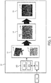
- FIG. 1 shows a dose planning system 10 according to the invention.
- the dose planning system comprises a biopsy map creation module 13, a probability map calculation module 14 and a dose planning module 15.
- a dose planning workflow using the invention could start with the acquisition 11 of images of an organ of interest based on which the suspicious locations within the organ could be identified. Also non-suspicious locations could be identified. These images could for example be magnetic resonance (MR) images.
- the MR images could be provided to registration module 12.
- image guided biopsy system 102 could acquire ultrasound images for biopsy guidance by means of ultrasound system 101. At least one of the ultrasound images is provided to the registration module 12.
- the registration module then registers the ultrasound image with the MR image, such that the identified suspicious and non-suspicious locations of the organ can be translated to the imaging coordinate system of the ultrasound system 101.
- An operator of the system could then guide a photonic needle 100 to the identified locations to perform histopathological analysis on the tissue 17.
- tissue analysis results in tissue characteristics like tumour cell density, percentage of tumour cells, tumour aggressiveness etc.
- the tissue characteristics determined from the biopsy tissue 17 and biopsy locations 16 are provided to the biopsy map creation module 13, which creates a biopsy map by linking the biopsy locations to the corresponding tissue characteristics.
- the biopsy map serves as an input for the probability map calculation module 14, which uses it to calculate a tumour probability map 18.
- line 103 surrounds an area wherein the tumour probability exceeds a certain threshold.
- the probability map calculation module 14 could be configured for creating the tumour probability map 18 based on interpolation or a tumour shape model. Interpolation could be advantageous, since this method does not require prior knowledge on tumour shape.
- tumour shape model could make use of available statistical information on tumour spread in relation to e.g tumour cell density, tumour agressiveness, DNA mutations, DNA expression levels, protein levels found in the biopsy material.
- Tumour shape models are for example known from Shen et al. Optimized prostate biopsy via a statistical atlas of cancer spatial distribution Medical Image Analysis 8 (2004) 139-150 . In their approach, they experimentally generate a global probability cloud for finding a positive biopsy finding and use it for optimal needle placement.
- the key item here of use for the present invention, is the probability distribution, which can be used for modeling the tumour probability map.
- tumour distributions which could be used as an input to generate a tumour probability map are Menze et al. Image-based modeling of tumour growth in patients with glioma Optimal control in image processing, Springer, Heidelberg/Germany, 2011. hal-00825866 and Gevertz et al. Simulating tumour growth in confined heterogeneous environments Phys. Biol. 5 (2008) 036010 . Also further data could be collected on the likelihood of tumour presence on a certain location given a positive or negative biopsy sample at another location.
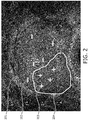
- Figure 2 shows an example of a tumour probability map.
- Figure 2 shows an ultrasound image of a prostate 204. Locations where a biopsy has been taken, but no tumour was found are indicated by means of a "-" sign 202. Locations where a biopsy has been taken and where tumour was found in the biopsy sample are indicated with a "+" sign 203.
- the tumour probability decreases from positions 203 towards line 204, which is an iso-line indicating a certain value for the tumour probability, e.g. 95%.
- the tumour probability map is provided to the dose planning module 15, which creates a dose plan 19 based on the tumour probability map.
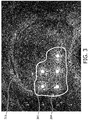
- Figure 3 shows a dose plan corresponding to the tumour probability map of figure 2 .
- the area surrounded by iso-line 204 is considered as gross tumour volume (GTV) and the treatment is planned as such.
- GTV gross tumour volume
- the dose planning module could for example create the dose plan based on the tumour probability map by means of radiobiological models. These models typically take into account tumour cell density, but they could also take into account tumour aggressiveness or the level of hypoxia, which affects at least radiotherapeutic outcome and may be determined based on e.g. HIF-1 levels. These values could be obtained from the biopsy samples and used in the tumour probability map.
- the radiation dose could also be determined based on interpolation. Alternatively, one could also choose to apply a boost dose to a region with high (e.g. > 95%) tumour probability and apply standard dose to regions with low to intermediate tumour probability (e.g. 5-95%).
- the dose planning module could be also configured to use dose constraints for an organ at risk located near the organ to be treated. However, other examples are possible and the invention is not restricted to the examples disclosed.
Landscapes
- Health & Medical Sciences (AREA)
- Life Sciences & Earth Sciences (AREA)
- Engineering & Computer Science (AREA)
- Biomedical Technology (AREA)
- Surgery (AREA)
- Nuclear Medicine, Radiotherapy & Molecular Imaging (AREA)
- General Health & Medical Sciences (AREA)
- Public Health (AREA)
- Veterinary Medicine (AREA)
- Animal Behavior & Ethology (AREA)
- Medical Informatics (AREA)
- Heart & Thoracic Surgery (AREA)
- Molecular Biology (AREA)
- Radiology & Medical Imaging (AREA)
- Pathology (AREA)
- Physics & Mathematics (AREA)
- Otolaryngology (AREA)
- Robotics (AREA)
- Electromagnetism (AREA)
- Optics & Photonics (AREA)
- Biophysics (AREA)
- Epidemiology (AREA)
- Primary Health Care (AREA)
- Medicinal Chemistry (AREA)
- Bioinformatics & Cheminformatics (AREA)
- Chemical & Material Sciences (AREA)
- Plasma & Fusion (AREA)
- Radiation-Therapy Devices (AREA)
- Ultra Sonic Daignosis Equipment (AREA)
Claims (10)
- Système de planification de doses pour un traitement thérapeutique de tissu malade d'un organe d'intérêt comprenant- un module de création de carte de biopsies configuré pour recevoir des informations de biopsie pour un organe d'intérêt concernant des emplacements de biopsie et des caractéristiques tissulaires de tissus trouvés au niveau des emplacements de biopsie, dans lequel le module de création de carte de biopsies est en outre configuré pour créer une carte de biopsies annotée de façon spatiale pour l'organe, en liant les informations spatiales concernant les emplacements de biopsie aux caractéristiques tissulaires de tissus trouvés au niveau des emplacements de biopsie correspondants, et- un module de calcul de carte de probabilités configuré pour créer une carte de probabilité de tumeur en calculant une probabilité de tumeur pour des emplacements dans l'organe à partir desquels aucune biopsie n'a été prise en utilisant les caractéristiques tumorales et/ou tissulaires provenant des emplacements de biopsie, et- un module de planification de doses configuré pour créer un plan de doses sur la base de la carte de probabilités de tumeur, dans lequel des contraintes de planification sont telles que pour une zone avec une probabilité moyenne de tumeur plus élevée, une dose planifiée plus élevée est planifiée et pour une zone avec une probabilité moyenne de tumeur plus faible, une dose planifiée plus faible est planifiée.
- Système de planification de doses selon la revendication 1, dans lequel la carte de probabilités de tumeur est une distribution spatiale d'une chance particulière parmi des chances évaluées de présence de tumeur, des densités cellulaires de tumeur attendues ou un niveau d'agressivité de tumeur.
- Système de planification de doses selon la revendication 1 ou 2, comprenant en outre un système de biopsie guidé par image configuré pour prélever une biopsie à partir d'emplacements prédéterminés dans l'organe et configuré en outre pour fournir au moins des informations spatiales concernant les emplacements de biopsie au module de création de carte de biopsies.
- Système de planification de dose selon la revendication 3, dans lequel le système de biopsie guidé par image comprend une aiguille photonique, dans lequel l'aiguille photonique est configurée pour fournir des informations de biopsie au module de création de carte de biopsies pour l'organe d'intérêt concernant des emplacements de biopsie et des caractéristiques tissulaires de tissus trouvés au niveau des emplacements de biopsie.
- Système de planification de doses selon la revendication 3 ou 4, dans lequel le système de biopsie guidé par image comprend un système à ultrasons pour le guidage par image pendant la biopsie.
- Système de planification de doses selon la revendication 5, comprenant un module de mise en correspondance configuré pour mettre en correspondance une image de l'organe acquise par le système à ultrasons avec une image précédente de l'organe acquise au moyen d'une seconde modalité d'imagerie, dans lequel les emplacements de biopsie sont au moins partiellement déterminés sur la base de l'image précédente.
- Système de planification de doses selon l'une des revendications précédentes, configuré pour créer un plan de doses pour au moins l'un d'un groupe de traitements, comprenant la brachythérapie, la thérapie protonique, la cryothérapie, l'ablation par radio fréquence, l'ablation laser et le traitement aux ultrasons focalisés d'intensité élevée.
- Système de planification de doses selon l'une quelconque des revendications précédentes, dans lequel le module de calcul de carte de probabilités est configuré pour créer la carte de probabilités de tumeur sur la base d'une interpolation des caractéristiques tumorales et/ou tissulaires entre les emplacements de biopsie ou sur la base d'un modèle de forme de tumeur utilisant les caractéristiques tumorales et/ou tissulaires en tant qu'entrée.
- Système de planification de doses selon l'une quelconque des revendications précédentes, dans lequel les caractéristiques tumorales sont au moins un groupe de caractéristiques comprenant la densité cellulaire, la taille de tumeur dans un échantillon de biopsie, le pourcentage de tumeur par échantillon de biopsie ou une mesure liée à l'agressivité de tumeur.
- Système de planification de doses selon l'une quelconque des revendications précédentes, dans lequel le module de planification de doses est en outre configuré pour utiliser des contraintes de dose pour un organe en danger situé près de l'organe à traiter.
Applications Claiming Priority (2)
| Application Number | Priority Date | Filing Date | Title |
|---|---|---|---|
| EP15171904 | 2015-06-12 | ||
| PCT/EP2016/063336 WO2016198626A1 (fr) | 2015-06-12 | 2016-06-10 | Système de planification de dose |
Publications (2)
| Publication Number | Publication Date |
|---|---|
| EP3307387A1 EP3307387A1 (fr) | 2018-04-18 |
| EP3307387B1 true EP3307387B1 (fr) | 2018-12-05 |
Family
ID=53476687
Family Applications (1)
| Application Number | Title | Priority Date | Filing Date |
|---|---|---|---|
| EP16728060.1A Active EP3307387B1 (fr) | 2015-06-12 | 2016-06-10 | Système de planification de dose |
Country Status (6)
| Country | Link |
|---|---|
| US (1) | US10695130B2 (fr) |
| EP (1) | EP3307387B1 (fr) |
| JP (1) | JP6564073B2 (fr) |
| CN (1) | CN107743409B (fr) |
| RU (1) | RU2693204C1 (fr) |
| WO (1) | WO2016198626A1 (fr) |
Families Citing this family (3)
| Publication number | Priority date | Publication date | Assignee | Title |
|---|---|---|---|---|
| WO2016175758A2 (fr) | 2015-04-28 | 2016-11-03 | Analogic Corporation | Pilotage guidé par image d'un réseau de transducteurs et/ou d'un instrument |
| RU2693204C1 (ru) * | 2015-06-12 | 2019-07-01 | Конинклейке Филипс Н.В. | Система планирования дозы |
| AU2019238342A1 (en) | 2018-03-23 | 2020-10-15 | Avent, Inc. | System and method for controlling energy delivered to an area of tissue during a treatment procedure |
Family Cites Families (16)
| Publication number | Priority date | Publication date | Assignee | Title |
|---|---|---|---|---|
| US5398690A (en) * | 1994-08-03 | 1995-03-21 | Batten; Bobby G. | Slaved biopsy device, analysis apparatus, and process |
| US20030135115A1 (en) * | 1997-11-24 | 2003-07-17 | Burdette Everette C. | Method and apparatus for spatial registration and mapping of a biopsy needle during a tissue biopsy |
| WO2000014668A1 (fr) * | 1998-09-08 | 2000-03-16 | Catholic University Of America | Procede et systeme de detection perfectionnee du cancer de la prostate |
| US6438401B1 (en) * | 2000-04-28 | 2002-08-20 | Alpha Intervention Technology, Inc. | Indentification and quantification of needle displacement departures from treatment plan |
| US7831293B2 (en) | 2005-05-10 | 2010-11-09 | Advanced Clinical Solutions, Inc. | Method of defining a biological target for treatment |
| JP5587798B2 (ja) * | 2008-03-03 | 2014-09-10 | コーニンクレッカ フィリップス エヌ ヴェ | 画像ベースx線誘導システム及び光針による生検誘導 |
| US9113816B2 (en) * | 2008-11-11 | 2015-08-25 | Eigen, Inc. | System and method for prostate biopsy |
| JP2010273854A (ja) | 2009-05-28 | 2010-12-09 | Fujifilm Corp | 放射線画像表示装置、方法及びプログラム |
| US9014780B2 (en) * | 2009-11-20 | 2015-04-21 | Koninklijke Philips N.V. | Image-based biopsy guidance method |
| DE102010028105A1 (de) | 2010-04-22 | 2011-10-27 | Siemens Aktiengesellschaft | Verfahren, Vorrichtung und Gerätesystem für die Therapie von Prostatakrebs |
| US9478022B2 (en) | 2011-08-22 | 2016-10-25 | Siemens Medical Solutions Usa, Inc. | Method and system for integrated radiological and pathological information for diagnosis, therapy selection, and monitoring |
| JP6106259B2 (ja) * | 2012-03-21 | 2017-03-29 | コーニンクレッカ フィリップス エヌ ヴェKoninklijke Philips N.V. | 医用イメージングと生検データとを統合する臨床ワークステーション及びこれを使用する方法 |
| WO2013140357A1 (fr) | 2012-03-21 | 2013-09-26 | Koninklijke Philips N.V. | Station de travail clinique intégrant des données d'imagerie médicale et de biopsie et procédés l'utilisant |
| US9370304B2 (en) * | 2012-06-06 | 2016-06-21 | The Regents Of The University Of Michigan | Subvolume identification for prediction of treatment outcome |
| EP2878338B1 (fr) * | 2013-11-28 | 2018-04-11 | RaySearch Laboratories AB | Procédé et système de planification de traitement radiothérapeutique basé sur l'incertitude |
| RU2693204C1 (ru) * | 2015-06-12 | 2019-07-01 | Конинклейке Филипс Н.В. | Система планирования дозы |
-
2016
- 2016-06-10 RU RU2018100614A patent/RU2693204C1/ru active
- 2016-06-10 US US15/572,840 patent/US10695130B2/en active Active
- 2016-06-10 JP JP2017564497A patent/JP6564073B2/ja active Active
- 2016-06-10 EP EP16728060.1A patent/EP3307387B1/fr active Active
- 2016-06-10 CN CN201680034336.7A patent/CN107743409B/zh active Active
- 2016-06-10 WO PCT/EP2016/063336 patent/WO2016198626A1/fr active Application Filing
Non-Patent Citations (1)
| Title |
|---|
| None * |
Also Published As
| Publication number | Publication date |
|---|---|
| WO2016198626A1 (fr) | 2016-12-15 |
| EP3307387A1 (fr) | 2018-04-18 |
| JP6564073B2 (ja) | 2019-08-21 |
| US10695130B2 (en) | 2020-06-30 |
| CN107743409B (zh) | 2020-06-05 |
| RU2693204C1 (ru) | 2019-07-01 |
| US20180153619A1 (en) | 2018-06-07 |
| CN107743409A (zh) | 2018-02-27 |
| JP2018518277A (ja) | 2018-07-12 |
Similar Documents
| Publication | Publication Date | Title |
|---|---|---|
| US20230010515A1 (en) | Method and system for identifying biomarkers using a probability map | |
| CN105592887B (zh) | 使用3d信息作为输入来预测可实现的剂量分布 | |
| RU2529381C2 (ru) | Формирование модели усовершенствованного изображения | |
| EP3140810B1 (fr) | Procédé de génération d'informations synthétiques de densité d'électrons pour des calculs de doses basés sur une irm | |
| US7343030B2 (en) | Dynamic tumor treatment system | |
| CN108815721B (zh) | 一种照射剂量确定方法及系统 | |
| Zhao et al. | Incorporating imaging information from deep neural network layers into image guided radiation therapy (IGRT) | |
| US20080039723A1 (en) | System and method for 3-d biopsy | |
| CN107072624A (zh) | 用于自动治疗计划的系统和方法 | |
| CN103857439A (zh) | 语义放射治疗计划优化指导 | |
| US20160143576A1 (en) | Mri image fusion methods and uses thereof | |
| CN103908254A (zh) | 用于病变候选探测的系统和方法 | |
| EP3307387B1 (fr) | Système de planification de dose | |
| CN113181563B (zh) | 粒子植入肿瘤内放疗剂量规划方法、系统及介质 | |
| Poulin et al. | Validation of MRI to TRUS registration for high-dose-rate prostate brachytherapy | |
| CN113994380A (zh) | 基于深度学习的消融区域确定方法 | |
| Lei et al. | Catheter position prediction using deep‐learning‐based multi‐atlas registration for high‐dose rate prostate brachytherapy | |
| CN103959345A (zh) | 使用颜色的剂量分布显示方法 | |
| Raidou | Uncertainty visualization: Recent developments and future challenges in prostate cancer radiotherapy planning | |
| US9566451B2 (en) | Method and device for irradiation treatment planning | |
| CN117427286B (zh) | 一种基于能谱ct的肿瘤放疗靶区识别方法、系统及设备 | |
| Zhao et al. | Deriving optimal planning organ at risk volume margins in prostate external beam radiotherapy | |
| Holupka et al. | A Deep Learning Approach to Radioactive Seed Detection for Prostate Seed Implants | |
| Zhao et al. | Automatic target positioning and tracking for image-guided radiotherapy without implanted fiducials | |
| Priester et al. | Prediction and Mapping of Intraprostatic Tumor Extent with Artificial Intelligence |
Legal Events
| Date | Code | Title | Description |
|---|---|---|---|
| STAA | Information on the status of an ep patent application or granted ep patent |
Free format text: STATUS: THE INTERNATIONAL PUBLICATION HAS BEEN MADE |
|
| PUAI | Public reference made under article 153(3) epc to a published international application that has entered the european phase |
Free format text: ORIGINAL CODE: 0009012 |
|
| STAA | Information on the status of an ep patent application or granted ep patent |
Free format text: STATUS: REQUEST FOR EXAMINATION WAS MADE |
|
| 17P | Request for examination filed |
Effective date: 20180112 |
|
| AK | Designated contracting states |
Kind code of ref document: A1 Designated state(s): AL AT BE BG CH CY CZ DE DK EE ES FI FR GB GR HR HU IE IS IT LI LT LU LV MC MK MT NL NO PL PT RO RS SE SI SK SM TR |
|
| AX | Request for extension of the european patent |
Extension state: BA ME |
|
| GRAP | Despatch of communication of intention to grant a patent |
Free format text: ORIGINAL CODE: EPIDOSNIGR1 |
|
| STAA | Information on the status of an ep patent application or granted ep patent |
Free format text: STATUS: GRANT OF PATENT IS INTENDED |
|
| DAX | Request for extension of the european patent (deleted) | ||
| INTG | Intention to grant announced |
Effective date: 20180615 |
|
| DAV | Request for validation of the european patent (deleted) | ||
| GRAS | Grant fee paid |
Free format text: ORIGINAL CODE: EPIDOSNIGR3 |
|
| GRAA | (expected) grant |
Free format text: ORIGINAL CODE: 0009210 |
|
| STAA | Information on the status of an ep patent application or granted ep patent |
Free format text: STATUS: THE PATENT HAS BEEN GRANTED |
|
| AK | Designated contracting states |
Kind code of ref document: B1 Designated state(s): AL AT BE BG CH CY CZ DE DK EE ES FI FR GB GR HR HU IE IS IT LI LT LU LV MC MK MT NL NO PL PT RO RS SE SI SK SM TR |
|
| REG | Reference to a national code |
Ref country code: GB Ref legal event code: FG4D |
|
| REG | Reference to a national code |
Ref country code: CH Ref legal event code: EP |
|
| REG | Reference to a national code |
Ref country code: AT Ref legal event code: REF Ref document number: 1072316 Country of ref document: AT Kind code of ref document: T Effective date: 20181215 |
|
| REG | Reference to a national code |
Ref country code: IE Ref legal event code: FG4D |
|
| REG | Reference to a national code |
Ref country code: DE Ref legal event code: R096 Ref document number: 602016007882 Country of ref document: DE |
|
| REG | Reference to a national code |
Ref country code: NL Ref legal event code: MP Effective date: 20181205 |
|
| REG | Reference to a national code |
Ref country code: AT Ref legal event code: MK05 Ref document number: 1072316 Country of ref document: AT Kind code of ref document: T Effective date: 20181205 |
|
| REG | Reference to a national code |
Ref country code: LT Ref legal event code: MG4D |
|
| PG25 | Lapsed in a contracting state [announced via postgrant information from national office to epo] |
Ref country code: AT Free format text: LAPSE BECAUSE OF FAILURE TO SUBMIT A TRANSLATION OF THE DESCRIPTION OR TO PAY THE FEE WITHIN THE PRESCRIBED TIME-LIMIT Effective date: 20181205 Ref country code: ES Free format text: LAPSE BECAUSE OF FAILURE TO SUBMIT A TRANSLATION OF THE DESCRIPTION OR TO PAY THE FEE WITHIN THE PRESCRIBED TIME-LIMIT Effective date: 20181205 Ref country code: LT Free format text: LAPSE BECAUSE OF FAILURE TO SUBMIT A TRANSLATION OF THE DESCRIPTION OR TO PAY THE FEE WITHIN THE PRESCRIBED TIME-LIMIT Effective date: 20181205 Ref country code: BG Free format text: LAPSE BECAUSE OF FAILURE TO SUBMIT A TRANSLATION OF THE DESCRIPTION OR TO PAY THE FEE WITHIN THE PRESCRIBED TIME-LIMIT Effective date: 20190305 Ref country code: HR Free format text: LAPSE BECAUSE OF FAILURE TO SUBMIT A TRANSLATION OF THE DESCRIPTION OR TO PAY THE FEE WITHIN THE PRESCRIBED TIME-LIMIT Effective date: 20181205 Ref country code: LV Free format text: LAPSE BECAUSE OF FAILURE TO SUBMIT A TRANSLATION OF THE DESCRIPTION OR TO PAY THE FEE WITHIN THE PRESCRIBED TIME-LIMIT Effective date: 20181205 Ref country code: FI Free format text: LAPSE BECAUSE OF FAILURE TO SUBMIT A TRANSLATION OF THE DESCRIPTION OR TO PAY THE FEE WITHIN THE PRESCRIBED TIME-LIMIT Effective date: 20181205 Ref country code: NO Free format text: LAPSE BECAUSE OF FAILURE TO SUBMIT A TRANSLATION OF THE DESCRIPTION OR TO PAY THE FEE WITHIN THE PRESCRIBED TIME-LIMIT Effective date: 20190305 |
|
| PG25 | Lapsed in a contracting state [announced via postgrant information from national office to epo] |
Ref country code: RS Free format text: LAPSE BECAUSE OF FAILURE TO SUBMIT A TRANSLATION OF THE DESCRIPTION OR TO PAY THE FEE WITHIN THE PRESCRIBED TIME-LIMIT Effective date: 20181205 Ref country code: GR Free format text: LAPSE BECAUSE OF FAILURE TO SUBMIT A TRANSLATION OF THE DESCRIPTION OR TO PAY THE FEE WITHIN THE PRESCRIBED TIME-LIMIT Effective date: 20190306 Ref country code: AL Free format text: LAPSE BECAUSE OF FAILURE TO SUBMIT A TRANSLATION OF THE DESCRIPTION OR TO PAY THE FEE WITHIN THE PRESCRIBED TIME-LIMIT Effective date: 20181205 Ref country code: SE Free format text: LAPSE BECAUSE OF FAILURE TO SUBMIT A TRANSLATION OF THE DESCRIPTION OR TO PAY THE FEE WITHIN THE PRESCRIBED TIME-LIMIT Effective date: 20181205 |
|
| PG25 | Lapsed in a contracting state [announced via postgrant information from national office to epo] |
Ref country code: NL Free format text: LAPSE BECAUSE OF FAILURE TO SUBMIT A TRANSLATION OF THE DESCRIPTION OR TO PAY THE FEE WITHIN THE PRESCRIBED TIME-LIMIT Effective date: 20181205 |
|
| PG25 | Lapsed in a contracting state [announced via postgrant information from national office to epo] |
Ref country code: PL Free format text: LAPSE BECAUSE OF FAILURE TO SUBMIT A TRANSLATION OF THE DESCRIPTION OR TO PAY THE FEE WITHIN THE PRESCRIBED TIME-LIMIT Effective date: 20181205 Ref country code: PT Free format text: LAPSE BECAUSE OF FAILURE TO SUBMIT A TRANSLATION OF THE DESCRIPTION OR TO PAY THE FEE WITHIN THE PRESCRIBED TIME-LIMIT Effective date: 20190405 Ref country code: IT Free format text: LAPSE BECAUSE OF FAILURE TO SUBMIT A TRANSLATION OF THE DESCRIPTION OR TO PAY THE FEE WITHIN THE PRESCRIBED TIME-LIMIT Effective date: 20181205 Ref country code: CZ Free format text: LAPSE BECAUSE OF FAILURE TO SUBMIT A TRANSLATION OF THE DESCRIPTION OR TO PAY THE FEE WITHIN THE PRESCRIBED TIME-LIMIT Effective date: 20181205 |
|
| PG25 | Lapsed in a contracting state [announced via postgrant information from national office to epo] |
Ref country code: SK Free format text: LAPSE BECAUSE OF FAILURE TO SUBMIT A TRANSLATION OF THE DESCRIPTION OR TO PAY THE FEE WITHIN THE PRESCRIBED TIME-LIMIT Effective date: 20181205 Ref country code: EE Free format text: LAPSE BECAUSE OF FAILURE TO SUBMIT A TRANSLATION OF THE DESCRIPTION OR TO PAY THE FEE WITHIN THE PRESCRIBED TIME-LIMIT Effective date: 20181205 Ref country code: SM Free format text: LAPSE BECAUSE OF FAILURE TO SUBMIT A TRANSLATION OF THE DESCRIPTION OR TO PAY THE FEE WITHIN THE PRESCRIBED TIME-LIMIT Effective date: 20181205 Ref country code: RO Free format text: LAPSE BECAUSE OF FAILURE TO SUBMIT A TRANSLATION OF THE DESCRIPTION OR TO PAY THE FEE WITHIN THE PRESCRIBED TIME-LIMIT Effective date: 20181205 Ref country code: IS Free format text: LAPSE BECAUSE OF FAILURE TO SUBMIT A TRANSLATION OF THE DESCRIPTION OR TO PAY THE FEE WITHIN THE PRESCRIBED TIME-LIMIT Effective date: 20190405 |
|
| REG | Reference to a national code |
Ref country code: DE Ref legal event code: R097 Ref document number: 602016007882 Country of ref document: DE |
|
| PLBE | No opposition filed within time limit |
Free format text: ORIGINAL CODE: 0009261 |
|
| STAA | Information on the status of an ep patent application or granted ep patent |
Free format text: STATUS: NO OPPOSITION FILED WITHIN TIME LIMIT |
|
| PG25 | Lapsed in a contracting state [announced via postgrant information from national office to epo] |
Ref country code: DK Free format text: LAPSE BECAUSE OF FAILURE TO SUBMIT A TRANSLATION OF THE DESCRIPTION OR TO PAY THE FEE WITHIN THE PRESCRIBED TIME-LIMIT Effective date: 20181205 |
|
| 26N | No opposition filed |
Effective date: 20190906 |
|
| PG25 | Lapsed in a contracting state [announced via postgrant information from national office to epo] |
Ref country code: MC Free format text: LAPSE BECAUSE OF FAILURE TO SUBMIT A TRANSLATION OF THE DESCRIPTION OR TO PAY THE FEE WITHIN THE PRESCRIBED TIME-LIMIT Effective date: 20181205 |
|
| REG | Reference to a national code |
Ref country code: CH Ref legal event code: PL |
|
| REG | Reference to a national code |
Ref country code: BE Ref legal event code: MM Effective date: 20190630 |
|
| PG25 | Lapsed in a contracting state [announced via postgrant information from national office to epo] |
Ref country code: TR Free format text: LAPSE BECAUSE OF FAILURE TO SUBMIT A TRANSLATION OF THE DESCRIPTION OR TO PAY THE FEE WITHIN THE PRESCRIBED TIME-LIMIT Effective date: 20181205 |
|
| PG25 | Lapsed in a contracting state [announced via postgrant information from national office to epo] |
Ref country code: IE Free format text: LAPSE BECAUSE OF NON-PAYMENT OF DUE FEES Effective date: 20190610 |
|
| PG25 | Lapsed in a contracting state [announced via postgrant information from national office to epo] |
Ref country code: LI Free format text: LAPSE BECAUSE OF NON-PAYMENT OF DUE FEES Effective date: 20190630 Ref country code: BE Free format text: LAPSE BECAUSE OF NON-PAYMENT OF DUE FEES Effective date: 20190630 Ref country code: CH Free format text: LAPSE BECAUSE OF NON-PAYMENT OF DUE FEES Effective date: 20190630 Ref country code: LU Free format text: LAPSE BECAUSE OF NON-PAYMENT OF DUE FEES Effective date: 20190610 |
|
| GBPC | Gb: european patent ceased through non-payment of renewal fee |
Effective date: 20200610 |
|
| PG25 | Lapsed in a contracting state [announced via postgrant information from national office to epo] |
Ref country code: GB Free format text: LAPSE BECAUSE OF NON-PAYMENT OF DUE FEES Effective date: 20200610 |
|
| PG25 | Lapsed in a contracting state [announced via postgrant information from national office to epo] |
Ref country code: CY Free format text: LAPSE BECAUSE OF FAILURE TO SUBMIT A TRANSLATION OF THE DESCRIPTION OR TO PAY THE FEE WITHIN THE PRESCRIBED TIME-LIMIT Effective date: 20181205 |
|
| PG25 | Lapsed in a contracting state [announced via postgrant information from national office to epo] |
Ref country code: MT Free format text: LAPSE BECAUSE OF FAILURE TO SUBMIT A TRANSLATION OF THE DESCRIPTION OR TO PAY THE FEE WITHIN THE PRESCRIBED TIME-LIMIT Effective date: 20181205 Ref country code: HU Free format text: LAPSE BECAUSE OF FAILURE TO SUBMIT A TRANSLATION OF THE DESCRIPTION OR TO PAY THE FEE WITHIN THE PRESCRIBED TIME-LIMIT; INVALID AB INITIO Effective date: 20160610 |
|
| PG25 | Lapsed in a contracting state [announced via postgrant information from national office to epo] |
Ref country code: SI Free format text: LAPSE BECAUSE OF FAILURE TO SUBMIT A TRANSLATION OF THE DESCRIPTION OR TO PAY THE FEE WITHIN THE PRESCRIBED TIME-LIMIT Effective date: 20181205 |
|
| PG25 | Lapsed in a contracting state [announced via postgrant information from national office to epo] |
Ref country code: MK Free format text: LAPSE BECAUSE OF FAILURE TO SUBMIT A TRANSLATION OF THE DESCRIPTION OR TO PAY THE FEE WITHIN THE PRESCRIBED TIME-LIMIT Effective date: 20181205 |
|
| PGFP | Annual fee paid to national office [announced via postgrant information from national office to epo] |
Ref country code: FR Payment date: 20230622 Year of fee payment: 8 Ref country code: DE Payment date: 20230627 Year of fee payment: 8 |