EP3307162B1 - Pulse oximetry - Google Patents
Pulse oximetry Download PDFInfo
- Publication number
- EP3307162B1 EP3307162B1 EP16807015.9A EP16807015A EP3307162B1 EP 3307162 B1 EP3307162 B1 EP 3307162B1 EP 16807015 A EP16807015 A EP 16807015A EP 3307162 B1 EP3307162 B1 EP 3307162B1
- Authority
- EP
- European Patent Office
- Prior art keywords
- signal
- red
- intensity
- pulsatile
- tissue
- Prior art date
- Legal status (The legal status is an assumption and is not a legal conclusion. Google has not performed a legal analysis and makes no representation as to the accuracy of the status listed.)
- Active
Links
- 238000002106 pulse oximetry Methods 0.000 title claims description 29
- 230000000541 pulsatile effect Effects 0.000 claims description 45
- 238000000034 method Methods 0.000 claims description 36
- QVGXLLKOCUKJST-UHFFFAOYSA-N atomic oxygen Chemical compound [O] QVGXLLKOCUKJST-UHFFFAOYSA-N 0.000 claims description 26
- 229910052760 oxygen Inorganic materials 0.000 claims description 26
- 239000001301 oxygen Substances 0.000 claims description 26
- 230000003287 optical effect Effects 0.000 claims description 20
- 230000003993 interaction Effects 0.000 claims description 12
- 230000008569 process Effects 0.000 claims description 12
- 238000012545 processing Methods 0.000 claims description 9
- 210000000707 wrist Anatomy 0.000 claims description 8
- 238000012937 correction Methods 0.000 claims description 7
- 210000000624 ear auricle Anatomy 0.000 claims description 6
- 210000003423 ankle Anatomy 0.000 claims description 5
- 210000001367 artery Anatomy 0.000 claims description 3
- 238000010521 absorption reaction Methods 0.000 claims 2
- 230000005540 biological transmission Effects 0.000 description 16
- 108010054147 Hemoglobins Proteins 0.000 description 11
- 102000001554 Hemoglobins Human genes 0.000 description 11
- 239000008280 blood Substances 0.000 description 11
- 210000004369 blood Anatomy 0.000 description 11
- 238000010586 diagram Methods 0.000 description 7
- 238000002496 oximetry Methods 0.000 description 7
- 238000005259 measurement Methods 0.000 description 6
- 210000004204 blood vessel Anatomy 0.000 description 5
- 230000017531 blood circulation Effects 0.000 description 4
- 238000012544 monitoring process Methods 0.000 description 4
- 208000017667 Chronic Disease Diseases 0.000 description 3
- IQFVPQOLBLOTPF-UHFFFAOYSA-L Congo Red Chemical compound [Na+].[Na+].C1=CC=CC2=C(N)C(N=NC3=CC=C(C=C3)C3=CC=C(C=C3)N=NC3=C(C4=CC=CC=C4C(=C3)S([O-])(=O)=O)N)=CC(S([O-])(=O)=O)=C21 IQFVPQOLBLOTPF-UHFFFAOYSA-L 0.000 description 3
- 238000002835 absorbance Methods 0.000 description 3
- 230000008321 arterial blood flow Effects 0.000 description 3
- 230000000694 effects Effects 0.000 description 3
- 238000005516 engineering process Methods 0.000 description 3
- 210000001061 forehead Anatomy 0.000 description 3
- 230000005855 radiation Effects 0.000 description 3
- 206010021143 Hypoxia Diseases 0.000 description 2
- 238000004458 analytical method Methods 0.000 description 2
- 238000013459 approach Methods 0.000 description 2
- 230000006399 behavior Effects 0.000 description 2
- 230000008859 change Effects 0.000 description 2
- 210000000038 chest Anatomy 0.000 description 2
- 208000037265 diseases, disorders, signs and symptoms Diseases 0.000 description 2
- 238000011156 evaluation Methods 0.000 description 2
- 210000003811 finger Anatomy 0.000 description 2
- 239000012530 fluid Substances 0.000 description 2
- 210000003739 neck Anatomy 0.000 description 2
- 230000000737 periodic effect Effects 0.000 description 2
- 230000035900 sweating Effects 0.000 description 2
- INGWEZCOABYORO-UHFFFAOYSA-N 2-(furan-2-yl)-7-methyl-1h-1,8-naphthyridin-4-one Chemical compound N=1C2=NC(C)=CC=C2C(O)=CC=1C1=CC=CO1 INGWEZCOABYORO-UHFFFAOYSA-N 0.000 description 1
- UGFAIRIUMAVXCW-UHFFFAOYSA-N Carbon monoxide Chemical compound [O+]#[C-] UGFAIRIUMAVXCW-UHFFFAOYSA-N 0.000 description 1
- 208000006545 Chronic Obstructive Pulmonary Disease Diseases 0.000 description 1
- 108010064719 Oxyhemoglobins Proteins 0.000 description 1
- 230000001154 acute effect Effects 0.000 description 1
- 239000000654 additive Substances 0.000 description 1
- 230000000996 additive effect Effects 0.000 description 1
- 208000007502 anemia Diseases 0.000 description 1
- 208000006673 asthma Diseases 0.000 description 1
- 238000010241 blood sampling Methods 0.000 description 1
- 238000004364 calculation method Methods 0.000 description 1
- 229910002091 carbon monoxide Inorganic materials 0.000 description 1
- 230000001413 cellular effect Effects 0.000 description 1
- LEYJJTBJCFGAQN-UHFFFAOYSA-N chembl1985378 Chemical compound OC1=CC=C2C=CC=CC2=C1N=NC(C=C1)=CC=C1N=NC1=CC=C(S(O)(=O)=O)C=C1 LEYJJTBJCFGAQN-UHFFFAOYSA-N 0.000 description 1
- 230000001684 chronic effect Effects 0.000 description 1
- 238000004590 computer program Methods 0.000 description 1
- 239000000994 contrast dye Substances 0.000 description 1
- 108010002255 deoxyhemoglobin Proteins 0.000 description 1
- 230000001419 dependent effect Effects 0.000 description 1
- 238000001514 detection method Methods 0.000 description 1
- 201000010099 disease Diseases 0.000 description 1
- 230000009189 diving Effects 0.000 description 1
- 238000000605 extraction Methods 0.000 description 1
- 238000001914 filtration Methods 0.000 description 1
- 230000006870 function Effects 0.000 description 1
- PCHJSUWPFVWCPO-UHFFFAOYSA-N gold Chemical compound [Au] PCHJSUWPFVWCPO-UHFFFAOYSA-N 0.000 description 1
- 230000003862 health status Effects 0.000 description 1
- 208000018875 hypoxemia Diseases 0.000 description 1
- 230000001146 hypoxic effect Effects 0.000 description 1
- 238000001727 in vivo Methods 0.000 description 1
- 230000001939 inductive effect Effects 0.000 description 1
- 239000007924 injection Substances 0.000 description 1
- 238000002347 injection Methods 0.000 description 1
- 230000031700 light absorption Effects 0.000 description 1
- 230000005055 memory storage Effects 0.000 description 1
- 230000004048 modification Effects 0.000 description 1
- 238000012986 modification Methods 0.000 description 1
- 238000006213 oxygenation reaction Methods 0.000 description 1
- 230000010412 perfusion Effects 0.000 description 1
- 230000003836 peripheral circulation Effects 0.000 description 1
- 238000000513 principal component analysis Methods 0.000 description 1
- 230000029058 respiratory gaseous exchange Effects 0.000 description 1
- 230000000391 smoking effect Effects 0.000 description 1
- 238000004611 spectroscopical analysis Methods 0.000 description 1
- 239000000126 substance Substances 0.000 description 1
- 230000002459 sustained effect Effects 0.000 description 1
- 208000024891 symptom Diseases 0.000 description 1
- 230000000007 visual effect Effects 0.000 description 1
Images


Classifications
-
- A—HUMAN NECESSITIES
- A61—MEDICAL OR VETERINARY SCIENCE; HYGIENE
- A61B—DIAGNOSIS; SURGERY; IDENTIFICATION
- A61B5/00—Measuring for diagnostic purposes; Identification of persons
- A61B5/145—Measuring characteristics of blood in vivo, e.g. gas concentration, pH value; Measuring characteristics of body fluids or tissues, e.g. interstitial fluid, cerebral tissue
- A61B5/1455—Measuring characteristics of blood in vivo, e.g. gas concentration, pH value; Measuring characteristics of body fluids or tissues, e.g. interstitial fluid, cerebral tissue using optical sensors, e.g. spectral photometrical oximeters
- A61B5/14551—Measuring characteristics of blood in vivo, e.g. gas concentration, pH value; Measuring characteristics of body fluids or tissues, e.g. interstitial fluid, cerebral tissue using optical sensors, e.g. spectral photometrical oximeters for measuring blood gases
- A61B5/14552—Details of sensors specially adapted therefor
-
- A—HUMAN NECESSITIES
- A61—MEDICAL OR VETERINARY SCIENCE; HYGIENE
- A61B—DIAGNOSIS; SURGERY; IDENTIFICATION
- A61B5/00—Measuring for diagnostic purposes; Identification of persons
- A61B5/68—Arrangements of detecting, measuring or recording means, e.g. sensors, in relation to patient
- A61B5/6801—Arrangements of detecting, measuring or recording means, e.g. sensors, in relation to patient specially adapted to be attached to or worn on the body surface
- A61B5/6802—Sensor mounted on worn items
- A61B5/681—Wristwatch-type devices
-
- A—HUMAN NECESSITIES
- A61—MEDICAL OR VETERINARY SCIENCE; HYGIENE
- A61B—DIAGNOSIS; SURGERY; IDENTIFICATION
- A61B5/00—Measuring for diagnostic purposes; Identification of persons
- A61B5/72—Signal processing specially adapted for physiological signals or for diagnostic purposes
- A61B5/7203—Signal processing specially adapted for physiological signals or for diagnostic purposes for noise prevention, reduction or removal
- A61B5/7207—Signal processing specially adapted for physiological signals or for diagnostic purposes for noise prevention, reduction or removal of noise induced by motion artifacts
- A61B5/7214—Signal processing specially adapted for physiological signals or for diagnostic purposes for noise prevention, reduction or removal of noise induced by motion artifacts using signal cancellation, e.g. based on input of two identical physiological sensors spaced apart, or based on two signals derived from the same sensor, for different optical wavelengths
-
- A—HUMAN NECESSITIES
- A61—MEDICAL OR VETERINARY SCIENCE; HYGIENE
- A61B—DIAGNOSIS; SURGERY; IDENTIFICATION
- A61B2560/00—Constructional details of operational features of apparatus; Accessories for medical measuring apparatus
- A61B2560/02—Operational features
- A61B2560/0223—Operational features of calibration, e.g. protocols for calibrating sensors
-
- A—HUMAN NECESSITIES
- A61—MEDICAL OR VETERINARY SCIENCE; HYGIENE
- A61B—DIAGNOSIS; SURGERY; IDENTIFICATION
- A61B2562/00—Details of sensors; Constructional details of sensor housings or probes; Accessories for sensors
- A61B2562/02—Details of sensors specially adapted for in-vivo measurements
- A61B2562/0233—Special features of optical sensors or probes classified in A61B5/00
-
- A—HUMAN NECESSITIES
- A61—MEDICAL OR VETERINARY SCIENCE; HYGIENE
- A61B—DIAGNOSIS; SURGERY; IDENTIFICATION
- A61B5/00—Measuring for diagnostic purposes; Identification of persons
- A61B5/68—Arrangements of detecting, measuring or recording means, e.g. sensors, in relation to patient
- A61B5/6801—Arrangements of detecting, measuring or recording means, e.g. sensors, in relation to patient specially adapted to be attached to or worn on the body surface
- A61B5/6813—Specially adapted to be attached to a specific body part
- A61B5/6814—Head
- A61B5/6815—Ear
- A61B5/6816—Ear lobe
-
- A—HUMAN NECESSITIES
- A61—MEDICAL OR VETERINARY SCIENCE; HYGIENE
- A61B—DIAGNOSIS; SURGERY; IDENTIFICATION
- A61B5/00—Measuring for diagnostic purposes; Identification of persons
- A61B5/68—Arrangements of detecting, measuring or recording means, e.g. sensors, in relation to patient
- A61B5/6801—Arrangements of detecting, measuring or recording means, e.g. sensors, in relation to patient specially adapted to be attached to or worn on the body surface
- A61B5/6813—Specially adapted to be attached to a specific body part
- A61B5/6823—Trunk, e.g., chest, back, abdomen, hip
-
- A—HUMAN NECESSITIES
- A61—MEDICAL OR VETERINARY SCIENCE; HYGIENE
- A61B—DIAGNOSIS; SURGERY; IDENTIFICATION
- A61B5/00—Measuring for diagnostic purposes; Identification of persons
- A61B5/68—Arrangements of detecting, measuring or recording means, e.g. sensors, in relation to patient
- A61B5/6801—Arrangements of detecting, measuring or recording means, e.g. sensors, in relation to patient specially adapted to be attached to or worn on the body surface
- A61B5/6813—Specially adapted to be attached to a specific body part
- A61B5/6824—Arm or wrist
-
- A—HUMAN NECESSITIES
- A61—MEDICAL OR VETERINARY SCIENCE; HYGIENE
- A61B—DIAGNOSIS; SURGERY; IDENTIFICATION
- A61B5/00—Measuring for diagnostic purposes; Identification of persons
- A61B5/68—Arrangements of detecting, measuring or recording means, e.g. sensors, in relation to patient
- A61B5/6801—Arrangements of detecting, measuring or recording means, e.g. sensors, in relation to patient specially adapted to be attached to or worn on the body surface
- A61B5/6813—Specially adapted to be attached to a specific body part
- A61B5/6825—Hand
- A61B5/6826—Finger
-
- A—HUMAN NECESSITIES
- A61—MEDICAL OR VETERINARY SCIENCE; HYGIENE
- A61B—DIAGNOSIS; SURGERY; IDENTIFICATION
- A61B5/00—Measuring for diagnostic purposes; Identification of persons
- A61B5/68—Arrangements of detecting, measuring or recording means, e.g. sensors, in relation to patient
- A61B5/6801—Arrangements of detecting, measuring or recording means, e.g. sensors, in relation to patient specially adapted to be attached to or worn on the body surface
- A61B5/6813—Specially adapted to be attached to a specific body part
- A61B5/6829—Foot or ankle
Definitions
- the present application relates to systems and methods for performing pulse oximetry.
- Pulse oximetry is a method for estimating blood oxygen saturation by utilizing specialized light sources and optical sensors. Tuned light wavelengths are either transmitted through or reflected from a human tissue and are used to estimate a relative proportion of oxygenated blood. This estimated oxygen saturation, termed SpO 2 , is generally strongly related to arterial blood oxygen saturation.
- the main advantages of the pulse oximetry over other methods of determining oxygen saturation, such as blood sampling, is that the pulse oximetry is non-invasive, minimally intrusive, generally not painful, portable if it needs to be, and provides for continuous readings.
- SpO 2 For a healthy human being at normal altitudes, SpO 2 is typically 95% or above, 90% or below indicating hypoxemia, and sustained periods of 80% or below possibly resulting/indicating serious medical complications. SpO 2 can reflect statuses of individuals suffering from various clinical disorders such as the Chronic Obstructive Pulmonary Disease (COPD) or asthma, whether in a stable chronic condition or during an acute phase. Pulse oximetry is also useful in neonatal monitoring, surgical monitoring, or status evaluation when the possibility of oxygen depletion must be considered (pilot monitoring, deep sea diving, and so forth).
- COPD Chronic Obstructive Pulmonary Disease
- Pulse oximetry is also useful in neonatal monitoring, surgical monitoring, or status evaluation when the possibility of oxygen depletion must be considered (pilot monitoring, deep sea diving, and so forth).
- Certain clinical conditions can interfere with either the accuracy of pulse oximetry or affect interpretation of results.
- Diseases which affect peripheral circulation can make the SpO 2 an inaccurate estimate of arterial oxygenation; anemia will impede utilization of blood oxygen, whatever the saturation level.
- Human activity and behavior can also affect results of the pulse oximetry measurements. Movement of the sensor used in pulse oximetry can interfere with signal acquisition. Temperature changes can affect blood flow to the area being monitored with the sensor. Sweating can affect optical quality. Smoking can increase carbon monoxide which competes with oxygen to bind hemoglobin and can confuse most systems. Contrast dye injections can interfere with blood optical qualities.
- Pulse oximetry depends on differences in light absorbance characteristics of oxygenated hemoglobin (oxyhemoglobin) and non-oxygenated hemoglobin (deoxyhemoglobin). The former absorbs light at about 660 nm (in the visible red range) and the latter absorbs light at about 940 nm (infra-red). Both light signals, whether reflected or transmitted, fluctuate with the arterial pulse. The resulting signals, photoplethysmograms (PPGs) can indicate volume changes due to blood flow. Pulse oximetry utilizes the intensity change (light signal fluctuation at each heartbeat) for each wavelength to eliminate the confounding optical effects of other tissues (which remain constant).
- SpO 2 can be estimated using the Beer-Lambert Law, which relates to light absorbance due to the concentration of a substance in media, and empirically-derived reference curves from blood samples of hypoxic volunteers, based on the ratio of these changes in each wavelength (delta 660nm/delta 940nm), although other complex factors are often included in the calculations.
- the light sources are light-emitting diodes (LEDs) optimized for output at each of the target wavelengths.
- LEDs light-emitting diodes
- a single optical sensor (often a photodiode) may be used for both. Each LED can be activated separately, and accompanied by a "dark" period where neither is on (to obtain ambient light levels). The sensor records light transmitted or reflected for each LED. The obtained signals can be processed in real time or offline.
- the sensors can be utilized in either a transmission or a reflectance mode.
- the sensor In the transmission mode, the sensor is typically attached or clipped to a translucent body part (finger, toe, earlobe, and so forth).
- the LED light sources that can be located are on one side of a body part, the sensor can be located on the directly opposite side. The light passes through the entirety of the body part, from one side to the other, and is thus modulated by the pulsating arterial blood flow.
- the light source and the sensor are on the same side of the body part (e.g. forehead, finger, and wrist), and the light is reflected from the skin and the underlying near-surface tissues back to the sensor.
- the document US 7,184,809 B1 describes an apparatus for determining arterial oxygen saturation.
- the apparatus includes first and second radiation emitters that emit light at first and second wavelengths and a radiation detector that receives light at the first and second wavelengths after absorbance through the patient's blood.
- the document US 2011/0060200 A1 describes an apparatus for performing in vivo blood spectrometry.
- the apparatus uses a combination of light emitters and detectors with a light wavelength combination with more than two wavelengths.
- the evaluation of measured signals is based on conventional transmission type signal processing that considers periodic changes in the light absorption as the radiation passes through the tissue.
- the present disclosure provides systems and methods for performing pulse oximetry.
- Embodiments of the present disclosure allow measuring medical parameters, for example, a photoplethysmogram (PPG) of a patient in a non-intrusive manner while, for example, the patient is at home, at work, outdoors, traveling, or is located at some other stationary or mobile environment.
- Some embodiments of the present disclosure include a wearable device.
- the wearable device can be worn at a wrist, ankle, chest, neck, and positioned at other sites of a human body.
- the wearable device can allow measuring the PPG of the patient without requiring the patient to take an active role in the process.
- the PPG data collected by the pulse oximetry during an extended period of time can be analyzed to detect and track trends in medical parameters, for example, oxygen saturation, and to make conclusions concerning symptoms and a progression of one or more chronic diseases from which the patient might suffer.
- a method for performing pulse oximetry includes emitting a light signal for a period of time.
- the method allows detecting a modulated light signal.
- the modulated light signal can include a red signal and an infrared signal.
- the detected signal is a result of an interaction of the light signal with a human tissue.
- the human tissue can include a pulsatile tissue and a non-pulsatile tissue.
- the method includes processing the modulated light signal to estimate at least oxygen saturation in the pulsatile tissue during the time period.
- the processing includes at least removing a non-pulsatile component due to the interaction of the light signal with the non-pulsatile tissue.
- the system 100 can include at least a wearable device 110.
- the wearable device 110 can include sensors 120.
- the wearable device 110 is worn by a patient 130 (for example, on a wrist, ankle, earlobe, neck, chest, fingertip, and the like) for an extended period of time.
- the wearable device 110 can be carried out as a watch, a bracelet, a wristband, a belt, a neck band, and the like.
- the wearable device 110 can be operable to constantly collect, via sensors 120, sensor data from a patient 130. Based on the sensor data, the wearable device 110 can be operable to generate PPG data, and, based on the PPG data, obtain further medical parameters, for example, oxygen saturation, pulse rate, and so forth.
- the system 100 includes a mobile device 140.
- the mobile device 140 can be communicatively coupled to the wearable device 110.
- the mobile device 140 is operable to communicate with the wearable device 110 via a wireless connection such as, for example, Wi-Fi, Bluetooth, Infrared (IR), and the like.
- the mobile device 140 can include a mobile phone, a smart phone, a phablet, a tablet computer, a notebook, and so forth.
- the mobile device 140 can be operable to receive the sensors data and analyze the sensor data to generate PPG data.
- the system 100 may include a cloud-based computing resource 150 (also referred to as a computing cloud).
- the cloud-based computing resource 150 includes one or more server farms/clusters comprising a collection of computer servers and is co-located with network switches and/or routers.
- the mobile device 140 is communicatively coupled to the computing cloud 150.
- the mobile device 140 can be operable to send the sensor data to the computing cloud 150 for further analysis (for example, for extracting medical parameters from the sensor data and storing the results).
- the computing cloud 150 can be operable to run one or more applications and to provide reports regarding health status of the patient, based on trends in medical parameters over time.
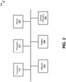
- FIG. 2 is a block diagram illustrating components of wearable device 110, according to an example embodiment.
- the example wearable device 110 includes a transmitter 210, a processor 220, memory storage 230, a battery 240, at least two light-emitting diodes 250, and one or more optical sensors 260.
- the wearable device 110 may comprise additional or different components to provide a particular operation or functionality. Similarly, in other embodiments, the wearable device 110 includes fewer components that perform similar or equivalent functions to those depicted in FIG. 2 .
- the transmitter 210 can be configured to communicate with a network such as the Internet, a Wide Area Network (WAN), a Local Area Network (LAN), a cellular network, and so forth, to send data streams (for example sensor data, PPG data, and messages).
- a network such as the Internet, a Wide Area Network (WAN), a Local Area Network (LAN), a cellular network, and so forth, to send data streams (for example sensor data, PPG data, and messages).
- the processor 220 can include hardware and/or software, which is operable to execute computer programs stored in memory 230.
- the processor 220 can use floating point operations, complex operations, and other operations, including processing and analyzing sensor data.
- the battery 240 is operable to provide electrical power for operation of other components of the wearable device 110.
- the battery 240 is a rechargeable battery.
- the battery 240 is recharged using an inductive charging technology.
- the LEDs 250 are operable to emit light signals of a red wavelength (typically 660 nm) and infrared wavelength (940 nm). Each of the LEDs is activated separately and accompanied by a "dark" period where neither of the LEDs is on to obtain ambient light levels.
- a single LED can be used to emit the both infrared and red light signals.
- the lights can be absorbed by human blood (mostly by hemoglobin).
- the methods for pulse oximetry are based on the fact that oxygenated hemoglobin absorbs more infrared light while deoxygenated hemoglobin absorbs more red light. Oxygenated hemoglobin allows more red light to pass through while deoxygenated hemoglobin allows more infrared light to pass through.
- the optical sensor(s) 260 can receive light signals modulated by a human tissue. Based on the changes in the intensities of the modulated light signals, one or more medical parameters, such as, for example, oxygen saturation, arterial blood flow, pulse rate, and respiration can be determined.
- the LEDs 250 and optical sensor(s) 260 can be utilized in either a transmission or a reflectance mode for pulse oximetry.
- the LEDs 250 and sensor 260 are typically attached or clipped to a translucent body part (e.g., a finger, toe, and earlobe).
- the LEDs 250 are located on one side of the body part while the optical sensor(s) 260 are located directly on the opposite site.
- the light passes through the entirety of the body part, from one side to the other, and is thus modulated by the pulsating arterial blood flow.
- the LEDs 250 and optical sensor(s) 260 are located on the same side of the body part (e.g. a forehead, finger, and wrist), and the light is reflected from the skin and underlying near-surface tissues back to the optical sensor(s) 260.
- FIG. 3A is a block diagram illustrating details of transmission pulse oximetry.
- the light signals 310 emitted by LEDs 250 in red and infrared wavelengths are transmitted through highly perfused pulsatile tissue 320 (for example, blood vessels in a fingertip or an earlobe).
- the light signals 340 modulated across the pulsatile tissue 320 can be detected by optical sensor(s) 260. Some portions of the light signals 310 are reflected by non-pulsatile tissue 330 to produce a reflected light signal 350.
- the transmission pulse oximetry assumes that all detected light passes through pulsating blood vessels and the e ⁇ cd term expresses the attenuation of light by the blood fluid (mostly by hemoglobin) according to the Beer-Lambert law.
- the ratio R expresses the ratio between the attenuation coefficients at the transmission frequencies of red and infrared wavelengths.
- the ratio R can indicate a ratio between oxygenated hemoglobin and deoxygenated hemoglobin.
- the ratio R can be converted to a corresponding oxygen saturation (SpO 2 ) value via an empirically-derived look-up table.
- FIG. 3B is a block diagram illustrating details of reflectance pulse oximetry. Unlike the transmission pulse oximetry, light signals 310 emitted by LEDs 250 is reflected back to the optical sensor(s) 260 from both pulsatile tissue 320 (pulsating arteries) and non-pulsatile tissue 330 (e.g., skin and underlying tissue). In FIG. 3B , the corresponding reflected light signals are denoted as signal 360 and signal 350.
- the reflected signal 350 from non-pulsatile tissue 330 has a negligible significance in conventional transmission oximetry (see FIG. 3A ), as well as in strong signal reflectance oximetry when, for example, operated on a highly perfused tissue such as a fingertip or a forehead.
- the non-pulsatile tissue reflection should be accounted for in order to avoid an erroneous SpO 2 reading. Therefore, the contribution of the non-pulsatile tissue needs to be identified and accounted for, to enable an accurate SpO 2 reading in such cases.
- FIG. 4 shows a plot of example PPG 410 which can be obtained with reflectance pulse oximetry.
- the PPG represents the intensity I of the light signal 310 (either the red signal or the infrared signal) as modulated by a human tissue mostly due to a blood flow.
- Both the high peaks I H and low peaks I L of the PPG 410 include a component I T due to the non-pulsatile tissue reflection.
- the line 420 illustrates a base intensity line for PPG and the line 430 illustrates the addition in intensity of reflected signal I due to non-pulsatile tissue (for example, skin).
- the following embodiments can be used to estimate the additive contribution of the non-pulsatile tissue.
- K 1 While in the transmission oximetry K 1 is small relative to K 2 and may thus be neglected in both red and infrared measurements, it may not be neglected in weak signal cases such as in the general reflectance oximetry or in the low perfusion transmission oximetry.
- L red > 0, L ir > 0 denote arbitrary scalars, representing the bias generated by the non-pulsatile signal components:
- R 1 L red L ir log I H red ⁇ L red I L red ⁇ L red log I H ir ⁇ L ir I L ir ⁇ L ir
- R 1 (I 0 K 1 red , I 0 K 1 ir ) R.
- the correction factor M can be estimated during a calibration process and can be used in subsequent measurements to calibrate the measured R value.
- calibration of the device 110 might be requested by the device itself when the calculated SpO 2 value is not as expected, for example if it is outside a predefined range.
- the device 110 might also signal that calibration is needed if calculated values vary substantially from normal values recorded for a given user, patient, etc. In such as case, the device 110 might proactively request re-calibration. If readings remain outside the expected range, a warning (audible, tactile, visual or the like) may be generation. If recalibration of the device 110 causes the measured values to return to the expected range, then the new calibration parameters are maintained and normal operation resumes.
- Higher Order Statistic In various embodiments, Higher Order Statistic, Principal Component Analysis, and other methods can be used to optimize the calibration process.
- the PPG signal 410 ( FIG. 4 ) can be contaminated by a noise due to environment conditions, activity of a user of the wearable device 110 and behavior of the user. For example, movements of the user can affect the detection by optical sensor(s) 260 of the reflected or absorbed light signal(s). Temperature changes can affect blood flow to the area with the sensor. Sweating of user can also affect the optical quality of the optical sensor(s) 260.
- a band-pass filter can be applied to a raw PPG signal to remove all frequencies outside the range of a heart beat rate typical for a human.
- the band-pass filter can remove all frequencies outside the range from 30 to 300 per minute.
- the noise and waveforms in PPG signal can be separated using an autoregressive (AR) model.
- the PPG waveform can be modeled with a low order AR model.
- the AR model can identify the pulse frequency in the PPG signal even for short time signal segments.
- AR band-pass filter can be constructed.
- the AR band-pass filter can allow passing frequencies within a certain range around the pulse rate frequency (for example +/-10% from the pulse rate frequency) to filter out all frequencies outside the range.
- a clean PPG signal can be generated.
- the clean PPG signal can be used to determine a ratio for SpO 2 oxygen saturation according to embodiments of present disclosure.
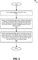
- FIG. 5 is a flow chart of a method 500 for data collected from a single wrist, according to an example embodiment.
- Method 500 can commence at block 510 with emitting a light signal for a period of time.
- the method 500 can proceed with detecting a modulated light signal.
- the modulated light signal can include a red signal and an infrared signal.
- the detected signal can be a result of interaction of the light signal with a human tissue.
- the human tissue can include a pulsatile tissue and a non-pulsatile tissue.
- the method 500 can process the modulated light signal to estimate at least oxygen saturation in the pulsatile tissue during the time period.
- the processing can include at least removing a non-pulsatile component due to the interaction of the light signal with the non-pulsatile tissue.
- the non-pulsatile component is removed by removing a first parameter from maximums and minimums of an intensity associated with the infrared signal and removing a second parameter from maximums and minimums of the intensity associated with the red signal.
- the correction factor can be pre-determined via a calibration process.
- FIG. 6 illustrates example plots of a raw infrared signal 610 and raw red signal 620 measured during a period of 1 minute.
- the red signal 620 can be shifted.
- FIG. 7 illustrates example plots of infrared signal 710 and red signal 720.
- the infrared signal 710 can be obtained from the raw infrared signal 610 by band-pass filtering.
- the red signal 720 is band-passed filtered raw red signal 620.
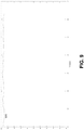
- FIG. 8 illustrated an example plot of a ratio 800 for determining the SpO 2 saturation.
- the ratio 900 can be determined based on the band-filtered infrared signal 710 and red signal 720 using the method of embodiment No 1 described above.
- FIG. 9 illustrates an example plot of SpO 2 oxygen saturation determined based on ratio 800.
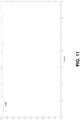
- FIG. 10 illustrates an example plot of ratio 1000 for determining SpO 2 oxygen saturation.
- the ratio 1000 can be determined based on band-pass filtered infrared signal 710 and band-pass filtered red signal 720 using the method of embodiment No 2 described above.
- FIG. 11 illustrates an example plot of SpO 2 oxygen saturation determined based on ratio 1100.
Landscapes
- Health & Medical Sciences (AREA)
- Life Sciences & Earth Sciences (AREA)
- Physics & Mathematics (AREA)
- Engineering & Computer Science (AREA)
- Medical Informatics (AREA)
- Animal Behavior & Ethology (AREA)
- Pathology (AREA)
- Veterinary Medicine (AREA)
- Biomedical Technology (AREA)
- Heart & Thoracic Surgery (AREA)
- Public Health (AREA)
- Molecular Biology (AREA)
- Surgery (AREA)
- Biophysics (AREA)
- General Health & Medical Sciences (AREA)
- Signal Processing (AREA)
- Spectroscopy & Molecular Physics (AREA)
- Optics & Photonics (AREA)
- Artificial Intelligence (AREA)
- Computer Vision & Pattern Recognition (AREA)
- Physiology (AREA)
- Psychiatry (AREA)
- Measurement Of The Respiration, Hearing Ability, Form, And Blood Characteristics Of Living Organisms (AREA)
Description
- The present application relates to systems and methods for performing pulse oximetry.
- It should not be assumed that any of the approaches described in this section qualify as prior art merely by virtue of their inclusion in this section.
- Pulse oximetry is a method for estimating blood oxygen saturation by utilizing specialized light sources and optical sensors. Tuned light wavelengths are either transmitted through or reflected from a human tissue and are used to estimate a relative proportion of oxygenated blood. This estimated oxygen saturation, termed SpO2, is generally strongly related to arterial blood oxygen saturation. The main advantages of the pulse oximetry over other methods of determining oxygen saturation, such as blood sampling, is that the pulse oximetry is non-invasive, minimally intrusive, generally not painful, portable if it needs to be, and provides for continuous readings.
- For a healthy human being at normal altitudes, SpO2 is typically 95% or above, 90% or below indicating hypoxemia, and sustained periods of 80% or below possibly resulting/indicating serious medical complications. SpO2 can reflect statuses of individuals suffering from various clinical disorders such as the Chronic Obstructive Pulmonary Disease (COPD) or asthma, whether in a stable chronic condition or during an acute phase. Pulse oximetry is also useful in neonatal monitoring, surgical monitoring, or status evaluation when the possibility of oxygen depletion must be considered (pilot monitoring, deep sea diving, and so forth).
- Certain clinical conditions can interfere with either the accuracy of pulse oximetry or affect interpretation of results. Diseases which affect peripheral circulation can make the SpO2 an inaccurate estimate of arterial oxygenation; anemia will impede utilization of blood oxygen, whatever the saturation level.
- Human activity and behavior can also affect results of the pulse oximetry measurements. Movement of the sensor used in pulse oximetry can interfere with signal acquisition. Temperature changes can affect blood flow to the area being monitored with the sensor. Sweating can affect optical quality. Smoking can increase carbon monoxide which competes with oxygen to bind hemoglobin and can confuse most systems. Contrast dye injections can interfere with blood optical qualities.
- Pulse oximetry depends on differences in light absorbance characteristics of oxygenated hemoglobin (oxyhemoglobin) and non-oxygenated hemoglobin (deoxyhemoglobin). The former absorbs light at about 660 nm (in the visible red range) and the latter absorbs light at about 940 nm (infra-red). Both light signals, whether reflected or transmitted, fluctuate with the arterial pulse. The resulting signals, photoplethysmograms (PPGs) can indicate volume changes due to blood flow. Pulse oximetry utilizes the intensity change (light signal fluctuation at each heartbeat) for each wavelength to eliminate the confounding optical effects of other tissues (which remain constant). SpO2 can be estimated using the Beer-Lambert Law, which relates to light absorbance due to the concentration of a substance in media, and empirically-derived reference curves from blood samples of hypoxic volunteers, based on the ratio of these changes in each wavelength (delta 660nm/delta 940nm), although other complex factors are often included in the calculations.
- Typically, the light sources are light-emitting diodes (LEDs) optimized for output at each of the target wavelengths. A single optical sensor (often a photodiode) may be used for both. Each LED can be activated separately, and accompanied by a "dark" period where neither is on (to obtain ambient light levels). The sensor records light transmitted or reflected for each LED. The obtained signals can be processed in real time or offline.
- The sensors can be utilized in either a transmission or a reflectance mode. In the transmission mode, the sensor is typically attached or clipped to a translucent body part (finger, toe, earlobe, and so forth). The LED light sources that can be located are on one side of a body part, the sensor can be located on the directly opposite side. The light passes through the entirety of the body part, from one side to the other, and is thus modulated by the pulsating arterial blood flow. In the reflectance mode, the light source and the sensor are on the same side of the body part (e.g. forehead, finger, and wrist), and the light is reflected from the skin and the underlying near-surface tissues back to the sensor.
- Despite the conceptually different optical paths in the reflectance oximetry, conventional transmission type signal processing for the extraction of oxygen saturation is currently employed; though the sensor part is sometimes adapted to enhance the reflectance signal, the usage of a transmission model for reflectance analysis often results in unstable and erroneous SpO2 estimates.
- Wearable monitoring of chronic outpatients can be greatly enhanced by accurate SpO2 measurements, whereas from the usability standpoint a reflectance device which can be attached to body sites such as a wrist or an ankle would impose minimal burden on normal activities; hence, developing reliable reflectance oximetry devices, based on a specific light reflectance model, holds great promise for outpatients suffering from chronic diseases.
- The document
US 7,184,809 B1 describes an apparatus for determining arterial oxygen saturation. The apparatus includes first and second radiation emitters that emit light at first and second wavelengths and a radiation detector that receives light at the first and second wavelengths after absorbance through the patient's blood. - The document
US 2011/0060200 A1 describes an apparatus for performing in vivo blood spectrometry. The apparatus uses a combination of light emitters and detectors with a light wavelength combination with more than two wavelengths. The evaluation of measured signals is based on conventional transmission type signal processing that considers periodic changes in the light absorption as the radiation passes through the tissue. - Documents
US2001/005773 A1 andUS 2005/070775 A1 disclose systems and methods for obtaining oxygen saturation measurements with high accuracy. - The invention is defined in the independent claims. The dependent claims describe preferred embodiments of the invention.
- Embodiments are illustrated by way of example and not limitation in the figures of the accompanying drawings, in which like references indicate similar elements.
-
FIG. 1 is a block diagram showing an example system for performing pulse oximetry using a wearable device. -
FIG. 2 is a block diagram showing components of an example device for performing pulse oximetry. -
FIG. 3A is a block diagram illustrating example details of transmission pulse oximetry. -
FIG. 3B is a block diagram illustrating example details of reflectance pulse oximetry. -
FIG. 4 shows an example plot of a photoplethysmogram (PPG). -
FIG. 5 is a flow chart showing an example method for performing pulse oximetry. -
FIG. 6 illustrates example plots of a raw infrared PPG signal and a raw red PPG signal. -
FIG. 7 illustrates example plots of a band-pass filtered infrared PPG signal and a band-passed filtered red PPG signal. -
FIG. 8 illustrates an example plot of a ratio for determining the SpO2 oxygen saturation. -
FIG. 9 illustrates an example plot of SpO2 oxygen saturation determined based on the ratio ofFIG. 8 . -
FIG. 10 illustrates example plot of a ratio for determining the SpO2 oxygen saturation. -
FIG. 11 illustrates an example plot of SpO2 oxygen saturation determined based on the ratio ofFIG. 10 . - The following detailed description includes references to the accompanying drawings, which form a part of the detailed description. The drawings show illustrations in accordance with exemplary embodiments. These exemplary embodiments, which are also referred to herein as "examples," are described in enough detail to enable those skilled in the art to practice the present subject matter. The embodiments can be combined, other embodiments can be utilized, or structural, logical and electrical changes can be made without departing from the scope of what is claimed. The following detailed description is, therefore, not to be taken in a limiting sense, and the scope is defined by the appended claims.
- The present disclosure provides systems and methods for performing pulse oximetry. Embodiments of the present disclosure allow measuring medical parameters, for example, a photoplethysmogram (PPG) of a patient in a non-intrusive manner while, for example, the patient is at home, at work, outdoors, traveling, or is located at some other stationary or mobile environment. Some embodiments of the present disclosure include a wearable device. The wearable device can be worn at a wrist, ankle, chest, neck, and positioned at other sites of a human body. The wearable device can allow measuring the PPG of the patient without requiring the patient to take an active role in the process. The PPG data collected by the pulse oximetry during an extended period of time can be analyzed to detect and track trends in medical parameters, for example, oxygen saturation, and to make conclusions concerning symptoms and a progression of one or more chronic diseases from which the patient might suffer.
- According to some example embodiments, a method for performing pulse oximetry includes emitting a light signal for a period of time. The method allows detecting a modulated light signal. The modulated light signal can include a red signal and an infrared signal. The detected signal is a result of an interaction of the light signal with a human tissue. The human tissue can include a pulsatile tissue and a non-pulsatile tissue. The method includes processing the modulated light signal to estimate at least oxygen saturation in the pulsatile tissue during the time period. The processing includes at least removing a non-pulsatile component due to the interaction of the light signal with the non-pulsatile tissue.
- Referring now to
FIG. 1 , anexample system 100 for performing pulse oximetry is shown. Thesystem 100 can include at least awearable device 110. Thewearable device 110 can includesensors 120. In some embodiments, thewearable device 110 is worn by a patient 130 (for example, on a wrist, ankle, earlobe, neck, chest, fingertip, and the like) for an extended period of time. In various embodiments, thewearable device 110 can be carried out as a watch, a bracelet, a wristband, a belt, a neck band, and the like. - The
wearable device 110 can be operable to constantly collect, viasensors 120, sensor data from apatient 130. Based on the sensor data, thewearable device 110 can be operable to generate PPG data, and, based on the PPG data, obtain further medical parameters, for example, oxygen saturation, pulse rate, and so forth. - In some embodiments, the
system 100 includes amobile device 140. Themobile device 140 can be communicatively coupled to thewearable device 110. In various embodiments, themobile device 140 is operable to communicate with thewearable device 110 via a wireless connection such as, for example, Wi-Fi, Bluetooth, Infrared (IR), and the like. Themobile device 140 can include a mobile phone, a smart phone, a phablet, a tablet computer, a notebook, and so forth. Themobile device 140 can be operable to receive the sensors data and analyze the sensor data to generate PPG data. - In further embodiments, the
system 100 may include a cloud-based computing resource 150 (also referred to as a computing cloud). In some embodiments, the cloud-basedcomputing resource 150 includes one or more server farms/clusters comprising a collection of computer servers and is co-located with network switches and/or routers. In certain embodiments, themobile device 140 is communicatively coupled to thecomputing cloud 150. Themobile device 140 can be operable to send the sensor data to thecomputing cloud 150 for further analysis (for example, for extracting medical parameters from the sensor data and storing the results). Thecomputing cloud 150 can be operable to run one or more applications and to provide reports regarding health status of the patient, based on trends in medical parameters over time. -
FIG. 2 is a block diagram illustrating components ofwearable device 110, according to an example embodiment. The examplewearable device 110 includes atransmitter 210, aprocessor 220,memory storage 230, abattery 240, at least two light-emittingdiodes 250, and one or moreoptical sensors 260. Thewearable device 110 may comprise additional or different components to provide a particular operation or functionality. Similarly, in other embodiments, thewearable device 110 includes fewer components that perform similar or equivalent functions to those depicted inFIG. 2 . - The
transmitter 210 can be configured to communicate with a network such as the Internet, a Wide Area Network (WAN), a Local Area Network (LAN), a cellular network, and so forth, to send data streams (for example sensor data, PPG data, and messages). - The
processor 220 can include hardware and/or software, which is operable to execute computer programs stored inmemory 230. Theprocessor 220 can use floating point operations, complex operations, and other operations, including processing and analyzing sensor data. - In some embodiments, the
battery 240 is operable to provide electrical power for operation of other components of thewearable device 110. In some embodiments, thebattery 240 is a rechargeable battery. In certain embodiments, thebattery 240 is recharged using an inductive charging technology. - In various embodiments, the
LEDs 250 are operable to emit light signals of a red wavelength (typically 660 nm) and infrared wavelength (940 nm). Each of the LEDs is activated separately and accompanied by a "dark" period where neither of the LEDs is on to obtain ambient light levels. In some embodiments, a single LED can be used to emit the both infrared and red light signals. The lights can be absorbed by human blood (mostly by hemoglobin). The methods for pulse oximetry are based on the fact that oxygenated hemoglobin absorbs more infrared light while deoxygenated hemoglobin absorbs more red light. Oxygenated hemoglobin allows more red light to pass through while deoxygenated hemoglobin allows more infrared light to pass through. The optical sensor(s) 260 (typically a photodiode) can receive light signals modulated by a human tissue. Based on the changes in the intensities of the modulated light signals, one or more medical parameters, such as, for example, oxygen saturation, arterial blood flow, pulse rate, and respiration can be determined. - The
LEDs 250 and optical sensor(s) 260 can be utilized in either a transmission or a reflectance mode for pulse oximetry. In the transmission mode, theLEDs 250 andsensor 260 are typically attached or clipped to a translucent body part (e.g., a finger, toe, and earlobe). TheLEDs 250 are located on one side of the body part while the optical sensor(s) 260 are located directly on the opposite site. The light passes through the entirety of the body part, from one side to the other, and is thus modulated by the pulsating arterial blood flow. In the reflectance mode, theLEDs 250 and optical sensor(s) 260 are located on the same side of the body part (e.g. a forehead, finger, and wrist), and the light is reflected from the skin and underlying near-surface tissues back to the optical sensor(s) 260. -
FIG. 3A is a block diagram illustrating details of transmission pulse oximetry. The light signals 310 emitted byLEDs 250 in red and infrared wavelengths are transmitted through highly perfused pulsatile tissue 320 (for example, blood vessels in a fingertip or an earlobe). The light signals 340 modulated across thepulsatile tissue 320 can be detected by optical sensor(s) 260. Some portions of the light signals 310 are reflected bynon-pulsatile tissue 330 to produce a reflectedlight signal 350. The detectedlight signal 340, I can be evaluated as follows:
blood vessels 320, and c is a light attenuation coefficient of blood fluid. - In the traditional approach, the transmission pulse oximetry assumes that all detected light passes through pulsating blood vessels and the e―cd term expresses the attenuation of light by the blood fluid (mostly by hemoglobin) according to the Beer-Lambert law.
- The diameter of the
blood vessel 320 has periodic variation according to pulse rate frequency with maximum and minimum values:


- The ratio R expresses the ratio between the attenuation coefficients at the transmission frequencies of red and infrared wavelengths. The ratio R can indicate a ratio between oxygenated hemoglobin and deoxygenated hemoglobin. The ratio R can be converted to a corresponding oxygen saturation (SpO2) value via an empirically-derived look-up table.
-
FIG. 3B is a block diagram illustrating details of reflectance pulse oximetry. Unlike the transmission pulse oximetry,light signals 310 emitted byLEDs 250 is reflected back to the optical sensor(s) 260 from both pulsatile tissue 320 (pulsating arteries) and non-pulsatile tissue 330 (e.g., skin and underlying tissue). InFIG. 3B , the corresponding reflected light signals are denoted assignal 360 and signal 350. The reflectedsignal 350 fromnon-pulsatile tissue 330 has a negligible significance in conventional transmission oximetry (seeFIG. 3A ), as well as in strong signal reflectance oximetry when, for example, operated on a highly perfused tissue such as a fingertip or a forehead. - In case of a weak pulsatile signal, the non-pulsatile tissue reflection should be accounted for in order to avoid an erroneous SpO2 reading. Therefore, the contribution of the non-pulsatile tissue needs to be identified and accounted for, to enable an accurate SpO2 reading in such cases.
-
FIG. 4 shows a plot ofexample PPG 410 which can be obtained with reflectance pulse oximetry. The PPG represents the intensity I of the light signal 310 (either the red signal or the infrared signal) as modulated by a human tissue mostly due to a blood flow. Both the high peaks IH and low peaks IL of thePPG 410 include a component IT due to the non-pulsatile tissue reflection. Theline 420 illustrates a base intensity line for PPG and theline 430 illustrates the addition in intensity of reflected signal I due to non-pulsatile tissue (for example, skin). The following embodiments can be used to estimate the additive contribution of the non-pulsatile tissue. - According to an example embodiment of present technology, the detected signal I can be modeled as follows:

- While in the transmission oximetry K 1 is small relative to K 2 and may thus be neglected in both red and infrared measurements, it may not be neglected in weak signal cases such as in the general reflectance oximetry or in the low perfusion transmission oximetry.
- Let us consider the following modification. Let Lred > 0, Lir > 0 denote arbitrary scalars, representing the bias generated by the non-pulsatile signal components:

- It can be shown that R1(I0K1 red, I0K1 ir) = R. The desired constants Lred = I0K1 red and Lir = I0K1 ir can be found during a calibration process, where a gold standard measurement provides the true R value R = Rtrue and the resulting constants (Lred, Lir) are optimized to fulfill the equationR1(Lred, Lir) = Rtrue.
- According to another embodiment of present technology, the alternating current (AC) component of the PPG signal I (i.e. the difference between maximum and minimum values), assuming a small change in blood vessel size, may be expressed as;


- Denoting


- In certain instances, calibration of the
device 110 might be requested by the device itself when the calculated SpO2 value is not as expected, for example if it is outside a predefined range. Thedevice 110 might also signal that calibration is needed if calculated values vary substantially from normal values recorded for a given user, patient, etc. In such as case, thedevice 110 might proactively request re-calibration. If readings remain outside the expected range, a warning (audible, tactile, visual or the like) may be generation. If recalibration of thedevice 110 causes the measured values to return to the expected range, then the new calibration parameters are maintained and normal operation resumes. - In various embodiments, Higher Order Statistic, Principal Component Analysis, and other methods can be used to optimize the calibration process.
- The PPG signal 410 (
FIG. 4 ) can be contaminated by a noise due to environment conditions, activity of a user of thewearable device 110 and behavior of the user. For example, movements of the user can affect the detection by optical sensor(s) 260 of the reflected or absorbed light signal(s). Temperature changes can affect blood flow to the area with the sensor. Sweating of user can also affect the optical quality of the optical sensor(s) 260. - In various embodiments, to remove the noise in PPG signal, a band-pass filter can be applied to a raw PPG signal to remove all frequencies outside the range of a heart beat rate typical for a human. For example, the band-pass filter can remove all frequencies outside the range from 30 to 300 per minute.
- Additionally, in some embodiments, the noise and waveforms in PPG signal can be separated using an autoregressive (AR) model. The PPG waveform can be modeled with a low order AR model. The AR model can identify the pulse frequency in the PPG signal even for short time signal segments. After identifying the pulse rate frequency, AR band-pass filter can be constructed. The AR band-pass filter can allow passing frequencies within a certain range around the pulse rate frequency (for example +/-10% from the pulse rate frequency) to filter out all frequencies outside the range. As a result, a clean PPG signal can be generated. The clean PPG signal can be used to determine a ratio for SpO2 oxygen saturation according to embodiments of present disclosure.
-
FIG. 5 is a flow chart of amethod 500 for data collected from a single wrist, according to an example embodiment.Method 500 can commence atblock 510 with emitting a light signal for a period of time. Atblock 520, themethod 500 can proceed with detecting a modulated light signal. The modulated light signal can include a red signal and an infrared signal. The detected signal can be a result of interaction of the light signal with a human tissue. The human tissue can include a pulsatile tissue and a non-pulsatile tissue. - At
block 530, themethod 500 can process the modulated light signal to estimate at least oxygen saturation in the pulsatile tissue during the time period. The processing can include at least removing a non-pulsatile component due to the interaction of the light signal with the non-pulsatile tissue. In some embodiments, the non-pulsatile component is removed by removing a first parameter from maximums and minimums of an intensity associated with the infrared signal and removing a second parameter from maximums and minimums of the intensity associated with the red signal. The first parameter and the second parameter can be pre-determined using a calibration process to reproduce a true value for


- In other embodiments, the non-pulsatile component is removed by multiplying a ratio


-
FIG. 6 illustrates example plots of a rawinfrared signal 610 and rawred signal 620 measured during a period of 1 minute. Thered signal 620 can be shifted. -
FIG. 7 illustrates example plots ofinfrared signal 710 andred signal 720. Theinfrared signal 710 can be obtained from the rawinfrared signal 610 by band-pass filtering. Thered signal 720 is band-passed filtered rawred signal 620. -
FIG. 8 illustrated an example plot of aratio 800 for determining the SpO2 saturation. Theratio 900 can be determined based on the band-filteredinfrared signal 710 andred signal 720 using the method of embodiment No 1 described above.FIG. 9 illustrates an example plot of SpO2 oxygen saturation determined based onratio 800.FIG. 10 illustrates an example plot ofratio 1000 for determining SpO2 oxygen saturation. Theratio 1000 can be determined based on band-pass filteredinfrared signal 710 and band-pass filteredred signal 720 using the method ofembodiment No 2 described above.FIG. 11 illustrates an example plot of SpO2 oxygen saturation determined based onratio 1100.
Claims (11)
- A method for performing a pulse oximetry, the method comprising:emitting a light signal (310) for a period of time;detecting a modulated light signal (340), the modulated light signal (340) including a red signal (620) and an infrared signal (610) originating from an interaction of the light signal (310) with a human tissue, the human tissue including a pulsatile tissue (320) and a non-pulsatile tissue (330), wherein the interaction of the light signal (310) with the human tissue includes reflection of the light signal from the human tissue; andprocessing the modulated light signal (340) to estimate at least an oxygen saturation in the pulsatile tissue (320) during the period of time, wherein the processing includes at least removing a non-pulsatile component resulting from the interaction of the light signal (310) with the non-pulsatile tissue (330),wherein the removing the non-pulsatile component includes:removing a first parameter from maximums and minimums of an intensity associated with the infrared signal (610); andremoving a second parameter from maximums and minimums of the intensity associated with the red signal (620),wherein the first and second parameters represent a bias generated by the non-pulsatile signal components, wherein the first parameter and the second parameter are pre-determined using a calibration process to reproduce a true value for a ratio =


 or wherein the removing the non-pulsatile component includes multiplying a ratio


- The method of claim 1, wherein the pulsatile tissue (320) includes an artery.
- The method of claim 1 or 2, wherein the non-pulsatile tissue (330) includes skin.
- The method of any one of the preceding claims, wherein the human tissue is associated with one of the following: a fingertip, a wrist, an ankle, a neck, a chest, and an earlobe.
- The method of any one of the preceding claims, wherein the interaction of the light signal with the human tissue includes absorption of the light signal by the human tissue.
- A system for performing a pulse oximetry, the system comprising:at least one light source (250) operable to emit a light signal (310) for a period of time;at least one optical sensor (260) operable to detect a modulated light signal (340), the modulated light signal (340) including a red signal (620) and an infrared signal (610) originating from an interaction of the light signal (310) with a human tissue, the human tissue including a pulsatile tissue (320) and a non-pulsatile tissue (330), wherein the interaction of the light signal (310) with the human tissue includes reflection of the light signal from the human tissue; andat least one processor (220) communicatively coupled to the at least one optical sensor (260) and operable to process the modulated light signal to estimate at least an oxygen saturation in the pulsatile tissue (320) during the period of time, wherein the processing includes at least removing a non-pulsatile component due to the interaction of the light signal (310) with the non-pulsatile tissue (330),wherein the removing the non-pulsatile component includes:removing a first parameter from maximums and minimums of an intensity associated with the infrared signal (610); andremoving a second parameter from maximums and minimums of the intensity associated with the red signal (620),wherein the first and second parameters represent a bias generated by the non-pulsatile signal components, wherein the first parameter and the second parameter are pre-determined using a calibration process to reproduce a true value for a ratio =


 or wherein the removing the non-pulsatile component includes multiplying a ratio


- The system of claim 6, wherein the pulsatile tissue (320) includes an artery, and/or wherein the non-pulsatile tissue (330) includes skin.
- The system of claim 6 or 7, wherein the human tissue is associated with one of the following: a fingertip, a wrist, an ankle, a neck, a chest, and an earlobe.
- The system of any one of claims 6-8, wherein the interaction of the light signal with the human tissue includes absorption of the light signal by the human tissue.
- The system of any one of claims 6-9, wherein the system signals a user to calibrate the system if measured signal values are outside a specified range, or wherein the system signals a user to calibrate the system if measured signal values vary from an average established for the user.
- A non-transitory computer-readable storage medium having embodied thereon instructions, which when executed by a processor that is communicatively coupled to at least one light source (250) and at least one optical sensor (260), perform steps of the method according to any one of claims 1-5.
Applications Claiming Priority (2)
| Application Number | Priority Date | Filing Date | Title |
|---|---|---|---|
| US14/738,711 US10470692B2 (en) | 2015-06-12 | 2015-06-12 | System for performing pulse oximetry |
| PCT/IL2016/050513 WO2016199123A1 (en) | 2015-06-12 | 2016-05-15 | Pulse oximetry |
Publications (3)
| Publication Number | Publication Date |
|---|---|
| EP3307162A1 EP3307162A1 (en) | 2018-04-18 |
| EP3307162A4 EP3307162A4 (en) | 2019-02-20 |
| EP3307162B1 true EP3307162B1 (en) | 2021-11-17 |
Family
ID=57503129
Family Applications (1)
| Application Number | Title | Priority Date | Filing Date |
|---|---|---|---|
| EP16807015.9A Active EP3307162B1 (en) | 2015-06-12 | 2016-05-15 | Pulse oximetry |
Country Status (4)
| Country | Link |
|---|---|
| US (1) | US10470692B2 (en) |
| EP (1) | EP3307162B1 (en) |
| CN (1) | CN107920786B (en) |
| WO (1) | WO2016199123A1 (en) |
Families Citing this family (11)
| Publication number | Priority date | Publication date | Assignee | Title |
|---|---|---|---|---|
| US10470692B2 (en) | 2015-06-12 | 2019-11-12 | ChroniSense Medical Ltd. | System for performing pulse oximetry |
| US11160461B2 (en) | 2015-06-12 | 2021-11-02 | ChroniSense Medical Ltd. | Blood pressure measurement using a wearable device |
| US11464457B2 (en) | 2015-06-12 | 2022-10-11 | ChroniSense Medical Ltd. | Determining an early warning score based on wearable device measurements |
| US11160459B2 (en) | 2015-06-12 | 2021-11-02 | ChroniSense Medical Ltd. | Monitoring health status of people suffering from chronic diseases |
| US11712190B2 (en) | 2015-06-12 | 2023-08-01 | ChroniSense Medical Ltd. | Wearable device electrocardiogram |
| US10952638B2 (en) | 2015-06-12 | 2021-03-23 | ChroniSense Medical Ltd. | System and method for monitoring respiratory rate and oxygen saturation |
| US10687742B2 (en) | 2015-06-12 | 2020-06-23 | ChroniSense Medical Ltd. | Using invariant factors for pulse oximetry |
| US11000235B2 (en) | 2016-03-14 | 2021-05-11 | ChroniSense Medical Ltd. | Monitoring procedure for early warning of cardiac episodes |
| EP3843632A4 (en) * | 2018-08-29 | 2021-11-24 | Tel Hashomer Medical Research Infrastructure And Services Ltd. | System and method for determining oxygenated-blood content of biological tissue |
| US11771350B1 (en) | 2020-09-11 | 2023-10-03 | Apple Inc. | System and method for robust pulse oximetry using asymmetric distance-dependent calibration |
| US11666256B2 (en) * | 2020-10-27 | 2023-06-06 | Michael Edward Labrecque | Pulse oximeter sensor |
Citations (1)
| Publication number | Priority date | Publication date | Assignee | Title |
|---|---|---|---|---|
| US20050070775A1 (en) * | 1996-10-10 | 2005-03-31 | Nellcor Puritan Bennett Incorporated | Motion compatible sensor for non-invasive optical blood analysis |
Family Cites Families (77)
| Publication number | Priority date | Publication date | Assignee | Title |
|---|---|---|---|---|
| US3885552A (en) | 1972-11-16 | 1975-05-27 | Pacemaker Diagnostic Clinic Of | Cardiac function monitoring system and method for use in association with cardiac pacer apparatus |
| US3898984A (en) | 1974-02-04 | 1975-08-12 | Us Navy | Ambulatory patient monitoring system |
| US4331154A (en) | 1979-10-15 | 1982-05-25 | Tech Engineering & Design | Blood pressure and heart rate measuring watch |
| IL61465A (en) | 1980-11-12 | 1984-09-30 | Univ Ramot | Method and apparatus for monitoring electrocardiogram(ecg)signals |
| US4802486A (en) | 1985-04-01 | 1989-02-07 | Nellcor Incorporated | Method and apparatus for detecting optical pulses |
| US5316008A (en) | 1990-04-06 | 1994-05-31 | Casio Computer Co., Ltd. | Measurement of electrocardiographic wave and sphygmus |
| EP1357481A3 (en) * | 1991-03-07 | 2005-04-27 | Masimo Corporation | Signal processing apparatus and method |
| US5503148A (en) * | 1994-11-01 | 1996-04-02 | Ohmeda Inc. | System for pulse oximetry SPO2 determination |
| US5692505A (en) * | 1996-04-25 | 1997-12-02 | Fouts; James Michael | Data processing systems and methods for pulse oximeters |
| US6163715A (en) * | 1996-07-17 | 2000-12-19 | Criticare Systems, Inc. | Direct to digital oximeter and method for calculating oxygenation levels |
| EP2305110B1 (en) | 1997-03-17 | 2018-04-11 | Adidas AG | Physiologic signs feedback system |
| US6139494A (en) | 1997-10-15 | 2000-10-31 | Health Informatics Tools | Method and apparatus for an integrated clinical tele-informatics system |
| US6519486B1 (en) * | 1998-10-15 | 2003-02-11 | Ntc Technology Inc. | Method, apparatus and system for removing motion artifacts from measurements of bodily parameters |
| US6760609B2 (en) | 1999-07-14 | 2004-07-06 | Providence Health System - Oregon | Adaptive calibration pulsed oximetry method and device |
| NL1012943C2 (en) | 1999-08-31 | 2001-03-01 | Tno | Detector and imaging device for determining concentration ratios. |
| US9183351B2 (en) | 2000-05-30 | 2015-11-10 | Vladimir Shusterman | Mobile system with network-distributed data processing for biomedical applications |
| US6606510B2 (en) * | 2000-08-31 | 2003-08-12 | Mallinckrodt Inc. | Oximeter sensor with digital memory encoding patient data |
| US6501974B2 (en) | 2001-01-22 | 2002-12-31 | Datex-Ohmeda, Inc. | Compensation of human variability in pulse oximetry |
| US6527725B1 (en) | 2001-01-25 | 2003-03-04 | Colin Corporation | Blood pressure estimating apparatus |
| US7756558B2 (en) * | 2004-05-24 | 2010-07-13 | Trutouch Technologies, Inc. | Apparatus and methods for mitigating the effects of foreign interferents on analyte measurements in spectroscopy |
| US6709402B2 (en) * | 2002-02-22 | 2004-03-23 | Datex-Ohmeda, Inc. | Apparatus and method for monitoring respiration with a pulse oximeter |
| US7738935B1 (en) * | 2002-07-09 | 2010-06-15 | Pacesetter, Inc. | Methods and devices for reduction of motion-induced noise in pulse oximetry |
| JP4562078B2 (en) | 2002-07-29 | 2010-10-13 | イデシア・リミテッド | Method and apparatus for electronic biometric identity recognition |
| KR100552681B1 (en) * | 2003-04-25 | 2006-02-20 | 삼성전자주식회사 | Apparatus and method for diagnosing sleep apnea |
| RS49856B (en) | 2004-01-16 | 2008-08-07 | Boško Bojović | METHOD AND DEVICE FOR VISUAL THREE-DIMENSIONAL PRESENTATlON OF ECG DATA |
| CN1698536A (en) | 2004-05-20 | 2005-11-23 | 香港中文大学 | Cuff-less type blood pressure continuous measuring method using automatic compensation |
| US7909768B1 (en) * | 2004-07-19 | 2011-03-22 | Pacesetter, Inc. | Reducing data acquisition, power and processing for hemodynamic signal sampling |
| US7544168B2 (en) | 2004-09-30 | 2009-06-09 | Jerusalem College Of Technology | Measuring systolic blood pressure by photoplethysmography |
| US7865223B1 (en) * | 2005-03-14 | 2011-01-04 | Peter Bernreuter | In vivo blood spectrometry |
| US8688189B2 (en) | 2005-05-17 | 2014-04-01 | Adnan Shennib | Programmable ECG sensor patch |
| CA2524507A1 (en) | 2005-10-26 | 2007-04-26 | Coeurmetrics Inc | Multi-sensor high-resolution extraction of heart sounds |
| US7184809B1 (en) | 2005-11-08 | 2007-02-27 | Woolsthorpe Technologies, Llc | Pulse amplitude indexing method and apparatus |
| IL185609A0 (en) * | 2007-08-30 | 2008-01-06 | Dan Furman | Multi function senssor |
| US8152731B2 (en) | 2006-02-10 | 2012-04-10 | Inovise Medical, Inc. | Wavelet transform and pattern recognition method for heart sound analysis |
| US8646447B2 (en) | 2006-11-13 | 2014-02-11 | Resmed Limited | Systems, methods, and/or apparatuses for non-invasive monitoring of respiratory parameters in sleep disordered breathing |
| US20080208069A1 (en) | 2007-02-27 | 2008-08-28 | Michael Sasha John | System and methods of hierarchical cardiac event detection |
| JP5176020B2 (en) | 2007-03-02 | 2013-04-03 | 国立大学法人 名古屋工業大学 | In vivo lumen body evaluation device |
| WO2009008933A2 (en) | 2007-04-11 | 2009-01-15 | The Board Of Regents Of The University Of Texas System | Optoacoustic monitoring of multiple parameters |
| US8602997B2 (en) | 2007-06-12 | 2013-12-10 | Sotera Wireless, Inc. | Body-worn system for measuring continuous non-invasive blood pressure (cNIBP) |
| WO2008154647A1 (en) | 2007-06-12 | 2008-12-18 | Triage Wireless, Inc. | Vital sign monitor for cufflessly measuring blood pressure corrected for vascular index |
| EP2170155A4 (en) | 2007-06-28 | 2012-01-25 | Cardiosoft Llp | Diagnostic and predictive system and methodology using multiple parameter electrocardiography superscores |
| US8412296B2 (en) | 2007-07-20 | 2013-04-02 | General Electric Company | Non-invasive determination of the concentration of a blood substance |
| US8792949B2 (en) * | 2008-03-31 | 2014-07-29 | Covidien Lp | Reducing nuisance alarms |
| US8909330B2 (en) | 2009-05-20 | 2014-12-09 | Sotera Wireless, Inc. | Body-worn device and associated system for alarms/alerts based on vital signs and motion |
| JP5814230B2 (en) * | 2009-06-05 | 2015-11-17 | ノニン・メディカル・インコーポレーテッド | Oximetry with remote display |
| US8437824B2 (en) * | 2009-06-17 | 2013-05-07 | Sotera Wireless, Inc. | Body-worn pulse oximeter |
| US20110082355A1 (en) | 2009-07-30 | 2011-04-07 | Oxitone Medical Ltd. | Photoplethysmography device and method |
| US8364250B2 (en) | 2009-09-15 | 2013-01-29 | Sotera Wireless, Inc. | Body-worn vital sign monitor |
| US8463347B2 (en) | 2009-09-30 | 2013-06-11 | Nellcor Puritan Bennett Ireland | Systems and methods for normalizing a plethysmograph signal for improved feature analysis |
| US8727977B2 (en) | 2010-03-10 | 2014-05-20 | Sotera Wireless, Inc. | Body-worn vital sign monitor |
| EP2430975A1 (en) | 2010-09-17 | 2012-03-21 | Stichting IMEC Nederland | Principal component analysis or independent component analysis applied to ambulatory electrocardiogram signals |
| US8761853B2 (en) | 2011-01-20 | 2014-06-24 | Nitto Denko Corporation | Devices and methods for non-invasive optical physiological measurements |
| KR20140058502A (en) | 2011-06-20 | 2014-05-14 | 헬스와치 리미티드 | Independent non-interfering wearable health monitoring and alert system |
| US20130338460A1 (en) | 2012-06-18 | 2013-12-19 | David Da He | Wearable Device for Continuous Cardiac Monitoring |
| CA2912358A1 (en) | 2012-08-10 | 2014-02-13 | Cnv Systems Ltd. | Mobile device system for measurement of cardiovascular health |
| US9060699B2 (en) | 2012-09-21 | 2015-06-23 | Beth Israel Deaconess Medical Center, Inc. | Multilead ECG template-derived residua for arrhythmia risk assessment |
| US20140275888A1 (en) | 2013-03-15 | 2014-09-18 | Venture Gain LLC | Wearable Wireless Multisensor Health Monitor with Head Photoplethysmograph |
| US8866606B1 (en) | 2013-07-16 | 2014-10-21 | Rockwilli RMR LLC | Systems and methods for automated personal emergency responses |
| WO2015070030A1 (en) | 2013-11-08 | 2015-05-14 | Spangler Scientific Llc | Prediction of risk for sudden cardiac death |
| US9918666B2 (en) | 2014-01-13 | 2018-03-20 | The Board Of Regents, The University Of Texas System | Systems and methods for physiological signal enhancement and biometric extraction using non-invasive optical sensors |
| EP3125746B1 (en) | 2014-04-02 | 2021-11-17 | Koninklijke Philips N.V. | System and method for detecting variation of heart rate of a user |
| US10028668B2 (en) | 2014-05-06 | 2018-07-24 | Alivecor, Inc. | Blood pressure monitor |
| US9924896B2 (en) | 2014-06-23 | 2018-03-27 | Koninklijke Philips N.V. | Device, system and method for determining the concentration of a substance in the blood of a subject |
| US10478127B2 (en) | 2014-06-23 | 2019-11-19 | Sherlock Solutions, LLC | Apparatuses, methods, processes, and systems related to significant detrimental changes in health parameters and activating lifesaving measures |
| US10265024B2 (en) | 2014-07-26 | 2019-04-23 | Salutron, Inc. | Sensor system for heart rate measurement per axis of shared orientation |
| US20160089033A1 (en) | 2014-09-29 | 2016-03-31 | Microsoft Corporation | Determining timing and context for cardiovascular measurements |
| JP6760952B2 (en) | 2015-02-19 | 2020-09-23 | ブライトシード・エルエルシーBriteseed,Llc | Systems and methods for determining vascular size and / or margin |
| TW201633999A (en) | 2015-03-20 | 2016-10-01 | 力智電子股份有限公司 | Optical sensing apparatus and operating method thereof |
| US11160461B2 (en) | 2015-06-12 | 2021-11-02 | ChroniSense Medical Ltd. | Blood pressure measurement using a wearable device |
| US11464457B2 (en) | 2015-06-12 | 2022-10-11 | ChroniSense Medical Ltd. | Determining an early warning score based on wearable device measurements |
| US10687742B2 (en) | 2015-06-12 | 2020-06-23 | ChroniSense Medical Ltd. | Using invariant factors for pulse oximetry |
| US10470692B2 (en) | 2015-06-12 | 2019-11-12 | ChroniSense Medical Ltd. | System for performing pulse oximetry |
| US11712190B2 (en) | 2015-06-12 | 2023-08-01 | ChroniSense Medical Ltd. | Wearable device electrocardiogram |
| US11160459B2 (en) | 2015-06-12 | 2021-11-02 | ChroniSense Medical Ltd. | Monitoring health status of people suffering from chronic diseases |
| WO2017127530A1 (en) | 2016-01-19 | 2017-07-27 | Zansors Llc | Wireless monitoring system |
| US11000235B2 (en) | 2016-03-14 | 2021-05-11 | ChroniSense Medical Ltd. | Monitoring procedure for early warning of cardiac episodes |
| EP3493734B1 (en) | 2016-08-02 | 2021-09-08 | ChroniSense Medical Ltd. | Blood pressure measurement using a wearable device |
-
2015
- 2015-06-12 US US14/738,711 patent/US10470692B2/en active Active
-
2016
- 2016-05-15 EP EP16807015.9A patent/EP3307162B1/en active Active
- 2016-05-15 WO PCT/IL2016/050513 patent/WO2016199123A1/en active Application Filing
- 2016-05-15 CN CN201680042023.6A patent/CN107920786B/en active Active
Patent Citations (1)
| Publication number | Priority date | Publication date | Assignee | Title |
|---|---|---|---|---|
| US20050070775A1 (en) * | 1996-10-10 | 2005-03-31 | Nellcor Puritan Bennett Incorporated | Motion compatible sensor for non-invasive optical blood analysis |
Also Published As
| Publication number | Publication date |
|---|---|
| EP3307162A4 (en) | 2019-02-20 |
| CN107920786B (en) | 2020-12-29 |
| WO2016199123A1 (en) | 2016-12-15 |
| US10470692B2 (en) | 2019-11-12 |
| US20160361003A1 (en) | 2016-12-15 |
| CN107920786A (en) | 2018-04-17 |
| EP3307162A1 (en) | 2018-04-18 |
Similar Documents
| Publication | Publication Date | Title |
|---|---|---|
| EP3307162B1 (en) | Pulse oximetry | |
| US10687742B2 (en) | Using invariant factors for pulse oximetry | |
| EP3383256B1 (en) | Systems and methods for detecting photoplethysmographic device usage | |
| US10952638B2 (en) | System and method for monitoring respiratory rate and oxygen saturation | |
| US11160461B2 (en) | Blood pressure measurement using a wearable device | |
| US10376157B2 (en) | Systems and methods for determining respiration information using phase locked loop | |
| US9788768B2 (en) | Physiological parameter tracking system | |
| US20110112382A1 (en) | Systems and methods for combined physiological sensors | |
| US20080004513A1 (en) | VCSEL Tissue Spectrometer | |
| US20150018647A1 (en) | Method and apparatus for monitoring a subject for blood oxygen saturation | |
| US20140180044A1 (en) | Methods and systems for determining signal quality of a physiological signal | |
| JP2004202190A (en) | Biological information measuring device | |
| US20230148961A1 (en) | Systems and methods for computationally efficient non-invasive blood quality measurement | |
| US20140221852A1 (en) | Systems and methods for determining respiration information using frequency demodulation | |
| US20180085069A1 (en) | Method and Device for Processing Bio-Signals | |
| EP3928690A1 (en) | Method and apparatus for assessing peripheral arterial tone | |
| US20220369943A1 (en) | Method and system for evaluating the quality of ratio of ratios values | |
| US9560978B2 (en) | Systems and methods for determining respiration information from a physiological signal using amplitude demodulation | |
| US20240268720A1 (en) | Wearable device with function of determining hemoglobin concentration, method and system for determining hemoglobin concentration | |
| RU2805810C1 (en) | Wearable device with function of determining hemoglobin concentration, method and system for determining hemoglobin concentration | |
| CN112932475B (en) | Method and device for calculating blood oxygen saturation, electronic equipment and storage medium | |
| US20230094301A1 (en) | Determining transient decelerations | |
| Singh et al. | Smart health-care monitoring system—Optical heart rate monitor | |
| Pekander et al. | Reflective SpO 2 measurement system and method | |
| CN116940280A (en) | Alternative energy modes for measuring blood and tissue oxygenation |
Legal Events
| Date | Code | Title | Description |
|---|---|---|---|
| STAA | Information on the status of an ep patent application or granted ep patent |
Free format text: STATUS: THE INTERNATIONAL PUBLICATION HAS BEEN MADE |
|
| PUAI | Public reference made under article 153(3) epc to a published international application that has entered the european phase |
Free format text: ORIGINAL CODE: 0009012 |
|
| STAA | Information on the status of an ep patent application or granted ep patent |
Free format text: STATUS: REQUEST FOR EXAMINATION WAS MADE |
|
| 17P | Request for examination filed |
Effective date: 20180112 |
|
| AK | Designated contracting states |
Kind code of ref document: A1 Designated state(s): AL AT BE BG CH CY CZ DE DK EE ES FI FR GB GR HR HU IE IS IT LI LT LU LV MC MK MT NL NO PL PT RO RS SE SI SK SM TR |
|
| AX | Request for extension of the european patent |
Extension state: BA ME |
|
| DAV | Request for validation of the european patent (deleted) | ||
| DAX | Request for extension of the european patent (deleted) | ||
| A4 | Supplementary search report drawn up and despatched |
Effective date: 20190121 |
|
| RIC1 | Information provided on ipc code assigned before grant |
Ipc: A61B 5/1455 20060101AFI20190115BHEP Ipc: A61B 5/00 20060101ALI20190115BHEP |
|
| RAP1 | Party data changed (applicant data changed or rights of an application transferred) |
Owner name: CHRONISENSE MEDICAL LTD. |
|
| RIN1 | Information on inventor provided before grant (corrected) |
Inventor name: KARELIN, BORIS Inventor name: LANGE, DANIEL H. |
|
| STAA | Information on the status of an ep patent application or granted ep patent |
Free format text: STATUS: EXAMINATION IS IN PROGRESS |
|
| 17Q | First examination report despatched |
Effective date: 20200806 |
|
| STAA | Information on the status of an ep patent application or granted ep patent |
Free format text: STATUS: EXAMINATION IS IN PROGRESS |
|
| GRAP | Despatch of communication of intention to grant a patent |
Free format text: ORIGINAL CODE: EPIDOSNIGR1 |
|
| STAA | Information on the status of an ep patent application or granted ep patent |
Free format text: STATUS: GRANT OF PATENT IS INTENDED |
|
| INTG | Intention to grant announced |
Effective date: 20210309 |
|
| GRAS | Grant fee paid |
Free format text: ORIGINAL CODE: EPIDOSNIGR3 |
|
| GRAJ | Information related to disapproval of communication of intention to grant by the applicant or resumption of examination proceedings by the epo deleted |
Free format text: ORIGINAL CODE: EPIDOSDIGR1 |
|
| GRAL | Information related to payment of fee for publishing/printing deleted |
Free format text: ORIGINAL CODE: EPIDOSDIGR3 |
|
| STAA | Information on the status of an ep patent application or granted ep patent |
Free format text: STATUS: EXAMINATION IS IN PROGRESS |
|
| GRAP | Despatch of communication of intention to grant a patent |
Free format text: ORIGINAL CODE: EPIDOSNIGR1 |
|
| STAA | Information on the status of an ep patent application or granted ep patent |
Free format text: STATUS: GRANT OF PATENT IS INTENDED |
|
| INTC | Intention to grant announced (deleted) | ||
| INTG | Intention to grant announced |
Effective date: 20210810 |
|
| GRAA | (expected) grant |
Free format text: ORIGINAL CODE: 0009210 |
|
| STAA | Information on the status of an ep patent application or granted ep patent |
Free format text: STATUS: THE PATENT HAS BEEN GRANTED |
|
| AK | Designated contracting states |
Kind code of ref document: B1 Designated state(s): AL AT BE BG CH CY CZ DE DK EE ES FI FR GB GR HR HU IE IS IT LI LT LU LV MC MK MT NL NO PL PT RO RS SE SI SK SM TR |
|
| REG | Reference to a national code |
Ref country code: GB Ref legal event code: FG4D |
|
| REG | Reference to a national code |
Ref country code: IE Ref legal event code: FG4D |
|
| REG | Reference to a national code |
Ref country code: DE Ref legal event code: R096 Ref document number: 602016066371 Country of ref document: DE |
|
| REG | Reference to a national code |
Ref country code: AT Ref legal event code: REF Ref document number: 1447406 Country of ref document: AT Kind code of ref document: T Effective date: 20211215 |
|
| REG | Reference to a national code |
Ref country code: LT Ref legal event code: MG9D |
|
| REG | Reference to a national code |
Ref country code: NL Ref legal event code: MP Effective date: 20211117 |
|
| REG | Reference to a national code |
Ref country code: AT Ref legal event code: MK05 Ref document number: 1447406 Country of ref document: AT Kind code of ref document: T Effective date: 20211117 |
|
| PG25 | Lapsed in a contracting state [announced via postgrant information from national office to epo] |
Ref country code: RS Free format text: LAPSE BECAUSE OF FAILURE TO SUBMIT A TRANSLATION OF THE DESCRIPTION OR TO PAY THE FEE WITHIN THE PRESCRIBED TIME-LIMIT Effective date: 20211117 Ref country code: LT Free format text: LAPSE BECAUSE OF FAILURE TO SUBMIT A TRANSLATION OF THE DESCRIPTION OR TO PAY THE FEE WITHIN THE PRESCRIBED TIME-LIMIT Effective date: 20211117 Ref country code: FI Free format text: LAPSE BECAUSE OF FAILURE TO SUBMIT A TRANSLATION OF THE DESCRIPTION OR TO PAY THE FEE WITHIN THE PRESCRIBED TIME-LIMIT Effective date: 20211117 Ref country code: BG Free format text: LAPSE BECAUSE OF FAILURE TO SUBMIT A TRANSLATION OF THE DESCRIPTION OR TO PAY THE FEE WITHIN THE PRESCRIBED TIME-LIMIT Effective date: 20220217 Ref country code: AT Free format text: LAPSE BECAUSE OF FAILURE TO SUBMIT A TRANSLATION OF THE DESCRIPTION OR TO PAY THE FEE WITHIN THE PRESCRIBED TIME-LIMIT Effective date: 20211117 |
|
| PG25 | Lapsed in a contracting state [announced via postgrant information from national office to epo] |
Ref country code: IS Free format text: LAPSE BECAUSE OF FAILURE TO SUBMIT A TRANSLATION OF THE DESCRIPTION OR TO PAY THE FEE WITHIN THE PRESCRIBED TIME-LIMIT Effective date: 20220317 Ref country code: SE Free format text: LAPSE BECAUSE OF FAILURE TO SUBMIT A TRANSLATION OF THE DESCRIPTION OR TO PAY THE FEE WITHIN THE PRESCRIBED TIME-LIMIT Effective date: 20211117 Ref country code: PT Free format text: LAPSE BECAUSE OF FAILURE TO SUBMIT A TRANSLATION OF THE DESCRIPTION OR TO PAY THE FEE WITHIN THE PRESCRIBED TIME-LIMIT Effective date: 20220317 Ref country code: PL Free format text: LAPSE BECAUSE OF FAILURE TO SUBMIT A TRANSLATION OF THE DESCRIPTION OR TO PAY THE FEE WITHIN THE PRESCRIBED TIME-LIMIT Effective date: 20211117 Ref country code: NO Free format text: LAPSE BECAUSE OF FAILURE TO SUBMIT A TRANSLATION OF THE DESCRIPTION OR TO PAY THE FEE WITHIN THE PRESCRIBED TIME-LIMIT Effective date: 20220217 Ref country code: NL Free format text: LAPSE BECAUSE OF FAILURE TO SUBMIT A TRANSLATION OF THE DESCRIPTION OR TO PAY THE FEE WITHIN THE PRESCRIBED TIME-LIMIT Effective date: 20211117 Ref country code: LV Free format text: LAPSE BECAUSE OF FAILURE TO SUBMIT A TRANSLATION OF THE DESCRIPTION OR TO PAY THE FEE WITHIN THE PRESCRIBED TIME-LIMIT Effective date: 20211117 Ref country code: HR Free format text: LAPSE BECAUSE OF FAILURE TO SUBMIT A TRANSLATION OF THE DESCRIPTION OR TO PAY THE FEE WITHIN THE PRESCRIBED TIME-LIMIT Effective date: 20211117 Ref country code: GR Free format text: LAPSE BECAUSE OF FAILURE TO SUBMIT A TRANSLATION OF THE DESCRIPTION OR TO PAY THE FEE WITHIN THE PRESCRIBED TIME-LIMIT Effective date: 20220218 Ref country code: ES Free format text: LAPSE BECAUSE OF FAILURE TO SUBMIT A TRANSLATION OF THE DESCRIPTION OR TO PAY THE FEE WITHIN THE PRESCRIBED TIME-LIMIT Effective date: 20211117 |
|
| PG25 | Lapsed in a contracting state [announced via postgrant information from national office to epo] |
Ref country code: SM Free format text: LAPSE BECAUSE OF FAILURE TO SUBMIT A TRANSLATION OF THE DESCRIPTION OR TO PAY THE FEE WITHIN THE PRESCRIBED TIME-LIMIT Effective date: 20211117 Ref country code: SK Free format text: LAPSE BECAUSE OF FAILURE TO SUBMIT A TRANSLATION OF THE DESCRIPTION OR TO PAY THE FEE WITHIN THE PRESCRIBED TIME-LIMIT Effective date: 20211117 Ref country code: RO Free format text: LAPSE BECAUSE OF FAILURE TO SUBMIT A TRANSLATION OF THE DESCRIPTION OR TO PAY THE FEE WITHIN THE PRESCRIBED TIME-LIMIT Effective date: 20211117 Ref country code: EE Free format text: LAPSE BECAUSE OF FAILURE TO SUBMIT A TRANSLATION OF THE DESCRIPTION OR TO PAY THE FEE WITHIN THE PRESCRIBED TIME-LIMIT Effective date: 20211117 Ref country code: DK Free format text: LAPSE BECAUSE OF FAILURE TO SUBMIT A TRANSLATION OF THE DESCRIPTION OR TO PAY THE FEE WITHIN THE PRESCRIBED TIME-LIMIT Effective date: 20211117 Ref country code: CZ Free format text: LAPSE BECAUSE OF FAILURE TO SUBMIT A TRANSLATION OF THE DESCRIPTION OR TO PAY THE FEE WITHIN THE PRESCRIBED TIME-LIMIT Effective date: 20211117 |
|
| REG | Reference to a national code |
Ref country code: DE Ref legal event code: R097 Ref document number: 602016066371 Country of ref document: DE |
|
| PLBE | No opposition filed within time limit |
Free format text: ORIGINAL CODE: 0009261 |
|
| STAA | Information on the status of an ep patent application or granted ep patent |
Free format text: STATUS: NO OPPOSITION FILED WITHIN TIME LIMIT |
|
| 26N | No opposition filed |
Effective date: 20220818 |
|
| PG25 | Lapsed in a contracting state [announced via postgrant information from national office to epo] |
Ref country code: AL Free format text: LAPSE BECAUSE OF FAILURE TO SUBMIT A TRANSLATION OF THE DESCRIPTION OR TO PAY THE FEE WITHIN THE PRESCRIBED TIME-LIMIT Effective date: 20211117 |
|
| PG25 | Lapsed in a contracting state [announced via postgrant information from national office to epo] |
Ref country code: SI Free format text: LAPSE BECAUSE OF FAILURE TO SUBMIT A TRANSLATION OF THE DESCRIPTION OR TO PAY THE FEE WITHIN THE PRESCRIBED TIME-LIMIT Effective date: 20211117 |
|
| REG | Reference to a national code |
Ref country code: CH Ref legal event code: PL |
|
| REG | Reference to a national code |
Ref country code: BE Ref legal event code: MM Effective date: 20220531 |
|
| PG25 | Lapsed in a contracting state [announced via postgrant information from national office to epo] |
Ref country code: MC Free format text: LAPSE BECAUSE OF FAILURE TO SUBMIT A TRANSLATION OF THE DESCRIPTION OR TO PAY THE FEE WITHIN THE PRESCRIBED TIME-LIMIT Effective date: 20211117 Ref country code: LU Free format text: LAPSE BECAUSE OF NON-PAYMENT OF DUE FEES Effective date: 20220515 Ref country code: LI Free format text: LAPSE BECAUSE OF NON-PAYMENT OF DUE FEES Effective date: 20220531 Ref country code: CH Free format text: LAPSE BECAUSE OF NON-PAYMENT OF DUE FEES Effective date: 20220531 |
|
| PG25 | Lapsed in a contracting state [announced via postgrant information from national office to epo] |
Ref country code: IE Free format text: LAPSE BECAUSE OF NON-PAYMENT OF DUE FEES Effective date: 20220515 |
|
| PG25 | Lapsed in a contracting state [announced via postgrant information from national office to epo] |
Ref country code: IT Free format text: LAPSE BECAUSE OF FAILURE TO SUBMIT A TRANSLATION OF THE DESCRIPTION OR TO PAY THE FEE WITHIN THE PRESCRIBED TIME-LIMIT Effective date: 20211117 Ref country code: BE Free format text: LAPSE BECAUSE OF NON-PAYMENT OF DUE FEES Effective date: 20220531 |
|
| PG25 | Lapsed in a contracting state [announced via postgrant information from national office to epo] |
Ref country code: HU Free format text: LAPSE BECAUSE OF FAILURE TO SUBMIT A TRANSLATION OF THE DESCRIPTION OR TO PAY THE FEE WITHIN THE PRESCRIBED TIME-LIMIT; INVALID AB INITIO Effective date: 20160515 |
|
| PG25 | Lapsed in a contracting state [announced via postgrant information from national office to epo] |
Ref country code: MK Free format text: LAPSE BECAUSE OF FAILURE TO SUBMIT A TRANSLATION OF THE DESCRIPTION OR TO PAY THE FEE WITHIN THE PRESCRIBED TIME-LIMIT Effective date: 20211117 Ref country code: CY Free format text: LAPSE BECAUSE OF FAILURE TO SUBMIT A TRANSLATION OF THE DESCRIPTION OR TO PAY THE FEE WITHIN THE PRESCRIBED TIME-LIMIT Effective date: 20211117 |
|
| PG25 | Lapsed in a contracting state [announced via postgrant information from national office to epo] |
Ref country code: TR Free format text: LAPSE BECAUSE OF FAILURE TO SUBMIT A TRANSLATION OF THE DESCRIPTION OR TO PAY THE FEE WITHIN THE PRESCRIBED TIME-LIMIT Effective date: 20211117 |
|
| PGFP | Annual fee paid to national office [announced via postgrant information from national office to epo] |
Ref country code: GB Payment date: 20240529 Year of fee payment: 9 |
|
| PGFP | Annual fee paid to national office [announced via postgrant information from national office to epo] |
Ref country code: DE Payment date: 20240523 Year of fee payment: 9 |
|
| PGFP | Annual fee paid to national office [announced via postgrant information from national office to epo] |
Ref country code: FR Payment date: 20240523 Year of fee payment: 9 |
|
| PG25 | Lapsed in a contracting state [announced via postgrant information from national office to epo] |
Ref country code: MT Free format text: LAPSE BECAUSE OF FAILURE TO SUBMIT A TRANSLATION OF THE DESCRIPTION OR TO PAY THE FEE WITHIN THE PRESCRIBED TIME-LIMIT Effective date: 20211117 |