EP3304499B1 - Pre-operative development of patient-specific vascular patch graft prototypes for pediatric and neonatal patients - Google Patents
Pre-operative development of patient-specific vascular patch graft prototypes for pediatric and neonatal patients Download PDFInfo
- Publication number
- EP3304499B1 EP3304499B1 EP15750480.4A EP15750480A EP3304499B1 EP 3304499 B1 EP3304499 B1 EP 3304499B1 EP 15750480 A EP15750480 A EP 15750480A EP 3304499 B1 EP3304499 B1 EP 3304499B1
- Authority
- EP
- European Patent Office
- Prior art keywords
- patch
- patient
- vascular
- generation device
- specific
- Prior art date
- Legal status (The legal status is an assumption and is not a legal conclusion. Google has not performed a legal analysis and makes no representation as to the accuracy of the status listed.)
- Active
Links
- 230000002792 vascular Effects 0.000 title claims description 87
- 238000011161 development Methods 0.000 title description 2
- 210000003484 anatomy Anatomy 0.000 claims description 63
- 238000000034 method Methods 0.000 claims description 31
- 230000002526 effect on cardiovascular system Effects 0.000 claims description 18
- 230000000004 hemodynamic effect Effects 0.000 claims description 15
- 230000010354 integration Effects 0.000 claims description 15
- 230000009466 transformation Effects 0.000 claims description 14
- 230000000694 effects Effects 0.000 claims description 13
- 238000002513 implantation Methods 0.000 claims description 12
- 230000015572 biosynthetic process Effects 0.000 claims description 11
- 238000005520 cutting process Methods 0.000 claims description 8
- 238000007639 printing Methods 0.000 claims description 8
- 238000013507 mapping Methods 0.000 claims description 7
- 230000002980 postoperative effect Effects 0.000 claims description 7
- 230000036772 blood pressure Effects 0.000 claims description 6
- 238000012545 processing Methods 0.000 claims description 5
- 238000002591 computed tomography Methods 0.000 claims description 3
- 238000010276 construction Methods 0.000 claims description 3
- 238000002595 magnetic resonance imaging Methods 0.000 claims description 3
- 238000006243 chemical reaction Methods 0.000 claims description 2
- 230000007555 cardiovascular defect Effects 0.000 description 7
- 230000002950 deficient Effects 0.000 description 7
- 238000004519 manufacturing process Methods 0.000 description 7
- 239000000463 material Substances 0.000 description 6
- 210000001519 tissue Anatomy 0.000 description 6
- 238000013461 design Methods 0.000 description 5
- 238000001356 surgical procedure Methods 0.000 description 5
- 210000004027 cell Anatomy 0.000 description 4
- 210000002216 heart Anatomy 0.000 description 4
- 210000003709 heart valve Anatomy 0.000 description 4
- 206010010356 Congenital anomaly Diseases 0.000 description 3
- 230000032683 aging Effects 0.000 description 3
- 230000007547 defect Effects 0.000 description 3
- 238000005457 optimization Methods 0.000 description 3
- 230000035882 stress Effects 0.000 description 3
- -1 Polyethylene terephthalate Polymers 0.000 description 2
- 210000004556 brain Anatomy 0.000 description 2
- 238000004891 communication Methods 0.000 description 2
- 230000000875 corresponding effect Effects 0.000 description 2
- 230000012010 growth Effects 0.000 description 2
- 238000012986 modification Methods 0.000 description 2
- 230000004048 modification Effects 0.000 description 2
- 238000000110 selective laser sintering Methods 0.000 description 2
- 229920002994 synthetic fiber Polymers 0.000 description 2
- 230000009885 systemic effect Effects 0.000 description 2
- 230000000451 tissue damage Effects 0.000 description 2
- 231100000827 tissue damage Toxicity 0.000 description 2
- 210000005167 vascular cell Anatomy 0.000 description 2
- 241000283690 Bos taurus Species 0.000 description 1
- 208000002330 Congenital Heart Defects Diseases 0.000 description 1
- 241000282898 Sus scrofa Species 0.000 description 1
- 206010057469 Vascular stenosis Diseases 0.000 description 1
- HCHKCACWOHOZIP-UHFFFAOYSA-N Zinc Chemical compound [Zn] HCHKCACWOHOZIP-UHFFFAOYSA-N 0.000 description 1
- 238000004458 analytical method Methods 0.000 description 1
- 210000000709 aorta Anatomy 0.000 description 1
- 210000001367 artery Anatomy 0.000 description 1
- 230000003143 atherosclerotic effect Effects 0.000 description 1
- 239000000560 biocompatible material Substances 0.000 description 1
- 229920000249 biocompatible polymer Polymers 0.000 description 1
- 239000008280 blood Substances 0.000 description 1
- 210000004369 blood Anatomy 0.000 description 1
- 230000017531 blood circulation Effects 0.000 description 1
- 230000004641 brain development Effects 0.000 description 1
- 210000001715 carotid artery Anatomy 0.000 description 1
- 208000028831 congenital heart disease Diseases 0.000 description 1
- 230000002596 correlated effect Effects 0.000 description 1
- 230000008021 deposition Effects 0.000 description 1
- 230000018109 developmental process Effects 0.000 description 1
- 230000004069 differentiation Effects 0.000 description 1
- 238000005516 engineering process Methods 0.000 description 1
- 239000012530 fluid Substances 0.000 description 1
- 239000007943 implant Substances 0.000 description 1
- 230000008018 melting Effects 0.000 description 1
- 238000002844 melting Methods 0.000 description 1
- 210000003516 pericardium Anatomy 0.000 description 1
- 229920000139 polyethylene terephthalate Polymers 0.000 description 1
- 239000005020 polyethylene terephthalate Substances 0.000 description 1
- 229920001343 polytetrafluoroethylene Polymers 0.000 description 1
- 239000004810 polytetrafluoroethylene Substances 0.000 description 1
- 229920002635 polyurethane Polymers 0.000 description 1
- 239000004814 polyurethane Substances 0.000 description 1
- 230000008569 process Effects 0.000 description 1
- 230000000750 progressive effect Effects 0.000 description 1
- 210000001147 pulmonary artery Anatomy 0.000 description 1
- 210000003492 pulmonary vein Anatomy 0.000 description 1
- 230000008929 regeneration Effects 0.000 description 1
- 238000011069 regeneration method Methods 0.000 description 1
- 238000007634 remodeling Methods 0.000 description 1
- 230000004222 uncontrolled growth Effects 0.000 description 1
Images

Classifications
-
- A—HUMAN NECESSITIES
- A61—MEDICAL OR VETERINARY SCIENCE; HYGIENE
- A61F—FILTERS IMPLANTABLE INTO BLOOD VESSELS; PROSTHESES; DEVICES PROVIDING PATENCY TO, OR PREVENTING COLLAPSING OF, TUBULAR STRUCTURES OF THE BODY, e.g. STENTS; ORTHOPAEDIC, NURSING OR CONTRACEPTIVE DEVICES; FOMENTATION; TREATMENT OR PROTECTION OF EYES OR EARS; BANDAGES, DRESSINGS OR ABSORBENT PADS; FIRST-AID KITS
- A61F2/00—Filters implantable into blood vessels; Prostheses, i.e. artificial substitutes or replacements for parts of the body; Appliances for connecting them with the body; Devices providing patency to, or preventing collapsing of, tubular structures of the body, e.g. stents
- A61F2/02—Prostheses implantable into the body
- A61F2/04—Hollow or tubular parts of organs, e.g. bladders, tracheae, bronchi or bile ducts
- A61F2/06—Blood vessels
-
- G—PHYSICS
- G06—COMPUTING; CALCULATING OR COUNTING
- G06T—IMAGE DATA PROCESSING OR GENERATION, IN GENERAL
- G06T17/00—Three dimensional [3D] modelling, e.g. data description of 3D objects
-
- A—HUMAN NECESSITIES
- A61—MEDICAL OR VETERINARY SCIENCE; HYGIENE
- A61F—FILTERS IMPLANTABLE INTO BLOOD VESSELS; PROSTHESES; DEVICES PROVIDING PATENCY TO, OR PREVENTING COLLAPSING OF, TUBULAR STRUCTURES OF THE BODY, e.g. STENTS; ORTHOPAEDIC, NURSING OR CONTRACEPTIVE DEVICES; FOMENTATION; TREATMENT OR PROTECTION OF EYES OR EARS; BANDAGES, DRESSINGS OR ABSORBENT PADS; FIRST-AID KITS
- A61F2/00—Filters implantable into blood vessels; Prostheses, i.e. artificial substitutes or replacements for parts of the body; Appliances for connecting them with the body; Devices providing patency to, or preventing collapsing of, tubular structures of the body, e.g. stents
- A61F2/02—Prostheses implantable into the body
- A61F2/24—Heart valves ; Vascular valves, e.g. venous valves; Heart implants, e.g. passive devices for improving the function of the native valve or the heart muscle; Transmyocardial revascularisation [TMR] devices; Valves implantable in the body
- A61F2/2412—Heart valves ; Vascular valves, e.g. venous valves; Heart implants, e.g. passive devices for improving the function of the native valve or the heart muscle; Transmyocardial revascularisation [TMR] devices; Valves implantable in the body with soft flexible valve members, e.g. tissue valves shaped like natural valves
- A61F2/2415—Manufacturing methods
-
- A—HUMAN NECESSITIES
- A61—MEDICAL OR VETERINARY SCIENCE; HYGIENE
- A61F—FILTERS IMPLANTABLE INTO BLOOD VESSELS; PROSTHESES; DEVICES PROVIDING PATENCY TO, OR PREVENTING COLLAPSING OF, TUBULAR STRUCTURES OF THE BODY, e.g. STENTS; ORTHOPAEDIC, NURSING OR CONTRACEPTIVE DEVICES; FOMENTATION; TREATMENT OR PROTECTION OF EYES OR EARS; BANDAGES, DRESSINGS OR ABSORBENT PADS; FIRST-AID KITS
- A61F2240/00—Manufacturing or designing of prostheses classified in groups A61F2/00 - A61F2/26 or A61F2/82 or A61F9/00 or A61F11/00 or subgroups thereof
- A61F2240/001—Designing or manufacturing processes
- A61F2240/002—Designing or making customized prostheses
-
- A—HUMAN NECESSITIES
- A61—MEDICAL OR VETERINARY SCIENCE; HYGIENE
- A61F—FILTERS IMPLANTABLE INTO BLOOD VESSELS; PROSTHESES; DEVICES PROVIDING PATENCY TO, OR PREVENTING COLLAPSING OF, TUBULAR STRUCTURES OF THE BODY, e.g. STENTS; ORTHOPAEDIC, NURSING OR CONTRACEPTIVE DEVICES; FOMENTATION; TREATMENT OR PROTECTION OF EYES OR EARS; BANDAGES, DRESSINGS OR ABSORBENT PADS; FIRST-AID KITS
- A61F2250/00—Special features of prostheses classified in groups A61F2/00 - A61F2/26 or A61F2/82 or A61F9/00 or A61F11/00 or subgroups thereof
- A61F2250/0058—Additional features; Implant or prostheses properties not otherwise provided for
- A61F2250/0082—Additional features; Implant or prostheses properties not otherwise provided for specially designed for children, e.g. having means for adjusting to their growth
-
- G—PHYSICS
- G06—COMPUTING; CALCULATING OR COUNTING
- G06T—IMAGE DATA PROCESSING OR GENERATION, IN GENERAL
- G06T2210/00—Indexing scheme for image generation or computer graphics
- G06T2210/41—Medical
Definitions
- the present invention relates to a system for manufacturing of vascular patches in the form pre-operative surgical planning prototypes providing optimized patient specific 3-D patch geometries.
- the primary disadvantage associated with the above procedure lies in that the determination of the physical characteristics and the optimum geometrical shape of the patch including the stitching line's length and geometry is correlated with the experience of the surgeon.
- Standard artificial patch materials in rectangular form are needed to be cut in appropriate sizes according to the specific needs of the patient
- Inappropriately configured patches not only cause loss of patch material but may typically lead to undesired graft deformations such as wrinkling in the case of patches larger than required and vascular stenosis if a patch smaller than necessary is implanted. If it turns out that the implanted patch was not tailored to the individual patient's anatomy, the surgeon further intervenes to recruit neighboring tissue to solve the problem, which may in turn cause excessive or unnecessary stress on the stitches and uncontrolled growth of the local tissue. Further, wrinkling on the graft causes early stage deformations due to the deviation of the hemodynamic forces on the vascular cells from the physiological values.
- patch grafts without an accurately modeled 3-D design leads to distorted vascular architecture consuming extra energy from the heart.
- the time period the surgeon needs during the surgery in order for preparing and designing the artificial patch may critically increase the coronary bypass surgery duration, which may than result with brain and systemic tissue damage, in which case especially neonatal and pediatric patients' brain development can be affected dramatically.
- the heart valve structures may be fabricated from a biocompatible polymer and include one or more heart valve leaflet structures incorporated within a conduit.
- the valve structures may incorporate one or more conduit sinuses, as well as a gap between the lower margin of the valve leaflets and the interior of the conduit.
- the valve structures may include one or more valve sinuses created in a space between the valve leaflets and the conduit inner surface. Computational fluid dynamics and mechanical modeling may be used to design the valve leaflets with optimal characteristics.
- a heart valve structure may also incorporate a biodegradable component to which cells may adhere The incorporated cells may arise from patient cells migrating to the biodegradable component, or the component may be pre-seeded with cells prior to implantation in a patient.
- Another prior art document describes the virtual design of a patient specific stent: KELVIN K L WONG ET AL: "Hemodynamic analysis of virtual stent design for atherosclerotic carotid artery",2011 COMPUTING IN CARDIOLOGY (CINC 2011) : HANGZHOU, CHINA, 18 - 21 SEPTEMBER 2011, IEEE, PISCATAWAY, NJ, 18 September 2011 (2011-09-18), pages 157-160, XP032126898 .
- the present invention provides a computer aided pre-operative surgical planning device for optimization of patient specific 3D graft geometries to serve as a reference to the operator.
- the present invention is devised under the recognition that it remains a need that grafts being implanted should have a longer service life.
- Patient specific 3D graft geometries serving as a reference to the surgeon will eliminate undesired graft deformation such as wrinkling and enable optimization of stitching to remove the possibility of excessive stitching that impose unnecessary stress on the graft.
- Primary object of the present invention is to provide a patch generation device for the development of patient specific 3D graft geometries to be used as pre-operative reference prototypes.
- Congenital heart defects relates to defects in the structure of the heart that are present at birth.
- the blood flow is affected by the defects in the heart's vascular structure.
- the treatment of cardiovascular defects depending on the type and severity of the defect requires palliative reconstructive surgical operations where vascular patch grafts are implanted to the patient.
- a patch generation device in the form of a computer aided pre-operative planning station generates patient specific vascular graft prototypes.
- the prototypes serve for the purpose of avoiding undesired graft deformations on the cardiovascular vessels of the patient by serving as 3D graft models in the form of reference structures to guide the operator in designing the artificial patch graft to be implanted. Therefore, the surgeon will be able to detect graft positions that may lead to undesired graft deformation such as wrinkling or to optimize stitching to remove the possibility of excessive stitching that impose unnecessary loads on the graft.
- the invention therefore relates to a device to produce 3D patient specific vascular graft structures by a 3D graft planning system running a plurality of specifically dedicated software modules in communication with a prototype module effectuating 3D production of the patch grafts.
- the present invention relates to a system for manufacturing of vascular patches in the form pre-operative surgical planning prototypes providing optimized patient specific 3-D patch geometries.
- Congenital cardiovascular defects in neonatal and pediatric patients are treated through palliative reconstructive surgical operations where the commercially available artificial vascular transplants/grafts fabricated from synthetic materials are implanted. Since patient-specific cardiovascular anatomies are highly complex and cardiovascular structures of neonatal and pediatric patients grow in size while their vascular micro-structure alters significantly with aging under increasing mechanical loads, artificial grafts not having specifically engineered geometries are unable to comply with changing vascular size and fail beyond their operating dimensions unlike live tissue.
- the vascular grafts are generally fabricated from synthetic materials. Biocompatible and blood compatible artificial grafts replaceable with the defected tissue are required for the treatment of neonatal and pediatric cardiovascular defects. To this end, suitable polymeric vascular structures are implanted to treat cardiovascular defects of pediatric or neonatal patients.
- Vascular conduits and patches conventionally used for this purpose can be referred to as for instance Polyethylene terephthalate, Polytetrafluoroethylene, bovine or swine pericardium (Lam et al., 2012; Kapadia et al., 2008: Laube et al., 2000; Hoshi et al., 2013: Pok and Jacot, 2011).
- Autogenic pericardial implants are ideal for the neonatal and pediatric patients due to their capability of regeneration and complying with the growth of the patient.
- the essential function needed to be fulfilled by various artificial grafts is the biological and mechanical compatibility with the patient's tissue.
- polyurethanes having structural diversity are considered as one of the most biocompatible materials usable as of today.
- the present invention proposes a patch generation device (1) for the structuring of patient-specific cardiovascular grafts as a computer-aided system producing planar and 3D graft prototypes to serve as a reference prototype during the pre-operative form giving stage of a patch graft by the surgeon, the patch graft given shape as such being than implanted to the defected part of the patient's cardiovascular vessels. Therefore, the present invention proposes fabrication of specially configured reference graft prototypes having patient-specific geometries.
- the planning of the patient specific graft is based on MRI or CT scanning of the patient's vascular anatomy by which 3D hemodynamic reconstruction of the vascular structures are performed.
- 3D hemodynamic reconstruction of the vascular structures For instance, computer software modules by the commercial names 3D Doctor, Mimics or Scan-IP running on the patch generation device (1) can be used in this regard.
- the anatomic reconstruction is performed by the patch generation device's (1) processing unit with multicolor differentiation according to hemodynamic differences within the cardiovascular structure.
- the patient's reconstructed cardiovascular anatomy is integrated with the ideal healthy 3D cardiovascular anatomy construction by the patch generation device (1), preferably using an appropriate software module such as for instance SketchCad TM so as to determine the specific parts of the vascular structure to which a patch graft being restructured in a patient-specific manner can be implanted on the patient's vascular structure.
- the integration of the healthy and defected vascular structures is effectuated through conformal mapping technique by which the two anatomies are juxtaposed while the defective vascular surfaces requiring modification are indicated on an image display medium in communication with the patch generation device (1) with a distinguishing color in comparison to the ideal vascular surface.
- the acquired anatomy of the defective vascular surfaces requiring modification therefore represents the graft structure with a specific vascular geometry obtained with the blood pressure therein, i.e. in post-operative state.
- finite element method is used to eliminate the pressure effect on the geometrical structure of the defective vascular surfaces and the acquired corresponding graft structure.
- the pressure effect is removed by finite element method so that the acquired graft structure is geometrically reshaped, typically reduced in size in a hemodynamically location-specific proportionality so as to be able to take its post-operative shape when the pressure effect will be present.
- an appropriate software module such as Adina TM can be used in this procedure.
- the unpressurised state graft structure is mapped on a planar surface providing a master structure as a reference geometry for the patch graft to be cut by the surgeon in order for implanting to the pediatric or neonatal patient replacing the defective cardiovascular graft.
- the patch generation device (1) effectuates planar and 3-D printing of the patient-specific graft geometries as prototypes so as to be used by the surgeon as a reference structure when cutting the artificial biocompatible patch to be implanted, thereby manifestly demonstrating the 3-D geometry of the artificial graft to replace the patient's defective vascular graft. While the planar patch prototype will serve for cutting the patch material along predetermined cutting lines in a specific size, the 3-D patch prototype will serve as a 3-D model corresponding to the 3-D geometry of the artificial graft required to be implanted.
- the graft prototypes fabricated according to the proposed system serve to the purpose of assisting the surgeon in implanting the patch graft in an accurately positioned manner so that undesired post-operative graft deformations in treatment of congenital cardiovascular defects in neonatal and pediatric patients can be eliminated.
- the patch generation device (1) is a computer-aided system running a plurality of software modules effectuating generation of patient-specific graft geometries.
- a modeling module (2) performs hemodynamic reconstruction of the patient's vascular structures so as to perform multicolor demonstration of the hemodynamic behavior in the patient's vascular structures.
- the reconstruction task includes formation and scaling of the vascular anatomy so as to be processed by an integration module (3) as will be delineated below.
- the integration module (3) performs formation and scaling of a healthy vascular anatomy with which the patient's reconstructed cardiovascular anatomy is integrated in the manner that said reconstructed defected vascular anatomy and the healthy vascular anatomy are overlapped to reveal the patient's specific anatomical parts into which a patch graft implantation is needed.
- the formation and scaling of the patient's vascular anatomy by the modelling module (2) and a healthy vascular anatomy by the integration module (3) enables the integration module (3) to isolate and geometrically specify the exact surface area and associated stitching lines on the patient's vascular anatomy by combining both of the two three-dimensional anatomies of healthy and defective vascular structures.
- a transformation module (4) converts the obtained patch graft shape on the surface of the defected vascular structure under the effect of blood pressure to a non-pressured vascular structure by finite element method allowing reconstruction of a 3-D patch graft model in pre-operative state.
- the transformation module (4) also effectuates mapping of the patch graft geometry on a plane surface.
- a prototype module (5) than prints the pre-operative state three-dimensional patch graft model as well as the planar patch graft in the form of planar and three-dimensional objects.
- the planar object can be used by the surgeon for cutting the artificial patch graft by surgical scissors in a form specifically adapted to the patient. Further, the surgeon will be provided with a patient-specific 3-D model of the graft patch to enable him acquire a 3-D geometry replica of the artificial graft required to be implanted.
- the patch geometry will enable the surgeon to detect graft positions that may lead to undesired graft deformation such as wrinkling or to optimize stitching to remove the possibility of excessive stitching that impose unnecessary loads on the graft.
- the reconstruction of a 3-D patch graft model according to the present invention is also applicable to the great arteries including pulmonary artery, pulmonary veins and aorta.
- 3-D printing techniques have recently become extensively known to the skilled worker. It is also worthy of note that 3-D printing equipment is commercially available in the market. 3-D printing techniques particularly differ in the manner layers of the final object are built one above another. Use of melting or softening material as well as selective laser sintering (SLS), fused deposition modeling (FDM) and stereolithography (SLA) constitute the common technologies.
- SLS selective laser sintering
- FDM fused deposition modeling
- SLA stereolithography
- the present invention proposes a patch generation device (1) for manufacturing of vascular patches in the form of pre-operative surgical planning prototypes providing optimized patient-specific patch geometries, said patch generation device (1) comprising a processing unit effectuating processing of the optimized patient-specific patch geometries.
- said patch generation device (1) comprises a modeling module (2) performing structural and hemodynamic reconstruction of a patient's vascular structures.
- said patch generation device (1) comprises an integration module (3) performing formation and scaling of a healthy vascular anatomy with which the patient's reconstructed cardiovascular anatomy is integrated in the manner that said reconstructed defected vascular anatomy and the healthy vascular anatomy are overlapped to reveal the patient's specific anatomical parts into which a patch graft implantation is envisageable.
- said patch generation device (1) comprises a transformation module (4), converting the revealed patient-specific anatomical parts under the effect of blood pressure and into which a patch graft implantation is envisageable to a non-pressured vascular structure by finite element method allowing reconstruction of a 3-D patch graft model in pre-operative state.
- said modeling module (2) performs formation and scaling of the patient's vascular anatomy enabling multicolor demonstration of the hemodynamic behavior and performance in the patient's vascular structures.
- said integration module (3) isolates and geometrically specifies an exact surface loading and associated stitching forces on the patient's vascular anatomy by combining both of the three-dimensional anatomies of the healthy and defected vascular anatomies.
- said transformation module (4) effectuates mapping of the surface loading and stitching forces on a plane surface generating a planar reconstruction of pre-operative state patch graft model with predetermined cutting lines in a specific geometry providing a master structure as a reference geometry.
- a prototype module (5) of the patch generation device (1) prints the reconstructed pre-operative state three-dimensional patch graft model as a surgical template.
- a prototype module (5) of the patch generation device (1) prints the reconstructed pre-operative state planar patch graft model.
- said modeling module (2) performs formation and scaling of the patient's vascular anatomy based on MRI or CT scans by which 3D hemodynamic reconstruction of the vascular structures is performed.
- integration of the healthy and defected vascular anatomies is effectuated through a computational technique such as conformal mapping technique.
- the transformation module (4) effectuates conversion of the revealed post-operative state patient-specific anatomical parts into which a patch graft implantation is envisageable to generate a pre-operative state three-dimensional patch graft model in non-pressured state such that finite element method is used to eliminate the pressure effect on the pre-operative state three-dimensional patch graft model.
- the pre-operative state three-dimensional patch graft model in non-pressured state is reduced in size in a hemodynamically location-specific proportionality so as to be able to take its post-operative shape when the pressure effect will be present.
- said patch graft model is additionally dimensionally restructured in a patient specific manner based on specific predetermined classifications from a patient database according to the demographic data of the patient.
- the final structure of the patch graft model can undergo a second transformation only to adapt the same to the specific anatomical conditions of the patient, such specific anatomical conditions including demographic properties such as for instance age, sex and weight of the patient.
- a specific database containing such demographic data can be used to reveal characteristic differences between patients regarding the structural effects on the patch grafts in terms of the surface loading and stitching forces.
- patch grafts of two otherwise identical patients to be applied under identical anatomical conditions can be structurally slightly different depending on the weight of each patient.
- the present invention further proposes a method for fabricating vascular patches in the form pre-operative surgical planning prototypes providing optimized patient-specific patch geometries by means of a patch generation device (1) as set forth in Claim 1, said method comprising the steps of, a) performing structural and hemodynamic reconstruction of a patient's vascular structures by a modeling module (2), b) performing formation and scaling of a healthy vascular anatomy with which the patient's reconstructed cardiovascular anatomy is integrated by an integration module (3) in the manner that said reconstructed defected vascular anatomy and the healthy vascular anatomy are overlapped to reveal the patient's specific anatomical parts into which a patch graft implantation is envisageable, c) converting by a transformation module (4) the revealed patient-specific anatomical parts under the effect of blood pressure and into which a patch graft implantation is envisageable to a non-pressured vascular structure by finite element method allowing reconstruction of a 3-D patch graft model in pre-operative state and, d)printing the reconstructed pre-operative state three
- said method further comprises the step of mapping of the surface area and associated stitching lines on a plane surface generating a planar reconstruction of pre-operative state patch graft model with predetermined cutting lines in a specific geometry by said transformation module (4).
- said method further comprises the step of printing the reconstructed pre-operative state planar patch graft model.
- the present invention further proposes a pre-operative state patch graft model manufactured according to the method for fabricating vascular patches in the form pre-operative surgical planning prototypes.
- a pre-operative state patch graft model manufactured by said patch generation device is proposed.
- the present invention further proposes a conduit manufactured by said patch generation device, wherein said conduit is manufactured in the form of a tubular 3D rapid prototype.
- the determination of the physical characteristics and the optimum geometrical shape of the artificial patch including the stitching line's length and geometry is made by the surgeon during the surgical operation.
- Two-dimensional standard artificial patch materials in rectangular form are needed to be cut in appropriate sizes according to the specific needs of the patient. Otherwise an inappropriately structured patch may cause graft deformations if not tailored to the individual patient's anatomy, additionally causing stress on the stitches, consumption of extra energy by the heart and deformations due to the deviation of the hemodynamic forces on the vascular cells from the physiological values.
- patient-specific models/prototypes of graft patches play a crucial role in assisting the surgeon while he designs the structural aspects of the artificial patch. It is also further critical that this process of design should be terminated in the quickest manner as possible because the time period the surgeon needs during the surgery for designing the artificial patch may cause brain and systemic tissue damage in neonatal and pediatric patients.
- patient-specific models/prototypes of graft patches in accordance with the present invention substantially accelerate the in-surgery procedures carried out by the surgeon.
Description
- The present invention relates to a system for manufacturing of vascular patches in the form pre-operative surgical planning prototypes providing optimized patient specific 3-D patch geometries.
- Congenital cardiovascular defects in neonatal and pediatric patients are treated through palliative reconstructive surgical operations where the commercially available vascular transplants/grafts are generally fabricated from synthetic "non-remodeling" materials. Compared to adults, cardiovascular structures of neonatal and pediatric patients grow in size and their vascular micro-structure alters significantly with aging under increasing mechanical loads.
- Typically, a series of open-heart surgeries must be performed at progressive stages of growth (First stage at birth, second stage in 2-3 years old, third stage in 4-5 years old and further operations in adulthood (∼50 years+)) since artificial grafts are unable to comply with changing vascular size and fail beyond their operating dimensions unlike live tissue. Additionally, it is further to be noted that surgical grafts should have geometries specifically engineered to the specific patient as cardiovascular anatomies are highly complex and differ between patients.
- Treatment of cardiovascular defects in neonatal and pediatric patients therefore involves significant complexity and is prone to serious medical complications compared to adult patients. Due to the nature of the cardiovascular structures of neonatal and pediatric patients and their vascular micro-structure altering with aging under increasing mechanical loads, surgical operations are successively conducted by which the commercially available vascular patches are implanted to the patient. However, as noted, since the cardiovascular anatomies have complex differentiating structures, commercially available square or rectangular two dimensional patches are typically needed to be structured and shaped prior to the operation by the surgeon according to the specific needs of the patient.
- The primary disadvantage associated with the above procedure lies in that the determination of the physical characteristics and the optimum geometrical shape of the patch including the stitching line's length and geometry is correlated with the experience of the surgeon. Standard artificial patch materials in rectangular form are needed to be cut in appropriate sizes according to the specific needs of the patient Inappropriately configured patches not only cause loss of patch material but may typically lead to undesired graft deformations such as wrinkling in the case of patches larger than required and vascular stenosis if a patch smaller than necessary is implanted. If it turns out that the implanted patch was not tailored to the individual patient's anatomy, the surgeon further intervenes to recruit neighboring tissue to solve the problem, which may in turn cause excessive or unnecessary stress on the stitches and uncontrolled growth of the local tissue. Further, wrinkling on the graft causes early stage deformations due to the deviation of the hemodynamic forces on the vascular cells from the physiological values.
- While excessive stitching imposes unnecessary loads on the graft, it is to be noted that optimization of the stitching line in an appropriately structured patch removes the possibility of excessive stitching.
- It is further to be noted that patch grafts without an accurately modeled 3-D design leads to distorted vascular architecture consuming extra energy from the heart. As a final critical inconvenience, the time period the surgeon needs during the surgery in order for preparing and designing the artificial patch may critically increase the coronary bypass surgery duration, which may than result with brain and systemic tissue damage, in which case especially neonatal and pediatric patients' brain development can be affected dramatically.
- A prior art publication in the technical field of the present invention may be referred to as
WO/2013/019756 , which discloses artificial heart valve structures and methods of their fabrication. The heart valve structures may be fabricated from a biocompatible polymer and include one or more heart valve leaflet structures incorporated within a conduit. The valve structures may incorporate one or more conduit sinuses, as well as a gap between the lower margin of the valve leaflets and the interior of the conduit. In addition, the valve structures may include one or more valve sinuses created in a space between the valve leaflets and the conduit inner surface. Computational fluid dynamics and mechanical modeling may be used to design the valve leaflets with optimal characteristics. A heart valve structure may also incorporate a biodegradable component to which cells may adhere The incorporated cells may arise from patient cells migrating to the biodegradable component, or the component may be pre-seeded with cells prior to implantation in a patient. Another prior art document describes the virtual design of a patient specific stent: KELVIN K L WONG ET AL: "Hemodynamic analysis of virtual stent design for atherosclerotic carotid artery",2011 COMPUTING IN CARDIOLOGY (CINC 2011) : HANGZHOU, CHINA, 18 - 21 SEPTEMBER 2011, IEEE, PISCATAWAY, NJ, 18 September 2011 (2011-09-18), pages 157-160, XP032126898. - The present invention provides a computer aided pre-operative surgical planning device for optimization of patient specific 3D graft geometries to serve as a reference to the operator.
- The present invention is devised under the recognition that it remains a need that grafts being implanted should have a longer service life. Patient specific 3D graft geometries serving as a reference to the surgeon will eliminate undesired graft deformation such as wrinkling and enable optimization of stitching to remove the possibility of excessive stitching that impose unnecessary stress on the graft.
- Primary object of the present invention is to provide a patch generation device for the development of patient specific 3D graft geometries to be used as pre-operative reference prototypes.
- Congenital heart defects relates to defects in the structure of the heart that are present at birth. The blood flow is affected by the defects in the heart's vascular structure. The treatment of cardiovascular defects depending on the type and severity of the defect requires palliative reconstructive surgical operations where vascular patch grafts are implanted to the patient.
- According to the present invention, a patch generation device in the form of a computer aided pre-operative planning station generates patient specific vascular graft prototypes. The prototypes serve for the purpose of avoiding undesired graft deformations on the cardiovascular vessels of the patient by serving as 3D graft models in the form of reference structures to guide the operator in designing the artificial patch graft to be implanted. Therefore, the surgeon will be able to detect graft positions that may lead to undesired graft deformation such as wrinkling or to optimize stitching to remove the possibility of excessive stitching that impose unnecessary loads on the graft.
- The invention therefore relates to a device to produce 3D patient specific vascular graft structures by a 3D graft planning system running a plurality of specifically dedicated software modules in communication with a prototype module effectuating 3D production of the patch grafts.
- Accompanying drawings are given solely for the purpose of exemplifying a patch generation device for developing patient specific 3D graft geometries, whose advantages over prior art were outlined above and will be explained in brief hereinafter.
- The drawings are not meant to delimit the scope of protection as identified in the claims nor should they be referred to alone in an effort to interpret the scope identified in said claims without recourse to the technical disclosure in the description of the present invention.
-
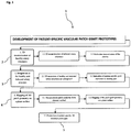
Fig. 1 demonstrates operational steps of a pre-operative patch generation device for the fabrication of grafts patches having patient-specific geometries according to the present invention. -
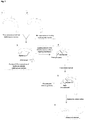
Fig. 2 demonstrates a series of schematic views of healthy and defective vascular anatomies representing operational steps of a pre-operative patch generation device according to the present invention. - The following numerals are referred to in the detailed description of the present invention:
- 1) Patch generation device
- 2) Modelling module
- 3) Integration module
- 4) Transformation module
- 5) Prototype module
- The present invention relates to a system for manufacturing of vascular patches in the form pre-operative surgical planning prototypes providing optimized patient specific 3-D patch geometries.
- Congenital cardiovascular defects in neonatal and pediatric patients are treated through palliative reconstructive surgical operations where the commercially available artificial vascular transplants/grafts fabricated from synthetic materials are implanted. Since patient-specific cardiovascular anatomies are highly complex and cardiovascular structures of neonatal and pediatric patients grow in size while their vascular micro-structure alters significantly with aging under increasing mechanical loads, artificial grafts not having specifically engineered geometries are unable to comply with changing vascular size and fail beyond their operating dimensions unlike live tissue.
- The vascular grafts are generally fabricated from synthetic materials. Biocompatible and blood compatible artificial grafts replaceable with the defected tissue are required for the treatment of neonatal and pediatric cardiovascular defects. To this end, suitable polymeric vascular structures are implanted to treat cardiovascular defects of pediatric or neonatal patients. Vascular conduits and patches conventionally used for this purpose can be referred to as for instance Polyethylene terephthalate, Polytetrafluoroethylene, bovine or swine pericardium (Lam et al., 2012; Kapadia et al., 2008: Laube et al., 2000; Hoshi et al., 2013: Pok and Jacot, 2011).
- Autogenic pericardial implants are ideal for the neonatal and pediatric patients due to their capability of regeneration and complying with the growth of the patient. The essential function needed to be fulfilled by various artificial grafts is the biological and mechanical compatibility with the patient's tissue. In this regard, polyurethanes having structural diversity are considered as one of the most biocompatible materials usable as of today.
- The present invention proposes a patch generation device (1) for the structuring of patient-specific cardiovascular grafts as a computer-aided system producing planar and 3D graft prototypes to serve as a reference prototype during the pre-operative form giving stage of a patch graft by the surgeon, the patch graft given shape as such being than implanted to the defected part of the patient's cardiovascular vessels. Therefore, the present invention proposes fabrication of specially configured reference graft prototypes having patient-specific geometries.
- The planning of the patient specific graft is based on MRI or CT scanning of the patient's vascular anatomy by which 3D hemodynamic reconstruction of the vascular structures are performed. For instance, computer software modules by the
commercial names 3D Doctor, Mimics or Scan-IP running on the patch generation device (1) can be used in this regard. The anatomic reconstruction is performed by the patch generation device's (1) processing unit with multicolor differentiation according to hemodynamic differences within the cardiovascular structure. - The patient's reconstructed cardiovascular anatomy is integrated with the ideal healthy 3D cardiovascular anatomy construction by the patch generation device (1), preferably using an appropriate software module such as for instance SketchCad ™ so as to determine the specific parts of the vascular structure to which a patch graft being restructured in a patient-specific manner can be implanted on the patient's vascular structure. The integration of the healthy and defected vascular structures is effectuated through conformal mapping technique by which the two anatomies are juxtaposed while the defective vascular surfaces requiring modification are indicated on an image display medium in communication with the patch generation device (1) with a distinguishing color in comparison to the ideal vascular surface.
- It is to be noted that the acquired anatomy of the defective vascular surfaces requiring modification therefore represents the graft structure with a specific vascular geometry obtained with the blood pressure therein, i.e. in post-operative state. To obtain the pre-operative vascular anatomy of the graft geometry in non-pressured state, finite element method is used to eliminate the pressure effect on the geometrical structure of the defective vascular surfaces and the acquired corresponding graft structure. The pressure effect is removed by finite element method so that the acquired graft structure is geometrically reshaped, typically reduced in size in a hemodynamically location-specific proportionality so as to be able to take its post-operative shape when the pressure effect will be present. Preferably, an appropriate software module such as Adina ™ can be used in this procedure.
- Subsequently, the unpressurised state graft structure is mapped on a planar surface providing a master structure as a reference geometry for the patch graft to be cut by the surgeon in order for implanting to the pediatric or neonatal patient replacing the defective cardiovascular graft.
- The patch generation device (1) according to the present invention effectuates planar and 3-D printing of the patient-specific graft geometries as prototypes so as to be used by the surgeon as a reference structure when cutting the artificial biocompatible patch to be implanted, thereby manifestly demonstrating the 3-D geometry of the artificial graft to replace the patient's defective vascular graft. While the planar patch prototype will serve for cutting the patch material along predetermined cutting lines in a specific size, the 3-D patch prototype will serve as a 3-D model corresponding to the 3-D geometry of the artificial graft required to be implanted. Therefore, the graft prototypes fabricated according to the proposed system serve to the purpose of assisting the surgeon in implanting the patch graft in an accurately positioned manner so that undesired post-operative graft deformations in treatment of congenital cardiovascular defects in neonatal and pediatric patients can be eliminated.
- The patch generation device (1) according to the present invention is a computer-aided system running a plurality of software modules effectuating generation of patient-specific graft geometries. A modeling module (2) performs hemodynamic reconstruction of the patient's vascular structures so as to perform multicolor demonstration of the hemodynamic behavior in the patient's vascular structures. The reconstruction task includes formation and scaling of the vascular anatomy so as to be processed by an integration module (3) as will be delineated below.
- The integration module (3) performs formation and scaling of a healthy vascular anatomy with which the patient's reconstructed cardiovascular anatomy is integrated in the manner that said reconstructed defected vascular anatomy and the healthy vascular anatomy are overlapped to reveal the patient's specific anatomical parts into which a patch graft implantation is needed.
- The formation and scaling of the patient's vascular anatomy by the modelling module (2) and a healthy vascular anatomy by the integration module (3) enables the integration module (3) to isolate and geometrically specify the exact surface area and associated stitching lines on the patient's vascular anatomy by combining both of the two three-dimensional anatomies of healthy and defective vascular structures.
- Further, a transformation module (4), converts the obtained patch graft shape on the surface of the defected vascular structure under the effect of blood pressure to a non-pressured vascular structure by finite element method allowing reconstruction of a 3-D patch graft model in pre-operative state. The transformation module (4) also effectuates mapping of the patch graft geometry on a plane surface.
- According to the present invention, a prototype module (5) than prints the pre-operative state three-dimensional patch graft model as well as the planar patch graft in the form of planar and three-dimensional objects. The planar object can be used by the surgeon for cutting the artificial patch graft by surgical scissors in a form specifically adapted to the patient. Further, the surgeon will be provided with a patient-specific 3-D model of the graft patch to enable him acquire a 3-D geometry replica of the artificial graft required to be implanted.
- The patch geometry will enable the surgeon to detect graft positions that may lead to undesired graft deformation such as wrinkling or to optimize stitching to remove the possibility of excessive stitching that impose unnecessary loads on the graft.
- It is to be noted that the reconstruction of a 3-D patch graft model according to the present invention is also applicable to the great arteries including pulmonary artery, pulmonary veins and aorta.
- 3-D printing techniques have recently become extensively known to the skilled worker. It is also worthy of note that 3-D printing equipment is commercially available in the market. 3-D printing techniques particularly differ in the manner layers of the final object are built one above another. Use of melting or softening material as well as selective laser sintering (SLS), fused deposition modeling (FDM) and stereolithography (SLA) constitute the common technologies.
- In a nutshell, the present invention proposes a patch generation device (1) for manufacturing of vascular patches in the form of pre-operative surgical planning prototypes providing optimized patient-specific patch geometries, said patch generation device (1) comprising a processing unit effectuating processing of the optimized patient-specific patch geometries.
- In one embodiment of the present invention, said patch generation device (1) comprises a modeling module (2) performing structural and hemodynamic reconstruction of a patient's vascular structures.
- In a further embodiment of the present invention, said patch generation device (1) comprises an integration module (3) performing formation and scaling of a healthy vascular anatomy with which the patient's reconstructed cardiovascular anatomy is integrated in the manner that said reconstructed defected vascular anatomy and the healthy vascular anatomy are overlapped to reveal the patient's specific anatomical parts into which a patch graft implantation is envisageable.
- In a further embodiment of the present invention, said patch generation device (1) comprises a transformation module (4), converting the revealed patient-specific anatomical parts under the effect of blood pressure and into which a patch graft implantation is envisageable to a non-pressured vascular structure by finite element method allowing reconstruction of a 3-D patch graft model in pre-operative state.
- In a further embodiment of the present invention, said modeling module (2) performs formation and scaling of the patient's vascular anatomy enabling multicolor demonstration of the hemodynamic behavior and performance in the patient's vascular structures.
- In a further embodiment of the present invention, said integration module (3) isolates and geometrically specifies an exact surface loading and associated stitching forces on the patient's vascular anatomy by combining both of the three-dimensional anatomies of the healthy and defected vascular anatomies.
- In a further embodiment of the present invention, said transformation module (4) effectuates mapping of the surface loading and stitching forces on a plane surface generating a planar reconstruction of pre-operative state patch graft model with predetermined cutting lines in a specific geometry providing a master structure as a reference geometry.
- In a further embodiment of the present invention, a prototype module (5) of the patch generation device (1) prints the reconstructed pre-operative state three-dimensional patch graft model as a surgical template.
- In a further embodiment of the present invention, a prototype module (5) of the patch generation device (1) prints the reconstructed pre-operative state planar patch graft model.
- In a further embodiment of the present invention, said modeling module (2) performs formation and scaling of the patient's vascular anatomy based on MRI or CT scans by which 3D hemodynamic reconstruction of the vascular structures is performed.
- In a further embodiment of the present invention, integration of the healthy and defected vascular anatomies is effectuated through a computational technique such as conformal mapping technique.
- In a further embodiment of the present invention, the transformation module (4) effectuates conversion of the revealed post-operative state patient-specific anatomical parts into which a patch graft implantation is envisageable to generate a pre-operative state three-dimensional patch graft model in non-pressured state such that finite element method is used to eliminate the pressure effect on the pre-operative state three-dimensional patch graft model.
- In a further embodiment of the present invention, the pre-operative state three-dimensional patch graft model in non-pressured state is reduced in size in a hemodynamically location-specific proportionality so as to be able to take its post-operative shape when the pressure effect will be present.
- In a further embodiment of the present invention, said patch graft model is additionally dimensionally restructured in a patient specific manner based on specific predetermined classifications from a patient database according to the demographic data of the patient. In other words, the final structure of the patch graft model can undergo a second transformation only to adapt the same to the specific anatomical conditions of the patient, such specific anatomical conditions including demographic properties such as for instance age, sex and weight of the patient. It is to be noted that a specific database containing such demographic data can be used to reveal characteristic differences between patients regarding the structural effects on the patch grafts in terms of the surface loading and stitching forces. As a specific example, patch grafts of two otherwise identical patients to be applied under identical anatomical conditions can be structurally slightly different depending on the weight of each patient.
- The present invention further proposes a method for fabricating vascular patches in the form pre-operative surgical planning prototypes providing optimized patient-specific patch geometries by means of a patch generation device (1) as set forth in
Claim 1, said method comprising the steps of, a) performing structural and hemodynamic reconstruction of a patient's vascular structures by a modeling module (2), b) performing formation and scaling of a healthy vascular anatomy with which the patient's reconstructed cardiovascular anatomy is integrated by an integration module (3) in the manner that said reconstructed defected vascular anatomy and the healthy vascular anatomy are overlapped to reveal the patient's specific anatomical parts into which a patch graft implantation is envisageable, c) converting by a transformation module (4) the revealed patient-specific anatomical parts under the effect of blood pressure and into which a patch graft implantation is envisageable to a non-pressured vascular structure by finite element method allowing reconstruction of a 3-D patch graft model in pre-operative state and, d)printing the reconstructed pre-operative state three-dimensional patch graft model. - In a further embodiment of the present invention, said method further comprises the step of mapping of the surface area and associated stitching lines on a plane surface generating a planar reconstruction of pre-operative state patch graft model with predetermined cutting lines in a specific geometry by said transformation module (4).
- In a further embodiment of the present invention, said method further comprises the step of printing the reconstructed pre-operative state planar patch graft model.
- The present invention further proposes a pre-operative state patch graft model manufactured according to the method for fabricating vascular patches in the form pre-operative surgical planning prototypes.
- In a further embodiment of the present invention, a pre-operative state patch graft model manufactured by said patch generation device is proposed.
- The present invention further proposes a conduit manufactured by said patch generation device, wherein said conduit is manufactured in the form of a tubular 3D rapid prototype.
- In sum, the determination of the physical characteristics and the optimum geometrical shape of the artificial patch including the stitching line's length and geometry is made by the surgeon during the surgical operation. Two-dimensional standard artificial patch materials in rectangular form are needed to be cut in appropriate sizes according to the specific needs of the patient. Otherwise an inappropriately structured patch may cause graft deformations if not tailored to the individual patient's anatomy, additionally causing stress on the stitches, consumption of extra energy by the heart and deformations due to the deviation of the hemodynamic forces on the vascular cells from the physiological values.
- Therefore, the patient-specific models/prototypes of graft patches play a crucial role in assisting the surgeon while he designs the structural aspects of the artificial patch. It is also further critical that this process of design should be terminated in the quickest manner as possible because the time period the surgeon needs during the surgery for designing the artificial patch may cause brain and systemic tissue damage in neonatal and pediatric patients. To this end, patient-specific models/prototypes of graft patches in accordance with the present invention substantially accelerate the in-surgery procedures carried out by the surgeon.
Claims (15)
- A patch generation device (1) for generation of vascular patches in the form of pre-operative surgical planning models providing optimized patient-specific patch geometries, said patch generation device (1) comprising a processing unit effectuating processing of the optimized patient-specific patch geometries characterized in that;
said patch generation device (1) comprises a modeling module (2) performing structural and hemodynamic reconstruction of a patient's vascular structures,
said patch generation device (1) comprises an integration module (3) performing formation and scaling of a healthy vascular anatomy with which the patient's reconstructed cardiovascular anatomy is integrated in the manner that said reconstructed defected vascular anatomy and the healthy vascular anatomy are overlapped to reveal the patient's specific anatomical parts into which a patch graft implantation is envisageable,
said patch generation device (1) comprises a transformation module (4), converting the revealed patient-specific anatomical parts under the effect of blood pressure and into which a patch graft implantation is envisageable to a non-pressured vascular structure by finite element method whereby construction of a 3-D patch graft model in pre-operative state is facilitated and,
said integration module (3) isolates and geometrically specifies an exact surface loading and associated stitching forces on the patient's vascular anatomy by combining both of the three-dimensional anatomies of the healthy and defected vascular anatomies. - A patch generation device (1) as set forth in Claim 1, characterized in that said modeling module (2) performs formation and scaling of the patient's vascular anatomy enabling multicolor demonstration of the hemodynamic behavior and performance in the patient's vascular structures.
- A patch generation device (1) as set forth in Claim 1, characterized in that said transformation module (4) effectuates mapping of the surface loading and stitching forces on a plane surface generating a planar reconstruction of pre-operative state patch graft model with predetermined cutting lines in a specific geometry providing a master structure as a reference geometry.
- A patch generation device (1) as set forth in Claim 1, characterized in that a prototype module (5) of the patch generation device (1) prints the reconstructed pre-operative state three-dimensional patch graft model as a template.
- A patch generation device (1) as set forth in Claim 3, characterized in that a prototype module (5) of the patch generation device (1) prints the reconstructed pre-operative state planar patch graft model.
- A patch generation device (1) as set forth in Claim 2, characterized in that said modeling module (2) performs formation and scaling of the patient's vascular anatomy based on MRI or CT scans by which 3D hemodynamic reconstruction of the vascular structures is performed.
- A patch generation device (1) as set forth in Claim 1, characterized in that integration of the healthy and defected vascular anatomies is effectuated through a computational technique.
- A patch generation device (1) as set forth in Claim 1, characterized in that the transformation module (4) effectuates conversion of the revealed post-operative state patient-specific anatomical parts into which a patch graft implantation is envisageable to generate a pre-operative state three-dimensional patch graft model in non-pressured state such that finite element method is used to eliminate the pressure effect on the pre-operative state three-dimensional patch graft model.
- A patch generation device (1) as set forth in Claim 8, characterized in that the pre-operative state three-dimensional patch graft model in non-pressured state is reduced in size in a hemodynamically location-specific proportionality so as to be able to take its post-operative shape when the pressure effect will be present.
- A patch generation device (1) as set forth in Claim 1 or 3, characterized in that said patch graft model is additionally dimensionally restructured in a patient specific manner based on specific predetermined classifications from a patient database according to the demographic data of the patient.
- A method for fabricating vascular patches in the form pre-operative surgical planning prototypes providing optimized patient-specific patch geometries by means of a patch generation device (1) as set forth in Claim 1, said method comprising the steps of;a) performing structural and hemodynamic reconstruction of a patient's vascular structures by a modeling module (2),b) performing formation and scaling of a healthy vascular anatomy with which the patient's reconstructed cardiovascular anatomy is integrated by an integration module (3) in the manner that said reconstructed defected vascular anatomy and the healthy vascular anatomy are overlapped to reveal the patient's specific anatomical parts into which a patch graft implantation is envisageable,c) converting by a transformation module (4) the revealed patient-specific anatomical parts under the effect of blood pressure and into which a patch graft implantation is envisageable to a non-pressured vascular structure by finite element method whereby construction of a 3-D patch graft model in pre-operative state is facilitated and,d) isolating and geometrically specifying by an integration module (3) an exact surface loading and associated stitching forces on the patient's vascular anatomy by combining both of the three-dimensional anatomies of the healthy and defected vascular anatomies.e) printing the reconstructed pre-operative state three-dimensional patch graft model.
- A method for fabricating vascular patches in the form pre-operative surgical planning prototypes as set forth in Claim 11, said method further comprises the step of mapping of the surface area and associated stitching lines on a plane surface generating a planar reconstruction of pre-operative state patch graft model with predetermined cutting lines in a specific geometry by said transformation module (4).
- A method for fabricating vascular patches in the form pre-operative surgical planning prototypes as set forth in Claim 12, said method further comprises the step of printing the reconstructed pre-operative state planar patch graft model.
- A pre-operative state patch graft model generated by the patch generation device (1) according to Claim 1.
- A conduit manufactured by the patch generation device (1) according to Claim 4, wherein said conduit is manufactured in the form of a tubular 3D rapid prototype.
Applications Claiming Priority (1)
| Application Number | Priority Date | Filing Date | Title |
|---|---|---|---|
| PCT/TR2015/000243 WO2016195605A1 (en) | 2015-05-29 | 2015-05-29 | Pre-operative development of patient-specific vascular patch graft prototypes for pediatric and neonatal patients |
Publications (2)
| Publication Number | Publication Date |
|---|---|
| EP3304499A1 EP3304499A1 (en) | 2018-04-11 |
| EP3304499B1 true EP3304499B1 (en) | 2020-01-08 |
Family
ID=53836801
Family Applications (1)
| Application Number | Title | Priority Date | Filing Date |
|---|---|---|---|
| EP15750480.4A Active EP3304499B1 (en) | 2015-05-29 | 2015-05-29 | Pre-operative development of patient-specific vascular patch graft prototypes for pediatric and neonatal patients |
Country Status (3)
| Country | Link |
|---|---|
| US (1) | US10729529B2 (en) |
| EP (1) | EP3304499B1 (en) |
| WO (1) | WO2016195605A1 (en) |
Families Citing this family (5)
| Publication number | Priority date | Publication date | Assignee | Title |
|---|---|---|---|---|
| RU2020100063A (en) | 2017-06-12 | 2021-07-14 | Те Юниверсити Оф Норт Каролина Эт Чепел Хилл | TRANSPLANT COMPOSITION FOR CELL TRANSFER |
| CN111210427B (en) * | 2020-01-17 | 2023-03-10 | 云南大学 | Time-change-based method for analyzing post-operation shrinkage of in-vivo light-weight patch |
| RU2725075C2 (en) * | 2020-02-12 | 2020-06-29 | Федеральное государственное бюджетное учреждение "Национальный медицинский исследовательский центр радиологии" Министерства здравоохранения Российской Федерации (ФГБУ "НМИЦ радиологии" Минздрава России) | Three-dimensional modeling method for 3d printing when planning pancreatic resection in patients with tumor involvement |
| EP4225207A1 (en) * | 2020-10-07 | 2023-08-16 | Children's Medical Center Corporation | Computational planning of surgical reconstruction |
| US11920597B2 (en) | 2021-03-10 | 2024-03-05 | Indiana University Research And Technology Corporation | Boundary layer powered circulatory assist device |
Family Cites Families (1)
| Publication number | Priority date | Publication date | Assignee | Title |
|---|---|---|---|---|
| CA2855943C (en) | 2011-07-29 | 2019-10-29 | Carnegie Mellon University | Artificial valved conduits for cardiac reconstructive procedures and methods for their production |
-
2015
- 2015-05-29 EP EP15750480.4A patent/EP3304499B1/en active Active
- 2015-05-29 WO PCT/TR2015/000243 patent/WO2016195605A1/en active Application Filing
-
2017
- 2017-11-28 US US15/824,845 patent/US10729529B2/en active Active
Non-Patent Citations (1)
| Title |
|---|
| None * |
Also Published As
| Publication number | Publication date |
|---|---|
| US10729529B2 (en) | 2020-08-04 |
| WO2016195605A1 (en) | 2016-12-08 |
| EP3304499A1 (en) | 2018-04-11 |
| US20180078356A1 (en) | 2018-03-22 |
Similar Documents
| Publication | Publication Date | Title |
|---|---|---|
| US10729529B2 (en) | System for pre-operative development of patient-specific vascular patch graft prototypes for pediatric and neonatal patients | |
| Yap et al. | 3D printed bio-models for medical applications | |
| CN106073870B (en) | A kind of facies articularis ossium rebuilds the method that implant is repaired in 3D printing | |
| Sun et al. | Bio-CAD modeling and its applications in computer-aided tissue engineering | |
| US20110016690A1 (en) | Process to design and fabricate a custom-fit implant | |
| Starly et al. | Three-dimensional reconstruction for medical-CAD modeling | |
| CN105930617A (en) | Method for designing and forming stiffness-controllable bone tumor defect repair implant | |
| JP2015516243A (en) | Implantable bone augmentation and method for manufacturing implantable bone augmentation | |
| US8090540B2 (en) | Method for designing 3-dimensional porous tissue engineering scaffold | |
| Modi et al. | Design and additive manufacturing of patient-specific cranial and pelvic bone implants from computed tomography data | |
| Gall et al. | Computer-aided planning and reconstruction of cranial 3D implants | |
| CN103942826A (en) | High match degree skull restoration reconstruction method | |
| CN105912863A (en) | Method and system for preparing artificial bone | |
| Lashkarinia et al. | Computational pre-surgical planning of arterial patch reconstruction: parametric limits and in vitro validation | |
| Moiduddin et al. | Computer assisted design and analysis of customized porous plate for mandibular reconstruction | |
| Šljivić et al. | Implemenation of FEM and rapid prototyping in maxillofacial surgery | |
| CN110353861B (en) | Personalized preparation method of human sacrum prosthesis | |
| Hatamleh et al. | Management of extensive frontal cranioplasty defects | |
| CN104537164A (en) | Integrated system and method for bone defect repair | |
| Colombo et al. | Automatic 3D reconstruction of transfemoral residual limb from MRI images | |
| Kouhi et al. | Design and fabrication of reconstructive mandibular models using fused deposition modeling | |
| Moncayo-Matute et al. | Description and application of a comprehensive methodology for custom implant design and surgical planning | |
| Abdel-Sayed et al. | Rapid prototyping for training purposes in cardiovascular surgery | |
| Dhakshyani et al. | FDM models and FEA in dysplastic hip | |
| Phanindra Bogu et al. | Homogenous scaffold-based cranial/skull implant modelling and structural analysis—unit cell algorithm-meshless approach |
Legal Events
| Date | Code | Title | Description |
|---|---|---|---|
| STAA | Information on the status of an ep patent application or granted ep patent |
Free format text: STATUS: THE INTERNATIONAL PUBLICATION HAS BEEN MADE |
|
| PUAI | Public reference made under article 153(3) epc to a published international application that has entered the european phase |
Free format text: ORIGINAL CODE: 0009012 |
|
| STAA | Information on the status of an ep patent application or granted ep patent |
Free format text: STATUS: REQUEST FOR EXAMINATION WAS MADE |
|
| 17P | Request for examination filed |
Effective date: 20171227 |
|
| AK | Designated contracting states |
Kind code of ref document: A1 Designated state(s): AL AT BE BG CH CY CZ DE DK EE ES FI FR GB GR HR HU IE IS IT LI LT LU LV MC MK MT NL NO PL PT RO RS SE SI SK SM TR |
|
| AX | Request for extension of the european patent |
Extension state: BA ME |
|
| DAV | Request for validation of the european patent (deleted) | ||
| DAX | Request for extension of the european patent (deleted) | ||
| GRAP | Despatch of communication of intention to grant a patent |
Free format text: ORIGINAL CODE: EPIDOSNIGR1 |
|
| STAA | Information on the status of an ep patent application or granted ep patent |
Free format text: STATUS: GRANT OF PATENT IS INTENDED |
|
| RIC1 | Information provided on ipc code assigned before grant |
Ipc: G06T 17/00 20060101AFI20190813BHEP Ipc: A61F 2/06 20130101ALI20190813BHEP |
|
| INTG | Intention to grant announced |
Effective date: 20190913 |
|
| GRAS | Grant fee paid |
Free format text: ORIGINAL CODE: EPIDOSNIGR3 |
|
| GRAA | (expected) grant |
Free format text: ORIGINAL CODE: 0009210 |
|
| STAA | Information on the status of an ep patent application or granted ep patent |
Free format text: STATUS: THE PATENT HAS BEEN GRANTED |
|
| AK | Designated contracting states |
Kind code of ref document: B1 Designated state(s): AL AT BE BG CH CY CZ DE DK EE ES FI FR GB GR HR HU IE IS IT LI LT LU LV MC MK MT NL NO PL PT RO RS SE SI SK SM TR |
|
| REG | Reference to a national code |
Ref country code: GB Ref legal event code: FG4D |
|
| REG | Reference to a national code |
Ref country code: CH Ref legal event code: EP |
|
| REG | Reference to a national code |
Ref country code: DE Ref legal event code: R096 Ref document number: 602015045171 Country of ref document: DE |
|
| REG | Reference to a national code |
Ref country code: IE Ref legal event code: FG4D |
|
| REG | Reference to a national code |
Ref country code: AT Ref legal event code: REF Ref document number: 1223639 Country of ref document: AT Kind code of ref document: T Effective date: 20200215 |
|
| REG | Reference to a national code |
Ref country code: NL Ref legal event code: FP |
|
| REG | Reference to a national code |
Ref country code: LT Ref legal event code: MG4D |
|
| PG25 | Lapsed in a contracting state [announced via postgrant information from national office to epo] |
Ref country code: RS Free format text: LAPSE BECAUSE OF FAILURE TO SUBMIT A TRANSLATION OF THE DESCRIPTION OR TO PAY THE FEE WITHIN THE PRESCRIBED TIME-LIMIT Effective date: 20200108 Ref country code: PT Free format text: LAPSE BECAUSE OF FAILURE TO SUBMIT A TRANSLATION OF THE DESCRIPTION OR TO PAY THE FEE WITHIN THE PRESCRIBED TIME-LIMIT Effective date: 20200531 Ref country code: NO Free format text: LAPSE BECAUSE OF FAILURE TO SUBMIT A TRANSLATION OF THE DESCRIPTION OR TO PAY THE FEE WITHIN THE PRESCRIBED TIME-LIMIT Effective date: 20200408 Ref country code: FI Free format text: LAPSE BECAUSE OF FAILURE TO SUBMIT A TRANSLATION OF THE DESCRIPTION OR TO PAY THE FEE WITHIN THE PRESCRIBED TIME-LIMIT Effective date: 20200108 Ref country code: LT Free format text: LAPSE BECAUSE OF FAILURE TO SUBMIT A TRANSLATION OF THE DESCRIPTION OR TO PAY THE FEE WITHIN THE PRESCRIBED TIME-LIMIT Effective date: 20200108 |
|
| PG25 | Lapsed in a contracting state [announced via postgrant information from national office to epo] |
Ref country code: GR Free format text: LAPSE BECAUSE OF FAILURE TO SUBMIT A TRANSLATION OF THE DESCRIPTION OR TO PAY THE FEE WITHIN THE PRESCRIBED TIME-LIMIT Effective date: 20200409 Ref country code: BG Free format text: LAPSE BECAUSE OF FAILURE TO SUBMIT A TRANSLATION OF THE DESCRIPTION OR TO PAY THE FEE WITHIN THE PRESCRIBED TIME-LIMIT Effective date: 20200408 Ref country code: IS Free format text: LAPSE BECAUSE OF FAILURE TO SUBMIT A TRANSLATION OF THE DESCRIPTION OR TO PAY THE FEE WITHIN THE PRESCRIBED TIME-LIMIT Effective date: 20200508 Ref country code: SE Free format text: LAPSE BECAUSE OF FAILURE TO SUBMIT A TRANSLATION OF THE DESCRIPTION OR TO PAY THE FEE WITHIN THE PRESCRIBED TIME-LIMIT Effective date: 20200108 Ref country code: LV Free format text: LAPSE BECAUSE OF FAILURE TO SUBMIT A TRANSLATION OF THE DESCRIPTION OR TO PAY THE FEE WITHIN THE PRESCRIBED TIME-LIMIT Effective date: 20200108 Ref country code: HR Free format text: LAPSE BECAUSE OF FAILURE TO SUBMIT A TRANSLATION OF THE DESCRIPTION OR TO PAY THE FEE WITHIN THE PRESCRIBED TIME-LIMIT Effective date: 20200108 |
|
| REG | Reference to a national code |
Ref country code: DE Ref legal event code: R097 Ref document number: 602015045171 Country of ref document: DE |
|
| PG25 | Lapsed in a contracting state [announced via postgrant information from national office to epo] |
Ref country code: SM Free format text: LAPSE BECAUSE OF FAILURE TO SUBMIT A TRANSLATION OF THE DESCRIPTION OR TO PAY THE FEE WITHIN THE PRESCRIBED TIME-LIMIT Effective date: 20200108 Ref country code: EE Free format text: LAPSE BECAUSE OF FAILURE TO SUBMIT A TRANSLATION OF THE DESCRIPTION OR TO PAY THE FEE WITHIN THE PRESCRIBED TIME-LIMIT Effective date: 20200108 Ref country code: DK Free format text: LAPSE BECAUSE OF FAILURE TO SUBMIT A TRANSLATION OF THE DESCRIPTION OR TO PAY THE FEE WITHIN THE PRESCRIBED TIME-LIMIT Effective date: 20200108 Ref country code: SK Free format text: LAPSE BECAUSE OF FAILURE TO SUBMIT A TRANSLATION OF THE DESCRIPTION OR TO PAY THE FEE WITHIN THE PRESCRIBED TIME-LIMIT Effective date: 20200108 Ref country code: RO Free format text: LAPSE BECAUSE OF FAILURE TO SUBMIT A TRANSLATION OF THE DESCRIPTION OR TO PAY THE FEE WITHIN THE PRESCRIBED TIME-LIMIT Effective date: 20200108 Ref country code: ES Free format text: LAPSE BECAUSE OF FAILURE TO SUBMIT A TRANSLATION OF THE DESCRIPTION OR TO PAY THE FEE WITHIN THE PRESCRIBED TIME-LIMIT Effective date: 20200108 Ref country code: CZ Free format text: LAPSE BECAUSE OF FAILURE TO SUBMIT A TRANSLATION OF THE DESCRIPTION OR TO PAY THE FEE WITHIN THE PRESCRIBED TIME-LIMIT Effective date: 20200108 |
|
| PLBE | No opposition filed within time limit |
Free format text: ORIGINAL CODE: 0009261 |
|
| STAA | Information on the status of an ep patent application or granted ep patent |
Free format text: STATUS: NO OPPOSITION FILED WITHIN TIME LIMIT |
|
| REG | Reference to a national code |
Ref country code: AT Ref legal event code: MK05 Ref document number: 1223639 Country of ref document: AT Kind code of ref document: T Effective date: 20200108 |
|
| 26N | No opposition filed |
Effective date: 20201009 |
|
| PG25 | Lapsed in a contracting state [announced via postgrant information from national office to epo] |
Ref country code: LI Free format text: LAPSE BECAUSE OF NON-PAYMENT OF DUE FEES Effective date: 20200531 Ref country code: MC Free format text: LAPSE BECAUSE OF FAILURE TO SUBMIT A TRANSLATION OF THE DESCRIPTION OR TO PAY THE FEE WITHIN THE PRESCRIBED TIME-LIMIT Effective date: 20200108 Ref country code: AT Free format text: LAPSE BECAUSE OF FAILURE TO SUBMIT A TRANSLATION OF THE DESCRIPTION OR TO PAY THE FEE WITHIN THE PRESCRIBED TIME-LIMIT Effective date: 20200108 Ref country code: IT Free format text: LAPSE BECAUSE OF FAILURE TO SUBMIT A TRANSLATION OF THE DESCRIPTION OR TO PAY THE FEE WITHIN THE PRESCRIBED TIME-LIMIT Effective date: 20200108 Ref country code: CH Free format text: LAPSE BECAUSE OF NON-PAYMENT OF DUE FEES Effective date: 20200531 |
|
| PG25 | Lapsed in a contracting state [announced via postgrant information from national office to epo] |
Ref country code: SI Free format text: LAPSE BECAUSE OF FAILURE TO SUBMIT A TRANSLATION OF THE DESCRIPTION OR TO PAY THE FEE WITHIN THE PRESCRIBED TIME-LIMIT Effective date: 20200108 Ref country code: PL Free format text: LAPSE BECAUSE OF FAILURE TO SUBMIT A TRANSLATION OF THE DESCRIPTION OR TO PAY THE FEE WITHIN THE PRESCRIBED TIME-LIMIT Effective date: 20200108 |
|
| REG | Reference to a national code |
Ref country code: BE Ref legal event code: MM Effective date: 20200531 |
|
| PG25 | Lapsed in a contracting state [announced via postgrant information from national office to epo] |
Ref country code: LU Free format text: LAPSE BECAUSE OF NON-PAYMENT OF DUE FEES Effective date: 20200529 |
|
| PG25 | Lapsed in a contracting state [announced via postgrant information from national office to epo] |
Ref country code: FR Free format text: LAPSE BECAUSE OF NON-PAYMENT OF DUE FEES Effective date: 20200531 |
|
| PG25 | Lapsed in a contracting state [announced via postgrant information from national office to epo] |
Ref country code: BE Free format text: LAPSE BECAUSE OF NON-PAYMENT OF DUE FEES Effective date: 20200531 |
|
| PG25 | Lapsed in a contracting state [announced via postgrant information from national office to epo] |
Ref country code: MT Free format text: LAPSE BECAUSE OF FAILURE TO SUBMIT A TRANSLATION OF THE DESCRIPTION OR TO PAY THE FEE WITHIN THE PRESCRIBED TIME-LIMIT Effective date: 20200108 Ref country code: CY Free format text: LAPSE BECAUSE OF FAILURE TO SUBMIT A TRANSLATION OF THE DESCRIPTION OR TO PAY THE FEE WITHIN THE PRESCRIBED TIME-LIMIT Effective date: 20200108 |
|
| PG25 | Lapsed in a contracting state [announced via postgrant information from national office to epo] |
Ref country code: MK Free format text: LAPSE BECAUSE OF FAILURE TO SUBMIT A TRANSLATION OF THE DESCRIPTION OR TO PAY THE FEE WITHIN THE PRESCRIBED TIME-LIMIT Effective date: 20200108 Ref country code: AL Free format text: LAPSE BECAUSE OF FAILURE TO SUBMIT A TRANSLATION OF THE DESCRIPTION OR TO PAY THE FEE WITHIN THE PRESCRIBED TIME-LIMIT Effective date: 20200108 |
|
| PGFP | Annual fee paid to national office [announced via postgrant information from national office to epo] |
Ref country code: IE Payment date: 20220414 Year of fee payment: 8 Ref country code: DE Payment date: 20220531 Year of fee payment: 8 |
|
| PGFP | Annual fee paid to national office [announced via postgrant information from national office to epo] |
Ref country code: NL Payment date: 20230531 Year of fee payment: 9 |
|
| PGFP | Annual fee paid to national office [announced via postgrant information from national office to epo] |
Ref country code: TR Payment date: 20230529 Year of fee payment: 9 |
|
| PGFP | Annual fee paid to national office [announced via postgrant information from national office to epo] |
Ref country code: GB Payment date: 20230531 Year of fee payment: 9 |
|
| REG | Reference to a national code |
Ref country code: DE Ref legal event code: R119 Ref document number: 602015045171 Country of ref document: DE |
|
| REG | Reference to a national code |
Ref country code: IE Ref legal event code: MM4A |