EP3304084B1 - Methods and kits - Google Patents
Methods and kits Download PDFInfo
- Publication number
- EP3304084B1 EP3304084B1 EP16728358.9A EP16728358A EP3304084B1 EP 3304084 B1 EP3304084 B1 EP 3304084B1 EP 16728358 A EP16728358 A EP 16728358A EP 3304084 B1 EP3304084 B1 EP 3304084B1
- Authority
- EP
- European Patent Office
- Prior art keywords
- sequence
- seq
- mcm5
- antibody
- monoclonal antibody
- Prior art date
- Legal status (The legal status is an assumption and is not a legal conclusion. Google has not performed a legal analysis and makes no representation as to the accuracy of the status listed.)
- Active
Links
- 238000000034 method Methods 0.000 title claims description 95
- 101150002398 MCM5 gene Proteins 0.000 claims description 201
- 239000012139 lysis buffer Substances 0.000 claims description 97
- 210000002700 urine Anatomy 0.000 claims description 93
- 239000000872 buffer Substances 0.000 claims description 85
- 210000004027 cell Anatomy 0.000 claims description 85
- LENZDBCJOHFCAS-UHFFFAOYSA-N tris Chemical compound OCC(N)(CO)CO LENZDBCJOHFCAS-UHFFFAOYSA-N 0.000 claims description 68
- 238000003556 assay Methods 0.000 claims description 61
- FAPWRFPIFSIZLT-UHFFFAOYSA-M Sodium chloride Chemical compound [Na+].[Cl-] FAPWRFPIFSIZLT-UHFFFAOYSA-M 0.000 claims description 59
- 125000003275 alpha amino acid group Chemical group 0.000 claims description 56
- 239000007983 Tris buffer Substances 0.000 claims description 49
- PXIPVTKHYLBLMZ-UHFFFAOYSA-N Sodium azide Chemical compound [Na+].[N-]=[N+]=[N-] PXIPVTKHYLBLMZ-UHFFFAOYSA-N 0.000 claims description 48
- 238000001514 detection method Methods 0.000 claims description 48
- 239000003599 detergent Substances 0.000 claims description 39
- 238000002965 ELISA Methods 0.000 claims description 35
- 208000000236 Prostatic Neoplasms Diseases 0.000 claims description 34
- 206010060862 Prostate cancer Diseases 0.000 claims description 33
- 239000011780 sodium chloride Substances 0.000 claims description 30
- 229920004890 Triton X-100 Polymers 0.000 claims description 25
- 239000013504 Triton X-100 Substances 0.000 claims description 23
- 208000007097 Urinary Bladder Neoplasms Diseases 0.000 claims description 23
- 206010005003 Bladder cancer Diseases 0.000 claims description 22
- 238000011534 incubation Methods 0.000 claims description 22
- 201000005112 urinary bladder cancer Diseases 0.000 claims description 22
- 229960003964 deoxycholic acid Drugs 0.000 claims description 21
- 208000006593 Urologic Neoplasms Diseases 0.000 claims description 20
- 108010001336 Horseradish Peroxidase Proteins 0.000 claims description 19
- 239000003153 chemical reaction reagent Substances 0.000 claims description 19
- FWMNVWWHGCHHJJ-SKKKGAJSSA-N 4-amino-1-[(2r)-6-amino-2-[[(2r)-2-[[(2r)-2-[[(2r)-2-amino-3-phenylpropanoyl]amino]-3-phenylpropanoyl]amino]-4-methylpentanoyl]amino]hexanoyl]piperidine-4-carboxylic acid Chemical compound C([C@H](C(=O)N[C@H](CC(C)C)C(=O)N[C@H](CCCCN)C(=O)N1CCC(N)(CC1)C(O)=O)NC(=O)[C@H](N)CC=1C=CC=CC=1)C1=CC=CC=C1 FWMNVWWHGCHHJJ-SKKKGAJSSA-N 0.000 claims description 16
- 102000039446 nucleic acids Human genes 0.000 claims description 14
- 108020004707 nucleic acids Proteins 0.000 claims description 14
- 150000007523 nucleic acids Chemical class 0.000 claims description 14
- 230000001965 increasing effect Effects 0.000 claims description 13
- DBMJMQXJHONAFJ-UHFFFAOYSA-M Sodium laurylsulphate Chemical compound [Na+].CCCCCCCCCCCCOS([O-])(=O)=O DBMJMQXJHONAFJ-UHFFFAOYSA-M 0.000 claims description 12
- 238000003118 sandwich ELISA Methods 0.000 claims description 11
- 206010044412 transitional cell carcinoma Diseases 0.000 claims description 10
- 239000013049 sediment Substances 0.000 claims description 8
- 239000000243 solution Substances 0.000 claims description 8
- 239000000758 substrate Substances 0.000 claims description 8
- 230000002485 urinary effect Effects 0.000 claims description 8
- 238000005119 centrifugation Methods 0.000 claims description 7
- 238000010008 shearing Methods 0.000 claims description 7
- 235000019333 sodium laurylsulphate Nutrition 0.000 claims description 6
- 239000012089 stop solution Substances 0.000 claims description 6
- 102100034001 DNA replication licensing factor MCM5 Human genes 0.000 claims description 5
- 102000004190 Enzymes Human genes 0.000 claims description 2
- 108090000790 Enzymes Proteins 0.000 claims description 2
- 108010079756 Minichromosome Maintenance Complex Component 5 Proteins 0.000 claims description 2
- KXGVEGMKQFWNSR-LLQZFEROSA-N deoxycholic acid Chemical compound C([C@H]1CC2)[C@H](O)CC[C@]1(C)[C@@H]1[C@@H]2[C@@H]2CC[C@H]([C@@H](CCC(O)=O)C)[C@@]2(C)[C@@H](O)C1 KXGVEGMKQFWNSR-LLQZFEROSA-N 0.000 claims 3
- 208000008839 Kidney Neoplasms Diseases 0.000 claims 1
- 206010061336 Pelvic neoplasm Diseases 0.000 claims 1
- 125000002081 peroxide group Chemical group 0.000 claims 1
- 239000000523 sample Substances 0.000 description 136
- 108090000765 processed proteins & peptides Proteins 0.000 description 78
- 235000001014 amino acid Nutrition 0.000 description 66
- 102000004196 processed proteins & peptides Human genes 0.000 description 60
- 229920001184 polypeptide Polymers 0.000 description 49
- 238000006467 substitution reaction Methods 0.000 description 46
- 230000027455 binding Effects 0.000 description 40
- 235000002639 sodium chloride Nutrition 0.000 description 39
- 239000000090 biomarker Substances 0.000 description 36
- 239000000306 component Substances 0.000 description 34
- 239000002773 nucleotide Substances 0.000 description 33
- 125000003729 nucleotide group Chemical group 0.000 description 33
- 108091003079 Bovine Serum Albumin Proteins 0.000 description 28
- 206010028980 Neoplasm Diseases 0.000 description 28
- 150000001413 amino acids Chemical class 0.000 description 28
- 229940098773 bovine serum albumin Drugs 0.000 description 28
- 238000012360 testing method Methods 0.000 description 28
- 229940024606 amino acid Drugs 0.000 description 24
- 239000003381 stabilizer Substances 0.000 description 24
- 102000007066 Prostate-Specific Antigen Human genes 0.000 description 23
- 108010072866 Prostate-Specific Antigen Proteins 0.000 description 23
- 239000000203 mixture Substances 0.000 description 21
- 108090000623 proteins and genes Proteins 0.000 description 21
- 102000004169 proteins and genes Human genes 0.000 description 20
- 108020004414 DNA Proteins 0.000 description 19
- 239000012634 fragment Substances 0.000 description 18
- FHHPUSMSKHSNKW-SMOYURAASA-M sodium deoxycholate Chemical compound [Na+].C([C@H]1CC2)[C@H](O)CC[C@]1(C)[C@@H]1[C@@H]2[C@@H]2CC[C@H]([C@@H](CCC([O-])=O)C)[C@@]2(C)[C@@H](O)C1 FHHPUSMSKHSNKW-SMOYURAASA-M 0.000 description 18
- 201000011510 cancer Diseases 0.000 description 17
- LOKCTEFSRHRXRJ-UHFFFAOYSA-I dipotassium trisodium dihydrogen phosphate hydrogen phosphate dichloride Chemical compound P(=O)(O)(O)[O-].[K+].P(=O)(O)([O-])[O-].[Na+].[Na+].[Cl-].[K+].[Cl-].[Na+] LOKCTEFSRHRXRJ-UHFFFAOYSA-I 0.000 description 17
- 239000002953 phosphate buffered saline Substances 0.000 description 17
- 210000004408 hybridoma Anatomy 0.000 description 16
- 235000018102 proteins Nutrition 0.000 description 16
- 239000006166 lysate Substances 0.000 description 15
- 238000002493 microarray Methods 0.000 description 15
- UMCMPZBLKLEWAF-BCTGSCMUSA-N 3-[(3-cholamidopropyl)dimethylammonio]propane-1-sulfonate Chemical compound C([C@H]1C[C@H]2O)[C@H](O)CC[C@]1(C)[C@@H]1[C@@H]2[C@@H]2CC[C@H]([C@@H](CCC(=O)NCCC[N+](C)(C)CCCS([O-])(=O)=O)C)[C@@]2(C)[C@@H](O)C1 UMCMPZBLKLEWAF-BCTGSCMUSA-N 0.000 description 14
- KCXVZYZYPLLWCC-UHFFFAOYSA-N EDTA Chemical compound OC(=O)CN(CC(O)=O)CCN(CC(O)=O)CC(O)=O KCXVZYZYPLLWCC-UHFFFAOYSA-N 0.000 description 14
- 210000002307 prostate Anatomy 0.000 description 13
- 210000003932 urinary bladder Anatomy 0.000 description 13
- 241000699666 Mus <mouse, genus> Species 0.000 description 12
- 239000012083 RIPA buffer Substances 0.000 description 11
- 230000004044 response Effects 0.000 description 11
- 238000004458 analytical method Methods 0.000 description 10
- 239000000427 antigen Substances 0.000 description 10
- 108091007433 antigens Proteins 0.000 description 10
- 102000036639 antigens Human genes 0.000 description 10
- 108091033319 polynucleotide Proteins 0.000 description 10
- 102000040430 polynucleotide Human genes 0.000 description 10
- 239000002157 polynucleotide Substances 0.000 description 10
- 150000003839 salts Chemical class 0.000 description 10
- 239000004599 antimicrobial Substances 0.000 description 9
- 238000003745 diagnosis Methods 0.000 description 9
- 230000000670 limiting effect Effects 0.000 description 9
- 239000008188 pellet Substances 0.000 description 9
- 241000283707 Capra Species 0.000 description 8
- PXHVJJICTQNCMI-UHFFFAOYSA-N Nickel Chemical compound [Ni] PXHVJJICTQNCMI-UHFFFAOYSA-N 0.000 description 8
- 238000001574 biopsy Methods 0.000 description 8
- 239000000463 material Substances 0.000 description 8
- 238000012216 screening Methods 0.000 description 8
- DVLFYONBTKHTER-UHFFFAOYSA-N 3-(N-morpholino)propanesulfonic acid Chemical compound OS(=O)(=O)CCCN1CCOCC1 DVLFYONBTKHTER-UHFFFAOYSA-N 0.000 description 7
- 102000007260 Deoxyribonuclease I Human genes 0.000 description 7
- 108010008532 Deoxyribonuclease I Proteins 0.000 description 7
- 108020002230 Pancreatic Ribonuclease Proteins 0.000 description 7
- 102000005891 Pancreatic ribonuclease Human genes 0.000 description 7
- 229920001213 Polysorbate 20 Polymers 0.000 description 7
- 238000002835 absorbance Methods 0.000 description 7
- 150000001875 compounds Chemical class 0.000 description 7
- 238000010790 dilution Methods 0.000 description 7
- 239000012895 dilution Substances 0.000 description 7
- 230000014509 gene expression Effects 0.000 description 7
- 230000007170 pathology Effects 0.000 description 7
- 239000000137 peptide hydrolase inhibitor Substances 0.000 description 7
- 239000000256 polyoxyethylene sorbitan monolaurate Substances 0.000 description 7
- 235000010486 polyoxyethylene sorbitan monolaurate Nutrition 0.000 description 7
- 238000010186 staining Methods 0.000 description 7
- 239000013598 vector Substances 0.000 description 7
- JKMHFZQWWAIEOD-UHFFFAOYSA-N 2-[4-(2-hydroxyethyl)piperazin-1-yl]ethanesulfonic acid Chemical compound OCC[NH+]1CCN(CCS([O-])(=O)=O)CC1 JKMHFZQWWAIEOD-UHFFFAOYSA-N 0.000 description 6
- WRDABNWSWOHGMS-UHFFFAOYSA-N AEBSF hydrochloride Chemical compound Cl.NCCC1=CC=C(S(F)(=O)=O)C=C1 WRDABNWSWOHGMS-UHFFFAOYSA-N 0.000 description 6
- WCUXLLCKKVVCTQ-UHFFFAOYSA-M Potassium chloride Chemical compound [Cl-].[K+] WCUXLLCKKVVCTQ-UHFFFAOYSA-M 0.000 description 6
- 229940124158 Protease/peptidase inhibitor Drugs 0.000 description 6
- 239000006180 TBST buffer Substances 0.000 description 6
- 230000002159 abnormal effect Effects 0.000 description 6
- 230000009089 cytolysis Effects 0.000 description 6
- 238000012217 deletion Methods 0.000 description 6
- 230000037430 deletion Effects 0.000 description 6
- 239000012909 foetal bovine serum Substances 0.000 description 6
- 230000003211 malignant effect Effects 0.000 description 6
- 230000003019 stabilising effect Effects 0.000 description 6
- QAPSNMNOIOSXSQ-YNEHKIRRSA-N 1-[(2r,4s,5r)-4-[tert-butyl(dimethyl)silyl]oxy-5-(hydroxymethyl)oxolan-2-yl]-5-methylpyrimidine-2,4-dione Chemical compound O=C1NC(=O)C(C)=CN1[C@@H]1O[C@H](CO)[C@@H](O[Si](C)(C)C(C)(C)C)C1 QAPSNMNOIOSXSQ-YNEHKIRRSA-N 0.000 description 5
- 241000588724 Escherichia coli Species 0.000 description 5
- 101001017545 Homo sapiens DNA replication licensing factor MCM5 Proteins 0.000 description 5
- 239000007993 MOPS buffer Substances 0.000 description 5
- 206010027476 Metastases Diseases 0.000 description 5
- 230000008901 benefit Effects 0.000 description 5
- 239000003795 chemical substances by application Substances 0.000 description 5
- 230000000295 complement effect Effects 0.000 description 5
- 238000002574 cystoscopy Methods 0.000 description 5
- 201000010099 disease Diseases 0.000 description 5
- 208000037265 diseases, disorders, signs and symptoms Diseases 0.000 description 5
- 238000002474 experimental method Methods 0.000 description 5
- 210000004907 gland Anatomy 0.000 description 5
- 230000003993 interaction Effects 0.000 description 5
- 230000009871 nonspecific binding Effects 0.000 description 5
- 210000002966 serum Anatomy 0.000 description 5
- 239000006228 supernatant Substances 0.000 description 5
- 238000004448 titration Methods 0.000 description 5
- RZQXOGQSPBYUKH-UHFFFAOYSA-N 3-[[1,3-dihydroxy-2-(hydroxymethyl)propan-2-yl]azaniumyl]-2-hydroxypropane-1-sulfonate Chemical compound OCC(CO)(CO)NCC(O)CS(O)(=O)=O RZQXOGQSPBYUKH-UHFFFAOYSA-N 0.000 description 4
- 239000007995 HEPES buffer Substances 0.000 description 4
- 241000282414 Homo sapiens Species 0.000 description 4
- FSVCELGFZIQNCK-UHFFFAOYSA-N N,N-bis(2-hydroxyethyl)glycine Chemical compound OCCN(CCO)CC(O)=O FSVCELGFZIQNCK-UHFFFAOYSA-N 0.000 description 4
- SEQKRHFRPICQDD-UHFFFAOYSA-N N-tris(hydroxymethyl)methylglycine Chemical compound OCC(CO)(CO)[NH2+]CC([O-])=O SEQKRHFRPICQDD-UHFFFAOYSA-N 0.000 description 4
- 241000283973 Oryctolagus cuniculus Species 0.000 description 4
- 230000009286 beneficial effect Effects 0.000 description 4
- 230000000903 blocking effect Effects 0.000 description 4
- 239000007853 buffer solution Substances 0.000 description 4
- 239000002738 chelating agent Substances 0.000 description 4
- 230000000875 corresponding effect Effects 0.000 description 4
- 230000006872 improvement Effects 0.000 description 4
- 238000005259 measurement Methods 0.000 description 4
- 229940126619 mouse monoclonal antibody Drugs 0.000 description 4
- 229910052759 nickel Inorganic materials 0.000 description 4
- 238000011002 quantification Methods 0.000 description 4
- 239000013074 reference sample Substances 0.000 description 4
- 238000011160 research Methods 0.000 description 4
- 239000007787 solid Substances 0.000 description 4
- 238000002198 surface plasmon resonance spectroscopy Methods 0.000 description 4
- 210000000626 ureter Anatomy 0.000 description 4
- 210000003708 urethra Anatomy 0.000 description 4
- JLHMJWHSBYZWJJ-UHFFFAOYSA-N 1,2-thiazole 1-oxide Chemical compound O=S1C=CC=N1 JLHMJWHSBYZWJJ-UHFFFAOYSA-N 0.000 description 3
- UAIUNKRWKOVEES-UHFFFAOYSA-N 3,3',5,5'-tetramethylbenzidine Chemical compound CC1=C(N)C(C)=CC(C=2C=C(C)C(N)=C(C)C=2)=C1 UAIUNKRWKOVEES-UHFFFAOYSA-N 0.000 description 3
- 206010004446 Benign prostatic hyperplasia Diseases 0.000 description 3
- 206010009944 Colon cancer Diseases 0.000 description 3
- 229910052693 Europium Inorganic materials 0.000 description 3
- 208000004403 Prostatic Hyperplasia Diseases 0.000 description 3
- 239000002250 absorbent Substances 0.000 description 3
- 230000002745 absorbent Effects 0.000 description 3
- 230000015556 catabolic process Effects 0.000 description 3
- 238000010367 cloning Methods 0.000 description 3
- 230000021615 conjugation Effects 0.000 description 3
- 238000011161 development Methods 0.000 description 3
- 230000000694 effects Effects 0.000 description 3
- 238000009472 formulation Methods 0.000 description 3
- 238000009432 framing Methods 0.000 description 3
- 208000006750 hematuria Diseases 0.000 description 3
- 238000003018 immunoassay Methods 0.000 description 3
- 239000011159 matrix material Substances 0.000 description 3
- 229920003023 plastic Polymers 0.000 description 3
- 239000004033 plastic Substances 0.000 description 3
- 229920000136 polysorbate Polymers 0.000 description 3
- 239000001103 potassium chloride Substances 0.000 description 3
- 235000011164 potassium chloride Nutrition 0.000 description 3
- 238000000746 purification Methods 0.000 description 3
- 230000002829 reductive effect Effects 0.000 description 3
- 238000012289 standard assay Methods 0.000 description 3
- 238000003860 storage Methods 0.000 description 3
- 208000024891 symptom Diseases 0.000 description 3
- GPRLSGONYQIRFK-MNYXATJNSA-N triton Chemical compound [3H+] GPRLSGONYQIRFK-MNYXATJNSA-N 0.000 description 3
- 238000002604 ultrasonography Methods 0.000 description 3
- 208000023747 urothelial carcinoma Diseases 0.000 description 3
- 239000011534 wash buffer Substances 0.000 description 3
- 238000001262 western blot Methods 0.000 description 3
- GBHSCKFAHCEEAZ-UHFFFAOYSA-N 2-[hydroxymethyl(methyl)amino]acetic acid Chemical compound OCN(C)CC(O)=O GBHSCKFAHCEEAZ-UHFFFAOYSA-N 0.000 description 2
- 206010002091 Anaesthesia Diseases 0.000 description 2
- 108700016232 Arg(2)-Sar(4)- dermorphin (1-4) Proteins 0.000 description 2
- BVKZGUZCCUSVTD-UHFFFAOYSA-M Bicarbonate Chemical compound OC([O-])=O BVKZGUZCCUSVTD-UHFFFAOYSA-M 0.000 description 2
- 108010077544 Chromatin Proteins 0.000 description 2
- 230000004543 DNA replication Effects 0.000 description 2
- 238000012286 ELISA Assay Methods 0.000 description 2
- 101710088172 HTH-type transcriptional regulator RipA Proteins 0.000 description 2
- VEXZGXHMUGYJMC-UHFFFAOYSA-N Hydrochloric acid Chemical compound Cl VEXZGXHMUGYJMC-UHFFFAOYSA-N 0.000 description 2
- FFEARJCKVFRZRR-BYPYZUCNSA-N L-methionine Chemical compound CSCC[C@H](N)C(O)=O FFEARJCKVFRZRR-BYPYZUCNSA-N 0.000 description 2
- TWRXJAOTZQYOKJ-UHFFFAOYSA-L Magnesium chloride Chemical compound [Mg+2].[Cl-].[Cl-] TWRXJAOTZQYOKJ-UHFFFAOYSA-L 0.000 description 2
- CSNNHWWHGAXBCP-UHFFFAOYSA-L Magnesium sulfate Chemical compound [Mg+2].[O-][S+2]([O-])([O-])[O-] CSNNHWWHGAXBCP-UHFFFAOYSA-L 0.000 description 2
- 241000124008 Mammalia Species 0.000 description 2
- YNLCVAQJIKOXER-UHFFFAOYSA-N N-[tris(hydroxymethyl)methyl]-3-aminopropanesulfonic acid Chemical compound OCC(CO)(CO)NCCCS(O)(=O)=O YNLCVAQJIKOXER-UHFFFAOYSA-N 0.000 description 2
- 108700019961 Neoplasm Genes Proteins 0.000 description 2
- 102000048850 Neoplasm Genes Human genes 0.000 description 2
- 102000007056 Recombinant Fusion Proteins Human genes 0.000 description 2
- 108010008281 Recombinant Fusion Proteins Proteins 0.000 description 2
- PMZURENOXWZQFD-UHFFFAOYSA-L Sodium Sulfate Chemical compound [Na+].[Na+].[O-]S([O-])(=O)=O PMZURENOXWZQFD-UHFFFAOYSA-L 0.000 description 2
- UZMAPBJVXOGOFT-UHFFFAOYSA-N Syringetin Natural products COC1=C(O)C(OC)=CC(C2=C(C(=O)C3=C(O)C=C(O)C=C3O2)O)=C1 UZMAPBJVXOGOFT-UHFFFAOYSA-N 0.000 description 2
- MUMGGOZAMZWBJJ-DYKIIFRCSA-N Testostosterone Chemical compound O=C1CC[C@]2(C)[C@H]3CC[C@](C)([C@H](CC4)O)[C@@H]4[C@@H]3CCC2=C1 MUMGGOZAMZWBJJ-DYKIIFRCSA-N 0.000 description 2
- 239000007997 Tricine buffer Substances 0.000 description 2
- 208000034953 Twin anemia-polycythemia sequence Diseases 0.000 description 2
- 239000002671 adjuvant Substances 0.000 description 2
- 238000001949 anaesthesia Methods 0.000 description 2
- 230000037005 anaesthesia Effects 0.000 description 2
- 238000010420 art technique Methods 0.000 description 2
- 230000001580 bacterial effect Effects 0.000 description 2
- 239000007998 bicine buffer Substances 0.000 description 2
- 239000006172 buffering agent Substances 0.000 description 2
- 238000006243 chemical reaction Methods 0.000 description 2
- 210000003483 chromatin Anatomy 0.000 description 2
- 239000011248 coating agent Substances 0.000 description 2
- 238000000576 coating method Methods 0.000 description 2
- 238000004590 computer program Methods 0.000 description 2
- 230000001268 conjugating effect Effects 0.000 description 2
- 238000002405 diagnostic procedure Methods 0.000 description 2
- 230000029087 digestion Effects 0.000 description 2
- KCFYHBSOLOXZIF-UHFFFAOYSA-N dihydrochrysin Natural products COC1=C(O)C(OC)=CC(C2OC3=CC(O)=CC(O)=C3C(=O)C2)=C1 KCFYHBSOLOXZIF-UHFFFAOYSA-N 0.000 description 2
- 231100000673 dose–response relationship Toxicity 0.000 description 2
- 239000003814 drug Substances 0.000 description 2
- 229940079593 drug Drugs 0.000 description 2
- 238000005516 engineering process Methods 0.000 description 2
- OGPBJKLSAFTDLK-UHFFFAOYSA-N europium atom Chemical compound [Eu] OGPBJKLSAFTDLK-UHFFFAOYSA-N 0.000 description 2
- 230000007717 exclusion Effects 0.000 description 2
- 238000001914 filtration Methods 0.000 description 2
- 230000006870 function Effects 0.000 description 2
- PCHJSUWPFVWCPO-UHFFFAOYSA-N gold Chemical compound [Au] PCHJSUWPFVWCPO-UHFFFAOYSA-N 0.000 description 2
- 229940088597 hormone Drugs 0.000 description 2
- 239000005556 hormone Substances 0.000 description 2
- 230000001939 inductive effect Effects 0.000 description 2
- 238000003780 insertion Methods 0.000 description 2
- 230000037431 insertion Effects 0.000 description 2
- 238000011835 investigation Methods 0.000 description 2
- BPHPUYQFMNQIOC-NXRLNHOXSA-N isopropyl beta-D-thiogalactopyranoside Chemical compound CC(C)S[C@@H]1O[C@H](CO)[C@H](O)[C@H](O)[C@H]1O BPHPUYQFMNQIOC-NXRLNHOXSA-N 0.000 description 2
- 210000003734 kidney Anatomy 0.000 description 2
- 229910052747 lanthanoid Inorganic materials 0.000 description 2
- 150000002602 lanthanoids Chemical class 0.000 description 2
- 230000003902 lesion Effects 0.000 description 2
- 229960003136 leucine Drugs 0.000 description 2
- 239000003446 ligand Substances 0.000 description 2
- 239000007788 liquid Substances 0.000 description 2
- 230000002934 lysing effect Effects 0.000 description 2
- 238000012423 maintenance Methods 0.000 description 2
- 238000004519 manufacturing process Methods 0.000 description 2
- 238000004949 mass spectrometry Methods 0.000 description 2
- 108020004999 messenger RNA Proteins 0.000 description 2
- 230000009401 metastasis Effects 0.000 description 2
- 229930182817 methionine Natural products 0.000 description 2
- 238000012544 monitoring process Methods 0.000 description 2
- 230000035772 mutation Effects 0.000 description 2
- 238000011474 orchiectomy Methods 0.000 description 2
- 239000000244 polyoxyethylene sorbitan monooleate Substances 0.000 description 2
- 235000010482 polyoxyethylene sorbitan monooleate Nutrition 0.000 description 2
- 229950008882 polysorbate Drugs 0.000 description 2
- 229920000053 polysorbate 80 Polymers 0.000 description 2
- 229940068968 polysorbate 80 Drugs 0.000 description 2
- SCVFZCLFOSHCOH-UHFFFAOYSA-M potassium acetate Chemical compound [K+].CC([O-])=O SCVFZCLFOSHCOH-UHFFFAOYSA-M 0.000 description 2
- 238000002360 preparation method Methods 0.000 description 2
- 238000003908 quality control method Methods 0.000 description 2
- 238000010814 radioimmunoprecipitation assay Methods 0.000 description 2
- 230000010076 replication Effects 0.000 description 2
- 239000012146 running buffer Substances 0.000 description 2
- 230000035945 sensitivity Effects 0.000 description 2
- 238000000926 separation method Methods 0.000 description 2
- 238000002415 sodium dodecyl sulfate polyacrylamide gel electrophoresis Methods 0.000 description 2
- JQWHASGSAFIOCM-UHFFFAOYSA-M sodium periodate Chemical compound [Na+].[O-]I(=O)(=O)=O JQWHASGSAFIOCM-UHFFFAOYSA-M 0.000 description 2
- 210000004989 spleen cell Anatomy 0.000 description 2
- 206010041823 squamous cell carcinoma Diseases 0.000 description 2
- 239000000126 substance Substances 0.000 description 2
- 239000004094 surface-active agent Substances 0.000 description 2
- 238000002560 therapeutic procedure Methods 0.000 description 2
- 210000001519 tissue Anatomy 0.000 description 2
- LWIHDJKSTIGBAC-UHFFFAOYSA-K tripotassium phosphate Chemical compound [K+].[K+].[K+].[O-]P([O-])([O-])=O LWIHDJKSTIGBAC-UHFFFAOYSA-K 0.000 description 2
- 238000002562 urinalysis Methods 0.000 description 2
- HDTRYLNUVZCQOY-UHFFFAOYSA-N α-D-glucopyranosyl-α-D-glucopyranoside Natural products OC1C(O)C(O)C(CO)OC1OC1C(O)C(O)C(O)C(CO)O1 HDTRYLNUVZCQOY-UHFFFAOYSA-N 0.000 description 1
- MTCFGRXMJLQNBG-REOHCLBHSA-N (2S)-2-Amino-3-hydroxypropansäure Chemical compound OC[C@H](N)C(O)=O MTCFGRXMJLQNBG-REOHCLBHSA-N 0.000 description 1
- ZORQXIQZAOLNGE-UHFFFAOYSA-N 1,1-difluorocyclohexane Chemical compound FC1(F)CCCCC1 ZORQXIQZAOLNGE-UHFFFAOYSA-N 0.000 description 1
- 229940100555 2-methyl-4-isothiazolin-3-one Drugs 0.000 description 1
- HSTOKWSFWGCZMH-UHFFFAOYSA-N 3,3'-diaminobenzidine Chemical compound C1=C(N)C(N)=CC=C1C1=CC=C(N)C(N)=C1 HSTOKWSFWGCZMH-UHFFFAOYSA-N 0.000 description 1
- ZTOJFFHGPLIVKC-UHFFFAOYSA-N 3-ethyl-2-[(3-ethyl-6-sulfo-1,3-benzothiazol-2-ylidene)hydrazinylidene]-1,3-benzothiazole-6-sulfonic acid Chemical compound S1C2=CC(S(O)(=O)=O)=CC=C2N(CC)C1=NN=C1SC2=CC(S(O)(=O)=O)=CC=C2N1CC ZTOJFFHGPLIVKC-UHFFFAOYSA-N 0.000 description 1
- YRNWIFYIFSBPAU-UHFFFAOYSA-N 4-[4-(dimethylamino)phenyl]-n,n-dimethylaniline Chemical compound C1=CC(N(C)C)=CC=C1C1=CC=C(N(C)C)C=C1 YRNWIFYIFSBPAU-UHFFFAOYSA-N 0.000 description 1
- 229940100484 5-chloro-2-methyl-4-isothiazolin-3-one Drugs 0.000 description 1
- GUBGYTABKSRVRQ-XLOQQCSPSA-N Alpha-Lactose Chemical compound O[C@@H]1[C@@H](O)[C@@H](O)[C@@H](CO)O[C@H]1O[C@@H]1[C@@H](CO)O[C@H](O)[C@H](O)[C@H]1O GUBGYTABKSRVRQ-XLOQQCSPSA-N 0.000 description 1
- 108010039627 Aprotinin Proteins 0.000 description 1
- 239000004475 Arginine Substances 0.000 description 1
- DCXYFEDJOCDNAF-UHFFFAOYSA-N Asparagine Natural products OC(=O)C(N)CC(N)=O DCXYFEDJOCDNAF-UHFFFAOYSA-N 0.000 description 1
- 241000894006 Bacteria Species 0.000 description 1
- XGDFITZJGKUSDK-UDYGKFQRSA-N Bestatin (hydrochloride) Chemical compound Cl.CC(C)C[C@@H](C(O)=O)NC(=O)[C@@H](O)[C@H](N)CC1=CC=CC=C1 XGDFITZJGKUSDK-UDYGKFQRSA-N 0.000 description 1
- 206010006187 Breast cancer Diseases 0.000 description 1
- 208000026310 Breast neoplasm Diseases 0.000 description 1
- 102100021935 C-C motif chemokine 26 Human genes 0.000 description 1
- 101100236724 Caenorhabditis elegans mcm-5 gene Proteins 0.000 description 1
- CURLTUGMZLYLDI-UHFFFAOYSA-N Carbon dioxide Chemical compound O=C=O CURLTUGMZLYLDI-UHFFFAOYSA-N 0.000 description 1
- 208000009458 Carcinoma in Situ Diseases 0.000 description 1
- 241000700199 Cavia porcellus Species 0.000 description 1
- 206010008342 Cervix carcinoma Diseases 0.000 description 1
- 108020004705 Codon Proteins 0.000 description 1
- 206010061818 Disease progression Diseases 0.000 description 1
- 239000006144 Dulbecco’s modified Eagle's medium Substances 0.000 description 1
- 241000233866 Fungi Species 0.000 description 1
- WHUUTDBJXJRKMK-UHFFFAOYSA-N Glutamic acid Natural products OC(=O)C(N)CCC(O)=O WHUUTDBJXJRKMK-UHFFFAOYSA-N 0.000 description 1
- SXRSQZLOMIGNAQ-UHFFFAOYSA-N Glutaraldehyde Chemical compound O=CCCCC=O SXRSQZLOMIGNAQ-UHFFFAOYSA-N 0.000 description 1
- DHMQDGOQFOQNFH-UHFFFAOYSA-N Glycine Chemical compound NCC(O)=O DHMQDGOQFOQNFH-UHFFFAOYSA-N 0.000 description 1
- 208000032843 Hemorrhage Diseases 0.000 description 1
- 241000238631 Hexapoda Species 0.000 description 1
- 101000897493 Homo sapiens C-C motif chemokine 26 Proteins 0.000 description 1
- 101100236727 Homo sapiens MCM5 gene Proteins 0.000 description 1
- DGAQECJNVWCQMB-PUAWFVPOSA-M Ilexoside XXIX Chemical compound C[C@@H]1CC[C@@]2(CC[C@@]3(C(=CC[C@H]4[C@]3(CC[C@@H]5[C@@]4(CC[C@@H](C5(C)C)OS(=O)(=O)[O-])C)C)[C@@H]2[C@]1(C)O)C)C(=O)O[C@H]6[C@@H]([C@H]([C@@H]([C@H](O6)CO)O)O)O.[Na+] DGAQECJNVWCQMB-PUAWFVPOSA-M 0.000 description 1
- 102000008394 Immunoglobulin Fragments Human genes 0.000 description 1
- 108010021625 Immunoglobulin Fragments Proteins 0.000 description 1
- 206010061218 Inflammation Diseases 0.000 description 1
- XUJNEKJLAYXESH-REOHCLBHSA-N L-Cysteine Chemical compound SC[C@H](N)C(O)=O XUJNEKJLAYXESH-REOHCLBHSA-N 0.000 description 1
- QNAYBMKLOCPYGJ-REOHCLBHSA-N L-alanine Chemical compound C[C@H](N)C(O)=O QNAYBMKLOCPYGJ-REOHCLBHSA-N 0.000 description 1
- DCXYFEDJOCDNAF-REOHCLBHSA-N L-asparagine Chemical compound OC(=O)[C@@H](N)CC(N)=O DCXYFEDJOCDNAF-REOHCLBHSA-N 0.000 description 1
- CKLJMWTZIZZHCS-REOHCLBHSA-N L-aspartic acid Chemical compound OC(=O)[C@@H](N)CC(O)=O CKLJMWTZIZZHCS-REOHCLBHSA-N 0.000 description 1
- WHUUTDBJXJRKMK-VKHMYHEASA-N L-glutamic acid Chemical compound OC(=O)[C@@H](N)CCC(O)=O WHUUTDBJXJRKMK-VKHMYHEASA-N 0.000 description 1
- ZDXPYRJPNDTMRX-VKHMYHEASA-N L-glutamine Chemical compound OC(=O)[C@@H](N)CCC(N)=O ZDXPYRJPNDTMRX-VKHMYHEASA-N 0.000 description 1
- AGPKZVBTJJNPAG-WHFBIAKZSA-N L-isoleucine Chemical compound CC[C@H](C)[C@H](N)C(O)=O AGPKZVBTJJNPAG-WHFBIAKZSA-N 0.000 description 1
- ROHFNLRQFUQHCH-YFKPBYRVSA-N L-leucine Chemical compound CC(C)C[C@H](N)C(O)=O ROHFNLRQFUQHCH-YFKPBYRVSA-N 0.000 description 1
- 239000004395 L-leucine Substances 0.000 description 1
- COLNVLDHVKWLRT-QMMMGPOBSA-N L-phenylalanine Chemical compound OC(=O)[C@@H](N)CC1=CC=CC=C1 COLNVLDHVKWLRT-QMMMGPOBSA-N 0.000 description 1
- AYFVYJQAPQTCCC-GBXIJSLDSA-N L-threonine Chemical compound C[C@@H](O)[C@H](N)C(O)=O AYFVYJQAPQTCCC-GBXIJSLDSA-N 0.000 description 1
- QIVBCDIJIAJPQS-VIFPVBQESA-N L-tryptophane Chemical compound C1=CC=C2C(C[C@H](N)C(O)=O)=CNC2=C1 QIVBCDIJIAJPQS-VIFPVBQESA-N 0.000 description 1
- 125000002707 L-tryptophyl group Chemical group [H]C1=C([H])C([H])=C2C(C([C@](N([H])[H])(C(=O)[*])[H])([H])[H])=C([H])N([H])C2=C1[H] 0.000 description 1
- OUYCCCASQSFEME-QMMMGPOBSA-N L-tyrosine Chemical compound OC(=O)[C@@H](N)CC1=CC=C(O)C=C1 OUYCCCASQSFEME-QMMMGPOBSA-N 0.000 description 1
- KZSNJWFQEVHDMF-BYPYZUCNSA-N L-valine Chemical compound CC(C)[C@H](N)C(O)=O KZSNJWFQEVHDMF-BYPYZUCNSA-N 0.000 description 1
- GUBGYTABKSRVRQ-QKKXKWKRSA-N Lactose Natural products OC[C@H]1O[C@@H](O[C@H]2[C@H](O)[C@@H](O)C(O)O[C@@H]2CO)[C@H](O)[C@@H](O)[C@H]1O GUBGYTABKSRVRQ-QKKXKWKRSA-N 0.000 description 1
- ROHFNLRQFUQHCH-UHFFFAOYSA-N Leucine Natural products CC(C)CC(N)C(O)=O ROHFNLRQFUQHCH-UHFFFAOYSA-N 0.000 description 1
- GDBQQVLCIARPGH-UHFFFAOYSA-N Leupeptin Natural products CC(C)CC(NC(C)=O)C(=O)NC(CC(C)C)C(=O)NC(C=O)CCCN=C(N)N GDBQQVLCIARPGH-UHFFFAOYSA-N 0.000 description 1
- KDXKERNSBIXSRK-UHFFFAOYSA-N Lysine Natural products NCCCCC(N)C(O)=O KDXKERNSBIXSRK-UHFFFAOYSA-N 0.000 description 1
- 239000004472 Lysine Substances 0.000 description 1
- 208000032271 Malignant tumor of penis Diseases 0.000 description 1
- 108060004795 Methyltransferase Proteins 0.000 description 1
- 102000003794 Mini-chromosome maintenance proteins Human genes 0.000 description 1
- 108090000159 Mini-chromosome maintenance proteins Proteins 0.000 description 1
- 108010085220 Multiprotein Complexes Proteins 0.000 description 1
- 102000007474 Multiprotein Complexes Human genes 0.000 description 1
- 101100533947 Mus musculus Serpina3k gene Proteins 0.000 description 1
- 102000019040 Nuclear Antigens Human genes 0.000 description 1
- 108010051791 Nuclear Antigens Proteins 0.000 description 1
- 102000007999 Nuclear Proteins Human genes 0.000 description 1
- 108010089610 Nuclear Proteins Proteins 0.000 description 1
- 208000002471 Penile Neoplasms Diseases 0.000 description 1
- 206010034299 Penile cancer Diseases 0.000 description 1
- 206010035226 Plasma cell myeloma Diseases 0.000 description 1
- 229920003171 Poly (ethylene oxide) Polymers 0.000 description 1
- 239000002202 Polyethylene glycol Substances 0.000 description 1
- 239000004793 Polystyrene Substances 0.000 description 1
- 206010051482 Prostatomegaly Diseases 0.000 description 1
- 230000018199 S phase Effects 0.000 description 1
- 206010040047 Sepsis Diseases 0.000 description 1
- MTCFGRXMJLQNBG-UHFFFAOYSA-N Serine Natural products OCC(N)C(O)=O MTCFGRXMJLQNBG-UHFFFAOYSA-N 0.000 description 1
- VMHLLURERBWHNL-UHFFFAOYSA-M Sodium acetate Chemical compound [Na+].CC([O-])=O VMHLLURERBWHNL-UHFFFAOYSA-M 0.000 description 1
- 108091081024 Start codon Proteins 0.000 description 1
- QAOWNCQODCNURD-UHFFFAOYSA-N Sulfuric acid Chemical compound OS(O)(=O)=O QAOWNCQODCNURD-UHFFFAOYSA-N 0.000 description 1
- AYFVYJQAPQTCCC-UHFFFAOYSA-N Threonine Natural products CC(O)C(N)C(O)=O AYFVYJQAPQTCCC-UHFFFAOYSA-N 0.000 description 1
- 239000004473 Threonine Substances 0.000 description 1
- HDTRYLNUVZCQOY-WSWWMNSNSA-N Trehalose Natural products O[C@@H]1[C@@H](O)[C@@H](O)[C@@H](CO)O[C@@H]1O[C@@H]1[C@H](O)[C@@H](O)[C@@H](O)[C@@H](CO)O1 HDTRYLNUVZCQOY-WSWWMNSNSA-N 0.000 description 1
- QIVBCDIJIAJPQS-UHFFFAOYSA-N Tryptophan Natural products C1=CC=C2C(CC(N)C(O)=O)=CNC2=C1 QIVBCDIJIAJPQS-UHFFFAOYSA-N 0.000 description 1
- 208000006105 Uterine Cervical Neoplasms Diseases 0.000 description 1
- KZSNJWFQEVHDMF-UHFFFAOYSA-N Valine Natural products CC(C)C(N)C(O)=O KZSNJWFQEVHDMF-UHFFFAOYSA-N 0.000 description 1
- 241000700605 Viruses Species 0.000 description 1
- 230000003187 abdominal effect Effects 0.000 description 1
- 230000002378 acidificating effect Effects 0.000 description 1
- 239000000654 additive Substances 0.000 description 1
- 208000009956 adenocarcinoma Diseases 0.000 description 1
- 239000000853 adhesive Substances 0.000 description 1
- 230000001070 adhesive effect Effects 0.000 description 1
- IBVAQQYNSHJXBV-UHFFFAOYSA-N adipic acid dihydrazide Chemical compound NNC(=O)CCCCC(=O)NN IBVAQQYNSHJXBV-UHFFFAOYSA-N 0.000 description 1
- 238000001042 affinity chromatography Methods 0.000 description 1
- QYPPJABKJHAVHS-UHFFFAOYSA-N agmatine Chemical compound NCCCCNC(N)=N QYPPJABKJHAVHS-UHFFFAOYSA-N 0.000 description 1
- 235000004279 alanine Nutrition 0.000 description 1
- HDTRYLNUVZCQOY-LIZSDCNHSA-N alpha,alpha-trehalose Chemical compound O[C@@H]1[C@@H](O)[C@H](O)[C@@H](CO)O[C@@H]1O[C@@H]1[C@H](O)[C@@H](O)[C@H](O)[C@@H](CO)O1 HDTRYLNUVZCQOY-LIZSDCNHSA-N 0.000 description 1
- 125000000539 amino acid group Chemical group 0.000 description 1
- 238000013459 approach Methods 0.000 description 1
- 229960004405 aprotinin Drugs 0.000 description 1
- 239000007864 aqueous solution Substances 0.000 description 1
- ODKSFYDXXFIFQN-UHFFFAOYSA-N arginine Natural products OC(=O)C(N)CCCNC(N)=N ODKSFYDXXFIFQN-UHFFFAOYSA-N 0.000 description 1
- 238000003491 array Methods 0.000 description 1
- 235000009582 asparagine Nutrition 0.000 description 1
- 229960001230 asparagine Drugs 0.000 description 1
- 235000003704 aspartic acid Nutrition 0.000 description 1
- OHDRQQURAXLVGJ-HLVWOLMTSA-N azane;(2e)-3-ethyl-2-[(e)-(3-ethyl-6-sulfo-1,3-benzothiazol-2-ylidene)hydrazinylidene]-1,3-benzothiazole-6-sulfonic acid Chemical compound [NH4+].[NH4+].S/1C2=CC(S([O-])(=O)=O)=CC=C2N(CC)C\1=N/N=C1/SC2=CC(S([O-])(=O)=O)=CC=C2N1CC OHDRQQURAXLVGJ-HLVWOLMTSA-N 0.000 description 1
- OQFSQFPPLPISGP-UHFFFAOYSA-N beta-carboxyaspartic acid Natural products OC(=O)C(N)C(C(O)=O)C(O)=O OQFSQFPPLPISGP-UHFFFAOYSA-N 0.000 description 1
- 230000000740 bleeding effect Effects 0.000 description 1
- 239000002981 blocking agent Substances 0.000 description 1
- 210000004369 blood Anatomy 0.000 description 1
- 239000008280 blood Substances 0.000 description 1
- 210000000988 bone and bone Anatomy 0.000 description 1
- 238000002725 brachytherapy Methods 0.000 description 1
- 239000008366 buffered solution Substances 0.000 description 1
- 230000002308 calcification Effects 0.000 description 1
- 238000004364 calculation method Methods 0.000 description 1
- 244000309466 calf Species 0.000 description 1
- 239000012482 calibration solution Substances 0.000 description 1
- 150000001718 carbodiimides Chemical class 0.000 description 1
- 235000011089 carbon dioxide Nutrition 0.000 description 1
- 125000002091 cationic group Chemical group 0.000 description 1
- 238000004113 cell culture Methods 0.000 description 1
- 239000006143 cell culture medium Substances 0.000 description 1
- 230000022131 cell cycle Effects 0.000 description 1
- 239000013592 cell lysate Substances 0.000 description 1
- 210000000170 cell membrane Anatomy 0.000 description 1
- 210000002421 cell wall Anatomy 0.000 description 1
- 201000010881 cervical cancer Diseases 0.000 description 1
- 230000008859 change Effects 0.000 description 1
- 210000004978 chinese hamster ovary cell Anatomy 0.000 description 1
- DHNRXBZYEKSXIM-UHFFFAOYSA-N chloromethylisothiazolinone Chemical compound CN1SC(Cl)=CC1=O DHNRXBZYEKSXIM-UHFFFAOYSA-N 0.000 description 1
- 238000004587 chromatography analysis Methods 0.000 description 1
- 239000003593 chromogenic compound Substances 0.000 description 1
- KJPRLNWUNMBNBZ-UHFFFAOYSA-N cinnamic aldehyde Natural products O=CC=CC1=CC=CC=C1 KJPRLNWUNMBNBZ-UHFFFAOYSA-N 0.000 description 1
- 208000029742 colonic neoplasm Diseases 0.000 description 1
- 230000000052 comparative effect Effects 0.000 description 1
- 239000002299 complementary DNA Substances 0.000 description 1
- 230000001010 compromised effect Effects 0.000 description 1
- 238000013170 computed tomography imaging Methods 0.000 description 1
- 239000012141 concentrate Substances 0.000 description 1
- 210000002808 connective tissue Anatomy 0.000 description 1
- 239000000470 constituent Substances 0.000 description 1
- 239000013068 control sample Substances 0.000 description 1
- 238000007796 conventional method Methods 0.000 description 1
- 239000008358 core component Substances 0.000 description 1
- 230000002596 correlated effect Effects 0.000 description 1
- 238000004132 cross linking Methods 0.000 description 1
- 239000013078 crystal Substances 0.000 description 1
- 239000012228 culture supernatant Substances 0.000 description 1
- XUJNEKJLAYXESH-UHFFFAOYSA-N cysteine Natural products SCC(N)C(O)=O XUJNEKJLAYXESH-UHFFFAOYSA-N 0.000 description 1
- 235000018417 cysteine Nutrition 0.000 description 1
- 230000034994 death Effects 0.000 description 1
- 231100000517 death Toxicity 0.000 description 1
- 238000010908 decantation Methods 0.000 description 1
- 230000003247 decreasing effect Effects 0.000 description 1
- 239000008367 deionised water Substances 0.000 description 1
- 229910021641 deionized water Inorganic materials 0.000 description 1
- 230000001419 dependent effect Effects 0.000 description 1
- 239000002274 desiccant Substances 0.000 description 1
- 239000000104 diagnostic biomarker Substances 0.000 description 1
- 238000010586 diagram Methods 0.000 description 1
- 229940042399 direct acting antivirals protease inhibitors Drugs 0.000 description 1
- 230000005750 disease progression Effects 0.000 description 1
- 238000010494 dissociation reaction Methods 0.000 description 1
- 230000005593 dissociations Effects 0.000 description 1
- 230000009429 distress Effects 0.000 description 1
- 206010013990 dysuria Diseases 0.000 description 1
- 238000012143 endoscopic resection Methods 0.000 description 1
- 230000007613 environmental effect Effects 0.000 description 1
- 210000002615 epidermis Anatomy 0.000 description 1
- 210000002919 epithelial cell Anatomy 0.000 description 1
- 210000000981 epithelium Anatomy 0.000 description 1
- 210000003527 eukaryotic cell Anatomy 0.000 description 1
- 238000011156 evaluation Methods 0.000 description 1
- 239000013020 final formulation Substances 0.000 description 1
- 238000000684 flow cytometry Methods 0.000 description 1
- 239000011888 foil Substances 0.000 description 1
- 239000012520 frozen sample Substances 0.000 description 1
- 235000013922 glutamic acid Nutrition 0.000 description 1
- 239000004220 glutamic acid Substances 0.000 description 1
- ZDXPYRJPNDTMRX-UHFFFAOYSA-N glutamine Natural products OC(=O)C(N)CCC(N)=O ZDXPYRJPNDTMRX-UHFFFAOYSA-N 0.000 description 1
- 230000012010 growth Effects 0.000 description 1
- 230000036541 health Effects 0.000 description 1
- 238000010562 histological examination Methods 0.000 description 1
- 102000053540 human MCM5 Human genes 0.000 description 1
- 230000002209 hydrophobic effect Effects 0.000 description 1
- 238000003384 imaging method Methods 0.000 description 1
- 230000001900 immune effect Effects 0.000 description 1
- 238000010166 immunofluorescence Methods 0.000 description 1
- 230000016784 immunoglobulin production Effects 0.000 description 1
- 238000013198 immunometric assay Methods 0.000 description 1
- 201000004933 in situ carcinoma Diseases 0.000 description 1
- 238000000338 in vitro Methods 0.000 description 1
- 238000001727 in vivo Methods 0.000 description 1
- 238000010348 incorporation Methods 0.000 description 1
- 230000004054 inflammatory process Effects 0.000 description 1
- 230000005764 inhibitory process Effects 0.000 description 1
- ZPNFWUPYTFPOJU-LPYSRVMUSA-N iniprol Chemical compound C([C@H]1C(=O)NCC(=O)NCC(=O)N[C@H]2CSSC[C@H]3C(=O)N[C@@H](CCCCN)C(=O)N[C@@H](C)C(=O)N[C@@H](CCCNC(N)=N)C(=O)N[C@H](C(N[C@H](C(=O)N[C@@H](CCCNC(N)=N)C(=O)N[C@@H](CC=4C=CC(O)=CC=4)C(=O)N[C@@H](CC=4C=CC=CC=4)C(=O)N[C@@H](CC=4C=CC(O)=CC=4)C(=O)N[C@@H](CC(N)=O)C(=O)N[C@@H](C)C(=O)N[C@@H](CCCCN)C(=O)N[C@@H](C)C(=O)NCC(=O)N[C@@H](CC(C)C)C(=O)N[C@@H](CSSC[C@H](NC(=O)[C@H](CC(O)=O)NC(=O)[C@H](CCC(O)=O)NC(=O)[C@H](C)NC(=O)[C@H](CO)NC(=O)[C@H](CCCCN)NC(=O)[C@H](CC=4C=CC=CC=4)NC(=O)[C@H](CC(N)=O)NC(=O)[C@H](CC(N)=O)NC(=O)[C@H](CCCNC(N)=N)NC(=O)[C@H](CCCCN)NC(=O)[C@H](C)NC(=O)[C@H](CCCNC(N)=N)NC2=O)C(=O)N[C@@H](CCSC)C(=O)N[C@@H](CCCNC(N)=N)C(=O)N[C@@H]([C@@H](C)O)C(=O)N[C@@H](CSSC[C@H](NC(=O)[C@H](CC=2C=CC=CC=2)NC(=O)[C@H](CC(O)=O)NC(=O)[C@H]2N(CCC2)C(=O)[C@@H](N)CCCNC(N)=N)C(=O)N[C@@H](CC(C)C)C(=O)N[C@@H](CCC(O)=O)C(=O)N2[C@@H](CCC2)C(=O)N2[C@@H](CCC2)C(=O)N[C@@H](CC=2C=CC(O)=CC=2)C(=O)N[C@@H]([C@@H](C)O)C(=O)NCC(=O)N2[C@@H](CCC2)C(=O)N3)C(=O)NCC(=O)NCC(=O)N[C@@H](C)C(O)=O)C(=O)N[C@@H](CCC(N)=O)C(=O)N[C@H](C(=O)N[C@@H](CC=2C=CC=CC=2)C(=O)N[C@H](C(=O)N1)C(C)C)[C@@H](C)O)[C@@H](C)CC)=O)[C@@H](C)CC)C1=CC=C(O)C=C1 ZPNFWUPYTFPOJU-LPYSRVMUSA-N 0.000 description 1
- 239000003999 initiator Substances 0.000 description 1
- 230000000977 initiatory effect Effects 0.000 description 1
- 230000003834 intracellular effect Effects 0.000 description 1
- 238000001990 intravenous administration Methods 0.000 description 1
- 230000009545 invasion Effects 0.000 description 1
- 238000012977 invasive surgical procedure Methods 0.000 description 1
- 230000000622 irritating effect Effects 0.000 description 1
- 229960000310 isoleucine Drugs 0.000 description 1
- AGPKZVBTJJNPAG-UHFFFAOYSA-N isoleucine Natural products CCC(C)C(N)C(O)=O AGPKZVBTJJNPAG-UHFFFAOYSA-N 0.000 description 1
- 210000000244 kidney pelvis Anatomy 0.000 description 1
- 101150066555 lacZ gene Proteins 0.000 description 1
- 239000008101 lactose Substances 0.000 description 1
- GDBQQVLCIARPGH-ULQDDVLXSA-N leupeptin Chemical compound CC(C)C[C@H](NC(C)=O)C(=O)N[C@@H](CC(C)C)C(=O)N[C@H](C=O)CCCN=C(N)N GDBQQVLCIARPGH-ULQDDVLXSA-N 0.000 description 1
- 108010052968 leupeptin Proteins 0.000 description 1
- 239000012160 loading buffer Substances 0.000 description 1
- 101150091368 mab-20 gene Proteins 0.000 description 1
- UEGPKNKPLBYCNK-UHFFFAOYSA-L magnesium acetate Chemical compound [Mg+2].CC([O-])=O.CC([O-])=O UEGPKNKPLBYCNK-UHFFFAOYSA-L 0.000 description 1
- 239000011654 magnesium acetate Substances 0.000 description 1
- 235000011285 magnesium acetate Nutrition 0.000 description 1
- 229940069446 magnesium acetate Drugs 0.000 description 1
- 229910001629 magnesium chloride Inorganic materials 0.000 description 1
- 235000011147 magnesium chloride Nutrition 0.000 description 1
- GVALZJMUIHGIMD-UHFFFAOYSA-H magnesium phosphate Chemical compound [Mg+2].[Mg+2].[Mg+2].[O-]P([O-])([O-])=O.[O-]P([O-])([O-])=O GVALZJMUIHGIMD-UHFFFAOYSA-H 0.000 description 1
- 239000004137 magnesium phosphate Substances 0.000 description 1
- 229960002261 magnesium phosphate Drugs 0.000 description 1
- 229910000157 magnesium phosphate Inorganic materials 0.000 description 1
- 235000010994 magnesium phosphates Nutrition 0.000 description 1
- 229910052943 magnesium sulfate Inorganic materials 0.000 description 1
- 235000019341 magnesium sulphate Nutrition 0.000 description 1
- 238000002595 magnetic resonance imaging Methods 0.000 description 1
- 230000036210 malignancy Effects 0.000 description 1
- 208000016847 malignant urinary system neoplasm Diseases 0.000 description 1
- 210000004962 mammalian cell Anatomy 0.000 description 1
- 238000013507 mapping Methods 0.000 description 1
- 239000003550 marker Substances 0.000 description 1
- 101150070711 mcm2 gene Proteins 0.000 description 1
- 238000010338 mechanical breakdown Methods 0.000 description 1
- 229910052751 metal Inorganic materials 0.000 description 1
- 239000002184 metal Substances 0.000 description 1
- 229910021645 metal ion Inorganic materials 0.000 description 1
- 230000001394 metastastic effect Effects 0.000 description 1
- 208000037819 metastatic cancer Diseases 0.000 description 1
- 208000011575 metastatic malignant neoplasm Diseases 0.000 description 1
- 206010061289 metastatic neoplasm Diseases 0.000 description 1
- 229960004452 methionine Drugs 0.000 description 1
- BEGLCMHJXHIJLR-UHFFFAOYSA-N methylisothiazolinone Chemical compound CN1SC=CC1=O BEGLCMHJXHIJLR-UHFFFAOYSA-N 0.000 description 1
- 230000000813 microbial effect Effects 0.000 description 1
- 238000002156 mixing Methods 0.000 description 1
- 238000012986 modification Methods 0.000 description 1
- 230000004048 modification Effects 0.000 description 1
- 238000010369 molecular cloning Methods 0.000 description 1
- 210000004877 mucosa Anatomy 0.000 description 1
- 210000003205 muscle Anatomy 0.000 description 1
- 201000000050 myeloid neoplasm Diseases 0.000 description 1
- 238000013188 needle biopsy Methods 0.000 description 1
- 238000013059 nephrectomy Methods 0.000 description 1
- 238000010606 normalization Methods 0.000 description 1
- 210000004940 nucleus Anatomy 0.000 description 1
- 230000003287 optical effect Effects 0.000 description 1
- 230000003647 oxidation Effects 0.000 description 1
- 238000007254 oxidation reaction Methods 0.000 description 1
- 230000036407 pain Effects 0.000 description 1
- 230000036961 partial effect Effects 0.000 description 1
- 230000037361 pathway Effects 0.000 description 1
- 210000003899 penis Anatomy 0.000 description 1
- 108010091212 pepstatin Proteins 0.000 description 1
- FAXGPCHRFPCXOO-LXTPJMTPSA-N pepstatin A Chemical compound OC(=O)C[C@H](O)[C@H](CC(C)C)NC(=O)[C@H](C)NC(=O)C[C@H](O)[C@H](CC(C)C)NC(=O)[C@H](C(C)C)NC(=O)[C@H](C(C)C)NC(=O)CC(C)C FAXGPCHRFPCXOO-LXTPJMTPSA-N 0.000 description 1
- 150000002978 peroxides Chemical group 0.000 description 1
- COLNVLDHVKWLRT-UHFFFAOYSA-N phenylalanine Natural products OC(=O)C(N)CC1=CC=CC=C1 COLNVLDHVKWLRT-UHFFFAOYSA-N 0.000 description 1
- 239000008363 phosphate buffer Substances 0.000 description 1
- 239000013612 plasmid Substances 0.000 description 1
- 229920001223 polyethylene glycol Polymers 0.000 description 1
- -1 polyoxyethylene Polymers 0.000 description 1
- 229940068977 polysorbate 20 Drugs 0.000 description 1
- 229920002223 polystyrene Polymers 0.000 description 1
- 235000011056 potassium acetate Nutrition 0.000 description 1
- XAEFZNCEHLXOMS-UHFFFAOYSA-M potassium benzoate Chemical compound [K+].[O-]C(=O)C1=CC=CC=C1 XAEFZNCEHLXOMS-UHFFFAOYSA-M 0.000 description 1
- 229910000160 potassium phosphate Inorganic materials 0.000 description 1
- 235000011009 potassium phosphates Nutrition 0.000 description 1
- OTYBMLCTZGSZBG-UHFFFAOYSA-L potassium sulfate Chemical compound [K+].[K+].[O-]S([O-])(=O)=O OTYBMLCTZGSZBG-UHFFFAOYSA-L 0.000 description 1
- 229910052939 potassium sulfate Inorganic materials 0.000 description 1
- 239000001120 potassium sulphate Substances 0.000 description 1
- 235000011151 potassium sulphates Nutrition 0.000 description 1
- 239000003755 preservative agent Substances 0.000 description 1
- 230000002335 preservative effect Effects 0.000 description 1
- 230000008569 process Effects 0.000 description 1
- 238000012545 processing Methods 0.000 description 1
- 239000000047 product Substances 0.000 description 1
- 230000002250 progressing effect Effects 0.000 description 1
- 210000001236 prokaryotic cell Anatomy 0.000 description 1
- 201000007094 prostatitis Diseases 0.000 description 1
- 230000001681 protective effect Effects 0.000 description 1
- 238000002731 protein assay Methods 0.000 description 1
- 238000000751 protein extraction Methods 0.000 description 1
- 238000011472 radical prostatectomy Methods 0.000 description 1
- 238000003127 radioimmunoassay Methods 0.000 description 1
- 238000001959 radiotherapy Methods 0.000 description 1
- 239000000376 reactant Substances 0.000 description 1
- 238000010188 recombinant method Methods 0.000 description 1
- 238000005932 reductive alkylation reaction Methods 0.000 description 1
- 230000022983 regulation of cell cycle Effects 0.000 description 1
- 230000003252 repetitive effect Effects 0.000 description 1
- 230000003362 replicative effect Effects 0.000 description 1
- 238000002271 resection Methods 0.000 description 1
- 230000000717 retained effect Effects 0.000 description 1
- 230000000630 rising effect Effects 0.000 description 1
- 239000012723 sample buffer Substances 0.000 description 1
- 238000005464 sample preparation method Methods 0.000 description 1
- 238000007423 screening assay Methods 0.000 description 1
- 238000007789 sealing Methods 0.000 description 1
- 230000028327 secretion Effects 0.000 description 1
- 210000000582 semen Anatomy 0.000 description 1
- 238000013207 serial dilution Methods 0.000 description 1
- 201000008261 skin carcinoma Diseases 0.000 description 1
- 229910052708 sodium Inorganic materials 0.000 description 1
- 239000011734 sodium Substances 0.000 description 1
- 239000001632 sodium acetate Substances 0.000 description 1
- 235000017281 sodium acetate Nutrition 0.000 description 1
- 239000001488 sodium phosphate Substances 0.000 description 1
- 229910000162 sodium phosphate Inorganic materials 0.000 description 1
- 235000011008 sodium phosphates Nutrition 0.000 description 1
- 159000000000 sodium salts Chemical group 0.000 description 1
- 229910052938 sodium sulfate Inorganic materials 0.000 description 1
- 235000011152 sodium sulphate Nutrition 0.000 description 1
- HFQQZARZPUDIFP-UHFFFAOYSA-M sodium;2-dodecylbenzenesulfonate Chemical compound [Na+].CCCCCCCCCCCCC1=CC=CC=C1S([O-])(=O)=O HFQQZARZPUDIFP-UHFFFAOYSA-M 0.000 description 1
- 239000007790 solid phase Substances 0.000 description 1
- 239000001593 sorbitan monooleate Substances 0.000 description 1
- 235000011069 sorbitan monooleate Nutrition 0.000 description 1
- 229940035049 sorbitan monooleate Drugs 0.000 description 1
- 238000001179 sorption measurement Methods 0.000 description 1
- 125000006850 spacer group Chemical group 0.000 description 1
- 230000009870 specific binding Effects 0.000 description 1
- 238000010561 standard procedure Methods 0.000 description 1
- 239000012536 storage buffer Substances 0.000 description 1
- 230000000153 supplemental effect Effects 0.000 description 1
- 230000004083 survival effect Effects 0.000 description 1
- 230000008961 swelling Effects 0.000 description 1
- 230000008685 targeting Effects 0.000 description 1
- 238000010998 test method Methods 0.000 description 1
- 229960003604 testosterone Drugs 0.000 description 1
- 238000010257 thawing Methods 0.000 description 1
- 230000007704 transition Effects 0.000 description 1
- 230000032258 transport Effects 0.000 description 1
- 238000012384 transportation and delivery Methods 0.000 description 1
- 230000000472 traumatic effect Effects 0.000 description 1
- 125000002306 tributylsilyl group Chemical group C(CCC)[Si](CCCC)(CCCC)* 0.000 description 1
- RYFMWSXOAZQYPI-UHFFFAOYSA-K trisodium phosphate Chemical compound [Na+].[Na+].[Na+].[O-]P([O-])([O-])=O RYFMWSXOAZQYPI-UHFFFAOYSA-K 0.000 description 1
- 230000004614 tumor growth Effects 0.000 description 1
- OUYCCCASQSFEME-UHFFFAOYSA-N tyrosine Natural products OC(=O)C(N)CC1=CC=C(O)C=C1 OUYCCCASQSFEME-UHFFFAOYSA-N 0.000 description 1
- 201000004435 urinary system cancer Diseases 0.000 description 1
- 210000001635 urinary tract Anatomy 0.000 description 1
- 210000003741 urothelium Anatomy 0.000 description 1
- 238000010200 validation analysis Methods 0.000 description 1
- 239000004474 valine Substances 0.000 description 1
- 229960004295 valine Drugs 0.000 description 1
- 238000011179 visual inspection Methods 0.000 description 1
- 238000012800 visualization Methods 0.000 description 1
- 238000005406 washing Methods 0.000 description 1
- 239000002699 waste material Substances 0.000 description 1
- XLYOFNOQVPJJNP-UHFFFAOYSA-N water Chemical compound O XLYOFNOQVPJJNP-UHFFFAOYSA-N 0.000 description 1
Images


Classifications
-
- G—PHYSICS
- G01—MEASURING; TESTING
- G01N—INVESTIGATING OR ANALYSING MATERIALS BY DETERMINING THEIR CHEMICAL OR PHYSICAL PROPERTIES
- G01N33/00—Investigating or analysing materials by specific methods not covered by groups G01N1/00 - G01N31/00
- G01N33/48—Biological material, e.g. blood, urine; Haemocytometers
- G01N33/50—Chemical analysis of biological material, e.g. blood, urine; Testing involving biospecific ligand binding methods; Immunological testing
- G01N33/68—Chemical analysis of biological material, e.g. blood, urine; Testing involving biospecific ligand binding methods; Immunological testing involving proteins, peptides or amino acids
- G01N33/6893—Chemical analysis of biological material, e.g. blood, urine; Testing involving biospecific ligand binding methods; Immunological testing involving proteins, peptides or amino acids related to diseases not provided for elsewhere
-
- G—PHYSICS
- G01—MEASURING; TESTING
- G01N—INVESTIGATING OR ANALYSING MATERIALS BY DETERMINING THEIR CHEMICAL OR PHYSICAL PROPERTIES
- G01N33/00—Investigating or analysing materials by specific methods not covered by groups G01N1/00 - G01N31/00
- G01N33/48—Biological material, e.g. blood, urine; Haemocytometers
- G01N33/50—Chemical analysis of biological material, e.g. blood, urine; Testing involving biospecific ligand binding methods; Immunological testing
- G01N33/53—Immunoassay; Biospecific binding assay; Materials therefor
- G01N33/574—Immunoassay; Biospecific binding assay; Materials therefor for cancer
- G01N33/57407—Specifically defined cancers
- G01N33/57434—Specifically defined cancers of prostate
-
- G—PHYSICS
- G01—MEASURING; TESTING
- G01N—INVESTIGATING OR ANALYSING MATERIALS BY DETERMINING THEIR CHEMICAL OR PHYSICAL PROPERTIES
- G01N33/00—Investigating or analysing materials by specific methods not covered by groups G01N1/00 - G01N31/00
- G01N33/48—Biological material, e.g. blood, urine; Haemocytometers
- G01N33/50—Chemical analysis of biological material, e.g. blood, urine; Testing involving biospecific ligand binding methods; Immunological testing
- G01N33/53—Immunoassay; Biospecific binding assay; Materials therefor
- G01N33/577—Immunoassay; Biospecific binding assay; Materials therefor involving monoclonal antibodies binding reaction mechanisms characterised by the use of monoclonal antibodies; monoclonal antibodies per se are classified with their corresponding antigens
Definitions
- the invention relates to methods for detecting the presence of a Mcm5 in a sample from a patient and for kits that may be used in such methods.
- Urological cancers (occasionally referred to as 'urinary system cancers') are a major and increasing epidemiological problem. Two of the most economically important urological cancers are bladder cancer and prostate cancer.
- Prostate cancer is the second most common cancer in men after non-melanoma skin cancer, with over 35,000 new cases diagnosed each year in the UK; about 10,200 deaths annually are caused by prostate cancer. There are around 300,000 new cases in Europe, 190,000 in the US and 670,000 worldwide annually. Cancer Research UK report that a quarter of all new cases of cancer diagnosed in men in the UK are prostate cancers and 60% of new diagnoses are in men aged over 70 years. The most common form of the disease is adenocarcinoma. The 5-year survival rate is almost 80% in the UK. There is no known environmental cause but those with close relatives with prostate or breast cancer are more at risk of developing the disease. West African and Afro-Caribbean males have an increased risk of prostate cancer.
- prostate cancer The symptoms of prostate cancer are similar to those caused by benign enlargement of the prostate gland, and include urgency to urinate, difficulty or pain in passing urine and rarely, blood in the urine or semen. However, in many men the disease remains symptomless until painful metastases form, predominantly in the bones.
- Treatment depends on the stage and grade of the tumour and the patient's general health and age. Options include active surveillance, partial or radical prostatectomy, orchidectomy, hormone treatment, and radiotherapy such as brachytherapy.
- Orchidectomy and hormone treatment reduce or eliminate the production of testosterone, which is essential for tumour growth.
- the definite diagnosis of prostate cancer requires a multi-faceted approach.
- the current gold standard diagnostic test for prostate cancer is the histological examination of biopsy material.
- the decision to biopsy is based on age-related serum (Prostate Specific Antigen) PSA level and/or an abnormal digital rectal examination (DRE).
- DRE in which the gland is palpated trans-rectally to examine for abnormal morphology is also non-specific. Tumours that are too small to alter the morphology of the gland will not be detected, and abnormal morphology or enlargement is also caused by non-malignant conditions. This is a problem in the art. Samples of the prostate gland are commonly taken using TRUS (trans-rectal ultra sound)-guided needle biopsy.
- a number of needle cores are taken, typically up to 12, in order to maximize the area of the gland sampled.
- the procedure is carried out in the outpatients department under local anaesthesia by a urologist with the aid of a nurse or healthcare assistant. This procedure suffers from drawbacks including being somewhat painful for the patient, and exposing the patient to a risk of sepsis and/or bleeding.
- the tissue cores are microscopically examined in a laboratory for the presence of malignant cells, which has the problem of being labour intensive and requiring highly trained cytologists, as well as being vulnerable to human error.
- biopsies are invasive and costly. There is a need in the art for a more cost-effective, reliable and/or non-invasive tool for the diagnosis and/or surveillance of urological cancer such as prostate cancer.
- urological cancer such as prostate cancer.
- Known alternate and/or less invasive diagnostic procedures for prostate cancer involve the analysis of specific biological markers ('biomarkers').
- PCA3 prostate cancer gene 3
- This urinary assay identifies non-coding mRNA from the PCA3 gene that is overexpressed in prostate cancer (Hessels & Schalken, The use of PCA3 in the diagnosis of prostate cancer. Nat Rev Urol, 6, 255-61; 2009 ).
- the PCA3 test (Gen-Probe, Inc) relies on the analysis of a first-catch urine specimen produced after a defined form of prostate massage used to express prostatic secretions, which contain epithelial cells into the urethra.
- PCA3 Prostate Cancer Gene 3
- PSA Prostate Specific Antigen
- Symptomatic patients presenting in primary care are typically given a serum PSA test and a DRE.
- PSA is not specific for prostate cancer.
- PSA is a constitutively expressed tissue specific intracellular enzyme.
- a low concentration of PSA is present in the serum of men with healthy prostate glands.
- a raised level of PSA in serum occurs due to leakage from the prostate gland and is an indication of the relative size of the gland.
- Raised PSA can occur in non-malignant conditions such as benign prostatic hyperplasia and prostatitis and also in prostate cancer. As men grow older, the volume of the gland increases resulting in rising PSA levels in the absence of malignant disease.
- the accuracy of the PSA test as measured in ROC (receiver operating characteristic) analysis is 0.678 ( Thompson et al., Operating characteristics of a prostate-specific antigen in men with an initial PSA level of 3.0 ng/ml or lower. JAMA, 294, 66-70; 2005 ). In the UK, PSA tests are usually carried out in hospital laboratories although rapid point-of-care assays are available.
- Bladder cancer is the fourth most common cancer in men and the ninth most common cancer in women and results in significant morbidity and mortality ( Jemal et al. CA Cancer J Clin. 2007. 57:43-66 .). Most patients with bladder cancer receive the diagnosis after they present with gross or microscopic haematuria or with other irritative voiding symptoms, such as frequency and dysuria. At initial diagnosis, approximately 70% of patients have bladder cancers that are confined to the epithelium or subepithelial connective tissue. These cancers can be managed with endoscopic resection and intravesical therapy. The recurrence rate for these tumours ranges from 50% to 70% and 10% to 15% of cases progress to muscle invasion over a 5-year period ( Shariat et al., 2008. Rev Urol.
- bladder cancer requires a combination of procedures. Presently there are no methods to identify accurately and easily the presence of early bladder cancer. Screening for bladder cancer in patients who present to the urology clinic with appropriate symptoms is currently done with urinalysis, cystoscopy and a scanning procedure such as abdominal ultrasound, intravenous urogram, computed tomography or magnetic resonance imaging. Urine cytology, in which cells from urine samples are examined microscopically, is used occasionally. Cystoscopy, the mainstay for the detection of bladder cancer, is a relatively short, minimally traumatic procedure performed with local urethral anaesthesia, which identifies nearly all papillary and sessile lesions. Nevertheless, it is still invasive and a cause of discomfort and distress to the patient.
- cystoscopy may be inconclusive at times because of the grossly abnormal appearance of the bladder mucosa, especially in patients with an indwelling catheter or active inflammation, and it is unable to detect cancers within the ureters.
- cystoscopy has an appreciable false-negative rate either from operator error or from small areas of "carcinoma in situ", which may be difficult to detect.
- Urine cytology In urine cytology for bladder cancer, exfoliated cells can be investigated for the presence of specific cell-surface antigens, nuclear morphology, gene expression or other biological markers.
- Urine cytology has a high sensitivity and specificity for the detection of high-grade bladder cancer, but it lacks the sensitivity to detect low grade tumours ( Wiener et al. Acta Cytol. 1993;37:163-169 ).
- the accuracy of urine cytology in predicting bladder cancer recurrence may vary widely, in part because there is an element of subjectivity in the interpretation of the results. Hence, cytology is not ideal for screening for and surveillance of bladder cancer.
- Mcm5 is a biomarker for cancer ( WO99021014 ).
- a raised level of Mcm proteins such as Mcm5 in urine sediment is associated with malignant changes in the prostate gland.
- Mcm proteins such as Mcm5 in urine sediment is associated with malignant changes in the prostate gland. Hence raised levels of these Mcm proteins could be used to detect prostate cancer.
- DELFIA ® Dissociation-Enhanced Lanthanide Fluorometric Immunoassay
- anti-Mcm5 monoclonal antibodies in a double antibody assay
- Dudderidge et al. BJC, 103, 701 - 707; 2010
- Mcm5 as a urinary biomarker for prostate cancer detection and concluded that it 'seems to be a simple, accurate and non-invasive method for identifying patients with prostate cancer'.
- Mcm5 Compared with the PSA test, which has a specificity of 30%, the specificity of Mcm5 was estimated at between 73% and 93%. Importantly, benign prostatic hyperplasia did not generate false positive results, which is a disadvantage of the PSA test.
- the assessment of Mcm5 and other Mcm proteins currently requires a specialised laboratory with sophisticated instrumentation and highly skilled operatives, thus the assay is not suited to the pathology laboratory or point-of-care applications.
- Mcm proteins are present in cells, the Mcm proteins must be released from cells in a sample such as a urine sample.
- a method for preparing such samples is disclosed in, Dudderidge et al.
- This method is also seen in other documents such as Stoeber et al 2002 (Journal of the National Cancer Institute, 94, 1071-1079; 2002 ).
- This urine sample preparing method involves multiple steps using multiple reagents and is extremely time-consuming. Typically these methods take at least two hours. Thus, there is a need for a method to prepare urine samples which is considerably less onerous. Such a method would be more suited to pathology laboratory or point-of-care applications.
- Described herein are methods, compositions and kits useful for the early detection of urological cancer such as prostate cancer without the need for invasive surgical procedures.
- the methods and compositions are suitable for use in the clinical laboratory and/or for point-of-care applications.
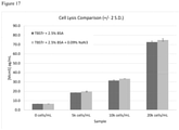
- the present inventors have demonstrated that the complicated urine sample preparation methods described in the prior art are not required to release biomarkers such as Mcm5 from cells in samples such as urine samples. Rather, all that is required is to expose the sample to a lysis buffer which is capable of releasing the biomarker from cells in the sample. This is much simpler than the prior art methods.
- the cells in a urine sample may be prepared and lysed by the addition of a single lysate buffer.
- Mcm protein assay which does not use immunofluorescence can accurately detect whether a subject has a urological cancer. This is beneficial over prior art techniques which use Europium labels and DELFIA detection. Such methods are expensive and complicated and thus unsuitable for use in pathology laboratory or point-of-care applications.
- the present invention relates to antibodies which bind to biomarkers such as an Mcm protein, for example Mcm5, in a highly specific manner. These antibodies are useful in detecting levels of the biomarker and the present inventors have demonstrated that these antibodies may be used in the methods of the invention. The inventors have demonstrated that these antibodies bind to Mcm5 from samples that have been exposed to a lysis buffer which is capable of releasing the biomarker from cells in the sample.
- the present invention relates to antibodies that bind to Mcm5 but bind to different epitopes to one another (12A7 binds to SEQ ID NO: 1 and 4B4 binds to SEQ ID NO: 2).
- Such antibodies are advantageous as they can be used in assays to detect Mcm5 (or similar Mcm proteins containing these epitopes).
- these antibodies can be used in the methods of the invention, which comprise a sandwich (two-site) assay, namely a sandwich ELISA assay.
- two antibodies that bind to Mcm5 must be identified. Ideally, these two antibodies must bind to different epitopes to one another, but also the two different epitopes to which the two antibodies bind must be spatially positioned such that both antibodies may bind to Mcm5 at the same time (without substantial steric hindrance).
- the present inventors have developed two such antibodies (named 12A7 and 4B4). These two antibodies bind to SEQ ID NO: 1 and SEQ ID NO: 2 respectively.
- SEQ ID NO: 1 and SEQ ID NO: 2 represent two different Mcm5 epitopes which are spatially arranged on Mcm5 to allow the two antibodies to bind to Mcm5 at the same time. This is demonstrated in Examples 2 and 3.
- the present inventors are the first to determine two Mcm5 epitopes which are arranged such that antibodies to each epitope may bind simultaneously. Furthermore, the inventors are the first to provide sequences of two antibodies that can bind independently to Mcm5.
- a method for detecting the presence of minichromosome maintenance complex component 5 (Mcm5) in a sample from a subject comprising:
- a kit comprising a lysis buffer which is capable of releasing Mcm5 from cells in a urine sample wherein the lysis buffer comprises a detergent comprising triton X-100; and a first monoclonal antibody and a second monoclonal antibody wherein the first monoclonal antibody is an antibody which:
- Described herein is a method for detecting the presence of Mcm5 indicative of a urological cancer in a subject, the method comprising:
- the presence of higher than normal levels of Mcm5 in the sample is indicative of urological cancer. Whether or not the level of Mcm5 in the sample is higher than normal may be determined by comparing the amount detected with the amount detected in a reference sample. In some embodiments the method is a method for detection of a urological cancer.
- MCM minichromosome maintenance
- MCM minichromosome maintenance complex
- Mcm5 may be assayed according to any suitable method known in the art.
- MCM proteins 2 - 7 comprise part of the pre-replication complexes which form on chromatin and which are essential prerequisites, or licensing factors, for subsequent DNA replication.
- the MCM protein complexes act as replicative helicases and thus are core components of the DNA replication machinery.
- MCMs are upregulated in the transition from the G0 to G1/S phase of the cell cycle and actively participate in cell cycle regulation.
- the MCM proteins form an annular structure around the chromatin.
- Mcm5 refers to a polypeptide of SEQ ID NO: 35, a polypeptide 85%, 90%, 95%, 98% or 100% identity to SEQ ID NO: 35.
- Mcm5 may be encoded by a polynucleotide having a sequence 85%, 90%, 95%, 98% or 100% identical to SEQ ID NO: 36.
- the method of the invention comprises a step of preparing a sample from the subject wherein preparing the sample comprises exposing the sample to a lysis buffer.
- preparing the sample refers to steps taken to release Mcm5 from cells within the sample in order to allow for step (b) of the invention (performing an assay to determine the concentration of Mcm5) to take place.
- This step comprises exposing the sample to a lysis buffer.
- the step of "preparing the sample” comprises a step of concentrating cells in the urine sample. Optionally the cells are concentrated by filtration or by centrifugation.
- the term ' exposing the sample to a lysis buffer ' can be considered to refer to manipulating the urine sample in such a way that the cells within the urine sample (or a substantial portion of these cells) are in contact with the lysis buffer.
- the sample is a urine sample.
- the sample is centrifuged to provide a sample pellet, the supernatant is discarded and the sample pellet is re-suspended in the lysis buffer.
- concentrated buffer components are added to a liquid urine sample to form a solution comprising the sample exposed to the lysis buffer.
- the method of the invention comprises a step of " providing a sample from a subject ", wherein this step is non-invasive (not-surgical), for example collection of urine.
- the sample comprises urine or urinary sediment.
- Mcm5 is typically found in the nuclei of cells present in urine.
- the sample comprises cells within the urine. More suitably, those cells may be concentrated by any known technique common in the art, such as filtration or more suitably centrifugal collection of the cells from urine. Enriching the cells from the urine may increase the signal, and may facilitate detection.
- the sample is first catch urine, such as may be obtained after massage of the prostate gland (when the subject is male). Even more suitably, the sample may comprise the first few milliliters of first catch urine.
- the sample may comprise first pass urine, suitably first pass urine produced after massage of the prostate gland.
- the sample may comprise urinary sediment such as sedimented cells collected from urine (such as from total urine, or from first catch urine as explained above).
- a sample comprising cells is prepared using a process consisting of the following steps: a. centrifugation of the sample to provide a sample pellet; and b. resuspension of the pelleted cells from the sample in a lysis buffer.
- the sample is centrifuged for between 1 minute and 30 minutes at between 500g and 5000g, for between 1 minute and 20 minutes at between 750g and 2500g, for between 3 minutes and 10 minutes at between 750g and 2000g, or for around 5 minutes at 1500g.
- the pellet is resuspended using an adjustable pipette with a disposable tip.
- the method does not comprise incubation of the sample at a temperature greater than 90°C for around 45 minutes.
- the method does not comprise incubation of the sample at a high temperature.
- a high temperature is a temperature greater than 50°C, greater than 60°C, greater than 70°C, greater than 80°C, greater than 90°C, between 50°C and 120°C, between 60°C and 110°C, between 70°C and 100°C, or between 80°C and 100°C.
- the method does not comprise incubation of the sample at a temperature greater than 50°C, greater than 60°C, greater than 70°C, greater than 80°C, greater than 90°C, between 50°C and 120°C, between 60°C and 110°C, between 70°C and 100°C, or between 80°C and 100°C for more than 30 minutes, more than 35 minutes, more than 40 minutes, more than 45 minutes, between 30 minutes and 2 hours, between 35 minutes and 2 hours, or between 40 minutes and 2 hours.
- the method does not comprise shearing the nucleic acids by passing the sample through a 21 gauge needle. In a further embodiment the method does not comprise exposing the sample to mechanical shearing.
- the method does not comprise incubation of the urine sample at a temperature greater than 90°C for greater than 45 minutes, shearing nucleic acids in the urine sample by passing the urine sample through a 21 gauge needle, digesting the nucleic acids by exposing the urine sample to DNase I or RNase A, centrifuging the sample at 15,000 g for 10 minutes, wherein the lysis buffer is not PBS containing 0.4% sodium deoxycholate and 0.02% sodium azide.
- the method does not comprise digesting the nucleic acids by exposing the sample to DNase I or RNase A.
- the phrase ' does not comprise digesting the nucleic acids by exposing the urine sample to DNase I or RNase A ' means the sample should not be exposed to a concentration of DNase I or RNase A that is effective to cause significant digestion of the nucleic acids in the sample.
- the sample is not exposed to more than 20U/ml DNase I or more than 1 ⁇ g/mL RNase A.
- the sample is not exposed to more than 1U/ml, more than 5U/ml, more than 10U/ml, more than 15U/ml, between 1U/ml and 500U/ml, between 5U/ml and 250U/ml, between 10U/ml and 100U/mL or between 15U/ml and 100U/ml of DNase I.
- the sample is not exposed to more than 0.1 ⁇ g/mL, more than 0.2 ⁇ g/mL, more than 0.5 ⁇ g/mL, more than 0.7 ⁇ g/mL, between 0.1 ⁇ g/ml and 100 ⁇ g/ml, between 0.5 ⁇ g/mL and 50 ⁇ g/mL or between 0.5 ⁇ g/mL and 25 ⁇ g/mL of RNase A.
- the method does not comprise centrifuging the sample at 15,000 g for ten minutes. In a further embodiment the method does not comprise centrifuging the sample. In a further embodiment the method does not comprise centrifuging the sample at more than 10,000 g, more than 12,000 g, more than 14,000 g or more than 14,500 g. In a further embodiment the method does not comprise centrifuging the sample for more than 2 minutes, more than 5 minutes, more than 7 minutes or more than 8 minutes.
- Lysis buffers are generally buffers which are used for the purpose of lysing cells.
- the lysis buffer is preferably compatible with the method used for subsequent analysis. For example, where the analysis method is double-antibody sandwich ELISA, the lysis buffer must not degrade the capture antibody bound to the surface of the microtitre plate.
- Lysis buffers generally but not exclusively comprise one or more detergents (also known as surfactants), one or more salts and a buffering agent. The concentrations of these components affect the efficacy of the lysis buffer.
- the lysis buffer is capable of releasing Mcm5 from cells in the sample.
- the lysis buffer is capable of releasing a biomarker such as Mcm5 from cells in the sample.
- a lysis buffer will be considered to be " capable of releasing a biomarker such as Mcm5 from cells in the sample " if the amount of the biomarker such as Mcm5 released is greater than 40%, 50%, 60%, 70%, or 80% the amount released when a buffer containing 0.08% sodium deoxycholate, 0.08% CHAPS, 2 mM EDTA, 150 mM Trizma pH 7.6 is used.
- a lysis buffer will be considered to be " capable of releasing a biomarker such as Mcm5 from cells in the sample " if the amount of a biomarker such as Mcm5 released is greater than 40%, 50%, 60%, 70%, or 80% the amount released when a buffer containing 10mM Tris (pH 7.6), 200 mM NaCl, 2.5 % BSA, 0.1% Triton X100 and 0.09% Sodium azide is used.
- the amount of a biomarker such as Mcm5 that is released using a lysis buffer may be determined by assaying the amount present after exposure to the lysis buffer and comparing it to a reference sample (preferably a sample which is substantially the same or identical to the first sample) which has been exposed to a buffer containing 0.08% sodium deoxycholate, 0.08% CHAPS, 2 mM EDTA, 150 mM Trizma pH 7.6.
- a reference sample preferably a sample which is substantially the same or identical to the first sample
- the amount of a biomarker such as Mcm5 that is released using a lysis buffer may be determined by assaying the amount present after exposure to the lysis buffer and comparing it to a reference sample (preferably a sample which is substantially the same or identical to the first sample) which has been exposed to a buffer containing 10mM Tris (pH 7.6), 200 mM NaCl, 2.5 % BSA, 0.1% Triton X100 and 0.09% Sodium azide.
- the amount of a biomarker such as Mcm5 that is released may be measured using a sandwich ELISA assay such as the assay described in Example 4.
- the antibodies used in the sandwich assay should be antibodies that bind to the biomarker such as Mcm5, for example a first monoclonal antibody or second monoclonal antibody used in the invention. Preferably 12A7 and 4B4 antibodies are used.
- the lysis buffer is capable of releasing Mcm5 from cells in the urine sample and does not substantially degrade the Mcm5 protein.
- a buffer can be considered to not substantially degrade Mcm5 protein if the amount of intact Mcm5 present after exposure of the sample to the lysis buffer is greater than 40%, 50%, 60%, 70%, or 80% the amount of intact Mcm5 after exposure to a buffer containing 0.08% sodium deoxycholate, 0.08% CHAPS, 2 mM EDTA, 150 mM Trizma pH 7.6.
- Mcm5 protein can be considered to be intact, if the binding site for antibodies that bind to SEQ ID NO: 1 and SEQ ID NO: 2 (such as the first monoclonal antibody and second monoclonal antibody or antibodies 12A7 and 4B4) are present. It is within the capabilities of the skilled person to determine how much Mcm5 within a sample is degraded. The sample should be tested to see whether the Mcm5 within the sample can bind to the first monoclonal antibody and the second monoclonal antibody used in the invention. This may be measured using a sandwich ELISA assay such as that described in Example 4. The antibodies used in the sandwich assay should be antibodies that bind to Mcm5, for example a first monoclonal antibody or second monoclonal antibody used in the invention. Preferably 12A7 and 4B4 antibodies are used.
- the lysis buffer is capable of releasing Mcm5 from cells in a fresh sample.
- fresh sample refers to a sample that has not been frozen.
- the " fresh sample” has been obtained from a patient less than 10 days, less than 5 days, less than 2 days or less than 1 day prior to its use in the methods of the invention.
- the cells are exposed to the lysis buffer and then frozen before further treatment.
- the urine sample may be concentrated by centrifugation followed by discarding the supernatant and then the lysis buffer may be added to the concentrated cells.
- the frozen sample is thawed prior to a step of performing an assay to determine the concentration of the at least one biomarker in the urine sample.
- the sample is frozen for at least 1 hour, at least 1 day, at least 5 days or at least 10 days prior to a step of performing an assay to determine the concentration of the at least one biomarker in the urine sample.
- the sample is frozen for between 1 hour and 1 year, between 1 hour and 6 months, between 1 day and 3 months or around one week prior to a step of performing an assay to determine the concentration of the at least one biomarker in the urine sample.
- the lysis buffer described herein may be Cytobuster TM Protein Extraction Reagent.
- the lysis buffer does not comprise sodium azide. In one embodiment the lysis buffer is not PBS containing 0.4% sodium deoxycholate and 0.02% sodium azide.
- the lysis buffer used in the present invention comprises a detergent (also referred to as a surfactant).
- the lysis buffer used in the present invention comprises a detergent which comprises triton X-100.
- detergents are compounds that are known to disrupt cell walls.
- Detergents are amphiphilic having both hydrophobic and hydrophilic regions. Suitable detergents are well known to the person of skill in the art.
- the detergent is an anionic detergent, a cationic detergent, a non-ionic detergent or a zwitterionic detergent.
- the detergent is selected from the group consisting of sodium deoxycholate, 3-[(3-Cholamidopropyl)dimethylammonio]-1-propanesulphonate (CHAPS), alkylbenzenesulphonates, sodium dodecylbenzenesulphonate, a Tween detergent (such as polyoxyethylene (2b), sorbitan monooleate or Tween-20), and a Triton detergent (such as polyethylene glycol p-(1, 1, 3, 3,-tetramethylbutyl)-phenyl ether or Triton X-100).
- the detergent comprises sodium deoxychlolate.
- the detergent comprises CHAPS.
- the detergent comprises sodium deoxycholate and CHAPS.
- the lysis buffer comprises sodium deoxycholate at a concentration between 0.01% and 0.15%, between 0.03% and 0.10%, between 0.05% and 0.09%, or about 0.08%.
- the lysis buffer comprises CHAPS at a concentration between 0.01% and 0.15%, between 0.03% and 0.10%, between 0.05% and 0.09%, or about 0.08%.
- the detergent comprises triton X-100.
- the detergent comprises triton X-100 at a concentration between 0.01% and 25%, between 0.01% and 10%, between 0.05% and 5%, between 0.05% and 1%, between 0.05% and 0.5%, between 0.075% and 0.125% or around 0.1%.
- the detergent consists of triton X-100.
- the detergent consists of triton X-100 at a concentration between 0.01% and 25%, between 0.01% and 10%, between 0.05% and 5%, between 0.05% and 1%, between 0.05% and 0.5%, between 0.075% and 0.125% or around 0.1%.
- the detergent comprises a polysorbate, preferably polysorbate 20 (also known as Tween 20).
- the detergent comprises polysorbate at a concentration between 0.01% and 5%, between 0.02% and 1%, between 0.03% and 0.07% or around 0.05%.
- the detergent comprises sodium deoxycholate or sodium dodecylsulphate.
- the detergent may comprise sodium deoxycholate or sodium dodecylsulphate at a concentration between 0.1% and 20%, between 0.5% and 10%, between 0.5% and 5%, between 0.75% and 1.25% or around 1%.
- the lysis buffer comprises a chelating agent. In a further embodiment the lysis buffer does not comprise a chelating agent. Chelating agents are multidentate ligands which can coordinate metal ions. Suitably the chelating agent is EDTA (ethylenediaminetetraacetic acid).
- the lysis buffer comprises EDTA at a concentration between 0.5mM and 10mM, between 1mM and 5mM, between 1.5mM and 3mM, or about 2mM.
- the lysis buffer comprises a buffer component.
- a buffer component can be considered to be any component which maintains the pH of the lysis buffer at a pH varying by less than 2.0 pH units, 1.5 pH units or 1.0 pH units. Examples of buffers which are suitable for this purpose are well known to the person skilled in the art.
- the buffer component is a buffer selected from the group consisting of TAPS (3- ⁇ [tris(hydroxymethyl)methyl]amino ⁇ propanesulphonic acid), Bicine (N,N-bis(2-hydroxyethyl)glycine), Tris (tris(hydroxymethyl)methylamine), Tricine (N-tris(hydroxymethyl(methylglycine), TAPSO (3-[N-Tris(hydroxymethyl)methylamino]-2-hydroxypropanesulphonic acid, HEPES (4-2-hydroxyethyl-1-piperazineethanesulphonic acid) and MOPS (3-(N-morpholino)propanesulphonic acid.
- the buffer component comprises Tris, phosphate buffered saline (PBS), MOPS, bicarbonate or HEPES buffer.
- the buffer component comprises Tris, PBS, MOPS or bicarbonate buffer.
- the buffer component comprises Tris, PBS or bicarbonate buffer.
- the buffer component is Trizma or Tris buffer.
- the buffer component comprises or consists of Trizma or Tris buffer.
- Trizma may refer to Trizma ® Pre-set crystals pH 7.6' (Sigma-Aldrich Cat. No. T7943).
- the buffer component maintains the pH of the buffer at a pH between pH 4 and pH 9, between pH 5 and pH 8, between pH 6 and pH 8, or around pH 7.6.
- the buffer component is Trizma or Tris buffer, and the buffer component maintains the pH of the lysis buffer between pH 4 and pH 9, between pH 5 and pH 8, between pH 6 and pH 8, or around pH 7.6.
- the buffer component comprises or consists of Trizma or Tris buffer, and the buffer component maintains the pH of the lysis buffer between pH 4 and pH 9, between pH 5 and pH 8, between pH 6 and pH 8, or around pH 7.6.
- the buffer component comprises tris buffer, for example at a concentration greater than 5 mM, between 5 mM and 350 mM, between 200 mM and 300 mM, between 225 mM and 275 mM between 10mM and 25mM, between 8mM and 12mM, around 10 mM or around 250 mM.
- the buffer component consists of tris buffer, for example at a concentration greater than 5 mM, between 5 mM and 350 mM, between 200 mM and 300 mM, between 225 mM and 275 mM, between 10mM and 25mM, between 8mM and 12mM, around 10 mM or around 250 mM.
- the buffer component comprises phosphate buffered saline, for example at a concentration of between 5 mM and 250 mM, between 50 mM and 250 mM or around 100 mM.
- the lysis buffer comprises sodium deoxycholate at a concentration between 0.01% and 0.15%, CHAPS at a concentration between 0.01% and 0.15%, EDTA at a concentration between 0.5mM and 10Mm, and Trizma or Tris buffer, wherein the Trizma or Tris buffer maintains the pH of the lysis buffer between pH 4 and pH 9.
- the lysis buffer comprises sodium deoxycholate at a concentration between 0.05% and 0.09%, CHAPS at a concentration between 0.05% and 0.09%, EDTA at a concentration between 1.5mM and 3mM, and Trizma or Tris buffer, wherein the Trizma or Tris buffer maintains the pH of the lysis buffer between pH 6 and pH 8.
- the lysis buffer comprises sodium deoxycholate at a concentration about 0.08%, CHAPS at a concentration about 0.08%, EDTA at a concentration about 2mM, and Trizma or Tris buffer, wherein the Trizma or Tris buffer maintains the pH of the lysis buffer around pH 7.6.
- the lysis buffer comprises a salt selected from the group consisting of sodium chloride, potassium chloride, magnesium chloride, sodium sulphate, potassium sulphate, magnesium sulphate, sodium acetate, potassium acetate, magnesium acetate, sodium phosphate, potassium phosphate or magnesium phosphate.
- the salt is a sodium salt or a potassium salt.
- the salt is sodium chloride or potassium chloride.
- the salt comprises or consists of sodium chloride or potassium chloride.
- the salt is at a concentration between 20 mM and 300 mM, between 150 mM and 300 mM, between 100 mM and 200 mM, between 150 mM and 250 mM, between 175 mM and 275 mM or around 200 mM.
- the salt is sodium chloride, for example at a concentration between 20 mM and 300 mM, between 150 mM and 300 mM, between 100 mM and 200 mM or around 200 mM.
- the salt comprises or consists of sodium chloride, for example at a concentration between 20 mM and 300 mM, between 150 mM and 300 mM, between 100 mM and 200 mM or around 200 mM.
- the lysis buffer has an ionic strength of between 1 mM and 500mM, between 50 mM and 450 mM, between 100 mM and 250 mM, between 100 mM and 175 mM, or between 125 mM and 175 mM.
- Various components of the buffer may contribute to this ionic strength.
- the lysis buffer may comprise a buffer component (such as Tris) and an additional salt component (such as sodium chloride) and both of these components may contribute to the ionic strength of the lysis buffer.
- the lysis buffer comprises a stabiliser.
- a “stabiliser” is a component that reduces the breakdown of proteins.
- a stabiliser reduces the breakdown of Mcm5.
- a buffer component stabilises Mcm5 can be tested using a sandwich ELISA assay such as the assay described in Example 4.
- a sample may be exposed to a lysis buffer comprising the potential stabiliser and a control sample may be exposed to a lysis buffer lacking the potential stabiliser.
- the level of Mcm5 that can be detected after exposure is measured using the sandwich ELISA.
- the potential stabiliser is a stabiliser.
- a stabiliser may prevent the breakdown of Mcm5 such that 1%, 2%, 5%, 10%, 20% or 25% more Mcm5 is present after storage in a buffer comprising a stabiliser for 1 day, 3 days, 5 days, 1 week or 2 weeks (compared to a buffer that does not comprise the stabiliser).
- the stabiliser may be a stabiliser selected from the group consisting of bovine serum albumin (BSA), foetal bovine serum (FBS) and a protease inhibitor.
- protease inhibitors include 4-(2-aminoethyl)benzenesulphonyl fluoride hydrochloride (Petrabloc SC or AEBSF), a protease inhibitor cocktail (such as Sigma P8340) comprising AEBSF, aprotinin, bestatin hydrochloride, N-(trans-epoxysuccinyl)-L-leucine 4-guanidinobutylamide (E-64), leupeptin hemisulphate salt and pepstatin A, and Roche complete protease inhibitor.
- BSA bovine serum albumin
- FBS foetal bovine serum
- protease inhibitors include 4-(2-aminoethyl)benzenesulphonyl fluoride hydrochloride (Petrabloc SC or AEBSF),
- the stabiliser is BSA, for example at a concentration between 0.1% and 20%, between 0.1% and 10%, between 0.1% and 5%, between 1% and 3%, between 2.2% and 2.7% or around 2.5%.
- the stabiliser comprises or consists of BSA, for example at a concentration between 0.1% and 20%, between 0.1% and 10%, between 0.1% and 5%, between 1% and 3%, between 2.2% and 2.7% or around 2.5%.
- the lysis buffer may comprise an antimicrobial agent.
- a component is an "antimicrobial agent" if it reduces the replication of microbes, for example bacteria, viruses or fungi.
- the antimicrobial agent is sodium azide or an isothiazolone.
- the antimicrobial agent comprises or consists of sodium azide or an isothiazolone.
- the isothiazolone may be 2-methyl-4-isothiazolin-3-one and/or 5-chloro-2-methyl-4-isothiazolin-3-one.
- the antimicrobial agent comprises sodium azide, for example at a concentration between 0.01% and 5%, between 0.02% and 1.5%, between 0.07% and 0.12% or around 0.09%.
- the antimicrobial agent consists of sodium azide, for example at a concentration between 0.01% and 5%, between 0.02% and 1.5%, between 0.07% and 0.12% or around 0.09%.
- the lysis buffer may comprise:
- the lysis buffer may comprise:
- the lysis buffer may comprise:
- the lysis buffer may comprise:
- the lysis buffer may consist of:
- the lysis buffer may consist of:
- the lysis buffer may consist of:
- the lysis buffer may consist of:
- the lysis buffer used in the invention is used for releasing at least one biomarker from cells in a urine sample, wherein the at least one biomarker is Mcm5.
- the lysis buffer may be RIPA buffer.
- the term "detecting the presence of Mcm5” can be considered to be substitutable with the term " performing an assay to determine the concentration " of Mcm5.
- the presence of Mcm5 can be considered to be detected, where its concentration is higher than a defined cut-off level.
- the cut-off may be a concentration of Mcm5 higher than the mean value from healthy patients plus a multiple of the standard deviation shown by the values derived from healthy subjects.
- the concentration of, for example, the full length Mcm5 polypeptide be scored. Indeed, it is possible that detection may take place by assaying particular fragments of Mcm5 being present which are thus taken to indicate the presence of the overall biomarker polypeptide in the sample. This is especially true if the samples are analysed for example by mass spectrometry. Therefore the invention embraces the detection of fragments of Mcm5 biomarkers. Suitably the fragment is sufficiently long to enable its unique identification by immunological or mass spectrometry methods.
- Such a fragment is suitably at least 6 amino acids in length, suitably at least 7 amino acids in length, suitably at least 8 amino acids in length, suitably at least 9 amino acids in length, suitably at least 10 amino acids in length, suitably at least 15 amino acids, suitably at least 25 amino acids, suitably at least 50 amino acids, suitably at least 100 amino acids, or suitably the majority of Mcm5.
- a fragment comprises a small fragment of Mcm5, whilst being long enough to retain an identifiable amino acid sequence or mass.
- the assay to determine the concentration of Mcm5 is performed by exposing the sample to a first monoclonal antibody and/or a second monoclonal antibody.
- the assay comprises exposing the sample to both a first monoclonal antibody and a second monoclonal antibody.
- the kits of the invention comprise a first monoclonal antibody and a second monoclonal antibody.
- the term 'antibody' can refer to naturally occurring forms or recombinant antibodies such as single-chain antibodies, chimeric antibodies or humanised antibodies.
- the terms ' antibody ' and ' antibodies ' may also be considered to encompass fragments of antibodies that can bind to a target protein, such as an Mcm protein like Mcm5. Such fragments may include Fab' 2 , F'(ab) 2 , Fv, single chain antibodies or diabodies.
- antibodies used in the invention are naturally occurring, full length antibodies (rather than fragments).
- the antibody is a mouse antibody (i.e. originally derived from mouse spleen cells).
- antibodies are formed from two heavy chains and two lights chains.
- Each heavy chain is made up of a heavy chain constant region (CH) and a heavy chain variable region (VH).
- each light chain is made up of a light chain constant region (CL) and a light chain variable region (VL).
- the VH and VL regions comprise complementarity defining regions (CDRs).
- the CDRs are, primarily, responsible for specific binding to the target protein.
- An antibody used in the invention may be an antibody of any class, particularly IgG, IgM or IgA.
- the antibody is an IgG antibody.
- monoclonal antibody is intended to refer to an antibody that is obtained from a population of substantially similar antibodies, i.e. from a population of antibodies that are identical except for a minority of naturally occurring variants. A population of monoclonal antibodies will bind to the same epitope. Monoclonal antibodies can be produced by a variety of techniques well known to the person skilled in the art and including the methods disclosed in “ Monoclonal Antibodies: A manual of techniques", H Zola (CRC Press, 1988 ) and in “ Monoclonal Hybridoma Antibodies: Techniques and Application", SG Hurrell (CRC Press, 1982 ).
- the first monoclonal antibody and/or the second monoclonal antibody bind specifically to Mcm5.
- the first monoclonal antibody binds to SEQ ID NO: 1.
- the second monoclonal antibody binds to SEQ ID NO: 2.
- the first monoclonal antibody or the second monoclonal antibody will bind to an epitope (fragment) of Mcm5 (for example SEQ ID NO: 1 or SEQ ID NO: 2).
- an epitope fragment of Mcm5
- the term "antibody which binds to Mcm5" refers to an antibody that binds to only a single epitope of Mcm5 such as SEQ ID NO: 1 or SEQ ID NO: 2.
- the term ' binding affinity ' refers to the ability of an antibody to bind to its target.
- the term ' specifically binds ' refers to an antibody that binds to a target such as Mcm5 with a binding affinity that is at least 2-fold, 10-fold, 50-fold or 100-fold greater than its binding affinity for binding to another non-target molecule.
- the non-target molecule is an Mcm protein, other than Mcm5, such as Mcm2.
- an antibody used in the invention is capable of binding to Mcm5, with a binding affinity that is at least 2-fold, 10-fold, 50-fold or 100-fold greater than its binding affinity for binding to another non-target molecule.
- an antibody used in the invention binds to SEQ ID NO: 1 (such as the first monoclonal antibody) or SEQ ID NO: 2 (such as the second monoclonal antibody) with a binding affinity that is at least 2-fold, 10-fold, 50-fold or 100-fold greater than its binding affinity for binding to another non-target molecule.
- a preferred method for the evaluation of binding affinity for Mcm5 is by ELISA.
- the first monoclonal antibody and/or the second monoclonal antibody have an affinity for Mcm5 (measured as an EC50 or 50% maximum binding concentration, as described in Example 2) of 2500 ng/ml or lower, 1500 ng/ml or lower, 1000 ng/ml or lower, 600 ng/ml or lower, 50 ng/ml or lower, 30 ng/ml or lower, 20 ng/ml or lower, or 10 ng/ml or lower.
- the EC50 will typically be higher than 1 ng/ml and thus the EC50 may be between 1 ng/ml and any of the upper limits specified in the preceding sentence.
- the binding kinetics (e.g. binding affinity) of the antibody also can be assessed by standard assays known in the art, such as by Surface Plasmon Resonance (SPR) (e.g. Biacore TM system) analysis.
- SPR Surface Plasmon Resonance
- the affinity constant (KD) for binding to Mcm5 is preferably in the range of 1-10 000 nM, 1-1 000 nM, 1-500 nM, 1-100 nM, 1-50 nM or 1-10 nM.
- the association rate (ka) is preferably in the range of 0.4-3.4 ⁇ 10 6 1/M.
- the dissociation rate (kd) is preferably in the range of 1-10 ⁇ 10 -3 1/s. These values may typically be determined by SPR (surface plasmon resonance).
- CDRs Complementary determining regions
- antibodies 12A7 or 4B4 i.e. a CDR selected from the group consisting of:
- Antibodies that have the same CDRs as the 4B4 and 12A7 antibodies may differ substantially from the sequences of 4B4 and 12A7 in other regions. Such antibodies may, for example, be antibody fragments.
- sequence that differs from SEQ ID NO: 3 by a single amino acid substitution refers to the possibility of replacing one amino acid defined in SEQ ID NO: 3 by a different amino acid. Preferably such a replacement is a conservative amino acid substitution.
- the following eight groups each contain amino acids that are typically conservative substitutions for one another:
- the first monoclonal antibody may comprise at least one CDR from the heavy chain of 12A7 (12A7 CDRH1, 12A7 CDRH2 or 12A7 CDRH3) as well as at least one CDR from the light chain of 12A7 (12A7 CDRL1, 12A7 CDRL2 or 12A7 CDRL3).
- the first monoclonal antibody may comprise at least two CDRs from the heavy chain of 12A7 and at least two CDRs from the light chain of 12A7.
- the first monoclonal antibody comprises all three CDRs from the heavy chain of 12A7 and all three CDRs from the light chain of 12A7.
- the first monoclonal antibody may comprise 12A7 CDRL1 and 12A7 CDRL2, 12A7 CDRL1 and 12A7 CDRL3, 12A7 CDRL1 and 12A7 CDRH1, 12A7 CDRL1 and 12A7 CDRH2, 12A7 CDRL1 and 12A7 CDRH3, 12A7 CDRL2 and 12A7 CDRL3, 12A7 CDRL2 and 12A7 CRL3 and 12A7 CDRH1, 12A7 CDRL2 and 12A7 CDRH2, 12A7 CDRL2 and 12A7 CDRH3, 12A7 CDRL3 and 12A7 CDRH1, 12A7 CDRL3 and 12A7 CDRH2, 12A7 CDRL3 and 12A7 CDRH3, 12A7 CDRH1 and 12A7 CDRH2, 12A7 CDRH1 and 12A7 CDRH3, or 12A7 CDRH2 and 12A7 CDRH3.
- the second monoclonal antibody may comprise at least one CDR from the heavy chain of 4B4 (4B4 CDRH1, 4B4 CDRH2 or 4B4 CDRH3) as well as at least one CDR from the light chain of 4B4 (4B4 CDRL1, 4B4 CDRL2 or 4B4 CDRL3).
- the second monoclonal antibody may comprise at least two CDRs from the heavy chain of 4B4 and at least two CDRs from the light chain of 4B4.
- the second monoclonal antibody comprises all three CDRs from the heavy chain of 4B4 and all three CDRs from the light chain of 4B4.
- the second monoclonal antibody may comprise 4B4 CDRL1 and 4B4 CDRL2, 4B4 CDRL1 and 4B4 CDRL3, 4B4 CDRL1 and 4B4 CDRH1, 4B4 CDRL1 and 4B4 CDRH2, 4B4 CDRL1 and 4B4 CDRH3, 4B4 CDRL2 and 4B4 CDRL3, 4B4 CDRL2 and 4B4 CDRH1, 4B4 CDRL2 and 4B4 CDRH2, 4B4 CDRL2 and 4B4 CDRH3, 4B4 CDRL3 and 4B4 CDRH1, 4B4 CDRL3 and 4B4 CDRH2, 4B4 CDRL3 and 4B4 CDRH3, 4B4 CDRH1 and 4B4 CDRH2, 4B4 CDRH1 and 4B4 CDRH3, or 4B4 CDRH2 and 4B4 CDRH3.
- An antibody may comprise at least one CDR having a sequence identical to that described in any one of SEQ ID NO: 3 (12A7 CDRL1), SEQ ID NO :5 (12A7 CDRL2), SEQ ID NO: 7 (12A7 CDRL3), SEQ ID NO: 9 (12A7 CDRH1), SEQ ID NO: 11 (12A7 CDRH2), SEQ ID NO: 13 (12A7 CDR H3), SEQ ID NO: 15 (4B4 CDRL1), SEQ ID NO: 17 (4B4 CDRL2), SEQ ID NO: 19 (4B4 CDRL3), SEQ ID NO: 21 (4B4 CDRH1), SEQ ID NO: 23 (4B4 CDRH2) or SEQ ID NO: 25 (4B4 CDRH3).
- the 12A7 CDRL2 has the sequence described in SEQ ID NO: 5. In a further embodiment where the first monoclonal antibody comprises 12A7 CDRL1, the 12A7 CDRL1 has the sequence described in SEQ ID NO: 3. In a further embodiment where the first monoclonal antibody comprises 12A7 CDRL3, the 12A7 CDRL3 has the sequence described in SEQ ID NO: 7. In a further embodiment where the first monoclonal antibody comprises 12A7 CDRH1, the 12A7 CDRH1 has the sequence described in SEQ ID NO: 9. In a further embodiment where the first monoclonal antibody comprises 12A7 CDRH2, the 12A7 CDRH2 has the sequence described in SEQ ID NO: 11.
- the first monoclonal antibody comprises 12A7 CDRH3, the 12A7 CDRH3 has the sequence described in SEQ ID NO: 13.
- the second monoclonal antibody comprises 4B4 CDRL1
- the 4B4 CDRL1 has the sequence described in SEQ ID NO: 15.
- the second monoclonal antibody comprises 4B4 CDRL2
- the 4B4 CDRL2 has the sequence described in SEQ ID NO: 17.
- the 4B4 CDRL3 has the sequence described in SEQ ID NO: 19.
- the 4B4 CDRH1 has the sequence described in SEQ ID NO: 21.
- the 4B4 CDRH2 has the sequence described in SEQ ID NO: 23.
- the 4B4 CDRH3 has the sequence described in SEQ ID NO: 25.
- an antibody comprising at least one of the CDRs of 12A7 or 4B4 binds (optionally specifically binds) to Mcm5. Even more preferably an antibody comprising at least one of the CDRs of 12A7 or 4B4 binds (optionally specifically binds) to SEQ ID NO: 1 or SEQ ID NO: 2.
- the first monoclonal antibody preferably comprises a heavy chain variable region having a sequence at least 85%, 90%, 95%, 98%, 99% or 100% identical to SEQ ID NO: 29.
- the first monoclonal antibody preferably comprises a light chain variable region sequence having a sequence at least 85%, 90%, 95%, 98%, 99% or 100% identical to SEQ ID NO: 27.
- the second monoclonal antibody preferably comprises a heavy chain variable region having a sequence at least 85%, 90%, 95%, 98%, 99% or 100% identical to SEQ ID NO: 33.
- the second monoclonal antibody preferably comprises a light chain variable region sequence having a sequence at least 85%, 90%, 95%, 98%, 99% or 100% identical to SEQ ID NO: 31.
- Such antibodies may be referred to as "variant antibodies”.
- the first monoclonal antibody may comprise a heavy chain variable region having a sequence at least 95% identical to SEQ ID NO: 29.
- the first monoclonal antibody may comprise a heavy chain variable region having a sequence at least 98% identical to SEQ ID NO: 29.
- the first monoclonal antibody may comprise a light chain variable region having a sequence at least 95% identical to SEQ ID NO: 27.
- the first monoclonal antibody may comprise a light chain variable region having a sequence at least 98% identical to SEQ ID NO: 27.
- the first monoclonal antibody may comprise a light chain variable region having a sequence at least 95% identical to SEQ ID NO: 27 and a heavy chain variable region having a sequence at least 95% identical to SEQ ID NO: 29.
- the first monoclonal antibody may comprise a heavy chain variable region having a sequence at least 98% identical to SEQ ID NO: 29 and a light chain variable region having a sequence at least 98% identical to SEQ ID NO: 27.
- the second monoclonal antibody may comprise a heavy chain variable region having a sequence at least 95% identical to SEQ ID NO: 33.
- the second monoclonal antibody may comprise a heavy chain variable region having a sequence at least 98% identical to SEQ ID NO: 33.
- the second monoclonal antibody may comprise a light chain variable region having a sequence at least 95% identical to SEQ ID NO: 31.
- the second monoclonal antibody may comprise a light chain variable region having a sequence at least 98% identical to SEQ ID NO: 31.
- the second monoclonal antibody may have a heavy chain variable region having a sequence at least 95% identical to SEQ ID NO:33 and a light chain variable region having a sequence at least 95% identical to SEQ ID NO: 31.
- the second monoclonal antibody may have a heavy chain variable region having a sequence at least 98% identical to SEQ ID NO: 33 and a light chain variable region having a sequence at least 98% identical to SEQ ID NO: 31.
- variant antibodies bind to an epitope having an amino acid sequence of SEQ ID NO: 1 or 2 and have CDRs which are identical to the CDRs of the 12A7 or 4B4 antibodies or have CDRs which vary in only a single amino acid substitution (preferably a conservative amino acid substitution).
- the first monoclonal antibody and/or the second monoclonal antibody may have framework regions which differ in sequence quite significantly from those described in SEQ ID NO: 27, 29, 31 or 33.
- the first monoclonal antibody comprises a heavy chain variable region having a sequence at least 85%, 90%, 95%, 98%, 99% or 100% identical to SEQ ID NO: 29, the antibody further comprises at least one of 12A7 CDRH1, 12A7 CDRH2 or 12A7 CDRH3. It is understood by the person skilled in the art that, since target binding specificity is determined by the CDRs, an antibody comprising the CDRs of 12A7 may still bind to Mcm5 even if the remainder of the antibody sequence is quite variable.
- the first monoclonal antibody comprises at least one of 12A7 CDRH1, 12A7 CDRH2 or 12A7 CDRH3
- the first monoclonal antibody preferably comprises a heavy chain variable region having a sequence at least 90% identical to SEQ ID NO: 29.
- the first monoclonal antibody used in the invention comprises 12A7 CDRH1, 12A7 CDRH2 and 12A7 CDRH3 and comprises a heavy chain variable region having a sequence at least 95% identical to SEQ ID NO:29.
- the second monoclonal antibody comprises a heavy chain variable region having a sequence at least 85%, 90%, 95%, 98%, 99% or 100% identical to SEQ ID NO: 33
- the second monoclonal antibody further comprises at least one of 4B4 CDRH1, 4B4 CDRH2 or 4B4 CDRH3. It is understood by the person skilled in the art that, since target binding specificity is determined by the CDRs, an antibody comprising the CDRs of 4B4 may still bind to Mcm5 even if the remainder of the antibody sequence is quite variable.
- the second monoclonal antibody comprises at least one of 4B4 CDRH1, 4B4 CDRH2 or 4B4 CDRH3
- the second monoclonal antibody preferably comprises a heavy chain variable region having a sequence at least 90% identical to SEQ ID NO: 33.
- the second monoclonal antibody comprises 4B4 CDRH1, 4B4 CDRH2 and 4B4 CDRH3 and comprises a heavy chain variable region having a sequence at least 95% identical to SEQ ID NO: 33.
- the first monoclonal antibody comprises a light chain variable region having a sequence at least 85%, 90%, 95%, 98%, 99% or 100% identical to SEQ ID NO: 27, the first monoclonal antibody further comprises at least one of 12A7 CDRL1, 12A7 CDRL2 or 12A7 CDRL3. It is understood by the person skilled in the art that, since target binding specificity is determined by the CDRs, an antibody comprising the CDRs of 12A7 may still bind to Mcm5 even if the remainder of the antibody sequence is quite variable.
- the first monoclonal antibody comprises at least one of 12A7 CDRL1, 12A7 CDRL2 or 12A7 CDRL3
- the first monoclonal antibody preferably comprises a light chain variable region having a sequence at least 90% identical to SEQ ID NO: 27.
- the first monoclonal antibody comprises 12A7 CDRL1, 12A7 CDRL2 and 12A7 CDRL3 and comprises a light chain variable region having a sequence at least 95% identical to SEQ ID NO: 27.
- the second monoclonal antibody used in the invention comprises a light chain variable region having a sequence at least 85%, 90%, 95%, 98%, 99% or 100% identical to SEQ ID NO: 31
- the second monoclonal antibody further comprises at least one of 4B4 CDRL1, 4B4 CDRL2 or 4B4 CDRL3. It is understood by the person skilled in the art that, since target binding specificity is determined by the CDRs, an antibody comprising the CDRs of 4B4 may still bind to Mcm5 even if the remainder of the antibody sequence is quite variable.
- the second monoclonal antibody comprises at least one of 4B4 CDRL1, 4B4 CDRL2 or 4B4 CDRL3
- the second monoclonal antibody preferably comprises a heavy chain variable region having a sequence at least 90% identical to SEQ ID NO: 31.
- the second monoclonal antibody comprises 4B4 CDRL1, 4B4 CDRL2 and 4B4 CDRL3 and comprises a heavy chain variable region having a sequence at least 95% identical to SEQ ID NO:31.
- An antibody having a heavy chain variable sequence identical to SEQ ID NO: 29 and a light chain variable sequence identical to SEQ ID NO: 27 may be referred to as antibody 12A7.
- An antibody having a heavy chain variable sequence identical to SEQ ID NO: 33 and a light chain variable sequence identical to SEQ ID NO: 31 may be referred to as an antibody 4B4.
- the first monoclonal antibody will compete for binding to Mcm5 with the 12A7 antibody.
- the second monoclonal antibody will compete for binding to Mcm5 with the 4B4 antibody.
- an antibody is ' at least 90%, 95%, 98%, 99% or 100% identical ' to a second antibody if the sequences have at least 90%, 95%, 98%, 99% or 100% identity when assessed using ClustalW (Thompson et al. , 1994) with the following parameters:
- variant antibodies which bind to Mcm5.
- Such variant antibodies may comprise 1, 2, 3, 4, 5, 6, 7, 8, 9, 10, 15, 20, 25, 50 or 100 substitution or deletion mutations compared to SEQ ID NOs: 27, 29, 31 or 33.
- ' Deletion ' variant antibodies may comprise the deletion of 1, 2, 3, 4, 5 or more amino acids or, in some cases, the deletion of entire regions of SEQ ID NOs: 27, 29, 31 or 33.
- 'Substitution' variants may comprise the replacement of 1, 2, 3, 4, 5 or more amino acids with the same number of new amino acids.
- the variant antibodies used in the invention comprise sequences differing from SEQ ID NOs: 27, 29, 31 or 33 by conservative amino acid substitutions (optionally only by conservative amino acid substitutions).
- conservative amino acid substitutions are unlikely to alter the binding properties of an antibody.
- the first monoclonal antibody is an antibody that competes with an antibody comprising 12A7 CDRH1, 12A7 CDRH2, 12A7 CDRH3, 12A7 CDRL1, 12A7 CDRL2, and 12A7 CDRL3.
- the second monoclonal antibody is an antibody that competes with an antibody comprising 4B4 CDRH1, 4B4 CDRH2, 4B4 CDRH3, 4B4 CDRL1, 4B4 CDRL2, and 4B4 CDRL3.
- the first monoclonal antibody is an antibody that competes with an antibody comprising a heavy chain variable region having a sequence identical to SEQ ID NO: 29 and a light chain variable region having a sequence identical to SEQ ID NO: 27.
- the second monoclonal antibody is an antibody that competes with an antibody comprising a heavy chain variable region having a sequence identical to SEQ ID NO: 33 and a light chain variable region having a sequence identical to SEQ ID NO: 31.
- an ELISA assay may be performed by immobilising the reference antibody, such as a 4B4 or a 12A7 antibody, on an ELISA plate. The plate is then blocked with a suitable blocking agent. An excess of a second antibody is added (the second antibody is the antibody whose ability to compete with the 4B4 or 12A7 antibody is to be assessed for example a first monoclonal antibody or a second monoclonal antibody that competes with 4B4 or 12A7). Mcm5 is added. Following a suitable incubation period, the ELISA plate is washed and an Mcm5 detection agent is added to measure the amount of Mcm5 bound to the immobilised reference antibody.
- the reference antibody such as a 4B4 or a 12A7 antibody
- a suitable detection reagent is a labelled 12A7 antibody.
- a suitable detection reagent is a labelled 4B4 antibody.
- the concentration at which 50% inhibition occurs is known as the Ki.
- An antibody that competes with 12A7 or 4B4 may bind with a Ki 2-fold, 5-fold, 10-fold, 50-fold or 100-fold lower than an antibody which does not bind to Mcm5.
- An antibody that competes with a particular antibody may bind to the same epitope as that particular antibody (for example SEQ ID NO: 1 or SEQ ID NO: 2). It is possible to determine to which epitope an antibody binds by performing an experiment in which the target protein, such as Mcm5, is digested into fragments (for example see Example 3 below). The affinity with which the 12A7 and 4B4 antibodies to bind to each of the fragments can then be determined. Once the fragment (representing the epitope), to which the first antibody binds with a reasonable affinity, has been determined, the epitope may be synthesised. It is then possible to determine whether a second antibody is capable of binding to the same epitope by measuring the binding affinity of that second antibody to the fragment.
- the first monoclonal antibody or the second monoclonal antibody may be produced from a polynucleotide which encodes, and is capable of expressing, it.
- a polynucleotide may encode an antibody light chain, and an antibody heavy chain or both.
- Two polynucleotides may be provided, one of which encodes an antibody light chain and the other of which encodes the corresponding antibody heavy chain.
- Such a polynucleotide or pair of polynucleotides may be expressed together such that an antibody used in the invention is generated.
- the polynucleotide may be part of a vector which typically further comprises control sequences operably linked to the inserted sequence, thus allowing for expression of the antibody used in the invention in vivo.
- the vector further comprises appropriate initiators, promoters, enhances and other elements which may be necessary and which are positioned in the correct orientation, in order to allow for expression of a polypeptide.
- Polynucleotides can be synthesised according to methods well known in the art, as described by way of example in Sambrook et al (1989, Molecular Cloning - a laboratory manual; Cold Spring Harbor Press ).
- the polynucleotides or vectors may be expressed in a host cell.
- host cells may be eukaryotic cells or prokaryotic cells.
- the host cells may be mammalian cells, insect cells, or bacterial cells.
- host cells that may be modified by insertion of vectors or nucleic acids include mammalian HEK293T, CHO, HeLa, NS0 and COS cells.
- bacterial cells such as E.coli may be used.
- Such host cells may be cultured using routine methods to produce an antibody used in the invention.
- the first monoclonal antibody or the second monoclonal antibody may also be produced from a hybridoma. It is well within the ability of the person skilled in the art to develop a hybridoma expressing an antibody used in the invention. This may be performed by immunising a mammal such as a mouse, rabbit or guinea pig, with an antigen (for example Mcm5). It may be beneficial to include an adjuvant such as Freund's complete adjuvant. The spleen cells of the immunised mammal removed and fused with myeloma cells to form hybridoma lines which are immortal given appropriate conditions and which secrete antibodies. The hybridomas are separated into single clones and the antibodies secreted by each clone are evaluated for their binding ability to Mcm5 protein.
- the first monoclonal antibody or the second monoclonal antibody may be produced using any method.
- the antibodies are produced directly from a hybridoma cell line.
- the antibodies are produced recombinantly.
- the cell line can be cultivated in vitro, for example in a vessel such as a cell culture flask or a fermenter, and the antibody can be isolated from the vessel using conventional techniques known in the art.
- an antibody will be considered to have been " produced by a hybridoma cell line " if the antibody is derived from an antibody which was produced by the hybridoma cell line.
- Such antibodies can be considered to have been "derived” from an antibody which was produced by the hybridoma cell line.
- antibodies used in the invention are produced directly from the hybridoma cell line itself.
- antibodies used in the invention may be produced using recombinant techniques.
- the sequence of an antibody used in the invention may be used to transfect a host cell line of the invention.
- the cell lines may be grown in a flask or a fermenter, and the antibody expressed by the cell can be isolated and purified using standard techniques (such as affinity chromatography).
- the antibodies may then be conjugated to a radio label or immobilised to a solid support.
- a suitable solid support is an ELISA plate.
- the method of the invention comprises exposing the sample to the first monoclonal antibody and/or the second monoclonal antibody and measuring the amount of Mcm5 that binds to the first monoclonal antibody and/or the second monoclonal antibody.
- This step may be carried out using an assay such as a sandwich assay.
- This step may be carried out using an ELISA assay.
- this step is carried out using a sandwich ELISA assay. This step does not comprise an immunofluorometric assay.
- this step may comprise steps of capturing the Mcm5 using a "capture antibody” already bound to the plate, and detecting how much antigen has been captured using a "detection antibody".
- the detection antibody has been pre-conjugated to a label such as the enzyme HRP (Horse Radish Peroxidase).
- HRP Hase Radish Peroxidase
- the ELISA plate with the labelled detection antibody is then washed to remove any excess unbound detection antibody.
- the washed plate is then exposed to an agent whose properties are changed by the label in a measurable manner.
- the concentration of the detection antibody may then be determined. For example if the detection antibody is conjugated to (labelled with) horseradish peroxidase, the ELISA plate may be exposed to TMB substrate.
- the concentration of the detection antibody, and therefore the concentration of Mcm5 in the original sample, may then be determined by quantitation of the colour change corresponding to the conversion of TMB into a coloured product.
- the first monoclonal antibody may be the capture antibody and the second monoclonal antibody may be the detection antibody.
- the first monoclonal antibody may be the detection antibody and the second monoclonal antibody may be the capture antibody.
- the method of the invention may comprise exposing Mcm5 to a detection antibody.
- the first monoclonal antibody or the second monoclonal antibody may be a detection antibody.
- the detection antibody may be conjugated to any suitable label (for example Europium 3+ or Horseradish Peroxidase).
- the label may be directly, attached or may be attached via a linker (such as Adipic Acid Dihydrazide (ADH)).
- the label may be attached by chemical conjugation.
- Methods of conjugating labels to antibodies are known in the art.
- carbodiimide conjugation Bauminger & Wilchek (1980) Methods Enzymol. 70, 151-159
- Other methods for conjugating a label to an antibody can also be used.
- sodium periodate oxidation followed by reductive alkylation of appropriate reactants can be used, as can glutaraldehyde cross-linking.
- a determination must be made that the conjugated detection antibody maintains its targeting ability and that the conjugated label maintains its function.
- the detection antibody is labelled by conjugation to Horseradish Peroxidase.
- kits comprising a lysis buffer which is capable of releasing Mcm5 from cells in a urine sample, a first monoclonal antibody and a second monoclonal antibody wherein the first monoclonal antibody is an antibody which:
- kits of the invention is suitable for performing the method of the invention.
- the first monoclonal antibody and/or the second monoclonal antibody are present in the kit as part of a composition.
- the kit comprises a vial or other suitable container containing a composition comprising the first monoclonal antibody (which is optionally conjugated to a label) and optionally comprising further components.
- the kit comprises a vial or other suitable container containing a composition comprising the second monoclonal antibody (which is optionally conjugated to a label) and optionally comprising further components.
- Such compositions may further comprise suitable stabilising agents that are capable of stabilising the antibody in solution.
- the composition may comprise a buffer selected from the group consisting of TAPS (3- ⁇ [tris(hydroxymethyl)methyl]amino ⁇ propanesulphonic acid), Bicine (N,N-bis(2-hydroxyethyl)glycine), Tris (tris(hydroxymethyl)methylamine), Tricine (N-tris(hydroxymethyl(methylglycine), TAPSO (3-[N-Tris(hydroxymethyl)methylamino]-2-hydroxypropanesulphonic acid), HEPES (4-2-hydroxyethyl-1-piperazineethanesulphonic acid), phosphate buffer saline and MOPS (3-(N-morpholino)propanesulphonic acid.
- TAPS 3- ⁇ [tris(hydroxymethyl)methyl]amino ⁇ propanesulphonic acid
- Bicine N,N-bis(2-hydroxyethyl)glycine
- Tris tris(hydroxymethyl)methylamine
- Tricine N-tris(hydroxymethyl(methylglycine)
- TAPSO (3-[N-Tris(
- the composition comprises phosphate-buffered saline pH 7.6. In a further preferred embodiment the composition comprises phosphate-buffered saline with between 0.01% and 0.09% sodium azide.
- the composition comprises agents suitable for storing the antibodies used in the invention at room temperature or at a temperature less than or equal to 15°C, 10°C, 7°C, 5°C, 0°C, -10°C, or -25°C.
- the composition comprises agents suitable for storing the antibodies used in the invention at a temperature between 2°C and 8°C.
- the composition comprises a stabilising agent such as a sugar, for example lactose or trehalose.
- the first monoclonal antibody and/or the second monoclonal antibody present in the kit is a "detection antibody". If the antibody used in the invention is conjugated, for example to a Europium label, it is advantageous to include a 7.5% purified BSA stabiliser in the composition.
- the first monoclonal antibody and/or the second monoclonal antibody is present in the kit bound to a solid support such as an ELISA plate.
- a solid support such as an ELISA plate.
- an antibody bound to a solid support may be referred to as a "capture antibody”.
- the kit comprises a calibrator.
- the " calibrator” is a preparation of one or more known concentrations of Mcm5.
- the kit comprises a substrate reagent.
- a substrate reagent can be used to detect the detection antibody.
- the substrate reagent may comprise TMB (3, 3', 5, 5'-tetramethylbenzidine), DAB (3, 3'-diaminobenzidine) or ABTS (2, 2'-azino-bis (3-ethylbenzothiazoline-6-sulphonic acid).
- the substrate reagent comprises TMB.
- the substrate reagent is peroxide and TMB.
- kit further comprises a wash solution and/or a stop solution.
- the kit of the present invention is suitable for performing a sandwich ELISA.
- the method of the invention further comprises a step of comparing the concentration of Mcm5 determined to reference values.
- a kit of the invention may comprise a reference standard.
- the reference standard typically refers to a sample from a healthy individual i.e. one who has not suffered urological cancer.
- the reference standard can be an actual sample analysed in parallel.
- the reference standard can be one or more values previously derived from a comparative sample e.g. a sample from a healthy subject (for example a subject who is known not to be suffering from a urological cancer).
- a mere numeric comparison may be made by comparing the value determined for the sample from the subject to the numeric value of a previously analysed reference sample. The advantage of this is not having to duplicate the analysis by determining concentrations in individual reference samples in parallel each time a sample from a subject is analysed.
- the reference standard is matched to the subject being analysed e.g. by gender e.g. by age e.g. by ethnic background or other such criteria which are well known in the art.
- the reference standard may be a number such as an absolute concentration determined by one or more previous studies.
- Reference standards may suitably be matched to specific patient sub-groups e.g. elderly subjects, or those with a previous relevant history such as a predisposition to urological cancer.
- the reference standard is matched to the sample type being analysed.
- concentration of the biomarker polypeptide(s) being assayed may vary depending on the type or nature of the sample (e.g. conventional urine sample vs first catch sample after prostatic massage). It will be immediately apparent to the skilled worker that the concentration value(s) for the reference standard should be for the same or a comparable sample to that being tested in the method(s) of the invention. For example, if the sample being assayed is first catch urine then the reference standard value should be for first catch urine to ensure that it is capable of meaningful crosscomparison.
- the protein concentrations determined may be compared to a previous sample from the same subject. This can be beneficial in monitoring the possibility of recurrence in a subject. This can be beneficial in monitoring the course and/or effectiveness of a treatment of a subject.
- the method may comprise further step(s) of comparing the value(s) determined for the sample of interest to one or more value(s) determined for the same biomarker(s) from different samples such as samples taken at different time points for the same subject. By making such a comparison, information can be gathered about whether a particular marker is increasing or decreasing in a particular subject. This information may be useful in diagnosing or predicting changes over time, or changes inhibited or stimulated by a particular treatment or therapy regime, or any other variable of interest.
- the methods can be used to determine whether, for example after treatment of the patient with a drug or candidate drug, or by tumour resection, the disease has progressed or not, or that the rate of disease progression has been modified.
- the result can inform the pathway of further treatment.
- an abnormal value indicates an increased likelihood of a urological cancer in the subject.
- the patient is diagnosed as having a urological cancer where the concentration of the Mcm5 determined in step (b) is higher than the mean value from healthy patients plus a multiple of the standard deviation shown by the values derived from healthy subjects.
- the invention also provides methods for detecting a urological cancer. Such methods offer a significant advantage in that they will detect any tumours which are in intimate contact with the flow or urine in the urological system (i.e kidneys, ureters, bladder and urethra) of the patient being assayed.
- urological system i.e kidneys, ureters, bladder and urethra
- Urological cancer includes all types of transitional cell carcinoma which might arise in the urological system.
- One example of transitional cell carcinoma is bladder cancer.
- transitional cells are present throughout the urological system including the lining of the renal pelvis, the ureters which conduct urine from the kidneys to the bladder and the urethra, as well as the wall of the bladder itself. Transitional cell carcinoma may arise in any of these locations.
- bladder cancer there is a range of different sub-types of bladder cancer, i.e. there are types of bladder cancer other than transitional cell carcinoma, although this (transitional cell carcinoma) is the most common type of bladder cancer.
- certain urological tumours may be detected even though they may not be in direct contact with the urine flow.
- a prostate tumour which is not typically in contact with the urine flow, but from which cells exfoliate and exude into the urethra.
- the methods of the invention also offer the advantage of being capable of detecting metastatic cancers, for example metastases of a different primary cancer which metastases have alighted in the urological system.
- the methods of the invention are capable of detecting any cancer which either grew into the urological system (e.g., a colon cancer which may have invaded locally) or any cancer which has invaded through a metastasis (e.g., a remote primary cancer, which has given rise to one or more metastatic tumours within the urological system of the subject).
- the methods of the invention may also advantageously be applied to the detection of prostate cancer.
- the invention is advantageously applied to the detection of bladder cancer.
- the invention is applied to the detection of both prostate and bladder cancer.
- sequence homology can also be considered in terms of functional similarity (i.e., amino acid residues having similar chemical properties/functions), in the context of the present document it is preferred to express homology in terms of sequence identity.
- Sequence comparisons can be conducted by eye or, more usually, with the aid of readily available sequence comparison programs. These publicly and commercially available computer programs can calculate percent homology (such as percent identity) between two or more sequences.
- Percent identity may be calculated over contiguous sequences, i.e., one sequence is aligned with the other sequence and each amino acid in one sequence is directly compared with the corresponding amino acid in the other sequence, one residue at a time. This is called an " ungapped " alignment. Typically, such ungapped alignments are performed only over a relatively short number of residues (for example less than 50 contiguous amino acids). For comparison over longer sequences, gap scoring is used to produce an optimal alignment to accurately reflect identity levels in related sequences having insertion(s) or deletion(s) relative to one another.
- a suitable computer program for carrying out such an alignment is the GCG Wisconsin Bestfit package (University of Wisconsin, U.S.A; Devereux et al., 1984, Nucleic Acids Research 12:387 ).
- Examples of other software than can perform sequence comparisons include, but are not limited to, the BLAST package, FASTA ( Altschul et al., 1990, J. Mol. Biol. 215:403-410 ) and the GENEWORKS suite of comparison tools.
- a homologous amino acid sequence is taken to include an amino acid sequence which is at least 40, 50, 60, 70, 80 or 90% identical.
- a polypeptide having at least 90% sequence identity to the biomarker of interest will be taken as indicative of the presence of that biomarker; more suitably a polypeptide which is 95% or more suitably 98% identical at the amino acid level will be taken to indicate presence of that biomarker.
- said comparison is made over at least the length of the polypeptide or fragment which is being assayed to determine the presence or absence of the biomarker of interest. Most suitably the comparison is made across the full length of the polypeptide of interest.
- a lysis buffer consisting of a detergent contains detergent and no other components.
- a lysis buffer comprising a detergent consisting of polysorbate 80 may comprise components other than detergents but the only detergent in the lysis buffer is polysorbate 80.
- Stage IIA Expression and Purification (small scale - 10ml)
- the recombinant plasmid containing the Mcm5 fragment was transformed into BL21 E. coli (n ⁇ n- ⁇ DE3) to ensure high yields of expressed protein.
- the identified clone with high levels of recombinant protein was cultured at the 0.5L scale and induced with IPTG. Cells were pelleted and stored at -70°C. Cells were disrupted in a denaturing lysis buffer described by Stoeber et al. J Natl Cancer Inst 2002 94 1071-9 .
- His-tagged recombinant protein was affinity purified under denaturing conditions on an immobilized metal affinity (IMAC) chromatography column. Protein were eluted
- Protein A purified MAbs were obtained from the Welcome/CRUK Institute for Cancer and Developmental Biology. These antibodies were screened for their ability to bind to Mcm5.
- Ni-NTA His-Sorb 96 well microtitre coated plate wells Qiagen.
- the Ni-NTA coated plates, pre-blocked with BSA, ensure an orientated presentation of the 6x His tagged Mcm through Nickel affinity binding to the spacer in comparison to the random presentation of the Mcm5 to the well that would be achieved by passive adsorption plate coating.
- the Mcm5 at a concentration of 625 ng/ml was Nickel affinity immobilised to Ni-NTA His-Sorb surface (Qiagen) of a 96 well plate at 200 ⁇ l/well resulting in 125 ng hs Mcm5/well.
- the Mcm5 Ag-Ab complexes of the MAbs were detected by a colorimetric ELISA using goat anti-mouse IgG conjugated to HRP, TMB reagent and 1M H 2 SO 4 assay stop solution, for the measurement of light absorbance at 450 nanometres (Abs 450 nm).
- An absence of Non-specific binding (NSB) of goat anti-mouse HRP conjugate to the Ni-NTA His-Sorb surface and Mcm5 was excluded by performing the ELISA on wells free from MAbs.
- the degree of NSB of the mouse MAbs being screened to the Ni-NTA coated polystyrene wells was established by performing the ELISA in wells with the omission of Mcm5 wells in parallel to performing the ELISA in wells with the Nickel affinity immobilisation of Mcm5 to the microtitre wells.
- Relative affinities of the protein A purified MAbs were determined by normalisation of Ab concentrations and application of the MAbs to the colorimetric screening assay under Ag-limiting conditions.
- Ag limiting conditions were determined by linear titration of Mcm5 across the Ni-NTA His-Sorb microtitre plate wells. An observed maximal Abs 450 nm of ⁇ 3.000 is indicative of Ag limiting conditions, because the maximum Abs 450 reading obtainable when Ag is non-limiting is 4.000.
- the Ag titration was performed using rabbit Mcm5 PAb (101) and Goat anti Rabbit HRP to measure ELISA response. A concentration that gave 25 ng of Mcm5 available for binding to each Ni-NTA well was limiting to the ELISA Abs 450 response of the PAb.
- the rabbit PAb was chosen to determine Ag-limiting conditions for investigation of the mouse MAbs, as the full array of Mcm5 epitopes are bound by the PAb. This ensured that the MAbs could be investigated in combined incubation studies to determine the degree of epitope competition.
- the concentration of IgG in the protein-A purified MAb solutions was determined by Abs 280 nm measurement in a spectrophotometer and MAb concentrations were normalised by dilution in PBS.
- the 50% maximum binding concentration was then determined by a linear dilution of the MAbs applied to an ELISA of Nickel affinity Mcm5 wells under Mcm5 Ag-limiting conditions. To demonstrate the plateau of the binding signal to the 25 ng/well hs Mcm5rf on the plate the MAb 7A3 was run in the ELISA over a greater concentration range.
- the 50% maximum binding response for 12A7 was obtained at a concentration of 20 ng/well. 12A7 is shown by its 50% maximum binding concentration to demonstrate the highest relative affinity to hs Mcm5. (Table 3).
- Table 3 MAb titrations against limiting 25ng/well antigen couples to Ni-NTA MAb nb/well MAb 4B4 12A7 0 0.086 0.051 0.25 0.108 0.189 2.5 0.464 0.762 25 0.966 1.804 500 1.848 2.808 1000 2.414 3.099
- two MAbs that are targeted to distinct epitopes are required.
- a pair of MAbs competing for the same epitope will provide an ELISA Abs 450 nm measurement equal to the Abs 450 nm signal of the higher relative affinity Ab when it is incubated in the absence of the competing MAb, however, incubation of a pair of MAbs targeted to distinct epitopes will provide an Abs 450 measurement equal to the sum of both the MAbs individual Abs 450 nm responses to the limited Ag available provided there is an absence of steric hindrance.
- the MAbs 12A7 and 4B4 were normalised to the 50% maximum binding concentration of 20 ng/well of the highest relative affinity MAb 12A7.
- the MAbs at normalised concentration underwent an ELISA using both individual and paired Ab incubations on the 25 ng/well Mcm5 Ag limiting Ni-NTA plate.
- the highest Abs 450 nm signal measured from a combined incubation of a pair of the MAbs would be indicative of the most suitable MAbs for use in a two-site IFMA.
- the Ab Ag epitope competition ELISA study results show that the MAb 4B4 is targeted to a distinct epitope compatible for two-site binding with the 12A7.
- the distinct epitope targeted by 4B4 is demonstrated by the Abs 450 response' being equal to the sum of both MAbs binding to the limiting Ag when 4B4 was incubated in parallel to the other MAbs.
- Table 4: Ab Ag epitope competition ELISA study results MAb 20 ng/well incubation Mcm5 25ng/well Abs 450-650 response 12A7 1.375 4B4 1.244 12A7+4B4 2.693
- the MAbs were incubated at a normalised concentration of 20 ng/well, the 50% maximum binding concentration of the MAb with the highest relative affinity 12A7.
- the optimal MAbs for two-site immunoassay development are highlighted in blue, and their ELISA Abs 450 responses highlighted in yellow.
- Microarray Content The sequence of antigen Mcm 5 (aa 367-582) was translated into 15 aa peptides with a peptide-peptide overlap
- Samples Mouse monoclonal IgG antibodies 12A7 and 4B4 Washing Buffer: PBS, pH 7.4 with 0.05% Tween 20 (3 ⁇ 1 min after each assay)
- Blocking Buffer Rockland blocking buffer MB-070 (30 min before the first assay)
- Incubation Buffer PBS, pH 7.4 with 0.05% Tween 20 and 10% Rockland blocking buffer
- Control Antibodies Monoclonal anti-HA (12CA5)-DyLight680 (1:1000), monoclonal anti-FLAG(M2)-DyLight800 (1:
- Pre-staining of one of the peptide microarrays was done with the secondary goat anti-mouse IgG (H+L) DyLight680 antibody at a dilution of 1:5000 to investigate background interactions with the antigen-derived peptides that could interfere with the main assays.
- Subsequent incubation of the peptide microarrays with mouse monoclonal IgG antibodies 12A7 and 4B4 at concentrations of 1 ⁇ g/ml and 10 ⁇ g/ml in incubation buffer was followed by staining with the secondary antibody and read-out at a scanning intensity of 5 (red).
- HA and Flag control peptides framing the peptide arrays were finally stained as internal quality control to confirm the assay quality and the peptide microarray integrity (scanning intensities red/green: 5/7).
- Quantification of spot intensities and peptide annotation were done with PepSlide ® Analyzer.
- a software algorithm breaks down fluorescence intensities of each spot into raw, foreground and background signal and calculates the standard deviation of foreground median intensities. Based on averaged foreground median intensities, intensity maps were generated and interactions in the peptide maps highlighted by an intensity color code with red for high and white for low spot intensities.
- one of the peptide microarrays was initially incubated with the secondary goat anti-mouse IgG (H+L) DyLight680 antibody at a dilution of 1:5000 for 30 min at room temperature to analyze background interactions with the antigen-derived peptides. At a scanning intensity of 5, we did not observe any background due to non-specific binding of the secondary antibody. Data quantification with PepSlide ® Analyzer was neither possible nor required, since the absence of any spot pattern hampered alignment of the microarray grid.
- the peptide microarrays were incubated with mouse monoclonal antibody 12A7 at concentrations of 1 ⁇ g/ml and 10 ⁇ g/ml (top right). After each incubation, staining with the secondary goat anti-mouse IgG (H+L) DyLight680 antibody was followed by read-out at a scanning intensity of 5. We observed a strong and well-defined epitope-like spot pattern formed by a row of neighbored peptides with a consensus motif at excellent signal to noise ratios.
- the peptide microarrays were incubated with mouse monoclonal antibody 4B4 at concentrations of 1 ⁇ g/ml) and 10 ⁇ g/ml. After each incubation, staining with the secondary goat anti-mouse IgG (H+L) DyLight680 antibody was followed by read-out at a scanning intensity of 5.
- H+L secondary goat anti-mouse IgG
- DyLight680 antibody was followed by read-out at a scanning intensity of 5.
- the raw intensities of the epitope-like spot pattern were similar but the background interactions were slightly increased indicating a signal saturation of the antibody in the concentration range of 1 - 10 ⁇ g/ml.
- the final staining of the HA and Flag control peptides framing the peptide microarray gave rise to the expected and well-defined spot pattern and
- An embodiment of the invention is a simple ELISA test for Mcm5, which usefully replaces the specialized and complex Mcm5 DELFIA ® test (see Stoeber et al 2002 (Journal of the National Cancer Institute, 94, 1071-1079; 2002 ), thus the test could be carried out in a typical clinical chemistry laboratory.
- the test is a direct double-monoclonal antibody sandwich enzyme-linked immunoassay. Both antibodies have high affinity and specificity for the antigen Mcm5. Any Mcm5 in the sample under test is captured by a specific monoclonal antibody bound to the surface of the microtitre plate well.
- Detector antibody is then added, which binds to a different site on the antigen and which is linked to the enzyme horseradish peroxidase (HRP).
- HRP horseradish peroxidase
- the presence and amount of HRP retained in plate wells is assessed by measuring the intensity of colour that develops after the addition of tetramethylbenzidine (TMB), which is a chromogenic substrate of HRP.
- TMB tetramethylbenzidine
- Optical density is proportional to the concentration of Mcm5 in the sample within defined limits.
- mAbs Two monoclonal antibodies (mAbs) are used in the assay, 12A7 and 4B4.
- the antibodies were obtained from Cancer Research Technology Ltd (CRT) under a non-exclusive licensing agreement with the applicant.
- CRT Cancer Research Technology Ltd
- mAb 12A7 was used for plate coating
- mAb 4B4 for detection.
- Specimen collection and preparation The test can be performed on human urine. Samples should be collected in, for example, 150mL plastic bottles with a screw cap, which should be clean but not necessarily sterile. The collection bottle should not contain any preservative and ideally should be of a volume of greater than 100mL. Samples may be stored for up to 4 hours at 2 - 8°C prior to use.
- Test components are described herewith in the form of a kit (see Table 1). Each kit contains sufficient materials for a single 96-well microplate, or 80 determinations in duplicate wells if the whole plate is used at one time.
- Table 1 MCM5 kit contents Quantity Anti-MCM5 antibody coated microplate comprising twelve 8-well strips coated with mouse monoclonal anti-MCM5 antibody and a stabiliser packed in a sealed foil pouch with desiccant. 1 microtitre plate, 96 wells Wash solution , 20x concentrate. 1 ⁇ 50mL PBS buffer containing Tween-20 at 0.05% after dilution to 1 litre in deionized water. Anti-MCM5 antibody-HRP conjugate.
- Test Procedure Before proceeding with the assay, all reagents should be brought to room temperature (20 - 27°C) for 30 minutes. Unused reagents are stored at 2-8°C after use. Sufficient microplate well strips are removed for each sample to be tested, including a dilution series of calibrators and quality control samples, to be assayed in duplicate. 100 ⁇ E of the appropriate calibrator-or specimen is pipetted into the assigned well which is incubated at room temperature (20 - 27°C) for thirty (30) minutes on a rotary microtitre plate shaker at 700 rpm. The contents is are discarded and the wells washed by six changes of 300 ⁇ l of wash buffer.
- 100 ⁇ l of the Anti Mcm5 HRP Antibody reagent/conjugate is then added to each well. After mixing, the wells are incubated stationary for 30 minutes at room temperature (20 - 27°C). The contents of the microplate are then discarded by decantation or aspiration. If decanting, tap and blot the plate dry with absorbent paper and then wash the wells as described above. 100ul of Substrate Reagent is then added to all wells and incubated at room temperature (20 - 27°C) for thirty 30 minutes. The reaction is stopped by addition of 100uL of stop solution to each well. The absorbance in each well is read at 450nm (using a reference wavelength of 620-630nm to minimize well imperfections) in a microplate reader. The results should be read within 30 minutes of adding the stop solution.
- Calibrator samples are provided. These are prepared using a serial dilution at the time of the assay and discarded after use (see Figure 2 ).
- a dose-response curve is used to ascertain the concentration of Mcm5 in unknown specimens. This can be constructed manually or automatically using a computer programme. For manual calculation, record the absorbance obtained from the printout of the microplate reader. Plot the absorbance for each duplicate dilution versus the corresponding Mcm5 concentration in ng/mL on linear graph paper then draw the best-fit curve through the plotted points. To determine the concentration of Mcm5 for an unknown, locate the average absorbance of the duplicates for each unknown on the vertical axis of the graph, find the intersecting point on the curve, and read the concentration (in ng/ml) from the horizontal axis of the graph.
- Table 2 illustrates the results from a typical experiment. Table 2. Sample ID Value (pg/ml) Well number Abs (A) Mean Abs (B) CAL G (blank) 0 A1 0.082 0.083 B1 0.084 CALF 25 C1 0.164 0.163 D1 0.162 CALE 50 E1 0.245 0.242 F1 0.239 CALD 100 G1 0.395 1393 H1 0.390 CAL C 200 A2 0.693 1681 B2 0.669 CAL B 400 C2 1.281 1.252 D2 1.222 CAL A 800 E2 2.458 2.342 F2 2.226
- the Mcm5 procedure has an analytical detection limit of ⁇ 7 pg/ml.
- Example 5 Clinical study of Mcm5 in 116 subjects being investigated for bladder cancer.
- Urine specimens were obtained from 116 patients the majority of whom were subsequently examined by flexible cystoscopy and scanning by CT or ultrasound. If abnormal results were obtained the patients were referred for biopsy. Where available the results of these investigations were collected and added to the data set.
- the levels of Mcm5 were measured in each subject using the ELISA technique described above (Example 4). Eight cases of bladder cancer were clinically confirmed of which five were scored as positive in the Mcm5 assay. Table: In the table "+" indicates that the Mcm5 assay result exceeded the mean of the known negatives + 3 SDs (negative mean OD 0.05, SD 0.0593). Therefore the cut point was arbitrarily set at OD 0.23.
- Mcm5 begins to plateau at 225mM Tris, with only the 250mM Tris being higher, and then the signal is stable until 325mM, after which a decrease is seen. To avoid potential issues with manufacturing and variation, a concentration on a stable area of the plateau would be the optimal choice, such as 300mM.
- Example 8 suggests buffers having around 10mM tris are most effective, so buffers having 10mM tris and different NaCl concentrations were investigated.
- the results are presented in Figure 12 .
- the titration of NaCl shows the best candidate to be the 10mM Tris with 200mM NaCl.
- the data also seems to show that when the ionic strength reaches a certain point, precision is compromised.
- the chosen formulation of the buffer from this point was 10mM Tris, 200mM NaCl & 0.1% Triton X-100, to which stabilising compounds were then added to find a suitable final formulation.
- the 0.09% sodium azide is the best candidate for anti-microbial agent as there is less than 10% deviation from the reference buffer.
Description
- The invention relates to methods for detecting the presence of a Mcm5 in a sample from a patient and for kits that may be used in such methods.
- Urological cancers (occasionally referred to as 'urinary system cancers') are a major and increasing epidemiological problem. Two of the most economically important urological cancers are bladder cancer and prostate cancer.
- Prostate cancer is the second most common cancer in men after non-melanoma skin cancer, with over 35,000 new cases diagnosed each year in the UK; about 10,200 deaths annually are caused by prostate cancer. There are around 300,000 new cases in Europe, 190,000 in the US and 670,000 worldwide annually. Cancer Research UK report that a quarter of all new cases of cancer diagnosed in men in the UK are prostate cancers and 60% of new diagnoses are in men aged over 70 years. The most common form of the disease is adenocarcinoma. The 5-year survival rate is almost 80% in the UK. There is no known environmental cause but those with close relatives with prostate or breast cancer are more at risk of developing the disease. West African and Afro-Caribbean males have an increased risk of prostate cancer.
- The symptoms of prostate cancer are similar to those caused by benign enlargement of the prostate gland, and include urgency to urinate, difficulty or pain in passing urine and rarely, blood in the urine or semen. However, in many men the disease remains symptomless until painful metastases form, predominantly in the bones.
- Treatment depends on the stage and grade of the tumour and the patient's general health and age. Options include active surveillance, partial or radical prostatectomy, orchidectomy, hormone treatment, and radiotherapy such as brachytherapy.
- Orchidectomy and hormone treatment reduce or eliminate the production of testosterone, which is essential for tumour growth.
- The definite diagnosis of prostate cancer requires a multi-faceted approach. The current gold standard diagnostic test for prostate cancer is the histological examination of biopsy material. The decision to biopsy is based on age-related serum (Prostate Specific Antigen) PSA level and/or an abnormal digital rectal examination (DRE). DRE, in which the gland is palpated trans-rectally to examine for abnormal morphology is also non-specific. Tumours that are too small to alter the morphology of the gland will not be detected, and abnormal morphology or enlargement is also caused by non-malignant conditions. This is a problem in the art. Samples of the prostate gland are commonly taken using TRUS (trans-rectal ultra sound)-guided needle biopsy. A number of needle cores are taken, typically up to 12, in order to maximize the area of the gland sampled. The procedure is carried out in the outpatients department under local anaesthesia by a urologist with the aid of a nurse or healthcare assistant. This procedure suffers from drawbacks including being somewhat painful for the patient, and exposing the patient to a risk of sepsis and/or bleeding. The tissue cores are microscopically examined in a laboratory for the presence of malignant cells, which has the problem of being labour intensive and requiring highly trained cytologists, as well as being vulnerable to human error.
- It can be appreciated that biopsies are invasive and costly. There is a need in the art for a more cost-effective, reliable and/or non-invasive tool for the diagnosis and/or surveillance of urological cancer such as prostate cancer. Known alternate and/or less invasive diagnostic procedures for prostate cancer involve the analysis of specific biological markers ('biomarkers').
- An example of a nucleic acid biomarker of prostate cancer is the PCA3 (prostate cancer gene 3) test. This urinary assay identifies non-coding mRNA from the PCA3 gene that is overexpressed in prostate cancer (Hessels & Schalken, The use of PCA3 in the diagnosis of prostate cancer. Nat Rev Urol, 6, 255-61; 2009). The PCA3 test (Gen-Probe, Inc) relies on the analysis of a first-catch urine specimen produced after a defined form of prostate massage used to express prostatic secretions, which contain epithelial cells into the urethra. As a diagnostic for prostate cancer PCA3 has a ROC (receiver operating characteristic curve) value of 0.68 (Chun et al, Prostate Cancer Gene 3 (PCA3): development and internal validation of a novel biopsy nomogram. Eur Urol; 2009 vol 56 p659-668) which is similar to that for the PSA test discussed below. However, the PCA3 test is costly and not amenable to point-of-care use, which are problems with this prior art technique.
- An example of a protein biomarker, which is frequently used to indicate the presence of prostate cancer is PSA (Prostate Specific Antigen). Symptomatic patients presenting in primary care are typically given a serum PSA test and a DRE. However, PSA is not specific for prostate cancer. PSA is a constitutively expressed tissue specific intracellular enzyme. A low concentration of PSA is present in the serum of men with healthy prostate glands. A raised level of PSA in serum occurs due to leakage from the prostate gland and is an indication of the relative size of the gland. Raised PSA can occur in non-malignant conditions such as benign prostatic hyperplasia and prostatitis and also in prostate cancer. As men grow older, the volume of the gland increases resulting in rising PSA levels in the absence of malignant disease. In a recent study it was found that 60 - 70% of 'positive' PSA tests (serum level of PSA greater than 4 ng/mL) were not associated with cancer (Kilpeläinen et al., False-positive screening results in the Finnish prostate cancer screening trial, British Journal of Cancer. 102, 469-474; 2010). The high rate of false positive results leads to many unnecessary biopsy operations and renders the test inappropriate for population screening. In addition the PSA test fails to detect a significant number of cases of prostate cancer, particularly in younger men. The accuracy of the PSA test as measured in ROC (receiver operating characteristic) analysis is 0.678 (Thompson et al., Operating characteristics of a prostate-specific antigen in men with an initial PSA level of 3.0 ng/ml or lower. JAMA, 294, 66-70; 2005). In the UK, PSA tests are usually carried out in hospital laboratories although rapid point-of-care assays are available.
- Bladder cancer is the fourth most common cancer in men and the ninth most common cancer in women and results in significant morbidity and mortality (Jemal et al. CA Cancer J Clin. 2007. 57:43-66.). Most patients with bladder cancer receive the diagnosis after they present with gross or microscopic haematuria or with other irritative voiding symptoms, such as frequency and dysuria. At initial diagnosis, approximately 70% of patients have bladder cancers that are confined to the epithelium or subepithelial connective tissue. These cancers can be managed with endoscopic resection and intravesical therapy. The recurrence rate for these tumours ranges from 50% to 70% and 10% to 15% of cases progress to muscle invasion over a 5-year period (Shariat et al., 2008. Rev Urol. 10:120-135). Recurrence may be seen locally and more rarely in the upper urinary tract even after several years, necessitating lifelong surveillance. The remaining 30% of patients have muscle-invasive cancer at initial diagnosis. Of this population, 50% have distant metastasis within 2 years, and 60% die within 5 years despite treatment.
- The definite diagnosis of bladder cancer requires a combination of procedures. Presently there are no methods to identify accurately and easily the presence of early bladder cancer. Screening for bladder cancer in patients who present to the urology clinic with appropriate symptoms is currently done with urinalysis, cystoscopy and a scanning procedure such as abdominal ultrasound, intravenous urogram, computed tomography or magnetic resonance imaging. Urine cytology, in which cells from urine samples are examined microscopically, is used occasionally. Cystoscopy, the mainstay for the detection of bladder cancer, is a relatively short, minimally traumatic procedure performed with local urethral anaesthesia, which identifies nearly all papillary and sessile lesions. Nevertheless, it is still invasive and a cause of discomfort and distress to the patient. In addition, cystoscopy may be inconclusive at times because of the grossly abnormal appearance of the bladder mucosa, especially in patients with an indwelling catheter or active inflammation, and it is unable to detect cancers within the ureters. Although considered the gold standard for diagnosis of bladder cancer because it allows direct visualization and biopsy of the bladder urothelium, cystoscopy has an appreciable false-negative rate either from operator error or from small areas of "carcinoma in situ", which may be difficult to detect. (van der Poel & Debruyne. Curr Opin Urol. 2001; 11:503-509; Herr. BJU Int. 1999; 84:1102-1103.)
- In urine cytology for bladder cancer, exfoliated cells can be investigated for the presence of specific cell-surface antigens, nuclear morphology, gene expression or other biological markers. Urine cytology has a high sensitivity and specificity for the detection of high-grade bladder cancer, but it lacks the sensitivity to detect low grade tumours (Wiener et al. Acta Cytol. 1993;37:163-169). The accuracy of urine cytology in predicting bladder cancer recurrence may vary widely, in part because there is an element of subjectivity in the interpretation of the results. Hence, cytology is not ideal for screening for and surveillance of bladder cancer.
- Mcm5 is a biomarker for cancer (
WO99021014 - Hence raised levels of these Mcm proteins could be used to detect prostate cancer. Using DELFIA® (Dissociation-Enhanced Lanthanide Fluorometric Immunoassay) and anti-Mcm5 monoclonal antibodies 4B4 and 12A7 in a double antibody assay, Dudderidge et al. (BJC, 103, 701 - 707; 2010) investigated the use of Mcm5 as a urinary biomarker for prostate cancer detection and concluded that it 'seems to be a simple, accurate and non-invasive method for identifying patients with prostate cancer'. Compared with the PSA test, which has a specificity of 30%, the specificity of Mcm5 was estimated at between 73% and 93%. Importantly, benign prostatic hyperplasia did not generate false positive results, which is a disadvantage of the PSA test.
- A raised level of Mcm proteins such as Mcm5 in urine sediment is associated with malignant changes in the prostate gland. Hence raised levels of these Mcm proteins could be used to detect prostate cancer. Using DELFIA® (Dissociation-Enhanced Lanthanide Fluorometric Immunoassay) and anti-Mcm5 monoclonal antibodies in a double antibody assay, Dudderidge et al. (BJC, 103, 701 - 707; 2010) investigated the use of Mcm5 as a urinary biomarker for prostate cancer detection and concluded that it 'seems to be a simple, accurate and non-invasive method for identifying patients with prostate cancer'. Compared with the PSA test, which has a specificity of 30%, the specificity of Mcm5 was estimated at between 73% and 93%. Importantly, benign prostatic hyperplasia did not generate false positive results, which is a disadvantage of the PSA test. The assessment of Mcm5 and other Mcm proteins currently requires a specialised laboratory with sophisticated instrumentation and highly skilled operatives, thus the assay is not suited to the pathology laboratory or point-of-care applications.
- This is further compounded by the complicated methods which are used to prepare samples for analysis by detection of biomarkers. Since Mcm proteins are present in cells, the Mcm proteins must be released from cells in a sample such as a urine sample. A method for preparing such samples is disclosed in, Dudderidge et al. (BJC, 103, 701 - 707; 2010) which describes that the samples must be processed using a large number of steps including (1) centrifugation at 1500g for 5 min at 4°C, (2) discarding the supernatant, (3) washing the cell pellet three times with 500µl of PBS, (3) resuspending the cell pellets in 250µl or 500µl of processing buffer (PBS, 0.4% sodium dodecyl sulphate and 0.02% sodiumqzide), (4) incubating the resuspended samples at 95°C for 45 minutes, (5) shearing the DNA in the sample by passing the lysis through a 21-gauge needle, (6) digestion of the nucleic acids with DNase I and RNase A for 2 h at 37°C, and (7) centrifugation at 15000g for 10 min. This method is also seen in other documents such as Stoeber et al 2002 (Journal of the National Cancer Institute, 94, 1071-1079; 2002). This urine sample preparing method involves multiple steps using multiple reagents and is extremely time-consuming. Typically these methods take at least two hours. Thus, there is a need for a method to prepare urine samples which is considerably less onerous. Such a method would be more suited to pathology laboratory or point-of-care applications.
- The present assays for detection of Mcm proteins, particularly Mcm5, require the use of DELFIA technology which is complicated to use and involves expensive equipment and reagents. Thus, an assay based on DELFIA is not suitable for pathology laboratory or point-of-care applications and there is a need for an assay that is suitable for such applications.
- Furthermore, there is no disclosure of sequences of antibodies that bind to Mcm5 in a highly specific manner and, therefore, are suitable for designing a high affinity assay for Mcm5.
- Described herein are methods, compositions and kits useful for the early detection of urological cancer such as prostate cancer without the need for invasive surgical procedures. The methods and compositions are suitable for use in the clinical laboratory and/or for point-of-care applications.
- The invention is defined in
claims 1 and 3. Further aspects and preferred embodiments are defined in the dependent claims. Any aspects, embodiments and examples of the present disclosure which do not fall under the scope of the appended claims do not form part of the invention and are merely provided for illustrative purposes. - The present inventors have demonstrated that the complicated urine sample preparation methods described in the prior art are not required to release biomarkers such as Mcm5 from cells in samples such as urine samples. Rather, all that is required is to expose the sample to a lysis buffer which is capable of releasing the biomarker from cells in the sample. This is much simpler than the prior art methods. According to the present invention, in some embodiments the cells in a urine sample may be prepared and lysed by the addition of a single lysate buffer.
- The present inventors have also demonstrated that an Mcm protein assay which does not use immunofluorescence can accurately detect whether a subject has a urological cancer. This is beneficial over prior art techniques which use Europium labels and DELFIA detection. Such methods are expensive and complicated and thus unsuitable for use in pathology laboratory or point-of-care applications.
- In addition, the present invention relates to antibodies which bind to biomarkers such as an Mcm protein, for example Mcm5, in a highly specific manner. These antibodies are useful in detecting levels of the biomarker and the present inventors have demonstrated that these antibodies may be used in the methods of the invention. The inventors have demonstrated that these antibodies bind to Mcm5 from samples that have been exposed to a lysis buffer which is capable of releasing the biomarker from cells in the sample. The present invention relates to antibodies that bind to Mcm5 but bind to different epitopes to one another (12A7 binds to SEQ ID NO: 1 and 4B4 binds to SEQ ID NO: 2). Such antibodies are advantageous as they can be used in assays to detect Mcm5 (or similar Mcm proteins containing these epitopes). In particular, these antibodies can be used in the methods of the invention, which comprise a sandwich (two-site) assay, namely a sandwich ELISA assay.
- However, in order for such an assay to be developed, two antibodies that bind to Mcm5 must be identified. Ideally, these two antibodies must bind to different epitopes to one another, but also the two different epitopes to which the two antibodies bind must be spatially positioned such that both antibodies may bind to Mcm5 at the same time (without substantial steric hindrance). The present inventors have developed two such antibodies (named 12A7 and 4B4). These two antibodies bind to SEQ ID NO: 1 and SEQ ID NO: 2 respectively. SEQ ID NO: 1 and SEQ ID NO: 2 represent two different Mcm5 epitopes which are spatially arranged on Mcm5 to allow the two antibodies to bind to Mcm5 at the same time. This is demonstrated in Examples 2 and 3. The present inventors are the first to determine two Mcm5 epitopes which are arranged such that antibodies to each epitope may bind simultaneously. Furthermore, the inventors are the first to provide sequences of two antibodies that can bind independently to Mcm5.
- Accordingly in a first aspect of the invention there is provided a method for detecting the presence of minichromosome maintenance complex component 5 (Mcm5) in a sample from a subject, the method comprising:
- (a) preparing a urine sample from said subject wherein preparing the urine sample comprises exposing the urine sample to a lysis buffer capable of releasing Mcm5 from cells in the urine sample thereby releasing Mcm5 from cells in the urine sample, and wherein the lysis buffer comprises a detergent comprising triton X-100;
- (b) performing an assay to determine the concentration of Mcm5 released from cells in the urine sample by exposing the urine sample to a first monoclonal antibody and a second monoclonal antibody and measuring the amount of Mcm5 that binds to the first monoclonal antibody and the second monoclonal antibody, wherein the assay is a sandwich ELISA assay which comprises capturing Mcm5 using a capture antibody and detecting the concentration of Mcm5 using a detection antibody, and wherein the capture antibody is the first monoclonal antibody and the detection antibody is the second monoclonal antibody or the capture antibody is the second monoclonal antibody and the detection antibody is the first monoclonal antibody; and
- (c) comparing the concentration of Mcm5 determined in step (b) to reference values; wherein the first monoclonal antibody and the second monoclonal antibody bind to Mcm5, and wherein the first monoclonal antibody binds to an epitope having the amino acid sequence of SEQ ID NO: 1, and the second monoclonal antibody binds to an epitope having the amino acid sequence of SEQ ID NO: 2.
- In a second aspect of the invention there is provided a kit comprising a lysis buffer which is capable of releasing Mcm5 from cells in a urine sample wherein the lysis buffer comprises a detergent comprising triton X-100; and a first monoclonal antibody and a second monoclonal antibody wherein the first monoclonal antibody is an antibody which:
- (i) binds to an epitope having the amino acid sequence of SEQ ID NO: 1;
- (ii) comprises a 12A7 CDRH1 which has a sequence of SEQ ID NO: 9; a 12A7 CDRH2 which has a sequence of SEQ ID NO: 11; a 12A7 CDRH3 which has a sequence of SEQ ID NO: 13; a 12A7 CDRL1 which has a sequence of SEQ ID NO: 3; a 12A7 CDRL2 which has a sequence of SEQ ID NO: 5; and a 12A7 CDRL3 which has a sequence of SEQ ID NO: 7; or
- (iii) comprises a heavy chain variable region having a sequence identical to SEQ ID NO: 29 and a light chain variable region having a sequence identical to SEQ ID NO: 27;
- (i) binds to an epitope having the amino acid sequence of SEQ ID NO: 2;
- (ii) comprises a 4B4 CDRH1 which has a sequence of SEQ ID NO: 21; a 4B4 CDRH2 which has a sequence of SEQ ID NO: 23; a 4B4 CDRH3 which has a sequence of SEQ ID NO: 25; a 4B4 CDRL1 which has a sequence of SEQ ID NO: 15; a 4B4 CDRL2 which has a sequence of SEQ ID NO: 17; and a 4B4 CDRL3 which has a sequence of SEQ ID NO: 19; or
- (iii) comprises a heavy chain variable region having a sequence identical to SEQ ID NO: 33 and a light chain variable region having a sequence identical to SEQ ID NO: 31.
-
-
Figure 1 shows a graph describing the absorbance at 450nm of different concentrations of Mcm5. -
Figure 2 shows a diagram describing how to dilute the stock calibration solution. -
Figure 3 shows a graph comparing the amount of Mcm5 released from urothelial carcinoma cells after exposure to 4 different lysis buffers (Uro LB "0.08% sodium deoxycholate, 0.08% CHAPS, 2 mM EDTA, 150 mM Trizma pH 7.6), Cytobuster TM, Glolysis TM, and 0.1% SDS 50 mM Trizma pH 7.6, 150 mM NaC, ImM EDTA. -
Figure 4 shows a graph comparing the amount of Mcm5 released from urothelial carcinoma cells after exposure to 4 different lysis buffers (Uro LB "0.08% sodium deoxycholate, 0.08% CHAPS, 2 mM EDTA, 150 mM Trizma pH 7.6), Cytobuster TM, Glolysis TM, and 0.1% SDS 50 mM Trizma pH 7.6, 150 mM NaC, 1mM EDTA. -
Figure 5 shows a graph comparing CytoBuster, RIPA buffer and TBS-T Buffer prior to modification. -
Figure 6 shows a graph comparing buffers based on TBST Buffer and comprising elements from RIPA buffer, Pre- & Post-Lysate Freeze-Thaw. -
Figure 7 shows a graph comparing buffers based on RIPA buffer but having some RIPA buffer elements removed, Pre- & Post-Lysate Freeze-Thaw. -
Figure 8 shows a graph comparing buffers having different concentrations of Tris and Triton X-100, using Fresh Lysates. -
Figure 9 shows a graph comparing different buffer base formulations (TBS, PBS, Bicarbonate, MOPS & HEPES), using Fresh Lysates. -
Figure 10 shows a graph comparing buffers having different concentrations of Tris (100mM - 350mM), using Fresh Lysates. -
Figure 11 shows a graph comparing buffers based on TBS-T and RIPA buffers but having altered ionic strength, using Freeze-Thawed Lysates. -
Figure 12 shows a graph comparing TBS-T buffers having altered NaCl strength, using Freeze-Thawed Lysates. -
Figures 13A and 13B show graphs comparing TBS-T buffers which further comprise various stabilising agents. -
Figure 14 shows a graph comparing TBST buffers comprising different concentrations of Stabilising Agents, after 6 Days Storage. -
Figures 15A, 15B and15C show graphs assessing the efficacy of TBST Buffer +/- 2.5% BSA in various cell lines. -
Figure 16 shows a graph comparing TBST + 2.5% BSA buffers with or without ProClin950. -
Figure 17 shows a graph comparing TBST + 2.5% BSA buffers with or without Sodium Azide (NaN3). - Described herein is a method for detecting the presence of Mcm5 indicative of a urological cancer in a subject, the method comprising:
- (a) preparing a sample from said subject wherein preparing the sample comprises exposing the sample to a lysis buffer;
- (b) performing an assay to determine the concentration of Mcm5 by exposing the sample to a first monoclonal antibody and/or a second monoclonal antibody and measuring the amount of Mcm5 that binds to the first monoclonal antibody and/or the second monoclonal antibody; and
- (c) comparing the concentration of Mcm5 determined in step (b) to reference values; wherein the first monoclonal antibody and the second monoclonal antibody bind to Mcm5.
- In some embodiments the presence of higher than normal levels of Mcm5 in the sample is indicative of urological cancer. Whether or not the level of Mcm5 in the sample is higher than normal may be determined by comparing the amount detected with the amount detected in a reference sample. In some embodiments the method is a method for detection of a urological cancer.
- Minichromosome maintenance (MCM) proteins have previously been used as diagnostic biomarkers for cervical cancer. Elevated levels of a minichromosome maintenance complex (MCM) family protein such as the nuclear protein Mcm5 can be used to detect bladder cancer cells in urine sediments as well as prostate cancer. The assessment of Mcm5 is often carried out in a specialised laboratory with sophisticated instrumentation and highly skilled operatives (Stoeber et al. JNCI. 2002: 94: 1071-1079) and this style of analysis can be expensive or impractical to carry out in the pathology laboratory or point-of-care applications. However, it is an advantage that the inventors of the instant application have succeeded in developing a modified double-antibody sandwich ELISA format using a pair of specific monoclonal antibodies that is suited to the pathology laboratory and point-of-care applications to measure accurately Mcm5 levels in urinary sediments. However, this preferred type of assay does not confine the analysis of Mcm5 to this mode, according to the disclosure Mcm5 may be assayed according to any suitable method known in the art.
- MCM proteins 2 - 7 comprise part of the pre-replication complexes which form on chromatin and which are essential prerequisites, or licensing factors, for subsequent DNA replication. The MCM protein complexes act as replicative helicases and thus are core components of the DNA replication machinery. MCMs are upregulated in the transition from the G0 to G1/S phase of the cell cycle and actively participate in cell cycle regulation. The MCM proteins form an annular structure around the chromatin.
- The human Mcm5 gene maps to 22q13.1 and the mature Mcm5 protein consists of 734 amino acids (SEQ ID NO: 1;
Figure 1 : UNIPROT P33992: HUMAN DNA replication licensing factor MCM5). The term "Mcm5" refers to a polypeptide of SEQ ID NO: 35, a polypeptide 85%, 90%, 95%, 98% or 100% identity to SEQ ID NO: 35. Mcm5 may be encoded by a polynucleotide having a sequence 85%, 90%, 95%, 98% or 100% identical to SEQ ID NO: 36. - The method of the invention comprises a step of preparing a sample from the subject wherein preparing the sample comprises exposing the sample to a lysis buffer. The phrase "preparing the sample" refers to steps taken to release Mcm5 from cells within the sample in order to allow for step (b) of the invention (performing an assay to determine the concentration of Mcm5) to take place. This step comprises exposing the sample to a lysis buffer. Suitably the step of "preparing the sample " comprises a step of concentrating cells in the urine sample. Optionally the cells are concentrated by filtration or by centrifugation.
- The term 'exposing the sample to a lysis buffer' can be considered to refer to manipulating the urine sample in such a way that the cells within the urine sample (or a substantial portion of these cells) are in contact with the lysis buffer. The sample is a urine sample. Suitably, the sample is centrifuged to provide a sample pellet, the supernatant is discarded and the sample pellet is re-suspended in the lysis buffer. Alternatively, for example, concentrated buffer components are added to a liquid urine sample to form a solution comprising the sample exposed to the lysis buffer.
- Optionally the method of the invention comprises a step of "providing a sample from a subject", wherein this step is non-invasive (not-surgical), for example collection of urine. The sample comprises urine or urinary sediment.
- Mcm5 is typically found in the nuclei of cells present in urine. Thus, suitably the sample comprises cells within the urine. More suitably, those cells may be concentrated by any known technique common in the art, such as filtration or more suitably centrifugal collection of the cells from urine. Enriching the cells from the urine may increase the signal, and may facilitate detection. When testing for prostate cancer, most suitably the sample is first catch urine, such as may be obtained after massage of the prostate gland (when the subject is male). Even more suitably, the sample may comprise the first few milliliters of first catch urine. In other words, suitably the sample may comprise first pass urine, suitably first pass urine produced after massage of the prostate gland. Even more suitably, the sample may comprise urinary sediment such as sedimented cells collected from urine (such as from total urine, or from first catch urine as explained above).
- In one embodiment a sample comprising cells is prepared using a process consisting of the following steps: a. centrifugation of the sample to provide a sample pellet; and b. resuspension of the pelleted cells from the sample in a lysis buffer.
- Suitably the sample is centrifuged for between 1 minute and 30 minutes at between 500g and 5000g, for between 1 minute and 20 minutes at between 750g and 2500g, for between 3 minutes and 10 minutes at between 750g and 2000g, or for around 5 minutes at 1500g. Suitably the pellet is resuspended using an adjustable pipette with a disposable tip.
- In one embodiment the method does not comprise incubation of the sample at a temperature greater than 90°C for around 45 minutes. Optionally the method does not comprise incubation of the sample at a high temperature. According to the present invention, a high temperature is a temperature greater than 50°C, greater than 60°C, greater than 70°C, greater than 80°C, greater than 90°C, between 50°C and 120°C, between 60°C and 110°C, between 70°C and 100°C, or between 80°C and 100°C. Optionally the method does not comprise incubation of the sample at a temperature greater than 50°C, greater than 60°C, greater than 70°C, greater than 80°C, greater than 90°C, between 50°C and 120°C, between 60°C and 110°C, between 70°C and 100°C, or between 80°C and 100°C for more than 30 minutes, more than 35 minutes, more than 40 minutes, more than 45 minutes, between 30 minutes and 2 hours, between 35 minutes and 2 hours, or between 40 minutes and 2 hours.
- In a further embodiment the method does not comprise shearing the nucleic acids by passing the sample through a 21 gauge needle. In a further embodiment the method does not comprise exposing the sample to mechanical shearing.
- Optionally the method does not comprise incubation of the urine sample at a temperature greater than 90°C for greater than 45 minutes, shearing nucleic acids in the urine sample by passing the urine sample through a 21 gauge needle, digesting the nucleic acids by exposing the urine sample to DNase I or RNase A, centrifuging the sample at 15,000 g for 10 minutes, wherein the lysis buffer is not PBS containing 0.4% sodium deoxycholate and 0.02% sodium azide.
- In a further embodiment the method does not comprise digesting the nucleic acids by exposing the sample to DNase I or RNase A.
- The phrase 'does not comprise digesting the nucleic acids by exposing the urine sample to DNase I or RNase A' means the sample should not be exposed to a concentration of DNase I or RNase A that is effective to cause significant digestion of the nucleic acids in the sample. Preferably the sample is not exposed to more than 20U/ml DNase I or more than 1µg/mL RNase A. Suitably the sample is not exposed to more than 1U/ml, more than 5U/ml, more than 10U/ml, more than 15U/ml, between 1U/ml and 500U/ml, between 5U/ml and 250U/ml, between 10U/ml and 100U/mL or between 15U/ml and 100U/ml of DNase I. Suitably the sample is not exposed to more than 0.1µg/mL, more than 0.2µg/mL, more than 0.5µg/mL, more than 0.7µg/mL, between 0.1µg/ml and 100µg/ml, between 0.5µg/mL and 50µg/mL or between 0.5µg/mL and 25µg/mL of RNase A.
- In a further embodiment the method does not comprise centrifuging the sample at 15,000 g for ten minutes. In a further embodiment the method does not comprise centrifuging the sample. In a further embodiment the method does not comprise centrifuging the sample at more than 10,000 g, more than 12,000 g, more than 14,000 g or more than 14,500 g. In a further embodiment the method does not comprise centrifuging the sample for more than 2 minutes, more than 5 minutes, more than 7 minutes or more than 8 minutes.
- Lysis buffers are generally buffers which are used for the purpose of lysing cells. The lysis buffer is preferably compatible with the method used for subsequent analysis. For example, where the analysis method is double-antibody sandwich ELISA, the lysis buffer must not degrade the capture antibody bound to the surface of the microtitre plate. Lysis buffers generally but not exclusively comprise one or more detergents (also known as surfactants), one or more salts and a buffering agent. The concentrations of these components affect the efficacy of the lysis buffer. The lysis buffer is capable of releasing Mcm5 from cells in the sample.
- The lysis buffer is capable of releasing a biomarker such as Mcm5 from cells in the sample. A lysis buffer will be considered to be "capable of releasing a biomarker such as Mcm5 from cells in the sample" if the amount of the biomarker such as Mcm5 released is greater than 40%, 50%, 60%, 70%, or 80% the amount released when a buffer containing 0.08% sodium deoxycholate, 0.08% CHAPS, 2 mM EDTA, 150 mM Trizma pH 7.6 is used. In an embodiment a lysis buffer will be considered to be "capable of releasing a biomarker such as Mcm5 from cells in the sample" if the amount of a biomarker such as Mcm5 released is greater than 40%, 50%, 60%, 70%, or 80% the amount released when a buffer containing 10mM Tris (pH 7.6), 200 mM NaCl, 2.5 % BSA, 0.1% Triton X100 and 0.09% Sodium azide is used. The amount of a biomarker such as Mcm5 that is released using a lysis buffer may be determined by assaying the amount present after exposure to the lysis buffer and comparing it to a reference sample (preferably a sample which is substantially the same or identical to the first sample) which has been exposed to a buffer containing 0.08% sodium deoxycholate, 0.08% CHAPS, 2 mM EDTA, 150 mM Trizma pH 7.6. In an embodiment the amount of a biomarker such as Mcm5 that is released using a lysis buffer may be determined by assaying the amount present after exposure to the lysis buffer and comparing it to a reference sample (preferably a sample which is substantially the same or identical to the first sample) which has been exposed to a buffer containing 10mM Tris (pH 7.6), 200 mM NaCl, 2.5 % BSA, 0.1% Triton X100 and 0.09% Sodium azide. The amount of a biomarker such as Mcm5 that is released may be measured using a sandwich ELISA assay such as the assay described in Example 4. The antibodies used in the sandwich assay should be antibodies that bind to the biomarker such as Mcm5, for example a first monoclonal antibody or second monoclonal antibody used in the invention. Preferably 12A7 and 4B4 antibodies are used.
- In an even more preferred embodiment of the invention the lysis buffer is capable of releasing Mcm5 from cells in the urine sample and does not substantially degrade the Mcm5 protein. A buffer can be considered to not substantially degrade Mcm5 protein if the amount of intact Mcm5 present after exposure of the sample to the lysis buffer is greater than 40%, 50%, 60%, 70%, or 80% the amount of intact Mcm5 after exposure to a buffer containing 0.08% sodium deoxycholate, 0.08% CHAPS, 2 mM EDTA, 150 mM Trizma pH 7.6. Mcm5 protein can be considered to be intact, if the binding site for antibodies that bind to SEQ ID NO: 1 and SEQ ID NO: 2 (such as the first monoclonal antibody and second monoclonal antibody or antibodies 12A7 and 4B4) are present. It is within the capabilities of the skilled person to determine how much Mcm5 within a sample is degraded. The sample should be tested to see whether the Mcm5 within the sample can bind to the first monoclonal antibody and the second monoclonal antibody used in the invention. This may be measured using a sandwich ELISA assay such as that described in Example 4. The antibodies used in the sandwich assay should be antibodies that bind to Mcm5, for example a first monoclonal antibody or second monoclonal antibody used in the invention. Preferably 12A7 and 4B4 antibodies are used.
- In one embodiment the lysis buffer is capable of releasing Mcm5 from cells in a fresh sample. The term "fresh sample" refers to a sample that has not been frozen. Preferably, the "fresh sample" has been obtained from a patient less than 10 days, less than 5 days, less than 2 days or less than 1 day prior to its use in the methods of the invention. In one embodiment the cells are exposed to the lysis buffer and then frozen before further treatment. For example, the urine sample may be concentrated by centrifugation followed by discarding the supernatant and then the lysis buffer may be added to the concentrated cells. Preferably the frozen sample is thawed prior to a step of performing an assay to determine the concentration of the at least one biomarker in the urine sample. Preferably the sample is frozen for at least 1 hour, at least 1 day, at least 5 days or at least 10 days prior to a step of performing an assay to determine the concentration of the at least one biomarker in the urine sample. Preferably the sample is frozen for between 1 hour and 1 year, between 1 hour and 6 months, between 1 day and 3 months or around one week prior to a step of performing an assay to determine the concentration of the at least one biomarker in the urine sample.
- The lysis buffer described herein may be Cytobuster™ Protein Extraction Reagent.
- In one embodiment the lysis buffer does not comprise sodium azide. In one embodiment the lysis buffer is not PBS containing 0.4% sodium deoxycholate and 0.02% sodium azide.
- The lysis buffer used in the present invention comprises a detergent (also referred to as a surfactant). The lysis buffer used in the present invention comprises a detergent which comprises triton X-100. In general detergents are compounds that are known to disrupt cell walls. Detergents are amphiphilic having both hydrophobic and hydrophilic regions. Suitable detergents are well known to the person of skill in the art. Suitably the detergent is an anionic detergent, a cationic detergent, a non-ionic detergent or a zwitterionic detergent. Suitably the detergent is selected from the group consisting of sodium deoxycholate, 3-[(3-Cholamidopropyl)dimethylammonio]-1-propanesulphonate (CHAPS), alkylbenzenesulphonates, sodium dodecylbenzenesulphonate, a Tween detergent (such as polyoxyethylene (2b), sorbitan monooleate or Tween-20), and a Triton detergent (such as polyethylene glycol p-(1, 1, 3, 3,-tetramethylbutyl)-phenyl ether or Triton X-100). Suitably the detergent comprises sodium deoxychlolate. Suitably the detergent comprises CHAPS. Suitably the detergent comprises sodium deoxycholate and CHAPS. Suitably the lysis buffer comprises sodium deoxycholate at a concentration between 0.01% and 0.15%, between 0.03% and 0.10%, between 0.05% and 0.09%, or about 0.08%. Suitably the lysis buffer comprises CHAPS at a concentration between 0.01% and 0.15%, between 0.03% and 0.10%, between 0.05% and 0.09%, or about 0.08%. Suitably the detergent comprises triton X-100. Preferably the detergent comprises triton X-100 at a concentration between 0.01% and 25%, between 0.01% and 10%, between 0.05% and 5%, between 0.05% and 1%, between 0.05% and 0.5%, between 0.075% and 0.125% or around 0.1%. Suitably the detergent consists of triton X-100. Preferably the detergent consists of triton X-100 at a concentration between 0.01% and 25%, between 0.01% and 10%, between 0.05% and 5%, between 0.05% and 1%, between 0.05% and 0.5%, between 0.075% and 0.125% or around 0.1%. Suitably the detergent comprises a polysorbate, preferably polysorbate 20 (also known as Tween 20). Suitably the detergent comprises polysorbate at a concentration between 0.01% and 5%, between 0.02% and 1%, between 0.03% and 0.07% or around 0.05%. Suitably the detergent comprises sodium deoxycholate or sodium dodecylsulphate. For example the detergent may comprise sodium deoxycholate or sodium dodecylsulphate at a concentration between 0.1% and 20%, between 0.5% and 10%, between 0.5% and 5%, between 0.75% and 1.25% or around 1%.
- In a further embodiment the lysis buffer comprises a chelating agent. In a further embodiment the lysis buffer does not comprise a chelating agent. Chelating agents are multidentate ligands which can coordinate metal ions. Suitably the chelating agent is EDTA (ethylenediaminetetraacetic acid). Optionally the lysis buffer comprises EDTA at a concentration between 0.5mM and 10mM, between 1mM and 5mM, between 1.5mM and 3mM, or about 2mM.
- In a further embodiment the lysis buffer comprises a buffer component. A buffer component can be considered to be any component which maintains the pH of the lysis buffer at a pH varying by less than 2.0 pH units, 1.5 pH units or 1.0 pH units. Examples of buffers which are suitable for this purpose are well known to the person skilled in the art. In an embodiment the buffer component is a buffer selected from the group consisting of TAPS (3-{[tris(hydroxymethyl)methyl]amino}propanesulphonic acid), Bicine (N,N-bis(2-hydroxyethyl)glycine), Tris (tris(hydroxymethyl)methylamine), Tricine (N-tris(hydroxymethyl(methylglycine), TAPSO (3-[N-Tris(hydroxymethyl)methylamino]-2-hydroxypropanesulphonic acid, HEPES (4-2-hydroxyethyl-1-piperazineethanesulphonic acid) and MOPS (3-(N-morpholino)propanesulphonic acid. Suitably the buffer component comprises Tris, phosphate buffered saline (PBS), MOPS, bicarbonate or HEPES buffer. Suitably the buffer component comprises Tris, PBS, MOPS or bicarbonate buffer. Suitably the buffer component comprises Tris, PBS or bicarbonate buffer. In one embodiment the buffer component is Trizma or Tris buffer. In one embodiment the buffer component comprises or consists of Trizma or Tris buffer. Trizma may refer to Trizma® Pre-set crystals pH 7.6' (Sigma-Aldrich Cat. No. T7943). In a further embodiment the buffer component maintains the pH of the buffer at a pH between pH 4 and pH 9, between
pH 5 and pH 8, between pH 6 and pH 8, or around pH 7.6. Optionally the buffer component is Trizma or Tris buffer, and the buffer component maintains the pH of the lysis buffer between pH 4 and pH 9, betweenpH 5 and pH 8, between pH 6 and pH 8, or around pH 7.6. Optionally the buffer component comprises or consists of Trizma or Tris buffer, and the buffer component maintains the pH of the lysis buffer between pH 4 and pH 9, betweenpH 5 and pH 8, between pH 6 and pH 8, or around pH 7.6. - Preferably the buffer component comprises tris buffer, for example at a concentration greater than 5 mM, between 5 mM and 350 mM, between 200 mM and 300 mM, between 225 mM and 275 mM between 10mM and 25mM, between 8mM and 12mM, around 10 mM or around 250 mM. Preferably the buffer component consists of tris buffer, for example at a concentration greater than 5 mM, between 5 mM and 350 mM, between 200 mM and 300 mM, between 225 mM and 275 mM, between 10mM and 25mM, between 8mM and 12mM, around 10 mM or around 250 mM. Suitably the buffer component comprises phosphate buffered saline, for example at a concentration of between 5 mM and 250 mM, between 50 mM and 250 mM or around 100 mM.
- In an embodiment the lysis buffer comprises sodium deoxycholate at a concentration between 0.01% and 0.15%, CHAPS at a concentration between 0.01% and 0.15%, EDTA at a concentration between 0.5mM and 10Mm, and Trizma or Tris buffer, wherein the Trizma or Tris buffer maintains the pH of the lysis buffer between pH 4 and pH 9. In a further embodiment the lysis buffer comprises sodium deoxycholate at a concentration between 0.05% and 0.09%, CHAPS at a concentration between 0.05% and 0.09%, EDTA at a concentration between 1.5mM and 3mM, and Trizma or Tris buffer, wherein the Trizma or Tris buffer maintains the pH of the lysis buffer between pH 6 and pH 8. In a further embodiment the lysis buffer comprises sodium deoxycholate at a concentration about 0.08%, CHAPS at a concentration about 0.08%, EDTA at a concentration about 2mM, and Trizma or Tris buffer, wherein the Trizma or Tris buffer maintains the pH of the lysis buffer around pH 7.6.
- In one embodiment the lysis buffer comprises a salt selected from the group consisting of sodium chloride, potassium chloride, magnesium chloride, sodium sulphate, potassium sulphate, magnesium sulphate, sodium acetate, potassium acetate, magnesium acetate, sodium phosphate, potassium phosphate or magnesium phosphate. Preferably the salt is a sodium salt or a potassium salt. Suitably the salt is sodium chloride or potassium chloride. Suitably the salt comprises or consists of sodium chloride or potassium chloride. Suitably the salt is at a concentration between 20 mM and 300 mM, between 150 mM and 300 mM, between 100 mM and 200 mM, between 150 mM and 250 mM, between 175 mM and 275 mM or around 200 mM. Preferably the salt is sodium chloride, for example at a concentration between 20 mM and 300 mM, between 150 mM and 300 mM, between 100 mM and 200 mM or around 200 mM. Preferably the salt comprises or consists of sodium chloride, for example at a concentration between 20 mM and 300 mM, between 150 mM and 300 mM, between 100 mM and 200 mM or around 200 mM.
- In one embodiment the lysis buffer has an ionic strength of between 1 mM and 500mM, between 50 mM and 450 mM, between 100 mM and 250 mM, between 100 mM and 175 mM, or between 125 mM and 175 mM. Various components of the buffer may contribute to this ionic strength. For example, the lysis buffer may comprise a buffer component (such as Tris) and an additional salt component (such as sodium chloride) and both of these components may contribute to the ionic strength of the lysis buffer.
- In one embodiment the lysis buffer comprises a stabiliser. A "stabiliser" is a component that reduces the breakdown of proteins. For example, a stabiliser reduces the breakdown of Mcm5. Whether a buffer component stabilises Mcm5 can be tested using a sandwich ELISA assay such as the assay described in Example 4. For example, a sample may be exposed to a lysis buffer comprising the potential stabiliser and a control sample may be exposed to a lysis buffer lacking the potential stabiliser. The level of Mcm5 that can be detected after exposure is measured using the sandwich ELISA. If the level of Mcm5 in the sample having the buffer comprising the potential stabiliser is greater than the level of Mcm5 in the sample having the buffer that does not comprise the potential stabiliser, then the potential stabiliser is a stabiliser. A stabiliser may prevent the breakdown of Mcm5 such that 1%, 2%, 5%, 10%, 20% or 25% more Mcm5 is present after storage in a buffer comprising a stabiliser for 1 day, 3 days, 5 days, 1 week or 2 weeks (compared to a buffer that does not comprise the stabiliser).
- The stabiliser may be a stabiliser selected from the group consisting of bovine serum albumin (BSA), foetal bovine serum (FBS) and a protease inhibitor. For example, protease inhibitors include 4-(2-aminoethyl)benzenesulphonyl fluoride hydrochloride (Petrabloc SC or AEBSF), a protease inhibitor cocktail (such as Sigma P8340) comprising AEBSF, aprotinin, bestatin hydrochloride, N-(trans-epoxysuccinyl)-L-leucine 4-guanidinobutylamide (E-64), leupeptin hemisulphate salt and pepstatin A, and Roche complete protease inhibitor. Preferably the stabiliser is BSA, for example at a concentration between 0.1% and 20%, between 0.1% and 10%, between 0.1% and 5%, between 1% and 3%, between 2.2% and 2.7% or around 2.5%. Preferably the stabiliser comprises or consists of BSA, for example at a concentration between 0.1% and 20%, between 0.1% and 10%, between 0.1% and 5%, between 1% and 3%, between 2.2% and 2.7% or around 2.5%.
- The lysis buffer may comprise an antimicrobial agent. A component is an "antimicrobial agent" if it reduces the replication of microbes, for example bacteria, viruses or fungi. In one embodiment the antimicrobial agent is sodium azide or an isothiazolone. In one embodiment the antimicrobial agent comprises or consists of sodium azide or an isothiazolone. The isothiazolone may be 2-methyl-4-isothiazolin-3-one and/or 5-chloro-2-methyl-4-isothiazolin-3-one. Preferably the antimicrobial agent comprises sodium azide, for example at a concentration between 0.01% and 5%, between 0.02% and 1.5%, between 0.07% and 0.12% or around 0.09%. Preferably the antimicrobial agent consists of sodium azide, for example at a concentration between 0.01% and 5%, between 0.02% and 1.5%, between 0.07% and 0.12% or around 0.09%.
- The lysis buffer may comprise:
- (i) between 1 mM and 500 mM Tris;
- (ii) between 5 mM and 500 mM sodium chloride;
- (iii) between 0.1% and 20% BSA;
- (iv) between 0.001% and 10% Triton X-100; and
- (v) between 0.001% and 1% sodium azide.
- Alternatively, the lysis buffer may comprise:
- (i) between 1 mM and 150 mM Tris;
- (ii) between 50 mM and 400 mM sodium chloride;
- (iii) between 0.5% and 10% BSA;
- (iv) between 0.01% and 5% Triton X-100; and
- (v) between 0.01% and 0.5% sodium azide.
- Alternatively, the lysis buffer may comprise:
- (i) between 1 mM and 100 mM Tris;
- (ii) between 100 mM and 300 mM sodium chloride;
- (iii) between 1% and 5% BSA;
- (iv) between 0.01% and 1% Triton X-100; and
- (v) between 0.01% and 0.1% sodium azide.
- The lysis buffer may comprise:
- (i) around 10 mM Tris;
- (ii) around 200 mM sodium chloride;
- (iii) around 2.5% BSA;
- (iv) around 0.1% Triton X-100; and
- (v) around 0.09% sodium azide.
- The lysis buffer may consist of:
- (i) between 1 mM and 500 mM Tris;
- (ii) between 5 mM and 500 mM sodium chloride;
- (iii) between 0.1% and 20% BSA;
- (iv) between 0.001% and 10% Triton X-100; and
- (v) between 0.001% and 1% sodium azide.
- Alternatively, the lysis buffer may consist of:
- (i) between 1 mM and 150 mM Tris;
- (ii) between 50 mM and 400 mM sodium chloride;
- (iii) between 0.5% and 10% BSA;
- (iv) between 0.01% and 5% Triton X-100; and
- (v) between 0.01% and 0.5% sodium azide.
- Alternatively, the lysis buffer may consist of:
- (i) between 1 mM and 100 mM Tris;
- (ii) between 100 mM and 300 mM sodium chloride;
- (iii) between 1% and 5% BSA;
- (iv) between 0.01% and 1% Triton X-100; and
- (v) between 0.01% and 0.1% sodium azide.
- The lysis buffer may consist of:
- (i) around 10 mM Tris;
- (ii) around 200 mM sodium chloride;
- (iii) around 2.5% BSA;
- (iv) around 0.1% Triton X-100; and
- (v) around 0.09% sodium azide.
- In one embodiment the lysis buffer used in the invention is used for releasing at least one biomarker from cells in a urine sample, wherein the at least one biomarker is Mcm5.
- The lysis buffer may be RIPA buffer.
- In one embodiment of the invention the term "detecting the presence of Mcm5" can be considered to be substitutable with the term "performing an assay to determine the concentration" of Mcm5. In such embodiments, the presence of Mcm5 can be considered to be detected, where its concentration is higher than a defined cut-off level. For example the cut-off may be a concentration of Mcm5 higher than the mean value from healthy patients plus a multiple of the standard deviation shown by the values derived from healthy subjects.
- There is no particular requirement that the concentration of, for example, the full length Mcm5 polypeptide be scored. Indeed, it is possible that detection may take place by assaying particular fragments of Mcm5 being present which are thus taken to indicate the presence of the overall biomarker polypeptide in the sample. This is especially true if the samples are analysed for example by mass spectrometry. Therefore the invention embraces the detection of fragments of Mcm5 biomarkers. Suitably the fragment is sufficiently long to enable its unique identification by immunological or mass spectrometry methods. Such a fragment is suitably at least 6 amino acids in length, suitably at least 7 amino acids in length, suitably at least 8 amino acids in length, suitably at least 9 amino acids in length, suitably at least 10 amino acids in length, suitably at least 15 amino acids, suitably at least 25 amino acids, suitably at least 50 amino acids, suitably at least 100 amino acids, or suitably the majority of Mcm5. Suitably a fragment comprises a small fragment of Mcm5, whilst being long enough to retain an identifiable amino acid sequence or mass.
- The assay to determine the concentration of Mcm5 is performed by exposing the sample to a first monoclonal antibody and/or a second monoclonal antibody. Preferably the assay comprises exposing the sample to both a first monoclonal antibody and a second monoclonal antibody. The kits of the invention comprise a first monoclonal antibody and a second monoclonal antibody.
- The term 'antibody' can refer to naturally occurring forms or recombinant antibodies such as single-chain antibodies, chimeric antibodies or humanised antibodies. The terms 'antibody' and 'antibodies' may also be considered to encompass fragments of antibodies that can bind to a target protein, such as an Mcm protein like Mcm5. Such fragments may include Fab'2, F'(ab)2, Fv, single chain antibodies or diabodies. In a preferred embodiment antibodies used in the invention are naturally occurring, full length antibodies (rather than fragments). In a preferred embodiment the antibody is a mouse antibody (i.e. originally derived from mouse spleen cells).
- In general, antibodies are formed from two heavy chains and two lights chains. Each heavy chain is made up of a heavy chain constant region (CH) and a heavy chain variable region (VH). Similarly each light chain is made up of a light chain constant region (CL) and a light chain variable region (VL). The VH and VL regions comprise complementarity defining regions (CDRs). The CDRs are, primarily, responsible for specific binding to the target protein.
- An antibody used in the invention may be an antibody of any class, particularly IgG, IgM or IgA. In a preferred embodiment the antibody is an IgG antibody.
- The term "monoclonal antibody" is intended to refer to an antibody that is obtained from a population of substantially similar antibodies, i.e. from a population of antibodies that are identical except for a minority of naturally occurring variants. A population of monoclonal antibodies will bind to the same epitope. Monoclonal antibodies can be produced by a variety of techniques well known to the person skilled in the art and including the methods disclosed in "Monoclonal Antibodies: A manual of techniques", H Zola (CRC Press, 1988) and in "Monoclonal Hybridoma Antibodies: Techniques and Application", SG Hurrell (CRC Press, 1982).
- The first monoclonal antibody and/or the second monoclonal antibody bind specifically to Mcm5. The first monoclonal antibody binds to SEQ ID NO: 1. The second monoclonal antibody binds to SEQ ID NO: 2.
- The first monoclonal antibody or the second monoclonal antibody will bind to an epitope (fragment) of Mcm5 (for example SEQ ID NO: 1 or SEQ ID NO: 2). Thus, the term "antibody which binds to Mcm5" refers to an antibody that binds to only a single epitope of Mcm5 such as SEQ ID NO: 1 or SEQ ID NO: 2.
- For the purposes of the present invention the term 'binding affinity' refers to the ability of an antibody to bind to its target. For the purposes of the present invention, the term 'specifically binds' refers to an antibody that binds to a target such as Mcm5 with a binding affinity that is at least 2-fold, 10-fold, 50-fold or 100-fold greater than its binding affinity for binding to another non-target molecule. In an embodiment the non-target molecule is an Mcm protein, other than Mcm5, such as Mcm2. Preferably, an antibody used in the invention is capable of binding to Mcm5, with a binding affinity that is at least 2-fold, 10-fold, 50-fold or 100-fold greater than its binding affinity for binding to another non-target molecule. Even more preferably, an antibody used in the invention binds to SEQ ID NO: 1 (such as the first monoclonal antibody) or SEQ ID NO: 2 (such as the second monoclonal antibody) with a binding affinity that is at least 2-fold, 10-fold, 50-fold or 100-fold greater than its binding affinity for binding to another non-target molecule.
- A preferred method for the evaluation of binding affinity for Mcm5 is by ELISA. Preferably, the first monoclonal antibody and/or the second monoclonal antibody have an affinity for Mcm5 (measured as an EC50 or 50% maximum binding concentration, as described in Example 2) of 2500 ng/ml or lower, 1500 ng/ml or lower, 1000 ng/ml or lower, 600 ng/ml or lower, 50 ng/ml or lower, 30 ng/ml or lower, 20 ng/ml or lower, or 10 ng/ml or lower. The EC50 will typically be higher than 1 ng/ml and thus the EC50 may be between 1 ng/ml and any of the upper limits specified in the preceding sentence. Other standard assays to evaluate the binding ability of ligands such as antibodies towards targets are known in the art, including for example, Western blots, RIAs, and flow cytometry analysis. The binding kinetics (e.g. binding affinity) of the antibody also can be assessed by standard assays known in the art, such as by Surface Plasmon Resonance (SPR) (e.g. Biacore ™ system) analysis. The affinity constant (KD) for binding to Mcm5 is preferably in the range of 1-10 000 nM, 1-1 000 nM, 1-500 nM, 1-100 nM, 1-50 nM or 1-10 nM. The association rate (ka) is preferably in the range of 0.4-3.4 × 10⁶ 1/M. The dissociation rate (kd) is preferably in the range of 1-10 × 10-3 1/s. These values may typically be determined by SPR (surface plasmon resonance).
- Described herein are antibodies that preferably comprise at least one of the CDRs of antibodies 12A7 or 4B4, i.e. a CDR selected from the group consisting of:
- a. 12A7 CDRH1 which has a sequence of SEQ ID NO: 9 or a sequence that differs from SEQ ID NO: 9 by 1, 2 or 3 amino acid substitutions;
- b. 12A7 CDRH2 which has a sequence of SEQ ID NO: 11 or a sequence that differs from SEQ ID NO: 11 by 1, 2 or 3 amino acid substitutions;
- c. 12A7 CDRH3 which has a sequence of SEQ ID NO: 13 or a sequence that differs from SEQ ID NO: 13 by 1, 2 or 3 amino acid substitutions;
- d. 12A7 CDRL1 which has a sequence of SEQ ID NO: 3 or a sequence that differs from SEQ ID NO: 3 by 1, 2 or 3 amino acid substitutions;
- e. 12A7 CDRL2 which has a sequence of SEQ ID NO: 5 or a sequence that differs from SEQ ID NO: 5 by 1, 2 or 3 amino acid substitutions;
- f. 12A7 CDRL3 which has a sequence of SEQ ID NO: 7 or a sequence that differs from SEQ ID NO: 7 by 1, 2 or 3 amino acid substitutions;
- g. 4B4 CDRH1 which has a sequence of SEQ ID NO: 21 or a sequence that differs from SEQ ID NO: 21 by 1, 2 or 3 amino acid substitutions;
- h. 4B4 CDRH2 which has a sequence of SEQ ID NO: 23 or a sequence that differs from SEQ ID NO: 23 by 1, 2 or 3 amino acid substitutions;
- i. 4B4 CDRH3 which has a sequence of SEQ ID NO: 25 or a sequence that differs from SEQ ID NO: 25 by 1, 2 or 3 amino acid substitutions
- j. 4B4 CDRL1 which has a sequence of SEQ ID NO: 15 or a sequence that differs from SEQ ID NO: 15 by 1, 2 or 3 amino acid substitutions;
- k. 4B4 CDRL2 which has a sequence of SEQ ID NO: 17 or a sequence that differs from SEQ ID NO: 17 by 1, 2 or 3 amino acid substitutions; and
- 1. 4B4 CDRL3 which has a sequence of SEQ ID NO: 19 or a sequence that differs from SEQ ID NO: 19 by 1, 2 or 3 amino acid substitutions.
- Antibodies that have the same CDRs as the 4B4 and 12A7 antibodies may differ substantially from the sequences of 4B4 and 12A7 in other regions. Such antibodies may, for example, be antibody fragments.
- The phrase "sequence that differs from SEQ ID NO: 3 by a single amino acid substitution" refers to the possibility of replacing one amino acid defined in SEQ ID NO: 3 by a different amino acid. Preferably such a replacement is a conservative amino acid substitution. The following eight groups each contain amino acids that are typically conservative substitutions for one another:
- 1) Alanine, Glycine;
- 2) Aspartic acid, Glutamic acid;
- 3) Asparagine, Glutamine;
- 4) Arginine, Lysine;
- 5) Isoleucine, Leucine, Methionine, Valine;
- 6) Phenylalanine, Tyrosine, Tryptophan;
- 7) Serine, Threonine; and
- 8) Cysteine, Methionine.
- The first monoclonal antibody may comprise at least one CDR from the heavy chain of 12A7 (12A7 CDRH1, 12A7 CDRH2 or 12A7 CDRH3) as well as at least one CDR from the light chain of 12A7 (12A7 CDRL1, 12A7 CDRL2 or 12A7 CDRL3). The first monoclonal antibody may comprise at least two CDRs from the heavy chain of 12A7 and at least two CDRs from the light chain of 12A7. In a preferred embodiment the first monoclonal antibody comprises all three CDRs from the heavy chain of 12A7 and all three CDRs from the light chain of 12A7. The first monoclonal antibody may comprise 12A7 CDRL1 and 12A7 CDRL2, 12A7 CDRL1 and 12A7 CDRL3, 12A7 CDRL1 and 12A7 CDRH1, 12A7 CDRL1 and 12A7 CDRH2, 12A7 CDRL1 and 12A7 CDRH3, 12A7 CDRL2 and 12A7 CDRL3, 12A7 CDRL2 and 12A7 CDRH1, 12A7 CDRL2 and 12A7 CDRH2, 12A7 CDRL2 and 12A7 CDRH3, 12A7 CDRL3 and 12A7 CDRH1, 12A7 CDRL3 and 12A7 CDRH2, 12A7 CDRL3 and 12A7 CDRH3, 12A7 CDRH1 and 12A7 CDRH2, 12A7 CDRH1 and 12A7 CDRH3, or 12A7 CDRH2 and 12A7 CDRH3.
- The second monoclonal antibody may comprise at least one CDR from the heavy chain of 4B4 (4B4 CDRH1, 4B4 CDRH2 or 4B4 CDRH3) as well as at least one CDR from the light chain of 4B4 (4B4 CDRL1, 4B4 CDRL2 or 4B4 CDRL3). The second monoclonal antibody may comprise at least two CDRs from the heavy chain of 4B4 and at least two CDRs from the light chain of 4B4. In a preferred embodiment the second monoclonal antibody comprises all three CDRs from the heavy chain of 4B4 and all three CDRs from the light chain of 4B4. The second monoclonal antibody may comprise 4B4 CDRL1 and 4B4 CDRL2, 4B4 CDRL1 and 4B4 CDRL3, 4B4 CDRL1 and 4B4 CDRH1, 4B4 CDRL1 and 4B4 CDRH2, 4B4 CDRL1 and 4B4 CDRH3, 4B4 CDRL2 and 4B4 CDRL3, 4B4 CDRL2 and 4B4 CDRH1, 4B4 CDRL2 and 4B4 CDRH2, 4B4 CDRL2 and 4B4 CDRH3, 4B4 CDRL3 and 4B4 CDRH1, 4B4 CDRL3 and 4B4 CDRH2, 4B4 CDRL3 and 4B4 CDRH3, 4B4 CDRH1 and 4B4 CDRH2, 4B4 CDRH1 and 4B4 CDRH3, or 4B4 CDRH2 and 4B4 CDRH3.
- An antibody may comprise at least one CDR having a sequence identical to that described in any one of SEQ ID NO: 3 (12A7 CDRL1), SEQ ID NO :5 (12A7 CDRL2), SEQ ID NO: 7 (12A7 CDRL3), SEQ ID NO: 9 (12A7 CDRH1), SEQ ID NO: 11 (12A7 CDRH2), SEQ ID NO: 13 (12A7 CDR H3), SEQ ID NO: 15 (4B4 CDRL1), SEQ ID NO: 17 (4B4 CDRL2), SEQ ID NO: 19 (4B4 CDRL3), SEQ ID NO: 21 (4B4 CDRH1), SEQ ID NO: 23 (4B4 CDRH2) or SEQ ID NO: 25 (4B4 CDRH3). In an embodiment where the first monoclonal antibody comprises 12A7 CDRL2, the 12A7 CDRL2 has the sequence described in SEQ ID NO: 5. In a further embodiment where the first monoclonal antibody comprises 12A7 CDRL1, the 12A7 CDRL1 has the sequence described in SEQ ID NO: 3. In a further embodiment where the first monoclonal antibody comprises 12A7 CDRL3, the 12A7 CDRL3 has the sequence described in SEQ ID NO: 7. In a further embodiment where the first monoclonal antibody comprises 12A7 CDRH1, the 12A7 CDRH1 has the sequence described in SEQ ID NO: 9. In a further embodiment where the first monoclonal antibody comprises 12A7 CDRH2, the 12A7 CDRH2 has the sequence described in SEQ ID NO: 11. In a further embodiment where the first monoclonal antibody comprises 12A7 CDRH3, the 12A7 CDRH3 has the sequence described in SEQ ID NO: 13. In a further embodiment where the second monoclonal antibody comprises 4B4 CDRL1, the 4B4 CDRL1has the sequence described in SEQ ID NO: 15. In a further embodiment where the second monoclonal antibody comprises 4B4 CDRL2, the 4B4 CDRL2 has the sequence described in SEQ ID NO: 17. In a further embodiment where the second monoclonal antibody comprises 4B4 CDRL3, the 4B4 CDRL3 has the sequence described in SEQ ID NO: 19. In a further embodiment where the second monoclonal antibody comprises 4B4 CDRH1, the 4B4 CDRH1 has the sequence described in SEQ ID NO: 21. In a further embodiment where the second monoclonal antibody comprises 4B4 CDRH2, the 4B4 CDRH2 has the sequence described in SEQ ID NO: 23. In a further embodiment where the second monoclonal antibody comprises 4B4 CDRH3, the 4B4 CDRH3 has the sequence described in SEQ ID NO: 25.
- Preferably an antibody comprising at least one of the CDRs of 12A7 or 4B4 binds (optionally specifically binds) to Mcm5. Even more preferably an antibody comprising at least one of the CDRs of 12A7 or 4B4 binds (optionally specifically binds) to SEQ ID NO: 1 or SEQ ID NO: 2.
- The first monoclonal antibody preferably comprises a heavy chain variable region having a sequence at least 85%, 90%, 95%, 98%, 99% or 100% identical to SEQ ID NO: 29. The first monoclonal antibody preferably comprises a light chain variable region sequence having a sequence at least 85%, 90%, 95%, 98%, 99% or 100% identical to SEQ ID NO: 27. The second monoclonal antibody preferably comprises a heavy chain variable region having a sequence at least 85%, 90%, 95%, 98%, 99% or 100% identical to SEQ ID NO: 33. The second monoclonal antibody preferably comprises a light chain variable region sequence having a sequence at least 85%, 90%, 95%, 98%, 99% or 100% identical to SEQ ID NO: 31. Such antibodies may be referred to as "variant antibodies".
- The first monoclonal antibody may comprise a heavy chain variable region having a sequence at least 95% identical to SEQ ID NO: 29. The first monoclonal antibody may comprise a heavy chain variable region having a sequence at least 98% identical to SEQ ID NO: 29. The first monoclonal antibody may comprise a light chain variable region having a sequence at least 95% identical to SEQ ID NO: 27. The first monoclonal antibody may comprise a light chain variable region having a sequence at least 98% identical to SEQ ID NO: 27. The first monoclonal antibody may comprise a light chain variable region having a sequence at least 95% identical to SEQ ID NO: 27 and a heavy chain variable region having a sequence at least 95% identical to SEQ ID NO: 29. The first monoclonal antibody may comprise a heavy chain variable region having a sequence at least 98% identical to SEQ ID NO: 29 and a light chain variable region having a sequence at least 98% identical to SEQ ID NO: 27.
- The second monoclonal antibody may comprise a heavy chain variable region having a sequence at least 95% identical to SEQ ID NO: 33. The second monoclonal antibody may comprise a heavy chain variable region having a sequence at least 98% identical to SEQ ID NO: 33. The second monoclonal antibody may comprise a light chain variable region having a sequence at least 95% identical to SEQ ID NO: 31. The second monoclonal antibody may comprise a light chain variable region having a sequence at least 98% identical to SEQ ID NO: 31. The second monoclonal antibody may have a heavy chain variable region having a sequence at least 95% identical to SEQ ID NO:33 and a light chain variable region having a sequence at least 95% identical to SEQ ID NO: 31. The second monoclonal antibody may have a heavy chain variable region having a sequence at least 98% identical to SEQ ID NO: 33 and a light chain variable region having a sequence at least 98% identical to SEQ ID NO: 31.
- As is known to the person skilled in the art, antibodies contain multiple regions including framework regions. Deletion or addition of amino acids in the framework regions is unlikely to affect the ability of the antibody to bind to its target. On the other hand, mutations in the CDRs are considerably more likely to affect the ability of an antibody to bind to a target. Thus, in certain embodiments of the invention, variant antibodies bind to an epitope having an amino acid sequence of SEQ ID NO: 1 or 2 and have CDRs which are identical to the CDRs of the 12A7 or 4B4 antibodies or have CDRs which vary in only a single amino acid substitution (preferably a conservative amino acid substitution). The first monoclonal antibody and/or the second monoclonal antibody may have framework regions which differ in sequence quite significantly from those described in SEQ ID NO: 27, 29, 31 or 33.
- Optionally, where the first monoclonal antibody comprises a heavy chain variable region having a sequence at least 85%, 90%, 95%, 98%, 99% or 100% identical to SEQ ID NO: 29, the antibody further comprises at least one of 12A7 CDRH1, 12A7 CDRH2 or 12A7 CDRH3. It is understood by the person skilled in the art that, since target binding specificity is determined by the CDRs, an antibody comprising the CDRs of 12A7 may still bind to Mcm5 even if the remainder of the antibody sequence is quite variable. For this reason where the first monoclonal antibody comprises at least one of 12A7 CDRH1, 12A7 CDRH2 or 12A7 CDRH3 the first monoclonal antibody preferably comprises a heavy chain variable region having a sequence at least 90% identical to SEQ ID NO: 29. In a more preferred embodiment the first monoclonal antibody used in the invention comprises 12A7 CDRH1, 12A7 CDRH2 and 12A7 CDRH3 and comprises a heavy chain variable region having a sequence at least 95% identical to SEQ ID NO:29.
- Optionally, where the second monoclonal antibody comprises a heavy chain variable region having a sequence at least 85%, 90%, 95%, 98%, 99% or 100% identical to SEQ ID NO: 33, the second monoclonal antibody further comprises at least one of 4B4 CDRH1, 4B4 CDRH2 or 4B4 CDRH3. It is understood by the person skilled in the art that, since target binding specificity is determined by the CDRs, an antibody comprising the CDRs of 4B4 may still bind to Mcm5 even if the remainder of the antibody sequence is quite variable. For this reason where the second monoclonal antibody comprises at least one of 4B4 CDRH1, 4B4 CDRH2 or 4B4 CDRH3 the second monoclonal antibody preferably comprises a heavy chain variable region having a sequence at least 90% identical to SEQ ID NO: 33. In a more preferred embodiment the second monoclonal antibody comprises 4B4 CDRH1, 4B4 CDRH2 and 4B4 CDRH3 and comprises a heavy chain variable region having a sequence at least 95% identical to SEQ ID NO: 33.
- Optionally, where the first monoclonal antibody comprises a light chain variable region having a sequence at least 85%, 90%, 95%, 98%, 99% or 100% identical to SEQ ID NO: 27, the first monoclonal antibody further comprises at least one of 12A7 CDRL1, 12A7 CDRL2 or 12A7 CDRL3. It is understood by the person skilled in the art that, since target binding specificity is determined by the CDRs, an antibody comprising the CDRs of 12A7 may still bind to Mcm5 even if the remainder of the antibody sequence is quite variable. For this reason where the first monoclonal antibody comprises at least one of 12A7 CDRL1, 12A7 CDRL2 or 12A7 CDRL3 the first monoclonal antibody preferably comprises a light chain variable region having a sequence at least 90% identical to SEQ ID NO: 27. In a more preferred embodiment the first monoclonal antibody comprises 12A7 CDRL1, 12A7 CDRL2 and 12A7 CDRL3 and comprises a light chain variable region having a sequence at least 95% identical to SEQ ID NO: 27.
- Optionally, where the second monoclonal antibody used in the invention comprises a light chain variable region having a sequence at least 85%, 90%, 95%, 98%, 99% or 100% identical to SEQ ID NO: 31, the second monoclonal antibody further comprises at least one of 4B4 CDRL1, 4B4 CDRL2 or 4B4 CDRL3. It is understood by the person skilled in the art that, since target binding specificity is determined by the CDRs, an antibody comprising the CDRs of 4B4 may still bind to Mcm5 even if the remainder of the antibody sequence is quite variable. For this reason where the second monoclonal antibody comprises at least one of 4B4 CDRL1, 4B4 CDRL2 or 4B4 CDRL3 the second monoclonal antibody preferably comprises a heavy chain variable region having a sequence at least 90% identical to SEQ ID NO: 31. In a more preferred embodiment the second monoclonal antibody comprises 4B4 CDRL1, 4B4 CDRL2 and 4B4 CDRL3 and comprises a heavy chain variable region having a sequence at least 95% identical to SEQ ID NO:31.
- In a further embodiment of the invention the first monoclonal antibody comprises:
- (i) 12A7 CDRH1, 12A7 CDRH2 and 12A7 CDRH3;
- (ii) a heavy chain variable region having a sequence at least 95% identical to SEQ ID NO: 29;
- (iii) 12A7 CDRL1, 12A7 CDRL2 and 12A7 CDRL3; and
- (iv) a light chain variable region having a sequence at least 95% identical to SEQ ID NO: 27.
- In a further embodiment the second monoclonal antibody comprises:
- (i) 4B4 CDRH1, 4B4 CDRH2 and 4B4 CDRH3;
- (ii) a heavy chain variable region having a sequence at least 95% identical to SEQ ID NO: 33;
- (iii) 4B4 CDRL1, 4B4 CDRL2 and 4B4 CDRL3; and
- (iv) a heavy chain variable region having a sequence at least 95% identical to SEQ ID NO: 31.
- An antibody having a heavy chain variable sequence identical to SEQ ID NO: 29 and a light chain variable sequence identical to SEQ ID NO: 27 may be referred to as antibody 12A7. An antibody having a heavy chain variable sequence identical to SEQ ID NO: 33 and a light chain variable sequence identical to SEQ ID NO: 31 may be referred to as an antibody 4B4.
- In some embodiments the first monoclonal antibody will compete for binding to Mcm5 with the 12A7 antibody. Similarly, in some embodiments of the invention the second monoclonal antibody will compete for binding to Mcm5 with the 4B4 antibody.
- For the purposes of the present invention an antibody is ' at least 90%, 95%, 98%, 99% or 100% identical' to a second antibody if the sequences have at least 90%, 95%, 98%, 99% or 100% identity when assessed using ClustalW (Thompson et al., 1994) with the following parameters:
- Pairwise alignment parameters -Method: accurate, Matrix:PAM, Gap open penalty: 10.00, Gap extension penalty: 0.10;
- Multiple alignment parameters -Matrix: PAM, Gap open penalty: 10.00, %identity for delay: 30, Penalize end gaps: on, Gap separation distance: 0, Negative matrix: no, Gap extension penalty: 0.20, Residue-specific gap penalties: on, Hydrophilic gap penalties: on, Hydrophilic residues: GPSNDQEKR.
- It is well within the knowledge of the person skilled in the art how to make variant antibodies which bind to Mcm5. Such variant antibodies may comprise 1, 2, 3, 4, 5, 6, 7, 8, 9, 10, 15, 20, 25, 50 or 100 substitution or deletion mutations compared to SEQ ID NOs: 27, 29, 31 or 33. 'Deletion' variant antibodies may comprise the deletion of 1, 2, 3, 4, 5 or more amino acids or, in some cases, the deletion of entire regions of SEQ ID NOs: 27, 29, 31 or 33. 'Substitution' variants may comprise the replacement of 1, 2, 3, 4, 5 or more amino acids with the same number of new amino acids.
- Preferably, the variant antibodies used in the invention comprise sequences differing from SEQ ID NOs: 27, 29, 31 or 33 by conservative amino acid substitutions (optionally only by conservative amino acid substitutions). The skilled person is well aware that such conservative substitutions are unlikely to alter the binding properties of an antibody.
- Further described herein is a first monoclonal antibody that competes with an antibody which:
- (i) binds to a polypeptide having an amino acid sequence of SEQ ID NO: 1;
- (ii) comprises at least one Complementary Determining Region (CDR) selected from the group consisting of:
- (a) 12A7 CDRH1 which has a sequence of SEQ ID NO: 9 or a sequence that differs from SEQ ID NO:9 by a single amino acid substitution;
- (b) 12A7 CDRH2 which has a sequence of SEQ ID NO: 11 or a sequence that differs from SEQ ID NO:11 by a single amino acid substitution;
- (c) 12A7 CDRH3 which has a sequence of SEQ ID NO: 13 or a sequence that differs from SEQ ID NO:13 by a single amino acid substitution;
- (d) 12A7 CDRL1 which has a sequence of SEQ ID NO: 3 or a sequence that differs from SEQ ID NO:3 by a single amino acid substitution;
- (e) 12A7 CDRL2 which has a sequence of SEQ ID NO: 5 or a sequence that differs from SEQ ID NO:5 by a single amino acid substitution; and
- (f) 12A7 CDRL3 which has a sequence of SEQ ID NO: 7 or a sequence that differs from SEQ ID NO:7 by a single amino acid substitution;
- (iii) comprises a heavy chain variable region having a sequence at least 85%, 90%, 95%, 98%, 99% or 100% identical to SEQ ID NO: 29; or
- (iv) comprises a light chain variable region sequence having a sequence at least 85%, 90%, 95%, 98%, 99% or 100% identical to SEQ ID NO: 27.
- Also described herein is a second monoclonal antibody that competes with an antibody which:
- (i) binds to a polypeptide having an amino acid sequence of SEQ ID NO: 2;
- (ii) comprises at least one Complementary Determining Region (CDR) selected from the group consisting of:
- (a) 4B4 CDRH1 which has a sequence of SEQ ID NO: 21 or a sequence that differs from SEQ ID NO:21 by a single amino acid substitution;
- (b) 4B4 CDRH2 which has a sequence of SEQ ID NO: 23 or a sequence that differs from SEQ ID NO:23 by a single amino acid substitution; and
- (c) 4B4 CDRH3 which has a sequence of SEQ ID NO: 25 or a sequence that differs from SEQ ID NO:25 by a single amino acid substitution;
- (d) 4B4 CDRL1 which has a sequence of SEQ ID NO: 15 or a sequence that differs from SEQ ID NO:15 by a single amino acid substitution;
- (e) 4B4 CDRL2 which has a sequence of SEQ ID NO: 17 or a sequence that differs from SEQ ID NO:17 by a single amino acid substitution; and
- (f) 4B4 CDRL3 which has a sequence of SEQ ID NO: 19 or a sequence that differs from SEQ ID NO:19 by a single amino acid substitution;
- (iii) comprises a heavy chain variable region having a sequence at least 85%, 90%, 95%, 98%, 99% or 100% identical to SEQ ID NO: 33; or
- (iv) comprises a light chain variable region sequence having a sequence at least 85%, 90%, 95%, 98%, 99% or 100% identical to SEQ ID NO: 31.
- Preferably the first monoclonal antibody is an antibody that competes with an antibody comprising 12A7 CDRH1, 12A7 CDRH2, 12A7 CDRH3, 12A7 CDRL1, 12A7 CDRL2, and 12A7 CDRL3. Preferably the second monoclonal antibody is an antibody that competes with an antibody comprising 4B4 CDRH1, 4B4 CDRH2, 4B4 CDRH3, 4B4 CDRL1, 4B4 CDRL2, and 4B4 CDRL3. More preferably the first monoclonal antibody is an antibody that competes with an antibody comprising a heavy chain variable region having a sequence identical to SEQ ID NO: 29 and a light chain variable region having a sequence identical to SEQ ID NO: 27. More preferably the second monoclonal antibody is an antibody that competes with an antibody comprising a heavy chain variable region having a sequence identical to SEQ ID NO: 33 and a light chain variable region having a sequence identical to SEQ ID NO: 31.
- It is within the abilities of the person skilled in the art, to determine whether an antibody competes with another antibody used in the invention.
- For example, an ELISA assay may be performed by immobilising the reference antibody, such as a 4B4 or a 12A7 antibody, on an ELISA plate. The plate is then blocked with a suitable blocking agent. An excess of a second antibody is added (the second antibody is the antibody whose ability to compete with the 4B4 or 12A7 antibody is to be assessed for example a first monoclonal antibody or a second monoclonal antibody that competes with 4B4 or 12A7). Mcm5 is added. Following a suitable incubation period, the ELISA plate is washed and an Mcm5 detection agent is added to measure the amount of Mcm5 bound to the immobilised reference antibody. If this test is to be used to detect whether an antibody competes with 4B4, a suitable detection reagent is a labelled 12A7 antibody. Similarly if this test is to be used to detect whether an antibody competes with 12A7, a suitable detection reagent is a labelled 4B4 antibody. The concentration at which 50% inhibition occurs is known as the Ki. An antibody that competes with 12A7 or 4B4 may bind with a Ki 2-fold, 5-fold, 10-fold, 50-fold or 100-fold lower than an antibody which does not bind to Mcm5.
- An antibody that competes with a particular antibody may bind to the same epitope as that particular antibody (for example SEQ ID NO: 1 or SEQ ID NO: 2). It is possible to determine to which epitope an antibody binds by performing an experiment in which the target protein, such as Mcm5, is digested into fragments (for example see Example 3 below). The affinity with which the 12A7 and 4B4 antibodies to bind to each of the fragments can then be determined. Once the fragment (representing the epitope), to which the first antibody binds with a reasonable affinity, has been determined, the epitope may be synthesised. It is then possible to determine whether a second antibody is capable of binding to the same epitope by measuring the binding affinity of that second antibody to the fragment.
- The first monoclonal antibody or the second monoclonal antibody may be produced from a polynucleotide which encodes, and is capable of expressing, it. Where the antibody comprises two or more chains, a polynucleotide may encode an antibody light chain, and an antibody heavy chain or both. Two polynucleotides may be provided, one of which encodes an antibody light chain and the other of which encodes the corresponding antibody heavy chain. Such a polynucleotide or pair of polynucleotides may be expressed together such that an antibody used in the invention is generated. The polynucleotide may be part of a vector which typically further comprises control sequences operably linked to the inserted sequence, thus allowing for expression of the antibody used in the invention in vivo. Preferably the vector further comprises appropriate initiators, promoters, enhances and other elements which may be necessary and which are positioned in the correct orientation, in order to allow for expression of a polypeptide.
- Polynucleotides can be synthesised according to methods well known in the art, as described by way of example in Sambrook et al (1989, Molecular Cloning - a laboratory manual; Cold Spring Harbor Press).
- The polynucleotides or vectors may be expressed in a host cell. Such host cells may be eukaryotic cells or prokaryotic cells. For example, the host cells may be mammalian cells, insect cells, or bacterial cells. Particular examples of host cells that may be modified by insertion of vectors or nucleic acids include mammalian HEK293T, CHO, HeLa, NS0 and COS cells. Alternatively bacterial cells such as E.coli may be used.
- Such host cells may be cultured using routine methods to produce an antibody used in the invention.
- The first monoclonal antibody or the second monoclonal antibody may also be produced from a hybridoma. It is well within the ability of the person skilled in the art to develop a hybridoma expressing an antibody used in the invention. This may be performed by immunising a mammal such as a mouse, rabbit or guinea pig, with an antigen (for example Mcm5). It may be beneficial to include an adjuvant such as Freund's complete adjuvant. The spleen cells of the immunised mammal removed and fused with myeloma cells to form hybridoma lines which are immortal given appropriate conditions and which secrete antibodies. The hybridomas are separated into single clones and the antibodies secreted by each clone are evaluated for their binding ability to Mcm5 protein.
- The first monoclonal antibody or the second monoclonal antibody may be produced using any method. In one embodiment, the antibodies are produced directly from a hybridoma cell line. In a further embodiment of the invention, the antibodies are produced recombinantly.
- To produce an antibody from a hybridoma cell line, the cell line can be cultivated in vitro, for example in a vessel such as a cell culture flask or a fermenter, and the antibody can be isolated from the vessel using conventional techniques known in the art. For the purposes of the present invention, an antibody will be considered to have been "produced by a hybridoma cell line" if the antibody is derived from an antibody which was produced by the hybridoma cell line. For example, it is well within the abilities of the person skilled in the art to obtain an antibody from a hybridoma cell line, sequence the amino acid sequence of the antibody, and transfect cells such as CHO cells or E.coli cells with vectors encoding the amino acid sequence of the antibody. Such antibodies can be considered to have been "derived" from an antibody which was produced by the hybridoma cell line. In one particular embodiment, antibodies used in the invention are produced directly from the hybridoma cell line itself.
- Alternatively antibodies used in the invention may be produced using recombinant techniques. The sequence of an antibody used in the invention may be used to transfect a host cell line of the invention. The cell lines may be grown in a flask or a fermenter, and the antibody expressed by the cell can be isolated and purified using standard techniques (such as affinity chromatography).
- Once purified the antibodies may then be conjugated to a radio label or immobilised to a solid support. An example of a suitable solid support is an ELISA plate.
- The method of the invention comprises exposing the sample to the first monoclonal antibody and/or the second monoclonal antibody and measuring the amount of Mcm5 that binds to the first monoclonal antibody and/or the second monoclonal antibody. This step may be carried out using an assay such as a sandwich assay. This step may be carried out using an ELISA assay. According to the method of the invention, this step is carried out using a sandwich ELISA assay. This step does not comprise an immunofluorometric assay.
- For example, this step may comprise steps of capturing the Mcm5 using a "capture antibody" already bound to the plate, and detecting how much antigen has been captured using a "detection antibody". The detection antibody has been pre-conjugated to a label such as the enzyme HRP (Horse Radish Peroxidase). The ELISA plate with the labelled detection antibody is then washed to remove any excess unbound detection antibody. The washed plate is then exposed to an agent whose properties are changed by the label in a measurable manner. The concentration of the detection antibody may then be determined. For example if the detection antibody is conjugated to (labelled with) horseradish peroxidase, the ELISA plate may be exposed to TMB substrate. The concentration of the detection antibody, and therefore the concentration of Mcm5 in the original sample, may then be determined by quantitation of the colour change corresponding to the conversion of TMB into a coloured product. In such an assay the first monoclonal antibody may be the capture antibody and the second monoclonal antibody may be the detection antibody. Alternatively the first monoclonal antibody may be the detection antibody and the second monoclonal antibody may be the capture antibody.
- The method of the invention may comprise exposing Mcm5 to a detection antibody. In addition, in kits of the invention, the first monoclonal antibody or the second monoclonal antibody may be a detection antibody. The detection antibody may be conjugated to any suitable label (for example Europium3+ or Horseradish Peroxidase). The label may be directly, attached or may be attached via a linker (such as Adipic Acid Dihydrazide (ADH)).
- The label may be attached by chemical conjugation. Methods of conjugating labels to antibodies are known in the art. For example, carbodiimide conjugation (Bauminger & Wilchek (1980) Methods Enzymol. 70, 151-159) may be used to conjugate labels to antibodies. Other methods for conjugating a label to an antibody can also be used. For example, sodium periodate oxidation followed by reductive alkylation of appropriate reactants can be used, as can glutaraldehyde cross-linking. However, it is recognised that, regardless of which method of producing a conjugate used in the invention is selected, a determination must be made that the conjugated detection antibody maintains its targeting ability and that the conjugated label maintains its function.
- In a preferred embodiment the detection antibody is labelled by conjugation to Horseradish Peroxidase.
- Described herein is a kit comprising a lysis buffer which is capable of releasing Mcm5 from cells in a urine sample, a first monoclonal antibody and a second monoclonal antibody wherein the first monoclonal antibody is an antibody which:
- (i) binds to a polypeptide having an amino acid sequence of SEQ ID NO: 1;
- (ii) comprises at least one Complementary Determining Region (CDR) selected from the group consisting of:
- (a) 12A7 CDRH1 which has a sequence of SEQ ID NO: 9 or a sequence that differs from SEQ ID NO:9 by a single amino acid substitution;
- (b) 12A7 CDRH2 which has a sequence of SEQ ID NO: 11 or a sequence that differs from SEQ ID NO:11 by a single amino acid substitution;
- (c) 12A7 CDRH3 which has a sequence of SEQ ID NO: 13 or a sequence that differs from SEQ ID NO:13 by a single amino acid substitution;
- (d) 12A7 CDRL1 which has a sequence of SEQ ID NO: 3 or a sequence that differs from SEQ ID NO:3 by a single amino acid substitution;
- (e) 12A7 CDRL2 which has a sequence of SEQ ID NO: 5 or a sequence that differs from SEQ ID NO:5 by a single amino acid substitution; and
- (f) 12A7 CDRL3 which has a sequence of SEQ ID NO: 7 or a sequence that differs from SEQ ID NO:7 by a single amino acid substitution;
- (iii) comprises a heavy chain variable region having a sequence at least 85%, 90%, 95%, 98%, 99% or 100% identical to SEQ ID NO: 29;
- (iv) comprises a light chain variable region sequence having a sequence at least 85%, 90%, 95%, 98%, 99% or 100% identical to SEQ ID NO: 27; or
- (v) competes with the antibody of (i), (ii), (iii) or (iv);
- (i) binds to a polypeptide having an amino acid sequence of SEQ ID NO: 2;
- (ii) comprises at least one Complementary Determining Region (CDR) selected from the group consisting of:
- (a) 4B4 CDRH1 which has a sequence of SEQ ID NO: 21 or a sequence that differs from SEQ ID NO:21 by a single amino acid substitution;
- (b) 4B4 CDRH2 which has a sequence of SEQ ID NO: 23 or a sequence that differs from SEQ ID NO:23 by a single amino acid substitution; and
- (c) 4B4 CDRH3 which has a sequence of SEQ ID NO: 25 or a sequence that differs from SEQ ID NO:25 by a single amino acid substitution;
- (d) 4B4 CDRL1 which has a sequence of SEQ ID NO: 15 or a sequence that differs from SEQ ID NO:15 by a single amino acid substitution;
- (e) 4B4 CDRL2 which has a sequence of SEQ ID NO: 17 or a sequence that differs from SEQ ID NO:17 by a single amino acid substitution; and
- (f) 4B4 CDRL3 which has a sequence of SEQ ID NO: 19 or a sequence that differs from SEQ ID NO:19 by a single amino acid substitution;
- (iii) comprises a heavy chain variable region having a sequence at least 85%, 90%, 95%, 98%, 99% or 100% identical to SEQ ID NO: 33;
- (iv) comprises a light chain variable region sequence having a sequence at least 85%, 90%, 95%, 98%, 99% or 100% identical to SEQ ID NO: 31; or
- (v) competes with the antibody of (i), (ii), (iii) or (iv).
- Suitably a kit of the invention is suitable for performing the method of the invention.
- In some embodiments the first monoclonal antibody and/or the second monoclonal antibody are present in the kit as part of a composition. In some embodiments the kit comprises a vial or other suitable container containing a composition comprising the first monoclonal antibody (which is optionally conjugated to a label) and optionally comprising further components. In some embodiments the kit comprises a vial or other suitable container containing a composition comprising the second monoclonal antibody (which is optionally conjugated to a label) and optionally comprising further components. Such compositions may further comprise suitable stabilising agents that are capable of stabilising the antibody in solution. For example, the composition may comprise a buffer selected from the group consisting of TAPS (3-{[tris(hydroxymethyl)methyl]amino}propanesulphonic acid), Bicine (N,N-bis(2-hydroxyethyl)glycine), Tris (tris(hydroxymethyl)methylamine), Tricine (N-tris(hydroxymethyl(methylglycine), TAPSO (3-[N-Tris(hydroxymethyl)methylamino]-2-hydroxypropanesulphonic acid), HEPES (4-2-hydroxyethyl-1-piperazineethanesulphonic acid), phosphate buffer saline and MOPS (3-(N-morpholino)propanesulphonic acid. In a preferred embodiment the composition comprises phosphate-buffered saline pH 7.6. In a further preferred embodiment the composition comprises phosphate-buffered saline with between 0.01% and 0.09% sodium azide. Preferably, the composition comprises agents suitable for storing the antibodies used in the invention at room temperature or at a temperature less than or equal to 15°C, 10°C, 7°C, 5°C, 0°C, -10°C, or -25°C. In a preferred embodiment the composition comprises agents suitable for storing the antibodies used in the invention at a temperature between 2°C and 8°C. In a further embodiment, the composition comprises a stabilising agent such as a sugar, for example lactose or trehalose.
- In some embodiments the first monoclonal antibody and/or the second monoclonal antibody present in the kit is a "detection antibody". If the antibody used in the invention is conjugated, for example to a Europium label, it is advantageous to include a 7.5% purified BSA stabiliser in the composition.
- In some embodiments the first monoclonal antibody and/or the second monoclonal antibody is present in the kit bound to a solid support such as an ELISA plate. In such embodiments an antibody bound to a solid support may be referred to as a "capture antibody".
- In an embodiment the kit comprises a calibrator. The "calibrator" is a preparation of one or more known concentrations of Mcm5.
- In an embodiment the kit comprises a substrate reagent. A substrate reagent can be used to detect the detection antibody. For example, where a "detection antibody" is conjugated to horse radish peroxidase the substrate reagent may comprise TMB (3, 3', 5, 5'-tetramethylbenzidine), DAB (3, 3'-diaminobenzidine) or ABTS (2, 2'-azino-bis (3-ethylbenzothiazoline-6-sulphonic acid). In a preferred embodiment the substrate reagent comprises TMB. In a more preferred embodiment the substrate reagent is peroxide and TMB.
- In an embodiment the kit further comprises a wash solution and/or a stop solution. The kit of the present invention is suitable for performing a sandwich ELISA.
- The method of the invention further comprises a step of comparing the concentration of Mcm5 determined to reference values. A kit of the invention may comprise a reference standard. The reference standard typically refers to a sample from a healthy individual i.e. one who has not suffered urological cancer.
- The reference standard can be an actual sample analysed in parallel. Alternatively the reference standard can be one or more values previously derived from a comparative sample e.g. a sample from a healthy subject (for example a subject who is known not to be suffering from a urological cancer). In such embodiments a mere numeric comparison may be made by comparing the value determined for the sample from the subject to the numeric value of a previously analysed reference sample. The advantage of this is not having to duplicate the analysis by determining concentrations in individual reference samples in parallel each time a sample from a subject is analysed.
- Suitably the reference standard is matched to the subject being analysed e.g. by gender e.g. by age e.g. by ethnic background or other such criteria which are well known in the art. The reference standard may be a number such as an absolute concentration determined by one or more previous studies.
- Reference standards may suitably be matched to specific patient sub-groups e.g. elderly subjects, or those with a previous relevant history such as a predisposition to urological cancer.
- Suitably the reference standard is matched to the sample type being analysed. For example the concentration of the biomarker polypeptide(s) being assayed may vary depending on the type or nature of the sample (e.g. conventional urine sample vs first catch sample after prostatic massage). It will be immediately apparent to the skilled worker that the concentration value(s) for the reference standard should be for the same or a comparable sample to that being tested in the method(s) of the invention. For example, if the sample being assayed is first catch urine then the reference standard value should be for first catch urine to ensure that it is capable of meaningful crosscomparison. In particular, extreme care must be taken if inferences are attempted by comparison between concentrations determined for a subject of interest and concentrations determined for reference standards where the nature of the sample is nonidentical between the two. Suitably the sample type for the reference standard and the sample type for the subject of interest are the same.
- It should be noted that for some embodiments of the invention, the protein concentrations determined may be compared to a previous sample from the same subject. This can be beneficial in monitoring the possibility of recurrence in a subject. This can be beneficial in monitoring the course and/or effectiveness of a treatment of a subject. In this embodiment the method may comprise further step(s) of comparing the value(s) determined for the sample of interest to one or more value(s) determined for the same biomarker(s) from different samples such as samples taken at different time points for the same subject. By making such a comparison, information can be gathered about whether a particular marker is increasing or decreasing in a particular subject. This information may be useful in diagnosing or predicting changes over time, or changes inhibited or stimulated by a particular treatment or therapy regime, or any other variable of interest.
- In this way, the methods can be used to determine whether, for example after treatment of the patient with a drug or candidate drug, or by tumour resection, the disease has progressed or not, or that the rate of disease progression has been modified. The result can inform the pathway of further treatment.
- In some embodiments an abnormal value (for example a value that is statistically different to the reference value) indicates an increased likelihood of a urological cancer in the subject. Preferably the patient is diagnosed as having a urological cancer where the concentration of the Mcm5 determined in step (b) is higher than the mean value from healthy patients plus a multiple of the standard deviation shown by the values derived from healthy subjects.
- The invention also provides methods for detecting a urological cancer. Such methods offer a significant advantage in that they will detect any tumours which are in intimate contact with the flow or urine in the urological system (i.e kidneys, ureters, bladder and urethra) of the patient being assayed.
- Urological cancer includes all types of transitional cell carcinoma which might arise in the urological system. One example of transitional cell carcinoma is bladder cancer. However, transitional cells are present throughout the urological system including the lining of the renal pelvis, the ureters which conduct urine from the kidneys to the bladder and the urethra, as well as the wall of the bladder itself. Transitional cell carcinoma may arise in any of these locations.
- It should be noted that there is a range of different sub-types of bladder cancer, i.e. there are types of bladder cancer other than transitional cell carcinoma, although this (transitional cell carcinoma) is the most common type of bladder cancer.
- It is an advantage of the methods of the invention that other primary cancers such as penile cancer may also be detected, for example if they impinge on the urethral tract as it passes through the penis.
- In addition, certain urological tumours may be detected even though they may not be in direct contact with the urine flow. One example is a prostate tumour which is not typically in contact with the urine flow, but from which cells exfoliate and exude into the urethra.
- The methods of the invention also offer the advantage of being capable of detecting metastatic cancers, for example metastases of a different primary cancer which metastases have alighted in the urological system. In this embodiment, the methods of the invention are capable of detecting any cancer which either grew into the urological system (e.g., a colon cancer which may have invaded locally) or any cancer which has invaded through a metastasis (e.g., a remote primary cancer, which has given rise to one or more metastatic tumours within the urological system of the subject).
- In one embodiment, the methods of the invention may also advantageously be applied to the detection of prostate cancer. In one embodiment, the invention is advantageously applied to the detection of bladder cancer. In one embodiment, the invention is applied to the detection of both prostate and bladder cancer.
- Although sequence homology can also be considered in terms of functional similarity (i.e., amino acid residues having similar chemical properties/functions), in the context of the present document it is preferred to express homology in terms of sequence identity. Sequence comparisons can be conducted by eye or, more usually, with the aid of readily available sequence comparison programs. These publicly and commercially available computer programs can calculate percent homology (such as percent identity) between two or more sequences.
- Percent identity may be calculated over contiguous sequences, i.e., one sequence is aligned with the other sequence and each amino acid in one sequence is directly compared with the corresponding amino acid in the other sequence, one residue at a time. This is called an "ungapped" alignment. Typically, such ungapped alignments are performed only over a relatively short number of residues (for example less than 50 contiguous amino acids). For comparison over longer sequences, gap scoring is used to produce an optimal alignment to accurately reflect identity levels in related sequences having insertion(s) or deletion(s) relative to one another. A suitable computer program for carrying out such an alignment is the GCG Wisconsin Bestfit package (University of Wisconsin, U.S.A; Devereux et al., 1984, Nucleic Acids Research 12:387). Examples of other software than can perform sequence comparisons include, but are not limited to, the BLAST package, FASTA (Altschul et al., 1990, J. Mol. Biol. 215:403-410) and the GENEWORKS suite of comparison tools.
- In the context of the present document, a homologous amino acid sequence is taken to include an amino acid sequence which is at least 40, 50, 60, 70, 80 or 90% identical. Most suitably a polypeptide having at least 90% sequence identity to the biomarker of interest will be taken as indicative of the presence of that biomarker; more suitably a polypeptide which is 95% or more suitably 98% identical at the amino acid level will be taken to indicate presence of that biomarker. Suitably said comparison is made over at least the length of the polypeptide or fragment which is being assayed to determine the presence or absence of the biomarker of interest. Most suitably the comparison is made across the full length of the polypeptide of interest.
- The terms "comprises" means "includes". Thus, the word "comprises" and variations such as "comprise ", and "comprising" will be understood to imply the inclusion of a stated compound or composition or step, or group of compounds or steps, but not to the exclusion of any other compounds, composition, steps or groups thereof.
- The term "consists of" should also be understood to have its normal meaning in the art, i.e. that the stated feature or group of features is included, to the exclusion of further features. For example a lysis buffer consisting of a detergent contains detergent and no other components. On the other hand a lysis buffer comprising a detergent consisting of polysorbate 80 may comprise components other than detergents but the only detergent in the lysis buffer is polysorbate 80.
Table 1: Sequences SEQ ID NO Nucleotide/Polypeptide 1 WDETKGE (epitope to which antibody 12A7 binds) 2 DDRVAIH (epitope to which antibody 4B4 binds 3 12A7 light chain CDR 1 polypeptide sequence 4 12A7 light chain CDR 1 nucleotide sequence 5 12A7 light chain CDR 2 polypeptide sequence 6 12A7 light chain CDR 2 nucleotide sequence 7 12A7 light chain CDR 3 polypeptide sequence 8 12A7 light chain CDR 3 nucleotide sequence 9 12A7 heavy chain CDR 1 polypeptide sequence 10 12A7 heavy chain CDR 1 nucleotide sequence 11 12A7 heavy chain CDR 2 polypeptide sequence 12 12A7 heavy chain CDR 2 nucleotide sequence 13 12A7 heavy chain CDR 3 polypeptide sequence 14 12A7 heavy chain CDR 3 nucleotide sequence 15 4B4 light chain CDR 1 polypeptide sequence 16 4B4 light chain CDR 1 nucleotide sequence 17 4B4 light chain CDR 2 polypeptide sequence 18 4B4 light chain CDR 2 nucleotide sequence 19 4B4 light chain CDR 3 polypeptide sequence 20 4B4 light chain CDR 3 nucleotide sequence 21 4B4 heavy chain CDR 1 polypeptide sequence 22 4B4 heavy chain CDR 1 nucleotide sequence 23 4B4 heavy chain CDR 2 polypeptide sequence 24 4B4 heavy chain CDR 2 nucleotide sequence 25 4B4 heavy chain CDR 3 polypeptide sequence 26 4B4 heavy chain CDR 3 nucleotide sequence 27 12A7 full light chain variable region sequence (polypeptide) 28 12A7 full light chain variable region sequence (nucleotide) 29 12A7 full heavy chain variable region sequence (polypeptide) 30 12A7 full heavy chain variable region sequence (nucleotide) 31 4B4 full light chain variable region sequence (polypeptide) 32 4B4 full light chain variable region sequence (nucleotide) variable region 33 4B4 full heavy chain variable region sequence (polypeptide) 34 4B4 full heavy chain variable region sequence (nucleotide) 35 Mcm5 polypeptide sequence 36 Mcm5 polynucleotide sequence -
- 1. A verified IMAGE clone containing the human MCM5 cDNA was obtained and used as a PCR template in order to amplify a 648bp fragment of DNA. The forward PCR primer encoded n initiation codon as well as a modified unpatented poly-Histidine tag (HQ)4.
- 2. The PCR fragment was cloned into the inducible expression E. coli vector pTRC99a using a double restriction site strategy. This vector allows inducible expression directed by the hybrid trp/lac "trc" promoter. Downstream is a lacZ ribosome binding site (RBS) that is situated at an optimized distance from an initiation ATG codon, which is supplied by the NcoI cloning site.
- 3. The recombinant plasmid containing the Mcm5 fragment was transformed into BL21 E. coli (nοn-λDE3) to ensure high yields of expressed protein.
- 4. Several small volume E. coli cultures were induced with IPTG. Cells will be lysed in SDS loading buffer and total protein separated by SDS-PAGE. High expressing clones will be identified by Western blotting using a mouse monoclonal supplied by Urosens. Stage IIB: Expression and Purification (large scale - 500ml)
- 5. The identified clone with high levels of recombinant protein was cultured at the 0.5L scale and induced with IPTG. Cells were pelleted and stored at -70°C. Cells were disrupted in a denaturing lysis buffer described by Stoeber et al. J Natl Cancer Inst 2002 94 1071-9.
- 6. His-tagged recombinant protein was affinity purified under denaturing conditions on an immobilized metal affinity (IMAC) chromatography column. Protein were eluted
- using an acidic buffer and dialysed against storage buffer (PBS containing 0.3 SDS) 7. Eluted protein was analyzed and quantified by SDS-PAGE and western blotting using a mouse monoclonal supplied by Urosens.
- Protein A purified MAbs were obtained from the Welcome/CRUK Institute for Cancer and Developmental Biology. These antibodies were screened for their ability to bind to Mcm5.
- An ELISA of recombinant His6-tagged Mcm5 was performed using Ni-NTA His-Sorb 96 well microtitre coated plate wells (Qiagen). The Ni-NTA coated plates, pre-blocked with BSA, ensure an orientated presentation of the 6x His tagged Mcm through Nickel affinity binding to the spacer in comparison to the random presentation of the Mcm5 to the well that would be achieved by passive adsorption plate coating. The Mcm5 at a concentration of 625 ng/ml was Nickel affinity immobilised to Ni-NTA His-Sorb surface (Qiagen) of a 96 well plate at 200 µl/well resulting in 125 ng hsMcm5/well. This was done following the manufacturers instructions to apply a His-6 tagged protein or peptide to the plate wells at a titre>100 ng/well. This was done to ensure that the protein or peptide is not limiting to Ab detection. The screening of the MAbs for development of a Mcm5 IFMA was performed with an enzyme linked immunosorbent assay (ELISA) of Mcm5. To screen MAbs, supernatants from hybridoma cultures expressing the MAbs were incubated in the microtitre wells at RT for 3 h and then washed away with an automated 96 well plate washer. The Mcm5 Ag-Ab complexes of the MAbs were detected by a colorimetric ELISA using goat anti-mouse IgG conjugated to HRP, TMB reagent and 1M H2SO4 assay stop solution, for the measurement of light absorbance at 450 nanometres (
Abs 450 nm). An absence of Non-specific binding (NSB) of goat anti-mouse HRP conjugate to the Ni-NTA His-Sorb surface and Mcm5 was excluded by performing the ELISA on wells free from MAbs. The degree of NSB of the mouse MAbs being screened to the Ni-NTA coated polystyrene wells was established by performing the ELISA in wells with the omission of Mcm5 wells in parallel to performing the ELISA in wells with the Nickel affinity immobilisation of Mcm5 to the microtitre wells. - The performance in the colorimetric ELISA of a mouse MAb in the media samples taken from the hybridomas (Table 2) was used for selection of the highlighted clones, 12A7 and 4B4. The selected MAb hybridomas were grown up to produce IgG for purification from the culture supernatant using solid-phase protein-A separation according to the manufacturer's instructions (Amersham Pharmacia Biotech Little Chalfont Buckinghamshire, UK). The 12A7 MAb gave rise to the greatest response to the Mcm5.
Table 2: hybridoma MAb screening ELISA Abs 450-650 results Mcm5 MAb Mcm5 well response NSB well response Mcm5 specific response 12A7 1.404 0.421 0.983 4B4 1.622 1.040 0.582 - Relative affinities of the protein A purified MAbs were determined by normalisation of Ab concentrations and application of the MAbs to the colorimetric screening assay under Ag-limiting conditions. Ag limiting conditions were determined by linear titration of Mcm5 across the Ni-NTA His-Sorb microtitre plate wells. An observed
maximal Abs 450 nm of <3.000 is indicative of Ag limiting conditions, because themaximum Abs 450 reading obtainable when Ag is non-limiting is 4.000. The Ag titration was performed using rabbit Mcm5 PAb (101) and Goat anti Rabbit HRP to measure ELISA response. A concentration that gave 25 ng of Mcm5 available for binding to each Ni-NTA well was limiting to theELISA Abs 450 response of the PAb. The rabbit PAb was chosen to determine Ag-limiting conditions for investigation of the mouse MAbs, as the full array of Mcm5 epitopes are bound by the PAb. This ensured that the MAbs could be investigated in combined incubation studies to determine the degree of epitope competition. - The concentration of IgG in the protein-A purified MAb solutions was determined by Abs 280 nm measurement in a spectrophotometer and MAb concentrations were normalised by dilution in PBS. The 50% maximum binding concentration was then determined by a linear dilution of the MAbs applied to an ELISA of Nickel affinity Mcm5 wells under Mcm5 Ag-limiting conditions. To demonstrate the plateau of the binding signal to the 25 ng/well hsMcm5rf on the plate the MAb 7A3 was run in the ELISA over a greater concentration range. The 50% maximum binding response for 12A7 was obtained at a concentration of 20 ng/well. 12A7 is shown by its 50% maximum binding concentration to demonstrate the highest relative affinity to hsMcm5. (Table 3).
Table 3: MAb titrations against limiting 25ng/well antigen couples to Ni-NTA MAb nb/well MAb 4B4 12A7 0 0.086 0.051 0.25 0.108 0.189 2.5 0.464 0.762 25 0.966 1.804 500 1.848 2.808 1000 2.414 3.099 - For a two-site immunometric assay two MAbs that are targeted to distinct epitopes are required. In Ag limiting conditions a pair of MAbs competing for the same epitope will provide an
ELISA Abs 450 nm measurement equal to theAbs 450 nm signal of the higher relative affinity Ab when it is incubated in the absence of the competing MAb, however, incubation of a pair of MAbs targeted to distinct epitopes will provide anAbs 450 measurement equal to the sum of both the MAbsindividual Abs 450 nm responses to the limited Ag available provided there is an absence of steric hindrance. The MAbs 12A7 and 4B4 were normalised to the 50% maximum binding concentration of 20 ng/well of the highest relative affinity MAb 12A7. The MAbs at normalised concentration underwent an ELISA using both individual and paired Ab incubations on the 25 ng/well Mcm5 Ag limiting Ni-NTA plate. Thehighest Abs 450 nm signal measured from a combined incubation of a pair of the MAbs would be indicative of the most suitable MAbs for use in a two-site IFMA. - The Ab Ag epitope competition ELISA study results (Table 4) show that the MAb 4B4 is targeted to a distinct epitope compatible for two-site binding with the 12A7. The distinct epitope targeted by 4B4 is demonstrated by the
Abs 450 response' being equal to the sum of both MAbs binding to the limiting Ag when 4B4 was incubated in parallel to the other MAbs.Table 4: Ab Ag epitope competition ELISA study results MAb 20 ng/well incubation Mcm5 25ng/well Abs 450-650 response 12A7 1.375 4B4 1.244 12A7+4B4 2.693 - The MAbs were incubated at a normalised concentration of 20 ng/well, the 50% maximum binding concentration of the MAb with the highest relative affinity 12A7. The optimal MAbs for two-site immunoassay development are highlighted in blue, and their
ELISA Abs 450 responses highlighted in yellow. - Materials used:
Microarray Content: The sequence of antigen Mcm 5 (aa 367-582) was translated into 15 aa peptides with a peptide-peptide overlap Samples: Mouse monoclonal IgG antibodies 12A7 and 4B4 Washing Buffer: PBS, pH 7.4 with 0.05% Tween 20 (3×1 min after each assay) Blocking Buffer: Rockland blocking buffer MB-070 (30 min before the first assay) Incubation Buffer: PBS, pH 7.4 with 0.05 % Tween 20 and 10% Rockland blocking bufferAssay Conditions: Antibody concentrations of 1 µg/ml and 10 µg/ml in incubation buffer; incubation for 16 h at 4°C and Secondary Antibody: Goat anti-mouse IgG (H+L) DyLight 680 antibody; Control Antibodies: Monoclonal anti-HA (12CA5)-DyLight680 (1:1000), monoclonal anti-FLAG(M2)-DyLight800 (1:500); staining in Scanner: LI-COR Odyssey Imaging System; scanning offset 0.8 mm, resolution 21 µm, scanning intensity red/green of 5/7 - Pre-staining of one of the peptide microarrays was done with the secondary goat anti-mouse IgG (H+L) DyLight680 antibody at a dilution of 1:5000 to investigate background interactions with the antigen-derived peptides that could interfere with the main assays. Subsequent incubation of the peptide microarrays with mouse monoclonal IgG antibodies 12A7 and 4B4 at concentrations of 1 µg/ml and 10 µg/ml in incubation buffer was followed by staining with the secondary antibody and read-out at a scanning intensity of 5 (red). HA and Flag control peptides framing the peptide arrays were finally stained as internal quality control to confirm the assay quality and the peptide microarray integrity (scanning intensities red/green: 5/7).
- Quantification of spot intensities and peptide annotation were done with PepSlide® Analyzer. A software algorithm breaks down fluorescence intensities of each spot into raw, foreground and background signal and calculates the standard deviation of foreground median intensities. Based on averaged foreground median intensities, intensity maps were generated and interactions in the peptide maps highlighted by an intensity color code with red for high and white for low spot intensities.
- We further plotted averaged spot intensities of all assays with the antibody samples against the antigen sequence from the N- to the C-terminus to visualize overall spot intensities and signal to noise ratios. The intensity plots were correlated with peptide and intensity maps as well as with visual inspection of the microarray scans to identify peptides and epitopes that were recognized by the antibody samples. In case it was not clear if a certain amino acid contributed to antibody binding.
- After 15 min pre-swelling in washing buffer and 30 min in blocking buffer, one of the peptide microarrays was initially incubated with the secondary goat anti-mouse IgG (H+L) DyLight680 antibody at a dilution of 1:5000 for 30 min at room temperature to analyze background interactions with the antigen-derived peptides. At a scanning intensity of 5, we did not observe any background due to non-specific binding of the secondary antibody. Data quantification with PepSlide® Analyzer was neither possible nor required, since the absence of any spot pattern hampered alignment of the microarray grid.
- The peptide microarrays were incubated with mouse monoclonal antibody 12A7 at concentrations of 1 µg/ml and 10 µg/ml (top right). After each incubation, staining with the secondary goat anti-mouse IgG (H+L) DyLight680 antibody was followed by read-out at a scanning intensity of 5. We observed a strong and well-defined epitope-like spot pattern formed by a row of neighbored peptides with a consensus motif at excellent signal to noise ratios.
- The final staining of the HA and Flag control peptides framing the peptide microarray gave rise to the expected and well-defined spot pattern and validated the overall peptide microarray integrity.
- Data quantification was followed by generation of peptide and intensity maps as well as of intensity plots for the assays with mouse monoclonal antibody 12A7 at concentrations of 1 µg/ml and 10 µg/ml; the intensity plot of the latter was leveled to provide a clearer data overview. The signals were based on the epitope-like spot pattern observed in the microarray scan and attributed to peptides with the consensus motif WDETKGE.
- The peptide microarrays were incubated with mouse monoclonal antibody 4B4 at concentrations of 1 µg/ml) and 10 µg/ml. After each incubation, staining with the secondary goat anti-mouse IgG (H+L) DyLight680 antibody was followed by read-out at a scanning intensity of 5. We observed two strong and well-defined epitope-like spot patterns formed by a row of neighbored peptides with consensus motifs at excellent signal to noise ratios. At an antibody concentration of 10 µg/ml the raw intensities of the epitope-like spot pattern were similar but the background interactions were slightly increased indicating a signal saturation of the antibody in the concentration range of 1 - 10 µg/ml. The final staining of the HA and Flag control peptides framing the peptide microarray gave rise to the expected and well-defined spot pattern and validated the overall peptide microarray integrity.
- Data quantification was followed by generation of peptide and intensity maps as well as of intensity plots for the assays with mouse monoclonal antibody 4B4 at concentrations of 1 µg/ml and 10 µg/ml; the intensity plot of the latter was leveled to provide a clearer data overview. At an antibody concentration of 10 µg/ml the raw intensities of the epitope-like spot pattern were similar but the background interactions were slightly increased leading to reduced normalized signal intensities. The signals were based on the epitope-like spot patterns observed in the microarray scan and attributed to peptides with the consensus motifs DDRVAIH and WDETKGE.
- Single voided urine specimens were obtained from patients attending a haematuria clinic at Heatherwood Hospital (Heatherwood and Wexham Park Hospitals NHS Foundation Trust), having obtained ethical approval from the Local Research Ethics Committee.
Urinalysis using Multistix 10 SG dipsticks (Siemens) was carried out on fresh samples and the remainder of the urine was dispensed into 1 ml or 15 ml Falcon tubes which were placed in dry ice for transport to the laboratory. - An embodiment of the invention is a simple ELISA test for Mcm5, which usefully replaces the specialized and complex Mcm5 DELFIA® test (see Stoeber et al 2002 (Journal of the National Cancer Institute, 94, 1071-1079; 2002), thus the test could be carried out in a typical clinical chemistry laboratory. The test is a direct double-monoclonal antibody sandwich enzyme-linked immunoassay. Both antibodies have high affinity and specificity for the antigen Mcm5. Any Mcm5 in the sample under test is captured by a specific monoclonal antibody bound to the surface of the microtitre plate well. Detector antibody is then added, which binds to a different site on the antigen and which is linked to the enzyme horseradish peroxidase (HRP). The presence and amount of HRP retained in plate wells is assessed by measuring the intensity of colour that develops after the addition of tetramethylbenzidine (TMB), which is a chromogenic substrate of HRP. Optical density is proportional to the concentration of Mcm5 in the sample within defined limits. By testing a range of concentrations of Mcm5, a dose-response curve can be generated from which the antigen concentration of an unknown can be ascertained (see
Figures 2 and3 ). - Two monoclonal antibodies (mAbs) are used in the assay, 12A7 and 4B4. The antibodies were obtained from Cancer Research Technology Ltd (CRT) under a non-exclusive licensing agreement with the applicant. For ELISA tests, mAb 12A7 was used for plate coating, and mAb 4B4 for detection.
- Specimen collection and preparation: The test can be performed on human urine. Samples should be collected in, for example, 150mL plastic bottles with a screw cap, which should be clean but not necessarily sterile. The collection bottle should not contain any preservative and ideally should be of a volume of greater than 100mL. Samples may be stored for up to 4 hours at 2 - 8°C prior to use. To prepare urine specimens for assay 30mL of the sample is transferred to a 50mL plastic centrifuge tube with screw cap After centrifugation for 5 min at 1,500g the supernatant is carefully decanted and treated as waste according to local H&S guidelines. The tubes are placed inverted on absorbent paper and excess liquid is allowed to drain. 250µl Lysis/sample buffer is added to the pellet; the pellet is resuspended using an adjustable pipette with disposable tip.
- Reagents: Test components are described herewith in the form of a kit (see Table 1). Each kit contains sufficient materials for a single 96-well microplate, or 80 determinations in duplicate wells if the whole plate is used at one time.
Table 1: MCM5 kit contents Quantity Anti-MCM5 antibody coated microplate comprising twelve 8-well strips coated with mouse monoclonal anti-MCM5 antibody and a stabiliser packed in a sealed foil pouch with desiccant. 1 microtitre plate, 96 wells Wash solution, 20x concentrate. 1 × 50mL PBS buffer containing Tween-20 at 0.05% after dilution to 1 litre in deionized water. Anti-MCM5 antibody-HRP conjugate. 1x11mL Mouse anti-MCM5 monoclonal antibody conjugated to horseradish peroxidase (HRP) at working concentration in a buffered solution containing protein and an anti-microbial agent. TMB substrate reagent. 1 × 11 mL A colourless solution that develops a blue colour in the presence of HRP. Stop solution. 1 × 11mL Aqueous 1M hydrochloric acid. Personal protective equipment is recommended to avoid direct exposure. Lysis/running buffer. 1 × 15mL An aqueous solution containing a buffering agent and a mild detergent. Calibrator set. 4 vials Vials containing lyophilised purified MCM5 protein at concentrations of 900, 300, 100 and 33 pg/mL, to be reconstituted in 1 mL lysis/running buffer. Controls set. 2 vials Vials containing lyophilised protein/cells. Adhesive material. 1 sheet For sealing the plate wells during incubations. Instructions for Use. 1 copy Also available at www.urosens.com - In addition to the constituents listed in Table 1, the following additional materials are helpful and may optionally be provided in a kit of the invention. These materials are to be regarded as separately disclosed and may therefore be individually added to the kit of the invention. Pipette(s) capable of delivering 50 µl and 100 µl volumes with a precision of better than 1.5%; dispenser(s) for repetitive deliveries of 100 µl and 300 µl volumes with a precision of better than 1.5%; microplate washer or a squeeze bottle (optional); a microplate reader with 450nm and 620nm wavelength absorbance capabilities, absorbent paper for blotting the microplate wells and plastic wrap or microplate covers for incubation steps plus a timer.
- Test Procedure: Before proceeding with the assay, all reagents should be brought to room temperature (20 - 27°C) for 30 minutes. Unused reagents are stored at 2-8°C after use. Sufficient microplate well strips are removed for each sample to be tested, including a dilution series of calibrators and quality control samples, to be assayed in duplicate. 100 µE of the appropriate calibrator-or specimen is pipetted into the assigned well which is incubated at room temperature (20 - 27°C) for thirty (30) minutes on a rotary microtitre plate shaker at 700 rpm. The contents is are discarded and the wells washed by six changes of 300µl of wash buffer. 100µl of the Anti Mcm5 HRP Antibody reagent/conjugate is then added to each well. After mixing, the wells are incubated stationary for 30 minutes at room temperature (20 - 27°C). The contents of the microplate are then discarded by decantation or aspiration. If decanting, tap and blot the plate dry with absorbent paper and then wash the wells as described above. 100ul of Substrate Reagent is then added to all wells and incubated at room temperature (20 - 27°C) for thirty 30 minutes. The reaction is stopped by addition of 100uL of stop solution to each well. The absorbance in each well is read at 450nm (using a reference wavelength of 620-630nm to minimize well imperfections) in a microplate reader. The results should be read within 30 minutes of adding the stop solution.
- Calibrator samples are provided. These are prepared using a serial dilution at the time of the assay and discarded after use (see
Figure 2 ). - To interpret the results a dose-response curve is used to ascertain the concentration of Mcm5 in unknown specimens. This can be constructed manually or automatically using a computer programme. For manual calculation, record the absorbance obtained from the printout of the microplate reader. Plot the absorbance for each duplicate dilution versus the corresponding Mcm5 concentration in ng/mL on linear graph paper then draw the best-fit curve through the plotted points. To determine the concentration of Mcm5 for an unknown, locate the average absorbance of the duplicates for each unknown on the vertical axis of the graph, find the intersecting point on the curve, and read the concentration (in ng/ml) from the horizontal axis of the graph.
- Table 2 illustrates the results from a typical experiment.
Table 2. Sample ID Value (pg/ml) Well number Abs (A) Mean Abs (B) CAL G (blank) 0 A1 0.082 0.083 B1 0.084 CALF 25 C1 0.164 0.163 D1 0.162 CALE 50 E1 0.245 0.242 F1 0.239 CALD 100 G1 0.395 1393 H1 0.390 CAL C 200 A2 0.693 1681 B2 0.669 CAL B 400 C2 1.281 1.252 D2 1.222 CAL A 800 E2 2.458 2.342 F2 2.226 - The Mcm5 procedure has an analytical detection limit of < 7 pg/ml.
- Urine specimens were obtained from 116 patients the majority of whom were subsequently examined by flexible cystoscopy and scanning by CT or ultrasound. If abnormal results were obtained the patients were referred for biopsy. Where available the results of these investigations were collected and added to the data set. The levels of Mcm5 were measured in each subject using the ELISA technique described above (Example 4). Eight cases of bladder cancer were clinically confirmed of which five were scored as positive in the Mcm5 assay.
Table: In the table "+" indicates that the Mcm5 assay result exceeded the mean of the known negatives + 3 SDs (negative mean OD 0.05, SD 0.0593). Therefore the cut point was arbitrarily set at OD 0.23. Patient ID Number Grade/Stage Location ELISA result 6 G2 pTa, papillary tumour, bladder + 25 pTa, ureter - 27 G2 pTa, papillary tumour, bladder - 57 G1 pTa, 5 mm tumour, bladder - 86 Squamous cell carcinoma, bladder + 92 Squamous cell carcinoma, bladder + 96 G3 pT2, bladder (gross haematuria) + 115 G3 pT2, bladder + - There were also four cases in which a positive ELISA result occurred in the absence of a confirmed diagnosis of malignancy: Patient 13 (Papillary lesions were observed in the bladder; biopsy result was not recorded. Prostate occlusive); Patient 30 (No other pathology recorded); Patient 65 (Nephrectomy recorded); Patient 101 (enlarged prostate with calcification). It is known that the presence of calculi can cause false positive results due to the abrasive effect of the material on the basal layer of the epidermis leading to release of MCM+ cells, but rate of such cases is too small to impact on the present assay.
- Standard assay statistics were calculated for the Mcm5 assay data. Although the true clinical end-point was not available for all patients in the study, the skilled person will appreciate that this modest panel has yielded convincing results. In particular the study has provided a realistic estimate of specificity of 96% (104/108).
- Known numbers of T24 urothelial carcinoma cells were lysed in each buffer and the lysates tested in ELISA for Mcm5. The results demonstrate a marked difference in lysis efficiency in this assay. The buffer containing SDS is typical of those described in the literature but is significantly less efficient than Cytobuster or 'Urosens LB' (0.08% sodium deoxycholate, 0.08% CHAPS, 2 mM EDTA, 150 mM Trizma pH 7.6). The results are presented in
Figure 3 . - The lysis of cells in CytoBuster was compared to cells in TBS Triton buffer (TBST) and RIPA buffer with a control of cell pellets reconstituted in DMEM (cell culture medium). The results are described in
Figure 5 . - The potency of Mcm5 in the lysed samples was tested after application of buffer to cells. Some samples were frozen after exposure to buffer and before testing. Ideally, a universal buffer that would be used in both scenarios would be useful.
- The control results proved that when fresh samples were tested, the cells remain intact when no lysing agent is added, but post- freeze-thaw (FT), the cells burst by mechanical breakdown of the cell membrane, thereby releasing Mcm5. This experiment showed that when fresh lysates are used, the TBST buffer is roughly equivalent to the CytoBuster.
- However, after samples have been frozen, the CytoBuster lysates undergo a drastic loss of potency, whereas the TBST is not as strongly affected. The RIPA lysates buffer, although lower when derived from fresh sample retains all of the potency post-FT.
- Additional components were added to TBT to try to improve stability of TBST lysates. The potency of Mcm5 in the lysed samples was tested after application of buffer to both fresh and frozen cells. The results of this experiment are presented in
Figure 6 . - These data (described in
Figure 6 ) demonstrated that the stabilising components of the RIPA buffer are the sodium deoxycholate (SDC) and the sodium dodecyl sulfate (SDS), but unfortunately the incorporation of these elements into the simpler TBST buffer cause a dramatic loss in signal. A supplemental test was performed to remove elements of the RIPA buffer one-by-one to see if removal of any element could improve the RIPA signal to the same level as the TBST while retaining stability. The results are described inFigure 7 . - Various buffers comprising differing concentrations of Tris and Triton x-100 were compared. The potency of Mcm5 in the lysed samples was tested after application of buffer to both fresh and frozen cells. The results are presented in
Figure 8 . - The data showed that increasing the Tris concentration caused an improvement on cell lysate Mcm5 levels, although increasing the Triton X-100 concentration had no effect.
- To ensure the optimal base for the lysis buffer is used before progressing further, several compounds were tested across various pH ranges and buffer types. Several of these buffers gave good results, but TBS was shown to have the highest signal. These data are described in
Figure 9 . - A further titration of Tris was performed to see whether the signal would still increase if a higher buffer concentration was used, and to see where the improvement plateaued. The results are presented in
Figure 10 . - The level of Mcm5 begins to plateau at 225mM Tris, with only the 250mM Tris being higher, and then the signal is stable until 325mM, after which a decrease is seen. To avoid potential issues with manufacturing and variation, a concentration on a stable area of the plateau would be the optimal choice, such as 300mM.
- It was hypothesised that altering the Tris concentration may be effective due to increased ionic strength. Since this can be equally achieved by adding NaCl, compositions having different concentrations of NaCl were tested. The results are presented in
Figure 11 . - The data shows that increasing the NaCl concentration in TBST (TBS + Tween 20) buffers with lower Tris levels improves signal, but the same effect is not seen in RIPA buffer, regardless of the Tris concentration. Increasing the Tris concentration does not seem to have any substantial improvement on standard RIPA buffer (25mM Tris and 150mM NaCl) signal even if the NaCl level is reduced in compensation. The 10mM Tris 150mM NaCl TBST has the highest signal obtained in the data set (approximately double that of the CytoBuster), so this buffer was optimised in terms of NaCl concentration and assessed separately for stabilising components.
- The data presented in Example 8 suggests buffers having around 10mM tris are most effective, so buffers having 10mM tris and different NaCl concentrations were investigated. The results are presented in
Figure 12 . - The titration of NaCl shows the best candidate to be the 10mM Tris with 200mM NaCl. The data also seems to show that when the ionic strength reaches a certain point, precision is compromised. The chosen formulation of the buffer from this point was 10mM Tris, 200mM NaCl & 0.1% Triton X-100, to which stabilising compounds were then added to find a suitable final formulation.
- Various stabilisers were tested for tested for their effectiveness in stabilising the Mcm5 samples when combined with TBS-T.
- In terms of potential candidates, the 1% FBS (foetal bovine serum), 2.5% BSA (bovine serum albumin), 0.25mM Pefabloc SC, 0.1mM Pefabloc SC, 0.5% Sigma P8340 protease inhibitor cocktail and the 0.25x Roche cOmplete protease inhibitor cocktail all had encouraging data without compromising the background or positive sample signal compared to the buffer without stability additives. These data are presented in
Figures 13A and 13B . - These compounds were tested with various storage temperatures after 6 days to see how they compared to the additive-free TBST and the CytoBuster. These data are presented in
Figure 14 . - All the stabilisers were effective at stabilising the compositions. The commercially produced protease inhibitor cocktails (Pefabloc, Sigma P3840 & Roche cOmplete) were stable, but reduced the Mcm5 signal. The 2.5% BSA performed better than the 1% FBS at -20°C. Performance at 4°C and fresh were equivalent for 1% FBS and 2.5% BSA, and both seem to increase signal comparison to standard TBST buffer. As there was a loss at -20°C for the BSA, a repeat experiment was performed using additional cell lines to confirm the improvement to stability.
- A TBSTr + 2.5% BSA composition was compared with TBSTr and CytoBuster. The results are presented in
Figures 15A, 15B and15C . - With all cell lines, the TBST + 2.5% BSA has proven to be both stable (concentration within 10% of fresh lysate) and to have the highest signal after 1 FT cycle than both CytoBuster and TBST. Generally speaking, CytoBuster appears to give very variable results with fresh lysates, but there is always a pronounced loss of potency when freeze-thawing is performed.
- As BSA containing buffers tend to be prone to microbial growth, the two anti-microbial agents ProClin950 and sodium azide (NaN3) were tested with the TBST +2.5% BSA formulation. The results are presented in
Figures 16 and17 . - The 0.09% sodium azide is the best candidate for anti-microbial agent as there is less than 10% deviation from the reference buffer.
-
- <110> Arquer Diagnostics Ltd
- <120> MCM5 ASSAY
- <130> N403140WO
- <140> tbc
<141> tbc - <150> tbc
<151> tbc - <160> 36
- <170> PatentIn version 3.5
- <210> 1
<211> 7
<212> PRT
<213> Artificial sequence - <220>
<223> Epitope to which antibody 12A7 binds - <400> 1

- <210> 2
<211> 7
<212> PRT
<213> Artificial sequence - <220>
<223> Epitope to which antibody 4B4 binds - <400> 2

- <210> 3
<211> 11
<212> PRT
<213> Artificial sequence - <220>
<223> 12A7light chain CDR 1 polypeptide sequence - <400> 3

- <210> 4
<211> 36
<212> DNA
<213> Artificial sequence - <220>
<223> 12A7light chain CDR 1 nucleotide sequence - <400> 4
cagaaccttg tacaaagtaa tggaaacacc tattta 36 - <210> 5
<211> 4
<212> PRT
<213> Artificial sequence - <220>
<223> 12A7light chain CDR 2 polypeptide sequence - <220>
<221> misc_feature
<222> (4)..(4)
<223> Xaa can be any naturally occurring amino acid - <400> 5

- <210> 6
<211> 10
<212> DNA
<213> Artificial sequence - <220>
<223> 12A7light chain CDR 2 nucleotide sequence - <400> 6
aagtttccaa 10 - <210> 7
<211> 9
<212> PRT
<213> Artificial sequence - <220>
<223> 12A7 light chain CDR 3 polypeptide sequence - <400> 7

- <210> 8
<211> 27
<212> DNA
<213> Artificial sequence - <220>
<223> 12A7 light chain CDR 3 nucleotide sequence - <400> 8
tctcaaagta cacgtgttcc gtacaca 27 - <210> 9
<211> 10
<212> PRT
<213> Artificial sequence - <220>
<223> 12A7heavy chain CDR 1 polypeptide sequence - <400> 9

- <210> 10
<211> 30
<212> DNA
<213> Artificial sequence - <220>
<223> 12A7heavy chain CDR 1 nucleotide sequence - <400> 10
gggttttcac tgagcacttc tggtatgggt 30 - <210> 11
<211> 7
<212> PRT
<213> Artificial sequence - <220>
<223> 12A7heavy chain CDR 2 polypeptide sequence - <400> 11

- <210> 12
<211> 21
<212> DNA
<213> Artificial sequence - <220>
<223> 12A7heavy chain CDR 2 nucleotide sequence - <400> 12
attttctggg atgatgacaa g 21 - <210> 13
<211> 13
<212> PRT
<213> Artificial sequence - <220>
<223> 12A7 heavy chain CDR 3 polypeptide sequence - <400> 13

- <210> 14
<211> 39
<212> DNA
<213> Artificial sequence - <220>
<223> 12A7 heavy chain CDR 3 nucleotide sequence - <400> 14
gcgcggcgaa gtgactacaa ttactactct atggactac 39 - <210> 15
<211> 6
<212> PRT
<213> Artificial sequence - <220>
<223> 4B4light chain CDR 1 polypeptide sequence - <400> 15

- <210> 16
<211> 18
<212> DNA
<213> Artificial sequence - <220>
<223> 4B4light chain CDR 1 nucleotide sequence - <400> 16
caggacattg gtagtagc 18 - <210> 17
<211> 4
<212> PRT
<213> Artificial sequence - <220>
<223> 4B4light chain CDR 2 polypeptide sequence - <220>
<221> misc_feature
<222> (4)..(4)
<223> Xaa can be any naturally occurring amino acid - <400> 17

- <210> 18
<211> 10
<212> DNA
<213> Artificial sequence - <220>
<223> 4B4light chain CDR 2 nucleotide sequence - <220>
<221> misc_feature
<222> (10)..(10)
<223> n is a, c, g, or t - <400> 18
gccacatccn 10 - <210> 19
<211> 9
<212> PRT
<213> Artificial sequence - <220>
<223> 4B4 light chain CDR 3 polypeptide sequence - <400> 19

- <210> 20
<211> 27
<212> DNA
<213> Artificial sequence - <220>
<223> 4B4 light chain CDR 3 nucleotide sequence - <400> 20
ctacaatatg ctagttctcc tccgacg 27 - <210> 21
<211> 8
<212> PRT
<213> Artificial sequence - <220>
<223> 4B4heavy chain CDR 1 polypeptide sequence - <400> 21

- <210> 22
<211> 24
<212> DNA
<213> Artificial sequence - <220>
<223> 4B4heavy chain CDR 1 nucleotide sequence - <400> 22
ggattcactt tcagtaacta tgcc 24 - <210> 23
<211> 8
<212> PRT
<213> Artificial sequence - <220>
<223> 4B4heavy chain CDR 2 polypeptide sequence - <400> 23

- <210> 24
<211> 24
<212> DNA
<213> Artificial sequence - <220>
<223> 4B4heavy chain CDR 2 nucleotide sequence - <400> 24
attagtcgtg gtggtagtta cacc 24 - <210> 25
<211> 15
<212> PRT
<213> Artificial sequence - <220>
<223> 4B4 heavy chain CDR 3 polypeptide sequence - <400> 25

- <210> 26
<211> 45
<212> DNA
<213> Artificial sequence - <220>
<223> 4B4 heavy chain CDR 3 nucleotide sequence - <400> 26
gcaagacatg gatataatta cgacgacggg gcctggtttg ctaac 45 - <210> 27
<211> 113
<212> PRT
<213> Artificial sequence - <220>
<223> 12A7 full variable light chain sequence (polypeptide) - <400> 27

- <210> 28
<211> 338
<212> DNA
<213> Artificial sequence - <220>
<223> 12A7 full variable light chain sequence (nucleotide) - <400> 28

- <210> 29
<211> 121
<212> PRT
<213> Artificial sequence - <220>
<223> 12A7 full variable heavy chain sequence (polypeptide) - <400> 29

- <210> 30
<211> 363
<212> DNA
<213> Artificial sequence - <220>
<223> 12A7 full variable heavy chain sequence (nucleotide) - <400> 30

- <210> 31
<211> 107
<212> PRT
<213> Artificial sequence - <220>
<223> 4B4 full variable light chain sequence (polypeptide) - <400> 31

- <210> 32
<211> 322
<212> DNA
<213> Artificial sequence - <220>
<223> 4B4 full variable light chain sequence (nucleotide) - <400> 32

- <210> 33
<211> 121
<212> PRT
<213> Artificial sequence - <220>
<223> 4B4 full variable heavy chain sequence (polypeptide) - <400> 33

- <210> 34
<211> 366
<212> DNA
<213> Artificial sequence - <220>
<223> 4B4 full variable heavy chain sequence (nucleotide) - <400> 34

- <210> 35
<211> 734
<212> PRT
<213> Homo sapiens - <220>
<223> Mcm5 (polypeptide) - <400> 35


- <210> 36
<211> 2534
<212> DNA
<213> Homo sapiens - <220>
<223> Mcm5 (mRNA) - <400> 36


Claims (10)
- A method for detecting the presence of mini chromosome maintenance complex component 5 (Mcm5) in a subject, the method comprising:(a) preparing a urine sample from said subject wherein preparing the urine sample comprises exposing the urine sample to a lysis buffer capable of releasing Mcm5 from cells in the urine sample thereby releasing Mcm5 from cells in the urine sample, and wherein the lysis buffer comprises a detergent comprising triton X-100;(b) performing an assay to determine the concentration of Mcm5 released from cells in the urine sample by exposing the urine sample to a first monoclonal antibody and a second monoclonal antibody and measuring the amount of Mcm5 that binds to the first monoclonal antibody and the second monoclonal antibody, wherein the assay is a sandwich ELISA assay which comprises capturing Mcm5 using a capture antibody and detecting the concentration of Mcm5 using a detection antibody, and wherein the capture antibody is the first monoclonal antibody and the detection antibody is the second monoclonal antibody or the capture antibody is the second monoclonal antibody and the detection antibody is the first monoclonal antibody; and(c) comparing the concentration of Mcm5 determined in step (b) to a reference value previously derived from a sample from a healthy subject;wherein the first monoclonal antibody and the second monoclonal antibody bind to Mcm5, and wherein the first monoclonal antibody binds to an epitope having the amino acid sequence of SEQ ID NO: 1, and the second monoclonal antibody binds to an epitope having the amino acid sequence of SEQ ID NO: 2.
- The method of claim 1 wherein the first monoclonal antibody is an antibody which:(i) comprises a 12A7 CDRH1 which has a sequence of SEQ ID NO: 9; a 12A7 CDRH2 which has a sequence of SEQ ID NO: 11; a 12A7 CDRH3 which has a sequence of SEQ ID NO: 13; a 12A7 CDRL1 which has a sequence of SEQ ID NO: 3; a 12A7 CDRL2 which has a sequence of SEQ ID NO: 5; and a 12A7 CDRL3 which has a sequence of SEQ ID NO: 7; or(ii) comprises a heavy chain variable region having a sequence identical to SEQ ID NO: 29 and a light chain variable region sequence having a sequence identical to SEQ ID NO: 27; and/orthe second monoclonal antibody is an antibody which:(i) comprises a 4B4 CDRH1 which has a sequence of SEQ ID NO: 21; a 4B4 CDRH2 which has a sequence of SEQ ID NO: 23; a 4B4 CDRH3 which has a sequence of SEQ ID NO: 25; a 4B4 CDRL1 which has a sequence of SEQ ID NO: 15; a 4B4 CDRL2 which has a sequence of SEQ ID NO: 17; and a 4B4 CDRL3 which has a sequence of SEQ ID NO: 19; or(ii) comprises a heavy chain variable region having a sequence identical to SEQ ID NO: 33 and a light chain variable region having a sequence identical to SEQ ID NO: 31.
- A kit comprising:a lysis buffer which is capable of releasing Mcm5 from cells in a urine sample, wherein the lysis buffer comprises a detergent comprising triton X-100; anda first monoclonal antibody and a second monoclonal antibody wherein the first monoclonal antibody is an antibody which:and the second monoclonal antibody is an antibody which:(i) binds to an epitope having the amino acid sequence of SEQ ID NO: 1;(ii) comprises a 12A7 CDRH1 which has a sequence of SEQ ID NO: 9; a 12A7 CDRH2 which has a sequence of SEQ ID NO: 11; a 12A7 CDRH3 which has a sequence of SEQ ID NO: 13; a 12A7 CDRL1 which has a sequence of SEQ ID NO: 3; a 12A7 CDRL2 which has a sequence of SEQ ID NO: 5; and a 12A7 CDRL3 which has a sequence of SEQ ID NO: 7; or(iii) comprises a heavy chain variable region having a sequence identical to SEQ ID NO: 29 and a light chain variable region sequence having a sequence identical to SEQ ID NO: 27;(i) binds to an epitope having the amino acid sequence of SEQ ID NO: 2;(ii) comprises a 4B4 CDRH1 which has a sequence of SEQ ID NO: 21; a 4B4 CDRH2 which has a sequence of SEQ ID NO: 23; a 4B4 CDRH3 which has a sequence of SEQ ID NO: 25; a 4B4 CDRL1 which has a sequence of SEQ ID NO: 15; a 4B4 CDRL2 which has a sequence of SEQ ID NO: 17; and a 4B4 CDRL3 which has a sequence of SEQ ID NO: 19; or(iii) comprises a heavy chain variable region having a sequence identical to SEQ ID NO: 33 and a light chain variable region having a sequence identical to SEQ ID NO: 31.
- The method or kit of any one of the preceding claims wherein the first monoclonal antibody and/or the second monoclonal antibody is Fab'2, F'(ab)2, Fv, a single chain antibody or a diabody.
- The method of any one of claims 1, 2, or 4, wherein(a) a value for the concentration of Mcm5 that is higher than the reference value indicates an increased likelihood of a urological cancer in said subject;(b) the capture antibody is immobilised on an ELISA plate, optionally wherein the detection antibody is conjugated to Horse Radish Peroxidase;(c) the method further comprises diagnosing a patient as having the urological cancer where the concentration of the Mcm5 determined in step (b) is higher than the mean value from healthy patients plus a multiple of the standard deviation shown by the values derived from healthy subjects;(d) the urine sample comprises urinary sediment;(e) the urine sample comprises urinary sediment obtained from first catch urine collected after prostatic massage;(f) a value for the concentration of Mcm5 that is higher than the reference value indicates an increased likelihood of a urological cancer in said subject and said urological cancer comprises transitional cell carcinoma, optionally wherein said transitional cell carcinoma comprises:(i) bladder cancer;(ii) prostate cancer; or(iii) cancer of the pelvis or the kidney;(g) the lysis buffer is not PBS containing 0.4% sodium deoxycholate and 0.02% sodium azide;(h) the method does not comprise:(i) incubation of the urine sample at a temperature greater than 95°C for 45 minutes;(ii) shearing the nucleic acids by passing the urine sample through a 21 gauge needle;(iii) digesting the nucleic acids by exposing the urine sample to Dnase I or Rnase A; and/or(iv) centrifuging the urine sample at 15,000 g for 10 minutes;(i) preparing the urine sample comprises a step of concentrating cells in the urine sample; and/or(j) the urine sample comprises cells and preparing the urine sample consists of the following steps:(i) centrifugation of the urine sample; and(ii) resuspension of the pelleted cells from the urine sample in the lysis buffer.
- The method or kit of any one of the preceding claims wherein(a) the capture antibody is bound to an ELISA plate; and/or(b) the detection antibody is conjugated to horseradish peroxidase.
- The method or kit of any one of the preceding claims wherein(a) the lysis buffer does not substantially denature the Mcm5 protein;(b) the lysis buffer does not denature an antibody;(c) the detergent comprises or consists of triton X-100 at a concentration between 0.01% and 25%, between 0.01% and 10%, between 0.05% and 5%, between 0.05% and 1% , between 0.05% and 0.5% or 0.1%;(d) the detergent comprises sodium deoxycholate or sodium dodecylsulphate;(e) the detergent comprises sodium deoxycholate or sodium dodecylsulphate at a concentration between 0.1% and 20%, between 0.5% and 10%, between 0.5% and 5% or 1%;(f) the lysis buffer comprises a buffer component;(g) the lysis buffer comprises a buffer component and the buffer component is, comprises or consists of Trizma or Tris buffer;(h) the lysis buffer comprises a buffer component and the buffer component comprises or consists of Tris buffer at a concentration greater than 5 mM, between 5 mM and 350 mM, between 200 mM and 300 mM, between 10 mM and 25mM, 10 mM 250 mM;(i) the lysis buffer comprises a buffer component and the buffer component maintains the pH of the buffer between pH 4 and pH 9, between pH 5 and pH 8, between pH 6 and pH 8 or pH 7.6;(j) the lysis buffer comprises sodium chloride at a concentration between 20 mM and 300 mM, between 150 mM and 300 mM, between 100 mM and 200 mM or 200 mM; and/or(k) the lysis buffer has an ionic strength of between 1 mM and 500mM, between 50 mM and 450 mM, between 100 mM and 250 mM, or between 100 mM and 175 mM.
- The method or kit of any one of claims 1 to 6 wherein the detergent consists of triton X-100.
- The method of any one of the preceding claims wherein(a) the method does not comprise incubation of the urine sample at a temperature greater than 50°C, greater than 60°C, greater than 70°C, greater than 80°C, greater than 90°C, between 50°C and 120°C, between 60°C and 110°C, between 70°C and 100°C, or between 80°C and 100°C;(b) the method does not comprise incubation of the urine sample at a temperature greater than 50°C, greater than 60°C, greater than 70°C, greater than 80°C, greater than 90°C, between 50°C and 120°C, between 60°C and 110°C, between 70°C and 100°C, or between 80°C and 100°C for more than 30 minutes, more than 35 minutes, more than 40 minutes, more than 45 minutes, between 30 minutes and 2 hours, between 35 minutes and 2 hours, or between 40 minutes and 2 hours;(c) the method does not comprise shearing nucleic acids in the urine sample by mechanical shearing;(d) the method does not comprise exposing the urine sample to enzymes which digest nucleic acids; and/or(e) the method does not comprise centrifuging the urine sample at 15,000 g for 10 minutes.
- The kit of any one of claims 3, 4, or 6-8 further comprising(a) a calibrator, optionally wherein the calibrator is a solution of Mcm5 of a known concentration;(b) a substrate reagent which can be used to detect the concentration of the detection antibody, optionally wherein the substrate reagent is peroxide and TMB; and/or(c) a wash solution and/or a stop solution.
Priority Applications (1)
| Application Number | Priority Date | Filing Date | Title |
|---|---|---|---|
| EP22156153.3A EP4060344A1 (en) | 2015-06-08 | 2016-06-01 | Methods and kits |
Applications Claiming Priority (3)
| Application Number | Priority Date | Filing Date | Title |
|---|---|---|---|
| GBGB1509908.8A GB201509908D0 (en) | 2015-06-08 | 2015-06-08 | Methods and kits |
| GBGB1603567.7A GB201603567D0 (en) | 2016-03-01 | 2016-03-01 | Method and kits |
| PCT/GB2016/051611 WO2016198835A1 (en) | 2015-06-08 | 2016-06-01 | Methods and kits |
Related Child Applications (2)
| Application Number | Title | Priority Date | Filing Date |
|---|---|---|---|
| EP22156153.3A Division EP4060344A1 (en) | 2015-06-08 | 2016-06-01 | Methods and kits |
| EP22156153.3A Division-Into EP4060344A1 (en) | 2015-06-08 | 2016-06-01 | Methods and kits |
Publications (2)
| Publication Number | Publication Date |
|---|---|
| EP3304084A1 EP3304084A1 (en) | 2018-04-11 |
| EP3304084B1 true EP3304084B1 (en) | 2022-03-23 |
Family
ID=56117887
Family Applications (2)
| Application Number | Title | Priority Date | Filing Date |
|---|---|---|---|
| EP22156153.3A Pending EP4060344A1 (en) | 2015-06-08 | 2016-06-01 | Methods and kits |
| EP16728358.9A Active EP3304084B1 (en) | 2015-06-08 | 2016-06-01 | Methods and kits |
Family Applications Before (1)
| Application Number | Title | Priority Date | Filing Date |
|---|---|---|---|
| EP22156153.3A Pending EP4060344A1 (en) | 2015-06-08 | 2016-06-01 | Methods and kits |
Country Status (4)
| Country | Link |
|---|---|
| US (2) | US11391744B2 (en) |
| EP (2) | EP4060344A1 (en) |
| ES (1) | ES2911415T3 (en) |
| WO (1) | WO2016198835A1 (en) |
Families Citing this family (2)
| Publication number | Priority date | Publication date | Assignee | Title |
|---|---|---|---|---|
| GB201912605D0 (en) * | 2019-09-02 | 2019-10-16 | Arquer Diagnostics Ltd | Cancer assay |
| CN112595634B (en) * | 2020-12-14 | 2021-09-24 | 青岛理工大学 | Internal deformation analysis experimental device and method for three-dimensional granular material |
Family Cites Families (138)
| Publication number | Priority date | Publication date | Assignee | Title |
|---|---|---|---|---|
| US4022576A (en) | 1975-06-09 | 1977-05-10 | I. C. L. Scientific | Method and apparatus for preparation of liquids containing suspended material for examination |
| US4794076A (en) | 1985-09-23 | 1988-12-27 | Vxr, Inc. | Simultaneous extraction of a ligand from a sample and capture by anti-ligands therefor in ligand/anti-ligand assays |
| GB8526741D0 (en) | 1985-10-30 | 1985-12-04 | Boots Celltech Diagnostics | Binding assay device |
| US4868108A (en) | 1985-12-12 | 1989-09-19 | Hygeia Sciences, Incorporated | Multiple-antibody detection of antigen |
| US5016644A (en) | 1989-01-10 | 1991-05-21 | La Mina Ltd. | Urine testing module and method of collecting urine antigen |
| US5087556A (en) | 1989-05-17 | 1992-02-11 | Actimed Laboratories, Inc. | Method for quantitative analysis of body fluid constituents |
| CA2040088A1 (en) | 1991-04-09 | 1992-10-10 | Raouf A. Guirguis | Method for testing urine antigen |
| EP0509158A1 (en) | 1991-04-10 | 1992-10-21 | La Mina Ltd. | Method of testing for urine antigen |
| EP0763738A1 (en) | 1995-09-14 | 1997-03-19 | Unipath Limited | Assays for Chlamydia in diluted urine samples |
| US6361954B1 (en) | 1996-05-02 | 2002-03-26 | Cold Spring Harbor Laboratory | Methods of immunoassay for human CDC6 |
| CO5080817A1 (en) * | 1997-10-21 | 2001-09-25 | Cancer Res Campaign Tech Ltd | DETERMINATION OF ABNORMALITIES IN CELLULAR GROWTH |
| US6008003A (en) | 1997-10-28 | 1999-12-28 | Promega Corporation | Non-invasive diagnostic method for interstitial cystitis and bladder cancer |
| AUPP713498A0 (en) | 1998-11-17 | 1998-12-10 | Chandler, Howard Milne | A method of detecting blood |
| WO2000039586A2 (en) | 1998-12-30 | 2000-07-06 | Sedum Laboratories | Tetramethylbenzidine formulation for horseradish peroxidase enzyme assays |
| DE19915057A1 (en) | 1999-04-01 | 2000-10-19 | Forschungszentrum Borstel | Monoclonal antibodies to the human Mcm3 protein, process for their preparation and their use |
| AU5698500A (en) | 1999-07-06 | 2001-01-22 | Cancer Research Ventures Limited | Assay method to identify s-phase cells on cell sections or permeabilised cells and uses thereof |
| DE19936618A1 (en) | 1999-08-04 | 2001-03-15 | Monotec Gmbh | Diagnostic kit and its use |
| US20050208558A1 (en) | 1999-10-19 | 2005-09-22 | Applera Corporation | Detection kits, such as nucleic acid arrays, for detecting the expression or 10,000 or more Drosophila genes and uses thereof |
| WO2001071042A2 (en) | 2000-03-23 | 2001-09-27 | Pe Corporation (Ny) | Detection kits, such as nucleic acid arrays, for detecting the expression of 10,000 or more drosophila genes and uses thereof |
| WO2004043361A2 (en) | 2002-11-08 | 2004-05-27 | Genentech, Inc. | Compositions and methods for the treatment of natural killer cell related diseases |
| US6812339B1 (en) | 2000-09-08 | 2004-11-02 | Applera Corporation | Polymorphisms in known genes associated with human disease, methods of detection and uses thereof |
| US20070037165A1 (en) | 2000-09-08 | 2007-02-15 | Applera Corporation | Polymorphisms in known genes associated with human disease, methods of detection and uses thereof |
| DE10054632B4 (en) | 2000-11-03 | 2007-07-26 | Michael Prof. Dr.med. Giesing | A method for isolating disseminated cancer cells from a cell-containing body fluid and use of a filter device in such a method |
| US20040241876A1 (en) | 2000-12-22 | 2004-12-02 | France Fannes | Flow through assay device, diagnostic kit comprising said assay device and use of said assay device in the detection of an analyte present in a sample |
| KR100420400B1 (en) | 2001-04-30 | 2004-03-04 | (주)이인 | Multi-functional material emitting far-infrared ray in aqueous phase and the use thereof |
| WO2002101075A2 (en) | 2001-06-13 | 2002-12-19 | Millennium Pharmaceuticals, Inc. | Novel genes, compositions, kits, and methods for identification, assessment, prevention, and therapy of cervical cancer |
| ATE443259T1 (en) | 2001-09-20 | 2009-10-15 | Univ Texas | DETERMINATION OF CIRCULATIVE THERAPEUTIC ANTIBODIES, ANTIGENS AND ANTIGEN-ANTIBODY COMPLEXES USING ELISA TESTS |
| US20070237770A1 (en) | 2001-11-30 | 2007-10-11 | Albert Lai | Novel compositions and methods in cancer |
| EP2388265A1 (en) | 2002-02-22 | 2011-11-23 | Genentech, Inc. | Compositions and methods for the treatment of immune related diseases |
| ATE261126T1 (en) | 2002-08-01 | 2004-03-15 | Mtm Lab Ag | PROCEDURE FOR SOLUTION-BASED DIAGNOSIS |
| DE10238046A1 (en) | 2002-08-20 | 2004-03-04 | Giesing, Michael, Prof. Dr.med. | Procedure for the examination of body fluids for cancer cells, its use, corresponding analysis kits and use of certain active substances for the treatment of cancer |
| WO2004023973A2 (en) | 2002-09-12 | 2004-03-25 | Incyte Corporation | Molecules for diagnostics and therapeutics |
| AU2003298261A1 (en) | 2002-09-26 | 2004-05-04 | Cellzome Ag | Protein complexes of the tumor-necrosis-factor-alpha (tnf-alpha) signalling pathway |
| AU2003295328A1 (en) | 2002-10-02 | 2004-04-23 | Genentech, Inc. | Compositions and methods for the diagnosis and treatment of tumor |
| EP1422526A1 (en) | 2002-10-28 | 2004-05-26 | MTM Laboratories AG | Method for improved diagnosis of dysplasias |
| AU2003298607B9 (en) | 2002-10-29 | 2011-08-04 | Genentech, Inc. | Compositions and methods for the treatment of immune related diseases |
| AU2004220626B2 (en) | 2003-02-05 | 2010-07-29 | Iquum Inc. | Sample processing tubule |
| DE10316701A1 (en) | 2003-04-09 | 2004-11-04 | Hinzmann, Bernd, Dr. | New nucleic acid, and derived proteins, useful for diagnosis of bronchial cancer and in screening for therapeutic and diagnostic agents |
| US7361460B2 (en) | 2003-04-11 | 2008-04-22 | Digene Corporation | Approach to molecular diagnosis of human papillomavirus-related diseases |
| ATE289685T1 (en) | 2003-08-25 | 2005-03-15 | Mtm Lab Ag | METHOD FOR DETECTING CARCINOMAS IN SOLUBILIZED CERVICAL BODY SAMPLES |
| EP1510820B1 (en) | 2003-08-25 | 2010-03-17 | MTM Laboratories AG | Method for detecting medically relevant conditions in a solubilized LBC sample |
| CN100559182C (en) | 2003-08-29 | 2009-11-11 | 株式会社东芝 | Chromogenic reagent, concentration measuring kit, method for measurement of concentration and the sensor chip that uses in the method |
| US20060233788A1 (en) | 2003-09-05 | 2006-10-19 | Heiman Mark L | Anti-ghrelin antibodies |
| JP2005080524A (en) | 2003-09-05 | 2005-03-31 | Japan Science & Technology Agency | Prostate caner marker polypeptide, antibody to the same, and method for diagnosing prostate cancer using the same |
| US20050069900A1 (en) | 2003-09-25 | 2005-03-31 | Cytyc Corporation | Analyte sample detection |
| US8344211B2 (en) | 2008-08-13 | 2013-01-01 | Ceres, Inc. | Plant nucleotide sequences and corresponding polypeptides |
| DE602005019694D1 (en) | 2004-02-27 | 2010-04-15 | Inst Curie | NEW PROLIFERATION MARKERS IN CLINICAL PRACTICE AND THEIR USE FOR CANCER PROGNOSIS OR DIAGNOSIS |
| US7604956B2 (en) | 2004-03-01 | 2009-10-20 | Biotraces, Inc. | Supersensitive immunoassays |
| KR20110027823A (en) | 2004-03-24 | 2011-03-16 | 트리패스 이미징, 인코포레이티드 | Methods and compositions for the detection of cervical disease |
| EP2495324B1 (en) | 2004-04-09 | 2015-03-04 | Genecare Research Institute Co., Ltd | Cancer cell-specific apoptosis-inducing agents that target chromosome stabilization-associated genes |
| JP4685369B2 (en) | 2004-04-30 | 2011-05-18 | 独立行政法人科学技術振興機構 | Rheumatoid arthritis diagnostic reagent |
| AU2011236061B2 (en) | 2004-07-23 | 2015-02-19 | Pacific Edge Limited | Urine markers for detection of bladder cancer |
| KR20220116354A (en) | 2004-07-23 | 2022-08-22 | 퍼시픽 에지 리미티드 | Urine markers for detection of bladder cancer |
| EP1628135A1 (en) | 2004-08-20 | 2006-02-22 | MTM Laboratories AG | Method for detecting medically relevant conditions in a solubilized LBC sample |
| WO2006039582A2 (en) | 2004-09-30 | 2006-04-13 | The Regents Of The University Of California | Compositions and methods for diagnosing and treating brain cancer and identifying neural stem cells |
| US8951746B2 (en) | 2004-11-05 | 2015-02-10 | Southwest Research Institute | Method and devices for screening cervical cancer |
| WO2006071970A2 (en) | 2004-12-23 | 2006-07-06 | Georgetown University | Methods for the detection of analytes in a sample |
| WO2006086573A2 (en) | 2005-02-09 | 2006-08-17 | The United States Of America As Represented By The Secretary Of The Navy | Diagnostic assay for orientia tsutsugamushi by detection of responsive gene expression |
| FR2883488B1 (en) | 2005-03-24 | 2010-12-10 | Inst Nat Sante Rech Med | METHOD AND DEVICE FOR SEPARATING BY VERTICAL FILTRATION BIOLOGICAL PARTICLES CONTAINED IN A LIQUID |
| US7595380B2 (en) | 2005-04-27 | 2009-09-29 | Tripath Imaging, Inc. | Monoclonal antibodies and methods for their use in the detection of cervical disease |
| US7842467B1 (en) | 2005-05-12 | 2010-11-30 | Celera Corporation | Breast disease targets and uses thereof |
| CA2608315C (en) | 2005-05-13 | 2014-08-05 | Centre National De La Recherche Scientifique | Use of a new gene coding for a new member of the mcm2-8 family in pharmaceutical compositions |
| KR101068612B1 (en) | 2005-05-24 | 2011-09-30 | 휴마시스 주식회사 | Diagnostic device for measuring the ratio of similar structual protein |
| JPWO2007018309A1 (en) | 2005-08-11 | 2009-02-19 | 萬有製薬株式会社 | Method for evaluating compound using molecule on Rb pathway as index and method for molecular diagnosis |
| GB0518877D0 (en) | 2005-09-15 | 2005-10-26 | Medical Res Council | Markers and methods |
| EP1775590A1 (en) | 2005-10-11 | 2007-04-18 | Laboratorios S.A.L.V.A.T., S.A. | Non-invasive in vitro method to detect transitional cell carcinoma of the bladder |
| GB0521521D0 (en) | 2005-10-21 | 2005-11-30 | Medical Res Council | Diagnostic methods and kits |
| WO2007067968A2 (en) | 2005-12-08 | 2007-06-14 | Novartis Ag | Effects of inhibitors of fgfr3 on gene transcription |
| ES2330465T3 (en) | 2006-02-03 | 2009-12-10 | Mtm Laboratories Ag | PROCEDURE TO PERFORM A DENATURING IMMUNO TEST. |
| US20070184460A1 (en) | 2006-02-09 | 2007-08-09 | Wei-Mei Ching | Diagnostic assay for Orientia tsutsugamushi by detection of responsive gene expression |
| EP2343376A3 (en) | 2006-03-24 | 2011-11-23 | BASF Plant Science GmbH | Proteins associated with abiotic stress response and homologs |
| EP2520935A3 (en) | 2006-08-09 | 2013-02-13 | Homestead Clinical Corporation | Organ-specific proteins and methods of their use |
| US20100094560A1 (en) | 2006-08-15 | 2010-04-15 | Prometheus Laboratories Inc. | Methods for diagnosing irritable bowel syndrome |
| CA2665709C (en) | 2006-10-11 | 2018-04-03 | Janssen Pharmaceutica N.V. | Compositions and methods for treating and diagnosing irritable bowel syndrome |
| KR100886670B1 (en) | 2007-01-11 | 2009-03-04 | 김현기 | Novel proliferation marker in papillary thyroid carcinoma and novel method of diagnosis detecting the marker |
| ES2304306B1 (en) | 2007-03-20 | 2009-07-07 | Indas Biotech, S.L.U. | METHOD OF DIAGNOSIS AND / OR FORECAST OF CANCER VESICAL. |
| US20080254455A1 (en) | 2007-04-12 | 2008-10-16 | Haiying Wang | Detecting prostate cancer |
| GB0708002D0 (en) | 2007-04-25 | 2007-06-06 | Univ Sheffield | Antibodies |
| JP2010531142A (en) | 2007-06-22 | 2010-09-24 | サークル バイオロジクス、 エルエルシー. | Liquid concentrator, autologous concentrated body fluid, and methods of use thereof |
| JP5196890B2 (en) | 2007-06-29 | 2013-05-15 | オリンパス株式会社 | Nucleated cell recovery apparatus and nucleated cell recovery method |
| US20110236953A1 (en) | 2007-08-03 | 2011-09-29 | Beth Israel Deaconness Medical Center | EMBRYONIC CEREBROSPINAL FLUID (e-CSF), PROTEINS FROM e-CSF, AND RELATED METHODS AND COMPOSITIONS |
| JP5515740B2 (en) | 2007-09-27 | 2014-06-11 | 国立大学法人 新潟大学 | A urine pretreatment agent for urine protein quantification, a urine pretreatment method, and a urine protein quantification method. |
| NZ584963A (en) | 2007-10-02 | 2012-11-30 | Theranos Inc | Modular Point-of-care devices as addressible assay units with tips of assay units having interior to immobilize reagents by capillary action |
| GB0720113D0 (en) | 2007-10-15 | 2007-11-28 | Cambridge Cancer Diagnostics L | Diagnostic, prognostic and predictive testing for cancer |
| US8476026B2 (en) | 2008-04-01 | 2013-07-02 | The Brigham And Women's Hospital, Inc. | Biomarkers of ovarian cancer |
| CN101358976A (en) | 2008-04-28 | 2009-02-04 | 北京华大吉比爱生物技术有限公司 | Micro array-ELISA detecting kit for detecting six tumor markers |
| US20110065605A1 (en) | 2008-05-14 | 2011-03-17 | Eth Zurich | Method for biomarker and drug-target discovery for prostate cancer diagnosis and treatment as well as biomarker assays determined therewith |
| GB0811567D0 (en) | 2008-06-24 | 2008-07-30 | Cytosystems Ltd | Assay |
| US8071815B2 (en) | 2008-07-17 | 2011-12-06 | Arup Kumar Indra | CTIP2 expression in squamous cell carcinoma |
| WO2010025928A1 (en) | 2008-09-04 | 2010-03-11 | Mtm Laboratories Ag | Method for prediction of the progression risk of tumors |
| JP2010088376A (en) | 2008-10-09 | 2010-04-22 | Olympus Corp | Cell recovering device and liquid containing adipose-derived cell |
| US20110257034A1 (en) | 2008-10-10 | 2011-10-20 | Cornell University | Methods for identifying genes which predict disease outcome for patients with colon cancer |
| EP2350317A4 (en) | 2008-10-20 | 2012-06-27 | Univ Colorado Regents | Biological markers predictive of anti-cancer response to insulin-like growth factor-1 receptor kinase inhibitors |
| CN101738469A (en) | 2008-11-19 | 2010-06-16 | 中国海洋大学 | Method for detecting food allergen based on visible protein chip |
| KR101343035B1 (en) | 2008-12-22 | 2013-12-18 | 삼성전자 주식회사 | Product for analyzing a target material comprising a solid substrate immobilized with first receptor, blocking material and second target material and use thereof |
| WO2010107654A2 (en) | 2009-03-16 | 2010-09-23 | Abaxis, Inc. | Split flow device for analyses of specific-binding partners |
| US20100240546A1 (en) | 2009-03-20 | 2010-09-23 | Samuel Chun Lap Lo | Use of biomarkers for the diagnosis and prognosis of lung cancer |
| WO2010125566A2 (en) * | 2009-04-27 | 2010-11-04 | Technion Research And Development Foundation Ltd. | Markers for cancer detection |
| CN101993926A (en) | 2009-08-10 | 2011-03-30 | 芮屈生物技术(上海)有限公司 | In-situ hybridization detection kit for MCM2 genes and detection method and application thereof |
| GB0921873D0 (en) | 2009-12-15 | 2010-01-27 | Cytosystems Ltd | Assay |
| US9267948B2 (en) | 2009-12-30 | 2016-02-23 | Brigham Young University | Compositions and methods for cancer management using antibodies binding to nucleotide salvage pathway enzymes and complexes thereof |
| EP2531300A1 (en) | 2010-02-03 | 2012-12-12 | President and Fellows of Harvard College | Devices and methods for multiplexed assays |
| EP2542700A4 (en) | 2010-03-04 | 2013-09-11 | Purdue Research Foundation | Integrated assay that combines flow-cytometry and multiplexed hpv genotype identification |
| WO2011126482A1 (en) | 2010-04-07 | 2011-10-13 | Egenix, Inc. | Immunoassay for the diagnosis of prostate cancer |
| SE535084C2 (en) | 2010-04-16 | 2012-04-10 | Prostasom Handelsbolag | Procedure for cancer diagnosis |
| US20110262921A1 (en) | 2010-04-23 | 2011-10-27 | Sabichi Anita L | Test for the Detection of Bladder Cancer |
| US8900813B2 (en) | 2010-08-17 | 2014-12-02 | Medical Diagnostic Laboratories, Llc | CIP2A as a biomarker in detection of cervical cancer |
| AU2011311824B2 (en) | 2010-10-08 | 2016-02-04 | Cellanyx Diagnostics, Llc | Systems, methods and devices for measuring growth/oncogenic & migration/metastatic potential |
| US8658376B2 (en) | 2010-10-20 | 2014-02-25 | Medical Diagnostic Laboratories, Llc | DEK as a urine based biomarker for bladder cancer |
| WO2012065117A2 (en) | 2010-11-12 | 2012-05-18 | William Marsh Rice University | Oral cancer point of care diagnostics |
| WO2012090479A1 (en) | 2010-12-28 | 2012-07-05 | Oncotherapy Science, Inc. | Mcm7 as a target gene for cancer therapy and diagnosis |
| GB201100223D0 (en) | 2011-01-07 | 2011-02-23 | Cytosystems Ltd | Diagnostic method |
| WO2012109466A2 (en) | 2011-02-10 | 2012-08-16 | Sanford-Burnham Medical Research Institute | Long non-coding rna spry4-it1 as a diagnostic and therapeutic agent |
| RU2470301C2 (en) | 2011-03-16 | 2012-12-20 | Учреждение Российской академии наук Институт биоорганической химии им. академиков М.М. Шемякина и Ю.А. Овчинникова РАН | Method of diagnosing urinary bladder cancer by means of oncomarker kifc1 (versions) and set for its realisation |
| RU2463354C1 (en) | 2011-03-16 | 2012-10-10 | Учреждение Российской академии наук Институт биоорганической химии им. академиков М.М. Шемякина и Ю.А. Овчинникова РАН | Diagnostic technique for bladder cancer by cancer-specific marker tedp1 (versions) and kit for implementing it |
| CN102707063A (en) | 2011-03-28 | 2012-10-03 | 侯亚义 | Function of mini-chromosome maintenance complex component in disease diagnosis and prevention of systemic lupus erythematosus |
| RU2456607C1 (en) | 2011-04-27 | 2012-07-20 | ФЕДЕРАЛЬНОЕ ГОСУДАРСТВЕННОЕ УЧРЕЖДЕНИЕ "РОССИЙСКИЙ НАУЧНЫЙ ЦЕНТР РАДИОЛОГИИ И ХИРУРГИЧЕСКИХ ТЕХНОЛОГИЙ" МИНИСТЕРСТВА ЗДРАВООХРАНЕНИЯ И СОЦИАЛЬНОГО РАЗВИТИЯ РОССИЙСКОЙ ФЕДЕРАЦИИ (ФГУ "РНЦРХТ" Минздравсоцразвития России) | Diagnostic technique for urothelial bladder cancer |
| EP2705366B1 (en) | 2011-05-04 | 2017-07-12 | Pop Test LLC | Diagnostic device |
| CN102375061B (en) | 2011-09-20 | 2014-07-30 | 国家人口计生委科学技术研究所 | ELISA (Enzyme Linked Immunosorbent Assay) kit for detecting prostate cancer |
| US20130115599A1 (en) | 2011-11-08 | 2013-05-09 | Medical Diagnostic Laboratories, Llc | Increased cip2a expression and bladder cancer in humans |
| WO2013071247A1 (en) | 2011-11-10 | 2013-05-16 | The United States Of America, As Represented By The Secretary, Department Of Health And Human Servic | Gene expression signatures of neoplasm responsiveness to theraphy |
| GB201200178D0 (en) | 2012-01-06 | 2012-02-22 | Cytosystems Ltd | Diagnostic method and system |
| CN102998455B (en) | 2012-02-14 | 2016-01-13 | 昂科生物医学技术(苏州)有限公司 | The kit of a kind of detection or diagnosing prostate cancer |
| EP2815238B1 (en) | 2012-02-15 | 2020-04-08 | Biocon Limited | A process for detection and optional quantification of an analyte |
| AU2013243950A1 (en) | 2012-04-02 | 2014-10-30 | Moderna Therapeutics, Inc. | Modified polynucleotides |
| JP2012196211A (en) | 2012-04-10 | 2012-10-18 | Japan Science & Technology Agency | Prostatic cancer marker polypeptide, antibody to the same, and method for diagnosing prostatic cancer using the same |
| US20150105283A1 (en) | 2012-05-30 | 2015-04-16 | Clarient Diagnostics Services, Inc. | Multiplexed diagnosis method for classical hodgkin lymphoma |
| WO2013190075A2 (en) | 2012-06-20 | 2013-12-27 | Meyer Helmut E | Specific biomarkers for hepatocellular carcinoma (hcc) |
| US20150218646A1 (en) | 2012-07-20 | 2015-08-06 | Diagnocure Inc | Methods, kits and compositions for providing a clinical assessment of prostate cancer |
| US10613087B2 (en) | 2012-08-10 | 2020-04-07 | Analiza, Inc. | Methods and devices for analyzing species to determine diseases |
| WO2014032899A1 (en) | 2012-08-31 | 2014-03-06 | Novo Nordisk A/S | Diagnosis and treatment of lupus nephritis |
| WO2014065889A2 (en) | 2012-10-28 | 2014-05-01 | Clarient Diagnostic Services, Inc. | Multiplexed method for diagnosing classical hodgkin lymphoma |
| WO2014071029A1 (en) | 2012-10-31 | 2014-05-08 | The General Hospital Corporation | Methods and assays for treatment of bladder cancer |
| RU2522231C1 (en) | 2012-11-14 | 2014-07-10 | Евгений Иосифович Гоуфман | Method of diagnosing oncological diseases and immunoassay set for its realisation |
| MY176888A (en) | 2012-11-26 | 2020-08-25 | Univ Sains Malaysia | Method for detection of target analyte(s) |
| CN103558384A (en) | 2013-11-08 | 2014-02-05 | 点亮生物科技无锡有限公司 | Chemiluminescent kit for simultaneously detecting four tumor markers and preparation method of chemiluminescent kit |
| CN103725599B (en) | 2014-01-14 | 2015-05-27 | 刘韬 | Fluid film filter cell separation device |
| KR101627408B1 (en) | 2014-03-06 | 2016-06-03 | 김홍 | Device and method for extractioning high rich plasma |
-
2016
- 2016-06-01 ES ES16728358T patent/ES2911415T3/en active Active
- 2016-06-01 EP EP22156153.3A patent/EP4060344A1/en active Pending
- 2016-06-01 EP EP16728358.9A patent/EP3304084B1/en active Active
- 2016-06-01 US US15/580,663 patent/US11391744B2/en active Active
- 2016-06-01 WO PCT/GB2016/051611 patent/WO2016198835A1/en active Application Filing
-
2021
- 2021-12-21 US US17/558,421 patent/US20220260588A1/en active Pending
Non-Patent Citations (2)
| Title |
|---|
| SHULTZ RANDALL W. ET AL: "Abstract", PLANT PHYSIOLOGY, vol. 150, no. 2, 4 June 2009 (2009-06-04), pages 658 - 669, XP055835918, Retrieved from the Internet <URL:https://academic.oup.com/plphys/article-pdf/150/2/658/37116721/plphys_v150_2_658.pdf> DOI: 10.1104/pp.109.136614 * |
| STOEBER: "Stoeber PhD thesis Chapter 8 in part", CAMBRIDGE UNIVERSITY LIBRARY, 1 January 2001 (2001-01-01), pages 142 - 162, XP055836110 * |
Also Published As
| Publication number | Publication date |
|---|---|
| US11391744B2 (en) | 2022-07-19 |
| EP3304084A1 (en) | 2018-04-11 |
| WO2016198835A1 (en) | 2016-12-15 |
| EP4060344A1 (en) | 2022-09-21 |
| US20220260588A1 (en) | 2022-08-18 |
| ES2911415T3 (en) | 2022-05-19 |
| US20190079102A1 (en) | 2019-03-14 |
Similar Documents
| Publication | Publication Date | Title |
|---|---|---|
| US20230184773A1 (en) | Methods for analysing a urine sample | |
| US20220260588A1 (en) | Methods and kits | |
| US20210277142A1 (en) | Monoclonal antibodies binding mcm5 | |
| EP2962113B1 (en) | Anti-p40 antibodies systems and methods | |
| EP2900265B1 (en) | Anti-uroplakin ii antibodies systems and methods | |
| AU2008284271B2 (en) | Matriptase protein and uses thereof | |
| JP2008502891A (en) | Use of protein PDX1 as a marker for breast cancer | |
| EP3867647B1 (en) | Use of mcm5 as a marker for gynaecological cancers | |
| US20110275100A1 (en) | Serpin b 13 as a marker for squamous cell carcinoma of the lung | |
| WO2021246153A1 (en) | Method and reagent for detecting pancreatic cancers | |
| US20220050110A1 (en) | Use of mcm5 as a marker in semen for prostate cancer | |
| US20220283162A1 (en) | Mcm5 elisa for the detection of recurrent bladder cancer | |
| JP2011226882A (en) | Urinary system disease marker and antibody the same, and urinary system disease diagnosis kit | |
| JP2013096783A (en) | Data detection method, diagnostic drug and diagnostic kit for determining pulmonary adenocarcinoma | |
| Doble et al. | Diagnosis of Genito-Urinary Tract Cancer by Detection |
Legal Events
| Date | Code | Title | Description |
|---|---|---|---|
| STAA | Information on the status of an ep patent application or granted ep patent |
Free format text: STATUS: THE INTERNATIONAL PUBLICATION HAS BEEN MADE |
|
| PUAI | Public reference made under article 153(3) epc to a published international application that has entered the european phase |
Free format text: ORIGINAL CODE: 0009012 |
|
| STAA | Information on the status of an ep patent application or granted ep patent |
Free format text: STATUS: REQUEST FOR EXAMINATION WAS MADE |
|
| 17P | Request for examination filed |
Effective date: 20180102 |
|
| AK | Designated contracting states |
Kind code of ref document: A1 Designated state(s): AL AT BE BG CH CY CZ DE DK EE ES FI FR GB GR HR HU IE IS IT LI LT LU LV MC MK MT NL NO PL PT RO RS SE SI SK SM TR |
|
| AX | Request for extension of the european patent |
Extension state: BA ME |
|
| DAV | Request for validation of the european patent (deleted) | ||
| DAX | Request for extension of the european patent (deleted) | ||
| STAA | Information on the status of an ep patent application or granted ep patent |
Free format text: STATUS: EXAMINATION IS IN PROGRESS |
|
| 17Q | First examination report despatched |
Effective date: 20181211 |
|
| STAA | Information on the status of an ep patent application or granted ep patent |
Free format text: STATUS: EXAMINATION IS IN PROGRESS |
|
| TPAC | Observations filed by third parties |
Free format text: ORIGINAL CODE: EPIDOSNTIPA |
|
| STAA | Information on the status of an ep patent application or granted ep patent |
Free format text: STATUS: EXAMINATION IS IN PROGRESS |
|
| GRAP | Despatch of communication of intention to grant a patent |
Free format text: ORIGINAL CODE: EPIDOSNIGR1 |
|
| STAA | Information on the status of an ep patent application or granted ep patent |
Free format text: STATUS: GRANT OF PATENT IS INTENDED |
|
| INTG | Intention to grant announced |
Effective date: 20211223 |
|
| GRAS | Grant fee paid |
Free format text: ORIGINAL CODE: EPIDOSNIGR3 |
|
| GRAA | (expected) grant |
Free format text: ORIGINAL CODE: 0009210 |
|
| STAA | Information on the status of an ep patent application or granted ep patent |
Free format text: STATUS: THE PATENT HAS BEEN GRANTED |
|
| AK | Designated contracting states |
Kind code of ref document: B1 Designated state(s): AL AT BE BG CH CY CZ DE DK EE ES FI FR GB GR HR HU IE IS IT LI LT LU LV MC MK MT NL NO PL PT RO RS SE SI SK SM TR |
|
| REG | Reference to a national code |
Ref country code: GB Ref legal event code: FG4D |
|
| REG | Reference to a national code |
Ref country code: CH Ref legal event code: EP |
|
| REG | Reference to a national code |
Ref country code: DE Ref legal event code: R096 Ref document number: 602016070256 Country of ref document: DE |
|
| REG | Reference to a national code |
Ref country code: IE Ref legal event code: FG4D |
|
| REG | Reference to a national code |
Ref country code: AT Ref legal event code: REF Ref document number: 1477786 Country of ref document: AT Kind code of ref document: T Effective date: 20220415 |
|
| REG | Reference to a national code |
Ref country code: ES Ref legal event code: FG2A Ref document number: 2911415 Country of ref document: ES Kind code of ref document: T3 Effective date: 20220519 |
|
| REG | Reference to a national code |
Ref country code: LT Ref legal event code: MG9D |
|
| REG | Reference to a national code |
Ref country code: NL Ref legal event code: MP Effective date: 20220323 |
|
| PG25 | Lapsed in a contracting state [announced via postgrant information from national office to epo] |
Ref country code: SE Free format text: LAPSE BECAUSE OF FAILURE TO SUBMIT A TRANSLATION OF THE DESCRIPTION OR TO PAY THE FEE WITHIN THE PRESCRIBED TIME-LIMIT Effective date: 20220323 Ref country code: RS Free format text: LAPSE BECAUSE OF FAILURE TO SUBMIT A TRANSLATION OF THE DESCRIPTION OR TO PAY THE FEE WITHIN THE PRESCRIBED TIME-LIMIT Effective date: 20220323 Ref country code: NO Free format text: LAPSE BECAUSE OF FAILURE TO SUBMIT A TRANSLATION OF THE DESCRIPTION OR TO PAY THE FEE WITHIN THE PRESCRIBED TIME-LIMIT Effective date: 20220623 Ref country code: LT Free format text: LAPSE BECAUSE OF FAILURE TO SUBMIT A TRANSLATION OF THE DESCRIPTION OR TO PAY THE FEE WITHIN THE PRESCRIBED TIME-LIMIT Effective date: 20220323 Ref country code: HR Free format text: LAPSE BECAUSE OF FAILURE TO SUBMIT A TRANSLATION OF THE DESCRIPTION OR TO PAY THE FEE WITHIN THE PRESCRIBED TIME-LIMIT Effective date: 20220323 Ref country code: BG Free format text: LAPSE BECAUSE OF FAILURE TO SUBMIT A TRANSLATION OF THE DESCRIPTION OR TO PAY THE FEE WITHIN THE PRESCRIBED TIME-LIMIT Effective date: 20220623 |
|
| REG | Reference to a national code |
Ref country code: AT Ref legal event code: MK05 Ref document number: 1477786 Country of ref document: AT Kind code of ref document: T Effective date: 20220323 |
|
| PG25 | Lapsed in a contracting state [announced via postgrant information from national office to epo] |
Ref country code: LV Free format text: LAPSE BECAUSE OF FAILURE TO SUBMIT A TRANSLATION OF THE DESCRIPTION OR TO PAY THE FEE WITHIN THE PRESCRIBED TIME-LIMIT Effective date: 20220323 Ref country code: GR Free format text: LAPSE BECAUSE OF FAILURE TO SUBMIT A TRANSLATION OF THE DESCRIPTION OR TO PAY THE FEE WITHIN THE PRESCRIBED TIME-LIMIT Effective date: 20220624 Ref country code: FI Free format text: LAPSE BECAUSE OF FAILURE TO SUBMIT A TRANSLATION OF THE DESCRIPTION OR TO PAY THE FEE WITHIN THE PRESCRIBED TIME-LIMIT Effective date: 20220323 |
|
| PG25 | Lapsed in a contracting state [announced via postgrant information from national office to epo] |
Ref country code: NL Free format text: LAPSE BECAUSE OF FAILURE TO SUBMIT A TRANSLATION OF THE DESCRIPTION OR TO PAY THE FEE WITHIN THE PRESCRIBED TIME-LIMIT Effective date: 20220323 |
|
| PG25 | Lapsed in a contracting state [announced via postgrant information from national office to epo] |
Ref country code: SM Free format text: LAPSE BECAUSE OF FAILURE TO SUBMIT A TRANSLATION OF THE DESCRIPTION OR TO PAY THE FEE WITHIN THE PRESCRIBED TIME-LIMIT Effective date: 20220323 Ref country code: SK Free format text: LAPSE BECAUSE OF FAILURE TO SUBMIT A TRANSLATION OF THE DESCRIPTION OR TO PAY THE FEE WITHIN THE PRESCRIBED TIME-LIMIT Effective date: 20220323 Ref country code: RO Free format text: LAPSE BECAUSE OF FAILURE TO SUBMIT A TRANSLATION OF THE DESCRIPTION OR TO PAY THE FEE WITHIN THE PRESCRIBED TIME-LIMIT Effective date: 20220323 Ref country code: PT Free format text: LAPSE BECAUSE OF FAILURE TO SUBMIT A TRANSLATION OF THE DESCRIPTION OR TO PAY THE FEE WITHIN THE PRESCRIBED TIME-LIMIT Effective date: 20220725 Ref country code: EE Free format text: LAPSE BECAUSE OF FAILURE TO SUBMIT A TRANSLATION OF THE DESCRIPTION OR TO PAY THE FEE WITHIN THE PRESCRIBED TIME-LIMIT Effective date: 20220323 Ref country code: CZ Free format text: LAPSE BECAUSE OF FAILURE TO SUBMIT A TRANSLATION OF THE DESCRIPTION OR TO PAY THE FEE WITHIN THE PRESCRIBED TIME-LIMIT Effective date: 20220323 Ref country code: AT Free format text: LAPSE BECAUSE OF FAILURE TO SUBMIT A TRANSLATION OF THE DESCRIPTION OR TO PAY THE FEE WITHIN THE PRESCRIBED TIME-LIMIT Effective date: 20220323 |
|
| PG25 | Lapsed in a contracting state [announced via postgrant information from national office to epo] |
Ref country code: PL Free format text: LAPSE BECAUSE OF FAILURE TO SUBMIT A TRANSLATION OF THE DESCRIPTION OR TO PAY THE FEE WITHIN THE PRESCRIBED TIME-LIMIT Effective date: 20220323 Ref country code: IS Free format text: LAPSE BECAUSE OF FAILURE TO SUBMIT A TRANSLATION OF THE DESCRIPTION OR TO PAY THE FEE WITHIN THE PRESCRIBED TIME-LIMIT Effective date: 20220723 Ref country code: AL Free format text: LAPSE BECAUSE OF FAILURE TO SUBMIT A TRANSLATION OF THE DESCRIPTION OR TO PAY THE FEE WITHIN THE PRESCRIBED TIME-LIMIT Effective date: 20220323 |
|
| REG | Reference to a national code |
Ref country code: DE Ref legal event code: R097 Ref document number: 602016070256 Country of ref document: DE |
|
| PLBE | No opposition filed within time limit |
Free format text: ORIGINAL CODE: 0009261 |
|
| STAA | Information on the status of an ep patent application or granted ep patent |
Free format text: STATUS: NO OPPOSITION FILED WITHIN TIME LIMIT |
|
| PG25 | Lapsed in a contracting state [announced via postgrant information from national office to epo] |
Ref country code: MC Free format text: LAPSE BECAUSE OF FAILURE TO SUBMIT A TRANSLATION OF THE DESCRIPTION OR TO PAY THE FEE WITHIN THE PRESCRIBED TIME-LIMIT Effective date: 20220323 Ref country code: DK Free format text: LAPSE BECAUSE OF FAILURE TO SUBMIT A TRANSLATION OF THE DESCRIPTION OR TO PAY THE FEE WITHIN THE PRESCRIBED TIME-LIMIT Effective date: 20220323 |
|
| REG | Reference to a national code |
Ref country code: CH Ref legal event code: PL |
|
| REG | Reference to a national code |
Ref country code: BE Ref legal event code: MM Effective date: 20220630 |
|
| 26N | No opposition filed |
Effective date: 20230102 |
|
| PG25 | Lapsed in a contracting state [announced via postgrant information from national office to epo] |
Ref country code: LI Free format text: LAPSE BECAUSE OF NON-PAYMENT OF DUE FEES Effective date: 20220630 Ref country code: IE Free format text: LAPSE BECAUSE OF NON-PAYMENT OF DUE FEES Effective date: 20220601 Ref country code: CH Free format text: LAPSE BECAUSE OF NON-PAYMENT OF DUE FEES Effective date: 20220630 Ref country code: LU Free format text: LAPSE BECAUSE OF NON-PAYMENT OF DUE FEES Effective date: 20220601 |
|
| PG25 | Lapsed in a contracting state [announced via postgrant information from national office to epo] |
Ref country code: SI Free format text: LAPSE BECAUSE OF FAILURE TO SUBMIT A TRANSLATION OF THE DESCRIPTION OR TO PAY THE FEE WITHIN THE PRESCRIBED TIME-LIMIT Effective date: 20220323 Ref country code: BE Free format text: LAPSE BECAUSE OF NON-PAYMENT OF DUE FEES Effective date: 20220630 |
|
| P01 | Opt-out of the competence of the unified patent court (upc) registered |
Effective date: 20230330 |
|
| PGFP | Annual fee paid to national office [announced via postgrant information from national office to epo] |
Ref country code: IT Payment date: 20230510 Year of fee payment: 8 Ref country code: FR Payment date: 20230411 Year of fee payment: 8 Ref country code: DE Payment date: 20230404 Year of fee payment: 8 |
|
| PGFP | Annual fee paid to national office [announced via postgrant information from national office to epo] |
Ref country code: GB Payment date: 20230413 Year of fee payment: 8 Ref country code: ES Payment date: 20230712 Year of fee payment: 8 |
|
| PG25 | Lapsed in a contracting state [announced via postgrant information from national office to epo] |
Ref country code: HU Free format text: LAPSE BECAUSE OF FAILURE TO SUBMIT A TRANSLATION OF THE DESCRIPTION OR TO PAY THE FEE WITHIN THE PRESCRIBED TIME-LIMIT; INVALID AB INITIO Effective date: 20160601 |