EP3298408B1 - Verfahren zur diagnose von aps mittels bestimmung von anti-par1-antikörpern - Google Patents
Verfahren zur diagnose von aps mittels bestimmung von anti-par1-antikörpern Download PDFInfo
- Publication number
- EP3298408B1 EP3298408B1 EP16724366.6A EP16724366A EP3298408B1 EP 3298408 B1 EP3298408 B1 EP 3298408B1 EP 16724366 A EP16724366 A EP 16724366A EP 3298408 B1 EP3298408 B1 EP 3298408B1
- Authority
- EP
- European Patent Office
- Prior art keywords
- par1
- antibody
- subject
- antibodies
- aps
- Prior art date
- Legal status (The legal status is an assumption and is not a legal conclusion. Google has not performed a legal analysis and makes no representation as to the accuracy of the status listed.)
- Active
Links
Images


Classifications
-
- G—PHYSICS
- G01—MEASURING; TESTING
- G01N—INVESTIGATING OR ANALYSING MATERIALS BY DETERMINING THEIR CHEMICAL OR PHYSICAL PROPERTIES
- G01N33/00—Investigating or analysing materials by specific methods not covered by groups G01N1/00 - G01N31/00
- G01N33/48—Biological material, e.g. blood, urine; Haemocytometers
- G01N33/50—Chemical analysis of biological material, e.g. blood, urine; Testing involving biospecific ligand binding methods; Immunological testing
- G01N33/53—Immunoassay; Biospecific binding assay; Materials therefor
- G01N33/564—Immunoassay; Biospecific binding assay; Materials therefor for pre-existing immune complex or autoimmune disease, i.e. systemic lupus erythematosus, rheumatoid arthritis, multiple sclerosis, rheumatoid factors or complement components C1-C9
-
- G—PHYSICS
- G01—MEASURING; TESTING
- G01N—INVESTIGATING OR ANALYSING MATERIALS BY DETERMINING THEIR CHEMICAL OR PHYSICAL PROPERTIES
- G01N33/00—Investigating or analysing materials by specific methods not covered by groups G01N1/00 - G01N31/00
- G01N33/48—Biological material, e.g. blood, urine; Haemocytometers
- G01N33/50—Chemical analysis of biological material, e.g. blood, urine; Testing involving biospecific ligand binding methods; Immunological testing
- G01N33/68—Chemical analysis of biological material, e.g. blood, urine; Testing involving biospecific ligand binding methods; Immunological testing involving proteins, peptides or amino acids
- G01N33/6893—Chemical analysis of biological material, e.g. blood, urine; Testing involving biospecific ligand binding methods; Immunological testing involving proteins, peptides or amino acids related to diseases not provided for elsewhere
-
- G—PHYSICS
- G01—MEASURING; TESTING
- G01N—INVESTIGATING OR ANALYSING MATERIALS BY DETERMINING THEIR CHEMICAL OR PHYSICAL PROPERTIES
- G01N2800/00—Detection or diagnosis of diseases
- G01N2800/22—Haematology
- G01N2800/226—Thrombotic disorders, i.e. thrombo-embolism irrespective of location/organ involved, e.g. renal vein thrombosis, venous thrombosis
Definitions
- the present disclosure relates to the field of medicine, particular to the field of diagnosis of an autoimmune disease. Furthermore, the disclosure relates to the field of detection of PARI antibodies in a sample of a subject to be diagnosed for diagnosis of APS.
- Antiphospholipid syndrome or antiphospholipid antibody syndrome is an autoimmune, hypercoagulable state caused by antiphospholipid antibodies.
- APS provokes blood clots (thrombosis) in both, arteries and veins, as well as pregnancy-related complications such as miscarriage, stillbirth, preterm delivery, and severe preeclampsia.
- Antiphospholipid syndrome can be primary or secondary. In some cases, APS leads to rapid organ failure due to generalised thrombosis; this is termed "catastrophic antiphospholipid syndrome” (CAPS) and is associated with a high risk of death.
- CPS catastrophic antiphospholipid syndrome
- Treatment of antiphospholipid syndrome is usually conducted using anticoagulant medication such as heparin to reduce the risk of further episodes of thrombosis and improve the prognosis of pregnancy.
- anticoagulant medication such as heparin
- APS For diagnosis of APS currently at least one clinical event, i.e. thrombosis or pregnancy complication, and two antibody blood tests spaced at least three months apart that confirm the presence of either lupus anticoagulant, or anti- ⁇ 2-glycoprotein-I (since ⁇ 2-glycoprotein-I antibodies are a subset of anti-cardiolipin antibodies, an anti-cardiolipin assay can be performed as a less specific proxy) are needed making the diagnosis laborious and time-consuming.
- genetic thrombophilia is part of the differential diagnosis of APS and can coexist in some APS patients. Presence of genetic thrombophilia may determine the need for anticoagulation therapy. Thus genetic thrombophilia screening can consist of:
- Classification with APS requires evidence of both one or more specific, documented clinical events (either a vascular thrombosis and/or adverse obstetric event) and the confirmed presence of a repeated aPL.
- the Sapporo APS classification criteria (1998, published in 1999) were replaced by the Sydney criteria in 2006 ( Miyakis S, et al. (February 2006). "International consensus statement on an update of the classification criteria for definite antiphospholipid syndrome (APS)". J. Thromb. Haemost. 4 (2): 295-306 ). Based on the most recent criteria, classification with APS requires one clinical and one laboratory manifestation.
- the clinical manifestation requires a documented episode of arterial, venous, or small vessel thrombosis - other than superficial venous thrombosis - in any tissue or organ by objective validated criteria with no significant evidence of inflammation in the vessel wall, and/or one or more unexplained deaths of a morphologically normal fetus (documented by ultrasound or direct examination of the foetus) at or beyond the 10th week of gestation and/or 3 or more unexplained consecutive spontaneous abortions before the 10th week of gestation, with maternal anatomic or hormonal abnormalities and paternal and maternal chromosomal causes excluded or at least 1 premature birth of a morphologically normal neonate before the 34th week of gestation due to eclampsia or severe pre-eclampsia according to standard definitions, or recognized features of placental insufficiency.
- APS may be divided into three subtypes: primary (the absence of any comorbidity), secondary (when there is a pre-existing autoimmune condition, most frequently systemic lupus erythematosus, SLE), and catastrophic (when there is simultaneous multi-organ failure with small vessel occlusion). It may be advisable to classify APS into one of the following categories for research purposes 2006 ( Miyakis S, et al. (February 2006). "International consensus statement on an update of the classification criteria for definite antiphospholipid syndrome (APS)". J. Thromb. Haemost. 4 (2): 295-306 ):
- Catastrophic APS diagnosis requires ( Asherson RA, Cervera R, de Groot PG, Erkan D, Boffa MC, Piette JC, Khamashta MA, Shoenfeld Y (2003). "Catastrophic antiphospholipid syndrome: international consensus statement on classification criteria and treatment guidelines”. Lupus 12 (7): 530-543 ):
- D1 ( Lopez-Pedrera et al., 2010; Arthritis & Rheumatism, vol. 62, no. 3, p. 869-877 ) relates to the differential expression of protease-activated receptors (PARs) in monocytes and their regulation by anticardiolipin antibodies from patients with antiphospholipid syndrome (APS).
- D2 Ford et al, 1998; British Journal of Haematology, vol. 102, no. 3, p. 841-849 ) relates to IgG from patients with antiphospholipid syndrome binding to platelets without induction of platelet activation.
- the invention relates to the embodiments as defined in the claims.
- the method as described in the context of the invention relates to an "in vitro" method.
- the present disclosure relates to a method for diagnosis an antiphospholipid syndrome (APS) in a subject wherein, presence or absence of an anti-protease-activated receptor 1 (PARI) antibody is detected in a sample from the subject diagnosed, and wherein the presence of an anti-PAR1 antibody is indicative of the disease.
- APS antiphospholipid syndrome
- PARI anti-protease-activated receptor 1
- the levels of anti-PAR1 antibody in the sample of the subject to be diagnosed and a control level, e.g. the levels occurring in samples of subjects without an auto-immune disease, e.g. healthy subjects.
- a control level e.g. the levels occurring in samples of subjects without an auto-immune disease, e.g. healthy subjects.
- the disclosure also relates to the use of PAR1 protein or an immunogenic peptide thereof for the diagnosis of APS. Also encompassed by the disclosure is the use of a kit in for the diagnosis of APS, wherein the kit comprises PARI protein or an immunogenic peptide thereof.
- the invention also relates to a method for the removal of anti-PAR1 antibodies from isolated blood, (i) wherein in a first step the presence or absence of an anti-PAR1 antibody is determined in a blood sample from a subject to be diagnosed for APS; (ii) wherein upon determining the presence of an anti-PAR1 antibody the antibody is removed from isolated blood of the subject.
- the invention further relates to an in vitro use of a kit for the diagnosis of APS according to the method of the invention, wherein the kit comprises PARI, wherein the PARI is immobilized on a surface.
- the present invention relates to an anti-coagulant drug for use in the treatment of APS, wherein the subject to be treated showed presence of anti-PAR1 antibodies in samples.
- the present invention is based on the surprising finding of the inventors that in samples of patients with APS the presence of anti-PAR1 antibodies can be found as compared to healthy subjects. In other words the inventors have found healthy subjects have less detectable antibodies against protease-activated receptor 1 (PARI) in the liquid body samples as compared to patients suffering from APS.
- PARI protease-activated receptor 1
- the present invention is based on the finding of that presence or levels of autoimmune-antibodies directed against PARI in subjects have diagnostic and predictive properties.
- the antibodies to be detected in connection with the present invention are therefore autoantibodies, i.e. those produced by immune system of the subject to be diagnosed or being or to be treated.
- APS is selected from the group consisting of primary APS, secondary APS and catastrophic APS. Definitions can be retrieved from the section "Background” as outlined herein above.
- the presence or absence, or levels, respectively, of anti-PAR1 antibodies in samples of the patient to be diagnosed are determined. Therefore, they may be compared to levels in samples of the control groups as defined herein. However, in one embodiment the levels are compared to fixed values, i.e. thresholds under or over which a certain diagnosis, or prognosis is given. To this end, unit-standards may be applied.
- the present inventors set out such standard for the PARI using serum samples from systemic sclerosis patients. Systemic sclerosis patients are known to have high levels of autoimmune antibodies in general. Hence, the inventors took a serum sample of a systemic sclerosis patient.
- unit/ml refers to the concentration of antibodies standardised as exemplified herein.
- 40 units/ml refers to a dilution of 1:100 of a serum sample of systemic sclerosis patients.
- the serum sample may be derived from a single patient or of a cohort of a plurality of patients, e.g.
- the standard for the concentrations of the autoimmune antibodies is generated in the following way: a serum sample of a systemic sclerosis patient (or a larger cohort) is diluted (a) 1:100 for standard point 40 Units/ml, (b) 1:200 for standard point 20 Units/ml, (c) 1:400 for standard point 10 Units/ml, (d) 1:800 for standard point 5 Units/ml and (e) 1:1600 for standard point 2.5 Units/ml.
- the invention further pertains to a method for diagnosis of APS, wherein the level of antibodies against protease-activated receptor 1 (PARI) is determined in a sample from a subject to be diagnosed and wherein a level of anti-PAR1 antibodies of more than 4 units/ml is indicative of APS, preferably a level of anti-PAR1 antibodies of more than 5 units/ml, more preferably of more than 7.5 units/ml.
- PARI protease-activated receptor 1
- ratios may be determined as outlined herein.
- the determined levels and the control levels are determined using the same standard, e.g. the one outlined herein or any other standard accessible to the skilled person.
- “equal” level as described herein means that the levels differ by not more than ⁇ 10 %, preferably by not more than ⁇ 5 %, more preferably by not more than ⁇ 2 %.
- “Decreased” or “increased” level as described herein mean that the levels differ by more than 10 %, preferably by more than 15 %, preferably more than 20 %. In terms of ratios, equal preferably relates to ratios between 0.9 fold to 1.1 fold, preferably between 0.95 fold to 1.05 fold, more preferably 0.98 fold to 1.02 fold.
- the levels of the anti-PAR1 antibodies a may be analyzed in a number of fashions well known to a person skilled in the art. For example, each assay result obtained may be compared to a "normal" value, or a value indicating a particular disease or outcome. A particular diagnosis/prognosis may depend upon the comparison of each assay result to such a value, which may be referred to as a diagnostic or prognostic "threshold".
- assays for one or more diagnostic or prognostic indicators are correlated to a condition or disease by merely the presence or absence of the indicator(s) in the assay. For example, an assay can be designed so that a positive signal only occurs above a particular threshold concentration of interest, and below which concentration the assay provides no signal above background.
- ROC curves Receiver Operating Characteristic curves
- the area under the ROC curve is a measure of the probability that the perceived measurement will allow correct identification of a condition.
- ROC curves can be used even when test results don't necessarily give an accurate number.
- a threshold is selected to provide a ROC curve area of greater than about 0.5, more preferably greater than about 0.7, still more preferably greater than about 0.8, even more preferably greater than about 0.85, and most preferably greater than about 0.9.
- the term "about” in this context refers to +/- 5% of a given measurement.
- the horizontal axis of the ROC curve represents (1-specificity), which increases with the rate of false positives.
- the vertical axis of the curve represents sensitivity, which increases with the rate of true positives.
- the value of (1-specificity) may be determined, and a corresponding sensitivity may be obtained.
- the area under the ROC curve is a measure of the probability that the measured marker level will allow correct identification of a disease or condition. Thus, the area under the ROC curve can be used to determine the effectiveness of the test.
- a positive likelihood ratio, negative likelihood ratio, odds ratio, or hazard ratio is used as a measure of a test's ability to predict risk or diagnose a disease.
- a value of 1 indicates that a positive result is equally likely among subjects in both the "diseased" and "control" groups; a value greater than 1 indicates that a positive result is more likely in the diseased group; and a value less than 1 indicates that a positive result is more likely in the control group.
- a value of 1 indicates that a negative result is equally likely among subjects in both the "diseased" and "control" groups; a value greater than 1 indicates that a negative result is more likely in the test group; and a value less than 1 indicates that a negative result is more likely in the control group.
- a value of 1 indicates that a positive result is equally likely among subjects in both the "diseased" and “control” groups; a value greater than 1 indicates that a positive result is more likely in the diseased group; and a value less than 1 indicates that a positive result is more likely in the control group.
- a value of 1 indicates that the relative risk of an endpoint is equal in both the "diseased" and “control” groups; a value greater than 1 indicates that the risk is greater in the diseased group; and a value less than 1 indicates that the risk is greater in the control group.
- associating a diagnostic or prognostic indicator, with a diagnosis or with a prognostic risk of a future clinical outcome is a statistical analysis.
- a marker level of higher than X may signal that a patient is more likely to suffer from an APS than patients with a level less than or equal to X, as determined by a level of statistical significance.
- a change in marker concentration from baseline levels may be reflective of patient prognosis, and the degree of change in marker level may be related to the severity of adverse events.
- Preferred confidence intervals of the invention are 90%, 95%, 97.5%, 98%, 99%, 99.5%, 99.9% and 99.99%, while preferred p values are 0.1, 0.05, 0.025, 0.02, 0.01, 0.005, 0.001, and 0.0001.
- Suitable threshold levels for the stratification of subjects into different groups have to be determined for each particular combination of PAR1-antibodies, disease and/or medication. This can e.g. be done by grouping a reference population of patients according to their level of PAR1-antibodies into certain quantiles, e.g. quartiles, quintiles or even according to suitable percentiles. For each of the quantiles or groups above and below certain percentiles, hazard ratios can be calculated comparing the risk for an adverse outcome, i.e. an "APS", e.g. in terms of presence and absence of APS in patients. In such a scenario, a hazard ratio (HR) above 1 indicates a higher risk for having or acquiring APS.
- HR hazard ratio
- a HR below 1 indicates beneficial effects of a certain treatment in the group of patients.
- a HR around 1 (e.g. +/- 0.1) indicates no elevated risk but also no benefit from medication for the particular group of patients.
- the diagnosis for a subject is determined by relating the patient's individual level of marker peptide to certain percentiles (e.g. the 95 th or 97.5 th percentile) of a healthy population.
- a determined level in said subject to be diagnosed above the 80% percentile of the control levels is attributed to the presence of the antibody in the sample. Consequently, a determined level in said subject to be diagnosed above the 80% percentile of the control levels is attributed to the presence APS in said subject. As will be outlined in greater detail herein below, even increased percentile values may be applied as the cut off. Hence, in one embodiment a determined level in said subject to be diagnosed above the 90% percentile of the control levels is attributed to the presence of the antibody in the sample.
- a determined level in said subject to be diagnosed above the 90% percentile of the control levels is attributed to the presence APS in said subject.
- a determined level in said subject to be diagnosed above the 95% percentile of the control levels is attributed to the presence of the antibody in the sample. Consequently, a determined level in said subject to be diagnosed above the 95% percentile of the control levels is attributed to the presence APS in said subject.
- Even higher cut-off values may be applied.
- the control levels may be derived from subjects not having APS, preferably from healthy subjects or subjects suffering from a depression, more preferably from healthy subjects.
- the control levels hence are levels derived from a healthy population, i.e. a population of individuals not suffering from an autoimmune disease, preferably not suffering from APS.
- Kaplan-Meier estimators may be used for the assessment or prediction of the outcome or risk (e.g. diagnosis) of a patient.
- the presence or absence, or levels, respectively, of anti-PAR1 antibodies in samples of the patient to be diagnosed are determined. Therefore, they may be compared to levels in samples of the control groups as defined herein.
- the presence or absence of the anti-PAR1 antibody is detected by the following steps: (i) determining the level of anti-PAR1 antibodies in a sample of the subject to be diagnosed; and (ii) comparing the determined level to an anti-PAR1 antibody control level derived from one or more subjects without an autoimmune disease; wherein an increased level of anti-PAR1 antibody in the sample of the subject to be diagnosed as compared to the anti-PAR1 antibody control level denotes the presence of an anti-PAR1 antibody.
- the subject(s) without an autoimmune disease are preferably confirmed healthy subjects.
- the presence of anti-PAR1 antibodies may be determined in terms of being above a certain threshold.
- the mean level of anti-PAR1 antibodies in serum samples of patients suffering from APS is 10.0 units/ml) and in healthy subjects 4.1 units/ml).
- the determination of the presence is performed by determining the ratio of the determined level in the sample of the subject to be diagnosed and the control level.
- a level in the sample of the subject to be diagnosed of more than 1.1 fold as compared to the control level from subjects without an autoimmune disease, preferably a healthy subject is indicative of the presence of anti-PAR1 antibodies in the sample of the subject to be diagnosed; preferably a level in the sample of the subject to be diagnosed of more than 1.8 fold as compared to the control level from subjects without an autoimmune disease, preferably a healthy subject, is indicative of the presence anti-PAR1 antibodies in the sample of the subject to be diagnosed; more preferably a level in the sample of the subject to be diagnosed of more than 2 fold as compared to the control level from subjects without an autoimmune disease, preferably a healthy subject, is indicative of the presence of APS in the subject to be diagnosed.
- the sample of the subject to be diagnosed in which the level of anti-PAR1 antibodies is to be determined is preferably a bodily fluid such as whole blood or lymph or fractions of blood such as serum or plasma.
- the sample is plasma or serum.
- the sample in which the level of anti-PAR1 antibodies is to be detected is the same as the sample from which the control levels are derived. That is, if the levels are detected in a plasma sample of the subject to be diagnosed, the determined levels should be compared to control levels derived from plasma samples of the respective control subject. It is also clear from the results provided herewith, that the nature of the so chosen sample does not change the ratio of the levels in cancer patients when comparing them to the control levels measured in the same type of sample.
- the sample may need to be homogenized, or extracted with a solvent prior to use as described herein in order to obtain a liquid sample.
- a liquid sample hereby may be a solution or suspension.
- Liquid samples may be subjected to one or more pre-treatments prior to use as described herein. Such pre-treatments include, but are not limited to dilution, filtration, centrifugation, concentration, sedimentation, precipitation, dialysis. Pre-treatments may also include the addition of chemical or biochemical substances to the solution, such as acids, bases, buffers, salts, solvents, reactive dyes, detergents, emulsifiers, chelators.
- “Plasma” as described herein is the virtually cell-free supernatant of blood containing anticoagulant obtained after centrifugation.
- anticoagulants include calcium ion binding compounds such as EDTA or citrate and thrombin inhibitors such as heparinates or hirudin.
- Cell-free plasma can be obtained by centrifugation of the anticoagulated blood (e.g. citrated, EDTA or heparinized blood) for at least 15 minutes at 2000 to 3000 g.
- “Serum” is the liquid fraction of whole blood that is collected after the blood is allowed to clot. When coagulated blood (clotted blood) is centrifuged serum can be obtained as supernatant. It does not contain fibrinogen, although some clotting factors remain.
- the ratio may be directly used to diagnose APC in a subject.
- a level in the sample of the subject to be diagnosed of more than 1.1 fold as compared to the control level from subjects without an autoimmune disease is indicative of the presence of APS in the subject to be diagnosed; preferably a level in the sample of the subject to be diagnosed of more than 1.8 fold as compared to the control level from subjects without an autoimmune disease is indicative of the presence of APS in the subject to be diagnosed, more preferably a level in the sample of the subject to be diagnosed of more than 2 fold as compared to the control level from subjects without an autoimmune disease is indicative of the presence of APS in the subject to be diagnosed.
- the control level is preferably derived from one or more healthy subjects.
- parameters of the subject may be considered as well.
- Such parameters in a multivariate model may include gender, age, clinical and other markers. These predictors can either be measures (as e.g. level of a biomarker) or categorical data (as e.g. response to a previous treatment).
- measures as e.g. level of a biomarker
- categorical data as e.g. response to a previous treatment.
- diagnostic markers only give a certain degree of sensitivity and specificity, as also outlined herein. He knows that different further parameters might be considered in order to increase both. Nevertheless, the present invention provides a new and superior marker for diagnosis, prognosis of APS.
- the presence of one or more further diagnostic markers for ABC is detected in a sample from the subject to be diagnosed.
- a diagnostic method of the present invention presence of Factor V Leiden variant and the prothrombin G20210A mutation, Factor VIII levels, a MTHFR mutation, levels of protein C, levels of free and total protein S, levels of Factor VIII, levels of antithrombin, levels of plasminogen, levels of tissue plasminogen activator (TPA), levels of plasminogen activator inhibitor-1 (PAI-1), or antibodies directed to targets of aPL, such as ⁇ 2 glycoprotein 1 and antiphosphatidyl serine may be tested in addition.
- TPA tissue plasminogen activator
- PAI-1 plasminogen activator inhibitor-1
- the method preferably relates to the diagnosis of the presence of APS in the subject or to the diagnosis of the risk to acquire APS.
- the method is a method for diagnosis of the presence of APS in the subject to be diagnosed.
- anti-PARl refers to antibodies present in samples of patients suffering from APS, the antibodies specifically binding PARI protein or an immunogenic peptide thereof.
- Methods for detection of antibodies are known by the skilled person and include immunoassays.
- the anti-PAR1 antibody is detected using an immunoassay comprising the steps of (a) contacting the sample with a protease-activated receptor 1 (PARI) or an antigenic peptide fragment thereof under conditions allowing for the formation of a complex between anti-PAR1 antibodies with PAR1 or the antigenic peptide fragment thereof; (b) detecting the complex.
- PARI protease-activated receptor 1
- the antibodies to be detected or determined according to the present invention are directed against PAR1.
- Specific binding of an antibody normally occurs via binding of a binding site of the antigen, i.e. the epitope.
- the antibodies of the present invention are those specifically binding to PAR1. This binding may occur via recognition of sequence or structural epitopes.
- the skilled person is aware of methods of how to determine specific epitopes, e.g. fragments of the antigen PAR1, which are recognized and bound by the antibodies to be determined. Fragments of PAR1 binding to the auto antibodies are called immunogenic fragments. Methods for determining fragments of an antigen binding the antibody are described in several publications (e.g.
- anti-PAR1 antibodies are understood as any immunoglobulin specifically recognizing/binding to PAR1, preferably PARI as defined herein.
- PARI and “PARI-receptor” equally relate to the “protease-activated receptor 1” (also known as “coagulation factor II receptor”, “thrombin receptor”, “F2R”, “TR” and “CF2R”).
- Protease-activated receptors are involved in a number of essential biological processes such as blood clotting, regulation of vascular tone and vascular permeability, motility of the gastrointestinal tract, perception of pain, inflammatory response (including arthritis), angiogenesis, muscle growth and bone cell differentiation and proliferation.
- PARs are members of the 7-trans-membrane-helix G protein-coupled receptor (GPCR) super family and are activated by cleavage of part of their extracellular domain. They are expressed throughout the body. Particularly high expression occurs in platelets, but also on endothelial cells, myocytes and neurons. Expression of PAR on cells is influenced by the presence of cytokines such as TNF ⁇ .
- PARI and PAR2 are the best studied among the PAR-type receptors and share a sequence homology of 30 %.
- PAR receptors are activated by the action of serine proteases such as thrombin (PAR 1, 3 and 4) and trypsin (PAR 2).
- PAR 1 and PAR2 are both activated by Factor Xa, PAR2 is also activated by proteinase III.
- proteases cleave the N-terminus of the receptor, which in turn acts as a tethered ligand. In the cleaved state, part of the receptor itself acts as the agonist, causing a physiological response.
- PAR-type receptors act through the actions of G-proteins, Raf/Ras activation and calcium signaling to cause cellular actions. Inactivation of PAR receptors is, inter alia, mediated by elastases and proteases. A number of agonist and antagonist of PAR have been developed, however, their effects on activation and inhibition of the receptors is highly dependent on the type of cells carrying the respective receptor, the surrounding environment of these cells and ligand concentration.
- PAR1-deficient mouse embryos were shown to have a 50% intrauterine lethality. In the surviving knockout mice, an increased deposition of extracellular matrix components in the tissues, infiltration of lymphocytes in the lungs and the occurrence of membranoproliferative glomerulonephritis have been observed. PAR2-deficient mice have shown to exhibit a disturbed leukocyte migration and suffer from nephritis, arthritis and pneumonia.
- the "PARI-receptor” may be present in its natural cellular environment and can be used together with the material associated with the receptor in its natural state as well as in isolated form with respect to its primary, secondary and tertiary structures.
- the PARI-receptor is well known to those skilled in the art.
- the receptor is preferably used in isolated form, i.e. essentially free of other proteins, lipids, carbohydrates or other substances naturally associated with the receptor. "Essentially free of” means that the receptor is at least 75%, preferably at least 85%, more preferably at least 95% and especially preferably at least 99% free of other proteins, lipids, carbohydrates or other substances naturally associated with the receptor.
- the naturally occurring receptor as well as all modifications, mutants or derivatives of the PAR1-receptor can be used.
- a PAR1-receptor produced by means of recombinant techniques which receptor includes amino acid modifications, such as inversions, deletions, insertions, additions etc. can be used according to the disclosure provided that this part of the essential function of the PARI-receptor is present, namely the capability of binding antibodies.
- the PARI-receptor being used may also comprise exceptional amino acids and/or modifications of such as alkylation, oxidation, thiol-modification, denaturation, oligomerization and the like.
- the receptor can also be synthesized by chemical means.
- the PARI-receptor particularly can be a protein and/or peptide or a fusion protein, which in addition to other proteins, peptides or fragments thereof, includes the PARI-receptor as a whole or in part.
- analogous properties can be determined by those skilled in the art.
- polypeptides or peptides have 50-60%, 70% or 80%, preferably 90%, more preferably 95%, and most preferably 98% homology to peptides identified as PARI-receptor, and said homology can be determined, e.g. by means of Smith-Waterman homology search algorithm, using the MPFRCH program (Oxford Molecular), for example.
- protein comprises also molecules differing from the original sequence by deletion(s), insertion(s), substitution(s) and/or other modifications well known in the prior art and/or comprising a fragment of the original amino acid molecule, the PAR1-receptor still exhibiting the properties mentioned herein, preferably being the immunogenic or antigenic peptide according to the disclosure.
- a peptide has preferably at least a length of 100 amino acid residues but may also be shorter, e.g. at least 12, 15, 20 or 25 amino acid residues in length. Also included are allele variants and modifications.
- Residues 42 to 425 in SEQ ID NO:1 and SEQ ID NO:2 relate to the amino acid sequence of the mature receptor, while residues 1 to 26 relate to a signal peptide and residues 27 to 41 are removed from the receptor proprotein upon activation.
- PARI in the context of the present disclosure preferably relates to the mature PARI receptor corresponding to residues 42 to 425 of SEQ ID NO:1 or SEQ ID NO:2.
- the receptor may be glycosylated in vivo.
- all of the above illustrated modifications of the PARI -receptor will be referred to as "functionally analogous peptides or proteins" in brief.
- PARI relates to SEQ ID NO:1.
- immunogenic peptide or “antigenic peptide” as used herein interchangeably is a portion of PARI that is recognized (i.e., specifically bound) by the anti-PAR1 antibodies in the sample of the patient to be detected.
- immunogenic peptides generally comprise at least 5 amino acid residues, more preferably at least 10, and still more preferably at least 20 amino acid residues of PAR1. However, they may also comprise at least 30, 40, 50, 60, 70, 80, 90, 100, 110, 120, 130, 140 or 150 amino acid residues.
- immunogenic peptide of PAR1 comprises also molecules differing from the original sequence by deletion(s), insertion(s), substitution(s) and/or other modifications well known in the prior art and/or comprising a fragment of the original amino acid molecule, the receptor still exhibiting the properties mentioned above. Also included are allele variants and modifications. Methods of producing the above changes in the amino acid sequence are well known to those skilled in the art and have been described in the standard textbooks of molecular biology, see e.g. Sambrook et al ., supra. Those skilled in the art will also be able to determine whether a PARI immunogenic peptide, thus, modified still has the properties mentioned above. Database entries exist in several well known Databases.
- the immunogenic peptide is preferably a fragment of the sequence of SEQ ID NO:1 or SEQ ID NO:2, preferably a fragment of SEQ ID NO:1.
- PARI may be produced by expression in cells, preferably eukaryotic cells or in cell free, preferably eukaryotic cell free systems.
- PARI may be present in its natural cellular environment and can be used together with the material associated with the receptor in its natural state as well as in isolated form.
- Suitable expression systems include Chinese hamster ovary (CHO) cells overexpressing the human PARI.
- CHO Chinese hamster ovary
- cell extracts can be used to detect anti-PAR1 antibodies. Based on the weight of the whole receptor in the preparation (e.g.
- the isolated receptor should account for at least 0.5%, preferably at least 5% more preferably at least 25%, and in a particular preferred embodiment at least 50%.
- the receptor is preferably used in isolated form, i.e. essentially free of other proteins, lipids, carbohydrates or other substances naturally associated with the receptor. "Essentially free of” means that the receptor is at least 75%, preferably at least 85%, more preferably at least 95% and especially preferably at least 99% free of other proteins, lipids, carbohydrates or other substances naturally associated with the receptor.
- the anti-PAR1 antibody may particularly be selected from the group of IgA-antibody, IgG-antibody and IgM-antibody, preferably an IgG antibody, e.g. IgG1, IgG2, IgG3 and IgG4.
- the PAR1 or the antigenic peptide fragment thereof may preferably be immobilized on a surface.
- the complex may for example preferably be detected using a secondary antibody against the Fc portion of the anti- PARI antibody.
- the secondary antibody may be an anti-IgG antibody.
- the subject is a human and
- the secondary antibody may for example be labeled with a detectable marker, e.g. a peroxidase.
- the anti-PAR1 antibody is preferably detected in an immunoassay.
- Suitable immunoassays may be selected from the group of immunoprecipitation, enzyme immunoassay (EIA)), enzyme-linked immunosorbent-assays (ELISA), radioimmunoassay (RIA), fluorescent immunoassay, a chemiluminescent assay, an agglutination assay, nephelometric assay, turbidimetric assay, a Western Blot, a competitive immunoassay, a noncompetitive immunoassay, a homogeneous immunoassay a heterogeneous immunoassay, a bioassay and a reporter assay such as a luciferase assay or FACS based assays like Luminex®.
- the immunoassay is an enzyme linked immunosorbent assay (ELISA).
- the immunoassays can be homogenous or heterogeneous assays, competitive and non-competitive assays.
- the assay is in the form of a sandwich assay, which is a non-competitive immunoassay, wherein the anti-PAR1 antibody (i.e. the "analyte") to be detected and/or quantified is allowed to bind to an immobilized PARI protein or immunogenic peptide fragment thereof and to a secondary antibody.
- the PAR1 or fragment thereof i.e. a peptide
- the secondary antibody is an antibody which is labeled, e.g. with a dye, with a radioisotope, or a reactive or catalytically active moiety such as a peroxidase, e.g. horseradish peroxidase.
- the amount of labeled antibody bound to the analyte is then measured by an appropriate method.
- the general composition and procedures involved with "sandwich assays" are well-established and known to the skilled person ( The Immunoassay Handbook, Ed. David Wild, Elsevier LTD, Oxford; 3rd ed.
- Sandwich immunoassays can for example be designed as one-step assays or as two-step assays.
- the detectable label may for example be based on fluorescence or chemiluminescence.
- the labelling system comprises rare earth cryptates or rare earth chelates in combination with a fluorescence dye or chemiluminescence dye, in particular a dye of the cyanine type.
- fluorescence based assays comprise the use of dyes, which may for instance be selected from the group comprising FAM (5-or 6-carboxyfluorescein), VIC, NED, Fluorescein, Fluoresceinisothiocyanate (FITC), IRD-700/800, Cyanine dyes, such as CY3, CY5, CY3.5, CY5.5, Cy7, Xanthen, 6-Carboxy-2',4',7',4,7-hexachlorofluorescein (HEX), TET, 6-Carboxy-4' ,5' -dichloro-2' ,7' -dimethodyfluorescein (JOE), N,N,N' ,N' -Tetramethyl-6-carboxyrhodamine (TAMRA), 6-Carboxy-X-rhodamine (ROX), 5-Carboxyrhodamine-6G (R6G5), 6-carboxyrhodamine-6G (RG
- chemiluminescence based assays comprise the use of dyes, based on the physical principles described for chemiluminescent materials in Kirk-Othmer. Encyclopedia of chemical technology, 4th ed., executive editor. J. I. Kroschwitz; editor. M. Howe-Grant. John Wiley & Sons, 1993, vol. 15, p. 518-562 .
- Preferred chemiluminescent dyes are acridiniumesters.
- the immunoassay is an ELISA.
- the disclosure relates to the use of a PAR1 or an immunogenic peptide thereof for the diagnosis of APS.
- Particulars of the PARI protein and the immunogenic peptide thereof have been outlined herein above and apply also to the uses thereof.
- kits in for the diagnosis of APS, wherein the kit comprises PAR1 or an immunogenic peptide thereof.
- the kit comprises PAR1 or an immunogenic peptide thereof.
- Particulars of the PARI protein and the immunogenic peptide thereof have been outlined herein above and apply also to the uses thereof.
- the kit preferably comprises means for detecting antibodies, preferably a secondary antibody binding the Fc portion of the anti-PAR1 antibody to be detected.
- controls for comparing the determined levels may be of different nature e.g. depending on the assay used.
- the kit as disclosed herein may for example comprise one or more controls comprising anti-PAR1 antibodies at the desired control level.
- the kit may comprise one or more standard solutions each solution comprising anti-PAR1 antibody at different levels, such standard solutions are particularly preferred in cases were a standard curves are to be applied. Exemplary dilutions and levels for such standard solutions are outlined herein above.
- kits as disclosed herein are meant for the detection of autoimmune antibodies in samples of a subject.
- they comprise means for the preparation of blood, e.g. for gaining serum or plasma thereof.
- the kit may comprise control composition and/or standards.
- the control composition preferably comprises anti-PAR1 antibodies as positive control.
- the kit may comprise one or a plurality of standard compositions.
- a standard composition comprises anti-PAR1 antibodies at a defined concentration. As outlined herein, determination of concentration of autoimmune-antibodies may be performed using standard curves.
- kits as disclosed herein may comprise one or more standard compositions having a defined concentration of anti-PAR1 antibodies, preferably of the kind to be detected in the method.
- a standard composition of the kits as disclosed herein comprise anti-PAR1 antibodies at concentrations selected from the group consisting of 40 units/ml, 20 units/ml, 10 units/ml, 5 units/ml and 2.5 units/ml.
- the kit comprises five standard compositions with the recited concentration.
- the kit comprises one standard composition with the highest concentration of the standard curve or even higher concentrations to allow production of dilutions for different desired concentration, e.g. the kit may comprise a standard compositions comprising 80 units/ml or 40 units/ml of anti-PAR1 antibodies.
- the other concentrations may be produced at the side of the end user by further dilutions, e.g. in PBS.
- a dilution buffer may therefore also be comprised in the kits as disclosed herein.
- kits may further comprise a carrier, package or container that is compartmentalized to receive one or more containers such as vials, tubes, and the like, each of the container(s) comprising one of the separate elements to be used in the method.
- the kit as disclosed herein will typically comprise the container comprising the elements described above and one or more other containers comprising materials desirable from a commercial and user standpoint, including buffers, diluents, filters, needles, syringes, and package inserts with instructions for use.
- a label can be provided on the container to indicate that the composition is used for a specific therapeutic or non-therapeutic application, and can also indicate directions for either in vivo or in vitro use, such as those described herein. Directions and or other information can also be included on an insert which is included with the kit.
- the kit preferably comprises means for handling and/or processing a blood sample.
- APS is treated by administering an anti-coagulant drug.
- anti-coagulant drug or "anticoagulant” are used synonymously and interchangeably herein.
- Anticoagulants are a class of drugs that work to prevent the coagulation (clotting) of blood. Such substances occur naturally in leeches and blood-sucking insects.
- a group of pharmaceuticals called anticoagulants can be used as an injection into human beings as a medication for thrombotic disorders, in particular APS.
- Oral anticoagulants are also available.
- Anticoagulants reduce blood clotting which can help prevent coagulation associated diseases.
- the decision to begin therapeutic anticoagulation often involves the use of multiple bleeding risk predictable outcome tools as non-invasive pre-test stratifications due to the potential for bleeds while on blood thinning agents. Due to the risk of increased bleeding, it is desirable to administer the anti-coagulant drug only if indicated, e.g. only in case an APS is indeed present in a subject.
- the present disclosure also relates to an anti-coagulant drug for use in the treatment of APS, wherein the subject to be treated shows presence of anti-PAR1 antibodies in samples.
- the presence of anti-PAR1 antibodies in the subject to be treated is detected in samples of the subject.
- the anti-coagulant drug is only administered to the patient upon detection of anti-PAR1 antibodies in subject to be treated, preferably in a sample as defined herein.
- the anti-coagulant drug for use according to the invention is administered to the subject upon diagnosis of APS using a method for diagnosis according to the invention.
- the anti-coagulant drug is administered in case the result of the method for diagnosis according to the invention is indicative of APS.
- anticoagulants are available.
- the traditional ones warfarin, other coumarins and heparins
- NOACs novel oral anticoagulants
- DOACs directly acting oral anticoagulants
- These agents include inhibitors of factor IIa (dabigatran) and factor Xa (rivaroxaban, apixaban and edoxaban) and they have been shown to be as good or possibly better than the coumarins with less serious side effects.
- the newer anticoagulants (NOACs/DOACs) are more expensive than the traditional ones.
- the anti-coagulant drug is selected from the group consisting of heparin; low-molecular weight heparin; coumarins such as warfarin, brodifacoum, difenacoum, acenocoumarol, phenprocoumon, atromentin, and phenindione; Novel Oral Anticoagulants (NOACs) such as dabigatran, rivaroxaban, and apixaban; fondaparinux; idraparinux; direct factor Xa inhibitors such as rivaroxaban, apixaban, edoxaban, betrixaban (LY517717; Portola Pharmaceuticals), darexaban (YM150; Astellas), TAK-442 letaxaban (Takeda), and eribaxaban (PD0348292, Pfizer); and direct thrombin inhibitors such as hirudin, lepirudin, bivalirudin dabigatran, and antithrombin
- Heparin is a biological substance, usually made from pig intestines. It works by activating antithrombin III, which blocks thrombin from clotting blood. Heparin can be used in vivo (by injection), and also in vitro to prevent blood or plasma clotting in or on medical devices. Low molecular weight heparin, a more highly processed product, is useful and has fewer side effects. It may be desirable to remove anti-PAR1 antibodies from isolated blood of a patient diagnosed for the APS.
- the invention also relates to a method for the removal of anti-PAR1 antibodies from isolated blood, (i) wherein in a first step the presence or absence of an anti-PAR1 antibody is determined in a blood sample from a subject to be diagnosed for APS; (ii) wherein upon determining the presence of an anti-PAR1 antibody the antibody is removed from isolated blood of the subject.
- the invention also relates to a method for removal of anti-PAR1 antibodies from isolated blood.
- the sample in which the presence and/or level of anti-PAR1 antibodies is determined may be the same as the isolated blood from which the anti-PAR1 antibodies are removed.
- the anti-CXCR antibodies are determined in a blood sample of a subject and anti-PAR1 antibodies are than removed from isolated blood of said patient, the isolated blood not being said blood sample.
- the gist of the invention is the correlation of increased levels of anti-PAR1 antibodies and the presence of APS. Without being bound by theory it is believed that the disease is, inter alia, triggered by the immune system of the subject through direction of its activity against the own body or tissues thereof.
- the methods as outlined herein above gives the guidance that the immune system is active against own tissues. Hence, it is indicated to remove antibodies from the blood of the patient, e.g. plasmapheresis.
- the method for removal of anti-PAR1 antibodies basically relates to two steps, i.e. the determining step and the removal step. It will be acknowledged that the removal step may comprise the removal of more than only anti-PAR1 antibodies, e.g. by the removal of a plurality of immunoglobulins. A specific removal of anti-PAR1 antibodies is not necessary.
- the anti-PAR1 antibodies are removed through removal of a plurality of antibodies comprising anti-PAR1 antibodies, preferably the removal comprises the removal of all IgA-antibodies, and/or all IgG-antibodies and/or all IgM-antibody from said isolated blood, preferably all or a plurality of IgG antibodies, e.g. IgG1, IgG2, IgG3 and IgG4.
- IgG antibodies are removed from isolated blood of a subject upon determination of an anti-PAR1 antibody in a sample of said subject. Removal of a plurality of antibodies is known in the art and also referred to as immunoadsorption.
- the removal step (ii) includes the removal of total antibodies from said isolated blood, serum or plasma. It may be preferred that the removal includes at least a type of antibodies. Hence, in one embodiment the removal step (ii) includes the removal of total IgG, IgM, IgA or IgE antibodies. As outlined herein, the identified anti-PAR1 antibodies in samples of APS patients are of IgG types.
- step (ii) at least includes the removal of IgG antibodies from said isolated blood, serum or plasma.
- Means and methods for removing antibodies from isolated blood are commonly known by the skilled person and are commercially available and also referred to as immunapharesis ( see e.g. Globaffin®, Fresenius Medical Care, Germany).
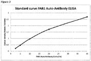
- a standard curve was generated.
- a serum sample of a systemic sclerosis patient was diluted (a) 1:100 for standard point 40 Units/ml, (b) 1:200 for standard point 20 Units/ml, (c) 1:400 for standard point 10 Units/ml, (d) 1:800 for standard point 5 Units/ml and (e) 1:1600 for standard point 2.5 Units/ml.
- the optical density was determined using the kit and method of example above. Each standard point was performed in duplicates.
- the standard curve is depicted in Figure 3 .
- Anti-PAR1 antibody levels in serum samples from 198 healthy donors ("healthy") and 83 patients with APS ("APS") were measured using the kit and method of example 1. The levels were determined in units/mL.
- Fig. 1 shows the comparison of anti-PAR1 antibody level for case and control subjects. Patient suffering from APS had significantly increased levels (p ⁇ 0.05) of anti- PARI antibodies as compared to healthy controls.
- a threshold of e.g. 4 unit/ml lies within the 5 th percentile of subjects with APS and about 90 th of healthy subject, i.e. 95 percent of patients with APS have a higher level than 4 units/ml and 90 percent of healthy subjects have levels below that threshold. This shows that a clear cut-off is possible with the superior method as described herein.
- the results of the present Examples show that anti-PAR1 antibody levels are significantly increased in patients with APS compared to healthy controls.
- the clear statistical cut-off provides for a method allowing the diagnosis of APS by determining the level of anti-PAR1 antibodies in samples of a subject to be diagnosed.
Landscapes
- Health & Medical Sciences (AREA)
- Life Sciences & Earth Sciences (AREA)
- Immunology (AREA)
- Engineering & Computer Science (AREA)
- Hematology (AREA)
- Molecular Biology (AREA)
- Chemical & Material Sciences (AREA)
- Biomedical Technology (AREA)
- Urology & Nephrology (AREA)
- Cell Biology (AREA)
- Analytical Chemistry (AREA)
- Biotechnology (AREA)
- Pathology (AREA)
- Food Science & Technology (AREA)
- Medicinal Chemistry (AREA)
- Physics & Mathematics (AREA)
- Microbiology (AREA)
- Biochemistry (AREA)
- General Health & Medical Sciences (AREA)
- General Physics & Mathematics (AREA)
- Rehabilitation Therapy (AREA)
- Rheumatology (AREA)
- Proteomics, Peptides & Aminoacids (AREA)
- Peptides Or Proteins (AREA)
Claims (16)
- In vitro-Verfahren für die Diagnose von Anti-Phospholipid-Syndrom (APS; antiphospholipid syndrome) bei einem Individuum, wobei das Vorliegen oder das Fehlen eines Anti-Protease-aktivierten Rezeptor 1 (PAR1)-Antikörpers in einer Probe von dem zu diagnostizierenden Individuum nachgewiesen wird und wobei das Vorliegen eines Anti-PAR1-Antikörpers auf die Erkrankung hinweist, wobei das Vorliegen oder das Fehlen des Anti-PAR1-Antikörpers durch die folgenden Schritte nachgewiesen wird: (i) Bestimmen des Spiegels von Anti-PAR1-Antikörpern in einer Probe von dem zu diagnostizierenden Individuum; und (ii) Vergleichen des bestimmten Spiegels mit einem Anti-PAR1-Antikörper-Kontrollspiegel, der von einem Individuum ohne eine Autoimmunkrankheit stammt; wobei ein im Vergleich zum Anti-PAR1-Antikörper-Kontrollspiegel erhöhter Spiegel an Anti-PAR1-Antikörpern in der Probe des zu diagnostizierenden Individuums das Vorliegen eines Anti-PAR1-Antikörpers kennzeichnet.
- In vitro-Verfahren nach Anspruch 1, wobei ein Anti-PAR1-Antikörper-Spiegel in der Probe von dem zu diagnostizierenden Individuum oberhalb des 80. Perzentils von Kontrollspiegeln einer gesunden Population auf APS bei dem zu diagnostizierenden Individuum hinweist.
- In vitro-Verfahren nach einem der vorangehenden Ansprüche, wobei der Anti-PAR1-Antikörper unter Verwendung eines Immunassays nachgewiesen wird, der die Schritte umfasst:(a) Inkontaktbringen der Probe mit einem Protease-aktivierten Rezeptor 1 (PAR1) unter Bedingungen, die die Bildung eines Komplexes aus Anti-PAR1-Antikörpern und PAR1 ermöglichen;(b) Nachweis des Komplexes.
- In vitro-Verfahren nach Anspruch 3, wobei der Protease-aktivierte Rezeptor 1 (PAR1) auf einer Oberfläche immobilisiert ist.
- In vitro-Verfahren nach Anspruch 3 oder 4, wobei der Protease-aktivierte Rezeptor 1 (PAR1) die Sequenz von SEQ ID NO:1 hat.
- In vitro-Verfahren nach einem der Ansprüche 3 bis 5, wobei der Komplex unter Verwendung eines sekundären Antikörpers gegen den Fc-Teil des Anti-PAR1-Antikörpers nachgewiesen wird.
- In vitro-Verfahren nach Anspruch 6, wobei der Anti-PAR1-Antikörper ein IgG-Antikörper ist und der sekundäre Antikörper ein Anti-IgG-Antikörper ist.
- In vitro-Verfahren nach Anspruch 6 oder 7, wobei der sekundäre Antikörper mit einem nachweisbaren Marker markiert ist.
- In vitro-Verfahren nach einem der Ansprüche 3 bis 8, wobei der Immunassay ausgewählt ist aus der Gruppe aus Immunpräzipitation, Enzym-Immunassay (EIA), Radio-Immunassay (RIA) oder Fluoreszenzimmunassay, einem Chemilumineszenz-Assay, einem Agglutinationsassay, nephelometrischen Assay, turbidimetrischen Assay, einem Western Blot, einem kompetitiven Immunassay, einem nicht-kompetitiven Immunassay, einem homogenen Immunassay, einem heterogenen Immunassay und einem Reporterassay wie zum Beispiel einem Luciferase-Assay.
- In vitro-Verfahren nach Anspruch 9, wobei der Immunassay ein ELISA ist.
- In vitro-Verwendung eines Kits für die Diagnose von APS nach dem Verfahren nach einem der Ansprüche 1 bis 10, wobei der Kit PAR1 umfasst, wobei PAR1 auf einer Oberfläche immobilisiert ist.
- In vitro-Verwendung nach Anspruch 11, wobei der Kit Mittel zum Nachweis von Antikörpern umfasst, bevorzugt einen sekundären Antikörper, der den Fc-Teil des nachzuweisenden Anti-PAR1-Antikörpers bindet.
- In vitro-Verwendung nach Anspruch 11 oder 12, wobei der Kit des Weiteren Mittel zum Behandeln und/oder Prozessieren einer Blutprobe umfasst.
- Anti-Koagulans-Arzneistoff zur Verwendung bei der Behandlung von APS, wobei das zu behandelnde Individuum das Vorliegen von Anti-PAR1-Antikörpern in Proben gezeigt hat, wobei der Anti-Koagulans-Arzneistoff dem zu behandelnden Individuum nach der Diagnose von APS mit einem Verfahren nach einem der Ansprüche 1 bis 10 verabreicht wird.
- Anti-Koagulans-Arzneistoff zur Verwendung nach Anspruch 14, wobei der Anti-Koagulans-Arzneistoff Heparin ist.
- In vitro-Verfahren zur Entfernung von Anti-PAR1-Antikörpern aus isoliertem Blut,(i) wobei in einem ersten Schritt das Vorliegen oder das Fehlen eines Anti-PAR1-Antikörpers in einer Blutprobe von einem in Hinblick auf APS zu diagnostizierenden Individuum bestimmt wird,(ii) wobei nach Bestimmen des Vorliegens eines Anti-PAR1-Antikörpers der Antikörper aus dem isolierten Blut des Individuums entfernt wird.
Applications Claiming Priority (2)
| Application Number | Priority Date | Filing Date | Title |
|---|---|---|---|
| EP15168898 | 2015-05-22 | ||
| PCT/EP2016/061021 WO2016188801A1 (en) | 2015-05-22 | 2016-05-17 | A method for diagnosing aps using determination of anti-par1 antibodies |
Publications (2)
| Publication Number | Publication Date |
|---|---|
| EP3298408A1 EP3298408A1 (de) | 2018-03-28 |
| EP3298408B1 true EP3298408B1 (de) | 2021-05-05 |
Family
ID=53191551
Family Applications (1)
| Application Number | Title | Priority Date | Filing Date |
|---|---|---|---|
| EP16724366.6A Active EP3298408B1 (de) | 2015-05-22 | 2016-05-17 | Verfahren zur diagnose von aps mittels bestimmung von anti-par1-antikörpern |
Country Status (3)
| Country | Link |
|---|---|
| US (1) | US20180284117A1 (de) |
| EP (1) | EP3298408B1 (de) |
| WO (1) | WO2016188801A1 (de) |
-
2016
- 2016-05-17 US US15/575,687 patent/US20180284117A1/en not_active Abandoned
- 2016-05-17 EP EP16724366.6A patent/EP3298408B1/de active Active
- 2016-05-17 WO PCT/EP2016/061021 patent/WO2016188801A1/en not_active Ceased
Also Published As
| Publication number | Publication date |
|---|---|
| EP3298408A1 (de) | 2018-03-28 |
| WO2016188801A1 (en) | 2016-12-01 |
| US20180284117A1 (en) | 2018-10-04 |
Similar Documents
| Publication | Publication Date | Title |
|---|---|---|
| Pignatelli et al. | Seronegative antiphospholipid syndrome: refining the value of “non-criteria” antibodies for diagnosis and clinical management | |
| Nayfe et al. | Seronegative antiphospholipid syndrome | |
| Branch et al. | Antiphospholipid syndrome: obstetric diagnosis, management, and controversies | |
| Wijetilleka et al. | Novel insights into pathogenesis, diagnosis and treatment of antiphospholipid syndrome | |
| Ernest et al. | Obstetric antiphospholipid syndrome: an update on pathophysiology and management | |
| Galli et al. | Antiphospholipid antibodies and pregnancy | |
| Bertin et al. | Single or triple positivity for antiphospholipid antibodies in “carriers” or symptomatic patients: Untangling the knot | |
| Chaturvedi et al. | Clinical risk assessment in the antiphospholipid syndrome: current landscape and emerging biomarkers | |
| Ząbczyk et al. | Antiphosphatidylserine/prothrombin complex antibodies as a determinant of prothrombotic plasma fibrin clot properties in patients with antiphospholipid syndrome | |
| Antovic et al. | Laboratory investigation | |
| EP3417291B1 (de) | Anti-cxc-chemokin-rezeptor-antikörper und c-x-c-chemokine zur diagnose von autoimmunerkrankungen und graft-versus-host-erkrankungen | |
| US20180284117A1 (en) | Method for diagnosing APS using determination of anti-PAR1 antibodies | |
| Yamada et al. | Anti-β2 glycoprotein-I antibody increases the risk of pregnancy-induced hypertension: a case-controlled study | |
| WO2017207616A9 (en) | Diagnosis of cancer by detecting auto-antibodies against y box binding protein (yb-1) | |
| Sugiura-Ogasawara et al. | Phosphatidylserine-dependent antiprothrombin antibodies are not useful markers for high-risk women with recurrent miscarriages | |
| EP4314834B1 (de) | Diagnose von morbus alzheimer durch nachweis von autoantikörpern gegen das y-box-bindende protein-1 (yb-1) | |
| EP3213075B1 (de) | Diagnose einer autoimmunerkrankung unter verwendung von gegen c5a-rezeptor gerichteten antikörpern | |
| Vlachoyiannopoulos et al. | Antiphospholipid antibodies: laboratory and pathogenetic aspects | |
| Na et al. | Clinical phenotype of a first unprovoked acute pulmonary embolism associated with antiphospholipid antibody syndrome | |
| Al-Sayed et al. | Diagnostic and prognostic values of serum HSP70 and YKL-40 in patients with rheumatoid arthritis | |
| MBBc et al. | Gene expression in obstetric antiphospholipid syndrome: a systematic review | |
| Schlosser et al. | Autoantibodies against the protease inhibitor calpastatin: a new risk factor for venous thrombosis? | |
| NASIRUDDIN | The clinical and laboratory significance of antiphospholipid syndrome patients in Hospital USM | |
| Casini | Congenital Fibrinogen Disorders, Diagnosis, and Management | |
| US20220276251A1 (en) | Diagnosis of cancer using detection of antibodies directed against pd1 and pd-l1 |
Legal Events
| Date | Code | Title | Description |
|---|---|---|---|
| STAA | Information on the status of an ep patent application or granted ep patent |
Free format text: STATUS: THE INTERNATIONAL PUBLICATION HAS BEEN MADE |
|
| PUAI | Public reference made under article 153(3) epc to a published international application that has entered the european phase |
Free format text: ORIGINAL CODE: 0009012 |
|
| STAA | Information on the status of an ep patent application or granted ep patent |
Free format text: STATUS: REQUEST FOR EXAMINATION WAS MADE |
|
| 17P | Request for examination filed |
Effective date: 20171215 |
|
| AK | Designated contracting states |
Kind code of ref document: A1 Designated state(s): AL AT BE BG CH CY CZ DE DK EE ES FI FR GB GR HR HU IE IS IT LI LT LU LV MC MK MT NL NO PL PT RO RS SE SI SK SM TR |
|
| AX | Request for extension of the european patent |
Extension state: BA ME |
|
| DAV | Request for validation of the european patent (deleted) | ||
| DAX | Request for extension of the european patent (deleted) | ||
| STAA | Information on the status of an ep patent application or granted ep patent |
Free format text: STATUS: EXAMINATION IS IN PROGRESS |
|
| 17Q | First examination report despatched |
Effective date: 20181009 |
|
| GRAP | Despatch of communication of intention to grant a patent |
Free format text: ORIGINAL CODE: EPIDOSNIGR1 |
|
| STAA | Information on the status of an ep patent application or granted ep patent |
Free format text: STATUS: GRANT OF PATENT IS INTENDED |
|
| INTG | Intention to grant announced |
Effective date: 20201120 |
|
| GRAS | Grant fee paid |
Free format text: ORIGINAL CODE: EPIDOSNIGR3 |
|
| GRAA | (expected) grant |
Free format text: ORIGINAL CODE: 0009210 |
|
| STAA | Information on the status of an ep patent application or granted ep patent |
Free format text: STATUS: THE PATENT HAS BEEN GRANTED |
|
| AK | Designated contracting states |
Kind code of ref document: B1 Designated state(s): AL AT BE BG CH CY CZ DE DK EE ES FI FR GB GR HR HU IE IS IT LI LT LU LV MC MK MT NL NO PL PT RO RS SE SI SK SM TR |
|
| REG | Reference to a national code |
Ref country code: GB Ref legal event code: FG4D |
|
| REG | Reference to a national code |
Ref country code: CH Ref legal event code: EP |
|
| REG | Reference to a national code |
Ref country code: AT Ref legal event code: REF Ref document number: 1390446 Country of ref document: AT Kind code of ref document: T Effective date: 20210515 |
|
| REG | Reference to a national code |
Ref country code: IE Ref legal event code: FG4D |
|
| REG | Reference to a national code |
Ref country code: DE Ref legal event code: R096 Ref document number: 602016057342 Country of ref document: DE |
|
| REG | Reference to a national code |
Ref country code: LT Ref legal event code: MG9D |
|
| REG | Reference to a national code |
Ref country code: AT Ref legal event code: MK05 Ref document number: 1390446 Country of ref document: AT Kind code of ref document: T Effective date: 20210505 |
|
| PG25 | Lapsed in a contracting state [announced via postgrant information from national office to epo] |
Ref country code: HR Free format text: LAPSE BECAUSE OF FAILURE TO SUBMIT A TRANSLATION OF THE DESCRIPTION OR TO PAY THE FEE WITHIN THE PRESCRIBED TIME-LIMIT Effective date: 20210505 Ref country code: AT Free format text: LAPSE BECAUSE OF FAILURE TO SUBMIT A TRANSLATION OF THE DESCRIPTION OR TO PAY THE FEE WITHIN THE PRESCRIBED TIME-LIMIT Effective date: 20210505 Ref country code: BG Free format text: LAPSE BECAUSE OF FAILURE TO SUBMIT A TRANSLATION OF THE DESCRIPTION OR TO PAY THE FEE WITHIN THE PRESCRIBED TIME-LIMIT Effective date: 20210805 Ref country code: FI Free format text: LAPSE BECAUSE OF FAILURE TO SUBMIT A TRANSLATION OF THE DESCRIPTION OR TO PAY THE FEE WITHIN THE PRESCRIBED TIME-LIMIT Effective date: 20210505 Ref country code: LT Free format text: LAPSE BECAUSE OF FAILURE TO SUBMIT A TRANSLATION OF THE DESCRIPTION OR TO PAY THE FEE WITHIN THE PRESCRIBED TIME-LIMIT Effective date: 20210505 |
|
| PG25 | Lapsed in a contracting state [announced via postgrant information from national office to epo] |
Ref country code: IS Free format text: LAPSE BECAUSE OF FAILURE TO SUBMIT A TRANSLATION OF THE DESCRIPTION OR TO PAY THE FEE WITHIN THE PRESCRIBED TIME-LIMIT Effective date: 20210905 Ref country code: LV Free format text: LAPSE BECAUSE OF FAILURE TO SUBMIT A TRANSLATION OF THE DESCRIPTION OR TO PAY THE FEE WITHIN THE PRESCRIBED TIME-LIMIT Effective date: 20210505 Ref country code: GR Free format text: LAPSE BECAUSE OF FAILURE TO SUBMIT A TRANSLATION OF THE DESCRIPTION OR TO PAY THE FEE WITHIN THE PRESCRIBED TIME-LIMIT Effective date: 20210806 Ref country code: PT Free format text: LAPSE BECAUSE OF FAILURE TO SUBMIT A TRANSLATION OF THE DESCRIPTION OR TO PAY THE FEE WITHIN THE PRESCRIBED TIME-LIMIT Effective date: 20210906 Ref country code: PL Free format text: LAPSE BECAUSE OF FAILURE TO SUBMIT A TRANSLATION OF THE DESCRIPTION OR TO PAY THE FEE WITHIN THE PRESCRIBED TIME-LIMIT Effective date: 20210505 Ref country code: NO Free format text: LAPSE BECAUSE OF FAILURE TO SUBMIT A TRANSLATION OF THE DESCRIPTION OR TO PAY THE FEE WITHIN THE PRESCRIBED TIME-LIMIT Effective date: 20210805 Ref country code: RS Free format text: LAPSE BECAUSE OF FAILURE TO SUBMIT A TRANSLATION OF THE DESCRIPTION OR TO PAY THE FEE WITHIN THE PRESCRIBED TIME-LIMIT Effective date: 20210505 Ref country code: SE Free format text: LAPSE BECAUSE OF FAILURE TO SUBMIT A TRANSLATION OF THE DESCRIPTION OR TO PAY THE FEE WITHIN THE PRESCRIBED TIME-LIMIT Effective date: 20210505 |
|
| REG | Reference to a national code |
Ref country code: NL Ref legal event code: MP Effective date: 20210505 |
|
| REG | Reference to a national code |
Ref country code: CH Ref legal event code: PL |
|
| PG25 | Lapsed in a contracting state [announced via postgrant information from national office to epo] |
Ref country code: NL Free format text: LAPSE BECAUSE OF FAILURE TO SUBMIT A TRANSLATION OF THE DESCRIPTION OR TO PAY THE FEE WITHIN THE PRESCRIBED TIME-LIMIT Effective date: 20210505 |
|
| PG25 | Lapsed in a contracting state [announced via postgrant information from national office to epo] |
Ref country code: CH Free format text: LAPSE BECAUSE OF NON-PAYMENT OF DUE FEES Effective date: 20210531 Ref country code: LU Free format text: LAPSE BECAUSE OF NON-PAYMENT OF DUE FEES Effective date: 20210517 Ref country code: LI Free format text: LAPSE BECAUSE OF NON-PAYMENT OF DUE FEES Effective date: 20210531 Ref country code: ES Free format text: LAPSE BECAUSE OF FAILURE TO SUBMIT A TRANSLATION OF THE DESCRIPTION OR TO PAY THE FEE WITHIN THE PRESCRIBED TIME-LIMIT Effective date: 20210505 Ref country code: RO Free format text: LAPSE BECAUSE OF FAILURE TO SUBMIT A TRANSLATION OF THE DESCRIPTION OR TO PAY THE FEE WITHIN THE PRESCRIBED TIME-LIMIT Effective date: 20210505 Ref country code: EE Free format text: LAPSE BECAUSE OF FAILURE TO SUBMIT A TRANSLATION OF THE DESCRIPTION OR TO PAY THE FEE WITHIN THE PRESCRIBED TIME-LIMIT Effective date: 20210505 Ref country code: CZ Free format text: LAPSE BECAUSE OF FAILURE TO SUBMIT A TRANSLATION OF THE DESCRIPTION OR TO PAY THE FEE WITHIN THE PRESCRIBED TIME-LIMIT Effective date: 20210505 Ref country code: DK Free format text: LAPSE BECAUSE OF FAILURE TO SUBMIT A TRANSLATION OF THE DESCRIPTION OR TO PAY THE FEE WITHIN THE PRESCRIBED TIME-LIMIT Effective date: 20210505 Ref country code: SK Free format text: LAPSE BECAUSE OF FAILURE TO SUBMIT A TRANSLATION OF THE DESCRIPTION OR TO PAY THE FEE WITHIN THE PRESCRIBED TIME-LIMIT Effective date: 20210505 Ref country code: SM Free format text: LAPSE BECAUSE OF FAILURE TO SUBMIT A TRANSLATION OF THE DESCRIPTION OR TO PAY THE FEE WITHIN THE PRESCRIBED TIME-LIMIT Effective date: 20210505 |
|
| REG | Reference to a national code |
Ref country code: DE Ref legal event code: R097 Ref document number: 602016057342 Country of ref document: DE |
|
| REG | Reference to a national code |
Ref country code: BE Ref legal event code: MM Effective date: 20210531 |
|
| PLBE | No opposition filed within time limit |
Free format text: ORIGINAL CODE: 0009261 |
|
| STAA | Information on the status of an ep patent application or granted ep patent |
Free format text: STATUS: NO OPPOSITION FILED WITHIN TIME LIMIT |
|
| PG25 | Lapsed in a contracting state [announced via postgrant information from national office to epo] |
Ref country code: MC Free format text: LAPSE BECAUSE OF FAILURE TO SUBMIT A TRANSLATION OF THE DESCRIPTION OR TO PAY THE FEE WITHIN THE PRESCRIBED TIME-LIMIT Effective date: 20210505 |
|
| 26N | No opposition filed |
Effective date: 20220208 |
|
| PG25 | Lapsed in a contracting state [announced via postgrant information from national office to epo] |
Ref country code: IE Free format text: LAPSE BECAUSE OF NON-PAYMENT OF DUE FEES Effective date: 20210517 |
|
| PG25 | Lapsed in a contracting state [announced via postgrant information from national office to epo] |
Ref country code: IS Free format text: LAPSE BECAUSE OF FAILURE TO SUBMIT A TRANSLATION OF THE DESCRIPTION OR TO PAY THE FEE WITHIN THE PRESCRIBED TIME-LIMIT Effective date: 20210905 Ref country code: AL Free format text: LAPSE BECAUSE OF FAILURE TO SUBMIT A TRANSLATION OF THE DESCRIPTION OR TO PAY THE FEE WITHIN THE PRESCRIBED TIME-LIMIT Effective date: 20210505 |
|
| PG25 | Lapsed in a contracting state [announced via postgrant information from national office to epo] |
Ref country code: IT Free format text: LAPSE BECAUSE OF FAILURE TO SUBMIT A TRANSLATION OF THE DESCRIPTION OR TO PAY THE FEE WITHIN THE PRESCRIBED TIME-LIMIT Effective date: 20210505 Ref country code: BE Free format text: LAPSE BECAUSE OF NON-PAYMENT OF DUE FEES Effective date: 20210531 |
|
| PG25 | Lapsed in a contracting state [announced via postgrant information from national office to epo] |
Ref country code: HU Free format text: LAPSE BECAUSE OF FAILURE TO SUBMIT A TRANSLATION OF THE DESCRIPTION OR TO PAY THE FEE WITHIN THE PRESCRIBED TIME-LIMIT; INVALID AB INITIO Effective date: 20160517 |
|
| P01 | Opt-out of the competence of the unified patent court (upc) registered |
Effective date: 20230413 |
|
| PG25 | Lapsed in a contracting state [announced via postgrant information from national office to epo] |
Ref country code: CY Free format text: LAPSE BECAUSE OF FAILURE TO SUBMIT A TRANSLATION OF THE DESCRIPTION OR TO PAY THE FEE WITHIN THE PRESCRIBED TIME-LIMIT Effective date: 20210505 |
|
| PG25 | Lapsed in a contracting state [announced via postgrant information from national office to epo] |
Ref country code: MK Free format text: LAPSE BECAUSE OF FAILURE TO SUBMIT A TRANSLATION OF THE DESCRIPTION OR TO PAY THE FEE WITHIN THE PRESCRIBED TIME-LIMIT Effective date: 20210505 |
|
| PG25 | Lapsed in a contracting state [announced via postgrant information from national office to epo] |
Ref country code: MT Free format text: LAPSE BECAUSE OF FAILURE TO SUBMIT A TRANSLATION OF THE DESCRIPTION OR TO PAY THE FEE WITHIN THE PRESCRIBED TIME-LIMIT Effective date: 20210505 |
|
| PGFP | Annual fee paid to national office [announced via postgrant information from national office to epo] |
Ref country code: DE Payment date: 20250530 Year of fee payment: 10 |
|
| PGFP | Annual fee paid to national office [announced via postgrant information from national office to epo] |
Ref country code: FR Payment date: 20250527 Year of fee payment: 10 |
|
| PG25 | Lapsed in a contracting state [announced via postgrant information from national office to epo] |
Ref country code: TR Free format text: LAPSE BECAUSE OF FAILURE TO SUBMIT A TRANSLATION OF THE DESCRIPTION OR TO PAY THE FEE WITHIN THE PRESCRIBED TIME-LIMIT Effective date: 20210505 |
|
| PGFP | Annual fee paid to national office [announced via postgrant information from national office to epo] |
Ref country code: GB Payment date: 20260331 Year of fee payment: 11 |