EP3296397A1 - Rev-clip, ein neuer ansatz zum identifizieren von mit rna interagierenden proteinen - Google Patents
Rev-clip, ein neuer ansatz zum identifizieren von mit rna interagierenden proteinen Download PDFInfo
- Publication number
- EP3296397A1 EP3296397A1 EP16189688.1A EP16189688A EP3296397A1 EP 3296397 A1 EP3296397 A1 EP 3296397A1 EP 16189688 A EP16189688 A EP 16189688A EP 3296397 A1 EP3296397 A1 EP 3296397A1
- Authority
- EP
- European Patent Office
- Prior art keywords
- rna
- molecule
- molecules
- target rna
- splint
- Prior art date
- Legal status (The legal status is an assumption and is not a legal conclusion. Google has not performed a legal analysis and makes no representation as to the accuracy of the status listed.)
- Withdrawn
Links
- 108090000623 proteins and genes Proteins 0.000 title abstract description 69
- 102000004169 proteins and genes Human genes 0.000 title abstract description 68
- 238000013459 approach Methods 0.000 title description 2
- 108091032973 (ribonucleotides)n+m Proteins 0.000 claims abstract description 317
- 238000000034 method Methods 0.000 claims abstract description 94
- 108091092328 cellular RNA Proteins 0.000 claims abstract description 8
- 102000044126 RNA-Binding Proteins Human genes 0.000 claims description 72
- 108700020471 RNA-Binding Proteins Proteins 0.000 claims description 69
- 210000004027 cell Anatomy 0.000 claims description 65
- 238000000137 annealing Methods 0.000 claims description 31
- 239000011324 bead Substances 0.000 claims description 31
- 239000013592 cell lysate Substances 0.000 claims description 23
- QKNYBSVHEMOAJP-UHFFFAOYSA-N 2-amino-2-(hydroxymethyl)propane-1,3-diol;hydron;chloride Chemical compound Cl.OCC(N)(CO)CO QKNYBSVHEMOAJP-UHFFFAOYSA-N 0.000 claims description 20
- 108091034117 Oligonucleotide Proteins 0.000 claims description 20
- 125000003729 nucleotide group Chemical group 0.000 claims description 19
- 241000894007 species Species 0.000 claims description 19
- 102000016911 Deoxyribonucleases Human genes 0.000 claims description 18
- 108010053770 Deoxyribonucleases Proteins 0.000 claims description 18
- 230000000295 complement effect Effects 0.000 claims description 18
- 239000002773 nucleotide Substances 0.000 claims description 18
- 108020004414 DNA Proteins 0.000 claims description 16
- FAPWRFPIFSIZLT-UHFFFAOYSA-M Sodium chloride Chemical compound [Na+].[Cl-] FAPWRFPIFSIZLT-UHFFFAOYSA-M 0.000 claims description 16
- KCXVZYZYPLLWCC-UHFFFAOYSA-N EDTA Chemical compound OC(=O)CN(CC(O)=O)CCN(CC(O)=O)CC(O)=O KCXVZYZYPLLWCC-UHFFFAOYSA-N 0.000 claims description 13
- 238000003776 cleavage reaction Methods 0.000 claims description 13
- 230000007017 scission Effects 0.000 claims description 13
- 238000005406 washing Methods 0.000 claims description 13
- 239000006166 lysate Substances 0.000 claims description 12
- 239000000074 antisense oligonucleotide Substances 0.000 claims description 11
- 238000012230 antisense oligonucleotides Methods 0.000 claims description 11
- 238000004132 cross linking Methods 0.000 claims description 10
- JLCPHMBAVCMARE-UHFFFAOYSA-N [3-[[3-[[3-[[3-[[3-[[3-[[3-[[3-[[3-[[3-[[3-[[5-(2-amino-6-oxo-1H-purin-9-yl)-3-[[3-[[3-[[3-[[3-[[3-[[5-(2-amino-6-oxo-1H-purin-9-yl)-3-[[5-(2-amino-6-oxo-1H-purin-9-yl)-3-hydroxyoxolan-2-yl]methoxy-hydroxyphosphoryl]oxyoxolan-2-yl]methoxy-hydroxyphosphoryl]oxy-5-(5-methyl-2,4-dioxopyrimidin-1-yl)oxolan-2-yl]methoxy-hydroxyphosphoryl]oxy-5-(6-aminopurin-9-yl)oxolan-2-yl]methoxy-hydroxyphosphoryl]oxy-5-(6-aminopurin-9-yl)oxolan-2-yl]methoxy-hydroxyphosphoryl]oxy-5-(6-aminopurin-9-yl)oxolan-2-yl]methoxy-hydroxyphosphoryl]oxy-5-(6-aminopurin-9-yl)oxolan-2-yl]methoxy-hydroxyphosphoryl]oxyoxolan-2-yl]methoxy-hydroxyphosphoryl]oxy-5-(5-methyl-2,4-dioxopyrimidin-1-yl)oxolan-2-yl]methoxy-hydroxyphosphoryl]oxy-5-(4-amino-2-oxopyrimidin-1-yl)oxolan-2-yl]methoxy-hydroxyphosphoryl]oxy-5-(5-methyl-2,4-dioxopyrimidin-1-yl)oxolan-2-yl]methoxy-hydroxyphosphoryl]oxy-5-(5-methyl-2,4-dioxopyrimidin-1-yl)oxolan-2-yl]methoxy-hydroxyphosphoryl]oxy-5-(6-aminopurin-9-yl)oxolan-2-yl]methoxy-hydroxyphosphoryl]oxy-5-(6-aminopurin-9-yl)oxolan-2-yl]methoxy-hydroxyphosphoryl]oxy-5-(4-amino-2-oxopyrimidin-1-yl)oxolan-2-yl]methoxy-hydroxyphosphoryl]oxy-5-(4-amino-2-oxopyrimidin-1-yl)oxolan-2-yl]methoxy-hydroxyphosphoryl]oxy-5-(4-amino-2-oxopyrimidin-1-yl)oxolan-2-yl]methoxy-hydroxyphosphoryl]oxy-5-(6-aminopurin-9-yl)oxolan-2-yl]methoxy-hydroxyphosphoryl]oxy-5-(4-amino-2-oxopyrimidin-1-yl)oxolan-2-yl]methyl [5-(6-aminopurin-9-yl)-2-(hydroxymethyl)oxolan-3-yl] hydrogen phosphate Polymers Cc1cn(C2CC(OP(O)(=O)OCC3OC(CC3OP(O)(=O)OCC3OC(CC3O)n3cnc4c3nc(N)[nH]c4=O)n3cnc4c3nc(N)[nH]c4=O)C(COP(O)(=O)OC3CC(OC3COP(O)(=O)OC3CC(OC3COP(O)(=O)OC3CC(OC3COP(O)(=O)OC3CC(OC3COP(O)(=O)OC3CC(OC3COP(O)(=O)OC3CC(OC3COP(O)(=O)OC3CC(OC3COP(O)(=O)OC3CC(OC3COP(O)(=O)OC3CC(OC3COP(O)(=O)OC3CC(OC3COP(O)(=O)OC3CC(OC3COP(O)(=O)OC3CC(OC3COP(O)(=O)OC3CC(OC3COP(O)(=O)OC3CC(OC3COP(O)(=O)OC3CC(OC3COP(O)(=O)OC3CC(OC3COP(O)(=O)OC3CC(OC3CO)n3cnc4c(N)ncnc34)n3ccc(N)nc3=O)n3cnc4c(N)ncnc34)n3ccc(N)nc3=O)n3ccc(N)nc3=O)n3ccc(N)nc3=O)n3cnc4c(N)ncnc34)n3cnc4c(N)ncnc34)n3cc(C)c(=O)[nH]c3=O)n3cc(C)c(=O)[nH]c3=O)n3ccc(N)nc3=O)n3cc(C)c(=O)[nH]c3=O)n3cnc4c3nc(N)[nH]c4=O)n3cnc4c(N)ncnc34)n3cnc4c(N)ncnc34)n3cnc4c(N)ncnc34)n3cnc4c(N)ncnc34)O2)c(=O)[nH]c1=O JLCPHMBAVCMARE-UHFFFAOYSA-N 0.000 claims description 9
- 150000003839 salts Chemical class 0.000 claims description 9
- 239000011534 wash buffer Substances 0.000 claims description 9
- 108020000948 Antisense Oligonucleotides Proteins 0.000 claims description 8
- 241000282414 Homo sapiens Species 0.000 claims description 8
- 230000027455 binding Effects 0.000 claims description 8
- 239000011780 sodium chloride Substances 0.000 claims description 8
- 108010083644 Ribonucleases Proteins 0.000 claims description 7
- 102000006382 Ribonucleases Human genes 0.000 claims description 7
- 108090000994 Catalytic RNA Proteins 0.000 claims description 6
- 102000053642 Catalytic RNA Human genes 0.000 claims description 6
- 125000002652 ribonucleotide group Chemical group 0.000 claims description 6
- 108091092562 ribozyme Proteins 0.000 claims description 6
- 239000007790 solid phase Substances 0.000 claims description 6
- 108090001102 Hammerhead ribozyme Proteins 0.000 claims description 5
- 108091028664 Ribonucleotide Proteins 0.000 claims description 4
- 229960003964 deoxycholic acid Drugs 0.000 claims description 4
- 239000002336 ribonucleotide Substances 0.000 claims description 4
- FHHPUSMSKHSNKW-SMOYURAASA-M sodium deoxycholate Chemical compound [Na+].C([C@H]1CC2)[C@H](O)CC[C@]1(C)[C@@H]1[C@@H]2[C@@H]2CC[C@H]([C@@H](CCC([O-])=O)C)[C@@]2(C)[C@@H](O)C1 FHHPUSMSKHSNKW-SMOYURAASA-M 0.000 claims description 4
- 108020004491 Antisense DNA Proteins 0.000 claims description 3
- FGUUSXIOTUKUDN-IBGZPJMESA-N C1(=CC=CC=C1)N1C2=C(NC([C@H](C1)NC=1OC(=NN=1)C1=CC=CC=C1)=O)C=CC=C2 Chemical compound C1(=CC=CC=C1)N1C2=C(NC([C@H](C1)NC=1OC(=NN=1)C1=CC=CC=C1)=O)C=CC=C2 FGUUSXIOTUKUDN-IBGZPJMESA-N 0.000 claims description 3
- 108091028043 Nucleic acid sequence Proteins 0.000 claims description 3
- 101710188535 RNA ligase 2 Proteins 0.000 claims description 3
- 101710204104 RNA-editing ligase 2, mitochondrial Proteins 0.000 claims description 3
- 239000003816 antisense DNA Substances 0.000 claims description 3
- 125000001400 nonyl group Chemical group [H]C([*])([H])C([H])([H])C([H])([H])C([H])([H])C([H])([H])C([H])([H])C([H])([H])C([H])([H])C([H])([H])[H] 0.000 claims description 3
- 108020004635 Complementary DNA Proteins 0.000 claims description 2
- 229920001213 Polysorbate 20 Polymers 0.000 claims description 2
- 238000010804 cDNA synthesis Methods 0.000 claims description 2
- 239000002299 complementary DNA Substances 0.000 claims description 2
- 230000003100 immobilizing effect Effects 0.000 claims description 2
- 239000000256 polyoxyethylene sorbitan monolaurate Substances 0.000 claims description 2
- 235000010486 polyoxyethylene sorbitan monolaurate Nutrition 0.000 claims description 2
- GNFTZDOKVXKIBK-UHFFFAOYSA-N 3-(2-methoxyethoxy)benzohydrazide Chemical compound COCCOC1=CC=CC(C(=O)NN)=C1 GNFTZDOKVXKIBK-UHFFFAOYSA-N 0.000 claims 1
- 210000004962 mammalian cell Anatomy 0.000 claims 1
- 230000004570 RNA-binding Effects 0.000 abstract description 8
- 102100024544 SURP and G-patch domain-containing protein 1 Human genes 0.000 abstract description 8
- 238000002955 isolation Methods 0.000 abstract description 6
- 238000000746 purification Methods 0.000 abstract description 6
- 230000001404 mediated effect Effects 0.000 abstract description 5
- 108010026552 Proteome Proteins 0.000 abstract description 4
- 238000012512 characterization method Methods 0.000 abstract description 4
- 230000004962 physiological condition Effects 0.000 abstract description 4
- 238000001727 in vivo Methods 0.000 abstract description 3
- 235000018102 proteins Nutrition 0.000 description 63
- 238000012155 cross-linking immunoprecipitation Methods 0.000 description 57
- 238000010828 elution Methods 0.000 description 29
- 102000040650 (ribonucleotides)n+m Human genes 0.000 description 24
- YBJHBAHKTGYVGT-ZKWXMUAHSA-N (+)-Biotin Chemical compound N1C(=O)N[C@@H]2[C@H](CCCCC(=O)O)SC[C@@H]21 YBJHBAHKTGYVGT-ZKWXMUAHSA-N 0.000 description 18
- 239000000872 buffer Substances 0.000 description 17
- 238000004458 analytical method Methods 0.000 description 16
- 239000003795 chemical substances by application Substances 0.000 description 16
- CSCPPACGZOOCGX-UHFFFAOYSA-N Acetone Chemical compound CC(C)=O CSCPPACGZOOCGX-UHFFFAOYSA-N 0.000 description 12
- 108010090804 Streptavidin Proteins 0.000 description 12
- 108090000765 processed proteins & peptides Proteins 0.000 description 12
- DTQVDTLACAAQTR-UHFFFAOYSA-N Trifluoroacetic acid Chemical compound OC(=O)C(F)(F)F DTQVDTLACAAQTR-UHFFFAOYSA-N 0.000 description 10
- 108020004999 messenger RNA Proteins 0.000 description 10
- 239000000243 solution Substances 0.000 description 10
- 239000011616 biotin Substances 0.000 description 9
- 229960002685 biotin Drugs 0.000 description 9
- 235000020958 biotin Nutrition 0.000 description 9
- 238000006243 chemical reaction Methods 0.000 description 9
- 238000004949 mass spectrometry Methods 0.000 description 9
- TWRXJAOTZQYOKJ-UHFFFAOYSA-L Magnesium chloride Chemical compound [Mg+2].[Cl-].[Cl-] TWRXJAOTZQYOKJ-UHFFFAOYSA-L 0.000 description 8
- 239000003599 detergent Substances 0.000 description 8
- 239000012139 lysis buffer Substances 0.000 description 8
- 239000000203 mixture Substances 0.000 description 8
- 102000004196 processed proteins & peptides Human genes 0.000 description 8
- 238000011282 treatment Methods 0.000 description 8
- 238000001262 western blot Methods 0.000 description 8
- 108700015925 CELF1 Proteins 0.000 description 7
- 102100033676 CUGBP Elav-like family member 1 Human genes 0.000 description 7
- 102100033073 Polypyrimidine tract-binding protein 1 Human genes 0.000 description 7
- 101710132817 Polypyrimidine tract-binding protein 1 Proteins 0.000 description 7
- 230000003993 interaction Effects 0.000 description 7
- 150000002500 ions Chemical class 0.000 description 7
- WEVYAHXRMPXWCK-UHFFFAOYSA-N Acetonitrile Chemical compound CC#N WEVYAHXRMPXWCK-UHFFFAOYSA-N 0.000 description 6
- 101710086015 RNA ligase Proteins 0.000 description 6
- XSQUKJJJFZCRTK-UHFFFAOYSA-N Urea Chemical compound NC(N)=O XSQUKJJJFZCRTK-UHFFFAOYSA-N 0.000 description 6
- BDAGIHXWWSANSR-UHFFFAOYSA-N methanoic acid Natural products OC=O BDAGIHXWWSANSR-UHFFFAOYSA-N 0.000 description 6
- 238000000338 in vitro Methods 0.000 description 5
- 238000011534 incubation Methods 0.000 description 5
- 101150107790 CELF1 gene Proteins 0.000 description 4
- ZHNUHDYFZUAESO-UHFFFAOYSA-N Formamide Chemical compound NC=O ZHNUHDYFZUAESO-UHFFFAOYSA-N 0.000 description 4
- 229940124158 Protease/peptidase inhibitor Drugs 0.000 description 4
- 108020003224 Small Nucleolar RNA Proteins 0.000 description 4
- 102000042773 Small Nucleolar RNA Human genes 0.000 description 4
- 108020004566 Transfer RNA Proteins 0.000 description 4
- 239000011230 binding agent Substances 0.000 description 4
- 230000001413 cellular effect Effects 0.000 description 4
- 238000011109 contamination Methods 0.000 description 4
- 230000001419 dependent effect Effects 0.000 description 4
- 230000029087 digestion Effects 0.000 description 4
- 230000000694 effects Effects 0.000 description 4
- KWGKDLIKAYFUFQ-UHFFFAOYSA-M lithium chloride Chemical compound [Li+].[Cl-] KWGKDLIKAYFUFQ-UHFFFAOYSA-M 0.000 description 4
- 229910001629 magnesium chloride Inorganic materials 0.000 description 4
- 108091070501 miRNA Proteins 0.000 description 4
- 239000002679 microRNA Substances 0.000 description 4
- 108091027963 non-coding RNA Proteins 0.000 description 4
- 102000042567 non-coding RNA Human genes 0.000 description 4
- 239000008188 pellet Substances 0.000 description 4
- 239000000137 peptide hydrolase inhibitor Substances 0.000 description 4
- 239000000047 product Substances 0.000 description 4
- 238000000926 separation method Methods 0.000 description 4
- 238000004885 tandem mass spectrometry Methods 0.000 description 4
- OSWFIVFLDKOXQC-UHFFFAOYSA-N 4-(3-methoxyphenyl)aniline Chemical compound COC1=CC=CC(C=2C=CC(N)=CC=2)=C1 OSWFIVFLDKOXQC-UHFFFAOYSA-N 0.000 description 3
- 102000007469 Actins Human genes 0.000 description 3
- 108010085238 Actins Proteins 0.000 description 3
- 208000002109 Argyria Diseases 0.000 description 3
- 239000006144 Dulbecco’s modified Eagle's medium Substances 0.000 description 3
- PEDCQBHIVMGVHV-UHFFFAOYSA-N Glycerine Chemical compound OCC(O)CO PEDCQBHIVMGVHV-UHFFFAOYSA-N 0.000 description 3
- 102000009572 RNA Polymerase II Human genes 0.000 description 3
- 108010009460 RNA Polymerase II Proteins 0.000 description 3
- 108090000631 Trypsin Proteins 0.000 description 3
- 102000004142 Trypsin Human genes 0.000 description 3
- 239000004202 carbamide Substances 0.000 description 3
- 239000003638 chemical reducing agent Substances 0.000 description 3
- 238000001514 detection method Methods 0.000 description 3
- 201000010099 disease Diseases 0.000 description 3
- 208000037265 diseases, disorders, signs and symptoms Diseases 0.000 description 3
- 238000011067 equilibration Methods 0.000 description 3
- 235000019253 formic acid Nutrition 0.000 description 3
- 239000012145 high-salt buffer Substances 0.000 description 3
- 238000003368 label free method Methods 0.000 description 3
- 239000007788 liquid Substances 0.000 description 3
- 238000001294 liquid chromatography-tandem mass spectrometry Methods 0.000 description 3
- 230000004048 modification Effects 0.000 description 3
- 238000012986 modification Methods 0.000 description 3
- 238000011002 quantification Methods 0.000 description 3
- 239000011541 reaction mixture Substances 0.000 description 3
- 239000002342 ribonucleoside Substances 0.000 description 3
- 239000002904 solvent Substances 0.000 description 3
- 239000012588 trypsin Substances 0.000 description 3
- 201000009030 Carcinoma Diseases 0.000 description 2
- HEDRZPFGACZZDS-UHFFFAOYSA-N Chloroform Chemical compound ClC(Cl)Cl HEDRZPFGACZZDS-UHFFFAOYSA-N 0.000 description 2
- 102000053602 DNA Human genes 0.000 description 2
- 102000052510 DNA-Binding Proteins Human genes 0.000 description 2
- 108700020911 DNA-Binding Proteins Proteins 0.000 description 2
- 108010067770 Endopeptidase K Proteins 0.000 description 2
- 102000004190 Enzymes Human genes 0.000 description 2
- 108090000790 Enzymes Proteins 0.000 description 2
- WQZGKKKJIJFFOK-GASJEMHNSA-N Glucose Natural products OC[C@H]1OC(O)[C@H](O)[C@@H](O)[C@@H]1O WQZGKKKJIJFFOK-GASJEMHNSA-N 0.000 description 2
- 241000282412 Homo Species 0.000 description 2
- 108010001336 Horseradish Peroxidase Proteins 0.000 description 2
- 108091007460 Long intergenic noncoding RNA Proteins 0.000 description 2
- 241000283973 Oryctolagus cuniculus Species 0.000 description 2
- ISWSIDIOOBJBQZ-UHFFFAOYSA-N Phenol Chemical compound OC1=CC=CC=C1 ISWSIDIOOBJBQZ-UHFFFAOYSA-N 0.000 description 2
- BQCADISMDOOEFD-UHFFFAOYSA-N Silver Chemical compound [Ag] BQCADISMDOOEFD-UHFFFAOYSA-N 0.000 description 2
- 108091046869 Telomeric non-coding RNA Proteins 0.000 description 2
- ISAKRJDGNUQOIC-UHFFFAOYSA-N Uracil Chemical group O=C1C=CNC(=O)N1 ISAKRJDGNUQOIC-UHFFFAOYSA-N 0.000 description 2
- 210000000227 basophil cell of anterior lobe of hypophysis Anatomy 0.000 description 2
- UDSAIICHUKSCKT-UHFFFAOYSA-N bromophenol blue Chemical compound C1=C(Br)C(O)=C(Br)C=C1C1(C=2C=C(Br)C(O)=C(Br)C=2)C2=CC=CC=C2S(=O)(=O)O1 UDSAIICHUKSCKT-UHFFFAOYSA-N 0.000 description 2
- 230000015556 catabolic process Effects 0.000 description 2
- 238000004113 cell culture Methods 0.000 description 2
- 210000003169 central nervous system Anatomy 0.000 description 2
- 239000007795 chemical reaction product Substances 0.000 description 2
- 238000006731 degradation reaction Methods 0.000 description 2
- 239000012149 elution buffer Substances 0.000 description 2
- 210000001671 embryonic stem cell Anatomy 0.000 description 2
- 238000002546 full scan Methods 0.000 description 2
- 230000006870 function Effects 0.000 description 2
- 239000000499 gel Substances 0.000 description 2
- 239000008103 glucose Substances 0.000 description 2
- 239000011544 gradient gel Substances 0.000 description 2
- 210000003494 hepatocyte Anatomy 0.000 description 2
- 238000002347 injection Methods 0.000 description 2
- 239000007924 injection Substances 0.000 description 2
- JDNTWHVOXJZDSN-UHFFFAOYSA-N iodoacetic acid Chemical compound OC(=O)CI JDNTWHVOXJZDSN-UHFFFAOYSA-N 0.000 description 2
- 210000003734 kidney Anatomy 0.000 description 2
- 238000004811 liquid chromatography Methods 0.000 description 2
- 239000012160 loading buffer Substances 0.000 description 2
- 230000007246 mechanism Effects 0.000 description 2
- 239000002609 medium Substances 0.000 description 2
- 239000012528 membrane Substances 0.000 description 2
- 239000013642 negative control Substances 0.000 description 2
- 239000012071 phase Substances 0.000 description 2
- 238000002264 polyacrylamide gel electrophoresis Methods 0.000 description 2
- 230000002829 reductive effect Effects 0.000 description 2
- 230000004044 response Effects 0.000 description 2
- 229910052709 silver Inorganic materials 0.000 description 2
- 239000004332 silver Substances 0.000 description 2
- 239000007921 spray Substances 0.000 description 2
- 238000012360 testing method Methods 0.000 description 2
- 238000012546 transfer Methods 0.000 description 2
- 238000009736 wetting Methods 0.000 description 2
- DGVVWUTYPXICAM-UHFFFAOYSA-N β‐Mercaptoethanol Chemical compound OCCS DGVVWUTYPXICAM-UHFFFAOYSA-N 0.000 description 2
- HMFKFHLTUCJZJO-UHFFFAOYSA-N 2-{2-[3,4-bis(2-hydroxyethoxy)oxolan-2-yl]-2-(2-hydroxyethoxy)ethoxy}ethyl dodecanoate Chemical compound CCCCCCCCCCCC(=O)OCCOCC(OCCO)C1OCC(OCCO)C1OCCO HMFKFHLTUCJZJO-UHFFFAOYSA-N 0.000 description 1
- 101710159080 Aconitate hydratase A Proteins 0.000 description 1
- 101710159078 Aconitate hydratase B Proteins 0.000 description 1
- 241000251468 Actinopterygii Species 0.000 description 1
- 108091023037 Aptamer Proteins 0.000 description 1
- 206010003805 Autism Diseases 0.000 description 1
- 208000020706 Autistic disease Diseases 0.000 description 1
- 102000014914 Carrier Proteins Human genes 0.000 description 1
- 241000206602 Eukaryota Species 0.000 description 1
- 108091007417 HOX transcript antisense RNA Proteins 0.000 description 1
- 108091027305 Heteroduplex Proteins 0.000 description 1
- 101000828537 Homo sapiens Synaptic functional regulator FMR1 Proteins 0.000 description 1
- 101000891092 Homo sapiens TAR DNA-binding protein 43 Proteins 0.000 description 1
- 102100034343 Integrase Human genes 0.000 description 1
- 101710203526 Integrase Proteins 0.000 description 1
- XUJNEKJLAYXESH-REOHCLBHSA-N L-Cysteine Chemical compound SC[C@H](N)C(O)=O XUJNEKJLAYXESH-REOHCLBHSA-N 0.000 description 1
- ZDXPYRJPNDTMRX-VKHMYHEASA-N L-glutamine Chemical compound OC(=O)[C@@H](N)CCC(N)=O ZDXPYRJPNDTMRX-VKHMYHEASA-N 0.000 description 1
- 229930182816 L-glutamine Natural products 0.000 description 1
- FFEARJCKVFRZRR-BYPYZUCNSA-N L-methionine Chemical compound CSCC[C@H](N)C(O)=O FFEARJCKVFRZRR-BYPYZUCNSA-N 0.000 description 1
- 102000003960 Ligases Human genes 0.000 description 1
- 108090000364 Ligases Proteins 0.000 description 1
- 108091007767 MALAT1 Proteins 0.000 description 1
- 108091027881 NEAT1 Proteins 0.000 description 1
- 102000007999 Nuclear Proteins Human genes 0.000 description 1
- 108010089610 Nuclear Proteins Proteins 0.000 description 1
- 229910019142 PO4 Inorganic materials 0.000 description 1
- 239000002033 PVDF binder Substances 0.000 description 1
- 108020002230 Pancreatic Ribonuclease Proteins 0.000 description 1
- 102000005891 Pancreatic ribonuclease Human genes 0.000 description 1
- 108010029485 Protein Isoforms Proteins 0.000 description 1
- 102000001708 Protein Isoforms Human genes 0.000 description 1
- LCTONWCANYUPML-UHFFFAOYSA-M Pyruvate Chemical compound CC(=O)C([O-])=O LCTONWCANYUPML-UHFFFAOYSA-M 0.000 description 1
- 108091008103 RNA aptamers Proteins 0.000 description 1
- 101710105008 RNA-binding protein Proteins 0.000 description 1
- 238000003559 RNA-seq method Methods 0.000 description 1
- 108010046983 Ribonuclease T1 Proteins 0.000 description 1
- 102000002278 Ribosomal Proteins Human genes 0.000 description 1
- 108010000605 Ribosomal Proteins Proteins 0.000 description 1
- 240000004808 Saccharomyces cerevisiae Species 0.000 description 1
- 108020004682 Single-Stranded DNA Proteins 0.000 description 1
- 108020004688 Small Nuclear RNA Proteins 0.000 description 1
- 102000039471 Small Nuclear RNA Human genes 0.000 description 1
- 108020004459 Small interfering RNA Proteins 0.000 description 1
- 241000251131 Sphyrna Species 0.000 description 1
- 102100023532 Synaptic functional regulator FMR1 Human genes 0.000 description 1
- 102100040347 TAR DNA-binding protein 43 Human genes 0.000 description 1
- 102000039634 Untranslated RNA Human genes 0.000 description 1
- 108020004417 Untranslated RNA Proteins 0.000 description 1
- DRTQHJPVMGBUCF-XVFCMESISA-N Uridine Chemical group O[C@@H]1[C@H](O)[C@@H](CO)O[C@H]1N1C(=O)NC(=O)C=C1 DRTQHJPVMGBUCF-XVFCMESISA-N 0.000 description 1
- 108091007416 X-inactive specific transcript Proteins 0.000 description 1
- 108091035715 XIST (gene) Proteins 0.000 description 1
- 230000021736 acetylation Effects 0.000 description 1
- 238000006640 acetylation reaction Methods 0.000 description 1
- 230000009471 action Effects 0.000 description 1
- 230000003044 adaptive effect Effects 0.000 description 1
- GFFGJBXGBJISGV-UHFFFAOYSA-N adenyl group Chemical group N1=CN=C2N=CNC2=C1N GFFGJBXGBJISGV-UHFFFAOYSA-N 0.000 description 1
- 238000001261 affinity purification Methods 0.000 description 1
- 235000001014 amino acid Nutrition 0.000 description 1
- 150000001413 amino acids Chemical class 0.000 description 1
- 206010002026 amyotrophic lateral sclerosis Diseases 0.000 description 1
- 230000001580 bacterial effect Effects 0.000 description 1
- 230000008901 benefit Effects 0.000 description 1
- 108091008324 binding proteins Proteins 0.000 description 1
- 230000033228 biological regulation Effects 0.000 description 1
- 230000015572 biosynthetic process Effects 0.000 description 1
- 210000000133 brain stem Anatomy 0.000 description 1
- 238000007623 carbamidomethylation reaction Methods 0.000 description 1
- 229910052799 carbon Inorganic materials 0.000 description 1
- 230000003197 catalytic effect Effects 0.000 description 1
- 230000010261 cell growth Effects 0.000 description 1
- 230000033077 cellular process Effects 0.000 description 1
- 230000008859 change Effects 0.000 description 1
- 239000013522 chelant Substances 0.000 description 1
- 238000010276 construction Methods 0.000 description 1
- 238000001816 cooling Methods 0.000 description 1
- 238000012258 culturing Methods 0.000 description 1
- 235000018417 cysteine Nutrition 0.000 description 1
- XUJNEKJLAYXESH-UHFFFAOYSA-N cysteine Natural products SCC(N)C(O)=O XUJNEKJLAYXESH-UHFFFAOYSA-N 0.000 description 1
- 230000007423 decrease Effects 0.000 description 1
- 230000003247 decreasing effect Effects 0.000 description 1
- 238000013461 design Methods 0.000 description 1
- 238000010586 diagram Methods 0.000 description 1
- 229910003460 diamond Inorganic materials 0.000 description 1
- 239000010432 diamond Substances 0.000 description 1
- 238000010494 dissociation reaction Methods 0.000 description 1
- 230000005593 dissociations Effects 0.000 description 1
- VHJLVAABSRFDPM-QWWZWVQMSA-N dithiothreitol Chemical compound SC[C@@H](O)[C@H](O)CS VHJLVAABSRFDPM-QWWZWVQMSA-N 0.000 description 1
- 238000001035 drying Methods 0.000 description 1
- 230000008482 dysregulation Effects 0.000 description 1
- 239000012636 effector Substances 0.000 description 1
- 238000012869 ethanol precipitation Methods 0.000 description 1
- ZMMJGEGLRURXTF-UHFFFAOYSA-N ethidium bromide Chemical compound [Br-].C12=CC(N)=CC=C2C2=CC=C(N)C=C2[N+](CC)=C1C1=CC=CC=C1 ZMMJGEGLRURXTF-UHFFFAOYSA-N 0.000 description 1
- 229960005542 ethidium bromide Drugs 0.000 description 1
- 238000002474 experimental method Methods 0.000 description 1
- 239000012634 fragment Substances 0.000 description 1
- 238000013467 fragmentation Methods 0.000 description 1
- 238000006062 fragmentation reaction Methods 0.000 description 1
- 239000011521 glass Substances 0.000 description 1
- 230000004190 glucose uptake Effects 0.000 description 1
- 210000005260 human cell Anatomy 0.000 description 1
- 125000002887 hydroxy group Chemical group [H]O* 0.000 description 1
- 238000001114 immunoprecipitation Methods 0.000 description 1
- 230000006872 improvement Effects 0.000 description 1
- 238000000126 in silico method Methods 0.000 description 1
- 230000001939 inductive effect Effects 0.000 description 1
- 206010022498 insulinoma Diseases 0.000 description 1
- 230000000670 limiting effect Effects 0.000 description 1
- 238000004895 liquid chromatography mass spectrometry Methods 0.000 description 1
- 239000007791 liquid phase Substances 0.000 description 1
- 230000004807 localization Effects 0.000 description 1
- 238000013507 mapping Methods 0.000 description 1
- 239000000463 material Substances 0.000 description 1
- 230000004060 metabolic process Effects 0.000 description 1
- 229930182817 methionine Natural products 0.000 description 1
- 238000002493 microarray Methods 0.000 description 1
- 230000005012 migration Effects 0.000 description 1
- 238000013508 migration Methods 0.000 description 1
- 230000003278 mimic effect Effects 0.000 description 1
- 238000002156 mixing Methods 0.000 description 1
- 230000009871 nonspecific binding Effects 0.000 description 1
- 230000003647 oxidation Effects 0.000 description 1
- 238000007254 oxidation reaction Methods 0.000 description 1
- 208000021255 pancreatic insulinoma Diseases 0.000 description 1
- 239000002245 particle Substances 0.000 description 1
- 230000037361 pathway Effects 0.000 description 1
- NBIIXXVUZAFLBC-UHFFFAOYSA-K phosphate Chemical compound [O-]P([O-])([O-])=O NBIIXXVUZAFLBC-UHFFFAOYSA-K 0.000 description 1
- 239000010452 phosphate Substances 0.000 description 1
- 125000002467 phosphate group Chemical group [H]OP(=O)(O[H])O[*] 0.000 description 1
- 229920003023 plastic Polymers 0.000 description 1
- 239000004033 plastic Substances 0.000 description 1
- 229920002401 polyacrylamide Polymers 0.000 description 1
- 230000008488 polyadenylation Effects 0.000 description 1
- 229920000136 polysorbate Polymers 0.000 description 1
- 239000004810 polytetrafluoroethylene Substances 0.000 description 1
- 229920001343 polytetrafluoroethylene Polymers 0.000 description 1
- 229920002981 polyvinylidene fluoride Polymers 0.000 description 1
- 238000001556 precipitation Methods 0.000 description 1
- 238000002360 preparation method Methods 0.000 description 1
- 230000008569 process Effects 0.000 description 1
- 238000012545 processing Methods 0.000 description 1
- 230000006916 protein interaction Effects 0.000 description 1
- 230000004850 protein–protein interaction Effects 0.000 description 1
- 238000011084 recovery Methods 0.000 description 1
- 230000000717 retained effect Effects 0.000 description 1
- 238000003757 reverse transcription PCR Methods 0.000 description 1
- 230000002441 reversible effect Effects 0.000 description 1
- 210000003705 ribosome Anatomy 0.000 description 1
- 238000005070 sampling Methods 0.000 description 1
- 238000012216 screening Methods 0.000 description 1
- 230000035945 sensitivity Effects 0.000 description 1
- 238000012163 sequencing technique Methods 0.000 description 1
- -1 snRNA Proteins 0.000 description 1
- 238000002415 sodium dodecyl sulfate polyacrylamide gel electrophoresis Methods 0.000 description 1
- 239000007787 solid Substances 0.000 description 1
- 239000011877 solvent mixture Substances 0.000 description 1
- 238000003860 storage Methods 0.000 description 1
- 229960005322 streptomycin Drugs 0.000 description 1
- 239000006228 supernatant Substances 0.000 description 1
- 230000008685 targeting Effects 0.000 description 1
- 230000001225 therapeutic effect Effects 0.000 description 1
- 230000002103 transcriptional effect Effects 0.000 description 1
- 208000001072 type 2 diabetes mellitus Diseases 0.000 description 1
- 229940035893 uracil Drugs 0.000 description 1
- XLYOFNOQVPJJNP-UHFFFAOYSA-N water Substances O XLYOFNOQVPJJNP-UHFFFAOYSA-N 0.000 description 1
- 210000005253 yeast cell Anatomy 0.000 description 1
Images



Classifications
-
- C—CHEMISTRY; METALLURGY
- C12—BIOCHEMISTRY; BEER; SPIRITS; WINE; VINEGAR; MICROBIOLOGY; ENZYMOLOGY; MUTATION OR GENETIC ENGINEERING
- C12N—MICROORGANISMS OR ENZYMES; COMPOSITIONS THEREOF; PROPAGATING, PRESERVING, OR MAINTAINING MICROORGANISMS; MUTATION OR GENETIC ENGINEERING; CULTURE MEDIA
- C12N15/00—Mutation or genetic engineering; DNA or RNA concerning genetic engineering, vectors, e.g. plasmids, or their isolation, preparation or purification; Use of hosts therefor
- C12N15/09—Recombinant DNA-technology
- C12N15/10—Processes for the isolation, preparation or purification of DNA or RNA
Definitions
- the invention relates to methods for the global and specific isolation and analysis of the in vivo RNA-Binding Proteome (RBPome) present in a cell.
- the method is performed on cells that have been exposed to UV-light which serves to selectively cross-link cellular RNAs to their associated protein partners under physiological conditions.
- the method employs RNA splint-mediated ligation to RNA adapters, to selectively isolate cellular RNAs with their associated protein partners, and purification steps that ensure a higher yield and purity of proteins representing a cell's RBPome.
- ncRNAs non-coding RNAs
- RNA-binding proteins RNA-binding proteins
- RBPs RNA Bind Proteins
- RBPs are also involved in alternative poly-adenylation, mRNA localization, storage, degradation and modification; and RBPs endowed with catalytic activity may use RNA as an effector.
- RBPs are essential partners for both mRNAs and ncRNAs, and the dysregulation of these interactions are implicated as the origin of certain diseases, for instance, with brain and central nervous system (CNS) diseases. Examples include TARDBP and FUS involved in amyotrophic lateral sclerosis RBFOX and FMR1 in autism and many more RBPs are expected to be directly involved. Dissecting the RNA-protein interactome network is thus a central challenge of modern biology.
- RNA Binding Domain a modular structural interface named RNA Binding Domain or RBD.
- RBD RNA Binding Domain
- Different RBD families have been characterized and have been used to identify new RBPs by homology.
- the combination of in silico screenings and experimental evidences suggest that human and other higher eukaryotes may display more than 1500 RBPs.
- the lack of efficient genome-scale methods might explain why many of the RNA-protein interactions elude characterization [1] [2] [3].
- Methods such as the yeast triple-hybrid, protein microarrays, and the aptamer-based capture can be used to identify candidate RBPs, but each of these methods suffer from the drawback of being low-throughput, as well as revealing non-physiological interactions, leading to many false positives [7] [8] [9] [3] [1].
- a recent approach based on oligo-dT capture after UV cross-linking has extended the list of RBPs [10] [11] [12] [13] [14] [15] [16].
- this method also, unexpectedly, detected several transcriptional factors, ribosomal proteins, enzymes involved in metabolism and non-RNA related proteins.
- RNA interactome capture gives a global overview of the interacting partners, but cannot center on a precise transcript and its interacting RBPs. Accordingly, there exists a need for a more specific and high through-put method for detection of RBPs as well as RBPs interacting with a specific RNA transcript.
- the Rev-CLIP method is principally designed to capture proteins bound to poly-adenylated RNAs (polyA+ RNAs).
- PolyA+ RNAs are usually assigned to the broad definition of "mRNA” but also include non-coding RNAs transcribed by RNA polymerase II, i.e. primarily long non-coding RNAs (IncRNAs). While polyA+ RNAs harbor the greatest complexity in the cell, comprising more than 80,000 different mRNAs and more than 50,000 IncRNAs, overall they represent less than 5% of total RNA, evidencing the need for very sensitive but also highly specific methods for analysis of their RBPs. However, a variant of the Rev-CLIP can target non-polyA RNAs as well.
- the invention provides A method for isolating cellular RNA Binding Proteins comprising:
- Affinity group is a component of (or is linked to) an RNA adapter molecule; and is characterised by an affinity for a capture molecule.
- Biotin is an affinity group that has affinity for the capture molecule streptavidin and may be linked to an RNA adapter molecule.
- Capture means is a capture agent suitable for capture of an RNA adapter molecule comprising an affinity group, and for use in affinity purification of RNA ligated to an RNA adapter molecule.
- Protein denaturing washing conditions are conditions that employ a high salt wash solution comprising at least: one or more detergents, a high-salt concentration, and one or more reducing agents.
- a high salt wash solution is a buffer comprising 50mM Tris-HCl, pH 7.4; 1M NaCl; 1mM EDTA; 0.1% SDS; 0.5% Sodium deoxycholate; and 1% nonyl phenoxypolyethoxylethanol (NP-40), 5mM DTT.
- Gapmet is a DNA-based oligonucleotide of 20 to 30 nucleotides in length, containing a core of 7-12 deoxynucleotides flanked at 5' and 3' ends by a sequence of 5 to 10 2'-O-methylated ribonucleotides.
- RBP RNA Binding Protein
- RBPome ensemble of RBPs in a given cell type.
- RBD RNA Binding Domain
- RNA adapter molecule a non-DNase degradable oligonucleotide (e.g. RNA) comprising an affinity group which can be affinity purified.
- RNA non-DNase degradable oligonucleotide
- Splint molecule a non-DNase degradable oligonucleotide for guiding the positioning of the RNA adapter molecule next to the target RNA molecule (e.g. a polyA+ RNA molecule), and thereby enabling RNA ligase-mediated ligation of the target RNA molecule to the RNA adapter molecule.
- RNA splint molecule is an example of a suitable non-DNase degradable oligonucleotide.
- Splint ligating is the ligase-mediated ligation of a target molecule to an adapter molecule, wherein the respective target molecule and adapter molecule are annealed to a splint molecule.
- the present invention relates to methods for the global and specific analysis of the in vivo RBPome present in a cell.
- the invention provides a new method, referred to as Rev-CLIP, to capture proteins interacting with target RNAs, in particular polyA+ RNA.
- Rev-CLIP is the RNA-centric complement of the sequencing of "Cross-Linked Immune-Precipitated RNAs", known as the CLIP method [4].
- the Rev-CLIP method is performed on cells that have been exposed to UV-light, in order to selectively cross-link cellular RNAs to their associated protein partners under physiological conditions; and comprises the following steps (illustrated in Figure 1 ):
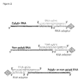
- the RNA splint molecule comprises two domains that are designed to anneal to complementary sequences in the target RNA molecule and the RNA adapter molecule, such that when annealed, the base-paired terminal nucleotide of the target RNA molecule and the adjacent base-paired terminal nucleotide of the RNA adapter molecule are in sufficient proximity to permit their ligation together by means of an RNA ligase (see Figure 2 ).
- the RNA splint molecule comprises a domain whose nucleotide sequence is capable of annealing to the target RNA molecule in the cell lysate.
- An RNA splint molecule designed to anneal with polyA+ RNA molecules comprising a 3' polyA+ tail, will typically comprise a complementary 3' terminal domain having about 20-25 uridine nucleotide residues.
- An RNA splint molecule comprising a 3' terminal uracil domain is particularly suitable for creating a global RBPome encompassing all RBPs in direct contact with RNA polymerase II transcribed RNAs.
- the sequence of the RNA splint molecule may be designed to anneal to a 5' terminal RNA sequence that is specific for a given target RNA molecule. Such a splint would display a 5' terminal sequence complementary to the sequence of the 5' end of the specific target RNA molecule.
- splint ligation could also be utilised to target the specific 3' end sequence of transcripts which are not poly-adenylated (for example, if transcribed by a polymerase other than RNA pol II or if the polyA tail is removed by any cellular process). Accordingly, an RNA splint molecule designed to anneal to the 3' end of a specific non-polyA RNA molecule would display a 3' end sequence complementary to the 3' end sequence of the target non-polyA RNA molecule.
- a specific splint RNA molecule requires that the terminal nucleotide of the 5' or 3' end sequence of the target RNA molecule to which the splint will anneal, is known.
- a new terminal nucleotide in the target RNA molecule can be created artificially by site-specific cleavage of the target RNA, thereby creating a 5' or 3' end sequence to which a complementary splint RNA molecule will anneal.
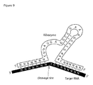
- Site-specific cleavage of the target RNA may be performed in vitro, as shown in Figure 9 , on the product of step c) of the method of the invention.
- Sequence-specific trans-acting hammerhead ribozymes [19] [20] are catalytic RNA oligonucleotides that are used for this reaction.
- the one or more HHRs are added to the cell lysate from step c) where the one or more HHRs each anneal to their respective target RNA molecules, each forming an RNA-duplex, which is then cleaved by the HHR activity.
- Site-specific cleavage of the target RNA creates a new 5' terminal nucleotide in the target RNA molecule, suitable for splint-ligation to the RNA adapter.
- the second domain of the RNA splint molecule is designed to anneal to the RNA adapter molecule.
- the two domains in the RNA splint molecule will be of equal length, i.e. each about 20 - 25 nucleotides.
- the sequence specifically complementary to the target RNA molecule will be the 3' end of the RNA splint.
- the sequence specifically complementary to the target RNA molecule will be the 5' end of the RNA splint.
- the sequence of the second domain of the RNA splint molecule is complementary to the sequence of the RNA adapter molecule.
- the two domains of the RNA splint molecule are contiguous, so that the RNA splint will bring the 3' OH end of the target RNA molecule and the 5' phosphate end of the RNA adapter molecule in close vicinity mandatory for ligation, or the opposite if designed to ligate the 5'end of the target RNA molecule.
- T4 RNA ligase 2 is then used to covalently link the 3' OH group to the 5' phosphate group without requiring any additional molecule except for ATP.
- the RNA adapter molecule is designed to anneal to the RNA splint molecule, and accordingly comprises a sequence complementary to half (second domain) of the RNA splint. If the strategy is to target the 3' end or the 5' end of a given RNA molecule, the RNA adapter molecule will anneal, respectively, to the 5' or the 3' end of the RNA splint molecule.
- the RNA adapter molecule further comprises an affinity group. Suitable affinity groups are known in the art, but a preference is given to molecules displaying strong affinity for a capture agent, such as the affinity group biotin for the capture agent streptavidin, or RNA aptamers for specific peptides.
- the capture agent is generally immobilized by being attached on beads. For example, biotin can be chemically linked to one end of the RNA adapter molecule, while streptavidin is linked to beads. Use of affinity groups having a strong affinity for a capture agent ensures a complete capture of the ligated RNA
- RNA ligase e.g. 10-50U T4 RNA ligase2
- ATP is added to a final concentration about 1mM
- MgCl 2 is added to a final concentration about 10mM.
- Step (e) of the method The ligated target RNA - RNA adapter molecules (e.g. polyA+ RNA-RNA adapter molecules), obtained in step (d), are then captured by means of a capture agent.
- a capture agent is attached to a solid phase, such that the RNA adapter molecules can be captured on a solid phase that can be "washed" free of non-captured molecules.
- the capture agent e.g. streptavidin
- the magnetic beads can be immobilised on a surface by a magnetic field, thereby facilitating their separation from a liquid phase either during washing or elution.
- the capture agent is used to capture the ligated target RNA - RNA adapter molecules formed in the annealing/ligation reaction in the liquid medium comprising the cell lysate and an annealing buffer.
- the incubation conditions chosen for “capture” depend on the mode of capture and the identity of the capture agent employed. Typically “capture” is performed by adding the capture agent to the annealing/ligation liquid reaction mixture in a container and mixing the liquid overnight at 4°C. The captured ligated target RNA - RNA adapter molecules are then washed and the major part of cell lysate and the components added to mediate annealing and ligation are discarded.
- the beads can be captured on the wall of the container by means of a magnet.
- the use of magnetic beads assists the subsequent step of washing the captured target RNA molecules free of previous reaction components.
- the washing steps can be performed by using a capture agent immobilised on a solid surface.
- the washing procedure plays a key role in the specificity of the Rev-CLIP method, since it facilitates the removal of molecules that are neither target RNA (e.g. polyA+ RNA) molecules derived from the cell lysate, nor associated cross-linked RBPs that make up the cell's RBPome.
- target RNA e.g. polyA+ RNA
- Typical sources of contamination are DNA, abundant non-polyA RNA molecules such as ribosomal (rRNA), transfer (tRNA), small nuclear (snRNA), small nucleolar (snoRNA), and micro (miRNA) RNAs and the binding proteins associated with each of these sources of contamination.
- rRNA ribosomal
- tRNA transfer
- snRNA small nuclear
- snoRNA small nucleolar
- miRNA micro
- the washing procedure is designed, not only to remove unbound components of the cell lysate, but even more importantly to destabilise any non-ligated DNA or RNA molecules that have annealed to the RNA adapter or RNA splint molecules. Since the target RNA molecules annealed to the RNA splint molecules have also been ligated to the RNA adapter molecule, these captured RNA molecules cannot be released by the washing procedure.
- a suitable washing procedure comprises sequentially incubating the captured target RNA-RNA adapter molecules in a buffers having a high salt content and then a low salt content (see below), typically at low temperature; combined with exchanging the sample container with a new container. The use of high salt washes is expected to remove non-specific protein interactions, while the use of low salt washes is expected to remove the RNA-RNA or DNA-RNA interactions which occurred during the annealing step.
- wash solutions used in (e) are: i) a High-Salt wash buffer, and 2) a No-Salt wash buffer, each comprising one or more detergents, and ethylenediaminetetraacetic acid (EDTA).
- An example of a suitable series of washing buffers is: 1) a High Salt wash buffer (50mM Tris-HCl, pH 7.4; 1M NaCl; 1mM EDTA; 0.1% SDS; 0.5% Sodium deoxycholate; 1% nonyl phenoxypolyethoxylethanol (NP-40); 2) a No-Salt wash buffer (20mM Tris-HCl Ph7.4; 0.2% Tween 20 [2-[2-[3,4-bis(2-hydroxyethoxy)oxolan-2-yl]-2-(2-hydroxyethoxy)ethoxy]ethyl dodecanoate]); and finally 3) an RNase-compatible wash buffer (10mM Tris-HCl pH 7.4, 150mM NaCl, 0,05% NP40, 2
- Detergent prevents protein-protein interactions so that proteins recovered after elution are bona fide RBPs cross-linked to the target RNA or polyA+ RNA molecules rather than proteins with affinity for any recovered RBPs. Additionally, the presence of reducing agents (e.g. dithiothreitol, DTT) helps to dissociate any tertiary structure of the proteins and thereby decreases the risk of non-RBP proteins forming pockets capable of retaining the target RNA molecules or RNA adapter molecules. 3) Detergents prevent non-specific binding of proteins to the walls of reaction vessels or to capture agent beads. The washing solution components not only contribute to increasing the specificity of the Rev-CLIP method, but also to countering the loss of RBPs during the course of the purification.
- reducing agents e.g. dithiothreitol, DTT
- VRC ribonucleoside vanadyl complex
- VRC is initially added with the lysis buffer to counter the endogenous RNAses but is progressively removed by the successive washes containing EDTA, which also serve to progressively remove endogenous RNases. It is therefore important to maintain an RNase-free environment throughout the REV-CLIP method, since the target RNA molecules lose this protection during the course of the purification, in order to be degraded by the exogenous RNases added in the final elution step.
- step c) DNase treatments can be extensively used in step c) since no DNA per se is used for the capture of the target RNA molecules.
- step (e) the subsequent washes in step (e) are important so that no DNase enzyme remains that might otherwise interfere with an RNaseH-based elution.
- Step (f) of the method The target RNA molecules and their associated RBPs captured from the cell lysate in step (e), can either be treated to release all cellular RBPs referred to as RBPome (according to a first embodiment); or specific species of captured target RNA molecules and their associated RBPs can be released (according to a second embodiment).
- the captured target RNA-RNA adapter molecules are digested with a non-specific RNase, for example RNaseI (EC. 3.1.27.5) or RNaseT1 (EC: 3.1.27.3) at about 37°C, which releases the RBPs associated with all the captured target RNA-RNA adapter molecules.
- RNaseI EC. 3.1.27.5
- RNaseT1 EC: 3.1.27.3
- a selected species of target RNA molecule is released by means of an exogenous RNaseH (EC. 3.1.26.4) guided by the presence of an antisense oligonucleotide.
- an antisense oligonucleotide can be used sequentially to target different regions of the same target RNA molecule or additional species of target RNA molecule, thereby accelerating the throughput of the characterization of these target RNA molecules.
- the present invention provides a new type of antisense oligonucleotide, referred to as gapmet.
- Gapmets are 20 - 30nt oligonucleotides in length (e.g. 20 nt) harboring a 7-12 nt DNA core (e.g.
- the gapmets tend to increase the specificity by guiding RNaseH activity only when most, if not all, of its nucleotide residues are base-paired to target RNA molecules displaying complementary sequences.
- RNA-DNA duplex for the action of RNaseH (which may need to recognize at least 7-8 DNA-RNA paired-bases), although oligonucleotides made only of unmodified DNA would be sufficient.
- RNaseH which may need to recognize at least 7-8 DNA-RNA paired-bases
- oligonucleotides made only of unmodified DNA would be sufficient.
- RNA target molecule may alternatively be performed by providing one or more trans-acting sequence-specific ribozyme (e.g. hammerhead ribozyme) comprising 5' and 3' antisense oligonucleotides, where the antisense oligonucleotides anneal to the ribonucleotides abutting the cleavage site of the selected species of target RNA molecule, to release RNA Binding Proteins bound to the selected species of target RNA molecule.
- sequence-specific ribozyme e.g. hammerhead ribozyme
- RNA-sequencing or RT-PCR RNA-sequencing or RT-PCR.
- the RNA elution can be performed by proteolytically digesting the affinity group-capture agent (e.g. biotin and streptavidin) as well as the associated RBPs by proteinase K treatment (EC. 3.4.21.62).
- a mild RNA digestion using low amounts of non-specific RNase can favour the RNA elution since the streptavidin core interacting with biotin can be difficult to degrade proteolytically.
- the released target RNA molecules can be selectively extracted by phenol/chloroform separation and recovered by ethanol precipitation.
- the RBPs obtained according to the first embodiment of step (f) represents the cell's genome-scale RBPome, which can be analysed and identified, for example by Mass Spectrometry.
- the RBPs obtained according to the second embodiment of step (f) represent the group of RBPs playing a role in the processing, expression or function of specific species of target RNA molecule (e.g. polyA+ RNA), which can also be analysed and identified, for example by Mass Spectrometry.
- target RNA molecule e.g. polyA+ RNA
- the Rev-CLIP method has been applied to isolate the RBPome of HEK and MIN6 cells, and the composition of the isolated RBPomes has been determined and compared with the RBPome isolated using methods in the prior art.
- the Rev-CLIP method is shown to provide a surprising improvement over the IC method, particularly in respect of the selectivity for RBPs associated with polyA+ RNA molecules; and the low level of contamination with proteins known to bind to DNA and non-polyA or non-targeted RNA molecules.
- RevCLIP allows the identification of RBPs directly involved in the response to glucose uptake, thereby identifying protein targets relevant for treatment of type II diabetes.
- RNA-specific Rev-CLIP has been performed on HEK cells by targeting 4 candidates lincRNAs in order to characterise the direct RBP protein partners.
- the Rev-CLIP method can be used to isolate the RBPome of all cells that can be cultured ex-vivo and in vitro, including cells of eukaryotic and prokaryotic origin.
- the Rev-CLIP method can be applied to bacterial, mycobacterial and yeast cells. Since an analysis of the RBPome can be used to identify future therapeutic targets, the Rev-CLIP method is advantageously applied to cells of mammalian origin. In particular, Rev-CLIP method may be applied to any cell type, for example, of mouse, rat or human origin.
- Example 1 Construction and in vitro testing of a biotinylated RNA adapter and splint for capturing polyA+ RNA
- a biotinylated RNA adapter in combination with a splint RNA were synthesized and tested for use in the selective capture of polyadenylated RNA.
- the RNA adapter and splint were tested using an artificially poly-adenylated RNA as a mimic for polyA+ RNAs present in a cell lysate.
- the artificial polyA+ RNA comprised a 53nt long RNA oligonucleotide harboring a 3' tail of 25 Adenine residues, having the sequence:
- RNA adapter The sequence of the RNA adapter and RNA splint are as follows:
- RNA 5'-AUGCCUUGGCACCCGAGAAUUCCA(U) 25 -3' [SEQ ID No: 3]
- RNA Hammerhead ribozyme: 5'-(Stem I)-CUGAUGAGGCCGAAAGGCCGAAA-(Stem III)-3' [SEQ ID No:16] where Stem I is 4-10 nucleotides complementary to the sequence of the target RNA molecule that is 3' of the cleavage site and Stem III, starting with C or U, that contains 4-10 additional nucleotides complementary to the target RNA molecule that is 5' of the cleavage site C, where the cleavage site is unbasepaired and lies between Stem I and Stem III (see Figure 9 ).
- RNA capture and splint-ligation were tested by preparing a series of annealing mixes, where one or more components required for polyA+ RNA capture and ligation were omitted.
- An annealing reaction was performed to anneal the RNA adapter to the RNA splint as follows: 50pmol of the biotinylated RNA adapter was combined with 50pmol of the splint RNA in 30 ⁇ l total annealing buffer (75mM KCI; 20mM TrisHCl pH7.5). This first annealing reaction mix was incubated for 1min at 85°C, then for 3min at 65°C, 10min at 37°C, and finally on ice for 5min.
- RNA adapter-splint was then annealed to the artificial polyA+ RNA by combining 30 ⁇ l of the first annealing reaction with 50pmol artificial polyA+ RNA (suspended in 40 ⁇ l) in a 1.5ml tube and incubating this second annealing reaction mixture for 3 min at 55°C, then 10 min at 37°C, and finally 10 min on ice.
- the products of the second annealing reaction were then ligated by adding 30U of T4RNA ligase2 (supplied by MClab) in a ligation reaction comprising 1mM ATP, 10mM MgCl2, followed by incubation at 37°C for 2h.
- the ligation reaction products were then denatured for 2min at 90°C in formamide blue (30% deionized formamide; 6mM EDTA; bromophenol blue) and analysed by separation on a denaturing 15% urea PAGE pre-warmed at 50°C.
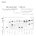
- a ligation reaction product (RB) whose migration corresponds to the expected size of polyA+ RNA (R) ligated to the RNA adapter was detected following PAGE separation under denaturing conditions.
- This ligated product was only obtained when the polyA+ RNA (R) annealing steps were performed in the presence of both the RNA adapter (B) and the RNA splint (S), and the ligation step was performed in the presence of T4 RNA ligase2.
- the T4 RNA ligase2 could not ligate the RNA adapter to the single stranded polyA+ RNA, but instead required the presence of the RNA splint.
- Example 2 The Rev-CLIP method was tested and shown to capture polyA+ RNA-bound RBPs in cell lysates.
- the Rev-CLIP method was applied to Human Embryonic Kidney (HEK) cells and tested for its ability to capture specific polyA+ RNA-bound RBPs known to be present in the RBPome of HEK cells.
- HEK cell culture A culture of HEK cells was obtained by culturing HEK cells in pyruvate buffered Dulbecco's Modified Eagle's Medium (DMEM) supplemented with 10% heat inactivated FBS (supplied by Gibco), Penicillin-Streptomycin and L-glutamin, in Nunclon Delta-treated flasks (supplied by Thermoscientific) or TC treated dishes (supplied by Corning).
- DMEM Dulbecco's Modified Eagle's Medium
- FBS heat inactivated FBS
- Penicillin-Streptomycin and L-glutamin in Nunclon Delta-treated flasks (supplied by Thermoscientific) or TC treated dishes (supplied by Corning).
- the splint ligation was performed in two steps:
- Membranes are re-blotted using stripping buffer (supplied by Pierce). Transferred proteins were screened with the following antibodies: anti-ßactin (1:10,000, mouse, A2228, Sigma), anti-CELF1 (1:5,000, rabbit, ab129115, Abcam), anti-PTBP1 (1:10,000, rabbit, ab133734, Abcam), HRP-anti-mouse IgG (1:20,000, 115-035-003, supplied by Jackson Immuno Europe), HRP-anti-rabbit IgG (1:10,000, 111-035-008, supplied by Jackson Immuno Europe) using the standard Western blot protocol.
- anti-ßactin (1:10,000, mouse, A2228, Sigma
- anti-CELF1 (1:5,000, rabbit, ab129115, Abcam
- anti-PTBP1 (1:10,000, rabbit, ab133734, Abcam
- HRP-anti-mouse IgG (1:20,000, 115-035-003, supplied by Jackson Immuno Europe
- HRP-anti-rabbit IgG (1:10,000, 111
- Horse Radish Peroxidase (HRP) signal was revealed by use of SuperSignal West Femto Maximum Sensitivity kit (supplied by Pierce) on a G:box chemi XR5.
- the Silver Staining was carried out on a duplicate NuPAGE 4-12% gradient gel loaded with the same samples using Sigma Silver Staining Kit.
- each of the proteins ⁇ -actin, polypyrimidine tract binding protein 1 (PTBP1) and CUG Triplet Repeat, RNA-Binding Protein 1 (CELF1) were detected in lysates of HEK cells.
- the two RBPs, PTBP1 and CELF1, present in the cell lysate were also selectively detected amongst the proteins captured by the Rev-CLIP method, which as expected did not include ⁇ -actin.
- the capture of the two RBPs by the Rev-CLIP method was dependent on employing the RNA adapter, the RNA splint and the T4 ligase2, and omission of any one of these components prevented the capture of these RBPs.
- a harsh elution was additionally performed after the elution in step f), by exposing all the remaining beads to 30 ⁇ l SDS loading buffer and by shaking at 800rpm for 30min at 70°C. This harsh elution entirely degrades streptavidin and therefore shows what was captured but not properly eluted from the beads by the previous step f), as well as what remained attached to the beads, mediated by RNA-independent non-specific interaction with the beads.
- molecules of rRNA, tRNA, snRNA, snoRNA and miRNA should not be captured by the RNA splint-ligation used for polyA+ capture as they lack the required long stretch of A residues at their 3' end. Since the cell lysate, used in the Rev-CLIP method, is subjected to extensive DNase treatment, the risk of recovery of DNA fragments and their associated proteins binding are drastically reduced.
- the Rev-CLIP method is performed on UV-exposed cells. The effect of this UV exposure on the results of Rev-CLIP method was analysed by the comparison of samples prepared under two conditions, UV or non-UV exposed cells.
- UV-cross-linking of cells in step (a) of Rev-CLIP is performed to covalently bind RBPs to RNA under physiological conditions.
- the Rev-CLIP method uses harsh washes (detergents, reducing agents and high salt buffer) that denature all the proteins, and under these conditions, RBPs would lose their ability to bind to polyA+ RNAs during the purification. Only those RBPs which have been previously cross-linked to polyA+ RNA can thus be captured.
- the non-UV exposed cells are used as a negative control, where RBPs should not be retained due to the denaturing conditions and the stringent washes.
- the recovered proteins are expected to be proteins that are very abundant, or to be proteins that retain the ability to stick to RNA when denatured, or simply proteins with an affinity for the beads, biotin, streptavidin and/or the plastic of the microcentrifuge tubes. For instance, proteins which may employ biotin as a cofactor might be recovered for that reason (only ten proteins are known to have such properties in humans).
- the proteins recovered from UV-exposed cell samples outnumber those recovered from non-UV exposed cell samples ( Figure 5A ).
- the stained gel in Figure 5A clearly shows a greater amount of protein bands released in the RNase elution buffer than from the UV-cross-linked samples.
- the Western blot analysis confirms the presence of RBPs, such as the two RBPs, PTBP1 and CELF1, in the lane comprising the UV-exposed sample, but not in the lane comprising the non-UV exposed sample.
- the Silver Staining and the Western Blot methods have a detection threshold that is much higher than that of Mass Spectrometry (MS). Nonetheless, as demonstrated in Example 3, MS detects about five times more RBP proteins when Rev-CLIP is performed on UV-exposed cell samples versus non-UV exposed samples.
- the RBPome was isolated from both HEK cells, as well as mouse insulinoma-derived ⁇ -cell line26 cells (MIN6), using the Rev-CLIP method, as described in Example 2.
- the MIN6 cells were grown in DMEM containing 50 ⁇ M ß-mercaptoethanol. The glucose concentration of the medium was adjusted to 3mM (Low), 25mM (High) or was 22mM if not mentioned.
- the proteome and component RBPs isolated using Rev-CLIP (samples obtained in step (f) of Rev-CLIP) were characterised and quantitated using the methods set out below.
- Protein pellets (10 ⁇ g) were dissolved in 22 ⁇ l buffer D (8M Urea; 50mM Tris-HCl pH 8.0; 45mM DTT) and incubated 45min at room temperature. Then 4 ⁇ l freshly prepared 500mM IodoAcetic Acid (IAA) were added before incubation for 1h at room temperature in the dark. Trypsin digestion was performed by addition of 74 ⁇ l of 50mM Tris-HCl pH 8.0 and 2 ⁇ l 0.1 ⁇ g/ ⁇ l trypsin dissolved in 10mM HCl, in order to get a protein/trypsin ratio of 50:1, and incubated overnight at 25°C.
- buffer D 8M Urea; 50mM Tris-HCl pH 8.0; 45mM DTT
- IAA IodoAcetic Acid
- the digestion was stopped by addition of 2 ⁇ l of 10% trifluoroacetic acid (TFA).
- TFA trifluoroacetic acid
- the protein sample was concentrated using the speedvac and acidified with 2 ⁇ l 10% TFA to reach pH ⁇ 2.
- a C18 stage tipping protocol was applied.
- C18 filters were placed carefully at the very end of a DL-10 Diamond tower pack 10 ⁇ l tips using a sampling tool syringe.
- C18 filters were activated by rinsing them sequentially with wetting/elution solution (60% acetonitrile, 0.1% TFA) and equilibration solution (0.1% TFA) removed by short spin at max 4,000rpm. 5 ⁇ g of peptides were rapidly loaded with a 20sec spin at 3,000rpm.
- the filters were washed twice with 20 ⁇ l equilibration solution and stored at 4°C in a 50ml Falcon tube with 50 ⁇ l equilibration solution until MS is performed.
- the peptide samples were eluted slowly with 10 ⁇ l wetting/elution solution, using a syringe, and kept only shortly at 4°C before MS.
- the 10 ⁇ l were almost completely dried using the speedvac.
- 2.5 ⁇ l of 2% formic acid were added and the volume adjusted to 10 ⁇ l with water.
- a sample of 0.5 ⁇ l was used for Maldi analysis and the remainder for LC-MS.
- LC-MS / MS analysis Samples, prepared as above, were analyzed by liquid chromatography tandem mass spectrometry (LC-MS/MS) and data were recorded in a data dependent manner, automatically switching between MS and MS/MS acquisition, on a quadrupole mass filter instrument with an Orbitrap mass analyzer (Q-Exactive, Thermo scientific, Bremen, Germany).
- the liquid chromatography system was coupled to the mass spectrometer through an EASY spray source operated at 2.1kV with no sheath or auxiliary gas applied a heated capillary temperature of 275°C and an S-lens RF at 50. Liquid chromatography was performed on an EASY nLC-1000 (Thermo scientific, Odense, Denmark).
- EASY-spray columns (Thermo scientific ES803) were used to online separate the peptides prior to analysis. These columns have an inner diameter of 75 ⁇ m and are packed with reverse phase C18 material with 2 ⁇ m size particles. During elution an in build column oven kept the temperature of the column at 45°C.
- the mobile phase consisted of Solvent A containing 0.1% formic acid and the peptides were eluted by an increasing gradient of Solvent B containing 80% acetonitrile in 0.1% formic acid.
- the total gradient was 130min and all changes of solvent mixtures were done in a linear fashion.
- the Solvent B concentration at the start of the gradient was 6% hereafter the gradient was as follows: 14%B in 37min, 25%B in 42min, 38%B in 21min, 60%B in 20min, 95%B in 3min and 95%B for 7min.
- the full scans were acquired in the Orbitrap with a resolution of 70,000 and a predictive automatic gain control (AGC) of 3,000,000 based on the AGC from the previous full scan. If this was not reached, a maximum injection time of 20ms was applied. For the full scans the scan range was adjusted to 300-1750 m/z.
- AGC automatic gain control
- the top 10 most abundant ions from the full scan were further sequentially selected for fragmentation and MS/MS analysis with an isolation window of 1.6 m/z and a minimum intensity threshold of 100,000. Ions with unassigned charge states, charge states of 1 and isotopes of ions were excluded from selection for MS/MS analysis. Further analyzed ions were excluded from reselection for a 30 sec time period. For the MS/MS scans the resolution was adjusted to 35,000 with a predictive AGC of 1,000,000, a maximum injection time of 60ms and a fixed first mass of 120m/z. The ions were fragmented in a higher-energy collision dissociation (HCD) cell with normalized collision energy (NCE) of 25%.
- HCD collision dissociation
- NCE normalized collision energy
- MaxQuant (v1.5.2.12) was used to analyze the data. Peptides were compared against the ENSEMBL release81 GRCh38 human and Swissprot mouse proteome database for the HEK and MIN6 cells, respectively. Default parameters were used: FDR of 0.01, peptides with a minimum length of seven amino acids, cysteine carbamidomethylation set as a fixed modification, N-terminal acetylation and methionine oxidation set as variable modifications. Label-Free-Quantification (LFQ) is used. Only peptides uniquely mapping to one protein group were used for further analysis. Quantification was performed when at least one unique peptide was detected. Ion counts from three technical replicates were pooled and pseudocounts were used to calculate ratios between experiments including or no UV-crosslinking.
- LFQ Label-Free-Quantification
- the Rev-CLIP performed in HEK and MIN6 detected, respectively, a total of 639 and 648 proteins which passed the selected cutoffs set for peptide counts (at least 2 peptides detected in the 3 biological replicates) and the fold change between UV versus non-UV exposed samples (the averaged peptide counts log2FC must be above 0.5 and the averaged LFQ intensities FC must be above 2).
- the HEK and MIN6 RBPomes identified herein show a large overlap with a database of RBD-containing proteins (RBPDB, [17]) and "RNA binding" as GO terms ( figure 6 ).
- Rev-CLIP RNA interactome capture technique
- Rev-CLIP enables the isolation of a population of proteins that bind to polyA+ RNA, comprising fewer proteins known to bind to non-polyA RNA (e.g.rRNA, tRNA, snoRNA, snRNA and miRNA) or DNA, as compared to proteins isolated by the IC method.
- non-polyA RNA e.g.rRNA, tRNA, snoRNA, snRNA and miRNA
- the RBPs bound to a specific polyA+ RNA molecule can be selectively released using the specific Rev-CLIP method. According to this alternative method, all polyA+ RNAs are captured as previously described, but the elution of RBPs is limited to those bound to a given polyA+ RNA molecule.
- step (f) The specific Rev-CLIP method shares the same steps (a)-(e) with the Rev-CLIP set out in Example 2.
- the method only diverges in step (f), at the stage of washing the beads, which was performed in buffer R' (50mM TrisHCl, pH7.5; 100mM NaCl; 3mM MgCl2).
- a first wash (and controlled elution) of the beads was performed by addition of 550 ⁇ l Buffer R'; with mild shaking on wheel at 4°C for 5min, followed by addition of 20U RNaseH (MCLab) (8 ⁇ l).
- the tubes were incubated further on a wheel at 45°C for 15min, the beads were captured on a magnetic stand, and the eluate fraction collected in a tube named "elution control".
- the beads were then washed successively - twice for 5min at 45°C on wheel with No-Salt buffer; and then one time for 5min at 4°C; one time with High Salt buffer, and finally one time with buffer R'.
- the tubes were changed between washes.
- the specific elution was then carried out as follows: The beads were re-suspended in 400 ⁇ l buffer R' and put on wheel at 45°C for 5min. Then, 30pmol of a given RNA-DNA hybrid oligonucleotide ("gapmet", see above) was added to the re-suspended beads, and the tubes were placed on wheel at 45°C for 5min. Then 20U RNaseH were added and the tubes were incubated on the wheel at 45°C for 15min. The specific elution fractions were finally collected. A supplementary elution was performed by adding 150 ⁇ l of Buffer R' on beads, incubated again at 45°C for 5min and collected into the first collected fraction.
- gapmet RNA-DNA hybrid oligonucleotide
- RNAs were digested in presence of 200U of RNase I plus 1000U of RNase T1 at 37°C for 20min and precipitated with 5 volumes of cold acetone. One part of the fraction (usually one tenth) was kept for RNA analysis after digestion of proteins with proteinase K as previously described.
- the specific Rev-CLIP method was applied to 4 RNA species: NEAT1, HOTAIR, XIST and MALAT1, employing 3 different gapmets for each of these 4 RNAs.
- the sequences of the gapmets were:
Landscapes
- Genetics & Genomics (AREA)
- Engineering & Computer Science (AREA)
- Chemical & Material Sciences (AREA)
- Health & Medical Sciences (AREA)
- Life Sciences & Earth Sciences (AREA)
- Zoology (AREA)
- Wood Science & Technology (AREA)
- Organic Chemistry (AREA)
- Biotechnology (AREA)
- General Engineering & Computer Science (AREA)
- Biomedical Technology (AREA)
- Bioinformatics & Cheminformatics (AREA)
- Physics & Mathematics (AREA)
- Molecular Biology (AREA)
- Microbiology (AREA)
- Crystallography & Structural Chemistry (AREA)
- Plant Pathology (AREA)
- Biochemistry (AREA)
- General Health & Medical Sciences (AREA)
- Biophysics (AREA)
- Measuring Or Testing Involving Enzymes Or Micro-Organisms (AREA)
Priority Applications (1)
| Application Number | Priority Date | Filing Date | Title |
|---|---|---|---|
| EP16189688.1A EP3296397A1 (de) | 2016-09-20 | 2016-09-20 | Rev-clip, ein neuer ansatz zum identifizieren von mit rna interagierenden proteinen |
Applications Claiming Priority (1)
| Application Number | Priority Date | Filing Date | Title |
|---|---|---|---|
| EP16189688.1A EP3296397A1 (de) | 2016-09-20 | 2016-09-20 | Rev-clip, ein neuer ansatz zum identifizieren von mit rna interagierenden proteinen |
Publications (1)
| Publication Number | Publication Date |
|---|---|
| EP3296397A1 true EP3296397A1 (de) | 2018-03-21 |
Family
ID=57046977
Family Applications (1)
| Application Number | Title | Priority Date | Filing Date |
|---|---|---|---|
| EP16189688.1A Withdrawn EP3296397A1 (de) | 2016-09-20 | 2016-09-20 | Rev-clip, ein neuer ansatz zum identifizieren von mit rna interagierenden proteinen |
Country Status (1)
| Country | Link |
|---|---|
| EP (1) | EP3296397A1 (de) |
Cited By (3)
| Publication number | Priority date | Publication date | Assignee | Title |
|---|---|---|---|---|
| CN113583982A (zh) * | 2020-04-30 | 2021-11-02 | 香港城市大学深圳研究院 | 确定长链非编码核糖核酸相互作用蛋白的新方法 |
| CN114106109A (zh) * | 2021-11-25 | 2022-03-01 | 嘉华药锐科技(北京)有限公司 | 一种细胞质靶向的rna结合蛋白富集试剂及其制备方法及应用 |
| CN119265174A (zh) * | 2024-12-09 | 2025-01-07 | 天津凯莱英生物科技有限公司 | 用于催化rna连接反应的固定化酶及其制备方法和应用 |
-
2016
- 2016-09-20 EP EP16189688.1A patent/EP3296397A1/de not_active Withdrawn
Non-Patent Citations (24)
| Title |
|---|
| ASCANO M; GERSTBERGER S; TUSCHL T: "Multi-disciplinary methods to define RNA-protein interactions and regulatory networks", CURR OPIN GENET DEV, vol. 23, 2013, pages 20 - 8 |
| BALTZ AG; MUNSCHAUER M; SCHWANHAUSSER B ET AL.: "The mRNA-bound proteome and its global occupancy profile on protein-coding transcripts", MOL CELL, vol. 46, 2012, pages 674 - 90, XP028522157, DOI: doi:10.1016/j.molcel.2012.05.021 |
| BECKMANN BM; HOROS R; FISCHER B ET AL.: "The RNA-binding proteomes from yeast to man harbour conserved enigmRBPs", NAT COMMUN, vol. 6, 2015, pages 10127 |
| BERNSTEIN DS; BUTER N; STUMPF C; WICKENS M.: "Analyzing mRNA-protein complexes using a yeast three-hybrid system", METHODS, vol. 26, 2002, pages 123 - 41, XP002985923, DOI: doi:10.1016/S1046-2023(02)00015-4 |
| BUTTER F; SCHEIBE M; MORL M; MANN M: "Unbiased RNA-protein interaction screen by quantitative proteomics", PROC NATL ACAD SCI U S A, vol. 106, 2009, pages 10626 - 31, XP055069956, DOI: doi:10.1073/pnas.0812099106 |
| CASTELLO A; FISCHER B; EICHELBAUM K ET AL.: "Insights into RNA biology from an atlas of mammalian mRNA-binding proteins", CELL, vol. 149, 2012, pages 1393 - 406, XP028521125, DOI: doi:10.1016/j.cell.2012.04.031 |
| COMOGLIO FEDERICO ET AL: "Sensitive and highly resolved identification of RNA-protein interaction sites in PAR-CLIP data", BMC BIOINFORMATICS, vol. 16, February 2015 (2015-02-01), XP002764251 * |
| CONRAD T; ALBRECHT AS; DE MELO COSTA VR; SAUER S; MEIERHOFER D; OROM UA: "Serial interactome capture of the human cell nucleus", NAT COMMUN, vol. 7, 2016, pages 11212 |
| COOK KB; KAZAN H; ZUBERI K; MORRIS Q; HUGHES TR: "RBPDB: a database of RNA-binding specificities", NUCLEIC ACIDS RES, vol. 39, 2010, pages D301 - 8 |
| DARNELL RB: "HITS-CLIP: panoramic views of protein-RNA regulation in living cells", WILEY INTERDISCIP REV RNA, vol. 1, 2010, pages 266 - 86 |
| FAORO C; ATAIDE SF: "Ribonomic approaches to study the RNA-binding proteome", FEBS LETT, vol. 588, 2014, pages 3649 - 64, XP029072949, DOI: doi:10.1016/j.febslet.2014.07.039 |
| FAORO CAMILLA ET AL: "Ribonomic approaches to study the RNA-binding proteome", FEBS LETTERS, ELSEVIER, AMSTERDAM, NL, vol. 588, no. 20, 19 August 2014 (2014-08-19), pages 3649 - 3664, XP029072949, ISSN: 0014-5793, DOI: 10.1016/J.FEBSLET.2014.07.039 * |
| FERRE FABRIZIO ET AL: "Revealing protein-lncRNA interaction", BRIEFINGS IN BIOINFORMATICS, vol. 17, no. 1, Sp. Iss. SI, January 2016 (2016-01-01), pages 106 - 116, XP002764250 * |
| FORSTER AC; SYMONS RH: "Self-cleavage of plus and minus RNAs of a virusoid and a structural model for the active sites", CELL, vol. 49, no. 2, 24 April 1987 (1987-04-24), pages 211 - 20, XP023884042, DOI: doi:10.1016/0092-8674(87)90562-9 |
| KONIG J; ZARNACK K; LUSCOMBE NM; ULE J: "Protein-RNA interactions: new genomic technologies and perspectives", NAT REV GENET, vol. 13, 2012, pages 77 - 83, XP002732730, DOI: doi:10.1038/nrg3141 |
| KWON SC; YI H; EICHELBAUM K ET AL.: "The RNA-binding protein repertoire of embryonic stem cells", NAT STRUCT MOL BIOL, vol. 20, 2013, pages 1122 - 30 |
| LIEPELT A; NAARMANN-DE VRIES IS; SIMONS N ET AL.: "Identification of RNA-binding Proteins in Macrophages by Interactome Capture", MOL CELL PROTEOMICS, vol. 15, 2016, pages 2699 - 714 |
| MITCHELL SF; JAIN S; SHE M; PARKER R: "Global analysis of yeast mRNPs", NAT STRUCT MOL BIOL, vol. 20, 2013, pages 127 - 33 |
| PASHEV IG; DIMITROV SI; ANGELOV D: "Crosslinking proteins to nucleic acids by ultraviolet laser irradiation", TRENDS BIOCHEM SCI, vol. 16, 1991, pages 323 - 6, XP025864788, DOI: doi:10.1016/0968-0004(91)90133-G |
| PLEY HW1; FLAHERTY KM; MCKAY DB: "Three-dimensional structure of a hammerhead ribozyme", NATURE, vol. 372, no. 6501, 3 November 1994 (1994-11-03), pages 68 - 74 |
| RILEY KJ; STEITZ JA: "Observer Effect'' in genome-wide surveys of protein-RNA interactions", MOL CELL, vol. 49, 2013, pages 601 - 4, XP028985601, DOI: doi:10.1016/j.molcel.2013.01.030 |
| SIPRASHVILI Z; WEBSTER DE; KRETZ M ET AL.: "Identification of proteins binding coding and non-coding human RNAs using protein microarrays", BMC GENOMICS, vol. 13, 2012, pages 633, XP021138577, DOI: doi:10.1186/1471-2164-13-633 |
| ULE J; JENSEN KB; RUGGIU M; MELE A; ULE A; DARNELL RB: "CLIP identifies Nova-regulated RNA networks in the brain", SCIENCE, vol. 302, 2003, pages 1212 - 5 |
| ZHANG YIN ET AL: "CLIP: viewing the RNA world from an RNA-protein interactome perspective", SCIENCE CHINA LIFE SCIENCES, ZHONGGUO KEXUE ZAZHISHE, CHINA, vol. 58, no. 1, 10 January 2015 (2015-01-10), pages 75 - 88, XP035710997, ISSN: 1674-7305, [retrieved on 20150110], DOI: 10.1007/S11427-014-4764-5 * |
Cited By (5)
| Publication number | Priority date | Publication date | Assignee | Title |
|---|---|---|---|---|
| CN113583982A (zh) * | 2020-04-30 | 2021-11-02 | 香港城市大学深圳研究院 | 确定长链非编码核糖核酸相互作用蛋白的新方法 |
| CN113583982B (zh) * | 2020-04-30 | 2023-06-27 | 香港城市大学深圳研究院 | 确定长链非编码核糖核酸相互作用蛋白的新方法 |
| CN114106109A (zh) * | 2021-11-25 | 2022-03-01 | 嘉华药锐科技(北京)有限公司 | 一种细胞质靶向的rna结合蛋白富集试剂及其制备方法及应用 |
| CN114106109B (zh) * | 2021-11-25 | 2024-06-04 | 嘉华药锐科技(北京)有限公司 | 一种细胞质靶向的rna结合蛋白富集试剂及其制备方法及应用 |
| CN119265174A (zh) * | 2024-12-09 | 2025-01-07 | 天津凯莱英生物科技有限公司 | 用于催化rna连接反应的固定化酶及其制备方法和应用 |
Similar Documents
| Publication | Publication Date | Title |
|---|---|---|
| Yap et al. | Hybridization-proximity labeling reveals spatially ordered interactions of nuclear RNA compartments | |
| Backlund et al. | Plasticity of nuclear and cytoplasmic stress responses of RNA-binding proteins | |
| Chung et al. | The use of duplex-specific nuclease in ribosome profiling and a user-friendly software package for Ribo-seq data analysis | |
| US20120040857A1 (en) | Isolation of factors that associate directly or indirectly with chromatin | |
| WO2023221842A1 (zh) | 一种高通量单细胞转录组测序方法及其应用 | |
| Gong et al. | Biochemical analysis of long non-coding RNA-containing ribonucleoprotein complexes | |
| JP7733642B2 (ja) | シーケンシングのためのrna試料を調製する方法およびそのキット | |
| Aeby et al. | Decapping enzyme 1A breaks X-chromosome symmetry by controlling Tsix elongation and RNA turnover | |
| CN107109698B (zh) | Rna stitch测序:用于直接映射细胞中rna:rna相互作用的测定 | |
| Bartoli et al. | Conserved methyltransferase Spb1 targets mRNAs for regulated modification with 2′-O-methyl ribose | |
| EP3296397A1 (de) | Rev-clip, ein neuer ansatz zum identifizieren von mit rna interagierenden proteinen | |
| Wolin et al. | SPIDR: a highly multiplexed method for mapping RNA-protein interactions uncovers a potential mechanism for selective translational suppression upon cellular stress | |
| Hofman et al. | Nuclear Argonaute: miRNA complexes recognize target sequences within chromatin-associated RNA and silence gene expression | |
| Kastelic et al. | mRNA interactome capture in mammalian cells | |
| JP7313696B2 (ja) | 増強されたRNAインタラクトーム捕捉(eRIC) | |
| Silverman et al. | Transcriptome-wide ribonuclease-mediated protein footprinting to identify RNA–protein interaction sites | |
| Simon et al. | Principles and practices of hybridization capture experiments to study long noncoding RNAs that act on chromatin | |
| EP2783001B1 (de) | Multiplexisolierung von proteinassoziierten nukleinsäuren | |
| MC MK et al. | 2800 Kgs. Lyngby (DK) | |
| Trendel et al. | The human RNA-binding proteome and its dynamics during arsenite-induced translational arrest | |
| Cottrell et al. | Urb-RIP–an adaptable and efficient approach for immunoprecipitation of RNAs and associated RNAs/proteins | |
| EP2744902B1 (de) | Isolierung von faktoren zur direkten oder indirekten assoziation mit nicht-kodierenden rnas | |
| Desideri | Investigating the role of Charme: a chromatin-associated lncRNA supervisor of mammalian myogenesis | |
| US20230408504A1 (en) | Antibody barcoded beads and uses thereof | |
| US20250034552A1 (en) | COMPOSITIONS AND METHODS FOR ISOLATION OF RIBOSOME PROTECTED MESSENGER RNA (mRNA) FOOTPRINTS OR FRAGMENTS AND KITS THEREOF |
Legal Events
| Date | Code | Title | Description |
|---|---|---|---|
| PUAI | Public reference made under article 153(3) epc to a published international application that has entered the european phase |
Free format text: ORIGINAL CODE: 0009012 |
|
| AK | Designated contracting states |
Kind code of ref document: A1 Designated state(s): AL AT BE BG CH CY CZ DE DK EE ES FI FR GB GR HR HU IE IS IT LI LT LU LV MC MK MT NL NO PL PT RO RS SE SI SK SM TR |
|
| AX | Request for extension of the european patent |
Extension state: BA ME |
|
| STAA | Information on the status of an ep patent application or granted ep patent |
Free format text: STATUS: THE APPLICATION IS DEEMED TO BE WITHDRAWN |
|
| 18D | Application deemed to be withdrawn |
Effective date: 20180922 |