EP3290517B1 - Method for predicting therapeutic effect of pd-1/pd-l1 inhibitor using abnormality in pd-l1(cd274) as index - Google Patents
Method for predicting therapeutic effect of pd-1/pd-l1 inhibitor using abnormality in pd-l1(cd274) as index Download PDFInfo
- Publication number
- EP3290517B1 EP3290517B1 EP16786551.8A EP16786551A EP3290517B1 EP 3290517 B1 EP3290517 B1 EP 3290517B1 EP 16786551 A EP16786551 A EP 16786551A EP 3290517 B1 EP3290517 B1 EP 3290517B1
- Authority
- EP
- European Patent Office
- Prior art keywords
- abnormality
- gene
- expression
- utr
- cancer
- Prior art date
- Legal status (The legal status is an assumption and is not a legal conclusion. Google has not performed a legal analysis and makes no representation as to the accuracy of the status listed.)
- Active
Links
Images


Classifications
-
- G—PHYSICS
- G01—MEASURING; TESTING
- G01N—INVESTIGATING OR ANALYSING MATERIALS BY DETERMINING THEIR CHEMICAL OR PHYSICAL PROPERTIES
- G01N33/00—Investigating or analysing materials by specific methods not covered by groups G01N1/00 - G01N31/00
- G01N33/48—Biological material, e.g. blood, urine; Haemocytometers
- G01N33/50—Chemical analysis of biological material, e.g. blood, urine; Testing involving biospecific ligand binding methods; Immunological testing
- G01N33/53—Immunoassay; Biospecific binding assay; Materials therefor
- G01N33/575—Immunoassay; Biospecific binding assay; Materials therefor for cancer
- G01N33/57557—Immunoassay; Biospecific binding assay; Materials therefor for cancer of other specific parts of the body, e.g. brain
-
- C—CHEMISTRY; METALLURGY
- C12—BIOCHEMISTRY; BEER; SPIRITS; WINE; VINEGAR; MICROBIOLOGY; ENZYMOLOGY; MUTATION OR GENETIC ENGINEERING
- C12Q—MEASURING OR TESTING PROCESSES INVOLVING ENZYMES, NUCLEIC ACIDS OR MICROORGANISMS; COMPOSITIONS OR TEST PAPERS THEREFOR; PROCESSES OF PREPARING SUCH COMPOSITIONS; CONDITION-RESPONSIVE CONTROL IN MICROBIOLOGICAL OR ENZYMOLOGICAL PROCESSES
- C12Q1/00—Measuring or testing processes involving enzymes, nucleic acids or microorganisms; Compositions therefor; Processes of preparing such compositions
- C12Q1/68—Measuring or testing processes involving enzymes, nucleic acids or microorganisms; Compositions therefor; Processes of preparing such compositions involving nucleic acids
- C12Q1/6876—Nucleic acid products used in the analysis of nucleic acids, e.g. primers or probes
- C12Q1/6883—Nucleic acid products used in the analysis of nucleic acids, e.g. primers or probes for diseases caused by alterations of genetic material
- C12Q1/6886—Nucleic acid products used in the analysis of nucleic acids, e.g. primers or probes for diseases caused by alterations of genetic material for cancer
-
- A—HUMAN NECESSITIES
- A61—MEDICAL OR VETERINARY SCIENCE; HYGIENE
- A61K—PREPARATIONS FOR MEDICAL, DENTAL OR TOILETRY PURPOSES
- A61K39/00—Medicinal preparations containing antigens or antibodies
- A61K39/395—Antibodies; Immunoglobulins; Immune serum, e.g. antilymphocytic serum
-
- A—HUMAN NECESSITIES
- A61—MEDICAL OR VETERINARY SCIENCE; HYGIENE
- A61K—PREPARATIONS FOR MEDICAL, DENTAL OR TOILETRY PURPOSES
- A61K45/00—Medicinal preparations containing active ingredients not provided for in groups A61K31/00 - A61K41/00
-
- C—CHEMISTRY; METALLURGY
- C07—ORGANIC CHEMISTRY
- C07K—PEPTIDES
- C07K16/00—Immunoglobulins [IG], e.g. monoclonal or polyclonal antibodies
- C07K16/18—Immunoglobulins [IG], e.g. monoclonal or polyclonal antibodies against material from animals or humans
- C07K16/28—Immunoglobulins [IG], e.g. monoclonal or polyclonal antibodies against material from animals or humans against receptors, cell surface antigens or cell surface determinants
- C07K16/2803—Immunoglobulins [IG], e.g. monoclonal or polyclonal antibodies against material from animals or humans against receptors, cell surface antigens or cell surface determinants against the immunoglobulin superfamily
- C07K16/2827—Immunoglobulins [IG], e.g. monoclonal or polyclonal antibodies against material from animals or humans against receptors, cell surface antigens or cell surface determinants against the immunoglobulin superfamily against B7 molecules, e.g. CD80, CD86
-
- C—CHEMISTRY; METALLURGY
- C12—BIOCHEMISTRY; BEER; SPIRITS; WINE; VINEGAR; MICROBIOLOGY; ENZYMOLOGY; MUTATION OR GENETIC ENGINEERING
- C12N—MICROORGANISMS OR ENZYMES; COMPOSITIONS THEREOF; PROPAGATING, PRESERVING, OR MAINTAINING MICROORGANISMS; MUTATION OR GENETIC ENGINEERING; CULTURE MEDIA
- C12N15/00—Mutation or genetic engineering; DNA or RNA concerning genetic engineering, vectors, e.g. plasmids, or their isolation, preparation or purification; Use of hosts therefor
- C12N15/09—Recombinant DNA-technology
-
- C—CHEMISTRY; METALLURGY
- C12—BIOCHEMISTRY; BEER; SPIRITS; WINE; VINEGAR; MICROBIOLOGY; ENZYMOLOGY; MUTATION OR GENETIC ENGINEERING
- C12Q—MEASURING OR TESTING PROCESSES INVOLVING ENZYMES, NUCLEIC ACIDS OR MICROORGANISMS; COMPOSITIONS OR TEST PAPERS THEREFOR; PROCESSES OF PREPARING SUCH COMPOSITIONS; CONDITION-RESPONSIVE CONTROL IN MICROBIOLOGICAL OR ENZYMOLOGICAL PROCESSES
- C12Q1/00—Measuring or testing processes involving enzymes, nucleic acids or microorganisms; Compositions therefor; Processes of preparing such compositions
- C12Q1/68—Measuring or testing processes involving enzymes, nucleic acids or microorganisms; Compositions therefor; Processes of preparing such compositions involving nucleic acids
-
- G—PHYSICS
- G01—MEASURING; TESTING
- G01N—INVESTIGATING OR ANALYSING MATERIALS BY DETERMINING THEIR CHEMICAL OR PHYSICAL PROPERTIES
- G01N33/00—Investigating or analysing materials by specific methods not covered by groups G01N1/00 - G01N31/00
- G01N33/48—Biological material, e.g. blood, urine; Haemocytometers
- G01N33/50—Chemical analysis of biological material, e.g. blood, urine; Testing involving biospecific ligand binding methods; Immunological testing
- G01N33/53—Immunoassay; Biospecific binding assay; Materials therefor
- G01N33/575—Immunoassay; Biospecific binding assay; Materials therefor for cancer
- G01N33/57505—Immunoassay; Biospecific binding assay; Materials therefor for cancer of the blood, e.g. leukaemia
-
- G—PHYSICS
- G01—MEASURING; TESTING
- G01N—INVESTIGATING OR ANALYSING MATERIALS BY DETERMINING THEIR CHEMICAL OR PHYSICAL PROPERTIES
- G01N33/00—Investigating or analysing materials by specific methods not covered by groups G01N1/00 - G01N31/00
- G01N33/48—Biological material, e.g. blood, urine; Haemocytometers
- G01N33/50—Chemical analysis of biological material, e.g. blood, urine; Testing involving biospecific ligand binding methods; Immunological testing
- G01N33/53—Immunoassay; Biospecific binding assay; Materials therefor
- G01N33/575—Immunoassay; Biospecific binding assay; Materials therefor for cancer
- G01N33/5751—Immunoassay; Biospecific binding assay; Materials therefor for cancer of the skin, e.g. melanoma
-
- G—PHYSICS
- G01—MEASURING; TESTING
- G01N—INVESTIGATING OR ANALYSING MATERIALS BY DETERMINING THEIR CHEMICAL OR PHYSICAL PROPERTIES
- G01N33/00—Investigating or analysing materials by specific methods not covered by groups G01N1/00 - G01N31/00
- G01N33/48—Biological material, e.g. blood, urine; Haemocytometers
- G01N33/50—Chemical analysis of biological material, e.g. blood, urine; Testing involving biospecific ligand binding methods; Immunological testing
- G01N33/53—Immunoassay; Biospecific binding assay; Materials therefor
- G01N33/575—Immunoassay; Biospecific binding assay; Materials therefor for cancer
- G01N33/5752—Immunoassay; Biospecific binding assay; Materials therefor for cancer of the lungs
-
- G—PHYSICS
- G01—MEASURING; TESTING
- G01N—INVESTIGATING OR ANALYSING MATERIALS BY DETERMINING THEIR CHEMICAL OR PHYSICAL PROPERTIES
- G01N33/00—Investigating or analysing materials by specific methods not covered by groups G01N1/00 - G01N31/00
- G01N33/48—Biological material, e.g. blood, urine; Haemocytometers
- G01N33/50—Chemical analysis of biological material, e.g. blood, urine; Testing involving biospecific ligand binding methods; Immunological testing
- G01N33/53—Immunoassay; Biospecific binding assay; Materials therefor
- G01N33/575—Immunoassay; Biospecific binding assay; Materials therefor for cancer
- G01N33/5753—Immunoassay; Biospecific binding assay; Materials therefor for cancer of the stomach or small intestine
-
- G—PHYSICS
- G01—MEASURING; TESTING
- G01N—INVESTIGATING OR ANALYSING MATERIALS BY DETERMINING THEIR CHEMICAL OR PHYSICAL PROPERTIES
- G01N33/00—Investigating or analysing materials by specific methods not covered by groups G01N1/00 - G01N31/00
- G01N33/48—Biological material, e.g. blood, urine; Haemocytometers
- G01N33/50—Chemical analysis of biological material, e.g. blood, urine; Testing involving biospecific ligand binding methods; Immunological testing
- G01N33/53—Immunoassay; Biospecific binding assay; Materials therefor
- G01N33/575—Immunoassay; Biospecific binding assay; Materials therefor for cancer
- G01N33/5755—Immunoassay; Biospecific binding assay; Materials therefor for cancer of the uterine cervix, uterine corpus or endometrium
-
- A—HUMAN NECESSITIES
- A61—MEDICAL OR VETERINARY SCIENCE; HYGIENE
- A61K—PREPARATIONS FOR MEDICAL, DENTAL OR TOILETRY PURPOSES
- A61K39/00—Medicinal preparations containing antigens or antibodies
- A61K2039/505—Medicinal preparations containing antigens or antibodies comprising antibodies
-
- C—CHEMISTRY; METALLURGY
- C07—ORGANIC CHEMISTRY
- C07K—PEPTIDES
- C07K16/00—Immunoglobulins [IG], e.g. monoclonal or polyclonal antibodies
- C07K16/18—Immunoglobulins [IG], e.g. monoclonal or polyclonal antibodies against material from animals or humans
-
- C—CHEMISTRY; METALLURGY
- C12—BIOCHEMISTRY; BEER; SPIRITS; WINE; VINEGAR; MICROBIOLOGY; ENZYMOLOGY; MUTATION OR GENETIC ENGINEERING
- C12Q—MEASURING OR TESTING PROCESSES INVOLVING ENZYMES, NUCLEIC ACIDS OR MICROORGANISMS; COMPOSITIONS OR TEST PAPERS THEREFOR; PROCESSES OF PREPARING SUCH COMPOSITIONS; CONDITION-RESPONSIVE CONTROL IN MICROBIOLOGICAL OR ENZYMOLOGICAL PROCESSES
- C12Q2600/00—Oligonucleotides characterized by their use
- C12Q2600/106—Pharmacogenomics, i.e. genetic variability in individual responses to drugs and drug metabolism
-
- C—CHEMISTRY; METALLURGY
- C12—BIOCHEMISTRY; BEER; SPIRITS; WINE; VINEGAR; MICROBIOLOGY; ENZYMOLOGY; MUTATION OR GENETIC ENGINEERING
- C12Q—MEASURING OR TESTING PROCESSES INVOLVING ENZYMES, NUCLEIC ACIDS OR MICROORGANISMS; COMPOSITIONS OR TEST PAPERS THEREFOR; PROCESSES OF PREPARING SUCH COMPOSITIONS; CONDITION-RESPONSIVE CONTROL IN MICROBIOLOGICAL OR ENZYMOLOGICAL PROCESSES
- C12Q2600/00—Oligonucleotides characterized by their use
- C12Q2600/158—Expression markers
-
- G—PHYSICS
- G01—MEASURING; TESTING
- G01N—INVESTIGATING OR ANALYSING MATERIALS BY DETERMINING THEIR CHEMICAL OR PHYSICAL PROPERTIES
- G01N2333/00—Assays involving biological materials from specific organisms or of a specific nature
- G01N2333/435—Assays involving biological materials from specific organisms or of a specific nature from animals; from humans
- G01N2333/705—Assays involving receptors, cell surface antigens or cell surface determinants
- G01N2333/70503—Immunoglobulin superfamily, e.g. VCAMs, PECAM, LFA-3
- G01N2333/70521—CD28, CD152
-
- G—PHYSICS
- G01—MEASURING; TESTING
- G01N—INVESTIGATING OR ANALYSING MATERIALS BY DETERMINING THEIR CHEMICAL OR PHYSICAL PROPERTIES
- G01N2333/00—Assays involving biological materials from specific organisms or of a specific nature
- G01N2333/435—Assays involving biological materials from specific organisms or of a specific nature from animals; from humans
- G01N2333/705—Assays involving receptors, cell surface antigens or cell surface determinants
- G01N2333/70503—Immunoglobulin superfamily, e.g. VCAMs, PECAM, LFA-3
- G01N2333/70532—B7 molecules, e.g. CD80, CD86
-
- G—PHYSICS
- G01—MEASURING; TESTING
- G01N—INVESTIGATING OR ANALYSING MATERIALS BY DETERMINING THEIR CHEMICAL OR PHYSICAL PROPERTIES
- G01N2800/00—Detection or diagnosis of diseases
- G01N2800/52—Predicting or monitoring the response to treatment, e.g. for selection of therapy based on assay results in personalised medicine; Prognosis
Definitions
- the present invention relates to a method of predicting an effect of treatment by a PD-1/PD-L1 blockade such as anti PD-1 antibody by examining the presence or absence of an abnormality of PD-L1 gene in a tumor cell.
- a PD-1/PD-L1 blockade is effective for various types of malignant tumors including metastatic malignant melanoma, renal cancer, lung cancer and Hodgkin's disease.
- an anti PD-1 monoclonal antibody i.e., Nivolumab
- searching a biomarker for predicting an effect of treatment by this blockade is an urgent issue. It has been found that the efficacy rate is high when "PD-L1 positive” is found by immunostaining in tumor cells and peripheral immune cells. However, evaluation of PD-L1 positive rate by immunostaining is not sufficient in view of sensitivity/specificity. It is desired that prediction on efficacy of therapy is further improved.
- a PD-1/PD-L1 blockade is effective in many malignant tumors. Of them, malignant tumors on which the blockade less effectively works are included. However, even in such tumors, a case having a structural abnormality of PD-L1 accompanied by high expression thereof is likely to be present. However, a useful method for sorting out a case where a PD-1/PD-L1 blockade possibly works has not yet been established.
- Patent Literature 1 JP Patent Publication (Kokai) No. 2006-340714
- An object of the present invention is to provide an effective method for predicting an effect of treatment by a PD-1/PD-L1 blockade. Another object is to develop a platform for sorting out a case on which these blockades effectively work with a high probability even in tumors to which a PD-1/PD-L1 blockade has not yet been applied.
- the present inventors conducted comprehensive gene analysis (RNA sequencing: 57 cases, whole genome sequencing: 11 cases) of adult T cell leukemia/lymphoma (ATL). Based on the gene analysis, they clarified that a structural abnormality of PD-L1 (CD274) is present in about 20% of ATL cases. Examples of the structural abnormality include all structural abnormalities such as deletion, tandem duplication, inversion and translocation. These structural abnormalities commonly have a deletion of 3'UTR. Furthermore in all cases having a structural abnormality, a remarkable increase of PD-L1 mRNA level was observed and an increase of PD-L1 protein expression on the cell surface was confirmed by flow cytometry. Moreover, studies were conducted on the presence of the same abnormality in other cancers based on TCGA data.
- the present inventors found that the effect of a PD-1/PD-L1 blockade can be determined and evaluated based on a deletion of 3'UTR in PD-L1 gene as an index and accomplished the present invention.
- the scope of the invention is defined by the appended claims.
- PD-L1 is highly expressed due to a structural abnormality of PD-L1 gene. More specifically, a structural abnormality of PD-L1 (mainly serving as an immune checkpoint) and the resulting fusion gene with a non-coding region were identified as a gene abnormality in various types of malignant tumors beyond ordinal expectation.
- an anti PD-1/PD-L1 blockade is overwhelmingly effective (efficacy rate: about 90%) in a preclinical study (mouse model) of this research and in Hodgkin's disease, which is known as only one case where PD-L1 is constantly activated by a genomic abnormality, it is expected that an anti PD-1/PD-L1 blockade is extremely effective for patients having a PD-L1 structural abnormality and becomes a prospective biomarker. Accordingly, the genetic abnormality directly relating to an effective treatment is an extremely prospective target for clinical examination.
- an effect of treatment by the PD-1/PD-L1 blockade can be successfully predicted based on detection of a PD-L1 structural abnormality. Also, even in the cases of tumors to which a PD-1/PD-L1 blockade has not yet been applied, if an abnormality of PD-L1 gene is present therein, the PD-1/PD-L1 blockade is likely to be effective. If such a case is sorted out, a novel therapy can be possibly established. In addition, if a clinical trial is carried out based on a structural abnormality of PD-L1 as an index, the number of diseases for which it is indicated can be effectively increased. This means that the range of the indication is not limited to malignant tumors, for which the indication has been approved or under consideration, and can be enlarged to malignant tumors, for which effectiveness of the blockage is in general regarded as being insufficient.
- the present invention relates to a method of evaluating and determining the effectiveness of a PD-1/PD-L1 blockade in a patient with a malignant tumor, based on a genomic abnormality relating to effectiveness of the PD-1/PD-L1 blockade in the patient as an index, or a method of obtaining supportive data for evaluating and determining the effectiveness of a PD-1/PD-L1 blockade in the patient.
- evaluation/determination is also referred to as prediction.
- PD-L1 is also referred to as CD274 or B7-H1.
- the genomic abnormality relating to effectiveness of a PD-1/PD-L1 blockade refers to an abnormality, which is found in the genome of a patient with a malignant tumor and on which a PD-1/PD-L1 blockade has a high effect.
- an abnormality include an abnormality of chromosome 9p24.1, on which PD-L1 gene is present, and an abnormality of PD-L1 gene.
- Such an abnormality is referred to also as a structural variation (SV) of PD-L1 gene.
- genomic abnormality relating to effectiveness of a PD-1/PD-L1 blockade examples include structural abnormalities such as tandem duplication, deletion, inversion and translocation of a gene; abnormalities in number of copies such as an increase or decrease in number and uniparental disomy; and qualitative (deletion of 3'UTR) and quantitative abnormalities of an expression product and a transcript product.
- the PD-L1 abnormal expression in tumor cells refers to acceleration of PD-L1 gene expression compared to that in cells of a healthy person or that in tumor cells having a normal PD-L1 gene; more specifically, to the case where the expression of PD-L1 at an mRNA or protein level is 5 times or more, preferably 10 times or more, further preferably 20 times or more, and further preferably 50 times or more as high as normal cases. If abnormal expression of PD-L1 is detected, it is possible to predict that genomic abnormality relating to effectiveness of a PD-1/PD-L1 blockade is present.
- T cells In a living body, T cells have an immune function against a tumor (i.e., T cells attack tumor cells).
- T cells i.e., T cells attack tumor cells.
- PD-L1 ligand expressed by a tumor cell binds to PD-1 (Programmed cell death 1) expressed by a T cell
- cell death of the T cell is induced, with the result that the immune function against a tumor is suppressed.
- a PD-1/PD-L1 blockade blocks binding of PD-L1 of a tumor cell and PD-1 of a T cell, thereby suppressing the immune function of the T cell against a tumor.
- the PD-1/PD-L1 blockade include anti PD-1 antibody and anti PD-L1 antibody.
- Examples of the anti PD-1/PD-L1 antibody used as a cancer drug include Nivolumab, MPDL3280A, pembrolizumab (MK-3475), MEDI4736, MSB0010718C, Pidilizumab and MEDI0680.
- a PD-1/PD-L1 blockade highly effectively works in the case of a patient where PD-L1 is highly expressed in tumor cells and peripheral immune cells. Accordingly, if the expression level of PD-L1 is measured, the effectiveness of a PD-1/PD-L1 blockade can be determined. For example, expression of PD-L1 in a tumor cell can be checked by immunostaining; however, evaluation of a PD-L1 positive rate by immunostaining is not sufficient in view of sensitivity and specificity.
- genomic abnormality in a patient with a malignant tumor relates to the effectiveness of a PD-1/PD-L1 blockade in tumor cells can be found just by investigating whether genomic abnormality relates to acceleration of PD-L1 gene expression; more specifically, can be determined by comprehensively analyzing structural abnormalities of the whole genome in the tumor cell, at the same time, determining PD-L1 gene expression in the tumor cell at an mRNA level or protein level and associating a genomic (structural) abnormality with acceleration of the PD-L1 gene expression.
- DNA is isolated from a tumor cell and the whole genome is sequenced and analyzed for genomic structural abnormalities such as tandem duplication, inversion, translocation and deletion; at the same time, the total RNA of the tumor cell is sequenced and PD-L1 gene expression is determined; and then, association of a genomic abnormality with the PD-L1 gene expression may be analyzed. For example, in a number of tumor cells having a certain genomic abnormality, if acceleration of PD-L1 gene expression is found, it can be determined that the abnormality is associated with acceleration of the PD-L1 gene expression and relates to the effectiveness of a PD-1/PD-L1 blockade.
- genomic structural abnormalities such as tandem duplication, inversion, translocation and deletion
- the type of tumor to which the method of the present invention of evaluating and determining the effectiveness of a PD-L1/PD-1 blockade can be applied, is not limited. At least, adult T cell leukemia/lymphoma, stomach cancer, large intestine cancer, bladder cancer, cervical cancer, renal cancer, lung adenocarcinoma, skin malignant melanoma, B cell lymphoma, esophageal cancer, head and neck cancer and uterine body cancer, are mentioned.
- the large intestinal cancer includes colon cancer and rectal cancer.
- a tumor specimen is taken from a subject suffering from a malignant tumor and tumor cells of the specimen may be subjected to genomic abnormality analysis.
- PD-L1 gene is present in 9p24.1 region.
- the structural abnormality of the gene include tandem duplication, inversion, translocation and deletion.
- 3'UTR has a deletion due to structural abnormality of PD-L1 gene
- PD-L1 is highly expressed. This is because 3'UTR is a region that plays an important role in keeping stability and regulating translation of mRNA, and if this region is deleted, the gene loses these functions.
- the deletion of 3'UTR in PD-L1 gene includes a complete deletion and a partial deletion.
- the abnormality in number of copies (CNV) of 9p24.1 region including PD-L1 gene include an increase and decrease in copy number and uniparental disomy. The abnormality in number of copies occurs in association with structural abnormality. When a deletion in the 3'UTR occurs, PD-L1 is highly expressed.
- the PD-L1 gene having 3'UTR deleted is partly truncated in the middle of an exon region, with the result that a different protein from a wild-type PD-L1 is produced.
- PD-L1 protein
- PD-L1 has a function suppressing antitumor immunity. Accordingly, regardless of whether the PD-L1 protein is truncated or not, a tumor cell highly expressing PD-L1 protein due to genomic abnormality of PD-L1 gene is blocked in the function of suppressing tumor immunity by a PD-1/PD-L1 blockade. Accordingly, blocking of the PD-1/PD-L1 is effective for treating a malignant tumor having a genomic abnormality of PD-L1 gene.
- the nucleotide sequence of PD-L1 gene is registered under NM_014143.
- the nucleotide sequence of 3'UTR of PD-L1 gene is represented by SEQ ID No: 2.
- a truncation on or downstream of exon 5 or 6 may sometimes be present.
- a truncation of exon a truncation of a whole region of exon 6 and exon 7, a truncation of the whole exon 7 and a truncation in the middle of exon 7.
- such a truncation of exon refers to a truncation of exon 6 or a whole or partial truncation of exon 7.
- a truncation on and downstream of exon 4 more specifically, a truncation of the whole region of exon 5, exon 6 and exon 7 may occur.
- abnormality of PD-L1 protein high expression is mentioned.
- the high expression of PD-L1 protein occurs independent of an increase of PD-L1 gene in number of copies and relates to structural abnormality of PD-L1 gene. Since exon 5 or 6 constitutes the cytoplasmic domain of PD-L1 protein, if a deletion on and downstream exon 4 is present, the abnormality of PD-L1 protein includes a partial deletion of the cytoplasmic domain.
- a carcinogenic virus such as HPV (e.g., Human papilloma virus (HPV) 16) and EBV (Epstein-barr virus), if it is introduced into PD-L1 gene region, sometimes induces a structural abnormality.
- HPV Human papilloma virus
- EBV Epstein-Barr virus
- Genomic abnormality relating to effectiveness of a PD-1/PD-L1 blockade can be analyzed in accordance with a general method for analyzing genomic structural abnormality and gene abnormality using tumor cells taken from a patient with a malignant tumor.
- the analysis method include an analysis method based on sequence analysis by extracting DNA or RNA from tumor cells and directly determining the sequence thereof by a method known in the art such as the PCR/Sanger sequence method, dideoxy method and Maxam-Gilbert method; an analysis method for global gene including a whole genome sequencing, whole exon sequencing and a target sequencing by a next generation sequencer; a FISH (fluorescence in situ hybridization) method; a hybridization method using a specific probe to a region having a gene abnormality or a microarray (DNA chip) having a specific probe immobilized therein, such as a copy number analysis method using e.g., an SNP array and a CGH array; and methods using a specific primer to a region having a gene abnormality.
- Examples of the method using a primer include PCR method, NASBA method, LCR method, SDA method, LAMP method, a method using restriction fragment length polymorphism (RFLP) and a primer extension method (TaqMan (registered trade mark) method).
- PCR method e.g., Genome Sequencer FLX (GD FLX) (Roche) and Illumina HiSeq/MiSeq (Illumina) can be used.
- a deletion in 3'UTR of PD-L1 gene can be detected by a method using a probe and/or a primer as well as by RNA sequencing and a FISH method.
- a deletion of 3'UTR in mRNA can be detected by calculating the ratio of transcript amount of exons (exon 1 to 5) of PD-L1 gene having no deletion and the transcript amount of 3'UTR. More specifically, mRNA of any one of exons 1 to 5 of PD-L1 gene, for example, exon 3 or exon 4 and mRNA of 3'UTR region are quantified by RNA sequencing and quantitative PCR and the ratio of expression of exon 3 or exon 4/3'UTR expression is calculated. In this manner, the presence or absence of 3'UTR expression, more specifically, the presence or absence of 3'UTR deletion, can be determined. If the ratio is high, it can be determined that 3'UTR is deleted.
- the case where the ratio of exon 3 expression/3'UTR expression is large refers to the case where exon 3 expression/3'UTR expression is large relative to exon 3 expression/3'UTR expression in a tumor cell having no abnormality of PD-L1 gene; for example, refers to the case where the ratio of the exon 3 expression/3'UTR expression is a predetermined value or more, more specifically, double or more, preferably 3 times or more, further preferably 5 times or more.
- the case where the ratio of exon 4 expression/3'UTR expression is large refers to the case where exon 4 expression/3'UTR expression is larger relative to exon 4 expression/3'UTR expression in a tumor cell having no abnormality of PD-L1 gene, for example, refers to the case where the ratio of exon 4 expression/3'UTR expression is a predetermined value or more, more specifically, double or more, preferably 3 times or more, further preferably 5 times or more.
- the abnormal PD-L1 gene expression can be detected by measuring PD-L1 mRNA in a tumor cell by use of a (semi) quantitative PCR, such as RQ-PCR and RT-PCR.
- RQ-PCR is a method of continuously detecting accumulation of a PCR product during a PCR process, thereby enabling easy and accurate quantification in the exponential phase in the beginning of PCR.
- the probe or primer to be used in the above methods consists of a nucleotide fragment (preferably a DNA fragment) consisting of a nucleotide sequence containing a genomic abnormality site relating to acceleration of PD-L1 gene expression; a nucleotide sequence complementary to the nucleotide sequence or a sequence capable of hybridizing with either one of these sequences under stringent conditions.
- the number of bases is 5 to 50, preferably 10 to 30 and further preferably 10 to 25.
- the abnormal protein expressed reacts with an antibody against the C terminal region of PD-L1 protein (antibody against the C terminal of PD-L1) and does not react with an antibody against the N terminal region of PD-L1 protein (antibody against the N terminal of PD-L1).
- the structural abnormality of PD-L1 gene can be identified. More specifically, for example, PD-L1 protein in a tumor cell is stained with an antibody against the C terminal of PD-L1 and an antibody against the N terminal of PD-L1, which are stained with e.g., a fluorescence dye, to visualize the PD-L1 protein. If the protein is stained with the anti N terminal antibody but not stained with the anti C terminal antibody, it can be determined that the PD-L1 gene has a structural abnormality.
- a PD-1/PD-L1 blockade When presence of an abnormality of PD-L1 gene relating to acceleration of PD-L1 gene expression in a tumor cell taken from a subject is found by the method of the present invention, it is possible to evaluate and determine that a PD-1/PD-L1 blockade has an effect on a malignant tumor of a subject with a high probability. Also, in a tumor cell of a malignant tumor, if the presence of abnormality of PD-L1 gene relating to acceleration of PD-L1 gene expression is found, it is possible to evaluate and determine that a PD-1/PD-L1 blockade has an effect on the malignant tumor with a high probability.
- the percentage of patients to which a PD-1/PD-L1 blockade effectively works varies depending upon the type of malignant tumor. For example, in adult T cell leukemia/lymphoma, stomach cancer, large intestinal cancer, bladder cancer, cervical cancer, renal cancer, lung cancer, skin malignant melanoma, B cell lymphoma, esophageal cancer, head and neck cancer and uterine body cancer, if a transcript has a 3'UTR abnormality, expression of PD-L1 is known to be accelerated. With respect to patients suffering from these malignant cancers, whether a PD-1/PD-L1 blockade is effective or not can be evaluated by the method of the present invention. Furthermore, with respect to patients suffering from the other malignant tumors, whether a PD-1/PD-L1 blockade is effective or not can be evaluated by the method of the present invention.
- the ratio of the patients on which a PD-1/PD-L1 blockade effectively works is high but it is possible that the ratio is low in e. g., malignant melanoma, lung cancer and renal cancer.
- the method of the present invention not only enables to evaluate and determine the effectiveness of a PD-1/PD-L1 blockade on the patients suffering from a malignant tumor (the ratio of patients effectively treated with a PD-1/PD-L1 blockade is high) but also enables to evaluate and determine the effectiveness of a PD-1/PD-L1 blockade on the patients suffering from a malignant tumor (the ratio of patients effectively treated with a PD-1/PD-L1 blockade is low and a PD-1/PD-L1 is not conventionally applied).
- the present invention relates to a method of predicting whether or not a PD-1/PD-L1 blockade is effective for treating a subject suffering from a malignant tumor, and includes a method comprising detecting genomic abnormality relating to effectiveness of a PD-1/PD-L1 blockade in a tumor cell taken from a subject suffering from a malignant tumor on which a PD-1/PD-L1 blockade have a low effect and evaluating that the PD-1/PD-L1 blockade is effective for treating the subject when an abnormality is present, thereby expanding the range of the indication for the PD-1/PD-L1 blockade to the tumor on which the PD-1/PD-L1 blockade has a low effect.
- the method of the present invention enables to evaluate and determine whether a PD-1/PD-L1 blockade is effective or not for various types of malignant tumors or patients suffering from various types of malignant tumors and enables to select an appropriate treatment method for a patient.
- an immune checkpoint blockade such as a PD-1/PD-L1 blockade containing an anti PD-1 monoclonal antibody or an anti PD-L1 monoclonal antibody may be administered to the subject.
- the dosage varies depending on e.g., the age, body weight and symptom.
- the dosage of 0.001 mg to 100 mg per dose may be administered by parenteral administration such as intravenous injection, intraperitoneal injection, subcutaneous injection and intramuscular injection or oral administration at intervals of several days, several weeks or several months.
- the blockade to be administered may contain a pharmacologically acceptable carrier, diluent or excipient.
- the dosage form of the blockade is not limited, a dosage form for oral administration such as a tablet, a capsule, a granule, a powder and a syrup, or a dosage form for parenteral administration such as an injection, a drip, a suppository and a spray may be mentioned.
- RNA sequencing of 57 tumor specimens of ATL adult T cell leukemia
- total genome sequencing of tumor-normal (buccal mucosa) pairs of ATL 11 cases were performed.
- RNA of tumor specimens was extracted by the RNeasy Mini kit (QIAGEN) and RINe was measured by the Agilent RNA ScreenTape System (Agilent).
- RNA sequencing was constructed by using RNA (200 to 500 ng) having RINe of 7 or more by the NEBNext Ultra RNA Library Prep Kit (New England Biolabs) and the nucleotide sequence was determined by HiSeq2000/2500.
- the data were analyzed by use of the algorithm called as Genomon Fusion (http: //genomon.hgc.jp/rna/) publicly disclosed by the human genome analysis center of the Institute of Medical Science, the University of Tokyo. In this manner, fusion genes were identified and expression of the genes was analyzed.
- a library of the whole genome sequence was constructed by using WGS using NEBNext DNA Library Prep Reagent (New England Biolabs) and nucleotide sequences were determined by HiSeq2000/2500.
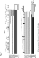
- tandem duplication (bold diagonal line from the upper right to the lower left): three cases; inversion (thin diagonal line from the upper left to the lower right): three cases; translocation (open): three cases; and deletion (thin-line from the upper right to the lower left): two cases.
- CD274 was intact up to exon 5 in all cases; however, a part of the region on and downstream exon 5 was truncated in some cases.
- FPKM Frragments Per Kilobase of transcript per Million fragments sequenced
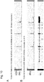
- CD274 is remarkably and highly expressed in almost all cases having a structural abnormality (+), compared to the cases (solid) having no structural abnormality.
- RNA sequencing data of ATL (57 cases) in Example 2
- the sequence reads were displayed at individual exons of CD274 (NM_014143) by IGV (integrative genome viewer) (provided by Broad Institute).
- CD274 consists of 7 exons and exon 7 is mostly occupied by 3'UTR.
- transcripts were truncated at exon 5 or exon 6. In these cases, the sequences of the transcripts were confirmed by RNA sequencing to identify the sequences.
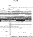
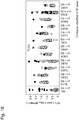
- the results are shown in Figure 5 .
- the nucleotide sequence in the lower part of Figure 5 is the sequence of a transcript of a case having a structural abnormality (+) (Miya26).
- a wild type CD274 was transcribed up to the 3' end of exon 6.
- the transcript was followed by an intron region of CBLB gene on chromosome 3 (area in the framed).
- the intron region contains a termination codon (represented by italic TGA), a polyA signal sequence (AATAAA underlined: the sequence required for binding of polyA) and a polyA sequence (double underlined).
- This result shows that an alternative transcript formed by CD274 structural abnormality functions as a suitable transcript.
- the whole sequence shown in Figure 5 is represented by SEQ ID No: 1; whereas the sequence of 3'UTR is represented by SEQ ID No: 2.
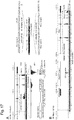
- NP_054862 was used as a reference sequence. Based on the sequence of the transcript of Example 5, the amino acid sequence of a case where CD274 was truncated in the middle was identified in silico.
- CD274 consists of an Extracellular domain, a Transmembrane domain and a Cytoplasmic domain. The first two domains are considered as important for immune escape of tumor cells due to PD1/PD-L1 mechanism.
- CD274 In the truncated CD274 (Miya15, Miya24, Miya26, Miya30, Sas8, Kyo4), the first two domains are maintained intact, suggesting that the alterative transcripts can function as CD274 with a high probability.
- CD274 expression on the cell surface was evaluated by flow cytometry using tumor cells in the case where a CD274 structural abnormality was identified by RNA sequencing. Tumor cells were stained with PE/Cy7 anti-human CD274 (B7-H1, PD-L1) Antibody (clone: 29E.2A3, BioLegend) and analyzed by LSR2 Fortessa (BD Biosciences).
- CD274 is highly expressed at a protein level in the cases having a structural abnormality (+).
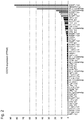
- RNA sequencing data stored in the U.S. database of next generation sequence data of various types of malignant tumors TCGA (https: //tcga-data.nci.nih.gov/tcga/), CD274 structural abnormalities in various types of malignant tumors and expression were analyzed.
- ACC adenoid cystic carcinoma
- BLCA bladder carcinoma
- CESC cervical squamous cell carcinoma
- COAD colon adenocarcinoma
- DLBC diffuse large B-cell lymphoma
- ESCA esophageal carcinoma
- GBM glioblastoma
- HNSC head-neck squamous cell carcinoma
- KICH kidney chromophobe carcinoma
- KIRC stomach clear cell carcinoma
- LAML acute myelogenous leukemia
- LGG low grade glioma
- LIHC liver hepatocellular carcinoma
- LUAD lung adenocarcinoma
- LUSC lung squamous cellular carcinoma
- MESO malignant mesothelioma
- OV ovarian cancer
- PAAD pancreas adenocarcinoma
- PCPG pancreas adenocarcinoma
- PCPG pancreas adenocarcinoma
- PCPG pancre
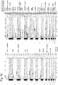
- the numbers in the figure represent the number of specimens of individual tumors subjected to RNA sequencing. In total, 10,000 cases or more were analyzed.
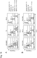
- TCGA publicly discloses expression data (RSEM values are employed and RPKM values are employed only in stomach cancer) calculated based on RNA sequencing data.
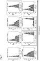
- CD274 expression levels (after logarithmic transformation) of individual cancer cases obtained based on the data were displayed as a histogram.
- Figure 9 shows the results of four types of cancers. High expression of CD274 was observed in some cases of various cancers. Thus, a cutoff value was determined based on expression values and more specific analysis was performed. As the cutoff value, a logarithmically transformed RSEM values were used in the cases except stomach cancer; whereas a logarithmically transformed RPKM value was used in the case of stomach cancer; more specifically, 8 was used in cancer cases except stomach cancer and 11 was used in the case of stomach cancer (the cutoff values are appropriately set for evaluation).
- SKCM represents skin cutaneous melanoma ( Figure 9A ), COADREAD colorectal adenocarcinoma, ( Figure 9B ), STAD stomach adenocarcinoma ( Figure 9C ) and DLBC diffuse large B-cell lymphoma ( Figure 9D ).
- bladder carcinoma single case
- cervical squamous cell carcinoma CEC
- KIRC kidney renal clear cell carcinoma
- COAD colon adenocarcinoma
- RTD cutaneous malignant melanoma
- SKCM single case
- stomach adenocarcinoma STAD: three cases (4 cases in total) were the cases where the same expression pattern as ATL (high CD274 expression and no 3'UTR expression) was found.
- FIG 11 two cases of diffuse large B-cell lymphoma (DLBC) were similarly shown as an example ( Figures 11A and B ).
- DLBC diffuse large B-cell lymphoma
- CD274 was highly expressed in 2 cases out of 48 cases having RNA sequencing data and the same structural abnormality was found in both of the two cases.
- Numbers 1 to 7 in the lower parts of Figures 10 and 11 represent exon numbers.
- Copy number analysis was performed by SNP array (method: GISTIC) in ATL 426 cases.
- tumor specimens were taken in accordance with the protocol approved by the ethical committee of Kyoto University and subjected to global genetic mutation analysis.
- the sequence of the forward primer was GGCATCCAAGATACAAACTCAA (SEQ ID NO: 10) and the sequence of the reverse primer was CAGAAGTTCCAATGCTGGATTA (SEQ ID NO: 11).
- a holding step was carried out at 95°C 10 sec, and a cycling step (95°C 5 sec, 60°C 30 sec, 72°C 30 sec) was repeated 50 times.
- Detection was carried out by LightCycler (registered trade mark) 480 System (Roche Applied Science). As the internal control, 18S was used and the ratio to CD274 was shown.
- PD-L1 was immuno-stained with an anti N terminal antibody (E1J2J, Cell Signaling Technology) and an anti C terminal antibody (SP142, Spring Bioscience).
- E1J2J Cell Signaling Technology
- SP142 Spring Bioscience
- Figure 15A shows the results of ATL059 having no PD-L1 SV (ATL059: PD-L1 SV (-)).
- Figure 15B shows the results of ATL075 of PD-L1 SV (+) having an intact ORF (ATL075: PD-L1 SV (+), intact ORF).
- Figure 15C shows the results of ATL012 of SV (+) having a truncated ORF (ATL012: PD-L1 SV (+), truncated ORF).
- the results of immunostaining with the anti N terminal antibody are shown.
- the results of immunostaining with the anti C terminal antibody are shown.
- FIG 15A shows the results of ATL059 having no PD-L1 SV, tumor cells were not stained (in Figure 15A , cells sporadically stained (looks in grey in a monochrome photography) are macrophages).
- ATL012 of SV (+) having a truncated ORF was intensively stained with the anti N terminal antibody and not stained with the anti C terminal antibody.
- results show that if PD-L1 SV is present, expression of PD-L1 (at a protein level) is strong and increases.
- the fact that ATL012 is not stained with the anti C terminal antibody is consistent with the fact that ORF is truncated. Further, the results show that PD-L1 SV can be identified by double staining with the anti N terminal antibody and anti C terminal antibody.
- results show that the same results are obtained also in western blot by antibodies against PD-L1, i.e., an anti N terminal antibody and an anti C terminal antibody.
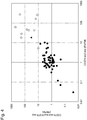
- Cases (lowest 30 cases) corresponding to top 10% were selected based on 33 types of carcinomas (solid cancer) publicly disclosed from TCGA and expression data (RSEM values) calculated based on RNA sequencing data of 10210 cases. RNA sequencing data were downloaded, and cases of CD274 fusion gene (+) and/or a virus insert (+) in the CD274 region and/or a relative high CD274 exon 4/3'UTR ratio were selected. As to DLBC and STAD, since a frequency was high, all cases were used as subjects.
- RNA sequencing data of total 1691 cases were downloaded and analyzed. Based on the data, expression of CD274 (after RPKM value of exon 4 was logarithmically transformed) of individual cases of each carcinoma was displayed.
- Solid circles represent cases of CD274 fusion gene (+) and/or a relative high CD274 exon 4/3'UTR ratio.
- Double circles represent cases having a virus insert (+) in the CD274 region.
- CD274 fusion gene (+) and/or a virus insert (+) in the CD274 region and/or a relatively high CD274 exon 4/3'UTR ratio were observed. From the results, it was found that an abnormality of truncated 3'UTR in CD274 was observed in various types of cancers. Further, these cases mostly occur where CD274 is most highly expressed in carcinomas, suggesting that 3'UTR is extremely important for regulating CD274 expression.
- Figure 17 shows the verification results of a VS-A9U7-01 case (CESC) ( Figure 17A ) and a FP-7998-01 case (STAD) ( Figure 17B ).
- CESC VS-A9U7-01 case
- STAD FP-7998-01 case
- CD274 expression is remarkably increased by introducing a deletion or an inversion in CD274 3'UTR by CRISPR/Cas9 system.
- a human (upper panel) or mouse (lower panel) cell line was transfected with sgRNA and Cas9 (targeting two sites: 5' end and 3' end of CD274 3'UTR).
- sgRNA and Cas9 targeting two sites: 5' end and 3' end of CD274 3'UTR.
- CRISPR clustered regularly interspaced short palindoromic repeats
- Cas9 Cas9
- human cell lines i.e., HEK293T (fetal kidney), T2 (hybrid of T cell and B cell), PC-9 (lung cancer) and mouse cell lines, i.e., EG7-OVA (T cell lymphoma, expressing Ovalbumin), P815 (mastocytoma) and B16-F10 (malignant melanoma) were selected.
- HEK293T fetal kidney
- T2 hybrid of T cell and B cell
- PC-9 lung cancer
- mouse cell lines i.e., EG7-OVA (T cell lymphoma, expressing Ovalbumin), P815 (mastocytoma) and B16-F10 (malignant melanoma
- the cell lines (SgPD-L1 F ⁇ R) having a deletion or an inversion introduced in the 3'UTR exhibited a remarkable increase in CD274 expression, compared to parental cell lines and mock introduced cell lines.
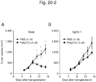
- EG7-OVAcell line (SgPd-L1) in which high expression of CD274 in association with CD274 3'UTR abnormality was induced by the CRISPR-Cas9 system and a control cell line (Mock) were subcutaneously transplanted to the isogenic mice.
- An immunostimulating agent, poly (I: C) or PBS as a control was administered from 7th day after the transplantation and a tumor diameter was periodically measured. In this experiment, it is considered that poly (I: C) induces an immunity against a tumor (in particular ovalbumin).
- Figure 20-1 shows the protocol of the experiment.
- Figure 20-2 shows changes in tumor diameter; more specifically, a change in tumor diameter of Mock is shown in Figure 20-2A and a change in tumor diameter of EG7-OVA cell line (SgPD-L1), in which high expression of CD274 was induced, is shown in Figure 20-2B .
- SgPD-L1 EG7-OVA cell line
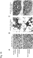
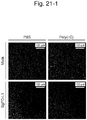
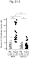
- Figure 21-1 shows stained images.
- Figure 21-2 shows CD8 positive T cells within the tumor.
- the number of CD8 positive T cells stained green increases.
- PD-1/PD-L1 block can suppress promotion of tumor formation and immunosuppressive effect by high expression of CD274 in association with CD274 3'UTR abnormality.
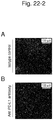
- an EG7-OVA cell line in which high expression of CD274 in association with CD274 3'UTR abnormality was induced by CRISPR-Cas9 system, was subcutaneously transplanted to the isogenic mice.
- an immunostimulating agent poly (I: C)
- an anti PD-L1 antibody or an isotype control was intraperitoneally injected and the effect on the EG7-OVA tumor was evaluated.
- the diameter of a tumor was periodically measured. Fourteenth or fifteenth day after the transplantation, immunostaining with CD8 and DAPI were carried out to evaluate the extent of infiltration of CD8 positive T cells into the tumor.
- Figure 22-1 is a graph showing a change of tumor diameter with time.
- Figure 22-2 shows immunostained images with CD8 and DAPI.
- Figure 22-3 shows CD8 positive T cells in a tumor.
- the number of CD8 positive T cells stained green is larger in a mouse to which an anti PD-L1 antibody was administered ( Figure 22-3B ) than in the mouse ( Figure 22-3A ) to which an isotype control was administered.
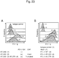
- ATL patient's specimens (5 specimens of ATL049, ATL050, ATL022 and ATL079) or a PC-9 cell line, to which CD274 wild type (WT) or a truncation mutant was introduced by a retrovirus
- WT CD274 wild type
- a truncation mutant was introduced by a retrovirus
- the binding ability to PD-1 Ig was evaluated by flow cytometry.
- ATL050 had intact ORF (Intact) without truncation
- ATL022 and ATL079 had a truncated ORF (Truncated).
- the mutant introduced into PC-9 was derived from ATL020 (defective Ex7) or ATL079 (defective Ex6 and 7).
- Figure 23 shows the results.
- Figure 23A shows the evaluation results of ATL patient's specimens and
- Figure 23B shows the results of the PC-9 cell line.
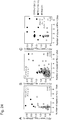
- CD274 SV relates to CD274 overexpression independently of the number of copies in the CD274 region
- Figure 24 shows the results.
- Figure 24A shows the results of DLBC (B cellular lymphoma).
- Figure 24B shows the results of STAD (stomach adenocarcinoma).
- Figure 24C shows the results of ATL (adult T cell leukemia).
- solid circles represent the cases of CD274 fusion gene (+) and/or a relatively high CD274 exon 4/3'UTR ratio and double circles represent the cases of a virus insert (+) in the CD274 region.
- CD274 SV significantly independently related to high expression of CD274.
- CD274 SV relates to CD274 expression independently of the number of CD274 copies.
- the present invention enables to determine the effectiveness of a PD-1/PD-L1 blockade in various types of cancers, and further determine the effectiveness of a PD-1/PD-L1 blockade per patient with a malignant tumor. As a result, the possibility of a PD-1/PD-L1 blockade in treatment of malignant tumors can be increased.
Landscapes
- Health & Medical Sciences (AREA)
- Life Sciences & Earth Sciences (AREA)
- Chemical & Material Sciences (AREA)
- Immunology (AREA)
- Engineering & Computer Science (AREA)
- Molecular Biology (AREA)
- Biomedical Technology (AREA)
- Hematology (AREA)
- Urology & Nephrology (AREA)
- General Health & Medical Sciences (AREA)
- Medicinal Chemistry (AREA)
- Biochemistry (AREA)
- Microbiology (AREA)
- Biotechnology (AREA)
- Physics & Mathematics (AREA)
- Analytical Chemistry (AREA)
- Organic Chemistry (AREA)
- Pathology (AREA)
- Cell Biology (AREA)
- General Physics & Mathematics (AREA)
- Food Science & Technology (AREA)
- Genetics & Genomics (AREA)
- Proteomics, Peptides & Aminoacids (AREA)
- Wood Science & Technology (AREA)
- Zoology (AREA)
- Bioinformatics & Cheminformatics (AREA)
- Biophysics (AREA)
- General Engineering & Computer Science (AREA)
- Public Health (AREA)
- Veterinary Medicine (AREA)
- Pharmacology & Pharmacy (AREA)
- Epidemiology (AREA)
- Animal Behavior & Ethology (AREA)
- Oncology (AREA)
- Hospice & Palliative Care (AREA)
- Plant Pathology (AREA)
- Mycology (AREA)
- Measuring Or Testing Involving Enzymes Or Micro-Organisms (AREA)
- Investigating Or Analysing Biological Materials (AREA)
- Peptides Or Proteins (AREA)
Description
- The present invention relates to a method of predicting an effect of treatment by a PD-1/PD-L1 blockade such as anti PD-1 antibody by examining the presence or absence of an abnormality of PD-L1 gene in a tumor cell.
- Recently, it has been elucidated that a PD-1/PD-L1 blockade is effective for various types of malignant tumors including metastatic malignant melanoma, renal cancer, lung cancer and Hodgkin's disease. As the PD-1/PD-L1 blockade, an anti PD-1 monoclonal antibody, i.e., Nivolumab, has been put into practical use (see, Patent Literature 1). At present, searching a biomarker for predicting an effect of treatment by this blockade is an urgent issue. It has been found that the efficacy rate is high when "PD-L1 positive" is found by immunostaining in tumor cells and peripheral immune cells. However, evaluation of PD-L1 positive rate by immunostaining is not sufficient in view of sensitivity/specificity. It is desired that prediction on efficacy of therapy is further improved.
- It has been increasingly clear that a PD-1/PD-L1 blockade is effective in many malignant tumors. Of them, malignant tumors on which the blockade less effectively works are included. However, even in such tumors, a case having a structural abnormality of PD-L1 accompanied by high expression thereof is likely to be present. However, a useful method for sorting out a case where a PD-1/PD-L1 blockade possibly works has not yet been established.
- Patent Literature 1:
JP Patent Publication (Kokai) No. 2006-340714 - An object of the present invention is to provide an effective method for predicting an effect of treatment by a PD-1/PD-L1 blockade. Another object is to develop a platform for sorting out a case on which these blockades effectively work with a high probability even in tumors to which a PD-1/PD-L1 blockade has not yet been applied.
- The present inventors conducted comprehensive gene analysis (RNA sequencing: 57 cases, whole genome sequencing: 11 cases) of adult T cell leukemia/lymphoma (ATL). Based on the gene analysis, they clarified that a structural abnormality of PD-L1 (CD274) is present in about 20% of ATL cases. Examples of the structural abnormality include all structural abnormalities such as deletion, tandem duplication, inversion and translocation. These structural abnormalities commonly have a deletion of 3'UTR. Furthermore in all cases having a structural abnormality, a remarkable increase of PD-L1 mRNA level was observed and an increase of PD-L1 protein expression on the cell surface was confirmed by flow cytometry. Moreover, studies were conducted on the presence of the same abnormality in other cancers based on TCGA data. As a result, it was found that the same abnormality is present in various types of malignant tumors such as stomach cancer, lung cancer, large intestinal cancer, cervical cancer, malignant melanoma and B cell lymphoma. This result suggests that a structural abnormality, i.e., a deletion of 3'UTR of PD-L1 accompanied by high expression of PD-L1 is a common abnormality occurring in various types of malignant tumors.
- Based on these results, the present inventors found that the effect of a PD-1/PD-L1 blockade can be determined and evaluated based on a deletion of 3'UTR in PD-L1 gene as an index and accomplished the present invention. The scope of the invention is defined by the appended claims.
- More specifically, the present disclosure is as follows.
- [1] A method of predicting whether or not PD-1/PD-L1 blockade is effective for treatment of a subject suffering from malignant tumor, which comprises detecting abnormality of genome relating to effectiveness of the PD-1/PD-L1 blockade in a tumor cell that has been taken from the subject and evaluating the PD-1/PD-L1 blockade as useful for the treatment of the subject when there is the abnormality.
- [2] The method according to [1], wherein the abnormality of genome relating to effectiveness of the PD-1/PD-L1 blockade is abnormality of PD-L1 gene relating to the acceleration of the expression of PD-L1 (CD274) gene.
- [3] The method according to [2], wherein the abnormality of PD-L1 gene is partial or complete deletion of 3' UTR region of PD-L1 gene.
- [4] The method according to [2], wherein the abnormality of PD-L1 gene is change in copy number which induces deletion of 3' UTR region of PD-L1 gene.
- [5] The method according to [2], wherein the abnormality of PD-L1 gene is complete or partial truncation of
exon 5,exon 6 andexon 7 from PD-L1 transcript. - [6] The method according to [2], wherein the abnormality of PD-L1 gene is complete or partial truncation of
exon 5,exon 6 andexon 7 from PD-L1 transcript. - [7] The method according to [2], which comprises quantifying a transcript of any one of
exon 1 toexon 4 of PD-L1 gene and a transcript of 3' UTR region of PD-L1 gene and calculating a ratio between the amount of the transcript of the exon and the amount of the transcript of the 3' UTR region, and evaluating the PD-1/PD-L1 blockade as useful for the treatment of cancer when the ratio is not less than a predetermined value. - [8] The method according to [2], wherein the abnormality of PD-L1 gene is acceleration of the expression of PD-L1 mRNA.
- [9] The method according to [2], wherein the abnormality of PD-L1 gene is the abnormality caused by the insertion of a virus.
- [10] The method according to [9], wherein the virus is Human papilloma virus (HPV) or EBV (Epstein-barr virus).
- [11] The method according to [2], which comprises staining a PD-L1 protein in a tumor cell taken from the subject by immunohistochemical staining using an antibody against a C terminal region of PD-L1 and an antibody against a N terminal region of PD-L1, and evaluating the PD-1/PD-L1 blockade as useful for the treatment of the subject when the tumor cell becomes stained with the antibody against a N terminal region of PD-L1 but the tumor cell does not become stained with the antibody against a C terminal region of PD-L1.
- [12] The method according to any one of [1] to [11], wherein the malignant tumor is selected from the group consisting of adult T-cell leukemia/adult T-cell leukemia lymphoma, stomach cancer, large intestinal cancer, bladder cancer, cervical cancer, renal cancer, lung adenocarcinoma, cutaneous malignant melanoma, and B cell lymphoma.
- [13] The method according to any one of [1] to [11], wherein the malignant tumor is selected from the group consisting of esophageal cancer, head and neck cancer, and uterine body cancer.
- [14] The method according to any one of [1] to [13], wherein the PD-1/PD-L1 blockade is anti PD-1 antibody or anti PD-L1 antibody.
- [15] A treatment method comprising detecting an abnormality of genome relating to effectiveness of a PD-1/PD-L1 blockade in a tumor cell taken from a subject suffering from a malignant tumor and applying a treatment with the PD-1/PD-L1 blockade when the abnormality is present.
- As shown in Examples, in various malignant tumors, there are cases where PD-L1 is highly expressed due to a structural abnormality of PD-L1 gene. More specifically, a structural abnormality of PD-L1 (mainly serving as an immune checkpoint) and the resulting fusion gene with a non-coding region were identified as a gene abnormality in various types of malignant tumors beyond ordinal expectation. In consideration that an anti PD-1/PD-L1 blockade is overwhelmingly effective (efficacy rate: about 90%) in a preclinical study (mouse model) of this research and in Hodgkin's disease, which is known as only one case where PD-L1 is constantly activated by a genomic abnormality, it is expected that an anti PD-1/PD-L1 blockade is extremely effective for patients having a PD-L1 structural abnormality and becomes a prospective biomarker. Accordingly, the genetic abnormality directly relating to an effective treatment is an extremely prospective target for clinical examination.
- As to a malignant tumor to which a PD-1/PD-L1 blockade is already applied, an effect of treatment by the PD-1/PD-L1 blockade can be successfully predicted based on detection of a PD-L1 structural abnormality. Also, even in the cases of tumors to which a PD-1/PD-L1 blockade has not yet been applied, if an abnormality of PD-L1 gene is present therein, the PD-1/PD-L1 blockade is likely to be effective. If such a case is sorted out, a novel therapy can be possibly established. In addition, if a clinical trial is carried out based on a structural abnormality of PD-L1 as an index, the number of diseases for which it is indicated can be effectively increased. This means that the range of the indication is not limited to malignant tumors, for which the indication has been approved or under consideration, and can be enlarged to malignant tumors, for which effectiveness of the blockage is in general regarded as being insufficient.
-
- [
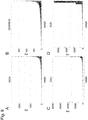
Figure 1] Figure 1 shows the search results for structural abnormality in 9p24.1 region in RNA sequencing (57 cases) and whole genome sequencing (11 cases). - [
Figure 2] Figure 2 shows the analysis results on the relationship between structural abnormalities of RNA sequencing (57 cases) and CD274 expression. - [
Figure 3] Figure 3 shows the analysis results of CD274 expression in individual exons. - [
Figure 4] Figure 4 shows the relationship between expression inexon 3, the ratio of expression inexon 3/3'UTR expression and CD274 structural abnormality. - [
Figure 5] Figure 5 shows the analysis results of a transcript of CD274 truncated in the middle and identified in ATL. - [
Figure 6] Figure 6 shows amino acid sequences of transcripts of CD274 truncated in the middle. - [
Figure 7] Figure 7 shows expression of CD274 protein on membrane surfaces of ATL patient-derived cells. - [
Figure 8] Figure 8 shows analysis results of CD274 structural abnormality and expression in various types of cancers based on TCGA data. - [
Figure 9] Figure 9 shows CD274 expression in malignant melanoma, large intestinal cancer, stomach cancer and B cell lymphoma. - [
Figure 10] Figure 10 shows analysis results (I) of CD274 expression in various types of malignant tumors based on TCGA data. - [
Figure 11] Figure 11 shows analysis results (II) of CD274 expression in various types of malignant tumors based on TCGA data. - [
Figure 12] Figure 12 shows results (GISTIC results) of CD274 abnormalities detected by SNP array. - [
Figure 13] Figure 13 shows results (CNAG results) of CD274 abnormalities detected by SNP array. - [
Figure 14] Figure 14 shows results of CD274 abnormalities detected by RQ-PCR. - [
Figure 15] Figure 15 shows immunostaining results of PD-L1 expression in ATL patients. - [
Figure 16] Figure 16 shows analysis results (II) of CD274 structural abnormality and expression in various types of cancers based on TCGA data. - [
Figure 17] Figure 17 shows an insert of a carcinogenesis virus in CD274 region and abnormality in CD274 3'UTR. - [
Figure 18] Figure 18 shows that CD274 overexpression induced by introducing an abnormality of CD274 3'UTR of a human and mouse cell lines. - [
Figure 19] Figure 19 shows that high expression of CD274 in association with a CD274 3'UTR abnormality induces apoptosis of PD-1 expressing T cells. - [
Figure 20-1] Figure 20-1 shows a protocol of an experiment evaluating the effect of high expression of CD274 in association with a CD274 3'UTR abnormality on tumorigenic potential. - [
Figure 20-2] Figure 20-2 shows the effect of high expression of CD274 in association with a CD274 3'UTR abnormality on tumorigenic potential. - [
Figure 21-1] Figure 21-1 shows stained images showing that high expression of CD274 in association with a CD274 3'UTR abnormality suppresses infiltration of CD8 positive T cells into a tumor. - [
Figure 21-2] Figure 21-2 is a graph (the number of cells) showing that high expression of CD274 in association with a CD274 3'UTR abnormality suppresses infiltration of CD8 positive T cell into a tumor. - [
Figure 22-1] Figure 22-1 is a graph showing the effect of a PD-1/PD-L1 blockade against tumorigenic potential due to high expression of CD274 in association with a CD274 3'UTR abnormality, based on a time-dependent change of a tumor in diameter. - [
Figure 22-2] Figure 22-2 shows the effect of a PD-1/PD-L1 blockade against tumorigenic potential due to high expression of CD274 in association with a CD274 3'UTR abnormality, based on immunostaining. - [
Figure 22-3] Figure 22-3 shows the effect of a PD-1/PD-L1 blockade against tumorigenic potential due to high expression of CD274 in association with a CD274 3'UTR abnormality, based on the number of cells. - [
Figure 23] Figure 23 shows PD-1 binding ability of a mutant whose CD274 open reading frame is truncated, based on flow cytometric evaluation. - [
Figure 24] Figure 24 shows graphs showing that a CD274 structural abnormality relates to CD274 overexpression independently of the number of copies of the CD274 region. - Now, the present invention will be more specifically described, below.
- The present invention relates to a method of evaluating and determining the effectiveness of a PD-1/PD-L1 blockade in a patient with a malignant tumor, based on a genomic abnormality relating to effectiveness of the PD-1/PD-L1 blockade in the patient as an index, or a method of obtaining supportive data for evaluating and determining the effectiveness of a PD-1/PD-L1 blockade in the patient. In the present invention, evaluation/determination is also referred to as prediction. PD-L1 is also referred to as CD274 or B7-H1.
- The genomic abnormality relating to effectiveness of a PD-1/PD-L1 blockade herein refers to an abnormality, which is found in the genome of a patient with a malignant tumor and on which a PD-1/PD-L1 blockade has a high effect. Examples of such an abnormality include an abnormality of chromosome 9p24.1, on which PD-L1 gene is present, and an abnormality of PD-L1 gene. Such an abnormality is referred to also as a structural variation (SV) of PD-L1 gene. With these abnormalities, abnormal expression of PD-L1 can be induced in tumor cells of a patient with a malignant tumor. Examples of the genomic abnormality relating to effectiveness of a PD-1/PD-L1 blockade include structural abnormalities such as tandem duplication, deletion, inversion and translocation of a gene; abnormalities in number of copies such as an increase or decrease in number and uniparental disomy; and qualitative (deletion of 3'UTR) and quantitative abnormalities of an expression product and a transcript product. The PD-L1 abnormal expression in tumor cells refers to acceleration of PD-L1 gene expression compared to that in cells of a healthy person or that in tumor cells having a normal PD-L1 gene; more specifically, to the case where the expression of PD-L1 at an mRNA or protein level is 5 times or more, preferably 10 times or more, further preferably 20 times or more, and further preferably 50 times or more as high as normal cases. If abnormal expression of PD-L1 is detected, it is possible to predict that genomic abnormality relating to effectiveness of a PD-1/PD-L1 blockade is present.
- In a living body, T cells have an immune function against a tumor (i.e., T cells attack tumor cells). However, when PD-L1 ligand expressed by a tumor cell binds to PD-1 (Programmed cell death 1) expressed by a T cell, cell death of the T cell is induced, with the result that the immune function against a tumor is suppressed. A PD-1/PD-L1 blockade blocks binding of PD-L1 of a tumor cell and PD-1 of a T cell, thereby suppressing the immune function of the T cell against a tumor. Examples of the PD-1/PD-L1 blockade include anti PD-1 antibody and anti PD-L1 antibody. Examples of the anti PD-1/PD-L1 antibody used as a cancer drug include Nivolumab, MPDL3280A, pembrolizumab (MK-3475), MEDI4736, MSB0010718C, Pidilizumab and MEDI0680. A PD-1/PD-L1 blockade highly effectively works in the case of a patient where PD-L1 is highly expressed in tumor cells and peripheral immune cells. Accordingly, if the expression level of PD-L1 is measured, the effectiveness of a PD-1/PD-L1 blockade can be determined. For example, expression of PD-L1 in a tumor cell can be checked by immunostaining; however, evaluation of a PD-L1 positive rate by immunostaining is not sufficient in view of sensitivity and specificity.
- Whether genomic abnormality in a patient with a malignant tumor relates to the effectiveness of a PD-1/PD-L1 blockade in tumor cells can be found just by investigating whether genomic abnormality relates to acceleration of PD-L1 gene expression; more specifically, can be determined by comprehensively analyzing structural abnormalities of the whole genome in the tumor cell, at the same time, determining PD-L1 gene expression in the tumor cell at an mRNA level or protein level and associating a genomic (structural) abnormality with acceleration of the PD-L1 gene expression. Further more specifically, DNA is isolated from a tumor cell and the whole genome is sequenced and analyzed for genomic structural abnormalities such as tandem duplication, inversion, translocation and deletion; at the same time, the total RNA of the tumor cell is sequenced and PD-L1 gene expression is determined; and then, association of a genomic abnormality with the PD-L1 gene expression may be analyzed. For example, in a number of tumor cells having a certain genomic abnormality, if acceleration of PD-L1 gene expression is found, it can be determined that the abnormality is associated with acceleration of the PD-L1 gene expression and relates to the effectiveness of a PD-1/PD-L1 blockade.
- Actually, when PD-L1 gene expression is accelerated by a genomic abnormality, apoptosis of T cells expressing PD-1 is induced. Also when PD-L1 gene expression is accelerated by a genomic abnormality, infiltration of CD8 positive cytotoxic T cells into a tumor is suppressed and tumor growth is accelerated. Furthermore, immunity escape due to PD-L1 genomic abnormality is suppressed by anti PD-1/PD-L1 block.
- The type of tumor, to which the method of the present invention of evaluating and determining the effectiveness of a PD-L1/PD-1 blockade can be applied, is not limited. At least, adult T cell leukemia/lymphoma, stomach cancer, large intestine cancer, bladder cancer, cervical cancer, renal cancer, lung adenocarcinoma, skin malignant melanoma, B cell lymphoma, esophageal cancer, head and neck cancer and uterine body cancer, are mentioned. The large intestinal cancer includes colon cancer and rectal cancer.
- In the method of the present invention, a tumor specimen is taken from a subject suffering from a malignant tumor and tumor cells of the specimen may be subjected to genomic abnormality analysis.
- In the method of the present invention, as specific example of the analysis object, i.e., genomic abnormality relating to effectiveness of a PD-1/PD-L1 blockade, the following abnormalities are mentioned.
- PD-L1 gene is present in 9p24.1 region. Examples of the structural abnormality of the gene include tandem duplication, inversion, translocation and deletion. When 3'UTR has a deletion due to structural abnormality of PD-L1 gene, PD-L1 is highly expressed. This is because 3'UTR is a region that plays an important role in keeping stability and regulating translation of mRNA, and if this region is deleted, the gene loses these functions. The deletion of 3'UTR in PD-L1 gene includes a complete deletion and a partial deletion. Examples of the abnormality in number of copies (CNV) of 9p24.1 region including PD-L1 gene include an increase and decrease in copy number and uniparental disomy. The abnormality in number of copies occurs in association with structural abnormality. When a deletion in the 3'UTR occurs, PD-L1 is highly expressed.
- The PD-L1 gene having 3'UTR deleted is partly truncated in the middle of an exon region, with the result that a different protein from a wild-type PD-L1 is produced. PD-L1 (protein) consists of three domains, i.e., an extracellular domain, a transmembrane domain and a cytoplasmic domain. In binding to PD-1, the extracellular domain and transmembrane domain are participated. Even if the PD-L1 gene is truncated in the middle of the exon region, PD-L1 keeps a binding ability to PD-1. More specifically, even if PD-L1 gene is truncated in the middle of the exon region, as long as it has the extracellular domain and transmembrane domain, PD-L1 has a function suppressing antitumor immunity. Accordingly, regardless of whether the PD-L1 protein is truncated or not, a tumor cell highly expressing PD-L1 protein due to genomic abnormality of PD-L1 gene is blocked in the function of suppressing tumor immunity by a PD-1/PD-L1 blockade. Accordingly, blocking of the PD-1/PD-L1 is effective for treating a malignant tumor having a genomic abnormality of PD-L1 gene.
- The nucleotide sequence of PD-L1 gene is registered under NM_014143. The nucleotide sequence of 3'UTR of PD-L1 gene is represented by SEQ ID No: 2.
- As abnormality of PD-L1 mRNA, whole or partial truncation of 3'UTR of mRNA and accompanying high expression are mentioned. In addition, a truncation on or downstream of
exon exon 6 andexon 7, a truncation of thewhole exon 7 and a truncation in the middle ofexon 7. In the present invention, such a truncation of exon refers to a truncation ofexon 6 or a whole or partial truncation ofexon 7. Further, a truncation on and downstream ofexon 4, more specifically, a truncation of the whole region ofexon 5,exon 6 andexon 7 may occur. - As abnormality of PD-L1 protein, high expression is mentioned. The high expression of PD-L1 protein occurs independent of an increase of PD-L1 gene in number of copies and relates to structural abnormality of PD-L1 gene. Since
exon downstream exon 4 is present, the abnormality of PD-L1 protein includes a partial deletion of the cytoplasmic domain. - A carcinogenic virus such as HPV (e.g., Human papilloma virus (HPV) 16) and EBV (Epstein-barr virus), if it is introduced into PD-L1 gene region, sometimes induces a structural abnormality. For example, in some cases where Human papilloma virus (HPV) in inserted into
intron 6 of PD-L1 gene and Epstein-Barr virus (EBV) gene is inserted in an upstream gene region adjacent to PD-L1 gene and located upstream thereof, a PD-L1 transcript is truncated at 3'UTR. - Genomic abnormality relating to effectiveness of a PD-1/PD-L1 blockade can be analyzed in accordance with a general method for analyzing genomic structural abnormality and gene abnormality using tumor cells taken from a patient with a malignant tumor. Examples of the analysis method include an analysis method based on sequence analysis by extracting DNA or RNA from tumor cells and directly determining the sequence thereof by a method known in the art such as the PCR/Sanger sequence method, dideoxy method and Maxam-Gilbert method; an analysis method for global gene including a whole genome sequencing, whole exon sequencing and a target sequencing by a next generation sequencer; a FISH (fluorescence in situ hybridization) method; a hybridization method using a specific probe to a region having a gene abnormality or a microarray (DNA chip) having a specific probe immobilized therein, such as a copy number analysis method using e.g., an SNP array and a CGH array; and methods using a specific primer to a region having a gene abnormality. Examples of the method using a primer include PCR method, NASBA method, LCR method, SDA method, LAMP method, a method using restriction fragment length polymorphism (RFLP) and a primer extension method (TaqMan (registered trade mark) method). As the next generation sequencer, e.g., Genome Sequencer FLX (GD FLX) (Roche) and Illumina HiSeq/MiSeq (Illumina) can be used.
- A deletion in 3'UTR of PD-L1 gene can be detected by a method using a probe and/or a primer as well as by RNA sequencing and a FISH method.
- A deletion of 3'UTR in mRNA can be detected by calculating the ratio of transcript amount of exons (
exon 1 to 5) of PD-L1 gene having no deletion and the transcript amount of 3'UTR. More specifically, mRNA of any one ofexons 1 to 5 of PD-L1 gene, for example,exon 3 orexon 4 and mRNA of 3'UTR region are quantified by RNA sequencing and quantitative PCR and the ratio of expression ofexon 3 orexon 4/3'UTR expression is calculated. In this manner, the presence or absence of 3'UTR expression, more specifically, the presence or absence of 3'UTR deletion, can be determined. If the ratio is high, it can be determined that 3'UTR is deleted. The case where the ratio ofexon 3 expression/3'UTR expression is large refers to the case whereexon 3 expression/3'UTR expression is large relative toexon 3 expression/3'UTR expression in a tumor cell having no abnormality of PD-L1 gene; for example, refers to the case where the ratio of theexon 3 expression/3'UTR expression is a predetermined value or more, more specifically, double or more, preferably 3 times or more, further preferably 5 times or more. Similarly, the case where the ratio ofexon 4 expression/3'UTR expression is large refers to the case whereexon 4 expression/3'UTR expression is larger relative toexon 4 expression/3'UTR expression in a tumor cell having no abnormality of PD-L1 gene, for example, refers to the case where the ratio ofexon 4 expression/3'UTR expression is a predetermined value or more, more specifically, double or more, preferably 3 times or more, further preferably 5 times or more. - The abnormal PD-L1 gene expression can be detected by measuring PD-L1 mRNA in a tumor cell by use of a (semi) quantitative PCR, such as RQ-PCR and RT-PCR. RQ-PCR is a method of continuously detecting accumulation of a PCR product during a PCR process, thereby enabling easy and accurate quantification in the exponential phase in the beginning of PCR.
- The probe or primer to be used in the above methods, consists of a nucleotide fragment (preferably a DNA fragment) consisting of a nucleotide sequence containing a genomic abnormality site relating to acceleration of PD-L1 gene expression; a nucleotide sequence complementary to the nucleotide sequence or a sequence capable of hybridizing with either one of these sequences under stringent conditions. The number of bases is 5 to 50, preferably 10 to 30 and further preferably 10 to 25.
- In the case of having a deletion on and downstream of
exon 4, more specifically, a deletion of the C terminal region of PD-L1 protein due to a structural abnormality of PD-L1 gene, more specifically a case having a truncated ORF, the abnormal protein expressed reacts with an antibody against the C terminal region of PD-L1 protein (antibody against the C terminal of PD-L1) and does not react with an antibody against the N terminal region of PD-L1 protein (antibody against the N terminal of PD-L1). Accordingly, if the antibody against the C terminal of PD-L1 and the antibody against the N terminal of PD-L1 are used to examine the reactivity between both antibodies and the PD-L1 protein produced, the structural abnormality of PD-L1 gene can be identified. More specifically, for example, PD-L1 protein in a tumor cell is stained with an antibody against the C terminal of PD-L1 and an antibody against the N terminal of PD-L1, which are stained with e.g., a fluorescence dye, to visualize the PD-L1 protein. If the protein is stained with the anti N terminal antibody but not stained with the anti C terminal antibody, it can be determined that the PD-L1 gene has a structural abnormality. - When presence of an abnormality of PD-L1 gene relating to acceleration of PD-L1 gene expression in a tumor cell taken from a subject is found by the method of the present invention, it is possible to evaluate and determine that a PD-1/PD-L1 blockade has an effect on a malignant tumor of a subject with a high probability. Also, in a tumor cell of a malignant tumor, if the presence of abnormality of PD-L1 gene relating to acceleration of PD-L1 gene expression is found, it is possible to evaluate and determine that a PD-1/PD-L1 blockade has an effect on the malignant tumor with a high probability.
- It is considered that the percentage of patients to which a PD-1/PD-L1 blockade effectively works varies depending upon the type of malignant tumor. For example, in adult T cell leukemia/lymphoma, stomach cancer, large intestinal cancer, bladder cancer, cervical cancer, renal cancer, lung cancer, skin malignant melanoma, B cell lymphoma, esophageal cancer, head and neck cancer and uterine body cancer, if a transcript has a 3'UTR abnormality, expression of PD-L1 is known to be accelerated. With respect to patients suffering from these malignant cancers, whether a PD-1/PD-L1 blockade is effective or not can be evaluated by the method of the present invention. Furthermore, with respect to patients suffering from the other malignant tumors, whether a PD-1/PD-L1 blockade is effective or not can be evaluated by the method of the present invention.
- Moreover, for example, in adult T cell leukemia and, stomach cancer, the ratio of the patients on which a PD-1/PD-L1 blockade effectively works is high but it is possible that the ratio is low in e. g., malignant melanoma, lung cancer and renal cancer. The method of the present invention not only enables to evaluate and determine the effectiveness of a PD-1/PD-L1 blockade on the patients suffering from a malignant tumor (the ratio of patients effectively treated with a PD-1/PD-L1 blockade is high) but also enables to evaluate and determine the effectiveness of a PD-1/PD-L1 blockade on the patients suffering from a malignant tumor (the ratio of patients effectively treated with a PD-1/PD-L1 blockade is low and a PD-1/PD-L1 is not conventionally applied).
- More specifically, the present invention relates to a method of predicting whether or not a PD-1/PD-L1 blockade is effective for treating a subject suffering from a malignant tumor, and includes a method comprising detecting genomic abnormality relating to effectiveness of a PD-1/PD-L1 blockade in a tumor cell taken from a subject suffering from a malignant tumor on which a PD-1/PD-L1 blockade have a low effect and evaluating that the PD-1/PD-L1 blockade is effective for treating the subject when an abnormality is present, thereby expanding the range of the indication for the PD-1/PD-L1 blockade to the tumor on which the PD-1/PD-L1 blockade has a low effect.
- As mentioned above, the method of the present invention enables to evaluate and determine whether a PD-1/PD-L1 blockade is effective or not for various types of malignant tumors or patients suffering from various types of malignant tumors and enables to select an appropriate treatment method for a patient.
- If an abnormality of PD-L1 gene relating to acceleration of the PD-L1 gene expression is found in tumor cells taken from a subject, and if a PD-1/PD-L1 blockade can be evaluated and determined to effectively treat a malignant tumor of the subject with a high probability, an immune checkpoint blockade such as a PD-1/PD-L1 blockade containing an anti PD-1 monoclonal antibody or an anti PD-L1 monoclonal antibody may be administered to the subject.
- The dosage varies depending on e.g., the age, body weight and symptom. The dosage of 0.001 mg to 100 mg per dose may be administered by parenteral administration such as intravenous injection, intraperitoneal injection, subcutaneous injection and intramuscular injection or oral administration at intervals of several days, several weeks or several months. The blockade to be administered may contain a pharmacologically acceptable carrier, diluent or excipient. The dosage form of the blockade is not limited, a dosage form for oral administration such as a tablet, a capsule, a granule, a powder and a syrup, or a dosage form for parenteral administration such as an injection, a drip, a suppository and a spray may be mentioned.
- The present invention will be more specifically described by way of the following Examples; however, the present invention is not limited by these Examples.
- RNA sequencing of 57 tumor specimens of ATL (adult T cell leukemia) and total genome sequencing of tumor-normal (buccal mucosa) pairs of ATL (11 cases) were performed.
- RNA of tumor specimens was extracted by the RNeasy Mini kit (QIAGEN) and RINe was measured by the Agilent RNA ScreenTape System (Agilent).
- A library for RNA sequencing was constructed by using RNA (200 to 500 ng) having RINe of 7 or more by the NEBNext Ultra RNA Library Prep Kit (New England Biolabs) and the nucleotide sequence was determined by HiSeq2000/2500. The data were analyzed by use of the algorithm called as Genomon Fusion (http: //genomon.hgc.jp/rna/) publicly disclosed by the human genome analysis center of the Institute of Medical Science, the University of Tokyo. In this manner, fusion genes were identified and expression of the genes was analyzed.
- A library of the whole genome sequence was constructed by using WGS using NEBNext DNA Library Prep Reagent (New England Biolabs) and nucleotide sequences were determined by HiSeq2000/2500.
- Analysis was carried out as described in TOTOKI Y. et al., ).
- The results are shown in
Figure 1 . A structural abnormality (fusion gene) in 9p24.1 region was observed in 11 cases out of 57 cases of ATL patients. - A wide variety of structural abnormalities were found as follows: tandem duplication (bold diagonal line from the upper right to the lower left): three cases; inversion (thin diagonal line from the upper left to the lower right): three cases; translocation (open): three cases; and deletion (thin-line from the upper right to the lower left): two cases.
- However, all these cases commonly had a deletion of 3'UTR (untranslated region responsible for post transcriptional regulation).
- CD274 was intact up to
exon 5 in all cases; however, a part of the region on anddownstream exon 5 was truncated in some cases. - The relationship between structural abnormality and CD274 expression was analyzed based on the results of RNA sequencing using ATL (57 cases) tumor specimens of Example 1.
- As an expression value, FPKM (Fragments Per Kilobase of transcript per Million fragments sequenced) was used.
- The results are shown in
Figure 2 . The patterns in the bar graph ofFigure 2 (bold diagonal line from the upper right to the lower left, thin diagonal line from the upper left to the lower right, open, thin-line from the upper right to the lower left) correspond to the patterns ofFigure 1 . - In the cases having a structural abnormality (common in Example 1), high expression of CD274 was observed.
- CD274 is remarkably and highly expressed in almost all cases having a structural abnormality (+), compared to the cases (solid) having no structural abnormality.
- Using RNA sequencing data of ATL (57 cases) in Example 2, the sequence reads were displayed at individual exons of CD274 (NM_014143) by IGV (integrative genome viewer) (provided by Broad Institute).
- CD274 consists of 7 exons and
exon 7 is mostly occupied by 3'UTR. - The results are shown in
Figure 3 . The numbers of 1 to 7 in the lower part ofFigure 3 represent the exon numbers. In the case having no structural abnormality (-) (Miya31), expression in 3'UTR was relatively high compared to those of other exons. In contrast, in the cases having a structural abnormality (+) (Miya26 & 30), expression in 3'UTR disappeared. The same results were obtained in other cases of a structural abnormality (+). The results show that in the cases having a structural abnormality (+), expression of the 3'UTR site disappears with deletion of 3'UTR. - In the cases having a structural abnormality (+), expression in 3'UTR disappears. Thus, expression is underestimated when expression of the whole gene is evaluated as is in Example 3. As a result, the relationship with structural abnormality likely to be unclear. Then, using RNA sequencing data of ATL (57 cases) of Example 3, the ratios of expression of individual exons of CD274 and expression of 3'UTR were calculated. Of the calculation results, expression of
exon 3, which had the most clear relationship with structural abnormality, and the ratio ofexon 3 expression/3'UTR expression (ex3 exp/3'UTR exp) was displayed as a scatter chart. - The results are shown in
Figure 4 . InFigure 4 , open circles represent cases having a structural abnormality (+), whereas solid circles represent cases having no structural abnormality (-). The abbreviation "ex3" representsexon 3 and "exp" represents expression. In view of expression ofCD274 exon 3 alone and the ratio ofexon 3 expression to 3'UTR expression, the ex3 exp/3'UTR exp, is high in structural abnormality (+) cases. Thus, structural abnormality (+) cases (open circle) were clearly separated. - In about half of CD274 structural abnormality (+) cases (11 cases) identified based on RNA sequencing results of ATL cases (57 cases), transcripts were truncated at
exon 5 orexon 6. In these cases, the sequences of the transcripts were confirmed by RNA sequencing to identify the sequences. - The results are shown in
Figure 5 . The nucleotide sequence in the lower part ofFigure 5 is the sequence of a transcript of a case having a structural abnormality (+) (Miya26). A wild type CD274 was transcribed up to the 3' end ofexon 6. The transcript was followed by an intron region of CBLB gene on chromosome 3 (area in the framed). The intron region contains a termination codon (represented by italic TGA), a polyA signal sequence (AATAAA underlined: the sequence required for binding of polyA) and a polyA sequence (double underlined). This result shows that an alternative transcript formed by CD274 structural abnormality functions as a suitable transcript. The whole sequence shown inFigure 5 is represented by SEQ ID No: 1; whereas the sequence of 3'UTR is represented by SEQ ID No: 2. - NP_054862 was used as a reference sequence. Based on the sequence of the transcript of Example 5, the amino acid sequence of a case where CD274 was truncated in the middle was identified in silico.
- The results are shown in
Figure 6 . CD274 consists of an Extracellular domain, a Transmembrane domain and a Cytoplasmic domain. The first two domains are considered as important for immune escape of tumor cells due to PD1/PD-L1 mechanism. - In
Figure 6 , the sequences from the middle part of transmembrane domains (sequences in frame) are shown. The sequences in the frames are the sequences of transmembrane domains and the sequences italicized and underlined represent the sequences of the cytoplasmic domains. The "CD274 Wt" in the upper part ofFigure 6 represents a wild type sequence. - In the truncated CD274 (Miya15, Miya24, Miya26, Miya30, Sas8, Kyo4), the first two domains are maintained intact, suggesting that the alterative transcripts can function as CD274 with a high probability.
- CD274 expression on the cell surface was evaluated by flow cytometry using tumor cells in the case where a CD274 structural abnormality was identified by RNA sequencing. Tumor cells were stained with PE/Cy7 anti-human CD274 (B7-H1, PD-L1) Antibody (clone: 29E.2A3, BioLegend) and analyzed by LSR2 Fortessa (BD Biosciences).
- The results are shown in
Figure 7 . InFigure 7 , the cases having a structural abnormality (+) each are surrounded by a frame. Also in the cases having a structural abnormality (-), low expression of CD274 is observed; however, in the case having a structural abnormality (+), a significantly high expression of CD274 is confirmed. The expression level was increased about 100 times. - The results show that CD274 is highly expressed at a protein level in the cases having a structural abnormality (+).
- Using RNA sequencing data stored in the U.S. database of next generation sequence data of various types of malignant tumors, TCGA (https: //tcga-data.nci.nih.gov/tcga/), CD274 structural abnormalities in various types of malignant tumors and expression were analyzed.
- The results are shown in
Figure 8 . In the figure, abbreviations show the following carcinomas. ACC: adenoid cystic carcinoma, BLCA: bladder carcinoma, CESC: cervical squamous cell carcinoma, COAD: colon adenocarcinoma, DLBC: diffuse large B-cell lymphoma, ESCA: esophageal carcinoma, GBM: glioblastoma, HNSC: head-neck squamous cell carcinoma, KICH: kidney chromophobe carcinoma, KIRC: stomach clear cell carcinoma, KIRP: kidney renal papillary cell carcinoma, LAML: acute myelogenous leukemia, LGG: low grade glioma, LIHC: liver hepatocellular carcinoma, LUAD: lung adenocarcinoma, LUSC: lung squamous cellular carcinoma, MESO: malignant mesothelioma, OV: ovarian cancer, PAAD: pancreas adenocarcinoma, PCPG: pheochromocytoma and paraganglioma, PRAD: prostate adenocarcinoma, READ: rectal adenocarcinoma, SARC: sarcoma, SKCM: skin cutaneous melanoma, STAD: stomach adenocarcinoma, TGCT: testicular germ cell tumor, THCA: thyroid carcinoma, THYM: thymoma, UCEC: uterine corpus endometrial carcinoma, UCS: uterine carcinosarcoma, UVM: uveal melanoma - The numbers in the figure represent the number of specimens of individual tumors subjected to RNA sequencing. In total, 10,000 cases or more were analyzed.
- TCGA publicly discloses expression data (RSEM values are employed and RPKM values are employed only in stomach cancer) calculated based on RNA sequencing data. CD274 expression levels (after logarithmic transformation) of individual cancer cases obtained based on the data were displayed as a histogram.
-
Figure 9 shows the results of four types of cancers. High expression of CD274 was observed in some cases of various cancers. Thus, a cutoff value was determined based on expression values and more specific analysis was performed. As the cutoff value, a logarithmically transformed RSEM values were used in the cases except stomach cancer; whereas a logarithmically transformed RPKM value was used in the case of stomach cancer; more specifically, 8 was used in cancer cases except stomach cancer and 11 was used in the case of stomach cancer (the cutoff values are appropriately set for evaluation). InFigure 9 , SKCM represents skin cutaneous melanoma (Figure 9A ), COADREAD colorectal adenocarcinoma, (Figure 9B ), STAD stomach adenocarcinoma (Figure 9C ) and DLBC diffuse large B-cell lymphoma (Figure 9D ). - With respect to the cases exhibiting a TCGA cutoff value or more (high CD274 expression case), expressions of individual exons were evaluated based on the RNA sequencing data already mapped, in the same manner as in Example 4, and displayed by the IGV (integrative genome viewer) (provided by Broad Institute).
- The results are shown in
Figure 10 . Of the cases there, cases of showing the same expression pattern as ATL (high expression and no 3'UTR expression) were found. Further, in these cases, the sequences were more specifically analyzed based on RNA sequencing data. As a result, a structural abnormality of CD274 was confirmed. For example, stomach adenocarcinoma (STAD) (Figure 10A ) and colon adenocarcinoma (COAD) (Figure 10B ) are mentioned. Other than these, bladder carcinoma (BLCA): single case; cervical squamous cell carcinoma (CESC): single case; kidney renal clear cell carcinoma (KIRC): single case; lung adenocarcinoma (LUAD): two cases; colon adenocarcinoma (COAD): single case (two cases in total)/rectal cancer (READ): single case; cutaneous malignant melanoma (SKCM): single case; and stomach adenocarcinoma (STAD): three cases (4 cases in total) were the cases where the same expression pattern as ATL (high CD274 expression and no 3'UTR expression) was found. - In
Figure 11 , two cases of diffuse large B-cell lymphoma (DLBC) were similarly shown as an example (Figures 11A and B ). In DLBC, CD274 was highly expressed in 2 cases out of 48 cases having RNA sequencing data and the same structural abnormality was found in both of the two cases. -
Numbers 1 to 7 in the lower parts ofFigures 10 and11 represent exon numbers. - The results shown in
Figures 10 and11 demonstrate that structural abnormality of CD274 including 3'UTR and high expression of CD274 in association with the structural abnormality occur in various types of cancers. - Copy number analysis was performed by SNP array (method: GISTIC) in
ATL 426 cases. - From patients who were diagnosed as ATL and their informed consents were obtained, tumor specimens were taken in accordance with the protocol approved by the ethical committee of Kyoto University and subjected to global genetic mutation analysis.
- First, copy number analysis using tumor specimens of
ATL 426 cases was carried out by SNP array (Affymetrix 250K 282 cases, Illumina 610K 144 cases). As the algorithm, CNAG/AsCNAR and GISTIC2.0 were used. An increase or decrease in number of copies at local sites was identified. - The results based on GISTIC are shown in
Figure 12 . In the ATL patients, the number of copies increased at 26 local sites and the number of copies decreased at 50 local sites. One of the sites is 9p24.1 region, at which tandem duplication (14%) of CD274 (PD-L1) was observed. - The results based on CNAG are shown in
Figure 13 . InFigure 13 , an increase in copy number is shown in the upper panel; whereas a decrease in copy number is shown in the lower panel. Most of the copies tandemly amplified in 9p24.1 region (called in GISTIC) were truncated copies containing CD274 gene up to the middle thereof and lacking 3'UTR. A decrease in number of copies including part of the 3' side of CD274 gene was observed, demonstrating that a defective 3'UTR is also caused by a decrease in copy number. At least, in the cases having an increase in copy number of Miya23, Miya24 and Miya26, a structural abnormality (+) having defective 3'UTR was found by whole genome sequencing and high expression of CD274 was found by RNA sequencing. The same findings were obtained also in the cases having a decrease in copy number of Miya37, Miya38 and Sas2. - Expression of CD274 was evaluated by RQ-PCR in 13 ATL cases.
- RNA was extracted from ATL tumor specimens by RNeasy Mini kit (QIAGEN) and subjected to reverse transcription by ReverTra Ace (registered trade mark) qPCR RT Kit (TOYOBO), and then, PCR was carried out by SYBR Premix Ex Taq II (TAKARA). The sequence of the forward primer was GGCATCCAAGATACAAACTCAA (SEQ ID NO: 10) and the sequence of the reverse primer was CAGAAGTTCCAATGCTGGATTA (SEQ ID NO: 11). In PCR, a holding step was carried out at 95°
C 10 sec, and a cycling step (95°C 5 sec, 60°C 30 sec, 72°C 30 sec) was repeated 50 times. Detection was carried out by LightCycler (registered trade mark) 480 System (Roche Applied Science). As the internal control, 18S was used and the ratio to CD274 was shown. - The results are shown in
Figure 14 . In the cases of ATL patients, three cases having a structural abnormality were observed (in the bar chart, two bars with a bold diagonal line from the upper right to the lower left (tandem duplication was observed) and a single bar with a thin diagonal line from the upper left to the lower right (inversion was observed)). In these cases, CD274 was highly expressed. - In three cases, i.e., ATL059 having no PD-L1 structural abnormality (SV: structural variation), ATL075 of PD-L1 SV (+) having an intact ORF and ATL012 of SV (+) having a truncated ORF, PD-L1 was immuno-stained with an anti N terminal antibody (E1J2J, Cell Signaling Technology) and an anti C terminal antibody (SP142, Spring Bioscience).
- An antigen antibody complex was visualized by Histofine (registered trade mark) Simple Stain MAX PO (Nichirei Bioscience).
- The results are shown in
Figure 15. Figure 15A shows the results of ATL059 having no PD-L1 SV (ATL059: PD-L1 SV (-)).Figure 15B shows the results of ATL075 of PD-L1 SV (+) having an intact ORF (ATL075: PD-L1 SV (+), intact ORF).Figure 15C shows the results of ATL012 of SV (+) having a truncated ORF (ATL012: PD-L1 SV (+), truncated ORF). In the upper panel ofFigure 15 , the results of immunostaining with the anti N terminal antibody are shown. In the lower panel ofFigure 15 , the results of immunostaining with the anti C terminal antibody are shown. As shown inFigure 15A , in ATL059 having no PD-L1 SV, tumor cells were not stained (inFigure 15A , cells sporadically stained (looks in grey in a monochrome photography) are macrophages). - As shown in
Figure 15B , ATL075 of PD-L1 SV (+) having an intact ORF was intensively stained with both anti N terminal antibody and anti C terminal antibody. - As shown in
Figure 15C , ATL012 of SV (+) having a truncated ORF was intensively stained with the anti N terminal antibody and not stained with the anti C terminal antibody. - The results show that if PD-L1 SV is present, expression of PD-L1 (at a protein level) is strong and increases. The fact that ATL012 is not stained with the anti C terminal antibody is consistent with the fact that ORF is truncated. Further, the results show that PD-L1 SV can be identified by double staining with the anti N terminal antibody and anti C terminal antibody.
- The results show that the same results are obtained also in western blot by antibodies against PD-L1, i.e., an anti N terminal antibody and an anti C terminal antibody.
- Cases (lowest 30 cases) corresponding to top 10% were selected based on 33 types of carcinomas (solid cancer) publicly disclosed from TCGA and expression data (RSEM values) calculated based on RNA sequencing data of 10210 cases. RNA sequencing data were downloaded, and cases of CD274 fusion gene (+) and/or a virus insert (+) in the CD274 region and/or a relative
high CD274 exon 4/3'UTR ratio were selected. As to DLBC and STAD, since a frequency was high, all cases were used as subjects. - RNA sequencing data of total 1691 cases were downloaded and analyzed. Based on the data, expression of CD274 (after RPKM value of
exon 4 was logarithmically transformed) of individual cases of each carcinoma was displayed. - The results are shown in
Figure 16 . - In
Figure 16 , solid circles represent cases of CD274 fusion gene (+) and/or a relativehigh CD274 exon 4/3'UTR ratio. Double circles represent cases having a virus insert (+) in the CD274 region. - In 12 cancer tissues in total (26 cases in total), CD274 fusion gene (+) and/or a virus insert (+) in the CD274 region and/or a relatively
high CD274 exon 4/3'UTR ratio were observed. From the results, it was found that an abnormality of truncated 3'UTR in CD274 was observed in various types of cancers. Further, these cases mostly occur where CD274 is most highly expressed in carcinomas, suggesting that 3'UTR is extremely important for regulating CD274 expression. - BLCA: bladder carcinoma: single case
- CESC: cervical squamous cell carcinoma: 2 cases
- COAD: colon adenocarcinoma: 2 cases
- DLBC: diffuse large B-cell lymphoma: 4 cases
- ESCA: esophageal carcinoma (: single case
- HNSC: head-neck squamous cell carcinoma: single case
- KIRC: kidney clear cell carcinoma: single case
- LUAD: lung adenocarcinoma: 2 cases
- READ: rectal adenocarcinoma: single case
- SKCM: skin cutaneous melanoma: single case
- STAD: stomach adenocarcinoma: 9 cases
- UCEC: uterine corpus endometrial carcinoma: single case
- Using 1,691-case RNA sequencing data downloaded from TCGA, whether insertion of a cancer-related virus in the genome is found within CD274 gene or the periphery thereof was investigated.
- Of them, in the 26 cases as mentioned above, three cases (CESC: single case, HNSC: single case, STAD: single case) had an insertion of a carcinogenesis virus in the CD274 region.
-
Figure 17 shows the verification results of a VS-A9U7-01 case (CESC) (Figure 17A ) and a FP-7998-01 case (STAD) (Figure 17B ). - In the VS-A9U7-01 case (CESC) having an insert of Human papilloma virus (HPV) 16 in
intron 6 of CD274, the ORF of a CD274 transcript was truncated inexon 6 and a transcript havingCD274 intron 6 followed by HPV (human papilloma virus) E2/E5 gene was formed. - In the FP-7998-01 case (STAD) having an insert of Epstein-Barr virus (EBV) in
intron 3 of PLGRKT, which is an upstream gene adjacent to CD274, tandem duplication including the region occurred and a CD274 transcript was truncated at 3'UTR. - These results show that insertion of a carcinogenesis virus in the CD274 region is associated with abnormality of CD274 3'UTR.
- 16. CD274 expression is remarkably increased by introducing a deletion or an inversion in CD274 3'UTR by CRISPR/Cas9 system.
- A human (upper panel) or mouse (lower panel) cell line was transfected with sgRNA and Cas9 (targeting two sites: 5' end and 3' end of CD274 3'UTR). In this manner, to these cell lines, a deletion or inversion corresponding to the whole length (2.7 kb) of CD274 3'UTR was introduced by CRISPR (clustered regularly interspaced short palindoromic repeats)/Cas9 system. The resultant cells were purified and CD274 expression on the cell surface was evaluated by flow cytometry. Whether a desired deletion or inversion was introduced was confirmed by PCR and sequencing.
- As subjects, human cell lines, i.e., HEK293T (fetal kidney), T2 (hybrid of T cell and B cell), PC-9 (lung cancer) and mouse cell lines, i.e., EG7-OVA (T cell lymphoma, expressing Ovalbumin), P815 (mastocytoma) and B16-F10 (malignant melanoma) were selected.
- The results are shown in
Figure 18 . The results of human cell lines are shown in the upper panel and the results of mouse cell lines are shown in the lower panel. - As a result, in all human/mouse cell lines used as subjects, the cell lines (SgPD-L1 F×R) having a deletion or an inversion introduced in the 3'UTR exhibited a remarkable increase in CD274 expression, compared to parental cell lines and mock introduced cell lines.
- These results experimentally prove that there is a causal relationship between a structural abnormality of CD274 3'UTR identified by gene analysis and high expression of CD274; more specifically that overexpression of CD274 is induced by abnormality of CD274 3'UTR.
- To evaluate the effect of high expression of CD274 in association with CD274 3'UTR abnormality on the immunity system in vitro, a PC-9 cell line in which high expression of CD274 in association with CD274 3'UTR abnormality was induced by the CRISPR-Cas9 system or a control cell line (Mock), and Jurkat T cell line having PD-1 introduced therein or a control cell line having no (Mock) PD-1 introduced therein were co-cultured and thereafter the degree of apoptosis of the Jurkat T cells was evaluated by flow cytometry using Annexin-V.
- The results are shown in
Figure 19 . "SgPD-L1" of "PC-9" in the horizontal axis indicates a PC-9 cell line in which high expression of CD274 in association with CD274 3'UTR abnormality was induced. The "(-)" indicates a PC-9 cell line in which high expression of CD274 in association with CD274 3'UTR abnormality was not induced. PD-1 of "Jurkat" in the horizontal axis indicates a Jurkat T cell line having PD-1 introduced therein and "Mock" indicates a control cell line in which PD-1 is not introduced. - It was shown that high expression of CD274 in association with CD274 3'UTR abnormality strongly induces apoptosis of T cells expressing PD-1 (the results of isotype control on the right in
Figure 19 ). - The results suggest that high expression of CD274 in association with CD274 3'UTR abnormality is responsible for escape from immunity.
- 18. High expression of CD274 in association with CD274 3'UTR abnormality promotes tumorigenesis.
- To evaluate the effect of high expression of CD274 in association with CD274 3'UTR abnormality on tumorigenic potential in vivo, EG7-OVAcell line (SgPd-L1) in which high expression of CD274 in association with CD274 3'UTR abnormality was induced by the CRISPR-Cas9 system and a control cell line (Mock) were subcutaneously transplanted to the isogenic mice. An immunostimulating agent, poly (I: C) or PBS as a control was administered from 7th day after the transplantation and a tumor diameter was periodically measured. In this experiment, it is considered that poly (I: C) induces an immunity against a tumor (in particular ovalbumin).
-
Figure 20-1 shows the protocol of the experiment.Figure 20-2 shows changes in tumor diameter; more specifically, a change in tumor diameter of Mock is shown inFigure 20-2A and a change in tumor diameter of EG7-OVA cell line (SgPD-L1), in which high expression of CD274 was induced, is shown inFigure 20-2B . - In the control cell line, a reduction in tumor diameter by administration of poly (I: C) was observed; however, in the cell line having high expression of CD274 in association with CD274 3'UTR abnormality, tumor increased, even if poly (I: C) was administered, to the same level as in the cell line to which poly (I: C) was not administered.
- The results show that high expression of CD274 in association with CD274 3'UTR abnormality can overcome the tumor suppressing effect by antitumor immunity, in vivo.
- 19. High expression of CD274 in association with CD274 3'UTR abnormality suppresses infiltration of CD8 positive T cells into tumor.
- In the experiment of transplantation of EG7-OVA cell line to isogenic mice performed in the same manner as in Example 18, a tumor was taken from a mouse on 14 or 15th day after the transplantation and immuno-stained with CD8 and DAPI. In this manner, infiltration of CD8 positive T cells into the tumor was evaluated. The number of CD8 positive cells was counted in no less than 20 viewing fields of slices taken from
representative 2 to 3 mice. -
Figure 21-1 shows stained images.Figure 21-2 shows CD8 positive T cells within the tumor. Of the stained images ofFigure 21-1 , in a stained image (upper right) of the control cell line (Mock) to which poly (I: C) was administered, the number of CD8 positive T cells stained green increases. - In the control cell line (Mock), infiltration of CD8 positive T cells into the tumor was increased by poly (I: C) administration; however, in the cell line (SgPD-L1) highly expressing CD274 in association with CD274 3'UTR abnormality, even if poly (I: C) was administered, the number of infiltrating cells only slightly increased.
- The results suggest that high expression of CD274 in association with CD274 3'UTR abnormality has a suppressive effect of infiltration of CD8 positive cytotoxicity T cells into a tumor in vivo.
- 20. PD-1/PD-L1 block can suppress promotion of tumor formation and immunosuppressive effect by high expression of CD274 in association with CD274 3'UTR abnormality.
- To confirm the effect of PD-1/PD-L1 block against tumorigenic potential due to high expression of CD274 in association with CD274 3'UTR abnormality in vivo, an EG7-OVA cell line, in which high expression of CD274 in association with CD274 3'UTR abnormality was induced by CRISPR-Cas9 system, was subcutaneously transplanted to the isogenic mice. To mice to which an immunostimulating agent, poly (I: C), was administered, an anti PD-L1 antibody or an isotype control was intraperitoneally injected and the effect on the EG7-OVA tumor was evaluated. In these recipient mice, the diameter of a tumor was periodically measured. Fourteenth or fifteenth day after the transplantation, immunostaining with CD8 and DAPI were carried out to evaluate the extent of infiltration of CD8 positive T cells into the tumor.
-
Figure 22-1 is a graph showing a change of tumor diameter with time.Figure 22-2 shows immunostained images with CD8 and DAPI.Figure 22-3 shows CD8 positive T cells in a tumor. In the stained images inFigure 22-2 , the number of CD8 positive T cells stained green is larger in a mouse to which an anti PD-L1 antibody was administered (Figure 22-3B ) than in the mouse (Figure 22-3A ) to which an isotype control was administered. - As shown in the figures, it was found that tumorigenicity and infiltration of CD8 positive T cells into a tumor are suppressed by an anti PD-L1 antibody.
- The results show that PD-1/PD-L1 block can suppress tumor formation and immunosuppressive effect due to high expression of CD274 in association with CD274 3'UTR abnormality in vivo.
- 21. Mutant in which a CD274 ORF was runcated by CD274 SV maintains PD-1 binding ability.
- Using ATL patient's specimens (5 specimens of ATL049, ATL050, ATL022 and ATL079) or a PC-9 cell line, to which CD274 wild type (WT) or a truncation mutant was introduced by a retrovirus, the binding ability to PD-1 Ig was evaluated by flow cytometry. Of the ATL patient's specimens, ATL050 had intact ORF (Intact) without truncation, ATL022 and ATL079 had a truncated ORF (Truncated). The mutant introduced into PC-9 was derived from ATL020 (defective Ex7) or ATL079 (defective Ex6 and 7).
-
Figure 23 shows the results.Figure 23A shows the evaluation results of ATL patient's specimens andFigure 23B shows the results of the PC-9 cell line. - Either one of the ATL patient's specimens and CD274 gene-introduced PC-9 cell line, regardless of the intact or truncated ORF, binding to a receptor of PD-L1, i.e., PD-1, was confirmed.
- The results show that PD-1 binding ability is not affected by truncation of CD274 ORF due to a CD274 structural abnormality and function of CD274 overexpressed as seen in CD274 SV (+) cases is maintained.
- Using
DLBC 48 specimen and STAD415 specimen downloaded from TCGA and ATL43 specimen (experimental sample of the invention), how to relate CD274 expression (exon 4 RPKM) obtained by RNA sequencing analysis and the number of copies in the CD274 region obtained by SNP array with CD274 expression, was examined. As the statistical method, covariance analysis was employed. -
Figure 24 shows the results.Figure 24A shows the results of DLBC (B cellular lymphoma).Figure 24B shows the results of STAD (stomach adenocarcinoma).Figure 24C shows the results of ATL (adult T cell leukemia). InFigure 24 , solid circles represent the cases of CD274 fusion gene (+) and/or a relativelyhigh CD274 exon 4/3'UTR ratio and double circles represent the cases of a virus insert (+) in the CD274 region. - In any one of DLBC, STAD and ATL, CD274 SV significantly independently related to high expression of CD274.
- It is known that an increase in number of CD274 copies relates to high expression of CD274; however, the results demonstrate that CD274 SV relates to CD274 expression independently of the number of CD274 copies.
- The present invention enables to determine the effectiveness of a PD-1/PD-L1 blockade in various types of cancers, and further determine the effectiveness of a PD-1/PD-L1 blockade per patient with a malignant tumor. As a result, the possibility of a PD-1/PD-L1 blockade in treatment of malignant tumors can be increased.
- SEQ ID NO: 10, 11 primer
-
- <110> Kyoto University
- <120> A method for predicting an efficacy of PD-1/PD-L1 inhibition therapy by assessing genomic abnormality of PD-L1(CD274) gene
- <130> PH-6552-PCT
- <150>
JP 2015-093599
<151> 2015-04-30 - <160> 13
- <170> PatentIn version 3.5
- <210> 1
<211> 1135
<212> DNA
<213> Homo sapiens - <400> 1

- <210> 2
<211> 2710
<212> DNA
<213> Homo sapiens - <400> 2

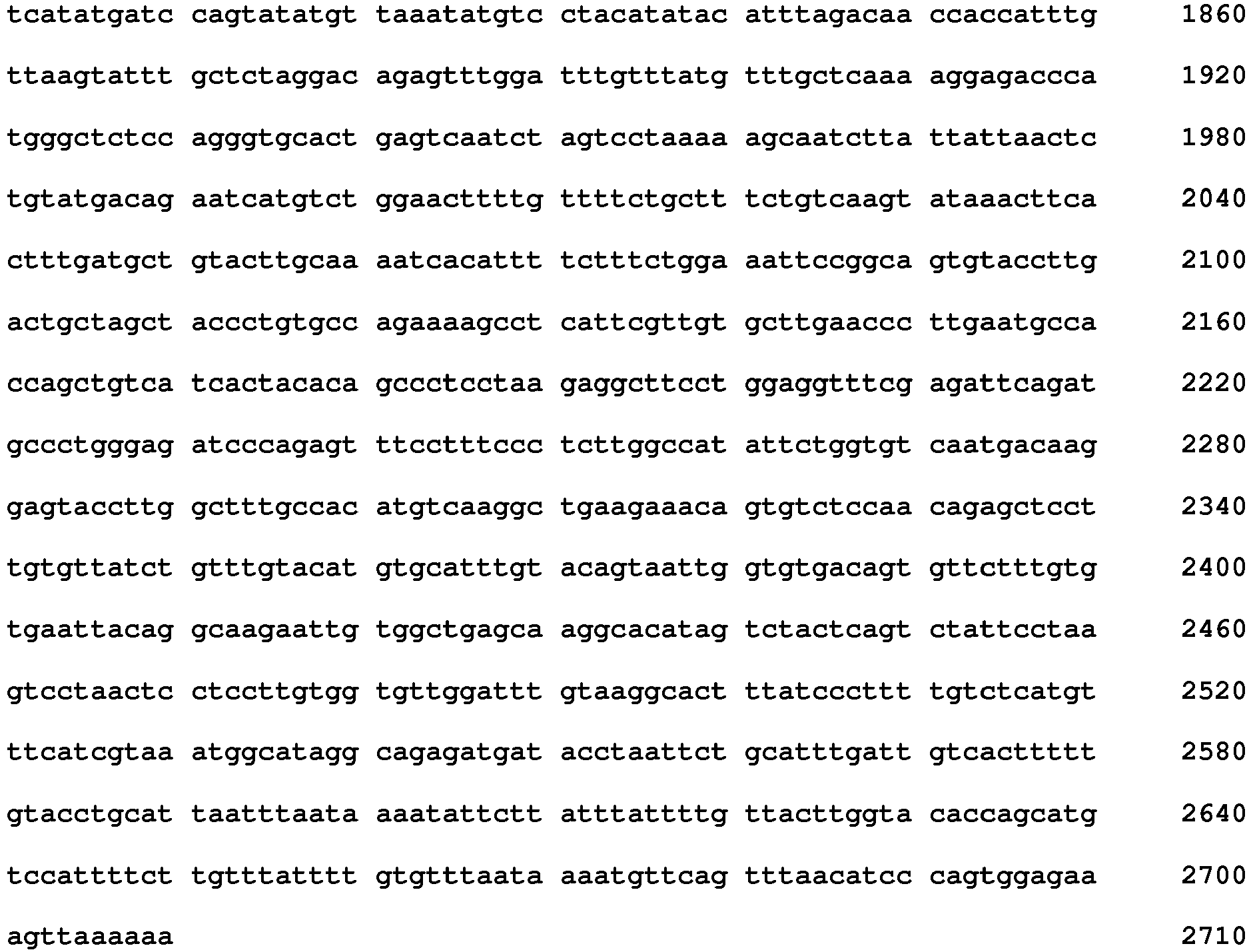
- <210> 3
<211> 50
<212> PRT
<213> Homo sapiens - <400> 3

- <210> 4
<211> 61
<212> PRT
<213> Homo sapiens - <400> 4

- <210> 5
<211> 75
<212> PRT
<213> Homo sapiens - <400> 5

- <210> 6
<211> 57
<212> PRT
<213> Homo sapiens - <400> 6


- <210> 7
<211> 39
<212> PRT
<213> Homo sapiens - <400> 7

- <210> 8
<211> 36
<212> PRT
<213> Homo sapiens - <400> 8

- <210> 9
<211> 37
<212> PRT
<213> Homo sapiens - <400> 9

- <210> 10
<211> 22
<212> DNA
<213> Artificial - <220>
<223> Primer - <400> 10
ggcatccaag atacaaactcaa 22 - <210> 11
<211> 22
<212> DNA
<213> Artificial - <220>
<223> Primer - <400> 11
cagaagttcc aatgctggat ta 22 - <210> 12
<211> 40
<212> DNA
<213> Artificial - <220>
<223> Synthetic - <400> 12
acacagctga tcacaaaaat gtaaaacata aaagtgcaat 40 - <210> 13
<211> 40
<212> DNA
<213> Artificial - <220>
<223> Synthetic - <400> 13
aaactcaaag aagcaaagtg gtaagaatat cagaaggaat 40
Claims (9)
- An in vitro method of predicting whether or not a PD-1/PD-L1 blockade is effective for treatment of a subject suffering from a malignant tumor, which comprises detecting a complete or partial deletion in 3'UTR region of PD-L1 gene in a tumor cell that has been taken from the subject and evaluating the PD-1/PD-L1 blockade as useful for the treatment of the subject when the complete or partial deletion in 3'UTR region of PD-L1 gene is present.
- The method according to claim 1, wherein the deletion in 3'UTR region of PD-L1 gene is induced by change in copy number of the PD-L1 gene.
- The method according to claim 1 or claim 2, which comprises quantifying a transcript of any one of exon 1 to exon 5 of PD-L1 gene and a transcript of 3' UTR region of PD-L1 gene and calculating a ratio between the amount of the transcript of the exon and the amount of the transcript of the 3' UTR region, and evaluating the PD-1/PD-L1 blockade as useful for the treatment of cancer when the ratio is not less than a predetermined value.
- The method according to claim 1, which comprises staining a PD-L1 protein in a tumor cell taken from the subject by immunohistochemical staining using an antibody against a C terminal region of PD-L1 and an antibody against a N terminal region of PD-L1, and evaluating the PD-1/PD-L1 blockade as useful for the treatment of the subject when the tumor cell becomes stained with the antibody against a N terminal region of PD-L1 but the tumor cell does not become stained with the antibody against a C terminal region of PD-L1.
- The method according to any one of claims 1 to 3, wherein the malignant tumor is selected from the group consisting of adult T-cell leukemia/adult T-cell leukemia lymphoma, stomach cancer, large intestinal cancer, bladder cancer, cervical cancer, renal cancer, lung adenocarcinoma, cutaneous malignant melanoma, and B cell lymphoma.
- The method according to any one of claims 1 to 3, wherein the malignant tumor is selected from the group consisting of esophageal cancer, head and neck cancer, rectal cancer, and uterine body cancer.
- The method according to any one of claims 1 to 3, wherein the PD-1/PD-L1 blockade is anti PD-1 antibody or anti PD-L1 antibody.
- The method according to claim 1, further comprising detecting a structural abnormality of tandem duplication, inversion, translocation or deletion in 9p24.1 region in association with the complete or partial deletion in 3'UTR region of PD-L1 gene.
- The method according to claim 5, wherein the malignant tumor is adult T-cell leukemia/adult T-cell leukemia lymphoma.
Applications Claiming Priority (2)
| Application Number | Priority Date | Filing Date | Title |
|---|---|---|---|
| JP2015093599 | 2015-04-30 | ||
| PCT/JP2016/063326 WO2016175275A1 (en) | 2015-04-30 | 2016-04-28 | Method for predicting therapeutic effect of pd-1/pd-l1 inhibitor using abnormality in pd-l1(cd274) as index |
Publications (3)
| Publication Number | Publication Date |
|---|---|
| EP3290517A1 EP3290517A1 (en) | 2018-03-07 |
| EP3290517A4 EP3290517A4 (en) | 2018-09-26 |
| EP3290517B1 true EP3290517B1 (en) | 2020-01-01 |
Family
ID=57199792
Family Applications (1)
| Application Number | Title | Priority Date | Filing Date |
|---|---|---|---|
| EP16786551.8A Active EP3290517B1 (en) | 2015-04-30 | 2016-04-28 | Method for predicting therapeutic effect of pd-1/pd-l1 inhibitor using abnormality in pd-l1(cd274) as index |
Country Status (5)
| Country | Link |
|---|---|
| US (1) | US10585100B2 (en) |
| EP (1) | EP3290517B1 (en) |
| JP (1) | JP6810396B2 (en) |
| ES (1) | ES2767730T3 (en) |
| WO (1) | WO2016175275A1 (en) |
Families Citing this family (14)
| Publication number | Priority date | Publication date | Assignee | Title |
|---|---|---|---|---|
| CN108112254B (en) | 2015-03-13 | 2022-01-28 | 西托姆克斯治疗公司 | anti-PDL 1 antibodies, activatable anti-PDL 1 antibodies, and methods of use thereof |
| MX383464B (en) | 2015-07-13 | 2025-03-14 | Cytomx Therapeutics Inc | ANTI-PD-1 ANTIBODIES, ACTIVATABLE ANTI-PD-1 ANTIBODIES, AND METHODS OF USING THE SAME. |
| WO2017213117A1 (en) * | 2016-06-10 | 2017-12-14 | 公益財団法人がん研究会 | Multiple staining method and staining kit |
| JP7235508B2 (en) * | 2016-11-24 | 2023-03-08 | 第一三共株式会社 | Method for Predicting Cancer Susceptibility to Treatment with PD-1 Immune Checkpoint Inhibitors |
| CN110914302A (en) | 2017-06-01 | 2020-03-24 | 赛托姆克斯治疗学股份有限公司 | Activatable anti-PDL1 antibodies and methods of using the same |
| SG11202003127WA (en) * | 2017-10-06 | 2020-05-28 | Singapore Health Serv Pte Ltd | Methods for treating lymphomas |
| JP2021510538A (en) * | 2018-01-15 | 2021-04-30 | エピアクシス セラピューティクス プロプライエタリー リミテッド | Protein molecules and their use |
| WO2019159825A1 (en) * | 2018-02-14 | 2019-08-22 | 国立大学法人名古屋大学 | Biomarker for predicting effects of anti-pd-1 antibody/anti-pd-l1 antibody therapy |
| WO2020063433A1 (en) * | 2018-09-29 | 2020-04-02 | 四川科伦博泰生物医药股份有限公司 | Anti-tumor use of anti-programmed death ligand-1 (pd-l1) antibody |
| CN113692449A (en) * | 2018-12-21 | 2021-11-23 | 新加坡科技研究局 | Methods of predicting the benefit of immune checkpoint inhibition therapy |
| TW202227643A (en) * | 2020-09-18 | 2022-07-16 | 日商小野藥品工業股份有限公司 | Prediction method for response to immune checkpoint inhibitor |
| CN117062842A (en) * | 2021-01-19 | 2023-11-14 | 昆士兰医学研究所理事会 | Novel bicyclic peptide |
| JP2024540987A (en) * | 2021-10-21 | 2024-11-06 | バーテックス ファーマシューティカルズ インコーポレイテッド | Low immune cells |
| KR20230125672A (en) * | 2022-02-21 | 2023-08-29 | 사회복지법인 삼성생명공익재단 | A composition for predicting the reactivity of immune checkpoint inhibitors in cancer patients |
Family Cites Families (8)
| Publication number | Priority date | Publication date | Assignee | Title |
|---|---|---|---|---|
| DK2439273T3 (en) | 2005-05-09 | 2019-06-03 | Ono Pharmaceutical Co | HUMAN MONOCLONAL ANTIBODIES FOR PROGRAMMED DEATH-1 (PD-1) AND PROCEDURES FOR TREATMENT OF CANCER USING ANTI-PD-1 ANTIBODIES ALONE OR IN COMBINATION WITH OTHER IMMUNTER APPLICATIONS |
| WO2008041594A1 (en) * | 2006-09-26 | 2008-04-10 | Olympus Corporation | Method for testing on sensitivity to antibody drug |
| MX2011013578A (en) | 2009-06-19 | 2012-01-20 | Merck Patent Gmbh | BIOMARKERS AND METHODS TO DETERMINE THE EFFICIENCY OF ANTI-RECEIVING ANTIBODIES OF THE EPIDERMAL GROWTH FACTOR (ANTI-EGFR) IN CANCER THERAPY. |
| US20130230866A1 (en) * | 2010-09-17 | 2013-09-05 | Tohoku University | Method for Determining Effectiveness of Medicine Containing Antibody as Component |
| JP5900489B2 (en) * | 2011-03-25 | 2016-04-06 | コニカミノルタ株式会社 | Immunohistochemical staining method and method for determining the effectiveness of an antibody drug using the same |
| KR102291355B1 (en) * | 2012-11-30 | 2021-08-19 | 에프. 호프만-라 로슈 아게 | Identification of patients in need of pd-l1 inhibitor cotherapy |
| US20150071910A1 (en) * | 2013-03-15 | 2015-03-12 | Genentech, Inc. | Biomarkers and methods of treating pd-1 and pd-l1 related conditions |
| JP6667790B2 (en) | 2014-09-19 | 2020-03-18 | 国立大学法人京都大学 | Test method for T cell lymphoma |
-
2016
- 2016-04-28 WO PCT/JP2016/063326 patent/WO2016175275A1/en not_active Ceased
- 2016-04-28 US US15/569,898 patent/US10585100B2/en active Active
- 2016-04-28 ES ES16786551T patent/ES2767730T3/en active Active
- 2016-04-28 EP EP16786551.8A patent/EP3290517B1/en active Active
- 2016-04-28 JP JP2017515602A patent/JP6810396B2/en active Active
Non-Patent Citations (1)
| Title |
|---|
| None * |
Also Published As
| Publication number | Publication date |
|---|---|
| EP3290517A1 (en) | 2018-03-07 |
| EP3290517A4 (en) | 2018-09-26 |
| US10585100B2 (en) | 2020-03-10 |
| JPWO2016175275A1 (en) | 2018-03-15 |
| ES2767730T3 (en) | 2020-06-18 |
| JP6810396B2 (en) | 2021-01-06 |
| US20180113131A1 (en) | 2018-04-26 |
| WO2016175275A1 (en) | 2016-11-03 |
Similar Documents
| Publication | Publication Date | Title |
|---|---|---|
| EP3290517B1 (en) | Method for predicting therapeutic effect of pd-1/pd-l1 inhibitor using abnormality in pd-l1(cd274) as index | |
| Wang et al. | Identifies microtubule-binding protein CSPP1 as a novel cancer biomarker associated with ferroptosis and tumor microenvironment | |
| Philpott et al. | The NF1 somatic mutational landscape in sporadic human cancers | |
| Verma et al. | Next generation sequencing-based emerging trends in molecular biology of gastric cancer | |
| JP5955557B2 (en) | Pathways underlying hereditary pancreatic tumorigenesis and hereditary pancreatic oncogenes | |
| Skeldon et al. | Patients with Lynch syndrome mismatch repair gene mutations are at higher risk for not only upper tract urothelial cancer but also bladder cancer | |
| JP6704861B2 (en) | Methods for selecting personalized triple therapies for cancer treatment | |
| US9982304B2 (en) | ARID1A and PPP2R1A mutations in cancer | |
| JPWO2016060278A1 (en) | How to predict the sensitivity of drug therapy to colorectal cancer | |
| JP2013212052A (en) | Kras variant and tumor biology | |
| Martinez-Castillo et al. | Genetic alterations in the BCR-ABL1 fusion gene related to imatinib resistance in chronic myeloid leukemia | |
| Barajas et al. | UBTF tandem duplications in pediatric myelodysplastic syndrome and acute myeloid leukemia: implications for clinical screening and diagnosis | |
| Delhorme et al. | CDX2 controls genes involved in the metabolism of 5-fluorouracil and is associated with reduced efficacy of chemotherapy in colorectal cancer | |
| EP2780473B1 (en) | Differential identification of pancreatic cysts | |
| WO2012131092A2 (en) | Method and kits for the prediction of response/nonresponse to the treatment with an anti-egfr antibody in patients with colorectal cancer of all uicc stages | |
| DE102017125780B3 (en) | Method for determining the response of a malignant disease to immunotherapy | |
| Deng et al. | DNA methylation-mediated silencing of Neuronatin promotes hepatocellular carcinoma proliferation through the PI3K-Akt signaling pathway | |
| WO2013071142A1 (en) | Biomarkers of response to proteasome inhibitors | |
| TWI651414B (en) | Biomarkers, probes, kits for detecting biological samples and methods for non-invasive qualitative determination of the degree of deterioration of endometriosis | |
| CN116457473A (en) | ER-modulating genome sleeve for use in endocrine therapy for monitoring breast cancer | |
| Cleary | Analysis of clonal mutations in cancer as a means of studying variation in somatic mutation processes | |
| Petrovics et al. | A Review of LSAMP in Tumorigenesis | |
| Wang et al. | ERBB2 mutations promote recurrence and metastasis in non-muscle-invasive bladder cancer via HIF-1 phosphorylation: insights from whole exome sequencing | |
| Vrathasha et al. | Variant-to-gene mapping identifies ARHGEF12 as a primary open-angle glaucoma effector gene operating within retinal ganglion cells | |
| Isidori | Addressing tumor heterogeneity in esophageal adenocarcinoma through different molecular approaches |
Legal Events
| Date | Code | Title | Description |
|---|---|---|---|
| STAA | Information on the status of an ep patent application or granted ep patent |
Free format text: STATUS: THE INTERNATIONAL PUBLICATION HAS BEEN MADE |
|
| PUAI | Public reference made under article 153(3) epc to a published international application that has entered the european phase |
Free format text: ORIGINAL CODE: 0009012 |
|
| STAA | Information on the status of an ep patent application or granted ep patent |
Free format text: STATUS: REQUEST FOR EXAMINATION WAS MADE |
|
| 17P | Request for examination filed |
Effective date: 20171130 |
|
| AK | Designated contracting states |
Kind code of ref document: A1 Designated state(s): AL AT BE BG CH CY CZ DE DK EE ES FI FR GB GR HR HU IE IS IT LI LT LU LV MC MK MT NL NO PL PT RO RS SE SI SK SM TR |
|
| AX | Request for extension of the european patent |
Extension state: BA ME |
|
| DAV | Request for validation of the european patent (deleted) | ||
| DAX | Request for extension of the european patent (deleted) | ||
| REG | Reference to a national code |
Ref country code: DE Ref legal event code: R079 Ref document number: 602016027420 Country of ref document: DE Free format text: PREVIOUS MAIN CLASS: C12N0015090000 Ipc: G01N0033574000 |
|
| RIC1 | Information provided on ipc code assigned before grant |
Ipc: A61P 35/02 20060101ALI20180816BHEP Ipc: C12N 15/09 20060101ALI20180816BHEP Ipc: G01N 33/53 20060101ALI20180816BHEP Ipc: A61K 45/00 20060101ALI20180816BHEP Ipc: C07K 16/18 20060101ALI20180816BHEP Ipc: A61P 35/00 20060101ALI20180816BHEP Ipc: C12Q 1/68 20180101ALI20180816BHEP Ipc: A61K 39/395 20060101ALI20180816BHEP Ipc: G01N 33/574 20060101AFI20180816BHEP |
|
| A4 | Supplementary search report drawn up and despatched |
Effective date: 20180824 |
|
| GRAP | Despatch of communication of intention to grant a patent |
Free format text: ORIGINAL CODE: EPIDOSNIGR1 |
|
| STAA | Information on the status of an ep patent application or granted ep patent |
Free format text: STATUS: GRANT OF PATENT IS INTENDED |
|
| INTG | Intention to grant announced |
Effective date: 20190723 |
|
| GRAS | Grant fee paid |
Free format text: ORIGINAL CODE: EPIDOSNIGR3 |
|
| GRAA | (expected) grant |
Free format text: ORIGINAL CODE: 0009210 |
|
| STAA | Information on the status of an ep patent application or granted ep patent |
Free format text: STATUS: THE PATENT HAS BEEN GRANTED |
|
| AK | Designated contracting states |
Kind code of ref document: B1 Designated state(s): AL AT BE BG CH CY CZ DE DK EE ES FI FR GB GR HR HU IE IS IT LI LT LU LV MC MK MT NL NO PL PT RO RS SE SI SK SM TR |
|
| REG | Reference to a national code |
Ref country code: GB Ref legal event code: FG4D |
|
| REG | Reference to a national code |
Ref country code: CH Ref legal event code: EP Ref country code: AT Ref legal event code: REF Ref document number: 1220423 Country of ref document: AT Kind code of ref document: T Effective date: 20200115 |
|
| REG | Reference to a national code |
Ref country code: DE Ref legal event code: R096 Ref document number: 602016027420 Country of ref document: DE |
|
| REG | Reference to a national code |
Ref country code: IE Ref legal event code: FG4D |
|
| REG | Reference to a national code |
Ref country code: NL Ref legal event code: MP Effective date: 20200101 |
|
| REG | Reference to a national code |
Ref country code: ES Ref legal event code: FG2A Ref document number: 2767730 Country of ref document: ES Kind code of ref document: T3 Effective date: 20200618 |
|
| REG | Reference to a national code |
Ref country code: LT Ref legal event code: MG4D |
|
| PG25 | Lapsed in a contracting state [announced via postgrant information from national office to epo] |
Ref country code: NL Free format text: LAPSE BECAUSE OF FAILURE TO SUBMIT A TRANSLATION OF THE DESCRIPTION OR TO PAY THE FEE WITHIN THE PRESCRIBED TIME-LIMIT Effective date: 20200101 Ref country code: PT Free format text: LAPSE BECAUSE OF FAILURE TO SUBMIT A TRANSLATION OF THE DESCRIPTION OR TO PAY THE FEE WITHIN THE PRESCRIBED TIME-LIMIT Effective date: 20200527 Ref country code: CZ Free format text: LAPSE BECAUSE OF FAILURE TO SUBMIT A TRANSLATION OF THE DESCRIPTION OR TO PAY THE FEE WITHIN THE PRESCRIBED TIME-LIMIT Effective date: 20200101 Ref country code: LT Free format text: LAPSE BECAUSE OF FAILURE TO SUBMIT A TRANSLATION OF THE DESCRIPTION OR TO PAY THE FEE WITHIN THE PRESCRIBED TIME-LIMIT Effective date: 20200101 Ref country code: NO Free format text: LAPSE BECAUSE OF FAILURE TO SUBMIT A TRANSLATION OF THE DESCRIPTION OR TO PAY THE FEE WITHIN THE PRESCRIBED TIME-LIMIT Effective date: 20200401 Ref country code: RS Free format text: LAPSE BECAUSE OF FAILURE TO SUBMIT A TRANSLATION OF THE DESCRIPTION OR TO PAY THE FEE WITHIN THE PRESCRIBED TIME-LIMIT Effective date: 20200101 Ref country code: FI Free format text: LAPSE BECAUSE OF FAILURE TO SUBMIT A TRANSLATION OF THE DESCRIPTION OR TO PAY THE FEE WITHIN THE PRESCRIBED TIME-LIMIT Effective date: 20200101 |
|
| PG25 | Lapsed in a contracting state [announced via postgrant information from national office to epo] |
Ref country code: IS Free format text: LAPSE BECAUSE OF FAILURE TO SUBMIT A TRANSLATION OF THE DESCRIPTION OR TO PAY THE FEE WITHIN THE PRESCRIBED TIME-LIMIT Effective date: 20200501 Ref country code: HR Free format text: LAPSE BECAUSE OF FAILURE TO SUBMIT A TRANSLATION OF THE DESCRIPTION OR TO PAY THE FEE WITHIN THE PRESCRIBED TIME-LIMIT Effective date: 20200101 Ref country code: GR Free format text: LAPSE BECAUSE OF FAILURE TO SUBMIT A TRANSLATION OF THE DESCRIPTION OR TO PAY THE FEE WITHIN THE PRESCRIBED TIME-LIMIT Effective date: 20200402 Ref country code: LV Free format text: LAPSE BECAUSE OF FAILURE TO SUBMIT A TRANSLATION OF THE DESCRIPTION OR TO PAY THE FEE WITHIN THE PRESCRIBED TIME-LIMIT Effective date: 20200101 Ref country code: SE Free format text: LAPSE BECAUSE OF FAILURE TO SUBMIT A TRANSLATION OF THE DESCRIPTION OR TO PAY THE FEE WITHIN THE PRESCRIBED TIME-LIMIT Effective date: 20200101 Ref country code: BG Free format text: LAPSE BECAUSE OF FAILURE TO SUBMIT A TRANSLATION OF THE DESCRIPTION OR TO PAY THE FEE WITHIN THE PRESCRIBED TIME-LIMIT Effective date: 20200401 |
|
| REG | Reference to a national code |
Ref country code: DE Ref legal event code: R097 Ref document number: 602016027420 Country of ref document: DE |
|
| PG25 | Lapsed in a contracting state [announced via postgrant information from national office to epo] |
Ref country code: EE Free format text: LAPSE BECAUSE OF FAILURE TO SUBMIT A TRANSLATION OF THE DESCRIPTION OR TO PAY THE FEE WITHIN THE PRESCRIBED TIME-LIMIT Effective date: 20200101 Ref country code: SM Free format text: LAPSE BECAUSE OF FAILURE TO SUBMIT A TRANSLATION OF THE DESCRIPTION OR TO PAY THE FEE WITHIN THE PRESCRIBED TIME-LIMIT Effective date: 20200101 Ref country code: DK Free format text: LAPSE BECAUSE OF FAILURE TO SUBMIT A TRANSLATION OF THE DESCRIPTION OR TO PAY THE FEE WITHIN THE PRESCRIBED TIME-LIMIT Effective date: 20200101 Ref country code: RO Free format text: LAPSE BECAUSE OF FAILURE TO SUBMIT A TRANSLATION OF THE DESCRIPTION OR TO PAY THE FEE WITHIN THE PRESCRIBED TIME-LIMIT Effective date: 20200101 Ref country code: SK Free format text: LAPSE BECAUSE OF FAILURE TO SUBMIT A TRANSLATION OF THE DESCRIPTION OR TO PAY THE FEE WITHIN THE PRESCRIBED TIME-LIMIT Effective date: 20200101 |
|
| PLBE | No opposition filed within time limit |
Free format text: ORIGINAL CODE: 0009261 |
|
| STAA | Information on the status of an ep patent application or granted ep patent |
Free format text: STATUS: NO OPPOSITION FILED WITHIN TIME LIMIT |
|
| REG | Reference to a national code |
Ref country code: AT Ref legal event code: MK05 Ref document number: 1220423 Country of ref document: AT Kind code of ref document: T Effective date: 20200101 |
|
| PG25 | Lapsed in a contracting state [announced via postgrant information from national office to epo] |
Ref country code: MC Free format text: LAPSE BECAUSE OF FAILURE TO SUBMIT A TRANSLATION OF THE DESCRIPTION OR TO PAY THE FEE WITHIN THE PRESCRIBED TIME-LIMIT Effective date: 20200101 |
|
| REG | Reference to a national code |
Ref country code: CH Ref legal event code: PL |
|
| 26N | No opposition filed |
Effective date: 20201002 |
|
| PG25 | Lapsed in a contracting state [announced via postgrant information from national office to epo] |
Ref country code: CH Free format text: LAPSE BECAUSE OF NON-PAYMENT OF DUE FEES Effective date: 20200430 Ref country code: LU Free format text: LAPSE BECAUSE OF NON-PAYMENT OF DUE FEES Effective date: 20200428 Ref country code: AT Free format text: LAPSE BECAUSE OF FAILURE TO SUBMIT A TRANSLATION OF THE DESCRIPTION OR TO PAY THE FEE WITHIN THE PRESCRIBED TIME-LIMIT Effective date: 20200101 Ref country code: LI Free format text: LAPSE BECAUSE OF NON-PAYMENT OF DUE FEES Effective date: 20200430 |
|
| REG | Reference to a national code |
Ref country code: BE Ref legal event code: MM Effective date: 20200430 |
|
| PG25 | Lapsed in a contracting state [announced via postgrant information from national office to epo] |
Ref country code: BE Free format text: LAPSE BECAUSE OF NON-PAYMENT OF DUE FEES Effective date: 20200430 Ref country code: PL Free format text: LAPSE BECAUSE OF FAILURE TO SUBMIT A TRANSLATION OF THE DESCRIPTION OR TO PAY THE FEE WITHIN THE PRESCRIBED TIME-LIMIT Effective date: 20200101 Ref country code: SI Free format text: LAPSE BECAUSE OF FAILURE TO SUBMIT A TRANSLATION OF THE DESCRIPTION OR TO PAY THE FEE WITHIN THE PRESCRIBED TIME-LIMIT Effective date: 20200101 |
|
| PG25 | Lapsed in a contracting state [announced via postgrant information from national office to epo] |
Ref country code: IE Free format text: LAPSE BECAUSE OF NON-PAYMENT OF DUE FEES Effective date: 20200428 |
|
| PG25 | Lapsed in a contracting state [announced via postgrant information from national office to epo] |
Ref country code: TR Free format text: LAPSE BECAUSE OF FAILURE TO SUBMIT A TRANSLATION OF THE DESCRIPTION OR TO PAY THE FEE WITHIN THE PRESCRIBED TIME-LIMIT Effective date: 20200101 Ref country code: MT Free format text: LAPSE BECAUSE OF FAILURE TO SUBMIT A TRANSLATION OF THE DESCRIPTION OR TO PAY THE FEE WITHIN THE PRESCRIBED TIME-LIMIT Effective date: 20200101 Ref country code: CY Free format text: LAPSE BECAUSE OF FAILURE TO SUBMIT A TRANSLATION OF THE DESCRIPTION OR TO PAY THE FEE WITHIN THE PRESCRIBED TIME-LIMIT Effective date: 20200101 |
|
| PG25 | Lapsed in a contracting state [announced via postgrant information from national office to epo] |
Ref country code: MK Free format text: LAPSE BECAUSE OF FAILURE TO SUBMIT A TRANSLATION OF THE DESCRIPTION OR TO PAY THE FEE WITHIN THE PRESCRIBED TIME-LIMIT Effective date: 20200101 Ref country code: AL Free format text: LAPSE BECAUSE OF FAILURE TO SUBMIT A TRANSLATION OF THE DESCRIPTION OR TO PAY THE FEE WITHIN THE PRESCRIBED TIME-LIMIT Effective date: 20200101 |
|
| PGFP | Annual fee paid to national office [announced via postgrant information from national office to epo] |
Ref country code: DE Payment date: 20250422 Year of fee payment: 10 |
|
| PGFP | Annual fee paid to national office [announced via postgrant information from national office to epo] |
Ref country code: GB Payment date: 20250423 Year of fee payment: 10 Ref country code: ES Payment date: 20250530 Year of fee payment: 10 |
|
| PGFP | Annual fee paid to national office [announced via postgrant information from national office to epo] |
Ref country code: IT Payment date: 20250424 Year of fee payment: 10 |
|
| PGFP | Annual fee paid to national office [announced via postgrant information from national office to epo] |
Ref country code: FR Payment date: 20250425 Year of fee payment: 10 |
|
| REG | Reference to a national code |
Ref country code: DE Ref legal event code: R079 Ref document number: 602016027420 Country of ref document: DE Free format text: PREVIOUS MAIN CLASS: G01N0033574000 Ipc: G01N0033575000 |