EP3276005B1 - Composition and method for affecting the binding of antigen-binding polypeptides to antigens - Google Patents
Composition and method for affecting the binding of antigen-binding polypeptides to antigens Download PDFInfo
- Publication number
- EP3276005B1 EP3276005B1 EP16181971.9A EP16181971A EP3276005B1 EP 3276005 B1 EP3276005 B1 EP 3276005B1 EP 16181971 A EP16181971 A EP 16181971A EP 3276005 B1 EP3276005 B1 EP 3276005B1
- Authority
- EP
- European Patent Office
- Prior art keywords
- calmodulin
- binding
- polypeptide
- seq
- permutated
- Prior art date
- Legal status (The legal status is an assumption and is not a legal conclusion. Google has not performed a legal analysis and makes no representation as to the accuracy of the status listed.)
- Active
Links
Images







Classifications
-
- C—CHEMISTRY; METALLURGY
- C07—ORGANIC CHEMISTRY
- C07K—PEPTIDES
- C07K14/00—Peptides having more than 20 amino acids; Gastrins; Somatostatins; Melanotropins; Derivatives thereof
- C07K14/435—Peptides having more than 20 amino acids; Gastrins; Somatostatins; Melanotropins; Derivatives thereof from animals; from humans
- C07K14/46—Peptides having more than 20 amino acids; Gastrins; Somatostatins; Melanotropins; Derivatives thereof from animals; from humans from vertebrates
- C07K14/47—Peptides having more than 20 amino acids; Gastrins; Somatostatins; Melanotropins; Derivatives thereof from animals; from humans from vertebrates from mammals
- C07K14/4701—Peptides having more than 20 amino acids; Gastrins; Somatostatins; Melanotropins; Derivatives thereof from animals; from humans from vertebrates from mammals not used
- C07K14/4728—Calcium binding proteins, e.g. calmodulin
-
- C—CHEMISTRY; METALLURGY
- C12—BIOCHEMISTRY; BEER; SPIRITS; WINE; VINEGAR; MICROBIOLOGY; ENZYMOLOGY; MUTATION OR GENETIC ENGINEERING
- C12N—MICROORGANISMS OR ENZYMES; COMPOSITIONS THEREOF; PROPAGATING, PRESERVING, OR MAINTAINING MICROORGANISMS; MUTATION OR GENETIC ENGINEERING; CULTURE MEDIA
- C12N9/00—Enzymes; Proenzymes; Compositions thereof; Processes for preparing, activating, inhibiting, separating or purifying enzymes
- C12N9/10—Transferases (2.)
- C12N9/12—Transferases (2.) transferring phosphorus containing groups, e.g. kinases (2.7)
-
- C—CHEMISTRY; METALLURGY
- C07—ORGANIC CHEMISTRY
- C07K—PEPTIDES
- C07K16/00—Immunoglobulins [IG], e.g. monoclonal or polyclonal antibodies
- C07K16/40—Immunoglobulins [IG], e.g. monoclonal or polyclonal antibodies against enzymes
-
- C—CHEMISTRY; METALLURGY
- C12—BIOCHEMISTRY; BEER; SPIRITS; WINE; VINEGAR; MICROBIOLOGY; ENZYMOLOGY; MUTATION OR GENETIC ENGINEERING
- C12Y—ENZYMES
- C12Y207/00—Transferases transferring phosphorus-containing groups (2.7)
- C12Y207/11—Protein-serine/threonine kinases (2.7.11)
- C12Y207/11018—Myosin-light-chain kinase (2.7.11.18)
-
- C—CHEMISTRY; METALLURGY
- C07—ORGANIC CHEMISTRY
- C07K—PEPTIDES
- C07K2317/00—Immunoglobulins specific features
- C07K2317/50—Immunoglobulins specific features characterized by immunoglobulin fragments
- C07K2317/56—Immunoglobulins specific features characterized by immunoglobulin fragments variable (Fv) region, i.e. VH and/or VL
-
- C—CHEMISTRY; METALLURGY
- C07—ORGANIC CHEMISTRY
- C07K—PEPTIDES
- C07K2317/00—Immunoglobulins specific features
- C07K2317/60—Immunoglobulins specific features characterized by non-natural combinations of immunoglobulin fragments
- C07K2317/62—Immunoglobulins specific features characterized by non-natural combinations of immunoglobulin fragments comprising only variable region components
- C07K2317/622—Single chain antibody (scFv)
-
- C—CHEMISTRY; METALLURGY
- C07—ORGANIC CHEMISTRY
- C07K—PEPTIDES
- C07K2319/00—Fusion polypeptide
- C07K2319/01—Fusion polypeptide containing a localisation/targetting motif
- C07K2319/02—Fusion polypeptide containing a localisation/targetting motif containing a signal sequence
-
- C—CHEMISTRY; METALLURGY
- C07—ORGANIC CHEMISTRY
- C07K—PEPTIDES
- C07K2319/00—Fusion polypeptide
- C07K2319/01—Fusion polypeptide containing a localisation/targetting motif
- C07K2319/03—Fusion polypeptide containing a localisation/targetting motif containing a transmembrane segment
Definitions
- the present invention relates to the field of binding modulation of antigen-binding polypeptides, in particular to a calmodulin-linker-based system for said modulation.
- Antigen-binding polypeptides such as single chain variable fragments (scFv) comprise of the variable domains of the light (V L ) and heavy (V H ) chain of a corresponding full-length antibody.
- V L variable domains of the light
- V H heavy chain
- scTv structurally similar T cell receptors
- both chains are normally connected by a linker which is flexible and does not show any tendency to interfere with folding of the individual immunoglobulin domains.
- these linkers contain assemblies or variations of (Gly 4 Ser) repeats, inspired by the unstructured linkers connecting the domains of filamentous bacteriophage minor coat protein III.
- ScFv antibody fragments are widely used in a variety of applications, such as for research, diagnostic purposes and even as therapeutics.
- Immunotoxins which are used for cancer therapy, are often based on a single chain fragment fused to a bacterial toxin to mediate targeted killing.
- Another approach is based on bispecific antibodies (BiTEs, bispecific T cell engagers) which activate and redirect cytotoxic T cells against cancer cells.
- CAR (chimeric antigen receptor)-T cell therapy also relies on scFvs specific for malignant cells. Essential for all of these applications is the extraordinary specificity, selectivity and affinity of antibody paratopes. These properties would also be very useful for the purification of biomaterials, in particular proteins, vaccines or cells.
- Meister, G. E. & Joshi, N. S. disclose a switchable enzyme which bases on the interaction of WT-calmodulin and soluble M13 peptide. In the peptide-bound form the enzyme exhibits an up to 120 times higher catalytic activity compared to the inactive (no peptide bound) state.
- WO2002014371A1 discloses Fv constructs having an affinity that can be influenced for a substance to be linked, wherein the Fv constructs have peptides linked to the variable regions and containing binding sites for effector molecules.
- the effector molecules are ions or antibodies.
- Guntas, G. et al. disclose the creation of a molecular switch of the enzyme TEM1 ⁇ -lactamase by circularly permutating the gene encoding the enzyme TEM1 ⁇ -lactamase and subsequently inserting it into the gene encoding E. coli maltose binding protein which functions as the linker.
- WO2005/072392A2 discloses molecular switches, for example with switching activity greater than previously demonstrated, or with altered ligand recognition and binding, and methods of making these molecules involving circular permutation of nucleic acid or amino acid sequences.
- Molecular switches have been created by recombining non-homologous genes in vitro and subjecting the genes to evolutionary pressure using combinatorial techniques. The approach is envisioned as "rolling" two proteins across each other's surfaces and fusing them at points where their surfaces meet. The approach allows for recombination and testing of maximal numbers of geometric configurations between the two domains. Libraries comprising vast numbers of such fused molecules are provided from which molecular switches with optimal characteristics can be isolated.
- certain locations within GFP tolerate insertion of entire proteins, and conformational changes in the insert can have profound effects on the fluorescence.
- insertions of calmodulin or a zinc finger domain in place of Tyr-145 of a yellow mutant (enhanced yellow fluorescent protein) of GFP result in indicator proteins whose fluorescence can be enhanced several-fold upon metal binding.
- the calmodulin graft into enhanced yellow fluorescent protein can monitor cytosolic Ca 2+ in single mammalian cells.
- Calmodulin undergoes large conformational changes, depending on the presence of calcium and calmodulin-binding peptides (CBP). In a calcium- and peptide-unbound form, it adopts a closed conformation ( Kuboniwa, H. et al., 1995, Nat Struct Biol 2, 768-776 ). The distance between the N- and C-terminus is at its highest in the calcium-bound, open form ( Chattopadhyaya, R. et al., 1992, J Mol Biol 228, 1177-1192 ), whereas the termini approach each other when calmodulin binds to a ligand, or a suitable fragment thereof, like peptide M13 ( Ikura, M. et al., 1992, Science 256, 632-638 ).
- a polypeptide comprising calmodulin (CaM) and two immunoglobulin superfamily domains, wherein said two immunoglobulin superfamily domains are linked via said calmodulin can be used to affect in both directions the binding of said polypeptide to its antigen by contacting said polypeptide with a calmodulin binding molecule and ions binding to the Ca 2+ binding site of calmodulin.
- calmodulin linker a polypeptide comprising calmodulin (CaM) and two immunoglobulin superfamily domains, wherein said two immunoglobulin superfamily domains are linked via said calmodulin
- the system comprising the three parts i) the polypeptide with CaM as linker between the two immunoglobulin superfamily domains, ii) the CaM binding molecule such as a CaM-binding peptide (e.g. M13 peptide), and iii) ions binding to the Ca 2+ binding site of CaM such as Ca 2+ is superior compared to systems known in the art: It is not a prerequisite for a functional system to eliminate calcium ions from the solution comprising said polypeptide before switching the binding of the polypeptide to its antigen. The switch is only achieved by adding soluble CaM binding molecules such as M13 peptide to the solution in the presence of ions binding to the Ca 2+ binding site of CaM. This is superior compared to systems known in the art, particular for the use in living organisms, for example when used to modulate the affinity of a CAR on a T cell or other suitable effector cell, as in this situation, the Ca 2+ concentration may not be sufficiently adjustable.
- the CaM binding molecule such as
- the change in binding of the antigen binding domains of the polypeptide triggered by the binding of a CaM binding molecule and ions to the calmodulin linker of the polypeptide can result either in an enhanced binding or in a reduced binding of the polypeptide to its antigen.
- Polypeptides as disclosed herein can be released from the binding antigen by adding CaM binding molecules such as M13 peptide and ions such as Ca 2+ without harsh conditions.
- calmodulin sequence as a universal linker to regulate the binding of a polypeptide comprising two immunoglobulin superfamily domains has been demonstrated herein firstly for different scFvs, including one specific for lysozyme, and secondly other scFvs with quite different affinities and antigen classes, including proteins and haptens.
- compositions comprising the above mentioned components, methods for affecting the binding of the polypeptides, and the use of the polypeptides for affecting the binding of the polypeptides to their antigens are disclosed.
- composition (a system, a set, or a kit) comprising
- the calmodulin is a linker sequence between the two immunoglobulin superfamily domains and serves as a universal allosteric regulator of these two domains.
- the CaM (or the CaM sequence) may be any sequence or part of a sequence of CaM which maintains the characteristics of the WT calmodulin protein to bind both, ions at the Ca 2+ binding site and a calmodulin binding molecule, and thereby changing its conformation.
- the sequence of calmodulin may also be a functional fragment of the full-length calmodulin protein (e.g. a truncated protein of calmodulin) or a fragment of the full-length calmodulin protein having a sequence identity of at least 70%, or at least 75%, or at least 80%, or at least 85%, or at least 90%, or at least 95%, or at least 97%, or at least 98%, or at least 99% at the amino acid sequence level to the corresponding part of said full-length calmodulin.
- a functional fragment of the full-length calmodulin protein e.g. a truncated protein of calmodulin
- a fragment of the full-length calmodulin protein having a sequence identity of at least 70%, or at least 75%, or at least 80%, or at least 85%, or at least 90%, or at least 95%, or at least 97%, or at least 98%, or at least 99% at the amino acid sequence level to the corresponding part of said full-
- composition as disclosed above wherein said binding of said calmodulin-binding molecule and of said ions to the Ca 2+ binding site of calmodulin enhances or reduces the binding of said polypeptide to said antigen.
- Said calmodulin binding molecule may be a calmodulin binding peptide.
- Said calmodulin binding peptide may be selected and derived from the group of naturally occurring calmodulin ligands consisting of myosin light chain kinase, caldesmon, calspermin, phosphofructokinase, calcineurin, calcium ATPase, spectrin, glutamate receptor, nitric oxidase synthase, serine/threonine-protein phosphatase, tumor necrosis factor receptor, estrogen receptor, calcium channel subunits and calcium/calmodulin-dependent protein kinases.
- Said CaM binding peptide may be M13 peptide derived from the rabbit myosin light chain kinase or a variant thereof.
- Said calmodulin binding peptide may be selected from the group of peptides consisting of SEQ ID NO: 1 to SEQ ID NO: 65.
- said calmodulin binding peptide may be selected from the group of peptides consisting of SEQ ID NO: 1, SEQ ID NO: 2, SEQ ID NO: 3, SEQ ID NO: 7, SEQ ID NO: 8, SEQ ID NO: 9, SEQ ID NO: 46, SEQ ID NO: 47, SEQ ID NO: 49, SEQ ID NO: 51 and SEQ ID NO: 53.
- any peptide or polypeptide which can bind to CaM and thereby provides the allosteric change may be used.
- calmodulin in complex with high-affinity peptidic substrates are available ( Montigiani et al., 996, J. Mol. Biol. 258:6-13 ). These peptides correspond to the calmodulin-binding regions of different protein kinases.
- methods such as peptide phage display, ribosome display or other established combinatorial selection systems well known in the art (see e.g. Ullman CG et al. Brief Funct Genomics, 2011 May;10(3):125-34 or Galán A et al. Mol Biosyst, 2016 Jun 16 . [Epub ahead of print]) may be used to identify variants of naturally occurring sequences or synthetic sequences which can bind to calmodulin.
- Said ions binding to the Ca 2+ binding site of calmodulin may be any ions that can be bound by the Ca 2+ binding site of calmodulin resulting in a conformational change of the CaM in the presence of a CaM binding molecule, preferentially said ions are calcium ions (Ca 2+ ).
- Said calmodulin may be a permutated calmodulin.
- a permutated CaM can be generated e.g. by circular permutation, a general method for permutating proteins which is well known in the art (see e.g. " Circular Permutation of Proteins" in "Protein Engineering Handbook” (Ed.: Stefan Lutz, Uwe T. Bornscheuer) Wiley-VCH 2009 ), and as described herein.
- a permutated CaM may be generated synthetically on nucleic acid or amino acid level, especially it may be generated synthetically when the sequence which is intended to use for the generation of the polypeptide as disclosed herein comprising the permutated CaM is known and can be generated purposefully.
- Said polypeptide of said composition comprising said permutated calmodulin and said two immunoglobulin superfamily domains, wherein said two immunoglobulin superfamily domains are linked via said permutated calmodulin may be obtained e.g. by the method comprising
- the permutated calmodulin of the composition as described above may be selected from the group consisting of SEQ ID NO: 67 to SEQ ID NO: 123.
- the permutated calmodulin of the composition as described above may be selected from the group consisting of SEQ ID NO: 67 to SEQ ID NO: 72.
- the permutated calmodulin may be selected from the group consisting of SEQ ID NO: 67, SEQ ID NO: 68, SEQ ID NO: 71 and SEQ ID NO: 72.
- composition as disclosed herein may comprise
- composition wherein said polypeptide may be a single chain Fv (scFv) comprising the calmodulin and a variable region of a heavy chain of an immunoglobulin (V H ) and a variable region of a light chain of an immunoglobulin (V L ).
- scFv single chain Fv
- composition as described above, wherein the polypeptide comprising a scFv specific for the antigen CD14 and a permutated CaM has the sequence of SEQ ID NO: 124 and said CaM binding peptide has the sequence of SEQ ID NO: 1 or SEQ ID NO: 7 or SEQ ID NO: 8 or SEQ ID NO: 9, or said polypeptide has the sequence of SEQ ID NO: 125 and said CaM binding peptide has the sequence of SEQ ID NO: 1 or SEQ ID NO: 7 or SEQ ID NO: 8 or SEQ ID NO: 9 or SEQ ID NO: 53, or said polypeptide has the sequence of SEQ ID NO: 126 and said CaM binding peptide has the sequence of SEQ ID NO: 1 or SEQ ID NO: 47, or said polypeptide has the sequence of SEQ ID NO: 127 and said CaM binding peptide has the sequence of SEQ ID NO: 1 or SEQ ID NO: 47 or SEQ ID NO: 53.
- composition as described above, wherein the polypeptide comprising a scFv specific for biotin and a permutated CaM has the sequence of SEQ ID NO: 128 and said CaM binding peptide has the sequence of SEQ ID NO: 1 or SEQ ID NO: 46, or said polypeptide has the sequence of SEQ ID NO: 129 and said CaM binding peptide has the sequence of SEQ ID NO: 1 or SEQ ID NO: 46, or said polypeptide has the sequence of SEQ ID NO: 130 and said CaM binding peptide has the sequence of SEQ ID NO: 1 or SEQ ID NO: 53.
- composition as described above, wherein the polypeptide comprising a scFv specific for the antigen CD4 and a permutated CaM has the sequence of SEQ ID NO: 131 and said CaM binding peptide has the sequence of SEQ ID NO: 1 or SEQ ID NO: 46, or said polypeptide has the sequence of SEQ ID NO: 132 and said CaM binding peptide has the sequence of SEQ ID NO: 1 or SEQ ID NO: 46, or said polypeptide has the sequence of SEQ ID NO: 133 and said CaM binding peptide has the sequence of SEQ ID NO: 1 or SEQ ID NO: 51 or SEQ ID NO: 53, or said polypeptide has the sequence of SEQ ID NO: 134 and said CaM binding peptide has the sequence of SEQ ID NO: 1 or SEQ ID NO: 51 or SEQ ID NO: 53.
- composition wherein said polypeptide is part of an antigen binding domain of a chimeric antigen receptor (CAR).
- CAR chimeric antigen receptor
- Said CAR may comprise an antigen binding domain, a transmembrane domain and cytoplasmic signaling domain(s).
- the invention provides a method for affecting the binding of a polypeptide to an antigen to be bound, wherein said polypeptide comprises a calmodulin and two immunoglobulin superfamily domains wherein said two immunoglobulin superfamily domains are linked via said calmodulin, the method comprising the step of contacting said polypeptide with a calmodulin binding molecule in the presence of ions binding to the Ca 2+ binding site of calmodulin, thereby affecting the binding of said polypeptide to the antigen.
- the CaM is a permutated CaM as described above.
- the CaM binding molecule is a CaM binding peptide as described above.
- Said method for affecting the binding of a polypeptide wherein before said contacting of said calmodulin binding molecule with said calmodulin said polypeptide is contacted with the antigen to be bound by said polypeptide, and wherein said contacting of said calmodulin binding molecule to said calmodulin in the presence of ions binding to the Ca 2+ binding site of calmodulin affects the binding of said polypeptide to the antigen.
- Said affecting of the binding may be a reduction (decrease) of binding, thereby releasing the polypeptide from the antigen.
- said affecting of the binding may be an enhancement (an increase) of binding, thereby enhancing the binding between said polypeptide and the antigen. Enhancing said binding may be of interest when the binding of said polypeptide to the antigen is weak or does not occur in the absence of said CaM binding molecule and said ions binding to the Ca 2+ binding site of CaM.
- Said affecting of the binding may be a change of one or both of the kinetic properties of the binding reaction (the association and dissociation rate constants, or on-rate and off-rate) while overall affinity may be changed (affecting the affinity) in this process or remain the same (affecting the binding kinetics but not the equilibrium affinity), thereby changing the kinetics of the interaction of the polypeptide with the antigen.
- Said affecting of binding may be an allosteric change induced within the antigen.
- the invention provides the use of a polypeptide (as described above) comprising a calmodulin, preferentially a permutated calmodulin, and two immunoglobulin superfamily domains, wherein said two immunoglobulin superfamily domains are linked via said calmodulin, preferentially said permutated calmodulin, for affecting in the presence of a calmodulin binding molecule and ions binding to the Ca 2+ binding site of calmodulin the binding of said polypeptide to an antigen to be bound by said polypeptide.
- the composition may be used for enrichment (e.g. positive selection) of cells expressing on the cell surface the antigen recognized by the polypeptide, e.g. as disclosed herein.
- Methods suited for enrichment are well known in the art and include but are not limited to flow cytometry such as fluorescence activated cell sorting (FACS) or magnetic cell separation such as MACS® (Miltenyi Biotec GmbH).
- the polypeptide as disclosed herein, specific for an antigen can be used for direct or indirect magnetic labeling of cells expressing said antigen on their cell surface in a sample comprising said cells and other cells (not expressing said antigen).
- the antigen-expressing cells are magnetically labeled with MicroBeads (magnetic particles) conjugated to said polypeptide.
- the cell sample is loaded on a MACS® Column which is placed in the magnetic field of a MACS® Separator.
- the magnetically labeled antigen-expressing cells are retained on the column.
- the unlabeled cells run through.
- an "elution” solution comprising a CaM binding molecule such as M13 peptide and e.g. Ca 2+ ions allow to reduce the binding of the polypeptide to the antigen, thereby releasing the cell expressing said antigen from the immobilized polypeptide conjugated to the magnetic particle, i.e. the cell can be eluted from the column without the need of removal of the magnetic field.
- a CaM binding molecule such as M13 peptide and e.g. Ca 2+ ions
- the composition may be used for the enrichment (i.e. purification) of proteins fused to an antigen recognized by the polypeptide comprising a scFv comprising calmodulin, preferentially a permutated CaM, and a variable region of a heavy chain of an immunoglobulin and a variable region of a light chain of an immunoglobulin, wherein said variable regions are linked via said calmodulin, preferentially permutated CaM.
- the polypeptide invention as described herein may be immobilized e.g. on a resin.
- target protein i.e.
- an "elution" solution comprising a CaM binding molecule such as M13 peptide and e.g. Ca 2+ ions allow to reduce the binding of the polypeptide to the antigen-comprising target protein, thereby releasing the target protein without the need of harsh elution conditions.
- the polypeptide is a scFv comprising the calmodulin, preferentially a permutated CaM, and a variable region of a heavy chain of an immunoglobulin and a variable region of a light chain of an immunoglobulin, wherein said variable regions are linked via said calmodulin, preferentially permutated CaM, and wherein said scFv is the antigen-binding domain (or part of the antigen-binding domain) of a chimeric antigen receptor (CAR).
- the CAR may comprise said antigen binding domain, a transmembrane domain and cytoplasmic signaling domains.
- Said CAR may be released from an antigen bound to said CAR by contacting said CAR with a CaM binding molecule and ions binding to the Ca 2+ binding site of CaM, if said contacting results in a reduction of binding of the antigen binding domain to the antigen.
- said CAR may bind sufficiently strong to the antigen to induce or activate signaling in the cell expressing said CAR not until a CaM binding molecule and ions binding to the Ca 2+ binding site of CaM are contacted with said CAR, if said contacting results in an increase of binding of the antigen binding domain to the antigen.
- calmodulin or “sequence of calmodulin” as used herein refers to a sequence having a sequence identity of at least 70%, or at least 75%, or at least 80%, or at least 85%, or at least 90%, or at least 95%, or at least 97%, or at least 98%, or at least 99% at the amino acid sequence level to the wild type calmodulin (SEQ ID NO: 66) if the calmodulin did not experience a permutation.
- sequence identity may be determined using pairwise alignments using alignments programs for amino acid sequences well known to the art.
- the sequence of calmodulin may also be a functional fragment of the full-length calmodulin protein (e.g. a truncated protein of calmodulin) or a fragment of the full-length calmodulin protein having a sequence identity of at least 70%, or at least 75%, or at least 80%, or at least 85%, or at least 90%, or at least 95%, or at least 97%, or at least 98%, or at least 99% at the amino acid sequence level to the corresponding sequence of said full-length calmodulin if this part of the calmodulin sequence did not experience a permutation.
- a functional fragment of the full-length calmodulin protein e.g. a truncated protein of calmodulin
- a fragment of the full-length calmodulin protein having a sequence identity of at least 70%, or at least 75%, or at least 80%, or at least 85%, or at least 90%, or at least 95%, or at least 97%, or at least 98%, or
- the calmodulin or a functional fragment thereof may be also a permutated calmodulin or functional fragment thereof.
- the order of sequence may be changed in a permutated calmodulin it maintains the characteristics of the WT calmodulin to bind both, ions at the Ca 2+ binding site and a calmodulin binding molecule, and thereby changing its conformation (i.e. the fragment of the calmodulin remains functional).
- a permutated CaM may be generated e.g. by a method using circular permutation.
- a circular permutation is a relationship between proteins whereby the proteins have a changed order of amino acids in their peptide sequence. The result is a protein structure with different connectivity, but overall similar three-dimensional (3D) shape.
- Circular permutation can occur as the result of natural evolutionary events, posttranslational modifications, or artificially engineered mutations.
- a permutated calmodulin as used herein may also have some additional amino acid residues in its sequence. This may be the result of the generation of a permutated CaM due to e.g. addition of recognition sequences of restriction enzymes on the level of the nucleic acid sequence of said calmodulin.
- the additional sequence may be any sequence, preferentially the sequence may be a sequence which does not result in larger conformational changes (or any change at all) when the position of said additional sequence changes within the polypeptide, e.g. due to the permutation process of the CaM including said additional sequence.
- a well-suited additional sequence may be the amino acid sequence GGSG within the permutated CaM as the result of the nucleic acid sequence recognized by the restriction enzyme Bam HI, which may be used at the ends of the nucleic acid sequence of CaM, resulting in the additional sequence on the amino acid level of GGSG after digestion of the nucleic acid sequence with Bam HI and subsequent circularization of the sequence and translation into a polypeptide.
- Bam HI restriction enzyme
- the permutation of the calmodulin linker as disclosed herein may be at any position of the calmodulin sequence, preferentially the permutated calmodulins (the permutated calmodulin linkers) are C-terminally permutated variants or those permutated in the middle of the former calmodulin encoding gene.
- permutated calmodulin refers to any calmodulin derived amino acids sequence which has an altered order of amino acids in its peptide sequence compared to the WT calmodulin sequence but saves the characteristics of the CaM to change its conformation when the altered sequence of the CaM binds a CaM binding molecule and ions binding to the Ca 2+ binding site of the CaM, wherein said conformational change affects the binding of the polypeptide as disclosed herein to its antigen.
- two immunoglobulin superfamily domains in the context of "a polypeptide comprising calmodulin and two immunoglobulin superfamily domains” as used herein refers to the use of two domains associated with the immunoglobulin superfamily within said polypeptide.
- all amino acid variations i.e. substitutions, additions or eliminations
- amino acids of an immunoglobulin superfamily domain are included under this definition, which do not lead to the loss of the domain function which is to contribute to antigen binding.
- the immunoglobulin superfamily is a large group of cell surface and soluble proteins that are mainly involved in the recognition, binding, or adhesion processes of cells.
- Members of the immunoglobulin superfamily include cell surface antigen receptors, co-receptors and co-stimulatory molecules of the immune system, molecules involved in antigen presentation to lymphocytes, cell adhesion molecules, certain cytokine receptors and intracellular muscle proteins.
- a specific example of the "two immunoglobulin superfamily domains" is an Fv fragment of an immunoglobulin, comprising a variable region of a heavy chain of an immunoglobulin and a variable region of a light chain of an immunoglobulin.
- a single-chain variable fragment is a fusion protein of the variable regions of the heavy (V H ) and light chains (V L ) of immunoglobulins, normally connected with a short linker peptide of up to about 25 amino acids.
- the linker is often rich in glycine for flexibility, as well as serine or threonine for solubility, and can either connect the N-terminus of the V H with the C-terminus of the V L , or vice versa.
- This protein retains the specificity of the original immunoglobulin, despite removal of the constant regions and the introduction of the linker.
- the linker is replaced by the calmodulin sequence.
- variable chains of the T cell receptors which share overall structural properties very similar to the Fv fragment, and constitute a scTv containing the calmodulin sequence as a linker.
- scTv can include the variable regions of the ⁇ - and ⁇ -chain or the variable regions of the ⁇ - and ⁇ - chains.
- the term "antigen" is intended to include substances that bind to or evoke the production of one or more antibodies or bind to the above mentioned scFv, scTv or analogous binding entities composed of two immunoglobulin superfamily members. Each antibody binds to a specific antigen by way of an interaction similar to the fit between a lock and a key.
- the substance may be from the external environment or formed within the body.
- the term “antigen” comprises, but is not limited to, proteins, peptides, polypeptides, oligopeptides, lipids, carbohydrates, haptens, vitamins, hormones, synthetic molecules, and combinations thereof, for example a glycosylated protein, a glycolipid or a biotinylated vitamin.
- An antigen may be on the cell surface or inside the cell. Preferentially, an antigen is on the cell surface of a cell. In another embodiment, the antigen is in solution in a complex mixture of other substances, like in the cultivation supernatant of a bioreactor or a fraction derived thereof. In another embodiment, the antigen is in solution in a complex mixture of other proteins, like blood plasma or other body fluids or a bioreactor cultivation medium supernatant.
- paratope The area of the antibody which is located towards the antigen and includes amino acid side chains forming chemical linkages like hydrogen bonds, electrostatic bonds or hydrophobic interactions with the antigen, is termed "paratope”. It is an effect to achieve by the present invention to influence the structure of this paratope in a way that its binding to the antigen is influenced by altering the position, orientation, distance or binding energy of one or several said amino acid side chains or of the entire V domain to the antigen.
- an antigen-binding domain of an antibody or fragment thereof refers to an antigen-binding domain which recognizes and binds to a specific antigen, but does not substantially recognize or bind other antigens in a sample.
- An antigen-binding domain that binds specifically to an antigen from one species may bind also to the homologous antigen from another species. This cross-species reactivity is typical to many antibodies and therefore not contrary to the definition of that antigen-binding domain as specific.
- An antigen-binding domain that specifically binds to an antigen may bind also to different allelic forms of the antigen (allelic variants, splice variants, isoforms etc.) or homologous variants of this antigen from the same gene family. These cross reactivities are typical to many antibodies and therefore not contrary to the definition of that antigen-binding domain as specific.
- An antigen-binding domain that specifically binds to an antigen may bind also to a limited number of completely different structures, known as mimotopes (see e.g. Reineke U. et al., J Immunol Methods. 2002 Sep 1;267(1):37-51 ; Keitel T. et al., Cell. 1997 Dec 12;91(6):811-20 ). This reactivity is typical to many antibodies and therefore not contrary to the definition of that antigen-binding domain as specific.
- affecting the binding refers to a change of one or both of the kinetic properties of the binding reaction (the association and dissociation rate constants) while overall affinity may be changed (affecting the equilibrium affinity) in this process or remain the same (affecting the binding kinetics but not the affinity), thereby changing the kinetics of the interaction of the polypeptide as disclosed herein with the antigen.
- affinity refers to the strength of the sum total of non-covalent interactions between a single binding site of a molecule (e.g., the antigen binding domain(s) of a polypeptide such as the variable domains of a light and heavy chain of immunoglobulins in a scFv) and its binding partner (e.g., an antigen).
- binding affinity refers to intrinsic binding affinity which reflects a 1:1 interaction between members of a binding pair (e.g., antigen-binding polypeptide and its antigen).
- the affinity of a molecule X for its partner Y can generally be represented by the equilibrium dissociation constant (K D ). Affinity can be measured by common methods known in the art (e.g., BiacoreTM measurement, and calculated from the dissociation and association constants).
- Regulation of binding means a conformation or entropy change in the calmodulin linker of the polypeptide between the two immunoglobulin superfamily domains, e.g. the V H and V L regions of a scFv fragment, triggered by the contact of the calmodulin linker with a calmodulin binding molecule and ions that bind to the Ca 2+ binding site of the CaM that affect the conformation of the antigen binding site, resulting in a (measurable) change of the binding.
- the change of the binding may be a reduction (decrease) of binding or an enhancement (an increase) of binding or a change of dissociation or association rate constants of the polypeptide as disclosed herein to its antigen.
- binding of i) a calmodulin binding molecule and ii) ions binding to the Ca 2+ binding site of calmodulin as used herein has to be a concerted binding, i.e. both components have to be bound to the CaM of the polypeptide to affect the modulation of said binding.
- ions binding to the Ca 2+ binding site of calmodulin and the polypeptide of the invention may be present in the same solution (which is not sufficient to induce the modulation of said binding) but not before the addition of a CaM binding molecule will lead to the modulation of the binding of the polypeptide as disclosed herein (as now a concerted binding of both, a calmodulin binding molecule and ions binding to the Ca 2+ binding site of calmodulin to said polypeptide is possible).
- polypeptide comprising calmodulin and two immunoglobulin superfamily domains, wherein said two immunoglobulin superfamily domains are linked via said calmodulin refers to a polypeptide having the following domains in an order of: a first immunoglobulin superfamily domain - calmodulin - a second immunoglobulin superfamily domain. Preferentially, the linkage between all these partial sequences (or domains) is via peptide bonds resulting in a continuous amino acid sequence of the polypeptide.
- the polypeptide may be a polypeptide having said domains (peptides) in above mentioned order but the connection between one, more or all of these domains (peptides) may be by covalent or non-covalent bounds other than the peptide bond, e.g. a disulphide bridge (S-S bond) between two domains such as a first disulphide bridge between a V H domain and the calmodulin and a second disulphide bridge between a V L domain and the calmodulin.
- S-S bond disulphide bridge
- the polypeptide comprising calmodulin and two immunoglobulin superfamily domains may also be assembled in part or completely by protein assembly methods using Sortase, Peptide Ligase, Protein Splicing or other methods well known in the art to connect protein domains based on suitable recognition sequences or tags (see e.g. van Vught R et al., Comput Struct Biotechnol J., 2014 Feb 14;9:e201402001 ).
- the polypeptide comprising calmodulin and two immunoglobulin superfamily domains may also be assembled in part or completely by chemical bonds forming by CLICK-chemistry after recombinant insertion of non-natural amino acids into the said protein domains using methods well known in the art (see e.g.
- the linkage can be achieved by producing a polypeptide from an assembled gene using appropriate recombinant production systems. In one embodiment, this production can be achieved by transforming bacterial or eukaryotic cells with an appropriate expression vector. In another embodiment, the linkage can be achieved by forming one or more suitable covalent bonds between one or both immunoglobulin superfamily domains and the calmodulin. In this case, the immunoglobulin superfamily domains and the calmodulin can be produced by different known methods, and linked after the production. In one embodiment, this production can be achieved by transforming bacterial or eukaryotic cells with appropriate expression vectors to produce the separate fragments. In one embodiment, this production can be achieved by peptide synthesis.
- calmodulin binding molecule refers to any molecule which can bind to calmodulin and which can trigger a conformational or stability change in the presence of ions that bind to the Ca 2+ binding site of CaM resulting in the modulation of the binding of the polypeptide as disclosed herein.
- Said CaM binding molecule is not part of the polypeptide as disclosed herein, it is a free molecule which can bind to said polypeptide via binding sites of the calmodulin to said CaM binding molecule.
- Said calmodulin binding molecule may be a calmodulin binding peptide.
- Said CaM binding peptide may be M13 peptide derived from the myosin light chain kinase or a variant thereof.
- said CaM binding peptide may be another peptide than M13 peptide, e.g. another naturally occuring peptide or synthetically generated peptide.
- Said calmodulin binding peptide may be selected from the group consisting of SEQ ID NO: 1 to SEQ ID NO: 65.
- said calmodulin binding peptide may be selected from the group of peptides consisting of SEQ ID NO: 1, SEQ ID NO: 2, SEQ ID NO: 3, SEQ ID NO: 7, SEQ ID NO: 8, SEQ ID NO: 9, SEQ ID NO: 46, SEQ ID NO: 47, SEQ ID NO: 49, SEQ ID NO: 51 and SEQ ID NO: 53.
- any peptide or polypeptide which can bind to CaM may be used.
- calmodulin in complex with high-affinity peptidic substrates are available ( Montigiani et al., 996, J. Mol. Biol. 258:6-13 ). These peptides correspond to the calmodulin-binding regions of different protein kinases. Alternatively, methods such as peptide phage display may be used to identify further sequences which can bind to calmodulin and cause the conformation change.
- the gene encoding for human calmodulin (SEQ ID NO: 66) was optimized for recombinant expression in E. coli, flanking sequences encoding for Bam HI recognition sites were added and the construct was synthesized by DNA2.0 (Menlo Park, USA). Circular permutation of the gene was performed by polymerase chain reaction (PCR) ( FIG 1 ).
- PCR polymerase chain reaction
- the plasmid bearing the gene of interest was amplified in E. coli NEB® 5-alpha (New England Biolabs, Frankfurt a.M., Germany) and purified by using the NucleoBond® Xtra Maxi Plasmid Purification Kit (Macherey-Nagel, Düren, Germany).
- the gene encoding for calmodulin was excised from the plasmid by restriction with Bam HI (all enzymes were obtained from New England Biolabs, Frankfurt a.M., Germany, if not indicated otherwise), gel electrophoresis and extraction of the fragment by utilizing the NucleoSpin® Gel and PCR clean-up Kit (Macherey-Nagel, Düren, Germany). Thereafter, the fragment was circularized by ligation with T4 DNA ligase (incubation overnight at 16°C). The DNA concentration did not exceed 2.5 ng/ ⁇ L in the reaction mixture to prevent the formation of multimers. The reaction was stopped by incubation at 65°C for 10 min and circular forms were isolated by gel electrophoresis and gel extraction.
- the circularized DNA fragment was used as template for the generation of permutated variants by PCR.
- 152 different oligonucleotide pairs (obtained from Metabion, Planegg/Steinmaschinen, Germany) were designed for the amplification of all possible permutated variants of calmodulin.
- the forward-oligonucleotides contained overlapping sequences encoding for Nhe I recognition sites, while the reverse-oligonucleotides included overhangs encoding for Eco RV restriction sites to facilitate cloning of the PCR products into the target vector.
- PCR was performed with Phusion® High-Fidelity DNA Polymerase as indicated in the manufacturer's manual.
- the size of the products was verified by gel electrophoresis and the remaining reaction mixture was desalted by using the NucleoSpin® Gel and PCR clean-up Kit, followed by restriction with Nhe I und Eco RV.
- the reaction was stopped by incubation at 80°C for 20 min and directly used for ligation into the vector backbone, which had been equally treated with Nhe I and Eco RV and afterwards purified by gel electrophoresis and extraction. Partial blunt-end ligation was performed with T4 DNA ligase according to the manufacturer's instructions. After incubation overnight at 16°C, the reaction was stopped (65°C, 10 min) and directly transformed into the expression strain E. coli W3110 using standard protocols. Positive clones were identified by colony PCR using REDTaq® DNA Polymerase (Sigma-Aldrich, Kunststoff, Germany) and sequencing (GATC Biotech, Cologne, Germany).
- Calmodulin conformation has been shown to change when binding to the calmodulin-binding peptide M13 (residues 577-602 of skeletal muscle myosin light chain kinase).
- M13 calmodulin-binding peptide
- Cells harboring the desired construct were grown overnight at 37°C and 1000 rpm in 96-well polypropylene U-bottom plates (Greiner Bio-One, Solingen, Germany) in 180 ⁇ L 2x YP-GK-medium (2x YP-medium [16 g L -1 soy peptone, 10 g L -1 yeast extract, 5 g L -1 NaCl, pH 7.0] containing 100 mM glucose and 50 ⁇ g/mL kanamycin) per well. The next day, 170 ⁇ L fresh medium was inoculated with 5 ⁇ L overnight culture and shaken at 1000 rpm for 6 h at 30°C.
- Protein expression was induced with a final concentration of 0.2 mM IPTG and cultures were incubated overnight at 25°C.
- Bacteria were harvested by centrifugation (4000 g, 20 min, 4°C) and stored at -20°C or directly processed for enzyme-linked immunosorbent assay (ELISA) screening.
- ELISA enzyme-linked immunosorbent assay
- the pellets were resuspended in 100 ⁇ L TE-buffer (100 mM Tris, 10 mM EDTA; pH 9.0) per well and shaken for 2 h at 37°C and 1000 rpm.
- the protein containing supernatant was separated from the cells by centrifugation (4000 g, 20 min, RT) and directly used for ELISA.
- D1.3 scFv-CaM-variants showing modified binding properties towards the antigen (lysozyme) in presence of M13 peptide were identified by competitive ELISA. 100 ng of lysozyme was coated to 96-well Nunc MaxiSorp® ELISA plates (Thermo Fisher Scientific, Dreieich, Germany) in 1x tris-buffered saline (TBS) (50 mM Tris, 150 mM NaCl; pH 8.0) overnight at 4°C.
- TBS tris-buffered saline
- scFvs were preincubated in 96-well polypropylene plates (Greiner Bio-One, Solingen, Germany) for 1 h at RT and afterwards 100 ⁇ L of the protein solution was transferred to the blocked and washed (three times with 1x TBST) ELISA plates. After incubation at RT for 1.5 h, plates were washed again (three times with 1x TBST) and horseradish peroxidase (HRP)-conjugated anti-His-antibody (1:10,000 diluted in 1x B-TBS, 100 ⁇ L per well; Miltenyi Biotec, Bergisch Gladbach, Germany) was added for detection of bound scFv-fusions.
- HRP horseradish peroxidase
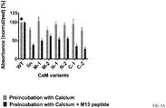
- the calmodulin-mediated change of binding observed in the initial screening was achieved after preincubation with the modulator M13.
- M13 calmodulin linkers
- a release ELISA to test whether M13 peptide binding to the calmodulin linkers can also induce the dissociation of an already established antibody-antigen complex.
- M13 peptide was added, with calcium-only buffer used for control.
- the same scFvs were analysed by the competitive preincubation approach described above (compare Example 2) on the same plate for calibration.
- the scFv-CaM-constructs were produced in 500 mL shake flask scale.
- cells harboring the desired construct were grown overnight at 37°C and 250 rpm in 30 mL 2x YP-GK-medium (2x YP-medium [16 g L -1 soy peptone, 10 g L -1 yeast extract, 5 g L -1 NaCl, pH 7.0] containing 100 mM glucose and 50 ⁇ g/mL kanamycin).
- 500 mL fresh medium was inoculated to an OD 600 of 0.1 and shaken in 2 L shake flasks (37°C, 250 rpm) until an OD 600 of 1.0 was reached.
- Protein expression was induced with a final concentration of 0.2 mM IPTG and cultures were further incubated at 25°C for 4 h. Bacteria were harvested by centrifugation (4000 g, 20 min, 4°C) and the bacterial pellet was directly processed or stored at -20°C until periplasmic extraction and protein purification.
- the bacterial pellet was resuspended in 10 mL TE-buffer per g pellet (100 mM Tris, 10 mM EDTA; pH 9.0 or pH 7.4, depending on the isoelectric point of the scFv-fusion) and incubated overnight at 37°C at 250 rpm. The next day, Benzonase® Nuclease (final concentration: 1 U/mL; Merck, Darmstadt, Germany) and MgCl 2 (20 mM) were added for DNA clearance. Furthermore, HaltTM Protease Inhibitor Cocktail (Thermo Fisher Scientific, Dreieich, Germany) was added to prevent degradation of the target protein.
- the mixture was incubated for 1 h at 37°C and 250 rpm. Afterwards, the protein containing supernatant was separated from the cell debris by centrifugation (5000 g, 20 min, RT) and prepared for purification by addition of 11x dilution buffer (110 mM Tris, 550 mM NaCl, 55 mM Imidazol; pH 9.0 or pH 7.4).
- 11x dilution buffer 110 mM Tris, 550 mM NaCl, 55 mM Imidazol; pH 9.0 or pH 7.4
- Nickel SepharoseTM 6 resin 250 ⁇ L Nickel SepharoseTM 6 resin (GE Healthcare, Solingen, Germany) was equilibrated with 10 column volumes (CV) washing buffer 1 (50 mM NaH 2 PO 4 , 50 mM NaCl, 5 mM Imidazol; pH 9.0 or pH 7.4) on Poly-Prep® Chromatography Columns (Bio-Rad, Kunststoff, Germany) and afterwards loaded with the periplasmic supernatant, followed by washing with 30 CV washing buffer 1 and 15 CV washing buffer 2 (50 mM NaH 2 PO 4 , 50 mM NaCl, 25 mM Imidazol; pH 9.0 or pH 7.4).
- CV washing buffer 1 50 mM NaH 2 PO 4 , 50 mM NaCl, 5 mM Imidazol; pH 9.0 or pH 7.4
- the protein was eluted with 5 CV elution buffer 1 (50 mM NaH 2 PO 4 , 50 mM NaCl, 150 mM Imidazol; pH 9.0 or pH 7.4) and 5 CV elution buffer 2 (50 mM NaH 2 PO 4 , 50 mM NaCl, 350 mM Imidazol; pH 9.0 or pH 7.4).
- Target protein containing fractions were pooled and dialyzed at 4°C against 200 volumes of 1x tris-buffered saline (TBS) (50 mM Tris, 150 mM NaCl; pH 8.0) for 2 h, followed by another dialysis against fresh buffer (200 volumes) for 2 h.
- TBS 1x tris-buffered saline
- the competitive ELISA was performed as described in Example 2.
- the release ELISA differed from the competitive ELISA only in the preincubation step and an additional release step was performed to evaluate whether already bound antibody fragments dissociate from the antigen in presence of M13 peptide.
- Purified scFvs for both setups were diluted to appropriate concentrations (0.1 ⁇ M) in 1x B-TBS/5 mM CaCl 2 and directly transferred (i.e. without preincubation step in polypropylene plates) to the blocked ELISA plates. After initial binding of the scFvs (1.5 h, RT), plates were washed (three times with 1x TBST) and different release-buffers were added.
- the production and purification of different scFv-fusions was performed as described in Example 3.
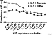
- the titration ELISA differed from the competitive ELISA (described in Example 2) only in the buffer composition used for preincubation. From column 11 to 2, M13 peptide concentration was sequentially diluted (dilution factor: 1:2) in 1x TBS/5 mM CaCl 2 (highest concentration in column 11: 3.2 ⁇ M; lowest concentration in column 2: 6.25 nM; control in column 1: 0 nM).
- the titration was additionally performed in 1x B-TBS/5 mM EDTA.
- the linkers identified to provide modulation of binding in scFv D1.3 can be used as a "universal" module to change binding properties in other scFv fragments than D1.3
- the characterized CaM-linker variants were cloned into other scFv antibodies with different specificities.
- the specificities were chosen according to their utility in future cell staining and separation applications, with two scFvs directed against different human clusters of differentiation (CD14, CD4) and the small hapten biotin.
- human blood cells PBMC
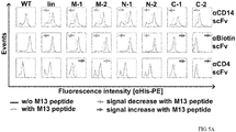
- Bound scFvs were detected with fluorescently labeled anti-His-antibodies.
- Incubation protocols for flow cytometric analysis were comparable to the pre-incubation ELISA, with buffers including and without M13 peptide.
- PBMC peripheral blood mononuclear cells
- scFvs were diluted to appropriate concentrations in 50 ⁇ L 1x B-TBS/5 mM CaCl 2 (pH 7.4 [anti-Biotin] or pH 8.0 [anti-CD14, anti-CD4]) per well.
- peptide M13 peptide [Anaspec, Fremont, USA], M13-variants library and CBP (calmodulin-binding peptide) library [Genscript, Piscataway, USA]
- the diluted scFvs were preincubated for 45 min at RT and afterwards chilled on ice for 5 min. After addition of 2x10 5 cells (in 45 ⁇ L 1x B-TBS/5 mM CaCl 2 ) and incubation for 20 min on ice, the wells were filled with buffer up to a volume of 285 ⁇ L and centrifuged at 300 g for 10 min at 4°C.
- the cells were resuspended in 110 ⁇ L 1x B-TBS/5 mM CaCl 2 containing anti-His-PE (phycoerythrin) (dilution: 1:11), incubated on ice for 10 min, followed by a further washing step and finally resuspended in 200 ⁇ L buffer.
- the analysis was performed on a MACSQuant® Analyzer 10 in chill 96 rack mode with automated addition of propidium iodide solution for exclusion of dead cells (final concentration: 1 ⁇ g/mL). A total of 10,000 events were collected for each sample.
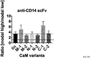
- FIG 5B , C and D show the ratio of the high binding signals in comparison to the low binding signals.
- the high modal fluorescence intensities were divided by the low modal fluorescence intensities.
- a ratio of 1 corresponded to a non-switchable antibody.
- permutated calmodulin-linkers preferably of C-terminally permutated variants or those permutated in the middle of the former calmodulin encoding gene, led to a higher change in binding signal than the linear calmodulin-linker.
- two further specificities were analysed according to their switching behaviour, anti-CD20 scFv-CaM-fusions and anti-FITC (fluorescein isothiocyanate) scFv-CaM-variants.
- Calmodulin binds to a variety of binding partners.
- a peptide screening was done.
- 38 mutated variants of the M13 peptide were analysed, e.g. substitution mutants known to have higher affinities for calmodulin, truncated variants and combinations thereof.
- 29 peptides derived from further calmodulin-binding proteins like calcium ATPase, spectrin and nitric oxidase synthase were analysed with regard to potential binding modulating properties.
- the analysis was performed via competitive staining of PBMC as in the previous experiments described in Example 5.
- the complete screening i.e. of the whole peptide libraries
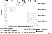
- the complete screening was performed with 4 different linker variants of anti-CD 14 scFv (lin, M-2, N-1, C-1) and anti-CD4 scFv (lin, M-1, N-1, C-1).
- the wildtype of each specificity was used as a control.
- CBP-Var16 calcium-transporting ATPase
- CBP-Var12 a peptide derived from endothelial nitric oxidase synthase had the same effect on the C-terminally permutated anti-CD4 scFv-CaM-fusions ( FIG 7C , CBP-Var12).
Landscapes
- Chemical & Material Sciences (AREA)
- Health & Medical Sciences (AREA)
- Organic Chemistry (AREA)
- Life Sciences & Earth Sciences (AREA)
- Genetics & Genomics (AREA)
- Zoology (AREA)
- General Health & Medical Sciences (AREA)
- Biochemistry (AREA)
- Engineering & Computer Science (AREA)
- Wood Science & Technology (AREA)
- Bioinformatics & Cheminformatics (AREA)
- Medicinal Chemistry (AREA)
- Molecular Biology (AREA)
- General Engineering & Computer Science (AREA)
- Biophysics (AREA)
- Proteomics, Peptides & Aminoacids (AREA)
- Biomedical Technology (AREA)
- Microbiology (AREA)
- Biotechnology (AREA)
- Immunology (AREA)
- Toxicology (AREA)
- Gastroenterology & Hepatology (AREA)
- Peptides Or Proteins (AREA)
Description
- The present invention relates to the field of binding modulation of antigen-binding polypeptides, in particular to a calmodulin-linker-based system for said modulation.
- Antigen-binding polypeptides such as single chain variable fragments (scFv) comprise of the variable domains of the light (VL) and heavy (VH) chain of a corresponding full-length antibody. A similar architecture has also been applied to the structurally similar T cell receptors (scTv) as well as scFab-fragments. In such constructs, both chains are normally connected by a linker which is flexible and does not show any tendency to interfere with folding of the individual immunoglobulin domains. In many cases, these linkers contain assemblies or variations of (Gly4Ser) repeats, inspired by the unstructured linkers connecting the domains of filamentous bacteriophage minor coat protein III.
- ScFv antibody fragments are widely used in a variety of applications, such as for research, diagnostic purposes and even as therapeutics. Immunotoxins, which are used for cancer therapy, are often based on a single chain fragment fused to a bacterial toxin to mediate targeted killing. Another approach is based on bispecific antibodies (BiTEs, bispecific T cell engagers) which activate and redirect cytotoxic T cells against cancer cells. CAR (chimeric antigen receptor)-T cell therapy also relies on scFvs specific for malignant cells. Essential for all of these applications is the extraordinary specificity, selectivity and affinity of antibody paratopes. These properties would also be very useful for the purification of biomaterials, in particular proteins, vaccines or cells. However, the usually very high affinity of antibodies requires harsh elution conditions, which typically impairs folding, integrity or viability of the eluted materials. Therefore, antibodies which retain their excellent specificity while being adjustable in respect of their affinity without requiring harsh conditions for this adjustment would be advantageous for protein purification, cell separation and cell analysis. Even the introduction of an affinity-adjustable antibody for therapy may be envisioned, for example as an additional safety mechanism in CAR-T cell therapy.
- Kobatake, E. et al. (2012, Biotechnol Lett 34, 1019-23) disclose an affinity changeable antibody in response to calcium. The system is based on a fusion-peptide comprising scFv, wild-type (WT) calmodulin, and a calmodulin-binding peptide. The switch is generated by the addition of calcium to the system. One disadvantage is that the solution must be calcium-free before the intended switch.
- Meister, G. E. & Joshi, N. S. (2013, Chembiochem 14, 1460-7) disclose a switchable enzyme which bases on the interaction of WT-calmodulin and soluble M13 peptide. In the peptide-bound form the enzyme exhibits an up to 120 times higher catalytic activity compared to the inactive (no peptide bound) state.
-
WO2002014371A1 discloses Fv constructs having an affinity that can be influenced for a substance to be linked, wherein the Fv constructs have peptides linked to the variable regions and containing binding sites for effector molecules. The effector molecules are ions or antibodies. - Guntas, G. et al. (2004, Chem Biol 11, 1483-7) and
WO2003078575A2 disclose the creation of a molecular switch of the enzyme TEM1 β-lactamase by circularly permutating the gene encoding the enzyme TEM1 β-lactamase and subsequently inserting it into the gene encoding E. coli maltose binding protein which functions as the linker. -
WO2005/072392A2 discloses molecular switches, for example with switching activity greater than previously demonstrated, or with altered ligand recognition and binding, and methods of making these molecules involving circular permutation of nucleic acid or amino acid sequences. Molecular switches have been created by recombining non-homologous genes in vitro and subjecting the genes to evolutionary pressure using combinatorial techniques. The approach is envisioned as "rolling" two proteins across each other's surfaces and fusing them at points where their surfaces meet. The approach allows for recombination and testing of maximal numbers of geometric configurations between the two domains. Libraries comprising vast numbers of such fused molecules are provided from which molecular switches with optimal characteristics can be isolated. - Megeed, Z. et al. (2006, Biomacromolecules 7, 999-1004) disclose a fusion peptide of scFv with elastin as linker resulting in a temperature dependent affinity of the antigen binding domain to the antigen.
- Miyawaki, A. et al. (1997, Nature 388, 882-887) disclose a polypeptide comprising a fluorescent protein, wherein its domains are linked by a calmodulin-M13-peptide. Baird et al. (1999, PNAS 96: 11241-11246) showed that several rearrangements of GFPs, in which the amino and carboxyl portions are interchanged and rejoined with a short spacer connecting the original termini, still provide fluorescence. These circular permutations have altered pKa values and orientations of the chromophore with respect to a fusion partner. Furthermore, certain locations within GFP tolerate insertion of entire proteins, and conformational changes in the insert can have profound effects on the fluorescence. For example, insertions of calmodulin or a zinc finger domain in place of Tyr-145 of a yellow mutant (enhanced yellow fluorescent protein) of GFP result in indicator proteins whose fluorescence can be enhanced several-fold upon metal binding. The calmodulin graft into enhanced yellow fluorescent protein can monitor cytosolic Ca2+ in single mammalian cells.
- Nagai et al. (2001, PNAS 98:3197-3202) showed by using a circularly permuted green fluorescent protein (cpGFP), in which the amino and carboxyl portions had been interchanged and reconnected by a short spacer between the original termini that they could visualize Ca2+-dependent protein-protein interactions in living cells by fluorescence readouts. The cpGFP was fused to calmodulin and its ligand derived peptide, M13. The chimeric protein was fluorescent and its spectral properties changed reversibly with the amount of Ca2+.
- Calmodulin (CaM) undergoes large conformational changes, depending on the presence of calcium and calmodulin-binding peptides (CBP). In a calcium- and peptide-unbound form, it adopts a closed conformation (Kuboniwa, H. et al., 1995, Nat Struct Biol 2, 768-776). The distance between the N- and C-terminus is at its highest in the calcium-bound, open form (Chattopadhyaya, R. et al., 1992, J Mol Biol 228, 1177-1192), whereas the termini approach each other when calmodulin binds to a ligand, or a suitable fragment thereof, like peptide M13 (Ikura, M. et al., 1992, Science 256, 632-638).
- Montigiani et al. (1996, J. Mol. Biol. 258:6-13) and Hultschig et al. (2004, J. Mol. Biol. 343:559-568) identified high affinity mutants of the CaM binding peptide "M13" which is derived from the rabbit myosin light chain kinase.
- There is a need in the art for an alternative or improved composition and/or method for affecting the binding of antigen-binding polypeptides to their respective antigens.
- Surprisingly, it was shown by the inventors that a polypeptide comprising calmodulin (CaM) and two immunoglobulin superfamily domains, wherein said two immunoglobulin superfamily domains are linked via said calmodulin (a "calmodulin linker"), can be used to affect in both directions the binding of said polypeptide to its antigen by contacting said polypeptide with a calmodulin binding molecule and ions binding to the Ca2+ binding site of calmodulin. The concerted binding of said calmodulin-binding molecule and of said ions to said Ca2+ binding site of calmodulin leads to conformational changes and influences the binding of said polypeptide for an antigen to be bound by said polypeptide. The system (or composition) comprising the three parts i) the polypeptide with CaM as linker between the two immunoglobulin superfamily domains, ii) the CaM binding molecule such as a CaM-binding peptide (e.g. M13 peptide), and iii) ions binding to the Ca2+ binding site of CaM such as Ca2+ is superior compared to systems known in the art: It is not a prerequisite for a functional system to eliminate calcium ions from the solution comprising said polypeptide before switching the binding of the polypeptide to its antigen. The switch is only achieved by adding soluble CaM binding molecules such as M13 peptide to the solution in the presence of ions binding to the Ca2+ binding site of CaM. This is superior compared to systems known in the art, particular for the use in living organisms, for example when used to modulate the affinity of a CAR on a T cell or other suitable effector cell, as in this situation, the Ca2+ concentration may not be sufficiently adjustable.
- Even more surprisingly, it was found that a permutation of the linker component, i.e. the calmodulin, resulted in even stronger change of binding of the polypeptide of the invention to its antigen compared to the use of the described WT CaM as linker, at least by a factor of 2, as shown in Example 5 (see also
FIG 5B , C, D). Regularly, in the art a permutation in comparable systems (polypeptides with antigen binding domains) has been used only with regard of the non-linker part. - Also unexpectedly the use of variants of the normally used M13 peptide or the use of other peptides than the M13 peptide in the herein disclosed system (or compositions) resulted in a stronger change of binding of the polypeptide as disclosed herein to its antigen compared to the use of the M13 peptide itself.
- Best results with regard to a switchable binding modulation is achieved when permutated CaM is combined with variants of the M13 peptide or other peptides than the M13 peptide, some specific combinations of defined permutated CaMs and defined CaM binding peptides are especially preferred as disclosed herein.
- Surprisingly, the change in binding of the antigen binding domains of the polypeptide triggered by the binding of a CaM binding molecule and ions to the calmodulin linker of the polypeptide can result either in an enhanced binding or in a reduced binding of the polypeptide to its antigen.
- Polypeptides as disclosed herein can be released from the binding antigen by adding CaM binding molecules such as M13 peptide and ions such as Ca2+ without harsh conditions.
- The general applicability of a calmodulin sequence as a universal linker to regulate the binding of a polypeptide comprising two immunoglobulin superfamily domains has been demonstrated herein firstly for different scFvs, including one specific for lysozyme, and secondly other scFvs with quite different affinities and antigen classes, including proteins and haptens.
- Herein compositions comprising the above mentioned components, methods for affecting the binding of the polypeptides, and the use of the polypeptides for affecting the binding of the polypeptides to their antigens are disclosed.
-
-
FIG 1 : Cloning of circularly permutated calmodulin variants. -
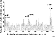
FIG 2 : Identification of switchable anti-lysozyme scFv-Calmodulin-variants by competitive ELISA. -
FIG 3 : Analysis of M13 peptide dependent binding behavior of anti-lysozyme scFv-Calmodulin-variants by competitive ELISA (A) and release ELISA (B). -
FIG 4 : Evaluation of the specific M13 peptide dependent decrease of binding signal by titration ELISA (A, B, C, D, E, F, G, H). -
FIG 5 : Identification of switchable anti-CD 14, anti-Biotin and anti-CD4 scFv-Calmodulin-fusions by competitive staining of PBMC and analysis by flow cytometry (A). Comparison of the extent of binding modulating properties of different Calmodulin-linker variants in anti-CD14 scFv (B), anti-Biotin scFv (C) and anti-CD4 scFv (D). -
FIG 6 : Overview of further calmodulin-binding peptides with binding modulating properties (A, B). -
FIG 7 : Identification of further calmodulin-binding peptides with binding modulating properties - most promising peptides (A, B, C). - In a first aspect the invention provides a composition (a system, a set, or a kit) comprising
- i) a polypeptide comprising calmodulin and two immunoglobulin superfamily domains, wherein said two immunoglobulin superfamily domains are linked via said calmodulin; and
- ii) a calmodulin binding molecule; and
- iii) ions binding to the Ca2+ binding site of calmodulin,
- The calmodulin is a linker sequence between the two immunoglobulin superfamily domains and serves as a universal allosteric regulator of these two domains. The CaM (or the CaM sequence) may be any sequence or part of a sequence of CaM which maintains the characteristics of the WT calmodulin protein to bind both, ions at the Ca2+ binding site and a calmodulin binding molecule, and thereby changing its conformation. This includes a calmodulin or a sequence of calmodulin having a sequence identity of at least 70%, or at least 75%, or at least 80%, or at least 85%, or at least 90%, or at least 95%, or at least 97%, or at least 98%, or at least 99% at the amino acid sequence level to the wild type calmodulin.
- The sequence of calmodulin may also be a functional fragment of the full-length calmodulin protein (e.g. a truncated protein of calmodulin) or a fragment of the full-length calmodulin protein having a sequence identity of at least 70%, or at least 75%, or at least 80%, or at least 85%, or at least 90%, or at least 95%, or at least 97%, or at least 98%, or at least 99% at the amino acid sequence level to the corresponding part of said full-length calmodulin.
- In general, all amino acid variations (i.e. substitutions, additions or eliminations of amino acids of the calmodulin) are included under this definition, which do not lead to the loss of the described characteristics of the calmodulin to provide the allosteric change, or a functional fragment thereof to bind both, ions at the Ca2+ binding side and a calmodulin binding molecule, and thereby changing its conformation.
- The composition as disclosed above, wherein said binding of said calmodulin-binding molecule and of said ions to the Ca2+ binding site of calmodulin enhances or reduces the binding of said polypeptide to said antigen.
- Said calmodulin binding molecule may be a calmodulin binding peptide. Said calmodulin binding peptide may be selected and derived from the group of naturally occurring calmodulin ligands consisting of myosin light chain kinase, caldesmon, calspermin, phosphofructokinase, calcineurin, calcium ATPase, spectrin, glutamate receptor, nitric oxidase synthase, serine/threonine-protein phosphatase, tumor necrosis factor receptor, estrogen receptor, calcium channel subunits and calcium/calmodulin-dependent protein kinases. Said CaM binding peptide may be M13 peptide derived from the rabbit myosin light chain kinase or a variant thereof.
- Said calmodulin binding peptide may be selected from the group of peptides consisting of SEQ ID NO: 1 to SEQ ID NO: 65. Preferentially, said calmodulin binding peptide may be selected from the group of peptides consisting of SEQ ID NO: 1, SEQ ID NO: 2, SEQ ID NO: 3, SEQ ID NO: 7, SEQ ID NO: 8, SEQ ID NO: 9, SEQ ID NO: 46, SEQ ID NO: 47, SEQ ID NO: 49, SEQ ID NO: 51 and SEQ ID NO: 53.
- But in general, any peptide or polypeptide which can bind to CaM and thereby provides the allosteric change may be used.
- Three-dimensional structures of calmodulin in complex with high-affinity peptidic substrates are available (Montigiani et al., 996, J. Mol. Biol. 258:6-13). These peptides correspond to the calmodulin-binding regions of different protein kinases. Alternatively, methods such as peptide phage display, ribosome display or other established combinatorial selection systems well known in the art (see e.g. Ullman CG et al. Brief Funct Genomics, 2011 May;10(3):125-34 or Galán A et al. Mol Biosyst, 2016 Jun 16. [Epub ahead of print]) may be used to identify variants of naturally occurring sequences or synthetic sequences which can bind to calmodulin.
- Said ions binding to the Ca2+ binding site of calmodulin may be any ions that can be bound by the Ca2+ binding site of calmodulin resulting in a conformational change of the CaM in the presence of a CaM binding molecule, preferentially said ions are calcium ions (Ca2+).
- Said calmodulin may be a permutated calmodulin. Generally, a permutated CaM can be generated e.g. by circular permutation, a general method for permutating proteins which is well known in the art (see e.g. "Circular Permutation of Proteins" in "Protein Engineering Handbook" (Ed.: Stefan Lutz, Uwe T. Bornscheuer) Wiley-VCH 2009), and as described herein.
- Alternatively, a permutated CaM may be generated synthetically on nucleic acid or amino acid level, especially it may be generated synthetically when the sequence which is intended to use for the generation of the polypeptide as disclosed herein comprising the permutated CaM is known and can be generated purposefully.
- As demonstrated herein the vast majority of permutated calmodulins generated and used herein induce a regulation of binding of the polypeptides as disclosed herein (
FIG 2 ) by addition of different CaM binding molecules and ions such as Ca2+, wherein the regulation of binding is more distinct than the regulation by the wildtype CaM (FIG 5 B ,C ,D ). - Said polypeptide of said composition comprising said permutated calmodulin and said two immunoglobulin superfamily domains, wherein said two immunoglobulin superfamily domains are linked via said permutated calmodulin may be obtained e.g. by the method comprising
- a) Creating at least one insertion nucleic acid sequence encoding a permutated calmodulin
- b) Creating an acceptor nucleic acid sequence encoding a polypeptide comprising two immunoglobulin superfamily domains
- c) Inserting the at least one insertion sequence of a) into the acceptor sequence of b), wherein one insertion sequence of a) is inserted between the parts of the acceptor sequence b) which encode the two immunoglobulin superfamily domains of b)
- d) Transforming a host with the nucleic acid sequences of c)
- e) Selecting for transformed hosts harboring the sequence(s) of c)
- f) Screening for transformed hosts expressing polypeptides comprising two immunoglobulin superfamily domains linked via permutated calmodulin by exposing the polypeptides produced by the transformed hosts to said calmodulin-binding molecule and identifying the transformed hosts harboring polypeptides which impact the binding of said polypeptides to the antigen in the presence of ions binding to the Ca2+ binding site of calmodulin.
- The permutated calmodulin of the composition as described above may be selected from the group consisting of SEQ ID NO: 67 to SEQ ID NO: 123. Preferentially, the permutated calmodulin of the composition as described above may be selected from the group consisting of SEQ ID NO: 67 to SEQ ID NO: 72. Most preferentially, the permutated calmodulin may be selected from the group consisting of SEQ ID NO: 67, SEQ ID NO: 68, SEQ ID NO: 71 and SEQ ID NO: 72.
- A preferred composition as disclosed herein may comprise
- i) a polypeptide comprising calmodulin and two immunoglobulin superfamily domains, wherein said two immunoglobulin superfamily domains are linked via said calmodulin; and
- ii) a calmodulin binding molecule; and
- iii) ions binding to the Ca2+ binding site of calmodulin,
- Said composition, wherein said polypeptide may be a single chain Fv (scFv) comprising the calmodulin and a variable region of a heavy chain of an immunoglobulin (VH) and a variable region of a light chain of an immunoglobulin (VL).
- Especially preferred is a composition as described above, wherein the polypeptide comprising a scFv specific for the antigen CD14 and a permutated CaM has the sequence of SEQ ID NO: 124 and said CaM binding peptide has the sequence of SEQ ID NO: 1 or SEQ ID NO: 7 or SEQ ID NO: 8 or SEQ ID NO: 9, or said polypeptide has the sequence of SEQ ID NO: 125 and said CaM binding peptide has the sequence of SEQ ID NO: 1 or SEQ ID NO: 7 or SEQ ID NO: 8 or SEQ ID NO: 9 or SEQ ID NO: 53, or said polypeptide has the sequence of SEQ ID NO: 126 and said CaM binding peptide has the sequence of SEQ ID NO: 1 or SEQ ID NO: 47, or said polypeptide has the sequence of SEQ ID NO: 127 and said CaM binding peptide has the sequence of SEQ ID NO: 1 or SEQ ID NO: 47 or SEQ ID NO: 53.
- Furthermore, especially preferred is a composition as described above, wherein the polypeptide comprising a scFv specific for biotin and a permutated CaM has the sequence of SEQ ID NO: 128 and said CaM binding peptide has the sequence of SEQ ID NO: 1 or SEQ ID NO: 46, or said polypeptide has the sequence of SEQ ID NO: 129 and said CaM binding peptide has the sequence of SEQ ID NO: 1 or SEQ ID NO: 46, or said polypeptide has the sequence of SEQ ID NO: 130 and said CaM binding peptide has the sequence of SEQ ID NO: 1 or SEQ ID NO: 53.
- Moreover, especially preferred is a composition as described above, wherein the polypeptide comprising a scFv specific for the antigen CD4 and a permutated CaM has the sequence of SEQ ID NO: 131 and said CaM binding peptide has the sequence of SEQ ID NO: 1 or SEQ ID NO: 46, or said polypeptide has the sequence of SEQ ID NO: 132 and said CaM binding peptide has the sequence of SEQ ID NO: 1 or SEQ ID NO: 46, or said polypeptide has the sequence of SEQ ID NO: 133 and said CaM binding peptide has the sequence of SEQ ID NO: 1 or SEQ ID NO: 51 or SEQ ID NO: 53, or said polypeptide has the sequence of SEQ ID NO: 134 and said CaM binding peptide has the sequence of SEQ ID NO: 1 or SEQ ID NO: 51 or SEQ ID NO: 53.
- As evident by
FIG 7A , B and C (or Example 6) said combinations of specific sequences of permutated CaM and CaM binding peptides display the strongest changes in binding between said polypeptides and their antigens, respectively. - Said composition, wherein said polypeptide is part of an antigen binding domain of a chimeric antigen receptor (CAR). Said CAR may comprise an antigen binding domain, a transmembrane domain and cytoplasmic signaling domain(s).
- In a further aspect the invention provides a method for affecting the binding of a polypeptide to an antigen to be bound, wherein said polypeptide comprises a calmodulin and two immunoglobulin superfamily domains wherein said two immunoglobulin superfamily domains are linked via said calmodulin, the method comprising the step of contacting said polypeptide with a calmodulin binding molecule in the presence of ions binding to the Ca2+ binding site of calmodulin, thereby affecting the binding of said polypeptide to the antigen.
- Preferentially, the CaM is a permutated CaM as described above.
- Preferentially the CaM binding molecule is a CaM binding peptide as described above.
- Said method for affecting the binding of a polypeptide, wherein before said contacting of said calmodulin binding molecule with said calmodulin said polypeptide is contacted with the antigen to be bound by said polypeptide, and wherein said contacting of said calmodulin binding molecule to said calmodulin in the presence of ions binding to the Ca2+ binding site of calmodulin affects the binding of said polypeptide to the antigen. Said affecting of the binding may be a reduction (decrease) of binding, thereby releasing the polypeptide from the antigen. Alternatively, said affecting of the binding may be an enhancement (an increase) of binding, thereby enhancing the binding between said polypeptide and the antigen. Enhancing said binding may be of interest when the binding of said polypeptide to the antigen is weak or does not occur in the absence of said CaM binding molecule and said ions binding to the Ca2+ binding site of CaM.
- Said affecting of the binding may be a change of one or both of the kinetic properties of the binding reaction (the association and dissociation rate constants, or on-rate and off-rate) while overall affinity may be changed (affecting the affinity) in this process or remain the same (affecting the binding kinetics but not the equilibrium affinity), thereby changing the kinetics of the interaction of the polypeptide with the antigen.
- Said affecting of binding may be an allosteric change induced within the antigen.
- In another aspect the invention provides the use of a polypeptide (as described above) comprising a calmodulin, preferentially a permutated calmodulin, and two immunoglobulin superfamily domains, wherein said two immunoglobulin superfamily domains are linked via said calmodulin, preferentially said permutated calmodulin, for affecting in the presence of a calmodulin binding molecule and ions binding to the Ca2+ binding site of calmodulin the binding of said polypeptide to an antigen to be bound by said polypeptide.
- In one embodiment of the invention the composition comprises
- i) a polypeptide such as a scFv, e.g. lysozyme binding scFv D1.3, having the sequence of SEQ ID NO: 135, comprising calmodulin such as a permutated CaM, e.g. having the sequence of the group consisting of SEQ ID NO: 67 to SEQ ID NO: 123 and two immunoglobulin superfamily domains such as a variable region of a heavy chain of an immunoglobulin (VH) and a variable region of a light chain of an immunoglobulin (VL), wherein said two immunoglobulin superfamily domains are linked via said calmodulin; and
- ii) a calmodulin binding molecule such as M13 peptide; and
- iii) ions binding to the Ca2+ binding site of calmodulin such as Ca2+,
- In one embodiment of the invention the composition may be used for enrichment (e.g. positive selection) of cells expressing on the cell surface the antigen recognized by the polypeptide, e.g. as disclosed herein. Methods suited for enrichment are well known in the art and include but are not limited to flow cytometry such as fluorescence activated cell sorting (FACS) or magnetic cell separation such as MACS® (Miltenyi Biotec GmbH).
- Exemplarily the principle of MACS® separation (Miltenyi Biotec GmbH, Germany) is described here: The polypeptide as disclosed herein, specific for an antigen can be used for direct or indirect magnetic labeling of cells expressing said antigen on their cell surface in a sample comprising said cells and other cells (not expressing said antigen). First the antigen-expressing cells are magnetically labeled with MicroBeads (magnetic particles) conjugated to said polypeptide. Then the cell sample is loaded on a MACS® Column which is placed in the magnetic field of a MACS® Separator. The magnetically labeled antigen-expressing cells are retained on the column. The unlabeled cells run through. The addition of an "elution" solution comprising a CaM binding molecule such as M13 peptide and e.g. Ca2+ ions allow to reduce the binding of the polypeptide to the antigen, thereby releasing the cell expressing said antigen from the immobilized polypeptide conjugated to the magnetic particle, i.e. the cell can be eluted from the column without the need of removal of the magnetic field.
- In one embodiment of the invention the composition may be used for the enrichment (i.e. purification) of proteins fused to an antigen recognized by the polypeptide comprising a scFv comprising calmodulin, preferentially a permutated CaM, and a variable region of a heavy chain of an immunoglobulin and a variable region of a light chain of an immunoglobulin, wherein said variable regions are linked via said calmodulin, preferentially permutated CaM. The polypeptide invention as described herein may be immobilized e.g. on a resin. Next, target protein (i.e. protein which has to be purified) containing material is incubated with the polypeptide-coupled resin to allow for the binding of the polypeptide to the target protein fused to an antigen recognized by the polypeptide invention. Unbound material is removed by washing of the resin material. The addition of an "elution" solution comprising a CaM binding molecule such as M13 peptide and e.g. Ca2+ ions allow to reduce the binding of the polypeptide to the antigen-comprising target protein, thereby releasing the target protein without the need of harsh elution conditions.
- In one embodiment of the invention the polypeptide is a scFv comprising the calmodulin, preferentially a permutated CaM, and a variable region of a heavy chain of an immunoglobulin and a variable region of a light chain of an immunoglobulin, wherein said variable regions are linked via said calmodulin, preferentially permutated CaM, and wherein said scFv is the antigen-binding domain (or part of the antigen-binding domain) of a chimeric antigen receptor (CAR). The CAR may comprise said antigen binding domain, a transmembrane domain and cytoplasmic signaling domains. Said CAR may be released from an antigen bound to said CAR by contacting said CAR with a CaM binding molecule and ions binding to the Ca2+ binding site of CaM, if said contacting results in a reduction of binding of the antigen binding domain to the antigen. Alternatively, said CAR may bind sufficiently strong to the antigen to induce or activate signaling in the cell expressing said CAR not until a CaM binding molecule and ions binding to the Ca2+ binding site of CaM are contacted with said CAR, if said contacting results in an increase of binding of the antigen binding domain to the antigen. These procedures allow a control of interactions between cells expressing said CAR and the antigen by providing a small peptide, as calcium may be present in sufficient amounts physiologically. This may help to reduce or prevent severe side effects in a patient if cells expressing said CAR are used in a cell immunotherapy e.g. to fight cancer cells in a patient.
- In one embodiment of the invention the composition is a composition comprising
- i) a polypeptide comprising a permutated calmodulin and two immunoglobulin superfamily domains, wherein said two immunoglobulin superfamily domains are linked via said calmodulin,
- ii) a calmodulin binding molecule; and
- iii) ions binding to the Ca2+ binding site of calmodulin,
- α) a) creating an insertion nucleic acid sequence library, wherein said insertion sequence comprises a sequence which encodes said permutated calmodulin comprising the steps of:
- i) obtaining an insertion nucleic acid sequence which encodes calmodulin that binds a calmodulin-binding molecule and ions that bind to the Ca2+ binding site of calmodulin;
- ii) ligating said insertion nucleic acid sequence to circularize said insertion nucleic acid sequence;
- iii) creating oligonucleotide pairs which allow the amplification of said permutated insertion nucleic acid sequence by polymerase chain reaction using the circularized insertion nucleic acid sequence of ii) as a template and which furthermore comprise nucleic acid overhangs with recognition sequences of restriction enzymes
- iv) performing a polymerase chain reaction using the circularized insertion nucleic acid sequence of ii) and the oligonucleotide pairs of iii) to allow the amplification of said permutated insertion nucleic acid sequence;
- v) digesting said permutated insertion nucleic acid sequence of iv) with restriction enzymes recognizing said recognition sequences to allow the specific introduction of the digested insertion nucleic acid sequence into the acceptor nucleic acid sequence;
- b) creating an acceptor nucleic acid sequence, wherein said acceptor nucleic acid sequence comprises a sequence which encodes said two immunoglobulin superfamily domains, comprising the steps of:
- i) obtaining said acceptor nucleic acid sequence comprising the same recognition sequences of restriction enzymes of a)iii) between the sequences which encode said two immunoglobulin superfamily domains;
- ii) digesting said acceptor nucleic acid sequence of i) with said restriction enzymes recognizing said recognition sequences that allow the introduction of the permutated insertion nucleic acid sequence of a)v) between the two immunoglobulin superfamily domains of said acceptor nucleic acid sequence;
- c) ligating the nucleic acids of a)v) and b)ii) so that an insertion nucleic acid sequence inserts into the digested acceptor nucleic acid sequence;
- d) transforming a host with one or a library of the ligated sequences of c);
- e) selecting for transformed hosts harboring the ligated nucleic acid sequences;
- f) screening for transformed hosts expressing polypeptides comprising two immunoglobulin superfamily domains linked via calmodulin by exposing the polypeptides produced by the transformed hosts to said calmodulin-binding molecule and identifying the transformed hosts harboring polypeptides which impact the binding of said polypeptides to the antigen in the presence of ions binding to the Ca2+ binding site of calmodulin; or
- β) a) creating an insertion nucleic acid sequence library, wherein said insertion sequence comprises a sequence which encodes said permutated calmodulin comprising the steps of:
- i) obtaining an insertion nucleic acid sequence which encodes calmodulin that binds a calmodulin-binding molecule and ions that bind to the Ca2+ binding site of calmodulin;
- ii) ligating said insertion nucleic acid sequence to circularize said insertion nucleic acid sequence;
- iii) digesting the insertion nucleic acid sequence of ii) to randomly introduce a single double-stranded break for the creation of an insertion nucleic acid sequence library;
- b) creating an acceptor nucleic acid sequence, wherein said acceptor nucleic acid sequence comprises a sequence which encodes said two immunoglobulin superfamily domains, comprising the steps of:
- i) obtaining said acceptor nucleic acid sequence;
- ii) digesting said acceptor nucleic acid sequence of i) with restriction enzymes recognizing recognition sequences that allow the introduction of the insertion nucleic acid sequence library between the two immunoglobulin superfamily domains of said acceptor nucleic acid sequence; optionally
- iii) blunt ending said acceptor sequence if said restriction enzymes produce sticky ends
- c) ligating the nucleic acids of the library of a)iii) and b)ii), optionally b)iii) so that an insertion nucleic acid sequence inserts into the digested acceptor nucleic acid sequence;
- d) transforming a host with the library of the ligated sequences of c);
- e) selecting for transformed hosts harboring the ligated nucleic acid sequences;
- f) screening for transformed hosts expressing polypeptides comprising two immunoglobulin superfamily domains linked via calmodulin by exposing the polypeptides produced by the transformed hosts to said calmodulin-binding molecule and identifying the transformed hosts harboring polypeptides which impact the binding of said polypeptides to the antigen in the presence of ions binding to the Ca2+ binding site of calmodulin; or
- γ) a) creating an insertion nucleic acid sequence or a sequence library which encodes said permutated calmodulin comprising the steps of:
- i) synthetically generating said nucleic acid sequence of said permutated calmodulin comprising nucleic acid overhangs with recognition sequences of restriction enzymes;
- ii) digesting said permutated insertion nucleic acid sequence of i) with restriction enzymes recognizing said recognition sequences to allow the specific introduction of the digested insertion nucleic acid sequence into the acceptor nucleic acid sequence;
- b) creating an acceptor nucleic acid sequence, wherein said acceptor nucleic acid sequence comprises a sequence which encodes said two immunoglobulin superfamily domains, comprising the steps of:
- i) obtaining said acceptor nucleic acid sequence comprising the same recognition sequences of restriction enzymes of a)i) between the sequences which encode said two immunoglobulin superfamily domains;
- ii) digesting said acceptor nucleic acid sequence of i) with said restriction enzymes recognizing said recognition sequences that allow the introduction of the permutated insertion nucleic acid sequence of a)ii) between the two immunoglobulin superfamily domains of said acceptor nucleic acid sequence;
- c) ligating the nucleic acids of a)ii) and b)ii) so that an insertion nucleic acid sequence inserts into the digested acceptor nucleic acid sequence;
- d) transforming a host with one or a library of the ligated sequences of c);
- e) selecting for transformed hosts harboring the ligated nucleic acid sequences;
- f) screening for transformed hosts expressing polypeptides comprising two immunoglobulin superfamily domains linked via calmodulin by exposing the polypeptides produced by the transformed hosts to said calmodulin-binding molecule and identifying the transformed hosts harboring polypeptides which affect the binding of said polypeptides to the antigen in the presence of ions binding to the Ca2+ binding site of calmodulin; or
- δ) a) creating the nucleic acid sequences encoding said polypeptide comprising said permutated calmodulin by synthetically creating said nucleic acid sequences b) transforming a host with one or a library of the sequences of a);
- c) selecting for transformed hosts harboring the nucleic acid sequences;
- d) screening for transformed hosts expressing polypeptides comprising two immunoglobulin superfamily domains linked via calmodulin by exposing the polypeptides produced by the transformed hosts to said calmodulin-binding molecule and identifying the transformed hosts harboring polypeptides which impact the binding of said polypeptides to the antigen in the presence of ions binding to the Ca2+ binding site of calmodulin.
- Unless defined otherwise, technical and scientific terms used herein have the same meaning as commonly understood by one of ordinary skill in the art to which this invention belongs.
- The term "calmodulin" or "sequence of calmodulin" as used herein refers to a sequence having a sequence identity of at least 70%, or at least 75%, or at least 80%, or at least 85%, or at least 90%, or at least 95%, or at least 97%, or at least 98%, or at least 99% at the amino acid sequence level to the wild type calmodulin (SEQ ID NO: 66) if the calmodulin did not experience a permutation. In this context, "sequence identity" may be determined using pairwise alignments using alignments programs for amino acid sequences well known to the art.
- The sequence of calmodulin may also be a functional fragment of the full-length calmodulin protein (e.g. a truncated protein of calmodulin) or a fragment of the full-length calmodulin protein having a sequence identity of at least 70%, or at least 75%, or at least 80%, or at least 85%, or at least 90%, or at least 95%, or at least 97%, or at least 98%, or at least 99% at the amino acid sequence level to the corresponding sequence of said full-length calmodulin if this part of the calmodulin sequence did not experience a permutation.
- In general, all amino acid variations (i.e. substitutions, additions or eliminations of amino acids of the calmodulin) are included under this definition, which do not lead to the loss of the function of the calmodulin or a functional fragment thereof to provide the change of its conformation.
- The calmodulin or a functional fragment thereof (in all its variants as described above) may be also a permutated calmodulin or functional fragment thereof. Although the order of sequence may be changed in a permutated calmodulin it maintains the characteristics of the WT calmodulin to bind both, ions at the Ca2+ binding site and a calmodulin binding molecule, and thereby changing its conformation (i.e. the fragment of the calmodulin remains functional). A permutated CaM may be generated e.g. by a method using circular permutation. A circular permutation is a relationship between proteins whereby the proteins have a changed order of amino acids in their peptide sequence. The result is a protein structure with different connectivity, but overall similar three-dimensional (3D) shape.
- Circular permutation can occur as the result of natural evolutionary events, posttranslational modifications, or artificially engineered mutations.
- Because of this, it is often possible to design circular permutations of proteins. Today, circular permutations are generated routinely in the lab using standard genetics techniques (see e.g. "Circular Permutation of Proteins" in "Protein Engineering Handbook" (Ed.: Stefan Lutz, Uwe T. Bornscheuer) Wiley-VCH 2009).
- A permutated calmodulin as used herein may also have some additional amino acid residues in its sequence. This may be the result of the generation of a permutated CaM due to e.g. addition of recognition sequences of restriction enzymes on the level of the nucleic acid sequence of said calmodulin. The additional sequence may be any sequence, preferentially the sequence may be a sequence which does not result in larger conformational changes (or any change at all) when the position of said additional sequence changes within the polypeptide, e.g. due to the permutation process of the CaM including said additional sequence. In this context a well-suited additional sequence may be the amino acid sequence GGSG within the permutated CaM as the result of the nucleic acid sequence recognized by the restriction enzyme BamHI, which may be used at the ends of the nucleic acid sequence of CaM, resulting in the additional sequence on the amino acid level of GGSG after digestion of the nucleic acid sequence with BamHI and subsequent circularization of the sequence and translation into a polypeptide. This additional sequence leads to minor or no conformational changes regardless of the position within the permutated CaM and does not affect the binding of the polypeptide as disclosed herein to its antigen.
- Generally, the permutation of the calmodulin linker as disclosed herein may be at any position of the calmodulin sequence, preferentially the permutated calmodulins (the permutated calmodulin linkers) are C-terminally permutated variants or those permutated in the middle of the former calmodulin encoding gene.
- Although some permutation sites prevent the protein from folding correctly, many permutants have been created with nearly identical function to the original protein.
- The term "permutated calmodulin" as used herein refers to any calmodulin derived amino acids sequence which has an altered order of amino acids in its peptide sequence compared to the WT calmodulin sequence but saves the characteristics of the CaM to change its conformation when the altered sequence of the CaM binds a CaM binding molecule and ions binding to the Ca2+ binding site of the CaM, wherein said conformational change affects the binding of the polypeptide as disclosed herein to its antigen.
- The term "two immunoglobulin superfamily domains" in the context of "a polypeptide comprising calmodulin and two immunoglobulin superfamily domains" as used herein refers to the use of two domains associated with the immunoglobulin superfamily within said polypeptide. In general, all amino acid variations (i.e. substitutions, additions or eliminations) of amino acids of an immunoglobulin superfamily domain are included under this definition, which do not lead to the loss of the domain function which is to contribute to antigen binding. The immunoglobulin superfamily is a large group of cell surface and soluble proteins that are mainly involved in the recognition, binding, or adhesion processes of cells. Molecules categorized as members of this superfamily based on shared structural features with immunoglobulins; they all possess a domain known as an immunoglobulin domain or fold. Members of the immunoglobulin superfamily include cell surface antigen receptors, co-receptors and co-stimulatory molecules of the immune system, molecules involved in antigen presentation to lymphocytes, cell adhesion molecules, certain cytokine receptors and intracellular muscle proteins. A specific example of the "two immunoglobulin superfamily domains" is an Fv fragment of an immunoglobulin, comprising a variable region of a heavy chain of an immunoglobulin and a variable region of a light chain of an immunoglobulin.
- A single-chain variable fragment (scFv) is a fusion protein of the variable regions of the heavy (VH) and light chains (VL) of immunoglobulins, normally connected with a short linker peptide of up to about 25 amino acids. The linker is often rich in glycine for flexibility, as well as serine or threonine for solubility, and can either connect the N-terminus of the VH with the C-terminus of the VL, or vice versa. This protein retains the specificity of the original immunoglobulin, despite removal of the constant regions and the introduction of the linker. In the present invention the linker is replaced by the calmodulin sequence. Another specific example of the "two immunoglobulin superfamily domains" are the variable chains of the T cell receptors, which share overall structural properties very similar to the Fv fragment, and constitute a scTv containing the calmodulin sequence as a linker. These scTv can include the variable regions of the α- and β-chain or the variable regions of the γ- and δ- chains.
- As used herein, the term "antigen" is intended to include substances that bind to or evoke the production of one or more antibodies or bind to the above mentioned scFv, scTv or analogous binding entities composed of two immunoglobulin superfamily members. Each antibody binds to a specific antigen by way of an interaction similar to the fit between a lock and a key. The substance may be from the external environment or formed within the body. The term "antigen" comprises, but is not limited to, proteins, peptides, polypeptides, oligopeptides, lipids, carbohydrates, haptens, vitamins, hormones, synthetic molecules, and combinations thereof, for example a glycosylated protein, a glycolipid or a biotinylated vitamin. An antigen may be on the cell surface or inside the cell. Preferentially, an antigen is on the cell surface of a cell. In another embodiment, the antigen is in solution in a complex mixture of other substances, like in the cultivation supernatant of a bioreactor or a fraction derived thereof. In another embodiment, the antigen is in solution in a complex mixture of other proteins, like blood plasma or other body fluids or a bioreactor cultivation medium supernatant.
- The area of the antibody which is located towards the antigen and includes amino acid side chains forming chemical linkages like hydrogen bonds, electrostatic bonds or hydrophobic interactions with the antigen, is termed "paratope". It is an effect to achieve by the present invention to influence the structure of this paratope in a way that its binding to the antigen is influenced by altering the position, orientation, distance or binding energy of one or several said amino acid side chains or of the entire V domain to the antigen.
- The terms "specifically binds to" or "specific for" with respect to an antigen-binding domain of an antibody or fragment thereof (e.g. a scFv or scTv) refer to an antigen-binding domain which recognizes and binds to a specific antigen, but does not substantially recognize or bind other antigens in a sample. An antigen-binding domain that binds specifically to an antigen from one species may bind also to the homologous antigen from another species. This cross-species reactivity is typical to many antibodies and therefore not contrary to the definition of that antigen-binding domain as specific. An antigen-binding domain that specifically binds to an antigen may bind also to different allelic forms of the antigen (allelic variants, splice variants, isoforms etc.) or homologous variants of this antigen from the same gene family. These cross reactivities are typical to many antibodies and therefore not contrary to the definition of that antigen-binding domain as specific. An antigen-binding domain that specifically binds to an antigen may bind also to a limited number of completely different structures, known as mimotopes (see e.g. Reineke U. et al., J Immunol Methods. 2002 ; Keitel T. et al., Cell. 1997 Dec 12;91(6):811-20). This reactivity is typical to many antibodies and therefore not contrary to the definition of that antigen-binding domain as specific.
- The term "affecting the binding" as used herein refers to a change of one or both of the kinetic properties of the binding reaction (the association and dissociation rate constants) while overall affinity may be changed (affecting the equilibrium affinity) in this process or remain the same (affecting the binding kinetics but not the affinity), thereby changing the kinetics of the interaction of the polypeptide as disclosed herein with the antigen.
- The term "affinity" refers to the strength of the sum total of non-covalent interactions between a single binding site of a molecule (e.g., the antigen binding domain(s) of a polypeptide such as the variable domains of a light and heavy chain of immunoglobulins in a scFv) and its binding partner (e.g., an antigen). Unless indicated otherwise, as used herein, "binding affinity" refers to intrinsic binding affinity which reflects a 1:1 interaction between members of a binding pair (e.g., antigen-binding polypeptide and its antigen). The affinity of a molecule X for its partner Y can generally be represented by the equilibrium dissociation constant (KD). Affinity can be measured by common methods known in the art (e.g., Biacore™ measurement, and calculated from the dissociation and association constants).
- The terms "regulation", "modulation", "affecting" "influence", and "change" in the context of the regulation/modulation/affecting/influence/change of the binding of the polypeptide as disclosed herein to its antigen can be used interchangeably. Regulation of binding as used herein means a conformation or entropy change in the calmodulin linker of the polypeptide between the two immunoglobulin superfamily domains, e.g. the VH and VL regions of a scFv fragment, triggered by the contact of the calmodulin linker with a calmodulin binding molecule and ions that bind to the Ca2+ binding site of the CaM that affect the conformation of the antigen binding site, resulting in a (measurable) change of the binding. The change of the binding may be a reduction (decrease) of binding or an enhancement (an increase) of binding or a change of dissociation or association rate constants of the polypeptide as disclosed herein to its antigen.
- The binding of i) a calmodulin binding molecule and ii) ions binding to the Ca2+ binding site of calmodulin as used herein has to be a concerted binding, i.e. both components have to be bound to the CaM of the polypeptide to affect the modulation of said binding. In one embodiment of the invention ions binding to the Ca2+ binding site of calmodulin and the polypeptide of the invention may be present in the same solution (which is not sufficient to induce the modulation of said binding) but not before the addition of a CaM binding molecule will lead to the modulation of the binding of the polypeptide as disclosed herein (as now a concerted binding of both, a calmodulin binding molecule and ions binding to the Ca2+ binding site of calmodulin to said polypeptide is possible).
- The term "polypeptide comprising calmodulin and two immunoglobulin superfamily domains, wherein said two immunoglobulin superfamily domains are linked via said calmodulin" as used herein refers to a polypeptide having the following domains in an order of: a first immunoglobulin superfamily domain - calmodulin - a second immunoglobulin superfamily domain. Preferentially, the linkage between all these partial sequences (or domains) is via peptide bonds resulting in a continuous amino acid sequence of the polypeptide. Alternatively, the polypeptide may be a polypeptide having said domains (peptides) in above mentioned order but the connection between one, more or all of these domains (peptides) may be by covalent or non-covalent bounds other than the peptide bond, e.g. a disulphide bridge (S-S bond) between two domains such as a first disulphide bridge between a VH domain and the calmodulin and a second disulphide bridge between a VL domain and the calmodulin. The polypeptide comprising calmodulin and two immunoglobulin superfamily domains may also be assembled in part or completely by protein assembly methods using Sortase, Peptide Ligase, Protein Splicing or other methods well known in the art to connect protein domains based on suitable recognition sequences or tags (see e.g. van Vught R et al., Comput Struct Biotechnol J., 2014 Feb 14;9:e201402001). The polypeptide comprising calmodulin and two immunoglobulin superfamily domains may also be assembled in part or completely by chemical bonds forming by CLICK-chemistry after recombinant insertion of non-natural amino acids into the said protein domains using methods well known in the art (see e.g. Maruani A et al., Org Biomol Chem., 2016 Jul 14;14(26):6165-78). In one embodiment, the linkage can be achieved by producing a polypeptide from an assembled gene using appropriate recombinant production systems. In one embodiment, this production can be achieved by transforming bacterial or eukaryotic cells with an appropriate expression vector. In another embodiment, the linkage can be achieved by forming one or more suitable covalent bonds between one or both immunoglobulin superfamily domains and the calmodulin. In this case, the immunoglobulin superfamily domains and the calmodulin can be produced by different known methods, and linked after the production. In one embodiment, this production can be achieved by transforming bacterial or eukaryotic cells with appropriate expression vectors to produce the separate fragments. In one embodiment, this production can be achieved by peptide synthesis.
- The term "calmodulin binding molecule" as used herein refers to any molecule which can bind to calmodulin and which can trigger a conformational or stability change in the presence of ions that bind to the Ca2+ binding site of CaM resulting in the modulation of the binding of the polypeptide as disclosed herein. Said CaM binding molecule is not part of the polypeptide as disclosed herein, it is a free molecule which can bind to said polypeptide via binding sites of the calmodulin to said CaM binding molecule. Said calmodulin binding molecule may be a calmodulin binding peptide. Said CaM binding peptide may be M13 peptide derived from the myosin light chain kinase or a variant thereof. Alternatively, said CaM binding peptide may be another peptide than M13 peptide, e.g. another naturally occuring peptide or synthetically generated peptide. Said calmodulin binding peptide may be selected from the group consisting of SEQ ID NO: 1 to SEQ ID NO: 65. Preferentially, said calmodulin binding peptide may be selected from the group of peptides consisting of SEQ ID NO: 1, SEQ ID NO: 2, SEQ ID NO: 3, SEQ ID NO: 7, SEQ ID NO: 8, SEQ ID NO: 9, SEQ ID NO: 46, SEQ ID NO: 47, SEQ ID NO: 49, SEQ ID NO: 51 and SEQ ID NO: 53.
- But in general, any peptide or polypeptide which can bind to CaM may be used.
- Three-dimensional structures of calmodulin in complex with high-affinity peptidic substrates are available (Montigiani et al., 996, J. Mol. Biol. 258:6-13). These peptides correspond to the calmodulin-binding regions of different protein kinases. Alternatively, methods such as peptide phage display may be used to identify further sequences which can bind to calmodulin and cause the conformation change.
- Hereinafter, the present invention is described in more detail and specifically with reference to the examples, which however are not intended to limit the present invention.
- To identify the optimal arrangement of calmodulin to achieve the effect of a conformation change affecting antibody binding when inserted between the VH and VL antigen binding regions of an antibody, a gene library of 152 different variants representing all possible insertion points throughout a circularized calmodulin molecule was generated.
- First, the gene encoding for human calmodulin (SEQ ID NO: 66) was optimized for recombinant expression in E. coli, flanking sequences encoding for BamHI recognition sites were added and the construct was synthesized by DNA2.0 (Menlo Park, USA). Circular permutation of the gene was performed by polymerase chain reaction (PCR) (
FIG 1 ). First, the plasmid bearing the gene of interest was amplified in E. coli NEB® 5-alpha (New England Biolabs, Frankfurt a.M., Germany) and purified by using the NucleoBond® Xtra Maxi Plasmid Purification Kit (Macherey-Nagel, Düren, Germany). Next, the gene encoding for calmodulin was excised from the plasmid by restriction with BamHI (all enzymes were obtained from New England Biolabs, Frankfurt a.M., Germany, if not indicated otherwise), gel electrophoresis and extraction of the fragment by utilizing the NucleoSpin® Gel and PCR clean-up Kit (Macherey-Nagel, Düren, Germany). Thereafter, the fragment was circularized by ligation with T4 DNA ligase (incubation overnight at 16°C). The DNA concentration did not exceed 2.5 ng/µL in the reaction mixture to prevent the formation of multimers. The reaction was stopped by incubation at 65°C for 10 min and circular forms were isolated by gel electrophoresis and gel extraction. The circularized DNA fragment was used as template for the generation of permutated variants by PCR. 152 different oligonucleotide pairs (obtained from Metabion, Planegg/Steinkirchen, Germany) were designed for the amplification of all possible permutated variants of calmodulin. The forward-oligonucleotides contained overlapping sequences encoding for NheI recognition sites, while the reverse-oligonucleotides included overhangs encoding for EcoRV restriction sites to facilitate cloning of the PCR products into the target vector. PCR was performed with Phusion® High-Fidelity DNA Polymerase as indicated in the manufacturer's manual. The size of the products was verified by gel electrophoresis and the remaining reaction mixture was desalted by using the NucleoSpin® Gel and PCR clean-up Kit, followed by restriction with NheI und EcoRV. The reaction was stopped by incubation at 80°C for 20 min and directly used for ligation into the vector backbone, which had been equally treated with NheI and EcoRV and afterwards purified by gel electrophoresis and extraction. Partial blunt-end ligation was performed with T4 DNA ligase according to the manufacturer's instructions. After incubation overnight at 16°C, the reaction was stopped (65°C, 10 min) and directly transformed into the expression strain E. coli W3110 using standard protocols. Positive clones were identified by colony PCR using REDTaq® DNA Polymerase (Sigma-Aldrich, Munich, Germany) and sequencing (GATC Biotech, Cologne, Germany). - Of the resulting 152 different PCR products, 145 could be cloned successfully as a linker into the lysozyme binding scFv (D1.3 scFv-WT without CaM-linker: SEQ ID NO: 135).
- Calmodulin conformation has been shown to change when binding to the calmodulin-binding peptide M13 (residues 577-602 of skeletal muscle myosin light chain kinase). To test the influence of M13 peptide on the calmodulin-scFv fusion proteins, all constructs were produced in E. coli in microtiter plate format.
- Cells harboring the desired construct were grown overnight at 37°C and 1000 rpm in 96-well polypropylene U-bottom plates (Greiner Bio-One, Solingen, Germany) in 180 µL 2x YP-GK-medium (2x YP-medium [16 g L-1 soy peptone, 10 g L-1 yeast extract, 5 g L-1 NaCl, pH 7.0] containing 100 mM glucose and 50 µg/mL kanamycin) per well. The next day, 170 µL fresh medium was inoculated with 5 µL overnight culture and shaken at 1000 rpm for 6 h at 30°C. Protein expression was induced with a final concentration of 0.2 mM IPTG and cultures were incubated overnight at 25°C. Bacteria were harvested by centrifugation (4000 g, 20 min, 4°C) and stored at -20°C or directly processed for enzyme-linked immunosorbent assay (ELISA) screening. For periplasmic extraction of target protein, the pellets were resuspended in 100 µL TE-buffer (100 mM Tris, 10 mM EDTA; pH 9.0) per well and shaken for 2 h at 37°C and 1000 rpm. The protein containing supernatant was separated from the cells by centrifugation (4000 g, 20 min, RT) and directly used for ELISA.
- D1.3 scFv-CaM-variants showing modified binding properties towards the antigen (lysozyme) in presence of M13 peptide were identified by competitive ELISA. 100 ng of lysozyme was coated to 96-well Nunc MaxiSorp® ELISA plates (Thermo Fisher Scientific, Dreieich, Germany) in 1x tris-buffered saline (TBS) (50 mM Tris, 150 mM NaCl; pH 8.0) overnight at 4°C. The next day, plates were washed three times with 1x TBST (1x TBS + 0.05% [v/v]
Tween® 20; pH 8.0) and afterwards blocked with 1x B-TBS (1x TBS + 1% [w/v] bovine serum albumin; pH 8.0) for at least 1 h at RT. Crude lysates from microtiter plate expression were diluted 1:10 in 1x B-TBS/5 mM CaCl2 (setup A) or 1x B-TBS/5 mM CaCl2/1 µM M13 peptide (Anaspec, Fremont, USA) (setup B). Purified scFvs were also diluted in the mentioned buffers to appropriate concentrations (0.1 µM). The diluted scFvs were preincubated in 96-well polypropylene plates (Greiner Bio-One, Solingen, Germany) for 1 h at RT and afterwards 100 µL of the protein solution was transferred to the blocked and washed (three times with 1x TBST) ELISA plates. After incubation at RT for 1.5 h, plates were washed again (three times with 1x TBST) and horseradish peroxidase (HRP)-conjugated anti-His-antibody (1:10,000 diluted in 1x B-TBS, 100 µL per well; Miltenyi Biotec, Bergisch Gladbach, Germany) was added for detection of bound scFv-fusions. After another washing step, visualization of bound antibody-complexes was performed by addition of 100 µL TMB (3,3',5,5'-Tetramethylbenzidine) substrate (Seramun Diagnostica, Heidesee, Germany) per well. The reaction was stopped with 100 µL 0.5 M H2SO4 and absorbance (450 nm) was measured with a Versamax® ELISA microplate reader (Molecular Devices, Sunnyvale, USA). Nearly all constructs showed a lower binding signal in presence of M13 peptide (FIG 2 ; ratio higher than 1 corresponded to scFvs which showed a lower binding signal in presence of M13 peptide), while no signal changes were observed for the wildtype control (WT) ([G4S]3-linker). The largest differences in signal intensities were observed around three regions of the calmodulin chain: close to the N-terminus or the C-terminus, and aroundamino acid 80. The two fusions from each of these three permutation regions with the largest binding difference (named N-1+2, M-1+2, C-1+2, respectively) as well as the non-permutated CaM variant (i.e. fused by its naive N/C-termini, named lin) were used for further analysis. These results confirmed that calmodulin inserted in the linker position between the V regions of antibody D1.3 can induce an M13 peptide-dependent influence on antigen binding. Insertion points are permissive at several different amino acid positions, but mainly clustered either around the position of the wildtype termini or the middle of the calmodulin polypeptide. - The calmodulin-mediated change of binding observed in the initial screening was achieved after preincubation with the modulator M13. Next, we designed a release ELISA to test whether M13 peptide binding to the calmodulin linkers can also induce the dissociation of an already established antibody-antigen complex. After an initial binding phase of scFv variants on antigen in calcium-containing buffer, M13 peptide was added, with calcium-only buffer used for control. In parallel, the same scFvs were analysed by the competitive preincubation approach described above (compare Example 2) on the same plate for calibration.
- First, the scFv-CaM-constructs were produced in 500 mL shake flask scale. For protein expression, cells harboring the desired construct were grown overnight at 37°C and 250 rpm in 30 mL 2x YP-GK-medium (2x YP-medium [16 g L-1 soy peptone, 10 g L-1 yeast extract, 5 g L-1 NaCl, pH 7.0] containing 100 mM glucose and 50 µg/mL kanamycin). The next day, 500 mL fresh medium was inoculated to an OD600 of 0.1 and shaken in 2 L shake flasks (37°C, 250 rpm) until an OD600 of 1.0 was reached. Protein expression was induced with a final concentration of 0.2 mM IPTG and cultures were further incubated at 25°C for 4 h. Bacteria were harvested by centrifugation (4000 g, 20 min, 4°C) and the bacterial pellet was directly processed or stored at -20°C until periplasmic extraction and protein purification.
- For purification of the scFv-constructs, the bacterial pellet was resuspended in 10 mL TE-buffer per g pellet (100 mM Tris, 10 mM EDTA; pH 9.0 or pH 7.4, depending on the isoelectric point of the scFv-fusion) and incubated overnight at 37°C at 250 rpm. The next day, Benzonase® Nuclease (final concentration: 1 U/mL; Merck, Darmstadt, Germany) and MgCl2 (20 mM) were added for DNA clearance. Furthermore, Halt™ Protease Inhibitor Cocktail (Thermo Fisher Scientific, Dreieich, Germany) was added to prevent degradation of the target protein. The mixture was incubated for 1 h at 37°C and 250 rpm. Afterwards, the protein containing supernatant was separated from the cell debris by centrifugation (5000 g, 20 min, RT) and prepared for purification by addition of 11x dilution buffer (110 mM Tris, 550 mM NaCl, 55 mM Imidazol; pH 9.0 or pH 7.4). 250 µL
Nickel Sepharose™ 6 resin (GE Healthcare, Solingen, Germany) was equilibrated with 10 column volumes (CV) washing buffer 1 (50 mM NaH2PO4, 50 mM NaCl, 5 mM Imidazol; pH 9.0 or pH 7.4) on Poly-Prep® Chromatography Columns (Bio-Rad, Munich, Germany) and afterwards loaded with the periplasmic supernatant, followed by washing with 30CV washing buffer 1 and 15 CV washing buffer 2 (50 mM NaH2PO4, 50 mM NaCl, 25 mM Imidazol; pH 9.0 or pH 7.4). The protein was eluted with 5 CV elution buffer 1 (50 mM NaH2PO4, 50 mM NaCl, 150 mM Imidazol; pH 9.0 or pH 7.4) and 5 CV elution buffer 2 (50 mM NaH2PO4, 50 mM NaCl, 350 mM Imidazol; pH 9.0 or pH 7.4). Target protein containing fractions were pooled and dialyzed at 4°C against 200 volumes of 1x tris-buffered saline (TBS) (50 mM Tris, 150 mM NaCl; pH 8.0) for 2 h, followed by another dialysis against fresh buffer (200 volumes) for 2 h. The final dialysis was performed overnight at 4°C against 500 volumes of buffer. Protein concentration was determined with the Pierce™ Coomassie Protein Assay Kit (Thermo Fisher Scientific, Dreieich, Germany) according to the manufacturer's instructions and afterwards used for competitive and release ELISA. - The competitive ELISA was performed as described in Example 2. The release ELISA differed from the competitive ELISA only in the preincubation step and an additional release step was performed to evaluate whether already bound antibody fragments dissociate from the antigen in presence of M13 peptide. Purified scFvs for both setups (setup A and setup B) were diluted to appropriate concentrations (0.1 µM) in 1x B-TBS/5 mM CaCl2 and directly transferred (i.e. without preincubation step in polypropylene plates) to the blocked ELISA plates. After initial binding of the scFvs (1.5 h, RT), plates were washed (three times with 1x TBST) and different release-buffers were added. For the control (setup A), wells were filled with 100 µL 1x B-TBS/5 mM CaCl2, whereas 1x B-TBS/5 mM CaCl2/1 µM M13 peptide was added in setup B. After incubation for 1 h at RT, plates were treated comparable to the competitive ELISA.
- Absorbance was measured and normalized, where the signal obtained for the wildtype control (indicated by black rhombus) in calcium-containing buffer was set to 100%. The median results of four experiments (n=4) are shown in
FIG 3 , where the error bars indicate the standard deviation. The drop of affinity in the presence of peptide in the release-setup (FIG 3B ) was comparable to the drop observed in the preincubation approach (FIG 3A ). The highest decrease in signal was observed for the variants with CaM-linkers permutated C-terminally (C-1, C-2) and the M-1 variant. Therefore, the use of these permutated CaM-variants was advantageous over the linear (i.e. not permutated) CaM-linker. Taken together, these results indicated that a specific release of scFv-CaM-fusion from its antigen was achieved by adding M13 peptide, indicating a loss of binding. - To evaluate whether the modulation of binding of the D1.3 scFv-CaM-variants is specific, the binding of a defined amount of scFv (0.1 µM) as a function of increasing concentrations of M13 peptide in presence of calcium was determined. A control titration was performed in EDTA-containing buffer to assess any calcium-independent effect of M13 peptide.
- The production and purification of different scFv-fusions was performed as described in Example 3. The titration ELISA differed from the competitive ELISA (described in Example 2) only in the buffer composition used for preincubation. From column 11 to 2, M13 peptide concentration was sequentially diluted (dilution factor: 1:2) in 1x TBS/5 mM CaCl2 (highest concentration in column 11: 3.2 µM; lowest concentration in column 2: 6.25 nM; control in column 1: 0 nM). For the evaluation if the interaction between calmodulin and M13 peptide is dependent on calcium, the titration was additionally performed in 1x B-TBS/5 mM EDTA. Nearly all analysed scFv-CaM-variants showed a calcium-dependent decrease in antigen binding with increasing peptide-concentration. At a concentration of 0.1 µM M13 peptide, a molar ratio of 1:1 (indicated by arrows), no further loss of binding signal was observed (
FIG 4B , C, E, F, G, H, empty triangles). Only the M-2 variant did not show this tendency (FIG 4D ) and behaved similar to the wildtype control (FIG 4A ). The C-terminally permutated (FIG 4G ,H ) and the M-1 variant (FIG 4C ) provided the strongest dissociation, corroborating the results shown inFIG 3 . In the EDTA-controls, increasing M13 peptide concentrations did not have an influence on the absorbance measured (FIG 4A , B, C, D, E, F, G, H, filled squares). However, overall antigen/scFv-CaM binding was clearly affected by the presence of EDTA, as binding signals obtained in the EDTA-setup were much lower than the corresponding ones with calcium. - In summary, six out of seven tested scFv-CaM-fusions showed M13 peptide-dependent antigen binding, with a maximum loss of binding at a 1:1 molar ratio of M13 peptide:scFv.
- To investigate whether the linkers identified to provide modulation of binding in scFv D1.3 can be used as a "universal" module to change binding properties in other scFv fragments than D1.3, the characterized CaM-linker variants were cloned into other scFv antibodies with different specificities. The specificities were chosen according to their utility in future cell staining and separation applications, with two scFvs directed against different human clusters of differentiation (CD14, CD4) and the small hapten biotin. To identify M13 peptide-dependent scFv-CaM-variants for those specificities, human blood cells (PBMC) were stained using purified scFvs and subsequently analysed by flow cytometry. Bound scFvs were detected with fluorescently labeled anti-His-antibodies. Incubation protocols for flow cytometric analysis were comparable to the pre-incubation ELISA, with buffers including and without M13 peptide.
- The production and purification of different scFv-fusions was performed as described in Example 3. All antibodies and staining reagents used for flow cytometry applications were from Miltenyi Biotec (Bergisch Gladbach, Germany). For stainings with anti-Biotin scFv-variants, PBMC (peripheral blood mononuclear cells) were prestained with appropriate IgG-conjugates. The stainings were performed in 1.5 mL microtubes. 1x10⁶ PBMC per sample were incubated on ice for 10 min in 110 µL 1x B-TBS (1x TBS + 0.5% [w/v] bovine serum albumin) + 5 mM CaCl2 (pH 7.4) containing anti-CD14-Biotin (dilution: 1:11). The reaction was stopped by addition of 1 mL buffer and centrifugation at 300 g for 5 min at 4°C. The supernatant was removed completely and the pellet was stored on ice and resuspended in buffer immediately before the next staining step. The following stainings were performed in 96-well polypropylene plates. Purified scFvs were diluted to appropriate concentrations in 50 µL 1x B-TBS/5 mM CaCl2 (pH 7.4 [anti-Biotin] or pH 8.0 [anti-CD14, anti-CD4]) per well. For competitive screenings, peptide (M13 peptide [Anaspec, Fremont, USA], M13-variants library and CBP (calmodulin-binding peptide) library [Genscript, Piscataway, USA]) was added in molar excess in a total volume of 5 µL per well, whereas the control stainings were supplied with 5 µL 1x B-TBS/5 mM CaCl2. The diluted scFvs were preincubated for 45 min at RT and afterwards chilled on ice for 5 min. After addition of 2x10⁵ cells (in 45 µL 1x B-TBS/5 mM CaCl2) and incubation for 20 min on ice, the wells were filled with buffer up to a volume of 285 µL and centrifuged at 300 g for 10 min at 4°C. Subsequently, the cells were resuspended in 110 µL 1x B-TBS/5 mM CaCl2 containing anti-His-PE (phycoerythrin) (dilution: 1:11), incubated on ice for 10 min, followed by a further washing step and finally resuspended in 200 µL buffer. The analysis was performed on a
MACSQuant® Analyzer 10 in chill 96 rack mode with automated addition of propidium iodide solution for exclusion of dead cells (final concentration: 1 µg/mL). A total of 10,000 events were collected for each sample. - The highest M13 peptide dependent decrease in fluorescence intensity was obtained for the anti-CD14 scFv-CaM-variants (
FIG 5A , first row). In case of anti-Biotin scFvs (FIG 5A , second row), two linker-variants (M-1, M-2) resulted in a high peptide-dependent decrease of the binding signal, although a slight decrease was observed for the remaining clones as well (lin, N-1, N-2). Interestingly, the C-terminally permutated variants (C-1, C-2) led to the opposite switching behaviour with an increase in fluorescence intensities. The same trend was monitored for three anti-CD4 scFvs (M-1, C-1, C-2), showing an even higher signal increase than anti-Biotin scFv-variants (FIG 5A , third row) upon M13 peptide incubation. -
FIG 5B , C and D show the ratio of the high binding signals in comparison to the low binding signals. The high modal fluorescence intensities were divided by the low modal fluorescence intensities. A ratio of 1 (indicated by broken line) corresponded to a non-switchable antibody. These figures show, that the use of permutated CaM-variants resulted in a highly switchable antibody in all specificities. In contrast to that, the linear variant provided only one highly switchable antibody fragment (anti-CD14 scFv) (FIG 5B ), since the generated anti-Biotin scFv (FIG 5C ) only showed a slight decrease in binding signal and the anti-CD4 scFv showed no change at all (FIG 5D ). All in all, the use of permutated calmodulin-linkers, preferably of C-terminally permutated variants or those permutated in the middle of the former calmodulin encoding gene, led to a higher change in binding signal than the linear calmodulin-linker. Moreover, two further specificities were analysed according to their switching behaviour, anti-CD20 scFv-CaM-fusions and anti-FITC (fluorescein isothiocyanate) scFv-CaM-variants. For the anti-CD20 scFvs, a slight peptide-dependent decrease of binding signal was observed for all CaM-variants, whereas none of the tested anti-FITC scFv-CaM-variants showed a switching in binding behaviour (data not shown). In summary, we have shown that the mechanism of modulation of binding through a calmodulin-linker/M13 peptide combination could be transferred to four of the tested five antibody specificities. Furthermore, the use of permutated CaM-linkers was advantageous over the linear CaM-variant. - Calmodulin binds to a variety of binding partners. To investigate if further calmodulin-binding peptides or mutants derived from M13 are able to modulate the binding properties of the scFv-CaM-fusions, a peptide screening was done. On the one hand, 38 mutated variants of the M13 peptide were analysed, e.g. substitution mutants known to have higher affinities for calmodulin, truncated variants and combinations thereof. In addition, 29 peptides derived from further calmodulin-binding proteins like calcium ATPase, spectrin and nitric oxidase synthase were analysed with regard to potential binding modulating properties. The analysis was performed via competitive staining of PBMC as in the previous experiments described in Example 5. The complete screening (i.e. of the whole peptide libraries) was performed with 4 different linker variants of anti-CD 14 scFv (lin, M-2, N-1, C-1) and anti-CD4 scFv (lin, M-1, N-1, C-1). The wildtype of each specificity was used as a control. All 38 mutated variants of the M13 peptide showed affinity modulating properties, whereas 24 of 29 analysed peptides derived from alternative calmodulin-binding proteins resulted in a change in binding signal in at least one analysed scFv-CaM-fusion (
FIG 6A (variants of M13 peptide) andFIG 6B (calmodulin-binding peptides); only peptides which modulated at least one scFv-variant are included in the figure). Most of the tested peptides showed an effect comparable to the M13 peptide or a slightly less influence. We focused on the peptide variants, which led to an even higher signal decrease or increase than M13 and analysed all scFv-CaM-fusions of anti-CD 14, anti-Biotin and anti-CD4 scFv (FIG 7A , B, C). For anti-CD14 scFv, three different variants of the known high affinity mutant of M13 resulted in a higher signal decrease in the clones permutated in the middle of CaM and the linear clone (FIG 7A , M13-Var5/6/7). CBP-Var8, a derivative of spectrin, led to a slightly higher decrease in the C-terminally permutated and the linear variant (FIG 7A , CBP-Var8). Unexpectedly, a peptide derived from calcium-transporting ATPase (CBP-Var16) resulted in an increase in fluorescence signal in the M-2 and C-2 variants (FIG 7A , CBP-Var16). The same opposite behaviour was monitored for the C-terminally permutated clones of anti-CD4 scFv, where M13 peptide led to an increase in binding signal, while CBP-Var16 resulted in an unexpected decrease in fluorescence intensity (FIG 7C , CBP-Var16). Two anti-Biotin scFv-CaM-fusions (M-1, M-2) showed such an opposite switching behaviour in presence of CBP-Var6, another derivative of calcium-transporting ATPase (FIG 7B , CBP-Var6). Surprisingly, CBP-Var16 had a further increasing effect on the C-1 clone and showed no opposite switching behaviour as in the other scFv-CaM-fusions (FIG 7B , CBP-Var16). The same was observed for the anti-CD4 scFv M-variants, where CBP-Var6 led to a further increase in fluorescence intensity (FIG 7C , CBP-Var6). Furthermore, a peptide derived from endothelial nitric oxidase synthase (CBP-Var12) had the same effect on the C-terminally permutated anti-CD4 scFv-CaM-fusions (FIG 7C , CBP-Var12). Another unexpected decrease in binding signal was observed for the linear anti-CD4 scFv clone, triggered by CBP-Var10, derived from ionotropic glutamate receptor NMDA1, although the M13 peptide did not have any influence at all (FIG 7C , CBP-Var10). In summary, we have shown that the majority of the tested calmodulin-binding peptides led to a modulation of binding in the scFv-CaM-fusions. Some candidates were identified which resulted in an even higher decrease or increase of binding signal than the wildtype variant of M13. Furthermore, some peptides resulted in an unexpected opposite switching behaviour. -
- <110> Miltenyi Biotec GmbH
- <120> Composition and method for affecting the binding of antigen-binding polypeptides to antigens
- <130> Mil_096
- <160> 135
- <170> PatentIn version 3.5
- <210> 1
<211> 26
<212> PRT
<213> artificial sequence - <220>
<223> M13 peptide (WT, L); peptide from protein of Oryctolagus cuniculus - <400> 1

- <210> 2
<211> 25
<212> PRT
<213> artificial sequence - <220>
<223> M13 peptide (WT, S); peptide from protein of Oryctolagus cuniculus - <400> 2

- <210> 3
<211> 19
<212> PRT
<213> artificial sequence - <220>
<223> M13 peptide (WT, XS); peptide from protein of Oryctolagus cuniculus - <400> 3


- <210> 4
<211> 18
<212> PRT
<213> artificial sequence - <220>
<223> M13-Var2 - <400> 4

- <210> 5
<211> 18
<212> PRT
<213> artificial sequence - <220>
<223> M13-Var3 - <400> 5

- <210> 6
<211> 17
<212> PRT
<213> artificial sequence - <220>
<223> M13-Var4 - <400> 6

- <210> 7
<211> 19
<212> PRT
<213> artificial sequence - <220>
<223> M13-Var5 - <400> 7

- <210> 8
<211> 18
<212> PRT
<213> artificial sequence - <220>
<223> M13-Var6 - <400> 8

- <210> 9
<211> 18
<212> PRT
<213> artificial sequence - <220>
<223> M13-Var7 - <400> 9

- <210> 10
<211> 17
<212> PRT
<213> artificial sequence - <220>
<223> M13-Var8 - <400> 10


- <210> 11
<211> 19
<212> PRT
<213> artificial sequence - <220>
<223> M13-Var9 - <400> 11

- <210> 12
<211> 19
<212> PRT
<213> artificial sequence - <220>
<223> M13-Var10 - <400> 12

- <210> 13
<211> 19
<212> PRT
<213> artificial sequence - <220>
<223> M13-Var11 - <400> 13

- <210> 14
<211> 19
<212> PRT
<213> artificial sequence - <220>
<223> M13-Var12 - <400> 14

- <210> 15
<211> 19
<212> PRT
<213> artificial sequence - <220>
<223> M13-Var13 - <400> 15

- <210> 16
<211> 19
<212> PRT
<213> artificial sequence - <220>
<223> M13-Var14 - <400> 16

- <210> 17
<211> 19
<212> PRT
<213> artificial sequence - <220>
<223> M13-Var15 - <400> 17


- <210> 18
<211> 19
<212> PRT
<213> artificial sequence - <220>
<223> M13-Var16 - <400> 18

- <210> 19
<211> 19
<212> PRT
<213> artificial sequence - <220>
<223> M13-Var17 - <400> 19

- <210> 20
<211> 19
<212> PRT
<213> artificial sequence - <220>
<223> M13-Var18 - <400> 20

- <210> 21
<211> 19
<212> PRT
<213> artificial sequence - <220>
<223> M13-Var19 - <400> 21

- <210> 22
<211> 19
<212> PRT
<213> artificial sequence - <220>
<223> M13-Var20 - <400> 22

- <210> 23
<211> 19
<212> PRT
<213> artificial sequence - <220>
<223> M13-Var21 - <400> 23

- <210> 24
<211> 19
<212> PRT
<213> artificial sequence - <220>
<223> M13-Var22 - <400> 24


- <210> 25
<211> 19
<212> PRT
<213> artificial sequence - <220>
<223> M13-Var23 - <400> 25

- <210> 26
<211> 19
<212> PRT
<213> artificial sequence - <220>
<223> M13-Var24 - <400> 26

- <210> 27
<211> 19
<212> PRT
<213> artificial sequence - <220>
<223> M13-Var25 - <400> 27

- <210> 28
<211> 19
<212> PRT
<213> artificial sequence - <220>
<223> M13-Var26 - <400> 28

- <210> 29
<211> 19
<212> PRT
<213> artificial sequence - <220>
<223> M13-Var27 - <400> 29

- <210> 30
<211> 19
<212> PRT
<213> artificial sequence - <220>
<223> M13-Var28 - <400> 30

- <210> 31
<211> 19
<212> PRT
<213> artificial sequence - <220>
<223> M13-Var29 - <400> 31


- <210> 32
<211> 19
<212> PRT
<213> artificial sequence - <220>
<223> M13-Var30 - <400> 32

- <210> 33
<211> 19
<212> PRT
<213> artificial sequence - <220>
<223> M13-Var31 - <400> 33

- <210> 34
<211> 19
<212> PRT
<213> artificial sequence - <220>
<223> M13-Var32 - <400> 34

- <210> 35
<211> 19
<212> PRT
<213> artificial sequence - <220>
<223> M13-Var33 - <400> 35

- <210> 36
<211> 19
<212> PRT
<213> artificial sequence - <220>
<223> M13-Var34 - <400> 36

- <210> 37
<211> 19
<212> PRT
<213> artificial sequence - <220>
<223> M13-Var35 - <400> 37

- <210> 38
<211> 19
<212> PRT
<213> artificial sequence - <220>
<223> M13-Var36 - <400> 38


- <210> 39
<211> 19
<212> PRT
<213> artificial sequence - <220>
<223> M13-Var37 - <400> 39

- <210> 40
<211> 19
<212> PRT
<213> artificial sequence - <220>
<223> M13-Var38 - <400> 40

- <210> 41
<211> 19
<212> PRT
<213> artificial sequence - <220>
<223> M13-Var39 - <400> 41

- <210> 42
<211> 17
<212> PRT
<213> artificial sequence - <220>
<223> CBP-Var2; peptide from protein of Homo sapiens - <400> 42

- <210> 43
<211> 20
<212> PRT
<213> artificial sequence - <220>
<223> CBP-Var3; peptide from protein of Homo sapiens - <400> 43

- <210> 44
<211> 25
<212> PRT
<213> artificial sequence - <220>
<223> CBP-Var4; peptide from protein of Oryctolagus cuniculus - <400> 44

- <210> 45
<211> 24
<212> PRT
<213> artificial sequence - <220>
<223> CBP-Var5; peptide from protein of Rattus norvegicus - <400> 45


- <210> 46
<211> 24
<212> PRT
<213> artificial sequence - <220>
<223> CBP-Var6; peptide derived from protein of Gallus gallus - <400> 46

- <210> 47
<211> 24
<212> PRT
<213> artificial sequence - <220>
<223> CBP-Var8; peptide from protein of Homo sapiens - <400> 47

- <210> 48
<211> 25
<212> PRT
<213> artificial sequence - <220>
<223> CBP-Var9; peptide from protein of Mus musculus - <400> 48

- <210> 49
<211> 24
<212> PRT
<213> artificial sequence - <220>
<223> CBP-Var10; peptide from protein of Homo sapiens - <400> 49

- <210> 50
<211> 23
<212> PRT
<213> artificial sequence - <220>
<223> CBP-Var11; peptide from protein of Homo sapiens - <400> 50

- <210> 51
<211> 20
<212> PRT
<213> artificial sequence - <220>
<223> CBP-Var12; peptide from protein of Homo sapiens - <400> 51

- <210> 52
<211> 25
<212> PRT
<213> artificial sequence - <220>
<223> CBP-Var13; peptide from protein of Homo sapiens - <400> 52


- <210> 53
<211> 20
<212> PRT
<213> artificial sequence - <220>
<223> CBP-Var16; peptide from protein of Gallus gallus - <400> 53

- <210> 54
<211> 17
<212> PRT
<213> artificial sequence - <220>
<223> CBP-Var18; peptide from protein of Homo sapiens - <400> 54

- <210> 55
<211> 19
<212> PRT
<213> artificial sequence - <220>
<223> CBP-Var19; peptide from protein of Homo sapiens - <400> 55

- <210> 56
<211> 21
<212> PRT
<213> artificial sequence - <220>
<223> CBP-Var20; peptide from protein of Homo sapiens - <400> 56

- <210> 57
<211> 21
<212> PRT
<213> artificial sequence - <220>
<223> CBP-Var21; peptide from protein of Homo sapiens - <400> 57

- <210> 58
<211> 21
<212> PRT
<213> artificial sequence - <220>
<223> CBP-Var22; peptide from protein of Homo sapiens - <400> 58

- <210> 59
<211> 24
<212> PRT
<213> artificial sequence - <220>
<223> CBP-Var23; peptide derived from protein of Homo sapiens - <400> 59


- <210> 60
<211> 23
<212> PRT
<213> artificial sequence - <220>
<223> CBP-Var24; peptide derived from protein of Oryctolagus cuniculus - <400> 60

- <210> 61
<211> 23
<212> PRT
<213> artificial sequence - <220>
<223> CBP-Var25; peptide derived from protein of Homo sapiens - <400> 61

- <210> 62
<211> 22
<212> PRT
<213> artificial sequence - <220>
<223> CBP-Var26; peptide derived from protein of Ovis aries musimom - <400> 62

- <210> 63
<211> 25
<212> PRT
<213> artificial sequence - <220>
<223> CBP-Var27; peptide from protein of Homo sapiens - <400> 63

- <210> 64
<211> 25
<212> PRT
<213> artificial sequence - <220>
<223> CBP-Var28; peptide from protein of Homo sapiens - <400> 64

- <210> 65
<211> 22
<212> PRT
<213> artificial sequence - <220>
<223> CBP-Var30; peptide from protein of Homo sapiens - <400> 65

- <210> 66
<211> 148
<212> PRT
<213> artificial sequence - <220>
<223> Calmodulin from Homo sapiens (WT sequence): lin - <400> 66


- <210> 67
<211> 152
<212> PRT
<213> artificial sequence - <220>
<223> permutated calmodulin-variant: M-1 - <400> 67


- <210> 68
<211> 152
<212> PRT
<213> artificial sequence - <220>
<223> permutated calmodulin-variant: M-2 - <400> 68

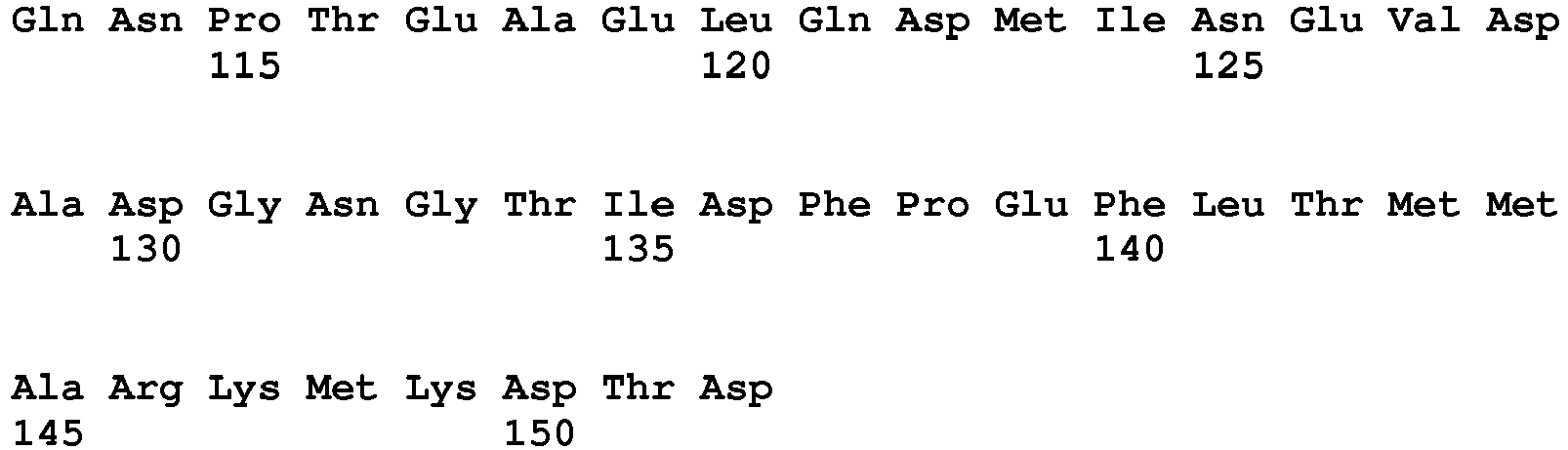
- <210> 69
<211> 152
<212> PRT
<213> artificial sequence - <220>
<223> permutated calmodulin-variant: N-1 - <400> 69

- <210> 70
<211> 152
<212> PRT
<213> artificial sequence - <220>
<223> permutated calmodulin-variant: N-2 - <400> 70

- <210> 71
<211> 152
<212> PRT
<213> artificial sequence - <220>
<223> permutated calmodulin-variant: C-1 - <400> 71


- <210> 72
<211> 152
<212> PRT
<213> artificial sequence - <220>
<223> permutated calmodulin-variant: C-2 - <400> 72


- <210> 73
<211> 152
<212> PRT
<213> artificial sequence - <220>
<223> permutated calmodulin-variant: 1-S - <400> 73
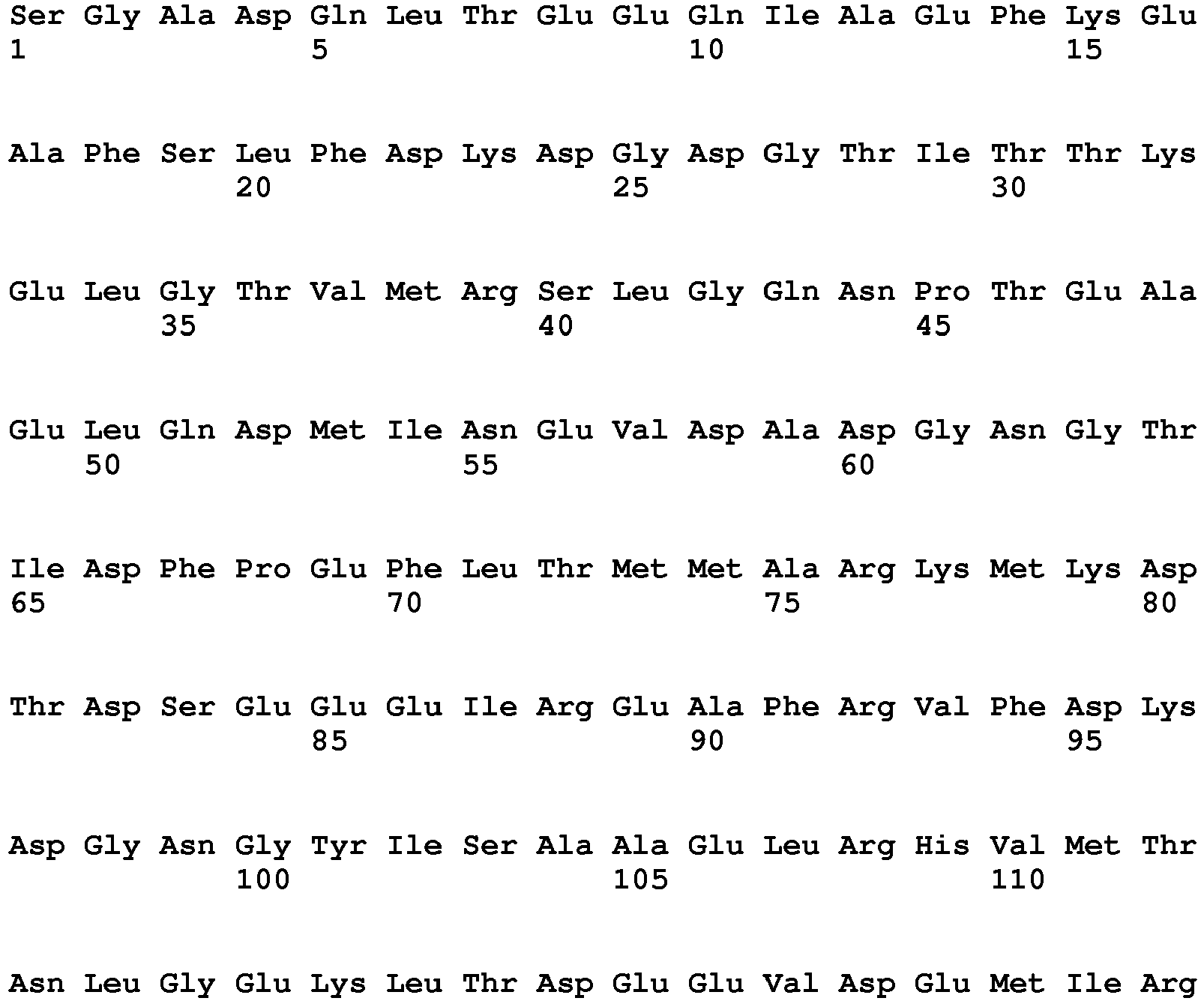

- <210> 74
<211> 152
<212> PRT
<213> artificial sequence - <220>
<223> permutated calmodulin-variant: 2-G - <400> 74

- <210> 75
<211> 152
<212> PRT
<213> artificial sequence - <220>
<223> permutated calmodulin-variant: 5-Q - <400> 75

- <210> 76
<211> 152
<212> PRT
<213> artificial sequence - <220>
<223> permutated calmodulin-variant: 6-L - <400> 76


- <210> 77
<211> 152
<212> PRT
<213> artificial sequence - <220>
<223> permutated calmodulin-variant: 8-E - <400> 77


- <210> 78
<211> 152
<212> PRT
<213> artificial sequence - <220>
<223> permutated calmodulin-variant: 9-E - <400> 78


- <210> 79
<211> 152
<212> PRT
<213> artificial sequence - <220>
<223> permutated calmodulin-variant: 10-Q - <400> 79

- <210> 80
<211> 152
<212> PRT
<213> artificial sequence - <220>
<223> permutated calmodulin-variant: 11-I - <400> 80

- <210> 81
<211> 152
<212> PRT
<213> artificial sequence - <220>
<223> permutated calmodulin-variant: 13-E - <400> 81


- <210> 82
<211> 152
<212> PRT
<213> artificial sequence - <220>
<223> permutated calmodulin-variant: 14-F - <400> 82


- <210> 83
<211> 152
<212> PRT
<213> artificial sequence - <220>
<223> permutated calmodulin-variant: 16-E - <400> 83
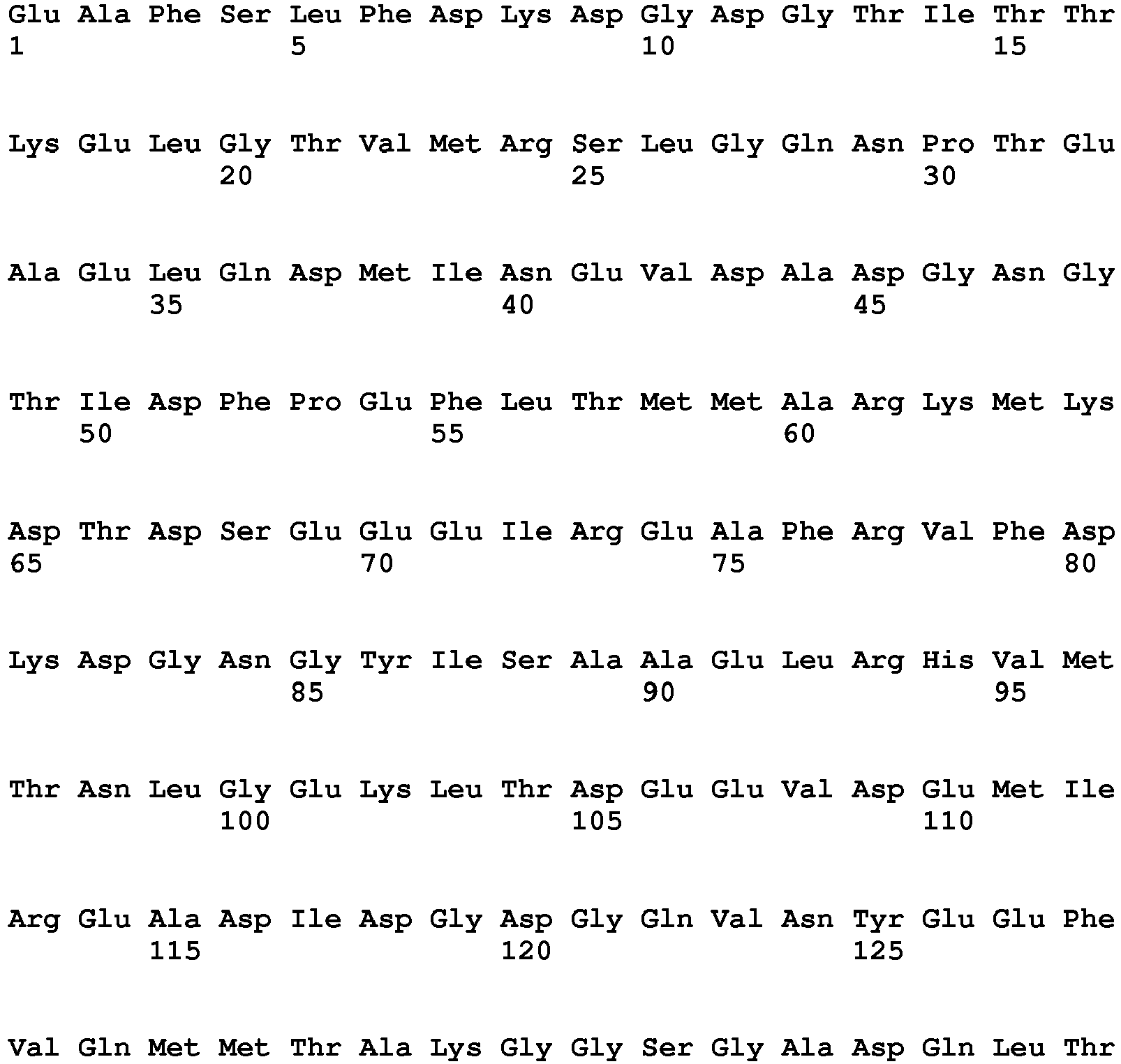

- <210> 84
<211> 152
<212> PRT
<213> artificial sequence - <220>
<223> permutated calmodulin-variant: 26-D - <400> 84

- <210> 85
<211> 152
<212> PRT
<213> artificial sequence - <220>
<223> permutated calmodulin-variant: 40-S - <400> 85

- <210> 86
<211> 152
<212> PRT
<213> artificial sequence - <220>
<223> permutated calmodulin-variant: 45-P - <400> 86


- <210> 87
<211> 152
<212> PRT
<213> artificial sequence - <220>
<223> permutated calmodulin-variant: 46-T - <400> 87


- <210> 88
<211> 152
<212> PRT
<213> artificial sequence - <220>
<223> permutated calmodulin-variant: 56-E - <400> 88


- <210> 89
<211> 152
<212> PRT
<213> artificial sequence - <220>
<223> permutated calmodulin-variant: 59-A - <400> 89

- <210> 90
<211> 152
<212> PRT
<213> artificial sequence - <220>
<223> permutated calmodulin-variant: 60-D - <400> 90

- <210> 91
<211> 152
<212> PRT
<213> artificial sequence - <220>
<223> permutated calmodulin-variant: 61-G - <400> 91

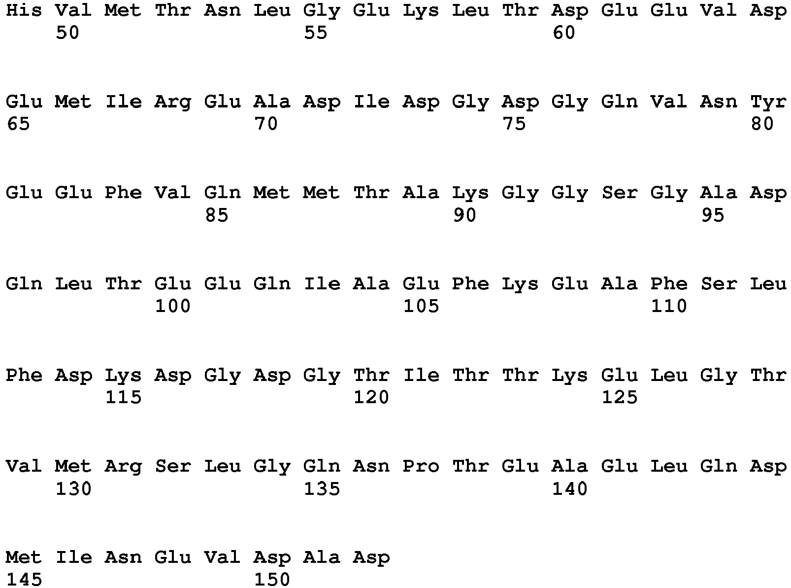
- <210> 92
<211> 152
<212> PRT
<213> artificial sequence - <220>
<223> permutated calmodulin-variant: 62-N - <400> 92
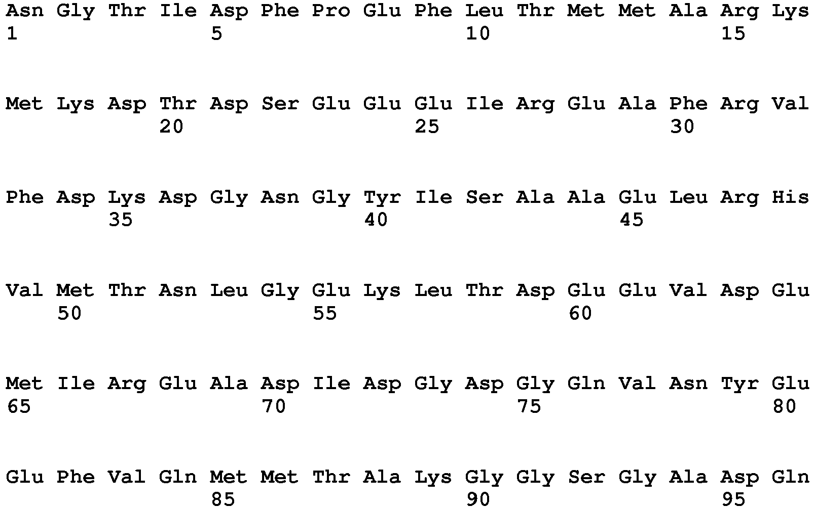

- <210> 93
<211> 152
<212> PRT
<213> artificial sequence - <220>
<223> permutated calmodulin-variant: 65-I - <400> 93


- <210> 94
<211> 152
<212> PRT
<213> artificial sequence - <220>
<223> permutated calmodulin-variant: 75-A - <400> 94

- <210> 95
<211> 152
<212> PRT
<213> artificial sequence - <220>
<223> permutated calmodulin-variant: 76-R - <400> 95
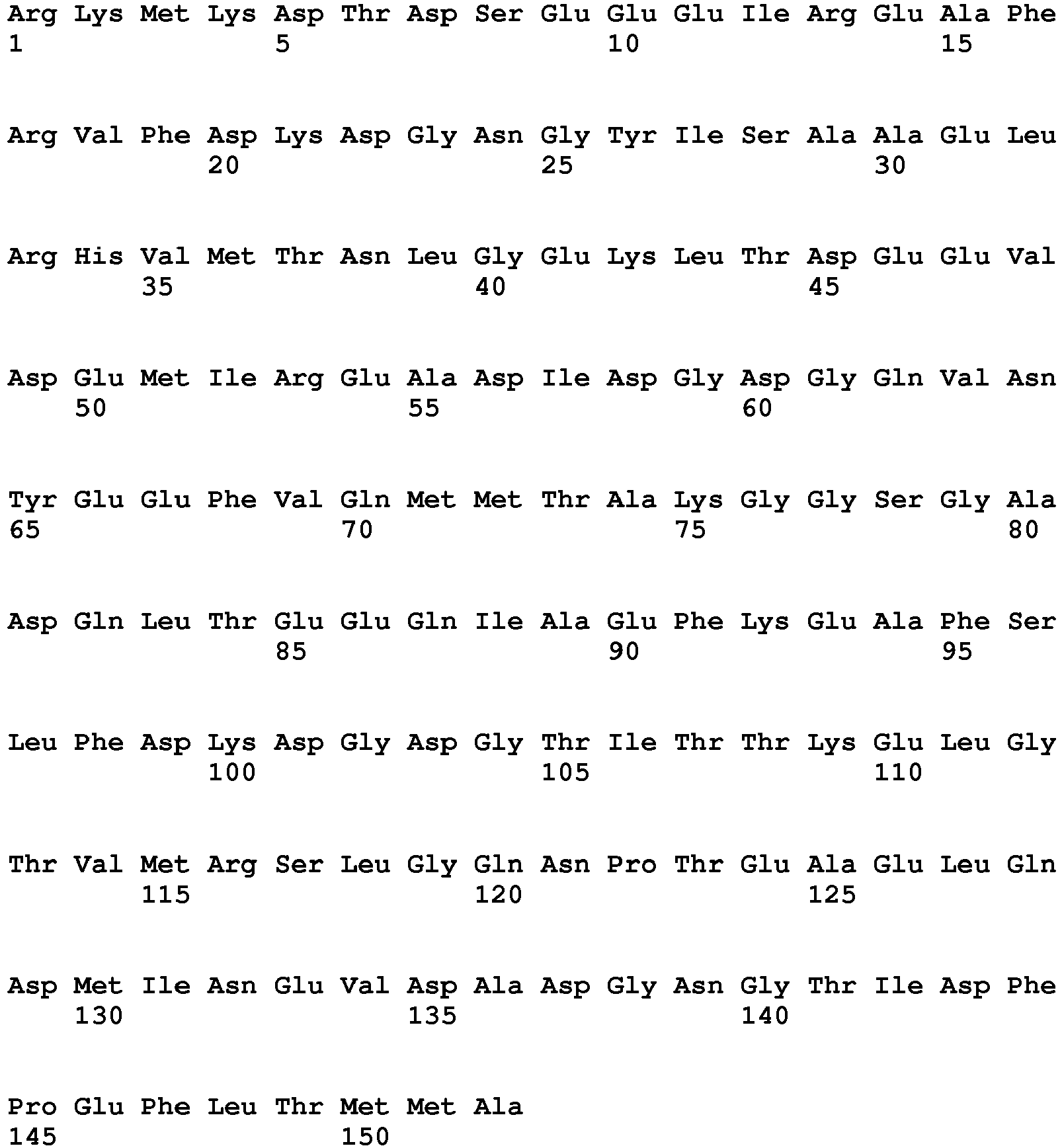
- <210> 96
<211> 152
<212> PRT
<213> artificial sequence - <220>
<223> permutated calmodulin-variant: 77-K - <400> 96


- <210> 97
<211> 152
<212> PRT
<213> artificial sequence - <220>
<223> permutated calmodulin-variant: 79-K - <400> 97


- <210> 98
<211> 152
<212> PRT
<213> artificial sequence - <220>
<223> permutated calmodulin-variant: 80-D - <400> 98

- <210> 99
<211> 152
<212> PRT
<213> artificial sequence - <220>
<223> permutated calmodulin-variant: 81-T - <400> 99

- <210> 100
<211> 152
<212> PRT
<213> artificial sequence - <220>
<223> permutated calmodulin-variant: 82-D - <400> 100


- <210> 101
<211> 152
<212> PRT
<213> artificial sequence - <220>
<223> permutated calmodulin-variant: 85-E - <400> 101


- <210> 102
<211> 152
<212> PRT
<213> artificial sequence - <220>
<223> permutated calmodulin-variant: 98-G - <400> 102


- <210> 103
<211> 152
<212> PRT
<213> artificial sequence - <220>
<223> permutated calmodulin-variant: 99-N - <400> 103

- <210> 104
<211> 152
<212> PRT
<213> artificial sequence - <220>
<223> permutated calmodulin-variant: 103-S - <400> 104

- <210> 105
<211> 152
<212> PRT
<213> artificial sequence - <220>
<223> permutated calmodulin-variant: 104-A - <400> 105


- <210> 106
<211> 152
<212> PRT
<213> artificial sequence - <220>
<223> permutated calmodulin-variant: 110-V - <400> 106


- <210> 107
<211> 152
<212> PRT
<213> artificial sequence - <220>
<223> permutated calmodulin-variant: 111-M - <400> 107


- <210> 108
<211> 152
<212> PRT
<213> artificial sequence - <220>
<223> permutated calmodulin-variant: 112-T - <400> 108

- <210> 109
<211> 152
<212> PRT
<213> artificial sequence - <220>
<223> permutated calmodulin-variant: 113-N - <400> 109
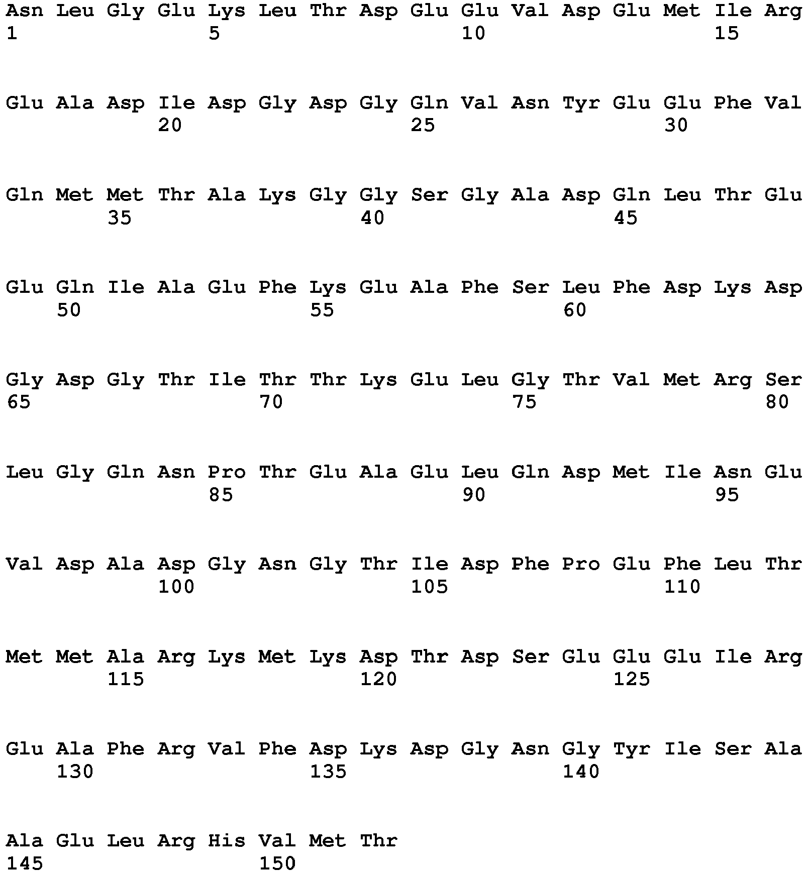
- <210> 110
<211> 152
<212> PRT
<213> artificial sequence - <220>
<223> permutated calmodulin-variant: 117-K - <400> 110


- <210> 111
<211> 152
<212> PRT
<213> artificial sequence - <220>
<223> permutated calmodulin-variant: 118-L - <400> 111


- <210> 112
<211> 152
<212> PRT
<213> artificial sequence - <220>
<223> permutated calmodulin-variant: 119-T - <400> 112


- <210> 113
<211> 152
<212> PRT
<213> artificial sequence - <220>
<223> permutated calmodulin-variant: 120-D - <400> 113

- <210> 114
<211> 152
<212> PRT
<213> artificial sequence - <220>
<223> permutated calmodulin-variant: 123-V - <400> 114
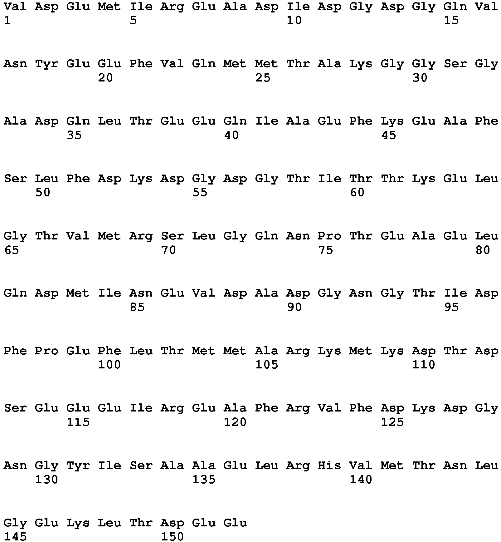
- <210> 115
<211> 152
<212> PRT
<213> artificial sequence - <220>
<223> permutated calmodulin-variant: 124-D - <400> 115


- <210> 116
<211> 152
<212> PRT
<213> artificial sequence - <220>
<223> permutated calmodulin-variant: 125-E - <400> 116
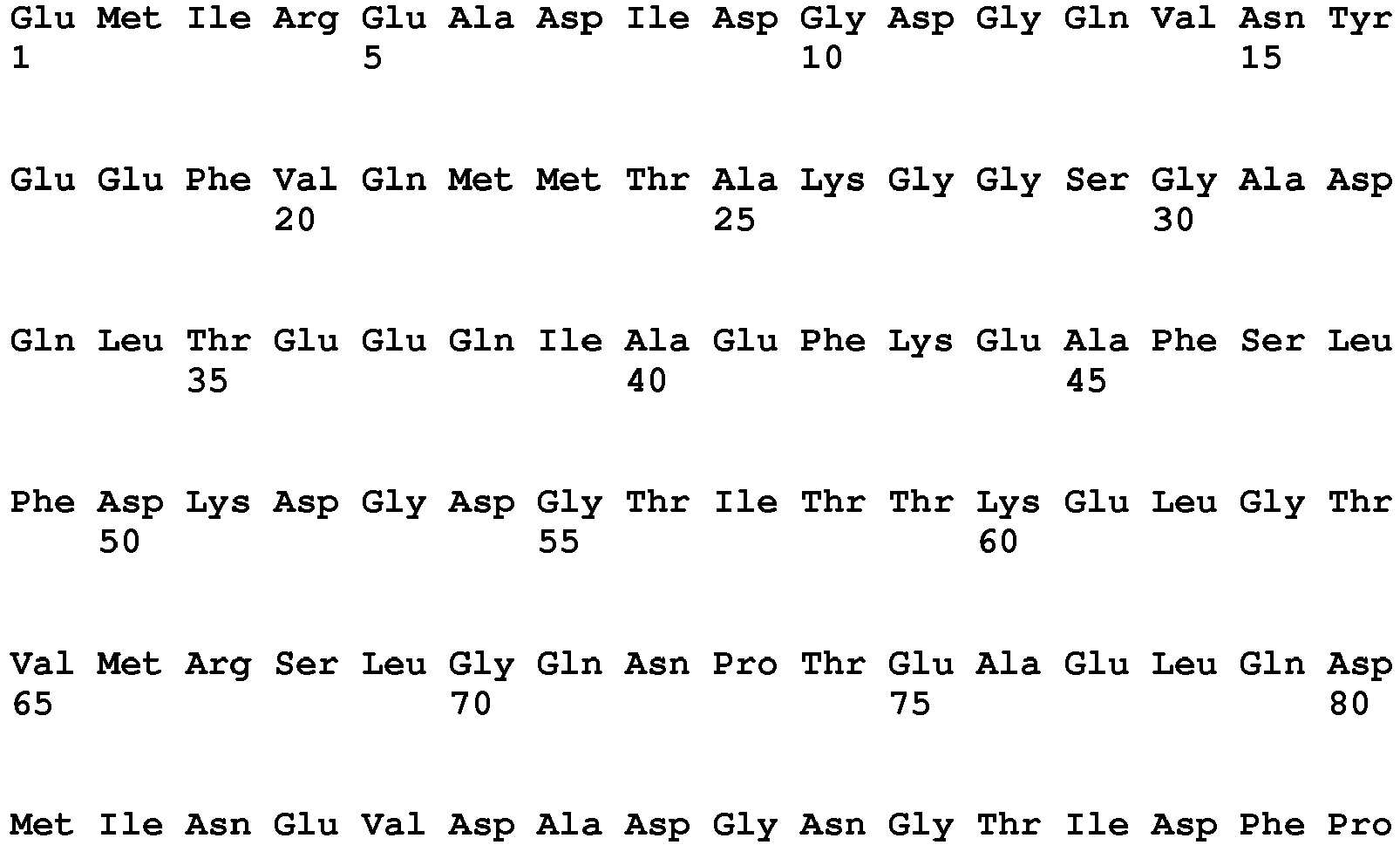

- <210> 117
<211> 152
<212> PRT
<213> artificial sequence - <220>
<223> permutated calmodulin-variant: 130-A - <400> 117


- <210> 118
<211> 152
<212> PRT
<213> artificial sequence - <220>
<223> permutated calmodulin-variant: 134-G - <400> 118

- <210> 119
<211> 152
<212> PRT
<213> artificial sequence - <220>
<223> permutated calmodulin-variant: 135-D - <400> 119

- <210> 120
<211> 152
<212> PRT
<213> artificial sequence - <220>
<223> permutated calmodulin-variant: 144-V - <400> 120


- <210> 121
<211> 152
<212> PRT
<213> artificial sequence - <220>
<223> permutated calmodulin-variant: 148-T - <400> 121


- <210> 122
<211> 152
<212> PRT
<213> artificial sequence - <220>
<223> permutated calmodulin-variant: 151-G - <400> 122


- <210> 123
<211> 152
<212> PRT
<213> artificial sequence - <220>
<223> permutated calmodulin-variant: 152-G - <400> 123

- <210> 124
<211> 409
<212> PRT
<213> artificial sequence - <220>
<223> anti-CD14 scFv (TUEK4)_M-1 - <400> 124


- <210> 125
<211> 409
<212> PRT
<213> artificial sequence - <220>
<223> anti-CD14 scFv (TUEK4)_M-2 - <400> 125


- <210> 126
<211> 409
<212> PRT
<213> artificial sequence - <220>
<223> anti-CD14 scFv (TUEK4)_C-1 - <400> 126


- <210> 127
<211> 409
<212> PRT
<213> artificial sequence - <220>
<223> anti-CD14 scFv (TUEK4)_C-2 - <400> 127


- <210> 128
<211> 413
<212> PRT
<213> artificial sequence - <220>
<223> anti-Biotin scFv (18E7.2)_M-1 - <400> 128


- <210> 129
<211> 413
<212> PRT
<213> artificial sequence - <220>
<223> anti-Biotin scFv (18E7.2)_M-2 - <400> 129


- <210> 130
<211> 413
<212> PRT
<213> artificial sequence - <220>
<223> anti-Biotin scFv (18E7.2)_C-1 - <400> 130


- <210> 131
<211> 406
<212> PRT
<213> artificial sequence - <220>
<223> anti-CD4 scFv (Q425)_M-1 - <400> 131
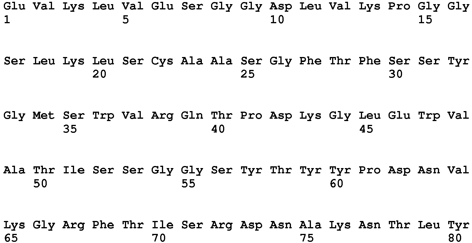


- <210> 132
<211> 406
<212> PRT
<213> artificial sequence - <220>
<223> anti-CD4 scFv (Q425)_M-2 - <400> 132


- <210> 133
<211> 406
<212> PRT
<213> artificial sequence - <220>
<223> anti-CD4 scFv (Q425)_C-1 - <400> 133


- <210> 134
<211> 406
<212> PRT
<213> artificial sequence - <220>
<223> anti-CD4 scFv (Q425)_C-2 - <400> 134


- <210> 135
<211> 263
<212> PRT
<213> artificial sequence - <220>
<223> clone D1.3 (WT); scFv derived from murine antibody against hen egg white lysozyme - <400> 135


wherein said permutated calmodulin has the sequence selected from the group consisting of sequences SEQ ID NO: 67 and SEQ ID NO: 68 and said calmodulin binding peptide has the sequence selected from the group consisting of sequences SEQ ID NO: 1, SEQ ID NO: 7, SEQ ID NO: 8, SEQ ID NO: 9 and SEQ ID NO: 46, or wherein said permutated calmodulin has the sequence SEQ ID NO: 68 and said calmodulin binding peptide has the sequence SEQ ID NO: 53, or wherein said permutated calmodulin has the sequence selected from the group consisting of sequences SEQ ID NO: 71 and SEQ ID NO: 72 and said calmodulin binding peptide has the sequence selected from the group consisting of sequences SEQ ID NO: 1, SEQ ID NO: 47, SEQ ID NO: 51 and SEQ ID NO: 53.
Claims (15)
- A composition comprisingi) a polypeptide comprising calmodulin and two immunoglobulin superfamily domains, wherein said two immunoglobulin superfamily domains are linked via said calmodulin,ii) a calmodulin binding molecule;iii) ions binding to the Ca2+ binding site of calmodulin,wherein the binding of said calmodulin-binding molecule and of said ions to said Ca2+ binding site of calmodulin affects the binding of said polypeptide to an antigen to be bound by said polypeptide.
- The composition according to claim 1, wherein said binding of said calmodulin-binding molecule and of ions to said Ca2+ binding site of calmodulin enhances or reduces the affinity of said polypeptide to said antigen.
- The composition according to claim 1 or 2, wherein said calmodulin binding molecule is a calmodulin binding peptide.
- The composition according to any one of the preceding claims, wherein said calmodulin is a permutated calmodulin.
- The composition according to claim 4, wherein said permutated calmodulin has the sequence selected from the group consisting of sequences SEQ ID NO: 67 to SEQ ID NO: 123.
- The composition according to any one of the claims 3 to 5, wherein said calmodulin binding peptide has the sequence selected from the group consisting of sequences SEQ ID NO: 1 to SEQ ID NO: 65.
- The composition according to claim 4, wherein said permutated calmodulin has the sequence selected from the group consisting of sequences SEQ ID NO: 67 and SEQ ID NO: 68 and said calmodulin binding peptide has the sequence selected from the group consisting of sequences SEQ ID NO: 1, SEQ ID NO: 7, SEQ ID NO: 8, SEQ ID NO: 9 and SEQ ID NO: 46, or wherein said permutated calmodulin has the sequence SEQ ID NO: 68 and said calmodulin binding peptide has the sequence SEQ ID NO: 53, or wherein said permutated calmodulin has the sequence selected from the group consisting of sequences SEQ ID NO: 71 and SEQ ID NO: 72 and said calmodulin binding peptide has the sequence selected from the group consisting of sequences SEQ ID NO: 1, SEQ ID NO: 47, SEQ ID NO: 51 and SEQ ID NO: 53.
- The composition according to any one of claims 1 to 7, wherein said polypeptide is a single chain Fv (scFv) comprising the calmodulin and a variable region of a heavy chain of an immunoglobulin and a variable region of a light chain of an immunoglobulin.
- The composition according to any one of the preceding claims, wherein said polypeptide is part of an antigen binding domain of a chimeric antigen receptor (CAR), said CAR comprising an antigen binding domain, a transmembrane domain and cytoplasmic signaling domain.
- The composition according to claim 4, wherein said polypeptide comprising said permutated calmodulin and two immunoglobulin superfamily domains, wherein said two immunoglobulin superfamily domains are linked via said permutated calmodulin, is obtainable by the method comprising the stepsa) Creating at least one insertion nucleic acid sequence encoding a permutated calmodulinb) Creating an acceptor nucleic acid sequence encoding a polypeptide comprising two immunoglobulin superfamily domainsc) Inserting the at least one insertion sequence of a) into the acceptor sequence of b), wherein one insertion sequence of a) is inserted between the parts of the acceptor sequence b) which encode the two immunoglobulin superfamily domains of b)d) Transforming a host with the nucleic acid sequences of c)e) Selecting for transformed hosts harboring the sequence(s) of c)f) Screening for transformed hosts expressing polypeptides comprising two immunoglobulin superfamily domains linked via permutated calmodulin by exposing the polypeptides produced by the transformed hosts to said calmodulin-binding molecule and identifying the transformed hosts harboring polypeptides which impact the binding of said polypeptides to the antigen in the presence of ions binding to the Ca2+ binding site of calmodulin.
- An in vitro method for affecting the binding of a polypeptide for an antigen to be bound, wherein said polypeptide comprises a calmodulin and two immunoglobulin superfamily domains wherein said two immunoglobulin superfamily domains are linked via said calmodulin, the method comprising the step of contacting said polypeptide with a calmodulin binding molecule in the presence of ions binding to the Ca2+ binding site of calmodulin, thereby affecting the binding of said polypeptide to the antigen.
- The method according to claim 11, wherein said polypeptide is contacted with the antigen to be bound by said polypeptide before said contacting of said calmodulin binding molecule with said polypeptide.
- The method according to claim 12, wherein said affecting of the binding is a reduction of binding, thereby releasing the polypeptide from the antigen.
- The method according to any one of claims 11 to 13, wherein said calmodulin is a permutated calmodulin.
- The in vitro use of a polypeptide comprising a permutated calmodulin and two immunoglobulin superfamily domains, wherein said two immunoglobulin superfamily domains are linked via said permutated calmodulin for affecting in the presence of a calmodulin binding molecule and ions binding to the Ca2+ binding site of calmodulin the binding of said polypeptide to an antigen to be bound by said polypeptide.
Priority Applications (3)
| Application Number | Priority Date | Filing Date | Title |
|---|---|---|---|
| ES16181971T ES2708657T3 (en) | 2016-07-29 | 2016-07-29 | Composition and method to affect the binding of antigen-binding polypeptides to antigens |
| EP16181971.9A EP3276005B1 (en) | 2016-07-29 | 2016-07-29 | Composition and method for affecting the binding of antigen-binding polypeptides to antigens |
| US15/650,724 US10730922B2 (en) | 2016-07-29 | 2017-07-14 | Composition and method for affecting the binding of antigen-binding polypeptides to antigens |
Applications Claiming Priority (1)
| Application Number | Priority Date | Filing Date | Title |
|---|---|---|---|
| EP16181971.9A EP3276005B1 (en) | 2016-07-29 | 2016-07-29 | Composition and method for affecting the binding of antigen-binding polypeptides to antigens |
Publications (2)
| Publication Number | Publication Date |
|---|---|
| EP3276005A1 EP3276005A1 (en) | 2018-01-31 |
| EP3276005B1 true EP3276005B1 (en) | 2019-01-09 |
Family
ID=56561239
Family Applications (1)
| Application Number | Title | Priority Date | Filing Date |
|---|---|---|---|
| EP16181971.9A Active EP3276005B1 (en) | 2016-07-29 | 2016-07-29 | Composition and method for affecting the binding of antigen-binding polypeptides to antigens |
Country Status (3)
| Country | Link |
|---|---|
| US (1) | US10730922B2 (en) |
| EP (1) | EP3276005B1 (en) |
| ES (1) | ES2708657T3 (en) |
Families Citing this family (2)
| Publication number | Priority date | Publication date | Assignee | Title |
|---|---|---|---|---|
| EP3523333B1 (en) * | 2016-10-10 | 2023-12-13 | Singapore Health Services Pte Ltd | Anti-ceacam6 antibodies and methods of use |
| EP4469478A1 (en) * | 2022-01-26 | 2024-12-04 | Mabswitch Inc. | Bispecific molecule with tunable affinity to a targetted antigen |
Family Cites Families (4)
| Publication number | Priority date | Publication date | Assignee | Title |
|---|---|---|---|---|
| DE50112379D1 (en) | 2000-08-11 | 2007-05-31 | Univ Ruprecht Karls Heidelberg | FV CONSTRUCTS WITH INFLUENCABLE AFFINITY TO A BONDING SUBSTANCE |
| US8492122B2 (en) | 2002-03-11 | 2013-07-23 | The Johns Hopkins University | Molecular switches and methods for making and using the same |
| JP4214206B2 (en) * | 2002-12-10 | 2009-01-28 | 独立行政法人理化学研究所 | Fluorescent indicator using FRET |
| US8338138B2 (en) | 2004-01-28 | 2012-12-25 | The John Hopkins University | Methods for making and using molecular switches involving circular permutation |
-
2016
- 2016-07-29 ES ES16181971T patent/ES2708657T3/en active Active
- 2016-07-29 EP EP16181971.9A patent/EP3276005B1/en active Active
-
2017
- 2017-07-14 US US15/650,724 patent/US10730922B2/en active Active
Non-Patent Citations (1)
| Title |
|---|
| None * |
Also Published As
| Publication number | Publication date |
|---|---|
| ES2708657T3 (en) | 2019-04-10 |
| EP3276005A1 (en) | 2018-01-31 |
| US20180030104A1 (en) | 2018-02-01 |
| US10730922B2 (en) | 2020-08-04 |
Similar Documents
| Publication | Publication Date | Title |
|---|---|---|
| US5723584A (en) | Biotinylation of proteins | |
| KR101763345B1 (en) | Protein combination-based fv library, and preparation method therefor | |
| US10202466B2 (en) | Linked peptide fluorogenic biosensors | |
| KR20050108389A (en) | Monoclonal antibody and hybridoma producing the same | |
| AU2014314214B2 (en) | Binding polypeptides having a mutated scaffold | |
| US11098081B2 (en) | Epitope tag and method for detection and/or purification of tagged polypeptides | |
| CN113150095A (en) | Scaffold proteins derived from plant cysteine protease inhibitors | |
| CN107108750A (en) | The method of the protein exhibiting expression of the non-covalent domain containing Fc on cell surface | |
| US10730922B2 (en) | Composition and method for affecting the binding of antigen-binding polypeptides to antigens | |
| EP1945673B1 (en) | Targeting and tracing of antigens in living cells | |
| AU2015305220B2 (en) | Affinity proteins and uses thereof | |
| JP2011193728A (en) | PEPTIDE HAVING BINDING CAPABILITY TO EpCAM | |
| Wu et al. | A fast and efficient procedure to produce scFvs specific for large macromolecular complexes | |
| CA2785359C (en) | Protein display | |
| US20160033490A1 (en) | Linked Peptide Fluorogenic Biosensors | |
| JP2011525103A (en) | Display vector and method and use thereof | |
| KR20110003547A (en) | Artificial protein scaffold | |
| Shukla et al. | Phage-displayed combinatorial peptide libraries in fusion to β-lactamase as reporter for an accelerated clone screening: Potential uses of selected enzyme-linked affinity reagents in downstream applications | |
| CN106905435B (en) | Method for preparing binding protein based on protein A mutant | |
| WO2008140538A1 (en) | Dna display screen for expression product with desired binding properties | |
| US20260109787A1 (en) | Production method of heteromultimeric protein, protein, nucleic acid, expression vector, transformant, and production method of protein | |
| SG190913A1 (en) | Modified tamavidin | |
| Crusius et al. | Tab2, a novel recombinant polypeptide tag offering sensitive and specific protein detection and reliable affinity purification | |
| EP3502130A1 (en) | Ligand regulated protein-protein interaction system | |
| KR20240066998A (en) | Method for cell panning |
Legal Events
| Date | Code | Title | Description |
|---|---|---|---|
| PUAI | Public reference made under article 153(3) epc to a published international application that has entered the european phase |
Free format text: ORIGINAL CODE: 0009012 |
|
| STAA | Information on the status of an ep patent application or granted ep patent |
Free format text: STATUS: THE APPLICATION HAS BEEN PUBLISHED |
|
| AK | Designated contracting states |
Kind code of ref document: A1 Designated state(s): AL AT BE BG CH CY CZ DE DK EE ES FI FR GB GR HR HU IE IS IT LI LT LU LV MC MK MT NL NO PL PT RO RS SE SI SK SM TR |
|
| AX | Request for extension of the european patent |
Extension state: BA ME |
|
| STAA | Information on the status of an ep patent application or granted ep patent |
Free format text: STATUS: REQUEST FOR EXAMINATION WAS MADE |
|
| 17P | Request for examination filed |
Effective date: 20180723 |
|
| RBV | Designated contracting states (corrected) |
Designated state(s): AL AT BE BG CH CY CZ DE DK EE ES FI FR GB GR HR HU IE IS IT LI LT LU LV MC MK MT NL NO PL PT RO RS SE SI SK SM TR |
|
| GRAP | Despatch of communication of intention to grant a patent |
Free format text: ORIGINAL CODE: EPIDOSNIGR1 |
|
| STAA | Information on the status of an ep patent application or granted ep patent |
Free format text: STATUS: GRANT OF PATENT IS INTENDED |
|
| RIC1 | Information provided on ipc code assigned before grant |
Ipc: C12N 15/85 20060101AFI20180924BHEP |
|
| INTG | Intention to grant announced |
Effective date: 20181024 |
|
| RIN1 | Information on inventor provided before grant (corrected) |
Inventor name: KELLMANN, SARAH-JANE Inventor name: THIE, HOLGER Inventor name: DUEBEL, STEFAN |
|
| GRAS | Grant fee paid |
Free format text: ORIGINAL CODE: EPIDOSNIGR3 |
|
| GRAA | (expected) grant |
Free format text: ORIGINAL CODE: 0009210 |
|
| STAA | Information on the status of an ep patent application or granted ep patent |
Free format text: STATUS: THE PATENT HAS BEEN GRANTED |
|
| AK | Designated contracting states |
Kind code of ref document: B1 Designated state(s): AL AT BE BG CH CY CZ DE DK EE ES FI FR GB GR HR HU IE IS IT LI LT LU LV MC MK MT NL NO PL PT RO RS SE SI SK SM TR |
|
| REG | Reference to a national code |
Ref country code: GB Ref legal event code: FG4D |
|
| REG | Reference to a national code |
Ref country code: CH Ref legal event code: EP Ref country code: AT Ref legal event code: REF Ref document number: 1087311 Country of ref document: AT Kind code of ref document: T Effective date: 20190115 |
|
| REG | Reference to a national code |
Ref country code: CH Ref legal event code: NV Representative=s name: TR-IP CONSULTING LLC, CH Ref country code: DE Ref legal event code: R096 Ref document number: 602016009136 Country of ref document: DE |
|
| REG | Reference to a national code |
Ref country code: IE Ref legal event code: FG4D |
|
| REG | Reference to a national code |
Ref country code: NL Ref legal event code: FP |
|
| REG | Reference to a national code |
Ref country code: ES Ref legal event code: FG2A Ref document number: 2708657 Country of ref document: ES Kind code of ref document: T3 Effective date: 20190410 |
|
| REG | Reference to a national code |
Ref country code: LT Ref legal event code: MG4D |
|
| REG | Reference to a national code |
Ref country code: AT Ref legal event code: MK05 Ref document number: 1087311 Country of ref document: AT Kind code of ref document: T Effective date: 20190109 |
|
| PG25 | Lapsed in a contracting state [announced via postgrant information from national office to epo] |
Ref country code: NO Free format text: LAPSE BECAUSE OF FAILURE TO SUBMIT A TRANSLATION OF THE DESCRIPTION OR TO PAY THE FEE WITHIN THE PRESCRIBED TIME-LIMIT Effective date: 20190409 Ref country code: PL Free format text: LAPSE BECAUSE OF FAILURE TO SUBMIT A TRANSLATION OF THE DESCRIPTION OR TO PAY THE FEE WITHIN THE PRESCRIBED TIME-LIMIT Effective date: 20190109 Ref country code: LT Free format text: LAPSE BECAUSE OF FAILURE TO SUBMIT A TRANSLATION OF THE DESCRIPTION OR TO PAY THE FEE WITHIN THE PRESCRIBED TIME-LIMIT Effective date: 20190109 Ref country code: PT Free format text: LAPSE BECAUSE OF FAILURE TO SUBMIT A TRANSLATION OF THE DESCRIPTION OR TO PAY THE FEE WITHIN THE PRESCRIBED TIME-LIMIT Effective date: 20190509 Ref country code: SE Free format text: LAPSE BECAUSE OF FAILURE TO SUBMIT A TRANSLATION OF THE DESCRIPTION OR TO PAY THE FEE WITHIN THE PRESCRIBED TIME-LIMIT Effective date: 20190109 Ref country code: FI Free format text: LAPSE BECAUSE OF FAILURE TO SUBMIT A TRANSLATION OF THE DESCRIPTION OR TO PAY THE FEE WITHIN THE PRESCRIBED TIME-LIMIT Effective date: 20190109 |
|
| PG25 | Lapsed in a contracting state [announced via postgrant information from national office to epo] |
Ref country code: LV Free format text: LAPSE BECAUSE OF FAILURE TO SUBMIT A TRANSLATION OF THE DESCRIPTION OR TO PAY THE FEE WITHIN THE PRESCRIBED TIME-LIMIT Effective date: 20190109 Ref country code: GR Free format text: LAPSE BECAUSE OF FAILURE TO SUBMIT A TRANSLATION OF THE DESCRIPTION OR TO PAY THE FEE WITHIN THE PRESCRIBED TIME-LIMIT Effective date: 20190410 Ref country code: RS Free format text: LAPSE BECAUSE OF FAILURE TO SUBMIT A TRANSLATION OF THE DESCRIPTION OR TO PAY THE FEE WITHIN THE PRESCRIBED TIME-LIMIT Effective date: 20190109 Ref country code: IS Free format text: LAPSE BECAUSE OF FAILURE TO SUBMIT A TRANSLATION OF THE DESCRIPTION OR TO PAY THE FEE WITHIN THE PRESCRIBED TIME-LIMIT Effective date: 20190509 Ref country code: BG Free format text: LAPSE BECAUSE OF FAILURE TO SUBMIT A TRANSLATION OF THE DESCRIPTION OR TO PAY THE FEE WITHIN THE PRESCRIBED TIME-LIMIT Effective date: 20190409 Ref country code: HR Free format text: LAPSE BECAUSE OF FAILURE TO SUBMIT A TRANSLATION OF THE DESCRIPTION OR TO PAY THE FEE WITHIN THE PRESCRIBED TIME-LIMIT Effective date: 20190109 |
|
| REG | Reference to a national code |
Ref country code: DE Ref legal event code: R097 Ref document number: 602016009136 Country of ref document: DE |
|
| PG25 | Lapsed in a contracting state [announced via postgrant information from national office to epo] |
Ref country code: DK Free format text: LAPSE BECAUSE OF FAILURE TO SUBMIT A TRANSLATION OF THE DESCRIPTION OR TO PAY THE FEE WITHIN THE PRESCRIBED TIME-LIMIT Effective date: 20190109 Ref country code: AL Free format text: LAPSE BECAUSE OF FAILURE TO SUBMIT A TRANSLATION OF THE DESCRIPTION OR TO PAY THE FEE WITHIN THE PRESCRIBED TIME-LIMIT Effective date: 20190109 Ref country code: RO Free format text: LAPSE BECAUSE OF FAILURE TO SUBMIT A TRANSLATION OF THE DESCRIPTION OR TO PAY THE FEE WITHIN THE PRESCRIBED TIME-LIMIT Effective date: 20190109 Ref country code: SK Free format text: LAPSE BECAUSE OF FAILURE TO SUBMIT A TRANSLATION OF THE DESCRIPTION OR TO PAY THE FEE WITHIN THE PRESCRIBED TIME-LIMIT Effective date: 20190109 Ref country code: CZ Free format text: LAPSE BECAUSE OF FAILURE TO SUBMIT A TRANSLATION OF THE DESCRIPTION OR TO PAY THE FEE WITHIN THE PRESCRIBED TIME-LIMIT Effective date: 20190109 Ref country code: AT Free format text: LAPSE BECAUSE OF FAILURE TO SUBMIT A TRANSLATION OF THE DESCRIPTION OR TO PAY THE FEE WITHIN THE PRESCRIBED TIME-LIMIT Effective date: 20190109 Ref country code: EE Free format text: LAPSE BECAUSE OF FAILURE TO SUBMIT A TRANSLATION OF THE DESCRIPTION OR TO PAY THE FEE WITHIN THE PRESCRIBED TIME-LIMIT Effective date: 20190109 |
|
| RAP2 | Party data changed (patent owner data changed or rights of a patent transferred) |
Owner name: DUEBEL, STEFAN Owner name: MILTENYI BIOTEC B.V. & CO. KG |
|
| PLBE | No opposition filed within time limit |
Free format text: ORIGINAL CODE: 0009261 |
|
| STAA | Information on the status of an ep patent application or granted ep patent |
Free format text: STATUS: NO OPPOSITION FILED WITHIN TIME LIMIT |
|
| REG | Reference to a national code |
Ref country code: DE Ref legal event code: R081 Ref document number: 602016009136 Country of ref document: DE Owner name: MABSWITCH INC., VAN NUYS, US Free format text: FORMER OWNERS: DUEBEL, STEFAN, 38108 BRAUNSCHWEIG, DE; MILTENYI BIOTEC GMBH, 51429 BERGISCH GLADBACH, DE Ref country code: DE Ref legal event code: R081 Ref document number: 602016009136 Country of ref document: DE Owner name: DUEBEL, STEFAN, DE Free format text: FORMER OWNERS: DUEBEL, STEFAN, 38108 BRAUNSCHWEIG, DE; MILTENYI BIOTEC GMBH, 51429 BERGISCH GLADBACH, DE Ref country code: DE Ref legal event code: R081 Ref document number: 602016009136 Country of ref document: DE Owner name: MILTENYI BIOTEC B.V. & CO. KG, DE Free format text: FORMER OWNERS: DUEBEL, STEFAN, 38108 BRAUNSCHWEIG, DE; MILTENYI BIOTEC GMBH, 51429 BERGISCH GLADBACH, DE |
|
| 26N | No opposition filed |
Effective date: 20191010 |
|
| REG | Reference to a national code |
Ref country code: CH Ref legal event code: PCAR Free format text: NEW ADDRESS: ROUTE DU COUTSET 18, 1485 NUVILLY (CH) |
|
| PG25 | Lapsed in a contracting state [announced via postgrant information from national office to epo] |
Ref country code: MC Free format text: LAPSE BECAUSE OF FAILURE TO SUBMIT A TRANSLATION OF THE DESCRIPTION OR TO PAY THE FEE WITHIN THE PRESCRIBED TIME-LIMIT Effective date: 20190109 Ref country code: SI Free format text: LAPSE BECAUSE OF FAILURE TO SUBMIT A TRANSLATION OF THE DESCRIPTION OR TO PAY THE FEE WITHIN THE PRESCRIBED TIME-LIMIT Effective date: 20190109 |
|
| PG25 | Lapsed in a contracting state [announced via postgrant information from national office to epo] |
Ref country code: TR Free format text: LAPSE BECAUSE OF FAILURE TO SUBMIT A TRANSLATION OF THE DESCRIPTION OR TO PAY THE FEE WITHIN THE PRESCRIBED TIME-LIMIT Effective date: 20190109 |
|
| PG25 | Lapsed in a contracting state [announced via postgrant information from national office to epo] |
Ref country code: LU Free format text: LAPSE BECAUSE OF NON-PAYMENT OF DUE FEES Effective date: 20190729 |
|
| PG25 | Lapsed in a contracting state [announced via postgrant information from national office to epo] |
Ref country code: IE Free format text: LAPSE BECAUSE OF NON-PAYMENT OF DUE FEES Effective date: 20190729 |
|
| REG | Reference to a national code |
Ref country code: BE Ref legal event code: PD Owner name: MILTENYI BIOTEC B.V. & CO. KG; DE Free format text: DETAILS ASSIGNMENT: CHANGE OF OWNER(S), ASSIGNMENT; FORMER OWNER NAME: DUEBEL, STEFAN Effective date: 20210120 |
|
| PG25 | Lapsed in a contracting state [announced via postgrant information from national office to epo] |
Ref country code: CY Free format text: LAPSE BECAUSE OF FAILURE TO SUBMIT A TRANSLATION OF THE DESCRIPTION OR TO PAY THE FEE WITHIN THE PRESCRIBED TIME-LIMIT Effective date: 20190109 |
|
| PG25 | Lapsed in a contracting state [announced via postgrant information from national office to epo] |
Ref country code: SM Free format text: LAPSE BECAUSE OF FAILURE TO SUBMIT A TRANSLATION OF THE DESCRIPTION OR TO PAY THE FEE WITHIN THE PRESCRIBED TIME-LIMIT Effective date: 20190109 |
|
| PG25 | Lapsed in a contracting state [announced via postgrant information from national office to epo] |
Ref country code: HU Free format text: LAPSE BECAUSE OF FAILURE TO SUBMIT A TRANSLATION OF THE DESCRIPTION OR TO PAY THE FEE WITHIN THE PRESCRIBED TIME-LIMIT; INVALID AB INITIO Effective date: 20160729 Ref country code: MT Free format text: LAPSE BECAUSE OF FAILURE TO SUBMIT A TRANSLATION OF THE DESCRIPTION OR TO PAY THE FEE WITHIN THE PRESCRIBED TIME-LIMIT Effective date: 20190109 |
|
| REG | Reference to a national code |
Ref country code: BE Ref legal event code: PD Owner name: MILTENYI BIOTEC B.V. & CO. KG; DE Free format text: DETAILS ASSIGNMENT: CHANGE OF OWNER(S), ASSIGNMENT; FORMER OWNER NAME: DUEBEL, STEFAN Effective date: 20220225 |
|
| REG | Reference to a national code |
Ref country code: GB Ref legal event code: 732E Free format text: REGISTERED BETWEEN 20220324 AND 20220330 |
|
| REG | Reference to a national code |
Ref country code: NL Ref legal event code: PD Owner name: MABSWITCH INC.; US Free format text: DETAILS ASSIGNMENT: CHANGE OF OWNER(S), ASSIGNMENT; FORMER OWNER NAME: MILTENYI BIOTEC GMBH Effective date: 20220401 |
|
| REG | Reference to a national code |
Ref country code: DE Ref legal event code: R081 Ref document number: 602016009136 Country of ref document: DE Owner name: MILTENYI BIOTEC B.V. & CO. KG, DE Free format text: FORMER OWNERS: DUEBEL, STEFAN, 38108 BRAUNSCHWEIG, DE; MILTENYI BIOTEC B.V. & CO. KG, 51429 BERGISCH GLADBACH, DE Ref country code: DE Ref legal event code: R081 Ref document number: 602016009136 Country of ref document: DE Owner name: MABSWITCH INC., VAN NUYS, US Free format text: FORMER OWNERS: DUEBEL, STEFAN, 38108 BRAUNSCHWEIG, DE; MILTENYI BIOTEC B.V. & CO. KG, 51429 BERGISCH GLADBACH, DE |
|
| REG | Reference to a national code |
Ref country code: ES Ref legal event code: PC2A Owner name: MABSWITCH INC. Effective date: 20220609 |
|
| PG25 | Lapsed in a contracting state [announced via postgrant information from national office to epo] |
Ref country code: MK Free format text: LAPSE BECAUSE OF FAILURE TO SUBMIT A TRANSLATION OF THE DESCRIPTION OR TO PAY THE FEE WITHIN THE PRESCRIBED TIME-LIMIT Effective date: 20190109 |
|
| PGFP | Annual fee paid to national office [announced via postgrant information from national office to epo] |
Ref country code: NL Payment date: 20250721 Year of fee payment: 10 |
|
| PGFP | Annual fee paid to national office [announced via postgrant information from national office to epo] |
Ref country code: ES Payment date: 20250826 Year of fee payment: 10 |
|
| PGFP | Annual fee paid to national office [announced via postgrant information from national office to epo] |
Ref country code: DE Payment date: 20250722 Year of fee payment: 10 |
|
| PGFP | Annual fee paid to national office [announced via postgrant information from national office to epo] |
Ref country code: IT Payment date: 20250724 Year of fee payment: 10 |
|
| PGFP | Annual fee paid to national office [announced via postgrant information from national office to epo] |
Ref country code: BE Payment date: 20250718 Year of fee payment: 10 Ref country code: GB Payment date: 20250722 Year of fee payment: 10 |
|
| PGFP | Annual fee paid to national office [announced via postgrant information from national office to epo] |
Ref country code: FR Payment date: 20250725 Year of fee payment: 10 |
|
| PGFP | Annual fee paid to national office [announced via postgrant information from national office to epo] |
Ref country code: CH Payment date: 20250801 Year of fee payment: 10 |
|
| REG | Reference to a national code |
Ref country code: CH Ref legal event code: R18 Free format text: ST27 STATUS EVENT CODE: U-0-0-R10-R18 (AS PROVIDED BY THE NATIONAL OFFICE) Effective date: 20260209 |
|
| REG | Reference to a national code |
Ref country code: CH Ref legal event code: R18 Free format text: ST27 STATUS EVENT CODE: U-0-0-R10-R18 (AS PROVIDED BY THE NATIONAL OFFICE) Effective date: 20260320 |