EP3275465B1 - Therapeutic compositions comprising a combination of a harmine and isovanillin component - Google Patents
Therapeutic compositions comprising a combination of a harmine and isovanillin component Download PDFInfo
- Publication number
- EP3275465B1 EP3275465B1 EP17185821.0A EP17185821A EP3275465B1 EP 3275465 B1 EP3275465 B1 EP 3275465B1 EP 17185821 A EP17185821 A EP 17185821A EP 3275465 B1 EP3275465 B1 EP 3275465B1
- Authority
- EP
- European Patent Office
- Prior art keywords
- graph
- death
- illustrating
- effect
- inducing
- Prior art date
- Legal status (The legal status is an assumption and is not a legal conclusion. Google has not performed a legal analysis and makes no representation as to the accuracy of the status listed.)
- Active
Links
- JVTZFYYHCGSXJV-UHFFFAOYSA-N isovanillin Chemical compound COC1=CC=C(C=O)C=C1O JVTZFYYHCGSXJV-UHFFFAOYSA-N 0.000 title claims description 241
- BXNJHAXVSOCGBA-UHFFFAOYSA-N Harmine Chemical compound N1=CC=C2C3=CC=C(OC)C=C3NC2=C1C BXNJHAXVSOCGBA-UHFFFAOYSA-N 0.000 title claims description 234
- RERZNCLIYCABFS-UHFFFAOYSA-N Harmaline hydrochloride Natural products C1CN=C(C)C2=C1C1=CC=C(OC)C=C1N2 RERZNCLIYCABFS-UHFFFAOYSA-N 0.000 title claims description 164
- VJHLDRVYTQNASM-UHFFFAOYSA-N harmine Natural products CC1=CN=CC=2NC3=CC(=CC=C3C=21)OC VJHLDRVYTQNASM-UHFFFAOYSA-N 0.000 title claims description 115
- 239000000203 mixture Substances 0.000 title claims description 75
- 230000001225 therapeutic effect Effects 0.000 title claims description 18
- 206010028980 Neoplasm Diseases 0.000 claims description 107
- 201000011510 cancer Diseases 0.000 claims description 54
- PSFDQSOCUJVVGF-UHFFFAOYSA-N harman Chemical compound C12=CC=CC=C2NC2=C1C=CN=C2C PSFDQSOCUJVVGF-UHFFFAOYSA-N 0.000 claims description 38
- 238000000034 method Methods 0.000 claims description 36
- 238000011282 treatment Methods 0.000 claims description 29
- 235000012141 vanillin Nutrition 0.000 claims description 29
- MWOOGOJBHIARFG-UHFFFAOYSA-N vanillin Chemical compound COC1=CC(C=O)=CC=C1O MWOOGOJBHIARFG-UHFFFAOYSA-N 0.000 claims description 28
- FGQOOHJZONJGDT-UHFFFAOYSA-N vanillin Natural products COC1=CC(O)=CC(C=O)=C1 FGQOOHJZONJGDT-UHFFFAOYSA-N 0.000 claims description 28
- 150000003839 salts Chemical class 0.000 claims description 19
- 230000001093 anti-cancer Effects 0.000 claims description 18
- JJVNINGBHGBWJH-UHFFFAOYSA-N ortho-vanillin Chemical compound COC1=CC=CC(C=O)=C1O JJVNINGBHGBWJH-UHFFFAOYSA-N 0.000 claims description 18
- RHVPEFQDYMMNSY-UHFFFAOYSA-N harmalol Chemical compound N1C2=CC(O)=CC=C2C2=C1C(C)=NCC2 RHVPEFQDYMMNSY-UHFFFAOYSA-N 0.000 claims description 16
- LBBJNGFCXDOYMQ-UHFFFAOYSA-N 1-methyl-2,9-dihydropyrido[3,4-b]indol-7-one Chemical compound C1=CC(=O)C=C2NC3=C(C)NC=CC3=C21 LBBJNGFCXDOYMQ-UHFFFAOYSA-N 0.000 claims description 15
- SATMZMMKDDTOSQ-UHFFFAOYSA-N Harmol Natural products C12=CC=C(O)C=C2NC2=C1C=CN=C2C SATMZMMKDDTOSQ-UHFFFAOYSA-N 0.000 claims description 15
- 239000002552 dosage form Substances 0.000 claims description 11
- 239000007787 solid Substances 0.000 claims description 8
- 239000002775 capsule Substances 0.000 claims description 5
- 238000002347 injection Methods 0.000 claims description 5
- 239000007924 injection Substances 0.000 claims description 5
- 239000006187 pill Substances 0.000 claims description 5
- 239000000243 solution Substances 0.000 claims description 5
- 239000003826 tablet Substances 0.000 claims description 5
- 239000007788 liquid Substances 0.000 claims description 4
- 239000000725 suspension Substances 0.000 claims description 4
- 239000000499 gel Substances 0.000 claims description 3
- 230000001747 exhibiting effect Effects 0.000 claims description 2
- 230000007761 synergistic anti-cancer Effects 0.000 claims 1
- 210000004027 cell Anatomy 0.000 description 507
- 230000000694 effects Effects 0.000 description 365
- 230000034994 death Effects 0.000 description 339
- 231100000517 death Toxicity 0.000 description 339
- 230000001939 inductive effect Effects 0.000 description 325
- VFLDPWHFBUODDF-FCXRPNKRSA-N curcumin Chemical compound C1=C(O)C(OC)=CC(\C=C\C(=O)CC(=O)\C=C\C=2C=C(OC)C(O)=CC=2)=C1 VFLDPWHFBUODDF-FCXRPNKRSA-N 0.000 description 313
- 235000012754 curcumin Nutrition 0.000 description 141
- VFLDPWHFBUODDF-UHFFFAOYSA-N diferuloylmethane Natural products C1=C(O)C(OC)=CC(C=CC(=O)CC(=O)C=CC=2C=C(OC)C(O)=CC=2)=C1 VFLDPWHFBUODDF-UHFFFAOYSA-N 0.000 description 141
- 229940109262 curcumin Drugs 0.000 description 139
- 239000004148 curcumin Substances 0.000 description 139
- 206010033128 Ovarian cancer Diseases 0.000 description 122
- 206010061535 Ovarian neoplasm Diseases 0.000 description 122
- 206010058467 Lung neoplasm malignant Diseases 0.000 description 96
- 201000005202 lung cancer Diseases 0.000 description 96
- 208000020816 lung neoplasm Diseases 0.000 description 96
- 206010025323 Lymphomas Diseases 0.000 description 90
- 208000032839 leukemia Diseases 0.000 description 69
- 206010060862 Prostate cancer Diseases 0.000 description 57
- 208000000236 Prostatic Neoplasms Diseases 0.000 description 57
- 238000012360 testing method Methods 0.000 description 57
- 239000000047 product Substances 0.000 description 55
- 208000014829 head and neck neoplasm Diseases 0.000 description 53
- 201000010536 head and neck cancer Diseases 0.000 description 51
- 230000001965 increasing effect Effects 0.000 description 49
- 125000005647 linker group Chemical group 0.000 description 46
- 125000003118 aryl group Chemical group 0.000 description 37
- 206010006187 Breast cancer Diseases 0.000 description 36
- 208000026310 Breast neoplasm Diseases 0.000 description 36
- 241000699670 Mus sp. Species 0.000 description 32
- JYTVKRNTTALBBZ-UHFFFAOYSA-N bis demethoxycurcumin Natural products C1=CC(O)=CC=C1C=CC(=O)CC(=O)C=CC1=CC=CC(O)=C1 JYTVKRNTTALBBZ-UHFFFAOYSA-N 0.000 description 32
- PREBVFJICNPEKM-YDWXAUTNSA-N bisdemethoxycurcumin Chemical compound C1=CC(O)=CC=C1\C=C\C(=O)CC(=O)\C=C\C1=CC=C(O)C=C1 PREBVFJICNPEKM-YDWXAUTNSA-N 0.000 description 32
- 229910052799 carbon Inorganic materials 0.000 description 32
- YXAKCQIIROBKOP-UHFFFAOYSA-N di-p-hydroxycinnamoylmethane Natural products C=1C=C(O)C=CC=1C=CC(=O)C=C(O)C=CC1=CC=C(O)C=C1 YXAKCQIIROBKOP-UHFFFAOYSA-N 0.000 description 32
- LFQSCWFLJHTTHZ-UHFFFAOYSA-N Ethanol Chemical compound CCO LFQSCWFLJHTTHZ-UHFFFAOYSA-N 0.000 description 29
- 230000030833 cell death Effects 0.000 description 29
- LBKFGYZQBSGRHY-UHFFFAOYSA-N 3-hydroxy-4-methoxybenzoic acid Chemical compound COC1=CC=C(C(O)=O)C=C1O LBKFGYZQBSGRHY-UHFFFAOYSA-N 0.000 description 28
- 241000282414 Homo sapiens Species 0.000 description 27
- AOJJSUZBOXZQNB-TZSSRYMLSA-N Doxorubicin Chemical compound O([C@H]1C[C@@](O)(CC=2C(O)=C3C(=O)C=4C=CC=C(C=4C(=O)C3=C(O)C=21)OC)C(=O)CO)[C@H]1C[C@H](N)[C@H](O)[C@H](C)O1 AOJJSUZBOXZQNB-TZSSRYMLSA-N 0.000 description 26
- 150000001875 compounds Chemical class 0.000 description 24
- 125000004432 carbon atom Chemical group C* 0.000 description 22
- 206010061902 Pancreatic neoplasm Diseases 0.000 description 20
- 201000002528 pancreatic cancer Diseases 0.000 description 20
- 230000000996 additive effect Effects 0.000 description 18
- 239000003814 drug Substances 0.000 description 18
- 208000015486 malignant pancreatic neoplasm Diseases 0.000 description 18
- 208000008443 pancreatic carcinoma Diseases 0.000 description 18
- 230000000052 comparative effect Effects 0.000 description 16
- 125000002887 hydroxy group Chemical group [H]O* 0.000 description 16
- 244000163122 Curcuma domestica Species 0.000 description 15
- 235000003373 curcuma longa Nutrition 0.000 description 15
- 210000002950 fibroblast Anatomy 0.000 description 15
- WHKRHBLAJFYZKF-UHFFFAOYSA-N 5-(hydroxymethyl)-2-methoxyphenol Chemical compound COC1=CC=C(CO)C=C1O WHKRHBLAJFYZKF-UHFFFAOYSA-N 0.000 description 14
- 235000003392 Curcuma domestica Nutrition 0.000 description 14
- 230000004611 cancer cell death Effects 0.000 description 14
- CRQDWQWZCNKKAC-UHFFFAOYSA-N harmalol Chemical compound N1C2=CC(=O)C=CC2=C2C1=C(C)NCC2 CRQDWQWZCNKKAC-UHFFFAOYSA-N 0.000 description 14
- 235000013976 turmeric Nutrition 0.000 description 14
- 208000003174 Brain Neoplasms Diseases 0.000 description 13
- 229960004679 doxorubicin Drugs 0.000 description 13
- 125000002915 carbonyl group Chemical group [*:2]C([*:1])=O 0.000 description 12
- 239000003795 chemical substances by application Substances 0.000 description 12
- 235000018102 proteins Nutrition 0.000 description 11
- 102000004169 proteins and genes Human genes 0.000 description 11
- 108090000623 proteins and genes Proteins 0.000 description 11
- 239000003981 vehicle Substances 0.000 description 11
- 230000006907 apoptotic process Effects 0.000 description 10
- 210000003169 central nervous system Anatomy 0.000 description 10
- 230000007246 mechanism Effects 0.000 description 10
- 201000008968 osteosarcoma Diseases 0.000 description 10
- 229940079593 drug Drugs 0.000 description 9
- 229910052739 hydrogen Inorganic materials 0.000 description 9
- 239000002207 metabolite Substances 0.000 description 9
- -1 phenolic aldehyde vanillin isomer Chemical class 0.000 description 9
- 239000002246 antineoplastic agent Substances 0.000 description 8
- 230000007423 decrease Effects 0.000 description 8
- 125000000956 methoxy group Chemical group [H]C([H])([H])O* 0.000 description 8
- 102000011727 Caspases Human genes 0.000 description 7
- 108010076667 Caspases Proteins 0.000 description 7
- 206010039491 Sarcoma Diseases 0.000 description 7
- 239000007857 degradation product Substances 0.000 description 7
- 230000002496 gastric effect Effects 0.000 description 7
- 231100000225 lethality Toxicity 0.000 description 7
- 125000001997 phenyl group Chemical group [H]C1=C([H])C([H])=C(*)C([H])=C1[H] 0.000 description 7
- 229940124597 therapeutic agent Drugs 0.000 description 7
- IJGRMHOSHXDMSA-UHFFFAOYSA-N Atomic nitrogen Chemical compound N#N IJGRMHOSHXDMSA-UHFFFAOYSA-N 0.000 description 6
- 206010053961 Mitochondrial toxicity Diseases 0.000 description 6
- 230000001154 acute effect Effects 0.000 description 6
- 125000000217 alkyl group Chemical group 0.000 description 6
- 150000001412 amines Chemical class 0.000 description 6
- 231100000296 mitochondrial toxicity Toxicity 0.000 description 6
- 229910052757 nitrogen Inorganic materials 0.000 description 6
- 239000011550 stock solution Substances 0.000 description 6
- 210000001519 tissue Anatomy 0.000 description 6
- 125000000229 (C1-C4)alkoxy group Chemical group 0.000 description 5
- 241000701806 Human papillomavirus Species 0.000 description 5
- 206010027476 Metastases Diseases 0.000 description 5
- 102000007066 Prostate-Specific Antigen Human genes 0.000 description 5
- 108010072866 Prostate-Specific Antigen Proteins 0.000 description 5
- 230000008859 change Effects 0.000 description 5
- 230000002354 daily effect Effects 0.000 description 5
- 231100000673 dose–response relationship Toxicity 0.000 description 5
- 125000005842 heteroatom Chemical group 0.000 description 5
- 239000004615 ingredient Substances 0.000 description 5
- 238000011081 inoculation Methods 0.000 description 5
- 230000005012 migration Effects 0.000 description 5
- 238000013508 migration Methods 0.000 description 5
- 238000002360 preparation method Methods 0.000 description 5
- 239000000126 substance Substances 0.000 description 5
- 208000024891 symptom Diseases 0.000 description 5
- 231100000419 toxicity Toxicity 0.000 description 5
- 230000001988 toxicity Effects 0.000 description 5
- AIFRHYZBTHREPW-UHFFFAOYSA-N β-carboline Chemical compound N1=CC=C2C3=CC=CC=C3NC2=C1 AIFRHYZBTHREPW-UHFFFAOYSA-N 0.000 description 5
- OKTJSMMVPCPJKN-UHFFFAOYSA-N Carbon Chemical group [C] OKTJSMMVPCPJKN-UHFFFAOYSA-N 0.000 description 4
- 102000004018 Caspase 6 Human genes 0.000 description 4
- 108090000425 Caspase 6 Proteins 0.000 description 4
- 102000004039 Caspase-9 Human genes 0.000 description 4
- 108090000566 Caspase-9 Proteins 0.000 description 4
- 206010008342 Cervix carcinoma Diseases 0.000 description 4
- 239000006144 Dulbecco’s modified Eagle's medium Substances 0.000 description 4
- 208000021309 Germ cell tumor Diseases 0.000 description 4
- 208000034176 Neoplasms, Germ Cell and Embryonal Diseases 0.000 description 4
- 208000007641 Pinealoma Diseases 0.000 description 4
- ZXLDQJLIBNPEFJ-MRVPVSSYSA-N Tetrahydroharmine Chemical compound C1CN[C@H](C)C2=C1C1=CC=C(OC)C=C1N2 ZXLDQJLIBNPEFJ-MRVPVSSYSA-N 0.000 description 4
- 208000006105 Uterine Cervical Neoplasms Diseases 0.000 description 4
- 125000005196 alkyl carbonyloxy group Chemical group 0.000 description 4
- 238000001815 biotherapy Methods 0.000 description 4
- 229910052796 boron Inorganic materials 0.000 description 4
- 230000006721 cell death pathway Effects 0.000 description 4
- 230000022534 cell killing Effects 0.000 description 4
- 230000004663 cell proliferation Effects 0.000 description 4
- 201000010881 cervical cancer Diseases 0.000 description 4
- 238000002512 chemotherapy Methods 0.000 description 4
- 230000001684 chronic effect Effects 0.000 description 4
- 229940127089 cytotoxic agent Drugs 0.000 description 4
- 150000002148 esters Chemical class 0.000 description 4
- 239000001963 growth medium Substances 0.000 description 4
- 230000001976 improved effect Effects 0.000 description 4
- 238000001727 in vivo Methods 0.000 description 4
- 230000003993 interaction Effects 0.000 description 4
- 230000009545 invasion Effects 0.000 description 4
- 230000002147 killing effect Effects 0.000 description 4
- 125000002496 methyl group Chemical group [H]C([H])([H])* 0.000 description 4
- FDPIMTJIUBPUKL-UHFFFAOYSA-N pentan-3-one Chemical compound CCC(=O)CC FDPIMTJIUBPUKL-UHFFFAOYSA-N 0.000 description 4
- 239000000902 placebo Substances 0.000 description 4
- 229940068196 placebo Drugs 0.000 description 4
- 208000029340 primitive neuroectodermal tumor Diseases 0.000 description 4
- 230000008569 process Effects 0.000 description 4
- 230000004044 response Effects 0.000 description 4
- 201000000849 skin cancer Diseases 0.000 description 4
- 210000002784 stomach Anatomy 0.000 description 4
- 125000001424 substituent group Chemical group 0.000 description 4
- ZXLDQJLIBNPEFJ-UHFFFAOYSA-N tetrahydro-beta-carboline Natural products C1CNC(C)C2=C1C1=CC=C(OC)C=C1N2 ZXLDQJLIBNPEFJ-UHFFFAOYSA-N 0.000 description 4
- 208000008732 thymoma Diseases 0.000 description 4
- 210000004881 tumor cell Anatomy 0.000 description 4
- UEPVWRDHSPMIAZ-IZTHOABVSA-N (1e,4z,6e)-5-hydroxy-7-(4-hydroxy-3-methoxyphenyl)-1-(4-hydroxyphenyl)hepta-1,4,6-trien-3-one Chemical compound C1=C(O)C(OC)=CC(\C=C\C(\O)=C\C(=O)\C=C\C=2C=CC(O)=CC=2)=C1 UEPVWRDHSPMIAZ-IZTHOABVSA-N 0.000 description 3
- JKMHFZQWWAIEOD-UHFFFAOYSA-N 2-[4-(2-hydroxyethyl)piperazin-1-yl]ethanesulfonic acid Chemical compound OCC[NH+]1CCN(CCS([O-])(=O)=O)CC1 JKMHFZQWWAIEOD-UHFFFAOYSA-N 0.000 description 3
- QTBSBXVTEAMEQO-UHFFFAOYSA-N Acetic acid Chemical compound CC(O)=O QTBSBXVTEAMEQO-UHFFFAOYSA-N 0.000 description 3
- 206010067484 Adverse reaction Diseases 0.000 description 3
- 201000008271 Atypical teratoid rhabdoid tumor Diseases 0.000 description 3
- 206010005003 Bladder cancer Diseases 0.000 description 3
- BTBUEUYNUDRHOZ-UHFFFAOYSA-N Borate Chemical compound [O-]B([O-])[O-] BTBUEUYNUDRHOZ-UHFFFAOYSA-N 0.000 description 3
- 108091003079 Bovine Serum Albumin Proteins 0.000 description 3
- 0 C*c1ccc(C=CC(C=Cc2ccc(*(C)C)cc2)=O)cc1 Chemical compound C*c1ccc(C=CC(C=Cc2ccc(*(C)C)cc2)=O)cc1 0.000 description 3
- HJTVQHVGMGKONQ-LUZURFALSA-N Curcumin II Natural products C1=C(O)C(OC)=CC(\C=C\C(=O)CC(=O)\C=C\C=2C=CC(O)=CC=2)=C1 HJTVQHVGMGKONQ-LUZURFALSA-N 0.000 description 3
- 206010013710 Drug interaction Diseases 0.000 description 3
- 102000001301 EGF receptor Human genes 0.000 description 3
- 108060006698 EGF receptor Proteins 0.000 description 3
- 239000007995 HEPES buffer Substances 0.000 description 3
- VEXZGXHMUGYJMC-UHFFFAOYSA-N Hydrochloric acid Chemical compound Cl VEXZGXHMUGYJMC-UHFFFAOYSA-N 0.000 description 3
- 244000061176 Nicotiana tabacum Species 0.000 description 3
- 235000002637 Nicotiana tabacum Nutrition 0.000 description 3
- MUBZPKHOEPUJKR-UHFFFAOYSA-N Oxalic acid Chemical compound OC(=O)C(O)=O MUBZPKHOEPUJKR-UHFFFAOYSA-N 0.000 description 3
- 206010035226 Plasma cell myeloma Diseases 0.000 description 3
- KWYUFKZDYYNOTN-UHFFFAOYSA-M Potassium hydroxide Chemical compound [OH-].[K+] KWYUFKZDYYNOTN-UHFFFAOYSA-M 0.000 description 3
- OFOBLEOULBTSOW-UHFFFAOYSA-N Propanedioic acid Natural products OC(=O)CC(O)=O OFOBLEOULBTSOW-UHFFFAOYSA-N 0.000 description 3
- JUJWROOIHBZHMG-UHFFFAOYSA-N Pyridine Chemical compound C1=CC=NC=C1 JUJWROOIHBZHMG-UHFFFAOYSA-N 0.000 description 3
- 208000000453 Skin Neoplasms Diseases 0.000 description 3
- FAPWRFPIFSIZLT-UHFFFAOYSA-M Sodium chloride Chemical compound [Na+].[Cl-] FAPWRFPIFSIZLT-UHFFFAOYSA-M 0.000 description 3
- HEMHJVSKTPXQMS-UHFFFAOYSA-M Sodium hydroxide Chemical compound [OH-].[Na+] HEMHJVSKTPXQMS-UHFFFAOYSA-M 0.000 description 3
- 208000007097 Urinary Bladder Neoplasms Diseases 0.000 description 3
- 239000002253 acid Substances 0.000 description 3
- 230000006838 adverse reaction Effects 0.000 description 3
- 125000004429 atom Chemical group 0.000 description 3
- 125000001797 benzyl group Chemical group [H]C1=C([H])C([H])=C(C([H])=C1[H])C([H])([H])* 0.000 description 3
- 230000015572 biosynthetic process Effects 0.000 description 3
- ZADPBFCGQRWHPN-UHFFFAOYSA-N boronic acid Chemical compound OBO ZADPBFCGQRWHPN-UHFFFAOYSA-N 0.000 description 3
- 150000007942 carboxylates Chemical class 0.000 description 3
- 239000003183 carcinogenic agent Substances 0.000 description 3
- OYKGNQNESCZSHQ-UHFFFAOYSA-N chembl1369266 Chemical compound O.O.Cl.N1C2=CC(O)=CC=C2C2=C1C(C)=NCC2 OYKGNQNESCZSHQ-UHFFFAOYSA-N 0.000 description 3
- KRKNYBCHXYNGOX-UHFFFAOYSA-N citric acid Chemical compound OC(=O)CC(O)(C(O)=O)CC(O)=O KRKNYBCHXYNGOX-UHFFFAOYSA-N 0.000 description 3
- NMRUIRRIQNAQEB-UHFFFAOYSA-N demethoxycurcumin Natural products OC(=CC(C=CC1=CC(=C(C=C1)O)OC)=O)C=CC1=CC=C(C=C1)O NMRUIRRIQNAQEB-UHFFFAOYSA-N 0.000 description 3
- 238000011161 development Methods 0.000 description 3
- 230000018109 developmental process Effects 0.000 description 3
- 230000008406 drug-drug interaction Effects 0.000 description 3
- 239000012091 fetal bovine serum Substances 0.000 description 3
- 125000001072 heteroaryl group Chemical group 0.000 description 3
- 125000000623 heterocyclic group Chemical group 0.000 description 3
- 239000001257 hydrogen Substances 0.000 description 3
- RBOUBJPHXSVUTH-UHFFFAOYSA-N hydron;1-methyl-2,9-dihydropyrido[3,4-b]indol-7-one;chloride Chemical compound Cl.C1=CC(=O)C=C2NC3=C(C)NC=CC3=C21 RBOUBJPHXSVUTH-UHFFFAOYSA-N 0.000 description 3
- 239000003112 inhibitor Substances 0.000 description 3
- 239000000543 intermediate Substances 0.000 description 3
- 210000004185 liver Anatomy 0.000 description 3
- 201000007270 liver cancer Diseases 0.000 description 3
- 208000014018 liver neoplasm Diseases 0.000 description 3
- 201000001441 melanoma Diseases 0.000 description 3
- 238000010232 migration assay Methods 0.000 description 3
- 201000005962 mycosis fungoides Diseases 0.000 description 3
- 208000025113 myeloid leukemia Diseases 0.000 description 3
- IQZPDFORWZTSKT-UHFFFAOYSA-N nitrosulphonic acid Chemical compound OS(=O)(=O)[N+]([O-])=O IQZPDFORWZTSKT-UHFFFAOYSA-N 0.000 description 3
- UEPVWRDHSPMIAZ-UHFFFAOYSA-N p-hydroxycinnamoyl feruloylmethane Natural products C1=C(O)C(OC)=CC(C=CC(O)=CC(=O)C=CC=2C=CC(O)=CC=2)=C1 UEPVWRDHSPMIAZ-UHFFFAOYSA-N 0.000 description 3
- 230000009467 reduction Effects 0.000 description 3
- 230000002829 reductive effect Effects 0.000 description 3
- 238000011160 research Methods 0.000 description 3
- 125000000446 sulfanediyl group Chemical group *S* 0.000 description 3
- 150000003462 sulfoxides Chemical class 0.000 description 3
- 229910052717 sulfur Inorganic materials 0.000 description 3
- 201000008205 supratentorial primitive neuroectodermal tumor Diseases 0.000 description 3
- 230000002195 synergetic effect Effects 0.000 description 3
- 231100000331 toxic Toxicity 0.000 description 3
- 230000002588 toxic effect Effects 0.000 description 3
- 201000005112 urinary bladder cancer Diseases 0.000 description 3
- XLYOFNOQVPJJNP-UHFFFAOYSA-N water Chemical compound O XLYOFNOQVPJJNP-UHFFFAOYSA-N 0.000 description 3
- 125000004178 (C1-C4) alkyl group Chemical group 0.000 description 2
- WBYWAXJHAXSJNI-VOTSOKGWSA-M .beta-Phenylacrylic acid Natural products [O-]C(=O)\C=C\C1=CC=CC=C1 WBYWAXJHAXSJNI-VOTSOKGWSA-M 0.000 description 2
- TXELARZTKDBEKS-UHFFFAOYSA-N 1-(4'-hydroxy-3'-methoxyphenyl)-7-phenyl-3-heptanone Chemical compound C1=C(O)C(OC)=CC(CCC(=O)CCCCC=2C=CC=CC=2)=C1 TXELARZTKDBEKS-UHFFFAOYSA-N 0.000 description 2
- HZAXFHJVJLSVMW-UHFFFAOYSA-N 2-Aminoethan-1-ol Chemical compound NCCO HZAXFHJVJLSVMW-UHFFFAOYSA-N 0.000 description 2
- QPDFBPIHEDAUKK-UHFFFAOYSA-N 2-bromo-3-hydroxy-4-methoxybenzaldehyde Chemical compound COC1=CC=C(C=O)C(Br)=C1O QPDFBPIHEDAUKK-UHFFFAOYSA-N 0.000 description 2
- XMIIGOLPHOKFCH-UHFFFAOYSA-N 3-phenylpropionic acid Chemical compound OC(=O)CCC1=CC=CC=C1 XMIIGOLPHOKFCH-UHFFFAOYSA-N 0.000 description 2
- 208000030507 AIDS Diseases 0.000 description 2
- 102000014156 AMP-Activated Protein Kinases Human genes 0.000 description 2
- 108010011376 AMP-Activated Protein Kinases Proteins 0.000 description 2
- 208000024893 Acute lymphoblastic leukemia Diseases 0.000 description 2
- 208000014697 Acute lymphocytic leukaemia Diseases 0.000 description 2
- 208000031261 Acute myeloid leukaemia Diseases 0.000 description 2
- 206010003571 Astrocytoma Diseases 0.000 description 2
- 241000894006 Bacteria Species 0.000 description 2
- 108060000903 Beta-catenin Proteins 0.000 description 2
- 102000015735 Beta-catenin Human genes 0.000 description 2
- 206010004593 Bile duct cancer Diseases 0.000 description 2
- 206010005949 Bone cancer Diseases 0.000 description 2
- 208000018084 Bone neoplasm Diseases 0.000 description 2
- ZOXJGFHDIHLPTG-UHFFFAOYSA-N Boron Chemical compound [B] ZOXJGFHDIHLPTG-UHFFFAOYSA-N 0.000 description 2
- 206010006143 Brain stem glioma Diseases 0.000 description 2
- 101100220616 Caenorhabditis elegans chk-2 gene Proteins 0.000 description 2
- 206010007275 Carcinoid tumour Diseases 0.000 description 2
- 208000037138 Central nervous system embryonal tumor Diseases 0.000 description 2
- WBYWAXJHAXSJNI-SREVYHEPSA-N Cinnamic acid Chemical compound OC(=O)\C=C/C1=CC=CC=C1 WBYWAXJHAXSJNI-SREVYHEPSA-N 0.000 description 2
- 206010009944 Colon cancer Diseases 0.000 description 2
- 208000009798 Craniopharyngioma Diseases 0.000 description 2
- 229930153442 Curcuminoid Natural products 0.000 description 2
- RGHNJXZEOKUKBD-SQOUGZDYSA-N D-gluconic acid Chemical compound OC[C@@H](O)[C@@H](O)[C@H](O)[C@@H](O)C(O)=O RGHNJXZEOKUKBD-SQOUGZDYSA-N 0.000 description 2
- 201000008228 Ependymoblastoma Diseases 0.000 description 2
- 206010014967 Ependymoma Diseases 0.000 description 2
- 206010014968 Ependymoma malignant Diseases 0.000 description 2
- 208000000461 Esophageal Neoplasms Diseases 0.000 description 2
- 208000012468 Ewing sarcoma/peripheral primitive neuroectodermal tumor Diseases 0.000 description 2
- 102000007665 Extracellular Signal-Regulated MAP Kinases Human genes 0.000 description 2
- 108010007457 Extracellular Signal-Regulated MAP Kinases Proteins 0.000 description 2
- VZCYOOQTPOCHFL-OWOJBTEDSA-N Fumaric acid Chemical compound OC(=O)\C=C\C(O)=O VZCYOOQTPOCHFL-OWOJBTEDSA-N 0.000 description 2
- 208000032612 Glial tumor Diseases 0.000 description 2
- 206010018338 Glioma Diseases 0.000 description 2
- AEMRFAOFKBGASW-UHFFFAOYSA-N Glycolic acid Chemical compound OCC(O)=O AEMRFAOFKBGASW-UHFFFAOYSA-N 0.000 description 2
- 241000711549 Hepacivirus C Species 0.000 description 2
- 208000017604 Hodgkin disease Diseases 0.000 description 2
- 208000021519 Hodgkin lymphoma Diseases 0.000 description 2
- 208000010747 Hodgkins lymphoma Diseases 0.000 description 2
- 241000282412 Homo Species 0.000 description 2
- 241000725303 Human immunodeficiency virus Species 0.000 description 2
- 206010061252 Intraocular melanoma Diseases 0.000 description 2
- 208000009164 Islet Cell Adenoma Diseases 0.000 description 2
- 208000007766 Kaposi sarcoma Diseases 0.000 description 2
- 206010023825 Laryngeal cancer Diseases 0.000 description 2
- CSNNHWWHGAXBCP-UHFFFAOYSA-L Magnesium sulfate Chemical compound [Mg+2].[O-][S+2]([O-])([O-])[O-] CSNNHWWHGAXBCP-UHFFFAOYSA-L 0.000 description 2
- 206010025557 Malignant fibrous histiocytoma of bone Diseases 0.000 description 2
- 208000000172 Medulloblastoma Diseases 0.000 description 2
- 206010027406 Mesothelioma Diseases 0.000 description 2
- 241001465754 Metazoa Species 0.000 description 2
- AFVFQIVMOAPDHO-UHFFFAOYSA-N Methanesulfonic acid Chemical compound CS(O)(=O)=O AFVFQIVMOAPDHO-UHFFFAOYSA-N 0.000 description 2
- 208000003445 Mouth Neoplasms Diseases 0.000 description 2
- 208000034578 Multiple myelomas Diseases 0.000 description 2
- 241000699666 Mus <mouse, genus> Species 0.000 description 2
- 208000033776 Myeloid Acute Leukemia Diseases 0.000 description 2
- 208000001894 Nasopharyngeal Neoplasms Diseases 0.000 description 2
- 206010061306 Nasopharyngeal cancer Diseases 0.000 description 2
- 208000015914 Non-Hodgkin lymphomas Diseases 0.000 description 2
- 206010030155 Oesophageal carcinoma Diseases 0.000 description 2
- 108010044422 Peptamen Proteins 0.000 description 2
- NBIIXXVUZAFLBC-UHFFFAOYSA-N Phosphoric acid Chemical compound OP(O)(O)=O NBIIXXVUZAFLBC-UHFFFAOYSA-N 0.000 description 2
- 206010050487 Pinealoblastoma Diseases 0.000 description 2
- 208000006664 Precursor Cell Lymphoblastic Leukemia-Lymphoma Diseases 0.000 description 2
- LCTONWCANYUPML-UHFFFAOYSA-N Pyruvic acid Chemical compound CC(=O)C(O)=O LCTONWCANYUPML-UHFFFAOYSA-N 0.000 description 2
- 201000000582 Retinoblastoma Diseases 0.000 description 2
- 208000004337 Salivary Gland Neoplasms Diseases 0.000 description 2
- 206010061934 Salivary gland cancer Diseases 0.000 description 2
- CDBYLPFSWZWCQE-UHFFFAOYSA-L Sodium Carbonate Chemical compound [Na+].[Na+].[O-]C([O-])=O CDBYLPFSWZWCQE-UHFFFAOYSA-L 0.000 description 2
- UIIMBOGNXHQVGW-UHFFFAOYSA-M Sodium bicarbonate Chemical compound [Na+].OC([O-])=O UIIMBOGNXHQVGW-UHFFFAOYSA-M 0.000 description 2
- 208000021712 Soft tissue sarcoma Diseases 0.000 description 2
- QAOWNCQODCNURD-UHFFFAOYSA-N Sulfuric acid Chemical compound OS(O)(=O)=O QAOWNCQODCNURD-UHFFFAOYSA-N 0.000 description 2
- 208000024313 Testicular Neoplasms Diseases 0.000 description 2
- 206010057644 Testis cancer Diseases 0.000 description 2
- 201000009365 Thymic carcinoma Diseases 0.000 description 2
- 208000024770 Thyroid neoplasm Diseases 0.000 description 2
- 102000003425 Tyrosinase Human genes 0.000 description 2
- 108060008724 Tyrosinase Proteins 0.000 description 2
- 201000005969 Uveal melanoma Diseases 0.000 description 2
- 241000700605 Viruses Species 0.000 description 2
- 208000033559 Waldenström macroglobulinemia Diseases 0.000 description 2
- CZNLTCTYLMYLHL-UHFFFAOYSA-N [6]-Paradol Chemical compound CCCCCCCC(=O)CCC1=CC=C(O)C(OC)=C1 CZNLTCTYLMYLHL-UHFFFAOYSA-N 0.000 description 2
- 239000013543 active substance Substances 0.000 description 2
- 208000020990 adrenal cortex carcinoma Diseases 0.000 description 2
- 208000007128 adrenocortical carcinoma Diseases 0.000 description 2
- 230000002411 adverse Effects 0.000 description 2
- 150000001299 aldehydes Chemical class 0.000 description 2
- 150000003973 alkyl amines Chemical class 0.000 description 2
- 150000001408 amides Chemical class 0.000 description 2
- DFYRUELUNQRZTB-UHFFFAOYSA-N apocynin Chemical group COC1=CC(C(C)=O)=CC=C1O DFYRUELUNQRZTB-UHFFFAOYSA-N 0.000 description 2
- 238000003556 assay Methods 0.000 description 2
- 230000009286 beneficial effect Effects 0.000 description 2
- WPYMKLBDIGXBTP-UHFFFAOYSA-N benzoic acid Chemical compound OC(=O)C1=CC=CC=C1 WPYMKLBDIGXBTP-UHFFFAOYSA-N 0.000 description 2
- 238000004820 blood count Methods 0.000 description 2
- 210000004204 blood vessel Anatomy 0.000 description 2
- 230000037396 body weight Effects 0.000 description 2
- 201000008873 bone osteosarcoma Diseases 0.000 description 2
- 230000005880 cancer cell killing Effects 0.000 description 2
- YKPUWZUDDOIDPM-SOFGYWHQSA-N capsaicin Chemical compound COC1=CC(CNC(=O)CCCC\C=C\C(C)C)=CC=C1O YKPUWZUDDOIDPM-SOFGYWHQSA-N 0.000 description 2
- 239000011203 carbon fibre reinforced carbon Substances 0.000 description 2
- 125000003178 carboxy group Chemical group [H]OC(*)=O 0.000 description 2
- 231100000357 carcinogen Toxicity 0.000 description 2
- 208000002458 carcinoid tumor Diseases 0.000 description 2
- 230000003197 catalytic effect Effects 0.000 description 2
- 230000001413 cellular effect Effects 0.000 description 2
- 238000006243 chemical reaction Methods 0.000 description 2
- 230000000973 chemotherapeutic effect Effects 0.000 description 2
- 235000013985 cinnamic acid Nutrition 0.000 description 2
- 229930016911 cinnamic acid Natural products 0.000 description 2
- 239000000356 contaminant Substances 0.000 description 2
- 125000004122 cyclic group Chemical group 0.000 description 2
- 230000004069 differentiation Effects 0.000 description 2
- 238000010790 dilution Methods 0.000 description 2
- 239000012895 dilution Substances 0.000 description 2
- XBDQKXXYIPTUBI-UHFFFAOYSA-N dimethylselenoniopropionate Natural products CCC(O)=O XBDQKXXYIPTUBI-UHFFFAOYSA-N 0.000 description 2
- 201000010099 disease Diseases 0.000 description 2
- 208000037265 diseases, disorders, signs and symptoms Diseases 0.000 description 2
- 239000006185 dispersion Substances 0.000 description 2
- 238000005516 engineering process Methods 0.000 description 2
- 201000004101 esophageal cancer Diseases 0.000 description 2
- 125000001495 ethyl group Chemical group [H]C([H])([H])C([H])([H])* 0.000 description 2
- 230000005284 excitation Effects 0.000 description 2
- 238000000605 extraction Methods 0.000 description 2
- 208000024519 eye neoplasm Diseases 0.000 description 2
- 238000009472 formulation Methods 0.000 description 2
- 229910052736 halogen Inorganic materials 0.000 description 2
- 150000002367 halogens Chemical class 0.000 description 2
- 208000024200 hematopoietic and lymphoid system neoplasm Diseases 0.000 description 2
- 208000002672 hepatitis B Diseases 0.000 description 2
- 238000003384 imaging method Methods 0.000 description 2
- 210000000987 immune system Anatomy 0.000 description 2
- 208000015181 infectious disease Diseases 0.000 description 2
- 230000002401 inhibitory effect Effects 0.000 description 2
- 150000007529 inorganic bases Chemical class 0.000 description 2
- 238000007912 intraperitoneal administration Methods 0.000 description 2
- 238000001990 intravenous administration Methods 0.000 description 2
- SUMDYPCJJOFFON-UHFFFAOYSA-N isethionic acid Chemical compound OCCS(O)(=O)=O SUMDYPCJJOFFON-UHFFFAOYSA-N 0.000 description 2
- 210000003734 kidney Anatomy 0.000 description 2
- 210000000244 kidney pelvis Anatomy 0.000 description 2
- JVTAAEKCZFNVCJ-UHFFFAOYSA-N lactic acid Chemical compound CC(O)C(O)=O JVTAAEKCZFNVCJ-UHFFFAOYSA-N 0.000 description 2
- 206010023841 laryngeal neoplasm Diseases 0.000 description 2
- 208000012987 lip and oral cavity carcinoma Diseases 0.000 description 2
- 230000001404 mediated effect Effects 0.000 description 2
- 201000008203 medulloepithelioma Diseases 0.000 description 2
- 239000012528 membrane Substances 0.000 description 2
- WBYWAXJHAXSJNI-UHFFFAOYSA-N methyl p-hydroxycinnamate Natural products OC(=O)C=CC1=CC=CC=C1 WBYWAXJHAXSJNI-UHFFFAOYSA-N 0.000 description 2
- 125000000325 methylidene group Chemical group [H]C([H])=* 0.000 description 2
- 210000003470 mitochondria Anatomy 0.000 description 2
- 230000002438 mitochondrial effect Effects 0.000 description 2
- TXXHDPDFNKHHGW-UHFFFAOYSA-N muconic acid Chemical compound OC(=O)C=CC=CC(O)=O TXXHDPDFNKHHGW-UHFFFAOYSA-N 0.000 description 2
- 125000001624 naphthyl group Chemical group 0.000 description 2
- 208000018795 nasal cavity and paranasal sinus carcinoma Diseases 0.000 description 2
- 239000013642 negative control Substances 0.000 description 2
- 201000008106 ocular cancer Diseases 0.000 description 2
- 201000002575 ocular melanoma Diseases 0.000 description 2
- 150000007530 organic bases Chemical class 0.000 description 2
- 230000002611 ovarian Effects 0.000 description 2
- 229910052760 oxygen Inorganic materials 0.000 description 2
- 239000001301 oxygen Substances 0.000 description 2
- 208000022102 pancreatic neuroendocrine neoplasm Diseases 0.000 description 2
- 244000045947 parasite Species 0.000 description 2
- 230000037361 pathway Effects 0.000 description 2
- 201000008785 pediatric osteosarcoma Diseases 0.000 description 2
- AQIXEPGDORPWBJ-UHFFFAOYSA-N pentan-3-ol Chemical compound CCC(O)CC AQIXEPGDORPWBJ-UHFFFAOYSA-N 0.000 description 2
- 239000008194 pharmaceutical composition Substances 0.000 description 2
- 201000003113 pineoblastoma Diseases 0.000 description 2
- 208000010626 plasma cell neoplasm Diseases 0.000 description 2
- 235000013824 polyphenols Nutrition 0.000 description 2
- 230000035935 pregnancy Effects 0.000 description 2
- 230000035755 proliferation Effects 0.000 description 2
- 210000002307 prostate Anatomy 0.000 description 2
- JUJWROOIHBZHMG-UHFFFAOYSA-O pyridinium Chemical compound C1=CC=[NH+]C=C1 JUJWROOIHBZHMG-UHFFFAOYSA-O 0.000 description 2
- 125000004076 pyridyl group Chemical group 0.000 description 2
- 102000005962 receptors Human genes 0.000 description 2
- 108020003175 receptors Proteins 0.000 description 2
- YGSDEFSMJLZEOE-UHFFFAOYSA-N salicylic acid Chemical compound OC(=O)C1=CC=CC=C1O YGSDEFSMJLZEOE-UHFFFAOYSA-N 0.000 description 2
- 229920006395 saturated elastomer Polymers 0.000 description 2
- 239000011780 sodium chloride Substances 0.000 description 2
- DAEPDZWVDSPTHF-UHFFFAOYSA-M sodium pyruvate Chemical compound [Na+].CC(=O)C([O-])=O DAEPDZWVDSPTHF-UHFFFAOYSA-M 0.000 description 2
- 210000004872 soft tissue Anatomy 0.000 description 2
- UCSJYZPVAKXKNQ-HZYVHMACSA-N streptomycin Chemical compound CN[C@H]1[C@H](O)[C@@H](O)[C@H](CO)O[C@H]1O[C@@H]1[C@](C=O)(O)[C@H](C)O[C@H]1O[C@@H]1[C@@H](NC(N)=N)[C@H](O)[C@@H](NC(N)=N)[C@H](O)[C@H]1O UCSJYZPVAKXKNQ-HZYVHMACSA-N 0.000 description 2
- 230000009885 systemic effect Effects 0.000 description 2
- 201000003120 testicular cancer Diseases 0.000 description 2
- 125000001544 thienyl group Chemical group 0.000 description 2
- 125000002813 thiocarbonyl group Chemical group *C(*)=S 0.000 description 2
- 201000002510 thyroid cancer Diseases 0.000 description 2
- JOXIMZWYDAKGHI-UHFFFAOYSA-N toluene-4-sulfonic acid Chemical compound CC1=CC=C(S(O)(=O)=O)C=C1 JOXIMZWYDAKGHI-UHFFFAOYSA-N 0.000 description 2
- VZCYOOQTPOCHFL-UHFFFAOYSA-N trans-butenedioic acid Natural products OC(=O)C=CC(O)=O VZCYOOQTPOCHFL-UHFFFAOYSA-N 0.000 description 2
- QAIPRVGONGVQAS-DUXPYHPUSA-N trans-caffeic acid Chemical compound OC(=O)\C=C\C1=CC=C(O)C(O)=C1 QAIPRVGONGVQAS-DUXPYHPUSA-N 0.000 description 2
- 206010044412 transitional cell carcinoma Diseases 0.000 description 2
- 230000004614 tumor growth Effects 0.000 description 2
- 208000018417 undifferentiated high grade pleomorphic sarcoma of bone Diseases 0.000 description 2
- 210000000626 ureter Anatomy 0.000 description 2
- 206010046885 vaginal cancer Diseases 0.000 description 2
- 208000013139 vaginal neoplasm Diseases 0.000 description 2
- DGVVWUTYPXICAM-UHFFFAOYSA-N β‐Mercaptoethanol Chemical compound OCCS DGVVWUTYPXICAM-UHFFFAOYSA-N 0.000 description 2
- YNVAHBUBGBLIEY-WGDLNXRISA-N (1e,4e)-1,5-bis(2-hydroxyphenyl)penta-1,4-dien-3-one Chemical compound OC1=CC=CC=C1\C=C\C(=O)\C=C\C1=CC=CC=C1O YNVAHBUBGBLIEY-WGDLNXRISA-N 0.000 description 1
- VXKNQOUEXUHOLI-KQQUZDAGSA-N (1e,4e)-1,5-bis(3,5-dimethoxyphenyl)penta-1,4-dien-3-one Chemical compound COC1=CC(OC)=CC(\C=C\C(=O)\C=C\C=2C=C(OC)C=C(OC)C=2)=C1 VXKNQOUEXUHOLI-KQQUZDAGSA-N 0.000 description 1
- ZIUSSTSXXLLKKK-KOBPDPAPSA-N (1e,4z,6e)-5-hydroxy-1,7-bis(4-hydroxy-3-methoxyphenyl)hepta-1,4,6-trien-3-one Chemical compound C1=C(O)C(OC)=CC(\C=C\C(\O)=C\C(=O)\C=C\C=2C=C(OC)C(O)=CC=2)=C1 ZIUSSTSXXLLKKK-KOBPDPAPSA-N 0.000 description 1
- ZUPHXNBLQCSEIA-UHFFFAOYSA-N (1xi,3xi)-1,2,3,4-Tetrahydro-1-methyl-beta-carboline-3-carboxylic acid Chemical compound N1C2=CC=CC=C2C2=C1C(C)NC(C(O)=O)C2 ZUPHXNBLQCSEIA-UHFFFAOYSA-N 0.000 description 1
- QBYIENPQHBMVBV-HFEGYEGKSA-N (2R)-2-hydroxy-2-phenylacetic acid Chemical compound O[C@@H](C(O)=O)c1ccccc1.O[C@@H](C(O)=O)c1ccccc1 QBYIENPQHBMVBV-HFEGYEGKSA-N 0.000 description 1
- 125000004191 (C1-C6) alkoxy group Chemical group 0.000 description 1
- 125000000171 (C1-C6) haloalkyl group Chemical group 0.000 description 1
- 125000004209 (C1-C8) alkyl group Chemical group 0.000 description 1
- ACEAELOMUCBPJP-UHFFFAOYSA-N (E)-3,4,5-trihydroxycinnamic acid Natural products OC(=O)C=CC1=CC(O)=C(O)C(O)=C1 ACEAELOMUCBPJP-UHFFFAOYSA-N 0.000 description 1
- NLDDIKRKFXEWBK-CQSZACIVSA-N (S)-6-Gingerol Natural products CCCCC[C@@H](O)CC(=O)CCC1=CC=C(O)C(OC)=C1 NLDDIKRKFXEWBK-CQSZACIVSA-N 0.000 description 1
- MIOPJNTWMNEORI-GMSGAONNSA-N (S)-camphorsulfonic acid Chemical compound C1C[C@@]2(CS(O)(=O)=O)C(=O)C[C@@H]1C2(C)C MIOPJNTWMNEORI-GMSGAONNSA-N 0.000 description 1
- BJEPYKJPYRNKOW-REOHCLBHSA-N (S)-malic acid Chemical compound OC(=O)[C@@H](O)CC(O)=O BJEPYKJPYRNKOW-REOHCLBHSA-N 0.000 description 1
- ONIYXBSQSPCBOF-BQYQJAHWSA-N (e)-1-(2-hydroxy-4,6-dimethoxyphenyl)-3-(2-hydroxyphenyl)prop-2-en-1-one Chemical compound COC1=CC(OC)=CC(O)=C1C(=O)\C=C\C1=CC=CC=C1O ONIYXBSQSPCBOF-BQYQJAHWSA-N 0.000 description 1
- UICNNPDZLGFJRO-UHFFFAOYSA-N 1-methyl-2,6-diphenylpiperidin-4-one Chemical compound CN1C(C=2C=CC=CC=2)CC(=O)CC1C1=CC=CC=C1 UICNNPDZLGFJRO-UHFFFAOYSA-N 0.000 description 1
- AMMPLVWPWSYRDR-UHFFFAOYSA-N 1-methylbicyclo[2.2.2]oct-2-ene-4-carboxylic acid Chemical compound C1CC2(C(O)=O)CCC1(C)C=C2 AMMPLVWPWSYRDR-UHFFFAOYSA-N 0.000 description 1
- ONIYXBSQSPCBOF-UHFFFAOYSA-N 2,2'-Dihydroxy-4',6'-dimethoxychalcon Natural products COC1=CC(OC)=CC(O)=C1C(=O)C=CC1=CC=CC=C1O ONIYXBSQSPCBOF-UHFFFAOYSA-N 0.000 description 1
- HCQGWXOTFBXXCN-UHFFFAOYSA-N 2,3-dimethylidenecyclohexan-1-one Chemical compound C=C1CCCC(=O)C1=C HCQGWXOTFBXXCN-UHFFFAOYSA-N 0.000 description 1
- YGTUPRIZNBMOFV-UHFFFAOYSA-N 2-(4-hydroxybenzoyl)benzoic acid Chemical compound OC(=O)C1=CC=CC=C1C(=O)C1=CC=C(O)C=C1 YGTUPRIZNBMOFV-UHFFFAOYSA-N 0.000 description 1
- UPHOPMSGKZNELG-UHFFFAOYSA-N 2-hydroxynaphthalene-1-carboxylic acid Chemical compound C1=CC=C2C(C(=O)O)=C(O)C=CC2=C1 UPHOPMSGKZNELG-UHFFFAOYSA-N 0.000 description 1
- QIMAWMNCDJNNHI-UHFFFAOYSA-N 3-(3,4-dimethoxyphenyl)-1-(2-hydroxy-4,5-dimethoxyphenyl)prop-2-en-1-one Chemical compound C1=C(OC)C(OC)=CC=C1C=CC(=O)C1=CC(OC)=C(OC)C=C1O QIMAWMNCDJNNHI-UHFFFAOYSA-N 0.000 description 1
- CWVRJTMFETXNAD-FWCWNIRPSA-N 3-O-Caffeoylquinic acid Natural products O[C@H]1[C@@H](O)C[C@@](O)(C(O)=O)C[C@H]1OC(=O)\C=C\C1=CC=C(O)C(O)=C1 CWVRJTMFETXNAD-FWCWNIRPSA-N 0.000 description 1
- BMYNFMYTOJXKLE-UHFFFAOYSA-N 3-azaniumyl-2-hydroxypropanoate Chemical compound NCC(O)C(O)=O BMYNFMYTOJXKLE-UHFFFAOYSA-N 0.000 description 1
- KLSHZDPXXKAHIJ-UHFFFAOYSA-N 3-bromo-4-hydroxy-5-methoxybenzaldehyde Chemical compound COC1=CC(C=O)=CC(Br)=C1O KLSHZDPXXKAHIJ-UHFFFAOYSA-N 0.000 description 1
- ZRPLANDPDWYOMZ-UHFFFAOYSA-N 3-cyclopentylpropionic acid Chemical compound OC(=O)CCC1CCCC1 ZRPLANDPDWYOMZ-UHFFFAOYSA-N 0.000 description 1
- KLXKKFGRNPBWGM-UHFFFAOYSA-N 3-hydroxy-2-iodo-4-methoxybenzaldehyde Chemical compound COC1=CC=C(C=O)C(I)=C1O KLXKKFGRNPBWGM-UHFFFAOYSA-N 0.000 description 1
- PLMVHPDUAVZEDR-UHFFFAOYSA-N 3-methoxy-2-methyl-1H-pyrrolo[2,3-f]quinoline Chemical compound COC1=C(NC2=C3C(=CC=C12)N=CC=C3)C PLMVHPDUAVZEDR-UHFFFAOYSA-N 0.000 description 1
- RJWBTWIBUIGANW-UHFFFAOYSA-N 4-chlorobenzenesulfonic acid Chemical compound OS(=O)(=O)C1=CC=C(Cl)C=C1 RJWBTWIBUIGANW-UHFFFAOYSA-N 0.000 description 1
- HBMCQTHGYMTCOF-UHFFFAOYSA-N 4-hydroxyphenyl acetate Chemical compound CC(=O)OC1=CC=C(O)C=C1 HBMCQTHGYMTCOF-UHFFFAOYSA-N 0.000 description 1
- AWQSAIIDOMEEOD-UHFFFAOYSA-N 5,5-Dimethyl-4-(3-oxobutyl)dihydro-2(3H)-furanone Chemical compound CC(=O)CCC1CC(=O)OC1(C)C AWQSAIIDOMEEOD-UHFFFAOYSA-N 0.000 description 1
- 208000002008 AIDS-Related Lymphoma Diseases 0.000 description 1
- 231100000582 ATP assay Toxicity 0.000 description 1
- 229930195730 Aflatoxin Natural products 0.000 description 1
- XWIYFDMXXLINPU-UHFFFAOYSA-N Aflatoxin G Chemical compound O=C1OCCC2=C1C(=O)OC1=C2C(OC)=CC2=C1C1C=COC1O2 XWIYFDMXXLINPU-UHFFFAOYSA-N 0.000 description 1
- 201000004384 Alopecia Diseases 0.000 description 1
- 206010061424 Anal cancer Diseases 0.000 description 1
- 208000007860 Anus Neoplasms Diseases 0.000 description 1
- 206010073360 Appendix cancer Diseases 0.000 description 1
- 241001272873 Arum palaestinum Species 0.000 description 1
- 206010003445 Ascites Diseases 0.000 description 1
- 208000010839 B-cell chronic lymphocytic leukemia Diseases 0.000 description 1
- 208000032791 BCR-ABL1 positive chronic myelogenous leukemia Diseases 0.000 description 1
- 206010004146 Basal cell carcinoma Diseases 0.000 description 1
- 239000005711 Benzoic acid Substances 0.000 description 1
- 208000011691 Burkitt lymphomas Diseases 0.000 description 1
- 125000004648 C2-C8 alkenyl group Chemical group 0.000 description 1
- RJYZAPXSNAXKPO-BTABOOAISA-N COc(cc(/C=C/C(/C(/Cc1ccccc1)=C(/C=C/c(cc1OC)ccc1O)\O)=O)cc1)c1O Chemical compound COc(cc(/C=C/C(/C(/Cc1ccccc1)=C(/C=C/c(cc1OC)ccc1O)\O)=O)cc1)c1O RJYZAPXSNAXKPO-BTABOOAISA-N 0.000 description 1
- KKNIUAHCSFSFAZ-GGWOSOGESA-N COc(cc(/C=C/C(CC(/C=C/c(cc1OC)cc([N+]([O-])=O)c1O)=O)=O)cc1[N+]([O-])=O)c1O Chemical compound COc(cc(/C=C/C(CC(/C=C/c(cc1OC)cc([N+]([O-])=O)c1O)=O)=O)cc1[N+]([O-])=O)c1O KKNIUAHCSFSFAZ-GGWOSOGESA-N 0.000 description 1
- ZMGUKFHHNQMKJI-CIOHCNBKSA-N COc(ccc(/C=C/C(/C=C(/C=C/c(cc1)cc(OC)c1OC)\O)=O)c1)c1OC Chemical compound COc(ccc(/C=C/C(/C=C(/C=C/c(cc1)cc(OC)c1OC)\O)=O)c1)c1OC ZMGUKFHHNQMKJI-CIOHCNBKSA-N 0.000 description 1
- KKIMSIOZBKPANE-MKICQXMISA-N COc1ccc(/C=C/C(/C=C/c2ccccc2)=O)cc1 Chemical compound COc1ccc(/C=C/C(/C=C/c2ccccc2)=O)cc1 KKIMSIOZBKPANE-MKICQXMISA-N 0.000 description 1
- PZIRUHCJZBGLDY-UHFFFAOYSA-N Caffeoylquinic acid Natural products CC(CCC(=O)C(C)C1C(=O)CC2C3CC(O)C4CC(O)CCC4(C)C3CCC12C)C(=O)O PZIRUHCJZBGLDY-UHFFFAOYSA-N 0.000 description 1
- 229940127291 Calcium channel antagonist Drugs 0.000 description 1
- UXVMQQNJUSDDNG-UHFFFAOYSA-L Calcium chloride Chemical compound [Cl-].[Cl-].[Ca+2] UXVMQQNJUSDDNG-UHFFFAOYSA-L 0.000 description 1
- WWZKQHOCKIZLMA-UHFFFAOYSA-N Caprylic acid Natural products CCCCCCCC(O)=O WWZKQHOCKIZLMA-UHFFFAOYSA-N 0.000 description 1
- CURLTUGMZLYLDI-UHFFFAOYSA-N Carbon dioxide Chemical compound O=C=O CURLTUGMZLYLDI-UHFFFAOYSA-N 0.000 description 1
- 206010007279 Carcinoid tumour of the gastrointestinal tract Diseases 0.000 description 1
- 201000009030 Carcinoma Diseases 0.000 description 1
- NYSZJNUIVUBQMM-BQYQJAHWSA-N Cardamonin Chemical compound COC1=CC(O)=CC(O)=C1C(=O)\C=C\C1=CC=CC=C1 NYSZJNUIVUBQMM-BQYQJAHWSA-N 0.000 description 1
- 108090000397 Caspase 3 Proteins 0.000 description 1
- 108090000567 Caspase 7 Proteins 0.000 description 1
- 102000004046 Caspase-2 Human genes 0.000 description 1
- 108090000552 Caspase-2 Proteins 0.000 description 1
- 102100029855 Caspase-3 Human genes 0.000 description 1
- 102100038902 Caspase-7 Human genes 0.000 description 1
- 102100026548 Caspase-8 Human genes 0.000 description 1
- 108090000538 Caspase-8 Proteins 0.000 description 1
- CWRPPMJJFODNMQ-RWEWTDSWSA-N Cc(cc(CCC(/C(/Cc1ccccc1)=C(/CCc(cc1)cc(OC)c1O)\O)=O)cc1)c1O Chemical compound Cc(cc(CCC(/C(/Cc1ccccc1)=C(/CCc(cc1)cc(OC)c1O)\O)=O)cc1)c1O CWRPPMJJFODNMQ-RWEWTDSWSA-N 0.000 description 1
- GPAAWNJDOIZWQD-PHEQNACWSA-N Cc1ccc(/C=C/C(/C=C/c2ccc(C)cc2)=O)cc1 Chemical compound Cc1ccc(/C=C/C(/C=C/c2ccc(C)cc2)=O)cc1 GPAAWNJDOIZWQD-PHEQNACWSA-N 0.000 description 1
- 206010057248 Cell death Diseases 0.000 description 1
- 238000007808 Cell invasion assay Methods 0.000 description 1
- DQFBYFPFKXHELB-UHFFFAOYSA-N Chalcone Natural products C=1C=CC=CC=1C(=O)C=CC1=CC=CC=C1 DQFBYFPFKXHELB-UHFFFAOYSA-N 0.000 description 1
- 201000009047 Chordoma Diseases 0.000 description 1
- 208000010833 Chronic myeloid leukaemia Diseases 0.000 description 1
- 102000008186 Collagen Human genes 0.000 description 1
- 108010035532 Collagen Proteins 0.000 description 1
- 208000001333 Colorectal Neoplasms Diseases 0.000 description 1
- 206010010774 Constipation Diseases 0.000 description 1
- IZLBLUIBVMGMIY-ZZXKWVIFSA-N Cyclocurcumin Chemical compound C1=C(O)C(OC)=CC(\C=C\C=2OC(CC(=O)C=2)C=2C=C(OC)C(O)=CC=2)=C1 IZLBLUIBVMGMIY-ZZXKWVIFSA-N 0.000 description 1
- IGXWBGJHJZYPQS-SSDOTTSWSA-N D-Luciferin Chemical compound OC(=O)[C@H]1CSC(C=2SC3=CC=C(O)C=C3N=2)=N1 IGXWBGJHJZYPQS-SSDOTTSWSA-N 0.000 description 1
- RGHNJXZEOKUKBD-UHFFFAOYSA-N D-gluconic acid Natural products OCC(O)C(O)C(O)C(O)C(O)=O RGHNJXZEOKUKBD-UHFFFAOYSA-N 0.000 description 1
- 108020004414 DNA Proteins 0.000 description 1
- 230000004568 DNA-binding Effects 0.000 description 1
- CYCGRDQQIOGCKX-UHFFFAOYSA-N Dehydro-luciferin Natural products OC(=O)C1=CSC(C=2SC3=CC(O)=CC=C3N=2)=N1 CYCGRDQQIOGCKX-UHFFFAOYSA-N 0.000 description 1
- AFWKBSMFXWNGRE-ONEGZZNKSA-N Dehydrozingerone Chemical compound COC1=CC(\C=C\C(C)=O)=CC=C1O AFWKBSMFXWNGRE-ONEGZZNKSA-N 0.000 description 1
- 206010012735 Diarrhoea Diseases 0.000 description 1
- MYMOFIZGZYHOMD-UHFFFAOYSA-N Dioxygen Chemical compound O=O MYMOFIZGZYHOMD-UHFFFAOYSA-N 0.000 description 1
- 208000030453 Drug-Related Side Effects and Adverse reaction Diseases 0.000 description 1
- 241000196324 Embryophyta Species 0.000 description 1
- 206010014733 Endometrial cancer Diseases 0.000 description 1
- 206010014759 Endometrial neoplasm Diseases 0.000 description 1
- 208000017259 Extragonadal germ cell tumor Diseases 0.000 description 1
- BJGNCJDXODQBOB-UHFFFAOYSA-N Fivefly Luciferin Natural products OC(=O)C1CSC(C=2SC3=CC(O)=CC=C3N=2)=N1 BJGNCJDXODQBOB-UHFFFAOYSA-N 0.000 description 1
- 102100023371 Forkhead box protein N1 Human genes 0.000 description 1
- 208000022072 Gallbladder Neoplasms Diseases 0.000 description 1
- WQZGKKKJIJFFOK-GASJEMHNSA-N Glucose Natural products OC[C@H]1OC(O)[C@H](O)[C@@H](O)[C@@H]1O WQZGKKKJIJFFOK-GASJEMHNSA-N 0.000 description 1
- WHUUTDBJXJRKMK-UHFFFAOYSA-N Glutamic acid Natural products OC(=O)C(N)CCC(O)=O WHUUTDBJXJRKMK-UHFFFAOYSA-N 0.000 description 1
- 241000590002 Helicobacter pylori Species 0.000 description 1
- 101000907576 Homo sapiens Forkhead box protein N1 Proteins 0.000 description 1
- UFHFLCQGNIYNRP-UHFFFAOYSA-N Hydrogen Chemical compound [H][H] UFHFLCQGNIYNRP-UHFFFAOYSA-N 0.000 description 1
- 206010020772 Hypertension Diseases 0.000 description 1
- 206010021042 Hypopharyngeal cancer Diseases 0.000 description 1
- 206010056305 Hypopharyngeal neoplasm Diseases 0.000 description 1
- 206010061598 Immunodeficiency Diseases 0.000 description 1
- BJIOGJUNALELMI-ONEGZZNKSA-N Isoeugenol Natural products COC1=CC(\C=C\C)=CC=C1O BJIOGJUNALELMI-ONEGZZNKSA-N 0.000 description 1
- 208000008839 Kidney Neoplasms Diseases 0.000 description 1
- WHUUTDBJXJRKMK-VKHMYHEASA-N L-glutamic acid Chemical compound OC(=O)[C@@H](N)CCC(O)=O WHUUTDBJXJRKMK-VKHMYHEASA-N 0.000 description 1
- ZDXPYRJPNDTMRX-VKHMYHEASA-N L-glutamine Chemical compound OC(=O)[C@@H](N)CCC(N)=O ZDXPYRJPNDTMRX-VKHMYHEASA-N 0.000 description 1
- 229930182816 L-glutamine Natural products 0.000 description 1
- 201000005099 Langerhans cell histiocytosis Diseases 0.000 description 1
- 206010024264 Lethargy Diseases 0.000 description 1
- OYHQOLUKZRVURQ-HZJYTTRNSA-N Linoleic acid Chemical compound CCCCC\C=C/C\C=C/CCCCCCCC(O)=O OYHQOLUKZRVURQ-HZJYTTRNSA-N 0.000 description 1
- 206010061523 Lip and/or oral cavity cancer Diseases 0.000 description 1
- DDWFXDSYGUXRAY-UHFFFAOYSA-N Luciferin Natural products CCc1c(C)c(CC2NC(=O)C(=C2C=C)C)[nH]c1Cc3[nH]c4C(=C5/NC(CC(=O)O)C(C)C5CC(=O)O)CC(=O)c4c3C DDWFXDSYGUXRAY-UHFFFAOYSA-N 0.000 description 1
- 208000031422 Lymphocytic Chronic B-Cell Leukemia Diseases 0.000 description 1
- 206010025312 Lymphoma AIDS related Diseases 0.000 description 1
- 102000019149 MAP kinase activity proteins Human genes 0.000 description 1
- 108040008097 MAP kinase activity proteins Proteins 0.000 description 1
- 208000006644 Malignant Fibrous Histiocytoma Diseases 0.000 description 1
- 208000030070 Malignant epithelial tumor of ovary Diseases 0.000 description 1
- 206010073059 Malignant neoplasm of unknown primary site Diseases 0.000 description 1
- 208000032271 Malignant tumor of penis Diseases 0.000 description 1
- 208000002030 Merkel cell carcinoma Diseases 0.000 description 1
- TXXHDPDFNKHHGW-CCAGOZQPSA-N Muconic acid Natural products OC(=O)\C=C/C=C\C(O)=O TXXHDPDFNKHHGW-CCAGOZQPSA-N 0.000 description 1
- 206010028193 Multiple endocrine neoplasia syndromes Diseases 0.000 description 1
- 208000029578 Muscle disease Diseases 0.000 description 1
- 201000003793 Myelodysplastic syndrome Diseases 0.000 description 1
- 208000033761 Myelogenous Chronic BCR-ABL Positive Leukemia Diseases 0.000 description 1
- 208000014767 Myeloproliferative disease Diseases 0.000 description 1
- 201000007224 Myeloproliferative neoplasm Diseases 0.000 description 1
- MBBZMMPHUWSWHV-BDVNFPICSA-N N-methylglucamine Chemical compound CNC[C@H](O)[C@@H](O)[C@H](O)[C@H](O)CO MBBZMMPHUWSWHV-BDVNFPICSA-N 0.000 description 1
- 206010028813 Nausea Diseases 0.000 description 1
- 238000011887 Necropsy Methods 0.000 description 1
- 206010028851 Necrosis Diseases 0.000 description 1
- CWVRJTMFETXNAD-KLZCAUPSSA-N Neochlorogenin-saeure Natural products O[C@H]1C[C@@](O)(C[C@@H](OC(=O)C=Cc2ccc(O)c(O)c2)[C@@H]1O)C(=O)O CWVRJTMFETXNAD-KLZCAUPSSA-N 0.000 description 1
- 206010029260 Neuroblastoma Diseases 0.000 description 1
- 206010029266 Neuroendocrine carcinoma of the skin Diseases 0.000 description 1
- GRYLNZFGIOXLOG-UHFFFAOYSA-N Nitric acid Chemical compound O[N+]([O-])=O GRYLNZFGIOXLOG-UHFFFAOYSA-N 0.000 description 1
- BNHFGYIPXPENKA-YDWXAUTNSA-N O=C(/C=C/c(cc1)ccc1F)/C=C/c(cc1)ccc1F Chemical compound O=C(/C=C/c(cc1)ccc1F)/C=C/c(cc1)ccc1F BNHFGYIPXPENKA-YDWXAUTNSA-N 0.000 description 1
- WOHFCBMZGVNRBL-FIFLTTCUSA-N Oc1cccc(/C=C/C(/C=C/c2cc(O)ccc2)=O)c1 Chemical compound Oc1cccc(/C=C/C(/C=C/c2cc(O)ccc2)=O)c1 WOHFCBMZGVNRBL-FIFLTTCUSA-N 0.000 description 1
- 208000000160 Olfactory Esthesioneuroblastoma Diseases 0.000 description 1
- AQRNEKDRSXYJIN-IRFILORWSA-N Oregonin Chemical compound O[C@@H]1[C@@H](O)[C@H](O)CO[C@H]1O[C@H](CC(=O)CCC=1C=C(O)C(O)=CC=1)CCC1=CC=C(O)C(O)=C1 AQRNEKDRSXYJIN-IRFILORWSA-N 0.000 description 1
- 206010031096 Oropharyngeal cancer Diseases 0.000 description 1
- 206010057444 Oropharyngeal neoplasm Diseases 0.000 description 1
- 208000007571 Ovarian Epithelial Carcinoma Diseases 0.000 description 1
- 206010061328 Ovarian epithelial cancer Diseases 0.000 description 1
- 206010033268 Ovarian low malignant potential tumour Diseases 0.000 description 1
- 208000002193 Pain Diseases 0.000 description 1
- 208000000821 Parathyroid Neoplasms Diseases 0.000 description 1
- 240000005523 Peganum harmala Species 0.000 description 1
- 235000005126 Peganum harmala Nutrition 0.000 description 1
- 229930182555 Penicillin Natural products 0.000 description 1
- JGSARLDLIJGVTE-MBNYWOFBSA-N Penicillin G Chemical compound N([C@H]1[C@H]2SC([C@@H](N2C1=O)C(O)=O)(C)C)C(=O)CC1=CC=CC=C1 JGSARLDLIJGVTE-MBNYWOFBSA-N 0.000 description 1
- 208000002471 Penile Neoplasms Diseases 0.000 description 1
- 206010034299 Penile cancer Diseases 0.000 description 1
- 208000037581 Persistent Infection Diseases 0.000 description 1
- 208000009565 Pharyngeal Neoplasms Diseases 0.000 description 1
- 206010034811 Pharyngeal cancer Diseases 0.000 description 1
- 208000007913 Pituitary Neoplasms Diseases 0.000 description 1
- 201000008199 Pleuropulmonary blastoma Diseases 0.000 description 1
- 238000004617 QSAR study Methods 0.000 description 1
- IWYDHOAUDWTVEP-UHFFFAOYSA-N R-2-phenyl-2-hydroxyacetic acid Natural products OC(=O)C(O)C1=CC=CC=C1 IWYDHOAUDWTVEP-UHFFFAOYSA-N 0.000 description 1
- 239000012980 RPMI-1640 medium Substances 0.000 description 1
- 208000015634 Rectal Neoplasms Diseases 0.000 description 1
- 206010038389 Renal cancer Diseases 0.000 description 1
- 206010039020 Rhabdomyolysis Diseases 0.000 description 1
- 239000006146 Roswell Park Memorial Institute medium Substances 0.000 description 1
- RYMZZMVNJRMUDD-UHFFFAOYSA-N SJ000286063 Natural products C12C(OC(=O)C(C)(C)CC)CC(C)C=C2C=CC(C)C1CCC1CC(O)CC(=O)O1 RYMZZMVNJRMUDD-UHFFFAOYSA-N 0.000 description 1
- 102000004265 STAT2 Transcription Factor Human genes 0.000 description 1
- 108010081691 STAT2 Transcription Factor Proteins 0.000 description 1
- 208000009359 Sezary Syndrome Diseases 0.000 description 1
- 208000021388 Sezary disease Diseases 0.000 description 1
- 206010041067 Small cell lung cancer Diseases 0.000 description 1
- 235000021355 Stearic acid Nutrition 0.000 description 1
- 208000005718 Stomach Neoplasms Diseases 0.000 description 1
- KDYFGRWQOYBRFD-UHFFFAOYSA-N Succinic acid Natural products OC(=O)CCC(O)=O KDYFGRWQOYBRFD-UHFFFAOYSA-N 0.000 description 1
- NINIDFKCEFEMDL-UHFFFAOYSA-N Sulfur Chemical group [S] NINIDFKCEFEMDL-UHFFFAOYSA-N 0.000 description 1
- 208000031673 T-Cell Cutaneous Lymphoma Diseases 0.000 description 1
- 206010042971 T-cell lymphoma Diseases 0.000 description 1
- 208000027585 T-cell non-Hodgkin lymphoma Diseases 0.000 description 1
- 210000001744 T-lymphocyte Anatomy 0.000 description 1
- FEWJPZIEWOKRBE-UHFFFAOYSA-N Tartaric acid Natural products [H+].[H+].[O-]C(=O)C(O)C(O)C([O-])=O FEWJPZIEWOKRBE-UHFFFAOYSA-N 0.000 description 1
- 206010043515 Throat cancer Diseases 0.000 description 1
- 206010044407 Transitional cell cancer of the renal pelvis and ureter Diseases 0.000 description 1
- GSEJCLTVZPLZKY-UHFFFAOYSA-N Triethanolamine Chemical compound OCCN(CCO)CCO GSEJCLTVZPLZKY-UHFFFAOYSA-N 0.000 description 1
- 208000015778 Undifferentiated pleomorphic sarcoma Diseases 0.000 description 1
- 206010046431 Urethral cancer Diseases 0.000 description 1
- 206010046458 Urethral neoplasms Diseases 0.000 description 1
- 208000002495 Uterine Neoplasms Diseases 0.000 description 1
- 206010047700 Vomiting Diseases 0.000 description 1
- 206010047741 Vulval cancer Diseases 0.000 description 1
- 208000004354 Vulvar Neoplasms Diseases 0.000 description 1
- 208000008383 Wilms tumor Diseases 0.000 description 1
- IUHFWCGCSVTMPG-UHFFFAOYSA-N [C].[C] Chemical group [C].[C] IUHFWCGCSVTMPG-UHFFFAOYSA-N 0.000 description 1
- 230000002159 abnormal effect Effects 0.000 description 1
- 230000005856 abnormality Effects 0.000 description 1
- 230000035508 accumulation Effects 0.000 description 1
- 238000009825 accumulation Methods 0.000 description 1
- 125000003668 acetyloxy group Chemical group [H]C([H])([H])C(=O)O[*] 0.000 description 1
- 230000002378 acidificating effect Effects 0.000 description 1
- 150000007513 acids Chemical class 0.000 description 1
- 125000003647 acryloyl group Chemical group O=C([*])C([H])=C([H])[H] 0.000 description 1
- 230000009471 action Effects 0.000 description 1
- 230000004913 activation Effects 0.000 description 1
- 239000005409 aflatoxin Substances 0.000 description 1
- 230000032683 aging Effects 0.000 description 1
- 125000003342 alkenyl group Chemical group 0.000 description 1
- 125000004450 alkenylene group Chemical group 0.000 description 1
- 125000003545 alkoxy group Chemical group 0.000 description 1
- 125000002947 alkylene group Chemical group 0.000 description 1
- BJEPYKJPYRNKOW-UHFFFAOYSA-N alpha-hydroxysuccinic acid Natural products OC(=O)C(O)CC(O)=O BJEPYKJPYRNKOW-UHFFFAOYSA-N 0.000 description 1
- NYSZJNUIVUBQMM-UHFFFAOYSA-N alpinetin chalcone Natural products COC1=CC(O)=CC(O)=C1C(=O)C=CC1=CC=CC=C1 NYSZJNUIVUBQMM-UHFFFAOYSA-N 0.000 description 1
- WNROFYMDJYEPJX-UHFFFAOYSA-K aluminium hydroxide Chemical compound [OH-].[OH-].[OH-].[Al+3] WNROFYMDJYEPJX-UHFFFAOYSA-K 0.000 description 1
- 229910000147 aluminium phosphate Inorganic materials 0.000 description 1
- 125000003277 amino group Chemical group 0.000 description 1
- IYIKLHRQXLHMJQ-UHFFFAOYSA-N amiodarone Chemical compound CCCCC=1OC2=CC=CC=C2C=1C(=O)C1=CC(I)=C(OCCN(CC)CC)C(I)=C1 IYIKLHRQXLHMJQ-UHFFFAOYSA-N 0.000 description 1
- 229960005260 amiodarone Drugs 0.000 description 1
- 238000004458 analytical method Methods 0.000 description 1
- 208000007502 anemia Diseases 0.000 description 1
- 150000001450 anions Chemical class 0.000 description 1
- 230000000840 anti-viral effect Effects 0.000 description 1
- 239000002543 antimycotic Substances 0.000 description 1
- 201000011165 anus cancer Diseases 0.000 description 1
- 229930188866 apocynin Natural products 0.000 description 1
- 230000001640 apoptogenic effect Effects 0.000 description 1
- 208000021780 appendiceal neoplasm Diseases 0.000 description 1
- 230000004596 appetite loss Effects 0.000 description 1
- 229910052785 arsenic Inorganic materials 0.000 description 1
- RQNWIZPPADIBDY-UHFFFAOYSA-N arsenic atom Chemical compound [As] RQNWIZPPADIBDY-UHFFFAOYSA-N 0.000 description 1
- 125000003710 aryl alkyl group Chemical group 0.000 description 1
- 239000010425 asbestos Substances 0.000 description 1
- QVGXLLKOCUKJST-UHFFFAOYSA-N atomic oxygen Chemical compound [O] QVGXLLKOCUKJST-UHFFFAOYSA-N 0.000 description 1
- 230000003190 augmentative effect Effects 0.000 description 1
- 239000003855 balanced salt solution Substances 0.000 description 1
- 230000008901 benefit Effects 0.000 description 1
- SRSXLGNVWSONIS-UHFFFAOYSA-N benzenesulfonic acid Chemical compound OS(=O)(=O)C1=CC=CC=C1 SRSXLGNVWSONIS-UHFFFAOYSA-N 0.000 description 1
- 229940092714 benzenesulfonic acid Drugs 0.000 description 1
- 235000010233 benzoic acid Nutrition 0.000 description 1
- GONOPSZTUGRENK-UHFFFAOYSA-N benzyl(trichloro)silane Chemical compound Cl[Si](Cl)(Cl)CC1=CC=CC=C1 GONOPSZTUGRENK-UHFFFAOYSA-N 0.000 description 1
- 229940076810 beta sitosterol Drugs 0.000 description 1
- LGJMUZUPVCAVPU-UHFFFAOYSA-N beta-Sitostanol Natural products C1CC2CC(O)CCC2(C)C2C1C1CCC(C(C)CCC(CC)C(C)C)C1(C)CC2 LGJMUZUPVCAVPU-UHFFFAOYSA-N 0.000 description 1
- NJKOMDUNNDKEAI-UHFFFAOYSA-N beta-sitosterol Natural products CCC(CCC(C)C1CCC2(C)C3CC=C4CC(O)CCC4C3CCC12C)C(C)C NJKOMDUNNDKEAI-UHFFFAOYSA-N 0.000 description 1
- 230000002146 bilateral effect Effects 0.000 description 1
- 208000026900 bile duct neoplasm Diseases 0.000 description 1
- 230000003115 biocidal effect Effects 0.000 description 1
- 230000004071 biological effect Effects 0.000 description 1
- 230000008512 biological response Effects 0.000 description 1
- MUYJSOCNDLUHPJ-UHFFFAOYSA-N bishydrocurcumin Natural products C1=C(O)C(OC)=CC(CCC(=O)CC(=O)C=CC=2C=C(OC)C(O)=CC=2)=C1 MUYJSOCNDLUHPJ-UHFFFAOYSA-N 0.000 description 1
- 230000000903 blocking effect Effects 0.000 description 1
- 210000004369 blood Anatomy 0.000 description 1
- 239000008280 blood Substances 0.000 description 1
- 208000012172 borderline epithelial tumor of ovary Diseases 0.000 description 1
- 229940098773 bovine serum albumin Drugs 0.000 description 1
- 210000004556 brain Anatomy 0.000 description 1
- 210000000133 brain stem Anatomy 0.000 description 1
- 210000000481 breast Anatomy 0.000 description 1
- 239000000872 buffer Substances 0.000 description 1
- 239000006172 buffering agent Substances 0.000 description 1
- KDYFGRWQOYBRFD-NUQCWPJISA-N butanedioic acid Chemical compound O[14C](=O)CC[14C](O)=O KDYFGRWQOYBRFD-NUQCWPJISA-N 0.000 description 1
- 125000000484 butyl group Chemical group [H]C([*])([H])C([H])([H])C([H])([H])C([H])([H])[H] 0.000 description 1
- 235000004883 caffeic acid Nutrition 0.000 description 1
- 229940074360 caffeic acid Drugs 0.000 description 1
- 239000000480 calcium channel blocker Substances 0.000 description 1
- 239000001110 calcium chloride Substances 0.000 description 1
- 229910001628 calcium chloride Inorganic materials 0.000 description 1
- AXCZMVOFGPJBDE-UHFFFAOYSA-L calcium dihydroxide Chemical compound [OH-].[OH-].[Ca+2] AXCZMVOFGPJBDE-UHFFFAOYSA-L 0.000 description 1
- 239000000920 calcium hydroxide Substances 0.000 description 1
- 229910001861 calcium hydroxide Inorganic materials 0.000 description 1
- 235000017663 capsaicin Nutrition 0.000 description 1
- 229960002504 capsaicin Drugs 0.000 description 1
- BVKZGUZCCUSVTD-UHFFFAOYSA-N carbonic acid Chemical compound OC(O)=O BVKZGUZCCUSVTD-UHFFFAOYSA-N 0.000 description 1
- 231100000504 carcinogenesis Toxicity 0.000 description 1
- 239000000969 carrier Substances 0.000 description 1
- 150000001768 cations Chemical class 0.000 description 1
- 239000013592 cell lysate Substances 0.000 description 1
- 230000012292 cell migration Effects 0.000 description 1
- 230000009087 cell motility Effects 0.000 description 1
- 238000012054 celltiter-glo Methods 0.000 description 1
- 150000001789 chalcones Chemical class 0.000 description 1
- 235000005513 chalcones Nutrition 0.000 description 1
- BSWAWVOHMZNXOS-UHFFFAOYSA-N chembl486816 Chemical compound Cl.N1C2=CC(O)=CC=C2C2=C1C(C)=NCC2 BSWAWVOHMZNXOS-UHFFFAOYSA-N 0.000 description 1
- 239000007795 chemical reaction product Substances 0.000 description 1
- 239000003153 chemical reaction reagent Substances 0.000 description 1
- 229940044683 chemotherapy drug Drugs 0.000 description 1
- 208000011654 childhood malignant neoplasm Diseases 0.000 description 1
- CWVRJTMFETXNAD-JUHZACGLSA-N chlorogenic acid Chemical compound O[C@@H]1[C@H](O)C[C@@](O)(C(O)=O)C[C@H]1OC(=O)\C=C\C1=CC=C(O)C(O)=C1 CWVRJTMFETXNAD-JUHZACGLSA-N 0.000 description 1
- 235000001368 chlorogenic acid Nutrition 0.000 description 1
- 229940074393 chlorogenic acid Drugs 0.000 description 1
- FFQSDFBBSXGVKF-KHSQJDLVSA-N chlorogenic acid Natural products O[C@@H]1C[C@](O)(C[C@@H](CC(=O)C=Cc2ccc(O)c(O)c2)[C@@H]1O)C(=O)O FFQSDFBBSXGVKF-KHSQJDLVSA-N 0.000 description 1
- 208000006990 cholangiocarcinoma Diseases 0.000 description 1
- 210000000349 chromosome Anatomy 0.000 description 1
- 208000032852 chronic lymphocytic leukemia Diseases 0.000 description 1
- BMRSEYFENKXDIS-KLZCAUPSSA-N cis-3-O-p-coumaroylquinic acid Natural products O[C@H]1C[C@@](O)(C[C@@H](OC(=O)C=Cc2ccc(O)cc2)[C@@H]1O)C(=O)O BMRSEYFENKXDIS-KLZCAUPSSA-N 0.000 description 1
- QAIPRVGONGVQAS-UHFFFAOYSA-N cis-caffeic acid Natural products OC(=O)C=CC1=CC=C(O)C(O)=C1 QAIPRVGONGVQAS-UHFFFAOYSA-N 0.000 description 1
- BJIOGJUNALELMI-ARJAWSKDSA-N cis-isoeugenol Chemical compound COC1=CC(\C=C/C)=CC=C1O BJIOGJUNALELMI-ARJAWSKDSA-N 0.000 description 1
- 229920001436 collagen Polymers 0.000 description 1
- 208000029742 colonic neoplasm Diseases 0.000 description 1
- 239000013065 commercial product Substances 0.000 description 1
- 238000004891 communication Methods 0.000 description 1
- 239000000039 congener Substances 0.000 description 1
- 239000000470 constituent Substances 0.000 description 1
- 229910052802 copper Inorganic materials 0.000 description 1
- 201000007241 cutaneous T cell lymphoma Diseases 0.000 description 1
- 208000017763 cutaneous neuroendocrine carcinoma Diseases 0.000 description 1
- 125000006165 cyclic alkyl group Chemical group 0.000 description 1
- IZLBLUIBVMGMIY-UHFFFAOYSA-N cyclocurcumin Natural products C1=C(O)C(OC)=CC(C=CC=2OC(CC(=O)C=2)C=2C=C(OC)C(O)=CC=2)=C1 IZLBLUIBVMGMIY-UHFFFAOYSA-N 0.000 description 1
- 239000000824 cytostatic agent Substances 0.000 description 1
- 230000001085 cytostatic effect Effects 0.000 description 1
- 231100000433 cytotoxic Toxicity 0.000 description 1
- 230000001472 cytotoxic effect Effects 0.000 description 1
- 230000003247 decreasing effect Effects 0.000 description 1
- AFWKBSMFXWNGRE-UHFFFAOYSA-N dehydrozingerone Natural products COC1=CC(C=CC(C)=O)=CC=C1O AFWKBSMFXWNGRE-UHFFFAOYSA-N 0.000 description 1
- 239000007933 dermal patch Substances 0.000 description 1
- 230000001066 destructive effect Effects 0.000 description 1
- 238000001514 detection method Methods 0.000 description 1
- NZZIMKJIVMHWJC-UHFFFAOYSA-N dibenzoylmethane Chemical compound C=1C=CC=CC=1C(=O)CC(=O)C1=CC=CC=C1 NZZIMKJIVMHWJC-UHFFFAOYSA-N 0.000 description 1
- 150000001991 dicarboxylic acids Chemical class 0.000 description 1
- 235000014113 dietary fatty acids Nutrition 0.000 description 1
- ZBCBWPMODOFKDW-UHFFFAOYSA-N diethanolamine Chemical compound OCCNCCO ZBCBWPMODOFKDW-UHFFFAOYSA-N 0.000 description 1
- MUYJSOCNDLUHPJ-XVNBXDOJSA-N dihydrocurcumin Chemical compound C1=C(O)C(OC)=CC(CCC(=O)CC(=O)\C=C\C=2C=C(OC)C(O)=CC=2)=C1 MUYJSOCNDLUHPJ-XVNBXDOJSA-N 0.000 description 1
- BWHPKBOLJFNCPW-UHFFFAOYSA-N dihydrocurcumin Natural products C1=C(O)C(OC)=CC(CCC(=O)C=C(O)C=CC=2C=C(OC)C(O)=CC=2)=C1 BWHPKBOLJFNCPW-UHFFFAOYSA-N 0.000 description 1
- 239000003085 diluting agent Substances 0.000 description 1
- 230000009429 distress Effects 0.000 description 1
- MOTZDAYCYVMXPC-UHFFFAOYSA-N dodecyl hydrogen sulfate Chemical compound CCCCCCCCCCCCOS(O)(=O)=O MOTZDAYCYVMXPC-UHFFFAOYSA-N 0.000 description 1
- 239000003651 drinking water Substances 0.000 description 1
- 235000020188 drinking water Nutrition 0.000 description 1
- 238000001647 drug administration Methods 0.000 description 1
- 239000003937 drug carrier Substances 0.000 description 1
- 239000000890 drug combination Substances 0.000 description 1
- 230000002500 effect on skin Effects 0.000 description 1
- 208000014616 embryonal neoplasm Diseases 0.000 description 1
- 230000002357 endometrial effect Effects 0.000 description 1
- 150000002085 enols Chemical group 0.000 description 1
- 210000003743 erythrocyte Anatomy 0.000 description 1
- 208000032099 esthesioneuroblastoma Diseases 0.000 description 1
- AFAXGSQYZLGZPG-UHFFFAOYSA-N ethanedisulfonic acid Chemical compound OS(=O)(=O)CCS(O)(=O)=O AFAXGSQYZLGZPG-UHFFFAOYSA-N 0.000 description 1
- CCIVGXIOQKPBKL-UHFFFAOYSA-M ethanesulfonate Chemical compound CCS([O-])(=O)=O CCIVGXIOQKPBKL-UHFFFAOYSA-M 0.000 description 1
- 125000001301 ethoxy group Chemical group [H]C([H])([H])C([H])([H])O* 0.000 description 1
- 238000001704 evaporation Methods 0.000 description 1
- 230000003203 everyday effect Effects 0.000 description 1
- 239000000284 extract Substances 0.000 description 1
- 201000008819 extrahepatic bile duct carcinoma Diseases 0.000 description 1
- 239000000194 fatty acid Substances 0.000 description 1
- 229930195729 fatty acid Natural products 0.000 description 1
- 150000004665 fatty acids Chemical class 0.000 description 1
- 238000000684 flow cytometry Methods 0.000 description 1
- 239000012530 fluid Substances 0.000 description 1
- 235000013305 food Nutrition 0.000 description 1
- 239000001530 fumaric acid Substances 0.000 description 1
- 201000010175 gallbladder cancer Diseases 0.000 description 1
- 206010017758 gastric cancer Diseases 0.000 description 1
- 201000011243 gastrointestinal stromal tumor Diseases 0.000 description 1
- 230000002068 genetic effect Effects 0.000 description 1
- 201000007116 gestational trophoblastic neoplasm Diseases 0.000 description 1
- NLDDIKRKFXEWBK-AWEZNQCLSA-N gingerol Chemical compound CCCCC[C@H](O)CC(=O)CCC1=CC=C(O)C(OC)=C1 NLDDIKRKFXEWBK-AWEZNQCLSA-N 0.000 description 1
- JZLXEKNVCWMYHI-UHFFFAOYSA-N gingerol Natural products CCCCC(O)CC(=O)CCC1=CC=C(O)C(OC)=C1 JZLXEKNVCWMYHI-UHFFFAOYSA-N 0.000 description 1
- 230000005182 global health Effects 0.000 description 1
- 239000000174 gluconic acid Substances 0.000 description 1
- 235000012208 gluconic acid Nutrition 0.000 description 1
- 150000008134 glucuronides Chemical group 0.000 description 1
- 239000004220 glutamic acid Substances 0.000 description 1
- 235000013922 glutamic acid Nutrition 0.000 description 1
- 230000021061 grooming behavior Effects 0.000 description 1
- 208000035474 group of disease Diseases 0.000 description 1
- 230000012010 growth Effects 0.000 description 1
- 208000024963 hair loss Diseases 0.000 description 1
- 230000003676 hair loss Effects 0.000 description 1
- 201000009277 hairy cell leukemia Diseases 0.000 description 1
- 201000010235 heart cancer Diseases 0.000 description 1
- 208000024348 heart neoplasm Diseases 0.000 description 1
- 229940037467 helicobacter pylori Drugs 0.000 description 1
- 201000005787 hematologic cancer Diseases 0.000 description 1
- DGCTVLNZTFDPDJ-UHFFFAOYSA-N heptane-3,5-dione Chemical compound CCC(=O)CC(=O)CC DGCTVLNZTFDPDJ-UHFFFAOYSA-N 0.000 description 1
- 239000000321 herbal drug Substances 0.000 description 1
- 238000004128 high performance liquid chromatography Methods 0.000 description 1
- 201000008298 histiocytosis Diseases 0.000 description 1
- 235000003642 hunger Nutrition 0.000 description 1
- 125000004435 hydrogen atom Chemical class [H]* 0.000 description 1
- 125000004356 hydroxy functional group Chemical group O* 0.000 description 1
- 201000006866 hypopharynx cancer Diseases 0.000 description 1
- 238000009169 immunotherapy Methods 0.000 description 1
- 239000007943 implant Substances 0.000 description 1
- 238000002513 implantation Methods 0.000 description 1
- 230000006872 improvement Effects 0.000 description 1
- 230000002779 inactivation Effects 0.000 description 1
- 238000011534 incubation Methods 0.000 description 1
- 230000005764 inhibitory process Effects 0.000 description 1
- 230000010354 integration Effects 0.000 description 1
- 230000002452 interceptive effect Effects 0.000 description 1
- 230000003834 intracellular effect Effects 0.000 description 1
- 238000007918 intramuscular administration Methods 0.000 description 1
- 238000007913 intrathecal administration Methods 0.000 description 1
- 230000005865 ionizing radiation Effects 0.000 description 1
- 229910052742 iron Inorganic materials 0.000 description 1
- 210000004153 islets of langerhan Anatomy 0.000 description 1
- 125000001449 isopropyl group Chemical group [H]C([H])([H])C([H])(*)C([H])([H])[H] 0.000 description 1
- 230000009916 joint effect Effects 0.000 description 1
- 201000010982 kidney cancer Diseases 0.000 description 1
- 239000004310 lactic acid Substances 0.000 description 1
- 235000014655 lactic acid Nutrition 0.000 description 1
- 210000001821 langerhans cell Anatomy 0.000 description 1
- 230000003902 lesion Effects 0.000 description 1
- 210000000265 leukocyte Anatomy 0.000 description 1
- 235000020778 linoleic acid Nutrition 0.000 description 1
- OYHQOLUKZRVURQ-IXWMQOLASA-N linoleic acid Natural products CCCCC\C=C/C\C=C\CCCCCCCC(O)=O OYHQOLUKZRVURQ-IXWMQOLASA-N 0.000 description 1
- 239000006193 liquid solution Substances 0.000 description 1
- 239000006194 liquid suspension Substances 0.000 description 1
- 230000007774 longterm Effects 0.000 description 1
- 235000021266 loss of appetite Nutrition 0.000 description 1
- 208000019017 loss of appetite Diseases 0.000 description 1
- 235000020984 low fruit intake Nutrition 0.000 description 1
- 210000004072 lung Anatomy 0.000 description 1
- 230000000527 lymphocytic effect Effects 0.000 description 1
- 239000006166 lysate Substances 0.000 description 1
- 201000000564 macroglobulinemia Diseases 0.000 description 1
- 229910052943 magnesium sulfate Inorganic materials 0.000 description 1
- VZCYOOQTPOCHFL-UPHRSURJSA-N maleic acid Chemical compound OC(=O)\C=C/C(O)=O VZCYOOQTPOCHFL-UPHRSURJSA-N 0.000 description 1
- 239000011976 maleic acid Substances 0.000 description 1
- 239000001630 malic acid Substances 0.000 description 1
- 235000011090 malic acid Nutrition 0.000 description 1
- 230000003211 malignant effect Effects 0.000 description 1
- 208000026045 malignant tumor of parathyroid gland Diseases 0.000 description 1
- 229960002510 mandelic acid Drugs 0.000 description 1
- 238000004519 manufacturing process Methods 0.000 description 1
- 239000003550 marker Substances 0.000 description 1
- 239000011159 matrix material Substances 0.000 description 1
- 230000010534 mechanism of action Effects 0.000 description 1
- 239000002609 medium Substances 0.000 description 1
- 238000002844 melting Methods 0.000 description 1
- 230000008018 melting Effects 0.000 description 1
- 210000000716 merkel cell Anatomy 0.000 description 1
- 229910052751 metal Inorganic materials 0.000 description 1
- 239000002184 metal Chemical class 0.000 description 1
- 230000009401 metastasis Effects 0.000 description 1
- 230000001394 metastastic effect Effects 0.000 description 1
- 206010061289 metastatic neoplasm Diseases 0.000 description 1
- 208000037970 metastatic squamous neck cancer Diseases 0.000 description 1
- 229940098779 methanesulfonic acid Drugs 0.000 description 1
- 235000010270 methyl p-hydroxybenzoate Nutrition 0.000 description 1
- 150000007522 mineralic acids Chemical class 0.000 description 1
- 238000002156 mixing Methods 0.000 description 1
- 230000004048 modification Effects 0.000 description 1
- 238000012986 modification Methods 0.000 description 1
- 239000003607 modifier Substances 0.000 description 1
- 230000003990 molecular pathway Effects 0.000 description 1
- 238000010172 mouse model Methods 0.000 description 1
- 208000018962 mouth sore Diseases 0.000 description 1
- 206010051747 multiple endocrine neoplasia Diseases 0.000 description 1
- 201000006462 myelodysplastic/myeloproliferative neoplasm Diseases 0.000 description 1
- 201000000050 myeloid neoplasm Diseases 0.000 description 1
- FUZZWVXGSFPDMH-UHFFFAOYSA-N n-hexanoic acid Natural products CCCCCC(O)=O FUZZWVXGSFPDMH-UHFFFAOYSA-N 0.000 description 1
- KVBGVZZKJNLNJU-UHFFFAOYSA-N naphthalene-2-sulfonic acid Chemical compound C1=CC=CC2=CC(S(=O)(=O)O)=CC=C21 KVBGVZZKJNLNJU-UHFFFAOYSA-N 0.000 description 1
- 230000008693 nausea Effects 0.000 description 1
- 230000017074 necrotic cell death Effects 0.000 description 1
- 201000008026 nephroblastoma Diseases 0.000 description 1
- 208000004235 neutropenia Diseases 0.000 description 1
- YCWSUKQGVSGXJO-NTUHNPAUSA-N nifuroxazide Chemical group C1=CC(O)=CC=C1C(=O)N\N=C\C1=CC=C([N+]([O-])=O)O1 YCWSUKQGVSGXJO-NTUHNPAUSA-N 0.000 description 1
- 229910017604 nitric acid Inorganic materials 0.000 description 1
- 208000002154 non-small cell lung carcinoma Diseases 0.000 description 1
- 231100000252 nontoxic Toxicity 0.000 description 1
- 230000003000 nontoxic effect Effects 0.000 description 1
- 108020004707 nucleic acids Proteins 0.000 description 1
- 102000039446 nucleic acids Human genes 0.000 description 1
- 150000007523 nucleic acids Chemical class 0.000 description 1
- 235000015097 nutrients Nutrition 0.000 description 1
- QIQXTHQIDYTFRH-UHFFFAOYSA-N octadecanoic acid Chemical compound CCCCCCCCCCCCCCCCCC(O)=O QIQXTHQIDYTFRH-UHFFFAOYSA-N 0.000 description 1
- OQCDKBAXFALNLD-UHFFFAOYSA-N octadecanoic acid Natural products CCCCCCCC(C)CCCCCCCCC(O)=O OQCDKBAXFALNLD-UHFFFAOYSA-N 0.000 description 1
- 201000005443 oral cavity cancer Diseases 0.000 description 1
- 238000003305 oral gavage Methods 0.000 description 1
- RBFTVHQXEPVALV-ZPQCQBRUSA-N oregonin Natural products O[C@@H]1O[C@H](O[C@H](CCc2ccc(O)c(O)c2)CC(=O)CCc3ccc(O)c(O)c3)[C@@H](O)[C@H](O)[C@H]1O RBFTVHQXEPVALV-ZPQCQBRUSA-N 0.000 description 1
- 210000000056 organ Anatomy 0.000 description 1
- 150000007524 organic acids Chemical class 0.000 description 1
- 235000005985 organic acids Nutrition 0.000 description 1
- 201000006958 oropharynx cancer Diseases 0.000 description 1
- 229940127084 other anti-cancer agent Drugs 0.000 description 1
- 208000021284 ovarian germ cell tumor Diseases 0.000 description 1
- 235000006408 oxalic acid Nutrition 0.000 description 1
- 210000000496 pancreas Anatomy 0.000 description 1
- FJKROLUGYXJWQN-UHFFFAOYSA-N papa-hydroxy-benzoic acid Natural products OC(=O)C1=CC=C(O)C=C1 FJKROLUGYXJWQN-UHFFFAOYSA-N 0.000 description 1
- 208000003154 papilloma Diseases 0.000 description 1
- 208000029211 papillomatosis Diseases 0.000 description 1
- 230000036961 partial effect Effects 0.000 description 1
- 229940049954 penicillin Drugs 0.000 description 1
- UCUUFSAXZMGPGH-UHFFFAOYSA-N penta-1,4-dien-3-one Chemical compound C=CC(=O)C=C UCUUFSAXZMGPGH-UHFFFAOYSA-N 0.000 description 1
- 230000000737 periodic effect Effects 0.000 description 1
- 239000000546 pharmaceutical excipient Substances 0.000 description 1
- 230000000144 pharmacologic effect Effects 0.000 description 1
- 230000004962 physiological condition Effects 0.000 description 1
- 208000010916 pituitary tumor Diseases 0.000 description 1
- IUGYQRQAERSCNH-UHFFFAOYSA-N pivalic acid Chemical compound CC(C)(C)C(O)=O IUGYQRQAERSCNH-UHFFFAOYSA-N 0.000 description 1
- 239000000419 plant extract Substances 0.000 description 1
- 238000007747 plating Methods 0.000 description 1
- 229910052697 platinum Inorganic materials 0.000 description 1
- WSHYKIAQCMIPTB-UHFFFAOYSA-M potassium;2-oxo-3-(3-oxo-1-phenylbutyl)chromen-4-olate Chemical compound [K+].[O-]C=1C2=CC=CC=C2OC(=O)C=1C(CC(=O)C)C1=CC=CC=C1 WSHYKIAQCMIPTB-UHFFFAOYSA-M 0.000 description 1
- 239000003755 preservative agent Substances 0.000 description 1
- 208000025638 primary cutaneous T-cell non-Hodgkin lymphoma Diseases 0.000 description 1
- 230000002062 proliferating effect Effects 0.000 description 1
- 235000019260 propionic acid Nutrition 0.000 description 1
- 125000001436 propyl group Chemical group [H]C([*])([H])C([H])([H])C([H])([H])[H] 0.000 description 1
- UMJSCPRVCHMLSP-UHFFFAOYSA-N pyridine Natural products COC1=CC=CN=C1 UMJSCPRVCHMLSP-UHFFFAOYSA-N 0.000 description 1
- 229940107700 pyruvic acid Drugs 0.000 description 1
- IUVKMZGDUIUOCP-BTNSXGMBSA-N quinbolone Chemical compound O([C@H]1CC[C@H]2[C@H]3[C@@H]([C@]4(C=CC(=O)C=C4CC3)C)CC[C@@]21C)C1=CCCC1 IUVKMZGDUIUOCP-BTNSXGMBSA-N 0.000 description 1
- 230000005855 radiation Effects 0.000 description 1
- 206010038038 rectal cancer Diseases 0.000 description 1
- 201000001275 rectum cancer Diseases 0.000 description 1
- 208000030859 renal pelvis/ureter urothelial carcinoma Diseases 0.000 description 1
- 230000008263 repair mechanism Effects 0.000 description 1
- 210000002345 respiratory system Anatomy 0.000 description 1
- 238000012552 review Methods 0.000 description 1
- 201000009410 rhabdomyosarcoma Diseases 0.000 description 1
- 229910052895 riebeckite Inorganic materials 0.000 description 1
- 229960004889 salicylic acid Drugs 0.000 description 1
- 201000004409 schistosomiasis Diseases 0.000 description 1
- 230000001235 sensitizing effect Effects 0.000 description 1
- 230000011664 signaling Effects 0.000 description 1
- RYMZZMVNJRMUDD-HGQWONQESA-N simvastatin Chemical compound C([C@H]1[C@@H](C)C=CC2=C[C@H](C)C[C@@H]([C@H]12)OC(=O)C(C)(C)CC)C[C@@H]1C[C@@H](O)CC(=O)O1 RYMZZMVNJRMUDD-HGQWONQESA-N 0.000 description 1
- 229960002855 simvastatin Drugs 0.000 description 1
- KZJWDPNRJALLNS-VJSFXXLFSA-N sitosterol Chemical compound C1C=C2C[C@@H](O)CC[C@]2(C)[C@@H]2[C@@H]1[C@@H]1CC[C@H]([C@H](C)CC[C@@H](CC)C(C)C)[C@@]1(C)CC2 KZJWDPNRJALLNS-VJSFXXLFSA-N 0.000 description 1
- 229950005143 sitosterol Drugs 0.000 description 1
- 201000008261 skin carcinoma Diseases 0.000 description 1
- 208000000587 small cell lung carcinoma Diseases 0.000 description 1
- 201000002314 small intestine cancer Diseases 0.000 description 1
- 239000000779 smoke Substances 0.000 description 1
- 229910000030 sodium bicarbonate Inorganic materials 0.000 description 1
- 229910000029 sodium carbonate Inorganic materials 0.000 description 1
- 229940054269 sodium pyruvate Drugs 0.000 description 1
- 238000000527 sonication Methods 0.000 description 1
- 125000006850 spacer group Chemical group 0.000 description 1
- 206010062261 spinal cord neoplasm Diseases 0.000 description 1
- 230000002269 spontaneous effect Effects 0.000 description 1
- 206010041823 squamous cell carcinoma Diseases 0.000 description 1
- 208000037969 squamous neck cancer Diseases 0.000 description 1
- 239000008117 stearic acid Substances 0.000 description 1
- 229960004274 stearic acid Drugs 0.000 description 1
- 230000004936 stimulating effect Effects 0.000 description 1
- 201000011549 stomach cancer Diseases 0.000 description 1
- 238000003860 storage Methods 0.000 description 1
- 229960005322 streptomycin Drugs 0.000 description 1
- 210000002536 stromal cell Anatomy 0.000 description 1
- 238000005556 structure-activity relationship Methods 0.000 description 1
- 238000007920 subcutaneous administration Methods 0.000 description 1
- 125000004434 sulfur atom Chemical group 0.000 description 1
- 239000006228 supernatant Substances 0.000 description 1
- 239000013589 supplement Substances 0.000 description 1
- 238000001356 surgical procedure Methods 0.000 description 1
- 230000004083 survival effect Effects 0.000 description 1
- 238000003786 synthesis reaction Methods 0.000 description 1
- 235000002906 tartaric acid Nutrition 0.000 description 1
- 239000011975 tartaric acid Substances 0.000 description 1
- 229960001367 tartaric acid Drugs 0.000 description 1
- 150000003512 tertiary amines Chemical class 0.000 description 1
- 238000002560 therapeutic procedure Methods 0.000 description 1
- 206010043554 thrombocytopenia Diseases 0.000 description 1
- BJIOGJUNALELMI-UHFFFAOYSA-N trans-isoeugenol Natural products COC1=CC(C=CC)=CC=C1O BJIOGJUNALELMI-UHFFFAOYSA-N 0.000 description 1
- 230000009466 transformation Effects 0.000 description 1
- 238000011277 treatment modality Methods 0.000 description 1
- 238000011269 treatment regimen Methods 0.000 description 1
- LENZDBCJOHFCAS-UHFFFAOYSA-N tris Chemical compound OCC(N)(CO)CO LENZDBCJOHFCAS-UHFFFAOYSA-N 0.000 description 1
- 229960000281 trometamol Drugs 0.000 description 1
- 208000029387 trophoblastic neoplasm Diseases 0.000 description 1
- 230000005740 tumor formation Effects 0.000 description 1
- 208000029729 tumor suppressor gene on chromosome 11 Diseases 0.000 description 1
- 206010046766 uterine cancer Diseases 0.000 description 1
- 208000037965 uterine sarcoma Diseases 0.000 description 1
- 229910052720 vanadium Inorganic materials 0.000 description 1
- 235000001366 vegetable intake Nutrition 0.000 description 1
- 238000003041 virtual screening Methods 0.000 description 1
- 210000001835 viscera Anatomy 0.000 description 1
- 230000008673 vomiting Effects 0.000 description 1
- 201000005102 vulva cancer Diseases 0.000 description 1
- 238000005406 washing Methods 0.000 description 1
- 230000004580 weight loss Effects 0.000 description 1
- QDLHCMPXEPAAMD-QAIWCSMKSA-N wortmannin Chemical compound C1([C@]2(C)C3=C(C4=O)OC=C3C(=O)O[C@@H]2COC)=C4[C@@H]2CCC(=O)[C@@]2(C)C[C@H]1OC(C)=O QDLHCMPXEPAAMD-QAIWCSMKSA-N 0.000 description 1
- QDLHCMPXEPAAMD-UHFFFAOYSA-N wortmannin Natural products COCC1OC(=O)C2=COC(C3=O)=C2C1(C)C1=C3C2CCC(=O)C2(C)CC1OC(C)=O QDLHCMPXEPAAMD-UHFFFAOYSA-N 0.000 description 1
- 229910052725 zinc Inorganic materials 0.000 description 1
Images
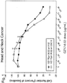
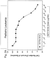


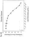
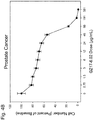


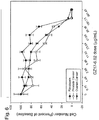

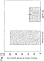

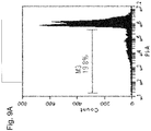
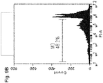


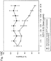

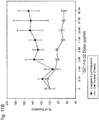

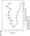

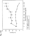


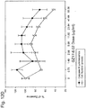
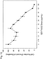

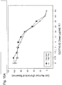


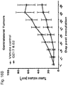
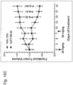

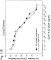
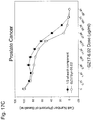


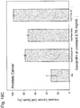


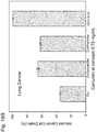

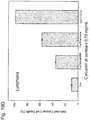


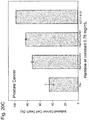

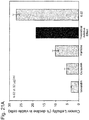


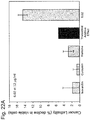

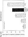
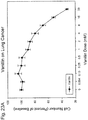


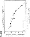
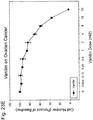
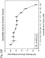
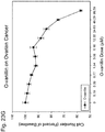

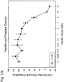


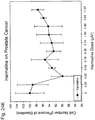

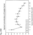


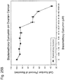
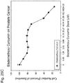
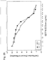
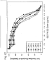

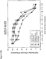

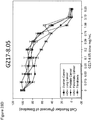
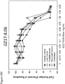
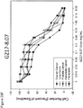
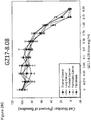

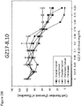
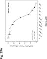

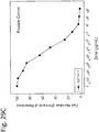
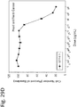
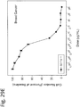


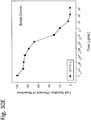

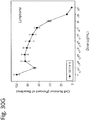

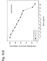

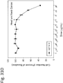


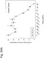
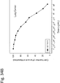
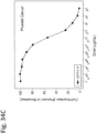

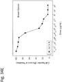


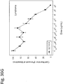

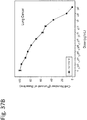
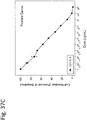

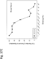
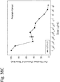
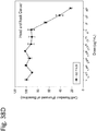
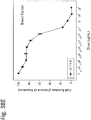

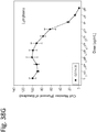


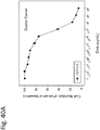

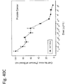


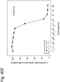


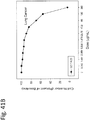
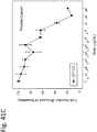
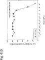
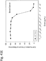

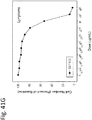
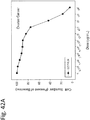
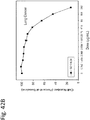

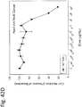
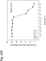
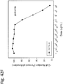
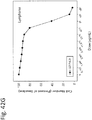

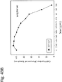
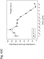
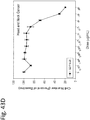
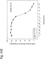
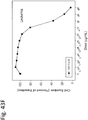

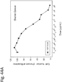


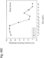
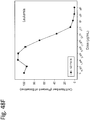
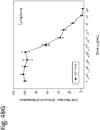

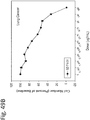


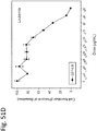
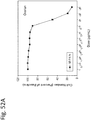

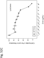
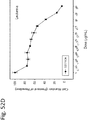
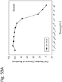
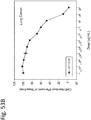
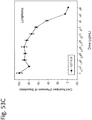
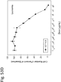
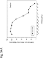

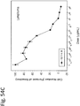
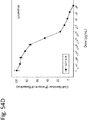

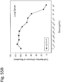
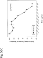
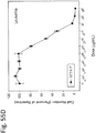

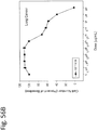


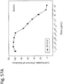
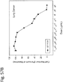
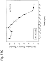


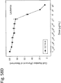


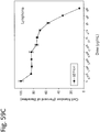

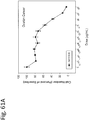
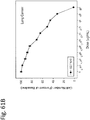
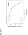
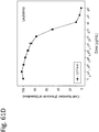
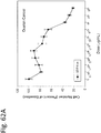
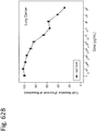


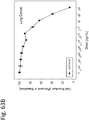

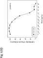
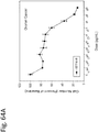
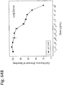
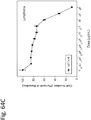
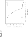


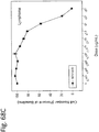
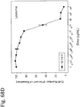
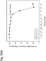

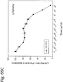

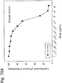

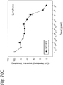
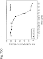
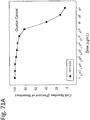


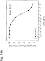


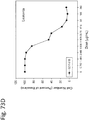


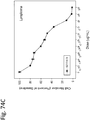
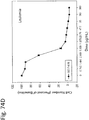


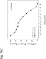


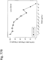
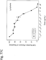


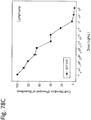


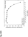

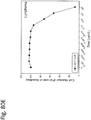


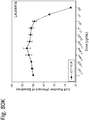

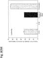

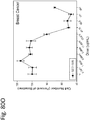
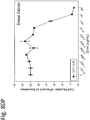
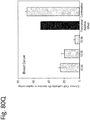


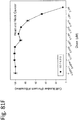

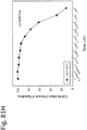
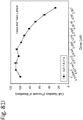
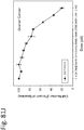

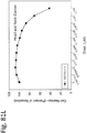
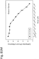

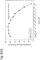


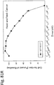

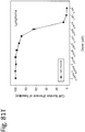
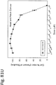
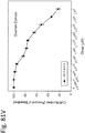

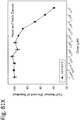
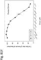

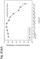

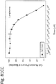
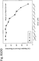
Classifications
-
- A—HUMAN NECESSITIES
- A61—MEDICAL OR VETERINARY SCIENCE; HYGIENE
- A61K—PREPARATIONS FOR MEDICAL, DENTAL OR TOILETRY PURPOSES
- A61K31/00—Medicinal preparations containing organic active ingredients
- A61K31/12—Ketones
-
- A—HUMAN NECESSITIES
- A61—MEDICAL OR VETERINARY SCIENCE; HYGIENE
- A61K—PREPARATIONS FOR MEDICAL, DENTAL OR TOILETRY PURPOSES
- A61K31/00—Medicinal preparations containing organic active ingredients
- A61K31/11—Aldehydes
-
- A—HUMAN NECESSITIES
- A61—MEDICAL OR VETERINARY SCIENCE; HYGIENE
- A61K—PREPARATIONS FOR MEDICAL, DENTAL OR TOILETRY PURPOSES
- A61K31/00—Medicinal preparations containing organic active ingredients
- A61K31/33—Heterocyclic compounds
- A61K31/395—Heterocyclic compounds having nitrogen as a ring hetero atom, e.g. guanethidine or rifamycins
- A61K31/435—Heterocyclic compounds having nitrogen as a ring hetero atom, e.g. guanethidine or rifamycins having six-membered rings with one nitrogen as the only ring hetero atom
- A61K31/4353—Heterocyclic compounds having nitrogen as a ring hetero atom, e.g. guanethidine or rifamycins having six-membered rings with one nitrogen as the only ring hetero atom ortho- or peri-condensed with heterocyclic ring systems
- A61K31/437—Heterocyclic compounds having nitrogen as a ring hetero atom, e.g. guanethidine or rifamycins having six-membered rings with one nitrogen as the only ring hetero atom ortho- or peri-condensed with heterocyclic ring systems the heterocyclic ring system containing a five-membered ring having nitrogen as a ring hetero atom, e.g. indolizine, beta-carboline
-
- A—HUMAN NECESSITIES
- A61—MEDICAL OR VETERINARY SCIENCE; HYGIENE
- A61K—PREPARATIONS FOR MEDICAL, DENTAL OR TOILETRY PURPOSES
- A61K45/00—Medicinal preparations containing active ingredients not provided for in groups A61K31/00 - A61K41/00
- A61K45/06—Mixtures of active ingredients without chemical characterisation, e.g. antiphlogistics and cardiaca
-
- A—HUMAN NECESSITIES
- A61—MEDICAL OR VETERINARY SCIENCE; HYGIENE
- A61P—SPECIFIC THERAPEUTIC ACTIVITY OF CHEMICAL COMPOUNDS OR MEDICINAL PREPARATIONS
- A61P35/00—Antineoplastic agents
-
- A—HUMAN NECESSITIES
- A61—MEDICAL OR VETERINARY SCIENCE; HYGIENE
- A61P—SPECIFIC THERAPEUTIC ACTIVITY OF CHEMICAL COMPOUNDS OR MEDICINAL PREPARATIONS
- A61P35/00—Antineoplastic agents
- A61P35/02—Antineoplastic agents specific for leukemia
-
- A—HUMAN NECESSITIES
- A61—MEDICAL OR VETERINARY SCIENCE; HYGIENE
- A61P—SPECIFIC THERAPEUTIC ACTIVITY OF CHEMICAL COMPOUNDS OR MEDICINAL PREPARATIONS
- A61P43/00—Drugs for specific purposes, not provided for in groups A61P1/00-A61P41/00
-
- C—CHEMISTRY; METALLURGY
- C07—ORGANIC CHEMISTRY
- C07D—HETEROCYCLIC COMPOUNDS
- C07D471/00—Heterocyclic compounds containing nitrogen atoms as the only ring hetero atoms in the condensed system, at least one ring being a six-membered ring with one nitrogen atom, not provided for by groups C07D451/00 - C07D463/00
- C07D471/02—Heterocyclic compounds containing nitrogen atoms as the only ring hetero atoms in the condensed system, at least one ring being a six-membered ring with one nitrogen atom, not provided for by groups C07D451/00 - C07D463/00 in which the condensed system contains two hetero rings
- C07D471/04—Ortho-condensed systems
-
- A—HUMAN NECESSITIES
- A61—MEDICAL OR VETERINARY SCIENCE; HYGIENE
- A61K—PREPARATIONS FOR MEDICAL, DENTAL OR TOILETRY PURPOSES
- A61K2300/00—Mixtures or combinations of active ingredients, wherein at least one active ingredient is fully defined in groups A61K31/00 - A61K41/00
-
- Y—GENERAL TAGGING OF NEW TECHNOLOGICAL DEVELOPMENTS; GENERAL TAGGING OF CROSS-SECTIONAL TECHNOLOGIES SPANNING OVER SEVERAL SECTIONS OF THE IPC; TECHNICAL SUBJECTS COVERED BY FORMER USPC CROSS-REFERENCE ART COLLECTIONS [XRACs] AND DIGESTS
- Y02—TECHNOLOGIES OR APPLICATIONS FOR MITIGATION OR ADAPTATION AGAINST CLIMATE CHANGE
- Y02A—TECHNOLOGIES FOR ADAPTATION TO CLIMATE CHANGE
- Y02A50/00—TECHNOLOGIES FOR ADAPTATION TO CLIMATE CHANGE in human health protection, e.g. against extreme weather
- Y02A50/30—Against vector-borne diseases, e.g. mosquito-borne, fly-borne, tick-borne or waterborne diseases whose impact is exacerbated by climate change
Definitions
- the present invention relates to combination chemotherapeutics for treatment of humans, and especially for the treatment of human cancers.
- the invention further provides dosage forms and regimens for administration to human patients, and methods of formulating such dosage forms to yield improvements in treatment outcomes. More particularly, the invention is concerned with specific chemotherapeutic dosage forms (e.g., liquid mixtures, capsules, pills, or tablets) containing one or more harmine component(s), and isovanillin component(s).
- Cancer is a generic term for a large group of diseases that can affect any part of the body. Other terms used are malignant tumors and neoplasms.
- One defining feature of cancer is the rapid creation of abnormal cells that grow beyond their usual boundaries, and which can then invade adjoining parts of the body and spread to other organs. This process is referred to as metastasis. Metastases are the major cause of death from cancer.
- the transformation from a normal cell into a tumor cell is a multistage process, typically a progression from a pre-cancerous lesion to malignant tumors. These changes are the result of the interaction between a person's genetic factors and three categories of external agents, including:
- Aging is another fundamental factor for the development of cancer.
- the incidence of cancer rises dramatically with age, most likely due to a buildup of risks for specific cancers that increase with age.
- the overall risk accumulation is combined with the tendency for cellular repair mechanisms to be less effective as a person grows older.
- HBV hepatitis B
- HCV hepatitis C virus
- HPV Human Papilloma Virus
- cancer treatment modalities are surgery, chemotherapy, and radiation treatments. All of these techniques have significant drawbacks in terms of side effects and patient discomfort.
- chemotherapy may result in significant decreases in white blood cell count (neutropenia), red blood cell count (anemia), and platelet count (thrombocytopenia). This can result in pain, diarrhea, constipation, mouth sores, hair loss, nausea, and vomiting.
- Bio therapy (sometimes called immunotherapy, biotherapy, or biological response modifier therapy) is a relatively new addition to the family of cancer treatments.
- Biological therapies use the body's immune system, either directly or indirectly, to fight cancer or to lessen the side effects that may be caused by some cancer treatments.
- Examples of famous negative drug interactions include the development of rhabdomyolysis, a severe muscle disease, when taking Simvastatin with Amiodarone. As a result, the FDA introduced a warning on the drug label about the interaction.
- US Patent No. 8,039.025 describes cancer treatments in the form of extracts of Arum palaestinum Boiss, supplemented with individual amounts of ⁇ -sitosterol, isovanillin, and linoleic acid.
- Vanillins -- a novel family of DNA-PK inhibitors Nucleic Acids Research, vol. 31, no. 19, October 1, 2003, pages 5501 - 5512 , and US 2008/221221 A1 disclose vanillin and wortmannin as DNA-PL inhibitors, and use of 2-bromo-isovanillin in the preparation of an anti-cancer medicament, respectively, and describe the anti-cancer and sensitizing effect of isovanillin derivatives.
- the present invention provides improved chemotherapeutics for treatment of humans, and especially in the treatment of human cancers, with novel combinations of compounds, which are useful against a wide variety of different cancers with minimal or nonexistent adverse side effects.
- the present invention provides a therapeutic composition
- a therapeutic composition comprising the combination of at least one harmine component, and at least one isovanillin component, said at least one isovanillin component selected from the group consisting of isovanillin, vanillin, orthovanillin, and pharmaceutically acceptable salts thereof, said at least one harmine component selected from the group consisting of harmine, harmaline, harmane, harmalol, harmol, and pharmaceutically acceptable salts thereof, said at least one isovanillin component being present in an amount greater than the amount of the at least one harmine component, the weight amount of said at least one isovanillin component being from about 25 to 95% by weight based upon the total weight of the harmine and isovanillin components taken as 100% by weight, the combination of the at least one harmine component and the at least one isovanillin component exhibiting anti-cancer activity.
- the present invention provides a method of preparing such a therapeutic composition comprising the steps of
- the present invention provides a therapeutic composition obtainable by said method.
- the chemotherapeutics of the invention comprise respective quantities of at least harmine component(s), and isovanillin component(s).
- the preferred chemotherapeutics include harmine component(s) and isovanillin component(s).
- the active components of the compositions consist essentially of curcumin, harmine, and isovanillin components in the case of three-component compositions.
- the curcumin component comprises curcumin
- the at least one harmine component comprises harmine
- the at least one isovanillin component comprises isovanillin or vanillin.
- the invention also describes methods for treatment of cancers by administration of appropriate quantities of the anti-cancer compositions hereof.
- the compositions are particularly designed for use in the treatment of cancers, and the compositions can be used for the manufacture of medicaments of anti-cancer therapeutic applications.
- the invention provides pharmaceutical compositions for the treatment of cancers comprising administering therapeutically effective amounts of the new compositions, prepared by processes known per se, with a pharmaceutically acceptable carrier.
- Curcumin (CAS #458-37-7) is a diaryl heptanoid, and has the molecular formula C21H20O6. Curcumin occurs as a part of a curcuminoid plant extract containing curcumin, demethoxycurcumin, and bis-demethoxycurcumin.
- Curcumin is sold as "Curcumin from Curcuma Longa (Turmeric)," which contains greater than 65% by weight curcumin, as determined by HPLC analysis.
- Harmine (CAS #442-51-3) is a methoxy methyl pyrido indole belonging to the ⁇ -carboline family of compounds, and has the molecular formula C13H12N2O. Harmine occurs in a number of different plants native to the Middle East and South America.
- Isovanillin (CAS #621-59-0) is a phenolic aldehyde vanillin isomer, and has the molecular formula C8H8O3.
- chemotherapeutic refers to the combinations of chemical compounds described herein as useful in the treatment of human conditions, especially human cancers.
- Chemotherapeutics may be cytostatic, selectively toxic or destructive of cancerous tissue and/or cells, but also include indiscriminately cytotoxic compounds used in cancer treatments.
- the combination therapeutic agents of the invention have been found to be effective in the treatment of a broad spectrum of human cancers, and also to other conditions, such as elevated PSA counts in men.
- the broad scope of utility with the agents of the invention is in itself highly unusual. However, this feature, together with the nonexistent or minimal side effects induced by the agents, represents a startling development in the art.
- the therapeutic agents of the invention are used in therapeutically effective amounts, i.e., amounts that will elicit the biological or medical response of a tissue, system, or subject that is being sought, and in particular to elicit some desired therapeutic effect against a variety of human cancers; the agents operate by preventing and/or inhibiting proliferation and/or survival of cancerous cells, and/or by slowing the progression of cancers.
- therapeutically effective amounts i.e., amounts that will elicit the biological or medical response of a tissue, system, or subject that is being sought, and in particular to elicit some desired therapeutic effect against a variety of human cancers; the agents operate by preventing and/or inhibiting proliferation and/or survival of cancerous cells, and/or by slowing the progression of cancers.
- an amount may be considered therapeutically effective even if the condition is not totally eradicated or prevented, but it or its symptoms and/or effects are improved or alleviated partially in the subject.
- the appropriate makeup of the agents hereof and dosing regimens using such agents will depend on the particular cancer being treated, the extent of the disease, and other factors related to the patient as determined by those skilled in the art.
- therapeutic or “treat,” as used herein, refer to products or processes in accordance with the invention that are intended to produce a beneficial change in an existing condition (e.g., cancerous tissue, tumor size, metastases, etc.) of a subject, such as by reducing the severity of the clinical symptoms and/or effects of the condition, and/or reducing the duration of the symptoms/effects of a subject.
- Additional ingredients may be included with the chemotherapeutic agents of the invention for administration to the subject.
- additional ingredients include, other active agents, preservatives, buffering agents, salts, carriers, excipients, diluents, or other pharmaceutically-acceptable ingredients.
- the active agents that could be included in the compositions include antiviral, antibiotic, or other anticancer compounds.
- the combined therapeutic agents of the invention preferably give synergistic results, which are entirely unexpected. Moreover, the lack of side effects when the agents are administered to patients is quite surprising and essentially unique.
- the terms "combination” or “in combination” are intended to embrace compositions wherein the components are physically intermixed as dosage forms, and to situations where the individual components are separately administered to a subject over relatively short periods of time, which would have the same therapeutic effects as a single dosage form.
- a therapeutically effective amount of an agent in accordance with the invention is administered to a subject in need thereof.
- Such may comprise a single unit dosage or, more usually, periodic (e.g., daily) administration of lower dosages over time.
- administration of such therapeutically effective amounts achieves an unexpected therapeutic synergy.
- the therapeutic two- or three-component compositions of the invention exhibit a joint action where one or more of the components supplements or enhances the action of at least one of the other components to produce an effect greater than that which may be obtained by use of individual components in equivalent quantities, or produce effects that could not be obtained with safe quantities of the other components, individually or in combination.
- one or more of the components working together produce an effect greater than the sum of their individual effects.
- the dosages may be administered in any convenient manner, such as by oral, rectal, nasal, ophthalmic, parenteral (including intraperitoneal, gastrointestinal, intrathecal, intravenous, cutaneous (e.g., dermal patch), subcutaneous (e.g. injection or implant), or intramuscular) administrations.
- the dosage forms of the invention may be in the form of liquids, gels, suspensions, solutions, or solids (e.g., tablets, pills, or capsules).
- therapeutically effective amounts of the agents of the invention may be co-administered with other chemotherapeutic agent(s), where the two products are administered substantially simultaneously or in any sequential manner.
- the phrase "and/or," when used in a list of two or more items, means that any one of the listed items can be employed by itself or any combination of two or more of the listed items can be employed.
- the composition can contain or exclude A alone; B alone; C alone; A and B in combination; A and C in combination; B and C in combination; or A, B, and C in combination.
- the present description also uses numerical ranges to quantify certain parameters relating to various embodiments of the invention. It should be understood that when numerical ranges are provided, such ranges are to be construed as providing literal support for claim or paragraph limitations that only recite the lower value of the range as well as claim or paragraph limitations that only recite the upper value of the range. For example, a disclosed numerical range of about 10 to about 100 provides literal support for a claim or paragraph reciting "greater than about 10" (with no upper bounds) and a claim or paragraph reciting "less than about 100" (with no lower bounds).
- curcumin, harmine, and isovanillin component(s) will be individually described.
- carbon-carbon chains e.g., alkyl, alkenyl, alkoxy, alkyl amine, alkenyl amine, aldehyde, or carboxylate
- these disclosures should be understood to refer to primary (straight), branched chain, or cyclic carbon chain groups.
- aryl groups means phenyl, substituted phenyl, napthyl, substituted napthyl; and heteroatom aryl groups refers to aryl groups containing a nitrogen, oxygen, boron, or sulfur atom, such as pyridine; heterocyclic groups refers to cyclic groups containing from 3-7 atoms, one or more of which is a nitrogen, oxygen, boron, or sulfur heteroatom; and amine refers to primary, secondary, tertiary, or quaternary amines.
- pharmaceutically acceptable salts with reference to the components means salts of the component compounds of the present invention which are pharmaceutically acceptable, i.e., salts which are useful in preparing pharmaceutical compositions that are generally safe, non-toxic, and neither biologically nor otherwise undesirable and are acceptable for human pharmaceutical use, and which possess the desired degree of pharmacological activity.
- Such pharmaceutically acceptable salts include acid addition salts formed with inorganic acids such as hydrochloric acid, hydrobromic acid, sulfuric acid, nitric acid, and phosphoric acid; or with organic acids such as 1,2-ethanedisulfonic acid, 2-hydroxyethanesulfonic acid, 2-naphthalenesulfonic acid, 3-phenylpropionic acid, 4,4'-methylenebis(3-hydroxy-2-ene-1-carboxylic acid), 4-methylbicyclo[2.2.2]oct-2-ene-1-carboxylic acid, acetic acid, aliphatic mono- and dicarboxylic acids, aliphatic sulfuric acids, aromatic sulfuric acids, benzenesulfonic acid, benzoic acid, camphorsulfonic acid, carbonic acid, cinnamic acid, citric acid, cyclopentanepropionic acid, ethanesulfonic acid, fumaric acid, glucoheptonic acid, gluconic acid,
- Pharmaceutically acceptable salts also include base addition salts which may be formed when acidic protons present are capable of reacting with inorganic or organic bases.
- Acceptable inorganic bases include sodium hydroxide, sodium carbonate, potassium hydroxide, aluminum hydroxide and calcium hydroxide.
- Acceptable organic bases include ethanolamine, diethanolamine, triethanolamine, tromethamine, and N-methylglucamine. It should be recognized that the particular anion or cation forming a part of any salt of this invention is not critical, so long as the salt, as a whole, is pharmacologically acceptable. Additional examples of pharmaceutically acceptable salts and their methods of preparation and use are presented in Handbook of Pharmaceutical Salts Properties, and Use, P. H. Stahl & C. G. Wermuth eds., ISBN 978-3-90639-058-1 (2008 ).
- the curcumin, harmine, and isovanillin component(s) may be obtained as synthetic compounds of high purity, or from modified naturally occurring sources. In either case, however, it is preferred that the component(s) be purified to a level of at least about 50% by weight, more preferably at least about 70% by weight, still more preferably at least about 90% by weight, and most preferably at least about 98% by weight.
- curcumin-based formulas are indicated “C-number,” (not to be confused with carbon chain numbers, which are indicated as “Cnumber,” without an intervening hyphen), harmine-based formulas are indicated as “H-number,” and isovanillin-based formulas are indicated as "I-number.”
- the preferred components of the invention are either synthetically derived or derived from one or more naturally occurring product(s) which have been significantly modified so as to contain at least about 25% by weight (more preferably at least about 50% by weight, and still more preferably about 70% by weight) of the desired component.
- synthetically derived means that the component in question was synthesized using specific starting ingredients and one or more chemical and/or biological reactions to obtain substantially pure compounds. Modification of naturally occurring products may involve extractions, or any other physical or chemical steps to achieve the desired end product, e.g., harmine components may be obtained from treatment of harmala seed, or curcumin components may be obtained from treatment of turmeric.
- curcumin can be synthetically derived to a high degree of purity.
- curcumin can be obtained by extraction or other treatment of naturally occurring turmeric so that the curcumin content of the modified turmeric has the above-noted levels of curcumin therein.
- curcumin component(s) shall mean curcumin, its metabolites and derivatives, isomers and tautomers thereof, esters, metal complexes (e.g., Cu, Fe, Zn, Pt, V), and pharmaceutically acceptable salts of any of the foregoing.
- Curcumin derivatives include both naturally occurring and synthetic derivatives, e.g., the spontaneous degradation products of curcumin, curcumin metabolites, and synthetic curcumin derivative compounds.
- Curcumin (diferuloylmethane, 1,7-bis(4-hydroxy3-methoxyphenyl)-1,6-heptadiene-3,5-dione) is a symmetrical diphenolic dienone, see structure C-1 below. It exists in solution as an equilibrium mixture of the symmetrical dienone (diketo) and the keto-enol tautomer; the keto-enol form is strongly favored by intramolecular hydrogen bonding. Curcumin contains two aryl rings separated by an unsaturated 7-carbon linker having a symmetrical ⁇ -diketone group (as used herein, " ⁇ -diketone” embraces both tautomeric forms, namely the diketo and enol forms). The aryl rings of curcumin contain a hydroxyl group in the para position and a methoxy group in the meta position.
- curcumin will spontaneously form degradation products, and especially one or more of the following:
- curcumin is differently metabolized in vivo depending upon the route of administration, see, Shen et al. The Pharmacology of Curcumin: Is it the Degradation Products? Trends in Molecular Medicine, March 2012 Vol. 18, No. 2 .
- the metabolites normally include one or more of the following:
- the metabolites generally include the following:
- curcumin derivatives include cyclocurcumin, bisdemethoxycurcumin, demethoxycurcumin, dihydrocurcumin, caffeic acid, cinnamic acid, isoeugenol, dibenzoylmethane, dehydrozingerone, capsaicin, [6]-gingerol, [6]-paradol, chlorogenic acid, yakuchinone A, oregonin, cassumuin A, and cassumuin B.
- Curcumin derivatives are expected to be beneficial for use in the described treatment methods.
- the term "curcumin derivative” is used interchangeably with the term “curcumin analog” and “curcumin analogue” (alternative spelling) and includes, for example, curcumin derivatives, analogs, curcuminoids and chalcones.
- the curcumin derivative includes first and second aryl groups covalently attached by way of a linker or a linking group.
- the second aryl group is absent, such that the curcumin derivative contains a first aryl group and the linker but no second aryl group at the distal end of the linker.
- the first and/or second aryl group is a heteroaryl group.
- the first and second aryl groups may be independently substituted or unsubstituted.
- Curcumin derivatives that exhibit improved pharmacokinetic properties and/or reduced toxicity are preferred.
- curcumin derivatives that include heteroaryl groups and/or unsaturated linkers arc expected to impart improved pharmacokinetic properties and/or reduced toxicity to the compounds, because they are expected to be less chemically reactive in vivo.
- One example of preferred curcumin derivatives includes those including one or two carbonyl groups in the linker region, including those derivatives that preserve the enone functionality of curcumin.
- Derivatives that include heteroaryl groups and/or unsaturated linkers are expected to be less likely to be degraded and/or form toxic adducts or intermediates under physiological conditions.
- curcumin derivatives of the invention are generally encompassed by the formula: Ar1-L-Ar2 C12 where Ar1 and Ar2 are independently aryl groups, and L is a divalent linking group that includes between 3 and 7 backbone carbon atoms, wherein one or more of the backbone carbon atoms include a carbonyl or hydroxyl moiety.
- Preferred aryl groups include phenyl, naphthyl, thienyl, pyridinium, and pyridyl groups.
- Aryl groups Ar1 and Ar2 may be substituted or unsubstituted, and one or more of the ring carbons may be substituted with a heteroatom, and especially N, S, B, or O.
- Ar1 can be an aryl group according to the formula: and Ar2 can be an aryl group according to the formula: where one or more of the aryl ring carbons of Ar1 and Ar2 may be independently substituted with a heteroatom selected from N, S, B, or O, and R1-R10 are independently selected from the group consisting of H, hydroxyl, halogen, amine, nitro, sulfonate, sulfoxide, thio, ester, carboxylate, amide, borate, C1-C4 boronate, C1-C8 alkyl, C2-C8 alkenyl, C1-C6 haloalkyl, C1-C6 alkoxy, C1-C6 amine, C2-C8 carboxyl, C2-C8 ester, C1-C4 aldehyde, and glucuronide groups;
- L is a divalent linker including from 3-7 backbone carbon atoms that form a chain connecting the Ar1,
- Ar1 and Ar2 are phenyl groups; R1-R10 are independently selected from the group consisting of H, hydroxyl, halogen, amine, nitro, sulfonate, thio, borate, C1-C2 boronate, sulfoxide, C1-C4 alkyl, C1-C4 alkoxy, C1-C4 alkylamine, C2-C6 alkenylamine, C1-C6 acetoxy, C1-C4 carboxyl, with at least one of R1-R5 and R6-R10 being hydroxyl.
- the linker L is a spacer that preferably includes 3, 4, 5, 6 or 7 carbon atoms that form a linear carbon chain connecting the first and second aryl groups.
- the carbons atoms in the carbon chain that trace out shortest path between the first and optional second aryl groups are referred to herein as the "backbone" carbon atoms.
- the number of backbone carbon atoms is readily determined in straight chain alkyl groups. In linkers that include a cyclic alkyl group as a constituent of the linear chain, the backbone carbon atoms include the least number of ring carbons possible.
- the number of backbone carbon atoms is used herein as a shorthand way to designate the length of the linker being used.
- a 7-carbon linker is a divalent linker that includes 7 backbone carbon atoms.
- the linker may be substituted or unsubstituted.
- the linker may further be saturated or unsaturated.
- the linker contains an odd number of carbon atoms (i.e., 3, 5, or 7 carbon atoms), and at least one unsaturated carbon-carbon bond.
- the linker may include a hydroxyl moiety in place of, or in addition to, the at least one carbonyl moiety.
- Curcumin derivatives include a linking group L that is preferably covalently attached at one end to aryl group Ar1.
- the linking group L may also be covalently attached at the other end to a second aryl group, Ar2, which is selected independently from Ar1.
- the linking group L is a divalent linking group that preferably includes an alkylene or an alkenylene group having between 3 and 7 backbone carbon atoms, and more advantageously an odd number of backbone carbon atoms (i.e., 3, 5, or 7 carbon atoms).
- the linker also preferably has at least one carbonyl moiety, and may further include a hydroxyl moiety in place of, or in addition to, the at least one carbonyl moiety.
- the linking group may be substituted or unsubstituted, and may be saturated or unsaturated.
- the linking group has a carbon-carbon double bond between the ⁇ and ⁇ carbons relative to Ar1 and/or Ar2 (e.g., see formulas C-1 and C-19 through C-33, which illustrate such a double bond.
- the linking group includes conjugated double bonds. Table 1 shows compounds with 7-carbon linkers; Table 2 shows compounds with 5-carbon linkers; and Table 3 shows compounds with 3-carbon linkers.
- a divalent linking group includes two carbons with unfilled valencies that provide valence points where a covalent bond can be formed to an adjacent alkyl or aryl group that also includes a carbon with an unfilled valency.
- a valence point is represented in a chemical formula by a bond that is shown as not being attached to another group (e.g., CH3-, wherein - represents the valence point).
- the distal valence point on the linking group can be filled with any substituent of interest, preferably a short chain alkyl group (e.g., C1-C6, more preferably C1-C4) or a hydrogen (H).
- a short chain alkyl group e.g., C1-C6, more preferably C1-C4
- H hydrogen
- Compounds lacking a second aryl group may be represented by formula: Ar1-L-R11 C-15 R11 can be, for example, a heterocyclic group or an alkyl group, preferably an alkyl group having four or fewer carbon atoms, e.g., a methyl group.
- R11 can alternately be an amine, a hydroxyl, a hydrogen, nitro, sulfonate, sulfoxide, thio, ester, carboxylate, amide, borate, or a C1-C4 boronate.
- the curcumin derivatives include one or two aryl groups (Arl and optionally Ar2) and a linking group L that is a 7-carbon linking group (i.e., a linking group that includes 7 backbone carbon atoms).
- the 7-carbon linking group includes at least one unsaturated carbon-carbon bond.
- linking groups are the divalent forms of 4-alkyl-1,6 heptadiene-3,5-dione; 4,4-dialkyl-1,6 heptadiene-3,5-dione; and heptane-3,5-dione.
- Table 1 shows a number of examples of curcumin derivatives that include a 7-carbon linker.
- the compounds shown contain two aryl rings separated by a 7-carbon linker having two carbonyls (or the equivalent keto-enol tautomer). In many, but not all, of the compounds, the linker is unsaturated. "Bn” refers to a benzyl group. TABLE 1 7-Carbon Linker Analogs.
- the curcumin derivatives include one or two aryl groups (Arl and optionally Ar2) that are linked by a linking group L that is a 5-carbon linking group (i.e., a linking group that includes 5 backbone carbon atoms).
- a linking group L is a 5-carbon linking group (i.e., a linking group that includes 5 backbone carbon atoms).
- the 5-carbon linking group includes at least one unsaturated carbon-carbon bond.
- curcumin derivatives may include a cyclic linking group.
- 1-methyl-2,6-diphenyl-4-piperidone provides a compound with a 5-carbon linking group that is bridged by a tertiary amine to form a cyclic alkylene linking group including the heteroatom nitrogen.
- Table 2 shows a number of examples of curcumin derivatives that include a 5-carbon linker.
- the compounds shown contain two aryl rings separated by a 5-carbon linker having a single carbonyl or hydroxyl. In many, but not all, of the compounds, the linker is unsaturated. TABLE 2 5-Carbon Linker Analogs.
- the curcumin derivatives include one or two aryl groups (Arl and optionally Ar2) that are linked by a linking group L that is a 3-carbon linking group (i.e., a linking group that includes 3 backbone carbon atoms).
- a 3-carbon linking group i.e., a linking group that includes 3 backbone carbon atoms.
- the 3-carbon linking group includes at least one unsaturated carbon-carbon bond.
- Table 3 shows a number of examples of curcumin derivatives that include a 3-carbon linker.
- the compounds shown generally have an unsaturated 3-carbon linker having a single carbonyl. While most of the examples shown have two aryl groups separated by the linker, several of the embodiments include only a single aryl group. In the examples that include only a single aryl group, a methyl group is provided at the other end of the linking group.
- One compound includes the heteroatom N in place of one of the backbone carbon atoms; however, this is still considered a 3-C linker in that 3 atoms (C, N, and C) are present along the shortest bridge between the two aryl groups. TABLE 3 3-Carbon Linker Analogs.
- Curcumin derivatives may include a variety of linking groups and Ar groups while retaining the necessary activity. Accordingly, additional curcumin analogs are contemplated. These include curcumin analogs containing central methylene substituents such as ethyl, propyl, butyl, isopropyl and substituted benzyl groups according to the formula:
- curcumin analogs which have a 5-carbon linker possess significant activity. Additional active analogs in this series may contain substituents such as hydroxy and methoxy groups on the aryl rings. Examples of these analogs are shown in the formula:
- curcumin derivatives A large number of naturally occurring and synthetic curcumin derivatives, the latter explicated by reference to the methods of synthesis thereof, are disclosed in Anand et al. "Biological Activities of Curcumin and Its Analogues (Congeners) Made By Man and Mother Nature.” Biochemical Pharmacology 76 (2008):1590-1611 .
- Representative synthetic curcumin derivatives are set forth below. where R18, R19, and R20 are each independently selected from the group consisting of H, C1-C4 alkyl, and C1-C4 amine groups.
- R24 and R25 are independently selected from the group consisting of OH, C1-C4 alkoxy, and C1-C4 alkylcarbonyloxy;
- R26 and R27 are independently selected from the group consisting of H, OH, C1-C4 alkoxy, and C1-C4 alkylcarbonyloxy;
- R28 is selected from the group consisting of H, OH, and C1-C4 alkylcarbonyloxy;
- R29 is selected from the group consisting of H and C1-C4 alkoxy.
- R30, R31, R32, R33, R34, R35, and R36 are independently selected from the group consisting ofH, OH, C1-C4 alkoxy, and C1-C4 alkylcarbonyloxy.
- R37 and R38 are independently selected from the group consisting of methoxy and OH, and the center benzene ring may be substituted in the 1,3- or the 1,4-position with the acryloyl groups.
- curcumin components of formula C-13A are preferred: where R1-R5 are as previously defined, and the * denotes the valence point where the additional portion of the compounds are attached. That is, the preferred components have the moiety depicted in C-13A and additional portion exemplified by the foregoing disclosure, e.g., the remaining structure of the linker L and Ar2 or R11.
- curcumin components are exemplified by formula C-12 where:
- the curcumin components should have:
- harmine component(s) shall mean harmine, harmaline, harmane, harmalol, harmol and pharmaceutically acceptable salts of any of the foregoing.
- Harmine belongs to the family of ⁇ -carboline alkaloids, its chemical name is 7-methoxy-1-methyl 9H-pyrrole[3,4-b]indole, and its molecular formula is C13H12N2O.
- the base structure of ⁇ -carboline is shown in H-0.
- Harmine has a molecular weight of 212.25 and a melting point of 261°C. Harmine was originally isolated from Peganum harmala, which is widely used as a traditional herbal drug in the Middle East and North Africa. The chemical structure of harmine, 1-methyl-7-methoxy- ⁇ -carboline, is shown as follows:
- isovanillin component(s) refers to isovanillin, vanillin, ortho -vanillin and pharmaceutically acceptable salts of any of the foregoing.
- Isovanillin is a phenolic aldehyde having a hydroxyl group at the meta position and a methoxy group at the para position. Isovanillin is illustrated in the following structure:
- Isovanillin is metabolized in vivo to vanillin, which is the same as structure 1-1, except that the hydroxyl and methoxy substituents are exchanged, i.e., in vanillin, the hydroxyl group is in the para position, and the methoxy group is in the meta position.
- compositions of the invention made by combining individual quantities of normally highly purified curcumin component(s), harmine component(s), and isovanillin component(s), the as-added amounts should give weight ratios of about 10:1.7:0.85 (isovanillin component(s):harmine component(s):curcumin component(s)). It will be seen that the isovanillin component(s) of the GZ17-6.02 product is/are the preponderant component(s) in the compositions on a weight basis, with the harmine and curcumin component(s) being present in lesser amounts on a weight basis.
- the isovanillin component(s) in the most preferred product should be present at a level at least three times (more preferably at least five times) greater than that of each of the harmine and curcumin component(s), again on a weight basis.
- the invention is not limited to such weight ratios.
- the isovanillin component(s) should be present at a level of from about 25-85% by weight
- the harmine component(s) should be present at a level of from about 7-50% by weight
- the curcumin component(s) should be present at a level of from about 5-40% by weight, based upon the total weight of the three components taken as 100% by weight.
- Two-component compositions made up of isovanillin component(s) plus harmine component(s) have weight amounts of the isovanillin component(s) from about 25% to 95% by weight (more preferably from about 75% to 95% by weight, and most preferably about 85% by weight), and the weight amounts of the harmine component(s) range from about 5% to 75% by weight (more preferably from about 5% to 25% by weight, and most preferably about 15% by weight) by weight, based upon the total weight of the two components in the compositions taken as 100% by weight.
- the isovanillin component(s) of any of the foregoing two- or three-component compositions containing isovanillin component(s) may be made up of initially added isovanillin component(s) plus any degradation products of the other component(s) yielding products within the ambit of the isovanillin component(s); for example, it is known that under certain circumstances curcumin will spontaneously degrade to give quantities of vanillin, and, in such circumstances, the total amount of the isovanillin component(s) would be the initially added amounts together with these degradation products.
- the ultimate amounts of the curcumin, harmine, and isovanillin component(s) in the compositions of the invention should be determined based upon the actual contents of the component(s) in question, regardless of whether these component(s) are derived from the originally added component(s) or as degradation products of some or all of these originally added component(s).
- compositions of the invention are quite variable owing to factors such as the patient's age, patient's physical condition, the type of condition(s) being treated (e.g., specific cancer(s)), and the severity of the conditions.
- the dosage form or route of administration employed such as liquid solutions or suspensions, capsules, pills, or tablets, via oral, parenteral, or injection
- the compositions should be dosed of from about 5 to 2000 mg per day, and more usually from about 100-800 mg per day.
- dosages may be based on a single administration per day, but more usually multiple administrations per day.
- the GZ17-6.02 product of the Examples was made by dispersing quantities of solid synthetic isovanillin (771 mg, 98% by weight purity), synthetic harmine (130.3 mg, 99% by weight purity), and a commercially available curcumin product derived by the treatment of turmeric (98.7 mg, containing 99.76% by weight curcuminoids, namely 71.38% curcumin, 15.68% demethoxycurcumin, and 12.70% bisdemethoxycurcumin), in a 1 mL ethanol at a weight ratio of 771:130.3:98.7 (isovanillin:harmine:curcumin product) in ethanol followed by sonication of the dispersion. Aliquots of this stock solution were then used to create the different GZ17-6.02 dilutions using stock media of the cells in question.
- Example 23 The two-component products described in Example 23 were made in the same fashion as the GZ17-6.02 agent, and the weight ratios of the two components were as set forth immediately above, e.g., the isovanillin/harmine sub-combinations contained a weight ratio of 771:130.3, and so forth.
- the preferred GZ17-6.02 product was tested with two different human head and neck cancers (HN5 and OSC19), in order to determine the extent of cell death induced by the product.
- the respective cells were individually cultured in a growth medium prepared using RPMI-1640 medium containing 11.1 mM D-glucose with 10% fetal bovine serum, 10 mM HEPES, 2 mM L-glutamine, 1 mM sodium pyruvate, 0.05 mM 2-mercaptoethanol, and Antibiotic-Antimycotic. These cells were maintained in T75 tissue culture flasks in a humidified incubator at 37°C and 5% CO 2 . The media were changed on a third day after cell plating, and the cells were passaged on day 5 by trypsinization.
- Custom-made micromolds with 100 ⁇ m diameter wells were loaded with the cells ( US Patent No. 8,735,154 ). The media were changed every day by partial replacement. Cell aggregates were allowed to form in the micromolds for 72 hours and then were transferred to a 100 mm non-treated tissue culture dishes for 3 additional days.
- the spheroids were passed through 70 ⁇ m and 100 ⁇ m cell strainers (#3431751 and # 43152, Corning, Tewksbury, MA) and maintained in HEPES Balanced Salt Solution comprised of: 20 mM HEPES, 114 mM NaCl, 4.7 mM KCl,1.2 mM KH 2 PO 4 ,1.16 mM MgSO 4 , 2.5 mM CaCl 2 , 25.5 mM NaHCO 3 and 0.2% bovine serum albumin (fatty acid free), pH 7.2.
- Fig. 1 demonstrated that GZ17-6.02 dramatically kills HN5 and OSC19 cancer cells in a dose-dependent manner.
- the X axis is the increasing dose of GZ17-6.02 and the Y axis is the average number of live cells in each well at the end of a 24 hour exposure to the stated dose of GZ17-6.02.
- GZ17-6.02 was found to induce significant cancer cell death in human pediatric leukemia cells and pediatric osteosarcoma in a dose-dependent manner.
- Jurkat leukemia cells were grown in suspension in media (RPMI supplemented with 10% FBS), maintained at approximately 500,000 cells/mL. The cells were plated in 96-well plates, and each well was exposed to a selected dose of GZ17-6.02 for 24 hours (a minimum of 4 replicates for each dosage). These cells were not treated to generate spheroids, but were directly plated onto the well plates. After a 24 hour exposure to the selected dosages of GZ17-6.02, PrestoBlue (Life Technologies, Inc) was added to each well and fluorescence readings were taken 4-6 hours later with an excitation wavelength of 485 nm and an emission wavelength of 560nm, using a microplate reader (Enspire Multimode, PerkinElmer). Results were averaged following background subtraction and normalized to untreated cell controls.
- PrestoBlue Life Technologies, Inc
- Human osteosarcoma cells also had a significant dose-dependent increase in cell death when exposed to GZ17-6.02 for 24 hours.
- the methods to create and test osteosarcoma spheroids were the same as the methods described in Example 1.
- lymphomas treatment methods used were identical to those described in Example 2 relative to the pediatric leukemia cells, whereas the lung cancer treatment method was the same as described in Example 1.
- Figs. 3A and 3B set forth the results of these tests, and demonstrate the effectiveness of GZ17-6.02 in inducing lymphoma and lung cancer cell death.
- the cells were treated and tested as set forth in Example 1, except that a different growth medium was used, namely Dulbecco's Modified Eagle Medium (DMEM) with 10% fetal bovine serum, and 1% penicillin/streptomycin mix (Sigma-Aldrich).
- DMEM Dulbecco's Modified Eagle Medium
- penicillin/streptomycin mix Sigma-Aldrich
- Examples 1-5 ten different cancer types were tested with GZ17-6.02 and they all were sensitive to the therapeutic agent.
- the compound killed nearly all of the cells in the dish at a dose of 3.13-6.25 mg/mL.
- This array of cancer types represents a substantial portion of human cancers.
- the results confirm that the therapeutic agent is effective in both solid tumor cancers and blood cancers.
- non-cancerous integumental (dermal) fibroblasts (hgf-1) were tested with GZ17-6.02 and compared with prostate cancer (22rv1) and ovarian cancer cells (A1847), to determine the effect of GZ17-6.02 on the non-cancerous cells versus the prostate cancer and ovarian cancer cells.
- the fibroblasts were treated as follows: The cells were grown to confluence in the DMEM medium of Example 5, and then placed in 96-well plates where they adhered to the bottom of the plates. Each well was then exposed to a selected dose of GZ17-6.02 for 24 hours (a minimum of 4 replicates each), and tested according to Example 1. The prostate cancer and ovarian cancer results were taken from the previous Example 4.
- the cancer cells begin to die at lower GZ17-6.02 doses than the non-cancerous fibroblast cells, demonstrating that GZ17-6.02 is more toxic to cancer cells versus the non-cancerous fibroblasts.
- Head and neck cancer cells were tested using a commercially available migration assay kit, Cultrex 96 Well Migration Assay (R&D Systems). The cell movement from the upper compartment to the lower compartment was measured as representative of cell migration. A negative control migration was also measured, using the GZ17-6.02 vehicle, namely a dilution of ethanol in the growth media to the same concentration as GZ17-6.02 in the growth media.
- the head and neck cancer cells were also assayed for migration using a Cultrex Cell Invasion Assay (R&D Systems).
- the ability of the head and neck cancer cells to invade a collagen matrix was measured by counting cells that exited the upper chamber and passed through a collagen-surrounded lower chamber. Similarly, a negative control for invasion was also measured, using the same ethanol vehicle employed in the migration assay.
- the GZ17-6.02 significantly inhibited migration so that almost no head and neck cancer cells could migrate from the site of origin. Likewise, GZ17-6.02 significantly inhibited invasion ( Fig. 7B ) by more than 60%.
- cancer blood or solid tumors
- This test demonstrates that at the ED 50 of GZ17-6.02 on head and neck cancer cells (OSC19) caused cell death via apoptosis.
- head and neck cancer cells were stained with an apoptotic fluorophore and sorted by flow cytometry, which allows a single cell to be counted and measured for fluorescence simultaneously. This histograms in Fig.
- FIG. 9A and 9B illustrate the shift in fluorescence intensity to a lower level, indicating apoptotic cell death. While the control ( FIG. 9A , head and neck cancer cells without GZ17-6.02) had little apoptotic cell death (19.8%), 48.2% of the cells exposed to GZ17-6.02 died of apoptosis ( Fig. 9B ). This indicates one type of mechanism of action of GZ17-6.02.
- Caspase proteins are intracellular proteins involved in the apoptotic programmed cell death pathway. In instances where live lung cancer cell number decrease while a caspase protein increases, it can be inferred by association that the caspase was activated, and was thus associated with the molecular pathway of the cell death.
- the lung cancer cells were assayed using caspase testing kits (Promega Caspase-Glo 3/7, -Glo 2, -Glo 6, -Glo 8, and -Glo 9).
- Sample lung cancer cells were homogenized on ice in the provided preparation buffer and agitated on a plate shaker at low speeds (around 300 rpm). The supernatants were exposed to the provided assay solutions and read on a microplate reader for luminescent output (Enspire Multimode, PerkinElmer).
- Example 10 The procedures of Example 10 were followed, with testing of GZ17-6.02 on ovarian cancer cells (A1847).
- Caspases 3 and 7 were activated at low doses of GZ17-6.02, indicating a receptor-mediated cell death ( FIG. 11A , filled circles).
- Caspase 6 was active at low doses of GZ17-6.02 ( Fig. 11B , filled circles).
- caspase 8 was activated, which typically signals cell death via the mitochondria ( Figs. 11C , filled circles) and caspase 9 was also activated ( Fig. 11D , filled circles).
- mitochondrial toxicity was monitored in comparison to cell death ( Fig. 11E , filled circles), indicating that the mitochondrial toxicity was at least partially involved in the subsequent cell death.
- Example 10 The procedures of Example 10 were followed, with testing of GZ17-6.02 on osteosarcoma cells (HOS). These tests indicated that levels of caspases 3 and 7 were significantly higher when exposed to doses of GZ17-6.02 over 0.4 mg/mL before or during cell death ( FIG. 12A , filled circles). Caspase 6 was activated at high doses of GZ17-6.02 ( Fig. 12B , filled circles). In contrast, caspase 2 ( Fig. 12C , filled circles) and caspase 9 ( Fig. 12D , filled circles) showed no activation that would indicate that they were involved in the cell death pathways induced by GZ17-6.02.
- HOS osteosarcoma cells
- Example 10 The procedures of Example 10 were followed, with testing of GZ17-6.02 on human head and neck cancer cells (OSC19).
- Fig. 13 (filled circles) indicates that there is no mitochondrial toxicity involved in GZ17-6.02 induced cell death in the head and neck cancer cells.
- chemotherapeutic agents can work by blocking the rapid proliferation of cancer cells, thus eventually halting the progression of the cancer.
- Fig. 14A Cellular proteins specific to a pathway of cell proliferation were tested in duplicate (resulting in 2 side-by-side dots for each protein in Fig. 14A ). The darker the dot, the more relative protein was present in the homogenate. As shown in Fig. 14B , cell proliferation proteins showed a dramatic reduction in amount when exposed to the head and neck cancer cell ED 50 dose of GZ17-6.02.
- the proteins showing the greatest reduction included epidermal growth factor receptor (block a compared to block a'), extracellular-signal-regulated kinase (block b compared to block b'), the catalytic subunit of AMP-activated kinase (block c compared to block c'), ⁇ -catenin (block d compared to block d'), STAT2 (block e compared to block e') and Chk-2 (block f compared to block f).
- Fig. 15A sets forth the ovarian cancer results
- Fig. 15B gives the lung cancer cell results.
- mice Six-week old FOXN1 mice were inoculated with human head and neck cancer cells (OSC19) bilaterally into the flank region, in order to induce the formation of palpable tumors.
- the bilateral tumors were measured at one week after cell injection, and had each grown to an average volume of approximately 9 mm 3 .
- Half of the mice were injected daily with 15 mg/kg body weight GZ17-6.02 stock solution into the right side tumors, starting on day 7 after inoculation of the tumor cells.
- the other half of the mice were injected with the same volume of the ethanol carrier of the stock solution into the right side tumors.
- each mouse had 2 palpable tumors, one on either flank, but only one side was injected with GZ17-6.02. Every 2-4 days, the tumors on both sides of each mouse were measured with vernier calipers in 2 perpendicular dimensions and the volumes calculated.
- Fig. 16A shows that GZ17-6.02 dramatically halted tumor growth in the treated mice, so that by 3 weeks there was a distinct and statistically significant difference between the vehicle-treated controls and the GZ17-6.02 treated mice. Further, the trend in the GZ17-6.02 treated mice was that the tumors began to decrease in volume from day 21 (although the decrease was not statistically significant) from tumor size at day 28.
- Fig. 16A shows the decline in tumor volume in the tumors that were directly injected with GZ17-6.02.
- Fig. 16B shows the halt in tumor growth that occurred in the non-injected tumors on the contralateral side of the neck of the treated mice.
- Fig. 16B demonstrates that GZ17-6.02 has a systemic anticancer effect, i.e., the contralateral tumor size decrease without direct injection of GZ17-6.02 indicates that the agent traveled through the bloodstream of the mice.
- mice showed no signs of complications, distress, or toxicity. Thus, none of the over 30 mice in the study exhibited any observable adverse reactions. Daily observation of the mice documented that none suffered any weight loss, new tumor formation, loss of appetite, change in fur appearance or grooming behavior, or change in activity owing to lethargy. Moreover, there were no gross abnormalities of any of the internal organs of the mice upon necropsy. It was thus concluded that there were no drug-drug interactions, which are common with multiple-drug anti-cancer compositions.
- mice study In another mice study, a head and neck tumor was surgically removed from a human patient, and approximate 35 mg-portions of the tumor were implanted in a first randomized group of ten nude-FOXN1 mice using a 5% ethanol in saline vehicle via oral gavage. A second randomized group of ten mice was treated only with the vehicle as a control, in the same manner as the first group. The first mice group was treated with 30 mg/kg/day doses of GZ17-6.02 five days/week, and the doses were increased to 50 mg/kg/day during the second week of treatment. The two groups of mice were treated for a total of three weeks, and tumor volumes were measured twice a week using a Vernier caliper. The results of this study are set forth in Fig. 16C , which illustrates that the fractional tumor volumes relative to the pretreatment tumor size were reduced in the first group of mice, while the control mice exhibited gradually increasing tumor burdens.
- a different ratio of isovanillin, harmine, and curcumin was used, as compared with GZ17-6.02, namely 1/3 by weight of each component.
- This formulation was prepared by mixing the three GZ17-6.02 components in 1 mL of ethanol to obtain a secondary stock solution.
- Fig. 17A shows that the original stock solution (filled circles) and the secondary stock solution (open circles) are bioequivalent in terms of ovarian cancer cell (A1847) kill rate.
- Figs. 17B and 17C show the same general effect for lung cancer cells (H358) and prostate cancer cells (22rv1).
- 18A-18D illustrate these results with ovarian cancer cells (A1847), lung cancer cells (H358), prostate cancer cells (22rvl), and lymphoma cells (MO205). As is evident, the two-component products each exhibited greater kill rates as compared with isovanillin alone.
- Example 18 The procedures of Example 18 were followed, except that curcumin was tested alone versus GZ17-6.02, together with curcumin:isovanillin and curcumin:harmine two-component products.
- the curcumin was present at a level of 0.78 mg/mL and the concentrations in the curcumin:isovanillin product were 0.78 mg/mL curcumin and 3.59 mg/mL isovanillin, and in the curcumin:harmine product the concentrations were 0.78 mg/mL curcumin and 0.59 mg/mL harmine.
- Figs. 19A-19D illustrate the results of this test, and confirm that the two-component products give better results than curcumin alone.
- Example 18 The procedures of Example 18 were followed, except that harmine was tested alone versus GZ17-6.02, together with harmine:isovanillin and harmine:curcumin two-component products.
- the harmine was present at a level of 0.78 mg/mL and the concentrations in the harmine:isovanillin product were 0.78 mg/mL harmine and 6.09 mg/mL isovanillin, and in the harmine:curcumin product the concentrations were 0.78 mg/mL curcumin and 1.03 mg/mL curcumin.
- Figs. 20A-20D illustrate the results of this test, and confirm that the two-component products give better results than harmine alone.
- GZ17-6.02 were tested against lymphoma cancer cells (MO205) at the component concentrations found in GZ17-6.02, at dosage rates of 12, 24, 48, and 96 ⁇ g/mL.
- the theoretical additive effect (black bar) of the 3 components was also calculated in each case and shown versus the actual test results found using GZ17-6.02.
- Figs. 21A-21D illustrate the results of these tests, and confirm that, at the tested dosages, the GZ17-6.02 product had a greater effect than the individual components and the theoretical additive effect thereof, thus establishing synergistic effects.
- Fig. 22A and 22B illustrate the results of these ovarian cancer tests, and confirm that, at the tested dosages, the GZ17-6.02 product had a greater effect than the individual components and the theoretical additive effect thereof. In an additional test at 48 ⁇ g/mL dosage rate, this effect was not observed.
- Fig. 22C sets forth the results using breast cancer cells.
- the GZ17-6.02 product was subjected to successive freeze/thaw cycles. In each cycle, the product was frozen at -20°C, followed by allowing the product temperature to equilibrate at room temperature. At the end of each cycle, the product was tested against ovarian cancer cells (A1847) using the methods of Example 1. A total of 10 successive freeze/thaw cycles were performed on the same sample. Fig. 27 illustrates that there was no significant change in the efficacy of the GZ17-6.02 against ovarian cancer cells.
- GZ17-8.02 (which is identical with the preferred GZ17-6.02 composition), and several different combinations of curcumin, harmine, and isovanillin derivatives were tested against ovarian cancer (A1847), lung cancer (H358), prostate cancer (22rvl), pancreatic cancer, and fibroblast cells (hgf-1), in order to confirm that such derivative combinations were effective.
- the weight ratio of the isovanillin component:curcumin component:harmine component was 7.7:1:1.3, and the compositions were prepared as described above and the tests were carried out as described in Examples 1, 2, and 6.
- the compositions are identified below, including the respective graphs giving the results of the tests:
- compositions having different combinations of isovanillin, curcumin, and harmine components were tested as set forth in Examples 1-6, against cancer cells to determine the anti-cancer effectiveness thereof.
- the specific cancer cell lines used in each case were those previously described.
- the weight ratio of the isovanillin component:curcumin component:harmine component was 7.7:1:1.3, and the compositions were prepared as described above in connection with the preparation of GZ17-6.02. A separate graph is provided for each test, which identifies the cancer cells tested.
- compositions were prepared: Composition Isovanillin Component Curcumin Component Harmine Component GZ17-10.00 orthovanillin curcumin harmaline GZ17-10.01 orthovanillin - harmaline GZ17-10.02 (not of the invention) orthovanillin curcumin - GZ17-10.03 (not of the invention) - curcumin harmaline GZ17-10.04 (not of the invention) orthovanillin - - GZ17-10.05 (not of the invention) - curcumin - GZ17-10.06 (not of the invention) - - harmaline GZ17-10.00 is identical with the above-described GZ17-6.02, having the identical amounts of the components and method of preparation.
- the two-component compositions have the same relative ratios, namely 7.7:1.3 for GZ17-10.01, 7.7:1 for GZ17-10.02, and 1:1.3 for GZ17-10.03.
- the single-component compositions contain the same amount of component as used in the three- and two-component compositions.
- compositions of this Example were tested against different cancer cell lines, as previously identified, using the methods of Examples 1-6, as set forth in the accompanying relevant graphs, 81A-81DD.
- mice Twelve immunocompromised (nude) mice were treated injected with MiaPaCa-2 cells (a pancreatic cancer cell line) that had been genetically transformed to express TdTomato and luciferin for in vivo tumor imaging. After tumors reached a measurable size (approximately 2 weeks), half of the mice were randomly assigned to the experimental group and the other half to the control group.
- MiaPaCa-2 cells a pancreatic cancer cell line
- the experimental group was treated with GZ17-6.02 at a daily dose of 100 mg/kg body weight.
- the drug administration was oral, dissolved in peptamen.
- Control mice were fed the vehicle (peptamen) alone. Once a week mice were lightly anesthetized in order to use IVIS imaging to detect the tumor sizes.
- On day 5 there was no statistical difference in the amount of fluorescence measured from the control (placebo-fed) and GZ17-6.02-fed mice.
- the placebo-treated mice had dramatic levels of peritoneal ascites that were not present in the GZ17-6.02 group.
- mice were sacrificed and tumor samples removed from the pancreas, liver, and lungs. The primary pancreatic tumors were weighed. The placebo-fed animals had pancreatic tumors that averaged 2.4 times statistically larger than the GZ17-6.02 fed mice.
- a 63-year-old male patient having a Prostate-Specific Antigen (PSA) count of 8.2 began a treatment regimen using a dosage form of GZ17-6.02.
- solid GZ17-6.02 prepared by evaporating the ethanol from the previously described GZ17-6.02 agent was dispersed in water in equal weight amounts, e.g., 5 grams solid GZ17-6.02 in 5 grams water.
- This dosage form was taken three times daily for six weeks, with each dose being four fluid ounces of the 50%:50% dispersion.
- the patient's PSA count had dropped to 2.1, and all prostate and urological tests were normal.
- the treating physician had no explanation for the decline in PSA.
- the inventors believe that the therapeutic agents of the invention reduce and/or eliminate cancer and/or the symptoms thereof by augmenting or stimulating the patients' immune systems. In this sense, the invention is believed to be a form of biological therapy.
- the invention is applicable to virtually all cancers, such as the following: Acute Lymphoblastic Leukemia, Adult; Acute Lymphoblastic Leukemia, Childhood; Acute Myeloid Leukemia, Adult; Acute Myeloid Leukemia, Childhood; Adrenocortical Carcinoma; Adrenocortical Carcinoma, Childhood; Adolescents, Cancer in; AIDS-Related Cancers; AIDS-Related Lymphoma; Anal Cancer; Appendix Cancer; Astrocytomas, Childhood; Atypical Teratoid/Rhabdoid Tumor, Childhood, Central Nervous System; Basal Cell Carcinoma; Bile Duct Cancer, Extrahepatic; Bladder Cancer; Bladder Cancer, Childhood; Bone Cancer, Osteosarcoma and Malignant Fibrous Histiocytoma; Brain Stem Glioma, Childhood; Brain Tumor, Adult; Brain Tumor, Brain Stem Glioma, Childhood; Brain Tumor,
Description
- The present invention relates to combination chemotherapeutics for treatment of humans, and especially for the treatment of human cancers. The invention further provides dosage forms and regimens for administration to human patients, and methods of formulating such dosage forms to yield improvements in treatment outcomes. More particularly, the invention is concerned with specific chemotherapeutic dosage forms (e.g., liquid mixtures, capsules, pills, or tablets) containing one or more harmine component(s), and isovanillin component(s).
- Cancer is a generic term for a large group of diseases that can affect any part of the body. Other terms used are malignant tumors and neoplasms. One defining feature of cancer is the rapid creation of abnormal cells that grow beyond their usual boundaries, and which can then invade adjoining parts of the body and spread to other organs. This process is referred to as metastasis. Metastases are the major cause of death from cancer.
- The transformation from a normal cell into a tumor cell is a multistage process, typically a progression from a pre-cancerous lesion to malignant tumors. These changes are the result of the interaction between a person's genetic factors and three categories of external agents, including:
- physical carcinogens, such as ultraviolet and ionizing radiation
- chemical carcinogens, such as asbestos, components of tobacco smoke, aflatoxin (a food contaminant) and arsenic (a drinking water contaminant)
- biological carcinogens, such as infections from certain viruses, bacteria or parasites.
∘ Some examples of infections associated with certain cancers: - Viruses: hepatitis B and liver cancer, Human Papilloma Virus (HPV) and cervical cancer, and human immunodeficiency virus (HIV) and Kaposi sarcoma.
- Bacteria: Helicobacter pylori and stomach cancer.
- Parasites: schistosomiasis and bladder cancer.
- Aging is another fundamental factor for the development of cancer. The incidence of cancer rises dramatically with age, most likely due to a buildup of risks for specific cancers that increase with age. The overall risk accumulation is combined with the tendency for cellular repair mechanisms to be less effective as a person grows older.
- Tobacco use, alcohol use, low fruit and vegetable intake, and chronic infections from hepatitis B (HBV), hepatitis C virus (HCV) and some types of Human Papilloma Virus (HPV) are leading risk factors for cancer in low- and middle-income countries. Cervical cancer, which is caused by HPV, is a leading cause of cancer death among women in low-income countries. In high-income countries, tobacco use, alcohol use, and being overweight or obese are major risk factors for cancer.
- The most common cancer treatment modalities are surgery, chemotherapy, and radiation treatments. All of these techniques have significant drawbacks in terms of side effects and patient discomfort. For example, chemotherapy may result in significant decreases in white blood cell count (neutropenia), red blood cell count (anemia), and platelet count (thrombocytopenia). This can result in pain, diarrhea, constipation, mouth sores, hair loss, nausea, and vomiting.
- Biological therapy (sometimes called immunotherapy, biotherapy, or biological response modifier therapy) is a relatively new addition to the family of cancer treatments. Biological therapies use the body's immune system, either directly or indirectly, to fight cancer or to lessen the side effects that may be caused by some cancer treatments.
- During chemotherapies involving multiple-drug treatments, adverse drug events are common, and indeed toxicities related to drug-drug interactions are one of the leading causes of hospitalizations in the US. Obach, R.S. "Drug-Drug Interactions: An Important Negative Attribute in Drugs." Drugs Today 39.5 (2003): 308-338. In fact, in any single-month period, one-fifth of all surveyed adults in the USA reported an adverse drug response. Hakkarainen, K.M. et al. "Prevalence and Perceived Preventability of Self-Reported Adverse Drug Events - A Population-Based Survey of 7,099 Adults." PLoS One 8.9 (2013): e73166. A large-scale study of adults aged 57-85 found that 29% were taking more than five prescription medications and nearly 5% were at risk of major adverse drug-drug interactions. In the field of oncology, a review of over 400 cancer patients determined that 77% were taking drugs that were considered to have a moderately severe potential for adverse drug interactions, and 9% had major adverse drug interactions. Ghalib, M.S. et al. "Alterations of Chemotherapeutic Pharmocokinetic Profiles by Drug-Drug Interactions." Expert Opin. Drug Metabl. Toxicol 5.2 (2009): 109-130.
- Such interactions are a global health problem, and the WHO has determined that negative drug interactions are leading causes of morbidity and mortality around the world, with up to 7% of all hospitalizations in the US due to negative drug interactions. A recent survey of a single hospital shows that 83% of hospitalized patients were prescribed drug combinations with the potential to cause adverse reactions. Patel, P.S. et al. "A Study of Potential Adverse Drug-Drug Interactions Among Prescribed Drugs in a Medicine Outpatient Department of a Tertiary Care Teaching Hospital." J. Basic Clin. Pharm. 5.2 (2014): 44-48.
- Examples of famous negative drug interactions include the development of rhabdomyolysis, a severe muscle disease, when taking Simvastatin with Amiodarone. As a result, the FDA introduced a warning on the drug label about the interaction. The calcium channel blocker Mibefradif, taken for high blood pressure, was removed from the market because of the harmful interaction with drugs that work on the electrical activity of the heart.
-
US Patent No. 8,039.025 describes cancer treatments in the form of extracts of Arum palaestinum Boiss, supplemented with individual amounts of β-sitosterol, isovanillin, and linoleic acid. -
FR 2 869 540 A1EP 1 634 881 - S. Durant: "Vanillins -- a novel family of DNA-PK inhibitors", Nucleic Acids Research, vol. 31, no. 19, October 1, 2003, pages 5501 - 5512, and
US 2008/221221 A1 disclose vanillin and wortmannin as DNA-PL inhibitors, and use of 2-bromo-isovanillin in the preparation of an anti-cancer medicament, respectively, and describe the anti-cancer and sensitizing effect of isovanillin derivatives. - Antonio Rescigno et al.: Vanilloid Derivatives as Tyrosinase Inhibitors driven by Virtual Screening-based QSAR Models", Drugs Testing and Analysis, vol. 3, no. 3, December 1, 2010, pages 176 - 181, describes tyrosinase inhibiting activity for isovanillin derivatives.
- Despite the immense amount of worldwide research and efforts to stem the tide of cancer and its side effects, the disease in its many manifestations continues to be a huge problem. Therefore, any new cancer treatment having a curative affect and/or the ability to ameliorate cancer symptoms and improve the lifestyle of patients is highly significant and important.
- The present invention provides improved chemotherapeutics for treatment of humans, and especially in the treatment of human cancers, with novel combinations of compounds, which are useful against a wide variety of different cancers with minimal or nonexistent adverse side effects.
- Thus, the present invention provides a therapeutic composition comprising the combination of at least one harmine component, and at least one isovanillin component, said at least one isovanillin component selected from the group consisting of isovanillin, vanillin, orthovanillin, and pharmaceutically acceptable salts thereof, said at least one harmine component selected from the group consisting of harmine, harmaline, harmane, harmalol, harmol, and pharmaceutically acceptable salts thereof, said at least one isovanillin component being present in an amount greater than the amount of the at least one harmine component, the weight amount of said at least one isovanillin component being from about 25 to 95% by weight based upon the total weight of the harmine and isovanillin components taken as 100% by weight, the combination of the at least one harmine component and the at least one isovanillin component exhibiting anti-cancer activity.
- Further, the present invention provides a method of preparing such a therapeutic composition comprising the steps of
- i) combining at least one harmine component and at least one isovanillin component, where the components are different, and
- ii) recovering the therapeutic composition.
- In addition, the present invention provides a therapeutic composition obtainable by said method.
- Generally speaking, the chemotherapeutics of the invention comprise respective quantities of at least harmine component(s), and isovanillin component(s). The preferred chemotherapeutics include harmine component(s) and isovanillin component(s). In particularly preferred embodiments, the active components of the compositions (i.e., those having a significant therapeutic effect) consist essentially of curcumin, harmine, and isovanillin components in the case of three-component compositions. Preferably, the curcumin component comprises curcumin, the at least one harmine component comprises harmine, and the at least one isovanillin component comprises isovanillin or vanillin.
- The invention also describes methods for treatment of cancers by administration of appropriate quantities of the anti-cancer compositions hereof. Hence, the compositions are particularly designed for use in the treatment of cancers, and the compositions can be used for the manufacture of medicaments of anti-cancer therapeutic applications. In addition, the invention provides pharmaceutical compositions for the treatment of cancers comprising administering therapeutically effective amounts of the new compositions, prepared by processes known per se, with a pharmaceutically acceptable carrier.
- Curcumin (CAS #458-37-7) is a diaryl heptanoid, and has the molecular formula C21H20O6. Curcumin occurs as a part of a curcuminoid plant extract containing curcumin, demethoxycurcumin, and bis-demethoxycurcumin. One commercially available effective curcuminoid is sold as "Curcumin from Curcuma Longa (Turmeric)," which contains greater than 65% by weight curcumin, as determined by HPLC analysis.
- Harmine (CAS #442-51-3) is a methoxy methyl pyrido indole belonging to the β-carboline family of compounds, and has the molecular formula C13H12N2O. Harmine occurs in a number of different plants native to the Middle East and South America.
- Isovanillin (CAS #621-59-0) is a phenolic aldehyde vanillin isomer, and has the molecular formula C8H8O3.
- A "chemotherapeutic" or "chemotherapeutic agent" as used herein refers to the combinations of chemical compounds described herein as useful in the treatment of human conditions, especially human cancers. Chemotherapeutics may be cytostatic, selectively toxic or destructive of cancerous tissue and/or cells, but also include indiscriminately cytotoxic compounds used in cancer treatments.
- The combination therapeutic agents of the invention have been found to be effective in the treatment of a broad spectrum of human cancers, and also to other conditions, such as elevated PSA counts in men. The broad scope of utility with the agents of the invention is in itself highly unusual. However, this feature, together with the nonexistent or minimal side effects induced by the agents, represents a startling development in the art.
-
-
Figure 1 is a graph of cell number versus dosage amounts of GZ17-6.02, illustrating the effect thereof in inducing the death of two different types of human head and neck cancer cells, as described in Example 1; -
Fig. 2A is a graph of cell number versus dosage amounts of GZ17-6.02, illustrating the effect thereof GZ17-6.02 in inducing the death of pediatric leukemia cells, as described in Example 2; -
Fig. 2B is a graph of cell number versus dosage amounts of GZ17-6.02, illustrating the effect thereof GZ17-6.02 in inducing the death of pediatric osteosarcoma cells, as described in Example 2; -
Fig. 3A is a graph of cell number versus dosage amounts of GZ17-6.02, illustrating the effect thereof GZ17-6.02 in inducing the death of lymphoma cells, as described in Example 3; -
Fig. 3B is a graph of cell number versus dosage amounts of GZ17-6.02, illustrating the effect thereof GZ17-6.02 in inducing the death of lung cancer cells, as described in Example 3; -
Fig. 4A is a graph of cell number versus dosage amounts of GZ17-6.02, illustrating the effect thereof GZ17-6.02 in inducing the death of ovarian cancer cells, as described in Example 4; -
Fig. 4B is a graph of cell number versus dosage amounts of GZ17-6.02, illustrating the effect thereof in inducing the death of prostate cancer cells, as described in Example 4; -
Fig. 5A is a graph of cell number versus dosage amounts of GZ17-6.02, illustrating the effect thereof in inducing the death of human breast cancer cells, as described in Example 5; -
Fig. 5B is a graph of cell number versus dosage amounts of GZ17-6.02, illustrating the effect thereof in inducing the death of pancreatic cancer cells, as described in Example 5; -
Fig. 6 is a graph of cell number versus doses of GZ17-6.02 illustrating the relative effects thereof in inducing cell death in prostate cancer and ovarian cancer cells, as compared with non-cancerous fibroblasts, as described in Example 6; -
Fig. 7A is a graph illustrating the effect of GZ17-6.02 in preventing migration of head and neck cancer cells, as described in Example 7; -
Fig. 7B is a graph illustrating the effect of GZ17-6.02 in preventing invasion of head and neck cancer cells, as described in Example 7; -
Fig. 8 is a graph of normalized cell number versus increasing doxorubicin doses alone and with the addition of two different concentrations of GZ17-6.02, as described in Example 8; -
Fig. 9A is a graph illustrating extent of apoptosis in a control test, as described in Example 9, and illustrating about 19.8% cell death via apoptosis; -
Fig. 9B is a graph illustrating extent of apoptosis in a test identical to the control test ofFig. 9A , but including GZ17-6.02, as described in Example 9, and illustrating about 48.2% cell death via apoptosis; -
Fig. 10A is a graph illustrating thatcaspase caspase 9 levels did not change in response to GZ17-6.02 in lung cancer cells, as described in Example 10; -
Fig. 10B is a graph illustrating thatcaspase 6 concentrations increased in response to GZ17-6.02 in lung cancer cells, as described in Example 10; -
Fig. 10C is a graph illustrating that ATP levels, as a marker of mitochondrial toxicity, were not increased by GZ17-6.02 in lung cancer cells, as described in Example 10; -
Fig. 11A is a graph illustrating the mechanisms of ovarian cancer cells death by application of GZ17-6.02, as explained in Example 11; -
Fig. 11B is a graph illustrating the mechanisms of ovarian cancer cells death by application of GZ17-6.02, as explained in Example 11; -
Fig. 11C is a graph illustrating the mechanisms of ovarian cancer cells death by application of GZ17-6.02, as explained in Example 11; -
Fig. 11D is a graph illustrating the mechanisms of ovarian cancer cells death by application of GZ17-6.02, as explained in Example 11; -
Fig. 11E is a graph illustrating the mechanisms of ovarian cancer cells death by application of GZ17-6.02, as explained in Example 11; -
Fig. 12A is a graph illustrating the mechanisms of osteosarcoma cells death by application of GZ17-6.02, as explained in Example 12; -
Fig. 12B is a graph illustrating the mechanisms of osteosarcoma cells death by application of GZ17-6.02, as explained in Example 12; -
Fig. 12C is a graph illustrating the mechanisms of osteosarcoma cells death by application of GZ17-6.02, as explained in Example 12; -
Fig. 12D is a graph illustrating the mechanisms of osteosarcoma cells death by application of GZ17-6.02, as explained in Example 12; -
Fig. 13 is a graph illustrating the mechanisms of human head and neck cancer cells death by application of GZ17-6.02, as explained in Example 13; -
Fig. 14A is a control quantitative dot-blot, which measures the relative amount of proteins, known to be involved in cell proliferation described in Example 14, where the proteins were epidermal growth factor receptor (EGFR), extracellular-signal-regulated kinase (ERK1/2), the catalytic subunit of AMP-activated kinase (AMPKα2), β-catenin, and Chk-2. -
Fig. 14B is a quantitative dot-block similar to that described inFig. 14A , but illustrating the amounts of the test proteins upon application of GZ17-6.02, as described in Example 14; -
Fig. 15A is a graph depicting the results of two independent scientists, each carrying out an identical induced cell death test with GZ17-6.02 on ovarian cancer cells, as described in Example 15; -
Fig. 15B is a graph depicting the results of two independent scientists, each carrying out an identical induced cell death test with GZ17-6.02 on lung cancer cells, as described in Example 4; and -
Fig. 16A is a comparative graph of tumor volume versus days after cancer cell inoculation in mice, between control inoculations (ethanol vehicle) and test inoculations containing the vehicle and GZ17-6.02, illustrating the dramatic reduction in tumor volumes in the test mice, as explained in Example 16; -
Fig. 16B is a comparative graph of contralateral tumor volume versus days after cancer cell inoculation in mice, indicating a systemic effect of use of GZ17-6.02, as explained in Example 16; -
Fig. 16C is a graph of fractional tumor volume versus days after treatment wherein one group of mice was implanted with human head and neck tumor cells using an implantation vehicle, and a control group of mice was implanted with only the vehicle, in order to determine tumor volume over time, as set forth in Example 16; -
Fig. 17A is a graph of cell number versus dosage amounts of GZ17-6.02 (open circles) versus a combined product including 1/3 by weight of each GZ17-6.02 component (filled circles), tested on ovarian cancer cells, as described in Example 4; -
Fig. 17B is a graph of cell number versus dosage amounts of GZ17-6.02 (open circles) versus a combined product including 1/3 by weight of each GZ17-6.02 component (filled circles), tested on lung cancer cells, as described in Example 3; -
Fig. 17C is a graph of cell number versus dosage amounts of GZ17-6.02 (open circles) versus a combined product including 1/3 by weight of each GZ17-6.02 component (filled circles), tested on prostate cancer cells, as described in Example 4; -
Fig. 18A is a graph of percent ovarian cancer cell death versus different component combinations of GZ17-6.02, illustrating results using isovanillin alone, and two-component products respectively including isovanillin plus curcumin, and isovanillin plus harmine, where the isovanillin concentration was held constant throughout; -
Fig. 18B is a graph of percent lung cancer cell death versus different component combinations of GZ17-6.02, illustrating results using isovanillin alone, and two-component products respectively including isovanillin plus curcumin, and isovanillin plus harmine, where the isovanillin concentration was held constant throughout; -
Fig. 18C is a graph of percent prostate cancer cell death versus different component combinations of GZ17-6.02, illustrating results using isovanillin alone, and two-component products respectively including isovanillin plus curcumin, and isovanillin plus harmine, where the isovanillin concentration was held constant throughout; -
Fig. 18D is a graph of percent lymphoma cancer cell death versus different component combinations of GZ17-6.02, illustrating results using isovanillin alone, and two-component products respectively including isovanillin plus curcumin, and isovanillin plus harmine, where the isovanillin concentration was held constant throughout; -
Fig. 19A is a graph of percent ovarian cancer cell death versus different component combinations of GZ17-6.02, illustrating results using curcumin alone, and two-component products respectively including curcumin plus isovanillin, and curcumin plus harmine, where the curcumin concentration was held constant throughout; -
Fig. 19B is a graph of percent lung cancer cell death versus different component combinations of GZ17-6.02, illustrating results using curcumin alone, and two-component products respectively including curcumin plus isovanillin, and curcumin plus harmine, where the curcumin concentration was held constant throughout; -
Fig. 19C is a graph of percent prostate cancer cell death versus different component combinations of GZ17-6.02, illustrating results using curcumin alone, and two-component products respectively including curcumin plus isovanillin, and curcumin plus harmine, where the curcumin concentration was held constant throughout; -
Fig. 19D is a graph of percent lymphoma cancer cell death versus different component combinations of GZ17-6.02, illustrating results using curcumin alone, and two-component products respectively including curcumin plus isovanillin, and curcumin plus harmine, where the curcumin concentration was held constant throughout; -
Fig. 20A is a graph of percent ovarian cancer cell death versus different component combinations of GZ17-6.02, illustrating results using harmine alone, and two-component products respectively including harmine plus isovanillin, and harmine plus curcumin, where the harmine concentration was held constant throughout; -
Fig. 20B is a graph of percent lung cancer cell death versus different component combinations of GZ17-6.02, illustrating results using harmine alone, and two-component products respectively including harmine plus isovanillin, and harmine plus curcumin, where the harmine concentration was held constant throughout; -
Fig. 20C is a graph of percent prostate cancer cell death versus different component combinations of GZ17-6.02, illustrating results using harmine alone, and two-component products respectively including harmine plus isovanillin, and harmine plus curcumin, where the harmine concentration was held constant throughout; -
Fig. 20D is a graph of percent lymphoma cancer cell death versus different component combinations of GZ17-6.02, illustrating results using harmine alone, and two-component products respectively including harmine plus isovanillin, and harmine plus curcumin, where the harmine concentration was held constant throughout; -
Fig. 21A is a graph of lymphoma cancer cell lethality using GZ17-6.02 at a dosage rate of 12 µg/mL, and using the three components of GZ17-6.02 individually at the concentration present in GZ17-6.02, and further illustrating the theoretical additive effect of the three components versus GZ17-6.02; -
Fig. 21B is a graph of lymphoma cancer cell lethality using GZ17-6.02 at a dosage rate of 24 µg/mL, and using the three components of GZ17-6.02 individually at the concentration present in GZ17-6.02, and further illustrating the theoretical additive effect of the three components versus GZ17-6.02; -
Fig. 21C is a graph of lymphoma cancer cell lethality using GZ17-6.02 at a dosage rate of 48 µg/mL, and using the three components of GZ17-6.02 individually at the concentration present in GZ17-6.02, and further illustrating the theoretical additive effect of the three components versus GZ17-6.02; -
Fig. 21D is a graph of lymphoma cancer cell lethality using GZ17-6.02 at a dosage rate of 96 µg/mL, and using the three components of GZ17-6.02 individually at the concentration present in GZ17-6.02, and further illustrating the theoretical additive effect of the three components versus GZ17-6.02; -
Fig. 22A is a graph of ovarian cancer cell lethality using GZ17-6.02 at a dosage rate of 12 µg/mL, and using the three components of GZ17-6.02 individually at the concentration present in GZ17-6.02, and further illustrating the theoretical additive effect of the three components versus GZ17-6.02; -
Fig. 22B is a graph of ovarian cancer cell lethality using GZ17-6.02 at a dosage rate of 24 µg/mL, and using the three components of GZ17-6.02 individually at the concentration present in GZ17-6.02, and further illustrating the theoretical additive effect of the three components versus GZ17-6.02; -
Fig. 22C is a graph of breast cancer cell lethality using GZ17-6.02 at a dosage rate of 24 µg/mL, and using the three components of GZ17-6.02 individually at the concentration present in GZ17-6.02, and further illustrating the theoretical additive effect of the three components versus GZ17-6.02; -
Fig. 23A is a graph of lung cancer cell number versus increasing dosage amounts of vanillin alone, as described in Example 1; -
Fig. 23B is a graph of lung cancer cell number versus increasing dosage amounts of isovanillic acid alone, as described in Example 1; -
Fig. 23C is a graph of lung cancer cell number versus increasing dosage amounts of O-vanillin alone, as described in Example 1; -
Fig. 23D is a graph of lung cancer cell number versus increasing dosage amounts of isovanillyl alcohol alone, as described in Example 1; -
Fig. 23E is a graph of ovarian cancer cell number versus increasing dosage amounts of vanillin alone, as described in Example 1; -
Fig. 23F is a graph of ovarian cancer cell number versus increasing dosage amounts of isovanillic acid alone, as described in Example 1; -
Fig. 23G is a graph of ovarian cancer cell number versus increasing dosage amounts of O-vanillin alone, as described in Example 1; -
Fig. 23H is a graph of ovarian cancer cell number versus increasing dosage amounts of isovanillyl alcohol alone, as described in Example 1; -
Fig. 23I is a graph of prostate cancer cell number versus increasing dosage amounts of vanillin alone, as described in Example 1; -
Fig. 23J is a graph of prostate cancer cell number versus increasing dosage amounts of isovanillic acid alone, as described in Example 1; -
Fig. 23K is a graph of prostate cancer cell number versus increasing dosage amounts of O-vanillin alone, as described in Example 1; -
Fig. 23L is a graph of prostate cancer cell number versus increasing dosage amounts of isovanillyl alcohol alone, as described in Example 1; -
Fig. 24A is a graph of lung cancer cell number versus increasing dosage amounts of harmaline alone, as described in Example 1; -
Fig. 24B is a graph of lung cancer cell number versus increasing dosage amounts of tetrahydro-harmine alone, as described in Example 1; -
Fig. 24C is a graph of lung cancer cell number versus increasing dosage amounts of harmol hydrochloride alone, as described in Example 1; -
Fig. 24D is a graph of lung cancer cell number versus increasing dosage amounts of harmalol hydrochloride dihydrate alone, as described in Example 1; -
Fig. 24E is a graph of lung cancer cell number versus increasing dosage amounts of harmane alone, as described in Example 1; -
Fig. 24F is a graph of ovarian cancer cell number versus increasing dosage amounts of harmaline alone, as described in Example 1; -
Fig. 24G is a graph of ovarian cancer cell number versus increasing dosage amounts of tetrahydro-harmine alone, as described in Example 1; -
Fig. 24H is a graph of ovarian cancer cell number versus increasing dosage amounts of harmol hydrochloride alone, as described in Example 1; -
Fig. 24I is a graph of ovarian cancer cell number versus increasing dosage amounts of harmalol hydrochloride dihydrate alone, as described in Example 1; -
Fig. 24J is a graph of ovarian cancer cell number versus increasing dosage amounts of harmane alone, as described in Example 1; -
Fig. 24K is a graph of prostate cancer cell number versus increasing dosage amounts of harmaline alone, as described in Example 1; -
Fig. 24L is a graph of prostate cancer cell number versus increasing dosage amounts of tetrahydro-harmine alone, as described in Example 1; -
Fig. 24M is a graph of prostate cancer cell number versus increasing dosage amounts of harmol hydrochloride alone, as described in Example 1; -
Fig. 24N is a graph of prostate cancer cell number versus increasing dosage amounts of harmalol hydrochloride dihydrate alone, as described in Example 1; -
Fig. 24O is a graph of prostate cancer cell number versus increasing dosage amounts of harmane alone, as described in Example 1; -
Fig. 25A is a graph of lung cancer cell number versus increasing dosage amounts of bisdemethoxy curcumin alone, as described in Example 1; -
Fig. 25B is a graph of ovarian cancer cell number versus increasing dosage amounts of bisdemethoxy curcumin alone, as described in Example 1; -
Fig. 25C is a graph of prostate cancer cell number versus increasing dosage amounts of bisdemethoxy curcumin alone, as described in Example 1; -
Fig. 26 is a graph of ovarian cancer cell number versus dosage amounts of GZ17-6.02 where the product was stored at varying temperatures over two months, confirming that the product has long-term stability; -
Fig. 27 is a graph of ovarian cancer cell number versus dosage amounts of GZ17-6.02, where the GZ17-6.02 was subjected to a series of successive freeze/thaw cycles, confirming that the product has excellent freeze/thaw stability; -
Fig 28A is a graph of cell number versus dosage amounts of GZ17-8.02, illustrating the effect thereof in inducing the death of two different types ovarian cancer, lung cancer, prostate cancer, pancreatic cancer and fibroblast cells, as described in Example 28; -
Fig 28B is a graph of cell number versus dosage amounts of GZ17-8.03, illustrating the effect thereof in inducing the death of ovarian cancer, lung cancer, prostate cancer, pancreatic cancer and fibroblast cells, as described in Example 28; -
Fig 28C is a graph of cell number versus dosage amounts of GZ17-8.04, illustrating the effect thereof in inducing the death of ovarian cancer, lung cancer, prostate cancer, pancreatic cancer and fibroblast cells, as described in Example 28; -
Fig 28D is a graph of cell number versus dosage amounts of GZ17-8.05, illustrating the effect thereof in inducing the death of ovarian cancer, lung cancer, prostate cancer, pancreatic cancer and fibroblast cells, as described in Example 28; -
Fig 28E is a graph of cell number versus dosage amounts of GZ17-8.06, illustrating the effect thereof in inducing the death of ovarian cancer, lung cancer, prostate cancer, pancreatic cancer and fibroblast cells, as described in Example 28; -
Fig 28F is a graph of cell number versus dosage amounts of GZ17-8.07, illustrating the effect thereof in inducing the death of ovarian cancer, lung cancer, prostate cancer, pancreatic cancer and fibroblast cells, as described in Example 28; -
Fig 28G is a graph of cell number versus dosage amounts of GZ17-8.08, illustrating the effect thereof in inducing the death of ovarian cancer, lung cancer, prostate cancer, pancreatic cancer and fibroblast cells, as described in Example 28; -
Fig 28H is a graph of cell number versus dosage amounts of GZ17-8.09, illustrating the effect thereof in inducing the death of ovarian cancer, lung cancer, prostate cancer, pancreatic cancer and fibroblast cells, as described in Example 28; -
Fig. 28I is a graph illustrating the effect of GZ17-8.10 against ovarian cancer, lung cancer, prostate cancer, pancreatic cancer and fibroblast cells, as described in Example 28. -
Fig. 29A is a graph of cell number versus dosage amounts of GZ17-8.11, illustrating the effect thereof in inducing the death of ovarian cancer; -
Fig. 29B is a graph of cell number versus dosage amounts of GZ17-8.11, illustrating the effect thereof in inducing the death of lung cancer; -
Fig. 29C is a graph of cell number versus dosage amounts of GZ17-8.11, illustrating the effect thereof in inducing the death of prostate cancer; -
Fig. 29D is a graph of cell number versus dosage amounts of GZ17-8.11, illustrating the effect thereof in inducing the death of head and neck cancer; -
Fig. 29E is a graph of cell number versus dosage amounts of GZ17-8.11, illustrating the effect thereof in inducing the death of breast cancer; -
Fig. 29F is a graph of cell number versus dosage amounts of GZ17-8.11, illustrating the effect thereof in inducing the death of leukemia; -
Fig. 29G is a graph of cell number versus dosage amounts of GZ17-8.11, illustrating the effect thereof in inducing the death of lymphoma; -
Fig. 30A is a graph of cell number versus dosage amounts of GZ17-8.12, illustrating the effect thereof in inducing the death of ovarian cancer; -
Fig. 30B is a graph of cell number versus dosage amounts of GZ17-8.12, illustrating the effect thereof in inducing the death of lung cancer; -
Fig. 30C is a graph of cell number versus dosage amounts of GZ17-8.12, illustrating the effect thereof in inducing the death of prostate cancer; -
Fig. 30D is a graph of cell number versus dosage amounts of GZ17-8.12, illustrating the effect thereof in inducing the death of head and neck cancer; -
Fig. 30E is a graph of cell number versus dosage amounts of GZ17-8.12, illustrating the effect thereof in inducing the death of breast cancer; -
Fig. 30F is a graph of cell number versus dosage amounts of GZ17-8.12, illustrating the effect thereof in inducing the death of leukemia; -
Fig. 30G is a graph of cell number versus dosage amounts of GZ17-8.12, illustrating the effect thereof in inducing the death of lymphoma; -
Fig. 31A is a graph of cell number versus dosage amounts of GZ17-8.13, illustrating the effect thereof in inducing the death of ovarian cancer; -
Fig. 31B is a graph of cell number versus dosage amounts of GZ17-8.13, illustrating the effect thereof in inducing the death of lung cancer; -
Fig. 31C is a graph of cell number versus dosage amounts of GZ17-8.13, illustrating the effect thereof in inducing the death of prostate cancer; -
Fig. 31D is a graph of cell number versus dosage amounts of GZ17-8.13, illustrating the effect thereof in inducing the death of head and neck cancer; -
Fig. 31E is a graph of cell number versus dosage amounts of GZ17-8.13, illustrating the effect thereof in inducing the death of breast cancer; -
Fig. 31F is a graph of cell number versus dosage amounts of GZ17-8.13, illustrating the effect thereof in inducing the death of leukemia; -
Fig. 31G is a graph of cell number versus dosage amounts of GZ17-8.13, illustrating the effect thereof in inducing the death of lymphoma; -
Fig. 32A is a graph of cell number versus dosage amounts of GZ17-8.14, illustrating the effect thereof in inducing the death of ovarian cancer; -
Fig. 32B is a graph of cell number versus dosage amounts of GZ17-8.14, illustrating the effect thereof in inducing the death of lung cancer; -
Fig. 32C is a graph of cell number versus dosage amounts of GZ17-8.14, illustrating the effect thereof in inducing the death of prostate cancer; -
Fig. 32D is a graph of cell number versus dosage amounts of GZ17-8.14, illustrating the effect thereof in inducing the death of head and neck cancer; -
Fig. 32E is a graph of cell number versus dosage amounts of GZ17-8.14, illustrating the effect thereof in inducing the death of breast cancer; -
Fig. 32F is a graph of cell number versus dosage amounts of GZ17-8.14, illustrating the effect thereof in inducing the death of leukemia; -
Fig. 32G is a graph of cell number versus dosage amounts of GZ17-8.14, illustrating the effect thereof in inducing the death of lymphoma; -
Fig. 33A is a graph of cell number versus dosage amounts of GZ17-8.15, illustrating the effect thereof in inducing the death of ovarian cancer; -
Fig. 33B is a graph of cell number versus dosage amounts of GZ17-8.15, illustrating the effect thereof in inducing the death of lung cancer; -
Fig. 33C is a graph of cell number versus dosage amounts of GZ17-8.15, illustrating the effect thereof in inducing the death of prostate cancer; -
Fig. 33D is a graph of cell number versus dosage amounts of GZ17-8.15, illustrating the effect thereof in inducing the death of head and neck cancer; -
Fig. 33E is a graph of cell number versus dosage amounts of GZ17-8.15, illustrating the effect thereof in inducing the death of breast cancer; -
Fig. 33F is a graph of cell number versus dosage amounts of GZ17-8.15, illustrating the effect thereof in inducing the death of leukemia; -
Fig. 33G is a graph of cell number versus dosage amounts of GZ17-8.15, illustrating the effect thereof in inducing the death of lymphoma; -
Fig. 34A is a graph of cell number versus dosage amounts of GZ17-8.16, illustrating the effect thereof in inducing the death of ovarian cancer; -
Fig. 34B is a graph of cell number versus dosage amounts of GZ17-8.16, illustrating the effect thereof in inducing the death of lung cancer; -
Fig. 34C is a graph of cell number versus dosage amounts of GZ17-8.16, illustrating the effect thereof in inducing the death of prostate cancer; -
Fig. 34D is a graph of cell number versus dosage amounts of GZ17-8.16, illustrating the effect thereof in inducing the death of head and neck cancer; -
Fig. 34E is a graph of cell number versus dosage amounts of GZ17-8.16, illustrating the effect thereof in inducing the death of breast cancer; -
Fig. 34F is a graph of cell number versus dosage amounts of GZ17-8.16, illustrating the effect thereof in inducing the death of leukemia; -
Fig. 34G is a graph of cell number versus dosage amounts of GZ17-8.16, illustrating the effect thereof in inducing the death of lymphoma; -
Fig. 35A is a graph of cell number versus dosage amounts of GZ17-8.17, illustrating the effect thereof in inducing the death of ovarian cancer; -
Fig. 35B is a graph of cell number versus dosage amounts of GZ17-8.17, illustrating the effect thereof in inducing the death of lung cancer; -
Fig. 35C is a graph of cell number versus dosage amounts of GZ17-8.17, illustrating the effect thereof in inducing the death of prostate cancer; -
Fig. 35D is a graph of cell number versus dosage amounts of GZ17-8.17, illustrating the effect thereof in inducing the death of head and neck cancer; -
Fig. 35E is a graph of cell number versus dosage amounts of GZ17-8.17, illustrating the effect thereof in inducing the death of breast cancer; -
Fig. 35F is a graph of cell number versus dosage amounts of GZ17-8.17, illustrating the effect thereof in inducing the death of leukemia; -
Fig. 35G is a graph of cell number versus dosage amounts of GZ17-8.17, illustrating the effect thereof in inducing the death of lymphoma; -
Fig. 36A is a graph of cell number versus dosage amounts of GZ17-8.18, illustrating the effect thereof in inducing the death of ovarian cancer; -
Fig. 36B is a graph of cell number versus dosage amounts of GZ17-8.18, illustrating the effect thereof in inducing the death of lung cancer; -
Fig. 36C is a graph of cell number versus dosage amounts of GZ17-8.18, illustrating the effect thereof in inducing the death of prostate cancer; -
Fig. 36D is a graph of cell number versus dosage amounts of GZ17-8.18, illustrating the effect thereof in inducing the death of head and neck cancer; -
Fig. 36E is a graph of cell number versus dosage amounts of GZ17-8.18, illustrating the effect thereof in inducing the death of breast cancer; -
Fig. 36F is a graph of cell number versus dosage amounts of GZ17-8.18, illustrating the effect thereof in inducing the death of leukemia; -
Fig. 36G is a graph of cell number versus dosage amounts of GZ17-8.18, illustrating the effect thereof in inducing the death of lymphoma; -
Fig. 37A is a graph of cell number versus dosage amounts of GZ17-8.19, illustrating the effect thereof in inducing the death of ovarian cancer; -
Fig. 37B is a graph of cell number versus dosage amounts of GZ17-8.19, illustrating the effect thereof in inducing the death of lung cancer; -
Fig. 37C is a graph of cell number versus dosage amounts of GZ17-8.19, illustrating the effect thereof in inducing the death of prostate cancer; -
Fig. 37D is a graph of cell number versus dosage amounts of GZ17-8.19, illustrating the effect thereof in inducing the death of head and neck cancer; -
Fig. 37E is a graph of cell number versus dosage amounts of GZ17-8.19, illustrating the effect thereof in inducing the death of breast cancer; -
Fig. 37F is a graph of cell number versus dosage amounts of GZ17-8.19, illustrating the effect thereof in inducing the death of leukemia; -
Fig. 37G is a graph of cell number versus dosage amounts of GZ17-8.19, illustrating the effect thereof in inducing the death of lymphoma; -
Fig. 38A is a graph of cell number versus dosage amounts of GZ17-8.20, illustrating the effect thereof in inducing the death of ovarian cancer; -
Fig. 38B is a graph of cell number versus dosage amounts of GZ17-8.20, illustrating the effect thereof in inducing the death of lung cancer; -
Fig. 38C is a graph of cell number versus dosage amounts of GZ17-8.20, illustrating the effect thereof in inducing the death of prostate cancer; -
Fig. 38D is a graph of cell number versus dosage amounts of GZ17-8.20, illustrating the effect thereof in inducing the death of head and neck cancer; -
Fig. 38E is a graph of cell number versus dosage amounts of GZ17-8.20, illustrating the effect thereof in inducing the death of breast cancer; -
Fig. 38F is a graph of cell number versus dosage amounts of GZ17-8.20, illustrating the effect thereof in inducing the death of leukemia; -
Fig. 38G is a graph of cell number versus dosage amounts of GZ17-8.20, illustrating the effect thereof in inducing the death of lymphoma; -
Fig. 39A is a graph of cell number versus dosage amounts of GZ17-8.21, illustrating the effect thereof in inducing the death of ovarian cancer; -
Fig. 39B is a graph of cell number versus dosage amounts of GZ17-8.21, illustrating the effect thereof in inducing the death of lung cancer; -
Fig. 39C is a graph of cell number versus dosage amounts of GZ17-8.21, illustrating the effect thereof in inducing the death of prostate cancer; -
Fig. 39D is a graph of cell number versus dosage amounts of GZ17-8.21, illustrating the effect thereof in inducing the death of head and neck cancer; -
Fig. 39E is a graph of cell number versus dosage amounts of GZ17-8.21, illustrating the effect thereof in inducing the death of breast cancer; -
Fig. 39F is a graph of cell number versus dosage amounts of GZ17-8.21, illustrating the effect thereof in inducing the death of leukemia; -
Fig. 39G is a graph of cell number versus dosage amounts of GZ17-8.21, illustrating the effect thereof in inducing the death of lymphoma; -
Fig. 40A is a graph of cell number versus dosage amounts of GZ17-8.22, illustrating the effect thereof in inducing the death of ovarian cancer; -
Fig. 40B is a graph of cell number versus dosage amounts of GZ17-8.22, illustrating the effect thereof in inducing the death of lung cancer; -
Fig. 40C is a graph of cell number versus dosage amounts of GZ17-8.22, illustrating the effect thereof in inducing the death of prostate cancer; -
Fig. 40D is a graph of cell number versus dosage amounts of GZ17-8.22, illustrating the effect thereof in inducing the death of head and neck cancer; -
Fig. 40E is a graph of cell number versus dosage amounts of GZ17-8.22, illustrating the effect thereof in inducing the death of breast cancer; -
Fig. 40F is a graph of cell number versus dosage amounts of GZ17-8.22, illustrating the effect thereof in inducing the death of leukemia; -
Fig. 40G is a graph of cell number versus dosage amounts of GZ17-8.22, illustrating the effect thereof in inducing the death of lymphoma; -
Fig. 41A is a graph of cell number versus dosage amounts of GZ17-8.23, illustrating the effect thereof in inducing the death of ovarian cancer; -
Fig. 41B is a graph of cell number versus dosage amounts of GZ17-8.23, illustrating the effect thereof in inducing the death of lung cancer; -
Fig. 41C is a graph of cell number versus dosage amounts of GZ17-8.23, illustrating the effect thereof in inducing the death of prostate cancer; -
Fig. 41D is a graph of cell number versus dosage amounts of GZ17-8.23, illustrating the effect thereof in inducing the death of head and neck cancer; -
Fig. 41E is a graph of cell number versus dosage amounts of GZ17-8.23, illustrating the effect thereof in inducing the death of breast cancer; -
Fig. 41F is a graph of cell number versus dosage amounts of GZ17-8.23, illustrating the effect thereof in inducing the death of leukemia; -
Fig. 41G is a graph of cell number versus dosage amounts of GZ17-8.23, illustrating the effect thereof in inducing the death of lymphoma; -
Fig. 42A is a graph of cell number versus dosage amounts of GZ17-8.24, illustrating the effect thereof in inducing the death of ovarian cancer; -
Fig. 42B is a graph of cell number versus dosage amounts of GZ17-8.24, illustrating the effect thereof in inducing the death of lung cancer; -
Fig. 42C is a graph of cell number versus dosage amounts of GZ17-8.24, illustrating the effect thereof in inducing the death of prostate cancer; -
Fig. 42D is a graph of cell number versus dosage amounts of GZ17-8.24, illustrating the effect thereof in inducing the death of head and neck cancer; -
Fig. 42E is a graph of cell number versus dosage amounts of GZ17-8.24, illustrating the effect thereof in inducing the death of breast cancer; -
Fig. 42F is a graph of cell number versus dosage amounts of GZ17-8.24, illustrating the effect thereof in inducing the death of leukemia; -
Fig. 42G is a graph of cell number versus dosage amounts of GZ17-8.24, illustrating the effect thereof in inducing the death of lymphoma; -
Fig. 43A is a graph of cell number versus dosage amounts of GZ17-8.25, illustrating the effect thereof in inducing the death of ovarian cancer; -
Fig. 43B is a graph of cell number versus dosage amounts of GZ17-8.25, illustrating the effect thereof in inducing the death of lung cancer; -
Fig. 43C is a graph of cell number versus dosage amounts of GZ17-8.25, illustrating the effect thereof in inducing the death of prostate cancer; -
Fig. 43D is a graph of cell number versus dosage amounts of GZ17-8.25, illustrating the effect thereof in inducing the death of head and neck cancer; -
Fig. 43E is a graph of cell number versus dosage amounts of GZ17-8.25, illustrating the effect thereof in inducing the death of breast cancer; -
Fig. 43F is a graph of cell number versus dosage amounts of GZ17-8.25, illustrating the effect thereof in inducing the death of leukemia; -
Fig. 43G is a graph of cell number versus dosage amounts of GZ17-8.25, illustrating the effect thereof in inducing the death of lymphoma; -
Fig. 44A is a graph of cell number versus dosage amounts of GZ17-8.26, illustrating the effect thereof in inducing the death of ovarian cancer; -
Fig. 44B is a graph of cell number versus dosage amounts of GZ17-8.26, illustrating the effect thereof in inducing the death of lung cancer; -
Fig. 44C is a graph of cell number versus dosage amounts of GZ17-8.26, illustrating the effect thereof in inducing the death of prostate cancer; -
Fig. 44D is a graph of cell number versus dosage amounts of GZ17-8.26, illustrating the effect thereof in inducing the death of head and neck cancer; -
Fig. 44E is a graph of cell number versus dosage amounts of GZ17-8.26, illustrating the effect thereof in inducing the death of breast cancer; -
Fig. 44F is a graph of cell number versus dosage amounts of GZ17-8.26, illustrating the effect thereof in inducing the death of leukemia; -
Fig. 44G is a graph of cell number versus dosage amounts of GZ17-8.26, illustrating the effect thereof in inducing the death of lymphoma; -
Fig. 45A is a graph of cell number versus dosage amounts of GZ17-8.27, illustrating the effect thereof in inducing the death of ovarian cancer; -
Fig. 45B is a graph of cell number versus dosage amounts of GZ17-8.27, illustrating the effect thereof in inducing the death of lung cancer; -
Fig. 45C is a graph of cell number versus dosage amounts of GZ17-8.27, illustrating the effect thereof in inducing the death of prostate cancer; -
Fig. 45D is a graph of cell number versus dosage amounts of GZ17-8.27, illustrating the effect thereof in inducing the death of head and neck cancer; -
Fig. 45E is a graph of cell number versus dosage amounts of GZ17-8.27, illustrating the effect thereof in inducing the death of breast cancer; -
Fig. 45F is a graph of cell number versus dosage amounts of GZ17-8.27, illustrating the effect thereof in inducing the death of leukemia; -
Fig. 45G is a graph of cell number versus dosage amounts of GZ17-8.27, illustrating the effect thereof in inducing the death of lymphoma; -
Fig. 46A is a graph of cell number versus dosage amounts of GZ17-8.28, illustrating the effect thereof in inducing the death of ovarian cancer; -
Fig. 46B is a graph of cell number versus dosage amounts of GZ17-8.28, illustrating the effect thereof in inducing the death of lung cancer; -
Fig. 46C is a graph of cell number versus dosage amounts of GZ17-8.28, illustrating the effect thereof in inducing the death of prostate cancer; -
Fig. 46D is a graph of cell number versus dosage amounts of GZ17-8.28, illustrating the effect thereof in inducing the death of head and neck cancer; -
Fig. 46E is a graph of cell number versus dosage amounts of GZ17-8.28, illustrating the effect thereof in inducing the death of breast cancer; -
Fig. 46F is a graph of cell number versus dosage amounts of GZ17-8.28, illustrating the effect thereof in inducing the death of leukemia; -
Fig. 46G is a graph of cell number versus dosage amounts of GZ17-8.28, illustrating the effect thereof in inducing the death of lymphoma; -
Fig. 47A is a graph of cell number versus dosage amounts of GZ17-8.29, illustrating the effect thereof in inducing the death of ovarian cancer; -
Fig. 47B is a graph of cell number versus dosage amounts of GZ17-8.29, illustrating the effect thereof in inducing the death of lung cancer; -
Fig. 47C is a graph of cell number versus dosage amounts of GZ17-8.29, illustrating the effect thereof in inducing the death of prostate cancer; -
Fig. 47D is a graph of cell number versus dosage amounts of GZ17-8.29, illustrating the effect thereof in inducing the death of head and neck cancer; -
Fig. 47E is a graph of cell number versus dosage amounts of GZ17-8.29, illustrating the effect thereof in inducing the death of breast cancer; -
Fig. 47F is a graph of cell number versus dosage amounts of GZ17-8.29, illustrating the effect thereof in inducing the death of leukemia; -
Fig. 47G is a graph of cell number versus dosage amounts of GZ17-8.29, illustrating the effect thereof in inducing the death of lymphoma; -
Fig. 48A is a graph of cell number versus dosage amounts of GZ17-8.30, illustrating the effect thereof in inducing the death of ovarian cancer; -
Fig. 48B is a graph of cell number versus dosage amounts of GZ17-8.30, illustrating the effect thereof in inducing the death of lung cancer; -
Fig. 48C is a graph of cell number versus dosage amounts of GZ17-8.30, illustrating the effect thereof in inducing the death of prostate cancer; -
Fig. 48D is a graph of cell number versus dosage amounts of GZ17-8.30, illustrating the effect thereof in inducing the death of head and neck cancer; -
Fig. 48E is a graph of cell number versus dosage amounts of GZ17-8.30, illustrating the effect thereof in inducing the death of breast cancer; -
Fig. 48F is a graph of cell number versus dosage amounts of GZ17-8.30, illustrating the effect thereof in inducing the death of leukemia; -
Fig. 48G is a graph of cell number versus dosage amounts of GZ17-8.30, illustrating the effect thereof in inducing the death of lymphoma; -
Fig. 49A is a graph of cell number versus dosage amounts of GZ17-8.31, illustrating the effect thereof in inducing the death of ovarian cancer; -
Fig. 49B is a graph of cell number versus dosage amounts of GZ17-8.31, illustrating the effect thereof in inducing the death of lung cancer; -
Fig. 49C is a graph of cell number versus dosage amounts of GZ17-8.31, illustrating the effect thereof in inducing the death of lymphoma; -
Fig. 49D is a graph of cell number versus dosage amounts of GZ17-8.31, illustrating the effect thereof in inducing the death of leukemia; -
Fig. 50A is a graph of cell number versus dosage amounts of GZ17-8.32, illustrating the effect thereof in inducing the death of ovarian cancer; -
Fig. 50B is a graph of cell number versus dosage amounts of GZ17-8.32, illustrating the effect thereof in inducing the death of lung cancer; -
Fig. 50C is a graph of cell number versus dosage amounts of GZ17-8.32, illustrating the effect thereof in inducing the death of lymphoma; -
Fig. 50D is a graph of cell number versus dosage amounts of GZ17-8.32, illustrating the effect thereof in inducing the death of leukemia; -
Fig. 51A is a graph of cell number versus dosage amounts of GZ17-8.33, illustrating the effect thereof in inducing the death of ovarian cancer; -
Fig. 51B is a graph of cell number versus dosage amounts of GZ17-8.33, illustrating the effect thereof in inducing the death of lung cancer; -
Fig. 51C is a graph of cell number versus dosage amounts of GZ17-8.33, illustrating the effect thereof in inducing the death of lymphoma; -
Fig. 51D is a graph of cell number versus dosage amounts of GZ17-8.33, illustrating the effect thereof in inducing the death of leukemia; -
Fig. 52A is a graph of cell number versus dosage amounts of GZ17-8.34, illustrating the effect thereof in inducing the death of ovarian cancer; -
Fig. 52B is a graph of cell number versus dosage amounts of GZ17-8.34, illustrating the effect thereof in inducing the death of lung cancer; -
Fig. 52C is a graph of cell number versus dosage amounts of GZ17-8.34, illustrating the effect thereof in inducing the death of lymphoma; -
Fig. 52D is a graph of cell number versus dosage amounts of GZ17-8.34, illustrating the effect thereof in inducing the death of leukemia; -
Fig. 53A is a graph of cell number versus dosage amounts of GZ17-8.35, illustrating the effect thereof in inducing the death of ovarian cancer; -
Fig. 53B is a graph of cell number versus dosage amounts of GZ17-8.35, illustrating the effect thereof in inducing the death of lung cancer; -
Fig. 53C is a graph of cell number versus dosage amounts of GZ17-8.35, illustrating the effect thereof in inducing the death of lymphoma; -
Fig. 53D is a graph of cell number versus dosage amounts of GZ17-8.35, illustrating the effect thereof in inducing the death of leukemia; -
Fig. 54A is a graph of cell number versus dosage amounts of GZ17-8.36, illustrating the effect thereof in inducing the death of ovarian cancer; -
Fig. 54B is a graph of cell number versus dosage amounts of GZ17-8.36, illustrating the effect thereof in inducing the death of lung cancer; -
Fig. 54C is a graph of cell number versus dosage amounts of GZ17-8.36, illustrating the effect thereof in inducing the death of lymphoma; -
Fig. 54D is a graph of cell number versus dosage amounts of GZ17-8.36, illustrating the effect thereof in inducing the death of leukemia; -
Fig. 55A is a graph of cell number versus dosage amounts of GZ17-8.37, illustrating the effect thereof in inducing the death of ovarian cancer; -
Fig. 55B is a graph of cell number versus dosage amounts of GZ17-8.37, illustrating the effect thereof in inducing the death of lung cancer; -
Fig. 55C is a graph of cell number versus dosage amounts of GZ17-8.37, illustrating the effect thereof in inducing the death of lymphoma; -
Fig. 55D is a graph of cell number versus dosage amounts of GZ17-8.37, illustrating the effect thereof in inducing the death of leukemia; -
Fig. 56A is a graph of cell number versus dosage amounts of GZ17-8.38, illustrating the effect thereof in inducing the death of ovarian cancer; -
Fig. 56B is a graph of cell number versus dosage amounts of GZ17-8.38, illustrating the effect thereof in inducing the death of lung cancer; -
Fig. 56C is a graph of cell number versus dosage amounts of GZ17-8.38, illustrating the effect thereof in inducing the death of lymphoma; -
Fig. 56D is a graph of cell number versus dosage amounts of GZ17-8.38, illustrating the effect thereof in inducing the death of leukemia; -
Fig. 57A is a graph of cell number versus dosage amounts of GZ17-8.39, illustrating the effect thereof in inducing the death of ovarian cancer; -
Fig. 57B is a graph of cell number versus dosage amounts of GZ17-8.39, illustrating the effect thereof in inducing the death of lung cancer; -
Fig. 57C is a graph of cell number versus dosage amounts of GZ17-8.39, illustrating the effect thereof in inducing the death of lymphoma; -
Fig. 57D is a graph of cell number versus dosage amounts of GZ17-8.39, illustrating the effect thereof in inducing the death of leukemia; -
Fig. 58A is a graph of cell number versus dosage amounts of GZ17-8.40, illustrating the effect thereof in inducing the death of ovarian cancer; -
Fig. 58B is a graph of cell number versus dosage amounts of GZ17-8.40, illustrating the effect thereof in inducing the death of lung cancer; -
Fig. 58C is a graph of cell number versus dosage amounts of GZ17-8.40, illustrating the effect thereof in inducing the death of lymphoma; -
Fig. 58D is a graph of cell number versus dosage amounts of GZ17-8.40, illustrating the effect thereof in inducing the death of leukemia; -
Fig. 59A is a graph of cell number versus dosage amounts of GZ17-8.41, illustrating the effect thereof in inducing the death of ovarian cancer; -
Fig. 59B is a graph of cell number versus dosage amounts of GZ17-8.41, illustrating the effect thereof in inducing the death of lung cancer; -
Fig. 59C is a graph of cell number versus dosage amounts of GZ17-8.41, illustrating the effect thereof in inducing the death of lymphoma; -
Fig. 59D is a graph of cell number versus dosage amounts of GZ17-8.41, illustrating the effect thereof in inducing the death of leukemia; -
Fig. 60A is a graph of cell number versus dosage amounts of GZ17-8.42, illustrating the effect thereof in inducing the death of ovarian cancer; -
Fig. 60B is a graph of cell number versus dosage amounts of GZ17-8.42, illustrating the effect thereof in inducing the death of lung cancer; -
Fig. 60C is a graph of cell number versus dosage amounts of GZ17-8.42, illustrating the effect thereof in inducing the death of lymphoma; -
Fig. 60D is a graph of cell number versus dosage amounts of GZ17-8.42, illustrating the effect thereof in inducing the death of leukemia; -
Fig. 61A is a graph of cell number versus dosage amounts of GZ17-8.43, illustrating the effect thereof in inducing the death of ovarian cancer; -
Fig. 61B is a graph of cell number versus dosage amounts of GZ17-8.43, illustrating the effect thereof in inducing the death of lung cancer; -
Fig. 61C is a graph of cell number versus dosage amounts of GZ17-8.43, illustrating the effect thereof in inducing the death of lymphoma; -
Fig. 61D is a graph of cell number versus dosage amounts of GZ17-8.43, illustrating the effect thereof in inducing the death of leukemia; -
Fig. 62A is a graph of cell number versus dosage amounts of GZ17-8.44, illustrating the effect thereof in inducing the death of ovarian cancer; -
Fig. 62B is a graph of cell number versus dosage amounts of GZ17-8.44, illustrating the effect thereof in inducing the death of lung cancer; -
Fig. 62C is a graph of cell number versus dosage amounts of GZ17-8.44, illustrating the effect thereof in inducing the death of lymphoma; -
Fig. 62D is a graph of cell number versus dosage amounts of GZ17-8.44, illustrating the effect thereof in inducing the death of leukemia; -
Fig. 63A is a graph of cell number versus dosage amounts of GZ17-8.45, illustrating the effect thereof in inducing the death of ovarian cancer; -
Fig. 63B is a graph of cell number versus dosage amounts of GZ17-8.45, illustrating the effect thereof in inducing the death of lung cancer; -
Fig. 63C is a graph of cell number versus dosage amounts of GZ17-8.45, illustrating the effect thereof in inducing the death of lymphoma; -
Fig. 63D is a graph of cell number versus dosage amounts of GZ17-8.45, illustrating the effect thereof in inducing the death of leukemia; -
Fig. 64A is a graph of cell number versus dosage amounts of GZ17-8.46, illustrating the effect thereof in inducing the death of ovarian cancer; -
Fig. 64B is a graph of cell number versus dosage amounts of GZ17-8.46, illustrating the effect thereof in inducing the death of lung cancer; -
Fig. 64C is a graph of cell number versus dosage amounts of GZ17-8.46, illustrating the effect thereof in inducing the death of lymphoma; -
Fig. 64D is a graph of cell number versus dosage amounts of GZ17-8.46, illustrating the effect thereof in inducing the death of leukemia; -
Fig. 65A is a graph of cell number versus dosage amounts of GZ17-8.47, illustrating the effect thereof in inducing the death of ovarian cancer; -
Fig. 65B is a graph of cell number versus dosage amounts of GZ17-8.47, illustrating the effect thereof in inducing the death of lung cancer; -
Fig. 65C is a graph of cell number versus dosage amounts of GZ17-8.47, illustrating the effect thereof in inducing the death of lymphoma; -
Fig. 65D is a graph of cell number versus dosage amounts of GZ17-8.47, illustrating the effect thereof in inducing the death of leukemia; -
Fig. 66A is a graph of cell number versus dosage amounts of GZ17-8.48, illustrating the effect thereof in inducing the death of ovarian cancer; -
Fig. 66B is a graph of cell number versus dosage amounts of GZ17-8.48, illustrating the effect thereof in inducing the death of lung cancer; -
Fig. 66C is a graph of cell number versus dosage amounts of GZ17-8.48, illustrating the effect thereof in inducing the death of lymphoma; -
Fig. 66D is a graph of cell number versus dosage amounts of GZ17-8.48, illustrating the effect thereof in inducing the death of leukemia; -
Fig. 67A is a graph of cell number versus dosage amounts of GZ17-8.49, illustrating the effect thereof in inducing the death of ovarian cancer; -
Fig. 67B is a graph of cell number versus dosage amounts of GZ17-8.49, illustrating the effect thereof in inducing the death of lung cancer; -
Fig. 67C is a graph of cell number versus dosage amounts of GZ17-8.49, illustrating the effect thereof in inducing the death of lymphoma; -
Fig. 67D is a graph of cell number versus dosage amounts of GZ17-8.49, illustrating the effect thereof in inducing the death of leukemia; -
Fig. 68A is a graph of cell number versus dosage amounts of GZ17-8.50, illustrating the effect thereof in inducing the death of ovarian cancer; -
Fig. 68B is a graph of cell number versus dosage amounts of GZ17-8.50, illustrating the effect thereof in inducing the death of lung cancer; -
Fig. 68C is a graph of cell number versus dosage amounts of GZ17-8.50, illustrating the effect thereof in inducing the death of lymphoma; -
Fig. 68D is a graph of cell number versus dosage amounts of GZ17-8.50, illustrating the effect thereof in inducing the death of leukemia; -
Fig. 69A is a graph of cell number versus dosage amounts of GZ17-8.51, illustrating the effect thereof in inducing the death of ovarian cancer; -
Fig. 69B is a graph of cell number versus dosage amounts of GZ17-8.51, illustrating the effect thereof in inducing the death of lung cancer; -
Fig. 69C is a graph of cell number versus dosage amounts of GZ17-8.51, illustrating the effect thereof in inducing the death of lymphoma; -
Fig. 69D is a graph of cell number versus dosage amounts of GZ17-8.51, illustrating the effect thereof in inducing the death of leukemia; -
Fig. 70A is a graph of cell number versus dosage amounts of GZ17-8.52, illustrating the effect thereof in inducing the death of ovarian cancer; -
Fig. 70B is a graph of cell number versus dosage amounts of GZ17-8.52, illustrating the effect thereof in inducing the death of lung cancer; -
Fig. 70C is a graph of cell number versus dosage amounts of GZ17-8.52, illustrating the effect thereof in inducing the death of lymphoma; -
Fig. 70D is a graph of cell number versus dosage amounts of GZ17-8.52, illustrating the effect thereof in inducing the death of leukemia; -
Fig. 71A is a graph of cell number versus dosage amounts of GZ17-8.53, illustrating the effect thereof in inducing the death of ovarian cancer; -
Fig. 71B is a graph of cell number versus dosage amounts of GZ17-8.53, illustrating the effect thereof in inducing the death of lung cancer; -
Fig. 71C is a graph of cell number versus dosage amounts of GZ17-8.53, illustrating the effect thereof in inducing the death of lymphoma; -
Fig. 71D is a graph of cell number versus dosage amounts of GZ17-8.53, illustrating the effect thereof in inducing the death of leukemia; -
Fig. 72A is a graph of cell number versus dosage amounts of GZ17-8.54, illustrating the effect thereof in inducing the death of ovarian cancer; -
Fig. 72B is a graph of cell number versus dosage amounts of GZ17-8.54, illustrating the effect thereof in inducing the death of lung cancer; -
Fig. 72C is a graph of cell number versus dosage amounts of GZ17-8.54, illustrating the effect thereof in inducing the death of lymphoma; -
Fig. 72D is a graph of cell number versus dosage amounts of GZ17-8.54, illustrating the effect thereof in inducing the death of leukemia; -
Fig. 73A is a graph of cell number versus dosage amounts of GZ17-8.55, illustrating the effect thereof in inducing the death of ovarian cancer; -
Fig. 73B is a graph of cell number versus dosage amounts of GZ17-8.55, illustrating the effect thereof in inducing the death of lung cancer; -
Fig. 73C is a graph of cell number versus dosage amounts of GZ17-8.55, illustrating the effect thereof in inducing the death of lymphoma; -
Fig. 73D is a graph of cell number versus dosage amounts of GZ17-8.55, illustrating the effect thereof in inducing the death of leukemia; -
Fig. 74A is a graph of cell number versus dosage amounts of GZ17-8.56, illustrating the effect thereof in inducing the death of ovarian cancer; -
Fig. 74B is a graph of cell number versus dosage amounts of GZ17-8.56, illustrating the effect thereof in inducing the death of lung cancer; -
Fig. 74C is a graph of cell number versus dosage amounts of GZ17-8.56, illustrating the effect thereof in inducing the death of lymphoma; -
Fig. 74D is a graph of cell number versus dosage amounts of GZ17-8.56, illustrating the effect thereof in inducing the death of leukemia; -
Fig. 75A is a graph of cell number versus dosage amounts of GZ17-8.57, illustrating the effect thereof in inducing the death of ovarian cancer; -
Fig. 75B is a graph of cell number versus dosage amounts of GZ17-8.57, illustrating the effect thereof in inducing the death of lung cancer; -
Fig. 75C is a graph of cell number versus dosage amounts of GZ17-8.57, illustrating the effect thereof in inducing the death of lymphoma; -
Fig. 75D is a graph of cell number versus dosage amounts of GZ17-8.57, illustrating the effect thereof in inducing the death of leukemia; -
Fig. 76A is a graph of cell number versus dosage amounts of GZ17-8.58, illustrating the effect thereof in inducing the death of ovarian cancer; -
Fig. 76B is a graph of cell number versus dosage amounts of GZ17-8.58, illustrating the effect thereof in inducing the death of lung cancer; -
Fig. 76C is a graph of cell number versus dosage amounts of GZ17-8.58, illustrating the effect thereof in inducing the death of lymphoma; -
Fig. 76D is a graph of cell number versus dosage amounts of GZ17-8.58, illustrating the effect thereof in inducing the death of leukemia; -
Fig. 77A is a graph of cell number versus dosage amounts of GZ17-8.59, illustrating the effect thereof in inducing the death of ovarian cancer; -
Fig. 77B is a graph of cell number versus dosage amounts of GZ17-8.59, illustrating the effect thereof in inducing the death of lung cancer; -
Fig. 77C is a graph of cell number versus dosage amounts of GZ17-8.59, illustrating the effect thereof in inducing the death of lymphoma; -
Fig. 77D is a graph of cell number versus dosage amounts of GZ17-8.59, illustrating the effect thereof in inducing the death of leukemia; -
Fig. 78A is a graph of cell number versus dosage amounts of GZ17-8.60, illustrating the effect thereof in inducing the death of ovarian cancer; -
Fig. 78B is a graph of cell number versus dosage amounts of GZ17-8.60, illustrating the effect thereof in inducing the death of lung cancer; -
Fig. 78C is a graph of cell number versus dosage amounts of GZ17-8.60, illustrating the effect thereof in inducing the death of lymphoma; -
Fig. 78D is a graph of cell number versus dosage amounts of GZ17-8.60, illustrating the effect thereof in inducing the death of leukemia; -
Fig. 79A is a graph of cell number versus dosage amounts of GZ17-8.61, illustrating the effect thereof in inducing the death of ovarian cancer; -
Fig. 79B is a graph of cell number versus dosage amounts of GZ17-8.61, illustrating the effect thereof in inducing the death of lung cancer; -
Fig. 79C is a graph of cell number versus dosage amounts of GZ17-8.61, illustrating the effect thereof in inducing the death of lymphoma; -
Fig. 79D is a graph of cell number versus dosage amounts of GZ17-8.61, illustrating the effect thereof in inducing the death of leukemia; -
Fig. 80A is a graph of cell number versus dosage amounts of GZ17-10.04, illustrating the effect thereof in inducing the death of ovarian cancer; -
Fig. 80B is a graph of cell number versus dosage amounts of GZ17-10.05, illustrating the effect thereof in inducing the death of ovarian cancer; -
Fig. 80C is a graph of cell number versus dosage amounts of GZ17-10.06, illustrating the effect thereof in inducing the death of ovarian cancer; -
Fig. 80D is a graph of cell number versus dosage amounts of GZ17-10.04, illustrating the effect thereof in inducing the death of lymphoma; -
Fig. 80E is a graph of cell number versus dosage amounts of GZ17-10.05, illustrating the effect thereof in inducing the death of lymphoma; -
Fig. 80F is a graph of cell number versus dosage amounts of GZ17-10.06, illustrating the effect thereof in inducing the death of lymphoma; -
Fig. 80G is a comparative bar graph illustrating the comparative lymphoma cell-killing effect of the individual components, GZ17-10.04-10.06, versus the theoretical additive effect of these components, and the actual effect thereof, demonstrating the synergism of the three-component composition; -
Fig. 80H is a comparative bar graph illustrating the comparative lymphoma cell-killing effect of the individual components, GZ17-10.04 and10.06, versus the theoretical additive effect of these components, and the actual effect thereof, demonstrating the synergism of the three-component composition; -
Fig. 80I is a graph of cell number versus dosage amounts of GZ17-10.04, illustrating the effect thereof in inducing the death of leukemia; -
Fig. 80J is a graph of cell number versus dosage amounts of GZ17-10.05, illustrating the effect thereof in inducing the death of leukemia; -
Fig. 80K is a graph of cell number versus dosage amounts of GZ17-10.06, illustrating the effect thereof in inducing the death of leukemia; -
Fig. 80L is a comparative bar graph illustrating the comparative leukemia cell-killing effect of the individual components, GZ17-10.04-10.06, versus the theoretical additive effect of these components, and the actual effect thereof, demonstrating the synergism of the three-component composition; -
Fig. 80M is a comparative bar graph illustrating the comparative leukemia cell-killing effect of the individual components, GZ17-10.04 and 10.06, versus the theoretical additive effect of these components, and the actual effect thereof, demonstrating the synergism of the three-component composition; -
Fig. 80N is a graph of cell number versus dosage amounts of GZ17-10.04, illustrating the effect thereof in inducing the death of breast cancer; -
Fig. 80O is a graph of cell number versus dosage amounts of GZ17-10.05, illustrating the effect thereof in inducing the death of breast cancer; -
Fig. 80P is a graph of cell number versus dosage amounts of GZ17-10.06, illustrating the effect thereof in inducing the death of breast cancer; -
Fig. 80Q is a comparative bar graph illustrating the comparative breast cancer cell-killing effect of the individual components, GZ17-10.04-10.06, versus the theoretical additive effect of these components, and the actual effect thereof, demonstrating the synergism of the three-component composition; -
Fig. 80R is a comparative bar graph illustrating the comparative breast cancer cell-killing effect of the individual components, GZ17-10.04 and 10.06, versus the theoretical additive effect of these components, and the actual effect thereof, demonstrating the synergism of the three-component composition; -
Fig. 81A is a graph of cell number versus dosage amounts of GZ17-08.512, illustrating the effect thereof in inducing the death of ovarian cancer; -
Fig. 81B is a graph of cell number versus dosage amounts of GZ17-08.512, illustrating the effect thereof in inducing the death of lymphoma; -
Fig. 81C is a graph of cell number versus dosage amounts of GZ17-08.512, illustrating the effect thereof in inducing the death of head and neck cancer; -
Fig. 81D is a graph of cell number versus dosage amounts of GZ17-08.513, illustrating the effect thereof in inducing the death of ovarian cancer; -
Fig. 81E is a graph of cell number versus dosage amounts of GZ17-08.513, illustrating the effect thereof in inducing the death of lymphoma; -
Fig. 81F is a graph of cell number versus dosage amounts of GZ17-08.513, illustrating the effect thereof in inducing the death of head and neck cancer; -
Fig. 81G is a graph of cell number versus dosage amounts of GZ17-08.514, illustrating the effect thereof in inducing the death of ovarian cancer; -
Fig. 81H is a graph of cell number versus dosage amounts of GZ17-08.514, illustrating the effect thereof in inducing the death of lymphoma; -
Fig. 81I is a graph of cell number versus dosage amounts of GZ17-08.514, illustrating the effect thereof in inducing the death of head and neck cancer; -
Fig. 81J is a graph of cell number versus dosage amounts of GZ17-08.515, illustrating the effect thereof in inducing the death of ovarian cancer; -
Fig. 81K is a graph of cell number versus dosage amounts of GZ17-08.515, illustrating the effect thereof in inducing the death of lymphoma; -
Fig. 81L is a graph of cell number versus dosage amounts of GZ17-08.515, illustrating the effect thereof in inducing the death of head and neck cancer; -
Fig. 81M is a graph of cell number versus dosage amounts of GZ17-08.516, illustrating the effect thereof in inducing the death of ovarian cancer; -
Fig. 81N is a graph of cell number versus dosage amounts of GZ17-08.516, illustrating the effect thereof in inducing the death of lymphoma; -
Fig. 81O is a graph of cell number versus dosage amounts of GZ17-08.516, illustrating the effect thereof in inducing the death of head and neck cancer; -
Fig. 81P is a graph of cell number versus dosage amounts of GZ17-08.517, illustrating the effect thereof in inducing the death of ovarian cancer; -
Fig. 81Q is a graph of cell number versus dosage amounts of GZ17-08.517, illustrating the effect thereof in inducing the death of lymphoma; -
Fig. 81R is a graph of cell number versus dosage amounts of GZ17-08.517, illustrating the effect thereof in inducing the death of head and neck cancer; -
Fig. 81S is a graph of cell number versus dosage amounts of GZ17-08.518, illustrating the effect thereof in inducing the death of ovarian cancer; -
Fig. 81T is a graph of cell number versus dosage amounts of GZ17-08.518, illustrating the effect thereof in inducing the death of lymphoma; -
Fig. 81U is a graph of cell number versus dosage amounts of GZ17-08.518, illustrating the effect thereof in inducing the death of head and neck cancer; -
Fig. 81V is a graph of cell number versus dosage amounts of GZ17-08.519, illustrating the effect thereof in inducing the death of ovarian cancer; -
Fig. 81W is a graph of cell number versus dosage amounts of GZ17-08.519, illustrating the effect thereof in inducing the death of lymphoma; -
Fig. 81X is a graph of cell number versus dosage amounts of GZ17-08.519, illustrating the effect thereof in inducing the death of head and neck cancer; -
Fig. 81Y is a graph of cell number versus dosage amounts of GZ17-08.520, illustrating the effect thereof in inducing the death of ovarian cancer; -
Fig. 81Z is a graph of cell number versus dosage amounts of GZ17-08.520, illustrating the effect thereof in inducing the death of lymphoma; -
Fig. 81AA is a graph of cell number versus dosage amounts of GZ17-08.520, illustrating the effect thereof in inducing the death of head and neck cancer; -
Fig. 81BB is a graph of cell number versus dosage amounts of GZ17-08.521, illustrating the effect thereof in inducing the death of ovarian cancer; -
Fig. 81CC is a graph of cell number versus dosage amounts of GZ17-08.521, illustrating the effect thereof in inducing the death of lymphoma; and -
Fig. 81DD is a graph of cell number versus dosage amounts of GZ17-08.521, illustrating the effect thereof in inducing the death of head and neck cancer. - The therapeutic agents of the invention are used in therapeutically effective amounts, i.e., amounts that will elicit the biological or medical response of a tissue, system, or subject that is being sought, and in particular to elicit some desired therapeutic effect against a variety of human cancers; the agents operate by preventing and/or inhibiting proliferation and/or survival of cancerous cells, and/or by slowing the progression of cancers. Those skilled in the art recognize that an amount may be considered therapeutically effective even if the condition is not totally eradicated or prevented, but it or its symptoms and/or effects are improved or alleviated partially in the subject. Of course, the appropriate makeup of the agents hereof and dosing regimens using such agents will depend on the particular cancer being treated, the extent of the disease, and other factors related to the patient as determined by those skilled in the art. Hence, the terms "therapeutic" or "treat," as used herein, refer to products or processes in accordance with the invention that are intended to produce a beneficial change in an existing condition (e.g., cancerous tissue, tumor size, metastases, etc.) of a subject, such as by reducing the severity of the clinical symptoms and/or effects of the condition, and/or reducing the duration of the symptoms/effects of a subject.
- Additional ingredients may be included with the chemotherapeutic agents of the invention for administration to the subject. Such additional ingredients include, other active agents, preservatives, buffering agents, salts, carriers, excipients, diluents, or other pharmaceutically-acceptable ingredients. The active agents that could be included in the compositions include antiviral, antibiotic, or other anticancer compounds.
- The combined therapeutic agents of the invention preferably give synergistic results, which are entirely unexpected. Moreover, the lack of side effects when the agents are administered to patients is quite surprising and essentially unique. As used herein, the terms "combination" or "in combination" are intended to embrace compositions wherein the components are physically intermixed as dosage forms, and to situations where the individual components are separately administered to a subject over relatively short periods of time, which would have the same therapeutic effects as a single dosage form.
- In use, a therapeutically effective amount of an agent in accordance with the invention is administered to a subject in need thereof. Such may comprise a single unit dosage or, more usually, periodic (e.g., daily) administration of lower dosages over time. Advantageously, administration of such therapeutically effective amounts achieves an unexpected therapeutic synergy. This means that the therapeutic two- or three-component compositions of the invention exhibit a joint action where one or more of the components supplements or enhances the action of at least one of the other components to produce an effect greater than that which may be obtained by use of individual components in equivalent quantities, or produce effects that could not be obtained with safe quantities of the other components, individually or in combination. Generally, one or more of the components working together produce an effect greater than the sum of their individual effects.
- The dosages may be administered in any convenient manner, such as by oral, rectal, nasal, ophthalmic, parenteral (including intraperitoneal, gastrointestinal, intrathecal, intravenous, cutaneous (e.g., dermal patch), subcutaneous (e.g. injection or implant), or intramuscular) administrations. The dosage forms of the invention may be in the form of liquids, gels, suspensions, solutions, or solids (e.g., tablets, pills, or capsules). Moreover, therapeutically effective amounts of the agents of the invention may be co-administered with other chemotherapeutic agent(s), where the two products are administered substantially simultaneously or in any sequential manner.
- Additional advantages of the various embodiments of the invention will be apparent to those skilled in the art upon review of the disclosure herein and the working examples below. It will be appreciated that the various embodiments described herein are not necessarily mutually exclusive unless otherwise indicated herein. For example, a feature described or depicted in one embodiment may also be included in other embodiments, but is not necessarily included. Thus, the present invention encompasses a variety of combinations and/or integrations of the specific embodiments described herein.
- As used herein, the phrase "and/or," when used in a list of two or more items, means that any one of the listed items can be employed by itself or any combination of two or more of the listed items can be employed. For example, if a composition is described as containing or excluding components A, B, and/or C, the composition can contain or exclude A alone; B alone; C alone; A and B in combination; A and C in combination; B and C in combination; or A, B, and C in combination.
- The present description also uses numerical ranges to quantify certain parameters relating to various embodiments of the invention. It should be understood that when numerical ranges are provided, such ranges are to be construed as providing literal support for claim or paragraph limitations that only recite the lower value of the range as well as claim or paragraph limitations that only recite the upper value of the range. For example, a disclosed numerical range of about 10 to about 100 provides literal support for a claim or paragraph reciting "greater than about 10" (with no upper bounds) and a claim or paragraph reciting "less than about 100" (with no lower bounds).
- In the ensuing discussion, the curcumin, harmine, and isovanillin component(s) will be individually described. In such discussions, where terms are used which specify or imply the presence of carbon-carbon chains (e.g., alkyl, alkenyl, alkoxy, alkyl amine, alkenyl amine, aldehyde, or carboxylate), these disclosures should be understood to refer to primary (straight), branched chain, or cyclic carbon chain groups. Moreover, unless indicated otherwise, reference to aryl groups means phenyl, substituted phenyl, napthyl, substituted napthyl; and heteroatom aryl groups refers to aryl groups containing a nitrogen, oxygen, boron, or sulfur atom, such as pyridine; heterocyclic groups refers to cyclic groups containing from 3-7 atoms, one or more of which is a nitrogen, oxygen, boron, or sulfur heteroatom; and amine refers to primary, secondary, tertiary, or quaternary amines.
- As used herein, pharmaceutically acceptable salts with reference to the components means salts of the component compounds of the present invention which are pharmaceutically acceptable, i.e., salts which are useful in preparing pharmaceutical compositions that are generally safe, non-toxic, and neither biologically nor otherwise undesirable and are acceptable for human pharmaceutical use, and which possess the desired degree of pharmacological activity. Such pharmaceutically acceptable salts include acid addition salts formed with inorganic acids such as hydrochloric acid, hydrobromic acid, sulfuric acid, nitric acid, and phosphoric acid; or with organic acids such as 1,2-ethanedisulfonic acid, 2-hydroxyethanesulfonic acid, 2-naphthalenesulfonic acid, 3-phenylpropionic acid, 4,4'-methylenebis(3-hydroxy-2-ene-1-carboxylic acid), 4-methylbicyclo[2.2.2]oct-2-ene-1-carboxylic acid, acetic acid, aliphatic mono- and dicarboxylic acids, aliphatic sulfuric acids, aromatic sulfuric acids, benzenesulfonic acid, benzoic acid, camphorsulfonic acid, carbonic acid, cinnamic acid, citric acid, cyclopentanepropionic acid, ethanesulfonic acid, fumaric acid, glucoheptonic acid, gluconic acid, glutamic acid, glycolic acid, heptanoic acid, hexanoic acid, hydroxynaphthoic acid, lactic acid, laurylsulfuric acid, maleic acid, malic acid, malonic acid, mandelic acid, methanesulfonic acid, muconic acid, o-(4-hydroxybenzoyl)benzoic acid, oxalic acid, p-chlorobenzenesulfonic acid, phenyl-substituted alkanoic acids, propionic acid, p-toluenesulfonic acid, pyruvic acid, salicylic acid, stearic acid, succinic acid, tartaric acid, tertiarybutylacetic acid, and trimethylacetic acid. Pharmaceutically acceptable salts also include base addition salts which may be formed when acidic protons present are capable of reacting with inorganic or organic bases. Acceptable inorganic bases include sodium hydroxide, sodium carbonate, potassium hydroxide, aluminum hydroxide and calcium hydroxide. Acceptable organic bases include ethanolamine, diethanolamine, triethanolamine, tromethamine, and N-methylglucamine. It should be recognized that the particular anion or cation forming a part of any salt of this invention is not critical, so long as the salt, as a whole, is pharmacologically acceptable. Additional examples of pharmaceutically acceptable salts and their methods of preparation and use are presented in Handbook of Pharmaceutical Salts Properties, and Use, P. H. Stahl & C. G. Wermuth eds., ISBN 978-3-90639-058-1 (2008).
- As noted below, the curcumin, harmine, and isovanillin component(s) may be obtained as synthetic compounds of high purity, or from modified naturally occurring sources. In either case, however, it is preferred that the component(s) be purified to a level of at least about 50% by weight, more preferably at least about 70% by weight, still more preferably at least about 90% by weight, and most preferably at least about 98% by weight.
- The individual discussions of the component(s) contain structural formulas. In order to be entirely clear, curcumin-based formulas are indicated "C-number," (not to be confused with carbon chain numbers, which are indicated as "Cnumber," without an intervening hyphen), harmine-based formulas are indicated as "H-number," and isovanillin-based formulas are indicated as "I-number."
- Before discussing the individual components of the invention, it should be understood that use of unmodified, naturally occurring sources of the components is generally not appropriate or desirable, because these naturally occurring products contain relatively small amounts of the desired components and/or have potentially interfering compounds therein. For example, naturally occurring turmeric has only approximately 2-3% by weight curcumin therein and accordingly the straightforward use of unmodified turmeric would not be suitable for the invention. In like manner, naturally occurring harmala seed contains only a very minor amount of harmine and such a product would also be inappropriate.
- Thus, the preferred components of the invention are either synthetically derived or derived from one or more naturally occurring product(s) which have been significantly modified so as to contain at least about 25% by weight (more preferably at least about 50% by weight, and still more preferably about 70% by weight) of the desired component. As used herein, "synthetically derived" means that the component in question was synthesized using specific starting ingredients and one or more chemical and/or biological reactions to obtain substantially pure compounds. Modification of naturally occurring products may involve extractions, or any other physical or chemical steps to achieve the desired end product, e.g., harmine components may be obtained from treatment of harmala seed, or curcumin components may be obtained from treatment of turmeric.
- For example, curcumin can be synthetically derived to a high degree of purity. Alternately, curcumin can be obtained by extraction or other treatment of naturally occurring turmeric so that the curcumin content of the modified turmeric has the above-noted levels of curcumin therein.
- As used herein, "curcumin component(s)" shall mean curcumin, its metabolites and derivatives, isomers and tautomers thereof, esters, metal complexes (e.g., Cu, Fe, Zn, Pt, V), and pharmaceutically acceptable salts of any of the foregoing. Curcumin derivatives include both naturally occurring and synthetic derivatives, e.g., the spontaneous degradation products of curcumin, curcumin metabolites, and synthetic curcumin derivative compounds.
- Curcumin (diferuloylmethane, 1,7-bis(4-hydroxy3-methoxyphenyl)-1,6-heptadiene-3,5-dione) is a symmetrical diphenolic dienone, see structure C-1 below. It exists in solution as an equilibrium mixture of the symmetrical dienone (diketo) and the keto-enol tautomer; the keto-enol form is strongly favored by intramolecular hydrogen bonding.

- It is known that, under certain pH and other conditions, curcumin will spontaneously form degradation products, and especially one or more of the following:


- It has been determined that curcumin is differently metabolized in vivo depending upon the route of administration, see, Shen et al. The Pharmacology of Curcumin: Is it the Degradation Products? Trends in Molecular Medicine, March 2012 Vol. 18, No. 2. Thus, when curcumin is orally administered, the metabolites normally include one or more of the following:


- On the other hand, where the route of administration is intravenous/intraperitoneal, the metabolites generally include the following:


- Other naturally occurring curcumin derivatives include cyclocurcumin, bisdemethoxycurcumin, demethoxycurcumin, dihydrocurcumin, caffeic acid, cinnamic acid, isoeugenol, dibenzoylmethane, dehydrozingerone, capsaicin, [6]-gingerol, [6]-paradol, chlorogenic acid, yakuchinone A, oregonin, cassumuin A, and cassumuin B.
- Curcumin derivatives are expected to be beneficial for use in the described treatment methods. The term "curcumin derivative" is used interchangeably with the term "curcumin analog" and "curcumin analogue" (alternative spelling) and includes, for example, curcumin derivatives, analogs, curcuminoids and chalcones. In one embodiment the curcumin derivative includes first and second aryl groups covalently attached by way of a linker or a linking group. In another embodiment, the second aryl group is absent, such that the curcumin derivative contains a first aryl group and the linker but no second aryl group at the distal end of the linker. Optionally, the first and/or second aryl group is a heteroaryl group. The first and second aryl groups may be independently substituted or unsubstituted.
- Curcumin derivatives that exhibit improved pharmacokinetic properties and/or reduced toxicity are preferred. For example, curcumin derivatives that include heteroaryl groups and/or unsaturated linkers arc expected to impart improved pharmacokinetic properties and/or reduced toxicity to the compounds, because they are expected to be less chemically reactive in vivo. One example of preferred curcumin derivatives includes those including one or two carbonyl groups in the linker region, including those derivatives that preserve the enone functionality of curcumin. Derivatives that include heteroaryl groups and/or unsaturated linkers are expected to be less likely to be degraded and/or form toxic adducts or intermediates under physiological conditions.
- Thus, in one aspect, curcumin derivatives of the invention are generally encompassed by the formula:
Ar1-L-Ar2 C12
where Ar1 and Ar2 are independently aryl groups, and L is a divalent linking group that includes between 3 and 7 backbone carbon atoms, wherein one or more of the backbone carbon atoms include a carbonyl or hydroxyl moiety. - Preferred aryl groups include phenyl, naphthyl, thienyl, pyridinium, and pyridyl groups.
- Aryl groups Ar1 and Ar2 may be substituted or unsubstituted, and one or more of the ring carbons may be substituted with a heteroatom, and especially N, S, B, or O.
- For example, in one embodiment, Ar1 can be an aryl group according to the formula:


- The linker L is a spacer that preferably includes 3, 4, 5, 6 or 7 carbon atoms that form a linear carbon chain connecting the first and second aryl groups. The carbons atoms in the carbon chain that trace out shortest path between the first and optional second aryl groups are referred to herein as the "backbone" carbon atoms. The number of backbone carbon atoms is readily determined in straight chain alkyl groups. In linkers that include a cyclic alkyl group as a constituent of the linear chain, the backbone carbon atoms include the least number of ring carbons possible. The number of backbone carbon atoms is used herein as a shorthand way to designate the length of the linker being used. For example, a 7-carbon linker is a divalent linker that includes 7 backbone carbon atoms.
- Preferably at least one of the backbone carbon atoms is included in a carbonyl (C=O) or thio carbonyl (C=S) moiety. The linker may be substituted or unsubstituted. The linker may further be saturated or unsaturated. In a preferred embodiment, the linker contains an odd number of carbon atoms (i.e., 3, 5, or 7 carbon atoms), and at least one unsaturated carbon-carbon bond. In additional embodiments, the linker may include a hydroxyl moiety in place of, or in addition to, the at least one carbonyl moiety.
- Curcumin derivatives include a linking group L that is preferably covalently attached at one end to aryl group Ar1. Optionally, the linking group L may also be covalently attached at the other end to a second aryl group, Ar2, which is selected independently from Ar1. The linking group L is a divalent linking group that preferably includes an alkylene or an alkenylene group having between 3 and 7 backbone carbon atoms, and more advantageously an odd number of backbone carbon atoms (i.e., 3, 5, or 7 carbon atoms). The linker also preferably has at least one carbonyl moiety, and may further include a hydroxyl moiety in place of, or in addition to, the at least one carbonyl moiety. The linking group may be substituted or unsubstituted, and may be saturated or unsaturated. Preferably, the linking group has a carbon-carbon double bond between the α and β carbons relative to Ar1 and/or Ar2 (e.g., see formulas C-1 and C-19 through C-33, which illustrate such a double bond. Still more preferably, the linking group includes conjugated double bonds. Table 1 shows compounds with 7-carbon linkers; Table 2 shows compounds with 5-carbon linkers; and Table 3 shows compounds with 3-carbon linkers.
- A divalent linking group includes two carbons with unfilled valencies that provide valence points where a covalent bond can be formed to an adjacent alkyl or aryl group that also includes a carbon with an unfilled valency. Generally, a valence point is represented in a chemical formula by a bond that is shown as not being attached to another group (e.g., CH3-, wherein - represents the valence point).
- In embodiments wherein the curcumin derivative lacks the second aryl group Ar2, the distal valence point on the linking group can be filled with any substituent of interest, preferably a short chain alkyl group (e.g., C1-C6, more preferably C1-C4) or a hydrogen (H). Compounds lacking a second aryl group may be represented by formula:
Ar1-L-R11 C-15
R11 can be, for example, a heterocyclic group or an alkyl group, preferably an alkyl group having four or fewer carbon atoms, e.g., a methyl group. R11 can alternately be an amine, a hydroxyl, a hydrogen, nitro, sulfonate, sulfoxide, thio, ester, carboxylate, amide, borate, or a C1-C4 boronate. - In one embodiment, the curcumin derivatives include one or two aryl groups (Arl and optionally Ar2) and a linking group L that is a 7-carbon linking group (i.e., a linking group that includes 7 backbone carbon atoms). Preferably, the 7-carbon linking group includes at least one unsaturated carbon-carbon bond. Examples of 7-carbon linking groups include
-CH=CH-(CO)-CR12=C(OH)-CH=CH-, C-16
-CH=CH-(CO)-C(R12)2-(CO)-CH=CH-, and C-17
-CH=CH-(CO)-CH=C(OH)-CH=CH- C-18
where R12 includes substituent alkyl, arylalkyl, or aryl groups comprising 10 carbon atoms or less. In some embodiments, R12 may be a methyl, ethyl, or benzyl group. These linking groups are the divalent forms of 4-alkyl-1,6 heptadiene-3,5-dione; 4,4-dialkyl-1,6 heptadiene-3,5-dione; and heptane-3,5-dione. - Table 1 shows a number of examples of curcumin derivatives that include a 7-carbon linker. The compounds shown contain two aryl rings separated by a 7-carbon linker having two carbonyls (or the equivalent keto-enol tautomer). In many, but not all, of the compounds, the linker is unsaturated. "Bn" refers to a benzyl group.
TABLE 1 7-Carbon Linker Analogs. 


- In a further embodiment, the curcumin derivatives include one or two aryl groups (Arl and optionally Ar2) that are linked by a linking group L that is a 5-carbon linking group (i.e., a linking group that includes 5 backbone carbon atoms). Preferably, the 5-carbon linking group includes at least one unsaturated carbon-carbon bond. Examples of 5-carbon linking groups include:
-CH=CH-(CO)-CH=CH-, C-42
-CH2-CH2-(CO)-CH2-CH2-, C-43
-CH2-CH2-CH(OH)-CH2-CH2-, C-44


- These linking groups are the divalent forms of 1,4-pentadiene-3-one; pentan-3-one; pentan-3-ol, 2,6; bis(methylene)cyclohexanone; and 1,2,4,5-diepoxy pentan-3-one. As noted herein, curcumin derivatives may include a cyclic linking group. For example, 1-methyl-2,6-diphenyl-4-piperidone provides a compound with a 5-carbon linking group that is bridged by a tertiary amine to form a cyclic alkylene linking group including the heteroatom nitrogen.
- Table 2 shows a number of examples of curcumin derivatives that include a 5-carbon linker. The compounds shown contain two aryl rings separated by a 5-carbon linker having a single carbonyl or hydroxyl. In many, but not all, of the compounds, the linker is unsaturated.
TABLE 2 5-Carbon Linker Analogs. 


- The curcumin derivatives include one or two aryl groups (Arl and optionally Ar2) that are linked by a linking group L that is a 3-carbon linking group (i.e., a linking group that includes 3 backbone carbon atoms). Preferably, the 3-carbon linking group includes at least one unsaturated carbon-carbon bond. An example of a 3-carbon linking group is -CH=CH-(CO)-; i.e., a divalent form of propenone.
- Table 3 shows a number of examples of curcumin derivatives that include a 3-carbon linker. The compounds shown generally have an unsaturated 3-carbon linker having a single carbonyl. While most of the examples shown have two aryl groups separated by the linker, several of the embodiments include only a single aryl group. In the examples that include only a single aryl group, a methyl group is provided at the other end of the linking group. One compound includes the heteroatom N in place of one of the backbone carbon atoms; however, this is still considered a 3-C linker in that 3 atoms (C, N, and C) are present along the shortest bridge between the two aryl groups.
TABLE 3 3-Carbon Linker Analogs. 


- Curcumin derivatives may include a variety of linking groups and Ar groups while retaining the necessary activity. Accordingly, additional curcumin analogs are contemplated. These include curcumin analogs containing central methylene substituents such as ethyl, propyl, butyl, isopropyl and substituted benzyl groups according to the formula:

- Additional analogs that are contemplated are those having a pyridine ring with and without a central methylene substituent on the 7-carbon linker such as those shown in the formulas:

- Many curcumin analogs which have a 5-carbon linker possess significant activity. Additional active analogs in this series may contain substituents such as hydroxy and methoxy groups on the aryl rings. Examples of these analogs are shown in the formula:

- Other analogs include heterocyclic moieties, which may be substituted or unsubstituted, as shown in the formulas:

- A large number of naturally occurring and synthetic curcumin derivatives, the latter explicated by reference to the methods of synthesis thereof, are disclosed in Anand et al. "Biological Activities of Curcumin and Its Analogues (Congeners) Made By Man and Mother Nature." Biochemical Pharmacology 76 (2008):1590-1611. Representative synthetic curcumin derivatives are set forth below.


-


- Additionally, some or all of the carbonyl (C=O) moieties of formulae C-148 through C-162 may be substituted with thio carbonyl (C=S) moieties.
- Based upon the structure activity relationships of curcumin and curcumin derivatives, curcumin components of formula C-13A are preferred:

- The most preferred curcumin components are exemplified by formula C-12 where:
- Ar1 and Ar2 are each aryl groups, and especially phenyl, naphthyl, thienyl, pyridinium, and pyridyl groups, and most preferably phenyl groups, wherein all of the foregoing may be substituted or unsubstituted;
- L contains either 5 or 7 backbone carbon atoms and at least one of a carbonyl or hydroxyl group;
- at least one of R1-R5 and R6-R10 is hydroxyl.
- Still more preferably, the curcumin components should have:
- the R2 and R7, or R3 and R7, substituents of formulas C-13 and C-14 as hydroxyl;
- the R3 and R8 substituents of formulas C-13 and C-14 as methoxy or ethoxy (most preferably methoxy);
- a β-diketone group in the linker L of formula C-12; and/or
- at least one, and preferably two, carbon-carbon double bonds in the linker L of formula C-12, where at least one of the double bonds is located between the α and β carbons relative to Ar1 and/or Ar2;
- where the curcumin component is not apocynin.
- Use has also been made of a commercially available, proprietary curcumin product sold under the designation "ResCu" by Davospharma, and which is asserted to be a more bioavailable form of standard curcumin. This commercial product has been tested in connection with the invention and found to be a suitable curcumin component.
- As used herein, "harmine component(s)" shall mean harmine, harmaline, harmane, harmalol, harmol and pharmaceutically acceptable salts of any of the foregoing.
- Harmine belongs to the family of β-carboline alkaloids, its chemical name is 7-methoxy-1-methyl 9H-pyrrole[3,4-b]indole, and its molecular formula is C13H12N2O. The base structure of β-carboline is shown in H-0.

- Harmine has a molecular weight of 212.25 and a melting point of 261°C. Harmine was originally isolated from Peganum harmala, which is widely used as a traditional herbal drug in the Middle East and North Africa. The chemical structure of harmine, 1-methyl-7-methoxy-β-carboline, is shown as follows:

- As used herein, "isovanillin component(s)" refers to isovanillin, vanillin, ortho-vanillin and pharmaceutically acceptable salts of any of the foregoing.
- Isovanillin is a phenolic aldehyde having a hydroxyl group at the meta position and a methoxy group at the para position. Isovanillin is illustrated in the following structure:

- Isovanillin is metabolized in vivo to vanillin, which is the same as structure 1-1, except that the hydroxyl and methoxy substituents are exchanged, i.e., in vanillin, the hydroxyl group is in the para position, and the methoxy group is in the meta position.
- In the case of the three-component compositions of the invention, made by combining individual quantities of normally highly purified curcumin component(s), harmine component(s), and isovanillin component(s), the as-added amounts should give weight ratios of about 10:1.7:0.85 (isovanillin component(s):harmine component(s):curcumin component(s)). It will be seen that the isovanillin component(s) of the GZ17-6.02 product is/are the preponderant component(s) in the compositions on a weight basis, with the harmine and curcumin component(s) being present in lesser amounts on a weight basis. Generally, the isovanillin component(s) in the most preferred product should be present at a level at least three times (more preferably at least five times) greater than that of each of the harmine and curcumin component(s), again on a weight basis. However, the invention is not limited to such weight ratios. In terms of amounts of the three components, the isovanillin component(s) should be present at a level of from about 25-85% by weight, the harmine component(s) should be present at a level of from about 7-50% by weight, and the curcumin component(s) should be present at a level of from about 5-40% by weight, based upon the total weight of the three components taken as 100% by weight.
- Two-component compositions made up of isovanillin component(s) plus harmine component(s) have weight amounts of the isovanillin component(s) from about 25% to 95% by weight (more preferably from about 75% to 95% by weight, and most preferably about 85% by weight), and the weight amounts of the harmine component(s) range from about 5% to 75% by weight (more preferably from about 5% to 25% by weight, and most preferably about 15% by weight) by weight, based upon the total weight of the two components in the compositions taken as 100% by weight.
- In this connection, it should be understood that the isovanillin component(s) of any of the foregoing two- or three-component compositions containing isovanillin component(s) may be made up of initially added isovanillin component(s) plus any degradation products of the other component(s) yielding products within the ambit of the isovanillin component(s); for example, it is known that under certain circumstances curcumin will spontaneously degrade to give quantities of vanillin, and, in such circumstances, the total amount of the isovanillin component(s) would be the initially added amounts together with these degradation products. More broadly, the ultimate amounts of the curcumin, harmine, and isovanillin component(s) in the compositions of the invention should be determined based upon the actual contents of the component(s) in question, regardless of whether these component(s) are derived from the originally added component(s) or as degradation products of some or all of these originally added component(s).
- As indicated previously, levels of dosing using the compositions of the invention is quite variable owing to factors such as the patient's age, patient's physical condition, the type of condition(s) being treated (e.g., specific cancer(s)), and the severity of the conditions. In general, however, regardless of the dosage form or route of administration employed, such as liquid solutions or suspensions, capsules, pills, or tablets, via oral, parenteral, or injection, the compositions should be dosed of from about 5 to 2000 mg per day, and more usually from about 100-800 mg per day. Such dosages may be based on a single administration per day, but more usually multiple administrations per day.
- The following Examples set forth preferred therapeutic agents and methods in accordance with the invention, but it is to be understood that these examples are given by way of illustration only, and nothing therein should be taken as a limitation upon the overall scope of the invention. A number of the Examples set forth various tests using the preferred drug of the invention, GZ17-6.02, which is sometimes referred to as GZ17Syn-6.02.
- The GZ17-6.02 product of the Examples was made by dispersing quantities of solid synthetic isovanillin (771 mg, 98% by weight purity), synthetic harmine (130.3 mg, 99% by weight purity), and a commercially available curcumin product derived by the treatment of turmeric (98.7 mg, containing 99.76% by weight curcuminoids, namely 71.38% curcumin, 15.68% demethoxycurcumin, and 12.70% bisdemethoxycurcumin), in a 1 mL ethanol at a weight ratio of 771:130.3:98.7 (isovanillin:harmine:curcumin product) in ethanol followed by sonication of the dispersion. Aliquots of this stock solution were then used to create the different GZ17-6.02 dilutions using stock media of the cells in question.
- The two-component products described in Example 23 were made in the same fashion as the GZ17-6.02 agent, and the weight ratios of the two components were as set forth immediately above, e.g., the isovanillin/harmine sub-combinations contained a weight ratio of 771:130.3, and so forth.
- In this example, the preferred GZ17-6.02 product was tested with two different human head and neck cancers (HN5 and OSC19), in order to determine the extent of cell death induced by the product.
- The respective cells were individually cultured in a growth medium prepared using RPMI-1640 medium containing 11.1 mM D-glucose with 10% fetal bovine serum, 10 mM HEPES, 2 mM L-glutamine, 1 mM sodium pyruvate, 0.05 mM 2-mercaptoethanol, and Antibiotic-Antimycotic. These cells were maintained in T75 tissue culture flasks in a humidified incubator at 37°C and 5% CO2. The media were changed on a third day after cell plating, and the cells were passaged on
day 5 by trypsinization. - Custom-made micromolds with 100 µm diameter wells were loaded with the cells (
US Patent No. 8,735,154 ). The media were changed every day by partial replacement. Cell aggregates were allowed to form in the micromolds for 72 hours and then were transferred to a 100 mm non-treated tissue culture dishes for 3 additional days. The spheroids were passed through 70 µm and 100 µm cell strainers (#3431751 and # 43152, Corning, Tewksbury, MA) and maintained in HEPES Balanced Salt Solution comprised of: 20 mM HEPES, 114 mM NaCl, 4.7 mM KCl,1.2 mM KH2PO4,1.16 mM MgSO4, 2.5 mM CaCl2, 25.5 mM NaHCO3 and 0.2% bovine serum albumin (fatty acid free), pH 7.2. - Individual wells of a 96 well plate were manually loaded with 15-20 cancer spheroids each, and exposed to selected doses of GZ17-6.02. Each trial was run with at least 4 replicates at each dose. After a 24 hour exposure to the selected dosages of GZ17-6.02, PrestoBlue (Life Technologies, Inc) was added to each well and fluorescence readings were taken 4-6 hours later with an excitation wavelength of 485 nm and an emission wavelength of 560nm, using a microplate reader (Enspire Multimode, PerkinElmer). Results were averaged following background subtraction and normalized to untreated cell controls.
-
Fig. 1 demonstrated that GZ17-6.02 dramatically kills HN5 and OSC19 cancer cells in a dose-dependent manner. The X axis is the increasing dose of GZ17-6.02 and the Y axis is the average number of live cells in each well at the end of a 24 hour exposure to the stated dose of GZ17-6.02. - In this example, GZ17-6.02 was found to induce significant cancer cell death in human pediatric leukemia cells and pediatric osteosarcoma in a dose-dependent manner.
- Jurkat leukemia cells were grown in suspension in media (RPMI supplemented with 10% FBS), maintained at approximately 500,000 cells/mL. The cells were plated in 96-well plates, and each well was exposed to a selected dose of GZ17-6.02 for 24 hours (a minimum of 4 replicates for each dosage). These cells were not treated to generate spheroids, but were directly plated onto the well plates. After a 24 hour exposure to the selected dosages of GZ17-6.02, PrestoBlue (Life Technologies, Inc) was added to each well and fluorescence readings were taken 4-6 hours later with an excitation wavelength of 485 nm and an emission wavelength of 560nm, using a microplate reader (Enspire Multimode, PerkinElmer). Results were averaged following background subtraction and normalized to untreated cell controls.
- Human osteosarcoma cells (HOS) also had a significant dose-dependent increase in cell death when exposed to GZ17-6.02 for 24 hours. The methods to create and test osteosarcoma spheroids were the same as the methods described in Example 1.
- As illustrated in
Figs. 2A and2B , increasing doses of GZ17-6.02 induced significant cell death in both leukemia and osteosarcoma cell types tested. - In this example, the effect of increasing doses of GZ17-6.02 in killing human lymphoma cells (mo205) and human lung cancer cells (H358) was tested.
- The lymphomas treatment methods used were identical to those described in Example 2 relative to the pediatric leukemia cells, whereas the lung cancer treatment method was the same as described in Example 1.
-
Figs. 3A and3B set forth the results of these tests, and demonstrate the effectiveness of GZ17-6.02 in inducing lymphoma and lung cancer cell death. - In this example, the effect of increasing doses of GZ17-6.02 in killing human ovarian cancer cells (A1847) and human prostate cancer cells (22rv1) was tested.
- The cells were treated and tested as set forth in Example 1.
- This test confirmed that both types of cells experienced dose-dependent death when exposed to GZ17-6.02, see
Figs. 4A and4B . - In this example, the effect of increasing doses of GZ17-6.02 in killing human breast cancer cells (du4475) and human pancreatic cancer cells (panc-1) was tested.
- The cells were treated and tested as set forth in Example 1, except that a different growth medium was used, namely Dulbecco's Modified Eagle Medium (DMEM) with 10% fetal bovine serum, and 1% penicillin/streptomycin mix (Sigma-Aldrich).
- This test confirmed that both types of cells experienced dose-dependent death when exposed to GZ17-6.02, see
Figs. 5A and5B . - As is evident from Examples 1-5, ten different cancer types were tested with GZ17-6.02 and they all were sensitive to the therapeutic agent. In most of the cancers (head and neck cancer (OSC19), leukemia, osteosarcoma (bone cancer), lymphoma, lung cancer, ovarian, prostate, breast, and pancreatic cancer), the compound killed nearly all of the cells in the dish at a dose of 3.13-6.25 mg/mL. This array of cancer types represents a substantial portion of human cancers. Moreover, the results confirm that the therapeutic agent is effective in both solid tumor cancers and blood cancers.
- In this test, non-cancerous integumental (dermal) fibroblasts (hgf-1) were tested with GZ17-6.02 and compared with prostate cancer (22rv1) and ovarian cancer cells (A1847), to determine the effect of GZ17-6.02 on the non-cancerous cells versus the prostate cancer and ovarian cancer cells.
- The fibroblasts were treated as follows: The cells were grown to confluence in the DMEM medium of Example 5, and then placed in 96-well plates where they adhered to the bottom of the plates. Each well was then exposed to a selected dose of GZ17-6.02 for 24 hours (a minimum of 4 replicates each), and tested according to Example 1. The prostate cancer and ovarian cancer results were taken from the previous Example 4.
- As illustrated in
Fig. 6 , the cancer cells begin to die at lower GZ17-6.02 doses than the non-cancerous fibroblast cells, demonstrating that GZ17-6.02 is more toxic to cancer cells versus the non-cancerous fibroblasts. - Migration of cells away from a primary tumor, followed by invasion of those tumor cells first into the blood vessels and later out of the blood vessels and into other tissues, are the two steps essential for metastases to form from a primary tumor. The cells were grown in the DMEM media of Example 5.
- Head and neck cancer cells (OSC19) were tested using a commercially available migration assay kit,
Cultrex 96 Well Migration Assay (R&D Systems). The cell movement from the upper compartment to the lower compartment was measured as representative of cell migration. A negative control migration was also measured, using the GZ17-6.02 vehicle, namely a dilution of ethanol in the growth media to the same concentration as GZ17-6.02 in the growth media. - The head and neck cancer cells were also assayed for migration using a Cultrex Cell Invasion Assay (R&D Systems). The ability of the head and neck cancer cells to invade a collagen matrix was measured by counting cells that exited the upper chamber and passed through a collagen-surrounded lower chamber. Similarly, a negative control for invasion was also measured, using the same ethanol vehicle employed in the migration assay.
- As illustrated in
Fig. 7A , the GZ17-6.02 significantly inhibited migration so that almost no head and neck cancer cells could migrate from the site of origin. Likewise, GZ17-6.02 significantly inhibited invasion (Fig. 7B ) by more than 60%. - In this test, the synergism of GZ17-6.02 with doxorubicin (a common chemotherapy drug) was tested on ovarian cancer cells. In this test, increasing doses of doxorubicin were administered to ovarian cancer spheroids and cell deaths were measured, as described in Example 1. The protocol involved testing doxorubicin alone at various dosages (
Fig. 8 , doxorubicin alone - closed circles), with comparative tests using the various dosages of doxorubicin in combination with either 0.2 mg/mL (Fig. 8 open circles) or 0.4 mg/mL (Fig. 8 closed triangles) GZ17-6.02. Additionally, the induced cell death results from use of 0.2 mg/mL GZ17-6.02 alone (Fig. 8 , line A) and 0.4 mg/mL GZ17-6.02 alone (Fig. 8 , line B) were determined. - As is evident from
Fig. 8 , even at the lowest dose of doxorubicin (0.002 µM), the addition of GZ17-6.02 at 0.2 and 0.4 mg/mL increased cell death significantly below the level of either compound alone. At low doses of doxorubicin alone (up to 0.158 µM), doxorubicin was ineffective. GZ17-6.02 administered alone at a dose of 0.2 mg/mL resulted in cell death at a level represented by the upper horizontal dotted line inFig. 8 . Moreover, GZ17-6.02 administered alone at a dose of 0.4 mg/mL resulted in cell death at a level represented by the lower horizontal dotted line inFig. 8 . Thus, even at these low dosages, addition of GZ17-6.02 significantly increased cell death (see data points under both of the horizontal dotted lines inFig. 8 ). This demonstrates the synergy of the combination of GZ17-6.02 and doxorubicin, even at low dosages of both GZ17-6.02 and doxorubicin. This trend is confirmed at higher dosages of doxorubicin, where the combined products were always more effective than doxorubicin alone. - There are multiple general ways in which cancer (blood or solid tumors) can be blocked by chemotherapy. One is to actually induce death in the cancer cells (apoptosis), and a second is to block the ability of cells to receive nutrients, basically starving them to death (necrosis). This test demonstrates that at the ED50 of GZ17-6.02 on head and neck cancer cells (OSC19) caused cell death via apoptosis. In the test, head and neck cancer cells were stained with an apoptotic fluorophore and sorted by flow cytometry, which allows a single cell to be counted and measured for fluorescence simultaneously. This histograms in
Fig. 9A and9B illustrate the shift in fluorescence intensity to a lower level, indicating apoptotic cell death. While the control (Fig. 9A , head and neck cancer cells without GZ17-6.02) had little apoptotic cell death (19.8%), 48.2% of the cells exposed to GZ17-6.02 died of apoptosis (Fig. 9B ). This indicates one type of mechanism of action of GZ17-6.02. - Caspase proteins are intracellular proteins involved in the apoptotic programmed cell death pathway. In instances where live lung cancer cell number decrease while a caspase protein increases, it can be inferred by association that the caspase was activated, and was thus associated with the molecular pathway of the cell death.
- In these tests, the lung cancer cells (H358) were assayed using caspase testing kits (Promega Caspase-
Glo 3/7, -Glo 2, -Glo 6, -Glo 8, and -Glo 9). Sample lung cancer cells were homogenized on ice in the provided preparation buffer and agitated on a plate shaker at low speeds (around 300 rpm). The supernatants were exposed to the provided assay solutions and read on a microplate reader for luminescent output (Enspire Multimode, PerkinElmer). - The test results indicated that
caspases 3 and 7 (Fig. 10A , filled triangles) increased dramatically in association with cell death (filled circles). In addition, results indicated that caspase 6 (Fig. 10B , open circles) increased in association with cell death (filled circles). In contrast,caspase 9 was not activated (Fig. 10A , open circles). This implies a receptor-mediated cell death pathway and not a mitochondrial cell death pathway. In order to confirm this, an ATP assay (CellTiter-Glo, Promega) was conducted in which toxicity of the mitochondria were measured at the same time as cell death, based upon ATP concentrations. If a mitochondrial toxicity were responsible for cell death, it would happen at lower doses or prior in time to the cell death.Fig. 10C confirms that there was no significant mitochondrial toxicity (filled circles) while there was cell death (open circles). - The procedures of Example 10 were followed, with testing of GZ17-6.02 on ovarian cancer cells (A1847). Caspases 3 and 7 were activated at low doses of GZ17-6.02, indicating a receptor-mediated cell death (
Fig. 11A , filled circles).Caspase 6 was active at low doses of GZ17-6.02 (Fig. 11B , filled circles). However,caspase 8 was activated, which typically signals cell death via the mitochondria (Figs. 11C , filled circles) andcaspase 9 was also activated (Fig. 11D , filled circles). In order to confirm the mitochondrial cell death pathway, mitochondrial toxicity was monitored in comparison to cell death (Fig. 11E , filled circles), indicating that the mitochondrial toxicity was at least partially involved in the subsequent cell death. - The procedures of Example 10 were followed, with testing of GZ17-6.02 on osteosarcoma cells (HOS). These tests indicated that levels of
caspases Fig. 12A , filled circles).Caspase 6 was activated at high doses of GZ17-6.02 (Fig. 12B , filled circles). In contrast, caspase 2 (Fig. 12C , filled circles) and caspase 9 (Fig. 12D , filled circles) showed no activation that would indicate that they were involved in the cell death pathways induced by GZ17-6.02. - The procedures of Example 10 were followed, with testing of GZ17-6.02 on human head and neck cancer cells (OSC19).
Fig. 13 (filled circles) indicates that there is no mitochondrial toxicity involved in GZ17-6.02 induced cell death in the head and neck cancer cells. - In addition to directly killing cancer cells, chemotherapeutic agents can work by blocking the rapid proliferation of cancer cells, thus eventually halting the progression of the cancer.
- In this test, a multiplex dot-based Western assay for proliferative proteins was used to measure the relative amounts of proteins known to be involved in cell proliferation signaling, in both a control test (no GZ17-6.02) and an inactivation test (with GZ17-6.02, at the ED50 for head and neck cancer cells).
- Following a 24 hour exposure to GZ17-6.02 or vehicle head and neck cancer cells (OSC19) were homogenized and the lysate loaded onto the Human Phospho-Kinase Array Kit (R&D Systems) using the following procedure. The antibody-attached membranes were incubated overnight at 4°C with the respective cell lysates, followed by repeated washing and incubation in the detection antibodies for 24 hours. After subsequent repeated washes, streptavidin-HRP was applied for 30 minutes. Membranes were washed and exposed to film after applying chemiluminescence reagent mix.
- Cellular proteins specific to a pathway of cell proliferation were tested in duplicate (resulting in 2 side-by-side dots for each protein in
Fig. 14A ). The darker the dot, the more relative protein was present in the homogenate. As shown inFig. 14B , cell proliferation proteins showed a dramatic reduction in amount when exposed to the head and neck cancer cell ED50 dose of GZ17-6.02. The proteins showing the greatest reduction included epidermal growth factor receptor (block a compared to block a'), extracellular-signal-regulated kinase (block b compared to block b'), the catalytic subunit of AMP-activated kinase (block c compared to block c'), β-catenin (block d compared to block d'), STAT2 (block e compared to block e') and Chk-2 (block f compared to block f). - In this example, two independent scientists conducted identical tests with ovarian cancer and lung cancer cells (A1847 and H358) using the techniques described in Example 1, in order to determine induced cell death upon application of GZ17-6.02. The nearly identical results (filled circles versus open circles) confirm the reliability of the testing as a determination of GZ17-6.02 effectiveness.
Fig. 15A sets forth the ovarian cancer results, whereasFig. 15B gives the lung cancer cell results. - Six-week old FOXN1 mice were inoculated with human head and neck cancer cells (OSC19) bilaterally into the flank region, in order to induce the formation of palpable tumors. The bilateral tumors were measured at one week after cell injection, and had each grown to an average volume of approximately 9 mm3. Half of the mice were injected daily with 15 mg/kg body weight GZ17-6.02 stock solution into the right side tumors, starting on
day 7 after inoculation of the tumor cells. The other half of the mice were injected with the same volume of the ethanol carrier of the stock solution into the right side tumors. Thus, each mouse had 2 palpable tumors, one on either flank, but only one side was injected with GZ17-6.02. Every 2-4 days, the tumors on both sides of each mouse were measured with vernier calipers in 2 perpendicular dimensions and the volumes calculated. -
Fig. 16A shows that GZ17-6.02 dramatically halted tumor growth in the treated mice, so that by 3 weeks there was a distinct and statistically significant difference between the vehicle-treated controls and the GZ17-6.02 treated mice. Further, the trend in the GZ17-6.02 treated mice was that the tumors began to decrease in volume from day 21 (although the decrease was not statistically significant) from tumor size atday 28.Fig. 16A shows the decline in tumor volume in the tumors that were directly injected with GZ17-6.02.Fig. 16B shows the halt in tumor growth that occurred in the non-injected tumors on the contralateral side of the neck of the treated mice.Fig. 16B demonstrates that GZ17-6.02 has a systemic anticancer effect, i.e., the contralateral tumor size decrease without direct injection of GZ17-6.02 indicates that the agent traveled through the bloodstream of the mice. - Throughout the study, the animals showed no signs of complications, distress, or toxicity. Thus, none of the over 30 mice in the study exhibited any observable adverse reactions. Daily observation of the mice documented that none suffered any weight loss, new tumor formation, loss of appetite, change in fur appearance or grooming behavior, or change in activity owing to lethargy. Moreover, there were no gross abnormalities of any of the internal organs of the mice upon necropsy. It was thus concluded that there were no drug-drug interactions, which are common with multiple-drug anti-cancer compositions.
- In another mice study, a head and neck tumor was surgically removed from a human patient, and approximate 35 mg-portions of the tumor were implanted in a first randomized group of ten nude-FOXN1 mice using a 5% ethanol in saline vehicle via oral gavage. A second randomized group of ten mice was treated only with the vehicle as a control, in the same manner as the first group. The first mice group was treated with 30 mg/kg/day doses of GZ17-6.02 five days/week, and the doses were increased to 50 mg/kg/day during the second week of treatment. The two groups of mice were treated for a total of three weeks, and tumor volumes were measured twice a week using a Vernier caliper. The results of this study are set forth in
Fig. 16C , which illustrates that the fractional tumor volumes relative to the pretreatment tumor size were reduced in the first group of mice, while the control mice exhibited gradually increasing tumor burdens. - In this Example, a different ratio of isovanillin, harmine, and curcumin was used, as compared with GZ17-6.02, namely 1/3 by weight of each component. This formulation was prepared by mixing the three GZ17-6.02 components in 1 mL of ethanol to obtain a secondary stock solution.
Fig. 17A shows that the original stock solution (filled circles) and the secondary stock solution (open circles) are bioequivalent in terms of ovarian cancer cell (A1847) kill rate.Figs. 17B and17C show the same general effect for lung cancer cells (H358) and prostate cancer cells (22rv1). - In this Example, a known ovarian cancer cell kill rate for GZ17-6.02 was selected, and the amount of isovanillin in this dosage (0.78 mg/mL) was tested against GZ17-6.02. Additionally, sub-combinations of isovanillin:curcumin and isovanillin:harmine were tested, where the concentrations of the two ingredients were identical to those in the GZ17-6.02. Thus, the concentration of the isovanillin:curcumin product was 0.78 mg/mL isovanillin and 0.13 mg/mL curcumin, and the concentration of the isovanillin:harmine product was 0.78 mg/mL isovanillin and 0.099 mg/mL harmine.
Figs. 18A-18D illustrate these results with ovarian cancer cells (A1847), lung cancer cells (H358), prostate cancer cells (22rvl), and lymphoma cells (MO205). As is evident, the two-component products each exhibited greater kill rates as compared with isovanillin alone. - The procedures of Example 18 were followed, except that curcumin was tested alone versus GZ17-6.02, together with curcumin:isovanillin and curcumin:harmine two-component products. In this test, the curcumin was present at a level of 0.78 mg/mL and the concentrations in the curcumin:isovanillin product were 0.78 mg/mL curcumin and 3.59 mg/mL isovanillin, and in the curcumin:harmine product the concentrations were 0.78 mg/mL curcumin and 0.59 mg/mL harmine.
Figs. 19A-19D illustrate the results of this test, and confirm that the two-component products give better results than curcumin alone. - The procedures of Example 18 were followed, except that harmine was tested alone versus GZ17-6.02, together with harmine:isovanillin and harmine:curcumin two-component products. In this test, the harmine was present at a level of 0.78 mg/mL and the concentrations in the harmine:isovanillin product were 0.78 mg/mL harmine and 6.09 mg/mL isovanillin, and in the harmine:curcumin product the concentrations were 0.78 mg/mL curcumin and 1.03 mg/mL curcumin.
Figs. 20A-20D illustrate the results of this test, and confirm that the two-component products give better results than harmine alone. - In this example, the individual components of GZ17-6.02 were tested against lymphoma cancer cells (MO205) at the component concentrations found in GZ17-6.02, at dosage rates of 12, 24, 48, and 96 µg/mL. The theoretical additive effect (black bar) of the 3 components was also calculated in each case and shown versus the actual test results found using GZ17-6.02.
Figs. 21A-21D illustrate the results of these tests, and confirm that, at the tested dosages, the GZ17-6.02 product had a greater effect than the individual components and the theoretical additive effect thereof, thus establishing synergistic effects. In this regard, those skilled in the art understand that dosages of anticancer products actually administered to patients depend on a number of factors, including age, weight, sex, physical conditions, types of cancers, and stages of cancers. By the same token, it should also be understood that at some dosage levels and/or specific concentrations of components, the results ofFigs. 21A-21D may not be duplicated. These same considerations apply to the tests set forth in Examples 22 and 23 below. In most cases, however, compositions giving therapeutic synergies at the selected dosages are preferred. - In this example, the individual components of GZ17-6.02 were tested against ovarian cancer cells (A1847) and breast cancer cells (du4475) at the component concentrations found in GZ17-6.02, at dosage rates of 12 and 24 µg/mL. The theoretical additive effect (black bar) of the 3 components was also calculated in each case and shown versus the actual test results found using GZ17-6.02.
Figs. 22A and22B illustrate the results of these ovarian cancer tests, and confirm that, at the tested dosages, the GZ17-6.02 product had a greater effect than the individual components and the theoretical additive effect thereof. In an additional test at 48 µg/mL dosage rate, this effect was not observed.Fig. 22C sets forth the results using breast cancer cells. - In this example, increasing concentrations of different isovanillin derivatives or metabolites were tested using lung cancer cells (H358), ovarian cancer cells (A1847), and prostate cancer cells (22rvl), to determine the anticancer effects of the derivatives/metabolites, using the techniques of Example 1. Each of the derivatives/metabolites (vanillin, isovanillic acid, O-vanillin, and isovanillyl alcohol) demonstrated anticancer effects (see
Figs. 23A-23L ). - In this example, increasing concentrations of different harmine derivatives or metabolites were tested using lung cancer cells (H358), ovarian cancer cells (A1847), and prostate cancer cells (22rvl), to determine the anticancer effects of the derivatives/metabolites, using the techniques of Example 1. Each of the derivatives/metabolites (harmaline, tetrahydro-harmine, harmole hydrochloride, harmalol hydrochloride, and harmane) demonstrated anticancer effects (see
Figs. 24A-24O ). - In this example, increasing concentrations of a curcumin derivative, bisdemethoxy curcumin, were tested using lung cancer cells (H358), ovarian cancer cells (A1847), and prostate cancer cells (22rvl), to determine the anticancer effects of the derivative, using the techniques of Example 1. The results confirmed the anticancer effects of this derivative (see
Figs. 25A-25C ). - In this test, the preferred GZ17-6.02 product was stored at various temperatures over a two-month period, and then tested against ovarian cancer cells (A1847). The results are set forth in
Fig. 26 , which illustrates that at storage temperatures of -20°C and 4°C, the product maintained its potency. - In this test, the GZ17-6.02 product was subjected to successive freeze/thaw cycles. In each cycle, the product was frozen at -20°C, followed by allowing the product temperature to equilibrate at room temperature. At the end of each cycle, the product was tested against ovarian cancer cells (A1847) using the methods of Example 1. A total of 10 successive freeze/thaw cycles were performed on the same sample.
Fig. 27 illustrates that there was no significant change in the efficacy of the GZ17-6.02 against ovarian cancer cells. - In this series of tests, GZ17-8.02 (which is identical with the preferred GZ17-6.02 composition), and several different combinations of curcumin, harmine, and isovanillin derivatives were tested against ovarian cancer (A1847), lung cancer (H358), prostate cancer (22rvl), pancreatic cancer, and fibroblast cells (hgf-1), in order to confirm that such derivative combinations were effective. In each case, the weight ratio of the isovanillin component:curcumin component:harmine component was 7.7:1:1.3, and the compositions were prepared as described above and the tests were carried out as described in Examples 1, 2, and 6. The compositions are identified below, including the respective graphs giving the results of the tests:
- GZ17-8.02 (
Fig. 28A ) - GZI7-8.03 contained vanillin, turmeric-derived curcumin, and harmine (
Fig. 28B ) - GZI7-8.04 contained orthovanillin, turmeric-derived curcumin, and harmine, and (
Fig. 28C ) - GZI7-8.05 contained isovanillyl alcohol, turmeric-derived curcumin, and harmine (
Fig. 28D ) (not of the invention) - GZI7-8.06 contained isovanillic acid, turmeric-derived curcumin, and harmine (
Fig. 28E ) (not of the invention) - GZI7-8.07 contained isovanillin, turmeric-derived curcumin, and harmaline (
Fig. 28F ) - GZI7-8.08 contained isovanillin, turmeric-derived curcumin, and harmane (
Fig. 28G ) - GZI7-8.09 contained isovanillin, 100% synthetic curcumin, and harmine (
Fig. 28H ) - GZI7-8.10 contained isovanillin, bisdemethoxy curcumin, and harmine (
Fig. 28I ) Examples 29-79 - In each of the following examples, respective compositions having different combinations of isovanillin, curcumin, and harmine components were tested as set forth in Examples 1-6, against cancer cells to determine the anti-cancer effectiveness thereof. The specific cancer cell lines used in each case were those previously described. In all cases, the weight ratio of the isovanillin component:curcumin component:harmine component was 7.7:1:1.3, and the compositions were prepared as described above in connection with the preparation of GZ17-6.02. A separate graph is provided for each test, which identifies the cancer cells tested.
- The following Table sets forth the Example numbers, composition designation, components, and corresponding Figure numbers for this series of tests. Where "curcumin" is specified, this refers to turmeric-derived curcumin, and where "curcumin (syn)" is specified, this refers to essentially pure synthetically-derived curcumin.
Example No. Composition Isovanillin Component Curcumin Component Harmine Component Figure Nos. 29 GZ17-08.11 isovanillin bisdemethoxy curcumin harmaline 29 A-G 30 GZ17-08.12 isovanillin bisdemethoxy curcumin harmane 30 A-G 31 GZ17-08.13 isovanillin bisdemethoxy curcumin harmalol 31 A-G 32 GZ17-08.14 isovanillin bisdemethoxy curcumin harmol 32 A-G 33 GZ17-08.15 vanillin bisdemethoxy curcumin harmine 33 A-G 34 GZ17-08.16 vanillin bisdemethoxy curcumin harmaline 34 A-G 35 GZ17-08.17 vanillin bisdemethoxy curcumin harmane 35 A-G 36 GZ17-08.18 vanillin bisdemethoxy curcumin harmalol 36 A-G 37 GZ17-08.19 vanillin bisdemethoxy curcumin harmol 37 A-G 38 GZ17-08.20 isovanillin curcumin (syn) harmalol 38 A-G 39 GZ17-08.21 isovanillin curcumin (syn) harmol 39 A-G 40 GZ17-08.22 vanillin curcumin (syn) harmaline 40 A-G 41 GZ17-08.23 vanillin curcumin (syn) harmane 41 A-G 42 GZ17-08.24 vanillin curcumin (syn) harmalol 42 A-G 43 GZ17-08.25 vanillin curcumin (syn) harmol 43 A-G 44 GZ17-08.26 orthovanillin bisdemethoxy curcumin harmine 44 A-G 45 GZ17-08.27 orthovanillin bisdemethoxy curcumin harmaline 45 A-G 46 GZ17-08.28 orthovanillin bisdemethoxy curcumin harmane 46 A-G 47 GZ17-08.29 orthovanillin bisdemethoxy curcumin harmalol 47 A-G 48 GZ17-08.30 orthovanillin bisdemethoxy curcumin harmol 48 A-G 49 GZ17-08.31 (not of the invention) isovanillyl alcohol bisdemethoxy curcumin harmine 49 A-D 50 GZ17-08.32 (not of the invention) isovanillyl alcohol bisdemethoxy curcumin harmaline 50 A-D 51 GZ17-08.33 (not of the invention) isovanillyl alcohol bisdemethoxy curcumin harmane 51 A-D 52 GZ17-08.34 (not of the invention) isovanillyl alcohol bisdemethoxy curcumin harmalol 52 A-D 53 GZ17-08.35 (not of the invention) isovanillyl alcohol bisdemethoxy curcumin harmol 53 A-D 54 GZ17-08.36 orthovanillin curcumin (syn) harmaline 54 A-D 55 GZ17-08.37 orthovanillin curcumin (syn) harmane 55 A-D 56 GZ17-08.38 orthovanillin curcumin (syn) harmalol 56 A-D 57 GZ17-08.39 orthovanillin curcumin (syn) harmol 57 A-D 58 GZ17-08.40 (not of the invention) isovanillyl alcohol curcumin (syn) harmaline 58 A-D 59 GZ17-08.41 (not of the invention) isovanillyl alcohol curcumin (syn) harmane 59 A-D 60 GZ17-08.42 (not of the invention) isovanillyl alcohol curcumin (syn) harmalol 60 A-D 61 GZ17-08.43 (not of the invention) isovanillyl alcohol curcumin (syn) harmol 61 A-D 62 GZ17-08.44 (not of the invention) isovanillic acid bisdemethoxy curcumin harmine 62 A-D 63 GZ17-08.45 (not of the invention) isovanillic acid bisdemethoxy curcumin harmaline 63 A-D 64 GZ17-08.46 (not of the invention) isovanillic acid bisdemethoxy curcumin harmane 64 A-D 65 GZ17-08.47 (not of the invention) isovanillic acid bisdemethoxy curcumin harmalol 65 A-D 66 GZ17-08.48 (not of the invention) isovanillic acid bisdemethoxy curcumin harmol 66 A-D 67 GZ17-08.49 (not of the invention) isovanillic acid curcumin (syn) harmaline 67 A-D 68 GZ17-08.50(not of the invention) isovanillic acid curcumin (syn) harmane 68 A-D 69 GZ17-08.51(not of the invention) isovanillic acid curcumin (syn) harmalol 69 A-D 70 GZ17-08.52(not of the invention) isovanillic acid curcumin (syn) harmol 70 A-D 71 GZ17-08.53(not of the invention) 5-bromovanillin curcumin harmine 71 A-D 72 GZ17-08.54(not of the invention) 2-bromo-3-hydroxy-4-methoxy benzaldehyde curcumin harmine 72 A-D 73 GZ17-08.55(not of the invention) 2-iodo-3-hydroxy-4-methoxy benzaldehyde curcumin harmine 73 A-D 74 GZ17-08.56 isovanillin (1 E,4E)-1,5-bis(3,5-dimethoxyphenyl)-1,4-pentadien-3-one harmine 74 A-D 75 GZ17-08.57 isovanillin cardamonin harmine 75 A-D 76 GZ17-08.58 isovanillin 2'-hydroxy-3,4,4',5'-tetramethoxychalcone harmine 76 A-D 77 GZ17-08.59 isovanillin 2,2'-dihydroxy-4',6'-dimethoxychalcone harmine 77 A-D 78 GZ17-08.60 isovanillin (1 E,4E)-1,5-Bis(2-Hydroxyphenyl)-1,4-pentadien-3-one harmine 78 A-D 79 GZ17-08.61 (not of the invention) isovanillin curcumin 1,2,3,4-tetrahydroharmane-3-carboxylic acid 79 A-D - These test results confirm that all of the compositions were effective anti-cancer agents.
- In this series of tests, the following compositions were prepared:
Composition Isovanillin Component Curcumin Component Harmine Component GZ17-10.00 orthovanillin curcumin harmaline GZ17-10.01 orthovanillin - harmaline GZ17-10.02 (not of the invention) orthovanillin curcumin - GZ17-10.03 (not of the invention) - curcumin harmaline GZ17-10.04 (not of the invention) orthovanillin - - GZ17-10.05 (not of the invention) - curcumin - GZ17-10.06 (not of the invention) - - harmaline - The respective compositions of this Example were tested against different cancer cell lines, as previously identified, using the methods of Examples 1-6, as set forth in the accompanying relevant graphs, 80A-80R. Six of these (
Figs. 80G , H, L, M, Q, and R) are comparative bar graphs setting forth the theoretical additive effect of the components, and the results obtained using the complete formulations, thereby demonstrating the synergistic effects of the compositions of the invention. - These test results confirm that all of the compositions were effective anti-cancer agents.
- In this series of tests, the following compositions were prepared:
Composition Isovanillin Component (ratio component = 7.7) Harmine Component (ratio component = 1.3) GZ17-08.512 isovanillin harmine GZ17-08.513 isovanillin harmaline GZ17-08.514 isovanillin harmane GZ17-08.515 isovanillin harmalol GZ17-08.516 isovanillin harmol GZ17-08.517 vanillin harmine GZ17-08.518 vanillin harmaline GZ17-08.519 vanillin harmane GZ17-08.520 vanillin harmalol GZ17-08.521 vanillin harmol - The respective compositions of this Example were tested against different cancer cell lines, as previously identified, using the methods of Examples 1-6, as set forth in the accompanying relevant graphs, 81A-81DD.
- These test results confirm that all of the compositions were effective anti-cancer agents.
- The purpose of this study was to determine the effect of GZ17-6.02 on the regression of growth and the inhibition of metastases in a pancreatic cancer mouse model. Twelve immunocompromised (nude) mice were treated injected with MiaPaCa-2 cells (a pancreatic cancer cell line) that had been genetically transformed to express TdTomato and luciferin for in vivo tumor imaging. After tumors reached a measurable size (approximately 2 weeks), half of the mice were randomly assigned to the experimental group and the other half to the control group.
- The experimental group was treated with GZ17-6.02 at a daily dose of 100 mg/kg body weight. The drug administration was oral, dissolved in peptamen. Control mice were fed the vehicle (peptamen) alone. Once a week mice were lightly anesthetized in order to use IVIS imaging to detect the tumor sizes. On
day 5, there was no statistical difference in the amount of fluorescence measured from the control (placebo-fed) and GZ17-6.02-fed mice. However, byday 20, there was a statistically significant decrease in the amount of fluorescence, indicating a decreased total tumor burden, in the mice fed GZ17-6.02, as compared with the control. In addition, the placebo-treated mice had dramatic levels of peritoneal ascites that were not present in the GZ17-6.02 group. - At the end of 21 days of treatment with GZ17-6.02 or the placebo, the mice were sacrificed and tumor samples removed from the pancreas, liver, and lungs. The primary pancreatic tumors were weighed. The placebo-fed animals had pancreatic tumors that averaged 2.4 times statistically larger than the GZ17-6.02 fed mice.
- A 63-year-old male patient having a Prostate-Specific Antigen (PSA) count of 8.2 began a treatment regimen using a dosage form of GZ17-6.02. Specifically, solid GZ17-6.02 prepared by evaporating the ethanol from the previously described GZ17-6.02 agent was dispersed in water in equal weight amounts, e.g., 5 grams solid GZ17-6.02 in 5 grams water. This dosage form was taken three times daily for six weeks, with each dose being four fluid ounces of the 50%:50% dispersion. At the end of six weeks, the patient's PSA count had dropped to 2.1, and all prostate and urological tests were normal. The treating physician had no explanation for the decline in PSA. The patient experienced no observable adverse reactions to the treatment with GZ17-6.02.
- Although not wishing to be bound by any theory of operation, the inventors believe that the therapeutic agents of the invention reduce and/or eliminate cancer and/or the symptoms thereof by augmenting or stimulating the patients' immune systems. In this sense, the invention is believed to be a form of biological therapy. As such, it is considered that the invention is applicable to virtually all cancers, such as the following: Acute Lymphoblastic Leukemia, Adult; Acute Lymphoblastic Leukemia, Childhood; Acute Myeloid Leukemia, Adult; Acute Myeloid Leukemia, Childhood; Adrenocortical Carcinoma; Adrenocortical Carcinoma, Childhood; Adolescents, Cancer in; AIDS-Related Cancers; AIDS-Related Lymphoma; Anal Cancer; Appendix Cancer; Astrocytomas, Childhood; Atypical Teratoid/Rhabdoid Tumor, Childhood, Central Nervous System; Basal Cell Carcinoma; Bile Duct Cancer, Extrahepatic; Bladder Cancer; Bladder Cancer, Childhood; Bone Cancer, Osteosarcoma and Malignant Fibrous Histiocytoma; Brain Stem Glioma, Childhood; Brain Tumor, Adult; Brain Tumor, Brain Stem Glioma, Childhood; Brain Tumor, Central Nervous System Atypical Teratoid/Rhabdoid Tumor, Childhood; Brain Tumor, Central Nervous System Embryonal Tumors, Childhood; Brain Tumor, Astrocytomas, Childhood; Brain Tumor, Craniopharyngioma, Childhood; Brain Tumor, Ependymoblastoma, Childhood; Brain Tumor, Ependymoma, Childhood; Brain Tumor, Medulloblastoma, Childhood; Brain Tumor, Medulloepithelioma, Childhood; Brain Tumor, Pineal Parenchymal Tumors of Intermediate Differentiation, Childhood; Brain Tumor, Supratentorial Primitive Neuroectodermal Tumors and Pineoblastoma, Childhood; Brain and Spinal Cord Tumors, Childhood (Other); Breast Cancer; Breast Cancer and Pregnancy; Breast Cancer, Childhood; Breast Cancer, Male; Bronchial Tumors, Childhood; Burkitt Lymphoma; Carcinoid Tumor, Childhood; Carcinoid Tumor,Gastrointestinal; Carcinoma of Unknown Primary; Central Nervous System Atypical Teratoid/Rhabdoid Tumor, Childhood; Central Nervous System Embryonal Tumors, Childhood; Central Nervous System (CNS) Lymphoma, Primary; Cervical Cancer; Cervical Cancer, Childhood; Childhood Cancers; Chordoma, Childhood; Chronic Lymphocytic Leukemia; Chronic Myelogenous Leukemia; Chronic Myeloproliferative Disorders; Colon Cancer; Colorectal Cancer, Childhood; Craniopharyngioma, Childhood; Cutaneous T-Cell Lymphoma; Embryonal Tumors, Central Nervous System, Childhood; Endometrial Cancer; Ependymoblastoma, Childhood; Ependymoma, Childhood; Esophageal Cancer; Esophageal Cancer, Childhood; Esthesioneuroblastoma, Childhood; Ewing Sarcoma Family of Tumors; Extracranial Germ Cell Tumor, Childhood; Extragonadal Germ Cell Tumor; Extrahepatic Bile Duct Cancer; Eye Cancer, Intraocular Melanoma; Eye Cancer, Retinoblastoma; Gallbladder Cancer; Gastric (Stomach) Cancer; Gastric (Stomach) Cancer, Childhood; Gastrointestinal Carcinoid Tumor; Gastrointestinal Stromal Tumor (GIST); Gastrointestinal Stromal Cell Tumor, Childhood; Germ Cell Tumor, Extracranial, Childhood; Germ Cell Tumor, Extragonadal; Germ Cell Tumor, Ovarian; Gestational Trophoblastic Tumor; Glioma, Adult; Glioma, Childhood Brain Stem; Hairy Cell Leukemia; Head and Neck Cancer; Heart Cancer, Childhood; Hepatocellular (Liver) Cancer, Adult (Primary); Hepatocellular (Liver) Cancer, Childhood (Primary); Histiocytosis, Langerhans Cell; Hodgkin Lymphoma, Adult; Hodgkin Lymphoma, Childhood; Hypopharyngeal Cancer; Intraocular Melanoma; Islet Cell Tumors (Endocrine Pancreas); Kaposi Sarcoma; Kidney (Renal Cell) Cancer; Kidney Cancer, Childhood; Langerhans Cell Histiocytosis; Laryngeal Cancer; Laryngeal Cancer, Childhood; Leukemia, Acute Lymphoblastic, Adult; Leukemia, Acute Lymphoblastic, Childhood; Leukemia, Acute Myeloid, Adult; Leukemia, Acute Myeloid, Childhood; Leukemia, Chronic Lymphocytic; Leukemia, Chronic Myelogenous; Leukemia, Hairy Cell; Lip and Oral Cavity Cancer; Liver Cancer, Adult (Primary); Liver Cancer, Childhood (Primary); Lung Cancer, Non-Small Cell; Lung Cancer, Small Cell; Lymphoma, AIDS-Related; Lymphoma, Burkitt; Lymphoma, Cutaneous T-Cell; Lymphoma, Hodgkin, Adult; Lymphoma, Hodgkin, Childhood; Lymphoma, Non-Hodgkin, Adult; Lymphoma, Non-Hodgkin, Childhood; Lymphoma, Primary Central Nervous System (CNS); Macroglobulinemia, Waldenström; Malignant Fibrous Histiocytoma of Bone and Osteosarcoma; Medulloblastoma, Childhood; Medulloepithelioma, Childhood; Melanoma; Melanoma, Intraocular (Eye); Merkel Cell Carcinoma; Mesothelioma, Adult Malignant; Mesothelioma, Childhood; Metastatic Squamous Neck Cancer with Occult Primary; Mouth Cancer; Multiple Endocrine Neoplasia Syndromes, Childhood; Multiple Myeloma/Plasma Cell Neoplasm; Mycosis Fungoides; Myelodysplastic Syndromes; Myelodysplastic/Myeloproliferative Neoplasms; Myelogenous Leukemia, Chronic; Myeloid Leukemia, Adult Acute; Myeloid Leukemia, Childhood Acute; Myeloma, Multiple; Myeloproliferative Disorders, Chronic; Nasal Cavity and Paranasal Sinus Cancer; Nasopharyngeal Cancer; Nasopharyngeal Cancer, Childhood; Neuroblastoma; Non-Hodgkin Lymphoma, Adult; Non-Hodgkin Lymphoma, Childhood; Non-Small Cell Lung Cancer; Oral Cancer, Childhood; Oral Cavity Cancer, Lip and; Oropharyngeal Cancer; Osteosarcoma and Malignant Fibrous Histiocytoma of Bone; Ovarian Cancer, Childhood; Ovarian Epithelial Cancer; Ovarian Germ Cell Tumor; Ovarian Low Malignant Potential Tumor; Pancreatic Cancer; Pancreatic Cancer, Childhood; Pancreatic Cancer, Islet Cell Tumors; Papillomatosis, Childhood; Paranasal Sinus and Nasal Cavity Cancer; Parathyroid Cancer; Penile Cancer; Pharyngeal Cancer; Pineal Parenchymal Tumors of Intermediate Differentiation, Childhood; Pineoblastoma and Supratentorial Primitive Neuroectodermal Tumors, Childhood; Pituitary Tumor; Plasma Cell Neoplasm/Multiple Myeloma; Pleuropulmonary Blastoma, Childhood; Pregnancy and Breast Cancer; Primary Central Nervous System (CNS) Lymphoma; Prostate Cancer; Rectal Cancer; Renal Cell (Kidney) Cancer; Renal Pelvis and Ureter, Transitional Cell Cancer; Respiratory Tract Cancer with Chromosome 15 Changes; Retinoblastoma; Rhabdomyosarcoma, Childhood; Salivary Gland Cancer; Salivary Gland Cancer, Childhood; Sarcoma, Ewing Sarcoma Family of Tumors; Sarcoma, Kaposi; Sarcoma, Soft Tissue, Adult; Sarcoma, Soft Tissue, Childhood; Sarcoma, Uterine; Sezary Syndrome; Skin Cancer (Nonmelanoma); Skin Cancer, Childhood; Skin Cancer (Melanoma); Skin Carcinoma, Merkel Cell; Small Cell Lung Cancer; Small Intestine Cancer; Soft Tissue Sarcoma, Adult; Soft Tissue Sarcoma, Childhood; Squamous Cell Carcinoma; Squamous Neck Cancer with Occult Primary, Metastatic; Stomach (Gastric) Cancer; Stomach (Gastric) Cancer, Childhood; Supratentorial Primitive Neuroectodermal Tumors, Childhood; T-Cell Lymphoma, Cutaneous; Testicular Cancer; Testicular Cancer, Childhood; Throat Cancer; Thymoma and Thymic Carcinoma; Thymoma and Thymic Carcinoma, Childhood; Thyroid Cancer; Thyroid Cancer, Childhood; Transitional Cell Cancer of the Renal Pelvis and Ureter; Trophoblastic Tumor, Gestational; Unknown Primary Site, Carcinoma of, Adult; Unknown Primary Site, Cancer of, Childhood; Unusual Cancers of Childhood; Ureter and Renal Pelvis, Transitional Cell Cancer; Urethral Cancer; Uterine Cancer, Endometrial; Uterine Sarcoma; Vaginal Cancer; Vaginal Cancer, Childhood; Vulvar Cancer; Waldenström Macroglobulinemia; Wilms Tumor; Women's Cancers.
Claims (11)
- A therapeutic composition comprising the combination of at least one harmine component, and at least one isovanillin component, said at least one isovanillin component selected from the group consisting of isovanillin, vanillin, orthovanillin, and pharmaceutically acceptable salts thereof, said at least one harmine component selected from the group consisting of harmine, harmaline, harmane, harmalol, harmol, and pharmaceutically acceptable salts thereof, said at least one isovanillin component being present in an amount greater than the amount of the at least one harmine component, the weight amount of said at least one isovanillin component being from about 25 to 95% by weight based upon the total weight of the harmine and isovanillin components taken as 100% by weight, the combination of the at least one harmine component and the at least one isovanillin component exhibiting anti-cancer activity.
- The composition of claim 1, wherein the therapeutic composition gives synergistic anti-cancer results.
- The composition of any of claims 1 and 2, said harmine component being harmaline, and said isovanillin component being orthovanillin.
- The composition of any of claims 1 and 2, said harmine component being harmaline, and said isovanillin component being vanillin.
- The composition of any of claims 1, 2, and 4, said composition being an anti-cancer composition for administration to a cancer patient.
- The composition of any of claims 1, 2, and 4, for use in the treatment of cancer.
- A method of preparing a therapeutic composition in accordance with any of claims 1, 2, and 4, comprising the steps ofi) combining at least one harmine component and at least one isovanillin component, where the components are different, andii) recovering the therapeutic composition.
- The method of claim 7, wherein the therapeutic composition is in the dosage form of a liquid, gel, suspension, solution, or solid,
preferably wherein the solid is a tablet, pill, or capsule. - The method of claim 7 or claim 8, wherein the composition is for oral, parenteral, or injection administration.
- Therapeutic composition, obtainable by the method of any of claims 7, 8, and 9.
- The composition of claim 10, which composition is in the dosage form of a liquid, gel, suspension, solution, or solid, preferably wherein the solid is a tablet, pill, or capsule.
Applications Claiming Priority (6)
| Application Number | Priority Date | Filing Date | Title |
|---|---|---|---|
| US201462066686P | 2014-10-21 | 2014-10-21 | |
| US201562161090P | 2015-05-13 | 2015-05-13 | |
| US14/721,011 US9402834B2 (en) | 2014-10-21 | 2015-05-26 | Human therapeutic agents |
| US201562184051P | 2015-06-24 | 2015-06-24 | |
| EP15853030.3A EP3188721B1 (en) | 2014-10-21 | 2015-10-16 | Human therapeutic agents |
| PCT/US2015/055968 WO2016064676A1 (en) | 2014-10-21 | 2015-10-16 | Human therapeutic agents |
Related Parent Applications (2)
| Application Number | Title | Priority Date | Filing Date |
|---|---|---|---|
| EP15853030.3A Division EP3188721B1 (en) | 2014-10-21 | 2015-10-16 | Human therapeutic agents |
| EP15853030.3A Division-Into EP3188721B1 (en) | 2014-10-21 | 2015-10-16 | Human therapeutic agents |
Publications (2)
| Publication Number | Publication Date |
|---|---|
| EP3275465A1 EP3275465A1 (en) | 2018-01-31 |
| EP3275465B1 true EP3275465B1 (en) | 2020-05-06 |
Family
ID=55761352
Family Applications (2)
| Application Number | Title | Priority Date | Filing Date |
|---|---|---|---|
| EP15853030.3A Active EP3188721B1 (en) | 2014-10-21 | 2015-10-16 | Human therapeutic agents |
| EP17185821.0A Active EP3275465B1 (en) | 2014-10-21 | 2015-10-16 | Therapeutic compositions comprising a combination of a harmine and isovanillin component |
Family Applications Before (1)
| Application Number | Title | Priority Date | Filing Date |
|---|---|---|---|
| EP15853030.3A Active EP3188721B1 (en) | 2014-10-21 | 2015-10-16 | Human therapeutic agents |
Country Status (13)
| Country | Link |
|---|---|
| EP (2) | EP3188721B1 (en) |
| JP (1) | JP6937237B2 (en) |
| KR (1) | KR20170066425A (en) |
| CN (3) | CN107106519B (en) |
| AU (1) | AU2015336311B2 (en) |
| CA (1) | CA2955114C (en) |
| DK (1) | DK3188721T3 (en) |
| ES (1) | ES2744913T3 (en) |
| IL (1) | IL250990A0 (en) |
| MX (1) | MX2017002489A (en) |
| RU (1) | RU2017115885A (en) |
| TW (1) | TW201625230A (en) |
| WO (1) | WO2016064676A1 (en) |
Families Citing this family (10)
| Publication number | Priority date | Publication date | Assignee | Title |
|---|---|---|---|---|
| WO2016181220A2 (en) * | 2015-05-13 | 2016-11-17 | Ions Pharmaceutical S.À R.L. | Therapeutic compositions and methods of use thereof |
| EP3735419A4 (en) | 2018-01-05 | 2021-09-15 | Icahn School of Medicine at Mount Sinai | Method of increasing proliferation of pancreatic beta cells, treatment method, and composition |
| US11246902B2 (en) | 2018-02-23 | 2022-02-15 | Ankh Life Sciences Limited | Nutritional supplement for mammals |
| CA3093340A1 (en) | 2018-03-20 | 2019-09-26 | Icahn School Of Medicine At Mount Sinai | Kinase inhibitor compounds and compositions and methods of use |
| US11453665B2 (en) | 2018-04-13 | 2022-09-27 | Ankh Life Sciences Limited | Thymoquinone/harmaline and related reaction products |
| US10875859B2 (en) | 2018-04-13 | 2020-12-29 | Ankh Life Sciences Limited | Thymoquinone/harmaline and related reaction products |
| US11453664B2 (en) | 2018-04-13 | 2022-09-27 | Ankh Life Sciences Limited | Thymoquinone/harmaline and related reaction products |
| US11452715B2 (en) | 2018-04-13 | 2022-09-27 | Ankh Life Sciences Limited | Thymoquinone/harmaline and related reaction products |
| US10947253B2 (en) | 2019-08-05 | 2021-03-16 | Ankh Life Sciences Limited | Fused polycyclic dimers |
| JP2023080697A (en) * | 2021-11-30 | 2023-06-09 | 株式会社渡辺オイスター研究所 | Growth inhibitor for metastatic human prostate cancer cells |
Family Cites Families (15)
| Publication number | Priority date | Publication date | Assignee | Title |
|---|---|---|---|---|
| JPS54126731A (en) * | 1978-03-20 | 1979-10-02 | Rikagaku Kenkyusho | Carcinostatic agent |
| US7355081B2 (en) * | 2002-04-17 | 2008-04-08 | The University Of North Carolina At Chapel Hill | Curcumin analogues and uses thereof |
| JP2004075666A (en) * | 2002-05-15 | 2004-03-11 | Keio Gijuku | Therapeutic agent of hematopoietic organ tumor, immunosuppressant and molecule-targeting therapeutic agent taking p53 as target molecule |
| CN100503607C (en) * | 2003-06-02 | 2009-06-24 | 新疆华世丹药物研究有限责任公司 | Yageine derivative compounds and their uses |
| FR2869540B1 (en) * | 2004-04-30 | 2008-05-16 | Centre Nat Rech Scient Cnrse | PHARMACEUTICAL COMPOSITIONS CONTAINING B-CARBOLINE DERIVATIVES AND THEIR USE FOR THE TREATMENT OF CANCER |
| CN100553628C (en) * | 2005-08-25 | 2009-10-28 | 中国人民解放军军事医学科学院放射与辐射医学研究所 | The application of 2-bromo-isovanillin in the anticancer and/or radiation of preparation, chemotherapy sensitizing medicine |
| US8735154B2 (en) | 2006-10-30 | 2014-05-27 | The University Of Kansas | Templated islet cells and small islet cell clusters for diabetes treatment |
| WO2008150899A1 (en) * | 2007-05-29 | 2008-12-11 | Emory University | Combination therapies for treatment of cancer and inflammatory diseases |
| CN101669931B (en) * | 2008-09-08 | 2012-07-04 | 北京鼎国昌盛生物技术有限责任公司 | Application of long effective curcumin derivative in preparing anti-tumor disease drug |
| US20120053208A1 (en) * | 2009-04-15 | 2012-03-01 | The Ohio State University Research Foundation | Curcumin Analogs as Dual JAK2/STAT3 Inhibitors and Methods of Making and Using the Same |
| US20130231360A1 (en) * | 2010-04-22 | 2013-09-05 | The Brigham And Women S Hospital, Inc. | Beta-Carbolines as Inhibitors of Haspin and DYRK Kinases |
| US20120058208A1 (en) * | 2010-09-04 | 2012-03-08 | Synthite Industries Ltd. | Synergistic Composition for Enhancing Bioavailability of Curcumin |
| US20120108500A1 (en) * | 2010-10-07 | 2012-05-03 | Naoki Sakane | Compositions and Methods for Modulating Immunodeficiency Virus Transcription |
| US8039025B1 (en) | 2010-10-15 | 2011-10-18 | Life Plus, LLC | Methods and dosage forms for the treatment of human cancers |
| WO2015006646A1 (en) * | 2013-07-11 | 2015-01-15 | Specialty Nutrition Group, Inc. | Compositions comprising hydroxytyrosol, curcumin, and/or astaxanthin and use thereof |
-
2015
- 2015-10-16 RU RU2017115885A patent/RU2017115885A/en not_active Application Discontinuation
- 2015-10-16 KR KR1020177009791A patent/KR20170066425A/en unknown
- 2015-10-16 ES ES15853030T patent/ES2744913T3/en active Active
- 2015-10-16 CN CN201580057827.9A patent/CN107106519B/en active Active
- 2015-10-16 JP JP2017522542A patent/JP6937237B2/en active Active
- 2015-10-16 WO PCT/US2015/055968 patent/WO2016064676A1/en active Application Filing
- 2015-10-16 CN CN201911016462.1A patent/CN110772512A/en active Pending
- 2015-10-16 CN CN202210871857.5A patent/CN115569136A/en active Pending
- 2015-10-16 CA CA2955114A patent/CA2955114C/en active Active
- 2015-10-16 DK DK15853030.3T patent/DK3188721T3/en active
- 2015-10-16 AU AU2015336311A patent/AU2015336311B2/en active Active
- 2015-10-16 EP EP15853030.3A patent/EP3188721B1/en active Active
- 2015-10-16 EP EP17185821.0A patent/EP3275465B1/en active Active
- 2015-10-16 MX MX2017002489A patent/MX2017002489A/en unknown
- 2015-10-21 TW TW104134465A patent/TW201625230A/en unknown
-
2017
- 2017-03-07 IL IL250990A patent/IL250990A0/en unknown
Non-Patent Citations (1)
| Title |
|---|
| None * |
Also Published As
| Publication number | Publication date |
|---|---|
| CN115569136A (en) | 2023-01-06 |
| RU2017115885A (en) | 2018-11-23 |
| ES2744913T3 (en) | 2020-02-26 |
| AU2015336311A2 (en) | 2020-02-06 |
| EP3188721A4 (en) | 2018-05-02 |
| IL250990A0 (en) | 2017-04-30 |
| JP6937237B2 (en) | 2021-09-22 |
| EP3188721B1 (en) | 2019-06-12 |
| EP3188721A1 (en) | 2017-07-12 |
| AU2015336311B2 (en) | 2020-05-14 |
| CN107106519B (en) | 2023-01-24 |
| CN107106519A (en) | 2017-08-29 |
| CN110772512A (en) | 2020-02-11 |
| WO2016064676A1 (en) | 2016-04-28 |
| MX2017002489A (en) | 2017-09-13 |
| DK3188721T3 (en) | 2019-08-12 |
| EP3275465A1 (en) | 2018-01-31 |
| CA2955114C (en) | 2023-01-03 |
| JP2017531689A (en) | 2017-10-26 |
| CA2955114A1 (en) | 2016-04-28 |
| BR112017006190A2 (en) | 2017-12-26 |
| AU2015336311A1 (en) | 2017-02-09 |
| KR20170066425A (en) | 2017-06-14 |
| TW201625230A (en) | 2016-07-16 |
Similar Documents
| Publication | Publication Date | Title |
|---|---|---|
| EP3275465B1 (en) | Therapeutic compositions comprising a combination of a harmine and isovanillin component | |
| US10092550B2 (en) | Therapeutic compositions containing curcumin, harmine, and isovanillin components, and methods of use thereof | |
| US9402834B2 (en) | Human therapeutic agents | |
| US11266634B2 (en) | Methods for treating melanoma with human therapeutic agents | |
| US20170042867A1 (en) | Therapeutic compositions containing harmine and isovanillin components, and methods of use thereof | |
| WO2016181220A2 (en) | Therapeutic compositions and methods of use thereof | |
| US20170105975A1 (en) | Therapeutic compositions containing harmine and isovanillin components, and methods of use thereof | |
| US20170216222A1 (en) | Therapeutic compositions containing harmine and isovanillin components, and methods of use thereof | |
| US20170105976A1 (en) | Therapeutic compositions containing harmine and isovanillin components, and methods of use thereof | |
| BR112017006190B1 (en) | THERAPEUTIC COMPOSITIONS, THEIR MANUFACTURING PROCESSES, AND THEIR USE |
Legal Events
| Date | Code | Title | Description |
|---|---|---|---|
| PUAI | Public reference made under article 153(3) epc to a published international application that has entered the european phase |
Free format text: ORIGINAL CODE: 0009012 |
|
| STAA | Information on the status of an ep patent application or granted ep patent |
Free format text: STATUS: THE APPLICATION HAS BEEN PUBLISHED |
|
| AC | Divisional application: reference to earlier application |
Ref document number: 3188721 Country of ref document: EP Kind code of ref document: P |
|
| AK | Designated contracting states |
Kind code of ref document: A1 Designated state(s): AL AT BE BG CH CY CZ DE DK EE ES FI FR GB GR HR HU IE IS IT LI LT LU LV MC MK MT NL NO PL PT RO RS SE SI SK SM TR |
|
| AX | Request for extension of the european patent |
Extension state: BA ME |
|
| RAP1 | Party data changed (applicant data changed or rights of an application transferred) |
Owner name: ANKH LIFE SCIENCES LIMITED |
|
| STAA | Information on the status of an ep patent application or granted ep patent |
Free format text: STATUS: REQUEST FOR EXAMINATION WAS MADE |
|
| 17P | Request for examination filed |
Effective date: 20180723 |
|
| RBV | Designated contracting states (corrected) |
Designated state(s): AL AT BE BG CH CY CZ DE DK EE ES FI FR GB GR HR HU IE IS IT LI LT LU LV MC MK MT NL NO PL PT RO RS SE SI SK SM TR |
|
| STAA | Information on the status of an ep patent application or granted ep patent |
Free format text: STATUS: EXAMINATION IS IN PROGRESS |
|
| 17Q | First examination report despatched |
Effective date: 20181205 |
|
| GRAP | Despatch of communication of intention to grant a patent |
Free format text: ORIGINAL CODE: EPIDOSNIGR1 |
|
| STAA | Information on the status of an ep patent application or granted ep patent |
Free format text: STATUS: GRANT OF PATENT IS INTENDED |
|
| INTG | Intention to grant announced |
Effective date: 20200120 |
|
| GRAS | Grant fee paid |
Free format text: ORIGINAL CODE: EPIDOSNIGR3 |
|
| GRAA | (expected) grant |
Free format text: ORIGINAL CODE: 0009210 |
|
| STAA | Information on the status of an ep patent application or granted ep patent |
Free format text: STATUS: THE PATENT HAS BEEN GRANTED |
|
| AC | Divisional application: reference to earlier application |
Ref document number: 3188721 Country of ref document: EP Kind code of ref document: P |
|
| AK | Designated contracting states |
Kind code of ref document: B1 Designated state(s): AL AT BE BG CH CY CZ DE DK EE ES FI FR GB GR HR HU IE IS IT LI LT LU LV MC MK MT NL NO PL PT RO RS SE SI SK SM TR |
|
| REG | Reference to a national code |
Ref country code: GB Ref legal event code: FG4D |
|
| REG | Reference to a national code |
Ref country code: CH Ref legal event code: EP Ref country code: AT Ref legal event code: REF Ref document number: 1265721 Country of ref document: AT Kind code of ref document: T Effective date: 20200515 |
|
| REG | Reference to a national code |
Ref country code: DE Ref legal event code: R096 Ref document number: 602015052616 Country of ref document: DE |
|
| REG | Reference to a national code |
Ref country code: IE Ref legal event code: FG4D |
|
| REG | Reference to a national code |
Ref country code: CH Ref legal event code: NV Representative=s name: NOVAGRAAF INTERNATIONAL SA, CH |
|
| REG | Reference to a national code |
Ref country code: SE Ref legal event code: TRGR |
|
| REG | Reference to a national code |
Ref country code: LT Ref legal event code: MG4D |
|
| REG | Reference to a national code |
Ref country code: NL Ref legal event code: MP Effective date: 20200506 |
|
| PG25 | Lapsed in a contracting state [announced via postgrant information from national office to epo] |
Ref country code: GR Free format text: LAPSE BECAUSE OF FAILURE TO SUBMIT A TRANSLATION OF THE DESCRIPTION OR TO PAY THE FEE WITHIN THE PRESCRIBED TIME-LIMIT Effective date: 20200807 Ref country code: LT Free format text: LAPSE BECAUSE OF FAILURE TO SUBMIT A TRANSLATION OF THE DESCRIPTION OR TO PAY THE FEE WITHIN THE PRESCRIBED TIME-LIMIT Effective date: 20200506 Ref country code: IS Free format text: LAPSE BECAUSE OF FAILURE TO SUBMIT A TRANSLATION OF THE DESCRIPTION OR TO PAY THE FEE WITHIN THE PRESCRIBED TIME-LIMIT Effective date: 20200906 Ref country code: PT Free format text: LAPSE BECAUSE OF FAILURE TO SUBMIT A TRANSLATION OF THE DESCRIPTION OR TO PAY THE FEE WITHIN THE PRESCRIBED TIME-LIMIT Effective date: 20200907 Ref country code: NO Free format text: LAPSE BECAUSE OF FAILURE TO SUBMIT A TRANSLATION OF THE DESCRIPTION OR TO PAY THE FEE WITHIN THE PRESCRIBED TIME-LIMIT Effective date: 20200806 Ref country code: FI Free format text: LAPSE BECAUSE OF FAILURE TO SUBMIT A TRANSLATION OF THE DESCRIPTION OR TO PAY THE FEE WITHIN THE PRESCRIBED TIME-LIMIT Effective date: 20200506 |
|
| PG25 | Lapsed in a contracting state [announced via postgrant information from national office to epo] |
Ref country code: LV Free format text: LAPSE BECAUSE OF FAILURE TO SUBMIT A TRANSLATION OF THE DESCRIPTION OR TO PAY THE FEE WITHIN THE PRESCRIBED TIME-LIMIT Effective date: 20200506 Ref country code: HR Free format text: LAPSE BECAUSE OF FAILURE TO SUBMIT A TRANSLATION OF THE DESCRIPTION OR TO PAY THE FEE WITHIN THE PRESCRIBED TIME-LIMIT Effective date: 20200506 Ref country code: RS Free format text: LAPSE BECAUSE OF FAILURE TO SUBMIT A TRANSLATION OF THE DESCRIPTION OR TO PAY THE FEE WITHIN THE PRESCRIBED TIME-LIMIT Effective date: 20200506 Ref country code: BG Free format text: LAPSE BECAUSE OF FAILURE TO SUBMIT A TRANSLATION OF THE DESCRIPTION OR TO PAY THE FEE WITHIN THE PRESCRIBED TIME-LIMIT Effective date: 20200806 |
|
| REG | Reference to a national code |
Ref country code: AT Ref legal event code: MK05 Ref document number: 1265721 Country of ref document: AT Kind code of ref document: T Effective date: 20200506 |
|
| PG25 | Lapsed in a contracting state [announced via postgrant information from national office to epo] |
Ref country code: NL Free format text: LAPSE BECAUSE OF FAILURE TO SUBMIT A TRANSLATION OF THE DESCRIPTION OR TO PAY THE FEE WITHIN THE PRESCRIBED TIME-LIMIT Effective date: 20200506 Ref country code: AL Free format text: LAPSE BECAUSE OF FAILURE TO SUBMIT A TRANSLATION OF THE DESCRIPTION OR TO PAY THE FEE WITHIN THE PRESCRIBED TIME-LIMIT Effective date: 20200506 |
|
| PG25 | Lapsed in a contracting state [announced via postgrant information from national office to epo] |
Ref country code: CZ Free format text: LAPSE BECAUSE OF FAILURE TO SUBMIT A TRANSLATION OF THE DESCRIPTION OR TO PAY THE FEE WITHIN THE PRESCRIBED TIME-LIMIT Effective date: 20200506 Ref country code: RO Free format text: LAPSE BECAUSE OF FAILURE TO SUBMIT A TRANSLATION OF THE DESCRIPTION OR TO PAY THE FEE WITHIN THE PRESCRIBED TIME-LIMIT Effective date: 20200506 Ref country code: ES Free format text: LAPSE BECAUSE OF FAILURE TO SUBMIT A TRANSLATION OF THE DESCRIPTION OR TO PAY THE FEE WITHIN THE PRESCRIBED TIME-LIMIT Effective date: 20200506 Ref country code: DK Free format text: LAPSE BECAUSE OF FAILURE TO SUBMIT A TRANSLATION OF THE DESCRIPTION OR TO PAY THE FEE WITHIN THE PRESCRIBED TIME-LIMIT Effective date: 20200506 Ref country code: AT Free format text: LAPSE BECAUSE OF FAILURE TO SUBMIT A TRANSLATION OF THE DESCRIPTION OR TO PAY THE FEE WITHIN THE PRESCRIBED TIME-LIMIT Effective date: 20200506 Ref country code: EE Free format text: LAPSE BECAUSE OF FAILURE TO SUBMIT A TRANSLATION OF THE DESCRIPTION OR TO PAY THE FEE WITHIN THE PRESCRIBED TIME-LIMIT Effective date: 20200506 Ref country code: SM Free format text: LAPSE BECAUSE OF FAILURE TO SUBMIT A TRANSLATION OF THE DESCRIPTION OR TO PAY THE FEE WITHIN THE PRESCRIBED TIME-LIMIT Effective date: 20200506 Ref country code: IT Free format text: LAPSE BECAUSE OF FAILURE TO SUBMIT A TRANSLATION OF THE DESCRIPTION OR TO PAY THE FEE WITHIN THE PRESCRIBED TIME-LIMIT Effective date: 20200506 |
|
| REG | Reference to a national code |
Ref country code: DE Ref legal event code: R097 Ref document number: 602015052616 Country of ref document: DE |
|
| PG25 | Lapsed in a contracting state [announced via postgrant information from national office to epo] |
Ref country code: SK Free format text: LAPSE BECAUSE OF FAILURE TO SUBMIT A TRANSLATION OF THE DESCRIPTION OR TO PAY THE FEE WITHIN THE PRESCRIBED TIME-LIMIT Effective date: 20200506 Ref country code: PL Free format text: LAPSE BECAUSE OF FAILURE TO SUBMIT A TRANSLATION OF THE DESCRIPTION OR TO PAY THE FEE WITHIN THE PRESCRIBED TIME-LIMIT Effective date: 20200506 |
|
| PLBE | No opposition filed within time limit |
Free format text: ORIGINAL CODE: 0009261 |
|
| STAA | Information on the status of an ep patent application or granted ep patent |
Free format text: STATUS: NO OPPOSITION FILED WITHIN TIME LIMIT |
|
| 26N | No opposition filed |
Effective date: 20210209 |
|
| PG25 | Lapsed in a contracting state [announced via postgrant information from national office to epo] |
Ref country code: SI Free format text: LAPSE BECAUSE OF FAILURE TO SUBMIT A TRANSLATION OF THE DESCRIPTION OR TO PAY THE FEE WITHIN THE PRESCRIBED TIME-LIMIT Effective date: 20200506 |
|
| PG25 | Lapsed in a contracting state [announced via postgrant information from national office to epo] |
Ref country code: LU Free format text: LAPSE BECAUSE OF NON-PAYMENT OF DUE FEES Effective date: 20201016 Ref country code: MC Free format text: LAPSE BECAUSE OF FAILURE TO SUBMIT A TRANSLATION OF THE DESCRIPTION OR TO PAY THE FEE WITHIN THE PRESCRIBED TIME-LIMIT Effective date: 20200506 |
|
| REG | Reference to a national code |
Ref country code: BE Ref legal event code: MM Effective date: 20201031 |
|
| PG25 | Lapsed in a contracting state [announced via postgrant information from national office to epo] |
Ref country code: BE Free format text: LAPSE BECAUSE OF NON-PAYMENT OF DUE FEES Effective date: 20201031 |
|
| PG25 | Lapsed in a contracting state [announced via postgrant information from national office to epo] |
Ref country code: IE Free format text: LAPSE BECAUSE OF NON-PAYMENT OF DUE FEES Effective date: 20201016 |
|
| PG25 | Lapsed in a contracting state [announced via postgrant information from national office to epo] |
Ref country code: TR Free format text: LAPSE BECAUSE OF FAILURE TO SUBMIT A TRANSLATION OF THE DESCRIPTION OR TO PAY THE FEE WITHIN THE PRESCRIBED TIME-LIMIT Effective date: 20200506 Ref country code: MT Free format text: LAPSE BECAUSE OF FAILURE TO SUBMIT A TRANSLATION OF THE DESCRIPTION OR TO PAY THE FEE WITHIN THE PRESCRIBED TIME-LIMIT Effective date: 20200506 Ref country code: CY Free format text: LAPSE BECAUSE OF FAILURE TO SUBMIT A TRANSLATION OF THE DESCRIPTION OR TO PAY THE FEE WITHIN THE PRESCRIBED TIME-LIMIT Effective date: 20200506 |
|
| PG25 | Lapsed in a contracting state [announced via postgrant information from national office to epo] |
Ref country code: MK Free format text: LAPSE BECAUSE OF FAILURE TO SUBMIT A TRANSLATION OF THE DESCRIPTION OR TO PAY THE FEE WITHIN THE PRESCRIBED TIME-LIMIT Effective date: 20200506 |
|
| P01 | Opt-out of the competence of the unified patent court (upc) registered |
Effective date: 20230529 |
|
| PGFP | Annual fee paid to national office [announced via postgrant information from national office to epo] |
Ref country code: GB Payment date: 20231027 Year of fee payment: 9 |
|
| PGFP | Annual fee paid to national office [announced via postgrant information from national office to epo] |
Ref country code: SE Payment date: 20231027 Year of fee payment: 9 Ref country code: DE Payment date: 20231027 Year of fee payment: 9 Ref country code: CH Payment date: 20231102 Year of fee payment: 9 Ref country code: FR Payment date: 20231025 Year of fee payment: 9 |