EP3269800B1 - Self-aligned fibrous scaffolds for automechanoinduction of cell cultures - Google Patents
Self-aligned fibrous scaffolds for automechanoinduction of cell cultures Download PDFInfo
- Publication number
- EP3269800B1 EP3269800B1 EP17181082.3A EP17181082A EP3269800B1 EP 3269800 B1 EP3269800 B1 EP 3269800B1 EP 17181082 A EP17181082 A EP 17181082A EP 3269800 B1 EP3269800 B1 EP 3269800B1
- Authority
- EP
- European Patent Office
- Prior art keywords
- scaffold
- fibers
- cells
- automechanoinduction
- scaffolds
- Prior art date
- Legal status (The legal status is an assumption and is not a legal conclusion. Google has not performed a legal analysis and makes no representation as to the accuracy of the status listed.)
- Active
Links
Images



Classifications
-
- C—CHEMISTRY; METALLURGY
- C12—BIOCHEMISTRY; BEER; SPIRITS; WINE; VINEGAR; MICROBIOLOGY; ENZYMOLOGY; MUTATION OR GENETIC ENGINEERING
- C12N—MICROORGANISMS OR ENZYMES; COMPOSITIONS THEREOF; PROPAGATING, PRESERVING, OR MAINTAINING MICROORGANISMS; MUTATION OR GENETIC ENGINEERING; CULTURE MEDIA
- C12N5/00—Undifferentiated human, animal or plant cells, e.g. cell lines; Tissues; Cultivation or maintenance thereof; Culture media therefor
- C12N5/06—Animal cells or tissues; Human cells or tissues
- C12N5/0602—Vertebrate cells
- C12N5/0618—Cells of the nervous system
- C12N5/0619—Neurons
-
- C—CHEMISTRY; METALLURGY
- C12—BIOCHEMISTRY; BEER; SPIRITS; WINE; VINEGAR; MICROBIOLOGY; ENZYMOLOGY; MUTATION OR GENETIC ENGINEERING
- C12N—MICROORGANISMS OR ENZYMES; COMPOSITIONS THEREOF; PROPAGATING, PRESERVING, OR MAINTAINING MICROORGANISMS; MUTATION OR GENETIC ENGINEERING; CULTURE MEDIA
- C12N5/00—Undifferentiated human, animal or plant cells, e.g. cell lines; Tissues; Cultivation or maintenance thereof; Culture media therefor
- C12N5/0068—General culture methods using substrates
-
- C—CHEMISTRY; METALLURGY
- C12—BIOCHEMISTRY; BEER; SPIRITS; WINE; VINEGAR; MICROBIOLOGY; ENZYMOLOGY; MUTATION OR GENETIC ENGINEERING
- C12N—MICROORGANISMS OR ENZYMES; COMPOSITIONS THEREOF; PROPAGATING, PRESERVING, OR MAINTAINING MICROORGANISMS; MUTATION OR GENETIC ENGINEERING; CULTURE MEDIA
- C12N5/00—Undifferentiated human, animal or plant cells, e.g. cell lines; Tissues; Cultivation or maintenance thereof; Culture media therefor
- C12N5/06—Animal cells or tissues; Human cells or tissues
- C12N5/0602—Vertebrate cells
- C12N5/0634—Cells from the blood or the immune system
- C12N5/0645—Macrophages, e.g. Kuepfer cells in the liver; Monocytes
-
- C—CHEMISTRY; METALLURGY
- C12—BIOCHEMISTRY; BEER; SPIRITS; WINE; VINEGAR; MICROBIOLOGY; ENZYMOLOGY; MUTATION OR GENETIC ENGINEERING
- C12N—MICROORGANISMS OR ENZYMES; COMPOSITIONS THEREOF; PROPAGATING, PRESERVING, OR MAINTAINING MICROORGANISMS; MUTATION OR GENETIC ENGINEERING; CULTURE MEDIA
- C12N5/00—Undifferentiated human, animal or plant cells, e.g. cell lines; Tissues; Cultivation or maintenance thereof; Culture media therefor
- C12N5/06—Animal cells or tissues; Human cells or tissues
- C12N5/0602—Vertebrate cells
- C12N5/0652—Cells of skeletal and connective tissues; Mesenchyme
- C12N5/0662—Stem cells
- C12N5/0667—Adipose-derived stem cells [ADSC]; Adipose stromal stem cells
-
- C—CHEMISTRY; METALLURGY
- C12—BIOCHEMISTRY; BEER; SPIRITS; WINE; VINEGAR; MICROBIOLOGY; ENZYMOLOGY; MUTATION OR GENETIC ENGINEERING
- C12N—MICROORGANISMS OR ENZYMES; COMPOSITIONS THEREOF; PROPAGATING, PRESERVING, OR MAINTAINING MICROORGANISMS; MUTATION OR GENETIC ENGINEERING; CULTURE MEDIA
- C12N5/00—Undifferentiated human, animal or plant cells, e.g. cell lines; Tissues; Cultivation or maintenance thereof; Culture media therefor
- C12N5/06—Animal cells or tissues; Human cells or tissues
- C12N5/0602—Vertebrate cells
- C12N5/0693—Tumour cells; Cancer cells
-
- G—PHYSICS
- G01—MEASURING; TESTING
- G01N—INVESTIGATING OR ANALYSING MATERIALS BY DETERMINING THEIR CHEMICAL OR PHYSICAL PROPERTIES
- G01N33/00—Investigating or analysing materials by specific methods not covered by groups G01N1/00 - G01N31/00
- G01N33/48—Biological material, e.g. blood, urine; Haemocytometers
- G01N33/50—Chemical analysis of biological material, e.g. blood, urine; Testing involving biospecific ligand binding methods; Immunological testing
- G01N33/5005—Chemical analysis of biological material, e.g. blood, urine; Testing involving biospecific ligand binding methods; Immunological testing involving human or animal cells
-
- C—CHEMISTRY; METALLURGY
- C12—BIOCHEMISTRY; BEER; SPIRITS; WINE; VINEGAR; MICROBIOLOGY; ENZYMOLOGY; MUTATION OR GENETIC ENGINEERING
- C12N—MICROORGANISMS OR ENZYMES; COMPOSITIONS THEREOF; PROPAGATING, PRESERVING, OR MAINTAINING MICROORGANISMS; MUTATION OR GENETIC ENGINEERING; CULTURE MEDIA
- C12N2503/00—Use of cells in diagnostics
-
- C—CHEMISTRY; METALLURGY
- C12—BIOCHEMISTRY; BEER; SPIRITS; WINE; VINEGAR; MICROBIOLOGY; ENZYMOLOGY; MUTATION OR GENETIC ENGINEERING
- C12N—MICROORGANISMS OR ENZYMES; COMPOSITIONS THEREOF; PROPAGATING, PRESERVING, OR MAINTAINING MICROORGANISMS; MUTATION OR GENETIC ENGINEERING; CULTURE MEDIA
- C12N2506/00—Differentiation of animal cells from one lineage to another; Differentiation of pluripotent cells
- C12N2506/13—Differentiation of animal cells from one lineage to another; Differentiation of pluripotent cells from connective tissue cells, from mesenchymal cells
- C12N2506/1346—Differentiation of animal cells from one lineage to another; Differentiation of pluripotent cells from connective tissue cells, from mesenchymal cells from mesenchymal stem cells
-
- C—CHEMISTRY; METALLURGY
- C12—BIOCHEMISTRY; BEER; SPIRITS; WINE; VINEGAR; MICROBIOLOGY; ENZYMOLOGY; MUTATION OR GENETIC ENGINEERING
- C12N—MICROORGANISMS OR ENZYMES; COMPOSITIONS THEREOF; PROPAGATING, PRESERVING, OR MAINTAINING MICROORGANISMS; MUTATION OR GENETIC ENGINEERING; CULTURE MEDIA
- C12N2513/00—3D culture
-
- C—CHEMISTRY; METALLURGY
- C12—BIOCHEMISTRY; BEER; SPIRITS; WINE; VINEGAR; MICROBIOLOGY; ENZYMOLOGY; MUTATION OR GENETIC ENGINEERING
- C12N—MICROORGANISMS OR ENZYMES; COMPOSITIONS THEREOF; PROPAGATING, PRESERVING, OR MAINTAINING MICROORGANISMS; MUTATION OR GENETIC ENGINEERING; CULTURE MEDIA
- C12N2527/00—Culture process characterised by the use of mechanical forces, e.g. strain, vibration
-
- C—CHEMISTRY; METALLURGY
- C12—BIOCHEMISTRY; BEER; SPIRITS; WINE; VINEGAR; MICROBIOLOGY; ENZYMOLOGY; MUTATION OR GENETIC ENGINEERING
- C12N—MICROORGANISMS OR ENZYMES; COMPOSITIONS THEREOF; PROPAGATING, PRESERVING, OR MAINTAINING MICROORGANISMS; MUTATION OR GENETIC ENGINEERING; CULTURE MEDIA
- C12N2533/00—Supports or coatings for cell culture, characterised by material
-
- C—CHEMISTRY; METALLURGY
- C12—BIOCHEMISTRY; BEER; SPIRITS; WINE; VINEGAR; MICROBIOLOGY; ENZYMOLOGY; MUTATION OR GENETIC ENGINEERING
- C12N—MICROORGANISMS OR ENZYMES; COMPOSITIONS THEREOF; PROPAGATING, PRESERVING, OR MAINTAINING MICROORGANISMS; MUTATION OR GENETIC ENGINEERING; CULTURE MEDIA
- C12N2533/00—Supports or coatings for cell culture, characterised by material
- C12N2533/10—Mineral substrates
-
- C—CHEMISTRY; METALLURGY
- C12—BIOCHEMISTRY; BEER; SPIRITS; WINE; VINEGAR; MICROBIOLOGY; ENZYMOLOGY; MUTATION OR GENETIC ENGINEERING
- C12N—MICROORGANISMS OR ENZYMES; COMPOSITIONS THEREOF; PROPAGATING, PRESERVING, OR MAINTAINING MICROORGANISMS; MUTATION OR GENETIC ENGINEERING; CULTURE MEDIA
- C12N2533/00—Supports or coatings for cell culture, characterised by material
- C12N2533/30—Synthetic polymers
-
- C—CHEMISTRY; METALLURGY
- C12—BIOCHEMISTRY; BEER; SPIRITS; WINE; VINEGAR; MICROBIOLOGY; ENZYMOLOGY; MUTATION OR GENETIC ENGINEERING
- C12N—MICROORGANISMS OR ENZYMES; COMPOSITIONS THEREOF; PROPAGATING, PRESERVING, OR MAINTAINING MICROORGANISMS; MUTATION OR GENETIC ENGINEERING; CULTURE MEDIA
- C12N2535/00—Supports or coatings for cell culture characterised by topography
- C12N2535/10—Patterned coating
Definitions
- the present invention relates to biotechnology and applied biology, particularly to cell culturing systems, and more particularly to multi-dimensional cells culture systems using specially prepared fibrous supports (scaffolds).
- Regenerative medicine and cell therapy represent a breakthrough change in paradigm in healthcare compared to more traditional pharmacology approach [1].
- the role of proper in vitro protocols and systems for cells and drugs research is vital in creation of cost-effective and scientifically validated treatment methods, at the same time refining, reducing and replacing ("3R") expensive and scattered animal studies and long expensive clinical trials.
- scaffolds For efficient cell adhesion, proliferation, morphogenesis and differentiation, scaffolds should properly mimic natural in vivo microenvironments and offer local conditions needed for regulation of cellular functions [1,2,3,4]. Besides other factors, the surface and topography of a scaffold affects greatly stem cell specification [3] (for example, a fibrous scaffold was found to increase neural stem cell oligodendrocyte differentiation as well as greatly improve neurite extension and gene expression for neural markers [4]).
- US patent 8148122 describes flat polymeric, randomly oriented fibers type substrates for cells culturing.
- the specific niches with conductive surfaces can promote human mesenchymal stem cells (hMSC) differentiation towards electro-active lineages [3,4,5], opening new scenarios for regeneration of neural, cardiac and similar tissues and capable to assist drug research in vitro.
- hMSC human mesenchymal stem cells
- electro-active lineages [3,4,5]
- 3D scaffolds are essential.
- substrate stiffness and topology are well known to modulate primary cells shape and morphology with conditions required for further specific differentiation.
- ECM extracellular matrix
- the fibrous scaffolds recently gain more attention as they allow more variation in fiber diameter, packing density, porosity, surface state etc. to be tailored for specific needs.
- US patent application US 20060263417 describes 'Electrospun blends of natural and synthetic polymer fibers as tissue engineering scaffolds'.
- US patent US7704740 'Nanofibrillar structure and applications including cell and tissue culture' describes the manufacturing of random oriented electrospun nanofibers with the aim to proliferate cell and tissue cultures.
- all these fibers used are made of polymers and produced in random ordering, despite the possibility of their particular arrangement later using different technologies.
- An object of the present invention is to provide a novel biocompatible scaffold for multi-dimensional cultures of cells, providing a cell culture matrix that supports cells growth in a proper fashion, not requiring the optimization for individual cell types, but capable of inducing cell morphologies resembling the true cell (tissue) morphology, preserving the metabolism, receptors activation and protein expression in a way which resembles the true situation in the organism (in vivo).
- a biocompatible scaffold for multi-dimensional cultivation of cells comprises self-aligned metal oxide fibers with ultra-high anisotropy, wherein the fibers have a diameter in the range of 5-100 nm (not part of the invention), 20 to 100 nm, with their aspect ratio more than 10000:1.
- the diameter of the fibers is preferably about 20-50 nm and their aspect ratio simultaneously is more than 20000:1.
- the scaffolds might contain additions, coatings, infiltrated factors and other features as desired by the user and as feasible for the specific cell culturing protocols.
- the scaffolds might be also used in any known or future cell culture system, testing and evaluation method to determine specific culture reactions.
- the present invention provides a novel and inventive multi-dimensional cell culture system, which differs from known 2D and 3D systems (as shown in the practical examples), allowing cells to grow anisotropically (i.e. not equally, uniformly or randomly in all directions) in a specified milieu.
- the present invention gives principally new and non-conventional results concerning morphology, growth, proliferation, metabolism and differentiation of the different cell types.
- the present invention opens up many possibilities to study mixtures of cells and influence of agents on the cells metabolism and behavior in vitro, reducing the number of animal in vivo models an early stage research.
- the present invention provides a system where different cell types can be grown and therefore providing means to conduct comparable studies.
- the scaffolds were manufactured from aluminum oxide based material (however, other oxides, such as silica, titania, zirconia could also be used) and heat treated with regimes determined by a thermal analysis study [9].
- Some of the scaffolds were also coated by graphene with catalyst-free chemical vapor deposition (CVD) at 1000°C to demonstrate that coatings might be easily performed if required.
- CVD catalyst-free chemical vapor deposition
- graphene on the nanofibres was not functionalized in any way and became augmented to the underlying oxide fiber surface.
- Non-functionalized graphene was used as an example of bioinert material.
- Nanofibers with other average diameters like 7 and 25 nm and similar aspect ratios were also obtained, but most of the cell studies were conducted with 40 nm fiber scaffolds for consistency. No bioactive agents were added, nor other modification of the scaffolds was made, unlike most of their known studies, to exclude possible effects of bioactive additions on cells.
- Human MSCs were obtained from freshly isolated subcutaneous adipose tissue and characterized as reported [10]. Cancer cell lines MDA-MB231, Caco2, WM239A and Kelly, were obtained from ATCC. Cells were grown in culture media (DMEM) with 10% FBS (fetal bovine serum), 1 mg/ml penicillin and 0.1 mg/ml streptomycin at 37°C in 5% CO 2 . The scaffolds were pre-treated for three days before the cells seeding by complete medium with changing the medium for fresh every 24 hours to saturate them by active components adsorbed from liquid phase (Cellln Technologies, Estonia).
- DMEM culture media
- FBS fetal bovine serum
- penicillin fetal bovine serum
- streptomycin fetal bovine serum
- the scaffolds were pre-treated for three days before the cells seeding by complete medium with changing the medium for fresh every 24 hours to saturate them by active components adsorbed from liquid phase (Cellln Technologies, Estonia).
- F-actin filamentous actin
- FITC phalloidin tagged by FITC
- Phalloidin-FITC (1:100) staining lasted for 18 h at 4°C for scaffolds and 2 h at RT for controls.
- To stain the cells nucleus cells were incubated for 10 minutes with Hoechst 33342 (Invitrogen, 1 ⁇ g/ml). After a final wash, the phalloidin-stained cells were analyzed by Nikon Eclipse 80i microscope (Cellln Technologies, Estonia).
- PBMCs peripheral blood mononuclear cells
- RNAs were extracted directly from scaffolds by TRIzol ® (Ambion) reagent following 24 h cells growth on the scaffold, according to the manufacturer's recommendations.
- cDNAs were synthesized from DNase-treated (Ambion) RNA by RevertAid Reverse Transcriptase (Thermo Fisher Scientific) with addition of RiboLock (Thermo Fisher Scientific) according to the manufacturer's recommendations.
- cDNA quality was verified by RT-PCR by using GAPDH primers and HOT FIREpol ® Master Mix (Solis Biodyne, Estonia).
- RT-qPCR was performed in triplicates using EvaGreen qPCR mix plus no Rox (Solis Biodyne, Estonia) and the LightCycler ® 480 Real-Time PCR System (Roche Applied Science).
- the data were normalized to the cells grown without scaffold materials (control), converted to log scale and represented as a heat map (LionSolver 2.1, Reactive Search s.r.l., Italy).
- the minimal mapping error is achieved by minimizing sum of coordinates normalized with respect to the maximum and minimum along each dimension (in this case, decimal logarithm of relative expressions).
- the ELISA analysis was performed using high binding ELISA plates (Greiner BioOne) at RT according to the manufacturer's instructions. Optical density was measured using photospectrometer Spectramax 340 PC (Molecular Devices) at the wavelength 450 nm.
- Example 1 Human mesenchymal stem cells cultured on new scaffolds of this invention exhibit preferential orientation
- Fig. 2A and B The morphology, adhesion and distribution of viable hMSC after 3 days of culture on horizontally oriented new scaffolds and on control (glass) are demonstrated in Fig. 2A and B.
- the scaffolds under present invention were clearly guiding cells to line up along the fibers with a spindle-like shape and more than usually elongated lamellipodia extensions. High aspect ratio cellular orientations are developed throughout the scaffold, allowing directional connection between individual cells and formation of cells network. This confirms scaffolds do not impede the normal growth of stem cells. Furthermore, elongated morphology of hMSC and its high polarization create pre-requisites for preferential specific (e. g. neuronal or myogenic) lineage differentiation.
- Fig. 3A,B shows MDA-MB231 cancer cell growth.
- the initial signs of cancer cell infiltration (and hence local immobilization of the cells) were detected on vertical fibers allowing suggestion of volumetric cancer model.
- scaffolds allow creating more representative phenotype necessary to obtain multi-dimensional in vitro cancer model that more accurately mimics tumor genesis.
- this model differs from both known 2D and 3D cultures as it allows partial immobilization of th-e cells into a "half-dimensional" direction (into the depth), keeping them unlimited to planar mobility.
- the inventors call this culture as 2.5D to emphasize this difference.
- Novel scaffolds of this invention have an ability to modulate immune response of peripheral blood mononuclear cells.
- Immunological profiling of any material under the influence of in vivo or in vitro environment is an important content of biocompatibility evaluation and has a critical value for future clinical translation.
- PBMC peripheral immune system
- IL1 ⁇ , TNF ⁇ , IFN ⁇ , IL6, IL8, IL2, IL4, IL12 ⁇ , and CCL2 were assessed.
- the present invention provides a principally new solution overcoming the drawbacks known by prior art by providing a 2.5-dimensional biocompatible scaffold designed for various cell cultures, capable to provide novel and more efficient methods for cell and drug research as well as tissue engineering applications.
Landscapes
- Health & Medical Sciences (AREA)
- Engineering & Computer Science (AREA)
- Life Sciences & Earth Sciences (AREA)
- Biomedical Technology (AREA)
- Chemical & Material Sciences (AREA)
- Biotechnology (AREA)
- Organic Chemistry (AREA)
- Bioinformatics & Cheminformatics (AREA)
- Genetics & Genomics (AREA)
- Wood Science & Technology (AREA)
- Zoology (AREA)
- Microbiology (AREA)
- Cell Biology (AREA)
- Biochemistry (AREA)
- General Health & Medical Sciences (AREA)
- General Engineering & Computer Science (AREA)
- Immunology (AREA)
- Hematology (AREA)
- Developmental Biology & Embryology (AREA)
- Urology & Nephrology (AREA)
- Molecular Biology (AREA)
- Neurology (AREA)
- Oncology (AREA)
- Rheumatology (AREA)
- Gastroenterology & Hepatology (AREA)
- Tropical Medicine & Parasitology (AREA)
- Neurosurgery (AREA)
- Food Science & Technology (AREA)
- Medicinal Chemistry (AREA)
- Physics & Mathematics (AREA)
- Analytical Chemistry (AREA)
- General Physics & Mathematics (AREA)
- Pathology (AREA)
- Apparatus Associated With Microorganisms And Enzymes (AREA)
- Micro-Organisms Or Cultivation Processes Thereof (AREA)
Description
- The present invention relates to biotechnology and applied biology, particularly to cell culturing systems, and more particularly to multi-dimensional cells culture systems using specially prepared fibrous supports (scaffolds).
- Regenerative medicine and cell therapy represent a breakthrough change in paradigm in healthcare compared to more traditional pharmacology approach [1]. The role of proper in vitro protocols and systems for cells and drugs research is vital in creation of cost-effective and scientifically validated treatment methods, at the same time refining, reducing and replacing ("3R") expensive and scattered animal studies and long expensive clinical trials.
- For efficient cell adhesion, proliferation, morphogenesis and differentiation, scaffolds should properly mimic natural in vivo microenvironments and offer local conditions needed for regulation of cellular functions [1,2,3,4]. Besides other factors, the surface and topography of a scaffold affects greatly stem cell specification [3] (for example, a fibrous scaffold was found to increase neural stem cell oligodendrocyte differentiation as well as greatly improve neurite extension and gene expression for neural markers [4]). For example,
US patent 8148122 describes flat polymeric, randomly oriented fibers type substrates for cells culturing. - The specific niches with conductive surfaces can promote human mesenchymal stem cells (hMSC) differentiation towards electro-active lineages [3,4,5], opening new scenarios for regeneration of neural, cardiac and similar tissues and capable to assist drug research in vitro. For cells behavior analysis (stem cells proliferation and fate; cancer cells attachment and growth; gene expressions variations, etc.), many properties of 3D scaffolds are essential. For example, substrate stiffness and topology are well known to modulate primary cells shape and morphology with conditions required for further specific differentiation. However, their explicit interactions with cells and extracellular matrix (ECM) system are too complex to allow specific parameters to be separated to a reliable extent.
- Many approaches to establish three dimensional (3D) cell culture systems have been undertaken with the major aim to mimic the ECM and configuration and to give structural, dimensional stability to the cells in the culture. Most of them concern gel and collagen systems, polymeric nanofiber scaffolds and porous scaffolds (for example, as shown in
US patent 6337198 ) that intend to promote 3D cell growth [6,7,8]. All of these platforms developed to date, however, have distinct disadvantages such as cell aggregation, low cell survival or experimental limitations. One of the disadvantages is also a high specific response of the cell type to one or another scaffold type, which drives to use many different materials and systems for different cultures making it challenging to compare the results. - The fibrous scaffolds recently gain more attention as they allow more variation in fiber diameter, packing density, porosity, surface state etc. to be tailored for specific needs. For example, US patent application
US 20060263417 describes 'Electrospun blends of natural and synthetic polymer fibers as tissue engineering scaffolds'. Similarly, US patentUS7704740 'Nanofibrillar structure and applications including cell and tissue culture' describes the manufacturing of random oriented electrospun nanofibers with the aim to proliferate cell and tissue cultures. However, all these fibers used are made of polymers and produced in random ordering, despite the possibility of their particular arrangement later using different technologies. - An object of the present invention is to provide a novel biocompatible scaffold for multi-dimensional cultures of cells, providing a cell culture matrix that supports cells growth in a proper fashion, not requiring the optimization for individual cell types, but capable of inducing cell morphologies resembling the true cell (tissue) morphology, preserving the metabolism, receptors activation and protein expression in a way which resembles the true situation in the organism (in vivo).
- It is an object of this invention to provide a biocompatible scaffold for multi-dimensional cultivation of cells, the scaffold comprising highly aligned fibers oriented to form a scaffold with open porosity in a range of 70-99%, preferably 85-95% for cultured cells, wherein the fibers have a diameter of 5-100 nm (not part of the invention), 20 to 100nm and at the same time aspect ratio more than 10000:1 calculated as length divided by diameter, the fibers being made of non-cytotoxic, non-carcinogenic inorganic metal oxide, and the fibers being self-aligned into the scaffold during its manufacturing process.
- It is an object of this invention to provide a scaffold for cultivation of stem cells for the purpose of automechanoinduction of neural differentiation of these stem cells, the scaffold comprising highly aligned fibers oriented to form a scaffold with open porosity over 70%, wherein the fibers have a diameter of 5-100 nm (not part of the invention), 20 to 100 nm preferably 20-50 nm and having aspect ratio more than 10000:1 preferably more than 20000:1 calculated as length divided by diameter of the fibers, and wherein the fibers are made of non-cytotoxic, non-carcinogenic inorganic metal oxide, and the fibers are self-aligned into the scaffold during its manufacturing process.
- It is another object of this invention to provide a method to induce automechanoinduction of neural differentiation of stem cells by cultivating the stem cells on a scaffold comprising highly aligned fibers oriented to form a scaffold with open porosity over 70%, wherein the fibers have a diameter of 5-100 nm (not part of the invention), 20 to 100 nm preferably 20-50 nm and having aspect ratio more than 10000:1 preferably more than 20000:1 calculated as length divided by diameter of fibers, and wherein the fibers are made of non-cytotoxic, non-carcinogenic inorganic metal oxide, and the fibers are self-aligned into the scaffold during its manufacturing process.
- It is another object of this invention to provide a scaffold for cultivation of cancer cells for the purpose of automechanoinduction of gene expressions in these cancer cells to make them suitable as tumor models, the scaffold comprising highly aligned fibers oriented to form a scaffold with open porosity over 70%, wherein the fibers have a diameter of 5-100 nm (not part of the invention), 20 to 100 nm, preferably 20-50 nm and having aspect ratio more than 10000:1, preferably more than 20000:1 calculated as length divided by diameter of fibers, and wherein the fibers are made of non-cytotoxic, non-carcinogenic inorganic metal oxide, and the fibers are self-aligned into the scaffold during its manufacturing process.
- It is still another object of this invention to provide a method to induce automechanoinduction of gene expressions in cancer cells to make them suitable as tumor models, the method comprising cultivating the cancer cells on a scaffold comprising highly aligned fibers oriented to form a scaffold with open porosity over 70%, wherein the fibers have a diameter of 5-100 nm (not part of the invention), 20 to 100 nm, preferably 20-50 nm and having aspect ratio more than 10000:1, preferably more than 20000:1 calculated as length divided by diameter of fibers, and wherein the fibers are made of non-cytotoxic, non-carcinogenic inorganic metal oxide, and the fibers are self-aligned into the scaffold during its manufacturing process.
- According to a general embodiment of the invention a biocompatible scaffold for multi-dimensional cultivation of cells comprises self-aligned metal oxide fibers with ultra-high anisotropy, wherein the fibers have a diameter in the range of 5-100 nm (not part of the invention), 20 to 100 nm, with their aspect ratio more than 10000:1. In another embodiment the diameter of the fibers is preferably about 20-50 nm and their aspect ratio simultaneously is more than 20000:1.
- In any of the embodiments the scaffolds might contain additions, coatings, infiltrated factors and other features as desired by the user and as feasible for the specific cell culturing protocols. The scaffolds might be also used in any known or future cell culture system, testing and evaluation method to determine specific culture reactions.
- The present invention provides a novel and inventive multi-dimensional cell culture system, which differs from known 2D and 3D systems (as shown in the practical examples), allowing cells to grow anisotropically (i.e. not equally, uniformly or randomly in all directions) in a specified milieu. The present invention gives principally new and non-conventional results concerning morphology, growth, proliferation, metabolism and differentiation of the different cell types.
- The present invention opens up many possibilities to study mixtures of cells and influence of agents on the cells metabolism and behavior in vitro, reducing the number of animal in vivo models an early stage research.
- The present invention provides a system where different cell types can be grown and therefore providing means to conduct comparable studies.
-
-
Fig. 1 shows the fine structure of the self-aligned fibrous scaffold made of metal oxide (alumina) nanofibers of ~40 nm diameter. - Fig. 2 A, B shows contour morphology of the hMSC cultured on control (A) and new scaffolds (B), exhibiting preferential orientation.
- Fig. 3A, B shows images of the MDA-MB231 cancer cells cultures on the control (A) and new scaffolds (B).
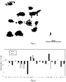
- Fig. 4 A and B shows gene expression heat maps for PBMC (A) and for hMSC (B) cultured on the scaffolds (all relative to control; note logaritmic scale which proves few orders of magnitude differences).
- For the reasons of clarity, the terminology used in this description means:
- "Fiber" refers to a fiber made of a non-cytotoxic, non-carcinogenic metal oxide, but not from polymers or bioactive ceramics (such as hydroxyapatite).
- "Automechanoinduction" means an induction of a mechanical stimulus to a cell or tissue from their own interaction with the substrate or media, without application of an external force, comprising that this induction leads to changes in cells or tissues behavior, structure, functionality, morphology, or any other relevant property. Automechanoinduction also excludes application of specific culture media, where said changes in cells or tissues are caused by e.g. adjustment of biochemical or chemical composition. In other words "automechanoinduction" describes the effect of mechanical stimulus on induction of the transformation of live cells and tissues. In known art, such stimulus is being introduced via direct mechanical activation or e.g. via fluid flow pressure changes. In present invention cell are facing such anisotropical substrate structure that forces them to adhere and expand/contract the membrane and the cytoskeleton without external mechanical or fluid pressure. This in its run triggers cell nucleus to start a cascade of gene expression, which will lead to changes in stem cell morphology, phenotype and differentiation pathways, i.e. such mechanoinduction is generated by the cells themselves.
- "Bioinert" means a material which does not enter into biochemical reactions with live cells leading to changes in their shape, functionality, proliferation or fate, neither undergoes a destruction or transformations (bioresorbtion) .
- "Scaffold" refers to a structure comprising a self-aligned network of fibers made in situ.
- "Randomly oriented fibers" refers to any fiber scaffold not fitting the description of the present invention, i.e. such fibers that have not been actively aligned or that do not follow any designed pattern of orientation to each other.
- "Self-aligned fibers" refers to a structure that consists of one or more fibers that are highly oriented in parallel to each other during the structure manufacturing process, i.e. not during the separate packing of the fibers info the structure (assembly) after the fibers have been manufactured otherwise.
- "Ultra-high anisotropy" refers to a self-aligned fibers scaffold which fibers have aspect ratios (length to diameter) of 10000:1 and more.
- The present invention will now be described in more detail hereafter with reference to the accompanying examples and figures, in which embodiments of the invention are shown. This invention may, however, be embodied in many different forms and should not be construed as limited to the embodiments set forth herein.
- The scaffolds were manufactured from aluminum oxide based material (however, other oxides, such as silica, titania, zirconia could also be used) and heat treated with regimes determined by a thermal analysis study [9]. These self-aligned metal oxide fibers (
Fig. 1 ) with an average single-fiber diameter of ~40 nm (TEM, HRSEM) and length of ~10-20 mm (aspect ratio ∼(10...20)·103 µm / 40·10-3 µm = (250000...500000):1) have 70-99% open porosity, preferably 85-95% open porosity (measured by BET and mercury porosimetry). Some of the scaffolds were also coated by graphene with catalyst-free chemical vapor deposition (CVD) at 1000°C to demonstrate that coatings might be easily performed if required. On the contrary to other studies, graphene on the nanofibres was not functionalized in any way and became augmented to the underlying oxide fiber surface. Non-functionalized graphene was used as an example of bioinert material. Nanofibers with other average diameters like 7 and 25 nm and similar aspect ratios were also obtained, but most of the cell studies were conducted with 40 nm fiber scaffolds for consistency. No bioactive agents were added, nor other modification of the scaffolds was made, unlike most of their known studies, to exclude possible effects of bioactive additions on cells. - Human MSCs were obtained from freshly isolated subcutaneous adipose tissue and characterized as reported [10]. Cancer cell lines MDA-MB231, Caco2, WM239A and Kelly, were obtained from ATCC. Cells were grown in culture media (DMEM) with 10% FBS (fetal bovine serum), 1 mg/ml penicillin and 0.1 mg/ml streptomycin at 37°C in 5% CO2. The scaffolds were pre-treated for three days before the cells seeding by complete medium with changing the medium for fresh every 24 hours to saturate them by active components adsorbed from liquid phase (Cellln Technologies, Estonia).
- For visualization of the cells, specific to filamentous actin (F-actin) phalloidin tagged by FITC (Sigma) was used. For hMSCs, pooled cells from three individuals with passage number below 5 were seeded on the scaffolds in 12-well plate (4 × 10⁴ cells per well). For cancer cells, 5 × 10⁴ cells were added to the each well with scaffolds. Similar cells grown on a flat glass at the same density and cell culture conditions were considered as the controls. The cells were fixed by 4% PFA at 48 h after seeding, washed by PBS (phosphate buffered saline) and permeabilized by 0.3% TRITON X-100 in PBS for 5 minutes. Phalloidin-FITC (1:100) staining lasted for 18 h at 4°C for scaffolds and 2 h at RT for controls. To stain the cells nucleus, cells were incubated for 10 minutes with Hoechst 33342 (Invitrogen, 1 µg/ml). After a final wash, the phalloidin-stained cells were analyzed by Nikon Eclipse 80i microscope (Cellln Technologies, Estonia).
- Calculation of the orientations of hMSC seeded was used with "ImageJ" software and Orientation-J Distribution plug-in (version 1.50g, National Institute of Health, USA). Original microscope images were converted into 8-bit color images and the pixels/distance ratio was calibrated based on the microscope camera bar. The images were threshold first by HSV color and then by brightness into binary images using Li algorithm. The orientation parameters were calculated with 5 pixel Gaussian window size and approximation with a cubic spline gradient. Finally, these data were treated with SigmaPlot software (Systat GmbH, Germany) into polar form.
- Immunologic response for peripheral blood mononuclear cells (PBMCs) was evaluated using PBMCs from healthy donors, isolated using Ficoll-Paque gradient fractionation (Cellln Technologies, Estonia). 2×10⁶ cells were used for each analysis. RNAs were extracted directly from scaffolds by TRIzol® (Ambion) reagent following 24 h cells growth on the scaffold, according to the manufacturer's recommendations. cDNAs were synthesized from DNase-treated (Ambion) RNA by RevertAid Reverse Transcriptase (Thermo Fisher Scientific) with addition of RiboLock (Thermo Fisher Scientific) according to the manufacturer's recommendations. cDNA quality was verified by RT-PCR by using GAPDH primers and HOT FIREpol® Master Mix (Solis Biodyne, Estonia). RT-qPCR was performed in triplicates using EvaGreen qPCR mix plus no Rox (Solis Biodyne, Estonia) and the LightCycler® 480 Real-Time PCR System (Roche Applied Science). The fold of change was calculated relatively to the control (cells grown without scaffolds) after normalization to GAPDH expression, using 2-ΔΔCt method (double difference of Ct). The values are respectively ΔCt = Ct(gene of interest) - Ct(GAPDH), and ΔΔCt = ΔCt(treated) - ΔCt(control). For visualization, the data were normalized to the cells grown without scaffold materials (control), converted to log scale and represented as a heat map (LionSolver 2.1, Reactive Search s.r.l., Italy). The minimal mapping error is achieved by minimizing sum of coordinates normalized with respect to the maximum and minimum along each dimension (in this case, decimal logarithm of relative expressions).
- Secretome ELISA analysis was done from cell culture media collected with soluble factors expression was analysed 24 h after initiation of cell culture (Protobios, Estonia). The levels of IL6, IL8/CXCL8, CCL2, IL1B, IL2, IL4, IL12, TNFα and IFNy secreted into the growth medium were measured using Human IL-6 DuoSet ELISA Development Kit (R&D System, Wiesbaden, Germany), Human IL-8 Standard ABTS ELISA Development Kit (Peprotech, Rock Hill, NJ, USA), Human CCL2/MCP-1 DuoSet (R&D System), Human IL-1β/IL-1F2 DuoSet ELISA Development Kit (R&D System, Wiesbaden, Germany), Human IL-2 ELISA Development Kit (Peprotech, Rock Hill, NJ, USA), Human IL-4 Standard ABTS (PeproTech, Rock Hill, NJ, USA), Human IL-12 ELISA Development Kit (Peprotech, Rock Hill, NJ, USA), Human TNFα ELISA Development Kit (Peprotech, Rock Hill, NJ, USA), Human IFN-γ ELISA Development Kit (Peprotech, Rock Hill, NJ, USA), and Human Standard ABTS ELISA Development Kit (Peprotech), respectively. The ELISA analysis was performed using high binding ELISA plates (Greiner BioOne) at RT according to the manufacturer's instructions. Optical density was measured using photospectrometer Spectramax 340 PC (Molecular Devices) at the wavelength 450 nm.
- The morphology, adhesion and distribution of viable hMSC after 3 days of culture on horizontally oriented new scaffolds and on control (glass) are demonstrated in Fig. 2A and B. The scaffolds under present invention were clearly guiding cells to line up along the fibers with a spindle-like shape and more than usually elongated lamellipodia extensions. High aspect ratio cellular orientations are developed throughout the scaffold, allowing directional connection between individual cells and formation of cells network. This confirms scaffolds do not impede the normal growth of stem cells. Furthermore, elongated morphology of hMSC and its high polarization create pre-requisites for preferential specific (e. g. neuronal or myogenic) lineage differentiation.
- Whereas response of stem cells to different substrate stiffness was studied earlier, the inventors for the first time have found the effect of such ultra-high scaffold anisotropy in a self-aligned, highly porous state, which was not previously reported. Most of known nanofiber-based scaffolds are randomly oriented or loosely packed, making a 3D nanostructure without such stiffness anisotropy [for example,
US 7704740 ,US 20060263417 ,US 20070269481 ]. Many aligned fibrous scaffolds are made of polymer fibers, the intrinsic elastic modulus of which is less than oxide ceramics by few orders of magnitude. The effect observed at scaffolds under present invention differs also substantially from known nanotopology studies, where nano-grooves or other patterns are known to support cell alignment, but without local differences in substrate stiffness. - Several cancer cell lines were seeded on the vertical and horizontal sides of the scaffolds of present invention. Cancer cells cultured on scaffolds possess extended microspikes and actin-rich filopodia protrusions suggesting high level of membrane activity and malignant migratory cancer phenotype (Fig. 3A,B shows MDA-MB231 cancer cell growth). The initial signs of cancer cell infiltration (and hence local immobilization of the cells) were detected on vertical fibers allowing suggestion of volumetric cancer model. By cancer cell infiltration, their reduced mobility and extended filopodia formation, scaffolds allow creating more representative phenotype necessary to obtain multi-dimensional in vitro cancer model that more accurately mimics tumor genesis. Furthermore, this model differs from both known 2D and 3D cultures as it allows partial immobilization of th-e cells into a "half-dimensional" direction (into the depth), keeping them unlimited to planar mobility. The inventors call this culture as 2.5D to emphasize this difference.
- Immunological profiling of any material under the influence of in vivo or in vitro environment is an important content of biocompatibility evaluation and has a critical value for future clinical translation. In addition to hMSC (Example 1), this was assessed with PBMC as they are an important part of the human peripheral immune system, responsible for transforming of the multitude of external stimuli to generate an adaptive immune response. The PBMC were grown on the scaffolds and tissue culture plastic (control) in vitro, and their inflammatory signatures were compared (
Fig. 4 ). The mRNA expression was analyzed for genes involved in the immune reaction and a secretion of cytokines (IL1β, TNFα, IFNγ, IL6, IL8, IL2, IL4, IL12β, and CCL2). - For PBMC, clear downregulation of pro-inflammatory TNF, IL1B, IL12B, IL6, CCL2 and COX2 cytokines was detected, demonstrating the strong ability of the scaffold to modulate immune response. Interestingly at the same time upregulated CCL18, IL1RN, IDO1 and TGFβ1 factors were observed. These factors participate in anti-inflammatory response, pointing out to the intrinsic possible immunomodulating effect of the scaffolds caused by their structure and composition alone, without any additions of external stimuli, which also was confirmed by ELISA (data not shown).
- For hMSCs, the promoted mRNA expression of CXCL8, CXCL9, CXCL10 and CSF2, the chemokines participating in neutrophil, monocyte or leukocyte trafficking, were observed, indicating the possible changes in chemotaxis. Additionally, reduced expression of COX2, CCL2 and IL6 cytokines indicates a good immune tolerance of these scaffolds.
- The present invention provides a principally new solution overcoming the drawbacks known by prior art by providing a 2.5-dimensional biocompatible scaffold designed for various cell cultures, capable to provide novel and more efficient methods for cell and drug research as well as tissue engineering applications.
- Other features and uses of the invention and their associated advantages will be evident to a one skilled in the art upon reading the description and the examples.
-
- 1. Lee, J., Cuddihy, M. J., Kotov, N. A. Three-dimensional cell culture matrices: state of the art. Tissue Eng Part B Rev 14, 61-86 (2008)
- 2. Bressan, E. et al. Graphene based scaffolds effects on stem cells commitment. J Translational Medicine 12, 296 (2014)
- 3. Jin, G., Li, K. The electrically conductive scaffold as the skeleton of stem cell niche in regenerative medicine. Materials Sci and Eng. C 45, 671 - 681 (2014)
- 4. Smith, L.A. et al. Directed differentiation and neurite extension of mouse embryonic stem cell on aligned poly(lactide) nanofibers functionalized with YIGSR peptide, Biomaterials 34 (2013) 9089-9095.
- 5. Talukdar, Y. et al. The effects of graphene nanostructures on mesenchymal stem cells. Biomaterials 35 (2014) 4863-4877
- 6. Discher, D. E., Janmey, P., Wang, Y.-L. Tissue cells feel and respond to the stiffness of their substrate. Science 310, 1139-43 (2005)
- 7. Coulombe, P. A., Wong, P. Cytoplasmic intermediate filaments revealed as dynamic and multipurpose scaffolds. Nat Cell Biol 6, 699-706 (2004)
- 8. Kenny, P. A. et al. The morphologies of breast cancer cell lines in three-dimensional assays correlate with their profiles of gene expression. Mol Oncol 1, 84-96 (2007)
- 9. Aghayan, M., Hussainova, I., Gasik, M., Kutuzov, M., Friman, M. Coupled thermal analysis of novel alumina nanofibers with ultrahigh aspect ratio. Thermochim Acta 574, 140-144 (2013)
- 10. Kazantseva, J. et al. Alternative splicing targeting the hTAF4-TAFH domain of TAF4 represses proliferation and accelerates chondrogenic differentiation of human mesenchymal stem cells. PLoS One 8(10):e74799 (2013)
Claims (12)
- A biocompatible scaffold for multi-dimensional cultivation of cells, said scaffolds capable of automechanoinduction and comprising:- highly aligned fibers oriented to form a scaffold with open porosity in a range of 70-99%, preferably 85-95% for the cultured cells, wherein the fibers have a diameter of 20-100 nm, and an aspect ratio more than 10000:1 calculated as length divided by diameter of the fibers,- said fibers being made of non-cytotoxic, non-carcinogenic inorganic metal oxide, and- said fibers being self-aligned into the scaffold during its manufacturing process.
- The scaffold according to claim 1, wherein the fibers have a length of 10-20 mm.
- The scaffold according to claim 1, wherein the diameter of the fibers is 20-50 nm.
- The scaffold according to claim 1, wherein the fibers have aspect ratio more than 20000:1 calculated as length divided by diameter of the fibers.
- The scaffold according to claim 1, wherein the fibers are coated with a bioinert material.
- The scaffold according to claim 5, wherein the bioinert material is non-functionalized graphene.
- The scaffold according to claim 1, wherein the metal oxide is selected from the group consisting of silica, titania, zirconia and alumina.
- The scaffold of according to claim 7, wherein the fibers are of aluminum oxide.
- The scaffold of claim 1, wherein the scaffold is for cultivation of stem cells and the scaffold is capable of inducing automechanoinduction of neural differentiation of the stem cells.
- The scaffold of claim 1, wherein the scaffold is for cultivation of cancer cells and the scaffold is capable of inducing automechanoinduction of gene expressions in the cancer cells to make them suitable as tumor models.
- A method to induce automechanoinduction of neural differentiation of stem cells by cultivating the stem cells on a scaffold of claim 1.
- A method to induce automechanoinduction of gene expressions in cancer cells by cultivating the cancer cells on a scaffold of claim 1.
Applications Claiming Priority (1)
| Application Number | Priority Date | Filing Date | Title |
|---|---|---|---|
| US201662361015P | 2016-07-12 | 2016-07-12 |
Publications (2)
| Publication Number | Publication Date |
|---|---|
| EP3269800A1 EP3269800A1 (en) | 2018-01-17 |
| EP3269800B1 true EP3269800B1 (en) | 2023-01-18 |
Family
ID=59399242
Family Applications (1)
| Application Number | Title | Priority Date | Filing Date |
|---|---|---|---|
| EP17181082.3A Active EP3269800B1 (en) | 2016-07-12 | 2017-07-12 | Self-aligned fibrous scaffolds for automechanoinduction of cell cultures |
Country Status (2)
| Country | Link |
|---|---|
| US (1) | US10370640B2 (en) |
| EP (1) | EP3269800B1 (en) |
Families Citing this family (1)
| Publication number | Priority date | Publication date | Assignee | Title |
|---|---|---|---|---|
| CN109266549A (en) * | 2018-10-25 | 2019-01-25 | 江苏久腾医学科技有限公司 | A kind of immunocyte culture bracket |
Family Cites Families (7)
| Publication number | Priority date | Publication date | Assignee | Title |
|---|---|---|---|---|
| US6103255A (en) | 1999-04-16 | 2000-08-15 | Rutgers, The State University | Porous polymer scaffolds for tissue engineering |
| US7704740B2 (en) | 2003-11-05 | 2010-04-27 | Michigan State University | Nanofibrillar structure and applications including cell and tissue culture |
| US8048446B2 (en) | 2005-05-10 | 2011-11-01 | Drexel University | Electrospun blends of natural and synthetic polymer fibers as tissue engineering scaffolds |
| JP5249785B2 (en) | 2006-01-27 | 2013-07-31 | ザ リージェンツ オブ ザ ユニバーシティ オブ カリフォルニア | Biomimetic scaffold |
| US20070238167A1 (en) | 2006-04-04 | 2007-10-11 | 3M Innovative Properties Company | Flat microfibers as matrices for cell growth |
| EP2833930B1 (en) * | 2012-04-04 | 2018-05-30 | University of Washington through its Center for Commercialization | Systems and method for engineering muscle tissue |
| US20170130194A1 (en) * | 2014-04-10 | 2017-05-11 | Rutgers, The State University Of New Jersey | Guiding stem cell differentiation using graphene-nanofiber hybrid scaffolds |
-
2017
- 2017-07-10 US US15/644,954 patent/US10370640B2/en active Active
- 2017-07-12 EP EP17181082.3A patent/EP3269800B1/en active Active
Non-Patent Citations (1)
| Title |
|---|
| ROMAN IVANOV ET AL: "Graphene-encapsulated aluminium oxide nanofibers as a novel type of nanofillers for electroconductive ceramics", JOURNAL OF THE EUROPEAN CERAMIC SOCIETY., vol. 35, no. 14, 1 November 2015 (2015-11-01), GB, pages 4017 - 4021, XP055579659, ISSN: 0955-2219, DOI: 10.1016/j.jeurceramsoc.2015.06.011 * |
Also Published As
| Publication number | Publication date |
|---|---|
| EP3269800A1 (en) | 2018-01-17 |
| US20180016551A1 (en) | 2018-01-18 |
| US10370640B2 (en) | 2019-08-06 |
Similar Documents
| Publication | Publication Date | Title |
|---|---|---|
| Lin et al. | Recent progress in stem cell differentiation directed by material and mechanical cues | |
| US20220370503A1 (en) | Methods and devices for the production and delivery of beneficial factors from stem cells | |
| Saei Arezoumand et al. | An overview on different strategies for the stemness maintenance of MSCs | |
| Chen et al. | Engineering multi-layered skeletal muscle tissue by using 3D microgrooved collagen scaffolds | |
| Edalat et al. | Engineering approaches toward deconstructing and controlling the stem cell environment | |
| Smith et al. | The enhancement of human embryonic stem cell osteogenic differentiation with nano-fibrous scaffolding | |
| Krontiras et al. | Adipogenic differentiation of stem cells in three‐dimensional porous bacterial nanocellulose scaffolds | |
| Ventre et al. | Engineering cell instructive materials to control cell fate and functions through material cues and surface patterning | |
| Jensen et al. | Functionalization of polycaprolactone scaffolds with hyaluronic acid and β-TCP facilitates migration and osteogenic differentiation of human dental pulp stem cells in vitro | |
| Park et al. | based bioactive scaffolds for stem cell-mediated bone tissue engineering | |
| Qian et al. | Bioactive polydimethylsiloxane surface for optimal human mesenchymal stem cell sheet culture | |
| Arshi et al. | Rigid microenvironments promote cardiac differentiation of mouse and human embryonic stem cells | |
| Kuo et al. | The effect of elastic biodegradable polyurethane electrospun nanofibers on the differentiation of mesenchymal stem cells | |
| Mashinchian et al. | Regulation of stem cell fate by nanomaterial substrates | |
| Herzog et al. | Cell communication in a coculture system consisting of outgrowth endothelial cells and primary osteoblasts | |
| Ahadian et al. | Graphene induces spontaneous cardiac differentiation in embryoid bodies | |
| Xu et al. | 3D bioprinting of multi-cellular tumor microenvironment for prostate cancer metastasis | |
| Tseng et al. | Biomaterial substrate‐mediated multicellular spheroid formation and their applications in tissue engineering | |
| JP2017518070A5 (en) | ||
| Zhang et al. | Cellular nanofiber structure with secretory activity-promoting characteristics for multicellular spheroid formation and hair follicle regeneration | |
| Cristallini et al. | The effect of bioartificial constructs that mimic myocardial structure and biomechanical properties on stem cell commitment towards cardiac lineage | |
| Pagliari et al. | A multistep procedure to prepare pre-vascularized cardiac tissue constructs using adult stem sells, dynamic cell cultures, and porous scaffolds | |
| Jing et al. | Effects of compound stimulation of fluid shear stress plus ultrasound on stem cell proliferation and osteogenesis | |
| Zhang et al. | Effect of substrate topography and chemistry on human mesenchymal stem cell markers: a transcriptome study | |
| Zhang et al. | Haversian bone-mimicking bioceramic scaffolds enhancing MSC-macrophage osteo-imunomodulation |
Legal Events
| Date | Code | Title | Description |
|---|---|---|---|
| PUAI | Public reference made under article 153(3) epc to a published international application that has entered the european phase |
Free format text: ORIGINAL CODE: 0009012 |
|
| STAA | Information on the status of an ep patent application or granted ep patent |
Free format text: STATUS: THE APPLICATION HAS BEEN PUBLISHED |
|
| AK | Designated contracting states |
Kind code of ref document: A1 Designated state(s): AL AT BE BG CH CY CZ DE DK EE ES FI FR GB GR HR HU IE IS IT LI LT LU LV MC MK MT NL NO PL PT RO RS SE SI SK SM TR |
|
| AX | Request for extension of the european patent |
Extension state: BA ME |
|
| STAA | Information on the status of an ep patent application or granted ep patent |
Free format text: STATUS: REQUEST FOR EXAMINATION WAS MADE |
|
| 17P | Request for examination filed |
Effective date: 20180712 |
|
| RBV | Designated contracting states (corrected) |
Designated state(s): AL AT BE BG CH CY CZ DE DK EE ES FI FR GB GR HR HU IE IS IT LI LT LU LV MC MK MT NL NO PL PT RO RS SE SI SK SM TR |
|
| STAA | Information on the status of an ep patent application or granted ep patent |
Free format text: STATUS: EXAMINATION IS IN PROGRESS |
|
| 17Q | First examination report despatched |
Effective date: 20190416 |
|
| GRAP | Despatch of communication of intention to grant a patent |
Free format text: ORIGINAL CODE: EPIDOSNIGR1 |
|
| STAA | Information on the status of an ep patent application or granted ep patent |
Free format text: STATUS: GRANT OF PATENT IS INTENDED |
|
| INTG | Intention to grant announced |
Effective date: 20211130 |
|
| GRAJ | Information related to disapproval of communication of intention to grant by the applicant or resumption of examination proceedings by the epo deleted |
Free format text: ORIGINAL CODE: EPIDOSDIGR1 |
|
| STAA | Information on the status of an ep patent application or granted ep patent |
Free format text: STATUS: EXAMINATION IS IN PROGRESS |
|
| INTC | Intention to grant announced (deleted) | ||
| GRAP | Despatch of communication of intention to grant a patent |
Free format text: ORIGINAL CODE: EPIDOSNIGR1 |
|
| STAA | Information on the status of an ep patent application or granted ep patent |
Free format text: STATUS: GRANT OF PATENT IS INTENDED |
|
| INTG | Intention to grant announced |
Effective date: 20220729 |
|
| GRAS | Grant fee paid |
Free format text: ORIGINAL CODE: EPIDOSNIGR3 |
|
| GRAA | (expected) grant |
Free format text: ORIGINAL CODE: 0009210 |
|
| STAA | Information on the status of an ep patent application or granted ep patent |
Free format text: STATUS: THE PATENT HAS BEEN GRANTED |
|
| AK | Designated contracting states |
Kind code of ref document: B1 Designated state(s): AL AT BE BG CH CY CZ DE DK EE ES FI FR GB GR HR HU IE IS IT LI LT LU LV MC MK MT NL NO PL PT RO RS SE SI SK SM TR |
|
| REG | Reference to a national code |
Ref country code: GB Ref legal event code: FG4D |
|
| REG | Reference to a national code |
Ref country code: DE Ref legal event code: R096 Ref document number: 602017065539 Country of ref document: DE |
|
| REG | Reference to a national code |
Ref country code: CH Ref legal event code: EP |
|
| REG | Reference to a national code |
Ref country code: AT Ref legal event code: REF Ref document number: 1544708 Country of ref document: AT Kind code of ref document: T Effective date: 20230215 Ref country code: IE Ref legal event code: FG4D |
|
| REG | Reference to a national code |
Ref country code: LT Ref legal event code: MG9D |
|
| REG | Reference to a national code |
Ref country code: NL Ref legal event code: MP Effective date: 20230118 |
|
| REG | Reference to a national code |
Ref country code: EE Ref legal event code: FG4A Ref document number: E023275 Country of ref document: EE Effective date: 20230418 Ref country code: AT Ref legal event code: MK05 Ref document number: 1544708 Country of ref document: AT Kind code of ref document: T Effective date: 20230118 |
|
| PG25 | Lapsed in a contracting state [announced via postgrant information from national office to epo] |
Ref country code: NL Free format text: LAPSE BECAUSE OF FAILURE TO SUBMIT A TRANSLATION OF THE DESCRIPTION OR TO PAY THE FEE WITHIN THE PRESCRIBED TIME-LIMIT Effective date: 20230118 |
|
| PG25 | Lapsed in a contracting state [announced via postgrant information from national office to epo] |
Ref country code: RS Free format text: LAPSE BECAUSE OF FAILURE TO SUBMIT A TRANSLATION OF THE DESCRIPTION OR TO PAY THE FEE WITHIN THE PRESCRIBED TIME-LIMIT Effective date: 20230118 Ref country code: PT Free format text: LAPSE BECAUSE OF FAILURE TO SUBMIT A TRANSLATION OF THE DESCRIPTION OR TO PAY THE FEE WITHIN THE PRESCRIBED TIME-LIMIT Effective date: 20230518 Ref country code: NO Free format text: LAPSE BECAUSE OF FAILURE TO SUBMIT A TRANSLATION OF THE DESCRIPTION OR TO PAY THE FEE WITHIN THE PRESCRIBED TIME-LIMIT Effective date: 20230418 Ref country code: LV Free format text: LAPSE BECAUSE OF FAILURE TO SUBMIT A TRANSLATION OF THE DESCRIPTION OR TO PAY THE FEE WITHIN THE PRESCRIBED TIME-LIMIT Effective date: 20230118 Ref country code: LT Free format text: LAPSE BECAUSE OF FAILURE TO SUBMIT A TRANSLATION OF THE DESCRIPTION OR TO PAY THE FEE WITHIN THE PRESCRIBED TIME-LIMIT Effective date: 20230118 Ref country code: HR Free format text: LAPSE BECAUSE OF FAILURE TO SUBMIT A TRANSLATION OF THE DESCRIPTION OR TO PAY THE FEE WITHIN THE PRESCRIBED TIME-LIMIT Effective date: 20230118 Ref country code: ES Free format text: LAPSE BECAUSE OF FAILURE TO SUBMIT A TRANSLATION OF THE DESCRIPTION OR TO PAY THE FEE WITHIN THE PRESCRIBED TIME-LIMIT Effective date: 20230118 Ref country code: AT Free format text: LAPSE BECAUSE OF FAILURE TO SUBMIT A TRANSLATION OF THE DESCRIPTION OR TO PAY THE FEE WITHIN THE PRESCRIBED TIME-LIMIT Effective date: 20230118 |
|
| PG25 | Lapsed in a contracting state [announced via postgrant information from national office to epo] |
Ref country code: SE Free format text: LAPSE BECAUSE OF FAILURE TO SUBMIT A TRANSLATION OF THE DESCRIPTION OR TO PAY THE FEE WITHIN THE PRESCRIBED TIME-LIMIT Effective date: 20230118 Ref country code: PL Free format text: LAPSE BECAUSE OF FAILURE TO SUBMIT A TRANSLATION OF THE DESCRIPTION OR TO PAY THE FEE WITHIN THE PRESCRIBED TIME-LIMIT Effective date: 20230118 Ref country code: IS Free format text: LAPSE BECAUSE OF FAILURE TO SUBMIT A TRANSLATION OF THE DESCRIPTION OR TO PAY THE FEE WITHIN THE PRESCRIBED TIME-LIMIT Effective date: 20230518 Ref country code: GR Free format text: LAPSE BECAUSE OF FAILURE TO SUBMIT A TRANSLATION OF THE DESCRIPTION OR TO PAY THE FEE WITHIN THE PRESCRIBED TIME-LIMIT Effective date: 20230419 Ref country code: FI Free format text: LAPSE BECAUSE OF FAILURE TO SUBMIT A TRANSLATION OF THE DESCRIPTION OR TO PAY THE FEE WITHIN THE PRESCRIBED TIME-LIMIT Effective date: 20230118 |
|
| REG | Reference to a national code |
Ref country code: DE Ref legal event code: R097 Ref document number: 602017065539 Country of ref document: DE |
|
| PG25 | Lapsed in a contracting state [announced via postgrant information from national office to epo] |
Ref country code: SM Free format text: LAPSE BECAUSE OF FAILURE TO SUBMIT A TRANSLATION OF THE DESCRIPTION OR TO PAY THE FEE WITHIN THE PRESCRIBED TIME-LIMIT Effective date: 20230118 Ref country code: RO Free format text: LAPSE BECAUSE OF FAILURE TO SUBMIT A TRANSLATION OF THE DESCRIPTION OR TO PAY THE FEE WITHIN THE PRESCRIBED TIME-LIMIT Effective date: 20230118 Ref country code: DK Free format text: LAPSE BECAUSE OF FAILURE TO SUBMIT A TRANSLATION OF THE DESCRIPTION OR TO PAY THE FEE WITHIN THE PRESCRIBED TIME-LIMIT Effective date: 20230118 Ref country code: CZ Free format text: LAPSE BECAUSE OF FAILURE TO SUBMIT A TRANSLATION OF THE DESCRIPTION OR TO PAY THE FEE WITHIN THE PRESCRIBED TIME-LIMIT Effective date: 20230118 |
|
| PLBE | No opposition filed within time limit |
Free format text: ORIGINAL CODE: 0009261 |
|
| STAA | Information on the status of an ep patent application or granted ep patent |
Free format text: STATUS: NO OPPOSITION FILED WITHIN TIME LIMIT |
|
| PG25 | Lapsed in a contracting state [announced via postgrant information from national office to epo] |
Ref country code: SK Free format text: LAPSE BECAUSE OF FAILURE TO SUBMIT A TRANSLATION OF THE DESCRIPTION OR TO PAY THE FEE WITHIN THE PRESCRIBED TIME-LIMIT Effective date: 20230118 |
|
| 26N | No opposition filed |
Effective date: 20231019 |
|
| PG25 | Lapsed in a contracting state [announced via postgrant information from national office to epo] |
Ref country code: SI Free format text: LAPSE BECAUSE OF FAILURE TO SUBMIT A TRANSLATION OF THE DESCRIPTION OR TO PAY THE FEE WITHIN THE PRESCRIBED TIME-LIMIT Effective date: 20230118 |
|
| PG25 | Lapsed in a contracting state [announced via postgrant information from national office to epo] |
Ref country code: MC Free format text: LAPSE BECAUSE OF FAILURE TO SUBMIT A TRANSLATION OF THE DESCRIPTION OR TO PAY THE FEE WITHIN THE PRESCRIBED TIME-LIMIT Effective date: 20230118 |
|
| PG25 | Lapsed in a contracting state [announced via postgrant information from national office to epo] |
Ref country code: MC Free format text: LAPSE BECAUSE OF FAILURE TO SUBMIT A TRANSLATION OF THE DESCRIPTION OR TO PAY THE FEE WITHIN THE PRESCRIBED TIME-LIMIT Effective date: 20230118 |
|
| REG | Reference to a national code |
Ref country code: CH Ref legal event code: PL |
|
| REG | Reference to a national code |
Ref country code: BE Ref legal event code: MM Effective date: 20230731 |
|
| PG25 | Lapsed in a contracting state [announced via postgrant information from national office to epo] |
Ref country code: LU Free format text: LAPSE BECAUSE OF NON-PAYMENT OF DUE FEES Effective date: 20230712 |
|
| PG25 | Lapsed in a contracting state [announced via postgrant information from national office to epo] |
Ref country code: LU Free format text: LAPSE BECAUSE OF NON-PAYMENT OF DUE FEES Effective date: 20230712 |
|
| REG | Reference to a national code |
Ref country code: IE Ref legal event code: MM4A |
|
| PG25 | Lapsed in a contracting state [announced via postgrant information from national office to epo] |
Ref country code: CH Free format text: LAPSE BECAUSE OF NON-PAYMENT OF DUE FEES Effective date: 20230731 |
|
| PG25 | Lapsed in a contracting state [announced via postgrant information from national office to epo] |
Ref country code: IT Free format text: LAPSE BECAUSE OF FAILURE TO SUBMIT A TRANSLATION OF THE DESCRIPTION OR TO PAY THE FEE WITHIN THE PRESCRIBED TIME-LIMIT Effective date: 20230118 Ref country code: BE Free format text: LAPSE BECAUSE OF NON-PAYMENT OF DUE FEES Effective date: 20230731 |
|
| PG25 | Lapsed in a contracting state [announced via postgrant information from national office to epo] |
Ref country code: IE Free format text: LAPSE BECAUSE OF NON-PAYMENT OF DUE FEES Effective date: 20230712 |
|
| PG25 | Lapsed in a contracting state [announced via postgrant information from national office to epo] |
Ref country code: IE Free format text: LAPSE BECAUSE OF NON-PAYMENT OF DUE FEES Effective date: 20230712 |
|
| PGFP | Annual fee paid to national office [announced via postgrant information from national office to epo] |
Ref country code: EE Payment date: 20240612 Year of fee payment: 8 |
|
| PGFP | Annual fee paid to national office [announced via postgrant information from national office to epo] |
Ref country code: DE Payment date: 20240612 Year of fee payment: 8 |
|
| PGFP | Annual fee paid to national office [announced via postgrant information from national office to epo] |
Ref country code: GB Payment date: 20240711 Year of fee payment: 8 |
|
| PGFP | Annual fee paid to national office [announced via postgrant information from national office to epo] |
Ref country code: FR Payment date: 20240711 Year of fee payment: 8 |
|
| PG25 | Lapsed in a contracting state [announced via postgrant information from national office to epo] |
Ref country code: BG Free format text: LAPSE BECAUSE OF FAILURE TO SUBMIT A TRANSLATION OF THE DESCRIPTION OR TO PAY THE FEE WITHIN THE PRESCRIBED TIME-LIMIT Effective date: 20230118 |
|
| PG25 | Lapsed in a contracting state [announced via postgrant information from national office to epo] |
Ref country code: BG Free format text: LAPSE BECAUSE OF FAILURE TO SUBMIT A TRANSLATION OF THE DESCRIPTION OR TO PAY THE FEE WITHIN THE PRESCRIBED TIME-LIMIT Effective date: 20230118 |
|
| PG25 | Lapsed in a contracting state [announced via postgrant information from national office to epo] |
Ref country code: CY Free format text: LAPSE BECAUSE OF FAILURE TO SUBMIT A TRANSLATION OF THE DESCRIPTION OR TO PAY THE FEE WITHIN THE PRESCRIBED TIME-LIMIT; INVALID AB INITIO Effective date: 20170712 |
|
| PG25 | Lapsed in a contracting state [announced via postgrant information from national office to epo] |
Ref country code: HU Free format text: LAPSE BECAUSE OF FAILURE TO SUBMIT A TRANSLATION OF THE DESCRIPTION OR TO PAY THE FEE WITHIN THE PRESCRIBED TIME-LIMIT; INVALID AB INITIO Effective date: 20170712 |
|
| PG25 | Lapsed in a contracting state [announced via postgrant information from national office to epo] |
Ref country code: TR Free format text: LAPSE BECAUSE OF FAILURE TO SUBMIT A TRANSLATION OF THE DESCRIPTION OR TO PAY THE FEE WITHIN THE PRESCRIBED TIME-LIMIT Effective date: 20230118 |
|
| REG | Reference to a national code |
Ref country code: DE Ref legal event code: R119 Ref document number: 602017065539 Country of ref document: DE |
|
| GBPC | Gb: european patent ceased through non-payment of renewal fee |
Effective date: 20250712 |
|
| PG25 | Lapsed in a contracting state [announced via postgrant information from national office to epo] |
Ref country code: GB Free format text: LAPSE BECAUSE OF NON-PAYMENT OF DUE FEES Effective date: 20250712 |
|
| PG25 | Lapsed in a contracting state [announced via postgrant information from national office to epo] |
Ref country code: DE Free format text: LAPSE BECAUSE OF NON-PAYMENT OF DUE FEES Effective date: 20260203 |
|
| PG25 | Lapsed in a contracting state [announced via postgrant information from national office to epo] |
Ref country code: FR Free format text: LAPSE BECAUSE OF NON-PAYMENT OF DUE FEES Effective date: 20250731 |
|
| PG25 | Lapsed in a contracting state [announced via postgrant information from national office to epo] |
Ref country code: EE Free format text: LAPSE BECAUSE OF NON-PAYMENT OF DUE FEES Effective date: 20250731 |