EP3258269A1 - Reagent kit used for detecting lipoprotein-associated phospholipase a2, and preparation method and application for reagent kit - Google Patents
Reagent kit used for detecting lipoprotein-associated phospholipase a2, and preparation method and application for reagent kit Download PDFInfo
- Publication number
- EP3258269A1 EP3258269A1 EP15881481.4A EP15881481A EP3258269A1 EP 3258269 A1 EP3258269 A1 EP 3258269A1 EP 15881481 A EP15881481 A EP 15881481A EP 3258269 A1 EP3258269 A1 EP 3258269A1
- Authority
- EP
- European Patent Office
- Prior art keywords
- lipoprotein
- associated phospholipase
- antibodies
- kit
- pla2
- Prior art date
- Legal status (The legal status is an assumption and is not a legal conclusion. Google has not performed a legal analysis and makes no representation as to the accuracy of the status listed.)
- Granted
Links
Images


Classifications
-
- G—PHYSICS
- G01—MEASURING; TESTING
- G01N—INVESTIGATING OR ANALYSING MATERIALS BY DETERMINING THEIR CHEMICAL OR PHYSICAL PROPERTIES
- G01N33/00—Investigating or analysing materials by specific methods not covered by groups G01N1/00 - G01N31/00
- G01N33/48—Biological material, e.g. blood, urine; Haemocytometers
- G01N33/50—Chemical analysis of biological material, e.g. blood, urine; Testing involving biospecific ligand binding methods; Immunological testing
- G01N33/53—Immunoassay; Biospecific binding assay; Materials therefor
- G01N33/573—Immunoassay; Biospecific binding assay; Materials therefor for enzymes or isoenzymes
-
- C—CHEMISTRY; METALLURGY
- C12—BIOCHEMISTRY; BEER; SPIRITS; WINE; VINEGAR; MICROBIOLOGY; ENZYMOLOGY; MUTATION OR GENETIC ENGINEERING
- C12Q—MEASURING OR TESTING PROCESSES INVOLVING ENZYMES, NUCLEIC ACIDS OR MICROORGANISMS; COMPOSITIONS OR TEST PAPERS THEREFOR; PROCESSES OF PREPARING SUCH COMPOSITIONS; CONDITION-RESPONSIVE CONTROL IN MICROBIOLOGICAL OR ENZYMOLOGICAL PROCESSES
- C12Q1/00—Measuring or testing processes involving enzymes, nucleic acids or microorganisms; Compositions therefor; Processes of preparing such compositions
- C12Q1/34—Measuring or testing processes involving enzymes, nucleic acids or microorganisms; Compositions therefor; Processes of preparing such compositions involving hydrolase
- C12Q1/44—Measuring or testing processes involving enzymes, nucleic acids or microorganisms; Compositions therefor; Processes of preparing such compositions involving hydrolase involving esterase
-
- C—CHEMISTRY; METALLURGY
- C12—BIOCHEMISTRY; BEER; SPIRITS; WINE; VINEGAR; MICROBIOLOGY; ENZYMOLOGY; MUTATION OR GENETIC ENGINEERING
- C12Y—ENZYMES
- C12Y301/00—Hydrolases acting on ester bonds (3.1)
- C12Y301/01—Carboxylic ester hydrolases (3.1.1)
- C12Y301/01004—Phospholipase A2 (3.1.1.4)
-
- G—PHYSICS
- G01—MEASURING; TESTING
- G01N—INVESTIGATING OR ANALYSING MATERIALS BY DETERMINING THEIR CHEMICAL OR PHYSICAL PROPERTIES
- G01N2333/00—Assays involving biological materials from specific organisms or of a specific nature
- G01N2333/90—Enzymes; Proenzymes
- G01N2333/914—Hydrolases (3)
- G01N2333/916—Hydrolases (3) acting on ester bonds (3.1), e.g. phosphatases (3.1.3), phospholipases C or phospholipases D (3.1.4)
- G01N2333/918—Carboxylic ester hydrolases (3.1.1)
Definitions
- the present disclosure relates to the field of detection of biochemical substances. More particularly, it relates to a kit for detecting lipoprotein-associated phospholipase A2 and a method for producing the same, as well as a method for detecting lipoprotein-associated phospholipase A2.
- Lp-PLA2 also known as platelet activating factor acetylhydrolase (PAF-AH)
- PAF-AH platelet activating factor acetylhydrolase
- PLA2G7 is a member of the class PLA2 of the phospholipase family. It is a serine-dependent phospholipase which has a catalytic activity with no need for Ca 2+ .
- Human plasma Lp-PLA2 has a molecular weight of 45 kDa and is mainly produced by secretion of macrophages, monocytes, T lymphocytes, mast cells, hepatocytes, and the like, with regulation by inflammatory mediators.
- Lp-PLA2 its secretion can be inhibited by gamma interferon and lipopolysaccharide but enhanced by platelet activating factor.
- the main roles of Lp-PLA2 include producing dodecanoic acid inflammatory substances, participating in phospholipid reconstitution and stable equilibrium of biological membrane, lipoprotein metabolism, cell signaling, and host reaction, and facilitating autologous disappearing of necrotic tissue of the body.
- Lp-PLA2 can promote the formation of atherosclerotic (AS) plaques and bind to low density lipoprotein (LDL) in the circulation.
- AS atherosclerotic
- LDL low density lipoprotein
- CHD coronary heart disease
- Lp-PLA2 is secreted by inflammatory cells in the atherosclerotic plaques and is significantly increased in severe atherosclerotic plaques. Therefore, it can be used as a marker for predicting future cardiovascular events of CHD.
- CHF congestive heart failure
- Lp-PLA2 may be an independent predictor of ischemic stroke, and determination of Lp-PLA2 level may provide predictive information in addition to conventional risk factor assessment, providing insights for guiding the prevention strategy.
- MS metabolic syndrome
- Lp-PLA2 methods for determination of Lp-PLA2 available on the market mainly involve spectrophotometry, latex-enhanced turbidimetry, radioactive immunoassay (RIA), and enzyme-linked immunosorbent assay (ELISA).
- spectrophotometrical methods for detecting Lp-PLA2.
- PAF thioesters as the substrate. Its mechanism involves enzymatic hydrolysis of the thioester group in the sn-2 position on the backbone of the PAF thioester analogue, releasing free thiol, followed by addition of 5'5-disulfide (dinitrobenzoic acid) of the thiol which can be detected, and detection of variation in absorbance at 405 nm.
- This type of substrates includes 1-thiodecanoyl-2-decanoyl-PC, 1-hexadecanoyl-2-thiohexadecanoyl-PC, and the like.
- the other method uses PAF analogues with a 4-nitrophenol group.
- This type of substrate has similar structure with the PAF thioester, except that the thioester group in the sn-2 position is displaced by the 4-nitrophenol group. Its mechanism involves enzymatic hydrolysis of the substrate, releasing the substance with 4-nitrophenol group which has instable properties and immediately decomposes into 4-nitrophenol, and detection of variation in absorbance at 405nm due to this change, thereby determining the enzyme activity.
- Lp-PLA2 in a sample undergoes an antigen-antibody reaction with sensitized latex particles of mouse anti-human Lp-PLA2 antibody in the reagent in a phosphate buffer system, causing agglutination under the action of polyethylene glycol as an accelerator to produce an increased turbidity, and variation in absorbance of the reaction solution is detected at 546 nm wavelength, which is proportional to the Lp-PLA2 content in the sample.
- Radioimmunoassay involves a procedure in accordance with the principle of competition mechanism, wherein the binding amount of 125I-Lp-PLA2 with antibody is a function of the Lp-PLA2 content in the standard or sample.
- the Lp-PLA2 concentration in the sample was determined by separating the binding fraction (B) from the free fraction (F) using an immune separation agent (PR), determining the radioactive intensity of the binding fraction, and processing the data.
- PR immune separation agent
- Enzyme-linked immunosorbent assay uses a double antibody sandwich method to determine the level of human Lp-PLA2 in whole blood samples.
- Purified human Lp-PLA2 antibodies are used to coat a microplate to prepare solid phase antibody.
- Lp-PLA2 is added to the wells coated on the monoclonal antibody and binds with the anti-Lp-PLA2 antibody labeled with horseradish peroxidase (HRP) to form an antibody-antigen-enzyme-labeled antibody complex.
- HRP horseradish peroxidase
- TMB (3, 3", 5, 5"-tetramethylbenzidine) was added as a substrate after thorough washing for coloration. TMB is converted to blue under the catalysis of HRP enzyme and to a final yellow color under the action of acid.
- the color tone is positively correlated with the Lp-PLA2 content in the sample.
- the absorbance (OD) is measured at 450 nm using a microplate reader.
- the content of human Lp-PLA2 in the sample is calculated using a standard curve.
- Enzyme-linked immunosorbent assay (ELISA) is a commonly used method for determination of Lp-PLA2.
- ELISA Enzyme-linked immunosorbent assay
- the enzyme immunoluminescence antibody coating for selection is limited to a single form and the binding with Lp-PLA2 is not thorough enough, resulting in low detection sensitivity. Relatively large numbers of uncertain factors in the microplate has caused poor repeatability of the test.
- enzyme-linked immunoassay is restricted by its degree of automation, rendering a relatively long reaction time.
- a kit for detecting Lp-PLA2.
- the concentration of Lp-PLA2 can be detected with high sensitivity, high repeatability, and high accuracy with a double antibody sandwich method.
- the precision of Lp-PLA2 detection can be further improved.
- the present disclosure also provides a method for preparing a chemiluminescence immunoassay kit for detecting Lp-PLA2.
- the present disclosure also provides applications of the Lp-PLA2 chemiluminescence immunoassay kit according to the present disclosure, in particular the application of the kit in performing a fully-automated chemiluminescence method for Lp-PLA2 detection to shorten operating time, reduce operation errors, and improve detection sensitivity with the specificity of chemical trace marker.
- kits for detecting Lp-PLA2 comprising one or more first anti-Lp-PLA2 antibodies coated on magnetic sphere for binding with Lp-PLA2 to be detected, and one or more second anti-Lp-PLA2 antibodies labeled with a trace marker for binding with Lp-PLA2 to be detected at other binding sites different from the binding sites on Lp-PLA2 for the first anti-Lp-PLA2 antibodies. That is, the difference between the first anti-Lp-PLA2 antibody and the second anti-Lp-PLA2 antibody lies in their different binding sites on Lp-PLA2 to be detected.
- the kit comprises one or more second anti-Lp-PLA2 antibodies coated on magnetic sphere for binding with Lp-PLA2 to be detected, and one or more first anti-Lp-PLA2 antibodies labeled with a trace marker for binding with Lp-PLA2 to be detected at other binding sites different from the binding sites on Lp-PLA2 for the first anti-Lp-PLA2 antibodies.
- the first anti-Lp-PLA2 antibody and the second anti-Lp-PLA2 antibody may each independently be an anti-Lp-PLA2 monoclonal antibody and /or an anti-Lp-PLA2 polyclonal antibody.
- the trace marker may be selected from trace markers commonly used in the art for labeling antigens or antibodies, such as adamantane, luminol and its derivatives, isluminol and its derivatives, acridinium esters, alkaline phosphatase or horseradish peroxidase, and particularly preferably N-(4-aminobutyl) -N-ethylisoluminol (ABEI).
- trace markers commonly used in the art for labeling antigens or antibodies such as adamantane, luminol and its derivatives, isluminol and its derivatives, acridinium esters, alkaline phosphatase or horseradish peroxidase, and particularly preferably N-(4-aminobutyl) -N-ethylisoluminol (ABEI).
- Magnetic sphere suitable for use in the present disclosure are also known as magnetic beads and may be magnetic microspheres commonly used in the art. It is preferable that the magnetic sphere used in the present disclosure are microscale solid-phase microspheres with superparamagnetism and extremely large capacity of protein adsorption formed by combining nanoscale Fe 2 O 3 or Fe 3 O 4 magnetic particles and an organic polymeric materials. Such magnetic sphere can be quickly magnetized in an applied magnetic field while having a remanence of zero after removal of the magnetic field. There is no particular limitation on the types of the organic polymeric material, which may be selected as necessary.
- the magnetic microspheres used in the present disclosure should have a diameter of 0.1 to 5 microns, and the magnetic microspheres may be surface modified to carry a variety of reactive functional groups including, but not limited to, -OH, -COOH, -NH 2 .
- the magnetic sphere is a complex of Fe 2 O 3 or Fe 3 O 4 magnetic particles and an organic polymeric material and has a particle size of 0.1 to 5 microns; and, the magnetic sphere are optionally modified by surface modification to carry one or more active functional groups.
- the concentrations of the first anti-Lp-PLA2 antibody and the second anti-Lp-PLA2 antibody in the kit are 10 to 200 ⁇ g/ml, respectively, the concentration of the trace marker is 0.1 to 1 mg/ml, and the concentration of the magnetic sphere is 0.1 to 5 mg/ml.
- the concentration for each component above is based on the amount of each independent component containing such component.
- the kit further comprises a low-point calibrator and a high-point calibrator of Lp-PLA2 and optionally a buffer.
- the low-point calibrator and high-point calibrator herein is referred to relative to each other, wherein the "low-point calibrator” refers to a calibrator made by diluting Lp-PLA2 to a concentration of 10 to 30 ng/ml using a 50% bovine serum product, and the "high-point calibrator” refers to a calibrator made by diluting Lp-PLA2 to a concentration of 500 to 700 ng/ml using a 50% bovine serum product.
- the low-point calibrator and high-point calibrator each optionally contains bovine serum albumin (BSA) and/or a preservative.
- BSA concentration is preferably 0.01 to 5 g/ml.
- the trace marker directly or indirectly labels the second anti-Lp-PLA2 antibody.
- Indirect labeling forms include, but are not limited to, indirect labeling via a fluorescein isothiocyanate (FITC) and anti-FITC antibody system or a streptavidin (SA) and biotin system.
- Direct labeling refers to labeling by directly linking ABEI to an anti-Lp-PLA2 antibody.
- Indirect labeling refers to labeling an anti-Lp-PLA2 antibody with ABEI via an intermediate medium linking system including, but not limited to, a FITC and anti-FITC antibody system or a streptavidin and biotin system.
- the present inventors have found that indirect marking advantageously weakens the spatial effect and facilitates the amplification of the signal, increasing detection sensitivity.
- the anti-FITC antibody is preferably a goat anti-FITC polyclonal antibody.
- the first anti-Lp-PLA2 antibodies are directly or indirectly coated on magnetic sphere.
- Indirect coating forms of the magnetic sphere include, but are not limited to, indirect coating by a FITC and anti-FITC antibody system or a streptavidin and biotin system.
- Direct coating refers to coating the magnetic sphere directly using anti-Lp-PLA2 antibodies; and indirect coating refers to coating the magnetic sphere using the anti-Lp-PLA2 antibodies via an intermediate medium linking system including, but not limited to, a FITC and anti-FITC antibody system or a streptavidin and biotin system.
- One advantage of indirect coating lies in that it weakens the spatial effect and facilitates the amplification of the signal, increasing detection sensitivity.
- the first anti-Lp-PLA2 antibodies are capable of binding to the sites 220 to 441 on Lp-PLA2, and the second anti-Lp-PLA2 antibodies are capable of binding to the sites 1 to 200 on Lp-PLA2.
- the first anti-Lp-PLA2 antibodies are coated on magnetic sphere, and the second anti-Lp-PLA2 antibodies are labeled with a trace marker.
- the first anti-Lp-PLA2 antibodies are directly or indirectly coated on magnetic sphere, and the indirect coating forms of magnetic sphere comprise indirect coating via a FITC and anti-FITC antibody system or a streptavidin and biotin system.
- the trace marker directly or indirectly labels the second anti-Lp-PLA2 antibody, and the indirect labeling forms comprise indirect labeling via a FITC and anti-FITC antibody system or a streptavidin and biotin system.
- the kit comprises any one of the components selected from M1 to M3 and any one of the components selected from N1 to N3, wherein M1 is a solution of the first anti-Lp-PLA2 antibody directly coated on magnetic sphere; M2 is a streptavidin solution directly coated on magnetic sphere and a biotinylated solution of the second anti-Lp-PLA2 antibody; M3 is an solution of anti-FITC antibody directly coated on magnetic sphere and a solution of the second anti-Lp-PLA2 antibody labeled with FITC; N1 is a solution of the first anti-Lp-PLA2 antibody directly labeled with a trace marker; N2 is a streptavidin solution directly labeled with a trace marker and a biotinylated solution of the first anti-Lp-PLA2 antibody; N3 is a solution of anti-FITC antibody directly labeled with a trace marker and a solution of the first anti-Lp-PLA2 antibody solution labele
- the kit comprises:
- Each of the components contains BSA and a preservative, the BSA concentration being 0.01 to 5 g/ml.
- the kit comprises:
- Each of the components contains BSA and a preservative, the BSA concentration being 0.01 to 5 g/ml.
- the kit comprises:
- Each of the components contains BSA and a preservative, the BSA concentration being 0.01 to 5 g/ml.
- the kit comprises:
- Each of the components contains BSA and a preservative, the BSA concentration being 0.01 to 5 g/ml.
- the present disclosure also provides a displacer comprising water as a solvent (preferably purified water) and the following components:
- the displacer comprises the following components relative to the total amount of the solvent:
- the displacer may further comprise 0.1 to 10 vol% of ethylene glycol and/or 0.1-10 vol% of glycerol.
- the displacer may further comprise an appropriate amount of preservative.
- the preservatives suitable for use in the present disclosure may be any one or a mixture of at least two or more selected from preservatives commonly used in the art, such as potassium sorbate, sodium benzoate, sodium azide, sodium nitrite, the Proclin series, or the like.
- the kit provided herein further comprises a displacer as described above. Since Lp-PLA2 is present in the human body primarily as a complex with the lipoprotein particles, the Lp-PLA2 antigen can be displaced from the complex with lipoprotein particles by the above-mentioned displacer so as to better react with an antibody. Therefore, the accuracy and sensitivity of the reagent can be remarkably improved by utilizing the function and characteristics of the displacer as described above.
- the present disclosure provides a method for preparing a kit as described above, comprising: directly or indirectly coating a first anti-Lp-PLA2 antibody on magnetic sphere, and directly or indirectly labeling a second anti-Lp-PLA2 antibody with a trace marker.
- the indirect labeling comprises labeling the second anti-Lp-PLA2 antibody with a trace marker via a FITC and anti-FITC antibody system or a streptavidin and biotin system.
- the indirect coating comprises coating the first anti-Lp-PLA2 antibody indirectly through a FITC and anti-FITC antibody system or a streptavidin and biotin system.
- the method for preparing a kit according to the present disclosure comprises directly or indirectly coating the first anti-Lp-PLA2 antibodies on magnetic sphere, and directly or indirectly labeling the second anti-Lp-PLA2 antibodies with a trace marker; wherein, the first anti-Lp-PLA2 antibodies bind to the sites 220-441 on the lipoprotein-associated phospholipase A2 and the second anti-Lp-PLA2 antibodies bind to the sites 1 to 200 on the lipoprotein-associated phospholipase A2.
- the forms of indirect labeling comprise indirect labeling via a fluorescein isothiocyanate and anti-fluorescein isothiocyanate antibody system or a streptavidin and biotin system; and, the indirect coating forms of the magnetic sphere comprise indirect coating via a fluorescein isothiocyanate and anti-fluorescein isothiocyanate antibody system or a streptavidin and biotin system.
- the method for preparing a kit according to the present disclosure may further comprise preparing a low-point calibrator and a high-point calibrator, and it may further comprise assembling the kit.
- a method for detecting Lp-PLA2 which comprises detecting Lp-PLA2 concentration in a subject sample by chemiluminescence immunoassay using a kit as described above.
- the method comprises detecting the concentration of Lp-PLA2 in a subject sample by chemiluminescent immunoassay using a kit as described above. Specifically, the first anti-Lp-PLA2 antibodies coated on the magnetic sphere in the kit and the second anti-Lp-PLA2 antibodies labeled with a trace marker are allowed to form a double antibody sandwich mode of the first anti- PLA2 antibody/Lp-PLA2/second anti-Lp-PLA2 antibody, with Lp-PLA2 in the subject sample, and luminescent substrate is added for determination of Lp-PLA2 concentration by chemiluminescence immunoassay.
- the method for detecting Lp-PLA2 concentration comprises detecting Lp-PLA2 concentration by a chemiluminescence immunoassay analyser using a kit as described above.
- the method is performed in full automation.
- the chemiluminescence immunoassay analyser is preferably a Maglumi series chemiluminescence immunoassay analyzer (manufactured by Shenzhen New Industries Biomedical Engineering Co., Ltd.).
- Detection of Lp-PLA2 by the kit provided herein can utilize a serum sample as a subject sample, eliminating the interference factors in the plasma and sparing the time for pretreatment of a whole blood sample.
- the kit provided herein utilizes a variety of antibody labeling forms.
- the use of a displacer can specifically displace Lp-PLA2 to ensure effective and accurate binding of antibodies and antigens and improve detection accuracy and sensitivity.
- the method for detecting Lp-PLA2 adopts an one-step process, that is, adding the components of the kit and the antigens to be detected into a reaction cup in one step for a mixed reaction to form a double antibody sandwich mode, which reduces operation steps and reaction time; it is also possible to utilize a machine to add samples to effectively reduce human errors and improve repeatability.
- the method for preparing the components of the kit is as follows:
- the immunomagnetic sphere used in this preparation procedure was a suspension of nano-magnetic microspheres at a concentration of 100 mg/ml with hydroxyl group of 95 mg KOH/g, manufactured by Merck Co., Ltd..
- the magnetic microspheres were suspended in the acetic acid buffer (pH 3.6) above of 5x coating volume to give a magnetic sphere concentration of 20 mg/ml, and 1-cyclohexyl-2-morpholinoethyl-carbodiimide metho-p-toluenesulfonate (CMC) was added to a concentration of 10 mg/ml.
- CMC 1-cyclohexyl-2-morpholinoethyl-carbodiimide metho-p-toluenesulfonate
- Purified anti-Lp-PLA2 monoclonal or polyclonal antibodies were added by a weight ratio of the resultant solution to the anti-Lp-PLA2 monoclonal or polyclonal antibodies at 1mg: 12 ⁇ g, and underwent reaction in a constant-temperature shaking bath incubator at 37 °C for 24 hours.
- Preparation of cleaning solution for magnetic sphere 500ml PBS buffer (pH 7.4) was prepared with 0.1mol/l PBS buffer and purified water at a volumetric ratio of 1: 9, into which 2.5g BSA was added, well mixed, and dissolved to prepare the cleaning solution for magnetic sphere.
- the magnetic sphere after the warm bath in step (2) were poured into a beaker, placed on magnet for precipitation, had the supernatant removed, washed under stirring with 5x volume of the cleaning solution for magnetic sphere, placed on magnet, and had the cleared supernatant removed. The cleaning procedure was repeated for four times.
- the magnetic sphere cleaned in step (3) were added to a mixed solution (primary composition of the mixed solution: 0.2 g/ml KH 2 PO 4 , 2.9 g/ml NaHPO 4 , 8g/ml NaCl, 2 g/ml NaN 3 , 5 g/ml BSA, 2 ml/ml Twain T-20, balanced with purified water) of 1x coating volume, to obtain a suspension of magnetic sphere of 1x coating volume with a suspension concentration of 20 mg/ml, i.e., the suspension of magnetic sphere coated with the anti-Lp-PLA2 monoclonal or polyclonal antibodies.
- a mixed solution primary composition of the mixed solution: 0.2 g/ml KH 2 PO 4 , 2.9 g/ml NaHPO 4 , 8g/ml NaCl, 2 g/ml NaN 3 , 5 g/ml BSA, 2 ml/ml Twain T-20, balanced with purified water
- the immunomagnetic sphere used in this preparation procedure was a suspension of nano-magnetic microspheres at a concentration of 100 mg/ml with hydroxyl group of 95 mg KOH/g, manufactured by Merck Co., Ltd..
- the magnetic microspheres were suspended in the acetic acid buffer (pH 3.6) above of 5x coating volume to give a magnetic sphere concentration of 20 mg/ml, and CMC (1-cyclohexyl-2-morpholinoethyl-carbodiimide metho-p-toluenesulfonate) was added to a concentration of 10 mg/ml.
- Streptavidin was added by a weight ratio of the resultant solution to streptavidin at 1mg: 12 ⁇ g, and underwent reaction in a constant-temperature shaking bath incubator at 37 °C for 24 hours.
- Preparation of cleaning solution for magnetic sphere 500ml PBS buffer (pH 7.4) was prepared with 0.1mol/l PBS buffer and purified water at a volumetric ratio of 1: 9, into which 2.5g BSA was added, well mixed, and dissolved to prepare the cleaning solution for magnetic sphere.
- the magnetic sphere after the warm bath in step (2) were poured into a beaker, placed on magnet for precipitation, had the supernatant removed, washed under stirring with 5x volume of the cleaning solution for magnetic sphere, placed on magnet, and had the cleared supernatant removed. The cleaning procedure was repeated for four times.
- the magnetic sphere cleaned in step (3) were added to a mixed solution (primary composition of the mixed solution: 0.2 g/ml KH 2 PO 4 , 2.9 g/ml NaHPO 4 , 8g/ml NaCl, 2 g/ml NaN 3 , 5 g/ml BSA, 2 ml/ml Twain T-20, balanced with purified water) of 1x coating volume, to obtain a suspension of magnetic sphere of 1x coating volume with a suspension concentration of 20 mg/ml, i.e., the suspension of magnetic sphere coated with streptavitin.
- a mixed solution primary composition of the mixed solution: 0.2 g/ml KH 2 PO 4 , 2.9 g/ml NaHPO 4 , 8g/ml NaCl, 2 g/ml NaN 3 , 5 g/ml BSA, 2 ml/ml Twain T-20, balanced with purified water
- the immunomagnetic sphere used in this preparation procedure was a suspension of nano-magnetic microspheres at a concentration of 100 mg/ml with hydroxyl group of 95 mg KOH/g, manufactured by Merck Co., Ltd..
- the magnetic microspheres were suspended in the acetic acid buffer (pH 3.6) above of 5x coating volume to give a magnetic sphere concentration of 20 mg/ml, and CMC (1-cyclohexyl-2-morpholinoethyl-carbodiimide metho-p-toluenesulfonate) was added to a concentration of 10 mg/ml.
- CMC 1-cyclohexyl-2-morpholinoethyl-carbodiimide metho-p-toluenesulfonate
- Anti-FITC monoclonal or polyclonal antibodies were added by a weight ratio of the resultant solution to the anti-FITC monoclonal or polyclonal antibodies at 1mg: 12 ⁇ g, and underwent reaction in a constant-temperature shaking bath incubator at 37 °C for 24 hours.
- Preparation of cleaning solution for magnetic sphere 500ml PBS buffer (pH 7.4) was prepared with 0.1mol/l PBS buffer and purified water at a volumetric ratio of 1: 9, into which 2.5g BSA was added, well mixed, and dissolved to prepare the cleaning solution for magnetic sphere.
- the magnetic sphere after the warm bath in step (2) were poured into a beaker, placed on magnet for precipitation, had the supernatant removed, washed under stirring with 5x volume of the cleaning solution for magnetic sphere, placed on magnet, and had the cleared supernatant removed. The cleaning procedure was repeated for four times.
- the magnetic sphere cleaned in step (3) were added to a mixed solution (primary composition of the mixed solution: 0.2 g/ml KH 2 PO 4 , 2.9 g/ml NaHPO 4 , 8g/ml NaCl, 2 g/ml NaN 3 , 5 g/ml BSA, 2 ml/ml Twain T-20, balanced with purified water) of 1x coating volume, to obtain a suspension of magnetic sphere of 1x coating volume with a suspension concentration of 20 mg/ml, i.e., the suspension of magnetic sphere coated with the anti-FITC monoclonal or polyclonal antibodies.
- a mixed solution primary composition of the mixed solution: 0.2 g/ml KH 2 PO 4 , 2.9 g/ml NaHPO 4 , 8g/ml NaCl, 2 g/ml NaN 3 , 5 g/ml BSA, 2 ml/ml Twain T-20, balanced with purified water
- the first anti-Lp-PLA2 antibodies were used to prepare a suspension of magnetic sphere coated with the first anti-Lp-PLA2 antibodies according to Preparation Example 1.
- the second anti-Lp-PLA2 antibodies were used to prepare a suspension of the second anti-Lp-PLA2 antibodies labeled with ABEI according to Preparation Example 4.
- Preparation of the displacer To a beaker, 1000ml of purified water was added, and 15g of CHAPS, 0.5g of casein, 2g of EDTA-2Na, 6g of DDT, 1g of Tris, 3g of MES, and 2g of NaN 3 were weighed and dissolved in the water in sequence. After complete dissolution of the above, 150 ml of newborn bovine serum, 5 ml of acetic acid, 25 ml of horse serum, 10 ml of glycerol, and 25 g of BSA were added, well mixed, and then filtered to obtain the displacer solution.
- the kit of the present example comprises each solution prepared as above.
- Lp-PLA2 in a sample was detected using a Maglumi 2000 plus chemiluminescence immunoassay analyser with the following general procedure:
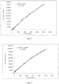
- Lp-PLA2 standards were prepared into solutions of 0ng/ml to 1000ng/ml and the concentrations of the standards were linearly fit with the detected luminous intensities to verify the linearity of the ten-point calibration curve. The results are shown in Table 1 and shown in Figure 1 .
- kit composition and detection method of this embodiment were substantially the same as in Example 1, except that the present embodiment does not use a displacer.
- the results are shown in Table 1-4 and Figure 2 .
- the first anti-Lp-PLA2 antibodies were used to prepare a solution of the first anti-Lp-PLA2 antibodies labeled with ABEI according to Preparation Example 4.
- the second anti-Lp-PLA2 antibodies were used to prepare a solution of the second anti-Lp-PLA2 antibodies labeled with FITC according to Preparation Example 6.
- Preparation of the displacer To a beaker, 1000ml of purified water was added, and 3g of ANS, 1g of casein, 3g of EDTA-2Na, 6g of DDT, 1g of Tris, 3g of MES, and 2g of NaN 3 were weighed and dissolved in the water in sequence. After complete dissolution of the above, 200 ml of newborn bovine serum, 5 ml of acetic acid, 30 ml of horse serum, 10 ml of glycol, and 25 g of BSA were measured and added, well mixed, and then filtered to obtain the displacer.
- a high-point calibrator solution of 562.43ng/ml and a low-point calibrator solution of 17.8ng/ml were prepared.
- the kit of the present example comprises each solution prepared as above.
- the detection procedure of Lp-PLA2 was substantially the same as that of Example 1, except for step 2): adding 40 ⁇ l of the displacer, 40 ⁇ l of the second anti-Lp-PLA2 antibody solution labeled with ABEI, 40 ⁇ l of the suspension of the magnetic sphere coated with the goat anti-FITC polyclonal antibodies, and 40 ⁇ l of the solution of the first anti-Lp-PLA2 antibodies labeled with FITC, and incubating for 30 minutes at 37 °C.
- kit composition and the detection method of this embodiment were substantially the same as those of Example 3, except that the displacer is not used in the present embodiment.
- the results are shown in Table 1-4 and Figure 4 .
- a suspension of magnetic sphere coated with streptavitin was prepared as described in Preparation Example 2 above.
- the first anti-Lp-PLA2 antibodies were used to prepare a solution of the first anti-Lp-PLA2 antibodies labeled with biotin according to Preparation Example 5.
- the second anti-Lp-PLA2 antibodies were used to prepare a solution of the second anti-Lp-PLA2 antibodies labeled with ABEI according to Preparation Example 4.
- Preparation of the displacer To a beaker, 1000ml of purified water was added, and 5g of sodium deoxycholate, 2g of casein, 2g of EDTA-2Na, 5g of DDT, 1.5g of Tris, 2g of MES, and 2g of NaN 3 were weighed and dissolved in the water in sequence. After complete dissolution of the above, 100 ml of newborn bovine serum, 8 ml of acetic acid, 20 ml of goat serum, 5 ml of glycol, and 25 g of BSA were measured and added, well mixed, and then filtered to obtain the displacer.
- a high-point calibrator solution of 562.43ng/ml and a low-point calibrator solution of 17.8ng/ml were prepared.
- the kit of the present example comprises each solution prepared as above.
- the detection procedure of Lp-PLA2 was substantially the same as that of Example 1, except for step 2): adding 50 ⁇ l of the displacer, 50 ⁇ l of the second anti-Lp-PLA2 antibody solution labeled with ABEI, 20 ⁇ l of the magnetic sphere coated with streptavitin, and 20 ⁇ l of the solution of the first anti-Lp-PLA2 antibodies labeled with biotin, and incubating for 30 minutes at 37 °C.
- kit composition and the detection method of this kit were substantially the same as those of Example 5, except that the displacer is not used in the present embodiment.
- the results are shown in Table 1-4, Figure 6 .
- the Lp-PLA2 concentrations of ten negative samples and ten positive samples were measured using each of the kits in Examples 1 to 6 described above, respectively, and the samples were each measured twice. The results are shown in Table 5.
- the kit provided herein has a fitted linear correlation coefficient of greater than 0.98 for the ten-point curve obtained from ten different concentrations of the standard solution, indicating that the kit provided herein can linearly reflect the Lp-PLA2 concentration in the human body with luminous intensity.
- the kits of Examples 2, 4, and 6 that did not comprise a displacer showed a ten-point curve with a linear correlation coefficient of about 0.99.
- Examples 1, 3 and 5 that comprised the displacer provided herein showed a linear correlation coefficient greater than 0.99, which was greater than the corresponding kits without the displacer, indicating that the displacer was able to improve the linear correlation of the Lp-PLA2concentration reflected by the luminous intensity of the kit.
- the 50% concentration of the high-point calibrator measured in Examples 1 to 6 did not deviated much from the theoretical values, i.e., the 50% theoretical concentration of the high-point calibrator.
- the recovery percentage ranged between 90% and 110%.
- the recovery percentages of the kits of Examples 1, 3, and 5 that comprised the displacer were higher than those kits without the displacer, indicating that the displacer provided herein were able to improve the determined recovery of the kits.
- the kit comprising the displacer (the kits of Examples 1, 3 and 5) was capable of sensitive detection of the sample value. This indicates that the kits comprising the displacer were more sensitive than those without. Meanwhile, comparing the the sample test results of the different kits, it can be found that the kits comprising the displacer had better repeatability of the determination results than those without.
- detection of a sample can be completed in 40 minutes, which greatly improves the detection efficiency at least by double compared with the conventional chemiluminescence enzyme-linked immunosorbent assay.
Abstract
Description
- The present disclosure relates to the field of detection of biochemical substances. More particularly, it relates to a kit for detecting lipoprotein-associated phospholipase A2 and a method for producing the same, as well as a method for detecting lipoprotein-associated phospholipase A2.
- Lp-PLA2, also known as platelet activating factor acetylhydrolase (PAF-AH), is encoded by the PLA2G7 gene and is a member of the class PLA2 of the phospholipase family. It is a serine-dependent phospholipase which has a catalytic activity with no need for Ca2+. Human plasma Lp-PLA2 has a molecular weight of 45 kDa and is mainly produced by secretion of macrophages, monocytes, T lymphocytes, mast cells, hepatocytes, and the like, with regulation by inflammatory mediators. For example, its secretion can be inhibited by gamma interferon and lipopolysaccharide but enhanced by platelet activating factor. The main roles of Lp-PLA2 include producing dodecanoic acid inflammatory substances, participating in phospholipid reconstitution and stable equilibrium of biological membrane, lipoprotein metabolism, cell signaling, and host reaction, and facilitating autologous disappearing of necrotic tissue of the body.
- Recent studies have shown that Lp-PLA2 can promote the formation of atherosclerotic (AS) plaques and bind to low density lipoprotein (LDL) in the circulation. The current study suggests that coronary heart disease (CHD) is a chronic inflammatory disease, with inflammation involved in various stages of CHD pathogenesis. Lp-PLA2 is secreted by inflammatory cells in the atherosclerotic plaques and is significantly increased in severe atherosclerotic plaques. Therefore, it can be used as a marker for predicting future cardiovascular events of CHD. Other studies have found that Lp-PLA2 is correlated with a risk of congestive heart failure (CHF). The onset of cerebral infarction is closely related to the instability of the AS plaque, while instable plaques, in turn, are prone to embolization or thrombosis, leading to infarction. Lp-PLA2 may be an independent predictor of ischemic stroke, and determination of Lp-PLA2 level may provide predictive information in addition to conventional risk factor assessment, providing insights for guiding the prevention strategy. Studies have found that low-level inflammatory response plays an important role when metabolic syndrome (MS) occurs, wherein the MS health response becomes more obvious with increased activity of Lp-PLA2.
- At present, methods for determination of Lp-PLA2 available on the market mainly involve spectrophotometry, latex-enhanced turbidimetry, radioactive immunoassay (RIA), and enzyme-linked immunosorbent assay (ELISA).
- Among them, there are currently two types of spectrophotometrical methods for detecting Lp-PLA2. One method uses PAF thioesters as the substrate. Its mechanism involves enzymatic hydrolysis of the thioester group in the sn-2 position on the backbone of the PAF thioester analogue, releasing free thiol, followed by addition of 5'5-disulfide (dinitrobenzoic acid) of the thiol which can be detected, and detection of variation in absorbance at 405 nm. This type of substrates includes 1-thiodecanoyl-2-decanoyl-PC, 1-hexadecanoyl-2-thiohexadecanoyl-PC, and the like. The other method uses PAF analogues with a 4-nitrophenol group. This type of substrate has similar structure with the PAF thioester, except that the thioester group in the sn-2 position is displaced by the 4-nitrophenol group. Its mechanism involves enzymatic hydrolysis of the substrate, releasing the substance with 4-nitrophenol group which has instable properties and immediately decomposes into 4-nitrophenol, and detection of variation in absorbance at 405nm due to this change, thereby determining the enzyme activity.
- In latex-enhanced turbidimetry, Lp-PLA2 in a sample undergoes an antigen-antibody reaction with sensitized latex particles of mouse anti-human Lp-PLA2 antibody in the reagent in a phosphate buffer system, causing agglutination under the action of polyethylene glycol as an accelerator to produce an increased turbidity, and variation in absorbance of the reaction solution is detected at 546 nm wavelength, which is proportional to the Lp-PLA2 content in the sample.
- Radioimmunoassay involves a procedure in accordance with the principle of competition mechanism, wherein the binding amount of 125I-Lp-PLA2 with antibody is a function of the Lp-PLA2 content in the standard or sample. The Lp-PLA2 concentration in the sample was determined by separating the binding fraction (B) from the free fraction (F) using an immune separation agent (PR), determining the radioactive intensity of the binding fraction, and processing the data.
- Enzyme-linked immunosorbent assay (ELISA) uses a double antibody sandwich method to determine the level of human Lp-PLA2 in whole blood samples. Purified human Lp-PLA2 antibodies are used to coat a microplate to prepare solid phase antibody. Lp-PLA2 is added to the wells coated on the monoclonal antibody and binds with the anti-Lp-PLA2 antibody labeled with horseradish peroxidase (HRP) to form an antibody-antigen-enzyme-labeled antibody complex. TMB (3, 3", 5, 5"-tetramethylbenzidine) was added as a substrate after thorough washing for coloration. TMB is converted to blue under the catalysis of HRP enzyme and to a final yellow color under the action of acid. The color tone is positively correlated with the Lp-PLA2 content in the sample. The absorbance (OD) is measured at 450 nm using a microplate reader. The content of human Lp-PLA2 in the sample is calculated using a standard curve. Enzyme-linked immunosorbent assay (ELISA) is a commonly used method for determination of Lp-PLA2. However, there is plenty of interferential components in the whole blood samples for Lp-PLA2 measurement, which affects the accuracy of measurement. Besides, the enzyme immunoluminescence antibody coating for selection is limited to a single form and the binding with Lp-PLA2 is not thorough enough, resulting in low detection sensitivity. Relatively large numbers of uncertain factors in the microplate has caused poor repeatability of the test. In addition, enzyme-linked immunoassay is restricted by its degree of automation, rendering a relatively long reaction time.
- As one object of the present disclosure, a kit is provided for detecting Lp-PLA2. Using this kit, the concentration of Lp-PLA2 can be detected with high sensitivity, high repeatability, and high accuracy with a double antibody sandwich method. Particularly, with the addition of a displacer in the kit, the precision of Lp-PLA2 detection can be further improved.
- The present disclosure also provides a method for preparing a chemiluminescence immunoassay kit for detecting Lp-PLA2.
- Further, the present disclosure also provides applications of the Lp-PLA2 chemiluminescence immunoassay kit according to the present disclosure, in particular the application of the kit in performing a fully-automated chemiluminescence method for Lp-PLA2 detection to shorten operating time, reduce operation errors, and improve detection sensitivity with the specificity of chemical trace marker.
- According to the present disclosure, a kit for detecting Lp-PLA2 is provided, comprising one or more first anti-Lp-PLA2 antibodies coated on magnetic sphere for binding with Lp-PLA2 to be detected, and one or more second anti-Lp-PLA2 antibodies labeled with a trace marker for binding with Lp-PLA2 to be detected at other binding sites different from the binding sites on Lp-PLA2 for the first anti-Lp-PLA2 antibodies. That is, the difference between the first anti-Lp-PLA2 antibody and the second anti-Lp-PLA2 antibody lies in their different binding sites on Lp-PLA2 to be detected. Thus, in an equivalent embodiment of the present disclosure, the kit comprises one or more second anti-Lp-PLA2 antibodies coated on magnetic sphere for binding with Lp-PLA2 to be detected, and one or more first anti-Lp-PLA2 antibodies labeled with a trace marker for binding with Lp-PLA2 to be detected at other binding sites different from the binding sites on Lp-PLA2 for the first anti-Lp-PLA2 antibodies..
- According to the present disclosure, the first anti-Lp-PLA2 antibody and the second anti-Lp-PLA2 antibody may each independently be an anti-Lp-PLA2 monoclonal antibody and /or an anti-Lp-PLA2 polyclonal antibody.
- According to the present disclosure, the trace marker may be selected from trace markers commonly used in the art for labeling antigens or antibodies, such as adamantane, luminol and its derivatives, isluminol and its derivatives, acridinium esters, alkaline phosphatase or horseradish peroxidase, and particularly preferably N-(4-aminobutyl) -N-ethylisoluminol (ABEI).
- Magnetic sphere suitable for use in the present disclosure are also known as magnetic beads and may be magnetic microspheres commonly used in the art. It is preferable that the magnetic sphere used in the present disclosure are microscale solid-phase microspheres with superparamagnetism and extremely large capacity of protein adsorption formed by combining nanoscale Fe2O3 or Fe3O4 magnetic particles and an organic polymeric materials. Such magnetic sphere can be quickly magnetized in an applied magnetic field while having a remanence of zero after removal of the magnetic field. There is no particular limitation on the types of the organic polymeric material, which may be selected as necessary.
- The magnetic microspheres used in the present disclosure should have a diameter of 0.1 to 5 microns, and the magnetic microspheres may be surface modified to carry a variety of reactive functional groups including, but not limited to, -OH, -COOH, -NH2.
- In a specific embodiment, the magnetic sphere is a complex of Fe2O3 or Fe3O4 magnetic particles and an organic polymeric material and has a particle size of 0.1 to 5 microns; and, the magnetic sphere are optionally modified by surface modification to carry one or more active functional groups.
- According to the present disclosure, the concentrations of the first anti-Lp-PLA2 antibody and the second anti-Lp-PLA2 antibody in the kit are 10 to 200 µg/ml, respectively, the concentration of the trace marker is 0.1 to 1 mg/ml, and the concentration of the magnetic sphere is 0.1 to 5 mg/ml. The concentration for each component above is based on the amount of each independent component containing such component.
- According to the present disclosure, the kit further comprises a low-point calibrator and a high-point calibrator of Lp-PLA2 and optionally a buffer. The low-point calibrator and high-point calibrator herein is referred to relative to each other, wherein the "low-point calibrator" refers to a calibrator made by diluting Lp-PLA2 to a concentration of 10 to 30 ng/ml using a 50% bovine serum product, and the "high-point calibrator" refers to a calibrator made by diluting Lp-PLA2 to a concentration of 500 to 700 ng/ml using a 50% bovine serum product. The low-point calibrator and high-point calibrator each optionally contains bovine serum albumin (BSA) and/or a preservative. The BSA concentration is preferably 0.01 to 5 g/ml.
- According to the present disclosure, the trace marker directly or indirectly labels the second anti-Lp-PLA2 antibody. Indirect labeling forms include, but are not limited to, indirect labeling via a fluorescein isothiocyanate (FITC) and anti-FITC antibody system or a streptavidin (SA) and biotin system. Direct labeling refers to labeling by directly linking ABEI to an anti-Lp-PLA2 antibody. Indirect labeling refers to labeling an anti-Lp-PLA2 antibody with ABEI via an intermediate medium linking system including, but not limited to, a FITC and anti-FITC antibody system or a streptavidin and biotin system. The present inventors have found that indirect marking advantageously weakens the spatial effect and facilitates the amplification of the signal, increasing detection sensitivity. The anti-FITC antibody is preferably a goat anti-FITC polyclonal antibody.
- According to the present disclosure, the first anti-Lp-PLA2 antibodies are directly or indirectly coated on magnetic sphere. Indirect coating forms of the magnetic sphere include, but are not limited to, indirect coating by a FITC and anti-FITC antibody system or a streptavidin and biotin system. Direct coating refers to coating the magnetic sphere directly using anti-Lp-PLA2 antibodies; and indirect coating refers to coating the magnetic sphere using the anti-Lp-PLA2 antibodies via an intermediate medium linking system including, but not limited to, a FITC and anti-FITC antibody system or a streptavidin and biotin system. One advantage of indirect coating lies in that it weakens the spatial effect and facilitates the amplification of the signal, increasing detection sensitivity.
- In some embodiments according to the present disclosure, the first anti-Lp-PLA2 antibodies are capable of binding to the sites 220 to 441 on Lp-PLA2, and the second anti-Lp-PLA2 antibodies are capable of binding to the sites 1 to 200 on Lp-PLA2. In a further embodiment, the first anti-Lp-PLA2 antibodies are coated on magnetic sphere, and the second anti-Lp-PLA2 antibodies are labeled with a trace marker. In these embodiments, the first anti-Lp-PLA2 antibodies are directly or indirectly coated on magnetic sphere, and the indirect coating forms of magnetic sphere comprise indirect coating via a FITC and anti-FITC antibody system or a streptavidin and biotin system. The trace marker directly or indirectly labels the second anti-Lp-PLA2 antibody, and the indirect labeling forms comprise indirect labeling via a FITC and anti-FITC antibody system or a streptavidin and biotin system.
- According to some embodiments of the present disclosure, the kit comprises any one of the components selected from M1 to M3 and any one of the components selected from N1 to N3, wherein M1 is a solution of the first anti-Lp-PLA2 antibody directly coated on magnetic sphere; M2 is a streptavidin solution directly coated on magnetic sphere and a biotinylated solution of the second anti-Lp-PLA2 antibody; M3 is an solution of anti-FITC antibody directly coated on magnetic sphere and a solution of the second anti-Lp-PLA2 antibody labeled with FITC; N1 is a solution of the first anti-Lp-PLA2 antibody directly labeled with a trace marker; N2 is a streptavidin solution directly labeled with a trace marker and a biotinylated solution of the first anti-Lp-PLA2 antibody; N3 is a solution of anti-FITC antibody directly labeled with a trace marker and a solution of the first anti-Lp-PLA2 antibody solution labeled with FITC; and, each solution of the components M1 to M3 and the components N1 to N3 optionally comprises BSA and/or a preservative. The BSA concentration is preferably 0.01 to 5 g/ml.
- In a specific embodiment of the present disclosure, the kit comprises:
- a1) a suspension of magnetic sphere coated with the first anti-Lp-PLA2 antibody, in which the concentration of the first anti-Lp-PLA2 antibody is 10 to 200 µg/ml and the concentration of magnetic sphere is 0.1 to 5 mg/ml;
- b1) a solution of the second anti-Lp-PLA2 antibody labeled with ABEI, in which the concentration of the second anti-Lp-PLA2 antibody is 10 to 200 µg/ml and the ABEI concentration is 0.1 to 1 mg/ml;
- c1) a low-point calibrator containing Lp-PLA2 at a concentration of 10 to 30 ng/ml;
- d1) a high-point calibrator containing Lp-PLA2 at a concentration of 500 to 700 ng/ml;
- Each of the components contains BSA and a preservative, the BSA concentration being 0.01 to 5 g/ml.
- In a specific embodiment of the present disclosure, the kit comprises:
- a2) a solution of magnetic sphere coated with streptavidin, in which the concentration of streptavidin is 10 to 200 µg/ml and the concentration of magnetic sphere is 0.1 to 5 mg/ml;
- b2) a solution of the second anti-Lp-PLA2 antibody labeled with ABEI, in which the concentration of the second anti-Lp-PLA2 antibody is 10 to 200 µg/ml and the ABEI concentration is 0.1 to 1 mg/ml;
- c2) a biotinylated solution of the first anti-Lp-PLA2 antibody, in which the concentration of the first anti-Lp-PLA2 antibody is 10 to 200 µg/ml and the concentration of biotin is 0.1 to 1 mg/ml;
- d2) a low-point calibrator containing Lp-PLA2 at a concentration of 10 to 30 ng/ml;
- e2) a high-point calibrator containing Lp-PLA2 at a concentration of 500 to 700 ng/ml;
- Each of the components contains BSA and a preservative, the BSA concentration being 0.01 to 5 g/ml.
- In a specific embodiment of the present disclosure, the kit comprises:
- a3) a solution of magnetic sphere coated with anti-FITC antibodies, in which the concentration of anti-FITC antibodies is 10 to 200 µg/ml and the concentration of magnetic sphere is 0.1 to 5 mg/ml;
- b3) a solution of the second anti-Lp-PLA2 antibody labeled with ABEI, in which the concentration of the second anti-Lp-PLA2 antibody is 10 to 200 µg/ml and the ABEI concentration is 0.1 to 1 mg/ml;
- c3) a solution of the first anti-Lp-PLA2 antibody labeled with FITC, in which the concentration of the first anti-Lp-PLA2 antibody is 10 to 200 µg/ml and the concentration of FITC is 0.1 to 1 mg/ml;
- d3) a low-point calibrator containing Lp-PLA2 at a concentration of 10 to 30 ng/ml;
- e3) a high-point calibrator containing Lp-PLA2 at a concentration of 500 to 700 ng/ml;
- Each of the components contains BSA and a preservative, the BSA concentration being 0.01 to 5 g/ml.
- In a specific embodiment of the present disclosure, the kit comprises:
- a4) a solution of magnetic sphere coated with the first anti-Lp-PLA2 antibodies, in which the concentration of the first anti-Lp-PLA2 antibodies is 10 to 200 µg/ml and the concentration of magnetic sphere is 0.1 to 5 mg/ml;
- b4) a biotinylated solution of the second anti-Lp-PLA2 antibody, in which the concentration of the second anti-Lp-PLA2 antibody is 10 to 200 µg/ml and the biotin concentration is 0.1 to 1 mg/ml;
- c4) a solution of streptavitin labeled with ABEI, in which the concentration of streptavitin is 10 to 200 µg/ml and the concentration of ABEI is 0.1 to 1 mg/ml;
- d4) a low-point calibrator containing Lp-PLA2 at a concentration of 10 to 30 ng/ml;
- e4) a high-point calibrator containing Lp-PLA2 at a concentration of 500 to 700 ng/ml;
- Each of the components contains BSA and a preservative, the BSA concentration being 0.01 to 5 g/ml.
- The present disclosure also provides a displacer comprising water as a solvent (preferably purified water) and the following components:
- at least one selected from the group consisting of 3- [3- (cholamidopropyl) dimethylammonio] propanesulfonate, 8-anilino-1-naphthalenesulfonic acid, sodium deoxycholate, barbiturate, and acetic acid;
- newborn bovine serum;
- goat serum and/or horse serum;
- dithiothreitol;
- tris (hydroxymethyl) aminomethane;
- hydrated 2-morpholinoethanesulfonic acid;
- casein; and
- disodium ethylenediaminetetraacetate.
- In a preferred embodiment of the present disclosure, the displacer comprises the following components relative to the total amount of the solvent:
- at least one selected from the group consisting of 0.1 to 5 wt% of 3- [3-(cholamidopropyl) dimethylammonio] propanesulfonate (CHAPS), 0.1 to 2 wt% of 8-anilino-1-naphthalenesulfonic acid (ANS), 0.1 to 2 wt% of sodium deoxycholate (SDC), 0.1 to 2 wt% of barbiturate, and 0.1 to 2 vol% of acetic acid;
- 1 to 50 vol% of newborn bovine serum;
- 0.1 to 10 vol% of goat serum and/or 0.1 to 10 vol% of horse serum;
- 0.1 to 10 wt% of dithiothreitol (DDT);
- 0.1 to 10 wt% of tris (hydroxymethyl) aminomethane (Tris);
- 0.1 to 10 wt% of monohydrate 2-morpholinoethanesulfonic acid (MES);
- 0.01 to 1 wt% of casein; and
- 0.01 to 1 wt% of disodium ethylenediaminetetraacetate (EDTA-2Na).
- The displacer may further comprise 0.1 to 10 vol% of ethylene glycol and/or 0.1-10 vol% of glycerol.
- The displacer may further comprise an appropriate amount of preservative.
- The preservatives suitable for use in the present disclosure may be any one or a mixture of at least two or more selected from preservatives commonly used in the art, such as potassium sorbate, sodium benzoate, sodium azide, sodium nitrite, the Proclin series, or the like.
- In a preferred embodiment of the present disclosure, the kit provided herein further comprises a displacer as described above. Since Lp-PLA2 is present in the human body primarily as a complex with the lipoprotein particles, the Lp-PLA2 antigen can be displaced from the complex with lipoprotein particles by the above-mentioned displacer so as to better react with an antibody. Therefore, the accuracy and sensitivity of the reagent can be remarkably improved by utilizing the function and characteristics of the displacer as described above.
- The present disclosure provides a method for preparing a kit as described above, comprising: directly or indirectly coating a first anti-Lp-PLA2 antibody on magnetic sphere, and directly or indirectly labeling a second anti-Lp-PLA2 antibody with a trace marker.
- In the method according to the present disclosure, the indirect labeling comprises labeling the second anti-Lp-PLA2 antibody with a trace marker via a FITC and anti-FITC antibody system or a streptavidin and biotin system.
- In the method according to the present disclosure, the indirect coating comprises coating the first anti-Lp-PLA2 antibody indirectly through a FITC and anti-FITC antibody system or a streptavidin and biotin system.
- The method for preparing a kit according to the present disclosure comprises directly or indirectly coating the first anti-Lp-PLA2 antibodies on magnetic sphere, and directly or indirectly labeling the second anti-Lp-PLA2 antibodies with a trace marker; wherein, the first anti-Lp-PLA2 antibodies bind to the sites 220-441 on the lipoprotein-associated phospholipase A2 and the second anti-Lp-PLA2 antibodies bind to the sites 1 to 200 on the lipoprotein-associated phospholipase A2. In these embodiments, the forms of indirect labeling comprise indirect labeling via a fluorescein isothiocyanate and anti-fluorescein isothiocyanate antibody system or a streptavidin and biotin system; and, the indirect coating forms of the magnetic sphere comprise indirect coating via a fluorescein isothiocyanate and anti-fluorescein isothiocyanate antibody system or a streptavidin and biotin system.
- The method for preparing a kit according to the present disclosure may further comprise preparing a low-point calibrator and a high-point calibrator, and it may further comprise assembling the kit.
- According to the present disclosure, there is also provided a method for detecting Lp-PLA2, which comprises detecting Lp-PLA2 concentration in a subject sample by chemiluminescence immunoassay using a kit as described above.
- In one embodiment, the method comprises detecting the concentration of Lp-PLA2 in a subject sample by chemiluminescent immunoassay using a kit as described above. Specifically, the first anti-Lp-PLA2 antibodies coated on the magnetic sphere in the kit and the second anti-Lp-PLA2 antibodies labeled with a trace marker are allowed to form a double antibody sandwich mode of the first anti- PLA2 antibody/Lp-PLA2/second anti-Lp-PLA2 antibody, with Lp-PLA2 in the subject sample, and luminescent substrate is added for determination of Lp-PLA2 concentration by chemiluminescence immunoassay.
- In one embodiment, the method for detecting Lp-PLA2 concentration comprises detecting Lp-PLA2 concentration by a chemiluminescence immunoassay analyser using a kit as described above. In a preferred embodiment of the present disclosure, the method is performed in full automation. According to the present disclosure, the chemiluminescence immunoassay analyser is preferably a Maglumi series chemiluminescence immunoassay analyzer (manufactured by Shenzhen New Industries Biomedical Engineering Co., Ltd.).
- Detection of Lp-PLA2 by the kit provided herein can utilize a serum sample as a subject sample, eliminating the interference factors in the plasma and sparing the time for pretreatment of a whole blood sample. The kit provided herein utilizes a variety of antibody labeling forms. In addition, the use of a displacer can specifically displace Lp-PLA2 to ensure effective and accurate binding of antibodies and antigens and improve detection accuracy and sensitivity.
- The method for detecting Lp-PLA2 provided herein adopts an one-step process, that is, adding the components of the kit and the antigens to be detected into a reaction cup in one step for a mixed reaction to form a double antibody sandwich mode, which reduces operation steps and reaction time; it is also possible to utilize a machine to add samples to effectively reduce human errors and improve repeatability.
-
-
Figure 1 show a linear fit plot of the Lp-PLA2 concentration and the relative luminous intensity according to Example 1; -
Figure 2 shows a linear fit plot of the Lp-PLA2 concentration and the relative luminous intensity according to Example 2; -
Figure 3 shows a linear fit plot of the Lp-PLA2 concentration and the relative luminous intensity according to Example 3; -
Figure 4 shows a linear fit plot of the Lp-PLA2 concentration and the relative luminous intensity according to Example 4. - The present disclosure will now be further described by way of specific embodiments and specific examples, and it is to be understood that the scope of the disclosure is not limited thereto.
- The method for preparing the components of the kit is as follows:
- The immunomagnetic sphere used in this preparation procedure was a suspension of nano-magnetic microspheres at a concentration of 100 mg/ml with hydroxyl group of 95 mg KOH/g, manufactured by Merck Co., Ltd..
- 2.55g sodium acetate trihydrate was weighed, dissolved in 4500ml of purified water, added with 14ml acetic acid, and well mixed to produce an acetic acid buffer, pH 3.6.
- The magnetic microspheres were suspended in the acetic acid buffer (pH 3.6) above of 5x coating volume to give a magnetic sphere concentration of 20 mg/ml, and 1-cyclohexyl-2-morpholinoethyl-carbodiimide metho-p-toluenesulfonate (CMC) was added to a concentration of 10 mg/ml. Purified anti-Lp-PLA2 monoclonal or polyclonal antibodies were added by a weight ratio of the resultant solution to the anti-Lp-PLA2 monoclonal or polyclonal antibodies at 1mg: 12µg, and underwent reaction in a constant-temperature shaking bath incubator at 37 °C for 24 hours.
- Preparation of cleaning solution for magnetic sphere: 500ml PBS buffer (pH 7.4) was prepared with 0.1mol/l PBS buffer and purified water at a volumetric ratio of 1: 9, into which 2.5g BSA was added, well mixed, and dissolved to prepare the cleaning solution for magnetic sphere.
- Cleaning: the magnetic sphere after the warm bath in step (2) were poured into a beaker, placed on magnet for precipitation, had the supernatant removed, washed under stirring with 5x volume of the cleaning solution for magnetic sphere, placed on magnet, and had the cleared supernatant removed. The cleaning procedure was repeated for four times.
- The magnetic sphere cleaned in step (3) were added to a mixed solution (primary composition of the mixed solution: 0.2 g/ml KH2PO4, 2.9 g/ml NaHPO4, 8g/ml NaCl, 2 g/ml NaN3, 5 g/ml BSA, 2 ml/ml Twain T-20, balanced with purified water) of 1x coating volume, to obtain a suspension of magnetic sphere of 1x coating volume with a suspension concentration of 20 mg/ml, i.e., the suspension of magnetic sphere coated with the anti-Lp-PLA2 monoclonal or polyclonal antibodies.
- The immunomagnetic sphere used in this preparation procedure was a suspension of nano-magnetic microspheres at a concentration of 100 mg/ml with hydroxyl group of 95 mg KOH/g, manufactured by Merck Co., Ltd..
- 2.55g sodium acetate trihydrate was weighed, dissolved in 4500ml of purified water, added with 14ml acetic acid, and well mixed to produce an acetic acid buffer, pH 3.6.
- The magnetic microspheres were suspended in the acetic acid buffer (pH 3.6) above of 5x coating volume to give a magnetic sphere concentration of 20 mg/ml, and CMC (1-cyclohexyl-2-morpholinoethyl-carbodiimide metho-p-toluenesulfonate) was added to a concentration of 10 mg/ml. Streptavidin was added by a weight ratio of the resultant solution to streptavidin at 1mg: 12µg, and underwent reaction in a constant-temperature shaking bath incubator at 37 °C for 24 hours.
- Preparation of cleaning solution for magnetic sphere: 500ml PBS buffer (pH 7.4) was prepared with 0.1mol/l PBS buffer and purified water at a volumetric ratio of 1: 9, into which 2.5g BSA was added, well mixed, and dissolved to prepare the cleaning solution for magnetic sphere.
- Cleaning: the magnetic sphere after the warm bath in step (2) were poured into a beaker, placed on magnet for precipitation, had the supernatant removed, washed under stirring with 5x volume of the cleaning solution for magnetic sphere, placed on magnet, and had the cleared supernatant removed. The cleaning procedure was repeated for four times.
- The magnetic sphere cleaned in step (3) were added to a mixed solution (primary composition of the mixed solution: 0.2 g/ml KH2PO4, 2.9 g/ml NaHPO4, 8g/ml NaCl, 2 g/ml NaN3, 5 g/ml BSA, 2 ml/ml Twain T-20, balanced with purified water) of 1x coating volume, to obtain a suspension of magnetic sphere of 1x coating volume with a suspension concentration of 20 mg/ml, i.e., the suspension of magnetic sphere coated with streptavitin.
- Preparation Example 3: Preparation of a suspension of magnetic sphere coated with anti-FITC monoclonal or polyclonal antibodies
- The immunomagnetic sphere used in this preparation procedure was a suspension of nano-magnetic microspheres at a concentration of 100 mg/ml with hydroxyl group of 95 mg KOH/g, manufactured by Merck Co., Ltd..
- 2.55g sodium acetate trihydrate was weighed, dissolved in 4500ml of purified water, added with 14ml acetic acid, and well mixed to produce an acetic acid buffer, pH 3.6.
- The magnetic microspheres were suspended in the acetic acid buffer (pH 3.6) above of 5x coating volume to give a magnetic sphere concentration of 20 mg/ml, and CMC (1-cyclohexyl-2-morpholinoethyl-carbodiimide metho-p-toluenesulfonate) was added to a concentration of 10 mg/ml. Anti-FITC monoclonal or polyclonal antibodies were added by a weight ratio of the resultant solution to the anti-FITC monoclonal or polyclonal antibodies at 1mg: 12µg, and underwent reaction in a constant-temperature shaking bath incubator at 37 °C for 24 hours.
- Preparation of cleaning solution for magnetic sphere: 500ml PBS buffer (pH 7.4) was prepared with 0.1mol/l PBS buffer and purified water at a volumetric ratio of 1: 9, into which 2.5g BSA was added, well mixed, and dissolved to prepare the cleaning solution for magnetic sphere.
- Cleaning: the magnetic sphere after the warm bath in step (2) were poured into a beaker, placed on magnet for precipitation, had the supernatant removed, washed under stirring with 5x volume of the cleaning solution for magnetic sphere, placed on magnet, and had the cleared supernatant removed. The cleaning procedure was repeated for four times.
- The magnetic sphere cleaned in step (3) were added to a mixed solution (primary composition of the mixed solution: 0.2 g/ml KH2PO4, 2.9 g/ml NaHPO4, 8g/ml NaCl, 2 g/ml NaN3, 5 g/ml BSA, 2 ml/ml Twain T-20, balanced with purified water) of 1x coating volume, to obtain a suspension of magnetic sphere of 1x coating volume with a suspension concentration of 20 mg/ml, i.e., the suspension of magnetic sphere coated with the anti-FITC monoclonal or polyclonal antibodies.
-
- (1) Preparation of pH 9.5 dialysis solution: in a 5000ml beaker, 14.31g of Na2CO3 and 26.46g of NaHCO3 was added, and filled up to 4500ml with water. The dialysate prepared was placed on a magnetic stirrer for later use.
- (2) A dialysis bag with an interception capacity of 14000 was chosen, a portion of which with appropriate size was prepared for later use. 1mg of anti-Lp-PLA2 monoclonal or polyclonal antibodies was dissolved and adjusted to 1ml with the dialysis solution prepared above, and placed into the dialysis bag. Dialysis was performed under stirring for 2 hours, and 300 µg of ABEI-hemisuccinimide-N-hydroxysuccinimide was added to the dialyzed solution for reaction at 37 °C for 2 hours to produce the solution of ABEI-labeled anti-Lp-PLA2 antibodies.
- (3) Purification of the solution of ABEI-labeled anti-Lp-PLA2 antibodies obtained in the above reaction was performed on a G-25 gel column.
- (4) An equal volume of 5 g/ml BSA protective solution was added to the purified solution of ABEI-labeled anti-Lp-PLA2 antibodies to obtain the final solution.
-
- (1) Preparation of pH 9.5 dialysis solution: in a 5000ml beaker, 14.31g of Na2CO3 and 26.46g of NaHCO3 was added, and filled up to 4500ml with water. The dialysate prepared was placed on a magnetic stirrer for later use.
- (2) A dialysis bag with an interception capacity of 14000 was chosen, a portion of which with appropriate size was prepared for later use. 1mg of anti-Lp-PLA2 monoclonal or polyclonal antibodies was dissolved and adjusted to 1ml with the dialysis solution prepared above, and placed into the dialysis bag. Dialysis was performed under stirring for 2 hours, and 300 µg of biotin was added to the dialyzed solution for reaction at 37 °C for 2 hours to produce the solution of biotinylated anti-Lp-PLA2 antibodies.
- (3) Purification of the biotinylated anti-Lp-PLA2 antibody solution obtained in the above reaction was performed on a G-25 gel column.
- (4) An equal volume of 5 g/ml BSA protective solution was added to the purified biotinylated anti-Lp-PLA2 antibody solution to obtain the final solution.
-
- (1) Preparation of pH 9.5 dialysis solution: in a 5000ml beaker, 14.31g of Na2CO3 and 26.46g of NaHCO3 was added, and filled up to 4500ml with water. The dialysate prepared was placed on a magnetic stirrer for later use.
- (2) A dialysis bag with an interception capacity of 14000 was chosen, a portion of which with appropriate size was prepared for later use. 1mg of anti-Lp-PLA2 monoclonal or polyclonal antibodies was dissolved and adjusted to 1ml with the dialysis solution prepared above, and placed into the dialysis bag. Dialysis was performed under stirring for 2 hours, and 100 µg of FITC was added to the dialyzed solution for reaction at 37 °C for 2 hours to produce the solution of FITC-labeled anti-Lp-PLA2 monoclonal or polyclonal antibodies.
- (3) Purifying of the solution of FITC-labeled anti-Lp-PLA2 monoclonal or polyclonal antibodies obtained in the above reaction was performed on a G-25 gel column.
- (4) An equal volume of 5 g/ml BSA protective solution was added to the purified solution of FITC-labeled anti-Lp-PLA2 monoclonal or polyclonal antibodies to obtain the final solution.
-
- (1) Preparation of pH 9.5 dialysis solution: in a 5000ml beaker, 14.31g of Na2CO3 and 26.46g of NaHCO3 was added, and filled up to 4500ml with water. The dialysate prepared was placed on a magnetic stirrer for later use.
- (2) A dialysis bag with an interception capacity of 14000 was chosen, a portion of which with appropriate size was prepared for later use. 1mg of anti-Lp-PLA2 monoclonal or polyclonal antibodies was dissolved and adjusted to 1ml with the dialysis solution prepared above, and placed into the dialysis bag. Dialysis was performed under stirring for 2 hours, and 50 µg of streptavidin was added to the dialyzed solution for reaction at 37 °C for 2 hours to produce the streptavidin-labeled anti-Lp-PLA2 antibodies.
- (3) Purification of the solution of streptavidin-labeled anti-Lp-PLA2 antibodies in the above reaction with a G-25 gel column.
- (4) An equal volume of 5 g/ml BSA protective solution was added to the purified solution of streptavidin-labeled anti-Lp-PLA2 antibodies to obtain the final solution.
-
- (1) Preparation of pH 9.5 dialysis solution: in a 5000ml beaker, 14.31g of Na2CO3 and 26.46g of NaHCO3 was added, and filled up to 4500ml with water. The dialysate prepared was placed on a magnetic stirrer for later use.
- (2) A dialysis bag with an interception capacity of 14000 was chosen, a portion of which with appropriate size was prepared for later use. 1mg of anti-Lp-PLA2 monoclonal or polyclonal antibodies was dissolved and adjusted to 1ml with the dialysis solution prepared above, and placed into the dialysis bag. Dialysis was performed under stirring for 2 hours, and 50 µg of anti-FITC monoclonal or polyclonal antibodies was added to the dialyzed solution for reaction at 37 °C for 2 hours to produce the solution of anti-Lp-PLA2 monoclonal or polyclonal antibodies labeled with anti-FITC monoclonal or polyclonal antibodies.
- (3) Purification of the solution of anti-Lp-PLA2 monoclonal or polyclonal antibodies labeled with anti-FITC monoclonal or polyclonal antibodies obtained in the above reaction was performed on a G-25 gel column.
- (4) An equal volume of 5 g/ml of BSA protective solution was added to the solution of anti-Lp-PLA2 monoclonal or polyclonal antibodies labeled with anti-FITC monoclonal or polyclonal antibodies to obtain the final solution.
-
- (1) Preparation of pH 9.5 dialysis solution: in a 5000ml beaker, 14.31g of Na2CO3 and 26.46g of NaHCO3 was added, and filled up to 4500ml with water. The dialysate prepared was placed on a magnetic stirrer for later use.
- (2) A dialysis bag with an interception capacity of 14000 was chosen, a portion of which with appropriate size was prepared for later use. 1mg of biotin was dissolved and adjusted to 1ml with the dialysis solution prepared above, and placed into the dialysis bag. Dialysis was performed under stirring for 2 hours, and 300 µg of ABEI-hemisuccinimide-N-hydroxysuccinimide was added to the dialyzed solution for reaction at 37 °C for 2 hours to produce the solution of biotinylated ABEI solution.
- (3) Purification of the biotinylated ABEI solution obtained in the above reaction was performed on a G-25 gel column.
- (4) An equal volume of 5 g/ml BSA protective solution was added to the purified biotinylated ABEI solution to obtain the final solution.
- In the following embodiments:
- the first anti-Lp-PLA2 antibody was purchased from Diazyme Laboratories (USA), Catalog No. SDJ710044, Clone No. 2A7A3;
- the second anti-Lp-PLA2 antibody was purchased from Diazyme Laboratories (USA), Catalog No. SDJ710041, Clone No. 3F1E1;
- the goat anti-FITC polyclonal antibody was purchased from the Jackson Laboratory (USA);
- the FITC was purchased from Shanghai Jining Shiye Co., Ltd.;
- the Lp-PLA2 standards were purchased from Diazyme Laboratories (USA);
- the ABEI was available from Shenzhen New Industries Biomedical Engineering Co., Ltd.;
- the magnetic microspheres were manufactured by Shenzhen New Industries Biomedical Engineering Co., Ltd., with 80% particle size distribution of 1-5µm, precipitation time of 10 to 15 seconds at a magnetic intensity of 4000 gauss, and protein adsorption concentration of 0.8mg to 1.2mg at 30mg BSA;
- the biotin and streptavidin were both purchased from Biosources (USA); and
- the Maglumi 2000 chemiluminescence analyzer was available from Shenzhen New Industries Biomedical Engineering Co., Ltd.
- The first anti-Lp-PLA2 antibodies were used to prepare a suspension of magnetic sphere coated with the first anti-Lp-PLA2 antibodies according to Preparation Example 1.
- The second anti-Lp-PLA2 antibodies were used to prepare a suspension of the second anti-Lp-PLA2 antibodies labeled with ABEI according to Preparation Example 4.
- Preparation of the displacer: To a beaker, 1000ml of purified water was added, and 15g of CHAPS, 0.5g of casein, 2g of EDTA-2Na, 6g of DDT, 1g of Tris, 3g of MES, and 2g of NaN3 were weighed and dissolved in the water in sequence. After complete dissolution of the above, 150 ml of newborn bovine serum, 5 ml of acetic acid, 25 ml of horse serum, 10 ml of glycerol, and 25 g of BSA were added, well mixed, and then filtered to obtain the displacer solution.
- Preparation of the calibrator dilution: 0.2g of KH2PO4, 2.9g of Na2HPO4•12H2O, 8g of NaCl, 5g of BSA, 2g of NaN3, and 0.125g of MgCl2 were accurately weighed with an analytical balance, to which purified water was slowly added to adjust the volume to 1000ml, stirred to fully dissolve the solids, and filtered with a filter membrane with 0.45 µm pore size for later use.
- Preparation of the high-point calibrator solution and low-point calibrator solution: An appropriate amount of Lp-PLA2 standard was taken and prepared into a high-point calibrator solution of 562.43 ng/ml and a low-point calibrator solution of 17.8 ng/ml using the dilution above.
- The kit of the present example comprises each solution prepared as above.
- Lp-PLA2 in a sample was detected using a Maglumi 2000 plus chemiluminescence immunoassay analyser with the following general procedure:
- 1) adding 20 µl of the subject sample or the calibrator to the cuvette;
- 2) adding 50 µl of the displacer, 50 µl of the second anti-Lp-PLA2 antibody solution labeled with ABEI, and 20 µl of the suspension of the magnetic sphere coated with the first anti-Lp-PLA2 antibodies, and incubating for 30 minutes at 37 °C;
- 3) cleaning by automatic perfusion of a system buffer;
- 4) adding a chemiluminescent substrate (manufactured by New Industries Biomedical Engineering Co., Ltd., catalog No.: 130299004M) and measuring the relative luminous intensity; and
- 5) calculate automatically the LP-PLA2 concentration of the sample using the luminous intensity of the sample by the working curve calibrated with the calibrator.
- Determination of linearity: Lp-PLA2 standards were prepared into solutions of 0ng/ml to 1000ng/ml and the concentrations of the standards were linearly fit with the detected luminous intensities to verify the linearity of the ten-point calibration curve. The results are shown in Table 1 and shown in
Figure 1 . - Determination of sensitivity: detection sensitivity was tested 20 times with the dilutions. The results are shown in Table 2.
- Determination of precision: The high-point calibration was tested repeated for ten times to calculate the relative standard deviation (Cv). The results are shown in Table 3.
- Determination of recovery: The doubling dilution of the high-point calibrator was measured for ten times, the average of which was then obtained to calculate the ratio of the average to the 50% theoretical concentration of the high-point calibrator. The results are shown in Table 4.
- The kit composition and detection method of this embodiment were substantially the same as in Example 1, except that the present embodiment does not use a displacer. The results are shown in Table 1-4 and
Figure 2 . - A suspension of magnetic sphere coated with the goat anti-FITC polyclonal antibodies prepared as described in Preparation Example 3 above.
- The first anti-Lp-PLA2 antibodies were used to prepare a solution of the first anti-Lp-PLA2 antibodies labeled with ABEI according to Preparation Example 4.
- The second anti-Lp-PLA2 antibodies were used to prepare a solution of the second anti-Lp-PLA2 antibodies labeled with FITC according to Preparation Example 6.
- Preparation of the displacer: To a beaker, 1000ml of purified water was added, and 3g of ANS, 1g of casein, 3g of EDTA-2Na, 6g of DDT, 1g of Tris, 3g of MES, and 2g of NaN3 were weighed and dissolved in the water in sequence. After complete dissolution of the above, 200 ml of newborn bovine serum, 5 ml of acetic acid, 30 ml of horse serum, 10 ml of glycol, and 25 g of BSA were measured and added, well mixed, and then filtered to obtain the displacer.
- A high-point calibrator solution of 562.43ng/ml and a low-point calibrator solution of 17.8ng/ml were prepared.
- The kit of the present example comprises each solution prepared as above.
- The detection procedure of Lp-PLA2 was substantially the same as that of Example 1, except for step 2): adding 40 µl of the displacer, 40 µl of the second anti-Lp-PLA2 antibody solution labeled with ABEI, 40 µl of the suspension of the magnetic sphere coated with the goat anti-FITC polyclonal antibodies, and 40 µl of the solution of the first anti-Lp-PLA2 antibodies labeled with FITC, and incubating for 30 minutes at 37 °C.
- The results are shown in Table 1-4 and
Figure 3 . - The kit composition and the detection method of this embodiment were substantially the same as those of Example 3, except that the displacer is not used in the present embodiment. The results are shown in Table 1-4 and
Figure 4 . - A suspension of magnetic sphere coated with streptavitin was prepared as described in Preparation Example 2 above.
- The first anti-Lp-PLA2 antibodies were used to prepare a solution of the first anti-Lp-PLA2 antibodies labeled with biotin according to Preparation Example 5.
- The second anti-Lp-PLA2 antibodies were used to prepare a solution of the second anti-Lp-PLA2 antibodies labeled with ABEI according to Preparation Example 4.
- Preparation of the displacer: To a beaker, 1000ml of purified water was added, and 5g of sodium deoxycholate, 2g of casein, 2g of EDTA-2Na, 5g of DDT, 1.5g of Tris, 2g of MES, and 2g of NaN3 were weighed and dissolved in the water in sequence. After complete dissolution of the above, 100 ml of newborn bovine serum, 8 ml of acetic acid, 20 ml of goat serum, 5 ml of glycol, and 25 g of BSA were measured and added, well mixed, and then filtered to obtain the displacer.
- A high-point calibrator solution of 562.43ng/ml and a low-point calibrator solution of 17.8ng/ml were prepared.
- The kit of the present example comprises each solution prepared as above.
- The detection procedure of Lp-PLA2 was substantially the same as that of Example 1, except for step 2): adding 50 µl of the displacer, 50 µl of the second anti-Lp-PLA2 antibody solution labeled with ABEI, 20 µl of the magnetic sphere coated with streptavitin, and 20 µl of the solution of the first anti-Lp-PLA2 antibodies labeled with biotin, and incubating for 30 minutes at 37 °C.
- The results are shown in Table 1-4 and
Figure 5 . - The kit composition and the detection method of this kit were substantially the same as those of Example 5, except that the displacer is not used in the present embodiment. The results are shown in Table 1-4,
Figure 6 . - The Lp-PLA2 concentrations of ten negative samples and ten positive samples were measured using each of the kits in Examples 1 to 6 described above, respectively, and the samples were each measured twice. The results are shown in Table 5.


- As can be seen from Table 1 and
Figures 1 to 6 , the kit provided herein has a fitted linear correlation coefficient of greater than 0.98 for the ten-point curve obtained from ten different concentrations of the standard solution, indicating that the kit provided herein can linearly reflect the Lp-PLA2 concentration in the human body with luminous intensity. The kits of Examples 2, 4, and 6 that did not comprise a displacer showed a ten-point curve with a linear correlation coefficient of about 0.99. Examples 1, 3 and 5 that comprised the displacer provided herein showed a linear correlation coefficient greater than 0.99, which was greater than the corresponding kits without the displacer, indicating that the displacer was able to improve the linear correlation of the Lp-PLA2concentration reflected by the luminous intensity of the kit. - As can be seen from Table 2, in spite of the different composition of kits in Examples 1, 3, and 5, the concentration of Lp-PLA2 in the sample dilutions were all detected as 0, indicating that the kits had high sensitivity and strong resistance to interference. The kits of Examples 2, 4, and 6 that did not comprise a displacer had detected Lp-PLA2 concentrations greater than zero in sample dilutions, indicating that addition of the displacer to the kit provided herein could significantly improve the detection accuracy and sensitivity.
- The data in Table 3 show that the results of the concentration determination of the high-point calibrator measured with the kits of Examples 1 to 6 had CVs lower than 5%. In particular, the CVs of Examples 1, 3, and 5 were lower than 2%, indicating good repeatability of the kits. Compared with the kits of Examples 2, 4, and 6, it also showed that the displacer was able to improve the repeatability of the kit.
- As can be seen from the data in Table 4, the 50% concentration of the high-point calibrator measured in Examples 1 to 6 did not deviated much from the theoretical values, i.e., the 50% theoretical concentration of the high-point calibrator. The recovery percentage ranged between 90% and 110%. However, the recovery percentages of the kits of Examples 1, 3, and 5 that comprised the displacer were higher than those kits without the displacer, indicating that the displacer provided herein were able to improve the determined recovery of the kits.
- As can be seen from the data in Table 5, when the Lp-PLA2 concentration of the sample was close to the minimum detection limit, the kit comprising the displacer (the kits of Examples 1, 3 and 5) was capable of sensitive detection of the sample value. This indicates that the kits comprising the displacer were more sensitive than those without. Meanwhile, comparing the the sample test results of the different kits, it can be found that the kits comprising the displacer had better repeatability of the determination results than those without.
- The results of the above embodiments show that all parameters determined were highly compliant with the quality standards, especially the kits containing the displacer, indicating that the kit design is reasonable to meet the requirements for detection of Lp-PLA2.
- Furthermore, according to the Lp-PLA2 detection method provided herein, detection of a sample can be completed in 40 minutes, which greatly improves the detection efficiency at least by double compared with the conventional chemiluminescence enzyme-linked immunosorbent assay.
- While the disclosure has been described in detail, modifications within the spirit and scope of the disclosure will be apparent to those skilled in the art. In addition, it should be understood that various aspects of the disclosure, various parts of the various embodiments, and various features recited may be combined or interchangeable in full or in part. In each of the specific embodiments above, those embodiments which refer to another embodiment may be suitably combined with other embodiments, as will be understood by those skilled in the art. Furthermore, it will be understood by those skilled in the art that the foregoing description is only for the purpose of illustration by way of example and is not intended to limit the disclosure.
Claims (27)
- A kit for detecting lipoprotein-associated phospholipase A2, characterized in that it comprises one or more first anti-lipoprotein-associated phospholipase A2 antibodies coated on a magnetic sphere for binding with lipoprotein-associated phospholipase A2 to be detected, and one or more second anti-lipoprotein-associated phospholipase A2 antibodies labeled with a trace marker for binding with the lipoprotein-associated phospholipase A2 to be detected at other binding sites different from the binding sites of the first anti-lipoprotein-associated phospholipase A2 antibodies with the lipoprotein-associated phospholipase A2 to be detected .
- The kit according to claim 1, characterized in that the first anti-lipoprotein-associated phospholipase A2 antibodies and the second anti-lipoprotein-associated phospholipase A2 antibodies are independently anti-lipoprotein-associated phospholipase A2 monoclonal antibodies and/or anti-lipoprotein-associated phospholipase A2 polyclonal antibodies.
- The kit according to claim 2, characterized in that the trace marker is at least one selected from the group consisting of adamantane, luminol and derivatives thereof, isluminol and derivatives thereof, acridinium esters, alkaline phosphatase, and horseradish peroxidase.
- The kit according to claim 3, characterized in that the trace marker is N-(4-aminobutyl) -N-ethylisoluminol.
- The kit according to claim 2, characterized in that the magnetic sphere is a complex of Fe2O3 or Fe3O4 magnetic particles and an organic polymeric material and has a particle size of 0.1 to 5 microns; and, the magnetic sphere are optionally modified by surface modification to carry one or more active functional groups.
- The kit according to claim 2, characterized in that
the first anti-lipoprotein-associated phospholipase A2 antibodies are directly or indirectly coated on the magnetic sphere, and the indirect coating forms include indirect coating via a fluorescein isothiocyanate and anti-fluorescein isothiocyanate antibody system or a streptavidin and biotin system; and
the trace marker directly or indirectly labels the second anti-lipoprotein-associated phospholipase A2 antibodies and the indirect labeling forms include indirect labeling via a fluorescein isothiocyanate and anti-fluorescein isothiocyanate antibody system or a streptavidin and biotin system. - The kit according to claim 6, characterized in that the kit comprises any one of the components selected from M1 to M3 and any one of the components selected from N1 to N3, whereinM1 is a solution of the first anti-lipoprotein-associated phospholipase A2 antibodies directly coated on the magnetic sphere;M2 is a solution of streptavidin directly coated on the magnetic sphere and a solution of biotinylated first anti-lipoprotein-associated phospholipase A2 antibodies;M3 is a solution of the anti-fluorescein isothiocyanate antibody directly coated on the magnetic sphere and a solution of the first anti-lipoprotein-associated phospholipase A2 antibodies labeled with fluorescein isothiocyanate;N1 is a solution of the second anti-lipoprotein-associated phospholipase A2 antibodies directly labeled with a trace marker;N2 is a solution of streptavidin directly labeled with a trace marker and a solution of biotinylated second anti-lipoprotein-associated phospholipase A2 antibodies;N3 is a solution of the anti-fluorescein isothiocyanate antibody directly labeled with a trace marker and a solution of the second anti-lipoprotein-associated phospholipase A2 antibodies labeled with fluorescein isothiocyanate; andeach solution of the components M1 to M3 and the components N1 to N3 optionally includes bovine serum albumin and/or a preservative.
- The kit according to any one of claims 1 to 7, characterized in that the kit further comprises a displacer including water as a solvent and the following components:at least one selected from the group consisting of 3- [3- (cholamidopropyl) dimethylammonio] propanesulfonate, 8-anilino-1-naphthalenesulfonic acid, sodium deoxycholate, barbiturate, and acetic acid;newborn bovine serum;goat serum and/or horse serum;dithiothreitol;tris (hydroxymethyl) aminomethane;hydrated 2-morpholinoethanesulfonic acid;casein; anddisodium ethylenediaminetetraacetate.
- The kit according to claim 8, characterized in that the kit comprises the following components in an amount relative to the total amount of the solvent:at least one selected from the group consisting of 0.1 to 5 wt% of 3- [3-(cholamidopropyl) dimethylammonio] propanesulfonate, 0.1 to 2 wt% of 8-anilino-1-naphthalenesulfonic acid, 0.1 to 2 wt% of sodium deoxycholate, 0.1 to 2 wt% of barbiturate, and 0.1 to 2 vol% of acetic acid;1 to 50 vol% of newborn bovine serum;0.1 to 10 vol% of goat serum and/or 0.1 to 10 vol% of horse serum;0.1 to 10 wt% of dithiothreitol;0.1 to 10 wt% of tris (hydroxymethyl) aminomethane;0.1 to 10 wt% of monohydrate 2-morpholinoethanesulfonic acid;0.01 to 1 wt% of casein; and0.01 to 1 wt% of disodium ethylenediaminetetraacetate.
- The kit of claim 9, characterized in that the kit further comprises a low-point calibrator and a high-point calibrator of lipoprotein-associated phospholipase A2 and optionally a buffer.
- The kit according to claim 2, characterized in that the concentrations of the first anti-lipoprotein-associated phospholipase A2 antibodies and the second anti-lipoprotein-associated phospholipase A2 antibodies in the kit are each 10 to 200 µg/ml, the concentration of the trace marker is 0.1 to 1 mg/ml, and the concentration of the magnetic sphere is 0.1 to 5 mg/ml.
- A method for preparing a kit for detecting lipoprotein-associated phospholipase A2, characterized in that it comprises:directly or indirectly coating a first anti-lipoprotein-associated phospholipase A2 antibody on magnetic sphere, and directly or indirectly labeling a second anti-lipoprotein-associated phospholipase A2 antibody with a trace marker, to obtain the kit including one or more first anti-lipoprotein-associated phospholipase A2 antibodies coated on magnetic sphere for binding with lipoprotein-associated phospholipase A2 to be detected, and one or more second anti-lipoprotein-associated phospholipase A2 antibodies labeled with a trace marker for binding with lipoprotein-associated phospholipase A2 to be detected at other binding sites different from the binding sites of the first anti-Lipoprotein-associated phospholipase A2 antibodies with lipoprotein-associated phospholipase A2 to be detected.
- The method according to claim 12, characterized in that
the indirect coating includes coating the first anti-lipoprotein-associated phospholipase A2 antibody indirectly via a fluorescein isothiocyanate and anti-fluorescein isothiocyanate antibody system or a streptavidin and biotin system; and
the indirect labeling includes labeling the second anti-lipoprotein-associated phospholipase A2 antibody via a fluorescein isothiocyanate and anti-fluorescein isothiocyanate antibody system or a streptavidin and biotin system. - The method of claim 12, characterized in that the method further comprises a step of preparing a displacer as a component of the kit, including: mixing water as a solvent and the following components:at least one selected from the group consisting of 3- [3- (cholamidopropyl) dimethylammonio] propanesulfonate, 8-anilino-1-naphthalenesulfonic acid, sodium deoxycholate, barbiturate, and acetic acid;newborn bovine serum;goat serum and/or horse serum;dithiothreitol;tris (hydroxymethyl) aminomethane;hydrated 2-morpholinoethanesulfonic acid;casein; anddisodium ethylenediaminetetraacetate.
- A process according to claim 14, characterized in that the displacer comprises the following components in an amount relative to the total amount of the solvent:at least one selected from the group consisting of 0.1 to 5 wt% of 3- [3-(cholamidopropyl) dimethylammonio] propanesulfonate, 0.1 to 2 wt% of 8-anilino-1-naphthalenesulfonic acid, 0.1 to 2 wt% of sodium deoxycholate, 0.1 to 2 wt% of barbiturate, and 0.1 to 2 vol% of acetic acid;1 to 50 vol% of newborn bovine serum;0.1 to 10 vol% of goat serum and/or 0.1 to 10 vol% of horse serum;0.1 to 10 wt% of dithiothreitol;0.1 to 10 wt% of tris (hydroxymethyl) aminomethane;0.1 to 10 wt% of monohydrate 2-morpholinoethanesulfonic acid;0.01 to 1 wt% of casein; and0.01 to 1 wt% of disodium ethylenediaminetetraacetate.
- The method according to claim 12, characterized in that the method further comprises preparing a low-point calibrator and a high-point calibrator of lipoprotein-associated phospholipase A2.
- The method according to claim 12, characterized in that the trace marker is at least one selected from the group consisting of adamantane, luminol and its derivatives, isluminol and its derivatives, acridinium esters, alkaline phosphatase, and horseradish peroxidase.
- The method according to claim 17, characterized in that the trace marker is N-(4-aminobutyl) -N-ethylisoluminol.
- The method according to claim 12, characterized in that the magnetic sphere is a complex of Fe2O3 or Fe3O4 magnetic particles and an organic polymeric material and has a particle size of 0.1 to 5 microns.
- The method according to claim 12, characterized in that, in the kit prepared, the concentrations of the first anti-lipoprotein-associated phospholipase A2 antibodies and the second anti-lipoprotein-associated phospholipase A2 antibodies are each 10 to 200 µg/ml, the concentration of the trace marker is 0.1 to 1 mg/ml, and the concentration of the magnetic sphere is 0.1 to 5 mg/ml.
- A method for detecting lipoprotein-associated phospholipase A2, characterized in that the method comprises using a kit for detecting lipoprotein-associated phospholipase A2 to detect a concentration of lipoprotein-associated phospholipase A2 in a subject sample by chemiluminescence immunoassay, wherein the kit includes one or more first anti-lipoprotein-associated phospholipase A2 antibodies coated on magnetic sphere for binding with lipoprotein-associated phospholipase A2 to be detected, and one or more second anti-lipoprotein-associated phospholipase A2 antibodies labeled with a trace marker for binding with lipoprotein-associated phospholipase A2 to be detected at other binding sites different from the binding sites of the first anti-Lipoprotein-associated phospholipase A2 antibodies with lipoprotein-associated phospholipase A2 to be detected.
- The method according to claim 21, characterized in that the trace marker is N-(4-aminobutyl) -N-ethylisoluminol.
- The method according to claim 21, characterized in that the magnetic sphere is a complex of Fe2O3 or Fe3O4 magnetic particles and an organic polymeric material and has a particle size of 0.1 to 5 microns.
- The method according to claim 21, characterized in that
the first anti-lipoprotein-associated phospholipase A2 antibody is directly or indirectly coated on the magnetic sphere, and the indirect coating forms include coating the first anti-lipoprotein-associated phospholipase A2 antibody indirectly via a fluorescein isothiocyanate and anti-fluorescein isothiocyanate antibody system or a streptavidin and biotin system; and
the trace marker directly or indirectly labels the second anti-lipoprotein-associated phospholipase A2 antibody, and the indirect labeling forms include labeling the second anti-lipoprotein-associated phospholipase A2 antibody via a fluorescein isothiocyanate and anti-fluorescein isothiocyanate antibody system or a streptavidin and biotin system. - The method of claim 21, characterized in that the kit further comprises a displacer including water as a solvent and the following components:at least one selected from the group consisting of 3- [3- (cholamidopropyl) dimethylammonio] propanesulfonate, 8-anilino-1-naphthalenesulfonic acid, sodium deoxycholate, barbiturate, and acetic acid;newborn bovine serum;goat serum and/or horse serum;dithiothreitol;tris (hydroxymethyl) aminomethane;hydrated 2-morpholinoethanesulfonic acid;casein; anddisodium ethylenediaminetetraacetate.
- The method according to claim 25, characterized in that the kit comprises the following components in an amount relative to the total amount of the solvent:at least one selected from the group consisting of 0.1 to 5 wt% of 3- [3-(cholamidopropyl) dimethylammonio] propanesulfonate, 0.1 to 2 wt% of 8-anilino-1-naphthalenesulfonic acid, 0.1 to 2 wt% of sodium deoxycholate, 0.1 to 2 wt% of barbiturate, and 0.1 to 2 vol% of acetic acid;1 to 50 vol% of newborn bovine serum;0.1 to 10 vol% of goat serum and/or 0.1 to 10 vol% of horse serum;0.1 to 10 wt% of dithiothreitol;0.1 to 10 wt% of tris (hydroxymethyl) aminomethane;0.1 to 10 wt% of monohydrate 2-morpholinoethanesulfonic acid;0.01 to 1 wt% of casein; and0.01 to 1 wt% of disodium ethylenediaminetetraacetate.
- The method according to claim 21, characterized in that, in the kit, the concentration of the first anti-lipoprotein-associated phospholipase A2 antibodies and the second anti-lipoprotein-associated phospholipase A2 antibodies are each 10 to 200 µg/ml, the concentration of the trace marker is 0.1 to 1 mg/ml, and the concentration of the magnetic sphere is 0.1 to 5 mg/ml.
Applications Claiming Priority (1)
| Application Number | Priority Date | Filing Date | Title |
|---|---|---|---|
| PCT/CN2015/072676 WO2016127319A1 (en) | 2015-02-10 | 2015-02-10 | Reagent kit used for detecting lipoprotein-associated phospholipase a2, and preparation method and application for reagent kit |
Publications (3)
| Publication Number | Publication Date |
|---|---|
| EP3258269A1 true EP3258269A1 (en) | 2017-12-20 |
| EP3258269A4 EP3258269A4 (en) | 2018-07-18 |
| EP3258269B1 EP3258269B1 (en) | 2019-06-05 |
Family
ID=56614966
Family Applications (1)
| Application Number | Title | Priority Date | Filing Date |
|---|---|---|---|
| EP15881481.4A Active EP3258269B1 (en) | 2015-02-10 | 2015-02-10 | Reagent kit used for detecting lipoprotein-associated phospholipase a2, and preparation method and application for reagent kit |
Country Status (3)
| Country | Link |
|---|---|
| US (1) | US10451624B2 (en) |
| EP (1) | EP3258269B1 (en) |
| WO (1) | WO2016127319A1 (en) |
Families Citing this family (8)
| Publication number | Priority date | Publication date | Assignee | Title |
|---|---|---|---|---|
| WO2016127318A1 (en) | 2015-02-10 | 2016-08-18 | 深圳市新产业生物医学工程股份有限公司 | Cardiac troponin i ultra-sensitive detection reagent kit, and ultra-sensitive detection method therefor |
| CN110907648A (en) * | 2019-10-12 | 2020-03-24 | 南京立顶生物科技有限公司 | Processing technology of lipoprotein-associated phospholipase |
| CN111398586A (en) * | 2020-03-07 | 2020-07-10 | 苏州德沃生物技术有限公司 | Single reagent L P-P L A2 latex enhanced immunoturbidimetry assay kit |
| CN111323583A (en) * | 2020-03-09 | 2020-06-23 | 安徽大千生物工程有限公司 | LP-PLA2 latex enhanced turbidimetry determination kit based on recombinant single-chain antibody and preparation and use methods thereof |
| CN111175242A (en) * | 2020-03-17 | 2020-05-19 | 湖南新大陆生物技术有限公司 | Lipoprotein phospholipase A2 detection kit and application thereof |
| CN112710651B (en) * | 2021-01-20 | 2023-10-13 | 浙江夸克生物科技有限公司 | Assay kit for lipoprotein-associated phospholipase A2 |
| CN113176407A (en) * | 2021-04-23 | 2021-07-27 | 郑州安图生物工程股份有限公司 | Kit for quantitatively detecting Lp-PLA2 content |
| CN113945711A (en) * | 2021-10-18 | 2022-01-18 | 北京和杰创新生物医学科技有限公司 | Processing method for reducing non-specific adsorption of magnetic beads in autoimmune antibody detection |
Family Cites Families (19)
| Publication number | Priority date | Publication date | Assignee | Title |
|---|---|---|---|---|
| US6498037B1 (en) * | 1991-03-04 | 2002-12-24 | Bayer Corporation | Method of handling reagents in a random access protocol |
| AU718767B2 (en) * | 1995-06-07 | 2000-04-20 | Bioveris Corporation | Electrochemiluminescent enzyme immunoassay |
| FR2847267B1 (en) * | 2002-11-19 | 2006-07-28 | Coletica | METHOD FOR TESTING THE ACTIVITY OF A POTENTIALLY ACTIVE SUBSTANCE FOR INHIBITING THE ENZYMATIC ACTIVITY OF A2 PHOSPHOLIPASE |
| EP1578947A4 (en) * | 2002-12-02 | 2006-12-06 | Abgenix Inc | Antibodies directed to phospholipase a2 and uses thereof |
| PL1633864T3 (en) * | 2003-05-28 | 2010-09-30 | Glaxo Group Ltd | High throughput assay of lp-pla2 activity |
| EP1718967B1 (en) | 2004-02-03 | 2013-04-24 | Diadexus, Inc. | METHODS OF DETECTING Lp-PLA2 ACTIVITY |
| US20070166777A1 (en) * | 2004-04-16 | 2007-07-19 | Yaping Shou | Methods for detecting lp-pla2 activity and inhibition of lp-pla2 activity |
| CN100464189C (en) | 2006-04-21 | 2009-02-25 | 深圳市新产业生物医学工程有限公司 | Magnetic separating direct chemical illuminating reagent and testing method using the same reagent |
| CN101441219A (en) | 2007-11-20 | 2009-05-27 | 山东泰邦生物制品有限公司 | Coronary heart disease and palsy risk diagnosis reagent kit and manufacturing method thereof |
| US20140283157A1 (en) * | 2013-03-15 | 2014-09-18 | Diadexus, Inc. | Lipoprotein-associated phospholipase a2 antibody compositions and methods of use |
| CN104231052B (en) * | 2013-06-18 | 2017-08-25 | 深圳市安群生物工程有限公司 | People Lp PLA2 epitope peptides, antigen, antibody, purposes and kit |
| US20150086998A1 (en) * | 2013-09-24 | 2015-03-26 | Thomas D. Schaal | Value-assigned solutions of lipoprotein-associated phospholipase a2 having a long shelf-life |
| CN103792353B (en) * | 2013-10-17 | 2015-07-15 | 武汉生之源生物科技有限公司 | Lipoprotein-related phospholipase A2 content detection kit and preparation method thereof |
| CN103645326B (en) | 2013-12-13 | 2015-09-16 | 同昕生物技术(北京)有限公司 | Detect chemiluminescence enzyme linked immunoassay reagent kit of platelet-activating factor acetylhydro-lase and preparation method thereof |
| CN103698535B (en) * | 2013-12-17 | 2016-05-25 | 南京健安医疗科技有限公司 | Platelet-activating factor acetylhydro-lase immue quantitative detection reagent box and preparation, method of operating |
| CN104007260A (en) * | 2014-06-13 | 2014-08-27 | 天津康尔克生物科技有限公司 | Lipoprotein-related phospholipase A2 enzyme-linked immunosorbent assay (ELISA) kit and preparation method thereof |
| US10114030B2 (en) | 2014-07-10 | 2018-10-30 | Shenzhen New Industries Biomedical Engineering Co., Ltd | Detection agent for detecting 25-hydroxy vitamin D, preparation method and use |
| CN104133062A (en) | 2014-08-20 | 2014-11-05 | 方磊 | Activity detection kit for lipoprotein phospholipase A2 in plasma and detection method thereof |
| ES2810760T3 (en) | 2015-02-10 | 2021-03-09 | Shenzhen New Ind Biomedical Engineering Co Ltd | Reagent kit used to detect gastrin-17 and preparation and application method for the reagent kit |
-
2015
- 2015-02-10 US US15/543,817 patent/US10451624B2/en active Active
- 2015-02-10 EP EP15881481.4A patent/EP3258269B1/en active Active
- 2015-02-10 WO PCT/CN2015/072676 patent/WO2016127319A1/en active Application Filing
Also Published As
| Publication number | Publication date |
|---|---|
| US20170363627A1 (en) | 2017-12-21 |
| WO2016127319A1 (en) | 2016-08-18 |
| US10451624B2 (en) | 2019-10-22 |
| EP3258269A4 (en) | 2018-07-18 |
| EP3258269B1 (en) | 2019-06-05 |
Similar Documents
| Publication | Publication Date | Title |
|---|---|---|
| EP3258269B1 (en) | Reagent kit used for detecting lipoprotein-associated phospholipase a2, and preparation method and application for reagent kit | |
| EP3258270B1 (en) | Cardiac troponin i ultra-sensitive detection reagent kit, and ultra-sensitive detection method therefor | |
| CN107817354A (en) | A kind of chemiluminescence detection kit of interleukin 6 and preparation method thereof | |
| EP3258266B1 (en) | Reagent kit used for detecting gastrin-17, and preparation method and application for reagent kit | |
| EP3009844B1 (en) | Method for measuring lipoprotein's capacity to accept cholesterol and reagent kit | |
| CN107543932A (en) | The magnetic microparticle chemiluminescence detection kit and preparation method of a kind of calcitonin | |
| EP2295969B1 (en) | Method for enhancing sensitivity or method for avoiding influence of hemoglobin in immunological measurement | |
| CN102944679A (en) | Kit for performing retinol binding protein detection by using latex turbidimetry | |
| CN104634970B (en) | For detecting the test kit of platelet-activating factor acetylhydro-lase and its preparing and application | |
| EP3985393B1 (en) | Method for measuring fibroblast growth factor-23 and reagent therefor | |
| US8900882B2 (en) | Method of assaying complex and kit to be used therefor | |
| CN116593709A (en) | Kit for measuring oxLDL (oxLDL) based on latex-enhanced immunoturbidimetry and preparation method thereof | |
| CN113376378A (en) | D-dimer detection kit, preparation method and application | |
| KR20010025027A (en) | Immunoassay reagents and immunoassay method | |
| WO2016100117A2 (en) | Accurate assay measurement of hydrophobic haptenic analytes | |
| CN107860930A (en) | The immunoturbidimetry detection reagent and method of a kind of cardic fatty acid binding protein | |
| WO2009013053A1 (en) | Combined method and kit for the sequential measurement of (1) the enzymatically active fraction and (2) the total amount of an enzyme | |
| CZ2001975A3 (en) | Cobalamin determination method | |
| CN114200129B (en) | Hematuria simultaneous detection kit of tissue metalloproteinase inhibitor-2 latex immunoturbidimetry | |
| WO2016127322A1 (en) | Hyperaldosteronism factor detection reagent kit, and preparation method and application therefor | |
| CN117110613A (en) | Diagnostic kit and application of LMWK-Fc in preparation of liver fibrosis diagnostic reagent | |
| CN116515843A (en) | Nucleic acid aptamer for specifically recognizing soybean trypsin inhibitor, kit for detecting inhibitor and application of kit |
Legal Events
| Date | Code | Title | Description |
|---|---|---|---|
| STAA | Information on the status of an ep patent application or granted ep patent |
Free format text: STATUS: THE INTERNATIONAL PUBLICATION HAS BEEN MADE |
|
| PUAI | Public reference made under article 153(3) epc to a published international application that has entered the european phase |
Free format text: ORIGINAL CODE: 0009012 |
|
| STAA | Information on the status of an ep patent application or granted ep patent |
Free format text: STATUS: REQUEST FOR EXAMINATION WAS MADE |
|
| 17P | Request for examination filed |
Effective date: 20170704 |
|
| AK | Designated contracting states |
Kind code of ref document: A1 Designated state(s): AL AT BE BG CH CY CZ DE DK EE ES FI FR GB GR HR HU IE IS IT LI LT LU LV MC MK MT NL NO PL PT RO RS SE SI SK SM TR |
|
| AX | Request for extension of the european patent |
Extension state: BA ME |
|
| DAX | Request for extension of the european patent (deleted) | ||
| A4 | Supplementary search report drawn up and despatched |
Effective date: 20180619 |
|
| RIC1 | Information provided on ipc code assigned before grant |
Ipc: C12Q 1/44 20060101ALI20180613BHEP Ipc: G01N 33/573 20060101AFI20180613BHEP Ipc: G01N 33/68 20060101ALI20180613BHEP |
|
| GRAP | Despatch of communication of intention to grant a patent |
Free format text: ORIGINAL CODE: EPIDOSNIGR1 |
|
| STAA | Information on the status of an ep patent application or granted ep patent |
Free format text: STATUS: GRANT OF PATENT IS INTENDED |
|
| INTG | Intention to grant announced |
Effective date: 20190228 |
|
| GRAS | Grant fee paid |
Free format text: ORIGINAL CODE: EPIDOSNIGR3 |
|
| GRAA | (expected) grant |
Free format text: ORIGINAL CODE: 0009210 |
|
| STAA | Information on the status of an ep patent application or granted ep patent |
Free format text: STATUS: THE PATENT HAS BEEN GRANTED |
|
| AK | Designated contracting states |
Kind code of ref document: B1 Designated state(s): AL AT BE BG CH CY CZ DE DK EE ES FI FR GB GR HR HU IE IS IT LI LT LU LV MC MK MT NL NO PL PT RO RS SE SI SK SM TR |
|
| REG | Reference to a national code |
Ref country code: GB Ref legal event code: FG4D |
|
| REG | Reference to a national code |
Ref country code: CH Ref legal event code: EP |
|
| REG | Reference to a national code |
Ref country code: AT Ref legal event code: REF Ref document number: 1140539 Country of ref document: AT Kind code of ref document: T Effective date: 20190615 |
|
| REG | Reference to a national code |
Ref country code: IE Ref legal event code: FG4D |
|
| REG | Reference to a national code |
Ref country code: DE Ref legal event code: R096 Ref document number: 602015031580 Country of ref document: DE |
|
| REG | Reference to a national code |
Ref country code: NL Ref legal event code: MP Effective date: 20190605 |
|
| REG | Reference to a national code |
Ref country code: LT Ref legal event code: MG4D |
|
| PG25 | Lapsed in a contracting state [announced via postgrant information from national office to epo] |
Ref country code: SE Free format text: LAPSE BECAUSE OF FAILURE TO SUBMIT A TRANSLATION OF THE DESCRIPTION OR TO PAY THE FEE WITHIN THE PRESCRIBED TIME-LIMIT Effective date: 20190605 Ref country code: HR Free format text: LAPSE BECAUSE OF FAILURE TO SUBMIT A TRANSLATION OF THE DESCRIPTION OR TO PAY THE FEE WITHIN THE PRESCRIBED TIME-LIMIT Effective date: 20190605 Ref country code: ES Free format text: LAPSE BECAUSE OF FAILURE TO SUBMIT A TRANSLATION OF THE DESCRIPTION OR TO PAY THE FEE WITHIN THE PRESCRIBED TIME-LIMIT Effective date: 20190605 Ref country code: LT Free format text: LAPSE BECAUSE OF FAILURE TO SUBMIT A TRANSLATION OF THE DESCRIPTION OR TO PAY THE FEE WITHIN THE PRESCRIBED TIME-LIMIT Effective date: 20190605 Ref country code: NO Free format text: LAPSE BECAUSE OF FAILURE TO SUBMIT A TRANSLATION OF THE DESCRIPTION OR TO PAY THE FEE WITHIN THE PRESCRIBED TIME-LIMIT Effective date: 20190905 Ref country code: AL Free format text: LAPSE BECAUSE OF FAILURE TO SUBMIT A TRANSLATION OF THE DESCRIPTION OR TO PAY THE FEE WITHIN THE PRESCRIBED TIME-LIMIT Effective date: 20190605 Ref country code: FI Free format text: LAPSE BECAUSE OF FAILURE TO SUBMIT A TRANSLATION OF THE DESCRIPTION OR TO PAY THE FEE WITHIN THE PRESCRIBED TIME-LIMIT Effective date: 20190605 |
|
| PG25 | Lapsed in a contracting state [announced via postgrant information from national office to epo] |
Ref country code: BG Free format text: LAPSE BECAUSE OF FAILURE TO SUBMIT A TRANSLATION OF THE DESCRIPTION OR TO PAY THE FEE WITHIN THE PRESCRIBED TIME-LIMIT Effective date: 20190905 Ref country code: LV Free format text: LAPSE BECAUSE OF FAILURE TO SUBMIT A TRANSLATION OF THE DESCRIPTION OR TO PAY THE FEE WITHIN THE PRESCRIBED TIME-LIMIT Effective date: 20190605 Ref country code: GR Free format text: LAPSE BECAUSE OF FAILURE TO SUBMIT A TRANSLATION OF THE DESCRIPTION OR TO PAY THE FEE WITHIN THE PRESCRIBED TIME-LIMIT Effective date: 20190906 Ref country code: RS Free format text: LAPSE BECAUSE OF FAILURE TO SUBMIT A TRANSLATION OF THE DESCRIPTION OR TO PAY THE FEE WITHIN THE PRESCRIBED TIME-LIMIT Effective date: 20190605 |
|
| REG | Reference to a national code |
Ref country code: AT Ref legal event code: MK05 Ref document number: 1140539 Country of ref document: AT Kind code of ref document: T Effective date: 20190605 |
|
| PG25 | Lapsed in a contracting state [announced via postgrant information from national office to epo] |
Ref country code: EE Free format text: LAPSE BECAUSE OF FAILURE TO SUBMIT A TRANSLATION OF THE DESCRIPTION OR TO PAY THE FEE WITHIN THE PRESCRIBED TIME-LIMIT Effective date: 20190605 Ref country code: NL Free format text: LAPSE BECAUSE OF FAILURE TO SUBMIT A TRANSLATION OF THE DESCRIPTION OR TO PAY THE FEE WITHIN THE PRESCRIBED TIME-LIMIT Effective date: 20190605 Ref country code: SK Free format text: LAPSE BECAUSE OF FAILURE TO SUBMIT A TRANSLATION OF THE DESCRIPTION OR TO PAY THE FEE WITHIN THE PRESCRIBED TIME-LIMIT Effective date: 20190605 Ref country code: PT Free format text: LAPSE BECAUSE OF FAILURE TO SUBMIT A TRANSLATION OF THE DESCRIPTION OR TO PAY THE FEE WITHIN THE PRESCRIBED TIME-LIMIT Effective date: 20191007 Ref country code: CZ Free format text: LAPSE BECAUSE OF FAILURE TO SUBMIT A TRANSLATION OF THE DESCRIPTION OR TO PAY THE FEE WITHIN THE PRESCRIBED TIME-LIMIT Effective date: 20190605 Ref country code: RO Free format text: LAPSE BECAUSE OF FAILURE TO SUBMIT A TRANSLATION OF THE DESCRIPTION OR TO PAY THE FEE WITHIN THE PRESCRIBED TIME-LIMIT Effective date: 20190605 Ref country code: AT Free format text: LAPSE BECAUSE OF FAILURE TO SUBMIT A TRANSLATION OF THE DESCRIPTION OR TO PAY THE FEE WITHIN THE PRESCRIBED TIME-LIMIT Effective date: 20190605 |
|
| PG25 | Lapsed in a contracting state [announced via postgrant information from national office to epo] |
Ref country code: IS Free format text: LAPSE BECAUSE OF FAILURE TO SUBMIT A TRANSLATION OF THE DESCRIPTION OR TO PAY THE FEE WITHIN THE PRESCRIBED TIME-LIMIT Effective date: 20191005 Ref country code: SM Free format text: LAPSE BECAUSE OF FAILURE TO SUBMIT A TRANSLATION OF THE DESCRIPTION OR TO PAY THE FEE WITHIN THE PRESCRIBED TIME-LIMIT Effective date: 20190605 |
|
| REG | Reference to a national code |
Ref country code: DE Ref legal event code: R097 Ref document number: 602015031580 Country of ref document: DE |
|
| PG25 | Lapsed in a contracting state [announced via postgrant information from national office to epo] |
Ref country code: TR Free format text: LAPSE BECAUSE OF FAILURE TO SUBMIT A TRANSLATION OF THE DESCRIPTION OR TO PAY THE FEE WITHIN THE PRESCRIBED TIME-LIMIT Effective date: 20190605 |
|
| PLBE | No opposition filed within time limit |
Free format text: ORIGINAL CODE: 0009261 |
|
| STAA | Information on the status of an ep patent application or granted ep patent |
Free format text: STATUS: NO OPPOSITION FILED WITHIN TIME LIMIT |
|
| PG25 | Lapsed in a contracting state [announced via postgrant information from national office to epo] |
Ref country code: PL Free format text: LAPSE BECAUSE OF FAILURE TO SUBMIT A TRANSLATION OF THE DESCRIPTION OR TO PAY THE FEE WITHIN THE PRESCRIBED TIME-LIMIT Effective date: 20190605 Ref country code: DK Free format text: LAPSE BECAUSE OF FAILURE TO SUBMIT A TRANSLATION OF THE DESCRIPTION OR TO PAY THE FEE WITHIN THE PRESCRIBED TIME-LIMIT Effective date: 20190605 |
|
| 26N | No opposition filed |
Effective date: 20200306 |
|
| PG25 | Lapsed in a contracting state [announced via postgrant information from national office to epo] |
Ref country code: SI Free format text: LAPSE BECAUSE OF FAILURE TO SUBMIT A TRANSLATION OF THE DESCRIPTION OR TO PAY THE FEE WITHIN THE PRESCRIBED TIME-LIMIT Effective date: 20190605 |
|
| REG | Reference to a national code |
Ref country code: CH Ref legal event code: PL |
|
| GBPC | Gb: european patent ceased through non-payment of renewal fee |
Effective date: 20200210 |
|
| REG | Reference to a national code |
Ref country code: BE Ref legal event code: MM Effective date: 20200229 |
|
| PG25 | Lapsed in a contracting state [announced via postgrant information from national office to epo] |
Ref country code: MC Free format text: LAPSE BECAUSE OF FAILURE TO SUBMIT A TRANSLATION OF THE DESCRIPTION OR TO PAY THE FEE WITHIN THE PRESCRIBED TIME-LIMIT Effective date: 20190605 Ref country code: LU Free format text: LAPSE BECAUSE OF NON-PAYMENT OF DUE FEES Effective date: 20200210 |
|
| PG25 | Lapsed in a contracting state [announced via postgrant information from national office to epo] |
Ref country code: CH Free format text: LAPSE BECAUSE OF NON-PAYMENT OF DUE FEES Effective date: 20200229 Ref country code: LI Free format text: LAPSE BECAUSE OF NON-PAYMENT OF DUE FEES Effective date: 20200229 |
|
| PG25 | Lapsed in a contracting state [announced via postgrant information from national office to epo] |
Ref country code: IE Free format text: LAPSE BECAUSE OF NON-PAYMENT OF DUE FEES Effective date: 20200210 Ref country code: GB Free format text: LAPSE BECAUSE OF NON-PAYMENT OF DUE FEES Effective date: 20200210 |
|
| PG25 | Lapsed in a contracting state [announced via postgrant information from national office to epo] |
Ref country code: BE Free format text: LAPSE BECAUSE OF NON-PAYMENT OF DUE FEES Effective date: 20200229 |
|
| PG25 | Lapsed in a contracting state [announced via postgrant information from national office to epo] |
Ref country code: MT Free format text: LAPSE BECAUSE OF FAILURE TO SUBMIT A TRANSLATION OF THE DESCRIPTION OR TO PAY THE FEE WITHIN THE PRESCRIBED TIME-LIMIT Effective date: 20190605 Ref country code: CY Free format text: LAPSE BECAUSE OF FAILURE TO SUBMIT A TRANSLATION OF THE DESCRIPTION OR TO PAY THE FEE WITHIN THE PRESCRIBED TIME-LIMIT Effective date: 20190605 |
|
| PG25 | Lapsed in a contracting state [announced via postgrant information from national office to epo] |
Ref country code: MK Free format text: LAPSE BECAUSE OF FAILURE TO SUBMIT A TRANSLATION OF THE DESCRIPTION OR TO PAY THE FEE WITHIN THE PRESCRIBED TIME-LIMIT Effective date: 20190605 |
|
| PGFP | Annual fee paid to national office [announced via postgrant information from national office to epo] |
Ref country code: FR Payment date: 20230217 Year of fee payment: 9 |
|
| PGFP | Annual fee paid to national office [announced via postgrant information from national office to epo] |
Ref country code: IT Payment date: 20230228 Year of fee payment: 9 Ref country code: DE Payment date: 20230216 Year of fee payment: 9 |
|
| P01 | Opt-out of the competence of the unified patent court (upc) registered |
Effective date: 20230507 |