EP3256844B1 - System and method for standardizing diffusion of a fluid into tissues - Google Patents
System and method for standardizing diffusion of a fluid into tissues Download PDFInfo
- Publication number
- EP3256844B1 EP3256844B1 EP16702956.0A EP16702956A EP3256844B1 EP 3256844 B1 EP3256844 B1 EP 3256844B1 EP 16702956 A EP16702956 A EP 16702956A EP 3256844 B1 EP3256844 B1 EP 3256844B1
- Authority
- EP
- European Patent Office
- Prior art keywords
- tissue
- time
- diffusion
- fixative
- standardized
- Prior art date
- Legal status (The legal status is an assumption and is not a legal conclusion. Google has not performed a legal analysis and makes no representation as to the accuracy of the status listed.)
- Active
Links
Images




Classifications
-
- G—PHYSICS
- G01—MEASURING; TESTING
- G01N—INVESTIGATING OR ANALYSING MATERIALS BY DETERMINING THEIR CHEMICAL OR PHYSICAL PROPERTIES
- G01N29/00—Investigating or analysing materials by the use of ultrasonic, sonic or infrasonic waves; Visualisation of the interior of objects by transmitting ultrasonic or sonic waves through the object
- G01N29/02—Analysing fluids
- G01N29/024—Analysing fluids by measuring propagation velocity or propagation time of acoustic waves
-
- G—PHYSICS
- G01—MEASURING; TESTING
- G01N—INVESTIGATING OR ANALYSING MATERIALS BY DETERMINING THEIR CHEMICAL OR PHYSICAL PROPERTIES
- G01N1/00—Sampling; Preparing specimens for investigation
- G01N1/28—Preparing specimens for investigation including physical details of (bio-)chemical methods covered elsewhere, e.g. G01N33/50, C12Q
- G01N1/30—Staining; Impregnating ; Fixation; Dehydration; Multistep processes for preparing samples of tissue, cell or nucleic acid material and the like for analysis
-
- G—PHYSICS
- G01—MEASURING; TESTING
- G01N—INVESTIGATING OR ANALYSING MATERIALS BY DETERMINING THEIR CHEMICAL OR PHYSICAL PROPERTIES
- G01N29/00—Investigating or analysing materials by the use of ultrasonic, sonic or infrasonic waves; Visualisation of the interior of objects by transmitting ultrasonic or sonic waves through the object
- G01N29/04—Analysing solids
- G01N29/07—Analysing solids by measuring propagation velocity or propagation time of acoustic waves
-
- G—PHYSICS
- G01—MEASURING; TESTING
- G01N—INVESTIGATING OR ANALYSING MATERIALS BY DETERMINING THEIR CHEMICAL OR PHYSICAL PROPERTIES
- G01N2291/00—Indexing codes associated with group G01N29/00
- G01N2291/02—Indexing codes associated with the analysed material
- G01N2291/024—Mixtures
-
- G—PHYSICS
- G01—MEASURING; TESTING
- G01N—INVESTIGATING OR ANALYSING MATERIALS BY DETERMINING THEIR CHEMICAL OR PHYSICAL PROPERTIES
- G01N2291/00—Indexing codes associated with group G01N29/00
- G01N2291/02—Indexing codes associated with the analysed material
- G01N2291/024—Mixtures
- G01N2291/02475—Tissue characterisation
Definitions
- the present subject disclosure relates to monitoring and controlling diffusion of fluids into tissue specimens. More particularly, the present subject disclosure relates to generation of optimized protocols for diffusing fluid into tissue specimens using acoustical monitoring.
- tissue to be tested for HER2 by immunohistochemistry should be fixed in neutral buffered formalin solution for at least 6 hours but not more than 72 hours. Even if this protocol is followed for breast samples, however, it is broad, inefficient, and leaves room for interpretation of best practices. Moreover, such fixation protocols often fail to preserve critical molecular features of the fixed tissue, such as phosphorylation of proteins.
- a two-temperature fixation method was recently developed to address these concerns, in which tissue is first immersed in cold fixative solution for a first period of time, followed by heating the tissue for the second period of time.
- the cold step permits the fixative solution to diffuse throughout the tissue without substantially causing cross-linking.
- the heating step leads to cross-linking by the fixative.
- the combination of a cold diffusion followed by a heating step leads to a tissue sample that is more completely fixed than by using standard methods.
- different tissue samples can vary considerably in size and shape, while fluids diffuse into different tissue types at different rates.
- the present inventors are unaware of any reason-based methodologies for identifying an optimal diffusion time of a given fluid into a variety of different tissue types, such that a single protocol can be developed that ensures adequate diffusion of the fluid into the tissue without using arbitrarily long diffusion times or other manipulations that could damage the integrity of the tissue.
- the disclosure provides methodologies and systems useful for optimizing diffusion times of fluids into tissue samples and generating standardized protocols applicable to a wide variety different tissue types.
- progress of diffusion can be monitored in several different tissue samples, and the degree of diffusion in the tissues can be correlated with quality of a subsequent assay performed on the tissues. From this information, a time-for-diffusion can be determined that ensures adequate diffusion across a wide variety of different tissue types, which can then be used to develop standardized protocols.
- a tissue preparation system may be programmed to either soak all tissue samples for this minimum amount of time, or to monitor the diffusion of a specific tissue sample and determine an optimal time for the soak, or any combination thereof.
- the subject disclosure relates to methods for generating static protocols for diffusing a fluid into a plurality of different types of tissues.
- the prediction is enabled by monitoring the completeness of diffusion of several of the similarly sized tissue samples, and correlating the progression of diffusion with a quality of the subsequent assay. From this, a minimum time for diffusion can be determined that results in an adequate diffusion of fluid into each of the different types of tissue samples for performing the subsequent analytical process.
- a method of developing a standardized protocol for ensuring a sufficient degree of diffusion of a fluid into a multiple different types of tissue samples is provided, said method being defined in claim 1.
- a "standardized protocol” is a single protocol that results in adequate diffusion of the fluid into a plurality of different types of tissue samples without requiring individual optimization for each of the different types of tissue samples. What qualifies as "adequate diffusion" will depend on the particular protocol and the desired results. For example, where the diffusion process is a part of an immersion fixation protocol, adequate diffusion can be determined by testing the resulting fixed tissues for, for example, clinically acceptable morphology in hematoxylin and eosin (H&E) stained tissue as determined by a qualified pathologist and/or adequate preservation of biomarkers in the tissue as determined by immunohistochemistry and/or in situ hybridization techniques.
- the standardized protocols generated by the methods described herein reduce the overall processing time compared to generally accepted protocols in the art while providing similar results to individually optimized protocols.
- Numerous protocols for processing and/or analyzing a tissue sample require a step in which a reagent is diffused into a tissue sample.
- the standardized protocol is an immersion fixation protocol.
- Immersion fixation is a technique for preserving a tissue sample by immersing the tissue sample into a liquid fixative solution for a period of time sufficient to allow the fixative to diffuse into the tissue.
- perfusion fixation primarily relies on the tissue's vasculature to distribute the fixative throughout the tissue.
- the immersion fixation protocol does not include a perfusion fixation step.
- the immersion fixation protocol uses an aldehyde-based fixative solution, such as glutaraldehyde- and/or formalin-based solutions.
- the standardized protocol comprises immersing a tissue sample, which is in an unfixed state, into a volume of cold aldehyde-based fixative for a period of time sufficient to allow the fixative to diffuse into the tissue to obtain a fixative-diffused tissue sample, followed by incubating the fixative-diffused tissue sample in the presence of a volume of an aldehyde-based fixative at a higher temperature for a sufficient period of time to allow cross-linking to occur (hereafter "two-temperature immersion fixation").
- Two-temperature immersion fixation methods represent an improvement over standard single-temperature fixation method and microwave fixation methods by ensuring complete penetration of fixative into the tissue before substantial chemical cross-linking occurs, which improves the speed at which acceptable fixation occurs and better preserves certain target analytes (such as phosphorylated proteins).
- aldehydes frequently used for immersion fixation include:
- Aldehydes are often used in combination with one another. Standard aldehyde combinations include 10% formalin + 1% (w/v) Glutaraldehyde. Atypical aldehydes have been used in certain specialized fixation applications, including: fumaraldehyde, 12.5% hydroxyadipaldehyde (pH 7.5), 10% crotonaldehyde (pH 7.4), 5% pyruvic aldehyde (pH 5.5), 10% acetaldehyde (pH 7.5), 10% acrolein (pH 7.6), and 5% methacrolein (pH 7.6).
- the aldehyde concentration used is higher than the above-mentioned standard concentrations.
- a high-concentration aldehyde-based fixative solution can be used having an aldehyde concentration that is at least 1.25-times higher than the standard concentration used to fix a selected tissue for immunohistochemistry with a substantially similar composition.
- the high-concentration aldehyde-based fixative solution is selected from: greater than 20% formalin, about 25% formalin or greater, about 27.5% formalin or greater, about 30% formalin or greater, from about 25% to about 50% formalin, from about 27.5% to about 50% formalin, from about 30% to about 50% formalin, from about 25% to about 40% formalin, from about 27.5% to about 40% formalin, and from about 30% to about 40% formalin.
- the term "about” shall encompass concentrations that do not result in a statistically significant difference in diffusion at 4 °C as measured by Bauer et al., Dynamic Subnanosecond Time-of-Flight Detection for Ultra-precise Diffusion Monitoring and Optimization of Biomarker Preservation, Proceedings of SPIE, Vol. 9040, 90400B-1 (2014-Mar-20 ).
- the immersion conditions in the present methods for generating a standardized protocol should be comparable to the conditions that will be used in the standardized protocol.
- the immersion step of the present methods should use the same conditions.
- the standardized protocol is a two-temperature immersion fixation protocol
- the same fixative volume and temperature should be used as will be used in the standardized protocol.
- tissue fixation protocols are often performed using tissue processing cassettes, such as, for example, (1) "Standard” tissue processing cassettes ( see, e.g., CellPath catalog # EAI-0104-10A); (2) "Biopsy” tissue processing cassettes ( see, e.g., CellPath catalog # EAK-0104-03A), which are used for typically smaller sized tissue samples; and (3) "resection” tissue processing cassettes ( see, e.g., CellPath catalog # EAG-0102-02A), which are typically used for larger tissue samples, such as prostate, brain, breast, and eye tissue.
- the tissues are sized to fit one of these types of tissue-processing cassettes ("histologically-sized tissue sample").
- the plurality of different types of similarly sized tissue samples ideally should be selected to have a range of diffusion characteristics that is generally representative of the different tissue types that could be used in a subsequent analytical process. This could involve selecting each type of sample that could possibly be used, or a subset thereof. When a subset is used, one or more tissue types that would be expected to have slower diffusion times relative to the rest of the potential tissues types useful in the selected analytical process should be selected.
- the plurality of different types of similarly sized tissues include 1 or more, 2 or more, 3 or more, 4 or more, 5 or more, 6 or more, 7 or more, 8 or more, 9 or more, 10 or more, 15 or more, 20 or more, 25 or more, or each of the tissue types selected from the group consisting of artery, gall bladder, rectum, ovary, uterus, stomach, kidney, jejunum, lower GI, ribcage, ileum, muscle, thyroid, cervix, testis, adrenal gland, colon, appendix, bladder, lung, pancreas, esophagus, tongue, cardiac, duodenum, lymph node, breast, brain, tonsil, liver, skin, spleen, and adipose tissue.
- the plurality of different types of similarly sized tissues includes at least one of the tissue types selected from the group consisting of lymph node, breast, brain, tonsil, liver, skin, spleen, and adipose tissue.
- TOF time of flight
- acoustic waves transmitted through the tissue.
- TOF is the amount of time that it takes a single acoustic wave to pass from an acoustic transmitter at a fixed position relative to the tissue sample to an acoustic receiver at a fixed position relative to the tissue sample.
- Methods for tracking TOF in tissues are described in, for example, US 2013-0224791 and US 2012-0329088 . These methods are based on the observation that, by displacing interstitial fluid in the tissue with a fluid having a discretely different sound velocity than the interstitial fluid (such as formalin) can change the speed at which sound travels through the tissue, thus causing a change in TOF.
- the TOF will either increase or decrease as more fluid diffuses into the tissue.
- TOF may be monitored using a system of acoustic probes, which includes at least one probe capable of transmitting ultrasonic waves (transmitter) and at least one probe capable of detecting ultrasonic waves (receiver).
- the ultrasonic waves transmitted by the transmitter preferably have an ultrasound intensity that does not significantly cause heating or damage to the tissue.
- a transmitter is used that is capable of transmitting ultrasonic waves at a low intensity of 0.5 W/cm 2 or less. In some embodiments, an intensity of 0.2 W/cm 2 or less, 0.05 W/cm 2 or less, or 0.02 W/cm 2 or less is used.
- the ultrasonic waves may be transmitted at a single frequency, for example, at a frequency selected from 0.5 - 10 MHz, for example, at 4 MHz +/- 1kHz.
- the transmitter and receiver may both be operably connected to a processor, which correlates the ultrasonic waves transmitted from the transmitter to the acoustic waves received by the receiver, and calculates TOF therefrom.
- the present methods for generating a standardized protocol use changes in TOF to indicate the extent to which a fluid has diffused into the tissue sample.
- Changes in TOF are tracked for each of the plurality of different types of similarly sized tissue samples over a period of time, the changes in TOF are correlated with a degree of diffusion, and the time required for each of the different types of similarly sized tissue samples to reach a predefined end point is determined.
- TOF measurements may be taken at discrete time points, or may be measured continuously.
- TOF measurements are not taken using high-intensity ultrasonic waves.
- “high-intensity ultrasonic waves” are ultrasonic waves transmitted at an intensity of 1 W/cm 2 or higher.
- the TOF measurements are taken using ultrasonic waves transmitted at an intensity of 0.5 W/cm2 or less, 0.2 W/cm2 or less, 0.05 W/cm2 or less, or 0.02 W/cm2 or less.
- TOF measurements are taken using ultrasonic waves transmitted at a single frequency selected from the range of 0.5 - 10 MHz, for example, at 4 MHz +/- 1kHz.
- TOF measurements are taken using ultrasonic waves transmitted at an intensity of 0.5 W/cm2 or less, 0.2 W/cm2 or less, 0.05 W/cm2 or less, or 0.02 W/cm2 or less and a single frequency selected from the range of 0.5 - 10 MHz, for example, at 4 MHz +/- 1kHz.
- the predefined end point is a predefined decay constant.
- Sample decay constants are calculated using a nonlinear global optimization algorithm which correlates empirically measured data with variables from a predefined equation.
- the variables e.g. decay constant, amplitude, offset
- the variables that best align the predefined equation with recorded data are returned as the true values of the tissue.
- an amount of time is selected for the standardized protocol based at least in part on the longest ⁇ ( ⁇ slowest ).
- the standardized diffusion time is selected such that a sufficient amount of time is provided for adequate diffusion of liquid into each of the different tissue types.
- the standardized diffusion time does not necessarily need to be equal to one of the ⁇ slowest .
- a standardized diffusion time can be selected that is lower than one or more of the ⁇ .
- the standardized protocol may contain multiple processing steps involving immersion of the tissue sample in the fluid, only one of which is explicitly for the purpose of ensuring adequate diffusion (such as the two-temperature fixation process discussed above).
- the standardized diffusion time may be higher than each of the ⁇ , for example, in order to compensate for potential variations in the composition, geometry, etc., of different samples of the same tissue types.
- the standardized diffusion time is in the range of: 0.5 - ⁇ slowest to 1.5- ⁇ slowest ; 0.75 - ⁇ slowest to 1.25 - ⁇ slowest ; 0.9- ⁇ slowest to 1.1- ⁇ slowest ; or 0.99- ⁇ slowest to 1.01 - ⁇ slowest .
- a standardized diffusion time is selected that reduces the overall processing time compared to generally accepted protocols in the art while providing similar results to individually optimized protocols.
- the standardized diffusion time is a part of a two-temperature immersion fixation protocol.
- the two-temperature immersion fixation protocol involves: (1) a diffusion step in an aldehyde-based fixative solution at a cold temperature; and (2) a cross-linking step, performed at a higher temperature (e.g. from 20 °C to 55 °C) to accelerate the rate of cross-linking induced by the aldehyde-based fixative.
- the diffusion step is performed in a fixative solution that is below 20 °C, below 15 °C, below 12 °C, below 10 °C, in the range of 0 °C to 10 °C, in the range of 0 °C to 12 °C, in the range of 0 °C to 15 °C, in the range of 2 °C to 10 °C, in the range of 2 °C to 12 °C, in the range of 2 °C to 15 °C, in the range of 5 °C to 10 °C, in the range of 5 °C to 12 °C, in the range of 5 °C to 15 °C.
- the cold temperatures used in the diffusion step have the dual benefits of increasing the rate of diffusion by inhibiting excessive cross-linking at the periphery of the tissue (which inhibits diffusion of the fixative into the tissue) and of reducing enzymatic activity in the tissue, thereby more accurately preserving molecular details of the tissue (such as phosphorylated proteins).
- the combination of cold and hot steps helps significantly reduce the processing time required for complete fixation of the tissues.
- the methods of standardizing protocols may include additional processing steps directed to the "hot" step.
- a method of developing a standardized two-temperature immersion fixation protocol comprising:
- the tissue samples are chosen to be from 1 mm thickness to 7mm thickness.
- the cold fixative is at a temperature of from 5 °C to 12 °C.
- the combined time of (a) and (b) is 8 hours or less.
- the tissue sample is not exposed to high-intensity ultrasonic waves during (a) or (b).
- the tissue sample is not exposed to ultrasonic waves at an intensity of greater than 1W/cm 2 during (a) or (b).
- the tissue sample is not exposed to ultrasonic waves at an intensity of greater than 0.5 W/cm 2 , greater than 0.2 W/cm 2 , greater than 0.05 W/cm 2 , or greater than 0.02 W/cm 2 during (a) or (b).
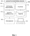
- FIG. 1 shows a tissue processing system 100 for optimizing tissue fixation using diffusion monitoring, according to an exemplary embodiment of the subject disclosure.
- System 100 comprises an acoustic monitoring device 102 communicatively coupled to a memory 110 for storing a plurality of processing modules or logical instructions that are executed by processor 105 coupled to computer 101.
- Acoustic monitoring device 102 may detect acoustic waves that have traveled through a tissue sample and may include one or more transmitters and one or more receivers. The tissue sample may be immersed in a liquid fixative while the transmitters and receivers communicate to detect time of flight (TOF) of acoustic waves.
- TOF time of flight
- Processing modules within memory 110 may include logical non-transitory computer-readable instructions for enabling processor 105 to perform operations including monitoring the diffusion of a fixative through a tissue sample, evaluating the speed of an acoustic wave traveling through the tissue sample based on time of flight, determining the time to reach at least a decay constant for each of the plurality of different types of tissue samples, selecting a standardized diffusion time, executing fixation protocols using the standardized diffusion time and a minimum fixation time, performing quality correlation for training purposes, storing standardized diffusion times and other results in a database, and other operations that potentially result in an output of quantitative or graphical results to a user operations computer 101. Consequently, although not shown in FIG. 1 , computer 101 may also include user input and output devices such as a keyboard, mouse, stylus, and a display / touchscreen.
- the measurements from the acoustic sensors in acoustic monitoring device 102 may be received by a diffusion monitoring module 111 to track the change of a ToF of acoustic signals through the tissue sample.
- the output ultrasound pulse thus accumulates a small transit time differential that increases as more fluid exchange occurs.
- Diffusion monitoring therefore includes dynamically tracking and quantifying the formalin diffusion until the tissue is at complete osmotic equilibrium and no more diffusion takes place.
- a fixation module 112 may execute a fixation protocol such as a standardized protocol as described herein.
- the modules include logic that is executed by processor 105.
- Logic refers to any information having the form of instruction signals and/or data that may be applied to affect the operation of a processor.
- Software is one example of such logic.
- processors are computer processors (processing units), microprocessors, digital signal processors, controllers and microcontrollers, etc.
- Logic may be formed from signals stored on a computer-readable medium such as memory 110 that, in an exemplary embodiment, may be a random access memory (RAM), read-only memories (ROM), erasable / electrically erasable programmable read-only memories (EPROMS/EEPROMS), flash memories, etc.
- RAM random access memory
- ROM read-only memories
- EPROMS/EEPROMS erasable programmable read-only memories
- flash memories etc.
- Logic may also comprise digital and/or analog hardware circuits, for example, hardware circuits comprising logical AND, OR, XOR, NAND, NOR, and other logical operations. Logic may be formed from combinations of software and hardware. On a network, logic may be programmed on a server, or a complex of servers. A particular logic unit is not limited to a single logical location on the network. Moreover, the modules need not be executed in any specific order. For instance, classification module 118 may be invoked during operation of training module 111, as well as during operation of CNN module 116. Each module may call another module when needed to be executed.
- FIG. 2 shows a method for determining an optimal time for diffusing a tissue sample, according to an exemplary embodiment of the subject disclosure.
- the method begins with formalin diffusion S201 of tissue samples that may be used to train the system. Upon soaking, a timer is started (S202) and the diffusion of the tissue sample is monitored (S203). A determination is made as to whether or not the tissue is sufficiently diffused (S203). This determination may be based on the correlation of formalin diffusion with a single exponential trend, where the time transient of the trend can be completely characterized by a decay constant.
- C is a constant offset in nanoseconds (ns)
- A is the amplitude of the decay in nanoseconds (ns)
- ⁇ is the decay constant in hours
- the signal amplitude represents the magnitude of the diffusion and is thus directly proportional the amount of fluid exchange.
- ToF diffusion trends may be fitted to the above equation using non-linear regression.
- scanning capabilities of the acoustic system enable calculating and tracking which spatial volume of the tissue sample diffuses the slowest. This decay constant represents the limiting factor for when the tissue is thoroughly diffused and is referred to as the slowest decay constant ( ⁇ slowest ).
- the method simply waits while continuing measurement (S204). Once the decay constant is reached, there is sufficient formaldehyde inside the tissue to guarantee quality staining, and the timer may be stopped (S205) and the time may be recorded in a database record associated with the specific tissue type and other details in database 214.
- the timer may be stopped (S205) and the time may be recorded in a database record associated with the specific tissue type and other details in database 214.
- t done ⁇ slowest 5 hour 4.5 hour ⁇ t done ⁇ ⁇ slowest
- this protocol may be able to better predict when the sample is optimally diffused based on analysis to predict when the sample is most properly stained, or record several ToF diffusion curves and use them to predict optimal fixation time depending on the sample.
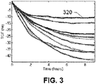
- FIG. 3 An exemplary ToF diffusion curve is shown in FIG. 3 .
- the diffusion is tracked at every position, and the region with the longest decay constant is correlated with an optimal result or an existing staining result.
- FIG. 3 shows ToF traces 320 generated from a human tonsil sample that was cold soaked in 10% NBF. Each curve represents a signal from a different spatial location within the tissue with a 1 mm shift for each reading.
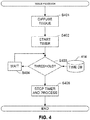
- FIG. 4 shows a method for optimizing tissue fixation, according to exemplary embodiments of the subject disclosure.
- a tissue processing system may be equipped with acoustic sensors on board for real-time monitoring of each tissue sample as it is provided into the system.
- the method begins with formalin diffusion (S401) of a tissue sample.
- a timer is started (S402) and the diffusion of the tissue sample is monitored for a threshold diffusion (S403).
- S403 threshold diffusion
- a determination is made as to whether or not the tissue is sufficiently diffused (S403). This may be based on one or more threshold diffusion constants stored in time database 414.
- a threshold may either include a decay constant threshold or a time period threshold.
- the method simply waits while continuing measurement (S404). Once the threshold time or decay constant is reached, there is sufficient formaldehyde inside the tissue to guarantee quality staining, and the timer may be stopped (S405) and the tissue is processed, by switching to heated formalin for a specified minimum time period, e.g. 1 hour.
- the minimum time period may be any time period sufficient for fixation to take place while the tissue is subject to heat to increase crosslinking kinetics. Any other process may be subsequently performed.
- Applicants have conducted a series of experiments based on histological staining results and monitored diffusion, resulting in a rule set for how much diffusion is needed across a variety of tissue types to guarantee quality staining. Diffusion of formalin into the tissue samples was dynamically monitored to determine how much crosslinking agent is required to produce ideal staining from downstream assays throughout the sample. A sample set of human tonsils was scanned using acoustic sensors and subsequently stained for morphology to calibrate the process.
- the acoustic sensors may include pairs of 4 MHz focused transducers (TA0040104-10, CNIRHurricane Tech (Shenzhen) Co., Ltd.) that are spatially aligned such that a tissue sample may be placed at their common foci.
- Samples of tonsil tissues of precise sizes were obtained by using tissue punches of either 4 or 6 mm in diameter.
- 6mm tonsil cores were placed into 10% NBF (Saturated aqueous formaldehyde from Fisher Scientific, Houston, TX, buffered to pH 6.8-7.2 with 100 mM phosphate buffer) previously chilled to 4°C for either 3 or 5 hours.
- NBF saturated aqueous formaldehyde from Fisher Scientific, Houston, TX, buffered to pH 6.8-7.2 with 100 mM phosphate buffer
- tissue processor may be a commercial dip-and-dunk tissue processor such as the Lynx II manufactured by Electron Microscopy Sciences (RTM) that may be modified to include the acoustic sensor assembly.
- RTM Electron Microscopy Sciences
- a mechanical head was designed using Solidworks® software to fit around and seal a standard reagent canister. Once sealed, an external vacuum system would initiate to degas the bulk reagent as well as the contents of the cassette, including the tissue.
- a cassette holder was designed for use with a standard sized histological cassette that securely held the tissue to prevent the sample from slipping during the experiment. Subsequent to fixation, 6 tonsil cores from each run were sectioned length wise and embedded cut side down in the mold. This multiblock arrangement allows for each of the 6 cores to be stained simultaneously. Good histomorphology was observed with these samples.
- FIG. 5 shows an optimized protocol for tissue fixation, according to an exemplary embodiment of the subject disclosure.
- This study indicates a protocol of 6 hours in cold formalin (S501) can standardize and optimize tissue processing across all types of tissues, up to 7 mm thick, and guarantee ideal biomarker and morphological structure preservation with a rapid protocol compared to standard room temperature fixation.
- the diffusion step (S501) is performed in a fixative solution that is either below 20 °C, below 15 °C, below 12 °C, below 10 °C, in the range of 0 °C to 10 °C, in the range of 0 °C to 12 °C, in the range of 0 °C to 15 °C, in the range of 2 °C to 10 °C, in the range of 2 °C to 12 °C, in the range of 2 °C to 15 °C, in the range of 5 °C to 10 °C, in the range of 5 °C to 12 °C, or in the range of 5 °C to 15 °C.
- the cold temperatures used in the diffusion step have the dual benefits of increasing the rate of diffusion by inhibiting excessive cross-linking at the periphery of the tissue (which inhibits diffusion of the fixative into the tissue) and of reducing enzymatic activity in the tissue, thereby more accurately preserving molecular details of the tissue (such as phosphorylated proteins).
- the cross-linking step (S502) is performed at a higher temperature (e.g. from 20 °C to 55 °C) to accelerate the rate of cross-linking induced by the aldehyde-based fixative.
- a higher temperature e.g. from 20 °C to 55 °C
- the combination of cold and hot steps helps significantly reduce the processing time required for complete fixation of the tissues.
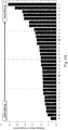
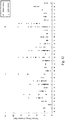
- FIGS. 6A-C show determinations of a time-of-fixation and corresponding decay constants for various tissue samples, according to an exemplary embodiment of the subject disclosure.
- a total of 236 samples were monitored representing 33 distinct organs and types of biological tissue.
- the average decay constant from each respective organ type is displayed, sorted from lowest to highest decay constant. Displayed in this fashion, more quickly diffusing tissues register to the left (i.e. smaller decay constants) and slowly diffusing tissues register to the right (i.e. larger decay constants).
- ASCO/CAP American Society of Clinical Oncology and the College of American Pathologists
- t done ⁇ slowest 5 hour 4.5 hour ⁇ t done ⁇ ⁇ slowest
- FIG. 6C depicts the cumulative density function (CDF) that is the integral of FIG. 6B . From the CDF, it is observed that 52.5% of samples have a slowest decay constant of less than 2 hours. Similarly, 84.8% of samples have decay constants less than 4 hours and 92.8% are less than 6 hours. From the tonsil-based experiments and the empirically-determined time equation, the data suggests that tissue samples need to be in cold formalin for an amount of time approximately equal to their slowest decay constant. Therefore, FIG. 6C would predict that nearly 93% of samples cold soaked in 10% NBF for 6 hours would stain pristinely with downstream assays.
- CDF cumulative density function
- This estimate of needing one decay constant of cold diffusion time is based on perfect staining throughout that sample, and is substantially more stringent than what is considered a diagnosable slide in current medical practice. Additionally, the rule set depicted in FIG. 5 was determined based on the amount of formalin in the slowest diffusing part of the tissue, meaning at threshold the majority of the tissue already has more than the critical amount of formalin needed for ideal staining. It therefore stands to reason that the remaining 7% of samples that are not at threshold after 6 hours of cold diffusion will still stain adequately enough for a clinician to make a diagnosis.
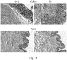
- FIGS. 7A-7D show stain quality comparisons between tissue samples stained using the disclosed methods versus the prior art, according to an exemplary embodiment of the subject disclosure.
- FIGS. 7A and 7B show 6mm colon tissue samples fixed with a static 6+1 ( FIG. 7A ) and as comparison, a 24 hr room temperature protocol ( FIG. 7B ) and analyzed for quality of histomorphology.
- FIGS. 7B and 7C show 6mm skin tissue samples fixed with 6+1 ( FIG. 7C ) and 24hr ( FIG. 7D ) protocols. All samples fixed with the 6+1 protocol had identical histomorphology compared to the same samples fixed with gold standard methods. This verifies that an analytical technique, based on time of flight principles, can be used to monitor diffusion of NBF in real time and optimize results.
- tissue samples were collected from surgeries under a waiver of consent, using procedures approved by the University of Washington Institutional Review Board. Upon excision, fresh tissue was carried to the pathology laboratory, generally within 30-60 minutes, and after the diagnostic pathologist had taken sections needed for diagnosis, 6mm cores were taken for further experimentation. For comparison of histomorphology between tissue fixed with experimental conditions and tissue generated by pathology department histotechnologists in a CLIA- and College of American Pathologists (CAP)-certified laboratory, unstained slides from the clinical tissue block generated in each case (10-48 hours RT formalin fixation) were collected.
- CAP College of American Pathologists
- Pairs of 4 MHz focused transducers (TA0040104-10, CNIRHurricane Tech (Shenzhen) Co., Ltd.) were spatially aligned and a sample was placed at their common foci.
- One transducer designated the transmitter, sends out an acoustic pulse that traverses the coupling fluid (i.e. formalin) and tissue and is detected by the receiving transducer.
- the transmitting transducer was programmed with a waveform generator (AD5930, Analog Devices) to transmit a sinusoidal wave for several hundred microseconds. That pulse train was then detected by the receiving transducer after traversing the fluid and tissue.
- the received ultrasound sinusoid and the transmitted sinusoid were compared electronically with a digital phase comparator (AD8302, Analog Devices).
- the output of the phase comparator yielded a valid reading during the region of temporal overlap between the transmitted and received pulses.
- the output of the phase comparator was allowed to stabilize before the output was queried with an integrated analog to digital converter on the microcontroller (ATmega2560, Atmel).
- the process was then repeated at multiple acoustic frequencies across the bandwidth of the transducer to build up the phase relationship between the input and output sinusoids across a frequency range. This acoustic phase-frequency sweep was then directly used to calculate the TOF using a post-processing algorithm analogous to acoustic interferometry and capable of detecting transit times with subnanosecond accuracy.
- the speed of sound in fluid has a large temperature dependence (e.g. ⁇ t water ⁇ 2.3ns/°C•mm at 4°C) that can greatly affect acoustic transit times especially because TOF is an integrated signal over the path length of the transducers.
- a large temperature dependence e.g. ⁇ t water ⁇ 2.3ns/°C•mm at 4°C
- TOF is an integrated signal over the path length of the transducers.
- the TOF was also calculated through only formalin and this acquisition, referred to as the reference channel, was used to compensate for spatiotemporal thermal gradients in the fluid.
- the reference compensation scheme worked best with relatively slow and low amplitude thermal transients in the fluid, so reagent temperature was precisely controlled using a developed pulse width modulation (PWM) scheme on the cooling hardware.
- PWM pulse width modulation
- the PWM temperature control used a proportional-integral-derivative (PID) based algorithm that regulate the temperature of the reagent tightly about the set point by making slight adjustments to the temperature in ⁇ 400 ⁇ s increments.
- PID proportional-integral-derivative
- the PWM algorithm was found to control the temperature of the fluid with a standard deviation of 0.05 °C about the set temperature. This precise temperature control used in conjunction with reference compensation virtually removed all environmental artifacts from the calculated signal. Unfiltered TOF traces had a typical noise value of less than 1.0 nanosecond.
- a commercial dip-and-dunk tissue processor ( Figure 8A , Lynx II, Electron Microscopy Sciences) was retrofit with custom developed acoustic hardware.
- a mechanical head was designed using Solidworks® software to fit around and seal a standard reagent canister. Once sealed, an external vacuum system would initiate to degas the bulk reagent as well as the contents of the cassette, including the tissue.
- a cassette holder was designed for use with a standard sized histological cassette ( Figure 8D , CellSafe 5, CellPath) that securely held the tissue to prevent the sample from slipping during the experiment. The cassette holder was attached to a vertical translation arm that would slide the cassette holder in one direction.
- the mechanical head was designed with two metal brackets on either side of the tissue cassette ( Figure 8B ).
- One bracket housed 5 transmitting transducers.
- the other bracket housed 5 receiving transducers that were spatially aligned with their respective transmitting transducers.
- the receiving bracket also housed a pair of transducers that were oriented orthogonal to the propagation axis of the other transducers ( Figure 8C ).
- This set of transducers served as a reference channel.
- the cassette was raised up and a second reference acquisition was acquired.
- the orthogonal reference sensors After each acquisition the orthogonal reference sensors would calculate a TOF value that was used to detect spatiotemporal variations in the fluid that had a profound effect on sound velocity.
- the cassette was then translated ⁇ 1mm vertically and the TOF value was calculated at the new position for all transducer pairs. The process was repeated to cover the entire cassette.
- the 2nd and 4th transducer pairs ( Figure 8C , bottom row) were turned off when scanning tissue in a standard sized cassette. This enabled the 1st, 3rd, and 5th transducer pairs ( Figure 8C , top row) to each scan one of the center three subdivisions of the cassette. Two tissue cores were then placed in each column, one on the top and one on the bottom.
- This setup enables TOF traces from 6 samples (2 rows X 3 columns) to simultaneously be obtained and significantly decreased run to run variation and increased throughput.
- the process was repeated over the course of the experiment for several hours until the tissue reached osmotic equilibrium and no more diffusion occurred producing a temporally flat TOF signal.
- One complete acquisition at all spatial locations takes about 90 seconds, although near real-time data acquisition is possible at one location ( ⁇ t ⁇ 1 second).
- the TOF in fluid is highly dependent on thermal fluctuations within the bulk media.
- the reference TOF value was subtracted from the TOF value obtain through the tissue and formalin to isolate the phase retardation from the tissue without spurious signals.
- a scaling factor was used to adjust for the slight geometrical difference in spacing between these two sensors and the pairs of scanning sensors.
- TOF x y t C x y + Ae ⁇ t / ⁇ x y
- C is a constant offset in ns
- A is the amplitude of the decay in ns
- ⁇ is the decay constant in hours
- the signal amplitude represents the magnitude of the diffusion and is thus directly proportional the amount of fluid exchange.
- the decay constant represents the time for the amplitude to decrease by 63% and is inversely proportional to the rate of formalin diffusion into the tissue (i.e.
- NBF Diffusion of NBF into tissue sections is controlled mainly by concentration of formaldehyde and time. Since NBF is a fixed concentration of formaldehyde (3.7% W/V), we reasoned there must be a minimum exposure time to cold NBF that produces excellent histomorphology.
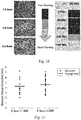
- a simple time course experiment was performed ( Figure 10 ) using 6 mm cores of human tonsil tissues submerged into 4 °C NBF followed by 1 hour in 45 °C NBF. We have previously determined that a shorter warm step can be standardized if sufficient formaldehyde is present in the sample. After multiple experiments were analyzed, a minimum of 3 hours of cold NBF (3+1) is required to produce good histomorphology. Tissue morphology was slightly better after 5 hours (5+1) but no additional benefit was seen at longer times with this one tissue type. Multiple cores were then examined using both 3+1 and 5+1 protocols as verification ( Figure 10 ).
- the average decay constant from each respective organ type is displayed in Figure 6A sorted from lowest to highest decay constant. Displayed in this fashion, more quickly diffusing tissues register to the left (i.e. smaller decay constants) and slowly diffusing tissues register to the right (i.e. larger decay constants).
- ASCO/CAP has recognized as needing extra time in formalin (e.g. breast, brain, fat) are located on the far right of the graph amongst the slowest quarter of tissues. Therefore, our TOF diffusion monitoring system has confirmed the ASCO/CAP guidelines of slowing diffusing tissues.
- the decay constants from the cumulative tissue dataset were analyzed to determine how long samples needed to be subjected to cold diffusion in 10% NBF to produce excellent downstream biomarker preservation.
- PDF probability density function
- the cumulative density function (CDF) is plotted in Figure 6C , which represents the integral of Figure 6B . From the CDF 52.5% of samples have a slowest decay constant of less than 2 hours. Similarly, 84.8% of samples have decay constants less than 4 hours and 92.8% are less than 6 hours. From the tonsil-based experiments and Eq. 2, the data suggests that tissue samples need to be in cold formalin for an amount of time approximately equal to their slowest decay constant. Therefore, Figure 6C would predict that nearly 93% of samples' cold soaked in 10% NBF for 6 hours would stain pristinely with downstream assays. Our estimate of needing one decay constant of cold diffusion time is based on perfect staining throughout that sample.
- This threshold is substantially more stringent that what is considered a diagnosable slide in current medical practice. Additionally, our ruleset was determined based on the amount of formalin in the slowest diffusing part of the tissue, meaning at threshold the majority of the tissue already has more than the critical amount of formalin needed for ideal staining. It therefore stands to reason that the remaining 7% of samples that are not at threshold after 6 hours of cold diffusion will still stain adequately enough for a clinician to make a diagnosis.
- the presented time needed in cold formalin is especially powerful because all of these factors are taken into account. Additionally, the large number of samples monitored in our study and the scanning capability of our system ensure 6 hours in cold formalin will ideally preserve tissue despite variability from different types of tissue (intersample variation) as well as contributions from tissue heterogeneity (intrasample variation). Moreover, because the slowest diffusing tissues (fat, brain, etc) were included in this study, we are confident other types of tissues not exclusively monitored will not be the limiting factor of a potential protocol. Thus, all samples up to 7 mm thick will stain properly after 6 hours in cold formalin.
- the present state-of-the-art in tissue processing and preservation is a one size fits all workflow that is profoundly unprepared for specimen handling in a personalized medicine workflow.
- This methodology cannot take into account sample specific variations in the concentration of formalin throughout the tissue, which is governed by the variable rate of formalin uptake by individual tissues.
- This study detailed a real-time diffusion monitoring system based on subnanosecond acoustic time-of-flight differences generated by the exchange of NBF and interstitial fluid during active reagent diffusion. Diffusion trends were empirically correlated with needed diffusion time, based on morphological staining results, in order to predict the needed amount of crosslinking agent necessary to guarantee ideal staining.
- Diffusion monitoring was then employed in a broad tissue collection study comprising over 200 individual samples and 33 different human organs. Results were coalesced and indicate that all tissue types, up to 7 mm thick, will produce diagnosable staining throughout the sample after 6 hours in cold 10% NBF and that the vast majority of samples ( ⁇ 93%) with stain ideally after this time in cold formalin. The observation that 6 hours of cold diffusion time would produce quality staining was then confirmed by staining several types of tissues processed with the 6+1 protocol. Overall, this research indicates a simple 6+1 protocol can standardize and optimize tissue processing across all types of tissues, up to 7 mm thick, and guarantee ideal biomarker and morphological structure preservation with a rapid protocol compared to standard room temperature fixation.
Landscapes
- Physics & Mathematics (AREA)
- Life Sciences & Earth Sciences (AREA)
- Health & Medical Sciences (AREA)
- General Physics & Mathematics (AREA)
- Pathology (AREA)
- Analytical Chemistry (AREA)
- Biochemistry (AREA)
- General Health & Medical Sciences (AREA)
- Chemical & Material Sciences (AREA)
- Immunology (AREA)
- Acoustics & Sound (AREA)
- Engineering & Computer Science (AREA)
- Biomedical Technology (AREA)
- Molecular Biology (AREA)
- Investigating Or Analysing Biological Materials (AREA)
- Sampling And Sample Adjustment (AREA)
- Investigating Or Analyzing Materials By The Use Of Ultrasonic Waves (AREA)
Priority Applications (1)
| Application Number | Priority Date | Filing Date | Title |
|---|---|---|---|
| EP21168155.6A EP3885762A1 (en) | 2015-02-09 | 2016-02-05 | Methods for standardizing diffusion of a fluid into tissues |
Applications Claiming Priority (2)
| Application Number | Priority Date | Filing Date | Title |
|---|---|---|---|
| US201562113787P | 2015-02-09 | 2015-02-09 | |
| PCT/EP2016/052447 WO2016128299A1 (en) | 2015-02-09 | 2016-02-05 | Materials and methods for standardizing diffusion of a fluid into tissues |
Related Child Applications (1)
| Application Number | Title | Priority Date | Filing Date |
|---|---|---|---|
| EP21168155.6A Division EP3885762A1 (en) | 2015-02-09 | 2016-02-05 | Methods for standardizing diffusion of a fluid into tissues |
Publications (2)
| Publication Number | Publication Date |
|---|---|
| EP3256844A1 EP3256844A1 (en) | 2017-12-20 |
| EP3256844B1 true EP3256844B1 (en) | 2021-04-14 |
Family
ID=55300525
Family Applications (2)
| Application Number | Title | Priority Date | Filing Date |
|---|---|---|---|
| EP16702956.0A Active EP3256844B1 (en) | 2015-02-09 | 2016-02-05 | System and method for standardizing diffusion of a fluid into tissues |
| EP21168155.6A Pending EP3885762A1 (en) | 2015-02-09 | 2016-02-05 | Methods for standardizing diffusion of a fluid into tissues |
Family Applications After (1)
| Application Number | Title | Priority Date | Filing Date |
|---|---|---|---|
| EP21168155.6A Pending EP3885762A1 (en) | 2015-02-09 | 2016-02-05 | Methods for standardizing diffusion of a fluid into tissues |
Country Status (6)
| Country | Link |
|---|---|
| US (2) | US11181506B2 (enExample) |
| EP (2) | EP3256844B1 (enExample) |
| JP (2) | JP6718457B2 (enExample) |
| AU (2) | AU2016218055B2 (enExample) |
| CA (2) | CA2969982C (enExample) |
| WO (1) | WO2016128299A1 (enExample) |
Families Citing this family (4)
| Publication number | Priority date | Publication date | Assignee | Title |
|---|---|---|---|---|
| EP3256844B1 (en) | 2015-02-09 | 2021-04-14 | Ventana Medical Systems, Inc. | System and method for standardizing diffusion of a fluid into tissues |
| WO2018119194A1 (en) | 2016-12-22 | 2018-06-28 | Ventana Medical Systems, Inc. | System and method for sample processing |
| US11460400B2 (en) | 2020-07-07 | 2022-10-04 | Sakura Finetek U.S.A., Inc. | Use of IR spectroscopy to evaluate penetration of reagents into biological specimen |
| JP7720412B2 (ja) * | 2021-05-13 | 2025-08-07 | ベンタナ メディカル システムズ, インコーポレイテッド | 組織固定時間のリアルタイム予測 |
Family Cites Families (17)
| Publication number | Priority date | Publication date | Assignee | Title |
|---|---|---|---|---|
| YU47190B (sh) * | 1988-02-19 | 1995-01-31 | Institut Za Opštu I Fizičku Hemiju | Uredjaj za neinvazivno akustičko ispitivanje elastičnosti mekih bioloških materijala |
| US5603325A (en) * | 1988-05-11 | 1997-02-18 | Lunar Corporation | Ultrasonic densitometer with width compensation |
| US5343863A (en) | 1988-05-11 | 1994-09-06 | Lunar Corporation | Ultrasonic densitometer device and method |
| WO2005031312A1 (en) | 2003-09-29 | 2005-04-07 | Vision Biosystems Limited | System and method for histological tissue specimen processing |
| US8679019B2 (en) * | 2007-12-03 | 2014-03-25 | Bone Index Finland Oy | Method for measuring of thicknesses of materials using an ultrasound technique |
| US20120270293A1 (en) * | 2009-12-07 | 2012-10-25 | Wei-Sing Chu | Standardization of tissue specimen preservation by ultrasound and temperature control |
| US10126216B2 (en) * | 2011-02-17 | 2018-11-13 | Ventana Medical Systems, Inc. | Method for tissue sample fixation |
| US10539487B2 (en) | 2010-03-04 | 2020-01-21 | Ventana Medical Systems, Inc. | Systems and methods for monitoring tissue sample processing |
| ES2759007T3 (es) * | 2010-03-04 | 2020-05-07 | Ventana Med Syst Inc | Sistema de procesamiento para procesar muestras usando energía acústica |
| EP2659269B1 (en) | 2010-12-23 | 2016-10-26 | Roche Diagniostics GmbH | Detection of a posttranslationally modified polypeptide by a bi-valent binding agent |
| CA2826770C (en) * | 2011-02-15 | 2020-06-23 | Hemosonics, Llc | Characterization of blood hemostasis and oxygen transport parameters |
| IN2014DN09073A (enExample) | 2012-08-09 | 2015-05-22 | Univ Leland Stanford Junior | |
| WO2016097164A2 (en) * | 2014-12-17 | 2016-06-23 | Ventana Medical Systems, Inc. | Accurately calculating acoustic time-of-flight |
| ES2825598T3 (es) * | 2014-12-17 | 2021-05-17 | Ventana Med Syst Inc | Protocolo de seguimiento de difusión para fijación de tejido optimizada |
| JP6632624B2 (ja) | 2014-12-17 | 2020-01-22 | ベンタナ メディカル システムズ, インコーポレイテッド | 真の拡散定数の取得 |
| EP3256844B1 (en) | 2015-02-09 | 2021-04-14 | Ventana Medical Systems, Inc. | System and method for standardizing diffusion of a fluid into tissues |
| WO2018119194A1 (en) * | 2016-12-22 | 2018-06-28 | Ventana Medical Systems, Inc. | System and method for sample processing |
-
2016
- 2016-02-05 EP EP16702956.0A patent/EP3256844B1/en active Active
- 2016-02-05 WO PCT/EP2016/052447 patent/WO2016128299A1/en not_active Ceased
- 2016-02-05 CA CA2969982A patent/CA2969982C/en active Active
- 2016-02-05 JP JP2017538206A patent/JP6718457B2/ja active Active
- 2016-02-05 AU AU2016218055A patent/AU2016218055B2/en active Active
- 2016-02-05 CA CA3159600A patent/CA3159600C/en active Active
- 2016-02-05 EP EP21168155.6A patent/EP3885762A1/en active Pending
-
2017
- 2017-08-08 US US15/672,213 patent/US11181506B2/en active Active
-
2019
- 2019-07-22 AU AU2019208145A patent/AU2019208145B2/en active Active
-
2020
- 2020-03-19 JP JP2020048889A patent/JP6909892B2/ja active Active
-
2021
- 2021-10-19 US US17/505,095 patent/US12360079B2/en active Active
Non-Patent Citations (1)
| Title |
|---|
| None * |
Also Published As
| Publication number | Publication date |
|---|---|
| US20170336363A1 (en) | 2017-11-23 |
| CA3159600A1 (en) | 2016-08-18 |
| CA2969982C (en) | 2022-08-02 |
| AU2019208145A1 (en) | 2019-08-08 |
| CA3159600C (en) | 2023-11-21 |
| EP3256844A1 (en) | 2017-12-20 |
| US20220034845A1 (en) | 2022-02-03 |
| JP2020109418A (ja) | 2020-07-16 |
| US12360079B2 (en) | 2025-07-15 |
| JP6718457B2 (ja) | 2020-07-08 |
| AU2016218055B2 (en) | 2019-05-16 |
| AU2016218055A1 (en) | 2017-06-29 |
| US11181506B2 (en) | 2021-11-23 |
| EP3885762A1 (en) | 2021-09-29 |
| AU2019208145B2 (en) | 2020-09-10 |
| JP2018509599A (ja) | 2018-04-05 |
| JP6909892B2 (ja) | 2021-07-28 |
| CA2969982A1 (en) | 2016-08-18 |
| WO2016128299A1 (en) | 2016-08-18 |
Similar Documents
| Publication | Publication Date | Title |
|---|---|---|
| US12360079B2 (en) | Standardizing diffusion of a fluid into tissue | |
| US20230243726A1 (en) | Diffusion monitoring protocol for optimized tissue fixation | |
| US12181387B2 (en) | System and method for sample preservation | |
| Lerch et al. | Precision medicine starts with preanalytics: Real-time assessment of tissue fixation quality by ultrasound time-of-flight analysis |
Legal Events
| Date | Code | Title | Description |
|---|---|---|---|
| STAA | Information on the status of an ep patent application or granted ep patent |
Free format text: STATUS: THE INTERNATIONAL PUBLICATION HAS BEEN MADE |
|
| PUAI | Public reference made under article 153(3) epc to a published international application that has entered the european phase |
Free format text: ORIGINAL CODE: 0009012 |
|
| STAA | Information on the status of an ep patent application or granted ep patent |
Free format text: STATUS: REQUEST FOR EXAMINATION WAS MADE |
|
| 17P | Request for examination filed |
Effective date: 20170911 |
|
| AK | Designated contracting states |
Kind code of ref document: A1 Designated state(s): AL AT BE BG CH CY CZ DE DK EE ES FI FR GB GR HR HU IE IS IT LI LT LU LV MC MK MT NL NO PL PT RO RS SE SI SK SM TR |
|
| AX | Request for extension of the european patent |
Extension state: BA ME |
|
| DAV | Request for validation of the european patent (deleted) | ||
| DAX | Request for extension of the european patent (deleted) | ||
| STAA | Information on the status of an ep patent application or granted ep patent |
Free format text: STATUS: EXAMINATION IS IN PROGRESS |
|
| 17Q | First examination report despatched |
Effective date: 20200429 |
|
| GRAP | Despatch of communication of intention to grant a patent |
Free format text: ORIGINAL CODE: EPIDOSNIGR1 |
|
| STAA | Information on the status of an ep patent application or granted ep patent |
Free format text: STATUS: GRANT OF PATENT IS INTENDED |
|
| INTG | Intention to grant announced |
Effective date: 20201127 |
|
| GRAS | Grant fee paid |
Free format text: ORIGINAL CODE: EPIDOSNIGR3 |
|
| GRAA | (expected) grant |
Free format text: ORIGINAL CODE: 0009210 |
|
| STAA | Information on the status of an ep patent application or granted ep patent |
Free format text: STATUS: THE PATENT HAS BEEN GRANTED |
|
| AK | Designated contracting states |
Kind code of ref document: B1 Designated state(s): AL AT BE BG CH CY CZ DE DK EE ES FI FR GB GR HR HU IE IS IT LI LT LU LV MC MK MT NL NO PL PT RO RS SE SI SK SM TR |
|
| REG | Reference to a national code |
Ref country code: GB Ref legal event code: FG4D |
|
| REG | Reference to a national code |
Ref country code: CH Ref legal event code: EP |
|
| REG | Reference to a national code |
Ref country code: DE Ref legal event code: R096 Ref document number: 602016056022 Country of ref document: DE |
|
| REG | Reference to a national code |
Ref country code: IE Ref legal event code: FG4D |
|
| REG | Reference to a national code |
Ref country code: AT Ref legal event code: REF Ref document number: 1382866 Country of ref document: AT Kind code of ref document: T Effective date: 20210515 |
|
| REG | Reference to a national code |
Ref country code: LT Ref legal event code: MG9D |
|
| REG | Reference to a national code |
Ref country code: AT Ref legal event code: MK05 Ref document number: 1382866 Country of ref document: AT Kind code of ref document: T Effective date: 20210414 |
|
| REG | Reference to a national code |
Ref country code: NL Ref legal event code: MP Effective date: 20210414 |
|
| PG25 | Lapsed in a contracting state [announced via postgrant information from national office to epo] |
Ref country code: AT Free format text: LAPSE BECAUSE OF FAILURE TO SUBMIT A TRANSLATION OF THE DESCRIPTION OR TO PAY THE FEE WITHIN THE PRESCRIBED TIME-LIMIT Effective date: 20210414 Ref country code: BG Free format text: LAPSE BECAUSE OF FAILURE TO SUBMIT A TRANSLATION OF THE DESCRIPTION OR TO PAY THE FEE WITHIN THE PRESCRIBED TIME-LIMIT Effective date: 20210714 Ref country code: HR Free format text: LAPSE BECAUSE OF FAILURE TO SUBMIT A TRANSLATION OF THE DESCRIPTION OR TO PAY THE FEE WITHIN THE PRESCRIBED TIME-LIMIT Effective date: 20210414 Ref country code: FI Free format text: LAPSE BECAUSE OF FAILURE TO SUBMIT A TRANSLATION OF THE DESCRIPTION OR TO PAY THE FEE WITHIN THE PRESCRIBED TIME-LIMIT Effective date: 20210414 Ref country code: NL Free format text: LAPSE BECAUSE OF FAILURE TO SUBMIT A TRANSLATION OF THE DESCRIPTION OR TO PAY THE FEE WITHIN THE PRESCRIBED TIME-LIMIT Effective date: 20210414 Ref country code: LT Free format text: LAPSE BECAUSE OF FAILURE TO SUBMIT A TRANSLATION OF THE DESCRIPTION OR TO PAY THE FEE WITHIN THE PRESCRIBED TIME-LIMIT Effective date: 20210414 |
|
| PG25 | Lapsed in a contracting state [announced via postgrant information from national office to epo] |
Ref country code: GR Free format text: LAPSE BECAUSE OF FAILURE TO SUBMIT A TRANSLATION OF THE DESCRIPTION OR TO PAY THE FEE WITHIN THE PRESCRIBED TIME-LIMIT Effective date: 20210715 Ref country code: IS Free format text: LAPSE BECAUSE OF FAILURE TO SUBMIT A TRANSLATION OF THE DESCRIPTION OR TO PAY THE FEE WITHIN THE PRESCRIBED TIME-LIMIT Effective date: 20210814 Ref country code: LV Free format text: LAPSE BECAUSE OF FAILURE TO SUBMIT A TRANSLATION OF THE DESCRIPTION OR TO PAY THE FEE WITHIN THE PRESCRIBED TIME-LIMIT Effective date: 20210414 Ref country code: RS Free format text: LAPSE BECAUSE OF FAILURE TO SUBMIT A TRANSLATION OF THE DESCRIPTION OR TO PAY THE FEE WITHIN THE PRESCRIBED TIME-LIMIT Effective date: 20210414 Ref country code: SE Free format text: LAPSE BECAUSE OF FAILURE TO SUBMIT A TRANSLATION OF THE DESCRIPTION OR TO PAY THE FEE WITHIN THE PRESCRIBED TIME-LIMIT Effective date: 20210414 Ref country code: PT Free format text: LAPSE BECAUSE OF FAILURE TO SUBMIT A TRANSLATION OF THE DESCRIPTION OR TO PAY THE FEE WITHIN THE PRESCRIBED TIME-LIMIT Effective date: 20210816 Ref country code: PL Free format text: LAPSE BECAUSE OF FAILURE TO SUBMIT A TRANSLATION OF THE DESCRIPTION OR TO PAY THE FEE WITHIN THE PRESCRIBED TIME-LIMIT Effective date: 20210414 Ref country code: NO Free format text: LAPSE BECAUSE OF FAILURE TO SUBMIT A TRANSLATION OF THE DESCRIPTION OR TO PAY THE FEE WITHIN THE PRESCRIBED TIME-LIMIT Effective date: 20210714 |
|
| REG | Reference to a national code |
Ref country code: DE Ref legal event code: R097 Ref document number: 602016056022 Country of ref document: DE |
|
| PG25 | Lapsed in a contracting state [announced via postgrant information from national office to epo] |
Ref country code: RO Free format text: LAPSE BECAUSE OF FAILURE TO SUBMIT A TRANSLATION OF THE DESCRIPTION OR TO PAY THE FEE WITHIN THE PRESCRIBED TIME-LIMIT Effective date: 20210414 Ref country code: ES Free format text: LAPSE BECAUSE OF FAILURE TO SUBMIT A TRANSLATION OF THE DESCRIPTION OR TO PAY THE FEE WITHIN THE PRESCRIBED TIME-LIMIT Effective date: 20210414 Ref country code: SM Free format text: LAPSE BECAUSE OF FAILURE TO SUBMIT A TRANSLATION OF THE DESCRIPTION OR TO PAY THE FEE WITHIN THE PRESCRIBED TIME-LIMIT Effective date: 20210414 Ref country code: SK Free format text: LAPSE BECAUSE OF FAILURE TO SUBMIT A TRANSLATION OF THE DESCRIPTION OR TO PAY THE FEE WITHIN THE PRESCRIBED TIME-LIMIT Effective date: 20210414 Ref country code: EE Free format text: LAPSE BECAUSE OF FAILURE TO SUBMIT A TRANSLATION OF THE DESCRIPTION OR TO PAY THE FEE WITHIN THE PRESCRIBED TIME-LIMIT Effective date: 20210414 Ref country code: CZ Free format text: LAPSE BECAUSE OF FAILURE TO SUBMIT A TRANSLATION OF THE DESCRIPTION OR TO PAY THE FEE WITHIN THE PRESCRIBED TIME-LIMIT Effective date: 20210414 Ref country code: DK Free format text: LAPSE BECAUSE OF FAILURE TO SUBMIT A TRANSLATION OF THE DESCRIPTION OR TO PAY THE FEE WITHIN THE PRESCRIBED TIME-LIMIT Effective date: 20210414 |
|
| PLBE | No opposition filed within time limit |
Free format text: ORIGINAL CODE: 0009261 |
|
| STAA | Information on the status of an ep patent application or granted ep patent |
Free format text: STATUS: NO OPPOSITION FILED WITHIN TIME LIMIT |
|
| 26N | No opposition filed |
Effective date: 20220117 |
|
| PG25 | Lapsed in a contracting state [announced via postgrant information from national office to epo] |
Ref country code: IS Free format text: LAPSE BECAUSE OF FAILURE TO SUBMIT A TRANSLATION OF THE DESCRIPTION OR TO PAY THE FEE WITHIN THE PRESCRIBED TIME-LIMIT Effective date: 20210814 Ref country code: AL Free format text: LAPSE BECAUSE OF FAILURE TO SUBMIT A TRANSLATION OF THE DESCRIPTION OR TO PAY THE FEE WITHIN THE PRESCRIBED TIME-LIMIT Effective date: 20210414 |
|
| PG25 | Lapsed in a contracting state [announced via postgrant information from national office to epo] |
Ref country code: IT Free format text: LAPSE BECAUSE OF FAILURE TO SUBMIT A TRANSLATION OF THE DESCRIPTION OR TO PAY THE FEE WITHIN THE PRESCRIBED TIME-LIMIT Effective date: 20210414 |
|
| PG25 | Lapsed in a contracting state [announced via postgrant information from national office to epo] |
Ref country code: MC Free format text: LAPSE BECAUSE OF FAILURE TO SUBMIT A TRANSLATION OF THE DESCRIPTION OR TO PAY THE FEE WITHIN THE PRESCRIBED TIME-LIMIT Effective date: 20210414 |
|
| REG | Reference to a national code |
Ref country code: BE Ref legal event code: MM Effective date: 20220228 |
|
| PG25 | Lapsed in a contracting state [announced via postgrant information from national office to epo] |
Ref country code: LU Free format text: LAPSE BECAUSE OF NON-PAYMENT OF DUE FEES Effective date: 20220205 |
|
| PG25 | Lapsed in a contracting state [announced via postgrant information from national office to epo] |
Ref country code: IE Free format text: LAPSE BECAUSE OF NON-PAYMENT OF DUE FEES Effective date: 20220205 |
|
| PG25 | Lapsed in a contracting state [announced via postgrant information from national office to epo] |
Ref country code: BE Free format text: LAPSE BECAUSE OF NON-PAYMENT OF DUE FEES Effective date: 20220228 |
|
| REG | Reference to a national code |
Ref country code: DE Ref legal event code: R082 Ref document number: 602016056022 Country of ref document: DE Representative=s name: KRAUS & LEDERER PARTGMBB, DE |
|
| PG25 | Lapsed in a contracting state [announced via postgrant information from national office to epo] |
Ref country code: HU Free format text: LAPSE BECAUSE OF FAILURE TO SUBMIT A TRANSLATION OF THE DESCRIPTION OR TO PAY THE FEE WITHIN THE PRESCRIBED TIME-LIMIT; INVALID AB INITIO Effective date: 20160205 |
|
| PG25 | Lapsed in a contracting state [announced via postgrant information from national office to epo] |
Ref country code: MK Free format text: LAPSE BECAUSE OF FAILURE TO SUBMIT A TRANSLATION OF THE DESCRIPTION OR TO PAY THE FEE WITHIN THE PRESCRIBED TIME-LIMIT Effective date: 20210414 Ref country code: CY Free format text: LAPSE BECAUSE OF FAILURE TO SUBMIT A TRANSLATION OF THE DESCRIPTION OR TO PAY THE FEE WITHIN THE PRESCRIBED TIME-LIMIT Effective date: 20210414 |
|
| PG25 | Lapsed in a contracting state [announced via postgrant information from national office to epo] |
Ref country code: MT Free format text: LAPSE BECAUSE OF FAILURE TO SUBMIT A TRANSLATION OF THE DESCRIPTION OR TO PAY THE FEE WITHIN THE PRESCRIBED TIME-LIMIT Effective date: 20210414 |
|
| PG25 | Lapsed in a contracting state [announced via postgrant information from national office to epo] |
Ref country code: TR Free format text: LAPSE BECAUSE OF FAILURE TO SUBMIT A TRANSLATION OF THE DESCRIPTION OR TO PAY THE FEE WITHIN THE PRESCRIBED TIME-LIMIT Effective date: 20210414 |
|
| REG | Reference to a national code |
Ref country code: CH Ref legal event code: U11 Free format text: ST27 STATUS EVENT CODE: U-0-0-U10-U11 (AS PROVIDED BY THE NATIONAL OFFICE) Effective date: 20260301 |
|
| PGFP | Annual fee paid to national office [announced via postgrant information from national office to epo] |
Ref country code: GB Payment date: 20260121 Year of fee payment: 11 |
|
| PGFP | Annual fee paid to national office [announced via postgrant information from national office to epo] |
Ref country code: DE Payment date: 20260121 Year of fee payment: 11 |
|
| PGFP | Annual fee paid to national office [announced via postgrant information from national office to epo] |
Ref country code: FR Payment date: 20260121 Year of fee payment: 11 |
|
| PGFP | Annual fee paid to national office [announced via postgrant information from national office to epo] |
Ref country code: CH Payment date: 20260301 Year of fee payment: 11 |