EP3256569B2 - Alveolarartige makrophagen und verfahren zur erzeugung davon - Google Patents
Alveolarartige makrophagen und verfahren zur erzeugung davon Download PDFInfo
- Publication number
- EP3256569B2 EP3256569B2 EP16748520.0A EP16748520A EP3256569B2 EP 3256569 B2 EP3256569 B2 EP 3256569B2 EP 16748520 A EP16748520 A EP 16748520A EP 3256569 B2 EP3256569 B2 EP 3256569B2
- Authority
- EP
- European Patent Office
- Prior art keywords
- macrophages
- alveolar
- cells
- psc
- csf
- Prior art date
- Legal status (The legal status is an assumption and is not a legal conclusion. Google has not performed a legal analysis and makes no representation as to the accuracy of the status listed.)
- Active
Links
- 210000002540 macrophage Anatomy 0.000 title claims description 170
- 238000000034 method Methods 0.000 title claims description 32
- 210000004027 cell Anatomy 0.000 claims description 166
- 210000001132 alveolar macrophage Anatomy 0.000 claims description 74
- 230000004069 differentiation Effects 0.000 claims description 60
- 210000001778 pluripotent stem cell Anatomy 0.000 claims description 56
- 108010017213 Granulocyte-Macrophage Colony-Stimulating Factor Proteins 0.000 claims description 47
- 102100039620 Granulocyte-macrophage colony-stimulating factor Human genes 0.000 claims description 47
- 102000007651 Macrophage Colony-Stimulating Factor Human genes 0.000 claims description 46
- 108010046938 Macrophage Colony-Stimulating Factor Proteins 0.000 claims description 46
- 102100022297 Integrin alpha-X Human genes 0.000 claims description 39
- 210000003566 hemangioblast Anatomy 0.000 claims description 36
- 108010073929 Vascular Endothelial Growth Factor A Proteins 0.000 claims description 35
- 102000005789 Vascular Endothelial Growth Factors Human genes 0.000 claims description 35
- 108010019530 Vascular Endothelial Growth Factors Proteins 0.000 claims description 35
- 230000001939 inductive effect Effects 0.000 claims description 35
- 238000000338 in vitro Methods 0.000 claims description 33
- 108010002386 Interleukin-3 Proteins 0.000 claims description 32
- 229940076264 interleukin-3 Drugs 0.000 claims description 32
- 210000002242 embryoid body Anatomy 0.000 claims description 31
- 230000003394 haemopoietic effect Effects 0.000 claims description 28
- 102000004889 Interleukin-6 Human genes 0.000 claims description 23
- 108090001005 Interleukin-6 Proteins 0.000 claims description 23
- 229940100601 interleukin-6 Drugs 0.000 claims description 23
- 238000012258 culturing Methods 0.000 claims description 18
- 102100024505 Bone morphogenetic protein 4 Human genes 0.000 claims description 17
- 210000000130 stem cell Anatomy 0.000 claims description 17
- 208000019693 Lung disease Diseases 0.000 claims description 15
- 239000003550 marker Substances 0.000 claims description 15
- -1 Wnt3a Proteins 0.000 claims description 13
- 108010023082 activin A Proteins 0.000 claims description 13
- 101100477560 Mus musculus Siglec5 gene Proteins 0.000 claims description 12
- 108010022164 acetyl-LDL Proteins 0.000 claims description 11
- 210000003716 mesoderm Anatomy 0.000 claims description 11
- 239000003814 drug Substances 0.000 claims description 9
- 239000000203 mixture Substances 0.000 claims description 8
- 101000934372 Homo sapiens Macrosialin Proteins 0.000 claims description 7
- 102100025136 Macrosialin Human genes 0.000 claims description 7
- 229940124597 therapeutic agent Drugs 0.000 claims description 6
- 210000003690 classically activated macrophage Anatomy 0.000 claims description 5
- 102100026439 Adhesion G protein-coupled receptor E1 Human genes 0.000 claims description 4
- 101000718225 Homo sapiens Adhesion G protein-coupled receptor E1 Proteins 0.000 claims description 4
- 101000762379 Homo sapiens Bone morphogenetic protein 4 Proteins 0.000 claims description 3
- 102000000646 Interleukin-3 Human genes 0.000 claims 6
- 241000699670 Mus sp. Species 0.000 description 61
- 201000000742 primary sclerosing cholangitis Diseases 0.000 description 57
- 239000002609 medium Substances 0.000 description 48
- 230000014509 gene expression Effects 0.000 description 37
- 210000004072 lung Anatomy 0.000 description 36
- 102100039064 Interleukin-3 Human genes 0.000 description 26
- 241000699666 Mus <mouse, genus> Species 0.000 description 24
- 241001465754 Metazoa Species 0.000 description 23
- LOKCTEFSRHRXRJ-UHFFFAOYSA-I dipotassium trisodium dihydrogen phosphate hydrogen phosphate dichloride Chemical compound P(=O)(O)(O)[O-].[K+].P(=O)(O)([O-])[O-].[Na+].[Na+].[Cl-].[K+].[Cl-].[Na+] LOKCTEFSRHRXRJ-UHFFFAOYSA-I 0.000 description 20
- 239000002953 phosphate buffered saline Substances 0.000 description 20
- 239000005090 green fluorescent protein Substances 0.000 description 19
- 101001046686 Homo sapiens Integrin alpha-M Proteins 0.000 description 18
- 102100022338 Integrin alpha-M Human genes 0.000 description 18
- 108090000623 proteins and genes Proteins 0.000 description 18
- 239000003102 growth factor Substances 0.000 description 16
- 101710169336 5'-deoxyadenosine deaminase Proteins 0.000 description 15
- 102000055025 Adenosine deaminases Human genes 0.000 description 15
- 108010049955 Bone Morphogenetic Protein 4 Proteins 0.000 description 15
- ACSIXWWBWUQEHA-UHFFFAOYSA-N clodronic acid Chemical compound OP(O)(=O)C(Cl)(Cl)P(O)(O)=O ACSIXWWBWUQEHA-UHFFFAOYSA-N 0.000 description 15
- 229960002286 clodronic acid Drugs 0.000 description 15
- FAPWRFPIFSIZLT-UHFFFAOYSA-M Sodium chloride Chemical compound [Na+].[Cl-] FAPWRFPIFSIZLT-UHFFFAOYSA-M 0.000 description 14
- 238000000684 flow cytometry Methods 0.000 description 14
- 238000001727 in vivo Methods 0.000 description 14
- 210000004979 bone marrow derived macrophage Anatomy 0.000 description 12
- 239000003446 ligand Substances 0.000 description 12
- 230000000670 limiting effect Effects 0.000 description 12
- UCSJYZPVAKXKNQ-HZYVHMACSA-N streptomycin Chemical compound CN[C@H]1[C@H](O)[C@@H](O)[C@H](CO)O[C@H]1O[C@@H]1[C@](C=O)(O)[C@H](C)O[C@H]1O[C@@H]1[C@@H](NC(N)=N)[C@H](O)[C@@H](NC(N)=N)[C@H](O)[C@H]1O UCSJYZPVAKXKNQ-HZYVHMACSA-N 0.000 description 12
- 230000004083 survival effect Effects 0.000 description 11
- 241000894006 Bacteria Species 0.000 description 10
- 230000000242 pagocytic effect Effects 0.000 description 10
- 238000011282 treatment Methods 0.000 description 10
- 101000738771 Homo sapiens Receptor-type tyrosine-protein phosphatase C Proteins 0.000 description 9
- 108090000174 Interleukin-10 Proteins 0.000 description 9
- 102100037422 Receptor-type tyrosine-protein phosphatase C Human genes 0.000 description 9
- 239000011324 bead Substances 0.000 description 9
- 210000004369 blood Anatomy 0.000 description 9
- 239000008280 blood Substances 0.000 description 9
- 210000002798 bone marrow cell Anatomy 0.000 description 9
- 230000004186 co-expression Effects 0.000 description 9
- 239000000243 solution Substances 0.000 description 9
- 210000001519 tissue Anatomy 0.000 description 9
- 206010057249 Phagocytosis Diseases 0.000 description 8
- 108010021843 fluorescent protein 583 Proteins 0.000 description 8
- 230000008782 phagocytosis Effects 0.000 description 8
- 230000002685 pulmonary effect Effects 0.000 description 8
- 239000011780 sodium chloride Substances 0.000 description 8
- 102000003974 Fibroblast growth factor 2 Human genes 0.000 description 7
- 108090000379 Fibroblast growth factor 2 Proteins 0.000 description 7
- 102100020715 Fms-related tyrosine kinase 3 ligand protein Human genes 0.000 description 7
- 101710162577 Fms-related tyrosine kinase 3 ligand protein Proteins 0.000 description 7
- 108090000723 Insulin-Like Growth Factor I Proteins 0.000 description 7
- 102000014429 Insulin-like growth factor Human genes 0.000 description 7
- 102000036693 Thrombopoietin Human genes 0.000 description 7
- 108010041111 Thrombopoietin Proteins 0.000 description 7
- 108010053099 Vascular Endothelial Growth Factor Receptor-2 Proteins 0.000 description 7
- 208000027418 Wounds and injury Diseases 0.000 description 7
- QVGXLLKOCUKJST-UHFFFAOYSA-N atomic oxygen Chemical compound [O] QVGXLLKOCUKJST-UHFFFAOYSA-N 0.000 description 7
- 238000002474 experimental method Methods 0.000 description 7
- 210000003958 hematopoietic stem cell Anatomy 0.000 description 7
- 238000011534 incubation Methods 0.000 description 7
- 208000014674 injury Diseases 0.000 description 7
- 239000002502 liposome Substances 0.000 description 7
- 239000000463 material Substances 0.000 description 7
- 239000001301 oxygen Substances 0.000 description 7
- 229910052760 oxygen Inorganic materials 0.000 description 7
- 238000002054 transplantation Methods 0.000 description 7
- 102000004127 Cytokines Human genes 0.000 description 6
- 108090000695 Cytokines Proteins 0.000 description 6
- 102000003814 Interleukin-10 Human genes 0.000 description 6
- 102100032010 Lipolysis-stimulated lipoprotein receptor Human genes 0.000 description 6
- 101710158772 Lipolysis-stimulated lipoprotein receptor Proteins 0.000 description 6
- 229930182555 Penicillin Natural products 0.000 description 6
- JGSARLDLIJGVTE-MBNYWOFBSA-N Penicillin G Chemical compound N([C@H]1[C@H]2SC([C@@H](N2C1=O)C(O)=O)(C)C)C(=O)CC1=CC=CC=C1 JGSARLDLIJGVTE-MBNYWOFBSA-N 0.000 description 6
- 241000700159 Rattus Species 0.000 description 6
- 230000015572 biosynthetic process Effects 0.000 description 6
- 238000004113 cell culture Methods 0.000 description 6
- 230000006378 damage Effects 0.000 description 6
- 229940049954 penicillin Drugs 0.000 description 6
- 229960005322 streptomycin Drugs 0.000 description 6
- 238000001890 transfection Methods 0.000 description 6
- 241000124008 Mammalia Species 0.000 description 5
- 241001529936 Murinae Species 0.000 description 5
- 230000001640 apoptogenic effect Effects 0.000 description 5
- 230000024245 cell differentiation Effects 0.000 description 5
- 230000001143 conditioned effect Effects 0.000 description 5
- 238000005516 engineering process Methods 0.000 description 5
- 230000001605 fetal effect Effects 0.000 description 5
- 230000006698 induction Effects 0.000 description 5
- 210000000440 neutrophil Anatomy 0.000 description 5
- 239000000047 product Substances 0.000 description 5
- 102000004169 proteins and genes Human genes 0.000 description 5
- 238000011084 recovery Methods 0.000 description 5
- 239000013598 vector Substances 0.000 description 5
- CIWBSHSKHKDKBQ-JLAZNSOCSA-N Ascorbic acid Chemical compound OC[C@H](O)[C@H]1OC(=O)C(O)=C1O CIWBSHSKHKDKBQ-JLAZNSOCSA-N 0.000 description 4
- 108010007127 Pulmonary Surfactant-Associated Protein D Proteins 0.000 description 4
- 102100027845 Pulmonary surfactant-associated protein D Human genes 0.000 description 4
- 102100033177 Vascular endothelial growth factor receptor 2 Human genes 0.000 description 4
- 206010069351 acute lung injury Diseases 0.000 description 4
- 239000003795 chemical substances by application Substances 0.000 description 4
- 210000004443 dendritic cell Anatomy 0.000 description 4
- 230000000694 effects Effects 0.000 description 4
- 238000007667 floating Methods 0.000 description 4
- 210000001654 germ layer Anatomy 0.000 description 4
- 208000012223 hereditary pulmonary alveolar proteinosis Diseases 0.000 description 4
- 208000015181 infectious disease Diseases 0.000 description 4
- 210000001616 monocyte Anatomy 0.000 description 4
- 239000002243 precursor Substances 0.000 description 4
- 230000008929 regeneration Effects 0.000 description 4
- 238000011069 regeneration method Methods 0.000 description 4
- 210000003437 trachea Anatomy 0.000 description 4
- 238000012546 transfer Methods 0.000 description 4
- 230000003612 virological effect Effects 0.000 description 4
- XLYOFNOQVPJJNP-UHFFFAOYSA-N water Chemical compound O XLYOFNOQVPJJNP-UHFFFAOYSA-N 0.000 description 4
- OZFAFGSSMRRTDW-UHFFFAOYSA-N (2,4-dichlorophenyl) benzenesulfonate Chemical compound ClC1=CC(Cl)=CC=C1OS(=O)(=O)C1=CC=CC=C1 OZFAFGSSMRRTDW-UHFFFAOYSA-N 0.000 description 3
- UZOVYGYOLBIAJR-UHFFFAOYSA-N 4-isocyanato-4'-methyldiphenylmethane Chemical compound C1=CC(C)=CC=C1CC1=CC=C(N=C=O)C=C1 UZOVYGYOLBIAJR-UHFFFAOYSA-N 0.000 description 3
- 108010059616 Activins Proteins 0.000 description 3
- 239000012583 B-27 Supplement Substances 0.000 description 3
- 239000012591 Dulbecco’s Phosphate Buffered Saline Substances 0.000 description 3
- 239000006144 Dulbecco’s modified Eagle's medium Substances 0.000 description 3
- 239000012981 Hank's balanced salt solution Substances 0.000 description 3
- 101000576894 Homo sapiens Macrophage mannose receptor 1 Proteins 0.000 description 3
- 101000914484 Homo sapiens T-lymphocyte activation antigen CD80 Proteins 0.000 description 3
- 101000863873 Homo sapiens Tyrosine-protein phosphatase non-receptor type substrate 1 Proteins 0.000 description 3
- 102100026818 Inhibin beta E chain Human genes 0.000 description 3
- ZDXPYRJPNDTMRX-VKHMYHEASA-N L-glutamine Chemical compound OC(=O)[C@@H](N)CCC(N)=O ZDXPYRJPNDTMRX-VKHMYHEASA-N 0.000 description 3
- 229930182816 L-glutamine Natural products 0.000 description 3
- 208000004852 Lung Injury Diseases 0.000 description 3
- 102100025354 Macrophage mannose receptor 1 Human genes 0.000 description 3
- 239000012580 N-2 Supplement Substances 0.000 description 3
- 238000011529 RT qPCR Methods 0.000 description 3
- 241000283984 Rodentia Species 0.000 description 3
- 238000000692 Student's t-test Methods 0.000 description 3
- 102100027222 T-lymphocyte activation antigen CD80 Human genes 0.000 description 3
- 206010043276 Teratoma Diseases 0.000 description 3
- 102100027654 Transcription factor PU.1 Human genes 0.000 description 3
- 206010069363 Traumatic lung injury Diseases 0.000 description 3
- 102100029948 Tyrosine-protein phosphatase non-receptor type substrate 1 Human genes 0.000 description 3
- 102000016549 Vascular Endothelial Growth Factor Receptor-2 Human genes 0.000 description 3
- 239000000488 activin Substances 0.000 description 3
- 230000001464 adherent effect Effects 0.000 description 3
- 238000010171 animal model Methods 0.000 description 3
- 238000003556 assay Methods 0.000 description 3
- 230000001580 bacterial effect Effects 0.000 description 3
- 210000001185 bone marrow Anatomy 0.000 description 3
- 238000010322 bone marrow transplantation Methods 0.000 description 3
- 239000012530 fluid Substances 0.000 description 3
- 239000007789 gas Substances 0.000 description 3
- 238000003306 harvesting Methods 0.000 description 3
- 230000011132 hemopoiesis Effects 0.000 description 3
- 230000028993 immune response Effects 0.000 description 3
- 210000004263 induced pluripotent stem cell Anatomy 0.000 description 3
- 238000002955 isolation Methods 0.000 description 3
- 231100000515 lung injury Toxicity 0.000 description 3
- 238000012423 maintenance Methods 0.000 description 3
- PJUIMOJAAPLTRJ-UHFFFAOYSA-N monothioglycerol Chemical compound OCC(O)CS PJUIMOJAAPLTRJ-UHFFFAOYSA-N 0.000 description 3
- 239000002245 particle Substances 0.000 description 3
- 108010027841 pegademase bovine Proteins 0.000 description 3
- 108010008929 proto-oncogene protein Spi-1 Proteins 0.000 description 3
- 230000001172 regenerating effect Effects 0.000 description 3
- 230000002441 reversible effect Effects 0.000 description 3
- 238000012353 t test Methods 0.000 description 3
- 230000001225 therapeutic effect Effects 0.000 description 3
- 238000004627 transmission electron microscopy Methods 0.000 description 3
- 239000003981 vehicle Substances 0.000 description 3
- JKMHFZQWWAIEOD-UHFFFAOYSA-N 2-[4-(2-hydroxyethyl)piperazin-1-yl]ethanesulfonic acid Chemical compound OCC[NH+]1CCN(CCS([O-])(=O)=O)CC1 JKMHFZQWWAIEOD-UHFFFAOYSA-N 0.000 description 2
- KDCGOANMDULRCW-UHFFFAOYSA-N 7H-purine Chemical compound N1=CNC2=NC=NC2=C1 KDCGOANMDULRCW-UHFFFAOYSA-N 0.000 description 2
- CSCPPACGZOOCGX-UHFFFAOYSA-N Acetone Chemical class CC(C)=O CSCPPACGZOOCGX-UHFFFAOYSA-N 0.000 description 2
- 102100022712 Alpha-1-antitrypsin Human genes 0.000 description 2
- 206010001881 Alveolar proteinosis Diseases 0.000 description 2
- 102100028681 C-type lectin domain family 4 member K Human genes 0.000 description 2
- 101710183165 C-type lectin domain family 4 member K Proteins 0.000 description 2
- 108090000909 Collectins Proteins 0.000 description 2
- 102000004405 Collectins Human genes 0.000 description 2
- 206010010099 Combined immunodeficiency Diseases 0.000 description 2
- 102000004190 Enzymes Human genes 0.000 description 2
- 108090000790 Enzymes Proteins 0.000 description 2
- 108010037362 Extracellular Matrix Proteins Proteins 0.000 description 2
- 102000010834 Extracellular Matrix Proteins Human genes 0.000 description 2
- 238000012413 Fluorescence activated cell sorting analysis Methods 0.000 description 2
- WQZGKKKJIJFFOK-GASJEMHNSA-N Glucose Natural products OC[C@H]1OC(O)[C@H](O)[C@@H](O)[C@@H]1O WQZGKKKJIJFFOK-GASJEMHNSA-N 0.000 description 2
- 239000007995 HEPES buffer Substances 0.000 description 2
- 108010033040 Histones Proteins 0.000 description 2
- 241000713666 Lentivirus Species 0.000 description 2
- 206010058467 Lung neoplasm malignant Diseases 0.000 description 2
- 239000012124 Opti-MEM Substances 0.000 description 2
- 239000008118 PEG 6000 Substances 0.000 description 2
- 229920002584 Polyethylene Glycol 6000 Polymers 0.000 description 2
- 102100025368 Runt-related transcription factor 2 Human genes 0.000 description 2
- 208000010285 Ventilator-Induced Lung Injury Diseases 0.000 description 2
- 239000002671 adjuvant Substances 0.000 description 2
- 230000002411 adverse Effects 0.000 description 2
- SHGAZHPCJJPHSC-YCNIQYBTSA-N all-trans-retinoic acid Chemical compound OC(=O)\C=C(/C)\C=C\C=C(/C)\C=C\C1=C(C)CCCC1(C)C SHGAZHPCJJPHSC-YCNIQYBTSA-N 0.000 description 2
- 238000000540 analysis of variance Methods 0.000 description 2
- 239000004599 antimicrobial Substances 0.000 description 2
- 230000006907 apoptotic process Effects 0.000 description 2
- 235000010323 ascorbic acid Nutrition 0.000 description 2
- 229960005070 ascorbic acid Drugs 0.000 description 2
- 239000011668 ascorbic acid Substances 0.000 description 2
- 210000002469 basement membrane Anatomy 0.000 description 2
- 206010006475 bronchopulmonary dysplasia Diseases 0.000 description 2
- 239000007975 buffered saline Substances 0.000 description 2
- 150000001720 carbohydrates Chemical class 0.000 description 2
- 235000014633 carbohydrates Nutrition 0.000 description 2
- 210000001715 carotid artery Anatomy 0.000 description 2
- 239000002771 cell marker Substances 0.000 description 2
- 230000001684 chronic effect Effects 0.000 description 2
- 230000003750 conditioning effect Effects 0.000 description 2
- 238000004624 confocal microscopy Methods 0.000 description 2
- 230000001419 dependent effect Effects 0.000 description 2
- 238000001514 detection method Methods 0.000 description 2
- 239000008121 dextrose Substances 0.000 description 2
- 201000010099 disease Diseases 0.000 description 2
- 208000037265 diseases, disorders, signs and symptoms Diseases 0.000 description 2
- 238000010494 dissociation reaction Methods 0.000 description 2
- 230000005593 dissociations Effects 0.000 description 2
- 238000001493 electron microscopy Methods 0.000 description 2
- 230000013020 embryo development Effects 0.000 description 2
- 210000001671 embryonic stem cell Anatomy 0.000 description 2
- 210000001900 endoderm Anatomy 0.000 description 2
- 210000002889 endothelial cell Anatomy 0.000 description 2
- 229940088598 enzyme Drugs 0.000 description 2
- 238000001317 epifluorescence microscopy Methods 0.000 description 2
- 210000002919 epithelial cell Anatomy 0.000 description 2
- 210000000981 epithelium Anatomy 0.000 description 2
- 230000001747 exhibiting effect Effects 0.000 description 2
- 210000002744 extracellular matrix Anatomy 0.000 description 2
- 238000000799 fluorescence microscopy Methods 0.000 description 2
- 239000011521 glass Substances 0.000 description 2
- 230000012010 growth Effects 0.000 description 2
- 239000001963 growth medium Substances 0.000 description 2
- 230000001976 improved effect Effects 0.000 description 2
- 230000004941 influx Effects 0.000 description 2
- 201000005202 lung cancer Diseases 0.000 description 2
- 208000020816 lung neoplasm Diseases 0.000 description 2
- 238000000386 microscopy Methods 0.000 description 2
- 230000005012 migration Effects 0.000 description 2
- 238000013508 migration Methods 0.000 description 2
- 238000004806 packaging method and process Methods 0.000 description 2
- 230000007170 pathology Effects 0.000 description 2
- 239000008188 pellet Substances 0.000 description 2
- 230000000737 periodic effect Effects 0.000 description 2
- 210000001539 phagocyte Anatomy 0.000 description 2
- 230000010287 polarization Effects 0.000 description 2
- 230000002035 prolonged effect Effects 0.000 description 2
- RXWNCPJZOCPEPQ-NVWDDTSBSA-N puromycin Chemical compound C1=CC(OC)=CC=C1C[C@H](N)C(=O)N[C@H]1[C@@H](O)[C@H](N2C3=NC=NC(=C3N=C2)N(C)C)O[C@@H]1CO RXWNCPJZOCPEPQ-NVWDDTSBSA-N 0.000 description 2
- 229950010131 puromycin Drugs 0.000 description 2
- 230000005855 radiation Effects 0.000 description 2
- 238000003753 real-time PCR Methods 0.000 description 2
- 229930002330 retinoic acid Natural products 0.000 description 2
- 230000028327 secretion Effects 0.000 description 2
- 210000000952 spleen Anatomy 0.000 description 2
- 238000010186 staining Methods 0.000 description 2
- 239000000126 substance Substances 0.000 description 2
- 239000006228 supernatant Substances 0.000 description 2
- 239000000725 suspension Substances 0.000 description 2
- 230000002459 sustained effect Effects 0.000 description 2
- 230000008685 targeting Effects 0.000 description 2
- 238000012360 testing method Methods 0.000 description 2
- 230000017423 tissue regeneration Effects 0.000 description 2
- 231100000331 toxic Toxicity 0.000 description 2
- 230000002588 toxic effect Effects 0.000 description 2
- 239000012096 transfection reagent Substances 0.000 description 2
- 229960001727 tretinoin Drugs 0.000 description 2
- 230000003442 weekly effect Effects 0.000 description 2
- FWMNVWWHGCHHJJ-SKKKGAJSSA-N 4-amino-1-[(2r)-6-amino-2-[[(2r)-2-[[(2r)-2-[[(2r)-2-amino-3-phenylpropanoyl]amino]-3-phenylpropanoyl]amino]-4-methylpentanoyl]amino]hexanoyl]piperidine-4-carboxylic acid Chemical compound C([C@H](C(=O)N[C@H](CC(C)C)C(=O)N[C@H](CCCCN)C(=O)N1CCC(N)(CC1)C(O)=O)NC(=O)[C@H](N)CC=1C=CC=CC=1)C1=CC=CC=C1 FWMNVWWHGCHHJJ-SKKKGAJSSA-N 0.000 description 1
- 206010001052 Acute respiratory distress syndrome Diseases 0.000 description 1
- 208000000884 Airway Obstruction Diseases 0.000 description 1
- 229920003319 Araldite® Polymers 0.000 description 1
- 201000001178 Bacterial Pneumonia Diseases 0.000 description 1
- 108091003079 Bovine Serum Albumin Proteins 0.000 description 1
- 101150013553 CD40 gene Proteins 0.000 description 1
- AQGNHMOJWBZFQQ-UHFFFAOYSA-N CT 99021 Chemical compound CC1=CNC(C=2C(=NC(NCCNC=3N=CC(=CC=3)C#N)=NC=2)C=2C(=CC(Cl)=CC=2)Cl)=N1 AQGNHMOJWBZFQQ-UHFFFAOYSA-N 0.000 description 1
- 208000006545 Chronic Obstructive Pulmonary Disease Diseases 0.000 description 1
- KRKNYBCHXYNGOX-UHFFFAOYSA-K Citrate Chemical compound [O-]C(=O)CC(O)(CC([O-])=O)C([O-])=O KRKNYBCHXYNGOX-UHFFFAOYSA-K 0.000 description 1
- 108010024682 Core Binding Factor Alpha 1 Subunit Proteins 0.000 description 1
- 201000003883 Cystic fibrosis Diseases 0.000 description 1
- 239000012594 Earle’s Balanced Salt Solution Substances 0.000 description 1
- 102100023795 Elafin Human genes 0.000 description 1
- 108010015972 Elafin Proteins 0.000 description 1
- 206010016654 Fibrosis Diseases 0.000 description 1
- 238000000729 Fisher's exact test Methods 0.000 description 1
- 108010010803 Gelatin Proteins 0.000 description 1
- SXRSQZLOMIGNAQ-UHFFFAOYSA-N Glutaraldehyde Chemical compound O=CCCCC=O SXRSQZLOMIGNAQ-UHFFFAOYSA-N 0.000 description 1
- 108010043121 Green Fluorescent Proteins Proteins 0.000 description 1
- 102100031573 Hematopoietic progenitor cell antigen CD34 Human genes 0.000 description 1
- 229920000209 Hexadimethrine bromide Polymers 0.000 description 1
- 102100026122 High affinity immunoglobulin gamma Fc receptor I Human genes 0.000 description 1
- 101000823116 Homo sapiens Alpha-1-antitrypsin Proteins 0.000 description 1
- 101000777663 Homo sapiens Hematopoietic progenitor cell antigen CD34 Proteins 0.000 description 1
- 101000913074 Homo sapiens High affinity immunoglobulin gamma Fc receptor I Proteins 0.000 description 1
- 101000935040 Homo sapiens Integrin beta-2 Proteins 0.000 description 1
- 101001076408 Homo sapiens Interleukin-6 Proteins 0.000 description 1
- 101001010513 Homo sapiens Leukocyte elastase inhibitor Proteins 0.000 description 1
- 101000632467 Homo sapiens Pulmonary surfactant-associated protein D Proteins 0.000 description 1
- 101000808011 Homo sapiens Vascular endothelial growth factor A Proteins 0.000 description 1
- 206010021143 Hypoxia Diseases 0.000 description 1
- 206010061598 Immunodeficiency Diseases 0.000 description 1
- 238000012404 In vitro experiment Methods 0.000 description 1
- 208000026350 Inborn Genetic disease Diseases 0.000 description 1
- 206010061218 Inflammation Diseases 0.000 description 1
- YQEZLKZALYSWHR-UHFFFAOYSA-N Ketamine Chemical compound C=1C=CC=C(Cl)C=1C1(NC)CCCCC1=O YQEZLKZALYSWHR-UHFFFAOYSA-N 0.000 description 1
- 101000962498 Macropis fulvipes Macropin Proteins 0.000 description 1
- BAVYZALUXZFZLV-UHFFFAOYSA-N Methylamine Chemical compound NC BAVYZALUXZFZLV-UHFFFAOYSA-N 0.000 description 1
- 102000016943 Muramidase Human genes 0.000 description 1
- 108010014251 Muramidase Proteins 0.000 description 1
- 241000699660 Mus musculus Species 0.000 description 1
- 101000746372 Mus musculus Granulocyte-macrophage colony-stimulating factor Proteins 0.000 description 1
- 101100340718 Mus musculus Il10 gene Proteins 0.000 description 1
- 101001033276 Mus musculus Interleukin-3 Proteins 0.000 description 1
- 101000716728 Mus musculus Kit ligand Proteins 0.000 description 1
- 108010062010 N-Acetylmuramoyl-L-alanine Amidase Proteins 0.000 description 1
- 206010028980 Neoplasm Diseases 0.000 description 1
- 102000008299 Nitric Oxide Synthase Human genes 0.000 description 1
- 108010021487 Nitric Oxide Synthase Proteins 0.000 description 1
- 206010035737 Pneumonia viral Diseases 0.000 description 1
- 102100037051 Protein Wnt-3a Human genes 0.000 description 1
- 101710118749 Protein Wnt-3a Proteins 0.000 description 1
- 208000004756 Respiratory Insufficiency Diseases 0.000 description 1
- 101710102802 Runt-related transcription factor 2 Proteins 0.000 description 1
- 241000191967 Staphylococcus aureus Species 0.000 description 1
- 108091023040 Transcription factor Proteins 0.000 description 1
- 102000040945 Transcription factor Human genes 0.000 description 1
- 102000004338 Transferrin Human genes 0.000 description 1
- 108090000901 Transferrin Proteins 0.000 description 1
- 102100040245 Tumor necrosis factor receptor superfamily member 5 Human genes 0.000 description 1
- COQLPRJCUIATTQ-UHFFFAOYSA-N Uranyl acetate Chemical compound O.O.O=[U]=O.CC(O)=O.CC(O)=O COQLPRJCUIATTQ-UHFFFAOYSA-N 0.000 description 1
- 101001062354 Xenopus tropicalis Forkhead box protein A2 Proteins 0.000 description 1
- 210000000683 abdominal cavity Anatomy 0.000 description 1
- 230000002159 abnormal effect Effects 0.000 description 1
- 230000005856 abnormality Effects 0.000 description 1
- 201000009628 adenosine deaminase deficiency Diseases 0.000 description 1
- 210000004504 adult stem cell Anatomy 0.000 description 1
- 239000000443 aerosol Substances 0.000 description 1
- 238000004220 aggregation Methods 0.000 description 1
- 210000005058 airway cell Anatomy 0.000 description 1
- 108010050122 alpha 1-Antitrypsin Proteins 0.000 description 1
- 229940024142 alpha 1-antitrypsin Drugs 0.000 description 1
- 102000018568 alpha-Defensin Human genes 0.000 description 1
- 108050007802 alpha-defensin Proteins 0.000 description 1
- 210000002588 alveolar type II cell Anatomy 0.000 description 1
- 238000004458 analytical method Methods 0.000 description 1
- 239000002260 anti-inflammatory agent Substances 0.000 description 1
- 229940121363 anti-inflammatory agent Drugs 0.000 description 1
- 239000007864 aqueous solution Substances 0.000 description 1
- 230000001174 ascending effect Effects 0.000 description 1
- 208000006673 asthma Diseases 0.000 description 1
- 208000036556 autosomal recessive T cell-negative B cell-negative NK cell-negative due to adenosine deaminase deficiency severe combined immunodeficiency Diseases 0.000 description 1
- 239000003855 balanced salt solution Substances 0.000 description 1
- 210000004082 barrier epithelial cell Anatomy 0.000 description 1
- 230000009286 beneficial effect Effects 0.000 description 1
- 102000012265 beta-defensin Human genes 0.000 description 1
- 108050002883 beta-defensin Proteins 0.000 description 1
- 230000005540 biological transmission Effects 0.000 description 1
- HOQPTLCRWVZIQZ-UHFFFAOYSA-H bis[[2-(5-hydroxy-4,7-dioxo-1,3,2$l^{2}-dioxaplumbepan-5-yl)acetyl]oxy]lead Chemical compound [Pb+2].[Pb+2].[Pb+2].[O-]C(=O)CC(O)(CC([O-])=O)C([O-])=O.[O-]C(=O)CC(O)(CC([O-])=O)C([O-])=O HOQPTLCRWVZIQZ-UHFFFAOYSA-H 0.000 description 1
- 230000000903 blocking effect Effects 0.000 description 1
- 210000000988 bone and bone Anatomy 0.000 description 1
- 229940098773 bovine serum albumin Drugs 0.000 description 1
- 239000000872 buffer Substances 0.000 description 1
- 201000011510 cancer Diseases 0.000 description 1
- 230000030833 cell death Effects 0.000 description 1
- 230000004663 cell proliferation Effects 0.000 description 1
- 230000001413 cellular effect Effects 0.000 description 1
- 238000005119 centrifugation Methods 0.000 description 1
- 230000008859 change Effects 0.000 description 1
- 238000012512 characterization method Methods 0.000 description 1
- 230000001010 compromised effect Effects 0.000 description 1
- 239000003636 conditioned culture medium Substances 0.000 description 1
- 238000001218 confocal laser scanning microscopy Methods 0.000 description 1
- 238000012937 correction Methods 0.000 description 1
- 229940124446 critical care medicine Drugs 0.000 description 1
- 230000007812 deficiency Effects 0.000 description 1
- 230000002950 deficient Effects 0.000 description 1
- 238000011161 development Methods 0.000 description 1
- 230000018109 developmental process Effects 0.000 description 1
- 239000012153 distilled water Substances 0.000 description 1
- 239000002552 dosage form Substances 0.000 description 1
- 239000003937 drug carrier Substances 0.000 description 1
- 210000003981 ectoderm Anatomy 0.000 description 1
- MDCUNMLZLNGCQA-HWOAGHQOSA-N elafin Chemical compound N([C@H](C(=O)N[C@@H](CCCCN)C(=O)NCC(=O)N1CCC[C@H]1C(=O)N[C@H](C(=O)N[C@@H](CO)C(=O)N[C@H](C(=O)N[C@@H](CCCCN)C(=O)N1CCC[C@H]1C(=O)NCC(=O)N[C@@H](CO)C(=O)N[C@@H]1C(=O)N2CCC[C@H]2C(=O)N[C@H](C(=O)N[C@H](C(=O)N[C@@H](CC(C)C)C(=O)N[C@H](C(=O)N[C@@H](CCCNC(N)=N)C(=O)N[C@H]2CSSC[C@H]3C(=O)NCC(=O)N[C@@H](CCSC)C(=O)N[C@@H](C)C(=O)N[C@@H](CSSC[C@H]4C(=O)N5CCC[C@H]5C(=O)NCC(=O)N[C@H](C(N[C@@H](CCCCN)C(=O)N[C@@H](CCCCN)C(=O)N[C@@H](CSSC[C@H](NC(=O)[C@H](CCCNC(N)=N)NC(=O)[C@H](CC(N)=O)NC(=O)[C@H]5N(CCC5)C(=O)[C@H]5N(CCC5)C(=O)[C@H](CC(N)=O)NC(=O)[C@H](CC(C)C)NC(=O)[C@H](CCSC)NC(=O)[C@H](C)NC2=O)C(=O)N[C@@H](CC(C)C)C(=O)N[C@@H](CCCCN)C(=O)N[C@@H](CC(O)=O)C(=O)N[C@@H]([C@@H](C)O)C(=O)N[C@@H](CC(O)=O)C(=O)N4)C(=O)N[C@@H](CSSC1)C(=O)N[C@@H](CCC(O)=O)C(=O)NCC(=O)N[C@@H](CO)C(=O)N3)=O)[C@@H](C)CC)C(=O)N[C@@H](CC=1C=CC=CC=1)C(=O)N[C@@H](C(C)C)C(=O)N1[C@@H](CCC1)C(=O)N[C@@H](CCC(N)=O)C(O)=O)[C@@H](C)CC)[C@@H](C)CC)[C@@H](C)CC)[C@@H](C)O)C(C)C)C(C)C)C(=O)[C@@H]1CCCN1C(=O)[C@H](CCC(O)=O)NC(=O)[C@H](CCC(N)=O)NC(=O)[C@H](C)N MDCUNMLZLNGCQA-HWOAGHQOSA-N 0.000 description 1
- 238000004520 electroporation Methods 0.000 description 1
- 108010048367 enhanced green fluorescent protein Proteins 0.000 description 1
- 210000003979 eosinophil Anatomy 0.000 description 1
- 208000002854 epidermolysis bullosa simplex superficialis Diseases 0.000 description 1
- 230000004890 epithelial barrier function Effects 0.000 description 1
- 230000010393 epithelial cell migration Effects 0.000 description 1
- 210000003743 erythrocyte Anatomy 0.000 description 1
- 239000013604 expression vector Substances 0.000 description 1
- 230000004761 fibrosis Effects 0.000 description 1
- 239000007850 fluorescent dye Substances 0.000 description 1
- 238000009472 formulation Methods 0.000 description 1
- 229920000159 gelatin Polymers 0.000 description 1
- 239000008273 gelatin Substances 0.000 description 1
- 235000019322 gelatine Nutrition 0.000 description 1
- 235000011852 gelatine desserts Nutrition 0.000 description 1
- 208000016361 genetic disease Diseases 0.000 description 1
- 229960002743 glutamine Drugs 0.000 description 1
- 244000144993 groups of animals Species 0.000 description 1
- 210000004524 haematopoietic cell Anatomy 0.000 description 1
- 230000036541 health Effects 0.000 description 1
- 102000046148 human BMP4 Human genes 0.000 description 1
- 102000052611 human IL6 Human genes 0.000 description 1
- 102000058223 human VEGFA Human genes 0.000 description 1
- 210000005260 human cell Anatomy 0.000 description 1
- 230000001146 hypoxic effect Effects 0.000 description 1
- 238000003384 imaging method Methods 0.000 description 1
- 210000002865 immune cell Anatomy 0.000 description 1
- 230000008629 immune suppression Effects 0.000 description 1
- 238000002513 implantation Methods 0.000 description 1
- 230000002458 infectious effect Effects 0.000 description 1
- 230000004054 inflammatory process Effects 0.000 description 1
- 238000001802 infusion Methods 0.000 description 1
- 230000000977 initiatory effect Effects 0.000 description 1
- 238000002347 injection Methods 0.000 description 1
- 239000007924 injection Substances 0.000 description 1
- 210000005007 innate immune system Anatomy 0.000 description 1
- 229960003299 ketamine Drugs 0.000 description 1
- 230000002147 killing effect Effects 0.000 description 1
- 238000011813 knockout mouse model Methods 0.000 description 1
- 238000002372 labelling Methods 0.000 description 1
- 210000004930 large organelle Anatomy 0.000 description 1
- 230000011360 lung alveolus development Effects 0.000 description 1
- 210000005265 lung cell Anatomy 0.000 description 1
- 230000007040 lung development Effects 0.000 description 1
- 230000002132 lysosomal effect Effects 0.000 description 1
- 229960000274 lysozyme Drugs 0.000 description 1
- 235000010335 lysozyme Nutrition 0.000 description 1
- 239000004325 lysozyme Substances 0.000 description 1
- 230000013227 macrophage apoptotic process Effects 0.000 description 1
- 230000014759 maintenance of location Effects 0.000 description 1
- 230000001404 mediated effect Effects 0.000 description 1
- 239000012528 membrane Substances 0.000 description 1
- 210000004379 membrane Anatomy 0.000 description 1
- 230000006371 metabolic abnormality Effects 0.000 description 1
- 230000000813 microbial effect Effects 0.000 description 1
- 210000004980 monocyte derived macrophage Anatomy 0.000 description 1
- 230000000877 morphologic effect Effects 0.000 description 1
- 210000003097 mucus Anatomy 0.000 description 1
- 210000000066 myeloid cell Anatomy 0.000 description 1
- 108020004707 nucleic acids Proteins 0.000 description 1
- 102000039446 nucleic acids Human genes 0.000 description 1
- 150000007523 nucleic acids Chemical class 0.000 description 1
- 229910000489 osmium tetroxide Inorganic materials 0.000 description 1
- 239000012285 osmium tetroxide Substances 0.000 description 1
- 150000002926 oxygen Chemical class 0.000 description 1
- 244000052769 pathogen Species 0.000 description 1
- 230000008506 pathogenesis Effects 0.000 description 1
- 230000001717 pathogenic effect Effects 0.000 description 1
- 230000001575 pathological effect Effects 0.000 description 1
- 230000001991 pathophysiological effect Effects 0.000 description 1
- 210000005259 peripheral blood Anatomy 0.000 description 1
- 239000011886 peripheral blood Substances 0.000 description 1
- 210000000680 phagosome Anatomy 0.000 description 1
- 230000000144 pharmacologic effect Effects 0.000 description 1
- 239000008363 phosphate buffer Substances 0.000 description 1
- 210000004043 pneumocyte Anatomy 0.000 description 1
- 239000003380 propellant Substances 0.000 description 1
- 239000012268 protein inhibitor Substances 0.000 description 1
- 229940121649 protein inhibitor Drugs 0.000 description 1
- 201000003489 pulmonary alveolar proteinosis Diseases 0.000 description 1
- 208000005069 pulmonary fibrosis Diseases 0.000 description 1
- 238000010188 recombinant method Methods 0.000 description 1
- 230000002829 reductive effect Effects 0.000 description 1
- 230000001105 regulatory effect Effects 0.000 description 1
- 230000008439 repair process Effects 0.000 description 1
- 238000011160 research Methods 0.000 description 1
- 229920005989 resin Polymers 0.000 description 1
- 239000011347 resin Substances 0.000 description 1
- 230000000241 respiratory effect Effects 0.000 description 1
- 201000004193 respiratory failure Diseases 0.000 description 1
- 230000004202 respiratory function Effects 0.000 description 1
- 230000036412 respiratory physiology Effects 0.000 description 1
- 230000004044 response Effects 0.000 description 1
- 230000000717 retained effect Effects 0.000 description 1
- 238000010839 reverse transcription Methods 0.000 description 1
- 238000012552 review Methods 0.000 description 1
- 229920006395 saturated elastomer Polymers 0.000 description 1
- 238000007790 scraping Methods 0.000 description 1
- 210000002966 serum Anatomy 0.000 description 1
- 238000009987 spinning Methods 0.000 description 1
- 238000010561 standard procedure Methods 0.000 description 1
- 238000007619 statistical method Methods 0.000 description 1
- 238000000528 statistical test Methods 0.000 description 1
- 238000003860 storage Methods 0.000 description 1
- 210000000115 thoracic cavity Anatomy 0.000 description 1
- 230000008467 tissue growth Effects 0.000 description 1
- 238000002627 tracheal intubation Methods 0.000 description 1
- 230000002103 transcriptional effect Effects 0.000 description 1
- 239000012581 transferrin Substances 0.000 description 1
- 230000009261 transgenic effect Effects 0.000 description 1
- 238000011830 transgenic mouse model Methods 0.000 description 1
- 238000011269 treatment regimen Methods 0.000 description 1
- 230000024883 vasodilation Effects 0.000 description 1
- 208000009421 viral pneumonia Diseases 0.000 description 1
- BPICBUSOMSTKRF-UHFFFAOYSA-N xylazine Chemical compound CC1=CC=CC(C)=C1NC1=NCCCS1 BPICBUSOMSTKRF-UHFFFAOYSA-N 0.000 description 1
- 229960001600 xylazine Drugs 0.000 description 1
- 210000001325 yolk sac Anatomy 0.000 description 1
Images


Classifications
-
- A—HUMAN NECESSITIES
- A61—MEDICAL OR VETERINARY SCIENCE; HYGIENE
- A61K—PREPARATIONS FOR MEDICAL, DENTAL OR TOILETRY PURPOSES
- A61K35/00—Medicinal preparations containing materials or reaction products thereof with undetermined constitution
- A61K35/12—Materials from mammals; Compositions comprising non-specified tissues or cells; Compositions comprising non-embryonic stem cells; Genetically modified cells
- A61K35/14—Blood; Artificial blood
- A61K35/15—Cells of the myeloid line, e.g. granulocytes, basophils, eosinophils, neutrophils, leucocytes, monocytes, macrophages or mast cells; Myeloid precursor cells; Antigen-presenting cells, e.g. dendritic cells
-
- A—HUMAN NECESSITIES
- A61—MEDICAL OR VETERINARY SCIENCE; HYGIENE
- A61P—SPECIFIC THERAPEUTIC ACTIVITY OF CHEMICAL COMPOUNDS OR MEDICINAL PREPARATIONS
- A61P11/00—Drugs for disorders of the respiratory system
-
- C—CHEMISTRY; METALLURGY
- C12—BIOCHEMISTRY; BEER; SPIRITS; WINE; VINEGAR; MICROBIOLOGY; ENZYMOLOGY; MUTATION OR GENETIC ENGINEERING
- C12N—MICROORGANISMS OR ENZYMES; COMPOSITIONS THEREOF; PROPAGATING, PRESERVING, OR MAINTAINING MICROORGANISMS; MUTATION OR GENETIC ENGINEERING; CULTURE MEDIA
- C12N5/00—Undifferentiated human, animal or plant cells, e.g. cell lines; Tissues; Cultivation or maintenance thereof; Culture media therefor
- C12N5/06—Animal cells or tissues; Human cells or tissues
- C12N5/0602—Vertebrate cells
- C12N5/0603—Embryonic cells ; Embryoid bodies
- C12N5/0606—Pluripotent embryonic cells, e.g. embryonic stem cells [ES]
-
- C—CHEMISTRY; METALLURGY
- C12—BIOCHEMISTRY; BEER; SPIRITS; WINE; VINEGAR; MICROBIOLOGY; ENZYMOLOGY; MUTATION OR GENETIC ENGINEERING
- C12N—MICROORGANISMS OR ENZYMES; COMPOSITIONS THEREOF; PROPAGATING, PRESERVING, OR MAINTAINING MICROORGANISMS; MUTATION OR GENETIC ENGINEERING; CULTURE MEDIA
- C12N5/00—Undifferentiated human, animal or plant cells, e.g. cell lines; Tissues; Cultivation or maintenance thereof; Culture media therefor
- C12N5/06—Animal cells or tissues; Human cells or tissues
- C12N5/0602—Vertebrate cells
- C12N5/0634—Cells from the blood or the immune system
- C12N5/0645—Macrophages, e.g. Kuepfer cells in the liver; Monocytes
-
- C—CHEMISTRY; METALLURGY
- C12—BIOCHEMISTRY; BEER; SPIRITS; WINE; VINEGAR; MICROBIOLOGY; ENZYMOLOGY; MUTATION OR GENETIC ENGINEERING
- C12N—MICROORGANISMS OR ENZYMES; COMPOSITIONS THEREOF; PROPAGATING, PRESERVING, OR MAINTAINING MICROORGANISMS; MUTATION OR GENETIC ENGINEERING; CULTURE MEDIA
- C12N2500/00—Specific components of cell culture medium
- C12N2500/90—Serum-free medium, which may still contain naturally-sourced components
- C12N2500/92—Medium free of human- or animal-derived components
-
- C—CHEMISTRY; METALLURGY
- C12—BIOCHEMISTRY; BEER; SPIRITS; WINE; VINEGAR; MICROBIOLOGY; ENZYMOLOGY; MUTATION OR GENETIC ENGINEERING
- C12N—MICROORGANISMS OR ENZYMES; COMPOSITIONS THEREOF; PROPAGATING, PRESERVING, OR MAINTAINING MICROORGANISMS; MUTATION OR GENETIC ENGINEERING; CULTURE MEDIA
- C12N2501/00—Active agents used in cell culture processes, e.g. differentation
- C12N2501/10—Growth factors
- C12N2501/115—Basic fibroblast growth factor (bFGF, FGF-2)
-
- C—CHEMISTRY; METALLURGY
- C12—BIOCHEMISTRY; BEER; SPIRITS; WINE; VINEGAR; MICROBIOLOGY; ENZYMOLOGY; MUTATION OR GENETIC ENGINEERING
- C12N—MICROORGANISMS OR ENZYMES; COMPOSITIONS THEREOF; PROPAGATING, PRESERVING, OR MAINTAINING MICROORGANISMS; MUTATION OR GENETIC ENGINEERING; CULTURE MEDIA
- C12N2501/00—Active agents used in cell culture processes, e.g. differentation
- C12N2501/10—Growth factors
- C12N2501/125—Stem cell factor [SCF], c-kit ligand [KL]
-
- C—CHEMISTRY; METALLURGY
- C12—BIOCHEMISTRY; BEER; SPIRITS; WINE; VINEGAR; MICROBIOLOGY; ENZYMOLOGY; MUTATION OR GENETIC ENGINEERING
- C12N—MICROORGANISMS OR ENZYMES; COMPOSITIONS THEREOF; PROPAGATING, PRESERVING, OR MAINTAINING MICROORGANISMS; MUTATION OR GENETIC ENGINEERING; CULTURE MEDIA
- C12N2501/00—Active agents used in cell culture processes, e.g. differentation
- C12N2501/10—Growth factors
- C12N2501/145—Thrombopoietin [TPO]
-
- C—CHEMISTRY; METALLURGY
- C12—BIOCHEMISTRY; BEER; SPIRITS; WINE; VINEGAR; MICROBIOLOGY; ENZYMOLOGY; MUTATION OR GENETIC ENGINEERING
- C12N—MICROORGANISMS OR ENZYMES; COMPOSITIONS THEREOF; PROPAGATING, PRESERVING, OR MAINTAINING MICROORGANISMS; MUTATION OR GENETIC ENGINEERING; CULTURE MEDIA
- C12N2501/00—Active agents used in cell culture processes, e.g. differentation
- C12N2501/10—Growth factors
- C12N2501/155—Bone morphogenic proteins [BMP]; Osteogenins; Osteogenic factor; Bone inducing factor
-
- C—CHEMISTRY; METALLURGY
- C12—BIOCHEMISTRY; BEER; SPIRITS; WINE; VINEGAR; MICROBIOLOGY; ENZYMOLOGY; MUTATION OR GENETIC ENGINEERING
- C12N—MICROORGANISMS OR ENZYMES; COMPOSITIONS THEREOF; PROPAGATING, PRESERVING, OR MAINTAINING MICROORGANISMS; MUTATION OR GENETIC ENGINEERING; CULTURE MEDIA
- C12N2501/00—Active agents used in cell culture processes, e.g. differentation
- C12N2501/10—Growth factors
- C12N2501/16—Activin; Inhibin; Mullerian inhibiting substance
-
- C—CHEMISTRY; METALLURGY
- C12—BIOCHEMISTRY; BEER; SPIRITS; WINE; VINEGAR; MICROBIOLOGY; ENZYMOLOGY; MUTATION OR GENETIC ENGINEERING
- C12N—MICROORGANISMS OR ENZYMES; COMPOSITIONS THEREOF; PROPAGATING, PRESERVING, OR MAINTAINING MICROORGANISMS; MUTATION OR GENETIC ENGINEERING; CULTURE MEDIA
- C12N2501/00—Active agents used in cell culture processes, e.g. differentation
- C12N2501/10—Growth factors
- C12N2501/165—Vascular endothelial growth factor [VEGF]
-
- C—CHEMISTRY; METALLURGY
- C12—BIOCHEMISTRY; BEER; SPIRITS; WINE; VINEGAR; MICROBIOLOGY; ENZYMOLOGY; MUTATION OR GENETIC ENGINEERING
- C12N—MICROORGANISMS OR ENZYMES; COMPOSITIONS THEREOF; PROPAGATING, PRESERVING, OR MAINTAINING MICROORGANISMS; MUTATION OR GENETIC ENGINEERING; CULTURE MEDIA
- C12N2501/00—Active agents used in cell culture processes, e.g. differentation
- C12N2501/20—Cytokines; Chemokines
- C12N2501/22—Colony stimulating factors (G-CSF, GM-CSF)
-
- C—CHEMISTRY; METALLURGY
- C12—BIOCHEMISTRY; BEER; SPIRITS; WINE; VINEGAR; MICROBIOLOGY; ENZYMOLOGY; MUTATION OR GENETIC ENGINEERING
- C12N—MICROORGANISMS OR ENZYMES; COMPOSITIONS THEREOF; PROPAGATING, PRESERVING, OR MAINTAINING MICROORGANISMS; MUTATION OR GENETIC ENGINEERING; CULTURE MEDIA
- C12N2501/00—Active agents used in cell culture processes, e.g. differentation
- C12N2501/20—Cytokines; Chemokines
- C12N2501/23—Interleukins [IL]
- C12N2501/2303—Interleukin-3 (IL-3)
-
- C—CHEMISTRY; METALLURGY
- C12—BIOCHEMISTRY; BEER; SPIRITS; WINE; VINEGAR; MICROBIOLOGY; ENZYMOLOGY; MUTATION OR GENETIC ENGINEERING
- C12N—MICROORGANISMS OR ENZYMES; COMPOSITIONS THEREOF; PROPAGATING, PRESERVING, OR MAINTAINING MICROORGANISMS; MUTATION OR GENETIC ENGINEERING; CULTURE MEDIA
- C12N2501/00—Active agents used in cell culture processes, e.g. differentation
- C12N2501/20—Cytokines; Chemokines
- C12N2501/23—Interleukins [IL]
- C12N2501/2306—Interleukin-6 (IL-6)
-
- C—CHEMISTRY; METALLURGY
- C12—BIOCHEMISTRY; BEER; SPIRITS; WINE; VINEGAR; MICROBIOLOGY; ENZYMOLOGY; MUTATION OR GENETIC ENGINEERING
- C12N—MICROORGANISMS OR ENZYMES; COMPOSITIONS THEREOF; PROPAGATING, PRESERVING, OR MAINTAINING MICROORGANISMS; MUTATION OR GENETIC ENGINEERING; CULTURE MEDIA
- C12N2501/00—Active agents used in cell culture processes, e.g. differentation
- C12N2501/20—Cytokines; Chemokines
- C12N2501/26—Flt-3 ligand (CD135L, flk-2 ligand)
-
- C—CHEMISTRY; METALLURGY
- C12—BIOCHEMISTRY; BEER; SPIRITS; WINE; VINEGAR; MICROBIOLOGY; ENZYMOLOGY; MUTATION OR GENETIC ENGINEERING
- C12N—MICROORGANISMS OR ENZYMES; COMPOSITIONS THEREOF; PROPAGATING, PRESERVING, OR MAINTAINING MICROORGANISMS; MUTATION OR GENETIC ENGINEERING; CULTURE MEDIA
- C12N2501/00—Active agents used in cell culture processes, e.g. differentation
- C12N2501/30—Hormones
- C12N2501/33—Insulin
-
- C—CHEMISTRY; METALLURGY
- C12—BIOCHEMISTRY; BEER; SPIRITS; WINE; VINEGAR; MICROBIOLOGY; ENZYMOLOGY; MUTATION OR GENETIC ENGINEERING
- C12N—MICROORGANISMS OR ENZYMES; COMPOSITIONS THEREOF; PROPAGATING, PRESERVING, OR MAINTAINING MICROORGANISMS; MUTATION OR GENETIC ENGINEERING; CULTURE MEDIA
- C12N2506/00—Differentiation of animal cells from one lineage to another; Differentiation of pluripotent cells
- C12N2506/02—Differentiation of animal cells from one lineage to another; Differentiation of pluripotent cells from embryonic cells
-
- C—CHEMISTRY; METALLURGY
- C12—BIOCHEMISTRY; BEER; SPIRITS; WINE; VINEGAR; MICROBIOLOGY; ENZYMOLOGY; MUTATION OR GENETIC ENGINEERING
- C12N—MICROORGANISMS OR ENZYMES; COMPOSITIONS THEREOF; PROPAGATING, PRESERVING, OR MAINTAINING MICROORGANISMS; MUTATION OR GENETIC ENGINEERING; CULTURE MEDIA
- C12N2506/00—Differentiation of animal cells from one lineage to another; Differentiation of pluripotent cells
- C12N2506/11—Differentiation of animal cells from one lineage to another; Differentiation of pluripotent cells from blood or immune system cells
-
- C—CHEMISTRY; METALLURGY
- C12—BIOCHEMISTRY; BEER; SPIRITS; WINE; VINEGAR; MICROBIOLOGY; ENZYMOLOGY; MUTATION OR GENETIC ENGINEERING
- C12N—MICROORGANISMS OR ENZYMES; COMPOSITIONS THEREOF; PROPAGATING, PRESERVING, OR MAINTAINING MICROORGANISMS; MUTATION OR GENETIC ENGINEERING; CULTURE MEDIA
- C12N2506/00—Differentiation of animal cells from one lineage to another; Differentiation of pluripotent cells
- C12N2506/45—Differentiation of animal cells from one lineage to another; Differentiation of pluripotent cells from artificially induced pluripotent stem cells
Definitions
- Non-limiting embodiments disclosed herein generally relate to differentiation of cells, such as differentiation of pluripotent stem cells (PSCs) or cells derived therefrom, into alveolar-like macrophage cells.
- PSCs pluripotent stem cells
- pluripotent stem cells In recent years, directed differentiation of pluripotent stem cells (PSCs) has rapidly become a major focus of regenerative medicine to help address the shortcomings of pulmonary therapeutics or transplantation. Specific efforts have focused on endoderm-derived lung epithelium tissue regeneration, while mesoderm-derived tissues in the lungs -such as non-circulating hematopoietic lineages- have received minimal attention.
- This oversight in pulmonary stem cell regenerative medicine has led to a failure to appropriately address the importance of the innate immune system of the lungs; particularly its most abundant population of airway cells, the alveolar macrophage (AM).
- AM alveolar macrophage
- This cell type is an environmentally adapted phagocytic macrophage unlike those of other tissues.
- Alveolar macrophages typically reside outside the epithelial barrier of the airways and are in constant contact with the external environment. It is uniquely peculiar that a cell in such a vulnerable environment exhibits an unusually long lifespan and has the capacity to self-renew within the lung apparently with little contribution from circulating monocytes.
- the origin of AMs has been considered in fate-tracing experiments illustrating that resident tissue macrophages, including lung macrophages, previously thought to have arisen from circulating adult monocytes, have a more primitive origin arising from early stages of embryogenesis.
- long living alveolar macrophages may differentiate from fetal monocytes during lung development, which likely arise from fetal hematopoietic stem cells (HSCs).
- HSCs fetal hematopoietic stem cells
- Fetal HSCs and monocytes are distinct in transcriptional and functional profile from their respective adult counterparts, which arise shortly after birth and not during embryogenesis.
- the fetal HSCs retain their early developmental signatures, which distinguish them from adult circulating hematopoietic cells. Retaining this primitive signature further distinguishes AMs from the mileu of circulating or bone marrow-derived adult immune cells, which do not possess such a primitive signature and cannot fully replenish the airways with macrophages of equal robustness and longevity.
- This is particularly exemplified during bone marrow transplantation (BMT) when alveolar macrophages die during myeloid ablative radiation.
- BMT bone marrow transplantation
- adult monocyte-derived macrophages cannot repopulate the lungs to pre-injury levels with macrophages retaining an embryonic signature and the resulting replenished airway macrophage populations display significant functional deficiencies following BMT.
- pulmonary macrophage transplantation might be a viable strategy to address a rare hereditary lung disease known as hereditary pulmonary alveolar proteinosis (herPAP).
- herPAP hereditary pulmonary alveolar proteinosis
- adult circulating hematopoietic cells or bonemarrow derived macrophages were used as the macrophage source for transplantation and in both studies, the macrophage population was conditioned in vivo by the lung.
- Suzuki et al (2014) American Journal of Respiratory and Critical Care Medicine 189(2), 183-193 describes the use of induced pluripotent stem cells to recapitulate pulmonary alveolar proteinosis pathogenesis.
- WO 2014/012933 describes methods for producing self-renewing, non-transformed macrophages.
- EP 2 647 698 describes methods for producing human eosinophils from human pluripotent stem cells.
- US 2009/017539 describes means and methods for stem cell proliferation and subsequent generation and expansion of progenitor cells.
- a method for generating alveolar-like macrophages from hemangioblasts comprising the steps of: i) culturing the hemangioblasts in hematopoietic-inducing medium comprising vascular endothelial growth factor (VEGF), stem cell factor (SCF), interleukin-3 (IL-3) and interleukin-6 (IL-6), for a sufficient period of time to generate macrophages, and ii) culturing the macrophages in an alveolar macrophage-inducing medium comprising granulocyte macrophage colony stimulating factor (GM-CSF), macrophage colony stimulating factor (M-CSF), SCF, and IL-3, under suitable conditions and for a sufficient period of time to yield alveolar-like macrophages.
- VEGF vascular endothelial growth factor
- SCF stem cell factor
- IL-3 interleukin-3
- IL-6 interleukin-6
- a method for differentiating pluripotent stem cells into alveolar-like macrophages comprising the steps of: i) incubating the pluripotent stem cells in a first differentiation medium, wherein the first differentiation medium is serum-free, to induce differentiation of the pluripotent stem cells into embryoid bodies; ii) culturing the embryoid bodies in second differentiation medium, comprising at least BMP4 Wnt3a, VEGF and Activin-A, for a period of time sufficient to generate hemangioblasts; (iii) culturing the hemangioblasts in a hematopoietic-inducing medium comprising VEGF, IL-3 and SCF, and IL-6, for a sufficient period of time to generate macrophages; and (iv) culturing the macrophages in an alveolar macrophage-inducing medium comprising GM-CSF, M-CSF, IL-3 and SCF under suitable conditions and for
- composition comprising a population of isolated in vitro -derived alveolar-like macrophages in a medical-grade, physiologically acceptable carrier.
- kits for use to generate alveolar-like macrophages from hemangioblasts in vitro comprises a serum-free hematopoietic-inducing medium comprising VEGF, IL-3 and SCF in hematopoietic amounts, optionally comprising one or more of IL-6, TPO, FLT3L and IGF-1; and a serum-free alveolar macrophage-inducing medium comprising GM-CSF, optionally comprising one or more of M-CSF, SCF, IL-3 and IL-6, wherein the hematopoietic-inducing medium comprises VEGF in an amount ranging from about 5 50 ng/ml, an amount of IL-3 in the range of about 10-100 ng/ml, and an amount of SCF ranging from about 10-100 ng/ml, and the amount of each of GM-CSF and M-CSF in the alveolar macrophage-inducing medium is about 10-50
- a method for generating alveolar-like macrophages from hemangioblasts comprises the steps of: i) culturing the hemangioblasts in hematopoietic-inducing medium comprising vascular endothelial growth factor (VEGF), stem cell factor (SCF), interleukin-3 (IL-3) and optionally one or more of interleukin-6 (IL-6), thrombopoietin (TPO), Fms-related tyrosine kinase 3 ligand (FLT3L) and insulin-like growth factor (IGF-1), for a sufficient period of time to generate macrophages, ii) culturing the macrophages in an alveolar macrophage-inducing medium comprising granulocyte macrophage colony stimulating factor (GM-CSF), and optionally comprising macrophage colony stimulating factor (M-CSF), SCF, IL-3 and IL-6, under suitable conditions and for a sufficient
- VEGF vascular end
- hemangioblasts may be generated from pluripotent stem cells.
- pluripotent stem cells PSCs
- a first differentiation medium preferably serum-free
- EBs embryoid bodies
- pluripotent stem cell is used herein to refer to undifferentiated biological cells that can differentiate into specialized cells.
- PSCs are capable of differentiating into all three germ layers and becoming any cell type in an animal body.
- PSCs have a cell morphology characteristic of undifferentiated cells and form teratomas when introduced into an immunocompromised animal, such as a severe combined immunodeficiency (SCID) mouse.
- SCID severe combined immunodeficiency
- Teratomas typically contain cells or tissues characteristic of all three germ layers.
- PSCs include pluripotent adult stem cells and induced pluripotent stem cells (iPSCs).
- iPSCs induced pluripotent stem cells
- An "embryoid body” or an "EB,” is an aggregate of cells derived from PSCs, which is rounded and comprises cell types derived from all three germ layers (i.e., the ectoderm, mesoderm and endoderm). Methods for generating EBs are well-known to one having ordinary skill in the art.
- Media suitable for differentiation of PSCs into EBs are known to those of skill in the art, and are commercially available.
- such media may include components such as Iscove's Modified Dulbecco's Media, Hams F-12 media, bovine serum albumin, B27 supplement without retinoic acid, transferrin, N2 supplement, ascorbic acid, L-glutamine, penicillin/streptomycin and monothioglycerol.
- the cells are incubated at an appropriate temperature, e.g. 37 °C, generally for a time period sufficient to yield EBs in which the cells are not committed to a particular germ layer.
- the method of differentiating PSCs into EBs may vary with the animal origin of the stem cell, for example, human PSCs versus non-human PSCs.
- differentiation of human PSCs into EBs is conducted in a medium comprising one or more growth factors such as BMP4 and bFGF under hypoxic conditions (e.g. 5% O 2 ), generally for a period of about 20-36 hours.
- differentiation of rodent PSCs into EBs may be conducted in a medium free from growth factors at an ambient oxygen level for a period of about 45-50 hours.
- the EBs are then cultured in a second differentiation medium comprising growth factors as defined in the claims in amounts sufficient to result in differentiation of the EB cells into hemangioblasts, multipotent precursor cells that can further differentiate into either hematopoietic or endothelial cells.
- differentiation may be confirmed based on cell expression of a hemangioblast mesoderm marker, such as kinase insert domain receptor (KDR) (for human cells), or Brachyury or flk-1 (fetal liver kinase-1) (for rodent cells).
- KDR kinase insert domain receptor
- flk-1 fetal liver kinase-1
- the medium used to differentiate EBs into hemangioblasts is as defined in the claims and includes, vascular endothelial growth factor (VEGF), Protein Wnt-3a (Wnt3a), Activin A and bone morphogenetic protein 4 (BMP4) in hematopoietic, mesoderm-inducing amounts, for example VEGF in an amount of no more than about 50 ng/ml, for example, in the range of about 5 to about 50 ng/ml; Activin A in an amount of no more than about 10 ng/ml, for example, in the range of about 0.1-10 ng/ml, and preferably 1 ng/ml; Wnt3a in an amount of up to about 5 ng/ml, for example in the range of about 1-5 ng/ml; and an amount of BMP4 of up to about 10 ng/ml, for example, in the range of about 1-10 ng/ml.
- VEGF vascular endothelial growth factor
- human EBs are incubated in a medium including 0.1-10 ng/ml Activin A for a period of time sufficient to yield hemangioblasts, e.g. about 3-5 days.
- rodent EBs, separated into single cells are cultured in a medium comprising VEGF, Activin A, Wnt3a and BMP4 for a period of time sufficient to yield hemangioblasts, e.g. about 45-50 hours.
- the hemangioblasts are then cultured in a hematopoietic-inducing medium comprising one or more growth factors and/or cytokines as defined in the claims, and under conditions, sufficient to promote differentiation of hemangioblasts into macrophages.
- the hematopoietic-inducing medium used to promote hemangioblast differentiation into macrophages may be a serum-free differentiation medium comprising a hematopoietic combination of VEGF, stem cell factor (SCF), interleukin-3 (IL-3), and interleukin-6 (IL-6).
- the amounts of the growth factors in the hematopoietic-inducing medium may be in a macrophage-inducing amount, for example, an amount of VEGF of no more than about 50 ng/ml, for example, in the range of about 5 to about 50 ng/ml; an amount of IL-3 in the range of about 10-100 ng/ml; an amount of SCF in the range of about 10-100 ng/ml; and an amount of IL-6 up to about 10 ng/ml, for example, in the range of about 1-10 ng/ml.
- VEGF an amount of VEGF of no more than about 50 ng/ml, for example, in the range of about 5 to about 50 ng/ml
- an amount of IL-3 in the range of about 10-100 ng/ml
- an amount of SCF in the range of about 10-100 ng/ml
- an amount of IL-6 up to about 10 ng/ml for example, in the range of about 1-10 ng/ml
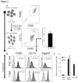
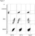
- macrophages may be confirmed, for example, by staining the cells to confirm they assume macrophage morphology, by a determination that the cells express macrophage proteins such as CD40, CD45, CD11b, CD64, F4/80(mice)/EMR1 (human), lysozyme M, MAC-1/MAC-3 and CD68, or that the cells exhibit properties of macrophages such as the capacity to internalize acetylated low density lipoproteins (Ac-LDL).
- macrophage proteins such as CD40, CD45, CD11b, CD64, F4/80(mice)/EMR1 (human), lysozyme M, MAC-1/MAC-3 and CD68, or that the cells exhibit properties of macrophages such as the capacity to internalize acetylated low density lipoproteins (Ac-LDL).
- Stem cell-derived macrophages obtained as described may be further conditioned to yield functional alveolar-like macrophages as defined in the claims.
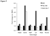
- a population of in vitro -derived alveolar-like macrophages which exhibit properties of classically activated M1 macrophages that are Myb-independent, characterized by one or more of the following: i) which exhibit at least 50% greater expression of CD11b than CD11b expression in primary alveolar macrophages; ii) which are expandable in vitro for at least about 1 month, such as at least 1 year or indefinitely; iii) which exhibit at least about 10% greater phagocytic activity than primary alveolar macrophages; and/or iv) which exhibit at least a 2-fold increase in expression of at least one of the LSR or RUNx2 genes than the expression of the LSR or RUNx2 genes in primary alveolar macrophages.
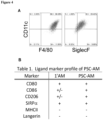
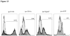
- alveolar-like macrophage refers to non-naturally occurring macrophages, generated in vitro from hemangioblasts prepared from PSCs, and which express markers expressed by naturally occurring alveolar macrophages, including one or more of F4/80(mice)/EMR1 (human), CD11c, SiglecF (mouse), CD80, CD86, CD206, CD11b, CD68, CD45 and SIRP ⁇ , and have a capacity for uptake of AcLDL.
- the PSC-derived macrophage may be cultured in an alveolar macrophage-inducing medium, e.g.
- the alveolar macrophage-inducing medium comprises Granulocyte-Macrophage Colony-Stimulating Factor (GM-CSF), also known as Colony Stimulating Factor 2 (CSF2), and optionally comprises Macrophage Colony-Stimulating Factor (M-CSF), also known as Colony Stimulating Factor 1 (CSF1).
- GM-CSF Granulocyte-Macrophage Colony-Stimulating Factor
- M-CSF Macrophage Colony-Stimulating Factor
- the alveolar macrophage-inducing medium may additionally comprise one or more of IL-3, IL-6 and SCF.
- the amounts of the growth factors in the alveolar macrophage-inducing medium will generally be amounts which stimulate generation of alveolar-like macrophages from myeloid macrophages, for example, an amount of GM-CSF of about 10-100 ng/ml, and optionally, an amount of M-CSF of about 10-100 ng/ml, or an amount of GM-CSF and M-CSF in a ratio ranging from about 1:10 to 10:1 GM-CSF to M-CSF.
- GM-CSF and M-CSF are used in about a 1:1 ratio, such as about 10-50 ng/ml of GM-CSF to about 10-50 ng/ml of M-CSF, e.g.
- alveolar-like macrophages may be confirmed minimally by expression of markers commonly expressed by alveolar macrophages such as F4/80(mice)/EMR1 (human), SiglecF (mouse), CD11c (human/mouse), CD68 (human), and uptake of AcLDL (human).
- markers commonly expressed by alveolar macrophages such as F4/80(mice)/EMR1 (human), SiglecF (mouse), CD11c (human/mouse), CD68 (human), and uptake of AcLDL (human).
- alveolar-like macrophages include CD45, CD11b (a unique marker of alveolar-like macrophages not highly expressed by primary Ams), SIRP ⁇ , CD80, CD86 and CD206.
- the formation of alveolar-like macrophages may also be confirmed based on functional characteristics, such as phagocytic activity, e.g. take up apoptotic material and bacteria, and binding of the lung innate immune collectin, SP-D. Alveolar-like macrophages resulting from the method provided herein are able to attain airway residence.
- the method includes: i) incubating the pluripotent stem cells in a first differentiation medium, optionally serum-free, to induce differentiation of the pluripotent stem cells into embryoid bodies; ii) culturing the embryoid bodies in second differentiation medium, optionally serum-free, comprising at least BMP4 and Activin-A, and optionally basic fibroblast growth factor (bFGF), for a period of time sufficient to generate hemangioblasts; (iii) culturing the hemangioblasts in a hematopoietic-inducing medium comprising VEGF, IL-3 and SCF, and optionally IL-6, for a sufficient period of time to generate macrophages; and (iv) culturing the macrophages in an alveolar macrophage-inducing medium comprising GM-CSF, and optionally M-CSF, under suitable
- the hemangioblasts are cultured in a hematopoietic-inducing medium comprising VEGF (about 5-50 ng/mL), SCF (about 10-100 ng/ml) and IL-3 (about 10-100 ng/ml) for a sufficient period of time to generate haematopoietic cells, wherein IL-6 (about 1-10 ng/mL) is optionally added during the culturing, as well as thrombopoietin (TPO) (about 10-100 ng/mL), Fms-related tyrosine kinase 3 ligand (FLT3L) (about 5-20 ng/mL) and insulin-like growth factor (IGF-1) (about 10-50 ng/mL) for a sufficient period of time to generate macrophages (e.g. for an amount of time sufficient to generate cells that detectably express CD11b, CD11c and/or CD45, and/or have a capacity to uptake Ac
- VEGF about
- Macrophages generated from human PSCs may be further conditioned to yield functional alveolar-like macrophages by incubation in medium as defined in the claims, and culturing for a sufficient period of time to yield alveolar-like macrophages (e.g. as determined by expression of CD11b, CD11c, CD45 and/or CD68, and/or having a capacity to uptake AcLDL).
- the amount of each of GM-CSF, and optionally M-CSF, in the medium is in the range of about 10-100 ng/mL, in a ratio of 1:10 to 10:1, and preferably a 1:1 ratio in an amount of about 20 ng/ml each.
- the alveolar-like macrophages generated as described herein can advantageously be expanded in vitro, e.g. proliferated in cell culture for a prolonged period of time (e.g. for at least about 1 month, e.g. for 2 or more months, preferably for at least 1 year, more preferably for more than 1 year, or, most preferably, indefinitely) without senescing, losing function or dying.
- expansion may be achieved in an expansion medium (with serum or serum-free) comprising M-CSF and GM-CSF in amounts ranging from about 1:10 to 10:1 M-CSF to GM-CSF.
- expansion of alveolar-like macrophages is conducted in an expansion medium comprising a 1:1 ratio of M-CSF to GM-CSF, and in other embodiments, the amount of GM-CSF relative to M-CSF is increased during expansion, for example, GM-CSF may be doubled relative to M-CSF. Concentrations of M-CSF and GM-CSF utilized for expansion may be in the range of about 20-100 ng/ml.
- the alveolar-like macrophages generated in vitro as described herein exhibit increased expression of one or more of CD11b, LSR and RUNx2 relative to primary alveolar macrophages.
- the alveolar-like macrophages generated in vitro as described herein are Myb- independent.
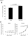
- the alveolar-like macrophages generated in vitro as described herein exhibit improved phagocytic activity relative to primary alveolar macrophages and blood- or bone marrow-derived macrophages.
- In vitro -generated alveolar-like macrophages may be genetically altered to generate alveolar-like macrophages carrying one or more genes for targeted gene correction or for targeted airway delivery of a therapeutic agent (such as a protein, cytokine or growth factor useful to treat a lung disease).
- a therapeutic agent such as a protein, cytokine or growth factor useful to treat a lung disease.
- anti-inflammatory agents such as IL-10
- antimicrobial agents such as alpha- and beta- defensins or nitric oxide synthase to optimize bacterial killing
- elafin anti-elastase protein inhibitor
- alpha-1 antitrypsin agents capable of dissolving mucus
- agents that stimulate vasodilation agents that enhance phagocytosis
- agents that target cancer cells
- alveolar-like macrophages may be engineered to incorporate a desired nucleic acid molecule or protein using well-established biotechnological techniques.
- a desired gene may be incorporated into a suitable expression vector using known recombinant methods, and the vector may then be introduced into target cells (alveolar-like macrophages or precursor cells) by electroporation, transfection using cationic lipid-based transfection reagents or viral-mediated transfection, e.g. lentiviral transfection.
- a therapeutic agent such as a protein or other molecule may itself be introduced into the target cells.
- genetically altered alveolar-like macrophages may be generated by genetically altering the PSCs or intermediate cell types from which the alveolar-like macrophages are derived using the above techniques prior to induction of alveolar-like macrophage generation.
- in vitro -derived genetically modified alveolar-like macrophages which may be used to treat lung disease in a mammal.
- the terms “treat”, “treating” or “treatment” are used herein to refer to methods that favorably alter a lung disease or disorder, including those that moderate, reverse, reduce the severity of, or protect against, the progression of a lung disease or disorder.
- a therapeutically effective amount of in vitro -derived alveolar-like macrophages are administered to a mammal in need of treatment.
- therapeutically effective amount is an amount of alveolar-like macrophages required to treat the disease that does not exceed an amount that may cause significant adverse effects to the mammal in need of treatment.
- Alveolar-like macrophage dosages that are therapeutically effective will vary on many factors including the nature of the condition to be treated, the mammal being treated and the dosage form utilized for administration.
- Appropriate dosages for use in such a treatment include dosages sufficient to result in airway residence of administered in vitro -derived alveolar-like macrophages of at least about 10%, and preferably, an airway residence of greater than 10%, for example, at least 20%, 30%, 40%, 50% or greater.
- the dosage of in vitro -derived alveolar-like macrophages useful to treat a lung disease or disorder may be a dosage in the range of about 10 5 to 10 8 cells, for a sufficient period of time to achieve treatment.
- the treatment regimen may include daily administration of alveolar-like macrophages, or dosages administered more or less frequently, e.g. on alternate days, weekly, or multiple dosages a day.
- the term "about” is used herein to mean an amount that may differ somewhat from the given value, by an amount that would not be expected to significantly affect activity or outcome as appreciated by one of skill in the art, for example, a variance of from 1-10% from the given value.
- Alveolar-like macrophages in accordance with a non-limiting embodiment may be formulated for therapeutic use by combination with a pharmaceutically acceptable carrier.
- pharmaceutically acceptable means acceptable for use in the pharmaceutical and veterinary arts, i.e. not being unacceptably toxic or otherwise unsuitable.
- the selected carrier may vary with intended mode of administration.
- alveolar-like macrophages may be formulated for administration by infusion or injection into a mammalian airway, e.g.
- a medical-grade, physiologically acceptable carrier such as an aqueous solution in sterile and pyrogen-free form, optionally buffered or made isotonic.
- the carrier may be a carbohydrate-containing solution (e.g. dextrose) or a saline solution comprising sodium chloride and optionally buffered.
- Suitable saline solutions may include varying concentrations of sodium chloride, for example, normal saline (0.9%), half-normal saline (0.45%), quarter-normal saline (0.22%), and solutions comprising greater amounts of sodium chloride (e.g. 3%-7%, or greater).
- Saline solutions may optionally include additional components, e.g. carbohydrates such as dextrose and the like.
- additional components include Ringer's solution, e.g. lactated or acetated Ringer's solution, phosphate buffered saline (PBS), TRIS (hydroxymethyl) aminomethane hydroxymethyl) aminomethane)-buffered saline (TBS), Hank's balanced salt solution (HBSS), Earle's balanced solution (EBSS), standard saline citrate (SSC), HEPES-buffered saline (HBS) and Gey's balanced salt solution (GBSS).
- PBS phosphate buffered saline
- TRIS hydroxymethyl) aminomethane hydroxymethyl) aminomethane
- TBS Hank's balanced salt solution
- EBSS Earle's balanced solution
- SSC standard saline citrate
- HBS HEPES-buffered saline
- GBSS Gey's balanced salt solution
- alveolar-like macrophages may be formulated for administration by routes including, but not limited to, inhalation.
- aerosol formulations may be prepared in which suitable propellant adjuvants are used.
- Other adjuvants may also be added to the composition regardless of how it is to be administered, for example, anti-microbial agents may be added to the composition to prevent microbial growth over prolonged storage periods.
- the present method advantageously provides a highly efficient growth-factor defined and extracellular matrix-independent in vitro differentiation protocol for the formation of macrophages from hemangioblasts derived from PSCs.
- macrophages can be further differentiated to generate alveolar-like macrophages suitable for use, e.g. for in vivo administration to mammalian lungs, to replace dysfunctional alveolar macrophages and promote survival of mammals with lung disease.
- the present alveolar-like macrophages exhibit improved phagocytic activity, e.g.
- the present alveolar-like macrophages exhibit molecular characteristics that differ from primary alveolar macrophages, for example, greater expression of CD11b (by at least about 50% or more, e.g.
- genes such as the LSR and RUNx2 e.g. at least a 2-fold increase in expression or more, e.g. a 3-5 fold increase in expression as compared to primary alveolar macrophages.
- alveolar-like macrophages which may be used to treat various macrophage-associated lung diseases.
- Implantation of the alveolar-like macrophages generated by the present method provides a non-pharmacological method of regenerating alveolar tissue in the treatment of genetic diseases such as adenosine deaminase (ADA)-deficiency, cystic fibrosis, hereditary pulmonary alveolar proteinosis (herPAP), and others, as well as acquired lung diseases such as chronic obstructive pulmonary disease (COPD), acute respiratory distress syndrome (ARDS), bronchopulmonary dysplasia (BPD), pulmonary fibrosis, lung cancer, radiation induced lung injury (RILI), ventilator induced lung injury (VILI), asthma, and bacterial or viral pneumonia.
- genetic diseases such as adenosine deaminase (ADA)-deficiency, cystic fibrosis, hereditary pulmonary alveolar proteinosis (herPAP), and others
- acquired lung diseases
- kits for use to generate alveolar-like macrophages from hemangioblasts in vitro comprises a serum-free hematopoietic-inducing medium comprising VEGF, IL-3 and SCF in hematopoietic amounts, optionally comprising one or more of IL-6, TPO, FLT3L and IGF-1; and a serum-free alveolar macrophage-inducing medium comprising GM-CSF, optionally comprising one or more of M-CSF, SCF, IL-3 and IL-6, wherein the hematopoietic-inducing medium comprises VEGF in an amount ranging from about 5 - 50 ng/ml, an amount of IL-3 in the range of about 10-100 ng/ml, and an amount of SCF ranging from about 10-100 ng/ml, and the amount of each of GM-CSF and M-CSF in the alveolar macrophage-inducing medium is about 10-50
- the kit may additionally include a medium for use to prepare hemangioblasts from PSCs, including first differentiation medium for differentiation of the PSCs into EBs, optionally including one or more growth factors such as BMP4 and bFGF, and second differentiation medium for differentiation of EBs into hemangioblasts comprising one or more mesoderm-inducing growth factors such as VEGF, Wnt3a, Activin A and BMP4 in hematopoietic, mesoderm-inducing amounts.
- first differentiation medium for differentiation of the PSCs into EBs optionally including one or more growth factors such as BMP4 and bFGF
- second differentiation medium for differentiation of EBs into hemangioblasts comprising one or more mesoderm-inducing growth factors such as VEGF, Wnt3a, Activin A and BMP4 in hematopoietic, mesoderm-inducing amounts.
- Amounts of the components of the second differentiation medium may be as follows: VEGF in an amount of no more than about 50 ng/ml, for example, in the range of about 5 to about 50 ng/ml; activin A in an amount of no more than about 10 ng/ml, for example, in the range of about 0.1-10 ng/ml; Wnt3a in an amount of up to about 5 ng/ml, for example in the range of about 1-5 ng/ml; and an amount of BMP4 of up to about 10 ng/ml, for example, in the range of about 1-10 ng/ml.
- the kit may additionally comprise a medium useful to expand stem cell-derived alveolar-like macrophages comprising GM-CSF and M-CSF in a ratio of 10:1 to 1:10, preferably in a ration of about 1:1 or 2:1.
- the kit may further include materials useful to conduct the present method including culture plates, welled plates, petri dishes and the like.
- the kit may also include instructions for conducting the present method as described herein.
- Non-limiting embodiments are described by reference to the following examples which are not to be construed as limiting.
- a highly efficient serum-free, feeder-free, growth-factor defined and extracellular matrix-independent in vitro differentiation protocol for pluripotent stem cell-derived macrophages was developed as follows.
- Pluripotent stem cells were maintained feeder-free on 0.1% gelatin coated 6-well plates in the serum-free '2 i ' pluripotent stem cell culture maintenance media consisting of a 1:1 ratio of Neurobasal media (Gibco, cat. # 21103-049) and Hams F-12 (Gibco, cat. # 11765-054); 0.5% v/v N2 supplement (Gibco, cat. # 17504044); 1% v/v B27 supplement (Gibco, cat. # 17502048); 0.05% w/v fraction V BSA (Gibco, cat. # 15260-037); 1% v/v penicillin/streptomycin (Life Technologies, cat.
- Embryonic stem cell (ESC) lines used as a reference example in this study include FoxA2/hCD4;Bry/GFP obtained from Gordon Keller's laboratory (University Health Network, Toronto, Canada), Flk-1-eGFP (referred to as Flk-1) obtained from Janet Rossant's laboratory (The Hospital for Sick Children, Toronto, Canada) and dsRed-MST (referred to as DsRed) obtained from Andras Nagy's laboratory (Samuel Lunenfeld Research Institute, Mt. Sinai Hospital, Toronto Canada).
- ESC Embryonic stem cell
- SFD serum-free basal differentiation media
- IMDM IMDM
- Hams F-12 media 0.05% w/v fraction V BSA
- 1% v/v B27 supplement without retinoic acid Gibco, cat # 12587-010
- 0.5% v/v N2 supplement 100 ⁇ g/mL ascorbic acid
- 1% v/v L-glutamine 1% v/v penicillin/streptomycin
- 0.004% v/v monothioglycerol Sigma, cat. # M6145-25ML.
- pluripotent stem cells were adjusted to a concentration of 70,000 cells/mL and seeded into a sterile untreated low adhesion surface Petri dish. Exactly 48 hours after induction, the embryoid bodies that had developed were re-aggregated into single cells at a concentration of 75,000 cells/ mL into an ultralow adhesion (ULA) cell culture plate (Corning, cat. # 3471) in mesoderm-inducing media containing 5 ng/ml of VEGF, 1 ng/ml Activin A, 3 ng/ml of Wnt3a and 1 ng/ml of BMP4.
- UAA ultralow adhesion
- the media was changed without EB re-aggregation to contain hematopoietic cocktails (10 ng/ml 11-3, 10 ng/ml SCF, 5 ng/ml VEGF and 2 ng/ml 11-6).
- Growth factors used in differentiation were all purchased from R&D Systems and re-constituted and stored according to the manufacturer's instructions: human VEGF (cat. #293-VE-010/CF); human Wnt3a (cat. #5036-WN-010); Activin A (cat. #338-AC-010/CF); human BMP4 (cat. #314-BP-010/CF); mouse IL-3 (cat. #403-ML-010/CF); mouse kit ligand (SCF) (cat.
- CD11c macrophage populations were expanded in DMEM/F12 supplemented with 10% FBS, 1% penicillin/streptomycin, GM-CSF and M-CSF in a 2:1 ratio (usually 20 ng/ml and 10 ng/ml, respectively). All cell culture differentiations and expansions were carried out using the Corning ULA plates indicated above.
- Fluorescently labeled Di-acetylated-LDL (Life Technologies, cat. # L3484) was added to serum-free macrophage cell cultures for 4 hours at 37°C 5% CO 2 and ambient O 2 . Fluorescence was assessed using wide field epifluorescence microscopy.
- Cells were harvested from ULA plates. Strongly adherent cells were removed using a brief ( ⁇ 5 min) incubation with animal-product free TrypLE cell dissociation enzyme and combined with non-adherent cells in suspension. Cells were pelleted by centrifugation at 450 x g and subjected to a second 2-3 minute incubation in TrypLE cell dissociation enzyme. Cells were washed with IMDM and resuspended in flow buffer (HBSS with 2% FBS and 1% HEPES) and passed through a 70 ⁇ m or a 40 ⁇ m filter for flow cytometry or FACS, respectively.
- flow buffer HBSS with 2% FBS and 1% HEPES
- RNA isolated from adult mouse lung tissue, mouse bone marrow and DsRED-PSC-AM The isolation and reverse transcription was performed as described previously ( Fox et al. Stem Cells Dev. 2014 Sep 22 [Epub]).
- Primers pairs used for PU.1 detection were as follows: forward primer, 5'-ACGATTCAGAGCTATACCAACGTCCA-3' (SEQ ID NO: 1); reverse primer, 5'-CTCTGCAGCTCTGTGAAGTGGTT-3' (SEQ ID NO: 2).
- Primer pairs use for Myb detection were: forward primer, 5'-TGCTCCTGATGTCAACAGAGAACGA-3' (SEQ ID NO: 3); reverse primer, 5'- GCACTATCCCCATGAGGTCTGGTC-3' (SEQ ID NO: 4).
- Macrophages were seeded into the wells of a glass chamber slide.
- Primary human neutrophils which undergo apoptosis at 37°C within 24 hours of isolation from peripheral blood, were labeled with Vybrant DiD fluorescent dye (Invitrogen, cat. # V22887,) and co-incubated with macrophages, some pre-stained with PKH26 fluorescent cell tracker (Sigma, cat. # PKH26PCL). Following the overnight co-incubation, cells were washed clean of freely floating cells (live or dead) and stained with image iT Live Lysosomal and Nuclear Labeling Kit (Invitrogen, cat. # 134202), as indicated, in accordance with the manufacturer's instructions.
- mice were sacrificed in accordance with the approved standard operating protocols of the laboratory animal services at the Hospital for Sick Children in Toronto, Ontario, Canada. The abdominal cavity of mice was opened and exsanguinated. The lungs and trachea were then revealed by continuing to open the thoracic cavity. A BD Angiocath was inserted into the exposed trachea and 1 mL of DPBS was used to inflate the lungs cyclically three times, collected and placed on ice. This was repeated 5 times for a total collection volume of 5 mL bronchoalveolar lavage (BAL) fluid per mouse.
- BAL bronchoalveolar lavage
- mice containing the enhanced green fluorescent protein fused to the histone 2B protein (H2B-GFP) bred to multiple generations on the CD1 background strain were used in accordance with standard operating protocols of the laboratory animal services at the Hospital for Sick Children in Toronto, Ontario, Canada.
- Adenosine deaminase deficient (ADA) mice FVB, 129-Ada tm1Mw -Tg[PLADA]4118Rkmb/J
- SPF pathogen free mice
- Clodronate liposomes were purchased from Encapsula NanoScience (Nashville, TN) as a registered product called Clodrosome TM
- the clodronate is packaged into a liposome, which upon internalization by phagocytosis releases the toxic clodronate to a macrophage and initiates macrophage apoptosis.
- the Clodrosome TM was administered to mice intranasally in two doses on consecutive days, first in a volume of 100 ⁇ L then in 50 ⁇ L.
- mice were anesthetized with a ketamine (75 mg/kg) and xylazine (10 mg/kg) mixture.
- a 25 gauge BD Angiocath was inserted into the trachea of the anaesthetized mouse and 0.5-1x10 6 cells in 50 ⁇ L of PBS was instilled into the lungs, followed by 300 ⁇ L of air.
- ADA knockout mice were performed on pups as young as postnatal day (PND) 3. Intranasal delivery of cells for the young pups was used because they are too small at this age for the intubation required for intratracheal transplantation.
- Fluorescence confocal microscopy was performed on a Leica CTRMIC 6000 confocal microscope with a Hamamatsu C910013 spinning disc camera (Leica Microsystems Inc.) and epifluorescence imaging was completed using a Leica DMI 3000B microscope with a Hamamatsu ORCA-HR camera (Leica Microsystems Inc.). Data was analyzed using the Volocity software suite (Perkin Elmer).
- Pluripotent stem cells were allowed to generate embryoid bodies (EBs) for two (2) days followed by culture with vascular endothelial growth factor (VEGF), Wnt3a, Activin and BMP4 until Day 4, where co-expression of the mesoderm markers, Brachyury and Flk-1, confirmed the development of hemangioblasts.
- VEGF vascular endothelial growth factor
- Wnt3a Wnt3a
- Activin vascular endothelial growth factor
- BMP4 vascular endothelial growth factor
- Hemangioblasts were cultured with all four soluble factors (VEGF, SCF, IL-3 and IL-6) to promote myeloid cell differentiation ( Fig. 1A ).
- FACS Fluorescence assisted cell sorting
- Alveolar macrophages mature in response to the lung environment and express high and constant levels of the dendritic cell marker, CD11c. It was determined if the pluripotent stem cell-derived macrophages (PSC-Mp) generated could acquire and maintain the levels of CD11c expression characteristic of alveolar macrophages. F4/80-sorted PSC-Mp were labeled with PKH26 red fluorescent cell tracker and adoptively transferred into the lungs of healthy mice via the trachea to determine if the PSC-macrophages could acquire the alveolar macrophage phenotype of F4/80 and CD11c co-expression, in vivo.
- PSC-Mp pluripotent stem cell-derived macrophages
- BAL bronchoalveolar lavage
- the PKH26 population had become indistinguishable from the resident alveolar macrophages based on co-expression of F4/80 and CD11c.
- Cells at this stage were noted to be Oct-4 negative and free of pluripotency (data not shown).
- FACS was then used to selectively acquire this population of cells that co-expressed F4/80 and CD11c, named PSC-alveolar-like macrophages (PSC-AMs).
- the present differentiation protocol generated a substantial yield of approximately 6 x 10 5 PSC-AMs for every 10 6 uninduced cells originally cultured with growth factors during the first week of the protocol ( Figure 3 ).
- the cells were expanded continuously without requiring additional sorting.
- the cells maintained expression of ligand markers found on primary alveolar macrophages, including F4/80, CD11c and SiglecF three weeks after their initial induction from pluripotency ( Figure 2B ).
- Expression of SiglecF demonstrated that the expansion of the PSC-Mp did not inadvertently convert the cells to dendritic cells, which do not express this marker.
- Sustained co-expression of these AM markers with other markers commonly expressed on primary AMs including CD80, CD86, CD206 and SIRP ⁇ was also confirmed, and it was verified that PSC-AM do not express MHCII or Langerin ( Figure 4 ).
- PSC-AMs ultrastructurally resemble primary AMs and determined that the cells were highly efficient at phagocytosing IgG-coated beads ( Figure 6A ), bacteria and apoptotic neutrophils, exhibiting increased phagocytosis in comparison to primary macrophages (by at least about 10%).
- Bone marrow cells (BMCs) were isolated from mice and the non-adherent progenitors were differentiated to BMDMs as previously described ( Suzuki et al. Nature 2014; 514: 450-454 ) for the purpose of comparing their expansion and phagocytic potential with PSC-AMs.
- PSC-AM are functionally similar to primary AM
- the PSC-AM were considered to be functional phagocytes like primary AM
- a bead uptake analysis was then conducted by incubating PKH26-labelled (red) PSC-AMs with fluorescent (green) IgG-coated beads and the PSC-AM were found to be more efficient in phagocytic uptake of the antibody-coated beads than primary AM
- a red fluorescent lysotracker was used to identify lyosomes in the PSC-AM and primary AMs, which were similarly abundant in both cell types.
- PSC-AMs remain in healthy airways after delivery and are functional phagocytes during acute lung injury
- PSC-AM are characteristically and functionally like AM in vitro
- dsRed-MST DsRed fluorophore
- DsRED-PSC-AMs were stained with primary antibodies targeting human SP-D and an ALEXA-488 secondary fluorescent antibody to detect SP-D binding.
- a population of DsRED-PSC-AMs displayed the typical punctate binding characteristic of SP-D binding to alveolar macrophages.
- the expanded DsRed-PSC-AMs were tracheally transferred to the lungs of healthy mice to determine if the cells would take residence in the airways. After 1 week, the animals were sacrificed to perform bronchoalveolar lavages (BAL) on the mice and harvest their spleens. DsRed + cells were recovered directly from the airways but not from the spleen indicating that the cells remained in the airways.
- BAL bronchoalveolar lavages
- H2B-eGFP mice which express eGFP linked to histone in all nucleated cells.
- the cells were examined from BAL of mice 2 h, 2 d, 2 wks, and 4 wks after airway delivery to determine if any DsRed cells remained throughout the surveyed timepoints. Constant DsRed population was detected that was distinctively separate from the host-derived GFP fluorescent cells.
- the total recovered BAL cell population was stained with fluorescent antibodies targeted to F4/80 and CD11c and the cells were gated based on DsRed and GFP.
- DsRed-PSC-AMs were subsequently delivered to the clodronate-treated airways of GFP mice to evaluate the functional properties of the PSC-AMs in vivo and determine if the PSC-AMs could perform phagocytosis in vivo.
- DsRed-PSC-AMs were delivered intratracheally to clodronate-treated mice four (4) days after the clodronate liposome treatment to reduce the risk that residual clodronate in the airways would affect the PSC-AMs upon delivery.
- BAL cells were then obtained from these clodronate-treated mice and evaluated by flow cytometry.
- DsRed-PSC-AMs cells remained in the injured lungs for 2 hours, 2 days and 6 days post instillation, which constituted up to 10 days following the initial clodronate injury. Moreover, approximately 90% of the DsRed population of cells retained their F4/80: CD11c co-expression characteristics during the lung injury, whereas the total population of GFP + BAL cells was comprised only partially of F4/80 + :CD11c + macrophages. Furthermore, 2 h after delivery of the DsRed-PSC-AMs to the injured lungs, host-derived material (containing GFP) was detectable within the DsRed-PSC-AMs that were recovered from the airways of clodronate-treated mice. Phagocytosis of host-derived GFP material continued until the endpoint, i.e. 6 days after DsRed-PSC-AM delivery. Additionally, differential staining of the BAL cells at each outlined timepoint was consistent with lung injury resolution.
- DsRed-PSC-AM instillation into the lungs of healthy mice was preceded by airway delivery of fluorescent bacteria ( S. aureus ). Within 2 hours the DsRed-PSC-AMs had performed phagocyotosis in vivo and engulfed the bacterial particles.
- PSC-AMs remain functional in healthy, injured and injury-resolving lungs indicates that these cells could be used for therapeutic interventions in a variety of acquired or inherited chronic lung diseases.
- the purine metabolic abnormality adenosine deaminase deficiency (ADA) has been characterized in transgenic mice lacking the gene. These ADA -/- animals develop a variety of pulmonary abnormalities shortly after birth, including compromised alveologenesis, fibrosis, inflammation, alveolar proteinosis and airway obstruction. Recent reports indicate that these animals die of respiratory failure within 18-21 days of birth and that their alveolar macrophages are non-functional, foamy and dying by apoptosis.
- the PSC-AM were tested in this ADA -/- model to determine if they could mitigate the pathophysiological effects of this multifaceted lung disease.
- ADA-/- mice can be saved with continuous administration of PEG-ADA.
- PSC-AMs 4 week old ADA -/- mice were used that were allowed to survive by weekly administration of PEG-ADA.
- PEG-ADA Prior to administration of PSC-AMs, PEG-ADA was discontinued and ⁇ 10 6 PSC-AMs in PBS or PBS vehicle alone was delivered directly to the airways of ADA +/- or ADA -/- mice (n ⁇ 4 per group) and the survival of animals was monitored.
- the ADA -/- mice receiving PSC-AM appeared healthy, vital and remained alive for at least 1 week after the single intrapulmonary dose of PSC-AMs, while half of the ADA -/- mice receiving only PBS were deceased by 48 hours. Within 6 days all PBS-treated ADA -/- animals were either deceased or required sacrifice. This difference in survival rates between the PSC-AM- and PBS-treated groups was statistically significant (log-rank, p ⁇ 0.0001) ( Figure 11 ).
- PSC-AMs were intranasally delivered into the airways of 3-4 day old ADA +/- and ADA -/- pups until day 15 as described in the methods section (the first delivery consisted of ⁇ 5x10 5 cells and each subsequent delivery consisted of ⁇ 1x10 6 cells).
- the animals were sacrificed at the defined endpoint of PND17 ⁇ 0.5, purposefully preceding the reported mortality limitation of ADA -/- mice. It was found that intranasal delivery of PSC-AM or PBS to heterozygotic (ADA +/- ) animals had no effect (neither negative nor positive) on survival as both groups of animals survived to the defined endpoint of PND17 ⁇ 0.5 ( Figure 10A , two bars on left).
- ADA +/- littermates treated and untreated with PSC-AM both displayed oxygen saturation levels of 87.7% ⁇ 6.5% and 86.6 ⁇ 2.2%, respectively ( Figure 10B left boxes).
- the saturated blood oxygen levels in PBS-treated ADA -/- mice was 65.9% ⁇ 1.3%; yet PSC-AM-treated ADA -/- mice displayed blood oxygen saturation levels of 90.1% ⁇ 2.6%, comparable to that of their heterozygotic littermates ( Figure 10B , right boxes).
- mice at four (4) and six (6) months after receiving PSC-AMs demonstrated that the mice were clear from any abnormal tissue growth or teratoma formation.
- Murine-induced pluripotent cells (iPS cell-line EOS-29, obtained from Dr. James Ellis, Hospital for Sick Children) were also used to generate alveolar-like macrophages. Using the identical protocol described in Example 1, murine induced pluripotent cells that express GFP only when pluripotency is induced, were differentiated into macrophages and sorted on Day 14 for F4/80 expression, while excluding any GFP-positive cells. One week later on Day 22 cells were confirmed to express F4/80 and the alveolar macrophage markers, CD11c and SiglecF, while displaying negligible GFP expression ( Fig. 13 ).
- Human pluripotent stem cells (CA1, HI and H1-GFP cells) were differentiated into CD11c macrophages.
- human pluripotent cells were differentiated to mesoderm through embryoid body formation using BMP4 and specified haemangioblast and blood precursor generation with low concentrations of activin followed by haematopoietic expansion with IL-3 and SCF culminating in macrophage colony growth with M-CSF and CD11c induction and maintenance with GM-CSF ( Fig. 14A ).
- human pluripotent stem cells growing in 5% O 2 were harvested on day 0 (D0) from serum-free, feeder-free or feeder-dependent conditions, by a brief 5 minute incubation with TrypLE (Life Technologies) followed by scraping or feeder depletion. Scraped or feeder-depleted cell clusters were resuspended in StemPro 34 hematopoeitic media (Life Technologies) supplemented with 10 ng/ml of BMP4 and incubated overnight in an Ultra Low Adhesion (ULA) (Corning) six well culture plate in 5% O 2 . This procedure was distinct from mouse differentiation since mouse cells remain growth factor free for up to 48 hours after EB induction begins and all of the mouse differentiations are completed with ambient oxygen levels.
- UUA Ultra Low Adhesion
- D1 On D1 (i.e., the next day following harvest), 5 ng/ml of bFGF was added to the culture. The addition of FGF2 is specific to human differentiation and not used with mouse cells.
- D2 On day 2 (D2) a very low concentration of Activin A (ranging from 0.1 - 10 ng/ml) was added to the floating culture of embryoid bodies and remained in the culture until day 4 (D4).
- D4 where KDR expression is expected
- Activin and BMP4 were removed and VEGF (10 ng/ml) and IL-6 (10ng/ml) were added. The addition of VEGF at this timepoint and this concentration is specific to the human differentiation process (in mice VEGF is added at D2 at a concentration of 5 ng/ml).
- D4-D6 SCF and IL-3 were added to the floating EBs at a concentration of 50 ng/ml (ranging from 10-100 ng/ml). The addition of SCF at this timepoint is specific to human differentiation; whereas SCF is added to differentiating mouse cells on D4 at a concentration of 10 ng/ml.
- D8 VEGF and bFGF are removed from the floating EBs. TPO (50 ng/ml), FLT3L (10 ng/ml) and IGF-1 (25 ng/ml) were added to the cultured EBs and the incubations then continued at ambient oxygen levels. Cells continued to grow under these conditions until D14.
- GM-CSF 50 ng/ml
- M-CSF 50 ng/ml
- the concentrations of GM-CSF and M-CSF were higher for human culture than the concentrations used for mouse cells.
- alveolar-like macrophages were identified.
- M-CSF was added (if not already present) and GM-CSF concentration was adjusted to twice that of M-CSF (i.e. GM-CSF, 100 ng/ml and M-CSF, 50 ng/ml) and all other growth factors were removed from culture for expansion. Similar to the mouse differentiation, macrophage colonies continued to expand under the control of M-CSF and GM-CSF beyond D35.
- CD68 and CD11c expression were analyzed and confirmed by flow cytometry with human specific antibodies ( FIG. 14B ).
- Human alveolar-like macrophages were further characterized by surface markers. Flow cytometry was used to determine surface expression of the hematopoietic marker, CD45, the myeloid marker, CD11b, and the alveolar macrophage marker, CD11c, on cells and to rule out the presence of endothelial cells expressing CD34. Fluorescence microscopy of the cells also indicates the ability of cells to uptake Ac-LDL, a known function of macrophages.
- a lentiviral transfer of the murine IL-10 gene using GeneCopoeia's EX-Mm03260-Lv201 lenti-vector to stem cell-derived alveolar-like macrophages was conducted as follows.
- the vector contained a CMV promoter driving the ORF of mouse IL10 (Accession: NM_010548.2) followed by SV40-eGFP-IRES-puromycin.
- HIV-based EX-Mm03260-Lv201 lenti-vector in conjunction with GeneCopoeia's Lenti-Pac TM HIV Expression Packaging vectors were co-transfected into HEK293T cells using GeneCopoeia's EndoFectin TM Lenti Transfection Reagent. Cells were incubated in the presence of 5% CO2 at 37°C overnight. Growth medium were changed to Opti-MEM containing 3% FBS with the addition of GeneCopoeia's Titerboost TM .
- Conditioned medium were collected after 24, 48 and 72 hours of incubation. Viral supernatant from transfected packaging cells was centrifuged at 2000 xg for 30 minutes to remove any loose cells and cell debris, and was then transferred to new tube. PEG 6000 solution was then added to make the final PEG 6000 concentration, 8.5% and the final NaCl concentration, 0.3 M. The mixture was incubated on ice for 3 to 6 hours, then centrifuged at 2000 xg for 30 minutes. The viral particle pellet was resuspended by pipetting in 1/20 of the original harvest volume of Opti-MEM. Infection of lentivirus was performed on a 24-well or 6-well plate.
- Target stem cell-derived alveolar-like macrophage cells were plated at least one day before infection at a cell density about 60-70% confluent. Appropriate amounts of concentrated viral particles were added to the target cells, together with polybrene, to a final concentration of 80 ⁇ g per milliliter. Growth medium were changed one to three days after infection. Mouse cells were selected under puromycin (0.75 ⁇ g per milliliter) for more than two weeks, positive cells were verified by GFP and IL-10 expression. Following selection, GFP-expressing cells were sorted using FACS analysis and expanded in DMEM/F12 with 10% FBS and 1% penicillin/streptomycin with 20 ng/ml of GM-CSF and 10 ng/ml of M-CSF.
- IL-10 IL-10-derived alveolar macrophages
- transfection was confirmed successful by visually observing using fluorescent microscopy that some cells newly expressed the GFP reporter.
- Luminex Cytokine 1-Plex targeting IL-10 was also used. This confirmed in 2 independent human PSC cell lines that macrophages undergoing the transfection expressed the IL-10 gene product; whereas cells not undergoing the transfection did not express the IL-10 gene product ( Figure 15 ).
- mice No noticeable adverse effects from transplantation of PSC-AMs to non-strain matched mice have been observed. Therefore, it was determined whether or not mouse PSC-AMs would be accepted by the airways of rats. 5 x 10 6 PSC-AMs suspended in a 300 ⁇ l bolus of DPBS was instilled directly into the airways of adult rats. The rats were monitored daily for 14 days. After a brief recovery period immediately following cell administration, all animals receiving mouse PSC-AMs continued to thrive and gained weight in equal proportions to their PBS-treated counterparts. Two weeks (14 days) following PSC-AM delivery, the rats were sacrificed for pulmonary examination. No evidence of extended injury was detectable.
- Pluripotent stem cells were used to generate Myb -independent macrophages from yolk-sac hematopoiesis that were conditioned in vitro to be tissue-specific and 'alveolar-like' to produce a cell more representative of a long living primary resident AM.
- Generation of these PSC-AMs is rapid and efficient; producing at least 6 x 10 5 cells per 1 million undifferentiated starting cells. This can be scaled up or down and the resultant cells are expandable for more than 1 year or can be cryopreserved without notable phenotypic changes.
- the PSC-AMs express and retain the distinguishing ligand markers F4/80, CD11c and SiglecF of primary AMs, without expressing markers of other cells types like MHCII or Langerin, while exhibiting functional qualities like primary AMs both in vitro and in vivo. Additional characterization of the PSC-AM indicates that these cells exhibit an M1-polarization phenotype when evaluated for basal level secreted cytokines. Pulmonary transplantation of only 5 x 10 5 PSC-AMs to healthy mice revealed retention of the cells in the airways for several weeks with no obvious transplantation associated injury or immune suppression requirement.
- the PSC-AMs can persist in the airways during acute lung injury and display immediate and sustained functional phagocytosis of host-derived cellular debris and bacteria in vivo. Most notably, was the remarkable contribution that PSC-AMs made to the regeneration and recovery of the pulmonary pathology typically exhibited in ADA -/- mice.
Landscapes
- Health & Medical Sciences (AREA)
- Life Sciences & Earth Sciences (AREA)
- Engineering & Computer Science (AREA)
- Biomedical Technology (AREA)
- Chemical & Material Sciences (AREA)
- Biotechnology (AREA)
- Zoology (AREA)
- Immunology (AREA)
- Organic Chemistry (AREA)
- Bioinformatics & Cheminformatics (AREA)
- Genetics & Genomics (AREA)
- Wood Science & Technology (AREA)
- Hematology (AREA)
- General Health & Medical Sciences (AREA)
- Cell Biology (AREA)
- Developmental Biology & Embryology (AREA)
- Microbiology (AREA)
- General Engineering & Computer Science (AREA)
- Biochemistry (AREA)
- Medicinal Chemistry (AREA)
- Veterinary Medicine (AREA)
- Pharmacology & Pharmacy (AREA)
- Animal Behavior & Ethology (AREA)
- Public Health (AREA)
- Reproductive Health (AREA)
- Gynecology & Obstetrics (AREA)
- Gastroenterology & Hepatology (AREA)
- Virology (AREA)
- Epidemiology (AREA)
- General Chemical & Material Sciences (AREA)
- Nuclear Medicine, Radiotherapy & Molecular Imaging (AREA)
- Chemical Kinetics & Catalysis (AREA)
- Pulmonology (AREA)
- Micro-Organisms Or Cultivation Processes Thereof (AREA)
- Medicines Containing Material From Animals Or Micro-Organisms (AREA)
Claims (10)
- Verfahren zur Erzeugung alveolarartiger Makrophagen aus Hämangioblasten, umfassend die folgenden Schritte: i) Kultivieren der Hämangioblasten in hämatopoetisch-induzierendem Medium, umfassend vaskulären endothelialen Wachstumsfaktor (VEGF), Stammzellfaktor (SCF), Interleukin-3 (IL-3) und Interleukin-6 (IL-6), über einen ausreichenden Zeitraum, um Makrophagen zu erzeugen, und ii) Kultivieren der Makrophagen in einem Alveolarmakrophagen-induzierenden Medium, umfassend Granulozyten-Makrophagen-Kolonie-stimulierenden Faktor (GM-CSF) und Makrophagen-Kolonie-stimulierenden Faktor (M-CSF), SCF und IL-3, unter geeigneten Bedingungen und über einen ausreichenden Zeitraum, um alveolarartige Makrophagen zu erhalten.
- Verfahren nach Anspruch 1, wobei das hämatopoetischinduzierende Medium Folgendes umfasst: VEGF in einer Menge im Bereich von etwa 5-50 ng/ml; eine Menge an IL-3 im Bereich von etwa 10-100 ng/ml; SCF in einer Menge im Bereich von etwa 10-100 ng/ml; und IL-6 im Bereich von etwa 1-10 ng/ml.
- Verfahren nach den Ansprüchen 1-2, wobei die Makrophagen über einen ausreichenden Zeitraum kultiviert werden, um Zellen zu erzeugen, die die Alveolarmakrophagen-Marker F4/80/EMR1, SiglecF und CD11c exprimieren; oder Zellen, die CD68 und CD11c exprimieren und die Fähigkeit haben, AcLDL aufzunehmen.
- Verfahren nach den Ansprüchen 1-3, wobei die Hämangioblasten durch Inkubieren von pluripotenten Stammzellen in einem ersten serumfreien Differenzierungsmedium erhalten werden, um eine Differenzierung der pluripotenten Stammzellen in Embryoid Bodies zu induzieren, die dann in einem zweiten Differenzierungsmedium, das BMP4, Wnt3a, VEGF und Activin-A umfasst, über einen ausreichenden Zeitraum kultiviert werden, um Zellen zu erhalten, die einen Hämangioblast-Mesoderm-Marker exprimieren.
- Verfahren zur Differenzierung pluripotenter Stammzellen in alveolarartige Makrophagen, umfassend die folgenden Schritte:i) Inkubieren der pluripotenten Stammzellen in einem ersten Differenzierungsmedium, wobei das erste Differenzierungsmedium serumfrei ist, um eine Differenzierung der pluripotenten Stammzellen in Embryoid Bodies zu induzieren;ii) Kultivieren der Embryoid Bodies in einem zweitem Differenzierungsmedium, das mindestens BMP4, Wnt3a, VEGF und Activin-A umfasst, über einen ausreichenden Zeitraum, um Hämangioblasten zu erzeugen;iii) Kultivieren der Hämangioblasten in einem hämatopoetisch-induzierenden Medium, umfassend VEGF, IL-3, SCF und IL-6, über einen ausreichenden Zeitraum, um Makrophagen zu erzeugen; undiv) Kultivieren der Makrophagen in einem Alveolarmakrophagen-induzierenden Medium, umfassend GM-CSF, M-CSF, IL-3 und SCF unter geeigneten Bedingungen und über einen ausreichenden Zeitraum, um alveolarartige Makrophagen zu erhalten.
- Population von in vitro abgeleiteten alveolarartigen Makrophagen, abgeleitet von pluripotenten Stammzellen, wobei die alveolarartigen Makrophagen CD11c exprimieren, die Eigenschaften von klassisch aktivierten M1-Makrophagen aufweisen, Myb-unabhängig sind, und in vitro für mindestens etwa 1 Monat, wie etwa mindestens 1 Jahr oder auf unbestimmte Zeit, expandierbar sind.
- Population von alveolarartigen Makrophagen nach Anspruch 6, genetisch modifiziert, um ein therapeutisches Mittel zu exprimieren.
- Population von alveolarartigen Makrophagen nach Anspruch 6, die unter Verwendung des Verfahrens nach einem der Ansprüche 1-5 erzeugt wurde.
- Zusammensetzung, umfassend eine Population isolierter in vitro abgeleiteter alveolarartiger Makrophagen nach Anspruch 6, in einem physiologisch verträglichen Träger von medizinischer Qualität.
- Zusammensetzung nach Anspruch 9, wobei die Makrophagen genetisch modifiziert sind, um ein therapeutisches Mittel, wie etwa ein therapeutisches Mittel, das zur Behandlung von Lungenerkrankungen nützlich ist, zu exprimieren.
Applications Claiming Priority (2)
| Application Number | Priority Date | Filing Date | Title |
|---|---|---|---|
| US201562114851P | 2015-02-11 | 2015-02-11 | |
| PCT/CA2016/050128 WO2016127259A1 (en) | 2015-02-11 | 2016-02-11 | Alveolar-like macrophages and method of generating same |
Publications (4)
| Publication Number | Publication Date |
|---|---|
| EP3256569A1 EP3256569A1 (de) | 2017-12-20 |
| EP3256569A4 EP3256569A4 (de) | 2018-08-22 |
| EP3256569B1 EP3256569B1 (de) | 2021-03-24 |
| EP3256569B2 true EP3256569B2 (de) | 2024-04-17 |
Family
ID=56614019
Family Applications (1)
| Application Number | Title | Priority Date | Filing Date |
|---|---|---|---|
| EP16748520.0A Active EP3256569B2 (de) | 2015-02-11 | 2016-02-11 | Alveolarartige makrophagen und verfahren zur erzeugung davon |
Country Status (4)
| Country | Link |
|---|---|
| US (1) | US11001806B2 (de) |
| EP (1) | EP3256569B2 (de) |
| CA (1) | CA2975780C (de) |
| WO (1) | WO2016127259A1 (de) |
Families Citing this family (11)
| Publication number | Priority date | Publication date | Assignee | Title |
|---|---|---|---|---|
| US10724003B2 (en) * | 2015-01-16 | 2020-07-28 | Agency For Science, Technology And Research | Method of differentiating human pluripotent stem cells into primitive macrophages |
| US12146161B2 (en) * | 2015-02-11 | 2024-11-19 | The Hosptial For Sick Children | Mammalian alveolar macrophages derived from pluripotent cells |
| EP3256569B2 (de) | 2015-02-11 | 2024-04-17 | The Hospital for Sick Children | Alveolarartige makrophagen und verfahren zur erzeugung davon |
| EP3399027A1 (de) | 2017-05-04 | 2018-11-07 | Medizinische Hochschule Hannover | Von stammzellen abstammende myeloide zellen, ihre herstellund und verwendung |
| WO2018213421A1 (en) | 2017-05-17 | 2018-11-22 | Trapnell, Bruce | Cell therapy with lentiviral transduced csf2ra transgene in the treatment of hereditary pulmonary alveolar proteinosis |
| CA3064134A1 (en) * | 2017-05-19 | 2018-11-22 | The Hospital For Sick Children | Method of treating respiratory tract infection |
| KR102111441B1 (ko) * | 2018-11-12 | 2020-05-15 | 대한민국 | 전분화능 줄기세포 유래 대식세포를 기반으로 하는 감염질환 치료용 약물 스크리닝 방법 |
| EP3947649A4 (de) * | 2019-03-28 | 2022-12-14 | The Hospital For Sick Children | Verfahren zur erzeugung von hemangioblasten |
| KR20240054468A (ko) * | 2022-10-18 | 2024-04-26 | 의료법인 성광의료재단 | 줄기세포에서 대식세포로의 분화 유도용 배지 조성물 및 이를 이용한 분화 방법 |
| CN116731967B (zh) * | 2023-08-16 | 2023-11-17 | 南京大学 | 从多能干细胞通过诱导分化制备巨噬细胞的方法 |
| CN119410583B (zh) * | 2025-01-07 | 2025-03-28 | 中国人民解放军军事科学院军事医学研究院 | 一种小鼠原代肺泡巨噬细胞的体外长期扩增培养方法 |
Family Cites Families (9)
| Publication number | Priority date | Publication date | Assignee | Title |
|---|---|---|---|---|
| US20060003446A1 (en) | 2002-05-17 | 2006-01-05 | Gordon Keller | Mesoderm and definitive endoderm cell populations |
| EP1941027B1 (de) * | 2005-09-28 | 2014-08-20 | IPD-Therapeutics B.V. | Methoden und mittel zur vermehrung von stammzellen und anschliessenden generierung und vermehrung von vorläuferzellen; produktion von effektorzellen als klinische therapeutika |
| WO2009104825A1 (en) * | 2008-02-18 | 2009-08-27 | Kaist | Method for inducing the defferentiation of embryonic stem cells into hemangioblast |
| SG188306A1 (en) | 2010-08-27 | 2013-04-30 | Univ Health Network | Methods for enriching pluripotent stem cell-derived cardiomyocyte progenitor cells and cardiomyocyte cells based on sirpa expression |
| JP5995237B2 (ja) | 2010-12-03 | 2016-09-21 | 国立大学法人京都大学 | 多能性幹細胞からの好酸球の製造方法 |
| JP6344821B2 (ja) | 2011-11-21 | 2018-06-20 | ユニバーシティ・ヘルス・ネットワーク | 造血前駆体の集団および造血前駆体のための幹細胞を富化する方法 |
| WO2014012933A1 (en) * | 2012-07-16 | 2014-01-23 | MAX-PLANCK-Gesellschaft zur Förderung der Wissenschaften e.V. | Method for the generation of non-transformed macrophage cell lines |
| EP3256569B2 (de) | 2015-02-11 | 2024-04-17 | The Hospital for Sick Children | Alveolarartige makrophagen und verfahren zur erzeugung davon |
| US20180360877A1 (en) | 2015-12-08 | 2018-12-20 | Inserm (Institut National De La Sante Et De La Recherche Medicale) | Methods for expanding a population of alveolar macrophages in a long term culture |
-
2016
- 2016-02-11 EP EP16748520.0A patent/EP3256569B2/de active Active
- 2016-02-11 WO PCT/CA2016/050128 patent/WO2016127259A1/en not_active Ceased
- 2016-02-11 CA CA2975780A patent/CA2975780C/en active Active
-
2017
- 2017-08-09 US US15/672,432 patent/US11001806B2/en active Active
Non-Patent Citations (4)
| Title |
|---|
| FEJER GYÖRGY, WEGNER MAREIKE DOROTHEE, GYÖRY ILDIKO, COHEN IDAN, ENGELHARD PEGGY, VORONOV ELENA, MANKE THOMAS, RUZSICS ZSOLT, DÖLK: "Nontransformed, GM-CSF–dependent macrophage lines are a unique model to study tissue macrophage functions", PNAS, 6 November 2013 (2013-11-06), pages E2191 - E2198 † |
| KENNEDY ET AL.: "Development of the hemangioblast defines the onset of hematopoiesis in human ES cell differentiation cultures", BLOOD, vol. 109, no. 7, 2007, pages 2679 - 2687 † |
| MICHAEL L LITVACK; WIGLE THEODORE J; LEE JOYCE; WANG JINXIA; ACKERLEY CAMERON; GRUNEBAUM EYAL; POST MARTIN: "Alveolar-like Stem Cell-derived Myb − Macrophages Promote Recovery and Survival in Airway Disease", AM J RESPIR CRIT CARE MED, vol. 193, no. 11, 2016, pages 1219 - 1229 † |
| ORLOVSKAYA I; SCHRAUFSTATTER I; LORING J; KHALDOYANIDI S: "Hematopoietic differentiation of embryonic stem cells", METHODS, vol. 45, no. 2, 2008, pages 159 - 167 † |
Also Published As
| Publication number | Publication date |
|---|---|
| EP3256569A4 (de) | 2018-08-22 |
| US20170335282A1 (en) | 2017-11-23 |
| EP3256569A1 (de) | 2017-12-20 |
| WO2016127259A1 (en) | 2016-08-18 |
| US11001806B2 (en) | 2021-05-11 |
| CA2975780A1 (en) | 2016-08-18 |
| CA2975780C (en) | 2023-08-01 |
| EP3256569B1 (de) | 2021-03-24 |
Similar Documents
| Publication | Publication Date | Title |
|---|---|---|
| EP3256569B2 (de) | Alveolarartige makrophagen und verfahren zur erzeugung davon | |
| US10526581B2 (en) | Modulation of cardiac stem-progenitor cell differentiation, assays and uses thereof | |
| CN115361959A (zh) | 用于使用间充质谱系前体或干细胞治疗过度炎症的方法 | |
| KR20220150919A (ko) | 중간엽 계통 전구체 또는 줄기 세포를 사용한 염증성 폐 질환의 치료 방법 | |
| US12146161B2 (en) | Mammalian alveolar macrophages derived from pluripotent cells | |
| US20240207323A1 (en) | Method for treating acute respiratory distress syndrome (ards) using mesenchymal lineage precursor or stem cells | |
| CN112538458A (zh) | 用于重编程细胞的方法 | |
| US12312598B2 (en) | Method of generating hemangioblasts | |
| WO2023119239A1 (en) | Method of treating severe graft versus host disease | |
| KR102161910B1 (ko) | 제1형 혈우병의 세포치료를 위한 고증식성 혈관내피세포 분화방법 및 용도 | |
| JP2017514523A (ja) | 細胞培養方法 | |
| WO2025046491A1 (en) | Third line treatment for gvhd | |
| Young et al. | Cell-Based Therapy for Neonatal Lung Diseases | |
| HK40106098A (en) | Therapeutic composition containing corneal endothelium replacement cells | |
| CN117337184A (zh) | 使用间充质谱系前体细胞或干细胞治疗急性呼吸窘迫综合征(ards)的方法 |
Legal Events
| Date | Code | Title | Description |
|---|---|---|---|
| STAA | Information on the status of an ep patent application or granted ep patent |
Free format text: STATUS: THE INTERNATIONAL PUBLICATION HAS BEEN MADE |
|
| PUAI | Public reference made under article 153(3) epc to a published international application that has entered the european phase |
Free format text: ORIGINAL CODE: 0009012 |
|
| STAA | Information on the status of an ep patent application or granted ep patent |
Free format text: STATUS: REQUEST FOR EXAMINATION WAS MADE |
|
| 17P | Request for examination filed |
Effective date: 20170906 |
|
| AK | Designated contracting states |
Kind code of ref document: A1 Designated state(s): AL AT BE BG CH CY CZ DE DK EE ES FI FR GB GR HR HU IE IS IT LI LT LU LV MC MK MT NL NO PL PT RO RS SE SI SK SM TR |
|
| AX | Request for extension of the european patent |
Extension state: BA ME |
|
| DAV | Request for validation of the european patent (deleted) | ||
| DAX | Request for extension of the european patent (deleted) | ||
| A4 | Supplementary search report drawn up and despatched |
Effective date: 20180724 |
|
| RIC1 | Information provided on ipc code assigned before grant |
Ipc: A61K 35/15 20150101ALI20180718BHEP Ipc: C12N 5/10 20060101ALI20180718BHEP Ipc: C12N 5/0789 20100101ALI20180718BHEP Ipc: C12N 5/0786 20100101AFI20180718BHEP Ipc: A61P 11/00 20060101ALI20180718BHEP |
|
| STAA | Information on the status of an ep patent application or granted ep patent |
Free format text: STATUS: EXAMINATION IS IN PROGRESS |
|
| 17Q | First examination report despatched |
Effective date: 20191021 |
|
| GRAJ | Information related to disapproval of communication of intention to grant by the applicant or resumption of examination proceedings by the epo deleted |
Free format text: ORIGINAL CODE: EPIDOSDIGR1 |
|
| GRAP | Despatch of communication of intention to grant a patent |
Free format text: ORIGINAL CODE: EPIDOSNIGR1 |
|
| GRAP | Despatch of communication of intention to grant a patent |
Free format text: ORIGINAL CODE: EPIDOSNIGR1 |
|
| STAA | Information on the status of an ep patent application or granted ep patent |
Free format text: STATUS: GRANT OF PATENT IS INTENDED |
|
| INTG | Intention to grant announced |
Effective date: 20201016 |
|
| GRAS | Grant fee paid |
Free format text: ORIGINAL CODE: EPIDOSNIGR3 |
|
| GRAA | (expected) grant |
Free format text: ORIGINAL CODE: 0009210 |
|
| STAA | Information on the status of an ep patent application or granted ep patent |
Free format text: STATUS: THE PATENT HAS BEEN GRANTED |
|
| AK | Designated contracting states |
Kind code of ref document: B1 Designated state(s): AL AT BE BG CH CY CZ DE DK EE ES FI FR GB GR HR HU IE IS IT LI LT LU LV MC MK MT NL NO PL PT RO RS SE SI SK SM TR |
|
| REG | Reference to a national code |
Ref country code: GB Ref legal event code: FG4D |
|
| REG | Reference to a national code |
Ref country code: CH Ref legal event code: EP |
|
| REG | Reference to a national code |
Ref country code: DE Ref legal event code: R096 Ref document number: 602016054832 Country of ref document: DE |
|
| REG | Reference to a national code |
Ref country code: IE Ref legal event code: FG4D |
|
| REG | Reference to a national code |
Ref country code: AT Ref legal event code: REF Ref document number: 1374539 Country of ref document: AT Kind code of ref document: T Effective date: 20210415 |
|
| REG | Reference to a national code |
Ref country code: LT Ref legal event code: MG9D |
|
| PG25 | Lapsed in a contracting state [announced via postgrant information from national office to epo] |
Ref country code: FI Free format text: LAPSE BECAUSE OF FAILURE TO SUBMIT A TRANSLATION OF THE DESCRIPTION OR TO PAY THE FEE WITHIN THE PRESCRIBED TIME-LIMIT Effective date: 20210324 Ref country code: GR Free format text: LAPSE BECAUSE OF FAILURE TO SUBMIT A TRANSLATION OF THE DESCRIPTION OR TO PAY THE FEE WITHIN THE PRESCRIBED TIME-LIMIT Effective date: 20210625 Ref country code: HR Free format text: LAPSE BECAUSE OF FAILURE TO SUBMIT A TRANSLATION OF THE DESCRIPTION OR TO PAY THE FEE WITHIN THE PRESCRIBED TIME-LIMIT Effective date: 20210324 Ref country code: NO Free format text: LAPSE BECAUSE OF FAILURE TO SUBMIT A TRANSLATION OF THE DESCRIPTION OR TO PAY THE FEE WITHIN THE PRESCRIBED TIME-LIMIT Effective date: 20210624 Ref country code: BG Free format text: LAPSE BECAUSE OF FAILURE TO SUBMIT A TRANSLATION OF THE DESCRIPTION OR TO PAY THE FEE WITHIN THE PRESCRIBED TIME-LIMIT Effective date: 20210624 |
|
| PG25 | Lapsed in a contracting state [announced via postgrant information from national office to epo] |
Ref country code: SE Free format text: LAPSE BECAUSE OF FAILURE TO SUBMIT A TRANSLATION OF THE DESCRIPTION OR TO PAY THE FEE WITHIN THE PRESCRIBED TIME-LIMIT Effective date: 20210324 Ref country code: RS Free format text: LAPSE BECAUSE OF FAILURE TO SUBMIT A TRANSLATION OF THE DESCRIPTION OR TO PAY THE FEE WITHIN THE PRESCRIBED TIME-LIMIT Effective date: 20210324 Ref country code: LV Free format text: LAPSE BECAUSE OF FAILURE TO SUBMIT A TRANSLATION OF THE DESCRIPTION OR TO PAY THE FEE WITHIN THE PRESCRIBED TIME-LIMIT Effective date: 20210324 |
|
| REG | Reference to a national code |
Ref country code: NL Ref legal event code: MP Effective date: 20210324 |
|
| REG | Reference to a national code |
Ref country code: AT Ref legal event code: MK05 Ref document number: 1374539 Country of ref document: AT Kind code of ref document: T Effective date: 20210324 |
|
| PG25 | Lapsed in a contracting state [announced via postgrant information from national office to epo] |
Ref country code: NL Free format text: LAPSE BECAUSE OF FAILURE TO SUBMIT A TRANSLATION OF THE DESCRIPTION OR TO PAY THE FEE WITHIN THE PRESCRIBED TIME-LIMIT Effective date: 20210324 |
|
| PG25 | Lapsed in a contracting state [announced via postgrant information from national office to epo] |
Ref country code: EE Free format text: LAPSE BECAUSE OF FAILURE TO SUBMIT A TRANSLATION OF THE DESCRIPTION OR TO PAY THE FEE WITHIN THE PRESCRIBED TIME-LIMIT Effective date: 20210324 Ref country code: CZ Free format text: LAPSE BECAUSE OF FAILURE TO SUBMIT A TRANSLATION OF THE DESCRIPTION OR TO PAY THE FEE WITHIN THE PRESCRIBED TIME-LIMIT Effective date: 20210324 Ref country code: LT Free format text: LAPSE BECAUSE OF FAILURE TO SUBMIT A TRANSLATION OF THE DESCRIPTION OR TO PAY THE FEE WITHIN THE PRESCRIBED TIME-LIMIT Effective date: 20210324 Ref country code: SM Free format text: LAPSE BECAUSE OF FAILURE TO SUBMIT A TRANSLATION OF THE DESCRIPTION OR TO PAY THE FEE WITHIN THE PRESCRIBED TIME-LIMIT Effective date: 20210324 Ref country code: AT Free format text: LAPSE BECAUSE OF FAILURE TO SUBMIT A TRANSLATION OF THE DESCRIPTION OR TO PAY THE FEE WITHIN THE PRESCRIBED TIME-LIMIT Effective date: 20210324 |
|
| PG25 | Lapsed in a contracting state [announced via postgrant information from national office to epo] |
Ref country code: IS Free format text: LAPSE BECAUSE OF FAILURE TO SUBMIT A TRANSLATION OF THE DESCRIPTION OR TO PAY THE FEE WITHIN THE PRESCRIBED TIME-LIMIT Effective date: 20210724 Ref country code: SK Free format text: LAPSE BECAUSE OF FAILURE TO SUBMIT A TRANSLATION OF THE DESCRIPTION OR TO PAY THE FEE WITHIN THE PRESCRIBED TIME-LIMIT Effective date: 20210324 Ref country code: RO Free format text: LAPSE BECAUSE OF FAILURE TO SUBMIT A TRANSLATION OF THE DESCRIPTION OR TO PAY THE FEE WITHIN THE PRESCRIBED TIME-LIMIT Effective date: 20210324 Ref country code: PT Free format text: LAPSE BECAUSE OF FAILURE TO SUBMIT A TRANSLATION OF THE DESCRIPTION OR TO PAY THE FEE WITHIN THE PRESCRIBED TIME-LIMIT Effective date: 20210726 Ref country code: PL Free format text: LAPSE BECAUSE OF FAILURE TO SUBMIT A TRANSLATION OF THE DESCRIPTION OR TO PAY THE FEE WITHIN THE PRESCRIBED TIME-LIMIT Effective date: 20210324 |
|
| REG | Reference to a national code |
Ref country code: DE Ref legal event code: R026 Ref document number: 602016054832 Country of ref document: DE |
|
| PLBI | Opposition filed |
Free format text: ORIGINAL CODE: 0009260 |
|
| PLAX | Notice of opposition and request to file observation + time limit sent |
Free format text: ORIGINAL CODE: EPIDOSNOBS2 |
|
| 26 | Opposition filed |
Opponent name: STRAWMAN LIMITED Effective date: 20211222 |
|
| PG25 | Lapsed in a contracting state [announced via postgrant information from national office to epo] |
Ref country code: DK Free format text: LAPSE BECAUSE OF FAILURE TO SUBMIT A TRANSLATION OF THE DESCRIPTION OR TO PAY THE FEE WITHIN THE PRESCRIBED TIME-LIMIT Effective date: 20210324 Ref country code: AL Free format text: LAPSE BECAUSE OF FAILURE TO SUBMIT A TRANSLATION OF THE DESCRIPTION OR TO PAY THE FEE WITHIN THE PRESCRIBED TIME-LIMIT Effective date: 20210324 Ref country code: ES Free format text: LAPSE BECAUSE OF FAILURE TO SUBMIT A TRANSLATION OF THE DESCRIPTION OR TO PAY THE FEE WITHIN THE PRESCRIBED TIME-LIMIT Effective date: 20210324 |
|
| PG25 | Lapsed in a contracting state [announced via postgrant information from national office to epo] |
Ref country code: SI Free format text: LAPSE BECAUSE OF FAILURE TO SUBMIT A TRANSLATION OF THE DESCRIPTION OR TO PAY THE FEE WITHIN THE PRESCRIBED TIME-LIMIT Effective date: 20210324 |
|
| PLBB | Reply of patent proprietor to notice(s) of opposition received |
Free format text: ORIGINAL CODE: EPIDOSNOBS3 |
|
| PG25 | Lapsed in a contracting state [announced via postgrant information from national office to epo] |
Ref country code: IS Free format text: LAPSE BECAUSE OF FAILURE TO SUBMIT A TRANSLATION OF THE DESCRIPTION OR TO PAY THE FEE WITHIN THE PRESCRIBED TIME-LIMIT Effective date: 20210724 |
|
| PG25 | Lapsed in a contracting state [announced via postgrant information from national office to epo] |
Ref country code: MC Free format text: LAPSE BECAUSE OF FAILURE TO SUBMIT A TRANSLATION OF THE DESCRIPTION OR TO PAY THE FEE WITHIN THE PRESCRIBED TIME-LIMIT Effective date: 20210324 |
|
| REG | Reference to a national code |
Ref country code: CH Ref legal event code: PL |
|
| REG | Reference to a national code |
Ref country code: BE Ref legal event code: MM Effective date: 20220228 |
|
| PG25 | Lapsed in a contracting state [announced via postgrant information from national office to epo] |
Ref country code: LU Free format text: LAPSE BECAUSE OF NON-PAYMENT OF DUE FEES Effective date: 20220211 |
|
| PG25 | Lapsed in a contracting state [announced via postgrant information from national office to epo] |
Ref country code: LI Free format text: LAPSE BECAUSE OF NON-PAYMENT OF DUE FEES Effective date: 20220228 Ref country code: IE Free format text: LAPSE BECAUSE OF NON-PAYMENT OF DUE FEES Effective date: 20220211 Ref country code: CH Free format text: LAPSE BECAUSE OF NON-PAYMENT OF DUE FEES Effective date: 20220228 |
|
| PG25 | Lapsed in a contracting state [announced via postgrant information from national office to epo] |
Ref country code: BE Free format text: LAPSE BECAUSE OF NON-PAYMENT OF DUE FEES Effective date: 20220228 |
|
| PGFP | Annual fee paid to national office [announced via postgrant information from national office to epo] |
Ref country code: IT Payment date: 20230126 Year of fee payment: 8 |
|
| P01 | Opt-out of the competence of the unified patent court (upc) registered |
Effective date: 20230512 |
|
| PUAH | Patent maintained in amended form |
Free format text: ORIGINAL CODE: 0009272 |
|
| STAA | Information on the status of an ep patent application or granted ep patent |
Free format text: STATUS: PATENT MAINTAINED AS AMENDED |
|
| PG25 | Lapsed in a contracting state [announced via postgrant information from national office to epo] |
Ref country code: HU Free format text: LAPSE BECAUSE OF FAILURE TO SUBMIT A TRANSLATION OF THE DESCRIPTION OR TO PAY THE FEE WITHIN THE PRESCRIBED TIME-LIMIT; INVALID AB INITIO Effective date: 20160211 |
|
| 27A | Patent maintained in amended form |
Effective date: 20240417 |
|
| AK | Designated contracting states |
Kind code of ref document: B2 Designated state(s): AL AT BE BG CH CY CZ DE DK EE ES FI FR GB GR HR HU IE IS IT LI LT LU LV MC MK MT NL NO PL PT RO RS SE SI SK SM TR |
|
| REG | Reference to a national code |
Ref country code: DE Ref legal event code: R102 Ref document number: 602016054832 Country of ref document: DE |
|
| PG25 | Lapsed in a contracting state [announced via postgrant information from national office to epo] |
Ref country code: MK Free format text: LAPSE BECAUSE OF FAILURE TO SUBMIT A TRANSLATION OF THE DESCRIPTION OR TO PAY THE FEE WITHIN THE PRESCRIBED TIME-LIMIT Effective date: 20210324 Ref country code: CY Free format text: LAPSE BECAUSE OF FAILURE TO SUBMIT A TRANSLATION OF THE DESCRIPTION OR TO PAY THE FEE WITHIN THE PRESCRIBED TIME-LIMIT Effective date: 20210324 |
|
| PG25 | Lapsed in a contracting state [announced via postgrant information from national office to epo] |
Ref country code: TR Free format text: LAPSE BECAUSE OF FAILURE TO SUBMIT A TRANSLATION OF THE DESCRIPTION OR TO PAY THE FEE WITHIN THE PRESCRIBED TIME-LIMIT Effective date: 20210324 |
|
| PG25 | Lapsed in a contracting state [announced via postgrant information from national office to epo] |
Ref country code: MT Free format text: LAPSE BECAUSE OF FAILURE TO SUBMIT A TRANSLATION OF THE DESCRIPTION OR TO PAY THE FEE WITHIN THE PRESCRIBED TIME-LIMIT Effective date: 20210324 |
|
| PG25 | Lapsed in a contracting state [announced via postgrant information from national office to epo] |
Ref country code: IT Free format text: LAPSE BECAUSE OF NON-PAYMENT OF DUE FEES Effective date: 20240211 |
|
| PGFP | Annual fee paid to national office [announced via postgrant information from national office to epo] |
Ref country code: DE Payment date: 20250121 Year of fee payment: 10 |
|
| PGFP | Annual fee paid to national office [announced via postgrant information from national office to epo] |
Ref country code: FR Payment date: 20250121 Year of fee payment: 10 |
|
| PGFP | Annual fee paid to national office [announced via postgrant information from national office to epo] |
Ref country code: GB Payment date: 20250116 Year of fee payment: 10 |