EP3251659A1 - Nanovesicles derived from cell membrane, and use thereof - Google Patents
Nanovesicles derived from cell membrane, and use thereof Download PDFInfo
- Publication number
- EP3251659A1 EP3251659A1 EP15882787.3A EP15882787A EP3251659A1 EP 3251659 A1 EP3251659 A1 EP 3251659A1 EP 15882787 A EP15882787 A EP 15882787A EP 3251659 A1 EP3251659 A1 EP 3251659A1
- Authority
- EP
- European Patent Office
- Prior art keywords
- nanovesicles
- cells
- substances
- proteins
- loaded
- Prior art date
- Legal status (The legal status is an assumption and is not a legal conclusion. Google has not performed a legal analysis and makes no representation as to the accuracy of the status listed.)
- Granted
Links
- 210000000170 cell membrane Anatomy 0.000 title claims abstract description 38
- 210000004027 cell Anatomy 0.000 claims abstract description 185
- 238000000034 method Methods 0.000 claims abstract description 101
- 239000000126 substance Substances 0.000 claims abstract description 75
- 201000010099 disease Diseases 0.000 claims abstract description 48
- 208000037265 diseases, disorders, signs and symptoms Diseases 0.000 claims abstract description 48
- 108090000623 proteins and genes Proteins 0.000 claims abstract description 41
- 102000004169 proteins and genes Human genes 0.000 claims abstract description 38
- 230000001225 therapeutic effect Effects 0.000 claims abstract description 35
- 239000000463 material Substances 0.000 claims abstract description 27
- 230000003834 intracellular effect Effects 0.000 claims abstract description 22
- 239000008194 pharmaceutical composition Substances 0.000 claims abstract description 20
- 238000003745 diagnosis Methods 0.000 claims abstract description 5
- 210000001616 monocyte Anatomy 0.000 claims description 70
- 239000012528 membrane Substances 0.000 claims description 37
- 238000000527 sonication Methods 0.000 claims description 33
- 239000012670 alkaline solution Substances 0.000 claims description 32
- HEMHJVSKTPXQMS-UHFFFAOYSA-M Sodium hydroxide Chemical compound [OH-].[Na+] HEMHJVSKTPXQMS-UHFFFAOYSA-M 0.000 claims description 30
- 108020004707 nucleic acids Proteins 0.000 claims description 28
- 102000039446 nucleic acids Human genes 0.000 claims description 28
- 150000007523 nucleic acids Chemical class 0.000 claims description 28
- 210000004962 mammalian cell Anatomy 0.000 claims description 23
- 239000000725 suspension Substances 0.000 claims description 23
- CDBYLPFSWZWCQE-UHFFFAOYSA-L Sodium Carbonate Chemical compound [Na+].[Na+].[O-]C([O-])=O CDBYLPFSWZWCQE-UHFFFAOYSA-L 0.000 claims description 22
- UIIMBOGNXHQVGW-UHFFFAOYSA-M Sodium bicarbonate Chemical compound [Na+].OC([O-])=O UIIMBOGNXHQVGW-UHFFFAOYSA-M 0.000 claims description 20
- 108010052285 Membrane Proteins Proteins 0.000 claims description 19
- 239000002096 quantum dot Substances 0.000 claims description 19
- QGZKDVFQNNGYKY-UHFFFAOYSA-N Ammonia Chemical compound N QGZKDVFQNNGYKY-UHFFFAOYSA-N 0.000 claims description 15
- 239000002202 Polyethylene glycol Substances 0.000 claims description 15
- 108020001507 fusion proteins Proteins 0.000 claims description 15
- 102000037865 fusion proteins Human genes 0.000 claims description 15
- 229920001223 polyethylene glycol Polymers 0.000 claims description 15
- 239000002246 antineoplastic agent Substances 0.000 claims description 14
- 239000000203 mixture Substances 0.000 claims description 14
- 239000002105 nanoparticle Substances 0.000 claims description 14
- 230000008685 targeting Effects 0.000 claims description 13
- 239000011324 bead Substances 0.000 claims description 12
- 229910000029 sodium carbonate Inorganic materials 0.000 claims description 11
- 102000018697 Membrane Proteins Human genes 0.000 claims description 10
- 108091093037 Peptide nucleic acid Proteins 0.000 claims description 10
- 108091006047 fluorescent proteins Proteins 0.000 claims description 10
- 102000034287 fluorescent proteins Human genes 0.000 claims description 10
- VTHJTEIRLNZDEV-UHFFFAOYSA-L magnesium dihydroxide Chemical compound [OH-].[OH-].[Mg+2] VTHJTEIRLNZDEV-UHFFFAOYSA-L 0.000 claims description 10
- 239000000347 magnesium hydroxide Substances 0.000 claims description 10
- 229910001862 magnesium hydroxide Inorganic materials 0.000 claims description 10
- 229910000030 sodium bicarbonate Inorganic materials 0.000 claims description 10
- 108010067225 Cell Adhesion Molecules Proteins 0.000 claims description 8
- 102000016289 Cell Adhesion Molecules Human genes 0.000 claims description 8
- 210000002540 macrophage Anatomy 0.000 claims description 7
- 230000034217 membrane fusion Effects 0.000 claims description 7
- 239000011859 microparticle Substances 0.000 claims description 7
- 102000004196 processed proteins & peptides Human genes 0.000 claims description 7
- 108090000765 processed proteins & peptides Proteins 0.000 claims description 7
- 235000017557 sodium bicarbonate Nutrition 0.000 claims description 7
- OKTJSMMVPCPJKN-UHFFFAOYSA-N Carbon Chemical compound [C] OKTJSMMVPCPJKN-UHFFFAOYSA-N 0.000 claims description 6
- 229920000858 Cyclodextrin Polymers 0.000 claims description 6
- 239000004037 angiogenesis inhibitor Substances 0.000 claims description 6
- 229940121369 angiogenesis inhibitor Drugs 0.000 claims description 6
- 239000002260 anti-inflammatory agent Substances 0.000 claims description 6
- 229940121363 anti-inflammatory agent Drugs 0.000 claims description 6
- UQSXHKLRYXJYBZ-UHFFFAOYSA-N iron oxide Inorganic materials [Fe]=O UQSXHKLRYXJYBZ-UHFFFAOYSA-N 0.000 claims description 6
- VBMVTYDPPZVILR-UHFFFAOYSA-N iron(2+);oxygen(2-) Chemical class [O-2].[Fe+2] VBMVTYDPPZVILR-UHFFFAOYSA-N 0.000 claims description 6
- HFHDHCJBZVLPGP-UHFFFAOYSA-N schardinger α-dextrin Chemical group O1C(C(C2O)O)C(CO)OC2OC(C(C2O)O)C(CO)OC2OC(C(C2O)O)C(CO)OC2OC(C(O)C2O)C(CO)OC2OC(C(C2O)O)C(CO)OC2OC2C(O)C(O)C1OC2CO HFHDHCJBZVLPGP-UHFFFAOYSA-N 0.000 claims description 6
- 210000000130 stem cell Anatomy 0.000 claims description 6
- 239000003053 toxin Substances 0.000 claims description 6
- 231100000765 toxin Toxicity 0.000 claims description 6
- 108700012359 toxins Proteins 0.000 claims description 6
- 108091023037 Aptamer Proteins 0.000 claims description 5
- 102000004127 Cytokines Human genes 0.000 claims description 5
- 108090000695 Cytokines Proteins 0.000 claims description 5
- KWYUFKZDYYNOTN-UHFFFAOYSA-M Potassium hydroxide Chemical compound [OH-].[K+] KWYUFKZDYYNOTN-UHFFFAOYSA-M 0.000 claims description 5
- 229910021529 ammonia Inorganic materials 0.000 claims description 5
- AXCZMVOFGPJBDE-UHFFFAOYSA-L calcium dihydroxide Chemical compound [OH-].[OH-].[Ca+2] AXCZMVOFGPJBDE-UHFFFAOYSA-L 0.000 claims description 5
- 239000002041 carbon nanotube Substances 0.000 claims description 5
- 229910021393 carbon nanotube Inorganic materials 0.000 claims description 5
- 210000004443 dendritic cell Anatomy 0.000 claims description 5
- PCHJSUWPFVWCPO-UHFFFAOYSA-N gold Chemical compound [Au] PCHJSUWPFVWCPO-UHFFFAOYSA-N 0.000 claims description 5
- 239000010931 gold Substances 0.000 claims description 5
- 229910052737 gold Inorganic materials 0.000 claims description 5
- 239000003102 growth factor Substances 0.000 claims description 5
- 235000013980 iron oxide Nutrition 0.000 claims description 5
- 125000004573 morpholin-4-yl group Chemical group N1(CCOCC1)* 0.000 claims description 5
- 229910000069 nitrogen hydride Inorganic materials 0.000 claims description 5
- 102000005962 receptors Human genes 0.000 claims description 5
- 108020003175 receptors Proteins 0.000 claims description 5
- 230000000692 anti-sense effect Effects 0.000 claims description 4
- 230000001413 cellular effect Effects 0.000 claims description 4
- 239000000523 sample Substances 0.000 claims description 4
- 239000003814 drug Substances 0.000 abstract description 30
- 229940079593 drug Drugs 0.000 abstract description 29
- 230000001086 cytosolic effect Effects 0.000 abstract description 11
- 230000000694 effects Effects 0.000 abstract description 8
- 238000000338 in vitro Methods 0.000 abstract description 8
- 238000009007 Diagnostic Kit Methods 0.000 abstract description 2
- 238000001727 in vivo Methods 0.000 abstract description 2
- 230000002829 reductive effect Effects 0.000 abstract description 2
- AOJJSUZBOXZQNB-TZSSRYMLSA-N Doxorubicin Chemical compound O([C@H]1C[C@@](O)(CC=2C(O)=C3C(=O)C=4C=CC=C(C=4C(=O)C3=C(O)C=21)OC)C(=O)CO)[C@H]1C[C@H](N)[C@H](O)[C@H](C)O1 AOJJSUZBOXZQNB-TZSSRYMLSA-N 0.000 description 47
- 239000000243 solution Substances 0.000 description 31
- 229960004679 doxorubicin Drugs 0.000 description 23
- 108020002230 Pancreatic Ribonuclease Proteins 0.000 description 22
- 102000005891 Pancreatic ribonuclease Human genes 0.000 description 22
- 238000012377 drug delivery Methods 0.000 description 19
- 239000002502 liposome Substances 0.000 description 17
- 108010048367 enhanced green fluorescent protein Proteins 0.000 description 15
- 230000030833 cell death Effects 0.000 description 14
- 108020004414 DNA Proteins 0.000 description 13
- 101000599852 Homo sapiens Intercellular adhesion molecule 1 Proteins 0.000 description 13
- 102100037877 Intercellular adhesion molecule 1 Human genes 0.000 description 13
- 229940031182 nanoparticles iron oxide Drugs 0.000 description 13
- 108091032973 (ribonucleotides)n+m Proteins 0.000 description 12
- 239000008188 pellet Substances 0.000 description 12
- 210000001519 tissue Anatomy 0.000 description 11
- MZOFCQQQCNRIBI-VMXHOPILSA-N (3s)-4-[[(2s)-1-[[(2s)-1-[[(1s)-1-carboxy-2-hydroxyethyl]amino]-4-methyl-1-oxopentan-2-yl]amino]-5-(diaminomethylideneamino)-1-oxopentan-2-yl]amino]-3-[[2-[[(2s)-2,6-diaminohexanoyl]amino]acetyl]amino]-4-oxobutanoic acid Chemical compound OC[C@@H](C(O)=O)NC(=O)[C@H](CC(C)C)NC(=O)[C@H](CCCN=C(N)N)NC(=O)[C@H](CC(O)=O)NC(=O)CNC(=O)[C@@H](N)CCCCN MZOFCQQQCNRIBI-VMXHOPILSA-N 0.000 description 10
- 102000004142 Trypsin Human genes 0.000 description 10
- 108090000631 Trypsin Proteins 0.000 description 10
- 108060008682 Tumor Necrosis Factor Proteins 0.000 description 10
- 102000000852 Tumor Necrosis Factor-alpha Human genes 0.000 description 10
- 238000002360 preparation method Methods 0.000 description 10
- 238000003753 real-time PCR Methods 0.000 description 10
- 239000012588 trypsin Substances 0.000 description 10
- 238000005199 ultracentrifugation Methods 0.000 description 10
- 102000016911 Deoxyribonucleases Human genes 0.000 description 8
- 108010053770 Deoxyribonucleases Proteins 0.000 description 8
- 238000011534 incubation Methods 0.000 description 8
- VBEQCZHXXJYVRD-GACYYNSASA-N uroanthelone Chemical compound C([C@@H](C(=O)N[C@H](C(=O)N[C@@H](CS)C(=O)N[C@@H](CC(N)=O)C(=O)N[C@@H](CS)C(=O)N[C@H](C(=O)N[C@@H]([C@@H](C)CC)C(=O)NCC(=O)N[C@@H](CC=1C=CC(O)=CC=1)C(=O)N[C@@H](CO)C(=O)NCC(=O)N[C@@H](CC(O)=O)C(=O)N[C@@H](CCCNC(N)=N)C(=O)N[C@@H](CS)C(=O)N[C@@H](CCC(N)=O)C(=O)N[C@@H]([C@@H](C)O)C(=O)N[C@@H](CCCNC(N)=N)C(=O)N[C@@H](CC(O)=O)C(=O)N[C@@H](CC(C)C)C(=O)N[C@@H](CCCNC(N)=N)C(=O)N[C@@H](CC=1C2=CC=CC=C2NC=1)C(=O)N[C@@H](CC=1C2=CC=CC=C2NC=1)C(=O)N[C@@H](CCC(O)=O)C(=O)N[C@@H](CC(C)C)C(=O)N[C@@H](CCCNC(N)=N)C(O)=O)C(C)C)[C@@H](C)O)NC(=O)[C@H](CO)NC(=O)[C@H](CC(O)=O)NC(=O)[C@H](CC(C)C)NC(=O)[C@H](CO)NC(=O)[C@H](CCC(O)=O)NC(=O)[C@@H](NC(=O)[C@H](CC=1NC=NC=1)NC(=O)[C@H](CCSC)NC(=O)[C@H](CS)NC(=O)[C@@H](NC(=O)CNC(=O)CNC(=O)[C@H](CC(N)=O)NC(=O)[C@H](CC(C)C)NC(=O)[C@H](CS)NC(=O)[C@H](CC=1C=CC(O)=CC=1)NC(=O)CNC(=O)[C@H](CC(O)=O)NC(=O)[C@H](CC=1C=CC(O)=CC=1)NC(=O)[C@H](CO)NC(=O)[C@H](CO)NC(=O)[C@H]1N(CCC1)C(=O)[C@H](CS)NC(=O)CNC(=O)[C@H]1N(CCC1)C(=O)[C@H](CC=1C=CC(O)=CC=1)NC(=O)[C@H](CO)NC(=O)[C@@H](N)CC(N)=O)C(C)C)[C@@H](C)CC)C1=CC=C(O)C=C1 VBEQCZHXXJYVRD-GACYYNSASA-N 0.000 description 8
- 102400001368 Epidermal growth factor Human genes 0.000 description 7
- 101800003838 Epidermal growth factor Proteins 0.000 description 7
- 229940116977 epidermal growth factor Drugs 0.000 description 7
- PEDCQBHIVMGVHV-UHFFFAOYSA-N Glycerine Chemical compound OCC(O)CO PEDCQBHIVMGVHV-UHFFFAOYSA-N 0.000 description 6
- 102000011931 Nucleoproteins Human genes 0.000 description 6
- 108010061100 Nucleoproteins Proteins 0.000 description 6
- 239000004698 Polyethylene Substances 0.000 description 6
- 229960002685 biotin Drugs 0.000 description 6
- 239000011616 biotin Substances 0.000 description 6
- 102000052116 epidermal growth factor receptor activity proteins Human genes 0.000 description 6
- 108700015053 epidermal growth factor receptor activity proteins Proteins 0.000 description 6
- YOHYSYJDKVYCJI-UHFFFAOYSA-N n-[3-[[6-[3-(trifluoromethyl)anilino]pyrimidin-4-yl]amino]phenyl]cyclopropanecarboxamide Chemical compound FC(F)(F)C1=CC=CC(NC=2N=CN=C(NC=3C=C(NC(=O)C4CC4)C=CC=3)C=2)=C1 YOHYSYJDKVYCJI-UHFFFAOYSA-N 0.000 description 6
- 229920000573 polyethylene Polymers 0.000 description 6
- 102000007469 Actins Human genes 0.000 description 5
- 108010085238 Actins Proteins 0.000 description 5
- 108010064593 Intercellular Adhesion Molecule-1 Proteins 0.000 description 5
- 102000015271 Intercellular Adhesion Molecule-1 Human genes 0.000 description 5
- GLNADSQYFUSGOU-GPTZEZBUSA-J Trypan blue Chemical compound [Na+].[Na+].[Na+].[Na+].C1=C(S([O-])(=O)=O)C=C2C=C(S([O-])(=O)=O)C(/N=N/C3=CC=C(C=C3C)C=3C=C(C(=CC=3)\N=N\C=3C(=CC4=CC(=CC(N)=C4C=3O)S([O-])(=O)=O)S([O-])(=O)=O)C)=C(O)C2=C1N GLNADSQYFUSGOU-GPTZEZBUSA-J 0.000 description 5
- 229940041181 antineoplastic drug Drugs 0.000 description 5
- 230000003247 decreasing effect Effects 0.000 description 5
- 102100032564 Golgin subfamily A member 2 Human genes 0.000 description 4
- 108010074556 Golgin subfamily A member 2 Proteins 0.000 description 4
- 230000000903 blocking effect Effects 0.000 description 4
- 239000007975 buffered saline Substances 0.000 description 4
- 238000006243 chemical reaction Methods 0.000 description 4
- 238000011161 development Methods 0.000 description 4
- 238000002347 injection Methods 0.000 description 4
- 239000007924 injection Substances 0.000 description 4
- 238000011068 loading method Methods 0.000 description 4
- XLYOFNOQVPJJNP-UHFFFAOYSA-N water Substances O XLYOFNOQVPJJNP-UHFFFAOYSA-N 0.000 description 4
- 238000001262 western blot Methods 0.000 description 4
- QAPSNMNOIOSXSQ-YNEHKIRRSA-N 1-[(2r,4s,5r)-4-[tert-butyl(dimethyl)silyl]oxy-5-(hydroxymethyl)oxolan-2-yl]-5-methylpyrimidine-2,4-dione Chemical compound O=C1NC(=O)C(C)=CN1[C@@H]1O[C@H](CO)[C@@H](O[Si](C)(C)C(C)(C)C)C1 QAPSNMNOIOSXSQ-YNEHKIRRSA-N 0.000 description 3
- JKMHFZQWWAIEOD-UHFFFAOYSA-N 2-[4-(2-hydroxyethyl)piperazin-1-yl]ethanesulfonic acid Chemical compound OCC[NH+]1CCN(CCS([O-])(=O)=O)CC1 JKMHFZQWWAIEOD-UHFFFAOYSA-N 0.000 description 3
- 108010024212 E-Selectin Proteins 0.000 description 3
- 102100023471 E-selectin Human genes 0.000 description 3
- 239000007995 HEPES buffer Substances 0.000 description 3
- 102100025390 Integrin beta-2 Human genes 0.000 description 3
- 239000000232 Lipid Bilayer Substances 0.000 description 3
- 206010058467 Lung neoplasm malignant Diseases 0.000 description 3
- 108010064548 Lymphocyte Function-Associated Antigen-1 Proteins 0.000 description 3
- 229930040373 Paraformaldehyde Natural products 0.000 description 3
- 229920001213 Polysorbate 20 Polymers 0.000 description 3
- 108010000134 Vascular Cell Adhesion Molecule-1 Proteins 0.000 description 3
- 102100023543 Vascular cell adhesion protein 1 Human genes 0.000 description 3
- 210000004369 blood Anatomy 0.000 description 3
- 239000008280 blood Substances 0.000 description 3
- 239000006059 cover glass Substances 0.000 description 3
- 210000002889 endothelial cell Anatomy 0.000 description 3
- 239000000499 gel Substances 0.000 description 3
- 201000005202 lung cancer Diseases 0.000 description 3
- 208000020816 lung neoplasm Diseases 0.000 description 3
- 239000002609 medium Substances 0.000 description 3
- 229920002866 paraformaldehyde Polymers 0.000 description 3
- 239000000256 polyoxyethylene sorbitan monolaurate Substances 0.000 description 3
- 235000010486 polyoxyethylene sorbitan monolaurate Nutrition 0.000 description 3
- 230000008569 process Effects 0.000 description 3
- 238000010200 validation analysis Methods 0.000 description 3
- 238000005406 washing Methods 0.000 description 3
- LFQSCWFLJHTTHZ-UHFFFAOYSA-N Ethanol Chemical compound CCO LFQSCWFLJHTTHZ-UHFFFAOYSA-N 0.000 description 2
- 101100341510 Mus musculus Itgal gene Proteins 0.000 description 2
- 239000002033 PVDF binder Substances 0.000 description 2
- 238000012228 RNA interference-mediated gene silencing Methods 0.000 description 2
- 238000004458 analytical method Methods 0.000 description 2
- 239000003012 bilayer membrane Substances 0.000 description 2
- 230000005540 biological transmission Effects 0.000 description 2
- 210000004204 blood vessel Anatomy 0.000 description 2
- 238000012790 confirmation Methods 0.000 description 2
- 238000010586 diagram Methods 0.000 description 2
- 239000002270 dispersing agent Substances 0.000 description 2
- 238000001378 electrochemiluminescence detection Methods 0.000 description 2
- 238000002073 fluorescence micrograph Methods 0.000 description 2
- 230000009368 gene silencing by RNA Effects 0.000 description 2
- 239000001963 growth medium Substances 0.000 description 2
- 230000036541 health Effects 0.000 description 2
- -1 i.e. Proteins 0.000 description 2
- 150000002632 lipids Chemical class 0.000 description 2
- 230000001404 mediated effect Effects 0.000 description 2
- 229920002981 polyvinylidene fluoride Polymers 0.000 description 2
- 239000000758 substrate Substances 0.000 description 2
- 238000012546 transfer Methods 0.000 description 2
- 210000003606 umbilical vein Anatomy 0.000 description 2
- 238000012795 verification Methods 0.000 description 2
- IAKPHLATFBHGOB-UHFFFAOYSA-M 1,1'-dioctadecyl-3,3,3'3'-tetramethylindocarbocyanine perchlorate Chemical compound [O-]Cl(=O)(=O)=O.CC1(C)C2=CC=CC=C2N(CCCCCCCCCCCCCCCCC)\C1=C/C=C/C1=[N+](CCCCCCCCCCCCCCCCC)C2=CC=CC=C2C1(C)C IAKPHLATFBHGOB-UHFFFAOYSA-M 0.000 description 1
- SXAMGRAIZSSWIH-UHFFFAOYSA-N 2-[3-[2-(2,3-dihydro-1H-inden-2-ylamino)pyrimidin-5-yl]-1,2,4-oxadiazol-5-yl]-1-(2,4,6,7-tetrahydrotriazolo[4,5-c]pyridin-5-yl)ethanone Chemical compound C1C(CC2=CC=CC=C12)NC1=NC=C(C=N1)C1=NOC(=N1)CC(=O)N1CC2=C(CC1)NN=N2 SXAMGRAIZSSWIH-UHFFFAOYSA-N 0.000 description 1
- QKNYBSVHEMOAJP-UHFFFAOYSA-N 2-amino-2-(hydroxymethyl)propane-1,3-diol;hydron;chloride Chemical compound Cl.OCC(N)(CO)CO QKNYBSVHEMOAJP-UHFFFAOYSA-N 0.000 description 1
- YLZOPXRUQYQQID-UHFFFAOYSA-N 3-(2,4,6,7-tetrahydrotriazolo[4,5-c]pyridin-5-yl)-1-[4-[2-[[3-(trifluoromethoxy)phenyl]methylamino]pyrimidin-5-yl]piperazin-1-yl]propan-1-one Chemical compound N1N=NC=2CN(CCC=21)CCC(=O)N1CCN(CC1)C=1C=NC(=NC=1)NCC1=CC(=CC=C1)OC(F)(F)F YLZOPXRUQYQQID-UHFFFAOYSA-N 0.000 description 1
- 101100289995 Caenorhabditis elegans mac-1 gene Proteins 0.000 description 1
- RYGMFSIKBFXOCR-UHFFFAOYSA-N Copper Chemical compound [Cu] RYGMFSIKBFXOCR-UHFFFAOYSA-N 0.000 description 1
- 102000004190 Enzymes Human genes 0.000 description 1
- 108090000790 Enzymes Proteins 0.000 description 1
- 108010035013 GE11 peptide Proteins 0.000 description 1
- 108010010803 Gelatin Proteins 0.000 description 1
- WQZGKKKJIJFFOK-GASJEMHNSA-N Glucose Chemical compound OC[C@H]1OC(O)[C@H](O)[C@@H](O)[C@@H]1O WQZGKKKJIJFFOK-GASJEMHNSA-N 0.000 description 1
- 102100031181 Glyceraldehyde-3-phosphate dehydrogenase Human genes 0.000 description 1
- 108010033040 Histones Proteins 0.000 description 1
- 229920002774 Maltodextrin Polymers 0.000 description 1
- 239000005913 Maltodextrin Substances 0.000 description 1
- 206010028980 Neoplasm Diseases 0.000 description 1
- 102000003992 Peroxidases Human genes 0.000 description 1
- 229940122907 Phosphatase inhibitor Drugs 0.000 description 1
- 102000006382 Ribonucleases Human genes 0.000 description 1
- 108010083644 Ribonucleases Proteins 0.000 description 1
- 108020004459 Small interfering RNA Proteins 0.000 description 1
- FAPWRFPIFSIZLT-UHFFFAOYSA-M Sodium chloride Chemical compound [Na+].[Cl-] FAPWRFPIFSIZLT-UHFFFAOYSA-M 0.000 description 1
- COQLPRJCUIATTQ-UHFFFAOYSA-N Uranyl acetate Chemical compound O.O.O=[U]=O.CC(O)=O.CC(O)=O COQLPRJCUIATTQ-UHFFFAOYSA-N 0.000 description 1
- 241000700605 Viruses Species 0.000 description 1
- 239000004480 active ingredient Substances 0.000 description 1
- 239000000654 additive Substances 0.000 description 1
- 239000003963 antioxidant agent Substances 0.000 description 1
- 239000007864 aqueous solution Substances 0.000 description 1
- 239000007900 aqueous suspension Substances 0.000 description 1
- 238000013473 artificial intelligence Methods 0.000 description 1
- 239000000823 artificial membrane Substances 0.000 description 1
- 239000011230 binding agent Substances 0.000 description 1
- 230000037396 body weight Effects 0.000 description 1
- UDSAIICHUKSCKT-UHFFFAOYSA-N bromophenol blue Chemical compound C1=C(Br)C(O)=C(Br)C=C1C1(C=2C=C(Br)C(O)=C(Br)C=2)C2=CC=CC=C2S(=O)(=O)O1 UDSAIICHUKSCKT-UHFFFAOYSA-N 0.000 description 1
- 239000000872 buffer Substances 0.000 description 1
- 239000001506 calcium phosphate Substances 0.000 description 1
- 229910000389 calcium phosphate Inorganic materials 0.000 description 1
- 235000011010 calcium phosphates Nutrition 0.000 description 1
- 201000011510 cancer Diseases 0.000 description 1
- 239000002775 capsule Substances 0.000 description 1
- 229910052799 carbon Inorganic materials 0.000 description 1
- 239000000969 carrier Substances 0.000 description 1
- 230000008859 change Effects 0.000 description 1
- 238000012512 characterization method Methods 0.000 description 1
- 238000007385 chemical modification Methods 0.000 description 1
- 150000001875 compounds Chemical class 0.000 description 1
- 230000001268 conjugating effect Effects 0.000 description 1
- 229910052802 copper Inorganic materials 0.000 description 1
- 239000010949 copper Substances 0.000 description 1
- 210000000805 cytoplasm Anatomy 0.000 description 1
- 210000004292 cytoskeleton Anatomy 0.000 description 1
- 230000037213 diet Effects 0.000 description 1
- 235000005911 diet Nutrition 0.000 description 1
- 239000003085 diluting agent Substances 0.000 description 1
- 239000012153 distilled water Substances 0.000 description 1
- 239000002552 dosage form Substances 0.000 description 1
- 229940115080 doxil Drugs 0.000 description 1
- 239000003937 drug carrier Substances 0.000 description 1
- 238000002296 dynamic light scattering Methods 0.000 description 1
- 238000004520 electroporation Methods 0.000 description 1
- 239000000839 emulsion Substances 0.000 description 1
- 230000002255 enzymatic effect Effects 0.000 description 1
- 230000029142 excretion Effects 0.000 description 1
- 235000013861 fat-free Nutrition 0.000 description 1
- MHMNJMPURVTYEJ-UHFFFAOYSA-N fluorescein-5-isothiocyanate Chemical compound O1C(=O)C2=CC(N=C=S)=CC=C2C21C1=CC=C(O)C=C1OC1=CC(O)=CC=C21 MHMNJMPURVTYEJ-UHFFFAOYSA-N 0.000 description 1
- 238000000799 fluorescence microscopy Methods 0.000 description 1
- 238000009472 formulation Methods 0.000 description 1
- 239000012737 fresh medium Substances 0.000 description 1
- 238000001502 gel electrophoresis Methods 0.000 description 1
- 239000008273 gelatin Substances 0.000 description 1
- 229920000159 gelatin Polymers 0.000 description 1
- 235000019322 gelatine Nutrition 0.000 description 1
- 235000011852 gelatine desserts Nutrition 0.000 description 1
- 239000011521 glass Substances 0.000 description 1
- 108020004445 glyceraldehyde-3-phosphate dehydrogenase Proteins 0.000 description 1
- 239000008187 granular material Substances 0.000 description 1
- 238000010348 incorporation Methods 0.000 description 1
- 238000001802 infusion Methods 0.000 description 1
- 239000003112 inhibitor Substances 0.000 description 1
- 230000002401 inhibitory effect Effects 0.000 description 1
- 239000007972 injectable composition Substances 0.000 description 1
- 230000000670 limiting effect Effects 0.000 description 1
- 231100000053 low toxicity Toxicity 0.000 description 1
- 239000000314 lubricant Substances 0.000 description 1
- 238000004020 luminiscence type Methods 0.000 description 1
- 210000004072 lung Anatomy 0.000 description 1
- 229940035034 maltodextrin Drugs 0.000 description 1
- 238000001000 micrograph Methods 0.000 description 1
- 238000000520 microinjection Methods 0.000 description 1
- 239000008267 milk Substances 0.000 description 1
- 210000004080 milk Anatomy 0.000 description 1
- 235000013336 milk Nutrition 0.000 description 1
- 239000011259 mixed solution Substances 0.000 description 1
- 230000004048 modification Effects 0.000 description 1
- 238000012986 modification Methods 0.000 description 1
- 230000003287 optical effect Effects 0.000 description 1
- 210000000056 organ Anatomy 0.000 description 1
- 239000002245 particle Substances 0.000 description 1
- 230000035699 permeability Effects 0.000 description 1
- 108040007629 peroxidase activity proteins Proteins 0.000 description 1
- 150000003904 phospholipids Chemical class 0.000 description 1
- 239000006187 pill Substances 0.000 description 1
- 229920002401 polyacrylamide Polymers 0.000 description 1
- 238000002264 polyacrylamide gel electrophoresis Methods 0.000 description 1
- 229920000642 polymer Polymers 0.000 description 1
- 238000001556 precipitation Methods 0.000 description 1
- 230000006916 protein interaction Effects 0.000 description 1
- 238000003757 reverse transcription PCR Methods 0.000 description 1
- 239000012679 serum free medium Substances 0.000 description 1
- 238000002791 soaking Methods 0.000 description 1
- 239000011780 sodium chloride Substances 0.000 description 1
- 239000007787 solid Substances 0.000 description 1
- 230000004936 stimulating effect Effects 0.000 description 1
- 239000000829 suppository Substances 0.000 description 1
- 239000004094 surface-active agent Substances 0.000 description 1
- 230000002459 sustained effect Effects 0.000 description 1
- 239000003826 tablet Substances 0.000 description 1
- 230000000699 topical effect Effects 0.000 description 1
- 230000009466 transformation Effects 0.000 description 1
- QORWJWZARLRLPR-UHFFFAOYSA-H tricalcium bis(phosphate) Chemical compound [Ca+2].[Ca+2].[Ca+2].[O-]P([O-])([O-])=O.[O-]P([O-])([O-])=O QORWJWZARLRLPR-UHFFFAOYSA-H 0.000 description 1
- 238000002604 ultrasonography Methods 0.000 description 1
- 238000005303 weighing Methods 0.000 description 1
Images


Classifications
-
- A—HUMAN NECESSITIES
- A61—MEDICAL OR VETERINARY SCIENCE; HYGIENE
- A61K—PREPARATIONS FOR MEDICAL, DENTAL OR TOILETRY PURPOSES
- A61K9/00—Medicinal preparations characterised by special physical form
- A61K9/10—Dispersions; Emulsions
- A61K9/127—Liposomes
- A61K9/1271—Non-conventional liposomes, e.g. PEGylated liposomes, liposomes coated with polymers
-
- A—HUMAN NECESSITIES
- A61—MEDICAL OR VETERINARY SCIENCE; HYGIENE
- A61K—PREPARATIONS FOR MEDICAL, DENTAL OR TOILETRY PURPOSES
- A61K9/00—Medicinal preparations characterised by special physical form
- A61K9/48—Preparations in capsules, e.g. of gelatin, of chocolate
- A61K9/50—Microcapsules having a gas, liquid or semi-solid filling; Solid microparticles or pellets surrounded by a distinct coating layer, e.g. coated microspheres, coated drug crystals
- A61K9/5005—Wall or coating material
- A61K9/5063—Compounds of unknown constitution, e.g. material from plants or animals
- A61K9/5068—Cell membranes or bacterial membranes enclosing drugs
-
- A—HUMAN NECESSITIES
- A61—MEDICAL OR VETERINARY SCIENCE; HYGIENE
- A61K—PREPARATIONS FOR MEDICAL, DENTAL OR TOILETRY PURPOSES
- A61K9/00—Medicinal preparations characterised by special physical form
- A61K9/10—Dispersions; Emulsions
- A61K9/127—Liposomes
- A61K9/1271—Non-conventional liposomes, e.g. PEGylated liposomes, liposomes coated with polymers
- A61K9/1273—Polymersomes; Liposomes with polymerisable or polymerised bilayer-forming substances
-
- A—HUMAN NECESSITIES
- A61—MEDICAL OR VETERINARY SCIENCE; HYGIENE
- A61K—PREPARATIONS FOR MEDICAL, DENTAL OR TOILETRY PURPOSES
- A61K9/00—Medicinal preparations characterised by special physical form
- A61K9/10—Dispersions; Emulsions
- A61K9/127—Liposomes
-
- A—HUMAN NECESSITIES
- A61—MEDICAL OR VETERINARY SCIENCE; HYGIENE
- A61K—PREPARATIONS FOR MEDICAL, DENTAL OR TOILETRY PURPOSES
- A61K31/00—Medicinal preparations containing organic active ingredients
- A61K31/70—Carbohydrates; Sugars; Derivatives thereof
- A61K31/7028—Compounds having saccharide radicals attached to non-saccharide compounds by glycosidic linkages
- A61K31/7034—Compounds having saccharide radicals attached to non-saccharide compounds by glycosidic linkages attached to a carbocyclic compound, e.g. phloridzin
- A61K31/704—Compounds having saccharide radicals attached to non-saccharide compounds by glycosidic linkages attached to a carbocyclic compound, e.g. phloridzin attached to a condensed carbocyclic ring system, e.g. sennosides, thiocolchicosides, escin, daunorubicin
-
- A—HUMAN NECESSITIES
- A61—MEDICAL OR VETERINARY SCIENCE; HYGIENE
- A61K—PREPARATIONS FOR MEDICAL, DENTAL OR TOILETRY PURPOSES
- A61K31/00—Medicinal preparations containing organic active ingredients
- A61K31/70—Carbohydrates; Sugars; Derivatives thereof
- A61K31/7088—Compounds having three or more nucleosides or nucleotides
- A61K31/713—Double-stranded nucleic acids or oligonucleotides
-
- A—HUMAN NECESSITIES
- A61—MEDICAL OR VETERINARY SCIENCE; HYGIENE
- A61K—PREPARATIONS FOR MEDICAL, DENTAL OR TOILETRY PURPOSES
- A61K33/00—Medicinal preparations containing inorganic active ingredients
- A61K33/24—Heavy metals; Compounds thereof
- A61K33/26—Iron; Compounds thereof
-
- A—HUMAN NECESSITIES
- A61—MEDICAL OR VETERINARY SCIENCE; HYGIENE
- A61K—PREPARATIONS FOR MEDICAL, DENTAL OR TOILETRY PURPOSES
- A61K38/00—Medicinal preparations containing peptides
- A61K38/16—Peptides having more than 20 amino acids; Gastrins; Somatostatins; Melanotropins; Derivatives thereof
- A61K38/43—Enzymes; Proenzymes; Derivatives thereof
- A61K38/46—Hydrolases (3)
- A61K38/465—Hydrolases (3) acting on ester bonds (3.1), e.g. lipases, ribonucleases
-
- A—HUMAN NECESSITIES
- A61—MEDICAL OR VETERINARY SCIENCE; HYGIENE
- A61K—PREPARATIONS FOR MEDICAL, DENTAL OR TOILETRY PURPOSES
- A61K47/00—Medicinal preparations characterised by the non-active ingredients used, e.g. carriers or inert additives; Targeting or modifying agents chemically bound to the active ingredient
- A61K47/30—Macromolecular organic or inorganic compounds, e.g. inorganic polyphosphates
- A61K47/36—Polysaccharides; Derivatives thereof, e.g. gums, starch, alginate, dextrin, hyaluronic acid, chitosan, inulin, agar or pectin
- A61K47/40—Cyclodextrins; Derivatives thereof
-
- A—HUMAN NECESSITIES
- A61—MEDICAL OR VETERINARY SCIENCE; HYGIENE
- A61K—PREPARATIONS FOR MEDICAL, DENTAL OR TOILETRY PURPOSES
- A61K47/00—Medicinal preparations characterised by the non-active ingredients used, e.g. carriers or inert additives; Targeting or modifying agents chemically bound to the active ingredient
- A61K47/50—Medicinal preparations characterised by the non-active ingredients used, e.g. carriers or inert additives; Targeting or modifying agents chemically bound to the active ingredient the non-active ingredient being chemically bound to the active ingredient, e.g. polymer-drug conjugates
-
- A—HUMAN NECESSITIES
- A61—MEDICAL OR VETERINARY SCIENCE; HYGIENE
- A61K—PREPARATIONS FOR MEDICAL, DENTAL OR TOILETRY PURPOSES
- A61K48/00—Medicinal preparations containing genetic material which is inserted into cells of the living body to treat genetic diseases; Gene therapy
-
- A—HUMAN NECESSITIES
- A61—MEDICAL OR VETERINARY SCIENCE; HYGIENE
- A61K—PREPARATIONS FOR MEDICAL, DENTAL OR TOILETRY PURPOSES
- A61K49/00—Preparations for testing in vivo
-
- A—HUMAN NECESSITIES
- A61—MEDICAL OR VETERINARY SCIENCE; HYGIENE
- A61K—PREPARATIONS FOR MEDICAL, DENTAL OR TOILETRY PURPOSES
- A61K49/00—Preparations for testing in vivo
- A61K49/001—Preparation for luminescence or biological staining
- A61K49/0063—Preparation for luminescence or biological staining characterised by a special physical or galenical form, e.g. emulsions, microspheres
- A61K49/0065—Preparation for luminescence or biological staining characterised by a special physical or galenical form, e.g. emulsions, microspheres the luminescent/fluorescent agent having itself a special physical form, e.g. gold nanoparticle
- A61K49/0067—Preparation for luminescence or biological staining characterised by a special physical or galenical form, e.g. emulsions, microspheres the luminescent/fluorescent agent having itself a special physical form, e.g. gold nanoparticle quantum dots, fluorescent nanocrystals
-
- A—HUMAN NECESSITIES
- A61—MEDICAL OR VETERINARY SCIENCE; HYGIENE
- A61K—PREPARATIONS FOR MEDICAL, DENTAL OR TOILETRY PURPOSES
- A61K49/00—Preparations for testing in vivo
- A61K49/001—Preparation for luminescence or biological staining
- A61K49/0063—Preparation for luminescence or biological staining characterised by a special physical or galenical form, e.g. emulsions, microspheres
- A61K49/0069—Preparation for luminescence or biological staining characterised by a special physical or galenical form, e.g. emulsions, microspheres the agent being in a particular physical galenical form
- A61K49/0076—Preparation for luminescence or biological staining characterised by a special physical or galenical form, e.g. emulsions, microspheres the agent being in a particular physical galenical form dispersion, suspension, e.g. particles in a liquid, colloid, emulsion
- A61K49/0084—Preparation for luminescence or biological staining characterised by a special physical or galenical form, e.g. emulsions, microspheres the agent being in a particular physical galenical form dispersion, suspension, e.g. particles in a liquid, colloid, emulsion liposome, i.e. bilayered vesicular structure
- A61K49/0086—Polymersome, i.e. liposome with polymerisable or polymerized bilayered-forming substances
-
- A—HUMAN NECESSITIES
- A61—MEDICAL OR VETERINARY SCIENCE; HYGIENE
- A61K—PREPARATIONS FOR MEDICAL, DENTAL OR TOILETRY PURPOSES
- A61K9/00—Medicinal preparations characterised by special physical form
- A61K9/10—Dispersions; Emulsions
- A61K9/127—Liposomes
- A61K9/1277—Processes for preparing; Proliposomes
-
- A—HUMAN NECESSITIES
- A61—MEDICAL OR VETERINARY SCIENCE; HYGIENE
- A61K—PREPARATIONS FOR MEDICAL, DENTAL OR TOILETRY PURPOSES
- A61K9/00—Medicinal preparations characterised by special physical form
- A61K9/48—Preparations in capsules, e.g. of gelatin, of chocolate
- A61K9/50—Microcapsules having a gas, liquid or semi-solid filling; Solid microparticles or pellets surrounded by a distinct coating layer, e.g. coated microspheres, coated drug crystals
- A61K9/5089—Processes
-
- A—HUMAN NECESSITIES
- A61—MEDICAL OR VETERINARY SCIENCE; HYGIENE
- A61K—PREPARATIONS FOR MEDICAL, DENTAL OR TOILETRY PURPOSES
- A61K9/00—Medicinal preparations characterised by special physical form
- A61K9/48—Preparations in capsules, e.g. of gelatin, of chocolate
- A61K9/50—Microcapsules having a gas, liquid or semi-solid filling; Solid microparticles or pellets surrounded by a distinct coating layer, e.g. coated microspheres, coated drug crystals
- A61K9/51—Nanocapsules; Nanoparticles
- A61K9/5192—Processes
-
- C—CHEMISTRY; METALLURGY
- C12—BIOCHEMISTRY; BEER; SPIRITS; WINE; VINEGAR; MICROBIOLOGY; ENZYMOLOGY; MUTATION OR GENETIC ENGINEERING
- C12Y—ENZYMES
- C12Y301/00—Hydrolases acting on ester bonds (3.1)
- C12Y301/27—Endoribonucleases producing 3'-phosphomonoesters (3.1.27)
- C12Y301/27005—Pancreatic ribonuclease (3.1.27.5)
Definitions
- the present invention relates to cell membrane-derived nanovesicles, a method of preparing the same, and a pharmaceutical composition and a diagnostic kit using the nanovesicles.
- a drug delivery system that controls the delivery and release of the administered drug into cells, tissues, and organs for optimal efficacy is important.
- the drug delivery system is used to maintain adequate blood concentrations of a drug to achieve the maximum therapeutic effect.
- advantages such as maximizing the efficacy and stability of a drug, prolonging the residence time of a drug, increasing the bioavailability of a drug, and minimizing the side effects of a drug can be achieved.
- the drug delivery system is a medical technique required to efficiently deliver a drug to specific body parts and is considered as important as medicine itself.
- DDSs drug delivery systems
- absorption-promoting and controlled DDSs oral, transdermal, mucosal
- sustained DDSs injections, oral, transdermal
- targeted DDSs oral, injections, etc.
- artificial intelligence (intelligent) DDSs on-demand type
- liposomes have been widely studied as a drug delivery system since liposomes were first used in the 1960s.
- Liposomes are artificial membranes similar to cell membranes made of various phospholipids, and can be seen as colloidal semi-solid dispersants with a nanoscale particle size.
- Liposomal preparations have low toxicity and are highly applicable because the size, charge, membrane composition, and membrane permeability of the liposomal preparations may be controlled according to prescription.
- stealth liposomes prepared by conjugating liposomes with a polymer such as polyethylene glycol (PEG) have been developed. The stealth liposomes increase the circulatory half-life of drugs by preventing drug-containing liposomes from being easily removed from the blood.
- PEG polyethylene glycol
- DOXIL that delivers doxorubicin, an anti-cancer agent
- DOXIL that delivers doxorubicin, an anti-cancer agent
- liposomes and stealth liposomes lack the ability to recognize specific types of cells, the liposomes and stealth liposomes may not be used for the purpose of delivering drugs to specific types of cells or tissues.
- liposomes conjugated with antibodies specific to a single target have been developed, but none have been commercialized after passing clinical trials.
- the extracellular vesicles and the nanovesicles artificially prepared from cells include intracellular components, such as cytosolic proteins and genetic materials (DNA and RNA), which are unnecessary for targeting.
- the components unnecessary for their targeting have a potential to lower the efficiency of drug loading or to induce side effects when administered into the body, and thus development of systems that are lacking their intraluminal components is essential.
- the present inventors have conducted studies for solving the conventional problems as described above. As a result, the inventors have found that, when cells are treated with an alkaline solution of pH 9 to 14, the membranes of the cells from which intracellular components, such as genetic materials and cytosolic proteins, are removed may be selectively extracted and that nanovesicles may be prepared using these extracted cell membranes. Thus, the present invention was completed.
- the present invention is directed to providing nanovesicles, wherein the nanovesicles are derived from the membranes of cells, the intracellular components of which have been removed by treating the cells with an alkaline solution of pH 9 to 14, and the size of the nanovesicles is smaller than that of the cells, and a method of delivering drugs using the same.
- nanovesicles wherein the nanovesicles are derived from the membranes of cells, the intracellular components of which have been removed by treating the cells with an alkaline solution of pH 9 to 14, and the size of the nanovesicles is smaller than that of the cells.
- the alkaline solution may be prepared using one or more selected from the group consisting of sodium carbonate (Na 2 CO 3 ), sodium hydroxide (NaOH), ammonia (NH 3 ), calcium hydroxide (Ca(OH) 2 ), potassium hydroxide (KOH), sodium hydrogen carbonate (NaHCO3), and magnesium hydroxide (Mg(OH) 2 ).
- the cells may be selected from the group consisting of monocytes, macrophages, dendritic cells, and stem cells as nucleated mammalian cells.
- the cells may be transformed cells that target specific types of cells or tissues.
- the cells may be transformed cells that express one or more selected from the group consisting of cell adhesion molecules, antibodies, targeting proteins, cell membrane fusion proteins, and fusion proteins thereof.
- the cells may be transformed cells that express one or more selected from the group consisting of growth factors, cytokines, receptors, fluorescent proteins, and fusion proteins thereof.
- the membranes of the nanovesicles may further include a component other than the membranes of the cells.
- the component other than the cell membrane may be cyclodextrin or polyethylene glycol.
- the nanovesicles may have a chemically modified membrane component.
- the nanovesicles may retain the topology of the membrane proteins of the cells from which the nanovesicles originated.
- Another aspect of the present invention provides a pharmaceutical composition for delivering substances, including the nanovesicles loaded with substances for treating or diagnosing diseases.
- the substances for treating or diagnosing diseases may be injected.
- the substances for treating or diagnosing diseases may be one or more selected from the group consisting of anti-cancer agents, anti-inflammatory agents, angiogenesis inhibitors, peptides, proteins, toxins, nucleic acids, beads, microparticles, and nanoparticles.
- the nucleic acids may be selected from the group consisting of DNA, RNA, aptamers, locked nucleic acids (LNAs), peptide nucleic acids (PNAs), and morpholinos.
- LNAs locked nucleic acids
- PNAs peptide nucleic acids
- the nanoparticles may be selected from the group consisting of iron oxides, gold, carbon nanotubes, and magnetic beads.
- the substances for treating or diagnosing diseases may be fluorescence-emitting materials.
- the fluorescence-emitting materials may be fluorescent proteins or quantum dots.
- Still another aspect of the present invention provides a method of preparing cell membrane-derived nanovesicles loaded with substances for treating or diagnosing diseases, the method including the following steps:
- Yet another aspect of the present invention provides a method of preparing cell membrane-derived nanovesicles loaded with substances for treating or diagnosing diseases, the method including the following steps:
- the method may further include a step of extruding the nanovesicles prepared by the sonication method.
- the alkaline solution may be prepared using one or more selected from the group consisting of sodium carbonate (Na 2 CO 3 ), sodium hydroxide (NaOH), ammonia (NH 3 ), calcium hydroxide (Ca(OH) 2 ), potassium hydroxide (KOH), sodium hydrogen carbonate (NaHCO 3 ), and magnesium hydroxide (Mg(OH) 2 ).
- the cells may be selected from the group consisting of monocytes, macrophages, dendritic cells, and stem cells as nucleated mammalian cells.
- the cells may be transformed cells that target specific types of cells or tissues.
- the cells may be transformed cells that express one or more selected from the group consisting of cell adhesion molecules, antibodies, targeting proteins, cell membrane fusion proteins, and fusion proteins thereof.
- the cells may be transformed cells that express one or more selected from the group consisting of growth factors, cytokines, receptors, fluorescent proteins, and fusion proteins thereof.
- the membranes of the nanovesicles may further include a component other than the membranes of the cells.
- the component other than the cell membrane may be cyclodextrin or polyethylene glycol.
- the nanovesicles may have a chemically modified membrane component.
- the nanovesicles may retain the topology of the membrane proteins of the cells from which the nanovesicles originated.
- the therapeutic or diagnostic substances may be injected from outside the cells.
- the substances for treating or diagnosing diseases may be one or more selected from the group consisting of anti-cancer agents, anti-inflammatory agents, angiogenesis inhibitors, peptides, proteins, toxins, nucleic acids, beads, microparticles, and nanoparticles.
- the nucleic acids may be selected from the group consisting of DNA, RNA, aptamers, locked nucleic acids (LNAs), peptide nucleic acids (PNAs), and morpholinos.
- LNAs locked nucleic acids
- PNAs peptide nucleic acids
- the nanoparticles may be selected from the group consisting of iron oxides, gold, carbon nanotubes, and magnetic beads.
- the substances for treating or diagnosing diseases may be fluorescence-emitting materials.
- the fluorescence-emitting materials may be fluorescent proteins or quantum dots.
- Yet another aspect of the present invention provides a composition for diagnosing diseases, including the nanovesicles of the present invention loaded with primers, probes, antisense nucleic acids, or antibodies required for diagnosis of diseases.
- Yet another aspect of the present invention provides a kit for diagnosing diseases, including the composition of the present invention.
- Yet another aspect of the present invention provides a method of delivering substances, the method including a step of delivering therapeutic or diagnostic substances into the specific types of cells or tissues using the nanovesicles of the present invention.
- Yet another aspect of the present invention provides a method of treating or diagnosing diseases, the method including a step of delivering therapeutic or diagnostic substances into the specific types of cells or tissues using the nanovesicles of the present invention
- Yet another aspect of the present invention provides a therapeutic or diagnostic use of the nanovesicles of the present invention.
- the cell membrane-derived nanovesicles according to the present invention can prevent the occurrence of potential side effects because intracellular or intraluminal materials (e.g., genetic materials and cytosolic proteins) unnecessary for delivery of therapeutic or diagnostic substances are removed from the extracellular vesicles and artificial nanovesicles.
- intracellular or intraluminal materials e.g., genetic materials and cytosolic proteins
- therapeutic or diagnostic substances may be predominantly delivered to the targeted cells or tissues while delivery of therapeutic or diagnostic substances to untargeted sites may be inhibited. Therefore, when the cell membrane-derived nanovesicles are applied to disease treatment, the side effects of therapeutic substances such as drugs can be reduced, so that suffering and inconvenience of patients can be alleviated during the course of treating diseases, and therapeutic efficacy can be improved.
- the cell membrane-derived nanovesicles of the present invention in which substances for the treatment or diagnosis of diseases are loaded, and a method of preparing the nanovesicles can be used in vitro or in vivo for therapeutic or diagnostic purposes, or for experimental use.
- the present invention provides nanovesicles, wherein the nanovesicles are derived from the membranes of cells, the intracellular components of which have been removed by treating the cells with an alkaline solution of pH 9 to 14, and the size of the nanovesicles is smaller than that of the cells.
- the inside and outside of the nanovesicles of the present invention are distinguished by a lipid bilayer membrane composed of the cell membrane components of an originating cell.
- These nanovesicles include cell membrane lipids and proteins of the originating cells and have the same topologies as the originating cells.
- intracellular materials such as genetic material (e.g., nucleic acids) and cytosolic proteins, are removed from the nanovesicles, and the size of the nanovesicles may be smaller than that of the originating cell, without being limited thereto.
- the alkaline solution of the present invention may be prepared using one or more selected from the group consisting of sodium carbonate (Na 2 CO 3 ), sodium hydroxide (NaOH), ammonia (NH 3 ), calcium hydroxide (Ca(OH) 2 ), potassium hydroxide (KOH), sodium hydrogen carbonate (NaHCO 3 ), and magnesium hydroxide (Mg(OH) 2 ), without being limited thereto.
- the cells of the present invention may be selected from the group consisting of monocytes, macrophages, dendritic cells, and stem cells as nucleated mammalian cells, and may be also selected from the group consisting of cells differentiated from stem cells, and in particular, may include transformed cells that target specific types of cells or tissues.
- the transformed cells may include various types of cells, which are transformed to express therapeutic and diagnostic substances, targeting substances, or substances necessary for cell membrane fusion with target cells, and the transformed cells may include a combination of two or more of the above cell types, without being limited thereto.
- the nucleated mammalian cells may be transformed by material treatment or gene introduction, transformed more than once, and transformed to inhibit the expression of one or more specific proteins.
- transformation refers to a method of increasing or decreasing the expression of substances such as proteins by stimulating cells and a method of changing the expression of proteins by gene introduction, and methods commonly used in the art may be used.
- methods of increasing or inhibiting the expression of specific proteins by gene introduction RNA interference (RNAi), virus-mediated methods, calcium phosphate precipitation, liposomes, electroporation, microinjection, ultrasound-mediated methods, and the like may be exemplified, and all generally known methods may be used.
- the cells of the present invention may be transformed cells that express one or more selected from the group consisting of cell adhesion molecules, antibodies, targeting proteins, cell membrane fusion proteins, and fusion proteins thereof, and may be also transformed cells that express one or more selected from the group consisting of growth factors, cytokines, receptors, fluorescent proteins, and fusion proteins thereof, without being limited thereto.
- the membranes of the nanovesicles of the present invention may include targeting substances, substances necessary for cell membrane fusion with target cells (e.g., fusogen), cyclodextrin, polyethylene glycol, and the like in addition to the cell membranes of originating cells, and the components other than the cell membranes may be added by various methods, including chemical modification of the cell membranes.
- target cells e.g., fusogen
- cyclodextrin e.g., cyclodextrin, polyethylene glycol, and the like
- the components other than the cell membranes may be added by various methods, including chemical modification of the cell membranes.
- the present invention provides a pharmaceutical composition for delivering substances, including the nanovesicles loaded with substances for treating or diagnosing diseases.
- the nanovesicles may be prepared in various sizes depending on tissues targeted by diagnostic or therapeutic substances, and the size of the nanovesicles is preferably 10 nm or more and 10 ⁇ m or less.
- Substances to be loaded into the nanovesicles of the present invention are not particularly limited, and may be therapeutic or diagnostic substances.
- substances prepared outside cells may be loaded, and the loaded substances may number one or more.
- the substances for treating or diagnosing diseases may be one or more selected from the group consisting of anti-cancer agents, anti-inflammatory agents, angiogenesis inhibitors, peptides, proteins, toxins, nucleic acids, beads, microparticles, and nanoparticles, without being limited thereto.
- the nucleic acids may be selected from the group consisting of DNA, RNA, aptamers, locked nucleic acids (LNAs), peptide nucleic acids (PNAs), and morpholinos, and the nanoparticles may be selected from the group consisting of iron oxides, gold, carbon nanotubes, and magnetic beads, without being limited thereto.
- LNAs locked nucleic acids
- PNAs peptide nucleic acids
- morpholinos and the nanoparticles may be selected from the group consisting of iron oxides, gold, carbon nanotubes, and magnetic beads, without being limited thereto.
- the substances for treating or diagnosing diseases according to the present invention may be fluorescence-emitting materials, preferably fluorescent proteins or quantum dots, without being limited thereto.
- the pharmaceutical composition of the present invention may contain one or more of anti-cancer agents, anti-inflammatory agents, angiogenesis inhibitors, peptides, proteins, toxins, nucleic acids, beads, microparticles or nanoparticles as active ingredients.
- the pharmaceutical composition may be mixed with pharmaceutically acceptable carriers such as saline, sterilized water, Ringer's solution, buffered saline, cyclodextrin, a dextrose solution, a maltodextrin solution, glycerol, ethanol, liposomes and one or more of these carriers, and the pharmaceutical composition may further contain other conventional additives such as antioxidants, buffers, and the like, when desired.
- diluents, dispersants, surfactants, binders, or lubricants may be additionally added to the pharmaceutical composition, and thus the pharmaceutical composition may be formulated into injectable formulations, such as aqueous solutions, suspensions and emulsions, pills, capsules, granules or tablets.
- the pharmaceutical composition may be suitably formulated according to each component using appropriate methods in the art or methods disclosed in the Remington's reference ( Remington's Pharmaceutical Science, Mack Publishing Company, Easton PA ).
- the pharmaceutical composition of the present invention is not particularly limited in formulation, but is preferably formulated as an injection or an inhalant.
- the method of administering the pharmaceutical composition of the present invention is not particularly limited, but the pharmaceutical composition may be administered parenterally, such as intravenously, subcutaneously, intraperitoneally, by inhalation or topical application, or orally, depending on the purpose.
- the dosage may be variously determined depending on a patient's body weight, age, sex, health condition, diet, administration time, administration methods, excretion amount and severity of diseases.
- Daily dose refers to the amount of a therapeutic substance capable of alleviating disease status upon administration of the therapeutic substance to an individual in need of treatment.
- the effective amount of the therapeutic substance depends on particular compounds, disease status and disease severity, individuals in need of treatment, which may be routinely determined by those of ordinary skill in the art.
- the dosage of the composition of the present invention to human body may be varied depending on the patient's age, weight, sex, dosage form, health condition and disease severity. On the basis of an adult patient weighing 70 kg, the dose is generally 0.1 to 1,000 mg/day, preferably 1 to 500 mg/day.
- the composition may also be administered dividedly once or several times a day at regular time intervals.
- the present invention provides a method of preparing cell membrane-derived nanovesicles loaded with substances for treating or diagnosing diseases, the method including a step of removing intracellular materials by treating cells with an alkaline solution of pH 9 to 14; a step of adding therapeutic or diagnostic substances into a suspension containing the membranes of the cells from which the intracellular materials have been removed; a step of preparing nanovesicles by applying a sonication method to the suspension to which the therapeutic or diagnostic substances are added; and a step of separating the prepared nanovesicles from the suspension.
- the present invention provides a method of preparing cell membrane-derived nanovesicles loaded with substances for treating or diagnosing diseases, the method including a step of removing intracellular materials by treating cells with an alkaline solution of pH 9 to 14; a step of preparing nanovesicles by applying a sonication method to a suspension containing the membranes of the cells from which the intracellular materials have been removed; a step of adding therapeutic or diagnostic substances into the suspension containing the nanovesicles and incubating the mixture; and a step of separating the incubated nanovesicles from the suspension.
- the method of the present invention may further include a step of extruding the nanovesicles prepared by the sonication method.
- nanovesicles were prepared from a monocyte cell line, U937, and transformed HEK293 and HT1080 cells using the method of preparing the nanovesicles, i.e., using alkaline solution treatment, sonication, and a density gradient method (see Example 1).
- the nanovesicles when the shape and size of nanovesicles prepared from monocytes and transformed cells were determined, the nanovesicles were in the form of a spherical lipid bilayer, and monocyte-derived nanovesicles exhibited a size of 100 to 200 nm, i.e., 180 nm on average, while transformed cell-derived nanovesicles exhibited a size of 70 to 150 nm, i.e., 100 nm on average.
- the nanovesicles did not contain cytoplasmic proteins and nucleoproteins, while cell membrane proteins were expressed in the nanovesicles.
- ⁇ -actin cytoplasmic protein
- H2B nucleoprotein
- GM130 cell membrane proteins
- ICAM1 cell membrane proteins
- the membrane proteins of the nanovesicles were located outside the nanovesicles, like the membrane proteins of monocyte cells. That is, the membrane proteins of the nanovesicles maintained the same topology as the membrane proteins of the monocyte cells (see Example 2).
- nanovesicles containing polyethylene glycol were prepared by adding cholesterol-polyethylene glycol-biotin to the monocyte-derived nanovesicles prepared according to the method of Example 1. Thereafter, streptavidin-HRP was added to the nanovesicles, followed by incubation. After incubation, BM-POD as a substrate was added thereto to measure the amount of color development. As a result, it was confirmed that cholesterol-polyethylene glycol-biotin was bound to the nanovesicles containing polyethylene glycol (see Example 3).
- nanovesicles when the nanovesicles were prepared using alkaline solution treatment, sonication, and a density gradient method according to the method of Example 1, in the step of performing sonication, FITC-siRNA, Qdot 705, or iron oxide nanoparticles were each loaded into nanovesicles, and the nanovesicles containing each of FITC-siRNA, Qdot 705, or iron oxide nanoparticles were prepared. After the preparation, it was inspected whether each substance was appropriately loaded into the nanovesicles. Fluorescence microscopy observation revealed that each of FITC-siRNA and Qdot 705 was properly loaded into nanovesicles. In addition, the density gradient method using an Optiprep solution confirmed that iron oxide nanoparticles were also loaded into nanovesicles (see Examples 4 to 6).
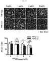
- monocyte-derived nanovesicles loaded with doxorubicin as a chemical drug or ribonuclease A (RNase A) as a protein drug were prepared. Thereafter, human umbilical vein endothelial cells (HUVECs), a human blood vessel cell line, were treated with TNF- ⁇ to activate the HUVECs, resulting in increased expression of cell adhesion molecules such as ICAM-1, VCAM-1, and E-selectin in cell membranes. It was confirmed whether the monocyte-derived nanovesicles recognize these molecules and the drugs are delivered to target cells. As a result, when TNF- ⁇ was treated, both doxorubicin and RNase A were delivered to HUVECs and induced cell death depending on the treatment concentration of the nanovesicles (see Example 7).
- doxorubicin as a chemical drug or ribonuclease A
- RNase A ribonuclease A
- the drug delivery capacity of transformed cell-derived nanovesicles was verified at the cellular level. After nanovesicles loaded with doxorubicin or RNase were prepared from HT1080 transformed cells overexpressing ICAM1, U937 monocyte cells were treated with different concentrations of the prepared nanovesicles and incubated for 24 hours. After incubation, the monocyte cells were stained with a trypan blue solution to measure the degree of cell death.
- the drugs were delivered into the monocyte cells by the nanovesicles and in the case of monocyte cells were treated with doxorubicin-loaded nanovesicles, cell death was induced at a concentration of 5 ⁇ g/ml or more and in the case of RNase A, cell death was induced at a concentration of 10 ⁇ g/ml or more (see Example 8).
- nanovesicles were prepared from transformed cells expressing adhesion molecules such as EGF and GE11 which were located in the membranes of the transformed cells and were capable of binding to EGFR expressed in the cell membranes of A549, a human lung cancer cell line. Then, the drug delivery capacity of the transformed cell-derived nanovesicles was confirmed.
- the nanovesicles loaded with doxorubicin were added to A549 cell-seeded plates and incubated. After incubation, the cells were observed under a fluorescence microscope. As a result, cell death was observed.
- GE11 and EGF present in nanovesicles which have been derived from transformed cells expressing EGF and GE11 in their cell membranes and loaded with anticancer drugs, may specifically bind to cells expressing EGFR on the cell surface and the loaded anticancer drugs may be delivered to cells to induce cell death (see Example 9).
- the present invention provides a composition for diagnosing diseases, including the nanovesicles of the present invention loaded with primers, probes, antisense nucleic acids, or antibodies required for diagnosis of diseases.
- the present invention provides a kit for diagnosing diseases, including the composition.
- Example 1 Preparation of nanovesicles by alkaline solution treatment and sonication
- nanovesicles derived from nucleated mammalian cells or transformed nucleated mammalian cells from which intracellular components had been removed monocytes or transformed cells were subjected to alkaline solution treatment, sonication, and a density gradient method, and a schematic diagram for preparing the nanovesicles is shown in FIG. 1 .
- the pellet was resuspended in 8 ml of the alkaline solution, stored in a rotor set at 4°C for 30 minutes, and was subjected to ultracentrifugation at 100,000 ⁇ g for 15 minutes to obtain a pellet.
- the obtained pellet was resuspended with 10 ml of a HEPES buffered saline (HBS) solution, and was again subjected to ultracentrifugation at 100,000 ⁇ g for 15 minutes to obtain a pellet.
- HBS HEPES buffered saline
- the pellet was suspended with 0.4 ml of a HBS solution, sonication was performed 30 times with a sonicator, sonication was performed again for 30 minutes in a water bath sonicator, and the solution was suspended with 6.6 ml of a HBS solution.
- 1 ml of 50% Optiprep, 2 ml of 10% Optiprep, and 7 ml of the suspension were sequentially added into an 11 ml ultracentrifuge tube, and ultracentrifugation was performed at 100,000 ⁇ g for 2 hours to obtain nanovesicles present in the layer between the 50% Optiprep and 10% Optiprep.
- the shape and size of the monocyte- and transformed cell-derived nanovesicles prepared by the method of Example 1 were analyzed.
- the nanovesicles derived from monocytes were observed using a transmission electron microscope.
- the nanovesicles derived from monocytes were adsorbed in a glow-discharged carbon-coated copper grid for 3 minutes.
- the grid was washed with distilled water, stained with 2% uranyl acetate for 1 minute, and observed with JEM101 (Jeol, Japan), an electron microscope.
- the nanovesicles prepared from monocytes using alkaline solution treatment were composed of lipid bilayers, had a size of 100 to 200 nm, and exhibited a generally spherical shape.
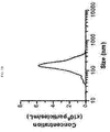
- the nanovesicles prepared from monocytes were diluted in 1 ml of HBS to a concentration of 0.5 ⁇ g/ml, 1 ml of the HBS solution containing the nanovesicles was placed in a chamber, and analyzed with a nanoparticle tracking analysis instrument.
- the size of the nanovesicles was 100 to 200 nm and the average size was 180 nm.
- nanovesicles prepared from transformed cells expressing epidermal growth factor (EGF) and a GE11 peptide (YHWYGYTPQNVI) were diluted in 1 ml of HBS to a concentration of 5 ⁇ g/ml, 1 ml of the HBS containing the nanovesicles was added to a cuvette and analyzed with a dynamic light scattering instrument.
- EGF epidermal growth factor
- YHWYGYTPQNVI GE11 peptide
- the nanovesicles prepared from the transformed cells had a size of 70 to 150 nm, and the average size thereof was 100 nm.
- nanovesicles prepared from monocytes using alkaline solution treatment according to the method of Example 1 contained cytoplasmic proteins and nucleoproteins
- western blotting was performed on the nanovesicles and monocyte cells.
- a blocking process was performed by soaking the membrane in 0.05% TBS-T (Tween-20) containing 3% non-fat milk and incubating the membrane for 2 hours. After blocking, the membrane was treated with primary antibodies that specifically bind to the respective proteins of ⁇ -actin, H2B, GM130, LFA1, and ICAM1, followed by reaction at room temperature for 2 hours. After washing the membrane three times with 0.05% TBS-T (Tween-20), the membrane was treated with secondary antibodies conjugated with peroxidase, followed by reaction at room temperature for 90 minutes.
- the membrane was washed three times with 0.05% TBS-T (Tween-20), and then the expression of each protein was determined using enhanced chemiluminescence (ECL, Amersham Co. No. RPN2106), WEST-ZOL (iNtRON. No. 16024), or Femto (Thermo Scientific. No. 37074).
- ECL enhanced chemiluminescence
- WEST-ZOL iNtRON. No. 16024
- Femto Thermo Scientific. No. 37074.
- ACTB ⁇ -actin
- H2B a protein constituting a histone in the nucleus
- MNV nanovesicles prepared using alkaline solution treatment
- nanovesicles prepared using alkaline solution treatment highly expressed cell membrane proteins such as LFA-1, ICAM1, and GM130.
- the nanovesicles prepared using alkaline treatment did not express cytoplasmic proteins and nucleoproteins, such as ⁇ -actin and H2B, whereas the nanovesicles contained more cell membrane proteins, such as LFA-1, ICAM1 and GM130, compared to cells.
- nanovesicles prepared from monocytes using alkaline solution treatment according to the method of Example 1 contained nucleic acids
- real-time RT-PCR and real-time PCR were performed on the nanovesicles and monocyte cells.
- nanovesicles prepared from monocytes and 1 ⁇ 10 5 monocyte cells were incubated at 95°C for 10 minutes. Consequently, the nanovesicles and the cells burst and all internal nucleic acids were released. Then, real-time RT-PCR and real-time PCR were performed to determine the expression level of GAPDH under conditions in which a DNA-degrading enzyme (e.g., DNase) was treated or not treated.
- a DNA-degrading enzyme e.g., DNase
- RNA was not present in the nanovesicles prepared from monocytes, whereas RNA was present in the monocyte cells.
- the solution containing the burst nanovesicles or monocyte cells were subjected to real-time PCR for GADPH without DNase treatment.
- DNA was not present in the nanovesicles prepared from monocytes whereas DNA was present in the monocyte cells.
- real-time PCR for GADPH was performed on the solution containing the burst nanovesicles or monocyte cells after DNase treatment, DNA was not present in the monocyte cells either. This result indicated that DNase had enzymatic activity.
- the nanovesicles prepared from monocytes were loaded with an enhanced green fluorescent protein (EGFP).
- EGFP enhanced green fluorescent protein
- Example 2 when nanovesicles were prepared from U937 cells, monocytes, as shown in Example 1, the pellet was suspended with 10 ml of a HEPES buffered saline (HBS) solution, ultracentrifugation was performed at 100,000 ⁇ g for 15 minutes to obtain a pellet. Then, the obtained pellet was suspended with HBS, EGFP was added to the HBS solution at a concentration of 50 ⁇ g/ml to prepare a solution with a final volume of 0.4 ml. The following procedures were performed in the same manner as Example 1, and monocyte-derived nanovesicles loaded with EGFP were obtained.
- HBS HEPES buffered saline
- Nanovesicles containing polyethylene glycol were prepared by loading polyethylene glycol into the monocyte-derived nanovesicles prepared according to the method of Example 1.
- suspension was performed by adding HBS, and the solution was extruded through a 1 ⁇ m filter (two-stack).
- 0.5 ml of 50% Optiprep, 1 ml of 10% Optiprep, and 3ml of the suspension were sequentially added into an ultracentrifuge tube, and then ultracentrifugation was performed at 100,000 ⁇ g for 2 hours to obtain nanovesicles present in the layer between the 50% Optiprep and 10% Optiprep.
- 5 ⁇ g/ml of cholesterol-polyethylene glycol-biotin was added and incubated at room temperature for 1 hour.
- 10 ⁇ g/ml of the nanovesicles containing polyethylene glycol was prepared, 100 ⁇ l of the prepared nanovesicles was added to each well of a 96 well plate, and incubated at room temperature for at least 12 hours to coat the wells of the plate. After washing three times with PBS, blocking was performed for 1 hour by adding 100 ⁇ l of 1% BSA/PBS. After blocking, the plate was washed three times with PBS, and then streptavidin-HRP was added and incubated for 20 minutes. Thereafter, a BM-POD substrate was added and the amount of color development was measured.
- the pellet was suspended with 10 ml of a HEPES buffered saline (HBS) solution, ultracentrifugation was performed at 100,000 ⁇ g for 15 minutes to obtain a pellet. Then, the obtained pellet was suspended with HBS, FITC-siRNA was added to the HBS solution at a concentration of 10 ⁇ M to prepare a solution with a final volume of 0.4 ml. Then, sonication was performed 30 times with a sonicator, sonication was performed again for 30 minutes in a water bath sonicator, and the solution was suspended with 0.6 ml of a HBS solution.
- HBS HEPES buffered saline
- the solution was extruded through a 1 ⁇ m filter (two-stack), and suspended with 2.0 ml HBS.
- 0.5 ml of 50% Optiprep, 1 ml of 10% Optiprep, and 3ml of the suspension were sequentially added into a 5 ml ultracentrifuge tube and then ultracentrifugation was performed at 100,000 ⁇ g for 2 hours to obtain nanovesicles loaded with FITC-siRNA in the layer between the 50% Optiprep and 10% Optiprep.
- nanovesicles prepared using the method contained FITC-siRNA
- 50 ⁇ l of the nanovesicles loaded with FITC-siRNA were prepared, and a DiI solution at a concentration of 10 ⁇ M was added and the nanovesicles were suspended.
- the solution was placed on a cover glass, the cover glass was placed in a humid chamber, and stored at 4°C for at least 12 hours.
- 5 ⁇ l of a mounting solution was loaded on a slide glass, the cover glass was placed thereon, and observation was performed using a fluorescence microscope.
- the nanovesicles were observed by the red fluorescence of Dil (1,1'-dioctadecyl-3,3,3'3'-tetramethylindocarbocyanine perchlorate), and siRNA was observed by the green fluorescence of FITC. The results are shown in FIG. 8 .
- nanovesicles not loaded with FITC-siRNA were labeled with DiI and exhibited red fluorescence, but did not exhibit the green fluorescence of FITC-siRNA.
- nanovesicles loaded with FITC-siRNA exhibited the red fluorescence of DiI as well as the green fluorescence of FITC-siRNA. The presence of green fluorescence and red fluorescence in the same location confirmed that FITC-siRNA was loaded into the nanovesicles.
- nanovesicles loaded with Qdot 705 the same method used in preparation of the FITC-siRNA-loaded nanovesicles of Example 4 was used, and in the step of sonication, 20 nM Qdot 705 was added instead of FITC-siRNA.
- nanovesicles prepared using the method contained Qdot 705
- 50 ⁇ l of nanovesicles loaded with 20 ⁇ g/ml of Qdot 705 were prepared. Samples were prepared in the same method as Example 4, and observation was performed using a fluorescence microscope. The nanovesicles were observed by the red fluorescence of Dil, and Qdot 705 was observed by a blue pseudo color.
- nanovesicles not loaded with Qdot 705 were labeled with DiI and exhibited red fluorescence, but did not exhibit the blue fluorescence of Qdot 705.
- nanovesicles loaded with Qdot 705 exhibited the red fluorescence of DiI as well as blue fluorescence. The presence of blue fluorescence and red fluorescence in the same location confirmed that Qdot 705 was loaded into the nanovesicles.
- nanovesicles loaded with iron oxide nanoparticles the same method used in preparation of the FITC-siRNA-loaded nanovesicles of Example 4 was used, in the step of sonication, 20 ⁇ g/ml of iron oxide nanoparticles was added instead of FITC-siRNA. Then, nanovesicles (MNV iron oxide ) loaded with iron oxide nanoparticles by a density gradient method using an Optiprep solution were compared with nanovesicles (MNV control ) not loaded with iron oxide nanoparticles.
- nanovesicles of the present invention deliver chemical drugs in a cell-specific-manner in vitro
- monocyte-derived nanovesicles loaded with doxorubicin, an anti-cancer agent were prepared and a fluorescence microscope was used to verify whether the drug was delivered in a cell-specific manner.
- HUVECs human umbilical vein endothelial cells
- ICAM-1, VCAM-1, and E-selectin bind to cell adhesion molecules such as LFA-1 and Mac-1 present in monocytes and macrophages. This protein interaction allows monocyte and macrophages to bind to blood endothelial cells.
- monocyte-derived nanovesicles loaded with doxorubicin were prepared according to the method of Example 1.
- step of sonication 800 ⁇ g/ml of doxorubicin was added, and sonication was performed to prepare nanovesicles loaded with doxorubicin.
- HUVECs were treated with 0, 1, 2, or 5 ⁇ g/ml of nanovesicles loaded with doxorubicin for 20 minutes, replaced with a fresh medium, and cultured for 36 hours.
- HUVECs were treated with 2 ⁇ M CellTracker (CellTracker, Invitrogen. No. C2925), incubated for 1 hour, and then treated with 4% paraformaldehyde for cell fixation. Then, the 4% paraformaldehyde was removed, PBS was added, and a fluorescence microscope was used to observe the cells and measure the number of cells.
- CellTracker CellTracker, Invitrogen. No. C2925
- the number of cells decreased to some extent, indicating that nonspecific drug delivery partially occurred.
- TNF- ⁇ was treated at various concentrations, the number of cells significantly decreased depending on the concentration of the nanovesicles. This indicated that more doxorubicin was delivered to HUVECs due to the presence of the nanovesicles. Based on these results, it was confirmed that the nanovesicles may be used to deliver chemical drugs in a cell-specific manner.
- nanovesicles of the present invention deliver protein drugs in a cell-specific-manner in vitro
- monocyte-derived nanovesicles loaded with ribonuclease A were prepared and a fluorescence microscope was used to verify whether RNase A was delivered in a cell-specific manner.
- Monocyte-derived nanovesicles loaded with RNase A were prepared according to the method of Example 1. In the step of sonication, 800 ⁇ g/ml of RNase A was added, and sonication was performed to prepare nanovesicles loaded with RNase A. Samples were prepared by the same principle and method as Example 7-1, and cells were observed under a fluorescence microscope, and then the number of cells was measured.
- nanovesicles prepared from transformed cells deliver drugs in a cell-specific manner like the monocyte-derived nanovesicles of Example 7, monocyte-derived nanovesicles loaded with doxorubicin, an anti-cancer agent, or RNase A were prepared. Monocyte cells were treated with the prepared nanovesicles, and then the number of living cells was measured.
- Nanovesicles which were derived from HT1080 transformed cells expressing ICAM-1 and loaded with doxorubicin or RNase A, were prepared according to the method of Example 1. In the step of sonication, 800 ⁇ g/ml of doxorubicin or RNase A was added, and sonication was performed to prepare nanovesicles loaded with doxorubicin or RNase A.
- nanovesicles prepared from transformed HT1080 cells expressing ICAM-1 were not loaded with drugs or loaded with drugs, and two types of nanovesicles were compared.
- cell death was not induced by treatment with a high concentration of nanovesicles.
- doxorubicin-loaded nanovesicles were treated, cell death was induced from 5 ⁇ g/ml.
- RNase A-loaded nanovesicles cell death was induced from 10 ⁇ g/ml.
- Example 9 Validation of in vitro drug delivery and cell-specific delivery using nanovesicles derived from transformed cells expressing GE11 and EGF in cell membranes
- Each well of a 12 well plate was seeded with 5 ⁇ 10 4 A549 cells, a human lung cancer cell line, expressing EGFR, and the cells were cultured for 16 hours.
- EGFR present in A549 is capable of binding to adhesion molecules such as EGF and GE11.
- the plate seeded with the A549 cells was washed with PBS, and 1 ml of a medium was added to each well of the plate. Then, 0.1 ⁇ 10 9 , 1 ⁇ 10 9 , or 10 ⁇ 10 9 particles/ml of nanovesicles, which had been prepared from transformed cells expressing EGF and GE11 in the membranes according to Example 1 and loaded with doxorubicin, were added to each well of the plate, followed by incubation for 20 minutes. After incubation, each well was washed with PBS, 1 ml of a medium was added thereto, and cultured for 36 hours.
- nanovesicles which had been derived from transformed cells expressing EGF and GE11 in the cell membranes and loaded with anticancer drugs, were capable of binding to cells expressing EGFR on the cell surface, and consequently, the loaded anticancer drugs were selectively delivered to the EGFR expressing cells, resulting in cell death.
Abstract
Description
- The present invention relates to cell membrane-derived nanovesicles, a method of preparing the same, and a pharmaceutical composition and a diagnostic kit using the nanovesicles.
- When a drug is administered to an individual for disease treatment, a drug delivery system (DDS) that controls the delivery and release of the administered drug into cells, tissues, and organs for optimal efficacy is important. The drug delivery system is used to maintain adequate blood concentrations of a drug to achieve the maximum therapeutic effect. When the drug delivery system is applied, advantages such as maximizing the efficacy and stability of a drug, prolonging the residence time of a drug, increasing the bioavailability of a drug, and minimizing the side effects of a drug can be achieved. Thus, the drug delivery system is a medical technique required to efficiently deliver a drug to specific body parts and is considered as important as medicine itself. Since the early 1960s, drug delivery systems have been developed in the forms of injection and infusion, in the form of suppositories, for nasal and oral administration, and for administration via the skin or the lungs. In recent years, various drug delivery systems (DDSs), such as absorption-promoting and controlled DDSs (oral, transdermal, mucosal), sustained DDSs (injections, oral, transdermal), targeted DDSs (oral, injections, etc.), and artificial intelligence (intelligent) DDSs (on-demand type), have been developed and applied differently according to patients and purpose of use.
- Among DDSs, liposomes have been widely studied as a drug delivery system since liposomes were first used in the 1960s. Liposomes are artificial membranes similar to cell membranes made of various phospholipids, and can be seen as colloidal semi-solid dispersants with a nanoscale particle size. Liposomal preparations have low toxicity and are highly applicable because the size, charge, membrane composition, and membrane permeability of the liposomal preparations may be controlled according to prescription. Recently, stealth liposomes prepared by conjugating liposomes with a polymer such as polyethylene glycol (PEG) have been developed. The stealth liposomes increase the circulatory half-life of drugs by preventing drug-containing liposomes from being easily removed from the blood. Using this method, DOXIL that delivers doxorubicin, an anti-cancer agent, has been commercialized. However, since liposomes and stealth liposomes lack the ability to recognize specific types of cells, the liposomes and stealth liposomes may not be used for the purpose of delivering drugs to specific types of cells or tissues. To enable liposomes to bind to specific targets, liposomes conjugated with antibodies specific to a single target have been developed, but none have been commercialized after passing clinical trials.
- Therefore, a delivery system using natural cell membranes instead of liposomes made of artificially synthesized lipids is being developed. Since cell-derived delivery systems utilize the natural targeting systems of the cells, unlike conventional delivery systems such as liposomes, targeting may be easily induced. As a representative example, studies on drug delivery using extracellular vesicles which are naturally secreted from cells are underway. In addition, studies have been reported on loading various drugs, such as anticancer drugs, into nanovesicles (microvesicles) artificially prepared from nucleated mammalian cells and delivering the nanovesicles to cancer tissues.
- In addition to substances required for their targeting, however, the extracellular vesicles and the nanovesicles artificially prepared from cells include intracellular components, such as cytosolic proteins and genetic materials (DNA and RNA), which are unnecessary for targeting. The components unnecessary for their targeting have a potential to lower the efficiency of drug loading or to induce side effects when administered into the body, and thus development of systems that are lacking their intraluminal components is essential.
- The present inventors have conducted studies for solving the conventional problems as described above. As a result, the inventors have found that, when cells are treated with an alkaline solution of pH 9 to 14, the membranes of the cells from which intracellular components, such as genetic materials and cytosolic proteins, are removed may be selectively extracted and that nanovesicles may be prepared using these extracted cell membranes. Thus, the present invention was completed.
- Accordingly, the present invention is directed to providing nanovesicles, wherein the nanovesicles are derived from the membranes of cells, the intracellular components of which have been removed by treating the cells with an alkaline solution of pH 9 to 14, and the size of the nanovesicles is smaller than that of the cells, and a method of delivering drugs using the same.
- It is another object of the present invention to provide a method of preparing cell membrane-derived nanovesicles loaded with substances for treating or diagnosing diseases.
- However, the technical problems to be solved by the present invention are not limited to the above-mentioned problems, and other problems not mentioned can be clearly understood by those skilled in the art from the following description.
- One aspect of the present invention provides nanovesicles, wherein the nanovesicles are derived from the membranes of cells, the intracellular components of which have been removed by treating the cells with an alkaline solution of pH 9 to 14, and the size of the nanovesicles is smaller than that of the cells.
- In one embodiment of the present invention, the alkaline solution may be prepared using one or more selected from the group consisting of sodium carbonate (Na2CO3), sodium hydroxide (NaOH), ammonia (NH3), calcium hydroxide (Ca(OH)2), potassium hydroxide (KOH), sodium hydrogen carbonate (NaHCO3), and magnesium hydroxide (Mg(OH)2).
- In another embodiment of the present invention, the cells may be selected from the group consisting of monocytes, macrophages, dendritic cells, and stem cells as nucleated mammalian cells.
- In another embodiment of the present invention, the cells may be transformed cells that target specific types of cells or tissues.
- In another embodiment of the present invention, the cells may be transformed cells that express one or more selected from the group consisting of cell adhesion molecules, antibodies, targeting proteins, cell membrane fusion proteins, and fusion proteins thereof.
- In another embodiment of the present invention, the cells may be transformed cells that express one or more selected from the group consisting of growth factors, cytokines, receptors, fluorescent proteins, and fusion proteins thereof.
- In another embodiment of the present invention, the membranes of the nanovesicles may further include a component other than the membranes of the cells.
- In another embodiment of the present invention, the component other than the cell membrane may be cyclodextrin or polyethylene glycol.
- In another embodiment of the present invention, the nanovesicles may have a chemically modified membrane component.
- In another embodiment of the present invention, the nanovesicles may retain the topology of the membrane proteins of the cells from which the nanovesicles originated.
- Another aspect of the present invention provides a pharmaceutical composition for delivering substances, including the nanovesicles loaded with substances for treating or diagnosing diseases.
- In one embodiment of the present invention, the substances for treating or diagnosing diseases may be injected.
- In another embodiment of the present invention, the substances for treating or diagnosing diseases may be one or more selected from the group consisting of anti-cancer agents, anti-inflammatory agents, angiogenesis inhibitors, peptides, proteins, toxins, nucleic acids, beads, microparticles, and nanoparticles.
- In another embodiment of the present invention, the nucleic acids may be selected from the group consisting of DNA, RNA, aptamers, locked nucleic acids (LNAs), peptide nucleic acids (PNAs), and morpholinos.
- In another embodiment of the present invention, the nanoparticles may be selected from the group consisting of iron oxides, gold, carbon nanotubes, and magnetic beads.
- In another embodiment of the present invention, the substances for treating or diagnosing diseases may be fluorescence-emitting materials.
- In another embodiment of the present invention, the fluorescence-emitting materials may be fluorescent proteins or quantum dots.
- Still another aspect of the present invention provides a method of preparing cell membrane-derived nanovesicles loaded with substances for treating or diagnosing diseases, the method including the following steps:
- (a) a step of removing intracellular materials by treating cells with an alkaline solution of pH 9 to 14;
- (b) a step of adding therapeutic or diagnostic substances into a suspension containing the membranes of the cells from which the intracellular materials have been removed;
- (c) a step of preparing nanovesicles by applying a sonication method to the suspension to which the therapeutic or diagnostic substances are added; and
- (d) a step of separating the prepared nanovesicles from the suspension.
- Yet another aspect of the present invention provides a method of preparing cell membrane-derived nanovesicles loaded with substances for treating or diagnosing diseases, the method including the following steps:
- (a) a step of removing intracellular materials by treating cells with an alkaline solution of pH 9 to 14;
- (b) a step of preparing nanovesicles by applying a sonication method to a suspension containing the membranes of the cells from which the intracellular materials have been removed;
- (c) a step of adding therapeutic or diagnostic substances into the suspension containing the nanovesicles and incubating the mixture; and
- (d) a step of separating the incubated nanovesicles from the suspension.
- In one embodiment of the present invention, the method may further include a step of extruding the nanovesicles prepared by the sonication method.
- In another embodiment of the present invention, the alkaline solution may be prepared using one or more selected from the group consisting of sodium carbonate (Na2CO3), sodium hydroxide (NaOH), ammonia (NH3), calcium hydroxide (Ca(OH)2), potassium hydroxide (KOH), sodium hydrogen carbonate (NaHCO3), and magnesium hydroxide (Mg(OH)2).
- In another embodiment of the present invention, the cells may be selected from the group consisting of monocytes, macrophages, dendritic cells, and stem cells as nucleated mammalian cells.
- In another embodiment of the present invention, the cells may be transformed cells that target specific types of cells or tissues.
- In another embodiment of the present invention, the cells may be transformed cells that express one or more selected from the group consisting of cell adhesion molecules, antibodies, targeting proteins, cell membrane fusion proteins, and fusion proteins thereof.
- In another embodiment of the present invention, the cells may be transformed cells that express one or more selected from the group consisting of growth factors, cytokines, receptors, fluorescent proteins, and fusion proteins thereof.
- In another embodiment of the present invention, the membranes of the nanovesicles may further include a component other than the membranes of the cells.
- In another embodiment of the present invention, the component other than the cell membrane may be cyclodextrin or polyethylene glycol.
- In another embodiment of the present invention, the nanovesicles may have a chemically modified membrane component.
- In another embodiment of the present invention, the nanovesicles may retain the topology of the membrane proteins of the cells from which the nanovesicles originated.
- In another embodiment of the present invention, the therapeutic or diagnostic substances may be injected from outside the cells.
- In another embodiment of the present invention, the substances for treating or diagnosing diseases may be one or more selected from the group consisting of anti-cancer agents, anti-inflammatory agents, angiogenesis inhibitors, peptides, proteins, toxins, nucleic acids, beads, microparticles, and nanoparticles.
- In another embodiment of the present invention, the nucleic acids may be selected from the group consisting of DNA, RNA, aptamers, locked nucleic acids (LNAs), peptide nucleic acids (PNAs), and morpholinos.
- In another embodiment of the present invention, the nanoparticles may be selected from the group consisting of iron oxides, gold, carbon nanotubes, and magnetic beads.
- In another embodiment of the present invention, the substances for treating or diagnosing diseases may be fluorescence-emitting materials.
- In another embodiment of the present invention, the fluorescence-emitting materials may be fluorescent proteins or quantum dots.
- Yet another aspect of the present invention provides a composition for diagnosing diseases, including the nanovesicles of the present invention loaded with primers, probes, antisense nucleic acids, or antibodies required for diagnosis of diseases.
- Yet another aspect of the present invention provides a kit for diagnosing diseases, including the composition of the present invention.
- Yet another aspect of the present invention provides a method of delivering substances, the method including a step of delivering therapeutic or diagnostic substances into the specific types of cells or tissues using the nanovesicles of the present invention.
- Yet another aspect of the present invention provides a method of treating or diagnosing diseases, the method including a step of delivering therapeutic or diagnostic substances into the specific types of cells or tissues using the nanovesicles of the present invention
- Yet another aspect of the present invention provides a therapeutic or diagnostic use of the nanovesicles of the present invention.
- The cell membrane-derived nanovesicles according to the present invention can prevent the occurrence of potential side effects because intracellular or intraluminal materials (e.g., genetic materials and cytosolic proteins) unnecessary for delivery of therapeutic or diagnostic substances are removed from the extracellular vesicles and artificial nanovesicles. In addition, since the nanovesicles can be targeted to the specific types of cells or tissues, therapeutic or diagnostic substances may be predominantly delivered to the targeted cells or tissues while delivery of therapeutic or diagnostic substances to untargeted sites may be inhibited. Therefore, when the cell membrane-derived nanovesicles are applied to disease treatment, the side effects of therapeutic substances such as drugs can be reduced, so that suffering and inconvenience of patients can be alleviated during the course of treating diseases, and therapeutic efficacy can be improved. In addition, the cell membrane-derived nanovesicles of the present invention, in which substances for the treatment or diagnosis of diseases are loaded, and a method of preparing the nanovesicles can be used in vitro or in vivo for therapeutic or diagnostic purposes, or for experimental use.
-
-
FIG. 1 illustrates nanovesicles derived from nucleated mammalian cells or transformed nucleated mammalian cells and shows a schematic diagram of a method of preparing the nanovesicles loaded with various substances, such as targeting substances and therapeutic and diagnostic substances. -
FIG. 2A shows a transmission electron microscope image of nanovesicles prepared from nucleated mammalian cells using alkaline solution treatment, sonication, and a density gradient method. -
FIG. 2B is a graph showing the size of nanovesicles prepared from nucleated mammalian cells using alkaline solution treatment, sonication, and a density gradient method. -
FIG. 3 is a graph showing the size of nanovesicles derived from transformed nucleated mammalian cells. -
FIG. 4 shows western blotting results confirming that cytoplasmic proteins and nucleoproteins are absent in nanovesicles derived from nucleated cells and that a large amount of membrane proteins is present. -
FIG. 5 shows the results of real-time RT-PCR and RT-PCR confirming that DNA and RNA are not present in nanovesicles derived from nucleated mammalian cells. -
FIG. 6 shows that the topology of nanovesicles derived from nucleated mammalian cells is normal. This was confirmed by determining whether an ICAM1 protein of nanovesicles derived from nucleated mammalian cells and an EGFP protein loaded into the nanovesicles were expressed, after trypsin treatment. -
FIG. 7 shows the binding of cholesterol-polyethylene glycol-biotin to the bilayer membranes of nanovesicles derived from nucleated mammalian cells. -
FIG. 8 shows fluorescence microscope images confirming that FITC-siRNA was loaded into nanovesicles derived from nucleated mammalian cells. -
FIG. 9 shows fluorescence microscope images confirming thatQdot 705 was loaded into nanovesicles derived from nucleated mammalian cells. -
FIG. 10 shows an image confirming that iron oxide nanoparticles were loaded into nanovesicles derived from nucleated mammalian cells. The iron oxide nanoparticles were loaded into the nanovesicles using a density gradient method in an Optiprep solution. -
FIG. 11 shows that, under TNF-α treatment conditions, nanovesicles derived from nucleated mammalian cells can be used to deliver doxorubicin into target cells and doxorubicin-induced cell death was increased when the nanovesicles were used. -
FIG. 12 shows that, under TNF-α treatment conditions, nanovesicles derived from nucleated mammalian cells can be used to deliver ribonuclease A (RNase A) into target cells and ribonuclease A-induced cell death was increased when the nanovesicles were used. -
FIG. 13 shows that nanovesicles derived from transformed nucleated cells can be used to deliver doxorubicin or RNase A into the specific types of cells and doxorubicin- or RNase A-induced cell death was increased when the nanovesicles were used. -
FIG. 14 shows that nanovesicles derived from transformed nucleated cells can be used to specifically deliver doxorubicin into human lung cancer cells and doxorubicin-induced cell death was increased when the nanovesicles were used. - The present invention provides nanovesicles, wherein the nanovesicles are derived from the membranes of cells, the intracellular components of which have been removed by treating the cells with an alkaline solution of pH 9 to 14, and the size of the nanovesicles is smaller than that of the cells.
- The inside and outside of the nanovesicles of the present invention are distinguished by a lipid bilayer membrane composed of the cell membrane components of an originating cell. These nanovesicles include cell membrane lipids and proteins of the originating cells and have the same topologies as the originating cells. However, intracellular materials, such as genetic material (e.g., nucleic acids) and cytosolic proteins, are removed from the nanovesicles, and the size of the nanovesicles may be smaller than that of the originating cell, without being limited thereto.
- The alkaline solution of the present invention may be prepared using one or more selected from the group consisting of sodium carbonate (Na2CO3), sodium hydroxide (NaOH), ammonia (NH3), calcium hydroxide (Ca(OH)2), potassium hydroxide (KOH), sodium hydrogen carbonate (NaHCO3), and magnesium hydroxide (Mg(OH)2), without being limited thereto.
- The cells of the present invention may be selected from the group consisting of monocytes, macrophages, dendritic cells, and stem cells as nucleated mammalian cells, and may be also selected from the group consisting of cells differentiated from stem cells, and in particular, may include transformed cells that target specific types of cells or tissues. Specifically, the transformed cells may include various types of cells, which are transformed to express therapeutic and diagnostic substances, targeting substances, or substances necessary for cell membrane fusion with target cells, and the transformed cells may include a combination of two or more of the above cell types, without being limited thereto.
- The nucleated mammalian cells may be transformed by material treatment or gene introduction, transformed more than once, and transformed to inhibit the expression of one or more specific proteins.
- As used herein, the term, "transformation" refers to a method of increasing or decreasing the expression of substances such as proteins by stimulating cells and a method of changing the expression of proteins by gene introduction, and methods commonly used in the art may be used. As methods of increasing or inhibiting the expression of specific proteins by gene introduction, RNA interference (RNAi), virus-mediated methods, calcium phosphate precipitation, liposomes, electroporation, microinjection, ultrasound-mediated methods, and the like may be exemplified, and all generally known methods may be used.
- The cells of the present invention may be transformed cells that express one or more selected from the group consisting of cell adhesion molecules, antibodies, targeting proteins, cell membrane fusion proteins, and fusion proteins thereof, and may be also transformed cells that express one or more selected from the group consisting of growth factors, cytokines, receptors, fluorescent proteins, and fusion proteins thereof, without being limited thereto.
- The membranes of the nanovesicles of the present invention may include targeting substances, substances necessary for cell membrane fusion with target cells (e.g., fusogen), cyclodextrin, polyethylene glycol, and the like in addition to the cell membranes of originating cells, and the components other than the cell membranes may be added by various methods, including chemical modification of the cell membranes.
- In addition, the present invention provides a pharmaceutical composition for delivering substances, including the nanovesicles loaded with substances for treating or diagnosing diseases.
- According to the present invention, the nanovesicles may be prepared in various sizes depending on tissues targeted by diagnostic or therapeutic substances, and the size of the nanovesicles is preferably 10 nm or more and 10 µm or less.
- Substances to be loaded into the nanovesicles of the present invention are not particularly limited, and may be therapeutic or diagnostic substances. In addition, substances prepared outside cells may be loaded, and the loaded substances may number one or more.
- The substances for treating or diagnosing diseases may be one or more selected from the group consisting of anti-cancer agents, anti-inflammatory agents, angiogenesis inhibitors, peptides, proteins, toxins, nucleic acids, beads, microparticles, and nanoparticles, without being limited thereto.
- The nucleic acids may be selected from the group consisting of DNA, RNA, aptamers, locked nucleic acids (LNAs), peptide nucleic acids (PNAs), and morpholinos, and the nanoparticles may be selected from the group consisting of iron oxides, gold, carbon nanotubes, and magnetic beads, without being limited thereto.
- The substances for treating or diagnosing diseases according to the present invention may be fluorescence-emitting materials, preferably fluorescent proteins or quantum dots, without being limited thereto.
- The pharmaceutical composition of the present invention may contain one or more of anti-cancer agents, anti-inflammatory agents, angiogenesis inhibitors, peptides, proteins, toxins, nucleic acids, beads, microparticles or nanoparticles as active ingredients. In addition, the pharmaceutical composition may be mixed with pharmaceutically acceptable carriers such as saline, sterilized water, Ringer's solution, buffered saline, cyclodextrin, a dextrose solution, a maltodextrin solution, glycerol, ethanol, liposomes and one or more of these carriers, and the pharmaceutical composition may further contain other conventional additives such as antioxidants, buffers, and the like, when desired. In addition, diluents, dispersants, surfactants, binders, or lubricants may be additionally added to the pharmaceutical composition, and thus the pharmaceutical composition may be formulated into injectable formulations, such as aqueous solutions, suspensions and emulsions, pills, capsules, granules or tablets. Furthermore, the pharmaceutical composition may be suitably formulated according to each component using appropriate methods in the art or methods disclosed in the Remington's reference (Remington's Pharmaceutical Science, Mack Publishing Company, Easton PA). The pharmaceutical composition of the present invention is not particularly limited in formulation, but is preferably formulated as an injection or an inhalant.
- The method of administering the pharmaceutical composition of the present invention is not particularly limited, but the pharmaceutical composition may be administered parenterally, such as intravenously, subcutaneously, intraperitoneally, by inhalation or topical application, or orally, depending on the purpose. The dosage may be variously determined depending on a patient's body weight, age, sex, health condition, diet, administration time, administration methods, excretion amount and severity of diseases. Daily dose refers to the amount of a therapeutic substance capable of alleviating disease status upon administration of the therapeutic substance to an individual in need of treatment. The effective amount of the therapeutic substance depends on particular compounds, disease status and disease severity, individuals in need of treatment, which may be routinely determined by those of ordinary skill in the art. As a non-limiting example, the dosage of the composition of the present invention to human body may be varied depending on the patient's age, weight, sex, dosage form, health condition and disease severity. On the basis of an adult patient weighing 70 kg, the dose is generally 0.1 to 1,000 mg/day, preferably 1 to 500 mg/day. The composition may also be administered dividedly once or several times a day at regular time intervals.
- In addition, the present invention provides a method of preparing cell membrane-derived nanovesicles loaded with substances for treating or diagnosing diseases, the method including a step of removing intracellular materials by treating cells with an alkaline solution of pH 9 to 14; a step of adding therapeutic or diagnostic substances into a suspension containing the membranes of the cells from which the intracellular materials have been removed; a step of preparing nanovesicles by applying a sonication method to the suspension to which the therapeutic or diagnostic substances are added; and a step of separating the prepared nanovesicles from the suspension.
- In addition, the present invention provides a method of preparing cell membrane-derived nanovesicles loaded with substances for treating or diagnosing diseases, the method including a step of removing intracellular materials by treating cells with an alkaline solution of pH 9 to 14; a step of preparing nanovesicles by applying a sonication method to a suspension containing the membranes of the cells from which the intracellular materials have been removed; a step of adding therapeutic or diagnostic substances into the suspension containing the nanovesicles and incubating the mixture; and a step of separating the incubated nanovesicles from the suspension.
- The method of the present invention may further include a step of extruding the nanovesicles prepared by the sonication method.
- In the present invention, nanovesicles were prepared from a monocyte cell line, U937, and transformed HEK293 and HT1080 cells using the method of preparing the nanovesicles, i.e., using alkaline solution treatment, sonication, and a density gradient method (see Example 1).
- In one embodiment of the present invention, when the shape and size of nanovesicles prepared from monocytes and transformed cells were determined, the nanovesicles were in the form of a spherical lipid bilayer, and monocyte-derived nanovesicles exhibited a size of 100 to 200 nm, i.e., 180 nm on average, while transformed cell-derived nanovesicles exhibited a size of 70 to 150 nm, i.e., 100 nm on average.
- In addition, when western blotting was performed on the monocyte-derived nanovesicles and monocyte cells to determine the expression levels of β-actin (cytoplasmic protein), H2B (nucleoprotein), GM130, LFA1 and ICAM1 (cell membrane proteins), the nanovesicles did not contain cytoplasmic proteins and nucleoproteins, while cell membrane proteins were expressed in the nanovesicles. In addition, as a result of real-time PCR, it was confirmed that the nanovesicles do not contain DNA and RNA, i.e., nucleic acids, unlike monocyte cells. As a result of analyzing of the topology of the nanovesicles, the membrane proteins of the nanovesicles were located outside the nanovesicles, like the membrane proteins of monocyte cells. That is, the membrane proteins of the nanovesicles maintained the same topology as the membrane proteins of the monocyte cells (see Example 2).
- In another embodiment of the present invention, nanovesicles containing polyethylene glycol were prepared by adding cholesterol-polyethylene glycol-biotin to the monocyte-derived nanovesicles prepared according to the method of Example 1. Thereafter, streptavidin-HRP was added to the nanovesicles, followed by incubation. After incubation, BM-POD as a substrate was added thereto to measure the amount of color development. As a result, it was confirmed that cholesterol-polyethylene glycol-biotin was bound to the nanovesicles containing polyethylene glycol (see Example 3).
- In another embodiment of the present invention, when the nanovesicles were prepared using alkaline solution treatment, sonication, and a density gradient method according to the method of Example 1, in the step of performing sonication, FITC-siRNA,
Qdot 705, or iron oxide nanoparticles were each loaded into nanovesicles, and the nanovesicles containing each of FITC-siRNA,Qdot 705, or iron oxide nanoparticles were prepared. After the preparation, it was inspected whether each substance was appropriately loaded into the nanovesicles. Fluorescence microscopy observation revealed that each of FITC-siRNA andQdot 705 was properly loaded into nanovesicles. In addition, the density gradient method using an Optiprep solution confirmed that iron oxide nanoparticles were also loaded into nanovesicles (see Examples 4 to 6). - In another embodiment of the present invention, it was demonstrated that drug delivery at the cellular level and cell-specific drug delivery are possible when monocyte-derived nanovesicles are used. Monocyte-derived nanovesicles loaded with doxorubicin as a chemical drug or ribonuclease A (RNase A) as a protein drug were prepared. Thereafter, human umbilical vein endothelial cells (HUVECs), a human blood vessel cell line, were treated with TNF-α to activate the HUVECs, resulting in increased expression of cell adhesion molecules such as ICAM-1, VCAM-1, and E-selectin in cell membranes. It was confirmed whether the monocyte-derived nanovesicles recognize these molecules and the drugs are delivered to target cells. As a result, when TNF-α was treated, both doxorubicin and RNase A were delivered to HUVECs and induced cell death depending on the treatment concentration of the nanovesicles (see Example 7).
- In another embodiment of the present invention, the drug delivery capacity of transformed cell-derived nanovesicles was verified at the cellular level. After nanovesicles loaded with doxorubicin or RNase were prepared from HT1080 transformed cells overexpressing ICAM1, U937 monocyte cells were treated with different concentrations of the prepared nanovesicles and incubated for 24 hours. After incubation, the monocyte cells were stained with a trypan blue solution to measure the degree of cell death. As a result, the drugs were delivered into the monocyte cells by the nanovesicles and in the case of monocyte cells were treated with doxorubicin-loaded nanovesicles, cell death was induced at a concentration of 5 µg/ml or more and in the case of RNase A, cell death was induced at a concentration of 10 µg/ml or more (see Example 8).
- In another embodiment of the present invention, nanovesicles were prepared from transformed cells expressing adhesion molecules such as EGF and GE11 which were located in the membranes of the transformed cells and were capable of binding to EGFR expressed in the cell membranes of A549, a human lung cancer cell line. Then, the drug delivery capacity of the transformed cell-derived nanovesicles was confirmed. The nanovesicles loaded with doxorubicin were added to A549 cell-seeded plates and incubated. After incubation, the cells were observed under a fluorescence microscope. As a result, cell death was observed. Based on the result, it was confirmed that GE11 and EGF present in nanovesicles, which have been derived from transformed cells expressing EGF and GE11 in their cell membranes and loaded with anticancer drugs, may specifically bind to cells expressing EGFR on the cell surface and the loaded anticancer drugs may be delivered to cells to induce cell death (see Example 9).
- In addition, the present invention provides a composition for diagnosing diseases, including the nanovesicles of the present invention loaded with primers, probes, antisense nucleic acids, or antibodies required for diagnosis of diseases.
- In addition, the present invention provides a kit for diagnosing diseases, including the composition.
- Hereinafter, preferred examples are described to facilitate understanding of the present invention. However, the following examples are provided only for the purpose of easier understanding of the present invention, and the present invention is not limited by the following examples.
- To prepare nanovesicles derived from nucleated mammalian cells or transformed nucleated mammalian cells from which intracellular components had been removed, monocytes or transformed cells were subjected to alkaline solution treatment, sonication, and a density gradient method, and a schematic diagram for preparing the nanovesicles is shown in
FIG. 1 . - 8 × 10⁷ U937 cells (ATCC No. CRL-1593.2) were used as monocytes and 4 × 10⁷ HEK293 cells (ATCC No. CRL-1573) or 4 × 10⁷ HT1080 cells (ATCC No. CCL-121) were used as transformed cells. Each cell type was treated with 8 ml of an alkaline solution (200 mM Na2CO3, 1× phosphatase inhibitor, pH 11.5), the solution was sonicated 30 times with a sonicator under the conditions of cycle: 0.5 and amplitude: 50, and ultracentrifugation was performed at 100,000 × g for 15 minutes to obtain a pellet. The pellet was resuspended in 8 ml of the alkaline solution, stored in a rotor set at 4°C for 30 minutes, and was subjected to ultracentrifugation at 100,000 × g for 15 minutes to obtain a pellet. The obtained pellet was resuspended with 10 ml of a HEPES buffered saline (HBS) solution, and was again subjected to ultracentrifugation at 100,000 × g for 15 minutes to obtain a pellet. The pellet was suspended with 0.4 ml of a HBS solution, sonication was performed 30 times with a sonicator, sonication was performed again for 30 minutes in a water bath sonicator, and the solution was suspended with 6.6 ml of a HBS solution. Next, 1 ml of 50% Optiprep, 2 ml of 10% Optiprep, and 7 ml of the suspension were sequentially added into an 11 ml ultracentrifuge tube, and ultracentrifugation was performed at 100,000 × g for 2 hours to obtain nanovesicles present in the layer between the 50% Optiprep and 10% Optiprep.
- The shape and size of the monocyte- and transformed cell-derived nanovesicles prepared by the method of Example 1 were analyzed. First, the nanovesicles derived from monocytes were observed using a transmission electron microscope. The nanovesicles derived from monocytes were adsorbed in a glow-discharged carbon-coated copper grid for 3 minutes. The grid was washed with distilled water, stained with 2% uranyl acetate for 1 minute, and observed with JEM101 (Jeol, Japan), an electron microscope.
- As a result, as shown in
FIG. 2a , it was found that the nanovesicles prepared from monocytes using alkaline solution treatment were composed of lipid bilayers, had a size of 100 to 200 nm, and exhibited a generally spherical shape. In addition, the nanovesicles prepared from monocytes were diluted in 1 ml of HBS to a concentration of 0.5 µg/ml, 1 ml of the HBS solution containing the nanovesicles was placed in a chamber, and analyzed with a nanoparticle tracking analysis instrument. As shown inFIG. 2b , the size of the nanovesicles was 100 to 200 nm and the average size was 180 nm. - In addition, nanovesicles prepared from transformed cells expressing epidermal growth factor (EGF) and a GE11 peptide (YHWYGYTPQNVI) were diluted in 1 ml of HBS to a concentration of 5 µg/ml, 1 ml of the HBS containing the nanovesicles was added to a cuvette and analyzed with a dynamic light scattering instrument.
- As a result, as shown in
FIG. 3 , the nanovesicles prepared from the transformed cells had a size of 70 to 150 nm, and the average size thereof was 100 nm. - To determine whether nanovesicles prepared from monocytes using alkaline solution treatment according to the method of Example 1 contained cytoplasmic proteins and nucleoproteins, western blotting was performed on the nanovesicles and monocyte cells.
- 5 µg of each of the nanovesicles and the monocyte cells was prepared, 5× loading dye (250 mM Tris-HCl, 10% SDS, 0.5% bromophenol blue, 50% glycerol) was added thereto so as to be finally 1×, and then the mixture was treated at 100°C for 5 minutes. A 12% polyacrylamide gel was prepared, samples were loaded onto the gel, and polyacrylamide gel electrophoresis was performed at 80 V for 2 hours. A transfer process was performed at 400 mA for 2 hours so that proteins on the gel were transferred onto a polyvinylidene fluoride (PVDF) membrane by electric current. After the transfer process was completed, a blocking process was performed by soaking the membrane in 0.05% TBS-T (Tween-20) containing 3% non-fat milk and incubating the membrane for 2 hours. After blocking, the membrane was treated with primary antibodies that specifically bind to the respective proteins of β-actin, H2B, GM130, LFA1, and ICAM1, followed by reaction at room temperature for 2 hours. After washing the membrane three times with 0.05% TBS-T (Tween-20), the membrane was treated with secondary antibodies conjugated with peroxidase, followed by reaction at room temperature for 90 minutes. Thereafter, the membrane was washed three times with 0.05% TBS-T (Tween-20), and then the expression of each protein was determined using enhanced chemiluminescence (ECL, Amersham Co. No. RPN2106), WEST-ZOL (iNtRON. No. 16024), or Femto (Thermo Scientific. No. 37074).
- As shown in
FIG. 4 , monocyte cells expressed β-actin (ACTB), which is a main component of the cytoskeleton and present in the cytoplasm, and H2B, a protein constituting a histone in the nucleus, whereas nanovesicles (MNV) prepared using alkaline solution treatment did not express β-actin (ACTB) and H2B. In addition, compared with monocyte cells, nanovesicles prepared using alkaline solution treatment highly expressed cell membrane proteins such as LFA-1, ICAM1, and GM130. Based on the results, the nanovesicles prepared using alkaline treatment did not express cytoplasmic proteins and nucleoproteins, such as β-actin and H2B, whereas the nanovesicles contained more cell membrane proteins, such as LFA-1, ICAM1 and GM130, compared to cells. - To determine whether the nanovesicles prepared from monocytes using alkaline solution treatment according to the method of Example 1 contained nucleic acids, real-time RT-PCR and real-time PCR were performed on the nanovesicles and monocyte cells.
- 100 ng of nanovesicles prepared from monocytes and 1 × 10⁵ monocyte cells were incubated at 95°C for 10 minutes. Consequently, the nanovesicles and the cells burst and all internal nucleic acids were released. Then, real-time RT-PCR and real-time PCR were performed to determine the expression level of GAPDH under conditions in which a DNA-degrading enzyme (e.g., DNase) was treated or not treated.
- As shown in
FIG. 5 , to determine whether the nanovesicles prepared from monocytes contained nucleic acids, real-time RT-PCR was performed to measure the expression of GADPH without DNase treatment. As a result, nucleic acids were not present in the nanovesicles prepared from monocytes, whereas nucleic acids were present in the monocyte cells. To determine whether the nanovesicles contained RNA, the solution containing the burst nanovesicles or monocyte cells was treated with DNase to remove DNA, and then DNase inhibitors were added thereto to inhibit DNase activity. Real-time RT-PCR was performed using RNA as a template to measure the expression of GADPH. As a result, RNA was not present in the nanovesicles prepared from monocytes, whereas RNA was present in the monocyte cells. To determine whether the nanovesicles contained DNA, the solution containing the burst nanovesicles or monocyte cells were subjected to real-time PCR for GADPH without DNase treatment. As a result, DNA was not present in the nanovesicles prepared from monocytes whereas DNA was present in the monocyte cells. Furthermore, when real-time PCR for GADPH was performed on the solution containing the burst nanovesicles or monocyte cells after DNase treatment, DNA was not present in the monocyte cells either. This result indicated that DNase had enzymatic activity. These results indicated that DNA and RNA are both present in the monocyte cells, whereas DNA and RNA are not present in the nanovesicles derived from monocyte cells. - To analyze the topology of the nanovesicles prepared from monocytes, among alkaline solution treatment, sonication, and a density gradient method according to the method of Example 1, in the step of sonication, the nanovesicles were loaded with an enhanced green fluorescent protein (EGFP).
- More specifically, when nanovesicles were prepared from U937 cells, monocytes, as shown in Example 1, the pellet was suspended with 10 ml of a HEPES buffered saline (HBS) solution, ultracentrifugation was performed at 100,000 × g for 15 minutes to obtain a pellet. Then, the obtained pellet was suspended with HBS, EGFP was added to the HBS solution at a concentration of 50 µg/ml to prepare a solution with a final volume of 0.4 ml. The following procedures were performed in the same manner as Example 1, and monocyte-derived nanovesicles loaded with EGFP were obtained. 10 µg of EGFP-loaded nanovesicles and 30 ng of EGFP proteins were prepared and were not treated with or treated with 1 µg of trypsin at 37°C for 4 hours to prepare trypsin-treated samples and trypsin-untreated samples. Western blotting was performed in the same manner as described in Example 2-2. ICAM1- or EGFP-specific antibodies were used as primary antibodies, and the '+' sign indicates treatment with trypsin, while the '-' sign indicates treatment without trypsin.
- As a result, as shown in
FIG. 6 , when trypsin was not treated, ICAM1 and EGFP proteins were detected in the case of nanovesicles (MNVEGFP) and EGFP proteins were also detected in the case of EGFP. However, when trypsin was treated, in the case of EGFP, EGFP proteins were degraded by trypsin and thus EGFP proteins were not detected. On the other hand, in the case of the nanovesicles, the extracellular domain of ICAM1 was degraded and not detected, and EGFP proteins loaded into the nanovesicles were not degraded. When the inner and outer parts of the cell membrane are partially reversed and thereby the extracellular domain of ICAM1 is directed toward the inside of nanovesicles, the extracellular domain of ICAM1 is not degraded by trypsin, and thus an antibody reaction may be induced. In the nanovesicles treated with trypsin, an antibody reaction with respect to ICAM1 was not observed. This result indicated that the extracellular domain of ICAM1 was directed toward the outside of the nanovesicles. In addition, in the case of proteins loaded in the nanovesicles, it can be seen that the proteins are protected by the bilayer membranes of the nanovesicles. - Based on these results, in the case of the nanovesicles prepared using alkaline solution treatment, it was deduced that the extracellular domains of cell membrane proteins other than ICAM1 were directed toward the outside of the nanovesicles as in the originating cells. This indicated that the membranes of the nanovesicles retain the same topology as the cell membranes.
- Nanovesicles containing polyethylene glycol were prepared by loading polyethylene glycol into the monocyte-derived nanovesicles prepared according to the method of Example 1.
- After sonication, suspension was performed by adding HBS, and the solution was extruded through a 1 µm filter (two-stack). 0.5 ml of 50% Optiprep, 1 ml of 10% Optiprep, and 3ml of the suspension were sequentially added into an ultracentrifuge tube, and then ultracentrifugation was performed at 100,000 × g for 2 hours to obtain nanovesicles present in the layer between the 50% Optiprep and 10% Optiprep. After ultracentrifugation, 5 µg/ml of cholesterol-polyethylene glycol-biotin was added and incubated at room temperature for 1 hour. Once again, 0.5 ml of 50% Optiprep, 1 ml of 10% Optiprep, and 3ml of the suspension were sequentially added into a 5 ml ultracentrifuge tube, and then ultracentrifugation was performed at 100,000 × g for 2 hours to obtain nanovesicles containing polyethylene glycol present in the layer between the 50% Optiprep and 10% Optiprep.
- Next, 10 µg/ml of the nanovesicles containing polyethylene glycol was prepared, 100 µl of the prepared nanovesicles was added to each well of a 96 well plate, and incubated at room temperature for at least 12 hours to coat the wells of the plate. After washing three times with PBS, blocking was performed for 1 hour by adding 100 µl of 1% BSA/PBS. After blocking, the plate was washed three times with PBS, and then streptavidin-HRP was added and incubated for 20 minutes. Thereafter, a BM-POD substrate was added and the amount of color development was measured.
- As a result, as shown in
FIG. 7 , when relative luminescence units (RLUs) were measured after color development, the pegylated nanovesicles (MNVPEGylation) containing polyethylene glycol exhibited a high RLU value due to the incorporation of cholesterol-polyethylene glycol-biotin into the nanovesicles. On the other hand, nanovesicles (MNVcontrol), which had been not treated with cholesterol-polyethylene glycol-biotin, exhibited a low RLU value. - To prepare monocyte-derived nanovesicles containing FITC-siRNA, among alkaline solution treatment, sonication, and a density gradient method according to the method of Example 1, in the step of sonication, nanovesicles were loaded with FITC-siRNA.
- More specifically, when nanovesicles were prepared from U937 cells, monocytes, as shown in Example 1, the pellet was suspended with 10 ml of a HEPES buffered saline (HBS) solution, ultracentrifugation was performed at 100,000 × g for 15 minutes to obtain a pellet. Then, the obtained pellet was suspended with HBS, FITC-siRNA was added to the HBS solution at a concentration of 10 µM to prepare a solution with a final volume of 0.4 ml. Then, sonication was performed 30 times with a sonicator, sonication was performed again for 30 minutes in a water bath sonicator, and the solution was suspended with 0.6 ml of a HBS solution. The solution was extruded through a 1 µm filter (two-stack), and suspended with 2.0 ml HBS. 0.5 ml of 50% Optiprep, 1 ml of 10% Optiprep, and 3ml of the suspension were sequentially added into a 5 ml ultracentrifuge tube and then ultracentrifugation was performed at 100,000 × g for 2 hours to obtain nanovesicles loaded with FITC-siRNA in the layer between the 50% Optiprep and 10% Optiprep.
- To verify whether nanovesicles prepared using the method contained FITC-siRNA, 50 µl of the nanovesicles loaded with FITC-siRNA were prepared, and a DiI solution at a concentration of 10 µM was added and the nanovesicles were suspended. The solution was placed on a cover glass, the cover glass was placed in a humid chamber, and stored at 4°C for at least 12 hours. 5 µl of a mounting solution was loaded on a slide glass, the cover glass was placed thereon, and observation was performed using a fluorescence microscope. The nanovesicles were observed by the red fluorescence of Dil (1,1'-dioctadecyl-3,3,3'3'-tetramethylindocarbocyanine perchlorate), and siRNA was observed by the green fluorescence of FITC. The results are shown in
FIG. 8 . - As a result, as shown in
FIG. 8 , nanovesicles not loaded with FITC-siRNA were labeled with DiI and exhibited red fluorescence, but did not exhibit the green fluorescence of FITC-siRNA. On the other hand, nanovesicles loaded with FITC-siRNA exhibited the red fluorescence of DiI as well as the green fluorescence of FITC-siRNA. The presence of green fluorescence and red fluorescence in the same location confirmed that FITC-siRNA was loaded into the nanovesicles. - To prepare nanovesicles loaded with
Qdot 705, the same method used in preparation of the FITC-siRNA-loaded nanovesicles of Example 4 was used, and in the step of sonication, 20nM Qdot 705 was added instead of FITC-siRNA. - To verify whether nanovesicles prepared using the method contained
Qdot Qdot 705 were prepared. Samples were prepared in the same method as Example 4, and observation was performed using a fluorescence microscope. The nanovesicles were observed by the red fluorescence of Dil, andQdot 705 was observed by a blue pseudo color. - As a result, as shown in
FIG. 9 , nanovesicles not loaded withQdot 705 were labeled with DiI and exhibited red fluorescence, but did not exhibit the blue fluorescence ofQdot 705. On the other hand, nanovesicles loaded withQdot 705 exhibited the red fluorescence of DiI as well as blue fluorescence. The presence of blue fluorescence and red fluorescence in the same location confirmed thatQdot 705 was loaded into the nanovesicles. - To prepare nanovesicles loaded with iron oxide nanoparticles, the same method used in preparation of the FITC-siRNA-loaded nanovesicles of Example 4 was used, in the step of sonication, 20 µg/ml of iron oxide nanoparticles was added instead of FITC-siRNA. Then, nanovesicles (MNViron oxide) loaded with iron oxide nanoparticles by a density gradient method using an Optiprep solution were compared with nanovesicles (MNVcontrol) not loaded with iron oxide nanoparticles.
- As a result, as shown in
FIG. 10 , the nanovesicles not loaded with iron oxide nanoparticles exhibited white bands, whereas the nanovesicles loaded with iron oxide nanoparticles exhibited dark brown bands. These results confirmed that iron oxide nanoparticles were loaded into nanovesicles. - To verify whether the nanovesicles of the present invention deliver chemical drugs in a cell-specific-manner in vitro, monocyte-derived nanovesicles loaded with doxorubicin, an anti-cancer agent, were prepared and a fluorescence microscope was used to verify whether the drug was delivered in a cell-specific manner.
- The wells of a 24 well plate were coated with 0.1% gelatin, each well of the plate was seeded with 3 × 10⁴ of human umbilical vein endothelial cells (HUVECs), and the cells were cultured for 12 hours. An existing culture medium was removed, and a culture medium containing 10 ng/ml of TNF-α was added thereto and cultured for 16 hours. When HUVECs, a human blood vessel cell line, were treated with TNF-α to activate the HUVECs, the expression of cell adhesion molecules such as ICAM-1, VCAM-1, and E-selectin increases in the cell membranes. The increased ICAM-1, VCAM-1, and E-selectin bind to cell adhesion molecules such as LFA-1 and Mac-1 present in monocytes and macrophages. This protein interaction allows monocyte and macrophages to bind to blood endothelial cells.
- Accordingly, monocyte-derived nanovesicles loaded with doxorubicin were prepared according to the method of Example 1. In the step of sonication, 800 µg/ml of doxorubicin was added, and sonication was performed to prepare nanovesicles loaded with doxorubicin.
- Next, HUVECs were treated with 0, 1, 2, or 5 µg/ml of nanovesicles loaded with doxorubicin for 20 minutes, replaced with a fresh medium, and cultured for 36 hours. After culture, HUVECs were treated with 2 µM CellTracker (CellTracker, Invitrogen. No. C2925), incubated for 1 hour, and then treated with 4% paraformaldehyde for cell fixation. Then, the 4% paraformaldehyde was removed, PBS was added, and a fluorescence microscope was used to observe the cells and measure the number of cells.
- As a result, as shown in
FIG. 11 , in the HUVECs without TNF-α treatment, the number of cells decreased to some extent, indicating that nonspecific drug delivery partially occurred. However, when TNF-α was treated at various concentrations, the number of cells significantly decreased depending on the concentration of the nanovesicles. This indicated that more doxorubicin was delivered to HUVECs due to the presence of the nanovesicles. Based on these results, it was confirmed that the nanovesicles may be used to deliver chemical drugs in a cell-specific manner. - To verify whether the nanovesicles of the present invention deliver protein drugs in a cell-specific-manner in vitro, monocyte-derived nanovesicles loaded with ribonuclease A (RNase A) were prepared and a fluorescence microscope was used to verify whether RNase A was delivered in a cell-specific manner.
- Monocyte-derived nanovesicles loaded with RNase A were prepared according to the method of Example 1. In the step of sonication, 800 µg/ml of RNase A was added, and sonication was performed to prepare nanovesicles loaded with RNase A. Samples were prepared by the same principle and method as Example 7-1, and cells were observed under a fluorescence microscope, and then the number of cells was measured.
- As a result, as shown in
FIG. 12 , when nanovesicles loaded with RNase A were treated at various concentrations, nonspecific drug delivery was partially observed in HUVECs without TNF-α treatment. However, when TNF-α was treated, the number of cells significantly decreased. This indicated that more RNase A was delivered to HUVECs due to the presence of the nanovesicles. Based on these results, it was confirmed that the nanovesicles may be used to deliver protein drugs in a cell-specific manner. - To verify whether the nanovesicles prepared from transformed cells deliver drugs in a cell-specific manner like the monocyte-derived nanovesicles of Example 7, monocyte-derived nanovesicles loaded with doxorubicin, an anti-cancer agent, or RNase A were prepared. Monocyte cells were treated with the prepared nanovesicles, and then the number of living cells was measured.
- Nanovesicles, which were derived from HT1080 transformed cells expressing ICAM-1 and loaded with doxorubicin or RNase A, were prepared according to the method of Example 1. In the step of sonication, 800 µg/ml of doxorubicin or RNase A was added, and sonication was performed to prepare nanovesicles loaded with doxorubicin or RNase A.
- Thereafter, 500 µl of a medium containing 1 × 10⁵ U937 cells, monocytes, was added to each well of a 24 well plate, and 0, 1, 2, 5, or 10 µg/ml of HT1080 cell-derived nanovesicles was added thereto, followed by incubation for 24 hours. After incubation, 50 µl of a solution containing the cells was added to each well of a 96 well plate, and each well was treated with 50 µl of a trypan blue solution. In living cells, trypan blue is secreted out of the cells again, so the cells are not stained with trypan blue. 10 µl of the mixed solution was added to a hemocytometer, and the number of cells not stained with trypan blue was measured under an optical microscope.
- As a result, as shown in
FIG. 13 , nanovesicles prepared from transformed HT1080 cells expressing ICAM-1 were not loaded with drugs or loaded with drugs, and two types of nanovesicles were compared. In the case of nanovesicles not loaded with drugs, cell death was not induced by treatment with a high concentration of nanovesicles. On the other hand, when doxorubicin-loaded nanovesicles were treated, cell death was induced from 5 µg/ml. In the case of RNase A-loaded nanovesicles, cell death was induced from 10 µg/ml. - Each well of a 12 well plate was seeded with 5 × 10⁴ A549 cells, a human lung cancer cell line, expressing EGFR, and the cells were cultured for 16 hours. EGFR present in A549 is capable of binding to adhesion molecules such as EGF and GE11.
- The plate seeded with the A549 cells was washed with PBS, and 1 ml of a medium was added to each well of the plate. Then, 0.1 × 10⁹, 1 × 10⁹, or 10 × 109 particles/ml of nanovesicles, which had been prepared from transformed cells expressing EGF and GE11 in the membranes according to Example 1 and loaded with doxorubicin, were added to each well of the plate, followed by incubation for 20 minutes. After incubation, each well was washed with PBS, 1 ml of a medium was added thereto, and cultured for 36 hours. Then, after PBS washing, 500 µl of a serum free medium was added to each well, 5 µM CellTracker (Invitrogen. No. C2925) was added thereto, and incubated for 30 minutes. Thereafter, 500 µl of 4% paraformaldehyde was added to each well, incubated for 10 minutes for cell fixation, and the fixed cells were observed under a fluorescence microscope.
- As a result, as shown in
FIG. 14 , it was confirmed that the number of living cells decreased as the concentration increased. In conclusion, nanovesicles, which had been derived from transformed cells expressing EGF and GE11 in the cell membranes and loaded with anticancer drugs, were capable of binding to cells expressing EGFR on the cell surface, and consequently, the loaded anticancer drugs were selectively delivered to the EGFR expressing cells, resulting in cell death. - The aforementioned description of the present invention is provided by way of example and those skilled in the art will understood that the present invention can be easily changed or modified into other specified forms without change or modification of the technical spirit or essential characteristics of the present invention. Therefore, it should be understood that the aforementioned examples are only provided by way of example and not provided to limit the present invention.
Claims (41)
- Nanovesicles, wherein the nanovesicles are derived from membranes of cells, intracellular components of which have been removed by treating the cells with an alkaline solution of pH 9 to 14, and a size of the nanovesicles is smaller than a size of the cells.
- The nanovesicles according to claim 1, wherein the alkaline solution is prepared using one or more selected from the group consisting of sodium carbonate (Na2CO3), sodium hydroxide (NaOH), ammonia (NH3), calcium hydroxide (Ca(OH)2), potassium hydroxide (KOH), sodium hydrogen carbonate (NaHCO3), and magnesium hydroxide (Mg(OH)2).
- The nanovesicles according to claim 1, wherein the cells are selected from the group consisting of monocytes, macrophages, dendritic cells, and stem cells as nucleated mammalian cells.
- The nanovesicles according to claim 1, wherein the cells are transformed cells that target specific types of cells or tissues.
- The nanovesicles according to claim 1, wherein the cells are transformed cells that express one or more selected from the group consisting of cell adhesion molecules, antibodies, targeting proteins, cell membrane fusion proteins, and fusion proteins thereof.
- The nanovesicles according to claim 1, wherein the cells are transformed cells that express one or more selected from the group consisting of growth factors, cytokines, receptors, fluorescent proteins, and fusion proteins thereof.
- The nanovesicles according to claim 1, wherein membranes of the nanovesicles further comprises non-cellular membrane components.
- The nanovesicles according to claim 7, wherein the component other than the cell membrane is cyclodextrin or polyethylene glycol.
- The nanovesicles according to claim 1, wherein the nanovesicles have a chemically modified membrane component.
- The nanovesicles according to claim 1, wherein the nanovesicles retain topology of membrane proteins of the cells from which the nanovesicles originated.
- A pharmaceutical composition for delivering substances, comprising the nanovesicles of claim 1 loaded with substances for treating or diagnosing diseases.
- The pharmaceutical composition according to claim 11, wherein the substances for treating or diagnosing diseases are injected.
- The pharmaceutical composition according to claim 11, wherein the substances for treating or diagnosing diseases are one or more selected from the group consisting of anti-cancer agents, anti-inflammatory agents, angiogenesis inhibitors, peptides, proteins, toxins, nucleic acids, beads, microparticles, and nanoparticles.
- The pharmaceutical composition according to claim 13, wherein the nucleic acids are selected from the group consisting of DNA, RNA, aptamers, locked nucleic acids (LNAs), peptide nucleic acids (PNAs), and morpholinos.
- The pharmaceutical composition according to claim 13, wherein the nanoparticles are selected from the group consisting of iron oxides, gold, carbon nanotubes, and magnetic beads.
- The pharmaceutical composition according to claim 11, wherein the substances for treating or diagnosing diseases are fluorescence-emitting materials.
- The pharmaceutical composition according to claim 16, wherein the fluorescence-emitting materials are fluorescent proteins or quantum dots.
- A method of preparing cell membrane-derived nanovesicles loaded with substances for treating or diagnosing diseases, the method comprising: (a) a step of removing intracellular materials by treating cells with an alkaline solution of pH 9 to 14; (b) a step of adding therapeutic or diagnostic substances into a suspension containing membranes of the cells from which the intracellular materials have been removed; (c) a step of preparing nanovesicles by applying a sonication method to the suspension to which the therapeutic or diagnostic substances are added; and (d) a step of isolating the prepared nanovesicles from the suspension.
- A method of preparing cell membrane-derived nanovesicles loaded with substances for treating or diagnosing diseases, the method comprising: (a) a step of removing intracellular materials by treating cells with an alkaline solution of pH 9 to 14; (b) a step of preparing nanovesicles by applying a sonication method to a suspension containing membranes of the cells from which the intracellular materials have been removed; (c) a step of adding therapeutic or diagnostic substances into the suspension containing the nanovesicles and incubating the mixture; and (d) a step of separating the incubated nanovesicles from the suspension.
- The method according to claim 18 or 19, further comprising a step of extruding the nanovesicles prepared by the sonication method.
- The method according to claim 18 or 19, wherein the alkaline solution is prepared using one or more selected from the group consisting of sodium carbonate (Na2CO3), sodium hydroxide (NaOH), ammonia (NH3), calcium hydroxide (Ca(OH)2), potassium hydroxide (KOH), sodium hydrogen carbonate (NaHCO3), and magnesium hydroxide (Mg(OH)2).
- The method according to claim 18 or 19, wherein the cells are selected from the group consisting of monocytes, macrophages, dendritic cells, and stem cells as nucleated mammalian cells.
- The method according to claim 18 or 19, wherein the cells are transformed cells that target specific types of cells or tissues.
- The method according to claim 18 or 19, wherein the cells are transformed cells that express one or more selected from the group consisting of cell adhesion molecules, antibodies, targeting proteins, cell membrane fusion proteins, and fusion proteins thereof.
- The method according to claim 18 or 19, wherein the cells are transformed cells that express one or more selected from the group consisting of growth factors, cytokines, receptors, fluorescent proteins, and fusion proteins thereof.
- The method according to claim 18 or 19, wherein membranes of the nanovesicles further comprises non-cellular membrane components.
- The method according to claim 26, wherein the component other than the cell membrane is cyclodextrin or polyethylene glycol.
- The method according to claim 18 or 19, wherein the nanovesicles have a chemically modified membrane component.
- The method according to claim 18 or 19, wherein the nanovesicles retain topology of membrane proteins of the cells from which the nanovesicles originated.
- The method according to claim 18 or 19, wherein the therapeutic or diagnostic substances are injected from outside the cells.
- The method according to claim 18 or 19, wherein the substances for treating or diagnosing diseases are one or more selected from the group consisting of anti-cancer agents, anti-inflammatory agents, angiogenesis inhibitors, peptides, proteins, toxins, nucleic acids, beads, microparticles, and nanoparticles.
- The method according to claim 31, wherein the nucleic acids are selected from the group consisting of DNA, RNA, aptamers, locked nucleic acids (LNAs), peptide nucleic acids (PNAs), and morpholinos.
- The method according to claim 31, wherein the nanoparticles are selected from the group consisting of iron oxides, gold, carbon nanotubes, and magnetic beads.
- The method according to claim 18 or 19, wherein the substances for treating or diagnosing diseases are fluorescence-emitting materials.
- The method according to claim 34, wherein the fluorescence-emitting materials are fluorescent proteins or quantum dots.
- A composition for diagnosing diseases, comprising the nanovesicles of claim 1 loaded with primers, probes, antisense nucleic acids, or antibodies required for diagnosis of diseases.
- A kit for diagnosing diseases, comprising the composition of claim 36.
- A method of delivering substances for treating or diagnosing diseases, the method comprising a step of delivering the nanovesicles of claim 1 loaded with substances for treating or diagnosing diseases into cells or tissues.
- A method of diagnosing diseases, the method comprising a step of delivering the nanovesicles of claim 1 loaded with substances for diagnosing diseases into cells or tissues.
- The method according to claim 39, wherein the diagnostic materials are primers, probes, antisense nucleic acids, peptides, proteins, beads, microparticles, nanoparticles, fluorescent materials or antibodies.
- A therapeutic or diagnostic use of the nanovesicles of claim 1.
Applications Claiming Priority (2)
| Application Number | Priority Date | Filing Date | Title |
|---|---|---|---|
| KR1020150014164A KR101720851B1 (en) | 2015-01-29 | 2015-01-29 | Nanovesicles derived from cell membrane and use thereof |
| PCT/KR2015/008801 WO2016133254A1 (en) | 2015-01-29 | 2015-08-24 | Nanovesicles derived from cell membrane, and use thereof |
Publications (3)
| Publication Number | Publication Date |
|---|---|
| EP3251659A1 true EP3251659A1 (en) | 2017-12-06 |
| EP3251659A4 EP3251659A4 (en) | 2018-08-08 |
| EP3251659B1 EP3251659B1 (en) | 2020-11-18 |
Family
ID=56692615
Family Applications (1)
| Application Number | Title | Priority Date | Filing Date |
|---|---|---|---|
| EP15882787.3A Active EP3251659B1 (en) | 2015-01-29 | 2015-08-24 | Nanovesicles derived from cell membrane, and use thereof |
Country Status (7)
| Country | Link |
|---|---|
| US (2) | US10973765B2 (en) |
| EP (1) | EP3251659B1 (en) |
| JP (1) | JP6463493B2 (en) |
| KR (1) | KR101720851B1 (en) |
| CN (1) | CN107427466B (en) |
| ES (1) | ES2844502T3 (en) |
| WO (1) | WO2016133254A1 (en) |
Cited By (1)
| Publication number | Priority date | Publication date | Assignee | Title |
|---|---|---|---|---|
| US11561223B2 (en) | 2017-09-15 | 2023-01-24 | Exocure Biosciences Inc. | Method and system for identifying membrane proteins on extracellular vesicles |
Families Citing this family (21)
| Publication number | Priority date | Publication date | Assignee | Title |
|---|---|---|---|---|
| JP2019536455A (en) * | 2016-11-30 | 2019-12-19 | ザ リージェンツ オブ ザ ユニバーシティ オブ カリフォルニア | Extracellular vesicles and methods and uses thereof |
| KR102184722B1 (en) * | 2018-01-31 | 2020-12-01 | 서울대학교산학협력단 | Nanovesicles from Adult Stem Cells and its use for targeted therapy |
| EP3752186A1 (en) * | 2018-02-15 | 2020-12-23 | North Carolina State University | Engineered nanovesicles as checkpoint blockade for cancer immunotherapy |
| KR102212335B1 (en) * | 2018-03-21 | 2021-02-04 | ㈜로제타엑소좀 | toxic-reduced extracellular vesicles derived from bacteria and use thereof |
| CN108619114B (en) * | 2018-05-02 | 2020-08-11 | 东南大学 | Dexamethasone-loaded macrophage-derived microvesicle and preparation method and application thereof |
| CN113717857B (en) * | 2018-12-18 | 2023-07-28 | 广州康臣药业有限公司 | Method for preparing planktonic algae containing pseudo-blank cells |
| JP2022545656A (en) * | 2019-08-16 | 2022-10-28 | ザ チルドレンズ メディカル センター コーポレーション | Large-scale generation of exosome mimics and their use |
| CN110711249B (en) * | 2019-09-19 | 2020-10-27 | 北京化工大学 | Preparation method of lysosome membrane-coated nanoparticles |
| CN110628719B (en) * | 2019-09-23 | 2021-08-24 | 山东省医学科学院基础医学研究所 | Method for inducing cells to rapidly produce vesicles and application thereof |
| CN114761032A (en) * | 2019-10-01 | 2022-07-15 | 庆北大学校产学协力团 | Ginger-derived extracellular vesicles and uses thereof |
| CN110743019A (en) * | 2019-10-29 | 2020-02-04 | 中国科学院武汉物理与数学研究所 | Cell membrane bionic nano probe for targeting lung adenocarcinoma tumor and application thereof |
| WO2021145675A1 (en) * | 2020-01-14 | 2021-07-22 | 주식회사 엠디뮨 | Cell-derived vesicles comprising target protein and method for producing same |
| CN111235103A (en) * | 2020-02-10 | 2020-06-05 | 上海交通大学医学院附属仁济医院 | Flow recognition nano vesicle for cell capture and preparation method and application thereof |
| CN111494341A (en) * | 2020-05-26 | 2020-08-07 | 南开大学 | Nano cell membrane drug-loaded vesicle and preparation method and application thereof |
| IT202000021871A1 (en) | 2020-09-16 | 2022-03-16 | Fondazione St Italiano Tecnologia | MULTI-TARGET NANOPARTICLES, OBTAINED FROM CELLS, FOR USE AS A VERSATILE THERANOSTIC |
| AU2021360901A1 (en) * | 2020-10-16 | 2023-05-25 | University Of Delaware | Cellular membrane vesicles and uses thereof |
| EP4281051A1 (en) * | 2021-03-10 | 2023-11-29 | Exocure Sweden AB | Vesicles and uses thereof |
| CN114377627B (en) * | 2022-01-13 | 2022-10-25 | 哈尔滨工业大学 | Method for preparing artificial cells containing onion-shaped multilayer phospholipid membrane structure by one-step temperature control method |
| CN115040494B (en) * | 2022-06-01 | 2023-09-12 | 南京中医药大学 | Multifunctional nano vesicle of ginsenoside-modified co-carried multielement complex, and preparation method and application thereof |
| WO2023244084A1 (en) * | 2022-06-17 | 2023-12-21 | 주식회사 엠디뮨 | Cell-derived vesicles containing cationic lipid and nucleic acid molecule, and preparation method thereof |
| CN115561145A (en) * | 2022-07-08 | 2023-01-03 | 苏州尔生生物医药有限公司 | Detection method and kit for cancer cell specific T cells |
Family Cites Families (10)
| Publication number | Priority date | Publication date | Assignee | Title |
|---|---|---|---|---|
| EP1427428B1 (en) * | 2001-09-18 | 2006-05-24 | Vasogen Ireland Limited | Apoptosis-mimicking natural vesicles and use thereof in medical treatment |
| WO2005121369A2 (en) * | 2004-06-02 | 2005-12-22 | Sourcepharm, Inc. | Rna-containing microvesicles and methods therefor |
| WO2009130649A2 (en) * | 2008-04-21 | 2009-10-29 | Nanoxis Ab | Plasma membrane vesicles and methods of making and using same |
| US20110262415A1 (en) * | 2008-12-21 | 2011-10-27 | Cnr - Consiglio Nazionale Delle Ricerche | Erythrocyte-based delivery system, method of preparation and uses thereof |
| US20120177574A1 (en) * | 2009-07-01 | 2012-07-12 | Aeon Medix Inc. | Microvesicles derived from nucleated, mammalian cells and use thereof |
| AR081626A1 (en) * | 2010-04-23 | 2012-10-10 | Cytokinetics Inc | AMINO-PYRIDAZINIC COMPOUNDS, PHARMACEUTICAL COMPOSITIONS THAT CONTAIN THEM AND USE OF THE SAME TO TREAT CARDIAC AND SKELETIC MUSCULAR DISORDERS |
| EP2589377B1 (en) * | 2010-07-01 | 2018-10-17 | Aeon Medix Inc. | Microvesicles derived from cell protoplast, and use thereof |
| JP6100762B2 (en) * | 2011-06-02 | 2017-03-22 | ザ リージェンツ オブ ザ ユニバーシティ オブ カリフォルニア | Nanoparticles encapsulated in membranes and methods of use |
| KR20130140934A (en) * | 2012-05-09 | 2013-12-26 | 삼성전자주식회사 | Methods for extracting directly microrna from microvesicle in cell line, cell culture or body fluid |
| JP6288635B2 (en) * | 2013-11-01 | 2018-03-07 | 国立大学法人山形大学 | Method for cutting cell membrane from animal cell, and cut cell membrane |
-
2015
- 2015-01-29 KR KR1020150014164A patent/KR101720851B1/en active IP Right Grant
- 2015-08-24 US US15/545,398 patent/US10973765B2/en active Active
- 2015-08-24 EP EP15882787.3A patent/EP3251659B1/en active Active
- 2015-08-24 CN CN201580075073.XA patent/CN107427466B/en active Active
- 2015-08-24 WO PCT/KR2015/008801 patent/WO2016133254A1/en active Application Filing
- 2015-08-24 ES ES15882787T patent/ES2844502T3/en active Active
- 2015-08-24 JP JP2017540097A patent/JP6463493B2/en active Active
-
2021
- 2021-03-25 US US17/212,131 patent/US20210212948A1/en active Pending
Cited By (1)
| Publication number | Priority date | Publication date | Assignee | Title |
|---|---|---|---|---|
| US11561223B2 (en) | 2017-09-15 | 2023-01-24 | Exocure Biosciences Inc. | Method and system for identifying membrane proteins on extracellular vesicles |
Also Published As
| Publication number | Publication date |
|---|---|
| CN107427466B (en) | 2021-03-26 |
| KR101720851B1 (en) | 2017-03-28 |
| US20210212948A1 (en) | 2021-07-15 |
| ES2844502T3 (en) | 2021-07-22 |
| EP3251659B1 (en) | 2020-11-18 |
| CN107427466A (en) | 2017-12-01 |
| US10973765B2 (en) | 2021-04-13 |
| US20180036240A1 (en) | 2018-02-08 |
| JP6463493B2 (en) | 2019-02-06 |
| WO2016133254A1 (en) | 2016-08-25 |
| EP3251659A4 (en) | 2018-08-08 |
| KR20160093321A (en) | 2016-08-08 |
| JP2018503656A (en) | 2018-02-08 |
Similar Documents
| Publication | Publication Date | Title |
|---|---|---|
| US20210212948A1 (en) | Nanovesicles derived from cell membrane, and use thereof | |
| Yang et al. | Optimized phospholipid-based nanoparticles for inner ear drug delivery and therapy | |
| EP2450032B1 (en) | Microvesicles derived from nucleated, mammalian cells and use thereof | |
| Pei et al. | Exosome membrane-modified M2 macrophages targeted nanomedicine: Treatment for allergic asthma | |
| JP5302187B2 (en) | Targeting agents for cancer cells and cancer-associated fibroblasts | |
| EP3107549B1 (en) | Anionic polyplexes for use in the delivery of nucleic acids | |
| US10781446B2 (en) | RNA nanoparticle for treatment of gastric cancer | |
| WO2021077856A1 (en) | Sirna and nano delivery system capable of silencing pcsk9 protein, and application of nano delivery system | |
| CN113355290B (en) | Anti-tumor engineered exosome, preparation method and application | |
| WO2021031839A1 (en) | Targeted polypeptide-modified drug-loaded lipoprotein nano-drug delivery system, and preparation and application thereof | |
| US9295640B2 (en) | Pharmaceutical composition for treating cancer using carbonate apatite nanoparticles | |
| EP2790739B1 (en) | Nanoparticles and uses thereof | |
| Tang et al. | A simple self-assembly nanomicelle based on brain tumor-targeting peptide-mediated siRNA delivery for glioma immunotherapy via intranasal administration | |
| CA3220319A1 (en) | Dna vector delivery using lipid nanoparticles | |
| CN112426537A (en) | Polypeptide nano micelle and preparation method and application thereof | |
| JP2014114267A (en) | Peptide for imparting transition ability to lipid membrane structure into hepatic endothelial cell and/or enhancing the same, lipid membrane structure having transition ability into hepatic endothelial cell or having had enhanced transition ability into hepatic endothelial cell, and agent for imparting transition ability to lipid membrane structure into hepatic endothelial cell and/or enhancing the same | |
| CN116144600A (en) | Cell membrane bionic nano vesicle for expressing transferrin, and preparation method and application thereof | |
| WO2023141562A1 (en) | Phosphate membrane nanodiscs conjugated to therapeutic agents and medical uses thereof | |
| CN112481299A (en) | RNAi expression plasmids for modulating the PD-1/PD-L1 pathway |
Legal Events
| Date | Code | Title | Description |
|---|---|---|---|
| STAA | Information on the status of an ep patent application or granted ep patent |
Free format text: STATUS: THE INTERNATIONAL PUBLICATION HAS BEEN MADE |
|
| PUAI | Public reference made under article 153(3) epc to a published international application that has entered the european phase |
Free format text: ORIGINAL CODE: 0009012 |
|
| STAA | Information on the status of an ep patent application or granted ep patent |
Free format text: STATUS: REQUEST FOR EXAMINATION WAS MADE |
|
| 17P | Request for examination filed |
Effective date: 20170814 |
|
| AK | Designated contracting states |
Kind code of ref document: A1 Designated state(s): AL AT BE BG CH CY CZ DE DK EE ES FI FR GB GR HR HU IE IS IT LI LT LU LV MC MK MT NL NO PL PT RO RS SE SI SK SM TR |
|
| AX | Request for extension of the european patent |
Extension state: BA ME |
|
| DAV | Request for validation of the european patent (deleted) | ||
| DAX | Request for extension of the european patent (deleted) | ||
| A4 | Supplementary search report drawn up and despatched |
Effective date: 20180706 |
|
| RIC1 | Information provided on ipc code assigned before grant |
Ipc: A61K 48/00 20060101ALI20180702BHEP Ipc: A61K 47/50 20170101ALI20180702BHEP Ipc: A61K 9/50 20060101ALI20180702BHEP Ipc: A61K 49/00 20060101ALI20180702BHEP Ipc: A61K 9/127 20060101AFI20180702BHEP Ipc: A61K 9/51 20060101ALI20180702BHEP |
|
| STAA | Information on the status of an ep patent application or granted ep patent |
Free format text: STATUS: EXAMINATION IS IN PROGRESS |
|
| 17Q | First examination report despatched |
Effective date: 20191204 |
|
| RIC1 | Information provided on ipc code assigned before grant |
Ipc: A61K 47/50 20170101ALI20200423BHEP Ipc: A61K 49/00 20060101ALI20200423BHEP Ipc: A61K 9/127 20060101AFI20200423BHEP Ipc: A61K 9/50 20060101ALI20200423BHEP Ipc: A61K 48/00 20060101ALI20200423BHEP Ipc: A61K 31/713 20060101ALI20200423BHEP Ipc: A61K 31/704 20060101ALI20200423BHEP Ipc: A61K 9/51 20060101ALI20200423BHEP |
|
| GRAP | Despatch of communication of intention to grant a patent |
Free format text: ORIGINAL CODE: EPIDOSNIGR1 |
|
| STAA | Information on the status of an ep patent application or granted ep patent |
Free format text: STATUS: GRANT OF PATENT IS INTENDED |
|
| INTG | Intention to grant announced |
Effective date: 20200605 |
|
| GRAS | Grant fee paid |
Free format text: ORIGINAL CODE: EPIDOSNIGR3 |
|
| GRAA | (expected) grant |
Free format text: ORIGINAL CODE: 0009210 |
|
| STAA | Information on the status of an ep patent application or granted ep patent |
Free format text: STATUS: THE PATENT HAS BEEN GRANTED |
|
| AK | Designated contracting states |
Kind code of ref document: B1 Designated state(s): AL AT BE BG CH CY CZ DE DK EE ES FI FR GB GR HR HU IE IS IT LI LT LU LV MC MK MT NL NO PL PT RO RS SE SI SK SM TR |
|
| REG | Reference to a national code |
Ref country code: GB Ref legal event code: FG4D |
|
| REG | Reference to a national code |
Ref country code: CH Ref legal event code: EP |
|
| REG | Reference to a national code |
Ref country code: IE Ref legal event code: FG4D |
|
| REG | Reference to a national code |
Ref country code: DE Ref legal event code: R096 Ref document number: 602015062347 Country of ref document: DE |
|
| REG | Reference to a national code |
Ref country code: AT Ref legal event code: REF Ref document number: 1334968 Country of ref document: AT Kind code of ref document: T Effective date: 20201215 |
|
| REG | Reference to a national code |
Ref country code: AT Ref legal event code: MK05 Ref document number: 1334968 Country of ref document: AT Kind code of ref document: T Effective date: 20201118 |
|
| REG | Reference to a national code |
Ref country code: NL Ref legal event code: MP Effective date: 20201118 |
|
| PG25 | Lapsed in a contracting state [announced via postgrant information from national office to epo] |
Ref country code: GR Free format text: LAPSE BECAUSE OF FAILURE TO SUBMIT A TRANSLATION OF THE DESCRIPTION OR TO PAY THE FEE WITHIN THE PRESCRIBED TIME-LIMIT Effective date: 20210219 Ref country code: NO Free format text: LAPSE BECAUSE OF FAILURE TO SUBMIT A TRANSLATION OF THE DESCRIPTION OR TO PAY THE FEE WITHIN THE PRESCRIBED TIME-LIMIT Effective date: 20210218 Ref country code: RS Free format text: LAPSE BECAUSE OF FAILURE TO SUBMIT A TRANSLATION OF THE DESCRIPTION OR TO PAY THE FEE WITHIN THE PRESCRIBED TIME-LIMIT Effective date: 20201118 Ref country code: PT Free format text: LAPSE BECAUSE OF FAILURE TO SUBMIT A TRANSLATION OF THE DESCRIPTION OR TO PAY THE FEE WITHIN THE PRESCRIBED TIME-LIMIT Effective date: 20210318 Ref country code: FI Free format text: LAPSE BECAUSE OF FAILURE TO SUBMIT A TRANSLATION OF THE DESCRIPTION OR TO PAY THE FEE WITHIN THE PRESCRIBED TIME-LIMIT Effective date: 20201118 |
|
| PG25 | Lapsed in a contracting state [announced via postgrant information from national office to epo] |
Ref country code: BG Free format text: LAPSE BECAUSE OF FAILURE TO SUBMIT A TRANSLATION OF THE DESCRIPTION OR TO PAY THE FEE WITHIN THE PRESCRIBED TIME-LIMIT Effective date: 20210218 Ref country code: IS Free format text: LAPSE BECAUSE OF FAILURE TO SUBMIT A TRANSLATION OF THE DESCRIPTION OR TO PAY THE FEE WITHIN THE PRESCRIBED TIME-LIMIT Effective date: 20210318 Ref country code: LV Free format text: LAPSE BECAUSE OF FAILURE TO SUBMIT A TRANSLATION OF THE DESCRIPTION OR TO PAY THE FEE WITHIN THE PRESCRIBED TIME-LIMIT Effective date: 20201118 Ref country code: PL Free format text: LAPSE BECAUSE OF FAILURE TO SUBMIT A TRANSLATION OF THE DESCRIPTION OR TO PAY THE FEE WITHIN THE PRESCRIBED TIME-LIMIT Effective date: 20201118 Ref country code: SE Free format text: LAPSE BECAUSE OF FAILURE TO SUBMIT A TRANSLATION OF THE DESCRIPTION OR TO PAY THE FEE WITHIN THE PRESCRIBED TIME-LIMIT Effective date: 20201118 Ref country code: AT Free format text: LAPSE BECAUSE OF FAILURE TO SUBMIT A TRANSLATION OF THE DESCRIPTION OR TO PAY THE FEE WITHIN THE PRESCRIBED TIME-LIMIT Effective date: 20201118 |
|
| REG | Reference to a national code |
Ref country code: LT Ref legal event code: MG9D |
|
| PG25 | Lapsed in a contracting state [announced via postgrant information from national office to epo] |
Ref country code: HR Free format text: LAPSE BECAUSE OF FAILURE TO SUBMIT A TRANSLATION OF THE DESCRIPTION OR TO PAY THE FEE WITHIN THE PRESCRIBED TIME-LIMIT Effective date: 20201118 |
|
| REG | Reference to a national code |
Ref country code: ES Ref legal event code: FG2A Ref document number: 2844502 Country of ref document: ES Kind code of ref document: T3 Effective date: 20210722 |
|
| PG25 | Lapsed in a contracting state [announced via postgrant information from national office to epo] |
Ref country code: SM Free format text: LAPSE BECAUSE OF FAILURE TO SUBMIT A TRANSLATION OF THE DESCRIPTION OR TO PAY THE FEE WITHIN THE PRESCRIBED TIME-LIMIT Effective date: 20201118 Ref country code: LT Free format text: LAPSE BECAUSE OF FAILURE TO SUBMIT A TRANSLATION OF THE DESCRIPTION OR TO PAY THE FEE WITHIN THE PRESCRIBED TIME-LIMIT Effective date: 20201118 Ref country code: CZ Free format text: LAPSE BECAUSE OF FAILURE TO SUBMIT A TRANSLATION OF THE DESCRIPTION OR TO PAY THE FEE WITHIN THE PRESCRIBED TIME-LIMIT Effective date: 20201118 Ref country code: EE Free format text: LAPSE BECAUSE OF FAILURE TO SUBMIT A TRANSLATION OF THE DESCRIPTION OR TO PAY THE FEE WITHIN THE PRESCRIBED TIME-LIMIT Effective date: 20201118 Ref country code: RO Free format text: LAPSE BECAUSE OF FAILURE TO SUBMIT A TRANSLATION OF THE DESCRIPTION OR TO PAY THE FEE WITHIN THE PRESCRIBED TIME-LIMIT Effective date: 20201118 Ref country code: SK Free format text: LAPSE BECAUSE OF FAILURE TO SUBMIT A TRANSLATION OF THE DESCRIPTION OR TO PAY THE FEE WITHIN THE PRESCRIBED TIME-LIMIT Effective date: 20201118 |
|
| REG | Reference to a national code |
Ref country code: DE Ref legal event code: R097 Ref document number: 602015062347 Country of ref document: DE |
|
| PG25 | Lapsed in a contracting state [announced via postgrant information from national office to epo] |
Ref country code: DK Free format text: LAPSE BECAUSE OF FAILURE TO SUBMIT A TRANSLATION OF THE DESCRIPTION OR TO PAY THE FEE WITHIN THE PRESCRIBED TIME-LIMIT Effective date: 20201118 |
|
| PLBE | No opposition filed within time limit |
Free format text: ORIGINAL CODE: 0009261 |
|
| STAA | Information on the status of an ep patent application or granted ep patent |
Free format text: STATUS: NO OPPOSITION FILED WITHIN TIME LIMIT |
|
| REG | Reference to a national code |
Ref country code: DE Ref legal event code: R081 Ref document number: 602015062347 Country of ref document: DE Owner name: ROSETTA EXOSOME CO., LTD, KR Free format text: FORMER OWNER: POSTECH ACADEMY-INDUSTRY FOUNDATION, POHANG-SI, GYEONGSANGBUK-DO, KR Ref country code: DE Ref legal event code: R081 Ref document number: 602015062347 Country of ref document: DE Owner name: SL BIGEN, INC., KR Free format text: FORMER OWNER: POSTECH ACADEMY-INDUSTRY FOUNDATION, POHANG-SI, GYEONGSANGBUK-DO, KR |
|
| 26N | No opposition filed |
Effective date: 20210819 |
|
| PG25 | Lapsed in a contracting state [announced via postgrant information from national office to epo] |
Ref country code: AL Free format text: LAPSE BECAUSE OF FAILURE TO SUBMIT A TRANSLATION OF THE DESCRIPTION OR TO PAY THE FEE WITHIN THE PRESCRIBED TIME-LIMIT Effective date: 20201118 Ref country code: NL Free format text: LAPSE BECAUSE OF FAILURE TO SUBMIT A TRANSLATION OF THE DESCRIPTION OR TO PAY THE FEE WITHIN THE PRESCRIBED TIME-LIMIT Effective date: 20201118 |
|
| REG | Reference to a national code |
Ref country code: GB Ref legal event code: 732E Free format text: REGISTERED BETWEEN 20211021 AND 20211027 |
|
| REG | Reference to a national code |
Ref country code: ES Ref legal event code: PC2A Owner name: ROSETTA EXOSOME CO., LTD. Effective date: 20211122 |
|
| PG25 | Lapsed in a contracting state [announced via postgrant information from national office to epo] |
Ref country code: SI Free format text: LAPSE BECAUSE OF FAILURE TO SUBMIT A TRANSLATION OF THE DESCRIPTION OR TO PAY THE FEE WITHIN THE PRESCRIBED TIME-LIMIT Effective date: 20201118 |
|
| REG | Reference to a national code |
Ref country code: CH Ref legal event code: PL |
|
| PG25 | Lapsed in a contracting state [announced via postgrant information from national office to epo] |
Ref country code: MC Free format text: LAPSE BECAUSE OF FAILURE TO SUBMIT A TRANSLATION OF THE DESCRIPTION OR TO PAY THE FEE WITHIN THE PRESCRIBED TIME-LIMIT Effective date: 20201118 |
|
| REG | Reference to a national code |
Ref country code: BE Ref legal event code: MM Effective date: 20210831 |
|
| PG25 | Lapsed in a contracting state [announced via postgrant information from national office to epo] |
Ref country code: LI Free format text: LAPSE BECAUSE OF NON-PAYMENT OF DUE FEES Effective date: 20210831 Ref country code: CH Free format text: LAPSE BECAUSE OF NON-PAYMENT OF DUE FEES Effective date: 20210831 |
|
| PG25 | Lapsed in a contracting state [announced via postgrant information from national office to epo] |
Ref country code: IS Free format text: LAPSE BECAUSE OF FAILURE TO SUBMIT A TRANSLATION OF THE DESCRIPTION OR TO PAY THE FEE WITHIN THE PRESCRIBED TIME-LIMIT Effective date: 20210318 Ref country code: LU Free format text: LAPSE BECAUSE OF NON-PAYMENT OF DUE FEES Effective date: 20210824 |
|
| PG25 | Lapsed in a contracting state [announced via postgrant information from national office to epo] |
Ref country code: IE Free format text: LAPSE BECAUSE OF NON-PAYMENT OF DUE FEES Effective date: 20210824 Ref country code: BE Free format text: LAPSE BECAUSE OF NON-PAYMENT OF DUE FEES Effective date: 20210831 |
|
| PG25 | Lapsed in a contracting state [announced via postgrant information from national office to epo] |
Ref country code: HU Free format text: LAPSE BECAUSE OF FAILURE TO SUBMIT A TRANSLATION OF THE DESCRIPTION OR TO PAY THE FEE WITHIN THE PRESCRIBED TIME-LIMIT; INVALID AB INITIO Effective date: 20150824 |
|
| PG25 | Lapsed in a contracting state [announced via postgrant information from national office to epo] |
Ref country code: CY Free format text: LAPSE BECAUSE OF FAILURE TO SUBMIT A TRANSLATION OF THE DESCRIPTION OR TO PAY THE FEE WITHIN THE PRESCRIBED TIME-LIMIT Effective date: 20201118 |
|
| PGFP | Annual fee paid to national office [announced via postgrant information from national office to epo] |
Ref country code: FR Payment date: 20230621 Year of fee payment: 9 |
|
| PGFP | Annual fee paid to national office [announced via postgrant information from national office to epo] |
Ref country code: IT Payment date: 20230621 Year of fee payment: 9 Ref country code: GB Payment date: 20230620 Year of fee payment: 9 Ref country code: ES Payment date: 20230908 Year of fee payment: 9 |
|
| PGFP | Annual fee paid to national office [announced via postgrant information from national office to epo] |
Ref country code: DE Payment date: 20230620 Year of fee payment: 9 |
|
| REG | Reference to a national code |
Ref country code: DE Ref legal event code: R081 Ref document number: 602015062347 Country of ref document: DE Owner name: SL BIGEN, INC., KR Free format text: FORMER OWNER: ROSETTA EXOSOME CO., LTD, SEONGNAM-SI, KR |
|
| REG | Reference to a national code |
Ref country code: DE Ref legal event code: R082 Ref document number: 602015062347 Country of ref document: DE Representative=s name: TER MEER STEINMEISTER & PARTNER PATENTANWAELTE, DE |