EP3250277B1 - Methods and systems for inhibiting vascular inflammation - Google Patents
Methods and systems for inhibiting vascular inflammation Download PDFInfo
- Publication number
- EP3250277B1 EP3250277B1 EP16743927.2A EP16743927A EP3250277B1 EP 3250277 B1 EP3250277 B1 EP 3250277B1 EP 16743927 A EP16743927 A EP 16743927A EP 3250277 B1 EP3250277 B1 EP 3250277B1
- Authority
- EP
- European Patent Office
- Prior art keywords
- corticosteroid
- blood vessel
- injection
- delivery
- microneedle
- Prior art date
- Legal status (The legal status is an assumption and is not a legal conclusion. Google has not performed a legal analysis and makes no representation as to the accuracy of the status listed.)
- Active
Links
Images


Classifications
-
- A—HUMAN NECESSITIES
- A61—MEDICAL OR VETERINARY SCIENCE; HYGIENE
- A61M—DEVICES FOR INTRODUCING MEDIA INTO, OR ONTO, THE BODY; DEVICES FOR TRANSDUCING BODY MEDIA OR FOR TAKING MEDIA FROM THE BODY; DEVICES FOR PRODUCING OR ENDING SLEEP OR STUPOR
- A61M25/00—Catheters; Hollow probes
- A61M25/0067—Catheters; Hollow probes characterised by the distal end, e.g. tips
- A61M25/0082—Catheter tip comprising a tool
- A61M25/0084—Catheter tip comprising a tool being one or more injection needles
-
- A—HUMAN NECESSITIES
- A61—MEDICAL OR VETERINARY SCIENCE; HYGIENE
- A61K—PREPARATIONS FOR MEDICAL, DENTAL OR TOILETRY PURPOSES
- A61K31/00—Medicinal preparations containing organic active ingredients
- A61K31/56—Compounds containing cyclopenta[a]hydrophenanthrene ring systems; Derivatives thereof, e.g. steroids
- A61K31/57—Compounds containing cyclopenta[a]hydrophenanthrene ring systems; Derivatives thereof, e.g. steroids substituted in position 17 beta by a chain of two carbon atoms, e.g. pregnane or progesterone
- A61K31/573—Compounds containing cyclopenta[a]hydrophenanthrene ring systems; Derivatives thereof, e.g. steroids substituted in position 17 beta by a chain of two carbon atoms, e.g. pregnane or progesterone substituted in position 21, e.g. cortisone, dexamethasone, prednisone or aldosterone
-
- A—HUMAN NECESSITIES
- A61—MEDICAL OR VETERINARY SCIENCE; HYGIENE
- A61P—SPECIFIC THERAPEUTIC ACTIVITY OF CHEMICAL COMPOUNDS OR MEDICINAL PREPARATIONS
- A61P29/00—Non-central analgesic, antipyretic or antiinflammatory agents, e.g. antirheumatic agents; Non-steroidal antiinflammatory drugs [NSAID]
-
- A—HUMAN NECESSITIES
- A61—MEDICAL OR VETERINARY SCIENCE; HYGIENE
- A61P—SPECIFIC THERAPEUTIC ACTIVITY OF CHEMICAL COMPOUNDS OR MEDICINAL PREPARATIONS
- A61P9/00—Drugs for disorders of the cardiovascular system
-
- A—HUMAN NECESSITIES
- A61—MEDICAL OR VETERINARY SCIENCE; HYGIENE
- A61M—DEVICES FOR INTRODUCING MEDIA INTO, OR ONTO, THE BODY; DEVICES FOR TRANSDUCING BODY MEDIA OR FOR TAKING MEDIA FROM THE BODY; DEVICES FOR PRODUCING OR ENDING SLEEP OR STUPOR
- A61M25/00—Catheters; Hollow probes
- A61M25/0067—Catheters; Hollow probes characterised by the distal end, e.g. tips
- A61M25/0082—Catheter tip comprising a tool
- A61M25/0084—Catheter tip comprising a tool being one or more injection needles
- A61M2025/0092—Single injection needle protruding laterally from the distal tip
-
- A—HUMAN NECESSITIES
- A61—MEDICAL OR VETERINARY SCIENCE; HYGIENE
- A61M—DEVICES FOR INTRODUCING MEDIA INTO, OR ONTO, THE BODY; DEVICES FOR TRANSDUCING BODY MEDIA OR FOR TAKING MEDIA FROM THE BODY; DEVICES FOR PRODUCING OR ENDING SLEEP OR STUPOR
- A61M25/00—Catheters; Hollow probes
- A61M25/10—Balloon catheters
- A61M2025/1043—Balloon catheters with special features or adapted for special applications
- A61M2025/105—Balloon catheters with special features or adapted for special applications having a balloon suitable for drug delivery, e.g. by using holes for delivery, drug coating or membranes
-
- A—HUMAN NECESSITIES
- A61—MEDICAL OR VETERINARY SCIENCE; HYGIENE
- A61M—DEVICES FOR INTRODUCING MEDIA INTO, OR ONTO, THE BODY; DEVICES FOR TRANSDUCING BODY MEDIA OR FOR TAKING MEDIA FROM THE BODY; DEVICES FOR PRODUCING OR ENDING SLEEP OR STUPOR
- A61M37/00—Other apparatus for introducing media into the body; Percutany, i.e. introducing medicines into the body by diffusion through the skin
- A61M37/0015—Other apparatus for introducing media into the body; Percutany, i.e. introducing medicines into the body by diffusion through the skin by using microneedles
- A61M2037/0023—Drug applicators using microneedles
Definitions
- the present disclosure relates generally to medical methods and devices. More particularly, the present disclosure relates to medical methods and kits for distributing pharmaceutical agents in the adventitial tissue surrounding a blood vessel.
- Coronary artery disease is the leading cause of death and morbidity in the United States and other western societies.
- atherosclerosis in the coronary arteries can cause myocardial infarction, commonly referred to as a heart attack, which can be immediately fatal or, even if survived, can cause damage to the heart which can incapacitate the patient.
- Other coronary diseases which cause death and incapacitation include congestive heart failure, vulnerable or unstable plaque, and cardiac arrhythmias.
- diseases of the peripheral vasculature can also be fatal or incapacitating. Blood clots and thrombus may occlude peripheral blood flow, leading to tissue and organ necrosis. Deep vein thrombosis in the legs can, in the worse cases, requiring amputation. Clots in the carotid artery can embolize and travel to the brain, potentially causing ischemic stroke.
- PTCA Percutaneous transluminal coronary angioplasty
- balloon angioplasty is less invasive, less traumatic, and significantly less expensive than bypass surgery.
- balloon angioplasty has not been considered to be as effective a treatment as bypass surgery.
- the effectiveness of balloon angioplasty has improved significantly with the introduction of stenting which involves the placement of a scaffold structure within the artery which has been treated by balloon angioplasty.
- the stent inhibits abrupt reclosure of the artery and has some benefit in reducing subsequent restenosis resulting from hyperplasia.
- Congestive heart failure and cardiac arrhythmias are usually treated differently than are occlusive diseases. Congestive heart failure is most often treated pharmaceutically, although no particular drug regimens have proven to be highly effective. Proposed mechanical approaches for treating congestive heart failure include constraints for inhibiting further dilation of the heart muscle, and pace makers and mechanical devices for enhancing heart function. Cardiac arrhythmias may also be treated with drug therapies, and reasonably effective intravascular treatments for ablating aberrant conductive paths on the endocardial surfaces also exist. No one treatment, however, for either of these conditions is completely effective in all cases.
- Pharmaceutical therapies for coronary artery and other cardiac and vascular diseases can be problematic in a number of respects.
- the release of a pharmaceutical agent directly on to the surface of a blood vessel wall within the heart or the peripheral vasculature frequently results in much or most of the drug being lost into the luminal blood flow.
- drugs which are difficult to deliver across the blood vessel wall will often not be able to reach therapeutically effective concentrations in the targeted tissue.
- additional and improved methods and kits for the intravascular delivery of pharmaceutical agents to treat coronary cerebral, hepatic, peripheral, and other vascular diseases.
- Such additional and improved methods and kits would preferably also be adaptable to treat non-vascular diseases, including cancers and other neoplastic diseases, diseases associated with particular organs or other compartmentalized tissue regions, and other conditions which might benefit from remote localized delivery of drugs via the vasculature.
- non-vascular diseases including cancers and other neoplastic diseases, diseases associated with particular organs or other compartmentalized tissue regions, and other conditions which might benefit from remote localized delivery of drugs via the vasculature.
- the methods and systems could provide for an extended volumetric distribution of the delivered pharmaceutical agent including both longitudinal and radial spreading from the injection site(s) in order to provide therapeutic dosage levels of the agent within the heart, liver, or other organ or compartmentalized tissue region. It would be further beneficial if the methods could efficiently deliver the drugs into the targeted tissue and limit or avoid the loss of drugs into the luminal blood flow. Similarly, it would beneficial to enhance the therapeutic concentrations of the pharmaceutical agent delivered to a particular targeted tissue. It would be still further beneficial if the persistence of such therapeutic concentrations of the pharmaceutical agent in the tissue were also increased, particularly in targeted tissues away from the blood vessel wall, including the adventitial tissue surrounding the blood vessel wall.
- Dexamethasone-eluting stents have been used to treat vascular disease as described in Gaspardone A, et al., Am J Cardiol 97:1311-1316 (2006 ); Han SH, et al., Am Heart J 152:887 (2006 ); and Konig A, et al., Am Heart J 153:979 (2007 ).
- US 6860867 discloses microfabricated surgical devices for use in catheter-based interventional procedures.
- US 2009/204104 discloses methods and devices for the delivering of therapeutic agents to the adventitia of a vessel using a catheter-based microsyringe.
- Zhou et al. (2007) J. Neurochemistry. 102:667-678 discloses the suppression of mRNA and protein expression of MCP-1 in activated microglia by dexamethasone.
- Owens et al. (2014) J. Vasc. Surg. 59(4):1016-24 discloses the administration of dexamethasone infusion into the adventitia of the femoropopliteal artery and the effects on local MCP-1 concentrations.
- the methods described herein are able to achieve enhanced concentrations of many pharmaceutical agents in targeted tissues surrounding a blood vessel, particularly adventitial tissues, more particularly coronary adventitial tissues.
- the methods rely on intravascular delivery of the pharmaceutical agent using a catheter having a deployable needle, usually a small needle or a microneedle.
- the catheter is advanced intravascularly to a target injection site (which may or may not be a diseased region) in a blood vessel.
- the needle is advanced through the blood vessel wall so that an aperture on the needle is positioned in a perivascular region (defined below) surrounding the injection site, and the pharmaceutical agent is delivered into the perivascular region through the microneedle.
- the invention provides a corticosteroid, for use in a method of treating vascular inflammation in a human patient, wherein the corticosteroid is for administration by injection into an adventitial space around a blood vessel, wherein said treatment reduces monocyte chemotractive protein-1 (MCP-1) in the circulating blood of said patient.
- MCP-1 monocyte chemotractive protein-1
- the corticosteroid for use according to the claimed invention facilitates a volumetric distribution of the pharmaceutical agent in the tissue of a living host.
- volumetric distribution it is meant that the pharmaceutical agent will be able to distribute both longitudinally and radially with respect to the axis of the blood vessel from which the agent is being injected.
- the agent will be able to distribute over a distance of at least 1 cm longitudinally and at least 1 cm radially from the site of injection over a time period no greater than 60 minutes.
- the volumetric distribution will be significantly greater than that, and a concentration of the agent measured at all locations at least 2 cm from the delivery site will be at least 10% of the concentration at the delivery site, again preferably after a period of 60 minutes.
- the methods herein preferably rely on positioning an aperture of the needle within the target blood vessel so that the aperture lies beyond an external elastic lamina (EEL) of the blood vessel wall by a distance not exceeding 5 mm, usually not exceeding 3 mm, and preferably not exceeding 0.5 mm.
- EEL external elastic lamina
- the lower end of the range is less critical, and it is necessary only that the aperture be at least partly beyond the other periphery of the EEL.
- lymphatic distribution it is preferred to deliver pharmaceutical agents having dimensions which do not exceed 200 nm, as larger substances are not efficiently distributed.
- the corticosteroid for use according to the present invention will find particular use in the coronary vasculature, including the arterial and venous vasculature, for treating a variety of conditions, including post-angioplasty and post-stenting hyperplasia, cardiac failure, coronary revascularization, and the like.
- the present invention will also find use outside of the coronary vasculature, including but not limited to use in the cerebral vasculature, the hepatic vasculature, the peripheral vasculature, and the vasculature of other organs and tissue compartments within a patient.
- Pharmaceutical agents may be delivered to treat virtually any condition which is amenable to localized drug delivery, including the delivery of antineoplastic agents to treat tumors and other neoplastic conditions, the delivery of antibiotics and other anti-infective agents to treat infections and other pathogen-based diseases, and the like.
- the invention is directed to a corticosteroid for use in a method of treating vascular inflammation.
- This delivery protocol has been found to have a number of unexpected advantages.
- First, direct injection into the perivascular region has been found to immediately provide relatively high concentrations of the pharmaceutical agent in volume immediately surrounding the injected tissue.
- the injected pharmaceutical agents have been found to distribute transmurally throughout the endothelial and intimal layers of the blood vessel, as well as in the media, or muscular layer, of the blood vessel wall.
- the pharmaceutical agent can migrate through the myocardium to reach the adventitia and wall structures surrounding blood vessels other than that through which the agent has been injected.
- Pathways for the distribution of the pharmaceutical agent are presently believed to exist through the pericardial space and the sub-epicardial space and may also exist in the vasa vasorum and other capillary channels through the muscle and connective tissues.
- the delivered and distributed pharmaceutical agent(s) will persist for hours or days and will release back into the blood vessel wall over time. Thus, a prolonged therapeutic effect based on the pharmaceutical agent may be achieved in both the adventitia and the blood vessel wall.
- the concentration of the pharmaceutical agent throughout its distribution region will be highly uniform.
- concentrations at other locations in the peripheral adventitia around the injection site will usually reach at least about 10% of the concentration at the injection site, often being at least about 25%, and sometimes being at least about 50%.
- concentrations in the adventitia at locations longitudinally separated from the injection site by about 5 cm will usually reach at least 5% of the concentration at the injection site, often being at least 10%, and sometimes being at least 25%.
- present invention will allow for the injection of a corticosteroid through non-diseased regions of the coronary and peripheral vasculature to treat adjacent or remote diseased regions of the vasculature. The latter is of particular advantage since the diseased regions may be refractory to effective microneedle or other intravascular delivery protocols.
- pharmaceutical agent(s) can be delivered into the adventitia surrounding the diseased regions through remote injection sites.
- the benefits of the present invention are achieved by delivering the corticosteroid agents into a perivascular region surrounding a coronary artery or other blood vessel.
- the perivascular region is defined as the region beyond external elastic lamina of an artery or beyond the tunica media of a vein.
- injection will be made directly into the vasa vasorum region of the adventitia, and it has been found that the pharmaceutical agent disperses through the adventitia circumferentially, longitudinally, and transmurally from injection site.
- Such distribution can provide for delivery of therapeutically effective concentrations of many drugs which would be difficult to administer in other ways.
- the adventitia is a layer of fatty tissue surrounding the arteries of the human and other vertebrate cardiovascular systems.
- the external elastic lamina separates the fatty adventitial tissue from muscular tissue that forms the arterial wall.
- Microneedles may be used to pass through the muscular tissue of the blood vessel and the EEL in order to reach the perivascular space into which the drug is injected.
- the drugs will typically either be in fluid form themselves, or will be suspended in aqueous or fluid carriers in order to permit dispersion of the pharmaceutical agents through the adventitia.
- the adventitial tissue has a high concentration of lipids which will preferentially solubilize lipophilic pharmaceutical agents and hydrophilic or other pharmaceutical agents which are incorporated into lipophilic carriers, adjuvants, or the like.
- lipophilic and non-lipophilic pharmaceutical agents will have the ability to diffuse within and through the adventitia, with the rate and extent of such diffusion being controlled, at least in part, by the degree and nature of the lipophilic moieties present in the pharmaceutical agents.
- the agents may tend to be preferentially absorbed by the lipids in the adventitia.
- Pharmaceutical agents do not, however, remain localized at the site of injection, but instead will migrate and spread through the adventitia to locations remote from the injection site.
- the affinity between the pharmaceutical agents and the lipids in the adventitia will provide for a controlled and sustained release of the lipophilic and other pharmaceutical agents over time.
- delivery of pharmaceutical agents into the adventitia creates a biological controlled release system for the agents.
- the pharmaceutical agents will slowly be released back from the adventitia into the muscle and other layers of the blood vessel wall to provide for prolonged pharmacological treatment of those areas.
- Such prolonged treatments can be particularly useful for inhibiting vascular hyperplasia and other conditions which are thought to initiate within the smooth muscle cells and other components of the blood vessel wall.
- compositions formulated to provide for sustained or controlled release of the pharmacologically active substances may be introduced directly into the adventitia by injection using a microneedle .
- Numerous particular controlled release formulations are known in the art. Exemplary formulations include those which provide for diffusion through pores of a microcarrier or other particle, erosion of particles or barrier films, and combinations thereof.
- microparticles or nanoparticles of pure (neat) pharmaceutical substances may be provided. Cross-linked forms of such substances may also be utilized, and combinations thereof with erodable polymers may be employed.
- Other conventional formulations, such as liposomes, solubilizers (e.g. cyclodextrins), and the like, may be provided to control release of the active substance in the pharmaceutical agent.
- a method for distributing a pharmaceutical agent in the adventitial tissue of a living vertebrate host's heart, such as a human heart comprises positioning a microneedle through the wall of a coronary blood vessel and delivering an amount of the pharmaceutical agent therethrough.
- the aperture of the microneedle is located in a perivascular space surrounding the blood vessel, and the pharmaceutical agent distributes substantially completely circumferentially through adventitial tissue surrounding the blood vessel at the site of the microneedle.
- the agent will further distribute longitudinally along the blood vessel over a distance of at least 1 cm, often a distance of a least 5 cm, and sometimes a distance of at least 10 cm, within a time period no greater than 60 minutes, often within 5 minutes of less.
- the concentration of the pharmaceutical agent in the adventitia will decrease in the longitudinal direction somewhat, usually, the concentration measured at a distance of 5 cm from the injection site will be at least 5% of the concentration measured at the same time at the injection site, often being at least 10%, frequently being as much as 25%, and sometimes being as much as 50%.
- the aperture of the microneedle will be positioned so that it lies beyond the external elastic lamina (EEL) of the blood vessel wall and into the perivascular region surrounding the wall.
- EEL external elastic lamina
- the aperture will be positioned at a distance from the inner wall of the blood vessel which is equal to at least 10% of the mean luminal diameter of the blood vessel at the injection site.
- the distance will be in the range from 10% to 75% of the mean luminal diameter.
- the amounts of the pharmaceutical agent delivered into the perivascular region may vary considerably, but will typically be in the range from 10 ⁇ 1 to 5000 ⁇ 1, typically being from 100 ⁇ 1 to 1000 ⁇ 1, and often being from 250 ⁇ 1 to 500 ⁇ 1.
- Such methods for distributing pharmaceutical agents will be most often used in coronary arteries, typically for the treatment of hyperplasia or vulnerable plaque.
- the methods may further find use, however, in patients suffering from other vascular diseases, such as those in the peripheral vasculature, and in patients suffering from coronary conditions, including congestive heart failure, cardiac arrhythmias, and the like.
- the methods are particularly useful in delivering pharmaceutical agents widely and uniformly through the myocardium by using one or a relatively low number of injections in the coronary vasculature.
- Methods for depoting a lipophilic or other pharmaceutical agent in the adventitial tissue of a living vertebrate host comprise positioning a microneedle through the wall of a coronary blood vessel and delivering an amount of the pharmaceutical agent into the perivascular space surrounding the blood vessel.
- the agent is delivered through an aperture in the microneedle directly into the perivascular space so that it distributes within the adventitial tissue surrounding the blood vessel.
- the interaction between the pharmaceutical agent and the lipid-containing adventitia provide for a depot or reservoir of the drug which is subsequently released into the blood vessel wall and other tissues in a controlled fashion over time.
- the depoting pharmaceutical agent in the coronary adventitial tissue may find the greatest use, the depoting and release of drugs from other adventitial tissues located surrounding the peripheral vasculature will also find use in the treatment of peripheral vascular disease, as well as diseases of other organs and tissues.
- Exemplary pharmaceutical agents for treating restenosis and hyperplasia include antiproliferative agents, immunosuppressive agents, anti-inflammatory agents, macrolide antibiotics, statins, anti-sense agents, metalloproteinase inhibitors, and cell cycle inhibitors and modulators.
- Agents for the treatment of arrhythmia include amiodarone, ibutilide, and mexiletine.
- Agents for the treatment of congestive heart failure include beta blockers, nitric oxide releasers, angiotensin converting enzyme inhibitors, and calcium channel antagonists.
- Agents for treatment of vulnerable (unstable) plaque include macrolide antibiotics, anti-inflammatory agents, statins, and thioglitazones.
- Agents for the treatment of vasospasm include cerapamil, and lapararin.
- Table I A more complete listing of pharmaceutical agents suitable for treating coronary, vascular, and other diseased tissues and organs is set forth in Table I below.
- the claimed invention relates to the use of corticosteroids.
- a method for delivering a pharmaceutical agent to a diseased treatment region in a coronary blood vessel comprises positioning a microneedle through the wall of a coronary artery at a delivery site spaced-apart from the diseased treatment region.
- the delivery site may be located within the same blood vessel as the diseased treatment region at a location which is longitudinally spaced-apart from said region, or may be located in a different blood vessel, including a different artery, or more usually, in a cognate coronary vein.
- an amount of the pharmaceutical agent is delivered through an aperture in the microneedle into a perivascular space surrounding the delivery site so that the agent distributes into adventitial tissue surrounding the diseased treatment region to provide for the desired therapy.
- the diseased treatment region may have been previously stented where the delivery site is spaced away from the stent, either longitudinally away from the stent in the same coronary artery or remote from the stent in another coronary artery or vein.
- Kits for delivering pharmaceutical agents to a patient suffering from or at risk of coronary artery or other vascular or non-vascular disease comprise a catheter and instructions for use of the catheter.
- the catheter has a microneedle which can be advanced from a blood vessel lumen through a wall of the blood vessel to position an aperture of the microneedle at a perivascular space surrounding the blood vessel.
- the instructions for use set forth any of the three exemplary treatment protocols described above.
- the pharmaceutical agent is delivered from a blood vessel lumen into a perivascular space surrounding the blood vessel so that the agent distributes circumferentially through the adventitial tissue surrounding the blood vessel.
- the agent will also distribute longitudinally along the blood vessel over a distance of at least 5 cm within a time of no greater than 5 minutes, usually within 1 minute or less. In some cases, the agent may further distribute into regions of the adventitia surrounding other blood vessels.
- Methods and apparatus may be provided for confirming that the aperture of the pharmaceutical agent injection needle is present beyond the external elastic lamina (EEL) before delivering pharmaceutical agent.
- EEL external elastic lamina
- the difficulty with such positioning is that the thickness of the EEL can vary significantly, typically being from 0.1 mm to 5 mm thick, usually being less than 3 mm thick.
- the effective deployed needle length may not always be sufficient to assure that the delivery aperture is in the preferred 0 mm to 5 mm cylindrical envelope region outside of the EEL.
- variations in thickness of plaque and other obstructive material which may be present on the interior of the blood vessel can also affect the ability of the needle to penetrate the vascular wall and position the delivery aperture at the requisite distance beyond the periphery of the EEL.
- Confirmation of the position of the pharmaceutical agent delivery aperture can be achieved in a variety of ways. Most simply, a bolus of radio opaque contrast agent or other visible media can be injected through the needle after initial positioning of the needle is achieved. By then observing the distribution of the media, usually fluoroscopically, the position of the aperture can be assessed. If the needle still lies within the EEL, the bolus will remain contained within the wall and will appear to have well defined edges and will usually taper longitudinally as the wall is dissected. If the aperture is properly positioned outside of the EEL, in contrast, the media will diffuse longitudinally along the vessel in the desired pattern. Finally, if the needle has extended beyond the preferred adventitial space and into muscle, the media will usually follow a non-homogenous diffusion pattern between the muscle fibers. Only when the desired pattern characteristic of adventitial delivery is confirmed will the pharmaceutical agent then be delivered.
- various sensors can be attached or otherwise coupled to the delivery needle, usually near the delivery aperture, in order to detect the position of the needle.
- Useful sensors include temperature sensors, pH sensors, electrical impedance sensors, and the like. It is also possible to measure back pressure on an injected fluid, either saline or other non-active agent or the pharmaceutical agent itself, in order to determine the needle position. Injection into the blood vessel wall will typically result in a greater back pressure than injection into the adventitial space. It will also be possible to monitor the insertion force of the needle, e.g., by providing a deflection gauge on a portion of the needle, or otherwise.
- the claimed invention provides a corticosteroid for use in a method of treating vascular inflammation in a human patient.
- the method may involve inhibiting inflammation in the patients vasculature, said method comprising identifying a patient at risk of or suffering from vascular inflammation, positioning a catheter within a lumen of a blood vessel of the patient, advancing a needle radially outwardly from the catheter through the blood vessel wall and into adventitial tissue surrounding the blood vessel at a target location, and delivering (by injecting) the corticosteroid into an adventitial space around a blood vessel in an amount sufficient to inhibit inflammation of the blood vessel.
- inhibit shall mean to reduce or prevent inflammation in the patient's vasculature.
- the patient may be at risk of or suffering from peripheral artery disease, particularly in an artery selected from the group consisting of iliac arteries, femoral arteries, popliteal arteries, tibial arteries, and peroneal arteries.
- the patient may be at risk of or suffering from coronary artery disease.
- exemplary anti-inflammatory agents include corticosteroids including mineralocorticoids and glucocorticoids, statins and non-steroidal anti-inflammatory drugs (NSAIDs), with dexamethasone being a preferred specific example.
- Vascular inflammation may be detected based a lack of a significant increase or a reduction, respectively, in biomarker levels in circulating blood such as high sensitivity C-reactive protein and monocyte chemotractive protein-1.
- these markers can serve as useful indicators of the ability of the present invention to reduce inflammation, where these markers can be detected in systemic circulating blood.
- These markers are typically increased by twofold or more upon injury of arterial tissues in the body, so the prevention, inhibition, or reduction of such rise is indicative of the ability to combat localized inflammation.
- said treatment of vascular inflammation reduces monocyte chemotractive protein-1 (MCP-1) in the circulating blood of the patient.
- MCP-1 monocyte chemotractive protein-1
- microfabricated catheters are utilized for intravascular injection.
- the following description provides two representative embodiments of catheters having microneedles suitable for the delivery of a pharmaceutical agent into a perivascular space or adventitial tissue.
- a more complete description of the catheters and methods for their fabrication is provided in U.S. Patent No. 6,547,803 B2 .
- the perivascular space is the potential space over the outer surface of a "vascular wall" of either an artery or vein.
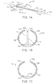
- a typical arterial wall is shown in cross-section where the endothelium E is the layer of the wall which is exposed to the blood vessel lumen L. Underlying the endothelium is the basement membrane BM which in turn is surrounded by the intima I. The intima, in turn, is surrounded by the internal elastic lamina IEL over which is located the media M. In turn, the media is covered by the external elastic lamina (EEL) which acts as the outer barrier separating the arterial wall, shown collectively as W, from the adventitial layer A.
- EEL external elastic lamina
- the microneedle is inserted, preferably in a substantially normal direction, into the wall of a vessel (artery or vein) to eliminate as much trauma to the patient as possible. Until the microneedle is at the site of an injection, it is positioned out of the way so that it does not scrape against arterial or venous walls with its tip. Specifically, the microneedle remains enclosed in the walls of an actuator or sheath attached to a catheter so that it will not injure the patient during intervention or the physician during handling. When the injection site is reached, movement of the actuator along the vessel terminated, and the actuator is operated to cause the microneedle to be thrust outwardly, substantially perpendicular to the central axis of a vessel, for instance, in which the catheter has been inserted.
- a microfabricated intravascular catheter 10 includes an actuator 12 having an actuator body 12a and central longitudinal axis 12 b.
- the actuator body more or less forms a C-shaped outline having an opening or slit 12d extending substantially along its length.
- a microneedle 14 is located within the actuator body, as discussed in more detail below, when the actuator is in its unactuated condition (furled state) ( Fig. 1B ). The microneedle is moved outside the actuator body when the actuator is operated to be in its actuated condition (unfurled state) ( Fig. 2B ).
- the actuator may be capped at its proximal end 12e and distal end 12f by a lead end 16 and a tip end 18, respectively, of a therapeutic catheter 20.
- the catheter tip end serves as a means of locating the actuator inside a blood vessel by use of a radio opaque coatings or markers.
- the catheter tip also forms a seal at the distal end 12f of the actuator.
- the lead end of the catheter provides the necessary interconnects (fluidic, mechanical, electrical or optical) at the proximal end 12e of the actuator.
- Retaining rings 22a and 22b are located at the distal and proximal ends, respectively, of the actuator.
- the catheter tip is joined to the retaining ring 22a, while the catheter lead is joined to retaining ring 22b.
- the retaining rings are made of a thin, on the order of 10 to 100 microns (pm), substantially rigid material, such as parylene (types C, D or N), or a metal, for example, aluminum, stainless steel, gold, titanium or tungsten.
- the retaining rings form a rigid substantially "C"- shaped structure at each end of the actuator.
- the catheter may be joined to the retaining rings by, for example, a butt-weld, an ultra sonic weld, integral polymer encapsulation or an adhesive such as an epoxy.
- the actuator body further comprises a central, expandable section 24 located between retaining rings 22a and 22b.
- the expandable section 24 includes an interior open area 26 for rapid expansion when an activating fluid is supplied to that area.
- the central section 24 is made of a thin, semi-rigid or rigid, expandable material, such as a polymer, for instance, parylene (types C, D or N), silicone, polyurethane or polyimide.
- the central section 24, upon actuation, is expandable somewhat like a balloon-device.
- the central section is capable of withstanding pressures of up to about 100 atmospheres upon application of the activating fluid to the open area 26.
- the material from which the central section is made of is rigid or semi-rigid in that the central section returns substantially to its original configuration and orientation (the unactuated condition) when the activating fluid is removed from the open area 26.
- the central section is very much unlike a balloon which has no inherently stable structure.
- the open area 26 of the actuator is connected to a delivery conduit, tube or fluid pathway 28 that extends from the catheter's lead end to the actuator's proximal end.
- the activating fluid is supplied to the open area via the delivery tube.
- the delivery tube may be constructed of Teflon ⁇ or other inert plastics.
- the activating fluid may be a saline solution or a radio-opaque dye.
- the microneedle 14 may be located approximately in the middle of the central section 24. However, as discussed below, this is not necessary, especially when multiple microneedles are used.
- the microneedle is affixed to an exterior surface 24a of the central section.
- the microneedle is affixed to the surface 24a by an adhesive, such as cyanoacrylate.
- the mesh-like structure (if included) may be-made of, for instance, steel or nylon.
- the microneedle includes a sharp tip 14a and a shaft 14b.
- the microneedle tip can provide an insertion edge or point.
- the shaft 14b can be hollow and the tip can have an outlet port 14c, permitting the injection of a pharmaceutical or drug into a patient.
- the microneedle does not need to be hollow, as it may be configured like a neural probe to accomplish other tasks.
- the microneedle extends approximately perpendicularly from surface 24a.
- the microneedle will move substantially perpendicularly to an axis of a vessel or artery into which has been inserted, to allow direct puncture or breach of vascular walls.
- the microneedle further includes a pharmaceutical or drug supply conduit, tube or fluid pathway 14d which places the microneedle in fluid communication with the appropriate fluid interconnect at the catheter lead end.
- This supply tube may be formed integrally with the shaft 14b, or it may be formed as a separate piece that is later joined to the shaft by, for example, an adhesive such as an epoxy.
- the needle 14 may be a 30-gauge, or smaller, steel needle.
- the microneedle may be microfabricated from polymers, other metals, metal alloys or semiconductor materials.
- the needle for example, may be made of parylene, silicon or glass. Microneedles and methods of fabrication are described in U.S. patent publication 2002/0188310 , entitled “Microfabricated Surgical Device", having common inventorship with but different assignment than the subject application.
- the catheter 20, in use, is inserted through an artery or vein and moved within a patient's vasculature, for instance, an artery 32, until a specific, targeted region 34 is reaches (see Fig. 3 ).
- the catheter 20 may follow a guide wire 36 that has previously been inserted into the patient.
- the catheter 20 may also follow the path of a previously-inserted guide catheter (not shown) that encompasses the guide wire.
- MRI magnetic resonance imaging
- the catheter After being positioned at the target region 34, movement of the catheter is terminated and the activating fluid is supplied to the open area 26 of the actuator, causing the expandable section 24 to rapidly unfurl, moving the microneedle 14 in a substantially perpendicular direction, relative to the longitudinal central axis 12b of the actuator body 12a, to puncture a vascular wall 32a. It may take only between approximately 100 milliseconds and two seconds for the microneedle to move from its furled state to its unfurled state.
- the ends of the actuator at the retaining rings 22a and 22b remain rigidly fixed to the catheter 20. Thus, they do not deform during actuation. Since the actuator begins as a furled structure, its so-called pregnant shape exists as an unstable buckling mode. This instability, upon actuation, produces a large-scale motion of the microneedle approximately perpendicular to the central axis of the actuator body, causing a rapid puncture of the vascular wall without a large momentum transfer. As a result, a microscale opening is produced with very minimal damage to the surrounding tissue. Also, since the momentum transfer is relatively small, only a negligible bias force is required to hold the catheter and actuator in place during actuation and puncture.
- the microneedle in fact, travels so quickly and with such force that it can enter perivascular tissue 32b as well as vascular tissue. Additionally, since the actuator is "parked” or stopped prior to actuation, more precise placement and control over penetration of the vascular wall are obtained.
- the activating fluid is exhausted from the open area 26 of the actuator, causing the expandable section 24 to return to its original, furled state. This also causes the microneedle to be withdrawn from the vascular wall. The microneedle, being withdrawn, is once again sheathed by the actuator.
- the microneedle may have an overall length of between about 200 and 3,000 microns (um).
- the interior cross-sectional dimension of the shaft 14b and supply tube 14d may be on the order of 20 to 250 um, while the tube's and shaft's exterior cross-sectional dimension may be between about 100 and 500 um.
- the overall length of the actuator body may be between about 5 and 50 millimeters (mm), while the exterior and interior cross-sectional dimensions of the actuator body can be between about 0.4 and 4 mm, and 0.5 and 5 mm, respectively.
- the gap or slit through which the central section of the actuator unfurls may have a length of about 4-40 mm, and a cross-sectional dimension of about 50-500 um.
- the diameter of the delivery tube for the activating fluid may be about 100 [tm to 1000 um.
- the catheter size may be between 1.5 and 15 French (Fr).
- a multiple-buckling actuator with a single supply tube for the activating fluid may also be utilized with the microneedle catheters described herein.
- the multiple-buckling actuator includes multiple needles that can be inserted into or through a vessel wall for providing injection at different locations or times.
- the actuator 120 includes microneedles 140 and 142 located at different points along a length or longitudinal dimension of the central, expandable section 240.
- the operating pressure of the activating fluid is selected so that the microneedles move at the same time.
- the pressure of the activating fluid may be selected so that the microneedle 140 moves before the microneedle 142.
- the microneedle 140 is located at a portion of the expandable section 240 (lower activation pressure) that, for the same activating fluid pressure, will buckle outwardly before that portion of the expandable section (higher activation pressure) where the microneedle 142 is located.
- the operating pressure of the activating fluid within the open area of the expandable section 240 is two pounds per square inch (psi)
- the microneedle 140 will move before the microneedle 142. It is only when the operating pressure is increased to four psi, for instance, that the microneedle 142 will move.
- this mode of operation provides staged buckling with the microneedle 140 moving at time t 1 , and pressure p 1 , and the microneedle 142 moving at time t 2 and p 2 , with ti, and p 1 , being less than t 2 and p 2 , respectively.
- staged buckling can also be provided with different pneumatic or hydraulic connections at different parts of the central section 240 in which each part includes an individual microneedle.
- an actuator 220 could be constructed such that its needles 222 and 224A move in different directions. As shown, upon actuation, the needles move at angle of approximately 90° to each other to puncture different parts of a vessel wall.
- a needle 224B (as shown in phantom) could alternatively be arranged to move at angle of about 180° to the needle 224A.
- the catheter 10 may be positioned so that the actuator 12 is positioned at a target site for injection within a blood vessel, as shown in Figs. 6A/6B .
- the actuator penetrates the needle 14 through the wall W so that it extends past the external elastic lamina (EEL) into the perivascular space surrounding the EEL.
- EEL external elastic lamina
- the pharmaceutical agent may be injected, typically in a volume from 10 ⁇ 1 to 5000 ⁇ 1, preferably from 100 ⁇ 1 to 1000 ⁇ 1, and more preferably 250 ⁇ 1 to 500 ⁇ 1, so that a plume P appears.
- the plume occupies a space immediately surrounding an aperture in the needle 14 and extending neither circumferentially nor longitudinally relative toward the external wall W of the blood vessel.
- the plume After a short time, typically in the range from 1 to 10 minutes, the plume extends circumferentially around the external wall W of the blood vessel and over a short distance longitudinally, as shown in Figs. 7A and 7B , respectively.
- the plume After a still further time, typically in the range from 5 minutes to 24 hours, the plume will extend substantially completely circumferentially, as illustrated in Fig. 8A , and will begin to extend longitudinally over extended lengths, typically being at least about 2 cm, more usually being about 5 cm, and often being 10 cm or longer, as illustrated in Fig. 8B .
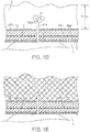
- aperture 300 of a microneedle 314 for volumetric delivery of a pharmaceutical agent
- the aperture 300 is positioned from the lumen L of a blood vessel using any of the microneedle catheter systems described above.
- aperture 300 of the microneedle 314 is positioned beyond the external elastic lamina EEL by a distance of 5 mm or less, preferably 3 mm or less, and usually 0.5 mm or less, as described previously.
- the needle To position the aperture within the requisite distance beyond the EEL, the needle must pass through the other layers of the blood vessel, as described above, in connection with Fig. 1A .
- these underlying layers will have a total thickness in the range from 0.1 mm to 5 mm, requiring that the needle extend from the blood vessel by a distance which is greater than the thickness of the wall.
- the aperture 300 releases the pharmaceutical agent which then begins to form a plume P, as illustrated in Fig. 1D .
- Fig. IE By positioning beyond the blood vessel wall, but less than the 5 mm limit, it has been found that extensive volumetric distribution of the pharmaceutical agent can be achieved, as shown in Fig. IE.
- the needle 14 of Fig. 3 can be positioned through the vascular wall so that it lies beyond the external elastic lamina (EEL), as shown in broken line in Fig. 3A . So long as the aperture 14a lies beyond the periphery of the EEL, and preferably a 5 mm annulus surrounding the vessel, successful delivery of the pharmaceutical agent can usually be achieved. To confirm that the aperture 14a lies within this target annual region, a bolus of contrast media can be injected prior to delivery of the pharmaceutical agent. If the aperture 14a has not penetrated through the EEL, as shown in Fig.
- EEL external elastic lamina
- the bolus of contrast media will remain constrained within the wall of the vessel forming a well defined, generally tapered or ovoid mass B, as shown in Fig. 3B .
- the aperture 14a is positioned beyond the EEL, and within the desired annular region, the bolus B will spread longitudinally along the blood vessel wall in a very short period of time, indicating that the drug may be affectively delivered, as shown in Fig. 3C .
- Sensor 15 may be a solid state pressure sensor. If the pressure builds up during injection (either of an inactive agent or the pharmaceutical agent, it is likely that the aperture 14a still lies within the blood vessel wall. If the pressure is lower, the physician can assume that the needle has reached the adventitia. Sensor 15 may also be a temperature, such as a small thermistor or thermocouple, located at the tip of the needle adjacent over then the aperture 14a. The temperature within the blood vessel wall will be different than that outside of the EEL, making position function of temperature.
- the sensor may be a pH detector, where the tissue within the blood vessel wall and beyond the EEL have detectable differences in pH.
- electrical impedance measurements characteristic of the tissues may be made with an impedance sensor 15.
- a deflection sensor 17, such as a flexible straining gauge, may be provided on a portion of the needle 14 which will deflect in response to insertion force. Insertion force through the blood vessel wall will be higher than that necessary to penetrate the tissue beyond the EEL. Thus, entry into the tissue beyond the EEL can be confirmed when the insertion force measured by the sensor 17 falls.
- the extent of migration of the pharmaceutical agent is not limited to the immediate region of the blood vessel through which the agent is been injected into the perivascular space.
- the pharmaceutical agent may extend further into and through the myocardium other connective tissues so that it surrounds the extravascular spaces around other blood vessels, including both arteries and veins.
- such broad myocardial, epicardial, or pericardial delivery can be particularly useful for treating non-localized cardiac conditions, such as conditions associated with congestive heart failure conditions associated with vulnerable or unstable plaque and conditions associated with cardiac arrhythmias. Delivery and diffusion of a pharmaceutical agent into a peripheral extravascular space can be particularly useful for treating diffuse vascular diseases.
- kits described above may be used to deliver a wide variety of pharmaceutical agents intended for both local and non-local treatment of the heart and vasculature.
- exemplary pharmaceutical agents include antineoplastic agents, antiproliferative agents, cytostatic agents, immunosuppressive agents, anti-inflammatory agents, macrolide antibiotics, antibiotics, antifungals, antivirals, antibodies, lipid lowering treatments, calcium channel blockers, ACE inhibitors, gene therapy agents, anti-sense drugs, double stranded short interfering RNA molecules, metalloproteinase inhibitors, growth factor inhibitors, cell cycle inhibitors, angiogenesis drugs, anti-angiogenesis drugs, and/or radiopaque contrast media for visualization of the injection under guided X-ray fluoroscopy.
- Each of these therapeutic agents has shown promise in the treatment of cardiovascular disease, restenosis, congestive heart failure, and/or vulnerable plaque lesions. Particular agents are set forth in Table I.
- the claimed invention is directed to a corticosteroid for use in a method of treating vascular inflammation. TABLE I 1.
- Antiproliferative agents, immunosuppressive agents, cytostatic, and anti-inflammatory agents including but not limited to sulindac, tranilast, ABT-578, AVI-4126, sirolimus, tacrolimus, everolimus, cortisone, dexamethosone, cyclosporine, cytochalisin D, valsartin, methyl prednisolone, thioglitazones, acetyl salicylic acid, sarpognelate, and nitric oxide releasing agents, which interfere with the pathological proliverative response after coronary antioplasty to prevent intimal hyperplasia, smooth muscle cell activation and migration, and neointimal thickening. 2.
- Antineoplastic agents including but not limited to paclitaxel, actinomycin D, and latrunculin A, which interfere with the pathological proliferative response after coronary angioplasty to prevent intimal hyperplasia, smooth muscle activation and migration and neointimal thickening.
- Macrolide antibiotics including but not limited to sirolimus, tacrolimus, everolimus, azinthromycin, clarithromycin, and erythromycin, which inhibit or kill microorganiss that may contribute to the inflammatory process that triggers or exacerbates restenosis and vulnerable plaque.
- macrolide antibiotics including but not limited to sirolimus and tacrolimus
- Other antibiotics including but not limited to sirolumus, tacrolimus, everolimus, azithromycin, clarithromycin, doxycycline, and erothromycin, inhibit or kill microorganisms that may contribute to the inflammatory process that triggers or exacerbates restenosis and vulnerable plaque.
- Antivirals including but not limited to acyclovir, ganciclovir, fancyclovir and valacyclovir, inhibit or kill viruses that may contribute to the inflammatory process that triggers or exacerbates restenosis and vulnerable plaque. 5.
- Antibodies which inhibit or kill microorganisms that may contribute to the inflammatory process that triggers or exacerbates restenosis and vulnerable plaque or to inhibit specific growth factors or cell regulators. 6. Lipid-lowering treatments, including but not limited to statins, such as trichostatin A, which modify plaques, reducing inflammation and stabilizing vulnerable plaques. 7. Gene therapy agents which achieve overexpression of genes that may ameliorate the process of vascular occlusive disease or the blockade of the expression of the genes that are critical to the pathogenesis of vascular occlusive disease. 8.
- Anti-sense agents including but not limited to AVI-4126, achieve blockade of genes and mRNA, including but not limited to c-myc, c-myb, PCNA, cdc2, cdk2, or cdk9s, through the use of short chains of nucleic acids known as antisense oligodeoxynucleotides.
- Metalloproteinase inhibitors including but not limited to batimastat, inhibit constrictive vessel remodeling. 10.
- Cell cycle inhibitors and modulators and growth factor inhibitors and modulators including but not limited to cytokine receptor inhibitors, such as interleukin 10 or propagermanium, and modulators of VEGF, IGF, and tubulin, inhibit or modulate entry of vascular smooth muscle cells into the cell cycle, cell migration, expression chemoattractants and adhesion molecules, extracellular matrix formation, and other factors that trigger neointimal hyperplasia.
- Angiogenesis genes or agents which increase microvasculature of the pericardium, vaso vasorum, and adventitia to increase blood flow.
- Anti-angiogenesis genes or agents inhibit factors that are associated with microvascularization of atherosclerotic plaque and which directly or indirectly also induce smooth muscle cell proliferation.
- Antithrombotics including but not limited to IIb/IIIa inhibitors, Abciximab, heparin, clopidigrel, and warfarin.
- Acutely harvested tissue ( ⁇ 2 hr post-procedure) showed 4+ staining of the adventitia when OGP was delivered with the microneedle catheter through the vessel wall. With increasing time after delivery, drug penetrated into the media and extended longitudinally 13-24 mm (mean, 15 mm) from the injection site. At 23 hr, staining was observed throughout the circumference of the artery, with longitudinal extension of 23-32 mm (mean, 27.5 mm). OGP delivered into the lumen without needle deployment resulted in staining on the luminal surface only.
- Staining showed delivery outside the external elastic lamina of the vessels and diffusion around the circumference.
- a lipophilic compound (tacrolimus): Eight swine underwent angiography. Twenty-two coronary arteries (2.25-2.75 mm) received 125 micrograms tacrolimus in two 500 micrograms injections approximately 1 cm apart. The two remaining arteries served as untreated controls. An untreated heart was used as a negative control. At 48 hours arteries were dissected from the musculature and perivascular fat, cut into 5 mm sections and analyzed by Liquid Chromatography/Mass Spectrometry against tacrolimus calibration standards containing homogenized untreated porcine heart tissue.
- microsyringe delivered agent to the adventitia, demonstrated by circumferential and longitudinal arterial distribution of fluorescent-labeled paclitaxel and silver nitrate.
- the paclitaxel studies showed that the distribution increased over time.
- Quantitative measurement of tacrolimus showed distribution of drug the full length of the artery, which was detectable 48 hours after injection.
- micro-needle (0.9 mm long x 140 m diameter) was deployed into the adventitia to deliver dexamethasone (DEX, 4 mg/ml) mixed with iodixanol contrast agent (80:20 ratio), providing fluoroscopic visualization.
- DEX dexamethasone
- iodixanol contrast agent 80:20 ratio
- C-Reactive Protein hsCRP
- Schillinger, M., et al. [Balloon angioplasty and stent implantation induce a vascular inflammatory reaction. J Endovasc Ther, 2002. 9(1): p. 59-66 .] appears to be dampened in patients treated with perivascular dexamethasone, indicating that this novel route of administration is capable of not only building up therapeutic levels of the drug, but that the drug is retained by tissue long enough to have a desired anti-inflammatory effect.
- Dexamethasone is a well-known anti-inflammatory agent with no observable adverse effect when compared to placebo in the adventitia of human or porcine AV graft, post-angioplasty.
- the Bullfrog ® Micro-Infusion Catheter has safely and successfully delivered dexamethasone sodium phosphate or placebo material marked with dilute contrast medium (a) to native porcine peripheral arteries and (b) to the porcine model and human AV graft, post-angioplasty. Infusions of contrast solutions are visible under fluoroscopy, providing positive feedback to physicians of infusate location and spread. The infusion of agents into diseased human peripheral arteries has been confirmed in more than 25 patients. The procedure has been safe and the device has effectively delivered therapeutic agents to the vasculature.
- a first-in-human study to test the safety and feasibility of dexamethasone administration through a microinfusion catheter according to the present invention (Bullfrog ® , Mercator MedSystems, Inc, San Leandro, Calif) was performed.
- Dexamethasone was injected into the superficial femoral and popliteal artery (http://www.clinicaltrials.gov).
- Unique identifier NCT 01507558).
- the study design was a prospective, single-center, investigator-initiated study that enrolled consecutive patients who met eligibility requirements from the San Francisco Veteran Affairs Medical Center. This study was approved by the Committee for Human Research and the University of California Clinical and Translational Science Institute. Safety data and outcomes were monitored by a Data Safety and Monitoring Committee that convened on a quarterly basis or as needed.
- the primary inclusion criteria were patients suffering from moderate to severe disabling claudication, ischemic rest pain, or minor tissue loss secondary to atherosclerotic lower extremity occlusive disease with TransAtlantic Inter-Society Consensus IIA-D lesions of the superficial femoral artery (SFA) or popliteal arteries.
- SFA superficial femoral artery
- the minimal reference vessel lumen diameter was required to be 3 to 6 mm, and the patient was required to have at least one infrapopliteal runoff vessel.
- Exclusion criteria included serum creatinine ⁇ 2.5 mg/dL, prior revascularization of the target limb, known allergy to contrast agents or dexamethasone, estimated life expectancy less than 1 year, or other concurrent illness in which the investigators thought would limit the patient's ability to follow the schedule of assessments.
- the Bullfrog ® Micro-Infusion is a rapid-exchange, wire-guided catheter with a balloon-sheathed 0.9-mm-long, 35-gauge (140 ⁇ m diameter) needle that delivers infusions to adventitial and perivascular tissues. It is Food and Drug Administration 510(k)-cleared for use in coronary and peripheral arteries. It is advanced through a 6F sheath over a 0.014-inch wire and can treat vessels from 3 to 6 mm in diameter. Three radio-opaque markers on the catheter allow for proper orientation of the needle. Using standard angioplasty inflation equipment, the balloon was inflated exposing the needle.
- the remainder of the infusate was delivered at a rate of 1 mL/min.
- the balloon was deflated, sheathing the needle, and allowing the catheter to be withdrawn.
- Fig. 12 Injections were administered approximately every 3 cm along the length of the treated arterial segment. Because the drug:contrast admixture can be visualized on both sides of the arterial wall, only one fluoroscopic view was necessary to confirm circumferential arterial coverage in the majority of cases.
- the target lesion was treated according to physician preference. All patients were treated with balloon angioplasty. If a flow-limiting dissection or residual stenosis was determined to require a stent, the protocol specified for treatment with dexamethasone prior to stent placement. In all cases, the microinfusion catheter was advanced to the treatment site following angioplasty to deliver dexamethasone into the arterial adventitia.
- the dosage utilized in this protocol was an off-the-shelf concentration of dexamethasone sodium phosphate for injection USP, 4 mg/mL, which is approved for reducing soft tissue inflammation.
- dexamethasone is indicated for soft tissue injection of 0.4 to 6 mg to treat acute exacerbations in a variety of inflammatory conditions.
- a similar dose (2-6 mg) should be used to treat each 3 cm of lesion (0.7-2 mg/cm), allowing for multiple infusions in the case of long lesions.
- the 3-cm benchmark was chosen based on typical longitudinal perivascular diffusion patterns in preclinical ex vivo cadaveric femoral artery studies (unpublished data).
- the dexamethasone sodium phosphate for injection USP which contains 4.0 mg dexamethasone phosphate per milliliter, was mixed 80%:20% with an iso-osmolar iodinated contrast medium (iodixanol 320 mg I/mL; GE HealthCare, Cork, Ireland) resulting in a final concentration of 3.2 mg dexamethasone phoshate and 60 to 74 mg of iodine in each milliliter of solution.
- the final dosing target was, therefore, determined to be approximately 0.5 mL of the diluted drug per centimeter of lesion or 1.6 mg/cm.
- the primary efficacy end point was a primary patency rate defined as freedom from the combined end points of target lesion revascularization, occlusion, or >50% restenosis in the treated lesion.
- Duplex ultrasonography was performed to assess restenosis and >50% restenosis was defined by a peak systolic velocity ratio >2.5. Rates of target lesion revascularization, death, and amputation end points were also analyzed. Secondary end points were change in Rutherford classification and ABI from baseline to 6 months.
- Inflammation as detected by plasma CRP has been linked to restenosis following peripheral intervention. As one of our intended goals was to reduce inflammation following vascular intervention, serum CRP was measured at baseline and 24 hours following the procedure.
- dexamethasone was able to be delivered to the adventitia of the target lesion.
- the mean volume injected was 3.8 ⁇ 1.9 mL, which contained a mean of 12.1 ⁇ 6.1 mg of dexamethasone sodium phosphate and .80 ⁇ .4 mL of contrast.
- the post-intervention immune response following femoropopliteal intervention has been shown to be independently associated with subsequent restenosis.
- the Schillinger 2002 published research would indicate the likelihood for a substantial rise in the hsCRP after revascularization therapy, and that this rise is linked to the rate of restenosis at 6 months following the revascularization procedure.
- the data from Schillinger 2002 and the 20-patient study described above are plotted in FIG15 , along with 41 patients from the larger 139 patient group (the DANCE Trial).
- dexamethasone when delivered and confirmed to be delivered into the perivascular tissue and adventitia around revascularized arteries, is capable of reducing the inflammatory spike that leads to further recruitment of inflammatory and remodeling cells, proliferation of the cells and inflammatory signal locally around the artery, and eventual migration of cells and fibrosis that leads to renarrowing of the blood vessel, or restenosis. See Figure 15 .
- Additional data coming out of the interim analysis of the DANCE study includes the first 9 patients to have MCP-1 analyzed from their circulating blood at baseline and at 24 hours. This testing indicates that rather than the expected rise in MCP-1, the use of dexamethasone to reduce inflammation, in fact, causes a marked drop in MCP-1 on average. The control of these inflammatory factors steers the body's healing processes away from switching into an aggressive fibrosis-driven and scar tissue generating process, but keeps them in a pro-healing mode, leading to less scarring and less restenosis of the arteries over time. See Figure 16 .
Landscapes
- Health & Medical Sciences (AREA)
- Life Sciences & Earth Sciences (AREA)
- Veterinary Medicine (AREA)
- Public Health (AREA)
- General Health & Medical Sciences (AREA)
- Animal Behavior & Ethology (AREA)
- Pharmacology & Pharmacy (AREA)
- Chemical & Material Sciences (AREA)
- Medicinal Chemistry (AREA)
- General Chemical & Material Sciences (AREA)
- Engineering & Computer Science (AREA)
- Nuclear Medicine, Radiotherapy & Molecular Imaging (AREA)
- Chemical Kinetics & Catalysis (AREA)
- Heart & Thoracic Surgery (AREA)
- Organic Chemistry (AREA)
- Pulmonology (AREA)
- Biomedical Technology (AREA)
- Pain & Pain Management (AREA)
- Rheumatology (AREA)
- Biophysics (AREA)
- Cardiology (AREA)
- Anesthesiology (AREA)
- Bioinformatics & Cheminformatics (AREA)
- Hematology (AREA)
- Epidemiology (AREA)
- Media Introduction/Drainage Providing Device (AREA)
- Medicinal Preparation (AREA)
- Materials For Medical Uses (AREA)
- Pharmaceuticals Containing Other Organic And Inorganic Compounds (AREA)
- Medicines That Contain Protein Lipid Enzymes And Other Medicines (AREA)
- Infusion, Injection, And Reservoir Apparatuses (AREA)
Applications Claiming Priority (2)
| Application Number | Priority Date | Filing Date | Title |
|---|---|---|---|
| US14/605,865 US10441747B2 (en) | 2002-01-22 | 2015-01-26 | Methods and systems for inhibiting vascular inflammation |
| PCT/US2016/014819 WO2016123051A1 (en) | 2015-01-26 | 2016-01-26 | Methods and systems for inhibiting vascular inflammation |
Publications (3)
| Publication Number | Publication Date |
|---|---|
| EP3250277A1 EP3250277A1 (en) | 2017-12-06 |
| EP3250277A4 EP3250277A4 (en) | 2018-09-19 |
| EP3250277B1 true EP3250277B1 (en) | 2022-09-21 |
Family
ID=56544212
Family Applications (1)
| Application Number | Title | Priority Date | Filing Date |
|---|---|---|---|
| EP16743927.2A Active EP3250277B1 (en) | 2015-01-26 | 2016-01-26 | Methods and systems for inhibiting vascular inflammation |
Country Status (7)
| Country | Link |
|---|---|
| EP (1) | EP3250277B1 (https=) |
| JP (2) | JP7361451B2 (https=) |
| CN (1) | CN107360713A (https=) |
| AU (1) | AU2016211717B2 (https=) |
| BR (1) | BR112017016048A2 (https=) |
| ES (1) | ES2934140T3 (https=) |
| WO (1) | WO2016123051A1 (https=) |
Families Citing this family (2)
| Publication number | Priority date | Publication date | Assignee | Title |
|---|---|---|---|---|
| WO2018218182A1 (en) | 2017-05-26 | 2018-11-29 | Mercator Medsystems, Inc. | Combination therapy for treatment of restenosis |
| CN116710103A (zh) * | 2020-10-01 | 2023-09-05 | 墨卡托医疗系统公司 | 用于静脉血栓形成的血管周围抗炎疗法 |
Citations (2)
| Publication number | Priority date | Publication date | Assignee | Title |
|---|---|---|---|---|
| US20070269385A1 (en) * | 2006-05-18 | 2007-11-22 | Mercator Medsystems, Inc | Devices, methods, and systems for delivering therapeutic agents for the treatment of sinusitis, rhinitis, and other disorders |
| WO2010104584A2 (en) * | 2009-03-13 | 2010-09-16 | Gore Enterprise Holdings, Inc. | Articles and methods of treating vascular conditions |
Family Cites Families (14)
| Publication number | Priority date | Publication date | Assignee | Title |
|---|---|---|---|---|
| DE69734060T2 (de) * | 1996-05-24 | 2006-06-29 | Angiotech Pharmaceuticals, Inc., Vancouver | Zubereitungen und verfahren zur behandlung oder prävention von krankheiten der körperpassagewege |
| AU2002249958B2 (en) * | 2001-01-16 | 2007-11-08 | Vascular Therapies, Inc. | Implantable device containing resorbable matrix material and anti-proliferative drugs for preventing or treating failure of hemodialysis vascular access and other vascular grafts |
| US20020188310A1 (en) | 2001-06-08 | 2002-12-12 | Seward Kirk Partick | Microfabricated surgical device |
| US6547303B1 (en) | 2001-08-13 | 2003-04-15 | Johnson Controls Technology Company | Pivoting seating system |
| US6860867B2 (en) | 2001-09-20 | 2005-03-01 | The Regents Of The University Of California | Method of interventional surgery |
| US6547803B2 (en) | 2001-09-20 | 2003-04-15 | The Regents Of The University Of California | Microfabricated surgical device for interventional procedures |
| AU2003205315A1 (en) | 2002-01-22 | 2003-09-02 | Endobionics, Inc. | Methods and kits for delivering pharmaceutical agents into the coronary vascular adventitia |
| US7744584B2 (en) * | 2002-01-22 | 2010-06-29 | Mercator Medsystems, Inc. | Methods and kits for volumetric distribution of pharmaceutical agents via the vascular adventitia and microcirculation |
| US20050249776A1 (en) * | 2003-12-19 | 2005-11-10 | Chen Chao C | Coated aneurysmal repair device |
| WO2005112569A2 (en) | 2004-05-13 | 2005-12-01 | Medtronic Vascular, Inc. | Methods for compounding and delivering a therapeutic agent to the adventitia of a vessel |
| JP2005349202A (ja) * | 2004-06-08 | 2005-12-22 | Cordis Corp | 治療薬を組織内へ導入する器具および方法 |
| US20100092534A1 (en) * | 2008-10-10 | 2010-04-15 | Medtronic Vascular, Inc. | Combination Local Delivery Using a Stent |
| US20120302954A1 (en) * | 2011-05-25 | 2012-11-29 | Zhao Jonathon Z | Expandable devices coated with a paclitaxel composition |
| US9961079B1 (en) | 2014-03-21 | 2018-05-01 | Symantec Corporation | Context aware intruder detection using WIFI MAC addresses |
-
2016
- 2016-01-26 ES ES16743927T patent/ES2934140T3/es active Active
- 2016-01-26 JP JP2017558356A patent/JP7361451B2/ja active Active
- 2016-01-26 AU AU2016211717A patent/AU2016211717B2/en active Active
- 2016-01-26 BR BR112017016048A patent/BR112017016048A2/pt not_active Application Discontinuation
- 2016-01-26 EP EP16743927.2A patent/EP3250277B1/en active Active
- 2016-01-26 WO PCT/US2016/014819 patent/WO2016123051A1/en not_active Ceased
- 2016-01-26 CN CN201680018657.8A patent/CN107360713A/zh active Pending
-
2023
- 2023-06-09 JP JP2023095451A patent/JP2023113904A/ja active Pending
Patent Citations (2)
| Publication number | Priority date | Publication date | Assignee | Title |
|---|---|---|---|---|
| US20070269385A1 (en) * | 2006-05-18 | 2007-11-22 | Mercator Medsystems, Inc | Devices, methods, and systems for delivering therapeutic agents for the treatment of sinusitis, rhinitis, and other disorders |
| WO2010104584A2 (en) * | 2009-03-13 | 2010-09-16 | Gore Enterprise Holdings, Inc. | Articles and methods of treating vascular conditions |
Non-Patent Citations (2)
| Title |
|---|
| OWENS CHRISTOPHER D ET AL: "Safety and feasibility of adjunctive dexamethasone infusion into the adventitia of the femoropopliteal artery following endovascular revascularization", JOURNAL OF VASCULAR SURGERY, ELSEVIER, AMSTERDAM, NL, vol. 59, no. 4, 11 January 2014 (2014-01-11), pages 1016 - 1024, XP028838047, ISSN: 0741-5214, DOI: 10.1016/J.JVS.2013.10.051 * |
| ZHOU YAN ET AL: "Dexamethasone suppresses monocyte chemoattractant protein-1 production via mitogen activated protein kinase phosphatase-1 dependent inhibition of Jun N-terminal kinase and p38 mitogen-activated protein kinase in activated rat microglia", JOURNAL OF NEUROCHEMISTRY, WILEY-BLACKWELL PUBLISHING LTD, GB, vol. 102, no. 3, 1 August 2007 (2007-08-01), pages 667 - 678, XP002557821, ISSN: 0022-3042, [retrieved on 20070226], DOI: 10.1111/J.1471-4159.2007.04535.X * |
Also Published As
| Publication number | Publication date |
|---|---|
| JP2023113904A (ja) | 2023-08-16 |
| AU2016211717A1 (en) | 2017-09-07 |
| AU2016211717B2 (en) | 2020-05-14 |
| HK1247868A1 (en) | 2018-10-05 |
| CN107360713A (zh) | 2017-11-17 |
| ES2934140T3 (es) | 2023-02-17 |
| EP3250277A4 (en) | 2018-09-19 |
| JP7361451B2 (ja) | 2023-10-16 |
| BR112017016048A2 (pt) | 2018-04-03 |
| WO2016123051A1 (en) | 2016-08-04 |
| EP3250277A1 (en) | 2017-12-06 |
| JP2018505022A (ja) | 2018-02-22 |
Similar Documents
| Publication | Publication Date | Title |
|---|---|---|
| US9061098B2 (en) | Methods and kits for volumetric distribution of pharmaceutical agents via the vascular adventitia and microcirculation | |
| US10441747B2 (en) | Methods and systems for inhibiting vascular inflammation | |
| US20070106249A1 (en) | Methods and kits for delivering pharmaceutical agents into the coronary vascular adventitia | |
| US20060189941A1 (en) | Methods and kits for volumetric distribution of pharmaceutical agents via the vascular adventitia and microcirculation | |
| US7141041B2 (en) | Catheters having laterally deployable needles | |
| US20070078620A1 (en) | Methods and kits for delivering pharmaceutical agents into the coronary vascular adventitia | |
| US20240024296A1 (en) | Combination therapy for treatment of restenosis | |
| JP2023113904A (ja) | 脈管炎症を阻害するための方法およびシステム | |
| HK1247868B (en) | Methods and systems for inhibiting vascular inflammation |
Legal Events
| Date | Code | Title | Description |
|---|---|---|---|
| STAA | Information on the status of an ep patent application or granted ep patent |
Free format text: STATUS: THE INTERNATIONAL PUBLICATION HAS BEEN MADE |
|
| PUAI | Public reference made under article 153(3) epc to a published international application that has entered the european phase |
Free format text: ORIGINAL CODE: 0009012 |
|
| STAA | Information on the status of an ep patent application or granted ep patent |
Free format text: STATUS: REQUEST FOR EXAMINATION WAS MADE |
|
| 17P | Request for examination filed |
Effective date: 20170823 |
|
| AK | Designated contracting states |
Kind code of ref document: A1 Designated state(s): AL AT BE BG CH CY CZ DE DK EE ES FI FR GB GR HR HU IE IS IT LI LT LU LV MC MK MT NL NO PL PT RO RS SE SI SK SM TR |
|
| AX | Request for extension of the european patent |
Extension state: BA ME |
|
| DAV | Request for validation of the european patent (deleted) | ||
| DAX | Request for extension of the european patent (deleted) | ||
| A4 | Supplementary search report drawn up and despatched |
Effective date: 20180817 |
|
| RIC1 | Information provided on ipc code assigned before grant |
Ipc: A61P 9/00 20060101ALI20180810BHEP Ipc: A61M 25/00 20060101ALI20180810BHEP Ipc: A61P 29/00 20060101ALI20180810BHEP Ipc: A61K 31/573 20060101AFI20180810BHEP |
|
| REG | Reference to a national code |
Ref country code: HK Ref legal event code: DE Ref document number: 1247868 Country of ref document: HK |
|
| STAA | Information on the status of an ep patent application or granted ep patent |
Free format text: STATUS: EXAMINATION IS IN PROGRESS |
|
| 17Q | First examination report despatched |
Effective date: 20200326 |
|
| REG | Reference to a national code |
Ref country code: DE Ref legal event code: R079 Ref document number: 602016075154 Country of ref document: DE Free format text: PREVIOUS MAIN CLASS: A61M0025000000 Ipc: A61K0031573000 |
|
| GRAP | Despatch of communication of intention to grant a patent |
Free format text: ORIGINAL CODE: EPIDOSNIGR1 |
|
| STAA | Information on the status of an ep patent application or granted ep patent |
Free format text: STATUS: GRANT OF PATENT IS INTENDED |
|
| RIC1 | Information provided on ipc code assigned before grant |
Ipc: A61M 37/00 20060101ALI20220328BHEP Ipc: A61M 25/10 20130101ALI20220328BHEP Ipc: A61M 25/00 20060101ALI20220328BHEP Ipc: A61P 29/00 20060101ALI20220328BHEP Ipc: A61P 9/00 20060101ALI20220328BHEP Ipc: A61K 31/573 20060101AFI20220328BHEP |
|
| INTG | Intention to grant announced |
Effective date: 20220412 |
|
| GRAS | Grant fee paid |
Free format text: ORIGINAL CODE: EPIDOSNIGR3 |
|
| GRAA | (expected) grant |
Free format text: ORIGINAL CODE: 0009210 |
|
| STAA | Information on the status of an ep patent application or granted ep patent |
Free format text: STATUS: THE PATENT HAS BEEN GRANTED |
|
| AK | Designated contracting states |
Kind code of ref document: B1 Designated state(s): AL AT BE BG CH CY CZ DE DK EE ES FI FR GB GR HR HU IE IS IT LI LT LU LV MC MK MT NL NO PL PT RO RS SE SI SK SM TR |
|
| REG | Reference to a national code |
Ref country code: GB Ref legal event code: FG4D |
|
| RIN1 | Information on inventor provided before grant (corrected) |
Inventor name: SEWARD, KIRK, PATRICK |
|
| REG | Reference to a national code |
Ref country code: CH Ref legal event code: EP |
|
| REG | Reference to a national code |
Ref country code: IE Ref legal event code: FG4D |
|
| REG | Reference to a national code |
Ref country code: DE Ref legal event code: R096 Ref document number: 602016075154 Country of ref document: DE |
|
| REG | Reference to a national code |
Ref country code: AT Ref legal event code: REF Ref document number: 1519631 Country of ref document: AT Kind code of ref document: T Effective date: 20221015 |
|
| REG | Reference to a national code |
Ref country code: LT Ref legal event code: MG9D |
|
| REG | Reference to a national code |
Ref country code: DE Ref legal event code: R081 Ref document number: 602016075154 Country of ref document: DE Owner name: MERCATOR MEDSYSTEMS, INC., SAN LEANDRO, US Free format text: FORMER OWNER: MERCATOR MEDSYSTEMS, INC., EMERYVILLE, CA, US |
|
| RAP4 | Party data changed (patent owner data changed or rights of a patent transferred) |
Owner name: MERCATOR MEDSYSTEMS, INC. |
|
| REG | Reference to a national code |
Ref country code: NL Ref legal event code: MP Effective date: 20220921 |
|
| PG25 | Lapsed in a contracting state [announced via postgrant information from national office to epo] |
Ref country code: SE Free format text: LAPSE BECAUSE OF FAILURE TO SUBMIT A TRANSLATION OF THE DESCRIPTION OR TO PAY THE FEE WITHIN THE PRESCRIBED TIME-LIMIT Effective date: 20220921 Ref country code: RS Free format text: LAPSE BECAUSE OF FAILURE TO SUBMIT A TRANSLATION OF THE DESCRIPTION OR TO PAY THE FEE WITHIN THE PRESCRIBED TIME-LIMIT Effective date: 20220921 Ref country code: NO Free format text: LAPSE BECAUSE OF FAILURE TO SUBMIT A TRANSLATION OF THE DESCRIPTION OR TO PAY THE FEE WITHIN THE PRESCRIBED TIME-LIMIT Effective date: 20221221 Ref country code: LV Free format text: LAPSE BECAUSE OF FAILURE TO SUBMIT A TRANSLATION OF THE DESCRIPTION OR TO PAY THE FEE WITHIN THE PRESCRIBED TIME-LIMIT Effective date: 20220921 Ref country code: LT Free format text: LAPSE BECAUSE OF FAILURE TO SUBMIT A TRANSLATION OF THE DESCRIPTION OR TO PAY THE FEE WITHIN THE PRESCRIBED TIME-LIMIT Effective date: 20220921 Ref country code: FI Free format text: LAPSE BECAUSE OF FAILURE TO SUBMIT A TRANSLATION OF THE DESCRIPTION OR TO PAY THE FEE WITHIN THE PRESCRIBED TIME-LIMIT Effective date: 20220921 |
|
| REG | Reference to a national code |
Ref country code: AT Ref legal event code: MK05 Ref document number: 1519631 Country of ref document: AT Kind code of ref document: T Effective date: 20220921 |
|
| REG | Reference to a national code |
Ref country code: ES Ref legal event code: FG2A Ref document number: 2934140 Country of ref document: ES Kind code of ref document: T3 Effective date: 20230217 |
|
| PG25 | Lapsed in a contracting state [announced via postgrant information from national office to epo] |
Ref country code: HR Free format text: LAPSE BECAUSE OF FAILURE TO SUBMIT A TRANSLATION OF THE DESCRIPTION OR TO PAY THE FEE WITHIN THE PRESCRIBED TIME-LIMIT Effective date: 20220921 Ref country code: GR Free format text: LAPSE BECAUSE OF FAILURE TO SUBMIT A TRANSLATION OF THE DESCRIPTION OR TO PAY THE FEE WITHIN THE PRESCRIBED TIME-LIMIT Effective date: 20221222 |
|
| PG25 | Lapsed in a contracting state [announced via postgrant information from national office to epo] |
Ref country code: SM Free format text: LAPSE BECAUSE OF FAILURE TO SUBMIT A TRANSLATION OF THE DESCRIPTION OR TO PAY THE FEE WITHIN THE PRESCRIBED TIME-LIMIT Effective date: 20220921 Ref country code: RO Free format text: LAPSE BECAUSE OF FAILURE TO SUBMIT A TRANSLATION OF THE DESCRIPTION OR TO PAY THE FEE WITHIN THE PRESCRIBED TIME-LIMIT Effective date: 20220921 Ref country code: PT Free format text: LAPSE BECAUSE OF FAILURE TO SUBMIT A TRANSLATION OF THE DESCRIPTION OR TO PAY THE FEE WITHIN THE PRESCRIBED TIME-LIMIT Effective date: 20230123 Ref country code: CZ Free format text: LAPSE BECAUSE OF FAILURE TO SUBMIT A TRANSLATION OF THE DESCRIPTION OR TO PAY THE FEE WITHIN THE PRESCRIBED TIME-LIMIT Effective date: 20220921 Ref country code: AT Free format text: LAPSE BECAUSE OF FAILURE TO SUBMIT A TRANSLATION OF THE DESCRIPTION OR TO PAY THE FEE WITHIN THE PRESCRIBED TIME-LIMIT Effective date: 20220921 |
|
| PG25 | Lapsed in a contracting state [announced via postgrant information from national office to epo] |
Ref country code: SK Free format text: LAPSE BECAUSE OF FAILURE TO SUBMIT A TRANSLATION OF THE DESCRIPTION OR TO PAY THE FEE WITHIN THE PRESCRIBED TIME-LIMIT Effective date: 20220921 Ref country code: PL Free format text: LAPSE BECAUSE OF FAILURE TO SUBMIT A TRANSLATION OF THE DESCRIPTION OR TO PAY THE FEE WITHIN THE PRESCRIBED TIME-LIMIT Effective date: 20220921 Ref country code: IS Free format text: LAPSE BECAUSE OF FAILURE TO SUBMIT A TRANSLATION OF THE DESCRIPTION OR TO PAY THE FEE WITHIN THE PRESCRIBED TIME-LIMIT Effective date: 20230121 Ref country code: EE Free format text: LAPSE BECAUSE OF FAILURE TO SUBMIT A TRANSLATION OF THE DESCRIPTION OR TO PAY THE FEE WITHIN THE PRESCRIBED TIME-LIMIT Effective date: 20220921 |
|
| REG | Reference to a national code |
Ref country code: DE Ref legal event code: R097 Ref document number: 602016075154 Country of ref document: DE |
|
| P01 | Opt-out of the competence of the unified patent court (upc) registered |
Effective date: 20230520 |
|
| PG25 | Lapsed in a contracting state [announced via postgrant information from national office to epo] |
Ref country code: NL Free format text: LAPSE BECAUSE OF FAILURE TO SUBMIT A TRANSLATION OF THE DESCRIPTION OR TO PAY THE FEE WITHIN THE PRESCRIBED TIME-LIMIT Effective date: 20220921 Ref country code: AL Free format text: LAPSE BECAUSE OF FAILURE TO SUBMIT A TRANSLATION OF THE DESCRIPTION OR TO PAY THE FEE WITHIN THE PRESCRIBED TIME-LIMIT Effective date: 20220921 |
|
| PLBE | No opposition filed within time limit |
Free format text: ORIGINAL CODE: 0009261 |
|
| STAA | Information on the status of an ep patent application or granted ep patent |
Free format text: STATUS: NO OPPOSITION FILED WITHIN TIME LIMIT |
|
| PG25 | Lapsed in a contracting state [announced via postgrant information from national office to epo] |
Ref country code: DK Free format text: LAPSE BECAUSE OF FAILURE TO SUBMIT A TRANSLATION OF THE DESCRIPTION OR TO PAY THE FEE WITHIN THE PRESCRIBED TIME-LIMIT Effective date: 20220921 |
|
| 26N | No opposition filed |
Effective date: 20230622 |
|
| PG25 | Lapsed in a contracting state [announced via postgrant information from national office to epo] |
Ref country code: SI Free format text: LAPSE BECAUSE OF FAILURE TO SUBMIT A TRANSLATION OF THE DESCRIPTION OR TO PAY THE FEE WITHIN THE PRESCRIBED TIME-LIMIT Effective date: 20220921 |
|
| REG | Reference to a national code |
Ref country code: CH Ref legal event code: PL |
|
| PG25 | Lapsed in a contracting state [announced via postgrant information from national office to epo] |
Ref country code: LU Free format text: LAPSE BECAUSE OF NON-PAYMENT OF DUE FEES Effective date: 20230126 |
|
| REG | Reference to a national code |
Ref country code: BE Ref legal event code: MM Effective date: 20230131 |
|
| PG25 | Lapsed in a contracting state [announced via postgrant information from national office to epo] |
Ref country code: LI Free format text: LAPSE BECAUSE OF NON-PAYMENT OF DUE FEES Effective date: 20230131 Ref country code: CH Free format text: LAPSE BECAUSE OF NON-PAYMENT OF DUE FEES Effective date: 20230131 |
|
| PG25 | Lapsed in a contracting state [announced via postgrant information from national office to epo] |
Ref country code: BE Free format text: LAPSE BECAUSE OF NON-PAYMENT OF DUE FEES Effective date: 20230131 |
|
| PG25 | Lapsed in a contracting state [announced via postgrant information from national office to epo] |
Ref country code: MC Free format text: LAPSE BECAUSE OF FAILURE TO SUBMIT A TRANSLATION OF THE DESCRIPTION OR TO PAY THE FEE WITHIN THE PRESCRIBED TIME-LIMIT Effective date: 20220921 |
|
| PG25 | Lapsed in a contracting state [announced via postgrant information from national office to epo] |
Ref country code: MC Free format text: LAPSE BECAUSE OF FAILURE TO SUBMIT A TRANSLATION OF THE DESCRIPTION OR TO PAY THE FEE WITHIN THE PRESCRIBED TIME-LIMIT Effective date: 20220921 |
|
| PG25 | Lapsed in a contracting state [announced via postgrant information from national office to epo] |
Ref country code: BG Free format text: LAPSE BECAUSE OF FAILURE TO SUBMIT A TRANSLATION OF THE DESCRIPTION OR TO PAY THE FEE WITHIN THE PRESCRIBED TIME-LIMIT Effective date: 20220921 |
|
| PG25 | Lapsed in a contracting state [announced via postgrant information from national office to epo] |
Ref country code: BG Free format text: LAPSE BECAUSE OF FAILURE TO SUBMIT A TRANSLATION OF THE DESCRIPTION OR TO PAY THE FEE WITHIN THE PRESCRIBED TIME-LIMIT Effective date: 20220921 |
|
| PG25 | Lapsed in a contracting state [announced via postgrant information from national office to epo] |
Ref country code: CY Free format text: LAPSE BECAUSE OF FAILURE TO SUBMIT A TRANSLATION OF THE DESCRIPTION OR TO PAY THE FEE WITHIN THE PRESCRIBED TIME-LIMIT; INVALID AB INITIO Effective date: 20160126 |
|
| PG25 | Lapsed in a contracting state [announced via postgrant information from national office to epo] |
Ref country code: HU Free format text: LAPSE BECAUSE OF FAILURE TO SUBMIT A TRANSLATION OF THE DESCRIPTION OR TO PAY THE FEE WITHIN THE PRESCRIBED TIME-LIMIT; INVALID AB INITIO Effective date: 20160126 |
|
| PG25 | Lapsed in a contracting state [announced via postgrant information from national office to epo] |
Ref country code: TR Free format text: LAPSE BECAUSE OF FAILURE TO SUBMIT A TRANSLATION OF THE DESCRIPTION OR TO PAY THE FEE WITHIN THE PRESCRIBED TIME-LIMIT Effective date: 20220921 |
|
| PGFP | Annual fee paid to national office [announced via postgrant information from national office to epo] |
Ref country code: GB Payment date: 20260127 Year of fee payment: 11 |
|
| PGFP | Annual fee paid to national office [announced via postgrant information from national office to epo] |
Ref country code: ES Payment date: 20260202 Year of fee payment: 11 |
|
| PGFP | Annual fee paid to national office [announced via postgrant information from national office to epo] |
Ref country code: DE Payment date: 20260128 Year of fee payment: 11 Ref country code: IE Payment date: 20260127 Year of fee payment: 11 |
|
| PGFP | Annual fee paid to national office [announced via postgrant information from national office to epo] |
Ref country code: IT Payment date: 20260121 Year of fee payment: 11 |
|
| PGFP | Annual fee paid to national office [announced via postgrant information from national office to epo] |
Ref country code: FR Payment date: 20260126 Year of fee payment: 11 |