EP3247410B1 - Verfahren und zusammensetzungen zur erhöhung der empfänglichkeit einer strahlenbehandlung durch hemmung der suppression von numerischer chromosomaler instabilität von krebszellen - Google Patents
Verfahren und zusammensetzungen zur erhöhung der empfänglichkeit einer strahlenbehandlung durch hemmung der suppression von numerischer chromosomaler instabilität von krebszellen Download PDFInfo
- Publication number
- EP3247410B1 EP3247410B1 EP16749573.8A EP16749573A EP3247410B1 EP 3247410 B1 EP3247410 B1 EP 3247410B1 EP 16749573 A EP16749573 A EP 16749573A EP 3247410 B1 EP3247410 B1 EP 3247410B1
- Authority
- EP
- European Patent Office
- Prior art keywords
- cells
- chromosome
- radiation
- cin
- cell
- Prior art date
- Legal status (The legal status is an assumption and is not a legal conclusion. Google has not performed a legal analysis and makes no representation as to the accuracy of the status listed.)
- Active
Links
Images







Classifications
-
- A—HUMAN NECESSITIES
- A61—MEDICAL OR VETERINARY SCIENCE; HYGIENE
- A61P—SPECIFIC THERAPEUTIC ACTIVITY OF CHEMICAL COMPOUNDS OR MEDICINAL PREPARATIONS
- A61P35/00—Antineoplastic agents
-
- A—HUMAN NECESSITIES
- A61—MEDICAL OR VETERINARY SCIENCE; HYGIENE
- A61K—PREPARATIONS FOR MEDICAL, DENTAL OR TOILETRY PURPOSES
- A61K41/00—Medicinal preparations obtained by treating materials with wave energy or particle radiation ; Therapies using these preparations
- A61K41/0038—Radiosensitizing, i.e. administration of pharmaceutical agents that enhance the effect of radiotherapy
-
- A—HUMAN NECESSITIES
- A61—MEDICAL OR VETERINARY SCIENCE; HYGIENE
- A61K—PREPARATIONS FOR MEDICAL, DENTAL OR TOILETRY PURPOSES
- A61K31/00—Medicinal preparations containing organic active ingredients
- A61K31/33—Heterocyclic compounds
- A61K31/395—Heterocyclic compounds having nitrogen as a ring hetero atom, e.g. guanethidine or rifamycins
- A61K31/535—Heterocyclic compounds having nitrogen as a ring hetero atom, e.g. guanethidine or rifamycins having six-membered rings with at least one nitrogen and one oxygen as the ring hetero atoms, e.g. 1,2-oxazines
- A61K31/5375—1,4-Oxazines, e.g. morpholine
- A61K31/5377—1,4-Oxazines, e.g. morpholine not condensed and containing further heterocyclic rings, e.g. timolol
-
- A—HUMAN NECESSITIES
- A61—MEDICAL OR VETERINARY SCIENCE; HYGIENE
- A61K—PREPARATIONS FOR MEDICAL, DENTAL OR TOILETRY PURPOSES
- A61K38/00—Medicinal preparations containing peptides
- A61K38/16—Peptides having more than 20 amino acids; Gastrins; Somatostatins; Melanotropins; Derivatives thereof
- A61K38/17—Peptides having more than 20 amino acids; Gastrins; Somatostatins; Melanotropins; Derivatives thereof from animals; from humans
- A61K38/1703—Peptides having more than 20 amino acids; Gastrins; Somatostatins; Melanotropins; Derivatives thereof from animals; from humans from vertebrates
- A61K38/1709—Peptides having more than 20 amino acids; Gastrins; Somatostatins; Melanotropins; Derivatives thereof from animals; from humans from vertebrates from mammals
-
- A—HUMAN NECESSITIES
- A61—MEDICAL OR VETERINARY SCIENCE; HYGIENE
- A61K—PREPARATIONS FOR MEDICAL, DENTAL OR TOILETRY PURPOSES
- A61K38/00—Medicinal preparations containing peptides
- A61K38/16—Peptides having more than 20 amino acids; Gastrins; Somatostatins; Melanotropins; Derivatives thereof
- A61K38/43—Enzymes; Proenzymes; Derivatives thereof
- A61K38/45—Transferases (2)
-
- A—HUMAN NECESSITIES
- A61—MEDICAL OR VETERINARY SCIENCE; HYGIENE
- A61K—PREPARATIONS FOR MEDICAL, DENTAL OR TOILETRY PURPOSES
- A61K45/00—Medicinal preparations containing active ingredients not provided for in groups A61K31/00 - A61K41/00
- A61K45/06—Mixtures of active ingredients without chemical characterisation, e.g. antiphlogistics and cardiaca
-
- A—HUMAN NECESSITIES
- A61—MEDICAL OR VETERINARY SCIENCE; HYGIENE
- A61N—ELECTROTHERAPY; MAGNETOTHERAPY; RADIATION THERAPY; ULTRASOUND THERAPY
- A61N5/00—Radiation therapy
- A61N5/10—X-ray therapy; Gamma-ray therapy; Particle-irradiation therapy
-
- A—HUMAN NECESSITIES
- A61—MEDICAL OR VETERINARY SCIENCE; HYGIENE
- A61N—ELECTROTHERAPY; MAGNETOTHERAPY; RADIATION THERAPY; ULTRASOUND THERAPY
- A61N5/00—Radiation therapy
- A61N5/10—X-ray therapy; Gamma-ray therapy; Particle-irradiation therapy
- A61N2005/1092—Details
- A61N2005/1098—Enhancing the effect of the particle by an injected agent or implanted device
Definitions
- the present disclosure relates to the field of cancer therapy using agents that promote whole-chromosomal instability, as irradiation sensitization agents, and thus to make patients more sensitive to radiation therapy, thereby increasing the effect of radiation therapy.
- HR repair is less error-prone than NHEJ, as the latter can join DSB ends of genomic DNA, which can lead to chromosomal translocations, acentric chromatin fragments as well as dicentric chromosomes which have two centromeres.
- Acentric chromatin fragments exhibit a high likelihood of missegregation during the subsequent mitosis, as they are incapable of establishing canonical attachment to spindle microtubules at the kinetochores.
- dicentric chromatin often leads to the formation of chromatin bridges where each centromere is attached to microtubules emanating from opposite spindle poles. Forces exerted by the mitotic spindle break chromatin bridges in a process termed the breakage-fusion-bridge cycle.
- telomere dysfunction can also be initiated by telomere dysfunction and replication stress. It is thus clear that DNA breaks generated by IR in dividing cells can directly lead to structural chromosomal instability (s-CIN), whose hallmarks are chromatin bridges and acentric chromatin fragments.
- s-CIN structural chromosomal instability
- w-CIN Another form of genome instability, present in the majority of solid tumors, is numerical (or whole) chromosomal instability (w-CIN).
- w-CIN primarily arises from errors in whole chromosome segregation during mitosis and it generates widespread aneuploidy in tumor cells.
- a phenotypic hallmark of w-CIN, both in cell culture and human tumor samples, is the presence of chromosomes that lag in the middle of the mitotic spindle during anaphase. These lagging chromosomes can directly lead to chromosome missegregation and aneuploidy.
- w-CIN does not exist separately from s-CIN, as it was recently shown that lagging chromosomes can also undergo severe structural damage by generating whole-chromosome containing micronuclei. These micronuclei are defective in DNA replication and repair and possess a faulty nuclear envelope leading to the pulverization of their enclosed chromosomes. Thus w-CIN can in turn lead to s-CIN.
- IR IR-associated cytoplasmic cytoplasmic cytoplasmic cytoplasmic cytoplasmic cytoplasmic cytoplasmic cytoplasmic cytoplasmic cytoplasmic cytoplasmic cytoplasmic cytoplasmic cytoplasmic cytoplasmic cytoplasmic cytoplasmic cytoplasmic cytoplasmic cytoplasmic cytoplasmic cytoplasmic cytoplasmic cytoplasmic cytoplasmic cytoplasmic cytoplasmic cytoplasmic cytoplasmic cytoplasmic cytoplasmic cytoplasmic cytoplasmic cytoplasmic cytoplasmic cytoplasmic cytoplasmic cytoplasmic cytoplasmic cytoplasmic cytoplasmic cytoplasmic cytoplasmic cytoplasmic cytoplasmic cytoplasmic cytoplasmic cytoplasmic cytoplasmic cytoplasmic cytoplasmic cytoplasmic cytoplasmic cytoplasmic cytoplasmic cytoplasmic cytoplasmic cytoplasmic cytoplasmic cytoplasmic cytoplasmic cytoplasmic
- the present disclosure relates to a radiosensitizing agent for use in a method of increasing susceptibility of cancer cells to ionizing radiation , wherein the radiosensitizing agent has one of the following properties: (a) it perturbs the process of chromosome segregation thereby increasing chromosome missegregation; or (b) it is an inhibitor of an agent that promotes faithful chromosome segregation induces numeric chromosome instability in said cells and this instability is induced substantially simultaneously with or closely prior to or closely after irradiating the cells.
- the radiosensitizing agent inhibits one or more of the following: Kif2b, MPS1, Eg5/Kinesin-5, Polo-like kinase 4, MCAK, Bubl and Hec1.
- the agent thus specifically targets proteins involved in maintaining or promoting faithful chromosome segregation.
- the cancer cells are solid tumor cancer cells for which radiation is an indicated therapeutic modality.
- tumors include head-and-neck cancer, rectal adenocarcinoma, glioblastoma multiform.
- the radiosensitizing agent is selected from the group consisting of MPS1 inhibitors, Eg5/Kinesin-5 inhibitors, Polo-like kinase 4 inhibitors, MCAK inhibitors, Bubl inhibitors and Heel inhibitors. (Bub1 and Heel inhibitors will produce the desired chromosomal instability but so will activators as described below.)
- the present disclosure relates to a radiosensitizing agent for use in a method for increasing cytotoxicity of ionizing radiation in a subject to be treated for a solid malignant tumor susceptible to treatment with ionizing radiation, the method comprising administering systemically to the subject or locally to the tumor an inhibitor of suppression of numerical chromosome instability incident to a first dose of said radiation, thereby enhancing chromosome missegregation during mitosis substantially simultaneously with or closely preceding treatment of the tumor with a subsequent dose of ionizing radiation.
- This subsequent dose can be delivered either closely before, closely after or up to 1 or even 2 months after increasing chromosomal instability as long as the chromosomal instability induction substantially persists.
- a method for reducing damage of noncancerous cells or tissue incident to ionizing radiation aimed at cancerous cells or tissue comprising exposing the noncancerous cells or tissue to a radioprotective agent (such as agonists of Kif2b or MCAK) which is an enhancer of suppression of chromosome missegregation and reduces numeric chromosome instability in said cells simultaneously with or immediately prior to or immediately following irradiating the cancerous cells or tissue with a therapeutically effective dose and regimen of ionizing radiation.
- a radioprotective agent such as agonists of Kif2b or MCAK
- the radioprotective agent is or specifically activates a protein involved in faithful chromosome segregation maintenance.
- the radioprotective agent is Kif2b or MCAK or an agonist or activator of Kif2b or MCAK.
- w-CIN in induced preferentially in cancer cells.
- Main contributors to cancer cell specificity are the following: 1) induction of w-CIN according to the present disclosure occurs mostly in rapidly dividing cells (such as cancer cells); and 2) the higher chromosomal missegregation before treatment, the more sensitive cells are to interventions aimed at further increasing w-CIN.
- adrenal tumors include for example adrenocortical carcinoma, bile duct, bladder, bone (e.g., Ewing's sarcoma, osteosarcoma, malignant fibrous histiocytoma), brain/CNS (e.g., astrocytoma, glioma, glioblastoma, childhood tumors, such as atypical teratoid/rhabdoid tumor, germ cell tumor, embryonal tumor, ependymoma), breast (including without limitation ductal carcinoma in situ, carcinoma, cervical, colon/rectum, endometrial, esophageal, eye (e.g., melanoma, retinoblastoma), gallbladder, gastrointestinal, kidney (e.g., renal cell, Wilms' tumor), heart, head and neck, laryngeal and hypopharyngeal, liver, lung, oral (e.g., lip, mouth, salivary gland) meso
- the present inventors thus have discovered that increasing chromosome missegregation together with radiation treatment would lead to sensitization of the tumor to radiation therapy. This in turn permits 1) to decrease dose of radiation and achieve the same effect, 2) to maintain dose of radiation and increase tumor sensitization in otherwise resistant tumors, 3) to increase radioprotection of normal organs.
- Subject means a patient (human or veterinary) or an experimental animal, such as a mouse or other rodent.
- Time interval of increased susceptibility means the period of time after administration of the CIN promoting agent, or radiosensitizing agent during which the resulting increase in w-CIN or lagging chromosomes is still substantially present.
- ionizing radiation means any radiation where a nuclear particle has sufficient energy to remove an electron or proton or other particle from an atom or molecule, thus producing an ion and a free electron or radical.
- ionizing radiation include, but are not limited to, gamma rays, X-rays, protons, electrons, alpha particles, carbon atoms, or particles emitted from a radioactive source including, but not limited to, yttrium and radium. Radiation from implanted material is included. Ionizing radiation is commonly used in medical radiotherapy and the specific techniques for such treatment will be apparent to a person of ordinary skill in the art. Other examples of radiation suitable for use in the present methods are provided elsewhere in the specification.
- radiosensitizing agent means agents which increase the susceptibility of cells to the damaging effects of ionizing radiation or which become more toxic to a cell after exposure of the cell to ionizing radiation.
- a radiosensitizing agent may permit lower doses of radiation to be administered and still provide a therapeutically effective dose.
- agents that increase chromosome missegregation by specifically targeting proteins that are involved in chromosome missegregation (activating such proteins) or in faithful chromosome segregation (inhibiting such proteins) are radiosensitizing agents.
- chromosome missegregation agents that suppress (or more accurately increase the suppression of) chromosome missegregation.
- radioprotective agents have the property of enhancing suppression of chromosomal missegregation and reducing numeric chromosomal instability.
- Tumor as used herein means primary or metastatic tumor and includes the list of the tumor in the summary, above.
- the radiation is ionizing radiation.
- Radiation may be electromagnetic or particulate in nature. Electromagnetic radiation includes, but is not limited to, x-rays and gamma rays. Particulate radiation includes, but is not limited to, electron beams, proton beans, neutron beams, alpha particles, and negative pimesons.
- the unit of absorbed dose is the gray (Gy), which is defined as the absorption of 1 joule per kilogram.
- the energy of the radiation determines the depth of absorption as well as the nature of the atomic interaction.
- Radiotherapy can be administered by a conventional radiological treatment apparatus and methods, or by intraoperative and sterotactic methods. Radiation may also be delivered by other methods that include, but are not limited to, targeted delivery, systemic delivery of targeted radioactive conjugates and intracavitary techniques (brachytherapy). Other radiation methods not described above can also be used to practice this invention.
- ionizing radiation is used to target tissues or cells, such as neoplastic tissues or cells, for selective delivery of a CIN promoting active agent via a delivery vehicle comprising the active agent.
- target tissues or cells such as neoplastic tissues or cells
- a delivery vehicle comprising the active CIN promoting agent is administered before, during, or both before and during the exposure to radiation. Radiation immediately preceding delivery of the CIN promoting agent to the tumor is also possible.
- radiation therapy is delivered using radioactive isotopes (brachytherapy).
- brachytherapy can be either high-dose rate or low dose rate brachytherapy.
- High dose rate brachytherapy is usually delivered using Ir-192 (but not exclusively) and can be given in one or more doses and doses.
- Low dose rate brachytherapy can be delivered using radioactive palladium or iodine and it involves permanent or long-term placement of seeds in and around the target and dose delivery obeys the half-life of the decay of the radioactive substance and can take weeks to months to deliver the majority of the dose.
- the radiosensitizing agent described herein can be used in the treatment of various types of solid tumors.
- solid tumors have been provided in the Summary of the Disclosure.
- malignant tumors which can be treated by methods described herein can be used in the treatment of cancer, include without limitation adrenal tumors (e.g., adrenocortical carcinoma), anal, bile duct, bladder, bone tumors (e.g., Ewing's sarcoma, osteosarcoma, malignant fibrous histiocytoma), brain/CNS (tumors e.g., astrocytoma, glioma, glioblastoma, childhood tumors, such as atypical teratoid/rhabdoid tumor, germ cell tumor, embryonal tumor, ependymoma), breast tumors(including without limitation ductal carcinoma in situ, carcinoma, cervical, colon/rectum, endometrial, esophageal, eye (e.g., melanoma, retinoblastoma), gallbladder, gastrointestinal, kidney (e.g., renal cell, Wilms' tumor), heart
- Radiation treatment may be given as fractionated doses or as a bolus dose.
- radiation can be administered in a range of 1 to about 50 fractions, with each fraction size being within the range of 0.1 to about 50 Gy.
- dosage of each fraction is about 2 to about 30 Gy.
- dosage of each fraction is about 4 to about 25 Gy.
- dosage of each fraction is about 10 to about 20 Gy.
- Particular dosage amounts include, but are not limited to, 0.4 (or 40 cGY), 1, 2, 4, 10 and 20 Gy.
- the source of ionizing radiation comprises an external beam photon irradiation source, which is typically utilized at energy levels ranging from about 10 kV (KeV) to about 18 MV (MeV) per photon beam, or a brachytherapy source directly applied in the tumor cavity.
- These sources of radiation can include, but are not limited to, yttrium, radium.
- the source of ionizing radiation comprises an external beam electron irradiation source, which is typically utilized at energy levels ranging from about 10KeV to about 20 MeV per electron beam.
- the source of ionizing radiation comprises an external beam proton irradiation source, which is typically utilized at energy levels ranging from about 10MEV to about 300 MeV per proton beam.
- a preferred minimum source-axis distance comprises about 80 cm but can range considerably up and down as those skilled in the art appreciate.
- the subject receives local-regional irradiation via fields that are designed to encompass sites of disease requiring palliation or primary treatment while endeavoring to spare noncancerous tissue as much as possible.
- a preferred dosage range comprises about 500 to about 1500 cGy, and at times 2400 cGy, with a preferred dosage range comprising about 800 to about 1200cGy.
- a preferred dosage range comprises about 1000 to about 3000 cGy, and at times up to 800cGy, with a preferred dosage range comprising about 1500 to about 2500cGy, and with a more preferred dosage amount comprising about 200cGy.
- a preferred dosage range comprises about 1000 to about 6000 cGy, with a preferred dosage range comprising about 2000 to about 4000cGy, and with a more preferred dosage amount comprising about 3000 cGy.
- a preferred dosage range comprises about 1000 to about 7000 cGy, with a preferred dosage range comprising about 2000 to about 5000 cGy, and with a more preferred dosage amount comprising about 3500 CGy.
- a preferred dosage range comprises about 2000 to about 12000 cGy, with a preferred dosage range comprising about 4000 to about 8000 cGy, and with a more preferred dosage amount comprising about 6000 cGy.
- radiation is administered 1 to about 50 times. Frequency of radiation treatment can be from 3 times per day to about once per month. In further embodiments, radiation is administered once per 3 weeks, once per 2 weeks, once per week, 2-6 times per week, at least once a day, twice a day or three times a day, or any combination thereof.
- a suitable administration regimen includes a schedule where fractionations are given 2 times per day for 2 days followed by a month long pause, and this cycle is repeated numerous times.
- Actual dosage levels of the agent that promotes CIN and radiation may be varied so as to obtain the desired therapeutic response for a particular subject, composition and mode of administration, without being toxic to the subject.
- the selected dosage level will depend upon a variety of factors, including the route of administration, the rate of breakdown of the active form of the CIN promoting agent, the duration of treatment, other drugs, compounds, and/or materials used in combination with the particular CIN promoting agent, the age, sex, weight, condition, general health and prior medical history of the patient being treated, and similar relevant factors well known in the art.
- high dose rate brachytherapy is given in one or more doses, where doses can range anywhere from about 0.1 Gy to about 50 Gy.
- high dose rate brachytherapy is administered intra-operatively.
- low dose rate brachytherapy is delivered using radioactive palladium or iodine and it involves permanent or long-term placement of seeds in and around the target and dose delivery obeys the half-life of the decay of the radioactive substance and can take weeks to months to deliver the majority of the dose.
- dose is delivered at a rate of anywhere between 0.001Gy-1Gy/hour in low-dose rate brachytherapy
- the therapeutic combination of the present disclosure can be combined with other cancer therapies, including, but not limited to, adjunct therapies (such as, but not limited to, surgical tumor resection and chemotherapy).
- Resection is often a standard procedure for the treatment of tumours.
- the types of surgery that may be used in combination with the present invention include, but are not limited to, preventative, curative and palliative surgery, and any other method that would be contemplated by those of skill in the art.
- agents in the categories described below all alter chromosome missegragation and eventually cause w-CIN by either specifically inhibiting Kif2b, MCAK, MPS1, Eg5/Kinesin-5, Polo-like kinase 4, Mad2, Heel, Bub1, or BubR1 or by activating Mad2, Heel, BubR1, or Bub1.
- the present disclosure provides evidence that over-expression of the microtubule-depolymerizing kinesin-13 proteins, Kif2b or MCAK, which localize to the attachment sites of chromosomes to spindle microtubules at the kinetochores, leads to the suppression of w-CIN in otherwise chromosomally unstable cell This suppression is persistent, lasting more than 30 days both in vitro and in vivo studies. Furthermore, Kif2b overexpressing cells displayed greater than twofold reduction in chromosome segregation errors during anaphase, which led to radiation resistance in vivo ( Fig. 6 ). These findings indicate that the inhibition of proteins that localize to the attachment sites of chromosomes to spindle microtubules at the kinetochores can be used to increase w-CIN in cancer cells and as such promote radiosensitization.
- Kif2b or MCAK microtubule-depolymerizing kinesin-13 proteins
- suitable agents that induce numeric chromosomal instability are those that inhibit proteins that localize to the attachment sites of chromosomes to spindle microtubules at the kinetochores during mitosis. Proteins known to localize to the attachment sites of chromosomes to spindle microtubules at the kinetochores during mitosis include, but are not limited to Kif2b and MCAK. Thus, in one embodiment, suitable agents that induce numeric chromosomal instability are those that specifically target and inhibit Kif2b or MCAK.
- Kif2b belongs to the kinesin-13 family of proteins and localizes to kinetochores during early mitosis ( Manning AL et al. Mol. Biol. Cell. 18:2970-2979 (2007 )).
- Kif2b can be inhibited using agents that inhibit Kif2b specifically.
- One such agent is DHTP ((Z)-2- (4-((5-(4-chlorophenyl)-6-(isopropoxycarbonyl)-7-methyl-3-oxo-3,5-dihydro-2H-thiazolo[3,2-a]pyrimidin-2-ylidene)methyl)phenoxy)acetic acid).
- DHTP has been shown to be a potent compound that inhibits kinein-13 induced microtubule depolymerization, where the IC5 0 of DHTP in inhibiting Kif2b is 1.2 ⁇ M ( FEBS Lett. 27;588(14):2315-20 (2014 )).
- IC5 0 of DHTP in inhibiting Kif2b is 1.2 ⁇ M
- MCAK inhibitory activity against MCAK
- the IC 50 of DHTP in inhibiting MCAK is 4.6 ⁇ M FEBS Lett. 27;588(14):2315-20 (2014 )).
- Kif2b can be inhibited using agents that inhibit Kif2b specifically but indirectly (e.g., through specific activation or inhibition of another protein that inhibits or activates Kif2b).
- agents that inhibit Kif2b specifically but indirectly e.g., through specific activation or inhibition of another protein that inhibits or activates Kif2b.
- the possibility of such indirect specific effect is not limited to Kif2b but in principle extends to any radiosensitizing or radioprotective agent employed in the present methods.
- the core SAC kinases monopolar spindle-1 (Mps1, also known as TTK) is a serine threonine kinase which functions as a core component of the spindle assembly checkpoint (SAC) ( Lauze' et al. EMBO J. 14, 1655-1663, (1995 )), a key surveillance mechanism that monitors the attachment of spindle microtubules to the kinetochores of the chromosomes during pro-metaphase and halts the transitions to anaphase until all chromosomes are bioriented, fully attached, and correctly tensed at the metaphase plate. Mps1 is expressed in the mitosis phase of the cell cycle in proliferating cells.
- SAC spindle assembly checkpoint
- Mps1 activity causes cells to prematurely exit mitosis with unattached chromosomes, resulting in severe chromosome missegregation and aneuploidy ( Colombo et al. Cancer Res., 70, 10255-10264 (2010 ); Jemaa et al. Cell Death Differ. 20, 1532-1545 (2013 )).
- Overexpression of Mps1 has been observed in several cancer cell lines and tumor types including lung and breast cancers, where higher Mps1 levels correlate with worse prognosis.
- Established anti-mitotic drugs such as vinca alkaloids, taxanes, or epothilones activate SAC either by destabilizing or stabilizing spindle microtubules resulting in mitotic arrest. Prolonged arrest in mitosis forces a cell either into a mitotic exit without cytokinesis or into a mitotic catastrophe leading to cell death.
- Such drugs do not target chromosome missegregation or faithful segregation specifically and are not included within the specifically acting agents of the present disclosure.
- specific Mps1 inhibitors inactivate the SAC and accelerate progression of cells through mitosis eventually resulting in severe chromosomal missegregation, mitotic catastrophe, and cell death.
- Mps1 inhibition leads to failure of cells to arrest in mitosis in response to anti-mitotic drugs.
- the combination of microtubule-interfering agents and Mps1 inhibition strongly increases chromosomal segregation errors and cell death and therefore, constitutes an efficient strategy for selectively eliminating tumor cells.
- BAY 1161909 has been administered orally, with a starting dose of 0.75 mg twice daily, on a 14-day cycle - D1, D2, D8, D9 and 28 day cycle - D8, D9 D15 and D16 of a 28 day cycle.
- Mps1 inhibitors include N-(4- ⁇ 2-[(2-cyanophenyl)amino] [1,2,4]triazolo[1,5-a]pyridin-6-yl ⁇ phenyl)-2-phenylacetamide (Mps-BAY1) (a triazolopyridine), N-cyclopropyl-4- ⁇ 8-[(2-methylpropyl)amino]-6-(quinolin-5-yl)imidazo[1,2-a]pyrazin-3-yl ⁇ benzamide (Mps-BAY2a), and N-cyclopropyl-4- ⁇ 8-(isobutylamino)imidazo[1,2-a]pyrazin-3-yl ⁇ benzamide (Mps-BAY2b).
- Eg5 a member of the kinesin superfamily, plays a key role in mitosis, as it is required for the formation of a bipolar spindle. Eg5 controls mitosis through bipolar spindle formation and thus chromosome separation ( Blangy et al. Cell, 83,1159-1169 (1995 )). Eg5 is overexpressed in many proliferative tissues including leukemia as well as solid tumors such as breast, lung, ovarian, bladder and pancreatic cancers ( Hedge et al. Proc. Am. Soc. Clin. Oncol., 22 (2003 ), Ding et al. Int. J. Urol., 18, 432-438 (2011 ), Liu et al. J.
- Eg5 inhibitors include, but are not limited to 4SC-205, currently in Phase I Clinical Trial (ClinicalTrials.gov ID: NCT01065025, Open Label, Dose Escalation Trial of Oral Eg5 Kinesin-spindle Inhibitor 4SC-205 in Patients With Advanced Malignancies (AEGIS); and AZD4877, which was part of a Phase I trial in patients with solid and lymphoid malignancies ( Gerecitano et al. Invest New Drugs. (2):355-62 (2013 )). 4SC -205 was administered once or twice weekly at doses of 25mg - 200mg.
- AZD4877 a standard 3 + 3 dose-escalation design was used, where AZD4877 was given as an intravenous infusion on days 1, 4, 8 and 11 of each 21-day cycle ( Gerecitano et al. Invest New Drugs. (2):355-62 (2013 )).
- Eg5 inhibitors please refer to El-Nassan HB, Eur J Med Chem. 62:614-31 (2013 ).
- Eg5 inhibitors have been shown to be effective against taxolresistant cancer cells ( Marcus et al. J. Biol. Chem., 280 (2005 )). Therefore, given the findings of the present disclosure, combining inhibition of proteins that promote faithful segregation of chromosomes with radiation therapy can offer new treatment solutions for tumors resistant to other therapies.
- PLK4 is a conserved key regulator of centriole duplication ( Bettencourt-Dias et al. Curr. Biol. 15, 2199-2207 (2005 )). Dysregulation of PLK4 expression causes loss of centrosome numeral integrity, which promotes genomic instability ( Ganem et al. Nature 460, 278-282 (2009 ). Thus, in some embodiments, the present disclosure relates to the use of PLK4 inhibitors in cancer cells in order to generate cancer cells more susceptible to radiation therapy compared to cancer cells that not treated with PLK4 inhibitors prior to radiation therapy.
- the PLK4 inhibitor R1530 downregulates the expression of mitotic checkpoint kinase BubR1, which in leads to polyploidy ( Tovar et al. Cell Cycle. 9(16):3364-75 (2010 )).
- BubR1 and Bubl are paralogous serine/threonine kinases that perform different functions in the spindle assembly checkpoint (SAC).
- BubR1 associates with unattached kinetochores, contributes to stabilizing kinetochore-MT attachments and aligning chromosomes, and forms part of the mitotic checkpoint complex (MCC).
- MCC mitotic checkpoint complex
- Bubl binds kinetochores and plays a key role in establishing the mitotic spindle checkpoint and aligning chromosomes in addition to its central role in ensuring fidelity during chromosomal segregation into daughter cells (Yu et al. 4:262-265 (2005)).
- Bub1 transgenic mice develop aneuploid tumors ( Ricke, et al. J. Cell Biol. 193, 1049-1064 (2011 )).
- BubR1 overexpression leads to high incidence of aneuploidy coupled with malignant progression ( Ando et al. Cancer Sci. 101:639-645 (2010 )).
- BubR1 at basal level functions to prevent missegregation of sister chromatids during mitosis, but either gain or loss of BubR1 expression promote CIN-driven tumorigenesis and cancer progression.
- the inventors anticipate that either inhibition of activation of BubR1, as well as Bubl can be manipulated in order to achieve proper radiosensitization.
- a small molecule agonist could be developed through a chemical screen.
- Mad2 is a central component of the spindle assembly checkpoint, which is a feedback control that prevents cells with incompletely assembled spindles from leaving mitosis. Partial loss of checkpoint control, via deletion of one MAD2 allele results in a defective mitotic checkpoint in human cancer cells, leading to an increased rate of chromosome missegregation events and an increased frequency of aneuploid metaphases compared to cells control cancer cells ( Michel et al. Nature 409, 355-359 (2001 )).
- use of specific Mad2 inhibitors in conjunction with radiation therapy is anticipated to lead to enhanced tumor treatment response.
- Kastl et al. identified a specific MAD2 inhibitor-1 (M2I-1), the first small molecule inhibitor targeting the binding of Mad2 to Cdc20, an essential protein-protein interaction (PPI) within the SAC ( Kastl et al. ACS Chem. Biol., 10 (7) 1661-1666 (2015 )).
- M2I-1 MAD2 inhibitor-1
- PPI essential protein-protein interaction
- Hec1 inhibitors include, but are not limited to TAI-95 and TAI-1 ( Huang et al. Mol Cancer Ther. 13(6):1419-30 (2014 ), Huang et al. J Exp Clin Cancer_Res. 33:6, (2014 )).
- TAI-95 is highly potent in breast cancer cell lines, with GI 50 between 14.29 and 73.65 nmol/L.
- TAI-95 showed excellent oral efficacy in an in vivo breast cancer model, where mice were treated with TAI-95 twice a day for 28 days, using TAI-95 orally at 10, 25, 50 mg/kg (mpk) or intravenously at 10, 25, 50 mpk ( Huang et al. Mol Cancer Ther. 13(6):1419-30 (2014 ).
- mitotic checkpoint genes known to be implicated in tumors such as Mad2 and Heel
- both partial inactivation and overactivation of the mitotic checkpoint promote chromosomal instability.
- Heel overexpression hyperactivates the mitotic checkpoint and induces tumor formation in vivo ( Diaz-Rodriguez et al. Proc Natl Acad Sci USA. 105(43):16719-24 (2008 )).
- overexpression of Heel resulted in lagging chromosomes and aneuploidy ( Diaz-Rodriguez et al. Proc Natl Acad Sci U S A. 105(43):16719-24 (2008 )).
- Mad2 overexpression has also been shown to promote aneuploidy and tumorigenesis in mice ( Sotillo et al. Cancer Cell. 11:9-23 (2007 )).
- agents that cause activation of upregulation of Mad2 can also be used to promote radiosensitization.
- Noncoding RNA activated by DNA damage NORAD
- MCAK ab42676 (Abcam, Cambridge MA); (M01) , clone 1G2 (Abnova, Walnut, CA MPS1 [N1] (ab11108) (Abcam, Cambridge, MA); (7E3) (MA5-15523) (Thermo Fisher Scientific, Waltham, MA) Eg5/Kinesin-5 10C7/Eg5 (Bio Legend, San Diego, CA) PA5-28933 (Thermo Fisher Scientific, Waltham, MA) PLK4 [36-298] (ab17057) (Abcam, Cambridge, MA); A300-251A (Bethyl Laboratories Montgomery, TX) Mad2 ENZ-ABS169-0200 (Enzo Life Sciences, Farmingdale, NY); sc-393188 (Santa Cruz Biotechnology, Dallas, Texas) Heel ab3613 (Abcam, Cambridge, MA); A300-771A (Bethyl Laboratories Montgomery, TX)
- target proteins can be inhibited or activated for the purposes of inducing w-CIN and consequently radiosensitization were described.
- the inventors provide approximate threshold levels for inhibition or activation of target genes, wherein such threshold levels are expected to provide levels of target genes sufficient for induction of w-CIN and radiosensitization.
- Table 2 includes approximate threshold levels for inhibition of genes that promote faithful chromosome segregation, while Table 3 provides such threshold levels for activation of genes that promote chromosome missegragation.
- Table 2 Increase in CIN via inhibition of genes that promote faithful chromosome segregation.
- Activation or overexpression of proteins that promote faithful chromosome segregation can be used as a method of protection of noncancerous cells against radiation. Since overexpression of Kif2b results in decreased CIN, agents that activate or upregulate Kif2b can be used as radioprotectors. Such radioprotectors can serve to protect noncancerous cells preferentially as the radiation intensity is focused on the tumor (e.g., in conformal radiation), making any radioprotector in the tumor cells practically ineffective. Furthermore, radioprotectors can alternatively be used to shield and protect organs or tissues.
- the digestive tract can be protected by delivering a radio-protector that is not systemically absorbed but it can have a topical or local effect on the tract, which tends to receive the most damage of ionizing radiation.
- a radio-protector that is not systemically absorbed but it can have a topical or local effect on the tract, which tends to receive the most damage of ionizing radiation.
- organs include, but are not limited to the pharynx, esophagus, stomach, small and large intestines, and rectum.
- This approach to tissue protection can also be applied to other mucosal surface such as the vaginal tract and the cervix.
- Fluorescence in situ hybridization is one of the main methods for the assessment of w-CIN status in tumors. Variations in chromosome copy number across the cell population can be quantified using fluorescently labeled DNA probes that bind to the centromeres of specific chromosomes. FISH thus allows the assessment of the chromosomal state of hundreds of cells, and the rate of change can be inferred from the cell-to-cell variability in chromosome number ( Speicer et al. Nat Rev Genet 6: 782-792 (2005 )).
- Additional methods for assessing w-CIN status include, but are not limited to flow and DNA image cytometry. These methods have been discussed by Darzynkiewicz et al. Adv Exp Med Biol 676: 137-147 (2010 )). Both of these methods measure cellular DNA content through the use of dyes that bind stoichiometrically to DNA, allowing DNA cell cycle distribution and ploidy to be determined.
- Single-cell, comparative genomic hybridization (CGH) ( Fiegler et al., Nucleic Acids Res 35: e15 (2007 )) can yield information on both numerical and structural chromosomal aberrations at a single-cell level, and heterogeneity can then be quantified by comparing multiple cells.
- Karyotypic complexity measures of CIN are commonly performed on a combined population of cells.
- Conventional array CGH uses DNA from multiple cells, and can be used to define the both structural chromosomal complexity and copy number changes in a tumor sample ( Pinkel et al. Nat Genet 37: S11-S17 (2005 )).
- Chromosomal instability substantiates poor prognosis in patients with diffuse large B-cell lymphoma.
- Bakhoum SF1 Danilova OV, Kaur P, Levy NB, Compton DA. See also, Cancer. 2014 Jun 1;120(11):1733-42. doi: 10.1002/cncr.28656 . Epub 2014 Mar 6.
- Chromosomal instability portends superior response of rectal adenocarcinoma to chemoradiation therapy.
- Zaki BI1 Suriawinata AA, Eastman AR, Garner KM, Bakhoum SF
- the present disclosure comprises novel insights into genome damage induced by IR, beyond direct DNA breaks, which damage occurs outside of the primary nucleus.
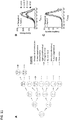
- the inventors have shown that when IR is delivered to mitotic cells, it can directly lead to errors in whole-chromosome segregation, which subsequently leads to the formation of micronuclei and chromosome pulverization hours to days later ( Fig. 6I ).
- the type of missegregation errors in irradiated cells are dependent on the time lapsed after IR exposure. This is likely dependent on the phase of the cell cycle during which cells are irradiated.
- the multilayered genomic damage described herein provides an explanation for the extraordinarily sensitivity of mitotic cells to IR ( Gunderson et al. Clinical Radiation Oncology. Churchill Livingstone; 2011 , Terasima et al. Biophys J. 1963; 3:11-33 , whereby IR exposure during mitosis not only leads to direct DNA breaks but also to additional numerical and downstream structural chromosomal damage. This cell cycle-dependent sensitivity has been exploited in the way radiation treatment is delivered in clinical settings.
- a fundamental rationale for dividing radiation treatment dose into small daily fractions is to enact lethal damage onto the sensitive subpopulation of tumor cells, including the mitotic subpopulation, while sparing toxicity to the surrounding normal tissue which typically contains fewer mitotic cells and is more adept at DNA repair ( Gunderson et al. Clinical Radiation Oncology. Churchill Livingstone; 2011 . Therefore, fractionated radiation therapy can maximize damage to mitotic cell population in otherwise non-synchronized tumors.
- Chromosome pulverization has been postulated to represent a precursor to massive chromosomal rearrangements known as chromothripsis ( Stephens et al. Cell. 144:27-40 (2011 )).
- the findings of the present disclosure indicate that pulverization is likely deleterious to cellular viability. In rare instances, however, these punctuated genomic alterations could lead to selective advantage and generate highly aggressive tumors, which represent a rare but devastating late side-effect of radiation therapy.
- work described in the present disclosure suggests that chromosome pulverization and subsequent chromothripsis would be a defining feature of radiation- induced secondary tumors.
- DMEM Dulbecco's modified medium
- McCoy's medium for HCT116
- fetal bovine serum 50 IU ml -1 penicillin
- 50 ml -1 streptomycin 50 ml -1 streptomycin.
- U251 cells were kindly provided from the laboratory of Mark A. Israel (Geisel School of Medicine at Dartmouth), HCT116 cells (both p53 +/ ⁇ and p53 -/- were kindly provided by the laboratory of Bert Vogelstein (Johns Hopkins University).
- For plasmid selection cells were maintained in 0.5-1.0 mg ml -1 of G418 (geneticin).
- Cells were ⁇ -irradiated using a 137 Cs-irradiator at a rate of 2.38Gy/min or using external beam radiation at 6 MeV delivered by a linear accelerator according to safety rules of Dartmouth and UCSF.
- Tubulin-specific mAb DM1 ⁇ (Sigma-Aldrich), Anti-centromere antibody (CREST, Dartmouth), Anti-cleaved caspase-3 antibody (Cell Signaling), anti-Ki67-antibody (Ventana), anti- ⁇ -H2AX-antibody (Novus Biologicals), GFP-specific antibody (William Wickner, Dartmouth). Antibodies were used at dilutions of 1:1000 or 1:10000 (for GFP-specific antibody).
- TBS-BSA Tris-buffered saline with 5% bovine serum albumin
- TBS-BSA Tris-buffered saline with 5% bovine serum albumin
- Antibodies were diluted in TBS-BSA + 0.1% Triton X-100 and coverslips incubated for 3 hours at room temperature, then washed with TBS-BSA for 5 minutes.
- Secondary antibodies were diluted in TBS-BSA + 0.1% Triton X-100 and coverslips incubated for 1 hour at room temperature.
- HCT116 p53-/- cells were treated with 100 ⁇ M monastrol or DMSO control for 8 h and then ⁇ -irradiated. Immediately following irradiation, cells were washed with PBS twice and then recovered in fresh media for 1 h.
- FISH analysis cells were collected by trypsinization, briefly resuspended in 75 mM potassium chloride, fixed, washed twice in 3:1 methanol/acetic acid mix, dropped onto wet slides, air dried, and stained with DAPI. FISH was performed using both ⁇ -satellite and subtelomere probes specific for the centromeric and q arm telomeric regions of chromosomes 2 respectively (Cytocell). Cells were hybridized according to the manufacturer's protocol, and chromosome signals in at least 300 nuclei were scored.
- HCT116 p53 - / - cells 43 were implanted subcutaneously into the flanks of CD1-Nude mice (4-6 week-old males supplied by the UCSF Breeding Core or Jackson Labs). Tumors were measured with calipers. Volume was calculated by the following formula: width 2 x length x 0.5.
- Tumors were exposed to gamma irradiation ( 137 Cs) at fractionated doses (5 consecutive days X 2Gy) when tumors were ⁇ 300mm 3 or at a single dose (1day X 10Gy) when tumors were ⁇ 800mm 3 . Tumors were isolated and cultured or sectioned for immunohistochemistry.
- Clonogenic assays Cells were either trypsinized (for non-synchronized populations) or collected using mitotic shake-off (for mitotic population) serially diluted and irradiated in their native medium. Cells were then plated in 25-cm 2 T-flasks and clones were grown for 18 days. Clones were stained with Crystal violet and colonies were counted when they reached an approximate size of -50 cell/clone 29 . Relative viability was determined based on the 0 Gy dose.
- Antigen retrieval for immunohistochemistry was performed in Tris EDTA pH 8.0 for 30 minutes at 95 degrees Celsius. Slides were treated with blocking reagent (Vector M.O.M. kit BMK-2202) for 32 minutes. Immunohistochemistry was performed using primary antibodies for Ki67 (Ventana RRF 790-4286, undiluted, room temperature for 16 minutes) or cleaved caspase 3 (Cell Signaling #9661, diluted 1:50 in M.O.M. diluent, 37 degrees Celsius for 60 minutes). Antibody detection was performed using the Ventana IView Detection Kit (760-091).
- High-resolution fluorescence microscopy was used to examine various types of errors during anaphase in three human cell lines derived from normal human retinal epithelium (RPE1), colorectal cancer (HCT116) or glioma (U251). These cells were either near- diploid and chromosomally stable (RPE and HCT116) or aneuploid and chromosomally unstable (U251). RPE1 and HCT116 had an intact p53-signalling pathway ( Thompson et al. Cell Biol. 2010;188:369-381 , whereas U251 contain defective p53 signalling ( Gomez-Godinez et al. Nucleic Acids Res. 38:e202-e202. (2010 ).
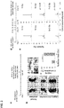
- Fig. 1a displays U251 cells fixed 25 minutes following exposure to 12 Gy and stained for centromeres using anti-centromere antibody (white dots), and DNA using (light grey cloud).
- Fig. 1a shows percentages of chromosome missegregation in response to 0 or 12 Gy IR dose in anaphase spindles of RPE1, U251, and HCT116 cells.
- the legging chromosomes evaluated after IR exposure displayed centromere staining and maintained attachments to microtubules emanating from opposite spindle poles ( Fig. 7a ).
- legging chromosomes exhibited similar levels of staining of y-H2AX, a marker of DNA double stand breaks (DSBs), compared with the remaining chromosomes ( Fig. 7b ). Additionally, the inventors did not observe significant increase in spindles with chromatin bridges ( Fig. 1a, b ).
- IR exposure on chromosome segregation were further evaluated by exposure of HCT116 cells to 6 Gy, followed by fluorescence in situ hybridization (FISH) using centromere and telomere probes for chromosome 2 on irradiated nuclei 1 hour later.
- FISH fluorescence in situ hybridization
- Fig. 1c exposure of non-synchronized cells to IR did not result in significant short-term change in chromosome number.
- IR exposure resulted in approximately 2 fold increase in aneuploidy as evidenced by balanced changes in both centromere and telomere probes specific to human chromosome 2 ( Fig. 1d ).
- HCCT116 cells devoid of tumor suppressor, p53 were used in this experiment, to allow for the proliferation of aneuploidy cells should they emerge ( Thompson and Compton, J Cell Biol. 188(3):369-81, 2010 ).
- HCT116 p53-/- cells were exposed to 0 or 6 Gy of IR and chromosome segregation errors were evaluated at 25 minutes, 12 h, 25 h, and 1 month following the IR exposure. As shown in Fig.
- anaphase spindles examined 24 h or up to 1 month after IR exposure revealed a significant increase in both lagging chromosomes and chromatin bridges ( Fig. 1e ).
- HCT116 p53 -/- cells that normally exhibit low rates of chromosomes missegregation and are thus considered chromosomally stable and near-diploid ( Thompson and Compton, J Cell Biol. 188(3):369-81, 2010 ).
- HCT116 p53 -/cells were subcutaneously injected into nude mice and after 25 days transplanted tumours were exposed to 0 or 10 Gy of IR. Following formalin-fixation of tumours 25 min later, tumour sections were stained with hematoxylin and eosin and the effects of radiation on mitotic cells were evaluated ( Fig. 2a, b). Fig.
- FIG. 2b shows an example of normal anaphase and anaphase cells containing lagging chromosomes in HCT116 p53-/- xenografts after IR exposure.
- Fig. 2c tumours exposed to 10 Gy of IR exhibited significantly higher rates of chromosome segregation errors during anaphase compared with control, non-irradiated, tumours.
- haematoxylin-stained chromatin was frequently visible in the central spindle during anaphase ( Fig. 2b and insets). This chromatin often contained a central constriction reminiscent of centromeric DNA suggesting that this chromatin encompassed whole chromosomes.
- experimental limitations preclude us from resolving lagging chromosomes from acentric chromatin fragments with absolute certainty in fixed tumour tissues.
- HCT116 p53-/- xenografts were exposed to varying doses of radiation (0 Gy, 10 Gy and five daily fractions of 2 Gy over 5 consecutive days ( Fig. 2 d) ).
- the latter fractionated regimen (2 Gy ⁇ 5 days) aims at targeting an overall larger number of mitotic cells over consecutive days.
- Cells were subsequently derived from irradiated tumours and passaged in culture for an additional 15 days to obtain sufficient numbers of cells for karyotype analysis ( Fig. 2d ).
- lagging chromosomes can lead to downstream defects that culminate in structural chromosomal damage ( Hatch et al. Cell. 154:47-60, 2013 ), such as the exclusion of lagging chromosomes from the primary nucleus in the subsequent G1 phase of the cell cycle, resulting in the formation of micronuclei.
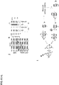
- RPE1 and U251 cells examined 12 h after IR exposure showed increased frequencies of whole-chromosome-containing micronuclei that positively stained for both DNA and centromeres ( Fig. 3a-3c ).
- mitotic U251 cells obtained by mitotic shake-off were irradiated with 12 Gy and chromosome spreads examined 24 h after irradiation in order to assay for chromosome pulverization in the subsequent mitosis as previously described by Crasta et al. (Nature. 482:53-58, 2012 ) ( Fig. 3d ).
- the appearance of many small chromosome fragments and decondensed chromatin indicate the consequences of chromosome pulverization ( Fig. 3e ).
- 12 Gy of IR to mitotic U251 cells led to a significant increase in the fraction of chromosome spreads displaying pulverized chromosomes.
- ⁇ - H2AX density in primary nuclei was significantly lower 12 h after IR exposure compared with 25 min, congruent with DNA repair activity.
- ⁇ - H2AX density in micronuclei was significantly increased at 12 h as compared with 25 min after IR exposure ( Fig. 3g,h ).
- micronuclei are not only defective in DNA repair but can actively generate additional DNA damage. This additional damage is likely the consequence of faulty attempts at DNA repair and defective micronuclei nuclear envelope structures. Therefore, by inducing mitotic errors, IR leads to amplifications of structural chromosomal defects that predominantly occur outside of the primary nucleus (extra-nuclear). Unlike DNA damage caused directly by IR, these defects are precipitated many hours after IR exposure.
- Kif2b is a microtubule-depolymerizing kinesin-13 protein that specifically corrects erroneous microtubule attachments to chromosomes ( Bakhoum et al. Nat Cell Biol. 2009;11:27-35 ; Manning et al. Mol Biol Cell. 2007;18:2970-2979 ).
- the inventors measured relative ⁇ - H2AX fluorescence intensity in irradiated mitotic U251 cells. As shown in Fig. 4a , there was no difference between control and GFP- Kif2b-overexpressing U251 cells. Furthermore, when the average number of ⁇ - H2AX foci in the primary nuclei 20 min after IR exposure was compared to that of 12 h after IR exposure, there was no significant difference between control and GFP- Kif2b-overexpressing cells ( Fig. 4b, c ).
- Mitosis has long been recognized, for unclear reasons, as the most radiation sensitive phase of the cell cycle ( Terasima et al. Biophys J. 1963;3:11-33 ; Sinclair et al. Radiat Res. 1966;29:450-474 ).
- the inventors have accomplished to selectively reduce chromosome segregation errors without influencing the canonical IR-induced DNA damage and repair within the primary nucleus. This allows for testing whether whole-chromosome segregation errors might independently contribute towards the sensitivity of mitotic cells to IR.
- cells were enriched for mitotic cells using mitotic shake-off before irradiation and plating for colony growth ( Fig. 5a ).
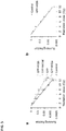
- Fig. 5a GFP-Kif2b overexpression led to significant increase in the viability after irradiation, whereby at 12 Gy of IR, these cells were ⁇ 20-fold more resistant compared with control cells.
- GFP-Kif2b overexpression led to increased viability in RPE1 cells as well ( Fig. 8a ).
- GFP-Kif2b overexpression did not alter the growth rate of U251 cells in culture or did it significantly influence their karyotypic distribution or modal chromosome numbers ( Fig. 8b, 8c ).
- GFP- MCAK a second kinesin-13 protein also known to suppress w-CIN
- overexpression of GFP alone or the third microtubule-depolymerizing kinesin-13 paralogue, GFP- Kif2a did not alter the clonogenic potential of irradiated cells compared with control ( Fig. 5a , and Fig. 7c ).
- Fig. 9 Absolute bioluminescence values before the initiation of treatment increased at comparable rates in GFP-and GFP- Kif2b-overexpressing tumours suggesting similar tumour growth rates.
- Tumours overexpressing only GFP showed a robust response to radiation treatment as judged by approximately tenfold reduction in luciferase signal at the end of the treatment course ( Fig. 6b-6d ).
- Fig. 6b-6d there was striking resistance to radiation treatment in tumours derived from cells overexpressing GFP- Kif2b
- Fig. 6c shows normalized bioluminescence of intracranial U251 xenografts overexpressing GFP or GFP-Kif2b after initiation of IR treatment.
- Fig. 9 shows normalized bioluminescence of intracranial U251 xenografts overexpressing GFP or GFP-Kif2b after initiation of IR treatment.
- GFP- and GFP- Kif2b-expressing tumours also exhibited similar frequencies of multipolar mitoses known to occur after radiation exposure ( Sato et al. Oncogene 19, 5281-5290 (2000 )), Fog. 6h and 6i.
- GFP- Kif2b-expressing tumours displayed decreased apoptosis as indicated by lower cleaved caspase 3 staining ( Fig. 6j and 6k ). Therefore, suppression of numerical chromosomal instability by altering kinetochore-microtubule attachment stability leads to significant radiation resistance likely by suppressing cell death resultant from excessive chromosomal damage.
- TNBC triple negative breast cancer
- mice Under this genetic background, -58% of 60 mice developed spontaneous intraductal carcinomas characterized with high-grade pleomorphic nuclei, significant chromosome copy number alterations (CNAs) and numerous mitoses, highly resembling human TNBC.
- CNAs chromosome copy number alterations
- patient-derived xenograft model will be used as well. Briefly, 10 5 cells will be transplanted in the mammary fat pads of immuno-competent BL6 hosts. Once tumors reach 5-mm in diameter, 2Gy daily fractions to a total dose of 24Gy or a biologically equivalent single-dose of 13Gy will be delivered using the JLShepherd @ Associates irradiator (model: MK1-68).
- breast cancer cell lines are highly dependent on Plk4 function and loss of tumor suppressor phosphatase and tensin homolog ( PTEN), a frequent occurrence in TNBC, is synthetically lethal with loss of Plk4 activity Brough, R. et al. Cancer Discovery 1, 260-273 (2011 )), making the latter an attractive target to further explore in combination with established therapies in TNBC.
- Plk4 tumor suppressor phosphatase and tensin homolog
- CFI-400945 will be administered at a concentration of 9.4 mg/kg daily for 2 weeks as previously described ( Mason et al. Cancer Cell 26, 163-176 (2014 )). Tumor response will be measured using calipers and bioluminescence imaging. Additionally, measures of the time-to-relapse will be taken and direct comparison will be made between BRCA-proficient and BRCA-deficient tumors. For statistical methods please see Vertebrate Animals section. Targeted sequencing and FISH will be to used assess the PTEN status of these tumors given the known synthetic lethality with the loss of PTEN and plk4.
- chromosome missegregation frequency 1.9x10 -3 per chromosome copy per cell division
- CIN may represent a predictive marker of response to therapies that are known to induce chromosome missegregation ( Jamal-Hanjani, M. et al. Annals of Oncology 26, 1340-1346 (2015 ), Zaki, et al. Cancer 120, 1733-1742 (2014 )); these therapies likely drive a tumor from an optimal fitness state to a state so chromosomally unstable that it is no longer compatible with viability.
- the present inventors have discovered that increasing chromosome missegregation together with radiation treatment would lead to sensitization of the tumor to radiation therapy. This in turn permits 1) to decrease dose of radiation and achieve the same effect, 2) to maintain dose of radiation and increase tumor sensitization in otherwise resistant tumors, 3) to increase radioprotection of normal organs.
Landscapes
- Health & Medical Sciences (AREA)
- Life Sciences & Earth Sciences (AREA)
- Animal Behavior & Ethology (AREA)
- Veterinary Medicine (AREA)
- Public Health (AREA)
- General Health & Medical Sciences (AREA)
- Engineering & Computer Science (AREA)
- Medicinal Chemistry (AREA)
- Chemical & Material Sciences (AREA)
- Pharmacology & Pharmacy (AREA)
- Epidemiology (AREA)
- Biomedical Technology (AREA)
- Nuclear Medicine, Radiotherapy & Molecular Imaging (AREA)
- Radiology & Medical Imaging (AREA)
- Pathology (AREA)
- Gastroenterology & Hepatology (AREA)
- Bioinformatics & Cheminformatics (AREA)
- Immunology (AREA)
- Proteomics, Peptides & Aminoacids (AREA)
- General Chemical & Material Sciences (AREA)
- Organic Chemistry (AREA)
- Marine Sciences & Fisheries (AREA)
- Zoology (AREA)
- Chemical Kinetics & Catalysis (AREA)
- Pharmaceuticals Containing Other Organic And Inorganic Compounds (AREA)
- Medicines That Contain Protein Lipid Enzymes And Other Medicines (AREA)
Claims (4)
- Strahlungssensibilisierungsmittel zur Verwendung bei einem Krebstherapieverfahren, wobei das Strahlungssensibilisierungsmittel die Anfälligkeit von Krebszellen gegenüber ionisierender Strahlung erhöht, wobei das Strahlungssensibilisierungsmittel spezifisch den Prozess der Chromosomensegregation stört, wodurch Fehlsegregation induziert wird, oder spezifisch ein zweites Mittel hemmt, das eine zuverlässige Chromosomensegregation fördert, wodurch das Strahlungssensibilisierungsmittel eine Unbeständigkeit der Chromosomenanzahl in den Zellen induziert, wobei das Strahlungssensibilisierungsmittel gleichzeitig mit oder kurz vor oder kurz nach einer Bestrahlung der Zellen mit einer therapeutisch wirksamen Dosis und einem Schema ionisierender Strahlung verabreicht wird und wobei das Strahlungssensibilisierungsmittel aus der Gruppe ausgewählt ist, die aus Kif2b-Inhibitoren, MPS1-Inhibitoren, EgS/Kinesin-5-Inhibitoren, Inhibitoren der Polo-like-Kinase 4, MCAK-Inhibitoren, Bub1-Inhibitoren und Hecl-Inhibitoren besteht.
- Strahlungssensibilisierungsmittel zur Verwendung nach Anspruch 1, wobei das Strahlungssensibilisierungsmittel spezifisch eines oder mehrere der folgenden hemmt: Kif2b, MPS1, Eg5/Kinesin-5, Polo-like-Kinase 4, MCAK, Bub1 und Hecl.
- Strahlungssensibilisierungsmittel zur Verwendung nach Anspruch 1, wobei es sich bei den Krebszellen um Krebszellen eines soliden Tumors handelt.
- Strahlungssensibilisierungsmittel zur Verwendung nach Anspruch 1, wobei die ionisierende Strahlung in einer oder zwei oder mehr geteilten Dosen verabreicht wird.
Priority Applications (1)
| Application Number | Priority Date | Filing Date | Title |
|---|---|---|---|
| EP21196551.2A EP3981430A1 (de) | 2015-01-21 | 2016-01-21 | Verfahren und zusammensetzungen zur erhöhung der empfänglichkeit einer strahlenbehandlung durch hemmung der suppression von numerischer chromosomaler instabilität von krebszellen |
Applications Claiming Priority (2)
| Application Number | Priority Date | Filing Date | Title |
|---|---|---|---|
| US201562106204P | 2015-01-21 | 2015-01-21 | |
| PCT/US2016/014400 WO2016130297A2 (en) | 2015-01-21 | 2016-01-21 | Methods and compositions for increasing susceptibility to radiation treatment by inhibiting suppression of numerical chromosomal instability of cancer cells |
Related Child Applications (1)
| Application Number | Title | Priority Date | Filing Date |
|---|---|---|---|
| EP21196551.2A Division EP3981430A1 (de) | 2015-01-21 | 2016-01-21 | Verfahren und zusammensetzungen zur erhöhung der empfänglichkeit einer strahlenbehandlung durch hemmung der suppression von numerischer chromosomaler instabilität von krebszellen |
Publications (3)
| Publication Number | Publication Date |
|---|---|
| EP3247410A2 EP3247410A2 (de) | 2017-11-29 |
| EP3247410A4 EP3247410A4 (de) | 2018-10-10 |
| EP3247410B1 true EP3247410B1 (de) | 2021-09-15 |
Family
ID=56615060
Family Applications (2)
| Application Number | Title | Priority Date | Filing Date |
|---|---|---|---|
| EP21196551.2A Ceased EP3981430A1 (de) | 2015-01-21 | 2016-01-21 | Verfahren und zusammensetzungen zur erhöhung der empfänglichkeit einer strahlenbehandlung durch hemmung der suppression von numerischer chromosomaler instabilität von krebszellen |
| EP16749573.8A Active EP3247410B1 (de) | 2015-01-21 | 2016-01-21 | Verfahren und zusammensetzungen zur erhöhung der empfänglichkeit einer strahlenbehandlung durch hemmung der suppression von numerischer chromosomaler instabilität von krebszellen |
Family Applications Before (1)
| Application Number | Title | Priority Date | Filing Date |
|---|---|---|---|
| EP21196551.2A Ceased EP3981430A1 (de) | 2015-01-21 | 2016-01-21 | Verfahren und zusammensetzungen zur erhöhung der empfänglichkeit einer strahlenbehandlung durch hemmung der suppression von numerischer chromosomaler instabilität von krebszellen |
Country Status (3)
| Country | Link |
|---|---|
| US (3) | US20180008709A1 (de) |
| EP (2) | EP3981430A1 (de) |
| WO (1) | WO2016130297A2 (de) |
Families Citing this family (5)
| Publication number | Priority date | Publication date | Assignee | Title |
|---|---|---|---|---|
| JP2020525597A (ja) | 2017-07-05 | 2020-08-27 | コベストロ、ドイチュラント、アクチエンゲゼルシャフトCovestro Deutschland Ag | ポリイソシアネートの連続希釈 |
| WO2019046949A1 (en) | 2017-09-08 | 2019-03-14 | University Health Network | POLYTHERAPIES FOR INHIBITING KINASE 4 OF THE POLO TYPE |
| CN108950001B (zh) * | 2018-08-31 | 2020-08-11 | 青岛泱深生物医药有限公司 | 一种与直肠腺癌发生发展相关的分子标志物 |
| KR102324002B1 (ko) * | 2019-10-07 | 2021-11-08 | 대구가톨릭대학교산학협력단 | 투과이온빔을 이용한 쿨롱자극 산화철-단백질응집 분쇄장치 및 투과이온빔의 선량측정도구 |
| WO2025101836A1 (en) * | 2023-11-10 | 2025-05-15 | The Trustees Of Dartmouth College | Method for inducing aneuploidy and/or chromosomal instability in human or non-human primate pluripotent stem cells and diagnostic/therapeutic uses thereof |
Family Cites Families (3)
| Publication number | Priority date | Publication date | Assignee | Title |
|---|---|---|---|---|
| US20060177828A1 (en) | 2003-09-18 | 2006-08-10 | Armour Christopher D | Alternatively spliced isoform of mitotic centromere-associated kinesin (MCAK) |
| US8541476B2 (en) * | 2007-09-14 | 2013-09-24 | The Trustees Of The University Of Pennsylvania | Compounds for treatment of malignant tumors |
| WO2012078703A2 (en) * | 2010-12-08 | 2012-06-14 | Fox Chase Cancer Center | Inhibiting chromosome instability in ovarian cancer |
-
2016
- 2016-01-21 US US15/544,811 patent/US20180008709A1/en not_active Abandoned
- 2016-01-21 WO PCT/US2016/014400 patent/WO2016130297A2/en not_active Ceased
- 2016-01-21 EP EP21196551.2A patent/EP3981430A1/de not_active Ceased
- 2016-01-21 EP EP16749573.8A patent/EP3247410B1/de active Active
-
2020
- 2020-03-13 US US16/818,966 patent/US11623006B2/en active Active
-
2023
- 2023-02-27 US US18/175,433 patent/US12133892B2/en active Active
Also Published As
| Publication number | Publication date |
|---|---|
| US20200282054A1 (en) | 2020-09-10 |
| WO2016130297A2 (en) | 2016-08-18 |
| US11623006B2 (en) | 2023-04-11 |
| EP3247410A4 (de) | 2018-10-10 |
| US12133892B2 (en) | 2024-11-05 |
| US20180008709A1 (en) | 2018-01-11 |
| EP3981430A1 (de) | 2022-04-13 |
| EP3247410A2 (de) | 2017-11-29 |
| US20230293693A1 (en) | 2023-09-21 |
Similar Documents
| Publication | Publication Date | Title |
|---|---|---|
| US12133892B2 (en) | Methods and compositions for increasing susceptibility to radiation treatment by inhibiting suppression of numerical chromosomal instability of cancer cells | |
| Prete et al. | Pericytes elicit resistance to vemurafenib and sorafenib therapy in thyroid carcinoma via the TSP-1/TGFβ1 axis | |
| Maranto et al. | STAT5A/B blockade sensitizes prostate cancer to radiation through inhibition of RAD51 and DNA repair | |
| Neznanov et al. | Proteotoxic stress targeted therapy (PSTT): induction of protein misfolding enhances the antitumor effect of the proteasome inhibitor bortezomib | |
| Zhao et al. | Caspase-3 knockout attenuates radiation-induced tumor repopulation via impairing the ATM/p53/Cox-2/PGE2 pathway in non-small cell lung cancer | |
| CN110582581A (zh) | 用于治疗过度增殖性疾病的方法中的atr激酶的抑制剂 | |
| KR20220151027A (ko) | Mdm2 억제제의 간헐적 투여 | |
| EP3268087A1 (de) | Verfahren zur behandlung von krebs mit ror-gamma-inhibitoren | |
| Xue et al. | Therapeutic evaluation of palbociclib and its compatibility with other chemotherapies for primary and recurrent nasopharyngeal carcinoma | |
| CN115998872B (zh) | 一种含有抑制核酸内切酶功能的药物及其抗肿瘤的用途 | |
| Zhang et al. | Hedgehog signaling and the glioma-associated oncogene in cancer radioresistance | |
| US12594293B2 (en) | Use of 6-thio-dG to treat therapy-resistant telomerasepositive pediatric brain tumors | |
| Odhiambo et al. | Preclinical evaluation of the ATR inhibitor BAY 1895344 as a radiosensitizer for head and neck squamous cell carcinoma | |
| Wu et al. | BMS-345541 sensitizes MCF-7 breast cancer cells to ionizing radiation by selective inhibition of homologous recombinational repair of DNA double-strand breaks | |
| CA2829217C (en) | Compounds and methods of use in ablative radiotherapy | |
| Lee et al. | Inhibition of Aberrantly Overexpressed Polo-like Kinase 4 Is a Potential Effective Treatment for DNA Damage Repair–Deficient Uterine Leiomyosarcoma | |
| Stigliano et al. | Current and emerging therapeutic options in adrenocortical cancer treatment | |
| US20190365745A1 (en) | Use of chronic treatment with atr inhibitors to sensitize cancer cells to parp inhibitors | |
| WO2020210829A1 (en) | Methods of promoting long-term survival of patients with advanced chemotherapy-resistant malignancies | |
| EP3958854A1 (de) | Verwendung von tg02 zur behandlung von gliomen bei pädiatrischen patienten | |
| Wright | Investigating combinations of radium-223 and androgen deprivation therapies in advanced prostate cancer | |
| US20260021099A1 (en) | Cancer Treatment Targeted to Tumor Adaptive Responses to Protein Synthesis Stress | |
| Bunz | Cancer Therapy | |
| Roggero Savoini et al. | CDK4/6 inhibitors promote PARP1 degradation and synergize with PARP inhibitors in non-small cell lung cancer | |
| Zhao et al. | JS-K enhances renal cell carcinoma radiosensitivity through the Wnt/β-catenin signaling pathway and inhibits DNA damage repair |
Legal Events
| Date | Code | Title | Description |
|---|---|---|---|
| STAA | Information on the status of an ep patent application or granted ep patent |
Free format text: STATUS: THE INTERNATIONAL PUBLICATION HAS BEEN MADE |
|
| PUAI | Public reference made under article 153(3) epc to a published international application that has entered the european phase |
Free format text: ORIGINAL CODE: 0009012 |
|
| STAA | Information on the status of an ep patent application or granted ep patent |
Free format text: STATUS: REQUEST FOR EXAMINATION WAS MADE |
|
| 17P | Request for examination filed |
Effective date: 20170817 |
|
| AK | Designated contracting states |
Kind code of ref document: A2 Designated state(s): AL AT BE BG CH CY CZ DE DK EE ES FI FR GB GR HR HU IE IS IT LI LT LU LV MC MK MT NL NO PL PT RO RS SE SI SK SM TR |
|
| AX | Request for extension of the european patent |
Extension state: BA ME |
|
| DAV | Request for validation of the european patent (deleted) | ||
| DAX | Request for extension of the european patent (deleted) | ||
| A4 | Supplementary search report drawn up and despatched |
Effective date: 20180906 |
|
| RIC1 | Information provided on ipc code assigned before grant |
Ipc: A61K 41/00 20060101ALI20180901BHEP Ipc: A61N 5/10 20060101ALI20180901BHEP Ipc: A61K 48/00 20060101AFI20180901BHEP Ipc: A61P 35/00 20060101ALI20180901BHEP |
|
| STAA | Information on the status of an ep patent application or granted ep patent |
Free format text: STATUS: EXAMINATION IS IN PROGRESS |
|
| 17Q | First examination report despatched |
Effective date: 20190513 |
|
| GRAP | Despatch of communication of intention to grant a patent |
Free format text: ORIGINAL CODE: EPIDOSNIGR1 |
|
| STAA | Information on the status of an ep patent application or granted ep patent |
Free format text: STATUS: GRANT OF PATENT IS INTENDED |
|
| INTG | Intention to grant announced |
Effective date: 20210406 |
|
| RAP3 | Party data changed (applicant data changed or rights of an application transferred) |
Owner name: THE TRUSTEES OF DARTMOUTH COLLEGE Owner name: MEMORIAL SLOAN-KETTERING CANCER CENTER |
|
| GRAS | Grant fee paid |
Free format text: ORIGINAL CODE: EPIDOSNIGR3 |
|
| GRAA | (expected) grant |
Free format text: ORIGINAL CODE: 0009210 |
|
| STAA | Information on the status of an ep patent application or granted ep patent |
Free format text: STATUS: THE PATENT HAS BEEN GRANTED |
|
| AK | Designated contracting states |
Kind code of ref document: B1 Designated state(s): AL AT BE BG CH CY CZ DE DK EE ES FI FR GB GR HR HU IE IS IT LI LT LU LV MC MK MT NL NO PL PT RO RS SE SI SK SM TR |
|
| REG | Reference to a national code |
Ref country code: CH Ref legal event code: EP |
|
| REG | Reference to a national code |
Ref country code: DE Ref legal event code: R096 Ref document number: 602016063730 Country of ref document: DE |
|
| REG | Reference to a national code |
Ref country code: IE Ref legal event code: FG4D |
|
| REG | Reference to a national code |
Ref country code: AT Ref legal event code: REF Ref document number: 1430020 Country of ref document: AT Kind code of ref document: T Effective date: 20211015 |
|
| REG | Reference to a national code |
Ref country code: LT Ref legal event code: MG9D |
|
| REG | Reference to a national code |
Ref country code: NL Ref legal event code: MP Effective date: 20210915 |
|
| PG25 | Lapsed in a contracting state [announced via postgrant information from national office to epo] |
Ref country code: BG Free format text: LAPSE BECAUSE OF FAILURE TO SUBMIT A TRANSLATION OF THE DESCRIPTION OR TO PAY THE FEE WITHIN THE PRESCRIBED TIME-LIMIT Effective date: 20211215 Ref country code: LT Free format text: LAPSE BECAUSE OF FAILURE TO SUBMIT A TRANSLATION OF THE DESCRIPTION OR TO PAY THE FEE WITHIN THE PRESCRIBED TIME-LIMIT Effective date: 20210915 Ref country code: NO Free format text: LAPSE BECAUSE OF FAILURE TO SUBMIT A TRANSLATION OF THE DESCRIPTION OR TO PAY THE FEE WITHIN THE PRESCRIBED TIME-LIMIT Effective date: 20211215 Ref country code: FI Free format text: LAPSE BECAUSE OF FAILURE TO SUBMIT A TRANSLATION OF THE DESCRIPTION OR TO PAY THE FEE WITHIN THE PRESCRIBED TIME-LIMIT Effective date: 20210915 Ref country code: HR Free format text: LAPSE BECAUSE OF FAILURE TO SUBMIT A TRANSLATION OF THE DESCRIPTION OR TO PAY THE FEE WITHIN THE PRESCRIBED TIME-LIMIT Effective date: 20210915 Ref country code: RS Free format text: LAPSE BECAUSE OF FAILURE TO SUBMIT A TRANSLATION OF THE DESCRIPTION OR TO PAY THE FEE WITHIN THE PRESCRIBED TIME-LIMIT Effective date: 20210915 Ref country code: SE Free format text: LAPSE BECAUSE OF FAILURE TO SUBMIT A TRANSLATION OF THE DESCRIPTION OR TO PAY THE FEE WITHIN THE PRESCRIBED TIME-LIMIT Effective date: 20210915 |
|
| REG | Reference to a national code |
Ref country code: AT Ref legal event code: MK05 Ref document number: 1430020 Country of ref document: AT Kind code of ref document: T Effective date: 20210915 |
|
| PG25 | Lapsed in a contracting state [announced via postgrant information from national office to epo] |
Ref country code: LV Free format text: LAPSE BECAUSE OF FAILURE TO SUBMIT A TRANSLATION OF THE DESCRIPTION OR TO PAY THE FEE WITHIN THE PRESCRIBED TIME-LIMIT Effective date: 20210915 Ref country code: GR Free format text: LAPSE BECAUSE OF FAILURE TO SUBMIT A TRANSLATION OF THE DESCRIPTION OR TO PAY THE FEE WITHIN THE PRESCRIBED TIME-LIMIT Effective date: 20211216 |
|
| PG25 | Lapsed in a contracting state [announced via postgrant information from national office to epo] |
Ref country code: AT Free format text: LAPSE BECAUSE OF FAILURE TO SUBMIT A TRANSLATION OF THE DESCRIPTION OR TO PAY THE FEE WITHIN THE PRESCRIBED TIME-LIMIT Effective date: 20210915 |
|
| PG25 | Lapsed in a contracting state [announced via postgrant information from national office to epo] |
Ref country code: IS Free format text: LAPSE BECAUSE OF FAILURE TO SUBMIT A TRANSLATION OF THE DESCRIPTION OR TO PAY THE FEE WITHIN THE PRESCRIBED TIME-LIMIT Effective date: 20220115 Ref country code: SM Free format text: LAPSE BECAUSE OF FAILURE TO SUBMIT A TRANSLATION OF THE DESCRIPTION OR TO PAY THE FEE WITHIN THE PRESCRIBED TIME-LIMIT Effective date: 20210915 Ref country code: SK Free format text: LAPSE BECAUSE OF FAILURE TO SUBMIT A TRANSLATION OF THE DESCRIPTION OR TO PAY THE FEE WITHIN THE PRESCRIBED TIME-LIMIT Effective date: 20210915 Ref country code: RO Free format text: LAPSE BECAUSE OF FAILURE TO SUBMIT A TRANSLATION OF THE DESCRIPTION OR TO PAY THE FEE WITHIN THE PRESCRIBED TIME-LIMIT Effective date: 20210915 Ref country code: PT Free format text: LAPSE BECAUSE OF FAILURE TO SUBMIT A TRANSLATION OF THE DESCRIPTION OR TO PAY THE FEE WITHIN THE PRESCRIBED TIME-LIMIT Effective date: 20220117 Ref country code: PL Free format text: LAPSE BECAUSE OF FAILURE TO SUBMIT A TRANSLATION OF THE DESCRIPTION OR TO PAY THE FEE WITHIN THE PRESCRIBED TIME-LIMIT Effective date: 20210915 Ref country code: NL Free format text: LAPSE BECAUSE OF FAILURE TO SUBMIT A TRANSLATION OF THE DESCRIPTION OR TO PAY THE FEE WITHIN THE PRESCRIBED TIME-LIMIT Effective date: 20210915 Ref country code: ES Free format text: LAPSE BECAUSE OF FAILURE TO SUBMIT A TRANSLATION OF THE DESCRIPTION OR TO PAY THE FEE WITHIN THE PRESCRIBED TIME-LIMIT Effective date: 20210915 Ref country code: EE Free format text: LAPSE BECAUSE OF FAILURE TO SUBMIT A TRANSLATION OF THE DESCRIPTION OR TO PAY THE FEE WITHIN THE PRESCRIBED TIME-LIMIT Effective date: 20210915 Ref country code: CZ Free format text: LAPSE BECAUSE OF FAILURE TO SUBMIT A TRANSLATION OF THE DESCRIPTION OR TO PAY THE FEE WITHIN THE PRESCRIBED TIME-LIMIT Effective date: 20210915 Ref country code: AL Free format text: LAPSE BECAUSE OF FAILURE TO SUBMIT A TRANSLATION OF THE DESCRIPTION OR TO PAY THE FEE WITHIN THE PRESCRIBED TIME-LIMIT Effective date: 20210915 |
|
| REG | Reference to a national code |
Ref country code: DE Ref legal event code: R097 Ref document number: 602016063730 Country of ref document: DE |
|
| PLBE | No opposition filed within time limit |
Free format text: ORIGINAL CODE: 0009261 |
|
| STAA | Information on the status of an ep patent application or granted ep patent |
Free format text: STATUS: NO OPPOSITION FILED WITHIN TIME LIMIT |
|
| PG25 | Lapsed in a contracting state [announced via postgrant information from national office to epo] |
Ref country code: DK Free format text: LAPSE BECAUSE OF FAILURE TO SUBMIT A TRANSLATION OF THE DESCRIPTION OR TO PAY THE FEE WITHIN THE PRESCRIBED TIME-LIMIT Effective date: 20210915 |
|
| 26N | No opposition filed |
Effective date: 20220616 |
|
| PG25 | Lapsed in a contracting state [announced via postgrant information from national office to epo] |
Ref country code: SI Free format text: LAPSE BECAUSE OF FAILURE TO SUBMIT A TRANSLATION OF THE DESCRIPTION OR TO PAY THE FEE WITHIN THE PRESCRIBED TIME-LIMIT Effective date: 20210915 Ref country code: MC Free format text: LAPSE BECAUSE OF FAILURE TO SUBMIT A TRANSLATION OF THE DESCRIPTION OR TO PAY THE FEE WITHIN THE PRESCRIBED TIME-LIMIT Effective date: 20210915 |
|
| REG | Reference to a national code |
Ref country code: CH Ref legal event code: PL |
|
| REG | Reference to a national code |
Ref country code: BE Ref legal event code: MM Effective date: 20220131 |
|
| PG25 | Lapsed in a contracting state [announced via postgrant information from national office to epo] |
Ref country code: LU Free format text: LAPSE BECAUSE OF NON-PAYMENT OF DUE FEES Effective date: 20220121 |
|
| PG25 | Lapsed in a contracting state [announced via postgrant information from national office to epo] |
Ref country code: BE Free format text: LAPSE BECAUSE OF NON-PAYMENT OF DUE FEES Effective date: 20220131 |
|
| REG | Reference to a national code |
Ref country code: FR Ref legal event code: PLFP Year of fee payment: 8 |
|
| PG25 | Lapsed in a contracting state [announced via postgrant information from national office to epo] |
Ref country code: LI Free format text: LAPSE BECAUSE OF NON-PAYMENT OF DUE FEES Effective date: 20220131 Ref country code: CH Free format text: LAPSE BECAUSE OF NON-PAYMENT OF DUE FEES Effective date: 20220131 |
|
| PG25 | Lapsed in a contracting state [announced via postgrant information from national office to epo] |
Ref country code: IT Free format text: LAPSE BECAUSE OF FAILURE TO SUBMIT A TRANSLATION OF THE DESCRIPTION OR TO PAY THE FEE WITHIN THE PRESCRIBED TIME-LIMIT Effective date: 20210915 Ref country code: IE Free format text: LAPSE BECAUSE OF NON-PAYMENT OF DUE FEES Effective date: 20220121 |
|
| P01 | Opt-out of the competence of the unified patent court (upc) registered |
Effective date: 20230517 |
|
| PG25 | Lapsed in a contracting state [announced via postgrant information from national office to epo] |
Ref country code: HU Free format text: LAPSE BECAUSE OF FAILURE TO SUBMIT A TRANSLATION OF THE DESCRIPTION OR TO PAY THE FEE WITHIN THE PRESCRIBED TIME-LIMIT; INVALID AB INITIO Effective date: 20160121 |
|
| PG25 | Lapsed in a contracting state [announced via postgrant information from national office to epo] |
Ref country code: MK Free format text: LAPSE BECAUSE OF FAILURE TO SUBMIT A TRANSLATION OF THE DESCRIPTION OR TO PAY THE FEE WITHIN THE PRESCRIBED TIME-LIMIT Effective date: 20210915 Ref country code: CY Free format text: LAPSE BECAUSE OF FAILURE TO SUBMIT A TRANSLATION OF THE DESCRIPTION OR TO PAY THE FEE WITHIN THE PRESCRIBED TIME-LIMIT Effective date: 20210915 |
|
| PG25 | Lapsed in a contracting state [announced via postgrant information from national office to epo] |
Ref country code: TR Free format text: LAPSE BECAUSE OF FAILURE TO SUBMIT A TRANSLATION OF THE DESCRIPTION OR TO PAY THE FEE WITHIN THE PRESCRIBED TIME-LIMIT Effective date: 20210915 |
|
| PG25 | Lapsed in a contracting state [announced via postgrant information from national office to epo] |
Ref country code: MT Free format text: LAPSE BECAUSE OF FAILURE TO SUBMIT A TRANSLATION OF THE DESCRIPTION OR TO PAY THE FEE WITHIN THE PRESCRIBED TIME-LIMIT Effective date: 20210915 |
|
| PGFP | Annual fee paid to national office [announced via postgrant information from national office to epo] |
Ref country code: GB Payment date: 20251204 Year of fee payment: 11 |
|
| PGFP | Annual fee paid to national office [announced via postgrant information from national office to epo] |
Ref country code: FR Payment date: 20251208 Year of fee payment: 11 |
|
| PGFP | Annual fee paid to national office [announced via postgrant information from national office to epo] |
Ref country code: DE Payment date: 20251203 Year of fee payment: 11 |