EP3246706A1 - Method for specific cleavage of c(alpha)-c bond and side chain of protein and peptide, and method for determining amino acid sequence - Google Patents
Method for specific cleavage of c(alpha)-c bond and side chain of protein and peptide, and method for determining amino acid sequence Download PDFInfo
- Publication number
- EP3246706A1 EP3246706A1 EP17170513.0A EP17170513A EP3246706A1 EP 3246706 A1 EP3246706 A1 EP 3246706A1 EP 17170513 A EP17170513 A EP 17170513A EP 3246706 A1 EP3246706 A1 EP 3246706A1
- Authority
- EP
- European Patent Office
- Prior art keywords
- hydroxy
- peptide
- nitrobenzoic acid
- protein
- bond
- Prior art date
- Legal status (The legal status is an assumption and is not a legal conclusion. Google has not performed a legal analysis and makes no representation as to the accuracy of the status listed.)
- Withdrawn
Links
- 0 CC*c1cc(C(O)=O)cc(O)c1 Chemical compound CC*c1cc(C(O)=O)cc(O)c1 0.000 description 2
- QRYSWXFQLFLJTC-UHFFFAOYSA-N [O-][N+](c1cc(C(O)=O)ccc1O)=O Chemical compound [O-][N+](c1cc(C(O)=O)ccc1O)=O QRYSWXFQLFLJTC-UHFFFAOYSA-N 0.000 description 1
Images




Classifications
-
- G—PHYSICS
- G01—MEASURING; TESTING
- G01N—INVESTIGATING OR ANALYSING MATERIALS BY DETERMINING THEIR CHEMICAL OR PHYSICAL PROPERTIES
- G01N33/00—Investigating or analysing materials by specific methods not covered by groups G01N1/00 - G01N31/00
- G01N33/48—Biological material, e.g. blood, urine; Haemocytometers
- G01N33/50—Chemical analysis of biological material, e.g. blood, urine; Testing involving biospecific ligand binding methods; Immunological testing
- G01N33/68—Chemical analysis of biological material, e.g. blood, urine; Testing involving biospecific ligand binding methods; Immunological testing involving proteins, peptides or amino acids
- G01N33/6803—General methods of protein analysis not limited to specific proteins or families of proteins
- G01N33/6842—Proteomic analysis of subsets of protein mixtures with reduced complexity, e.g. membrane proteins, phosphoproteins, organelle proteins
-
- C—CHEMISTRY; METALLURGY
- C07—ORGANIC CHEMISTRY
- C07K—PEPTIDES
- C07K1/00—General methods for the preparation of peptides, i.e. processes for the organic chemical preparation of peptides or proteins of any length
- C07K1/12—General methods for the preparation of peptides, i.e. processes for the organic chemical preparation of peptides or proteins of any length by hydrolysis, i.e. solvolysis in general
- C07K1/128—General methods for the preparation of peptides, i.e. processes for the organic chemical preparation of peptides or proteins of any length by hydrolysis, i.e. solvolysis in general sequencing
-
- C—CHEMISTRY; METALLURGY
- C07—ORGANIC CHEMISTRY
- C07C—ACYCLIC OR CARBOCYCLIC COMPOUNDS
- C07C205/00—Compounds containing nitro groups bound to a carbon skeleton
- C07C205/49—Compounds containing nitro groups bound to a carbon skeleton the carbon skeleton being further substituted by carboxyl groups
- C07C205/57—Compounds containing nitro groups bound to a carbon skeleton the carbon skeleton being further substituted by carboxyl groups having nitro groups and carboxyl groups bound to carbon atoms of six-membered aromatic rings of the carbon skeleton
- C07C205/59—Compounds containing nitro groups bound to a carbon skeleton the carbon skeleton being further substituted by carboxyl groups having nitro groups and carboxyl groups bound to carbon atoms of six-membered aromatic rings of the carbon skeleton the carbon skeleton being further substituted by singly-bound oxygen atoms
-
- C—CHEMISTRY; METALLURGY
- C07—ORGANIC CHEMISTRY
- C07C—ACYCLIC OR CARBOCYCLIC COMPOUNDS
- C07C205/00—Compounds containing nitro groups bound to a carbon skeleton
- C07C205/49—Compounds containing nitro groups bound to a carbon skeleton the carbon skeleton being further substituted by carboxyl groups
- C07C205/57—Compounds containing nitro groups bound to a carbon skeleton the carbon skeleton being further substituted by carboxyl groups having nitro groups and carboxyl groups bound to carbon atoms of six-membered aromatic rings of the carbon skeleton
- C07C205/59—Compounds containing nitro groups bound to a carbon skeleton the carbon skeleton being further substituted by carboxyl groups having nitro groups and carboxyl groups bound to carbon atoms of six-membered aromatic rings of the carbon skeleton the carbon skeleton being further substituted by singly-bound oxygen atoms
- C07C205/60—Compounds containing nitro groups bound to a carbon skeleton the carbon skeleton being further substituted by carboxyl groups having nitro groups and carboxyl groups bound to carbon atoms of six-membered aromatic rings of the carbon skeleton the carbon skeleton being further substituted by singly-bound oxygen atoms in ortho-position to the carboxyl group, e.g. nitro-salicylic acids
-
- G—PHYSICS
- G01—MEASURING; TESTING
- G01N—INVESTIGATING OR ANALYSING MATERIALS BY DETERMINING THEIR CHEMICAL OR PHYSICAL PROPERTIES
- G01N33/00—Investigating or analysing materials by specific methods not covered by groups G01N1/00 - G01N31/00
- G01N33/48—Biological material, e.g. blood, urine; Haemocytometers
- G01N33/50—Chemical analysis of biological material, e.g. blood, urine; Testing involving biospecific ligand binding methods; Immunological testing
- G01N33/68—Chemical analysis of biological material, e.g. blood, urine; Testing involving biospecific ligand binding methods; Immunological testing involving proteins, peptides or amino acids
- G01N33/6803—General methods of protein analysis not limited to specific proteins or families of proteins
- G01N33/6848—Methods of protein analysis involving mass spectrometry
- G01N33/6851—Methods of protein analysis involving laser desorption ionisation mass spectrometry
-
- H—ELECTRICITY
- H01—ELECTRIC ELEMENTS
- H01J—ELECTRIC DISCHARGE TUBES OR DISCHARGE LAMPS
- H01J49/00—Particle spectrometers or separator tubes
- H01J49/004—Combinations of spectrometers, tandem spectrometers, e.g. MS/MS, MSn
- H01J49/0045—Combinations of spectrometers, tandem spectrometers, e.g. MS/MS, MSn characterised by the fragmentation or other specific reaction
- H01J49/0059—Combinations of spectrometers, tandem spectrometers, e.g. MS/MS, MSn characterised by the fragmentation or other specific reaction by a photon beam, photo-dissociation
Definitions
- the present invention relates to a method for specific cleavage of a C ⁇ -C bond of a peptide backbone and a side chain of a protein and a peptide. Further, the present invention relates to a method for determining an amino acid sequence of protein and peptide by measuring the protein and the peptide by a mass spectrometry method, in particular MALDI-MS (Matrix-Assisted Laser Desorption/Ionization Mass Spectrometry) using at least one hydroxynitrobenzoic acid selected from the group consisting of 3-hydroxy-2-nitrobenzoic acid, 4-hydroxy-3-nitrobenzoic acid, 5-hydroxy-2-nitrobenzoic acid, 3-hydroxy-5-nitrobenzoic acid and 4-hydroxy-2-nitrobenzoic acid as a matrix.
- MALDI-MS Microx-Assisted Laser Desorption/Ionization Mass Spectrometry
- Non-Patent Document 1 JP-A-2005-326391 (Patent Document 1) and Non-Patent Document 1 disclose that a peptide obtained by previously modifying a peptide with a 2-nitrobenzenesulphenyl group is subjected to MALDI (Matrix-Assisted Laser Desorption/Ionization) mass spectrometry using, as a matrix, ⁇ -cyano-3-hydroxycinnamic acid (3-CHCA), 3-hydroxy-4-nitrobenzoic acid (3H4NBA), or a mixture of them.
- MALDI Microx-Assisted Laser Desorption/Ionization
- Patent Document 2 discloses a method for specifically cleaving an N-C ⁇ bond of a peptide backbone by conducting MALDI mass spectrometry using 5-amino salicylic acid (5-ASA) as a matrix.
- 5-ASA 5-amino salicylic acid
- Non-Patent Document 3 JP-A-2013-130570 (Patent Document 3) and Non-Patent Document 3 disclose a method for specifically cleaving an N-C ⁇ bond or a C ⁇ -C bond of a peptide backbone including irradiating a protein or a peptide with laser light in the presence of a naphthalene compound substituted with a 1-hydroxy-5-functional group.
- a naphthalene compound substituted with a 1-hydroxy-5-functional group 1-hydroxy-5-aminonaphthalene, or 1,5-dihydroxynaphthalene is used.
- Non-Patent Document 4 discloses a method for specifically cleaving a C ⁇ -C bond of a peptide backbone by conducting MALDI mass spectrometry using 5-nitro salicylic acid (5-NSA), 4-nitro salicylic acid (4-NSA), 3-nitro salicylic acid (3-NSA), or 5-formyl salicylic acid (5-FSA).
- the In-Source Decay (ISD) method in MALDI mass spectrometry is a method of inducing fragmentation of a sample ion in the ion source, and analyzing the generated fragment ion.
- ISD In-Source Decay
- Non-Patent Document 5 The mainstream in MALDI-ISD in analysis of a peptide and a protein uses a reducing matrix (Non-Patent Document 5).
- a hydrogen radical derived from a matrix molecule and induced by laser light irradiation is given to the sample molecule (i.e. reducing matrix) in the condition that the matrix coexists with the sample, and cleavage of an N-C ⁇ bond of a peptide backbone is induced, and mainly, ion species of c-series ion and z-series ion are generated (Non-Patent Document 5, and Non-Patent Document 6).
- Non-Patent Document 7 As a matrix for generating c-series or z-series ion species, for example, 1,5-diaminonaphthalene (1,5-DAN) is known (Non-Patent Document 7).
- c-series ion species generated by 1,5-DAN cleavage on the left side of amino acid proline (Pro, P) is very difficult to occur, and a c-series ion is not generated by cleavage on the left side of proline (Pro, P). This is because proline (Pro, P) has a cyclic structure in the c-series cleavage (including cleavage of N-C ⁇ bond) site. Therefore, it is difficult to obtain sequence information of the site containing proline (Pro, P).
- w-series ion species may be generated by cleavage of a side chain via z-series ion species, and in such a case, there is a possibility that leucine (Leu, L) and isoleucine (Ile, I) can be distinguished from each other, but the possibility is limited to the case where there is a basic amino acid such as arginine (Arg, R) or lysin (Lys, K) at the C terminus of the peptide.
- Arg, R arginine
- Lys, K lysin
- Non-Patent Document 5 it is also known to use an oxidizing matrix in MALDI-ISD in analysis of peptide and protein (Non-Patent Document 5).
- hydrogen radical elimination from the sample molecule occurs in response to laser light irradiation in the condition that the matrix coexists with the sample, and the eliminated hydrogen radical is given to the matrix molecule (i.e. oxidizing matrix) (Non-Patent Document 5).
- Non-Patent Documents 5, 8, and 9 Upon cleavage of a C ⁇ -C bond of a peptide backbone caused by hydrogen radical elimination from the sample molecule, ion species of a-series ion and x-series ion are generated (Non-Patent Documents 5, 8, and 9), and further a d-series ion is easily generated by cleavage of a side chain (Non-Patent Document 5).
- Non-Patent Documents 4, 5, and 8 As a matrix that promotes hydrogen radical elimination and generates a-series ion species, for example, 5-FSA to which a functional group -CHO is added (Non-Patent Documents 4, 5, and 8), and 5-NSA to which a functional group -NO 2 is added (Non-Patent Documents 4, 5, and 8) are known (Non-Patent Documents 4, 5, 8, and 9).
- the generation efficiency of a-series ion species by 5-FSA or 5-NSA tends to be relatively low in comparison with the generation efficiency of c-series ion species by 1,5-DAN.
- c-series ion species can be obtained with high generation efficiency and high sensitivity by using 1,5-DAN; however, for a-series or x-series ion species, there is currently no oxidizing matrix capable of generating a-series or x-series ion species with adequately high generation efficiency and sensitivity as well as with small contamination of other ion species. Also, currently, it is not possible to distinguish between leucine (Leu, L) and isoleucine (Ile, I) which are amino acids having the same mass by the matrix for generating c-series or z-series ion species.
- An object of the present invention is to provide a method for specifically cleaving a C ⁇ -C bond of a peptide backbone and/or a side chain of a protein and a peptide, and a method for determining amino acid sequences of protein and peptide by the method.
- the present inventors have intensively studied, and as a result, have found that the above object can be achieved by using, as a matrix, at least one hydroxynitrobenzoic acid selected from the group consisting of 3-hydroxy-2-nitrobenzoic acid, 4-hydroxy-3-nitrobenzoic acid, 5-hydroxy-2-nitrobenzoic acid, 3-hydroxy-5-nitrobenzoic acid and 4-hydroxy-2-nitrobenzoic acid.
- the present invention includes the following.
- ISD fragmentation is promoted generally by setting the laser intensity higher than that in a normal analysis. That is, in the present invention, by irradiating a protein or a peptide of a sample to be analyzed with laser light having an intensity somewhat higher than the quantity and density required for generation of proton addition molecule [M+H] + derived from the sample in the presence of at least one hydroxynitrobenzoic acid selected from the group consisting of 3-hydroxy-2-nitrobenzoic acid, 4-hydroxy-3-nitrobenzoic acid, 5-hydroxy-2-nitrobenzoic acid, 3-hydroxy-5-nitrobenzoic acid and 4-hydroxy-2-nitrobenzoic acid as a matrix, hydrogen radical elimination reaction from the protein or the peptide is promoted to cause generation of mainly a-series and/or x-series ion species, and further cause generation of d-series ion species.
- a protein or a peptide with laser light in the presence of at least one hydroxynitrobenzoic acid selected from the group consisting of 3-hydroxy-2-nitrobenzoic acid, 4-hydroxy-3-nitrobenzoic acid, 5-hydroxy-2-nitrobenzoic acid, 3-hydroxy-5-nitrobenzoic acid and 4-hydroxy-2-nitrobenzoic acid, hydrogen radical elimination reaction from the protein or the peptide is promoted, and a C ⁇ -C bond of a peptide backbone and/or a side chain bond can be specifically cleaved.
- at least one hydroxynitrobenzoic acid selected from the group consisting of 3-hydroxy-2-nitrobenzoic acid, 4-hydroxy-3-nitrobenzoic acid, 5-hydroxy-2-nitrobenzoic acid, 3-hydroxy-5-nitrobenzoic acid and 4-hydroxy-2-nitrobenzoic acid
- a-series and/or x-series ion species are mainly generated as fragment ions, and further d-series ion species are also generated.
- mass spectrometry By detecting and analyzing the a-series and/or the x-series ion species by mass spectrometry, it is possible to detect cleavage on the left side of proline (Pro, P) and to obtain sequence information of a site containing proline (Pro, P).
- Leu, L leucine
- Ile, I isoleucine
- the scheme (a) shows a mechanism of generation of c-series ion and z-series ion by cleavage of an N-C ⁇ bond of a peptide backbone.
- proline (Pro, P) has a cyclic structure in the c-series cleavage (including cleavage of N-C ⁇ bond) site, cleavage of an N-C ⁇ bond is very difficult to occur in the case of proline (Pro, P), and a c-series ion is not generated by cleavage on the left side of proline (Pro, P).
- the scheme (b) shows a mechanism of generation of a-series ion and x-series ion by cleavage of a C ⁇ -C bond of a peptide backbone.
- a protein or peptide sample is irradiated with laser light in the presence of at least one hydroxynitrobenzoic acid selected from the group consisting of 3-hydroxy-2-nitrobenzoic acid, 4-hydroxy-3-nitrobenzoic acid, 5-hydroxy-2-nitrobenzoic acid, 3-hydroxy-5-nitrobenzoic acid and 4-hydroxy-2-nitrobenzoic acid as a matrix.
- hydroxynitrobenzoic acid selected from the group consisting of 3-hydroxy-2-nitrobenzoic acid, 4-hydroxy-3-nitrobenzoic acid, 5-hydroxy-2-nitrobenzoic acid, 3-hydroxy-5-nitrobenzoic acid and 4-hydroxy-2-nitrobenzoic acid as a matrix.
- the specific hydroxynitrobenzoic acid only one kind or a combination of two or more kinds may be used.
- a C ⁇ -C bond of a peptide backbone and/or a side chain bond are/is specifically cleaved.
- 3-hydroxy-2-nitrobenzoic acid (3H2NBA) as an example.
- hydroxynitrobenzoic acid selected from the group consisting of 3-hydroxy-2-nitrobenzoic acid, 4-hydroxy-3-nitrobenzoic acid, 5-hydroxy-2-nitrobenzoic acid, 3-hydroxy-5-nitrobenzoic acid and 4-hydroxy-2-nitrobenzoic acid as a matrix
- hydrogen radical elimination from the sample molecule occurs by the laser light irradiation, and the eliminated hydrogen radical is given to the specific hydroxynitrobenzoic acid molecule described above.
- the specific hydroxynitrobenzoic acid has a high acceptability of hydrogen radical by a nitro group, and hydrogen radical elimination from the sample molecule easily occurs.
- ion species of a-series ion and/or x-series ion are generated. Further, ion species of d-series ion are easily generated by cleavage of a side chain from the a-series ion species.
- the generated ion species of a-series ion and/or x-series ion, and the ion species of d-series ion generated by cleavage of a side chain are subjected to mass spectrometry, and the amino acid sequence of the protein or the peptide can be determined.
- a crystal for mass spectrometry can be obtained through the step of forming, on a target plate for mass spectrometry, a liquid droplet of a mixture liquid containing, in a solvent, at least a protein or a peptide to be analyzed, and at least one hydroxynitrobenzoic acid matrix selected from the group consisting of 3-hydroxy-2-nitrobenzoic acid, 4-hydroxy-3-nitrobenzoic acid, 5-hydroxy-2-nitrobenzoic acid, 3-hydroxy-5-nitrobenzoic acid and 4-hydroxy-2-nitrobenzoic acid, and the step of removing the solvent from the formed liquid droplet of the mixture liquid to obtain a non-volatile matter (i.e., at least the analyte and the matrix) contained in the mixture liquid as a residue.
- the thus obtained residue is a crystal for mass spectrometry.
- the term "crystal for mass spectrometry" is synonymous with the term "residue”.
- a conductive metal plate usually used in MALDI mass spectrometry may be used.
- a stainless plate may be used.
- a specific method for preparing the liquid droplet of the mixture liquid on the target plate is not particularly limited. For example, first, a sample solution containing an analyte, and a matrix solution are prepared separately from each other. Then, these solutions are mixed to obtain a mixture liquid, and the obtained mixture liquid is dropped onto a target plate. Alternatively, these solutions may be mixed on a target plate by dropping these solutions onto the same position on the target plate (on-target mix). In the case of on-target mix, the order of dropping the solutions is not particularly limited.
- the solvent of the mixture liquid may be selected from the group consisting of water, acetonitrile (ACN), trifluoroacetic acid (TFA), methanol (MeOH), ethanol (EtOH), tetrahydrofuran (THF), dimethylsulfoxide (DMSO), and the like.
- an aqueous ACN solution, an aqueous ACN-TFA solution, MeOH-TFA, an aqueous MeOH solution, an aqueous EtOH-TFA solution, an aqueous EtOH solution, an aqueous THF-TFA solution, an aqueous THF solution, an aqueous DMSO-TFA solution, an aqueous DMSO solution or the like is used, and more preferably, an aqueous ACN solution or an aqueous ACN-TFA solution may be used.
- the concentration of ACN in the aqueous ACN-TFA solution may be, for example, 10 to 90 vol%, preferably 25 to 75 vol%, and the concentration of TFA in the aqueous ACN-TFA solution may be, for example, 0.05 to 1 vol%, preferably 0.05 to 0.1 vol%.
- the volume of the liquid droplet of the mixture liquid is not particularly limited, and may be appropriately determined by those skilled in the art.
- the liquid droplet of the mixture liquid may be formed in the well.
- the liquid droplet is formed so as to have a volume that can be held in the well. More specifically, the liquid droplet may be formed so as to have a volume of about 0.1 ⁇ L to 2 ⁇ L, for example, about 0.5 ⁇ L.
- the amount of the matrix contained per one residue (that is, per one crystal for mass spectrometry) generated by evaporation may be, for example, 1 pmol to 1,000 nmol, preferably 10 pmol to 100 nmol as a guide.
- the amount of the analyte may be in the range of, for example, 1 amol to 100 pmol, or in the range of 100 amol to 50 pmol of sample with respect to 10 nmol of the matrix.
- the residue has a substantially circular shape on a surface in contact with the target plate. That is, the outer edge of the residue is substantially circular.
- the average diameter of the substantially circular shape may vary depending on the amount of the sample, the volume of the liquid droplet, the amount of the matrix, the composition of the solvent etc., but is for example 0.1 to 3 mm, preferably 0.5 to 2 mm. It is to be noted that the average diameter is the average of the lengths of line segments cut from lines passing through the center of gravity of the substantially circular shape by the outer edge of the residue.
- the substance to be analyzed mainly exists in the substantial circle in the substantially circular residue obtained by removal of the solvent. Therefore, it is possible to easily conduct ionization of the substance to be analyzed without specifying the laser irradiation position at the time of ionization.
- a mass spectrometer used in the present invention is not particularly limited as long as it is used in combination with a MALDI (Matrix-Assisted Laser Desorption/Ionization) ion source.
- a mass spectrometer examples include MALDI-TOF (Matrix-Assisted Laser Desorption/Ionization-Time-of-Flight) mass spectrometers, MALDI-QIT (Matrix-Assisted Laser Desorption/Ionization-Quadrupole Ion Trap) mass spectrometers, MALDI-QIT-TOF (Matrix-Assisted Laser Desorption/Ionization-Quadrupole Ion Trap-Time-of-Flight) mass spectrometers, MALDI-Q-TOF (Matrix-Assisted Laser Desorption/Ionization-Quadrupole-Time-of-Flight) mass spectrometers, MALDI-FTICR (Matrix-Assisted Laser Desorption/Ionization-Fourier Transform Ion Cyclotron Re
- 3H2NBA, 4H3NBA, 5H2NBA, 3H5NBA, 4H2NBA and 5-NSA have a functional group -NO 2 .
- This -NO 2 group exerts the effect of drawing out a hydrogen radical from the peptide sample by laser light irradiation, and causes cleavage of a C ⁇ -C bond of a peptide backbone.
- a-series ion species and x-series ion species are generated, and further d-series ion species are generated.
- 1,5-DAN has a functional group -NH 2 .
- the -NH 2 group exerts the effect of generating a hydrogen radical derived from the 1,5-DAN molecule by laser light irradiation, and adding the hydrogen radical to the peptide sample, and causes cleavage of an N-C ⁇ bond of a peptide backbone.
- c-series ion species and z-series ion species are generated.
- Sample plate a stainless plate having a thickness of 2 mm was used.
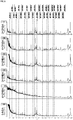
- Fig. 1 shows overall views of ISD mass spectra (m/z: 360-1840) of N-Acetyl-Renin substrate when 3H2NBA (top row), 4H3NBA (second row), 5H2NBA (third row), 3H5NBA (fourth row), 4H2NBA (fifth row), or 1, 5-DAN (bottom row) is used as a matrix
- Fig. 2 is a partial enlarged view of a low mass region in Fig. 1 (m/z: 530-1070)
- Fig. 3 is a partial enlarged view of a high mass region in Fig. 1 (m/z: 1030-1620).
- a-series ion species including an a6 ion were continuously detected without being buried in cluster ions when 3H2NBA, 4H3NBA, 5H2NBA, 3H5NBA, or 4H2NBA was used.
- amino acids for example, Ile/Leu (113) ⁇ ⁇ Asn (114) ⁇ ⁇ Asp (115), Gln/Lys (128) ⁇ ⁇ Glu (129)] having a difference in mass of 1, for example.
- the fifth amino acid from the N-terminus is not leucine (Leu, L) having the same mass number, but is isoleucine (Ile, I), and the tenth and the eleventh amino acids are not isoleucine (Ile, I), but are leucine (Leu, L).
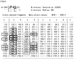
- Fig. 4 shows an ISD ion theoretical value list of N-Acetyl-Renin substrate.
- Fig. 5 shows overall views of ISD mass spectra (m/z: 450-4450) of Amyloid ⁇ [1-40] when 3H2NBA (top row), 4H3NBA (second row), 5H2NBA (third row), 3H5NBA (fourth row), 4H2NBA (fifth row), or 5-NSA (bottom row) is used as a matrix
- Fig. 6 is a partial enlarged view of a low mass region in Fig. 5 (m/z: 1320-2060)
- Fig. 7 is a partial enlarged view of a high mass region in Fig. 5 (m/z: 3040-3780).
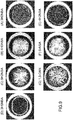
- Fig. 9(A) is a stereoscopic microphotograph showing an example of condition of a residue after dropping a 3H2NBA solution as a matrix, and a peptide sample solution of Amyloid ⁇ [1-40] on a sample plate, and drying the mixed solution.
- Fig. 9(B) is a photograph obtained when 4H3NBA is used as a matrix.
- Fig. 9(C) is a photograph obtained when 5H2NBA is used as a matrix.
- Fig. 9(D) is a photograph obtained when 3H5NBA is used as a matrix.
- Fig. 9(E) is a photograph obtained when 4H2NBA is used as a matrix.
- Fig. 9(F) is a stereoscopic microphotograph showing an example of condition of a residue after dropping a 5-NSA solution as a matrix, and a peptide sample solution of Amyloid ⁇ [1-40] on a sample plate, and drying the mixed solution.
- Fig. 9(G) is a photograph obtained when 1, 5-DAN is used as a matrix.
Landscapes
- Life Sciences & Earth Sciences (AREA)
- Health & Medical Sciences (AREA)
- Chemical & Material Sciences (AREA)
- Engineering & Computer Science (AREA)
- Molecular Biology (AREA)
- Physics & Mathematics (AREA)
- Bioinformatics & Cheminformatics (AREA)
- Bioinformatics & Computational Biology (AREA)
- Organic Chemistry (AREA)
- Hematology (AREA)
- Immunology (AREA)
- Biomedical Technology (AREA)
- Urology & Nephrology (AREA)
- Biophysics (AREA)
- Analytical Chemistry (AREA)
- General Health & Medical Sciences (AREA)
- Medicinal Chemistry (AREA)
- Biochemistry (AREA)
- Proteomics, Peptides & Aminoacids (AREA)
- Spectroscopy & Molecular Physics (AREA)
- Biotechnology (AREA)
- Cell Biology (AREA)
- Microbiology (AREA)
- Food Science & Technology (AREA)
- General Physics & Mathematics (AREA)
- Pathology (AREA)
- Optics & Photonics (AREA)
- Chemical Kinetics & Catalysis (AREA)
- Genetics & Genomics (AREA)
- Other Investigation Or Analysis Of Materials By Electrical Means (AREA)
- Investigating Or Analysing Biological Materials (AREA)
Abstract
Description
- The present invention relates to a method for specific cleavage of a Cα-C bond of a peptide backbone and a side chain of a protein and a peptide. Further, the present invention relates to a method for determining an amino acid sequence of protein and peptide by measuring the protein and the peptide by a mass spectrometry method, in particular MALDI-MS (Matrix-Assisted Laser Desorption/Ionization Mass Spectrometry) using at least one hydroxynitrobenzoic acid selected from the group consisting of 3-hydroxy-2-nitrobenzoic acid, 4-hydroxy-3-nitrobenzoic acid, 5-hydroxy-2-nitrobenzoic acid, 3-hydroxy-5-nitrobenzoic acid and 4-hydroxy-2-nitrobenzoic acid as a matrix.
-
JP-A-2005-326391 Non-Patent Document 1 disclose that a peptide obtained by previously modifying a peptide with a 2-nitrobenzenesulphenyl group is subjected to MALDI (Matrix-Assisted Laser Desorption/Ionization) mass spectrometry using, as a matrix, α-cyano-3-hydroxycinnamic acid (3-CHCA), 3-hydroxy-4-nitrobenzoic acid (3H4NBA), or a mixture of them. However, these documents neither disclose nor suggest that a Cα-C bond of a peptide backbone and a side chain of protein and peptide are specifically cleaved with such a matrix. -
WO 2011/007743 (Patent Document 2) and Non-PatentDocument 2 disclose a method for specifically cleaving an N-Cα bond of a peptide backbone by conducting MALDI mass spectrometry using 5-amino salicylic acid (5-ASA) as a matrix. -
JP-A-2013-130570 Document 3 disclose a method for specifically cleaving an N-Cα bond or a Cα-C bond of a peptide backbone including irradiating a protein or a peptide with laser light in the presence of a naphthalene compound substituted with a 1-hydroxy-5-functional group. As the naphthalene compound substituted with a 1-hydroxy-5-functional group, 1-hydroxy-5-aminonaphthalene, or 1,5-dihydroxynaphthalene is used. - Non-Patent
Document 4 discloses a method for specifically cleaving a Cα-C bond of a peptide backbone by conducting MALDI mass spectrometry using 5-nitro salicylic acid (5-NSA), 4-nitro salicylic acid (4-NSA), 3-nitro salicylic acid (3-NSA), or 5-formyl salicylic acid (5-FSA). - The In-Source Decay (ISD) method in MALDI mass spectrometry is a method of inducing fragmentation of a sample ion in the ion source, and analyzing the generated fragment ion. For obtaining amino acid sequence information of a peptide chain, it is required that ion species of any one or a plurality of a-, b-, c-, x-, y-, and z-series are continuously detected in the mass spectrometry.
- The mainstream in MALDI-ISD in analysis of a peptide and a protein uses a reducing matrix (Non-Patent Document 5). In this case, a hydrogen radical derived from a matrix molecule and induced by laser light irradiation is given to the sample molecule (i.e. reducing matrix) in the condition that the matrix coexists with the sample, and cleavage of an N-Cα bond of a peptide backbone is induced, and mainly, ion species of c-series ion and z-series ion are generated (Non-Patent
Document 5, and Non-Patent Document 6). However, when the reducing matrix is used, cleavage on the left side of amino acid proline (Pro, P) is very difficult to occur, and a c-series ion is not generated by cleavage on the left side of proline (Pro, P). Further, when the reducing matrix is used, ion species of d-series ion are hardly generated by cleavage of a side chain. - As a matrix for generating c-series or z-series ion species, for example, 1,5-diaminonaphthalene (1,5-DAN) is known (Non-Patent Document 7). In c-series ion species generated by 1,5-DAN, cleavage on the left side of amino acid proline (Pro, P) is very difficult to occur, and a c-series ion is not generated by cleavage on the left side of proline (Pro, P). This is because proline (Pro, P) has a cyclic structure in the c-series cleavage (including cleavage of N-Cα bond) site. Therefore, it is difficult to obtain sequence information of the site containing proline (Pro, P). Since it is difficult to generate d-series ion species occurring by cleavage of a side chain via a-series ion species, it is not possible to distinguish between leucine (Leu, L) and isoleucine (Ile, I) having completely the same mass of amino acid residue by d-series ion species. In the use of 1,5-DAN, w-series ion species may be generated by cleavage of a side chain via z-series ion species, and in such a case, there is a possibility that leucine (Leu, L) and isoleucine (Ile, I) can be distinguished from each other, but the possibility is limited to the case where there is a basic amino acid such as arginine (Arg, R) or lysin (Lys, K) at the C terminus of the peptide.
- Features of the 1,5-DAN matrix that is often used in MALDI-ISD can be summarized as follows:
- [1] 1,5-DAN causes generation of c-series, or z-series and/or w-series ion species (generation of a-series and/or d-series ion species is difficult).
- [2] 1,5-DAN is suspicious of carcinogenicity.
- [3] 1,5-DAN is easy to sublimate in vacuo. Thus, when 1,5-DAN is used, the measuring time is limited, and the quantitative performance is liable to be impaired. Also, carryover, which is contamination with sublimated 1,5-DAN at the time of other measurement, is likely to occur.
- [4] 1,5-DAN is easy to deteriorate (be oxidized) in a solution state. For example, 1,5-DAN is easy to deteriorate by oxygen or the like in water or acetonitrile used as a solvent. Thus, it is difficult to stock 1,5-DAN in a solution state.
- [5] When 1,5-DAN is used, the surface uniformity of the residue on the sample plate after mixing, dropping and drying of the sample/matrix solution is low, and the thickness largely differs depending on the position on the sample plate. Thus, it is difficult to obtain excellent data by Raster, and the resolving power and the mass accuracy are declined in TOF.
- [6] Due to generation of a large quantity of cluster ions derived from the 1,5-DAN matrix, it is difficult to obtain information on the N-terminus side of the peptide chain (normally, several residues on the N-terminus side).
- On the other hand, it is also known to use an oxidizing matrix in MALDI-ISD in analysis of peptide and protein (Non-Patent Document 5). In this case, hydrogen radical elimination from the sample molecule occurs in response to laser light irradiation in the condition that the matrix coexists with the sample, and the eliminated hydrogen radical is given to the matrix molecule (i.e. oxidizing matrix) (Non-Patent Document 5). Upon cleavage of a Cα-C bond of a peptide backbone caused by hydrogen radical elimination from the sample molecule, ion species of a-series ion and x-series ion are generated (Non-Patent
Documents - As a matrix that promotes hydrogen radical elimination and generates a-series ion species, for example, 5-FSA to which a functional group -CHO is added (Non-Patent
Documents Documents Documents -
- Patent Document 1:
JP-A-2005-326391 - Patent Document 2:
WO 2011/007743 - Patent Document 3:
JP-A-2013-130570 -
- Non-Patent Document 1: "Selective detection of 2-nitrobenzenesulfenyl-labeled peptide by matrix-assisted laser desorption/ionization-time of flight mass spectrometry using a novel matrix", E. Matsuo, C. Toda, M. Watanabe, N. Ojima, S. Izumi, K. Tanaka, S. Tsunasawa, O. Nishimura: Proteomics (2006) Vol. 6, pp. 2042-2049
- Non-Patent Document 2: "In-source decay and fragmentation characteristics of peptide using 5-aminosalicylic acid as a matrix in matrix-assisted laser desorption/ionization mass spectrometry", M. Sakakura, M. Takayama: J. Am. Soc. Mass Spectrom. (2010) Vol. 21, pp. 979-988
- Non-Patent Document 3: "5-Amino-1-naphthol, a
- Non-Patent Document 4: "Influence of initial velocity of analytes on in-source decay products in MALDI mass spectrometry using salicylic acid derivative matrices", D. Asakawa, M. Sakakura, M. Takayama: Int. J. Mass spectrom. (2013) Vol. 337, pp. 29-33
- Non-Patent Document 5: "Principles of Hydrogen Radical Mediated Peptide/Protein Fragmentation during Matrix-Assisted Laser Desorption/Ionization Mass Spectrometry", D. Asakawa: Mass Spectrometry Reviews (2014) (
DOI 10. 1002/mas) - Non-Patent Document 6: "N-Cα bond cleavage of the peptide backbone via hydrogen abstraction", M. Takayama: J. Am. Soc. Mass Spectrom. (2001) Vol. 12, pp. 1044-1049
- Non-Patent Document 7: "Rational selection of the optimum MALDI matrix for top-down proteomics by in-source decay", K. Demeure, L. Quinton, V. Gabelica, E.-D. Pauw: Anal. Chem. (2007) Vol. 79, pp. 8678-8685
- Non-Patent Document 8: "Cα-C Bond Cleavage of the Peptide Backbone in MALDI In-Source Decay Using Salicylic Acid Derivative Matrices", D. Asakawa, M. Takayama: J. Am. Soc. Mass Spectrom. (2011) Vol. 22, pp. 1224-1233
- Non-Patent Document 9: "Matrix Effect on In-Source Decay Products of Peptides in Matrix-Assisted Laser Desorption/Ionization", D. Asakawa, M. Sakakura, M. Takayama, Mass Spectrometry (2012) Vol. 1, A0002
- As described above, in mass spectrometry of peptide or protein, c-series ion species can be obtained with high generation efficiency and high sensitivity by using 1,5-DAN; however, for a-series or x-series ion species, there is currently no oxidizing matrix capable of generating a-series or x-series ion species with adequately high generation efficiency and sensitivity as well as with small contamination of other ion species. Also, currently, it is not possible to distinguish between leucine (Leu, L) and isoleucine (Ile, I) which are amino acids having the same mass by the matrix for generating c-series or z-series ion species. In such a current state, there is a demand for a method for specifically cleaving a Cα-C bond of protein or peptide efficiently, detecting ion species such as a-series ion with high sensitivity with small contamination of other ion species, and achieving rapid analysis. Further, there is a demand for a method for specifically cleaving a side chain, detecting d-series ion with high sensitivity, and thereby determining amino acid sequences of protein and peptide.
- An object of the present invention is to provide a method for specifically cleaving a Cα-C bond of a peptide backbone and/or a side chain of a protein and a peptide, and a method for determining amino acid sequences of protein and peptide by the method.
- The present inventors have intensively studied, and as a result, have found that the above object can be achieved by using, as a matrix, at least one hydroxynitrobenzoic acid selected from the group consisting of 3-hydroxy-2-nitrobenzoic acid, 4-hydroxy-3-nitrobenzoic acid, 5-hydroxy-2-nitrobenzoic acid, 3-hydroxy-5-nitrobenzoic acid and 4-hydroxy-2-nitrobenzoic acid.
- The present invention includes the following.
- (1) A method for specifically cleaving a Cα-C bond of a peptide backbone and/or a side chain bond of a protein or a peptide, comprising irradiating a protein or a peptide with laser light in the presence of at least one hydroxynitrobenzoic acid selected from the group consisting of:
- 3-hydroxy-2-nitrobenzoic acid:

- 4-hydroxy-3-nitrobenzoic acid:

- 5-hydroxy-2-nitrobenzoic acid:

- 3-hydroxy-5-nitrobenzoic acid:

- 4-hydroxy-2-nitrobenzoic acid:

- 3-hydroxy-2-nitrobenzoic acid:
- (2) A method for determining an amino acid sequence of a protein or a peptide, comprising:
- irradiating a protein or a peptide with laser light in the presence of at least one hydroxynitrobenzoic acid selected from the group consisting of:
- 3-hydroxy-2-nitrobenzoic acid:

- 4-hydroxy-3-nitrobenzoic acid:

- 5-hydroxy-2-nitrobenzoic acid:

- 3-hydroxy-5-nitrobenzoic acid:

- 4-hydroxy-2-nitrobenzoic acid:
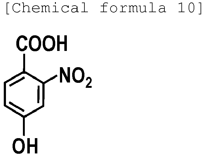
- 3-hydroxy-2-nitrobenzoic acid:
analyzing generated fragment ions by mass spectrometry. - irradiating a protein or a peptide with laser light in the presence of at least one hydroxynitrobenzoic acid selected from the group consisting of:
- (3) A method for determining an amino acid sequence of a protein or a peptide, comprising:
- irradiating a protein or a peptide with laser light in the presence of at least one hydroxynitrobenzoic acid selected from the group consisting of 3-hydroxy-2-nitrobenzoic acid, 4-hydroxy-3-nitrobenzoic acid, 5-hydroxy-2-nitrobenzoic acid, 3-hydroxy-5-nitrobenzoic acid, and 4-hydroxy-2-nitrobenzoic acid as a matrix to specifically cleave a Cα-C bond of a peptide backbone and/or a side chain bond; and
- analyzing generated fragment ions by MALDI mass spectrometry.
- (4) The method for determining an amino acid sequence of a protein or a peptide according to the above (2) or (3), wherein as the generated fragment ions, a-series ion species and/or x-series ion species are analyzed.
- (5) The method for determining an amino acid sequence of a protein or a peptide according to the above (2) or (3), wherein as the generated fragment ions, a-series ion species and d-series ion species are analyzed.
- (6) A reagent for specifically cleaving a Cα-C bond of a peptide backbone and/or a side chain bond of a protein or a peptide, comprising at least one hydroxynitrobenzoic acid selected from the group consisting of:
- 3-hydroxy-2-nitrobenzoic acid:

- 4-hydroxy-3-nitrobenzoic acid:

- 5-hydroxy-2-nitrobenzoic acid:


- 3-hydroxy-5-nitrobenzoic acid:

- 4-hydroxy-2-nitrobenzoic acid:

- 3-hydroxy-2-nitrobenzoic acid:
- In the present invention, in MALDI-ISD analysis, ISD fragmentation is promoted generally by setting the laser intensity higher than that in a normal analysis. That is, in the present invention, by irradiating a protein or a peptide of a sample to be analyzed with laser light having an intensity somewhat higher than the quantity and density required for generation of proton addition molecule [M+H]+ derived from the sample in the presence of at least one hydroxynitrobenzoic acid selected from the group consisting of 3-hydroxy-2-nitrobenzoic acid, 4-hydroxy-3-nitrobenzoic acid, 5-hydroxy-2-nitrobenzoic acid, 3-hydroxy-5-nitrobenzoic acid and 4-hydroxy-2-nitrobenzoic acid as a matrix, hydrogen radical elimination reaction from the protein or the peptide is promoted to cause generation of mainly a-series and/or x-series ion species, and further cause generation of d-series ion species.
- In the present invention, by irradiating a protein or a peptide with laser light in the presence of at least one hydroxynitrobenzoic acid selected from the group consisting of 3-hydroxy-2-nitrobenzoic acid, 4-hydroxy-3-nitrobenzoic acid, 5-hydroxy-2-nitrobenzoic acid, 3-hydroxy-5-nitrobenzoic acid and 4-hydroxy-2-nitrobenzoic acid, hydrogen radical elimination reaction from the protein or the peptide is promoted, and a Cα-C bond of a peptide backbone and/or a side chain bond can be specifically cleaved.
- As a result of specific cleavage of a Cα-C bond of a peptide backbone and/or a side chain bond, a-series and/or x-series ion species are mainly generated as fragment ions, and further d-series ion species are also generated. By detecting and analyzing the a-series and/or the x-series ion species by mass spectrometry, it is possible to detect cleavage on the left side of proline (Pro, P) and to obtain sequence information of a site containing proline (Pro, P). Meanwhile, by detecting and analyzing the d-series ion species by mass spectrometry, it is possible to distinguish between leucine (Leu, L) and isoleucine (Ile, I) having completely the same mass of amino acid residue. As described above, according to the present invention, it is possible to obtain more amino acid sequence information rapidly with high sensitivity, and with high coverage for a protein or a peptide to be analyzed. In particular, it is possible to obtain amino acid sequence information by only one MS measurement without conducting a multistage analysis as in MS/MS.
-
-
Fig. 1 shows overall views of ISD mass spectra of N-Acetyl-Renin substrate when 3-hydroxy-2-nitrobenzoic acid (3H2NBA), 4-hydroxy-3-nitrobenzoic acid (4H3NBA), 5-hydroxy-2-nitrobenzoic acid (5H2NBA), 3-hydroxy-5-nitrobenzoic acid (3H5NBA), 4-hydroxy-2-nitrobenzoic acid (4H2NBA) and 1, 5-diaminonaphthalene (1,5-DAN) are used as a matrix from the top. The horizontal axis represents mass/charge (m/z), and the vertical axis represents ion intensity. -
Fig. 2 is a partial enlarged view of a low mass region inFig. 1 . -
Fig. 3 is a partial enlarged view of a high mass region inFig. 1 . -
Fig. 4 is an ISD ion theoretical value list of N-Acetyl-Renin substrate. -
Fig. 5 is overall views of ISD mass spectra of Amyloid β [1-40] when 3-hydroxy-2-nitrobenzoic acid (3H2NBA), 4-hydroxy-3-nitrobenzoic acid (4H3NBA), 5-hydroxy-2-nitrobenzoic acid (5H2NBA), 3-hydroxy-5-nitrobenzoic acid (3H5NBA), 4-hydroxy-2-nitrobenzoic acid (4H2NBA) and 5-nitro salicylic acid (5-NSA) are used as a matrix from the top. The horizontal axis represents mass/charge (m/z), and the vertical axis represents ion intensity. -
Fig. 6 is a partial enlarged view of a low mass region inFig. 5 . -
Fig. 7 is a partial enlarged view of a high mass region inFig. 5 . -
Fig. 8 is an ISD ion theoretical value list of Amyloid β [1-40]. -
Fig. 9 shows stereoscopic microphotographs each showing an example of condition of a residue after dropping, on a sample plate, a peptide sample solution of Amyloid β [1-40] and a matrix solution using:- (A) 3-hydroxy-2-nitrobenzoic acid (3H2NBA),
- (B) 4-hydroxy-3-nitrobenzoic acid (4H3NBA),
- (C) 5-hydroxy-2-nitrobenzoic acid (5H2NBA),
- (D) 3-hydroxy-5-nitrobenzoic acid (3H5NBA),
- (E) 4-hydroxy-2-nitrobenzoic acid (4H2NBA),
- (F) 5-nitrosalicylic acid (5-NSA), or
- (G) 1,5-diaminonaphthalene (1,5-DAN)
- For obtaining amino acid sequence information of peptide chains for a protein or a peptide to be analyzed, it is a requirement that ion species of any one or a plurality of a-, b-, c-, x-, y-, and z-series are detected continuously in mass spectrometry. The following chemical structural formula shows the naming rule of fragmentation of a peptide backbone for a peptide composed of four residues as an example. R1, R2, R3, and R4 each represent a side chain of an amino acid residue.

- Next, referring to chemical schemes, generation mechanisms of c-series ion and z-series ion, and, a-series ion and x-series ion by cleavage of a peptide backbone will be described (see
scheme 1 of Non-Patent document 8).

- The scheme (a) shows a mechanism of generation of c-series ion and z-series ion by cleavage of an N-Cα bond of a peptide backbone. When laser light irradiation is conducted in the condition that a reducing matrix coexists with a protein or peptide sample, a hydrogen radical derived from the matrix molecule and induced by the laser light irradiation is given to the sample molecule (i.e. reducing matrix), and cleavage of an N-Cα bond of the peptide backbone is induced, and mainly, ion species of c-series ion and/or z-series ion are generated. Since proline (Pro, P) has a cyclic structure in the c-series cleavage (including cleavage of N-Cα bond) site, cleavage of an N-Cα bond is very difficult to occur in the case of proline (Pro, P), and a c-series ion is not generated by cleavage on the left side of proline (Pro, P).
- The scheme (b) shows a mechanism of generation of a-series ion and x-series ion by cleavage of a Cα-C bond of a peptide backbone. When laser light irradiation is conducted in the condition that an oxidizing matrix coexists with a protein or peptide sample, hydrogen radical elimination from the sample molecule occurs by the laser light irradiation, and the eliminated hydrogen radical is given to the matrix molecule (oxidizing matrix). Upon cleavage of a Cα-C bond of the peptide backbone caused by hydrogen radical elimination from the sample molecule, ion species of a-series ion and/or x-series ion are generated. Further, d-series ion species are easily generated by cleavage of a side chain from the a-series ion species.
- In the present invention, a protein or peptide sample is irradiated with laser light in the presence of at least one hydroxynitrobenzoic acid selected from the group consisting of 3-hydroxy-2-nitrobenzoic acid, 4-hydroxy-3-nitrobenzoic acid, 5-hydroxy-2-nitrobenzoic acid, 3-hydroxy-5-nitrobenzoic acid and 4-hydroxy-2-nitrobenzoic acid as a matrix. As the specific hydroxynitrobenzoic acid, only one kind or a combination of two or more kinds may be used. According to the above scheme (b), a Cα-C bond of a peptide backbone and/or a side chain bond are/is specifically cleaved. Hereinafter, more specific chemical schemes are shown while taking 3-hydroxy-2-nitrobenzoic acid (3H2NBA) as an example.

- When a protein or peptide sample is irradiated with laser light in the presence of at least one hydroxynitrobenzoic acid selected from the group consisting of 3-hydroxy-2-nitrobenzoic acid, 4-hydroxy-3-nitrobenzoic acid, 5-hydroxy-2-nitrobenzoic acid, 3-hydroxy-5-nitrobenzoic acid and 4-hydroxy-2-nitrobenzoic acid as a matrix, hydrogen radical elimination from the sample molecule occurs by the laser light irradiation, and the eliminated hydrogen radical is given to the specific hydroxynitrobenzoic acid molecule described above. The specific hydroxynitrobenzoic acid has a high acceptability of hydrogen radical by a nitro group, and hydrogen radical elimination from the sample molecule easily occurs. Upon cleavage of a Cα-C bond of a peptide backbone caused by the hydrogen radical elimination from the sample molecule, ion species of a-series ion and/or x-series ion are generated. Further, ion species of d-series ion are easily generated by cleavage of a side chain from the a-series ion species.
- The generated ion species of a-series ion and/or x-series ion, and the ion species of d-series ion generated by cleavage of a side chain are subjected to mass spectrometry, and the amino acid sequence of the protein or the peptide can be determined.
- A crystal for mass spectrometry can be obtained through the step of forming, on a target plate for mass spectrometry, a liquid droplet of a mixture liquid containing, in a solvent, at least a protein or a peptide to be analyzed, and at least one hydroxynitrobenzoic acid matrix selected from the group consisting of 3-hydroxy-2-nitrobenzoic acid, 4-hydroxy-3-nitrobenzoic acid, 5-hydroxy-2-nitrobenzoic acid, 3-hydroxy-5-nitrobenzoic acid and 4-hydroxy-2-nitrobenzoic acid, and the step of removing the solvent from the formed liquid droplet of the mixture liquid to obtain a non-volatile matter (i.e., at least the analyte and the matrix) contained in the mixture liquid as a residue. The thus obtained residue is a crystal for mass spectrometry. In this specification, the term "crystal for mass spectrometry" is synonymous with the term "residue".
- As the target for mass spectrometry, a conductive metal plate usually used in MALDI mass spectrometry may be used. Specifically, a stainless plate may be used.
- A specific method for preparing the liquid droplet of the mixture liquid on the target plate is not particularly limited. For example, first, a sample solution containing an analyte, and a matrix solution are prepared separately from each other. Then, these solutions are mixed to obtain a mixture liquid, and the obtained mixture liquid is dropped onto a target plate. Alternatively, these solutions may be mixed on a target plate by dropping these solutions onto the same position on the target plate (on-target mix). In the case of on-target mix, the order of dropping the solutions is not particularly limited.
- The solvent of the mixture liquid may be selected from the group consisting of water, acetonitrile (ACN), trifluoroacetic acid (TFA), methanol (MeOH), ethanol (EtOH), tetrahydrofuran (THF), dimethylsulfoxide (DMSO), and the like. More specifically, as the solvent of the mixture liquid, an aqueous ACN solution, an aqueous ACN-TFA solution, MeOH-TFA, an aqueous MeOH solution, an aqueous EtOH-TFA solution, an aqueous EtOH solution, an aqueous THF-TFA solution, an aqueous THF solution, an aqueous DMSO-TFA solution, an aqueous DMSO solution or the like is used, and more preferably, an aqueous ACN solution or an aqueous ACN-TFA solution may be used. The concentration of ACN in the aqueous ACN-TFA solution may be, for example, 10 to 90 vol%, preferably 25 to 75 vol%, and the concentration of TFA in the aqueous ACN-TFA solution may be, for example, 0.05 to 1 vol%, preferably 0.05 to 0.1 vol%.
- The volume of the liquid droplet of the mixture liquid is not particularly limited, and may be appropriately determined by those skilled in the art. When a well is provided on the target plate, the liquid droplet of the mixture liquid may be formed in the well. In this case, the liquid droplet is formed so as to have a volume that can be held in the well. More specifically, the liquid droplet may be formed so as to have a volume of about 0.1 µL to 2 µL, for example, about 0.5 µL.
- Next, the solvent is removed from the liquid droplet of the mixture liquid on the target plate. The removal of the solvent includes natural evaporation of the solvent. The amount of the matrix contained per one residue (that is, per one crystal for mass spectrometry) generated by evaporation may be, for example, 1 pmol to 1,000 nmol, preferably 10 pmol to 100 nmol as a guide. The amount of the analyte may be in the range of, for example, 1 amol to 100 pmol, or in the range of 100 amol to 50 pmol of sample with respect to 10 nmol of the matrix.
- The residue has a substantially circular shape on a surface in contact with the target plate. That is, the outer edge of the residue is substantially circular. The average diameter of the substantially circular shape may vary depending on the amount of the sample, the volume of the liquid droplet, the amount of the matrix, the composition of the solvent etc., but is for example 0.1 to 3 mm, preferably 0.5 to 2 mm. It is to be noted that the average diameter is the average of the lengths of line segments cut from lines passing through the center of gravity of the substantially circular shape by the outer edge of the residue.
- When an ordinary metallic plate is used as the target for mass spectrometry, the substance to be analyzed mainly exists in the substantial circle in the substantially circular residue obtained by removal of the solvent. Therefore, it is possible to easily conduct ionization of the substance to be analyzed without specifying the laser irradiation position at the time of ionization.
- A mass spectrometer used in the present invention is not particularly limited as long as it is used in combination with a MALDI (Matrix-Assisted Laser Desorption/Ionization) ion source. Examples of such a mass spectrometer include MALDI-TOF (Matrix-Assisted Laser Desorption/Ionization-Time-of-Flight) mass spectrometers, MALDI-QIT (Matrix-Assisted Laser Desorption/Ionization-Quadrupole Ion Trap) mass spectrometers, MALDI-QIT-TOF (Matrix-Assisted Laser Desorption/Ionization-Quadrupole Ion Trap-Time-of-Flight) mass spectrometers, MALDI-Q-TOF (Matrix-Assisted Laser Desorption/Ionization-Quadrupole-Time-of-Flight) mass spectrometers, MALDI-FTICR (Matrix-Assisted Laser Desorption/Ionization-Fourier Transform Ion Cyclotron Resonance) mass spectrometers, and MALDI-Orbitrap (Matrix-Assisted Laser Desorption/Ionization-Orbitrap) mass spectrometers.
- Hereinbelow, the present invention will be described specifically with reference to examples, but is not limited to the following examples.
- In the following examples, the following two kinds of peptide samples were used.
- N-Acetyl-Renin Substrate (amino acid sequence: Ac-DRVYIHPFHLLVYS) (SEQ ID NO: 1)
- Amyloid β [1-40] (amino acid sequence: DAEFRHDSGYEVHHQKLVFFAEDVGSNKGAIIGLMVGGVV) (SEQ ID NO: 2)
- As a matrix, the following seven kinds were used.
3H2NBA (3-hydroxy-2-nitrobenzoic acid), present invention
- 4H3NBA (4-hydroxy-3-nitrobenzoic acid), present invention

- 5H2NBA (5-hydroxy-2-nitrobenzoic acid), present invention

- 3H5NBA (3-hydroxy-5-nitrobenzoic acid), present invention
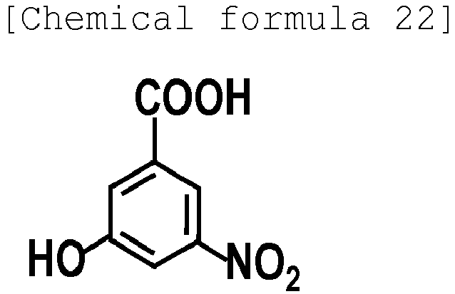
- 4H2NBA (4-hydroxy-2-nitrobenzoic acid), present invention

- 5-NSA (5-nitrosalicylic acid), for comparison

- 1,5-DAN (1,5-Diaminonaphthalene), for comparison

- Among the above matrixes, 3H2NBA, 4H3NBA, 5H2NBA, 3H5NBA, 4H2NBA and 5-NSA have a functional group -NO2. This -NO2 group exerts the effect of drawing out a hydrogen radical from the peptide sample by laser light irradiation, and causes cleavage of a Cα-C bond of a peptide backbone. As a result, a-series ion species and x-series ion species are generated, and further d-series ion species are generated.
- Among the above matrixes, 1,5-DAN has a functional group -NH2. The -NH2 group exerts the effect of generating a hydrogen radical derived from the 1,5-DAN molecule by laser light irradiation, and adding the hydrogen radical to the peptide sample, and causes cleavage of an N-Cα bond of a peptide backbone. As a result, c-series ion species and z-series ion species are generated.
- Sample plate: a stainless plate having a thickness of 2 mm was used.
- Mass spectrometer: MALDI-Time-of-Flightmass spectrometer [AXIMA-Performance (registered trademark), available from Shimadzu Corporation] was used.
-
- Peptide sample: N-Acetyl-Renin Substrate (Ac-DRVYIHPFHLLVYS) (SEQ ID NO: 1)
- Matrix:
- 3H2NBA (3-hydroxy-2-nitrobenzoic acid), present invention
- 4H3NBA (4-hydroxy-3-nitrobenzoic acid), present invention
- 5H2NBA (5-hydroxy-2-nitrobenzoic acid), present invention
- 3H5NBA (3-hydroxy-5-nitrobenzoic acid), present invention
- 4H2NBA (4-hydroxy-2-nitrobenzoic acid), present invention
- 1,5-DAN (1,5-Diaminonaphthalene), for comparison
-
- (1) As a peptide sample solution, a 20 pmol/µL N-Acetyl-Renin Substrate solution in water was prepared.
- (2) As a matrix solution, a 10 mg/mL solution of 3H2NBA, 4H3NBA, 5H2NBA, 3H5NBA, or 4H2NBA in 75% ACN, water was prepared, and for comparison, a 10 mg/mL solution of 1,5-DAN in 75% ACN, water was prepared.
- (3) The sample solution (0.5 µL) prepared in (1) was dropped onto a MALDI sample plate, and then the matrix solution (0.5 µL) prepared in (2) was dropped thereonto (on-target mix). The amount of the peptide sample per one well was 10 pmol.
- (4) After the solvent was volatilized, measurement was conducted by a Raster analysis for 40 × 40 = 1,600 points in a 1.2 mm- to 1.7 mm-square depending on the extent of the residue on the plate by MALDI TOFMS [AXIMA Performance (registered trademark), available from Shimadzu Corporation)] in a positive ion mode and linear mode.
-
Fig. 1 shows overall views of ISD mass spectra (m/z: 360-1840) of N-Acetyl-Renin substrate when 3H2NBA (top row), 4H3NBA (second row), 5H2NBA (third row), 3H5NBA (fourth row), 4H2NBA (fifth row), or 1, 5-DAN (bottom row) is used as a matrix,Fig. 2 is a partial enlarged view of a low mass region inFig. 1 (m/z: 530-1070), andFig. 3 is a partial enlarged view of a high mass region inFig. 1 (m/z: 1030-1620). - As shown in the spectra of the bottom rows in
Figs. 1 and2 , when 1,5-DAN was used, a large variety and a large quantity of cluster ions derived from the matrix were detected in the low mass region (for example, inFig. 2 , a large number of high intensity peaks observed in m/z: 420 to 650). Therefore, even if c-series ion species are detected, analysis tends to be difficult. Also as shown in the spectrum of the bottom row inFig. 2 , a c6 ion was not detected. This is because the seventh amino acid from the N-terminus of N-Acetyl-Renin Substrate (amino acid sequence: Ac-DRVYIH"P"FHLLVYS) is proline (Pro, P), and a c-series ion between the previous sixth amino acid histidine (His, H) and the seventh amino acid was not detected. - In contrast, as shown in the spectra of the first to the fifth rows in
Figs. 1 and2 , a-series ion species including an a6 ion were continuously detected without being buried in cluster ions when 3H2NBA, 4H3NBA, 5H2NBA, 3H5NBA, or 4H2NBA was used. - As shown in the spectrum of the bottom row in
Fig. 3 , when 1,5-DAN was used, all the c-series ion species were detected in the high mass region; however, the mass resolution was low and the mass accuracy was also low. This is mainly attributable to the fact that the form of the residue after dropping the 1,5-DAN solution and the sample solution on the sample plate and drying the mixed solution is nonuniform (see, for example, the later-describedFig. 9(G) ), and the thickness differs depending on the site due to growth of needle-like giant crystals, and thus the mass resolution and the mass accuracy deteriorate in the Time-of-Flight mass spectrometry. Accordingly, it is expected to be difficult to distinguish between amino acids [for example, Ile/Leu (113) ← → Asn (114) ← → Asp (115), Gln/Lys (128) ← → Glu (129)] having a difference in mass of 1, for example. - In contrast, as mainly shown in
Figs. 1 ,2 and3 , when 3H2NBA, 4H3NBA, 5H2NBA, 3H5NBA, or 4H2NBA was used, not only all the a-series ion species were detected, but also d5, d10, and d11 were detected. Therefore, it could be determined that the fifth amino acid from the N-terminus of N-Acetyl-Renin Substrate (amino acid sequence: Ac-DRVY"I"HPFH"LL"VYS) is isoleucine (Ile, I), and both the tenth and eleventh amino acids are leucine (Leu, L). In other words, it could be determined that the fifth amino acid from the N-terminus is not leucine (Leu, L) having the same mass number, but is isoleucine (Ile, I), and the tenth and the eleventh amino acids are not isoleucine (Ile, I), but are leucine (Leu, L). -
Fig. 4 shows an ISD ion theoretical value list of N-Acetyl-Renin substrate. -
- Peptide sample: Amyloid β [1-40] (amino acid sequence: DAEFRHDSGYEVHHQKLVFFAEDVGSNKGAIIGLMVGGVV) (SEQ ID NO: 2)
- Matrix:
- 3H2NBA (3-hydroxy-2-nitrobenzoic acid), present invention
- 4H3NBA (4-hydroxy-3-nitrobenzoic acid), present invention
- 5H2NBA (5-hydroxy-2-nitrobenzoic acid), present invention
- 3H5NBA (3-hydroxy-5-nitrobenzoic acid), present invention
- 4H2NBA (4-hydroxy-2-nitrobenzoic acid), present invention
- 5-NSA (5-nitrosalicylic acid), for comparison
-
- (1) As a peptide sample solution, a 20 pmol/µL Amyloid β [1-40] solution in 50% ACN 0.1% TFA water was prepared.
- (2) As a matrix solution, a 10 mg/mL solution of 3H2NBA, 4H3NBA, 5H2NBA, 3H5NBA, or 4H2NBA in 75% ACN, water was prepared, and for comparison, a 10 mg/mL solution of 5-NSA in 75% ACN, water was prepared.
- (3) The sample solution (0.5 µL) prepared in (1) was dropped onto a MALDI sample plate, and then the matrix solution (0.5 µL) prepared in (2) was dropped thereonto (on-target mix). The amount of the peptide sample per one well was 10 pmol.
- (4) After the solvent was volatilized, measurement was conducted by a Raster analysis for 40 × 40 = 1,600 points in a 1.2 mm- to 1. 7 mm-square depending on the extent of the residue on the plate by MALDI TOFMS [AXIMA Performance (registered trademark), available from Shimadzu Corporation)] in a positive ion mode and linear mode.
-
Fig. 5 shows overall views of ISD mass spectra (m/z: 450-4450) of Amyloid β [1-40] when 3H2NBA (top row), 4H3NBA (second row), 5H2NBA (third row), 3H5NBA (fourth row), 4H2NBA (fifth row), or 5-NSA (bottom row) is used as a matrix,Fig. 6 is a partial enlarged view of a low mass region inFig. 5 (m/z: 1320-2060), andFig. 7 is a partial enlarged view of a high mass region inFig. 5 (m/z: 3040-3780). - As shown in the spectra of
Fig. 5 , when 5-NSA was used, a large variety and a large quantity of cluster ions derived from the matrix were detected in the low mass region, and it tends to be difficult to obtain sequence information in the low mass region. Further, as shown inFig. 6 , ion species other than a- and d-series ion species were detected in a relatively large quantity, and as shown inFig. 7 , detection of d-series ion species in a high mass region is particularly difficult, and thus, it tends to be difficult to obtain sequence information in the intermediate mass region and the high mass region. - In contrast, as shown in the spectra of the top row to the fifth row of
Figs. 6 to 7 , when 3H2NBA, 4H3NBA, 5H2NBA, 3H5NBA, or 4H2NBA was used, all the a-series ion species, and all the d-series ion species (d17, d31, d32, d34) distinguishing between isoleucine (Ile, I) and leucine (Leu, L) were detected. Therefore, it is easy to obtain sequence information. - Additional advantageous effects when 3H2NBA, 4H3NBA, 5H2NBA, 3H5NBA, or 4H2NBA is used as a matrix will be shown by referring to
Figs. 9(A) to 9(G) . -
Fig. 9(A) is a stereoscopic microphotograph showing an example of condition of a residue after dropping a 3H2NBA solution as a matrix, and a peptide sample solution of Amyloid β [1-40] on a sample plate, and drying the mixed solution. Similarly,Fig. 9(B) is a photograph obtained when 4H3NBA is used as a matrix.Fig. 9(C) is a photograph obtained when 5H2NBA is used as a matrix.Fig. 9(D) is a photograph obtained when 3H5NBA is used as a matrix.Fig. 9(E) is a photograph obtained when 4H2NBA is used as a matrix. -
Fig. 9(F) is a stereoscopic microphotograph showing an example of condition of a residue after dropping a 5-NSA solution as a matrix, and a peptide sample solution of Amyloid β [1-40] on a sample plate, and drying the mixed solution.Fig. 9(G) is a photograph obtained when 1, 5-DAN is used as a matrix. - As shown in
Fig. 9 , when (A) 3H2NBA, (B) 4H3NBA, (C) 5H2NBA, (D) 3H5NBA, or (E) 4H2NBA was used as a matrix, the form inside the residue to be ionized was observed to be relatively uniform by a square Raster scan. In contrast, it was also observed that when (G) 1,5-DAN was used, an elongated crystal grew largely, and thus the form of the residue was nonuniform. - As shown in
Fig. 9(F) , when 5-NSA was used as a matrix, the form inside the residue to be ionized was observed to be relatively uniform by a square Raster scan as is the case with using (A) 3H2NBA, (B) 4H3NBA, (C) 5H2NBA, (D) 3H5NBA, or (E) 4H2NBA as a matrix, but the thickness of the residue tends to be large.


Claims (6)
- A method for specifically cleaving a Cα-C bond of a peptide backbone and/or a side chain bond of a protein or a peptide, comprising irradiating a protein or a peptide with laser light in the presence of at least one hydroxynitrobenzoic acid selected from the group consisting of:3-hydroxy-2-nitrobenzoic acid:
 4-hydroxy-3-nitrobenzoic acid:
 5-hydroxy-2-nitrobenzoic acid:
 3-hydroxy-5-nitrobenzoic acid:
 4-hydroxy-2-nitrobenzoic acid:
- A method for determining an amino acid sequence of a protein or a peptide, comprising:irradiating a protein or a peptide with laser light in the presence of at least one hydroxynitrobenzoic acid selected from the group consisting of:to specifically cleave a Cα-C bond of a peptide backbone and/or a side chain bond; and3-hydroxy-2-nitrobenzoic acid:
 4-hydroxy-3-nitrobenzoic acid:
 5-hydroxy-2-nitrobenzoic acid:
 3-hydroxy-5-nitrobenzoic acid:
 4-hydroxy-2-nitrobenzoic acid: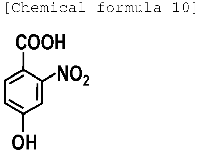
analyzing generated fragment ions by mass spectrometry. - A method for determining an amino acid sequence of a protein or a peptide, comprising:irradiating a protein or a peptide with laser light in the presence of at least one hydroxynitrobenzoic acid selected from the group consisting of 3-hydroxy-2-nitrobenzoic acid, 4-hydroxy-3-nitrobenzoic acid, 5-hydroxy-2-nitrobenzoic acid, 3-hydroxy-5-nitrobenzoic acid, and 4-hydroxy-2-nitrobenzoic acid as a matrix to specifically cleave a Cα-C bond of a peptide backbone and/or a side chain bond; andanalyzing generated fragment ions by MALDI mass spectrometry.
- The method for determining an amino acid sequence of a protein or a peptide according to claim 2, wherein as the generated fragment ions, a-series ion species and/or x-series ion species are analyzed.
- The method for determining an amino acid sequence of a protein or a peptide according to claim 2, wherein as the generated fragment ions, a-series ion species and d-series ion species are analyzed.
- A reagent for specifically cleaving a Cα-C bond of a peptide backbone and/or a side chain bond of a protein or a peptide, comprising at least one hydroxynitrobenzoic acid selected from the group consisting of:3-hydroxy-2-nitrobenzoic acid:
 4-hydroxy-3-nitrobenzoic acid:
 5-hydroxy-2-nitrobenzoic acid:
 3-hydroxy-5-nitrobenzoic acid:
 4-hydroxy-2-nitrobenzoic acid:
Applications Claiming Priority (1)
| Application Number | Priority Date | Filing Date | Title |
|---|---|---|---|
| JP2016098326A JP6587976B2 (en) | 2016-05-16 | 2016-05-16 | Cα-C bond and side chain specific cleavage method of protein and peptide, and amino acid sequencing method |
Publications (1)
| Publication Number | Publication Date |
|---|---|
| EP3246706A1 true EP3246706A1 (en) | 2017-11-22 |
Family
ID=58873599
Family Applications (1)
| Application Number | Title | Priority Date | Filing Date |
|---|---|---|---|
| EP17170513.0A Withdrawn EP3246706A1 (en) | 2016-05-16 | 2017-05-10 | Method for specific cleavage of c(alpha)-c bond and side chain of protein and peptide, and method for determining amino acid sequence |
Country Status (3)
| Country | Link |
|---|---|
| US (1) | US10626141B2 (en) |
| EP (1) | EP3246706A1 (en) |
| JP (1) | JP6587976B2 (en) |
Families Citing this family (2)
| Publication number | Priority date | Publication date | Assignee | Title |
|---|---|---|---|---|
| CN110297095A (en) * | 2019-07-26 | 2019-10-01 | 四川农业大学 | A kind of screening technique of insect allergic protein |
| JP7202554B2 (en) * | 2019-09-20 | 2023-01-12 | 国立大学法人広島大学 | Cleavage methods, analytical methods and specific cleavage reagents specifically cleaving the Ca-C bonds and/or side chains of peptide backbones |
Citations (4)
| Publication number | Priority date | Publication date | Assignee | Title |
|---|---|---|---|---|
| JP2005326391A (en) | 2004-04-13 | 2005-11-24 | Shimadzu Corp | Method for measuring hydrophobic peptides by MALDI mass spectrometer |
| EP1739419A1 (en) * | 2004-04-13 | 2007-01-03 | Shimadzu Corporation | Method of measuring substance by mass spectrometry and kit therefor |
| WO2011007743A1 (en) | 2009-07-16 | 2011-01-20 | 公立大学法人横浜市立大学 | Method for specific cleavage of n-cα bond in peptide main chain |
| JP2013130570A (en) | 2011-11-21 | 2013-07-04 | Yokohama City Univ | METHOD FOR SPECIFIC CLEAVAGE OF N-Cα BOND OR Cα-C BOND IN PEPTIDE MAIN CHAIN OF PROTEIN AND PHOSPHORYLATED PEPTIDE |
Family Cites Families (3)
| Publication number | Priority date | Publication date | Assignee | Title |
|---|---|---|---|---|
| WO2005050172A2 (en) * | 2003-11-14 | 2005-06-02 | Indiana University Research And Technology Corporation | Methods and apparatus for mass spectral analysis of peptides and proteins |
| JP4165502B2 (en) * | 2004-05-24 | 2008-10-15 | 株式会社島津製作所 | Method for selectively measuring a specific substance from a mixture by MALDI mass spectrometry |
| US20060243899A1 (en) * | 2004-05-24 | 2006-11-02 | Shimadzu Corporation | Method for selective measurement of specific substances from a mixture by maldi mass spectrometry |
-
2016
- 2016-05-16 JP JP2016098326A patent/JP6587976B2/en active Active
-
2017
- 2017-05-10 EP EP17170513.0A patent/EP3246706A1/en not_active Withdrawn
- 2017-05-12 US US15/594,461 patent/US10626141B2/en active Active
Patent Citations (4)
| Publication number | Priority date | Publication date | Assignee | Title |
|---|---|---|---|---|
| JP2005326391A (en) | 2004-04-13 | 2005-11-24 | Shimadzu Corp | Method for measuring hydrophobic peptides by MALDI mass spectrometer |
| EP1739419A1 (en) * | 2004-04-13 | 2007-01-03 | Shimadzu Corporation | Method of measuring substance by mass spectrometry and kit therefor |
| WO2011007743A1 (en) | 2009-07-16 | 2011-01-20 | 公立大学法人横浜市立大学 | Method for specific cleavage of n-cα bond in peptide main chain |
| JP2013130570A (en) | 2011-11-21 | 2013-07-04 | Yokohama City Univ | METHOD FOR SPECIFIC CLEAVAGE OF N-Cα BOND OR Cα-C BOND IN PEPTIDE MAIN CHAIN OF PROTEIN AND PHOSPHORYLATED PEPTIDE |
Non-Patent Citations (12)
Also Published As
| Publication number | Publication date |
|---|---|
| US20170327533A1 (en) | 2017-11-16 |
| US10626141B2 (en) | 2020-04-21 |
| JP6587976B2 (en) | 2019-10-09 |
| JP2017207312A (en) | 2017-11-24 |
Similar Documents
| Publication | Publication Date | Title |
|---|---|---|
| Banerjee et al. | Electrospray ionization mass spectrometry: a technique to access the information beyond the molecular weight of the analyte | |
| Loo et al. | Tandem mass spectrometry of very large molecules. 2. Dissociation of multiply charged proline-containing proteins from electrospray ionization | |
| Comby-Zerbino et al. | The emergence of mass spectrometry for characterizing nanomaterials. Atomically precise nanoclusters and beyond | |
| Chintalapudi et al. | An integrated electrocatalytic nESI-MS platform for quantification of fatty acid isomers directly from untreated biofluids | |
| Zhang et al. | Native mass spectrometry of photosynthetic pigment–protein complexes | |
| Håkansson et al. | High-sensitivity electron capture dissociation tandem FTICR mass spectrometry of microelectrosprayed peptides | |
| Niessen et al. | Introduction to mass spectrometry, a tutorial | |
| Berthias et al. | Differential ion mobility separations of D/L peptide epimers | |
| Kühn-Hölsken et al. | Complete MALDI-ToF MS analysis of cross-linked peptide–RNA oligonucleotides derived from nonlabeled UV-irradiated ribonucleoprotein particles | |
| US7446309B2 (en) | In-plane distribution measurement method | |
| Jones et al. | Ionic liquid matrix-induced metastable decay of peptides and oligonucleotides and stabilization of phospholipids in MALDI FTMS analyses | |
| Mathew et al. | Ion imaging of native protein complexes using orthogonal time-of-flight mass spectrometry and a Timepix detector | |
| US10626141B2 (en) | Method for specific cleavage of C alpha-C bond and side chain of protein and peptide, and method for determining amino acid sequence | |
| Towers et al. | Introduction of 4-chloro-α-cyanocinnamic acid liquid matrices for high sensitivity UV-MALDI MS | |
| JP6083595B2 (en) | Method for specific cleavage of N-Cα bond or Cα-C bond between peptide main chain of protein and phosphorylated peptide | |
| JP6587962B2 (en) | Cα-C bond and side chain specific cleavage method of protein and peptide, and amino acid sequencing method | |
| Yazdabadi et al. | Using surfactants as matrix for the matrix-assisted laser desorption/ionization time of flight mass spectrometry (MALDI-TOF-MS) of amino acids: Sodium dodecyl sulfate (SDS) and sodium octyl sulfate (SOS) | |
| Abaye et al. | Peptide polarity and the position of arginine as sources of selectivity during positive electrospray ionisation mass spectrometry | |
| Walton et al. | Effects of solvent composition on ionization and fragmentation within the solution-cathode glow discharge | |
| English et al. | Matrix‐assisted laser desorption/ionization time‐of‐flight mass spectrometry of model prebiotic peptides: Optimization of sample preparation | |
| US9448241B2 (en) | Mass spectrometry method for a polypeptide including a cysteine residue | |
| JP2012233925A (en) | Mass spectroscope, ionizing agent for mass spectrometry, and mass spectrometric method | |
| US20260015657A1 (en) | Nucleic Acid Analytical Method and MALDI Matrix Used for Same | |
| US20140087408A1 (en) | Information acquisition method | |
| WO2025216124A1 (en) | Method for analyzing oligonucleotide having phosphoramidate bond or phosphorodiamidate bond |
Legal Events
| Date | Code | Title | Description |
|---|---|---|---|
| PUAI | Public reference made under article 153(3) epc to a published international application that has entered the european phase |
Free format text: ORIGINAL CODE: 0009012 |
|
| AK | Designated contracting states |
Kind code of ref document: A1 Designated state(s): AL AT BE BG CH CY CZ DE DK EE ES FI FR GB GR HR HU IE IS IT LI LT LU LV MC MK MT NL NO PL PT RO RS SE SI SK SM TR |
|
| AX | Request for extension of the european patent |
Extension state: BA ME |
|
| 17P | Request for examination filed |
Effective date: 20180521 |
|
| RBV | Designated contracting states (corrected) |
Designated state(s): AL AT BE BG CH CY CZ DE DK EE ES FI FR GB GR HR HU IE IS IT LI LT LU LV MC MK MT NL NO PL PT RO RS SE SI SK SM TR |
|
| 17Q | First examination report despatched |
Effective date: 20181016 |
|
| GRAP | Despatch of communication of intention to grant a patent |
Free format text: ORIGINAL CODE: EPIDOSNIGR1 |
|
| INTG | Intention to grant announced |
Effective date: 20191007 |
|
| RIN1 | Information on inventor provided before grant (corrected) |
Inventor name: TANAKA, KOICHI Inventor name: IZUMI, SHUNSUKE Inventor name: FUKUYAMA, YUKO |
|
| STAA | Information on the status of an ep patent application or granted ep patent |
Free format text: STATUS: THE APPLICATION IS DEEMED TO BE WITHDRAWN |
|
| 18D | Application deemed to be withdrawn |
Effective date: 20200218 |