EP3220908B1 - Compositions and methods for treating endometriosis - Google Patents
Compositions and methods for treating endometriosis Download PDFInfo
- Publication number
- EP3220908B1 EP3220908B1 EP15861702.7A EP15861702A EP3220908B1 EP 3220908 B1 EP3220908 B1 EP 3220908B1 EP 15861702 A EP15861702 A EP 15861702A EP 3220908 B1 EP3220908 B1 EP 3220908B1
- Authority
- EP
- European Patent Office
- Prior art keywords
- cells
- endometriosis
- endometriotic
- cell
- autophagic
- Prior art date
- Legal status (The legal status is an assumption and is not a legal conclusion. Google has not performed a legal analysis and makes no representation as to the accuracy of the status listed.)
- Active
Links
- 201000009273 Endometriosis Diseases 0.000 title claims description 126
- 238000000034 method Methods 0.000 title claims description 72
- 239000000203 mixture Substances 0.000 title description 64
- 230000003902 lesion Effects 0.000 claims description 169
- XXSMGPRMXLTPCZ-UHFFFAOYSA-N hydroxychloroquine Chemical compound ClC1=CC=C2C(NC(C)CCCN(CCO)CC)=CC=NC2=C1 XXSMGPRMXLTPCZ-UHFFFAOYSA-N 0.000 claims description 115
- 229960004171 hydroxychloroquine Drugs 0.000 claims description 114
- 230000014509 gene expression Effects 0.000 claims description 90
- 230000002886 autophagic effect Effects 0.000 claims description 88
- 239000003112 inhibitor Substances 0.000 claims description 64
- 108010092776 Autophagy-Related Protein 5 Proteins 0.000 claims description 26
- 102000016614 Autophagy-Related Protein 5 Human genes 0.000 claims description 26
- 239000008194 pharmaceutical composition Substances 0.000 claims description 26
- 108010092778 Autophagy-Related Protein 7 Proteins 0.000 claims description 25
- 102000016613 Autophagy-Related Protein 7 Human genes 0.000 claims description 25
- 101000605630 Homo sapiens Phosphatidylinositol 3-kinase catalytic subunit type 3 Proteins 0.000 claims description 24
- 102100038329 Phosphatidylinositol 3-kinase catalytic subunit type 3 Human genes 0.000 claims description 24
- 239000002552 dosage form Substances 0.000 claims description 23
- 150000003839 salts Chemical class 0.000 claims description 23
- 108020004459 Small interfering RNA Proteins 0.000 claims description 20
- WHTVZRBIWZFKQO-AWEZNQCLSA-N (S)-chloroquine Chemical group ClC1=CC=C2C(N[C@@H](C)CCCN(CC)CC)=CC=NC2=C1 WHTVZRBIWZFKQO-AWEZNQCLSA-N 0.000 claims description 12
- 229960003677 chloroquine Drugs 0.000 claims description 11
- WHTVZRBIWZFKQO-UHFFFAOYSA-N chloroquine Natural products ClC1=CC=C2C(NC(C)CCCN(CC)CC)=CC=NC2=C1 WHTVZRBIWZFKQO-UHFFFAOYSA-N 0.000 claims description 11
- 238000000338 in vitro Methods 0.000 claims description 8
- JTUYDBHQGOZPQQ-UHFFFAOYSA-N n-(7-chloroquinolin-4-yl)-n'-[2-[(7-chloroquinolin-4-yl)amino]ethyl]-n'-methylethane-1,2-diamine;trihydrochloride Chemical compound Cl.Cl.Cl.ClC1=CC=C2C(NCCN(CCNC=3C4=CC=C(Cl)C=C4N=CC=3)C)=CC=NC2=C1 JTUYDBHQGOZPQQ-UHFFFAOYSA-N 0.000 claims description 8
- 239000003550 marker Substances 0.000 claims description 7
- 239000003937 drug carrier Substances 0.000 claims description 5
- 238000007918 intramuscular administration Methods 0.000 claims description 2
- 238000007912 intraperitoneal administration Methods 0.000 claims description 2
- 238000001990 intravenous administration Methods 0.000 claims description 2
- 238000007920 subcutaneous administration Methods 0.000 claims description 2
- 210000004027 cell Anatomy 0.000 description 242
- 241000699670 Mus sp. Species 0.000 description 157
- 239000000090 biomarker Substances 0.000 description 105
- 210000001519 tissue Anatomy 0.000 description 81
- 108090000623 proteins and genes Proteins 0.000 description 74
- LOKCTEFSRHRXRJ-UHFFFAOYSA-I dipotassium trisodium dihydrogen phosphate hydrogen phosphate dichloride Chemical compound P(=O)(O)(O)[O-].[K+].P(=O)(O)([O-])[O-].[Na+].[Na+].[Cl-].[K+].[Cl-].[Na+] LOKCTEFSRHRXRJ-UHFFFAOYSA-I 0.000 description 69
- 239000002953 phosphate buffered saline Substances 0.000 description 69
- 239000000523 sample Substances 0.000 description 65
- 102000004169 proteins and genes Human genes 0.000 description 60
- -1 form amine hydrochlorides Chemical class 0.000 description 54
- 210000004696 endometrium Anatomy 0.000 description 51
- 230000004900 autophagic degradation Effects 0.000 description 50
- 150000001875 compounds Chemical class 0.000 description 45
- 235000018102 proteins Nutrition 0.000 description 41
- 238000003556 assay Methods 0.000 description 40
- 208000037265 diseases, disorders, signs and symptoms Diseases 0.000 description 38
- 101001052506 Homo sapiens Microtubule-associated proteins 1A/1B light chain 3A Proteins 0.000 description 37
- 102100024178 Microtubule-associated proteins 1A/1B light chain 3A Human genes 0.000 description 37
- 102100024177 Microtubule-associated proteins 1A/1B light chain 3B Human genes 0.000 description 36
- 101710171597 Microtubule-associated proteins 1A/1B light chain 3B Proteins 0.000 description 36
- 229920002477 rna polymer Polymers 0.000 description 36
- 108090000765 processed proteins & peptides Proteins 0.000 description 34
- 238000011282 treatment Methods 0.000 description 34
- 238000009472 formulation Methods 0.000 description 32
- 108020004414 DNA Proteins 0.000 description 30
- 102000053602 DNA Human genes 0.000 description 27
- 235000002639 sodium chloride Nutrition 0.000 description 27
- 239000007788 liquid Substances 0.000 description 26
- 102000004196 processed proteins & peptides Human genes 0.000 description 26
- 239000000243 solution Substances 0.000 description 26
- 201000010099 disease Diseases 0.000 description 25
- 210000005168 endometrial cell Anatomy 0.000 description 25
- 102000040430 polynucleotide Human genes 0.000 description 25
- 108091033319 polynucleotide Proteins 0.000 description 25
- 230000009870 specific binding Effects 0.000 description 25
- 239000002157 polynucleotide Substances 0.000 description 24
- 239000000839 emulsion Substances 0.000 description 23
- 229920001184 polypeptide Polymers 0.000 description 23
- 108010065472 Vimentin Proteins 0.000 description 22
- 102100035071 Vimentin Human genes 0.000 description 22
- 210000005048 vimentin Anatomy 0.000 description 22
- 241000699666 Mus <mouse, genus> Species 0.000 description 21
- 241001465754 Metazoa Species 0.000 description 20
- 239000000463 material Substances 0.000 description 20
- 101710128836 Large T antigen Proteins 0.000 description 19
- 210000004369 blood Anatomy 0.000 description 19
- 239000008280 blood Substances 0.000 description 19
- 210000002919 epithelial cell Anatomy 0.000 description 19
- 230000003993 interaction Effects 0.000 description 19
- 239000002344 surface layer Substances 0.000 description 19
- XLYOFNOQVPJJNP-UHFFFAOYSA-N water Substances O XLYOFNOQVPJJNP-UHFFFAOYSA-N 0.000 description 19
- 102000004072 Beclin-1 Human genes 0.000 description 18
- 108090000524 Beclin-1 Proteins 0.000 description 18
- 239000004055 small Interfering RNA Substances 0.000 description 18
- 230000004083 survival effect Effects 0.000 description 18
- 230000002357 endometrial effect Effects 0.000 description 17
- 238000003753 real-time PCR Methods 0.000 description 17
- 239000007787 solid Substances 0.000 description 17
- 239000000126 substance Substances 0.000 description 17
- 239000004094 surface-active agent Substances 0.000 description 17
- 239000003085 diluting agent Substances 0.000 description 16
- 239000012634 fragment Substances 0.000 description 16
- 239000000546 pharmaceutical excipient Substances 0.000 description 16
- 208000024891 symptom Diseases 0.000 description 16
- 230000025164 anoikis Effects 0.000 description 15
- 239000003795 chemical substances by application Substances 0.000 description 15
- 230000000694 effects Effects 0.000 description 15
- 239000000499 gel Substances 0.000 description 15
- 238000007373 indentation Methods 0.000 description 15
- 239000003921 oil Substances 0.000 description 15
- 239000003826 tablet Substances 0.000 description 15
- 238000001262 western blot Methods 0.000 description 15
- 241000829100 Macaca mulatta polyomavirus 1 Species 0.000 description 14
- 230000004913 activation Effects 0.000 description 14
- 238000002493 microarray Methods 0.000 description 14
- 238000012545 processing Methods 0.000 description 14
- FAPWRFPIFSIZLT-UHFFFAOYSA-M Sodium chloride Chemical compound [Na+].[Cl-] FAPWRFPIFSIZLT-UHFFFAOYSA-M 0.000 description 13
- 238000011161 development Methods 0.000 description 13
- 230000018109 developmental process Effects 0.000 description 13
- 208000035475 disorder Diseases 0.000 description 13
- 230000012010 growth Effects 0.000 description 13
- 239000010410 layer Substances 0.000 description 13
- 108020004999 messenger RNA Proteins 0.000 description 13
- 239000002245 particle Substances 0.000 description 13
- 239000013641 positive control Substances 0.000 description 13
- 238000010186 staining Methods 0.000 description 13
- 239000000725 suspension Substances 0.000 description 13
- 101000950671 Chelon ramada Myosin light chain 3, skeletal muscle isoform Proteins 0.000 description 12
- 102100038595 Estrogen receptor Human genes 0.000 description 12
- WZUVPPKBWHMQCE-UHFFFAOYSA-N Haematoxylin Chemical compound C12=CC(O)=C(O)C=C2CC2(O)C1C1=CC=C(O)C(O)=C1OC2 WZUVPPKBWHMQCE-UHFFFAOYSA-N 0.000 description 12
- 206010028980 Neoplasm Diseases 0.000 description 12
- 102100020814 Sequestosome-1 Human genes 0.000 description 12
- 102100039988 Serine/threonine-protein kinase ULK1 Human genes 0.000 description 12
- 230000008859 change Effects 0.000 description 12
- 239000003995 emulsifying agent Substances 0.000 description 12
- 239000006210 lotion Substances 0.000 description 12
- 239000003755 preservative agent Substances 0.000 description 12
- 102000003998 progesterone receptors Human genes 0.000 description 12
- 108090000468 progesterone receptors Proteins 0.000 description 12
- 238000000159 protein binding assay Methods 0.000 description 12
- LYCAIKOWRPUZTN-UHFFFAOYSA-N Ethylene glycol Chemical compound OCCO LYCAIKOWRPUZTN-UHFFFAOYSA-N 0.000 description 11
- 102100032543 Phosphatidylinositol 3,4,5-trisphosphate 3-phosphatase and dual-specificity protein phosphatase PTEN Human genes 0.000 description 11
- 101710132081 Phosphatidylinositol 3,4,5-trisphosphate 3-phosphatase and dual-specificity protein phosphatase PTEN Proteins 0.000 description 11
- 230000004642 autophagic pathway Effects 0.000 description 11
- 210000000981 epithelium Anatomy 0.000 description 11
- 210000002540 macrophage Anatomy 0.000 description 11
- 239000002609 medium Substances 0.000 description 11
- 235000019198 oils Nutrition 0.000 description 11
- 239000002674 ointment Substances 0.000 description 11
- VBICKXHEKHSIBG-UHFFFAOYSA-N 1-monostearoylglycerol Chemical compound CCCCCCCCCCCCCCCCCC(=O)OCC(O)CO VBICKXHEKHSIBG-UHFFFAOYSA-N 0.000 description 10
- 102000000905 Cadherin Human genes 0.000 description 10
- 108050007957 Cadherin Proteins 0.000 description 10
- 102000004127 Cytokines Human genes 0.000 description 10
- 108090000695 Cytokines Proteins 0.000 description 10
- 108010007005 Estrogen Receptor alpha Proteins 0.000 description 10
- LFQSCWFLJHTTHZ-UHFFFAOYSA-N Ethanol Chemical compound CCO LFQSCWFLJHTTHZ-UHFFFAOYSA-N 0.000 description 10
- PEDCQBHIVMGVHV-UHFFFAOYSA-N Glycerine Chemical compound OCC(O)CO PEDCQBHIVMGVHV-UHFFFAOYSA-N 0.000 description 10
- 102100033421 Keratin, type I cytoskeletal 18 Human genes 0.000 description 10
- 102100023972 Keratin, type II cytoskeletal 8 Human genes 0.000 description 10
- 108010066327 Keratin-18 Proteins 0.000 description 10
- 108010070511 Keratin-8 Proteins 0.000 description 10
- 108091092878 Microsatellite Proteins 0.000 description 10
- 230000027455 binding Effects 0.000 description 10
- 230000015572 biosynthetic process Effects 0.000 description 10
- 239000000872 buffer Substances 0.000 description 10
- 239000006071 cream Substances 0.000 description 10
- 230000006698 induction Effects 0.000 description 10
- NOESYZHRGYRDHS-UHFFFAOYSA-N insulin Chemical compound N1C(=O)C(NC(=O)C(CCC(N)=O)NC(=O)C(CCC(O)=O)NC(=O)C(C(C)C)NC(=O)C(NC(=O)CN)C(C)CC)CSSCC(C(NC(CO)C(=O)NC(CC(C)C)C(=O)NC(CC=2C=CC(O)=CC=2)C(=O)NC(CCC(N)=O)C(=O)NC(CC(C)C)C(=O)NC(CCC(O)=O)C(=O)NC(CC(N)=O)C(=O)NC(CC=2C=CC(O)=CC=2)C(=O)NC(CSSCC(NC(=O)C(C(C)C)NC(=O)C(CC(C)C)NC(=O)C(CC=2C=CC(O)=CC=2)NC(=O)C(CC(C)C)NC(=O)C(C)NC(=O)C(CCC(O)=O)NC(=O)C(C(C)C)NC(=O)C(CC(C)C)NC(=O)C(CC=2NC=NC=2)NC(=O)C(CO)NC(=O)CNC2=O)C(=O)NCC(=O)NC(CCC(O)=O)C(=O)NC(CCCNC(N)=N)C(=O)NCC(=O)NC(CC=3C=CC=CC=3)C(=O)NC(CC=3C=CC=CC=3)C(=O)NC(CC=3C=CC(O)=CC=3)C(=O)NC(C(C)O)C(=O)N3C(CCC3)C(=O)NC(CCCCN)C(=O)NC(C)C(O)=O)C(=O)NC(CC(N)=O)C(O)=O)=O)NC(=O)C(C(C)CC)NC(=O)C(CO)NC(=O)C(C(C)O)NC(=O)C1CSSCC2NC(=O)C(CC(C)C)NC(=O)C(NC(=O)C(CCC(N)=O)NC(=O)C(CC(N)=O)NC(=O)C(NC(=O)C(N)CC=1C=CC=CC=1)C(C)C)CC1=CN=CN1 NOESYZHRGYRDHS-UHFFFAOYSA-N 0.000 description 10
- 150000007523 nucleic acids Chemical class 0.000 description 10
- 210000001672 ovary Anatomy 0.000 description 10
- 230000008506 pathogenesis Effects 0.000 description 10
- 239000012071 phase Substances 0.000 description 10
- 238000003752 polymerase chain reaction Methods 0.000 description 10
- RXWNCPJZOCPEPQ-NVWDDTSBSA-N puromycin Chemical compound C1=CC(OC)=CC=C1C[C@H](N)C(=O)N[C@H]1[C@@H](O)[C@H](N2C3=NC=NC(=C3N=C2)N(C)C)O[C@@H]1CO RXWNCPJZOCPEPQ-NVWDDTSBSA-N 0.000 description 10
- 241000894007 species Species 0.000 description 10
- 239000003381 stabilizer Substances 0.000 description 10
- 101000688996 Homo sapiens Ski-like protein Proteins 0.000 description 9
- DNIAPMSPPWPWGF-UHFFFAOYSA-N Propylene glycol Chemical compound CC(O)CO DNIAPMSPPWPWGF-UHFFFAOYSA-N 0.000 description 9
- 102100024451 Ski-like protein Human genes 0.000 description 9
- 229920002472 Starch Polymers 0.000 description 9
- 239000013543 active substance Substances 0.000 description 9
- 238000004458 analytical method Methods 0.000 description 9
- 239000011324 bead Substances 0.000 description 9
- 239000011230 binding agent Substances 0.000 description 9
- 230000002209 hydrophobic effect Effects 0.000 description 9
- 239000000314 lubricant Substances 0.000 description 9
- 102000039446 nucleic acids Human genes 0.000 description 9
- 108020004707 nucleic acids Proteins 0.000 description 9
- 230000002062 proliferating effect Effects 0.000 description 9
- 235000019698 starch Nutrition 0.000 description 9
- 239000000758 substrate Substances 0.000 description 9
- 102100025064 Cellular tumor antigen p53 Human genes 0.000 description 8
- 102100033296 Gamma-aminobutyric acid receptor-associated protein-like 1 Human genes 0.000 description 8
- 101710106570 Gamma-aminobutyric acid receptor-associated protein-like 1 Proteins 0.000 description 8
- 102100039619 Granulocyte colony-stimulating factor Human genes 0.000 description 8
- 102100039121 Histone-lysine N-methyltransferase MECOM Human genes 0.000 description 8
- 101000721661 Homo sapiens Cellular tumor antigen p53 Proteins 0.000 description 8
- 101001033728 Homo sapiens Histone-lysine N-methyltransferase MECOM Proteins 0.000 description 8
- 241001529936 Murinae Species 0.000 description 8
- 241000283973 Oryctolagus cuniculus Species 0.000 description 8
- 206010033128 Ovarian cancer Diseases 0.000 description 8
- 206010061535 Ovarian neoplasm Diseases 0.000 description 8
- 150000001413 amino acids Chemical class 0.000 description 8
- 238000000576 coating method Methods 0.000 description 8
- 230000000875 corresponding effect Effects 0.000 description 8
- 230000003247 decreasing effect Effects 0.000 description 8
- 238000000684 flow cytometry Methods 0.000 description 8
- 239000002207 metabolite Substances 0.000 description 8
- 239000013642 negative control Substances 0.000 description 8
- 230000002611 ovarian Effects 0.000 description 8
- 238000007911 parenteral administration Methods 0.000 description 8
- 239000002904 solvent Substances 0.000 description 8
- UCSJYZPVAKXKNQ-HZYVHMACSA-N streptomycin Chemical compound CN[C@H]1[C@H](O)[C@@H](O)[C@H](CO)O[C@H]1O[C@@H]1[C@](C=O)(O)[C@H](C)O[C@H]1O[C@@H]1[C@@H](NC(N)=N)[C@H](O)[C@@H](NC(N)=N)[C@H](O)[C@H]1O UCSJYZPVAKXKNQ-HZYVHMACSA-N 0.000 description 8
- 238000011740 C57BL/6 mouse Methods 0.000 description 7
- 108010012236 Chemokines Proteins 0.000 description 7
- 102000019034 Chemokines Human genes 0.000 description 7
- 102000004190 Enzymes Human genes 0.000 description 7
- 108090000790 Enzymes Proteins 0.000 description 7
- 108010037362 Extracellular Matrix Proteins Proteins 0.000 description 7
- 102000010834 Extracellular Matrix Proteins Human genes 0.000 description 7
- 108010017080 Granulocyte Colony-Stimulating Factor Proteins 0.000 description 7
- 108700011259 MicroRNAs Proteins 0.000 description 7
- 101710143123 Mothers against decapentaplegic homolog 2 Proteins 0.000 description 7
- 102100025751 Mothers against decapentaplegic homolog 2 Human genes 0.000 description 7
- 239000002202 Polyethylene glycol Substances 0.000 description 7
- 239000000427 antigen Substances 0.000 description 7
- 108091007433 antigens Proteins 0.000 description 7
- 102000036639 antigens Human genes 0.000 description 7
- 239000002775 capsule Substances 0.000 description 7
- 230000001413 cellular effect Effects 0.000 description 7
- 235000010980 cellulose Nutrition 0.000 description 7
- 229920002678 cellulose Polymers 0.000 description 7
- 230000001419 dependent effect Effects 0.000 description 7
- 229940088598 enzyme Drugs 0.000 description 7
- 229940011871 estrogen Drugs 0.000 description 7
- 239000000262 estrogen Substances 0.000 description 7
- 235000019441 ethanol Nutrition 0.000 description 7
- 210000002744 extracellular matrix Anatomy 0.000 description 7
- 210000001035 gastrointestinal tract Anatomy 0.000 description 7
- 230000000762 glandular Effects 0.000 description 7
- 238000003018 immunoassay Methods 0.000 description 7
- 238000002347 injection Methods 0.000 description 7
- 239000007924 injection Substances 0.000 description 7
- 102000006495 integrins Human genes 0.000 description 7
- 108010044426 integrins Proteins 0.000 description 7
- 210000000265 leukocyte Anatomy 0.000 description 7
- 239000002679 microRNA Substances 0.000 description 7
- 210000003101 oviduct Anatomy 0.000 description 7
- 210000003200 peritoneal cavity Anatomy 0.000 description 7
- 210000002381 plasma Anatomy 0.000 description 7
- 229920001223 polyethylene glycol Polymers 0.000 description 7
- 230000002829 reductive effect Effects 0.000 description 7
- 230000004044 response Effects 0.000 description 7
- 239000008107 starch Substances 0.000 description 7
- 229940032147 starch Drugs 0.000 description 7
- 210000002536 stromal cell Anatomy 0.000 description 7
- VOXZDWNPVJITMN-ZBRFXRBCSA-N 17β-estradiol Chemical compound OC1=CC=C2[C@H]3CC[C@](C)([C@H](CC4)O)[C@@H]4[C@@H]3CCC2=C1 VOXZDWNPVJITMN-ZBRFXRBCSA-N 0.000 description 6
- RZVAJINKPMORJF-UHFFFAOYSA-N Acetaminophen Chemical compound CC(=O)NC1=CC=C(O)C=C1 RZVAJINKPMORJF-UHFFFAOYSA-N 0.000 description 6
- 108010014380 Autophagy-Related Protein-1 Homolog Proteins 0.000 description 6
- IAZDPXIOMUYVGZ-UHFFFAOYSA-N Dimethylsulphoxide Chemical compound CS(C)=O IAZDPXIOMUYVGZ-UHFFFAOYSA-N 0.000 description 6
- 102100034174 Eukaryotic translation initiation factor 2-alpha kinase 3 Human genes 0.000 description 6
- 101710196292 Eukaryotic translation initiation factor 2-alpha kinase 3 Proteins 0.000 description 6
- 101000607335 Homo sapiens Serine/threonine-protein kinase ULK1 Proteins 0.000 description 6
- 108091028043 Nucleic acid sequence Proteins 0.000 description 6
- ISWSIDIOOBJBQZ-UHFFFAOYSA-N Phenol Chemical compound OC1=CC=CC=C1 ISWSIDIOOBJBQZ-UHFFFAOYSA-N 0.000 description 6
- 229920003171 Poly (ethylene oxide) Polymers 0.000 description 6
- 102100023712 Poly [ADP-ribose] polymerase 1 Human genes 0.000 description 6
- 229920000776 Poly(Adenosine diphosphate-ribose) polymerase Polymers 0.000 description 6
- 108091008611 Protein Kinase B Proteins 0.000 description 6
- 102100033810 RAC-alpha serine/threonine-protein kinase Human genes 0.000 description 6
- 235000001014 amino acid Nutrition 0.000 description 6
- 229940024606 amino acid Drugs 0.000 description 6
- 210000004957 autophagosome Anatomy 0.000 description 6
- 239000001913 cellulose Substances 0.000 description 6
- OSASVXMJTNOKOY-UHFFFAOYSA-N chlorobutanol Chemical compound CC(C)(O)C(Cl)(Cl)Cl OSASVXMJTNOKOY-UHFFFAOYSA-N 0.000 description 6
- 238000001514 detection method Methods 0.000 description 6
- 235000013305 food Nutrition 0.000 description 6
- 238000001502 gel electrophoresis Methods 0.000 description 6
- 230000002055 immunohistochemical effect Effects 0.000 description 6
- 238000003364 immunohistochemistry Methods 0.000 description 6
- 238000011534 incubation Methods 0.000 description 6
- 238000004519 manufacturing process Methods 0.000 description 6
- 238000010172 mouse model Methods 0.000 description 6
- 239000002773 nucleotide Substances 0.000 description 6
- 125000003729 nucleotide group Chemical group 0.000 description 6
- 229920000642 polymer Polymers 0.000 description 6
- 229920000036 polyvinylpyrrolidone Polymers 0.000 description 6
- 235000013855 polyvinylpyrrolidone Nutrition 0.000 description 6
- 230000008569 process Effects 0.000 description 6
- 230000003248 secreting effect Effects 0.000 description 6
- 239000011780 sodium chloride Substances 0.000 description 6
- 235000000346 sugar Nutrition 0.000 description 6
- 239000003981 vehicle Substances 0.000 description 6
- 108091003079 Bovine Serum Albumin Proteins 0.000 description 5
- 108091006146 Channels Proteins 0.000 description 5
- 108010010803 Gelatin Proteins 0.000 description 5
- 101001106413 Homo sapiens Macrophage-stimulating protein receptor Proteins 0.000 description 5
- 101000605639 Homo sapiens Phosphatidylinositol 4,5-bisphosphate 3-kinase catalytic subunit alpha isoform Proteins 0.000 description 5
- 101000887051 Homo sapiens Ubiquitin-like-conjugating enzyme ATG3 Proteins 0.000 description 5
- 229920002153 Hydroxypropyl cellulose Polymers 0.000 description 5
- 102000004877 Insulin Human genes 0.000 description 5
- 108090001061 Insulin Proteins 0.000 description 5
- 102100021435 Macrophage-stimulating protein receptor Human genes 0.000 description 5
- 208000002193 Pain Diseases 0.000 description 5
- 102100038332 Phosphatidylinositol 4,5-bisphosphate 3-kinase catalytic subunit alpha isoform Human genes 0.000 description 5
- BUGBHKTXTAQXES-UHFFFAOYSA-N Selenium Chemical compound [Se] BUGBHKTXTAQXES-UHFFFAOYSA-N 0.000 description 5
- 108700026518 Sequestosome-1 Proteins 0.000 description 5
- 102000004338 Transferrin Human genes 0.000 description 5
- 108090000901 Transferrin Proteins 0.000 description 5
- 102100039930 Ubiquitin-like-conjugating enzyme ATG3 Human genes 0.000 description 5
- 239000000730 antalgic agent Substances 0.000 description 5
- 238000003491 array Methods 0.000 description 5
- 210000003567 ascitic fluid Anatomy 0.000 description 5
- 230000004908 autophagic flux Effects 0.000 description 5
- 230000030833 cell death Effects 0.000 description 5
- 239000011248 coating agent Substances 0.000 description 5
- 239000003086 colorant Substances 0.000 description 5
- 230000001086 cytosolic effect Effects 0.000 description 5
- 235000014113 dietary fatty acids Nutrition 0.000 description 5
- 238000010790 dilution Methods 0.000 description 5
- 239000012895 dilution Substances 0.000 description 5
- 239000006185 dispersion Substances 0.000 description 5
- 239000003814 drug Substances 0.000 description 5
- 239000003974 emollient agent Substances 0.000 description 5
- 102000052116 epidermal growth factor receptor activity proteins Human genes 0.000 description 5
- 108700015053 epidermal growth factor receptor activity proteins Proteins 0.000 description 5
- 229930195729 fatty acid Natural products 0.000 description 5
- 239000000194 fatty acid Substances 0.000 description 5
- 239000012091 fetal bovine serum Substances 0.000 description 5
- 229920000159 gelatin Polymers 0.000 description 5
- 239000008273 gelatin Substances 0.000 description 5
- 235000019322 gelatine Nutrition 0.000 description 5
- 235000011852 gelatine desserts Nutrition 0.000 description 5
- 239000008187 granular material Substances 0.000 description 5
- 239000001257 hydrogen Substances 0.000 description 5
- 229910052739 hydrogen Inorganic materials 0.000 description 5
- 235000010977 hydroxypropyl cellulose Nutrition 0.000 description 5
- 239000001863 hydroxypropyl cellulose Substances 0.000 description 5
- 238000001727 in vivo Methods 0.000 description 5
- 229940125396 insulin Drugs 0.000 description 5
- 210000003734 kidney Anatomy 0.000 description 5
- 235000010445 lecithin Nutrition 0.000 description 5
- 239000000787 lecithin Substances 0.000 description 5
- 150000002632 lipids Chemical class 0.000 description 5
- 210000004185 liver Anatomy 0.000 description 5
- 239000002480 mineral oil Substances 0.000 description 5
- 229940042472 mineral oil Drugs 0.000 description 5
- 235000010446 mineral oil Nutrition 0.000 description 5
- YOHYSYJDKVYCJI-UHFFFAOYSA-N n-[3-[[6-[3-(trifluoromethyl)anilino]pyrimidin-4-yl]amino]phenyl]cyclopropanecarboxamide Chemical compound FC(F)(F)C1=CC=CC(NC=2N=CN=C(NC=3C=C(NC(=O)C4CC4)C=CC=3)C=2)=C1 YOHYSYJDKVYCJI-UHFFFAOYSA-N 0.000 description 5
- GLDOVTGHNKAZLK-UHFFFAOYSA-N octadecan-1-ol Chemical compound CCCCCCCCCCCCCCCCCCO GLDOVTGHNKAZLK-UHFFFAOYSA-N 0.000 description 5
- 210000000056 organ Anatomy 0.000 description 5
- 230000036407 pain Effects 0.000 description 5
- 235000019271 petrolatum Nutrition 0.000 description 5
- 239000000843 powder Substances 0.000 description 5
- 238000004393 prognosis Methods 0.000 description 5
- 239000003380 propellant Substances 0.000 description 5
- 229950010131 puromycin Drugs 0.000 description 5
- 230000009467 reduction Effects 0.000 description 5
- 229910052711 selenium Inorganic materials 0.000 description 5
- 239000011669 selenium Substances 0.000 description 5
- 239000011734 sodium Substances 0.000 description 5
- 229910052708 sodium Inorganic materials 0.000 description 5
- 229940083542 sodium Drugs 0.000 description 5
- 238000001356 surgical procedure Methods 0.000 description 5
- 230000001225 therapeutic effect Effects 0.000 description 5
- 239000012581 transferrin Substances 0.000 description 5
- 238000004627 transmission electron microscopy Methods 0.000 description 5
- 108091032973 (ribonucleotides)n+m Proteins 0.000 description 4
- 102000040650 (ribonucleotides)n+m Human genes 0.000 description 4
- GUBGYTABKSRVRQ-XLOQQCSPSA-N Alpha-Lactose Chemical compound O[C@@H]1[C@@H](O)[C@@H](O)[C@@H](CO)O[C@H]1O[C@@H]1[C@@H](CO)O[C@H](O)[C@H](O)[C@H]1O GUBGYTABKSRVRQ-XLOQQCSPSA-N 0.000 description 4
- 102100021569 Apoptosis regulator Bcl-2 Human genes 0.000 description 4
- IJGRMHOSHXDMSA-UHFFFAOYSA-N Atomic nitrogen Chemical compound N#N IJGRMHOSHXDMSA-UHFFFAOYSA-N 0.000 description 4
- 102100023610 Autophagy-related protein 2 homolog B Human genes 0.000 description 4
- FBPFZTCFMRRESA-KVTDHHQDSA-N D-Mannitol Chemical compound OC[C@@H](O)[C@@H](O)[C@H](O)[C@H](O)CO FBPFZTCFMRRESA-KVTDHHQDSA-N 0.000 description 4
- 101710139422 Eotaxin Proteins 0.000 description 4
- 102100023688 Eotaxin Human genes 0.000 description 4
- 101000905703 Homo sapiens Autophagy-related protein 2 homolog B Proteins 0.000 description 4
- 101000599951 Homo sapiens Insulin-like growth factor I Proteins 0.000 description 4
- 101001046686 Homo sapiens Integrin alpha-M Proteins 0.000 description 4
- DGAQECJNVWCQMB-PUAWFVPOSA-M Ilexoside XXIX Chemical compound C[C@@H]1CC[C@@]2(CC[C@@]3(C(=CC[C@H]4[C@]3(CC[C@@H]5[C@@]4(CC[C@@H](C5(C)C)OS(=O)(=O)[O-])C)C)[C@@H]2[C@]1(C)O)C)C(=O)O[C@H]6[C@@H]([C@H]([C@@H]([C@H](O6)CO)O)O)O.[Na+] DGAQECJNVWCQMB-PUAWFVPOSA-M 0.000 description 4
- 102100037852 Insulin-like growth factor I Human genes 0.000 description 4
- 102100022338 Integrin alpha-M Human genes 0.000 description 4
- GUBGYTABKSRVRQ-QKKXKWKRSA-N Lactose Natural products OC[C@H]1O[C@@H](O[C@H]2[C@H](O)[C@@H](O)C(O)O[C@@H]2CO)[C@H](O)[C@@H](O)[C@H]1O GUBGYTABKSRVRQ-QKKXKWKRSA-N 0.000 description 4
- 229930195725 Mannitol Natural products 0.000 description 4
- 108050000637 N-cadherin Proteins 0.000 description 4
- 239000004264 Petrolatum Substances 0.000 description 4
- 108091007412 Piwi-interacting RNA Proteins 0.000 description 4
- 229920002125 Sokalan® Polymers 0.000 description 4
- CZMRCDWAGMRECN-UGDNZRGBSA-N Sucrose Chemical compound O[C@H]1[C@H](O)[C@@H](CO)O[C@@]1(CO)O[C@@H]1[C@H](O)[C@@H](O)[C@H](O)[C@@H](CO)O1 CZMRCDWAGMRECN-UGDNZRGBSA-N 0.000 description 4
- 229930006000 Sucrose Natural products 0.000 description 4
- 229960000686 benzalkonium chloride Drugs 0.000 description 4
- CADWTSSKOVRVJC-UHFFFAOYSA-N benzyl(dimethyl)azanium;chloride Chemical compound [Cl-].C[NH+](C)CC1=CC=CC=C1 CADWTSSKOVRVJC-UHFFFAOYSA-N 0.000 description 4
- 210000001124 body fluid Anatomy 0.000 description 4
- 239000001768 carboxy methyl cellulose Substances 0.000 description 4
- 239000004359 castor oil Substances 0.000 description 4
- 235000019438 castor oil Nutrition 0.000 description 4
- 230000003833 cell viability Effects 0.000 description 4
- 238000006243 chemical reaction Methods 0.000 description 4
- 239000003153 chemical reaction reagent Substances 0.000 description 4
- HVYWMOMLDIMFJA-DPAQBDIFSA-N cholesterol Chemical compound C1C=C2C[C@@H](O)CC[C@]2(C)[C@@H]2[C@@H]1[C@@H]1CC[C@H]([C@H](C)CCCC(C)C)[C@@]1(C)CC2 HVYWMOMLDIMFJA-DPAQBDIFSA-N 0.000 description 4
- 239000002299 complementary DNA Substances 0.000 description 4
- 238000012790 confirmation Methods 0.000 description 4
- 230000002596 correlated effect Effects 0.000 description 4
- 238000003745 diagnosis Methods 0.000 description 4
- 239000002612 dispersion medium Substances 0.000 description 4
- 229940079593 drug Drugs 0.000 description 4
- YQGOJNYOYNNSMM-UHFFFAOYSA-N eosin Chemical compound [Na+].OC(=O)C1=CC=CC=C1C1=C2C=C(Br)C(=O)C(Br)=C2OC2=C(Br)C(O)=C(Br)C=C21 YQGOJNYOYNNSMM-UHFFFAOYSA-N 0.000 description 4
- 150000002148 esters Chemical class 0.000 description 4
- 230000002068 genetic effect Effects 0.000 description 4
- 210000004907 gland Anatomy 0.000 description 4
- 235000011187 glycerol Nutrition 0.000 description 4
- ZEMPKEQAKRGZGQ-XOQCFJPHSA-N glycerol triricinoleate Natural products CCCCCC[C@@H](O)CC=CCCCCCCCC(=O)OC[C@@H](COC(=O)CCCCCCCC=CC[C@@H](O)CCCCCC)OC(=O)CCCCCCCC=CC[C@H](O)CCCCCC ZEMPKEQAKRGZGQ-XOQCFJPHSA-N 0.000 description 4
- 239000001963 growth medium Substances 0.000 description 4
- 210000002216 heart Anatomy 0.000 description 4
- BXWNKGSJHAJOGX-UHFFFAOYSA-N hexadecan-1-ol Chemical compound CCCCCCCCCCCCCCCCO BXWNKGSJHAJOGX-UHFFFAOYSA-N 0.000 description 4
- 150000005828 hydrofluoroalkanes Chemical class 0.000 description 4
- 229920001477 hydrophilic polymer Polymers 0.000 description 4
- 235000010979 hydroxypropyl methyl cellulose Nutrition 0.000 description 4
- 229920003088 hydroxypropyl methyl cellulose Polymers 0.000 description 4
- 239000001866 hydroxypropyl methyl cellulose Substances 0.000 description 4
- 238000011532 immunohistochemical staining Methods 0.000 description 4
- 239000000411 inducer Substances 0.000 description 4
- 239000004615 ingredient Substances 0.000 description 4
- 239000007928 intraperitoneal injection Substances 0.000 description 4
- 230000001788 irregular Effects 0.000 description 4
- DKYWVDODHFEZIM-UHFFFAOYSA-N ketoprofen Chemical compound OC(=O)C(C)C1=CC=CC(C(=O)C=2C=CC=CC=2)=C1 DKYWVDODHFEZIM-UHFFFAOYSA-N 0.000 description 4
- 229960000991 ketoprofen Drugs 0.000 description 4
- 239000008101 lactose Substances 0.000 description 4
- 229940099367 lanolin alcohols Drugs 0.000 description 4
- 239000000594 mannitol Substances 0.000 description 4
- 235000010355 mannitol Nutrition 0.000 description 4
- 230000007246 mechanism Effects 0.000 description 4
- 230000004048 modification Effects 0.000 description 4
- 238000012986 modification Methods 0.000 description 4
- 229960005489 paracetamol Drugs 0.000 description 4
- 239000012188 paraffin wax Substances 0.000 description 4
- 229940066842 petrolatum Drugs 0.000 description 4
- 229920001983 poloxamer Polymers 0.000 description 4
- 239000001267 polyvinylpyrrolidone Substances 0.000 description 4
- 238000002360 preparation method Methods 0.000 description 4
- 239000000047 product Substances 0.000 description 4
- 238000011160 research Methods 0.000 description 4
- 238000012163 sequencing technique Methods 0.000 description 4
- 210000000952 spleen Anatomy 0.000 description 4
- 229960005322 streptomycin Drugs 0.000 description 4
- 239000005720 sucrose Substances 0.000 description 4
- 150000008163 sugars Chemical class 0.000 description 4
- 239000006228 supernatant Substances 0.000 description 4
- 208000011580 syndromic disease Diseases 0.000 description 4
- 238000012360 testing method Methods 0.000 description 4
- 238000001890 transfection Methods 0.000 description 4
- 210000004291 uterus Anatomy 0.000 description 4
- 239000001993 wax Substances 0.000 description 4
- JNYAEWCLZODPBN-JGWLITMVSA-N (2r,3r,4s)-2-[(1r)-1,2-dihydroxyethyl]oxolane-3,4-diol Chemical compound OC[C@@H](O)[C@H]1OC[C@H](O)[C@H]1O JNYAEWCLZODPBN-JGWLITMVSA-N 0.000 description 3
- GVJHHUAWPYXKBD-UHFFFAOYSA-N (±)-α-Tocopherol Chemical compound OC1=C(C)C(C)=C2OC(CCCC(C)CCCC(C)CCCC(C)C)(C)CCC2=C1C GVJHHUAWPYXKBD-UHFFFAOYSA-N 0.000 description 3
- IIZPXYDJLKNOIY-JXPKJXOSSA-N 1-palmitoyl-2-arachidonoyl-sn-glycero-3-phosphocholine Chemical compound CCCCCCCCCCCCCCCC(=O)OC[C@H](COP([O-])(=O)OCC[N+](C)(C)C)OC(=O)CCC\C=C/C\C=C/C\C=C/C\C=C/CCCCC IIZPXYDJLKNOIY-JXPKJXOSSA-N 0.000 description 3
- 239000000263 2,3-dihydroxypropyl (Z)-octadec-9-enoate Substances 0.000 description 3
- RZRNAYUHWVFMIP-GDCKJWNLSA-N 3-oleoyl-sn-glycerol Chemical compound CCCCCCCC\C=C/CCCCCCCC(=O)OC[C@H](O)CO RZRNAYUHWVFMIP-GDCKJWNLSA-N 0.000 description 3
- 102100034580 AT-rich interactive domain-containing protein 1A Human genes 0.000 description 3
- 101150115055 ATG16 gene Proteins 0.000 description 3
- 101150034092 ATG4 gene Proteins 0.000 description 3
- CSCPPACGZOOCGX-UHFFFAOYSA-N Acetone Chemical class CC(C)=O CSCPPACGZOOCGX-UHFFFAOYSA-N 0.000 description 3
- 108091023037 Aptamer Proteins 0.000 description 3
- 108010061408 Autophagy-Related Protein 12 Proteins 0.000 description 3
- 102000012035 Autophagy-Related Protein 12 Human genes 0.000 description 3
- 102100020826 Autophagy-related protein 9B Human genes 0.000 description 3
- 229920002134 Carboxymethyl cellulose Polymers 0.000 description 3
- 102100021903 Cysteine protease ATG4B Human genes 0.000 description 3
- 102100021902 Cysteine protease ATG4C Human genes 0.000 description 3
- FBPFZTCFMRRESA-FSIIMWSLSA-N D-Glucitol Natural products OC[C@H](O)[C@H](O)[C@@H](O)[C@H](O)CO FBPFZTCFMRRESA-FSIIMWSLSA-N 0.000 description 3
- FBPFZTCFMRRESA-JGWLITMVSA-N D-glucitol Chemical compound OC[C@H](O)[C@@H](O)[C@H](O)[C@H](O)CO FBPFZTCFMRRESA-JGWLITMVSA-N 0.000 description 3
- 230000009946 DNA mutation Effects 0.000 description 3
- XEKOWRVHYACXOJ-UHFFFAOYSA-N Ethyl acetate Chemical compound CCOC(C)=O XEKOWRVHYACXOJ-UHFFFAOYSA-N 0.000 description 3
- ZZSNKZQZMQGXPY-UHFFFAOYSA-N Ethyl cellulose Chemical compound CCOCC1OC(OC)C(OCC)C(OCC)C1OC1C(O)C(O)C(OC)C(CO)O1 ZZSNKZQZMQGXPY-UHFFFAOYSA-N 0.000 description 3
- 239000001856 Ethyl cellulose Substances 0.000 description 3
- 102000008857 Ferritin Human genes 0.000 description 3
- 108050000784 Ferritin Proteins 0.000 description 3
- 238000008416 Ferritin Methods 0.000 description 3
- 238000000729 Fisher's exact test Methods 0.000 description 3
- WQZGKKKJIJFFOK-GASJEMHNSA-N Glucose Natural products OC[C@H]1OC(O)[C@H](O)[C@@H](O)[C@@H]1O WQZGKKKJIJFFOK-GASJEMHNSA-N 0.000 description 3
- DHMQDGOQFOQNFH-UHFFFAOYSA-N Glycine Chemical compound NCC(O)=O DHMQDGOQFOQNFH-UHFFFAOYSA-N 0.000 description 3
- 102100031561 Hamartin Human genes 0.000 description 3
- 108010018924 Heme Oxygenase-1 Proteins 0.000 description 3
- 102100028006 Heme oxygenase 1 Human genes 0.000 description 3
- 101000924266 Homo sapiens AT-rich interactive domain-containing protein 1A Proteins 0.000 description 3
- 101000971171 Homo sapiens Apoptosis regulator Bcl-2 Proteins 0.000 description 3
- 101000785053 Homo sapiens Autophagy-related protein 9B Proteins 0.000 description 3
- 101000753468 Homo sapiens Cysteine protease ATG4B Proteins 0.000 description 3
- 101000753453 Homo sapiens Cysteine protease ATG4C Proteins 0.000 description 3
- 101000795643 Homo sapiens Hamartin Proteins 0.000 description 3
- 101000795659 Homo sapiens Tuberin Proteins 0.000 description 3
- 101000789849 Homo sapiens Ubiquitin-like-conjugating enzyme ATG10 Proteins 0.000 description 3
- KFZMGEQAYNKOFK-UHFFFAOYSA-N Isopropanol Chemical compound CC(C)O KFZMGEQAYNKOFK-UHFFFAOYSA-N 0.000 description 3
- 101100193693 Kirsten murine sarcoma virus K-RAS gene Proteins 0.000 description 3
- 239000004166 Lanolin Substances 0.000 description 3
- 235000010643 Leucaena leucocephala Nutrition 0.000 description 3
- 240000007472 Leucaena leucocephala Species 0.000 description 3
- FYYHWMGAXLPEAU-UHFFFAOYSA-N Magnesium Chemical compound [Mg] FYYHWMGAXLPEAU-UHFFFAOYSA-N 0.000 description 3
- 206010027476 Metastases Diseases 0.000 description 3
- 229920000168 Microcrystalline cellulose Polymers 0.000 description 3
- 229920000881 Modified starch Polymers 0.000 description 3
- 101100271280 Neurospora crassa (strain ATCC 24698 / 74-OR23-1A / CBS 708.71 / DSM 1257 / FGSC 987) cpr-1 gene Proteins 0.000 description 3
- 108091034117 Oligonucleotide Proteins 0.000 description 3
- 101150071263 PARK7 gene Proteins 0.000 description 3
- 229930182555 Penicillin Natural products 0.000 description 3
- JGSARLDLIJGVTE-MBNYWOFBSA-N Penicillin G Chemical compound N([C@H]1[C@H]2SC([C@@H](N2C1=O)C(O)=O)(C)C)C(=O)CC1=CC=CC=C1 JGSARLDLIJGVTE-MBNYWOFBSA-N 0.000 description 3
- RVGRUAULSDPKGF-UHFFFAOYSA-N Poloxamer Chemical compound C1CO1.CC1CO1 RVGRUAULSDPKGF-UHFFFAOYSA-N 0.000 description 3
- 101710179684 Poly [ADP-ribose] polymerase Proteins 0.000 description 3
- 101800001821 Precursor of protein E3/E2 Proteins 0.000 description 3
- 102000007659 Protein Deglycase DJ-1 Human genes 0.000 description 3
- 108010032428 Protein Deglycase DJ-1 Proteins 0.000 description 3
- 102100023085 Serine/threonine-protein kinase mTOR Human genes 0.000 description 3
- VYPSYNLAJGMNEJ-UHFFFAOYSA-N Silicium dioxide Chemical compound O=[Si]=O VYPSYNLAJGMNEJ-UHFFFAOYSA-N 0.000 description 3
- 102000039471 Small Nuclear RNA Human genes 0.000 description 3
- 235000021355 Stearic acid Nutrition 0.000 description 3
- 108010065917 TOR Serine-Threonine Kinases Proteins 0.000 description 3
- 108020004566 Transfer RNA Proteins 0.000 description 3
- 102100031638 Tuberin Human genes 0.000 description 3
- 102100028060 Ubiquitin-like-conjugating enzyme ATG10 Human genes 0.000 description 3
- 239000000654 additive Substances 0.000 description 3
- 238000001042 affinity chromatography Methods 0.000 description 3
- 150000001298 alcohols Chemical class 0.000 description 3
- 150000001408 amides Chemical class 0.000 description 3
- 125000000539 amino acid group Chemical group 0.000 description 3
- 125000003277 amino group Chemical group 0.000 description 3
- 238000010171 animal model Methods 0.000 description 3
- 239000007864 aqueous solution Substances 0.000 description 3
- 230000008901 benefit Effects 0.000 description 3
- WQZGKKKJIJFFOK-VFUOTHLCSA-N beta-D-glucose Chemical compound OC[C@H]1O[C@@H](O)[C@H](O)[C@@H](O)[C@@H]1O WQZGKKKJIJFFOK-VFUOTHLCSA-N 0.000 description 3
- 238000001574 biopsy Methods 0.000 description 3
- CJZGTCYPCWQAJB-UHFFFAOYSA-L calcium stearate Chemical compound [Ca+2].CCCCCCCCCCCCCCCCCC([O-])=O.CCCCCCCCCCCCCCCCCC([O-])=O CJZGTCYPCWQAJB-UHFFFAOYSA-L 0.000 description 3
- 235000013539 calcium stearate Nutrition 0.000 description 3
- 239000008116 calcium stearate Substances 0.000 description 3
- OSGAYBCDTDRGGQ-UHFFFAOYSA-L calcium sulfate Chemical compound [Ca+2].[O-]S([O-])(=O)=O OSGAYBCDTDRGGQ-UHFFFAOYSA-L 0.000 description 3
- 201000011510 cancer Diseases 0.000 description 3
- 235000010948 carboxy methyl cellulose Nutrition 0.000 description 3
- 239000008112 carboxymethyl-cellulose Substances 0.000 description 3
- 229940105329 carboxymethylcellulose Drugs 0.000 description 3
- 230000015556 catabolic process Effects 0.000 description 3
- 230000021164 cell adhesion Effects 0.000 description 3
- 238000004113 cell culture Methods 0.000 description 3
- 238000012054 celltiter-glo Methods 0.000 description 3
- 230000006364 cellular survival Effects 0.000 description 3
- 238000012512 characterization method Methods 0.000 description 3
- 229960004926 chlorobutanol Drugs 0.000 description 3
- 230000001684 chronic effect Effects 0.000 description 3
- KRKNYBCHXYNGOX-UHFFFAOYSA-N citric acid Chemical compound OC(=O)CC(O)(C(O)=O)CC(O)=O KRKNYBCHXYNGOX-UHFFFAOYSA-N 0.000 description 3
- 230000001351 cycling effect Effects 0.000 description 3
- 238000006731 degradation reaction Methods 0.000 description 3
- 230000003111 delayed effect Effects 0.000 description 3
- 238000010586 diagram Methods 0.000 description 3
- 239000002270 dispersing agent Substances 0.000 description 3
- 230000008030 elimination Effects 0.000 description 3
- 238000003379 elimination reaction Methods 0.000 description 3
- 239000008387 emulsifying waxe Substances 0.000 description 3
- 201000002595 endometriosis of ovary Diseases 0.000 description 3
- 239000003623 enhancer Substances 0.000 description 3
- 229960005309 estradiol Drugs 0.000 description 3
- 229920001249 ethyl cellulose Polymers 0.000 description 3
- 235000019325 ethyl cellulose Nutrition 0.000 description 3
- 238000011156 evaluation Methods 0.000 description 3
- 238000002474 experimental method Methods 0.000 description 3
- 150000004665 fatty acids Chemical class 0.000 description 3
- 239000000945 filler Substances 0.000 description 3
- 239000012530 fluid Substances 0.000 description 3
- 239000006260 foam Substances 0.000 description 3
- 230000002496 gastric effect Effects 0.000 description 3
- 239000003349 gelling agent Substances 0.000 description 3
- 239000011521 glass Substances 0.000 description 3
- 150000004676 glycans Chemical class 0.000 description 3
- 230000036541 health Effects 0.000 description 3
- 238000004128 high performance liquid chromatography Methods 0.000 description 3
- UFVKGYZPFZQRLF-UHFFFAOYSA-N hydroxypropyl methyl cellulose Chemical compound OC1C(O)C(OC)OC(CO)C1OC1C(O)C(O)C(OC2C(C(O)C(OC3C(C(O)C(O)C(CO)O3)O)C(CO)O2)O)C(CO)O1 UFVKGYZPFZQRLF-UHFFFAOYSA-N 0.000 description 3
- 238000003365 immunocytochemistry Methods 0.000 description 3
- 238000007901 in situ hybridization Methods 0.000 description 3
- 230000036512 infertility Effects 0.000 description 3
- 208000000509 infertility Diseases 0.000 description 3
- 231100000535 infertility Toxicity 0.000 description 3
- 230000005764 inhibitory process Effects 0.000 description 3
- 150000002576 ketones Chemical class 0.000 description 3
- 229940039717 lanolin Drugs 0.000 description 3
- 235000019388 lanolin Nutrition 0.000 description 3
- 229940067606 lecithin Drugs 0.000 description 3
- 238000004811 liquid chromatography Methods 0.000 description 3
- 239000012669 liquid formulation Substances 0.000 description 3
- 210000004072 lung Anatomy 0.000 description 3
- 229910052749 magnesium Inorganic materials 0.000 description 3
- HQKMJHAJHXVSDF-UHFFFAOYSA-L magnesium stearate Chemical compound [Mg+2].CCCCCCCCCCCCCCCCCC([O-])=O.CCCCCCCCCCCCCCCCCC([O-])=O HQKMJHAJHXVSDF-UHFFFAOYSA-L 0.000 description 3
- 238000004949 mass spectrometry Methods 0.000 description 3
- 230000001404 mediated effect Effects 0.000 description 3
- 235000019813 microcrystalline cellulose Nutrition 0.000 description 3
- 229940016286 microcrystalline cellulose Drugs 0.000 description 3
- 239000008108 microcrystalline cellulose Substances 0.000 description 3
- RZRNAYUHWVFMIP-UHFFFAOYSA-N monoelaidin Natural products CCCCCCCCC=CCCCCCCCC(=O)OCC(O)CO RZRNAYUHWVFMIP-UHFFFAOYSA-N 0.000 description 3
- QIQXTHQIDYTFRH-UHFFFAOYSA-N octadecanoic acid Chemical compound CCCCCCCCCCCCCCCCCC(O)=O QIQXTHQIDYTFRH-UHFFFAOYSA-N 0.000 description 3
- OQCDKBAXFALNLD-UHFFFAOYSA-N octadecanoic acid Natural products CCCCCCCC(C)CCCCCCCCC(O)=O OQCDKBAXFALNLD-UHFFFAOYSA-N 0.000 description 3
- 239000003883 ointment base Substances 0.000 description 3
- 210000003463 organelle Anatomy 0.000 description 3
- 230000008520 organization Effects 0.000 description 3
- 208000030747 ovarian endometriosis Diseases 0.000 description 3
- 101800002664 p62 Proteins 0.000 description 3
- 230000001354 painful effect Effects 0.000 description 3
- 210000000496 pancreas Anatomy 0.000 description 3
- 230000007170 pathology Effects 0.000 description 3
- 230000035515 penetration Effects 0.000 description 3
- 229940049954 penicillin Drugs 0.000 description 3
- 210000004303 peritoneum Anatomy 0.000 description 3
- 229960003742 phenol Drugs 0.000 description 3
- WVDDGKGOMKODPV-ZQBYOMGUSA-N phenyl(114C)methanol Chemical compound O[14CH2]C1=CC=CC=C1 WVDDGKGOMKODPV-ZQBYOMGUSA-N 0.000 description 3
- 239000008363 phosphate buffer Substances 0.000 description 3
- 229960000502 poloxamer Drugs 0.000 description 3
- 229920001282 polysaccharide Polymers 0.000 description 3
- 239000005017 polysaccharide Substances 0.000 description 3
- 230000002335 preservative effect Effects 0.000 description 3
- 230000001850 reproductive effect Effects 0.000 description 3
- 230000001177 retroviral effect Effects 0.000 description 3
- 108091029842 small nuclear ribonucleic acid Proteins 0.000 description 3
- 235000019333 sodium laurylsulphate Nutrition 0.000 description 3
- 239000000600 sorbitol Substances 0.000 description 3
- 235000010356 sorbitol Nutrition 0.000 description 3
- 239000008117 stearic acid Substances 0.000 description 3
- 230000035882 stress Effects 0.000 description 3
- CCEKAJIANROZEO-UHFFFAOYSA-N sulfluramid Chemical group CCNS(=O)(=O)C(F)(F)C(F)(F)C(F)(F)C(F)(F)C(F)(F)C(F)(F)C(F)(F)C(F)(F)F CCEKAJIANROZEO-UHFFFAOYSA-N 0.000 description 3
- 230000008685 targeting Effects 0.000 description 3
- RTKIYNMVFMVABJ-UHFFFAOYSA-L thimerosal Chemical compound [Na+].CC[Hg]SC1=CC=CC=C1C([O-])=O RTKIYNMVFMVABJ-UHFFFAOYSA-L 0.000 description 3
- 229940033663 thimerosal Drugs 0.000 description 3
- 210000001541 thymus gland Anatomy 0.000 description 3
- 239000000717 tumor promoter Substances 0.000 description 3
- 239000011534 wash buffer Substances 0.000 description 3
- 229920003169 water-soluble polymer Polymers 0.000 description 3
- PUPZLCDOIYMWBV-UHFFFAOYSA-N (+/-)-1,3-Butanediol Chemical compound CC(O)CCO PUPZLCDOIYMWBV-UHFFFAOYSA-N 0.000 description 2
- LNAZSHAWQACDHT-XIYTZBAFSA-N (2r,3r,4s,5r,6s)-4,5-dimethoxy-2-(methoxymethyl)-3-[(2s,3r,4s,5r,6r)-3,4,5-trimethoxy-6-(methoxymethyl)oxan-2-yl]oxy-6-[(2r,3r,4s,5r,6r)-4,5,6-trimethoxy-2-(methoxymethyl)oxan-3-yl]oxyoxane Chemical compound CO[C@@H]1[C@@H](OC)[C@H](OC)[C@@H](COC)O[C@H]1O[C@H]1[C@H](OC)[C@@H](OC)[C@H](O[C@H]2[C@@H]([C@@H](OC)[C@H](OC)O[C@@H]2COC)OC)O[C@@H]1COC LNAZSHAWQACDHT-XIYTZBAFSA-N 0.000 description 2
- ODIGIKRIUKFKHP-UHFFFAOYSA-N (n-propan-2-yloxycarbonylanilino) acetate Chemical compound CC(C)OC(=O)N(OC(C)=O)C1=CC=CC=C1 ODIGIKRIUKFKHP-UHFFFAOYSA-N 0.000 description 2
- YFMFNYKEUDLDTL-UHFFFAOYSA-N 1,1,1,2,3,3,3-heptafluoropropane Chemical compound FC(F)(F)C(F)C(F)(F)F YFMFNYKEUDLDTL-UHFFFAOYSA-N 0.000 description 2
- LVGUZGTVOIAKKC-UHFFFAOYSA-N 1,1,1,2-tetrafluoroethane Chemical compound FCC(F)(F)F LVGUZGTVOIAKKC-UHFFFAOYSA-N 0.000 description 2
- 102000010400 1-phosphatidylinositol-3-kinase activity proteins Human genes 0.000 description 2
- JKMHFZQWWAIEOD-UHFFFAOYSA-N 2-[4-(2-hydroxyethyl)piperazin-1-yl]ethanesulfonic acid Chemical compound OCC[NH+]1CCN(CCS([O-])(=O)=O)CC1 JKMHFZQWWAIEOD-UHFFFAOYSA-N 0.000 description 2
- WRMNZCZEMHIOCP-UHFFFAOYSA-N 2-phenylethanol Chemical compound OCCC1=CC=CC=C1 WRMNZCZEMHIOCP-UHFFFAOYSA-N 0.000 description 2
- HIQIXEFWDLTDED-UHFFFAOYSA-N 4-hydroxy-1-piperidin-4-ylpyrrolidin-2-one Chemical compound O=C1CC(O)CN1C1CCNCC1 HIQIXEFWDLTDED-UHFFFAOYSA-N 0.000 description 2
- WYWHKKSPHMUBEB-UHFFFAOYSA-N 6-Mercaptoguanine Natural products N1C(N)=NC(=S)C2=C1N=CN2 WYWHKKSPHMUBEB-UHFFFAOYSA-N 0.000 description 2
- 229920002126 Acrylic acid copolymer Polymers 0.000 description 2
- 229920001817 Agar Polymers 0.000 description 2
- 229920000936 Agarose Polymers 0.000 description 2
- 239000005995 Aluminium silicate Substances 0.000 description 2
- 206010002091 Anaesthesia Diseases 0.000 description 2
- CIWBSHSKHKDKBQ-JLAZNSOCSA-N Ascorbic acid Chemical compound OC[C@H](O)[C@H]1OC(=O)C(O)=C1O CIWBSHSKHKDKBQ-JLAZNSOCSA-N 0.000 description 2
- BSYNRYMUTXBXSQ-UHFFFAOYSA-N Aspirin Chemical compound CC(=O)OC1=CC=CC=C1C(O)=O BSYNRYMUTXBXSQ-UHFFFAOYSA-N 0.000 description 2
- 241000416162 Astragalus gummifer Species 0.000 description 2
- 102100020823 Autophagy-related protein 9A Human genes 0.000 description 2
- 206010061692 Benign muscle neoplasm Diseases 0.000 description 2
- COVZYZSDYWQREU-UHFFFAOYSA-N Busulfan Chemical compound CS(=O)(=O)OCCCCOS(C)(=O)=O COVZYZSDYWQREU-UHFFFAOYSA-N 0.000 description 2
- 239000004322 Butylated hydroxytoluene Substances 0.000 description 2
- NLZUEZXRPGMBCV-UHFFFAOYSA-N Butylhydroxytoluene Chemical compound CC1=CC(C(C)(C)C)=C(O)C(C(C)(C)C)=C1 NLZUEZXRPGMBCV-UHFFFAOYSA-N 0.000 description 2
- QFOHBWFCKVYLES-UHFFFAOYSA-N Butylparaben Chemical compound CCCCOC(=O)C1=CC=C(O)C=C1 QFOHBWFCKVYLES-UHFFFAOYSA-N 0.000 description 2
- 102100025248 C-X-C motif chemokine 10 Human genes 0.000 description 2
- 239000004215 Carbon black (E152) Substances 0.000 description 2
- DLGOEMSEDOSKAD-UHFFFAOYSA-N Carmustine Chemical compound ClCCNC(=O)N(N=O)CCCl DLGOEMSEDOSKAD-UHFFFAOYSA-N 0.000 description 2
- 241001092081 Carpenteria Species 0.000 description 2
- 108090000994 Catalytic RNA Proteins 0.000 description 2
- 102000053642 Catalytic RNA Human genes 0.000 description 2
- UDKCHVLMFQVBAA-UHFFFAOYSA-M Choline salicylate Chemical compound C[N+](C)(C)CCO.OC1=CC=CC=C1C([O-])=O UDKCHVLMFQVBAA-UHFFFAOYSA-M 0.000 description 2
- 229920000858 Cyclodextrin Polymers 0.000 description 2
- CMSMOCZEIVJLDB-UHFFFAOYSA-N Cyclophosphamide Chemical compound ClCCN(CCCl)P1(=O)NCCCO1 CMSMOCZEIVJLDB-UHFFFAOYSA-N 0.000 description 2
- 238000000116 DAPI staining Methods 0.000 description 2
- XMSXQFUHVRWGNA-UHFFFAOYSA-N Decamethylcyclopentasiloxane Chemical compound C[Si]1(C)O[Si](C)(C)O[Si](C)(C)O[Si](C)(C)O[Si](C)(C)O1 XMSXQFUHVRWGNA-UHFFFAOYSA-N 0.000 description 2
- 229920002307 Dextran Polymers 0.000 description 2
- AOJJSUZBOXZQNB-TZSSRYMLSA-N Doxorubicin Chemical compound O([C@H]1C[C@@](O)(CC=2C(O)=C3C(=O)C=4C=CC=C(C=4C(=O)C3=C(O)C=21)OC)C(=O)CO)[C@H]1C[C@H](N)[C@H](O)[C@H](C)O1 AOJJSUZBOXZQNB-TZSSRYMLSA-N 0.000 description 2
- 206010013935 Dysmenorrhoea Diseases 0.000 description 2
- GHASVSINZRGABV-UHFFFAOYSA-N Fluorouracil Chemical compound FC1=CNC(=O)NC1=O GHASVSINZRGABV-UHFFFAOYSA-N 0.000 description 2
- 108010009306 Forkhead Box Protein O1 Proteins 0.000 description 2
- 102100035427 Forkhead box protein O1 Human genes 0.000 description 2
- WSFSSNUMVMOOMR-UHFFFAOYSA-N Formaldehyde Chemical compound O=C WSFSSNUMVMOOMR-UHFFFAOYSA-N 0.000 description 2
- 229930091371 Fructose Natural products 0.000 description 2
- 239000005715 Fructose Substances 0.000 description 2
- RFSUNEUAIZKAJO-ARQDHWQXSA-N Fructose Chemical compound OC[C@H]1O[C@](O)(CO)[C@@H](O)[C@@H]1O RFSUNEUAIZKAJO-ARQDHWQXSA-N 0.000 description 2
- UGJMXCAKCUNAIE-UHFFFAOYSA-N Gabapentin Chemical compound OC(=O)CC1(CN)CCCCC1 UGJMXCAKCUNAIE-UHFFFAOYSA-N 0.000 description 2
- WHUUTDBJXJRKMK-UHFFFAOYSA-N Glutamic acid Natural products OC(=O)C(N)CCC(O)=O WHUUTDBJXJRKMK-UHFFFAOYSA-N 0.000 description 2
- SXRSQZLOMIGNAQ-UHFFFAOYSA-N Glutaraldehyde Chemical compound O=CCCCC=O SXRSQZLOMIGNAQ-UHFFFAOYSA-N 0.000 description 2
- 239000007995 HEPES buffer Substances 0.000 description 2
- 101000785057 Homo sapiens Autophagy-related protein 9A Proteins 0.000 description 2
- UFHFLCQGNIYNRP-UHFFFAOYSA-N Hydrogen Chemical compound [H][H] UFHFLCQGNIYNRP-UHFFFAOYSA-N 0.000 description 2
- 229920000663 Hydroxyethyl cellulose Polymers 0.000 description 2
- 239000004354 Hydroxyethyl cellulose Substances 0.000 description 2
- HEFNNWSXXWATRW-UHFFFAOYSA-N Ibuprofen Chemical compound CC(C)CC1=CC=C(C(C)C(O)=O)C=C1 HEFNNWSXXWATRW-UHFFFAOYSA-N 0.000 description 2
- DCXYFEDJOCDNAF-REOHCLBHSA-N L-asparagine Chemical compound OC(=O)[C@@H](N)CC(N)=O DCXYFEDJOCDNAF-REOHCLBHSA-N 0.000 description 2
- FBOZXECLQNJBKD-ZDUSSCGKSA-N L-methotrexate Chemical compound C=1N=C2N=C(N)N=C(N)C2=NC=1CN(C)C1=CC=C(C(=O)N[C@@H](CCC(O)=O)C(O)=O)C=C1 FBOZXECLQNJBKD-ZDUSSCGKSA-N 0.000 description 2
- KDXKERNSBIXSRK-UHFFFAOYSA-N Lysine Natural products NCCCCC(N)C(O)=O KDXKERNSBIXSRK-UHFFFAOYSA-N 0.000 description 2
- 241000124008 Mammalia Species 0.000 description 2
- 208000037093 Menstruation Disturbances Diseases 0.000 description 2
- 201000004458 Myoma Diseases 0.000 description 2
- WHNWPMSKXPGLAX-UHFFFAOYSA-N N-Vinyl-2-pyrrolidone Chemical compound C=CN1CCCC1=O WHNWPMSKXPGLAX-UHFFFAOYSA-N 0.000 description 2
- CMWTZPSULFXXJA-UHFFFAOYSA-N Naproxen Natural products C1=C(C(C)C(O)=O)C=CC2=CC(OC)=CC=C21 CMWTZPSULFXXJA-UHFFFAOYSA-N 0.000 description 2
- 108091007960 PI3Ks Proteins 0.000 description 2
- 229930040373 Paraformaldehyde Natural products 0.000 description 2
- 108091093037 Peptide nucleic acid Proteins 0.000 description 2
- ZTHYODDOHIVTJV-UHFFFAOYSA-N Propyl gallate Chemical compound CCCOC(=O)C1=CC(O)=C(O)C(O)=C1 ZTHYODDOHIVTJV-UHFFFAOYSA-N 0.000 description 2
- LOUPRKONTZGTKE-WZBLMQSHSA-N Quinine Chemical compound C([C@H]([C@H](C1)C=C)C2)C[N@@]1[C@@H]2[C@H](O)C1=CC=NC2=CC=C(OC)C=C21 LOUPRKONTZGTKE-WZBLMQSHSA-N 0.000 description 2
- 206010065951 Retrograde menstruation Diseases 0.000 description 2
- 108020004682 Single-Stranded DNA Proteins 0.000 description 2
- DBMJMQXJHONAFJ-UHFFFAOYSA-M Sodium laurylsulphate Chemical compound [Na+].CCCCCCCCCCCCOS([O-])(=O)=O DBMJMQXJHONAFJ-UHFFFAOYSA-M 0.000 description 2
- QAOWNCQODCNURD-UHFFFAOYSA-L Sulfate Chemical compound [O-]S([O-])(=O)=O QAOWNCQODCNURD-UHFFFAOYSA-L 0.000 description 2
- 235000019486 Sunflower oil Nutrition 0.000 description 2
- NKANXQFJJICGDU-QPLCGJKRSA-N Tamoxifen Chemical compound C=1C=CC=CC=1C(/CC)=C(C=1C=CC(OCCN(C)C)=CC=1)/C1=CC=CC=C1 NKANXQFJJICGDU-QPLCGJKRSA-N 0.000 description 2
- 229920001615 Tragacanth Polymers 0.000 description 2
- GSEJCLTVZPLZKY-UHFFFAOYSA-N Triethanolamine Chemical compound OCCN(CCO)CCO GSEJCLTVZPLZKY-UHFFFAOYSA-N 0.000 description 2
- 210000001015 abdomen Anatomy 0.000 description 2
- 230000001594 aberrant effect Effects 0.000 description 2
- 239000002250 absorbent Substances 0.000 description 2
- 230000002745 absorbent Effects 0.000 description 2
- 238000009825 accumulation Methods 0.000 description 2
- 229960001138 acetylsalicylic acid Drugs 0.000 description 2
- 230000002378 acidificating effect Effects 0.000 description 2
- RJURFGZVJUQBHK-UHFFFAOYSA-N actinomycin D Natural products CC1OC(=O)C(C(C)C)N(C)C(=O)CN(C)C(=O)C2CCCN2C(=O)C(C(C)C)NC(=O)C1NC(=O)C1=C(N)C(=O)C(C)=C2OC(C(C)=CC=C3C(=O)NC4C(=O)NC(C(N5CCCC5C(=O)N(C)CC(=O)N(C)C(C(C)C)C(=O)OC4C)=O)C(C)C)=C3N=C21 RJURFGZVJUQBHK-UHFFFAOYSA-N 0.000 description 2
- 230000009471 action Effects 0.000 description 2
- 238000007792 addition Methods 0.000 description 2
- 239000008272 agar Substances 0.000 description 2
- 150000005215 alkyl ethers Chemical class 0.000 description 2
- SHGAZHPCJJPHSC-YCNIQYBTSA-N all-trans-retinoic acid Chemical compound OC(=O)\C=C(/C)\C=C\C=C(/C)\C=C\C1=C(C)CCCC1(C)C SHGAZHPCJJPHSC-YCNIQYBTSA-N 0.000 description 2
- 235000012211 aluminium silicate Nutrition 0.000 description 2
- 150000001412 amines Chemical class 0.000 description 2
- 230000037005 anaesthesia Effects 0.000 description 2
- 230000003444 anaesthetic effect Effects 0.000 description 2
- 229940035676 analgesics Drugs 0.000 description 2
- 125000000129 anionic group Chemical group 0.000 description 2
- 239000003945 anionic surfactant Substances 0.000 description 2
- 230000000078 anti-malarial effect Effects 0.000 description 2
- 230000001754 anti-pyretic effect Effects 0.000 description 2
- 239000003430 antimalarial agent Substances 0.000 description 2
- 239000003963 antioxidant agent Substances 0.000 description 2
- 235000006708 antioxidants Nutrition 0.000 description 2
- 239000002221 antipyretic Substances 0.000 description 2
- 229940125716 antipyretic agent Drugs 0.000 description 2
- 238000013459 approach Methods 0.000 description 2
- 239000013584 assay control Substances 0.000 description 2
- 229960002170 azathioprine Drugs 0.000 description 2
- LMEKQMALGUDUQG-UHFFFAOYSA-N azathioprine Chemical compound CN1C=NC([N+]([O-])=O)=C1SC1=NC=NC2=C1NC=N2 LMEKQMALGUDUQG-UHFFFAOYSA-N 0.000 description 2
- UREZNYTWGJKWBI-UHFFFAOYSA-M benzethonium chloride Chemical compound [Cl-].C1=CC(C(C)(C)CC(C)(C)C)=CC=C1OCCOCC[N+](C)(C)CC1=CC=CC=C1 UREZNYTWGJKWBI-UHFFFAOYSA-M 0.000 description 2
- 229960001950 benzethonium chloride Drugs 0.000 description 2
- WPYMKLBDIGXBTP-UHFFFAOYSA-N benzoic acid Chemical compound OC(=O)C1=CC=CC=C1 WPYMKLBDIGXBTP-UHFFFAOYSA-N 0.000 description 2
- SESFRYSPDFLNCH-UHFFFAOYSA-N benzyl benzoate Chemical compound C=1C=CC=CC=1C(=O)OCC1=CC=CC=C1 SESFRYSPDFLNCH-UHFFFAOYSA-N 0.000 description 2
- 239000010839 body fluid Substances 0.000 description 2
- 210000000988 bone and bone Anatomy 0.000 description 2
- 239000012888 bovine serum Substances 0.000 description 2
- 229960002092 busulfan Drugs 0.000 description 2
- 235000010354 butylated hydroxytoluene Nutrition 0.000 description 2
- 229940095259 butylated hydroxytoluene Drugs 0.000 description 2
- 125000003178 carboxy group Chemical group [H]OC(*)=O 0.000 description 2
- 229960005243 carmustine Drugs 0.000 description 2
- 230000005754 cellular signaling Effects 0.000 description 2
- 229960000541 cetyl alcohol Drugs 0.000 description 2
- 238000007385 chemical modification Methods 0.000 description 2
- 229940107161 cholesterol Drugs 0.000 description 2
- 235000012000 cholesterol Nutrition 0.000 description 2
- 229960002688 choline salicylate Drugs 0.000 description 2
- 229960004316 cisplatin Drugs 0.000 description 2
- DQLATGHUWYMOKM-UHFFFAOYSA-L cisplatin Chemical compound N[Pt](N)(Cl)Cl DQLATGHUWYMOKM-UHFFFAOYSA-L 0.000 description 2
- 239000007979 citrate buffer Substances 0.000 description 2
- OROGSEYTTFOCAN-DNJOTXNNSA-N codeine Chemical compound C([C@H]1[C@H](N(CC[C@@]112)C)C3)=C[C@H](O)[C@@H]1OC1=C2C3=CC=C1OC OROGSEYTTFOCAN-DNJOTXNNSA-N 0.000 description 2
- 238000007906 compression Methods 0.000 description 2
- 230000006835 compression Effects 0.000 description 2
- 239000012141 concentrate Substances 0.000 description 2
- 235000008504 concentrate Nutrition 0.000 description 2
- 238000011109 contamination Methods 0.000 description 2
- 229920001577 copolymer Polymers 0.000 description 2
- 229940097362 cyclodextrins Drugs 0.000 description 2
- 229940086555 cyclomethicone Drugs 0.000 description 2
- 235000018417 cysteine Nutrition 0.000 description 2
- XUJNEKJLAYXESH-UHFFFAOYSA-N cysteine Natural products SCC(N)C(O)=O XUJNEKJLAYXESH-UHFFFAOYSA-N 0.000 description 2
- OPTASPLRGRRNAP-UHFFFAOYSA-N cytosine Chemical compound NC=1C=CNC(=O)N=1 OPTASPLRGRRNAP-UHFFFAOYSA-N 0.000 description 2
- RBLGLDWTCZMLRW-UHFFFAOYSA-K dicalcium;phosphate;dihydrate Chemical compound O.O.[Ca+2].[Ca+2].[O-]P([O-])([O-])=O RBLGLDWTCZMLRW-UHFFFAOYSA-K 0.000 description 2
- 235000005911 diet Nutrition 0.000 description 2
- 230000037213 diet Effects 0.000 description 2
- 239000007884 disintegrant Substances 0.000 description 2
- GVGUFUZHNYFZLC-UHFFFAOYSA-N dodecyl benzenesulfonate;sodium Chemical compound [Na].CCCCCCCCCCCCOS(=O)(=O)C1=CC=CC=C1 GVGUFUZHNYFZLC-UHFFFAOYSA-N 0.000 description 2
- 230000008482 dysregulation Effects 0.000 description 2
- 238000005516 engineering process Methods 0.000 description 2
- 230000007705 epithelial mesenchymal transition Effects 0.000 description 2
- 108010038795 estrogen receptors Proteins 0.000 description 2
- BEFDCLMNVWHSGT-UHFFFAOYSA-N ethenylcyclopentane Chemical compound C=CC1CCCC1 BEFDCLMNVWHSGT-UHFFFAOYSA-N 0.000 description 2
- 229940093476 ethylene glycol Drugs 0.000 description 2
- 239000002360 explosive Substances 0.000 description 2
- 230000035558 fertility Effects 0.000 description 2
- 239000000796 flavoring agent Substances 0.000 description 2
- MHMNJMPURVTYEJ-UHFFFAOYSA-N fluorescein-5-isothiocyanate Chemical compound O1C(=O)C2=CC(N=C=S)=CC=C2C21C1=CC=C(O)C=C1OC1=CC(O)=CC=C21 MHMNJMPURVTYEJ-UHFFFAOYSA-N 0.000 description 2
- 229960002949 fluorouracil Drugs 0.000 description 2
- 235000013355 food flavoring agent Nutrition 0.000 description 2
- 238000004108 freeze drying Methods 0.000 description 2
- 230000006870 function Effects 0.000 description 2
- 125000000524 functional group Chemical group 0.000 description 2
- 230000009368 gene silencing by RNA Effects 0.000 description 2
- 239000008103 glucose Substances 0.000 description 2
- 229940075507 glyceryl monostearate Drugs 0.000 description 2
- 150000002334 glycols Chemical class 0.000 description 2
- 238000007490 hematoxylin and eosin (H&E) staining Methods 0.000 description 2
- 230000003054 hormonal effect Effects 0.000 description 2
- 229930195733 hydrocarbon Natural products 0.000 description 2
- 150000002430 hydrocarbons Chemical class 0.000 description 2
- OROGSEYTTFOCAN-UHFFFAOYSA-N hydrocodone Natural products C1C(N(CCC234)C)C2C=CC(O)C3OC2=C4C1=CC=C2OC OROGSEYTTFOCAN-UHFFFAOYSA-N 0.000 description 2
- 239000000017 hydrogel Substances 0.000 description 2
- 229920001600 hydrophobic polymer Polymers 0.000 description 2
- WGCNASOHLSPBMP-UHFFFAOYSA-N hydroxyacetaldehyde Natural products OCC=O WGCNASOHLSPBMP-UHFFFAOYSA-N 0.000 description 2
- 235000019447 hydroxyethyl cellulose Nutrition 0.000 description 2
- 229940071826 hydroxyethyl cellulose Drugs 0.000 description 2
- 229960001680 ibuprofen Drugs 0.000 description 2
- 238000010166 immunofluorescence Methods 0.000 description 2
- 238000002991 immunohistochemical analysis Methods 0.000 description 2
- 239000002955 immunomodulating agent Substances 0.000 description 2
- 229940121354 immunomodulator Drugs 0.000 description 2
- 239000007943 implant Substances 0.000 description 2
- 230000002779 inactivation Effects 0.000 description 2
- 230000002757 inflammatory effect Effects 0.000 description 2
- 230000028709 inflammatory response Effects 0.000 description 2
- 230000002401 inhibitory effect Effects 0.000 description 2
- NLYAJNPCOHFWQQ-UHFFFAOYSA-N kaolin Chemical compound O.O.O=[Al]O[Si](=O)O[Si](=O)O[Al]=O NLYAJNPCOHFWQQ-UHFFFAOYSA-N 0.000 description 2
- 239000002502 liposome Substances 0.000 description 2
- 230000004807 localization Effects 0.000 description 2
- 210000001165 lymph node Anatomy 0.000 description 2
- 239000006166 lysate Substances 0.000 description 2
- 210000003712 lysosome Anatomy 0.000 description 2
- 230000001868 lysosomic effect Effects 0.000 description 2
- 239000011777 magnesium Substances 0.000 description 2
- 235000019359 magnesium stearate Nutrition 0.000 description 2
- 238000012423 maintenance Methods 0.000 description 2
- 229940057917 medium chain triglycerides Drugs 0.000 description 2
- 239000012528 membrane Substances 0.000 description 2
- GLVAUDGFNGKCSF-UHFFFAOYSA-N mercaptopurine Chemical compound S=C1NC=NC2=C1NC=N2 GLVAUDGFNGKCSF-UHFFFAOYSA-N 0.000 description 2
- 229920003145 methacrylic acid copolymer Polymers 0.000 description 2
- 229960000485 methotrexate Drugs 0.000 description 2
- 229920000609 methyl cellulose Polymers 0.000 description 2
- 235000010981 methylcellulose Nutrition 0.000 description 2
- 239000001923 methylcellulose Substances 0.000 description 2
- 229960002900 methylcellulose Drugs 0.000 description 2
- LXCFILQKKLGQFO-UHFFFAOYSA-N methylparaben Chemical compound COC(=O)C1=CC=C(O)C=C1 LXCFILQKKLGQFO-UHFFFAOYSA-N 0.000 description 2
- 244000005700 microbiome Species 0.000 description 2
- 235000013336 milk Nutrition 0.000 description 2
- 239000008267 milk Substances 0.000 description 2
- 210000004080 milk Anatomy 0.000 description 2
- 230000000116 mitigating effect Effects 0.000 description 2
- 239000001788 mono and diglycerides of fatty acids Substances 0.000 description 2
- BQJCRHHNABKAKU-KBQPJGBKSA-N morphine Chemical compound O([C@H]1[C@H](C=C[C@H]23)O)C4=C5[C@@]12CCN(C)[C@@H]3CC5=CC=C4O BQJCRHHNABKAKU-KBQPJGBKSA-N 0.000 description 2
- 210000000754 myometrium Anatomy 0.000 description 2
- GOQYKNQRPGWPLP-UHFFFAOYSA-N n-heptadecyl alcohol Natural products CCCCCCCCCCCCCCCCCO GOQYKNQRPGWPLP-UHFFFAOYSA-N 0.000 description 2
- 229960002009 naproxen Drugs 0.000 description 2
- CMWTZPSULFXXJA-VIFPVBQESA-M naproxen(1-) Chemical compound C1=C([C@H](C)C([O-])=O)C=CC2=CC(OC)=CC=C21 CMWTZPSULFXXJA-VIFPVBQESA-M 0.000 description 2
- 230000007935 neutral effect Effects 0.000 description 2
- HYWYRSMBCFDLJT-UHFFFAOYSA-N nimesulide Chemical compound CS(=O)(=O)NC1=CC=C([N+]([O-])=O)C=C1OC1=CC=CC=C1 HYWYRSMBCFDLJT-UHFFFAOYSA-N 0.000 description 2
- 229960000965 nimesulide Drugs 0.000 description 2
- 229910052757 nitrogen Inorganic materials 0.000 description 2
- 108091027963 non-coding RNA Proteins 0.000 description 2
- 102000042567 non-coding RNA Human genes 0.000 description 2
- 239000002736 nonionic surfactant Substances 0.000 description 2
- 230000009871 nonspecific binding Effects 0.000 description 2
- 231100000252 nontoxic Toxicity 0.000 description 2
- 230000003000 nontoxic effect Effects 0.000 description 2
- 229920002866 paraformaldehyde Polymers 0.000 description 2
- 230000001575 pathological effect Effects 0.000 description 2
- 230000037361 pathway Effects 0.000 description 2
- 239000008188 pellet Substances 0.000 description 2
- 239000000137 peptide hydrolase inhibitor Substances 0.000 description 2
- 125000002467 phosphate group Chemical group [H]OP(=O)(O[H])O[*] 0.000 description 2
- 238000000053 physical method Methods 0.000 description 2
- 239000000049 pigment Substances 0.000 description 2
- 229920003023 plastic Polymers 0.000 description 2
- 239000004033 plastic Substances 0.000 description 2
- 239000004014 plasticizer Substances 0.000 description 2
- 229920002401 polyacrylamide Polymers 0.000 description 2
- 229920005862 polyol Polymers 0.000 description 2
- 150000003077 polyols Chemical class 0.000 description 2
- 229920000136 polysorbate Polymers 0.000 description 2
- 230000002980 postoperative effect Effects 0.000 description 2
- XOFYZVNMUHMLCC-ZPOLXVRWSA-N prednisone Chemical compound O=C1C=C[C@]2(C)[C@H]3C(=O)C[C@](C)([C@@](CC4)(O)C(=O)CO)[C@@H]4[C@@H]3CCC2=C1 XOFYZVNMUHMLCC-ZPOLXVRWSA-N 0.000 description 2
- 229960004618 prednisone Drugs 0.000 description 2
- 229930010796 primary metabolite Natural products 0.000 description 2
- QELSKZZBTMNZEB-UHFFFAOYSA-N propylparaben Chemical compound CCCOC(=O)C1=CC=C(O)C=C1 QELSKZZBTMNZEB-UHFFFAOYSA-N 0.000 description 2
- 239000011541 reaction mixture Substances 0.000 description 2
- 238000012552 review Methods 0.000 description 2
- 108091092562 ribozyme Proteins 0.000 description 2
- 150000003873 salicylate salts Chemical class 0.000 description 2
- 229930000044 secondary metabolite Natural products 0.000 description 2
- 239000008159 sesame oil Substances 0.000 description 2
- 235000011803 sesame oil Nutrition 0.000 description 2
- 239000002356 single layer Substances 0.000 description 2
- 210000000813 small intestine Anatomy 0.000 description 2
- 229940080264 sodium dodecylbenzenesulfonate Drugs 0.000 description 2
- WQQPDTLGLVLNOH-UHFFFAOYSA-M sodium;4-hydroxy-4-oxo-3-sulfobutanoate Chemical class [Na+].OC(=O)CC(C([O-])=O)S(O)(=O)=O WQQPDTLGLVLNOH-UHFFFAOYSA-M 0.000 description 2
- 235000010199 sorbic acid Nutrition 0.000 description 2
- 239000004334 sorbic acid Substances 0.000 description 2
- 229940075582 sorbic acid Drugs 0.000 description 2
- PRAKJMSDJKAYCZ-UHFFFAOYSA-N squalane Chemical compound CC(C)CCCC(C)CCCC(C)CCCCC(C)CCCC(C)CCCC(C)C PRAKJMSDJKAYCZ-UHFFFAOYSA-N 0.000 description 2
- 238000007619 statistical method Methods 0.000 description 2
- 230000003637 steroidlike Effects 0.000 description 2
- 210000002784 stomach Anatomy 0.000 description 2
- 239000002600 sunflower oil Substances 0.000 description 2
- 239000000375 suspending agent Substances 0.000 description 2
- 230000008961 swelling Effects 0.000 description 2
- 239000000454 talc Substances 0.000 description 2
- 229910052623 talc Inorganic materials 0.000 description 2
- 235000012222 talc Nutrition 0.000 description 2
- 229960003087 tioguanine Drugs 0.000 description 2
- MNRILEROXIRVNJ-UHFFFAOYSA-N tioguanine Chemical compound N1C(N)=NC(=S)C2=NC=N[C]21 MNRILEROXIRVNJ-UHFFFAOYSA-N 0.000 description 2
- 238000011200 topical administration Methods 0.000 description 2
- 230000000699 topical effect Effects 0.000 description 2
- 239000012049 topical pharmaceutical composition Substances 0.000 description 2
- 235000010487 tragacanth Nutrition 0.000 description 2
- 239000000196 tragacanth Substances 0.000 description 2
- 229940116362 tragacanth Drugs 0.000 description 2
- 239000006200 vaporizer Substances 0.000 description 2
- 235000015112 vegetable and seed oil Nutrition 0.000 description 2
- 239000008158 vegetable oil Substances 0.000 description 2
- SFLSHLFXELFNJZ-QMMMGPOBSA-N (-)-norepinephrine Chemical compound NC[C@H](O)C1=CC=C(O)C(O)=C1 SFLSHLFXELFNJZ-QMMMGPOBSA-N 0.000 description 1
- AKNNEGZIBPJZJG-MSOLQXFVSA-N (-)-noscapine Chemical class CN1CCC2=CC=3OCOC=3C(OC)=C2[C@@H]1[C@@H]1C2=CC=C(OC)C(OC)=C2C(=O)O1 AKNNEGZIBPJZJG-MSOLQXFVSA-N 0.000 description 1
- MEJYDZQQVZJMPP-ULAWRXDQSA-N (3s,3ar,6r,6ar)-3,6-dimethoxy-2,3,3a,5,6,6a-hexahydrofuro[3,2-b]furan Chemical compound CO[C@H]1CO[C@@H]2[C@H](OC)CO[C@@H]21 MEJYDZQQVZJMPP-ULAWRXDQSA-N 0.000 description 1
- FPVKHBSQESCIEP-UHFFFAOYSA-N (8S)-3-(2-deoxy-beta-D-erythro-pentofuranosyl)-3,6,7,8-tetrahydroimidazo[4,5-d][1,3]diazepin-8-ol Natural products C1C(O)C(CO)OC1N1C(NC=NCC2O)=C2N=C1 FPVKHBSQESCIEP-UHFFFAOYSA-N 0.000 description 1
- WRIDQFICGBMAFQ-UHFFFAOYSA-N (E)-8-Octadecenoic acid Natural products CCCCCCCCCC=CCCCCCCC(O)=O WRIDQFICGBMAFQ-UHFFFAOYSA-N 0.000 description 1
- FDKXTQMXEQVLRF-ZHACJKMWSA-N (E)-dacarbazine Chemical compound CN(C)\N=N\c1[nH]cnc1C(N)=O FDKXTQMXEQVLRF-ZHACJKMWSA-N 0.000 description 1
- LKJPYSCBVHEWIU-KRWDZBQOSA-N (R)-bicalutamide Chemical compound C([C@@](O)(C)C(=O)NC=1C=C(C(C#N)=CC=1)C(F)(F)F)S(=O)(=O)C1=CC=C(F)C=C1 LKJPYSCBVHEWIU-KRWDZBQOSA-N 0.000 description 1
- TVYLLZQTGLZFBW-ZBFHGGJFSA-N (R,R)-tramadol Chemical compound COC1=CC=CC([C@]2(O)[C@H](CCCC2)CN(C)C)=C1 TVYLLZQTGLZFBW-ZBFHGGJFSA-N 0.000 description 1
- JCIIKRHCWVHVFF-UHFFFAOYSA-N 1,2,4-thiadiazol-5-amine;hydrochloride Chemical compound Cl.NC1=NC=NS1 JCIIKRHCWVHVFF-UHFFFAOYSA-N 0.000 description 1
- OKMWKBLSFKFYGZ-UHFFFAOYSA-N 1-behenoylglycerol Chemical compound CCCCCCCCCCCCCCCCCCCCCC(=O)OCC(O)CO OKMWKBLSFKFYGZ-UHFFFAOYSA-N 0.000 description 1
- AXTGDCSMTYGJND-UHFFFAOYSA-N 1-dodecylazepan-2-one Chemical compound CCCCCCCCCCCCN1CCCCCC1=O AXTGDCSMTYGJND-UHFFFAOYSA-N 0.000 description 1
- FDCJDKXCCYFOCV-UHFFFAOYSA-N 1-hexadecoxyhexadecane Chemical compound CCCCCCCCCCCCCCCCOCCCCCCCCCCCCCCCC FDCJDKXCCYFOCV-UHFFFAOYSA-N 0.000 description 1
- IXPNQXFRVYWDDI-UHFFFAOYSA-N 1-methyl-2,4-dioxo-1,3-diazinane-5-carboximidamide Chemical compound CN1CC(C(N)=N)C(=O)NC1=O IXPNQXFRVYWDDI-UHFFFAOYSA-N 0.000 description 1
- VSNHCAURESNICA-NJFSPNSNSA-N 1-oxidanylurea Chemical compound N[14C](=O)NO VSNHCAURESNICA-NJFSPNSNSA-N 0.000 description 1
- NRKYWOKHZRQRJR-UHFFFAOYSA-N 2,2,2-trifluoroacetamide Chemical class NC(=O)C(F)(F)F NRKYWOKHZRQRJR-UHFFFAOYSA-N 0.000 description 1
- UEJJHQNACJXSKW-UHFFFAOYSA-N 2-(2,6-dioxopiperidin-3-yl)-1H-isoindole-1,3(2H)-dione Chemical compound O=C1C2=CC=CC=C2C(=O)N1C1CCC(=O)NC1=O UEJJHQNACJXSKW-UHFFFAOYSA-N 0.000 description 1
- SMZOUWXMTYCWNB-UHFFFAOYSA-N 2-(2-methoxy-5-methylphenyl)ethanamine Chemical compound COC1=CC=C(C)C=C1CCN SMZOUWXMTYCWNB-UHFFFAOYSA-N 0.000 description 1
- TXQPXJKRNHJWAX-UHFFFAOYSA-N 2-(3-aminopropylamino)ethylsulfanylphosphonic acid;trihydrate Chemical compound O.O.O.NCCCNCCSP(O)(O)=O TXQPXJKRNHJWAX-UHFFFAOYSA-N 0.000 description 1
- HZAXFHJVJLSVMW-UHFFFAOYSA-N 2-Aminoethan-1-ol Chemical compound NCCO HZAXFHJVJLSVMW-UHFFFAOYSA-N 0.000 description 1
- NIXOWILDQLNWCW-UHFFFAOYSA-N 2-Propenoic acid Natural products OC(=O)C=C NIXOWILDQLNWCW-UHFFFAOYSA-N 0.000 description 1
- QXLQZLBNPTZMRK-UHFFFAOYSA-N 2-[(dimethylamino)methyl]-1-(2,4-dimethylphenyl)prop-2-en-1-one Chemical compound CN(C)CC(=C)C(=O)C1=CC=C(C)C=C1C QXLQZLBNPTZMRK-UHFFFAOYSA-N 0.000 description 1
- NMYLSLKWQQWWSC-GWTDSMLYSA-N 2-amino-9-[(2r,3r,4s,5r)-3,4-dihydroxy-5-(hydroxymethyl)oxolan-2-yl]-3h-purin-6-one;phosphoric acid Chemical compound OP(O)(O)=O.C1=NC=2C(=O)NC(N)=NC=2N1[C@@H]1O[C@H](CO)[C@@H](O)[C@H]1O NMYLSLKWQQWWSC-GWTDSMLYSA-N 0.000 description 1
- POAOYUHQDCAZBD-UHFFFAOYSA-N 2-butoxyethanol Chemical compound CCCCOCCO POAOYUHQDCAZBD-UHFFFAOYSA-N 0.000 description 1
- OPJWPPVYCOPDCM-UHFFFAOYSA-N 2-ethylhexyl octadecanoate Chemical compound CCCCCCCCCCCCCCCCCC(=O)OCC(CC)CCCC OPJWPPVYCOPDCM-UHFFFAOYSA-N 0.000 description 1
- CTXGTHVAWRBISV-UHFFFAOYSA-N 2-hydroxyethyl dodecanoate Chemical compound CCCCCCCCCCCC(=O)OCCO CTXGTHVAWRBISV-UHFFFAOYSA-N 0.000 description 1
- RFVNOJDQRGSOEL-UHFFFAOYSA-N 2-hydroxyethyl octadecanoate Chemical compound CCCCCCCCCCCCCCCCCC(=O)OCCO RFVNOJDQRGSOEL-UHFFFAOYSA-N 0.000 description 1
- VCNPGCHIKPSUSP-UHFFFAOYSA-N 2-hydroxypropyl tetradecanoate Chemical compound CCCCCCCCCCCCCC(=O)OCC(C)O VCNPGCHIKPSUSP-UHFFFAOYSA-N 0.000 description 1
- LQJBNNIYVWPHFW-UHFFFAOYSA-N 20:1omega9c fatty acid Natural products CCCCCCCCCCC=CCCCCCCCC(O)=O LQJBNNIYVWPHFW-UHFFFAOYSA-N 0.000 description 1
- NDMPLJNOPCLANR-UHFFFAOYSA-N 3,4-dihydroxy-15-(4-hydroxy-18-methoxycarbonyl-5,18-seco-ibogamin-18-yl)-16-methoxy-1-methyl-6,7-didehydro-aspidospermidine-3-carboxylic acid methyl ester Natural products C1C(CC)(O)CC(CC2(C(=O)OC)C=3C(=CC4=C(C56C(C(C(O)C7(CC)C=CCN(C67)CC5)(O)C(=O)OC)N4C)C=3)OC)CN1CCC1=C2NC2=CC=CC=C12 NDMPLJNOPCLANR-UHFFFAOYSA-N 0.000 description 1
- MZPBGKHCHOCSOL-UHFFFAOYSA-N 3-(dodecylamino)propanoic acid;sodium Chemical compound [Na].CCCCCCCCCCCCNCCC(O)=O MZPBGKHCHOCSOL-UHFFFAOYSA-N 0.000 description 1
- AOJJSUZBOXZQNB-VTZDEGQISA-N 4'-epidoxorubicin Chemical compound O([C@H]1C[C@@](O)(CC=2C(O)=C3C(=O)C=4C=CC=C(C=4C(=O)C3=C(O)C=21)OC)C(=O)CO)[C@H]1C[C@H](N)[C@@H](O)[C@H](C)O1 AOJJSUZBOXZQNB-VTZDEGQISA-N 0.000 description 1
- ZHSKUOZOLHMKEA-UHFFFAOYSA-N 4-[5-[bis(2-chloroethyl)amino]-1-methylbenzimidazol-2-yl]butanoic acid;hydron;chloride Chemical compound Cl.ClCCN(CCCl)C1=CC=C2N(C)C(CCCC(O)=O)=NC2=C1 ZHSKUOZOLHMKEA-UHFFFAOYSA-N 0.000 description 1
- XAUDJQYHKZQPEU-KVQBGUIXSA-N 5-aza-2'-deoxycytidine Chemical compound O=C1N=C(N)N=CN1[C@@H]1O[C@H](CO)[C@@H](O)C1 XAUDJQYHKZQPEU-KVQBGUIXSA-N 0.000 description 1
- NMUSYJAQQFHJEW-KVTDHHQDSA-N 5-azacytidine Chemical compound O=C1N=C(N)N=CN1[C@H]1[C@H](O)[C@H](O)[C@@H](CO)O1 NMUSYJAQQFHJEW-KVTDHHQDSA-N 0.000 description 1
- USSIQXCVUWKGNF-UHFFFAOYSA-N 6-(dimethylamino)-4,4-diphenylheptan-3-one Chemical compound C=1C=CC=CC=1C(CC(C)N(C)C)(C(=O)CC)C1=CC=CC=C1 USSIQXCVUWKGNF-UHFFFAOYSA-N 0.000 description 1
- VVIAGPKUTFNRDU-UHFFFAOYSA-N 6S-folinic acid Natural products C1NC=2NC(N)=NC(=O)C=2N(C=O)C1CNC1=CC=C(C(=O)NC(CCC(O)=O)C(O)=O)C=C1 VVIAGPKUTFNRDU-UHFFFAOYSA-N 0.000 description 1
- STQGQHZAVUOBTE-UHFFFAOYSA-N 7-Cyan-hept-2t-en-4,6-diinsaeure Natural products C1=2C(O)=C3C(=O)C=4C(OC)=CC=CC=4C(=O)C3=C(O)C=2CC(O)(C(C)=O)CC1OC1CC(N)C(O)C(C)O1 STQGQHZAVUOBTE-UHFFFAOYSA-N 0.000 description 1
- QSBYPNXLFMSGKH-UHFFFAOYSA-N 9-Heptadecensaeure Natural products CCCCCCCC=CCCCCCCCC(O)=O QSBYPNXLFMSGKH-UHFFFAOYSA-N 0.000 description 1
- SHGAZHPCJJPHSC-ZVCIMWCZSA-N 9-cis-retinoic acid Chemical compound OC(=O)/C=C(\C)/C=C/C=C(/C)\C=C\C1=C(C)CCCC1(C)C SHGAZHPCJJPHSC-ZVCIMWCZSA-N 0.000 description 1
- 229930000680 A04AD01 - Scopolamine Natural products 0.000 description 1
- QTBSBXVTEAMEQO-UHFFFAOYSA-M Acetate Chemical compound CC([O-])=O QTBSBXVTEAMEQO-UHFFFAOYSA-M 0.000 description 1
- 102100022900 Actin, cytoplasmic 1 Human genes 0.000 description 1
- 108010085238 Actins Proteins 0.000 description 1
- 108010087905 Adenovirus E1B Proteins Proteins 0.000 description 1
- 102100026439 Adhesion G protein-coupled receptor E1 Human genes 0.000 description 1
- 235000019489 Almond oil Nutrition 0.000 description 1
- QGZKDVFQNNGYKY-UHFFFAOYSA-O Ammonium Chemical compound [NH4+] QGZKDVFQNNGYKY-UHFFFAOYSA-O 0.000 description 1
- 244000303258 Annona diversifolia Species 0.000 description 1
- 235000002198 Annona diversifolia Nutrition 0.000 description 1
- 108020005544 Antisense RNA Proteins 0.000 description 1
- 239000004475 Arginine Substances 0.000 description 1
- BFYIZQONLCFLEV-DAELLWKTSA-N Aromasine Chemical compound O=C1C=C[C@]2(C)[C@H]3CC[C@](C)(C(CC4)=O)[C@@H]4[C@@H]3CC(=C)C2=C1 BFYIZQONLCFLEV-DAELLWKTSA-N 0.000 description 1
- 108010024976 Asparaginase Proteins 0.000 description 1
- 102000015790 Asparaginase Human genes 0.000 description 1
- DCXYFEDJOCDNAF-UHFFFAOYSA-N Asparagine Natural products OC(=O)C(N)CC(N)=O DCXYFEDJOCDNAF-UHFFFAOYSA-N 0.000 description 1
- 208000023275 Autoimmune disease Diseases 0.000 description 1
- 108010082399 Autophagy-Related Proteins Proteins 0.000 description 1
- MLDQJTXFUGDVEO-UHFFFAOYSA-N BAY-43-9006 Chemical compound C1=NC(C(=O)NC)=CC(OC=2C=CC(NC(=O)NC=3C=C(C(Cl)=CC=3)C(F)(F)F)=CC=2)=C1 MLDQJTXFUGDVEO-UHFFFAOYSA-N 0.000 description 1
- 108091012583 BCL2 Proteins 0.000 description 1
- 239000005711 Benzoic acid Substances 0.000 description 1
- 229940122361 Bisphosphonate Drugs 0.000 description 1
- 108010006654 Bleomycin Proteins 0.000 description 1
- BTBUEUYNUDRHOZ-UHFFFAOYSA-N Borate Chemical compound [O-]B([O-])[O-] BTBUEUYNUDRHOZ-UHFFFAOYSA-N 0.000 description 1
- 241000283690 Bos taurus Species 0.000 description 1
- 206010006187 Breast cancer Diseases 0.000 description 1
- 208000026310 Breast neoplasm Diseases 0.000 description 1
- 210000003771 C cell Anatomy 0.000 description 1
- 102100021935 C-C motif chemokine 26 Human genes 0.000 description 1
- 101710098275 C-X-C motif chemokine 10 Proteins 0.000 description 1
- 229940126074 CDK kinase inhibitor Drugs 0.000 description 1
- GAGWJHPBXLXJQN-UORFTKCHSA-N Capecitabine Chemical compound C1=C(F)C(NC(=O)OCCCCC)=NC(=O)N1[C@H]1[C@H](O)[C@H](O)[C@@H](C)O1 GAGWJHPBXLXJQN-UORFTKCHSA-N 0.000 description 1
- GAGWJHPBXLXJQN-UHFFFAOYSA-N Capecitabine Natural products C1=C(F)C(NC(=O)OCCCCC)=NC(=O)N1C1C(O)C(O)C(C)O1 GAGWJHPBXLXJQN-UHFFFAOYSA-N 0.000 description 1
- 241000283707 Capra Species 0.000 description 1
- BVKZGUZCCUSVTD-UHFFFAOYSA-L Carbonate Chemical compound [O-]C([O-])=O BVKZGUZCCUSVTD-UHFFFAOYSA-L 0.000 description 1
- 108010076667 Caspases Proteins 0.000 description 1
- 102000011727 Caspases Human genes 0.000 description 1
- 241000700199 Cavia porcellus Species 0.000 description 1
- 229920000623 Cellulose acetate phthalate Polymers 0.000 description 1
- PTHCMJGKKRQCBF-UHFFFAOYSA-N Cellulose, microcrystalline Chemical compound OC1C(O)C(OC)OC(CO)C1OC1C(O)C(O)C(OC)C(CO)O1 PTHCMJGKKRQCBF-UHFFFAOYSA-N 0.000 description 1
- 235000013913 Ceratonia Nutrition 0.000 description 1
- 241001060815 Ceratonia Species 0.000 description 1
- LZZYPRNAOMGNLH-UHFFFAOYSA-M Cetrimonium bromide Chemical compound [Br-].CCCCCCCCCCCCCCCC[N+](C)(C)C LZZYPRNAOMGNLH-UHFFFAOYSA-M 0.000 description 1
- JWBOIMRXGHLCPP-UHFFFAOYSA-N Chloditan Chemical compound C=1C=CC=C(Cl)C=1C(C(Cl)Cl)C1=CC=C(Cl)C=C1 JWBOIMRXGHLCPP-UHFFFAOYSA-N 0.000 description 1
- GHXZTYHSJHQHIJ-UHFFFAOYSA-N Chlorhexidine Chemical compound C=1C=C(Cl)C=CC=1NC(N)=NC(N)=NCCCCCCN=C(N)N=C(N)NC1=CC=C(Cl)C=C1 GHXZTYHSJHQHIJ-UHFFFAOYSA-N 0.000 description 1
- 235000001258 Cinchona calisaya Nutrition 0.000 description 1
- KRKNYBCHXYNGOX-UHFFFAOYSA-K Citrate Chemical compound [O-]C(=O)CC(O)(CC([O-])=O)C([O-])=O KRKNYBCHXYNGOX-UHFFFAOYSA-K 0.000 description 1
- PTOAARAWEBMLNO-KVQBGUIXSA-N Cladribine Chemical compound C1=NC=2C(N)=NC(Cl)=NC=2N1[C@H]1C[C@H](O)[C@@H](CO)O1 PTOAARAWEBMLNO-KVQBGUIXSA-N 0.000 description 1
- 244000060011 Cocos nucifera Species 0.000 description 1
- 235000013162 Cocos nucifera Nutrition 0.000 description 1
- 102100034770 Cyclin-dependent kinase inhibitor 3 Human genes 0.000 description 1
- PMATZTZNYRCHOR-CGLBZJNRSA-N Cyclosporin A Chemical compound CC[C@@H]1NC(=O)[C@H]([C@H](O)[C@H](C)C\C=C\C)N(C)C(=O)[C@H](C(C)C)N(C)C(=O)[C@H](CC(C)C)N(C)C(=O)[C@H](CC(C)C)N(C)C(=O)[C@@H](C)NC(=O)[C@H](C)NC(=O)[C@H](CC(C)C)N(C)C(=O)[C@H](C(C)C)NC(=O)[C@H](CC(C)C)N(C)C(=O)CN(C)C1=O PMATZTZNYRCHOR-CGLBZJNRSA-N 0.000 description 1
- 108010036949 Cyclosporine Proteins 0.000 description 1
- 206010011732 Cyst Diseases 0.000 description 1
- UHDGCWIWMRVCDJ-CCXZUQQUSA-N Cytarabine Chemical compound O=C1N=C(N)C=CN1[C@H]1[C@@H](O)[C@H](O)[C@@H](CO)O1 UHDGCWIWMRVCDJ-CCXZUQQUSA-N 0.000 description 1
- 210000004128 D cell Anatomy 0.000 description 1
- 108010092160 Dactinomycin Proteins 0.000 description 1
- ZBNZXTGUTAYRHI-UHFFFAOYSA-N Dasatinib Chemical compound C=1C(N2CCN(CCO)CC2)=NC(C)=NC=1NC(S1)=NC=C1C(=O)NC1=C(C)C=CC=C1Cl ZBNZXTGUTAYRHI-UHFFFAOYSA-N 0.000 description 1
- IJVCSMSMFSCRME-KBQPJGBKSA-N Dihydromorphine Chemical compound O([C@H]1[C@H](CC[C@H]23)O)C4=C5[C@@]12CCN(C)[C@@H]3CC5=CC=C4O IJVCSMSMFSCRME-KBQPJGBKSA-N 0.000 description 1
- 206010061818 Disease progression Diseases 0.000 description 1
- 239000006144 Dulbecco’s modified Eagle's medium Substances 0.000 description 1
- 208000005171 Dysmenorrhea Diseases 0.000 description 1
- 208000004483 Dyspareunia Diseases 0.000 description 1
- 206010014733 Endometrial cancer Diseases 0.000 description 1
- 206010014759 Endometrial neoplasm Diseases 0.000 description 1
- HTIJFSOGRVMCQR-UHFFFAOYSA-N Epirubicin Natural products COc1cccc2C(=O)c3c(O)c4CC(O)(CC(OC5CC(N)C(=O)C(C)O5)c4c(O)c3C(=O)c12)C(=O)CO HTIJFSOGRVMCQR-UHFFFAOYSA-N 0.000 description 1
- 241000283074 Equus asinus Species 0.000 description 1
- 241000283073 Equus caballus Species 0.000 description 1
- 241000588698 Erwinia Species 0.000 description 1
- RSEPBGGWRJCQGY-RBRWEJTLSA-N Estradiol valerate Chemical compound C1CC2=CC(O)=CC=C2[C@@H]2[C@@H]1[C@@H]1CC[C@H](OC(=O)CCCC)[C@@]1(C)CC2 RSEPBGGWRJCQGY-RBRWEJTLSA-N 0.000 description 1
- 229920003134 Eudragit® polymer Polymers 0.000 description 1
- HKVAMNSJSFKALM-GKUWKFKPSA-N Everolimus Chemical compound C1C[C@@H](OCCO)[C@H](OC)C[C@@H]1C[C@@H](C)[C@H]1OC(=O)[C@@H]2CCCCN2C(=O)C(=O)[C@](O)(O2)[C@H](C)CC[C@H]2C[C@H](OC)/C(C)=C/C=C/C=C/[C@@H](C)C[C@@H](C)C(=O)[C@H](OC)[C@H](O)/C(C)=C/[C@@H](C)C(=O)C1 HKVAMNSJSFKALM-GKUWKFKPSA-N 0.000 description 1
- 108700024394 Exon Proteins 0.000 description 1
- 241000282326 Felis catus Species 0.000 description 1
- 108010029961 Filgrastim Proteins 0.000 description 1
- VWUXBMIQPBEWFH-WCCTWKNTSA-N Fulvestrant Chemical compound OC1=CC=C2[C@H]3CC[C@](C)([C@H](CC4)O)[C@@H]4[C@@H]3[C@H](CCCCCCCCCS(=O)CCCC(F)(F)C(F)(F)F)CC2=C1 VWUXBMIQPBEWFH-WCCTWKNTSA-N 0.000 description 1
- 241000233866 Fungi Species 0.000 description 1
- 108091006027 G proteins Proteins 0.000 description 1
- 102000013446 GTP Phosphohydrolases Human genes 0.000 description 1
- 102000030782 GTP binding Human genes 0.000 description 1
- 108091000058 GTP-Binding Proteins 0.000 description 1
- 101710113436 GTPase KRas Proteins 0.000 description 1
- 108091006109 GTPases Proteins 0.000 description 1
- 241000287828 Gallus gallus Species 0.000 description 1
- 229920001503 Glucan Polymers 0.000 description 1
- 102100031181 Glyceraldehyde-3-phosphate dehydrogenase Human genes 0.000 description 1
- JZNWSCPGTDBMEW-UHFFFAOYSA-N Glycerophosphorylethanolamin Natural products NCCOP(O)(=O)OCC(O)CO JZNWSCPGTDBMEW-UHFFFAOYSA-N 0.000 description 1
- 239000004471 Glycine Substances 0.000 description 1
- BLCLNMBMMGCOAS-URPVMXJPSA-N Goserelin Chemical compound C([C@@H](C(=O)N[C@H](COC(C)(C)C)C(=O)N[C@@H](CC(C)C)C(=O)N[C@@H](CCCN=C(N)N)C(=O)N1[C@@H](CCC1)C(=O)NNC(N)=O)NC(=O)[C@H](CO)NC(=O)[C@H](CC=1C2=CC=CC=C2NC=1)NC(=O)[C@H](CC=1NC=NC=1)NC(=O)[C@H]1NC(=O)CC1)C1=CC=C(O)C=C1 BLCLNMBMMGCOAS-URPVMXJPSA-N 0.000 description 1
- 108010069236 Goserelin Proteins 0.000 description 1
- 102100039620 Granulocyte-macrophage colony-stimulating factor Human genes 0.000 description 1
- 102100035108 High affinity nerve growth factor receptor Human genes 0.000 description 1
- 241000282412 Homo Species 0.000 description 1
- 101000718225 Homo sapiens Adhesion G protein-coupled receptor E1 Proteins 0.000 description 1
- 101000897493 Homo sapiens C-C motif chemokine 26 Proteins 0.000 description 1
- 101000858088 Homo sapiens C-X-C motif chemokine 10 Proteins 0.000 description 1
- 101000945639 Homo sapiens Cyclin-dependent kinase inhibitor 3 Proteins 0.000 description 1
- 101000947178 Homo sapiens Platelet basic protein Proteins 0.000 description 1
- STECJAGHUSJQJN-GAUPFVANSA-N Hyoscine Natural products C1([C@H](CO)C(=O)OC2C[C@@H]3N([C@H](C2)[C@@H]2[C@H]3O2)C)=CC=CC=C1 STECJAGHUSJQJN-GAUPFVANSA-N 0.000 description 1
- XDXDZDZNSLXDNA-TZNDIEGXSA-N Idarubicin Chemical compound C1[C@H](N)[C@H](O)[C@H](C)O[C@H]1O[C@@H]1C2=C(O)C(C(=O)C3=CC=CC=C3C3=O)=C3C(O)=C2C[C@@](O)(C(C)=O)C1 XDXDZDZNSLXDNA-TZNDIEGXSA-N 0.000 description 1
- XDXDZDZNSLXDNA-UHFFFAOYSA-N Idarubicin Natural products C1C(N)C(O)C(C)OC1OC1C2=C(O)C(C(=O)C3=CC=CC=C3C3=O)=C3C(O)=C2CC(O)(C(C)=O)C1 XDXDZDZNSLXDNA-UHFFFAOYSA-N 0.000 description 1
- 108060003951 Immunoglobulin Proteins 0.000 description 1
- 229930010555 Inosine Natural products 0.000 description 1
- UGQMRVRMYYASKQ-KQYNXXCUSA-N Inosine Chemical compound O[C@@H]1[C@H](O)[C@@H](CO)O[C@H]1N1C2=NC=NC(O)=C2N=C1 UGQMRVRMYYASKQ-KQYNXXCUSA-N 0.000 description 1
- 108010078049 Interferon alpha-2 Proteins 0.000 description 1
- 102100026720 Interferon beta Human genes 0.000 description 1
- 101710147309 Interferon epsilon Proteins 0.000 description 1
- 102100026688 Interferon epsilon Human genes 0.000 description 1
- 102100037850 Interferon gamma Human genes 0.000 description 1
- 102100022469 Interferon kappa Human genes 0.000 description 1
- 108010047761 Interferon-alpha Proteins 0.000 description 1
- 102000006992 Interferon-alpha Human genes 0.000 description 1
- 108090000467 Interferon-beta Proteins 0.000 description 1
- 108010074328 Interferon-gamma Proteins 0.000 description 1
- 108010050904 Interferons Proteins 0.000 description 1
- 102000014150 Interferons Human genes 0.000 description 1
- 108010065805 Interleukin-12 Proteins 0.000 description 1
- 102000013462 Interleukin-12 Human genes 0.000 description 1
- 108010002350 Interleukin-2 Proteins 0.000 description 1
- 108010002586 Interleukin-7 Proteins 0.000 description 1
- 108010063738 Interleukins Proteins 0.000 description 1
- 102000015696 Interleukins Human genes 0.000 description 1
- 108010066420 Iron-Regulatory Proteins Proteins 0.000 description 1
- 102000018434 Iron-Regulatory Proteins Human genes 0.000 description 1
- ALFGKMXHOUSVAD-UHFFFAOYSA-N Ketobemidone Chemical compound C=1C=CC(O)=CC=1C1(C(=O)CC)CCN(C)CC1 ALFGKMXHOUSVAD-UHFFFAOYSA-N 0.000 description 1
- QNAYBMKLOCPYGJ-REOHCLBHSA-N L-alanine Chemical compound C[C@H](N)C(O)=O QNAYBMKLOCPYGJ-REOHCLBHSA-N 0.000 description 1
- CKLJMWTZIZZHCS-REOHCLBHSA-N L-aspartic acid Chemical compound OC(=O)[C@@H](N)CC(O)=O CKLJMWTZIZZHCS-REOHCLBHSA-N 0.000 description 1
- AGPKZVBTJJNPAG-WHFBIAKZSA-N L-isoleucine Chemical compound CC[C@H](C)[C@H](N)C(O)=O AGPKZVBTJJNPAG-WHFBIAKZSA-N 0.000 description 1
- ROHFNLRQFUQHCH-YFKPBYRVSA-N L-leucine Chemical compound CC(C)C[C@H](N)C(O)=O ROHFNLRQFUQHCH-YFKPBYRVSA-N 0.000 description 1
- FFEARJCKVFRZRR-BYPYZUCNSA-N L-methionine Chemical compound CSCC[C@H](N)C(O)=O FFEARJCKVFRZRR-BYPYZUCNSA-N 0.000 description 1
- COLNVLDHVKWLRT-QMMMGPOBSA-N L-phenylalanine Chemical compound OC(=O)[C@@H](N)CC1=CC=CC=C1 COLNVLDHVKWLRT-QMMMGPOBSA-N 0.000 description 1
- QIVBCDIJIAJPQS-VIFPVBQESA-N L-tryptophane Chemical compound C1=CC=C2C(C[C@H](N)C(O)=O)=CNC2=C1 QIVBCDIJIAJPQS-VIFPVBQESA-N 0.000 description 1
- OUYCCCASQSFEME-QMMMGPOBSA-N L-tyrosine Chemical compound OC(=O)[C@@H](N)CC1=CC=C(O)C=C1 OUYCCCASQSFEME-QMMMGPOBSA-N 0.000 description 1
- KZSNJWFQEVHDMF-BYPYZUCNSA-N L-valine Chemical compound CC(C)[C@H](N)C(O)=O KZSNJWFQEVHDMF-BYPYZUCNSA-N 0.000 description 1
- 239000005517 L01XE01 - Imatinib Substances 0.000 description 1
- 239000005411 L01XE02 - Gefitinib Substances 0.000 description 1
- 239000005551 L01XE03 - Erlotinib Substances 0.000 description 1
- 239000002147 L01XE04 - Sunitinib Substances 0.000 description 1
- 239000005511 L01XE05 - Sorafenib Substances 0.000 description 1
- 239000002067 L01XE06 - Dasatinib Substances 0.000 description 1
- 239000002136 L01XE07 - Lapatinib Substances 0.000 description 1
- 239000003798 L01XE11 - Pazopanib Substances 0.000 description 1
- 239000002118 L01XE12 - Vandetanib Substances 0.000 description 1
- 239000002145 L01XE14 - Bosutinib Substances 0.000 description 1
- 239000002146 L01XE16 - Crizotinib Substances 0.000 description 1
- 239000002144 L01XE18 - Ruxolitinib Substances 0.000 description 1
- 239000002138 L01XE21 - Regorafenib Substances 0.000 description 1
- 239000002137 L01XE24 - Ponatinib Substances 0.000 description 1
- 239000002176 L01XE26 - Cabozantinib Substances 0.000 description 1
- 239000002177 L01XE27 - Ibrutinib Substances 0.000 description 1
- XNRVGTHNYCNCFF-UHFFFAOYSA-N Lapatinib ditosylate monohydrate Chemical compound O.CC1=CC=C(S(O)(=O)=O)C=C1.CC1=CC=C(S(O)(=O)=O)C=C1.O1C(CNCCS(=O)(=O)C)=CC=C1C1=CC=C(N=CN=C2NC=3C=C(Cl)C(OCC=4C=C(F)C=CC=4)=CC=3)C2=C1 XNRVGTHNYCNCFF-UHFFFAOYSA-N 0.000 description 1
- 108090001090 Lectins Proteins 0.000 description 1
- 102000004856 Lectins Human genes 0.000 description 1
- ROHFNLRQFUQHCH-UHFFFAOYSA-N Leucine Natural products CC(C)CC(N)C(O)=O ROHFNLRQFUQHCH-UHFFFAOYSA-N 0.000 description 1
- 108010000817 Leuprolide Proteins 0.000 description 1
- GQYIWUVLTXOXAJ-UHFFFAOYSA-N Lomustine Chemical compound ClCCN(N=O)C(=O)NC1CCCCC1 GQYIWUVLTXOXAJ-UHFFFAOYSA-N 0.000 description 1
- 239000004472 Lysine Substances 0.000 description 1
- 102000003939 Membrane transport proteins Human genes 0.000 description 1
- 108090000301 Membrane transport proteins Proteins 0.000 description 1
- XADCESSVHJOZHK-UHFFFAOYSA-N Meperidine Chemical compound C=1C=CC=CC=1C1(C(=O)OCC)CCN(C)CC1 XADCESSVHJOZHK-UHFFFAOYSA-N 0.000 description 1
- XOGTZOOQQBDUSI-UHFFFAOYSA-M Mesna Chemical compound [Na+].[O-]S(=O)(=O)CCS XOGTZOOQQBDUSI-UHFFFAOYSA-M 0.000 description 1
- VVQNEPGJFQJSBK-UHFFFAOYSA-N Methyl methacrylate Chemical compound COC(=O)C(C)=C VVQNEPGJFQJSBK-UHFFFAOYSA-N 0.000 description 1
- 229930192392 Mitomycin Natural products 0.000 description 1
- 241000282341 Mustela putorius furo Species 0.000 description 1
- 241000204031 Mycoplasma Species 0.000 description 1
- NWIBSHFKIJFRCO-WUDYKRTCSA-N Mytomycin Chemical compound C1N2C(C(C(C)=C(N)C3=O)=O)=C3[C@@H](COC(N)=O)[C@@]2(OC)[C@@H]2[C@H]1N2 NWIBSHFKIJFRCO-WUDYKRTCSA-N 0.000 description 1
- STECJAGHUSJQJN-UHFFFAOYSA-N N-Methyl-scopolamin Natural products C1C(C2C3O2)N(C)C3CC1OC(=O)C(CO)C1=CC=CC=C1 STECJAGHUSJQJN-UHFFFAOYSA-N 0.000 description 1
- SECXISVLQFMRJM-UHFFFAOYSA-N N-Methylpyrrolidone Chemical class CN1CCCC1=O SECXISVLQFMRJM-UHFFFAOYSA-N 0.000 description 1
- ZDZOTLJHXYCWBA-VCVYQWHSSA-N N-debenzoyl-N-(tert-butoxycarbonyl)-10-deacetyltaxol Chemical compound O([C@H]1[C@H]2[C@@](C([C@H](O)C3=C(C)[C@@H](OC(=O)[C@H](O)[C@@H](NC(=O)OC(C)(C)C)C=4C=CC=CC=4)C[C@]1(O)C3(C)C)=O)(C)[C@@H](O)C[C@H]1OC[C@]12OC(=O)C)C(=O)C1=CC=CC=C1 ZDZOTLJHXYCWBA-VCVYQWHSSA-N 0.000 description 1
- BLXXJMDCKKHMKV-UHFFFAOYSA-N Nabumetone Chemical compound C1=C(CCC(C)=O)C=CC2=CC(OC)=CC=C21 BLXXJMDCKKHMKV-UHFFFAOYSA-N 0.000 description 1
- RGPDEAGGEXEMMM-UHFFFAOYSA-N Nefopam Chemical compound C12=CC=CC=C2CN(C)CCOC1C1=CC=CC=C1 RGPDEAGGEXEMMM-UHFFFAOYSA-N 0.000 description 1
- 239000000020 Nitrocellulose Substances 0.000 description 1
- 101150056950 Ntrk2 gene Proteins 0.000 description 1
- 239000004677 Nylon Substances 0.000 description 1
- 239000005642 Oleic acid Substances 0.000 description 1
- ZQPPMHVWECSIRJ-UHFFFAOYSA-N Oleic acid Natural products CCCCCCCCC=CCCCCCCCC(O)=O ZQPPMHVWECSIRJ-UHFFFAOYSA-N 0.000 description 1
- 108700020796 Oncogene Proteins 0.000 description 1
- 208000007571 Ovarian Epithelial Carcinoma Diseases 0.000 description 1
- BRUQQQPBMZOVGD-XFKAJCMBSA-N Oxycodone Chemical compound O=C([C@@H]1O2)CC[C@@]3(O)[C@H]4CC5=CC=C(OC)C2=C5[C@@]13CCN4C BRUQQQPBMZOVGD-XFKAJCMBSA-N 0.000 description 1
- 229910019142 PO4 Inorganic materials 0.000 description 1
- 229930012538 Paclitaxel Natural products 0.000 description 1
- OQILCOQZDHPEAZ-UHFFFAOYSA-N Palmitinsaeure-octylester Natural products CCCCCCCCCCCCCCCC(=O)OCCCCCCCC OQILCOQZDHPEAZ-UHFFFAOYSA-N 0.000 description 1
- 235000019483 Peanut oil Nutrition 0.000 description 1
- 241001494479 Pecora Species 0.000 description 1
- 208000000450 Pelvic Pain Diseases 0.000 description 1
- 241000009328 Perro Species 0.000 description 1
- 102100036154 Platelet basic protein Human genes 0.000 description 1
- 229920002845 Poly(methacrylic acid) Polymers 0.000 description 1
- 229920001214 Polysorbate 60 Polymers 0.000 description 1
- 239000004793 Polystyrene Substances 0.000 description 1
- 239000004372 Polyvinyl alcohol Substances 0.000 description 1
- ZLMJMSJWJFRBEC-UHFFFAOYSA-N Potassium Chemical compound [K] ZLMJMSJWJFRBEC-UHFFFAOYSA-N 0.000 description 1
- ONIBWKKTOPOVIA-UHFFFAOYSA-N Proline Natural products OC(=O)C1CCCN1 ONIBWKKTOPOVIA-UHFFFAOYSA-N 0.000 description 1
- HDSBZMRLPLPFLQ-UHFFFAOYSA-N Propylene glycol alginate Chemical compound OC1C(O)C(OC)OC(C(O)=O)C1OC1C(O)C(O)C(C)C(C(=O)OCC(C)O)O1 HDSBZMRLPLPFLQ-UHFFFAOYSA-N 0.000 description 1
- GOOHAUXETOMSMM-UHFFFAOYSA-N Propylene oxide Chemical compound CC1CO1 GOOHAUXETOMSMM-UHFFFAOYSA-N 0.000 description 1
- 229940124158 Protease/peptidase inhibitor Drugs 0.000 description 1
- 238000002123 RNA extraction Methods 0.000 description 1
- 238000012228 RNA interference-mediated gene silencing Methods 0.000 description 1
- 239000013614 RNA sample Substances 0.000 description 1
- 108091030071 RNAI Proteins 0.000 description 1
- 241000700159 Rattus Species 0.000 description 1
- 108090000873 Receptor Protein-Tyrosine Kinases Proteins 0.000 description 1
- MTCFGRXMJLQNBG-UHFFFAOYSA-N Serine Natural products OCC(N)C(O)=O MTCFGRXMJLQNBG-UHFFFAOYSA-N 0.000 description 1
- 229920001800 Shellac Polymers 0.000 description 1
- 108091027967 Small hairpin RNA Proteins 0.000 description 1
- 238000000692 Student's t-test Methods 0.000 description 1
- 241000282898 Sus scrofa Species 0.000 description 1
- 238000003917 TEM image Methods 0.000 description 1
- QJJXYPPXXYFBGM-LFZNUXCKSA-N Tacrolimus Chemical compound C1C[C@@H](O)[C@H](OC)C[C@@H]1\C=C(/C)[C@@H]1[C@H](C)[C@@H](O)CC(=O)[C@H](CC=C)/C=C(C)/C[C@H](C)C[C@H](OC)[C@H]([C@H](C[C@H]2C)OC)O[C@@]2(O)C(=O)C(=O)N2CCCC[C@H]2C(=O)O1 QJJXYPPXXYFBGM-LFZNUXCKSA-N 0.000 description 1
- NAVMQTYZDKMPEU-UHFFFAOYSA-N Targretin Chemical compound CC1=CC(C(CCC2(C)C)(C)C)=C2C=C1C(=C)C1=CC=C(C(O)=O)C=C1 NAVMQTYZDKMPEU-UHFFFAOYSA-N 0.000 description 1
- BPEGJWRSRHCHSN-UHFFFAOYSA-N Temozolomide Chemical compound O=C1N(C)N=NC2=C(C(N)=O)N=CN21 BPEGJWRSRHCHSN-UHFFFAOYSA-N 0.000 description 1
- CBPNZQVSJQDFBE-FUXHJELOSA-N Temsirolimus Chemical compound C1C[C@@H](OC(=O)C(C)(CO)CO)[C@H](OC)C[C@@H]1C[C@@H](C)[C@H]1OC(=O)[C@@H]2CCCCN2C(=O)C(=O)[C@](O)(O2)[C@H](C)CC[C@H]2C[C@H](OC)/C(C)=C/C=C/C=C/[C@@H](C)C[C@@H](C)C(=O)[C@H](OC)[C@H](O)/C(C)=C/[C@@H](C)C(=O)C1 CBPNZQVSJQDFBE-FUXHJELOSA-N 0.000 description 1
- 241001061127 Thione Species 0.000 description 1
- RYYWUUFWQRZTIU-UHFFFAOYSA-N Thiophosphoric acid Chemical class OP(O)(S)=O RYYWUUFWQRZTIU-UHFFFAOYSA-N 0.000 description 1
- FOCVUCIESVLUNU-UHFFFAOYSA-N Thiotepa Chemical compound C1CN1P(N1CC1)(=S)N1CC1 FOCVUCIESVLUNU-UHFFFAOYSA-N 0.000 description 1
- AYFVYJQAPQTCCC-UHFFFAOYSA-N Threonine Natural products CC(O)C(N)C(O)=O AYFVYJQAPQTCCC-UHFFFAOYSA-N 0.000 description 1
- 239000004473 Threonine Substances 0.000 description 1
- GWEVSGVZZGPLCZ-UHFFFAOYSA-N Titan oxide Chemical compound O=[Ti]=O GWEVSGVZZGPLCZ-UHFFFAOYSA-N 0.000 description 1
- 108700019146 Transgenes Proteins 0.000 description 1
- 108010050144 Triptorelin Pamoate Proteins 0.000 description 1
- 229920004890 Triton X-100 Polymers 0.000 description 1
- 239000013504 Triton X-100 Substances 0.000 description 1
- QIVBCDIJIAJPQS-UHFFFAOYSA-N Tryptophan Natural products C1=CC=C2C(CC(N)C(O)=O)=CNC2=C1 QIVBCDIJIAJPQS-UHFFFAOYSA-N 0.000 description 1
- 108010040002 Tumor Suppressor Proteins Proteins 0.000 description 1
- 102000001742 Tumor Suppressor Proteins Human genes 0.000 description 1
- COQLPRJCUIATTQ-UHFFFAOYSA-N Uranyl acetate Chemical compound O.O.O=[U]=O.CC(O)=O.CC(O)=O COQLPRJCUIATTQ-UHFFFAOYSA-N 0.000 description 1
- 206010046798 Uterine leiomyoma Diseases 0.000 description 1
- KZSNJWFQEVHDMF-UHFFFAOYSA-N Valine Natural products CC(C)C(N)C(O)=O KZSNJWFQEVHDMF-UHFFFAOYSA-N 0.000 description 1
- 241000251539 Vertebrata <Metazoa> Species 0.000 description 1
- 241001416177 Vicugna pacos Species 0.000 description 1
- JXLYSJRDGCGARV-WWYNWVTFSA-N Vinblastine Natural products O=C(O[C@H]1[C@](O)(C(=O)OC)[C@@H]2N(C)c3c(cc(c(OC)c3)[C@]3(C(=O)OC)c4[nH]c5c(c4CCN4C[C@](O)(CC)C[C@H](C3)C4)cccc5)[C@@]32[C@H]2[C@@]1(CC)C=CCN2CC3)C JXLYSJRDGCGARV-WWYNWVTFSA-N 0.000 description 1
- 241000700605 Viruses Species 0.000 description 1
- 229930003427 Vitamin E Natural products 0.000 description 1
- TVXBFESIOXBWNM-UHFFFAOYSA-N Xylitol Natural products OCCC(O)C(O)C(O)CCO TVXBFESIOXBWNM-UHFFFAOYSA-N 0.000 description 1
- 229920002494 Zein Polymers 0.000 description 1
- RTJVUHUGTUDWRK-CSLCKUBZSA-N [(2r,4ar,6r,7r,8s,8ar)-6-[[(5s,5ar,8ar,9r)-9-(3,5-dimethoxy-4-phosphonooxyphenyl)-8-oxo-5a,6,8a,9-tetrahydro-5h-[2]benzofuro[6,5-f][1,3]benzodioxol-5-yl]oxy]-2-methyl-7-[2-(2,3,4,5,6-pentafluorophenoxy)acetyl]oxy-4,4a,6,7,8,8a-hexahydropyrano[3,2-d][1,3]d Chemical compound COC1=C(OP(O)(O)=O)C(OC)=CC([C@@H]2C3=CC=4OCOC=4C=C3[C@@H](O[C@H]3[C@@H]([C@@H](OC(=O)COC=4C(=C(F)C(F)=C(F)C=4F)F)[C@@H]4O[C@H](C)OC[C@H]4O3)OC(=O)COC=3C(=C(F)C(F)=C(F)C=3F)F)[C@@H]3[C@@H]2C(OC3)=O)=C1 RTJVUHUGTUDWRK-CSLCKUBZSA-N 0.000 description 1
- 210000000683 abdominal cavity Anatomy 0.000 description 1
- 230000003187 abdominal effect Effects 0.000 description 1
- 210000003815 abdominal wall Anatomy 0.000 description 1
- 229960000853 abiraterone Drugs 0.000 description 1
- GZOSMCIZMLWJML-VJLLXTKPSA-N abiraterone Chemical compound C([C@H]1[C@H]2[C@@H]([C@]3(CC[C@H](O)CC3=CC2)C)CC[C@@]11C)C=C1C1=CC=CN=C1 GZOSMCIZMLWJML-VJLLXTKPSA-N 0.000 description 1
- 230000002159 abnormal effect Effects 0.000 description 1
- 238000002835 absorbance Methods 0.000 description 1
- 238000010521 absorption reaction Methods 0.000 description 1
- 239000008351 acetate buffer Substances 0.000 description 1
- DPXJVFZANSGRMM-UHFFFAOYSA-N acetic acid;2,3,4,5,6-pentahydroxyhexanal;sodium Chemical compound [Na].CC(O)=O.OCC(O)C(O)C(O)C(O)C=O DPXJVFZANSGRMM-UHFFFAOYSA-N 0.000 description 1
- 108010052004 acetyl-2-naphthylalanyl-3-chlorophenylalanyl-1-oxohexadecyl-seryl-4-aminophenylalanyl(hydroorotyl)-4-aminophenylalanyl(carbamoyl)-leucyl-ILys-prolyl-alaninamide Proteins 0.000 description 1
- RJURFGZVJUQBHK-IIXSONLDSA-N actinomycin D Chemical compound C[C@H]1OC(=O)[C@H](C(C)C)N(C)C(=O)CN(C)C(=O)[C@@H]2CCCN2C(=O)[C@@H](C(C)C)NC(=O)[C@H]1NC(=O)C1=C(N)C(=O)C(C)=C2OC(C(C)=CC=C3C(=O)N[C@@H]4C(=O)N[C@@H](C(N5CCC[C@H]5C(=O)N(C)CC(=O)N(C)[C@@H](C(C)C)C(=O)O[C@@H]4C)=O)C(C)C)=C3N=C21 RJURFGZVJUQBHK-IIXSONLDSA-N 0.000 description 1
- 239000004480 active ingredient Substances 0.000 description 1
- 101150063416 add gene Proteins 0.000 description 1
- 208000009956 adenocarcinoma Diseases 0.000 description 1
- 230000002411 adverse Effects 0.000 description 1
- 229960001686 afatinib Drugs 0.000 description 1
- ULXXDDBFHOBEHA-CWDCEQMOSA-N afatinib Chemical compound N1=CN=C2C=C(O[C@@H]3COCC3)C(NC(=O)/C=C/CN(C)C)=CC2=C1NC1=CC=C(F)C(Cl)=C1 ULXXDDBFHOBEHA-CWDCEQMOSA-N 0.000 description 1
- 238000001261 affinity purification Methods 0.000 description 1
- 239000000556 agonist Substances 0.000 description 1
- 235000004279 alanine Nutrition 0.000 description 1
- 150000001299 aldehydes Chemical group 0.000 description 1
- 229960005310 aldesleukin Drugs 0.000 description 1
- 108700025316 aldesleukin Proteins 0.000 description 1
- 229960000548 alemtuzumab Drugs 0.000 description 1
- 229920000615 alginic acid Polymers 0.000 description 1
- 235000010443 alginic acid Nutrition 0.000 description 1
- 229960001445 alitretinoin Drugs 0.000 description 1
- 150000008055 alkyl aryl sulfonates Chemical class 0.000 description 1
- 150000008051 alkyl sulfates Chemical class 0.000 description 1
- 229940045714 alkyl sulfonate alkylating agent Drugs 0.000 description 1
- 150000008052 alkyl sulfonates Chemical class 0.000 description 1
- 239000008168 almond oil Substances 0.000 description 1
- 230000004075 alteration Effects 0.000 description 1
- 229960000473 altretamine Drugs 0.000 description 1
- SNAAJJQQZSMGQD-UHFFFAOYSA-N aluminum magnesium Chemical compound [Mg].[Al] SNAAJJQQZSMGQD-UHFFFAOYSA-N 0.000 description 1
- 229960001097 amifostine Drugs 0.000 description 1
- 229920003144 amino alkyl methacrylate copolymer Polymers 0.000 description 1
- 239000002280 amphoteric surfactant Substances 0.000 description 1
- AVKUERGKIZMTKX-NJBDSQKTSA-N ampicillin Chemical compound C1([C@@H](N)C(=O)N[C@H]2[C@H]3SC([C@@H](N3C2=O)C(O)=O)(C)C)=CC=CC=C1 AVKUERGKIZMTKX-NJBDSQKTSA-N 0.000 description 1
- 229960000723 ampicillin Drugs 0.000 description 1
- 229960002932 anastrozole Drugs 0.000 description 1
- YBBLVLTVTVSKRW-UHFFFAOYSA-N anastrozole Chemical compound N#CC(C)(C)C1=CC(C(C)(C#N)C)=CC(CN2N=CN=C2)=C1 YBBLVLTVTVSKRW-UHFFFAOYSA-N 0.000 description 1
- 230000033115 angiogenesis Effects 0.000 description 1
- 150000008064 anhydrides Chemical class 0.000 description 1
- 229940031955 anhydrous lanolin Drugs 0.000 description 1
- 239000010775 animal oil Substances 0.000 description 1
- 230000003078 antioxidant effect Effects 0.000 description 1
- VEQOALNAAJBPNY-UHFFFAOYSA-N antipyrine Chemical compound CN1C(C)=CC(=O)N1C1=CC=CC=C1 VEQOALNAAJBPNY-UHFFFAOYSA-N 0.000 description 1
- 229940027983 antiseptic and disinfectant quaternary ammonium compound Drugs 0.000 description 1
- 238000003782 apoptosis assay Methods 0.000 description 1
- 230000005775 apoptotic pathway Effects 0.000 description 1
- 239000008346 aqueous phase Substances 0.000 description 1
- 239000007900 aqueous suspension Substances 0.000 description 1
- ODKSFYDXXFIFQN-UHFFFAOYSA-N arginine Natural products OC(=O)C(N)CCCNC(N)=N ODKSFYDXXFIFQN-UHFFFAOYSA-N 0.000 description 1
- 235000010323 ascorbic acid Nutrition 0.000 description 1
- 229960005070 ascorbic acid Drugs 0.000 description 1
- 239000011668 ascorbic acid Substances 0.000 description 1
- 229960003272 asparaginase Drugs 0.000 description 1
- DCXYFEDJOCDNAF-UHFFFAOYSA-M asparaginate Chemical compound [O-]C(=O)C(N)CC(N)=O DCXYFEDJOCDNAF-UHFFFAOYSA-M 0.000 description 1
- 235000009582 asparagine Nutrition 0.000 description 1
- 229960001230 asparagine Drugs 0.000 description 1
- 235000003704 aspartic acid Nutrition 0.000 description 1
- 239000012131 assay buffer Substances 0.000 description 1
- 125000004429 atom Chemical group 0.000 description 1
- 230000005033 autophagosome formation Effects 0.000 description 1
- 230000004929 autophagosome-lysosome fusion Effects 0.000 description 1
- 229960003005 axitinib Drugs 0.000 description 1
- RITAVMQDGBJQJZ-FMIVXFBMSA-N axitinib Chemical compound CNC(=O)C1=CC=CC=C1SC1=CC=C(C(\C=C\C=2N=CC=CC=2)=NN2)C2=C1 RITAVMQDGBJQJZ-FMIVXFBMSA-N 0.000 description 1
- 229960002756 azacitidine Drugs 0.000 description 1
- VSRXQHXAPYXROS-UHFFFAOYSA-N azanide;cyclobutane-1,1-dicarboxylic acid;platinum(2+) Chemical compound [NH2-].[NH2-].[Pt+2].OC(=O)C1(C(O)=O)CCC1 VSRXQHXAPYXROS-UHFFFAOYSA-N 0.000 description 1
- 230000001580 bacterial effect Effects 0.000 description 1
- 229960003094 belinostat Drugs 0.000 description 1
- NCNRHFGMJRPRSK-MDZDMXLPSA-N belinostat Chemical compound ONC(=O)\C=C\C1=CC=CC(S(=O)(=O)NC=2C=CC=CC=2)=C1 NCNRHFGMJRPRSK-MDZDMXLPSA-N 0.000 description 1
- 229960001215 bendamustine hydrochloride Drugs 0.000 description 1
- 235000010233 benzoic acid Nutrition 0.000 description 1
- 229960004365 benzoic acid Drugs 0.000 description 1
- 229960002903 benzyl benzoate Drugs 0.000 description 1
- OQFSQFPPLPISGP-UHFFFAOYSA-N beta-carboxyaspartic acid Natural products OC(=O)C(N)C(C(O)=O)C(O)=O OQFSQFPPLPISGP-UHFFFAOYSA-N 0.000 description 1
- 229960000397 bevacizumab Drugs 0.000 description 1
- 229960002938 bexarotene Drugs 0.000 description 1
- 229960000997 bicalutamide Drugs 0.000 description 1
- 210000000941 bile Anatomy 0.000 description 1
- HOQPTLCRWVZIQZ-UHFFFAOYSA-H bis[[2-(5-hydroxy-4,7-dioxo-1,3,2$l^{2}-dioxaplumbepan-5-yl)acetyl]oxy]lead Chemical compound [Pb+2].[Pb+2].[Pb+2].[O-]C(=O)CC(O)(CC([O-])=O)C([O-])=O.[O-]C(=O)CC(O)(CC([O-])=O)C([O-])=O HOQPTLCRWVZIQZ-UHFFFAOYSA-H 0.000 description 1
- 150000004663 bisphosphonates Chemical class 0.000 description 1
- 230000000740 bleeding effect Effects 0.000 description 1
- 229960001561 bleomycin Drugs 0.000 description 1
- OYVAGSVQBOHSSS-UAPAGMARSA-O bleomycin A2 Chemical compound N([C@H](C(=O)N[C@H](C)[C@@H](O)[C@H](C)C(=O)N[C@@H]([C@H](O)C)C(=O)NCCC=1SC=C(N=1)C=1SC=C(N=1)C(=O)NCCC[S+](C)C)[C@@H](O[C@H]1[C@H]([C@@H](O)[C@H](O)[C@H](CO)O1)O[C@@H]1[C@H]([C@@H](OC(N)=O)[C@H](O)[C@@H](CO)O1)O)C=1N=CNC=1)C(=O)C1=NC([C@H](CC(N)=O)NC[C@H](N)C(N)=O)=NC(N)=C1C OYVAGSVQBOHSSS-UAPAGMARSA-O 0.000 description 1
- 230000000903 blocking effect Effects 0.000 description 1
- 210000001772 blood platelet Anatomy 0.000 description 1
- GXJABQQUPOEUTA-RDJZCZTQSA-N bortezomib Chemical compound C([C@@H](C(=O)N[C@@H](CC(C)C)B(O)O)NC(=O)C=1N=CC=NC=1)C1=CC=CC=C1 GXJABQQUPOEUTA-RDJZCZTQSA-N 0.000 description 1
- 229960001467 bortezomib Drugs 0.000 description 1
- UBPYILGKFZZVDX-UHFFFAOYSA-N bosutinib Chemical compound C1=C(Cl)C(OC)=CC(NC=2C3=CC(OC)=C(OCCCN4CCN(C)CC4)C=C3N=CC=2C#N)=C1Cl UBPYILGKFZZVDX-UHFFFAOYSA-N 0.000 description 1
- 229960003736 bosutinib Drugs 0.000 description 1
- 229960000455 brentuximab vedotin Drugs 0.000 description 1
- RMRJXGBAOAMLHD-IHFGGWKQSA-N buprenorphine Chemical compound C([C@]12[C@H]3OC=4C(O)=CC=C(C2=4)C[C@@H]2[C@]11CC[C@]3([C@H](C1)[C@](C)(O)C(C)(C)C)OC)CN2CC1CC1 RMRJXGBAOAMLHD-IHFGGWKQSA-N 0.000 description 1
- 229960001736 buprenorphine Drugs 0.000 description 1
- 235000014121 butter Nutrition 0.000 description 1
- 235000019282 butylated hydroxyanisole Nutrition 0.000 description 1
- 229940067596 butylparaben Drugs 0.000 description 1
- 210000004899 c-terminal region Anatomy 0.000 description 1
- 229960001573 cabazitaxel Drugs 0.000 description 1
- BMQGVNUXMIRLCK-OAGWZNDDSA-N cabazitaxel Chemical compound O([C@H]1[C@@H]2[C@]3(OC(C)=O)CO[C@@H]3C[C@@H]([C@]2(C(=O)[C@H](OC)C2=C(C)[C@@H](OC(=O)[C@H](O)[C@@H](NC(=O)OC(C)(C)C)C=3C=CC=CC=3)C[C@]1(O)C2(C)C)C)OC)C(=O)C1=CC=CC=C1 BMQGVNUXMIRLCK-OAGWZNDDSA-N 0.000 description 1
- 229960001292 cabozantinib Drugs 0.000 description 1
- ONIQOQHATWINJY-UHFFFAOYSA-N cabozantinib Chemical compound C=12C=C(OC)C(OC)=CC2=NC=CC=1OC(C=C1)=CC=C1NC(=O)C1(C(=O)NC=2C=CC(F)=CC=2)CC1 ONIQOQHATWINJY-UHFFFAOYSA-N 0.000 description 1
- 239000001506 calcium phosphate Substances 0.000 description 1
- 229910000389 calcium phosphate Inorganic materials 0.000 description 1
- 235000011010 calcium phosphates Nutrition 0.000 description 1
- 229960004117 capecitabine Drugs 0.000 description 1
- 235000013877 carbamide Nutrition 0.000 description 1
- 150000001720 carbohydrates Chemical class 0.000 description 1
- 235000014633 carbohydrates Nutrition 0.000 description 1
- 150000004649 carbonic acid derivatives Chemical class 0.000 description 1
- 229960004562 carboplatin Drugs 0.000 description 1
- 150000007942 carboxylates Chemical class 0.000 description 1
- 150000001732 carboxylic acid derivatives Chemical group 0.000 description 1
- BLMPQMFVWMYDKT-NZTKNTHTSA-N carfilzomib Chemical compound C([C@@H](C(=O)N[C@@H](CC(C)C)C(=O)N[C@@H](CC=1C=CC=CC=1)C(=O)N[C@@H](CC(C)C)C(=O)[C@]1(C)OC1)NC(=O)CN1CCOCC1)CC1=CC=CC=C1 BLMPQMFVWMYDKT-NZTKNTHTSA-N 0.000 description 1
- 229960002438 carfilzomib Drugs 0.000 description 1
- 108010021331 carfilzomib Proteins 0.000 description 1
- 239000000969 carrier Substances 0.000 description 1
- 230000001925 catabolic effect Effects 0.000 description 1
- 230000003197 catalytic effect Effects 0.000 description 1
- 125000002091 cationic group Chemical group 0.000 description 1
- 239000003093 cationic surfactant Substances 0.000 description 1
- 229960000590 celecoxib Drugs 0.000 description 1
- RZEKVGVHFLEQIL-UHFFFAOYSA-N celecoxib Chemical compound C1=CC(C)=CC=C1C1=CC(C(F)(F)F)=NN1C1=CC=C(S(N)(=O)=O)C=C1 RZEKVGVHFLEQIL-UHFFFAOYSA-N 0.000 description 1
- 230000025084 cell cycle arrest Effects 0.000 description 1
- 230000006037 cell lysis Effects 0.000 description 1
- 230000004663 cell proliferation Effects 0.000 description 1
- 238000003570 cell viability assay Methods 0.000 description 1
- 230000010001 cellular homeostasis Effects 0.000 description 1
- 230000030570 cellular localization Effects 0.000 description 1
- 230000007248 cellular mechanism Effects 0.000 description 1
- 230000004640 cellular pathway Effects 0.000 description 1
- 230000010094 cellular senescence Effects 0.000 description 1
- 229940081734 cellulose acetate phthalate Drugs 0.000 description 1
- 238000005119 centrifugation Methods 0.000 description 1
- YMNCVRSYJBNGLD-KURKYZTESA-N cephalotaxine Chemical compound C([C@@]12C=C([C@H]([C@H]2C2=C3)O)OC)CCN1CCC2=CC1=C3OCO1 YMNCVRSYJBNGLD-KURKYZTESA-N 0.000 description 1
- 229940043431 ceratonia Drugs 0.000 description 1
- 235000013339 cereals Nutrition 0.000 description 1
- 210000001175 cerebrospinal fluid Anatomy 0.000 description 1
- 229960001602 ceritinib Drugs 0.000 description 1
- VERWOWGGCGHDQE-UHFFFAOYSA-N ceritinib Chemical compound CC=1C=C(NC=2N=C(NC=3C(=CC=CC=3)S(=O)(=O)C(C)C)C(Cl)=CN=2)C(OC(C)C)=CC=1C1CCNCC1 VERWOWGGCGHDQE-UHFFFAOYSA-N 0.000 description 1
- 229940082500 cetostearyl alcohol Drugs 0.000 description 1
- 229960000800 cetrimonium bromide Drugs 0.000 description 1
- 229960005395 cetuximab Drugs 0.000 description 1
- 229960001927 cetylpyridinium chloride Drugs 0.000 description 1
- NFCRBQADEGXVDL-UHFFFAOYSA-M cetylpyridinium chloride monohydrate Chemical compound O.[Cl-].CCCCCCCCCCCCCCCC[N+]1=CC=CC=C1 NFCRBQADEGXVDL-UHFFFAOYSA-M 0.000 description 1
- JCKYGMPEJWAADB-UHFFFAOYSA-N chlorambucil Chemical compound OC(=O)CCCC1=CC=C(N(CCCl)CCCl)C=C1 JCKYGMPEJWAADB-UHFFFAOYSA-N 0.000 description 1
- 229960004630 chlorambucil Drugs 0.000 description 1
- 229960003260 chlorhexidine Drugs 0.000 description 1
- VXIVSQZSERGHQP-UHFFFAOYSA-N chloroacetamide Chemical class NC(=O)CCl VXIVSQZSERGHQP-UHFFFAOYSA-N 0.000 description 1
- BHQCQFFYRZLCQQ-OELDTZBJSA-N cholic acid Chemical class C([C@H]1C[C@H]2O)[C@H](O)CC[C@]1(C)[C@@H]1[C@@H]2[C@@H]2CC[C@H]([C@@H](CCC(O)=O)C)[C@@]2(C)[C@@H](O)C1 BHQCQFFYRZLCQQ-OELDTZBJSA-N 0.000 description 1
- 210000000349 chromosome Anatomy 0.000 description 1
- 229960001265 ciclosporin Drugs 0.000 description 1
- LOUPRKONTZGTKE-UHFFFAOYSA-N cinchonine Natural products C1C(C(C2)C=C)CCN2C1C(O)C1=CC=NC2=CC=C(OC)C=C21 LOUPRKONTZGTKE-UHFFFAOYSA-N 0.000 description 1
- 229960002436 cladribine Drugs 0.000 description 1
- 230000007012 clinical effect Effects 0.000 description 1
- 229960000928 clofarabine Drugs 0.000 description 1
- WDDPHFBMKLOVOX-AYQXTPAHSA-N clofarabine Chemical compound C1=NC=2C(N)=NC(Cl)=NC=2N1[C@@H]1O[C@H](CO)[C@@H](O)[C@@H]1F WDDPHFBMKLOVOX-AYQXTPAHSA-N 0.000 description 1
- 239000008199 coating composition Substances 0.000 description 1
- 229960004126 codeine Drugs 0.000 description 1
- 239000008294 cold cream Substances 0.000 description 1
- 239000003184 complementary RNA Substances 0.000 description 1
- 239000008139 complexing agent Substances 0.000 description 1
- 239000000470 constituent Substances 0.000 description 1
- 238000010276 construction Methods 0.000 description 1
- 239000012050 conventional carrier Substances 0.000 description 1
- 238000007796 conventional method Methods 0.000 description 1
- 235000005687 corn oil Nutrition 0.000 description 1
- 239000002285 corn oil Substances 0.000 description 1
- 235000012343 cottonseed oil Nutrition 0.000 description 1
- 239000002385 cottonseed oil Substances 0.000 description 1
- 229940111134 coxibs Drugs 0.000 description 1
- 229960005061 crizotinib Drugs 0.000 description 1
- KTEIFNKAUNYNJU-GFCCVEGCSA-N crizotinib Chemical compound O([C@H](C)C=1C(=C(F)C=CC=1Cl)Cl)C(C(=NC=1)N)=CC=1C(=C1)C=NN1C1CCNCC1 KTEIFNKAUNYNJU-GFCCVEGCSA-N 0.000 description 1
- 238000012864 cross contamination Methods 0.000 description 1
- 229920006037 cross link polymer Polymers 0.000 description 1
- 239000013078 crystal Substances 0.000 description 1
- 238000012258 culturing Methods 0.000 description 1
- 238000013211 curve analysis Methods 0.000 description 1
- 239000002875 cyclin dependent kinase inhibitor Substances 0.000 description 1
- 229940043378 cyclin-dependent kinase inhibitor Drugs 0.000 description 1
- JURKNVYFZMSNLP-UHFFFAOYSA-N cyclobenzaprine Chemical compound C1=CC2=CC=CC=C2C(=CCCN(C)C)C2=CC=CC=C21 JURKNVYFZMSNLP-UHFFFAOYSA-N 0.000 description 1
- 229960003572 cyclobenzaprine Drugs 0.000 description 1
- 239000003255 cyclooxygenase 2 inhibitor Substances 0.000 description 1
- 229960004397 cyclophosphamide Drugs 0.000 description 1
- 229930182912 cyclosporin Natural products 0.000 description 1
- 208000031513 cyst Diseases 0.000 description 1
- 229960000684 cytarabine Drugs 0.000 description 1
- 229940104302 cytosine Drugs 0.000 description 1
- 229960002465 dabrafenib Drugs 0.000 description 1
- BFSMGDJOXZAERB-UHFFFAOYSA-N dabrafenib Chemical compound S1C(C(C)(C)C)=NC(C=2C(=C(NS(=O)(=O)C=3C(=CC=CC=3F)F)C=CC=2)F)=C1C1=CC=NC(N)=N1 BFSMGDJOXZAERB-UHFFFAOYSA-N 0.000 description 1
- 229960000640 dactinomycin Drugs 0.000 description 1
- 229960002448 dasatinib Drugs 0.000 description 1
- STQGQHZAVUOBTE-VGBVRHCVSA-N daunorubicin Chemical compound O([C@H]1C[C@@](O)(CC=2C(O)=C3C(=O)C=4C=CC=C(C=4C(=O)C3=C(O)C=21)OC)C(C)=O)[C@H]1C[C@H](N)[C@H](O)[C@H](C)O1 STQGQHZAVUOBTE-VGBVRHCVSA-N 0.000 description 1
- 229960000975 daunorubicin Drugs 0.000 description 1
- 229960003603 decitabine Drugs 0.000 description 1
- 238000000354 decomposition reaction Methods 0.000 description 1
- 230000007423 decrease Effects 0.000 description 1
- 229960002272 degarelix Drugs 0.000 description 1
- MEUCPCLKGZSHTA-XYAYPHGZSA-N degarelix Chemical compound C([C@H](C(=O)N[C@@H](CC(C)C)C(=O)N[C@@H](CCCCNC(C)C)C(=O)N1[C@@H](CCC1)C(=O)N[C@H](C)C(N)=O)NC(=O)[C@H](CC=1C=CC(NC(=O)[C@H]2NC(=O)NC(=O)C2)=CC=1)NC(=O)[C@H](CO)NC(=O)[C@@H](CC=1C=NC=CC=1)NC(=O)[C@@H](CC=1C=CC(Cl)=CC=1)NC(=O)[C@@H](CC=1C=C2C=CC=CC2=CC=1)NC(C)=O)C1=CC=C(NC(N)=O)C=C1 MEUCPCLKGZSHTA-XYAYPHGZSA-N 0.000 description 1
- 238000012217 deletion Methods 0.000 description 1
- 230000037430 deletion Effects 0.000 description 1
- 229960002923 denileukin diftitox Drugs 0.000 description 1
- 108010017271 denileukin diftitox Proteins 0.000 description 1
- 230000001627 detrimental effect Effects 0.000 description 1
- 229960003957 dexamethasone Drugs 0.000 description 1
- UREBDLICKHMUKA-CXSFZGCWSA-N dexamethasone Chemical compound C1CC2=CC(=O)C=C[C@]2(C)[C@]2(F)[C@@H]1[C@@H]1C[C@@H](C)[C@@](C(=O)CO)(O)[C@@]1(C)C[C@@H]2O UREBDLICKHMUKA-CXSFZGCWSA-N 0.000 description 1
- 239000008121 dextrose Substances 0.000 description 1
- ZBCBWPMODOFKDW-UHFFFAOYSA-N diethanolamine Chemical compound OCCNCCO ZBCBWPMODOFKDW-UHFFFAOYSA-N 0.000 description 1
- 229940043237 diethanolamine Drugs 0.000 description 1
- XXJWXESWEXIICW-UHFFFAOYSA-N diethylene glycol monoethyl ether Chemical compound CCOCCOCCO XXJWXESWEXIICW-UHFFFAOYSA-N 0.000 description 1
- XYYVYLMBEZUESM-UHFFFAOYSA-N dihydrocodeine Natural products C1C(N(CCC234)C)C2C=CC(=O)C3OC2=C4C1=CC=C2OC XYYVYLMBEZUESM-UHFFFAOYSA-N 0.000 description 1
- 229940120889 dipyrone Drugs 0.000 description 1
- 229940042399 direct acting antivirals protease inhibitors Drugs 0.000 description 1
- 230000005750 disease progression Effects 0.000 description 1
- 238000004090 dissolution Methods 0.000 description 1
- 239000012153 distilled water Substances 0.000 description 1
- 229960003668 docetaxel Drugs 0.000 description 1
- POULHZVOKOAJMA-UHFFFAOYSA-M dodecanoate Chemical compound CCCCCCCCCCCC([O-])=O POULHZVOKOAJMA-UHFFFAOYSA-M 0.000 description 1
- 230000007783 downstream signaling Effects 0.000 description 1
- ZWAOHEXOSAUJHY-ZIYNGMLESA-N doxifluridine Chemical compound O[C@@H]1[C@H](O)[C@@H](C)O[C@H]1N1C(=O)NC(=O)C(F)=C1 ZWAOHEXOSAUJHY-ZIYNGMLESA-N 0.000 description 1
- 229950005454 doxifluridine Drugs 0.000 description 1
- 229960004679 doxorubicin Drugs 0.000 description 1
- 238000012377 drug delivery Methods 0.000 description 1
- 239000003792 electrolyte Substances 0.000 description 1
- 238000001493 electron microscopy Methods 0.000 description 1
- 230000009881 electrostatic interaction Effects 0.000 description 1
- 230000001804 emulsifying effect Effects 0.000 description 1
- 201000003908 endometrial adenocarcinoma Diseases 0.000 description 1
- 208000029382 endometrium adenocarcinoma Diseases 0.000 description 1
- WXCXUHSOUPDCQV-UHFFFAOYSA-N enzalutamide Chemical compound C1=C(F)C(C(=O)NC)=CC=C1N1C(C)(C)C(=O)N(C=2C=C(C(C#N)=CC=2)C(F)(F)F)C1=S WXCXUHSOUPDCQV-UHFFFAOYSA-N 0.000 description 1
- 229960004671 enzalutamide Drugs 0.000 description 1
- 230000002255 enzymatic effect Effects 0.000 description 1
- 229960001904 epirubicin Drugs 0.000 description 1
- 229930013356 epothilone Natural products 0.000 description 1
- 150000003883 epothilone derivatives Chemical class 0.000 description 1
- 239000003822 epoxy resin Substances 0.000 description 1
- 229960003649 eribulin Drugs 0.000 description 1
- UFNVPOGXISZXJD-XJPMSQCNSA-N eribulin Chemical compound C([C@H]1CC[C@@H]2O[C@@H]3[C@H]4O[C@H]5C[C@](O[C@H]4[C@H]2O1)(O[C@@H]53)CC[C@@H]1O[C@H](C(C1)=C)CC1)C(=O)C[C@@H]2[C@@H](OC)[C@@H](C[C@H](O)CN)O[C@H]2C[C@@H]2C(=C)[C@H](C)C[C@H]1O2 UFNVPOGXISZXJD-XJPMSQCNSA-N 0.000 description 1
- 229960001433 erlotinib Drugs 0.000 description 1
- AAKJLRGGTJKAMG-UHFFFAOYSA-N erlotinib Chemical compound C=12C=C(OCCOC)C(OCCOC)=CC2=NC=NC=1NC1=CC=CC(C#C)=C1 AAKJLRGGTJKAMG-UHFFFAOYSA-N 0.000 description 1
- 210000003743 erythrocyte Anatomy 0.000 description 1
- 229960001842 estramustine Drugs 0.000 description 1
- FRPJXPJMRWBBIH-RBRWEJTLSA-N estramustine Chemical compound ClCCN(CCCl)C(=O)OC1=CC=C2[C@H]3CC[C@](C)([C@H](CC4)O)[C@@H]4[C@@H]3CCC2=C1 FRPJXPJMRWBBIH-RBRWEJTLSA-N 0.000 description 1
- 150000002170 ethers Chemical class 0.000 description 1
- 125000004494 ethyl ester group Chemical group 0.000 description 1
- 229960001617 ethyl hydroxybenzoate Drugs 0.000 description 1
- 235000010228 ethyl p-hydroxybenzoate Nutrition 0.000 description 1
- 239000004403 ethyl p-hydroxybenzoate Substances 0.000 description 1
- DEFVIWRASFVYLL-UHFFFAOYSA-N ethylene glycol bis(2-aminoethyl)tetraacetic acid Chemical compound OC(=O)CN(CC(O)=O)CCOCCOCCN(CC(O)=O)CC(O)=O DEFVIWRASFVYLL-UHFFFAOYSA-N 0.000 description 1
- GJQLBGWSDGMZKM-UHFFFAOYSA-N ethylhexyl palmitate Chemical compound CCCCCCCCCCCCCCCC(=O)OC(CC)CCCCC GJQLBGWSDGMZKM-UHFFFAOYSA-N 0.000 description 1
- NUVBSKCKDOMJSU-UHFFFAOYSA-N ethylparaben Chemical compound CCOC(=O)C1=CC=C(O)C=C1 NUVBSKCKDOMJSU-UHFFFAOYSA-N 0.000 description 1
- 229960005420 etoposide Drugs 0.000 description 1
- VJJPUSNTGOMMGY-MRVIYFEKSA-N etoposide Chemical compound COC1=C(O)C(OC)=CC([C@@H]2C3=CC=4OCOC=4C=C3[C@@H](O[C@H]3[C@@H]([C@@H](O)[C@@H]4O[C@H](C)OC[C@H]4O3)O)[C@@H]3[C@@H]2C(OC3)=O)=C1 VJJPUSNTGOMMGY-MRVIYFEKSA-N 0.000 description 1
- 229960004945 etoricoxib Drugs 0.000 description 1
- MNJVRJDLRVPLFE-UHFFFAOYSA-N etoricoxib Chemical compound C1=NC(C)=CC=C1C1=NC=C(Cl)C=C1C1=CC=C(S(C)(=O)=O)C=C1 MNJVRJDLRVPLFE-UHFFFAOYSA-N 0.000 description 1
- 229960005167 everolimus Drugs 0.000 description 1
- 230000029142 excretion Effects 0.000 description 1
- 229960000255 exemestane Drugs 0.000 description 1
- 238000010195 expression analysis Methods 0.000 description 1
- 239000000284 extract Substances 0.000 description 1
- 239000003889 eye drop Substances 0.000 description 1
- 229940012356 eye drops Drugs 0.000 description 1
- 201000002583 fallopian tube endometriosis Diseases 0.000 description 1
- 239000003925 fat Substances 0.000 description 1
- 235000019197 fats Nutrition 0.000 description 1
- 150000002191 fatty alcohols Chemical class 0.000 description 1
- 239000002657 fibrous material Substances 0.000 description 1
- 229960004177 filgrastim Drugs 0.000 description 1
- 239000007888 film coating Substances 0.000 description 1
- 238000009501 film coating Methods 0.000 description 1
- 235000013312 flour Nutrition 0.000 description 1
- 229960000961 floxuridine Drugs 0.000 description 1
- ODKNJVUHOIMIIZ-RRKCRQDMSA-N floxuridine Chemical compound C1[C@H](O)[C@@H](CO)O[C@H]1N1C(=O)NC(=O)C(F)=C1 ODKNJVUHOIMIIZ-RRKCRQDMSA-N 0.000 description 1
- MKXKFYHWDHIYRV-UHFFFAOYSA-N flutamide Chemical compound CC(C)C(=O)NC1=CC=C([N+]([O-])=O)C(C(F)(F)F)=C1 MKXKFYHWDHIYRV-UHFFFAOYSA-N 0.000 description 1
- 229960002074 flutamide Drugs 0.000 description 1
- 238000005187 foaming Methods 0.000 description 1
- VVIAGPKUTFNRDU-ABLWVSNPSA-N folinic acid Chemical compound C1NC=2NC(N)=NC(=O)C=2N(C=O)C1CNC1=CC=C(C(=O)N[C@@H](CCC(O)=O)C(O)=O)C=C1 VVIAGPKUTFNRDU-ABLWVSNPSA-N 0.000 description 1
- 239000011672 folinic acid Substances 0.000 description 1
- 235000008191 folinic acid Nutrition 0.000 description 1
- 150000003948 formamides Chemical class 0.000 description 1
- 229960002258 fulvestrant Drugs 0.000 description 1
- 229960002870 gabapentin Drugs 0.000 description 1
- WIGCFUFOHFEKBI-UHFFFAOYSA-N gamma-tocopherol Natural products CC(C)CCCC(C)CCCC(C)CCCC1CCC2C(C)C(O)C(C)C(C)C2O1 WIGCFUFOHFEKBI-UHFFFAOYSA-N 0.000 description 1
- 239000007789 gas Substances 0.000 description 1
- 229960002584 gefitinib Drugs 0.000 description 1
- XGALLCVXEZPNRQ-UHFFFAOYSA-N gefitinib Chemical compound C=12C=C(OCCCN3CCOCC3)C(OC)=CC2=NC=NC=1NC1=CC=C(F)C(Cl)=C1 XGALLCVXEZPNRQ-UHFFFAOYSA-N 0.000 description 1
- 239000007903 gelatin capsule Substances 0.000 description 1
- 229960005277 gemcitabine Drugs 0.000 description 1
- SDUQYLNIPVEERB-QPPQHZFASA-N gemcitabine Chemical compound O=C1N=C(N)C=CN1[C@H]1C(F)(F)[C@H](O)[C@@H](CO)O1 SDUQYLNIPVEERB-QPPQHZFASA-N 0.000 description 1
- 229960003297 gemtuzumab ozogamicin Drugs 0.000 description 1
- 238000009650 gentamicin protection assay Methods 0.000 description 1
- 235000013922 glutamic acid Nutrition 0.000 description 1
- 239000004220 glutamic acid Substances 0.000 description 1
- ZDXPYRJPNDTMRX-UHFFFAOYSA-N glutamine Natural products OC(=O)C(N)CCC(N)=O ZDXPYRJPNDTMRX-UHFFFAOYSA-N 0.000 description 1
- 229960003180 glutathione Drugs 0.000 description 1
- 108020004445 glyceraldehyde-3-phosphate dehydrogenase Proteins 0.000 description 1
- 229940075529 glyceryl stearate Drugs 0.000 description 1
- 229960002913 goserelin Drugs 0.000 description 1
- 229910052736 halogen Inorganic materials 0.000 description 1
- 239000007902 hard capsule Substances 0.000 description 1
- 238000003306 harvesting Methods 0.000 description 1
- UUVWYPNAQBNQJQ-UHFFFAOYSA-N hexamethylmelamine Chemical compound CN(C)C1=NC(N(C)C)=NC(N(C)C)=N1 UUVWYPNAQBNQJQ-UHFFFAOYSA-N 0.000 description 1
- HNDVDQJCIGZPNO-UHFFFAOYSA-N histidine Natural products OC(=O)C(N)CC1=CN=CN1 HNDVDQJCIGZPNO-UHFFFAOYSA-N 0.000 description 1
- HHXHVIJIIXKSOE-QILQGKCVSA-N histrelin Chemical compound CCNC(=O)[C@@H]1CCCN1C(=O)[C@H](CCCNC(N)=N)NC(=O)[C@H](CC(C)C)NC(=O)[C@H](NC(=O)[C@H](CC=1C=CC(O)=CC=1)NC(=O)[C@H](CO)NC(=O)[C@H](CC=1C2=CC=CC=C2NC=1)NC(=O)[C@H](CC=1N=CNC=1)NC(=O)[C@H]1NC(=O)CC1)CC(N=C1)=CN1CC1=CC=CC=C1 HHXHVIJIIXKSOE-QILQGKCVSA-N 0.000 description 1
- 229960002193 histrelin Drugs 0.000 description 1
- 108700020746 histrelin Proteins 0.000 description 1
- 229920001519 homopolymer Polymers 0.000 description 1
- 229940088597 hormone Drugs 0.000 description 1
- 239000005556 hormone Substances 0.000 description 1
- 150000004677 hydrates Chemical class 0.000 description 1
- 229940042795 hydrazides for tuberculosis treatment Drugs 0.000 description 1
- LLPOLZWFYMWNKH-CMKMFDCUSA-N hydrocodone Chemical compound C([C@H]1[C@H](N(CC[C@@]112)C)C3)CC(=O)[C@@H]1OC1=C2C3=CC=C1OC LLPOLZWFYMWNKH-CMKMFDCUSA-N 0.000 description 1
- 229960000240 hydrocodone Drugs 0.000 description 1
- 239000008172 hydrogenated vegetable oil Substances 0.000 description 1
- 239000008311 hydrophilic ointment Substances 0.000 description 1
- 125000002887 hydroxy group Chemical group [H]O* 0.000 description 1
- 229920003132 hydroxypropyl methylcellulose phthalate Polymers 0.000 description 1
- 229940031704 hydroxypropyl methylcellulose phthalate Drugs 0.000 description 1
- 229940071676 hydroxypropylcellulose Drugs 0.000 description 1
- 229920000639 hydroxypropylmethylcellulose acetate succinate Polymers 0.000 description 1
- 229960003943 hypromellose Drugs 0.000 description 1
- 238000009802 hysterectomy Methods 0.000 description 1
- XYFPWWZEPKGCCK-GOSISDBHSA-N ibrutinib Chemical compound C1=2C(N)=NC=NC=2N([C@H]2CN(CCC2)C(=O)C=C)N=C1C(C=C1)=CC=C1OC1=CC=CC=C1 XYFPWWZEPKGCCK-GOSISDBHSA-N 0.000 description 1
- 229960001507 ibrutinib Drugs 0.000 description 1
- 229960000908 idarubicin Drugs 0.000 description 1
- 229960003445 idelalisib Drugs 0.000 description 1
- YKLIKGKUANLGSB-HNNXBMFYSA-N idelalisib Chemical compound C1([C@@H](NC=2[C]3N=CN=C3N=CN=2)CC)=NC2=CC=CC(F)=C2C(=O)N1C1=CC=CC=C1 YKLIKGKUANLGSB-HNNXBMFYSA-N 0.000 description 1
- 229960001101 ifosfamide Drugs 0.000 description 1
- HOMGKSMUEGBAAB-UHFFFAOYSA-N ifosfamide Chemical compound ClCCNP1(=O)OCCCN1CCCl HOMGKSMUEGBAAB-UHFFFAOYSA-N 0.000 description 1
- 238000010191 image analysis Methods 0.000 description 1
- KTUFNOKKBVMGRW-UHFFFAOYSA-N imatinib Chemical compound C1CN(C)CCN1CC1=CC=C(C(=O)NC=2C=C(NC=3N=C(C=CN=3)C=3C=NC=CC=3)C(C)=CC=2)C=C1 KTUFNOKKBVMGRW-UHFFFAOYSA-N 0.000 description 1
- 229960002411 imatinib Drugs 0.000 description 1
- 150000002466 imines Chemical class 0.000 description 1
- 229960002751 imiquimod Drugs 0.000 description 1
- DOUYETYNHWVLEO-UHFFFAOYSA-N imiquimod Chemical compound C1=CC=CC2=C3N(CC(C)C)C=NC3=C(N)N=C21 DOUYETYNHWVLEO-UHFFFAOYSA-N 0.000 description 1
- 230000037451 immune surveillance Effects 0.000 description 1
- 230000036039 immunity Effects 0.000 description 1
- 238000003125 immunofluorescent labeling Methods 0.000 description 1
- 102000018358 immunoglobulin Human genes 0.000 description 1
- 229940072221 immunoglobulins Drugs 0.000 description 1
- 238000012744 immunostaining Methods 0.000 description 1
- 238000002513 implantation Methods 0.000 description 1
- 230000001976 improved effect Effects 0.000 description 1
- 230000006872 improvement Effects 0.000 description 1
- 230000000415 inactivating effect Effects 0.000 description 1
- 208000015181 infectious disease Diseases 0.000 description 1
- 230000004968 inflammatory condition Effects 0.000 description 1
- 238000001802 infusion Methods 0.000 description 1
- 239000007972 injectable composition Substances 0.000 description 1
- 229960003786 inosine Drugs 0.000 description 1
- 230000002452 interceptive effect Effects 0.000 description 1
- 229960003521 interferon alfa-2a Drugs 0.000 description 1
- 108010080375 interferon kappa Proteins 0.000 description 1
- 229940047124 interferons Drugs 0.000 description 1
- 229940047122 interleukins Drugs 0.000 description 1
- 238000011835 investigation Methods 0.000 description 1
- 239000003456 ion exchange resin Substances 0.000 description 1
- 229920003303 ion-exchange polymer Polymers 0.000 description 1
- 150000008040 ionic compounds Chemical class 0.000 description 1
- 229960005386 ipilimumab Drugs 0.000 description 1
- 229960004768 irinotecan Drugs 0.000 description 1
- UWKQSNNFCGGAFS-XIFFEERXSA-N irinotecan Chemical compound C1=C2C(CC)=C3CN(C(C4=C([C@@](C(=O)OC4)(O)CC)C=4)=O)C=4C3=NC2=CC=C1OC(=O)N(CC1)CCC1N1CCCCC1 UWKQSNNFCGGAFS-XIFFEERXSA-N 0.000 description 1
- 238000002955 isolation Methods 0.000 description 1
- AGPKZVBTJJNPAG-UHFFFAOYSA-N isoleucine Natural products CCC(C)C(N)C(O)=O AGPKZVBTJJNPAG-UHFFFAOYSA-N 0.000 description 1
- 229960000310 isoleucine Drugs 0.000 description 1
- QXJSBBXBKPUZAA-UHFFFAOYSA-N isooleic acid Natural products CCCCCCCC=CCCCCCCCCC(O)=O QXJSBBXBKPUZAA-UHFFFAOYSA-N 0.000 description 1
- 229940074928 isopropyl myristate Drugs 0.000 description 1
- XUGNVMKQXJXZCD-UHFFFAOYSA-N isopropyl palmitate Chemical compound CCCCCCCCCCCCCCCC(=O)OC(C)C XUGNVMKQXJXZCD-UHFFFAOYSA-N 0.000 description 1
- 229940075495 isopropyl palmitate Drugs 0.000 description 1
- 239000007951 isotonicity adjuster Substances 0.000 description 1
- 229960002014 ixabepilone Drugs 0.000 description 1
- FABUFPQFXZVHFB-CFWQTKTJSA-N ixabepilone Chemical compound C/C([C@@H]1C[C@@H]2O[C@]2(C)CCC[C@@H]([C@@H]([C@H](C)C(=O)C(C)(C)[C@H](O)CC(=O)N1)O)C)=C\C1=CSC(C)=N1 FABUFPQFXZVHFB-CFWQTKTJSA-N 0.000 description 1
- 229960003029 ketobemidone Drugs 0.000 description 1
- 150000002596 lactones Chemical class 0.000 description 1
- 229960004891 lapatinib Drugs 0.000 description 1
- 229940070765 laurate Drugs 0.000 description 1
- 229940094506 lauryl betaine Drugs 0.000 description 1
- IZWSFJTYBVKZNK-UHFFFAOYSA-N lauryl sulfobetaine Chemical compound CCCCCCCCCCCC[N+](C)(C)CCCS([O-])(=O)=O IZWSFJTYBVKZNK-UHFFFAOYSA-N 0.000 description 1
- 239000002523 lectin Substances 0.000 description 1
- GOTYRUGSSMKFNF-UHFFFAOYSA-N lenalidomide Chemical compound C1C=2C(N)=CC=CC=2C(=O)N1C1CCC(=O)NC1=O GOTYRUGSSMKFNF-UHFFFAOYSA-N 0.000 description 1
- 229960004942 lenalidomide Drugs 0.000 description 1
- 231100000636 lethal dose Toxicity 0.000 description 1
- 229960003881 letrozole Drugs 0.000 description 1
- HPJKCIUCZWXJDR-UHFFFAOYSA-N letrozole Chemical compound C1=CC(C#N)=CC=C1C(N1N=CN=C1)C1=CC=C(C#N)C=C1 HPJKCIUCZWXJDR-UHFFFAOYSA-N 0.000 description 1
- 229960001691 leucovorin Drugs 0.000 description 1
- GFIJNRVAKGFPGQ-LIJARHBVSA-N leuprolide Chemical compound CCNC(=O)[C@@H]1CCCN1C(=O)[C@H](CCCNC(N)=N)NC(=O)[C@H](CC(C)C)NC(=O)[C@@H](CC(C)C)NC(=O)[C@@H](NC(=O)[C@H](CO)NC(=O)[C@H](CC=1C2=CC=CC=C2NC=1)NC(=O)[C@H](CC=1N=CNC=1)NC(=O)[C@H]1NC(=O)CC1)CC1=CC=C(O)C=C1 GFIJNRVAKGFPGQ-LIJARHBVSA-N 0.000 description 1
- 229960004338 leuprorelin Drugs 0.000 description 1
- 229940059904 light mineral oil Drugs 0.000 description 1
- 239000006193 liquid solution Substances 0.000 description 1
- 239000006194 liquid suspension Substances 0.000 description 1
- 229960002247 lomustine Drugs 0.000 description 1
- 230000007774 longterm Effects 0.000 description 1
- 239000007937 lozenge Substances 0.000 description 1
- 210000002751 lymph Anatomy 0.000 description 1
- 239000012139 lysis buffer Substances 0.000 description 1
- 230000002080 lysosomotropic effect Effects 0.000 description 1
- 210000005075 mammary gland Anatomy 0.000 description 1
- 239000011159 matrix material Substances 0.000 description 1
- 229960004961 mechlorethamine Drugs 0.000 description 1
- HAWPXGHAZFHHAD-UHFFFAOYSA-N mechlorethamine Chemical compound ClCCN(C)CCCl HAWPXGHAZFHHAD-UHFFFAOYSA-N 0.000 description 1
- 229960001786 megestrol Drugs 0.000 description 1
- JBVNBBXAMBZTMQ-CEGNMAFCSA-N megestrol Chemical compound C1=CC2=CC(=O)CC[C@]2(C)[C@@H]2[C@@H]1[C@@H]1CC[C@@](C(=O)C)(O)[C@@]1(C)CC2 JBVNBBXAMBZTMQ-CEGNMAFCSA-N 0.000 description 1
- 229960001924 melphalan Drugs 0.000 description 1
- SGDBTWWWUNNDEQ-LBPRGKRZSA-N melphalan Chemical compound OC(=O)[C@@H](N)CC1=CC=C(N(CCCl)CCCl)C=C1 SGDBTWWWUNNDEQ-LBPRGKRZSA-N 0.000 description 1
- 239000000155 melt Substances 0.000 description 1
- 210000004379 membrane Anatomy 0.000 description 1
- 230000009061 membrane transport Effects 0.000 description 1
- 210000004914 menses Anatomy 0.000 description 1
- 229960001428 mercaptopurine Drugs 0.000 description 1
- 229960004635 mesna Drugs 0.000 description 1
- HEBKCHPVOIAQTA-UHFFFAOYSA-N meso ribitol Natural products OCC(O)C(O)C(O)CO HEBKCHPVOIAQTA-UHFFFAOYSA-N 0.000 description 1
- 230000004060 metabolic process Effects 0.000 description 1
- LVWZTYCIRDMTEY-UHFFFAOYSA-N metamizole Chemical compound O=C1C(N(CS(O)(=O)=O)C)=C(C)N(C)N1C1=CC=CC=C1 LVWZTYCIRDMTEY-UHFFFAOYSA-N 0.000 description 1
- 230000009401 metastasis Effects 0.000 description 1
- 206010061289 metastatic neoplasm Diseases 0.000 description 1
- CERQOIWHTDAKMF-UHFFFAOYSA-M methacrylate group Chemical group C(C(=C)C)(=O)[O-] CERQOIWHTDAKMF-UHFFFAOYSA-M 0.000 description 1
- 125000005395 methacrylic acid group Chemical group 0.000 description 1
- 229960001797 methadone Drugs 0.000 description 1
- 229930182817 methionine Natural products 0.000 description 1
- 125000002496 methyl group Chemical group [H]C([H])([H])* 0.000 description 1
- 235000010270 methyl p-hydroxybenzoate Nutrition 0.000 description 1
- 239000004292 methyl p-hydroxybenzoate Substances 0.000 description 1
- 229960002216 methylparaben Drugs 0.000 description 1
- 239000004530 micro-emulsion Substances 0.000 description 1
- 238000000386 microscopy Methods 0.000 description 1
- 150000007522 mineralic acids Chemical class 0.000 description 1
- 229960004857 mitomycin Drugs 0.000 description 1
- 229960000350 mitotane Drugs 0.000 description 1
- KKZJGLLVHKMTCM-UHFFFAOYSA-N mitoxantrone Chemical compound O=C1C2=C(O)C=CC(O)=C2C(=O)C2=C1C(NCCNCCO)=CC=C2NCCNCCO KKZJGLLVHKMTCM-UHFFFAOYSA-N 0.000 description 1
- 229960001156 mitoxantrone Drugs 0.000 description 1
- 230000009149 molecular binding Effects 0.000 description 1
- 229940045641 monobasic sodium phosphate Drugs 0.000 description 1
- 229910000403 monosodium phosphate Inorganic materials 0.000 description 1
- 235000019799 monosodium phosphate Nutrition 0.000 description 1
- 229960005181 morphine Drugs 0.000 description 1
- 238000000465 moulding Methods 0.000 description 1
- 229940126619 mouse monoclonal antibody Drugs 0.000 description 1
- 210000004877 mucosa Anatomy 0.000 description 1
- 230000035772 mutation Effects 0.000 description 1
- 230000000869 mutational effect Effects 0.000 description 1
- QCTVGFNUKWXQNN-UHFFFAOYSA-N n-(2-hydroxypropyl)octadecanamide Chemical compound CCCCCCCCCCCCCCCCCC(=O)NCC(C)O QCTVGFNUKWXQNN-UHFFFAOYSA-N 0.000 description 1
- JXTPJDDICSTXJX-UHFFFAOYSA-N n-Triacontane Natural products CCCCCCCCCCCCCCCCCCCCCCCCCCCCCC JXTPJDDICSTXJX-UHFFFAOYSA-N 0.000 description 1
- DVEKCXOJTLDBFE-UHFFFAOYSA-N n-dodecyl-n,n-dimethylglycinate Chemical compound CCCCCCCCCCCC[N+](C)(C)CC([O-])=O DVEKCXOJTLDBFE-UHFFFAOYSA-N 0.000 description 1
- OHDXDNUPVVYWOV-UHFFFAOYSA-N n-methyl-1-(2-naphthalen-1-ylsulfanylphenyl)methanamine Chemical compound CNCC1=CC=CC=C1SC1=CC=CC2=CC=CC=C12 OHDXDNUPVVYWOV-UHFFFAOYSA-N 0.000 description 1
- 229960004270 nabumetone Drugs 0.000 description 1
- 229960000751 nefopam Drugs 0.000 description 1
- 229960000801 nelarabine Drugs 0.000 description 1
- IXOXBSCIXZEQEQ-UHTZMRCNSA-N nelarabine Chemical compound C1=NC=2C(OC)=NC(N)=NC=2N1[C@@H]1O[C@H](CO)[C@@H](O)[C@@H]1O IXOXBSCIXZEQEQ-UHTZMRCNSA-N 0.000 description 1
- 230000000508 neurotrophic effect Effects 0.000 description 1
- XWXYUMMDTVBTOU-UHFFFAOYSA-N nilutamide Chemical compound O=C1C(C)(C)NC(=O)N1C1=CC=C([N+]([O-])=O)C(C(F)(F)F)=C1 XWXYUMMDTVBTOU-UHFFFAOYSA-N 0.000 description 1
- 229960002653 nilutamide Drugs 0.000 description 1
- 229920001220 nitrocellulos Polymers 0.000 description 1
- 239000008388 non-ionic emulsifying wax Substances 0.000 description 1
- 229960002748 norepinephrine Drugs 0.000 description 1
- SFLSHLFXELFNJZ-UHFFFAOYSA-N norepinephrine Natural products NCC(O)C1=CC=C(O)C(O)=C1 SFLSHLFXELFNJZ-UHFFFAOYSA-N 0.000 description 1
- 238000010899 nucleation Methods 0.000 description 1
- 229920001778 nylon Polymers 0.000 description 1
- 239000007764 o/w emulsion Substances 0.000 description 1
- 229960003347 obinutuzumab Drugs 0.000 description 1
- 229920002114 octoxynol-9 Polymers 0.000 description 1
- 229960002450 ofatumumab Drugs 0.000 description 1
- ZQPPMHVWECSIRJ-KTKRTIGZSA-N oleic acid Chemical compound CCCCCCCC\C=C/CCCCCCCC(O)=O ZQPPMHVWECSIRJ-KTKRTIGZSA-N 0.000 description 1
- 229940046166 oligodeoxynucleotide Drugs 0.000 description 1
- 229940005619 omacetaxine Drugs 0.000 description 1
- 229940005483 opioid analgesics Drugs 0.000 description 1
- 239000006186 oral dosage form Substances 0.000 description 1
- 150000007524 organic acids Chemical class 0.000 description 1
- QVYRGXJJSLMXQH-UHFFFAOYSA-N orphenadrine Chemical compound C=1C=CC=C(C)C=1C(OCCN(C)C)C1=CC=CC=C1 QVYRGXJJSLMXQH-UHFFFAOYSA-N 0.000 description 1
- 229960003941 orphenadrine Drugs 0.000 description 1
- 229910000489 osmium tetroxide Inorganic materials 0.000 description 1
- 239000012285 osmium tetroxide Substances 0.000 description 1
- 208000025661 ovarian cyst Diseases 0.000 description 1
- 229960001756 oxaliplatin Drugs 0.000 description 1
- DWAFYCQODLXJNR-BNTLRKBRSA-L oxaliplatin Chemical compound O1C(=O)C(=O)O[Pt]11N[C@@H]2CCCC[C@H]2N1 DWAFYCQODLXJNR-BNTLRKBRSA-L 0.000 description 1
- 230000001590 oxidative effect Effects 0.000 description 1
- 150000002924 oxiranes Chemical group 0.000 description 1
- 125000005430 oxychloro group Chemical group 0.000 description 1
- 229960002085 oxycodone Drugs 0.000 description 1
- 238000004806 packaging method and process Methods 0.000 description 1
- 229960001592 paclitaxel Drugs 0.000 description 1
- 229940046231 pamidronate Drugs 0.000 description 1
- WRUUGTRCQOWXEG-UHFFFAOYSA-N pamidronate Chemical compound NCCC(O)(P(O)(O)=O)P(O)(O)=O WRUUGTRCQOWXEG-UHFFFAOYSA-N 0.000 description 1
- 229960001972 panitumumab Drugs 0.000 description 1
- 230000036961 partial effect Effects 0.000 description 1
- 230000007310 pathophysiology Effects 0.000 description 1
- 229960000639 pazopanib Drugs 0.000 description 1
- CUIHSIWYWATEQL-UHFFFAOYSA-N pazopanib Chemical compound C1=CC2=C(C)N(C)N=C2C=C1N(C)C(N=1)=CC=NC=1NC1=CC=C(C)C(S(N)(=O)=O)=C1 CUIHSIWYWATEQL-UHFFFAOYSA-N 0.000 description 1
- 239000000312 peanut oil Substances 0.000 description 1
- HQQSBEDKMRHYME-UHFFFAOYSA-N pefloxacin mesylate Chemical compound [H+].CS([O-])(=O)=O.C1=C2N(CC)C=C(C(O)=O)C(=O)C2=CC(F)=C1N1CCN(C)CC1 HQQSBEDKMRHYME-UHFFFAOYSA-N 0.000 description 1
- 229960001744 pegaspargase Drugs 0.000 description 1
- 108010001564 pegaspargase Proteins 0.000 description 1
- 229960001373 pegfilgrastim Drugs 0.000 description 1
- 108010044644 pegfilgrastim Proteins 0.000 description 1
- 229960005079 pemetrexed Drugs 0.000 description 1
- QOFFJEBXNKRSPX-ZDUSSCGKSA-N pemetrexed Chemical compound C1=N[C]2NC(N)=NC(=O)C2=C1CCC1=CC=C(C(=O)N[C@@H](CCC(O)=O)C(O)=O)C=C1 QOFFJEBXNKRSPX-ZDUSSCGKSA-N 0.000 description 1
- 229960002340 pentostatin Drugs 0.000 description 1
- FPVKHBSQESCIEP-JQCXWYLXSA-N pentostatin Chemical compound C1[C@H](O)[C@@H](CO)O[C@H]1N1C(N=CNC[C@H]2O)=C2N=C1 FPVKHBSQESCIEP-JQCXWYLXSA-N 0.000 description 1
- 210000003024 peritoneal macrophage Anatomy 0.000 description 1
- 230000035699 permeability Effects 0.000 description 1
- 229960002087 pertuzumab Drugs 0.000 description 1
- 229960000482 pethidine Drugs 0.000 description 1
- 239000007793 ph indicator Substances 0.000 description 1
- 230000004526 pharmaceutical effect Effects 0.000 description 1
- 229940124531 pharmaceutical excipient Drugs 0.000 description 1
- 229960005222 phenazone Drugs 0.000 description 1
- COLNVLDHVKWLRT-UHFFFAOYSA-N phenylalanine Natural products OC(=O)C(N)CC1=CC=CC=C1 COLNVLDHVKWLRT-UHFFFAOYSA-N 0.000 description 1
- 229940067107 phenylethyl alcohol Drugs 0.000 description 1
- 229940096826 phenylmercuric acetate Drugs 0.000 description 1
- 239000010452 phosphate Substances 0.000 description 1
- 150000008104 phosphatidylethanolamines Chemical class 0.000 description 1
- 150000003904 phospholipids Chemical class 0.000 description 1
- 230000035790 physiological processes and functions Effects 0.000 description 1
- IHEHEFLXQFOQJO-UHFFFAOYSA-N piritramide Chemical compound C1CC(C(=O)N)(N2CCCCC2)CCN1CCC(C#N)(C=1C=CC=CC=1)C1=CC=CC=C1 IHEHEFLXQFOQJO-UHFFFAOYSA-N 0.000 description 1
- 229960001286 piritramide Drugs 0.000 description 1
- 210000004180 plasmocyte Anatomy 0.000 description 1
- 239000004584 polyacrylic acid Substances 0.000 description 1
- 229920000647 polyepoxide Polymers 0.000 description 1
- 229920002523 polyethylene Glycol 1000 Polymers 0.000 description 1
- 229940056099 polyglyceryl-4 oleate Drugs 0.000 description 1
- 229920001451 polypropylene glycol Polymers 0.000 description 1
- 229950008882 polysorbate Drugs 0.000 description 1
- 229940068965 polysorbates Drugs 0.000 description 1
- 229920002223 polystyrene Polymers 0.000 description 1
- 229920002689 polyvinyl acetate Polymers 0.000 description 1
- 239000011118 polyvinyl acetate Substances 0.000 description 1
- 229940100467 polyvinyl acetate phthalate Drugs 0.000 description 1
- 229920002451 polyvinyl alcohol Polymers 0.000 description 1
- 235000019422 polyvinyl alcohol Nutrition 0.000 description 1
- 229920002981 polyvinylidene fluoride Polymers 0.000 description 1
- 229960000688 pomalidomide Drugs 0.000 description 1
- UVSMNLNDYGZFPF-UHFFFAOYSA-N pomalidomide Chemical compound O=C1C=2C(N)=CC=CC=2C(=O)N1C1CCC(=O)NC1=O UVSMNLNDYGZFPF-UHFFFAOYSA-N 0.000 description 1
- 229960001131 ponatinib Drugs 0.000 description 1
- PHXJVRSECIGDHY-UHFFFAOYSA-N ponatinib Chemical compound C1CN(C)CCN1CC(C(=C1)C(F)(F)F)=CC=C1NC(=O)C1=CC=C(C)C(C#CC=2N3N=CC=CC3=NC=2)=C1 PHXJVRSECIGDHY-UHFFFAOYSA-N 0.000 description 1
- 239000011148 porous material Substances 0.000 description 1
- 230000001124 posttranscriptional effect Effects 0.000 description 1
- 239000011591 potassium Substances 0.000 description 1
- 229910052700 potassium Inorganic materials 0.000 description 1
- 229940069328 povidone Drugs 0.000 description 1
- 229920003124 powdered cellulose Polymers 0.000 description 1
- 235000019814 powdered cellulose Nutrition 0.000 description 1
- 229960000214 pralatrexate Drugs 0.000 description 1
- OGSBUKJUDHAQEA-WMCAAGNKSA-N pralatrexate Chemical compound C1=NC2=NC(N)=NC(N)=C2N=C1CC(CC#C)C1=CC=C(C(=O)N[C@@H](CCC(O)=O)C(O)=O)C=C1 OGSBUKJUDHAQEA-WMCAAGNKSA-N 0.000 description 1
- AYXYPKUFHZROOJ-ZETCQYMHSA-N pregabalin Chemical compound CC(C)C[C@H](CN)CC(O)=O AYXYPKUFHZROOJ-ZETCQYMHSA-N 0.000 description 1
- 229960001233 pregabalin Drugs 0.000 description 1
- 230000002265 prevention Effects 0.000 description 1
- 230000003449 preventive effect Effects 0.000 description 1
- 125000002924 primary amino group Chemical group [H]N([H])* 0.000 description 1
- 230000001686 pro-survival effect Effects 0.000 description 1
- CPTBDICYNRMXFX-UHFFFAOYSA-N procarbazine Chemical compound CNNCC1=CC=C(C(=O)NC(C)C)C=C1 CPTBDICYNRMXFX-UHFFFAOYSA-N 0.000 description 1
- 229960000624 procarbazine Drugs 0.000 description 1
- 229940002612 prodrug Drugs 0.000 description 1
- 239000000651 prodrug Substances 0.000 description 1
- 230000005522 programmed cell death Effects 0.000 description 1
- 230000035755 proliferation Effects 0.000 description 1
- 230000035752 proliferative phase Effects 0.000 description 1
- 230000001737 promoting effect Effects 0.000 description 1
- 230000000644 propagated effect Effects 0.000 description 1
- 235000010409 propane-1,2-diol alginate Nutrition 0.000 description 1
- 239000000770 propane-1,2-diol alginate Substances 0.000 description 1
- 239000000473 propyl gallate Substances 0.000 description 1
- 235000010388 propyl gallate Nutrition 0.000 description 1
- 229940075579 propyl gallate Drugs 0.000 description 1
- 235000010232 propyl p-hydroxybenzoate Nutrition 0.000 description 1
- 239000004405 propyl p-hydroxybenzoate Substances 0.000 description 1
- 229960003415 propylparaben Drugs 0.000 description 1
- 238000000164 protein isolation Methods 0.000 description 1
- 238000011002 quantification Methods 0.000 description 1
- 150000003856 quaternary ammonium compounds Chemical class 0.000 description 1
- 229960000948 quinine Drugs 0.000 description 1
- 229960002633 ramucirumab Drugs 0.000 description 1
- 108010014186 ras Proteins Proteins 0.000 description 1
- 102000005962 receptors Human genes 0.000 description 1
- 108020003175 receptors Proteins 0.000 description 1
- 238000011084 recovery Methods 0.000 description 1
- 210000000664 rectum Anatomy 0.000 description 1
- 229960004836 regorafenib Drugs 0.000 description 1
- FNHKPVJBJVTLMP-UHFFFAOYSA-N regorafenib Chemical compound C1=NC(C(=O)NC)=CC(OC=2C=C(F)C(NC(=O)NC=3C=C(C(Cl)=CC=3)C(F)(F)F)=CC=2)=C1 FNHKPVJBJVTLMP-UHFFFAOYSA-N 0.000 description 1
- 230000037425 regulation of transcription Effects 0.000 description 1
- 230000009712 regulation of translation Effects 0.000 description 1
- 230000001105 regulatory effect Effects 0.000 description 1
- 239000011347 resin Substances 0.000 description 1
- 229920005989 resin Polymers 0.000 description 1
- 229930002330 retinoic acid Natural products 0.000 description 1
- 229960004641 rituximab Drugs 0.000 description 1
- RZJQGNCSTQAWON-UHFFFAOYSA-N rofecoxib Chemical compound C1=CC(S(=O)(=O)C)=CC=C1C1=C(C=2C=CC=CC=2)C(=O)OC1 RZJQGNCSTQAWON-UHFFFAOYSA-N 0.000 description 1
- 229960000371 rofecoxib Drugs 0.000 description 1
- 229960003452 romidepsin Drugs 0.000 description 1
- OHRURASPPZQGQM-GCCNXGTGSA-N romidepsin Chemical compound O1C(=O)[C@H](C(C)C)NC(=O)C(=C/C)/NC(=O)[C@H]2CSSCC\C=C\[C@@H]1CC(=O)N[C@H](C(C)C)C(=O)N2 OHRURASPPZQGQM-GCCNXGTGSA-N 0.000 description 1
- OHRURASPPZQGQM-UHFFFAOYSA-N romidepsin Natural products O1C(=O)C(C(C)C)NC(=O)C(=CC)NC(=O)C2CSSCCC=CC1CC(=O)NC(C(C)C)C(=O)N2 OHRURASPPZQGQM-UHFFFAOYSA-N 0.000 description 1
- 108010091666 romidepsin Proteins 0.000 description 1
- 229960000215 ruxolitinib Drugs 0.000 description 1
- HFNKQEVNSGCOJV-OAHLLOKOSA-N ruxolitinib Chemical compound C1([C@@H](CC#N)N2N=CC(=C2)C=2C=3C=CNC=3N=CN=2)CCCC1 HFNKQEVNSGCOJV-OAHLLOKOSA-N 0.000 description 1
- 210000003296 saliva Anatomy 0.000 description 1
- 229960002530 sargramostim Drugs 0.000 description 1
- 108010038379 sargramostim Proteins 0.000 description 1
- STECJAGHUSJQJN-FWXGHANASA-N scopolamine Chemical compound C1([C@@H](CO)C(=O)O[C@H]2C[C@@H]3N([C@H](C2)[C@@H]2[C@H]3O2)C)=CC=CC=C1 STECJAGHUSJQJN-FWXGHANASA-N 0.000 description 1
- 229960002646 scopolamine Drugs 0.000 description 1
- 238000007790 scraping Methods 0.000 description 1
- 230000011218 segmentation Effects 0.000 description 1
- 238000009394 selective breeding Methods 0.000 description 1
- 230000035945 sensitivity Effects 0.000 description 1
- 208000004548 serous cystadenocarcinoma Diseases 0.000 description 1
- 210000002966 serum Anatomy 0.000 description 1
- 239000004208 shellac Substances 0.000 description 1
- 229940113147 shellac Drugs 0.000 description 1
- ZLGIYFNHBLSMPS-ATJNOEHPSA-N shellac Chemical compound OCCCCCC(O)C(O)CCCCCCCC(O)=O.C1C23[C@H](C(O)=O)CCC2[C@](C)(CO)[C@@H]1C(C(O)=O)=C[C@@H]3O ZLGIYFNHBLSMPS-ATJNOEHPSA-N 0.000 description 1
- 235000013874 shellac Nutrition 0.000 description 1
- 239000000377 silicon dioxide Substances 0.000 description 1
- 235000012239 silicon dioxide Nutrition 0.000 description 1
- 229960003323 siltuximab Drugs 0.000 description 1
- 239000000344 soap Substances 0.000 description 1
- 235000010413 sodium alginate Nutrition 0.000 description 1
- 239000000661 sodium alginate Substances 0.000 description 1
- 229940005550 sodium alginate Drugs 0.000 description 1
- WXMKPNITSTVMEF-UHFFFAOYSA-M sodium benzoate Chemical compound [Na+].[O-]C(=O)C1=CC=CC=C1 WXMKPNITSTVMEF-UHFFFAOYSA-M 0.000 description 1
- 235000010234 sodium benzoate Nutrition 0.000 description 1
- 239000004299 sodium benzoate Substances 0.000 description 1
- 229960003885 sodium benzoate Drugs 0.000 description 1
- 235000019812 sodium carboxymethyl cellulose Nutrition 0.000 description 1
- 229920001027 sodium carboxymethylcellulose Polymers 0.000 description 1
- 239000001509 sodium citrate Substances 0.000 description 1
- NLJMYIDDQXHKNR-UHFFFAOYSA-K sodium citrate Chemical compound O.O.[Na+].[Na+].[Na+].[O-]C(=O)CC(O)(CC([O-])=O)C([O-])=O NLJMYIDDQXHKNR-UHFFFAOYSA-K 0.000 description 1
- AJPJDKMHJJGVTQ-UHFFFAOYSA-M sodium dihydrogen phosphate Chemical compound [Na+].OP(O)([O-])=O AJPJDKMHJJGVTQ-UHFFFAOYSA-M 0.000 description 1
- HRZFUMHJMZEROT-UHFFFAOYSA-L sodium disulfite Chemical compound [Na+].[Na+].[O-]S(=O)S([O-])(=O)=O HRZFUMHJMZEROT-UHFFFAOYSA-L 0.000 description 1
- 238000002415 sodium dodecyl sulfate polyacrylamide gel electrophoresis Methods 0.000 description 1
- 235000010262 sodium metabisulphite Nutrition 0.000 description 1
- 239000004296 sodium metabisulphite Substances 0.000 description 1
- 239000012064 sodium phosphate buffer Substances 0.000 description 1
- JXKPEJDQGNYQSM-UHFFFAOYSA-M sodium propionate Chemical compound [Na+].CCC([O-])=O JXKPEJDQGNYQSM-UHFFFAOYSA-M 0.000 description 1
- 235000010334 sodium propionate Nutrition 0.000 description 1
- 239000004324 sodium propionate Substances 0.000 description 1
- 229960003212 sodium propionate Drugs 0.000 description 1
- 229920003109 sodium starch glycolate Polymers 0.000 description 1
- 239000008109 sodium starch glycolate Substances 0.000 description 1
- 229940079832 sodium starch glycolate Drugs 0.000 description 1
- IDXHDUOOTUFFOX-UHFFFAOYSA-M sodium;2-[2-hydroxyethyl-[2-(tetradecanoylamino)ethyl]amino]acetate Chemical compound [Na+].CCCCCCCCCCCCCC(=O)NCCN(CCO)CC([O-])=O IDXHDUOOTUFFOX-UHFFFAOYSA-M 0.000 description 1
- LLKGTXLYJMUQJX-UHFFFAOYSA-M sodium;3-[2-carboxyethyl(dodecyl)amino]propanoate Chemical compound [Na+].CCCCCCCCCCCCN(CCC(O)=O)CCC([O-])=O LLKGTXLYJMUQJX-UHFFFAOYSA-M 0.000 description 1
- 239000007901 soft capsule Substances 0.000 description 1
- 239000007909 solid dosage form Substances 0.000 description 1
- 229960003787 sorafenib Drugs 0.000 description 1
- 229940100515 sorbitan Drugs 0.000 description 1
- 239000003549 soybean oil Substances 0.000 description 1
- 235000012424 soybean oil Nutrition 0.000 description 1
- 239000007921 spray Substances 0.000 description 1
- 238000005507 spraying Methods 0.000 description 1
- 238000003892 spreading Methods 0.000 description 1
- 230000007480 spreading Effects 0.000 description 1
- 229940032094 squalane Drugs 0.000 description 1
- 230000006641 stabilisation Effects 0.000 description 1
- 238000011105 stabilization Methods 0.000 description 1
- 230000010473 stable expression Effects 0.000 description 1
- 239000007858 starting material Substances 0.000 description 1
- 238000001370 static light scattering Methods 0.000 description 1
- SFVFIFLLYFPGHH-UHFFFAOYSA-M stearalkonium chloride Chemical compound [Cl-].CCCCCCCCCCCCCCCCCC[N+](C)(C)CC1=CC=CC=C1 SFVFIFLLYFPGHH-UHFFFAOYSA-M 0.000 description 1
- 239000008174 sterile solution Substances 0.000 description 1
- 230000001954 sterilising effect Effects 0.000 description 1
- 238000004659 sterilization and disinfection Methods 0.000 description 1
- 229940084642 strontium-89 chloride Drugs 0.000 description 1
- AHBGXTDRMVNFER-FCHARDOESA-L strontium-89(2+);dichloride Chemical compound [Cl-].[Cl-].[89Sr+2] AHBGXTDRMVNFER-FCHARDOESA-L 0.000 description 1
- 238000006467 substitution reaction Methods 0.000 description 1
- LSNNMFCWUKXFEE-UHFFFAOYSA-L sulfite Chemical class [O-]S([O-])=O LSNNMFCWUKXFEE-UHFFFAOYSA-L 0.000 description 1
- BDHFUVZGWQCTTF-UHFFFAOYSA-M sulfonate Chemical compound [O-]S(=O)=O BDHFUVZGWQCTTF-UHFFFAOYSA-M 0.000 description 1
- 150000003457 sulfones Chemical class 0.000 description 1
- 229960001796 sunitinib Drugs 0.000 description 1
- WINHZLLDWRZWRT-ATVHPVEESA-N sunitinib Chemical compound CCN(CC)CCNC(=O)C1=C(C)NC(\C=C/2C3=CC(F)=CC=C3NC\2=O)=C1C WINHZLLDWRZWRT-ATVHPVEESA-N 0.000 description 1
- 230000008093 supporting effect Effects 0.000 description 1
- 238000003786 synthesis reaction Methods 0.000 description 1
- 229920001059 synthetic polymer Polymers 0.000 description 1
- 239000006188 syrup Substances 0.000 description 1
- 235000020357 syrup Nutrition 0.000 description 1
- 230000009885 systemic effect Effects 0.000 description 1
- 239000007916 tablet composition Substances 0.000 description 1
- 229960001967 tacrolimus Drugs 0.000 description 1
- QJJXYPPXXYFBGM-SHYZHZOCSA-N tacrolimus Natural products CO[C@H]1C[C@H](CC[C@@H]1O)C=C(C)[C@H]2OC(=O)[C@H]3CCCCN3C(=O)C(=O)[C@@]4(O)O[C@@H]([C@H](C[C@H]4C)OC)[C@@H](C[C@H](C)CC(=C[C@@H](CC=C)C(=O)C[C@H](O)[C@H]2C)C)OC QJJXYPPXXYFBGM-SHYZHZOCSA-N 0.000 description 1
- 229950003999 tafluposide Drugs 0.000 description 1
- 239000003760 tallow Substances 0.000 description 1
- 229960001603 tamoxifen Drugs 0.000 description 1
- RCINICONZNJXQF-MZXODVADSA-N taxol Chemical compound O([C@@H]1[C@@]2(C[C@@H](C(C)=C(C2(C)C)[C@H](C([C@]2(C)[C@@H](O)C[C@H]3OC[C@]3([C@H]21)OC(C)=O)=O)OC(=O)C)OC(=O)[C@H](O)[C@@H](NC(=O)C=1C=CC=CC=1)C=1C=CC=CC=1)O)C(=O)C1=CC=CC=C1 RCINICONZNJXQF-MZXODVADSA-N 0.000 description 1
- 229960004964 temozolomide Drugs 0.000 description 1
- 230000002123 temporal effect Effects 0.000 description 1
- 229960000235 temsirolimus Drugs 0.000 description 1
- QFJCIRLUMZQUOT-UHFFFAOYSA-N temsirolimus Natural products C1CC(O)C(OC)CC1CC(C)C1OC(=O)C2CCCCN2C(=O)C(=O)C(O)(O2)C(C)CCC2CC(OC)C(C)=CC=CC=CC(C)CC(C)C(=O)C(OC)C(O)C(C)=CC(C)C(=O)C1 QFJCIRLUMZQUOT-UHFFFAOYSA-N 0.000 description 1
- 229960001278 teniposide Drugs 0.000 description 1
- NRUKOCRGYNPUPR-QBPJDGROSA-N teniposide Chemical compound COC1=C(O)C(OC)=CC([C@@H]2C3=CC=4OCOC=4C=C3[C@@H](O[C@H]3[C@@H]([C@@H](O)[C@@H]4O[C@@H](OC[C@H]4O3)C=3SC=CC=3)O)[C@@H]3[C@@H]2C(OC3)=O)=C1 NRUKOCRGYNPUPR-QBPJDGROSA-N 0.000 description 1
- 150000003505 terpenes Chemical class 0.000 description 1
- 235000007586 terpenes Nutrition 0.000 description 1
- 150000003511 tertiary amides Chemical class 0.000 description 1
- 150000003512 tertiary amines Chemical class 0.000 description 1
- MHXBHWLGRWOABW-UHFFFAOYSA-N tetradecyl octadecanoate Chemical compound CCCCCCCCCCCCCCCCCC(=O)OCCCCCCCCCCCCCC MHXBHWLGRWOABW-UHFFFAOYSA-N 0.000 description 1
- OULAJFUGPPVRBK-UHFFFAOYSA-N tetratriacontyl alcohol Natural products CCCCCCCCCCCCCCCCCCCCCCCCCCCCCCCCCCO OULAJFUGPPVRBK-UHFFFAOYSA-N 0.000 description 1
- 229960003433 thalidomide Drugs 0.000 description 1
- 229940124597 therapeutic agent Drugs 0.000 description 1
- 239000002562 thickening agent Substances 0.000 description 1
- 150000003568 thioethers Chemical class 0.000 description 1
- 229960001196 thiotepa Drugs 0.000 description 1
- OGIDPMRJRNCKJF-UHFFFAOYSA-N titanium oxide Inorganic materials [Ti]=O OGIDPMRJRNCKJF-UHFFFAOYSA-N 0.000 description 1
- 235000010384 tocopherol Nutrition 0.000 description 1
- 229930003799 tocopherol Natural products 0.000 description 1
- 229960001295 tocopherol Drugs 0.000 description 1
- 239000011732 tocopherol Substances 0.000 description 1
- 229950003937 tolonium Drugs 0.000 description 1
- HNONEKILPDHFOL-UHFFFAOYSA-M tolonium chloride Chemical compound [Cl-].C1=C(C)C(N)=CC2=[S+]C3=CC(N(C)C)=CC=C3N=C21 HNONEKILPDHFOL-UHFFFAOYSA-M 0.000 description 1
- UCFGDBYHRUNTLO-QHCPKHFHSA-N topotecan Chemical compound C1=C(O)C(CN(C)C)=C2C=C(CN3C4=CC5=C(C3=O)COC(=O)[C@]5(O)CC)C4=NC2=C1 UCFGDBYHRUNTLO-QHCPKHFHSA-N 0.000 description 1
- 229960000303 topotecan Drugs 0.000 description 1
- 229960005026 toremifene Drugs 0.000 description 1
- XFCLJVABOIYOMF-QPLCGJKRSA-N toremifene Chemical compound C1=CC(OCCN(C)C)=CC=C1C(\C=1C=CC=CC=1)=C(\CCCl)C1=CC=CC=C1 XFCLJVABOIYOMF-QPLCGJKRSA-N 0.000 description 1
- 229960005267 tositumomab Drugs 0.000 description 1
- 229960004380 tramadol Drugs 0.000 description 1
- TVYLLZQTGLZFBW-GOEBONIOSA-N tramadol Natural products COC1=CC=CC([C@@]2(O)[C@@H](CCCC2)CN(C)C)=C1 TVYLLZQTGLZFBW-GOEBONIOSA-N 0.000 description 1
- 229960004066 trametinib Drugs 0.000 description 1
- LIRYPHYGHXZJBZ-UHFFFAOYSA-N trametinib Chemical compound CC(=O)NC1=CC=CC(N2C(N(C3CC3)C(=O)C3=C(NC=4C(=CC(I)=CC=4)F)N(C)C(=O)C(C)=C32)=O)=C1 LIRYPHYGHXZJBZ-UHFFFAOYSA-N 0.000 description 1
- LLPOLZWFYMWNKH-UHFFFAOYSA-N trans-dihydrocodeinone Natural products C1C(N(CCC234)C)C2CCC(=O)C3OC2=C4C1=CC=C2OC LLPOLZWFYMWNKH-UHFFFAOYSA-N 0.000 description 1
- 230000002103 transcriptional effect Effects 0.000 description 1
- 230000009466 transformation Effects 0.000 description 1
- 230000001131 transforming effect Effects 0.000 description 1
- 230000007704 transition Effects 0.000 description 1
- 238000013519 translation Methods 0.000 description 1
- 229960000575 trastuzumab Drugs 0.000 description 1
- 229960001612 trastuzumab emtansine Drugs 0.000 description 1
- 238000011269 treatment regimen Methods 0.000 description 1
- 229960001727 tretinoin Drugs 0.000 description 1
- 150000003626 triacylglycerols Chemical class 0.000 description 1
- QORWJWZARLRLPR-UHFFFAOYSA-H tricalcium bis(phosphate) Chemical compound [Ca+2].[Ca+2].[Ca+2].[O-]P([O-])([O-])=O.[O-]P([O-])([O-])=O QORWJWZARLRLPR-UHFFFAOYSA-H 0.000 description 1
- VXKHXGOKWPXYNA-PGBVPBMZSA-N triptorelin Chemical compound C([C@@H](C(=O)N[C@H](CC=1C2=CC=CC=C2NC=1)C(=O)N[C@@H](CC(C)C)C(=O)N[C@@H](CCCNC(N)=N)C(=O)N1[C@@H](CCC1)C(=O)NCC(N)=O)NC(=O)[C@H](CO)NC(=O)[C@H](CC=1C2=CC=CC=C2NC=1)NC(=O)[C@H](CC=1N=CNC=1)NC(=O)[C@H]1NC(=O)CC1)C1=CC=C(O)C=C1 VXKHXGOKWPXYNA-PGBVPBMZSA-N 0.000 description 1
- 229960004824 triptorelin Drugs 0.000 description 1
- 229960004418 trolamine Drugs 0.000 description 1
- 230000007306 turnover Effects 0.000 description 1
- OUYCCCASQSFEME-UHFFFAOYSA-N tyrosine Natural products OC(=O)C(N)CC1=CC=C(O)C=C1 OUYCCCASQSFEME-UHFFFAOYSA-N 0.000 description 1
- 210000001113 umbilicus Anatomy 0.000 description 1
- 210000002700 urine Anatomy 0.000 description 1
- 210000003934 vacuole Anatomy 0.000 description 1
- 238000001291 vacuum drying Methods 0.000 description 1
- 210000001215 vagina Anatomy 0.000 description 1
- 238000010200 validation analysis Methods 0.000 description 1
- 239000004474 valine Substances 0.000 description 1
- 229960000653 valrubicin Drugs 0.000 description 1
- ZOCKGBMQLCSHFP-KQRAQHLDSA-N valrubicin Chemical compound O([C@H]1C[C@](CC2=C(O)C=3C(=O)C4=CC=CC(OC)=C4C(=O)C=3C(O)=C21)(O)C(=O)COC(=O)CCCC)[C@H]1C[C@H](NC(=O)C(F)(F)F)[C@H](O)[C@H](C)O1 ZOCKGBMQLCSHFP-KQRAQHLDSA-N 0.000 description 1
- 229960000241 vandetanib Drugs 0.000 description 1
- UHTHHESEBZOYNR-UHFFFAOYSA-N vandetanib Chemical compound COC1=CC(C(/N=CN2)=N/C=3C(=CC(Br)=CC=3)F)=C2C=C1OCC1CCN(C)CC1 UHTHHESEBZOYNR-UHFFFAOYSA-N 0.000 description 1
- 230000007332 vesicle formation Effects 0.000 description 1
- 229960003048 vinblastine Drugs 0.000 description 1
- JXLYSJRDGCGARV-XQKSVPLYSA-N vincaleukoblastine Chemical compound C([C@@H](C[C@]1(C(=O)OC)C=2C(=CC3=C([C@]45[C@H]([C@@]([C@H](OC(C)=O)[C@]6(CC)C=CCN([C@H]56)CC4)(O)C(=O)OC)N3C)C=2)OC)C[C@@](C2)(O)CC)N2CCC2=C1NC1=CC=CC=C21 JXLYSJRDGCGARV-XQKSVPLYSA-N 0.000 description 1
- 229960004528 vincristine Drugs 0.000 description 1
- OGWKCGZFUXNPDA-XQKSVPLYSA-N vincristine Chemical compound C([N@]1C[C@@H](C[C@]2(C(=O)OC)C=3C(=CC4=C([C@]56[C@H]([C@@]([C@H](OC(C)=O)[C@]7(CC)C=CCN([C@H]67)CC5)(O)C(=O)OC)N4C=O)C=3)OC)C[C@@](C1)(O)CC)CC1=C2NC2=CC=CC=C12 OGWKCGZFUXNPDA-XQKSVPLYSA-N 0.000 description 1
- OGWKCGZFUXNPDA-UHFFFAOYSA-N vincristine Natural products C1C(CC)(O)CC(CC2(C(=O)OC)C=3C(=CC4=C(C56C(C(C(OC(C)=O)C7(CC)C=CCN(C67)CC5)(O)C(=O)OC)N4C=O)C=3)OC)CN1CCC1=C2NC2=CC=CC=C12 OGWKCGZFUXNPDA-UHFFFAOYSA-N 0.000 description 1
- UGGWPQSBPIFKDZ-KOTLKJBCSA-N vindesine Chemical compound C([C@@H](C[C@]1(C(=O)OC)C=2C(=CC3=C([C@]45[C@H]([C@@]([C@H](O)[C@]6(CC)C=CCN([C@H]56)CC4)(O)C(N)=O)N3C)C=2)OC)C[C@@](C2)(O)CC)N2CCC2=C1N=C1[C]2C=CC=C1 UGGWPQSBPIFKDZ-KOTLKJBCSA-N 0.000 description 1
- 229960004355 vindesine Drugs 0.000 description 1
- 229960002066 vinorelbine Drugs 0.000 description 1
- GBABOYUKABKIAF-GHYRFKGUSA-N vinorelbine Chemical compound C1N(CC=2C3=CC=CC=C3NC=22)CC(CC)=C[C@H]1C[C@]2(C(=O)OC)C1=CC([C@]23[C@H]([C@]([C@H](OC(C)=O)[C@]4(CC)C=CCN([C@H]34)CC2)(O)C(=O)OC)N2C)=C2C=C1OC GBABOYUKABKIAF-GHYRFKGUSA-N 0.000 description 1
- 230000003612 virological effect Effects 0.000 description 1
- 229960004449 vismodegib Drugs 0.000 description 1
- BPQMGSKTAYIVFO-UHFFFAOYSA-N vismodegib Chemical compound ClC1=CC(S(=O)(=O)C)=CC=C1C(=O)NC1=CC=C(Cl)C(C=2N=CC=CC=2)=C1 BPQMGSKTAYIVFO-UHFFFAOYSA-N 0.000 description 1
- 230000000007 visual effect Effects 0.000 description 1
- 235000019165 vitamin E Nutrition 0.000 description 1
- 229940046009 vitamin E Drugs 0.000 description 1
- 239000011709 vitamin E Substances 0.000 description 1
- 229960000237 vorinostat Drugs 0.000 description 1
- WAEXFXRVDQXREF-UHFFFAOYSA-N vorinostat Chemical compound ONC(=O)CCCCCCC(=O)NC1=CC=CC=C1 WAEXFXRVDQXREF-UHFFFAOYSA-N 0.000 description 1
- 229920003176 water-insoluble polymer Polymers 0.000 description 1
- 238000009736 wetting Methods 0.000 description 1
- 239000000080 wetting agent Substances 0.000 description 1
- 239000003871 white petrolatum Substances 0.000 description 1
- 229920001285 xanthan gum Polymers 0.000 description 1
- 235000010493 xanthan gum Nutrition 0.000 description 1
- 239000000230 xanthan gum Substances 0.000 description 1
- 229940082509 xanthan gum Drugs 0.000 description 1
- 239000000811 xylitol Substances 0.000 description 1
- HEBKCHPVOIAQTA-SCDXWVJYSA-N xylitol Chemical compound OC[C@H](O)[C@@H](O)[C@H](O)CO HEBKCHPVOIAQTA-SCDXWVJYSA-N 0.000 description 1
- 235000010447 xylitol Nutrition 0.000 description 1
- 229960002675 xylitol Drugs 0.000 description 1
- 229940093612 zein Drugs 0.000 description 1
- 239000005019 zein Substances 0.000 description 1
- 229960004276 zoledronic acid Drugs 0.000 description 1
- XRASPMIURGNCCH-UHFFFAOYSA-N zoledronic acid Chemical compound OP(=O)(O)C(P(O)(O)=O)(O)CN1C=CN=C1 XRASPMIURGNCCH-UHFFFAOYSA-N 0.000 description 1
- GVJHHUAWPYXKBD-IEOSBIPESA-N α-tocopherol Chemical compound OC1=C(C)C(C)=C2O[C@@](CCC[C@H](C)CCC[C@H](C)CCCC(C)C)(C)CCC2=C1C GVJHHUAWPYXKBD-IEOSBIPESA-N 0.000 description 1
Images


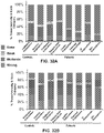


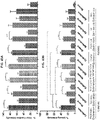


Classifications
-
- A—HUMAN NECESSITIES
- A61—MEDICAL OR VETERINARY SCIENCE; HYGIENE
- A61K—PREPARATIONS FOR MEDICAL, DENTAL OR TOILETRY PURPOSES
- A61K31/00—Medicinal preparations containing organic active ingredients
- A61K31/33—Heterocyclic compounds
- A61K31/395—Heterocyclic compounds having nitrogen as a ring hetero atom, e.g. guanethidine or rifamycins
- A61K31/435—Heterocyclic compounds having nitrogen as a ring hetero atom, e.g. guanethidine or rifamycins having six-membered rings with one nitrogen as the only ring hetero atom
- A61K31/47—Quinolines; Isoquinolines
- A61K31/4706—4-Aminoquinolines; 8-Aminoquinolines, e.g. chloroquine, primaquine
-
- A—HUMAN NECESSITIES
- A61—MEDICAL OR VETERINARY SCIENCE; HYGIENE
- A61K—PREPARATIONS FOR MEDICAL, DENTAL OR TOILETRY PURPOSES
- A61K31/00—Medicinal preparations containing organic active ingredients
- A61K31/33—Heterocyclic compounds
- A61K31/395—Heterocyclic compounds having nitrogen as a ring hetero atom, e.g. guanethidine or rifamycins
- A61K31/435—Heterocyclic compounds having nitrogen as a ring hetero atom, e.g. guanethidine or rifamycins having six-membered rings with one nitrogen as the only ring hetero atom
- A61K31/47—Quinolines; Isoquinolines
- A61K31/4709—Non-condensed quinolines and containing further heterocyclic rings
-
- A—HUMAN NECESSITIES
- A61—MEDICAL OR VETERINARY SCIENCE; HYGIENE
- A61K—PREPARATIONS FOR MEDICAL, DENTAL OR TOILETRY PURPOSES
- A61K31/00—Medicinal preparations containing organic active ingredients
- A61K31/70—Carbohydrates; Sugars; Derivatives thereof
- A61K31/7088—Compounds having three or more nucleosides or nucleotides
- A61K31/7105—Natural ribonucleic acids, i.e. containing only riboses attached to adenine, guanine, cytosine or uracil and having 3'-5' phosphodiester links
-
- A—HUMAN NECESSITIES
- A61—MEDICAL OR VETERINARY SCIENCE; HYGIENE
- A61K—PREPARATIONS FOR MEDICAL, DENTAL OR TOILETRY PURPOSES
- A61K35/00—Medicinal preparations containing materials or reaction products thereof with undetermined constitution
- A61K35/12—Materials from mammals; Compositions comprising non-specified tissues or cells; Compositions comprising non-embryonic stem cells; Genetically modified cells
- A61K35/48—Reproductive organs
-
- A—HUMAN NECESSITIES
- A61—MEDICAL OR VETERINARY SCIENCE; HYGIENE
- A61P—SPECIFIC THERAPEUTIC ACTIVITY OF CHEMICAL COMPOUNDS OR MEDICINAL PREPARATIONS
- A61P15/00—Drugs for genital or sexual disorders; Contraceptives
-
- C—CHEMISTRY; METALLURGY
- C12—BIOCHEMISTRY; BEER; SPIRITS; WINE; VINEGAR; MICROBIOLOGY; ENZYMOLOGY; MUTATION OR GENETIC ENGINEERING
- C12N—MICROORGANISMS OR ENZYMES; COMPOSITIONS THEREOF; PROPAGATING, PRESERVING, OR MAINTAINING MICROORGANISMS; MUTATION OR GENETIC ENGINEERING; CULTURE MEDIA
- C12N15/00—Mutation or genetic engineering; DNA or RNA concerning genetic engineering, vectors, e.g. plasmids, or their isolation, preparation or purification; Use of hosts therefor
- C12N15/09—Recombinant DNA-technology
- C12N15/11—DNA or RNA fragments; Modified forms thereof; Non-coding nucleic acids having a biological activity
- C12N15/113—Non-coding nucleic acids modulating the expression of genes, e.g. antisense oligonucleotides; Antisense DNA or RNA; Triplex- forming oligonucleotides; Catalytic nucleic acids, e.g. ribozymes; Nucleic acids used in co-suppression or gene silencing
-
- C—CHEMISTRY; METALLURGY
- C12—BIOCHEMISTRY; BEER; SPIRITS; WINE; VINEGAR; MICROBIOLOGY; ENZYMOLOGY; MUTATION OR GENETIC ENGINEERING
- C12Q—MEASURING OR TESTING PROCESSES INVOLVING ENZYMES, NUCLEIC ACIDS OR MICROORGANISMS; COMPOSITIONS OR TEST PAPERS THEREFOR; PROCESSES OF PREPARING SUCH COMPOSITIONS; CONDITION-RESPONSIVE CONTROL IN MICROBIOLOGICAL OR ENZYMOLOGICAL PROCESSES
- C12Q1/00—Measuring or testing processes involving enzymes, nucleic acids or microorganisms; Compositions therefor; Processes of preparing such compositions
- C12Q1/68—Measuring or testing processes involving enzymes, nucleic acids or microorganisms; Compositions therefor; Processes of preparing such compositions involving nucleic acids
- C12Q1/6876—Nucleic acid products used in the analysis of nucleic acids, e.g. primers or probes
- C12Q1/6883—Nucleic acid products used in the analysis of nucleic acids, e.g. primers or probes for diseases caused by alterations of genetic material
- C12Q1/6886—Nucleic acid products used in the analysis of nucleic acids, e.g. primers or probes for diseases caused by alterations of genetic material for cancer
-
- G—PHYSICS
- G01—MEASURING; TESTING
- G01N—INVESTIGATING OR ANALYSING MATERIALS BY DETERMINING THEIR CHEMICAL OR PHYSICAL PROPERTIES
- G01N33/00—Investigating or analysing materials by specific methods not covered by groups G01N1/00 - G01N31/00
- G01N33/48—Biological material, e.g. blood, urine; Haemocytometers
- G01N33/50—Chemical analysis of biological material, e.g. blood, urine; Testing involving biospecific ligand binding methods; Immunological testing
- G01N33/68—Chemical analysis of biological material, e.g. blood, urine; Testing involving biospecific ligand binding methods; Immunological testing involving proteins, peptides or amino acids
- G01N33/689—Chemical analysis of biological material, e.g. blood, urine; Testing involving biospecific ligand binding methods; Immunological testing involving proteins, peptides or amino acids related to pregnancy or the gonads
-
- C—CHEMISTRY; METALLURGY
- C12—BIOCHEMISTRY; BEER; SPIRITS; WINE; VINEGAR; MICROBIOLOGY; ENZYMOLOGY; MUTATION OR GENETIC ENGINEERING
- C12N—MICROORGANISMS OR ENZYMES; COMPOSITIONS THEREOF; PROPAGATING, PRESERVING, OR MAINTAINING MICROORGANISMS; MUTATION OR GENETIC ENGINEERING; CULTURE MEDIA
- C12N2310/00—Structure or type of the nucleic acid
- C12N2310/10—Type of nucleic acid
- C12N2310/14—Type of nucleic acid interfering N.A.
-
- C—CHEMISTRY; METALLURGY
- C12—BIOCHEMISTRY; BEER; SPIRITS; WINE; VINEGAR; MICROBIOLOGY; ENZYMOLOGY; MUTATION OR GENETIC ENGINEERING
- C12N—MICROORGANISMS OR ENZYMES; COMPOSITIONS THEREOF; PROPAGATING, PRESERVING, OR MAINTAINING MICROORGANISMS; MUTATION OR GENETIC ENGINEERING; CULTURE MEDIA
- C12N2320/00—Applications; Uses
- C12N2320/30—Special therapeutic applications
- C12N2320/31—Combination therapy
-
- C—CHEMISTRY; METALLURGY
- C12—BIOCHEMISTRY; BEER; SPIRITS; WINE; VINEGAR; MICROBIOLOGY; ENZYMOLOGY; MUTATION OR GENETIC ENGINEERING
- C12N—MICROORGANISMS OR ENZYMES; COMPOSITIONS THEREOF; PROPAGATING, PRESERVING, OR MAINTAINING MICROORGANISMS; MUTATION OR GENETIC ENGINEERING; CULTURE MEDIA
- C12N2320/00—Applications; Uses
- C12N2320/30—Special therapeutic applications
- C12N2320/32—Special delivery means, e.g. tissue-specific
-
- C—CHEMISTRY; METALLURGY
- C12—BIOCHEMISTRY; BEER; SPIRITS; WINE; VINEGAR; MICROBIOLOGY; ENZYMOLOGY; MUTATION OR GENETIC ENGINEERING
- C12N—MICROORGANISMS OR ENZYMES; COMPOSITIONS THEREOF; PROPAGATING, PRESERVING, OR MAINTAINING MICROORGANISMS; MUTATION OR GENETIC ENGINEERING; CULTURE MEDIA
- C12N2320/00—Applications; Uses
- C12N2320/30—Special therapeutic applications
- C12N2320/35—Special therapeutic applications based on a specific dosage / administration regimen
-
- C—CHEMISTRY; METALLURGY
- C12—BIOCHEMISTRY; BEER; SPIRITS; WINE; VINEGAR; MICROBIOLOGY; ENZYMOLOGY; MUTATION OR GENETIC ENGINEERING
- C12Q—MEASURING OR TESTING PROCESSES INVOLVING ENZYMES, NUCLEIC ACIDS OR MICROORGANISMS; COMPOSITIONS OR TEST PAPERS THEREFOR; PROCESSES OF PREPARING SUCH COMPOSITIONS; CONDITION-RESPONSIVE CONTROL IN MICROBIOLOGICAL OR ENZYMOLOGICAL PROCESSES
- C12Q2600/00—Oligonucleotides characterized by their use
- C12Q2600/118—Prognosis of disease development
-
- C—CHEMISTRY; METALLURGY
- C12—BIOCHEMISTRY; BEER; SPIRITS; WINE; VINEGAR; MICROBIOLOGY; ENZYMOLOGY; MUTATION OR GENETIC ENGINEERING
- C12Q—MEASURING OR TESTING PROCESSES INVOLVING ENZYMES, NUCLEIC ACIDS OR MICROORGANISMS; COMPOSITIONS OR TEST PAPERS THEREFOR; PROCESSES OF PREPARING SUCH COMPOSITIONS; CONDITION-RESPONSIVE CONTROL IN MICROBIOLOGICAL OR ENZYMOLOGICAL PROCESSES
- C12Q2600/00—Oligonucleotides characterized by their use
- C12Q2600/158—Expression markers
-
- G—PHYSICS
- G01—MEASURING; TESTING
- G01N—INVESTIGATING OR ANALYSING MATERIALS BY DETERMINING THEIR CHEMICAL OR PHYSICAL PROPERTIES
- G01N2333/00—Assays involving biological materials from specific organisms or of a specific nature
- G01N2333/435—Assays involving biological materials from specific organisms or of a specific nature from animals; from humans
- G01N2333/46—Assays involving biological materials from specific organisms or of a specific nature from animals; from humans from vertebrates
-
- G—PHYSICS
- G01—MEASURING; TESTING
- G01N—INVESTIGATING OR ANALYSING MATERIALS BY DETERMINING THEIR CHEMICAL OR PHYSICAL PROPERTIES
- G01N2800/00—Detection or diagnosis of diseases
- G01N2800/36—Gynecology or obstetrics
- G01N2800/364—Endometriosis, i.e. non-malignant disorder in which functioning endometrial tissue is present outside the uterine cavity
Definitions
- Endometriosis is a gynecological disease that afflicts women of child-bearing age. Although itself benign, it is a very painful, chronic, and inflammatory condition characterized by endometriotic lesions at ectopic sites and ovarian cysts leading to infertility and an increased risk of specific subtypes of ovarian cancer. Characteristic symptoms of endometriosis include chronic pelvic pain, pain during intercourse (dyspareunia), painful periods (dysmenorrhea), and infertility.
- Endometriotic lesions are characterized by functional endometrial-like tissue (epithelial glands and stroma) outside of the uterus, particularly in the peritoneal area, affecting reproductive organs, the bladder and the intestinal tract. Endometriosis is the third leading cause of gynecologic hospitalization in the United States. Hospital surveys estimate that the prevalence of endometriosis among all pre-menopausal women is up to 10%, which equates to about 180 million affected women and adolescents worldwide.
- endometriosis substantially and negatively affects the quality of life of affected adolescents and women.
- the definitive causes of endometriosis are unclear.
- eutopic endometrium deposited into the peritoneal cavity via retrograde menstruation is considered the source of cells that form endometriotic lesions.
- Development of therapeutic and strategies to diagnose, treat, mitigate, or eliminate the lesions and to relieve the pain, without interfering with the fertility potential, and prevention of further progression of this disease are greatly needed for these patients.
- WO 00/47739 discloses autoantigens diagnostic of endometriosis.
- Fukuda et al. discloses that the anti-malarial chloroquine suppresses proliferation and overcomes cisplatin resistance of endometrial cancer cells via autophagy inhibition.
- Ruiz et al. discloses the effect of hydroxychloroquine and characterization of autophagy in a mouse model of endometriosis.
- Embodiments of the present disclosure will employ, unless otherwise indicated, techniques of molecular biology, microbiology, nanotechnology, organic chemistry, biochemistry, botany and the like, which are within the skill of the art. Such techniques are explained fully in the literature.
- endometriotic cell refers to a pathologic endometrial cell that can grow and survive outside the endometrium as part of an endometriotic lesion.
- endometriotic lesion refers to a collection or population of endometriotic cells existing in a subject or directly removed from a subject.
- endometriosis refers to a disease or condition that is characterized by aberrant ectopic (outside the uterus) growth and survival of endometrial cells that have transformed from normal endometrial cells to pathologic endometrial cells.
- autophagic refers to relating to or characterizing autophagy.
- autophagy refers to the catabolic cell mechanism that involves cell degradation of unnecessary or dysfunctional cellular components through the actions of lysosomes.
- immortalized cell(s) refers to a cell or population of cells that have evaded normal cellular senescence and can proliferate indefinitely due to a biological difference, such as a DNA mutation or expression of an exogenous protein, whose expression or presence, results in an increase in the propagation capabilities compared to an average cell found in the same source as the immortalized cell(s). As long as cell culture conditions are maintained properly, immortalized cells can be propagated in cell culture indefinitely (unlimited number of passages).
- life-extended cell(s) refers to a cell or population of cells that can be cultured for at least between about 3 and about 20 passages longer than a non-life extended or normal cell(s) found in the same source.
- the ability to proliferate for about 3 to about 20 passages longer can be the result in a biological difference, such as a DNA mutation or expression of an exogenous protein, between the life-extended endometriotic cell and a normal endometriotic cell.
- corresponding to refers to the underlying biological relationship between these different molecules. As such, one of skill in the art would understand that operatively "corresponding to” can direct them to determine the possible underlying and/or resulting sequences of other molecules given the sequence of any other molecule which has a similar biological relationship with these molecules. For example, from a DNA sequence an RNA sequence can be determined and from an RNA sequence a cDNA sequence can be determined.
- life-extended endometriotic cell(s) refers to an endometriotic cell that has acquired a biological difference, such as a DNA mutation or expression of an exogenous protein, whose expression or presence, results in an increase in the propagation capabilities of the endometriotic cell(s) compared to non-life extended or normal endometriotic cells. Life-extended endometriotic cell(s) can have an ability to proliferate in culture for about 3 to about 20 passages longer as compared to non-life extended or normal endometriotic cells.
- personalized population of cells refers to a population of cells containing in vitro progeny of a cell derived from a endometriotic lesion from a subject in need of treatment, where the population of cells is suitable for use to determine that particular subject's endometriotic lesion characteristics and to test the efficacy of treatments directly on the endometriotic lesion cells.
- autophagic inhibitor refers to a compound, molecules (e . g ., DNA, RNA proteins (e.g., antibodies), or other substance that interacts with a transcript, protein, or other molecule that is involved in the autophagy pathway, such that autophagy is reduced as compared to a control.
- derivative refers to any compound having the same or a similar core structure to the compound but having at least one structural difference, including substituting, deleting, and/or adding one or more atoms or functional groups.
- derivative does not mean that the derivative is synthesized from the parent compound either as a starting material or intermediate, although this can be the case.
- derivative can include salts, prodrugs, or metabolites of the parent compound.
- Derivatives include compounds in which free amino groups in the parent compound have been derivatized to form amine hydrochlorides, p-toluene sulfoamides, benzoxycarboamides, t-butyloxycarboamides, thiourethane-type derivatives, trifluoroacetylamides, chloroacetylamides, or formamides.
- Derivatives include compounds in which carboxyl groups in the parent compound have been derivatized to form salts, methyl and ethyl esters or other types of esters or hydrazides.
- Derivatives include compounds in which hydroxyl groups in the parent compound have been derivatized to form O-acyl or O-alkyl derivatives.
- Derivatives include compounds in which a hydrogen bond donating group in the parent compound is replaced with another hydrogen bond donating group such as OH, NH, or SH.
- Derivatives include replacing a hydrogen bond acceptor group in the parent compound with another hydrogen bond acceptor group such as esters, ethers, ketones, carbonates, tertiary amines, imine, thiones, sulfones, tertiary amides, and sulfides.
- isolated means separated from constituents, cellular and otherwise, with which the polynucleotide, peptide, polypeptide, protein, antibody, or fragments thereof, are normally associated in nature.
- a non-naturally occurring polynucleotide, peptide, polypeptide, protein, antibody, or fragments thereof, does not require “isolation” to distinguish it from its naturally occurring counterpart.
- binding refers to binding that occurs between such paired species as nucleotide/nucleotide, enzyme/substrate, receptor/agonist, antibody/antigen, and lectin/carbohydrate that can be mediated by covalent or non-covalent interactions or a combination of covalent and non-covalent interactions.
- the binding which occurs is typically electrostatic, hydrogen- bonding, or the result of lipophilic interactions. Accordingly, "specific binding" occurs between a paired species where there is interaction between the two which produces a bound complex having the characteristics of an antibody/antigen or enzyme/substrate interaction.
- the specific binding is characterized by the binding of one member of a pair to a particular species and to no other species within the family of compounds to which the corresponding member of the binding member belongs.
- an antibody preferably binds to a single epitope and to no other epitope within the family of proteins.
- aptamer refers to single-stranded DNA or RNA molecules that can bind to pre-selected targets including proteins with high affinity and specificity. Their specificity and characteristics are not directly determined by their primary sequence, but instead by their tertiary structure.
- RNA differential production of RNA, including but not limited to mRNA, tRNA, miRNA, siRNA, snRNA, and piRNA transcribed from a gene or regulatory region of a genome or the protein product encoded by a gene as compared to the level of production of RNA or protein by the same gene or regulator region in a normal or a control cell.
- “differentially expressed” also refers to nucleotide sequences or proteins in a cell or tissue which have different temporal and/or spatial expression profiles as compared to a normal or control cell.
- separated refers to the state of being physically divided from the original source or population such that the separated compound, agent, particle, or molecule can no longer be considered part of the original source or population.
- expression refers to the process by which polynucleotides are transcribed into RNA transcripts. In the context of mRNA and other translated RNA species, “expression” also refers to the process or processes by which the transcribed RNA is subsequently translated into peptides, polypeptides, or proteins.
- peptide refers to two or more amino acids where the alpha carboxyl group of one amino acid is bound to the alpha amino group of another amino acid. Strings of 10 or more amino acids are also referred to herein as “polypeptides” or “proteins”.
- polypeptides or "proteins” are amino acid residue sequences. Those sequences are written left to right in the direction from the amino to the carboxy terminus. In accordance with standard nomenclature, amino acid residue sequences are denominated by either a three letter or a single letter code as indicated as follows: Alanine (Ala, A), Arginine (Arg, R), Asparagine (Asn, N), Aspartic Acid (Asp, D), Cysteine (Cys, C), Glutamine (Gin, Q), Glutamic Acid (Glu, E), Glycine (Gly, G), Histidine (His, H), Isoleucine (Ile, I), Leucine (Leu, L), Lysine (Lys, K), Methionine (Met, M), Phenylalanine (Phe, F), Proline (Pro, P), Serine (Ser, S), Threonine (Thr, T), Tryptophan (Trp, W), Ty
- Gene refers to a hereditary unit corresponding to a sequence of DNA that occupies a specific location on a chromosome and that contains the genetic instruction for a characteristic(s) or trait(s) in an organism. "Gene” also refers to the specific sequence of DNA that is transcribed into an RNA transcript that can be translated into a polypeptide or be a catalytic RNA molecule including but not limited to tRNA, siRNA, piRNA, miRNA, and shRNA.
- RNA deoxyribonucleic acid
- DNA deoxyribonucleic acid
- RNA ribonucleic acid
- DNA deoxyribonucleic acid
- RNA ribonucleic acid
- RNA can be in the form of a tRNA (transfer RNA), snRNA (small nuclear RNA), rRNA (ribosomal RNA), mRNA (messenger RNA), anti-sense RNA, RNAi (RNA interference construct), siRNA (short interfering RNA), microRNA (miRNA), or ribozymes.
- nucleic acid sequence and “oligonucleotide” also encompasses a nucleic acid and polynucleotide as defined above.
- DNA molecule includes nucleic acids/polynucleotides that are made of DNA.
- nucleic acid and polynucleotide generally refer to a string of at least two base-sugar-phosphate combinations and refers to, among others, single-and double-stranded DNA, DNA that is a mixture of single-and double-stranded regions, single-and double-stranded RNA, and RNA that is mixture of single- and double-stranded regions, hybrid molecules comprising DNA and RNA that can be single-stranded or, more typically, double-stranded or a mixture of single- and double-stranded regions.
- polynucleotide as used herein refers to triple-stranded regions comprising RNA or DNA or both RNA and DNA.
- the strands in such regions can be from the same molecule or from different molecules.
- the regions caninclude all of one or more of the molecules, but more typically involve only a region of some of the molecules.
- One of the molecules of a triple-helical region often is an oligonucleotide.
- Polynucleotide and “nucleic acids” also encompasses such chemically, enzymatically or metabolically modified forms of polynucleotides, as well as the chemical forms of DNA and RNA characteristic of viruses and cells, including simple and complex cells, inter alia.
- the term polynucleotide includes DNAs or RNAs as described above that contain one or more modified bases.
- DNAs or RNAs comprising unusual bases, such as inosine, or modified bases, such as tritylated bases, to name just two examples are polynucleotides as the term is used herein.
- Polynucleotide and “nucleic acids” also includes PNAs (peptide nucleic acids), phosphorothioates, and other variants of the phosphate backbone of native nucleic acids.
- Natural nucleic acids have a phosphate backbone, artificial nucleic acids can contain other types of backbones, but contain the same bases.
- DNAs or RNAs with backbones modified for stability or for other reasons are "nucleic acids" or "polynucleotide” as that term is intended herein.
- microRNA refers to a small non-coding RNA molecule containing about 21 to about 23 nucleotides found in organisms, which functions in transcriptional and post-transcriptional regulation of transcription and translation of RNA.
- purified is used in reference to a nucleic acid sequence, peptide, or polypeptide or other compound described herein that has increased purity relative to the natural environment.
- control is an alternative subject or sample used in an experiment for comparison purposes and included to minimize or distinguish the effect of variables other than an independent variable.
- a “control” can be a positive control, a negative control, or an assay or reaction control (an internal control to an assay or reaction included to confirm that the assay was functional).
- the positive or negative control can also be the assay or reaction control.
- dosage form or “unit dosage form” refers to a pharmaceutical formulation that is administered to a subject in need of treatment and generally can be in the form of tablets, capsules, sachets containing powder or granules, liquid solutions or suspensions, patches, and the like.
- effective amount can refer to the amount of a compound or molecule that will elicit the biological or medical response of a tissue, system, animal, or human that is being sought by the researcher, veterinarian, medical doctor or other clinician.
- the effective amount will vary depending on the compound or molecule, the disorder or condition (normal or abnormal) and its severity, the route of administration, time of administration, rate of excretion, drug or compound, judgment of the researcher, veterinarian, medical doctor or other clinician, dosage form, and the age, weight, general health, sex and/or diet of the subject to be treated.
- Effective amount can refer to the amount of an autophagic inhibitor, such as HCQ, that can treat, mitigate, or prevent endometriosis, an endometriotic lesion, or other symptom thereof.
- the “effective amount” can refer to the amount of an autophagic inhibitor, such as HCQ, that can prevent, mitigate, and/or reduce the formation of new endometriotic lesions and/or prevent progression or stage change of endometriosis.
- the terms “sufficient” and “effective”, as used interchangeably herein, can refer to an amount (e.g. mass, volume, dosage, concentration, and/or time period) needed to achieve one or more desired result(s). For example, a therapeutically effective amount refers to an amount needed to achieve one or more therapeutic effects.
- concentration used in reference to an amount of a molecule, compound, or composition, including, but not limited to, a chemical compound, polynucleotide, peptide, polypeptide, protein, antibody, or fragments thereof, that indicates that the sample is distinguishable from its naturally occurring counterpart in that the concentration or number of molecules per volume is greater than that of its naturally occurring counterpart.
- diluted used in reference to an amount of a molecule, compound, or composition including but not limited to, a chemical compound, polynucleotide, peptide, polypeptide, protein, antibody, or fragments thereof, that indicates that the sample is distinguishable from its naturally occurring counterpart in that the concentration or number of molecules per volume is less than that of its naturally occurring counterpart.
- pharmaceutical formulation refers to the combination of an active agent, compound, or ingredient with a pharmaceutically acceptable carrier or excipient, making the composition suitable for diagnostic, therapeutic, or preventive use in vitro, in vivo, or ex vivo.
- pharmaceutically acceptable carrier refers to a carrier, diluent, binder, lubricant, glidant, preservative, flavoring agent, coloring agent, or excipient that is useful in preparing a pharmaceutical formulation that is generally safe, non-toxic, and is neither biologically or otherwise undesirable, and includes a carrier or excipient that is acceptable for veterinary use as well as human pharmaceutical use.
- pharmaceutically acceptable salt refers to any salt derived from organic and inorganic acids of a compound described herein.
- Pharmaceutically acceptable salt also refers to a salt of a compound described having an acidic functional group, such as a carboxylic acid functional group, and a base.
- Pharmaceutically acceptable salt also includes hydrates of a salt of a compound described herein.
- subject refers to a vertebrate, preferably a mammal, more preferably a human. Mammals include, but are not limited to, murines, simians, humans, farm animals, sport animals, and pets.
- the term “pet” includes a dog, cat, guinea pig, mouse, rat, rabbit, ferret, and the like.
- farm animal includes a horse, sheep, goat, chicken, pig, cow, donkey, llama, alpaca, turkey, and the like.
- biocompatible or “biocompatibility” refers to the ability of a material to be used by a patient without eliciting an adverse or otherwise inappropriate host response in the patient to the material or an active derivative thereof, such as a metabolite, as compared to the host response in a normal or control patient.
- terapéutica refers to curing or treating a symptom of a disease or condition.
- treating can include inhibiting and/or resolving the disease, disorder or condition, e.g., impeding its progress; and relieving the disease, disorder, or condition, e.g., causing regression of the disease, disorder and/or condition.
- Treating the disease, disorder, or condition can include ameliorating at least one symptom of the particular disease, disorder, or condition, even if the underlying pathophysiology is not affected, such as treating the pain of a subject by administration of an analgesic agent even though such agent does not treat the cause of the pain.
- Treating can refer to reducing a symptom of endometriosis, such as reducing the size of a lesion, reducing the recurrence of endometriotic lesions, preventing the occurance of new lesions, and/or preventing or delaying the progression of endometriosis to a different stage.
- preventing includes preventing a disease, disorder or condition from occurring in a subject, which can be predisposed to the disease, disorder and/or condition but has not yet been diagnosed as having it.
- preventative can refer to hindering or stopping a disease or condition before it occurs or while the disease or condition is still in the sub-clinical phase.
- Preventing can refer to delaying or stopping the recurrance of endometriotic lesions; stopping, mitigating, and/or delaying the occurance of new endometriotic lesions; stopping, mitigating, and/or delaying the progression of endometriosis in an individual.
- mitigate can refer to reducing a particular characteristic, symptom, or other biological or physiological parameter associated with a disease or disorder, such as endometriosis.
- separated refers to the state of being physically divided from the original source or population such that the separated compound, agent, particle, chemical compound, or molecule can no longer be considered part of the original source or population.
- tangible medium of expression refers to a medium that is physically tangible and is not a mere abstract thought or an unrecorded spoken word. Tangible medium of expression includes, but is not limited to, words on a cellulosic or plastic material or data stored on a suitable device such as a flash memory or CD-ROM.
- active derivative and the like refer to a derivative of an autophagic inhibitor, such as HCQ, that retains an ability to treat/mitigate plasma cell proliferation and/or secondary complications associated with endometriosis. Assays for testing the ability of an active derivative to perform in this fashion are known to those of ordinary skill in the art.
- metabolite refers to substances that result from metabolism of a compound, such as an active agent of a pharmaceutical formulation, such as HCQ.
- active metabolite refers to a metabolite that induces a pharmaceutical or clinical effect, such as treating or preventing endometriosis or a symptom thereof, in a subject.
- primary metabolite refers to a metabolite that is directly involved in growth, development, and/or reproduction of a cell or organism.
- secondary metabolite refers to a metabolite that is not directly involved in growth, development, and/or reproduction of a cell or organism.
- capture molecule refers to a molecule that is configured to specifically bind one or more biomarker molecules of interest.
- a capture molecule can be a polynucleotide, antibody, antigen, apatmer, affibody, polypeptides, peptides, or combinations thereof that specifically bind one or more biomarkers of interest.
- the capture molecule can be configured to specifically bind a polynucleotide or polypeptide corresponding to ATG-5, ATG-7, ATG-9, DJ-1(Park7), hVps34, beclin-1, p-ULK1, ATG1(ULK1), p-mTOR, mTOR, , integrin, Src, FAK, ILK, rkB, AKT, LC3A, LC3 B,TSC1, TSC2, HO-1, PTEN, ARID1A, PIK3CA, K-RAS, BCL-2, ATG16, ATG12, ATG10, ATG3, LC3-1, ATG4, EVI1, RON, EGFR, SnoN, SkiL TGF ⁇ RII, p53, Smad2/3, p-ERK, ERK, PARP, cleaved PARP, p62, ferritin, E-cadherin, N-cadherin, vimentin cytokeratin-18 and
- Representative polypeptide and polynucleotide sequences for the aforementioned biomarkers and any other biomarkers described herein can be 100% identical, 90-100% identical, 80-90% identical, 70-80% identical, 60-70% identical, or 50-100% identical to any one of SEQ ID NOs: 1-314 or 100% identical, 90-100% identical, 80-90% identical, 70-80% identical, 60-70% identical, or 50-100% identical to a sequence corresponding to any one of SEQ ID NOs: 1-314.
- attachment refers to a covalent interaction or bond between a molecule on the surface of the support and the capture molecule so as to immobilize the capture molecule on the surface of the support.
- operatively-linked refers to a non-covalent interaction between the surface of the support and the capture molecule so as to immobilize the capture molecule on the surface of the support.
- non-covalent interactions include but are not limited to, entrapment by the surface substrate, ionic bonds, electrostatic interactions, van der Walls forces, dipole-dipole interactions, dipole-induced-dipole interactions, London dispersion forces, hydrogen bonding, halogen bonding, electromagnetic interactions, ⁇ - ⁇ interactions, cation- ⁇ interactions, anion- ⁇ interactions, polar ⁇ -interactions, and hydrophobic effects.
- biomarker can refer to any measurable molecule, including but not limited to polynucleotides and polypeptides, or compound in a subject whose presences, absolute amount, or relative amount, is indicative of some disease, condition, syndrome, disorder, or symptom thereof, or state thereof, such as endometriosis.
- body fluid refers to any liquid or liquid-like substance that originates in the body of a living organism.
- Body fluid includes, but is not limited to, whole blood, serum, buffy coat of blood or other blood fraction that contains substantially only the white blood cells and platelets, plasma, cerebral spinal fluid, urine, lymph, bile and saliva.
- affibody refers to an engineered protein that is an antibody mimetic and can specifically bind a target molecule, and is based on a three-helix bundle domain, where each of the three helixes contains a polypeptide having about 58 amino acids.
- variant refers to a polypeptide that differs from a reference polypeptide, but retains essential properties.
- a typical variant of a polypeptide differs in amino acid sequence from another, reference polypeptide. Generally, differences are limited so that the sequences of the reference polypeptide and the variant are closely similar overall and, in many regions, identical.
- a variant and reference polypeptide can differ in amino acid sequence by one or more modifications (e.g., substitutions, additions, and/or deletions).
- a substituted or inserted amino acid residue can or cannot be one encoded by the genetic code.
- a variant of a polypeptide can be naturally occurring such as an allelic variant, or it can be a variant that is not known to occur naturally.
- wild-type refers to the typical form of an organism, variety, strain, gene, protein, or characteristic as it occurs in nature, as distinguished from mutant forms that can result from selective breeding or transformation with a transgene.
- diagnosis refers to the identification or determination of the nature and circumstances of a disease, disorder, condition, syndrome, or symptom thereof in a subject.
- prognose refers to determining a prognosis for a disease, disorder, condition, syndrome, or symptom thereof.
- prognosis refers to a prediction or forecast of a chance of recovery, complete or partial, from a disease, disorder, condition, syndrome, or symptom thereof.
- personalized population of cells or “personalized cell” refer to an autologous population of cells or cells are an in vitro population of cells or cell that can be used to diagnose and/or prognose an individual and/or test the individual's response to a treatment.
- emollients refers to an externally applied agent that softens or soothes skin and are generally known in the art and listed in compendia, such as the " Handbook of Pharmaceutical Excipients", 4th Ed., Pharmaceutical Press, 2003 .
- surfactants refers to surface-active agents that lower surface tension and thereby increase the emulsifying, foaming, dispersing, spreading and wetting properties of a product.
- emulsifiers refers to surface active substances which promote the suspension of one liquid in another and promote the formation of a stable mixture, or emulsion, of oil and water.
- oil refers to a composition containing at least about 95% wt of a lipophilic substance.
- lipophilic substances include but are not limited to naturally occurring and synthetic oils, fats, fatty acids, lecithins, triglycerides and combinations thereof.
- emulsion is a composition containing a mixture of non-miscible components homogenously blended together.
- the non-miscible components can include a lipophilic component and an aqueous component.
- Emsion can also refer to a preparation of one liquid distributed in small globules throughout the body of a second liquid. The first liquid is the discontinuous phase and the second liquid is the continuous phase. When oil is first liquid and water or an aqueous solution is the second liquid, it is referred to herein as an "oil in water emulsion".
- water or an aqueous solution is the first liquid and oil or oleagionous substance is the second liquid, it is referred to herein as "water-in-oil" emulsion".
- oil phase and aqueous phase can contain one or more surfactants, emulsifiers, emulsion stabilizers, buffers, and other excipients.
- Some emulsions can be gels or otherwise include a gel component.
- “lotion” refers to a low- to medium-viscosity liquid formulation.
- “Lotions” can contain finely powdered substances that are in soluble in the dispersion medium through the use of suspending agents and dispersing agents.
- “Lotions” can also have as the dispersed phase liquid substances that are immiscible with the vehicle and are usually dispersed by means of emulsifying agents or other suitable stabilizers.
- cream refers to a viscous liquid or semi-solid emulsion of either the "oil-in-water” or “water-in-oil type”.
- ointment refers to a semisolid preparation containing an ointment base and optionally one or more active agents.
- gel refers to a semisolid system containing dispersions of the autophagic inhibitor in a liquid vehicle that is rendered semisolid by the action of a thickening agent or polymeric material ("gelling agent") dissolved or suspended in the liquid vehicle.
- the liquid can include a lipophilic component, an aqueous component or both.
- Gelels can also be emulsions. Some gels, however, are not emulsions because they do not contain a homogenized blend of immiscible components
- Endometriosis is a gynecological disease that afflicts women of child-bearing age. Although itself benign, it is a very painful condition characterized by endometriotic lesions at ectopic sites leading to infertility and an increased risk of specific subtypes of ovarian cancer.
- Endometriosis is believed to result from biological or physiological events that resemble metastasis which can be characterized into 6 stages: (1) shedding of cells, (2) cell survival, (3) escape from immune surveillance, (4) adhesion to peritoneum, (5) angiogenesis, and (6) bleeding.
- formation of an endometriotic lesion is dependent on the combination of these events, the first step, survival, which, if it occurs, will allow cells to implant and develop into an endometriotic lesion and thus plays an important role in the pathogenesis of endometriosis. Indeed, altered cell death characteristics in the eutopic endometrium of women with endometriosis will contribute to survival of retrograde endometrial cells.
- Anoikis is a cell death event due to decreased cell adhesion and elimination of cells detached from substratum. Thus, anoikis will prevent epithelial cells from shedding from their original location ("misplaced" cells), to colonize at ectopic sites. In cancer, anoikis resistance leads to tumor metastases.
- Mechanisms of anoikis resistance include (1) acquisition of an epithelial-mesenchymal transition (EMT)-like phenotype, (2) activation of integrin and downstream signaling mediators (i.e. Src, FAK, and ILK), (3) increased expression of TrkB, a neurotrophic tyrosine kinase receptor (suppressor of caspase-associated anoikis) in endometriosis patients and certain cancers associated with cancer metastases and activation of the pro-survival PI3K/AKT pathway and (4) autophagy which promotes survival leading to increased attachment to the extracellular matrix (ECM).
- EMT epithelial-mesenchymal transition
- Autophagy a survival mechanism that is activated in response to multiple stresses, is a "self-eating" process whereby damaged cellular material and organelles are sequestered in autophagosomes and degraded. Autophagy allows cells to survive given that they re-adhere to ECM in a timely fashion. Interestingly, detachment-induced autophagy directly results from a loss of ECM-integrin engagement. Epithelial cells depend on integrin-mediated cell adhesion to the ECM for proper growth and survival1. The pathways linking loss of integrin engagement at the cell surface to activation of the autophagic machinery remain elusive.
- epithelial cells such as endometrial cells
- epithelial cells depend on integrin-mediated cell adhesion to the ECM for proper growth and survival.
- Cell detachment from the ECM leads to anoikis, a cell death event, preventing colonization of cells at ectopic sites. If autophagy is induced, cells will escape anoikis and survive.
- Autophagy a "self-eating" process whereby damaged cellular material and organelles are sequestered in autophagosomes and degraded, is a survival mechanism activated in response to multiple stresses.
- dysregulation of autophagy in endometriotic cells contained in endometrial lesions contributes to aberrant cell survival.
- an autophagic inhibitor is administered to a subject having endometriosis.
- an endometriotic cell is contacted with an autophagic inhibitor.
- at least one symptom of endometriosis such as lesion size, can be reduced.
- endometriosis a symptom of endometriosis can be alleviated or mitigated by inhibiting autophagy in endometrial cells or endometriotic cells. Indeed, it was unknown how autophagy and its dysregulation affect endometrial cell survival and its contribution to the development of endometriotic lesions. This is likely due, in part, that endometriosis is a benign tumor and autophagy has only been correlated to metastatic tumors that are typically affiliated with invasive cancer tumors.
- compositions, formulations, methods, and assays that can improve the treatment, diagnosis, and/or prognosis of endometriosis and endometriotic lesions.
- Other compositions, compounds, methods, features, and advantages of the present disclosure will be or become apparent to one having ordinary skill in the art upon examination of the following drawings, detailed description, and examples. It is intended that all such additional compositions, compounds, methods, features, and advantages be included within this description, and be within the scope of the present disclosure.
- a life-extended population of cells contains an endometriotic cell, where the endometriotic cell is the in vitro progeny of a cell isolated from an endometriotic lesion, and where the endometriotic cell can have greater protein expression of one or more of the following autophagic markers selected from the group of ATG7, ATG5, and hVps34 as compared to a suitable non-endometriotic control cell or other control cell.
- Suitable non-endometriotic control cells include, but are not limited to ovarian cancer cells, non-diseased ovarian epithelial cells, and surface ovarian epithelial (T80) cells.
- the life-extended cells also express large T antigen.
- the life-extended population of cells can be used as a research tool to assess the effectiveness and safety of compounds to treat endometriosis.
- the population of cells is a personalized population of cells. This allows the determination of the efficacy of treatments on the cells from endometriotic lesions from the subject that will receive the treatment.
- the life-extended endometriotic cells can be prepared from primary endometriotic cells.
- Primary endometriotic cells can be maintained in a 1:1 mixture of Medium 199 and MCDB131 Medium supplemented with about 8% fetal bovine serum, penicillin (100 U/mL; diluted 1:100 in complete culture media)/streptomycin (100 mg/mL; diluted 1:100 in complete culture media), and insulin (5mg/mL) /transferrin (5mg/mL) /selenium (ITS) (5 ⁇ g/mL).
- primary endometriotic cells are transformed them to ubiquitously or conditionally express oncogene.
- primary endometriotic cells are infected with viral particles carrying and SV40 large T antigen gene, such that the transformed cells express a SV40 large T antigen.
- compositions containing an effective amount of an autophaghic inhibitor or active derivative thereof in a pharmaceutical carrier appropriate for administration to an individual in need thereof.
- the individual in need thereof can have or can be suspected of having endometriosis or an endometriotic lesion.
- Formulations can be administered orally, intravenously, intramuscularly, intravaginally, intraperitoneally, rectally, perenterally, topically, intranasally, or subcutaneously.
- Suitable autophagic inhibitors include but are not limited to chloroquine, hydroxychloroquine (also refered to herein as HCQ), and Lys05, pharmaceutically acceptable salts of chloroquine, pharmaceutically acceptable salts of hydroxychloroquine, pharmaceutically acceptable salts of Lys05, antibodies that specifically bind to a positive inducer of autophagy in endometrial cells, siRNA, miRNA, piRNA, or other RNA species that specifically binds to a positive inducer of autophagy in endometrial cells, and other molecules, compounds, or substances which bind or otherwise interact with a protein or other molecule involved in maintaining or increasing autophagy so as to reduce autophagy in the cell.
- the autophagic inhibitor or active derivative thereof can be formulated for parenteral delivery, such as injection or infusion, in the form of a solution or suspension.
- the autophagic inhibitor can be HCQ or a pharmaceutical salt thereof.
- the formulation can be administered via any route, such as, the blood stream or directly to the organ or tissue to be treated.
- Parenteral formulations can be prepared as aqueous compositions using techniques is known in the art.
- such compositions can be prepared as injectable formulations, for example, solutions or suspensions; solid forms suitable for using to prepare solutions or suspensions upon the addition of a reconstitution medium prior to injection; emulsions, such as water-in-oil (w/o) emulsions, oil-in-water (o/w) emulsions, and microemulsions thereof, liposomes, or emulsomes.
- injectable formulations for example, solutions or suspensions
- solid forms suitable for using to prepare solutions or suspensions upon the addition of a reconstitution medium prior to injection emulsions, such as water-in-oil (w/o) emulsions, oil-in-water (o/w) emulsions, and microemulsions thereof, liposomes, or emulsomes.
- emulsions such as water-in-oil (w/o) emulsions
- the carrier can be a solvent or dispersion medium containing, for example, water, ethanol, one or more polyols (e.g., glycerol, propylene glycol, and liquid polyethylene glycol), oils, such as vegetable oils (e.g., peanut oil, corn oil, sesame oil, etc.), and combinations thereof.
- polyols e.g., glycerol, propylene glycol, and liquid polyethylene glycol
- oils such as vegetable oils (e.g., peanut oil, corn oil, sesame oil, etc.)
- the proper fluidity can be maintained, for example, by the use of a coating, such as lecithin, by the maintenance of the required particle size in the case of dispersion and/or by the use of surfactants.
- isotonic agents for example, sugars or sodium chloride.
- Solutions and dispersions of the autophagic inhibitor or active derivative therof can be prepared in water or another solvent or dispersing medium suitably mixed with one or more pharmaceutically acceptable excipients including, but not limited to, surfactants, dispersants, emulsifiers, pH modifying agents, and combination thereof.
- Suitable surfactants can be anionic, cationic, amphoteric or nonionic surface active agents.
- Suitable anionic surfactants include, but are not limited to, those containing carboxylate, sulfonate and sulfate ions.
- Suitable anionic surfactants include sodium, potassium, ammonium of long chain alkyl sulfonates and alkyl aryl sulfonates such as sodium dodecylbenzene sulfonate; dialkyl sodium sulfosuccinates, such as sodium dodecylbenzene sulfonate; dialkyl sodium sulfosuccinates, such as sodium bis-(2-ethylthioxyl)-sulfosuccinate; and alkyl sulfates such as sodium lauryl sulfate.
- Suitable cationic surfactants include, but are not limited to, quaternary ammonium compounds such as benzalkonium chloride, benzethonium chloride, cetrimonium bromide, stearyl dimethylbenzyl ammonium chloride, polyoxyethylene and coconut amine.
- Suitable nonionic surfactants include ethylene glycol monostearate, propylene glycol myristate, glyceryl monostearate, glyceryl stearate, polyglyceryl-4-oleate, sorbitan acylate, sucrose acylate, PEG-150 laurate, PEG-400 monolaurate, polyoxyethylene monolaurate, polysorbates, polyoxyethylene octylphenylether, PEG-1000 cetyl ether, polyoxyethylene tridecyl ether, polypropylene glycol butyl ether, Poloxamer ® 401, stearoyl monoisopropanolamide, and polyoxyethylene hydrogenated tallow amide.
- amphoteric surfactants include sodium N-dodecyl- ⁇ -alanine, sodium N-lauryl- ⁇ -iminodipropionate, myristoamphoacetate, lauryl betaine and lauryl sulfobetaine.
- the formulation can contain a preservative to prevent the growth of microorganisms. Suitable preservatives include, but are not limited to, parabens, chlorobutanol, phenol, sorbic acid, and thimerosal.
- the formulation can also contain an antioxidant to prevent degradation of the autophagic inhibitor or active derivate thereof.
- the formulation can be buffered to a pH of 3-8 for parenteral administration upon reconstitution.
- Suitable buffers include, but are not limited to, phosphate buffers, acetate buffers, and citrate buffers.
- Water-soluble polymers can be used in the formulations for parenteral administration. Suitable water-soluble polymers include, but are not limited to, polyvinylpyrrolidone, dextran, carboxymethylcellulose, and polyethylene glycol.
- Sterile injectable solutions can be prepared by incorporating the autophagic inhibitor or active derivative thereof in the required amount in the appropriate solvent or dispersion medium with one or more of the excipients listed above, as required, followed by filtered sterilization.
- Dispersions can be prepared by incorporating the various sterilized autophagic inhibitor or derivative thereof into a sterile vehicle which contains the basic dispersion medium and the required other ingredients from those listed above.
- Sterile powders for the preparation of sterile injectable solutions can be prepared by vacuum-drying and freeze-drying techniques, which yields a powder of the autophagic inhibitor or active derivative thereof plus any additional desired ingredient from a previously sterile-filtered solution thereof.
- the powders can be prepared in such a manner that the particles are porous in nature, which can increase dissolution of the particles. Methods for making porous particles are well known in the art.
- compositions for parenteral administration can be in the form of a sterile aqueous solution or suspension of particles formed from one or more autophagic inhibitor or active derivative thereof.
- Acceptable solvents include, for example, water, Ringer's solution, phosphate buffered saline (PBS), and isotonic sodium chloride solution.
- PBS phosphate buffered saline
- the formulation can also be a sterile solution, suspension, or emulsion in a nontoxic, parenterally acceptable diluent or solvent such as 1,3-butanediol.
- the formulation can be distributed or packaged in a liquid form.
- formulations for parenteral administration can be packed as a solid, obtained, for example by lyophilization of a suitable liquid formulation.
- the solid can be reconstituted with an appropriate carrier or diluent prior to administration.
- Solutions, suspensions, or emulsions for parenteral administration can be buffered with an effective amount of buffer necessary to maintain a pH suitable for ocular administration.
- Suitable buffers include, but are not limited to, acetate, borate, carbonate, citrate, and phosphate buffers.
- Solutions, suspensions, or emulsions for parenteral administration can also contain one or more tonicity agents to adjust the isotonic range of the formulation.
- Suitable tonicity agents include, but are not limited to, glycerin, mannitol, sorbitol, sodium chloride, and other electrolytes.
- Solutions, suspensions, or emulsions for parenteral administration can also contain one or more preservatives to prevent bacterial contamination of the ophthalmic preparations.
- Suitable preservatives include, but are not limited to, polyhexamethylenebiguanidine (PHMB), benzalkonium chloride (BAK), stabilized oxychloro complexes (otherwise known as Purite ® ), phenylmercuric acetate, chlorobutanol, sorbic acid, chlorhexidine, benzyl alcohol, parabens, thimerosal, and mixtures thereof.
- Solutions, suspensions, or emulsions for parenteral administration can also contain one or more excipients, such as dispersing agents, wetting agents, and suspending agents.
- the autophagic inhibitor or active derivative thereof can be formulated for topical administration.
- the autophagic inhibitor can be HCQ or a salt thereof Suitable dosage forms for topical administration include creams, ointments, salves, sprays, gels, lotions, emulsions, liquids, and transdermal patches.
- the formulation can be formulated for transmucosal, transepithelial, transendothelial, or transdermal administration.
- the topical formulations can contain one or more chemical penetration enhancers, membrane permeability agents, membrane transport agents, emollients, surfactants, stabilizers, and combination thereof.
- the conjugates can be administered as a liquid formulation, such as a solution or suspension, a semi-solid formulation, such as a lotion or ointment, or a solid formulation.
- the autophagic inhibitor is formulated as liquids, including solutions and suspensions, such as eye drops or as a semi-solid formulation, such as ointment or lotion for topical application to the skin, to the mucosa, such as the eye, to the vagina, or to the rectum.
- the formulation can contain one or more excipients, such as emollients, surfactants, emulsifiers, penetration enhancers, and the like.
- excipients such as emollients, surfactants, emulsifiers, penetration enhancers, and the like.
- Suitable emollients include, without limitation, almond oil, castor oil, ceratonia extract, cetostearoyl alcohol, cetyl alcohol, cetyl esters wax, cholesterol, cottonseed oil, cyclomethicone, ethylene glycol palmitostearate, glycerin, glycerin monostearate, glyceryl monooleate, isopropyl myristate, isopropyl palmitate, lanolin, lecithin, light mineral oil, medium-chain triglycerides, mineral oil and lanolin alcohols, petrolatum, petrolatum and lanolin alcohols, soybean oil, starch, stearyl alcohol, sunflower oil, xylitol and combinations thereof.
- the emollients can be ethylhexylstearate and ethylhexyl palmitate.
- Suitable surfactants include, but are not limited to, emulsifying wax, glyceryl monooleate, polyoxyethylene alkyl ethers, polyoxyethylene castor oil derivatives, polysorbate, sorbitan esters, benzyl alcohol, benzyl benzoate, cyclodextrins, glycerin monostearate, poloxamer, povidone and combinations thereof.
- the surfactant can be stearyl alcohol.
- Suitable emulsifiers include, but are not limited to, acacia, metallic soaps, certain animal and vegetable oils, and various polar compounds, anionic emulsifying wax, calcium stearate, carbomers, cetostearyl alcohol, cetyl alcohol, cholesterol, diethanolamine, ethylene glycol palmitostearate, glycerin monostearate, glyceryl monooleate, hydroxpropyl cellulose, hypromellose, lanolin, hydrous, lanolin alcohols, lecithin, medium-chain triglycerides, methylcellulose, mineral oil and lanolin alcohols, monobasic sodium phosphate, monoethanolamine, nonionic emulsifying wax, oleic acid, poloxamer, poloxamers, polyoxyethylene alkyl ethers, polyoxyethylene castor oil derivatives, polyoxyethylene sorbitan fatty acid esters, polyoxyethylene stearates, propylene glycol alginate, self-emulsifying
- Suitable classes of penetration enhancers include, but are not limited to, fatty alcohols, fatty acid esters, fatty acids, fatty alcohol ethers, amino acids, phospholipids, lecithins, cholate salts, enzymes, amines and amides, complexing agents (liposomes, cyclodextrins, modified celluloses, and diimides), macrocyclics, such as macrocylic lactones, ketones, and anhydrides and cyclic ureas, surfactants, N-methyl pyrrolidones and derivatives thereof, DMSO and related compounds, ionic compounds, azone and related compounds, and solvents, such as alcohols, ketones, amides, polyols (e.g., glycols).
- Suitable emulsions include, but are not limited to, oil-in-water and water-in-oil emulsions. Either or both phases of the emulsions can include a surfactant, an emulsifying agent, and/or a liquid non-volatile non-aqueous material.
- the surfactant can be a non-ionic surfactant.
- the emulsifying agent is an emulsifying wax.
- the liquid non-volatile non-aqueous material is a glycol. In some embodiments, the glycol is propylene glycol.
- the oil phase can contain other suitable oily pharmaceutically acceptable excipients. Suitable oily pharmaceutically acceptable excipients include, but are not limited to, hydroxylated castor oil or sesame oil can be used in the oil phase as surfactants or emulsifiers.
- Lotions containing an autophagic inhibitor are also described herein.
- the lotion can be in the form of an emulsion having a viscosity of between 100 and 1000 centistokes.
- the fluidity of lotions can permit rapid and uniform application over a wide surface area.
- Lotions can be formulated to dry on the skin leaving a thin coat of their medicinal components on the skin's surface.
- Creams containing an autophagic inhibitor are also described herein.
- the cream can contain emulsifying agents and/or other stabilizing agents.
- the cream is in the form of a cream having a viscosity of greater than 1000 centistokes, typically in the range of 20,000-50,000 centistokes. Creams, as compared to ointments, can be easier to spread and easier to remove.
- Creams can be thicker than lotions, can have various uses, and can have more varied oils/butters, depending upon the desired effect upon the skin.
- the water-base percentage can be about 60% to about 75% and the oil-base can be about 20% to about 30% of the total, with the other percentages being the emulsifier agent, preservatives and additives for a total of 100%.
- ointments containing an autophagaic inhibitor and a suitable ointment base include hydrocarbon bases (e.g., petrolatum, white petrolatum, yellow ointment, and mineral oil); absorption bases (hydrophilic petrolatum, anhydrous lanolin, lanolin, and cold cream); water-removable bases (e.g., hydrophilic ointment), and water-soluble bases (e.g., polyethylene glycol ointments).
- Pastes typically differ from ointments in that they contain a larger percentage of solids. Pastes are typically more absorptive and less greasy that ointments prepared with the same components.
- Suitable gelling agents include, but are not limited to, modified celluloses, such as hydroxypropyl cellulose and hydroxyethyl cellulose; carbopol homopolymers and copolymers; and combinations thereof.
- Suitable solvents in the liquid vehicle include, but are not limited to, diglycol monoethyl ether; alklene glycols, such as propylene glycol; dimethyl isosorbide; alcohols, such as isopropyl alcohol and ethanol. The solvents can be selected for their ability to dissolve the drug.
- Other additives which can improve the skin feel and/or emolliency of the formulation, can also be incorporated.
- Such additives include, but are not limited, isopropyl myristate, ethyl acetate, C 12 -C 15 alkyl benzoates, mineral oil, squalane, cyclomethicone, capric/caprylic triglycerides, and combinations thereof.
- Foams can be an emulsion in combination with a gaseous propellant.
- the gaseous propellant can include hydrofluoroalkanes (HFAs).
- HFAs hydrofluoroalkanes
- Suitable propellants include HFAs such as 1,1,1,2-tetrafluoroethane (HFA 134a) and 1,1,1,2,3,3,3-heptafluoropropane (HFA 227), but mixtures and admixtures of these and other HFAs that are currently approved or can become approved for medical use are suitable.
- the propellants can be devoid of hydrocarbon propellant gases, which can produce flammable or explosive vapors during spraying.
- the foams can contain no volatile alcohols, which can produce flammable or explosive vapors during use.
- Buffers can be used to control pH of a composition.
- the buffers can buffer the composition from a pH of about 4 to a pH of about 7.5, from a pH of about 4 to a pH of about 7, or from a pH of about 5 to a pH of about 7.
- the buffer can be triethanolamine.
- Preservatives can be included to prevent the growth of fungi and microorganisms.
- Suitable preservatives include, but are not limited to, benzoic acid, butylparaben, ethyl paraben, methyl paraben, propylparaben, sodium benzoate, sodium propionate, benzalkonium chloride, benzethonium chloride, benzyl alcohol, cetylpyridinium chloride, chlorobutanol, phenol, phenylethyl alcohol, and thimerosal.
- the formulations can be provided via continuous delivery of one or more formulations to a patient in need thereof.
- repeated application can be done or a patch can be used to provide continuous administration of the noscapine analogs over an extended period of time.
- the autophagic inhibitors can be prepared in enteral formulations, such as for oral administration.
- the autophagic inhibior can be HCQ, active derivative thereof, or pharmaceutical salt thereof.
- Suitable oral dosage forms include tablets, capsules, solutions, suspensions, syrups, and lozenges. Tablets can be made using compression or molding techniques well known in the art.
- Gelatin or non-gelatin capsules can prepared as hard or soft capsule shells, which can encapsulate liquid, solid, and semi-solid fill materials, using techniques well known in the art.
- Formulations containing an autophagic inhibitor are prepared using pharmaceutically acceptable carriers.
- carrier includes, but is not limited to, diluents, preservatives, binders, lubricants, disintegrators, swelling agents, fillers, stabilizers, and combinations thereof.
- Polymers used in the dosage form include, but are not limited to, suitable hydrophobic or hydrophilic polymers and suitable pH dependent or independent polymers.
- Suitable hydrophobic and hydrophilic polymers include, but are not limited to, hydroxypropyl methylcellulose, hydroxypropyl cellulose, hydroxyethyl cellulose, carboxy methylcellulose, polyethylene glycol, ethylcellulose, microcrystalline cellulose, polyvinyl pyrrolidone, polyvinyl alcohol, polyvinyl acetate, and ion exchange resins.
- Carrier also includes all components of the coating composition which can include plasticizers, pigments, colorants, stabilizing agents, and glidants.
- Formulations containing an autophagic inhibitor can be prepared using one or more pharmaceutically acceptable excipients, including diluents, preservatives, binders, lubricants, disintegrators, swelling agents, fillers, stabilizers, and combinations thereof.
- Delayed release dosage formulations can be prepared as described in standard references such as " Pharmaceutical dosage form tablets”, eds. Liberman et. al. (New York, Marcel Dekker, Inc., 1989 ), “ Remington - The science and practice of pharmacy", 20th ed., Lippincott Williams & Wilkins, Baltimore, MD, 2000 , and " Pharmaceutical dosage forms and drug delivery systems", 6th Edition, Ansel et al., (Media, PA: Williams and Wilkins, 1995 ). These references provide information on excipients, materials, equipment and process for preparing tablets and capsules and delayed release dosage forms of tablets, capsules, and granules. These references provide information on carriers, materials, equipment and process for preparing tablets and capsules and delayed release dosage forms of tablets, capsules, and granules.
- the formulations containing an autophagic inhibitor can be coated with a suitable coating material, for example, to delay release once the particles have passed through the acidic environment of the stomach.
- suitable coating materials include, but are not limited to, cellulose polymers such as cellulose acetate phthalate, hydroxypropyl cellulose, hydroxypropyl methylcellulose, hydroxypropyl methylcellulose phthalate and hydroxypropyl methylcellulose acetate succinate; polyvinyl acetate phthalate, acrylic acid polymers and copolymers, and methacrylic resins that are commercially available under the trade name EUDRAGIT ® (Roth Pharma, Westerstadt, Germany), zein, shellac, and polysaccharides.
- Coatings can be formed with a different ratio of water soluble polymer, water insoluble polymers and/or pH dependent polymers, with or without water insoluble/water soluble non polymeric excipient, to produce the desired release profile.
- the coating can be performed on a dosage form (matrix or simple) which includes, but is not limited to, tablets (compressed with or without coated beads), capsules (with or without coated beads), beads, particle compositions, "ingredient as is” formulated as, but not limited to, suspension form or as a sprinkle dosage form.
- the coating material can contain conventional carriers such as plasticizers, pigments, colorants, glidants, stabilization agents, pore formers and surfactants.
- Optional pharmaceutically acceptable excipients include, but are not limited to, diluents, binders, lubricants, disintegrants, colorants, stabilizers, and surfactants.
- Diluents also referred to as "fillers,” can be used to increase the bulk of a solid dosage form so that a practical size is provided for compression of tablets or formation of beads and granules.
- Suitable diluents include, but are not limited to, dicalcium phosphate dihydrate, calcium sulfate, lactose, sucrose, mannitol, sorbitol, cellulose, microcrystalline cellulose, kaolin, sodium chloride, dry starch, hydrolyzed starches, pregelatinized starch, silicone dioxide, titanium oxide, magnesium aluminum silicate and powdered sugar.
- the usual diluents include inert powdered substances such as starches, powdered cellulose, especially crystalline and microcrystalline cellulose, sugars such as fructose, mannitol and sucrose, grain flours and similar edible powders.
- Typical diluents include, for example, various types of starch, lactose, mannitol, kaolin, calcium phosphate or sulfate, inorganic salts such as sodium chloride and powdered sugar. Powdered cellulose derivatives are also useful.
- Binders can impart cohesive qualities to a solid dosage formulation, and thus can ensure that a tablet or bead or granule remains intact after the formation of the dosage forms.
- Suitable binder materials include, but are not limited to, starch, pregelatinized starch, gelatin, sugars (including sucrose, glucose, dextrose, lactose and sorbitol), polyethylene glycol, waxes, natural and synthetic gums such as acacia, tragacanth, sodium alginate, cellulose, including hydroxypropylmethylcellulose, hydroxypropylcellulose, ethylcellulose, and veegum, and synthetic polymers such as acrylic acid and methacrylic acid copolymers, methacrylic acid copolymers, methyl methacrylate copolymers, aminoalkyl methacrylate copolymers, polyacrylic acid/polymethacrylic acid and polyvinylpyrrolidone.
- Typical tablet binders include substances such as starch, gelatin and sugars such as lactose, fructose, and glucose. Natural and synthetic gums, including acacia, alginates, methylcellulose, and polyvinylpyrrolidone can also be used. Polyethylene glycol, hydrophilic polymers, ethylcellulose and waxes can also serve as binders.
- Lubricants can be included to facilitate tablet manufacture. Suitable lubricants include, but are not limited to, magnesium stearate, calcium stearate, stearic acid, glycerol behenate, polyethylene glycol, talc, and mineral oil. A lubricant can be included in a tablet formulation to prevent the tablet and punches from sticking in the die. The lubricant can be chosen from such slippery solids as talc, magnesium and calcium stearate, stearic acid and hydrogenated vegetable oils.
- Disintegrants can be used to facilitate dosage form disintegration or "breakup" after administration, and generally include, but are not limited to, starch, sodium starch glycolate, sodium carboxymethyl starch, sodium carboxymethylcellulose, hydroxypropyl cellulose, pregelatinized starch, clays, cellulose, alginine, gums or cross linked polymers, such as cross-linked PVP (Polyplasdone ® XL from GAF Chemical Corp).
- PVP Polyplasdone ® XL from GAF Chemical Corp.
- Stabilizers can be used to inhibit or retard drug decomposition reactions which include, by way of example, oxidative reactions.
- Suitable stabilizers include, but are not limited to, antioxidants, butylated hydroxytoluene (BHT); ascorbic acid, its salts and esters; Vitamin E, tocopherol and its salts; sulfites such as sodium metabisulphite; cysteine and its derivatives; citric acid; propyl gallate, and butylated hydroxyanisole (BHA).
- one or more additional active agents are included in the pharmaceutical formulation that can contain an autophagic inhibitor such as HCQ.
- Suitable additional active agents include, but are not limited to, antipyretics, immunomodulators, chemotherapeutics and analgesics.
- Suitable antipyretics include, but are not limited to, non-steroidal anti-inflammants (e.g. ibuprofen, naproxen, ketoprofen, and nimesulide), aspirin and related salicylates (e.g. choline salicylate, magnesium salicylae, and sodium salicaylate), paracetamol/acetaminophen, metamizole, nabumetone, phenazone, and quinine.
- non-steroidal anti-inflammants e.g. ibuprofen, naproxen, ketoprofen, and nimesulide
- aspirin and related salicylates e.g. choline salicylate, magnesium salicylae, and sodium salicaylate
- paracetamol/acetaminophen metamizole
- metamizole nabumetone
- phenazone phenazone
- quinine quinine
- Suitable immunomodulators include, but are not limited to, prednisone, azathioprine, 6-MP, cyclosporine, tacrolimus, methotrexate, interleukins (e.g. IL-2, IL-7, and IL-12), cytokines (e.g. interferons (e.g. IFN- ⁇ , IFN- ⁇ , IFN- ⁇ , IFN- ⁇ , IFN- ⁇ , and IFN- ⁇ ), granulocyte colony-stimulating factor, and imiquimod), chemokines (e.g. CCL3, CCL26 and CXCL7), cytosine phosphate-guanosine, oligodeoxynucleotides, glucans, antibodies, and aptamers).
- interleukins e.g. IL-2, IL-7, and IL-12
- cytokines e.g. interferons (e.g. IFN- ⁇ , IFN- ⁇ , IFN- ⁇ , IFN-
- Suitable chemotherapeutics include, but are not limited to, paclitaxel, brentuximab vedotin, doxorubicin, 5-FU (fluorouracil), everolimus, pemetrexed, melphalan, pamidronate, anastrozole, exemestane, nelarabine, ofatumumab, bevacizumab, belinostat, tositumomab, carmustine, bleomycin, bosutinib, busulfan, alemtuzumab, irinotecan, vandetanib, bicalutamide, lomustine, daunorubicin, clofarabine, cabozantinib, dactinomycin, ramucirumab, cytarabine, cytoxan, cyclophosphamide, decitabine, dexamethasone, docetaxel, hydroxyurea, decarbazin
- Suitable analgesics include, but are not limited to, paracetamol/acetaminophen, non-steroidal anti-inflammants (e.g. ibuprofen, naproxen, ketoprofen, and nimesulide), COX-2 inhibitors (e.g. rofecoxib, celecoxib, and etoricoxib), opioids (e.g.
- morphine morphine, codeine, oxycodone, hydrocodone, dihydromorphine, pethidine, buprenorphine), tramadol, norepinephrine, flupiretine, nefopam, orphenadrine, pregabalin, gabapentin, cyclobenzaprine, scopolamine, methadone, ketobemidone, piritramide, and aspirin and related salicylates (e.g. choline salicylate, magnesium salicylae, and sodium salicaylate).
- salicylates e.g. choline salicylate, magnesium salicylae, and sodium salicaylate.
- autophagy can remove endometrial cells from a normal apoptotic pathway, which can allow the cells to survive and develop into ectopic lesions. Therefore, in some embodiments, autophagy can be inhibited in an endometrial cell, endometriotic cell, or endometriotic lesion cell, or populations thereof, by contacting the cell or population of cells with an effective amount of an autophagic inhibitor or active metabolite thereof. In some embodiments, contacting the cell with an autophagic inhibitor can include administering an autophagic inhibitor or pharmaceutical formulation thereof to a subject in need thereof as described elseswhere herein.
- Suitable autophagic inhibitors include, but are not limited to, chloroquine, hydroxychloroquine, and Lys05, pharmaceutically acceptable salts of chloroquine, pharmaceutically acceptable salts of hydroxychloroquine, pharmaceutically acceptable salts of Lys05, pharmaceutically acceptable formulations containing an effective amount of chloroquine, Lys05 or hydroxychloroquine, active derivatives of chloroquine, Lys05 or hydroxychloroquine, antibodies that specifically bind to a positive inducer of autophagy in endometrial cells, siRNA, miRNA, piRNA, or other RNA species that specifically binds to a positive inducer of autophagy in endometrial cells, and other molecules, compounds, or substances which bind or otherwise interact with a protein or other molecule involved in maintaining or increasing autophagy so as to reduce autophagy in the cell.
- ATG5 and/or ATG7 mRNAs can be targeted using siRNAs or other RNA species that can specifically bind with the ATG5 and/or ATG7 mRNA.
- enometriotic cells are contacted with compounds, molecules, or pharmaceutical formulations thereof that disrupt the interaction between beclin-1 and hVps34.
- endometriotic cells are contacted with compounds, molecules, or pharmaceutical formulations thereof that inhibit activation of ATG1/ULK1.
- the binding of an RNA species or other molecule to a protein or other molecule in the autophagic pathway results in a reduction in the expression and/or function of the bound protein or other molecule.
- the effective amount of the autophagic inhibitor, such as HCQ, or pharmaceutical formulation thereof can range from about 1 mg/kg to about 200 mg/kg. In some embodiments, the effective amount ranges from about 10 mg/kg to about 60 mg/kg. If further embodiments, the effective amount ranges from about 1 mg to about 700 mg. In some embodiments the effective amount can be about 400 mg to about 600 mg.
- the endometrial cell, endometriotic cell, or endometriotic lesion cell has a greater expression of at least one of the following autophagic markers selected from ATG7, ATG5, and hVps34 as compared to a control cell or population of control cells.
- an effective amount of the autophagic inhibitor such as HCQ, or pharmaceutical formulation thereof can be administered to a subject having endometriosis, an endometriotic lesion, or suspected of having endometriosis or an endometriotic lesion. Administration can be systemic or localized.
- the autophagic inhibitor is as described above.
- the effective amount can be administered one or more times per day. In an embodiment, the effective amount is administered once daily. In some embodiments, the effective amount can be about 400 mg to about 600 mg given once daily. In another embodiment, the effective amount is administered twice daily.
- the effect amount can be administered one or more times per week. In some embodiments the effective amount is administered 1day per week. In other embodiments, the effective amount is administered 2 to 7 days per week. The effective amount can be administered more than one time per month. In some embodiments, the effective amount is administered 2 times per week.
- the effective amount of an autphagic inhibitor or pharmacuetical formulation thereof can be administered in a dosage form.
- the effective amount can be divided into multiple dosage forms.
- the effective amount can be split into two dosage forms and the one dosage forms can be administered, for example, in the morning, and the second dosage form can be administered in the evening.
- the effective amount is given over two doses, in one day, the subject receives the effective amount.
- the effective amount is about 400 to about 600 mg per day.
- the dosage form can be formulated for oral, vaginal, intravenous, transdermal, subcutaneous, intraperitoneal, or intramuscular administration.
- the effective amount can range from about 1 mg/kg to about 200 mg/kg. In some embodiments, the effective amount ranges from about 10 mg/kg to about 60 mg/kg.
- the effect amount can be administered with or without food. Administration with food is such that there is food present in the stomach at the same time that the effective amount is administered or shortly thereafter. In other embodiments, an effective dose of about 400 mg to 600 mg can be administered orally once daily with food or a glass of milk.
- an initial effective dose of about 400mg to about 600mg can be administered orally once daily with food or a glass of milk until an improvement in at least one symptom is achieved, which is followed by a maintenance dose of about 50% of the initial dose given orally once daily.
- assays for detecting biomarkers of endometriosis or endometriotic lesion diagnosing, and/or prognosing endometriotic lesions and endomentriosis.
- capture molecules configured to specifically bind a biomarker that can be involved in the pathogenesis of endometriosis and/or endometriotic lesions.
- the capture molecules can be an antibody or fragment thereof, aptamer, affibody, polynucleotide, peptide, or polypeptide.
- the capture molecule is a polynucleotide.
- the capture molecule is an antibody or fragment thereof.
- the antibody can be a polyclonal antibody. Methods of producing polyclonal antibodies are generally known in the art.
- the antisera containing a polyclonal antibody is affinity purified against an epitope or antigen the polyclonal antibody was raised against to obtain only the antibody(ies) or fragment(s) thereof that specifically binds the epitope or antigen.
- Methods of affinity purification of polyclonal antibodies are generally known in the art.
- the antibody can be a monoclonal antibody. Methods of producing monoclonal antibodies are generally known in the art.
- the antibody is contained in an antiserum or culture media. In other embodiments, the antibody is provided as a lyophilized or dried product that can be reconstituted to a desired concentration.
- the antibody can be used at any concentration or dilution. In some embodiments, the antibody is diluted 1:1 to 1:500,000 in a suitable diluent. Suitable diluents are generally known, are commercially available, and include but are not limited to water, dimethyl sulfoxide (DMSO), ethanol and mixtures thereof. The diluent can contain one or more preservatives, protease inhibitors, salts, and pH indicators, blocking substrates, including but not limited to bovine serum album.
- DMSO dimethyl sulfoxide
- the diluent can contain one or more preservatives, protease inhibitors, salts, and pH indicators, blocking substrates, including but not limited to bovine serum album.
- the capture molecules described herein can be configured to specifically bind to a biomarker as described herein.
- the biomarker can be involved in the pathogenesis of endometriosis.
- the biomarker can be involved in the autophagic pathway, the development of anoikis resistance, and/or the activation status of a cell.
- the biomarker can be a oligonucleotide, polynucleotide, peptide, polypeptide, lipid, fatty acid, a polysaccharide, primary metabolite, secondary metabolite, terpenes, or combinations thereof.
- the biomarkers involved in pathogenesis of endometriotic lesions can be ATG-5, ATG-7, ATG-9, DJ-1(Park7), hVps34, beclin-1, p-ULK1, ULK, ATG1, p-mTOR, mTOR, p-AMK, AMK, integrin, Src, FAK, ILK, rkB, PI3K, AKT, LC3A (LC3-1), LC3 B (LC3-II),TSC1, TSC2, HO-1, PTEN, ARID1A, PIK3CA, K-RAS, PI3KIII, BCL-2, ATG16, ATG12, ATG10, ATG3, LC3-1, ATG4, EVI1, RON, EGFR, SnoN, SkiL TGF ⁇ RII, p53, Smad2/3, p-ERK, ERK, PARP, cleaved PARP, p62, ferritin, E-cadherin, N-
- the capture molecules described herein can be used in an assay to detect and/or quantify an amount of one or more biomarkers present in a sample or component thereof obtained from a subject.
- the sample can be a bodily fluid, an amount of bodily tissue, organ or bone, cell obtained from a subject, population of cells obtained from a subject, or a cell or population of cells obtained from a subject and cultured in vitro.
- the assay can contain the steps of contacting a sample or component thereof with a capture molecule as described herein that is configured to specifically bind to a biomarker as described herein and detecting the presence of specific binding of the biomarker by the capture molecule as compared to a control.
- the control can be an assay control, a negative control capture molecule (specifically binds to a molecule not involved in the pathogenesis of endometriotic lesions), positive control capture molecule (specifically binds to a molecule known to be involved in the pathogenesis of endometriotic lesions), a negative control sample (a sample derived from a non-endometriotic tissue, subject, cell, or population of cells), a positive control sample (a sample derived from an endometriotic tissue, cell, or population of cells or subject with endometriosis).
- the assay can be configured to be used to aid in the diagnosis, treatment, or prognosis of endometriosis and endometriotic lesions by the specific capture molecule or combination of capture molecules included in the assay.
- the assay can also contain the step of processing the sample prior to contacting the sample or component thereof with the capture molecule.
- the step of processing the sample, where the sample is whole blood can contain the step of processing the whole blood to form a processed blood plasma fraction.
- Methods of obtaining blood plasma from whole blood are generally known in the art.
- the processed blood plasma fraction is contacted with the capture molecule as opposed to the whole blood.
- the step of processing the sample, where the sample is whole blood can contain the step of processing the whole blood to form a processed white blood cell fraction.
- Methods of obtaining a white blood cell fraction or buffy coat are generally known in the art.
- the processed white blood cell fraction is contacted with the capture molecule as opposed to whole blood.
- the assay can also contain the step of processing a tissue sample prior to contacting the tissue sample with the capture molecule.
- the tissue can be fixed in a suitable fixing solution prior to contacting the capture molecule.
- suitable fixing solutions are generally known in the art and include, but are not limited to, paraformaldehyde and dilutions thereof.
- the tissue can be embedded in a suitable substrate prior to contacting the capture molecule.
- Suitable substrates include, but are not limited to, paraffin, agar, gelatin, or other wax
- the sample or component thereof can be processed using any suitable chemical method, physical method, or combinations thereof to release, concentrate, separate and/or isolate the biomarker(s) or other components of the sample prior to contacting the sample with the capture molecule.
- the assay can also contain the step of quantifying or calculating an amount of a biomarker present in the sample and/or the step of quantifying an amount of biomarker that is specifically bound to a capture molecule.
- the amount of biomarker present in the sample is quantified by quantifying the amount of biomarker that is specifically bound to a capture molecule. Specific binding of the biomarker and the capture molecule can result in a measurable, detectable, and/or quantifiable signal in binding assays, such as immunoassays.
- Methods of quantifying the amount of biomarker specifically bound to a capture molecule based on a measureable, detectable, and/or quantifiable signal in a binding assay are generally known in the art.
- the step of detecting the presence of specific binding of the biomarker by the capture molecule and/or the step of detecting, measuring, and/or quantifying the amount of biomarker specifically bound by the capture molecule is performed, at least in part, using a method selected from an array, polymerase chain reaction (PCR), quantitative PCR (qPCR), real-time PCR, real-time qPCR, flow cytometry, a western blot, an enzyme linked immunosorbant assay, immunohistochemistry, immunocytochemistry, in situ hybridization, nucleotide sequencing, mass spectrometry, 1-D gel electrophoresis, 2-D gel electrophoresis, high-performance liquid chromatography, liquid chromatography, affinity chromatography, or combinations thereof.
- PCR polymerase chain reaction
- qPCR quantitative PCR
- real-time PCR real-time PCR
- flow cytometry a western blot
- an enzyme linked immunosorbant assay immunohistochemistry, immunocytochemistry, in situ hybridization
- nucleotide sequencing
- the assay can contain the steps of contacting a sample or component thereof as described herein with a plurality of capture molecules, where each capture molecule is configured to specifically bind to a biomarker that can be involved in the pathogenesis of endometriotic lesions, and detecting the presence of specific binding of at least one biomarker by at least one of the capture molecules in the plurality of capture molecules.
- the plurality of capture molecules in the assay is configured such that biomarkers related to the autophagic pathway, development of anoikis resistance, activation status of the cell, or combinations thereof that may be present in a sample or component thereof can be evaluated by the assay.
- the plurality of capture molecules can be configured such that the autophagic biomarkers LC3A, ATG7, ATG5, beclin-1, and hVps34 can be detected. In some embodiments, the plurality of capture molecules is configured such that the activation status biomarkers p-ULK/ATG1, p-mTOR, pAMK can be detected. In further embodiments, the plurality of capture molecules is configured such that the autophagic biomarkers LC3A, ATG7, ATG5, beclin-1, and hVps34 and the activation status biomarkers p-ULK/ATG1, p-mTOR, pAMK can be detected.
- the assay can be configured such that each capture molecule in the plurality of capture molecules is configured to each specifically bind to a different biomarker. In other embodiments, the assay can be configured such that at least two of the capture molecules specifically bind to a different biomarker. In further embodiments, the assay can be configured such that at least two of the capture molecules specifically bind to the same biomarker. In embodiments where the assay contains the step of contacting a sample obtained from a subject with a plurality of capture molecules, the assay can further contain any additional steps as described herein, including, but not limited to, quantifying the specific binding of one or more biomarkers by one or more capture molecules and processing the sample or component thereof.
- arrays including, microarrays that can be used to detect one or more molecules of interest (biomarkers) present in a sample.
- one or more capture molecules are attached to or operatively linked to a support in essentially discrete locations on the support.
- the capture molecules are as described herein.
- the discrete locations on the support where the capture molecule(s) are attached to or operatively linked are individually referred to as a feature of the array and collectively as features.
- the features can be arranged in any desired arrangement on the support. The arrangement can be such that each feature has its own coordinate so as to allow identification of the capture molecule and/or biomarker detected at any given discrete location in the array according to the coordinate of the feature.
- arrays can also be referred to as "ordered arrays".
- the features can be arranged on the support to be 0.01 nm to 1 cm apart from another feature on the support.
- a single feature can contain a single capture molecule (singleplex) or can contain more than one capture molecules (multiplex).
- the support can be solid or semi-solid.
- the support can be rigid or be flexible.
- the support can contain one or more specialized layers that affect the functionality or performance of the array.
- the support can be two-dimensional or three-dimensional.
- the support can be made of glass, such as silicon dioxide or borosilicate; plastic, such as polystyrene, nylon, polyvinylidene difluoride; a fibrous material, such as cellulose, carboxy methyl cellulose, or nitrocellulose; a gel, such as agarose, a hydrogel, or polyacrylamide,
- the support can be formed into any desired shape, including but not limited to a square, a rectangle, a circle, a cube, a rectangular prism, or other regular or irregular polygonal shape or its corresponding three-dimensional shape.
- the support can have a length, a width, a height, a radius, and/or a diameter.
- the length of the support can range from about 1 ⁇ m to about 10 cm.
- the height of the support can range from about 1 ⁇ m to about 10 cm.
- the width of the support can range from about 1 ⁇ m to about 10 cm.
- the radius of the support can range from about 1 ⁇ m to about 10 cm.
- the diameter of the support can range from about 1 ⁇ m to about 10 cm.
- the support can contain a single layer to which the capture molecule is attached or operatively linked. In these embodiments, the support can also be referred to as the surface layer. In other embodiments, the support can contain more than one layer. In embodiments with more than one layer, the layer to which the capture molecule is attached or operatively linked is referred to as the surface layer.
- the surface layer can be modified to affect the interaction and/or reduce non-specific binding between a capture molecule and the support and/or the capture molecule and the biomarker. In some embodiments, surface layer is modified to enhance the interaction between the capture molecule and the surface layer and/or the interaction between the capture molecule and its corresponding biomarker. The modification of the surface layer can also reduce non-specific binding by the capture molecule and/or the biomarker.
- the surface layer is modified with a chemical modification.
- Suitable chemical modifications include, but are not limited to, reactive hydroxide groups, reactive primary, secondary, tertiary, and/or quaternary amine groups, a monolayer of a reactive antibody including but not limited to anti-glutathione S-transferase (anti-GST) antibodies, reactive epoxide groups, reactive methacrylate groups, aldehyde reactive groups, reactive A/G proteins that bind immunoglobulins, and 3-D film coatings, which are polymeric coatings containing activated covalent binding sites.
- 3-D film polymeric coatings include, but are not limited to, polysaccharides and hydrophilic polymers.
- the 3-D film activated covalent binding sites include, but are not limited to, N-hydroxy succamide esters.
- the surface layer can be modified to be positively charged, neutral, or negatively charged.
- the surface layer can be modified to be hydrophilic, hydrophobic, or to contain a mix of hydrophobic and hydrophilic regions.
- the modifications are patterned on the surface layer to form discrete functionalized areas to which the capture molecule is attached or operatively-linked.
- the hydrophilic regions are separated by hydrophobic regions.
- having mixed hydrophobic and hydrophilic regions are separated by hydrophilic regions.
- the surface layer is a gel, including but not limited to agarose, a hydrogel, or polyacrylamide.
- the support contains multiple discrete gel surface layers. These gel surface layers are also referred to as pads and can be arranged on the support in an ordered arrangement such that each gel pad is a feature of the array.
- the same capture molecule(s) are attached to or operatively linked to all the gel pads forming the surface layer of the support. In other embodiments, at least two of the gel pads have at least one different capture molecule attached or operatively linked thereto.
- the support can be configured to have one or more three dimensional discrete indentations or depressions in the surface layer.
- the capture molecule(s) can be attached or operatively linked to the indentation.
- the three dimensional indentions can be square, rectangular, round, or irregular shaped.
- the three dimensional indentations can form wells or channels.
- One or more indentations can be connected to another indentation by a three dimensional connector channel extending between the one or more wells.
- the connector channel is a microfluidic channel.
- the microfluidic channel contains wicking paper.
- a dimension of the indentation can range from about 1 ⁇ m to about 10 cm.
- a length of an indentation ranges from about 1 ⁇ m to about 10 cm.
- a width of an indentation can range from about 1 ⁇ m to about 10 cm.
- a height of an indentation can range from about 1 ⁇ m to about 10 cm.
- the radius of an indentation can range from about 1 ⁇ m to about 10 cm.
- the diameter of an indentation can range from about 1 ⁇ m to about 10 cm.
- the indentations can be so dimensioned so as to hold a specific volume. In some embodiments, the specific volume ranges from about 1 nL to about 1,000 mL. In a single array, the indentations can all be about the same dimension. In other embodiments, at least two of the indentations differ in at least one dimension. Any surface of an indentation can be modified as described above with respect to modification of the surface layer.
- the support can also contain additional layers beneath the surface layer and within the support.
- the additional layers can be directly beneath the surface layer or contain other layers, such as the support, between the additional layer and the surface layer.
- the additional layer can improve the signal to noise ratio, affect signal production produced by the binding of a capture molecule to a biomarker or other substrate, and affect other properties or performance parameters of the array.
- the additional layer is a dielectric layer. The dielectric layer can improve the reflection of the signal produced upon binding of a capture molecule and a biomarker.
- the array is a tissue microarray, which refers to a block of paraffin or other tissue embedding material that contains at least two tissue samples, where the tissue samples are positioned at discrete locations and arranged in a known order.
- the tissue samples can be core biopsies.
- the block can then be sliced and a slice of this block can be attached to or operatively linked to a suitable solid support. Suitable solid supports are described elsewhere herein.
- the block or slice thereof can then be contacted with a capture molecule and specific binding of a biomarker and the capture molecule can be detected.
- more than one slices of the block are attached or operatively linked to the solid support.
- the methods of diagnosing and/or prognosing endometriosis and endometriotic lesions of a subject can be performed using one or more of the capture molecules, assays, kits, and arrays described herein.
- Some methods of diagnosing and/or prognosing endometriosis and/or endometriotic lesions can include the steps of contacting a sample or component thereof with a capture molecule configured to bind a biomarker as described herein, detecting the presence of specific binding of the biomarker by the capture molecule, and diagnosing a stage, symptom, presence, and/or state of endometriosis and/or an endometriotic lesion when the presence of specific binding of the biomarker by the capture molecule is detected as compared to a control.
- LC3A, LC3B, ATG7, ATG5, hVps34, and/or DJ1 (Park 7) can be detected in the sample.
- the sample can be a bodily fluid, an amount of bodily tissue, organ or bone, cell obtained from a subject, population of cells obtained from a subject, or a cell or population of cells obtained from a subject and cultured in vitro.
- the sample or component thereof can be obtained from subject having or suspected of having endometriosis or a endometriotic lesion.
- the control can be an assay control, a negative control capture molecule (specifically binds to a molecule not involved in the pathogenesis of endometriotic lesions), positive control capture molecule (specifically binds to a molecule known to be involved in the pathogenesis of endometriotic lesions), a negative control sample (a sample derived from a non-endometriotic tissue, subject, cell, or population of cells), a positive control sample (a sample derived from an endometriotic tissue, cell, or population of cells or subject with endometriosis).
- a negative control capture molecule specifically binds to a molecule not involved in the pathogenesis of endometriotic lesions
- positive control capture molecule specifically binds to a molecule known to be involved in the pathogenesis of endometriotic lesions
- a negative control sample a sample derived from a non-endometriotic tissue, subject, cell, or population of cells
- a positive control sample a sample derived from an endometriotic tissue,
- Other methods of diagnosing and/or prognosing endometriosis and/or an endometriotic lesion can include the steps of contacting a sample or component thereof with a capture molecule configured to bind a biomarker as described herein, detecting the presence of specific binding of the biomarker by the capture molecule, and diagnosing a stage, symptom, presence, and/or state of endometriosis and/or an endometriotic lesion when the presence of specific binding of the biomarker by the capture molecule is not detected as compared to a control.
- Further methods of diagnosing and/or endometriosis and/or an endometriotic lesion can include the steps of contacting a sample or component thereof with a capture molecule configured to bind a biomarker as described herein, detecting the presence of specific binding of the biomarker by the capture molecule, quantifying an amount of biomarker specifically bound by the capture molecule, and diagnosing and/or prognosing a subject with endometriosis or an endometriotic lesion when the amount of specifically bound biomarker is greater than a control.
- the amount of LC3A, LC3B, ATG7, ATG5, hVps34, and/or DJ1 (Park 7) can be greater than the control.
- the amount can be an absolute amount or a relative amount.
- the amount can be relative to a control amount, a reference amount, and/or a standard amount.
- An absolute amount can be calculated from a standard curve.
- the amount of specifically bound biomarker can be about 0% to about 50% greater than the control, 50% to 100% greater than the control, about 100% to about 500% greater than the control, or greater than about 500% than a control.
- Other methods of diagnosing and/or prognosing endometriosis and/or an endometriotic lesion can include the steps of contacting a sample or component thereof with a capture molecule configured to bind a biomarker as described herein, detecting the presence of specific binding of the biomarker by the capture molecule, quantifying an amount of biomarker specifically bound by the capture molecule, and diagnosing and/or prognosing a subject with endometriosis or an endometriotic lesion when the amount of specifically bound biomarker is less than the control.
- the amount can be an absolute amount or a relative amount.
- the amount can be relative to a control amount, a reference amount, and/or a standard amount.
- An absolute amount can be calculated from a standard curve.
- the amount of specifically bound biomarker can be about 0% to about 50% less than the control, 50% to 100% less than the control, about 100% to about 500% less than the control, or less than about 500% than the control.
- Specific binding of the biomarker and the capture molecule can result in a measurable, detectable, and/or quantifiable signal in binding assays, such as immunoassays.
- Methods of quantifying the amount of biomarker specifically bound to a capture molecule based on a measureable, detectable, and/or quantifiable signal in a binding assay are generally known in the art.
- methods of diagnosing endometriosis and/or prognosing an endometriotic lesion can include the steps of contacting a sample or component thereof as described herein obtained from a subject with a plurality of capture molecules, where each capture molecule is configured to specifically bind to a biomarker as described herein, detecting the presence of specific binding of at least one biomarker by at least one of the capture molecules in the plurality of capture molecules, and diagnosing and/or prognosing endometriosis and/or endometriotic lesions if the presence of at least one of biomarker bound by at least one of the capture molecules of the plurality of capture molecules is detected.
- diagnosing endometriosis and/or endometriotic lesions occurs if the presence of at least two, three, or four biomarkers bound by one or more capture molecules of the plurality of molecules is detected.
- the plurality of capture molecules in the assay is configured such that biomarkers related to the autophagic pathway, development of anoikis resistance, activation status of the cell, or combinations thereof can be evaluated by the method.
- LC3A, LC3B, ATG7, ATG5, hVps34, DJ1 (Park 7), and/or combinations thereof can be detected in the sample.
- the amount can be relative to a control amount, a reference amount, and/or a standard amount.
- An absolute amount can be calculated from a standard curve.
- the amount of specifically bound biomarker can be about 0% to about 50% less than the control, 50% to 100% less than the control, about 100% to about 500% less than the control, or less than about 500% than the control.
- Specific binding of the biomarker and the capture molecule can result in a measurable, detectable, and/or quantifiable signal in binding assays, such as immunoassays.
- Methods of quantifying the amount of biomarker specifically bound to a capture molecule based on a measureable, detectable, and/or quantifiable signal in a binding assay are generally known in the art.
- methods of diagnosing and/or prognosing endometriosis and/or an endometriotic lesion can include the steps of contacting a sample or component thereof as described herein obtained from a subject with a plurality of capture molecules, where each capture molecule is configured to specifically bind to a biomarker as described herein, detecting the specific binding of at least one biomarker by at least one of the capture molecules in the plurality of capture molecules, quantifying an amount of a biomarker that is specifically bound by a capture molecule in the plurality of capture molecules, and diagnosing and/or prognosing endometriosis and/or endometriotic lesions if the amount of at least one of biomarker bound by the capture molecule is greater than a control.
- diagnosing and/or prognosing endometriosis and/or endometriotic lesions occurs if the amount of at least two biomarkers bound by one or more capture molecules of the plurality of molecules is greater than the control.
- the plurality of capture molecules in the assay is configured such that biomarkers related to the autophagic pathway, development of anoikis resistance, activation status of the cell, or combinations thereof can be evaluated by the method.
- the amount of LC3A, LC3B, ATG7, ATG5, hVps34, DJ1 (Park 7), and/or combinations thereof can be greater than a control.
- the amount can be relative to a control amount, a reference amount, and/or a standard amount.
- An absolute amount can be calculated from a standard curve.
- the amount of specifically bound biomarker can be about 0% to about 50% less than the control, 50% to 100% less than the control, about 100% to about 500% less than the control, or less than about 500% than the control.
- Specific binding of the biomarker and the capture molecule can result in a measurable, detectable, and/or quantifiable signal in binding assays, such as immunoassays.
- Methods of quantifying the amount of biomarker specifically bound to a capture molecule based on a measureable, detectable, and/or quantifiable signal in a binding assay are generally known in the art.
- methods of diagnosing and/or prognosing endometriosis and/or an endometriotic lesion can include the steps of contacting a sample or component thereof as described herein obtained from a subject with a plurality of capture molecules, where each capture molecule is configured to specifically bind to a biomarker as described herein, detecting the specific binding of at least one biomarker by at least one of the capture molecules in the plurality of capture molecules, quantifying an amount of a biomarker that is specifically bound by a capture molecule in the plurality of capture molecules, and diagnosing and/or prognosing endometriosis and/or endometriotic lesions if the amount of at least one of biomarker bound by the capture molecule is less than a control.
- diagnosing and/or prognosing endometriosis and/or endometriotic lesions occurs if the amount of at least two biomarkers bound by one or more capture molecules of the plurality of molecules is less than the control.
- the plurality of capture molecules in the assay is configured such that biomarkers related to the autophagic pathway, development of anoikis resistance, activation status of the cell, or combinations thereof can be evaluated by the method.
- the amount can be relative to a control amount, a reference amount, and/or a standard amount.
- An absolute amount can be calculated from a standard curve.
- the amount of specifically bound biomarker can be about 0% to about 50% less than the control, 50% to 100% less than the control, about 100% to about 500% less than the control, or less than about 500% than the control.
- Specific binding of the biomarker and the capture molecule can result in a measurable, detectable, and/or quantifiable signal in binding assays, such as immunoassays.
- Methods of quantifying the amount of biomarker specifically bound to a capture molecule based on a measureable, detectable, and/or quantifiable signal in a binding assay are generally known in the art.
- methods of diagnosing and/or prognosing endometriosis and/or an endometriotic lesion can include the steps of contacting a sample or component thereof as described herein obtained from a subject with a plurality of capture molecules, where each capture molecule is configured to specifically bind to a biomarker as described herein, detecting the specific binding of at least one biomarker by at least one of the capture molecules in the plurality of capture molecules, quantifying an amount of a biomarker that is specifically bound by a capture molecule in the plurality of capture molecules, and diagnosing and/or prognosing endometriosis and/or endometriotic lesions if the amount of at least one biomarker bound by the capture molecule is less than a control and the amount of at least one biomarker bound by the capture molecule is greater than a control.
- diagnosing and/or prognosing endometriosis and/or endometriotic lesions occurs if the amount of at least two biomarkers bound by one or more capture molecules of the plurality of molecules is less than the control and the amount of at least two biomarkers bound by one or more capture molecules of the plurality of molecules is greater than the a control.
- the plurality of capture molecules in the assay is configured such that biomarkers related to the autophagic pathway, development of anoikis resistance, activation status of the cell, or combinations thereof can be evaluated by the method.
- the amount of LC3A, LC3B, ATG7, ATG5, hVps34, DJ1 (Park 7), and/or combinations thereof can be greater than a control.
- the amount can be relative to a control amount, a reference amount, and/or a standard amount.
- An absolute amount can be calculated from a standard curve.
- the amount of specifically bound biomarker can be about 0% to about 50% less than the control, 50% to 100% less than the control, about 100% to about 500% less than the control, or less than about 500% than the control.
- Specific binding of the biomarker and the capture molecule can result in a measurable, detectable, and/or quantifiable signal in binding assays, such as immunoassays.
- Methods of quantifying the amount of biomarker specifically bound to a capture molecule based on a measureable, detectable, and/or quantifiable signal in a binding assay are generally known in the art.
- any of the methods of diagnosing and/or prognosing described herein can also contain the steps of obtaining the sample from the subject prior to contacting or component thereof with a capture molecule or plurality thereof. In other embodiments, any of the methods described above can also contain the step of processing the sample as previously described herein.
- the step of detecting the presence of specific binding of the biomarker by the capture molecule and/or the step of detecting, measuring, and/or quantifying the amount of biomarker specifically bound by the capture molecule can be performed, at least in part, using a method selected from an array, polymerase chain reaction (PCR), quantitative PCR (qPCR), real-time PCR, real-time qPCR, flow cytometry, a western blot, an enzyme linked immunosorbant assay, immunohistochemistry, immunocytochemistry, in situ hybridization, nucleotide sequencing, mass spectrometry, 1-D gel electrophoresis, 2-D gel electrophoresis, high-performance liquid chromatography, liquid chromatography, affinity chromatography, or combinations thereof.
- PCR polymerase chain reaction
- qPCR quantitative PCR
- real-time PCR real-time PCR
- flow cytometry a western blot
- an enzyme linked immunosorbant assay immunohistochemistry, immunocytochemistry, in situ hybridization
- the methods of diagnosing and/or prognosing described herein can also contain the step of processing the sample prior to contacting sample with the capture molecule.
- the step of processing the sample, where the sample is whole blood can contain the step of processing the whole blood to form a processed blood plasma fraction.
- Methods of obtaining blood plasma from whole blood are generally known in the art.
- the processed blood plasma fraction is contacted with the capture molecule as opposed to the whole blood.
- the step of processing the sample, where the sample is whole blood can contain the step of processing the whole blood to form a processed white blood cell fraction.
- Methods of obtaining a white blood cell fraction or buffy coat are generally known in the art.
- the processed white blood cell fraction is contacted with the capture molecule as opposed to whole blood.
- the methods of diagnosing and/or prognosing described herein can also contain the step of processing a tissue sample prior to contacting the tissue sample with the capture molecule.
- the tissue can be fixed in a suitable fixing solution prior to contacting the capture molecule.
- suitable fixing solutions are generally known in the art and include, but are not limited to, paraformaldehyde and dilutions thereof
- the tissue can be fixed in a suitable substrate prior to contacting the capture molecule.
- Suitable substrates include, but are not limited to, paraffin, agar, gelatin, or other wax.
- the sample or component thereof can be processed using any suitable chemical method, physical method, or combinations thereof to release, concentrate, separate and/or isolate the biomarker(s) or other components of the sample prior to contacting the sample with the capture molecule.
- the autophagic inhibitors described herein can be presented as a combination kit.
- kit or “kit of parts” refers to the compound or pharmaceutical formulations and additional components that are used to package, sell, market, deliver, and/or administer the combination of elements or a single element, such as the active ingredient, contained therein.
- additional components include but are not limited to, packaging, syringes, blister packages, bottles, and the like.
- the combination kit can contain the active agents in a single dosage form (e.g. a tablet) or in separate dosage forms.
- the combination kit can contain each agent in separate pharmaceutical formulations.
- the separate pharmaceutical formulations can be contained in a single package or in separate packages within the kit.
- the combination kit also includes instructions printed on or otherwise contained in a tangible medium of expression.
- the instructions can provide information regarding the content of the autophagic inhibitor or pharmaceutical formulations containing the autophagic inhibitor contained therein, safety information regarding the content of the compound(s) or pharmaceutical formulation(s) contained therein, information regarding the dosages, indications for use, and/or recommended treatment regimen(s) for the compound(s) and/or pharmaceutical formulations contained therein.
- the instructions provide directions for administering the autophagic inhibitor or pharmaceutical formulation containing an autophagic inhibitor to a subject having endometriosis.
- kits containing one or more capture molecules described herein can contain one or more antibodies or fragments thereof configured to specifically bind a biomarker described herein.
- the kit can contain polynucleotides configured to specifically bind a biomarker described herein.
- the kit can also contain a reagent for performing an array, polymerase chain reaction (PCR), quantitative PCR (qPCR), real-time PCR, real-time qPCR, flow cytometry, a western blot, an enzyme linked immunosorbant assay, immunohistochemistry, immunocytochemistry, in situ hybridization, nucleotide sequencing, mass spectrometry, 1-D gel electrophoresis, 2-D gel electrophoresis, high-performance liquid chromatography, liquid chromatography, affinity chromatography, or combinations thereof.
- the kit can contain instructions fixed in a tangible medium of expression where the instructions provide for diagnosing and/or prognosing endometriosis and/or an endometriotic lesion.
- Example 1 Tissue microarray for evaluation of autophagy in endometriotic lesions
- TMA tissue microarray
- the TMA contained endometriotic ectopic lesions from various sites together with eutopic endometrium.
- the TMA contained endometriotic ectopic lesions from various sites together with eutopic (normal and from endometriosis patients) endometrium from specific phases of the endometrium cycle 30.
- a total of 164 core biopsies obtained from 83 tissue blocks were used to construct the tissue microarray (TMA). For most blocks, 2 different core biopsies were included in the TMA.
- the total number of cores on the TMA are 29 ovarian endometriosis, 16 fallopian tube endometriosis, 34 peritoneal endometriosis, 4 skin (umbilical) endometriosis, 7 gastrointestinal (GU), 22 eutopic endometrium of patients with endometriosis (EE), 14 control proliferative phase endometrium (PE), and 38 control secretory phase endometrium (SE).
- LC3A levels were assessed using an antibody which detects both the soluble and autophagosomal-bound forms of the LC3A protein.
- High levels of SLS LC3A structures were previously observed to be correlated with tumor aggressiveness and/or disease progression.
- the intensity of stromal LC3A expression was reduced in the ectopic lesions (ovarian and fallopian) relative to the intensity in the epithelial glands ( Figs.
- IE cells Life-extended endometriotic (IE) cells were treated with 25 ⁇ M chloroquine for up to about 96 hours. Cells were then stained with crystal violet and measured for overall growth at an absorbance of 570 n, using a Bioteck plate reader.
- Chloriquine is an inhibitor of autophagic flux, which inhibits the autophagosome-lysosome fusion and likely leads to an accumulation of autophagosomes in the IE cell line. Furthermore, it was observed that cellular survival was dramatically inhibited upon chloroquine (CQ) treatment ( Fig. 11 ). Moreover, it was observed that there was also a marked increase in LC3-II levels ( Fig. 12 ). These results suggest that life-extended endometriotic cells can be "addicted" to autophagy for cellular survival. Cells addicted to autophagy are dependent on autophagy for cell survival. In other words, without autophagy, cells addicted to autophagy succumb to cell death.
- CQ chloroquine
- mice (6-8 week old C57BL/6 background mice) were fed on a mouse diet and given ad libitum access to water and kept on a light/dark cycle of 12/12h under controlled conditions. Prior to any invasive procedure, the mice were anesthetized using Isofluorane (initial vaporizer flow rate of 3-5%) implemented once prior to an invasive procedure. The mice were also given Ketoprofen (10mg/kg) subcutaneously as a post-operative analgesic agent for the first 24 hours and then as needed. All surgical procedures were performed under sterile conditions.
- mice were ordered at 3-5 weeks of age to receive them and allow them to acclimate to the housing conditions prior to the start of the investigation when the mice are about 6 to about 8 weeks of age.
- endometriosis is an estrogen dependent disease
- donor mice were supplemented with estrogens (either in pellet-form or via injection of 17- ⁇ -estradiol) to closely represent the in vivo conditions as well as generating a uniform hormone profile in the absence of normal animal cycling.
- Donor mice used were C57BL/6 mice (wild type) (Jackson laboratories). All recipient mice were in the C57BL/6 genetic background.
- Donor mice were treated with 17- ⁇ -estradiol (estrogen dependency of the growth of endometriotic lesions is a well-known phenomenon).
- Estrogen dependency of the growth of endometriotic lesions is a known phenomenon.
- donor mice were euthanized, uterine horns removed and subdivided into 2 equal fragments that can then be minced and injected into 2 recipient mice as described below.
- One uterine horn can be injected into 1 recipient mouse, while the other uterine horn can be into the second recipient mouse. In this way, sufficient uterine horn tissue can be collected from a single donor mouse to be subdivided into 2 recipient mice.
- Endometrial fragments were obtained by peeling off the serosa and myometrium gently followed by mincing using a razor blade.
- the fragments were suspended in about 0.6ml of phosphate buffered saline and were injected with an 18-gauge needle through the abdominal wall below the umbilicus into the peritoneal cavity of recipient mice with a ratio of one donor to two recipients (equivalent quantities).
- This procedure was performed under anesthetic using Isofluorane (initial vaporizer flow rate of 3-5%) implemented once prior to the invasive procedure as well as Ketoprofen (10mg/kg) subcutaneously as a post-operative analgesic agent for the first 24 hours and then as needed. All surgical procedures were performed under sterile conditions.
- mice were used: The first group received daily intraperitoneal injections with hydroxychloroquine at 60mg/kg in 100ul of PBS and the second group received 100 ⁇ l of PBS and a one-time treatment with hydroxychloroquine (60mg/kg) ((HCQ) dissolved in PBS) on the day of endometrial fragment implantation.
- mice were euthanized for evaluation of lesions on 14 days after the start of treatment. The abdomen was inspected and lesions were excised from surrounding tissue. Results are shown in Fig. 5 , which demonstrates the effectiveness of hydroxychlorquine on endometriotic lesions.
- ITS insulin/transferrin/selenium
- streptomycin 100 mg/mL; diluted 1:100 in complete culture media
- insulin 5mg/mL
- transferrin 5mg/mL
- selenium 5 ⁇ g/mL
- SV40 large T antigen was used to immortalize the primary cells.
- the LargeT antigen pBABE-puro vector was obtained from Addgene. Retroviral particles were generated utilizing HEK293T cells. These cells were transfected with pCGP and pVSVG vector.
- Retroviral media Collections of retroviral media were collected at 48h and 72h post-transfection. These particles were filtered (removal of HEK293T cells) and then used to infect the primary endometriotic cells. After infection, cells were treated with puromycin (2.5 ⁇ g/mL) to select for positively infected cells. Six colonies were isolated and were expanded and validated for future use.
- T80 normal immortalized ovarian surface epithelia cell
- the primary cells expressed tumor suppressors PTEN and Smad2/3 with undetectable levels of the tumor promoters EVI1 and SnoN/SkiL.
- E-cadherin was not able to be detected expression in both the primary C and D cell lines although a low level of expression of the stromal markers, vimentin and N-cadherin, was observed.
- the primary cell lines contain characteristics of stromal cells. Indeed, visual observation of the primary cells under the light microscope clearly demonstrated the presence of two distinct cell types, one with honeycomb morphology representative of an epithelial-like endometriotic cell population with another cell type of a more elongated, fibroblast-like morphology.
- stromal marker vimentin (relative to primary cells) was observed ( FIGs. 7A and 7B ) in the life-extended cells (IE), which suggests the successful selection of the epithelial-like population of endometriotic cells.
- the epithelial nature of the selected populations was confirmed via immunofluorescence staining for cytokeratin-18 (an epithelial marker) and vimentin (a mesenchymal marker) ( Figs. 8A-8L ).
- T80 cells were utilized as a positive control for cytokeratin-18 and a negative control for vimentin.
- SKOV3 ovarian cancer cells were utilized as a positive control for vimentin and cytokeratin-18.
- SV40 Large T antigen does not significantly alter the protein expression of EV11 and SnoN (tumor promoting singals), Smad2/3 (tumor suppressor), ATG7, ATG5, beclin-1, and hVps34 (autophagy markers) as compared to primary endomtriotic cells that do not express the SV40 large T antigen.
- SnoN tumor promoting singals
- Smad2/3 tumor suppressor
- ATG7 ATG5
- beclin-1 beclin-1
- hVps34 autophagy markers
- IE cells The life extended endometrial cells generated from transforming primary endometrial cells with SV40 Large T antigen (IE cells) were observed to have a unique STR profile.
- Ras and PIK3CA are considered important mediators in the transition from endometriosis to clear cell ovarian cancer
- the mutational status of the genes in the life extended (IE) cells was assessed. Briefly, genomic DNA from IE cells was isolated and primers for PIK3CA (specifically at exons 9 and 20) as well as K-Ras (specific for the GTP binding site) were synthesized and utilized for sequencing. Their sequences were compared to the normal sequence expression derived from T80 parental cells to confirm no mutations were present.
- STR short tandem repeat
- Genetica provides a highly sensitive DNA screen of the cell lines provided and compares them with available data bases. The service is useful for both human and mouse cell types and allows for certainty of cross contamination down to 5% of the DNA.
- the assessment included a survey of a database of cell lines including cells from ATCC. The analysis indicated that the IE cells were unique in originfrom any other cell type in the databases available to Genetica.
- ATG autophagy-related gene
- CK8 cytokeratin 8
- EIF2AK3 eukaryotic translation initiation factor 2-alpha kinase 3
- ER ⁇ estrogen receptor alpha
- FBS Fetal Bovine Serum
- GABARAPL1 GABA(A) receptor-associated protein like 1
- G-CSF granulocyte colony-stimulating factor
- GI gastrointestinal tract
- HCQ hydroxychloroquine
- IGF1 insulin-like growth factor 1
- IP-10 10 kDa interferon gamma-induced protein
- IRGM1 immunity-related GTPase family M1
- ITS insulin transferrin selenium
- LC3B microtubule-associated protein 1 light chain 3 beta
- PE phosphatidylethanolamine
- PIK3C3C3 phosphatidylinositol 3-kinase
- Endometriosis is a chronic, painful, and debilitating disease in which endometrium-like glandular and stromal cells grow outside the uterine cavity.[1, 2] It is an inflammatory and estrogen-dependent disease that affects 6-10% of women during their reproductive years and up to 50% of women receiving fertility treatments. [3] Sampson's hypothesis (the most accepted theory) states that shed endometrial tissue during menses reaches the peritoneal cavity by exiting the uterus through the fallopian tubes by retrograde menstruation. [4-6] These shed endometrial cells survive, implant, and grow at ectopic locations, developing into endometriotic lesions. [5, 7]
- Autophagy is a complex process that begins with the formation of double-membrane vesicles, termed autophagosomes, which engulf cytoplasmic components. Briefly, autophagosomes fuse with lysosomes to degrade and recycle their cargo comprised of oxidized proteins, lipids, and damaged organelles.
- This Example evaluates the therapeutic effects of a lysosomotropic and autophagic flux inhibitor, hydroxychloroquine (HCQ), [15-17] on human endometriotic cells and in an established mouse model of endometriosis. The results demonstrate a non-hormonal treatment for this still incurable and common disease.
- HCQ hydroxychloroquine
- Hydroxychloroquine alters human endometrial and endometriotic cell survival as well as lesion number and histopathology in a mouse model of endometriosis.
- an autophagic flux inhibitor could alter the survival capacity of cells isolated from human endometriotic lesions.
- life-extended human endometriotic cells isolated from two individual patients derived from different types of lesions and thus were tested separately
- 25 ⁇ M hydroxychloroquine (HCQ) 25 ⁇ M hydroxychloroquine
- mice receive injections of uterine horn fragments that develop into lesions within 2 weeks were utilized.
- Receipient endometriosis-induced mice were treated with 60 mg/kg HCQ21 or phosphate-buffered saline (PBS, Fig. 12 ). This treatment was repeated once every seven days post-induction. Mice that were neither injected with uterine horn fragments nor treated were used as controls ( Fig. 39 ). All of the mice were euthanized at the same time (14 days after endometriosis induction for both the PBS and HCQ treatment groups).
- H&E hematoxylin and eosin
- HCQ induces cellular disorganization in murine endometriotic lesions and eutopic endometria.
- a murine tissue microarray comprised of 113 cores and performed H&E as well as immunohistochemical staining was developed.
- the TMA contained uterine horns and ovaries from 10 PBS- and 10 HCQ-treated mice, as well as a mammary gland, a kidney, a lymph node and a small intestine from a PBS-treated mouse for use as antibody controls.
- Induction of endometriosis down-regulates mRNA and protein expression of autophagic markers in ectopic compared to eutopic murine endometrium.
- Figs. 20A-20J The epithelial cells of the glands were positive for CK8, ER ⁇ , and PR expression which provides supporting data that the collected lesions originated from endometrial tissue ( Figs. 20A-20J ). An absence of glandular components was observed in four out of the seven stained lesions from HCQ-treated mice as demonstrated by CK8, ER ⁇ , and PR immunohistochemical staining. These results (with the H&E data presented in Figs. 14A-14D ) suggest that HCQ alters the organization of ectopic growths in the murine model of endometriosis.
- Figs. 21A-21H displays representative images of positive and negative staining controls for the antibodies used.
- LC3B-I, LC3B-II, LC3A-I, and LC3A-II were decreased in lesions compared to uterine horns from both PBS- and HCQ-treated mice.
- GABARAPL1-I was detected in uterine horns collected from both groups of treated mice and was decreased in the lesions; however, the conjugated form, GABARAPL1-II, was not observed in any of the murine specimens. A decrease was also observed in p62 in endometriotic lesions relative to uterine horns that was independent of HCQ treatment. FOXO1 and AMPK ⁇ protein levels in the uterine horns were variable amongst the samples analyzed, although they were both reduced within the lesions ( Figs. 23 and 24A-H ).
- HCQ treatment altered expression of autophagic mediators in other organs
- various tissue specimens kidneys, thymus, spleen, lung, pancreas, heart, and liver
- LC3B levels were assessed ( Figs. 46 and 47A-47H ).
- the lung and heart showed differences in LC3B-II expression following HCQ treatment.
- RNA expression of autophagic markers is dysregulated in eutopic endometria upon induction of endometriosis. Eutopic endometria from patients with endometriosis differs from the eutopic endometria from endometriosis-free subjects.
- an RT2-PCR autophagy focused profiler array was used to analyze RNA isolated from uterine horns from control (non-induced) and recipient (untreated). The uterine horns from recipient mice were compared with those from PBS-treated recipient mice to verify there was no significant change that occurred upon intraperitoneal injection with PBS. Three representative samples were selected from each group based on RNA quality.
- FIG. 25 A heat map comparing gene expression in RNA isolated from uterine horns from control mice to recipient mice is shown in Fig. 25 . The results indicated that there is a subset of autophagy genes that is differentially expressed. A volcano plot is shown in Fig. 26 that displays the fold-changes in autophagy genes in eutopic endometria between recipient and control mice. Dysregulated genes were identified (with statistical significance) between these two groups of samples.
- TEM was performed ( FIGs. 30A-30G ). Although no autophagosomes were identified in eutopic endometria from control mice ( Figs. 30A-D ) and eutopic endometria from endometriosis-induced mice ( Figs. 30E-G ), an increase was observed in lipid droplet numbers in the epithelial cells of eutopic endometria from endometriosis-induced mice.
- Immunohistochemical staining of LC3B in the epithelium and stromal components of eutopic and ectopic endometrium in patients with endometriosis The cellular localization of LC3B within human eutopic and ectopic endometrium was assessed by applying an immunohistochemical (IHC) approach using a human endometriosis and endometrium tissue microarray.
- IHC immunohistochemical
- LC3B was localized primarily to the epithelium although staining was also noted in the stroma.
- H-score system To quantify the intensity of LC3B expression at these specific cellular locations, we segmented the sections using the H-score system into strong, moderate, weak, or no expression ( Figs. 32A-32B ). The proportion of strong expression was elevated in the epithelial cells of the proliferative endometrium from cases (40.6%) and those from ovarian and fallopian tube lesions (38.8% and 38.0%, respectively).
- GI gastrointestinal tract
- proliferative endometrium from controls (14.1%)
- proliferative endometrium from endometriosis patients (13.6%)
- secretory endometrium from controls (12.0%)
- Tissue Microarray All protocols in this study were approved by the Institutional Review Board (IRB) at the Ponce Research Institute (Ponce, Puerto Rico). Samples in the tissue microarray (TMA) were obtained in a de-identified fashion from archived samples at a private pathology laboratory (Southern Pathology Laboratories in Ponce, Puerto Rico). Details regarding the human tissue microarray (TMA) used in this study have been previously described.
- the patients and controls recruited into this biobank were not currently or have been for at least 3 months prior to surgery on any hormonal medication.
- C57BL / 6 Mouse Model Five-week-old C57BL/6 female mice were purchased from Jackson laboratories. All animals were maintained under standard 12 h photoperiod; food and water were available ad libitum throughout the study. All experimental procedures and animal care were approved by the Animal Care and Use Committee (IACUC) of the University of South Florida (R IS00000101), in accordance with the principles described in the Guide for the Care and Use of Laboratory Animals of the National Institutes of Health. All surgical procedures were performed under aseptic conditions using anesthesia. The mouse model of endometriosis was performed as previously described. [19, 20] Donoranimals received a peritoneal injection of 3 ⁇ g/mouse of ⁇ -estradiol-17-valerate (Sigma, St.
- HCQ- treatment animals were intraperitoneally injected with 100 ⁇ l of 60 mg/kg of HCQ (#AC26301, Fisher Scientific, Pittsburgh, PA) while control treatment animals received an intraperitoneal injection of 100 ⁇ l sterile PBS.
- mice Eight-week-old Balb/c female mice were obtained from the Charles River Laboratories (Calco, Como, Italy) and handled as previously described [19, 20] and in accordance with the European Union guidelines as well as with the approval of the Institutional Animal Care and Use Committee of San Raffaele Scientific Institute (Protocol n. 484) (Milan, Italy). Briefly, donor mice were injected with 17 ⁇ -estradiol (AMSA, Rome, Italy; 3 ⁇ g/mouse) and sacrificed one week later. The uterus was removed and fragmented, after scraping to remove the myometrium, using scissors. The endometrial tissues were weighed and resuspended in saline with ampicillin (1 mg/ml).
- mice received an intraperitoneal injection, using a syringe containing half of the resuspension (Day 0). Mice were euthanized by administering a lethal dose of anesthetic on day 12. The abdomen was opened and lesions were isolated and collected by an operator blinded to the experiment.
- T-HESC cell line which are human endometrial stromal cells derived from a uterine myoma (ATCC, Manassas, VA). This cell line was maintained in phenol red-free DMEM/F12 (1:1) containing 8% charcoal-dextran treated FBS, 500ng/mL puromycin, 1% ITS+ Premix (BD Bioscience, San Jose, CA), and 15 mM HEPES.
- the cell lines used in the present study were tested to be mycoplasma negative and STR (short tandem repeat) profiled (Genetica DNA Laboratories, Cincinnati, OH).
- Endometriotic cells were seeded at 50,000 cells/well in a 24-well plate, whereas T- HESC cells were seeded at 250,000 cells/well in a 6-well plate.
- a 50 mM hydroxychloroquine (HCQ) (#AC26301, Fisher Scientific, Pittsburgh, PA, USA) stock was prepared in phosphate-buffered saline (PBS) (and 0.22 ⁇ m filter sterilized); it was used at a final concentration of 25 ⁇ M in complete media.
- PBS phosphate-buffered saline
- T-HESC cells were seeded at 350,000 cells/well in a 6-well plate. After overnight adherence, cells were then transfected with either non-targeting control siRNA, ATG5, beclin-1, ATG7, PIK3C3, or LC3B siRNA according to previously described methods. [18, 54] The day after the second round of siRNA transfection, cells were re-seeded at 5,000 cells/well in opaque 96- well plates. Three days post re-seeding, cell viability was assessed using CellTiter-glo reagent as described above.
- LC3B Immunohistochemistry of LC3B.
- Samples in the TMA were collected in a de-identified fashion from archived samples in a Pathology Lab as described in Human Subjects above. Briefly, slides were deparaffinized and stained using the automated system Ventana Discovery XT (Ventana Medical Systems, Arlington, AZ) with EZ Prep solution. The heatantigen retrieval method was performed at a pH of 8.0.
- the primary antibody, LC3 (AP1802a) which detects LC3B was obtained from Abgent (San Diego, CA) and diluted at a ratio of 1:25 in Dako antibody diluent (Carpenteria, CA) followed by a 32 min incubation at room temperature.
- the data was then compiled for each core in the separate epithelium and stromal regions, which was represented by percent positivity, then directly correlated with protein expression.
- RNA isolation, real-time PCR, and RT2-PCR Total RNA was isolated using the RNeasy kit following the manufacturer's instructions (QIAGEN, Valencia, CA). RNA concentration and purity was determined using a 1000 NanoDrop (Thermo Scientific, Pittsburgh, PA). Lesion mass varied by samples, and this was reflected in the RNA amounts obtained (range of mass: 0.9 mg to 25 mg). Three RNA samples from uterine horns, having a 260/280 ratio higher than 1.8 and a 260/230 ratio higher than 1.7, were selected from recipient, donor, HCQ-treated, and PBS-treated animals (12 samples in total) for RT2-PCR analyses.
- Synthesis of cDNA was performed using 0.5 ⁇ g of total RNA, after DNA elimination step using the RT2 First Strand kit per manufacturer instructions (QIAGEN, Valencia, CA). After DNA elimination, the reaction mix was incubated at 42°C for 15 min, followed by 95°C for 5 min using a DNA Engine ® Peltier Thermal cycler (Bio-Rad, Hercules, CA). A total of 102 ⁇ l of the cDNA reaction mix was added to the master mix containing 1,248 ⁇ l of RNAse- free water and 1,350 ⁇ l of 2x RT2 SYBR green master mix. Twenty-five ⁇ l of the master mix were carefully added to each well of the RT2 profiler PCR autophagy array.
- Quantification was performed using the Applied Biosystems cycler (Life Technologies, Grand Island, NY).
- the PCR cycling program included activation for 10 min at 95°C, followed by 40 cycles for 15 seconds at 95°C with 1 min at 60°C.
- the PCR cycling program finalized with a melt curve analysis and data was analyzed using the QIAGEN web-based software.
- Tissues used for protein analyses included uterine horns, ectopic lesions, ovaries, thymus, kidneys, heart, pancreas, spleen, and liver. Samples were flash frozen in liquid nitrogen and stored at -80°C until use. Tissues were homogenized in ice-cold lysis buffer containing 1% Triton X-100, 50 mM HEPES, 150 mM NaCl, 1 mM MgCI2, 1 mM EGTA, 10% glycerol, and protease inhibitor cocktail (Roche, Indianapolis, IN) using a PowerGen 125 homogenizer (Fisher Scientific, Pittsburgh, PA).
- Samples were centrifuged at 14,000 rpm for 10 min at 4°C. The supernatants were collected and total protein concentration was determined using the BCA assay (ThermoScientific, Rockford, IL), and a Bio Tek synergy 2 microplate reader (Winooski, VT). Samples were normalized and then run onto 10 or 12% SDSpolyacrylamide gels prepared in a Criterion ® Cassette system (Bio-Rad, Hercules, CA) as previously described.
- LC3B rabbit polyclonal (#2775,1:1,000), LC3A rabbit monoclonal (#4599 (D50G8), 1:1,000), Beclin-1 rabbit polyclonal (#3738,1:1,000), GABARAPL1 rabbit monoclonal (#13733 (E1J4E), 1:1,000), AMPK ⁇ rabbit monoclonal (#2603 (23A3), 1:500), FOXO1 rabbit monoclonal (#2880 (C29H4), 1:1,000), and Pan-Actin rabbit polyclonal (#4968, 1:500) were all obtained from Cell Signaling Technology (Danvers, MA).
- the p62 mouse monoclonal antibody (#610832, 1:1,000) was obtained from BD Biosciences (San Jose, CA).
- Hematoxylin / eosin staining Collected samples were immediately preserved in 10% neutral buffered formalin at the animal facility. Samples were embedded in paraffin, sectioned, and transferred to slides for hematoxylin/eosin and immunohistochemical staining. A pathologist reviewed each case and delimited the region of interest, containing epithelial and stromal cells, for each specimen.
- a mouse tissue microarray was prepared at the Tissue Core Facility at the Moffitt Cancer Center. The mouse tissue microarray contained a total of 113 core samples, which included 10 uterine horns and 10 ovaries from both PBS and HCQ treated mice.
- the TMA included mouse mammary tissue, liver, small intestine, and lymph nodes from a PBS treated mouse. Lesions were analyzed from independent blocks. Slides were stained using a Leica Bond RX automated system (Leica Biosytems, Buffalo Grove, IL) following manufacturer's instructions with proprietary reagents. Slides were deparaffinized on an automated system with Dewax Solution (Leica Biosystems).
- the antigen retrieval method used for Progesterone Receptor (PR) was enzymatic with Enzyme Solution 1 at 15 min (Leica), for vimentin and Estrogen Receptor (ER) was heat induced with Epitope Retrieval Solution 1 at 20 min (Leica), for cytokeratin 8 (CK-8) was heat induced with Epitope Retrieval Solution at 10 min (Leica), and for LC3B was heat-induced with Epitope Retrieval Solution 1 at 10 min (Leica Biosystems).
- Dako antibody diluent Carpenteria, CA: PR (#ab131486, 1:500, Abcam, Cambridge, MA), vimentin (#5741 (D21H3), 1:100, Cell Signaling, Danvers, MA), ER ⁇ (#ab32063 (E115), 1:200, Abcam, Cambridge, MA), Cytokeratin-8 (#ab53280 (EP1628Y), 1:200, Abcam, Cambridge, MA), and LC3B (#ab51520, 1:1,500, Abcam, Cambridge, MA) and incubated for 30 min.
- PR #ab131486, 1:500, Abcam, Cambridge, MA
- vimentin #5741 (D21H3), 1:100, Cell Signaling, Danvers, MA
- ER ⁇ #ab32063 (E115), 1:200, Abcam, Cambridge, MA
- Cytokeratin-8 #ab53280 (EP1628Y), 1:200, Abcam, Cambridge, MA
- LC3B #ab51520, 1:1
- Antibody detection solution was allowed to warm to room temperature, and then 25 ⁇ l was added to each well and incubated for 1 h at room temperature on a plate shaker, protected from light. Next, 25 ⁇ l of Streptavidin-Phycoerythrin was added to each well containing the detection antibodies and incubated for 30 min at room temperature protected from light on a plate shaker. After the incubation, the plate was washed twice as previously described and 150 ⁇ l of Sheath Fluid was added to each well. The plate was analyzed using MAGPIX TM instrument and xPONENT software solutions, version 4.2.
- the cells were incubated in 0.4 ⁇ g of APC Rat anti-mouse CD11b clone M1/70 (# 553312, BD Pharmingen, San Jose, CA) and anti mPE-F4/80/EMR1 (# FAB5580C, R&D Systems, Minneapolis, MN)) at room temperature for 30 min, protected from light. After incubation, 700 ⁇ l of PBS was added to each tube and centrifuged for 1 min at 4°C. The supernatant was decanted, and the cells were resuspended in 300 ⁇ l in PBS and analyzed by flow cytometry.
- the tissue was dehydrated through a graded series of acetone dilutions, cleared with propylene oxide, infiltrated overnight, embedded in LX 112 epoxy resin mix (Ladd Research, Williston, VT) and polymerized at 70°C. Entire cross sections of the uterine horns were obtained at 0.25 ⁇ m-0.35 ⁇ m thickness and 70-80 nm thickness, and stained with 1% toluidine blue stain (for light microscopy) or 8% uranyl acetate and Reynold's lead citrate (for electron microscopy) respectively. The endometrium of both control and experimental animals was observed and photographed using an FEI Morgagni TEM (Hillsboro, OR) with an AMT ActiveVu camera (Woburn, MA).
Description
- This application claims the benefit of
U.S. Provisional Application Serial No. 62/081,464 filed on November 18, 2014 - This invention was made with government support under Grant No. 1R21HD075225-01A1, awarded by the National Institutes of Health. The government has certain rights in the invention.
- This application contains a sequence listing filed in electronic form as an ASCII.txt file entitled 02309665.txt, created on November 18, 2015, and having a size of 1.66 MB. The content of the sequence listing is incorporated herein in its entirety.
- Endometriosis is a gynecological disease that afflicts women of child-bearing age. Although itself benign, it is a very painful, chronic, and inflammatory condition characterized by endometriotic lesions at ectopic sites and ovarian cysts leading to infertility and an increased risk of specific subtypes of ovarian cancer. Characteristic symptoms of endometriosis include chronic pelvic pain, pain during intercourse (dyspareunia), painful periods (dysmenorrhea), and infertility.
- Endometriotic lesions are characterized by functional endometrial-like tissue (epithelial glands and stroma) outside of the uterus, particularly in the peritoneal area, affecting reproductive organs, the bladder and the intestinal tract. Endometriosis is the third leading cause of gynecologic hospitalization in the United States. Hospital surveys estimate that the prevalence of endometriosis among all pre-menopausal women is up to 10%, which equates to about 180 million affected women and adolescents worldwide.
- Due to the severe pain, multiple surgeries, including hysterectomies, and the negative impact on the reproductive capacity, endometriosis substantially and negatively affects the quality of life of affected adolescents and women. The definitive causes of endometriosis are unclear. However, eutopic endometrium deposited into the peritoneal cavity via retrograde menstruation, is considered the source of cells that form endometriotic lesions. Development of therapeutic and strategies to diagnose, treat, mitigate, or eliminate the lesions and to relieve the pain, without interfering with the fertility potential, and prevention of further progression of this disease are greatly needed for these patients.
- Bauckman (2014; ProQuest Dissertations Publishing) discloses characterization of iron response in gynecological cell lines.
-
WO 00/47739 -
US 6,572,858 discloses uses for anti-malarial therapeutic agents. - Fukuda et al. (2015; Gynecologic Oncology 137, 538-545) discloses that the anti-malarial chloroquine suppresses proliferation and overcomes cisplatin resistance of endometrial cancer cells via autophagy inhibition.
- Ruiz et al. (2016; Cell Death and Disease 7, e2059) discloses the effect of hydroxychloroquine and characterization of autophagy in a mouse model of endometriosis.
-
US 2009/0238876 discloses bisphosphonates for treating endometriosis. - Further aspects of the present disclosure will be readily appreciated upon review of the detailed description of its various embodiments, described below, when taken in conjunction with the accompanying drawings.
-
Figs. 1A-1E demonstrates results from an immunohistochemical assay for LC3 in ovary tissue from a subject having ovarian endometriosis (Fig. 1A ), fallopian tissue from a subject having fallopian endometriosis (Fig. 1B ), lesion tissue from a subject having proliferative endometriosis (Fig. 1C ), control proliferative endometrium control tissue (Fig. 1D ), secretory endometrium control tissue (Fig. 1E ). LC3 expression was assessed by comparing the relative expression in endometriotic tissue (Figs. 1A-1B ) to proliferative endometrium from patients (Fig. 1C ) and controls (Fig. 1D ). -
Figs. 2A-2B demonstrates the results from a qualitative assessment of the intensity of LC3 staining ofFigs. 1A-1E . The intensity of LC3 staining in the stroma was assessed independently from the glandular epithelium. Results are presented as negative, weak, moderate, and strong staining patterns. -
Fig. 3 demonstrates the formation of "stone-like" structures (SLS) in endometriotic lesions. Arrows indicate exemplary SLSs. -
Fig. 4 demonstrates the results from an immunoassay in which protein expression of ATG7, ATG6, hVps34 in normal endometrium cells (HES) and immortalized endometriotic cells (IE) were evaluated. Expression is assessed relative to a GAPDH control. -
Fig. 5 demonstrates the effect of an autophagic inhibitor on endometriotic cells in vivo. The P value between the two groups was 0.0019. -
Fig. 6A and6B demonstrate the expression of various proteins in primary endometrial cell lines used to develop a life-extended endometrial cell line. Primary endometriotic cells were compared to endometrioid, clear cell, adenocarcinoma, and serous carcinoma cell lines to determine the expression patterns of tumor promoters (EVI1, RON, EGFR, SnoN, AKT), tumor suppressors (TGFβRII, Smad2/3,PTEN), autophagy markers (ATG5, ATG7, beclin-1, hVps34), epithelial markers (E-cadherin), and stromal markers (Ncadherin, vimentin). -
Figs. 7A and7B demonstrate expression of various proteins in primary endometrial cells lines used to develop a life-extended endometrial cell line. Primary D endometriotic cells were retrovirally infected with SV40 large T antigen and selected with puromycin. Individual colonies were picked and grown in culture for one month. Lysate was collected from each colony and assessed for expression of large T antigen. Other markers were used to assess change in cell expression due to SV40 large T antigen (EVI1, SnoN, and autophagy markers) as well as confirmation of positive selection of epithelial cells (absence of vimentin). Expression of p53 indicates inactivation via expression of large T antigen. -
Figs. 8A-8L demonstrate the confirmation of absence of vimentin and presence of cytokeratin-18 (Cy3) (epithelial marker) was performed via immunofluorescence. T80 cells served as a positive control for Cy3 and SKOV3 was used a positive control for vimentin.Figs. 8A-8D show life extended endometrial cells.Figs. 8E-8H show SKOV3 cells.Figs. 81-8L show T80 cells.Figs. 8A, 8E, and 8I show expression cytokeratin-18 (Cy3),Figs. 8B, 8F, and 8J show expression of Vimentin (FITC).Figs. 8C, 8G, and 8K show DAPI staining.Figs. 8D, 8H, and 8L show the merged image of Cy3, FITC, and DAPI staining. -
Figs. 9A-9D show images of life-extended human endometriotic cells treated (Figs. 9C-9D ) with hydroxychloroquine (HCQ) or untreated (Figs. 9A-9B ). Life-extended human endometriotic cells were isolated from two different leision types and thus treated separately (C and D). -
Fig. 10 shows a graph demonstrating results from a cell viability assay in life-extended human endometriotic cells treated with HCQ or control. Life-extended human endometriotic cells were isolated from two different leision types and thus treated separately (C and D). -
Fig. 11 shows an image of a western blot demonstrating protein content of LC3B-I, LC3B-II, and a control (Pan-Actin) in HCQ or control treated life-extended human endometriotic cells. Life-extended human endometriotic cells were isolated from two different leision types and thus treated separately (C and D). -
Fig. 12 shows a diagram of a treatment regime for mice to determine the effects of HCQ on endometriotic lesions. -
Figs. 13A-13C show graphs demonstrating the number of endometriotic lesions per mouse (Fig. 13A ), lesion area (mm2) and lesion volume (mm3) in mice treated with a control (PBS) or HCQ as set forth inFig. 12 . -
Figs. 14A-14D show representative hematoxylin and eosin (H&E) stained tissue images of uterine horn tissue (Figs. 14A and 14C ) and lesion tissue (Figs. 14B and 14D ) in control (PBS) and HCQ treated mice treated according to the regimine set forth inFig. 12 . Black arrowheads indicated glandular compartments and plack arrows indicate epithelial cells within the lesions (p=0.03, per Fisher's exact test). -
Fig. 15 shows a graph demonstrating the amount of the cytokines/chemokines G-CSF, eotaxin, and IP-10 (in pg/mL) present in the peritoneal fluid collected from control mice and recipient mice using a mouse cytokine and cehmokine magnetic bead panel assay. -
Fig. 16 shows a graph demonstrating the amount of the cytokines/chemokines G-CSF, eotaxin, and IP-10 (in pg/mL) present in the peritoneal fluid collected from control (PBS) treated mice and HCQ treated mice using a mouse cytokine and cehmokine magnetic bead panel assay. -
Figs. 17A-17C demonstrate macrophages in the peritoneal cavity of control and endometriosis-induced mice at the time of sample collection (2-weeks post induction of endometriosis).Figs. 17A-17B demonstrate the flow cytometry results using canonical macrophage markers CD11b and F4/80 in normal (Fig. 17A ) and recipient mice (Fig. 17B). Fig 17C shows a graph demonstrating macrophage number as a percent of total cells examined in control and recipient mice. -
Figs. 18A-18C demonstrate macrophages in the peritoneal cavity of control (PBS) treated and HCQ treated mice at the time of sample collection (2-weeks post iduction of endometriosis).Figs. 18A-18B demonstrate the flow cytometry results using canonical macrophage markers CD11b and F4/80 in normal (Fig. 18A ) and recipient mice (Fig. 187B).Fig 18C shows a graph demonstrating macrophage number as a percent of total cells examined in control and recipient mice. -
Figs. 19A-19X show images of tissue sections of uterine horns of control (PBS) treated (Figs. 19A-19F ) and HCQ treated (Figs. 19G-19L ) mice and ovaries of control (PBS) treated (Figs. 19M-19R ) and HCQ treated (Figs. 19S-19X ) mice, where the tissue sections were subjected to various stains or immunohistochemical analysis. -
Figs. 20A-20J show images of tissue sections of lesions of control (PBS) treated (Figs. 20A-20E ) and HCQ treated (Figs. 20F-20J ) mice, where the tissue sections were subjected to various stains or immunohistochemical analysis. -
Figs. 21A-21H shows representative images of positive (Figs. 21A-21E ) and negative (Figs. 21F-21H ) staining controls used for the antibodies used inFigs. 20A-20J . -
Figs. 22A-22J show graphs demonstrating mRNA abundance of 10 molecule involved in the autophagic pathway (as expressed as RNA fold change) in the uterine horns and lesions of control (PBS) and HCQ treated mice. • indicates individual reps of uterine horns of mice treated with PBS, ■ indicated individual reps of lesions of mice treated with PBS, ▲ indicates individual reps of uterine horns of mice treated with HCQ, and ▼ indicates individual reps of lesions of mice treated with HCQ. -
Fig. 23 shows a representative image of a western analysis analyzing protein levels of various autophagic markers in the uterine horns and lesions of mice treated with PBS or HCQ. -
Figs. 24A-24H show graphs demonstrating normalized protein expression of various autophagic markers in the uterine horns and lesions of mice treated with PBS or HCQ. • indicates individual reps of uterine horns of mice treated with PBS, ■ indicated individual reps of lesions of mice treated with PBS, ▲ indicates individual reps of uterine horns of mice treated with HCQ, and ▼ indicates individual reps of lesions of mice treated with HCQ. -
Fig. 25 shows a heat map demonstrating a comparison of RNA isolated from uterine horns of control mice to recipent mice. -
Fig. 26 shows a volcano plot that displays the fold-changes in autophagy genes in eutopic endometria between recipient and control mice. -
Figs. 27A-27M show graphs demonstratig mRNA expression (expressed as a Fold-change) of various autophagic markers in PBS treated, control, and recipent mice. • indicates individual reps of uterine horns from control mice, ■ indicates individual reps of uterine horns from recipient mice, and A indicates individual reps from PBS treated mice. -
Fig. 28 shows a representative image of a western blot demonstrating protein expression of various autophagic markers in the uterine horns of control and recipeint mice. -
Figs. 29A-29H show graphs demonstrating the normalized protein expression of various autophagic markers in control and recipeint mice.• indicates individual reps of uterine horns from control mice and ■ indicates individual reps of uterine horns from recipient mice. -
Figs. 30A-30B show representative transmission electron microscopy (TEM) images of eutropic endometria of endondometriosis-induced mice (Figs. 30E-30G ) and control mice (Figs. 30A-30D ). -
Figs. 31A-31J show representative immunohistochemical images for endometrium (controls and patients) and lesions (fallopian tubes, ovaries, peritoneal, gastrointestinal, and skin). -
Figs. 32A-32B show graphs demonstrating % tissue intensity in each category (none, weak, moderate, or strong) of stromal (Fig. 34A ) and epithelial (Fig. 34B ) expression. -
Fig. 33 shows a diagram summary of the observed effect of HCQ on endometriotic lesions. -
Figs. 34A-34B show images of control and HCQ treated T-HESC human endometrial stromal cells derived from a myoma. -
Fig. 35 shows a graph demonstrating surviability of contol and HCQ treated T-HESC cells. -
Fig. 36 shows an image of a representative western blot demonstrating LC3B-I, LC3B-II and Pan-actin protein expression control and HCQ treated T-HESC cells. -
Fig. 37 shows an image of a representative western blot demonstrating the protein expression various autophagic markers and Pan-actin in T-HESC cells treated with various siRNAs. -
Fig. 38 shows a graph demonstrating cell survival (as expressed as relative light units) of T-HESC cells after being treated with various siRNAs. -
Fig. 39 shows a diagram demonstrating a treatment regine and sample collection time line. -
Figs. 40A-40B show representative images of control (non-injected mice) or recipient mice. The white arrow indicates ectopic lesions. Mice were treated as set forth inFig. 39 . -
Figs. 41A-41H show graphs demonsting mRNA expression (expressed as Fold-change) of various autophagic markers in the uterine horns and lesions in the same recipient mouse. • indicates individual reps of uterine horns from recipeint mice, ■ indicates individual reps of lesions from the same recipient mice -
Fig. 42 shows a volcano plot demonstrating the fold-changes in autophagy genes in eutopic endometria between recipient and control mice. -
Figs. 43A-43B show graphs demonstrating the total positive intensity (Fig. 43A ) and % of cells with a strong intensity (Fig. 43B ) using the H-score system of LC3B protein expression (as determined by immunohistochemistry) in the indicated smaples of controls and patients. -
Fig. 44 shows images of representative western blots demonstrating LC3B-I and LC3B-II protein expression in various tissues of control and recipient mice. -
Figs. 45A-45I show graphs demonstrating normalized protein expression of LC3B-II in various tissues of control and recipient mice. -
Fig. 46 shows images of representative western blots demonstrating LC3B-I and LC3B-II protein expression in various tissues of PBS treated and HCQ treated mice. -
Figs. 47A-47H show graphs demonstrating normalized protein expression of LC3B-II in various tissues of of PBS treated and HCQ treated mice. -
Fig. 48 shows a table demonstrating RT2 PCR and Real-Time PCR validation results for the various genes indicated in control and recipient mice. -
Fig. 49 shows a table demonstrating fold change and P-values for various genes indicated in uterine horns from endometriosis-induced mice relative to those from control mice. - Where a range of values is provided, it is understood that each intervening value, to the tenth of the unit of the lower limit unless the context clearly dictates otherwise, between the upper and lower limit of that range and any other stated or intervening value in that stated range, is encompassed within the disclosure. The upper and lower limits of these smaller ranges can independently be included in the smaller ranges and are also encompassed within the disclosure, subject to any specifically excluded limit in the stated range. Where the stated range includes one or both of the limits, ranges excluding either or both of those included limits are also included in the disclosure.
- Unless defined otherwise, all technical and scientific terms used herein have the same meaning as commonly understood by one of ordinary skill in the art to which this disclosure belongs.
- All publications and patents cited in this specification disclose and describe the methods and/or materials in connection with which the publications are cited. The citation of any publication is for its disclosure prior to the filing date and should not be construed as an admission that the present disclosure is not entitled to antedate such publication by virtue of prior disclosure. Further, the dates of publication provided could be different from the actual publication dates that may need to be independently confirmed.
- As will be apparent to those of skill in the art upon reading this disclosure, each of the individual embodiments described and illustrated herein has discrete components and features which can be readily separated from or combined with the features of any of the other several embodiments without departing from the scope or spirit of the present disclosure. Any recited method can be carried out in the order of events recited or in any other order that is logically possible.
- Embodiments of the present disclosure will employ, unless otherwise indicated, techniques of molecular biology, microbiology, nanotechnology, organic chemistry, biochemistry, botany and the like, which are within the skill of the art. Such techniques are explained fully in the literature.
- As used herein, "endometriotic cell" refers to a pathologic endometrial cell that can grow and survive outside the endometrium as part of an endometriotic lesion.
- As used herein, "endometriotic lesion" refers to a collection or population of endometriotic cells existing in a subject or directly removed from a subject.
- As used herein, "endometriosis" refers to a disease or condition that is characterized by aberrant ectopic (outside the uterus) growth and survival of endometrial cells that have transformed from normal endometrial cells to pathologic endometrial cells.
- As used herein, "autophagic" refers to relating to or characterizing autophagy.
- As used here, "autophagy" refers to the catabolic cell mechanism that involves cell degradation of unnecessary or dysfunctional cellular components through the actions of lysosomes.
- As used herein, "immortalized cell(s)" refers to a cell or population of cells that have evaded normal cellular senescence and can proliferate indefinitely due to a biological difference, such as a DNA mutation or expression of an exogenous protein, whose expression or presence, results in an increase in the propagation capabilities compared to an average cell found in the same source as the immortalized cell(s). As long as cell culture conditions are maintained properly, immortalized cells can be propagated in cell culture indefinitely (unlimited number of passages).
- As used herein, "life-extended cell(s)" refers to a cell or population of cells that can be cultured for at least between about 3 and about 20 passages longer than a non-life extended or normal cell(s) found in the same source. The ability to proliferate for about 3 to about 20 passages longer can be the result in a biological difference, such as a DNA mutation or expression of an exogenous protein, between the life-extended endometriotic cell and a normal endometriotic cell.
- As used herein with reference to the relationship between DNA, cDNA, cRNA, RNA, and other nucelotides and polynucleotides and protein/peptides, the term "corresponding to" refers to the underlying biological relationship between these different molecules. As such, one of skill in the art would understand that operatively "corresponding to" can direct them to determine the possible underlying and/or resulting sequences of other molecules given the sequence of any other molecule which has a similar biological relationship with these molecules. For example, from a DNA sequence an RNA sequence can be determined and from an RNA sequence a cDNA sequence can be determined.
- As used herein, "life-extended endometriotic cell(s)" refers to an endometriotic cell that has acquired a biological difference, such as a DNA mutation or expression of an exogenous protein, whose expression or presence, results in an increase in the propagation capabilities of the endometriotic cell(s) compared to non-life extended or normal endometriotic cells. Life-extended endometriotic cell(s) can have an ability to proliferate in culture for about 3 to about 20 passages longer as compared to non-life extended or normal endometriotic cells.
- As used herein, "personalized population of cells" refers to a population of cells containing in vitro progeny of a cell derived from a endometriotic lesion from a subject in need of treatment, where the population of cells is suitable for use to determine that particular subject's endometriotic lesion characteristics and to test the efficacy of treatments directly on the endometriotic lesion cells.
- As used herein, "autophagic inhibitor" refers to a compound, molecules (e.g., DNA, RNA proteins (e.g., antibodies), or other substance that interacts with a transcript, protein, or other molecule that is involved in the autophagy pathway, such that autophagy is reduced as compared to a control.
- As used herein, "derivative" refers to any compound having the same or a similar core structure to the compound but having at least one structural difference, including substituting, deleting, and/or adding one or more atoms or functional groups. The term "derivative" does not mean that the derivative is synthesized from the parent compound either as a starting material or intermediate, although this can be the case. The term "derivative" can include salts, prodrugs, or metabolites of the parent compound. Derivatives include compounds in which free amino groups in the parent compound have been derivatized to form amine hydrochlorides, p-toluene sulfoamides, benzoxycarboamides, t-butyloxycarboamides, thiourethane-type derivatives, trifluoroacetylamides, chloroacetylamides, or formamides. Derivatives include compounds in which carboxyl groups in the parent compound have been derivatized to form salts, methyl and ethyl esters or other types of esters or hydrazides. Derivatives include compounds in which hydroxyl groups in the parent compound have been derivatized to form O-acyl or O-alkyl derivatives. Derivatives include compounds in which a hydrogen bond donating group in the parent compound is replaced with another hydrogen bond donating group such as OH, NH, or SH. Derivatives include replacing a hydrogen bond acceptor group in the parent compound with another hydrogen bond acceptor group such as esters, ethers, ketones, carbonates, tertiary amines, imine, thiones, sulfones, tertiary amides, and sulfides.
- As used herein, "isolated" means separated from constituents, cellular and otherwise, with which the polynucleotide, peptide, polypeptide, protein, antibody, or fragments thereof, are normally associated in nature. A non-naturally occurring polynucleotide, peptide, polypeptide, protein, antibody, or fragments thereof, does not require "isolation" to distinguish it from its naturally occurring counterpart.
- As used herein, "specific binding," "specifically bound," and the like, refer to binding that occurs between such paired species as nucleotide/nucleotide, enzyme/substrate, receptor/agonist, antibody/antigen, and lectin/carbohydrate that can be mediated by covalent or non-covalent interactions or a combination of covalent and non-covalent interactions. When the interaction of the two species produces a non- covalently bound complex, the binding which occurs is typically electrostatic, hydrogen- bonding, or the result of lipophilic interactions. Accordingly, "specific binding" occurs between a paired species where there is interaction between the two which produces a bound complex having the characteristics of an antibody/antigen or enzyme/substrate interaction. In particular, the specific binding is characterized by the binding of one member of a pair to a particular species and to no other species within the family of compounds to which the corresponding member of the binding member belongs. Thus, for example, an antibody preferably binds to a single epitope and to no other epitope within the family of proteins.
- As used herein, "aptamer" refers to single-stranded DNA or RNA molecules that can bind to pre-selected targets including proteins with high affinity and specificity. Their specificity and characteristics are not directly determined by their primary sequence, but instead by their tertiary structure.
- As used herein, "differentially expressed," refers to the differential production of RNA, including but not limited to mRNA, tRNA, miRNA, siRNA, snRNA, and piRNA transcribed from a gene or regulatory region of a genome or the protein product encoded by a gene as compared to the level of production of RNA or protein by the same gene or regulator region in a normal or a control cell. In another context, "differentially expressed," also refers to nucleotide sequences or proteins in a cell or tissue which have different temporal and/or spatial expression profiles as compared to a normal or control cell.
- As used herein, "separated" refers to the state of being physically divided from the original source or population such that the separated compound, agent, particle, or molecule can no longer be considered part of the original source or population.
- As used herein, "expression" refers to the process by which polynucleotides are transcribed into RNA transcripts. In the context of mRNA and other translated RNA species, "expression" also refers to the process or processes by which the transcribed RNA is subsequently translated into peptides, polypeptides, or proteins.
- As used herein, "peptide" refers to two or more amino acids where the alpha carboxyl group of one amino acid is bound to the alpha amino group of another amino acid. Strings of 10 or more amino acids are also referred to herein as "polypeptides" or "proteins".
- As used herein, "polypeptides" or "proteins" are amino acid residue sequences. Those sequences are written left to right in the direction from the amino to the carboxy terminus. In accordance with standard nomenclature, amino acid residue sequences are denominated by either a three letter or a single letter code as indicated as follows: Alanine (Ala, A), Arginine (Arg, R), Asparagine (Asn, N), Aspartic Acid (Asp, D), Cysteine (Cys, C), Glutamine (Gin, Q), Glutamic Acid (Glu, E), Glycine (Gly, G), Histidine (His, H), Isoleucine (Ile, I), Leucine (Leu, L), Lysine (Lys, K), Methionine (Met, M), Phenylalanine (Phe, F), Proline (Pro, P), Serine (Ser, S), Threonine (Thr, T), Tryptophan (Trp, W), Tyrosine (Tyr, Y), and Valine (Val, V).
- As used herein, "gene" refers to a hereditary unit corresponding to a sequence of DNA that occupies a specific location on a chromosome and that contains the genetic instruction for a characteristic(s) or trait(s) in an organism. "Gene" also refers to the specific sequence of DNA that is transcribed into an RNA transcript that can be translated into a polypeptide or be a catalytic RNA molecule including but not limited to tRNA, siRNA, piRNA, miRNA, and shRNA.
- As used herein, "deoxyribonucleic acid (DNA)" and "ribonucleic acid (RNA)" generally refer to any polyribonucleotide or polydeoxribonucleotide, which can be unmodified RNA or DNA or modified RNA or DNA. RNA can be in the form of a tRNA (transfer RNA), snRNA (small nuclear RNA), rRNA (ribosomal RNA), mRNA (messenger RNA), anti-sense RNA, RNAi (RNA interference construct), siRNA (short interfering RNA), microRNA (miRNA), or ribozymes.
- As used herein, "nucleic acid sequence" and "oligonucleotide" also encompasses a nucleic acid and polynucleotide as defined above.
- As used herein, "DNA molecule" includes nucleic acids/polynucleotides that are made of DNA.
- As used herein, "nucleic acid" and "polynucleotide" generally refer to a string of at least two base-sugar-phosphate combinations and refers to, among others, single-and double-stranded DNA, DNA that is a mixture of single-and double-stranded regions, single-and double-stranded RNA, and RNA that is mixture of single- and double-stranded regions, hybrid molecules comprising DNA and RNA that can be single-stranded or, more typically, double-stranded or a mixture of single- and double-stranded regions. In addition, polynucleotide as used herein refers to triple-stranded regions comprising RNA or DNA or both RNA and DNA. The strands in such regions can be from the same molecule or from different molecules. The regions caninclude all of one or more of the molecules, but more typically involve only a region of some of the molecules. One of the molecules of a triple-helical region often is an oligonucleotide. "Polynucleotide" and "nucleic acids" also encompasses such chemically, enzymatically or metabolically modified forms of polynucleotides, as well as the chemical forms of DNA and RNA characteristic of viruses and cells, including simple and complex cells, inter alia. For instance, the term polynucleotide includes DNAs or RNAs as described above that contain one or more modified bases. Thus, DNAs or RNAs comprising unusual bases, such as inosine, or modified bases, such as tritylated bases, to name just two examples, are polynucleotides as the term is used herein. "Polynucleotide" and "nucleic acids" also includes PNAs (peptide nucleic acids), phosphorothioates, and other variants of the phosphate backbone of native nucleic acids. Natural nucleic acids have a phosphate backbone, artificial nucleic acids can contain other types of backbones, but contain the same bases. Thus, DNAs or RNAs with backbones modified for stability or for other reasons are "nucleic acids" or "polynucleotide" as that term is intended herein.
- As used herein, "microRNA" refers to a small non-coding RNA molecule containing about 21 to about 23 nucleotides found in organisms, which functions in transcriptional and post-transcriptional regulation of transcription and translation of RNA.
- As used herein, "purified" is used in reference to a nucleic acid sequence, peptide, or polypeptide or other compound described herein that has increased purity relative to the natural environment.
- As used herein, "about," "approximately," and the like, when used in connection with a numerical variable, generally refers to the value of the variable and to all values of the variable that are within the experimental error (e.g., within the 95% confidence interval for the mean) or within .+-.10% of the indicated value, whichever is greater.
- As used herein, "control" is an alternative subject or sample used in an experiment for comparison purposes and included to minimize or distinguish the effect of variables other than an independent variable. A "control" can be a positive control, a negative control, or an assay or reaction control (an internal control to an assay or reaction included to confirm that the assay was functional). In some instances, the positive or negative control can also be the assay or reaction control.
- As used herein, "dosage form" or "unit dosage form" refers to a pharmaceutical formulation that is administered to a subject in need of treatment and generally can be in the form of tablets, capsules, sachets containing powder or granules, liquid solutions or suspensions, patches, and the like.
- As used herein, "effective amount" can refer to the amount of a compound or molecule that will elicit the biological or medical response of a tissue, system, animal, or human that is being sought by the researcher, veterinarian, medical doctor or other clinician. The effective amount will vary depending on the compound or molecule, the disorder or condition (normal or abnormal) and its severity, the route of administration, time of administration, rate of excretion, drug or compound, judgment of the researcher, veterinarian, medical doctor or other clinician, dosage form, and the age, weight, general health, sex and/or diet of the subject to be treated. "Effective amount" can refer to the amount of an autophagic inhibitor, such as HCQ, that can treat, mitigate, or prevent endometriosis, an endometriotic lesion, or other symptom thereof. The "effective amount" can refer to the amount of an autophagic inhibitor, such as HCQ, that can prevent, mitigate, and/or reduce the formation of new endometriotic lesions and/or prevent progression or stage change of endometriosis. The terms "sufficient" and "effective", as used interchangeably herein, can refer to an amount (e.g. mass, volume, dosage, concentration, and/or time period) needed to achieve one or more desired result(s). For example, a therapeutically effective amount refers to an amount needed to achieve one or more therapeutic effects.
- As used herein, "concentrated" used in reference to an amount of a molecule, compound, or composition, including, but not limited to, a chemical compound, polynucleotide, peptide, polypeptide, protein, antibody, or fragments thereof, that indicates that the sample is distinguishable from its naturally occurring counterpart in that the concentration or number of molecules per volume is greater than that of its naturally occurring counterpart.
- As used herein, "diluted" used in reference to an amount of a molecule, compound, or composition including but not limited to, a chemical compound, polynucleotide, peptide, polypeptide, protein, antibody, or fragments thereof, that indicates that the sample is distinguishable from its naturally occurring counterpart in that the concentration or number of molecules per volume is less than that of its naturally occurring counterpart.
- As used herein, "pharmaceutical formulation" refers to the combination of an active agent, compound, or ingredient with a pharmaceutically acceptable carrier or excipient, making the composition suitable for diagnostic, therapeutic, or preventive use in vitro, in vivo, or ex vivo.
- As used herein, "pharmaceutically acceptable carrier, diluent, binders, lubricants, glidant, preservative, flavoring agent, coloring agent, and excipient" refers to a carrier, diluent, binder, lubricant, glidant, preservative, flavoring agent, coloring agent, or excipient that is useful in preparing a pharmaceutical formulation that is generally safe, non-toxic, and is neither biologically or otherwise undesirable, and includes a carrier or excipient that is acceptable for veterinary use as well as human pharmaceutical use.
- As used herein, "pharmaceutically acceptable salt" refers to any salt derived from organic and inorganic acids of a compound described herein. Pharmaceutically acceptable salt also refers to a salt of a compound described having an acidic functional group, such as a carboxylic acid functional group, and a base. Pharmaceutically acceptable salt also includes hydrates of a salt of a compound described herein.
- As used interchangeably herein, "subject," "individual," or "patient," refers to a vertebrate, preferably a mammal, more preferably a human. Mammals include, but are not limited to, murines, simians, humans, farm animals, sport animals, and pets. The term "pet" includes a dog, cat, guinea pig, mouse, rat, rabbit, ferret, and the like. The term farm animal includes a horse, sheep, goat, chicken, pig, cow, donkey, llama, alpaca, turkey, and the like.
- As used herein, "biocompatible" or "biocompatibility" refers to the ability of a material to be used by a patient without eliciting an adverse or otherwise inappropriate host response in the patient to the material or an active derivative thereof, such as a metabolite, as compared to the host response in a normal or control patient.
- As used herein, "therapeutic" refers to curing or treating a symptom of a disease or condition.
- The term "treating", as used herein, can include inhibiting and/or resolving the disease, disorder or condition, e.g., impeding its progress; and relieving the disease, disorder, or condition, e.g., causing regression of the disease, disorder and/or condition. Treating the disease, disorder, or condition can include ameliorating at least one symptom of the particular disease, disorder, or condition, even if the underlying pathophysiology is not affected, such as treating the pain of a subject by administration of an analgesic agent even though such agent does not treat the cause of the pain. "Treating" can refer to reducing a symptom of endometriosis, such as reducing the size of a lesion, reducing the recurrence of endometriotic lesions, preventing the occurance of new lesions, and/or preventing or delaying the progression of endometriosis to a different stage.
- The term "preventing", as used herein includes preventing a disease, disorder or condition from occurring in a subject, which can be predisposed to the disease, disorder and/or condition but has not yet been diagnosed as having it. As used herein, "preventative" can refer to hindering or stopping a disease or condition before it occurs or while the disease or condition is still in the sub-clinical phase. "Preventing" can refer to delaying or stopping the recurrance of endometriotic lesions; stopping, mitigating, and/or delaying the occurance of new endometriotic lesions; stopping, mitigating, and/or delaying the progression of endometriosis in an individual.
- As used herein, "mitigate" can refer to reducing a particular characteristic, symptom, or other biological or physiological parameter associated with a disease or disorder, such as endometriosis.
- As used herein, "separated" refers to the state of being physically divided from the original source or population such that the separated compound, agent, particle, chemical compound, or molecule can no longer be considered part of the original source or population.
- As used herein, "tangible medium of expression" refers to a medium that is physically tangible and is not a mere abstract thought or an unrecorded spoken word. Tangible medium of expression includes, but is not limited to, words on a cellulosic or plastic material or data stored on a suitable device such as a flash memory or CD-ROM.
- As used herein, "active derivative" and the like refer to a derivative of an autophagic inhibitor, such as HCQ, that retains an ability to treat/mitigate plasma cell proliferation and/or secondary complications associated with endometriosis. Assays for testing the ability of an active derivative to perform in this fashion are known to those of ordinary skill in the art.
- As used herein, "metabolite" refers to substances that result from metabolism of a compound, such as an active agent of a pharmaceutical formulation, such as HCQ.
- As used herein, "active metabolite" refers to a metabolite that induces a pharmaceutical or clinical effect, such as treating or preventing endometriosis or a symptom thereof, in a subject.
- As used herein, "primary metabolite" refers to a metabolite that is directly involved in growth, development, and/or reproduction of a cell or organism.
- As used herein, "secondary metabolite" refers to a metabolite that is not directly involved in growth, development, and/or reproduction of a cell or organism.
- As used herein, "capture molecule" refers to a molecule that is configured to specifically bind one or more biomarker molecules of interest. A capture molecule can be a polynucleotide, antibody, antigen, apatmer, affibody, polypeptides, peptides, or combinations thereof that specifically bind one or more biomarkers of interest. For example, the capture molecule can be configured to specifically bind a polynucleotide or polypeptide corresponding to ATG-5, ATG-7, ATG-9, DJ-1(Park7), hVps34, beclin-1, p-ULK1, ATG1(ULK1), p-mTOR, mTOR, , integrin, Src, FAK, ILK, rkB, AKT, LC3A, LC3 B,TSC1, TSC2, HO-1, PTEN, ARID1A, PIK3CA, K-RAS, BCL-2, ATG16, ATG12, ATG10, ATG3, LC3-1, ATG4, EVI1, RON, EGFR, SnoN, SkiL TGFβRII, p53, Smad2/3, p-ERK, ERK, PARP, cleaved PARP, p62, ferritin, E-cadherin, N-cadherin, vimentin cytokeratin-18 and/or combinations thereof. Representative polypeptide and polynucleotide sequences for the aforementioned biomarkers and any other biomarkers described herein can be 100% identical, 90-100% identical, 80-90% identical, 70-80% identical, 60-70% identical, or 50-100% identical to any one of SEQ ID NOs: 1-314 or 100% identical, 90-100% identical, 80-90% identical, 70-80% identical, 60-70% identical, or 50-100% identical to a sequence corresponding to any one of SEQ ID NOs: 1-314.
- As used herein "essentially discrete" as applied to features of an array refers to the situation where 90% or more of the features of an array are not in direct contact with other features of the same array.
- As used herein "attached" as applied to capture molecules of an array refers to a covalent interaction or bond between a molecule on the surface of the support and the capture molecule so as to immobilize the capture molecule on the surface of the support.
- As used herein "operatively-linked" as applied to capture molecules of an array refers to a non-covalent interaction between the surface of the support and the capture molecule so as to immobilize the capture molecule on the surface of the support. Such non-covalent interactions include but are not limited to, entrapment by the surface substrate, ionic bonds, electrostatic interactions, van der Walls forces, dipole-dipole interactions, dipole-induced-dipole interactions, London dispersion forces, hydrogen bonding, halogen bonding, electromagnetic interactions, π-π interactions, cation-π interactions, anion-π interactions, polar π-interactions, and hydrophobic effects.
- As used herein, "biomarker" can refer to any measurable molecule, including but not limited to polynucleotides and polypeptides, or compound in a subject whose presences, absolute amount, or relative amount, is indicative of some disease, condition, syndrome, disorder, or symptom thereof, or state thereof, such as endometriosis.
- As used herein, "body fluid" refers to any liquid or liquid-like substance that originates in the body of a living organism. "Body fluid" includes, but is not limited to, whole blood, serum, buffy coat of blood or other blood fraction that contains substantially only the white blood cells and platelets, plasma, cerebral spinal fluid, urine, lymph, bile and saliva.
- As used herein, "affibody" refers to an engineered protein that is an antibody mimetic and can specifically bind a target molecule, and is based on a three-helix bundle domain, where each of the three helixes contains a polypeptide having about 58 amino acids.
- As used herein, "variant" refers to a polypeptide that differs from a reference polypeptide, but retains essential properties. A typical variant of a polypeptide differs in amino acid sequence from another, reference polypeptide. Generally, differences are limited so that the sequences of the reference polypeptide and the variant are closely similar overall and, in many regions, identical. A variant and reference polypeptide can differ in amino acid sequence by one or more modifications (e.g., substitutions, additions, and/or deletions). A substituted or inserted amino acid residue can or cannot be one encoded by the genetic code. A variant of a polypeptide can be naturally occurring such as an allelic variant, or it can be a variant that is not known to occur naturally.
- As used herein, "wild-type" refers to the typical form of an organism, variety, strain, gene, protein, or characteristic as it occurs in nature, as distinguished from mutant forms that can result from selective breeding or transformation with a transgene.
- As used herein, "diagnosis" refers to the identification or determination of the nature and circumstances of a disease, disorder, condition, syndrome, or symptom thereof in a subject.
- As used herein, "prognose," refers to determining a prognosis for a disease, disorder, condition, syndrome, or symptom thereof.
- As used herein, "prognosis" refers to a prediction or forecast of a chance of recovery, complete or partial, from a disease, disorder, condition, syndrome, or symptom thereof.
- As used herein, "personalized population of cells" or "personalized cell" refer to an autologous population of cells or cells are an in vitro population of cells or cell that can be used to diagnose and/or prognose an individual and/or test the individual's response to a treatment.
- As used herein, "emollients" refers to an externally applied agent that softens or soothes skin and are generally known in the art and listed in compendia, such as the "Handbook of Pharmaceutical Excipients", 4th Ed., Pharmaceutical Press, 2003.
- As used herein, "surfactants" refers to surface-active agents that lower surface tension and thereby increase the emulsifying, foaming, dispersing, spreading and wetting properties of a product.
- As used herein, "emulsifiers" refers to surface active substances which promote the suspension of one liquid in another and promote the formation of a stable mixture, or emulsion, of oil and water.
- As used herein, "oil" refers to a composition containing at least about 95% wt of a lipophilic substance. Examples of lipophilic substances include but are not limited to naturally occurring and synthetic oils, fats, fatty acids, lecithins, triglycerides and combinations thereof.
- As used herein, "emulsion" is a composition containing a mixture of non-miscible components homogenously blended together. The non-miscible components can include a lipophilic component and an aqueous component. "Emulsion" can also refer to a preparation of one liquid distributed in small globules throughout the body of a second liquid. The first liquid is the discontinuous phase and the second liquid is the continuous phase. When oil is first liquid and water or an aqueous solution is the second liquid, it is referred to herein as an "oil in water emulsion". When water or an aqueous solution is the first liquid and oil or oleagionous substance is the second liquid, it is referred to herein as "water-in-oil" emulsion". Either or both of the oil phase and aqueous phase can contain one or more surfactants, emulsifiers, emulsion stabilizers, buffers, and other excipients. Some emulsions can be gels or otherwise include a gel component.
- As used herein, "lotion" refers to a low- to medium-viscosity liquid formulation. "Lotions" can contain finely powdered substances that are in soluble in the dispersion medium through the use of suspending agents and dispersing agents. "Lotions" can also have as the dispersed phase liquid substances that are immiscible with the vehicle and are usually dispersed by means of emulsifying agents or other suitable stabilizers.
- As used herein, "cream" refers to a viscous liquid or semi-solid emulsion of either the "oil-in-water" or "water-in-oil type".
- As used herein, "ointment" refers to a semisolid preparation containing an ointment base and optionally one or more active agents.
- As used herein in the context of pharmaceutical formulations, "gel" refers to a semisolid system containing dispersions of the autophagic inhibitor in a liquid vehicle that is rendered semisolid by the action of a thickening agent or polymeric material ("gelling agent") dissolved or suspended in the liquid vehicle. The liquid can include a lipophilic component, an aqueous component or both. "Gels" can also be emulsions. Some gels, however, are not emulsions because they do not contain a homogenized blend of immiscible components
- Endometriosis is a gynecological disease that afflicts women of child-bearing age. Although itself benign, it is a very painful condition characterized by endometriotic lesions at ectopic sites leading to infertility and an increased risk of specific subtypes of ovarian cancer.
- Endometriosis is believed to result from biological or physiological events that resemble metastasis which can be characterized into 6 stages: (1) shedding of cells, (2) cell survival, (3) escape from immune surveillance, (4) adhesion to peritoneum, (5) angiogenesis, and (6) bleeding. Although formation of an endometriotic lesion is dependent on the combination of these events, the first step, survival, which, if it occurs, will allow cells to implant and develop into an endometriotic lesion and thus plays an important role in the pathogenesis of endometriosis. Indeed, altered cell death characteristics in the eutopic endometrium of women with endometriosis will contribute to survival of retrograde endometrial cells.
- Anoikis is a cell death event due to decreased cell adhesion and elimination of cells detached from substratum. Thus, anoikis will prevent epithelial cells from shedding from their original location ("misplaced" cells), to colonize at ectopic sites. In cancer, anoikis resistance leads to tumor metastases.
- Mechanisms of anoikis resistance include (1) acquisition of an epithelial-mesenchymal transition (EMT)-like phenotype, (2) activation of integrin and downstream signaling mediators (i.e. Src, FAK, and ILK), (3) increased expression of TrkB, a neurotrophic tyrosine kinase receptor (suppressor of caspase-associated anoikis) in endometriosis patients and certain cancers associated with cancer metastases and activation of the pro-survival PI3K/AKT pathway and (4) autophagy which promotes survival leading to increased attachment to the extracellular matrix (ECM). Autophagy, a survival mechanism that is activated in response to multiple stresses, is a "self-eating" process whereby damaged cellular material and organelles are sequestered in autophagosomes and degraded. Autophagy allows cells to survive given that they re-adhere to ECM in a timely fashion. Interestingly, detachment-induced autophagy directly results from a loss of ECM-integrin engagement. Epithelial cells depend on integrin-mediated cell adhesion to the ECM for proper growth and survival1. The pathways linking loss of integrin engagement at the cell surface to activation of the autophagic machinery remain elusive.
- In sum, epithelial cells, such as endometrial cells, depend on integrin-mediated cell adhesion to the ECM for proper growth and survival. Cell detachment from the ECM leads to anoikis, a cell death event, preventing colonization of cells at ectopic sites. If autophagy is induced, cells will escape anoikis and survive. Autophagy, a "self-eating" process whereby damaged cellular material and organelles are sequestered in autophagosomes and degraded, is a survival mechanism activated in response to multiple stresses. As demonstrated herein, dysregulation of autophagy in endometriotic cells contained in endometrial lesions contributes to aberrant cell survival. With this in mind, in one embodiment, an autophagic inhibitor is administered to a subject having endometriosis. In other embodiments, an endometriotic cell is contacted with an autophagic inhibitor. By inhibition of autophagy, at least one symptom of endometriosis, such as lesion size, can be reduced.
- Until now, it was not known that a symptom of endometriosis can be alleviated or mitigated by inhibiting autophagy in endometrial cells or endometriotic cells. Indeed, it was unknown how autophagy and its dysregulation affect endometrial cell survival and its contribution to the development of endometriotic lesions. This is likely due, in part, that endometriosis is a benign tumor and autophagy has only been correlated to metastatic tumors that are typically affiliated with invasive cancer tumors.
- With that said, disclosed herein are compositions, formulations, methods, and assays that can improve the treatment, diagnosis, and/or prognosis of endometriosis and endometriotic lesions. Other compositions, compounds, methods, features, and advantages of the present disclosure will be or become apparent to one having ordinary skill in the art upon examination of the following drawings, detailed description, and examples. It is intended that all such additional compositions, compounds, methods, features, and advantages be included within this description, and be within the scope of the present disclosure.
- Described herein are life-extended populations of cells generated from endometriotic lesions after in vitro culturing. In an embodiment, a life-extended population of cells contains an endometriotic cell, where the endometriotic cell is the in vitro progeny of a cell isolated from an endometriotic lesion, and where the endometriotic cell can have greater protein expression of one or more of the following autophagic markers selected from the group of ATG7, ATG5, and hVps34 as compared to a suitable non-endometriotic control cell or other control cell. Suitable non-endometriotic control cells include, but are not limited to ovarian cancer cells, non-diseased ovarian epithelial cells, and surface ovarian epithelial (T80) cells. In some embodiments, the life-extended cells also express large T antigen.
- The life-extended population of cells can be used as a research tool to assess the effectiveness and safety of compounds to treat endometriosis. In some embodiments, the population of cells is a personalized population of cells. This allows the determination of the efficacy of treatments on the cells from endometriotic lesions from the subject that will receive the treatment.
- The life-extended endometriotic cells can be prepared from primary endometriotic cells. Primary endometriotic cells can be maintained in a 1:1 mixture of Medium 199 and MCDB131 Medium supplemented with about 8% fetal bovine serum, penicillin (100 U/mL; diluted 1:100 in complete culture media)/streptomycin (100 mg/mL; diluted 1:100 in complete culture media), and insulin (5mg/mL) /transferrin (5mg/mL) /selenium (ITS) (5 µg/mL).
- In some embodiments, primary endometriotic cells are transformed them to ubiquitously or conditionally express oncogene. In other embodiments, primary endometriotic cells are infected with viral particles carrying and SV40 large T antigen gene, such that the transformed cells express a SV40 large T antigen.
- Provided herein are pharmaceutical formulations containing an effective amount of an autophaghic inhibitor or active derivative thereof in a pharmaceutical carrier appropriate for administration to an individual in need thereof. The individual in need thereof can have or can be suspected of having endometriosis or an endometriotic lesion. Formulations can be administered orally, intravenously, intramuscularly, intravaginally, intraperitoneally, rectally, perenterally, topically, intranasally, or subcutaneously. Suitable autophagic inhibitors include but are not limited to chloroquine, hydroxychloroquine (also refered to herein as HCQ), and Lys05, pharmaceutically acceptable salts of chloroquine, pharmaceutically acceptable salts of hydroxychloroquine, pharmaceutically acceptable salts of Lys05, antibodies that specifically bind to a positive inducer of autophagy in endometrial cells, siRNA, miRNA, piRNA, or other RNA species that specifically binds to a positive inducer of autophagy in endometrial cells, and other molecules, compounds, or substances which bind or otherwise interact with a protein or other molecule involved in maintaining or increasing autophagy so as to reduce autophagy in the cell.
- The autophagic inhibitor or active derivative thereof can be formulated for parenteral delivery, such as injection or infusion, in the form of a solution or suspension. The autophagic inhibitor can be HCQ or a pharmaceutical salt thereof. The formulation can be administered via any route, such as, the blood stream or directly to the organ or tissue to be treated.
- Parenteral formulations can be prepared as aqueous compositions using techniques is known in the art. Typically, such compositions can be prepared as injectable formulations, for example, solutions or suspensions; solid forms suitable for using to prepare solutions or suspensions upon the addition of a reconstitution medium prior to injection; emulsions, such as water-in-oil (w/o) emulsions, oil-in-water (o/w) emulsions, and microemulsions thereof, liposomes, or emulsomes.
- The carrier can be a solvent or dispersion medium containing, for example, water, ethanol, one or more polyols (e.g., glycerol, propylene glycol, and liquid polyethylene glycol), oils, such as vegetable oils (e.g., peanut oil, corn oil, sesame oil, etc.), and combinations thereof. The proper fluidity can be maintained, for example, by the use of a coating, such as lecithin, by the maintenance of the required particle size in the case of dispersion and/or by the use of surfactants. In many cases, it will be preferable to include isotonic agents, for example, sugars or sodium chloride.
- Solutions and dispersions of the autophagic inhibitor or active derivative therof can be prepared in water or another solvent or dispersing medium suitably mixed with one or more pharmaceutically acceptable excipients including, but not limited to, surfactants, dispersants, emulsifiers, pH modifying agents, and combination thereof.
- Suitable surfactants can be anionic, cationic, amphoteric or nonionic surface active agents. Suitable anionic surfactants include, but are not limited to, those containing carboxylate, sulfonate and sulfate ions. Suitable anionic surfactants include sodium, potassium, ammonium of long chain alkyl sulfonates and alkyl aryl sulfonates such as sodium dodecylbenzene sulfonate; dialkyl sodium sulfosuccinates, such as sodium dodecylbenzene sulfonate; dialkyl sodium sulfosuccinates, such as sodium bis-(2-ethylthioxyl)-sulfosuccinate; and alkyl sulfates such as sodium lauryl sulfate. Suitable cationic surfactants include, but are not limited to, quaternary ammonium compounds such as benzalkonium chloride, benzethonium chloride, cetrimonium bromide, stearyl dimethylbenzyl ammonium chloride, polyoxyethylene and coconut amine. Suitable nonionic surfactants include ethylene glycol monostearate, propylene glycol myristate, glyceryl monostearate, glyceryl stearate, polyglyceryl-4-oleate, sorbitan acylate, sucrose acylate, PEG-150 laurate, PEG-400 monolaurate, polyoxyethylene monolaurate, polysorbates, polyoxyethylene octylphenylether, PEG-1000 cetyl ether, polyoxyethylene tridecyl ether, polypropylene glycol butyl ether, Poloxamer® 401, stearoyl monoisopropanolamide, and polyoxyethylene hydrogenated tallow amide. Examples of amphoteric surfactants include sodium N-dodecyl-β-alanine, sodium N-lauryl-β-iminodipropionate, myristoamphoacetate, lauryl betaine and lauryl sulfobetaine.
- The formulation can contain a preservative to prevent the growth of microorganisms. Suitable preservatives include, but are not limited to, parabens, chlorobutanol, phenol, sorbic acid, and thimerosal. The formulation can also contain an antioxidant to prevent degradation of the autophagic inhibitor or active derivate thereof.
- The formulation can be buffered to a pH of 3-8 for parenteral administration upon reconstitution. Suitable buffers include, but are not limited to, phosphate buffers, acetate buffers, and citrate buffers.
- Water-soluble polymers can be used in the formulations for parenteral administration. Suitable water-soluble polymers include, but are not limited to, polyvinylpyrrolidone, dextran, carboxymethylcellulose, and polyethylene glycol. Sterile injectable solutions can be prepared by incorporating the autophagic inhibitor or active derivative thereof in the required amount in the appropriate solvent or dispersion medium with one or more of the excipients listed above, as required, followed by filtered sterilization. Dispersions can be prepared by incorporating the various sterilized autophagic inhibitor or derivative thereof into a sterile vehicle which contains the basic dispersion medium and the required other ingredients from those listed above. case of Sterile powders for the preparation of sterile injectable solutions can be prepared by vacuum-drying and freeze-drying techniques, which yields a powder of the autophagic inhibitor or active derivative thereof plus any additional desired ingredient from a previously sterile-filtered solution thereof. The powders can be prepared in such a manner that the particles are porous in nature, which can increase dissolution of the particles. Methods for making porous particles are well known in the art.
- Pharmaceutical formulations for parenteral administration can be in the form of a sterile aqueous solution or suspension of particles formed from one or more autophagic inhibitor or active derivative thereof. Acceptable solvents include, for example, water, Ringer's solution, phosphate buffered saline (PBS), and isotonic sodium chloride solution. The formulation can also be a sterile solution, suspension, or emulsion in a nontoxic, parenterally acceptable diluent or solvent such as 1,3-butanediol.
- In some instances, the formulation can be distributed or packaged in a liquid form. In other embodiments, formulations for parenteral administration can be packed as a solid, obtained, for example by lyophilization of a suitable liquid formulation. The solid can be reconstituted with an appropriate carrier or diluent prior to administration.
- Solutions, suspensions, or emulsions for parenteral administration can be buffered with an effective amount of buffer necessary to maintain a pH suitable for ocular administration. Suitable buffers include, but are not limited to, acetate, borate, carbonate, citrate, and phosphate buffers.
- Solutions, suspensions, or emulsions for parenteral administration can also contain one or more tonicity agents to adjust the isotonic range of the formulation. Suitable tonicity agents include, but are not limited to, glycerin, mannitol, sorbitol, sodium chloride, and other electrolytes.
- Solutions, suspensions, or emulsions for parenteral administration can also contain one or more preservatives to prevent bacterial contamination of the ophthalmic preparations. Suitable preservatives include, but are not limited to, polyhexamethylenebiguanidine (PHMB), benzalkonium chloride (BAK), stabilized oxychloro complexes (otherwise known as Purite®), phenylmercuric acetate, chlorobutanol, sorbic acid, chlorhexidine, benzyl alcohol, parabens, thimerosal, and mixtures thereof.
- Solutions, suspensions, or emulsions for parenteral administration can also contain one or more excipients, such as dispersing agents, wetting agents, and suspending agents.
- The autophagic inhibitor or active derivative thereof can be formulated for topical administration. The autophagic inhibitor can be HCQ or a salt thereof Suitable dosage forms for topical administration include creams, ointments, salves, sprays, gels, lotions, emulsions, liquids, and transdermal patches. The formulation can be formulated for transmucosal, transepithelial, transendothelial, or transdermal administration. The topical formulations can contain one or more chemical penetration enhancers, membrane permeability agents, membrane transport agents, emollients, surfactants, stabilizers, and combination thereof.
- In some embodiments, the conjugates can be administered as a liquid formulation, such as a solution or suspension, a semi-solid formulation, such as a lotion or ointment, or a solid formulation. In some embodiments, the autophagic inhibitor is formulated as liquids, including solutions and suspensions, such as eye drops or as a semi-solid formulation, such as ointment or lotion for topical application to the skin, to the mucosa, such as the eye, to the vagina, or to the rectum.
- The formulation can contain one or more excipients, such as emollients, surfactants, emulsifiers, penetration enhancers, and the like.
- Suitable emollients include, without limitation, almond oil, castor oil, ceratonia extract, cetostearoyl alcohol, cetyl alcohol, cetyl esters wax, cholesterol, cottonseed oil, cyclomethicone, ethylene glycol palmitostearate, glycerin, glycerin monostearate, glyceryl monooleate, isopropyl myristate, isopropyl palmitate, lanolin, lecithin, light mineral oil, medium-chain triglycerides, mineral oil and lanolin alcohols, petrolatum, petrolatum and lanolin alcohols, soybean oil, starch, stearyl alcohol, sunflower oil, xylitol and combinations thereof. In some embodimenst, the emollients can be ethylhexylstearate and ethylhexyl palmitate.
- Suitable surfactants include, but are not limited to, emulsifying wax, glyceryl monooleate, polyoxyethylene alkyl ethers, polyoxyethylene castor oil derivatives, polysorbate, sorbitan esters, benzyl alcohol, benzyl benzoate, cyclodextrins, glycerin monostearate, poloxamer, povidone and combinations thereof. In some embodiments, the surfactant can be stearyl alcohol.
- Suitable emulsifiers include, but are not limited to, acacia, metallic soaps, certain animal and vegetable oils, and various polar compounds, anionic emulsifying wax, calcium stearate, carbomers, cetostearyl alcohol, cetyl alcohol, cholesterol, diethanolamine, ethylene glycol palmitostearate, glycerin monostearate, glyceryl monooleate, hydroxpropyl cellulose, hypromellose, lanolin, hydrous, lanolin alcohols, lecithin, medium-chain triglycerides, methylcellulose, mineral oil and lanolin alcohols, monobasic sodium phosphate, monoethanolamine, nonionic emulsifying wax, oleic acid, poloxamer, poloxamers, polyoxyethylene alkyl ethers, polyoxyethylene castor oil derivatives, polyoxyethylene sorbitan fatty acid esters, polyoxyethylene stearates, propylene glycol alginate, self-emulsifying glyceryl monostearate, sodium citrate dehydrate, sodium lauryl sulfate, sorbitan esters, stearic acid, sunflower oil, tragacanth, triethanolamine, xanthan gum and combinations thereof. In some embodiments, the emulsifier can be glycerol stearate.
- Suitable classes of penetration enhancers include, but are not limited to, fatty alcohols, fatty acid esters, fatty acids, fatty alcohol ethers, amino acids, phospholipids, lecithins, cholate salts, enzymes, amines and amides, complexing agents (liposomes, cyclodextrins, modified celluloses, and diimides), macrocyclics, such as macrocylic lactones, ketones, and anhydrides and cyclic ureas, surfactants, N-methyl pyrrolidones and derivatives thereof, DMSO and related compounds, ionic compounds, azone and related compounds, and solvents, such as alcohols, ketones, amides, polyols (e.g., glycols).
- Suitable emulsions include, but are not limited to, oil-in-water and water-in-oil emulsions. Either or both phases of the emulsions can include a surfactant, an emulsifying agent, and/or a liquid non-volatile non-aqueous material. In some embodiments, the surfactant can be a non-ionic surfactant. In other embodiments, the emulsifying agent is an emulsifying wax. In further embodiments, the liquid non-volatile non-aqueous material is a glycol. In some embodiments, the glycol is propylene glycol. The oil phase can contain other suitable oily pharmaceutically acceptable excipients. Suitable oily pharmaceutically acceptable excipients include, but are not limited to, hydroxylated castor oil or sesame oil can be used in the oil phase as surfactants or emulsifiers.
- Lotions containing an autophagic inhibitor are also described herein. In some embodiments, the lotion can be in the form of an emulsion having a viscosity of between 100 and 1000 centistokes. The fluidity of lotions can permit rapid and uniform application over a wide surface area. Lotions can be formulated to dry on the skin leaving a thin coat of their medicinal components on the skin's surface.
- Creams containing an autophagic inhibitor are also described herein. The cream can contain emulsifying agents and/or other stabilizing agents. In some embodiments, the cream is in the form of a cream having a viscosity of greater than 1000 centistokes, typically in the range of 20,000-50,000 centistokes. Creams, as compared to ointments, can be easier to spread and easier to remove.
- One difference between a cream and a lotion is the viscosity, which is dependent on the amount/use of various oils and the percentage of water used to prepare the formulations. Creams can be thicker than lotions, can have various uses, and can have more varied oils/butters, depending upon the desired effect upon the skin. In some embodiments of a cream formulation, the water-base percentage can be about 60% to about 75% and the oil-base can be about 20% to about 30% of the total, with the other percentages being the emulsifier agent, preservatives and additives for a total of 100%.
- Also described herein are ointments containing an autophagaic inhibitor and a suitable ointment base. Suitable ointment bases include hydrocarbon bases (e.g., petrolatum, white petrolatum, yellow ointment, and mineral oil); absorption bases (hydrophilic petrolatum, anhydrous lanolin, lanolin, and cold cream); water-removable bases (e.g., hydrophilic ointment), and water-soluble bases (e.g., polyethylene glycol ointments). Pastes typically differ from ointments in that they contain a larger percentage of solids. Pastes are typically more absorptive and less greasy that ointments prepared with the same components.
- Also described herein are gels containing an autophagic inhibitor, a gelling agent, and a liquid vehicle. Suitable gelling agents include, but are not limited to, modified celluloses, such as hydroxypropyl cellulose and hydroxyethyl cellulose; carbopol homopolymers and copolymers; and combinations thereof. Suitable solvents in the liquid vehicle include, but are not limited to, diglycol monoethyl ether; alklene glycols, such as propylene glycol; dimethyl isosorbide; alcohols, such as isopropyl alcohol and ethanol. The solvents can be selected for their ability to dissolve the drug. Other additives, which can improve the skin feel and/or emolliency of the formulation, can also be incorporated. Such additives include, but are not limited, isopropyl myristate, ethyl acetate, C12-C15 alkyl benzoates, mineral oil, squalane, cyclomethicone, capric/caprylic triglycerides, and combinations thereof.
- Also described herein are foams that include the autophagic inhibitor. Foams can be an emulsion in combination with a gaseous propellant. The gaseous propellant can include hydrofluoroalkanes (HFAs). Suitable propellants include HFAs such as 1,1,1,2-tetrafluoroethane (HFA 134a) and 1,1,1,2,3,3,3-heptafluoropropane (HFA 227), but mixtures and admixtures of these and other HFAs that are currently approved or can become approved for medical use are suitable. The propellants can be devoid of hydrocarbon propellant gases, which can produce flammable or explosive vapors during spraying. Furthermore, the foams can contain no volatile alcohols, which can produce flammable or explosive vapors during use.
- Buffers can be used to control pH of a composition. The buffers can buffer the composition from a pH of about 4 to a pH of about 7.5, from a pH of about 4 to a pH of about 7, or from a pH of about 5 to a pH of about 7. In some embodiments, the buffer can be triethanolamine.
- Preservatives can be included to prevent the growth of fungi and microorganisms. Suitable preservatives include, but are not limited to, benzoic acid, butylparaben, ethyl paraben, methyl paraben, propylparaben, sodium benzoate, sodium propionate, benzalkonium chloride, benzethonium chloride, benzyl alcohol, cetylpyridinium chloride, chlorobutanol, phenol, phenylethyl alcohol, and thimerosal.
- In certain embodiments, the formulations can be provided via continuous delivery of one or more formulations to a patient in need thereof. For topical applications, repeated application can be done or a patch can be used to provide continuous administration of the noscapine analogs over an extended period of time.
- The autophagic inhibitors can be prepared in enteral formulations, such as for oral administration. The autophagic inhibior can be HCQ, active derivative thereof, or pharmaceutical salt thereof. Suitable oral dosage forms include tablets, capsules, solutions, suspensions, syrups, and lozenges. Tablets can be made using compression or molding techniques well known in the art. Gelatin or non-gelatin capsules can prepared as hard or soft capsule shells, which can encapsulate liquid, solid, and semi-solid fill materials, using techniques well known in the art.
- Formulations containing an autophagic inhibitor are prepared using pharmaceutically acceptable carriers. As generally used herein "carrier" includes, but is not limited to, diluents, preservatives, binders, lubricants, disintegrators, swelling agents, fillers, stabilizers, and combinations thereof. Polymers used in the dosage form include, but are not limited to, suitable hydrophobic or hydrophilic polymers and suitable pH dependent or independent polymers. Suitable hydrophobic and hydrophilic polymers include, but are not limited to, hydroxypropyl methylcellulose, hydroxypropyl cellulose, hydroxyethyl cellulose, carboxy methylcellulose, polyethylene glycol, ethylcellulose, microcrystalline cellulose, polyvinyl pyrrolidone, polyvinyl alcohol, polyvinyl acetate, and ion exchange resins. "Carrier" also includes all components of the coating composition which can include plasticizers, pigments, colorants, stabilizing agents, and glidants.
- Formulations containing an autophagic inhibitor can be prepared using one or more pharmaceutically acceptable excipients, including diluents, preservatives, binders, lubricants, disintegrators, swelling agents, fillers, stabilizers, and combinations thereof.
- Delayed release dosage formulations can be prepared as described in standard references such as "Pharmaceutical dosage form tablets", eds. Liberman et. al. (New York, Marcel Dekker, Inc., 1989), "Remington - The science and practice of pharmacy", 20th ed., Lippincott Williams & Wilkins, Baltimore, MD, 2000, and "Pharmaceutical dosage forms and drug delivery systems", 6th Edition, Ansel et al., (Media, PA: Williams and Wilkins, 1995). These references provide information on excipients, materials, equipment and process for preparing tablets and capsules and delayed release dosage forms of tablets, capsules, and granules. These references provide information on carriers, materials, equipment and process for preparing tablets and capsules and delayed release dosage forms of tablets, capsules, and granules.
- The formulations containing an autophagic inhibitor can be coated with a suitable coating material, for example, to delay release once the particles have passed through the acidic environment of the stomach. Suitable coating materials include, but are not limited to, cellulose polymers such as cellulose acetate phthalate, hydroxypropyl cellulose, hydroxypropyl methylcellulose, hydroxypropyl methylcellulose phthalate and hydroxypropyl methylcellulose acetate succinate; polyvinyl acetate phthalate, acrylic acid polymers and copolymers, and methacrylic resins that are commercially available under the trade name EUDRAGIT® (Roth Pharma, Westerstadt, Germany), zein, shellac, and polysaccharides.
- Coatings can be formed with a different ratio of water soluble polymer, water insoluble polymers and/or pH dependent polymers, with or without water insoluble/water soluble non polymeric excipient, to produce the desired release profile. The coating can be performed on a dosage form (matrix or simple) which includes, but is not limited to, tablets (compressed with or without coated beads), capsules (with or without coated beads), beads, particle compositions, "ingredient as is" formulated as, but not limited to, suspension form or as a sprinkle dosage form.
- Additionally, the coating material can contain conventional carriers such as plasticizers, pigments, colorants, glidants, stabilization agents, pore formers and surfactants. Optional pharmaceutically acceptable excipients include, but are not limited to, diluents, binders, lubricants, disintegrants, colorants, stabilizers, and surfactants.
- Diluents, also referred to as "fillers," can be used to increase the bulk of a solid dosage form so that a practical size is provided for compression of tablets or formation of beads and granules. Suitable diluents include, but are not limited to, dicalcium phosphate dihydrate, calcium sulfate, lactose, sucrose, mannitol, sorbitol, cellulose, microcrystalline cellulose, kaolin, sodium chloride, dry starch, hydrolyzed starches, pregelatinized starch, silicone dioxide, titanium oxide, magnesium aluminum silicate and powdered sugar. The usual diluents include inert powdered substances such as starches, powdered cellulose, especially crystalline and microcrystalline cellulose, sugars such as fructose, mannitol and sucrose, grain flours and similar edible powders. Typical diluents include, for example, various types of starch, lactose, mannitol, kaolin, calcium phosphate or sulfate, inorganic salts such as sodium chloride and powdered sugar. Powdered cellulose derivatives are also useful.
- Binders can impart cohesive qualities to a solid dosage formulation, and thus can ensure that a tablet or bead or granule remains intact after the formation of the dosage forms. Suitable binder materials include, but are not limited to, starch, pregelatinized starch, gelatin, sugars (including sucrose, glucose, dextrose, lactose and sorbitol), polyethylene glycol, waxes, natural and synthetic gums such as acacia, tragacanth, sodium alginate, cellulose, including hydroxypropylmethylcellulose, hydroxypropylcellulose, ethylcellulose, and veegum, and synthetic polymers such as acrylic acid and methacrylic acid copolymers, methacrylic acid copolymers, methyl methacrylate copolymers, aminoalkyl methacrylate copolymers, polyacrylic acid/polymethacrylic acid and polyvinylpyrrolidone. Typical tablet binders include substances such as starch, gelatin and sugars such as lactose, fructose, and glucose. Natural and synthetic gums, including acacia, alginates, methylcellulose, and polyvinylpyrrolidone can also be used. Polyethylene glycol, hydrophilic polymers, ethylcellulose and waxes can also serve as binders.
- Lubricants can be included to facilitate tablet manufacture. Suitable lubricants include, but are not limited to, magnesium stearate, calcium stearate, stearic acid, glycerol behenate, polyethylene glycol, talc, and mineral oil. A lubricant can be included in a tablet formulation to prevent the tablet and punches from sticking in the die. The lubricant can be chosen from such slippery solids as talc, magnesium and calcium stearate, stearic acid and hydrogenated vegetable oils.
- Disintegrants can be used to facilitate dosage form disintegration or "breakup" after administration, and generally include, but are not limited to, starch, sodium starch glycolate, sodium carboxymethyl starch, sodium carboxymethylcellulose, hydroxypropyl cellulose, pregelatinized starch, clays, cellulose, alginine, gums or cross linked polymers, such as cross-linked PVP (Polyplasdone® XL from GAF Chemical Corp).
- Stabilizers can be used to inhibit or retard drug decomposition reactions which include, by way of example, oxidative reactions. Suitable stabilizers include, but are not limited to, antioxidants, butylated hydroxytoluene (BHT); ascorbic acid, its salts and esters; Vitamin E, tocopherol and its salts; sulfites such as sodium metabisulphite; cysteine and its derivatives; citric acid; propyl gallate, and butylated hydroxyanisole (BHA).
- In some embodiments, one or more additional active agents are included in the pharmaceutical formulation that can contain an autophagic inhibitor such as HCQ. Suitable additional active agents include, but are not limited to, antipyretics, immunomodulators, chemotherapeutics and analgesics.
- Suitable antipyretics include, but are not limited to, non-steroidal anti-inflammants (e.g. ibuprofen, naproxen, ketoprofen, and nimesulide), aspirin and related salicylates (e.g. choline salicylate, magnesium salicylae, and sodium salicaylate), paracetamol/acetaminophen, metamizole, nabumetone, phenazone, and quinine.
- Suitable immunomodulators include, but are not limited to, prednisone, azathioprine, 6-MP, cyclosporine, tacrolimus, methotrexate, interleukins (e.g. IL-2, IL-7, and IL-12), cytokines (e.g. interferons (e.g. IFN-α, IFN-β, IFN-ε, IFN-κ, IFN-ω, and IFN-γ), granulocyte colony-stimulating factor, and imiquimod), chemokines (e.g. CCL3, CCL26 and CXCL7), cytosine phosphate-guanosine, oligodeoxynucleotides, glucans, antibodies, and aptamers).
- Suitable chemotherapeutics include, but are not limited to, paclitaxel, brentuximab vedotin, doxorubicin, 5-FU (fluorouracil), everolimus, pemetrexed, melphalan, pamidronate, anastrozole, exemestane, nelarabine, ofatumumab, bevacizumab, belinostat, tositumomab, carmustine, bleomycin, bosutinib, busulfan, alemtuzumab, irinotecan, vandetanib, bicalutamide, lomustine, daunorubicin, clofarabine, cabozantinib, dactinomycin, ramucirumab, cytarabine, cytoxan, cyclophosphamide, decitabine, dexamethasone, docetaxel, hydroxyurea, decarbazine, leuprolide, epirubicin, oxaliplatin, asparaginase, estramustine, cetuximab, vismodegib, aspargainase erwinia chyrsanthemi, amifostine, etoposide, flutamide, toremifene, fulvestrant, letrozole, degarelix, pralatrexate, methotrexate, floxuridine, obinutuzumab, gemcitabine, afatinib, imatinib mesylatem, carmustine, eribulin, trastuzumab, altretamine, topotecan, ponatinib, idarubicin, ifosfamide, ibrutinib, axitinib, interferon alfa-2a, gefitinib, romidepsin, ixabepilone, ruxolitinib, cabazitaxel, ado-trastuzumab emtansine, carfilzomib, chlorambucil, sargramostim, cladribine, mitotane, vincristine, procarbazine, megestrol, trametinib, mesna, strontium-89 chloride, mechlorethamine, mitomycin, busulfan, gemtuzumab ozogamicin, vinorelbine, filgrastim, pegfilgrastim, sorafenib, nilutamide, pentostatin, tamoxifen, mitoxantrone, pegaspargase, denileukin diftitox, alitretinoin, carboplatin, pertuzumab, cisplatin, pomalidomide, prednisone, aldesleukin, mercaptopurine, zoledronic acid, lenalidomide, rituximab, octretide, dasatinib, regorafenib, histrelin, sunitinib, siltuximab, omacetaxine, thioguanine (tioguanine), dabrafenib, erlotinib, bexarotene, temozolomide, thiotepa, thalidomide, BCG, temsirolimus, bendamustine hydrochloride, triptorelin, aresnic trioxide, lapatinib, valrubicin, panitumumab, vinblastine, bortezomib, tretinoin, azacitidine, pazopanib, teniposide, leucovorin, crizotinib, capecitabine, enzalutamide, ipilimumab, goserelin, vorinostat, idelalisib, ceritinib, abiraterone, epothilone, tafluposide, azathioprine, doxifluridine, vindesine, and all-trans retinoic acid.
- Suitable analgesics include, but are not limited to, paracetamol/acetaminophen, non-steroidal anti-inflammants (e.g. ibuprofen, naproxen, ketoprofen, and nimesulide), COX-2 inhibitors (e.g. rofecoxib, celecoxib, and etoricoxib), opioids (e.g. morphine, codeine, oxycodone, hydrocodone, dihydromorphine, pethidine, buprenorphine), tramadol, norepinephrine, flupiretine, nefopam, orphenadrine, pregabalin, gabapentin, cyclobenzaprine, scopolamine, methadone, ketobemidone, piritramide, and aspirin and related salicylates (e.g. choline salicylate, magnesium salicylae, and sodium salicaylate).
- It is demonstrated herein that autophagy can remove endometrial cells from a normal apoptotic pathway, which can allow the cells to survive and develop into ectopic lesions. Therefore, in some embodiments, autophagy can be inhibited in an endometrial cell, endometriotic cell, or endometriotic lesion cell, or populations thereof, by contacting the cell or population of cells with an effective amount of an autophagic inhibitor or active metabolite thereof. In some embodiments, contacting the cell with an autophagic inhibitor can include administering an autophagic inhibitor or pharmaceutical formulation thereof to a subject in need thereof as described elseswhere herein. Suitable autophagic inhibitors include, but are not limited to, chloroquine, hydroxychloroquine, and Lys05, pharmaceutically acceptable salts of chloroquine, pharmaceutically acceptable salts of hydroxychloroquine, pharmaceutically acceptable salts of Lys05, pharmaceutically acceptable formulations containing an effective amount of chloroquine, Lys05 or hydroxychloroquine, active derivatives of chloroquine, Lys05 or hydroxychloroquine, antibodies that specifically bind to a positive inducer of autophagy in endometrial cells, siRNA, miRNA, piRNA, or other RNA species that specifically binds to a positive inducer of autophagy in endometrial cells, and other molecules, compounds, or substances which bind or otherwise interact with a protein or other molecule involved in maintaining or increasing autophagy so as to reduce autophagy in the cell. In some embodiments, ATG5 and/or ATG7 mRNAs can be targeted using siRNAs or other RNA species that can specifically bind with the ATG5 and/or ATG7 mRNA. In other embodiments, enometriotic cells are contacted with compounds, molecules, or pharmaceutical formulations thereof that disrupt the interaction between beclin-1 and hVps34. In further embodiments, endometriotic cells are contacted with compounds, molecules, or pharmaceutical formulations thereof that inhibit activation of ATG1/ULK1.
- In some embodiments, the binding of an RNA species or other molecule to a protein or other molecule in the autophagic pathway results in a reduction in the expression and/or function of the bound protein or other molecule.
- The effective amount of the autophagic inhibitor, such as HCQ, or pharmaceutical formulation thereof can range from about 1 mg/kg to about 200 mg/kg. In some embodiments, the effective amount ranges from about 10 mg/kg to about 60 mg/kg. If further embodiments, the effective amount ranges from about 1 mg to about 700 mg. In some embodiments the effective amount can be about 400 mg to about 600 mg. In further embodiments, the endometrial cell, endometriotic cell, or endometriotic lesion cell has a greater expression of at least one of the following autophagic markers selected from ATG7, ATG5, and hVps34 as compared to a control cell or population of control cells.
- In other embodiments, an effective amount of the autophagic inhibitor, such as HCQ, or pharmaceutical formulation thereof can be administered to a subject having endometriosis, an endometriotic lesion, or suspected of having endometriosis or an endometriotic lesion. Administration can be systemic or localized. In some embodiments, the autophagic inhibitor is as described above. The effective amount can be administered one or more times per day. In an embodiment, the effective amount is administered once daily. In some embodiments, the effective amount can be about 400 mg to about 600 mg given once daily. In another embodiment, the effective amount is administered twice daily. The effect amount can be administered one or more times per week. In some embodiments the effective amount is administered 1day per week. In other embodiments, the effective amount is administered 2 to 7 days per week. The effective amount can be administered more than one time per month. In some embodiments, the effective amount is administered 2 times per week.
- In some embodiments, the effective amount of an autphagic inhibitor or pharmacuetical formulation thereof, can be administered in a dosage form. The effective amount can be divided into multiple dosage forms. For example, the effective amount can be split into two dosage forms and the one dosage forms can be administered, for example, in the morning, and the second dosage form can be administered in the evening. Although the effective amount is given over two doses, in one day, the subject receives the effective amount. In some embodiments the effective amount is about 400 to about 600 mg per day.
- The dosage form can be formulated for oral, vaginal, intravenous, transdermal, subcutaneous, intraperitoneal, or intramuscular administration. The effective amount can range from about 1 mg/kg to about 200 mg/kg. In some embodiments, the effective amount ranges from about 10 mg/kg to about 60 mg/kg. The effect amount can be administered with or without food. Administration with food is such that there is food present in the stomach at the same time that the effective amount is administered or shortly thereafter. In other embodiments, an effective dose of about 400 mg to 600 mg can be administered orally once daily with food or a glass of milk. In some embodiments, an initial effective dose of about 400mg to about 600mg can be administered orally once daily with food or a glass of milk until an improvement in at least one symptom is achieved, which is followed by a maintenance dose of about 50% of the initial dose given orally once daily.
- Also described herein are assays for detecting biomarkers of endometriosis or endometriotic lesion, diagnosing, and/or prognosing endometriotic lesions and endomentriosis. Alteration of expression and/or activation of ATG-5, ATG-7, ATG-9, DJ-1(Park7), hVps34, beclin-1, p-ULK1, ATG1(ULK1), p-mTOR, mTOR, , integrin, Src, FAK, ILK, rkB, AKT, LC3A, LC3 B,TSC1, TSC2, HO-1, PTEN, ARID1A, PIK3CA, K-RAS, BCL-2, ATG16, ATG12, ATG10, ATG3, LC3-1, ATG4, EVI1, RON, EGFR, SnoN, SkiL TGFβRII, p53, Smad2/3, p-ERK, ERK, PARP, cleaved PARP, p62, ferritin, E-cadherin, N-cadherin, vimentin cytokeratin-18 and/or combinations thereof. can be involved in the pathogenesis of endometriotic lesions. Evaluation of one or a combination of the aforementioned biomarkers can provide previously unappreciated information that can aid in an improved, more accurate, and/or earlier diagnosis or prognosis than conventional techniques.
- Described herein are capture molecules configured to specifically bind a biomarker that can be involved in the pathogenesis of endometriosis and/or endometriotic lesions. The capture molecules can be an antibody or fragment thereof, aptamer, affibody, polynucleotide, peptide, or polypeptide. In some embodiments, the capture molecule is a polynucleotide. In other embodiments, the capture molecule is an antibody or fragment thereof. The antibody can be a polyclonal antibody. Methods of producing polyclonal antibodies are generally known in the art. In some embodiments, the antisera containing a polyclonal antibody is affinity purified against an epitope or antigen the polyclonal antibody was raised against to obtain only the antibody(ies) or fragment(s) thereof that specifically binds the epitope or antigen. Methods of affinity purification of polyclonal antibodies are generally known in the art. The antibody can be a monoclonal antibody. Methods of producing monoclonal antibodies are generally known in the art. In some embodiments, the antibody is contained in an antiserum or culture media. In other embodiments, the antibody is provided as a lyophilized or dried product that can be reconstituted to a desired concentration.
- The antibody can be used at any concentration or dilution. In some embodiments, the antibody is diluted 1:1 to 1:500,000 in a suitable diluent. Suitable diluents are generally known, are commercially available, and include but are not limited to water, dimethyl sulfoxide (DMSO), ethanol and mixtures thereof. The diluent can contain one or more preservatives, protease inhibitors, salts, and pH indicators, blocking substrates, including but not limited to bovine serum album.
- The capture molecules described herein can be configured to specifically bind to a biomarker as described herein. In some embodiments, the biomarker can be involved in the pathogenesis of endometriosis. The biomarker can be involved in the autophagic pathway, the development of anoikis resistance, and/or the activation status of a cell. The biomarker can be a oligonucleotide, polynucleotide, peptide, polypeptide, lipid, fatty acid, a polysaccharide, primary metabolite, secondary metabolite, terpenes, or combinations thereof.
- The biomarkers involved in pathogenesis of endometriotic lesions can be ATG-5, ATG-7, ATG-9, DJ-1(Park7), hVps34, beclin-1, p-ULK1, ULK, ATG1, p-mTOR, mTOR, p-AMK, AMK, integrin, Src, FAK, ILK, rkB, PI3K, AKT, LC3A (LC3-1), LC3 B (LC3-II),TSC1, TSC2, HO-1, PTEN, ARID1A, PIK3CA, K-RAS, PI3KIII, BCL-2, ATG16, ATG12, ATG10, ATG3, LC3-1, ATG4, EVI1, RON, EGFR, SnoN, SkiL TGFβRII, p53, Smad2/3, p-ERK, ERK, PARP, cleaved PARP, p62, ferritin, E-cadherin, N-cadherin, vimentin cytokeratin-18 and/or combinations thereof.
- The capture molecules described herein can be used in an assay to detect and/or quantify an amount of one or more biomarkers present in a sample or component thereof obtained from a subject. The sample can be a bodily fluid, an amount of bodily tissue, organ or bone, cell obtained from a subject, population of cells obtained from a subject, or a cell or population of cells obtained from a subject and cultured in vitro.
- The assay can contain the steps of contacting a sample or component thereof with a capture molecule as described herein that is configured to specifically bind to a biomarker as described herein and detecting the presence of specific binding of the biomarker by the capture molecule as compared to a control. The control can be an assay control, a negative control capture molecule (specifically binds to a molecule not involved in the pathogenesis of endometriotic lesions), positive control capture molecule (specifically binds to a molecule known to be involved in the pathogenesis of endometriotic lesions), a negative control sample (a sample derived from a non-endometriotic tissue, subject, cell, or population of cells), a positive control sample (a sample derived from an endometriotic tissue, cell, or population of cells or subject with endometriosis). The assay can be configured to be used to aid in the diagnosis, treatment, or prognosis of endometriosis and endometriotic lesions by the specific capture molecule or combination of capture molecules included in the assay.
- The assay can also contain the step of processing the sample prior to contacting the sample or component thereof with the capture molecule. The step of processing the sample, where the sample is whole blood, can contain the step of processing the whole blood to form a processed blood plasma fraction. Methods of obtaining blood plasma from whole blood are generally known in the art. In these embodiments, the processed blood plasma fraction is contacted with the capture molecule as opposed to the whole blood. In other embodiments, the step of processing the sample, where the sample is whole blood, can contain the step of processing the whole blood to form a processed white blood cell fraction. Methods of obtaining a white blood cell fraction or buffy coat are generally known in the art. In these embodiments, the processed white blood cell fraction is contacted with the capture molecule as opposed to whole blood.
- The assay can also contain the step of processing a tissue sample prior to contacting the tissue sample with the capture molecule. The tissue can be fixed in a suitable fixing solution prior to contacting the capture molecule. Suitable fixing solutions are generally known in the art and include, but are not limited to, paraformaldehyde and dilutions thereof. The tissue can be embedded in a suitable substrate prior to contacting the capture molecule. Suitable substrates include, but are not limited to, paraffin, agar, gelatin, or other wax
- The sample or component thereof can be processed using any suitable chemical method, physical method, or combinations thereof to release, concentrate, separate and/or isolate the biomarker(s) or other components of the sample prior to contacting the sample with the capture molecule.
- The assay can also contain the step of quantifying or calculating an amount of a biomarker present in the sample and/or the step of quantifying an amount of biomarker that is specifically bound to a capture molecule. In some embodiments, the amount of biomarker present in the sample is quantified by quantifying the amount of biomarker that is specifically bound to a capture molecule. Specific binding of the biomarker and the capture molecule can result in a measurable, detectable, and/or quantifiable signal in binding assays, such as immunoassays. Methods of quantifying the amount of biomarker specifically bound to a capture molecule based on a measureable, detectable, and/or quantifiable signal in a binding assay are generally known in the art.
- In some embodiments, the step of detecting the presence of specific binding of the biomarker by the capture molecule and/or the step of detecting, measuring, and/or quantifying the amount of biomarker specifically bound by the capture molecule is performed, at least in part, using a method selected from an array, polymerase chain reaction (PCR), quantitative PCR (qPCR), real-time PCR, real-time qPCR, flow cytometry, a western blot, an enzyme linked immunosorbant assay, immunohistochemistry, immunocytochemistry, in situ hybridization, nucleotide sequencing, mass spectrometry, 1-D gel electrophoresis, 2-D gel electrophoresis, high-performance liquid chromatography, liquid chromatography, affinity chromatography, or combinations thereof.
- In further embodiments, the assay can contain the steps of contacting a sample or component thereof as described herein with a plurality of capture molecules, where each capture molecule is configured to specifically bind to a biomarker that can be involved in the pathogenesis of endometriotic lesions, and detecting the presence of specific binding of at least one biomarker by at least one of the capture molecules in the plurality of capture molecules. In some embodiments, the plurality of capture molecules in the assay is configured such that biomarkers related to the autophagic pathway, development of anoikis resistance, activation status of the cell, or combinations thereof that may be present in a sample or component thereof can be evaluated by the assay. In some embodiments, the plurality of capture molecules can be configured such that the autophagic biomarkers LC3A, ATG7, ATG5, beclin-1, and hVps34 can be detected. In some embodiments, the plurality of capture molecules is configured such that the activation status biomarkers p-ULK/ATG1, p-mTOR, pAMK can be detected. In further embodiments, the plurality of capture molecules is configured such that the autophagic biomarkers LC3A, ATG7, ATG5, beclin-1, and hVps34 and the activation status biomarkers p-ULK/ATG1, p-mTOR, pAMK can be detected.
- The assay can be configured such that each capture molecule in the plurality of capture molecules is configured to each specifically bind to a different biomarker. In other embodiments, the assay can be configured such that at least two of the capture molecules specifically bind to a different biomarker. In further embodiments, the assay can be configured such that at least two of the capture molecules specifically bind to the same biomarker. In embodiments where the assay contains the step of contacting a sample obtained from a subject with a plurality of capture molecules, the assay can further contain any additional steps as described herein, including, but not limited to, quantifying the specific binding of one or more biomarkers by one or more capture molecules and processing the sample or component thereof.
- Also described herein are arrays, including, microarrays that can be used to detect one or more molecules of interest (biomarkers) present in a sample. In an array, one or more capture molecules are attached to or operatively linked to a support in essentially discrete locations on the support. The capture molecules are as described herein. The discrete locations on the support where the capture molecule(s) are attached to or operatively linked are individually referred to as a feature of the array and collectively as features. The features can be arranged in any desired arrangement on the support. The arrangement can be such that each feature has its own coordinate so as to allow identification of the capture molecule and/or biomarker detected at any given discrete location in the array according to the coordinate of the feature. These arrays can also be referred to as "ordered arrays". The features can be arranged on the support to be 0.01 nm to 1 cm apart from another feature on the support. A single feature can contain a single capture molecule (singleplex) or can contain more than one capture molecules (multiplex).
- The support can be solid or semi-solid. The support can be rigid or be flexible. The support can contain one or more specialized layers that affect the functionality or performance of the array. The support can be two-dimensional or three-dimensional. The support can be made of glass, such as silicon dioxide or borosilicate; plastic, such as polystyrene, nylon, polyvinylidene difluoride; a fibrous material, such as cellulose, carboxy methyl cellulose, or nitrocellulose; a gel, such as agarose, a hydrogel, or polyacrylamide, The support can be formed into any desired shape, including but not limited to a square, a rectangle, a circle, a cube, a rectangular prism, or other regular or irregular polygonal shape or its corresponding three-dimensional shape. The support can have a length, a width, a height, a radius, and/or a diameter. The length of the support can range from about 1 µm to about 10 cm. The height of the support can range from about 1 µm to about 10 cm. The width of the support can range from about 1 µm to about 10 cm. The radius of the support can range from about 1 µm to about 10 cm. The diameter of the support can range from about 1 µm to about 10 cm.
- The support can contain a single layer to which the capture molecule is attached or operatively linked. In these embodiments, the support can also be referred to as the surface layer. In other embodiments, the support can contain more than one layer. In embodiments with more than one layer, the layer to which the capture molecule is attached or operatively linked is referred to as the surface layer. The surface layer can be modified to affect the interaction and/or reduce non-specific binding between a capture molecule and the support and/or the capture molecule and the biomarker. In some embodiments, surface layer is modified to enhance the interaction between the capture molecule and the surface layer and/or the interaction between the capture molecule and its corresponding biomarker. The modification of the surface layer can also reduce non-specific binding by the capture molecule and/or the biomarker.
- In some embodiments, the surface layer is modified with a chemical modification. Suitable chemical modifications include, but are not limited to, reactive hydroxide groups, reactive primary, secondary, tertiary, and/or quaternary amine groups, a monolayer of a reactive antibody including but not limited to anti-glutathione S-transferase (anti-GST) antibodies, reactive epoxide groups, reactive methacrylate groups, aldehyde reactive groups, reactive A/G proteins that bind immunoglobulins, and 3-D film coatings, which are polymeric coatings containing activated covalent binding sites. In some embodiments, 3-D film polymeric coatings include, but are not limited to, polysaccharides and hydrophilic polymers. In some embodiments, the 3-D film activated covalent binding sites include, but are not limited to, N-hydroxy succamide esters. The surface layer can be modified to be positively charged, neutral, or negatively charged. The surface layer can be modified to be hydrophilic, hydrophobic, or to contain a mix of hydrophobic and hydrophilic regions. In some embodiments, the modifications are patterned on the surface layer to form discrete functionalized areas to which the capture molecule is attached or operatively-linked. In some embodiments having mixed hydrophobic and hydrophilic regions, the hydrophilic regions are separated by hydrophobic regions. In other embodiments, having mixed hydrophobic and hydrophilic regions, the hydrophobic regions are separated by hydrophilic regions.
- In some embodiments, the surface layer is a gel, including but not limited to agarose, a hydrogel, or polyacrylamide. In some embodiments the support contains multiple discrete gel surface layers. These gel surface layers are also referred to as pads and can be arranged on the support in an ordered arrangement such that each gel pad is a feature of the array. In some embodiments, the same capture molecule(s) are attached to or operatively linked to all the gel pads forming the surface layer of the support. In other embodiments, at least two of the gel pads have at least one different capture molecule attached or operatively linked thereto.
- The support can be configured to have one or more three dimensional discrete indentations or depressions in the surface layer. The capture molecule(s) can be attached or operatively linked to the indentation. The three dimensional indentions can be square, rectangular, round, or irregular shaped. The three dimensional indentations can form wells or channels. One or more indentations can be connected to another indentation by a three dimensional connector channel extending between the one or more wells. In some embodiments, the connector channel is a microfluidic channel. In some embodiments, the microfluidic channel contains wicking paper. A dimension of the indentation can range from about 1 µm to about 10 cm. In some embodiments, a length of an indentation ranges from about 1 µm to about 10 cm. In further embodiments, a width of an indentation can range from about 1 µm to about 10 cm. In additional embodiments, a height of an indentation can range from about 1 µm to about 10 cm. In other embodiments, the radius of an indentation can range from about 1 µm to about 10 cm. In further embodiments, the diameter of an indentation can range from about 1 µm to about 10 cm. The indentations can be so dimensioned so as to hold a specific volume. In some embodiments, the specific volume ranges from about 1 nL to about 1,000 mL. In a single array, the indentations can all be about the same dimension. In other embodiments, at least two of the indentations differ in at least one dimension. Any surface of an indentation can be modified as described above with respect to modification of the surface layer.
- The support can also contain additional layers beneath the surface layer and within the support. The additional layers can be directly beneath the surface layer or contain other layers, such as the support, between the additional layer and the surface layer. The additional layer can improve the signal to noise ratio, affect signal production produced by the binding of a capture molecule to a biomarker or other substrate, and affect other properties or performance parameters of the array. In some embodiments the additional layer is a dielectric layer. The dielectric layer can improve the reflection of the signal produced upon binding of a capture molecule and a biomarker.
- In some embodiments, the array is a tissue microarray, which refers to a block of paraffin or other tissue embedding material that contains at least two tissue samples, where the tissue samples are positioned at discrete locations and arranged in a known order. The tissue samples can be core biopsies. The block can then be sliced and a slice of this block can be attached to or operatively linked to a suitable solid support. Suitable solid supports are described elsewhere herein. The block or slice thereof can then be contacted with a capture molecule and specific binding of a biomarker and the capture molecule can be detected. In some embodiments, more than one slices of the block are attached or operatively linked to the solid support.
- Also described herein are methods of diagnosing and prognosing endometriosis and endometriotic lesions of a subject. The methods of diagnosing and/or prognosing endometriosis and endometriotic lesions of a subject can be performed using one or more of the capture molecules, assays, kits, and arrays described herein.
- Some methods of diagnosing and/or prognosing endometriosis and/or endometriotic lesions can include the steps of contacting a sample or component thereof with a capture molecule configured to bind a biomarker as described herein, detecting the presence of specific binding of the biomarker by the capture molecule, and diagnosing a stage, symptom, presence, and/or state of endometriosis and/or an endometriotic lesion when the presence of specific binding of the biomarker by the capture molecule is detected as compared to a control. In some embodiments, LC3A, LC3B, ATG7, ATG5, hVps34, and/or DJ1 (Park 7) can be detected in the sample. The sample can be a bodily fluid, an amount of bodily tissue, organ or bone, cell obtained from a subject, population of cells obtained from a subject, or a cell or population of cells obtained from a subject and cultured in vitro. The sample or component thereof can be obtained from subject having or suspected of having endometriosis or a endometriotic lesion.
- The control can be an assay control, a negative control capture molecule (specifically binds to a molecule not involved in the pathogenesis of endometriotic lesions), positive control capture molecule (specifically binds to a molecule known to be involved in the pathogenesis of endometriotic lesions), a negative control sample (a sample derived from a non-endometriotic tissue, subject, cell, or population of cells), a positive control sample (a sample derived from an endometriotic tissue, cell, or population of cells or subject with endometriosis).
- Other methods of diagnosing and/or prognosing endometriosis and/or an endometriotic lesion can include the steps of contacting a sample or component thereof with a capture molecule configured to bind a biomarker as described herein, detecting the presence of specific binding of the biomarker by the capture molecule, and diagnosing a stage, symptom, presence, and/or state of endometriosis and/or an endometriotic lesion when the presence of specific binding of the biomarker by the capture molecule is not detected as compared to a control.
- Further methods of diagnosing and/or endometriosis and/or an endometriotic lesion can include the steps of contacting a sample or component thereof with a capture molecule configured to bind a biomarker as described herein, detecting the presence of specific binding of the biomarker by the capture molecule, quantifying an amount of biomarker specifically bound by the capture molecule, and diagnosing and/or prognosing a subject with endometriosis or an endometriotic lesion when the amount of specifically bound biomarker is greater than a control. In some embodiments, the amount of LC3A, LC3B, ATG7, ATG5, hVps34, and/or DJ1 (Park 7) can be greater than the control. The amount can be an absolute amount or a relative amount. The amount can be relative to a control amount, a reference amount, and/or a standard amount. An absolute amount can be calculated from a standard curve. The amount of specifically bound biomarker can be about 0% to about 50% greater than the control, 50% to 100% greater than the control, about 100% to about 500% greater than the control, or greater than about 500% than a control.
- Other methods of diagnosing and/or prognosing endometriosis and/or an endometriotic lesion can include the steps of contacting a sample or component thereof with a capture molecule configured to bind a biomarker as described herein, detecting the presence of specific binding of the biomarker by the capture molecule, quantifying an amount of biomarker specifically bound by the capture molecule, and diagnosing and/or prognosing a subject with endometriosis or an endometriotic lesion when the amount of specifically bound biomarker is less than the control. The amount can be an absolute amount or a relative amount. The amount can be relative to a control amount, a reference amount, and/or a standard amount. An absolute amount can be calculated from a standard curve. The amount of specifically bound biomarker can be about 0% to about 50% less than the control, 50% to 100% less than the control, about 100% to about 500% less than the control, or less than about 500% than the control. Specific binding of the biomarker and the capture molecule can result in a measurable, detectable, and/or quantifiable signal in binding assays, such as immunoassays. Methods of quantifying the amount of biomarker specifically bound to a capture molecule based on a measureable, detectable, and/or quantifiable signal in a binding assay are generally known in the art.
- In other methods of diagnosing endometriosis and/or prognosing an endometriotic lesion can include the steps of contacting a sample or component thereof as described herein obtained from a subject with a plurality of capture molecules, where each capture molecule is configured to specifically bind to a biomarker as described herein, detecting the presence of specific binding of at least one biomarker by at least one of the capture molecules in the plurality of capture molecules, and diagnosing and/or prognosing endometriosis and/or endometriotic lesions if the presence of at least one of biomarker bound by at least one of the capture molecules of the plurality of capture molecules is detected. In other embodiments, diagnosing endometriosis and/or endometriotic lesions occurs if the presence of at least two, three, or four biomarkers bound by one or more capture molecules of the plurality of molecules is detected. In some embodiments, the plurality of capture molecules in the assay is configured such that biomarkers related to the autophagic pathway, development of anoikis resistance, activation status of the cell, or combinations thereof can be evaluated by the method. In some embodiments, LC3A, LC3B, ATG7, ATG5, hVps34, DJ1 (Park 7), and/or combinations thereof can be detected in the sample.
- The amount can be relative to a control amount, a reference amount, and/or a standard amount. An absolute amount can be calculated from a standard curve. The amount of specifically bound biomarker can be about 0% to about 50% less than the control, 50% to 100% less than the control, about 100% to about 500% less than the control, or less than about 500% than the control. Specific binding of the biomarker and the capture molecule can result in a measurable, detectable, and/or quantifiable signal in binding assays, such as immunoassays. Methods of quantifying the amount of biomarker specifically bound to a capture molecule based on a measureable, detectable, and/or quantifiable signal in a binding assay are generally known in the art.
- In other methods of diagnosing and/or prognosing endometriosis and/or an endometriotic lesion can include the steps of contacting a sample or component thereof as described herein obtained from a subject with a plurality of capture molecules, where each capture molecule is configured to specifically bind to a biomarker as described herein, detecting the specific binding of at least one biomarker by at least one of the capture molecules in the plurality of capture molecules, quantifying an amount of a biomarker that is specifically bound by a capture molecule in the plurality of capture molecules, and diagnosing and/or prognosing endometriosis and/or endometriotic lesions if the amount of at least one of biomarker bound by the capture molecule is greater than a control. In other embodiments, diagnosing and/or prognosing endometriosis and/or endometriotic lesions occurs if the amount of at least two biomarkers bound by one or more capture molecules of the plurality of molecules is greater than the control. In some embodiments, the plurality of capture molecules in the assay is configured such that biomarkers related to the autophagic pathway, development of anoikis resistance, activation status of the cell, or combinations thereof can be evaluated by the method. In some embodiments, the amount of LC3A, LC3B, ATG7, ATG5, hVps34, DJ1 (Park 7), and/or combinations thereof can be greater than a control.
- The amount can be relative to a control amount, a reference amount, and/or a standard amount. An absolute amount can be calculated from a standard curve. The amount of specifically bound biomarker can be about 0% to about 50% less than the control, 50% to 100% less than the control, about 100% to about 500% less than the control, or less than about 500% than the control. Specific binding of the biomarker and the capture molecule can result in a measurable, detectable, and/or quantifiable signal in binding assays, such as immunoassays. Methods of quantifying the amount of biomarker specifically bound to a capture molecule based on a measureable, detectable, and/or quantifiable signal in a binding assay are generally known in the art.
- In other methods of diagnosing and/or prognosing endometriosis and/or an endometriotic lesion can include the steps of contacting a sample or component thereof as described herein obtained from a subject with a plurality of capture molecules, where each capture molecule is configured to specifically bind to a biomarker as described herein, detecting the specific binding of at least one biomarker by at least one of the capture molecules in the plurality of capture molecules, quantifying an amount of a biomarker that is specifically bound by a capture molecule in the plurality of capture molecules, and diagnosing and/or prognosing endometriosis and/or endometriotic lesions if the amount of at least one of biomarker bound by the capture molecule is less than a control. In other embodiments, diagnosing and/or prognosing endometriosis and/or endometriotic lesions occurs if the amount of at least two biomarkers bound by one or more capture molecules of the plurality of molecules is less than the control. In some embodiments, the plurality of capture molecules in the assay is configured such that biomarkers related to the autophagic pathway, development of anoikis resistance, activation status of the cell, or combinations thereof can be evaluated by the method.
- The amount can be relative to a control amount, a reference amount, and/or a standard amount. An absolute amount can be calculated from a standard curve. The amount of specifically bound biomarker can be about 0% to about 50% less than the control, 50% to 100% less than the control, about 100% to about 500% less than the control, or less than about 500% than the control. Specific binding of the biomarker and the capture molecule can result in a measurable, detectable, and/or quantifiable signal in binding assays, such as immunoassays. Methods of quantifying the amount of biomarker specifically bound to a capture molecule based on a measureable, detectable, and/or quantifiable signal in a binding assay are generally known in the art.
- In other methods of diagnosing and/or prognosing endometriosis and/or an endometriotic lesion can include the steps of contacting a sample or component thereof as described herein obtained from a subject with a plurality of capture molecules, where each capture molecule is configured to specifically bind to a biomarker as described herein, detecting the specific binding of at least one biomarker by at least one of the capture molecules in the plurality of capture molecules, quantifying an amount of a biomarker that is specifically bound by a capture molecule in the plurality of capture molecules, and diagnosing and/or prognosing endometriosis and/or endometriotic lesions if the amount of at least one biomarker bound by the capture molecule is less than a control and the amount of at least one biomarker bound by the capture molecule is greater than a control. In other embodiments, diagnosing and/or prognosing endometriosis and/or endometriotic lesions occurs if the amount of at least two biomarkers bound by one or more capture molecules of the plurality of molecules is less than the control and the amount of at least two biomarkers bound by one or more capture molecules of the plurality of molecules is greater than the a control. In some embodiments, the plurality of capture molecules in the assay is configured such that biomarkers related to the autophagic pathway, development of anoikis resistance, activation status of the cell, or combinations thereof can be evaluated by the method. In some embodiments, the amount of LC3A, LC3B, ATG7, ATG5, hVps34, DJ1 (Park 7), and/or combinations thereof can be greater than a control.
- The amount can be relative to a control amount, a reference amount, and/or a standard amount. An absolute amount can be calculated from a standard curve. The amount of specifically bound biomarker can be about 0% to about 50% less than the control, 50% to 100% less than the control, about 100% to about 500% less than the control, or less than about 500% than the control. Specific binding of the biomarker and the capture molecule can result in a measurable, detectable, and/or quantifiable signal in binding assays, such as immunoassays. Methods of quantifying the amount of biomarker specifically bound to a capture molecule based on a measureable, detectable, and/or quantifiable signal in a binding assay are generally known in the art.
- Any of the methods of diagnosing and/or prognosing described herein can also contain the steps of obtaining the sample from the subject prior to contacting or component thereof with a capture molecule or plurality thereof. In other embodiments, any of the methods described above can also contain the step of processing the sample as previously described herein.
- In some embodiments of the methods of diagnosing and/or prognosing described herein, the step of detecting the presence of specific binding of the biomarker by the capture molecule and/or the step of detecting, measuring, and/or quantifying the amount of biomarker specifically bound by the capture molecule can be performed, at least in part, using a method selected from an array, polymerase chain reaction (PCR), quantitative PCR (qPCR), real-time PCR, real-time qPCR, flow cytometry, a western blot, an enzyme linked immunosorbant assay, immunohistochemistry, immunocytochemistry, in situ hybridization, nucleotide sequencing, mass spectrometry, 1-D gel electrophoresis, 2-D gel electrophoresis, high-performance liquid chromatography, liquid chromatography, affinity chromatography, or combinations thereof.
- The methods of diagnosing and/or prognosing described herein can also contain the step of processing the sample prior to contacting sample with the capture molecule. The step of processing the sample, where the sample is whole blood, can contain the step of processing the whole blood to form a processed blood plasma fraction. Methods of obtaining blood plasma from whole blood are generally known in the art. In these embodiments, the processed blood plasma fraction is contacted with the capture molecule as opposed to the whole blood. In other embodiments, the step of processing the sample, where the sample is whole blood, can contain the step of processing the whole blood to form a processed white blood cell fraction. Methods of obtaining a white blood cell fraction or buffy coat are generally known in the art. In these embodiments, the processed white blood cell fraction is contacted with the capture molecule as opposed to whole blood.
- The methods of diagnosing and/or prognosing described herein can also contain the step of processing a tissue sample prior to contacting the tissue sample with the capture molecule. The tissue can be fixed in a suitable fixing solution prior to contacting the capture molecule. Suitable fixing solutions are generally known in the art and include, but are not limited to, paraformaldehyde and dilutions thereofThe tissue can be fixed in a suitable substrate prior to contacting the capture molecule. Suitable substrates include, but are not limited to, paraffin, agar, gelatin, or other wax.
- The sample or component thereof can be processed using any suitable chemical method, physical method, or combinations thereof to release, concentrate, separate and/or isolate the biomarker(s) or other components of the sample prior to contacting the sample with the capture molecule.
- The autophagic inhibitors described herein can be presented as a combination kit. As used herein, the terms "combination kit" or "kit of parts" refers to the compound or pharmaceutical formulations and additional components that are used to package, sell, market, deliver, and/or administer the combination of elements or a single element, such as the active ingredient, contained therein. Such additional components include but are not limited to, packaging, syringes, blister packages, bottles, and the like. When one or more of the components (e.g. active agents) contained in the kit are administered simultaneously, the combination kit can contain the active agents in a single dosage form (e.g. a tablet) or in separate dosage forms.
- When the agents are not administered simultaneously, the combination kit can contain each agent in separate pharmaceutical formulations. The separate pharmaceutical formulations can be contained in a single package or in separate packages within the kit. In some embodiments, the combination kit also includes instructions printed on or otherwise contained in a tangible medium of expression. The instructions can provide information regarding the content of the autophagic inhibitor or pharmaceutical formulations containing the autophagic inhibitor contained therein, safety information regarding the content of the compound(s) or pharmaceutical formulation(s) contained therein, information regarding the dosages, indications for use, and/or recommended treatment regimen(s) for the compound(s) and/or pharmaceutical formulations contained therein. In some embodiments, the instructions provide directions for administering the autophagic inhibitor or pharmaceutical formulation containing an autophagic inhibitor to a subject having endometriosis.
- Also described herein are kits containing one or more capture molecules described herein. In some embodiments, the kit can contain one or more antibodies or fragments thereof configured to specifically bind a biomarker described herein. The kit can contain polynucleotides configured to specifically bind a biomarker described herein The kit can also contain a reagent for performing an array, polymerase chain reaction (PCR), quantitative PCR (qPCR), real-time PCR, real-time qPCR, flow cytometry, a western blot, an enzyme linked immunosorbant assay, immunohistochemistry, immunocytochemistry, in situ hybridization, nucleotide sequencing, mass spectrometry, 1-D gel electrophoresis, 2-D gel electrophoresis, high-performance liquid chromatography, liquid chromatography, affinity chromatography, or combinations thereof. The kit can contain instructions fixed in a tangible medium of expression where the instructions provide for diagnosing and/or prognosing endometriosis and/or an endometriotic lesion.
- Now having described the embodiments of the present disclosure, in general, the following Examples describe some additional embodiments of the present disclosure.
- A tissue microarray (TMA) was used to evaluate autophagy in endometriotic lesions. The TMA contained endometriotic ectopic lesions from various sites together with eutopic endometrium. Specifically, the TMA contained endometriotic ectopic lesions from various sites together with eutopic (normal and from endometriosis patients) endometrium from specific phases of the
endometrium cycle 30. A total of 164 core biopsies obtained from 83 tissue blocks were used to construct the tissue microarray (TMA). For most blocks, 2 different core biopsies were included in the TMA. The total number of cores on the TMA are 29 ovarian endometriosis, 16 fallopian tube endometriosis, 34 peritoneal endometriosis, 4 skin (umbilical) endometriosis, 7 gastrointestinal (GU), 22 eutopic endometrium of patients with endometriosis (EE), 14 control proliferative phase endometrium (PE), and 38 control secretory phase endometrium (SE). - As shown in
Figs. 1A-1E , the levels of LC3A were assessed using an antibody which detects both the soluble and autophagosomal-bound forms of the LC3A protein. There existed three patterns of LC3A immunoreactivity: (1) diffuse cytoplasmic, (2) cytoplasmic/perinuclear, and (3) "stone-like" structures (SLS) which are round large and densely stained amorphous components enclosed within cytoplasmic vacuoles. High levels of SLS LC3A structures were previously observed to be correlated with tumor aggressiveness and/or disease progression. The intensity of stromal LC3A expression was reduced in the ectopic lesions (ovarian and fallopian) relative to the intensity in the epithelial glands (Figs. 2A-2B ), which displays increased LC3A expression with clear SLS structures (Fig. 3 ). Furthermore, as presented inFig. 4 , relative to HES (a normal endometrial epithelial cell line), life-extended endometriotic cells (abbreviated "IE") displays increased expression of autophagic markers (i.e. ATG7, ATG5, and hVps34). Collectively, the results suggest that the glandular epithelia of endometriotic lesions have increased LC3-II expression, which can correlate with increased levels of autophagic flux in the epithelium of lesions. - Life-extended endometriotic (IE) cells were treated with 25 µM chloroquine for up to about 96 hours. Cells were then stained with crystal violet and measured for overall growth at an absorbance of 570 n, using a Bioteck plate reader.
- Chloriquine is an inhibitor of autophagic flux, which inhibits the autophagosome-lysosome fusion and likely leads to an accumulation of autophagosomes in the IE cell line. Furthermore, it was observed that cellular survival was dramatically inhibited upon chloroquine (CQ) treatment (
Fig. 11 ). Moreover, it was observed that there was also a marked increase in LC3-II levels (Fig. 12 ). These results suggest that life-extended endometriotic cells can be "addicted" to autophagy for cellular survival. Cells addicted to autophagy are dependent on autophagy for cell survival. In other words, without autophagy, cells addicted to autophagy succumb to cell death. - To determine the effect of hydroxychloroquine on endometriosis in vivo, mice (6-8 week old C57BL/6 background mice) were fed on a mouse diet and given ad libitum access to water and kept on a light/dark cycle of 12/12h under controlled conditions. Prior to any invasive procedure, the mice were anesthetized using Isofluorane (initial vaporizer flow rate of 3-5%) implemented once prior to an invasive procedure. The mice were also given Ketoprofen (10mg/kg) subcutaneously as a post-operative analgesic agent for the first 24 hours and then as needed. All surgical procedures were performed under sterile conditions.
- Mice were ordered at 3-5 weeks of age to receive them and allow them to acclimate to the housing conditions prior to the start of the investigation when the mice are about 6 to about 8 weeks of age. As endometriosis is an estrogen dependent disease, donor mice were supplemented with estrogens (either in pellet-form or via injection of 17-β-estradiol) to closely represent the in vivo conditions as well as generating a uniform hormone profile in the absence of normal animal cycling. Donor mice used were C57BL/6 mice (wild type) (Jackson laboratories). All recipient mice were in the C57BL/6 genetic background. Donor mice were treated with 17-β-estradiol (estrogen dependency of the growth of endometriotic lesions is a well-known phenomenon). This will be administered 1 week prior to collection of uterine horns and endometrial fragment injection. Estrogen dependency of the growth of endometriotic lesions is a known phenomenon. Following 1 week of estrogen treatment, donor mice were euthanized, uterine horns removed and subdivided into 2 equal fragments that can then be minced and injected into 2 recipient mice as described below. One uterine horn can be injected into 1 recipient mouse, while the other uterine horn can be into the second recipient mouse. In this way, sufficient uterine horn tissue can be collected from a single donor mouse to be subdivided into 2 recipient mice.
- Endometrial fragments were obtained by peeling off the serosa and myometrium gently followed by mincing using a razor blade. The fragments were suspended in about 0.6ml of phosphate buffered saline and were injected with an 18-gauge needle through the abdominal wall below the umbilicus into the peritoneal cavity of recipient mice with a ratio of one donor to two recipients (equivalent quantities). This procedure was performed under anesthetic using Isofluorane (initial vaporizer flow rate of 3-5%) implemented once prior to the invasive procedure as well as Ketoprofen (10mg/kg) subcutaneously as a post-operative analgesic agent for the first 24 hours and then as needed. All surgical procedures were performed under sterile conditions.
- At two weeks post-induction, the lesions were developed and formed cyst-like structures. When the lesions had developed to this point, the mice were euthanized. Lesions were identified, removed, and analyzed.
- To assess the effect of hydroxychloroquine on lesion formation, two groups of C57BL/6 mice were used: The first group received daily intraperitoneal injections with hydroxychloroquine at 60mg/kg in 100ul of PBS and the second group received 100µl of PBS and a one-time treatment with hydroxychloroquine (60mg/kg) ((HCQ) dissolved in PBS) on the day of endometrial fragment implantation. Mice were euthanized for evaluation of lesions on 14 days after the start of treatment. The abdomen was inspected and lesions were excised from surrounding tissue. Results are shown in
Fig. 5 , which demonstrates the effectiveness of hydroxychlorquine on endometriotic lesions. - Primary endometriotic were maintained in 1:1 mixture of Medium 199 and MCDB131 with 8% setal bovine serum and penicillin/streptomycin along with insulin/transferrin/selenium (ITS) (streptomycin (100 mg/mL; diluted 1:100 in complete culture media), and insulin (5mg/mL) /transferrin (5mg/mL)/selenium (5 µg/mL)). SV40 large T antigen was used to immortalize the primary cells. The LargeT antigen pBABE-puro vector was obtained from Addgene. Retroviral particles were generated utilizing HEK293T cells. These cells were transfected with pCGP and pVSVG vector. Collections of retroviral media were collected at 48h and 72h post-transfection. These particles were filtered (removal of HEK293T cells) and then used to infect the primary endometriotic cells. After infection, cells were treated with puromycin (2.5 µg/mL) to select for positively infected cells. Six colonies were isolated and were expanded and validated for future use.
- Life extended endometriotic cells were characterized prior to (primary Endometrial cells) and after large T antigen expression (life-extended). As demonstrated in
Figs. 6A and6B Primary endometriotic cells derived from endometrial tissue of a patient having endometriosis (Primary C and D) were compared to endometrioid (TOV112D cells), clear cell ovarian cancer cell line (TOV21G), endometrial adenocarcinoma (MFE 296, MFE319, AN3-CA, KLE, HEC-1A), and serous epithelial ovarian carcinoma cell lines (OVCAR8, HEY, SKOV3) to determine the protein expression patterns (profiles) of tumor promoters (EVI1, RON, EGFR, SnoN, AKT), tumor suppressors (TGFβRII, Smad2/3,PTEN), autophagy markers (ATG5, ATG7, beclin-1, hVps34), epithelial markers (E-cadherin), and Stromal Markers (Ncadherin, vimentin). A normal immortalized ovarian surface epithelia cell (T80) was also evaluated in tandem. Protein expression was evaluated by western blot. All cells evaluated were mycoplasma-negative and STR (short tandem repeat) profiled to validate the cell lines and screen for HeLa cell contamination. - As demonstrated in
Figs. 6A and6B , the primary cells expressed tumor suppressors PTEN and Smad2/3 with undetectable levels of the tumor promoters EVI1 and SnoN/SkiL. Expression of E-cadherin was not able to be detected expression in both the primary C and D cell lines although a low level of expression of the stromal markers, vimentin and N-cadherin, was observed. These results suggest that the primary cell lines contain characteristics of stromal cells. Indeed, visual observation of the primary cells under the light microscope clearly demonstrated the presence of two distinct cell types, one with honeycomb morphology representative of an epithelial-like endometriotic cell population with another cell type of a more elongated, fibroblast-like morphology. Furthermore, after several passages in culture, the population shifted greatly in favor of the stromal-like morphology suggesting that the stromal-like cell population were capable of outgrowing the slower growing epithelial population (data not shown). In addition, it was observed that the cell line growth capacity slowed markedly after 10 passages in culture, which hindered long-term studies. - Primary D endometriotic cells were retrovirally infected with SV40 large T antigen and selected with puromycin. Individual colonies were picked and grown in culture for one month. Lysate was collected from each colony and assessed for expression of large T antigen. Other markers were used to assess change in cell expression due to SV40 large T antigen (EVI1, SnoN, and autophagy markers) as well as confirmation of positive selection of epithelial cells (absence of vimentin). Expression of p53 indicates inactivation via expression of large T antigen. The results of the protein expression analysis in these cells are shown in
Figs. 7A and7B . Confirmation of the absence of vimentin and presence of cytokeratin-18 (Cy3) (epithelial marker) was performed via immunofluorescence. T80 cells serve as a positive control for Cy3 and SKOV3 a positive control for vimentin. These results are shown inFigs. 8A-8L . - A decreased expression of the stromal marker vimentin (relative to primary cells) was observed (
Figs. 7A and7B ) in the life-extended cells (IE), which suggests the successful selection of the epithelial-like population of endometriotic cells. The epithelial nature of the selected populations was confirmed via immunofluorescence staining for cytokeratin-18 (an epithelial marker) and vimentin (a mesenchymal marker) (Figs. 8A-8L ). T80 cells were utilized as a positive control for cytokeratin-18 and a negative control for vimentin. SKOV3 ovarian cancer cells were utilized as a positive control for vimentin and cytokeratin-18. These results suggest that endometriotic SV40 large T antigen expressing cells had strong expression of cytokeratin-18 with undetectable expression of vimentin. - As demonstrated in
Figs. 7A and7B , stable expression of SV40 Large T antigen does not significantly alter the protein expression of EV11 and SnoN (tumor promoting singals), Smad2/3 (tumor suppressor), ATG7, ATG5, beclin-1, and hVps34 (autophagy markers) as compared to primary endomtriotic cells that do not express the SV40 large T antigen. As compared to clear cell ovarian cancer (TOV21G) and endometrioid ovarian cancer (TOV112D). The only observed difference in expression between these lines and the life-extended (IE) cells and these lines was the observed increase in p53. This increase could be due to the role of large T antigen in inactivating p53, which could result in an inability of p53 to turnover. - The life extended endometrial cells generated from transforming primary endometrial cells with SV40 Large T antigen (IE cells) were observed to have a unique STR profile. As Ras and PIK3CA are considered important mediators in the transition from endometriosis to clear cell ovarian cancer, the mutational status of the genes in the life extended (IE) cells was assessed. Briefly, genomic DNA from IE cells was isolated and primers for PIK3CA (specifically at exons 9 and 20) as well as K-Ras (specific for the GTP binding site) were synthesized and utilized for sequencing. Their sequences were compared to the normal sequence expression derived from T80 parental cells to confirm no mutations were present. In determine if the IE cells lack markers of other potentially contaminating cell types, genomic DNA from the IE cells was isolated and a short tandem repeat (STR) profiling analysis (via Genetica Laboratories) was completed. STR is the analysis of repeated segments of DNA (roughly 2-6 base pairs) spread through the genome. In brief, Genetica provides a highly sensitive DNA screen of the cell lines provided and compares them with available data bases. The service is useful for both human and mouse cell types and allows for certainty of cross contamination down to 5% of the DNA. The assessment included a survey of a database of cell lines including cells from ATCC. The analysis indicated that the IE cells were unique in originfrom any other cell type in the databases available to Genetica.
- Abbreviations: ATG, autophagy-related gene; BNIP3, BCL2/Adenovirus E1B 19kDa Interacting Protein; CK8,
cytokeratin 8; EIF2AK3, eukaryotic translation initiation factor 2-alpha kinase 3; ERα, estrogen receptor alpha; FBS, Fetal Bovine Serum; GABARAPL1, GABA(A) receptor-associated protein like 1; G-CSF, granulocyte colony-stimulating factor; GI, gastrointestinal tract; HCQ, hydroxychloroquine; IGF1, insulin-like growth factor 1; IP-10, 10 kDa interferon gamma-induced protein; IRGM1, immunity-related GTPase family M1; ITS, insulin transferrin selenium; LC3B, microtubule-associatedprotein 1light chain 3 beta; PE, phosphatidylethanolamine; PIK3C3, phosphatidylinositol 3-kinase,catalytic subunit type 3; PBS, phosphate buffered saline; PRKAA1, AMP-activated,alpha 1 catalytic subunit; PR, progesterone receptor; PTEN, phosphatase and tensin homolog; SQSTM1,Sequestosome 1; STR, short tandem repeat; SV40,simian virus 40; TEM, transmission electron microscopy; TMA, tissue microarray; ULK1, unc-51 likeautophagy activating kinase 1. - Introduction: Endometriosis is a chronic, painful, and debilitating disease in which endometrium-like glandular and stromal cells grow outside the uterine cavity.[1, 2] It is an inflammatory and estrogen-dependent disease that affects 6-10% of women during their reproductive years and up to 50% of women receiving fertility treatments. [3] Sampson's hypothesis (the most accepted theory) states that shed endometrial tissue during menses reaches the peritoneal cavity by exiting the uterus through the fallopian tubes by retrograde menstruation. [4-6] These shed endometrial cells survive, implant, and grow at ectopic locations, developing into endometriotic lesions. [5, 7]
- Epithelial cells normally undergo anoikis, a mechanism of programmed cell death, upon detachment from the extracellular matrix. [8] Without being bound to theory, autophagy could potentially alter the anoikis response in endometrial cells. This cellular pathway is involved in cellular homeostasis. [9, 10] Under conditions of stress, changes in autophagic flux can lead to altered cellular survival. [9, 10] Autophagy is a complex process that begins with the formation of double-membrane vesicles, termed autophagosomes, which engulf cytoplasmic components. Briefly, autophagosomes fuse with lysosomes to degrade and recycle their cargo comprised of oxidized proteins, lipids, and damaged organelles. Presently, there is limited evidence that autophagy contributes to the development and progression of endometriosis. In a surgical induction model of murine endometriosis, increased expression of ATG9A, an autophagic mediator that is involved in vesicle formation [11], was detected in the eutopic endometria from endometriosis-induced mice. [12] In human endometriomas (ovarian endometriosis), there was a reduction of LC3-II (the conjugated form of LC3) protein compared to control eutopic endometrial tissue. [13] In contrast, an independent study reported that the protein expression of LC3-II was elevated while p62 (which binds ubiquitinated cargo for degradation) was decreased in ovarian endometriomas compared to eutopic endometria of disease-free participants. [14]
- This Example evaluates the therapeutic effects of a lysosomotropic and autophagic flux inhibitor, hydroxychloroquine (HCQ), [15-17] on human endometriotic cells and in an established mouse model of endometriosis. The results demonstrate a non-hormonal treatment for this still incurable and common disease.
- Hydroxychloroquine alters human endometrial and endometriotic cell survival as well as lesion number and histopathology in a mouse model of endometriosis. To assess whether an autophagic flux inhibitor could alter the survival capacity of cells isolated from human endometriotic lesions, we treated life-extended human endometriotic cells (isolated from two individual patients derived from different types of lesions and thus were tested separately) with 25 µM hydroxychloroquine (HCQ). This dose was selected based on our previous studies. [18] As shown in
Fig. 9A and 9B , a marked reduction was observed in cell survival of human endometriotic cells from two different types of lesions (p<0.0001) following 5 days of HCQ treatment. To validate the activity of HCQ, a western blot analysis for LC3B was performed, which showed that LC3B-II increased with HCQ treatment in these human endometriotic cells (Fig. 9C ). A similar reduction in cell survival and increase in LC3B-II protein was noted in the T-HESC human endometrial stromal cells (derived from myoma,Figs. 37-38 ). To confirm the effect of autophagy inhibition, we performed siRNA knockdown for ATG5, beclin-1, ATG7, PIK3C3, and LC3B in human endometrial and endometriotic cells. Greater than 90% knockdown efficiency of the above described autophagic mediators in T-HESC cells was achieved (Fig. 37 ). Protein levels of p21 (a cyclin-dependent kinase inhibitor involved in cell cycle arrest) increased with siRNA targeting beclin-1, ATG7, and LC3B. Therefore, these autophagic mediators were investigated to evaluate their effects on cell viability using the CellTiter-glo assay in these cells. As shown inFig. 38 , the cell viability of T-HESC was significantly reduced in all of these knockdown conditions, particularly with ATG7. Without being bound to theory these observations suggest that the use of HCQ (or targeting autophagic mediators) can be detrimental to both human endometrial and endometriotic cell survival. - To determine whether treatment with HCQ alters the formation of endometriotic lesions, an induced model of murine endometriosis in which mice receive injections of uterine horn fragments that develop into lesions within 2 weeks was utilized. [19, 20] Receipient (endometriosis-induced) mice were treated with 60 mg/kg HCQ21 or phosphate-buffered saline (PBS,
Fig. 12 ). This treatment was repeated once every seven days post-induction. Mice that were neither injected with uterine horn fragments nor treated were used as controls (Fig. 39 ). All of the mice were euthanized at the same time (14 days after endometriosis induction for both the PBS and HCQ treatment groups). Ectopic lesions that developed in the recipient mice (white arrow,Figs. 40A-40B ) were counted, measured, and collected for RNA and protein analysis, as well as for histological staining. No lesions were observed in the control group (labeled as N,Figs. 40A-40B ). The majority (87.5%) of endometriosis-induced mice developed lesions. At the time of collection, we noted that the endometriotic lesions varied in size, color, and location across the treatment groups. As shown inFigs. 13A-13C , there was a significant reduction in the number of lesions that developed in mice treated with HCQ compared to those treated with PBS (p=0.0007; PBS-treated mice, n=24 [with a total of 46 lesions] and HCQ-treated mice, n=25 [with a total of 18 lesions]). - A randomly selected subset of the collected lesions and uterine horns were processed for staining with hematoxylin and eosin (H&E) (pathologically confirmed endometriotic lesions from PBS and HCQ-treated mice, n=15 each; uterine horns derived from PBS-treated mice, n=10; and uterine horns derived from HCQ-treated mice, n=10). Interestingly, as shown in
Figs. 14A-14D , an irregular epithelium pattern was observed in 5 out of 10 uterine horns derived from HCQ-treated mice compared to those derived from PBS-treated mice. In addition, we noted that the ectopic growths from HCQ-treated mice did not histologically resemble endometriotic lesions (i.e., did not contain the expected glandular components) while those treated with PBS did (Figs. 14A-14D , black arrowheads indicate glandular compartments while black arrows indicate epithelial cells within the lesions) (p=0.03, per Fisher's exact test). Together, these results demonstrate that HCQ reduces the number of endometriotic lesions and alters the cellular organization within these tissues. - Altered levels of peritoneal macrophages and IP-10 cytokine from HCQ-treated mice. To investigate changes in the inflammatory response to endometriosis, 32 cytokines/chemokines were quantified in the peritoneal fluid collected from control (n=4) and recipient (n=3) mice using a mouse cytokine and chemokine magnetic bead panel assay. Of the 32 analyzed cytokines/chemokines, it was identified that G-CSF, eotaxin, and IP-10 (also known as CXCL10) were within the sensitivity and detection limits of the assay.
Fig. 15 . In contrast, it was identified that IP-10 was significantly increased (p=0.0079) in peritoneal fluid obtained from HCQ-treated mice (n=5) compared to PBS-treated mice (n=5) while G-CSF and eotaxin remained unchanged (Fig. 16 ). - Macrophage numbers in control, recipient (untreated), PBS-treated, and HCQ-treated recipient mice was also evaluated. There was no significant change in the macrophage numbers present in the peritoneal cavity of control and endometriosis-induced mice at the time of sample collection (2 weeks post- induction), using the canonical macrophage markers CD11b and F4/80 (
Figs. 17A-17C ). However, a significant increase (p=0.0079) in macrophage numbers in HCQ-treated mice compared to PBS-treated mice (Figs. 18A-18C ) was observed. These data indicate that HCQ alters the inflammatory response of endometriosis-induced mice. - HCQ induces cellular disorganization in murine endometriotic lesions and eutopic endometria. To determine whether HCQ treatment alters the histopathology (tissue organization) of the recipient's uterine horns and other tissues, a murine tissue microarray (TMA) comprised of 113 cores and performed H&E as well as immunohistochemical staining was developed. The TMA contained uterine horns and ovaries from 10 PBS- and 10 HCQ-treated mice, as well as a mammary gland, a kidney, a lymph node and a small intestine from a PBS-treated mouse for use as antibody controls. Based on H&E staining, we observed that the luminal epithelium of the uterine horn endometrium from HCQ- treated mice had an irregular pattern (
Figs. 19A-19X ). Vimentin and cytokeratin 8 (CK8) appeared to be appropriately localized to the stromal and epithelial compartments, respectively, independently of HCQ treatment. Estrogen receptor α (ERα) was primarily localized to the epithelial cell layer of the endometrial glands, while progesterone receptor (PR) appeared to be evenly distributed between the stromal and epithelial cell compartments. [24] However, the PR staining was comparatively much weaker to that for ERα. Again, no differences were noted in the tissues from PBS and HCQ-treated mice for ERα and PR staining pattern or intensity. LC3B expression appeared more intense in HCQ-treated mice relative to PBS-treated mice in both the stromal and epithelial compartments (Figs. 19A-19X ). The same immunohistochemical markers were also stained for in ovaries, but no marked differences in the intensity or localization pattern of any of these proteins in these tissues from HCQ-treated mice relative to those from PBS-treated mice were observed. - Induction of endometriosis down-regulates mRNA and protein expression of autophagic markers in ectopic compared to eutopic murine endometrium. To determine whether the expression of autophagic mediators in uterine horns and lesions differs between PBS and HCQ- treated mice, real-time PCR was used to quantify the mRNA transcript levels of 10 major molecules involved in the autophagic pathway. In PBS-treated animals, it was determined that the mRNA levels of ATG5 (p=0.0294), ATG4B (p=0.0004), ATG2B (p=0.0440), and beclin-1 (p<0.0001) were significantly decreased in the analyzed endometriotic lesions compared to uterine horns (uterine horns from PBS- treated mice, n=14; lesions from PBS-treated mice, n=28; uterine horns from HCQ-treated mice, n=15; and lesions from HCQ-treated mice, n=7) (Figure 4a). Due to limited sample availability, LC3B and ATG2B were analyzed using a smaller number of samples (i.e., For LC3B: uterine horns from PBS-treated mice, n=9; lesions from PBS-treated mice, n=18; uterine horns from HCQ-treated mice, n=10; and lesions from HCQ-treated mice, n=4. For ATG2B: uterine horns from PBS-treated mice, n=5; lesions from PBS-treated mice, n=10; uterine horns from HCQ-treated mice, n=5; and lesions from HCQ-treated mice, n=2). No significant differences were noted between lesions and uterine horns from HCQ-treated C57BL/6 mice (likely due to smaller lesion numbers available in the HCQ group although similar trends were apparent). However, lesions obtained from these HCQ-treated mice had a significant increase in ATG5 (p=0.0499) and ATG3 (p=0.0248) compared to lesions from PBS-treated mice (
Figs. 22A-22J ). A certified pathologist confirmed epithelial and stromal components in the lesions analyzed. Lesions (independent blocks and not on the above-described TMA [see Materials and Methods section of this Example]) were also immunostained for CK8, vimentin, ERα, and PR, and LC3B (Figs. 20A-20J ). The epithelial cells of the glands were positive for CK8, ERα, and PR expression which provides supporting data that the collected lesions originated from endometrial tissue (Figs. 20A-20J ). An absence of glandular components was observed in four out of the seven stained lesions from HCQ-treated mice as demonstrated by CK8, ERα, and PR immunohistochemical staining. These results (with the H&E data presented inFigs. 14A-14D ) suggest that HCQ alters the organization of ectopic growths in the murine model of endometriosis.Figs. 21A-21H displays representative images of positive and negative staining controls for the antibodies used. - The Balb/c mouse strain was also used for the induction model to demonstrate that the changes observed in autophagy gene expression are independent of the mouse genetic strain used. [20] In this model, it was identified that the mRNA levels of ATG7 (p=0.0174), ATG4B (p=0.0020), and beclin-1 (p<0.0001) were significantly reduced in endometriotic lesions (n=8) compared to uterine horns (n=8) derived from the same recipient mice (
Figs. 41A-41F ). - Protein levels of autophagic markers were analyzed in both lesions and uterine horns from PBS and HCQ-treated mice (
Fig. 23 ) from the following groups: (1) PBS-treated mice: uterine horns (n=15), (2) HCQ-treated mice: uterine horns (n=15), (3) PBS-treated mice: lesions (n=10), and (4) HCQ-treated mice: lesions (n=7). LC3B-I, LC3B-II, LC3A-I, and LC3A-II were decreased in lesions compared to uterine horns from both PBS- and HCQ-treated mice. Expression of GABARAPL1-I was detected in uterine horns collected from both groups of treated mice and was decreased in the lesions; however, the conjugated form, GABARAPL1-II, was not observed in any of the murine specimens. A decrease was also observed in p62 in endometriotic lesions relative to uterine horns that was independent of HCQ treatment. FOXO1 and AMPKα protein levels in the uterine horns were variable amongst the samples analyzed, although they were both reduced within the lesions (Figs. 23 and24A-H ). To determine whether HCQ treatment altered expression of autophagic mediators in other organs, various tissue specimens (kidneys, thymus, spleen, lung, pancreas, heart, and liver) were harvested from each treatment group (5 PBS-treated mice and 5 HCQ-treated mice) and LC3B levels were assessed (Figs. 46 and47A-47H ). The lung and heart showed differences in LC3B-II expression following HCQ treatment. Overall, these results suggest that the protein expression of autophagic mediators is dysregulated in endometriotic lesions. - RNA expression of autophagic markers is dysregulated in eutopic endometria upon induction of endometriosis. Eutopic endometria from patients with endometriosis differs from the eutopic endometria from endometriosis-free subjects. [25, 26] To identify changes in the expression of key autophagic markers in this context, an RT2-PCR autophagy focused profiler array was used to analyze RNA isolated from uterine horns from control (non-induced) and recipient (untreated). The uterine horns from recipient mice were compared with those from PBS-treated recipient mice to verify there was no significant change that occurred upon intraperitoneal injection with PBS. Three representative samples were selected from each group based on RNA quality. A heat map comparing gene expression in RNA isolated from uterine horns from control mice to recipient mice is shown in
Fig. 25 . The results indicated that there is a subset of autophagy genes that is differentially expressed. A volcano plot is shown inFig. 26 that displays the fold-changes in autophagy genes in eutopic endometria between recipient and control mice. Dysregulated genes were identified (with statistical significance) between these two groups of samples. IGF1 was the an autophagic marker that was significantly increased (p=0.044); the remaining 12 markers were all significantly decreased (BNIP3, p=0.015; ATG9B, p=0.015; LC3A, p=0.007; LC3B, p=0.0012; PRKAA1, p=0.023; ATG4C, p=0.031; FAS, p=0.003; IRGM1, p=0.025; GABARAPL1, p=0.045; PTEN, p=0.048; EIF2AK3, p=0.043; and SQSTM1, p=0.054). As shown inFig. 42 , significant changes upon PBS treatment in the RT2-PCR array were not observed. - To validate these "top hits" (i.e., increased by at least 2-fold with p<0.05) identified from the autophagic pathway RT2-PCR profiler array, we performed real-time PCR using TaqMan FAM-labeled probes/primers (
Figs. 27A-27M and48 ) Using this approach, 10 of the 13 "top hits" were validated (Figs. 27A-27M ): ATG4C (p=0.0167), ATG9B (0.0113), EIF2AK3 (p=0.0068), FAS (p=0.0034), LC3A (p=0.0306), LC3B (p=0.0040), GABARAPL1 (p=0.0360), PTEN (p=0.0295), SQSTM1 (p=0.0008), and PRKAA1 (p=0.0065) were significantly reduced. It was observed that the expression of EIF2AK3 (p=0.0014) was increased (Figs. 27A-27M ). Taken together, these data suggest that autophagy is dysregulated in the eutopic endometria of endometriosis-induced mice. - Increased LC3 protein and lipid droplets in eutopic endometria of endometriosis-induced mice compared to eutopic endometria of controls. To determine whether the RNA level changes of key autophagic markers observed between the eutopic endometria of endometriosis-induced mice (n=10) and non-induced (control) mice (n=10) translated to protein level changes, their protein levels were assessed via western blot analyses. As shown in
Figs. 28 and29A-29H and49 , beclin-1 (2.20- fold change, p=0.0330), LC3B-I (4.00-fold change, p=0.0185), LC3B-II (6.76-fold change, p=0.0364), LC3A-II (1.97-fold change, p=0.0135), and GABARAPL1 (1.95-fold change, p=0.0334) were significantly increased in uterine horns from endometriosis-induced mice relative to those from control mice. LC3A-I and LC3B-I have an expected molecular weight of approximately 16kDa, while LC3A-II and LC3B-II have an expected molecular weight of approximately 14kDa.27. When GABARAPL1 expression was assessed, the conjugated form was not detected. While not being bound to theory, this observation suggests that the primary form expressed in these tissues is the cytosolic form (GABARAPL1-I). To assess if the increased levels of LC3B were specific to the uterine horns in the endometriosis-induced mice, LC3B protein levels were analyzed in homogenates prepared from kidneys, thymus, spleen, lung, pancreas, heart, liver, and ovaries from both recipient (n=3) and control (n=4) mice. Out of the nine tissues analyzed, only the left kidney, spleen, and liver appeared to show differences in LC3B-II levels (Figs. 44 and45A-45I ). - To test whether the observed increases in LC3A and LC3B correlated with an increase in autophagosome formation in the eutopic endometria of endometriosis-induced mice, TEM was performed (
Figs. 30A-30G ). Although no autophagosomes were identified in eutopic endometria from control mice (Figs. 30A-D ) and eutopic endometria from endometriosis-induced mice (Figs. 30E-G ), an increase was observed in lipid droplet numbers in the epithelial cells of eutopic endometria from endometriosis-induced mice. In addition, it was also observed that more "unhealthy" electron-dense epithelial cells in uterine horns from endometriosis-induced mice (Fig. 30E ) compared to control mice (Fig. 30A ). Together and without being bound by theory, these results suggest that expression of autophagic mediators (i.e., LC3) is dysregulated in the eutopic endometria of endometriosis-induced mice which is associated with an accumulation of lipid droplets in the epithelial cells. - Immunohistochemical staining of LC3B in the epithelium and stromal components of eutopic and ectopic endometrium in patients with endometriosis. The cellular localization of LC3B within human eutopic and ectopic endometrium was assessed by applying an immunohistochemical (IHC) approach using a human endometriosis and endometrium tissue microarray. [28] Representative IHC images for endometrium (controls and patients) and lesions (fallopian tubes, ovaries, peritoneal, gastrointestinal, and skin) are shown (
Figs. 31A-31J ). It was noted that LC3B was localized primarily to the epithelium although staining was also noted in the stroma. To quantify the intensity of LC3B expression at these specific cellular locations, we segmented the sections using the H-score system into strong, moderate, weak, or no expression (Figs. 32A-32B ). The proportion of strong expression was elevated in the epithelial cells of the proliferative endometrium from cases (40.6%) and those from ovarian and fallopian tube lesions (38.8% and 38.0%, respectively). The endometriotic tissue with the highest proportion of strong stromal expression was the gastrointestinal tract (GI) (17.4%), followed by proliferative endometrium from controls (14.1%), proliferative endometrium from endometriosis patients (13.6%), and secretory endometrium from controls (12.0%) (Figs. 32A-32B ). A significant difference in LC3B expression in the epithelium of secretory endometrium compared to proliferative endometrium (p=0.0193) was observed (Fig. 43B ). A significant increase in the expression in the epithelium of fallopian tube and ovarian endometriotic lesions compared to epithelium from the secretory endometrium of controls (p=0.0220 and p=0.0097, respectively) was also observed. In the stroma of peritoneal endometriotic lesions, LC3B was decreased compared to the stroma of proliferative endometrium from controls (p=0.0101). In addition, relative to the stroma, positive LC3B immunostaining was significantly more elevated in the epithelial component of the lesions in the fallopian tube, ovarian, and peritoneum but not in lesions derived from the gastrointestinal tract and the skin (Fig. 43A ). Together and without being bound by theory, LC3B expression and localization was predominant in the epithelium relative to the stromal components in all tissue types assessed. - Ethics and Tissue Microarray. All protocols in this study were approved by the Institutional Review Board (IRB) at the Ponce Research Institute (Ponce, Puerto Rico). Samples in the tissue microarray (TMA) were obtained in a de-identified fashion from archived samples at a private pathology laboratory (Southern Pathology Laboratories in Ponce, Puerto Rico). Details regarding the human tissue microarray (TMA) used in this study have been previously described. [28] Briefly, the TMAcontains 164 cores, which is comprised of lesions (from the ovaries (n=29), fallopian tubes (n=16),peritoneum (n=34), skin (n=4), and gastrointestinal tract (n=7)), eutopic endometrium from endometriosis patients (n=22), as well as secretory (n=38) and proliferative (n=14) endometrium from endometriosis-free patients. The patients and controls recruited into this biobank were not currently or have been for at least 3 months prior to surgery on any hormonal medication.
- C57BL/6 Mouse Model. Five-week-old C57BL/6 female mice were purchased from Jackson laboratories. All animals were maintained under standard 12 h photoperiod; food and water were available ad libitum throughout the study. All experimental procedures and animal care were approved by the Animal Care and Use Committee (IACUC) of the University of South Florida (R IS00000101), in accordance with the principles described in the Guide for the Care and Use of Laboratory Animals of the National Institutes of Health. All surgical procedures were performed under aseptic conditions using anesthesia. The mouse model of endometriosis was performed as previously described. [19, 20] Donoranimals received a peritoneal injection of 3 µg/mouse of β-estradiol-17-valerate (Sigma, St. Louis, MO); the dose utilized was based on previously reported data.20 One week after estrogen injection, donor animals were euthanized and each uterine horn was collected and minced using a Kirkland Tissue Mincer (Kirkland Products) with sterile normal saline. The minced material was centrifuged at 1,500 rpm for 1 min. Endometriosis was induced by injecting the uterine horn fragments intraperitoneally into the recipient animal. Mice were then randomly divided into two groups: HCQ- treatment animals were intraperitoneally injected with 100 µl of 60 mg/kg of HCQ (#AC26301, Fisher Scientific, Pittsburgh, PA) while control treatment animals received an intraperitoneal injection of 100 µl sterile PBS. The dose for HCQ utilized was based on previously published data and was comparable to doses used in treating patients with autoimmune diseases. [21] A second HCQ treatment was administered one week after endometriosis induction, using the same dose. Two weeks after induction, mice were euthanized and tissues (including lesions) were snap-frozen into liquid nitrogen. Lesions were measured using a caliper. Volume of the lesions was calculated according to the formula: 4/3πr2R. [52]
- Eight-week-old Balb/c female mice were obtained from the Charles River Laboratories (Calco, Como, Italy) and handled as previously described [19, 20] and in accordance with the European Union guidelines as well as with the approval of the Institutional Animal Care and Use Committee of San Raffaele Scientific Institute (Protocol n. 484) (Milan, Italy). Briefly, donor mice were injected with 17β-estradiol (AMSA, Rome, Italy; 3 µg/mouse) and sacrificed one week later. The uterus was removed and fragmented, after scraping to remove the myometrium, using scissors. The endometrial tissues were weighed and resuspended in saline with ampicillin (1 mg/ml). Two recipient mice received an intraperitoneal injection, using a syringe containing half of the resuspension (Day 0). Mice were euthanized by administering a lethal dose of anesthetic on
day 12. The abdomen was opened and lesions were isolated and collected by an operator blinded to the experiment. - Cell culture of life-extended human endometriotic and T-HESC cells, HCQ treatment, siRNA transfection, and survival assay. Primary human endometriotic cells culture conditions and life extension have been previously described. [18] These cells were derived from two different types of lesions (two independent patients) which were assessed separately as described below. Briefly, cells were maintained in MCDB 131:Medium 199 (1:1 ratio) supplemented with 8% FBS, penicillin/streptomycin, and insulin/transferrin/selenium (ITS). Cells were life-extended using SV40 Large T antigen. Retroviral particles generated in HEK293T were used to infect the primary cells. Media containing puromycin (2.5 µg/ml) was used to select primary cells resistant colonies. In addition, we obtained the T-HESC cell line, which are human endometrial stromal cells derived from a uterine myoma (ATCC, Manassas, VA). This cell line was maintained in phenol red-free DMEM/F12 (1:1) containing 8% charcoal-dextran treated FBS, 500ng/mL puromycin, 1% ITS+ Premix (BD Bioscience, San Jose, CA), and 15 mM HEPES. The cell lines used in the present study were tested to be mycoplasma negative and STR (short tandem repeat) profiled (Genetica DNA Laboratories, Cincinnati, OH). Endometriotic cells were seeded at 50,000 cells/well in a 24-well plate, whereas T- HESC cells were seeded at 250,000 cells/well in a 6-well plate. A 50 mM hydroxychloroquine (HCQ) (#AC26301, Fisher Scientific, Pittsburgh, PA, USA) stock was prepared in phosphate-buffered saline (PBS) (and 0.22 µm filter sterilized); it was used at a final concentration of 25 µM in complete media. [18, 53] Cells were treated for 18 hours with HCQ prior to protein harvest and western blotting analyses. For survival studies, cells were seeded at a density of 5,000 cells/well in a 96-well opaque plate and treated with 25 µM HCQ during five days. Cell viability was then assessed using CellTiter-glo reagent (Promega, Madison, WI). [18]
- For siRNA transfection studies, T-HESC cells were seeded at 350,000 cells/well in a 6-well plate. After overnight adherence, cells were then transfected with either non-targeting control siRNA, ATG5, beclin-1, ATG7, PIK3C3, or LC3B siRNA according to previously described methods. [18, 54] The day after the second round of siRNA transfection, cells were re-seeded at 5,000 cells/well in opaque 96- well plates. Three days post re-seeding, cell viability was assessed using CellTiter-glo reagent as described above.
- Immunohistochemistry of LC3B. Samples in the TMA were collected in a de-identified fashion from archived samples in a Pathology Lab as described in Human Subjects above. Briefly, slides were deparaffinized and stained using the automated system Ventana Discovery XT (Ventana Medical Systems, Tucson, AZ) with EZ Prep solution. The heatantigen retrieval method was performed at a pH of 8.0. The primary antibody, LC3 (AP1802a) which detects LC3B was obtained from Abgent (San Diego, CA) and diluted at a ratio of 1:25 in Dako antibody diluent (Carpenteria, CA) followed by a 32 min incubation at room temperature. Human breast cancer tissue was used as a positive control and the primary antibody was omitted for the negative control. Ventana OmniMap Anti-Rabbit Secondary antibody and the Ventana ChromoMap kit as the detection system were used. Hematoxylin was used as the counterstain.
The LC3 stained TMA was then scanned using the Aperio™ ScanScope XT (Vista, CA) with a 200X magnification and a 0.8 numerical aperture objective lens via the Basler tri-lineararray detection. Each core was then segmented using the TMA block software associated with the Spectrum program (version 10.2.5.2352) followed by manual segmentation into epithelial and stromal regions under the supervision of a pathologist. Image analysis was performed using an Aperio Positive Pixel Count® v9.0 algorithm with the following thresholds: Hue Value =0.1; Hue Width =0.5; Color Saturation Threshold =0.04; IWP(High) = 220; IWP(Low)=IP(High) = 175; IP(low) =ISP(High) =100; ISP(Low) =0 to segment positive staining of various intensities. The data was then compiled for each core in the separate epithelium and stromal regions, which was represented by percent positivity, then directly correlated with protein expression. - RNA isolation, real-time PCR, and RT2-PCR. Total RNA was isolated using the RNeasy kit following the manufacturer's instructions (QIAGEN, Valencia, CA). RNA concentration and purity was determined using a 1000 NanoDrop (Thermo Scientific, Pittsburgh, PA). Lesion mass varied by samples, and this was reflected in the RNA amounts obtained (range of mass: 0.9 mg to 25 mg). Three RNA samples from uterine horns, having a 260/280 ratio higher than 1.8 and a 260/230 ratio higher than 1.7, were selected from recipient, donor, HCQ-treated, and PBS-treated animals (12 samples in total) for RT2-PCR analyses. Synthesis of cDNA was performed using 0.5 µg of total RNA, after DNA elimination step using the RT2 First Strand kit per manufacturer instructions (QIAGEN, Valencia, CA). After DNA elimination, the reaction mix was incubated at 42°C for 15 min, followed by 95°C for 5 min using a DNA Engine® Peltier Thermal cycler (Bio-Rad, Hercules, CA). A total of 102 µl of the cDNA reaction mix was added to the master mix containing 1,248 µl of RNAse- free water and 1,350 µl of 2x RT2 SYBR green master mix. Twenty-five µl of the master mix were carefully added to each well of the RT2 profiler PCR autophagy array. Quantification was performed using the Applied Biosystems cycler (Life Technologies, Grand Island, NY). The PCR cycling program included activation for 10 min at 95°C, followed by 40 cycles for 15 seconds at 95°C with 1 min at 60°C. The PCR cycling program finalized with a melt curve analysis and data was analyzed using the QIAGEN web-based software.
- For real-time PCR studies, the One-step Master Mix (Applied Biosystems, Foster City, CA) was utilized with the following probes and primers as previously described:18 LC3B, Mm00782868_sH; ATG4B, Mm01701111_m1; ATG9A, Mm01264420_m1; ATG5, Mm00504340_m1; ATG7, Mm00512209_m1; ATG3, Mm00471287_m1; PIK3C3, Mm00619489_m1; ULK1, Mm00437238_m1; ATG9B, Mm01157883_g1; Beclin-1, Mm01265461_m1; ATG2B, Mm00512973_m1; ATG4C, Mm01259886_m1; BNIP3, Mm01275600_g1; EIF2AK3, Mm00438700_m1; FAS, Mm01204974_m1; LC3A, Mm00458725_g1; GABARAPL1, Mm00457880_m1; IGF1, Mm00439560_m1; IRGM1, Mm00492596_m1; SQSTM1 (p62), Mm00448091_m1; PRKAA1, Mm01296700_m1; PTEN,Mm00477208_m1. CT values were normalized to β-Actin (Mm00607939_s1) and RNA-fold changes were determined using the 2-ΔΔCt equation.
- Protein isolation, SDS-PAGE, and western blot analyses. Tissues used for protein analyses included uterine horns, ectopic lesions, ovaries, thymus, kidneys, heart, pancreas, spleen, and liver. Samples were flash frozen in liquid nitrogen and stored at -80°C until use. Tissues were homogenized in ice-cold lysis buffer containing 1% Triton X-100, 50 mM HEPES, 150 mM NaCl, 1 mM MgCI2, 1 mM EGTA, 10% glycerol, and protease inhibitor cocktail (Roche, Indianapolis, IN) using a PowerGen 125 homogenizer (Fisher Scientific, Pittsburgh, PA). Samples were centrifuged at 14,000 rpm for 10 min at 4°C. The supernatants were collected and total protein concentration was determined using the BCA assay (ThermoScientific, Rockford, IL), and a
Bio Tek synergy 2 microplate reader (Winooski, VT). Samples were normalized and then run onto 10 or 12% SDSpolyacrylamide gels prepared in a Criterion® Cassette system (Bio-Rad, Hercules, CA) as previously described. [54] The following antibodies and dilutions were used: LC3B rabbit polyclonal (#2775,1:1,000), LC3A rabbit monoclonal (#4599 (D50G8), 1:1,000), Beclin-1 rabbit polyclonal (#3738,1:1,000), GABARAPL1 rabbit monoclonal (#13733 (E1J4E), 1:1,000), AMPKα rabbit monoclonal (#2603 (23A3), 1:500), FOXO1 rabbit monoclonal (#2880 (C29H4), 1:1,000), and Pan-Actin rabbit polyclonal (#4968, 1:500) were all obtained from Cell Signaling Technology (Danvers, MA). The p62 mouse monoclonal antibody (#610832, 1:1,000) was obtained from BD Biosciences (San Jose, CA). - Hematoxylin/eosin staining, tissue microarray construction, and immunohistochemistry. Collected samples were immediately preserved in 10% neutral buffered formalin at the animal facility. Samples were embedded in paraffin, sectioned, and transferred to slides for hematoxylin/eosin and immunohistochemical staining. A pathologist reviewed each case and delimited the region of interest, containing epithelial and stromal cells, for each specimen. A mouse tissue microarray was prepared at the Tissue Core Facility at the Moffitt Cancer Center. The mouse tissue microarray contained a total of 113 core samples, which included 10 uterine horns and 10 ovaries from both PBS and HCQ treated mice. As control specimens for the utilized antibodies, the TMA included mouse mammary tissue, liver, small intestine, and lymph nodes from a PBS treated mouse. Lesions were analyzed from independent blocks. Slides were stained using a Leica Bond RX automated system (Leica Biosytems, Buffalo Grove, IL) following manufacturer's instructions with proprietary reagents. Slides were deparaffinized on an automated system with Dewax Solution (Leica Biosystems). The antigen retrieval method used for Progesterone Receptor (PR) was enzymatic with
Enzyme Solution 1 at 15 min (Leica), for vimentin and Estrogen Receptor (ER) was heat induced withEpitope Retrieval Solution 1 at 20 min (Leica), for cytokeratin 8 (CK-8) was heat induced with Epitope Retrieval Solution at 10 min (Leica), and for LC3B was heat-induced withEpitope Retrieval Solution 1 at 10 min (Leica
Biosystems). All antibodies were diluted in Dako antibody diluent (Carpenteria, CA): PR (#ab131486, 1:500, Abcam, Cambridge, MA), vimentin (#5741 (D21H3), 1:100, Cell Signaling, Danvers, MA), ERα (#ab32063 (E115), 1:200, Abcam, Cambridge, MA), Cytokeratin-8 (#ab53280 (EP1628Y), 1:200, Abcam, Cambridge, MA), and LC3B (#ab51520, 1:1,500, Abcam, Cambridge, MA) and incubated for 30 min. The Leica Bond Polymer Refine Detection System was used with a polymer incubation for 8 min. Hematoxylin was used as counterstain and slides were dehydrated and covered with a coverslip, following standard histological protocol. - Analysis of murine peritoneal inflammatory molecules. After animals were euthanized, 1 ml of sterile PBS was injected into the peritoneal cavity, the abdominal area was gently massaged, and the fluid collected. The collected fluid was centrifuged at 1,390 rpm for 5 min at 4°C and the resulting supernatant was then stored at -80°C. Levels of chemokines and cytokines were analyzed using a MCYTOMAG-70K-PX32 (Millipore, Billerica, MA) following manufacturer's instructions. Briefly, 200 µl of wash buffer was added to each well and incubated for 10 min at room temperature in a plate shaker. After incubation, the wash buffer was decanted and the plate was inverted and tapped on absorbent towel several times. Then, 25 µl of assay buffer was added to each well followed by 25 µl of concentration standards, assay controls, or samples. The premixed bottle was vortexed and 25 µl of the beads were added to each well. The plate was incubated overnight at 4°C, protected from light. Then the plate was incubated for 1 min on the hand-held magnet and the well content was gently decanted and tapped on absorbent pads. Each well was washed twice using 200 µl of wash buffer, followed by the incubation on the hand-held magnet. Antibody detection solution was allowed to warm to room temperature, and then 25 µl was added to each well and incubated for 1 h at room temperature on a plate shaker, protected from light. Next, 25 µl of Streptavidin-Phycoerythrin was added to each well containing the detection antibodies and incubated for 30 min at room temperature protected from light on a plate shaker. After the incubation, the plate was washed twice as previously described and 150 µl of Sheath Fluid was added to each well. The plate was analyzed using MAGPIX™ instrument and xPONENT software solutions, version 4.2.
- Flow cytometry. The pellet obtained after centrifugation of the peritoneal fluid wash (see above) was utilized for macrophage staining. When necessary, red blood cell lysis was performed according to the manufacturer's protocol (eBioscience, San Diego, CA). The cell pellets were resuspended in 1 ml cold PBS and transferred to flow cytometry tubes. Samples were centrifuged for 1 min at 1,390 rpm. The supernatant was decanted and cells were resuspended in 100 µl of PBS. Cells were blocked using 0.5 µg of Mouse BD Fc, Block™ (#553141, BD Pharmingen, San Jose, CA) for 5 min on ice. The cells were incubated in 0.4 µg of APC Rat anti-mouse CD11b clone M1/70 (# 553312, BD Pharmingen, San Jose, CA) and anti mPE-F4/80/EMR1 (# FAB5580C, R&D Systems, Minneapolis, MN)) at room temperature for 30 min, protected from light. After incubation, 700 µl of PBS was added to each tube and centrifuged for 1 min at 4°C. The supernatant was decanted, and the cells were resuspended in 300 µl in PBS and analyzed by flow cytometry.
- Transmission electron microscopy. Following induction of anesthesia, the abdominal cavity of the mice was opened to expose the uterine horns. Both uterine horns were removed and cut in cross sections of 2-3 mm long pieces, which were then rinsed in 0.1 M phosphate buffer to remove excess blood, and placed in 2.5% glutaraldehyde in 0.1 M sodium phosphate buffer, pH 7.2 at 4°C. The tissue was fixed in glutaraldehyde at 4°C for 24 h. Following fixation, the tissue was rinsed in buffer, sliced into 1mm thick rings and postfixed in 1% osmium tetroxide at 4°C for 2 h. Following buffer and distilled water rinses, the tissue was dehydrated through a graded series of acetone dilutions, cleared with propylene oxide, infiltrated overnight, embedded in LX 112 epoxy resin mix (Ladd Research, Williston, VT) and polymerized at 70°C. Entire cross sections of the uterine horns were obtained at 0.25 µm-0.35 µm thickness and 70-80 nm thickness, and stained with 1% toluidine blue stain (for light microscopy) or 8% uranyl acetate and Reynold's lead citrate (for electron microscopy) respectively. The endometrium of both control and experimental animals was observed and photographed using an FEI Morgagni TEM (Hillsboro, OR) with an AMT ActiveVu camera (Woburn, MA).
- Statistical analyses. All analyses were performed using GraphPad Prism software (version 6.04, La Jolla, CA). To calculate the significance of the observed disorganization of epithelial cells in eutopic endometria from endometriosis-induced mice treated with HCQ (compared to those treated with PBS, as a control), we used the Fisher's exact test. All other statistical analyses were calculated using the non-parametric student t-test and error bars displayed represent standard errors of the mean (SEM). Statistical significance was set at p≤0.05 (* indicates p≤0.05, ** indicates p≤0.01, *** indicates p≤0.001, and **** indicates p≤0.0001).
-
- 1. Giudice LC, Kao LC. Endometriosis. Lancet. 2004; 364: 1789-1799.
- 2. Bulun SE. Endometriosis. N Engl J Med. 2009; 360: 268-279.
- 3. Giudice LC. Clinical practice. Endometriosis. N Engl J Med. 2010; 362: 2389-2398.
- 4. Sourial S, Tempest N, Hapangama DK. Theories on the pathogenesis of endometriosis. Int J Reprod Med. 2014; 2014: 179515.
- 5. Sampson JA. Peritoneal endometriosis due to the menstrual dissemination of endometrial tissue into the peritoneal cavity. American Journal of Obstetrics and Gynecology. 1927; 14: 422-469.
- 6. Baldi A, Campioni M, Signorile PG. Endometriosis: pathogenesis, diagnosis, therapy and association with cancer (review). Oncol Rep. 2008; 19: 843-846.
- 7. Vercellini P, Vigano P, Somigliana E, Fedele L. Endometriosis: pathogenesis and treatment.Nat Rev Endocrinol. 2014; 10: 261-275.
- 8. Fung C, Lock R, Gao S, Salas E, Debnath J. Induction of autophagy during extracellular matrix detachment promotes cell survival. Mol Biol Cell. 2008; 19: 797-806.
- 9. Lamb CA, Yoshimori T, Tooze SA. The autophagosome: origins unknown, biogenesis complex. Nat Rev Mol Cell Biol. 2013; 14: 759-774.
- 10. Feng Y, He D, Yao Z, Klionsky DJ. The machinery of macroautophagy. Cell Res. 2014; 24: 24-41.
- 11. Legakis JE, Yen WL, Klionsky DJ. A cycling protein complex required for selective autophagy.Autophagy. 2007; 3: 422-432.
- 12. Pelch KE, Schroder AL, Kimball PA, Sharpe-Timms KL, Davis JW, Nagel SC. Aberrant gene expression profile in a mouse model of endometriosis mirrors that observed in women. Fertil Steril. 2010; 93: 1615-1627 e1618.
- 13. Choi J, Jo M, Lee E, Kim HJ, Choi D. Differential induction of autophagy by mTOR is associated with abnormal apoptosis in ovarian endometriotic cysts. Mol Hum Reprod. 2014; 20: 309-317.
- 14. Allavena G, Carrarelli P, Del Bello B, Luisi S, Petraglia F, Maellaro E. Autophagy is upregulated in ovarian endometriosis: a possible interplay with p53 and heme oxygenase-1. Fertil Steril. 2015; 103: 1244-1251 e1241.
- 15. Al-Bari MA. Chloroquine analogues in drug discovery: new directions of uses, mechanisms of actions and toxic manifestations from malaria to multifarious diseases. J Antimicrob Chemother. 2015; 70: 1608-1621.
- 16. Amaravadi RK, Lippincott-Schwartz J, Yin XM, Weiss WA, Takebe N, Timmer W, et al. Principles and current strategies for targeting autophagy for cancer treatment. Clin Cancer Res. 2011;17: 654-666.
- 17. Calabretta B, Salomoni P. Inhibition of autophagy: a new strategy to enhance sensitivity of chronic myeloid leukemia stem cells to tyrosine kinase inhibitors. Leuk Lymphoma. 2011; 52 Suppl 1:54-59.
- 18. Bauckman KA, Haller E, Flores I, Nanjundan M. Iron modulates cell survival in a Rasand MAPK-dependent manner in ovarian cells. Cell Death Dis. 2013; 4: e592.
- 19. Somigliana E, Vigano P, Rossi G, Carinelli S, Vignali M, Panina-Bordignon P. Endometrial ability to implant in ectopic sites can be prevented by interleukin-12 in a murine model of endometriosis. Hum Reprod. 1999; 14: 2944-2950.
- 20. Mariani M, Vigano P, Gentilini D, Camisa B, Caporizzo E, Di Lucia P, et al. The selective vitamin D receptor agonist, elocalcitol, reduces endometriosis development in a mouse model by inhibiting peritoneal inflammation. Hum Reprod. 2012; 27: 2010-2019.
- 21. McAfee Q, Zhang Z, Samanta A, Levi SM, Ma XH, Piao S, et al. Autophagy inhibitor Lys05 has single-agent antitumor activity and reproduces the phenotype of a genetic autophagy deficiency. Proc Natl Acad Sci U S A. 2012; 109: 8253-8258.
- 22. Bacci M, Capobianco A, Monno A, Cottone L, Di Puppo F, Camisa B, et al. Macrophages are alternatively activated in patients with endometriosis and required for growth and vascularization of lesions in a mouse model of disease. Am J Pathol. 2009; 175: 547-556.
- 23. Jang CH, Choi JH, Byun MS, Jue DM. Chloroquine inhibits production of TNF-alpha, IL-1beta and IL-6 from lipopolysaccharide-stimulated human monocytes/macrophages by different modes. Rheumatology (Oxford). 2006; 45: 703-710.
- 24. Dinulescu DM, Ince TA, Quade BJ, Shafer SA, Crowley D, Jacks T. Role of K-ras and Pten in the development of mouse models of endometriosis and endometrioid ovarian cancer. Nat Med. 2005; 11: 63-70.
- 25. Matsuzaki S, Canis M, Pouly JL, Botchorishvili R, Dechelotte PJ, Mage G. Differential expression of genes in eutopic and ectopic endometrium from patients with ovarian endometriosis. Fertil Steril. 2006; 86: 548-553.
- 26. Meola J, Rosa e Silva JC, Dentillo DB, da Silva WA, Jr., Veiga-Castelli LC, Bernardes LA, et al. Differentially expressed genes in eutopic and ectopic endometrium of women with endometriosis. Fertil Steril. 2010; 93: 1750-1773.
- 27. Klionsky DJ, Abdalla FC, Abeliovich H, Abraham RT, Acevedo-Arozena A, Adeli K, et al. Guidelines for the use and interpretation of assays for monitoring autophagy. Autophagy. 2012; 8: 445-544.
- 28. Colon-Diaz M, Baez-Vega P, Garcia M, Ruiz A, Monteiro JB, Fourquet J, et al. HDAC1 andHDAC2 are differentially expressed in endometriosis. Reprod Sci. 2012; 19: 483-492.
- 29. Lee SJ, Silverman E, Bargman JM. The role of antimalarial agents in the treatment of SLE and lupus nephritis. Nat Rev Nephrol. 2011; 7: 718-729.
- 30. Nothnick WB. Treating endometriosis as an autoimmune disease. Fertil Steril. 2001; 76: 223-231.
- 31. Nirgianakis K, Bersinger NA, McKinnon B, Kostov P, Imboden S, Mueller MD. Regression of the inflammatory microenvironment of the peritoneal cavity in women with endometriosis by GnRHa treatment. Eur J Obstet Gynecol Reprod Biol. 2013; 170: 550-554.
- 32. Sinaii N, Cleary SD, Ballweg ML, Nieman LK, Stratton P. High rates of autoimmune and endocrine disorders, fibromyalgia, chronic fatigue syndrome and atopic diseases among women with endometriosis: a survey analysis. Hum Reprod. 2002; 17: 2715-2724.
- 33. Kvaskoff M, Mu F, Terry KL, Harris HR, Poole EM, Farland L, et al. Endometriosis: a high-risk population for major chronic diseases? Hum Reprod Update. 2015; 21: 500-516.
- 34. Ben-Zvi I, Kivity S, Langevitz P, Shoenfeld Y. Hydroxychloroquine: from malaria to autoimmunity. Clin Rev Allergy Immunol. 2012; 42: 145-153.
- 35. von Adamek EV, Simoes MJ, Freitas V, Patriarca MT, Soares JM, Jr., Baracat EC. Lysosomal evaluation of endometrioma capsule epithelium and endometrium of patients with or without endometriosis. Clin Exp Obstet Gynecol. 2005; 32: 27-30.
- 36. Lee IH, Kawai Y, Fergusson MM, Rovira, II, Bishop AJ, Motoyama N, et al. Atg7 modulates p53 activity to regulate cell cycle and survival during metabolic stress. Science. 2012; 336: 225-228.
- 37. Taub DD, Lloyd AR, Conlon K, Wang JM, Ortaldo JR, Harada A, et al. Recombinant human interferon-.
- 38. Strieter RM, Kunkel SL, Arenberg DA, Burdick MD, Polverini PJ. Interferon gammainducible protein 10 (IP-10), a member of the C-X-C chemokine family, is an inhibitor of angiogenesis. Biochem Biophys Res Commun. 1995; 210: 51-57.
- 39. Galleri L, Luisi S, Rotondi M, Romagnani P, Cobellis L, Serio M, et al. Low serum and peritoneal fluid concentration of interferon-gamma-induced protein-10 (CXCL10) in women with endometriosis. Fertil Steril. 2009; 91: 331-334.
- 40. Mei J, Zhu XY, Jin LP, Duan ZL, Li DJ, Li MQ. Estrogen promotes the survival of human secretory phase endometrial stromal cells via CXCL12/CXCR4 up-regulationmediated autophagy inhibition. Hum Reprod. 2015.
- 41. Burney RO, Talbi S, Hamilton AE, Vo KC, Nyegaard M, Nezhat CR, et al. Gene expression analysis of endometrium reveals progesterone resistance and candidate susceptibility genes in women with endometriosis. Endocrinology. 2007; 148: 3814-3826.
- 42. Marino G, Uria JA, Puente XS, Quesada V, Bordallo J, Lopez-Otin C. Human autophagins, a family of cysteine proteinases potentially implicated in cell degradation by autophagy. J Biol Chem. 2003; 278: 3671-3678.
- 43. Geng J, Klionsky DJ. The Atg8 and Atg12 ubiquitin-like conjugation systems in macroautophagy. 'Protein modifications: beyond the usual suspects' review series. EMBO Rep. 2008;9: 859-864.
- 44. Li M, Hou Y, Wang J, Chen X, Shao ZM, Yin XM. Kinetics comparisons of mammalian Atg4 homologues indicate selective preferences toward diverse Atg8 substrates. J Biol Chem. 2011; 286:7327-7338.
- 45. Wordinger RJ, Dickey JF, Ellicott AR. Histochemical evaluation of the lipid droplet content of bovine oviductal and endometrial epithelial cells. J Reprod Fertil. 1977; 49: 113-114.
- 46. Shibata M, Yoshimura K, Tamura H, Ueno T, Nishimura T, Inoue T, et al. LC3, a microtubule- associated protein1A/B light chain3, is involved in cytoplasmic lipid droplet formation. Biochem Biophys Res Commun. 2010; 393: 274-279.
- 47. Taylor E, Williams C. Surgical treatment of endometriosis: location and patterns of disease at reoperation. Fertil Steril. 2010; 93: 57-61.
- 48. Laux-Biehlmann A, d'Hooghe T, Zollner TM. Menstruation pulls the trigger for inflammation and pain in endometriosis. Trends Pharmacol Sci. 2015; 36: 270-276.
- 49. Hadfield R, Mardon H, Barlow D, Kennedy S. Delay in the diagnosis of endometriosis: a survey of women from the USA and the UK. Hum Reprod. 1996; 11: 878-880.
- 50. Costedoat-Chalumeau N, Dunogue B, Leroux G, Morel N, Jallouli M, Le Guern V, et al. A Critical Review of the Effects of Hydroxychloroquine and Chloroquine on the Eye. Clin Rev Allergy Immunol. 2015.
- 51. Janssen NM, Genta MS. The effects of immunosuppressive and anti-inflammatory medications on fertility, pregnancy, and lactation. Arch Intern Med. 2000; 160: 610-619.
- 52. Bilotas MA, Olivares CN, Ricci AG, Baston JI, Bengochea TS, Meresman GF, et al. Interplay between Endometriosis and Pregnancy in a Mouse Model. PLoS One. 2015; 10: e0124900.
- 53. Perecko T, Kassab RB, Vasicek O, Pekarova M, Jancinova V, Lojek A. The effects of chloroquine and hydroxychloroquine on nitric oxide production in RAW 264.7 and bone marrow- derived macrophages. Folia Biol (Praha). 2014; 60 Suppl 1: 39-44.
- 54. Smith DM, Patel S, Raffoul F, Haller E, Mills GB, Nanjundan M. Arsenic trioxide induces a beclin-1-independent autophagic pathway via modulation of SnoN/SkiL expression in ovarian carcinoma cells. Cell Death Differ. 2010; 17: 1867-1881.
Claims (13)
- An autophagic inhibitor for use in a method of treating endometriosis in a subject in need thereof.
- A pharmaceutical formulation for use in treating endometriosis in a subject in need thereof comprising:an effective amount of an autophagic inhibitor anda pharmaceutically acceptable carrier.
- A kit for use in treating endometriosis, the kit comprising:an effective amount of an autophagic inhibitor provided in a dosage form;instructions fixed in a tangible medium of expression, where the instructions provide directions for administering the autophagic inhibitor to a subject having endometriosis.
- The autophagic inhibitor for use according to claim 1, the pharmaceutical composition for use according to claim 2 or the kit for use according to claim 3, wherein the autophagic inhibitor is selected from the group consisting of chloroquine, Lys05, hydroxychloroquine, pharmaceutically acceptable salts thereof, ATG5 siRNA, ATG7 siRNA, or combinations thereof.
- The autophagic inhibitor for use according to claim 1 or 4, the pharmaceutical composition for use according to claim 2 or 4 or the kit for use according to claim 3 or 4, wherein the dosage form is formulated for oral, vaginal, intravenous, transdermal, subcutaneous, intraperitoneal, or intramuscular administration.
- The autophagic inhibitor for use according to claim 1 or 4, the pharmaceutical composition for use according to claim 2 or 4, or the kit for use according to claim 3 or 4, wherein the autophagic inhibitor is provided in an effective amount ranging from 1 mg/kg to 200 mg/kg.
- The autophagic inhibitor for use according to any of claims 1 or 4 to 6, the pharmaceutical composition for use according to any of claims 2 or 4 to 6, or the kit for use according to any of claims 3 to 6, wherein the autophagic inhibitor is hydroxychloroquine.
- A method comprising contacting an endometriotic lesion cell with an effective amount of an autophagic inhibitor in vitro.
- The method of claim 8, wherein contacting an endometriotic lesion cell with an effective amount of an autophagic inhibitor prevents recurrence of an endometriotic lesion cell.
- The method of claim 8, wherein the autophagic inhibitor is selected from the group consisting of chloroquine, Lys05, hydroxychloroquine, pharmaceutically acceptable salts thereof, ATG5 siRNA, ATG7 siRNA, or combinations thereof.
- The method of claim 8, wherein the autophagic inhibitor is hydroxychloroquine.
- The method of any of claims 8 to 11, wherein the effective amount ranges from 1 mg/kg to 200 mg/kg.
- The method of any of claims 8 to 11, wherein the endometriotic lesion cell has greater expression as compared to a control cell of at least one autophagic marker selected from the group consisting of ATG7, ATG5, and hVps34.
Applications Claiming Priority (2)
| Application Number | Priority Date | Filing Date | Title |
|---|---|---|---|
| US201462081464P | 2014-11-18 | 2014-11-18 | |
| PCT/US2015/061393 WO2016081634A1 (en) | 2014-11-18 | 2015-11-18 | Compositions and methods for treating endometriosis |
Publications (3)
| Publication Number | Publication Date |
|---|---|
| EP3220908A1 EP3220908A1 (en) | 2017-09-27 |
| EP3220908A4 EP3220908A4 (en) | 2018-06-27 |
| EP3220908B1 true EP3220908B1 (en) | 2022-07-06 |
Family
ID=56014526
Family Applications (1)
| Application Number | Title | Priority Date | Filing Date |
|---|---|---|---|
| EP15861702.7A Active EP3220908B1 (en) | 2014-11-18 | 2015-11-18 | Compositions and methods for treating endometriosis |
Country Status (9)
| Country | Link |
|---|---|
| US (1) | US10695341B2 (en) |
| EP (1) | EP3220908B1 (en) |
| CA (1) | CA2968345A1 (en) |
| DK (1) | DK3220908T3 (en) |
| ES (1) | ES2928145T3 (en) |
| HU (1) | HUE060394T2 (en) |
| PL (1) | PL3220908T3 (en) |
| PT (1) | PT3220908T (en) |
| WO (1) | WO2016081634A1 (en) |
Families Citing this family (5)
| Publication number | Priority date | Publication date | Assignee | Title |
|---|---|---|---|---|
| US11835524B2 (en) * | 2017-03-06 | 2023-12-05 | University Of Southern California | Machine learning for digital pathology |
| WO2019169113A1 (en) * | 2018-03-02 | 2019-09-06 | Ponce Medical School Foundation, Inc. | Compositions and methods for the treatment of endometriosis |
| CN110484556B (en) * | 2018-05-14 | 2021-11-09 | 浙江省农业科学院 | Application of Atg5 transient silencing vector in relieving degradation of organelle localization protein |
| WO2023004072A1 (en) * | 2021-07-21 | 2023-01-26 | Slbst Pharma, Inc. | Hydroxychloroquine formulations, dosage forms, and methods of use |
| CN115804762A (en) * | 2022-12-20 | 2023-03-17 | 浙江大学 | Macrophage membrane coated ectopic endometrium targeted nanoparticle, preparation method and application |
Family Cites Families (5)
| Publication number | Priority date | Publication date | Assignee | Title |
|---|---|---|---|---|
| WO2000047739A2 (en) * | 1999-02-09 | 2000-08-17 | Dade Behring Inc. | Autoantigens for the diagnostic of endometriosis |
| WO2000066107A2 (en) * | 1999-04-30 | 2000-11-09 | Apt Pharmaceuticals, L.L.C. | Antimalarian agents for the treatment of asthma |
| AU2006215300C9 (en) | 2005-02-17 | 2010-05-06 | Hadasit Medical Research Services And Development Ltd. | Bisphosphonates for treating endometriosis |
| EP2313095A4 (en) | 2008-07-14 | 2013-04-17 | Univ Colorado | Methods and products for treating proliferative diseases |
| EP3159009B1 (en) * | 2014-06-17 | 2021-10-27 | Nitto Denko Corporation | Inhibitors of gst-pi and rb1cc1 for use in the treatment of cancer |
-
2015
- 2015-11-18 EP EP15861702.7A patent/EP3220908B1/en active Active
- 2015-11-18 US US15/527,998 patent/US10695341B2/en active Active
- 2015-11-18 CA CA2968345A patent/CA2968345A1/en not_active Abandoned
- 2015-11-18 ES ES15861702T patent/ES2928145T3/en active Active
- 2015-11-18 HU HUE15861702A patent/HUE060394T2/en unknown
- 2015-11-18 PT PT158617027T patent/PT3220908T/en unknown
- 2015-11-18 DK DK15861702.7T patent/DK3220908T3/en active
- 2015-11-18 WO PCT/US2015/061393 patent/WO2016081634A1/en active Application Filing
- 2015-11-18 PL PL15861702.7T patent/PL3220908T3/en unknown
Non-Patent Citations (2)
| Title |
|---|
| "METHODS IN ENZYMOLOGY", vol. 426, 1 January 2007, ACADEMIC PRESS, US, ISSN: 0076-6879, article DENISE K. MARCIANO ET AL: "Methods for Identifying Novel Integrin Ligands", pages: 223 - 237, XP055610067, DOI: 10.1016/S0076-6879(07)26011-2 * |
| P. A.B. KLEMMT ET AL: "Endometrial cells from women with endometriosis have increased adhesion and proliferative capacity in response to extracellular matrix components: towards a mechanistic model for endometriosis progression", HUMAN REPRODUCTION, vol. 22, no. 12, 18 October 2007 (2007-10-18), GB, pages 3139 - 3147, XP055609990, ISSN: 0268-1161, DOI: 10.1093/humrep/dem262 * |
Also Published As
| Publication number | Publication date |
|---|---|
| ES2928145T3 (en) | 2022-11-15 |
| US10695341B2 (en) | 2020-06-30 |
| CA2968345A1 (en) | 2016-05-26 |
| HUE060394T2 (en) | 2023-02-28 |
| WO2016081634A1 (en) | 2016-05-26 |
| PL3220908T3 (en) | 2022-11-07 |
| EP3220908A4 (en) | 2018-06-27 |
| EP3220908A1 (en) | 2017-09-27 |
| US20190388413A1 (en) | 2019-12-26 |
| DK3220908T3 (en) | 2022-09-26 |
| PT3220908T (en) | 2022-10-03 |
Similar Documents
| Publication | Publication Date | Title |
|---|---|---|
| US10736910B2 (en) | Treatment of autophagy-based disorders and related pharmaceutical compositions, diagnostic and screening assays and kits | |
| EP3220908B1 (en) | Compositions and methods for treating endometriosis | |
| EP3638231B1 (en) | Tinostamustine for use in the treatment of t-cell prolymphocytic leukaemia | |
| US11078277B2 (en) | Methods and pharmaceutical compositions for enhancing CD8+ T cell-dependent immune responses in subjects suffering from cancer | |
| KR20190110128A (en) | How to treat cancer with HSP90 inhibitors | |
| US20190055563A1 (en) | Polymerase q as a target in hr-deficient cancers | |
| US20210077582A1 (en) | Compositions and methods for increasing the efficacy of anti-pd-1 antibody immunotherapy | |
| US20230035892A1 (en) | Methods and compositions for treating cancer | |
| Shen et al. | AUY922 induces retinal toxicity through attenuating TRPM1 | |
| Sekar et al. | Impairing Gasdermin D-mediated pyroptosis is protective against retinal degeneration | |
| JP2023503615A (en) | Compositions and methods for treating diseases and conditions by depleting mitochondrial or genomic DNA from circulation | |
| WO2019129179A1 (en) | Use of compound in preparation of drug for treating cerebral small vessel disease | |
| JP2019501959A (en) | Use of Akt2 in tumor diagnosis and treatment | |
| WO2020051231A1 (en) | Use of delta-tocotrienol for treating cancer | |
| WO2020055946A1 (en) | Targeting egln1 in cancer | |
| US20240082232A1 (en) | Compositions and methods for treatment of ovarian and breast cancer | |
| WO2019103984A1 (en) | Compositions including fatp1, fatp3, fatp4, fatp5, and/or fatp6 inhibitors and uses thereof | |
| US20240084002A1 (en) | Methods and compositions utilizing ido1-dependent vascularizing cells for the treatment of pathological conditions involving neovascularization | |
| EP4291175A1 (en) | Methods of treating cancers using sting agonists | |
| WO2022197317A1 (en) | Compositions and methods for treatment of ovarian and breast cancer | |
| WO2022192796A1 (en) | Compositions and methods for treating and/or preventing therapy-related cardiomyopathy associated with neutrophil infiltration | |
| WO2022046800A2 (en) | Beclin 2 and uses thereof for treating cancer and neurodegenerative diseases | |
| WO2021247889A1 (en) | Inhibition of bmi1 eliminates cancer stem cells and activates antitumor immunity | |
| Schwarzera et al. | 10th Symposium of the Austrian Pharmacological Society (APHAR) |
Legal Events
| Date | Code | Title | Description |
|---|---|---|---|
| STAA | Information on the status of an ep patent application or granted ep patent |
Free format text: STATUS: THE INTERNATIONAL PUBLICATION HAS BEEN MADE |
|
| PUAI | Public reference made under article 153(3) epc to a published international application that has entered the european phase |
Free format text: ORIGINAL CODE: 0009012 |
|
| STAA | Information on the status of an ep patent application or granted ep patent |
Free format text: STATUS: REQUEST FOR EXAMINATION WAS MADE |
|
| 17P | Request for examination filed |
Effective date: 20170605 |
|
| AK | Designated contracting states |
Kind code of ref document: A1 Designated state(s): AL AT BE BG CH CY CZ DE DK EE ES FI FR GB GR HR HU IE IS IT LI LT LU LV MC MK MT NL NO PL PT RO RS SE SI SK SM TR |
|
| AX | Request for extension of the european patent |
Extension state: BA ME |
|
| DAV | Request for validation of the european patent (deleted) | ||
| DAX | Request for extension of the european patent (deleted) | ||
| A4 | Supplementary search report drawn up and despatched |
Effective date: 20180528 |
|
| RIC1 | Information provided on ipc code assigned before grant |
Ipc: A61K 31/7105 20060101ALI20180522BHEP Ipc: C12Q 1/6883 20180101ALI20180522BHEP Ipc: A61K 31/4709 20060101ALI20180522BHEP Ipc: A61K 35/48 20150101ALI20180522BHEP Ipc: A61K 31/4706 20060101ALI20180522BHEP Ipc: C12Q 1/6886 20180101ALI20180522BHEP Ipc: G01N 33/68 20060101ALI20180522BHEP Ipc: A61K 31/33 20060101AFI20180522BHEP |
|
| STAA | Information on the status of an ep patent application or granted ep patent |
Free format text: STATUS: EXAMINATION IS IN PROGRESS |
|
| 17Q | First examination report despatched |
Effective date: 20191104 |
|
| STAA | Information on the status of an ep patent application or granted ep patent |
Free format text: STATUS: EXAMINATION IS IN PROGRESS |
|
| STAA | Information on the status of an ep patent application or granted ep patent |
Free format text: STATUS: EXAMINATION IS IN PROGRESS |
|
| GRAP | Despatch of communication of intention to grant a patent |
Free format text: ORIGINAL CODE: EPIDOSNIGR1 |
|
| STAA | Information on the status of an ep patent application or granted ep patent |
Free format text: STATUS: GRANT OF PATENT IS INTENDED |
|
| RIC1 | Information provided on ipc code assigned before grant |
Ipc: A61P 15/00 20060101ALI20220103BHEP Ipc: G01N 33/68 20060101ALI20220103BHEP Ipc: C12Q 1/6886 20180101ALI20220103BHEP Ipc: C12Q 1/6883 20180101ALI20220103BHEP Ipc: A61K 31/7105 20060101ALI20220103BHEP Ipc: A61K 31/4709 20060101ALI20220103BHEP Ipc: A61K 35/48 20150101ALI20220103BHEP Ipc: A61K 31/4706 20060101ALI20220103BHEP Ipc: A61K 31/33 20060101AFI20220103BHEP |
|
| INTG | Intention to grant announced |
Effective date: 20220131 |
|
| GRAS | Grant fee paid |
Free format text: ORIGINAL CODE: EPIDOSNIGR3 |
|
| GRAA | (expected) grant |
Free format text: ORIGINAL CODE: 0009210 |
|
| STAA | Information on the status of an ep patent application or granted ep patent |
Free format text: STATUS: THE PATENT HAS BEEN GRANTED |
|
| AK | Designated contracting states |
Kind code of ref document: B1 Designated state(s): AL AT BE BG CH CY CZ DE DK EE ES FI FR GB GR HR HU IE IS IT LI LT LU LV MC MK MT NL NO PL PT RO RS SE SI SK SM TR |
|
| REG | Reference to a national code |
Ref country code: AT Ref legal event code: REF Ref document number: 1502367 Country of ref document: AT Kind code of ref document: T Effective date: 20220715 Ref country code: CH Ref legal event code: EP |
|
| REG | Reference to a national code |
Ref country code: DE Ref legal event code: R096 Ref document number: 602015079800 Country of ref document: DE |
|
| REG | Reference to a national code |
Ref country code: IE Ref legal event code: FG4D |
|
| REG | Reference to a national code |
Ref country code: DK Ref legal event code: T3 Effective date: 20220921 |
|
| REG | Reference to a national code |
Ref country code: RO Ref legal event code: EPE |
|
| REG | Reference to a national code |
Ref country code: PT Ref legal event code: SC4A Ref document number: 3220908 Country of ref document: PT Date of ref document: 20221003 Kind code of ref document: T Free format text: AVAILABILITY OF NATIONAL TRANSLATION Effective date: 20220927 Ref country code: FI Ref legal event code: FGE |
|
| REG | Reference to a national code |
Ref country code: NL Ref legal event code: FP |
|
| REG | Reference to a national code |
Ref country code: SE Ref legal event code: TRGR |
|
| REG | Reference to a national code |
Ref country code: LT Ref legal event code: MG9D |
|
| REG | Reference to a national code |
Ref country code: ES Ref legal event code: FG2A Ref document number: 2928145 Country of ref document: ES Kind code of ref document: T3 Effective date: 20221115 |
|
| REG | Reference to a national code |
Ref country code: NO Ref legal event code: T2 Effective date: 20220706 |
|
| REG | Reference to a national code |
Ref country code: GR Ref legal event code: EP Ref document number: 20220401999 Country of ref document: GR Effective date: 20221212 |
|
| PG25 | Lapsed in a contracting state [announced via postgrant information from national office to epo] |
Ref country code: RS Free format text: LAPSE BECAUSE OF FAILURE TO SUBMIT A TRANSLATION OF THE DESCRIPTION OR TO PAY THE FEE WITHIN THE PRESCRIBED TIME-LIMIT Effective date: 20220706 Ref country code: LV Free format text: LAPSE BECAUSE OF FAILURE TO SUBMIT A TRANSLATION OF THE DESCRIPTION OR TO PAY THE FEE WITHIN THE PRESCRIBED TIME-LIMIT Effective date: 20220706 Ref country code: LT Free format text: LAPSE BECAUSE OF FAILURE TO SUBMIT A TRANSLATION OF THE DESCRIPTION OR TO PAY THE FEE WITHIN THE PRESCRIBED TIME-LIMIT Effective date: 20220706 |
|
| PG25 | Lapsed in a contracting state [announced via postgrant information from national office to epo] |
Ref country code: IS Free format text: LAPSE BECAUSE OF FAILURE TO SUBMIT A TRANSLATION OF THE DESCRIPTION OR TO PAY THE FEE WITHIN THE PRESCRIBED TIME-LIMIT Effective date: 20221106 |
|
| PGFP | Annual fee paid to national office [announced via postgrant information from national office to epo] |
Ref country code: PL Payment date: 20221108 Year of fee payment: 8 Ref country code: BE Payment date: 20221118 Year of fee payment: 8 |
|
| REG | Reference to a national code |
Ref country code: HU Ref legal event code: AG4A Ref document number: E060394 Country of ref document: HU |
|
| REG | Reference to a national code |
Ref country code: DE Ref legal event code: R097 Ref document number: 602015079800 Country of ref document: DE |
|
| PG25 | Lapsed in a contracting state [announced via postgrant information from national office to epo] |
Ref country code: SM Free format text: LAPSE BECAUSE OF FAILURE TO SUBMIT A TRANSLATION OF THE DESCRIPTION OR TO PAY THE FEE WITHIN THE PRESCRIBED TIME-LIMIT Effective date: 20220706 |
|
| PLBE | No opposition filed within time limit |
Free format text: ORIGINAL CODE: 0009261 |
|
| STAA | Information on the status of an ep patent application or granted ep patent |
Free format text: STATUS: NO OPPOSITION FILED WITHIN TIME LIMIT |
|
| PG25 | Lapsed in a contracting state [announced via postgrant information from national office to epo] |
Ref country code: SK Free format text: LAPSE BECAUSE OF FAILURE TO SUBMIT A TRANSLATION OF THE DESCRIPTION OR TO PAY THE FEE WITHIN THE PRESCRIBED TIME-LIMIT Effective date: 20220706 Ref country code: EE Free format text: LAPSE BECAUSE OF FAILURE TO SUBMIT A TRANSLATION OF THE DESCRIPTION OR TO PAY THE FEE WITHIN THE PRESCRIBED TIME-LIMIT Effective date: 20220706 |
|
| 26N | No opposition filed |
Effective date: 20230411 |
|
| PG25 | Lapsed in a contracting state [announced via postgrant information from national office to epo] |
Ref country code: MC Free format text: LAPSE BECAUSE OF FAILURE TO SUBMIT A TRANSLATION OF THE DESCRIPTION OR TO PAY THE FEE WITHIN THE PRESCRIBED TIME-LIMIT Effective date: 20220706 Ref country code: AL Free format text: LAPSE BECAUSE OF FAILURE TO SUBMIT A TRANSLATION OF THE DESCRIPTION OR TO PAY THE FEE WITHIN THE PRESCRIBED TIME-LIMIT Effective date: 20220706 |
|
| PG25 | Lapsed in a contracting state [announced via postgrant information from national office to epo] |
Ref country code: SI Free format text: LAPSE BECAUSE OF FAILURE TO SUBMIT A TRANSLATION OF THE DESCRIPTION OR TO PAY THE FEE WITHIN THE PRESCRIBED TIME-LIMIT Effective date: 20220706 |
|
| P01 | Opt-out of the competence of the unified patent court (upc) registered |
Effective date: 20230802 |
|
| PGFP | Annual fee paid to national office [announced via postgrant information from national office to epo] |
Ref country code: NL Payment date: 20231126 Year of fee payment: 9 Ref country code: LU Payment date: 20231127 Year of fee payment: 9 |
|
| PGFP | Annual fee paid to national office [announced via postgrant information from national office to epo] |
Ref country code: GB Payment date: 20231127 Year of fee payment: 9 Ref country code: GR Payment date: 20231129 Year of fee payment: 9 |
|
| PGFP | Annual fee paid to national office [announced via postgrant information from national office to epo] |
Ref country code: ES Payment date: 20231201 Year of fee payment: 9 |
|
| PGFP | Annual fee paid to national office [announced via postgrant information from national office to epo] |
Ref country code: TR Payment date: 20231120 Year of fee payment: 9 Ref country code: SE Payment date: 20231127 Year of fee payment: 9 Ref country code: RO Payment date: 20231124 Year of fee payment: 9 Ref country code: PT Payment date: 20231120 Year of fee payment: 9 Ref country code: NO Payment date: 20231129 Year of fee payment: 9 Ref country code: IT Payment date: 20231122 Year of fee payment: 9 Ref country code: IE Payment date: 20231127 Year of fee payment: 9 Ref country code: HU Payment date: 20231129 Year of fee payment: 9 Ref country code: FR Payment date: 20231127 Year of fee payment: 9 Ref country code: FI Payment date: 20231127 Year of fee payment: 9 Ref country code: DK Payment date: 20231127 Year of fee payment: 9 Ref country code: DE Payment date: 20231129 Year of fee payment: 9 Ref country code: CZ Payment date: 20231121 Year of fee payment: 9 Ref country code: CH Payment date: 20231201 Year of fee payment: 9 Ref country code: AT Payment date: 20231121 Year of fee payment: 9 |
|
| PGFP | Annual fee paid to national office [announced via postgrant information from national office to epo] |
Ref country code: PL Payment date: 20231120 Year of fee payment: 9 Ref country code: BE Payment date: 20231127 Year of fee payment: 9 |