EP3207397B1 - System und verfahren zur schätzung einer relevanten menge eines dynamischen arterien-/gewebe-/venensystems - Google Patents
System und verfahren zur schätzung einer relevanten menge eines dynamischen arterien-/gewebe-/venensystems Download PDFInfo
- Publication number
- EP3207397B1 EP3207397B1 EP15787262.3A EP15787262A EP3207397B1 EP 3207397 B1 EP3207397 B1 EP 3207397B1 EP 15787262 A EP15787262 A EP 15787262A EP 3207397 B1 EP3207397 B1 EP 3207397B1
- Authority
- EP
- European Patent Office
- Prior art keywords
- station
- interest
- stations
- perfusion
- input function
- Prior art date
- Legal status (The legal status is an assumption and is not a legal conclusion. Google has not performed a legal analysis and makes no representation as to the accuracy of the status listed.)
- Active
Links
Images


Classifications
-
- G—PHYSICS
- G01—MEASURING; TESTING
- G01R—MEASURING ELECTRIC VARIABLES; MEASURING MAGNETIC VARIABLES
- G01R33/00—Arrangements or instruments for measuring magnetic variables
- G01R33/20—Arrangements or instruments for measuring magnetic variables involving magnetic resonance
- G01R33/44—Arrangements or instruments for measuring magnetic variables involving magnetic resonance using nuclear magnetic resonance [NMR]
- G01R33/48—NMR imaging systems
- G01R33/54—Signal processing systems, e.g. using pulse sequences ; Generation or control of pulse sequences; Operator console
- G01R33/56—Image enhancement or correction, e.g. subtraction or averaging techniques, e.g. improvement of signal-to-noise ratio and resolution
- G01R33/563—Image enhancement or correction, e.g. subtraction or averaging techniques, e.g. improvement of signal-to-noise ratio and resolution of moving material, e.g. flow contrast angiography
- G01R33/56366—Perfusion imaging
-
- A—HUMAN NECESSITIES
- A61—MEDICAL OR VETERINARY SCIENCE; HYGIENE
- A61B—DIAGNOSIS; SURGERY; IDENTIFICATION
- A61B5/00—Measuring for diagnostic purposes; Identification of persons
- A61B5/02—Detecting, measuring or recording for evaluating the cardiovascular system, e.g. pulse, heart rate, blood pressure or blood flow
- A61B5/026—Measuring blood flow
- A61B5/0263—Measuring blood flow using NMR
-
- A—HUMAN NECESSITIES
- A61—MEDICAL OR VETERINARY SCIENCE; HYGIENE
- A61B—DIAGNOSIS; SURGERY; IDENTIFICATION
- A61B5/00—Measuring for diagnostic purposes; Identification of persons
- A61B5/02—Detecting, measuring or recording for evaluating the cardiovascular system, e.g. pulse, heart rate, blood pressure or blood flow
- A61B5/026—Measuring blood flow
- A61B5/0275—Measuring blood flow using tracers, e.g. dye dilution
-
- A—HUMAN NECESSITIES
- A61—MEDICAL OR VETERINARY SCIENCE; HYGIENE
- A61B—DIAGNOSIS; SURGERY; IDENTIFICATION
- A61B5/00—Measuring for diagnostic purposes; Identification of persons
- A61B5/05—Detecting, measuring or recording for diagnosis by means of electric currents or magnetic fields; Measuring using microwaves or radio waves
- A61B5/055—Detecting, measuring or recording for diagnosis by means of electric currents or magnetic fields; Measuring using microwaves or radio waves involving electronic [EMR] or nuclear [NMR] magnetic resonance, e.g. magnetic resonance imaging
-
- A—HUMAN NECESSITIES
- A61—MEDICAL OR VETERINARY SCIENCE; HYGIENE
- A61B—DIAGNOSIS; SURGERY; IDENTIFICATION
- A61B6/00—Apparatus or devices for radiation diagnosis; Apparatus or devices for radiation diagnosis combined with radiation therapy equipment
- A61B6/02—Arrangements for diagnosis sequentially in different planes; Stereoscopic radiation diagnosis
- A61B6/03—Computed tomography [CT]
- A61B6/032—Transmission computed tomography [CT]
-
- A—HUMAN NECESSITIES
- A61—MEDICAL OR VETERINARY SCIENCE; HYGIENE
- A61B—DIAGNOSIS; SURGERY; IDENTIFICATION
- A61B6/00—Apparatus or devices for radiation diagnosis; Apparatus or devices for radiation diagnosis combined with radiation therapy equipment
- A61B6/48—Diagnostic techniques
- A61B6/481—Diagnostic techniques involving the use of contrast agents
-
- A—HUMAN NECESSITIES
- A61—MEDICAL OR VETERINARY SCIENCE; HYGIENE
- A61B—DIAGNOSIS; SURGERY; IDENTIFICATION
- A61B6/00—Apparatus or devices for radiation diagnosis; Apparatus or devices for radiation diagnosis combined with radiation therapy equipment
- A61B6/48—Diagnostic techniques
- A61B6/486—Diagnostic techniques involving generating temporal series of image data
-
- A—HUMAN NECESSITIES
- A61—MEDICAL OR VETERINARY SCIENCE; HYGIENE
- A61B—DIAGNOSIS; SURGERY; IDENTIFICATION
- A61B6/00—Apparatus or devices for radiation diagnosis; Apparatus or devices for radiation diagnosis combined with radiation therapy equipment
- A61B6/50—Apparatus or devices for radiation diagnosis; Apparatus or devices for radiation diagnosis combined with radiation therapy equipment specially adapted for specific body parts; specially adapted for specific clinical applications
- A61B6/507—Apparatus or devices for radiation diagnosis; Apparatus or devices for radiation diagnosis combined with radiation therapy equipment specially adapted for specific body parts; specially adapted for specific clinical applications for determination of haemodynamic parameters, e.g. perfusion CT
-
- A—HUMAN NECESSITIES
- A61—MEDICAL OR VETERINARY SCIENCE; HYGIENE
- A61B—DIAGNOSIS; SURGERY; IDENTIFICATION
- A61B6/00—Apparatus or devices for radiation diagnosis; Apparatus or devices for radiation diagnosis combined with radiation therapy equipment
- A61B6/52—Devices using data or image processing specially adapted for radiation diagnosis
- A61B6/5211—Devices using data or image processing specially adapted for radiation diagnosis involving processing of medical diagnostic data
- A61B6/5217—Devices using data or image processing specially adapted for radiation diagnosis involving processing of medical diagnostic data extracting a diagnostic or physiological parameter from medical diagnostic data
-
- G—PHYSICS
- G01—MEASURING; TESTING
- G01R—MEASURING ELECTRIC VARIABLES; MEASURING MAGNETIC VARIABLES
- G01R33/00—Arrangements or instruments for measuring magnetic variables
- G01R33/20—Arrangements or instruments for measuring magnetic variables involving magnetic resonance
- G01R33/44—Arrangements or instruments for measuring magnetic variables involving magnetic resonance using nuclear magnetic resonance [NMR]
- G01R33/48—NMR imaging systems
- G01R33/54—Signal processing systems, e.g. using pulse sequences ; Generation or control of pulse sequences; Operator console
- G01R33/56—Image enhancement or correction, e.g. subtraction or averaging techniques, e.g. improvement of signal-to-noise ratio and resolution
- G01R33/5601—Image enhancement or correction, e.g. subtraction or averaging techniques, e.g. improvement of signal-to-noise ratio and resolution involving use of a contrast agent for contrast manipulation, e.g. a paramagnetic, super-paramagnetic, ferromagnetic or hyperpolarised contrast agent
-
- G—PHYSICS
- G01—MEASURING; TESTING
- G01R—MEASURING ELECTRIC VARIABLES; MEASURING MAGNETIC VARIABLES
- G01R33/00—Arrangements or instruments for measuring magnetic variables
- G01R33/20—Arrangements or instruments for measuring magnetic variables involving magnetic resonance
- G01R33/44—Arrangements or instruments for measuring magnetic variables involving magnetic resonance using nuclear magnetic resonance [NMR]
- G01R33/48—NMR imaging systems
- G01R33/54—Signal processing systems, e.g. using pulse sequences ; Generation or control of pulse sequences; Operator console
- G01R33/56—Image enhancement or correction, e.g. subtraction or averaging techniques, e.g. improvement of signal-to-noise ratio and resolution
- G01R33/563—Image enhancement or correction, e.g. subtraction or averaging techniques, e.g. improvement of signal-to-noise ratio and resolution of moving material, e.g. flow contrast angiography
- G01R33/56375—Intentional motion of the sample during MR, e.g. moving table imaging
- G01R33/56383—Intentional motion of the sample during MR, e.g. moving table imaging involving motion of the sample as a whole, e.g. multistation MR or MR with continuous table motion
-
- G—PHYSICS
- G06—COMPUTING OR CALCULATING; COUNTING
- G06T—IMAGE DATA PROCESSING OR GENERATION, IN GENERAL
- G06T7/00—Image analysis
- G06T7/60—Analysis of geometric attributes
- G06T7/62—Analysis of geometric attributes of area, perimeter, diameter or volume
-
- A—HUMAN NECESSITIES
- A61—MEDICAL OR VETERINARY SCIENCE; HYGIENE
- A61B—DIAGNOSIS; SURGERY; IDENTIFICATION
- A61B6/00—Apparatus or devices for radiation diagnosis; Apparatus or devices for radiation diagnosis combined with radiation therapy equipment
- A61B6/04—Positioning of patients; Tiltable beds or the like
- A61B6/0487—Motor-assisted positioning
-
- G—PHYSICS
- G06—COMPUTING OR CALCULATING; COUNTING
- G06T—IMAGE DATA PROCESSING OR GENERATION, IN GENERAL
- G06T2207/00—Indexing scheme for image analysis or image enhancement
- G06T2207/30—Subject of image; Context of image processing
- G06T2207/30004—Biomedical image processing
- G06T2207/30024—Cell structures in vitro; Tissue sections in vitro
-
- G—PHYSICS
- G06—COMPUTING OR CALCULATING; COUNTING
- G06T—IMAGE DATA PROCESSING OR GENERATION, IN GENERAL
- G06T2207/00—Indexing scheme for image analysis or image enhancement
- G06T2207/30—Subject of image; Context of image processing
- G06T2207/30004—Biomedical image processing
- G06T2207/30101—Blood vessel; Artery; Vein; Vascular
- G06T2207/30104—Vascular flow; Blood flow; Perfusion
Definitions
- the invention relates to a processing unit, a system, a computer program product and a method for estimating a quantity of interest from perfusion data resulting from the acquisition of a plurality of volumes or stations of a patient.
- the invention is distinguished in particular from known methods by its precision and by a high degree of consistency of the parameters estimated in connection with a plurality of stations, in particular for studying organs.
- the invention is based in particular on Magnetic Resonance Perfusion imaging techniques (Perfusion Weighted Magnetic Resonance Imaging - PW-MRI - according to an Anglo-Saxon terminology). These techniques make it possible to quickly obtain valuable information on the hemodynamics of organs present in humans or animals. This information is particularly crucial for a practitioner seeking to establish a diagnosis and to make a therapeutic decision in the treatment of pathologies.
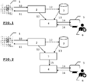
- an imaging device 1 by Nuclear Magnetic Resonance such as that illustrated by way of non-limiting example by the figures 1 and 2 .
- This can deliver a plurality of sequences of digital images 12 of one or more parts of the body, by way of non-limiting examples, the brain, the heart, the lungs, etc.
- Said device applies for this a combination of high frequency electromagnetic waves on the part of the body considered and measures the signal re-emitted by certain atoms, such as by way of non-limiting example, hydrogen for imaging by Nuclear Magnetic Resonance.
- the apparatus thus makes it possible to determine the chemical composition and therefore the nature of the biological tissues in each elementary volume, such as a voxel, of the imaged volume.
- the nuclear magnetic resonance imaging device 1 is controlled using a console 2.
- a user can thus choose parameters 11 to control the device 1. From information 10 produced by the device 1 , a plurality of sequences of digital images 12 of a part of a human or animal body are obtained.
- the nuclear magnetic resonance imaging apparatus 1 can optionally be included within an imaging analysis system.
- said apparatus 1 can be dissociated from the analysis system.
- the sequences of images 12 can optionally be stored within a server 3 and constitute a medical file 13 of a patient.
- a file 13 can include images of different types, such as perfusion or diffusion images.
- the image sequences 12 are analyzed by means of a dedicated processing unit 4.
- Said processing unit comprises means for communicating with the outside world to collect the images.
- Said means for communicating also allow the processing unit 4 to ultimately deliver, via restitution means 5 offering a graphic, sound or other rendering, to a practitioner 6 or to a researcher, a estimation of one or more quantities of interest, optionally formatted in the form of a content, such as hemodynamic parameters 14, from the digital images 12, advantageously perfusion images, by means of a man-machine interface adapted.
- the user 6, advantageously a practitioner, of the analysis system can thus confirm or invalidate a diagnosis, decide on a therapeutic action that he deems appropriate, deepen research work, etc.
- this user 6 can configure the operation of the processing unit 4 or of the restitution means 5, by means of parameters 16. For example, he can thus define display thresholds or choose the estimated parameters that he wishes to display.
- an imaging system further comprises a pre-processing unit 7 for analyzing the image sequences, for deducing therefrom experimental signals 15 (perfusion or permeability) and delivering them to the processing unit 4 which is thus relieved of this task.
- the processing unit 4 generally includes processing means, such as a computer, to implement an estimation method in the form of a program previously loaded into storage means cooperating with said processing means.
- Nuclear Magnetic Resonance perfusion images are obtained by injecting a contrast agent, for example a salt of gadolinium for Magnetic Resonance Imaging, intravenously and by recording its bolus over time at the level of each voxel of the image, by acquisitions at regular intervals.
- a contrast agent for example a salt of gadolinium for Magnetic Resonance Imaging
- Such images make it possible in particular to characterize the blood circulation in the tissue of a given organ, by following the evolution of the concentration of the contrast agent in each voxel of the volume acquired as a function of time.
- voxel Anglo-Saxon contraction of the term “volutric pixel”
- a voxel is understood to mean an elementary volume making it possible to measure the definition of a digital matrix image in three dimensions.
- Such a voxel can still be considered as a three-dimensional pixel. In all cases, such a voxel is a rectangular parallelepiped whose closed surface is constituted by its six faces
- a standard model makes it possible to relate the intensity of the signals S ( t ) measured over time t to the concentration C(t) of said contrast agent.
- C v ( t ) C a ( t ) ⁇ h ( t ) where h(t) is the impulse response of the system and ⁇ denotes the convolution product.
- Hemodynamic parameters such as BF, MTT or BV as well as the function of R ( t ) residues are currently estimated as follows, in the case of nuclear magnetic resonance perfusion imaging.
- the experimental perfusion signal S exp ( t ) sampled at the measurement instants t i , i 1 ,N, is converted into a concentration curve C ( t ). From the concentration curve C ( t ), and in assuming that the associated theoretical arterial input function C a ( t ) is known , the product BF ⁇ R ( t ) is estimated by numerical deconvolution.
- an experimental global arterial input function is chosen manually by the practitioner. It can be measured, for example, at the level of an artery close to the organ to be studied.
- local arterial input functions are obtained automatically from perfusion images, using signal processing techniques and selection criteria. For example, the “best” function is sought in the immediate vicinity of the current tissue voxel where it is desired to estimate the hemodynamic parameters or the complementary distribution functions.
- the purpose of this approach is to ultimately obtain estimates that are less biased and more precise by overcoming, at least in part, the problems of delay and dispersion.
- a first approach is a parametric deconvolution.
- each voxel is separated into two compartments, the intravascular space and the extravascular space, said compartments each respectively occupying a fraction of voxel v a and v e , also and respectively characterized by a contrast agent concentration C a ( t ) and C e ( t ) .
- the arterial input function must also be chosen.
- the choice of said function proves to be decisive for the estimation of the hemodynamic parameters: generally, the practitioner or the imaging system chooses the concentration curve of the contrast agent of a voxel located at the level of an artery as the function arterial inlet of all the voxels of the studied volume. Subsequently, the convolution product between the arterial input function and the residual function is numerically calculated by discretizing it on the perfusion data acquisition time grid.
- Different methods are applicable for estimate the hemodynamic parameters ⁇ hem from the contrast agent concentration curves within each voxel.
- the minimization is carried out numerically, advantageously using the execution of a nonlinear optimization algorithm.
- results can be presented in the form of parameter maps, then highlighting different types of tissue, namely healthy or pathological, according to the values of said parameters.
- a second approach is a more general approach based on nonparametric deconvolution: no model assumption is made for the residual function.
- the arterial input function must be chosen correctly with respect to the voxels studied, to allow the construction of the convolution matrix A.
- R you 1 R you 2 ... R you NOT vs
- VS you 1 VS you 2 ... VS you NOT BF . b
- such a method for estimating a quantity of interest consists in minimizing a criterion of the type
- 2 + ⁇ d ⁇ 2 where ⁇ d ⁇ 2 is a regularization term favoring certain solutions and obtaining an estimate of d by ⁇ arg min ⁇ ad ⁇ vs ⁇ 2 + ⁇ ⁇ ⁇ 2 .
- the experimental arterial input functions can be adjusted beforehand to a parametric or semi-parametric theoretical model C a ( t, ⁇ a ) where ⁇ a is a vector of parameters, in order to artificially increase the signal-to-noise ratio.
- a “naive” way of solving this problem would be to carry out as many perfusion examinations, in particular acquisitions, as necessary to completely cover the volume that one wishes to analyze.
- the total acquisition time in particular that spent by the patient in imaging devices, would be multiplied by the number of sub-volumes necessary to analyze the entire volume of interest, considerably increasing the total duration of the examination: such examinations would become very heavy and would be more difficult for the patient to bear.
- CT scans can consist of measuring the absorption of X-rays by the tissues of the different voxels.
- contrast agent for each sub-volume acquisition, an injection of contrast agent would be required.
- a contrast agent makes it possible to improve the quality of diagnostics by medical imaging by Magnetic Resonance, the toxicity at high concentration of such a contrast agent can prove to be problematic for a patient. Since the amount of contrast agent a patient can tolerate is limited, such an approach is unsuitable, and even potentially dangerous.
- the acquisition can be done, for example, according to the following protocol: a patient is in principle lying on a mobile table, usually motorized, moving in the field of vision of the Magnetic Resonance imaging device. After a first volume, also known by the term "Station", corresponding to an acquisition field of the imaging device, has been scanned by the Magnetic Resonance imaging device, the patient is moved within said imaging by means of said mobile table, so that a second volume or station contained in the acquisition field, possibly adjacent to the first volume or station previously analyzed, is acquired.
- a protocol is illustrated in connection with the picture 3 .
- a period of sixty seconds is required to sample all five volumes and return to the first volume.
- Such a period breaks down into an image acquisition sub-period of about thirty-nine seconds and a sub-period of about twenty-one seconds of movement of the mobile table.
- Each voxel of interest in the global volume is sampled every sixty seconds.
- the resulting concentration curves thus have a time resolution of sixty seconds.
- the perfusion data acquired and associated with each volume or station are subsequently processed in order to estimate a quantity of interest, such as, by way of nonlimiting examples, hemodynamic parameters.
- a “natural” approach consists in considering each of the stations independently of each other, by applying to them classic methods as described previously, namely parametric or non-parametric approaches.
- a model such as by way of non-limiting example the Ketty model, and an arterial input function can be chosen for each station.
- the hemodynamic parameters such as for example the parameters K Trans , k ep and v e , are thus estimated and the maps of the associated parameters can also be produced.
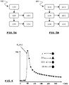
- the arterial input function specific to each station is therefore not sampled at the same instants, and each station seems to have a different arterial input function. Consequently, the arterial input function of each station is modeled according to a different model, valid only at the instants of acquisition of the station to which it is attached, as described in conjunction with the figure 4 .
- the figure 4 illustrates examples of arterial input functions produced and/or chosen for each station I to IV, said functions being represented in the form of a curve of concentration of a contrast agent as a function of time at an artery chosen.
- an independent arterial input function is determined automatically and/or manually. Indeed, the concentration curve of an arterial input function is characterized by a rapid rise followed by an equally rapid decrease, with a characteristic time of only a few tens of seconds.
- the invention makes it possible to respond to all or part of the drawbacks raised by the known solutions.
- a method for producing an estimate of a quantity of interest of a dynamic artery/tissue/vein system of an elementary volume - called voxel - of a patient's organ.
- Such a method is intended to be implemented by processing means of a processing unit of a magnetic resonance perfusion 2.
- imaging analysis system and comprises a step for estimating said quantity of interest for a station among a plurality of stations from infusion data related to said station.
- such a method comprises a step for constructing a common arterial input function from perfusion data linked to at least one station among said plurality of stations, each station being a volume corresponding to an acquisition field defined by a magnetic resonance medical imaging device
- said step for estimating the quantity of interest for a station of said plurality exploits, for any station of said plurality of stations, said constructed common arterial input function and the perfusion data related to said station to estimate the quantity of interest, said station comprising the elementary volume of the patient's organ
- step 120 to construct a common arterial input function can be implemented from perfusion data related to at least two of said plurality of stations.
- the invention provides that the step for constructing a common arterial input function can include a step for determining an arterial input function specific to each station among said at least one station, the function d the common arterial input being constructed from said proper arterial input function.
- the magnetic resonance perfusion imaging analysis system comprises means for restitution to a user of said system, said means for restitution cooperating with the processing unit, said method may comprise a subsequent step for triggering a restitution of the common arterial input function by said restitution means.
- the invention provides that the method may include a prior step of pre-processing the perfusion data, said step being arranged to correct said perfusion data.
- the invention relates to a method, according to claim 6, for producing an estimate of a quantity of interest of a dynamic artery/tissue/vein system of a region of interest, said region comprising at least one voxel.
- said quantity of interest is estimated by voxel by means of a method in accordance with the first object of the invention.
- said region of interest can extend over several stations among said plurality of stations .
- said method may comprise a step to trigger a restitution of said quantity of interest estimated for the voxels of the region of interest by said restitution means.
- the invention relates to a processing unit, according to independent claim 9, comprising means for communicating with the outside world and processing means cooperating with storage means.
- the means for communicating are adapted to receive from the outside world perfusion data linked with a plurality of stations, each station being a volume corresponding to an acquisition field defined by a medical imaging device, and the storage means comprise instructions executable or interpretable by the processing means of which the interpretation or execution of said instructions by said processing means causes the implementation of a method in accordance with the invention.
- the means for communicating of the processing unit according to the invention can deliver an estimated quantity of interest according to an appropriate format to restitution means capable of return it to a user.
- the invention relates to a perfusion imaging analysis system, according to claim 11, comprising a processing unit according to the invention and restitution means suitable for restoring to a user a quantity of interest estimated according to a method in accordance with the invention and implemented by said processing unit.
- the invention relates to a computer program product, according to claim 12, comprising one or more instructions that can be interpreted or executed by the processing means of a processing unit in accordance with the invention.
- Said processing unit further comprises storage means or cooperating with such storage means, said program being loadable into said storage means.
- the interpretation or execution of said instructions by said processing means causes the implementation of a method for estimating a quantity of interest in accordance with the invention.
- the figure 5a describes an example of a simplified algorithm of a method 100 - in accordance with the invention - for producing an estimate of a quantity of interest of a dynamic artery/tissue/vein system of an elementary volume which is called voxel - of an organ.
- a method 100 can be implemented by a processing unit of a perfusion imaging analysis system, such as the system described in connection with the figures 1 or 2 and adapted accordingly.
- the method 100 in accordance with the invention mainly comprises a step 130 for estimating a quantity of interest, by way of non-limiting examples a hemodynamic parameter or the function of the residues, from perfusion data linked to a plurality of stations .
- the term “station” means a volume corresponding to the acquisition field of a medical imaging device, such as the apparatus 1 of the perfusion imaging analysis system described in connection with the figures 1 or 2 .
- each station is linked or associated with a volume corresponding to an acquisition field defined by a medical imaging device.
- Said acquisition field is associated with a particular position of the mobile table of the medical imaging device, on which the patient is in principle lying down.
- the term "perfusion data” means all of the images or signals acquired at several times by a magnetic resonance medical imaging device for the purpose of studying the evolution of a contrast previously injected into a patient's body.
- Such data can advantageously consist of sequences of images, such as the sequences of images 12 described in connection with the figures 1 or 2 .
- the perfusion data can advantageously be stored in a server during their acquisition, to be processed later by a user of an imaging analysis system, advantageously a practitioner.
- the infusion data is advantageously linked or associated with a particular station.
- such a method 100 comprises a step 120 to construct a common arterial input function from infusion data in connection with at least one station among said plurality of stations.
- the construction of such an arterial input function according to step 120 cannot be limited to the perfusion data linked to a single station: in fact, in order, for example, to adjust an arterial input function model as close as possible to reality and to refine the construction of said common arterial input function, the data can be linked or associated with two or more stations, i.e. as many as stations available.
- the perfusion data can come from the five stations I to V to build a common arterial input function.
- the figure 6 describes an example of an arterial input function constructed from four stations I to IV among the five acquired, said stations being defined by the picture 3 and acquired according to the experimental acquisition protocol of professors Rahmouni and Luciani.
- the common arterial input function constructed is advantageously unique for all the stations: such an arterial input function can be described by an analytical function given in the form C a ( t, ⁇ a ) where ⁇ a is the set of parameters of the arterial input function.
- Such parameters ⁇ a are estimated at 120 from the perfusion data of the various stations and are the same for all the voxels of all the stations.
- Such an estimate of the parameters ⁇ a can be carried out, by way of non-limiting example, by adjusting the defined model C a ( t, ⁇ a ) on concentration curves of the contrast agent from arteries chosen in all the stations. the said Routes can be chosen manually or automatically depending on the chosen protocol.
- the parameters ⁇ a can also be adjusted together with hemodynamic parameters ⁇ hem of the voxels.
- the invention provides that the step 120 for constructing a common arterial input function can comprise a step for determining, manually or automatically, an arterial input function specific to or dedicated to a station among said at least one station, the common arterial input function being constructed from said proper arterial input function.
- the perfusion data can be linked or associated with two or more stations , i.e. up to as many stations as available. Such a preferred embodiment is described in connection with the figure 6 .
- the step 120 for constructing a common arterial input function C a ( t ) consists in adjusting said function to the measured values of concentrations of contrast agent at the sampling instants within each station, among the stations I to IV.
- said common arterial input function can be described by an analytical function given in the form C a ( t, ⁇ a ) where ⁇ a is the set of parameters of the arterial input function.
- a ⁇ A 1 ,A 2 ,k 1 ,k 2 , ⁇ ⁇ .
- step 120 for constructing a common arterial input function consists in choosing, from a database of existing arterial input functions, said functions being measured beforehand with a resolution sufficient time in a reference patient population, a common arterial input function.
- the choice of such an oversampled arterial input function could be made using the perfusion data of each station.
- the perfusion imaging analysis system described in connection with the figures 1 or 2 comprises restitution means 5, such as by way of non-limiting example a man-machine interface, such as a screen or any other equivalent means, to a user 6 of said system
- said restitution means 5 cooperating with the processing unit 4 said method may comprise a subsequent step 121 for triggering a restitution of the common arterial input function thus constructed by said restitution means.
- a step 121 can moreover trigger a restitution of the voxels of the perfusion data used to construct the common arterial input function.
- Such a step 121 makes it possible in particular, when the voxels have been selected automatically to construct the common arterial input function, to validate the relevance of said selected voxels, namely that the selected voxels actually correspond to voxels of interest at the level of the arteries .
- step 130 for estimating the quantity of interest exploits the constructed common arterial input function: such an arterial input function is valid for all stations at all times.
- Step 130 for estimating the quantity of interest can consist of the implementation by the processing unit of any known techniques for estimating hemodynamic parameters, advantageously parametric or non-parametric methods, as described above.
- the residual function can be described by a given analytical function in the form R ( t , ⁇ hem ) where ⁇ hem is the set of hemodynamic parameters.
- a method for optimizing the parameters of the function could advantageously be executed, such as least squares minimization, with or without constraint, or even a Bayesian estimation method.
- a convolution matrix can be constructed from the common constructed arterial input function using a finer time grid than that of the acquisition protocol, so as to sample correctly the different time scales present in said arterial input function.
- the SVD method Single Value Decomposition according to an Anglo-Saxon terminology
- the Bayesian deconvolution method will be preferred given the particularly proven accuracy of the estimates produced.
- the perfusion imaging analysis system described in connection with the figures 1 or 2 comprises restitution means 5, such as by way of non-limiting example a man-machine interface or any other equivalent means, to a user 6 of said system, said restitution means cooperating with the processing unit 4, said The method can include a subsequent step 131 to trigger a restitution of the estimated quantity of interest, such as for example the hemodynamic parameters 14, according to an appropriate format.
- a format can be a value, a color within a palette determined to express the intensity of said estimated quantity of interest, or any other equivalent means.
- the invention provides that the method can include a prior step 110 of preprocessing the perfusion data, said step consisting in particular of correcting artefacts or of applying any other corrective filter.
- Magnetic Resonance Imaging like all other medical imaging techniques, does not escape the formation of false images: artifacts.
- Artifacts are observable images that do not represent any anatomical reality. Very often, it is sought to avoid or minimize them by modifying certain acquisition or reconstruction parameters. Such artefacts can be of different natures.
- three corrections are generally applied to improve the quality of the perfusion data: a correction of the movements of the patient or of the movements due to breathing, heartbeats and blood flows, a correction of the field of view of the perfusion imaging device and/or denoising of the images.
- the figure 5b describes an example of a simplified algorithm of a method 200 - in accordance with the invention - for producing an estimate of a quantity of interest of a dynamic artery/tissue/vein system of a region of interest.
- a method 200 is arranged to produce an estimate of a quantity of interest, by way of non-limiting examples a hemodynamic parameter or residual function, of an artery/tissue/vein dynamic system of a region of interest .
- “Region of interest” means any region comprising at least one voxel. However, a region of interest cannot be limited to a single voxel, but can comprise a plurality of voxels, selected manually or automatically.
- said quantity of interest can be estimated for each voxel by means of a method 100 in accordance with the invention as described previously implemented iteratively for each voxel by the processing means of the processing unit 4.
- step 210 it is thus possible, according to step 210, to estimate a quantity of interest over a plurality of voxels defining a region of interest, possibly extending over several stations among a plurality of stations, for example , carry out the analysis of organs whose size exceeds the field of acquisition of the analysis apparatus by perfusion imaging.
- said method may include a subsequent step 211 for triggering restitution of said quantity of interest, namely a hemodynamic parameter 14, estimated for the voxels of the region of interest by said restitution means according to an appropriate format.
- the restitution can advantageously be similar to a map of parameters where each voxel corresponds to a degree of intensity linked to the estimated quantity of interest.
- Such a step may include a sub-step for viewing a parameter map for each station.
- Such an embodiment is described in connection with the figure 7 .
- the figure 7 presents five maps relating to blood flow, also known by the terminology K trans , estimated from a plurality of stations, advantageously five, in accordance with the invention, the five maps corresponding to stations I to V.
- Such maps for the stations I to V can be advantageously exploited within the framework of the study of the myeloma, cancer affecting the cells of the spinal cord.
- Each station thus comprises a representation independent of one another.
- the method 200 can comprise a step 220 for generating an overall volume from a plurality of stations. It may also include a step for triggering the restitution, for example in the form of a consolidated map integrating or merging the maps produced for said plurality of stations. Such a step 220 can thus comprise a sub-step for joining different maps of stations included in the plurality of stations. Such an embodiment is described in connection with the figure 8 .
- the figure 8 presents a consolidated map of a plurality of stations from maps of parameters relating to said estimated blood flow, said maps being represented by way of non-limiting examples in connection with the figure 7 .
- Two adjacent maps among the different stations I to V generally comprise an overlap zone, said overlap zone advantageously comprising corresponding peripheral voxels, corresponding in pairs and forming pairs of corresponding voxels.
- the quantity of interest in connection with such pairs of voxels may be estimated by the processing unit 4 as the result of a linear combination between the estimates of said quantity of interest of the corresponding voxels within the two adjacent stations.
- any discontinuity within the consolidated map, resulting from a merger of the parameter maps specific to each station, among stations I to V, is prevented or attenuated: observation of the presence of such a discontinuity can be verified using the restitution means 5 of a medical imaging device, such as that described in connection with the figures 1 or 2 .
- the maps of stations I and II, as well as II and IV are merged to generate a consolidated map.
- the method 200 can include a step 230 to verify the estimate of the estimated quantity of interest: such a verification can be carried out automatically or visually.
- Said verification step 230 may consist in detecting any notable discontinuity on a consolidated map.
- such a verification step 230 may consist in automatically verifying that the values of the estimates of said quantity of interest of the corresponding voxels are consistent, that is to say that the values of such estimates are placed in a narrow range of values.
- Such verification can be carried out using statistical tests, such as, by way of nonlimiting examples, the Kolmogorov-Smirnov test or, preferably, Bayesian theory.
- the method 200 can then include a step 231 for triggering highlighting, via the restitution means, of the voxels judged at 230 to be not very coherent.
- the invention makes it possible to make available to a practitioner a whole set of relevant and coherent information, information which could not be available using the techniques known from the state of the art.
- This provision is made possible by an adaptation of the processing unit 4 according to the figures 1 or 2 in that the processing means implement a method for estimating a quantity of interest comprising the construction of an arterial input function from perfusion data in connection with one or more stations.
- Such an implementation is advantageously made possible by loading or recording within storage means cooperating with said processing means, of a computer program product.
- the latter in fact comprises instructions that can be interpreted and/or executed by said processing means. The interpretation or execution of said instructions triggers the implementation of a method 100 or 200 in accordance with the invention.
- the means for communicating with the outside world of said processing unit can deliver a quantity of interest, namely the estimated parameters 14, according to an appropriate format to restitution means capable of restoring it to a user 6, said quantity of estimated interest which can advantageously be returned in the form, for example, of cards as illustrated by the figure 7 and 8 .
- the information delivered is thus more numerous, consistent, reproducible and accurate.
- the information available to the practitioner is thus of a nature to increase the practitioner's confidence in his determination of a diagnosis and his decision-making.
Landscapes
- Health & Medical Sciences (AREA)
- Life Sciences & Earth Sciences (AREA)
- Physics & Mathematics (AREA)
- Engineering & Computer Science (AREA)
- Nuclear Medicine, Radiotherapy & Molecular Imaging (AREA)
- Medical Informatics (AREA)
- General Health & Medical Sciences (AREA)
- Radiology & Medical Imaging (AREA)
- High Energy & Nuclear Physics (AREA)
- Surgery (AREA)
- Heart & Thoracic Surgery (AREA)
- Biomedical Technology (AREA)
- Molecular Biology (AREA)
- Pathology (AREA)
- Animal Behavior & Ethology (AREA)
- Public Health (AREA)
- Veterinary Medicine (AREA)
- Biophysics (AREA)
- Optics & Photonics (AREA)
- General Physics & Mathematics (AREA)
- Signal Processing (AREA)
- Condensed Matter Physics & Semiconductors (AREA)
- Physiology (AREA)
- Vascular Medicine (AREA)
- Cardiology (AREA)
- Hematology (AREA)
- Computer Vision & Pattern Recognition (AREA)
- Theoretical Computer Science (AREA)
- Dentistry (AREA)
- Oral & Maxillofacial Surgery (AREA)
- Pulmonology (AREA)
- Geometry (AREA)
- Magnetic Resonance Imaging Apparatus (AREA)
- Apparatus For Radiation Diagnosis (AREA)
- Measuring Pulse, Heart Rate, Blood Pressure Or Blood Flow (AREA)
- Measurement Of The Respiration, Hearing Ability, Form, And Blood Characteristics Of Living Organisms (AREA)
Claims (12)
- Verfahren (100) zum Erzeugen einer Schätzung einer Menge von Interesse (14) eines dynamischen Arterien/Gewebe/Venen-Systems eines Volumenelements - Voxel genannt - eines Organs eines Patienten, wobei das genannte Verfahren (100) von Verarbeitungsmitteln einer Verarbeitungseinheit (4) eines Magnetresonanz-Perfusionsbildgebungs-Analysesystems durchgeführt wird und einen Schritt (130) zum Schätzen der genannten Menge von Interesse (14) auf der Basis von Perfusionsdaten jeweils in Verbindung mit einer Station aus Perfusionsdaten jeweils in Verbindung mit einer Mehrzahl von Stationen, auf der Basis von Perfusionsdaten (12) in Verbindung mit der genannten Station beinhaltet, wobei jede Station ein Volumen ist, das einem Erfassungsfeld entspricht, definiert durch ein medizinisches Magnetresonanz-Bildgebungsgerät, das einen beweglichen Tisch umfasst, auf dem der Patient zum Erfassen der genannten Perfusionsdaten von Station zu Station bewegt wird, wobei das genannte Verfahren:
einen Schritt (120) zum Konstruieren einer gemeinsamen arteriellen Eingangsfunktion auf der Basis von Perfusionsdaten in Verbindung mit mindestens einer Station aus der genannten Mehrzahl von Stationen beinhaltet, und der genannte Schritt (130) zum Schätzen der Menge von Interesse auf der Basis der Perfusionsdaten in Verbindung mit einer Station aus der genannten Mehrzahl, für jede Station der genannten Mehrzahl von Stationen die konstruierte gemeinsame arterielle Eingangsfunktion und die Perfusionsdaten in Verbindung mit der genannten Station zum Schätzen der Menge von Interesse nutzt, wobei die genannte Station das Volumenelement des Organs des Patienten umfasst. - Verfahren (100) nach dem vorherigen Anspruch, bei dem der Schritt (120) zum Konstruieren einer gemeinsamen arteriellen Eingangsfunktion auf der Basis von Perfusionsdaten in Verbindung mit mindestens zwei Stationen aus der genannten Mehrzahl von Stationen durchgeführt wird.
- Verfahren (100) nach einem der vorherigen Ansprüche, bei dem der Schritt (120) zum Konstruieren einer gemeinsamen arteriellen Eingangsfunktion einen Schritt zum Bestimmen einer eigenen arteriellen Eingangsfunktion für eine Station aus der genannten mindestens einen Station beinhaltet, wobei die gemeinsame arterielle Eingangsfunktion auf der Basis der genannten eigenen arteriellen Eingangsfunktion konstruiert wird.
- Verfahren (100) nach einem der vorherigen Ansprüche, wobei das Magnetresonanz-Perfusionsbildgebungs-Analysesystem Mittel (5) zur Wiedergabe an einen Benutzer (6) des genannten Systems umfasst, wobei die genannten Wiedergabemittel (5) mit der Verarbeitungseinheit (4) zusammenarbeiten, wobei das genannte Verfahren einen nachfolgenden Schritt (121) zum Auslösen einer Wiedergabe der gemeinsamen arteriellen Eingangsfunktion durch die genannten Wiedergabemittel beinhaltet.
- Verfahren (100) nach einem der vorherigen Ansprüche, das einen Vorabschritt (110) des Vorverarbeitens der Perfusionsdaten (12) beinhaltet, wobei der genannte Schritt zum Korrigieren der genannten Perfusionsdaten (12) eingerichtet ist.
- Verfahren (200) zum Erzeugen einer Schätzung einer Menge von Interesse eines dynamischen Arterien/Gewebe/Venen-Systems einer Region von Interesse, wobei die genannte Region mindestens ein Voxel umfasst, wobei die genannte Menge von Interesse pro Voxel gemäß einem Verfahren (100) gemäß einem der Ansprüche 1 bis 5 geschätzt wird.
- Verfahren (200) nach dem vorherigen Anspruch, bei dem sich die genannte Region von Interesse über mehrere Stationen aus der Mehrzahl von Stationen erstreckt.
- Verfahren (200) nach Anspruch 6 oder 7, wobei das Magnetresonanz-Perfusionsbildgebungs-Analysesystem Mittel (5) zur Wiedergabe an einen Benutzer (6) des genannten Systems umfasst, wobei die genannten Wiedergabemittel (5) mit der Verarbeitungseinheit (4) zusammenarbeiten, wobei das genannte Verfahren einen nachfolgenden Schritt (211) zum Auslösen einer Wiedergabe der genannten geschätzten Menge von Interesse für die Voxel der Region von Interesse durch die genannten Wiedergabemittel (5) beinhaltet.
- Verarbeitungseinheit (4), die Mittel zur Kommunikation mit der Außenwelt und Verarbeitungsmittel umfasst, die mit Speichermitteln zusammenarbeiten, so dass:- die Mittel zur Kommunikation zum Empfangen, von der Außenwelt, von Perfusionsdaten (12) in Verbindung mit einer Mehrzahl von Stationen (I,II,III,IV,V) ausgelegt sind, wobei jede Station ein Volumen ist, das einem Erfassungsfeld entspricht, definiert durch ein medizinisches Magnetresonanz-Bildgebungsgerät, das einen beweglichen Tisch steuert, auf dem ein Patient zum Erfassen der genannten Perfusionsdaten von Station zu Station bewegt wird;- die Speichermittel Befehle enthalten, die von den Verarbeitungsmitteln ausgeführt oder interpretiert werden können, wobei die Interpretation oder Ausführung der genannten Befehle durch die genannten Verarbeitungsmittel die Durchführung eines Verfahrens (200) nach einem der Ansprüche 6 bis 8 bewirkt.
- Verarbeitungseinheit (4) nach dem vorherigen Anspruch, bei der die Mittel zur Kommunikation zum Liefern einer geschätzten Menge von Interesse (14) in einem geeigneten Format an Wiedergabemittel (5) ausgelegt sind, die zu ihrer Wiedergabe an einen Benutzer (6) geeignet sind.
- Magnetresonanz-Perfusionsbildgebungs-Analysesystem, das eine Verarbeitungseinheit (4) nach einem der Ansprüche 9 und 10 und Wiedergabemittel (5) umfasst, die zum Wiedergeben einer geschätzten Menge (14) gemäß einem Verfahren an einen Benutzer (6) ausgelegt sind, das einem der Ansprüche 6 bis 8 entspricht und von der genannten Verarbeitungseinheit (4) durchgeführt wird.
- Computerprogrammprodukt mit einem oder mehreren Befehlen, die von den Verarbeitungsmitteln einer Verarbeitungseinheit (4) gemäß einem der Ansprüche 9 und 10 interpretiert oder ausgeführt werden können, wobei die genannte Verarbeitungseinheit (4) außerdem Speichermittel umfasst oder mit solchen Speichermitteln zusammenarbeitet, wobei das genannte Programm in die genannten Speichermittel geladen werden kann, dadurch gekennzeichnet, dass die Interpretation oder Ausführung der genannten Befehle durch die genannten Verarbeitungsmittel die Durchführung eines Verfahrens (200) nach einem der Ansprüche 6 bis 8 bewirkt.
Applications Claiming Priority (2)
| Application Number | Priority Date | Filing Date | Title |
|---|---|---|---|
| FR1459822A FR3027115B1 (fr) | 2014-10-13 | 2014-10-13 | Systeme et procede pour estimer une quantite d'interet d'un systeme dynamique artere/tissu/veine |
| PCT/FR2015/052728 WO2016059329A1 (fr) | 2014-10-13 | 2015-10-09 | Système et procédé pour estimer une quantité d'intérêt d'un système dynamique artère/tissu/veine |
Publications (2)
| Publication Number | Publication Date |
|---|---|
| EP3207397A1 EP3207397A1 (de) | 2017-08-23 |
| EP3207397B1 true EP3207397B1 (de) | 2022-06-22 |
Family
ID=52684323
Family Applications (1)
| Application Number | Title | Priority Date | Filing Date |
|---|---|---|---|
| EP15787262.3A Active EP3207397B1 (de) | 2014-10-13 | 2015-10-09 | System und verfahren zur schätzung einer relevanten menge eines dynamischen arterien-/gewebe-/venensystems |
Country Status (6)
| Country | Link |
|---|---|
| US (1) | US9983287B2 (de) |
| EP (1) | EP3207397B1 (de) |
| JP (1) | JP6662866B2 (de) |
| ES (1) | ES2924754T3 (de) |
| FR (1) | FR3027115B1 (de) |
| WO (1) | WO2016059329A1 (de) |
Families Citing this family (4)
| Publication number | Priority date | Publication date | Assignee | Title |
|---|---|---|---|---|
| KR101840106B1 (ko) * | 2016-02-04 | 2018-04-26 | 가톨릭대학교 산학협력단 | 의료영상을 이용한 혈류 분석 방법 |
| US10213178B2 (en) * | 2016-09-22 | 2019-02-26 | Algotec Systems Ltd. | Calculation of perfusion parameters in medical imaging |
| CN113557442B (zh) * | 2019-02-06 | 2024-09-03 | 皇家飞利浦有限公司 | 图像重建设备与方法 |
| CN116342605B (zh) * | 2023-05-30 | 2023-08-11 | 杭州脉流科技有限公司 | Ct灌注影像参数估计方法、装置、设备和存储介质 |
Family Cites Families (19)
| Publication number | Priority date | Publication date | Assignee | Title |
|---|---|---|---|---|
| US5289823A (en) * | 1992-05-12 | 1994-03-01 | Colin Electronics Co., Ltd. | Non-invasive aortic blood flow sensor and method for non-invasively measuring aortic blood flow |
| US6542769B2 (en) * | 2000-12-18 | 2003-04-01 | The General Hospital Corporation | Imaging system for obtaining quantative perfusion indices |
| US6580937B2 (en) * | 2000-12-30 | 2003-06-17 | Ge Medical Systems Global Technology Co., Llc | Method for optimal imaging of the peripheral vasculature emphasizing distal arterial visualization in a multi-station examination |
| WO2003101294A1 (fr) * | 2002-05-31 | 2003-12-11 | Hitachi Medical Corporation | Dispositif d'imagerie par resonnance magnetique et procede ce-mra multistation |
| US6628743B1 (en) * | 2002-11-26 | 2003-09-30 | Ge Medical Systems Global Technology Company, Llc | Method and apparatus for acquiring and analyzing cardiac data from a patient |
| DK1747477T3 (da) * | 2004-05-04 | 2013-06-17 | Stiftelsen Universitetsforskning Bergen | Blindbestemmelse af arteriel indgangs- og vævsrestfunktioner i perfusions-mri |
| US8125484B2 (en) * | 2006-11-10 | 2012-02-28 | General Electric Company | Method, apparatus and user interface for determining an arterial input function used for calculating hemodynamic parameters |
| US8615116B2 (en) * | 2007-09-28 | 2013-12-24 | The Johns Hopkins University | Combined multi-detector CT angiography and CT myocardial perfusion imaging for the diagnosis of coronary artery disease |
| US20110098556A1 (en) * | 2008-03-11 | 2011-04-28 | Lennart Blomqvist | Computer-Based Method And System For Imaging-Based Dynamic Function Evaluation Of An Organ |
| EP2271257B1 (de) * | 2008-04-18 | 2014-03-19 | BiOxyDyn Limited | Bildgebungsverfahren |
| US20110150309A1 (en) * | 2009-11-27 | 2011-06-23 | University Health Network | Method and system for managing imaging data, and associated devices and compounds |
| JP5750452B2 (ja) * | 2010-01-04 | 2015-07-22 | メイヨ フォンデーシヨン フォー メディカル エジュケーション アンド リサーチ | 時間分解磁気共鳴血管造影と潅流画像のシステムと方法 |
| JP5897284B2 (ja) * | 2010-09-01 | 2016-03-30 | 株式会社東芝 | 医用画像処理装置 |
| FR2979453B1 (fr) * | 2011-08-26 | 2016-01-08 | Olea Medical | Systeme et procede pour estimer une quantite d'interet d'un systeme dynamique artere/tissu/veine |
| US8837800B1 (en) * | 2011-10-28 | 2014-09-16 | The Board Of Trustees Of The Leland Stanford Junior University | Automated detection of arterial input function and/or venous output function voxels in medical imaging |
| US9370304B2 (en) * | 2012-06-06 | 2016-06-21 | The Regents Of The University Of Michigan | Subvolume identification for prediction of treatment outcome |
| WO2014164963A1 (en) * | 2013-03-12 | 2014-10-09 | Levin Pavel | Method and apparatus for magnetic resonance imaging |
| DE102013216236A1 (de) * | 2013-08-15 | 2015-03-12 | Siemens Aktiengesellschaft | Verfahren zu einem Auswerten von ersten Bilddaten einer ersten Bildgebungsuntersuchung und von zweiten Bilddaten einer zweiten Bildgebungsuntersuchung sowie ein medizinisches Bildgebungssystem, das zur Ausführung des Verfahrens ausgelegt ist |
| DE102014201499A1 (de) * | 2014-01-28 | 2015-07-30 | Siemens Aktiengesellschaft | Verfahren und medizinische Bildgebungseinrichtung zur Bestimmung der Perfusion |
-
2014
- 2014-10-13 FR FR1459822A patent/FR3027115B1/fr active Active
-
2015
- 2015-10-09 JP JP2017519680A patent/JP6662866B2/ja active Active
- 2015-10-09 EP EP15787262.3A patent/EP3207397B1/de active Active
- 2015-10-09 WO PCT/FR2015/052728 patent/WO2016059329A1/fr not_active Ceased
- 2015-10-09 ES ES15787262T patent/ES2924754T3/es active Active
-
2016
- 2016-05-04 US US15/146,073 patent/US9983287B2/en active Active
Non-Patent Citations (2)
| Title |
|---|
| LIN S ET AL: "IRM CORPS ENTIER EN HAUTE RESOLUTION AVEC INJECTION DYNAMIQUE DE GADOLINIUM DANS LES SYNDROMES LYMPHOPROLIFERATIFS", JOURNAL DE RADIOLOGIE, vol. 88, no. 10, 1 October 2007 (2007-10-01), pages 1445, XP055890583 * |
| LUCIANI A ET AL: "HEMATOLOGIE: APPLICATIONS IRM CORPS ENTIER", JOURNAL DE RADIOLOGIE, vol. 90, no. 10, 1 October 2009 (2009-10-01), pages 1338, XP055890581 * |
Also Published As
| Publication number | Publication date |
|---|---|
| FR3027115A1 (fr) | 2016-04-15 |
| WO2016059329A1 (fr) | 2016-04-21 |
| FR3027115B1 (fr) | 2019-05-10 |
| EP3207397A1 (de) | 2017-08-23 |
| ES2924754T3 (es) | 2022-10-10 |
| US20160245889A1 (en) | 2016-08-25 |
| US9983287B2 (en) | 2018-05-29 |
| JP6662866B2 (ja) | 2020-03-11 |
| JP2017530816A (ja) | 2017-10-19 |
Similar Documents
| Publication | Publication Date | Title |
|---|---|---|
| EP2437664B1 (de) | Verfahren zur abschätzung hämodynamischer parameter mittels gemeinsamer abschätzung der parameter eines umfassenden perfusionsmodells | |
| CA2814377C (fr) | Systeme et procede pour estimer une quantite d'interet d'un systeme dynamique artere/tissu/veine | |
| EP2924586A1 (de) | System und verfahren zum schätzen einer bestimmten menge eines dynamischen arterien-/gewebe-/venensystems | |
| EP3207397B1 (de) | System und verfahren zur schätzung einer relevanten menge eines dynamischen arterien-/gewebe-/venensystems | |
| US20250072779A1 (en) | System and method for reconstructing a physiological signal of an artery/tissue/vein dynamic system of an organ in a surface space | |
| EP3274734A1 (de) | Mrt-system und verfahren zur schätzung eines physiologischen parameters auf der basis von zwei anderen geschätzten physiologischen parametern | |
| US7333647B2 (en) | Systems and method for generating an image | |
| EP2365455A1 (de) | Verfahren zur simultanen Extraktion der Eingabefunction und der pharmakokinetischen Parametern eines Wirkstoffs | |
| EP2345007B1 (de) | Verfahren zur schätzung der konzentration eines tracers in einer gewebestruktur und entsprechendes speichermedium und einrichtung | |
| WO2011064512A1 (fr) | Procédé pour estimer l'adéquation d'un processus d'estimation de paramètres hémodynamiques de perfusion | |
| EP4154215B1 (de) | System und verfahren zur schätzung eines indikators der gewebeaktivität eines organs | |
| Meurée | Arterial spin labelling: quality control and super-resolution | |
| FR3157630A1 (fr) | Procédé de défloutage d’une image résultant d’une séquence d’acquisition par un appareil d’imagerie médicale | |
| FR3152320A1 (fr) | Procédé de post-traitement d’une séquence d’acquisitions pour corriger les inhomogénéités de champs magnétiques d’un appareil d’imagerie par résonance magnétique | |
| FR3137827A1 (fr) | Procédé de post-traitement d’une séquence d’acquisition de perfusion par un appareil d’imagerie médicale |
Legal Events
| Date | Code | Title | Description |
|---|---|---|---|
| STAA | Information on the status of an ep patent application or granted ep patent |
Free format text: STATUS: THE INTERNATIONAL PUBLICATION HAS BEEN MADE |
|
| PUAI | Public reference made under article 153(3) epc to a published international application that has entered the european phase |
Free format text: ORIGINAL CODE: 0009012 |
|
| STAA | Information on the status of an ep patent application or granted ep patent |
Free format text: STATUS: REQUEST FOR EXAMINATION WAS MADE |
|
| 17P | Request for examination filed |
Effective date: 20170411 |
|
| AK | Designated contracting states |
Kind code of ref document: A1 Designated state(s): AL AT BE BG CH CY CZ DE DK EE ES FI FR GB GR HR HU IE IS IT LI LT LU LV MC MK MT NL NO PL PT RO RS SE SI SK SM TR |
|
| AX | Request for extension of the european patent |
Extension state: BA ME |
|
| DAV | Request for validation of the european patent (deleted) | ||
| DAX | Request for extension of the european patent (deleted) | ||
| RAP3 | Party data changed (applicant data changed or rights of an application transferred) |
Owner name: OLEA MEDICAL |
|
| GRAP | Despatch of communication of intention to grant a patent |
Free format text: ORIGINAL CODE: EPIDOSNIGR1 |
|
| STAA | Information on the status of an ep patent application or granted ep patent |
Free format text: STATUS: GRANT OF PATENT IS INTENDED |
|
| INTG | Intention to grant announced |
Effective date: 20210316 |
|
| GRAJ | Information related to disapproval of communication of intention to grant by the applicant or resumption of examination proceedings by the epo deleted |
Free format text: ORIGINAL CODE: EPIDOSDIGR1 |
|
| STAA | Information on the status of an ep patent application or granted ep patent |
Free format text: STATUS: REQUEST FOR EXAMINATION WAS MADE |
|
| STAA | Information on the status of an ep patent application or granted ep patent |
Free format text: STATUS: EXAMINATION IS IN PROGRESS |
|
| INTC | Intention to grant announced (deleted) | ||
| 17Q | First examination report despatched |
Effective date: 20210714 |
|
| GRAP | Despatch of communication of intention to grant a patent |
Free format text: ORIGINAL CODE: EPIDOSNIGR1 |
|
| STAA | Information on the status of an ep patent application or granted ep patent |
Free format text: STATUS: GRANT OF PATENT IS INTENDED |
|
| INTG | Intention to grant announced |
Effective date: 20220303 |
|
| GRAS | Grant fee paid |
Free format text: ORIGINAL CODE: EPIDOSNIGR3 |
|
| GRAA | (expected) grant |
Free format text: ORIGINAL CODE: 0009210 |
|
| STAA | Information on the status of an ep patent application or granted ep patent |
Free format text: STATUS: THE PATENT HAS BEEN GRANTED |
|
| AK | Designated contracting states |
Kind code of ref document: B1 Designated state(s): AL AT BE BG CH CY CZ DE DK EE ES FI FR GB GR HR HU IE IS IT LI LT LU LV MC MK MT NL NO PL PT RO RS SE SI SK SM TR |
|
| REG | Reference to a national code |
Ref country code: GB Ref legal event code: FG4D Free format text: NOT ENGLISH |
|
| REG | Reference to a national code |
Ref country code: CH Ref legal event code: EP |
|
| REG | Reference to a national code |
Ref country code: DE Ref legal event code: R096 Ref document number: 602015079564 Country of ref document: DE |
|
| REG | Reference to a national code |
Ref country code: AT Ref legal event code: REF Ref document number: 1500103 Country of ref document: AT Kind code of ref document: T Effective date: 20220715 |
|
| REG | Reference to a national code |
Ref country code: IE Ref legal event code: FG4D Free format text: LANGUAGE OF EP DOCUMENT: FRENCH |
|
| REG | Reference to a national code |
Ref country code: NL Ref legal event code: FP |
|
| REG | Reference to a national code |
Ref country code: SE Ref legal event code: TRGR |
|
| REG | Reference to a national code |
Ref country code: LT Ref legal event code: MG9D Ref country code: ES Ref legal event code: FG2A Ref document number: 2924754 Country of ref document: ES Kind code of ref document: T3 Effective date: 20221010 |
|
| PG25 | Lapsed in a contracting state [announced via postgrant information from national office to epo] |
Ref country code: NO Free format text: LAPSE BECAUSE OF FAILURE TO SUBMIT A TRANSLATION OF THE DESCRIPTION OR TO PAY THE FEE WITHIN THE PRESCRIBED TIME-LIMIT Effective date: 20220922 Ref country code: LT Free format text: LAPSE BECAUSE OF FAILURE TO SUBMIT A TRANSLATION OF THE DESCRIPTION OR TO PAY THE FEE WITHIN THE PRESCRIBED TIME-LIMIT Effective date: 20220622 Ref country code: HR Free format text: LAPSE BECAUSE OF FAILURE TO SUBMIT A TRANSLATION OF THE DESCRIPTION OR TO PAY THE FEE WITHIN THE PRESCRIBED TIME-LIMIT Effective date: 20220622 Ref country code: GR Free format text: LAPSE BECAUSE OF FAILURE TO SUBMIT A TRANSLATION OF THE DESCRIPTION OR TO PAY THE FEE WITHIN THE PRESCRIBED TIME-LIMIT Effective date: 20220923 Ref country code: FI Free format text: LAPSE BECAUSE OF FAILURE TO SUBMIT A TRANSLATION OF THE DESCRIPTION OR TO PAY THE FEE WITHIN THE PRESCRIBED TIME-LIMIT Effective date: 20220622 Ref country code: BG Free format text: LAPSE BECAUSE OF FAILURE TO SUBMIT A TRANSLATION OF THE DESCRIPTION OR TO PAY THE FEE WITHIN THE PRESCRIBED TIME-LIMIT Effective date: 20220922 |
|
| REG | Reference to a national code |
Ref country code: AT Ref legal event code: MK05 Ref document number: 1500103 Country of ref document: AT Kind code of ref document: T Effective date: 20220622 |
|
| PG25 | Lapsed in a contracting state [announced via postgrant information from national office to epo] |
Ref country code: RS Free format text: LAPSE BECAUSE OF FAILURE TO SUBMIT A TRANSLATION OF THE DESCRIPTION OR TO PAY THE FEE WITHIN THE PRESCRIBED TIME-LIMIT Effective date: 20220622 Ref country code: LV Free format text: LAPSE BECAUSE OF FAILURE TO SUBMIT A TRANSLATION OF THE DESCRIPTION OR TO PAY THE FEE WITHIN THE PRESCRIBED TIME-LIMIT Effective date: 20220622 |
|
| PG25 | Lapsed in a contracting state [announced via postgrant information from national office to epo] |
Ref country code: SM Free format text: LAPSE BECAUSE OF FAILURE TO SUBMIT A TRANSLATION OF THE DESCRIPTION OR TO PAY THE FEE WITHIN THE PRESCRIBED TIME-LIMIT Effective date: 20220622 Ref country code: SK Free format text: LAPSE BECAUSE OF FAILURE TO SUBMIT A TRANSLATION OF THE DESCRIPTION OR TO PAY THE FEE WITHIN THE PRESCRIBED TIME-LIMIT Effective date: 20220622 Ref country code: RO Free format text: LAPSE BECAUSE OF FAILURE TO SUBMIT A TRANSLATION OF THE DESCRIPTION OR TO PAY THE FEE WITHIN THE PRESCRIBED TIME-LIMIT Effective date: 20220622 Ref country code: PT Free format text: LAPSE BECAUSE OF FAILURE TO SUBMIT A TRANSLATION OF THE DESCRIPTION OR TO PAY THE FEE WITHIN THE PRESCRIBED TIME-LIMIT Effective date: 20221024 Ref country code: EE Free format text: LAPSE BECAUSE OF FAILURE TO SUBMIT A TRANSLATION OF THE DESCRIPTION OR TO PAY THE FEE WITHIN THE PRESCRIBED TIME-LIMIT Effective date: 20220622 Ref country code: CZ Free format text: LAPSE BECAUSE OF FAILURE TO SUBMIT A TRANSLATION OF THE DESCRIPTION OR TO PAY THE FEE WITHIN THE PRESCRIBED TIME-LIMIT Effective date: 20220622 Ref country code: AT Free format text: LAPSE BECAUSE OF FAILURE TO SUBMIT A TRANSLATION OF THE DESCRIPTION OR TO PAY THE FEE WITHIN THE PRESCRIBED TIME-LIMIT Effective date: 20220622 |
|
| PG25 | Lapsed in a contracting state [announced via postgrant information from national office to epo] |
Ref country code: PL Free format text: LAPSE BECAUSE OF FAILURE TO SUBMIT A TRANSLATION OF THE DESCRIPTION OR TO PAY THE FEE WITHIN THE PRESCRIBED TIME-LIMIT Effective date: 20220622 Ref country code: IS Free format text: LAPSE BECAUSE OF FAILURE TO SUBMIT A TRANSLATION OF THE DESCRIPTION OR TO PAY THE FEE WITHIN THE PRESCRIBED TIME-LIMIT Effective date: 20221022 |
|
| REG | Reference to a national code |
Ref country code: DE Ref legal event code: R097 Ref document number: 602015079564 Country of ref document: DE |
|
| PG25 | Lapsed in a contracting state [announced via postgrant information from national office to epo] |
Ref country code: AL Free format text: LAPSE BECAUSE OF FAILURE TO SUBMIT A TRANSLATION OF THE DESCRIPTION OR TO PAY THE FEE WITHIN THE PRESCRIBED TIME-LIMIT Effective date: 20220622 |
|
| PG25 | Lapsed in a contracting state [announced via postgrant information from national office to epo] |
Ref country code: DK Free format text: LAPSE BECAUSE OF FAILURE TO SUBMIT A TRANSLATION OF THE DESCRIPTION OR TO PAY THE FEE WITHIN THE PRESCRIBED TIME-LIMIT Effective date: 20220622 |
|
| PLBE | No opposition filed within time limit |
Free format text: ORIGINAL CODE: 0009261 |
|
| STAA | Information on the status of an ep patent application or granted ep patent |
Free format text: STATUS: NO OPPOSITION FILED WITHIN TIME LIMIT |
|
| 26N | No opposition filed |
Effective date: 20230323 |
|
| PG25 | Lapsed in a contracting state [announced via postgrant information from national office to epo] |
Ref country code: MC Free format text: LAPSE BECAUSE OF FAILURE TO SUBMIT A TRANSLATION OF THE DESCRIPTION OR TO PAY THE FEE WITHIN THE PRESCRIBED TIME-LIMIT Effective date: 20220622 |
|
| REG | Reference to a national code |
Ref country code: BE Ref legal event code: MM Effective date: 20221031 |
|
| PG25 | Lapsed in a contracting state [announced via postgrant information from national office to epo] |
Ref country code: LU Free format text: LAPSE BECAUSE OF NON-PAYMENT OF DUE FEES Effective date: 20221009 |
|
| PG25 | Lapsed in a contracting state [announced via postgrant information from national office to epo] |
Ref country code: SI Free format text: LAPSE BECAUSE OF FAILURE TO SUBMIT A TRANSLATION OF THE DESCRIPTION OR TO PAY THE FEE WITHIN THE PRESCRIBED TIME-LIMIT Effective date: 20220622 |
|
| PG25 | Lapsed in a contracting state [announced via postgrant information from national office to epo] |
Ref country code: BE Free format text: LAPSE BECAUSE OF NON-PAYMENT OF DUE FEES Effective date: 20221031 |
|
| PG25 | Lapsed in a contracting state [announced via postgrant information from national office to epo] |
Ref country code: IE Free format text: LAPSE BECAUSE OF NON-PAYMENT OF DUE FEES Effective date: 20221009 |
|
| P01 | Opt-out of the competence of the unified patent court (upc) registered |
Effective date: 20231222 |
|
| PG25 | Lapsed in a contracting state [announced via postgrant information from national office to epo] |
Ref country code: HU Free format text: LAPSE BECAUSE OF FAILURE TO SUBMIT A TRANSLATION OF THE DESCRIPTION OR TO PAY THE FEE WITHIN THE PRESCRIBED TIME-LIMIT; INVALID AB INITIO Effective date: 20151009 |
|
| PG25 | Lapsed in a contracting state [announced via postgrant information from national office to epo] |
Ref country code: CY Free format text: LAPSE BECAUSE OF FAILURE TO SUBMIT A TRANSLATION OF THE DESCRIPTION OR TO PAY THE FEE WITHIN THE PRESCRIBED TIME-LIMIT Effective date: 20220622 |
|
| PG25 | Lapsed in a contracting state [announced via postgrant information from national office to epo] |
Ref country code: MK Free format text: LAPSE BECAUSE OF FAILURE TO SUBMIT A TRANSLATION OF THE DESCRIPTION OR TO PAY THE FEE WITHIN THE PRESCRIBED TIME-LIMIT Effective date: 20220622 |
|
| PG25 | Lapsed in a contracting state [announced via postgrant information from national office to epo] |
Ref country code: TR Free format text: LAPSE BECAUSE OF FAILURE TO SUBMIT A TRANSLATION OF THE DESCRIPTION OR TO PAY THE FEE WITHIN THE PRESCRIBED TIME-LIMIT Effective date: 20220622 |
|
| PG25 | Lapsed in a contracting state [announced via postgrant information from national office to epo] |
Ref country code: MT Free format text: LAPSE BECAUSE OF FAILURE TO SUBMIT A TRANSLATION OF THE DESCRIPTION OR TO PAY THE FEE WITHIN THE PRESCRIBED TIME-LIMIT Effective date: 20220622 |
|
| PG25 | Lapsed in a contracting state [announced via postgrant information from national office to epo] |
Ref country code: BG Free format text: LAPSE BECAUSE OF FAILURE TO SUBMIT A TRANSLATION OF THE DESCRIPTION OR TO PAY THE FEE WITHIN THE PRESCRIBED TIME-LIMIT Effective date: 20220622 |
|
| PG25 | Lapsed in a contracting state [announced via postgrant information from national office to epo] |
Ref country code: BG Free format text: LAPSE BECAUSE OF FAILURE TO SUBMIT A TRANSLATION OF THE DESCRIPTION OR TO PAY THE FEE WITHIN THE PRESCRIBED TIME-LIMIT Effective date: 20220622 |
|
| PGFP | Annual fee paid to national office [announced via postgrant information from national office to epo] |
Ref country code: NL Payment date: 20250923 Year of fee payment: 11 Ref country code: IT Payment date: 20250923 Year of fee payment: 11 |
|
| PGFP | Annual fee paid to national office [announced via postgrant information from national office to epo] |
Ref country code: GB Payment date: 20250923 Year of fee payment: 11 |
|
| PGFP | Annual fee paid to national office [announced via postgrant information from national office to epo] |
Ref country code: FR Payment date: 20250924 Year of fee payment: 11 |
|
| PGFP | Annual fee paid to national office [announced via postgrant information from national office to epo] |
Ref country code: SE Payment date: 20250923 Year of fee payment: 11 |
|
| REG | Reference to a national code |
Ref country code: CH Ref legal event code: U11 Free format text: ST27 STATUS EVENT CODE: U-0-0-U10-U11 (AS PROVIDED BY THE NATIONAL OFFICE) Effective date: 20251101 |
|
| PGFP | Annual fee paid to national office [announced via postgrant information from national office to epo] |
Ref country code: DE Payment date: 20250923 Year of fee payment: 11 |
|
| PGFP | Annual fee paid to national office [announced via postgrant information from national office to epo] |
Ref country code: CH Payment date: 20251101 Year of fee payment: 11 |
|
| PGFP | Annual fee paid to national office [announced via postgrant information from national office to epo] |
Ref country code: ES Payment date: 20251103 Year of fee payment: 11 |