EP3173422A1 - Pharmaceutical composition containing, as active ingredient, granulocyte-colony stimulating factor mutant protein or transferrin fusion protein thereof - Google Patents
Pharmaceutical composition containing, as active ingredient, granulocyte-colony stimulating factor mutant protein or transferrin fusion protein thereof Download PDFInfo
- Publication number
- EP3173422A1 EP3173422A1 EP14898071.7A EP14898071A EP3173422A1 EP 3173422 A1 EP3173422 A1 EP 3173422A1 EP 14898071 A EP14898071 A EP 14898071A EP 3173422 A1 EP3173422 A1 EP 3173422A1
- Authority
- EP
- European Patent Office
- Prior art keywords
- csf
- protein
- expression vector
- transferrin
- amino acid
- Prior art date
- Legal status (The legal status is an assumption and is not a legal conclusion. Google has not performed a legal analysis and makes no representation as to the accuracy of the status listed.)
- Granted
Links
- 0 CC1*CCC1 Chemical compound CC1*CCC1 0.000 description 1
Images




Classifications
-
- C—CHEMISTRY; METALLURGY
- C07—ORGANIC CHEMISTRY
- C07K—PEPTIDES
- C07K14/00—Peptides having more than 20 amino acids; Gastrins; Somatostatins; Melanotropins; Derivatives thereof
- C07K14/435—Peptides having more than 20 amino acids; Gastrins; Somatostatins; Melanotropins; Derivatives thereof from animals; from humans
- C07K14/52—Cytokines; Lymphokines; Interferons
- C07K14/53—Colony-stimulating factor [CSF]
- C07K14/535—Granulocyte CSF; Granulocyte-macrophage CSF
-
- A—HUMAN NECESSITIES
- A61—MEDICAL OR VETERINARY SCIENCE; HYGIENE
- A61K—PREPARATIONS FOR MEDICAL, DENTAL OR TOILETRY PURPOSES
- A61K38/00—Medicinal preparations containing peptides
-
- A—HUMAN NECESSITIES
- A61—MEDICAL OR VETERINARY SCIENCE; HYGIENE
- A61K—PREPARATIONS FOR MEDICAL, DENTAL OR TOILETRY PURPOSES
- A61K38/00—Medicinal preparations containing peptides
- A61K38/16—Peptides having more than 20 amino acids; Gastrins; Somatostatins; Melanotropins; Derivatives thereof
- A61K38/17—Peptides having more than 20 amino acids; Gastrins; Somatostatins; Melanotropins; Derivatives thereof from animals; from humans
-
- A—HUMAN NECESSITIES
- A61—MEDICAL OR VETERINARY SCIENCE; HYGIENE
- A61K—PREPARATIONS FOR MEDICAL, DENTAL OR TOILETRY PURPOSES
- A61K38/00—Medicinal preparations containing peptides
- A61K38/16—Peptides having more than 20 amino acids; Gastrins; Somatostatins; Melanotropins; Derivatives thereof
- A61K38/17—Peptides having more than 20 amino acids; Gastrins; Somatostatins; Melanotropins; Derivatives thereof from animals; from humans
- A61K38/19—Cytokines; Lymphokines; Interferons
-
- A—HUMAN NECESSITIES
- A61—MEDICAL OR VETERINARY SCIENCE; HYGIENE
- A61K—PREPARATIONS FOR MEDICAL, DENTAL OR TOILETRY PURPOSES
- A61K38/00—Medicinal preparations containing peptides
- A61K38/16—Peptides having more than 20 amino acids; Gastrins; Somatostatins; Melanotropins; Derivatives thereof
- A61K38/17—Peptides having more than 20 amino acids; Gastrins; Somatostatins; Melanotropins; Derivatives thereof from animals; from humans
- A61K38/19—Cytokines; Lymphokines; Interferons
- A61K38/193—Colony stimulating factors [CSF]
-
- A—HUMAN NECESSITIES
- A61—MEDICAL OR VETERINARY SCIENCE; HYGIENE
- A61P—SPECIFIC THERAPEUTIC ACTIVITY OF CHEMICAL COMPOUNDS OR MEDICINAL PREPARATIONS
- A61P35/00—Antineoplastic agents
-
- A—HUMAN NECESSITIES
- A61—MEDICAL OR VETERINARY SCIENCE; HYGIENE
- A61P—SPECIFIC THERAPEUTIC ACTIVITY OF CHEMICAL COMPOUNDS OR MEDICINAL PREPARATIONS
- A61P9/00—Drugs for disorders of the cardiovascular system
- A61P9/10—Drugs for disorders of the cardiovascular system for treating ischaemic or atherosclerotic diseases, e.g. antianginal drugs, coronary vasodilators, drugs for myocardial infarction, retinopathy, cerebrovascula insufficiency, renal arteriosclerosis
-
- C—CHEMISTRY; METALLURGY
- C07—ORGANIC CHEMISTRY
- C07K—PEPTIDES
- C07K14/00—Peptides having more than 20 amino acids; Gastrins; Somatostatins; Melanotropins; Derivatives thereof
- C07K14/435—Peptides having more than 20 amino acids; Gastrins; Somatostatins; Melanotropins; Derivatives thereof from animals; from humans
- C07K14/52—Cytokines; Lymphokines; Interferons
-
- C—CHEMISTRY; METALLURGY
- C07—ORGANIC CHEMISTRY
- C07K—PEPTIDES
- C07K14/00—Peptides having more than 20 amino acids; Gastrins; Somatostatins; Melanotropins; Derivatives thereof
- C07K14/435—Peptides having more than 20 amino acids; Gastrins; Somatostatins; Melanotropins; Derivatives thereof from animals; from humans
- C07K14/79—Transferrins, e.g. lactoferrins, ovotransferrins
-
- C—CHEMISTRY; METALLURGY
- C07—ORGANIC CHEMISTRY
- C07K—PEPTIDES
- C07K19/00—Hybrid peptides, i.e. peptides covalently bound to nucleic acids, or non-covalently bound protein-protein complexes
-
- C—CHEMISTRY; METALLURGY
- C07—ORGANIC CHEMISTRY
- C07K—PEPTIDES
- C07K2319/00—Fusion polypeptide
-
- C—CHEMISTRY; METALLURGY
- C07—ORGANIC CHEMISTRY
- C07K—PEPTIDES
- C07K2319/00—Fusion polypeptide
- C07K2319/31—Fusion polypeptide fusions, other than Fc, for prolonged plasma life, e.g. albumin
-
- C—CHEMISTRY; METALLURGY
- C07—ORGANIC CHEMISTRY
- C07K—PEPTIDES
- C07K2319/00—Fusion polypeptide
- C07K2319/35—Fusion polypeptide containing a fusion for enhanced stability/folding during expression, e.g. fusions with chaperones or thioredoxin
-
- Y—GENERAL TAGGING OF NEW TECHNOLOGICAL DEVELOPMENTS; GENERAL TAGGING OF CROSS-SECTIONAL TECHNOLOGIES SPANNING OVER SEVERAL SECTIONS OF THE IPC; TECHNICAL SUBJECTS COVERED BY FORMER USPC CROSS-REFERENCE ART COLLECTIONS [XRACs] AND DIGESTS
- Y02—TECHNOLOGIES OR APPLICATIONS FOR MITIGATION OR ADAPTATION AGAINST CLIMATE CHANGE
- Y02A—TECHNOLOGIES FOR ADAPTATION TO CLIMATE CHANGE
- Y02A50/00—TECHNOLOGIES FOR ADAPTATION TO CLIMATE CHANGE in human health protection, e.g. against extreme weather
- Y02A50/30—Against vector-borne diseases, e.g. mosquito-borne, fly-borne, tick-borne or waterborne diseases whose impact is exacerbated by climate change
Definitions
- the present invention relates to a fusion protein in which transferrin is peptide-bonded to a terminal of a granulocyte-colony stimulating factor (G-CSF) protein or a G-CSF mutant protein.
- G-CSF granulocyte-colony stimulating factor
- G-CSF Granulocyte-colony stimulating factor
- the natural G-CSF which is now being used clinically is called 'Filgrastim', and is a recombinant protein composed of 175 amino acids originated from human amino acid sequence. This recombinant protein is expressed in E. coli and is not glycosylated unlike the natural type.
- G-CSF is used as an anticancer adjuvant for the prevention of infectious complications caused by neutropenia accompanied by cancer treatment by stimulating neutrophil granulopoiesis.
- G-CSF demonstrates a beneficial clinical effect on cancer patients because it can reduce side effects of febrile neutropenia caused by chemo-therapy or radio-therapy for cancer treatment and thereby can reduce death rate by chemo-therapy for cancer treatment.
- G-CSF increases the number of hematopoietic progenitor cells and accordingly reduces the side effects above.
- G-CSF stimulates bone marrow stem cells to move ischemic heart and accelerates differentiation of the stem cells into vascular cells and cardiomyocytes by stimulating myocardial regeneration.
- the recombinant human G-CSF displays pharmacological effects only for a short time. Therefore, it has to be administered at least once a day to treat ischemic disease or to treat leukopenia caused by anticancer chemo-therapy or radio-therapy. If a substance having a long in vivo half-life is administered, the administration times necessary for relieving leukopenia would be reduced and as a result it could bring the effect of preventing infectious complications.
- PEG Polyethylene glycol
- 'Pegfilgrastim' a substance called 'Pegfilgrastim' is produced.
- This substance is clinically used for the treatment of leukopenia.
- This substance has an increased in vivo half-life and displays a clinical effect while leukopenia continues even with the administration performed once or twice a week.
- a protein is fused with a chemical polymer by chemical reaction, and thus the problems of unsatisfactory fusion efficiency and complicated purification method can be caused.
- Cysteine the 17 th amino acid of G-CSF is exposed on the protein surface as nonsulfated binding state. In neutral pH, the exposed cysteine is combined with adjacent G-CSF cysteine via sulfide bond and as a result it loses its activity.
- a G-CSF mutant wherein the 17 th cysteine is substituted with serine displays increased stability in neutral pH, according to the previous reports.
- Transferrin is the third most abundant protein in blood plasma, which serves to transport iron ions present in the blood to various tissues. Transferrin has a relatively long half-life of 8 days, which is shorter than that of albumin or immunoglobulin G. It enters the cell through the transferrin receptor on the surface of the cell and once it supplies iron ions, it is released to the outside of the cell in a state of binding with the receptor. Using these characteristics, transferrin has been used as a fusion partner to increase circulating half-life by combining the proteins with short half-lives of the prior art.
- threonine the 116 th amino acid of human G-CSF
- cysteine was substituted with cysteine to induce sulfide bonding with the 17 th cysteine of the original amino acid sequence of G-CSF.
- Sulfide bonding makes the protein structure more stable so that the protein can be resistant against proteases, that is the protein now has a protease resistance.
- the attempt to have a protease resistance through the construction of such a mutant protein is new and the effect thereof has not been reported yet.
- the present inventors succeeded in making a fusion protein of transferrin and the mutant protein of human G-CSF wherein the 116 th amino acid was replaced with cysteine and further confirmed that the fusion protein had significantly increased specific activity and blood stability, compared with the unfused original human G-CSF, leading to the completion of this invention.
- G-CSF granulocyte-colony stimulating factor
- the present invention provides a fusion protein in which transferrin is peptide-bonded to a terminal of a granulocyte-colony stimulating factor (G-CSF) protein or a G-CSF mutant protein in which the 116 th threonine is substituted with cysteine in the amino acid sequence of the G-CSF, a G-CSF mutant protein in which the 116 th threonine is replaced with cysteine, and a use of the same.
- G-CSF granulocyte-colony stimulating factor
- the present invention also provides a pharmaceutical composition and an anticancer adjuvant for preventing or treating ischemic diseases or neutropenia comprising the fusion protein in which transferrin is peptide-bonded to a terminal of a granulocyte-colony stimulating factor (G-CSF) protein or a G-CSF mutant protein in which the 116 th threonine is substituted with cysteine in the amino acid sequence of the G-CSF or the G-CSF mutant protein in which the 116 th threonine is replaced with cysteine as an active ingredient.
- G-CSF granulocyte-colony stimulating factor
- the present invention also provides an expression vector containing the gene encoding a fusion protein in which transferrin is peptide-bonded to a terminal of a granulocyte-colony stimulating factor (G-CSF) protein or a G-CSF mutant protein in which the 116 th threonine is substituted with cysteine in the amino acid sequence of the G-CSF or a G-CSF mutant protein in which the 116 th threonine is replaced with cysteine and a transformant prepared by inserting the expression vector above in a host cell.
- G-CSF granulocyte-colony stimulating factor
- the present invention also provides a method for preparing the expression vector containing the gene encoding a fusion protein in which transferrin is peptide-bonded to a terminal of a granulocyte-colony stimulating factor (G-CSF) protein or a G-CSF mutant protein in which the 116 th threonine is substituted with cysteine in the amino acid sequence of the G-CSF or a G-CSF mutant protein in which the 116 th threonine is replaced with cysteine, and the transformant prepared by inserting the expression vector above in a host cell.
- G-CSF granulocyte-colony stimulating factor
- the present invention also provides a method for preventing or treating neutropenia or ischemic disease containing the step of administering the fusion protein in which transferrin is peptide-bonded to a terminal of a granulocyte-colony stimulating factor (G-CSF) protein or a G-CSF mutant protein in which the 116 th threonine is substituted with cysteine in the amino acid sequence of the G-CSF, the G-CSF mutant protein in which the 116 th threonine is replaced with cysteine, the expression vector, or the transformant above to a subject having neutropenia or ischemic disease.
- G-CSF granulocyte-colony stimulating factor
- the present invention also provides a method for reducing neutrophils containing the step of administering the fusion protein in which transferrin is peptide-bonded to a terminal of a granulocyte-colony stimulating factor (G-CSF) protein or a G-CSF mutant protein in which the 116 th threonine is substituted with cysteine in the amino acid sequence of the G-CSF, the G-CSF mutant protein in which the 116 th threonine is replaced with cysteine, the expression vector, or the transformant above to a subject.
- G-CSF granulocyte-colony stimulating factor
- the present invention provides a use of the fusion protein in which transferrin is peptide-bonded to a terminal of a granulocyte-colony stimulating factor (G-CSF) protein or a G-CSF mutant protein in which the 116 th threonine is substituted with cysteine in the amino acid sequence of the G-CSF, the G-CSF mutant protein in which the 116 th threonine is replaced with cysteine, the expression vector, or the transformant above.
- G-CSF granulocyte-colony stimulating factor
- the granulocyte-colony stimulating factor (G-CSF) mutant protein of the present invention or the transferrin fusion protein thereof displays a significantly increased specific activity and blood stability, compared with the conventional human G-CSF, and has a higher purification efficiency than the conventional PEGylated G-CSF characterized by the extended half-life, so that it can be advantageously used for preventing or treating ischemic diseases or neutropenia.
- G-CSF granulocyte-colony stimulating factor
- the present invention provides a fusion protein in which transferrin is peptide-bonded to a terminal of a granulocyte-colony stimulating factor (G-CSF) protein or a G-CSF mutant protein in which the 116 th threonine is substituted with cysteine in the amino acid sequence of the G-CSF, a G-CSF mutant protein in which the 116 th threonine is replaced with cysteine, and a use of the same.
- G-CSF granulocyte-colony stimulating factor
- the G-CSF or transferrin used in this invention can be originated from animals, plants, or microorganisms. It is more preferably human originated G-CSF or transferrin herein, but it can be also a heterologous protein having the equal activity to human originated G-CSF or transferrin.
- the protein above can additionally be modified such as phosphorylation, acetylation, methylation, glycosylation, etc., or it can be fused with another protein. As long as the function of the protein is not lost, such mutant protein can be considered as the same as the protein before any modification.
- Another amino acid that can replace the 116 th threonine is a hydrophobic amino acid, which is more precisely cysteine that is possibly linked to the 17 th cysteine of G-CSF sequence via disulfide bond, but not always limited thereto.
- the terminal where transferrin is bound can be either the amino terminal (5'-end, N-terminal) or the carboxy terminal (3'-end, C-terminal).
- the said granulocyte-colony stimulating factor (G-CSF) protein is the protein represented by SEQ. ID. NO: 1 but this sequence may have addition, deletion, or substitution of one or more amino acids in its sequence as long as the gene encoding the G-CSF is located at the same position on the chromosome and the activity is not changed.
- the granulocyte-colony stimulating factor (G-CSF) protein above preferably has at least 80% homology, more preferably at least 90% homology, and most preferably at least 95%, 96%, 97%, 98%, 99%, or 99.5% homology to the amino acid sequence represented by SEQ. ID. NO: 1, but not always limited thereto.
- the said transferrin is the protein represented by SEQ. ID. NO: 2 but this sequence may have addition, deletion, or substitution of one or more amino acids in its sequence as long as the gene encoding the transferrin is located at the same position on the chromosome and the activity is not changed.
- the transferrin protein above preferably has at least 80% homology, more preferably at least 90% homology, and most preferably at least 95%, 96%, 97%, 98%, 99%, or 99.5% homology to the amino acid sequence represented by SEQ. ID. NO: 2, but not always limited thereto.
- the said granulocyte-colony stimulating factor (G-CSF) mutant protein is the protein represented by SEQ. ID. NO: 3 but this sequence may have addition, deletion, or substitution of one or more amino acids in its sequence as long as the gene encoding the transferrin is located at the same position on the chromosome and the activity is not changed.
- the granulocyte-colony stimulating factor (G-CSF) mutant protein above preferably has at least 80% homology, more preferably at least 90% homology, and most preferably at least 95%, 96%, 97%, 98%, 99%, or 99.5% homology to the amino acid sequence represented by SEQ. ID. NO: 3, but not always limited thereto.
- the fusion protein in which transferrin is peptide-bonded to a terminal of G-CSF preferably has at least 80% homology, more preferably at least 90% homology, and most preferably at least 95%, 96%, 97%, 98%, 99%, or 99.5% homology to the amino acid sequence represented by SEQ. ID. NO: 4, but not always limited thereto.
- the fusion protein in which transferrin is peptide-bonded to a terminal of a G-CSF mutant protein in which the 116 th threonine is substituted with cysteine in the amino acid sequence of the G-CSF preferably has at least 80% homology, more preferably at least 90% homology, and most preferably at least 95%, 96%, 97%, 98%, 99%, or 99.5% homology to the amino acid sequence represented by SEQ. ID. NO: 5, but not always limited thereto.
- the said G-CSF, the G-CSF mutant protein in which the 116 th threonine is substituted with cysteine in the amino acid sequence of the G-CSF, or the transferrin may have the substitution of nucleotide sequence for gene manipulation.
- the nucleotide sequence of a restriction enzyme recognition site in the gene can be substituted with another nucleotide sequence encoding another amino acid which is equal to the original amino acid but not causing any changes in the protein activity, or a part of the gene terminal can be deleted, substituted or added with a restriction enzyme recognition site.
- thymine of the BamH I restriction enzyme recognition site of the protein gene, GGATCC can be substituted with cytosine, but not always limited thereto.
- the said restriction enzyme is exemplified by EcoR I , BamH I , Hind III , kpn I, Not I, Pst I, Sma I, Xho I, Fok I, Alw26 I, Bbv I, Bsr I, Ear I , Hph I , Mbo I , SfaN I, Tth111 I, Nae I, Nhe I , NgoM IV , Nhe I , Eco57 I, Bcg I , Bp I , Bsp24 I , Bae I , Cje I , EcoP I , Hint III , and StyLT I , etc.
- any restriction enzyme that is used in the art can be used according to the gene, expression vector or genetic manipulation environment without limitation.
- the present invention also provides a pharmaceutical composition for preventing or treating neutropenia and ischemic disease comprising the fusion protein in which transferrin is peptide-bonded to a terminal of a G-CSF mutant protein in which the 116 th threonine is substituted with cysteine in the amino acid sequence of the G-CSF or the G-CSF mutant protein in which the 116 th threonine is substituted with cysteine in the amino acid sequence of the G-CSF as an active ingredient, an anticancer adjuvant comprising the same, an expression vector comprising a gene encoding the said protein, and a transformant prepared by inserting the said expression vector in a host cell.
- the term 'neutropenia' in this invention indicates the abnormal reduction of neutrophils.
- the number of blood neutrophils is less than 1500/ ⁇ l, it is classified as light neutropenia and when the number of blood neutrophils is less than 1000/ ⁇ l, it is classified as moderate neutropenia.
- the number of blood neutrophils is less than 500/ ⁇ l, it is classified as severe neutropenia.
- leukopenia neutrophils is also included in neutropenia.
- the 'ischemic disease' of the present invention is caused by cell damage caused when the blood supply to the tissue is interrupted by hemorrhage, embolism, and infarction, etc, which is exemplified by trauma, graft rejection, stroke, cerebral infraction, ischemic renal disease, ischemic lung disease, infection mediated ischemic disease, ischemic limb disease, ischemic cardiomyopathy, myocardial infarction, and heart failure, etc.
- the said another amino acid that can replace the 116 th threonine can be a hydrophobic amino acid, which is precisely cysteine that is suitable for disulfide bond, but not always limited thereto.
- the said granulocyte-colony stimulating factor (G-CSF) protein is the protein represented by SEQ. ID. NO: 1 but this sequence may have addition, deletion, or substitution of one or more amino acids in its sequence as long as the gene encoding the G-CSF is located at the same position on the chromosome and the activity is not changed.
- the granulocyte-colony stimulating factor (G-CSF) protein above preferably has at least 80% homology, more preferably at least 90% homology, and most preferably at least 95%, 96%, 97%, 98%, 99%, or 99.5% homology to the amino acid sequence represented by SEQ. ID. NO: 1, but not always limited thereto.
- the said transferrin is the protein represented by SEQ. ID. NO: 2 but this sequence may have addition, deletion, or substitution of one or more amino acids in its sequence as long as the gene encoding the transferrin is located at the same position on the chromosome and the activity is not changed.
- the transferrin protein above preferably has at least 80% homology, more preferably at least 90% homology, and most preferably at least 95%, 96%, 97%, 98%, 99%, or 99.5% homology to the amino acid sequence represented by SEQ. ID. NO: 2, but not always limited thereto.
- the said granulocyte-colony stimulating factor (G-CSF) mutant protein is the protein represented by SEQ. ID. NO: 3 but this sequence may have addition, deletion, or substitution of one or more amino acids in its sequence as long as the gene encoding the transferrin is located at the same position on the chromosome and the activity is not changed.
- the granulocyte-colony stimulating factor (G-CSF) mutant protein above preferably has at least 80% homology, more preferably at least 90% homology, and most preferably at least 95%, 96%, 97%, 98%, 99%, or 99.5% homology to the amino acid sequence represented by SEQ. ID. NO: 3, but not always limited thereto.
- the fusion protein in which transferrin is peptide-bonded to a terminal of G-CSF preferably has at least 80% homology, more preferably at least 90% homology, and most preferably at least 95%, 96%, 97%, 98%, 99%, or 99.5% homology to the amino acid sequence represented by SEQ. ID. NO: 4, but not always limited thereto.
- the fusion protein in which transferrin is peptide-bonded to a terminal of a G-CSF mutant protein in which the 116 th threonine is substituted with another amino acid in the amino acid sequence of the G-CSF preferably has at least 80% homology, more preferably at least 90% homology, and most preferably at least 95%, 96%, 97%, 98%, 99%, or 99.5% homology to the amino acid sequence represented by SEQ. ID. NO: 5, but not always limited thereto.
- the said G-CSF, the G-CSF mutant protein in which the 116 th threonine is substituted with another amino acid in the amino acid sequence of the G-CSF, or the transferrin may have the substitution of nucleotide sequence for gene manipulation.
- the nucleotide sequence of a restriction enzyme recognition site in the gene can be substituted with another nucleotide sequence encoding another amino acid which is equal to the original amino acid but not causing any changes in the protein activity, or a part of the gene terminal can be deleted, substituted or added with a restriction enzyme recognition site.
- thymine of the BamH I restriction enzyme recognition site of the protein gene, GGATCC can be substituted with cytosine, but not always limited thereto.
- the said restriction enzyme is exemplified by EcoR I , BamH I , Hind III , kpn I , Not I , Pst I , Sma I , Xho I , Fok I , Alw26 I, Bbv I , Bsr I , Ear I, Hph I , Mbo I, SfaN I , Tth111 I , Nae I, Nhe I , NgoM IV , Nhe I , Eco57 I, Bcg I , Bp I , Bsp24 I , Bae l , Cje l , EcoP I , Hint III , and StyLT I , etc.
- any restriction enzyme that is used in the art can be used according to the gene, expression vector or genetic manipulation environment without limitation.
- the 'expression vector' of the invention is a tool to introduce the nucleic acid sequence encoding a target protein into a host cell, which includes plasmid, cosmid, BAC, and virus nucleic acid, etc.
- the said vector generally includes a selection marker such as an antibiotic resistant gene that can confirm the successful introduction of a target gene into a host cell.
- the vector can further include promoter, operator, initiation codon, stop codon, polyadenylated sequence, enhancer, Kozak sequence, and Shine-Dalgarno sequence, etc, for the expression of a target gene. If the vector is a replicable expression vector, it can contain a replication origin.
- the host cell herein can be selected from the group consisting of E . coli, yeast, fungi, plant cells, and animal cells, but not always limited thereto and any host cell that is used for the production of a recombinant protein in this field can be accepted.
- the pharmaceutical composition of the present invention can be prepared for oral or parenteral administration by mixing with generally used diluents or excipients such as fillers, extenders, binders, wetting agents, disintegrating agents and surfactant.
- Solid formulations for oral administration are tablets, pills, powders, granules and capsules. These solid formulations are prepared by mixing with one or more suitable excipients such as starch, calcium carbonate, sucrose or lactose, gelatin, etc. Except for the simple excipients, lubricants, for example magnesium stearate, talc, etc, can be used.
- Liquid formulations for oral administrations are suspensions, solutions, emulsions and syrups, and the above-mentioned formulations can contain various excipients such as wetting agents, sweeteners, aromatics and preservatives in addition to generally used simple diluents such as water and liquid paraffin.
- Formulations for parenteral administration are sterilized aqueous solutions, water-insoluble excipients, suspensions, emulsions, lyophilized preparations and suppositories.
- Water insoluble excipients and suspensions can contain, in addition to the active compound or compounds, propylene glycol, polyethylene glycol, vegetable oil like olive oil, injectable ester like ethylolate, etc.
- Suppositories can contain, in addition to the active compound or compounds, witepsol, macrogol, tween 61, cacao butter, laurin butter, glycerogelatin, etc.
- the pharmaceutical composition of the present invention can be administered orally or parenterally, and the parenteral administration includes skin external application, intrarectal injection, intravenous injection, intramuscular injection, hypodermic injection, intrathoracic injection, or intracerebroventricular injection.
- the effective dosage of the pharmaceutical composition can be determined according to weight, age, gender, health condition, diet, administration frequency, administration method, excretion and severity of disease, but these cannot limit the present invention by any means.
- An individual dosage preferably contains the amount of active compound that is suitable for being administered in a single dose.
- the single dose herein for the protein is 1 ⁇ 100 mg, preferably 3 ⁇ 50 mg, and more preferably 6 ⁇ 30 mg, which can be administered once a day or several times a day and can be administered dividedly to different parts.
- composition of the present invention can be administered alone or treated together with surgical operation, hormone therapy, chemo-therapy and biological regulators.
- the present invention also provides a method for preparing a fusion protein in which transferrin is peptide-bonded to a terminal of a granulocyte-colony stimulating factor (G-CSF) protein or a G-CSF mutant protein in which the 116 th threonine is substituted with cysteine in the amino acid sequence of the G-CSF, a G-CSF mutant protein in which the 116 th threonine is replaced with cysteine, an expression vector containing the gene encoding a fusion protein in which transferrin is peptide-bonded to a terminal of a granulocyte-colony stimulating factor (G-CSF) protein or a G-CSF mutant protein in which the 116 th threonine is substituted with cysteine in the amino acid sequence of the G-CSF or a G-CSF mutant protein in which the 116 th threonine is replaced with cysteine, and a transformant prepared by inserting the expression vector above in a host cell.
- the protein herein can be obtained by chemical synthesis or produced from natural cells, or purified from transfected host cells via recombinant DNA technology.
- the obtained protein may have an additional modification such as phosphorylation, acetylation, methylation, and glycosylation, or can be as fused with another protein, but such modification is allowed as long as the function of the protein is not changed.
- the method for preparing the protein using recombinant DNA technology is composed of the following steps:
- the step of obtaining the protein can be performed by using a purification technique well-known to those in the art, which is exemplified by protein precipitation, centrifugation, ultrasonic disruption, ultrafiltration, dialysis, gel filtration, adsorption chromatography, ion exchange chromatography, and affinity chromatography.
- a restriction enzyme recognition site was eliminated from the transferrin gene, and instead a restriction enzyme site was inserted in the 5'-end and also a restriction enzyme site and a stop codon were inserted in 3'-end, resulting in the construction of a plasmid.
- the plasmid was inserted the gene having the deletion of a restriction enzyme recognition site from G-CSF sequence, resulting in the construction of a plasmid wherein transferrin gene was fused with G-CSF gene.
- a G-CSF mutant was prepared by replacing the 116 th amino acid of G-CSF sequence with cysteine.
- a restriction enzyme recognition site was eliminated from the gene and instead a restriction enzyme site and Kozak sequence were inserted in the 5'-end and a restriction enzyme site was also inserted in 3'-end, resulting in the construction of a plasmid.
- transferrin gene was fused with the plasmid above to construct a plasmid wherein transferrin gene was fused with G-CSF mutant gene.
- the present invention also provides a method for preventing or treating neutropenia or ischemic disease containing the step of administering the fusion protein in which transferrin is peptide-bonded to a terminal of a granulocyte-colony stimulating factor (G-CSF) protein or a G-CSF mutant protein in which the 116 th threonine is substituted with cysteine in the amino acid sequence of the G-CSF, the G-CSF mutant protein in which the 116 th threonine is replaced with cysteine, the expression vector harboring the gene encoding the said protein, or the transformant prepared by inserting the said expression vector to a host cell to a subject having neutropenia or ischemic disease.
- G-CSF granulocyte-colony stimulating factor
- the granulocyte-colony stimulating factor (G-CSF) mutant protein of the present invention or the transferrin fusion protein thereof displays a significantly increased specific activity and blood stability, compared with the conventional human G-CSF, and has a higher purification efficiency than the conventional PEGylated G-CSF characterized by the extended half-life, so that it can be advantageously used for preventing or treating ischemic diseases or neutropenia.
- G-CSF granulocyte-colony stimulating factor
- the present invention also provides a method for reducing neutrophils containing the step of administering the fusion protein in which transferrin is peptide-bonded to a terminal of a granulocyte-colony stimulating factor (G-CSF) protein or a G-CSF mutant protein in which the 116 th threonine is substituted with cysteine in the amino acid sequence of the G-CSF, the G-CSF mutant protein in which the 116 th threonine is replaced with cysteine, the expression vector, or the transformant above to a subject.
- G-CSF granulocyte-colony stimulating factor
- the granulocyte-colony stimulating factor (G-CSF) mutant protein of the present invention or the transferrin fusion protein thereof displays a significantly increased specific activity and blood stability, compared with the conventional human G-CSF, and has a higher purification efficiency than the conventional PEGylated G-CSF characterized by the extended half-life, so that it can be advantageously used for reducing neutrophils
- the present invention provides a use of the fusion protein in which transferrin is peptide-bonded to a terminal of a granulocyte-colony stimulating factor (G-CSF) protein or a G-CSF mutant protein in which the 116 th threonine is substituted with cysteine in the amino acid sequence of the G-CSF, the G-CSF mutant protein in which the 116 th threonine is replaced with cysteine, the expression vector, or the transformant above.
- G-CSF granulocyte-colony stimulating factor
- the granulocyte-colony stimulating factor (G-CSF) mutant protein of the present invention or the transferrin fusion protein thereof displays a significantly increased specific activity and blood stability, compared with the conventional human G-CSF, and has a higher purification efficiency than the conventional PEGylated G-CSF characterized by the extended half-life, so that it can be advantageously used as an anticancer adjuvant, and used for preventing or treating ischemic diseases or neutropenia.
- G-CSF granulocyte-colony stimulating factor
- HepG2 cells were sub-cultured in DMEM (HyClone) supplemented with 10% fetal bovine serum (WELGENE) and 1% penicillin-streptomycin (Gibco) in a 37°C, 5% CO 2 incubator. Two days later, the HepG2 cell culture medium was discarded and the cells were washed with phosphate buffer (HyClone). The cell pellet was obtained, and the medium was eliminated by centrifugation. Then, the cell pellet was stored at -70°C.
- the frozen cells stored at -70°C were thawed and total mRNA was extracted therefrom by using RnaUs ToTal Tissue Premium RNA Preps (LeGene) with the standard conditions presented in the manual.
- cDNA was synthesized from the obtained total mRNA by using oligo (dT) with Premium Express 1 st Strand cDNA synthesis system (LeGene).
- PCR was performed by using the synthesized cDNA as a template with DNA polymerase (Phusion).
- the primers used in the reaction were custom-made in Cosmogenetech Co., Ltd. Particularly, Nhe I restriction enzyme site and Kozak sequence were inserted in the 5'-end of the transferrin gene and Xho I restriction enzyme site and stop codon (TAA) were inserted in the 3'-end of the gene.
- 10 pmol of a sense primer (5'-CATGCTAGCTCCACCATGAGGCTCGCCGTGGGAGCC-3', SEQ. ID. NO: 9) and 10 pmol of an antisense primer (5'-AGACTCGAGTTAAGGTCTACGGAAAGTGCAG-3', SEQ. ID.
- the purified transferrin gene and pcDNA3.1(+) plasmid were digested with the restriction enzymes Nhe I (Enzynomics) and Xho I (Enzynomics). 10 unit of the restriction enzyme and buffer 2 (10 mM Tris-HCL pH 7.9, 50 mM NaCl, 10 mM MgCl 2 , 1 mM DTT, 100 ⁇ g/ml BSA) were added thereto, followed by reaction at 37°C for 2 ⁇ 3 hours. Upon completion of the reaction, the size of the gene was confirmed by 1% agarose-gel electrophoresis. The target area of the agarose-gel was cut off and DNA was purified by using MEGAquick-spin TM Total Fragment DNA purification kit (iNtRoN).
- the purified transferrin gene and pcDNA3.1(+) vector were reacted with T4 DNA ligase (Takara) at 16°C for 16 hours.
- T4 DNA ligase Takara
- the vector was inserted in E. coli DH10B, which was distributed on Agar plate containing ampicillin, followed by culture at 37°C for 16 hours. Then, E . coli colony was selected.
- PCR was performed by using DNA polymerase.
- the selected E. coli colony was diluted in water, which was used as a template, followed by PCR with the sense primer (5'-CATGCTAGCTCCACCATGAGGCTCGCCGTGGGAGCC-3', SEQ. ID. NO: 9) and the antisense primer (5'-AGACTCGAGTTAAGGTCTACGGAAAGTGCAG-3', SEQ. ID. NO: 10) used for the transferrin gene amplification above.
- PCR was performed as follows; predenaturation at 98°C for 30 seconds, denaturation at 98°C for 10 seconds, annealing at 53°C for 30 seconds, polymerization at 72°C for 1 minute, 30 cycles from denaturation to polymerization, and final extension at 72°C for 10 minutes.
- electrophoresis on 1% agarose-gel was performed to confirm the gene.
- the confirmed E. coli colony was inoculated in a LB liquid medium (1% trypton, 0.5% yeast extract, 1% NaCl), followed by culture at 37°C for 16 hours in a shaking incubator.
- the cultured E. coli was centrifuged to separate the supernatant and the cell pellet.
- Plasmid DNA was purified from the cell pellet using ExprepTM Plasmid SV,mini (GeneAll). Sequencing of the purified plasmid was performed by Cosmogenetech Co., Ltd.
- the prepared transferrin expression plasmid was named in this invention 'pcDNA3.1(+)/preTf (SEQ. ID. NO: 6)' ( Figure 1 ).
- the BamH I site on the transferrin gene was mutated by site-direct mutagenesis to eliminate the BamH I restriction enzyme site.
- thymine (T) was replaced with cytosine (C) but the amino acid sequence was maintained by aspartic acid.
- C cytosine
- GGATCC with the sequence GGACCC, the sense primer (5'-CTATGGGTCAAAAGAGGACCCACAGACTTTCTATT-3', SEQ. ID.
- Plasmid DNA was purified from the cell pellet using a plasmid extraction kit (GeneAll). The substitution of nucleic acid of the BamH I restriction enzyme site on the transferrin gene was confirmed by sequencing.
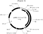
- the prepared plasmid was named in this invention' pcDNA3.1 (+) /preTf (B) (SEQ. ID. NO: 7)' ( Figure 2 ).
- Transferrin is a secretary protein that contains a signal peptide to help the transportation of a protein synthesized on N-terminal to cell membrane.
- Transferrin is composed of the amino acid sequence' MRLAVGALLVCAVLGLCLA (SEQ. ID. NO: 13)'.
- MRLAVGALLVCAVLGLCLA SEQ. ID. NO: 13
- PCR was performed by using pcDNA3.1(+)/preTf(B) as a template in the presence of DNA polymerase. Xho I restriction enzyme site and stop codon (TAA) were inserted in the 3'-end of the gene. PCR was performed by using pcDNA3.1(+)/preTf(B) as a template with DNA polymerase.
- the primers used in the reaction were custom-made in Cosmogenetech Co., Ltd. 10 pmol of a sense primer (5'-CTCGGATCCGTCCCTGATAAAACTGTGAGATG-3', SEQ. ID. NO: 14) and 10 pmol of an antisense primer (5'-AGACTCGAGTTAAGGTCTACGGAAAGTGCAG-3', SEQ. ID.
- the purified transferrin gene and pcDNA3.1(+) plasmid were digested with the restriction enzymes BamH I (Enzynomics) and Xho I (Enzynomics). 10 unit of the restriction enzyme and buffer 2 (10 mM Tris-HCL pH 7.9, 50 mM NaCl, 10 mM MgCl 2 , 1 mM DTT, 100 ⁇ g/ml BSA) were added thereto, followed by reaction at 37°C for 2 ⁇ 3 hours. Upon completion of the reaction, the size of the gene was confirmed by 1% agarose-gel electrophoresis.
- the target area of the agarose-gel was cut off and DNA was purified by using MEGAquick-spin TM Total Fragment DNA purification kit (iNtRoN).
- the purified transferrin gene and pcDNA3.1(+) vector proceeded to electrophoresis on 1% agarose-gel again, followed by DNA quantification.
- the transferrin gene and pcDNA3.1(+) vector were reacted with T4 DNA ligase (Takara) at 16°C for 16 hours.
- T4 DNA ligase T4 DNA ligase
- the vector was inserted in E. coli DH10B, which was distributed on Agar plate containing ampicillin, followed by culture at 37°C for 16 hours. Then, E . coli colony was selected.
- PCR was performed.
- the selected E. coli colony was diluted in water, which was used as a template, followed by PCR with the sense primer (5'-CTCGGATCCGTCCCTGATAAAACTGTGAGATG-3', SEQ. ID. NO: 14) and the antisense primer (5'-AGACTCGAGTTAAGGTCTACGGAAAGTGCAG-3', SEQ. ID. NO: 10) used for the transferrin gene amplification above.
- PCR was performed as follows; predenaturation at 98°C for 30 seconds, denaturation at 98°C for 10 seconds, annealing at 53°C for 30 seconds, polymerization at 72°C for 1 minute, 30 cycles from denaturation to polymerization, and final extension at 72°C for 10 minutes.
- electrophoresis on 1% agarose-gel was performed to confirm the gene.
- the confirmed E. coli colony was inoculated in a LB liquid medium (1% trypton, 0.5% yeast extract, 1% NaCl), followed by culture at 37°C for 16 hours in a shaking incubator. The cultured E . coli was centrifuged to separate the supernatant and the cell pellet.
- Plasmid DNA was purified from the cell pellet using ExprepTM Plasmid SV,mini (GeneAll). The insertion of the transferrin gene without signal peptide was confirmed by sequencing. The prepared plasmid was named in this invention ' pcDNA3.1(+)/Tf(B) (SEQ. ID. NO: 8)' ( Figure 3 ).
- pcDNA6/G-CSF plasmid was treated with 20 units of BamHI (Enzynomics), followed by reaction at 37°C for 3 hours.
- the reacted plasmid (45 ng) was amplified by PCR with 10 pmol of a sense primer (5'-CATGCTAGCTCCACCA TGGCTGGACCTGCCACCCAG-3', SEQ. ID. NO: 15) and an antisense primer (5'-CATGGATCCGGGCTGGGCAAGGTGGCG-3', SEQ. ID. NO: 16).
- a G-CSF gene fragment having Nhe I restriction enzyme site and Kozak sequence in the 5'-end and BamH I restriction enzyme site in the 3'-end was prepared.
- 1 ⁇ g of the prepared G-CSF gene fragment and 365 ng of pcDNA3.1(+)/Tf(B) plasmid were reacted with 10 units of Nhe I restriction enzyme and 20 units of BamH I restriction enzyme respectively for at 37°C for 4 hours.
- Electrophoresis was performed on DNA agarose-gel (0.5x TAE, 1% agarose).
- pcDNA3.1(+)/G-CSF-Tf To construct pcDNA3.1(+)/G-CSF-Tf, the purified pcDNA3.1(+)/Tf(B) plasmid and the G-CSF gene fragment were mixed at the molar ratio of 1:3, which was reacted with 350 U of T4 DNA ligase at 25°C for 1 hour. Then, E. coli DH5 ⁇ was transfected with the reaction product. Colonies showing resistance against ampicillin were selected. Colony PCR was performed to confirm the construction of pcDNA3.1(+)/G-CSF-Tf plasmid. Particularly, the colonies selected from the transfected E.
- coli DH5 ⁇ were amplified by colony PCR with 10 pmol of the sense primer (5'-TAATACGACTCACTATAGGG-3', SEQ. ID. NO: 17) and the antisense primer (5'-AATAGAAAGTCTGTGGGTCCTCCTTTGACCCATAG-3', SEQ. ID. NO: 18).
- the size of the amplified product was confirmed on agarose-gel (0.5X TAE, 1% agarose).
- the confirmed colony was inoculated in a LB liquid medium (trypton 50 mg, yeast extract 25 mg, and sodium chloride 50 mg per 5 ml) containing ampicillin (100 ⁇ g/ml), followed by culture for 15 hours.
- pcDNA3.1(+)/G-CSF-Tf was extracted by using a plasmid extraction kit. The successful construction of pcDNA3.1(+)/G-CSF-Tf was confirmed by sequencing.
- pET21a(+)/G-CSF(T116C) was confirmed by DNA agarose-gel (0.5X TAE, 1% agarose) electrophoresis.
- the target area of the agarose-gel was cut off and DNA was purified by using a gel purification kit (Cosmogenetech).
- E. coli DH10B was transfected with the purified pET21a(+)/G-CSF(T116C). Colonies showing resistance against ampicillin were selected.
- the selected colony was inoculated in a LB liquid medium (trypton 50 mg, yeast extract 25 mg, and sodium chloride 50 mg per 5 ml) containing ampicillin (100 ⁇ g/ml), followed by culture for 15 hours.
- pET21a(+)/G-CSF(T116C) was extracted by using a plasmid extraction kit. Sequencing was performed and as a result, the successful substitution of the nucleotide sequence was confirmed.
- the sense primer (5'-GCCGACTTTGCCACCTGCATCTGGCAGCAGAT-3', SEQ. ID. NO: 19) and the antisense primer (5'-ATCTGCTGCCAGATGCAGGTGGCAAAGTCGGC-3', SEQQ. ID. NO: 20) were dissolved respectively to the concentration of 10 pmol/ ⁇ l.
- 10 pmol of each primer was taken for PCR with 100 ng of the G-CSF gene (100 ng) inserted in pcDNA6.
- Site-directed mutagenesis was performed by the same manner as described in Example ⁇ 1-1> to obtain G-CSF(T116C) gene.
- the obtained plasmid was named in this invention' pcDNA6/G-CSF(T116C).
- pcDNA6/G-CSF(T116C) was confirmed by DNA agarose-gel (0.5X TAE, 1% agarose) electrophoresis.
- the target area of the agarose-gel was cut off and DNA was purified by using a gel purification kit (Cosmogenetech).
- E. coli DH10B was transfected with the purified pcDNA6/G-CSF(T116C)). Colonies showing resistance against ampicillin were selected.
- the selected colony was inoculated in a LB liquid medium (trypton 50 mg, yeast extract 25 mg, and sodium chloride 50 mg per 5 ml) containing ampicillin (100 ⁇ g/ml), followed by culture for 15 hours.
- pcDNA6/G-CSF(T116C) was extracted by using a plasmid extraction kit. Sequencing was performed and as a result, the successful substitution of the nucleotide sequence was confirmed.
- 1 ⁇ g of pcDNA6/G-CSF(T116C) plasmid was treated with 20 units of BamH I (Enzynomics), followed by reaction at 37°C for 3 hours.
- the reacted plasmid (45 ng) was amplified by PCR with 10 pmol of a sense primer (5'-CATGCTAGCTCCACCA TGGCTGGACCTGCCACCCAG-3', SEQ. ID.
- G-CSF(T116C) DNA fragment having Nhe I restriction enzyme site and Kozak sequence in the 5'-end and BamH I restriction enzyme site in the 3'-end was prepared.
- 1 ⁇ g of the prepared G-CSF(T116C) DNA fragment and 365 ng of pcDNA3.1 (+) /Tf (B) plasmid were reacted with 10 units of Nhe I restriction enzyme and 20 units of BamH I restriction enzyme respectively for at 37°C for 4 hours.
- Electrophoresis was performed on DNA agarose-gel (0.5x TAE, 1% agarose), followed by purification by using a gel extraction kit.
- pcDNA3.1(+)/G-CSF(T116C)-Tf the purified pcDNA3.1(+)/Tf(B) plasmid and the G-CSF(T116C) DNA fragment were mixed at the molar ratio of 1:3, which was reacted with 350 U of T4 DNA ligase at 25°C for 1 hour. Then, E. coli DH5 ⁇ was transfected with the reaction product. Colonies showing resistance against ampicillin were selected.
- Colony PCR was performed to confirm the construction of pcDNA3.1(+)/G-CSF(T116C)-Tf plasmid. Particularly, the colonies selected from the transfected E. coli DH5 ⁇ were amplified by colony PCR with 10 pmol of the sense primer (5'-TAATACGACTCACTATAGGG-3', SEQ. ID. NO: 17) and the antisense primer (5'-AATAGAAAGTCTGTGGGTCCTCCTTTGACCCATAG-3', SEQ. ID. NO: 18). The size of the amplified product was confirmed on agarose-gel (0.5X TAE, 1% agarose).
- the confirmed colony was inoculated in a LB liquid medium (trypton 50 mg, yeast extract 25 mg, and sodium chloride 50 mg per 5 ml) containing ampicillin (100 ⁇ g/ml), followed by culture for 15 hours.
- pcDNA3.1(+)/G-CSF(T116C)-Tf was extracted by using a plasmid extraction kit.
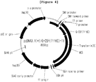
- the successful construction of pcDNA3.1(+)/G-CSF(T116C)-Tf (SEQ. ID. NO: 21) was confirmed by sequencing.
- Expi293FTM the human embryonic kidney cancer cell line modified for suspension culture.
- Expi293FTM cells included in Expi293FTM Expression system Kit (Gibco) were first thawed, which were inoculated in a sterilized 125 mL flask (Thermo) containing 30 mL of serum-free medium for animal cell culture (Gibco), followed by shaking culture at 125 rpm in a 37°C, 5% CO 2 incubator.
- the density of Expi293FTM cell line was maintained at 3 ⁇ 5x10 6 cells/mL. Sub-culture was performed every 3 ⁇ 4 days. After stabilized, the cells were inoculated in serum-free medium at the density of 2x10 6 cells/mL for transfection, followed by shaking culture for 24 hours. Just before the transfection, the cell density was adjusted to 2x10 6 cells/mL and the cell survival rate was confirmed at least 90% before the following experiment.
- ExpiFectamineTM 293 Transfection Enhancers 1 and 2 (Gibco) were added thereto in order to increase the protein expression, followed by additional culture for 32 hours under the same shaking culture condition above.
- the obtained cell culture fluid was centrifuged at 2,000 xg for 10 minutes and as a result the supernatant was obtained.
- the supernatant was mixed with 5x loading dye (Biosesang), which was boiled at 95°C for 20 minutes. The mixture was loaded on 10% SDS-PAGE, followed by electrophoresis at 160 V for 1 hour. Upon completion of the electrophoresis, the protein on the gel was transferred onto PVDF Immobilon-P-Transfer Membrane (Millipore). The membrane was reacted in 5% skim milk at room temperature for 1 hour. After washing the membrane with washing buffer (phosphate buffer containing 0.05% Tween 20), a transferrin antibody (2,000x, Santa Cruz) was added thereto, followed by reaction at room temperature for 2 hours.
- washing buffer phosphate buffer containing 0.05% Tween 20
- a transferrin antibody 2,000x, Santa Cruz
- the cells were dissolved in a lysis buffer (50 mM Tris-Cl (pH 8.0), 2 mM EDTA, 1 mM PMSF), followed by cell lysis using an ultrasonicator (lysis time: 5 minutes, ultrasonication: 30 seconds, stand-by: 40 seconds). Centrifugation was performed at 10.000 rpm for 30 minutes at 4°C to eliminate the supernatant. Then, the precipitate was dissolved in a washing buffer (50 mM Tris-Cl, pH 8.0, 2 mM EDTA, 0.3% Triton) and lysed using an ultrasonicator (lysis time: 5 minutes, ultrasonication: 30 seconds, stand-by: 40 seconds).
- a washing buffer 50 mM Tris-Cl, pH 8.0, 2 mM EDTA, 0.3% Triton
- Centrifugation was performed again at 10.000 rpm for 30 minutes at 4°C to eliminate the supernatant.
- the precipitate was dissolved in a washing buffer (50mM Tris-Cl, pH8.0, 2 mM EDTA, 1 M NaCl) and lysed using an ultrasonicator (lysis time: 5 minutes, ultrasonication: 30 seconds, stand-by: 40 seconds). Centrifugation was performed again at 10.000 rpm for 30 minutes at 4°C to eliminate the supernatant.
- the washed inclusion body was dissolved in a solubilization buffer (8 M Urea, 50 mM Tris-Cl, pH8.0).
- the purified G-CSF(T116C) was quantified by using silver staining.
- the standard quantification curve was obtained by using grasin (less than 30 ng/SDS-PAGE well), the G-CSF standard material.
- the unknown amount of each sample was compared to the standard quantification curve so as to quantify the protein.
- the preparation of the holo form of G-CSF(T116C)-Tf was based on [ Zhang et al. BMC Biotechnology 2012, 12:92 ].
- the purified G-CSF-Tf or G-CSF(T116C)-Tf was treated with the equivalent ferric ammonium citrate, followed by reaction at 37°C for 2 hours.
- the mixture was dialyzed in 2 L of phosphate buffered saline at 4°C for 15 hours, during which buffer was exchanged.
- the sample was 10-fold concentrated by using a concentrator (Millipore) ( Figure 7 ).
- the purified protein was quantified by Western blotting using a transferrin antibody.
- the measured transferrin was loaded in 10% SDS-PAGE at the concentrations of 15 ng, 20 ng, 25 ng, and 30 ng per each well.
- Another purified protein whose concentration was not determined was loaded in another well, followed by electrophoresis.
- Western blotting was performed to quantify the protein samples by comparing the purified protein with the transferrin standard curve.
- transferrin Once transferrin is bound to the transferrin receptor expressed on the cell surface, it enters the cell via endocytosis. After releasing the iron, the transferrin-receptor complex moves to the cell surface again and thereafter it is secreted in blood. Transferrin repeats the process above, that is the in vivo recycling metabolism, by which it supplies the iron and avoids being decomposed in the intracellular lysosome, resulting in the increased half-life. If a target protein having a short half-life is conjugated with transferrin, the protein experiences the in vivo recycling metabolism action of transferrin together with transferrin. As a result, the target protein also can avoid being decomposed in vivo. So, the fusion protein in which G-CSF is conjugated with transferrin needs to maintain the binding force to transferrin receptor in order to avoid in vivo degradation.
- the human hepatocellular carcinoma cell line HepG2 demonstrating high expression of TfR on the cell surface was used for the following experiment.
- HepG2 cells were inoculated in a 96-well plate at the density of 5x10 5 cells/well, followed by culture for 24 hours.
- the HepG2 cells attached on the plate were washed with phosphate buffer, to which serum-free DMEM (HyClone) supplemented with 1 mg/ml of BSA (Bovine Serum Albumin) was added.
- the cells were washed with phosphate buffer again, and then the test sample and fluorescence-labeled human transferrin Alexa Fluor® 647 (Life Technologies) were added to the serum-free DMEM supplemented with 1 mg/ml of BSA at the concentrations of 133 nM and 5 ⁇ g/mL respectively.
- the mixture above was treated to the HepG2 cells prepared above, which stood in a 37°C, 5% CO 2 incubator for 30 minutes.
- the protein sample remaining in the medium was washed with cold phosphate buffer.
- the transferrin receptor conjugated fluorescence-transferrin was quantified by fluorimetry. For the fluorimetry, light source was given at 650 nm and fluorescence intensity at 668 nm was measured.
- the transferrin receptor binding force of the holo form of G-CSF(T116C)-Tf prepared in Example ⁇ 3-4> was shown in Figure 8 .
- the transferrin receptor binding force of the G-CSF-Tf or G-CSF(T116C)-Tf fusion protein was similar to that of the natural human holo-transferrin (SIGMA). This result suggests that the G-CSF-Tf or G-CSF(T116C)-Tf fusion protein can have increased half-life through experiencing the in vivo recycling due to the conjugation with the transferrin receptor, like human transferrin.
- the human bone marrow derived cell line HL-60 used in this invention was distributed from Korean Collection for Type Culture (KCTC), Korea Research Institute of Bioscience and Biotechnology (KRIBB).
- the cell line was cultured in RPMI-1640 containing 10% FBS in a 37°C, 5% CO 2 incubator. The medium was replaced with a fresh one every 2 ⁇ 3 days. The cells were not anchorage-dependent.
- the reagents for the cell culture were purchased from HyClone.
- the number of cells was counted and the density was adjusted to be 2 x 10 5 cells/mL.
- the cells were treated with the differentiation inducer DMSO (dimethylsulfoxide, culture grade, SIGMA) at the final concentration of 1.25% (v/v), followed by culture for 3 days to induce the differentiation into granulocytes. Before the test sample was added, the cells were collected by centrifugation and washed with phosphate buffer (D-PBS). The cells were distributed in a 96-well plate (Corning), 100 ⁇ L per each well, at the density of 4 x10 4 cells/well.
- DMSO dimethylsulfoxide, culture grade, SIGMA
- the controls PEGylated human G-CSF (GenScript) and natural G-CSF (GenScript), and the test samples G-CSF-Tf, G-CSF(T116C)-Tf, and G-CSF(T116C) were diluted in desalting buffer (5mM Sodium Phosphate, 5% Sucrose, 0.004% Tween-20), which were added to each well containing the differentiated HL-60 cells, followed by culture in a 37°C, 5% CO 2 incubator for 72 hours.
- the final concentrations of each sample were 300, 200, 100, 30, 15, 3, 0.3, and 0.03 ng/mL.
- the cell proliferation activity of G-CSF, PEG.G-CSF, G-CSF(T116C), G-CSF-Tf, and G-CSF(T116C)-Tf is shown in Figure 9 .
- the cell proliferation activity of G-CSF(T116C) was lower than that of the natural G-CSF but was similar to that of the PEGylated G-CSF.
- the cell proliferation activity of the G-CSF(T116C)-Tf fusion protein in which transferrin is fused with a G-CSF(T116C) mutant protein was significantly increased, compared with that of G-CSF(T116C) and was also higher than that of the PEGylated G-CSF. Therefore, since the G-CSF(T116C)-Tf displays the higher cell proliferation activity than that of the commercial PEGylated G-CSF, it is expected to be a promising candidate as an agent for the treatment particularly using G-CSF.
- the protein was reacted with human serum and then the amount of G-CSF remaining in the serum for a designated time was measured.
- the high protein binding microtiter plate (Corning) coated with a rabbit polyclonal anti-G-CSF antibody (KOMA BIOTECH) at the concentration of approximately 2 ⁇ g/ml was prepared.
- 1X phosphate buffer containing 1% casein sodium salt was used as a blocking buffer.
- the standard solution and the test solution were mixed with a buffer (10x diluted blocking buffer).
- the protein sample and regents were distributed by 100 ⁇ l per each well.
- the plate was treated with a washing buffer (1X PBS, 0.05% Tween-20) three times (300 ⁇ l/well) to wash out the non-reacted samples.
- the protein sample and the detection antibody (rabbit polyclonal anti-G-CSF antibody, KOMA BIOTECH) were treated thereto stepwise at room temperature for about 2 hours, followed by reaction with streptavidin-horseradish peroxidase (Pierce) at 37°C for 30 minutes.
- TMB (3,3',5,5'-tetramethylbenzidine, Pierce) was treated thereto, which stood at room temperature for about 7 ⁇ 10 minutes and then color development was terminated with 2 M sulfuric acid. Then, OD 450 was measured with a microplate reader (TECAN).
- the absorbance was converted into concentration based on the calibration curve of protein concentration-absorbance to the standard solution.
- the standard curve was made using Excel program. The difference between the mean value of the absorbance corresponding to each G-CSF standard solution and the value of absorbance of the well treated with the buffer alone was calculated. For the determination of the test solution concentration, only when the absorbance value of each sample was in the range of the standard solution calibration curve, it was taken as valid.
- ) criterion calculated by using the correlation coefficient (R 2 ) as root ( ⁇ ) was set to at least 0.98.
- the amount of the remaining G-CSF protein (% G-CSF) was expressed by considering the concentration of G-CSF (pg/ml) at reaction time point of 0 h as 100%.
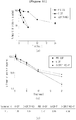
- the amount (%) of remaining G-CSF and G-CSF(T116C) by time period is shown in Figure 10 .
- the percentage of remaining G-CSF(T116C) was approximately 30% higher than that of natural G-CSF since 15 hours after the reaction started. It was confirmed that the disulfide bond between Cys116 and Cys17 was helpful for the increase of resistance of the protein against blood protease.
- the proteins were administered to mice and then the level of G-CSF in the rat serum was measured.
- the mentioned 'in vivo half-life' indicates the 'half-life of protein in serum'. That is, the time point when the G-CSF protein level was down to 50% by the initial concentration during its circulation in serum was expressed numerically. Pharmacokinetic calculation was performed using PKsolver v2.0.
- mice 6-week-old ICR and SPF male mice were purchased from Samtako. The mice were inspected and adapted for at least 6 days. On the last day of the adaptation period, individual weight was measured. Based on the body weight, healthy animals without weight gain and general symptoms were selected and randomly arranged so that the average weight of each group was as equal as possible (35 ⁇ 45 g per animal). Each experimental group was assigned 6 animals.
- a unit dose for each animal was calculated (5 mL/kg) based on the body weight measured immediately before the administration.
- the G-CSF mutant protein (20 ⁇ g/kg) or the G-CSF mutant transferrin fusion protein (100 ⁇ g/kg) was slowly injected into the Intravenous Bolus by using a disposable syringe (1mL, 26G needle). The day of administration was defined as day 1 of the test.
- blood was drawn from the animals by orbital collection at the time point of 0.25, 0.5, 1, 3, 6, 12, 24, 48, and 72 hours respectively.
- the blood sample was centrifuged at 10,000 ⁇ 13,000 rpm for 1 ⁇ 2 minutes to separate plasma.
- the separated plasma was loaded in a tube (about 50 ⁇ L/tube) attached with a label containing the information of test type, animal number, and sampling time.
- the tube was stored in a deep freezer (approximately -70°C) until the day of analysis.
- the amount of active G-CSF in plasma was determined by ELISA after thawing the sample on ice.
- the quantification of G-CSF in plasma by ELISA was performed by using Human G-CSF ELISA Kit, pink-ONE ( Koma Biotech.) according to the manual of the reagent.
- the calibration curve of the absorbance according to the protein concentration to the standard solution was prepared and the G-CSF mutant content in the test solution was determined by regression analysis
- Non-compartmental pharmacokinetic analysis was performed from the mean concentration-time profile of each test compound by applying IV Bolus non-compartment analysis input to PKsolver v2.0 program.
- the pharmacokinetic parameters evaluate the terminal half-life (t 1/2 ).
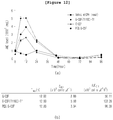
- FIG. 11 The half-life of proteins in plasma is shown in Figure 11 .
- G-CSF(T116C) mutant protein displayed a blood half-life similar to that of natural human G-CSF. This is a result of similar filtration rate in the kidney, since the G-CSF(T116C) mutant protein has a similar protein radius to the G-CSF.
- G-CSF-Tf protein showed an improved blood half-life compared to G-CSF. This is because the fusion of transferrin to the G-CSF protein results in a significant increase in the protein radius, resulting in a significantly lower renal filtration rate.
- the G-CSF(T116C)-Tf fusion protein in which G-CSF(T116C) was fused to transferrin showed more extended half-life than that of G-CSF-Tf but displayed similar half-life to that of PEGylated G-CSF. Therefore, it was suggested that when the G-CSF(T116C) mutant protein having the increased resistance against blood protease was fused to transferrin, the renal filtration rate was decreased and the blood stability was improved, resulting in the maximization of in vivo blood half-life ( Figure 11 ).
- hematologic analysis was performed using a rat disease model (Neutropenic Rat). All the procedures such as purchase, breeding, administration and hematologic analysis of test animals were performed by Qu-BEST Consulting Co., Ltd.
- the hematologic analysis items include WBC, NEU, RBC, HGB, HCT, MCV, MCH, MCHC, PLT, and Diffential leucocyte count, etc.
- 5-week-old SPF Sprague Dawley male rats were adapted at least 5 days and then randomly allocated into four groups. Each rat was in the weight range of 350 ⁇ 400 g.
- cyclophosphamide 90 mg/kg was intraperitoneally administered once to induce leukopenia (Neutropenic).
- the SD rats were intravenously administered with G-CSF once, followed by hematologic tests for 5 days.
- the test compound was diluted in PBS (Phosphate Buffered Saline) to make the final concentration of 0.004 mg/mL.
- a unit dose for each animal was calculated (2 mL/kg) based on the body weight measured immediately before the administration.
- the test sample was slowly injected into the Intravenous Bolus by using a disposable syringe (1mL, 26G needle). The day of administration was defined as day 1 of the test.
- blood collection (approximately 200 ⁇ l) was performed in the jugular vein using a disposable syringe (1 mL, 26G needle).
- the blood was stored in the tube treated with an anticoagulant (5% sodium EDTA) to prevent the blood from coagulation.
- the components in the blood were analyzed using an automatic blood analyzer in a refrigerated state within 3 hours. The results are presented with mean value and standard deviation. The difference between the control and each experimental group was compared statistically by using GraphPad Prism by One-way ANOVA and Dunnett's test.
- G-CSF(T116C)-Tf displayed the increased WBC (white blood cell) and neutrophil levels in the rats having leukopenia induced by cyclophosphamide up to 24 hours from the administration.
- G-CSF(T116C)-Tf fusion protein was circulated in vivo by using transferrin as a mediator, during which in vivo activity was maintained longer than G-CSF and was as high as that of PEGylated G-CSF.
- the pharmacokinetic parameters (Cmax and AUC0-t) of the fusion protein were higher than those of G-CSF, as shown in Figure 9 . This seemed to be because the neutrophilic precursor cell proliferation was greater in the protein than in PEGylated G-CSF in the same length of period ( Figure 12 ).
- the induced material in this invention exhibits G-CSF like biological activity that has been used clinically, so that the material of the present invention can be used for the treatment of ischemic disease or as an anticancer adjuvant for the prevention of infectious complications caused by neutropenia resulted from chemo-therapy or radiotherapy for the treatment of cancer due to its activity of stimulating neutrophil granulopoiesis.
Landscapes
- Health & Medical Sciences (AREA)
- Chemical & Material Sciences (AREA)
- Life Sciences & Earth Sciences (AREA)
- Organic Chemistry (AREA)
- Medicinal Chemistry (AREA)
- General Health & Medical Sciences (AREA)
- Proteomics, Peptides & Aminoacids (AREA)
- Gastroenterology & Hepatology (AREA)
- Zoology (AREA)
- Biochemistry (AREA)
- Biophysics (AREA)
- Genetics & Genomics (AREA)
- Molecular Biology (AREA)
- Toxicology (AREA)
- Immunology (AREA)
- Public Health (AREA)
- Animal Behavior & Ethology (AREA)
- Veterinary Medicine (AREA)
- Pharmacology & Pharmacy (AREA)
- Engineering & Computer Science (AREA)
- Bioinformatics & Cheminformatics (AREA)
- Epidemiology (AREA)
- Nuclear Medicine, Radiotherapy & Molecular Imaging (AREA)
- Chemical Kinetics & Catalysis (AREA)
- General Chemical & Material Sciences (AREA)
- Heart & Thoracic Surgery (AREA)
- Urology & Nephrology (AREA)
- Vascular Medicine (AREA)
- Cardiology (AREA)
- Preparation Of Compounds By Using Micro-Organisms (AREA)
- Peptides Or Proteins (AREA)
- Medicines That Contain Protein Lipid Enzymes And Other Medicines (AREA)
Abstract
Description
- The present invention relates to a fusion protein in which transferrin is peptide-bonded to a terminal of a granulocyte-colony stimulating factor (G-CSF) protein or a G-CSF mutant protein.
- Granulocyte-colony stimulating factor (G-CSF) is a glycoprotein that stimulates survival, proliferation, differentiation, and functions of neutrophil and granulocyte progenitor cells and mature neutrophils.
- The natural G-CSF which is now being used clinically is called 'Filgrastim', and is a recombinant protein composed of 175 amino acids originated from human amino acid sequence. This recombinant protein is expressed in E. coli and is not glycosylated unlike the natural type.
- G-CSF is used as an anticancer adjuvant for the prevention of infectious complications caused by neutropenia accompanied by cancer treatment by stimulating neutrophil granulopoiesis. G-CSF demonstrates a beneficial clinical effect on cancer patients because it can reduce side effects of febrile neutropenia caused by chemo-therapy or radio-therapy for cancer treatment and thereby can reduce death rate by chemo-therapy for cancer treatment. G-CSF increases the number of hematopoietic progenitor cells and accordingly reduces the side effects above.
- It is also known that G-CSF stimulates bone marrow stem cells to move ischemic heart and accelerates differentiation of the stem cells into vascular cells and cardiomyocytes by stimulating myocardial regeneration.
- The recombinant human G-CSF (rhG-CSF) displays pharmacological effects only for a short time. Therefore, it has to be administered at least once a day to treat ischemic disease or to treat leukopenia caused by anticancer chemo-therapy or radio-therapy. If a substance having a long in vivo half-life is administered, the administration times necessary for relieving leukopenia would be reduced and as a result it could bring the effect of preventing infectious complications.
- When Polyethylene glycol (PEG), the chemical polymer that is not degraded in vivo, is fused to the N-terminal of G-CSF, a substance called 'Pegfilgrastim' is produced. This substance is clinically used for the treatment of leukopenia. This substance has an increased in vivo half-life and displays a clinical effect while leukopenia continues even with the administration performed once or twice a week. However, in that case, a protein is fused with a chemical polymer by chemical reaction, and thus the problems of unsatisfactory fusion efficiency and complicated purification method can be caused.
- Cysteine, the 17th amino acid of G-CSF is exposed on the protein surface as nonsulfated binding state. In neutral pH, the exposed cysteine is combined with adjacent G-CSF cysteine via sulfide bond and as a result it loses its activity. A G-CSF mutant wherein the 17th cysteine is substituted with serine displays increased stability in neutral pH, according to the previous reports.
- Transferrin is the third most abundant protein in blood plasma, which serves to transport iron ions present in the blood to various tissues. Transferrin has a relatively long half-life of 8 days, which is shorter than that of albumin or immunoglobulin G. It enters the cell through the transferrin receptor on the surface of the cell and once it supplies iron ions, it is released to the outside of the cell in a state of binding with the receptor. Using these characteristics, transferrin has been used as a fusion partner to increase circulating half-life by combining the proteins with short half-lives of the prior art.
- In this invention, threonine, the 116th amino acid of human G-CSF, was substituted with cysteine to induce sulfide bonding with the 17th cysteine of the original amino acid sequence of G-CSF. Sulfide bonding makes the protein structure more stable so that the protein can be resistant against proteases, that is the protein now has a protease resistance. The attempt to have a protease resistance through the construction of such a mutant protein is new and the effect thereof has not been reported yet.
- There has been no reports of using the mutant protein of G-CSF wherein the 116th amino acid is replaced with cysteine for the fusion with transferrin to increase blood half-life of G-CSF.
- The present inventors succeeded in making a fusion protein of transferrin and the mutant protein of human G-CSF wherein the 116th amino acid was replaced with cysteine and further confirmed that the fusion protein had significantly increased specific activity and blood stability, compared with the unfused original human G-CSF, leading to the completion of this invention.
- It is an object of the present invention to provide a fusion protein in which transferrin is peptide-bonded to a terminal of a granulocyte-colony stimulating factor (G-CSF) protein or a G-CSF mutant protein in which the 116th threonine is substituted with cysteine in the amino acid sequence of the G-CSF.
- To achieve the above object, the present invention provides a fusion protein in which transferrin is peptide-bonded to a terminal of a granulocyte-colony stimulating factor (G-CSF) protein or a G-CSF mutant protein in which the 116th threonine is substituted with cysteine in the amino acid sequence of the G-CSF, a G-CSF mutant protein in which the 116th threonine is replaced with cysteine, and a use of the same.
- The present invention also provides a pharmaceutical composition and an anticancer adjuvant for preventing or treating ischemic diseases or neutropenia comprising the fusion protein in which transferrin is peptide-bonded to a terminal of a granulocyte-colony stimulating factor (G-CSF) protein or a G-CSF mutant protein in which the 116th threonine is substituted with cysteine in the amino acid sequence of the G-CSF or the G-CSF mutant protein in which the 116th threonine is replaced with cysteine as an active ingredient.
- The present invention also provides an expression vector containing the gene encoding a fusion protein in which transferrin is peptide-bonded to a terminal of a granulocyte-colony stimulating factor (G-CSF) protein or a G-CSF mutant protein in which the 116th threonine is substituted with cysteine in the amino acid sequence of the G-CSF or a G-CSF mutant protein in which the 116th threonine is replaced with cysteine and a transformant prepared by inserting the expression vector above in a host cell.
- The present invention also provides a method for preparing the expression vector containing the gene encoding a fusion protein in which transferrin is peptide-bonded to a terminal of a granulocyte-colony stimulating factor (G-CSF) protein or a G-CSF mutant protein in which the 116th threonine is substituted with cysteine in the amino acid sequence of the G-CSF or a G-CSF mutant protein in which the 116th threonine is replaced with cysteine, and the transformant prepared by inserting the expression vector above in a host cell.
- The present invention also provides a method for preventing or treating neutropenia or ischemic disease containing the step of administering the fusion protein in which transferrin is peptide-bonded to a terminal of a granulocyte-colony stimulating factor (G-CSF) protein or a G-CSF mutant protein in which the 116th threonine is substituted with cysteine in the amino acid sequence of the G-CSF, the G-CSF mutant protein in which the 116th threonine is replaced with cysteine, the expression vector, or the transformant above to a subject having neutropenia or ischemic disease.
- The present invention also provides a method for reducing neutrophils containing the step of administering the fusion protein in which transferrin is peptide-bonded to a terminal of a granulocyte-colony stimulating factor (G-CSF) protein or a G-CSF mutant protein in which the 116th threonine is substituted with cysteine in the amino acid sequence of the G-CSF, the G-CSF mutant protein in which the 116th threonine is replaced with cysteine, the expression vector, or the transformant above to a subject.
- In addition, the present invention provides a use of the fusion protein in which transferrin is peptide-bonded to a terminal of a granulocyte-colony stimulating factor (G-CSF) protein or a G-CSF mutant protein in which the 116th threonine is substituted with cysteine in the amino acid sequence of the G-CSF, the G-CSF mutant protein in which the 116th threonine is replaced with cysteine, the expression vector, or the transformant above.
- The granulocyte-colony stimulating factor (G-CSF) mutant protein of the present invention or the transferrin fusion protein thereof displays a significantly increased specific activity and blood stability, compared with the conventional human G-CSF, and has a higher purification efficiency than the conventional PEGylated G-CSF characterized by the extended half-life, so that it can be advantageously used for preventing or treating ischemic diseases or neutropenia.
- The application of the preferred embodiments of the present invention is best understood with reference to the accompanying drawings, wherein:
-
Figure 1 is a diagram illustrating the cleavage map of pcDNA3.1(+)/preTf vector. -
Figure 2 is a diagram illustrating the cleavage map of pcDNA3.1(+)/preTf(B) vector. -
Figure 3 is a diagram illustrating the cleavage map of pcDNA3.1(+)/Tf(B) vector. -
Figure 4 is a diagram illustrating the cleavage map of pcDNA3.1(+)/G-CSF(T116C)-Tf vector. -
Figure 5 is a diagram illustrating the expression of G-CSF(T116C)-Tf in the cell line Expi293F transfected with G-CSF(T116C)-Tf plasmid. -
Figure 6 is a diagram illustrating the G-CSF(T116C)-Tf protein separated by DEAE Affi-gel blue chromatography. -
Figure 7 is a diagram illustrating the preparation, concentration, and separation of Fe3+ fused G-CSF(T116C)-Tf in the form of holo (holoenzyme). -
Figure 8 is a diagram illustrating the binding force of G-CSF(T116C)-Tf in the form of holo to the transferrin receptor. -
Figure 9 is a diagram illustrating the cell proliferation activity of HL-60 cells treated with G-CSF, G-CSF-Tf, and G-CSF(T116C)-Tf. -
Figure 10 is a diagram illustrating the comparison of protease resistance between G-CSF and G-CSF(T116C). -
Figure 11 is a diagram illustrating the comparison of in vivo plasma half-life between G-CSF-Tf and G-CSF(T116C)-Tf. -
Figure 12 is a diagram illustrating the comparison of physiological activity between G-CSF and G-CSF(T116C)-Tf in the rat having neutropenia. - Hereinafter, the present invention is described in detail.
- The present invention provides a fusion protein in which transferrin is peptide-bonded to a terminal of a granulocyte-colony stimulating factor (G-CSF) protein or a G-CSF mutant protein in which the 116th threonine is substituted with cysteine in the amino acid sequence of the G-CSF, a G-CSF mutant protein in which the 116th threonine is replaced with cysteine, and a use of the same.
- The G-CSF or transferrin used in this invention can be originated from animals, plants, or microorganisms. It is more preferably human originated G-CSF or transferrin herein, but it can be also a heterologous protein having the equal activity to human originated G-CSF or transferrin.
- The protein above can additionally be modified such as phosphorylation, acetylation, methylation, glycosylation, etc., or it can be fused with another protein. As long as the function of the protein is not lost, such mutant protein can be considered as the same as the protein before any modification.
- Another amino acid that can replace the 116th threonine is a hydrophobic amino acid, which is more precisely cysteine that is possibly linked to the 17th cysteine of G-CSF sequence via disulfide bond, but not always limited thereto.
- The terminal where transferrin is bound can be either the amino terminal (5'-end, N-terminal) or the carboxy terminal (3'-end, C-terminal).
- The said granulocyte-colony stimulating factor (G-CSF) protein is the protein represented by SEQ. ID. NO: 1 but this sequence may have addition, deletion, or substitution of one or more amino acids in its sequence as long as the gene encoding the G-CSF is located at the same position on the chromosome and the activity is not changed.
- The granulocyte-colony stimulating factor (G-CSF) protein above preferably has at least 80% homology, more preferably at least 90% homology, and most preferably at least 95%, 96%, 97%, 98%, 99%, or 99.5% homology to the amino acid sequence represented by SEQ. ID. NO: 1, but not always limited thereto.
- The said transferrin is the protein represented by SEQ. ID. NO: 2 but this sequence may have addition, deletion, or substitution of one or more amino acids in its sequence as long as the gene encoding the transferrin is located at the same position on the chromosome and the activity is not changed.
- The transferrin protein above preferably has at least 80% homology, more preferably at least 90% homology, and most preferably at least 95%, 96%, 97%, 98%, 99%, or 99.5% homology to the amino acid sequence represented by SEQ. ID. NO: 2, but not always limited thereto.
- The said granulocyte-colony stimulating factor (G-CSF) mutant protein is the protein represented by SEQ. ID. NO: 3 but this sequence may have addition, deletion, or substitution of one or more amino acids in its sequence as long as the gene encoding the transferrin is located at the same position on the chromosome and the activity is not changed.
- The granulocyte-colony stimulating factor (G-CSF) mutant protein above preferably has at least 80% homology, more preferably at least 90% homology, and most preferably at least 95%, 96%, 97%, 98%, 99%, or 99.5% homology to the amino acid sequence represented by SEQ. ID. NO: 3, but not always limited thereto.
- The fusion protein in which transferrin is peptide-bonded to a terminal of G-CSF preferably has at least 80% homology, more preferably at least 90% homology, and most preferably at least 95%, 96%, 97%, 98%, 99%, or 99.5% homology to the amino acid sequence represented by SEQ. ID. NO: 4, but not always limited thereto.
- The fusion protein in which transferrin is peptide-bonded to a terminal of a G-CSF mutant protein in which the 116th threonine is substituted with cysteine in the amino acid sequence of the G-CSF preferably has at least 80% homology, more preferably at least 90% homology, and most preferably at least 95%, 96%, 97%, 98%, 99%, or 99.5% homology to the amino acid sequence represented by SEQ. ID. NO: 5, but not always limited thereto.
- The said G-CSF, the G-CSF mutant protein in which the 116th threonine is substituted with cysteine in the amino acid sequence of the G-CSF, or the transferrin may have the substitution of nucleotide sequence for gene manipulation. For example the nucleotide sequence of a restriction enzyme recognition site in the gene can be substituted with another nucleotide sequence encoding another amino acid which is equal to the original amino acid but not causing any changes in the protein activity, or a part of the gene terminal can be deleted, substituted or added with a restriction enzyme recognition site. For example, thymine of the BamHI restriction enzyme recognition site of the protein gene, GGATCC, can be substituted with cytosine, but not always limited thereto.
- The said restriction enzyme is exemplified by EcoRI, BamHI, Hind III, kpnI, NotI, PstI, SmaI, XhoI, FokI, Alw26I, BbvI, BsrI, EarI, Hph I, MboI, SfaNI, Tth111I, NaeI, NheI, NgoMIV, NheI, Eco57I, BcgI, BpI, Bsp24I, BaeI , CjeI , EcoPI, HintIII, and StyLTI, etc. However, any restriction enzyme that is used in the art can be used according to the gene, expression vector or genetic manipulation environment without limitation.
- The present invention also provides a pharmaceutical composition for preventing or treating neutropenia and ischemic disease comprising the fusion protein in which transferrin is peptide-bonded to a terminal of a G-CSF mutant protein in which the 116th threonine is substituted with cysteine in the amino acid sequence of the G-CSF or the G-CSF mutant protein in which the 116th threonine is substituted with cysteine in the amino acid sequence of the G-CSF as an active ingredient, an anticancer adjuvant comprising the same, an expression vector comprising a gene encoding the said protein, and a transformant prepared by inserting the said expression vector in a host cell.
- The term 'neutropenia' in this invention indicates the abnormal reduction of neutrophils. When the number of blood neutrophils is less than 1500/µl, it is classified as light neutropenia and when the number of blood neutrophils is less than 1000/µl, it is classified as moderate neutropenia. When the number of blood neutrophils is less than 500/µl, it is classified as severe neutropenia. In a large sense, leukopenia (neucopenia) is also included in neutropenia.
- The 'ischemic disease' of the present invention is caused by cell damage caused when the blood supply to the tissue is interrupted by hemorrhage, embolism, and infarction, etc, which is exemplified by trauma, graft rejection, stroke, cerebral infraction, ischemic renal disease, ischemic lung disease, infection mediated ischemic disease, ischemic limb disease, ischemic cardiomyopathy, myocardial infarction, and heart failure, etc.
- The said another amino acid that can replace the 116th threonine can be a hydrophobic amino acid, which is precisely cysteine that is suitable for disulfide bond, but not always limited thereto.
- The said granulocyte-colony stimulating factor (G-CSF) protein is the protein represented by SEQ. ID. NO: 1 but this sequence may have addition, deletion, or substitution of one or more amino acids in its sequence as long as the gene encoding the G-CSF is located at the same position on the chromosome and the activity is not changed.
- The granulocyte-colony stimulating factor (G-CSF) protein above preferably has at least 80% homology, more preferably at least 90% homology, and most preferably at least 95%, 96%, 97%, 98%, 99%, or 99.5% homology to the amino acid sequence represented by SEQ. ID. NO: 1, but not always limited thereto.
- The said transferrin is the protein represented by SEQ. ID. NO: 2 but this sequence may have addition, deletion, or substitution of one or more amino acids in its sequence as long as the gene encoding the transferrin is located at the same position on the chromosome and the activity is not changed.
- The transferrin protein above preferably has at least 80% homology, more preferably at least 90% homology, and most preferably at least 95%, 96%, 97%, 98%, 99%, or 99.5% homology to the amino acid sequence represented by SEQ. ID. NO: 2, but not always limited thereto.
- The said granulocyte-colony stimulating factor (G-CSF) mutant protein is the protein represented by SEQ. ID. NO: 3 but this sequence may have addition, deletion, or substitution of one or more amino acids in its sequence as long as the gene encoding the transferrin is located at the same position on the chromosome and the activity is not changed.
- The granulocyte-colony stimulating factor (G-CSF) mutant protein above preferably has at least 80% homology, more preferably at least 90% homology, and most preferably at least 95%, 96%, 97%, 98%, 99%, or 99.5% homology to the amino acid sequence represented by SEQ. ID. NO: 3, but not always limited thereto.
- The fusion protein in which transferrin is peptide-bonded to a terminal of G-CSF preferably has at least 80% homology, more preferably at least 90% homology, and most preferably at least 95%, 96%, 97%, 98%, 99%, or 99.5% homology to the amino acid sequence represented by SEQ. ID. NO: 4, but not always limited thereto.
- The fusion protein in which transferrin is peptide-bonded to a terminal of a G-CSF mutant protein in which the 116th threonine is substituted with another amino acid in the amino acid sequence of the G-CSF but this sequence may have addition, deletion, or substitution of one or more amino acids in its sequence as long as the gene encoding the G-CSF or transferrin is located at the same position on the chromosome and the activity is not changed.
- The fusion protein in which transferrin is peptide-bonded to a terminal of a G-CSF mutant protein in which the 116th threonine is substituted with another amino acid in the amino acid sequence of the G-CSF preferably has at least 80% homology, more preferably at least 90% homology, and most preferably at least 95%, 96%, 97%, 98%, 99%, or 99.5% homology to the amino acid sequence represented by SEQ. ID. NO: 5, but not always limited thereto.
- The said G-CSF, the G-CSF mutant protein in which the 116th threonine is substituted with another amino acid in the amino acid sequence of the G-CSF, or the transferrin may have the substitution of nucleotide sequence for gene manipulation. For example the nucleotide sequence of a restriction enzyme recognition site in the gene can be substituted with another nucleotide sequence encoding another amino acid which is equal to the original amino acid but not causing any changes in the protein activity, or a part of the gene terminal can be deleted, substituted or added with a restriction enzyme recognition site. For example, thymine of the BamHI restriction enzyme recognition site of the protein gene, GGATCC, can be substituted with cytosine, but not always limited thereto.
- The said restriction enzyme is exemplified by EcoRI, BamHI, HindIII, kpnI, NotI, PstI, SmaI, XhoI, FokI, Alw26I, BbvI, BsrI, EarI, HphI, MboI, SfaNI, Tth111I, NaeI, NheI, NgoMIV, NheI, Eco57I, BcgI, BpI, Bsp24I, Bael , Cjel , EcoPI, HintIII, and StyLTI, etc. However, any restriction enzyme that is used in the art can be used according to the gene, expression vector or genetic manipulation environment without limitation.
- The 'expression vector' of the invention is a tool to introduce the nucleic acid sequence encoding a target protein into a host cell, which includes plasmid, cosmid, BAC, and virus nucleic acid, etc. The said vector generally includes a selection marker such as an antibiotic resistant gene that can confirm the successful introduction of a target gene into a host cell. The vector can further include promoter, operator, initiation codon, stop codon, polyadenylated sequence, enhancer, Kozak sequence, and Shine-Dalgarno sequence, etc, for the expression of a target gene. If the vector is a replicable expression vector, it can contain a replication origin.
- The host cell herein can be selected from the group consisting of E. coli, yeast, fungi, plant cells, and animal cells, but not always limited thereto and any host cell that is used for the production of a recombinant protein in this field can be accepted.
- The pharmaceutical composition of the present invention can be prepared for oral or parenteral administration by mixing with generally used diluents or excipients such as fillers, extenders, binders, wetting agents, disintegrating agents and surfactant. Solid formulations for oral administration are tablets, pills, powders, granules and capsules. These solid formulations are prepared by mixing with one or more suitable excipients such as starch, calcium carbonate, sucrose or lactose, gelatin, etc. Except for the simple excipients, lubricants, for example magnesium stearate, talc, etc, can be used. Liquid formulations for oral administrations are suspensions, solutions, emulsions and syrups, and the above-mentioned formulations can contain various excipients such as wetting agents, sweeteners, aromatics and preservatives in addition to generally used simple diluents such as water and liquid paraffin. Formulations for parenteral administration are sterilized aqueous solutions, water-insoluble excipients, suspensions, emulsions, lyophilized preparations and suppositories. Water insoluble excipients and suspensions can contain, in addition to the active compound or compounds, propylene glycol, polyethylene glycol, vegetable oil like olive oil, injectable ester like ethylolate, etc. Suppositories can contain, in addition to the active compound or compounds, witepsol, macrogol, tween 61, cacao butter, laurin butter, glycerogelatin, etc.
- The pharmaceutical composition of the present invention can be administered orally or parenterally, and the parenteral administration includes skin external application, intrarectal injection, intravenous injection, intramuscular injection, hypodermic injection, intrathoracic injection, or intracerebroventricular injection.
- The effective dosage of the pharmaceutical composition can be determined according to weight, age, gender, health condition, diet, administration frequency, administration method, excretion and severity of disease, but these cannot limit the present invention by any means. An individual dosage preferably contains the amount of active compound that is suitable for being administered in a single dose.
- The single dose herein for the protein is 1 ∼ 100 mg, preferably 3 ∼ 50 mg, and more preferably 6 ∼ 30 mg, which can be administered once a day or several times a day and can be administered dividedly to different parts.
- The pharmaceutical composition of the present invention can be administered alone or treated together with surgical operation, hormone therapy, chemo-therapy and biological regulators.
- The present invention also provides a method for preparing a fusion protein in which transferrin is peptide-bonded to a terminal of a granulocyte-colony stimulating factor (G-CSF) protein or a G-CSF mutant protein in which the 116th threonine is substituted with cysteine in the amino acid sequence of the G-CSF, a G-CSF mutant protein in which the 116th threonine is replaced with cysteine, an expression vector containing the gene encoding a fusion protein in which transferrin is peptide-bonded to a terminal of a granulocyte-colony stimulating factor (G-CSF) protein or a G-CSF mutant protein in which the 116th threonine is substituted with cysteine in the amino acid sequence of the G-CSF or a G-CSF mutant protein in which the 116th threonine is replaced with cysteine, and a transformant prepared by inserting the expression vector above in a host cell.
- The protein herein can be obtained by chemical synthesis or produced from natural cells, or purified from transfected host cells via recombinant DNA technology. The obtained protein may have an additional modification such as phosphorylation, acetylation, methylation, and glycosylation, or can be as fused with another protein, but such modification is allowed as long as the function of the protein is not changed.
- The method for preparing the protein using recombinant DNA technology is composed of the following steps:
- inserting a target gene for expression in an expression vector;
- introducing the expression vector to a host cell; and
- obtaining the protein produced by culturing the host cell.
- However, the production of the protein is not limited to the above and any method known to those in the art can be used.
- The step of obtaining the protein can be performed by using a purification technique well-known to those in the art, which is exemplified by protein precipitation, centrifugation, ultrasonic disruption, ultrafiltration, dialysis, gel filtration, adsorption chromatography, ion exchange chromatography, and affinity chromatography.
- In a preferred embodiment of the present invention, a restriction enzyme recognition site was eliminated from the transferrin gene, and instead a restriction enzyme site was inserted in the 5'-end and also a restriction enzyme site and a stop codon were inserted in 3'-end, resulting in the construction of a plasmid. In the plasmid was inserted the gene having the deletion of a restriction enzyme recognition site from G-CSF sequence, resulting in the construction of a plasmid wherein transferrin gene was fused with G-CSF gene.
- In a preferred embodiment of the present invention, a G-CSF mutant was prepared by replacing the 116th amino acid of G-CSF sequence with cysteine. A restriction enzyme recognition site was eliminated from the gene and instead a restriction enzyme site and Kozak sequence were inserted in the 5'-end and a restriction enzyme site was also inserted in 3'-end, resulting in the construction of a plasmid. Then, transferrin gene was fused with the plasmid above to construct a plasmid wherein transferrin gene was fused with G-CSF mutant gene.
- The present invention also provides a method for preventing or treating neutropenia or ischemic disease containing the step of administering the fusion protein in which transferrin is peptide-bonded to a terminal of a granulocyte-colony stimulating factor (G-CSF) protein or a G-CSF mutant protein in which the 116th threonine is substituted with cysteine in the amino acid sequence of the G-CSF, the G-CSF mutant protein in which the 116th threonine is replaced with cysteine, the expression vector harboring the gene encoding the said protein, or the transformant prepared by inserting the said expression vector to a host cell to a subject having neutropenia or ischemic disease.
- The granulocyte-colony stimulating factor (G-CSF) mutant protein of the present invention or the transferrin fusion protein thereof displays a significantly increased specific activity and blood stability, compared with the conventional human G-CSF, and has a higher purification efficiency than the conventional PEGylated G-CSF characterized by the extended half-life, so that it can be advantageously used for preventing or treating ischemic diseases or neutropenia.
- The present invention also provides a method for reducing neutrophils containing the step of administering the fusion protein in which transferrin is peptide-bonded to a terminal of a granulocyte-colony stimulating factor (G-CSF) protein or a G-CSF mutant protein in which the 116th threonine is substituted with cysteine in the amino acid sequence of the G-CSF, the G-CSF mutant protein in which the 116th threonine is replaced with cysteine, the expression vector, or the transformant above to a subject.
- The granulocyte-colony stimulating factor (G-CSF) mutant protein of the present invention or the transferrin fusion protein thereof displays a significantly increased specific activity and blood stability, compared with the conventional human G-CSF, and has a higher purification efficiency than the conventional PEGylated G-CSF characterized by the extended half-life, so that it can be advantageously used for reducing neutrophils
- In addition, the present invention provides a use of the fusion protein in which transferrin is peptide-bonded to a terminal of a granulocyte-colony stimulating factor (G-CSF) protein or a G-CSF mutant protein in which the 116th threonine is substituted with cysteine in the amino acid sequence of the G-CSF, the G-CSF mutant protein in which the 116th threonine is replaced with cysteine, the expression vector, or the transformant above.
- The granulocyte-colony stimulating factor (G-CSF) mutant protein of the present invention or the transferrin fusion protein thereof displays a significantly increased specific activity and blood stability, compared with the conventional human G-CSF, and has a higher purification efficiency than the conventional PEGylated G-CSF characterized by the extended half-life, so that it can be advantageously used as an anticancer adjuvant, and used for preventing or treating ischemic diseases or neutropenia.
- Practical and presently preferred embodiments of the present invention are illustrative as shown in the following Examples.
- However, it will be appreciated that those skilled in the art, on consideration of this disclosure, may make modifications and improvements within the spirit and scope of the present invention.
- Total mRNA was obtained from the human hepatocellular carcinoma cell line HepG2. HepG2 cells were sub-cultured in DMEM (HyClone) supplemented with 10% fetal bovine serum (WELGENE) and 1% penicillin-streptomycin (Gibco) in a 37°C, 5% CO2 incubator. Two days later, the HepG2 cell culture medium was discarded and the cells were washed with phosphate buffer (HyClone). The cell pellet was obtained, and the medium was eliminated by centrifugation. Then, the cell pellet was stored at -70°C. The frozen cells stored at -70°C were thawed and total mRNA was extracted therefrom by using RnaUs ToTal Tissue Premium RNA Preps (LeGene) with the standard conditions presented in the manual. cDNA was synthesized from the obtained total mRNA by using oligo (dT) with
Premium Express 1st Strand cDNA synthesis system (LeGene). - PCR was performed by using the synthesized cDNA as a template with DNA polymerase (Phusion). The primers used in the reaction were custom-made in Cosmogenetech Co., Ltd. Particularly, NheI restriction enzyme site and Kozak sequence were inserted in the 5'-end of the transferrin gene and Xho I restriction enzyme site and stop codon (TAA) were inserted in the 3'-end of the gene. 10 pmol of a sense primer (5'-CATGCTAGCTCCACCATGAGGCTCGCCGTGGGAGCC-3', SEQ. ID. NO: 9) and 10 pmol of an antisense primer (5'-AGACTCGAGTTAAGGTCTACGGAAAGTGCAG-3', SEQ. ID. NO: 10) were used. The primers, DNA polymerase, and cDNA were mixed with buffer, to which distilled water was added to make the
total volume 50 µl. PCR was performed as follows; predenaturation at 98°C for 30 seconds, denaturation at 98°C for 10 seconds, annealing at 53°C for 30 seconds, polymerization at 72°C for 1 minute, 30 cycles from denaturation to polymerization, and final extension at 72°C for 10 minutes. The amplified transferrin gene was transferred onto 1% agarose-gel for electrophoresis to confirm the size. The target area of the agarose-gel was cut off and DNA was purified by using MEGAquick-spinTM Total Fragment DNA purification kit (iNtRoN). - The purified transferrin gene and pcDNA3.1(+) plasmid (Invitrogen) were digested with the restriction enzymes NheI (Enzynomics) and XhoI (Enzynomics). 10 unit of the restriction enzyme and buffer 2 (10 mM Tris-HCL pH 7.9, 50 mM NaCl, 10 mM MgCl2, 1 mM DTT, 100 µg/ml BSA) were added thereto, followed by reaction at 37°C for 2 ∼ 3 hours. Upon completion of the reaction, the size of the gene was confirmed by 1% agarose-gel electrophoresis. The target area of the agarose-gel was cut off and DNA was purified by using MEGAquick-spinTM Total Fragment DNA purification kit (iNtRoN).
- The purified transferrin gene and pcDNA3.1(+) vector were reacted with T4 DNA ligase (Takara) at 16°C for 16 hours. Upon completion of the reaction, the vector was inserted in E. coli DH10B, which was distributed on Agar plate containing ampicillin, followed by culture at 37°C for 16 hours. Then, E. coli colony was selected.
- To investigate whether or not the transferrin gene was successfully inserted in pcDNA3.1(+) plasmid, PCR was performed by using DNA polymerase. The selected E. coli colony was diluted in water, which was used as a template, followed by PCR with the sense primer (5'-CATGCTAGCTCCACCATGAGGCTCGCCGTGGGAGCC-3', SEQ. ID. NO: 9) and the antisense primer (5'-AGACTCGAGTTAAGGTCTACGGAAAGTGCAG-3', SEQ. ID. NO: 10) used for the transferrin gene amplification above. PCR was performed as follows; predenaturation at 98°C for 30 seconds, denaturation at 98°C for 10 seconds, annealing at 53°C for 30 seconds, polymerization at 72°C for 1 minute, 30 cycles from denaturation to polymerization, and final extension at 72°C for 10 minutes. Upon completion of the PCR, electrophoresis on 1% agarose-gel was performed to confirm the gene.
- The confirmed E. coli colony was inoculated in a LB liquid medium (1% trypton, 0.5% yeast extract, 1% NaCl), followed by culture at 37°C for 16 hours in a shaking incubator. The cultured E. coli was centrifuged to separate the supernatant and the cell pellet. Plasmid DNA was purified from the cell pellet using Exprep™ Plasmid SV,mini (GeneAll). Sequencing of the purified plasmid was performed by Cosmogenetech Co., Ltd. The prepared transferrin expression plasmid was named in this invention 'pcDNA3.1(+)/preTf (SEQ. ID. NO: 6)' (
Figure 1 ). - To use the BamHI restriction enzyme site for the construction of a plasmid, the BamHI site on the transferrin gene was mutated by site-direct mutagenesis to eliminate the BamHI restriction enzyme site. To eliminate the BamHI restriction enzyme site from the transferrin gene, thymine (T) was replaced with cytosine (C) but the amino acid sequence was maintained by aspartic acid. To replace the BamHI restriction enzyme site, GGATCC, with the sequence GGACCC, the sense primer (5'-CTATGGGTCAAAAGAGGACCCACAGACTTTCTATT-3', SEQ. ID. NO: 11) and the antisense primer (5'-AATAGAAAGTCTGTGGGTCCTCTTTTGACCCATAG-3', SEQ. ID. NO: 12) custom-made in Cosmogenetech Co., Ltd. were used. Site-direct mutagenesis was performed using the prepared pcDNA3.1(+)/preTf plasmid as a template by using Muta-direct Site Directed Mutagenesis kit (iNtRON) according to the manufacturer's protocol. Upon completion of the reaction, the gene was confirmed by electrophoresis on 1% agarose-gel. The confirmed E. coli colony was inoculated in a LB liquid medium containing ampicillin, followed by culture at 37°C for 16 hours in a shaking incubator. The cultured E. coli was centrifuged to separate the supernatant and the cell pellet. Plasmid DNA was purified from the cell pellet using a plasmid extraction kit (GeneAll). The substitution of nucleic acid of the BamHI restriction enzyme site on the transferrin gene was confirmed by sequencing. The prepared plasmid was named in this invention' pcDNA3.1 (+) /preTf (B) (SEQ. ID. NO: 7)' (
Figure 2 ). - Transferrin is a secretary protein that contains a signal peptide to help the transportation of a protein synthesized on N-terminal to cell membrane. Transferrin is composed of the amino acid sequence' MRLAVGALLVCAVLGLCLA (SEQ. ID. NO: 13)'. To insert a human leukocytopoiesis stimulating factor gene into the 5'-end of the transferrin gene, the signal peptide of the transferrin was eliminated and the restriction enzyme BamHI was placed in the 5'-end instead, resulting in the construction of a plasmid. In the 3' end were inserted XhoI and Stop codon (TAA). PCR was performed by using pcDNA3.1(+)/preTf(B) as a template in the presence of DNA polymerase. Xho I restriction enzyme site and stop codon (TAA) were inserted in the 3'-end of the gene. PCR was performed by using pcDNA3.1(+)/preTf(B) as a template with DNA polymerase. The primers used in the reaction were custom-made in Cosmogenetech Co., Ltd. 10 pmol of a sense primer (5'-CTCGGATCCGTCCCTGATAAAACTGTGAGATG-3', SEQ. ID. NO: 14) and 10 pmol of an antisense primer (5'-AGACTCGAGTTAAGGTCTACGGAAAGTGCAG-3', SEQ. ID. NO: 10) were used. The primers, DNA polymerase, and template were mixed with buffer, to which distilled water was added to make the
total volume 50 µl. PCR was performed as follows; predenaturation at 98°C for 30 seconds, denaturation at 98°C for 10 seconds, annealing at 53°C for 30 seconds, polymerization at 72°C for 1 minute, 30 cycles from denaturation to polymerization, and final extension at 72°C for 10 minutes. The amplified transferrin gene was transferred onto 1% agarose-gel for electrophoresis to confirm the size. The target area of the agarose-gel was cut off and DNA was purified by using MEGAquick-spinTM Total Fragment DNA purification kit (iNtRoN). The purified transferrin gene and pcDNA3.1(+) plasmid were digested with the restriction enzymes BamHI (Enzynomics) and XhoI (Enzynomics). 10 unit of the restriction enzyme and buffer 2 (10 mM Tris-HCL pH 7.9, 50 mM NaCl, 10 mM MgCl2, 1 mM DTT, 100 µg/ml BSA) were added thereto, followed by reaction at 37°C for 2 ∼ 3 hours. Upon completion of the reaction, the size of the gene was confirmed by 1% agarose-gel electrophoresis. The target area of the agarose-gel was cut off and DNA was purified by using MEGAquick-spinTM Total Fragment DNA purification kit (iNtRoN). The purified transferrin gene and pcDNA3.1(+) vector proceeded to electrophoresis on 1% agarose-gel again, followed by DNA quantification. The transferrin gene and pcDNA3.1(+) vector were reacted with T4 DNA ligase (Takara) at 16°C for 16 hours. Upon completion of the reaction, the vector was inserted in E. coli DH10B, which was distributed on Agar plate containing ampicillin, followed by culture at 37°C for 16 hours. Then, E. coli colony was selected. To investigate whether or not the transferrin gene was successfully inserted in pcDNA3.1(+) plasmid, PCR was performed. The selected E. coli colony was diluted in water, which was used as a template, followed by PCR with the sense primer (5'-CTCGGATCCGTCCCTGATAAAACTGTGAGATG-3', SEQ. ID. NO: 14) and the antisense primer (5'-AGACTCGAGTTAAGGTCTACGGAAAGTGCAG-3', SEQ. ID. NO: 10) used for the transferrin gene amplification above. PCR was performed as follows; predenaturation at 98°C for 30 seconds, denaturation at 98°C for 10 seconds, annealing at 53°C for 30 seconds, polymerization at 72°C for 1 minute, 30 cycles from denaturation to polymerization, and final extension at 72°C for 10 minutes. Upon completion of the PCR, electrophoresis on 1% agarose-gel was performed to confirm the gene. The confirmed E. coli colony was inoculated in a LB liquid medium (1% trypton, 0.5% yeast extract, 1% NaCl), followed by culture at 37°C for 16 hours in a shaking incubator. The cultured E. coli was centrifuged to separate the supernatant and the cell pellet. Plasmid DNA was purified from the cell pellet using Exprep™ Plasmid SV,mini (GeneAll). The insertion of the transferrin gene without signal peptide was confirmed by sequencing. The prepared plasmid was named in this invention ' pcDNA3.1(+)/Tf(B) (SEQ. ID. NO: 8)' (Figure 3 ). - To eliminate the BamHI restriction enzyme site from pcDNA6/G-CSF, pcDNA6/G-CSF plasmid was treated with 20 units of BamHI (Enzynomics), followed by reaction at 37°C for 3 hours. The reacted plasmid (45 ng) was amplified by PCR with 10 pmol of a sense primer (5'-CATGCTAGCTCCACCA TGGCTGGACCTGCCACCCAG-3', SEQ. ID. NO: 15) and an antisense primer (5'-CATGGATCCGGGCTGGGCAAGGTGGCG-3', SEQ. ID. NO: 16). As a result, a G-CSF gene fragment having Nhe I restriction enzyme site and Kozak sequence in the 5'-end and BamHI restriction enzyme site in the 3'-end was prepared. 1 µg of the prepared G-CSF gene fragment and 365 ng of pcDNA3.1(+)/Tf(B) plasmid were reacted with 10 units of NheI restriction enzyme and 20 units of BamHI restriction enzyme respectively for at 37°C for 4 hours. Electrophoresis was performed on DNA agarose-gel (0.5x TAE, 1% agarose). To construct pcDNA3.1(+)/G-CSF-Tf, the purified pcDNA3.1(+)/Tf(B) plasmid and the G-CSF gene fragment were mixed at the molar ratio of 1:3, which was reacted with 350 U of T4 DNA ligase at 25°C for 1 hour. Then, E. coli DH5α was transfected with the reaction product. Colonies showing resistance against ampicillin were selected. Colony PCR was performed to confirm the construction of pcDNA3.1(+)/G-CSF-Tf plasmid. Particularly, the colonies selected from the transfected E. coli DH5α were amplified by colony PCR with 10 pmol of the sense primer (5'-TAATACGACTCACTATAGGG-3', SEQ. ID. NO: 17) and the antisense primer (5'-AATAGAAAGTCTGTGGGTCCTCCTTTGACCCATAG-3', SEQ. ID. NO: 18). The size of the amplified product was confirmed on agarose-gel (0.5X TAE, 1% agarose). The confirmed colony was inoculated in a LB liquid medium (trypton 50 mg,
yeast extract 25 mg, andsodium chloride 50 mg per 5 ml) containing ampicillin (100 µg/ml), followed by culture for 15 hours. pcDNA3.1(+)/G-CSF-Tf was extracted by using a plasmid extraction kit. The successful construction of pcDNA3.1(+)/G-CSF-Tf was confirmed by sequencing. - 10 pmol of the sense primer (5'-GCCGACTTTGCCACCTGCATCTGGCAGCAGAT-3', SEQ. ID. NO: 19) and the antisense primer (5'-ATCTGCTGCCAGATGCAGGTGGCAAAGTCGGC-3', SEQ. ID. NO: 20) were mixed with pET21a(+)/G-CSF, followed by PCR. Site-directed mutagenesis was performed by the same manner as described in Example <1-1> to construct G-CSF(T116C). The resultant plasmid was named in this invention 'pET21a(+)/G-CSF(T116C)'.
- The size of pET21a(+)/G-CSF(T116C) was confirmed by DNA agarose-gel (0.5X TAE, 1% agarose) electrophoresis. The target area of the agarose-gel was cut off and DNA was purified by using a gel purification kit (Cosmogenetech). E. coli DH10B was transfected with the purified pET21a(+)/G-CSF(T116C). Colonies showing resistance against ampicillin were selected. The selected colony was inoculated in a LB liquid medium (trypton 50 mg,
yeast extract 25 mg, andsodium chloride 50 mg per 5 ml) containing ampicillin (100 µg/ml), followed by culture for 15 hours. pET21a(+)/G-CSF(T116C) was extracted by using a plasmid extraction kit. Sequencing was performed and as a result, the successful substitution of the nucleotide sequence was confirmed. - First, the sense primer (5'-GCCGACTTTGCCACCTGCATCTGGCAGCAGAT-3', SEQ. ID. NO: 19) and the antisense primer (5'-ATCTGCTGCCAGATGCAGGTGGCAAAGTCGGC-3', SEQQ. ID. NO: 20) were dissolved respectively to the concentration of 10 pmol/µℓ. 10 pmol of each primer was taken for PCR with 100 ng of the G-CSF gene (100 ng) inserted in pcDNA6. Site-directed mutagenesis was performed by the same manner as described in Example <1-1> to obtain G-CSF(T116C) gene. The obtained plasmid was named in this invention' pcDNA6/G-CSF(T116C).
- The size of pcDNA6/G-CSF(T116C) was confirmed by DNA agarose-gel (0.5X TAE, 1% agarose) electrophoresis. The target area of the agarose-gel was cut off and DNA was purified by using a gel purification kit (Cosmogenetech). E. coli DH10B was transfected with the purified pcDNA6/G-CSF(T116C)). Colonies showing resistance against ampicillin were selected. The selected colony was inoculated in a LB liquid medium (trypton 50 mg,
yeast extract 25 mg, andsodium chloride 50 mg per 5 ml) containing ampicillin (100 µg/ml), followed by culture for 15 hours. pcDNA6/G-CSF(T116C) was extracted by using a plasmid extraction kit. Sequencing was performed and as a result, the successful substitution of the nucleotide sequence was confirmed. To eliminate the BamHI restriction enzyme site from pcDNA6/G-CSF(T116C), 1µg of pcDNA6/G-CSF(T116C) plasmid was treated with 20 units of BamHI (Enzynomics), followed by reaction at 37°C for 3 hours. The reacted plasmid (45 ng) was amplified by PCR with 10 pmol of a sense primer (5'-CATGCTAGCTCCACCA TGGCTGGACCTGCCACCCAG-3', SEQ. ID. NO: 15) and an antisense primer (5'-CATGGATCCGGGCTGGGCAAGGTGGCG-3', SEQ. ID. NO: 16). As a result, a G-CSF(T116C) DNA fragment having Nhe I restriction enzyme site and Kozak sequence in the 5'-end and BamHI restriction enzyme site in the 3'-end was prepared. 1 µg of the prepared G-CSF(T116C) DNA fragment and 365 ng of pcDNA3.1 (+) /Tf (B) plasmid were reacted with 10 units of NheI restriction enzyme and 20 units of BamHI restriction enzyme respectively for at 37°C for 4 hours. Electrophoresis was performed on DNA agarose-gel (0.5x TAE, 1% agarose), followed by purification by using a gel extraction kit. To construct pcDNA3.1(+)/G-CSF(T116C)-Tf, the purified pcDNA3.1(+)/Tf(B) plasmid and the G-CSF(T116C) DNA fragment were mixed at the molar ratio of 1:3, which was reacted with 350 U of T4 DNA ligase at 25°C for 1 hour. Then, E. coli DH5α was transfected with the reaction product. Colonies showing resistance against ampicillin were selected. Colony PCR was performed to confirm the construction of pcDNA3.1(+)/G-CSF(T116C)-Tf plasmid. Particularly, the colonies selected from the transfected E. coli DH5α were amplified by colony PCR with 10 pmol of the sense primer (5'-TAATACGACTCACTATAGGG-3', SEQ. ID. NO: 17) and the antisense primer (5'-AATAGAAAGTCTGTGGGTCCTCCTTTGACCCATAG-3', SEQ. ID. NO: 18). The size of the amplified product was confirmed on agarose-gel (0.5X TAE, 1% agarose). The confirmed colony was inoculated in a LB liquid medium (trypton 50 mg,yeast extract 25 mg, andsodium chloride 50 mg per 5 ml) containing ampicillin (100 µg/ml), followed by culture for 15 hours. pcDNA3.1(+)/G-CSF(T116C)-Tf was extracted by using a plasmid extraction kit. The successful construction of pcDNA3.1(+)/G-CSF(T116C)-Tf (SEQ. ID. NO: 21) was confirmed by sequencing. - 1 µℓ of pET21a(+)/G-CSF(T116C) plasmid was inoculated in 20 µℓ of Rosetta2 (DE3), the expression strain, followed by reaction at 42°C for 90 seconds for transfection. The transfected expression strain was smeared on LB plate containing ampicillin (50 µg/mL), followed by culture at 37°C for 15 hours to obtain colonies. The colonies were inoculated in 100 mL of LB medium containing ampicillin (50 µg/mL) and chloramphenicol (25 µg/mL), followed by culture at 37°C for 15 hours. 20 mL of the cultured strain was inoculated in 1 L of LB medium containing ampicillin (50 µg/mL) and chloramphenicol (25 µg/mL), followed by culture with stirring at 37°C for 4 hours until OD600 reached 0.8. 1 mM IPTG was added thereto, followed by stirring culture at 37°C for 4 hours. Centrifugation was performed at 6000 rpm for 15 minutes to separate the supernatant and the precipitate respectively. The expression of the protein as an inclusion body was confirmed by SDS-PAGE.
- The proteins used in this invention were expressed by extracellular secretion using Expi293F™ (Gibco), the human embryonic kidney cancer cell line modified for suspension culture. Expi293F™ cells included in Expi293F™ Expression system Kit (Gibco) were first thawed, which were inoculated in a sterilized 125 mL flask (Thermo) containing 30 mL of serum-free medium for animal cell culture (Gibco), followed by shaking culture at 125 rpm in a 37°C, 5% CO2 incubator.
- The density of Expi293F™ cell line was maintained at 3 ∼ 5x10⁶ cells/mL. Sub-culture was performed every 3 ∼ 4 days. After stabilized, the cells were inoculated in serum-free medium at the density of 2x10⁶ cells/mL for transfection, followed by shaking culture for 24 hours. Just before the transfection, the cell density was adjusted to 2x10⁶ cells/mL and the cell survival rate was confirmed at least 90% before the following experiment.
- 22.5 µg of G-CSF-Tf and G-CSF(T116C)-Tf plasmid DNA was prepared and diluted in Opti-MEM (Gibco). 45 µL of ExpiFectamine™ 293
Reagent 45 was also diluted in Opti-MEM®, which stood at room temperature for 5 minutes. The plasmid and the ExpiFectamine™ 293 Reagent mixture were well mixed, which stood at room temperature for 20 minutes. Then, the mixture was evenly distributed to the cells. 16 ∼ 18 hours after the transfection, ExpiFectamine™ 293Transfection Enhancers 1 and 2 (Gibco) were added thereto in order to increase the protein expression, followed by additional culture for 32 hours under the same shaking culture condition above. The obtained cell culture fluid was centrifuged at 2,000 xg for 10 minutes and as a result the supernatant was obtained. - To confirm the protein expression, the supernatant was mixed with 5x loading dye (Biosesang), which was boiled at 95°C for 20 minutes. The mixture was loaded on 10% SDS-PAGE, followed by electrophoresis at 160 V for 1 hour. Upon completion of the electrophoresis, the protein on the gel was transferred onto PVDF Immobilon-P-Transfer Membrane (Millipore). The membrane was reacted in 5% skim milk at room temperature for 1 hour. After washing the membrane with washing buffer (phosphate buffer containing 0.05% Tween 20), a transferrin antibody (2,000x, Santa Cruz) was added thereto, followed by reaction at room temperature for 2 hours. After washing the membrane with washing buffer again, a horseradish peroxides-conjugated antibody (10,000x, Santa Cruz) was added thereto, followed by reaction at room temperature for 1 hour. Upon completion of the antigen-antibody reaction, the membrane was washed with washing buffer and color development was induced by using SuperSignal® West Pico Chemiluminescent Substrate (Thermo). The molecular weight of G-CSF-Tf and G-CSF(T116C)-Tf was 105.7 kDa, suggesting that the protein was normally expressed (
Figure 5 ). - To purify the inclusion body, the cells were dissolved in a lysis buffer (50 mM Tris-Cl (pH 8.0), 2 mM EDTA, 1 mM PMSF), followed by cell lysis using an ultrasonicator (lysis time: 5 minutes, ultrasonication: 30 seconds, stand-by: 40 seconds). Centrifugation was performed at 10.000 rpm for 30 minutes at 4°C to eliminate the supernatant. Then, the precipitate was dissolved in a washing buffer (50 mM Tris-Cl, pH 8.0, 2 mM EDTA, 0.3% Triton) and lysed using an ultrasonicator (lysis time: 5 minutes, ultrasonication: 30 seconds, stand-by: 40 seconds). Centrifugation was performed again at 10.000 rpm for 30 minutes at 4°C to eliminate the supernatant. The precipitate was dissolved in a washing buffer (50mM Tris-Cl, pH8.0, 2 mM EDTA, 1 M NaCl) and lysed using an ultrasonicator (lysis time: 5 minutes, ultrasonication: 30 seconds, stand-by: 40 seconds). Centrifugation was performed again at 10.000 rpm for 30 minutes at 4°C to eliminate the supernatant. The washed inclusion body was dissolved in a solubilization buffer (8 M Urea, 50 mM Tris-Cl, pH8.0). 10 mL of the dissolved protein sample was loaded in a dialysis tube, which was left in 2 L of a dialysis buffer (50 mM Tris-Cl pH 8.0, 0.1% Tween20) at 4°C for 16 hours for refolding. 2 M acetic acid was loaded to the reaction sample and pH was adjusted to 4.5, followed by centrifugation at 10.000 rpm for 30 minutes at 4°C. Desalting column (17-5087-05) connected to Hiprep 26/10 AKTA prime plus FPLC was filled with a desalting buffer (25 mM Na-Acetate pH 4.5, 5% Sucrose, 0.004% Tween20). Then, buffer exchange was performed by passing the supernatant separated by centrifugation through the column above.
- The purified G-CSF(T116C) was quantified by using silver staining. The standard quantification curve was obtained by using grasin (less than 30 ng/SDS-PAGE well), the G-CSF standard material. The unknown amount of each sample was compared to the standard quantification curve so as to quantify the protein.
- 30 mL of the supernatant containing G-CSF-Tf or G-CSF(T116C)-Tf was dialyzed in 4 L of 20 mM potassium phosphate buffer (pH 7.5), followed by buffer exchange. DEAE Affi-gel Blue column was filled with the G-CSF-Tf or G-CSF(T116C)-Tf included in the potassium phosphate buffer at the flow rate of 0.5 mL/min. Then, 72 mM of potassium phosphate was spilled thereon via step gradient manner and thus the proteins absorbed on the resin were separated from other proteins included in the culture fluid (
Figure 6 ). - The preparation of the holo form of G-CSF(T116C)-Tf was based on [Zhang et al. BMC Biotechnology 2012, 12:92]. The purified G-CSF-Tf or G-CSF(T116C)-Tf was treated with the equivalent ferric ammonium citrate, followed by reaction at 37°C for 2 hours. The mixture was dialyzed in 2 L of phosphate buffered saline at 4°C for 15 hours, during which buffer was exchanged. The sample was 10-fold concentrated by using a concentrator (Millipore) (
Figure 7 ). - The purified protein was quantified by Western blotting using a transferrin antibody. The measured transferrin was loaded in 10% SDS-PAGE at the concentrations of 15 ng, 20 ng, 25 ng, and 30 ng per each well. Another purified protein whose concentration was not determined was loaded in another well, followed by electrophoresis. Western blotting was performed to quantify the protein samples by comparing the purified protein with the transferrin standard curve.
- Once transferrin is bound to the transferrin receptor expressed on the cell surface, it enters the cell via endocytosis. After releasing the iron, the transferrin-receptor complex moves to the cell surface again and thereafter it is secreted in blood. Transferrin repeats the process above, that is the in vivo recycling metabolism, by which it supplies the iron and avoids being decomposed in the intracellular lysosome, resulting in the increased half-life. If a target protein having a short half-life is conjugated with transferrin, the protein experiences the in vivo recycling metabolism action of transferrin together with transferrin. As a result, the target protein also can avoid being decomposed in vivo. So, the fusion protein in which G-CSF is conjugated with transferrin needs to maintain the binding force to transferrin receptor in order to avoid in vivo degradation.
- To investigate the binding force of the proteins of the invention to the extracellular surface transferrin receptor (TfR), the human hepatocellular carcinoma cell line HepG2 demonstrating high expression of TfR on the cell surface was used for the following experiment. HepG2 cells were inoculated in a 96-well plate at the density of 5x10⁵ cells/well, followed by culture for 24 hours. The HepG2 cells attached on the plate were washed with phosphate buffer, to which serum-free DMEM (HyClone) supplemented with 1 mg/ml of BSA (Bovine Serum Albumin) was added. The mixture stood in a 37°C, 5% CO2 incubator for 30 minutes to eliminate endogenous transferrin from the cells. The cells were washed with phosphate buffer again, and then the test sample and fluorescence-labeled human transferrin Alexa Fluor® 647 (Life Technologies) were added to the serum-free DMEM supplemented with 1 mg/ml of BSA at the concentrations of 133 nM and 5 µg/mL respectively. The mixture above was treated to the HepG2 cells prepared above, which stood in a 37°C, 5% CO2 incubator for 30 minutes. The protein sample remaining in the medium was washed with cold phosphate buffer. The transferrin receptor conjugated fluorescence-transferrin was quantified by fluorimetry. For the fluorimetry, light source was given at 650 nm and fluorescence intensity at 668 nm was measured.
- The transferrin receptor binding force of the holo form of G-CSF(T116C)-Tf prepared in Example <3-4> was shown in
Figure 8 . As a result, as shown inFigure 8 , the transferrin receptor binding force of the G-CSF-Tf or G-CSF(T116C)-Tf fusion protein was similar to that of the natural human holo-transferrin (SIGMA). This result suggests that the G-CSF-Tf or G-CSF(T116C)-Tf fusion protein can have increased half-life through experiencing the in vivo recycling due to the conjugation with the transferrin receptor, like human transferrin. - To measure the biological activity of the purified G-CSF-Tf mutant protein, the proliferation of HL-60 cells differentiated by 1.25% DMSO was measured.
- The human bone marrow derived cell line HL-60 used in this invention was distributed from Korean Collection for Type Culture (KCTC), Korea Research Institute of Bioscience and Biotechnology (KRIBB). The cell line was cultured in RPMI-1640 containing 10% FBS in a 37°C, 5% CO2 incubator. The medium was replaced with a fresh one every 2 ∼ 3 days. The cells were not anchorage-dependent. The reagents for the cell culture were purchased from HyClone.
- The number of cells was counted and the density was adjusted to be 2 x 10⁵ cells/mL. The cells were treated with the differentiation inducer DMSO (dimethylsulfoxide, culture grade, SIGMA) at the final concentration of 1.25% (v/v), followed by culture for 3 days to induce the differentiation into granulocytes. Before the test sample was added, the cells were collected by centrifugation and washed with phosphate buffer (D-PBS). The cells were distributed in a 96-well plate (Corning), 100 µL per each well, at the density of 4 x10⁴ cells/well.
- The controls PEGylated human G-CSF (GenScript) and natural G-CSF (GenScript), and the test samples G-CSF-Tf, G-CSF(T116C)-Tf, and G-CSF(T116C) were diluted in desalting buffer (5mM Sodium Phosphate, 5% Sucrose, 0.004% Tween-20), which were added to each well containing the differentiated HL-60 cells, followed by culture in a 37°C, 5% CO2 incubator for 72 hours. The final concentrations of each sample were 300, 200, 100, 30, 15, 3, 0.3, and 0.03 ng/mL.
- To investigate the cell proliferation level by the administration of the test samples, 15 µL of CellTiter96™ (Promega) solution was added to each well, followed by culture for 4 hours. 100 µL of solubilization/stop solution was added to each well to terminate the reaction. OD570 of each well was measured with ELISA reader (TECAN). The increased number of cells was counted and analyzed. The cell proliferation activity of those test samples was presented as the concentration equivalent to that of G-CSF.
- The cell proliferation activity of G-CSF, PEG.G-CSF, G-CSF(T116C), G-CSF-Tf, and G-CSF(T116C)-Tf is shown in
Figure 9 . As shown inFigure 9 , the cell proliferation activity of G-CSF(T116C) was lower than that of the natural G-CSF but was similar to that of the PEGylated G-CSF. In the meantime, the cell proliferation activity of the G-CSF(T116C)-Tf fusion protein in which transferrin is fused with a G-CSF(T116C) mutant protein was significantly increased, compared with that of G-CSF(T116C) and was also higher than that of the PEGylated G-CSF. Therefore, since the G-CSF(T116C)-Tf displays the higher cell proliferation activity than that of the commercial PEGylated G-CSF, it is expected to be a promising candidate as an agent for the treatment particularly using G-CSF. - To measure the resistance against blood proteases of the G-CSF(T116C) mutant protein of the present invention, the protein was reacted with human serum and then the amount of G-CSF remaining in the serum for a designated time was measured.
- It was investigated whether or not the stability of the G-CSF mutant protein of the present invention was associated closely with the resistance against blood protease. Particularly, human serum (SIGMA) was inactivated at 56°C for 30 minutes. 2.8 µg/ml of the protein sample was added thereto at the ratio of 24:1 (v/v), followed by reaction at 37°C. The reaction hours were 0, 3, 6, 9, 12, 15, and 18 hours. Complete protease inhibitor cocktail (Roche) was treated to the sample at each designated hour, which was then stored at -70°C upon completion of the reaction.
- After thawing the sample, the amount of G-CSF remaining after the reaction with human serum was quantified by sandwich ELISA (Enzyme Linked Immunosorbent Assay). Calibration curve of absorbance according to the protein concentration to the standard solution was prepared and regression analysis was performed to determine the G-CSF protein content in the test solution.
- The high protein binding microtiter plate (Corning) coated with a rabbit polyclonal anti-G-CSF antibody (KOMA BIOTECH) at the concentration of approximately 2 µg/ml was prepared. 1X phosphate buffer containing 1% casein sodium salt was used as a blocking buffer. The standard solution and the test solution were mixed with a buffer (10x diluted blocking buffer). The protein sample and regents were distributed by 100 µl per each well. The plate was treated with a washing buffer (1X PBS, 0.05% Tween-20) three times (300 µl/well) to wash out the non-reacted samples. The protein sample and the detection antibody (rabbit polyclonal anti-G-CSF antibody, KOMA BIOTECH) were treated thereto stepwise at room temperature for about 2 hours, followed by reaction with streptavidin-horseradish peroxidase (Pierce) at 37°C for 30 minutes. TMB (3,3',5,5'-tetramethylbenzidine, Pierce) was treated thereto, which stood at room temperature for about 7 ∼ 10 minutes and then color development was terminated with 2 M sulfuric acid. Then, OD450 was measured with a microplate reader (TECAN).
- The absorbance was converted into concentration based on the calibration curve of protein concentration-absorbance to the standard solution. The standard curve was made using Excel program. The difference between the mean value of the absorbance corresponding to each G-CSF standard solution and the value of absorbance of the well treated with the buffer alone was calculated. For the determination of the test solution concentration, only when the absorbance value of each sample was in the range of the standard solution calibration curve, it was taken as valid. The absolute value (|R|) criterion calculated by using the correlation coefficient (R2) as root (√) was set to at least 0.98. The amount of the remaining G-CSF protein (% G-CSF) was expressed by considering the concentration of G-CSF (pg/ml) at reaction time point of 0 h as 100%.
- The amount (%) of remaining G-CSF and G-CSF(T116C) by time period is shown in
Figure 10 . The percentage of remaining G-CSF(T116C) was approximately 30% higher than that of natural G-CSF since 15 hours after the reaction started. It was confirmed that the disulfide bond between Cys116 and Cys17 was helpful for the increase of resistance of the protein against blood protease. - To investigate the in vivo half-life of the G-CSF mutant protein and the transferrin fusion protein of the invention, the proteins were administered to mice and then the level of G-CSF in the rat serum was measured. The mentioned 'in vivo half-life' indicates the 'half-life of protein in serum'. That is, the time point when the G-CSF protein level was down to 50% by the initial concentration during its circulation in serum was expressed numerically. Pharmacokinetic calculation was performed using PKsolver v2.0.
- Animal test was performed to determine the duration of the CSF mutant protein of the invention and its fusion protein in the body. All the procedures such as purchase, breeding, administration and blood collection of test animals were performed by Qu-BEST Consulting Co., Ltd.
- For the test animals, 6-week-old ICR and SPF male mice were purchased from Samtako. The mice were inspected and adapted for at least 6 days. On the last day of the adaptation period, individual weight was measured. Based on the body weight, healthy animals without weight gain and general symptoms were selected and randomly arranged so that the average weight of each group was as equal as possible (35 ∼ 45 g per animal). Each experimental group was assigned 6 animals.
- On the day of the administration of the test sample, a unit dose for each animal was calculated (5 mL/kg) based on the body weight measured immediately before the administration. The G-CSF mutant protein (20 µg/kg) or the G-CSF mutant transferrin fusion protein (100 µg/kg) was slowly injected into the Intravenous Bolus by using a disposable syringe (1mL, 26G needle). The day of administration was defined as
day 1 of the test. - After the injection, blood was drawn from the animals by orbital collection at the time point of 0.25, 0.5, 1, 3, 6, 12, 24, 48, and 72 hours respectively. The blood sample was centrifuged at 10,000 ∼ 13,000 rpm for 1 ∼ 2 minutes to separate plasma. The separated plasma was loaded in a tube (about 50 µL/tube) attached with a label containing the information of test type, animal number, and sampling time. The tube was stored in a deep freezer (approximately -70°C) until the day of analysis.
- The amount of active G-CSF in plasma was determined by ELISA after thawing the sample on ice. The quantification of G-CSF in plasma by ELISA was performed by using Human G-CSF ELISA Kit, pink-ONE (Koma Biotech.) according to the manual of the reagent. The calibration curve of the absorbance according to the protein concentration to the standard solution was prepared and the G-CSF mutant content in the test solution was determined by regression analysis
- Non-compartmental pharmacokinetic analysis was performed from the mean concentration-time profile of each test compound by applying IV Bolus non-compartment analysis input to PKsolver v2.0 program. The pharmacokinetic parameters evaluate the terminal half-life (t1/2).
- The half-life of proteins in plasma is shown in
Figure 11 . As shown inFigure 11 , G-CSF(T116C) mutant protein displayed a blood half-life similar to that of natural human G-CSF. This is a result of similar filtration rate in the kidney, since the G-CSF(T116C) mutant protein has a similar protein radius to the G-CSF. G-CSF-Tf protein showed an improved blood half-life compared to G-CSF. This is because the fusion of transferrin to the G-CSF protein results in a significant increase in the protein radius, resulting in a significantly lower renal filtration rate. The G-CSF(T116C)-Tf fusion protein in which G-CSF(T116C) was fused to transferrin showed more extended half-life than that of G-CSF-Tf but displayed similar half-life to that of PEGylated G-CSF. Therefore, it was suggested that when the G-CSF(T116C) mutant protein having the increased resistance against blood protease was fused to transferrin, the renal filtration rate was decreased and the blood stability was improved, resulting in the maximization of in vivo blood half-life (Figure 11 ). - To obtain the pharmacokinetic indices of the proteins of the present invention, hematologic analysis was performed using a rat disease model (Neutropenic Rat). All the procedures such as purchase, breeding, administration and hematologic analysis of test animals were performed by Qu-BEST Consulting Co., Ltd. The hematologic analysis items include WBC, NEU, RBC, HGB, HCT, MCV, MCH, MCHC, PLT, and Diffential leucocyte count, etc.
- 5-week-old SPF Sprague Dawley male rats were adapted at least 5 days and then randomly allocated into four groups. Each rat was in the weight range of 350 ∼ 400 g. One day before the administration of the test sample, cyclophosphamide (90 mg/kg) was intraperitoneally administered once to induce leukopenia (Neutropenic). The SD rats were intravenously administered with G-CSF once, followed by hematologic tests for 5 days. The test compound was diluted in PBS (Phosphate Buffered Saline) to make the final concentration of 0.004 mg/mL. On the day of the administration of the test sample, a unit dose for each animal was calculated (2 mL/kg) based on the body weight measured immediately before the administration. The test sample was slowly injected into the Intravenous Bolus by using a disposable syringe (1mL, 26G needle). The day of administration was defined as
day 1 of the test. - Before the administration and 6, 12, 24, 48, and 72 hours after the administration, blood collection (approximately 200 µℓ) was performed in the jugular vein using a disposable syringe (1 mL, 26G needle). The blood was stored in the tube treated with an anticoagulant (5% sodium EDTA) to prevent the blood from coagulation. The components in the blood were analyzed using an automatic blood analyzer in a refrigerated state within 3 hours. The results are presented with mean value and standard deviation. The difference between the control and each experimental group was compared statistically by using GraphPad Prism by One-way ANOVA and Dunnett's test.
- Time-dependent neutrophil levels after the administration of G-CSF-Tf and its mutant proteins were presented in
Figure 2 . As shown inFigure 12 , from the comparison of the neutrophil levels among the test samples, it was confirmed that the G-CSF(T116C)-Tf administered group displayed higher absolute neutrophil count than the G-CSF and PEGylated G-CSF administered groups after 6 and 12 hours from the administration. The absolute neutrophil count of G-CSF was a little higher than that of thecontrol 24 hours after the administration, which was though not significant. In the meantime, PEGylated G-CSF and G-cSF(T116C)-Tf displayed the increased WBC (white blood cell) and neutrophil levels in the rats having leukopenia induced by cyclophosphamide up to 24 hours from the administration. G-CSF(T116C)-Tf fusion protein was circulated in vivo by using transferrin as a mediator, during which in vivo activity was maintained longer than G-CSF and was as high as that of PEGylated G-CSF. On the other hand, the pharmacokinetic parameters (Cmax and AUC0-t) of the fusion protein were higher than those of G-CSF, as shown inFigure 9 . This seemed to be because the neutrophilic precursor cell proliferation was greater in the protein than in PEGylated G-CSF in the same length of period (Figure 12 ). - The induced material in this invention exhibits G-CSF like biological activity that has been used clinically, so that the material of the present invention can be used for the treatment of ischemic disease or as an anticancer adjuvant for the prevention of infectious complications caused by neutropenia resulted from chemo-therapy or radiotherapy for the treatment of cancer due to its activity of stimulating neutrophil granulopoiesis.
- Those skilled in the art will appreciate that the conceptions and specific embodiments disclosed in the foregoing description may be readily utilized as a basis for modifying or designing other embodiments for carrying out the same purposes of the present invention. Those skilled in the art will also appreciate that such equivalent embodiments do not depart from the spirit and scope of the invention as set forth in the appended Claims.


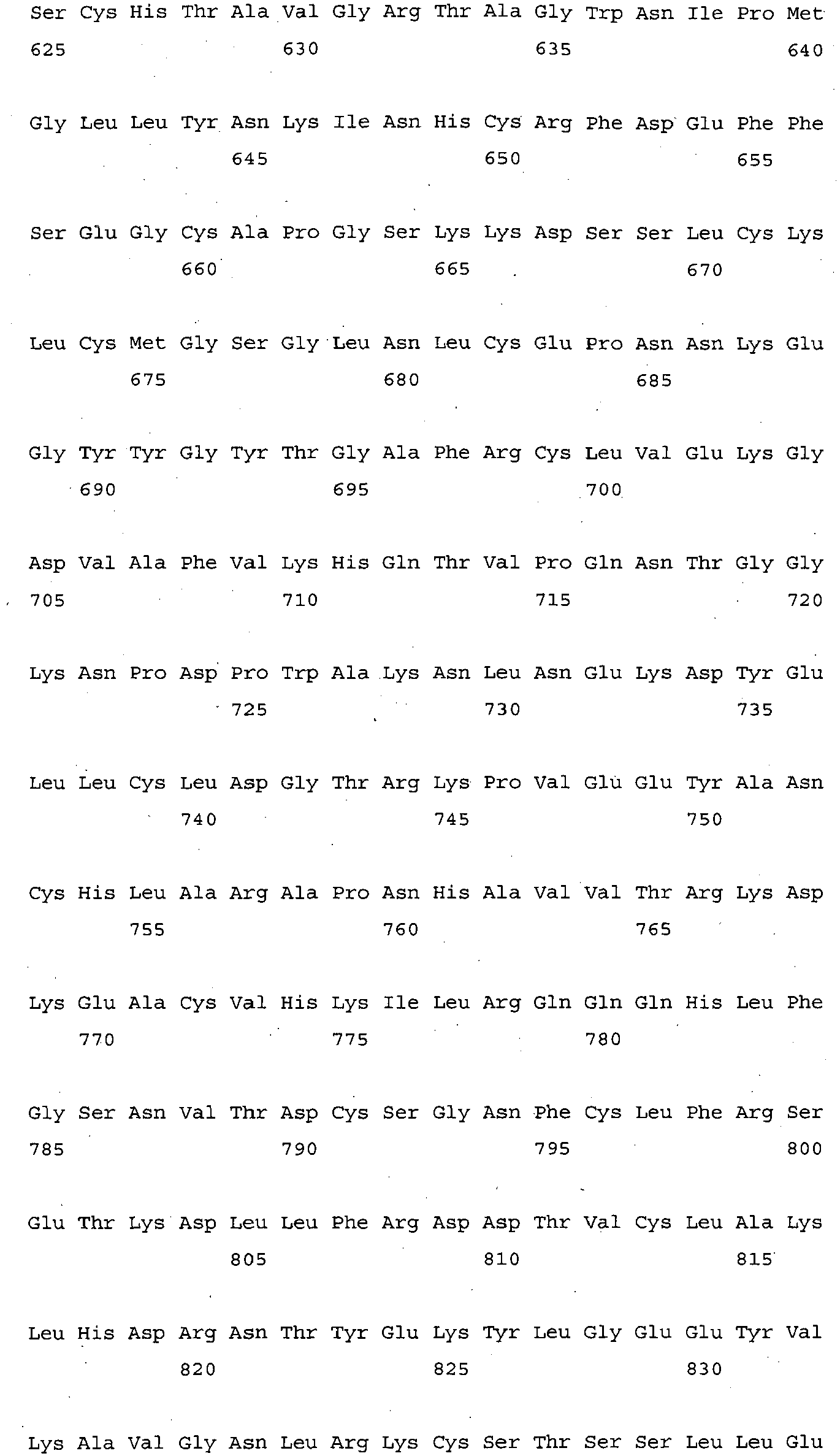


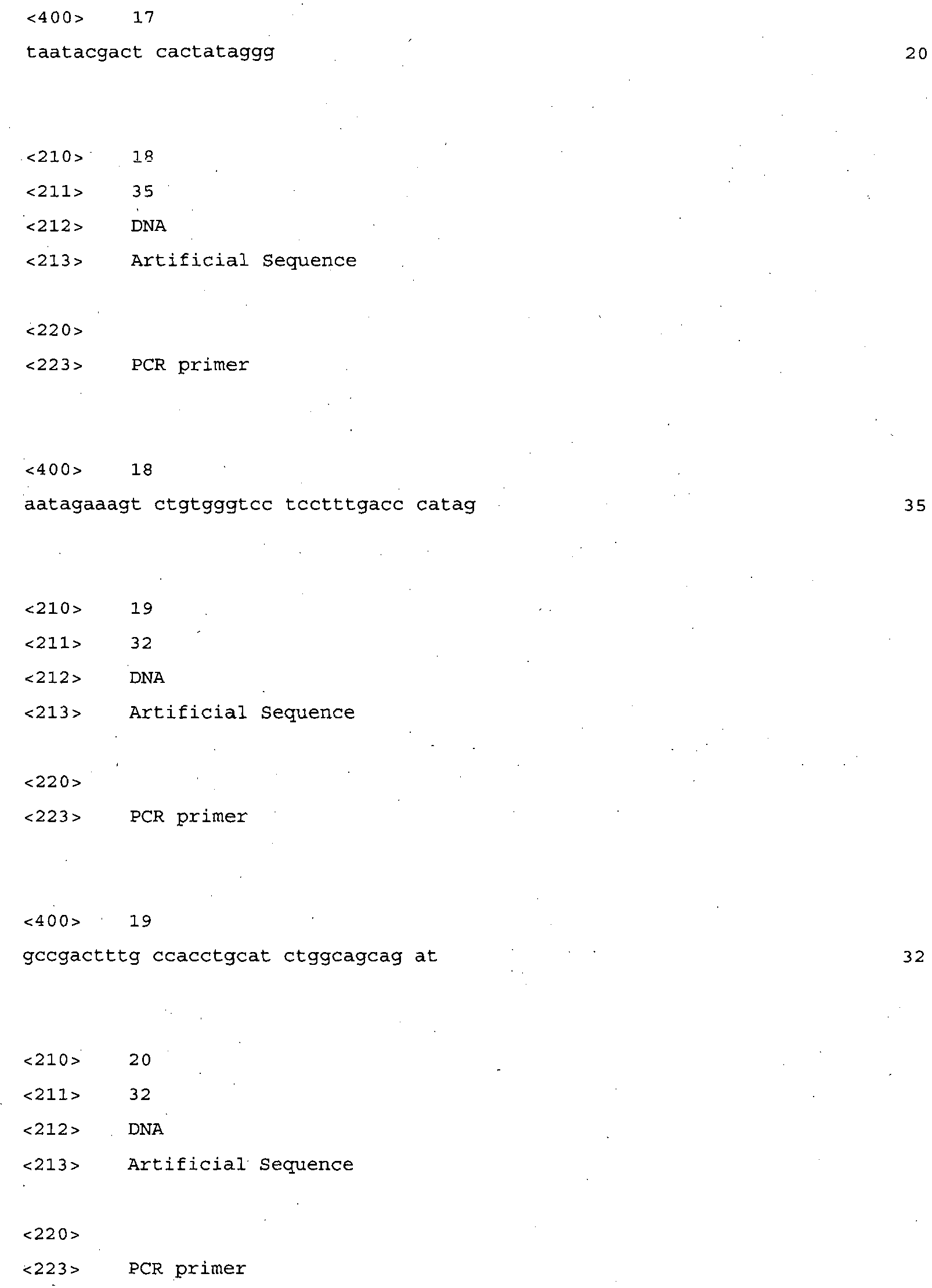


Claims (22)
- A granulocyte-colony stimulating factor mutant protein (G-CSF mutant protein) in which the 116th threonine is substituted with a hydrophobic amino acid.
- The G-CSF mutant protein according to claim 1, wherein the hydrophobic amino acid is suitable for sulfide bonding with the 17th cysteine of G-CSF.
- The G-CSF mutant protein according to claim 1, wherein the 116th amino acid threonine is substituted with cysteine.
- A fusion protein in which transferrin is peptide-bonded to a terminal of a granulocyte-colony stimulating factor (G-CSF) protein or a G-CSF mutant protein in which the 116th threonine is substituted with a hydrophobic amino acid.
- The fusion protein according to claim 4, wherein the transferrin has the deletion of a signal peptide at the amino terminal region.
- A pharmaceutical composition for the prevention or treatment of neutropenia comprising one of those proteins of claim 1 ∼ claim 5 as an active ingredient.
- An anticancer adjuvant comprising one of those proteins of claim 1 ∼ claim 5 as an active ingredient.
- A pharmaceutical composition for the prevention or treatment of ischemic disease comprising one of those proteins of claim 1 ∼ claim 5 as an active ingredient.
- An expression vector containing the gene encoding one of those proteins of claim 1 ∼ claim 5.
- The expression vector according to claim 9, wherein the gene encoding the protein above has the substitution of a nucleotide sequence of the restriction enzyme recognition site with another nucleotide sequence encoding the same amino acid as the one encoded by the said nucleotide sequence.
- The expression vector according to claim 9, wherein the gene encoding the protein has the substitution of thymine of the BamHI restriction enzyme recognition site (GGATCC) with cytosine.
- The expression vector according to claim 9, wherein the gene encoding the protein additionally contains a restriction enzyme recognition site at the end of the gene.
- The expression vector according to claim 9, wherein the expression vector is presented by one of the cleavage maps selected from the group consisting of Figures 1, 2, 3, and 4.
- A transformant prepared by inserting the expression vector of claim 9 into a host cell.
- The transformant according to claim 14, wherein the host cell is selected from the group consisting of E. coli, yeast, fungi, plant cells, and animal cells.
- A method for preparing the protein above containing the step of obtaining the target protein from the transformant prepared by inserting the expression vector harboring the gene encoding one of those proteins of claim 1 ∼ claim 5 into a host cell.
- A method for preventing or treating neutropenia containing the step of administering a protein selected among those of claim 1 ∼ claim 5, an expression vector comprising the gene encoding the protein selected above, or a transformant prepared by inserting the expression vector above into a host cell to a subject having neutropenia.
- A method for inhibiting the decrease in number of neutrophils containing the step of administering a protein selected among those of claim 1 ∼ claim 5, an expression vector comprising the gene encoding the protein selected above, or a transformant prepared by inserting the expression vector above into a host cell to a subject
- A method for preventing or treating ischemic disease containing the step of administering a protein selected among those of claim 1 ∼ claim 5, an expression vector comprising the gene encoding the protein selected above, or a transformant prepared by inserting the expression vector above into a host cell to a subject having ischemic disease.
- A use of one of those proteins of claim 1 ∼ claim 5, an expression vector containing the gene encoding the selected protein, or a transformant prepared by inserting the expression vector into a host cell for the prevention or treatment of neutropenia.
- A use of one of those proteins of claim 1 ∼ claim 5, an expression vector containing the gene encoding the selected protein, or a transformant prepared by inserting the expression vector into a host cell as an anticancer adjuvant.
- A use of one of those proteins of claim 1 ∼ claim 5, an expression vector containing the gene encoding the selected protein, or a transformant prepared by inserting the expression vector into a host cell for the treatment of ischemic disease.
Applications Claiming Priority (3)
| Application Number | Priority Date | Filing Date | Title |
|---|---|---|---|
| KR20140093260 | 2014-07-23 | ||
| KR1020140137414A KR101623906B1 (en) | 2014-07-23 | 2014-10-13 | Pharmaceutical compositions comprising mutant proteins of Granulocyte-colony stimulating factor or transferrin fusion proteins thereof |
| PCT/KR2014/011269 WO2016013725A1 (en) | 2014-07-23 | 2014-11-21 | Pharmaceutical composition containing, as active ingredient, granulocyte-colony stimulating factor mutant protein or transferrin fusion protein thereof |
Publications (3)
| Publication Number | Publication Date |
|---|---|
| EP3173422A1 true EP3173422A1 (en) | 2017-05-31 |
| EP3173422A4 EP3173422A4 (en) | 2018-03-14 |
| EP3173422B1 EP3173422B1 (en) | 2019-05-08 |
Family
ID=55354544
Family Applications (1)
| Application Number | Title | Priority Date | Filing Date |
|---|---|---|---|
| EP14898071.7A Active EP3173422B1 (en) | 2014-07-23 | 2014-11-21 | Pharmaceutical composition containing, as active ingredient, granulocyte-colony stimulating factor mutant protein or transferrin fusion protein thereof |
Country Status (4)
| Country | Link |
|---|---|
| US (2) | US10479822B2 (en) |
| EP (1) | EP3173422B1 (en) |
| KR (1) | KR101623906B1 (en) |
| ES (1) | ES2735899T3 (en) |
Families Citing this family (1)
| Publication number | Priority date | Publication date | Assignee | Title |
|---|---|---|---|---|
| US20200270683A1 (en) * | 2017-09-26 | 2020-08-27 | Brown University | Methods for obtaining embryonic stem cell dna methylation signatures |
Family Cites Families (10)
| Publication number | Priority date | Publication date | Assignee | Title |
|---|---|---|---|---|
| KR100356140B1 (en) | 1999-07-08 | 2002-10-19 | 한미약품공업 주식회사 | Modified Human Granulocyte-Colony Stimulating Factor and Process for Producing Same |
| US6555660B2 (en) | 2000-01-10 | 2003-04-29 | Maxygen Holdings Ltd. | G-CSF conjugates |
| HUP0203751A3 (en) * | 2000-01-10 | 2005-01-28 | Maxygen Holdings Ltd Redwood C | G-csf conjugates |
| KR100961859B1 (en) | 2001-07-11 | 2010-06-09 | 맥시겐 홀딩스 엘티디 | G-CS conjugate |
| WO2005034877A2 (en) | 2003-10-10 | 2005-04-21 | University Of Southern California | G-csf transferrin fusion proteins |
| EP2144923B1 (en) | 2007-04-03 | 2013-02-13 | BioGeneriX AG | Methods of treatment using glycopegylated g-csf |
| WO2009086132A2 (en) | 2007-12-20 | 2009-07-09 | University Of Southern California | Design of spacers to increase the expression of recombinant fusion proteins |
| CA2737756A1 (en) | 2008-09-19 | 2010-03-25 | Grant Yonehiro | Method for the treatment of radiation-induced neutropenia by administration of a multi-pegylated granulocyte colony stimulating factor (g-csf) variant |
| JP2012046469A (en) * | 2010-08-30 | 2012-03-08 | Univ Of Tsukuba | Apoptosis inducer containing lactoferrin |
| KR101330033B1 (en) | 2010-10-18 | 2013-11-22 | 대한민국 | Therapeutic Drug Composition For Ischemic Disease Comprising Human Granulocyte-Colony Stimulating Factor Mutant |
-
2014
- 2014-10-13 KR KR1020140137414A patent/KR101623906B1/en active Active
- 2014-11-21 EP EP14898071.7A patent/EP3173422B1/en active Active
- 2014-11-21 US US15/327,838 patent/US10479822B2/en active Active
- 2014-11-21 ES ES14898071T patent/ES2735899T3/en active Active
-
2019
- 2019-09-19 US US16/575,612 patent/US11421009B2/en active Active
Also Published As
| Publication number | Publication date |
|---|---|
| EP3173422A4 (en) | 2018-03-14 |
| US10479822B2 (en) | 2019-11-19 |
| KR20160012059A (en) | 2016-02-02 |
| EP3173422B1 (en) | 2019-05-08 |
| US20200002396A1 (en) | 2020-01-02 |
| KR101623906B1 (en) | 2016-05-24 |
| US20170218038A1 (en) | 2017-08-03 |
| ES2735899T3 (en) | 2019-12-20 |
| US11421009B2 (en) | 2022-08-23 |
Similar Documents
| Publication | Publication Date | Title |
|---|---|---|
| JP2022023184A (en) | Activin-ActRIIa antagonist and its use to increase red blood cell levels | |
| CN109929806B (en) | A stem cell expressing GLP1 and FGF21 and use thereof | |
| JP2022071041A (en) | Methods for dosing an activin-actriia antagonist and methods for monitoring treated patients | |
| EP2830646B1 (en) | Compositions and methods of use for treating metabolic disorders | |
| KR101993714B1 (en) | Compositions and methods of use in treating metabolic disorders | |
| US10653751B2 (en) | Methods of treating cancer metastasis by using interleukin-10 | |
| CN102164949B (en) | Novel recombinant fusion protein | |
| KR20170065026A (en) | Compositions and methods of use for treating metabolic disorders | |
| EA018037B1 (en) | Method for increasing red blood cell levels | |
| EP2986306A1 (en) | Methods of using interleukin-10 for treating diseases and disorders | |
| US20170190753A1 (en) | Recombinant proteins having factor h activity | |
| JP2021107453A (en) | Method and composition for increasing red blood cells | |
| CN103641896B (en) | The purposes of gelatin-like unit | |
| US20170204157A1 (en) | Human fgfr2c extracellular protein as well as coding gene and application thereof | |
| US11421009B2 (en) | Pharmaceutical composition containing, as active ingredient, granulocyte-colony stimulating factor mutant protein or transferrin fusion protein thereof | |
| WO2018205622A1 (en) | Pegylated polypeptide having tumor inhibition function, preparation method therefor and application thereof | |
| JP2003500053A (en) | Vascular endothelial growth factor dimer | |
| EP3366770A1 (en) | Dna vaccine against amyloid-beta and tau | |
| US8188039B2 (en) | VEGF-D mutants and their use | |
| KR20180003677A (en) | Pharmaceutical compositions comprising mutant proteins of human growth hormone or transferrin fusion proteins thereof | |
| Sano et al. | Production and characterization of genetically modified human IL-11 variants | |
| RU2515914C1 (en) | Hybrid protein based on recombinant human erythropoietin, having prolonged action (versions), and method of its production | |
| KR101077611B1 (en) | A preparation method of solubility recombinant protein by use of Chemotaxis protein cheZ as a fusion expression partner | |
| KR101841646B1 (en) | Method for the preparation of AIMP2-DX2 as a target for anti-cancer drug discovery | |
| WO2016013725A1 (en) | Pharmaceutical composition containing, as active ingredient, granulocyte-colony stimulating factor mutant protein or transferrin fusion protein thereof |
Legal Events
| Date | Code | Title | Description |
|---|---|---|---|
| STAA | Information on the status of an ep patent application or granted ep patent |
Free format text: STATUS: THE INTERNATIONAL PUBLICATION HAS BEEN MADE |
|
| PUAI | Public reference made under article 153(3) epc to a published international application that has entered the european phase |
Free format text: ORIGINAL CODE: 0009012 |
|
| STAA | Information on the status of an ep patent application or granted ep patent |
Free format text: STATUS: REQUEST FOR EXAMINATION WAS MADE |
|
| 17P | Request for examination filed |
Effective date: 20170216 |
|
| AK | Designated contracting states |
Kind code of ref document: A1 Designated state(s): AL AT BE BG CH CY CZ DE DK EE ES FI FR GB GR HR HU IE IS IT LI LT LU LV MC MK MT NL NO PL PT RO RS SE SI SK SM TR |
|
| AX | Request for extension of the european patent |
Extension state: BA ME |
|
| RAP1 | Party data changed (applicant data changed or rights of an application transferred) |
Owner name: CREAPLANET CO. LTD. |
|
| DAX | Request for extension of the european patent (deleted) | ||
| RIC1 | Information provided on ipc code assigned before grant |
Ipc: C07K 14/79 20060101ALI20171106BHEP Ipc: A61P 9/10 20060101ALI20171106BHEP Ipc: C07K 14/535 20060101AFI20171106BHEP Ipc: C07K 19/00 20060101ALI20171106BHEP Ipc: C12N 15/62 20060101ALI20171106BHEP Ipc: A61K 38/17 20060101ALI20171106BHEP Ipc: A61P 35/00 20060101ALI20171106BHEP |
|
| RIC1 | Information provided on ipc code assigned before grant |
Ipc: C07K 19/00 20060101ALI20180201BHEP Ipc: A61K 38/17 20060101ALI20180201BHEP Ipc: A61P 9/10 20060101ALI20180201BHEP Ipc: C07K 14/535 20060101AFI20180201BHEP Ipc: C07K 14/79 20060101ALI20180201BHEP Ipc: A61P 35/00 20060101ALI20180201BHEP Ipc: C12N 15/62 20060101ALI20180201BHEP |
|
| A4 | Supplementary search report drawn up and despatched |
Effective date: 20180208 |
|
| RAP1 | Party data changed (applicant data changed or rights of an application transferred) |
Owner name: SUPEX BNP CO., LTD. |
|
| GRAP | Despatch of communication of intention to grant a patent |
Free format text: ORIGINAL CODE: EPIDOSNIGR1 |
|
| STAA | Information on the status of an ep patent application or granted ep patent |
Free format text: STATUS: GRANT OF PATENT IS INTENDED |
|
| INTG | Intention to grant announced |
Effective date: 20190125 |
|
| GRAS | Grant fee paid |
Free format text: ORIGINAL CODE: EPIDOSNIGR3 |
|
| GRAF | Information related to payment of grant fee modified |
Free format text: ORIGINAL CODE: EPIDOSCIGR3 |
|
| GRAF | Information related to payment of grant fee modified |
Free format text: ORIGINAL CODE: EPIDOSCIGR3 |
|
| GRAF | Information related to payment of grant fee modified |
Free format text: ORIGINAL CODE: EPIDOSCIGR3 |
|
| GRAA | (expected) grant |
Free format text: ORIGINAL CODE: 0009210 |
|
| STAA | Information on the status of an ep patent application or granted ep patent |
Free format text: STATUS: THE PATENT HAS BEEN GRANTED |
|
| AK | Designated contracting states |
Kind code of ref document: B1 Designated state(s): AL AT BE BG CH CY CZ DE DK EE ES FI FR GB GR HR HU IE IS IT LI LT LU LV MC MK MT NL NO PL PT RO RS SE SI SK SM TR |
|
| REG | Reference to a national code |
Ref country code: GB Ref legal event code: FG4D |
|
| REG | Reference to a national code |
Ref country code: CH Ref legal event code: EP Ref country code: AT Ref legal event code: REF Ref document number: 1130003 Country of ref document: AT Kind code of ref document: T Effective date: 20190515 |
|
| REG | Reference to a national code |
Ref country code: DE Ref legal event code: R096 Ref document number: 602014046649 Country of ref document: DE |
|
| REG | Reference to a national code |
Ref country code: IE Ref legal event code: FG4D |
|
| REG | Reference to a national code |
Ref country code: CH Ref legal event code: NV Representative=s name: SCHMAUDER AND PARTNER AG PATENT- UND MARKENANW, CH |
|
| REG | Reference to a national code |
Ref country code: NL Ref legal event code: FP |
|
| REG | Reference to a national code |
Ref country code: LT Ref legal event code: MG4D |
|
| PG25 | Lapsed in a contracting state [announced via postgrant information from national office to epo] |
Ref country code: LT Free format text: LAPSE BECAUSE OF FAILURE TO SUBMIT A TRANSLATION OF THE DESCRIPTION OR TO PAY THE FEE WITHIN THE PRESCRIBED TIME-LIMIT Effective date: 20190508 Ref country code: HR Free format text: LAPSE BECAUSE OF FAILURE TO SUBMIT A TRANSLATION OF THE DESCRIPTION OR TO PAY THE FEE WITHIN THE PRESCRIBED TIME-LIMIT Effective date: 20190508 Ref country code: SE Free format text: LAPSE BECAUSE OF FAILURE TO SUBMIT A TRANSLATION OF THE DESCRIPTION OR TO PAY THE FEE WITHIN THE PRESCRIBED TIME-LIMIT Effective date: 20190508 Ref country code: NO Free format text: LAPSE BECAUSE OF FAILURE TO SUBMIT A TRANSLATION OF THE DESCRIPTION OR TO PAY THE FEE WITHIN THE PRESCRIBED TIME-LIMIT Effective date: 20190808 Ref country code: PT Free format text: LAPSE BECAUSE OF FAILURE TO SUBMIT A TRANSLATION OF THE DESCRIPTION OR TO PAY THE FEE WITHIN THE PRESCRIBED TIME-LIMIT Effective date: 20190908 Ref country code: AL Free format text: LAPSE BECAUSE OF FAILURE TO SUBMIT A TRANSLATION OF THE DESCRIPTION OR TO PAY THE FEE WITHIN THE PRESCRIBED TIME-LIMIT Effective date: 20190508 Ref country code: FI Free format text: LAPSE BECAUSE OF FAILURE TO SUBMIT A TRANSLATION OF THE DESCRIPTION OR TO PAY THE FEE WITHIN THE PRESCRIBED TIME-LIMIT Effective date: 20190508 |
|
| PG25 | Lapsed in a contracting state [announced via postgrant information from national office to epo] |
Ref country code: LV Free format text: LAPSE BECAUSE OF FAILURE TO SUBMIT A TRANSLATION OF THE DESCRIPTION OR TO PAY THE FEE WITHIN THE PRESCRIBED TIME-LIMIT Effective date: 20190508 Ref country code: RS Free format text: LAPSE BECAUSE OF FAILURE TO SUBMIT A TRANSLATION OF THE DESCRIPTION OR TO PAY THE FEE WITHIN THE PRESCRIBED TIME-LIMIT Effective date: 20190508 Ref country code: GR Free format text: LAPSE BECAUSE OF FAILURE TO SUBMIT A TRANSLATION OF THE DESCRIPTION OR TO PAY THE FEE WITHIN THE PRESCRIBED TIME-LIMIT Effective date: 20190809 Ref country code: BG Free format text: LAPSE BECAUSE OF FAILURE TO SUBMIT A TRANSLATION OF THE DESCRIPTION OR TO PAY THE FEE WITHIN THE PRESCRIBED TIME-LIMIT Effective date: 20190808 |
|
| REG | Reference to a national code |
Ref country code: AT Ref legal event code: MK05 Ref document number: 1130003 Country of ref document: AT Kind code of ref document: T Effective date: 20190508 |
|
| REG | Reference to a national code |
Ref country code: ES Ref legal event code: FG2A Ref document number: 2735899 Country of ref document: ES Kind code of ref document: T3 Effective date: 20191220 |
|
| PG25 | Lapsed in a contracting state [announced via postgrant information from national office to epo] |
Ref country code: CZ Free format text: LAPSE BECAUSE OF FAILURE TO SUBMIT A TRANSLATION OF THE DESCRIPTION OR TO PAY THE FEE WITHIN THE PRESCRIBED TIME-LIMIT Effective date: 20190508 Ref country code: SK Free format text: LAPSE BECAUSE OF FAILURE TO SUBMIT A TRANSLATION OF THE DESCRIPTION OR TO PAY THE FEE WITHIN THE PRESCRIBED TIME-LIMIT Effective date: 20190508 Ref country code: RO Free format text: LAPSE BECAUSE OF FAILURE TO SUBMIT A TRANSLATION OF THE DESCRIPTION OR TO PAY THE FEE WITHIN THE PRESCRIBED TIME-LIMIT Effective date: 20190508 Ref country code: AT Free format text: LAPSE BECAUSE OF FAILURE TO SUBMIT A TRANSLATION OF THE DESCRIPTION OR TO PAY THE FEE WITHIN THE PRESCRIBED TIME-LIMIT Effective date: 20190508 Ref country code: DK Free format text: LAPSE BECAUSE OF FAILURE TO SUBMIT A TRANSLATION OF THE DESCRIPTION OR TO PAY THE FEE WITHIN THE PRESCRIBED TIME-LIMIT Effective date: 20190508 Ref country code: EE Free format text: LAPSE BECAUSE OF FAILURE TO SUBMIT A TRANSLATION OF THE DESCRIPTION OR TO PAY THE FEE WITHIN THE PRESCRIBED TIME-LIMIT Effective date: 20190508 |
|
| REG | Reference to a national code |
Ref country code: DE Ref legal event code: R097 Ref document number: 602014046649 Country of ref document: DE |
|
| PG25 | Lapsed in a contracting state [announced via postgrant information from national office to epo] |
Ref country code: SM Free format text: LAPSE BECAUSE OF FAILURE TO SUBMIT A TRANSLATION OF THE DESCRIPTION OR TO PAY THE FEE WITHIN THE PRESCRIBED TIME-LIMIT Effective date: 20190508 |
|
| PLBE | No opposition filed within time limit |
Free format text: ORIGINAL CODE: 0009261 |
|
| STAA | Information on the status of an ep patent application or granted ep patent |
Free format text: STATUS: NO OPPOSITION FILED WITHIN TIME LIMIT |
|
| PG25 | Lapsed in a contracting state [announced via postgrant information from national office to epo] |
Ref country code: TR Free format text: LAPSE BECAUSE OF FAILURE TO SUBMIT A TRANSLATION OF THE DESCRIPTION OR TO PAY THE FEE WITHIN THE PRESCRIBED TIME-LIMIT Effective date: 20190508 |
|
| 26N | No opposition filed |
Effective date: 20200211 |
|
| PG25 | Lapsed in a contracting state [announced via postgrant information from national office to epo] |
Ref country code: PL Free format text: LAPSE BECAUSE OF FAILURE TO SUBMIT A TRANSLATION OF THE DESCRIPTION OR TO PAY THE FEE WITHIN THE PRESCRIBED TIME-LIMIT Effective date: 20190508 |
|
| PG25 | Lapsed in a contracting state [announced via postgrant information from national office to epo] |
Ref country code: SI Free format text: LAPSE BECAUSE OF FAILURE TO SUBMIT A TRANSLATION OF THE DESCRIPTION OR TO PAY THE FEE WITHIN THE PRESCRIBED TIME-LIMIT Effective date: 20190508 |
|
| PG25 | Lapsed in a contracting state [announced via postgrant information from national office to epo] |
Ref country code: MC Free format text: LAPSE BECAUSE OF FAILURE TO SUBMIT A TRANSLATION OF THE DESCRIPTION OR TO PAY THE FEE WITHIN THE PRESCRIBED TIME-LIMIT Effective date: 20190508 Ref country code: LU Free format text: LAPSE BECAUSE OF NON-PAYMENT OF DUE FEES Effective date: 20191121 |
|
| REG | Reference to a national code |
Ref country code: BE Ref legal event code: MM Effective date: 20191130 |
|
| PG25 | Lapsed in a contracting state [announced via postgrant information from national office to epo] |
Ref country code: IE Free format text: LAPSE BECAUSE OF NON-PAYMENT OF DUE FEES Effective date: 20191121 |
|
| PG25 | Lapsed in a contracting state [announced via postgrant information from national office to epo] |
Ref country code: BE Free format text: LAPSE BECAUSE OF NON-PAYMENT OF DUE FEES Effective date: 20191130 |
|
| PG25 | Lapsed in a contracting state [announced via postgrant information from national office to epo] |
Ref country code: CY Free format text: LAPSE BECAUSE OF FAILURE TO SUBMIT A TRANSLATION OF THE DESCRIPTION OR TO PAY THE FEE WITHIN THE PRESCRIBED TIME-LIMIT Effective date: 20190508 |
|
| PG25 | Lapsed in a contracting state [announced via postgrant information from national office to epo] |
Ref country code: IS Free format text: LAPSE BECAUSE OF FAILURE TO SUBMIT A TRANSLATION OF THE DESCRIPTION OR TO PAY THE FEE WITHIN THE PRESCRIBED TIME-LIMIT Effective date: 20190908 |
|
| PG25 | Lapsed in a contracting state [announced via postgrant information from national office to epo] |
Ref country code: HU Free format text: LAPSE BECAUSE OF FAILURE TO SUBMIT A TRANSLATION OF THE DESCRIPTION OR TO PAY THE FEE WITHIN THE PRESCRIBED TIME-LIMIT; INVALID AB INITIO Effective date: 20141121 Ref country code: MT Free format text: LAPSE BECAUSE OF FAILURE TO SUBMIT A TRANSLATION OF THE DESCRIPTION OR TO PAY THE FEE WITHIN THE PRESCRIBED TIME-LIMIT Effective date: 20190508 |
|
| REG | Reference to a national code |
Ref country code: DE Ref legal event code: R081 Ref document number: 602014046649 Country of ref document: DE Owner name: PANACEA INC., ANSAN-SI, KR Free format text: FORMER OWNER: SUPEX BNP CO., LTD., ANSAN-SI, GYEONGGI-DO, KR |
|
| REG | Reference to a national code |
Ref country code: NL Ref legal event code: HC Owner name: PANACEA INC.; KO Free format text: DETAILS ASSIGNMENT: CHANGE OF OWNER(S), CHANGE OF OWNER(S) NAME; FORMER OWNER NAME: SUPEX BNP CO., LTD. Effective date: 20211213 |
|
| REG | Reference to a national code |
Ref country code: ES Ref legal event code: PC2A Owner name: PANACEA INC. Effective date: 20220519 |
|
| PG25 | Lapsed in a contracting state [announced via postgrant information from national office to epo] |
Ref country code: MK Free format text: LAPSE BECAUSE OF FAILURE TO SUBMIT A TRANSLATION OF THE DESCRIPTION OR TO PAY THE FEE WITHIN THE PRESCRIBED TIME-LIMIT Effective date: 20190508 |
|
| P01 | Opt-out of the competence of the unified patent court (upc) registered |
Effective date: 20240409 |
|
| PGFP | Annual fee paid to national office [announced via postgrant information from national office to epo] |
Ref country code: NL Payment date: 20241024 Year of fee payment: 11 |
|
| PGFP | Annual fee paid to national office [announced via postgrant information from national office to epo] |
Ref country code: DE Payment date: 20241015 Year of fee payment: 11 |
|
| PGFP | Annual fee paid to national office [announced via postgrant information from national office to epo] |
Ref country code: GB Payment date: 20241017 Year of fee payment: 11 |
|
| PGFP | Annual fee paid to national office [announced via postgrant information from national office to epo] |
Ref country code: FR Payment date: 20241021 Year of fee payment: 11 |
|
| PGFP | Annual fee paid to national office [announced via postgrant information from national office to epo] |
Ref country code: IT Payment date: 20241028 Year of fee payment: 11 Ref country code: ES Payment date: 20241210 Year of fee payment: 11 |
|
| PGFP | Annual fee paid to national office [announced via postgrant information from national office to epo] |
Ref country code: CH Payment date: 20241201 Year of fee payment: 11 |
|
| REG | Reference to a national code |
Ref country code: DE Ref legal event code: R119 Ref document number: 602014046649 Country of ref document: DE |
|
| REG | Reference to a national code |
Ref country code: CH Ref legal event code: H13 Free format text: ST27 STATUS EVENT CODE: U-0-0-H10-H13 (AS PROVIDED BY THE NATIONAL OFFICE) Effective date: 20260623 |
|
| REG | Reference to a national code |
Ref country code: NL Ref legal event code: MM Effective date: 20251201 |
|
| PG25 | Lapsed in a contracting state [announced via postgrant information from national office to epo] |
Ref country code: CH Free format text: LAPSE BECAUSE OF NON-PAYMENT OF DUE FEES Effective date: 20251130 |