EP3170901B1 - Method for producing dendritic cells from stem cells - Google Patents
Method for producing dendritic cells from stem cells Download PDFInfo
- Publication number
- EP3170901B1 EP3170901B1 EP15822419.6A EP15822419A EP3170901B1 EP 3170901 B1 EP3170901 B1 EP 3170901B1 EP 15822419 A EP15822419 A EP 15822419A EP 3170901 B1 EP3170901 B1 EP 3170901B1
- Authority
- EP
- European Patent Office
- Prior art keywords
- cell
- cells
- prt
- mhc
- hla
- Prior art date
- Legal status (The legal status is an assumption and is not a legal conclusion. Google has not performed a legal analysis and makes no representation as to the accuracy of the status listed.)
- Active
Links
- 210000004443 dendritic cell Anatomy 0.000 title claims description 121
- 210000000130 stem cell Anatomy 0.000 title claims description 106
- 238000004519 manufacturing process Methods 0.000 title claims description 15
- 210000004027 cell Anatomy 0.000 claims description 367
- 238000000034 method Methods 0.000 claims description 99
- 210000001616 monocyte Anatomy 0.000 claims description 48
- 108010058597 HLA-DR Antigens Proteins 0.000 claims description 28
- 102000006354 HLA-DR Antigens Human genes 0.000 claims description 28
- 108010017213 Granulocyte-Macrophage Colony-Stimulating Factor Proteins 0.000 claims description 27
- 102100039620 Granulocyte-macrophage colony-stimulating factor Human genes 0.000 claims description 27
- 108010062347 HLA-DQ Antigens Proteins 0.000 claims description 20
- 239000012679 serum free medium Substances 0.000 claims description 20
- 102000004127 Cytokines Human genes 0.000 claims description 16
- 108090000695 Cytokines Proteins 0.000 claims description 16
- 102000004388 Interleukin-4 Human genes 0.000 claims description 16
- 108090000978 Interleukin-4 Proteins 0.000 claims description 16
- 229940028885 interleukin-4 Drugs 0.000 claims description 16
- 108010010378 HLA-DP Antigens Proteins 0.000 claims description 15
- 102000015789 HLA-DP Antigens Human genes 0.000 claims description 15
- 108010046938 Macrophage Colony-Stimulating Factor Proteins 0.000 claims description 15
- 102000007651 Macrophage Colony-Stimulating Factor Human genes 0.000 claims description 15
- 210000004504 adult stem cell Anatomy 0.000 claims description 9
- 238000000338 in vitro Methods 0.000 claims description 9
- 230000002163 immunogen Effects 0.000 claims description 8
- 230000002757 inflammatory effect Effects 0.000 claims description 7
- 238000012546 transfer Methods 0.000 claims description 7
- 210000004263 induced pluripotent stem cell Anatomy 0.000 claims description 6
- 210000001671 embryonic stem cell Anatomy 0.000 claims description 5
- 230000004936 stimulating effect Effects 0.000 claims description 5
- 108090000765 processed proteins & peptides Proteins 0.000 description 205
- 241000282414 Homo sapiens Species 0.000 description 169
- 108090000623 proteins and genes Proteins 0.000 description 153
- 235000009109 Betula pendula Nutrition 0.000 description 144
- 241000219430 Betula pendula Species 0.000 description 142
- 102000004169 proteins and genes Human genes 0.000 description 129
- 235000018102 proteins Nutrition 0.000 description 108
- 102000004196 processed proteins & peptides Human genes 0.000 description 93
- 150000001413 amino acids Chemical group 0.000 description 68
- 210000000612 antigen-presenting cell Anatomy 0.000 description 47
- 230000005847 immunogenicity Effects 0.000 description 46
- 229960000598 infliximab Drugs 0.000 description 41
- 210000003819 peripheral blood mononuclear cell Anatomy 0.000 description 35
- 239000002609 medium Substances 0.000 description 33
- 238000004458 analytical method Methods 0.000 description 26
- 239000000427 antigen Substances 0.000 description 26
- 108091007433 antigens Proteins 0.000 description 26
- 102000036639 antigens Human genes 0.000 description 26
- 230000014509 gene expression Effects 0.000 description 26
- 239000000203 mixture Substances 0.000 description 26
- 230000004069 differentiation Effects 0.000 description 24
- 210000002443 helper t lymphocyte Anatomy 0.000 description 24
- 230000036961 partial effect Effects 0.000 description 24
- 239000011324 bead Substances 0.000 description 18
- 239000013575 birch pollen allergen Substances 0.000 description 18
- 238000011282 treatment Methods 0.000 description 18
- 101000968532 Dictyocaulus viviparus DVA-1 polyprotein Proteins 0.000 description 17
- 102000018713 Histocompatibility Antigens Class II Human genes 0.000 description 17
- 101000914484 Homo sapiens T-lymphocyte activation antigen CD80 Proteins 0.000 description 17
- 102100027222 T-lymphocyte activation antigen CD80 Human genes 0.000 description 17
- 108010027412 Histocompatibility Antigens Class II Proteins 0.000 description 16
- 108010029485 Protein Isoforms Proteins 0.000 description 16
- 102000001708 Protein Isoforms Human genes 0.000 description 16
- -1 antibodies Proteins 0.000 description 15
- 230000003394 haemopoietic effect Effects 0.000 description 14
- 230000006698 induction Effects 0.000 description 14
- 210000002540 macrophage Anatomy 0.000 description 14
- 230000008672 reprogramming Effects 0.000 description 14
- 210000002966 serum Anatomy 0.000 description 14
- 101000946889 Homo sapiens Monocyte differentiation antigen CD14 Proteins 0.000 description 13
- 102100035877 Monocyte differentiation antigen CD14 Human genes 0.000 description 13
- 230000030741 antigen processing and presentation Effects 0.000 description 13
- 239000003814 drug Substances 0.000 description 13
- 210000001082 somatic cell Anatomy 0.000 description 13
- 108010010803 Gelatin Proteins 0.000 description 12
- 101001046686 Homo sapiens Integrin alpha-M Proteins 0.000 description 12
- 102100022338 Integrin alpha-M Human genes 0.000 description 12
- 102100022297 Integrin alpha-X Human genes 0.000 description 12
- 229920000159 gelatin Polymers 0.000 description 12
- 239000008273 gelatin Substances 0.000 description 12
- 235000019322 gelatine Nutrition 0.000 description 12
- 235000011852 gelatine desserts Nutrition 0.000 description 12
- 239000000243 solution Substances 0.000 description 12
- 239000006228 supernatant Substances 0.000 description 12
- 108091003079 Bovine Serum Albumin Proteins 0.000 description 11
- 238000012300 Sequence Analysis Methods 0.000 description 11
- 108010073929 Vascular Endothelial Growth Factor A Proteins 0.000 description 11
- 102100039037 Vascular endothelial growth factor A Human genes 0.000 description 11
- 239000012091 fetal bovine serum Substances 0.000 description 11
- 239000003550 marker Substances 0.000 description 11
- 239000006144 Dulbecco’s modified Eagle's medium Substances 0.000 description 10
- 102000003974 Fibroblast growth factor 2 Human genes 0.000 description 10
- 108090000379 Fibroblast growth factor 2 Proteins 0.000 description 10
- 108010088652 Histocompatibility Antigens Class I Proteins 0.000 description 10
- 102000008949 Histocompatibility Antigens Class I Human genes 0.000 description 10
- 206010028980 Neoplasm Diseases 0.000 description 10
- 229960000074 biopharmaceutical Drugs 0.000 description 10
- 201000006417 multiple sclerosis Diseases 0.000 description 10
- UCSJYZPVAKXKNQ-HZYVHMACSA-N streptomycin Chemical compound CN[C@H]1[C@H](O)[C@@H](O)[C@H](CO)O[C@H]1O[C@@H]1[C@](C=O)(O)[C@H](C)O[C@H]1O[C@@H]1[C@@H](NC(N)=N)[C@H](O)[C@@H](NC(N)=N)[C@H](O)[C@H]1O UCSJYZPVAKXKNQ-HZYVHMACSA-N 0.000 description 10
- 241001465754 Metazoa Species 0.000 description 9
- 201000011510 cancer Diseases 0.000 description 9
- 201000010099 disease Diseases 0.000 description 9
- 208000037265 diseases, disorders, signs and symptoms Diseases 0.000 description 9
- 230000002265 prevention Effects 0.000 description 9
- 108010019530 Vascular Endothelial Growth Factors Proteins 0.000 description 8
- 230000001464 adherent effect Effects 0.000 description 8
- 239000003795 chemical substances by application Substances 0.000 description 8
- 210000002257 embryonic structure Anatomy 0.000 description 8
- 238000005516 engineering process Methods 0.000 description 8
- 239000003102 growth factor Substances 0.000 description 8
- 239000000126 substance Substances 0.000 description 8
- 241000124008 Mammalia Species 0.000 description 7
- 241000288906 Primates Species 0.000 description 7
- 210000001744 T-lymphocyte Anatomy 0.000 description 7
- 229940024606 amino acid Drugs 0.000 description 7
- 235000001014 amino acid Nutrition 0.000 description 7
- 238000012258 culturing Methods 0.000 description 7
- 238000004949 mass spectrometry Methods 0.000 description 7
- 238000005259 measurement Methods 0.000 description 7
- 238000002360 preparation method Methods 0.000 description 7
- 229910021642 ultra pure water Inorganic materials 0.000 description 7
- 239000012498 ultrapure water Substances 0.000 description 7
- XLYOFNOQVPJJNP-UHFFFAOYSA-N water Substances O XLYOFNOQVPJJNP-UHFFFAOYSA-N 0.000 description 7
- OZFAFGSSMRRTDW-UHFFFAOYSA-N (2,4-dichlorophenyl) benzenesulfonate Chemical compound ClC1=CC(Cl)=CC=C1OS(=O)(=O)C1=CC=CC=C1 OZFAFGSSMRRTDW-UHFFFAOYSA-N 0.000 description 6
- QTBSBXVTEAMEQO-UHFFFAOYSA-N Acetic acid Chemical compound CC(O)=O QTBSBXVTEAMEQO-UHFFFAOYSA-N 0.000 description 6
- WEVYAHXRMPXWCK-UHFFFAOYSA-N Acetonitrile Chemical compound CC#N WEVYAHXRMPXWCK-UHFFFAOYSA-N 0.000 description 6
- WVDDGKGOMKODPV-UHFFFAOYSA-N Benzyl alcohol Chemical compound OCC1=CC=CC=C1 WVDDGKGOMKODPV-UHFFFAOYSA-N 0.000 description 6
- 239000012591 Dulbecco’s Phosphate Buffered Saline Substances 0.000 description 6
- 241000282412 Homo Species 0.000 description 6
- 101000911390 Homo sapiens Coagulation factor VIII Proteins 0.000 description 6
- NWIBSHFKIJFRCO-WUDYKRTCSA-N Mytomycin Chemical compound C1N2C(C(C(C)=C(N)C3=O)=O)=C3[C@@H](COC(N)=O)[C@@]2(OC)[C@@H]2[C@H]1N2 NWIBSHFKIJFRCO-WUDYKRTCSA-N 0.000 description 6
- 108010033276 Peptide Fragments Proteins 0.000 description 6
- 102000007079 Peptide Fragments Human genes 0.000 description 6
- 241000746983 Phleum pratense Species 0.000 description 6
- IQFYYKKMVGJFEH-XLPZGREQSA-N Thymidine Chemical compound O=C1NC(=O)C(C)=CN1[C@@H]1O[C@H](CO)[C@@H](O)C1 IQFYYKKMVGJFEH-XLPZGREQSA-N 0.000 description 6
- DTQVDTLACAAQTR-UHFFFAOYSA-N Trifluoroacetic acid Chemical compound OC(=O)C(F)(F)F DTQVDTLACAAQTR-UHFFFAOYSA-N 0.000 description 6
- 238000006243 chemical reaction Methods 0.000 description 6
- 229940079593 drug Drugs 0.000 description 6
- 235000013601 eggs Nutrition 0.000 description 6
- 239000002158 endotoxin Substances 0.000 description 6
- 238000000684 flow cytometry Methods 0.000 description 6
- 229920006008 lipopolysaccharide Polymers 0.000 description 6
- 206010039073 rheumatoid arthritis Diseases 0.000 description 6
- 238000012360 testing method Methods 0.000 description 6
- WOVKYSAHUYNSMH-RRKCRQDMSA-N 5-bromodeoxyuridine Chemical compound C1[C@H](O)[C@@H](CO)O[C@H]1N1C(=O)NC(=O)C(Br)=C1 WOVKYSAHUYNSMH-RRKCRQDMSA-N 0.000 description 5
- 208000023275 Autoimmune disease Diseases 0.000 description 5
- ZDXPYRJPNDTMRX-VKHMYHEASA-N L-glutamine Chemical compound OC(=O)[C@@H](N)CCC(N)=O ZDXPYRJPNDTMRX-VKHMYHEASA-N 0.000 description 5
- 229930182816 L-glutamine Natural products 0.000 description 5
- 229930182555 Penicillin Natural products 0.000 description 5
- JGSARLDLIJGVTE-MBNYWOFBSA-N Penicillin G Chemical compound N([C@H]1[C@H]2SC([C@@H](N2C1=O)C(O)=O)(C)C)C(=O)CC1=CC=CC=C1 JGSARLDLIJGVTE-MBNYWOFBSA-N 0.000 description 5
- 108060008682 Tumor Necrosis Factor Proteins 0.000 description 5
- 102000000852 Tumor Necrosis Factor-alpha Human genes 0.000 description 5
- 210000003719 b-lymphocyte Anatomy 0.000 description 5
- 210000001161 mammalian embryo Anatomy 0.000 description 5
- 239000000463 material Substances 0.000 description 5
- 230000001404 mediated effect Effects 0.000 description 5
- 229940049954 penicillin Drugs 0.000 description 5
- 230000035755 proliferation Effects 0.000 description 5
- 230000001737 promoting effect Effects 0.000 description 5
- 229960005322 streptomycin Drugs 0.000 description 5
- 238000000108 ultra-filtration Methods 0.000 description 5
- 239000013598 vector Substances 0.000 description 5
- WOVKYSAHUYNSMH-UHFFFAOYSA-N BROMODEOXYURIDINE Natural products C1C(O)C(CO)OC1N1C(=O)NC(=O)C(Br)=C1 WOVKYSAHUYNSMH-UHFFFAOYSA-N 0.000 description 4
- 102100024505 Bone morphogenetic protein 4 Human genes 0.000 description 4
- KCXVZYZYPLLWCC-UHFFFAOYSA-N EDTA Chemical compound OC(=O)CN(CC(O)=O)CCN(CC(O)=O)CC(O)=O KCXVZYZYPLLWCC-UHFFFAOYSA-N 0.000 description 4
- LFQSCWFLJHTTHZ-UHFFFAOYSA-N Ethanol Chemical compound CCO LFQSCWFLJHTTHZ-UHFFFAOYSA-N 0.000 description 4
- 102100028976 HLA class I histocompatibility antigen, B alpha chain Human genes 0.000 description 4
- 108010058607 HLA-B Antigens Proteins 0.000 description 4
- 208000009292 Hemophilia A Diseases 0.000 description 4
- 108091006905 Human Serum Albumin Proteins 0.000 description 4
- 102000008100 Human Serum Albumin Human genes 0.000 description 4
- 102100037850 Interferon gamma Human genes 0.000 description 4
- 108010074328 Interferon-gamma Proteins 0.000 description 4
- 108010002386 Interleukin-3 Proteins 0.000 description 4
- 102000000646 Interleukin-3 Human genes 0.000 description 4
- 102000004058 Leukemia inhibitory factor Human genes 0.000 description 4
- 108090000581 Leukemia inhibitory factor Proteins 0.000 description 4
- 101150042431 SOX gene Proteins 0.000 description 4
- PXIPVTKHYLBLMZ-UHFFFAOYSA-N Sodium azide Chemical compound [Na+].[N-]=[N+]=[N-] PXIPVTKHYLBLMZ-UHFFFAOYSA-N 0.000 description 4
- 102000036693 Thrombopoietin Human genes 0.000 description 4
- 108010041111 Thrombopoietin Proteins 0.000 description 4
- 125000000539 amino acid group Chemical group 0.000 description 4
- 210000004102 animal cell Anatomy 0.000 description 4
- 210000002459 blastocyst Anatomy 0.000 description 4
- 239000002775 capsule Substances 0.000 description 4
- 238000001514 detection method Methods 0.000 description 4
- 239000012153 distilled water Substances 0.000 description 4
- 238000010828 elution Methods 0.000 description 4
- 210000003958 hematopoietic stem cell Anatomy 0.000 description 4
- 238000001727 in vivo Methods 0.000 description 4
- NOESYZHRGYRDHS-UHFFFAOYSA-N insulin Chemical compound N1C(=O)C(NC(=O)C(CCC(N)=O)NC(=O)C(CCC(O)=O)NC(=O)C(C(C)C)NC(=O)C(NC(=O)CN)C(C)CC)CSSCC(C(NC(CO)C(=O)NC(CC(C)C)C(=O)NC(CC=2C=CC(O)=CC=2)C(=O)NC(CCC(N)=O)C(=O)NC(CC(C)C)C(=O)NC(CCC(O)=O)C(=O)NC(CC(N)=O)C(=O)NC(CC=2C=CC(O)=CC=2)C(=O)NC(CSSCC(NC(=O)C(C(C)C)NC(=O)C(CC(C)C)NC(=O)C(CC=2C=CC(O)=CC=2)NC(=O)C(CC(C)C)NC(=O)C(C)NC(=O)C(CCC(O)=O)NC(=O)C(C(C)C)NC(=O)C(CC(C)C)NC(=O)C(CC=2NC=NC=2)NC(=O)C(CO)NC(=O)CNC2=O)C(=O)NCC(=O)NC(CCC(O)=O)C(=O)NC(CCCNC(N)=N)C(=O)NCC(=O)NC(CC=3C=CC=CC=3)C(=O)NC(CC=3C=CC=CC=3)C(=O)NC(CC=3C=CC(O)=CC=3)C(=O)NC(C(C)O)C(=O)N3C(CCC3)C(=O)NC(CCCCN)C(=O)NC(C)C(O)=O)C(=O)NC(CC(N)=O)C(O)=O)=O)NC(=O)C(C(C)CC)NC(=O)C(CO)NC(=O)C(C(C)O)NC(=O)C1CSSCC2NC(=O)C(CC(C)C)NC(=O)C(NC(=O)C(CCC(N)=O)NC(=O)C(CC(N)=O)NC(=O)C(NC(=O)C(N)CC=1C=CC=CC=1)C(C)C)CC1=CN=CN1 NOESYZHRGYRDHS-UHFFFAOYSA-N 0.000 description 4
- 238000004895 liquid chromatography mass spectrometry Methods 0.000 description 4
- 239000006166 lysate Substances 0.000 description 4
- 206010028417 myasthenia gravis Diseases 0.000 description 4
- 210000004940 nucleus Anatomy 0.000 description 4
- RXWNCPJZOCPEPQ-NVWDDTSBSA-N puromycin Chemical compound C1=CC(OC)=CC=C1C[C@H](N)C(=O)N[C@H]1[C@@H](O)[C@H](N2C3=NC=NC(=C3N=C2)N(C)C)O[C@@H]1CO RXWNCPJZOCPEPQ-NVWDDTSBSA-N 0.000 description 4
- 230000002829 reductive effect Effects 0.000 description 4
- 238000011160 research Methods 0.000 description 4
- 239000011550 stock solution Substances 0.000 description 4
- DGVVWUTYPXICAM-UHFFFAOYSA-N β‐Mercaptoethanol Chemical compound OCCS DGVVWUTYPXICAM-UHFFFAOYSA-N 0.000 description 4
- MZOFCQQQCNRIBI-VMXHOPILSA-N (3s)-4-[[(2s)-1-[[(2s)-1-[[(1s)-1-carboxy-2-hydroxyethyl]amino]-4-methyl-1-oxopentan-2-yl]amino]-5-(diaminomethylideneamino)-1-oxopentan-2-yl]amino]-3-[[2-[[(2s)-2,6-diaminohexanoyl]amino]acetyl]amino]-4-oxobutanoic acid Chemical compound OC[C@@H](C(O)=O)NC(=O)[C@H](CC(C)C)NC(=O)[C@H](CCCN=C(N)N)NC(=O)[C@H](CC(O)=O)NC(=O)CNC(=O)[C@@H](N)CCCCN MZOFCQQQCNRIBI-VMXHOPILSA-N 0.000 description 3
- 206010067484 Adverse reaction Diseases 0.000 description 3
- 108090000145 Bacillolysin Proteins 0.000 description 3
- DWRXFEITVBNRMK-UHFFFAOYSA-N Beta-D-1-Arabinofuranosylthymine Natural products O=C1NC(=O)C(C)=CN1C1C(O)C(O)C(CO)O1 DWRXFEITVBNRMK-UHFFFAOYSA-N 0.000 description 3
- 102100036301 C-C chemokine receptor type 7 Human genes 0.000 description 3
- 101150013553 CD40 gene Proteins 0.000 description 3
- 102100035793 CD83 antigen Human genes 0.000 description 3
- 102100025570 Cancer/testis antigen 1 Human genes 0.000 description 3
- 102100026735 Coagulation factor VIII Human genes 0.000 description 3
- 206010009944 Colon cancer Diseases 0.000 description 3
- 208000001333 Colorectal Neoplasms Diseases 0.000 description 3
- 108020004414 DNA Proteins 0.000 description 3
- 201000003542 Factor VIII deficiency Diseases 0.000 description 3
- 102100020715 Fms-related tyrosine kinase 3 ligand protein Human genes 0.000 description 3
- 101710162577 Fms-related tyrosine kinase 3 ligand protein Proteins 0.000 description 3
- 101000762379 Homo sapiens Bone morphogenetic protein 4 Proteins 0.000 description 3
- 101000716065 Homo sapiens C-C chemokine receptor type 7 Proteins 0.000 description 3
- 101000946856 Homo sapiens CD83 antigen Proteins 0.000 description 3
- 101000856237 Homo sapiens Cancer/testis antigen 1 Proteins 0.000 description 3
- 101000595923 Homo sapiens Placenta growth factor Proteins 0.000 description 3
- 102000015696 Interleukins Human genes 0.000 description 3
- 108010063738 Interleukins Proteins 0.000 description 3
- 210000004322 M2 macrophage Anatomy 0.000 description 3
- 108700018351 Major Histocompatibility Complex Proteins 0.000 description 3
- 241000699666 Mus <mouse, genus> Species 0.000 description 3
- 102000035092 Neutral proteases Human genes 0.000 description 3
- 108091005507 Neutral proteases Proteins 0.000 description 3
- 208000015914 Non-Hodgkin lymphomas Diseases 0.000 description 3
- 102100035194 Placenta growth factor Human genes 0.000 description 3
- DNIAPMSPPWPWGF-UHFFFAOYSA-N Propylene glycol Chemical compound CC(O)CO DNIAPMSPPWPWGF-UHFFFAOYSA-N 0.000 description 3
- 201000004681 Psoriasis Diseases 0.000 description 3
- 229920002684 Sepharose Polymers 0.000 description 3
- 108091008874 T cell receptors Proteins 0.000 description 3
- 102000016266 T-Cell Antigen Receptors Human genes 0.000 description 3
- 102000004887 Transforming Growth Factor beta Human genes 0.000 description 3
- 108090001012 Transforming Growth Factor beta Proteins 0.000 description 3
- 239000013504 Triton X-100 Substances 0.000 description 3
- 229920004890 Triton X-100 Polymers 0.000 description 3
- 102100040245 Tumor necrosis factor receptor superfamily member 5 Human genes 0.000 description 3
- 230000004913 activation Effects 0.000 description 3
- 239000004480 active ingredient Substances 0.000 description 3
- 230000006838 adverse reaction Effects 0.000 description 3
- 208000006682 alpha 1-Antitrypsin Deficiency Diseases 0.000 description 3
- 229940125644 antibody drug Drugs 0.000 description 3
- 238000013459 approach Methods 0.000 description 3
- IQFYYKKMVGJFEH-UHFFFAOYSA-N beta-L-thymidine Natural products O=C1NC(=O)C(C)=CN1C1OC(CO)C(O)C1 IQFYYKKMVGJFEH-UHFFFAOYSA-N 0.000 description 3
- 230000015572 biosynthetic process Effects 0.000 description 3
- 239000007853 buffer solution Substances 0.000 description 3
- 239000013592 cell lysate Substances 0.000 description 3
- 210000003690 classically activated macrophage Anatomy 0.000 description 3
- 238000005345 coagulation Methods 0.000 description 3
- 230000015271 coagulation Effects 0.000 description 3
- 230000000052 comparative effect Effects 0.000 description 3
- 238000012136 culture method Methods 0.000 description 3
- 238000011161 development Methods 0.000 description 3
- 230000018109 developmental process Effects 0.000 description 3
- 238000003745 diagnosis Methods 0.000 description 3
- 210000002950 fibroblast Anatomy 0.000 description 3
- 230000006870 function Effects 0.000 description 3
- 230000002068 genetic effect Effects 0.000 description 3
- 210000004602 germ cell Anatomy 0.000 description 3
- 238000004128 high performance liquid chromatography Methods 0.000 description 3
- 229940088597 hormone Drugs 0.000 description 3
- 239000005556 hormone Substances 0.000 description 3
- 102000057593 human F8 Human genes 0.000 description 3
- 230000016784 immunoglobulin production Effects 0.000 description 3
- 238000001114 immunoprecipitation Methods 0.000 description 3
- 238000000126 in silico method Methods 0.000 description 3
- 239000003112 inhibitor Substances 0.000 description 3
- 239000007924 injection Substances 0.000 description 3
- 238000002347 injection Methods 0.000 description 3
- 238000000534 ion trap mass spectrometry Methods 0.000 description 3
- 239000012139 lysis buffer Substances 0.000 description 3
- 229960004857 mitomycin Drugs 0.000 description 3
- 239000002773 nucleotide Substances 0.000 description 3
- 125000003729 nucleotide group Chemical group 0.000 description 3
- 239000008188 pellet Substances 0.000 description 3
- 210000001778 pluripotent stem cell Anatomy 0.000 description 3
- 230000008569 process Effects 0.000 description 3
- 239000000047 product Substances 0.000 description 3
- 230000004044 response Effects 0.000 description 3
- 239000008279 sol Substances 0.000 description 3
- 230000020382 suppression by virus of host antigen processing and presentation of peptide antigen via MHC class I Effects 0.000 description 3
- 239000004094 surface-active agent Substances 0.000 description 3
- ZRKFYGHZFMAOKI-QMGMOQQFSA-N tgfbeta Chemical compound C([C@H](NC(=O)[C@H](C(C)C)NC(=O)CNC(=O)[C@H](CCC(O)=O)NC(=O)[C@H](CCCNC(N)=N)NC(=O)[C@H](CC(N)=O)NC(=O)[C@H](CC(C)C)NC(=O)[C@H]([C@@H](C)O)NC(=O)[C@H](CCC(O)=O)NC(=O)[C@H]([C@@H](C)O)NC(=O)[C@H](CC(C)C)NC(=O)CNC(=O)[C@H](C)NC(=O)[C@H](CO)NC(=O)[C@H](CCC(N)=O)NC(=O)[C@@H](NC(=O)[C@H](C)NC(=O)[C@H](C)NC(=O)[C@@H](NC(=O)[C@H](CC(C)C)NC(=O)[C@@H](N)CCSC)C(C)C)[C@@H](C)CC)C(=O)N[C@@H]([C@@H](C)O)C(=O)N[C@@H](C(C)C)C(=O)N[C@@H](CC=1C=CC=CC=1)C(=O)N[C@@H](C)C(=O)N1[C@@H](CCC1)C(=O)N[C@@H]([C@@H](C)O)C(=O)N[C@@H](CC(N)=O)C(=O)N[C@@H](CCC(O)=O)C(=O)N[C@@H](C)C(=O)N[C@@H](CC=1C=CC=CC=1)C(=O)N[C@@H](CCCNC(N)=N)C(=O)N[C@@H](C)C(=O)N[C@@H](CC(C)C)C(=O)N1[C@@H](CCC1)C(=O)N1[C@@H](CCC1)C(=O)N[C@@H](CCCNC(N)=N)C(=O)N[C@@H](CCC(O)=O)C(=O)N[C@@H](CCCNC(N)=N)C(=O)N[C@@H](CO)C(=O)N[C@@H](CCCNC(N)=N)C(=O)N[C@@H](CC(C)C)C(=O)N[C@@H](CC(C)C)C(O)=O)C1=CC=C(O)C=C1 ZRKFYGHZFMAOKI-QMGMOQQFSA-N 0.000 description 3
- 229940104230 thymidine Drugs 0.000 description 3
- 210000001519 tissue Anatomy 0.000 description 3
- 238000010792 warming Methods 0.000 description 3
- 238000005406 washing Methods 0.000 description 3
- 102000009027 Albumins Human genes 0.000 description 2
- 108010088751 Albumins Proteins 0.000 description 2
- 102000002260 Alkaline Phosphatase Human genes 0.000 description 2
- 108020004774 Alkaline Phosphatase Proteins 0.000 description 2
- 102100035248 Alpha-(1,3)-fucosyltransferase 4 Human genes 0.000 description 2
- 208000028185 Angioedema Diseases 0.000 description 2
- 206010002556 Ankylosing Spondylitis Diseases 0.000 description 2
- 244000274847 Betula papyrifera Species 0.000 description 2
- 235000009113 Betula papyrifera Nutrition 0.000 description 2
- 235000010928 Betula populifolia Nutrition 0.000 description 2
- 235000002992 Betula pubescens Nutrition 0.000 description 2
- 108010049955 Bone Morphogenetic Protein 4 Proteins 0.000 description 2
- 206010006187 Breast cancer Diseases 0.000 description 2
- 208000026310 Breast neoplasm Diseases 0.000 description 2
- 210000001239 CD8-positive, alpha-beta cytotoxic T lymphocyte Anatomy 0.000 description 2
- 208000035473 Communicable disease Diseases 0.000 description 2
- 229920002261 Corn starch Polymers 0.000 description 2
- FBPFZTCFMRRESA-KVTDHHQDSA-N D-Mannitol Chemical compound OC[C@@H](O)[C@@H](O)[C@H](O)[C@H](O)CO FBPFZTCFMRRESA-KVTDHHQDSA-N 0.000 description 2
- 230000004543 DNA replication Effects 0.000 description 2
- 241000196324 Embryophyta Species 0.000 description 2
- 102000003951 Erythropoietin Human genes 0.000 description 2
- 108090000394 Erythropoietin Proteins 0.000 description 2
- 108010011459 Exenatide Proteins 0.000 description 2
- HTQBXNHDCUEHJF-XWLPCZSASA-N Exenatide Chemical compound C([C@@H](C(=O)N[C@@H]([C@@H](C)CC)C(=O)N[C@@H](CCC(O)=O)C(=O)N[C@@H](CC=1C2=CC=CC=C2NC=1)C(=O)N[C@@H](CC(C)C)C(=O)N[C@@H](CCCCN)C(=O)N[C@@H](CC(N)=O)C(=O)NCC(=O)NCC(=O)N1[C@@H](CCC1)C(=O)N[C@@H](CO)C(=O)N[C@@H](CO)C(=O)NCC(=O)N[C@@H](C)C(=O)N1[C@@H](CCC1)C(=O)N1[C@@H](CCC1)C(=O)N1[C@@H](CCC1)C(=O)N[C@@H](CO)C(N)=O)NC(=O)[C@H](CC(C)C)NC(=O)[C@H](CCCNC(N)=N)NC(=O)[C@@H](NC(=O)[C@H](C)NC(=O)[C@H](CCC(O)=O)NC(=O)[C@H](CCC(O)=O)NC(=O)[C@H](CCC(O)=O)NC(=O)[C@H](CCSC)NC(=O)[C@H](CCC(N)=O)NC(=O)[C@H](CCCCN)NC(=O)[C@H](CO)NC(=O)[C@H](CC(C)C)NC(=O)[C@H](CC(O)=O)NC(=O)[C@H](CO)NC(=O)[C@@H](NC(=O)[C@H](CC=1C=CC=CC=1)NC(=O)[C@@H](NC(=O)CNC(=O)[C@H](CCC(O)=O)NC(=O)CNC(=O)[C@@H](N)CC=1NC=NC=1)[C@@H](C)O)[C@@H](C)O)C(C)C)C1=CC=CC=C1 HTQBXNHDCUEHJF-XWLPCZSASA-N 0.000 description 2
- 102100038000 F-box only protein 15 Human genes 0.000 description 2
- DHMQDGOQFOQNFH-UHFFFAOYSA-N Glycine Chemical group NCC(O)=O DHMQDGOQFOQNFH-UHFFFAOYSA-N 0.000 description 2
- 108010017080 Granulocyte Colony-Stimulating Factor Proteins 0.000 description 2
- 102000004269 Granulocyte Colony-Stimulating Factor Human genes 0.000 description 2
- 102100028972 HLA class I histocompatibility antigen, A alpha chain Human genes 0.000 description 2
- 102100028971 HLA class I histocompatibility antigen, C alpha chain Human genes 0.000 description 2
- 108010075704 HLA-A Antigens Proteins 0.000 description 2
- 108010052199 HLA-C Antigens Proteins 0.000 description 2
- 102000003964 Histone deacetylase Human genes 0.000 description 2
- 108090000353 Histone deacetylase Proteins 0.000 description 2
- 101001022185 Homo sapiens Alpha-(1,3)-fucosyltransferase 4 Proteins 0.000 description 2
- 101000878635 Homo sapiens F-box only protein 15 Proteins 0.000 description 2
- 101000746373 Homo sapiens Granulocyte-macrophage colony-stimulating factor Proteins 0.000 description 2
- 101001002709 Homo sapiens Interleukin-4 Proteins 0.000 description 2
- 101001094700 Homo sapiens POU domain, class 5, transcription factor 1 Proteins 0.000 description 2
- 101000713275 Homo sapiens Solute carrier family 22 member 3 Proteins 0.000 description 2
- 102000004877 Insulin Human genes 0.000 description 2
- 108090001061 Insulin Proteins 0.000 description 2
- 102000013462 Interleukin-12 Human genes 0.000 description 2
- 108010065805 Interleukin-12 Proteins 0.000 description 2
- 108010002350 Interleukin-2 Proteins 0.000 description 2
- 102000000588 Interleukin-2 Human genes 0.000 description 2
- 108090001005 Interleukin-6 Proteins 0.000 description 2
- 102000004889 Interleukin-6 Human genes 0.000 description 2
- 239000007760 Iscove's Modified Dulbecco's Medium Substances 0.000 description 2
- 101150039798 MYC gene Proteins 0.000 description 2
- TWRXJAOTZQYOKJ-UHFFFAOYSA-L Magnesium chloride Chemical compound [Mg+2].[Cl-].[Cl-] TWRXJAOTZQYOKJ-UHFFFAOYSA-L 0.000 description 2
- 101150076359 Mhc gene Proteins 0.000 description 2
- 241000699660 Mus musculus Species 0.000 description 2
- 101150012532 NANOG gene Proteins 0.000 description 2
- 102100035423 POU domain, class 5, transcription factor 1 Human genes 0.000 description 2
- 241001494479 Pecora Species 0.000 description 2
- ISWSIDIOOBJBQZ-UHFFFAOYSA-N Phenol Chemical compound OC1=CC=CC=C1 ISWSIDIOOBJBQZ-UHFFFAOYSA-N 0.000 description 2
- 206010060862 Prostate cancer Diseases 0.000 description 2
- 208000000236 Prostatic Neoplasms Diseases 0.000 description 2
- 238000010847 SEQUEST Methods 0.000 description 2
- 241000235070 Saccharomyces Species 0.000 description 2
- 240000004808 Saccharomyces cerevisiae Species 0.000 description 2
- 235000014680 Saccharomyces cerevisiae Nutrition 0.000 description 2
- 230000006052 T cell proliferation Effects 0.000 description 2
- 102000004338 Transferrin Human genes 0.000 description 2
- 108090000901 Transferrin Proteins 0.000 description 2
- 206010052779 Transplant rejections Diseases 0.000 description 2
- 239000007983 Tris buffer Substances 0.000 description 2
- 241000700605 Viruses Species 0.000 description 2
- 208000026935 allergic disease Diseases 0.000 description 2
- 102000015395 alpha 1-Antitrypsin Human genes 0.000 description 2
- 108010050122 alpha 1-Antitrypsin Proteins 0.000 description 2
- 238000003556 assay Methods 0.000 description 2
- 230000001580 bacterial effect Effects 0.000 description 2
- 235000019445 benzyl alcohol Nutrition 0.000 description 2
- SESFRYSPDFLNCH-UHFFFAOYSA-N benzyl benzoate Chemical compound C=1C=CC=CC=1C(=O)OCC1=CC=CC=C1 SESFRYSPDFLNCH-UHFFFAOYSA-N 0.000 description 2
- 239000011230 binding agent Substances 0.000 description 2
- 210000004369 blood Anatomy 0.000 description 2
- 239000008280 blood Substances 0.000 description 2
- 239000001506 calcium phosphate Substances 0.000 description 2
- 229910000389 calcium phosphate Inorganic materials 0.000 description 2
- 235000011010 calcium phosphates Nutrition 0.000 description 2
- 230000032823 cell division Effects 0.000 description 2
- 230000036755 cellular response Effects 0.000 description 2
- 238000004587 chromatography analysis Methods 0.000 description 2
- 208000032852 chronic lymphocytic leukemia Diseases 0.000 description 2
- 239000008120 corn starch Substances 0.000 description 2
- ATDGTVJJHBUTRL-UHFFFAOYSA-N cyanogen bromide Chemical group BrC#N ATDGTVJJHBUTRL-UHFFFAOYSA-N 0.000 description 2
- 238000010586 diagram Methods 0.000 description 2
- 238000010790 dilution Methods 0.000 description 2
- 239000012895 dilution Substances 0.000 description 2
- 230000000694 effects Effects 0.000 description 2
- 238000000132 electrospray ionisation Methods 0.000 description 2
- 229940105423 erythropoietin Drugs 0.000 description 2
- 210000003527 eukaryotic cell Anatomy 0.000 description 2
- 230000004720 fertilization Effects 0.000 description 2
- 239000000796 flavoring agent Substances 0.000 description 2
- 235000019634 flavors Nutrition 0.000 description 2
- 230000002538 fungal effect Effects 0.000 description 2
- 102000054766 genetic haplotypes Human genes 0.000 description 2
- 229960002743 glutamine Drugs 0.000 description 2
- 102000055229 human IL4 Human genes 0.000 description 2
- 229960000900 human factor viii Drugs 0.000 description 2
- 238000003384 imaging method Methods 0.000 description 2
- 230000028993 immune response Effects 0.000 description 2
- 238000011534 incubation Methods 0.000 description 2
- 230000001939 inductive effect Effects 0.000 description 2
- 208000015181 infectious disease Diseases 0.000 description 2
- 229940125396 insulin Drugs 0.000 description 2
- IZWSFJTYBVKZNK-UHFFFAOYSA-N lauryl sulfobetaine Chemical compound CCCCCCCCCCCC[N+](C)(C)CCCS([O-])(=O)=O IZWSFJTYBVKZNK-UHFFFAOYSA-N 0.000 description 2
- 238000001638 lipofection Methods 0.000 description 2
- 239000007788 liquid Substances 0.000 description 2
- HQKMJHAJHXVSDF-UHFFFAOYSA-L magnesium stearate Chemical compound [Mg+2].CCCCCCCCCCCCCCCCCC([O-])=O.CCCCCCCCCCCCCCCCCC([O-])=O HQKMJHAJHXVSDF-UHFFFAOYSA-L 0.000 description 2
- 238000012423 maintenance Methods 0.000 description 2
- 210000004962 mammalian cell Anatomy 0.000 description 2
- 108010082117 matrigel Proteins 0.000 description 2
- 238000001840 matrix-assisted laser desorption--ionisation time-of-flight mass spectrometry Methods 0.000 description 2
- 230000035800 maturation Effects 0.000 description 2
- BDAGIHXWWSANSR-UHFFFAOYSA-N methanoic acid Natural products OC=O BDAGIHXWWSANSR-UHFFFAOYSA-N 0.000 description 2
- 239000003921 oil Substances 0.000 description 2
- 235000019198 oils Nutrition 0.000 description 2
- 210000004681 ovum Anatomy 0.000 description 2
- 230000037361 pathway Effects 0.000 description 2
- 239000008194 pharmaceutical composition Substances 0.000 description 2
- 239000000546 pharmaceutical excipient Substances 0.000 description 2
- 239000000825 pharmaceutical preparation Substances 0.000 description 2
- YBYRMVIVWMBXKQ-UHFFFAOYSA-N phenylmethanesulfonyl fluoride Chemical compound FS(=O)(=O)CC1=CC=CC=C1 YBYRMVIVWMBXKQ-UHFFFAOYSA-N 0.000 description 2
- 239000008055 phosphate buffer solution Substances 0.000 description 2
- 238000003752 polymerase chain reaction Methods 0.000 description 2
- 235000010482 polyoxyethylene sorbitan monooleate Nutrition 0.000 description 2
- 229920000053 polysorbate 80 Polymers 0.000 description 2
- OXCMYAYHXIHQOA-UHFFFAOYSA-N potassium;[2-butyl-5-chloro-3-[[4-[2-(1,2,4-triaza-3-azanidacyclopenta-1,4-dien-5-yl)phenyl]phenyl]methyl]imidazol-4-yl]methanol Chemical compound [K+].CCCCC1=NC(Cl)=C(CO)N1CC1=CC=C(C=2C(=CC=CC=2)C2=N[N-]N=N2)C=C1 OXCMYAYHXIHQOA-UHFFFAOYSA-N 0.000 description 2
- 210000001236 prokaryotic cell Anatomy 0.000 description 2
- 230000002062 proliferating effect Effects 0.000 description 2
- 238000000746 purification Methods 0.000 description 2
- 229950010131 puromycin Drugs 0.000 description 2
- 238000004366 reverse phase liquid chromatography Methods 0.000 description 2
- 238000000926 separation method Methods 0.000 description 2
- 238000012163 sequencing technique Methods 0.000 description 2
- 238000002741 site-directed mutagenesis Methods 0.000 description 2
- 239000002904 solvent Substances 0.000 description 2
- 239000003381 stabilizer Substances 0.000 description 2
- 239000007858 starting material Substances 0.000 description 2
- 201000000596 systemic lupus erythematosus Diseases 0.000 description 2
- 238000004885 tandem mass spectrometry Methods 0.000 description 2
- 239000012581 transferrin Substances 0.000 description 2
- 230000014616 translation Effects 0.000 description 2
- QORWJWZARLRLPR-UHFFFAOYSA-H tricalcium bis(phosphate) Chemical compound [Ca+2].[Ca+2].[Ca+2].[O-]P([O-])([O-])=O.[O-]P([O-])([O-])=O QORWJWZARLRLPR-UHFFFAOYSA-H 0.000 description 2
- LENZDBCJOHFCAS-UHFFFAOYSA-N tris Chemical compound OCC(N)(CO)CO LENZDBCJOHFCAS-UHFFFAOYSA-N 0.000 description 2
- 208000001072 type 2 diabetes mellitus Diseases 0.000 description 2
- 241000712461 unidentified influenza virus Species 0.000 description 2
- 239000003981 vehicle Substances 0.000 description 2
- MRXDGVXSWIXTQL-HYHFHBMOSA-N (2s)-2-[[(1s)-1-(2-amino-1,4,5,6-tetrahydropyrimidin-6-yl)-2-[[(2s)-4-methyl-1-oxo-1-[[(2s)-1-oxo-3-phenylpropan-2-yl]amino]pentan-2-yl]amino]-2-oxoethyl]carbamoylamino]-3-phenylpropanoic acid Chemical compound C([C@H](NC(=O)N[C@H](C(=O)N[C@@H](CC(C)C)C(=O)N[C@@H](CC=1C=CC=CC=1)C=O)C1NC(N)=NCC1)C(O)=O)C1=CC=CC=C1 MRXDGVXSWIXTQL-HYHFHBMOSA-N 0.000 description 1
- LOGFVTREOLYCPF-KXNHARMFSA-N (2s,3r)-2-[[(2r)-1-[(2s)-2,6-diaminohexanoyl]pyrrolidine-2-carbonyl]amino]-3-hydroxybutanoic acid Chemical compound C[C@@H](O)[C@@H](C(O)=O)NC(=O)[C@H]1CCCN1C(=O)[C@@H](N)CCCCN LOGFVTREOLYCPF-KXNHARMFSA-N 0.000 description 1
- HNSDLXPSAYFUHK-UHFFFAOYSA-N 1,4-bis(2-ethylhexyl) sulfosuccinate Chemical group CCCCC(CC)COC(=O)CC(S(O)(=O)=O)C(=O)OCC(CC)CCCC HNSDLXPSAYFUHK-UHFFFAOYSA-N 0.000 description 1
- KSXTUUUQYQYKCR-LQDDAWAPSA-M 2,3-bis[[(z)-octadec-9-enoyl]oxy]propyl-trimethylazanium;chloride Chemical compound [Cl-].CCCCCCCC\C=C/CCCCCCCC(=O)OCC(C[N+](C)(C)C)OC(=O)CCCCCCC\C=C/CCCCCCCC KSXTUUUQYQYKCR-LQDDAWAPSA-M 0.000 description 1
- FADQCEBBTITJBI-UHFFFAOYSA-N 2-[(2-methoxyphenyl)methoxymethyl]oxirane Chemical compound COC1=CC=CC=C1COCC1OC1 FADQCEBBTITJBI-UHFFFAOYSA-N 0.000 description 1
- UMCMPZBLKLEWAF-BCTGSCMUSA-N 3-[(3-cholamidopropyl)dimethylammonio]propane-1-sulfonate Chemical compound C([C@H]1C[C@H]2O)[C@H](O)CC[C@]1(C)[C@@H]1[C@@H]2[C@@H]2CC[C@H]([C@@H](CCC(=O)NCCC[N+](C)(C)CCCS([O-])(=O)=O)C)[C@@]2(C)[C@@H](O)C1 UMCMPZBLKLEWAF-BCTGSCMUSA-N 0.000 description 1
- 101150033839 4 gene Proteins 0.000 description 1
- OSWFIVFLDKOXQC-UHFFFAOYSA-N 4-(3-methoxyphenyl)aniline Chemical compound COC1=CC=CC(C=2C=CC(N)=CC=2)=C1 OSWFIVFLDKOXQC-UHFFFAOYSA-N 0.000 description 1
- FWMNVWWHGCHHJJ-SKKKGAJSSA-N 4-amino-1-[(2r)-6-amino-2-[[(2r)-2-[[(2r)-2-[[(2r)-2-amino-3-phenylpropanoyl]amino]-3-phenylpropanoyl]amino]-4-methylpentanoyl]amino]hexanoyl]piperidine-4-carboxylic acid Chemical compound C([C@H](C(=O)N[C@H](CC(C)C)C(=O)N[C@H](CCCCN)C(=O)N1CCC(N)(CC1)C(O)=O)NC(=O)[C@H](N)CC=1C=CC=CC=1)C1=CC=CC=C1 FWMNVWWHGCHHJJ-SKKKGAJSSA-N 0.000 description 1
- MJZJYWCQPMNPRM-UHFFFAOYSA-N 6,6-dimethyl-1-[3-(2,4,5-trichlorophenoxy)propoxy]-1,6-dihydro-1,3,5-triazine-2,4-diamine Chemical compound CC1(C)N=C(N)N=C(N)N1OCCCOC1=CC(Cl)=C(Cl)C=C1Cl MJZJYWCQPMNPRM-UHFFFAOYSA-N 0.000 description 1
- KKJUPNGICOCCDW-UHFFFAOYSA-N 7-N,N-Dimethylamino-1,2,3,4,5-pentathiocyclooctane Chemical compound CN(C)C1CSSSSSC1 KKJUPNGICOCCDW-UHFFFAOYSA-N 0.000 description 1
- 244000215068 Acacia senegal Species 0.000 description 1
- 208000031261 Acute myeloid leukaemia Diseases 0.000 description 1
- 229920000936 Agarose Polymers 0.000 description 1
- HJCMDXDYPOUFDY-WHFBIAKZSA-N Ala-Gln Chemical compound C[C@H](N)C(=O)N[C@H](C(O)=O)CCC(N)=O HJCMDXDYPOUFDY-WHFBIAKZSA-N 0.000 description 1
- GUBGYTABKSRVRQ-XLOQQCSPSA-N Alpha-Lactose Chemical compound O[C@@H]1[C@@H](O)[C@@H](O)[C@@H](CO)O[C@H]1O[C@@H]1[C@@H](CO)O[C@H](O)[C@H](O)[C@H]1O GUBGYTABKSRVRQ-XLOQQCSPSA-N 0.000 description 1
- 206010003011 Appendicitis Diseases 0.000 description 1
- 241000228212 Aspergillus Species 0.000 description 1
- 241000228245 Aspergillus niger Species 0.000 description 1
- 241000416162 Astragalus gummifer Species 0.000 description 1
- 108091008875 B cell receptors Proteins 0.000 description 1
- 208000010839 B-cell chronic lymphocytic leukemia Diseases 0.000 description 1
- 208000003950 B-cell lymphoma Diseases 0.000 description 1
- 244000063299 Bacillus subtilis Species 0.000 description 1
- 235000014469 Bacillus subtilis Nutrition 0.000 description 1
- 108060000903 Beta-catenin Proteins 0.000 description 1
- 102000015735 Beta-catenin Human genes 0.000 description 1
- 102100024506 Bone morphogenetic protein 2 Human genes 0.000 description 1
- 241000283690 Bos taurus Species 0.000 description 1
- 208000003508 Botulism Diseases 0.000 description 1
- 241000167854 Bourreria succulenta Species 0.000 description 1
- 238000011740 C57BL/6 mouse Methods 0.000 description 1
- WEDIKSVWBUKTRA-WTKGVUNUSA-N CC[C@H](C)[C@H](NC(=O)CN)C(=O)N[C@@H](C(C)C)C(=O)N[C@@H](CCC(O)=O)C(=O)N[C@@H](CCC(N)=O)C(=O)N[C@H]1CSSC[C@@H]2NC(=O)[C@@H](NC(=O)[C@H](CO)NC(=O)[C@@H](NC(=O)[C@H](CSSC[C@H](NC(=O)[C@H](CC(C)C)NC(=O)[C@H](Cc3c[nH]cn3)NC(=O)[C@H](CCC(N)=O)NC(=O)[C@H](CC(N)=O)NC(=O)[C@@H](NC(=O)[C@@H](N)Cc3ccccc3)C(C)C)C(=O)NCC(=O)N[C@@H](CO)C(=O)N[C@@H](Cc3c[nH]cn3)C(=O)N[C@@H](CC(C)C)C(=O)N[C@@H](C(C)C)C(=O)N[C@@H](CCC(O)=O)C(=O)N[C@@H](C)C(=O)N[C@@H](CC(C)C)C(=O)N[C@@H](Cc3ccc(O)cc3)C(=O)N[C@@H](CC(C)C)C(=O)N[C@@H](C(C)C)C(=O)N[C@@H](CSSC[C@H](NC(=O)[C@H](Cc3ccc(O)cc3)NC(=O)[C@H](CC(N)=O)NC(=O)[C@H](CCC(O)=O)NC(=O)[C@H](CC(C)C)NC(=O)[C@H](CCC(N)=O)NC(=O)[C@H](Cc3ccc(O)cc3)NC(=O)[C@H](CC(C)C)NC(=O)[C@H](CO)NC2=O)C(=O)N[C@@H](CC(N)=O)C(O)=O)C(=O)NCC(=O)N[C@@H](CCC(O)=O)C(=O)N[C@@H](CCCNC(N)=N)C(=O)NCC(=O)N[C@@H](Cc2ccccc2)C(=O)N[C@@H](Cc2ccccc2)C(=O)N[C@@H](Cc2ccc(O)cc2)C(=O)N[C@@H]([C@@H](C)O)C(=O)N[C@@H](CC(O)=O)C(=O)N[C@@H](CCCCN)C(=O)N[C@@H]([C@@H](C)O)C(O)=O)NC1=O)[C@@H](C)O)[C@@H](C)CC Chemical compound CC[C@H](C)[C@H](NC(=O)CN)C(=O)N[C@@H](C(C)C)C(=O)N[C@@H](CCC(O)=O)C(=O)N[C@@H](CCC(N)=O)C(=O)N[C@H]1CSSC[C@@H]2NC(=O)[C@@H](NC(=O)[C@H](CO)NC(=O)[C@@H](NC(=O)[C@H](CSSC[C@H](NC(=O)[C@H](CC(C)C)NC(=O)[C@H](Cc3c[nH]cn3)NC(=O)[C@H](CCC(N)=O)NC(=O)[C@H](CC(N)=O)NC(=O)[C@@H](NC(=O)[C@@H](N)Cc3ccccc3)C(C)C)C(=O)NCC(=O)N[C@@H](CO)C(=O)N[C@@H](Cc3c[nH]cn3)C(=O)N[C@@H](CC(C)C)C(=O)N[C@@H](C(C)C)C(=O)N[C@@H](CCC(O)=O)C(=O)N[C@@H](C)C(=O)N[C@@H](CC(C)C)C(=O)N[C@@H](Cc3ccc(O)cc3)C(=O)N[C@@H](CC(C)C)C(=O)N[C@@H](C(C)C)C(=O)N[C@@H](CSSC[C@H](NC(=O)[C@H](Cc3ccc(O)cc3)NC(=O)[C@H](CC(N)=O)NC(=O)[C@H](CCC(O)=O)NC(=O)[C@H](CC(C)C)NC(=O)[C@H](CCC(N)=O)NC(=O)[C@H](Cc3ccc(O)cc3)NC(=O)[C@H](CC(C)C)NC(=O)[C@H](CO)NC2=O)C(=O)N[C@@H](CC(N)=O)C(O)=O)C(=O)NCC(=O)N[C@@H](CCC(O)=O)C(=O)N[C@@H](CCCNC(N)=N)C(=O)NCC(=O)N[C@@H](Cc2ccccc2)C(=O)N[C@@H](Cc2ccccc2)C(=O)N[C@@H](Cc2ccc(O)cc2)C(=O)N[C@@H]([C@@H](C)O)C(=O)N[C@@H](CC(O)=O)C(=O)N[C@@H](CCCCN)C(=O)N[C@@H]([C@@H](C)O)C(O)=O)NC1=O)[C@@H](C)O)[C@@H](C)CC WEDIKSVWBUKTRA-WTKGVUNUSA-N 0.000 description 1
- 241000700199 Cavia porcellus Species 0.000 description 1
- 241000282693 Cercopithecidae Species 0.000 description 1
- 108010012236 Chemokines Proteins 0.000 description 1
- 102000019034 Chemokines Human genes 0.000 description 1
- OLVPQBGMUGIKIW-UHFFFAOYSA-N Chymostatin Natural products C=1C=CC=CC=1CC(C=O)NC(=O)C(C(C)CC)NC(=O)C(C1NC(N)=NCC1)NC(=O)NC(C(O)=O)CC1=CC=CC=C1 OLVPQBGMUGIKIW-UHFFFAOYSA-N 0.000 description 1
- 102100022641 Coagulation factor IX Human genes 0.000 description 1
- 208000015943 Coeliac disease Diseases 0.000 description 1
- 241000699800 Cricetinae Species 0.000 description 1
- VMQMZMRVKUZKQL-UHFFFAOYSA-N Cu+ Chemical compound [Cu+] VMQMZMRVKUZKQL-UHFFFAOYSA-N 0.000 description 1
- 201000003883 Cystic fibrosis Diseases 0.000 description 1
- FBPFZTCFMRRESA-FSIIMWSLSA-N D-Glucitol Natural products OC[C@H](O)[C@H](O)[C@@H](O)[C@H](O)CO FBPFZTCFMRRESA-FSIIMWSLSA-N 0.000 description 1
- FBPFZTCFMRRESA-JGWLITMVSA-N D-glucitol Chemical compound OC[C@H](O)[C@@H](O)[C@H](O)[C@H](O)CO FBPFZTCFMRRESA-JGWLITMVSA-N 0.000 description 1
- WQZGKKKJIJFFOK-QTVWNMPRSA-N D-mannopyranose Chemical compound OC[C@H]1OC(O)[C@@H](O)[C@@H](O)[C@@H]1O WQZGKKKJIJFFOK-QTVWNMPRSA-N 0.000 description 1
- 229920002307 Dextran Polymers 0.000 description 1
- 206010059866 Drug resistance Diseases 0.000 description 1
- 206010013883 Dwarfism Diseases 0.000 description 1
- 238000002965 ELISA Methods 0.000 description 1
- 208000005189 Embolism Diseases 0.000 description 1
- 102000004190 Enzymes Human genes 0.000 description 1
- 108090000790 Enzymes Proteins 0.000 description 1
- 108010074604 Epoetin Alfa Proteins 0.000 description 1
- 241000588724 Escherichia coli Species 0.000 description 1
- 108010037362 Extracellular Matrix Proteins Proteins 0.000 description 1
- 102000010834 Extracellular Matrix Proteins Human genes 0.000 description 1
- 102100028072 Fibroblast growth factor 4 Human genes 0.000 description 1
- 108010029961 Filgrastim Proteins 0.000 description 1
- 241000233866 Fungi Species 0.000 description 1
- 102100022871 GTPase ERas Human genes 0.000 description 1
- 208000015872 Gaucher disease Diseases 0.000 description 1
- 244000059224 Gaultheria adenothrix Species 0.000 description 1
- 235000001721 Gaultheria adenothrix Nutrition 0.000 description 1
- WQZGKKKJIJFFOK-GASJEMHNSA-N Glucose Natural products OC[C@H]1OC(O)[C@H](O)[C@@H](O)[C@@H]1O WQZGKKKJIJFFOK-GASJEMHNSA-N 0.000 description 1
- 239000004471 Glycine Chemical group 0.000 description 1
- 229920000084 Gum arabic Polymers 0.000 description 1
- 102100028970 HLA class I histocompatibility antigen, alpha chain E Human genes 0.000 description 1
- 102100028966 HLA class I histocompatibility antigen, alpha chain F Human genes 0.000 description 1
- 102100028967 HLA class I histocompatibility antigen, alpha chain G Human genes 0.000 description 1
- 108010037799 HLA-DQ1 antigen Proteins 0.000 description 1
- 108010024164 HLA-G Antigens Proteins 0.000 description 1
- 102100021888 Helix-loop-helix protein 1 Human genes 0.000 description 1
- 208000031220 Hemophilia Diseases 0.000 description 1
- 241000711549 Hepacivirus C Species 0.000 description 1
- 208000005176 Hepatitis C Diseases 0.000 description 1
- 241000238631 Hexapoda Species 0.000 description 1
- 102000011787 Histone Methyltransferases Human genes 0.000 description 1
- 108010036115 Histone Methyltransferases Proteins 0.000 description 1
- 101000762366 Homo sapiens Bone morphogenetic protein 2 Proteins 0.000 description 1
- 101001060274 Homo sapiens Fibroblast growth factor 4 Proteins 0.000 description 1
- 101000620820 Homo sapiens GTPase ERas Proteins 0.000 description 1
- 101000986085 Homo sapiens HLA class I histocompatibility antigen, alpha chain E Proteins 0.000 description 1
- 101000986080 Homo sapiens HLA class I histocompatibility antigen, alpha chain F Proteins 0.000 description 1
- 101000897691 Homo sapiens Helix-loop-helix protein 1 Proteins 0.000 description 1
- 101000716729 Homo sapiens Kit ligand Proteins 0.000 description 1
- 101001139146 Homo sapiens Krueppel-like factor 2 Proteins 0.000 description 1
- 101001139134 Homo sapiens Krueppel-like factor 4 Proteins 0.000 description 1
- 101001109685 Homo sapiens Nuclear receptor subfamily 5 group A member 2 Proteins 0.000 description 1
- 101001081555 Homo sapiens Plasma protease C1 inhibitor Proteins 0.000 description 1
- 101000652321 Homo sapiens Protein SOX-15 Proteins 0.000 description 1
- 101000984042 Homo sapiens Protein lin-28 homolog A Proteins 0.000 description 1
- 101000984033 Homo sapiens Protein lin-28 homolog B Proteins 0.000 description 1
- 101000740205 Homo sapiens Sal-like protein 1 Proteins 0.000 description 1
- 101000740178 Homo sapiens Sal-like protein 4 Proteins 0.000 description 1
- 101000851696 Homo sapiens Steroid hormone receptor ERR2 Proteins 0.000 description 1
- 101000666775 Homo sapiens T-box transcription factor TBX3 Proteins 0.000 description 1
- 101000652332 Homo sapiens Transcription factor SOX-1 Proteins 0.000 description 1
- 101000652324 Homo sapiens Transcription factor SOX-17 Proteins 0.000 description 1
- 101000687905 Homo sapiens Transcription factor SOX-2 Proteins 0.000 description 1
- 101000687911 Homo sapiens Transcription factor SOX-3 Proteins 0.000 description 1
- 101000808011 Homo sapiens Vascular endothelial growth factor A Proteins 0.000 description 1
- 101000976622 Homo sapiens Zinc finger protein 42 homolog Proteins 0.000 description 1
- 102000002265 Human Growth Hormone Human genes 0.000 description 1
- 108010000521 Human Growth Hormone Proteins 0.000 description 1
- 239000000854 Human Growth Hormone Substances 0.000 description 1
- 206010020649 Hyperkeratosis Diseases 0.000 description 1
- 206010020751 Hypersensitivity Diseases 0.000 description 1
- 206010061598 Immunodeficiency Diseases 0.000 description 1
- 208000029462 Immunodeficiency disease Diseases 0.000 description 1
- 108010073961 Insulin Aspart Proteins 0.000 description 1
- 108010005714 Interferon beta-1b Proteins 0.000 description 1
- 108010050904 Interferons Proteins 0.000 description 1
- 102000014150 Interferons Human genes 0.000 description 1
- 108010002352 Interleukin-1 Proteins 0.000 description 1
- 102000000589 Interleukin-1 Human genes 0.000 description 1
- 102000003777 Interleukin-1 beta Human genes 0.000 description 1
- 108090000193 Interleukin-1 beta Proteins 0.000 description 1
- 102000003814 Interleukin-10 Human genes 0.000 description 1
- 108090000174 Interleukin-10 Proteins 0.000 description 1
- 108010002616 Interleukin-5 Proteins 0.000 description 1
- 108010002586 Interleukin-7 Proteins 0.000 description 1
- 108090001007 Interleukin-8 Proteins 0.000 description 1
- 108010002335 Interleukin-9 Proteins 0.000 description 1
- 206010059176 Juvenile idiopathic arthritis Diseases 0.000 description 1
- 208000007766 Kaposi sarcoma Diseases 0.000 description 1
- 208000008839 Kidney Neoplasms Diseases 0.000 description 1
- 102100020675 Krueppel-like factor 2 Human genes 0.000 description 1
- 102100020677 Krueppel-like factor 4 Human genes 0.000 description 1
- ONIBWKKTOPOVIA-BYPYZUCNSA-N L-Proline Chemical group OC(=O)[C@@H]1CCCN1 ONIBWKKTOPOVIA-BYPYZUCNSA-N 0.000 description 1
- AGPKZVBTJJNPAG-WHFBIAKZSA-N L-isoleucine Chemical compound CC[C@H](C)[C@H](N)C(O)=O AGPKZVBTJJNPAG-WHFBIAKZSA-N 0.000 description 1
- ROHFNLRQFUQHCH-YFKPBYRVSA-N L-leucine Chemical compound CC(C)C[C@H](N)C(O)=O ROHFNLRQFUQHCH-YFKPBYRVSA-N 0.000 description 1
- FFEARJCKVFRZRR-BYPYZUCNSA-N L-methionine Chemical compound CSCC[C@H](N)C(O)=O FFEARJCKVFRZRR-BYPYZUCNSA-N 0.000 description 1
- COLNVLDHVKWLRT-QMMMGPOBSA-N L-phenylalanine Chemical compound OC(=O)[C@@H](N)CC1=CC=CC=C1 COLNVLDHVKWLRT-QMMMGPOBSA-N 0.000 description 1
- QIVBCDIJIAJPQS-VIFPVBQESA-N L-tryptophane Chemical compound C1=CC=C2C(C[C@H](N)C(O)=O)=CNC2=C1 QIVBCDIJIAJPQS-VIFPVBQESA-N 0.000 description 1
- OUYCCCASQSFEME-QMMMGPOBSA-N L-tyrosine Chemical compound OC(=O)[C@@H](N)CC1=CC=C(O)C=C1 OUYCCCASQSFEME-QMMMGPOBSA-N 0.000 description 1
- KZSNJWFQEVHDMF-BYPYZUCNSA-N L-valine Chemical compound CC(C)[C@H](N)C(O)=O KZSNJWFQEVHDMF-BYPYZUCNSA-N 0.000 description 1
- GUBGYTABKSRVRQ-QKKXKWKRSA-N Lactose Natural products OC[C@H]1O[C@@H](O[C@H]2[C@H](O)[C@@H](O)C(O)O[C@@H]2CO)[C@H](O)[C@@H](O)[C@H]1O GUBGYTABKSRVRQ-QKKXKWKRSA-N 0.000 description 1
- 244000207740 Lemna minor Species 0.000 description 1
- 235000006439 Lemna minor Nutrition 0.000 description 1
- ROHFNLRQFUQHCH-UHFFFAOYSA-N Leucine Natural products CC(C)CC(N)C(O)=O ROHFNLRQFUQHCH-UHFFFAOYSA-N 0.000 description 1
- GDBQQVLCIARPGH-UHFFFAOYSA-N Leupeptin Natural products CC(C)CC(NC(C)=O)C(=O)NC(CC(C)C)C(=O)NC(C=O)CCCN=C(N)N GDBQQVLCIARPGH-UHFFFAOYSA-N 0.000 description 1
- 208000031422 Lymphocytic Chronic B-Cell Leukemia Diseases 0.000 description 1
- 108090000542 Lymphotoxin-alpha Proteins 0.000 description 1
- 102000004083 Lymphotoxin-alpha Human genes 0.000 description 1
- 108091054438 MHC class II family Proteins 0.000 description 1
- 241000712079 Measles morbillivirus Species 0.000 description 1
- 244000246386 Mentha pulegium Species 0.000 description 1
- 235000016257 Mentha pulegium Nutrition 0.000 description 1
- 235000004357 Mentha x piperita Nutrition 0.000 description 1
- 241000711408 Murine respirovirus Species 0.000 description 1
- 101000804949 Mus musculus Developmental pluripotency-associated protein 2 Proteins 0.000 description 1
- 241000699670 Mus sp. Species 0.000 description 1
- 208000033776 Myeloid Acute Leukemia Diseases 0.000 description 1
- 125000000729 N-terminal amino-acid group Chemical group 0.000 description 1
- 244000061176 Nicotiana tabacum Species 0.000 description 1
- 235000002637 Nicotiana tabacum Nutrition 0.000 description 1
- 102100022669 Nuclear receptor subfamily 5 group A member 2 Human genes 0.000 description 1
- 108091028043 Nucleic acid sequence Proteins 0.000 description 1
- 241000283973 Oryctolagus cuniculus Species 0.000 description 1
- 208000001132 Osteoporosis Diseases 0.000 description 1
- 239000002033 PVDF binder Substances 0.000 description 1
- 208000000733 Paroxysmal Hemoglobinuria Diseases 0.000 description 1
- 102100036050 Phosphatidylinositol N-acetylglucosaminyltransferase subunit A Human genes 0.000 description 1
- 206010035226 Plasma cell myeloma Diseases 0.000 description 1
- 102100040681 Platelet-derived growth factor C Human genes 0.000 description 1
- 241000209504 Poaceae Species 0.000 description 1
- 239000002202 Polyethylene glycol Substances 0.000 description 1
- 229920001213 Polysorbate 20 Polymers 0.000 description 1
- HCBIBCJNVBAKAB-UHFFFAOYSA-N Procaine hydrochloride Chemical compound Cl.CCN(CC)CCOC(=O)C1=CC=C(N)C=C1 HCBIBCJNVBAKAB-UHFFFAOYSA-N 0.000 description 1
- ONIBWKKTOPOVIA-UHFFFAOYSA-N Proline Chemical group OC(=O)C1CCCN1 ONIBWKKTOPOVIA-UHFFFAOYSA-N 0.000 description 1
- 229940124158 Protease/peptidase inhibitor Drugs 0.000 description 1
- 102000004245 Proteasome Endopeptidase Complex Human genes 0.000 description 1
- 108090000708 Proteasome Endopeptidase Complex Proteins 0.000 description 1
- 102100030244 Protein SOX-15 Human genes 0.000 description 1
- 102100025460 Protein lin-28 homolog A Human genes 0.000 description 1
- 102100025459 Protein lin-28 homolog B Human genes 0.000 description 1
- 208000010378 Pulmonary Embolism Diseases 0.000 description 1
- 239000012980 RPMI-1640 medium Substances 0.000 description 1
- 108010008281 Recombinant Fusion Proteins Proteins 0.000 description 1
- 102000007056 Recombinant Fusion Proteins Human genes 0.000 description 1
- 206010038389 Renal cancer Diseases 0.000 description 1
- 102100037204 Sal-like protein 1 Human genes 0.000 description 1
- 102100037192 Sal-like protein 4 Human genes 0.000 description 1
- 241001138501 Salmonella enterica Species 0.000 description 1
- VYPSYNLAJGMNEJ-UHFFFAOYSA-N Silicium dioxide Chemical compound O=[Si]=O VYPSYNLAJGMNEJ-UHFFFAOYSA-N 0.000 description 1
- 241000580858 Simian-Human immunodeficiency virus Species 0.000 description 1
- 206010041067 Small cell lung cancer Diseases 0.000 description 1
- 102100036831 Steroid hormone receptor ERR2 Human genes 0.000 description 1
- 101710172711 Structural protein Proteins 0.000 description 1
- 229930006000 Sucrose Natural products 0.000 description 1
- CZMRCDWAGMRECN-UGDNZRGBSA-N Sucrose Chemical compound O[C@H]1[C@H](O)[C@@H](CO)O[C@@]1(CO)O[C@@H]1[C@H](O)[C@@H](O)[C@H](O)[C@@H](CO)O1 CZMRCDWAGMRECN-UGDNZRGBSA-N 0.000 description 1
- 230000024932 T cell mediated immunity Effects 0.000 description 1
- 102100038409 T-box transcription factor TBX3 Human genes 0.000 description 1
- 102100024219 T-cell surface glycoprotein CD1a Human genes 0.000 description 1
- 206010043276 Teratoma Diseases 0.000 description 1
- 208000001435 Thromboembolism Diseases 0.000 description 1
- 229920001615 Tragacanth Polymers 0.000 description 1
- GYDJEQRTZSCIOI-UHFFFAOYSA-N Tranexamic acid Chemical compound NCC1CCC(C(O)=O)CC1 GYDJEQRTZSCIOI-UHFFFAOYSA-N 0.000 description 1
- 102100030248 Transcription factor SOX-1 Human genes 0.000 description 1
- 102100030243 Transcription factor SOX-17 Human genes 0.000 description 1
- 102100024270 Transcription factor SOX-2 Human genes 0.000 description 1
- 102100024276 Transcription factor SOX-3 Human genes 0.000 description 1
- YZCKVEUIGOORGS-NJFSPNSNSA-N Tritium Chemical compound [3H] YZCKVEUIGOORGS-NJFSPNSNSA-N 0.000 description 1
- QIVBCDIJIAJPQS-UHFFFAOYSA-N Tryptophan Natural products C1=CC=C2C(CC(N)C(O)=O)=CNC2=C1 QIVBCDIJIAJPQS-UHFFFAOYSA-N 0.000 description 1
- 206010067584 Type 1 diabetes mellitus Diseases 0.000 description 1
- KZSNJWFQEVHDMF-UHFFFAOYSA-N Valine Natural products CC(C)C(N)C(O)=O KZSNJWFQEVHDMF-UHFFFAOYSA-N 0.000 description 1
- 108010073925 Vascular Endothelial Growth Factor B Proteins 0.000 description 1
- 108010073923 Vascular Endothelial Growth Factor C Proteins 0.000 description 1
- 108010073919 Vascular Endothelial Growth Factor D Proteins 0.000 description 1
- 102100038217 Vascular endothelial growth factor B Human genes 0.000 description 1
- 102100038232 Vascular endothelial growth factor C Human genes 0.000 description 1
- 102100038234 Vascular endothelial growth factor D Human genes 0.000 description 1
- 241000269370 Xenopus <genus> Species 0.000 description 1
- 102100023550 Zinc finger protein 42 homolog Human genes 0.000 description 1
- 229960000446 abciximab Drugs 0.000 description 1
- 239000000205 acacia gum Substances 0.000 description 1
- 235000010489 acacia gum Nutrition 0.000 description 1
- 239000008351 acetate buffer Substances 0.000 description 1
- 239000002253 acid Substances 0.000 description 1
- 229940060205 adagen Drugs 0.000 description 1
- 229960002964 adalimumab Drugs 0.000 description 1
- 239000000654 additive Substances 0.000 description 1
- 239000002671 adjuvant Substances 0.000 description 1
- 229940031675 advate Drugs 0.000 description 1
- 238000001042 affinity chromatography Methods 0.000 description 1
- 235000004279 alanine Nutrition 0.000 description 1
- 125000003295 alanine group Chemical group N[C@@H](C)C(=O)* 0.000 description 1
- 108700025316 aldesleukin Proteins 0.000 description 1
- 229960000548 alemtuzumab Drugs 0.000 description 1
- 239000000783 alginic acid Substances 0.000 description 1
- 235000010443 alginic acid Nutrition 0.000 description 1
- 229920000615 alginic acid Polymers 0.000 description 1
- 229960001126 alginic acid Drugs 0.000 description 1
- 150000004781 alginic acids Chemical class 0.000 description 1
- 239000013566 allergen Substances 0.000 description 1
- 229960004784 allergens Drugs 0.000 description 1
- 230000007815 allergy Effects 0.000 description 1
- VREFGVBLTWBCJP-UHFFFAOYSA-N alprazolam Chemical compound C12=CC(Cl)=CC=C2N2C(C)=NN=C2CN=C1C1=CC=CC=C1 VREFGVBLTWBCJP-UHFFFAOYSA-N 0.000 description 1
- 230000004075 alteration Effects 0.000 description 1
- 125000003277 amino group Chemical group 0.000 description 1
- 239000003708 ampul Substances 0.000 description 1
- 208000007502 anemia Diseases 0.000 description 1
- 238000005571 anion exchange chromatography Methods 0.000 description 1
- 230000002421 anti-septic effect Effects 0.000 description 1
- 230000014102 antigen processing and presentation of exogenous peptide antigen via MHC class I Effects 0.000 description 1
- 230000000890 antigenic effect Effects 0.000 description 1
- 239000003963 antioxidant agent Substances 0.000 description 1
- 230000003078 antioxidant effect Effects 0.000 description 1
- 239000007864 aqueous solution Substances 0.000 description 1
- 229940038528 aralast Drugs 0.000 description 1
- 229950005725 arcitumomab Drugs 0.000 description 1
- 210000004507 artificial chromosome Anatomy 0.000 description 1
- 208000006673 asthma Diseases 0.000 description 1
- 229940089003 atryn Drugs 0.000 description 1
- 229940070874 babybig Drugs 0.000 description 1
- 229960004669 basiliximab Drugs 0.000 description 1
- 229940031422 benefix Drugs 0.000 description 1
- 229960002903 benzyl benzoate Drugs 0.000 description 1
- 229940075791 berinert Drugs 0.000 description 1
- 229940021459 betaseron Drugs 0.000 description 1
- 229960000397 bevacizumab Drugs 0.000 description 1
- 230000033228 biological regulation Effects 0.000 description 1
- 210000001109 blastomere Anatomy 0.000 description 1
- 210000000601 blood cell Anatomy 0.000 description 1
- 210000001124 body fluid Anatomy 0.000 description 1
- 239000010839 body fluid Substances 0.000 description 1
- 230000037396 body weight Effects 0.000 description 1
- 210000000481 breast Anatomy 0.000 description 1
- 239000000872 buffer Substances 0.000 description 1
- 229940084891 byetta Drugs 0.000 description 1
- 229960001838 canakinumab Drugs 0.000 description 1
- 229950001178 capromab Drugs 0.000 description 1
- 235000014633 carbohydrates Nutrition 0.000 description 1
- 230000000747 cardiac effect Effects 0.000 description 1
- 210000004413 cardiac myocyte Anatomy 0.000 description 1
- 238000005277 cation exchange chromatography Methods 0.000 description 1
- 125000002091 cationic group Chemical group 0.000 description 1
- 238000004113 cell culture Methods 0.000 description 1
- 230000010261 cell growth Effects 0.000 description 1
- 210000000170 cell membrane Anatomy 0.000 description 1
- 210000003855 cell nucleus Anatomy 0.000 description 1
- 230000004663 cell proliferation Effects 0.000 description 1
- 238000001516 cell proliferation assay Methods 0.000 description 1
- 238000002659 cell therapy Methods 0.000 description 1
- 230000001413 cellular effect Effects 0.000 description 1
- 239000001913 cellulose Substances 0.000 description 1
- 229920002678 cellulose Polymers 0.000 description 1
- 238000005119 centrifugation Methods 0.000 description 1
- 229940049197 cerezyme Drugs 0.000 description 1
- 229960005395 cetuximab Drugs 0.000 description 1
- 230000008859 change Effects 0.000 description 1
- 235000019693 cherries Nutrition 0.000 description 1
- 210000004978 chinese hamster ovary cell Anatomy 0.000 description 1
- 208000020832 chronic kidney disease Diseases 0.000 description 1
- 108010086192 chymostatin Proteins 0.000 description 1
- 229940088949 cinryze Drugs 0.000 description 1
- 238000003776 cleavage reaction Methods 0.000 description 1
- 230000001332 colony forming effect Effects 0.000 description 1
- 238000010835 comparative analysis Methods 0.000 description 1
- 150000001875 compounds Chemical class 0.000 description 1
- 238000001816 cooling Methods 0.000 description 1
- 238000010219 correlation analysis Methods 0.000 description 1
- 230000000139 costimulatory effect Effects 0.000 description 1
- 208000022993 cryopyrin-associated periodic syndrome Diseases 0.000 description 1
- 239000012228 culture supernatant Substances 0.000 description 1
- 230000009089 cytolysis Effects 0.000 description 1
- 229960002806 daclizumab Drugs 0.000 description 1
- 238000000354 decomposition reaction Methods 0.000 description 1
- 230000003247 decreasing effect Effects 0.000 description 1
- 238000012217 deletion Methods 0.000 description 1
- 230000037430 deletion Effects 0.000 description 1
- 229960001251 denosumab Drugs 0.000 description 1
- XEYBRNLFEZDVAW-ARSRFYASSA-N dinoprostone Chemical compound CCCCC[C@H](O)\C=C\[C@H]1[C@H](O)CC(=O)[C@@H]1C\C=C/CCCC(O)=O XEYBRNLFEZDVAW-ARSRFYASSA-N 0.000 description 1
- 229960002986 dinoprostone Drugs 0.000 description 1
- 239000003968 dna methyltransferase inhibitor Substances 0.000 description 1
- 108010067396 dornase alfa Proteins 0.000 description 1
- 239000002552 dosage form Substances 0.000 description 1
- 239000008298 dragée Substances 0.000 description 1
- 239000003937 drug carrier Substances 0.000 description 1
- 229940126534 drug product Drugs 0.000 description 1
- 229960002224 eculizumab Drugs 0.000 description 1
- 229960000284 efalizumab Drugs 0.000 description 1
- 238000004520 electroporation Methods 0.000 description 1
- 230000008030 elimination Effects 0.000 description 1
- 238000003379 elimination reaction Methods 0.000 description 1
- 230000012202 endocytosis Effects 0.000 description 1
- 239000012645 endogenous antigen Substances 0.000 description 1
- 229940088598 enzyme Drugs 0.000 description 1
- 238000003114 enzyme-linked immunosorbent spot assay Methods 0.000 description 1
- 239000003797 essential amino acid Substances 0.000 description 1
- 235000020776 essential amino acid Nutrition 0.000 description 1
- 238000011156 evaluation Methods 0.000 description 1
- 229940058249 evithrom Drugs 0.000 description 1
- 229960001519 exenatide Drugs 0.000 description 1
- 210000002744 extracellular matrix Anatomy 0.000 description 1
- 229940093443 fanolesomab Drugs 0.000 description 1
- 230000035558 fertility Effects 0.000 description 1
- 230000001605 fetal effect Effects 0.000 description 1
- 239000007850 fluorescent dye Substances 0.000 description 1
- 108091006047 fluorescent proteins Proteins 0.000 description 1
- 230000003325 follicular Effects 0.000 description 1
- 235000003599 food sweetener Nutrition 0.000 description 1
- 235000019253 formic acid Nutrition 0.000 description 1
- 238000005194 fractionation Methods 0.000 description 1
- 239000012634 fragment Substances 0.000 description 1
- 238000013467 fragmentation Methods 0.000 description 1
- 238000006062 fragmentation reaction Methods 0.000 description 1
- 238000004108 freeze drying Methods 0.000 description 1
- 238000010230 functional analysis Methods 0.000 description 1
- 239000005350 fused silica glass Substances 0.000 description 1
- 229960000578 gemtuzumab Drugs 0.000 description 1
- 238000001415 gene therapy Methods 0.000 description 1
- 238000003205 genotyping method Methods 0.000 description 1
- 239000008103 glucose Substances 0.000 description 1
- 229960001743 golimumab Drugs 0.000 description 1
- 210000003714 granulocyte Anatomy 0.000 description 1
- 239000013574 grass pollen allergen Substances 0.000 description 1
- 238000010438 heat treatment Methods 0.000 description 1
- 208000009429 hemophilia B Diseases 0.000 description 1
- 208000002672 hepatitis B Diseases 0.000 description 1
- 238000002744 homologous recombination Methods 0.000 description 1
- 230000006801 homologous recombination Effects 0.000 description 1
- 235000001050 hortel pimenta Nutrition 0.000 description 1
- 102000046148 human BMP4 Human genes 0.000 description 1
- 102000055151 human KITLG Human genes 0.000 description 1
- 102000044507 human SERPING1 Human genes 0.000 description 1
- 102000058223 human VEGFA Human genes 0.000 description 1
- 210000005260 human cell Anatomy 0.000 description 1
- 229940065770 humatrope Drugs 0.000 description 1
- 210000004408 hybridoma Anatomy 0.000 description 1
- 230000002209 hydrophobic effect Effects 0.000 description 1
- 229950007354 imciromab Drugs 0.000 description 1
- 108010039650 imiglucerase Proteins 0.000 description 1
- 210000002865 immune cell Anatomy 0.000 description 1
- 210000000987 immune system Anatomy 0.000 description 1
- 230000007813 immunodeficiency Effects 0.000 description 1
- 230000002998 immunogenetic effect Effects 0.000 description 1
- 238000009169 immunotherapy Methods 0.000 description 1
- 201000011422 infant botulism Diseases 0.000 description 1
- 206010022000 influenza Diseases 0.000 description 1
- 238000001802 infusion Methods 0.000 description 1
- 238000003780 insertion Methods 0.000 description 1
- 230000037431 insertion Effects 0.000 description 1
- 229940047124 interferons Drugs 0.000 description 1
- 230000003834 intracellular effect Effects 0.000 description 1
- 229940065638 intron a Drugs 0.000 description 1
- 230000009545 invasion Effects 0.000 description 1
- 238000005342 ion exchange Methods 0.000 description 1
- 230000005865 ionizing radiation Effects 0.000 description 1
- 150000002500 ions Chemical class 0.000 description 1
- 229960000310 isoleucine Drugs 0.000 description 1
- AGPKZVBTJJNPAG-UHFFFAOYSA-N isoleucine Natural products CCC(C)C(N)C(O)=O AGPKZVBTJJNPAG-UHFFFAOYSA-N 0.000 description 1
- 239000000644 isotonic solution Substances 0.000 description 1
- 229930027917 kanamycin Natural products 0.000 description 1
- 229960000318 kanamycin Drugs 0.000 description 1
- SBUJHOSQTJFQJX-NOAMYHISSA-N kanamycin Chemical compound O[C@@H]1[C@@H](O)[C@H](O)[C@@H](CN)O[C@@H]1O[C@H]1[C@H](O)[C@@H](O[C@@H]2[C@@H]([C@@H](N)[C@H](O)[C@@H](CO)O2)O)[C@H](N)C[C@@H]1N SBUJHOSQTJFQJX-NOAMYHISSA-N 0.000 description 1
- 229930182823 kanamycin A Natural products 0.000 description 1
- 210000003734 kidney Anatomy 0.000 description 1
- 201000010982 kidney cancer Diseases 0.000 description 1
- 238000002372 labelling Methods 0.000 description 1
- 239000008101 lactose Substances 0.000 description 1
- 101150072261 large T gene Proteins 0.000 description 1
- 238000002386 leaching Methods 0.000 description 1
- 208000032839 leukemia Diseases 0.000 description 1
- 229940087875 leukine Drugs 0.000 description 1
- GDBQQVLCIARPGH-ULQDDVLXSA-N leupeptin Chemical compound CC(C)C[C@H](NC(C)=O)C(=O)N[C@@H](CC(C)C)C(=O)N[C@H](C=O)CCCN=C(N)N GDBQQVLCIARPGH-ULQDDVLXSA-N 0.000 description 1
- 108010052968 leupeptin Proteins 0.000 description 1
- 101150111214 lin-28 gene Proteins 0.000 description 1
- 239000000314 lubricant Substances 0.000 description 1
- 210000003712 lysosome Anatomy 0.000 description 1
- 230000001868 lysosomic effect Effects 0.000 description 1
- 208000002780 macular degeneration Diseases 0.000 description 1
- 229910001629 magnesium chloride Inorganic materials 0.000 description 1
- 235000019359 magnesium stearate Nutrition 0.000 description 1
- 235000010355 mannitol Nutrition 0.000 description 1
- 238000001819 mass spectrum Methods 0.000 description 1
- 238000000816 matrix-assisted laser desorption--ionisation Methods 0.000 description 1
- 201000001441 melanoma Diseases 0.000 description 1
- 229930182817 methionine Natural products 0.000 description 1
- 239000003697 methyltransferase inhibitor Substances 0.000 description 1
- 238000002493 microarray Methods 0.000 description 1
- 239000011325 microbead Substances 0.000 description 1
- 239000003094 microcapsule Substances 0.000 description 1
- 238000010258 microfractionation Methods 0.000 description 1
- 238000000520 microinjection Methods 0.000 description 1
- 235000013336 milk Nutrition 0.000 description 1
- 239000008267 milk Substances 0.000 description 1
- 210000004080 milk Anatomy 0.000 description 1
- 239000007758 minimum essential medium Substances 0.000 description 1
- 239000002829 mitogen activated protein kinase inhibitor Substances 0.000 description 1
- 238000012986 modification Methods 0.000 description 1
- 230000004048 modification Effects 0.000 description 1
- 210000003003 monocyte-macrophage precursor cell Anatomy 0.000 description 1
- 210000000472 morula Anatomy 0.000 description 1
- 201000000050 myeloid neoplasm Diseases 0.000 description 1
- 208000010125 myocardial infarction Diseases 0.000 description 1
- 229960005027 natalizumab Drugs 0.000 description 1
- 239000013642 negative control Substances 0.000 description 1
- 229940029345 neupogen Drugs 0.000 description 1
- 238000006386 neutralization reaction Methods 0.000 description 1
- 230000003472 neutralizing effect Effects 0.000 description 1
- 239000002736 nonionic surfactant Substances 0.000 description 1
- 229940112879 novolog Drugs 0.000 description 1
- 229940112216 novoseven Drugs 0.000 description 1
- HEGSGKPQLMEBJL-RKQHYHRCSA-N octyl beta-D-glucopyranoside Chemical compound CCCCCCCCO[C@@H]1O[C@H](CO)[C@@H](O)[C@H](O)[C@H]1O HEGSGKPQLMEBJL-RKQHYHRCSA-N 0.000 description 1
- 229960002450 ofatumumab Drugs 0.000 description 1
- 229960000470 omalizumab Drugs 0.000 description 1
- 210000000287 oocyte Anatomy 0.000 description 1
- 210000000056 organ Anatomy 0.000 description 1
- 230000000242 pagocytic effect Effects 0.000 description 1
- 229960000402 palivizumab Drugs 0.000 description 1
- 229960001972 panitumumab Drugs 0.000 description 1
- 201000003045 paroxysmal nocturnal hemoglobinuria Diseases 0.000 description 1
- 230000008186 parthenogenesis Effects 0.000 description 1
- 244000052769 pathogen Species 0.000 description 1
- 230000001717 pathogenic effect Effects 0.000 description 1
- 108010091212 pepstatin Proteins 0.000 description 1
- FAXGPCHRFPCXOO-LXTPJMTPSA-N pepstatin A Chemical compound OC(=O)C[C@H](O)[C@H](CC(C)C)NC(=O)[C@H](C)NC(=O)C[C@H](O)[C@H](CC(C)C)NC(=O)[C@H](C(C)C)NC(=O)[C@H](C(C)C)NC(=O)CC(C)C FAXGPCHRFPCXOO-LXTPJMTPSA-N 0.000 description 1
- 239000000137 peptide hydrolase inhibitor Substances 0.000 description 1
- 210000005259 peripheral blood Anatomy 0.000 description 1
- 239000011886 peripheral blood Substances 0.000 description 1
- 229940021222 peritoneal dialysis isotonic solution Drugs 0.000 description 1
- 238000004634 pharmacological analysis method Methods 0.000 description 1
- COLNVLDHVKWLRT-UHFFFAOYSA-N phenylalanine Natural products OC(=O)C(N)CC1=CC=CC=C1 COLNVLDHVKWLRT-UHFFFAOYSA-N 0.000 description 1
- 239000002504 physiological saline solution Substances 0.000 description 1
- 239000013600 plasmid vector Substances 0.000 description 1
- 108010017992 platelet-derived growth factor C Proteins 0.000 description 1
- 229920001223 polyethylene glycol Polymers 0.000 description 1
- 235000010486 polyoxyethylene sorbitan monolaurate Nutrition 0.000 description 1
- 239000000256 polyoxyethylene sorbitan monolaurate Substances 0.000 description 1
- 239000000244 polyoxyethylene sorbitan monooleate Substances 0.000 description 1
- 229940068968 polysorbate 80 Drugs 0.000 description 1
- 229920002981 polyvinylidene fluoride Polymers 0.000 description 1
- 239000013641 positive control Substances 0.000 description 1
- 210000004986 primary T-cell Anatomy 0.000 description 1
- 229960001309 procaine hydrochloride Drugs 0.000 description 1
- 229940029359 procrit Drugs 0.000 description 1
- 229940099982 prolastin Drugs 0.000 description 1
- 229940087463 proleukin Drugs 0.000 description 1
- 210000004206 promonocyte Anatomy 0.000 description 1
- 238000011321 prophylaxis Methods 0.000 description 1
- XEYBRNLFEZDVAW-UHFFFAOYSA-N prostaglandin E2 Natural products CCCCCC(O)C=CC1C(O)CC(=O)C1CC=CCCCC(O)=O XEYBRNLFEZDVAW-UHFFFAOYSA-N 0.000 description 1
- 238000000159 protein binding assay Methods 0.000 description 1
- 229940107568 pulmozyme Drugs 0.000 description 1
- 238000004451 qualitative analysis Methods 0.000 description 1
- 238000003908 quality control method Methods 0.000 description 1
- 239000012857 radioactive material Substances 0.000 description 1
- 229960003876 ranibizumab Drugs 0.000 description 1
- 238000003753 real-time PCR Methods 0.000 description 1
- 108020003175 receptors Proteins 0.000 description 1
- 102000005962 receptors Human genes 0.000 description 1
- 108010013773 recombinant FVIIa Proteins 0.000 description 1
- 108010025139 recombinant factor VIII SQ Proteins 0.000 description 1
- 230000006798 recombination Effects 0.000 description 1
- 238000005215 recombination Methods 0.000 description 1
- 229940013175 recothrom Drugs 0.000 description 1
- 230000009467 reduction Effects 0.000 description 1
- 230000001172 regenerating effect Effects 0.000 description 1
- 229940116176 remicade Drugs 0.000 description 1
- 229940116243 retavase Drugs 0.000 description 1
- 108010051412 reteplase Proteins 0.000 description 1
- 229940053182 rhophylac Drugs 0.000 description 1
- 229960004641 rituximab Drugs 0.000 description 1
- CVHZOJJKTDOEJC-UHFFFAOYSA-N saccharin Chemical compound C1=CC=C2C(=O)NS(=O)(=O)C2=C1 CVHZOJJKTDOEJC-UHFFFAOYSA-N 0.000 description 1
- 229940081974 saccharin Drugs 0.000 description 1
- 235000019204 saccharin Nutrition 0.000 description 1
- 239000000901 saccharin and its Na,K and Ca salt Substances 0.000 description 1
- 108010038379 sargramostim Proteins 0.000 description 1
- 230000007017 scission Effects 0.000 description 1
- 229940000207 selenious acid Drugs 0.000 description 1
- MCAHWIHFGHIESP-UHFFFAOYSA-N selenous acid Chemical compound O[Se](O)=O MCAHWIHFGHIESP-UHFFFAOYSA-N 0.000 description 1
- 230000035945 sensitivity Effects 0.000 description 1
- 239000008159 sesame oil Substances 0.000 description 1
- 235000011803 sesame oil Nutrition 0.000 description 1
- 239000007974 sodium acetate buffer Substances 0.000 description 1
- 238000002415 sodium dodecyl sulfate polyacrylamide gel electrophoresis Methods 0.000 description 1
- 229960002920 sorbitol Drugs 0.000 description 1
- 239000003549 soybean oil Substances 0.000 description 1
- 235000012424 soybean oil Nutrition 0.000 description 1
- 241000894007 species Species 0.000 description 1
- 238000001228 spectrum Methods 0.000 description 1
- 238000009987 spinning Methods 0.000 description 1
- 230000000638 stimulation Effects 0.000 description 1
- 238000006467 substitution reaction Methods 0.000 description 1
- 239000005720 sucrose Substances 0.000 description 1
- 150000005846 sugar alcohols Polymers 0.000 description 1
- 239000007940 sugar coated tablet Substances 0.000 description 1
- 239000013589 supplement Substances 0.000 description 1
- 239000000725 suspension Substances 0.000 description 1
- 239000003765 sweetening agent Substances 0.000 description 1
- 230000008961 swelling Effects 0.000 description 1
- 208000024891 symptom Diseases 0.000 description 1
- 238000003786 synthesis reaction Methods 0.000 description 1
- 239000003826 tablet Substances 0.000 description 1
- 238000010257 thawing Methods 0.000 description 1
- 210000001685 thyroid gland Anatomy 0.000 description 1
- 229960003989 tocilizumab Drugs 0.000 description 1
- 229960005267 tositumomab Drugs 0.000 description 1
- 239000003053 toxin Substances 0.000 description 1
- 231100000765 toxin Toxicity 0.000 description 1
- 108700012359 toxins Proteins 0.000 description 1
- 230000009261 transgenic effect Effects 0.000 description 1
- 238000002054 transplantation Methods 0.000 description 1
- 229960000575 trastuzumab Drugs 0.000 description 1
- 229910052722 tritium Inorganic materials 0.000 description 1
- OUYCCCASQSFEME-UHFFFAOYSA-N tyrosine Natural products OC(=O)C(N)CC1=CC=C(O)C=C1 OUYCCCASQSFEME-UHFFFAOYSA-N 0.000 description 1
- 229960003824 ustekinumab Drugs 0.000 description 1
- 238000010200 validation analysis Methods 0.000 description 1
- 239000004474 valine Substances 0.000 description 1
- 210000003556 vascular endothelial cell Anatomy 0.000 description 1
- 238000012795 verification Methods 0.000 description 1
- 108700026220 vif Genes Proteins 0.000 description 1
- 239000013603 viral vector Substances 0.000 description 1
- 238000001262 western blot Methods 0.000 description 1
- 229940018782 wilate Drugs 0.000 description 1
- 229940032528 zemaira Drugs 0.000 description 1
Images
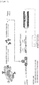


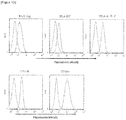
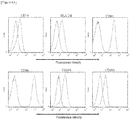
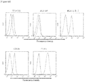


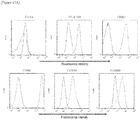


Classifications
-
- C—CHEMISTRY; METALLURGY
- C12—BIOCHEMISTRY; BEER; SPIRITS; WINE; VINEGAR; MICROBIOLOGY; ENZYMOLOGY; MUTATION OR GENETIC ENGINEERING
- C12N—MICROORGANISMS OR ENZYMES; COMPOSITIONS THEREOF; PROPAGATING, PRESERVING, OR MAINTAINING MICROORGANISMS; MUTATION OR GENETIC ENGINEERING; CULTURE MEDIA
- C12N5/00—Undifferentiated human, animal or plant cells, e.g. cell lines; Tissues; Cultivation or maintenance thereof; Culture media therefor
- C12N5/06—Animal cells or tissues; Human cells or tissues
- C12N5/0602—Vertebrate cells
- C12N5/0696—Artificially induced pluripotent stem cells, e.g. iPS
-
- G—PHYSICS
- G01—MEASURING; TESTING
- G01N—INVESTIGATING OR ANALYSING MATERIALS BY DETERMINING THEIR CHEMICAL OR PHYSICAL PROPERTIES
- G01N33/00—Investigating or analysing materials by specific methods not covered by groups G01N1/00 - G01N31/00
- G01N33/48—Biological material, e.g. blood, urine; Haemocytometers
- G01N33/50—Chemical analysis of biological material, e.g. blood, urine; Testing involving biospecific ligand binding methods; Immunological testing
- G01N33/68—Chemical analysis of biological material, e.g. blood, urine; Testing involving biospecific ligand binding methods; Immunological testing involving proteins, peptides or amino acids
- G01N33/6872—Intracellular protein regulatory factors and their receptors, e.g. including ion channels
-
- A—HUMAN NECESSITIES
- A61—MEDICAL OR VETERINARY SCIENCE; HYGIENE
- A61K—PREPARATIONS FOR MEDICAL, DENTAL OR TOILETRY PURPOSES
- A61K35/00—Medicinal preparations containing materials or reaction products thereof with undetermined constitution
- A61K35/12—Materials from mammals; Compositions comprising non-specified tissues or cells; Compositions comprising non-embryonic stem cells; Genetically modified cells
- A61K35/14—Blood; Artificial blood
- A61K35/15—Cells of the myeloid line, e.g. granulocytes, basophils, eosinophils, neutrophils, leucocytes, monocytes, macrophages or mast cells; Myeloid precursor cells; Antigen-presenting cells, e.g. dendritic cells
-
- A—HUMAN NECESSITIES
- A61—MEDICAL OR VETERINARY SCIENCE; HYGIENE
- A61K—PREPARATIONS FOR MEDICAL, DENTAL OR TOILETRY PURPOSES
- A61K38/00—Medicinal preparations containing peptides
-
- C—CHEMISTRY; METALLURGY
- C07—ORGANIC CHEMISTRY
- C07K—PEPTIDES
- C07K14/00—Peptides having more than 20 amino acids; Gastrins; Somatostatins; Melanotropins; Derivatives thereof
-
- C—CHEMISTRY; METALLURGY
- C07—ORGANIC CHEMISTRY
- C07K—PEPTIDES
- C07K14/00—Peptides having more than 20 amino acids; Gastrins; Somatostatins; Melanotropins; Derivatives thereof
- C07K14/415—Peptides having more than 20 amino acids; Gastrins; Somatostatins; Melanotropins; Derivatives thereof from plants
-
- C—CHEMISTRY; METALLURGY
- C07—ORGANIC CHEMISTRY
- C07K—PEPTIDES
- C07K14/00—Peptides having more than 20 amino acids; Gastrins; Somatostatins; Melanotropins; Derivatives thereof
- C07K14/435—Peptides having more than 20 amino acids; Gastrins; Somatostatins; Melanotropins; Derivatives thereof from animals; from humans
- C07K14/745—Blood coagulation or fibrinolysis factors
- C07K14/755—Factors VIII, e.g. factor VIII C (AHF), factor VIII Ag (VWF)
-
- C—CHEMISTRY; METALLURGY
- C07—ORGANIC CHEMISTRY
- C07K—PEPTIDES
- C07K16/00—Immunoglobulins [IGs], e.g. monoclonal or polyclonal antibodies
- C07K16/18—Immunoglobulins [IGs], e.g. monoclonal or polyclonal antibodies against material from animals or humans
- C07K16/24—Immunoglobulins [IGs], e.g. monoclonal or polyclonal antibodies against material from animals or humans against cytokines, lymphokines or interferons
- C07K16/241—Tumor Necrosis Factors
-
- C—CHEMISTRY; METALLURGY
- C07—ORGANIC CHEMISTRY
- C07K—PEPTIDES
- C07K7/00—Peptides having 5 to 20 amino acids in a fully defined sequence; Derivatives thereof
-
- C—CHEMISTRY; METALLURGY
- C12—BIOCHEMISTRY; BEER; SPIRITS; WINE; VINEGAR; MICROBIOLOGY; ENZYMOLOGY; MUTATION OR GENETIC ENGINEERING
- C12N—MICROORGANISMS OR ENZYMES; COMPOSITIONS THEREOF; PROPAGATING, PRESERVING, OR MAINTAINING MICROORGANISMS; MUTATION OR GENETIC ENGINEERING; CULTURE MEDIA
- C12N5/00—Undifferentiated human, animal or plant cells, e.g. cell lines; Tissues; Cultivation or maintenance thereof; Culture media therefor
- C12N5/0068—General culture methods using substrates
- C12N5/0075—General culture methods using substrates using microcarriers
-
- C—CHEMISTRY; METALLURGY
- C12—BIOCHEMISTRY; BEER; SPIRITS; WINE; VINEGAR; MICROBIOLOGY; ENZYMOLOGY; MUTATION OR GENETIC ENGINEERING
- C12N—MICROORGANISMS OR ENZYMES; COMPOSITIONS THEREOF; PROPAGATING, PRESERVING, OR MAINTAINING MICROORGANISMS; MUTATION OR GENETIC ENGINEERING; CULTURE MEDIA
- C12N5/00—Undifferentiated human, animal or plant cells, e.g. cell lines; Tissues; Cultivation or maintenance thereof; Culture media therefor
- C12N5/06—Animal cells or tissues; Human cells or tissues
- C12N5/0602—Vertebrate cells
- C12N5/0634—Cells from the blood or the immune system
- C12N5/0639—Dendritic cells, e.g. Langherhans cells in the epidermis
-
- C—CHEMISTRY; METALLURGY
- C12—BIOCHEMISTRY; BEER; SPIRITS; WINE; VINEGAR; MICROBIOLOGY; ENZYMOLOGY; MUTATION OR GENETIC ENGINEERING
- C12N—MICROORGANISMS OR ENZYMES; COMPOSITIONS THEREOF; PROPAGATING, PRESERVING, OR MAINTAINING MICROORGANISMS; MUTATION OR GENETIC ENGINEERING; CULTURE MEDIA
- C12N5/00—Undifferentiated human, animal or plant cells, e.g. cell lines; Tissues; Cultivation or maintenance thereof; Culture media therefor
- C12N5/06—Animal cells or tissues; Human cells or tissues
- C12N5/0602—Vertebrate cells
- C12N5/0634—Cells from the blood or the immune system
- C12N5/0645—Macrophages, e.g. Kuepfer cells in the liver; Monocytes
-
- C—CHEMISTRY; METALLURGY
- C12—BIOCHEMISTRY; BEER; SPIRITS; WINE; VINEGAR; MICROBIOLOGY; ENZYMOLOGY; MUTATION OR GENETIC ENGINEERING
- C12N—MICROORGANISMS OR ENZYMES; COMPOSITIONS THEREOF; PROPAGATING, PRESERVING, OR MAINTAINING MICROORGANISMS; MUTATION OR GENETIC ENGINEERING; CULTURE MEDIA
- C12N5/00—Undifferentiated human, animal or plant cells, e.g. cell lines; Tissues; Cultivation or maintenance thereof; Culture media therefor
- C12N5/10—Cells modified by introduction of foreign genetic material
-
- C—CHEMISTRY; METALLURGY
- C12—BIOCHEMISTRY; BEER; SPIRITS; WINE; VINEGAR; MICROBIOLOGY; ENZYMOLOGY; MUTATION OR GENETIC ENGINEERING
- C12P—FERMENTATION OR ENZYME-USING PROCESSES TO SYNTHESISE A DESIRED CHEMICAL COMPOUND OR COMPOSITION OR TO SEPARATE OPTICAL ISOMERS FROM A RACEMIC MIXTURE
- C12P21/00—Preparation of peptides or proteins
- C12P21/02—Preparation of peptides or proteins having a known sequence of two or more amino acids, e.g. glutathione
-
- C—CHEMISTRY; METALLURGY
- C12—BIOCHEMISTRY; BEER; SPIRITS; WINE; VINEGAR; MICROBIOLOGY; ENZYMOLOGY; MUTATION OR GENETIC ENGINEERING
- C12Q—MEASURING OR TESTING PROCESSES INVOLVING ENZYMES, NUCLEIC ACIDS OR MICROORGANISMS; COMPOSITIONS OR TEST PAPERS THEREFOR; PROCESSES OF PREPARING SUCH COMPOSITIONS; CONDITION-RESPONSIVE CONTROL IN MICROBIOLOGICAL OR ENZYMOLOGICAL PROCESSES
- C12Q1/00—Measuring or testing processes involving enzymes, nucleic acids or microorganisms; Compositions therefor; Processes of preparing such compositions
- C12Q1/02—Measuring or testing processes involving enzymes, nucleic acids or microorganisms; Compositions therefor; Processes of preparing such compositions involving viable microorganisms
-
- G—PHYSICS
- G01—MEASURING; TESTING
- G01N—INVESTIGATING OR ANALYSING MATERIALS BY DETERMINING THEIR CHEMICAL OR PHYSICAL PROPERTIES
- G01N33/00—Investigating or analysing materials by specific methods not covered by groups G01N1/00 - G01N31/00
- G01N33/48—Biological material, e.g. blood, urine; Haemocytometers
- G01N33/50—Chemical analysis of biological material, e.g. blood, urine; Testing involving biospecific ligand binding methods; Immunological testing
- G01N33/5005—Chemical analysis of biological material, e.g. blood, urine; Testing involving biospecific ligand binding methods; Immunological testing involving human or animal cells
- G01N33/5008—Chemical analysis of biological material, e.g. blood, urine; Testing involving biospecific ligand binding methods; Immunological testing involving human or animal cells for testing or evaluating the effect of chemical or biological compounds, e.g. drugs, cosmetics
- G01N33/5044—Chemical analysis of biological material, e.g. blood, urine; Testing involving biospecific ligand binding methods; Immunological testing involving human or animal cells for testing or evaluating the effect of chemical or biological compounds, e.g. drugs, cosmetics involving specific cell types
- G01N33/5047—Cells of the immune system
-
- C—CHEMISTRY; METALLURGY
- C07—ORGANIC CHEMISTRY
- C07K—PEPTIDES
- C07K2317/00—Immunoglobulins specific features
- C07K2317/20—Immunoglobulins specific features characterized by taxonomic origin
- C07K2317/24—Immunoglobulins specific features characterized by taxonomic origin containing regions, domains or residues from different species, e.g. chimeric, humanized or veneered
-
- C—CHEMISTRY; METALLURGY
- C12—BIOCHEMISTRY; BEER; SPIRITS; WINE; VINEGAR; MICROBIOLOGY; ENZYMOLOGY; MUTATION OR GENETIC ENGINEERING
- C12N—MICROORGANISMS OR ENZYMES; COMPOSITIONS THEREOF; PROPAGATING, PRESERVING, OR MAINTAINING MICROORGANISMS; MUTATION OR GENETIC ENGINEERING; CULTURE MEDIA
- C12N2501/00—Active agents used in cell culture processes, e.g. differentation
- C12N2501/20—Cytokines; Chemokines
- C12N2501/22—Colony stimulating factors (G-CSF, GM-CSF)
-
- C—CHEMISTRY; METALLURGY
- C12—BIOCHEMISTRY; BEER; SPIRITS; WINE; VINEGAR; MICROBIOLOGY; ENZYMOLOGY; MUTATION OR GENETIC ENGINEERING
- C12N—MICROORGANISMS OR ENZYMES; COMPOSITIONS THEREOF; PROPAGATING, PRESERVING, OR MAINTAINING MICROORGANISMS; MUTATION OR GENETIC ENGINEERING; CULTURE MEDIA
- C12N2501/00—Active agents used in cell culture processes, e.g. differentation
- C12N2501/20—Cytokines; Chemokines
- C12N2501/23—Interleukins [IL]
- C12N2501/2304—Interleukin-4 (IL-4)
-
- C—CHEMISTRY; METALLURGY
- C12—BIOCHEMISTRY; BEER; SPIRITS; WINE; VINEGAR; MICROBIOLOGY; ENZYMOLOGY; MUTATION OR GENETIC ENGINEERING
- C12N—MICROORGANISMS OR ENZYMES; COMPOSITIONS THEREOF; PROPAGATING, PRESERVING, OR MAINTAINING MICROORGANISMS; MUTATION OR GENETIC ENGINEERING; CULTURE MEDIA
- C12N2506/00—Differentiation of animal cells from one lineage to another; Differentiation of pluripotent cells
- C12N2506/45—Differentiation of animal cells from one lineage to another; Differentiation of pluripotent cells from artificially induced pluripotent stem cells
Definitions
- the invention relates to an in vitro method for producing a dendritic cell from a stem cell or a progenitor cell derived therefrom, wherein the dendritic cell expresses a MHC molecule, the method comprising the following steps:
- bio-pharmaceuticals antibody drugs, biologics, hormones, proteins, etc.
- immunogenicity a property of an antigen that induces antibody production or cell-mediated immunity is called immunogenicity.
- the bio-pharmaceuticals can act as antigens to induce antibody production in the bodies of patients. In such a case, neutralizing antibodies against the bio-pharmaceuticals are produced, sometimes resulting in the reduced efficiency of treatment.
- allergic response, leaching reaction, infusion reaction, or the like may be caused.
- antibodies that cause autoimmune diseases or the like due to the neutralization of endogenous self-proteins may be produced in response to the bio-pharmaceuticals.
- MHC I molecule a major histocompatibility complex
- a MHC I molecule class I
- MHC II molecule class II
- a MHC I molecule acts on killer T cells (CD8-positive T cells)
- the MHC II molecule acts on helper T cells (CD4-positive T cells).
- the MHC I molecule acts on endogenous antigens in autologous cells, while the MHC II molecule acts on foreign antigens.
- antigen-antibody reaction or the like can be caused, for example, against cancer antigens produced in cancer cells, through antigen presentation mediated by the MHC I molecule.
- antigen-antibody reaction or the like can be caused against foreign antigens such as the bio-pharmaceuticals, or toxins through antigen presentation mediated by the MHC II molecule.
- foreign proteins are first taken up into antigen-presenting cells by endocytosis. Subsequently, the taken-up proteins are decomposed into smaller peptides by lysosome. Then, each peptide binds to the MHC II molecule to form a complex. Then, the complex is delivered to the cell surface so that the peptide is presented as an epitope on the MHC II molecule. Subsequently, a T cell receptor on a helper T cell can bind to the antigen-presenting cell via the peptide.
- Methods have been developed which involve contacting an antigen-presenting cell such as a dendritic cell (DC) with a protein to induce antigen presentation, allowing a MHC molecule on the cell to present a peptide derived from the protein, then separating and purifying a complex of the MHC molecule and the peptide, then eluting the peptide, and directly identifying the sequence of the peptide by use of liquid chromatography mass spectrometry (LC/MS) or the like (Patent Literature 1, Patent Literature 2, and Non Patent Literature 1). These methods are called MAPPs (MHC-associated peptide proteomics).
- DC dendritic cell
- MAPPs MHC-associated peptide proteomics
- Non Patent Literature 1 Kropshofer, H, et al., J. Immunotoxicol., 3, 131, 2006
- PBMCs peripheral blood mononuclear cells
- dendritic cells obtained by the induction of differentiation of monocytes further separated from these PBMCs are utilized as antigen-presenting cells.
- the monocytes are present only at approximately 10% of PBMCs and are also limited by the number of divisions. Therefore, there is a limitation on the number of cells that can be obtained.
- MHC molecule allotypes may vary among different donors, it is impossible to constantly obtain a desired MHC molecule allotype.
- peptides to be identified by MAPPs may also vary.
- serum is added for inducing the differentiation of monocytes into dendritic cells in many cases. Therefore, peptide sequences derived from proteins in the serum might also be detected.
- PBMCs derived from a plurality of donors are often pooled and used as bulks. Thus, it has not been easy to determine which peptide among identified peptides is involved in the induction of immunogenicity in a certain patient.
- an antigen-presenting cell specifically, a major histocompatibility complex (MHC molecule)-expressing cell
- MHC molecule major histocompatibility complex
- the present invention provides the following:
- antigen-presenting cells having diverse MHC molecule allotypes can be stably secured by providing a plurality of stem cells expressing different MHC molecule allotypes.
- combined analysis to predict whether or not desired proteins have immunogenicity in patients, which has not been easy to accomplish so far can be conducted by providing one or more antigen-presenting cells expressing one or more of, e.g., MHC molecule allotypes carried by patients.
- a system using stem cells or progenitor cells derived therefrom as starting materials of antigen-presenting cells for MAPPs is more highly sensitive than a system using PBMCs as such starting materials.
- stem cells are not limited by the number of cell divisions, and methods for proliferation and maintenance thereof have already been established. Therefore, antigen-presenting cells expressing necessary MHC molecule allotypes can be produced and supplied stably in large amounts. Thus, the present invention is also excellent from the viewpoint of production cost and convenience.
- the possibility of detecting peptide sequences derived from proteins in serum can be circumvented by using a serum-free medium during the course of differentiation of stem cells into antigen-presenting cells.
- the protein may be, for example, a natural protein, a recombinant protein, or a synthetic peptide prepared by artificially bonding amino acids. It is understood that the protein may be one protein or a mixture of a plurality of different proteins. The protein may contain a non-natural amino acid. The protein may also be glycosylated, for example, when produced in vivo.
- the protein may be a protein (e.g., an antibody and a hormone) related to treatment or prevention for an animal (e.g. a human). In one aspect, the protein may be one or more, selected from the group consisting of cytokines, chemokines, growth factors, antibodies, enzymes, structural proteins, hormones, and fragments of any of these proteins.
- the protein is not particularly limited by the length of its amino acid sequence as long as the protein forms a complex with a MHC molecule for antigen presentation after being taken up into a cell and decomposed, after being taken up into a cell but not decomposed, or after being produced in a cell and decomposed.
- the protein may be a peptide itself that forms a complex with a MHC molecule for antigen presentation.
- the epitope refers to a particular structural unit of an antigen to be recognized and bound by an antibody.
- the epitope is a minimum unit for antigenicity and is also called antigenic determinant.
- the differentiation may refer to a state or an aspect, etc., in which an individual cell or a cell population, etc., which has been originally single or identical cell(s), is altered to acquire a complicated or distinct structure and/or function.
- the differentiation may be used interchangeably with the induction of differentiation, for example, and includes a state in which the induction of differentiation has been started, a state in which the induction of differentiation is ongoing, a state in which the induction of differentiation has been terminated, etc. It is understood that the differentiation should further encompass a state in which a cell or a cell population whose induction of differentiation has been terminated is proliferating, etc.
- the induction may mean that a certain cell or cell population, etc., is encouraged to differentiate structurally and/or functionally into another cell or cell population, etc.
- the induction is not particularly limited as long as the differentiation can be achieved.
- the stem cell means a pluripotent stem cell and is not particularly limited as long as the stem cell is a cell having pluripotent differentiation and the ability to self-renew.
- the stem cell include induced pluripotent stem cells (iPS cells), embryonic stem cells (ES cells), nuclear transfer ES cells (ntES cells), embryonic germ stem cells (EG cells), and adult stem cells ( WO2012/115276 ). These stem cells can be derived from a mammal, e.g. from a human.
- the ES cell is an embryo-derived stem cell that is derived from, for example, the inner cell mass of a blastocyst, which is an embryo after an 8-cell stage of a fertilized egg and morula.
- the ES cell can be established by isolating the inner cell mass from the blastocyst of a fertilized egg of a subject animal and culturing the inner cell mass on a feeder of fibroblasts, and methods for establishment and maintenance thereof are known in the art (e.g., US Patent No. 5,843,780 ).
- the ES cell may be selected by real-time PCR by using, for example, the expression of a gene marker such as alkaline phosphatase, OCT-3/4, or NANOG gene as an index.
- the human ES cell may be selected by using the expression of a gene marker such as OCT-3/4, NANOG, FBX15, FGF4, REX1, or ECAD gene as an index ( E. Kroon et al., (2008), Nat. Biotechnol., 26: 443-452 ).
- a gene marker such as OCT-3/4, NANOG, FBX15, FGF4, REX1, or ECAD gene as an index ( E. Kroon et al., (2008), Nat. Biotechnol., 26: 443-452 ).
- redundant embryos determined to be discarded among cryopreserved embryos that have not been brought back to mothers in fertility treatment based on external fertilization may be utilized as embryos for use in the preparation of human ES cells, or embryos that have stopped growing during development in the external fertilization process may be utilized as such embryos.
- unfertilized eggs may be utilized which lack the innate ability to grow into humans by themselves and are based on parthenogenesis in terms of cell division and growth.
- only single blastomeres of embryos at a cleavage stage prior to a blastocyst stage may be used to prepare ES cells without impairing the ability of the embryos to develop and without destroying fertilized eggs ( Chung Y, Klimanskaya I, Becker S, Marh J, Lu SJ, Johnson J, Meisner L, Lanza R. (2006). Nature 439: 216-219 .; Klimanskaya I, Chung Y, Becker S, Lu SJ, Lanza R. (2006).
- the ES cell may be prepared from a human embryo that has stopped developing ( Zhang X, Stojkovic P, Przyborski S, Cooke M, Armstrong L, Lako M, Stojkovic M. (2006). Stem Cells 24: 2669-2676 .).
- the iPS cell is a somatic cell-derived artificial stem cell that has properties substantially equivalent to the ES cell, for example, pluripotent differentiation and the ability to self-renew, and can be prepared by introducing a particular reprogramming factor in a DNA or protein form into a somatic cell ( K. Takahashi and S. Yamanaka (2006) Cell, 126: 663-676 ; K. Takahashi et al. (2007), Cell, 131: 861-872 ; J. Yu et al. (2007), Science, 318: 1917-1920 ; Nakagawa, M. et al., Nat. Biotechnol. 26: 101-106 (2008 ); and WO2007/069666 ).
- the somatic cell may refer to every animal cell (e.g. a mammalian cell including a human cell) except for germ-line cells and pluripotent stem cells.)
- the reprogramming factor may be a gene specifically expressed in ES cells, a gene product or non-cording RNA thereof, a gene that plays an important role in maintaining the undifferentiation of ES cells, a gene product or non-cording RNA thereof, or a low-molecular compound.
- the reprogramming factors may include OCT3/4, SOX2, SOX1, SOX3, SOX15, SOX17, KLF4, KLF2, c-MYC, N-MYC, L-MYC, NANOG, LIN28, FBX15, ERAS, ECAT15-2, TCLL, beta-catenin, LIN28B, SALL1, SALL4, ESRRB, NR5A2, and TBX3.
- These reprogramming factors may be used alone or in combination. Examples of the combination of the reprogramming factors can include the following combinations:
- Examples of the reprogramming factor or a factor promoting reprogramming may include MEK inhibitors, DNA methyl transferase inhibitors, histone deacetylase (HDAC) inhibitors, histone methyl transferase inhibitors, and p53 inhibitors, which are inhibitors generally known to those skilled in the art.
- the reprogramming factor may be introduced into a somatic cell according to a method generally known to those skilled in the art, for example, a calcium phosphate method, lipofection, or microinjection, optionally using a vector (e.g., a viral vector, a plasmid vector, and an artificial chromosome vector) or the like.
- a DMEM, DMEM/F12, or DME medium containing 10 to 15% FBS (which may further appropriately contain a leukemia inhibitory factor (LIF), penicillin/streptomycin, puromycin, L-glutamine, nonessential amino acids, ⁇ -mercaptoethanol, etc.), or a commercially available medium generally known to those skilled in the art may be appropriately used as a medium for the induction of the iPS cell.
- LIF leukemia inhibitory factor
- penicillin/streptomycin penicillin/streptomycin
- puromycin puromycin
- L-glutamine nonessential amino acids
- ⁇ -mercaptoethanol nonessential amino acids
- the culture of the iPS cell may be appropriately set according to the composition of the medium, etc.
- somatic cells are contacted with the reprogramming factor and cultured for approximately 4 to 7 days using a DMEM or DMEM/F12 medium containing 10% FBS at 37°C in the presence of 5% CO 2 .
- the cells are reseeded onto feeder cells (e.g., mitomycin C-treated STO cells and SNL cells).
- feeder cells e.g., mitomycin C-treated STO cells and SNL cells.
- the cells are cultured in a medium for primate ES cell culture containing a basic fibroblast growth factor (bFGF).
- bFGF basic fibroblast growth factor
- the cells are cultured on feeder cells (e.g., mitomycin C-treated STO cells and SNL cells) in a DMEM medium containing 10% FBS (which may further appropriately contain LIF, penicillin/streptomycin, puromycin, L-glutamine, nonessential amino acids, ⁇ -mercaptoethanol, etc.) at 37°C in the presence of 5% CO 2 . Approximately 25 to approximately 30 days later, or thereafter, an iPS-like colony may appear.
- somatic cells themselves to be reprogrammed may be used, or extracellular matrix or Matrigel (Becton, Dickinson and Company (BD)) may be used.
- the culture may be performed using a serum-free medium ( Sun N, et al., (2009), Proc Natl Acad Sci USA. 106: 15720-15725 ).
- the iPS cell may be selected according to the shape of the formed colony (e.g., whether to obtain a cell mass having a nearly spherical shape).
- a marker gene e.g., a drug resistance gene to be expressed in conjunction with a gene (e.g., alkaline phosphatase, OCT3/4, and NANOG genes) that is expressed by the reprogramming of somatic cells
- the cells can be cultured in a medium containing the corresponding drug to select the established iPS cell.
- the marker gene is a fluorescent protein gene
- the iPS cell can also be selected by observation under a fluorescence microscope.
- the iPS cell may be determined by culturing the cells in vitro by a differentiation method known in the art and using their ability to differentiate into desired cells as an index.
- the iPS cell may be determined by subcutaneously transplanting the cells into an immunodeficient mouse and analyzing tumor tissues formed after a lapse of a predetermined period to confirm that teratomas made up of a mixture of various tissues are formed.
- the iPS cell may be determined by confirming that a marker gene specifically expressed in ES cells is expressed.
- the iPS cell may be determined by detecting a genome-wide gene expression pattern using a microarray or the like to confirm that the cells have an expression pattern highly correlating with that of ES cells.
- established iPS cells may be furnished and used.
- the ntES cell is a clone embryo-derived ES cell prepared by a nuclear transfer technique and has substantially the same properties as those of fertilized egg-derived ES cells ( T. Wakayama et al. (2001), Science, 292: 740-743 ; S. Wakayama et al. (2005), Biol. Reprod., 72: 932-936 ; and J. Byrne et al. (2007), Nature, 450: 497-502 ).
- the ntES cell is an ES cell established from the inner cell mass of a blastocyst derived from a clone embryo obtained by replacing the nucleus of an unfertilized egg with the nucleus of a somatic cell.
- ntES cell For the preparation of the ntES cell, a nuclear transfer technique known in the art (e.g., J.B. Cibelli et al., (1998), Nature Biotechnol., 16: 642-646 ) may be combined with an ES cell preparation technique known in the art ( Sayaka Wakayama, et al., (2008), Experimental Medicine, Vol. 26, No. 5 (extra number), p. 47 to 52 ).
- the nucleus of a somatic cell may be injected into an enucleated unfertilized mammalian egg and cultured for a few hours for reprogramming.
- the EG cell is a cell that has pluripotency similar to that of ES cells and is established from a primordial germ cell during fetal life ( Y. Matsui et al., (1992), Cell, 70: 841-847 ).
- the EG cell may be established by culturing primordial germ cells in the presence of LIF, bFGF, stem cell factor (STF), or the like ( Y. Matsui et al., (1992), Cell, 70: 841-847 ).
- the adult stem cell is a cell that has not been finally differentiated and is found in vivo.
- the adult stem cell exists as a source of a progenitor cell for a finally differentiated cell.
- the adult stem cell is present in each tissue in vivo and is usually limited by the types of cells into which the adult stem cell can differentiate.
- examples of the adult stem cell include hematopoietic stem cells considered to be able to differentiate into monocytes, macrophages, dendritic cells, and the like.
- hematopoietic progenitor cells refer to cells differentiated from the hematopoietic stem cells.
- the progenitor cell derived from the stem cell may include every cell (e.g., mesodermal progenitor cells, hematopoietic progenitor cells, granulocyte macrophage colony-forming cells, lymphoblasts, monoblasts, promonocytes, and monocytes) that is observed in the course of differentiating the stem cell into an antigen-presenting cell (specifically, a MHC molecule-expressing cell).
- an antigen-presenting cell specifically, a MHC molecule-expressing cell.
- MHC I molecules Peter Parham (2007), THE IMMUNE SYSTEM; The Human Protein Atlas, http://www.proteinatlas.org /
- particular cells have MHC II molecules in addition to the MHC I molecules and can present foreign antigens on helper T cells via the MHC II molecules (these cells are also called professional antigen-presenting cells).
- the antigen-presenting cell may include both of these types of cells.
- Examples of the antigen-presenting cell of the latter type include dendritic cells, macrophages, monocytes, and B cells.
- thyroid follicular cells, fibroblasts, vascular endothelial cells, and the like also work as antigen-presenting cells when MHC II molecules are induced through the activation by cytokines such as interferons. Therefore, these cells may be also included in the latter type.
- a criterion to determine whether to have properties as antigen-presenting cells may be based on, for example, cells expressing MHC I and/or the MHC II molecules as an index and can be based on cells further expressing at least one of CD11a, CD11b, CD11c, CD14, CD15, CD40, CD80, CD83, CD86, CD123, CD205, CD206, CD209, and CCR7 as an index.
- the dendritic cells have the strong ability to present antigens and the strong ability to activate helper T cells and are therefore advantageous as antigen-presenting cells.
- the antigen-presenting cell is most preferably an immature dendritic cell.
- the dendritic cells are cells that have cell processes and assume a dendritic form.
- a criterion to determine whether to have properties as dendritic cells may be based on, for example, the further expression of at least one of CD11b, CD11c, CD40, CD80, CD83, CD86, CD123, CD205, CD206, CD209, and CCR7 in addition to the MHC II molecule as an index and can be based on the expression of all of the MHC II molecule, CD80, CD86, CD206, and CD209 as an index.
- a dendritic cell that expresses all of the MHC II molecule, CD80, CD86, CD206, and CD209 and is negative for CD14 is also described.
- a criterion to determine whether to have properties as macrophage cells may be based on, for example, the further expression of CD1 1b in addition to the MHC II molecule as an index.
- CD80 and CD86 are known to transduce signals to helper T cells and activate these cells.
- Dendritic cell-like cells obtained in the present Examples have properties similar to those of monocyte-derived dendritic cells in terms of cell shape, the expression of cell surface molecules, and the ability to stimulate helper T cells and may therefore be included in the dendritic cell described in the present specification. Likewise, monocyte-like cells obtained in the present Examples may be included in the monocyte described in the present specification.
- the antigen-presenting cell can be derived from a mammal, e.g. a human.
- the MHC molecule may be any of MHC I and MHC II molecules and is more preferably a MHC II molecule.
- Human MHC is called human leucocyte antigen (HLA).
- the MHC I molecules are further divided into classical class I (class Ia) and a non-classical class I (class Ib) molecules.
- classical class I molecules include HLA-A, HLA-B, and HLA-C in humans.
- non-classical class I molecules include HLA-E, HLA-F, and HLA-G in humans.
- examples of the MHC II molecules include HLA-DR, HLA-DQ, and HLA-DP in humans.
- the MHC molecule differs slightly in amino acid sequence in individuals even among animals of the same species and is further divided into some subtypes called allotypes.
- HLA-DR is known to have many allotypes such as DR1, DR2, DR3, DR4, ....
- Each allotype is linked on the MHC gene with the other allotypes. Therefore, these allotypes are inherited as a set from parent to child unless gene recombination occurs in this region. This unit is called haplotype.
- Stem cells e.g., iPS cells
- derived from a patient basically maintain the MHC gene sequence, as it is, of the patient even after being subcultured and/or differentiated. Therefore, antigen-presenting cells obtained by the differentiation of the stem cells maintain MHC molecule allotypes carried by the patient.
- the allotypes respectively form complexes with different antigen peptide fragments (epitopes) so that the epitopes can be presented on the cell surface. Therefore, the presence or absence or the degree of immunogenicity, adverse reaction, etc., caused by a target protein varies depending on each allotype set, in other words, each patient having the allotype set. Allotypes or haplotypes have a pattern characteristic of a race or an ethnic group and can therefore be utilized, for example, in the analysis of the presence or absence of immunogenicity, adverse reaction, etc., caused by a target protein for each race or ethnic group.
- a series of antigen-presenting cells having the MHC molecule allotypes of a fixed population are advantageously used for predicting the immunogenicity of a protein for the population.
- Allotypes carried by an individual person can be conveniently identified by genetic diagnosis (e.g., a HLA genotyping method which involves hybridizing DNA amplified by polymerase chain reaction (PCR) to probe-immobilized beads, digitizing the fluorescence intensity thereof, and analyzing the data to identify the HLA genotype) or the like. Therefore, whether the target protein causes immunogenicity, adverse reaction, etc., can be determined on the basis of information on the identified allotypes.
- Other specific examples of the genetic diagnosis method include methods described in, for example, International Journal of Immunogenetics, 2011; 38: 6, pp. 463-473 .
- the antigen-presenting cell may express one or more MHC molecule allotypes in a subject (e.g., a mammal, e.g. a human) intended to receive the target protein.
- a subject e.g., a mammal, e.g. a human
- a cell expressing one or more MHC molecule allotypes carried by a subject may be used as the stem cell or the progenitor cell derived therefrom according to the present disclosure.
- a subject e.g., a human patient or a healthy person
- one or more cells expressing one or more MHC molecule allotypes in the subject may be used such that all sets of MHC molecule allotypes carried by the subject are contained therein.
- cells expressing one or more MHC molecule allotypes with high expression frequency in a race or an ethnic group intended to be analyzed may be prepared.
- a fixed percentage e.g., 30% to 80% or more
- a population of the race or the ethnic group may be covered by preparing a plurality of such cells and analyzed for the immunogenicity.
- appropriate comparative analysis among human patients, between human patients and healthy persons, among healthy persons, etc. is advantageous.
- the method for differentiating the stem cell or the progenitor cell derived therefrom into the antigen-presenting cell is not particularly limited as long as the method is generally known to those skilled in the art. Methods described in WO2009/120891 ; WO2009/074341 ; Regen. Med. (2009) 4 (4), p. 513-526 ; WO2012/115276 ; WO2012/043651 ; PLoS One, July 2011, Vol.
- 410-417 may be used as methods for differentiating stem cells such as ES cells or iPS cells into, for example, monocytes, macrophages, B cells, or dendritic cells.
- stem cells such as ES cells or iPS cells
- monocytes such as monocytes, macrophages, B cells, or dendritic cells.
- dendritic cells for example, Regen. Med. (2009) 4 (4), p. 513-526 discloses a method for inducing the in vitro differentiation of human ES cells into dendritic cells in a serum-free medium.
- human ES cells are differentiated into monocytes using bone morphogenetic protein-4 (BMP-4), a granulocyte macrophage-colony stimulating factor (GM-CSF), a stem cell factor (SCF), and a vascular endothelial growth factor (VEGF); subsequently, the monocytes are further differentiated into immature dendritic cells using GM-CSF and interleukin-4 (IL-4); and the immature dendritic cells are further differentiated into mature dendritic cells using a maturation cocktail consisting of GM-MSF, TNF- ⁇ , interleukin-1 ⁇ (IL-1 ⁇ ), interferon-y (IFN- ⁇ ), and PGE2.
- BMP-4 bone morphogenetic protein-4
- GM-CSF granulocyte macrophage-colony stimulating factor
- SCF stem cell factor
- VEGF vascular endothelial growth factor
- GM-CSF granulocyte macrophage-colony stimulating factor
- SCF stem cell factor
- MHC molecule-expressing cells antigen-presenting cells
- this method may comprise the step of providing the stem cell or the progenitor cell derived therefrom and subsequently the following steps:
- the serum may refer to mammal-derived serum such as human serum, monkey serum, fetal bovine serum, sheep serum, rabbit serum, rat serum, guinea pig serum, or mouse serum.
- the serum-free medium refers to a medium that is supplemented neither with serum nor with a commercially available serum replacement such as B-27 and may be a medium containing at least one of albumin or an albumin replacement, transferrin or a transferrin replacement, insulin or an insulin replacement, and selenious acid.
- the serum-free medium may be a medium containing insulin-transferrin-selenium-X supplement (ITS).
- serum-free medium examples include a minimum essential medium (MEM), a Dulbecco's modified eagle medium (DMEM), an Iscove's modified Dulbecco's medium (IMDM), StemPro-34 medium (Life Technologies/Thermo Fisher Scientific Inc.), Stemline II (Sigma-Aldrich Corp.), and Primate ES cell medium (ReproCELL Inc.) each supplemented with ITS.
- MEM minimum essential medium
- DMEM Dulbecco's modified eagle medium
- IMDM Iscove's modified Dulbecco's medium
- StemPro-34 medium Life Technologies/Thermo Fisher Scientific Inc.
- Stemline II Stemline II
- Primate ES cell medium ReproCELL Inc.
- the step (a) may comprise the step of culturing the stem cell or the progenitor cell derived therefrom in a medium containing BMP family protein and subsequently culturing the cell in a medium containing a growth factor and a hematopoietic factor, or culturing the cell in a medium containing VEGF and then culturing the cell in a medium containing a hematopoietic factor to obtain the mesodermal progenitor cell.
- the step (b) may comprise the step of differentiating the mesodermal progenitor cell into the monocyte by culture in a medium containing a hematopoietic factor.
- the step (a) and the step (b) can be continuously performed.
- the BMP family protein may refer to a cytokine that belongs to the TGF- ⁇ superfamily and has approximately 20 subtypes.
- the BMP family protein may be BMP2 and/or BMP4.
- the growth factor may be VEGF and may be specifically VEGF-A, VEGF-B, VEGF-C, VEGF-D, VEGF-E, PlGF (placental growth factor)-1, PlGF-2, or a selective splicing variant thereof (e.g., variants composed of 121, 165, 189, or 206 amino acids are known for VEGF-A).
- the VEGF may be VEGF-A.
- the growth factor may further include bFGF in addition to VEGF.
- the hematopoietic factor is a factor promoting the differentiation and proliferation of blood cells and may be, for example, a stem cell factor (SCF), a granulocyte colony-stimulating factor (G-CSF), a granulocyte macrophage colony-stimulating factor (GM-CSF), a macrophage colony-stimulating factor (M-CSF), erythropoietin (EPO), thrombopoietin (TPO), an interleukin (IL), or Flt3 ligand.
- the interleukin may be IL-1, IL-2, IL-3, IL-4, IL-5, IL-6, IL-7, IL-8, or IL-9, etc.
- the hematopoietic factor for the step (b) may be selected from the group consisting of SCF, TPO, IL-3, Flt3 ligand, GM-CSF, and M-CSF. These hematopoietic factors may be used alone or in combination.
- the step (a) may be performed using VEGF as the growth factor and SCF as the hematopoietic factor in combination, and subsequently, the step (b) may be performed using GM-CSF and M-CSF as the hematopoietic factor in combination.
- medium may be replaced with a fresh one every few days (e.g., every 3 to 4 days) for culture.
- a non-adherent cell (monocyte or monocyte-like cell) is obtained by the step (b), this non-adherent cell may be used as the monocyte in the step (c). Whether the non-adherent cell has the properties of the monocyte can be confirmed by using, for example, flow cytometry and using, as an index, the expression of a monocyte marker such as CD14, CD45 hi , CD11a, CD11b, or CD15 in addition to the expression of the MHC II molecule.
- a monocyte marker such as CD14, CD45 hi , CD11a, CD11b, or CD15 in addition to the expression of the MHC II molecule.
- the cell proportion of monocytes for use in the induction of dendritic cells can be increased, for example, by separating only CD14-positive cells from non-adherent cells by use of a magnetic bead method or the like.
- the step (c) may further comprise the step of: (ci) differentiating the monocyte into an immature dendritic cell (or an immature dendritic cell-like cell) by (suspension-)culture in a medium containing a hematopoietic factor; and optionally comprise the step of: (cii) further contacting the obtained immature dendritic cell (or immature dendritic cell-like cell) with an immunogen and optionally an inflammatory cytokine to induce a mature dendritic cell (or a mature dendritic cell-like cell).
- the immature dendritic cell-like cell or the mature dendritic cell-like cell has the properties of the dendritic cell may be confirmed by using, for example, flow cytometry and using, as an index, the further expression of at least one of dendritic cell markers CD11b, CD11c, CD40, CD80, CD83, CD86, CD123, CD205, CD206, CD209, and CCR7 in addition to the expression of the MHC II molecule.
- Whether the dendritic cell has the properties of the immature dendritic cell or has the properties of the mature dendritic cell can be tested by using, for example, change in the expression of the MHC II molecule (HLA-DR, etc.) as an index.
- the hematopoietic factor may be any of the factors mentioned above.
- a combination of GM-CSF, IL-3, and IL-4 or a combination of GM-CSF and IL-4 may be used as the hematopoietic factor.
- the immunogen and the inflammatory cytokine can stimulate (pulse) the cell to induce a mature dendritic cell.
- the immature dendritic cell has high phagocytic capacity for an antigen but has the low ability to present the antigen, whereas this cell can be matured into a mature dendritic cell, for example, by the invasion of the antigen into an organism to enhance the expression of a protein, such as the MHC II molecule, necessary for antigen presentation and thereby improve the ability to present the antigen.
- the immunogen may be any substance that causes immune response when introduced into an organism. Examples thereof include lipopolysaccharide (LPS, which is present in a pathogen).
- LPS lipopolysaccharide
- the immature dendritic cell is contacted with a target protein having immunogenicity to induce the mature dendritic cell.
- the inflammatory cytokine may be, for example, tumor necrosis factor- ⁇ (TNF- ⁇ ), TNF- ⁇ , IL-12, or IFN- ⁇ . These immunogens or inflammatory cytokines may be appropriately used alone or in combination.
- step (d) differentiating the monocyte into a macrophage may be performed, instead of the step (c), according to a method described in PLoS One, April 2013, Vol. 8, Issue 4, e59243 .
- the monocyte can be differentiated into the macrophage using e.g. GM-CSF or M-CSF as the hematopoietic factor.
- the macrophage can be differentiated into M1 macrophage by adding, for example, IFN- ⁇ or LPS or can be differentiated into M2 macrophage by adding, for example, IL-4 or IL-13 (macrophages are known to be activated by receiving cytokines produced by helper T cells, and classical activation (M1 macrophage) and selective activation (M2 macrophage) are known).
- M1 macrophage classical activation
- M2 macrophage selective activation
- the respective concentrations of the growth factor, the hematopoietic factor, the cytokine, etc., used in each step mentioned above can be concentrations at which the antigen-presenting cell of interest is obtained, and can be appropriately determined by those skilled in the art.
- the concentration of BMP4 may be, for example, 5 to 150 ng/ml, e.g. 10 to 100 ng/ml, or e.g. 20 to 80 ng/ml.
- the concentration of VEGF may be, for example, 20 to 100 ng/ml, e.g. 30 to 70 ng/ml, or e.g. 40 to 50 ng/ml.
- the concentration of bFGF may be, for example, 10 to 100 ng/ml, e.g.
- the concentration of SCF may be, for example, 20 to 100 ng/ml, e.g. 30 to 70 ng/ml, or e.g. 40 to 50 ng/ml.
- the concentration of IL-3 may be, for example, 5 to 100 ng/ml, e.g. 30 to 70 ng/ml.
- the concentration of TPO may be, for example, 1 to 25 ng/ml, e.g. 1 to 10 ng/ml.
- the concentration of Flt3 ligand may be, for example, 10 to 100 ng/ml, e.g. 30 to 70 ng/ml.
- the concentration of GM-CSF may be, for example, 5 to 250 ng/ml, e.g. 50 to 200 ng/ml.
- the concentration of M-CSF may be, for example, 5 to 100 ng/ml, or e.g. 30 to 70 ng/ml.
- the concentration of IL-4 may be, for example, 3 to 100 ng/ml, e.g. 10 to 70 ng/ml.
- the concentration of TNF- ⁇ may be, for example, 0.05 to 50 ng/ml, or e.g. 0.1 to 20 ng/ml.
- the concentration of LPS may be, for example, 0.01 to 100 ⁇ g/ml, or e.g. 0.1 to 10 ⁇ g/ml.
- the concentration of the (target) protein to be evaluated can be, for example, a concentration at which an epitope on the protein can be identified, according to the purpose, or can be a concentration at which whether or not the protein has immunogenicity in a subject (e.g., a mammal, e.g. a human) can be evaluated.
- the concentration thereof can be a concentration at which the mature dendritic cell can be induced by the stimulation of the immature dendritic cell.
- concentration can be appropriately determined by those skilled in the art.
- Such a concentration may be, for example, 0.01 to 1000 ⁇ g/ml, or e.g. 0.1 to 100 ⁇ g/ml.
- the period of the step (a) may be, for example, 2 days or longer, e.g. 2 to 10 days, or e.g. 5 to 8 days.
- the period of the step (b) may be, for example, 1 day or longer, e.g. 20 to 200 days, or e.g. 50 to 150 days.
- the period of the step (ci) may be, for example, 1 day or longer, e.g. 1 to 10 days, or e.g. 4 to 6 days.
- the period of the step (cii) may be, for example, 12 hours or longer, e.g. 12 to 36 hours, or e.g. 24 hours (1 day).
- the period of the step (d) may be, for example, 1 day or longer, e.g. 1 to 20 days.
- the macrophage may be further differentiated into M1 macrophage or M2 macrophage.
- those skilled in the art can appropriately determine the optimum culture period in consideration of each culture condition, as a matter of course.
- the present disclosure may also relate to a method for producing a dendritic cell (in vitro) from a stem cell or a progenitor cell derived therefrom, comprising the steps (a) to (c), and a dendritic cell obtained or obtainable by the method.
- the dendritic cell produced by the method expresses not only the MHC II molecule but CD80 and CD86, costimulatory molecules of helper T cells and expresses carbohydrate receptors CD206 and CD209, suggesting that this cell has the ability to activate helper T cells and resistance to viruses and the like.
- sequence analysis of an epitope on a protein using the dendritic cell obtained by the method contributes to the development of a protein having low immunogenicity and, in addition, is also expected to bring about an excellent material for research on antigen-presenting cells against autoimmune diseases, viruses, and the like.
- the present disclosure further provides, as other aspects, for example, the following aspects:
- the dendritic cell or the cell composition may be used as a cell medicament for performing immune cell therapy for infectious diseases or malignant tumors, or for use in the control of immune response for the purpose of treating autoimmune diseases or rejection or the like associated with organ transplantation.
- the cell medicament may be appropriately used in combination with an auxiliary, for example, a medium, for the purpose of stably maintaining the dendritic cell.
- the present disclosure also relates to a method for identifying an epitope on a protein, comprising the following steps:
- step (A) can be be performed under serum-free conditions.
- the degree of immunogenicity can also be compared, for example, among different proteins, among different protein preparations, or among different bio-pharmaceuticals by use of the method for identifying an epitope on a protein. This method can also be utilized in the quality control of produced proteins.
- the amount of the MHC molecule-expressing cell necessary for obtaining, for example, 100 ng of MHC molecules may depend on the number of cells, the expression intensity of the MHC molecules, and the degree of the expression.
- the optimum amount of the cell can be appropriately determined by those skilled in the art.
- Each MHC II molecule allotype (e.g., HLA-DQ1) can carry approximately 500 to 1000 different peptide fragments ( Chicz R M et al., J Exp. Med. 1993, 178, 27-47 ; and Chicz R M & Urban R G, Immunol. Today, 1993, 15, 155-160 ).
- a great majority of these different peptides merely attain a very low copy number and therefore are not very likely to play a physiological role in vivo.
- peptide fragments that participate in immunogenicity and activate, for example, helper T cells attain a moderate to high copy number ( Latek R R & Unanue E R, Immunol. Rev. 1999, 172: 209-228 ).
- These peptides having a moderate to high copy number account for approximately 40 to 50% of the total amount of peptides eluted from MHC II molecules and can correspond to approximately 10 to 200 individual peptides.
- the peptide is a peptide that is derived from (the amino acid sequence of) the target protein and can form a complex with the MHC molecule on the surface of the antigen-presenting cell (specifically, the MHC molecule-expressing cell).
- the peptide may be bound with an intracellular or extracellular MHC molecule.
- Each MHC II molecule allotype can form a complex with diverse peptides, and the amount of the peptide necessary for the sequencing of each eluted peptide can be, for example, only a fmol amount.
- approximately a fmol amount of peptide fragments bound with MHC molecules can be isolated from, for example, approximately 0.1 to 5 ⁇ g of MHC molecules, and the sequences of the peptides can be identified.
- the cell membrane of the cell may be lysed. This lysis may be carried out by a method generally known to those skilled in the art, for example, freezing-thawing, use of a surfactant, or a combination thereof.
- a surfactant for example, Triton X-100 (TX100), Nonidet P-40 (NP-40), Tween 20, Tween 80, n-octylglucoside, ZWITTERGENT, Lubrol, or CHAPS may be used as the surfactant.
- TX100 Triton X-100
- NP-40 Nonidet P-40
- Tween 20 Tween 80
- n-octylglucoside ZWITTERGENT
- Lubrol Lubrol
- CHAPS CHAPS
- the cell lysate containing the solubilized MHC molecule-peptide complexes may be subjected to immunoprecipitation or immunoaffinity chromatography to purify the MHC molecule-peptide complexes.
- An antibody that is specific for each MHC molecule and is suitable for immunoprecipitation or immunoaffinity chromatography (anti-MHC I molecule antibody, for example, anti-HLA-A antibody, anti-HLA-B antibody, anti-HLA-C antibody, or anti-HLA-ABC antibody, etc.; or anti-MHC II molecule antibody, preferably anti-HLA-DR antibody, anti-HLA-DQ antibody, or anti-HLA-DP antibody) may be used for these methods.
- the specific antibody can be a monoclonal antibody and may be conjugated with beads (e.g., Sepharose beads or agarose beads) through a covalent bond or a noncovalent bond, for example, via protein A.
- beads e.g., Sepharose beads or agarose beads
- the amino group of the antibody may be covalently bonded to CNBr-activated Sepharose so that the antibody is immobilized thereon.
- a commercially available product may be purchased as the monoclonal antibody, or the monoclonal antibody may be purified from the supernatant of each corresponding hybridoma cell using protein A- or protein G-affinity chromatography.
- the MHC molecules may be immunoisolated, for example, by performing incubation while rotating the antibody-beads together with the cell lysate for a few hours. Also, the antibody-beads bound with the MHC molecule-peptide complexes may be washed in an Eppendorf tube. The results of immunoprecipitation may be analyzed by SDS-PAGE and Western blotting using an antibody that recognizes a denatured MHC molecule.
- a mixture of peptides that are derived from the target protein through the decomposition by the antigen-presenting cell is obtained by eluting the peptides from the complexes formed with the MHC molecules.
- the peptides may be eluted by a method generally known to those skilled in the art, for example, by use of a diluted acid, for example, diluted acetonitrile ( Jardetzky T S et al., Nature 1991 353, 326-329 ), diluted acetic acid and heating ( Rudensky A Y et al., Nature 1991, 353, 622-626 ; and Chicz R M et al., Nature 1992, 358, 764-768 ), or diluted trifluoroacetic acid ( Kropshofer H et al., J Exp Med 1992, 175, 1799-1803 ).
- the peptides can be eluted with diluted trifluoroacetic acid, for example, at 37°C.
- these complexes may be washed with water or a low-salt buffer solution in order to remove surfactant residues.
- a Tris buffer solution, a phosphate buffer solution, or an acetate buffer solution having a concentration of 0.5 to 10 mM may be used as the low-salt buffer solution.
- the MHC molecule-peptide complexes may be washed with ultrapure water for HPLC. This washing may be performed by ultrafiltration.
- the ultrafiltration may be performed in an ultrafiltration tube having, for example, a cutoff value of 30 kD, 20 kD, 10 kD, or 5 kD and a tube volume of 0.5 to 1.0 ml.
- the inside of the ultrafiltration tube may be washed, for example, 4 to 12 times, with a washing solution having tens of times the volume of the beads carrying the MHC molecule-peptide complexes.
- the peptides may be eluted from the MHC molecule-peptide complexes by use of this ultrafiltration tube. Subsequently, the eluted peptides may be dried by use of freeze drying or a centrifugal evaporator.
- the peptide mixture thus eluted may be fractionated and subjected to sequence analysis by use of liquid chromatography mass spectrometry (LC/MS) to identify (the amino acid sequence of) each peptide.
- LC/MS liquid chromatography mass spectrometry
- amino acid sequence of each peptide in the peptide mixture can be determined by the sequence analysis according to a method known in the art sufficient for sequencing a fmol amount of peptides.
- the identification reveals a protein from which the peptide is derived, and a sequence from which the peptide is derived in the protein.
- the eluted peptide mixture can be fractionated, for example, using reverse-phase chromatography and anion-exchange chromatography or cation-exchange chromatography in combination or reverse-phase chromatography alone.
- the fractionation may be performed on a HPLC mode using a fused-silica micro-capillary column connected either to nanoflow electrospray ion source in a mass spectrometer or to a microfractionation apparatus that spots fractions on a plate for MALDI analysis.
- Electrospray ionization tandem mass spectrometry (ESI-MS) or MALDI-post source decay (PSD) MS may be used as a mass spectrometry technique, e.g. ESI-MS.
- sequence analysis the amino acid sequence of each peptide may be determined by various approaches generally known to those skilled in the art.
- the sequence analysis may be performed by the computer analysis of a peptide fragment spectrum using, for example, MASCOT algorithm or SEQUEST algorithm.
- MASCOT algorithm or SEQUEST algorithm.
- SEQUEST algorithm These algorithms can employ protein and nucleotide sequence databases, for conducting the cross-correlation analysis of experimentally and theoretically prepared tandem mass spectra. This permits automatic high-throughput sequence analysis.
- Matrix-assisted laser desorption ionization time-of-flight (MALDI-TOF) mass spectrometry may be performed for the qualitative analysis of all of the peptides obtained by elution.
- the MALDI-TOF analysis can provide a rough outline regarding the complexity of the peptide mixture and the presence of a primary peptide.
- substances that have passed through a microcapillary column may be analyzed using a UV detector at a detection wavelength of 214 nm.
- the peak area of the peptide to be analyzed may be compared with the peak areas of serial amounts of standard peptides (control) to estimate the amount of the peptide.
- a set of peptides that are derived from the target protein by natural fragmentation in the antigen-presenting cell is obtained by eluting the peptides from the MHC molecules.
- a set of antigen-presenting cells exposed to the target protein as well as a set of antigen-presenting cells unexposed to the target protein as a negative control can be prepared and analyzed by comparison.
- a peptide that can be detected in only the antigen-presenting cells exposed to the target protein as compared with the antigen-presenting cells unexposed to the target protein may be identified as functioning as an epitope on the protein from which the peptide is derived, and having antigenicity.
- Whether or not the identified peptide functions as an epitope can be tested by using, for example, a MHC-binding motif, the ability to bind to MHC, or recognition by helper T cells as an index. Alternatively, this test may be appropriately combined with an in silico epitope prediction algorithm.
- the MHC-binding motif means a structural feature that is common in peptides binding to particular MHC molecules (allelic polymorphism) and is necessary for forming stable complexes with the MHC molecules.
- the peptide length varies from 12 to 18 amino acids, and even longer peptides can bind because both ends of the peptide-binding groove are open.
- Most of MHC II molecules can accommodate up to 4 residues (called “anchor residues”) related to the binding, at relative positions P1, P4, P6 and P9 contained in a nonameric core region. However, this core region may vary in distance from the N terminus of the peptide. In many cases, 2 to 4 N-terminal residues precede the core region.
- the PI anchor residue is located at position 3, 4, or 5 in many peptides that can form with complexes with the MHC II molecules.
- Peptides eluted from, for example, HLA-DR molecules can share a hydrophobic PI anchor such as tyrosine, phenylalanine, tryptophan, methionine, leucine, isoleucine, or valine.
- the positions and types of the anchor residues can be estimated from the peptide-binding motifs of frequently occurring MHC molecules.
- a computer algorithm that permits motif validation in peptide sequences can be obtained from, for example, "Tepitope" (www.vaccinome.com, J. Hammer, Nutley, USA).
- the ability to bind to MHC may be tested by a method generally known to those skilled in the art using the detected peptide itself (e.g., a synthetic peptide may be utilized) and a desired MHC molecule ( Kropshofer H et al., J. Exp. Med. 1992, 175, 1799-1803 ; Vogt A B et al., J. Immunol. 1994, 153, 1665-1673 ; and Sloan V S et al., Nature 1995, 375, 802-806 ).
- cellular binding assay using MHC molecule-expressing cell lines and biotinylated peptides may be used to test the ability to bind to MHC ( Arndt S O et al., EMBO J., 2000, 19, 1241-1251 ).
- the relative ability of each peptide to bind to MHC may be determined by measuring a concentration necessary to reduce the binding of a labelled reporter peptide to 50% (IC50).
- each identified peptide may be used as such a peptide, or a peptide having a sequence common in identified peptides (core sequence) may be used.
- the peptide to be detected is considered to depend on the types of MHC molecule allotypes, the strength of binding affinity for a MHC molecule, etc.
- helper T cells The ability to stimulate helper T cells is particularly important for testing whether the identified peptide functions as an epitope. When each identified peptide stimulates helper T cells, this may be used as one index for determining that the peptide has immunogenicity. This determination method may involve testing whether the peptide identified by the method of the present disclosure has the ability to activate helper T cells. Each identified peptide may be used as such a peptide, or a peptide having a sequence common in identified peptides (core sequence) may be used.
- helper T cells The cellular response of helper T cells may be measured by various in vitro methods generally known to those skilled in the art.
- MHC molecule-expressing cells e.g., monocytes, macrophages, or dendritic cells
- helper T cells are cultured together with helper T cells in the presence of the peptide to be evaluated.
- the uptake of radioactive material-labeled thymidine (T) during DNA replication may be measured with helper T cell proliferation as an index.
- 5-bromo-2'-deoxyuridine (BrdU) may be used instead of thymidine.
- helper T cells that have taken up BrdU during DNA replication are treated with a monoclonal antibody against BrdU.
- the amount of BrdU taken up may be measured using an enzymatically or fluorescently labeled secondary antibody (e.g., 5-Bromo-2'-deoxyuridine Labeling & Detection Kit III, Roche-Biochem, Cat No. 1 444 611).
- an enzymatically or fluorescently labeled secondary antibody e.g., 5-Bromo-2'-deoxyuridine Labeling & Detection Kit III, Roche-Biochem, Cat No. 1 444 611).
- Naive Primary T cell Assay (Proimmune Ltd.) which employs flow cytometry using the dilution of a fluorescent dye label 5,6-carboxyfluorescein diacetate succinimidyl ester (CFSE) by the proliferation of helper T cells as an index may be used in the measurement.
- the cellular response of helper T cells may be evaluated by measuring various cytokines produced from the helper T cells, instead of measuring the cell proliferation.
- cytokines examples include IL-2, IL-4, IL-6, IL-10, IL-12, IFN- ⁇ , and transforming growth factor- ⁇ (TGF- ⁇ ).
- TGF- ⁇ transforming growth factor- ⁇
- methods for measuring the cytokines include various methods generally known to those skilled in the art, for example, ELISA and ELISPOT.
- Dendritic cells produced by the method for producing a dendritic cell from a stem cell or a progenitor cell derived therefrom according to the present invention as mentioned above may be used as the MHC molecule-expressing cells.
- the MHC molecule-expressing cells may be rendered non-proliferative, for example, by treatment with ionizing radiation or mitomycin C before the assay.
- the present disclosure also relates to a method for producing a protein with reduced or eliminated immunogenicity, comprising the following steps:
- the amino acid sequence of the identified epitope peptide can be altered or modified to reduce or eliminate the binding of the epitope peptide to a MHC molecule or reduce or eliminate the immunogenicity.
- the reduction or elimination of the immunogenicity is determined by a method generally known to those skilled in the art without particular limitations and may be determined by using, for example, the aforementioned MHC-binding motif, ability to bind to MHC, or recognition by helper T cells as an index. Alternatively, this approach may be appropriately combined with an in silico epitope prediction algorithm.
- the alteration or the modification may be performed according to a method generally known to those skilled in the art.
- a DNA nucleotide sequence encoding the amino acid sequence of a protein comprising the epitope may be subjected to, for example, site-directed mutagenesis or homologous recombination for the insertion, substitution, or deletion, etc., of desired one or more nucleotides in the DNA sequence.
- one or more anchor residues important for binding to a MHC molecule are changed to other amino acid residues, whereby the immunogenicity can be reduced or eliminated.
- amino acid residue(s) important for the recognition of the epitope by, for example, T cell receptor on helper T cells or B cell receptor on B cells may be changed to other amino acid residues.
- the method for replacing the anchor residues important for binding to a MHC molecule is well known to those skilled in the art.
- the PI anchor of a HLA-DR1-restricted T cell epitope may be replaced with alanine, proline, glycine, or a charged amino acid residue ( Kropshofer et al., EMBO J. 15, 1996, 6144-6154 ).
- the protein having the modified epitope may be chemically synthesized or may be genetically or biologically synthesized.
- host cells or animals transiently or permanently harboring the gene of the protein having the modified epitope may be utilized.
- the host cells or the animals can be used, for example, as a production system for protein production or expression.
- Eukaryotic cells or prokaryotic cells may be used as the host cells.
- Examples of the eukaryotic cells that can be used as the host cells include animal cells, plant cells, and fungal cells.
- animal cells include: mammalian cells, for example, CHO ( Puck et al., (1958) J. Exp. Med. 108 (6): 945-956 ), COS, HEK293, 3T3, myeloma, BHK (baby hamster kidney), HeLa, and Vero; amphibian cells, for example, Xenopus oocyte cells ( Valle et al., Nature (1981) 291: 338-340 ); and insect cells, for example, Sf9, Sf21, and Tn5.
- CHO cells can be used for the purpose of large-scale expression in the animal cells.
- the host cells may be transfected with a vector having the gene of the protein having the modified epitope by a method, for example, a calcium phosphate method, a DEAE-dextran method, a method using a cationic ribosome DOTAP (manufactured by Boehringer Mannheim), electroporation, or lipofection.
- a method for example, a calcium phosphate method, a DEAE-dextran method, a method using a cationic ribosome DOTAP (manufactured by Boehringer Mannheim), electroporation, or lipofection.
- Nicotiana tabacum-derived cells and Lemna minor are known as a protein production system of the plant cells. These cells may be allowed to produce the protein by a callus culture method.
- a protein expression system using cells of a yeast for example, the genus Saccharomyces ( Saccharomyces cerevisiae, Saccharomyces pombe, etc.), or cells of a filamentous fungus, for example, the genus Aspergillus ( Aspergillus niger, etc.) may be used as the fungal cells.
- a production system using bacterial cells may be used.
- E. coli or Bacillus subtilis cells may be used as the bacterial cells.
- Examples of the animals include genetically recombinant animals and transgenic animals.
- the type of the animals is not limited, and, for example, cattle, sheep, or mice may be used. In such a case, for example, the protein secreted into a body fluid such as milk may be recovered.
- the protein having the modified epitope may be produced at a large scale continuously or commercially.
- the produced protein or a composition comprising the protein e.g., a pharmaceutical composition is also included in the present disclosure.
- the present disclosure also relates to a method for predicting whether or not a protein has immunogenicity in a subject, comprising the steps of:
- the prediction method also permits comparison of the presence or absence or the degree of immunogenicity based on each MHC molecule allotype or allotype set against the predetermined protein.
- one or more cells expressing one or more MHC molecule allotypes in the subject can be provided such that all sets of MHC molecule allotypes carried by the subject are contained therein.
- the stem cell can be a stem cell (e.g., an iPS cell or ES cell) derived from the subject (e.g., a mammal, e.g. a human). This is because antigen-presenting cells differentiated from stem cells maintain MHC molecule allotypes; thus, antigen-presenting cells that cover all sets of MHC molecule allotypes carried by the subject can be produced by use of the stem cell derived from the subject.
- the prediction method also relates to a method for selecting a subject (e.g. a patient) having immunogenicity or a method for selecting a subject (e.g. a patient) free from immunogenicity.
- the prediction method also relates to a method for indicating that one or more particular MHC molecule allotypes are involved in immunogenicity in association with a protein intended to be administered.
- the stem cell e.g., an iPS cell or an ES cell
- the subject e.g., a mammal, e.g. a human
- the stem cell may be prepared from any somatic cell or the like of the subject as long as the stem cell maintains the MHC molecule allotypes.
- the somatic cell are not particularly limited, and, for example, iPS cells can also be prepared from PBMCs separated from a human or the like.
- antigen-presenting cells such as dendritic cells can be established using the stem cells and thereby utilized in the prediction method.
- the subject-derived cells for use in the preparation of stem cells such as iPS cells may be cells whose MHC molecule allotypes (HLA genotypes in humans) can be identified, or cells whose MHC molecule allotypes have already been revealed (or predicted).
- iPS cells whose MHC molecule allotypes have already been revealed (or predicted) may be used.
- the present disclosure relates to a composition for the treatment and/or prevention of a disease related to a protein, in a subject, comprising the protein as an active ingredient, wherein the subject is selected from (only) subjects predicted to be free from the immunogenicity of the protein according to the aforementioned prediction method.
- the "composition for the treatment and/or prevention” may comprise a therapeutically and/or prophylactically effective amount of the protein, and the effective amount of the protein can be appropriately determined by those skilled in the art. Also, the "composition for the treatment and/or prevention" may contain one or more additional agents.
- the term "predicted to be free from the immunogenicity of the protein” may mean that the target protein does not evoke any immunogenicity in the subject or merely evokes immunogenicity to an extent tolerable from the viewpoint of efficacy, safety, etc.
- the protein or the composition for the treatment and/or prevention comprising the protein as an active ingredient can be administered to only a subject predicted to be free from immunogenicity, by preparing cells expressing one or more MHC molecule allotypes according to a race, an ethnic group, a population, or an individual person, and evaluating the immunogenicity of the protein.
- the composition can be a pharmaceutical composition.
- the (pharmaceutical) composition can be formulated and used by a pharmaceutical production method known in the art.
- the (pharmaceutical) composition can be used, for example, orally as an optionally sugar-coated tablet, a capsule, an elixir, or a microcapsule, or parenterally (e.g., percutaneously, intranasally, transbronchially, intramuscularly, or intravenously) in the form of an aseptic solution or suspension containing the protein in water or any of other pharmaceutically acceptable solutions.
- the (pharmaceutical) composition may be produced so as to appropriately contain a pharmaceutically acceptable carrier, flavor, excipient, vehicle, antiseptic, stabilizer, or binder.
- a binder such as gelatin, corn starch, gum tragacanth, or gum arabic
- an excipient such as crystalline cellulose
- a swelling agent such as corn starch, gelatin, or alginic acid
- a lubricant such as magnesium stearate
- a sweetener such as sucrose, lactose, or saccharin
- a flavor such as peppermint, Akamono oil, or cherry
- this capsule can further contain a liquid carrier such as oil and fat in addition to the materials described above.
- the aseptic solution for injection can be formulated according to a method well known to those skilled in the art using a vehicle such as injectable distilled water.
- a vehicle such as injectable distilled water.
- the aqueous solution for injection include physiological saline and isotonic solutions containing glucose or other adjuvants, for example, D-sorbitol, D-mannose, and D-mannitol.
- An appropriate solubilizer for example, an alcohol such as ethanol, a polyalcohol such as propylene glycol or polyethylene glycol, or a nonionic surfactant such as polysorbate 80(TM) or HCO-50 may be further used in combination therewith.
- oily liquid examples include sesame oil and soybean oil, which may be used in combination with a solubilizer, for example, benzyl benzoate or benzyl alcohol.
- a solubilizer for example, benzyl benzoate or benzyl alcohol.
- a buffer such as a phosphate buffer solution or a sodium acetate buffer solution, for example, a soothing agent such as procaine hydrochloride, for example, a stabilizer such as benzyl alcohol or phenol, or an antioxidant may also be contained therein.
- the prepared injection solution may usually be filled into an appropriate ampule for use.
- the dose, administration method, dosing intervals, etc., of the protein or the composition for the treatment and/or prevention comprising the protein as an active ingredient varies depending on the body weight, age, symptoms, etc., of a patient and can be appropriately selected and determined by those skilled in the art.
- the protein and the disease related to the protein are not particularly limited.
- the protein can be a protein that may cause, for example, immunogenicity or problems associated with efficacy or safety when administered into an organism.
- diseases include, but are not limited to, autoimmune diseases (e.g., rheumatoid arthritis, type I diabetes, multiple sclerosis (MS), coeliac disease, myasthenia gravis (MG), and systemic lupus erythematosus (SLE)), cancers (e.g., melanoma, breast cancer, B cell lymphomas, prostate cancer, and renal cancer), and infectious diseases (e.g. diseases caused by HIV, hepatitis C virus, measles virus, and mycobacteria).
- autoimmune diseases e.g., rheumatoid arthritis, type I diabetes, multiple sclerosis (MS), coeliac disease, myasthenia gravis (MG), and systemic lupus erythematosus (SLE)
- Examples of a combination of such a protein and a disease related to the protein may include examples described in Self/Nonself 2010; 1 (4) pp.314-322 ; PHARM TECH JAPAN Vol.28, No.10 (2012), pp.117 (2065)-126 (2074 ); Sorensen, P.S., et al., Neurology, 67 (9), 1681-3 (2006 ); Hesse, D., et al., Eur. J. Neurol., 14 (8), 850-9 (2007 ); Casadevall, N., et al., N. Engl. J. Med., 346 (7), 469-75 (2002 ); Gershon, S.K., et al., N. Eng. J. Med., 346 (20), 1584-6 (2002 ); and Locatelli F., et al., Perit. Dial. Int., 27 (Suppl2), S303-7 (2006 ).
- the combination of the protein and the disease related to the protein include, but are not limited to: muromonab and allograft rejection; abciximab and PTCA adjunct; rituximab and non-Hodgkin lymphoma; daclizumab and transplant rejection; trastuzumab and breast cancer; palivizumab and RSV prophylaxis; basiliximab and transplant rejection; infliximab and rheumatoid arthritis or Crohn; arcitumomab and colorectal cancer; canakinumab and cryopyrin-associated periodic syndrome; fanolesomab and imaging for appendicitis; imciromab and cardiac imaging for MI; capromab and prostate cancer diagnostic; nofetumomab and detection of SCLC; gemtuzumab and acute myeloid leukemia; alemtuzumab and B cell chronic lymphocytic leukemia; ibritumomab and non
- the present disclosure relates to
- the present disclosure relates to use of a stem cell or a progenitor cell derived therefrom, or a MHC molecule-expressing cell differentiated from the stem cell or the progenitor cell in various methods according to the present disclosure described above.
- numeric values described in the present specification are understood as values having given ranges according to the technical common sense of those skilled in the art, unless inconsistent to the context.
- the term “1 mg” is understood to represent “approximately 1 mg” and is understood to include a given variation.
- the term “1 to 5" described in the present specification is understood to concretely describe the individual values of "1, 2, 3, 4, and 5", unless inconsistent to the context.
- Human iPS cells Tic (JCRB1331), obtained from JCRB Cell Bank; and 201B7, obtained from iPS Academia Japan, Inc.
- Feeder cells EmbryoMax Primary Mouse Embryonic Fibroblasts (MEF), Hygro resistant, C57BL/6 (purchased from Merck Millipore Corp., Cat.: PMEF-HL); and SNL 76/7 feeder cells (SNL) (purchased from Cell Biolabs, Inc., Cat.: CBA-316).
- MEF EmbryoMax Primary Mouse Embryonic Fibroblasts
- C57BL/6 purchasedd from Merck Millipore Corp., Cat.: PMEF-HL
- SNL 76/7 feeder cells SNL
- the following positive control proteins were used as proteins having immunogenicity.
- Phl p1 is a pollen antigen of the family Poaceae and has been reported to have an epitope sequence ( Carla Oseroff et al., J of Immunol 2010: 185 (2): 943-955 ).
- Figures 3A and 3B each show results of examining molecules expressed on the cell surface of the monocyte-like cells prepared using Tic, wherein the results were obtained by analysis using a flow cytometer.
- the monocyte-like cells obtained by the present Examples were found to express a monocyte-specific marker CD14 and also found to express T cell-activating molecules CD80 and CD86 and adhesion molecules CD11b and CD11c.
- Figures 4A and 4B each show results of examining molecules expressed on the cell surface of the monocyte-like cells prepared using 201B7, wherein the results were obtained by analysis using a flow cytometer.
- the monocyte-like cells obtained by the present Examples were found to express a monocyte-specific marker CD14 and also found to express T cell-activating molecules CD80 and CD86 and adhesion molecules CD11b and CD11c, as with the monocyte-like cells prepared using Tic.
- Figures 5A and 5B each show results of examining molecules expressed on the cell surface of the dendritic cell-like cells prepared using Tic, wherein the results were obtained by analysis using a flow cytometer.
- the dendritic cell-like cells obtained by the present Examples were found to express antigen presentation molecules HLA-DR, HLA-DQ, HLA-DP, and HLA-ABC and dendritic cell-specific markers CD206 and CD209 and also found to express T cell-activating molecules CD80 and CD86 and adhesion molecules CD11b and CD11c, as with the monocyte-like cells. Increase in the expression of CD11c was found, as compared with the monocyte-like cells. On the other hand, the expression of the monocyte-specific marker CD14 was decreased. Each marker had a single peak, suggesting that cells having the features of dendritic cells were homogeneously prepared.
- Figures 6A and 6B each show results of examining molecules expressed on the cell surface of the dendritic cell-like cells prepared using 201B7, wherein the results were obtained by analysis using a flow cytometer.
- the dendritic cell-like cells prepared from 201B7 were also found to express antigen presentation molecules HLA-DR, HLA-DQ, HLA-DP, and HLA-ABC and dendritic cell-specific markers CD206 and CD209, as with the dendritic cell-like cells prepared from Tic.
- These dendritic cell-like cells were also found to express T cell-activating molecules CD80 and CD86 and adhesion molecules CD11b and CD11c, as with the monocyte-like cells.
- Figure 7 shows results of analyzing the amino acid sequences of peptides detected by the exposure of the dendritic cell-like cells prepared from Tic to Bet v1a ( Figure 7(a) ).
- a portion of the amino acid sequence of Bet v1a was detected from the peptides separated from HLA-DR molecules extracted from the dendritic cell-like cells exposed to Bet v1a.
- Figure 7 also shows results of analyzing the amino acid sequence of peptides detected both under Bet v1a non-treatment conditions (control) and by exposure to Bet v1a ( Figure 7(b) ).
- Epitope No. represents a group of detected peptides observed in order from the N terminus of the amino acid sequence of Bet v1a. For example, 18 types of peptides were detected in Epitope No. 1.
- Table 1A Betula verrucosa, birch pollen allergen 1, Isoform a (Bet v1a) epitope associated with iPS-DC (Tic line) Epitope No.
- Figure 8 shows results of analyzing the amino acid sequences of peptides detected by the exposure of the dendritic cell-like cells prepared from 201B7 to Bet v1a ( Figure 8(a) ).
- a portion of the amino acid sequence of Bet v1a was detected from the peptides separated from HLA-DR molecules extracted from the dendritic cell-like cells exposed to Bet v1a.
- Figure 8 also shows results of analyzing the amino acid sequence of peptides detected both under Bet v1a non-treatment conditions (control) and by exposure to Bet v1a ( Figure 8(b) ).
- Figure 9A shows results of analyzing the amino acid sequences of peptides detected by the exposure of the dendritic cell-like cells prepared from Tic to infliximab ( Figure 9A(a) ).
- a portion of the amino acid sequences found in the H and L chains of infliximab was detected from the peptides separated from HLA-DR molecules extracted from the dendritic cell-like cells exposed to infliximab.
- Figure 9B shows results of analyzing the amino acid sequence of peptides detected both under infliximab non-treatment conditions (control) and by exposure to infliximab ( Figure 9B(b) ). Only a portion of the amino acid sequence found in the H chain of infliximab was detected.
- Figures 10A to 10H each show results of analyzing the amino acid sequences of peptides detected by the exposure of the dendritic cell-like cells prepared from Tic to rhFVIII.
- a portion of the amino acid sequence of rhFVIII was detected from the peptides separated from HLA-DR molecules extracted from the dendritic cell-like cells exposed to rhFVIII.
- Figure 11 shows results of analyzing the amino acid sequences of peptides detected by the exposure of the dendritic cell-like cells prepared from Tic to Phl p1. In the present Examples, a portion of the amino acid sequence of Phl p1 was detected.
- the peptides presented on the HLA-DR molecules of the dendritic cell-like cells exposed to each antigen were separated and purified as HLA-DR-peptide complexes by use of the anti-HLA-DR beads, and the sequence of each presented peptide was identified by ion trap MS/MS mass spectrometry.
- the antigen-presenting cells such as monocyte-like cells or dendritic cell-like cells were confirmed to express HLA-DQ molecules, HLA-DP molecules, HLA-A molecules, HLA-B molecules, and HLA-C molecules.
- antigen-presented peptides can also be detected and identified by using, instead of the HLA-DR molecules, other MHC II molecules (e.g., HLA-DQ and HLA-DP molecules), or MHC I molecules.
- HLA-DR molecules instead of the HLA-DR molecules, other MHC II molecules (e.g., HLA-DQ and HLA-DP molecules), or MHC I molecules.
- HLA-DQ molecules can be identified using HLA-DQ molecules.
- Duquesnoy RJ, Marrari M, Tambur AR, Mulder A, Sousa LC, da Silva AS, do Monte SJ, First report on the antibody verification of HLA-DR, HLA-DQ and HLA-DP epitopes recorded in the HLA Epitope Registry, Hum Immunol. 2014 Nov; 75 (11): 1097-103 discloses that sequences more likely to be presented by HLA-DR, HLA-DQ, and HLA-DP molecules were predicted from a database, and the HLA-DQ and HLA-DP molecules antigen-present particular sequences, as with the HLA-DR molecules.
- MAPPs can also be applied to cells expressing other MHC II molecules such as HLA-DQ and HLA-DP molecules, which have been differentiated from a stem cell or a progenitor cell derived therefrom according to the present invention.
- MAPPs using MHC I molecules have already been reported in, for example, Wahl A, Schafer F, Bardet W, Buchli R, Air GM, Hildebrand WH., HLA class I molecules consistently present internal influenza epitopes. Proc Natl Acad Sci USA. 2009 Jan 13; 106 (2): 540-5 .
- cell lines allowed to express particular HLA-B molecule allotypes were sensitized with influenza virus, and influenza virus-derived peptide sequences presented on the HLA-B molecules were detected by MAPPs.
- MHC I molecules are known to be expressed by many cell lines. Hence, it is desirable to utilize cells highly expressing MHC I molecules, from the viewpoint of the easy detection of MHC I molecule-peptide complexes.
- a dendritic cell differentiated from a stem cell or a progenitor cell derived therefrom according to the present invention may be used in MAPPs using MHC I molecules.
- PBMCs Human peripheral blood mononuclear cells
- Betula verrucosa, birch pollen allergen 1, Isoform a (Bet v1a) (#Bet v 1.0101; Biomay AG) (amino acid sequence: SEQ ID NO: 1) as a white birch pollen allergen was used in the same way as in Examples of the present invention.
- Figure 12 shows results of examining molecules expressed on the cell surface of the monocytes, wherein the results were obtained by analysis using a flow cytometer.
- the monocytes obtained by Comparative Examples were found to express a monocyte-specific marker CD14 and an antigen presentation molecule HLA-DR and also found to express a T cell-activating molecule CD86.
- Figures 13A and 13B each show results of examining molecules expressed on the cell surface of the dendritic cells, wherein the results were obtained by analysis using a flow cytometer.
- the dendritic cells obtained by Comparative Examples were found to express antigen presentation molecules HLA-DR, HLA-DQ, and HLA-ABC and dendritic cell-specific markers CD206 and CD209 and also found to express T cell-activating molecules CD80 and CD86 and adhesion molecules CD11b and CD11c. On the other hand, the expression of CD14 was not observed.
- Figures 14 to 20 each show results of analyzing the amino acid sequences of peptides detected under Bet v1a addition conditions ( Figures 14(a) , 15(a) , 16(a) , 17(a) , 18(a) , 19(a) , and 20(a) ) or non-addition conditions (control) ( Figures 14(b) , 15(b) , 16(b) , 17(b) , 18(b) , 19(b) , and 20(b) ) for each human donor subjected to the evaluation.
- These specific amino acid sequences thus detected are also shown in Tables 2 to 8 (which correspond to Figures 14 to 20 , respectively). In these tables, Epitope No.
- Figures 21A and 21B each show results of comparing the amino acid sequences of peptides detected under Bet v1a addition conditions between use of dendritic cell-like cells derived from two types of human iPS cells and use of dendritic cells derived from PBMCs.
- the donors had different MHC II molecules, which therefore seemed to result in the difference in the amino acid sequences of the detected peptides among the donors.
- the original donors also had different types of MHC II molecules, which therefore seemed to result in the difference in the detected sequences between the lines.
- the present invention can be used in the fields of diagnosis or medicine of various bio-pharmaceuticals, for example, by applying the epitope sequence analysis of a protein.
- sequences presented on MHC class II molecules were detected as to all of the pollen-derived foreign proteins Bet v1a and Phl p1, the antibody drug product infliximab, and the drug product rhFVIII having an amino acid sequence analogous to that of an endogenous protein.
- the MAPPs of the present disclosure were capable of detecting epitope sequences for various proteins including bio-pharmaceuticals, regardless of features such as natural and non-natural ones or foreign and endogenous ones. There may be the possibility that the MAPPs of the present disclosure can be applied to a wide range of purposes such as the development of drugs as well as the analysis of epitopes for allergens or autoimmune diseases.
Description
- The invention relates to an in vitro method for producing a dendritic cell from a stem cell or a progenitor cell derived therefrom, wherein the dendritic cell expresses a MHC molecule, the method comprising the following steps:
- (a) differentiating the stem cell or the progenitor cell derived therefrom into a mesodermal progenitor cell;
- (b) differentiating the mesodermal progenitor cell into a monocyte in a serum-free medium containing a granulocyte macrophage colony-stimulating factor (GM-CSF) and a macrophage colony-stimulating factor (M-CSF); and
- (c) differentiating the monocyte into an immature dendritic cell in a serum-free medium, and optionally further stimulating the immature dendritic cell to obtain a mature dendritic cell;
- (i) the stem cell is selected from the group consisting of an induced pluripotent stem cell (iPS cell), an embryonic stem cell (ES cell), a nuclear transfer ES cell (ntES cell), an embryonic germ stem cell (EG cell), and an adult stem cell; and/or
- (ii) the MHC molecule is a MHC II molecule, wherein the MHC II molecule is HLA-DR, HLA-DQ or HLA-DP.
- In recent years, many bio-pharmaceuticals (antibody drugs, biologics, hormones, proteins, etc.) have contributed to medical innovation. However, immunogenicity possessed by these bio-pharmaceuticals is controversial, for example, from the viewpoint of efficacy and safety. In general, a property of an antigen that induces antibody production or cell-mediated immunity is called immunogenicity. The bio-pharmaceuticals can act as antigens to induce antibody production in the bodies of patients. In such a case, neutralizing antibodies against the bio-pharmaceuticals are produced, sometimes resulting in the reduced efficiency of treatment. Alternatively, allergic response, leaching reaction, infusion reaction, or the like may be caused. Alternatively, antibodies that cause autoimmune diseases or the like due to the neutralization of endogenous self-proteins may be produced in response to the bio-pharmaceuticals.
- For the process of antibody production, it is important that an antigen is presented on a major histocompatibility complex (also referred to as a MHC molecule) present on the cell surface of an antigen-presenting cell (APC) (this is called "antigen presentation"). A MHC I molecule (class I) and a MHC II molecule (class II) are known as MHC molecules involved in the antigen presentation. For example, the MHC I molecule acts on killer T cells (CD8-positive T cells), and the MHC II molecule acts on helper T cells (CD4-positive T cells). The MHC I molecule acts on endogenous antigens in autologous cells, while the MHC II molecule acts on foreign antigens. Thus, antigen-antibody reaction or the like can be caused, for example, against cancer antigens produced in cancer cells, through antigen presentation mediated by the MHC I molecule. On the other hand, antigen-antibody reaction or the like can be caused against foreign antigens such as the bio-pharmaceuticals, or toxins through antigen presentation mediated by the MHC II molecule.
- More specifically, in the case of the event mediated by the MHC I molecule, endogenous proteins in autologous cells are decomposed into smaller peptides by proteasome. Subsequently, each peptide binds to the MHC I molecule synthesized in the vesicle to form a complex. Then, the complex is delivered to the cell surface so that the peptide is presented as an epitope on the MHC I molecule.
- On the other hand, in the case of the event mediated by the MHC II molecule, foreign proteins are first taken up into antigen-presenting cells by endocytosis. Subsequently, the taken-up proteins are decomposed into smaller peptides by lysosome. Then, each peptide binds to the MHC II molecule to form a complex. Then, the complex is delivered to the cell surface so that the peptide is presented as an epitope on the MHC II molecule. Subsequently, a T cell receptor on a helper T cell can bind to the antigen-presenting cell via the peptide.
- However, these pathways are not definitive, and even foreign antigens may be processed by the MHC I molecule-mediated antigen presentation pathway (this is called "cross-priming").
- In order to circumvent the immunogenicity of antibody drugs, etc., research has been conducted on the identification of peptide sequences presented on MHC molecules. This permits the prediction of the immunogenicity of proteins or peptides intended to be administered to organisms. Furthermore, epitopes can be modified by site-directed mutagenesis for the purpose of producing non-immunogenic proteins, for example, on the basis of information on epitope sequences. A method using a prediction algorithm in silico and T cell proliferation assay (e.g., the measurement of the ability of helper T cells to proliferate by the uptake of tritium-labeled thymidine) are known as methods for identifying peptide sequences. However, it has been difficult to predict an epitope sequence, for example, only from the binding affinity between an epitope candidate peptide and a MHC molecule. Accordingly, there has been a demand for more accurately predicting an epitope that may play a causative role in the induction of immunogenicity by directly identifying the sequence of a peptide presented on a MHC molecule.
- Methods have been developed which involve contacting an antigen-presenting cell such as a dendritic cell (DC) with a protein to induce antigen presentation, allowing a MHC molecule on the cell to present a peptide derived from the protein, then separating and purifying a complex of the MHC molecule and the peptide, then eluting the peptide, and directly identifying the sequence of the peptide by use of liquid chromatography mass spectrometry (LC/MS) or the like (
Patent Literature 1,Patent Literature 2, and Non Patent Literature 1). These methods are called MAPPs (MHC-associated peptide proteomics). -
- Patent Literature 1: European Patent Application Publication No.
1715343 - Patent Literature 2: European Patent Application Publication No.
1826217 - Non Patent Literature 1: Kropshofer, H, et al., J. Immunotoxicol., 3, 131, 2006
- In one aspect, in the method of
Patent Literature 1, human peripheral blood mononuclear cells (PBMCs) separated by blood collection from a human are used as primary cells, and dendritic cells obtained by the induction of differentiation of monocytes further separated from these PBMCs are utilized as antigen-presenting cells. However, the monocytes are present only at approximately 10% of PBMCs and are also limited by the number of divisions. Therefore, there is a limitation on the number of cells that can be obtained. Furthermore, since MHC molecule allotypes may vary among different donors, it is impossible to constantly obtain a desired MHC molecule allotype. Thus, peptides to be identified by MAPPs may also vary. Since each component in blood also fluctuates depending on the states of patients, it is impossible to constantly isolate PBMCs under the same conditions. Accordingly, there has been demand for stably securing a plurality of antigen-presenting cells having diverse MHC molecule allotypes. - In an alternative aspect, serum is added for inducing the differentiation of monocytes into dendritic cells in many cases. Therefore, peptide sequences derived from proteins in the serum might also be detected.
- In an alternative aspect, at present, there is also a limitation on the amount of PBMCs that can be obtained. Therefore, PBMCs derived from a plurality of donors are often pooled and used as bulks. Thus, it has not been easy to determine which peptide among identified peptides is involved in the induction of immunogenicity in a certain patient.
- The present inventors have conducted diligent studies and consequently completed the present invention by, surprisingly, solving some or all of the problems described above, by the application of an antigen-presenting cell (specifically, a major histocompatibility complex (MHC molecule)-expressing cell) differentiated from a stem cell or a progenitor cell derived therefrom to MAPPs.
- Specifically, the present invention provides the following:
- 1. An in vitro method for producing a dendritic cell from a stem cell or a progenitor cell derived therefrom, wherein the dendritic cell expresses a MHC molecule, the method comprising the following steps:
- (a) differentiating the stem cell or the progenitor cell derived therefrom into a mesodermal progenitor cell;
- (b) differentiating the mesodermal progenitor cell into a monocyte in a serum-free medium containing a granulocyte macrophage colony-stimulating factor (GM-CSF) and a macrophage colony-stimulating factor (M-CSF); and
- (c) differentiating the monocyte into an immature dendritic cell in a serum-free medium, and optionally further stimulating the immature dendritic cell to obtain a mature dendritic cell;
- (i) the stem cell is selected from the group consisting of an induced pluripotent stem cell (iPS cell), an embryonic stem cell (ES cell), a nuclear transfer ES cell (ntES cell), an embryonic germ stem cell (EG cell), and an adult stem cell; and/or
- (ii) the MHC molecule is a MHC II molecule, wherein the MHC II molecule is HLA-DR, HLA-DQ or HLA-DP.
- 2. The method according to
item 1, wherein the step (c) comprises the step of:- (c1) differentiating the monocyte into the immature dendritic cell in a serum-free medium containing a granulocyte macrophage colony-stimulating factor (GM-CSF) and interleukin 4 (IL-4), and optionally comprise the step of:
- (c2) contacting the immature dendritic cell with an immunogen and optionally an inflammatory cytokine to induce the mature dendritic cell.
- In one aspect, for example, antigen-presenting cells having diverse MHC molecule allotypes can be stably secured by providing a plurality of stem cells expressing different MHC molecule allotypes. Thus, combined analysis to predict whether or not desired proteins have immunogenicity in patients, which has not been easy to accomplish so far, can be conducted by providing one or more antigen-presenting cells expressing one or more of, e.g., MHC molecule allotypes carried by patients.
- In an alternative aspect, it is suggested that a system using stem cells or progenitor cells derived therefrom as starting materials of antigen-presenting cells for MAPPs is more highly sensitive than a system using PBMCs as such starting materials.
- In an alternative aspect, stem cells are not limited by the number of cell divisions, and methods for proliferation and maintenance thereof have already been established. Therefore, antigen-presenting cells expressing necessary MHC molecule allotypes can be produced and supplied stably in large amounts. Thus, the present invention is also excellent from the viewpoint of production cost and convenience.
- In an alternative aspect, the possibility of detecting peptide sequences derived from proteins in serum can be circumvented by using a serum-free medium during the course of differentiation of stem cells into antigen-presenting cells.
-
- [
Figure 1] Figure 1 shows one example of the outline of a technique using MHC II molecules among techniques of MAPPs (MHC-associated peptide proteomics). In the diagram, DC refers to a dendritic cell. - [
Figure 2] Figure 2 shows one example of a scheme for obtaining dendritic cell-like cells by the differentiation of human iPS cells. - [
Figure 3A] Figure 3A shows results of examining molecules expressed on the cell surface of monocyte-like cells prepared from a Tic line, wherein the results were obtained by analysis using a flow cytometer. - [
Figure 3B] Figure 3B shows results of examining molecules expressed on the cell surface of monocyte-like cells prepared from a Tic line, wherein the results were obtained by analysis using a flow cytometer. - [
Figure 4A] Figure 4A shows results of examining molecules expressed on the cell surface of monocyte-like cells prepared from a 201B7 line, wherein the results were obtained by analysis using a flow cytometer. - [
Figure 4B] Figure 4B shows results of examining molecules expressed on the cell surface of monocyte-like cells prepared from a 201B7 line, wherein the results were obtained by analysis using a flow cytometer. - [
Figure 5A] Figure 5A shows results of examining molecules expressed on the cell surface of dendritic cell-like cells prepared from a Tic line, wherein the results were obtained by analysis using a flow cytometer. - [
Figure 5B] Figure 5B shows results of examining molecules expressed on the cell surface of dendritic cell-like cells prepared from a Tic line, wherein the results were obtained by analysis using a flow cytometer. - [
Figure 6A] Figure 6A shows results of examining molecules expressed on the cell surface of dendritic cell-like cells prepared from a 201B7 line, wherein the results were obtained by analysis using a flow cytometer. - [
Figure 6B] Figure 6B shows results of examining molecules expressed on the cell surface of dendritic cell-like cells prepared from a 201B7 line, wherein the results were obtained by analysis using a flow cytometer. - [
Figure 7] Figure 7 shows results of analyzing the amino acid sequences of peptides detected by exposure to Bet v1a in MAPPs using dendritic cell-like cells derived from human iPS cells (Tic line) (Figure 7(a) ), and results of analyzing the amino acid sequence of peptides detected both under Bet v1a non-treatment conditions and by exposure to Bet v1a (Figure 7(b) ). The amino acid sequence of Bet v1a is also shown. In the amino acid sequence of Bet v1a, peptides were detected at four roughly divided sites. - [
Figure 8] Figure 8 shows results of analyzing the amino acid sequences of peptides detected by exposure to Bet v1a in MAPPs using dendritic cell-like cells derived from human iPS cells (201B7 line) (Figure 8(a) ), and results of analyzing the amino acid sequence of peptides detected both under Bet v1a non-treatment conditions and by exposure to Bet v1a (Figure 8(b) ). The amino acid sequence of Bet v1a is also shown. In the amino acid sequence of Bet v1a, peptides were detected at three roughly divided sites. - [
Figure 9A] Figure 9A shows results of analyzing the amino acid sequences of peptides detected by exposure to infliximab in MAPPs using dendritic cell-like cells derived from human iPS cells (Tic line) (Figure 9A(a) ). The amino acid sequences of H and L chains of infliximab are also each shown. - [
Figure 9B] Figure 9B shows results of analyzing the amino acid sequences of peptides detected both under infliximab non-treatment conditions and by exposure to infliximab in MAPPs using dendritic cell-like cells derived from human iPS cells (Tic line) (Figure 9B(b) ). The amino acid sequences of H and L chains of infliximab are also each shown. - [
Figure 10A] Figure 10A shows results of analyzing the amino acid sequences of peptides detected by exposure to recombinant human Factor VIII in MAPPs using dendritic cell-like cells derived from human iPS cells (Tic line). - [
Figure 10B] Figure 10B shows a sequel ofFigure 10A . - [
Figure 10C] Figure 10C shows a sequel ofFigure 10B . - [
Figure 10D] Figure 10D shows a sequel ofFigure 10C . - [
Figure 10E] Figure 10E shows a sequel ofFigure 10D . - [
Figure 10F] Figure 10F shows a sequel ofFigure 10E - [
Figure 10G] Figure 10G shows a sequel ofFigure 10F . - [
Figure 10H] Figure 10H shows a sequel ofFigure 10G . - [
Figure 11] Figure 11 shows results of analyzing the amino acid sequences of peptides detected by exposure to Phl p1 in MAPPs using dendritic cell-like cells derived from human iPS cells (Tic line). - [
Figure 12] Figure 12 shows results of examining molecules expressed on the cell surface of monocytes, wherein the results were obtained by analysis using a flow cytometer. - [
Figure 13A] Figure 13A shows results of examining molecules expressed on the cell surface of dendritic cells, wherein the results were obtained by analysis using a flow cytometer. - [
Figure 13B] Figure 13B shows results of examining molecules expressed on the cell surface of dendritic cells, wherein the results were obtained by analysis using a flow cytometer. - [
Figure 14] Figure 14 shows results of analyzing the amino acid sequences of peptides detected under Bet v1a addition conditions and non-addition conditions (control) in MAPPs using dendritic cells derived from human donor PBMC. - [
Figure 15] Figure 15 shows results of analyzing the amino acid sequences of peptides detected under Bet v1a addition conditions and non-addition conditions (control) in MAPPs using dendritic cells derived from human donor PBMC. - [
Figure 16] Figure 16 shows results of analyzing the amino acid sequences of peptides detected under Bet v1a addition conditions and non-addition conditions (control) in MAPPs using dendritic cells derived from human donor PBMC. - [
Figure 17] Figure 17 shows results of analyzing the amino acid sequences of peptides detected under Bet v1a addition conditions and non-addition conditions (control) in MAPPs using dendritic cells derived from human donor PBMC. - [
Figure 18] Figure 18 shows results of analyzing the amino acid sequences of peptides detected under Bet v1a addition conditions and non-addition conditions (control) in MAPPs using dendritic cells derived from human donor PBMC. - [
Figure 19] Figure 19 shows results of analyzing the amino acid sequences of peptides detected under Bet v1a addition conditions and non-addition conditions (control) in MAPPs using dendritic cells derived from human donor PBMC. - [
Figure 20] Figure 20 shows results of analyzing the amino acid sequences of peptides detected under Bet v1a addition conditions and non-addition conditions (control) in MAPPs using dendritic cells derived from human donor PBMC. - [
Figure 21A] Figure 21A shows results of comparing the amino acid sequences of peptides detected under Bet v1a addition conditions between use of dendritic cell-like cells derived from human iPS cells and use of dendritic cells derived from PBMCs. - [
Figure 21B] Figure 21B shows a sequel ofFigure 21A . - Hereinafter, aspects of the present disclosure will be described.
- In the present specification, the protein may be, for example, a natural protein, a recombinant protein, or a synthetic peptide prepared by artificially bonding amino acids. It is understood that the protein may be one protein or a mixture of a plurality of different proteins. The protein may contain a non-natural amino acid. The protein may also be glycosylated, for example, when produced in vivo. The protein may be a protein (e.g., an antibody and a hormone) related to treatment or prevention for an animal (e.g. a human). In one aspect, the protein may be one or more, selected from the group consisting of cytokines, chemokines, growth factors, antibodies, enzymes, structural proteins, hormones, and fragments of any of these proteins.
- The protein is not particularly limited by the length of its amino acid sequence as long as the protein forms a complex with a MHC molecule for antigen presentation after being taken up into a cell and decomposed, after being taken up into a cell but not decomposed, or after being produced in a cell and decomposed. The protein may be a peptide itself that forms a complex with a MHC molecule for antigen presentation.
- In the present specification, the epitope refers to a particular structural unit of an antigen to be recognized and bound by an antibody. The epitope is a minimum unit for antigenicity and is also called antigenic determinant.
- In the present specification, the differentiation may refer to a state or an aspect, etc., in which an individual cell or a cell population, etc., which has been originally single or identical cell(s), is altered to acquire a complicated or distinct structure and/or function. The differentiation may be used interchangeably with the induction of differentiation, for example, and includes a state in which the induction of differentiation has been started, a state in which the induction of differentiation is ongoing, a state in which the induction of differentiation has been terminated, etc. It is understood that the differentiation should further encompass a state in which a cell or a cell population whose induction of differentiation has been terminated is proliferating, etc. In this context, the induction may mean that a certain cell or cell population, etc., is encouraged to differentiate structurally and/or functionally into another cell or cell population, etc. The induction is not particularly limited as long as the differentiation can be achieved.
- In the present specification, the stem cell means a pluripotent stem cell and is not particularly limited as long as the stem cell is a cell having pluripotent differentiation and the ability to self-renew. Examples of the stem cell include induced pluripotent stem cells (iPS cells), embryonic stem cells (ES cells), nuclear transfer ES cells (ntES cells), embryonic germ stem cells (EG cells), and adult stem cells (
WO2012/115276 ). These stem cells can be derived from a mammal, e.g. from a human. - The ES cell is an embryo-derived stem cell that is derived from, for example, the inner cell mass of a blastocyst, which is an embryo after an 8-cell stage of a fertilized egg and morula. The ES cell can be established by isolating the inner cell mass from the blastocyst of a fertilized egg of a subject animal and culturing the inner cell mass on a feeder of fibroblasts, and methods for establishment and maintenance thereof are known in the art (e.g.,
US Patent No. 5,843,780 ). The ES cell may be selected by real-time PCR by using, for example, the expression of a gene marker such as alkaline phosphatase, OCT-3/4, or NANOG gene as an index. Particularly, the human ES cell may be selected by using the expression of a gene marker such as OCT-3/4, NANOG, FBX15, FGF4, REX1, or ECAD gene as an index (E. Kroon et al., (2008), Nat. Biotechnol., 26: 443-452). - In one aspect, in view of ethical problems that may arise in association with the destroying of fertilized eggs or embryos, which may be potential human beings, for example, redundant embryos determined to be discarded among cryopreserved embryos that have not been brought back to mothers in fertility treatment based on external fertilization may be utilized as embryos for use in the preparation of human ES cells, or embryos that have stopped growing during development in the external fertilization process may be utilized as such embryos. Alternatively, unfertilized eggs may be utilized which lack the innate ability to grow into humans by themselves and are based on parthenogenesis in terms of cell division and growth. Alternatively, only single blastomeres of embryos at a cleavage stage prior to a blastocyst stage may be used to prepare ES cells without impairing the ability of the embryos to develop and without destroying fertilized eggs (Chung Y, Klimanskaya I, Becker S, Marh J, Lu SJ, Johnson J, Meisner L, Lanza R. (2006). Nature 439: 216-219.; Klimanskaya I, Chung Y, Becker S, Lu SJ, Lanza R. (2006). Nature 444: 481-485.; and Chung Y, Klimanskaya I, Becker S, Li T, Maserati M, Lu SJ, Zdravkovic T, Ilic D, Genbacev O, Fisher S, Krtolica A, Lanza R. (2008). Cell Stem Cell 2: 113-117.). Alternatively, the ES cell may be prepared from a human embryo that has stopped developing (Zhang X, Stojkovic P, Przyborski S, Cooke M, Armstrong L, Lako M, Stojkovic M. (2006). Stem Cells 24: 2669-2676.).
- The iPS cell is a somatic cell-derived artificial stem cell that has properties substantially equivalent to the ES cell, for example, pluripotent differentiation and the ability to self-renew, and can be prepared by introducing a particular reprogramming factor in a DNA or protein form into a somatic cell (K. Takahashi and S. Yamanaka (2006) Cell, 126: 663-676; K. Takahashi et al. (2007), Cell, 131: 861-872; J. Yu et al. (2007), Science, 318: 1917-1920; Nakagawa, M. et al., Nat. Biotechnol. 26: 101-106 (2008); and
WO2007/069666 ). (In this context, the somatic cell may refer to every animal cell (e.g. a mammalian cell including a human cell) except for germ-line cells and pluripotent stem cells.) - The reprogramming factor may be a gene specifically expressed in ES cells, a gene product or non-cording RNA thereof, a gene that plays an important role in maintaining the undifferentiation of ES cells, a gene product or non-cording RNA thereof, or a low-molecular compound. Examples of the reprogramming factors may include OCT3/4, SOX2, SOX1, SOX3, SOX15, SOX17, KLF4, KLF2, c-MYC, N-MYC, L-MYC, NANOG, LIN28, FBX15, ERAS, ECAT15-2, TCLL, beta-catenin, LIN28B, SALL1, SALL4, ESRRB, NR5A2, and TBX3. These reprogramming factors may be used alone or in combination. Examples of the combination of the reprogramming factors can include the following combinations:
- (i) OCT gene, KLF gene, SOX gene, and MYC gene;
- (ii) OCT gene, SOX gene, NANOG gene, and LIN28 gene;
- (iii) OCT gene, KLF gene, SOX gene, MYC gene, hTERT gene, and SV40 large T gene; and
- (iv) OCT gene, KLF gene, and SOX gene.
- Alternatively, for example, combinations described in
WO2007/069666 ,WO2008/118820 ,WO2009/007852 ,WO2009/032194 ,WO2009/058413 ,WO2009/057831 ,WO2009/075119 ,WO2009/079007 ,WO2009/091659 ,WO2009/101084 ,WO2009/101407 ,WO2009/102983 ,WO2009/114949 ,WO2009/117439 ,WO2009/126250 ,WO2009/126251 ,WO2009/126655 ,WO2009/157593 ,WO2010/009015 ,WO2010/033906 ,WO2010/033920 ,WO2010/042800 ,WO2010/050626 ,WO2010/056831 ,WO2010/068955 ,WO2010/098419 ,WO2010/102267 ,WO2010/111409 ,WO2010/111422 ,WO2010/115050 ,WO2010/124290 ,WO2010/147395 ,WO2010/147612 , andWO2012/115276 may be used as combinations of the reprogramming factors. Examples of the reprogramming factor or a factor promoting reprogramming may include MEK inhibitors, DNA methyl transferase inhibitors, histone deacetylase (HDAC) inhibitors, histone methyl transferase inhibitors, and p53 inhibitors, which are inhibitors generally known to those skilled in the art. The reprogramming factor may be introduced into a somatic cell according to a method generally known to those skilled in the art, for example, a calcium phosphate method, lipofection, or microinjection, optionally using a vector (e.g., a viral vector, a plasmid vector, and an artificial chromosome vector) or the like. For example, a DMEM, DMEM/F12, or DME medium containing 10 to 15% FBS (which may further appropriately contain a leukemia inhibitory factor (LIF), penicillin/streptomycin, puromycin, L-glutamine, nonessential amino acids, β-mercaptoethanol, etc.), or a commercially available medium generally known to those skilled in the art may be appropriately used as a medium for the induction of the iPS cell. - The culture of the iPS cell may be appropriately set according to the composition of the medium, etc. For example, somatic cells are contacted with the reprogramming factor and cultured for approximately 4 to 7 days using a DMEM or DMEM/F12 medium containing 10% FBS at 37°C in the presence of 5% CO2. Then, the cells are reseeded onto feeder cells (e.g., mitomycin C-treated STO cells and SNL cells). Approximately 10 days after the contact of somatic cells with the reprogramming factor, the cells are cultured in a medium for primate ES cell culture containing a basic fibroblast growth factor (bFGF). Approximately 30 to approximately 45 days after the contact, or thereafter, an iPS-like colony may appear. Alternatively, the cells are cultured on feeder cells (e.g., mitomycin C-treated STO cells and SNL cells) in a DMEM medium containing 10% FBS (which may further appropriately contain LIF, penicillin/streptomycin, puromycin, L-glutamine, nonessential amino acids, β-mercaptoethanol, etc.) at 37°C in the presence of 5% CO2. Approximately 25 to approximately 30 days later, or thereafter, an iPS-like colony may appear. Instead of the feeder cells, somatic cells themselves to be reprogrammed may be used, or extracellular matrix or Matrigel (Becton, Dickinson and Company (BD)) may be used. Alternatively, the culture may be performed using a serum-free medium (Sun N, et al., (2009), Proc Natl Acad Sci USA. 106: 15720-15725).
- The iPS cell may be selected according to the shape of the formed colony (e.g., whether to obtain a cell mass having a nearly spherical shape). Alternatively, in the case of introducing, as a marker gene, a drug resistance gene to be expressed in conjunction with a gene (e.g., alkaline phosphatase, OCT3/4, and NANOG genes) that is expressed by the reprogramming of somatic cells, the cells can be cultured in a medium containing the corresponding drug to select the established iPS cell. When the marker gene is a fluorescent protein gene, the iPS cell can also be selected by observation under a fluorescence microscope. Alternatively, the iPS cell may be determined by culturing the cells in vitro by a differentiation method known in the art and using their ability to differentiate into desired cells as an index. Alternatively, the iPS cell may be determined by subcutaneously transplanting the cells into an immunodeficient mouse and analyzing tumor tissues formed after a lapse of a predetermined period to confirm that teratomas made up of a mixture of various tissues are formed. Alternatively, the iPS cell may be determined by confirming that a marker gene specifically expressed in ES cells is expressed. Alternatively, the iPS cell may be determined by detecting a genome-wide gene expression pattern using a microarray or the like to confirm that the cells have an expression pattern highly correlating with that of ES cells.
- Alternatively, established iPS cells may be furnished and used.
- The ntES cell is a clone embryo-derived ES cell prepared by a nuclear transfer technique and has substantially the same properties as those of fertilized egg-derived ES cells (T. Wakayama et al. (2001), Science, 292: 740-743; S. Wakayama et al. (2005), Biol. Reprod., 72: 932-936; and J. Byrne et al. (2007), Nature, 450: 497-502). In short, the ntES cell is an ES cell established from the inner cell mass of a blastocyst derived from a clone embryo obtained by replacing the nucleus of an unfertilized egg with the nucleus of a somatic cell. For the preparation of the ntES cell, a nuclear transfer technique known in the art (e.g., J.B. Cibelli et al., (1998), Nature Biotechnol., 16: 642-646) may be combined with an ES cell preparation technique known in the art (Sayaka Wakayama, et al., (2008), Experimental Medicine, Vol. 26, No. 5 (extra number), p. 47 to 52). In the nuclear transfer, the nucleus of a somatic cell may be injected into an enucleated unfertilized mammalian egg and cultured for a few hours for reprogramming.
- The EG cell is a cell that has pluripotency similar to that of ES cells and is established from a primordial germ cell during fetal life (Y. Matsui et al., (1992), Cell, 70: 841-847). The EG cell may be established by culturing primordial germ cells in the presence of LIF, bFGF, stem cell factor (STF), or the like (Y. Matsui et al., (1992), Cell, 70: 841-847).
- The adult stem cell is a cell that has not been finally differentiated and is found in vivo. The adult stem cell exists as a source of a progenitor cell for a finally differentiated cell. The adult stem cell is present in each tissue in vivo and is usually limited by the types of cells into which the adult stem cell can differentiate. In the present disclosure, examples of the adult stem cell include hematopoietic stem cells considered to be able to differentiate into monocytes, macrophages, dendritic cells, and the like. In this context, hematopoietic progenitor cells refer to cells differentiated from the hematopoietic stem cells.
- In the present specification, the progenitor cell derived from the stem cell may include every cell (e.g., mesodermal progenitor cells, hematopoietic progenitor cells, granulocyte macrophage colony-forming cells, lymphoblasts, monoblasts, promonocytes, and monocytes) that is observed in the course of differentiating the stem cell into an antigen-presenting cell (specifically, a MHC molecule-expressing cell). In this context, almost all of nucleated cells have MHC I molecules (Peter Parham (2007), THE IMMUNE SYSTEM; The Human Protein Atlas, http://www.proteinatlas.org/) and can antigen-present endogenous proteins in autologous cells, on killer T cells via the MHC I molecules. On the other hand, particular cells have MHC II molecules in addition to the MHC I molecules and can present foreign antigens on helper T cells via the MHC II molecules (these cells are also called professional antigen-presenting cells).
- In the present specification, the antigen-presenting cell may include both of these types of cells. Examples of the antigen-presenting cell of the latter type include dendritic cells, macrophages, monocytes, and B cells. Further, thyroid follicular cells, fibroblasts, vascular endothelial cells, and the like also work as antigen-presenting cells when MHC II molecules are induced through the activation by cytokines such as interferons. Therefore, these cells may be also included in the latter type. A criterion to determine whether to have properties as antigen-presenting cells (specifically, MHC molecule-expressing cells, for example, dendritic cells, macrophages, monocytes, and B cells) may be based on, for example, cells expressing MHC I and/or the MHC II molecules as an index and can be based on cells further expressing at least one of CD11a, CD11b, CD11c, CD14, CD15, CD40, CD80, CD83, CD86, CD123, CD205, CD206, CD209, and CCR7 as an index.
- Particularly, the dendritic cells have the strong ability to present antigens and the strong ability to activate helper T cells and are therefore advantageous as antigen-presenting cells. The antigen-presenting cell is most preferably an immature dendritic cell. The dendritic cells are cells that have cell processes and assume a dendritic form. A criterion to determine whether to have properties as dendritic cells may be based on, for example, the further expression of at least one of CD11b, CD11c, CD40, CD80, CD83, CD86, CD123, CD205, CD206, CD209, and CCR7 in addition to the MHC II molecule as an index and can be based on the expression of all of the MHC II molecule, CD80, CD86, CD206, and CD209 as an index. A dendritic cell that expresses all of the MHC II molecule, CD80, CD86, CD206, and CD209 and is negative for CD14 is also described. A criterion to determine whether to have properties as macrophage cells may be based on, for example, the further expression of CD1 1b in addition to the MHC II molecule as an index. In this context, CD80 and CD86 are known to transduce signals to helper T cells and activate these cells. Dendritic cell-like cells obtained in the present Examples have properties similar to those of monocyte-derived dendritic cells in terms of cell shape, the expression of cell surface molecules, and the ability to stimulate helper T cells and may therefore be included in the dendritic cell described in the present specification. Likewise, monocyte-like cells obtained in the present Examples may be included in the monocyte described in the present specification.
- The antigen-presenting cell can be derived from a mammal, e.g. a human.
- In the present specification, the MHC molecule may be any of MHC I and MHC II molecules and is more preferably a MHC II molecule. Human MHC is called human leucocyte antigen (HLA). The MHC I molecules are further divided into classical class I (class Ia) and a non-classical class I (class Ib) molecules. Examples of the classical class I molecules include HLA-A, HLA-B, and HLA-C in humans. Examples of the non-classical class I molecules include HLA-E, HLA-F, and HLA-G in humans. On the other hand, examples of the MHC II molecules include HLA-DR, HLA-DQ, and HLA-DP in humans.
- The MHC molecule differs slightly in amino acid sequence in individuals even among animals of the same species and is further divided into some subtypes called allotypes. For example, HLA-DR is known to have many allotypes such as DR1, DR2, DR3, DR4, .... Each allotype is linked on the MHC gene with the other allotypes. Therefore, these allotypes are inherited as a set from parent to child unless gene recombination occurs in this region. This unit is called haplotype. Stem cells (e.g., iPS cells) derived from a patient basically maintain the MHC gene sequence, as it is, of the patient even after being subcultured and/or differentiated. Therefore, antigen-presenting cells obtained by the differentiation of the stem cells maintain MHC molecule allotypes carried by the patient.
- The allotypes respectively form complexes with different antigen peptide fragments (epitopes) so that the epitopes can be presented on the cell surface. Therefore, the presence or absence or the degree of immunogenicity, adverse reaction, etc., caused by a target protein varies depending on each allotype set, in other words, each patient having the allotype set. Allotypes or haplotypes have a pattern characteristic of a race or an ethnic group and can therefore be utilized, for example, in the analysis of the presence or absence of immunogenicity, adverse reaction, etc., caused by a target protein for each race or ethnic group.
- Thus, a series of antigen-presenting cells having the MHC molecule allotypes of a fixed population (race, ethnic group, etc.) are advantageously used for predicting the immunogenicity of a protein for the population.
- Allotypes carried by an individual person can be conveniently identified by genetic diagnosis (e.g., a HLA genotyping method which involves hybridizing DNA amplified by polymerase chain reaction (PCR) to probe-immobilized beads, digitizing the fluorescence intensity thereof, and analyzing the data to identify the HLA genotype) or the like. Therefore, whether the target protein causes immunogenicity, adverse reaction, etc., can be determined on the basis of information on the identified allotypes. Other specific examples of the genetic diagnosis method include methods described in, for example, International Journal of Immunogenetics, 2011; 38: 6, pp. 463-473.
- Thus, in one aspect, the antigen-presenting cell may express one or more MHC molecule allotypes in a subject (e.g., a mammal, e.g. a human) intended to receive the target protein.
- In a further aspect, a cell expressing one or more MHC molecule allotypes carried by a subject (e.g., a human patient or a healthy person) intended to be analyzed may be used as the stem cell or the progenitor cell derived therefrom according to the present disclosure. For example, one or more cells expressing one or more MHC molecule allotypes in the subject may be used such that all sets of MHC molecule allotypes carried by the subject are contained therein. Alternatively, cells expressing one or more MHC molecule allotypes with high expression frequency in a race or an ethnic group intended to be analyzed may be prepared. For example, a fixed percentage (e.g., 30% to 80% or more) of a population of the race or the ethnic group may be covered by preparing a plurality of such cells and analyzed for the immunogenicity. For example, appropriate comparative analysis among human patients, between human patients and healthy persons, among healthy persons, etc., is advantageous.
- In the present disclosure, the method for differentiating the stem cell or the progenitor cell derived therefrom into the antigen-presenting cell is not particularly limited as long as the method is generally known to those skilled in the art. Methods described in
WO2009/120891 ;WO2009/074341 ; Regen. Med. (2009) 4 (4), p. 513-526;WO2012/115276 ;WO2012/043651 ; PLoS One, July 2011, Vol. 6, ; Gene Therapy (2011), 1-1024 March 2011, doi: 10.1038/gt.2011.22; Japan Science and Technology Agency, Strategic Basic Research Programs, CREST: H20-23 Research Report on Fundamental Technologies for Medicine Concerning the Generation and Regulation of Induced Pluripotent Stem (iPS) Cells; International Journal of Cancer 2013 ; Zhuang, L. et al., J. Immunol. Methods (2012); PLoS One, April 2013, Vol. 8, ; and NATURE IMMUNOLOGY Vol. 5, No. 4, 2004, pp. 410-417 may be used as methods for differentiating stem cells such as ES cells or iPS cells into, for example, monocytes, macrophages, B cells, or dendritic cells. For example, Regen. Med. (2009) 4 (4), p. 513-526 discloses a method for inducing the in vitro differentiation of human ES cells into dendritic cells in a serum-free medium. In the method disclosed therein, human ES cells are differentiated into monocytes using bone morphogenetic protein-4 (BMP-4), a granulocyte macrophage-colony stimulating factor (GM-CSF), a stem cell factor (SCF), and a vascular endothelial growth factor (VEGF); subsequently, the monocytes are further differentiated into immature dendritic cells using GM-CSF and interleukin-4 (IL-4); and the immature dendritic cells are further differentiated into mature dendritic cells using a maturation cocktail consisting of GM-MSF, TNF-α, interleukin-1β (IL-1β), interferon-y (IFN-γ), and PGE2. Also, PLoS One, April 2013, Vol. 8, discloses that functional macrophages and dendritic cells were obtained on the basis of monocytes differentiated from ES cells and iPS cells. Furthermore, NATURE IMMUNOLOGY Vol. 5, No. 4, 2004, pp. 410-417 describes a method for preparing T cells from ES cells as a theme and discloses that B cells were also able to be prepared in the course of this preparation (e.g., in this literature, the second paragraph of the right column on p. 411 to the second paragraph of the left column on p. 412;Fig. 1 ). - As mentioned above, related techniques have been reported as to the technique itself of differentiating stem cells into antigen-presenting cells (MHC molecule-expressing cells). However, all of these literatures intend the exploitation of the techniques in regenerative medicine or immunotherapy and do not intend the application thereof to the epitope sequence analysis of proteins.
- For the specific method for differentiating the stem cell or the progenitor cell derived therefrom into the antigen-presenting cell according to the present disclosure, see e.g.
WO2012/115276 . When the antigen-presenting cell is, for example, a dendritic cell, this method may comprise the step of providing the stem cell or the progenitor cell derived therefrom and subsequently the following steps: - (a) differentiating the stem cell or the progenitor cell derived therefrom into a mesodermal progenitor cell;
- (b) differentiating the mesodermal progenitor cell into a monocyte; and
- (c) differentiating the monocyte into an immature dendritic cell, and optionally further stimulating the immature dendritic cell to obtain a mature dendritic cell.
- The serum may refer to mammal-derived serum such as human serum, monkey serum, fetal bovine serum, sheep serum, rabbit serum, rat serum, guinea pig serum, or mouse serum.
- The serum-free medium refers to a medium that is supplemented neither with serum nor with a commercially available serum replacement such as B-27 and may be a medium containing at least one of albumin or an albumin replacement, transferrin or a transferrin replacement, insulin or an insulin replacement, and selenious acid. The serum-free medium may be a medium containing insulin-transferrin-selenium-X supplement (ITS). Examples of the serum-free medium include a minimum essential medium (MEM), a Dulbecco's modified eagle medium (DMEM), an Iscove's modified Dulbecco's medium (IMDM), StemPro-34 medium (Life Technologies/Thermo Fisher Scientific Inc.), Stemline II (Sigma-Aldrich Corp.), and Primate ES cell medium (ReproCELL Inc.) each supplemented with ITS.
- The step (a) may comprise the step of culturing the stem cell or the progenitor cell derived therefrom in a medium containing BMP family protein and subsequently culturing the cell in a medium containing a growth factor and a hematopoietic factor, or culturing the cell in a medium containing VEGF and then culturing the cell in a medium containing a hematopoietic factor to obtain the mesodermal progenitor cell. The step (b) may comprise the step of differentiating the mesodermal progenitor cell into the monocyte by culture in a medium containing a hematopoietic factor. The step (a) and the step (b) can be continuously performed.
- The BMP family protein may refer to a cytokine that belongs to the TGF-β superfamily and has approximately 20 subtypes. In the present disclosure, the BMP family protein may be BMP2 and/or BMP4.
- The growth factor may be VEGF and may be specifically VEGF-A, VEGF-B, VEGF-C, VEGF-D, VEGF-E, PlGF (placental growth factor)-1, PlGF-2, or a selective splicing variant thereof (e.g., variants composed of 121, 165, 189, or 206 amino acids are known for VEGF-A). In the present disclosure, the VEGF may be VEGF-A. The growth factor may further include bFGF in addition to VEGF.
- The hematopoietic factor is a factor promoting the differentiation and proliferation of blood cells and may be, for example, a stem cell factor (SCF), a granulocyte colony-stimulating factor (G-CSF), a granulocyte macrophage colony-stimulating factor (GM-CSF), a macrophage colony-stimulating factor (M-CSF), erythropoietin (EPO), thrombopoietin (TPO), an interleukin (IL), or Flt3 ligand. The interleukin may be IL-1, IL-2, IL-3, IL-4, IL-5, IL-6, IL-7, IL-8, or IL-9, etc.
- The hematopoietic factor for the step (b) may be selected from the group consisting of SCF, TPO, IL-3, Flt3 ligand, GM-CSF, and M-CSF. These hematopoietic factors may be used alone or in combination.
- The step (a) may be performed using VEGF as the growth factor and SCF as the hematopoietic factor in combination, and subsequently, the step (b) may be performed using GM-CSF and M-CSF as the hematopoietic factor in combination. In the step (b), medium may be replaced with a fresh one every few days (e.g., every 3 to 4 days) for culture.
- Provided that a non-adherent cell (monocyte or monocyte-like cell) is obtained by the step (b), this non-adherent cell may be used as the monocyte in the step (c). Whether the non-adherent cell has the properties of the monocyte can be confirmed by using, for example, flow cytometry and using, as an index, the expression of a monocyte marker such as CD14, CD45hi, CD11a, CD11b, or CD15 in addition to the expression of the MHC II molecule. From the viewpoint of improving the efficiency of induction of dendritic cells, the cell proportion of monocytes for use in the induction of dendritic cells can be increased, for example, by separating only CD14-positive cells from non-adherent cells by use of a magnetic bead method or the like.
- The step (c) may further comprise the step of:
(ci) differentiating the monocyte into an immature dendritic cell (or an immature dendritic cell-like cell) by (suspension-)culture in a medium containing a hematopoietic factor; and optionally comprise the step of:
(cii) further contacting the obtained immature dendritic cell (or immature dendritic cell-like cell) with an immunogen and optionally an inflammatory cytokine to induce a mature dendritic cell (or a mature dendritic cell-like cell). - Whether the immature dendritic cell-like cell or the mature dendritic cell-like cell has the properties of the dendritic cell may be confirmed by using, for example, flow cytometry and using, as an index, the further expression of at least one of dendritic cell markers CD11b, CD11c, CD40, CD80, CD83, CD86, CD123, CD205, CD206, CD209, and CCR7 in addition to the expression of the MHC II molecule. Whether the dendritic cell has the properties of the immature dendritic cell or has the properties of the mature dendritic cell can be tested by using, for example, change in the expression of the MHC II molecule (HLA-DR, etc.) as an index.
- The hematopoietic factor may be any of the factors mentioned above. A combination of GM-CSF, IL-3, and IL-4 or a combination of GM-CSF and IL-4 may be used as the hematopoietic factor.
- Upon contact with the immature dendritic cell, the immunogen and the inflammatory cytokine can stimulate (pulse) the cell to induce a mature dendritic cell. The immature dendritic cell has high phagocytic capacity for an antigen but has the low ability to present the antigen, whereas this cell can be matured into a mature dendritic cell, for example, by the invasion of the antigen into an organism to enhance the expression of a protein, such as the MHC II molecule, necessary for antigen presentation and thereby improve the ability to present the antigen.
- The immunogen may be any substance that causes immune response when introduced into an organism. Examples thereof include lipopolysaccharide (LPS, which is present in a pathogen). In the present disclosure, when the protein to be evaluated has immunogenicity, those skilled in the art can understand that this protein can act as the immunogen. Thus, in an aspect, the immature dendritic cell is contacted with a target protein having immunogenicity to induce the mature dendritic cell.
- The inflammatory cytokine may be, for example, tumor necrosis factor-α (TNF-α), TNF-β, IL-12, or IFN-γ. These immunogens or inflammatory cytokines may be appropriately used alone or in combination.
- If it is desired to obtain a macrophage instead of the dendritic cell as the antigen-presenting cell, the following step:
(d) differentiating the monocyte into a macrophage
may be performed, instead of the step (c), according to a method described in PLoS One, April 2013, Vol. 8, . In such a case, the monocyte can be differentiated into the macrophage using e.g. GM-CSF or M-CSF as the hematopoietic factor. Further, the macrophage can be differentiated into M1 macrophage by adding, for example, IFN-γ or LPS or can be differentiated into M2 macrophage by adding, for example, IL-4 or IL-13 (macrophages are known to be activated by receiving cytokines produced by helper T cells, and classical activation (M1 macrophage) and selective activation (M2 macrophage) are known). - The respective concentrations of the growth factor, the hematopoietic factor, the cytokine, etc., used in each step mentioned above can be concentrations at which the antigen-presenting cell of interest is obtained, and can be appropriately determined by those skilled in the art. The concentration of BMP4 may be, for example, 5 to 150 ng/ml, e.g. 10 to 100 ng/ml, or e.g. 20 to 80 ng/ml. The concentration of VEGF may be, for example, 20 to 100 ng/ml, e.g. 30 to 70 ng/ml, or e.g. 40 to 50 ng/ml. The concentration of bFGF may be, for example, 10 to 100 ng/ml, e.g. 20 to 50 ng/ml. The concentration of SCF may be, for example, 20 to 100 ng/ml, e.g. 30 to 70 ng/ml, or e.g. 40 to 50 ng/ml. The concentration of IL-3 may be, for example, 5 to 100 ng/ml, e.g. 30 to 70 ng/ml. The concentration of TPO may be, for example, 1 to 25 ng/ml, e.g. 1 to 10 ng/ml. The concentration of Flt3 ligand may be, for example, 10 to 100 ng/ml, e.g. 30 to 70 ng/ml. The concentration of GM-CSF may be, for example, 5 to 250 ng/ml, e.g. 50 to 200 ng/ml. The concentration of M-CSF may be, for example, 5 to 100 ng/ml, or e.g. 30 to 70 ng/ml. The concentration of IL-4 may be, for example, 3 to 100 ng/ml, e.g. 10 to 70 ng/ml. The concentration of TNF-α may be, for example, 0.05 to 50 ng/ml, or e.g. 0.1 to 20 ng/ml. The concentration of LPS may be, for example, 0.01 to 100 µg/ml, or e.g. 0.1 to 10 µg/ml. These growth factors, hematopoietic factors, cytokines, etc., may be appropriately used in combination according to the purpose, and the optimum concentrations can be appropriately determined by those skilled in the art.
- The concentration of the (target) protein to be evaluated can be, for example, a concentration at which an epitope on the protein can be identified, according to the purpose, or can be a concentration at which whether or not the protein has immunogenicity in a subject (e.g., a mammal, e.g. a human) can be evaluated. Alternatively, the concentration thereof can be a concentration at which the mature dendritic cell can be induced by the stimulation of the immature dendritic cell. Such a concentration can be appropriately determined by those skilled in the art. Such a concentration may be, for example, 0.01 to 1000 µg/ml, or e.g. 0.1 to 100 µg/ml.
- In order to obtain the antigen-presenting cell of interest, those skilled in the art can appropriately optimize the period of each step in consideration of the types and combination of factors to be added to cells. The period of the step (a) may be, for example, 2 days or longer, e.g. 2 to 10 days, or e.g. 5 to 8 days. The period of the step (b) may be, for example, 1 day or longer, e.g. 20 to 200 days, or e.g. 50 to 150 days. In the step (c), the period of the step (ci) may be, for example, 1 day or longer, e.g. 1 to 10 days, or e.g. 4 to 6 days. The period of the step (cii) may be, for example, 12 hours or longer, e.g. 12 to 36 hours, or e.g. 24 hours (1 day). The period of the step (d) may be, for example, 1 day or longer, e.g. 1 to 20 days. For example, at
day 5 to 15, the macrophage may be further differentiated into M1 macrophage or M2 macrophage. However, those skilled in the art can appropriately determine the optimum culture period in consideration of each culture condition, as a matter of course. - The present disclosure may also relate to a method for producing a dendritic cell (in vitro) from a stem cell or a progenitor cell derived therefrom, comprising the steps (a) to (c), and a dendritic cell obtained or obtainable by the method. Specifically, the dendritic cell produced by the method expresses not only the MHC II molecule but CD80 and CD86, costimulatory molecules of helper T cells and expresses carbohydrate receptors CD206 and CD209, suggesting that this cell has the ability to activate helper T cells and resistance to viruses and the like. The sequence analysis of an epitope on a protein using the dendritic cell obtained by the method contributes to the development of a protein having low immunogenicity and, in addition, is also expected to bring about an excellent material for research on antigen-presenting cells against autoimmune diseases, viruses, and the like.
- Specifically, the present disclosure further provides, as other aspects, for example, the following aspects:
- [23] A method for producing a dendritic cell (in vitro) from a stem cell or a progenitor cell derived therefrom, comprising the following steps:
- (a') differentiating the stem cell or the progenitor cell derived therefrom into a mesodermal progenitor cell;
- (b') differentiating the mesodermal progenitor cell into a monocyte in a serum-free medium containing a granulocyte macrophage colony-stimulating factor (GM-CSF) and a macrophage colony-stimulating factor (M-CSF); and
- (c') differentiating the monocyte into an immature dendritic cell in a serum-free medium, and optionally further stimulating the immature dendritic cell to obtain a mature dendritic cell.
- [24] The method according to [23], wherein
the step (c') comprises the step of:
(c1') differentiating the monocyte into the immature dendritic cell in a serum-free medium containing a granulocyte macrophage colony-stimulating factor (GM-CSF) and interleukin 4 (IL-4), and optionally comprises the step of:
(c2') contacting the immature dendritic cell with an immunogen and optionally an inflammatory cytokine to induce the mature dendritic cell. - [25] A dendritic cell obtainable by a method according to [23] or [24].
- [26] The dendritic cell according to [25], wherein the dendritic cell further expresses at least one of CD80, CD86, CD206, and CD209 in addition to the MHC II molecule.
- [27] The dendritic cell according to [26], wherein the dendritic cell expresses all of CD80, CD86, CD206, and CD209.
- [28] A cell composition comprising a dendritic cell according to any of [25] to [27].
- The dendritic cell or the cell composition may be used as a cell medicament for performing immune cell therapy for infectious diseases or malignant tumors, or for use in the control of immune response for the purpose of treating autoimmune diseases or rejection or the like associated with organ transplantation. The cell medicament may be appropriately used in combination with an auxiliary, for example, a medium, for the purpose of stably maintaining the dendritic cell.
- In one aspect, the present disclosure also relates to a method for identifying an epitope on a protein, comprising the following steps:
- (A) contacting a major histocompatibility complex (MHC molecule)-expressing cell differentiated from a stem cell or a progenitor cell derived therefrom with a target protein;
- (B) isolating a complex of a peptide contained in the target protein and the MHC molecule from the MHC molecule-expressing cell; and
- (C) eluting the peptide from the complex and identifying the peptide.
The method may further comprise the following step: - (D) testing whether or not the identified peptide is an epitope that induces immunogenicity.
All of the steps of the method can be carried out in vitro. - For avoiding detecting the amino acid sequences of peptides derived from proteins in serum, step (A) can be be performed under serum-free conditions.
- The degree of immunogenicity (or antigenicity) can also be compared, for example, among different proteins, among different protein preparations, or among different bio-pharmaceuticals by use of the method for identifying an epitope on a protein. This method can also be utilized in the quality control of produced proteins.
- In the present disclosure, the amount of the MHC molecule-expressing cell necessary for obtaining, for example, 100 ng of MHC molecules may depend on the number of cells, the expression intensity of the MHC molecules, and the degree of the expression. The optimum amount of the cell can be appropriately determined by those skilled in the art.
- Each MHC II molecule allotype (e.g., HLA-DQ1) can carry approximately 500 to 1000 different peptide fragments (Chicz R M et al., J Exp. Med. 1993, 178, 27-47; and Chicz R M & Urban R G, Immunol. Today, 1993, 15, 155-160). However, a great majority of these different peptides merely attain a very low copy number and therefore are not very likely to play a physiological role in vivo. On the other hand, peptide fragments that participate in immunogenicity and activate, for example, helper T cells attain a moderate to high copy number (Latek R R & Unanue E R, Immunol. Rev. 1999, 172: 209-228). These peptides having a moderate to high copy number account for approximately 40 to 50% of the total amount of peptides eluted from MHC II molecules and can correspond to approximately 10 to 200 individual peptides.
- Many peptide fragments that form complexes with MHC II molecules are presented as 2 to 5 C-terminally and N-terminally truncated variants sharing a common core sequence of approximately 10 to 13 amino acids indispensable for recognition by T cell receptor (Rudensky AY et al., Nature 1992, 359, 429-431; and Chicz et al., Nature 1992, 358: 764-768). These variants constitute the same epitope. This means that the number of important different epitopes is actually smaller and falls within the range of, for example, approximately 5 to 70.
- The peptide is a peptide that is derived from (the amino acid sequence of) the target protein and can form a complex with the MHC molecule on the surface of the antigen-presenting cell (specifically, the MHC molecule-expressing cell). The peptide may be bound with an intracellular or extracellular MHC molecule. Each MHC II molecule allotype can form a complex with diverse peptides, and the amount of the peptide necessary for the sequencing of each eluted peptide can be, for example, only a fmol amount. According to the method of the present disclosure, approximately a fmol amount of peptide fragments bound with MHC molecules can be isolated from, for example, approximately 0.1 to 5 µg of MHC molecules, and the sequences of the peptides can be identified.
- In order to isolate the complexes of the MHC molecules and the peptides from the antigen-presenting cell, the cell membrane of the cell may be lysed. This lysis may be carried out by a method generally known to those skilled in the art, for example, freezing-thawing, use of a surfactant, or a combination thereof. For example, Triton X-100 (TX100), Nonidet P-40 (NP-40),
Tween 20, Tween 80, n-octylglucoside, ZWITTERGENT, Lubrol, or CHAPS may be used as the surfactant. Cell debris and nucleus are removed by centrifugation from the cell lysate containing the solubilized MHC molecule-peptide complexes. - The cell lysate containing the solubilized MHC molecule-peptide complexes may be subjected to immunoprecipitation or immunoaffinity chromatography to purify the MHC molecule-peptide complexes. An antibody that is specific for each MHC molecule and is suitable for immunoprecipitation or immunoaffinity chromatography (anti-MHC I molecule antibody, for example, anti-HLA-A antibody, anti-HLA-B antibody, anti-HLA-C antibody, or anti-HLA-ABC antibody, etc.; or anti-MHC II molecule antibody, preferably anti-HLA-DR antibody, anti-HLA-DQ antibody, or anti-HLA-DP antibody) may be used for these methods. The specific antibody can be a monoclonal antibody and may be conjugated with beads (e.g., Sepharose beads or agarose beads) through a covalent bond or a noncovalent bond, for example, via protein A. For example, the amino group of the antibody may be covalently bonded to CNBr-activated Sepharose so that the antibody is immobilized thereon. A commercially available product may be purchased as the monoclonal antibody, or the monoclonal antibody may be purified from the supernatant of each corresponding hybridoma cell using protein A- or protein G-affinity chromatography.
- The MHC molecules may be immunoisolated, for example, by performing incubation while rotating the antibody-beads together with the cell lysate for a few hours. Also, the antibody-beads bound with the MHC molecule-peptide complexes may be washed in an Eppendorf tube. The results of immunoprecipitation may be analyzed by SDS-PAGE and Western blotting using an antibody that recognizes a denatured MHC molecule.
- A mixture of peptides that are derived from the target protein through the decomposition by the antigen-presenting cell is obtained by eluting the peptides from the complexes formed with the MHC molecules.
- The peptides may be eluted by a method generally known to those skilled in the art, for example, by use of a diluted acid, for example, diluted acetonitrile (Jardetzky T S et al., Nature 1991 353, 326-329), diluted acetic acid and heating (Rudensky A Y et al., Nature 1991, 353, 622-626; and Chicz R M et al., Nature 1992, 358, 764-768), or diluted trifluoroacetic acid (Kropshofer H et al., J Exp Med 1992, 175, 1799-1803). The peptides can be eluted with diluted trifluoroacetic acid, for example, at 37°C.
- Before the elution of the peptides from the MHC molecule-peptide complexes, these complexes may be washed with water or a low-salt buffer solution in order to remove surfactant residues. A Tris buffer solution, a phosphate buffer solution, or an acetate buffer solution having a concentration of 0.5 to 10 mM may be used as the low-salt buffer solution. Alternatively, the MHC molecule-peptide complexes may be washed with ultrapure water for HPLC. This washing may be performed by ultrafiltration. The ultrafiltration may be performed in an ultrafiltration tube having, for example, a cutoff value of 30 kD, 20 kD, 10 kD, or 5 kD and a tube volume of 0.5 to 1.0 ml. The inside of the ultrafiltration tube may be washed, for example, 4 to 12 times, with a washing solution having tens of times the volume of the beads carrying the MHC molecule-peptide complexes.
- The peptides may be eluted from the MHC molecule-peptide complexes by use of this ultrafiltration tube. Subsequently, the eluted peptides may be dried by use of freeze drying or a centrifugal evaporator.
- The peptide mixture thus eluted may be fractionated and subjected to sequence analysis by use of liquid chromatography mass spectrometry (LC/MS) to identify (the amino acid sequence of) each peptide.
- The amino acid sequence of each peptide in the peptide mixture can be determined by the sequence analysis according to a method known in the art sufficient for sequencing a fmol amount of peptides.
- The identification reveals a protein from which the peptide is derived, and a sequence from which the peptide is derived in the protein.
- The eluted peptide mixture can be fractionated, for example, using reverse-phase chromatography and anion-exchange chromatography or cation-exchange chromatography in combination or reverse-phase chromatography alone. The fractionation may be performed on a HPLC mode using a fused-silica micro-capillary column connected either to nanoflow electrospray ion source in a mass spectrometer or to a microfractionation apparatus that spots fractions on a plate for MALDI analysis.
- Electrospray ionization tandem mass spectrometry (ESI-MS) or MALDI-post source decay (PSD) MS may be used as a mass spectrometry technique, e.g. ESI-MS.
- In the sequence analysis, the amino acid sequence of each peptide may be determined by various approaches generally known to those skilled in the art. The sequence analysis may be performed by the computer analysis of a peptide fragment spectrum using, for example, MASCOT algorithm or SEQUEST algorithm. These algorithms can employ protein and nucleotide sequence databases, for conducting the cross-correlation analysis of experimentally and theoretically prepared tandem mass spectra. This permits automatic high-throughput sequence analysis.
- Matrix-assisted laser desorption ionization time-of-flight (MALDI-TOF) mass spectrometry may be performed for the qualitative analysis of all of the peptides obtained by elution. The MALDI-TOF analysis can provide a rough outline regarding the complexity of the peptide mixture and the presence of a primary peptide.
- In order to estimate the amount of each single peptide eluted from the complex with the MHC molecule, substances that have passed through a microcapillary column may be analyzed using a UV detector at a detection wavelength of 214 nm. The peak area of the peptide to be analyzed may be compared with the peak areas of serial amounts of standard peptides (control) to estimate the amount of the peptide.
- A set of peptides that are derived from the target protein by natural fragmentation in the antigen-presenting cell is obtained by eluting the peptides from the MHC molecules. For identifying and excluding false-positive peptides, a set of antigen-presenting cells exposed to the target protein as well as a set of antigen-presenting cells unexposed to the target protein as a negative control can be prepared and analyzed by comparison. A peptide that can be detected in only the antigen-presenting cells exposed to the target protein as compared with the antigen-presenting cells unexposed to the target protein may be identified as functioning as an epitope on the protein from which the peptide is derived, and having antigenicity.
- Whether or not the identified peptide functions as an epitope can be tested by using, for example, a MHC-binding motif, the ability to bind to MHC, or recognition by helper T cells as an index. Alternatively, this test may be appropriately combined with an in silico epitope prediction algorithm.
- The MHC-binding motif means a structural feature that is common in peptides binding to particular MHC molecules (allelic polymorphism) and is necessary for forming stable complexes with the MHC molecules. In the case of MHC II molecules, the peptide length varies from 12 to 18 amino acids, and even longer peptides can bind because both ends of the peptide-binding groove are open. Most of MHC II molecules can accommodate up to 4 residues (called "anchor residues") related to the binding, at relative positions P1, P4, P6 and P9 contained in a nonameric core region. However, this core region may vary in distance from the N terminus of the peptide. In many cases, 2 to 4 N-terminal residues precede the core region. Thus, the PI anchor residue is located at
position - The ability to bind to MHC may be tested by a method generally known to those skilled in the art using the detected peptide itself (e.g., a synthetic peptide may be utilized) and a desired MHC molecule (Kropshofer H et al., J. Exp. Med. 1992, 175, 1799-1803; Vogt A B et al., J. Immunol. 1994, 153, 1665-1673; and Sloan V S et al., Nature 1995, 375, 802-806). Alternatively, cellular binding assay using MHC molecule-expressing cell lines and biotinylated peptides may be used to test the ability to bind to MHC (Arndt S O et al., EMBO J., 2000, 19, 1241-1251). The relative ability of each peptide to bind to MHC may be determined by measuring a concentration necessary to reduce the binding of a labelled reporter peptide to 50% (IC50). In this context, each identified peptide may be used as such a peptide, or a peptide having a sequence common in identified peptides (core sequence) may be used. The peptide to be detected is considered to depend on the types of MHC molecule allotypes, the strength of binding affinity for a MHC molecule, etc.
- The ability to stimulate helper T cells is particularly important for testing whether the identified peptide functions as an epitope. When each identified peptide stimulates helper T cells, this may be used as one index for determining that the peptide has immunogenicity. This determination method may involve testing whether the peptide identified by the method of the present disclosure has the ability to activate helper T cells. Each identified peptide may be used as such a peptide, or a peptide having a sequence common in identified peptides (core sequence) may be used.
- The cellular response of helper T cells may be measured by various in vitro methods generally known to those skilled in the art. For example, MHC molecule-expressing cells (e.g., monocytes, macrophages, or dendritic cells) are cultured together with helper T cells in the presence of the peptide to be evaluated. The uptake of radioactive material-labeled thymidine (T) during DNA replication may be measured with helper T cell proliferation as an index. Alternatively, 5-bromo-2'-deoxyuridine (BrdU) may be used instead of thymidine. In such a case, helper T cells that have taken up BrdU during DNA replication are treated with a monoclonal antibody against BrdU. Then, the amount of BrdU taken up may be measured using an enzymatically or fluorescently labeled secondary antibody (e.g., 5-Bromo-2'-deoxyuridine Labeling & Detection Kit III, Roche-Biochem, Cat No. 1 444 611). Alternatively, Naive Primary T cell Assay (Proimmune Ltd.) which employs flow cytometry using the dilution of a
fluorescent dye label 5,6-carboxyfluorescein diacetate succinimidyl ester (CFSE) by the proliferation of helper T cells as an index may be used in the measurement. Alternatively, the cellular response of helper T cells may be evaluated by measuring various cytokines produced from the helper T cells, instead of measuring the cell proliferation. Examples of such cytokines include IL-2, IL-4, IL-6, IL-10, IL-12, IFN-γ, and transforming growth factor-β (TGF-β). Examples of the method for measuring the cytokines include various methods generally known to those skilled in the art, for example, ELISA and ELISPOT. - Dendritic cells produced by the method for producing a dendritic cell from a stem cell or a progenitor cell derived therefrom according to the present invention as mentioned above may be used as the MHC molecule-expressing cells. The MHC molecule-expressing cells may be rendered non-proliferative, for example, by treatment with ionizing radiation or mitomycin C before the assay.
- In an alternative aspect, the present disclosure also relates to a method for producing a protein with reduced or eliminated immunogenicity, comprising the following steps:
- (1) identifying an epitope on a protein according to the aforementioned method;
- (2) modifying the epitope to reduce or eliminate the binding of the epitope to a MHC molecule; and
- (3) producing a protein having the modified epitope.
- In relation to the step (2), the amino acid sequence of the identified epitope peptide can be altered or modified to reduce or eliminate the binding of the epitope peptide to a MHC molecule or reduce or eliminate the immunogenicity. The reduction or elimination of the immunogenicity is determined by a method generally known to those skilled in the art without particular limitations and may be determined by using, for example, the aforementioned MHC-binding motif, ability to bind to MHC, or recognition by helper T cells as an index. Alternatively, this approach may be appropriately combined with an in silico epitope prediction algorithm.
- The alteration or the modification may be performed according to a method generally known to those skilled in the art. For example, a DNA nucleotide sequence encoding the amino acid sequence of a protein comprising the epitope may be subjected to, for example, site-directed mutagenesis or homologous recombination for the insertion, substitution, or deletion, etc., of desired one or more nucleotides in the DNA sequence. For example, one or more anchor residues important for binding to a MHC molecule are changed to other amino acid residues, whereby the immunogenicity can be reduced or eliminated. Alternatively, amino acid residue(s) important for the recognition of the epitope by, for example, T cell receptor on helper T cells or B cell receptor on B cells may be changed to other amino acid residues. The method for replacing the anchor residues important for binding to a MHC molecule is well known to those skilled in the art. For example, the PI anchor of a HLA-DR1-restricted T cell epitope may be replaced with alanine, proline, glycine, or a charged amino acid residue (Kropshofer et al., EMBO J. 15, 1996, 6144-6154).
- In relation to the step (3), the protein having the modified epitope may be chemically synthesized or may be genetically or biologically synthesized. In the case of genetic or biological synthesis, host cells or animals transiently or permanently harboring the gene of the protein having the modified epitope may be utilized. The host cells or the animals can be used, for example, as a production system for protein production or expression. Eukaryotic cells or prokaryotic cells may be used as the host cells.
- Examples of the eukaryotic cells that can be used as the host cells include animal cells, plant cells, and fungal cells. Examples of the animal cells include: mammalian cells, for example, CHO (Puck et al., (1958) J. Exp. Med. 108 (6): 945-956), COS, HEK293, 3T3, myeloma, BHK (baby hamster kidney), HeLa, and Vero; amphibian cells, for example, Xenopus oocyte cells (Valle et al., Nature (1981) 291: 338-340); and insect cells, for example, Sf9, Sf21, and Tn5. CHO cells can be used for the purpose of large-scale expression in the animal cells. The host cells may be transfected with a vector having the gene of the protein having the modified epitope by a method, for example, a calcium phosphate method, a DEAE-dextran method, a method using a cationic ribosome DOTAP (manufactured by Boehringer Mannheim), electroporation, or lipofection. For example, Nicotiana tabacum-derived cells and Lemna minor are known as a protein production system of the plant cells. These cells may be allowed to produce the protein by a callus culture method. A protein expression system using cells of a yeast, for example, the genus Saccharomyces (Saccharomyces cerevisiae, Saccharomyces pombe, etc.), or cells of a filamentous fungus, for example, the genus Aspergillus (Aspergillus niger, etc.) may be used as the fungal cells. In the case of using the prokaryotic cells, a production system using bacterial cells may be used. For example, E. coli or Bacillus subtilis cells may be used as the bacterial cells.
- Examples of the animals include genetically recombinant animals and transgenic animals. The type of the animals is not limited, and, for example, cattle, sheep, or mice may be used. In such a case, for example, the protein secreted into a body fluid such as milk may be recovered.
- In the present disclosure, the protein having the modified epitope may be produced at a large scale continuously or commercially. The produced protein or a composition comprising the protein (e.g., a pharmaceutical composition) is also included in the present disclosure.
- In an alternative aspect, the present disclosure also relates to a method for predicting whether or not a protein has immunogenicity in a subject, comprising the steps of:
- (I) providing a cell expressing one or more MHC molecule allotypes in the subject intended to receive the target protein, wherein the cell is differentiated from a stem cell or a progenitor cell derived therefrom;
- (II) contacting the "cell expressing one or more MHC molecule allotypes" with the target protein;
- (III) isolating a complex of a peptide contained in the target protein and the MHC molecule from the "cell expressing one or more MHC molecule allotypes";
- (IV) eluting the peptide from the complex and identifying the peptide; and
- (V) optionally testing whether or not the identified peptide is an epitope that induces immunogenicity, wherein
- Those skilled in the art can understand that the prediction method can be carried out by one of or an appropriate combination of two or more of the technical features described above in the present specification.
- The prediction method also permits comparison of the presence or absence or the degree of immunogenicity based on each MHC molecule allotype or allotype set against the predetermined protein.
- In the prediction method, one or more cells expressing one or more MHC molecule allotypes in the subject can be provided such that all sets of MHC molecule allotypes carried by the subject are contained therein. The stem cell can be a stem cell (e.g., an iPS cell or ES cell) derived from the subject (e.g., a mammal, e.g. a human). This is because antigen-presenting cells differentiated from stem cells maintain MHC molecule allotypes; thus, antigen-presenting cells that cover all sets of MHC molecule allotypes carried by the subject can be produced by use of the stem cell derived from the subject. Whether or not the target protein has immunogenicity in each subject (e.g., human patient or healthy person) may be evaluated individually by use of such antigen-presenting cells (accomplishment of individual medicine). In other words, the prediction method also relates to a method for selecting a subject (e.g. a patient) having immunogenicity or a method for selecting a subject (e.g. a patient) free from immunogenicity. Alternatively, the prediction method also relates to a method for indicating that one or more particular MHC molecule allotypes are involved in immunogenicity in association with a protein intended to be administered.
- It is considered that genes that define MHC molecule allotypes (HLA genotypes in humans) are preserved in almost all of somatic cells in one individual. Thus, the stem cell (e.g., an iPS cell or an ES cell) derived from the subject (e.g., a mammal, e.g. a human) may be prepared from any somatic cell or the like of the subject as long as the stem cell maintains the MHC molecule allotypes. Examples of the somatic cell are not particularly limited, and, for example, iPS cells can also be prepared from PBMCs separated from a human or the like. Examples of such reports include Soares FA, Pedersen RA, Vallier L., Generation of Human Induced Pluripotent Stem Cells from Peripheral Blood Mononuclear Cells Using Sendai Virus, Methods Mol Biol. 2015 Feb 17; Quintana-Bustamante O, Segovia JC., Generation of Patient-Specific induced Pluripotent Stem Cell from Peripheral Blood Mononuclear Cells by Sendai Reprogramming Vectors, Methods Mol Biol. 2014 Dec 19; Su RJ, Neises A, Zhang XB., Generation of iPS Cells from Human Peripheral Blood Mononuclear Cells Using Episomal Vectors, Methods Mol Biol. 2014 Nov 18; and Riedel M, Jou CJ, Lai S, Lux RL, Moreno AP, Spitzer KW, Christians E, Tristani-Firouzi M, Benjamin IJ., Functional and pharmacological analysis of cardiomyocytes differentiated from human peripheral blood mononuclear-derived pluripotent stem cells, Stem Cell Reports. 2014 May 29; 3 (1): 131-41. A large number of methods, as listed in the present specification, have been reported as methods for establishing antigen-presenting cells such as dendritic cells from stem cells such as iPS cells. Thus, those skilled in the art should understand that, for example, after preparation of stem cells such as iPS cells from cells such as PBMCs separated from the predetermined subject (e.g. a human patient), antigen-presenting cells such as dendritic cells can be established using the stem cells and thereby utilized in the prediction method. In this respect, the subject-derived cells for use in the preparation of stem cells such as iPS cells may be cells whose MHC molecule allotypes (HLA genotypes in humans) can be identified, or cells whose MHC molecule allotypes have already been revealed (or predicted). Alternatively, iPS cells whose MHC molecule allotypes have already been revealed (or predicted) may be used.
- In a further alternative aspect, the present disclosure relates to a composition for the treatment and/or prevention of a disease related to a protein, in a subject, comprising the protein as an active ingredient, wherein the subject is selected from (only) subjects predicted to be free from the immunogenicity of the protein according to the aforementioned prediction method. The "composition for the treatment and/or prevention" may comprise a therapeutically and/or prophylactically effective amount of the protein, and the effective amount of the protein can be appropriately determined by those skilled in the art. Also, the "composition for the treatment and/or prevention" may contain one or more additional agents.
- The term "predicted to be free from the immunogenicity of the protein" may mean that the target protein does not evoke any immunogenicity in the subject or merely evokes immunogenicity to an extent tolerable from the viewpoint of efficacy, safety, etc.
- The protein or the composition for the treatment and/or prevention comprising the protein as an active ingredient can be administered to only a subject predicted to be free from immunogenicity, by preparing cells expressing one or more MHC molecule allotypes according to a race, an ethnic group, a population, or an individual person, and evaluating the immunogenicity of the protein. The composition can be a pharmaceutical composition.
- When the protein is contained in the (pharmaceutical) composition for use, the (pharmaceutical) composition can be formulated and used by a pharmaceutical production method known in the art. The (pharmaceutical) composition can be used, for example, orally as an optionally sugar-coated tablet, a capsule, an elixir, or a microcapsule, or parenterally (e.g., percutaneously, intranasally, transbronchially, intramuscularly, or intravenously) in the form of an aseptic solution or suspension containing the protein in water or any of other pharmaceutically acceptable solutions. The (pharmaceutical) composition may be produced so as to appropriately contain a pharmaceutically acceptable carrier, flavor, excipient, vehicle, antiseptic, stabilizer, or binder. For example, a binder such as gelatin, corn starch, gum tragacanth, or gum arabic, an excipient such as crystalline cellulose, a swelling agent such as corn starch, gelatin, or alginic acid, a lubricant such as magnesium stearate, a sweetener such as sucrose, lactose, or saccharin, and a flavor such as peppermint, Akamono oil, or cherry may be used as additives that can be mixed into tablets or capsules. When the unit dosage form is a capsule, this capsule can further contain a liquid carrier such as oil and fat in addition to the materials described above. The aseptic solution for injection can be formulated according to a method well known to those skilled in the art using a vehicle such as injectable distilled water. Examples of the aqueous solution for injection include physiological saline and isotonic solutions containing glucose or other adjuvants, for example, D-sorbitol, D-mannose, and D-mannitol. An appropriate solubilizer, for example, an alcohol such as ethanol, a polyalcohol such as propylene glycol or polyethylene glycol, or a nonionic surfactant such as polysorbate 80(TM) or HCO-50 may be further used in combination therewith. Examples of the oily liquid include sesame oil and soybean oil, which may be used in combination with a solubilizer, for example, benzyl benzoate or benzyl alcohol. For example, a buffer such as a phosphate buffer solution or a sodium acetate buffer solution, for example, a soothing agent such as procaine hydrochloride, for example, a stabilizer such as benzyl alcohol or phenol, or an antioxidant may also be contained therein. The prepared injection solution may usually be filled into an appropriate ampule for use.
- The dose, administration method, dosing intervals, etc., of the protein or the composition for the treatment and/or prevention comprising the protein as an active ingredient varies depending on the body weight, age, symptoms, etc., of a patient and can be appropriately selected and determined by those skilled in the art.
- As for the "disease related to a protein", the protein and the disease related to the protein are not particularly limited. The protein can be a protein that may cause, for example, immunogenicity or problems associated with efficacy or safety when administered into an organism. Examples of the disease include, but are not limited to, autoimmune diseases (e.g., rheumatoid arthritis, type I diabetes, multiple sclerosis (MS), coeliac disease, myasthenia gravis (MG), and systemic lupus erythematosus (SLE)), cancers (e.g., melanoma, breast cancer, B cell lymphomas, prostate cancer, and renal cancer), and infectious diseases (e.g. diseases caused by HIV, hepatitis C virus, measles virus, and mycobacteria).
- Examples of a combination of such a protein and a disease related to the protein may include examples described in Self/; PHARM TECH JAPAN Vol.28, No.10 (2012), pp.117 (2065)-126 (2074); Sorensen, P.S., et al., Neurology, 67 (9), 1681-3 (2006); Hesse, D., et al., Eur. J. Neurol., 14 (8), 850-9 (2007); Casadevall, N., et al., N. Engl. J. Med., 346 (7), 469-75 (2002); Gershon, S.K., et al., N. Eng. J. Med., 346 (20), 1584-6 (2002); and Locatelli F., et al., Perit. Dial. Int., 27 (Suppl2), S303-7 (2006).
- Specific examples of the combination of the protein and the disease related to the protein include, but are not limited to: muromonab and allograft rejection; abciximab and PTCA adjunct; rituximab and non-Hodgkin lymphoma; daclizumab and transplant rejection; trastuzumab and breast cancer; palivizumab and RSV prophylaxis; basiliximab and transplant rejection; infliximab and rheumatoid arthritis or Crohn; arcitumomab and colorectal cancer; canakinumab and cryopyrin-associated periodic syndrome; fanolesomab and imaging for appendicitis; imciromab and cardiac imaging for MI; capromab and prostate cancer diagnostic; nofetumomab and detection of SCLC; gemtuzumab and acute myeloid leukemia; alemtuzumab and B cell chronic lymphocytic leukemia; ibritumomab and non-Hodgkin lymphoma; adalimumab and rheumatoid arthritis, Crohn, PsA, JIA, ankylosing spondylitis, or plaque psoriasis; omalizumab and asthma; efalizumab and psoriasis; tositumomab and non-Hodgkin lymphoma; cetuximab and colorectal cancer; bevacizumab and colorectal, breast, renal or NSCL cancer; panitumumab and colorectal cancer; ranibizumab and macular degeneration; eculizumab and paroxysmal nocturnal hemoglobinuria; natalizumab and multiple sclerosis (MS) or Crohn; golimumab and rheumatoid arthritis, PsA, or ankylosing spondylitis; cetolizumab pegol and rheumatoid arthritis or Crohn; ofatumumab and CLL; ustekinumab and plaque psoriasis; tocilizumab and rheumatoid arthritis; denosumab and osteoporosis; Prolastin and α1-antitrypsin deficiency; Aralast and α1-antitrypsin deficiency; Zemaira and α1-antitrypsin deficiency; Kogenate FS and hemophilia A; ReFacto and hemophilia A; Zyntha and hemophilia A; NovoSeven and hemophilia; Benefix and hemophilia B; ATryn and thromboembolism; BabyBIG and infant botulism; Berinert and angioedema; Cinryze and angioedema; Rhophylac and ITP; Evithrom and coagulation; Recothrom and coagulation; Wilate and coagulation; Cerezyme and Gaucher disease; exenatide or Byetta and type II diabetes; Intron A and leukemia, Kaposi sarcoma, or hepatitis B/C; Betaseron and multiple sclerosis; NovoLog and type II diabetes; Leukine and preventing infection in cancer; NEUPOGEN and preventing infection in cancer; Retavase and myocardial infarction or pulmonary embolism; Humatrope and dwarfism; Adagen and inherited immunodeficiency; Pulmozyme and cystic fibrosis; Procrit and anemia in chronic renal disease; and Proleukin and oncology.
- In a further alternative aspect, the present disclosure relates to
- [30] a method for treating and/or preventing a disease related to a protein, comprising the step of administering the protein to a subject in need of the treatment and/or prevention, wherein the subject is selected from (only) subjects predicted to be free from the immunogenicity of the protein according to the aforementioned prediction method.
In a further alternative aspect, the present disclosure relates to - [31] use of a protein for the production of a medicament for the treatment and/or prevention of a disease related to the protein, wherein a subject of the treatment and/or prevention is selected from (only) subjects predicted to be free from the immunogenicity of the protein according to the aforementioned prediction method.
- In a further alternative aspect, the present disclosure relates to use of a stem cell or a progenitor cell derived therefrom, or a MHC molecule-expressing cell differentiated from the stem cell or the progenitor cell in various methods according to the present disclosure described above.
- Those skilled in the art can understand that these aspects of the present disclosure can be carried out by one of or an appropriate combination of two or more of the technical features described in the present specification.
- Those skilled in the art should understand that one of or any combination of two or more of the aspects described in the present specification is also included in the present disclosure unless a technical contradiction arises on the basis of the technical common sense of those skilled in the art.
- The terms (including technical terms and scientific terms) used in the present specification are interpreted to have the same meanings as those understood in a broad sense by those skilled in the art to which the present disclosure belongs, unless otherwise defined. These terms used in the present specification should not be interpreted in an idealized or excessively formal sense, unless otherwise defined.
- The term "comprise" used in the present specification means that described items (members, steps, factors, numbers, etc.) are present and the presence of the other items (members, steps, factors, numbers, etc.) is not excluded therefrom, unless the context evidently requires different interpretation.
- The aspects of the present disclosure may be described with reference to a schematic diagram, which may be exaggerated for the purpose of clear illustration.
- The present disclosure can be understood by appropriately referring to, e.g., the contents of literatures (Patent Literatures and Non Patent Literatures) described herein in light of the technical common sense of those skilled in the art.
- The numeric values described in the present specification are understood as values having given ranges according to the technical common sense of those skilled in the art, unless inconsistent to the context. For example, the term "1 mg" is understood to represent "approximately 1 mg" and is understood to include a given variation. For example, the term "1 to 5" described in the present specification is understood to concretely describe the individual values of "1, 2, 3, 4, and 5", unless inconsistent to the context.
- Hereinafter, the present invention will be described in detail with reference to Examples.
- Each noun described in the present specification and claims is intended to indicate that one or more objects may be present unless otherwise specified in the present specification and claims and unless a contradiction arises in the context.
- Examples which do not fall under the subject-matter of the appended claims do not form part of the present invention.
- Human iPS cells: Tic (JCRB1331), obtained from JCRB Cell Bank; and 201B7, obtained from iPS Academia Japan, Inc.
- Feeder cells: EmbryoMax Primary Mouse Embryonic Fibroblasts (MEF), Hygro resistant, C57BL/6 (purchased from Merck Millipore Corp., Cat.: PMEF-HL); and SNL 76/7 feeder cells (SNL) (purchased from Cell Biolabs, Inc., Cat.: CBA-316).
-
- 1. Gelatin from porcine skin (Sigma-Aldrich Corp., Cat.: G1890) diluted to 0.1% with distilled water was prepared in a sol form by warming, added in an amount of 2 mL to each 60-mm dish, and left at 37°C for 30 to 180 minutes under 5% CO2 conditions to prepare a gelatin-coated dish.
- 2. Feeder cells (MEF) were suspended in DMEM (Gibco, Cat.: 10569-010) supplemented with 10% Embryonic Stem Cell Fetal Bovine Serum (FBS) (Gibco, Cat.: 10439-024), 2 mM L-glutamine (Invitrogen Corp., Cat.: 25030-081), and 0.5% Penicillin/Streptomycin (Invitrogen Corp., Cat.: 15140-122), diluted to 1 to 2 × 10⁵ cells/mL, inoculated in an amount of 4 mL to each gelatin-coated dish, and cultured at 37°C for 1 day under 5% CO2 conditions.
- 3. Culture for maintaining the undifferentiation of human iPS cells was carried out at 37°C under 5% CO2 conditions in the feeder cell-inoculated 60-mm dish using iPSellon (Cardio Inc., Cat.: 007101) supplemented with 10 ng/mL basic fibroblast growth factor (bFGF) (Wako Pure Chemical Industries, Ltd., Cat.: 064-04541).
- 4. A colony that differentiated in response to the proliferation of the cells was removed with a scraper. After reaction with 2 U/mL Neutral protease, grade I (Roche Applied Science, Cat.: 04 942 086 001), the feeder cells dissociated first were removed from the dish. Then, a colony of the human iPS cells was recovered from the dish using a scraper, suspended in iPSellon supplemented with 10 ng/mL bFGF, and inoculated to a newly feeder cell-inoculated 60-mm dish, and the culture was continued at 37°C under 5% CO2 conditions.
-
- 1. Gelatin from porcine skin diluted to 0.1% with distilled water was prepared in a sol form by warming, added in an amount of 2 mL to each 60-mm dish, and left at 37°C for 30 to 180 minutes under 5% CO2 conditions to prepare a gelatin-coated dish.
- 2. Feeder cells (SNL) were suspended in DMEM (Gibco, Cat.: 10569-010) supplemented with 7% FBS, 2 mM L-glutamine, and 0.5% Penicillin/Streptomycin, diluted to 1 to 2 × 10⁵ cells/mL, inoculated in an amount of 4 mL to each gelatin-coated dish, and cultured at 37°C for 1 day under 5% CO2 conditions.
- 3. Culture for maintaining the undifferentiation of human iPS cells was carried out at 37°C under 5% CO2 conditions in the feeder cell-inoculated 60-mm dish using Primate ES cell medium (ReproCELL Inc., Cat.: RCHEMD001) supplemented with 4 ng/mL bFGF.
- 4. A colony that differentiated in response to the proliferation of the cells was removed with a scraper. After reaction with 2 U/mL Neutral protease, grade I, the feeder cells dissociated first were removed from the dish. Then, a colony of the human iPS cells was recovered from the dish using a scraper, suspended in Primate ES cell medium supplemented with 4 ng/mL bFGF, and inoculated to a newly feeder cell-inoculated 60-mm dish, and the culture was continued at 37°C under 5% CO2 conditions.
-
- 1. Matrigel, growth-factor reduced (BD Biosciences, Cat.: 356230) diluted 40-fold with DMEM (Gibco, Cat.: 10569-010) was added in an amount of 2 mL to each 60-mm dish and left at 37°C for 12 to 72 hours under 5% CO2 conditions to prepare a MG dish.
- 2. Gelatin from porcine skin (Sigma-Aldrich Corp., Cat.: G1890) diluted into 0.1% with distilled water was prepared in a sol form by warming, added in an amount of 2 mL to each 60-mm dish, and left at 37°C for 30 to 180 minutes under 5% CO2 conditions to prepare a gelatin-coated dish.
- 3. As for a colony of human iPS cells cultured with their undifferentiation maintained, 2 U/mL Neutral protease, grade I (Roche Applied Science, Cat.: 04 942 086 001) was added to the dish, and the feeder cells dissociated first were removed from the dish. Then, a colony of the undifferentiated human iPS cells was recovered from the dish using a scraper, suspended in MEM Alpha 1x + Glutamax I (Life Technologies/Thermo Fisher Scientific Inc., Cat.: 32561-037) supplemented with 20% Fetal Bovine Serum, embryonic stem cell-qualified (FBS) (Life Technologies/Thermo Fisher Scientific Inc., Cat.: 16141), 1% L-glutamine (Invitrogen Corp., Cat.: 25030-081), 0.5% Penicillin/Streptomycin (Invitrogen Corp., Cat.: 15140-122), and 55 µM 2-mercaptoethanol (Invitrogen Corp., Cat.: 21985-023), and inoculated in an amount of 4 mL to each gelatin-coated dish from which the supernatant had been removed, and cultured at 37°C for 1 hour under 5% CO2 conditions so that the feeder cells were attached to the dish bottom and separated from the human iPS cell colony.
- 4. The whole amount of an unattached colony of the human iPS cells was recovered from the gelatin-coated dish, suspended in Primate ES cell medium supplemented with Insulin-Transferrin-Selenium-X 100X (ITS) (Life Technologies/Thermo Fisher Scientific Inc., Cat.: 51500-056) at a dilution ratio of 1/100-fold, inoculated in an amount of 3 mL to each MG dish from which the supernatant had been removed, and cultured at 37°C for 1 day under 5% CO2 conditions (see the left photograph on the upper column of
Figure 2 ). - 5. After removal of the whole amount of the medium from the dish, Primate ES cell medium supplemented with 1/100-fold ITS and 50 ng/mL recombinant human bone morphogenetic protein 4 (rhBMP4) (HumanZyme, Inc., Cat.: 314-BP) was added in an amount of 7 mL to each dish, followed by culture at 37°C for 4 days under 5% CO2 conditions (see the middle photograph on the upper column of
Figure 2 ). - 6. After removal of the whole amount of the medium from the dish, Primate ES cell medium supplemented with 1/100-fold ITS, 40 ng/mL recombinant human Vascular Endothelial Growth Factor 165 (rhVEGF165) (R&D Systems, Inc., Cat.: 293-VE), and 50 ng/mL recombinant human Stem Cell Factor (rhSCF) (R&D Systems, Inc., Cat.: 255-SC) was added in an amount of 4 mL to each dish, followed by culture at 37°C for 2 days under 5% CO2 conditions (see the right photograph on the upper column of
Figure 2 ). - 7. After removal of the whole amount of the medium, StemPro-34 medium (Life Technologies/Thermo Fisher Scientific Inc., Cat.: 10640) supplemented with 1/100-fold ITS, 100 ng/mL recombinant human Granulocyte Macrophage colony-stimulating factor (rhGM-CSF) (HumanZyme, Inc., Cat.: HZ-1082), and 50 ng/mL recombinant human Macrophage colony-stimulating factor (rhM-CSF) (HumanZyme, Inc., Cat.: HZ-1039) was added in an amount of 5 mL to each dish, followed by culture at 37°C under 5% CO2 conditions during which the culture solution was replaced with a fresh one every 3 to 4 days (see the middle photograph on the lower column of
Figure 2 ). - 8. The operation of
Step 7 was continued for 120 days. Non-adherent cells appeared since around culture day 50, and the non-adherent cells were recovered from the dish at a frequency of once per 7 days to 14 days and used as monocyte-like cells. - 9. A portion of the prepared monocyte-like cells was recovered, stained with an anti-HLA-DR antibody (BD Biosciences, Cat.: 347364), an anti-human HLA-DQ antibody (BD Biosciences, Cat.: 555563), an anti-human HLA-DP antibody (Santa Cruz Biotechnology, Inc., Cat.: sc-53308), an anti-human HLA-ABC antibody (BD Biosciences, Cat.: 555552), an anti-human CD14 antibody (BD Biosciences, Cat.: 558121), an anti-human CD80 antibody (BD Biosciences, Cat.: 561134), an anti-human CD86 antibody (BD Biosciences, Cat.: 561128), an anti-human CD206 antibody (BD Biosciences, Cat.: 551135), an anti-human CD209 antibody (BD Biosciences, Cat.: 551545), an anti-human CD11b antibody (BD Biosciences, Cat.: 555388), and an anti-human CD11c antibody (BD Biosciences, Cat.: 340544), and analyzed using a flow cytometry apparatus BD FACSCanto(TM) II (BD Biosciences).
- The following positive control proteins were used as proteins having immunogenicity.
- (1) Betula verrucosa,
birch pollen allergen 1, Isoform a (Bet v1a) (#Bet v 1.0101; Biomay AG) (amino acid sequence: SEQ ID NO: 1) as a white birch pollen allergen - (2) Infliximab (trade name: REMICADE(R) (Mitsubishi Tanabe Pharma Corp.) (amino acid sequences: heavy chain variable region: SEQ ID NO: 2; heavy chain constant region: SEQ ID NO: 3; light chain variable region: SEQ ID NO: 4; light chain constant region: SEQ ID NO: 5)
Infliximab has been clinically confirmed to elicit an anti-drug antibody (ADA) and thus considered to have an epitope sequence (Self/; Current Rheumatology Report 2005; 7: 3-9; and Current Opinion in Monoclonal Therapeutics 2003; 5 (2): 172-179). - (3) Recombinant human factor VIII (rhFVIII) (trade name: ADVATE(R) (Baxter) (amino acid sequence: SEQ ID NO: 112)
rhFVIII has been clinically confirmed to elicit an anti-drug antibody (ADA) and thus considered to have an epitope sequence (Simon D. Van Haren et al., Mol Cell Proteomics 2011: 10: M110.002246). Also, rhFVIII has approximately twice the molecular weight of a normal IgG antibody. - (4) Phleum pretense, timothy grass pollen allergen 1 (Phl p1) (trade name: Phl p 1.0102 (Biomay AG) (amino acid sequence: SEQ ID NO: 113)
- Phl p1 is a pollen antigen of the family Poaceae and has been reported to have an epitope sequence (Carla Oseroff et al., J of Immunol 2010: 185 (2): 943-955).
-
- 1. For the recovered monocyte-like cells, the medium was removed, and the cells were suspended at a cell density of 1 × 10⁵ cells/mL in StemPro-34 medium supplemented with 1/100-fold ITS, 200 ng/mL rhGM-CSF, and 10 ng/mL recombinant human Interleukin-4 (rhIL-4) (HumanZyme, Inc., Cat.: HZ-1075), inoculated in an amount of 3 mL/well to a 6-well plate, and cultured at 37°C for 5 days under 5% CO2 conditions.
- 2. 3.3 µg/mL Bet v1a or 10 µg/mL infliximab was added to each well, and subsequently, 10 ng/mL recombinant human Tumor Necrosis Factor-α (rhTNF-α) (HumanZyme, Inc., Cat.: HZ-1014) was added thereto, followed by culture at 37°C for 1 day under 5% CO2 conditions to yield dendritic cell-like cells. For the addition of rhFVIII or Phi p1, 2 mL of the culture supernatant was removed from each well, and then, 30 µg/mL rhFVIII or 10 µg/mL Phl p1 and subsequently 10 ng/mL rhTNF-α were added to each well, followed by culture at 37°C for 1 day under 5% CO2 conditions to yield dendritic cell-like cells.
- 3. The whole amount of the dendritic cell-like cells was recovered from the 6-well plate and spun down at 1200 rpm at 4°C for 5 minutes. Then, the whole supernatant was removed, and the cells were suspended in 1 mL of DPBS of 4°C. Subsequently, the whole amount thereof was transferred to an Eppendorf tube, and spun down at 2500 rpm at 4°C for 5 minutes. The whole supernatant was removed, and a pellet of the cells was prepared and stored at -80°C.
- 4. A portion of the prepared dendritic cell-like cells was recovered, stained with an anti-human HLA-DR antibody, an anti-human HLA-DQ antibody, an anti-human HLA-DP antibody, an anti-human HLA-ABC antibody, an anti-human CD14 antibody, an anti-human CD80 antibody, an anti-human CD86 antibody, an anti-human CD206 antibody, an anti-human CD209 antibody, an anti-human CD11b antibody, and an anti-human CD11c antibody, and analyzed using a flow cytometry apparatus BD FACSCanto(TM) II.
-
- 1. An anti-HLA-DR antibody G46-6 (BD Biosciences, Cat.: 555809) was immobilized at a final concentration of 1 mg/mL on CNBr-activated Sepharose beads (GE Healthcare Japan Corp., Cat.: 17-0430-01) to prepare anti-HLA-DR antibody-immobilized beads.
- 2. The anti-HLA-DR antibody-immobilized beads were stored in PBS (Wako Pure Chemical Industries, Ltd., Cat.: 041-20211) containing 0.02% sodium azide (Wako Pure Chemical Industries, Ltd., Cat.: 190-14901).
-
- 1. 1/10-fold 10% Triton X-100 (Roche Diag, Cat.: 11332481001) and 17/5000-fold protease inhibitor mix (a mixture of 11.6 mg/mL PMSF (Nacalai Tesque, Inc., Cat.: 27327-94), 1.7 mg/mL pepstatin A (Sigma-Aldrich Corp., Cat.: P4265-25MG), 1.7 mg/mL chymostatin (Roche Diag, Cat.: 11004638001), 0.8 mg/mL leupeptin (Sigma-Aldrich Corp., Cat.: L9783-25MG), and 133 mg/mL sodium azide (Wako Pure Chemical Industries, Ltd., Cat.: 190-14901)) were added to an ultrapure water (Wako Pure Chemical Industries, Ltd., Cat.: 210-01303) solution supplemented with 20 mM Tris (Sigma-Aldrich Corp., Cat.: T1503-1KG) and 5 mM MgCl2 (Merck KGaA, Cat.: 1.05833.0250) and prepared at pH 7.8 using HCl (Merck KGaA, Cat.: 1.00316.1000), to prepare a lysis buffer.
- 2. The lysis buffer was added in a 10-fold amount to the frozen pellet of the dendritic cell-like cells under ice cooling conditions and shaken at 1100 rpm at 4°C for 1 hour in Thermomixer Comfort (Eppendorf AG) to obtain a lysate.
- 3. The lysate was spun down at 14000 rpm at 4°C for 10 minutes and thereby separated from cells debris or cell nuclei.
- 4. The anti-HLA-DR antibody-immobilized beads were added in an amount of 5 to 10 µL with respect to 100 µL of the lysate and shaken overnight at 1100 rpm at 4°C in a horizontal shaker so that HLA-DR-peptide complexes in the lysate bound to the anti-HLA-DR antibody-immobilized beads.
- 5. The HLA-DR-peptide complexes bound with the anti-HLA-DR antibody-immobilized beads were spun down at 3000 rpm at 4°C for 1 minute and then washed once with 500 µL of a lysis buffer and further twice with 500 µL of PBS containing 0.1% Zwittergent 3-12 (Calbiochem, Cat.: 693015).
-
- 1. The HLA-DR-peptide complexes bound with the HLA-DR antibody-immobilized beads were suspended in 400 µL of ultrapure water, transferred to Ultrafree-MC filter (Durapore PVDF, 0.22 um) (Merck Millipore Corp.), and spun down at 14000 rpm at 4°C for 10 seconds.
- 2. The ultrapure water that dropped to the tube bottom was removed, and 400 µL of ultrapure water was added onto the filter, followed by spinning down at 14000 rpm at 4°C for 10 to 30 seconds. This washing operation was repetitively carried out 10 times.
- 3. 60 uL of ultrapure water containing 0.1% trifluoroacetic acid (Thermo Fisher Scientific Inc., Cat.: 28904) was added thereto, and a peptide mixture was eluted from the HLA-DR-peptide complexes by incubation at 37°C for 30 minutes and then spun down at 14000 rpm at 18°C for 3 minutes. The eluted peptide mixture was dried using vacuum centrifuge 5305C (Eppendorf AG).
-
- 1. The dried peptide mixture was redissolved in 15 µL of ultrapure water containing 2% acetonitrile (Wako Pure Chemical Industries, Ltd., Cat.: 018-19853), 0.5% acetic acid (Merck KGaA, Cat.: 1.00066.0250), and 1% formic acid (Merck KGaA, Cat.: 1.11670.1000). A 5 µL aliquot of this solution was injected to nano-LC Ultimate 3000 RSLCnano system (Dionex) connected to MS. The LC analysis conditions used are the conditions described in
EP1715343A1 or their similar conditions generally known to those skilled in the art, under which the analysis can be conducted using a column packed with a reverse-phase material and an ion-exchange material in combination or a column packed with a reverse-phase material alone and using an appropriate buffer solution. The HPLC column was connected to Orbitrap Elite (Thermo Fisher Scientific Inc.) equipped with a nano-LC electrospray ionization source, and the mass spectrometry was carried out by full scan accurate mass spectrometry and MS-MS according to the protocol of the manufacturer. - 2. The sequence analysis of the peptides was carried out using SEQUEST algorithm.
-
Figures 3A and3B each show results of examining molecules expressed on the cell surface of the monocyte-like cells prepared using Tic, wherein the results were obtained by analysis using a flow cytometer. The monocyte-like cells obtained by the present Examples were found to express a monocyte-specific marker CD14 and also found to express T cell-activating molecules CD80 and CD86 and adhesion molecules CD11b and CD11c. -
Figures 4A and4B each show results of examining molecules expressed on the cell surface of the monocyte-like cells prepared using 201B7, wherein the results were obtained by analysis using a flow cytometer. The monocyte-like cells obtained by the present Examples were found to express a monocyte-specific marker CD14 and also found to express T cell-activating molecules CD80 and CD86 and adhesion molecules CD11b and CD11c, as with the monocyte-like cells prepared using Tic. -
Figures 5A and5B each show results of examining molecules expressed on the cell surface of the dendritic cell-like cells prepared using Tic, wherein the results were obtained by analysis using a flow cytometer. The dendritic cell-like cells obtained by the present Examples were found to express antigen presentation molecules HLA-DR, HLA-DQ, HLA-DP, and HLA-ABC and dendritic cell-specific markers CD206 and CD209 and also found to express T cell-activating molecules CD80 and CD86 and adhesion molecules CD11b and CD11c, as with the monocyte-like cells. Increase in the expression of CD11c was found, as compared with the monocyte-like cells. On the other hand, the expression of the monocyte-specific marker CD14 was decreased. Each marker had a single peak, suggesting that cells having the features of dendritic cells were homogeneously prepared. -
Figures 6A and6B each show results of examining molecules expressed on the cell surface of the dendritic cell-like cells prepared using 201B7, wherein the results were obtained by analysis using a flow cytometer. The dendritic cell-like cells prepared from 201B7 were also found to express antigen presentation molecules HLA-DR, HLA-DQ, HLA-DP, and HLA-ABC and dendritic cell-specific markers CD206 and CD209, as with the dendritic cell-like cells prepared from Tic. These dendritic cell-like cells were also found to express T cell-activating molecules CD80 and CD86 and adhesion molecules CD11b and CD11c, as with the monocyte-like cells. -
Figure 7 shows results of analyzing the amino acid sequences of peptides detected by the exposure of the dendritic cell-like cells prepared from Tic to Bet v1a (Figure 7(a) ). In the present Examples, a portion of the amino acid sequence of Bet v1a was detected from the peptides separated from HLA-DR molecules extracted from the dendritic cell-like cells exposed to Bet v1a. -
Figure 7 also shows results of analyzing the amino acid sequence of peptides detected both under Bet v1a non-treatment conditions (control) and by exposure to Bet v1a (Figure 7(b) ). - These specific amino acid sequences thus detected are also shown in Tables 1A and 1B. In these tables, Epitope No. represents a group of detected peptides observed in order from the N terminus of the amino acid sequence of Bet v1a. For example, 18 types of peptides were detected in Epitope No. 1.
[Table 1A] Betula verrucosa, birch pollen allergen 1, Isoform a (Bet v1a) epitope associated with iPS-DC (Tic line)Epitope No. Bet v1a region # 1 ILDGDNLFPKVAPQAISSVENIEGNGGPGTIKK 23 - 55 ILDGDNLFPKVAPQAISSVENIEGNGGPGTIK 23 - 54 ILDGDNLFPKVAPQAISSVENIEGNGGPGT 23 - 52 ILDGDNLFPKVAPQAISSVENIEGNGGPG 23 - 51 LDGDNLFPKVAPQAISSVENIEGNGGPGTIK 24 - 54 DGDNLFPKVAPQAISSVENIEGNGGPGTIK 25 - 54 LFPKVAPQAISSVENIEGNGGPGTIK 29 - 54 LFPKVAPQAISSVENIEGNGGPG 29 - 51 FPKVAPQAISSVENIEGNGGPGTIK 30 - 54 KVAPQAISSVENIEGNGGPGTIKK 32 - 55 KVAPQAISSVENIEGNGGPGTIK 32 - 54 KVAPQAISSVENIEGNGGPG 32 - 51 VAPQAISSVENIEGNGGPGTIK 33 - 54 VAPQAISSVENIEGNGGPG 33 - 51 APQAISSVENIEGNGGPG 34 - 51 APQAISSVENIEGNG 34 - 48 APQAISSVENIEGN 34 - 47 GGPGTIKKISFPEGFPFK 48 - 65 #2 RVDEVDHTNFKYNY 70 - 83 #3 DTLEKISNEIKIVATPDGGSILK 93 - 115 KISNEIKIVATPDGGSILK 97 - 115 KISNEIKIVATPDGGSIL 97 - 114 KISNEIKIVATPDGGSI 97 - 113 ISNEIKIVATPDGGSILK 98 - 115 ISNEIKIVATPDGGSIL 98 - 114 ISNEIKIVATPDGGSI 98 - 113 ISNEIKIVATPDGGS 98 - 112 ISNEIKIVATPDGG 98 - 111 ISNEIKIVATPDG 98 - 110 NEIKIVATPDGGSIL 100 - 114 ATPDGGSILKISNKYHT 106 - 122 -
[Table 1B] #4 KEMGETLLRAVESYLLAHSDAYN 137 - 159 KEMGETLLRAVESYLLAHSDA 137 - 157 KEMGETLLRAVESYLLAHSD 137 - 156 KEMGETLLRAVESYLLAHS 137 - 155 KEMGETLLR 137 - 145 EMGETLLRAVESYLLAHSDA 138 - 157 EMGETLLRAVESYLLAHSD 138 - 156 EMGETLLRAVESYLLAHS 138 - 155 EMGETLLRAVESYLLAH 138 - 154 MGETLLRAVESYLLAHSDA 139 - 157 MGETLLRAVESYLLAHSD 139 - 156 MGETLLRAVESYLLAHS 139 - 155 MGETLLRAVESYLLAH 139 - 154 GETLLRAVESYLLAH 140 - 154 GETLLRAVESYLLAHSDA 140 - 157 GETLLRAVESYLLAHSD 140 - 156 GETLLRAVESYLLAHS 140 - 155 ETLLRAVESYLLAHSDA 141 - 157 ETLLRAVESYLLAHSD 141 - 156 ETLLRAVESYLLAHS 141 - 155 ETLLRAVESYLLAH 141 - 154 AVESYLLAHS 146 - 155 Betula verrucosa, birch pollen allergen 1, Isoform a (Bet v1a) epitope associated with untreated iPS-DC (Tic line)Epitope No. Bet v1a region #1' LFPKVAPQAISSVENIEGNG 29 - 48 #2' GPIGDTLEKISNEIKIVA 89 - 106 - Almost similar results were obtained by duplicate measurement, and reproducibility was obtained (second data not shown). Similar peptide sequences were also detected by performing the differentiation into dendritic cell-like cells at a different timing (changed timing of recovering non-adherent cells and adding GM-CSF and IL-4) and promoting a rise in the expression of HLA-DR, etc. (data not shown).
-
Figure 8 shows results of analyzing the amino acid sequences of peptides detected by the exposure of the dendritic cell-like cells prepared from 201B7 to Bet v1a (Figure 8(a) ). In the present Examples, a portion of the amino acid sequence of Bet v1a was detected from the peptides separated from HLA-DR molecules extracted from the dendritic cell-like cells exposed to Bet v1a. -
Figure 8 also shows results of analyzing the amino acid sequence of peptides detected both under Bet v1a non-treatment conditions (control) and by exposure to Bet v1a (Figure 8(b) ). - These specific amino acid sequences thus detected are also shown in Tables 1C and 1D. In these tables, Epitope No. represents a group of detected peptides observed in order from the N terminus of the amino acid sequence of Bet v1a.
[Table 1C] Betula verrucosa, birch pollen allergen 1, Isoform a (Bet v1a) epitope associated with iPS-DC (201B7 line)Epitope No. Bet v1a region # 1 RLFKAFILDGDNLFPK 17 - 32 LFKAFILDGDNLFPK 18 - 32 LFKAFILDGDNLFPKV 18 - 33 LFKAFILDGDNLFPKVA 18 - 34 LFKAFILDGDNLFPKVAP 18 - 35 LFKAFILDGDNLFPKVAPQ 18 - 36 LFKAFILDGDNLFPKVAPQA 18 - 37 LFKAFILDGDNLFPKVAPQAISSVEN 18 - 43 LFKAFILDGDNLFPKVAPQAISSVENIEGN 18 - 47 FKAFILDGDNLFPK 19 - 32 KAFILDGDNLFPK 20 - 32 KAFILDGDNLFPKVAPQ 20 - 36 AFILDGDNLFPK 21 - 32 #2 GGSILKISNKYHTKGD 110 - 125 GGSILKISNKYHTKGDHE 110 - 127 GSILKISNKYHTKG 111 - 124 #3 KEMGETLLRAVESYLLAHSDA 137 - 157 GETLLRAVESYLLAH 140 - 154 -
[Table 1D] Betula verrucosa, birch pollen allergen 1, Isoform a (Bet v1a) epitope associated with untreated iPS-DC (201B7 line)Epitope No. Bet v1a region #1' VAPQAISSVENIEGNGGPG 33 - 51 - Almost similar results were obtained by duplicate measurement, and reproducibility was obtained (second data not shown).
-
Figure 9A shows results of analyzing the amino acid sequences of peptides detected by the exposure of the dendritic cell-like cells prepared from Tic to infliximab (Figure 9A(a) ). In the present Examples, a portion of the amino acid sequences found in the H and L chains of infliximab was detected from the peptides separated from HLA-DR molecules extracted from the dendritic cell-like cells exposed to infliximab. -
Figure 9B shows results of analyzing the amino acid sequence of peptides detected both under infliximab non-treatment conditions (control) and by exposure to infliximab (Figure 9B(b) ). Only a portion of the amino acid sequence found in the H chain of infliximab was detected. - Almost similar results were obtained by duplicate measurement, and reproducibility was obtained (second data not shown). Similar peptide sequences were also detected by performing the differentiation into dendritic cell-like cells at a different timing (changed timing of recovering non-adherent cells and adding GM-CSF and IL-4) and promoting a rise in the expression of HLA-DR, etc. (data not shown).
-
Figures 10A to 10H each show results of analyzing the amino acid sequences of peptides detected by the exposure of the dendritic cell-like cells prepared from Tic to rhFVIII. In the present Examples, a portion of the amino acid sequence of rhFVIII was detected from the peptides separated from HLA-DR molecules extracted from the dendritic cell-like cells exposed to rhFVIII. - Almost similar results were obtained by duplicate measurement, and reproducibility was obtained (second data not shown).
-
Figure 11 shows results of analyzing the amino acid sequences of peptides detected by the exposure of the dendritic cell-like cells prepared from Tic to Phl p1. In the present Examples, a portion of the amino acid sequence of Phl p1 was detected. - Almost similar results were obtained by duplicate measurement, and reproducibility was obtained (second data not shown). Similar peptide sequences were also detected by starting the differentiation of human iPS cells into monocyte-like cells at a different timing, recovering non-adherent cells, performing the differentiation into dendritic cell-like cells, and promoting a rise in the expression of HLA-DR, etc. (data not shown).
- In the aforementioned methods, the peptides presented on the HLA-DR molecules of the dendritic cell-like cells exposed to each antigen were separated and purified as HLA-DR-peptide complexes by use of the anti-HLA-DR beads, and the sequence of each presented peptide was identified by ion trap MS/MS mass spectrometry. Here, as is evident from, for example,
Figures 3B ,4B , and5B , the antigen-presenting cells such as monocyte-like cells or dendritic cell-like cells were confirmed to express HLA-DQ molecules, HLA-DP molecules, HLA-A molecules, HLA-B molecules, and HLA-C molecules. Thus, those skilled in the art can understand that, similarly, antigen-presented peptides can also be detected and identified by using, instead of the HLA-DR molecules, other MHC II molecules (e.g., HLA-DQ and HLA-DP molecules), or MHC I molecules. - For example, Karbach J, Pauligk C, Bender A, Gnjatic S, Franzmann K, Wahle C, Jager D, Knuth A, Old LJ, Jager E., Identification of new NY-ESO-1 epitopes recognized by CD4+ T cells and presented by HLA-DQ B1 03011, Int J Cancer. 2006 discloses that antigen-specific T cells were prepared using dendritic cells allowed to present NY-ESO-1, a cancer antigen, and restimulated with an EBV-B cell line expressing HLA-DQ molecules allowed to present the same antigen as above, whereby peptide sequences in NY-ESO-1 presented by the HLA-DQ molecules were able to be detected. This literature also discloses that antigen-presented T cell epitopes can be identified using HLA-DQ molecules. For example, Duquesnoy RJ, Marrari M, Tambur AR, Mulder A, Sousa LC, da Silva AS, do Monte SJ, First report on the antibody verification of HLA-DR, HLA-DQ and HLA-DP epitopes recorded in the HLA Epitope Registry, Hum Immunol. 2014 Nov; 75 (11): 1097-103 discloses that sequences more likely to be presented by HLA-DR, HLA-DQ, and HLA-DP molecules were predicted from a database, and the HLA-DQ and HLA-DP molecules antigen-present particular sequences, as with the HLA-DR molecules. On the basis of such technical common sense, those skilled in the art will understand that MAPPs can also be applied to cells expressing other MHC II molecules such as HLA-DQ and HLA-DP molecules, which have been differentiated from a stem cell or a progenitor cell derived therefrom according to the present invention.
- Also, MAPPs using MHC I molecules have already been reported in, for example, Wahl A, Schafer F, Bardet W, Buchli R, Air GM, Hildebrand WH., HLA class I molecules consistently present internal influenza epitopes. Proc Natl Acad Sci USA. 2009 Jan 13; 106 (2): 540-5. In this literature, cell lines allowed to express particular HLA-B molecule allotypes were sensitized with influenza virus, and influenza virus-derived peptide sequences presented on the HLA-B molecules were detected by MAPPs. As mentioned above, MHC I molecules are known to be expressed by many cell lines. Hence, it is desirable to utilize cells highly expressing MHC I molecules, from the viewpoint of the easy detection of MHC I molecule-peptide complexes. In one embodiment, a dendritic cell differentiated from a stem cell or a progenitor cell derived therefrom according to the present invention may be used in MAPPs using MHC I molecules.
- Human peripheral blood mononuclear cells (PBMCs): (purchased from Lonza Co., Ltd.).
-
- 1. 80 µL/107 cells of DPBS (Invitrogen Corp., Cat: 14190) supplemented with 0.5% Human Serum Albumin low IgG (Sigma-Aldrich Corp., Cat.: A3782) and 2 mM EDTA 0.5 M stock solution pH 8.0 (Invitrogen Corp., Cat.: 15575) and 20 µL/107 cells of CD14 micro beads (Miltenyi Biotec K.K., Cat.: 130-050-201) were added to human peripheral blood mononuclear cells, which were then suspended with a vortex mixer and left standing at 4°C for 15 minutes under light shielding conditions.
- 2. 20 mL of DPBS containing 0.5% Human Serum Albumin low IgG and 2 mM EDTA 0.5 M stock solution pH 8.0 was added to the human peripheral blood mononuclear cells thus left standing for 15 minutes, and spun down at 1200 rpm at 4°C for 5 minutes, followed by the removal of the whole supernatant. This operation was performed two times.
- 3. DPBS containing 0.5% Human Serum Albumin low IgG and 2 mM EDTA 0.5 M stock solution pH 8.0 was added at 1.2 × 10⁸ cells/mL to the human peripheral blood mononuclear cells thus rendered free from the supernatant, and the human peripheral blood mononuclear cells recovered by passing through a magnet LS Column for cell separation (Miltenyi Biotec K.K., Cat.: 130-042-401) were used as monocytes.
- Betula verrucosa,
birch pollen allergen 1, Isoform a (Bet v1a) (#Bet v 1.0101; Biomay AG) (amino acid sequence: SEQ ID NO: 1) as a white birch pollen allergen was used in the same way as in Examples of the present invention. -
- 1. 20 mL of DPBS containing 0.5% Human Serum Albumin low IgG and 2 mM EDTA 0.5 M stock solution pH 8.0 was added to the recovered monocytes and spun down at 1200 rpm at 4°C for 5 minutes, followed by the removal of the whole supernatant.
- 2. The monocytes thus rendered free from the supernatant were suspended at a cell density of 3 × 10⁵ cells/mL using RPMI 1640 (Life Technologies/Thermo Fisher Scientific Inc., Cat.: 11875) supplemented with 10% fetal bovine serum (FBS) (Gibco, Cat.: 10270,26140), 1% non-essential amino acids (Gibco, Cat.: 11140-035), 1% Na-pyruvate (Gibco, Cat.: 11360-039), 1% kanamycin (Gibco, Cat.: 15160-047), 50 ng/mL recombinant human Granulocyte Macrophage colony-stimulating factor (rhGM-CSF) (R&D Systems, Inc., Cat.: 215-GM), and 3 ng/mL recombinant human Interleukin-4 (rhIL-4) (R&D Systems, Inc., Cat.: 204-IL), and inoculated in an amount of 3 mL/well to a 6-well plate, followed by culture at 37°C for 5 days under 5% CO2 conditions. The monocytes thus cultured for 5 days were used as dendritic cells.
- 3. After the 5-day culture, 2 mL/well of the supernatant was removed from each well, and 3.3 µg/mL Bet v1a and subsequently 1 µg/mL Lipopolysaccharides from Salmonella enterica serotype abortusequi (LPS) (Sigma-Aldrich Corp., Cat.: L5886) were added to each well, followed by culture at 37°C for 1 day under 5% CO2 conditions.
- 4. The whole amount of the dendritic cells thus cultured for 1 day was recovered from the 6-well plate and spun down at 1200 rpm at 4°C for 5 minutes. Then, the whole supernatant was removed, and the cells were suspended in 1 mL of DPBS of 4°C. Subsequently, the whole amount thereof was transferred to an Eppendorf tube, and spun down at 2500 rpm at 4°C for 5 minutes. The whole supernatant was removed, and a pellet of the cells was prepared and stored at -80°C.
- 5. A portion of the dendritic cells was recovered, stained with an anti-human HLA-DR antibody, an anti-human HLA-DQ antibody, an anti-human HLA-DP antibody, an anti-human HLA-ABC antibody, an anti-human CD14 antibody, an anti-human CD80 antibody, an anti-human CD86 antibody, an anti-human CD206 antibody, an anti-human CD209 antibody, an anti-human CD11b antibody, and an anti-human CD11c antibody, and analyzed using a flow cytometry apparatus BD FACSCanto(TM) II.
- This operation was performed in the same way as in Examples of the present invention.
- This operation was performed in the same way as in Examples of the present invention.
- This operation was performed in the same way as in Examples of the present invention.
- This operation was performed in the same way as in Examples of the present invention.
-
Figure 12 shows results of examining molecules expressed on the cell surface of the monocytes, wherein the results were obtained by analysis using a flow cytometer. The monocytes obtained by Comparative Examples were found to express a monocyte-specific marker CD14 and an antigen presentation molecule HLA-DR and also found to express a T cell-activating molecule CD86. -
Figures 13A and13B each show results of examining molecules expressed on the cell surface of the dendritic cells, wherein the results were obtained by analysis using a flow cytometer. The dendritic cells obtained by Comparative Examples were found to express antigen presentation molecules HLA-DR, HLA-DQ, and HLA-ABC and dendritic cell-specific markers CD206 and CD209 and also found to express T cell-activating molecules CD80 and CD86 and adhesion molecules CD11b and CD11c. On the other hand, the expression of CD14 was not observed. -
Figures 14 to 20 each show results of analyzing the amino acid sequences of peptides detected under Bet v1a addition conditions (Figures 14(a) ,15(a) ,16(a) ,17(a) ,18(a) ,19(a) , and20(a) ) or non-addition conditions (control) (Figures 14(b) ,15(b) ,16(b) ,17(b) ,18(b) ,19(b) , and20(b) ) for each human donor subjected to the evaluation. These specific amino acid sequences thus detected are also shown in Tables 2 to 8 (which correspond toFigures 14 to 20 , respectively). In these tables, Epitope No. represents a group of detected peptides observed in order from the N terminus of the amino acid sequence of Bet v1a.[Table 2] Betula verrucosa, birch pollen allergen 1, Isoform a (Bet v1a) epitope associated withdonor 484Epitope No. Bet v1a region # 1 EVDHTNFKYNYSVIEGGPIG 73 - 92 EVDHTNFKYNYSVIEGGPI 73 - 91 #2 TPDGGSILKISNKYHTKGDHE 107 - 127 -
[Table 3] Betula verrucosa, birch pollen allergen 1, Isoform a (Bet v1a) epitope associated withdonor 554Epitope No. Bet v1a region # 3 LFKAFILDGDNLFPKVAPQA 18 - 37 LFKAFILDGDNLFPKVAPQ 18 - 36 LFKAFILDGDNLFPK 18 - 32 LFKAFILDGDNLFP 18 - 31 FKAFILDGDNLFPK 19 - 32 #2 TPDGGSILKISNKYHTKGDHE 107 - 127 Betula verrucosa, birch pollen allergen 1, Isoform a (Bet v1a) epitope associated withuntreated donor 554Epitope No. Bet v1a region #1' LEKISNEIKIVATPDGGSI 95 - 113 -
[Table 4] Betula verrucosa, birch pollen allergen 1, Isoform a (Bet v1a) epitope associated withdonor 558Epitope No. # 4Bet v1a region LFPKVAPQAISSVENIEGNG 29 - 48 APQAISSVENIEGNGGPG 34 - 51 Betula verrucosa, birch pollen allergen 1, Isoform a (Bet v1a) epitope associated withuntreated donor 558Epitope No. #2' Bet v1a region LFPKVAPQAISSVENIEGNG 29 - 48 -
[Table 5] Betula verrucosa, birch pollen allergen 1, Isoform a (Bet v1a) epitope associated withdonor 560Epitope No. Bet v1a region # 3 LFKAFILDGDNLFPKVAPQA 18 - 37 LFKAFILDGDNLFPKVAPQ 18 - 36 LFKAFILDGDNLFPK 18 - 32 LFKAFILDGDNLFP 18 - 31 #1 EVDHTNFKYNYSVIEGGPIG 73 - 92 #2 TPDGGSILKISNKYHTKGDHE 107 - 127 -
[Table 6] Betula verrucosa, birch pollen allergen 1, Isoform a (Bet v1a) epitope associated withdonor 562Epitope No. Bet v1a region # 5 ENIEGNGGPGTIKKISFPEGF 42 - 62 #1 DRVDEVDHTNFKYNYSVIEGGPIG 69 - 92 EVDHTNFKYNYSVIEGGPIG 73 - 92 #2 TPDGGSILKISNKYHTKGDHE 107 - 127 GGSILKISNKYHTKGDHE 110 - 127 #6 VKASKEMGETLLRAVESYLLAHSDAYN 133 - 159 VKASKEMGETLLRAVESYLLAHSDA 133 - 157 ASKEMGETLLRAVESYLLAHSDA 135 - 157 EMGETLLRAVESYLLAHSDA 138 - 157 EMGETLLRAVESYLLAHSD 138 - 156 GETLLRAVESYLLAHSDA 140 - 157 GETLLRAVESYLLAHSD 140 - 156 GETLLRAVESYLLAHS 140 - 155 -
[Table 7] Betula verrucosa, birch pollen allergen 1, Isoform a (Bet v1a) epitope associated withdonor 565Epitope No. Bet v1a region #6 EMGETLLRAVESYLLAHS 138 - 155 GETLLRAVESYLLAHS 140 - 155 Betula verrucosa, birch pollen allergen 1, Isoform a (Bet v1a) epitope associated withuntreated donor 565Epitope No. Bet v1a region #3' PEGFPFKYVKDRVDE 59 - 73 -
[Table 8] Betula verrucosa, birch pollen allergen 1, Isoform a (Bet v1a) epitope associated withdonor 566Epitope No. Bet v1a region # 5 NIEGNGGPGTIKKISFPEGFP 43 - 63 #6 VKASKEMGETLLRAVESYLLAHSDAYN 133 - 159 VKASKEMGETLLRAVESYLLAHSDA 133 - 157 ASKEMGETLLRAVESYLLAHSDA 135 - 157 KEMGETLLRAVESYLLAHSDA 137 - 157 EMGETLLRAVESYLLAHSDA 138 - 157 EMGETLLRAVESYLLAHSD 138 - 156 MGETLLRAVESYLLAHSDA 139 157 MGETLLRAVESYLLAHSD 139 156 GETLLRAVESYLLAHSDA 140 157 GETLLRAVESYLLAHSD 140 - 156 GETLLRAVESYLLAHS 140 - 155 GETLLRAVESYLLAH 140 - 154 ETLLRAVESYLLAHSDA 141 - 157 ETLLRAVESYLLAHSD 141 - 156 ETLLRAVESYLLAHS 141 - 155 - Some of the peptides detected under the Bet v1a addition conditions were detected from the peptides detected under the Bet v1a non-addition conditions (control). However, more peptides were detected under the Bet v1a addition conditions than under the Bet v1a non-addition conditions.
-
Figures 21A and21B each show results of comparing the amino acid sequences of peptides detected under Bet v1a addition conditions between use of dendritic cell-like cells derived from two types of human iPS cells and use of dendritic cells derived from PBMCs. In the analysis using the dendritic cells derived from PBMCs, the donors had different MHC II molecules, which therefore seemed to result in the difference in the amino acid sequences of the detected peptides among the donors. As for the difference in the detected amino acid sequences between the dendritic cell-like cells derived from two types of iPS cells, the original donors also had different types of MHC II molecules, which therefore seemed to result in the difference in the detected sequences between the lines. Nonetheless, many sequences common in the peptides detected under the Bet v1a addition conditions using the dendritic cell-like cells derived from human iPS cells were consistent with the sequences of the peptides detected using the dendritic cells derived from PBMCs. The detected peptide sequence 140-155 was consistent with the sequence reported as an epitope sequence portion by S. Mutschlener et al., Journal of Allergy and Clinical Immunology Vol. 125 (3), 2010. - More peptides were detected and more antigens were presented using the dendritic cell-like cells derived from human iPS cells than using the dendritic cells derived from PBMCs. These results suggested that use of a dendritic cell differentiated from a stem cell or a progenitor cell derived therefrom has higher sensitivity than that of use of a PBMC-derived dendritic cell. This shows unpredictable remarkable effects of the present invention.
- The results described above suggested that MAPPs using a stem cell or a progenitor cell derived therefrom are more sensitive than MAPPs using PBMC and probably serve as an approach useful for reducing protein immunogenicity.
- The present invention can be used in the fields of diagnosis or medicine of various bio-pharmaceuticals, for example, by applying the epitope sequence analysis of a protein. In the studies shown herein, sequences presented on MHC class II molecules were detected as to all of the pollen-derived foreign proteins Bet v1a and Phl p1, the antibody drug product infliximab, and the drug product rhFVIII having an amino acid sequence analogous to that of an endogenous protein. The MAPPs of the present disclosure were capable of detecting epitope sequences for various proteins including bio-pharmaceuticals, regardless of features such as natural and non-natural ones or foreign and endogenous ones. There may be the possibility that the MAPPs of the present disclosure can be applied to a wide range of purposes such as the development of drugs as well as the analysis of epitopes for allergens or autoimmune diseases.
-
- SEQ ID NOs: 133 to 150: Partial peptide of the H chain of infliximab
- SEQ ID NO: 151: Partial peptide of the L chain of infliximab
- SEQ ID NOs: 152 and 153: Partial peptide of the H chain of infliximab
-
- <110> CHUGAI SEIYAKU KABUSHIKI KAISHA
- <120> METHOD FOR IDENTIFYING EPITOPE ON PROTEIN
- <130> AA1096 EP S3
- <140>
EP 15 82 2419.6
<141> 2015-07-13 - <150>
JP2014-144217
<151> 2014-07-14 - <160> 253
- <170> PatentIn version 3.1
- <210> 1
<211> 158
<212> PRT
<213> Betula verrucosa - <400> 1

- <210> 2
<211> 132
<212> PRT
<213> Mus musculus - <400> 2


- <210> 3
<211> 318
<212> PRT
<213> Homo sapiens - <400> 3

- <210> 4
<211> 117
<212> PRT
<213> Mus musculus - <400> 4


- <210> 5
<211> 97
<212> PRT
<213> Homo sapiens - <400> 5

- <210> 6
<211> 33
<212> PRT
<213> Betula verrucosa - <400> 6

- <210> 7
<211> 32
<212> PRT
<213> Betula verrucosa - <400> 7

- <210> 8
<211> 30
<212> PRT
<213> Betula verrucosa - <400> 8


- <210> 9
<211> 29
<212> PRT
<213> Betula verrucosa - <400> 9

- <210> 10
<211> 31
<212> PRT
<213> Betula verrucosa - <400> 10

- <210> 11
<211> 30
<212> PRT
<213> Betula verrucosa - <400> 11

- <210> 12
<211> 26
<212> PRT
<213> Betula verrucosa - <400> 12

- <210> 13
<211> 23
<212> PRT
<213> Betula verrucosa - <400> 13

- <210> 14
<211> 25
<212> PRT
<213> Betula verrucosa - <400> 14

- <210> 15
<211> 24
<212> PRT
<213> Betula verrucosa - <400> 15

- <210> 16
<211> 23
<212> PRT
<213> Betula verrucosa - <400> 16

- <210> 17
<211> 20
<212> PRT
<213> Betula verrucosa - <400> 17

- <210> 18
<211> 22
<212> PRT
<213> Betula verrucosa - <400> 18

- <210> 19
<211> 19
<212> PRT
<213> Betula verrucosa - <400> 19

- <210> 20
<211> 18
<212> PRT
<213> Betula verrucosa - <400> 20

- <210> 21
<211> 15
<212> PRT
<213> Betula verrucosa - <400> 21

- <210> 22
<211> 14
<212> PRT
<213> Betula verrucosa - <400> 22

- <210> 23
<211> 18
<212> PRT
<213> Betula verrucosa - <400> 23

- <210> 24
<211> 14
<212> PRT
<213> Betula verrucosa - <400> 24

- <210> 25
<211> 23
<212> PRT
<213> Betula verrucosa - <400> 25

- <210> 26
<211> 19
<212> PRT
<213> Betula verrucosa - <400> 26

- <210> 27
<211> 18
<212> PRT
<213> Betula verrucosa - <400> 27

- <210> 28
<211> 17
<212> PRT
<213> Betula verrucosa - <400> 28

- <210> 29
<211> 18
<212> PRT
<213> Betula verrucosa - <400> 29

- <210> 30
<211> 17
<212> PRT
<213> Betula verrucosa - <400> 30

- <210> 31
<211> 16
<212> PRT
<213> Betula verrucosa - <400> 31

- <210> 32
<211> 15
<212> PRT
<213> Betula verrucosa - <400> 32

- <210> 33
<211> 14
<212> PRT
<213> Betula verrucosa - <400> 33

- <210> 34
<211> 13
<212> PRT
<213> Betula verrucosa - <400> 34

- <210> 35
<211> 15
<212> PRT
<213> Betula verrucosa - <400> 35

- <210> 36
<211> 17
<212> PRT
<213> Betula verrucosa - <400> 36

- <210> 37
<211> 23
<212> PRT
<213> Betula verrucosa - <400> 37

- <210> 38
<211> 21
<212> PRT
<213> Betula verrucosa - <400> 38


- <210> 39
<211> 20
<212> PRT
<213> Betula verrucosa - <400> 39

- <210> 40
<211> 19
<212> PRT
<213> Betula verrucosa - <400> 40

- <210> 41
<211> 9
<212> PRT
<213> Betula verrucosa - <400> 41

- <210> 42
<211> 20
<212> PRT
<213> Betula verrucosa - <400> 42

- <210> 43
<211> 19
<212> PRT
<213> Betula verrucosa - <400> 43

- <210> 44
<211> 18
<212> PRT
<213> Betula verrucosa - <400> 44


- <210> 45
<211> 17
<212> PRT
<213> Betula verrucosa - <400> 45

- <210> 46
<211> 19
<212> PRT
<213> Betula verrucosa - <400> 46

- <210> 47
<211> 18
<212> PRT
<213> Betula verrucosa - <400> 47

- <210> 48
<211> 17
<212> PRT
<213> Betula verrucosa - <400> 48

- <210> 49
<211> 16
<212> PRT
<213> Betula verrucosa - <400> 49

- <210> 50
<211> 15
<212> PRT
<213> Betula verrucosa - <400> 50

- <210> 51
<211> 18
<212> PRT
<213> Betula verrucosa - <400> 51

- <210> 52
<211> 17
<212> PRT
<213> Betula verrucosa - <400> 52

- <210> 53
<211> 16
<212> PRT
<213> Betula verrucosa - <400> 53

- <210> 54
<211> 17
<212> PRT
<213> Betula verrucosa - <400> 54

- <210> 55
<211> 16
<212> PRT
<213> Betula verrucosa - <400> 55

- <210> 56
<211> 15
<212> PRT
<213> Betula verrucosa - <400> 56

- <210> 57
<211> 14
<212> PRT
<213> Betula verrucosa - <400> 57

- <210> 58
<211> 10
<212> PRT
<213> Betula verrucosa - <400> 58

- <210> 59
<211> 20
<212> PRT
<213> Betula verrucosa - <400> 59

- <210> 60
<211> 18
<212> PRT
<213> Betula verrucosa - <400> 60

- <210> 61
<211> 20
<212> PRT
<213> Betula verrucosa - <400> 61

- <210> 62
<211> 19
<212> PRT
<213> Betula verrucosa - <400> 62

- <210> 63
<211> 21
<212> PRT
<213> Betula verrucosa - <400> 63

- <210> 64
<211> 20
<212> PRT
<213> Betula verrucosa - <400> 64

- <210> 65
<211> 19
<212> PRT
<213> Betula verrucosa - <400> 65

- <210> 66
<211> 15
<212> PRT
<213> Betula verrucosa - <400> 66

- <210> 67
<211> 14
<212> PRT
<213> Betula verrucosa - <400> 67

- <210> 68
<211> 14
<212> PRT
<213> Betula verrucosa - <400> 68

- <210> 69
<211> 21
<212> PRT
<213> Betula verrucosa - <400> 69

- <210> 70
<211> 19
<212> PRT
<213> Betula verrucosa - <400> 70

- <210> 71
<211> 20
<212> PRT
<213> Betula verrucosa - <400> 71

- <210> 72
<211> 18
<212> PRT
<213> Betula verrucosa - <400> 72

- <210> 73
<211> 20
<212> PRT
<213> Betula verrucosa - <400> 73

- <210> 74
<211> 20
<212> PRT
<213> Betula verrucosa - <400> 74

- <210> 75
<211> 19
<212> PRT
<213> Betula verrucosa - <400> 75

- <210> 76
<211> 15
<212> PRT
<213> Betula verrucosa - <400> 76

- <210> 77
<211> 14
<212> PRT
<213> Betula verrucosa - <400> 77

- <210> 78
<211> 20
<212> PRT
<213> Betula verrucosa - <400> 78

- <210> 79
<211> 21
<212> PRT
<213> Betula verrucosa - <400> 79

- <210> 80
<211> 21
<212> PRT
<213> Betula verrucosa - <400> 80

- <210> 81
<211> 24
<212> PRT
<213> Betula verrucosa - <400> 81

- <210> 82
<211> 20
<212> PRT
<213> Betula verrucosa - <400> 82

- <210> 83
<211> 21
<212> PRT
<213> Betula verrucosa - <400> 83

- <210> 84
<211> 18
<212> PRT
<213> Betula verrucosa - <400> 84

- <210> 85
<211> 27
<212> PRT
<213> Betula verrucosa - <400> 85

- <210> 86
<211> 25
<212> PRT
<213> Betula verrucosa - <400> 86

- <210> 87
<211> 23
<212> PRT
<213> Betula verrucosa - <400> 87

- <210> 88
<211> 20
<212> PRT
<213> Betula verrucosa - <400> 88

- <210> 89
<211> 19
<212> PRT
<213> Betula verrucosa - <400> 89

- <210> 90
<211> 18
<212> PRT
<213> Betula verrucosa - <400> 90

- <210> 91
<211> 17
<212> PRT
<213> Betula verrucosa - <400> 91

- <210> 92
<211> 16
<212> PRT
<213> Betula verrucosa - <400> 92


- <210> 93
<211> 18
<212> PRT
<213> Betula verrucosa - <400> 93

- <210> 94
<211> 16
<212> PRT
<213> Betula verrucosa - <400> 94

- <210> 95
<211> 15
<212> PRT
<213> Betula verrucosa - <400> 95

- <210> 96
<211> 21
<212> PRT
<213> Betula verrucosa - <400> 96

- <210> 97
<211> 27
<212> PRT
<213> Betula verrucosa - <400> 97

- <210> 98
<211> 25
<212> PRT
<213> Betula verrucosa - <400> 98

- <210> 99
<211> 23
<212> PRT
<213> Betula verrucosa - <400> 99

- <210> 100
<211> 21
<212> PRT
<213> Betula verrucosa - <400> 100

- <210> 101
<211> 20
<212> PRT
<213> Betula verrucosa - <400> 101

- <210> 102
<211> 19
<212> PRT
<213> Betula verrucosa - <400> 102

- <210> 103
<211> 19
<212> PRT
<213> Betula verrucosa - <400> 103

- <210> 104
<211> 18
<212> PRT
<213> Betula verrucosa - <400> 104


- <210> 105
<211> 18
<212> PRT
<213> Betula verrucosa - <400> 105

- <210> 106
<211> 17
<212> PRT
<213> Betula verrucosa - <400> 106

- <210> 107
<211> 16
<212> PRT
<213> Betula verrucosa - <400> 107

- <210> 108
<211> 15
<212> PRT
<213> Betula verrucosa - <400> 108

- <210> 109
<211> 17
<212> PRT
<213> Betula verrucosa - <400> 109

- <210> 110
<211> 16
<212> PRT
<213> Betula verrucosa - <400> 110

- <210> 111
<211> 15
<212> PRT
<213> Betula verrucosa - <400> 111

- <210> 112
<211> 2332
<212> PRT
<213> Homo sapiens - <400> 112


- <210> 113
<211> 180
<212> PRT
<213> Phleum pratense - <400> 113

- <210> 114
<211> 16
<212> PRT
<213> Betula verrucosa - <400> 114

- <210> 115
<211> 15
<212> PRT
<213> Betula verrucosa - <400> 115

- <210> 116
<211> 16
<212> PRT
<213> Betula verrucosa - <400> 116

- <210> 117
<211> 17
<212> PRT
<213> Betula verrucosa - <400> 117

- <210> 118
<211> 18
<212> PRT
<213> Betula verrucosa - <400> 118

- <210> 119
<211> 19
<212> PRT
<213> Betula verrucosa - <400> 119

- <210> 120
<211> 20
<212> PRT
<213> Betula verrucosa - <400> 120

- <210> 121
<211> 26
<212> PRT
<213> Betula verrucosa - <400> 121

- <210> 122
<211> 30
<212> PRT
<213> Betula verrucosa - <400> 122


- <210> 123
<211> 14
<212> PRT
<213> Betula verrucosa - <400> 123

- <210> 124
<211> 13
<212> PRT
<213> Betula verrucosa - <400> 124

- <210> 125
<211> 17
<212> PRT
<213> Betula verrucosa - <400> 125

- <210> 126
<211> 12
<212> PRT
<213> Betula verrucosa - <400> 126

- <210> 127
<211> 16
<212> PRT
<213> Betula verrucosa - <400> 127

- <210> 128
<211> 18
<212> PRT
<213> Betula verrucosa - <400> 128

- <210> 129
<211> 14
<212> PRT
<213> Betula verrucosa - <400> 129

- <210> 130
<211> 21
<212> PRT
<213> Betula verrucosa - <400> 130

- <210> 131
<211> 15
<212> PRT
<213> Betula verrucosa - <400> 131

- <210> 132
<211> 19
<212> PRT
<213> Betula verrucosa - <400> 132

- <210> 133
<211> 21
<212> PRT
<213> Artificial Sequence - <220>
<223> A partial peptide of Infliximab Heavy Chain - <400> 133

- <210> 134
<211> 20
<212> PRT
<213> Artificial Sequence - <220>
<223> A partial peptide of Infliximab Heavy Chain - <400> 134


- <210> 135
<211> 19
<212> PRT
<213> Artificial Sequence - <220>
<223> A partial peptide of Infliximab Heavy Chain - <400> 135

- <210> 136
<211> 20
<212> PRT
<213> Artificial Sequence - <220>
<223> A partial peptide of Infliximab Heavy Chain - <400> 136

- <210> 137
<211> 19
<212> PRT
<213> Artificial Sequence - <220>
<223> A partial peptide of Infliximab Heavy Chain - <400> 137

- <210> 138
<211> 19
<212> PRT
<213> Artificial Sequence - <220>
<223> A partial peptide of Infliximab Heavy Chain - <400> 138

- <210> 139
<211> 18
<212> PRT
<213> Artificial Sequence - <220>
<223> A partial peptide of Infliximab Heavy Chain - <400> 139

- <210> 140
<211> 17
<212> PRT
<213> Artificial Sequence - <220>
<223> A partial peptide of Infliximab Heavy Chain - <400> 140

- <210> 141
<211> 16
<212> PRT
<213> Artificial Sequence - <220>
<223> A partial peptide of Infliximab Heavy Chain - <400> 141

- <210> 142
<211> 17
<212> PRT
<213> Artificial Sequence - <220>
<223> A partial peptide of Infliximab Heavy Chain - <400> 142

- <210> 143
<211> 16
<212> PRT
<213> Artificial Sequence - <220>
<223> A partial peptide of Infliximab Heavy Chain - <400> 143

- <210> 144
<211> 15
<212> PRT
<213> Artificial Sequence - <220>
<223> A partial peptide of Infliximab Heavy Chain - <400> 144


- <210> 145
<211> 16
<212> PRT
<213> Artificial Sequence - <220>
<223> A partial peptide of Infliximab Heavy Chain - <400> 145

- <210> 146
<211> 15
<212> PRT
<213> Artificial Sequence - <220>
<223> A partial peptide of Infliximab Heavy Chain - <400> 146

- <210> 147
<211> 14
<212> PRT
<213> Artificial Sequence - <220>
<223> A partial peptide of Infliximab Heavy Chain - <400> 147

- <210> 148
<211> 13
<212> PRT
<213> Artificial Sequence - <220>
<223> A partial peptide of Infliximab Heavy Chain - <400> 148

- <210> 149
<211> 17
<212> PRT
<213> Artificial Sequence - <220>
<223> A partial peptide of Infliximab Heavy Chain - <400> 149

- <210> 150
<211> 27
<212> PRT
<213> Artificial Sequence - <220>
<223> A partial peptide of Infliximab Heavy Chain - <400> 150

- <210> 151
<211> 15
<212> PRT
<213> Artificial Sequence - <220>
<223> A partial peptide of Infliximab Light Chain - <400> 151

- <210> 152
<211> 15
<212> PRT
<213> Artificial Sequence - <220>
<223> A partial peptide of Infliximab Heavy Chain - <400> 152

- <210> 153
<211> 17
<212> PRT
<213> Artificial Sequence - <220>
<223> A partial peptide of Infliximab Heavy Chain - <400> 153

- <210> 154
<211> 24
<212> PRT
<213> Homo sapiens - <400> 154

- <210> 155
<211> 25
<212> PRT
<213> Homo sapiens - <400> 155


- <210> 156
<211> 18
<212> PRT
<213> Homo sapiens - <400> 156

- <210> 157
<211> 19
<212> PRT
<213> Homo sapiens - <400> 157

- <210> 158
<211> 17
<212> PRT
<213> Homo sapiens - <400> 158

- <210> 159
<211> 18
<212> PRT
<213> Homo sapiens - <400> 159

- <210> 160
<211> 16
<212> PRT
<213> Homo sapiens - <400> 160

- <210> 161
<211> 17
<212> PRT
<213> Homo sapiens - <400> 161


- <210> 162
<211> 15
<212> PRT
<213> Homo sapiens - <400> 162

- <210> 163
<211> 14
<212> PRT
<213> Homo sapiens - <400> 163

- <210> 164
<211> 15
<212> PRT
<213> Homo sapiens - <400> 164

- <210> 165
<211> 14
<212> PRT
<213> Homo sapiens - <400> 165

- <210> 166
<211> 18
<212> PRT
<213> Homo sapiens - <400> 166

- <210> 167
<211> 13
<212> PRT
<213> Homo sapiens - <400> 167

- <210> 168
<211> 14
<212> PRT
<213> Homo sapiens - <400> 168

- <210> 169
<211> 16
<212> PRT
<213> Homo sapiens - <400> 169

- <210> 170
<211> 17
<212> PRT
<213> Homo sapiens - <400> 170

- <210> 171
<211> 20
<212> PRT
<213> Homo sapiens - <400> 171

- <210> 172
<211> 18
<212> PRT
<213> Homo sapiens - <400> 172

- <210> 173
<211> 19
<212> PRT
<213> Homo sapiens - <400> 173

- <210> 174
<211> 20
<212> PRT
<213> Homo sapiens - <400> 174

- <210> 175
<211> 23
<212> PRT
<213> Homo sapiens - <400> 175

- <210> 176
<211> 24
<212> PRT
<213> Homo sapiens - <400> 176

- <210> 177
<211> 17
<212> PRT
<213> Homo sapiens - <400> 177

- <210> 178
<211> 18
<212> PRT
<213> Homo sapiens - <400> 178

- <210> 179
<211> 19
<212> PRT
<213> Homo sapiens - <400> 179

- <210> 180
<211> 16
<212> PRT
<213> Homo sapiens - <400> 180

- <210> 181
<211> 17
<212> PRT
<213> Homo sapiens - <400> 181

- <210> 182
<211> 15
<212> PRT
<213> Homo sapiens - <400> 182

- <210> 183
<211> 16
<212> PRT
<213> Homo sapiens - <400> 183

- <210> 184
<211> 13
<212> PRT
<213> Homo sapiens - <400> 184

- <210> 185
<211> 12
<212> PRT
<213> Homo sapiens - <400> 185

- <210> 186
<211> 16
<212> PRT
<213> Homo sapiens - <400> 186

- <210> 187
<211> 15
<212> PRT
<213> Homo sapiens - <400> 187

- <210> 188
<211> 6
<212> PRT
<213> Homo sapiens - <400> 188

- <210> 189
<211> 27
<212> PRT
<213> Homo sapiens - <400> 189

- <210> 190
<211> 26
<212> PRT
<213> Homo sapiens - <400> 190

- <210> 191
<211> 25
<212> PRT
<213> Homo sapiens - <400> 191

- <210> 192
<211> 24
<212> PRT
<213> Homo sapiens - <400> 192

- <210> 193
<211> 19
<212> PRT
<213> Homo sapiens - <400> 193

- <210> 194
<211> 21
<212> PRT
<213> Homo sapiens - <400> 194

- <210> 195
<211> 22
<212> PRT
<213> Homo sapiens - <400> 195

- <210> 196
<211> 23
<212> PRT
<213> Homo sapiens - <400> 196

- <210> 197
<211> 24
<212> PRT
<213> Homo sapiens - <400> 197

- <210> 198
<211> 27
<212> PRT
<213> Homo sapiens - <400> 198


- <210> 199
<211> 15
<212> PRT
<213> Homo sapiens - <400> 199

- <210> 200
<211> 18
<212> PRT
<213> Homo sapiens - <400> 200

- <210> 201
<211> 19
<212> PRT
<213> Homo sapiens - <400> 201

- <210> 202
<211> 20
<212> PRT
<213> Homo sapiens - <400> 202

- <210> 203
<211> 21
<212> PRT
<213> Homo sapiens - <400> 203

- <210> 204
<211> 22
<212> PRT
<213> Homo sapiens - <400> 204


- <210> 205
<211> 24
<212> PRT
<213> Homo sapiens - <400> 205

- <210> 206
<211> 26
<212> PRT
<213> Homo sapiens - <400> 206

- <210> 207
<211> 16
<212> PRT
<213> Homo sapiens - <400> 207

- <210> 208
<211> 19
<212> PRT
<213> Homo sapiens - <400> 208

- <210> 209
<211> 20
<212> PRT
<213> Homo sapiens - <400> 209

- <210> 210
<211> 17
<212> PRT
<213> Homo sapiens - <400> 210

- <210> 211
<211> 18
<212> PRT
<213> Homo sapiens - <400> 211

- <210> 212
<211> 15
<212> PRT
<213> Homo sapiens - <400> 212

- <210> 213
<211> 27
<212> PRT
<213> Homo sapiens - <400> 213

- <210> 214
<211> 18
<212> PRT
<213> Homo sapiens - <400> 214

- <210> 215
<211> 17
<212> PRT
<213> Homo sapiens - <400> 215

- <210> 216
<211> 17
<212> PRT
<213> Homo sapiens - <400> 216

- <210> 217
<211> 16
<212> PRT
<213> Homo sapiens - <400> 217

- <210> 218
<211> 16
<212> PRT
<213> Homo sapiens - <400> 218

- <210> 219
<211> 15
<212> PRT
<213> Homo sapiens - <400> 219

- <210> 220
<211> 18
<212> PRT
<213> Homo sapiens - <400> 220

- <210> 221
<211> 19
<212> PRT
<213> Homo sapiens - <400> 221

- <210> 222
<211> 16
<212> PRT
<213> Homo sapiens - <400> 222

- <210> 223
<211> 17
<212> PRT
<213> Homo sapiens - <400> 223

- <210> 224
<211> 18
<212> PRT
<213> Homo sapiens - <400> 224

- <210> 225
<211> 19
<212> PRT
<213> Homo sapiens - <400> 225

- <210> 226
<211> 15
<212> PRT
<213> Homo sapiens - <400> 226

- <210> 227
<211> 16
<212> PRT
<213> Homo sapiens - <400> 227

- <210> 228
<211> 17
<212> PRT
<213> Homo sapiens - <400> 228

- <210> 229
<211> 14
<212> PRT
<213> Homo sapiens - <400> 229

- <210> 230
<211> 15
<212> PRT
<213> Homo sapiens - <400> 230

- <210> 231
<211> 16
<212> PRT
<213> Homo sapiens - <400> 231

- <210> 232
<211> 14
<212> PRT
<213> Homo sapiens - <400> 232

- <210> 233
<211> 9
<212> PRT
<213> Homo sapiens - <400> 233

- <210> 234
<211> 16
<212> PRT
<213> Homo sapiens - <400> 234

- <210> 235
<211> 15
<212> PRT
<213> Homo sapiens - <400> 235

- <210> 236
<211> 16
<212> PRT
<213> Homo sapiens - <400> 236

- <210> 237
<211> 16
<212> PRT
<213> Homo sapiens - <400> 237

- <210> 238
<211> 15
<212> PRT
<213> Homo sapiens - <400> 238

- <210> 239
<211> 16
<212> PRT
<213> Homo sapiens - <400> 239

- <210> 240
<211> 7
<212> PRT
<213> Homo sapiens - <400> 240

- <210> 241
<211> 8
<212> PRT
<213> Homo sapiens - <400> 241

- <210> 242
<211> 16
<212> PRT
<213> Homo sapiens - <400> 242

- <210> 243
<211> 17
<212> PRT
<213> Homo sapiens - <400> 243

- <210> 244
<211> 14
<212> PRT
<213> Homo sapiens - <400> 244

- <210> 245
<211> 16
<212> PRT
<213> Homo sapiens - <400> 245

- <210> 246
<211> 17
<212> PRT
<213> Homo sapiens - <400> 246

- <210> 247
<211> 15
<212> PRT
<213> Homo sapiens - <400> 247

- <210> 248
<211> 16
<212> PRT
<213> Homo sapiens - <400> 248

- <210> 249
<211> 14
<212> PRT
<213> Homo sapiens - <400> 249

- <210> 250
<211> 13
<212> PRT
<213> Homo sapiens - <400> 250

- <210> 251
<211> 29
<212> PRT
<213> Phleum pratense - <400> 251

- <210> 252
<211> 15
<212> PRT
<213> Phleum pratense - <400> 252

- <210> 253
<211> 11
<212> PRT
<213> Phleum pratense - <400> 253

Claims (2)
- An in vitro method for producing a dendritic cell from a stem cell or a progenitor cell derived therefrom, wherein the dendritic cell expresses a MHC molecule, the method comprising the following steps:(a) differentiating the stem cell or the progenitor cell derived therefrom into a mesodermal progenitor cell;(b) differentiating the mesodermal progenitor cell into a monocyte in a serum-free medium containing a granulocyte macrophage colony-stimulating factor (GM-CSF) and a macrophage colony-stimulating factor (M-CSF); and(c) differentiating the monocyte into an immature dendritic cell in a serum-free medium, and optionally further stimulating the immature dendritic cell to obtain a mature dendritic cell;wherein(i) the stem cell is selected from the group consisting of an induced pluripotent stem cell (iPS cell), an embryonic stem cell (ES cell), a nuclear transfer ES cell (ntES cell), an embryonic germ stem cell (EG cell), and an adult stem cell; and/or(ii) the MHC molecule is a MHC II molecule, wherein the MHC II molecule is HLA-DR, HLA-DQ or HLA-DP.
- The method according to claim 1, wherein the step (c) comprises the step of:
(c1) differentiating the monocyte into the immature dendritic cell in a serum-free medium containing a granulocyte macrophage colony-stimulating factor (GM-CSF) and interleukin 4 (IL-4), and optionally comprise the step of:
(c2) contacting the immature dendritic cell with an immunogen and optionally an inflammatory cytokine to induce the mature dendritic cell.
Priority Applications (1)
| Application Number | Priority Date | Filing Date | Title |
|---|---|---|---|
| EP21171749.1A EP3929302A1 (en) | 2014-07-14 | 2015-07-13 | Method for identifying epitope on protein |
Applications Claiming Priority (2)
| Application Number | Priority Date | Filing Date | Title |
|---|---|---|---|
| JP2014144217 | 2014-07-14 | ||
| PCT/JP2015/070072 WO2016010002A1 (en) | 2014-07-14 | 2015-07-13 | Method for identifying epitope of protein |
Related Child Applications (1)
| Application Number | Title | Priority Date | Filing Date |
|---|---|---|---|
| EP21171749.1A Division EP3929302A1 (en) | 2014-07-14 | 2015-07-13 | Method for identifying epitope on protein |
Publications (3)
| Publication Number | Publication Date |
|---|---|
| EP3170901A1 EP3170901A1 (en) | 2017-05-24 |
| EP3170901A4 EP3170901A4 (en) | 2018-01-24 |
| EP3170901B1 true EP3170901B1 (en) | 2021-06-02 |
Family
ID=55078494
Family Applications (2)
| Application Number | Title | Priority Date | Filing Date |
|---|---|---|---|
| EP15822419.6A Active EP3170901B1 (en) | 2014-07-14 | 2015-07-13 | Method for producing dendritic cells from stem cells |
| EP21171749.1A Withdrawn EP3929302A1 (en) | 2014-07-14 | 2015-07-13 | Method for identifying epitope on protein |
Family Applications After (1)
| Application Number | Title | Priority Date | Filing Date |
|---|---|---|---|
| EP21171749.1A Withdrawn EP3929302A1 (en) | 2014-07-14 | 2015-07-13 | Method for identifying epitope on protein |
Country Status (5)
| Country | Link |
|---|---|
| US (1) | US10718781B2 (en) |
| EP (2) | EP3170901B1 (en) |
| JP (1) | JP7012432B2 (en) |
| TW (1) | TWI719939B (en) |
| WO (1) | WO2016010002A1 (en) |
Families Citing this family (1)
| Publication number | Priority date | Publication date | Assignee | Title |
|---|---|---|---|---|
| EP4055058A4 (en) * | 2019-11-06 | 2023-12-06 | Commonwealth Scientific and Industrial Research Organisation | Binding proteins to cub domain-containing protein (cdcp1) |
Family Cites Families (42)
| Publication number | Priority date | Publication date | Assignee | Title |
|---|---|---|---|---|
| US5843780A (en) | 1995-01-20 | 1998-12-01 | Wisconsin Alumni Research Foundation | Primate embryonic stem cells |
| AU2002950778A0 (en) | 2002-08-15 | 2002-09-12 | The Corporation Of The Trustees Of The Order Of The Sisters Of Mercy In Queensland | A method of characterizing dendritic cells |
| ES2346574T3 (en) | 2002-10-02 | 2010-10-18 | F. Hoffmann-La Roche Ag | METHOD FOR THE IDENTIFICATION OF ANTIGEN PEPTIDES. |
| US9453219B2 (en) | 2003-05-15 | 2016-09-27 | Mello Biotech Taiwan Co., Ltd. | Cosmetic designs and products using intronic RNA |
| EP1715343B1 (en) | 2005-04-20 | 2009-12-09 | F. Hoffmann-La Roche Ag | Method for the identification of epitopes related to immunogenicity in biopharmaceuticals |
| ATE451618T1 (en) * | 2005-04-20 | 2009-12-15 | Hoffmann La Roche | METHOD FOR IDENTIFYING EPITOPES RELATED TO IMMUNOGENICITY IN BIOPARMACEUTICS |
| EP2206724A1 (en) | 2005-12-13 | 2010-07-14 | Kyoto University | Nuclear reprogramming factor |
| SG193652A1 (en) | 2007-03-23 | 2013-10-30 | Wisconsin Alumni Res Found | Somatic cell reprogramming |
| JP2008307007A (en) | 2007-06-15 | 2008-12-25 | Bayer Schering Pharma Ag | Human pluripotent stem cell induced from human tissue-originated undifferentiated stem cell after birth |
| EP2198011B1 (en) | 2007-08-31 | 2016-06-08 | Whitehead Institute for Biomedical Research | Wnt pathway stimulation in reprogramming somatic cells |
| WO2009057831A1 (en) | 2007-10-31 | 2009-05-07 | Kyoto University | Nuclear reprogramming method |
| JP5558097B2 (en) | 2007-12-10 | 2014-07-23 | 国立大学法人京都大学 | Efficient nuclear initialization method |
| EP2072617A1 (en) | 2007-12-12 | 2009-06-24 | Trimed Biotech GmbH | Method for producing dendritic cells |
| CA2709566A1 (en) | 2007-12-17 | 2009-06-25 | Gliamed, Inc. | Stem-like cells and method for reprogramming adult mammalian somatic cells |
| JP5945385B2 (en) | 2008-01-16 | 2016-07-05 | リン、シー−ランLIN,Shi−Lung | Generation of tumor-free pluripotent embryonic stem-like cells using inducible recombinant RNA factor |
| EP2250252A2 (en) | 2008-02-11 | 2010-11-17 | Cambridge Enterprise Limited | Improved reprogramming of mammalian cells, and the cells obtained |
| EP2090649A1 (en) | 2008-02-13 | 2009-08-19 | Fondazione Telethon | Method for reprogramming differentiated cells |
| WO2009102983A2 (en) | 2008-02-15 | 2009-08-20 | President And Fellows Of Harvard College | Efficient induction of pluripotent stem cells using small molecule compounds |
| MX2010010165A (en) | 2008-03-17 | 2010-11-25 | Scripps Research Inst | Combined chemical and genetic approaches for generation of induced pluripotent stem cells. |
| WO2009114949A1 (en) | 2008-03-20 | 2009-09-24 | UNIVERSITé LAVAL | Methods for deprogramming somatic cells and uses thereof |
| EP2268822B1 (en) | 2008-03-26 | 2019-08-28 | Biocon Limited | Improved fermentation process for higher yield coefficient of lipase-inhibitor with respect to consumed fatty acid |
| US8093049B2 (en) | 2008-03-27 | 2012-01-10 | Geron Corporation | Differentiation of primate pluripotent stem cells to hematopoietic lineage cells |
| CN102083982A (en) | 2008-04-07 | 2011-06-01 | 纽珀滕索有限公司 | Reprogramming a cell by inducing a pluripotent gene through RNA interference |
| CN101835890B (en) | 2008-06-27 | 2013-07-24 | 国立大学法人京都大学 | Method of efficiently establishing induced pluripotent stem cells |
| CA2730546A1 (en) | 2008-07-14 | 2010-01-21 | Oklahoma Medical Research Foundation | Production of pluripotent cells through inhibition of bright/arid3a function |
| WO2010147612A1 (en) | 2009-06-18 | 2010-12-23 | Lixte Biotechnology, Inc. | Methods of modulating cell regulation by inhibiting p53 |
| WO2010033920A2 (en) | 2008-09-19 | 2010-03-25 | Whitehead Institute For Biomedical Research | Compositions and methods for enhancing cell reprogramming |
| US20120021519A1 (en) | 2008-09-19 | 2012-01-26 | Presidents And Fellows Of Harvard College | Efficient induction of pluripotent stem cells using small molecule compounds |
| WO2010042800A1 (en) | 2008-10-10 | 2010-04-15 | Nevada Cancer Institute | Methods of reprogramming somatic cells and methods of use for such cells |
| WO2010050626A1 (en) | 2008-10-30 | 2010-05-06 | Kyoto University | Method for producing induced pluripotent stem cells |
| WO2010056831A2 (en) | 2008-11-12 | 2010-05-20 | Nupotential, Inc. | Reprogramming a cell by inducing a pluripotent gene through use of an hdac modulator |
| EP2376626A4 (en) | 2008-12-13 | 2012-10-17 | Dna Microarray | MICROENVIRONMENT NICHE ASSAY FOR CiPS SCREENING |
| CN102388136B (en) | 2009-02-27 | 2014-10-01 | 国立大学法人京都大学 | Novel nuclear reprogramming substance |
| WO2010102267A2 (en) | 2009-03-06 | 2010-09-10 | Ipierian, Inc. | Tgf-beta pathway inhibitors for enhancement of cellular reprogramming of human cells |
| WO2010111422A2 (en) | 2009-03-25 | 2010-09-30 | The Salk Institute For Biological Studies | Induced pluripotent stem cell generation using two factors and p53 inactivation |
| US9340775B2 (en) | 2009-03-25 | 2016-05-17 | The Salk Institute For Biological Studies | Induced pluripotent stem cell produced by transfecting a human neural stem cell with an episomal vector encoding the Oct4 and Nanog proteins |
| US8852940B2 (en) | 2009-04-01 | 2014-10-07 | The Regents Of The University Of California | Embryonic stem cell specific microRNAs promote induced pluripotency |
| WO2010124290A2 (en) | 2009-04-24 | 2010-10-28 | Whitehead Institute For Biomedical Research | Compositions and methods for deriving or culturing pluripotent cells |
| WO2010147395A2 (en) | 2009-06-16 | 2010-12-23 | Korea Research Institute Of Bioscience And Biotechnology | Medium composition comprising neuropeptide y for the generation, maintenance, prologned undifferentiated growth of pluripotent stem cells and method of culturing pluripotent stem cell using the same |
| SG10201403327WA (en) * | 2009-06-17 | 2014-10-30 | Abbvie Biotherapeutics Inc | Anti-vegf antibodies and their uses |
| JP5861191B2 (en) | 2010-09-30 | 2016-02-16 | 国立大学法人 熊本大学 | Method for producing myeloid blood cells |
| WO2012115276A1 (en) | 2011-02-23 | 2012-08-30 | Kyoto University | Method for producing dendritic cells from pluripotent stem cells |
-
2015
- 2015-07-13 US US15/326,295 patent/US10718781B2/en active Active
- 2015-07-13 JP JP2016534422A patent/JP7012432B2/en active Active
- 2015-07-13 EP EP15822419.6A patent/EP3170901B1/en active Active
- 2015-07-13 WO PCT/JP2015/070072 patent/WO2016010002A1/en active Application Filing
- 2015-07-13 EP EP21171749.1A patent/EP3929302A1/en not_active Withdrawn
- 2015-07-13 TW TW104122486A patent/TWI719939B/en active
Non-Patent Citations (1)
| Title |
|---|
| None * |
Also Published As
| Publication number | Publication date |
|---|---|
| EP3170901A1 (en) | 2017-05-24 |
| TW201608240A (en) | 2016-03-01 |
| EP3929302A1 (en) | 2021-12-29 |
| EP3170901A4 (en) | 2018-01-24 |
| WO2016010002A1 (en) | 2016-01-21 |
| JP7012432B2 (en) | 2022-01-28 |
| JPWO2016010002A1 (en) | 2017-04-27 |
| US20170219607A1 (en) | 2017-08-03 |
| US10718781B2 (en) | 2020-07-21 |
| TWI719939B (en) | 2021-03-01 |
Similar Documents
| Publication | Publication Date | Title |
|---|---|---|
| JP5861191B2 (en) | Method for producing myeloid blood cells | |
| JP6916746B2 (en) | How to Obtain Regulatory T Cells and Their Use | |
| JP6876746B2 (en) | Combination of medicines for immunotherapy | |
| JP6004594B2 (en) | Soluble mediator | |
| US9091679B2 (en) | Method for stimulating antigen-specific T cell responses using accelerated co-cultured dendritic cells | |
| KR20150010716A (en) | Immunomodulatory Agent and Uses Therefor | |
| Leylek et al. | The versatile plasmacytoid dendritic cell: function, heterogeneity, and plasticity | |
| WO2022059780A1 (en) | Method for producing regenerated t cells via ips cells | |
| EP3170901B1 (en) | Method for producing dendritic cells from stem cells | |
| KR20150028225A (en) | Methods for induction of antigen-specific regulatory t cells | |
| Thompson et al. | T cells signaled by NF-κB− dendritic cells are sensitized not anergic to subsequent activation | |
| EP2989459A1 (en) | Methods for stimulating antigen-specific t cell responses | |
| WO2017034459A1 (en) | New use | |
| AU2022259170A1 (en) | Cell bank composed of ips cells for introducing t cell receptor gene | |
| Tolstova et al. | The effect of TLR3 priming conditions on MSC immunosuppressive properties | |
| WO2022145490A1 (en) | Method for producing regenerated t cell via ips cell | |
| CA3221276A1 (en) | Selective tolerization - methods of selectively generating tolerogenic dendritic cells | |
| WO2021127744A1 (en) | Compositions and uses thereof | |
| EP4045064A1 (en) | Treatment of autoimmunity and transplant rejection through establishment and/or promotion of tolerogenic processes by fibroblast-mediated reprogramming of antigen presenting cells | |
| KING | Investigation of Complement Protein C1q-Implications for its protective roles against systemic lupus erythematosus |
Legal Events
| Date | Code | Title | Description |
|---|---|---|---|
| STAA | Information on the status of an ep patent application or granted ep patent |
Free format text: STATUS: THE INTERNATIONAL PUBLICATION HAS BEEN MADE |
|
| PUAI | Public reference made under article 153(3) epc to a published international application that has entered the european phase |
Free format text: ORIGINAL CODE: 0009012 |
|
| STAA | Information on the status of an ep patent application or granted ep patent |
Free format text: STATUS: REQUEST FOR EXAMINATION WAS MADE |
|
| 17P | Request for examination filed |
Effective date: 20170208 |
|
| AK | Designated contracting states |
Kind code of ref document: A1 Designated state(s): AL AT BE BG CH CY CZ DE DK EE ES FI FR GB GR HR HU IE IS IT LI LT LU LV MC MK MT NL NO PL PT RO RS SE SI SK SM TR |
|
| AX | Request for extension of the european patent |
Extension state: BA ME |
|
| DAV | Request for validation of the european patent (deleted) | ||
| DAX | Request for extension of the european patent (deleted) | ||
| A4 | Supplementary search report drawn up and despatched |
Effective date: 20180104 |
|
| RIC1 | Information provided on ipc code assigned before grant |
Ipc: C07K 7/00 20060101ALI20171221BHEP Ipc: C12P 21/02 20060101ALI20171221BHEP Ipc: A61K 38/00 20060101ALI20171221BHEP Ipc: C07K 14/00 20060101ALI20171221BHEP Ipc: C12Q 1/02 20060101AFI20171221BHEP Ipc: C12N 5/10 20060101ALI20171221BHEP Ipc: C12N 5/0735 20100101ALI20171221BHEP |
|
| STAA | Information on the status of an ep patent application or granted ep patent |
Free format text: STATUS: EXAMINATION IS IN PROGRESS |
|
| 17Q | First examination report despatched |
Effective date: 20190225 |
|
| GRAP | Despatch of communication of intention to grant a patent |
Free format text: ORIGINAL CODE: EPIDOSNIGR1 |
|
| STAA | Information on the status of an ep patent application or granted ep patent |
Free format text: STATUS: GRANT OF PATENT IS INTENDED |
|
| INTG | Intention to grant announced |
Effective date: 20201211 |
|
| GRAS | Grant fee paid |
Free format text: ORIGINAL CODE: EPIDOSNIGR3 |
|
| GRAA | (expected) grant |
Free format text: ORIGINAL CODE: 0009210 |
|
| STAA | Information on the status of an ep patent application or granted ep patent |
Free format text: STATUS: THE PATENT HAS BEEN GRANTED |
|
| REG | Reference to a national code |
Ref country code: CH Ref legal event code: EP |
|
| AK | Designated contracting states |
Kind code of ref document: B1 Designated state(s): AL AT BE BG CH CY CZ DE DK EE ES FI FR GB GR HR HU IE IS IT LI LT LU LV MC MK MT NL NO PL PT RO RS SE SI SK SM TR |
|
| REG | Reference to a national code |
Ref country code: GB Ref legal event code: FG4D |
|
| REG | Reference to a national code |
Ref country code: AT Ref legal event code: REF Ref document number: 1398460 Country of ref document: AT Kind code of ref document: T Effective date: 20210615 |
|
| REG | Reference to a national code |
Ref country code: IE Ref legal event code: FG4D |
|
| REG | Reference to a national code |
Ref country code: DE Ref legal event code: R096 Ref document number: 602015070060 Country of ref document: DE |
|
| REG | Reference to a national code |
Ref country code: LT Ref legal event code: MG9D |
|
| PG25 | Lapsed in a contracting state [announced via postgrant information from national office to epo] |
Ref country code: HR Free format text: LAPSE BECAUSE OF FAILURE TO SUBMIT A TRANSLATION OF THE DESCRIPTION OR TO PAY THE FEE WITHIN THE PRESCRIBED TIME-LIMIT Effective date: 20210602 Ref country code: BG Free format text: LAPSE BECAUSE OF FAILURE TO SUBMIT A TRANSLATION OF THE DESCRIPTION OR TO PAY THE FEE WITHIN THE PRESCRIBED TIME-LIMIT Effective date: 20210902 Ref country code: FI Free format text: LAPSE BECAUSE OF FAILURE TO SUBMIT A TRANSLATION OF THE DESCRIPTION OR TO PAY THE FEE WITHIN THE PRESCRIBED TIME-LIMIT Effective date: 20210602 Ref country code: LT Free format text: LAPSE BECAUSE OF FAILURE TO SUBMIT A TRANSLATION OF THE DESCRIPTION OR TO PAY THE FEE WITHIN THE PRESCRIBED TIME-LIMIT Effective date: 20210602 |
|
| REG | Reference to a national code |
Ref country code: NL Ref legal event code: MP Effective date: 20210602 |
|
| REG | Reference to a national code |
Ref country code: AT Ref legal event code: MK05 Ref document number: 1398460 Country of ref document: AT Kind code of ref document: T Effective date: 20210602 |
|
| PG25 | Lapsed in a contracting state [announced via postgrant information from national office to epo] |
Ref country code: PL Free format text: LAPSE BECAUSE OF FAILURE TO SUBMIT A TRANSLATION OF THE DESCRIPTION OR TO PAY THE FEE WITHIN THE PRESCRIBED TIME-LIMIT Effective date: 20210602 Ref country code: NO Free format text: LAPSE BECAUSE OF FAILURE TO SUBMIT A TRANSLATION OF THE DESCRIPTION OR TO PAY THE FEE WITHIN THE PRESCRIBED TIME-LIMIT Effective date: 20210902 Ref country code: SE Free format text: LAPSE BECAUSE OF FAILURE TO SUBMIT A TRANSLATION OF THE DESCRIPTION OR TO PAY THE FEE WITHIN THE PRESCRIBED TIME-LIMIT Effective date: 20210602 Ref country code: RS Free format text: LAPSE BECAUSE OF FAILURE TO SUBMIT A TRANSLATION OF THE DESCRIPTION OR TO PAY THE FEE WITHIN THE PRESCRIBED TIME-LIMIT Effective date: 20210602 Ref country code: LV Free format text: LAPSE BECAUSE OF FAILURE TO SUBMIT A TRANSLATION OF THE DESCRIPTION OR TO PAY THE FEE WITHIN THE PRESCRIBED TIME-LIMIT Effective date: 20210602 Ref country code: GR Free format text: LAPSE BECAUSE OF FAILURE TO SUBMIT A TRANSLATION OF THE DESCRIPTION OR TO PAY THE FEE WITHIN THE PRESCRIBED TIME-LIMIT Effective date: 20210903 |
|
| PG25 | Lapsed in a contracting state [announced via postgrant information from national office to epo] |
Ref country code: RO Free format text: LAPSE BECAUSE OF FAILURE TO SUBMIT A TRANSLATION OF THE DESCRIPTION OR TO PAY THE FEE WITHIN THE PRESCRIBED TIME-LIMIT Effective date: 20210602 Ref country code: NL Free format text: LAPSE BECAUSE OF FAILURE TO SUBMIT A TRANSLATION OF THE DESCRIPTION OR TO PAY THE FEE WITHIN THE PRESCRIBED TIME-LIMIT Effective date: 20210602 Ref country code: PT Free format text: LAPSE BECAUSE OF FAILURE TO SUBMIT A TRANSLATION OF THE DESCRIPTION OR TO PAY THE FEE WITHIN THE PRESCRIBED TIME-LIMIT Effective date: 20211004 Ref country code: ES Free format text: LAPSE BECAUSE OF FAILURE TO SUBMIT A TRANSLATION OF THE DESCRIPTION OR TO PAY THE FEE WITHIN THE PRESCRIBED TIME-LIMIT Effective date: 20210602 Ref country code: AT Free format text: LAPSE BECAUSE OF FAILURE TO SUBMIT A TRANSLATION OF THE DESCRIPTION OR TO PAY THE FEE WITHIN THE PRESCRIBED TIME-LIMIT Effective date: 20210602 Ref country code: CZ Free format text: LAPSE BECAUSE OF FAILURE TO SUBMIT A TRANSLATION OF THE DESCRIPTION OR TO PAY THE FEE WITHIN THE PRESCRIBED TIME-LIMIT Effective date: 20210602 Ref country code: EE Free format text: LAPSE BECAUSE OF FAILURE TO SUBMIT A TRANSLATION OF THE DESCRIPTION OR TO PAY THE FEE WITHIN THE PRESCRIBED TIME-LIMIT Effective date: 20210602 Ref country code: SK Free format text: LAPSE BECAUSE OF FAILURE TO SUBMIT A TRANSLATION OF THE DESCRIPTION OR TO PAY THE FEE WITHIN THE PRESCRIBED TIME-LIMIT Effective date: 20210602 Ref country code: SM Free format text: LAPSE BECAUSE OF FAILURE TO SUBMIT A TRANSLATION OF THE DESCRIPTION OR TO PAY THE FEE WITHIN THE PRESCRIBED TIME-LIMIT Effective date: 20210602 |
|
| REG | Reference to a national code |
Ref country code: DE Ref legal event code: R097 Ref document number: 602015070060 Country of ref document: DE |
|
| PG25 | Lapsed in a contracting state [announced via postgrant information from national office to epo] |
Ref country code: MC Free format text: LAPSE BECAUSE OF FAILURE TO SUBMIT A TRANSLATION OF THE DESCRIPTION OR TO PAY THE FEE WITHIN THE PRESCRIBED TIME-LIMIT Effective date: 20210602 |
|
| REG | Reference to a national code |
Ref country code: BE Ref legal event code: MM Effective date: 20210731 |
|
| PLBE | No opposition filed within time limit |
Free format text: ORIGINAL CODE: 0009261 |
|
| STAA | Information on the status of an ep patent application or granted ep patent |
Free format text: STATUS: NO OPPOSITION FILED WITHIN TIME LIMIT |
|
| PG25 | Lapsed in a contracting state [announced via postgrant information from national office to epo] |
Ref country code: DK Free format text: LAPSE BECAUSE OF FAILURE TO SUBMIT A TRANSLATION OF THE DESCRIPTION OR TO PAY THE FEE WITHIN THE PRESCRIBED TIME-LIMIT Effective date: 20210602 |
|
| 26N | No opposition filed |
Effective date: 20220303 |
|
| PG25 | Lapsed in a contracting state [announced via postgrant information from national office to epo] |
Ref country code: LU Free format text: LAPSE BECAUSE OF NON-PAYMENT OF DUE FEES Effective date: 20210713 Ref country code: AL Free format text: LAPSE BECAUSE OF FAILURE TO SUBMIT A TRANSLATION OF THE DESCRIPTION OR TO PAY THE FEE WITHIN THE PRESCRIBED TIME-LIMIT Effective date: 20210602 |
|
| PG25 | Lapsed in a contracting state [announced via postgrant information from national office to epo] |
Ref country code: IT Free format text: LAPSE BECAUSE OF FAILURE TO SUBMIT A TRANSLATION OF THE DESCRIPTION OR TO PAY THE FEE WITHIN THE PRESCRIBED TIME-LIMIT Effective date: 20210602 Ref country code: IE Free format text: LAPSE BECAUSE OF NON-PAYMENT OF DUE FEES Effective date: 20210713 Ref country code: BE Free format text: LAPSE BECAUSE OF NON-PAYMENT OF DUE FEES Effective date: 20210731 |
|
| PG25 | Lapsed in a contracting state [announced via postgrant information from national office to epo] |
Ref country code: HU Free format text: LAPSE BECAUSE OF FAILURE TO SUBMIT A TRANSLATION OF THE DESCRIPTION OR TO PAY THE FEE WITHIN THE PRESCRIBED TIME-LIMIT; INVALID AB INITIO Effective date: 20150713 |
|
| PG25 | Lapsed in a contracting state [announced via postgrant information from national office to epo] |
Ref country code: CY Free format text: LAPSE BECAUSE OF FAILURE TO SUBMIT A TRANSLATION OF THE DESCRIPTION OR TO PAY THE FEE WITHIN THE PRESCRIBED TIME-LIMIT Effective date: 20210602 |
|
| PGFP | Annual fee paid to national office [announced via postgrant information from national office to epo] |
Ref country code: FR Payment date: 20230620 Year of fee payment: 9 |
|
| PGFP | Annual fee paid to national office [announced via postgrant information from national office to epo] |
Ref country code: GB Payment date: 20230601 Year of fee payment: 9 Ref country code: CH Payment date: 20230801 Year of fee payment: 9 |
|
| PGFP | Annual fee paid to national office [announced via postgrant information from national office to epo] |
Ref country code: DE Payment date: 20230531 Year of fee payment: 9 |