EP3170000B1 - Method for treating rheumatoid arthritis - Google Patents
Method for treating rheumatoid arthritis Download PDFInfo
- Publication number
- EP3170000B1 EP3170000B1 EP15741291.7A EP15741291A EP3170000B1 EP 3170000 B1 EP3170000 B1 EP 3170000B1 EP 15741291 A EP15741291 A EP 15741291A EP 3170000 B1 EP3170000 B1 EP 3170000B1
- Authority
- EP
- European Patent Office
- Prior art keywords
- cell
- cells
- patient
- targeted therapy
- rituximab
- Prior art date
- Legal status (The legal status is an assumption and is not a legal conclusion. Google has not performed a legal analysis and makes no representation as to the accuracy of the status listed.)
- Active
Links
- 206010039073 rheumatoid arthritis Diseases 0.000 title claims description 68
- 238000000034 method Methods 0.000 title claims description 35
- 210000003719 b-lymphocyte Anatomy 0.000 claims description 148
- 238000011282 treatment Methods 0.000 claims description 35
- 229960004641 rituximab Drugs 0.000 claims description 34
- 102100022005 B-lymphocyte antigen CD20 Human genes 0.000 claims description 31
- 101000897405 Homo sapiens B-lymphocyte antigen CD20 Proteins 0.000 claims description 31
- 238000002626 targeted therapy Methods 0.000 claims description 31
- 210000005222 synovial tissue Anatomy 0.000 claims description 20
- 238000002560 therapeutic procedure Methods 0.000 claims description 20
- 102100032768 Complement receptor type 2 Human genes 0.000 claims description 12
- 101000941929 Homo sapiens Complement receptor type 2 Proteins 0.000 claims description 12
- 238000004458 analytical method Methods 0.000 claims description 9
- 230000014509 gene expression Effects 0.000 claims description 8
- 229950009760 epratuzumab Drugs 0.000 claims description 7
- 229960002450 ofatumumab Drugs 0.000 claims description 7
- 238000001514 detection method Methods 0.000 claims description 6
- 229950005751 ocrelizumab Drugs 0.000 claims description 5
- 229950000815 veltuzumab Drugs 0.000 claims description 5
- 210000004027 cell Anatomy 0.000 description 41
- 239000003795 chemical substances by application Substances 0.000 description 24
- 102000004889 Interleukin-6 Human genes 0.000 description 23
- 108090001005 Interleukin-6 Proteins 0.000 description 23
- 229940100601 interleukin-6 Drugs 0.000 description 23
- 230000004044 response Effects 0.000 description 19
- 230000011664 signaling Effects 0.000 description 15
- 108090000623 proteins and genes Proteins 0.000 description 11
- 102100024222 B-lymphocyte antigen CD19 Human genes 0.000 description 10
- 101000980825 Homo sapiens B-lymphocyte antigen CD19 Proteins 0.000 description 10
- 102000010781 Interleukin-6 Receptors Human genes 0.000 description 10
- 108010038501 Interleukin-6 Receptors Proteins 0.000 description 10
- 102100036922 Tumor necrosis factor ligand superfamily member 13B Human genes 0.000 description 10
- 239000000427 antigen Substances 0.000 description 10
- 102000036639 antigens Human genes 0.000 description 10
- 108091007433 antigens Proteins 0.000 description 10
- 230000001404 mediated effect Effects 0.000 description 10
- 210000004180 plasmocyte Anatomy 0.000 description 10
- 230000027455 binding Effects 0.000 description 9
- 230000006870 function Effects 0.000 description 9
- 230000004083 survival effect Effects 0.000 description 9
- 210000001258 synovial membrane Anatomy 0.000 description 9
- 108060008682 Tumor Necrosis Factor Proteins 0.000 description 8
- 102000000852 Tumor Necrosis Factor-alpha Human genes 0.000 description 8
- 230000004913 activation Effects 0.000 description 8
- 238000001574 biopsy Methods 0.000 description 8
- 230000004540 complement-dependent cytotoxicity Effects 0.000 description 8
- 239000003446 ligand Substances 0.000 description 8
- MZOFCQQQCNRIBI-VMXHOPILSA-N (3s)-4-[[(2s)-1-[[(2s)-1-[[(1s)-1-carboxy-2-hydroxyethyl]amino]-4-methyl-1-oxopentan-2-yl]amino]-5-(diaminomethylideneamino)-1-oxopentan-2-yl]amino]-3-[[2-[[(2s)-2,6-diaminohexanoyl]amino]acetyl]amino]-4-oxobutanoic acid Chemical compound OC[C@@H](C(O)=O)NC(=O)[C@H](CC(C)C)NC(=O)[C@H](CCCN=C(N)N)NC(=O)[C@H](CC(O)=O)NC(=O)CNC(=O)[C@@H](N)CCCCN MZOFCQQQCNRIBI-VMXHOPILSA-N 0.000 description 7
- 102100031585 ADP-ribosyl cyclase/cyclic ADP-ribose hydrolase 1 Human genes 0.000 description 7
- 102100038080 B-cell receptor CD22 Human genes 0.000 description 7
- 101000777636 Homo sapiens ADP-ribosyl cyclase/cyclic ADP-ribose hydrolase 1 Proteins 0.000 description 7
- 101000884305 Homo sapiens B-cell receptor CD22 Proteins 0.000 description 7
- 101000874179 Homo sapiens Syndecan-1 Proteins 0.000 description 7
- 108060003951 Immunoglobulin Proteins 0.000 description 7
- 108010017324 STAT3 Transcription Factor Proteins 0.000 description 7
- 102100024040 Signal transducer and activator of transcription 3 Human genes 0.000 description 7
- 102100035721 Syndecan-1 Human genes 0.000 description 7
- 210000001185 bone marrow Anatomy 0.000 description 7
- 102000018358 immunoglobulin Human genes 0.000 description 7
- 102000004169 proteins and genes Human genes 0.000 description 7
- 102000005962 receptors Human genes 0.000 description 7
- 108020003175 receptors Proteins 0.000 description 7
- 102000004127 Cytokines Human genes 0.000 description 6
- 108090000695 Cytokines Proteins 0.000 description 6
- 230000010056 antibody-dependent cellular cytotoxicity Effects 0.000 description 6
- 239000003435 antirheumatic agent Substances 0.000 description 6
- 239000002988 disease modifying antirheumatic drug Substances 0.000 description 6
- 230000004054 inflammatory process Effects 0.000 description 6
- 210000001503 joint Anatomy 0.000 description 6
- 210000001519 tissue Anatomy 0.000 description 6
- 102100027207 CD27 antigen Human genes 0.000 description 5
- 101000914511 Homo sapiens CD27 antigen Proteins 0.000 description 5
- 206010061218 Inflammation Diseases 0.000 description 5
- 102000015617 Janus Kinases Human genes 0.000 description 5
- 108010024121 Janus Kinases Proteins 0.000 description 5
- 210000001744 T-lymphocyte Anatomy 0.000 description 5
- 239000005557 antagonist Substances 0.000 description 5
- 210000000285 follicular dendritic cell Anatomy 0.000 description 5
- 230000007246 mechanism Effects 0.000 description 5
- 210000004379 membrane Anatomy 0.000 description 5
- 239000012528 membrane Substances 0.000 description 5
- 230000037361 pathway Effects 0.000 description 5
- 229960003989 tocilizumab Drugs 0.000 description 5
- 108010028006 B-Cell Activating Factor Proteins 0.000 description 4
- 102000003886 Glycoproteins Human genes 0.000 description 4
- 108090000288 Glycoproteins Proteins 0.000 description 4
- 108091007960 PI3Ks Proteins 0.000 description 4
- 102000003993 Phosphatidylinositol 3-kinases Human genes 0.000 description 4
- 108090000430 Phosphatidylinositol 3-kinases Proteins 0.000 description 4
- 108010065323 Tumor Necrosis Factor Ligand Superfamily Member 13 Proteins 0.000 description 4
- 230000015572 biosynthetic process Effects 0.000 description 4
- 230000004069 differentiation Effects 0.000 description 4
- 210000003297 immature b lymphocyte Anatomy 0.000 description 4
- 230000002757 inflammatory effect Effects 0.000 description 4
- 238000004519 manufacturing process Methods 0.000 description 4
- 210000003519 mature b lymphocyte Anatomy 0.000 description 4
- 230000008569 process Effects 0.000 description 4
- 108091008875 B cell receptors Proteins 0.000 description 3
- 230000003844 B-cell-activation Effects 0.000 description 3
- 102100030886 Complement receptor type 1 Human genes 0.000 description 3
- 238000000729 Fisher's exact test Methods 0.000 description 3
- 101000727061 Homo sapiens Complement receptor type 1 Proteins 0.000 description 3
- FBOZXECLQNJBKD-ZDUSSCGKSA-N L-methotrexate Chemical compound C=1N=C2N=C(N)N=C(N)C2=NC=1CN(C)C1=CC=C(C(=O)N[C@@H](CCC(O)=O)C(O)=O)C=C1 FBOZXECLQNJBKD-ZDUSSCGKSA-N 0.000 description 3
- 108090000708 Proteasome Endopeptidase Complex Proteins 0.000 description 3
- 102000004245 Proteasome Endopeptidase Complex Human genes 0.000 description 3
- 108700002718 TACI receptor-IgG Fc fragment fusion Proteins 0.000 description 3
- 230000002424 anti-apoptotic effect Effects 0.000 description 3
- 229950009925 atacicept Drugs 0.000 description 3
- 230000005784 autoimmunity Effects 0.000 description 3
- 229960003270 belimumab Drugs 0.000 description 3
- 230000004071 biological effect Effects 0.000 description 3
- 210000004369 blood Anatomy 0.000 description 3
- 230000011712 cell development Effects 0.000 description 3
- 239000002771 cell marker Substances 0.000 description 3
- 208000037976 chronic inflammation Diseases 0.000 description 3
- 230000006378 damage Effects 0.000 description 3
- 230000001419 dependent effect Effects 0.000 description 3
- 238000011161 development Methods 0.000 description 3
- 230000018109 developmental process Effects 0.000 description 3
- 239000000539 dimer Substances 0.000 description 3
- 230000000694 effects Effects 0.000 description 3
- 230000003628 erosive effect Effects 0.000 description 3
- 230000007768 histopathological growth pattern Effects 0.000 description 3
- 230000028993 immune response Effects 0.000 description 3
- 210000000987 immune system Anatomy 0.000 description 3
- 230000000527 lymphocytic effect Effects 0.000 description 3
- 210000002540 macrophage Anatomy 0.000 description 3
- 229960000485 methotrexate Drugs 0.000 description 3
- 210000003720 plasmablast Anatomy 0.000 description 3
- 230000000770 proinflammatory effect Effects 0.000 description 3
- 239000002464 receptor antagonist Substances 0.000 description 3
- 229940044551 receptor antagonist Drugs 0.000 description 3
- 230000001105 regulatory effect Effects 0.000 description 3
- 230000019491 signal transduction Effects 0.000 description 3
- 238000010186 staining Methods 0.000 description 3
- 230000009885 systemic effect Effects 0.000 description 3
- 102100021569 Apoptosis regulator Bcl-2 Human genes 0.000 description 2
- 102100025218 B-cell differentiation antigen CD72 Human genes 0.000 description 2
- 108091012583 BCL2 Proteins 0.000 description 2
- 102000051485 Bcl-2 family Human genes 0.000 description 2
- 108700038897 Bcl-2 family Proteins 0.000 description 2
- 229940124292 CD20 monoclonal antibody Drugs 0.000 description 2
- 101150013553 CD40 gene Proteins 0.000 description 2
- 102100032218 Cytokine-inducible SH2-containing protein Human genes 0.000 description 2
- 101710132484 Cytokine-inducible SH2-containing protein Proteins 0.000 description 2
- 108010008165 Etanercept Proteins 0.000 description 2
- WSFSSNUMVMOOMR-UHFFFAOYSA-N Formaldehyde Chemical compound O=C WSFSSNUMVMOOMR-UHFFFAOYSA-N 0.000 description 2
- 102100033067 Growth factor receptor-bound protein 2 Human genes 0.000 description 2
- 108091009389 Growth factor receptor-bound protein 2 Proteins 0.000 description 2
- 101000934359 Homo sapiens B-cell differentiation antigen CD72 Proteins 0.000 description 2
- 101000878605 Homo sapiens Low affinity immunoglobulin epsilon Fc receptor Proteins 0.000 description 2
- 101000884271 Homo sapiens Signal transducer CD24 Proteins 0.000 description 2
- 102000000589 Interleukin-1 Human genes 0.000 description 2
- 108010002352 Interleukin-1 Proteins 0.000 description 2
- UETNIIAIRMUTSM-UHFFFAOYSA-N Jacareubin Natural products CC1(C)OC2=CC3Oc4c(O)c(O)ccc4C(=O)C3C(=C2C=C1)O UETNIIAIRMUTSM-UHFFFAOYSA-N 0.000 description 2
- 102100038007 Low affinity immunoglobulin epsilon Fc receptor Human genes 0.000 description 2
- 108091054455 MAP kinase family Proteins 0.000 description 2
- 102000043136 MAP kinase family Human genes 0.000 description 2
- 241001529936 Murinae Species 0.000 description 2
- 241001111421 Pannus Species 0.000 description 2
- 108091000080 Phosphotransferase Proteins 0.000 description 2
- 229940079156 Proteasome inhibitor Drugs 0.000 description 2
- 102000010635 Protein Inhibitors of Activated STAT Human genes 0.000 description 2
- 108010038241 Protein Inhibitors of Activated STAT Proteins 0.000 description 2
- 201000001263 Psoriatic Arthritis Diseases 0.000 description 2
- 208000036824 Psoriatic arthropathy Diseases 0.000 description 2
- 108091027981 Response element Proteins 0.000 description 2
- 208000025747 Rheumatic disease Diseases 0.000 description 2
- 102100027744 Semaphorin-4D Human genes 0.000 description 2
- 102100038081 Signal transducer CD24 Human genes 0.000 description 2
- 102100040247 Tumor necrosis factor Human genes 0.000 description 2
- 102100040245 Tumor necrosis factor receptor superfamily member 5 Human genes 0.000 description 2
- 102100033019 Tyrosine-protein phosphatase non-receptor type 11 Human genes 0.000 description 2
- 101710116241 Tyrosine-protein phosphatase non-receptor type 11 Proteins 0.000 description 2
- 229960002964 adalimumab Drugs 0.000 description 2
- 210000000612 antigen-presenting cell Anatomy 0.000 description 2
- 230000006907 apoptotic process Effects 0.000 description 2
- 206010003246 arthritis Diseases 0.000 description 2
- 210000001188 articular cartilage Anatomy 0.000 description 2
- 239000000090 biomarker Substances 0.000 description 2
- 239000008280 blood Substances 0.000 description 2
- 229960001467 bortezomib Drugs 0.000 description 2
- GXJABQQUPOEUTA-RDJZCZTQSA-N bortezomib Chemical compound C([C@@H](C(=O)N[C@@H](CC(C)C)B(O)O)NC(=O)C=1N=CC=NC=1)C1=CC=CC=C1 GXJABQQUPOEUTA-RDJZCZTQSA-N 0.000 description 2
- 210000000845 cartilage Anatomy 0.000 description 2
- 230000011748 cell maturation Effects 0.000 description 2
- 230000001413 cellular effect Effects 0.000 description 2
- 230000005754 cellular signaling Effects 0.000 description 2
- 230000006020 chronic inflammation Effects 0.000 description 2
- 108091006007 citrullinated proteins Proteins 0.000 description 2
- 102000003675 cytokine receptors Human genes 0.000 description 2
- 108010057085 cytokine receptors Proteins 0.000 description 2
- 210000004443 dendritic cell Anatomy 0.000 description 2
- 239000012895 dilution Substances 0.000 description 2
- 238000010790 dilution Methods 0.000 description 2
- 201000010099 disease Diseases 0.000 description 2
- 208000037265 diseases, disorders, signs and symptoms Diseases 0.000 description 2
- 239000003814 drug Substances 0.000 description 2
- 229960000403 etanercept Drugs 0.000 description 2
- 210000001280 germinal center Anatomy 0.000 description 2
- 239000003102 growth factor Substances 0.000 description 2
- 238000011532 immunohistochemical staining Methods 0.000 description 2
- 238000003364 immunohistochemistry Methods 0.000 description 2
- 230000006698 induction Effects 0.000 description 2
- 230000008595 infiltration Effects 0.000 description 2
- 238000001764 infiltration Methods 0.000 description 2
- 208000027866 inflammatory disease Diseases 0.000 description 2
- 229960000598 infliximab Drugs 0.000 description 2
- 239000003112 inhibitor Substances 0.000 description 2
- 238000010988 intraclass correlation coefficient Methods 0.000 description 2
- VHOGYURTWQBHIL-UHFFFAOYSA-N leflunomide Chemical compound O1N=CC(C(=O)NC=2C=CC(=CC=2)C(F)(F)F)=C1C VHOGYURTWQBHIL-UHFFFAOYSA-N 0.000 description 2
- 229960000681 leflunomide Drugs 0.000 description 2
- 210000001165 lymph node Anatomy 0.000 description 2
- 210000003563 lymphoid tissue Anatomy 0.000 description 2
- 210000001806 memory b lymphocyte Anatomy 0.000 description 2
- 210000001616 monocyte Anatomy 0.000 description 2
- 210000005259 peripheral blood Anatomy 0.000 description 2
- 239000011886 peripheral blood Substances 0.000 description 2
- 230000026731 phosphorylation Effects 0.000 description 2
- 238000006366 phosphorylation reaction Methods 0.000 description 2
- 102000020233 phosphotransferase Human genes 0.000 description 2
- 239000002243 precursor Substances 0.000 description 2
- 210000001948 pro-b lymphocyte Anatomy 0.000 description 2
- 230000002062 proliferating effect Effects 0.000 description 2
- 230000035755 proliferation Effects 0.000 description 2
- 239000003207 proteasome inhibitor Substances 0.000 description 2
- 210000000130 stem cell Anatomy 0.000 description 2
- NCEXYHBECQHGNR-QZQOTICOSA-N sulfasalazine Chemical compound C1=C(O)C(C(=O)O)=CC(\N=N\C=2C=CC(=CC=2)S(=O)(=O)NC=2N=CC=CC=2)=C1 NCEXYHBECQHGNR-QZQOTICOSA-N 0.000 description 2
- 229960001940 sulfasalazine Drugs 0.000 description 2
- NCEXYHBECQHGNR-UHFFFAOYSA-N sulfasalazine Natural products C1=C(O)C(C(=O)O)=CC(N=NC=2C=CC(=CC=2)S(=O)(=O)NC=2N=CC=CC=2)=C1 NCEXYHBECQHGNR-UHFFFAOYSA-N 0.000 description 2
- 210000002437 synoviocyte Anatomy 0.000 description 2
- 201000000596 systemic lupus erythematosus Diseases 0.000 description 2
- 230000000451 tissue damage Effects 0.000 description 2
- 231100000827 tissue damage Toxicity 0.000 description 2
- 229940046728 tumor necrosis factor alpha inhibitor Drugs 0.000 description 2
- 239000002452 tumor necrosis factor alpha inhibitor Substances 0.000 description 2
- 238000002604 ultrasonography Methods 0.000 description 2
- HMLGSIZOMSVISS-ONJSNURVSA-N (7r)-7-[[(2z)-2-(2-amino-1,3-thiazol-4-yl)-2-(2,2-dimethylpropanoyloxymethoxyimino)acetyl]amino]-3-ethenyl-8-oxo-5-thia-1-azabicyclo[4.2.0]oct-2-ene-2-carboxylic acid Chemical compound N([C@@H]1C(N2C(=C(C=C)CSC21)C(O)=O)=O)C(=O)\C(=N/OCOC(=O)C(C)(C)C)C1=CSC(N)=N1 HMLGSIZOMSVISS-ONJSNURVSA-N 0.000 description 1
- 108091032973 (ribonucleotides)n+m Proteins 0.000 description 1
- 238000010600 3H thymidine incorporation assay Methods 0.000 description 1
- 102000000074 ADP-ribosyl Cyclase Human genes 0.000 description 1
- 108010080394 ADP-ribosyl Cyclase Proteins 0.000 description 1
- 108010022579 ATP dependent 26S protease Proteins 0.000 description 1
- 208000008822 Ankylosis Diseases 0.000 description 1
- 102100039341 Atrial natriuretic peptide receptor 2 Human genes 0.000 description 1
- 101710102159 Atrial natriuretic peptide receptor 2 Proteins 0.000 description 1
- 208000023275 Autoimmune disease Diseases 0.000 description 1
- 102000019260 B-Cell Antigen Receptors Human genes 0.000 description 1
- 108010012919 B-Cell Antigen Receptors Proteins 0.000 description 1
- 229940125814 BTK kinase inhibitor Drugs 0.000 description 1
- 208000020084 Bone disease Diseases 0.000 description 1
- 206010051728 Bone erosion Diseases 0.000 description 1
- 206010065687 Bone loss Diseases 0.000 description 1
- 241001260012 Bursa Species 0.000 description 1
- 101710134031 CCAAT/enhancer-binding protein beta Proteins 0.000 description 1
- 102100034798 CCAAT/enhancer-binding protein beta Human genes 0.000 description 1
- 108010056102 CD100 antigen Proteins 0.000 description 1
- 102100025221 CD70 antigen Human genes 0.000 description 1
- -1 CD79a and CD79b Proteins 0.000 description 1
- 208000005623 Carcinogenesis Diseases 0.000 description 1
- 208000000094 Chronic Pain Diseases 0.000 description 1
- 102000000503 Collagen Type II Human genes 0.000 description 1
- 108010041390 Collagen Type II Proteins 0.000 description 1
- 108010016626 Dipeptides Proteins 0.000 description 1
- 101150033452 Elk1 gene Proteins 0.000 description 1
- 108010067770 Endopeptidase K Proteins 0.000 description 1
- 108010021472 Fc gamma receptor IIB Proteins 0.000 description 1
- 102100025594 Guided entry of tail-anchored proteins factor CAMLG Human genes 0.000 description 1
- 101000934356 Homo sapiens CD70 antigen Proteins 0.000 description 1
- 101000932902 Homo sapiens Guided entry of tail-anchored proteins factor CAMLG Proteins 0.000 description 1
- 101000950695 Homo sapiens Mitogen-activated protein kinase 8 Proteins 0.000 description 1
- 101000617830 Homo sapiens Sterol O-acyltransferase 1 Proteins 0.000 description 1
- 102000051628 Interleukin-1 receptor antagonist Human genes 0.000 description 1
- 108700021006 Interleukin-1 receptor antagonist Proteins 0.000 description 1
- 102100026018 Interleukin-1 receptor antagonist protein Human genes 0.000 description 1
- 101710144554 Interleukin-1 receptor antagonist protein Proteins 0.000 description 1
- 108010055717 JNK Mitogen-Activated Protein Kinases Proteins 0.000 description 1
- 102000019145 JUN kinase activity proteins Human genes 0.000 description 1
- 206010023198 Joint ankylosis Diseases 0.000 description 1
- 206010023203 Joint destruction Diseases 0.000 description 1
- 108090001090 Lectins Proteins 0.000 description 1
- 102000004856 Lectins Human genes 0.000 description 1
- 102100037808 Mitogen-activated protein kinase 8 Human genes 0.000 description 1
- 101710135898 Myc proto-oncogene protein Proteins 0.000 description 1
- 102100038895 Myc proto-oncogene protein Human genes 0.000 description 1
- 208000002193 Pain Diseases 0.000 description 1
- 102000035195 Peptidases Human genes 0.000 description 1
- 108091005804 Peptidases Proteins 0.000 description 1
- 102000002727 Protein Tyrosine Phosphatase Human genes 0.000 description 1
- 102000016611 Proteoglycans Human genes 0.000 description 1
- 108010067787 Proteoglycans Proteins 0.000 description 1
- 101100287693 Rattus norvegicus Kcnh4 gene Proteins 0.000 description 1
- 101100287705 Rattus norvegicus Kcnh8 gene Proteins 0.000 description 1
- 108010008281 Recombinant Fusion Proteins Proteins 0.000 description 1
- 102000007056 Recombinant Fusion Proteins Human genes 0.000 description 1
- 102000007078 STAT Transcription Factors Human genes 0.000 description 1
- 108010072819 STAT Transcription Factors Proteins 0.000 description 1
- 208000021386 Sjogren Syndrome Diseases 0.000 description 1
- 102100021993 Sterol O-acyltransferase 1 Human genes 0.000 description 1
- 101000697584 Streptomyces lavendulae Streptothricin acetyltransferase Proteins 0.000 description 1
- 102000019361 Syndecan Human genes 0.000 description 1
- 108050006774 Syndecan Proteins 0.000 description 1
- 102000003705 Syndecan-1 Human genes 0.000 description 1
- 108090000058 Syndecan-1 Proteins 0.000 description 1
- 208000018359 Systemic autoimmune disease Diseases 0.000 description 1
- 208000033878 Tertiary Lymphoid Structures Diseases 0.000 description 1
- 108091023040 Transcription factor Proteins 0.000 description 1
- 102000040945 Transcription factor Human genes 0.000 description 1
- 101710150448 Transcriptional regulator Myc Proteins 0.000 description 1
- 102000004887 Transforming Growth Factor beta Human genes 0.000 description 1
- 108090001012 Transforming Growth Factor beta Proteins 0.000 description 1
- 108060008683 Tumor Necrosis Factor Receptor Proteins 0.000 description 1
- 206010054094 Tumour necrosis Diseases 0.000 description 1
- 206010053614 Type III immune complex mediated reaction Diseases 0.000 description 1
- 229960003697 abatacept Drugs 0.000 description 1
- 230000002159 abnormal effect Effects 0.000 description 1
- 230000009471 action Effects 0.000 description 1
- 230000003213 activating effect Effects 0.000 description 1
- 239000012190 activator Substances 0.000 description 1
- 230000001154 acute effect Effects 0.000 description 1
- 230000002411 adverse Effects 0.000 description 1
- 229960004238 anakinra Drugs 0.000 description 1
- 230000001028 anti-proliverative effect Effects 0.000 description 1
- 230000000259 anti-tumor effect Effects 0.000 description 1
- 238000003491 array Methods 0.000 description 1
- 238000003556 assay Methods 0.000 description 1
- SQVRNKJHWKZAKO-UHFFFAOYSA-N beta-N-Acetyl-D-neuraminic acid Natural products CC(=O)NC1C(O)CC(O)(C(O)=O)OC1C(O)C(O)CO SQVRNKJHWKZAKO-UHFFFAOYSA-N 0.000 description 1
- 239000003124 biologic agent Substances 0.000 description 1
- 229940125385 biologic drug Drugs 0.000 description 1
- 230000000903 blocking effect Effects 0.000 description 1
- 210000000988 bone and bone Anatomy 0.000 description 1
- 230000028956 calcium-mediated signaling Effects 0.000 description 1
- 230000036952 cancer formation Effects 0.000 description 1
- 231100000504 carcinogenesis Toxicity 0.000 description 1
- 230000015556 catabolic process Effects 0.000 description 1
- 230000003197 catalytic effect Effects 0.000 description 1
- 230000020411 cell activation Effects 0.000 description 1
- 230000021164 cell adhesion Effects 0.000 description 1
- 230000022131 cell cycle Effects 0.000 description 1
- 230000030833 cell death Effects 0.000 description 1
- 230000003915 cell function Effects 0.000 description 1
- 230000010261 cell growth Effects 0.000 description 1
- 230000006037 cell lysis Effects 0.000 description 1
- 239000002458 cell surface marker Substances 0.000 description 1
- 230000005889 cellular cytotoxicity Effects 0.000 description 1
- 230000036755 cellular response Effects 0.000 description 1
- 210000003588 centroblast Anatomy 0.000 description 1
- 210000002711 centrocyte Anatomy 0.000 description 1
- 230000008859 change Effects 0.000 description 1
- 230000001684 chronic effect Effects 0.000 description 1
- 208000037893 chronic inflammatory disorder Diseases 0.000 description 1
- 231100000749 chronicity Toxicity 0.000 description 1
- 230000004186 co-expression Effects 0.000 description 1
- 230000024203 complement activation Effects 0.000 description 1
- 230000000295 complement effect Effects 0.000 description 1
- 230000009918 complex formation Effects 0.000 description 1
- 230000001010 compromised effect Effects 0.000 description 1
- 230000003436 cytoskeletal effect Effects 0.000 description 1
- 230000034994 death Effects 0.000 description 1
- 238000006731 degradation reaction Methods 0.000 description 1
- 230000006866 deterioration Effects 0.000 description 1
- 238000003745 diagnosis Methods 0.000 description 1
- 230000029087 digestion Effects 0.000 description 1
- 230000003292 diminished effect Effects 0.000 description 1
- 230000009266 disease activity Effects 0.000 description 1
- 239000012636 effector Substances 0.000 description 1
- 206010016256 fatigue Diseases 0.000 description 1
- 210000002950 fibroblast Anatomy 0.000 description 1
- 230000003325 follicular Effects 0.000 description 1
- 230000004927 fusion Effects 0.000 description 1
- 229960001743 golimumab Drugs 0.000 description 1
- 238000007490 hematoxylin and eosin (H&E) staining Methods 0.000 description 1
- 230000011132 hemopoiesis Effects 0.000 description 1
- 230000008348 humoral response Effects 0.000 description 1
- 210000002865 immune cell Anatomy 0.000 description 1
- 230000016178 immune complex formation Effects 0.000 description 1
- 230000001900 immune effect Effects 0.000 description 1
- 229940072221 immunoglobulins Drugs 0.000 description 1
- 238000000338 in vitro Methods 0.000 description 1
- 230000001939 inductive effect Effects 0.000 description 1
- 210000004969 inflammatory cell Anatomy 0.000 description 1
- 230000028709 inflammatory response Effects 0.000 description 1
- 230000004941 influx Effects 0.000 description 1
- 238000001802 infusion Methods 0.000 description 1
- 230000002401 inhibitory effect Effects 0.000 description 1
- 230000005764 inhibitory process Effects 0.000 description 1
- 230000000977 initiatory effect Effects 0.000 description 1
- 230000031146 intracellular signal transduction Effects 0.000 description 1
- 238000011835 investigation Methods 0.000 description 1
- 229940043355 kinase inhibitor Drugs 0.000 description 1
- 239000002523 lectin Substances 0.000 description 1
- 230000003902 lesion Effects 0.000 description 1
- 230000007774 longterm Effects 0.000 description 1
- 210000004072 lung Anatomy 0.000 description 1
- 230000001926 lymphatic effect Effects 0.000 description 1
- 210000004698 lymphocyte Anatomy 0.000 description 1
- 239000003550 marker Substances 0.000 description 1
- 239000000463 material Substances 0.000 description 1
- 238000005259 measurement Methods 0.000 description 1
- 108020004999 messenger RNA Proteins 0.000 description 1
- 230000002297 mitogenic effect Effects 0.000 description 1
- 239000000203 mixture Substances 0.000 description 1
- 230000009456 molecular mechanism Effects 0.000 description 1
- 230000009707 neogenesis Effects 0.000 description 1
- 230000003472 neutralizing effect Effects 0.000 description 1
- 210000000440 neutrophil Anatomy 0.000 description 1
- 238000007481 next generation sequencing Methods 0.000 description 1
- 229940021182 non-steroidal anti-inflammatory drug Drugs 0.000 description 1
- 210000000056 organ Anatomy 0.000 description 1
- 230000008520 organization Effects 0.000 description 1
- 210000002997 osteoclast Anatomy 0.000 description 1
- 230000001599 osteoclastic effect Effects 0.000 description 1
- 206010033675 panniculitis Diseases 0.000 description 1
- 239000012188 paraffin wax Substances 0.000 description 1
- 230000008506 pathogenesis Effects 0.000 description 1
- 210000003516 pericardium Anatomy 0.000 description 1
- 230000002093 peripheral effect Effects 0.000 description 1
- 239000003757 phosphotransferase inhibitor Substances 0.000 description 1
- 230000006461 physiological response Effects 0.000 description 1
- 210000004224 pleura Anatomy 0.000 description 1
- 208000005987 polymyositis Diseases 0.000 description 1
- 230000034190 positive regulation of NF-kappaB transcription factor activity Effects 0.000 description 1
- 238000004393 prognosis Methods 0.000 description 1
- 230000000750 progressive effect Effects 0.000 description 1
- 230000001737 promoting effect Effects 0.000 description 1
- 108020000494 protein-tyrosine phosphatase Proteins 0.000 description 1
- 229940024999 proteolytic enzymes for treatment of wounds and ulcers Drugs 0.000 description 1
- 230000006798 recombination Effects 0.000 description 1
- 238000005215 recombination Methods 0.000 description 1
- 230000002829 reductive effect Effects 0.000 description 1
- 238000007634 remodeling Methods 0.000 description 1
- 238000012552 review Methods 0.000 description 1
- 210000003786 sclera Anatomy 0.000 description 1
- SQVRNKJHWKZAKO-OQPLDHBCSA-N sialic acid Chemical compound CC(=O)N[C@@H]1[C@@H](O)C[C@@](O)(C(O)=O)OC1[C@H](O)[C@H](O)CO SQVRNKJHWKZAKO-OQPLDHBCSA-N 0.000 description 1
- 229940126586 small molecule drug Drugs 0.000 description 1
- 239000000243 solution Substances 0.000 description 1
- 230000000392 somatic effect Effects 0.000 description 1
- 210000000952 spleen Anatomy 0.000 description 1
- 102000009076 src-Family Kinases Human genes 0.000 description 1
- 108010087686 src-Family Kinases Proteins 0.000 description 1
- 238000011301 standard therapy Methods 0.000 description 1
- 238000007619 statistical method Methods 0.000 description 1
- 238000013517 stratification Methods 0.000 description 1
- 210000005065 subchondral bone plate Anatomy 0.000 description 1
- 210000004304 subcutaneous tissue Anatomy 0.000 description 1
- 210000001179 synovial fluid Anatomy 0.000 description 1
- 201000004595 synovitis Diseases 0.000 description 1
- 238000003786 synthesis reaction Methods 0.000 description 1
- 230000008685 targeting Effects 0.000 description 1
- 210000002435 tendon Anatomy 0.000 description 1
- ZRKFYGHZFMAOKI-QMGMOQQFSA-N tgfbeta Chemical compound C([C@H](NC(=O)[C@H](C(C)C)NC(=O)CNC(=O)[C@H](CCC(O)=O)NC(=O)[C@H](CCCNC(N)=N)NC(=O)[C@H](CC(N)=O)NC(=O)[C@H](CC(C)C)NC(=O)[C@H]([C@@H](C)O)NC(=O)[C@H](CCC(O)=O)NC(=O)[C@H]([C@@H](C)O)NC(=O)[C@H](CC(C)C)NC(=O)CNC(=O)[C@H](C)NC(=O)[C@H](CO)NC(=O)[C@H](CCC(N)=O)NC(=O)[C@@H](NC(=O)[C@H](C)NC(=O)[C@H](C)NC(=O)[C@@H](NC(=O)[C@H](CC(C)C)NC(=O)[C@@H](N)CCSC)C(C)C)[C@@H](C)CC)C(=O)N[C@@H]([C@@H](C)O)C(=O)N[C@@H](C(C)C)C(=O)N[C@@H](CC=1C=CC=CC=1)C(=O)N[C@@H](C)C(=O)N1[C@@H](CCC1)C(=O)N[C@@H]([C@@H](C)O)C(=O)N[C@@H](CC(N)=O)C(=O)N[C@@H](CCC(O)=O)C(=O)N[C@@H](C)C(=O)N[C@@H](CC=1C=CC=CC=1)C(=O)N[C@@H](CCCNC(N)=N)C(=O)N[C@@H](C)C(=O)N[C@@H](CC(C)C)C(=O)N1[C@@H](CCC1)C(=O)N1[C@@H](CCC1)C(=O)N[C@@H](CCCNC(N)=N)C(=O)N[C@@H](CCC(O)=O)C(=O)N[C@@H](CCCNC(N)=N)C(=O)N[C@@H](CO)C(=O)N[C@@H](CCCNC(N)=N)C(=O)N[C@@H](CC(C)C)C(=O)N[C@@H](CC(C)C)C(O)=O)C1=CC=C(O)C=C1 ZRKFYGHZFMAOKI-QMGMOQQFSA-N 0.000 description 1
- 229940124597 therapeutic agent Drugs 0.000 description 1
- 230000007838 tissue remodeling Effects 0.000 description 1
- 238000013518 transcription Methods 0.000 description 1
- 230000035897 transcription Effects 0.000 description 1
- 230000007704 transition Effects 0.000 description 1
- 102000003298 tumor necrosis factor receptor Human genes 0.000 description 1
- 238000011144 upstream manufacturing Methods 0.000 description 1
Images
Classifications
-
- G—PHYSICS
- G01—MEASURING; TESTING
- G01N—INVESTIGATING OR ANALYSING MATERIALS BY DETERMINING THEIR CHEMICAL OR PHYSICAL PROPERTIES
- G01N33/00—Investigating or analysing materials by specific methods not covered by groups G01N1/00 - G01N31/00
- G01N33/48—Biological material, e.g. blood, urine; Haemocytometers
- G01N33/50—Chemical analysis of biological material, e.g. blood, urine; Testing involving biospecific ligand binding methods; Immunological testing
- G01N33/5005—Chemical analysis of biological material, e.g. blood, urine; Testing involving biospecific ligand binding methods; Immunological testing involving human or animal cells
- G01N33/5008—Chemical analysis of biological material, e.g. blood, urine; Testing involving biospecific ligand binding methods; Immunological testing involving human or animal cells for testing or evaluating the effect of chemical or biological compounds, e.g. drugs, cosmetics
- G01N33/5044—Chemical analysis of biological material, e.g. blood, urine; Testing involving biospecific ligand binding methods; Immunological testing involving human or animal cells for testing or evaluating the effect of chemical or biological compounds, e.g. drugs, cosmetics involving specific cell types
- G01N33/5047—Cells of the immune system
- G01N33/5052—Cells of the immune system involving B-cells
-
- G—PHYSICS
- G01—MEASURING; TESTING
- G01N—INVESTIGATING OR ANALYSING MATERIALS BY DETERMINING THEIR CHEMICAL OR PHYSICAL PROPERTIES
- G01N33/00—Investigating or analysing materials by specific methods not covered by groups G01N1/00 - G01N31/00
- G01N33/48—Biological material, e.g. blood, urine; Haemocytometers
- G01N33/50—Chemical analysis of biological material, e.g. blood, urine; Testing involving biospecific ligand binding methods; Immunological testing
- G01N33/53—Immunoassay; Biospecific binding assay; Materials therefor
- G01N33/569—Immunoassay; Biospecific binding assay; Materials therefor for microorganisms, e.g. protozoa, bacteria, viruses
- G01N33/56966—Animal cells
- G01N33/56972—White blood cells
-
- C—CHEMISTRY; METALLURGY
- C07—ORGANIC CHEMISTRY
- C07K—PEPTIDES
- C07K16/00—Immunoglobulins [IGs], e.g. monoclonal or polyclonal antibodies
- C07K16/18—Immunoglobulins [IGs], e.g. monoclonal or polyclonal antibodies against material from animals or humans
- C07K16/28—Immunoglobulins [IGs], e.g. monoclonal or polyclonal antibodies against material from animals or humans against receptors, cell surface antigens or cell surface determinants
- C07K16/2866—Immunoglobulins [IGs], e.g. monoclonal or polyclonal antibodies against material from animals or humans against receptors, cell surface antigens or cell surface determinants against receptors for cytokines, lymphokines, interferons
-
- G—PHYSICS
- G01—MEASURING; TESTING
- G01N—INVESTIGATING OR ANALYSING MATERIALS BY DETERMINING THEIR CHEMICAL OR PHYSICAL PROPERTIES
- G01N33/00—Investigating or analysing materials by specific methods not covered by groups G01N1/00 - G01N31/00
- G01N33/48—Biological material, e.g. blood, urine; Haemocytometers
- G01N33/50—Chemical analysis of biological material, e.g. blood, urine; Testing involving biospecific ligand binding methods; Immunological testing
- G01N33/5005—Chemical analysis of biological material, e.g. blood, urine; Testing involving biospecific ligand binding methods; Immunological testing involving human or animal cells
- G01N33/5008—Chemical analysis of biological material, e.g. blood, urine; Testing involving biospecific ligand binding methods; Immunological testing involving human or animal cells for testing or evaluating the effect of chemical or biological compounds, e.g. drugs, cosmetics
- G01N33/5082—Supracellular entities, e.g. tissue, organisms
-
- G—PHYSICS
- G01—MEASURING; TESTING
- G01N—INVESTIGATING OR ANALYSING MATERIALS BY DETERMINING THEIR CHEMICAL OR PHYSICAL PROPERTIES
- G01N33/00—Investigating or analysing materials by specific methods not covered by groups G01N1/00 - G01N31/00
- G01N33/48—Biological material, e.g. blood, urine; Haemocytometers
- G01N33/50—Chemical analysis of biological material, e.g. blood, urine; Testing involving biospecific ligand binding methods; Immunological testing
- G01N33/5005—Chemical analysis of biological material, e.g. blood, urine; Testing involving biospecific ligand binding methods; Immunological testing involving human or animal cells
- G01N33/5094—Chemical analysis of biological material, e.g. blood, urine; Testing involving biospecific ligand binding methods; Immunological testing involving human or animal cells for blood cell populations
-
- G—PHYSICS
- G01—MEASURING; TESTING
- G01N—INVESTIGATING OR ANALYSING MATERIALS BY DETERMINING THEIR CHEMICAL OR PHYSICAL PROPERTIES
- G01N33/00—Investigating or analysing materials by specific methods not covered by groups G01N1/00 - G01N31/00
- G01N33/48—Biological material, e.g. blood, urine; Haemocytometers
- G01N33/50—Chemical analysis of biological material, e.g. blood, urine; Testing involving biospecific ligand binding methods; Immunological testing
- G01N33/53—Immunoassay; Biospecific binding assay; Materials therefor
- G01N33/564—Immunoassay; Biospecific binding assay; Materials therefor for pre-existing immune complex or autoimmune disease, i.e. systemic lupus erythematosus, rheumatoid arthritis, multiple sclerosis, rheumatoid factors or complement components C1-C9
-
- G—PHYSICS
- G01—MEASURING; TESTING
- G01N—INVESTIGATING OR ANALYSING MATERIALS BY DETERMINING THEIR CHEMICAL OR PHYSICAL PROPERTIES
- G01N33/00—Investigating or analysing materials by specific methods not covered by groups G01N1/00 - G01N31/00
- G01N33/48—Biological material, e.g. blood, urine; Haemocytometers
- G01N33/50—Chemical analysis of biological material, e.g. blood, urine; Testing involving biospecific ligand binding methods; Immunological testing
- G01N33/68—Chemical analysis of biological material, e.g. blood, urine; Testing involving biospecific ligand binding methods; Immunological testing involving proteins, peptides or amino acids
- G01N33/6893—Chemical analysis of biological material, e.g. blood, urine; Testing involving biospecific ligand binding methods; Immunological testing involving proteins, peptides or amino acids related to diseases not provided for elsewhere
-
- C—CHEMISTRY; METALLURGY
- C07—ORGANIC CHEMISTRY
- C07K—PEPTIDES
- C07K2317/00—Immunoglobulins specific features
- C07K2317/20—Immunoglobulins specific features characterized by taxonomic origin
- C07K2317/24—Immunoglobulins specific features characterized by taxonomic origin containing regions, domains or residues from different species, e.g. chimeric, humanized or veneered
-
- C—CHEMISTRY; METALLURGY
- C07—ORGANIC CHEMISTRY
- C07K—PEPTIDES
- C07K2317/00—Immunoglobulins specific features
- C07K2317/70—Immunoglobulins specific features characterized by effect upon binding to a cell or to an antigen
- C07K2317/76—Antagonist effect on antigen, e.g. neutralization or inhibition of binding
-
- G—PHYSICS
- G01—MEASURING; TESTING
- G01N—INVESTIGATING OR ANALYSING MATERIALS BY DETERMINING THEIR CHEMICAL OR PHYSICAL PROPERTIES
- G01N2333/00—Assays involving biological materials from specific organisms or of a specific nature
- G01N2333/435—Assays involving biological materials from specific organisms or of a specific nature from animals; from humans
- G01N2333/52—Assays involving cytokines
- G01N2333/54—Interleukins [IL]
- G01N2333/5412—IL-6
-
- G—PHYSICS
- G01—MEASURING; TESTING
- G01N—INVESTIGATING OR ANALYSING MATERIALS BY DETERMINING THEIR CHEMICAL OR PHYSICAL PROPERTIES
- G01N2333/00—Assays involving biological materials from specific organisms or of a specific nature
- G01N2333/435—Assays involving biological materials from specific organisms or of a specific nature from animals; from humans
- G01N2333/705—Assays involving receptors, cell surface antigens or cell surface determinants
- G01N2333/70596—Molecules with a "CD"-designation not provided for elsewhere in G01N2333/705
-
- G—PHYSICS
- G01—MEASURING; TESTING
- G01N—INVESTIGATING OR ANALYSING MATERIALS BY DETERMINING THEIR CHEMICAL OR PHYSICAL PROPERTIES
- G01N2800/00—Detection or diagnosis of diseases
- G01N2800/10—Musculoskeletal or connective tissue disorders
- G01N2800/101—Diffuse connective tissue disease, e.g. Sjögren, Wegener's granulomatosis
- G01N2800/102—Arthritis; Rheumatoid arthritis, i.e. inflammation of peripheral joints
-
- G—PHYSICS
- G01—MEASURING; TESTING
- G01N—INVESTIGATING OR ANALYSING MATERIALS BY DETERMINING THEIR CHEMICAL OR PHYSICAL PROPERTIES
- G01N2800/00—Detection or diagnosis of diseases
- G01N2800/52—Predicting or monitoring the response to treatment, e.g. for selection of therapy based on assay results in personalised medicine; Prognosis
Definitions
- the invention relates to a method for determining whether a rheumatoid arthritis (RA) patient is susceptible to treatment with a B cell targeted therapy, such as Rituximab.
- the invention also relates to a method for treating a RA patient who is refractory to B cell targeted therapy.
- Inflammatory arthritis is a prominent clinical manifestation in diverse autoimmune disorders including rheumatoid arthritis (RA), psoriatic arthritis (PsA), systemic lupus erythematosus (SLE), Sjogren's syndrome and polymyositis.
- RA rheumatoid arthritis
- PsA psoriatic arthritis
- SLE systemic lupus erythematosus
- Sjogren's syndrome and polymyositis.
- RA is a chronic inflammatory disease that affects approximately 0.5 to 1% of the adult population in northern Europe and North America. It is a systemic inflammatory disease characterized by chronic inflammation in the synovial membrane of affected joints, which ultimately leads to loss of daily function due to chronic pain and fatigue. The majority of patients also experience progressive deterioration of cartilage and bone in the affected joints, which may eventually lead to permanent disability. The long-term prognosis of RA is poor, with approximately 50% of patients experiencing significant functional disability within 10 years from the time of diagnosis. Life expectancy is reduced by an average of 3-10 years.
- Inflammatory bone diseases such as RA
- RA Inflammatory bone diseases
- TNF- ⁇ tumor necrosis factor- ⁇
- RA immune response
- an immune response is thought to be initiated/perpetuated by one or several antigens presenting in the synovial compartment, producing an influx of acute inflammatory cells and lymphocytes into the joint.
- Successive waves of inflammation lead to the formation of an invasive and erosive tissue called pannus.
- IL-I interleukin-1
- B cells are thought to contribute to the immunopathogenesis of RA, predominantly by serving as the precursors of autoantibody-producing cells but also as antigen presenting cells (APC) and pro-inflammatory cytokine producing cells.
- a number of autoantibody specificities have been identified including antibodies to Type II collagen and proteoglycans, as well as rheumatoid factors and most importantly anti citrullinated protein antibodies (ACPA).
- ACPA citrullinated protein antibodies
- WO 2012/118750 discloses biomarkers comprising elevated total plasma/plasmablast cell mRNA or an elevated expression level of a plasma/plasmablast cell-enriched gene, useful for predicting the response of a patient to B-cell antagonist.
- DMARDs disease modifying anti-rheumatic drugs
- Methotrexate, leflunomide and sulfasalazine are traditional DMARDs and are often effective as first-line treatment.
- Biologic agents designed to target specific components of the immune system that play role in RA are also used as therapeutics.
- TNF- ⁇ inhibitors etanercept, infliximab and adalimumab
- human IL-1 receptor antagonist anakinra
- selective co-stimulation modulators abatacept
- Rituximab is indicated for the treatment of moderate to severe RA in adult patients who have had an inadequate response to, or cannot tolerate, one or more TNF- ⁇ inhibitor therapies. It has been shown to be effective in the treatment of RA in patients refractory to treatment with anti-TNF therapy.
- the Rituximab antibody is a genetically engineered chimeric murine/human monoclonal antibody directed against the CD20 antigen.
- Rituximab binds human complement and lyses lymphoid B-cell lines through complement-dependent cytotoxicity. Additionally, it has significant activity in assays for antibody-dependent cellular cyotoxicity. More recently, Rituximab has been shown to have anti-proliferative effects in tritiated thymidine-incorporation assays and to induce apoptosis directly. Other anti-CD19 and anti-CD20 antibodies have not been shown to have this activity.
- Rituximab treatment has been shown to result in B cell depletion in peripheral blood, bone marrow and the synovium. However, not all patients refractory to treatment with anti-TNF therapy are responsive to Rituximab treatment. Current evidence on the efficacy of Rituximab relates primarily to rheumatoid factor, ACPA positive patients, although even within this population clinical responses are heterogeneous with only 60% achieving an ACR20 response within 6 months.
- Rituximab is associated with various safety issues, especially infusion-related adverse events and is also very expensive, costing approximately $10,000 per treatment course.
- the present inventors have made the surprising finding that the histomorphological type of a synovial sample from a RA patient is predictive of the patient's response to B cell targeted therapy.
- the present invention relates to a method for determining whether a Rheumatoid Arthritis (RA) patient is susceptible to treatment with a B cell targeted therapy, which method comprises the step of analysing B cells and germinal centre-like structures (GC-LS) in a synovial tissue sample from the patient; wherein a patient whose synovial tissue sample is B cell rich and GC-LS negative is determined to be susceptible to treatment with the B cell targeted therapy, whereas a patient whose synovial tissue sample is B-cell poor or GC-LS positive is determined to be resistant to treatment with the B cell targeted therapy.
- a patient whose synovial tissue sample is B cell rich and GC-LS negative is determined to be susceptible to treatment with the B cell targeted therapy
- a patient whose synovial tissue sample is B-cell poor or GC-LS positive is determined to be resistant to treatment with the B cell targeted therapy.
- the step of analysing B cells and GC-LS is performed by histological analysis.
- a patient whose synovial tissue sample is B cell poor may be characterised by a diffuse pattern of a small number of B cells interspersed with prevalent myeloid inflammatory infiltrate, while a patient whose synovial tissue sample is B cell rich may be characterised by the clustering of B cells in an aggregated pattern.
- a patient who is GC-LS positive may be characterised by the presence of B cell aggregates and detection of CD21 within said aggregates.
- B cells may be analysed by determining CD20 expression or other markers in the synovial sample.
- the B cell targeted therapy may be B cell depletion therapy.
- the B cell targeted therapy may be selected from the following list: Rituximab, Ocrelizumab, Veltuzumab, Ofatumumab and Epratuzumab.
- the B cell targeted therapy may be Rituximab.
- a patient whose synovial tissue sample is GC-LS positive may be resistant to Rituximab therapy and determined to be suitable for treatment with an agent which downregulates IL-6 mediated signalling.
- the agent that downregulates IL-6 signalling may be an IL-6 receptor antagonist, for example the agent may be Tocilizumab.
- the method of the present invention may be performed using a synovial sample from a RA patient who is refractory to synthetic or biologic (e.g. anti-TNF) DMARD therapies.
- synthetic or biologic e.g. anti-TNF
- the present invention provides a method for treating a Rheumatoid Arthritis (RA) patient who is refractory to treatment with a B cell targeted therapy which comprises the step of disrupting germinal centre-like structures (GC-LS) in the synovium.
- RA Rheumatoid Arthritis
- the GC-LS may be disrupted by treatment with an agent which downregulates IL-6 mediated signalling, a proteasome inhibitor or a growth factor inhibitor.
- RA is a chronic, systemic inflammatory disorder that may affect many tissues and organs, but principally attacks synovial joints. It is a disabling and painful condition, which can lead to substantial loss of functioning and mobility if not adequately treated.
- RA a systemic autoimmune disease as autoimmunity plays a pivotal role in its chronicity and progression.
- synovial sample refers to a sample derived from a synovial joint. Typically the synovial sample will be derived for a synovial joint of a RA patient.
- a synovial sample may be a synovial tissue biopsy and the synovial joint may display active inflammation at the time the sample is taken.
- RA a number of cell types are involved in the aetiology of RA, including T cells, B cells, monocytes, macrophages, dendritic cells and synovial fibroblasts.
- Autoantibodies known to be associated with RA include those targeting Rheumatoid factor (RF) and anti citrullinated protein antibodies (ACPA).
- RF Rheumatoid factor
- ACPA anti citrullinated protein antibodies
- DMARDs disease-modifying antirheumatic drugs
- hydrochloroquine sulfasalazine
- leflunomide sulfasalazine
- methotrexate MTX
- TNF- ⁇ antagonists such as Adalimumab, Etanercept, Golimumab and Infliximab.
- TNF- ⁇ antagonist-refractory or inadequate responders ir.
- the method of the present invention may be performed on a synovial sample from a RA patient who has previously been determined to be refractory to DMARD-therapy and/or TNF- ⁇ antagonist therapy.
- the method may also be performed on a synovial sample from a RA patient unable to tolerate TNF- ⁇ antagonist therapy.
- B cells play a central role in the pathogenesis of RA.
- Immature B cells are produced in the bone marrow. After reaching the IgM + immature stage in the bone marrow, these immature B cells migrate to secondary lymphoid tissues (such as the spleen, lymph nodes) where they are called transitional B cells, and some of these cells differentiate into mature B lymphocytes and possibly plasma cells.
- secondary lymphoid tissues such as the spleen, lymph nodes
- B cells may be defined by a range of cell surface markers which are expressed at different stages of B cell development and maturation (Table 1). These B cell markers may include CD19, CD20, CD22, CD23, CD24, CD27, CD38, CD40, CD72, CD79a and CD79b, CD138 and immunoglobulin (Ig).
- Immunoglobulins are glycoproteins belonging to the immunoglobulin superfamily which recognise foreign antigens and facilitate the humoral response of the immune system. Ig may occur in two physical forms, a soluble form that is secreted from the cell, and a membrane-bound form that is attached to the surface of a B cell and is referred to as the B cell receptor (BCR). Mammalian Ig may be grouped into five classes (isotypes) based on which heavy chain they possess. Immature B cells, which have never been exposed to an antigen, are known as na ⁇ ve B cells and express only the IgM isotype in a cell surface bound form.
- B cells begin to express both IgM and IgD when they reach maturity - the co-expression of both these immunoglobulin isotypes renders the B cell 'mature' and ready to respond to antigen.
- B cell activation follows engagement of the cell bound antibody molecule with an antigen, causing the cell to divide and differentiate into an antibody producing plasma cell. In this activated form, the B cell starts to produce antibody in a secreted form rather than a membrane-bound form.
- Some daughter cells of the activated B cells undergo isotype switching to change from IgM or IgD to the other antibody isotypes, IgE, IgA or IgG, that have defined roles in the immune system.
- CD19 is expressed by essentially all B-lineage cells and regulates intracellular signal transduction by amplifying Src-family kinase activity.
- CD20 is a mature B cell-specific molecule that functions as a membrane embedded Ca2 + channel. Expression of CD20 is restricted to the B cell lineage from the pre-B-cell stage until terminal differentiation into plasma cells.
- CD22 functions as a mammalian lectin for ⁇ 2,6-linked sialic acid that regulates follicular B-cell survival and negatively regulates signaling.
- CD23 is a low-affinity receptor for IgE expressed on activated B cells that influences IgE production.
- CD24 is a GPI-anchored glycoprotein which was among the first pan-B-cell molecules to be identified.
- CD27 is a member of the TNF-receptor superfamily. It binds to its ligand CD70, and plays a key role in regulating B-cell activation and immunoglobulin synthesis. This receptor transduces signals that lead to the activation of NF- ⁇ B and MAPK8/JNK.
- CD38 is also known as cyclic ADP ribose hydrolase. It is a glycoprotein that also functions in cell adhesion, signal transduction and calcium signalling and is generally a marker of cell activation.
- CD40 serves as a critical survival factor for germinal centre (GC) B cells and is the ligand for CD154 expressed by T cells.
- CD72 functions as a negative regulator of signal transduction and as the B-cell ligand for Semaphorin 4D (CD100).
- CD79a/CD79b dimer is closely associated with the B-cell antigen receptor, and enables the cell to respond to the presence of antigens on its surface.
- the CD79a/CD79b dimer is present on the surface of B-cells throughout their life cycle, and is absent on all other healthy cells.
- CD138 is also known as Syndecan 1. Syndecans mediate cell binding, cell signalling, and cytoskeletal organization. CD138 may be useful as a cell surface marker for plasma cells.
- the method of the present invention comprises the step of analysing the presence of B cells in a synovial sample from a RA patient and determining if a RA patient is B cell rich or B cell poor. This analysis may involve determining the presence of cells expressing one or more of the markers detailed in Table 1.
- the presence of B cells may be determined by analysing the level and pattern of B cells in a synovial sample from a RA patient. Such analysis may be performed by histological analysis.
- the identification of RA patients who are B cell rich or B cell poor may be performed by using a system for grading lymphocytic aggregates known to those skilled in the art.
- a system for grading lymphocytic aggregates known to those skilled in the art.
- the present inventors use a system adapted from the one described in their previous publication ( Manzo et al. Eur J Immunol. (2005); 35(5):1347-1359 ).
- the radial cell count is estimated by counting the number of cells from the more centrally located vessel to the identifiable edge of its aggregate. The determination is made at the point of widest infiltration.
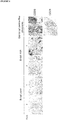
- the maximum lymphocytic aggregate identified may be graded according to the maximum radial cell count measured from the central vessel as described by Manzo et al. (as above). Aggregates may be categorised into three groups according to the radial cell count as shown in Table 5 and the grading atlas provided as Figures 2 and 3 . Table 5 Grade Radial Cell Count 0 Uninflamed / Diffuse No aggregates visualised 1 2-5 cells 2 6-10 cells 3 >10 cells
- biopsies are thus classified as B cell poor (Grade 0 and 1), B cell rich (Grade 2 and 3) and Germinal Center (GC) rich Grade 4 + CD 21 positivity (see Atlas 1, Figure 2 ).
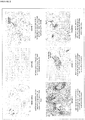
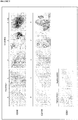
- Such a grading system may also be used in combination with analysis of B cell surface markers, for example CD20and/or CD79a (see Atlas 2, Figure 3 ).
- the presence of B cells within a synovial sample from a RA patient may be determined by analysing the level of B cell markers within the sample using techniques such as next-generation sequencing, gene expression arrays, PCR and proteomics. Such techniques may be used assess the level of a B cell marker RNA and/or protein within the synovial sample, with an increased level of the B cell marker determining a B cell rich profile.
- the B cell marker may be, for example, CD20 and/or CD79a.
- the synovial sample may be a synovial tissue biopsy.
- the method of the present invention comprises the step of analysing the presence of GC-LS in a synovial sample from a RA patient.
- Germinal centres are sites where mature B cells rapidly proliferate, differentiate, and undergo somatic hypermutation and class switch recombination during an immune response. During this process of rapid division and selection, B cells are known as centroblasts, and once they have stopped proliferating they are known as centrocytes. B cells within germinal centres express CD138 when they differentiate into plasma cells. Germinal centres develop dynamically after the activation of B cells by T-cell dependent antigen.
- GC-LS refers to an ectopic or tertiary lymphatic structure that forms in non-lymphoid tissues and may develop to become a place of autoantibody generation.
- GC-LS form in the synovium.
- GC-LS are typically characterised by the presence of aggregated T and/or B lymphocytes alongside follicular dendritic cells (FDCs).
- FDCs have high expression of complement receptors CR1 and CR2 (CD35 and CD21 respectively) and Fc-receptor Fc ⁇ RIIb (CD32). Further FDC specific molecular markers include FDC-M1, FDC-M2 and C4.
- the identification of GC-LS in a synovial sample of a RA patient may therefore involve determining the presence of cells positive for one or more of the above markers. For example it may involve determining the presence of plasma cells (CD138 + ) and/or FDCs (CD35 + -CD21 + ).
- GC-LS The identification of GC-LS may be performed using the immunohistochemistry scoring analysis as provided in Atlas 2 ( Figure 3 ).
- a RA patient identified as B cell rich and/or GC-LS negative by the method of the present invention is determined as being susceptible to treatment with a B cell targeted therapy whereas a patient who is B-cell poor and/or GC-LS positive is determined as resistant to treatment with a B cell targeted therapy.
- a B cell targeted therapy refers to the administration of an agent that interferes with or inhibits the development and/or function of B cells.
- the B cell targeted therapy may cause B cell depletion or the inhibition of B cell development and maturation.
- the B cell targeted therapy is directed against B cells in all stages of development other than undifferentiated stem cells and terminally differentiated antibody-producing plasma cells.
- the agent may be a small molecule drug, such as a Bruton's tyrosine kinase (BTK) inhibitor or other agent which targets B cell signalling pathways.
- BTK Bruton's tyrosine kinase
- Direct depletion of B cells may be performed through the use of monoclonal antibodies (mAbs) directed against cell surface markers (e.g. CD20 and CD22).
- mAbs monoclonal antibodies directed against cell surface markers (e.g. CD20 and CD22).
- Such mAbs bind to the target antigen and kill the cell by initiating a mixture of apoptosis, complement dependent cytotoxicity (CDC), and antibody-dependent cell-mediated cellular cytotoxicity (ADCC).
- CDC complement dependent cytotoxicity
- ADCC antibody-dependent cell-mediated cellular cytotoxicity
- the B cell targeted therapy used in the present invention may be an agent directed against CD20, for example Rituximab, Ocrelizumab, Veltuzumab or Ofatumumab, or an agent directed against CD22 such as Epratuzumab.
- Rituximab is a chimeric mouse/human immunoglobulin G1 (IgG1) monoclonal antibody to CD20 that stimulates B cell destruction upon binding to CD20.
- IgG1 immunoglobulin G1
- Rituximab depletes CD20 surface-positive naive and memory B cells from the blood, bone marrow and lymph nodes via mechanisms which include antibody-dependent cellular cytotoxicity (ADCC), complement dependent cytotoxicity (CDC). It does not affect CD20-negative early B cell lineage precursor cells and late B lineage plasma cells in the bone marrow.
- Ocrelizumab is a humanized anti-CD20 monoclonal antibody that causes CD20 + B cell depletion following binding to CD20 via mechanisms including ADCC and CDC.
- Veltuzumab is a humanized, second-generation anti-CD20 monoclonal antibody that causes CD20 + B cell depletion following binding to CD20 via mechanisms including ADCC and CDC.
- Ofatumumab is a human monoclonal IgG1 antibody to CD20 and may inhibit early-stage B lymphocyte activation.
- Ofatumumab targets a different epitope located closer to the N-terminus of CD20 compared to the epitope targeted by rituximab and includes an extracellular loop, as it binds to both the small and large loops of the CD20 molecule.
- Ofatumumab stimulates B cell destruction through ADCC and CDC pathways.
- a RA patient identified by the method of the present invention as GC-LS positive is determined to be suitable for treatment with an agent which downregulates interleukin-6 (IL-6) signalling.
- IL-6 interleukin-6
- IL-6 is a cytokine that provokes a broad range of cellular and physiological responses, including inflammation, hematopoiesis, and oncogenesis by regulating cell growth, gene activation, proliferation, survival, and differentiation. It is able to directly influence B cell activation state and late stage differentiation towards plasma cells.
- JAK Janus Kinase
- STAT3 is essential for GP130-mediated cell survival and G1 to S cell-cycle-transition signals. Both c-Myc and Pim have been identified as target genes of STAT3 and together can compensate for STAT3 in cell survival and cell-cycle transition. SHP2 links cytokine receptor to the Ras/MAP (Mitogen-Activated Protein) kinase pathway and is essential for mitogenic activity.
- Ras/MAP Mitogen-Activated Protein
- the Ras-mediated pathway acting through SHC, GRB2 (Growth Factor Receptor Bound protein-2) and SOS1 (Son of Sevenless-1) upstream and activating MAP kinases downstream, activates transcription factors such as Elk1 and NF-IL-6 (C/EBP- ⁇ ) that can act through their own cognate response elements in the genome.
- IL-6 In addition to JAK/STAT and Ras/MAP kinase pathways, IL-6 also activates PI3K (Phosphoinositide-3 Kinase).
- PI3K Phosphoinositide-3 Kinase
- the anti-apoptotic mechanism of PI3K/Akt is attributed to phosphorylation of the BCL2 family member BAD (BCL2 Associated Death Promoter) by Akt.
- BAD BCL2 Associated Death Promoter
- the phosphorylated BAD is then associated with 14-3-3, which sequesters BAD from BCLXL, thereby promoting cell survival.
- Regulating the BCL2 family member is also considered as one of the anti-apoptotic mechanisms of STAT3, which was may be capable of inducing BCL2 in pro-B cells.
- the termination of the IL-6-type cytokine signaling is through the action of tyrosine phosphatases, proteasome, and JAK kinase inhibitors SOCS (Suppressor of Cytokine Signaling), PIAS (Protein Inhibitors of Activated STATs), and internalization of the cytokine receptors via GP130.
- an agent which downregulates IL-6 signalling may interfere with or inhibit any of the above stages involved in IL-6 mediated signalling such that IL-6 signalling and responses are diminished.
- the agent may be an IL-6 receptor antagonist such as Tocilizumab, which is a humanized monoclonal antibody against the IL-6 receptor.
- An IL-6 receptor antagonist refers to an agent that reduces the level of IL-6 that is able to bind to the IL-6 receptor.
- the present invention also provides a method for treating a RA patient who is refractory to treatment with a B cell targeted therapy, which comprises the step of disrupting GC-LS in the synovium.
- the GC-LS may be disrupted by treatment with an agent which downregulates IL-6 mediated signalling, for example Tocilizumab.
- an agent which downregulates IL-6 mediated signalling for example Tocilizumab.
- Tocilizumab is a humanized monoclonal IgG1 antibody against the IL-6 receptor that binds to soluble and membrane-bound IL-6 receptor. Tocilizumab inhibits the induction of biological activity due to IL-6 in cells that have expressed both membrane-bound IL-6 receptor and gp130 molecules, and also inhibits the induction of biological activity due to IL-6/IL-6 receptor complex formation in cells that express gp130 alone. Furthermore, since it has the capacity to dissociate IL-6/IL-6 receptor complexes that have already formed, it is able to block IL-6 signal transduction.
- the GC-LS may be disrupted by treatment with a growth factor inhibitor or an agent that inhibits signalling required for B cell function.
- the agent may, for example, inhibit B-cell activating factor (BAFF) or a proliferation-inducing ligand (APRIL) signalling.
- BAFF B-cell activating factor
- APRIL proliferation-inducing ligand
- agents include, but are not limited to, Belimumab and Atacicept.
- Belimumab is a human monoclonal IgG1 ⁇ antibody that inhibits BAFF (also known as B-lymphocyte stimulator (BLyS)).
- BAFF also known as B-lymphocyte stimulator (BLyS)
- BAFF is a 285-amino acid type II protein that is a member of the TNF ligand superfamily and is a vital B cell survival factor, with important roles in the differentiation of immature to mature B cells and in immunoglobulin class switching and production. Belimumab inhibits B cell survival and differentiation by neutralizing soluble BAFF, without directly causing B cell death.
- Atacicept is a fully human recombinant fusion protein containing the extracellular ligand-binding portion of the TACI (Transmembrane Activator and CAML [calcium-modulator and cyclophilin-ligand]-Interactor) receptor and a modified Fc portion of human IgG. Atacicept, therefore, contains the binding portion of a receptor that binds both BAFF and APRIL.
- APRIL is a structural homologue of BAFF that is secreted as a soluble protein by monocytes, macrophages, dendritic cells, neutrophils and T cells, and shares some of the biological properties of BAFF.
- the GC-LS may be disrupted by a proteasome inhibitor such as Bortezomib.
- Bortezomib is an N-protected dipeptide that binds the catalytic site of the 26S proteasome, thereby inhibiting proteasome function.
- the proteasome regulates protein expression and function by degradation of ubiquitylated proteins, and also removes abnormal or misfolded proteins from the cell.
- the present disclosure also describes a kit for use in a method of determining whether a RA patient is susceptible to treatment with a B cell targeted therapy, which method comprises the step of analysing the presence of B cells and/or germinal centre-like structures (GC-LS) in a synovial tissue sample from the patient; wherein a patient whose synovial tissue sample is B cell rich and/or GC-LS negative is determined to be susceptible to treatment with the B cell targeted therapy, whereas a patient whose synovial tissue sample is B-cell poor and/or GC-LS positive is determined to be resistant to treatment with the B cell targeted therapy.
- GC-LS germinal centre-like structures
- the kit may comprise;
- Agents for the detection of GC-LS in a synovial sample of a RA patient may include agents that detect CD21.
- the kit may also comprise instructions for use.
- the kit may also comprise a B cell targeted therapy or an agent that downregulates IL-6 signalling.
- Example 1 An investigation into the molecular mechanisms predicting response to Rituximab in RA patients resistant to anti-TNF therapy
- Table 2 Patient demographics of 27 patients recruited are shown in Table 2.
- the primary outcome measure was the number of responders at 3 months within each histomorphological subgroup.
- Synovial tissue specimens were immediately fixed in 4% formalin. After paraffin embedding, 3 ⁇ m serial sections underwent routine H&E staining in order to define the predominant histological pattern of RA synovitis as either diffuse or aggregate. Lymphocytic aggregates were classified into three groups based on a scoring system the present inventors had adapted from their previous publication ( Manzo et al. Eur J Immunol. (2005); 35(5):1347-1359 ). Depending on the numbers of B cell identified per high power field (HPF), biopsies were classified as B cell poor (Grade 0 and 1), B cell rich (Grade 2 and 3) and Germinal Center (GC) rich Grade 4 + CD 21 positive.
- HPF high power field
Description
- The invention relates to a method for determining whether a rheumatoid arthritis (RA) patient is susceptible to treatment with a B cell targeted therapy, such as Rituximab. The invention also relates to a method for treating a RA patient who is refractory to B cell targeted therapy.
- Inflammatory arthritis is a prominent clinical manifestation in diverse autoimmune disorders including rheumatoid arthritis (RA), psoriatic arthritis (PsA), systemic lupus erythematosus (SLE), Sjogren's syndrome and polymyositis.
- RA is a chronic inflammatory disease that affects approximately 0.5 to 1% of the adult population in northern Europe and North America. It is a systemic inflammatory disease characterized by chronic inflammation in the synovial membrane of affected joints, which ultimately leads to loss of daily function due to chronic pain and fatigue. The majority of patients also experience progressive deterioration of cartilage and bone in the affected joints, which may eventually lead to permanent disability. The long-term prognosis of RA is poor, with approximately 50% of patients experiencing significant functional disability within 10 years from the time of diagnosis. Life expectancy is reduced by an average of 3-10 years.
- Inflammatory bone diseases, such as RA, are accompanied by bone loss around affected joints due to increased osteoclastic resorption. This process is mediated largely by increased local production of pro-inflammatory cytokines, of which tumor necrosis factor-α (TNF-α) is a major effector.
- In RA specifically, an immune response is thought to be initiated/perpetuated by one or several antigens presenting in the synovial compartment, producing an influx of acute inflammatory cells and lymphocytes into the joint. Successive waves of inflammation lead to the formation of an invasive and erosive tissue called pannus. This contains proliferating fibroblast-like synoviocytes and macrophages that produce proinflammatory cytokines such as TNF-α and interleukin-1 (IL-I). Local release of proteolytic enzymes, various inflammatory mediators, and osteoclast activation contributes to much of the tissue damage. There is loss of articular cartilage and the formation of bony erosions. Surrounding tendons and bursa may become affected by the inflammatory process. Ultimately, the integrity of the joint structure is compromised, producing disability.
- B cells are thought to contribute to the immunopathogenesis of RA, predominantly by serving as the precursors of autoantibody-producing cells but also as antigen presenting cells (APC) and pro-inflammatory cytokine producing cells. A number of autoantibody specificities have been identified including antibodies to Type II collagen and proteoglycans, as well as rheumatoid factors and most importantly anti citrullinated protein antibodies (ACPA). The generation of large quantities of antibody leads to immune complex formation and the activation of the complement cascade. This in turn amplifies the immune response and may culminate in local cell lysis.
-
WO 2012/118750 discloses biomarkers comprising elevated total plasma/plasmablast cell mRNA or an elevated expression level of a plasma/plasmablast cell-enriched gene, useful for predicting the response of a patient to B-cell antagonist. - Frances Humby et Al: "Ectopic Lymphoid Structures Support Ongoing Production of Class-Switched Autoantibodies in Rheumatoid Synovium", Annals of the Rheumatic Diseases, vol. 65, no. 1, 2009, p.el; and Antonio Manzo et Al: "Secondary and ectopic lymphoid tissue responses in rheumatoid arthritis: from inflammation to autoimmunity and tissue damage/remodeling", Immunological Reviews, vol. 233, no. 1, 2010, p.267-285, show that survival of functional synovial B cell niches may profoundly influence chronic inflammation, autoimmunity, and response to B cell-depleting therapies.
- Cañete J. D. et Al: "Clinical significance of synovial lymphoid neogenesis and its reversal after anti-tumour necrosis factor alpha therapy in rheumatoid arthritis", Annals of the Rheumatic Diseases, 2009, vol. 68, no. 5, 2009, p.751-756, provides similar findings for RA patients treated with anti-TNFalpha.
- Current standard therapies for RA which are used to modify the disease process and to delay joint destruction are known as disease modifying anti-rheumatic drugs (DMARDs). Methotrexate, leflunomide and sulfasalazine are traditional DMARDs and are often effective as first-line treatment.
- Biologic agents designed to target specific components of the immune system that play role in RA are also used as therapeutics. There are various groups of biologic treatments for RA including; TNF-α inhibitors (etanercept, infliximab and adalimumab), human IL-1 receptor antagonist (anakinra) and selective co-stimulation modulators (abatacept).
- Rituximab is indicated for the treatment of moderate to severe RA in adult patients who have had an inadequate response to, or cannot tolerate, one or more TNF-α inhibitor therapies. It has been shown to be effective in the treatment of RA in patients refractory to treatment with anti-TNF therapy.
- The Rituximab antibody is a genetically engineered chimeric murine/human monoclonal antibody directed against the CD20 antigen. Rituximab binds human complement and lyses lymphoid B-cell lines through complement-dependent cytotoxicity. Additionally, it has significant activity in assays for antibody-dependent cellular cyotoxicity. More recently, Rituximab has been shown to have anti-proliferative effects in tritiated thymidine-incorporation assays and to induce apoptosis directly. Other anti-CD19 and anti-CD20 antibodies have not been shown to have this activity.
- Rituximab treatment has been shown to result in B cell depletion in peripheral blood, bone marrow and the synovium. However, not all patients refractory to treatment with anti-TNF therapy are responsive to Rituximab treatment. Current evidence on the efficacy of Rituximab relates primarily to rheumatoid factor, ACPA positive patients, although even within this population clinical responses are heterogeneous with only 60% achieving an ACR20 response within 6 months.
- Rituximab is associated with various safety issues, especially infusion-related adverse events and is also very expensive, costing approximately $10,000 per treatment course.
- There is thus a need for a method to predict whether a given RA patient is likely to respond to Rituximab treatment. There is also a need for alternative method of treatment for RA patients who are refractory to treatment with Rituximab.
-
-
Figure 1 - immunohistochemical staining of tissue biopsy samples used in an exemplar B cell scoring system. Staining for CD20 was used to quantitatively assess the presence of B cells in RA synovial samples, being classified as B cell poor (score from 0 to 1) and B cell rich (score from 2-3) as is shown in the example images. CD21 expression was assessed in samples displaying a CD20 score of 4 and were positive for GC-LS / FDC networks when a cluster of CD21+ cells was detected. Original magnification x10. -
Figure 2 - Atlas 1: Scoring of aggregates on H&E specimens using a 0-3 scale -
Figure 3 - Atlas 2: Scoring of CD20, CD138 and CD21 by immunohistochemistry scoring analysis - The invention is defined by the claims.
- The present inventors have made the surprising finding that the histomorphological type of a synovial sample from a RA patient is predictive of the patient's response to B cell targeted therapy.
- Due to the systemic nature of RA, it was thought an assessment of biomarkers in peripheral blood was important for identification and stratification of patients who would respond to B cell targeted therapy (Owczarczyk et al. Sci Transl Med 2011;3:101ra92, Sellam et al. Arthritis & Rheumatism 2011; 63(4):933-938). The present invention, however, now shows that rather than looking at markers in blood, B cell levels and morphology in the synovium are predictive of a RA patient's response to B cell targeted therapy.
- In a first aspect, the present invention relates to a method for determining whether a Rheumatoid Arthritis (RA) patient is susceptible to treatment with a B cell targeted therapy, which method comprises the step of analysing B cells and germinal centre-like structures (GC-LS) in a synovial tissue sample from the patient; wherein a patient whose synovial tissue sample is B cell rich and GC-LS negative is determined to be susceptible to treatment with the B cell targeted therapy, whereas a patient whose synovial tissue sample is B-cell poor or GC-LS positive is determined to be resistant to treatment with the B cell targeted therapy.
- The step of analysing B cells and GC-LS is performed by histological analysis. A patient whose synovial tissue sample is B cell poor may be characterised by a diffuse pattern of a small number of B cells interspersed with prevalent myeloid inflammatory infiltrate, while a patient whose synovial tissue sample is B cell rich may be characterised by the clustering of B cells in an aggregated pattern. A patient who is GC-LS positive may be characterised by the presence of B cell aggregates and detection of CD21 within said aggregates.
- B cells may be analysed by determining CD20 expression or other markers in the synovial sample.
- The B cell targeted therapy may be B cell depletion therapy. The B cell targeted therapy may be selected from the following list: Rituximab, Ocrelizumab, Veltuzumab, Ofatumumab and Epratuzumab.
- The B cell targeted therapy may be Rituximab.
- A patient whose synovial tissue sample is GC-LS positive may be resistant to Rituximab therapy and determined to be suitable for treatment with an agent which downregulates IL-6 mediated signalling. The agent that downregulates IL-6 signalling may be an IL-6 receptor antagonist, for example the agent may be Tocilizumab.
- The method of the present invention may be performed using a synovial sample from a RA patient who is refractory to synthetic or biologic (e.g. anti-TNF) DMARD therapies.
- In a further aspect the present invention provides a method for treating a Rheumatoid Arthritis (RA) patient who is refractory to treatment with a B cell targeted therapy which comprises the step of disrupting germinal centre-like structures (GC-LS) in the synovium.
- The GC-LS may be disrupted by treatment with an agent which downregulates IL-6 mediated signalling, a proteasome inhibitor or a growth factor inhibitor.
- RA is a chronic, systemic inflammatory disorder that may affect many tissues and organs, but principally attacks synovial joints. It is a disabling and painful condition, which can lead to substantial loss of functioning and mobility if not adequately treated.
- The disease process involves an inflammatory response of the synovium, secondary to massive immune cell infiltration and proliferation of synovial cells, excess synovial fluid, and the development of fibrous tissue (pannus) in the synovium that attacks the cartilage and sub-chondral bone. This often leads to the destruction of articular cartilage and the formation of bone erosions with secondary ankylosis (fusion) of the joints. RA can also produce diffuse inflammation in the lungs, the pericardium, the pleura, the sclera, and also nodular lesions, most commonly in subcutaneous tissue. RA is considered a systemic autoimmune disease as autoimmunity plays a pivotal role in its chronicity and progression.
- The term synovial sample refers to a sample derived from a synovial joint. Typically the synovial sample will be derived for a synovial joint of a RA patient. A synovial sample may be a synovial tissue biopsy and the synovial joint may display active inflammation at the time the sample is taken.
- A number of cell types are involved in the aetiology of RA, including T cells, B cells, monocytes, macrophages, dendritic cells and synovial fibroblasts. Autoantibodies known to be associated with RA include those targeting Rheumatoid factor (RF) and anti citrullinated protein antibodies (ACPA).
- A typical patient with newly diagnosed RA is often treated initially with nonsteroidal antiinflammatory drugs and disease-modifying antirheumatic drugs (DMARDs) such as hydroychloroquine, sulfasalazine, leflunomide, or methotrexate (MTX), alone or in combinations. Patients who do not respond to general DMARDs may be termed DMARD-refractory.
- DMARD-refractory patients are often progressed to biological therapeutic agents, for example TNF-α antagonists such as Adalimumab, Etanercept, Golimumab and Infliximab. Patients who do not respond to TNF-α antagonist therapy may be termed TNF-α antagonist-refractory or inadequate responders (ir).
- The method of the present invention may be performed on a synovial sample from a RA patient who has previously been determined to be refractory to DMARD-therapy and/or TNF-α antagonist therapy. The method may also be performed on a synovial sample from a RA patient unable to tolerate TNF-α antagonist therapy.
- B cells play a central role in the pathogenesis of RA.
- Immature B cells are produced in the bone marrow. After reaching the IgM+ immature stage in the bone marrow, these immature B cells migrate to secondary lymphoid tissues (such as the spleen, lymph nodes) where they are called transitional B cells, and some of these cells differentiate into mature B lymphocytes and possibly plasma cells.
- B cells may be defined by a range of cell surface markers which are expressed at different stages of B cell development and maturation (Table 1). These B cell markers may include CD19, CD20, CD22, CD23, CD24, CD27, CD38, CD40, CD72, CD79a and CD79b, CD138 and immunoglobulin (Ig).
Table 1 Compartment Cell Type Markers Bone marrow Stem cell Pro-B cell CD19+ CD20- Ig- Pre-B cell CD19+ CD20+ Ig- Immature B cell CD19+ CD20+ Ig+ Peripheral compartments Naive B cell CD 19+ CD20+ Ig+ CD3 8+/- Naive activated B cell CD19+ CD20+ Ig+ CD38+ GC B cell CD19+ CD20+ Ig+ CD38++ Post-GC B cell CD19+ CD20+ Ig+ CD38+ Memory B cell CD 19+ CD20+ Ig+/- CD27+ IgM/IgG/IgA+ CD38- Plasma blast CD19+ CD20- Ig+/- CD27++ CD38++ Bone marrow Plasma cell CD19+/- CD20- Ig- CD27++ CD38+++ CD138+ - Immunoglobulins (Ig) are glycoproteins belonging to the immunoglobulin superfamily which recognise foreign antigens and facilitate the humoral response of the immune system. Ig may occur in two physical forms, a soluble form that is secreted from the cell, and a membrane-bound form that is attached to the surface of a B cell and is referred to as the B cell receptor (BCR). Mammalian Ig may be grouped into five classes (isotypes) based on which heavy chain they possess. Immature B cells, which have never been exposed to an antigen, are known as naïve B cells and express only the IgM isotype in a cell surface bound form. B cells begin to express both IgM and IgD when they reach maturity - the co-expression of both these immunoglobulin isotypes renders the B cell 'mature' and ready to respond to antigen. B cell activation follows engagement of the cell bound antibody molecule with an antigen, causing the cell to divide and differentiate into an antibody producing plasma cell. In this activated form, the B cell starts to produce antibody in a secreted form rather than a membrane-bound form. Some daughter cells of the activated B cells undergo isotype switching to change from IgM or IgD to the other antibody isotypes, IgE, IgA or IgG, that have defined roles in the immune system.
- CD19 is expressed by essentially all B-lineage cells and regulates intracellular signal transduction by amplifying Src-family kinase activity.
- CD20 is a mature B cell-specific molecule that functions as a membrane embedded Ca2+ channel. Expression of CD20 is restricted to the B cell lineage from the pre-B-cell stage until terminal differentiation into plasma cells.
- CD22 functions as a mammalian lectin for α2,6-linked sialic acid that regulates follicular B-cell survival and negatively regulates signaling.
- CD23 is a low-affinity receptor for IgE expressed on activated B cells that influences IgE production.
- CD24 is a GPI-anchored glycoprotein which was among the first pan-B-cell molecules to be identified.
- CD27 is a member of the TNF-receptor superfamily. It binds to its ligand CD70, and plays a key role in regulating B-cell activation and immunoglobulin synthesis. This receptor transduces signals that lead to the activation of NF-κB and MAPK8/JNK.
- CD38 is also known as cyclic ADP ribose hydrolase. It is a glycoprotein that also functions in cell adhesion, signal transduction and calcium signalling and is generally a marker of cell activation.
- CD40 serves as a critical survival factor for germinal centre (GC) B cells and is the ligand for CD154 expressed by T cells.
- CD72 functions as a negative regulator of signal transduction and as the B-cell ligand for Semaphorin 4D (CD100).
- CD79a/CD79b dimer is closely associated with the B-cell antigen receptor, and enables the cell to respond to the presence of antigens on its surface. The CD79a/CD79b dimer is present on the surface of B-cells throughout their life cycle, and is absent on all other healthy cells.
- CD138 is also known as
Syndecan 1. Syndecans mediate cell binding, cell signalling, and cytoskeletal organization. CD138 may be useful as a cell surface marker for plasma cells. - The method of the present invention comprises the step of analysing the presence of B cells in a synovial sample from a RA patient and determining if a RA patient is B cell rich or B cell poor. This analysis may involve determining the presence of cells expressing one or more of the markers detailed in Table 1.
- The presence of B cells may be determined by analysing the level and pattern of B cells in a synovial sample from a RA patient. Such analysis may be performed by histological analysis.
- The identification of RA patients who are B cell rich or B cell poor may be performed by using a system for grading lymphocytic aggregates known to those skilled in the art. In the examples section, the present inventors use a system adapted from the one described in their previous publication (Manzo et al. Eur J Immunol. (2005); 35(5):1347-1359). The radial cell count is estimated by counting the number of cells from the more centrally located vessel to the identifiable edge of its aggregate. The determination is made at the point of widest infiltration.
- The maximum lymphocytic aggregate identified may be graded according to the maximum radial cell count measured from the central vessel as described by Manzo et al. (as above). Aggregates may be categorised into three groups according to the radial cell count as shown in Table 5 and the grading atlas provided as
Figures 2 and3 .Table 5 Grade Radial Cell Count 0 Uninflamed / Diffuse No aggregates visualised 1 2-5 cells 2 6-10 cells 3 >10 cells - Depending on the numbers of B cell identified per high power field (HPF), biopsies are thus classified as B cell poor (
Grade 0 and 1), B cell rich (Grade 2 and 3) and Germinal Center (GC) rich Grade 4 + CD 21 positivity (seeAtlas 1,Figure 2 ). - Such a grading system may also be used in combination with analysis of B cell surface markers, for example CD20and/or CD79a (see
Atlas 2,Figure 3 ). - The presence of B cells within a synovial sample from a RA patient may be determined by analysing the level of B cell markers within the sample using techniques such as next-generation sequencing, gene expression arrays, PCR and proteomics. Such techniques may be used assess the level of a B cell marker RNA and/or protein within the synovial sample, with an increased level of the B cell marker determining a B cell rich profile. The B cell marker may be, for example, CD20 and/or CD79a. The synovial sample may be a synovial tissue biopsy.
- The method of the present invention comprises the step of analysing the presence of GC-LS in a synovial sample from a RA patient.
- Germinal centres are sites where mature B cells rapidly proliferate, differentiate, and undergo somatic hypermutation and class switch recombination during an immune response. During this process of rapid division and selection, B cells are known as centroblasts, and once they have stopped proliferating they are known as centrocytes. B cells within germinal centres express CD138 when they differentiate into plasma cells. Germinal centres develop dynamically after the activation of B cells by T-cell dependent antigen.
- The term GC-LS refers to an ectopic or tertiary lymphatic structure that forms in non-lymphoid tissues and may develop to become a place of autoantibody generation. In the context of RA, the GC-LS form in the synovium. GC-LS are typically characterised by the presence of aggregated T and/or B lymphocytes alongside follicular dendritic cells (FDCs).
- FDCs have high expression of complement receptors CR1 and CR2 (CD35 and CD21 respectively) and Fc-receptor FcγRIIb (CD32). Further FDC specific molecular markers include FDC-M1, FDC-M2 and C4.
- The identification of GC-LS in a synovial sample of a RA patient may therefore involve determining the presence of cells positive for one or more of the above markers. For example it may involve determining the presence of plasma cells (CD138+) and/or FDCs (CD35+-CD21+).
- The identification of GC-LS may be performed using the immunohistochemistry scoring analysis as provided in Atlas 2 (
Figure 3 ). - Determining the presence of GC-LS in a synovial sample of a RA patient may involve identifying FDCs within B cell aggregates using one or more of the above markers. Determining the presence of GC-LS may involve the identification of CD21+ cells within B cell aggregates in a synovial sample from a RA patient.
- A RA patient identified as B cell rich and/or GC-LS negative by the method of the present invention is determined as being susceptible to treatment with a B cell targeted therapy whereas a patient who is B-cell poor and/or GC-LS positive is determined as resistant to treatment with a B cell targeted therapy.
- A B cell targeted therapy refers to the administration of an agent that interferes with or inhibits the development and/or function of B cells. The B cell targeted therapy may cause B cell depletion or the inhibition of B cell development and maturation. Advantageously, the B cell targeted therapy is directed against B cells in all stages of development other than undifferentiated stem cells and terminally differentiated antibody-producing plasma cells. The agent may be a small molecule drug, such as a Bruton's tyrosine kinase (BTK) inhibitor or other agent which targets B cell signalling pathways.
- Direct depletion of B cells may be performed through the use of monoclonal antibodies (mAbs) directed against cell surface markers (e.g. CD20 and CD22). Such mAbs bind to the target antigen and kill the cell by initiating a mixture of apoptosis, complement dependent cytotoxicity (CDC), and antibody-dependent cell-mediated cellular cytotoxicity (ADCC).
- The B cell targeted therapy used in the present invention may be an agent directed against CD20, for example Rituximab, Ocrelizumab, Veltuzumab or Ofatumumab, or an agent directed against CD22 such as Epratuzumab.
- Rituximab is a chimeric mouse/human immunoglobulin G1 (IgG1) monoclonal antibody to CD20 that stimulates B cell destruction upon binding to CD20. Rituximab depletes CD20 surface-positive naive and memory B cells from the blood, bone marrow and lymph nodes via mechanisms which include antibody-dependent cellular cytotoxicity (ADCC), complement dependent cytotoxicity (CDC). It does not affect CD20-negative early B cell lineage precursor cells and late B lineage plasma cells in the bone marrow.
- Ocrelizumab is a humanized anti-CD20 monoclonal antibody that causes CD20+ B cell depletion following binding to CD20 via mechanisms including ADCC and CDC.
- Veltuzumab is a humanized, second-generation anti-CD20 monoclonal antibody that causes CD20+ B cell depletion following binding to CD20 via mechanisms including ADCC and CDC.
- Ofatumumab is a human monoclonal IgG1 antibody to CD20 and may inhibit early-stage B lymphocyte activation. Ofatumumab targets a different epitope located closer to the N-terminus of CD20 compared to the epitope targeted by rituximab and includes an extracellular loop, as it binds to both the small and large loops of the CD20 molecule. Ofatumumab stimulates B cell destruction through ADCC and CDC pathways.
- Epratuzumab is a humanized monoclonal IgG1 antibody to CD22. It contains a murine sequence comprising 5-10% of the molecule, the remainder being human framework sequences. Epratuzumab binds to the CD22 third extracellular domain (epitope B), without blocking the ligand binding site, with measured affinity of K d = 0.7 nm. In vitro studies showed epratuzumab induces CD22 phosphorylation by binding to its surface. It results in modulation, mostly negative, of BCR activation.
- In a further aspect, a RA patient identified by the method of the present invention as GC-LS positive is determined to be suitable for treatment with an agent which downregulates interleukin-6 (IL-6) signalling.
- IL-6 is a cytokine that provokes a broad range of cellular and physiological responses, including inflammation, hematopoiesis, and oncogenesis by regulating cell growth, gene activation, proliferation, survival, and differentiation. It is able to directly influence B cell activation state and late stage differentiation towards plasma cells.
- IL-6 signals through a receptor composed of two different subunits, an alpha subunit that produces ligand specificity and GP (Glycoprotein) 130, a receptor subunit shared in common with other cytokines in the IL-6 family. Binding of IL-6 to its receptor initiates cellular events including activation of JAK (Janus Kinase) kinases and activation of Ras-mediated signalling. Activated JAK kinases phosphorylate and activate STAT transcription factors, particularly STAT3 and SHP2. Phosphorylated STAT3 then forms a dimer and translocates into the nucleus to activate transcription of genes containing STAT3 response elements. STAT3 is essential for GP130-mediated cell survival and G1 to S cell-cycle-transition signals. Both c-Myc and Pim have been identified as target genes of STAT3 and together can compensate for STAT3 in cell survival and cell-cycle transition. SHP2 links cytokine receptor to the Ras/MAP (Mitogen-Activated Protein) kinase pathway and is essential for mitogenic activity.
- The Ras-mediated pathway, acting through SHC, GRB2 (Growth Factor Receptor Bound protein-2) and SOS1 (Son of Sevenless-1) upstream and activating MAP kinases downstream, activates transcription factors such as Elk1 and NF-IL-6 (C/EBP-β) that can act through their own cognate response elements in the genome.
- In addition to JAK/STAT and Ras/MAP kinase pathways, IL-6 also activates PI3K (Phosphoinositide-3 Kinase). The PI3K/Akt/NF-KappaB cascade activated by IL-6, functions cooperatively to achieve the maximal anti-apoptotic effect of IL-6 against TGF-β. The anti-apoptotic mechanism of PI3K/Akt is attributed to phosphorylation of the BCL2 family member BAD (BCL2 Associated Death Promoter) by Akt. The phosphorylated BAD is then associated with 14-3-3, which sequesters BAD from BCLXL, thereby promoting cell survival. Regulating the BCL2 family member is also considered as one of the anti-apoptotic mechanisms of STAT3, which was may be capable of inducing BCL2 in pro-B cells. The termination of the IL-6-type cytokine signaling is through the action of tyrosine phosphatases, proteasome, and JAK kinase inhibitors SOCS (Suppressor of Cytokine Signaling), PIAS (Protein Inhibitors of Activated STATs), and internalization of the cytokine receptors via GP130.
- An agent which downregulates IL-6 signalling may interfere with or inhibit any of the above stages involved in IL-6 mediated signalling such that IL-6 signalling and responses are diminished. For example the agent may be an IL-6 receptor antagonist such as Tocilizumab, which is a humanized monoclonal antibody against the IL-6 receptor. An IL-6 receptor antagonist refers to an agent that reduces the level of IL-6 that is able to bind to the IL-6 receptor.
- The present invention also provides a method for treating a RA patient who is refractory to treatment with a B cell targeted therapy, which comprises the step of disrupting GC-LS in the synovium.
- The GC-LS may be disrupted by treatment with an agent which downregulates IL-6 mediated signalling, for example Tocilizumab.
- Tocilizumab is a humanized monoclonal IgG1 antibody against the IL-6 receptor that binds to soluble and membrane-bound IL-6 receptor. Tocilizumab inhibits the induction of biological activity due to IL-6 in cells that have expressed both membrane-bound IL-6 receptor and gp130 molecules, and also inhibits the induction of biological activity due to IL-6/IL-6 receptor complex formation in cells that express gp130 alone. Furthermore, since it has the capacity to dissociate IL-6/IL-6 receptor complexes that have already formed, it is able to block IL-6 signal transduction.
- The GC-LS may be disrupted by treatment with a growth factor inhibitor or an agent that inhibits signalling required for B cell function. The agent may, for example, inhibit B-cell activating factor (BAFF) or a proliferation-inducing ligand (APRIL) signalling. Examples of such agents include, but are not limited to, Belimumab and Atacicept.
- Belimumab is a human monoclonal IgG1λ antibody that inhibits BAFF (also known as B-lymphocyte stimulator (BLyS)). BAFF is a 285-amino acid type II protein that is a member of the TNF ligand superfamily and is a vital B cell survival factor, with important roles in the differentiation of immature to mature B cells and in immunoglobulin class switching and production. Belimumab inhibits B cell survival and differentiation by neutralizing soluble BAFF, without directly causing B cell death.
- Atacicept is a fully human recombinant fusion protein containing the extracellular ligand-binding portion of the TACI (Transmembrane Activator and CAML [calcium-modulator and cyclophilin-ligand]-Interactor) receptor and a modified Fc portion of human IgG. Atacicept, therefore, contains the binding portion of a receptor that binds both BAFF and APRIL. APRIL is a structural homologue of BAFF that is secreted as a soluble protein by monocytes, macrophages, dendritic cells, neutrophils and T cells, and shares some of the biological properties of BAFF.
- The GC-LS may be disrupted by a proteasome inhibitor such as Bortezomib.
- Bortezomib is an N-protected dipeptide that binds the catalytic site of the 26S proteasome, thereby inhibiting proteasome function. The proteasome regulates protein expression and function by degradation of ubiquitylated proteins, and also removes abnormal or misfolded proteins from the cell.
- The present disclosure also describes a kit for use in a method of determining whether a RA patient is susceptible to treatment with a B cell targeted therapy, which method comprises the step of analysing the presence of B cells and/or germinal centre-like structures (GC-LS) in a synovial tissue sample from the patient;
wherein a patient whose synovial tissue sample is B cell rich and/or GC-LS negative is determined to be susceptible to treatment with the B cell targeted therapy, whereas a patient whose synovial tissue sample is B-cell poor and/or GC-LS positive is determined to be resistant to treatment with the B cell targeted therapy. - The kit may comprise;
- i) An agent for the detection of B cells in a synovial sample of a RA patient,
- ii) An agent for the detection of GC-LS in a synovial sample of a RA patient. Agents for the detection of B cells in a synovial sample from a RA patient may include agents that detect CD20 and/or CD79a.
- Agents for the detection of GC-LS in a synovial sample of a RA patient may include agents that detect CD21.
- The kit may also comprise instructions for use.
- The kit may also comprise a B cell targeted therapy or an agent that downregulates IL-6 signalling.
- The invention will now be further described by way of Examples, which are meant to serve to assist one of ordinary skill in the art in carrying out the invention and are not intended in any way to limit the scope of the invention.
- This was an open-label, ultrasound guided synovial biopsy-based study of RA patients. The primary aim was to investigate in anti-TNFα inadequate responders whether baseline synovial pathobiological phenotypes defined specific response/resistance subsets to Rituximab therapy.
- Patient demographics of 27 patients recruited are shown in Table 2.
Table 2: Patient demographics of total study population (n=27) No (%) Female 21 (77) Rheumatoid factor +ve 24 (88) CCP +ve 25 (97) Erosive 25 (97) Mean +/- St Dev Age 59.1 (+/- 14.1) Baseline DAS 6.1 (+/-1.6) - A preliminary analysis of the first 21 patients recruited to the study classified patients into either an aggregate or diffuse synovial histological pattern and numbers of responders / non-responders within each histological group were determined (Table 2). Results were analysed using Fisher's exact test and demonstrated a significant difference in response between groups (p=0.014), with a significantly higher percentage of aggregate (83%) versus diffuse (20%) patients responding to Rituximab treatment.
Table 3: Response to Rituximab is significantly associated with an aggregate histological pattern and non-response is significantly associated with a diffuse infiltrate in TNF inadequate responders (n=21, p=0.014). Clinical response to Rituximab Synovial Histomorphological Pattern Aggregate number (%) Diffuse number (%) Responder 5(83) 3 (20) Non-responder 1(17) 12 (80) - All 27 patients were classified as B cell rich, B cell poor or germinal centre like structures (GC-LS) positive (+ve). Numbers of responders and non-responders within each histological group were identified and results analysed using Fisher's exact test (Table 4). A significant difference (p=0.032) was seen between the percentage of patients responding to Rituximab in the B cell poor (22%) versus B cell rich (80%) group. In addition within the group of patients classified as GC-LS positive there was a trend towards non-response to Rituximab (75% non-responders vs 25% responders).
Table 4: Immunohistological classification of synovial tissue as B cell rich, B cell poor or Germinal centre demonstrates a significant difference in levels of response to Rituximab within B cell rich and B cell poor patients (n=27, p=0.032). Clinical response to Rituximab Synovial Histomorphological Pattern Germinal Centre +ve n (%) B cell rich n (%) B cell poor n (%) Responder 1(25) 4(80) 4(22) Non-responder 3(75) 1(20) 14(78) - At baseline patients underwent a minimally invasive ultrasound guided synovial biopsy of an actively inflamed joint. Baseline clinical data was collected including age, sero positivity for rheumatoid factor, anti-CCP antibody status, disease activity score (DAS) of 28 joint count and radiographs of hands and feet. Patients were infused with standard Rituximab therapy (2x 1g Rituximab therapy at a fortnightly interval) within 2 weeks of the biopsy. DAS-28 assessment along with routine bloods following Rituximab therapy were performed on a monthly basis for 12 months. Patients were classified as responders to treatment at 3 months according to EULAR response criteria and responders were eligible for re-treatment at 6 and 12 month time points. Non-responders to Rituximab were switched to other biologic drugs according to NICE guidelines (TA195 and TA198).
- The primary outcome measure was the number of responders at 3 months within each histomorphological subgroup.
- Synovial tissue specimens were immediately fixed in 4% formalin. After paraffin embedding, 3µm serial sections underwent routine H&E staining in order to define the predominant histological pattern of RA synovitis as either diffuse or aggregate. Lymphocytic aggregates were classified into three groups based on a scoring system the present inventors had adapted from their previous publication (Manzo et al. Eur J Immunol. (2005); 35(5):1347-1359). Depending on the numbers of B cell identified per high power field (HPF), biopsies were classified as B cell poor (
Grade 0 and 1), B cell rich (Grade 2 and 3) and Germinal Center (GC) rich Grade 4 + CD 21 positive. - In addition, formalin-fixed, paraffin-embedded tissue sections underwent immunohistochemical staining. Following antigen retrieval with Target retrieving solution (DAKO) sections were stained for CD20 (IgG2a, clone L26; DAKO) at an antibody dilution of 1:20 and following proteinase K digestion (DAKO) single staining for CD21 (IF8, DAKO) at a dilution of 1:20 was also performed to identify FDC networks (CD21 +ve) characterising germinal centre like structures (GC-LS). Based on the results of the CD21 staining and the presence of G2/G3 aggregates, samples were qualitatively classified as either B cell poor (diffuse), or B cell rich (CD21-ve) or germinal centre positive (CD21 +ve) (
Figure 1 ). The grading was performed at x10 magnification over the whole sublining area available for each section and by 2 independent observers with excellent correlation (Intra class correlation coefficient for repeated measurements of B cell score 0.96). - Numbers of patients classified as responders and non-responders within each histological group was determined and significant differences between groups analysed using Fisher's exact test. P values <0.05 were taken as significant. Intra class correlation coefficients were used to determine reliability of B cell score between two observers.
Claims (10)
- A method for determining whether a Rheumatoid Arthritis (RA) patient is susceptible to treatment with a B cell targeted therapy, which method comprises the step of analysing B cells and germinal centre-like structures (GC-LS) in a synovial tissue sample from the patient, wherein said step of analysing B cells and GC-LS is performed by histological analysis;
wherein a patient whose synovial tissue sample is B cell rich and GC-LS negative is determined to be susceptible to treatment with the B cell targeted therapy, whereas a patient whose synovial tissue sample is B-cell poor or GC-LS positive is determined to be resistant to treatment with the B cell targeted therapy. - A method according to claim 1, wherein a patient whose synovial tissue sample is B cell poor is characterised by a diffuse pattern of B cells and a patient whose synovial tissue sample is B cell rich is characterised by an aggregated pattern of B cells.
- A method according to any preceding claim, wherein a patient whose synovial tissue sample is GC-LS positive is characterised by the presence of B cell aggregates and detection of CD21 within said aggregates.
- A method according to any preceding claim, wherein B cells are analysed by determining CD20 expression in the synovial tissue sample.
- A method according to any preceding claim, wherein the B cell targeted therapy is B cell depletion therapy.
- A method according to any preceding claim, wherein the B cell targeted therapy is selected from the following list: Rituximab, Ocrelizumab, Veltuzumab, Ofatumumab and Epratuzumab.
- A method according to claim 6, wherein the B cell targeted therapy is Rituximab.
- A method according to any preceding claim, wherein the RA patient is refractory to anti-TNF therapy.
- A monoclonal antibody directed against B cell surface markers for use in treating rheumatoid arthritis (RA) in a patient determined to be susceptible to treatment with a B cell targeted therapy, wherein the patient has been identified by the method of any one of claims 1 to 8.
- The monoclonal antibody for use according to claim 9, wherein the monoclonal antibody is selected from: Rituximab, Ocrelizumab, Veltuzumab, Ofatumumab and Epratuzumab.
Priority Applications (1)
| Application Number | Priority Date | Filing Date | Title |
|---|---|---|---|
| EP21167005.4A EP3957993A1 (en) | 2014-07-17 | 2015-07-17 | Method for treating rheumatoid arthritis |
Applications Claiming Priority (2)
| Application Number | Priority Date | Filing Date | Title |
|---|---|---|---|
| GBGB1412736.9A GB201412736D0 (en) | 2014-07-17 | 2014-07-17 | Method |
| PCT/GB2015/052088 WO2016009226A1 (en) | 2014-07-17 | 2015-07-17 | Method for treating rheumatoid arthritis |
Related Child Applications (1)
| Application Number | Title | Priority Date | Filing Date |
|---|---|---|---|
| EP21167005.4A Division EP3957993A1 (en) | 2014-07-17 | 2015-07-17 | Method for treating rheumatoid arthritis |
Publications (2)
| Publication Number | Publication Date |
|---|---|
| EP3170000A1 EP3170000A1 (en) | 2017-05-24 |
| EP3170000B1 true EP3170000B1 (en) | 2021-04-07 |
Family
ID=51494758
Family Applications (2)
| Application Number | Title | Priority Date | Filing Date |
|---|---|---|---|
| EP15741291.7A Active EP3170000B1 (en) | 2014-07-17 | 2015-07-17 | Method for treating rheumatoid arthritis |
| EP21167005.4A Pending EP3957993A1 (en) | 2014-07-17 | 2015-07-17 | Method for treating rheumatoid arthritis |
Family Applications After (1)
| Application Number | Title | Priority Date | Filing Date |
|---|---|---|---|
| EP21167005.4A Pending EP3957993A1 (en) | 2014-07-17 | 2015-07-17 | Method for treating rheumatoid arthritis |
Country Status (4)
| Country | Link |
|---|---|
| US (2) | US10416159B2 (en) |
| EP (2) | EP3170000B1 (en) |
| GB (1) | GB201412736D0 (en) |
| WO (1) | WO2016009226A1 (en) |
Families Citing this family (2)
| Publication number | Priority date | Publication date | Assignee | Title |
|---|---|---|---|---|
| GB202100821D0 (en) * | 2021-01-21 | 2021-03-10 | Univ London Queen Mary | Method for treating rheumatoid arthritis |
| GB202116748D0 (en) * | 2021-11-19 | 2022-01-05 | Univ London Queen Mary | Method for treating rheumatoid arthritis |
Family Cites Families (1)
| Publication number | Priority date | Publication date | Assignee | Title |
|---|---|---|---|---|
| BR112013021725A2 (en) | 2011-02-28 | 2016-11-01 | Genentech Inc | biological markers and methods for predicting response to b-cell antagonists |
-
2014
- 2014-07-17 GB GBGB1412736.9A patent/GB201412736D0/en not_active Ceased
-
2015
- 2015-07-17 EP EP15741291.7A patent/EP3170000B1/en active Active
- 2015-07-17 EP EP21167005.4A patent/EP3957993A1/en active Pending
- 2015-07-17 US US15/326,620 patent/US10416159B2/en active Active
- 2015-07-17 WO PCT/GB2015/052088 patent/WO2016009226A1/en active Application Filing
-
2019
- 2019-09-16 US US16/572,432 patent/US11815434B2/en active Active
Non-Patent Citations (1)
| Title |
|---|
| None * |
Also Published As
| Publication number | Publication date |
|---|---|
| US20170205408A1 (en) | 2017-07-20 |
| US20200081006A1 (en) | 2020-03-12 |
| GB201412736D0 (en) | 2014-09-03 |
| EP3957993A1 (en) | 2022-02-23 |
| US11815434B2 (en) | 2023-11-14 |
| EP3170000A1 (en) | 2017-05-24 |
| US10416159B2 (en) | 2019-09-17 |
| WO2016009226A1 (en) | 2016-01-21 |
Similar Documents
| Publication | Publication Date | Title |
|---|---|---|
| EP2322553A2 (en) | Interleukin-17F antibodies and other IL-17F signaling antagonists and uses therefor | |
| JP2010524850A (en) | Methods and compositions for modulating the biological activity of IL-17F / IL-17A | |
| EP2367550B1 (en) | Il-3 inhibitors in use for treatment of rheumatoid arthritis in an early stage | |
| US11815434B2 (en) | Method for treating rheumatoid arthritis | |
| US20200087368A1 (en) | Identification of a novel b cell cytokine | |
| AU2013364049A1 (en) | TSPAN 33 is a candidate for antibody targeted therapy for the treatment of B cell Hodgkin lymphomas | |
| WO2022157506A9 (en) | Method for treating rheumatoid arthritis | |
| KR20220080124A (en) | How to predict the need for biologic therapy | |
| SYNOVIOCYTES | B cells and autoantibodies: diagnosis and pathophysiology | |
| Li et al. | All authors were involved in drafting the article or revising it critically for important intellectual content, and all authors approved the final version to be published. Dr. Yasuda had full access to all of the data in the study and takes responsibility for the integrity of the data and the accuracy of the data analysis. |
Legal Events
| Date | Code | Title | Description |
|---|---|---|---|
| STAA | Information on the status of an ep patent application or granted ep patent |
Free format text: STATUS: THE INTERNATIONAL PUBLICATION HAS BEEN MADE |
|
| PUAI | Public reference made under article 153(3) epc to a published international application that has entered the european phase |
Free format text: ORIGINAL CODE: 0009012 |
|
| STAA | Information on the status of an ep patent application or granted ep patent |
Free format text: STATUS: REQUEST FOR EXAMINATION WAS MADE |
|
| 17P | Request for examination filed |
Effective date: 20170217 |
|
| AK | Designated contracting states |
Kind code of ref document: A1 Designated state(s): AL AT BE BG CH CY CZ DE DK EE ES FI FR GB GR HR HU IE IS IT LI LT LU LV MC MK MT NL NO PL PT RO RS SE SI SK SM TR |
|
| AX | Request for extension of the european patent |
Extension state: BA ME |
|
| DAV | Request for validation of the european patent (deleted) | ||
| DAX | Request for extension of the european patent (deleted) | ||
| STAA | Information on the status of an ep patent application or granted ep patent |
Free format text: STATUS: EXAMINATION IS IN PROGRESS |
|
| 17Q | First examination report despatched |
Effective date: 20190820 |
|
| GRAP | Despatch of communication of intention to grant a patent |
Free format text: ORIGINAL CODE: EPIDOSNIGR1 |
|
| STAA | Information on the status of an ep patent application or granted ep patent |
Free format text: STATUS: GRANT OF PATENT IS INTENDED |
|
| INTG | Intention to grant announced |
Effective date: 20201015 |
|
| GRAS | Grant fee paid |
Free format text: ORIGINAL CODE: EPIDOSNIGR3 |
|
| GRAA | (expected) grant |
Free format text: ORIGINAL CODE: 0009210 |
|
| STAA | Information on the status of an ep patent application or granted ep patent |
Free format text: STATUS: THE PATENT HAS BEEN GRANTED |
|
| AK | Designated contracting states |
Kind code of ref document: B1 Designated state(s): AL AT BE BG CH CY CZ DE DK EE ES FI FR GB GR HR HU IE IS IT LI LT LU LV MC MK MT NL NO PL PT RO RS SE SI SK SM TR |
|
| REG | Reference to a national code |
Ref country code: GB Ref legal event code: FG4D |
|
| REG | Reference to a national code |
Ref country code: AT Ref legal event code: REF Ref document number: 1380341 Country of ref document: AT Kind code of ref document: T Effective date: 20210415 Ref country code: CH Ref legal event code: EP |
|
| REG | Reference to a national code |
Ref country code: DE Ref legal event code: R096 Ref document number: 602015067763 Country of ref document: DE |
|
| REG | Reference to a national code |
Ref country code: IE Ref legal event code: FG4D |
|
| REG | Reference to a national code |
Ref country code: LT Ref legal event code: MG9D |
|
| REG | Reference to a national code |
Ref country code: NL Ref legal event code: MP Effective date: 20210407 Ref country code: AT Ref legal event code: MK05 Ref document number: 1380341 Country of ref document: AT Kind code of ref document: T Effective date: 20210407 |
|
| PG25 | Lapsed in a contracting state [announced via postgrant information from national office to epo] |
Ref country code: FI Free format text: LAPSE BECAUSE OF FAILURE TO SUBMIT A TRANSLATION OF THE DESCRIPTION OR TO PAY THE FEE WITHIN THE PRESCRIBED TIME-LIMIT Effective date: 20210407 Ref country code: NL Free format text: LAPSE BECAUSE OF FAILURE TO SUBMIT A TRANSLATION OF THE DESCRIPTION OR TO PAY THE FEE WITHIN THE PRESCRIBED TIME-LIMIT Effective date: 20210407 Ref country code: LT Free format text: LAPSE BECAUSE OF FAILURE TO SUBMIT A TRANSLATION OF THE DESCRIPTION OR TO PAY THE FEE WITHIN THE PRESCRIBED TIME-LIMIT Effective date: 20210407 Ref country code: AT Free format text: LAPSE BECAUSE OF FAILURE TO SUBMIT A TRANSLATION OF THE DESCRIPTION OR TO PAY THE FEE WITHIN THE PRESCRIBED TIME-LIMIT Effective date: 20210407 Ref country code: BG Free format text: LAPSE BECAUSE OF FAILURE TO SUBMIT A TRANSLATION OF THE DESCRIPTION OR TO PAY THE FEE WITHIN THE PRESCRIBED TIME-LIMIT Effective date: 20210707 Ref country code: HR Free format text: LAPSE BECAUSE OF FAILURE TO SUBMIT A TRANSLATION OF THE DESCRIPTION OR TO PAY THE FEE WITHIN THE PRESCRIBED TIME-LIMIT Effective date: 20210407 |
|
| PG25 | Lapsed in a contracting state [announced via postgrant information from national office to epo] |
Ref country code: GR Free format text: LAPSE BECAUSE OF FAILURE TO SUBMIT A TRANSLATION OF THE DESCRIPTION OR TO PAY THE FEE WITHIN THE PRESCRIBED TIME-LIMIT Effective date: 20210708 Ref country code: IS Free format text: LAPSE BECAUSE OF FAILURE TO SUBMIT A TRANSLATION OF THE DESCRIPTION OR TO PAY THE FEE WITHIN THE PRESCRIBED TIME-LIMIT Effective date: 20210807 Ref country code: LV Free format text: LAPSE BECAUSE OF FAILURE TO SUBMIT A TRANSLATION OF THE DESCRIPTION OR TO PAY THE FEE WITHIN THE PRESCRIBED TIME-LIMIT Effective date: 20210407 Ref country code: PL Free format text: LAPSE BECAUSE OF FAILURE TO SUBMIT A TRANSLATION OF THE DESCRIPTION OR TO PAY THE FEE WITHIN THE PRESCRIBED TIME-LIMIT Effective date: 20210407 Ref country code: PT Free format text: LAPSE BECAUSE OF FAILURE TO SUBMIT A TRANSLATION OF THE DESCRIPTION OR TO PAY THE FEE WITHIN THE PRESCRIBED TIME-LIMIT Effective date: 20210809 Ref country code: NO Free format text: LAPSE BECAUSE OF FAILURE TO SUBMIT A TRANSLATION OF THE DESCRIPTION OR TO PAY THE FEE WITHIN THE PRESCRIBED TIME-LIMIT Effective date: 20210707 Ref country code: ES Free format text: LAPSE BECAUSE OF FAILURE TO SUBMIT A TRANSLATION OF THE DESCRIPTION OR TO PAY THE FEE WITHIN THE PRESCRIBED TIME-LIMIT Effective date: 20210407 Ref country code: SE Free format text: LAPSE BECAUSE OF FAILURE TO SUBMIT A TRANSLATION OF THE DESCRIPTION OR TO PAY THE FEE WITHIN THE PRESCRIBED TIME-LIMIT Effective date: 20210407 Ref country code: RS Free format text: LAPSE BECAUSE OF FAILURE TO SUBMIT A TRANSLATION OF THE DESCRIPTION OR TO PAY THE FEE WITHIN THE PRESCRIBED TIME-LIMIT Effective date: 20210407 |
|
| REG | Reference to a national code |
Ref country code: DE Ref legal event code: R097 Ref document number: 602015067763 Country of ref document: DE |
|
| PG25 | Lapsed in a contracting state [announced via postgrant information from national office to epo] |
Ref country code: SK Free format text: LAPSE BECAUSE OF FAILURE TO SUBMIT A TRANSLATION OF THE DESCRIPTION OR TO PAY THE FEE WITHIN THE PRESCRIBED TIME-LIMIT Effective date: 20210407 Ref country code: SM Free format text: LAPSE BECAUSE OF FAILURE TO SUBMIT A TRANSLATION OF THE DESCRIPTION OR TO PAY THE FEE WITHIN THE PRESCRIBED TIME-LIMIT Effective date: 20210407 Ref country code: EE Free format text: LAPSE BECAUSE OF FAILURE TO SUBMIT A TRANSLATION OF THE DESCRIPTION OR TO PAY THE FEE WITHIN THE PRESCRIBED TIME-LIMIT Effective date: 20210407 Ref country code: DK Free format text: LAPSE BECAUSE OF FAILURE TO SUBMIT A TRANSLATION OF THE DESCRIPTION OR TO PAY THE FEE WITHIN THE PRESCRIBED TIME-LIMIT Effective date: 20210407 Ref country code: CZ Free format text: LAPSE BECAUSE OF FAILURE TO SUBMIT A TRANSLATION OF THE DESCRIPTION OR TO PAY THE FEE WITHIN THE PRESCRIBED TIME-LIMIT Effective date: 20210407 Ref country code: RO Free format text: LAPSE BECAUSE OF FAILURE TO SUBMIT A TRANSLATION OF THE DESCRIPTION OR TO PAY THE FEE WITHIN THE PRESCRIBED TIME-LIMIT Effective date: 20210407 |
|
| PLBE | No opposition filed within time limit |
Free format text: ORIGINAL CODE: 0009261 |
|
| STAA | Information on the status of an ep patent application or granted ep patent |
Free format text: STATUS: NO OPPOSITION FILED WITHIN TIME LIMIT |
|
| REG | Reference to a national code |
Ref country code: CH Ref legal event code: PL |
|
| 26N | No opposition filed |
Effective date: 20220110 |
|
| PG25 | Lapsed in a contracting state [announced via postgrant information from national office to epo] |
Ref country code: MC Free format text: LAPSE BECAUSE OF FAILURE TO SUBMIT A TRANSLATION OF THE DESCRIPTION OR TO PAY THE FEE WITHIN THE PRESCRIBED TIME-LIMIT Effective date: 20210407 |
|
| REG | Reference to a national code |
Ref country code: BE Ref legal event code: MM Effective date: 20210731 |
|
| PG25 | Lapsed in a contracting state [announced via postgrant information from national office to epo] |
Ref country code: LI Free format text: LAPSE BECAUSE OF NON-PAYMENT OF DUE FEES Effective date: 20210731 Ref country code: CH Free format text: LAPSE BECAUSE OF NON-PAYMENT OF DUE FEES Effective date: 20210731 |
|
| PG25 | Lapsed in a contracting state [announced via postgrant information from national office to epo] |
Ref country code: IS Free format text: LAPSE BECAUSE OF FAILURE TO SUBMIT A TRANSLATION OF THE DESCRIPTION OR TO PAY THE FEE WITHIN THE PRESCRIBED TIME-LIMIT Effective date: 20210807 Ref country code: AL Free format text: LAPSE BECAUSE OF FAILURE TO SUBMIT A TRANSLATION OF THE DESCRIPTION OR TO PAY THE FEE WITHIN THE PRESCRIBED TIME-LIMIT Effective date: 20210407 Ref country code: LU Free format text: LAPSE BECAUSE OF NON-PAYMENT OF DUE FEES Effective date: 20210717 |
|
| PG25 | Lapsed in a contracting state [announced via postgrant information from national office to epo] |
Ref country code: IT Free format text: LAPSE BECAUSE OF FAILURE TO SUBMIT A TRANSLATION OF THE DESCRIPTION OR TO PAY THE FEE WITHIN THE PRESCRIBED TIME-LIMIT Effective date: 20210407 Ref country code: IE Free format text: LAPSE BECAUSE OF NON-PAYMENT OF DUE FEES Effective date: 20210717 Ref country code: BE Free format text: LAPSE BECAUSE OF NON-PAYMENT OF DUE FEES Effective date: 20210731 |
|
| PG25 | Lapsed in a contracting state [announced via postgrant information from national office to epo] |
Ref country code: HU Free format text: LAPSE BECAUSE OF FAILURE TO SUBMIT A TRANSLATION OF THE DESCRIPTION OR TO PAY THE FEE WITHIN THE PRESCRIBED TIME-LIMIT; INVALID AB INITIO Effective date: 20150717 |
|
| PG25 | Lapsed in a contracting state [announced via postgrant information from national office to epo] |
Ref country code: CY Free format text: LAPSE BECAUSE OF FAILURE TO SUBMIT A TRANSLATION OF THE DESCRIPTION OR TO PAY THE FEE WITHIN THE PRESCRIBED TIME-LIMIT Effective date: 20210407 |
|
| PGFP | Annual fee paid to national office [announced via postgrant information from national office to epo] |
Ref country code: GB Payment date: 20230724 Year of fee payment: 9 |
|
| PGFP | Annual fee paid to national office [announced via postgrant information from national office to epo] |
Ref country code: FR Payment date: 20230720 Year of fee payment: 9 Ref country code: DE Payment date: 20230720 Year of fee payment: 9 |