EP3169246B1 - Ultrasound tracking apparatus for disposable biopsy needles - Google Patents
Ultrasound tracking apparatus for disposable biopsy needles Download PDFInfo
- Publication number
- EP3169246B1 EP3169246B1 EP15760246.7A EP15760246A EP3169246B1 EP 3169246 B1 EP3169246 B1 EP 3169246B1 EP 15760246 A EP15760246 A EP 15760246A EP 3169246 B1 EP3169246 B1 EP 3169246B1
- Authority
- EP
- European Patent Office
- Prior art keywords
- introducer
- interface
- sensors
- biopsy
- recited
- Prior art date
- Legal status (The legal status is an assumption and is not a legal conclusion. Google has not performed a legal analysis and makes no representation as to the accuracy of the status listed.)
- Active
Links
- 238000001574 biopsy Methods 0.000 title claims description 61
- 238000002604 ultrasonography Methods 0.000 title description 31
- 238000012545 processing Methods 0.000 claims description 6
- 238000002059 diagnostic imaging Methods 0.000 claims description 2
- 239000000523 sample Substances 0.000 description 18
- 238000000034 method Methods 0.000 description 17
- 238000003384 imaging method Methods 0.000 description 13
- 238000005516 engineering process Methods 0.000 description 8
- 238000010304 firing Methods 0.000 description 8
- 230000006870 function Effects 0.000 description 6
- 238000010586 diagram Methods 0.000 description 4
- 230000008569 process Effects 0.000 description 4
- 239000002033 PVDF binder Substances 0.000 description 3
- 238000013461 design Methods 0.000 description 3
- 229920002981 polyvinylidene fluoride Polymers 0.000 description 3
- 238000004458 analytical method Methods 0.000 description 2
- 230000008901 benefit Effects 0.000 description 2
- 238000001514 detection method Methods 0.000 description 2
- 239000000463 material Substances 0.000 description 2
- 238000005259 measurement Methods 0.000 description 2
- 230000004044 response Effects 0.000 description 2
- 230000000284 resting effect Effects 0.000 description 2
- 238000012285 ultrasound imaging Methods 0.000 description 2
- 238000002679 ablation Methods 0.000 description 1
- 230000003321 amplification Effects 0.000 description 1
- 238000003491 array Methods 0.000 description 1
- 210000004204 blood vessel Anatomy 0.000 description 1
- 229920001577 copolymer Polymers 0.000 description 1
- 230000001419 dependent effect Effects 0.000 description 1
- 210000001035 gastrointestinal tract Anatomy 0.000 description 1
- 230000003993 interaction Effects 0.000 description 1
- 210000004072 lung Anatomy 0.000 description 1
- 239000011159 matrix material Substances 0.000 description 1
- 238000002324 minimally invasive surgery Methods 0.000 description 1
- 238000012986 modification Methods 0.000 description 1
- 230000004048 modification Effects 0.000 description 1
- 238000003199 nucleic acid amplification method Methods 0.000 description 1
- 210000000056 organ Anatomy 0.000 description 1
- 230000001575 pathological effect Effects 0.000 description 1
- 230000002093 peripheral effect Effects 0.000 description 1
- 238000009877 rendering Methods 0.000 description 1
- 230000011218 segmentation Effects 0.000 description 1
- 230000000007 visual effect Effects 0.000 description 1
- 238000012800 visualization Methods 0.000 description 1
Images


Classifications
-
- A—HUMAN NECESSITIES
- A61—MEDICAL OR VETERINARY SCIENCE; HYGIENE
- A61B—DIAGNOSIS; SURGERY; IDENTIFICATION
- A61B34/00—Computer-aided surgery; Manipulators or robots specially adapted for use in surgery
- A61B34/20—Surgical navigation systems; Devices for tracking or guiding surgical instruments, e.g. for frameless stereotaxis
-
- A—HUMAN NECESSITIES
- A61—MEDICAL OR VETERINARY SCIENCE; HYGIENE
- A61B—DIAGNOSIS; SURGERY; IDENTIFICATION
- A61B10/00—Other methods or instruments for diagnosis, e.g. instruments for taking a cell sample, for biopsy, for vaccination diagnosis; Sex determination; Ovulation-period determination; Throat striking implements
- A61B10/02—Instruments for taking cell samples or for biopsy
- A61B10/0233—Pointed or sharp biopsy instruments
- A61B10/0266—Pointed or sharp biopsy instruments means for severing sample
- A61B10/0275—Pointed or sharp biopsy instruments means for severing sample with sample notch, e.g. on the side of inner stylet
-
- A—HUMAN NECESSITIES
- A61—MEDICAL OR VETERINARY SCIENCE; HYGIENE
- A61B—DIAGNOSIS; SURGERY; IDENTIFICATION
- A61B90/00—Instruments, implements or accessories specially adapted for surgery or diagnosis and not covered by any of the groups A61B1/00 - A61B50/00, e.g. for luxation treatment or for protecting wound edges
- A61B90/36—Image-producing devices or illumination devices not otherwise provided for
- A61B90/37—Surgical systems with images on a monitor during operation
-
- A—HUMAN NECESSITIES
- A61—MEDICAL OR VETERINARY SCIENCE; HYGIENE
- A61B—DIAGNOSIS; SURGERY; IDENTIFICATION
- A61B17/00—Surgical instruments, devices or methods, e.g. tourniquets
- A61B2017/0023—Surgical instruments, devices or methods, e.g. tourniquets disposable
-
- A—HUMAN NECESSITIES
- A61—MEDICAL OR VETERINARY SCIENCE; HYGIENE
- A61B—DIAGNOSIS; SURGERY; IDENTIFICATION
- A61B17/00—Surgical instruments, devices or methods, e.g. tourniquets
- A61B2017/0046—Surgical instruments, devices or methods, e.g. tourniquets with a releasable handle; with handle and operating part separable
- A61B2017/00473—Distal part, e.g. tip or head
-
- A—HUMAN NECESSITIES
- A61—MEDICAL OR VETERINARY SCIENCE; HYGIENE
- A61B—DIAGNOSIS; SURGERY; IDENTIFICATION
- A61B17/00—Surgical instruments, devices or methods, e.g. tourniquets
- A61B17/34—Trocars; Puncturing needles
- A61B17/3403—Needle locating or guiding means
- A61B2017/3413—Needle locating or guiding means guided by ultrasound
-
- A—HUMAN NECESSITIES
- A61—MEDICAL OR VETERINARY SCIENCE; HYGIENE
- A61B—DIAGNOSIS; SURGERY; IDENTIFICATION
- A61B34/00—Computer-aided surgery; Manipulators or robots specially adapted for use in surgery
- A61B34/20—Surgical navigation systems; Devices for tracking or guiding surgical instruments, e.g. for frameless stereotaxis
- A61B2034/2046—Tracking techniques
- A61B2034/2063—Acoustic tracking systems, e.g. using ultrasound
-
- A—HUMAN NECESSITIES
- A61—MEDICAL OR VETERINARY SCIENCE; HYGIENE
- A61B—DIAGNOSIS; SURGERY; IDENTIFICATION
- A61B90/00—Instruments, implements or accessories specially adapted for surgery or diagnosis and not covered by any of the groups A61B1/00 - A61B50/00, e.g. for luxation treatment or for protecting wound edges
- A61B90/36—Image-producing devices or illumination devices not otherwise provided for
- A61B90/37—Surgical systems with images on a monitor during operation
- A61B2090/378—Surgical systems with images on a monitor during operation using ultrasound
- A61B2090/3782—Surgical systems with images on a monitor during operation using ultrasound transmitter or receiver in catheter or minimal invasive instrument
- A61B2090/3788—Surgical systems with images on a monitor during operation using ultrasound transmitter or receiver in catheter or minimal invasive instrument transmitter only
-
- A—HUMAN NECESSITIES
- A61—MEDICAL OR VETERINARY SCIENCE; HYGIENE
- A61B—DIAGNOSIS; SURGERY; IDENTIFICATION
- A61B90/00—Instruments, implements or accessories specially adapted for surgery or diagnosis and not covered by any of the groups A61B1/00 - A61B50/00, e.g. for luxation treatment or for protecting wound edges
- A61B90/39—Markers, e.g. radio-opaque or breast lesions markers
- A61B2090/3925—Markers, e.g. radio-opaque or breast lesions markers ultrasonic
Definitions
- This disclosure relates to medical instruments and more particularly to a system and method to track a needle under ultrasound guidance having dedicated hardware to enable cost-effective tracking.
- a biopsy can be described as a minimally invasive procedure where a sample of tissue is obtained for ex vivo pathologic analysis.
- a biopsy device or biopsy gun
- the biopsy gun can be a disposable device.
- a typical biopsy device can be positioned in tissue under some form of image guidance (typically ultrasound (US)) and then 'fired'. The act of firing generally first deploys the inner stylet and then the outer cannula in quick succession, thus capturing a tissue sample in the slot of the inner stylet. The actual location of the biopsy sample can be offset from the resting position of the biopsy device prior to firing.
- Document US 2014/0024928 A1 discloses a closed-loop ultrasound system that includes an ultrasound receiver, an ultrasound transmitter at least one of integral with or at a predetermined position relative to the ultrasound receiver, and a trigger circuit configured to receive detection signals from the ultrasound receiver and to provide trigger signals to the ultrasound transmitter in response to received detection signals.
- the ultrasound transmitter is configured to transmit ultrasound energy in response to the trigger signals.
- a system for tracking a medical device includes an introducer. Two or more sensors are disposed along a length of the introducer and are spaced apart along the length. An interface is configured to connect to the introducer such that the introducer and the interface operatively couple to and support the medical device wherein the two or more sensors are configured to provide feedback for positioning and orienting the medical device using medical imaging.
- Another system for tracking a medical device includes an introducer, and two or more sensors disposed along a length of the introducer and being spaced apart along the length.
- An interface is configured to connect to the introducer such that the introducer and the interface operatively couple to and support the medical device wherein the two or more sensors are configured to provide feedback for positioning and orienting the medical device.
- An interpretation module is configured to receive the feedback and generate image information for indicating a position and orientation of the introducer in an image.
- a method for tracking a medical device includes providing an introducer with two or more sensors disposed along a length of the introducer and being spaced apart along the length, the introducer being coupled to an interface; operatively supporting the medical device by the introducer and the interface; and receiving signals from a subject by the two or more sensors which are configured to provide feedback for positioning and orienting the medical device in a medical image.
- a biopsy introducer that includes one or more ultrasound sensors.
- the introducer may include disposable and/or non-disposable configurations.
- an interface clip is provided that attaches the introducer to a biopsy gun handle.
- the exemplary interface clip can be configured to retrofit multiple biopsy gun handles in an ergonomic manner.
- an interface clip can be non-disposable (e.g., reusable) and/or disposable.
- the interface clip can include adaptor electronics (e.g., amplifying and noise-cancelling electronics).
- the exemplary introducer in this case can be either non-disposable or disposable.
- the interface clip can be disposable.
- the exemplary introducer and interface clip can be combined into a single hardware design (device), since they can both be disposable.
- the interface clip does not need to include the adaptor electronics, as the adaptor electronics can be housed separately.
- Benefits of exemplary embodiments can include, but are not limited to, no requirement to sterilize the adaptor, since, e.g., the adaptor may not come in contact with the patient.
- the interface clip can be attached to the biopsy gun handle. Other embodiments can be commercialized independently and made compatible with multiple disposable biopsy needles on the market.
- dedicated hardware can be employed to enable cost-effective tracking of a needle or other device.
- InSitu technology can be utilized for biopsy procedures, without modifying the biopsy gun design.
- InSitu technology can be employed with commercially available biopsy guns, for example.
- a modular design can interface with the biopsy gun using a combination of non-disposable and/or disposable hardware to employ.
- the present invention will be described in terms of medical instruments; however, the teachings of the present invention are much broader and are applicable to any trackable instruments.
- the present principles are employed in tracking or analyzing complex biological or mechanical systems.
- the present principles are applicable to internal tracking procedures of biological systems and procedures in all areas of the body such as the lungs, gastro-intestinal tract, excretory organs, blood vessels, etc.
- the elements depicted in the FIGS. may be implemented in various combinations of hardware and software and provide functions which may be combined in a single element or multiple elements.
- processor or “controller” should not be construed to refer exclusively to hardware capable of executing software, and can implicitly include, without limitation, digital signal processor ("DSP") hardware, read-only memory (“ROM”) for storing software, random access memory (“RAM”), non-volatile storage, etc.
- DSP digital signal processor
- ROM read-only memory
- RAM random access memory
- non-volatile storage etc.
- such phrasing is intended to encompass the selection of the first listed option (A) only, or the selection of the second listed option (B) only, or the selection of the third listed option (C) only, or the selection of the first and the second listed options (A and B) only, or the selection of the first and third listed options (A and C) only, or the selection of the second and third listed options (B and C) only, or the selection of all three options (A and B and C).
- This may be extended, as readily apparent by one of ordinary skill in this and related arts, for as many items listed.
- the system 10 includes a biopsy gun 12 configured for needle tracking.
- the biopsy gun 12 includes a biopsy needle 14 having an inner stylet 16 disposed within an outer cannula 18.
- the needle 14 is in turn disposed within an introducer 20.
- the introducer 20 encapsulates the needle 14.
- the introducer 20 includes one or more tracking sensors 22.
- the tracking sensors 22 may include ultrasonic sensors although other types of sensors may be employed for tracking the needle 14.
- the introducer 20 is connected with an interface 32.
- the interface 32 connects the introducer 20 to a biopsy gun handle 24.
- the interface 32 may include adaptor electronics 26 therein.
- the adaptor electronics 26 may include noise cancellation modules 28 (software and/or hardware), amplifiers 30 and any another signal processing modules 34 needed to process received signals from sensors 22.
- the sensors 22 function as ultrasound trackers.
- the introducer 20 and the sensors 22 may be disposable or non-disposable.
- the ultrasound trackers for sensors 22 may include PZT, PVDF, or other piezoelectric element disposed between conductive plates or layers.
- the interface or interface clip 32 may be employed to attach the introducer 20 to the biopsy gun handle 24.
- the interface 32 may include the adaptor electronics 26 and be reusable (non-disposable).
- the interface 32 may be made disposable.
- the introducer 20 and interface 32 can be combined into a single disposable device.
- a sensor cable 36 can be provided (although wireless connections are also contemplated) as an output from the interface 32 and can be connected to an adaptor or other connector.
- the interface 32 may be reusable (non-disposable).
- the introducer 20 includes a hollow tube including one or more ultrasound trackers or sensors 22 that can be tracked using InSitu technology.
- the introducer 20 may have an inner diameter that is marginally thicker than the cannula 18, thereby permitting the cannula 18 and the stylet 16 to fit inside the introducer 20.
- the length of the introducer 20 can be approximately equal to the length of the needle 16 in its resting position prior to firing. If at least two sensors 22 are employed, the orientation of the introducer 20 (and also the cannula 18 and stylet 16) can be estimated. Therefore, the biopsy location coordinates can be computed prior to firing.
- the biopsy system 10 may work in conjunction with or be integrated in a workstation or console 42 from which a procedure is supervised and/or managed.
- Workstation 42 preferably includes one or more processors 44 and memory 46 for storing programs and applications.
- Memory 46 may store an interpretation module 45 configured to interpret feedback signals from sensors 22.
- Interpretation module 45 is configured to employ the signal feedback (and any other feedback, e.g., electromagnetic (EM) tracking) to reconstruct position and orientation of the introducer 20 or other medical device or instrument.
- the other medical devices may include a catheter, a guidewire, a probe, an endoscope, a robot, an electrode, a filter device, a balloon device, or other medical component, etc.
- workstation 42 includes an image processing module 48 configured to receive feedback from the sensors 22 and further process the information to determine position and orientation of the introducer 20 within a volume (subject) 54.
- An image 50 for the space or volume 54 can be generated and displayed on a display device 52 that indicates the position and orientation of the introducer 20 (and other components) in a live image.
- Interpretation module 45 can also be configured to determine an estimated position of where a biopsy sample will be taken in the subject 54.
- the interpretation module 45 may convey this information to the image processing module 48 to generate an image showing a location of the estimated position to assist a user.
- the image may include a line or other shape to provide a visual indicator (see FIG. 2 , estimated position 104).
- Workstation 42 includes the display 52 for viewing internal images of a subject (patient) or volume 54 and may include the image as an overlay or other rendering of the sensors 22, introducer 20, needle 14, etc.
- Display 52 may also permit a user to interact with the workstation 42 and its components and functions, or any other element within the system. This is further facilitated by an interface 60 which may include a keyboard, mouse, a joystick, a haptic device, or any other peripheral or control to permit user feedback from and interaction with the workstation 42.
- An imaging system 70 is provided for imaging the introducer 20 for guidance and positioning.
- the imaging system 70 includes an ultrasound imaging system, which employs an imaging probe 72.
- the imaging probe 72 provides ultrasonic energy, which is received by the sensors 20.
- the sensors 20 are electrically connected (by wires, not shown, or wirelessly) to the adaptor electronics 26 for signal processing and amplification.
- the adaptor electronics 26 may in turn be connected to the workstation 42 where the interpretation module 45 further processes the signals, registers the introducer 20 (and other components) to the images collected by the imaging system 70. While the imaging system 70 is described as an ultrasound imaging system 70, other imaging technologies may be employed.
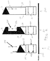
- an illustrative timeline 100 is shown for a biopsy procedure that employs the introducer with sensors in accordance with the present principles.
- a biopsy needle 14 is loaded in a ready-to-fire position.
- the orientation of the introducer 20 and therefore the needle 14 will be known.
- An estimated biopsy location 104 may be determined based upon the needle/introducer orientation and a known throw of the inner stylet 16 relative to the outer cannula 18. In other words, the estimated location 104 can easily be estimated using the positions of the sensors 22 as a baseline and adding the throw of the inner stylet 16 in the direction of the introducer 20.
- the estimated biopsy location 104 may be indicated in an image to assist the user.
- the sensors 22 may include ultrasound sensors.
- an ultrasound probe transmits signals that are received by the sensors 22.
- positions of the sensors 22 and therefore introducer 20 and the needle 14 can be determined in the ultrasound space and the estimated location 104 determined.
- the inner stylet 16 is fired.
- the inner stylet 16 rapidly advances to the throw extent to capture a biopsy sample in a chamber 106 of the inner stylet 16 that corresponds with the estimated position 104.
- the outer cannula 18 is advanced to shear off the biopsy sample in the chamber 106 and enclose the chamber 106 to safely remove the biopsy sample from the subject.
- Biopsy system 200 includes a disposable interface 232.
- the interface 232 is attached to an introducer 220 such that the interface 232 and the introducer 220 are removable and disposable from a biopsy gun handle 212.
- the biopsy gun 212 is configured for needle tracking.
- the biopsy gun 212 includes a biopsy needle 214 having an inner stylet 216 disposed within an outer cannula 218 as described above.
- the needle 214 is, in turn, disposed within the introducer 220, which may include a hollow tube introducer 220 to encapsulate the needle 214.
- the introducer 220 includes one or more tracking sensors 222 (e.g., on the inside diameter of the tube, although the sensors 222 may be mounted on an exterior of the introducer 220).
- the tracking sensors 222 may include ultrasonic sensors although other types of sensors may be employed for tracking the needle 214.
- the introducer 220 may be integrally formed with the interface 232 or the interface 232 may be a separate part that connects to the introducer 220.
- the interface 232 since it is disposable, may or may not include adaptor electronics therein.
- the adaptor electronics may be included in a separate module 228 for noise cancellation, amplifiers, etc. to process received signals from sensors 222.
- the sensors 222 may include one or more ultrasound trackers.
- the introducer 220 and the sensors 222 may be disposable.
- the ultrasound trackers for sensors 222 may include PZT, PVDF, or other piezoelectric element disposed between conductive plates or layers.
- the interface 232 may be employed to attach the introducer 220 to the biopsy gun handle 224.
- the interface 232 may include the adaptor electronics and be reusable (non-disposable), although a disposable embodiment may include a reusable adaptor electronics module 228. In this instance, the interface 232 is disposable and the adaptor electronics 228 are not disposable.
- a cable 234 can be provided as an output from the sensors 222 and can be connected to the adaptor electronics module 228 or other connector or system, e.g., a system employing InSitu technology (see e.g., FIG. 1 ).
- the interface 232 may include an opening 240 to receive the introducer 220.
- an electrical connection is completed between a wire or wires of the sensors 220 through the introducer 220 and to the cable 234 from the interface 232.
- the introducer 220 can be disposable or non-disposable with the sensors 222 and their wiring.
- the adaptor electronics can then be housed separately (module 228) so that it does not come in contact with the subject (e.g., the patient).
- InSitu ultrasound tracking technology
- InSitu technology can be used to estimate the position of a passive ultrasound sensor (e.g., PZT, PVDF, copolymer or other piezoelectric material) in a field of view (FOV) of a diagnostic B-mode image by analyzing a signal received by a sensor as beams of the imaging probe sweep the FOV.
- Time-of-flight measurements can be used to provide the axial/radial distance of the sensor 22 ( FIG.
- the elevational position of the sensor 22, 222 can also be obtained in a similar manner. Therefore, the 3D position of the sensor 22, 222 can be estimated in real-time, provided it is present within the FOV of the imaging transducer.
- the sensors 22, 222 on the introducer 20, 220 passively listen to the ultrasound waves impinging on them as the imaging probe's beams sweep the field of view. Analysis of these signals yields the position of the sensor 22, 222 on the introducer 20, 220 in the frame of reference of the ultrasound image. The position can then be overlaid on an ultrasound image for enhanced visualization, and the positions and their histories can be logged for tracking, segmentation, and other applications.
- Embodiments in accordance with the present principles can be made compatible with multiple biopsy needles on the market.
- the introducers described herein may be employed in procedures other than biopsy procedures.
- the present principles may be employed for ablation needle guidance, catheter guidance, endoscopic procedures, etc.
- corresponding and/or related systems incorporating and/or implementing the present principles are also contemplated and considered to be within the scope of the present invention.
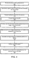
- an introducer is provided with two or more sensors disposed along a length of the introducer.
- the sensors are spaced apart from adjacent sensors along the introducer to assist in providing position and orientation information for tracking the introducer.
- the introducer is coupled to an interface at one end portion.
- the medical device is operatively supported by the introducer and the interface. This means that, e.g., if the medical device includes a biopsy needle, the needle fits within the introducer and is operable (e.g., can be fired) from the introducer.
- the interface supports the introducer by providing a mechanical support between the biopsy gun and the introducer. Other configurations are also contemplated.
- signals are received from a subject by the two or more sensors, which are configured to provide feedback for positioning and orienting the medical device in a medical image.
- the feedback signals are processed using adaptor electronics configured to connect to the sensors and provide noise cancellation, amplify the signals, filter the signals, etc.
- the introducer and therefore the medical device is positioned in a field of view of an image and aligned with a biopsy sample or other target using the feedback signals.
- the medical device may include a biopsy gun including a needle with an inner stylet and an outer cannula.
- An estimate position of a biopsy sample may be determined based upon a position and orientation of the introducer.
- the estimate position may be manually determined or may be computed using an interpretation module ( FIG. 1 ).
- an image may be generated on a display to show the estimate position based upon the position and orientation of the introducer.
- the image may include an indicator, such as an arrow, shape, line, etc. or may include an overlay or a virtual image.
- operative tasks are performed, for example, fire the biopsy gun, take the biopsy sample, etc.
- one or more of the introducer, the interface, the medical instrument may be disposed.
- the introducer is disposable and the interface is reusable.
- the introducer and the interface are disposed of as a single unit or integrated assembly.
- the interface may or may not include adaptor electronics.
Landscapes
- Health & Medical Sciences (AREA)
- Life Sciences & Earth Sciences (AREA)
- Surgery (AREA)
- Engineering & Computer Science (AREA)
- Medical Informatics (AREA)
- Public Health (AREA)
- Biomedical Technology (AREA)
- Heart & Thoracic Surgery (AREA)
- Veterinary Medicine (AREA)
- Molecular Biology (AREA)
- Animal Behavior & Ethology (AREA)
- General Health & Medical Sciences (AREA)
- Nuclear Medicine, Radiotherapy & Molecular Imaging (AREA)
- Pathology (AREA)
- Robotics (AREA)
- Gynecology & Obstetrics (AREA)
- Radiology & Medical Imaging (AREA)
- Oral & Maxillofacial Surgery (AREA)
- Ultra Sonic Daignosis Equipment (AREA)
Applications Claiming Priority (2)
| Application Number | Priority Date | Filing Date | Title |
|---|---|---|---|
| US201462025480P | 2014-07-16 | 2014-07-16 | |
| PCT/IB2015/055352 WO2016009366A1 (en) | 2014-07-16 | 2015-07-15 | Ultrasound tracking apparatus for disposable biopsy needles |
Publications (2)
| Publication Number | Publication Date |
|---|---|
| EP3169246A1 EP3169246A1 (en) | 2017-05-24 |
| EP3169246B1 true EP3169246B1 (en) | 2018-09-19 |
Family
ID=54065397
Family Applications (1)
| Application Number | Title | Priority Date | Filing Date |
|---|---|---|---|
| EP15760246.7A Active EP3169246B1 (en) | 2014-07-16 | 2015-07-15 | Ultrasound tracking apparatus for disposable biopsy needles |
Country Status (5)
| Country | Link |
|---|---|
| US (2) | US11197721B2 (zh) |
| EP (1) | EP3169246B1 (zh) |
| JP (1) | JP6396573B2 (zh) |
| CN (1) | CN106535807B (zh) |
| WO (1) | WO2016009366A1 (zh) |
Families Citing this family (6)
| Publication number | Priority date | Publication date | Assignee | Title |
|---|---|---|---|---|
| JP6396573B2 (ja) * | 2014-07-16 | 2018-09-26 | コーニンクレッカ フィリップス エヌ ヴェKoninklijke Philips N.V. | 使い捨てバイオプシー針のための超音波追跡装置 |
| CN110072463B (zh) * | 2016-12-12 | 2022-09-13 | 皇家飞利浦有限公司 | 包括无线收发器的智能跟踪介入工具 |
| JP7430180B2 (ja) | 2018-11-15 | 2024-02-09 | コーニンクレッカ フィリップス エヌ ヴェ | 医学的介入における同時センサ追跡 |
| CN109394317B (zh) * | 2018-12-14 | 2020-11-20 | 清华大学 | 穿刺路径规划装置及方法 |
| US20220240901A1 (en) * | 2019-06-13 | 2022-08-04 | Koninklijke Philips N.V. | Differentiating passive ultrasound sensors for interventional medical procedures |
| EP3771433A1 (en) * | 2019-07-30 | 2021-02-03 | Koninklijke Philips N.V. | Differentiating passive ultrasound sensors for interventional medical procedures |
Family Cites Families (14)
| Publication number | Priority date | Publication date | Assignee | Title |
|---|---|---|---|---|
| US7510534B2 (en) * | 2001-07-20 | 2009-03-31 | Ethicon Endo-Surgery, Inc. | Method for operating biopsy device |
| ATE303099T1 (de) * | 2002-03-19 | 2005-09-15 | Bard Dublin Itc Ltd | Vakuum-biopsievorrichtung |
| US8781555B2 (en) | 2007-11-26 | 2014-07-15 | C. R. Bard, Inc. | System for placement of a catheter including a signal-generating stylet |
| US20110015490A1 (en) | 2008-03-20 | 2011-01-20 | Koninklijke Philips Electronics N.V. | Method and system for cannula positioning |
| US20090247900A1 (en) | 2008-03-25 | 2009-10-01 | Brian Zimmer | Push button adjustable spacer |
| US8480592B2 (en) * | 2009-12-23 | 2013-07-09 | C. R. Bard, Inc. | Biopsy probe mechanism having multiple echogenic features |
| US20120316558A1 (en) | 2010-02-26 | 2012-12-13 | Koninklijke Philips Electronics N.V. | Interventional ablation device with tissue discriminating capability |
| CN103037762B (zh) | 2010-05-28 | 2016-07-13 | C·R·巴德股份有限公司 | 用于与针插入引导系统一起使用的装置 |
| US8764680B2 (en) * | 2010-11-01 | 2014-07-01 | Devicor Medical Products, Inc. | Handheld biopsy device with needle firing |
| US9414816B2 (en) * | 2011-06-23 | 2016-08-16 | Devicor Medical Products, Inc. | Introducer for biopsy device |
| US20150051482A1 (en) | 2012-02-09 | 2015-02-19 | Koninklijke Philips N.V | Shaft tracker for real-time navigation tracking |
| US9636083B2 (en) * | 2012-07-17 | 2017-05-02 | The Johns Hopkins University | High quality closed-loop ultrasound imaging system |
| CN102860841B (zh) | 2012-09-25 | 2014-10-22 | 陈颀潇 | 超声图像引导下穿刺手术的辅助导航系统 |
| JP6396573B2 (ja) * | 2014-07-16 | 2018-09-26 | コーニンクレッカ フィリップス エヌ ヴェKoninklijke Philips N.V. | 使い捨てバイオプシー針のための超音波追跡装置 |
-
2015
- 2015-07-15 JP JP2017501408A patent/JP6396573B2/ja not_active Expired - Fee Related
- 2015-07-15 CN CN201580038316.2A patent/CN106535807B/zh active Active
- 2015-07-15 WO PCT/IB2015/055352 patent/WO2016009366A1/en active Application Filing
- 2015-07-15 US US15/324,137 patent/US11197721B2/en active Active
- 2015-07-15 EP EP15760246.7A patent/EP3169246B1/en active Active
-
2021
- 2021-12-09 US US17/546,202 patent/US20220096171A1/en not_active Abandoned
Non-Patent Citations (1)
| Title |
|---|
| None * |
Also Published As
| Publication number | Publication date |
|---|---|
| JP6396573B2 (ja) | 2018-09-26 |
| CN106535807A (zh) | 2017-03-22 |
| US11197721B2 (en) | 2021-12-14 |
| CN106535807B (zh) | 2020-03-31 |
| WO2016009366A1 (en) | 2016-01-21 |
| JP2017524453A (ja) | 2017-08-31 |
| US20220096171A1 (en) | 2022-03-31 |
| EP3169246A1 (en) | 2017-05-24 |
| US20170245941A1 (en) | 2017-08-31 |
Similar Documents
| Publication | Publication Date | Title |
|---|---|---|
| US20220096171A1 (en) | Ultrasound tracking apparatus for disposable biopsy needles | |
| US11786318B2 (en) | Intelligent real-time tool and anatomy visualization in 3D imaging workflows for interventional procedures | |
| US7068867B2 (en) | Ultrasonic position indicator | |
| US20120123270A1 (en) | Device with integrated ultrasound transducers and flow sensor | |
| EP3967238B1 (en) | Rotation determination in an ultrasound beam | |
| EP3013245B1 (en) | Shape injection into ultrasound image to calibrate beam patterns in real-time | |
| JP6180903B2 (ja) | 局所座標系を使用した場所検知 | |
| US20120143055A1 (en) | Method and system for ultrasound imaging | |
| JP2003260057A (ja) | マッピング装置およびマッピング方法 | |
| JP2018537227A5 (zh) | ||
| US20210205025A1 (en) | Synchronized tracking of multiple interventional medical devices | |
| EP3054885B1 (en) | Image guidance system with user definable regions of interest | |
| EP3013246B1 (en) | Acoustic highlighting of interventional instruments | |
| US20150065875A1 (en) | Navigation attachment and utilization procedure | |
| US20190125302A1 (en) | Accelerometer in handle for ultrasound medical imaging device | |
| EP3316791B1 (en) | Multi-mode capacitive micromachined ultrasound transducer and associated devices and systems | |
| CN102512208A (zh) | 血管内前视和侧视结合的双视场超声成像装置及其方法 | |
| US10321847B2 (en) | Integrated tracking system for endocavity imaging | |
| US11660064B2 (en) | Intravascular ultrasound position identification | |
| EP3551081B1 (en) | Smart tracked interventional tools including wireless transceiver | |
| US20160317232A1 (en) | Medical imaging probe including an imaging sensor | |
| EP3668378B1 (en) | Catheter probe navigation device employing opposing transducers | |
| WO2023192875A1 (en) | Needle sensor derived image plane |
Legal Events
| Date | Code | Title | Description |
|---|---|---|---|
| STAA | Information on the status of an ep patent application or granted ep patent |
Free format text: STATUS: THE INTERNATIONAL PUBLICATION HAS BEEN MADE |
|
| PUAI | Public reference made under article 153(3) epc to a published international application that has entered the european phase |
Free format text: ORIGINAL CODE: 0009012 |
|
| STAA | Information on the status of an ep patent application or granted ep patent |
Free format text: STATUS: REQUEST FOR EXAMINATION WAS MADE |
|
| 17P | Request for examination filed |
Effective date: 20170216 |
|
| AK | Designated contracting states |
Kind code of ref document: A1 Designated state(s): AL AT BE BG CH CY CZ DE DK EE ES FI FR GB GR HR HU IE IS IT LI LT LU LV MC MK MT NL NO PL PT RO RS SE SI SK SM TR |
|
| AX | Request for extension of the european patent |
Extension state: BA ME |
|
| DAV | Request for validation of the european patent (deleted) | ||
| DAX | Request for extension of the european patent (deleted) | ||
| REG | Reference to a national code |
Ref country code: DE Ref legal event code: R079 Ref document number: 602015016692 Country of ref document: DE Free format text: PREVIOUS MAIN CLASS: A61B0017000000 Ipc: A61B0010020000 |
|
| RIC1 | Information provided on ipc code assigned before grant |
Ipc: A61B 34/20 20160101ALI20180108BHEP Ipc: A61B 17/34 20060101ALI20180108BHEP Ipc: A61B 90/00 20160101ALI20180108BHEP Ipc: A61B 17/00 20060101ALI20180108BHEP Ipc: A61B 10/02 20060101AFI20180108BHEP |
|
| GRAP | Despatch of communication of intention to grant a patent |
Free format text: ORIGINAL CODE: EPIDOSNIGR1 |
|
| STAA | Information on the status of an ep patent application or granted ep patent |
Free format text: STATUS: GRANT OF PATENT IS INTENDED |
|
| INTG | Intention to grant announced |
Effective date: 20180305 |
|
| GRAS | Grant fee paid |
Free format text: ORIGINAL CODE: EPIDOSNIGR3 |
|
| GRAA | (expected) grant |
Free format text: ORIGINAL CODE: 0009210 |
|
| STAA | Information on the status of an ep patent application or granted ep patent |
Free format text: STATUS: THE PATENT HAS BEEN GRANTED |
|
| AK | Designated contracting states |
Kind code of ref document: B1 Designated state(s): AL AT BE BG CH CY CZ DE DK EE ES FI FR GB GR HR HU IE IS IT LI LT LU LV MC MK MT NL NO PL PT RO RS SE SI SK SM TR |
|
| REG | Reference to a national code |
Ref country code: GB Ref legal event code: FG4D |
|
| REG | Reference to a national code |
Ref country code: CH Ref legal event code: EP |
|
| REG | Reference to a national code |
Ref country code: AT Ref legal event code: REF Ref document number: 1042317 Country of ref document: AT Kind code of ref document: T Effective date: 20181015 |
|
| REG | Reference to a national code |
Ref country code: IE Ref legal event code: FG4D |
|
| REG | Reference to a national code |
Ref country code: DE Ref legal event code: R096 Ref document number: 602015016692 Country of ref document: DE |
|
| REG | Reference to a national code |
Ref country code: NL Ref legal event code: MP Effective date: 20180919 |
|
| PG25 | Lapsed in a contracting state [announced via postgrant information from national office to epo] |
Ref country code: BG Free format text: LAPSE BECAUSE OF FAILURE TO SUBMIT A TRANSLATION OF THE DESCRIPTION OR TO PAY THE FEE WITHIN THE PRESCRIBED TIME-LIMIT Effective date: 20181219 Ref country code: GR Free format text: LAPSE BECAUSE OF FAILURE TO SUBMIT A TRANSLATION OF THE DESCRIPTION OR TO PAY THE FEE WITHIN THE PRESCRIBED TIME-LIMIT Effective date: 20181220 Ref country code: LT Free format text: LAPSE BECAUSE OF FAILURE TO SUBMIT A TRANSLATION OF THE DESCRIPTION OR TO PAY THE FEE WITHIN THE PRESCRIBED TIME-LIMIT Effective date: 20180919 Ref country code: SE Free format text: LAPSE BECAUSE OF FAILURE TO SUBMIT A TRANSLATION OF THE DESCRIPTION OR TO PAY THE FEE WITHIN THE PRESCRIBED TIME-LIMIT Effective date: 20180919 Ref country code: NO Free format text: LAPSE BECAUSE OF FAILURE TO SUBMIT A TRANSLATION OF THE DESCRIPTION OR TO PAY THE FEE WITHIN THE PRESCRIBED TIME-LIMIT Effective date: 20181219 Ref country code: FI Free format text: LAPSE BECAUSE OF FAILURE TO SUBMIT A TRANSLATION OF THE DESCRIPTION OR TO PAY THE FEE WITHIN THE PRESCRIBED TIME-LIMIT Effective date: 20180919 Ref country code: RS Free format text: LAPSE BECAUSE OF FAILURE TO SUBMIT A TRANSLATION OF THE DESCRIPTION OR TO PAY THE FEE WITHIN THE PRESCRIBED TIME-LIMIT Effective date: 20180919 |
|
| REG | Reference to a national code |
Ref country code: LT Ref legal event code: MG4D |
|
| PG25 | Lapsed in a contracting state [announced via postgrant information from national office to epo] |
Ref country code: HR Free format text: LAPSE BECAUSE OF FAILURE TO SUBMIT A TRANSLATION OF THE DESCRIPTION OR TO PAY THE FEE WITHIN THE PRESCRIBED TIME-LIMIT Effective date: 20180919 Ref country code: LV Free format text: LAPSE BECAUSE OF FAILURE TO SUBMIT A TRANSLATION OF THE DESCRIPTION OR TO PAY THE FEE WITHIN THE PRESCRIBED TIME-LIMIT Effective date: 20180919 Ref country code: AL Free format text: LAPSE BECAUSE OF FAILURE TO SUBMIT A TRANSLATION OF THE DESCRIPTION OR TO PAY THE FEE WITHIN THE PRESCRIBED TIME-LIMIT Effective date: 20180919 |
|
| REG | Reference to a national code |
Ref country code: AT Ref legal event code: MK05 Ref document number: 1042317 Country of ref document: AT Kind code of ref document: T Effective date: 20180919 |
|
| PG25 | Lapsed in a contracting state [announced via postgrant information from national office to epo] |
Ref country code: NL Free format text: LAPSE BECAUSE OF FAILURE TO SUBMIT A TRANSLATION OF THE DESCRIPTION OR TO PAY THE FEE WITHIN THE PRESCRIBED TIME-LIMIT Effective date: 20180919 Ref country code: AT Free format text: LAPSE BECAUSE OF FAILURE TO SUBMIT A TRANSLATION OF THE DESCRIPTION OR TO PAY THE FEE WITHIN THE PRESCRIBED TIME-LIMIT Effective date: 20180919 Ref country code: EE Free format text: LAPSE BECAUSE OF FAILURE TO SUBMIT A TRANSLATION OF THE DESCRIPTION OR TO PAY THE FEE WITHIN THE PRESCRIBED TIME-LIMIT Effective date: 20180919 Ref country code: IT Free format text: LAPSE BECAUSE OF FAILURE TO SUBMIT A TRANSLATION OF THE DESCRIPTION OR TO PAY THE FEE WITHIN THE PRESCRIBED TIME-LIMIT Effective date: 20180919 Ref country code: CZ Free format text: LAPSE BECAUSE OF FAILURE TO SUBMIT A TRANSLATION OF THE DESCRIPTION OR TO PAY THE FEE WITHIN THE PRESCRIBED TIME-LIMIT Effective date: 20180919 Ref country code: RO Free format text: LAPSE BECAUSE OF FAILURE TO SUBMIT A TRANSLATION OF THE DESCRIPTION OR TO PAY THE FEE WITHIN THE PRESCRIBED TIME-LIMIT Effective date: 20180919 Ref country code: ES Free format text: LAPSE BECAUSE OF FAILURE TO SUBMIT A TRANSLATION OF THE DESCRIPTION OR TO PAY THE FEE WITHIN THE PRESCRIBED TIME-LIMIT Effective date: 20180919 Ref country code: IS Free format text: LAPSE BECAUSE OF FAILURE TO SUBMIT A TRANSLATION OF THE DESCRIPTION OR TO PAY THE FEE WITHIN THE PRESCRIBED TIME-LIMIT Effective date: 20190119 Ref country code: PL Free format text: LAPSE BECAUSE OF FAILURE TO SUBMIT A TRANSLATION OF THE DESCRIPTION OR TO PAY THE FEE WITHIN THE PRESCRIBED TIME-LIMIT Effective date: 20180919 |
|
| PG25 | Lapsed in a contracting state [announced via postgrant information from national office to epo] |
Ref country code: PT Free format text: LAPSE BECAUSE OF FAILURE TO SUBMIT A TRANSLATION OF THE DESCRIPTION OR TO PAY THE FEE WITHIN THE PRESCRIBED TIME-LIMIT Effective date: 20190119 Ref country code: SK Free format text: LAPSE BECAUSE OF FAILURE TO SUBMIT A TRANSLATION OF THE DESCRIPTION OR TO PAY THE FEE WITHIN THE PRESCRIBED TIME-LIMIT Effective date: 20180919 Ref country code: SM Free format text: LAPSE BECAUSE OF FAILURE TO SUBMIT A TRANSLATION OF THE DESCRIPTION OR TO PAY THE FEE WITHIN THE PRESCRIBED TIME-LIMIT Effective date: 20180919 |
|
| REG | Reference to a national code |
Ref country code: DE Ref legal event code: R097 Ref document number: 602015016692 Country of ref document: DE |
|
| PLBE | No opposition filed within time limit |
Free format text: ORIGINAL CODE: 0009261 |
|
| STAA | Information on the status of an ep patent application or granted ep patent |
Free format text: STATUS: NO OPPOSITION FILED WITHIN TIME LIMIT |
|
| PG25 | Lapsed in a contracting state [announced via postgrant information from national office to epo] |
Ref country code: DK Free format text: LAPSE BECAUSE OF FAILURE TO SUBMIT A TRANSLATION OF THE DESCRIPTION OR TO PAY THE FEE WITHIN THE PRESCRIBED TIME-LIMIT Effective date: 20180919 |
|
| 26N | No opposition filed |
Effective date: 20190620 |
|
| PG25 | Lapsed in a contracting state [announced via postgrant information from national office to epo] |
Ref country code: SI Free format text: LAPSE BECAUSE OF FAILURE TO SUBMIT A TRANSLATION OF THE DESCRIPTION OR TO PAY THE FEE WITHIN THE PRESCRIBED TIME-LIMIT Effective date: 20180919 |
|
| PG25 | Lapsed in a contracting state [announced via postgrant information from national office to epo] |
Ref country code: MC Free format text: LAPSE BECAUSE OF FAILURE TO SUBMIT A TRANSLATION OF THE DESCRIPTION OR TO PAY THE FEE WITHIN THE PRESCRIBED TIME-LIMIT Effective date: 20180919 |
|
| REG | Reference to a national code |
Ref country code: CH Ref legal event code: PL |
|
| PG25 | Lapsed in a contracting state [announced via postgrant information from national office to epo] |
Ref country code: TR Free format text: LAPSE BECAUSE OF FAILURE TO SUBMIT A TRANSLATION OF THE DESCRIPTION OR TO PAY THE FEE WITHIN THE PRESCRIBED TIME-LIMIT Effective date: 20180919 |
|
| REG | Reference to a national code |
Ref country code: BE Ref legal event code: MM Effective date: 20190731 |
|
| PG25 | Lapsed in a contracting state [announced via postgrant information from national office to epo] |
Ref country code: CH Free format text: LAPSE BECAUSE OF NON-PAYMENT OF DUE FEES Effective date: 20190731 Ref country code: LU Free format text: LAPSE BECAUSE OF NON-PAYMENT OF DUE FEES Effective date: 20190715 Ref country code: LI Free format text: LAPSE BECAUSE OF NON-PAYMENT OF DUE FEES Effective date: 20190731 Ref country code: BE Free format text: LAPSE BECAUSE OF NON-PAYMENT OF DUE FEES Effective date: 20190731 |
|
| PG25 | Lapsed in a contracting state [announced via postgrant information from national office to epo] |
Ref country code: IE Free format text: LAPSE BECAUSE OF NON-PAYMENT OF DUE FEES Effective date: 20190715 |
|
| PG25 | Lapsed in a contracting state [announced via postgrant information from national office to epo] |
Ref country code: CY Free format text: LAPSE BECAUSE OF FAILURE TO SUBMIT A TRANSLATION OF THE DESCRIPTION OR TO PAY THE FEE WITHIN THE PRESCRIBED TIME-LIMIT Effective date: 20180919 |
|
| PG25 | Lapsed in a contracting state [announced via postgrant information from national office to epo] |
Ref country code: MT Free format text: LAPSE BECAUSE OF FAILURE TO SUBMIT A TRANSLATION OF THE DESCRIPTION OR TO PAY THE FEE WITHIN THE PRESCRIBED TIME-LIMIT Effective date: 20180919 Ref country code: HU Free format text: LAPSE BECAUSE OF FAILURE TO SUBMIT A TRANSLATION OF THE DESCRIPTION OR TO PAY THE FEE WITHIN THE PRESCRIBED TIME-LIMIT; INVALID AB INITIO Effective date: 20150715 |
|
| PG25 | Lapsed in a contracting state [announced via postgrant information from national office to epo] |
Ref country code: MK Free format text: LAPSE BECAUSE OF FAILURE TO SUBMIT A TRANSLATION OF THE DESCRIPTION OR TO PAY THE FEE WITHIN THE PRESCRIBED TIME-LIMIT Effective date: 20180919 |
|
| PGFP | Annual fee paid to national office [announced via postgrant information from national office to epo] |
Ref country code: GB Payment date: 20220719 Year of fee payment: 8 Ref country code: DE Payment date: 20220628 Year of fee payment: 8 |
|
| PGFP | Annual fee paid to national office [announced via postgrant information from national office to epo] |
Ref country code: FR Payment date: 20220725 Year of fee payment: 8 |
|
| REG | Reference to a national code |
Ref country code: DE Ref legal event code: R119 Ref document number: 602015016692 Country of ref document: DE |
|
| GBPC | Gb: european patent ceased through non-payment of renewal fee |
Effective date: 20230715 |
|
| PG25 | Lapsed in a contracting state [announced via postgrant information from national office to epo] |
Ref country code: DE Free format text: LAPSE BECAUSE OF NON-PAYMENT OF DUE FEES Effective date: 20240201 Ref country code: GB Free format text: LAPSE BECAUSE OF NON-PAYMENT OF DUE FEES Effective date: 20230715 |