EP3164048B1 - Real-time automatic registration feedback - Google Patents
Real-time automatic registration feedback Download PDFInfo
- Publication number
- EP3164048B1 EP3164048B1 EP15814452.7A EP15814452A EP3164048B1 EP 3164048 B1 EP3164048 B1 EP 3164048B1 EP 15814452 A EP15814452 A EP 15814452A EP 3164048 B1 EP3164048 B1 EP 3164048B1
- Authority
- EP
- European Patent Office
- Prior art keywords
- sensor
- luminal network
- location
- model
- registration
- Prior art date
- Legal status (The legal status is an assumption and is not a legal conclusion. Google has not performed a legal analysis and makes no representation as to the accuracy of the status listed.)
- Active
Links
- 210000004072 lung Anatomy 0.000 claims description 55
- 238000012545 processing Methods 0.000 claims description 25
- 230000005672 electromagnetic field Effects 0.000 claims description 13
- 238000000034 method Methods 0.000 description 26
- 210000001519 tissue Anatomy 0.000 description 21
- 238000001574 biopsy Methods 0.000 description 17
- 230000000712 assembly Effects 0.000 description 10
- 238000000429 assembly Methods 0.000 description 10
- 230000037361 pathway Effects 0.000 description 9
- 238000002591 computed tomography Methods 0.000 description 8
- 230000008569 process Effects 0.000 description 6
- 230000003213 activating effect Effects 0.000 description 5
- 238000003384 imaging method Methods 0.000 description 5
- 210000003437 trachea Anatomy 0.000 description 4
- 238000013276 bronchoscopy Methods 0.000 description 3
- 230000035515 penetration Effects 0.000 description 3
- 230000007423 decrease Effects 0.000 description 2
- 238000011161 development Methods 0.000 description 2
- 230000018109 developmental process Effects 0.000 description 2
- 238000010586 diagram Methods 0.000 description 2
- 238000005516 engineering process Methods 0.000 description 2
- 238000005286 illumination Methods 0.000 description 2
- 238000003780 insertion Methods 0.000 description 2
- 230000037431 insertion Effects 0.000 description 2
- 230000007246 mechanism Effects 0.000 description 2
- 238000012544 monitoring process Methods 0.000 description 2
- 239000007787 solid Substances 0.000 description 2
- 238000002679 ablation Methods 0.000 description 1
- 238000007792 addition Methods 0.000 description 1
- 210000003484 anatomy Anatomy 0.000 description 1
- 230000008859 change Effects 0.000 description 1
- 238000004891 communication Methods 0.000 description 1
- 230000006835 compression Effects 0.000 description 1
- 238000007906 compression Methods 0.000 description 1
- 238000013480 data collection Methods 0.000 description 1
- 238000001514 detection method Methods 0.000 description 1
- 238000002594 fluoroscopy Methods 0.000 description 1
- 230000002496 gastric effect Effects 0.000 description 1
- 230000001926 lymphatic effect Effects 0.000 description 1
- 230000003287 optical effect Effects 0.000 description 1
- 210000004224 pleura Anatomy 0.000 description 1
- 238000012552 review Methods 0.000 description 1
- 230000011218 segmentation Effects 0.000 description 1
- 230000001225 therapeutic effect Effects 0.000 description 1
- 230000002792 vascular Effects 0.000 description 1
- 238000012795 verification Methods 0.000 description 1
Images


Classifications
-
- A—HUMAN NECESSITIES
- A61—MEDICAL OR VETERINARY SCIENCE; HYGIENE
- A61B—DIAGNOSIS; SURGERY; IDENTIFICATION
- A61B5/00—Measuring for diagnostic purposes; Identification of persons
- A61B5/06—Devices, other than using radiation, for detecting or locating foreign bodies ; determining position of probes within or on the body of the patient
- A61B5/065—Determining position of the probe employing exclusively positioning means located on or in the probe, e.g. using position sensors arranged on the probe
- A61B5/066—Superposing sensor position on an image of the patient, e.g. obtained by ultrasound or x-ray imaging
-
- A—HUMAN NECESSITIES
- A61—MEDICAL OR VETERINARY SCIENCE; HYGIENE
- A61B—DIAGNOSIS; SURGERY; IDENTIFICATION
- A61B1/00—Instruments for performing medical examinations of the interior of cavities or tubes of the body by visual or photographical inspection, e.g. endoscopes; Illuminating arrangements therefor
- A61B1/00002—Operational features of endoscopes
- A61B1/00043—Operational features of endoscopes provided with output arrangements
- A61B1/00045—Display arrangement
-
- A—HUMAN NECESSITIES
- A61—MEDICAL OR VETERINARY SCIENCE; HYGIENE
- A61B—DIAGNOSIS; SURGERY; IDENTIFICATION
- A61B1/00—Instruments for performing medical examinations of the interior of cavities or tubes of the body by visual or photographical inspection, e.g. endoscopes; Illuminating arrangements therefor
- A61B1/04—Instruments for performing medical examinations of the interior of cavities or tubes of the body by visual or photographical inspection, e.g. endoscopes; Illuminating arrangements therefor combined with photographic or television appliances
-
- A—HUMAN NECESSITIES
- A61—MEDICAL OR VETERINARY SCIENCE; HYGIENE
- A61B—DIAGNOSIS; SURGERY; IDENTIFICATION
- A61B1/00—Instruments for performing medical examinations of the interior of cavities or tubes of the body by visual or photographical inspection, e.g. endoscopes; Illuminating arrangements therefor
- A61B1/267—Instruments for performing medical examinations of the interior of cavities or tubes of the body by visual or photographical inspection, e.g. endoscopes; Illuminating arrangements therefor for the respiratory tract, e.g. laryngoscopes, bronchoscopes
- A61B1/2676—Bronchoscopes
-
- A—HUMAN NECESSITIES
- A61—MEDICAL OR VETERINARY SCIENCE; HYGIENE
- A61B—DIAGNOSIS; SURGERY; IDENTIFICATION
- A61B5/00—Measuring for diagnostic purposes; Identification of persons
- A61B5/06—Devices, other than using radiation, for detecting or locating foreign bodies ; determining position of probes within or on the body of the patient
- A61B5/061—Determining position of a probe within the body employing means separate from the probe, e.g. sensing internal probe position employing impedance electrodes on the surface of the body
- A61B5/062—Determining position of a probe within the body employing means separate from the probe, e.g. sensing internal probe position employing impedance electrodes on the surface of the body using magnetic field
-
- A—HUMAN NECESSITIES
- A61—MEDICAL OR VETERINARY SCIENCE; HYGIENE
- A61B—DIAGNOSIS; SURGERY; IDENTIFICATION
- A61B5/00—Measuring for diagnostic purposes; Identification of persons
- A61B5/06—Devices, other than using radiation, for detecting or locating foreign bodies ; determining position of probes within or on the body of the patient
- A61B5/061—Determining position of a probe within the body employing means separate from the probe, e.g. sensing internal probe position employing impedance electrodes on the surface of the body
- A61B5/064—Determining position of a probe within the body employing means separate from the probe, e.g. sensing internal probe position employing impedance electrodes on the surface of the body using markers
-
- A—HUMAN NECESSITIES
- A61—MEDICAL OR VETERINARY SCIENCE; HYGIENE
- A61B—DIAGNOSIS; SURGERY; IDENTIFICATION
- A61B17/00—Surgical instruments, devices or methods, e.g. tourniquets
- A61B2017/00743—Type of operation; Specification of treatment sites
- A61B2017/00809—Lung operations
-
- A—HUMAN NECESSITIES
- A61—MEDICAL OR VETERINARY SCIENCE; HYGIENE
- A61B—DIAGNOSIS; SURGERY; IDENTIFICATION
- A61B34/00—Computer-aided surgery; Manipulators or robots specially adapted for use in surgery
- A61B34/10—Computer-aided planning, simulation or modelling of surgical operations
- A61B2034/101—Computer-aided simulation of surgical operations
- A61B2034/105—Modelling of the patient, e.g. for ligaments or bones
-
- A—HUMAN NECESSITIES
- A61—MEDICAL OR VETERINARY SCIENCE; HYGIENE
- A61B—DIAGNOSIS; SURGERY; IDENTIFICATION
- A61B34/00—Computer-aided surgery; Manipulators or robots specially adapted for use in surgery
- A61B34/20—Surgical navigation systems; Devices for tracking or guiding surgical instruments, e.g. for frameless stereotaxis
- A61B2034/2046—Tracking techniques
- A61B2034/2051—Electromagnetic tracking systems
-
- A—HUMAN NECESSITIES
- A61—MEDICAL OR VETERINARY SCIENCE; HYGIENE
- A61B—DIAGNOSIS; SURGERY; IDENTIFICATION
- A61B34/00—Computer-aided surgery; Manipulators or robots specially adapted for use in surgery
- A61B34/25—User interfaces for surgical systems
- A61B2034/252—User interfaces for surgical systems indicating steps of a surgical procedure
-
- A—HUMAN NECESSITIES
- A61—MEDICAL OR VETERINARY SCIENCE; HYGIENE
- A61B—DIAGNOSIS; SURGERY; IDENTIFICATION
- A61B90/00—Instruments, implements or accessories specially adapted for surgery or diagnosis and not covered by any of the groups A61B1/00 - A61B50/00, e.g. for luxation treatment or for protecting wound edges
- A61B90/36—Image-producing devices or illumination devices not otherwise provided for
- A61B2090/364—Correlation of different images or relation of image positions in respect to the body
- A61B2090/365—Correlation of different images or relation of image positions in respect to the body augmented reality, i.e. correlating a live optical image with another image
-
- A—HUMAN NECESSITIES
- A61—MEDICAL OR VETERINARY SCIENCE; HYGIENE
- A61B—DIAGNOSIS; SURGERY; IDENTIFICATION
- A61B5/00—Measuring for diagnostic purposes; Identification of persons
- A61B5/08—Detecting, measuring or recording devices for evaluating the respiratory organs
- A61B5/0803—Recording apparatus specially adapted therefor
-
- A—HUMAN NECESSITIES
- A61—MEDICAL OR VETERINARY SCIENCE; HYGIENE
- A61B—DIAGNOSIS; SURGERY; IDENTIFICATION
- A61B5/00—Measuring for diagnostic purposes; Identification of persons
- A61B5/74—Details of notification to user or communication with user or patient ; user input means
- A61B5/742—Details of notification to user or communication with user or patient ; user input means using visual displays
- A61B5/7425—Displaying combinations of multiple images regardless of image source, e.g. displaying a reference anatomical image with a live image
Definitions
- EM sensor 94 may be embedded or incorporated within biopsy tool 102 where biopsy tool 102 may alternatively be utilized for navigation without need of LG 92 or the necessary tool exchanges that use of LG 92 requires.
- a variety of useable biopsy tools are described in U.S. Provisional Patent Application Nos. 61/906,732 and 61/906,762 both entitled DEVICES, SYSTEMS, AND METHODS FOR NAVIGATING A BIOPSY TOOL TO A TARGET LOCATION AND OBTAINING A TISSUE SAMPLE USING THE SAME, filed November 20, 2013 and U.S. Provisional Patent Application No. 61/955,407 having the same title and filed March 14, 2014 , the entire contents of each of which is useable with EMN system 10 as described herein.
Description
- The present disclosure relates to bronchial registration and, more particularly, to a system for automatically registering a bronchial tree model with a patient's real bronchial tree.
- A common device for inspecting the airway of a patient is a bronchoscope. Typically, the bronchoscope is inserted into a patient's airways through the patient's nose or mouth and can extend into the lungs of the patient. A typical bronchoscope includes an elongated flexible tube having an illumination assembly for illuminating the region distal to the bronchoscope's tip, an imaging assembly for providing a video image from the bronchoscope's tip, and a working channel through which instruments, e.g., diagnostic instruments such as biopsy tools, therapeutic instruments can be inserted.
- Bronchoscopes, however, are limited in how far they may be advanced through the airways due to their size. Where the bronchoscope is too large to reach a target location deep in the lungs a clinician may utilize certain real-time imaging modalities such as fluoroscopy. Fluoroscopic images, while useful present certain drawbacks for navigation as it is often difficult to distinguish luminal passageways from solid tissue. Moreover, the images generated by the fluoroscope are two-dimensional whereas navigating the airways of a patient requires the ability to maneuver in three dimensions.
- To address these issues, systems have been developed that enable the development of three-dimensional models of the airways or other luminal networks, typically from a series of computed tomography (CT) images. One such system has been developed as part of the ILOGIC® ELECTROMAGNETIC NAVIGATION BRONCHOSCOPY® (ENB™), system currently sold by Covidien LP. The details of such a system are described in commonly assigned
U.S. Patent No. 7,233,820, entitled ENDOSCOPE STRUCTURES AND TECHNIQUES FOR NAVIGATING TO A TARGET IN BRANCHED STRUCTURE, filed on March 29, 2004, by Gilboa . - While the system as described in
U.S. Patent No. 7,233,820 is quite capable, there is always a need for development of improvements and additions to such systems. Examples of other known systems for navigating a luminal network can be found inWO 2005/058137 ,WO 2012/117381 , andWO 2009/111682 . - Provided in accordance with the presently claimed invention is a system for registering a luminal network to a 3D model of the luminal network with real-time feedback.
- A system for registering a luminal network of the airways to a 3D model of the luminal network includes: a generator for generating an electromagnetic field about the luminal network; a location sensor responsive to the electromagnetic field to produce location data; and a processing system configured to carry out the steps of: generating a 3D model of the luminal network based on images of the luminal network; tracking the location of the location sensor within the luminal network using location data acquired from the location sensor in the electromagnetic field; comparing the tracked locations of the location sensor with sensors located outside of the luminal network and the portions of the 3D model representative of open space; and presenting on a user interface an indication of which lung portions of the luminal network have been sufficiently traversed by the location sensor to register those lung portions of the luminal network to the 3D model, wherein the processing system requires the location sensor to be navigated to the second bifurcation of each airway passage of a lung portion in order for the lung portion to be sufficiently traversed; wherein the processing system is configured to determine that a done button for ending the acquisition of location data has been activated and, if so, to determine whether the acquired location data points are substantially located within the luminal network of the 3D model, and if not, restart the registration of the luminal network to the 3D model.
- Various aspects and features of the present disclosure are described hereinbelow with references to the drawings, wherein:
-
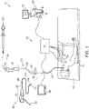
FIG. 1 is a perspective view of an electromagnetic navigation system in accordance with the present disclosure; -
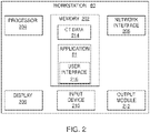
FIG. 2 is a schematic diagram of a workstation configured for use with the system ofFIG. 1 ; -
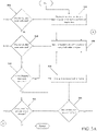
FIGs. 3A andB illustrate a flowchart illustrating a method of registering a luminal network to a 3D model of the luminal network with real-time feedback provided in accordance with the present disclosure (the method as such is not according to the claimed invention); -
FIG. 4 shows a lung survey prior to the start of registration; -
FIG. 5 shows the lung survey ofFIG. 4 after registration has started; -
FIG. 6 shows the lung survey ofFIG. 5 with an indicator activated for the trachea; -
FIG. 7 shows the lung survey ofFIG. 6 with an additional indicator activated for a first region of the lung ; -
FIG. 8 shows the lung survey ofFIG. 7 with an additional indicator activated for a second region of the lung; -
FIG. 9 shows the lung survey ofFIG. 8 with an additional indicator activated for a third region of the lung; -
FIG. 10 shows the lung survey ofFIG. 9 with an additional indicator activated for a fourth region of the lung; -
FIG. 11 shows a view presenting a slice of a 3D volume for verifying registration in accordance with the present disclosure; -
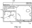
FIG. 12 shows the view ofFIG. 11 , presenting a further slice for verifying registration following movement of a sensor; and -
FIG. 13 shows a view for reviewing the registration in a 3D model in accordance with the present disclosure. - The present disclosure is directed to systems for automatically registering a bronchial tree model with a patient's airways. Various systems for generating the bronchial tree model are envisioned, some of which are more fully described in co-pending
U.S. Patent Application Nos. 13/838,805 ,13/838,997 13/839,224, all entitled PATHWAY PLANNING SYSTEM AND METHOD, filed on March 15, 2013, by Baker U.S. Provisional Patent Application No. 62/020,240, entitled SYSTEM AND METHOD FOR NAVIGATING WITHIN THE LUNG, filed on July 2, 2014, by Brown et al. - Additional features of the ENB system of the present disclosure are described in co-pending
U.S. Provisional Patent Application Nos. 62/020,177, entitled METHODS FOR MARKING BIOPSY LOCATION, filed on July 2, 2014, by Brown 62/020,238, entitled INTELLIGENT DISPLAY, filed on July 2, 2014, by KEHAT et al. 62/020,242, entitled UNIFIED COORDINATE SYSTEM FOR MULTIPLE CT SCANS OF PATIENT LUNGS, filed on July 2, 2014, by Greenburg 62/020,245, entitled ALIGNMENT CT, filed on July 2, 2014, by Klein et al. 62/020,250, entitled ALGORITHM FOR FLUOROSCOPIC POSE ESTIMATION, filed on July 2, 2014, by Merlet 62/020,253, entitled TRACHEA MARKING, filed on July 2, 2014, by Lachmanovich et al. 62/020,257, entitled AUTOMATIC DETECTION OF HUMAN LUNG TRACHEA, filed on July 2, 2014, by Markov et al. 62/020,261, entitled LUNG AND PLEURA SEGMENTATION, filed on July 2, 2014, by Markov et al. 62/020,258, entitled CONE VIEW - A METHOD OF PROVIDING DISTANCE AND ORIENTATION FEEDBACK WHILE NAVIGATING IN 3D, filed on July 2, 2014, by Lachmanovich et al. 62/020,262, entitled DYNAMIC 3D LUNG MAP VIEW FOR TOOL NAVIGATION INSIDE THE LUNG, filed on July 2, 2014, by Weingarten et al. U.S. Patent Application No. 12/780,678, entitled AUTOMATIC REGISTRATION TECHNIQUE, filed on May 14, 2010, by Dorian Averbuch - While the example implementations described below are directed to the bronchoscopy of a patient's airways, those skilled in the art will realize that the same or similar devices, systems, and methods may also be used in other lumen networks, such as, for example, the vascular, lymphatic, and/or gastrointestinal networks.
- With reference to
FIG. 1 , an electromagnetic navigation (EMN) system 10 is provided in accordance with the present disclosure. One such EMN system is the ELECTROMAGNETIC NAVIGATION BRONCHOSCOPY® system currently sold by Covidien LP. Among other tasks that may be performed using the EMN system 10 are planning a pathway to target tissue, navigating a positioning assembly to the target tissue, navigating a biopsy tool to the target tissue to obtain a tissue sample from the target tissue using the biopsy tool, digitally marking the location where the tissue sample was obtained, and placing one or more echogenic markers at or around the target. - EMN system 10 generally includes an operating table 40 configured to support a patient; a
bronchoscope 50 configured for insertion through the patient's mouth and/or nose into the patient's airways; monitoring equipment 60 coupled tobronchoscope 50 for displaying video images received frombronchoscope 50; atracking system 70 including atracking module 72, a plurality ofreference sensors 74, and anelectromagnetic field generator 76; aworkstation 80 including software and/or hardware used to facilitate pathway planning, identification of target tissue, navigation to target tissue, and digitally marking the biopsy location -
FIG. 1 also depicts two types ofcatheter guide assemblies catheter guide assembly handle 91, which is connected to an extended working channel (EWC) 96. EWC 96 is sized for placement into the working channel of abronchoscope 50. In operation, a locatable guide (LG) 92, including an electromagnetic (EM)sensor 94, is inserted into EWC 96 and locked into position such thatEM sensor 94 extends a desired distance beyond adistal tip 93 of EWC 96. The location ofEM sensor 94, and thus the distal end of EWC 96, within an electromagnetic field generated byelectromagnetic field generator 76 can be derived bytracking module 72, andworkstation 80. Catheter guide assemblies 90, 100 have different operating mechanisms, but each contain ahandle 91 that can be manipulated by rotation and compression to steerdistal tip 93 of LG 92 and EWC 96.Catheter guide assemblies 90 are currently marketed and sold by Covidien LP under the name SUPERDIMENSION® Procedure Kits. Similarly,catheter guide assemblies 100 are currently sold by Covidien LP under the name EDGE™ Procedure Kits. Both kits include ahandle 91, EWC 96, and LG 92. For a more detailed description of thecatheter guide assemblies U.S. Patent Application Serial No. 13/836,203 entitled MICROWAVE ABLATION CATHETER AND METHOD OF UTILIZING THE SAME, filed on March 15, 2013, by Ladtkow et al. - As illustrated in
FIG. 1 , the patient is shown lying on operating table 40 withbronchoscope 50 inserted through the patient's mouth and into the patient's airways. Bronchoscope 50 includes a source of illumination and a video imaging system (not explicitly shown) and is coupled to monitoring equipment 60, e.g., a video display, for displaying the video images received from the video imaging system ofbronchoscope 50. - Catheter guide assemblies 90, 100 including LG 92 and EWC 96 are configured for insertion through a working channel of
bronchoscope 50 into the patient's airways (although the catheter guide assemblies 90, 100 may alternatively be used without bronchoscope 50). LG 92 and EWC 96 are selectively lockable relative to one another via alocking mechanism 99. A six degrees-of-freedomelectromagnetic tracking system 70, e.g., similar to those disclosed inU.S. Patent No. 6,188,355 and publishedPCT Application Nos. WO 00/10456WO 01/67035 Tracking system 70 is configured for use withcatheter guide assemblies EM sensor 94 as it moves in conjunction with EWC 96 through the airways of the patient, as detailed below. - As shown in
FIG. 1 ,electromagnetic field generator 76 is positioned beneath the patient.Electromagnetic field generator 76 and the plurality ofreference sensors 74 are interconnected withtracking module 72, which derives the location of eachreference sensor 74 . One or more ofreference sensors 74 are attached to the chest of the patient. The coordinates ofreference sensors 74 are sent toworkstation 80, which includes andapplication 81 which uses data collected bysensors 74 to calculate a patient coordinate frame of reference. - Also shown in
FIG. 1 is acatheter biopsy tool 102 that is insertable intocatheter guide assemblies LG 92.Biopsy tool 102 is used to collect one or more tissue samples from the target tissue. As detailed below,biopsy tool 102 is further configured for use in conjunction with trackingsystem 70 to facilitate navigation ofbiopsy tool 102 to the target tissue, tracking of a location ofbiopsy tool 102 as it is manipulated relative to the target tissue to obtain the tissue sample, and/or marking the location where the tissue sample was obtained. - Although navigation is detailed above with respect to
EM sensor 94 being included inLG 92 it is also envisioned thatEM sensor 94 may be embedded or incorporated withinbiopsy tool 102 wherebiopsy tool 102 may alternatively be utilized for navigation without need ofLG 92 or the necessary tool exchanges that use ofLG 92 requires. A variety of useable biopsy tools are described inU.S. Provisional Patent Application Nos. 61/906,732 61/906,762 both entitled DEVICES, SYSTEMS, AND METHODS FOR NAVIGATING A BIOPSY TOOL TO A TARGET LOCATION AND OBTAINING A TISSUE SAMPLE USING THE SAME, filed November 20, 2013 U.S. Provisional Patent Application No. 61/955,407 having the same title and filed March 14, 2014 - During procedure planning,
workstation 80 utilizes computed tomographic (CT) image data for generating and viewing the 3D model of the patient's airways, enables the identification of target tissue on the 3D model (automatically, semi-automatically or manually), and allows for the selection of a pathway through the patient's airways to the target tissue. More specifically, the CT scans are processed and assembled into a 3D volume, which is then utilized to generate the 3D model of the patient's airways. The 3D model may be presented on a display monitor associated withworkstation 80, or in any other suitable fashion. Usingworkstation 80, various slices of the 3D volume and views of the 3D model may be presented and/or may be manipulated by a clinician to facilitate identification of a target and selection of a suitable pathway through the patient's airways to access the target. The 3D model may also show marks of the locations where previous biopsies were performed, including the dates, times, and other identifying information regarding the tissue samples obtained. These marks may also be selected as the target to which a pathway can be planned. Once selected, the pathway is saved for use during the navigation procedure. An example of a suitable pathway planning system and method is described inU.S. Patent Application Serial Nos. 13/838,805 ;13/838,997 13/839,224, all entitled PATHWAY PLANNING SYSTEM AND METHOD, filed on March 15, 2014, by Baker - During navigation,
EM sensor 94, in conjunction with trackingsystem 70, enables tracking ofEM sensor 94 and/orbiopsy tool 102 asEM sensor 94 orbiopsy tool 102 is advanced through the patient's airways. - Turning now to
FIG. 2 , there is shown a system diagram ofworkstation 80.Workstation 80 may includememory 202,processor 204,display 206,network interface 208,input device 210, and/oroutput module 212. -
Memory 202 includes any non-transitory computer-readable storage media for storing data and/or software that is executable byprocessor 204 and which controls the operation ofworkstation 80. In an embodiment,memory 202 may include one or more solid-state storage devices such as flash memory chips. Alternatively or in addition to the one or more solid-state storage devices,memory 202 may include one or more mass storage devices connected to theprocessor 204 through a mass storage controller (not shown) and a communications bus (not shown). Although the description of computer-readable media contained herein refers to a solid-state storage, it should be appreciated by those skilled in the art that computer-readable storage media can be any available media that can be accessed by theprocessor 204. That is, computer readable storage media includes non-transitory, volatile and non-volatile, removable and non-removable media implemented in any method or technology for storage of information such as computer-readable instructions, data structures, program modules or other data. For example, computer-readable storage media includes RAM, ROM, EPROM, EEPROM, flash memory or other solid state memory technology, CD-ROM, DVD, Blu-Ray or other optical storage, magnetic cassettes, magnetic tape, magnetic disk storage or other magnetic storage devices, or any other medium which can be used to store the desired information and which can be accessed byworkstation 80. -
Memory 202 may storeapplication 81 and/orCT data 214.Application 81 may, when executed byprocessor 204,cause display 206 to presentuser interface 216.Network interface 208 may be configured to connect to a network such as a local area network (LAN) consisting of a wired network and/or a wireless network, a wide area network (WAN), a wireless mobile network, a Bluetooth network, and/or the internet.Input device 210 may be any device by means of which a user may interact withworkstation 80, such as, for example, a mouse, keyboard, foot pedal, touch screen, and/or voice interface.Output module 212 may include any connectivity port or bus, such as, for example, parallel ports, serial ports, universal serial busses (USB), or any other similar connectivity port known to those skilled in the art. - Referring now to
FIGs. 3 A andB , there is shown a flowchart of an example method for automatically registering the 3D model with a patient's airways. The registration process generally involves the clinician navigatingEM sensor 94 through the airways of the patient's lungs to acquire location data. The location data is then compared to the 3D model to register the 3D model with the patient's airways. More specifically, data pertaining to locations ofEM sensor 94 whileLG 92 is moving through the airways is recorded usingelectromagnetic field generator 76,reference sensors 74, and trackingmodule 72. A shape resulting from this location data is compared to an interior geometry of passages of the 3D model, and a location correlation between the shape and the 3D model based on the comparison is determined, e.g., utilizingapplication 81. In addition,application 81 identifies non-tissue space (e.g., air filled cavities) in the 3D model.Application 81 aligns, or registers, an image representing a location ofEM sensor 94 ofLG 92 with an image of the 3D model based on the recorded location data and an assumption thatLG 92 remains located in non-tissue space in the patient's airways.Tracking system 70 tracks the location ofLG 92 viaEM sensor 94 asLG 92 is navigated through the patient's airways. Tracking the location ofLG 92 inside the patient's airways allowsapplication 81 to register the bronchial tree model with the patient's airways. Prior to the start of registration, the clinician loads a navigation plan intoapplication 81 frommemory 202, a USB device, or fromnetwork interface 208. The navigation plan may require that all or only some regions of the patient's lungs be registered. - At
step 302,user interface 216 displays instructions for a clinician performing registration to insertbronchoscope 50 into the patient via the patient's mouth or nose, and to placebronchoscope 50 mid-trachea in the patient. Next, the clinician insertsLG 92 intoEWC 96, and the combination intobronchoscope 50 such thatEM sensor 94 projects out from the distal end ofbronchoscope 50 by, for example,10mm. LG 92 andEWC 96 are locked in place such that they move in concert with one another. Alternatively,EM sensor 94 may be embedded within the distal tip ofEWC 96 and operate independently ofLG 92. - These instructions may be presented to the clinician via a
view 600 including a lungsurvey progression indicator 604 and avideo feed 606, as shown inFIG. 4 . Lungsurvey progression indicator 604 may provide the clinician with instructions on how to perform registration.Video feed 606 may present a real-time video feed captured by the video imaging system ofbronchoscope 50. When the clinician is ready to start registration, the clinician may activate a "start"button 602 inuser interface 216. The clinician may activate "start"button 602 by, for example, using a mouse or foot pedal, or by giving a voice command. When "start"button 602 is activated,user interface 216 may present aview 700 wherein lungsurvey progression indicator 604 shows the regions of the patient's lungs to be registered, as shown inFIG. 5 . The regions of the patient's lungs may correspond to the patient's lung lobes, and may includeregions FIG. 5 , the navigation plan calls for only 4 regions as well as the trachea to be registered, thus a total of 5 regions, but not necessarily all 5 lobes. One of skill in the art will recognize that a survey of all five lobes of the lungs will provide more data and potentially better registration. But as detailed below, such specificity is not necessarily required to achieve adequate registration of the CT images and the bronchial tree model formed therefrom to the patient's anatomy. View 700 also includes a "done"button 710 which the clinician may activate at any time to end the data acquisition phase of the registration process, and a "restart"button 712 which the clinician may activate at any time in order to restart the registration. - At
step 304,application 81 determines whether "done"button 710 has been activated. If "done"button 710 has been activated, processing proceeds to step 316. If "done"button 710 has not been activated, processing proceeds to step 306 whereapplication 81 acquires location data while the clinician navigatesEM sensor 94 about the patient's lungs. The clinician may navigateEM sensor 94 through the patient's airways into a first region of the patient's lungs. The clinician may choose to acquire location data in any lung region and in any order during the data acquisition portion of the registration process..EM sensor 94 may be navigated down multiple branches of the airways in the first region of the patient's lungs to acquire location data spread throughout the lung region. By acquiring location data in various branches of the airways spread throughout the lung region,application 81 may generate a more accurate shape to correlate with the 3D model. Further, by acquiring location data with sufficient depth, that is, by navigatingEM sensor 94 deep enough into the airways, the accuracy of the shape may further be improved. For example,application 81 may requireEM sensor 94 to be navigated to the second bifurcation of each airway passage of a lung region to acquire sufficient location data and depth of penetration before determining that that region has been sufficiently surveyed. The clinician receives feedback fromapplication 81 in the form of a checkmark in that region on thesurvey progression indicator 604 signifying that sufficient data has been acquired and at a sufficient depth of penetration. - While performing the
data acquisition step 306, theapplication 81 may periodically executestep 308, whereapplication 81 again determines whether "done"button 710 has been activated. If "done"button 710 has been activated, processing proceeds to step 316. If "done"button 710 has not been activated, processing proceeds to step 310, whereapplication 81 determines whether adequate location data have been collected. This determination may be composed of two steps. First,application 81 may determine whether enough data points have been acquired to even start registration processing. If enough data points have been acquired,application 81 may perform a second step of determining whether the survey has reached sufficient depth in a particular region. Ifapplication 81 determines that adequate data have been collected and sufficient depth of survey has been achieved,user interface 216 mayprovide the checkmark feedback in lungsurvey progression indicator 604 to the clinician, as described above. For example,application 81 may require that at least 200 data points be acquired for each lung region and to a sufficiency of depth. Having too few data points, or having the data points concentrated too shallow in the lung region may result in the location data being insufficient to provide the feedback to the clinician . - If
application 81 determines that the acquired location data is inadequate, processing returns to step 306, where more location data is acquired. Ifapplication 81 determines that the acquired location data is sufficient, processing proceeds to step 312, whereuser interface 216 updates view 700 to display afeedback indicator 801, such as a "check mark" symbol, indicating that a particular lung region has been sufficiently surveyed, as shown inFIG. 6 .FIGS. 6-10 show successive updates to view 700 as the location data acquisition phase progresses with each oflung regions feedback indicators - Thereafter, at
step 314,application 81 again determines whether "done"button 710 has been activated. If "done"button 710 has not been activated, processing loops back tostep 306. If "done"button 710 has been activated, processing proceeds to step 316, whereapplication 81 determines whether the acquired data points are located substantially within the airways of the corresponding 3D model. If the acquired data points are not substantially located within the airways, the data acquisition phase of registration has been unsuccessful and the entire process must be restarted. If the acquired data points are located substantially within the airways, processing proceeds to step 318. - At
step 318,application 312 determines whether all lung regions have received "check marks" and thus have been adequately surveyed. If yes, processing proceeds to step 326. If not, processing proceeds to step 320, whereapplication 81 determines whether the data points that have been acquired are distributed through the lung regions with enough spread to be sufficient for registration. While in a preferred embodiment the clinician will continue to acquire location data until all regions have received "check marks," it is possible to end the location data acquisition phase of the registration process at any time by activating the "done" button, which will then causeapplication 81 to analyze the acquired location data to determine whether registration is possible based on the acquired data points. - If the acquired data points are not distributed through the lung regions with enough spread, processing proceeds to step 322, where
user interface 216 displays an error message, where after processing returns to step 306 signifying to the clinician that insufficient data, insufficient spread of data, or insufficient depth of penetration have resulted in insufficient data collection for registration. Ifapplication 81 determines that the acquired data points are distributed through the lung regions with enough spread for registration, processing proceeds to step 324 whereuser interface 216 displays a message warning the clinician that, even though it is possible to perform registration based on the acquired location data, it is not ideal and the registration could be improved by acquiring additional location data. - Thereafter, processing proceeds to step 326 where
user interface 216 presents the clinician with aview 1300 for registration verification as shown inFIG. 11 .View 1300 presents the clinician with a stationaryEM sensor indicator 1302 overlaid on a displayedslice 1304 of the 3D volume of the currently loaded navigation plan, for example, as shown inFIG. 13 . The displayedslice 1304 may move about theEM sensor indicator 1302, and adifferent slice 1304 of the 3D volume may be displayed as the position ofEM sensor 94 within the patient's airways change, withEN sensor indicator 1302 remaining stationary, as can be seen by comparison ofFIGS. 11 and12 . Although theslice 1304 displayed inFIG. 11 is from the coronal direction, the clinician may alternatively select one of the axial or sagittal directions by activating adisplay bar 1310. As the clinician advancesEM sensor 94 through the patient's airways, the displayedslice 1304 changes based on the position ofEM sensor 94 relative to the registered 3D volume. - At
step 328, it is determined whether the registration is acceptable. For example, the clinician may determine whether the registration is acceptable. Once the clinician is satisfied that the registration is acceptable, for example, by determining that theEM sensor indicator 1302 does not stray from within the patient's airways as presented in the displayedslice 1304, the clinician accepts the registration by activating the "accept registration"button 1306. However, if the clinician determines that the registration is not acceptable, for example, ifEM sensor indicator 1302 strays from within the patient's airways as presented in the displayedslice 1304, the clinician may decline the registration by activating the "decline registration"button 1308, and proceed to repeat the registration process starting atstep 306. Although registration has now been completed by the clinician, the system 10 may continue to track the location ofEM sensor 94 within the patient's airways relative to the 3D volume and may continue to update and improve the registration during a subsequent navigation procedure. - Additionally, the registration of the patient's lungs may be reviewed either before or during navigation. As shown in
FIG. 13 ,application 81 may present a "Review Registration"view 1500 showing the3D model 1502 withindicators EM sensor 94 was tracked. Different shaped or colored indicators may be displayed for locations tracked during registration (i.e. indicators 1504) and locations tracked during navigation (i.e. indicators 1506). By using this view, the clinician can be confirm that all tracked locations ofEM sensor 94 are located within the 3D model's airways. If the tracked locations ofEM sensor 94 are located outside of the 3D model's airways, the registration is either incomplete or incorrect, and can be repeated. The clinician may openview 1500 at any time before or during navigation, and may close the view by activating the "done"button 1508.
Claims (11)
- A system (10) for registering a luminal network of the airways to a 3D model of the luminal network comprising:a generator (76) for generating an electromagnetic field about the luminal network;a location sensor (94) responsive to the electromagnetic field to produce location data; anda processing system (80) configured to carry out the steps of:generating a 3D model of a luminal network based on images of the luminal network;tracking (306) the location of the location sensor within the luminal network using location data acquired (306) from the location sensor in the electromagnetic field;comparing the tracked locations of the location sensor with sensors (74) located outside of the luminal network and the portions of the 3D model representative of open space; and presenting (312) on a user interface (216) an indication (801-805) of which lung portions (701-705) of the luminal network have been sufficiently traversed by the location sensor to register those lung portions of the luminal network to the 3D model, wherein the processing system requires the location sensor to be navigated to the second bifurcation of each airway passage of a lung portion in order for the lung portion to be sufficiently traversed;and wherein the processing system is configured to determine (308, 314) that a done button for ending the acquistion of location data has been activated and, if so, to determine (316) whether the acquired location data points are substantially located within the luminal network of the 3D model, and if not, restart the registration of the luminal network to the 3D model.
- The system of claim 1 further comprising a locatable guide (92) into which the sensor is insertable.
- The system of claim 2 further comprising a bronchoscope (50) into which the locatable guide is insertable.
- The system of claim 3, wherein the user interface is configured to present live bronchoscopic images (606).
- The system of claim 4, wherein the live bronchoscopic images depict the sensor.
- The system of claim 1, wherein the user interface (710) is configured to enable the user to end registration.
- The system of claim 1 wherein the user interface is configured to display the 3D model of the luminal network.
- The system of claim 1, wherein the processing system is configured allow a clinician to verify (328) the registration of the luminal network to the 3D model.
- The system of claim 8 wherein the processing system is configured to present on the user interface a 2D slice (1304) of the 3D model, wherein the 2D slice displays the location of the sensor.
- The system of claim 9, wherein movement of the sensor results in presentation of a 2D slice of the 3D model of the luminal network at the location to which the sensor has moved.
- The system of claim 10, wherein the processing system is configured to allow a clinician to determine, upon movement of the sensor, whether the sensor remains substantially within an identified boundary of the luminal network, in order for the clinician to verify the registration.
Applications Claiming Priority (2)
| Application Number | Priority Date | Filing Date | Title |
|---|---|---|---|
| US201462020220P | 2014-07-02 | 2014-07-02 | |
| PCT/US2015/038993 WO2016004310A2 (en) | 2014-07-02 | 2015-07-02 | Real-time automatic registration feedback |
Publications (3)
| Publication Number | Publication Date |
|---|---|
| EP3164048A2 EP3164048A2 (en) | 2017-05-10 |
| EP3164048A4 EP3164048A4 (en) | 2018-02-28 |
| EP3164048B1 true EP3164048B1 (en) | 2022-11-16 |
Family
ID=55016142
Family Applications (1)
| Application Number | Title | Priority Date | Filing Date |
|---|---|---|---|
| EP15814452.7A Active EP3164048B1 (en) | 2014-07-02 | 2015-07-02 | Real-time automatic registration feedback |
Country Status (7)
| Country | Link |
|---|---|
| US (2) | US10772532B2 (en) |
| EP (1) | EP3164048B1 (en) |
| JP (1) | JP6534193B2 (en) |
| CN (1) | CN107427204A (en) |
| AU (1) | AU2015283946B2 (en) |
| CA (1) | CA2953267C (en) |
| WO (1) | WO2016004310A2 (en) |
Families Citing this family (35)
| Publication number | Priority date | Publication date | Assignee | Title |
|---|---|---|---|---|
| US9633431B2 (en) | 2014-07-02 | 2017-04-25 | Covidien Lp | Fluoroscopic pose estimation |
| CA2953694A1 (en) | 2014-07-02 | 2016-01-07 | Covidien Lp | Alignment ct |
| US9603668B2 (en) | 2014-07-02 | 2017-03-28 | Covidien Lp | Dynamic 3D lung map view for tool navigation inside the lung |
| AU2015283946B2 (en) | 2014-07-02 | 2019-09-12 | Covidien Lp | Real-time automatic registration feedback |
| US20160000414A1 (en) * | 2014-07-02 | 2016-01-07 | Covidien Lp | Methods for marking biopsy location |
| US10709352B2 (en) | 2015-10-27 | 2020-07-14 | Covidien Lp | Method of using lung airway carina locations to improve ENB registration |
| US11172895B2 (en) | 2015-12-07 | 2021-11-16 | Covidien Lp | Visualization, navigation, and planning with electromagnetic navigation bronchoscopy and cone beam computed tomography integrated |
| US10582914B2 (en) | 2016-01-15 | 2020-03-10 | Covidien Lp | Navigable endobronchial tool to access tissue outside a bronchus |
| US10583270B2 (en) | 2016-03-14 | 2020-03-10 | Covidien Lp | Compound curve navigation catheter |
| US10478143B2 (en) | 2016-08-02 | 2019-11-19 | Covidien Lp | System and method of generating and updatng a three dimensional model of a luminal network |
| US10939963B2 (en) | 2016-09-01 | 2021-03-09 | Covidien Lp | Systems and methods for providing proximity awareness to pleural boundaries, vascular structures, and other critical intra-thoracic structures during electromagnetic navigation bronchoscopy |
| US10543044B2 (en) | 2016-09-27 | 2020-01-28 | Covidien Lp | Systems and methods for detecting pleural invasion for surgical and interventional planning |
| US10542953B2 (en) | 2016-09-27 | 2020-01-28 | Covidien Lp | Fissural assessment and surgical and interventional planning |
| US11222553B2 (en) | 2016-09-27 | 2022-01-11 | Covidien Lp | Enhanced approaches to training for bronchoscopy and thoracic procedures |
| CN109788992B (en) | 2016-11-02 | 2022-11-11 | 直观外科手术操作公司 | System and method for continuous registration for image-guided surgery |
| US20180140359A1 (en) | 2016-11-21 | 2018-05-24 | Covidien Lp | Electromagnetic navigation registration using ultrasound |
| CA2957977C (en) * | 2017-02-15 | 2019-03-26 | Synaptive Medical (Barbados) Inc. | Sensored surgical tool and surgical intraoperative tracking and imaging system incorporating same |
| US11793579B2 (en) | 2017-02-22 | 2023-10-24 | Covidien Lp | Integration of multiple data sources for localization and navigation |
| EP3372185B1 (en) | 2017-03-08 | 2023-10-18 | Covidien LP | System for navigating to a medical target |
| US11779192B2 (en) | 2017-05-03 | 2023-10-10 | Covidien Lp | Medical image viewer control from surgeon's camera |
| US10646284B2 (en) | 2017-12-05 | 2020-05-12 | Covidien Lp | Multi-rigid registration of magnetic navigation to a computed tomography volume |
| US20190175061A1 (en) * | 2017-12-11 | 2019-06-13 | Covidien Lp | Systems, methods, and computer-readable media for non-rigid registration of electromagnetic navigation space to ct volume |
| US11471217B2 (en) | 2017-12-11 | 2022-10-18 | Covidien Lp | Systems, methods, and computer-readable media for improved predictive modeling and navigation |
| US10984585B2 (en) | 2017-12-13 | 2021-04-20 | Covidien Lp | Systems, methods, and computer-readable media for automatic computed tomography to computed tomography registration |
| DE112018006449T5 (en) | 2017-12-19 | 2020-09-03 | Panasonic Intellectual Property Management Co., Ltd. | DIAMOND-COATED COMPOSITE HEAT SINKS FOR HIGH-PERFORMANCE LASER SYSTEMS |
| US11224392B2 (en) | 2018-02-01 | 2022-01-18 | Covidien Lp | Mapping disease spread |
| US11464576B2 (en) | 2018-02-09 | 2022-10-11 | Covidien Lp | System and method for displaying an alignment CT |
| US20190246946A1 (en) | 2018-02-15 | 2019-08-15 | Covidien Lp | 3d reconstruction and guidance based on combined endobronchial ultrasound and magnetic tracking |
| US20190298305A1 (en) | 2018-03-28 | 2019-10-03 | Covidien Lp | Electromagnetic navigation bronchoscopy using ultrasound |
| US11071591B2 (en) | 2018-07-26 | 2021-07-27 | Covidien Lp | Modeling a collapsed lung using CT data |
| US11705238B2 (en) | 2018-07-26 | 2023-07-18 | Covidien Lp | Systems and methods for providing assistance during surgery |
| US20200046433A1 (en) | 2018-08-10 | 2020-02-13 | Covidien Lp | Identification and notification of tool displacement during medical procedure |
| US20200324077A1 (en) | 2019-04-12 | 2020-10-15 | Covidien Lp | Method of manufacturing an elongated catheter having multiple sensors for three-dimensional location of the catheter |
| WO2023232678A1 (en) * | 2022-06-02 | 2023-12-07 | Koninklijke Philips N.V. | Navigation in hollow anatomical structures |
| EP4285854A1 (en) * | 2022-06-02 | 2023-12-06 | Koninklijke Philips N.V. | Navigation in hollow anatomical structures |
Citations (2)
| Publication number | Priority date | Publication date | Assignee | Title |
|---|---|---|---|---|
| WO2009111682A1 (en) * | 2008-03-06 | 2009-09-11 | Vida Diagnostics, Inc. | Systems and methods for navigation within a branched structure of a body |
| WO2012117381A1 (en) * | 2011-03-03 | 2012-09-07 | Koninklijke Philips Electronics N.V. | System and method for automated initialization and registration of navigation system |
Family Cites Families (148)
| Publication number | Priority date | Publication date | Assignee | Title |
|---|---|---|---|---|
| CA2142338C (en) | 1992-08-14 | 1999-11-30 | John Stuart Bladen | Position location system |
| US6757557B1 (en) | 1992-08-14 | 2004-06-29 | British Telecommunications | Position location system |
| DE4304571A1 (en) | 1993-02-16 | 1994-08-18 | Mdc Med Diagnostic Computing | Procedures for planning and controlling a surgical procedure |
| US5881124A (en) | 1994-03-31 | 1999-03-09 | Arch Development Corporation | Automated method and system for the detection of lesions in medical computed tomographic scans |
| US5829444A (en) | 1994-09-15 | 1998-11-03 | Visualization Technology, Inc. | Position tracking and imaging system for use in medical applications |
| US5803089A (en) | 1994-09-15 | 1998-09-08 | Visualization Technology, Inc. | Position tracking and imaging system for use in medical applications |
| US6694163B1 (en) | 1994-10-27 | 2004-02-17 | Wake Forest University Health Sciences | Method and system for producing interactive, three-dimensional renderings of selected body organs having hollow lumens to enable simulated movement through the lumen |
| US5782762A (en) | 1994-10-27 | 1998-07-21 | Wake Forest University | Method and system for producing interactive, three-dimensional renderings of selected body organs having hollow lumens to enable simulated movement through the lumen |
| US5920319A (en) | 1994-10-27 | 1999-07-06 | Wake Forest University | Automatic analysis in virtual endoscopy |
| US5611025A (en) | 1994-11-23 | 1997-03-11 | General Electric Company | Virtual internal cavity inspection system |
| US6151404A (en) | 1995-06-01 | 2000-11-21 | Medical Media Systems | Anatomical visualization system |
| US5729129A (en) | 1995-06-07 | 1998-03-17 | Biosense, Inc. | Magnetic location system with feedback adjustment of magnetic field generator |
| US5752513A (en) | 1995-06-07 | 1998-05-19 | Biosense, Inc. | Method and apparatus for determining position of object |
| US5592939A (en) | 1995-06-14 | 1997-01-14 | Martinelli; Michael A. | Method and system for navigating a catheter probe |
| US5697377A (en) | 1995-11-22 | 1997-12-16 | Medtronic, Inc. | Catheter mapping system and method |
| US6266551B1 (en) | 1996-02-15 | 2001-07-24 | Biosense, Inc. | Catheter calibration and usage monitoring system |
| IL125757A (en) | 1996-02-15 | 2003-09-17 | Biosense Inc | Medical procedures and apparatus using intrabody probes |
| US5699799A (en) | 1996-03-26 | 1997-12-23 | Siemens Corporate Research, Inc. | Automatic determination of the curved axis of a 3-D tube-shaped object in image volume |
| US6047080A (en) | 1996-06-19 | 2000-04-04 | Arch Development Corporation | Method and apparatus for three-dimensional reconstruction of coronary vessels from angiographic images |
| US6167296A (en) | 1996-06-28 | 2000-12-26 | The Board Of Trustees Of The Leland Stanford Junior University | Method for volumetric image navigation |
| US5971767A (en) | 1996-09-16 | 1999-10-26 | The Research Foundation Of State University Of New York | System and method for performing a three-dimensional virtual examination |
| US5891030A (en) | 1997-01-24 | 1999-04-06 | Mayo Foundation For Medical Education And Research | System for two dimensional and three dimensional imaging of tubular structures in the human body |
| US8682045B2 (en) | 1997-02-25 | 2014-03-25 | Wake Forest University Health Sciences | Virtual endoscopy with improved image segmentation and lesion detection |
| US6346940B1 (en) | 1997-02-27 | 2002-02-12 | Kabushiki Kaisha Toshiba | Virtualized endoscope system |
| US6019725A (en) | 1997-03-07 | 2000-02-01 | Sonometrics Corporation | Three-dimensional tracking and imaging system |
| US6246784B1 (en) | 1997-08-19 | 2001-06-12 | The United States Of America As Represented By The Department Of Health And Human Services | Method for segmenting medical images and detecting surface anomalies in anatomical structures |
| US6181348B1 (en) | 1997-09-22 | 2001-01-30 | Siemens Corporate Research, Inc. | Method for selective volume visualization via texture mapping |
| JPH11155881A (en) | 1997-09-26 | 1999-06-15 | Olympus Optical Co Ltd | Operative path retrieval device |
| US5987960A (en) | 1997-09-26 | 1999-11-23 | Picker International, Inc. | Tool calibrator |
| US6201387B1 (en) | 1997-10-07 | 2001-03-13 | Biosense, Inc. | Miniaturized position sensor having photolithographic coils for tracking a medical probe |
| IL122578A (en) | 1997-12-12 | 2000-08-13 | Super Dimension Ltd | Wireless six-degree-of-freedom locator |
| DE69805209T2 (en) | 1998-02-23 | 2002-11-28 | Algotec Systems Ltd | SYSTEM AND METHOD FOR AUTOMATIC ROUTE PLANNING |
| JP2003524443A (en) | 1998-08-02 | 2003-08-19 | スーパー ディメンション リミテッド | Medical guidance device |
| US6138045A (en) | 1998-08-07 | 2000-10-24 | Arch Development Corporation | Method and system for the segmentation and classification of lesions |
| DE19854241B4 (en) | 1998-11-24 | 2014-03-06 | Siemens Aktiengesellschaft | A method of displaying images displayed on a display monitor, and a device for processing and reproducing digital images |
| EP1058913B1 (en) | 1999-01-04 | 2005-08-31 | Koninklijke Philips Electronics N.V. | Method, system and apparatus for processing an image representing a tubular structure and for constructing a path through said structure |
| US6501981B1 (en) | 1999-03-16 | 2002-12-31 | Accuray, Inc. | Apparatus and method for compensating for respiratory and patient motions during treatment |
| US6466815B1 (en) | 1999-03-30 | 2002-10-15 | Olympus Optical Co., Ltd. | Navigation apparatus and surgical operation image acquisition/display apparatus using the same |
| US6233476B1 (en) | 1999-05-18 | 2001-05-15 | Mediguide Ltd. | Medical positioning system |
| US6611793B1 (en) | 1999-09-07 | 2003-08-26 | Scimed Life Systems, Inc. | Systems and methods to identify and disable re-use single use devices based on detecting environmental changes |
| US6387092B1 (en) | 1999-09-07 | 2002-05-14 | Scimed Life Systems, Inc. | Systems and methods to identify and disable re-used single use devices based on time elapsed from first therapeutic use |
| US6237604B1 (en) | 1999-09-07 | 2001-05-29 | Scimed Life Systems, Inc. | Systems and methods for preventing automatic identification of re-used single use devices |
| US6651669B1 (en) | 1999-09-07 | 2003-11-25 | Scimed Life Systems, Inc. | Systems and methods to identify and disable re-used single use devices based on cataloging catheter usage |
| WO2001017452A1 (en) | 1999-09-08 | 2001-03-15 | Curon Medical, Inc. | System for controlling a family of treatment devices |
| US6368285B1 (en) | 1999-09-21 | 2002-04-09 | Biosense, Inc. | Method and apparatus for mapping a chamber of a heart |
| US6381485B1 (en) | 1999-10-28 | 2002-04-30 | Surgical Navigation Technologies, Inc. | Registration of human anatomy integrated for electromagnetic localization |
| US7366562B2 (en) | 2003-10-17 | 2008-04-29 | Medtronic Navigation, Inc. | Method and apparatus for surgical navigation |
| EP1095628A3 (en) | 1999-10-29 | 2001-05-16 | Marconi Medical Systems, Inc. | Planning minimally invasive procedures for in - vivo placement of objects |
| AU2001241008A1 (en) | 2000-03-09 | 2001-09-17 | Super Dimension Ltd. | Object tracking using a single sensor or a pair of sensors |
| US6535756B1 (en) | 2000-04-07 | 2003-03-18 | Surgical Navigation Technologies, Inc. | Trajectory storage apparatus and method for surgical navigation system |
| US7085400B1 (en) | 2000-06-14 | 2006-08-01 | Surgical Navigation Technologies, Inc. | System and method for image based sensor calibration |
| US6650927B1 (en) | 2000-08-18 | 2003-11-18 | Biosense, Inc. | Rendering of diagnostic imaging data on a three-dimensional map |
| EP1365686A4 (en) | 2000-09-23 | 2009-12-02 | Ramin Shahidi | Endoscopic targeting method and system |
| US6925200B2 (en) | 2000-11-22 | 2005-08-02 | R2 Technology, Inc. | Graphical user interface for display of anatomical information |
| US7072501B2 (en) | 2000-11-22 | 2006-07-04 | R2 Technology, Inc. | Graphical user interface for display of anatomical information |
| US6829379B1 (en) | 2000-11-27 | 2004-12-07 | Ge Medical Systems Global Technology Company, Llc | Methods and apparatus to assist and facilitate vessel analysis |
| US7179220B2 (en) | 2001-02-07 | 2007-02-20 | Siemens Corporate Research, Inc. | Method for guiding flexible instrument procedures |
| EP1260179B1 (en) | 2001-05-22 | 2003-03-26 | BrainLAB AG | X-ray image registration device with a medical navigation system |
| US7324104B1 (en) | 2001-09-14 | 2008-01-29 | The Research Foundation Of State University Of New York | Method of centerline generation in virtual objects |
| US7336809B2 (en) | 2001-11-23 | 2008-02-26 | R2 Technology, Inc. | Segmentation in medical images |
| US7397937B2 (en) | 2001-11-23 | 2008-07-08 | R2 Technology, Inc. | Region growing in anatomical images |
| US6947786B2 (en) | 2002-02-28 | 2005-09-20 | Surgical Navigation Technologies, Inc. | Method and apparatus for perspective inversion |
| US6774624B2 (en) | 2002-03-27 | 2004-08-10 | Ge Medical Systems Global Technology Company, Llc | Magnetic tracking system |
| US7006677B2 (en) | 2002-04-15 | 2006-02-28 | General Electric Company | Semi-automatic segmentation algorithm for pet oncology images |
| EP1499235B1 (en) | 2002-04-17 | 2016-08-17 | Covidien LP | Endoscope structures and techniques for navigating to a target in branched structure |
| US7998062B2 (en) | 2004-03-29 | 2011-08-16 | Superdimension, Ltd. | Endoscope structures and techniques for navigating to a target in branched structure |
| US7630752B2 (en) | 2002-08-06 | 2009-12-08 | Stereotaxis, Inc. | Remote control of medical devices using a virtual device interface |
| US6892090B2 (en) | 2002-08-19 | 2005-05-10 | Surgical Navigation Technologies, Inc. | Method and apparatus for virtual endoscopy |
| US8862204B2 (en) | 2002-11-18 | 2014-10-14 | Mediguide Ltd. | Reducing mechanical stress on conductors and connection points in a position determinable interventional medical device |
| US6898263B2 (en) | 2002-11-27 | 2005-05-24 | Ge Medical Systems Global Technology Company, Llc | Method and apparatus for soft-tissue volume visualization |
| US7505809B2 (en) | 2003-01-13 | 2009-03-17 | Mediguide Ltd. | Method and system for registering a first image with a second image relative to the body of a patient |
| US20050033117A1 (en) | 2003-06-02 | 2005-02-10 | Olympus Corporation | Object observation system and method of controlling object observation system |
| US7822461B2 (en) | 2003-07-11 | 2010-10-26 | Siemens Medical Solutions Usa, Inc. | System and method for endoscopic path planning |
| US8055323B2 (en) | 2003-08-05 | 2011-11-08 | Imquant, Inc. | Stereotactic system and method for defining a tumor treatment region |
| DE10340544B4 (en) | 2003-09-01 | 2006-08-03 | Siemens Ag | Device for visual support of electrophysiology catheter application in the heart |
| JP3820244B2 (en) | 2003-10-29 | 2006-09-13 | オリンパス株式会社 | Insertion support system |
| EP1681011B1 (en) | 2003-10-31 | 2013-03-20 | Olympus Corporation | Insertion support system |
| JP3847744B2 (en) | 2003-11-04 | 2006-11-22 | オリンパス株式会社 | Insertion support system |
| EP1691666B1 (en) | 2003-12-12 | 2012-05-30 | University of Washington | Catheterscope 3d guidance and interface system |
| US20050267353A1 (en) * | 2004-02-04 | 2005-12-01 | Joel Marquart | Computer-assisted knee replacement apparatus and method |
| US7315639B2 (en) | 2004-03-03 | 2008-01-01 | Mevis Gmbh | Method of lung lobe segmentation and computer system |
| WO2005084571A1 (en) | 2004-03-03 | 2005-09-15 | Deutsches Krebsforschungszentrum Stiftung des öffentlichen Rechts | Incremental real time recording of tracked instruments in tubular organ structures inside the human body |
| US7811294B2 (en) | 2004-03-08 | 2010-10-12 | Mediguide Ltd. | Automatic guidewire maneuvering system and method |
| US8409167B2 (en) | 2004-07-19 | 2013-04-02 | Broncus Medical Inc | Devices for delivering substances through an extra-anatomic opening created in an airway |
| US7428334B2 (en) | 2004-08-27 | 2008-09-23 | General Electric Company | Methods and systems for 3D segmentation of ultrasound images |
| US7373271B1 (en) | 2004-09-20 | 2008-05-13 | Ascension Technology Corporation | System and method for measuring position and orientation using distortion-compensated magnetic fields |
| US7452357B2 (en) | 2004-10-22 | 2008-11-18 | Ethicon Endo-Surgery, Inc. | System and method for planning treatment of tissue |
| US7805269B2 (en) | 2004-11-12 | 2010-09-28 | Philips Electronics Ltd | Device and method for ensuring the accuracy of a tracking device in a volume |
| EP1869637A1 (en) | 2005-03-31 | 2007-12-26 | Paieon Inc. | Method and apparatus for positioning a device in a tubular organ |
| JP4914574B2 (en) | 2005-04-18 | 2012-04-11 | オリンパスメディカルシステムズ株式会社 | Endoscope shape detection device |
| US7236558B2 (en) | 2005-07-07 | 2007-06-26 | Terarecon, Inc. | Three-dimensional image display device creating three-dimensional image directly from projection data |
| WO2007015180A1 (en) | 2005-08-04 | 2007-02-08 | Koninklijke Philips Electronics, N.V. | System and method for magnetic tracking of a sensor for interventional device localization |
| DE102005037000B4 (en) | 2005-08-05 | 2011-06-01 | Siemens Ag | Device for the automated planning of an access path for a percutaneous, minimally invasive procedure |
| US7301332B2 (en) | 2005-10-06 | 2007-11-27 | Biosense Webster, Inc. | Magnetic sensor assembly |
| US7702153B2 (en) | 2005-10-07 | 2010-04-20 | Siemens Medical Solutions Usa, Inc. | Systems and methods for segmenting object of interest from medical image |
| US7518619B2 (en) | 2005-11-07 | 2009-04-14 | General Electric Company | Method and apparatus for integrating three-dimensional and two-dimensional monitors with medical diagnostic imaging workstations |
| US7756316B2 (en) | 2005-12-05 | 2010-07-13 | Siemens Medicals Solutions USA, Inc. | Method and system for automatic lung segmentation |
| US7907772B2 (en) | 2006-03-30 | 2011-03-15 | Accuray Incorporated | Delineation on three-dimensional medical image |
| US8208708B2 (en) | 2006-03-30 | 2012-06-26 | Koninklijke Philips Electronics N.V. | Targeting method, targeting device, computer readable medium and program element |
| JP4822142B2 (en) | 2006-05-02 | 2011-11-24 | 国立大学法人名古屋大学 | Endoscope insertion support system and endoscope insertion support method |
| ES2425241T3 (en) | 2006-05-18 | 2013-10-14 | Elekta Ltd. | Methods and systems of segmentation using reparametrization of limits |
| WO2008005953A2 (en) | 2006-06-30 | 2008-01-10 | Broncus Technologies, Inc. | Airway bypass site selection and treatment planning |
| EP2068736A4 (en) * | 2006-08-04 | 2009-12-30 | Abla Tx Inc | Methods and systems for planning, performing and monitoring thermal ablation |
| WO2008125910A2 (en) * | 2006-11-10 | 2008-10-23 | Superdimension, Ltd. | Adaptive navigation technique for navigating a catheter through a body channel or cavity |
| JP2010510815A (en) * | 2006-11-28 | 2010-04-08 | スーパーディメンション, リミテッド | Adaptive navigation technology for navigating a catheter through a body passage or cavity |
| US8267927B2 (en) | 2007-01-24 | 2012-09-18 | Koninklijke Philips Electronics N.V. | Advanced ablation planning |
| US8672836B2 (en) | 2007-01-31 | 2014-03-18 | The Penn State Research Foundation | Method and apparatus for continuous guidance of endoscopy |
| US9037215B2 (en) | 2007-01-31 | 2015-05-19 | The Penn State Research Foundation | Methods and apparatus for 3D route planning through hollow organs |
| EP2358269B1 (en) | 2007-03-08 | 2019-04-10 | Sync-RX, Ltd. | Image processing and tool actuation for medical procedures |
| JP4545169B2 (en) | 2007-04-12 | 2010-09-15 | 富士フイルム株式会社 | Image display method, apparatus and program |
| WO2008139354A2 (en) | 2007-05-10 | 2008-11-20 | Koninklijke Philips Electronics N. V. | Targeting method, targeting device, computer readable medium and program element |
| US20090012390A1 (en) | 2007-07-02 | 2009-01-08 | General Electric Company | System and method to improve illustration of an object with respect to an imaged subject |
| US8730237B2 (en) | 2007-08-03 | 2014-05-20 | Koninklijke Philips N.V. | Coupling the viewing direction of a blood vessel's CPR view with the viewing angle on the 3D tubular structure's rendered voxel volume and/or with the C-arm geometry of a 3D rotational angiography device's C-arm system |
| US8009891B2 (en) | 2007-09-27 | 2011-08-30 | General Electric Company | Systems and methods for image processing of 2D medical images |
| US8391952B2 (en) | 2007-10-11 | 2013-03-05 | General Electric Company | Coil arrangement for an electromagnetic tracking system |
| WO2009103046A2 (en) | 2008-02-14 | 2009-08-20 | The Penn State Research Foundation | Medical image reporting system and method |
| JP5372406B2 (en) | 2008-05-23 | 2013-12-18 | オリンパスメディカルシステムズ株式会社 | Medical equipment |
| JP5372407B2 (en) | 2008-05-23 | 2013-12-18 | オリンパスメディカルシステムズ株式会社 | Medical equipment |
| US8218847B2 (en) * | 2008-06-06 | 2012-07-10 | Superdimension, Ltd. | Hybrid registration method |
| WO2010064154A1 (en) | 2008-12-03 | 2010-06-10 | Koninklijke Philips Electronics, N.V. | Feedback system for integrating interventional planning and navigation |
| US8337397B2 (en) | 2009-03-26 | 2012-12-25 | Intuitive Surgical Operations, Inc. | Method and system for providing visual guidance to an operator for steering a tip of an endoscopic device toward one or more landmarks in a patient |
| JP5786108B2 (en) * | 2009-05-08 | 2015-09-30 | セント・ジュード・メディカル・ルクセンブルク・ホールディング・エスエーアールエル | Method and apparatus for controlling lesion size in catheter ablation therapy |
| EP2253287B1 (en) * | 2009-05-14 | 2018-08-15 | Covidien LP | Automatic registration technique |
| JP5836267B2 (en) * | 2009-05-18 | 2015-12-24 | コーニンクレッカ フィリップス エヌ ヴェKoninklijke Philips N.V. | Method and system for markerless tracking registration and calibration for an electromagnetic tracking endoscope system |
| US9259290B2 (en) | 2009-06-08 | 2016-02-16 | MRI Interventions, Inc. | MRI-guided surgical systems with proximity alerts |
| US8706193B2 (en) | 2009-06-22 | 2014-04-22 | Biosense Webster, Inc. | Catheter with obliquely-oriented coils |
| US8819591B2 (en) | 2009-10-30 | 2014-08-26 | Accuray Incorporated | Treatment planning in a virtual environment |
| US8698806B2 (en) | 2009-11-09 | 2014-04-15 | Maxon Computer Gmbh | System and method for performing volume rendering using shadow calculation |
| US9076222B2 (en) | 2009-12-16 | 2015-07-07 | Koninklijke Philips N.V. | Use of collection of plans to develop new optimization objectives |
| EP2377457B1 (en) | 2010-02-22 | 2016-07-27 | Olympus Corporation | Medical apparatus |
| JP4931027B2 (en) | 2010-03-29 | 2012-05-16 | 富士フイルム株式会社 | Medical image diagnosis support apparatus and method, and program |
| EP2605693B1 (en) | 2010-08-20 | 2019-11-06 | Veran Medical Technologies, Inc. | Apparatus for four dimensional soft tissue navigation |
| US8798227B2 (en) | 2010-10-15 | 2014-08-05 | Kabushiki Kaisha Toshiba | Medical image processing apparatus and X-ray computed tomography apparatus |
| US8768029B2 (en) | 2010-10-20 | 2014-07-01 | Medtronic Navigation, Inc. | Selected image acquisition technique to optimize patient model construction |
| EP2663252A1 (en) * | 2011-01-13 | 2013-11-20 | Koninklijke Philips N.V. | Intraoperative camera calibration for endoscopic surgery |
| JP5160699B2 (en) | 2011-01-24 | 2013-03-13 | オリンパスメディカルシステムズ株式会社 | Medical equipment |
| WO2012106320A1 (en) | 2011-02-04 | 2012-08-09 | The Penn State Research Foundation | Global and semi-global registration for image-based bronchoscopy guidance |
| US20120249546A1 (en) | 2011-04-04 | 2012-10-04 | Vida Diagnostics, Inc. | Methods and systems for visualization and analysis of sublobar regions of the lung |
| JP5784351B2 (en) * | 2011-04-22 | 2015-09-24 | 株式会社東芝 | X-ray diagnostic apparatus and image processing apparatus |
| US8709034B2 (en) | 2011-05-13 | 2014-04-29 | Broncus Medical Inc. | Methods and devices for diagnosing, monitoring, or treating medical conditions through an opening through an airway wall |
| US8920368B2 (en) | 2011-12-22 | 2014-12-30 | St. Jude Medical, Atrial Fibrillation Division, Inc. | Multi-user touch-based control of a remote catheter guidance system (RCGS) |
| CN104244831B (en) * | 2012-03-29 | 2016-10-19 | 株式会社岛津制作所 | Medical X-ray device |
| GB201208886D0 (en) * | 2012-05-18 | 2012-07-04 | King S College London | Virtual fiducial markers |
| US9259269B2 (en) * | 2012-08-07 | 2016-02-16 | Covidien Lp | Microwave ablation catheter and method of utilizing the same |
| JP6301332B2 (en) | 2012-08-14 | 2018-03-28 | インテュイティブ サージカル オペレーションズ, インコーポレイテッド | System and method for registration of multiple vision systems |
| CN104736085B (en) | 2012-10-12 | 2018-01-30 | 直观外科手术操作公司 | Determine position of the medicine equipment in branch's anatomical structure |
| US9592095B2 (en) * | 2013-05-16 | 2017-03-14 | Intuitive Surgical Operations, Inc. | Systems and methods for robotic medical system integration with external imaging |
| AU2015283946B2 (en) | 2014-07-02 | 2019-09-12 | Covidien Lp | Real-time automatic registration feedback |
-
2015
- 2015-07-02 AU AU2015283946A patent/AU2015283946B2/en not_active Ceased
- 2015-07-02 JP JP2016575071A patent/JP6534193B2/en not_active Expired - Fee Related
- 2015-07-02 EP EP15814452.7A patent/EP3164048B1/en active Active
- 2015-07-02 WO PCT/US2015/038993 patent/WO2016004310A2/en active Application Filing
- 2015-07-02 US US14/790,581 patent/US10772532B2/en active Active
- 2015-07-02 CA CA2953267A patent/CA2953267C/en active Active
- 2015-07-02 CN CN201580043052.XA patent/CN107427204A/en active Pending
-
2020
- 2020-08-18 US US16/995,966 patent/US11583205B2/en active Active
Patent Citations (2)
| Publication number | Priority date | Publication date | Assignee | Title |
|---|---|---|---|---|
| WO2009111682A1 (en) * | 2008-03-06 | 2009-09-11 | Vida Diagnostics, Inc. | Systems and methods for navigation within a branched structure of a body |
| WO2012117381A1 (en) * | 2011-03-03 | 2012-09-07 | Koninklijke Philips Electronics N.V. | System and method for automated initialization and registration of navigation system |
Also Published As
| Publication number | Publication date |
|---|---|
| AU2015283946B2 (en) | 2019-09-12 |
| EP3164048A4 (en) | 2018-02-28 |
| JP6534193B2 (en) | 2019-06-26 |
| AU2015283946A1 (en) | 2017-01-12 |
| CA2953267A1 (en) | 2016-01-07 |
| US11583205B2 (en) | 2023-02-21 |
| CN107427204A (en) | 2017-12-01 |
| US10772532B2 (en) | 2020-09-15 |
| US20160000356A1 (en) | 2016-01-07 |
| US20200375495A1 (en) | 2020-12-03 |
| CA2953267C (en) | 2022-11-15 |
| EP3164048A2 (en) | 2017-05-10 |
| WO2016004310A2 (en) | 2016-01-07 |
| JP2017526399A (en) | 2017-09-14 |
Similar Documents
| Publication | Publication Date | Title |
|---|---|---|
| US11583205B2 (en) | Real-time automatic registration feedback | |
| US11576556B2 (en) | System and method for navigating within the lung | |
| EP3164052B1 (en) | Systems for marking biopsy location | |
| US11576588B2 (en) | Method of using lung airway carina locations to improve ENB registration | |
| US11925333B2 (en) | System for fluoroscopic tracking of a catheter to update the relative position of a target and the catheter in a 3D model of a luminal network |
Legal Events
| Date | Code | Title | Description |
|---|---|---|---|
| STAA | Information on the status of an ep patent application or granted ep patent |
Free format text: STATUS: THE INTERNATIONAL PUBLICATION HAS BEEN MADE |
|
| PUAI | Public reference made under article 153(3) epc to a published international application that has entered the european phase |
Free format text: ORIGINAL CODE: 0009012 |
|
| STAA | Information on the status of an ep patent application or granted ep patent |
Free format text: STATUS: REQUEST FOR EXAMINATION WAS MADE |
|
| 17P | Request for examination filed |
Effective date: 20170201 |
|
| AK | Designated contracting states |
Kind code of ref document: A2 Designated state(s): AL AT BE BG CH CY CZ DE DK EE ES FI FR GB GR HR HU IE IS IT LI LT LU LV MC MK MT NL NO PL PT RO RS SE SI SK SM TR |
|
| AX | Request for extension of the european patent |
Extension state: BA ME |
|
| RIN1 | Information on inventor provided before grant (corrected) |
Inventor name: WEINGARTEN, OREN P. Inventor name: BROWN, ANDREW E. Inventor name: KLEIN, EYAL Inventor name: NEPOMNIASHCHY, ALEXANDER Y. Inventor name: LACHMANOVICH, ELAD D. |
|
| RIN1 | Information on inventor provided before grant (corrected) |
Inventor name: WEINGARTEN, OREN P. Inventor name: NEPOMNIASHCHY, ALEXANDER Y. Inventor name: KLEIN, EYAL Inventor name: BROWN, ANDREW E. Inventor name: LACHMANOVICH, ELAD D. |
|
| DAV | Request for validation of the european patent (deleted) | ||
| DAX | Request for extension of the european patent (deleted) | ||
| RIN1 | Information on inventor provided before grant (corrected) |
Inventor name: WEINGARTEN, OREN P. Inventor name: LACHMANOVICH, ELAD D. Inventor name: KLEIN, EYAL Inventor name: NEPOMNIASHCHY, ALEXANDER Y. Inventor name: BROWN, ANDREW E. |
|
| A4 | Supplementary search report drawn up and despatched |
Effective date: 20180125 |
|
| RIC1 | Information provided on ipc code assigned before grant |
Ipc: A61B 1/267 20060101AFI20180119BHEP Ipc: A61B 6/03 20060101ALI20180119BHEP Ipc: A61B 1/00 20060101ALI20180119BHEP |
|
| STAA | Information on the status of an ep patent application or granted ep patent |
Free format text: STATUS: EXAMINATION IS IN PROGRESS |
|
| 17Q | First examination report despatched |
Effective date: 20191209 |
|
| STAA | Information on the status of an ep patent application or granted ep patent |
Free format text: STATUS: EXAMINATION IS IN PROGRESS |
|
| GRAP | Despatch of communication of intention to grant a patent |
Free format text: ORIGINAL CODE: EPIDOSNIGR1 |
|
| STAA | Information on the status of an ep patent application or granted ep patent |
Free format text: STATUS: GRANT OF PATENT IS INTENDED |
|
| INTG | Intention to grant announced |
Effective date: 20220207 |
|
| GRAJ | Information related to disapproval of communication of intention to grant by the applicant or resumption of examination proceedings by the epo deleted |
Free format text: ORIGINAL CODE: EPIDOSDIGR1 |
|
| STAA | Information on the status of an ep patent application or granted ep patent |
Free format text: STATUS: EXAMINATION IS IN PROGRESS |
|
| GRAS | Grant fee paid |
Free format text: ORIGINAL CODE: EPIDOSNIGR3 |
|
| STAA | Information on the status of an ep patent application or granted ep patent |
Free format text: STATUS: GRANT OF PATENT IS INTENDED |
|
| GRAP | Despatch of communication of intention to grant a patent |
Free format text: ORIGINAL CODE: EPIDOSNIGR1 |
|
| INTC | Intention to grant announced (deleted) | ||
| INTG | Intention to grant announced |
Effective date: 20220623 |
|
| GRAA | (expected) grant |
Free format text: ORIGINAL CODE: 0009210 |
|
| STAA | Information on the status of an ep patent application or granted ep patent |
Free format text: STATUS: THE PATENT HAS BEEN GRANTED |
|
| AK | Designated contracting states |
Kind code of ref document: B1 Designated state(s): AL AT BE BG CH CY CZ DE DK EE ES FI FR GB GR HR HU IE IS IT LI LT LU LV MC MK MT NL NO PL PT RO RS SE SI SK SM TR |
|
| REG | Reference to a national code |
Ref country code: GB Ref legal event code: FG4D |
|
| REG | Reference to a national code |
Ref country code: CH Ref legal event code: EP |
|
| REG | Reference to a national code |
Ref country code: IE Ref legal event code: FG4D |
|
| REG | Reference to a national code |
Ref country code: DE Ref legal event code: R096 Ref document number: 602015081626 Country of ref document: DE |
|
| REG | Reference to a national code |
Ref country code: AT Ref legal event code: REF Ref document number: 1531246 Country of ref document: AT Kind code of ref document: T Effective date: 20221215 |
|
| REG | Reference to a national code |
Ref country code: LT Ref legal event code: MG9D |
|
| REG | Reference to a national code |
Ref country code: NL Ref legal event code: MP Effective date: 20221116 |
|
| REG | Reference to a national code |
Ref country code: AT Ref legal event code: MK05 Ref document number: 1531246 Country of ref document: AT Kind code of ref document: T Effective date: 20221116 |
|
| PG25 | Lapsed in a contracting state [announced via postgrant information from national office to epo] |
Ref country code: SE Free format text: LAPSE BECAUSE OF FAILURE TO SUBMIT A TRANSLATION OF THE DESCRIPTION OR TO PAY THE FEE WITHIN THE PRESCRIBED TIME-LIMIT Effective date: 20221116 Ref country code: PT Free format text: LAPSE BECAUSE OF FAILURE TO SUBMIT A TRANSLATION OF THE DESCRIPTION OR TO PAY THE FEE WITHIN THE PRESCRIBED TIME-LIMIT Effective date: 20230316 Ref country code: NO Free format text: LAPSE BECAUSE OF FAILURE TO SUBMIT A TRANSLATION OF THE DESCRIPTION OR TO PAY THE FEE WITHIN THE PRESCRIBED TIME-LIMIT Effective date: 20230216 Ref country code: LT Free format text: LAPSE BECAUSE OF FAILURE TO SUBMIT A TRANSLATION OF THE DESCRIPTION OR TO PAY THE FEE WITHIN THE PRESCRIBED TIME-LIMIT Effective date: 20221116 Ref country code: FI Free format text: LAPSE BECAUSE OF FAILURE TO SUBMIT A TRANSLATION OF THE DESCRIPTION OR TO PAY THE FEE WITHIN THE PRESCRIBED TIME-LIMIT Effective date: 20221116 Ref country code: ES Free format text: LAPSE BECAUSE OF FAILURE TO SUBMIT A TRANSLATION OF THE DESCRIPTION OR TO PAY THE FEE WITHIN THE PRESCRIBED TIME-LIMIT Effective date: 20221116 Ref country code: AT Free format text: LAPSE BECAUSE OF FAILURE TO SUBMIT A TRANSLATION OF THE DESCRIPTION OR TO PAY THE FEE WITHIN THE PRESCRIBED TIME-LIMIT Effective date: 20221116 |
|
| PG25 | Lapsed in a contracting state [announced via postgrant information from national office to epo] |
Ref country code: RS Free format text: LAPSE BECAUSE OF FAILURE TO SUBMIT A TRANSLATION OF THE DESCRIPTION OR TO PAY THE FEE WITHIN THE PRESCRIBED TIME-LIMIT Effective date: 20221116 Ref country code: PL Free format text: LAPSE BECAUSE OF FAILURE TO SUBMIT A TRANSLATION OF THE DESCRIPTION OR TO PAY THE FEE WITHIN THE PRESCRIBED TIME-LIMIT Effective date: 20221116 Ref country code: LV Free format text: LAPSE BECAUSE OF FAILURE TO SUBMIT A TRANSLATION OF THE DESCRIPTION OR TO PAY THE FEE WITHIN THE PRESCRIBED TIME-LIMIT Effective date: 20221116 Ref country code: IS Free format text: LAPSE BECAUSE OF FAILURE TO SUBMIT A TRANSLATION OF THE DESCRIPTION OR TO PAY THE FEE WITHIN THE PRESCRIBED TIME-LIMIT Effective date: 20230316 Ref country code: HR Free format text: LAPSE BECAUSE OF FAILURE TO SUBMIT A TRANSLATION OF THE DESCRIPTION OR TO PAY THE FEE WITHIN THE PRESCRIBED TIME-LIMIT Effective date: 20221116 Ref country code: GR Free format text: LAPSE BECAUSE OF FAILURE TO SUBMIT A TRANSLATION OF THE DESCRIPTION OR TO PAY THE FEE WITHIN THE PRESCRIBED TIME-LIMIT Effective date: 20230217 |
|
| PG25 | Lapsed in a contracting state [announced via postgrant information from national office to epo] |
Ref country code: NL Free format text: LAPSE BECAUSE OF FAILURE TO SUBMIT A TRANSLATION OF THE DESCRIPTION OR TO PAY THE FEE WITHIN THE PRESCRIBED TIME-LIMIT Effective date: 20221116 |
|
| PG25 | Lapsed in a contracting state [announced via postgrant information from national office to epo] |
Ref country code: SM Free format text: LAPSE BECAUSE OF FAILURE TO SUBMIT A TRANSLATION OF THE DESCRIPTION OR TO PAY THE FEE WITHIN THE PRESCRIBED TIME-LIMIT Effective date: 20221116 Ref country code: RO Free format text: LAPSE BECAUSE OF FAILURE TO SUBMIT A TRANSLATION OF THE DESCRIPTION OR TO PAY THE FEE WITHIN THE PRESCRIBED TIME-LIMIT Effective date: 20221116 Ref country code: EE Free format text: LAPSE BECAUSE OF FAILURE TO SUBMIT A TRANSLATION OF THE DESCRIPTION OR TO PAY THE FEE WITHIN THE PRESCRIBED TIME-LIMIT Effective date: 20221116 Ref country code: DK Free format text: LAPSE BECAUSE OF FAILURE TO SUBMIT A TRANSLATION OF THE DESCRIPTION OR TO PAY THE FEE WITHIN THE PRESCRIBED TIME-LIMIT Effective date: 20221116 Ref country code: CZ Free format text: LAPSE BECAUSE OF FAILURE TO SUBMIT A TRANSLATION OF THE DESCRIPTION OR TO PAY THE FEE WITHIN THE PRESCRIBED TIME-LIMIT Effective date: 20221116 |
|
| PGFP | Annual fee paid to national office [announced via postgrant information from national office to epo] |
Ref country code: FR Payment date: 20230621 Year of fee payment: 9 |
|
| REG | Reference to a national code |
Ref country code: DE Ref legal event code: R097 Ref document number: 602015081626 Country of ref document: DE |
|
| PG25 | Lapsed in a contracting state [announced via postgrant information from national office to epo] |
Ref country code: SK Free format text: LAPSE BECAUSE OF FAILURE TO SUBMIT A TRANSLATION OF THE DESCRIPTION OR TO PAY THE FEE WITHIN THE PRESCRIBED TIME-LIMIT Effective date: 20221116 Ref country code: AL Free format text: LAPSE BECAUSE OF FAILURE TO SUBMIT A TRANSLATION OF THE DESCRIPTION OR TO PAY THE FEE WITHIN THE PRESCRIBED TIME-LIMIT Effective date: 20221116 |
|
| PLBE | No opposition filed within time limit |
Free format text: ORIGINAL CODE: 0009261 |
|
| STAA | Information on the status of an ep patent application or granted ep patent |
Free format text: STATUS: NO OPPOSITION FILED WITHIN TIME LIMIT |
|
| 26N | No opposition filed |
Effective date: 20230817 |
|
| PGFP | Annual fee paid to national office [announced via postgrant information from national office to epo] |
Ref country code: GB Payment date: 20230620 Year of fee payment: 9 |
|
| PG25 | Lapsed in a contracting state [announced via postgrant information from national office to epo] |
Ref country code: SI Free format text: LAPSE BECAUSE OF FAILURE TO SUBMIT A TRANSLATION OF THE DESCRIPTION OR TO PAY THE FEE WITHIN THE PRESCRIBED TIME-LIMIT Effective date: 20221116 |
|
| PGFP | Annual fee paid to national office [announced via postgrant information from national office to epo] |
Ref country code: DE Payment date: 20230620 Year of fee payment: 9 |
|
| PG25 | Lapsed in a contracting state [announced via postgrant information from national office to epo] |
Ref country code: MC Free format text: LAPSE BECAUSE OF FAILURE TO SUBMIT A TRANSLATION OF THE DESCRIPTION OR TO PAY THE FEE WITHIN THE PRESCRIBED TIME-LIMIT Effective date: 20221116 |
|
| PG25 | Lapsed in a contracting state [announced via postgrant information from national office to epo] |
Ref country code: MC Free format text: LAPSE BECAUSE OF FAILURE TO SUBMIT A TRANSLATION OF THE DESCRIPTION OR TO PAY THE FEE WITHIN THE PRESCRIBED TIME-LIMIT Effective date: 20221116 |
|
| REG | Reference to a national code |
Ref country code: CH Ref legal event code: PL |
|
| REG | Reference to a national code |
Ref country code: BE Ref legal event code: MM Effective date: 20230731 |
|
| PG25 | Lapsed in a contracting state [announced via postgrant information from national office to epo] |
Ref country code: LU Free format text: LAPSE BECAUSE OF NON-PAYMENT OF DUE FEES Effective date: 20230702 |
|
| PG25 | Lapsed in a contracting state [announced via postgrant information from national office to epo] |
Ref country code: LU Free format text: LAPSE BECAUSE OF NON-PAYMENT OF DUE FEES Effective date: 20230702 |