EP3159403A1 - Method for preparing dendritic cells with increased specific gene expression, and composition for treating or preventing autoimmune diseases, containing dendritic cells prepared using same - Google Patents
Method for preparing dendritic cells with increased specific gene expression, and composition for treating or preventing autoimmune diseases, containing dendritic cells prepared using same Download PDFInfo
- Publication number
- EP3159403A1 EP3159403A1 EP15812791.0A EP15812791A EP3159403A1 EP 3159403 A1 EP3159403 A1 EP 3159403A1 EP 15812791 A EP15812791 A EP 15812791A EP 3159403 A1 EP3159403 A1 EP 3159403A1
- Authority
- EP
- European Patent Office
- Prior art keywords
- dendritic cells
- protein
- semi
- mature dendritic
- cells
- Prior art date
- Legal status (The legal status is an assumption and is not a legal conclusion. Google has not performed a legal analysis and makes no representation as to the accuracy of the status listed.)
- Granted
Links
- 210000004443 dendritic cell Anatomy 0.000 title claims abstract description 234
- 238000000034 method Methods 0.000 title claims abstract description 66
- 230000001965 increasing effect Effects 0.000 title claims abstract description 40
- 208000023275 Autoimmune disease Diseases 0.000 title claims abstract description 27
- 230000014509 gene expression Effects 0.000 title claims description 102
- 239000000203 mixture Substances 0.000 title description 13
- 108090000623 proteins and genes Proteins 0.000 claims abstract description 105
- 210000004027 cell Anatomy 0.000 claims abstract description 100
- 239000000427 antigen Substances 0.000 claims abstract description 88
- 102100022676 Nuclear receptor subfamily 4 group A member 2 Human genes 0.000 claims abstract description 47
- 101001109698 Homo sapiens Nuclear receptor subfamily 4 group A member 2 Proteins 0.000 claims abstract description 40
- 101000671859 Homo sapiens Ubiquitin-associated and SH3 domain-containing protein B Proteins 0.000 claims abstract description 40
- 239000003814 drug Substances 0.000 claims abstract description 40
- 206010039073 rheumatoid arthritis Diseases 0.000 claims abstract description 40
- 102100040338 Ubiquitin-associated and SH3 domain-containing protein B Human genes 0.000 claims abstract description 38
- XEYBRNLFEZDVAW-ARSRFYASSA-N dinoprostone Chemical compound CCCCC[C@H](O)\C=C\[C@H]1[C@H](O)CC(=O)[C@@H]1C\C=C/CCCC(O)=O XEYBRNLFEZDVAW-ARSRFYASSA-N 0.000 claims abstract description 38
- 102000004169 proteins and genes Human genes 0.000 claims abstract description 36
- 229940124597 therapeutic agent Drugs 0.000 claims abstract description 35
- 229960002986 dinoprostone Drugs 0.000 claims abstract description 32
- XEYBRNLFEZDVAW-UHFFFAOYSA-N prostaglandin E2 Natural products CCCCCC(O)C=CC1C(O)CC(=O)C1CC=CCCCC(O)=O XEYBRNLFEZDVAW-UHFFFAOYSA-N 0.000 claims abstract description 32
- 102000004127 Cytokines Human genes 0.000 claims abstract description 31
- 108090000695 Cytokines Proteins 0.000 claims abstract description 31
- 230000004043 responsiveness Effects 0.000 claims abstract description 12
- 239000004480 active ingredient Substances 0.000 claims abstract description 6
- 235000018102 proteins Nutrition 0.000 claims description 35
- 101000735566 Homo sapiens Protein-arginine deiminase type-4 Proteins 0.000 claims description 31
- 102100035731 Protein-arginine deiminase type-4 Human genes 0.000 claims description 31
- 108060008682 Tumor Necrosis Factor Proteins 0.000 claims description 22
- 102000000852 Tumor Necrosis Factor-alpha Human genes 0.000 claims description 22
- 102100028314 Filaggrin Human genes 0.000 claims description 21
- 101710088660 Filaggrin Proteins 0.000 claims description 21
- 108010065472 Vimentin Proteins 0.000 claims description 21
- 210000005048 vimentin Anatomy 0.000 claims description 21
- 102100038280 Prostaglandin G/H synthase 2 Human genes 0.000 claims description 15
- 108010070600 Glucose-6-phosphate isomerase Proteins 0.000 claims description 14
- 108010037462 Cyclooxygenase 2 Proteins 0.000 claims description 13
- 102100040061 Indoleamine 2,3-dioxygenase 1 Human genes 0.000 claims description 12
- 208000037265 diseases, disorders, signs and symptoms Diseases 0.000 claims description 12
- 102000004196 processed proteins & peptides Human genes 0.000 claims description 12
- 108090000765 processed proteins & peptides Proteins 0.000 claims description 12
- 102000009123 Fibrin Human genes 0.000 claims description 10
- 108010073385 Fibrin Proteins 0.000 claims description 10
- BWGVNKXGVNDBDI-UHFFFAOYSA-N Fibrin monomer Chemical compound CNC(=O)CNC(=O)CN BWGVNKXGVNDBDI-UHFFFAOYSA-N 0.000 claims description 10
- 102000006479 Heterogeneous-Nuclear Ribonucleoproteins Human genes 0.000 claims description 10
- 108010019372 Heterogeneous-Nuclear Ribonucleoproteins Proteins 0.000 claims description 10
- 229950003499 fibrin Drugs 0.000 claims description 10
- 201000010099 disease Diseases 0.000 claims description 9
- 230000002265 prevention Effects 0.000 claims description 9
- -1 rheumatoid factor Proteins 0.000 claims description 9
- 102000008186 Collagen Human genes 0.000 claims description 8
- 108010035532 Collagen Proteins 0.000 claims description 8
- 229920001436 collagen Polymers 0.000 claims description 8
- 102100036601 Aggrecan core protein Human genes 0.000 claims description 6
- 108010067219 Aggrecans Proteins 0.000 claims description 6
- 102100038910 Alpha-enolase Human genes 0.000 claims description 6
- 208000015943 Coeliac disease Diseases 0.000 claims description 6
- 108010033040 Histones Proteins 0.000 claims description 6
- 108010022181 Phosphopyruvate Hydratase Proteins 0.000 claims description 6
- 208000026872 Addison Disease Diseases 0.000 claims description 3
- 206010055128 Autoimmune neutropenia Diseases 0.000 claims description 3
- 208000008439 Biliary Liver Cirrhosis Diseases 0.000 claims description 3
- 208000033222 Biliary cirrhosis primary Diseases 0.000 claims description 3
- 206010008909 Chronic Hepatitis Diseases 0.000 claims description 3
- 206010009900 Colitis ulcerative Diseases 0.000 claims description 3
- 208000011231 Crohn disease Diseases 0.000 claims description 3
- 208000006926 Discoid Lupus Erythematosus Diseases 0.000 claims description 3
- 206010016654 Fibrosis Diseases 0.000 claims description 3
- 208000024869 Goodpasture syndrome Diseases 0.000 claims description 3
- 208000003807 Graves Disease Diseases 0.000 claims description 3
- 208000015023 Graves' disease Diseases 0.000 claims description 3
- 208000030836 Hashimoto thyroiditis Diseases 0.000 claims description 3
- 206010019755 Hepatitis chronic active Diseases 0.000 claims description 3
- 206010021245 Idiopathic thrombocytopenic purpura Diseases 0.000 claims description 3
- 208000007924 IgA Deficiency Diseases 0.000 claims description 3
- 208000003250 Mixed connective tissue disease Diseases 0.000 claims description 3
- 206010034277 Pemphigoid Diseases 0.000 claims description 3
- 201000011152 Pemphigus Diseases 0.000 claims description 3
- 208000031845 Pernicious anaemia Diseases 0.000 claims description 3
- 208000012654 Primary biliary cholangitis Diseases 0.000 claims description 3
- 206010039710 Scleroderma Diseases 0.000 claims description 3
- 206010039915 Selective IgA immunodeficiency Diseases 0.000 claims description 3
- 208000021386 Sjogren Syndrome Diseases 0.000 claims description 3
- 208000031981 Thrombocytopenic Idiopathic Purpura Diseases 0.000 claims description 3
- 206010067584 Type 1 diabetes mellitus Diseases 0.000 claims description 3
- 201000006704 Ulcerative Colitis Diseases 0.000 claims description 3
- 206010047642 Vitiligo Diseases 0.000 claims description 3
- 230000001363 autoimmune Effects 0.000 claims description 3
- 201000003710 autoimmune thrombocytopenic purpura Diseases 0.000 claims description 3
- 208000000594 bullous pemphigoid Diseases 0.000 claims description 3
- 208000025302 chronic primary adrenal insufficiency Diseases 0.000 claims description 3
- 230000007882 cirrhosis Effects 0.000 claims description 3
- 208000019425 cirrhosis of liver Diseases 0.000 claims description 3
- 208000004921 cutaneous lupus erythematosus Diseases 0.000 claims description 3
- 201000007156 immunoglobulin alpha deficiency Diseases 0.000 claims description 3
- 208000000509 infertility Diseases 0.000 claims description 3
- 230000036512 infertility Effects 0.000 claims description 3
- 231100000535 infertility Toxicity 0.000 claims description 3
- 201000006417 multiple sclerosis Diseases 0.000 claims description 3
- 206010028417 myasthenia gravis Diseases 0.000 claims description 3
- 201000001976 pemphigus vulgaris Diseases 0.000 claims description 3
- 208000005987 polymyositis Diseases 0.000 claims description 3
- 208000029138 selective IgA deficiency disease Diseases 0.000 claims description 3
- 201000000596 systemic lupus erythematosus Diseases 0.000 claims description 3
- 102000005731 Glucose-6-phosphate isomerase Human genes 0.000 claims 8
- 102000013127 Vimentin Human genes 0.000 claims 4
- 230000001225 therapeutic effect Effects 0.000 abstract description 6
- 238000002659 cell therapy Methods 0.000 abstract description 3
- 101150036080 at gene Proteins 0.000 abstract description 2
- 230000028327 secretion Effects 0.000 description 29
- 108091007433 antigens Proteins 0.000 description 26
- 102000036639 antigens Human genes 0.000 description 26
- 210000001744 T-lymphocyte Anatomy 0.000 description 22
- 102100035071 Vimentin Human genes 0.000 description 17
- 102100037850 Interferon gamma Human genes 0.000 description 15
- 108010074328 Interferon-gamma Proteins 0.000 description 15
- 101100315949 Homo sapiens UBASH3B gene Proteins 0.000 description 13
- 101150026563 NR4A2 gene Proteins 0.000 description 13
- 210000001616 monocyte Anatomy 0.000 description 13
- 230000006870 function Effects 0.000 description 11
- 230000028993 immune response Effects 0.000 description 10
- 230000006058 immune tolerance Effects 0.000 description 10
- 210000005259 peripheral blood Anatomy 0.000 description 10
- 239000011886 peripheral blood Substances 0.000 description 10
- 102100022900 Actin, cytoplasmic 1 Human genes 0.000 description 8
- 108010085238 Actins Proteins 0.000 description 8
- 239000012980 RPMI-1640 medium Substances 0.000 description 8
- 210000000612 antigen-presenting cell Anatomy 0.000 description 8
- 210000004369 blood Anatomy 0.000 description 8
- 239000008280 blood Substances 0.000 description 8
- 238000001943 fluorescence-activated cell sorting Methods 0.000 description 8
- 238000012544 monitoring process Methods 0.000 description 8
- 108010017213 Granulocyte-Macrophage Colony-Stimulating Factor Proteins 0.000 description 7
- 102100039620 Granulocyte-macrophage colony-stimulating factor Human genes 0.000 description 7
- 101150000187 PTGS2 gene Proteins 0.000 description 7
- 238000003556 assay Methods 0.000 description 7
- 238000004519 manufacturing process Methods 0.000 description 7
- 239000002609 medium Substances 0.000 description 7
- 230000009257 reactivity Effects 0.000 description 7
- 102100031132 Glucose-6-phosphate isomerase Human genes 0.000 description 6
- 238000003114 enzyme-linked immunosorbent spot assay Methods 0.000 description 6
- 101150091799 ido gene Proteins 0.000 description 6
- 230000001939 inductive effect Effects 0.000 description 6
- 238000003753 real-time PCR Methods 0.000 description 6
- 239000000758 substrate Substances 0.000 description 6
- 108020004459 Small interfering RNA Proteins 0.000 description 5
- 238000004458 analytical method Methods 0.000 description 5
- 230000000694 effects Effects 0.000 description 5
- 230000002441 reversible effect Effects 0.000 description 5
- 210000001519 tissue Anatomy 0.000 description 5
- 102000008946 Fibrinogen Human genes 0.000 description 4
- 108010049003 Fibrinogen Proteins 0.000 description 4
- 238000011161 development Methods 0.000 description 4
- 230000018109 developmental process Effects 0.000 description 4
- 230000004069 differentiation Effects 0.000 description 4
- 229940079593 drug Drugs 0.000 description 4
- 239000003937 drug carrier Substances 0.000 description 4
- 229940012952 fibrinogen Drugs 0.000 description 4
- 238000011534 incubation Methods 0.000 description 4
- 230000006698 induction Effects 0.000 description 4
- 210000002540 macrophage Anatomy 0.000 description 4
- 230000035755 proliferation Effects 0.000 description 4
- 210000003289 regulatory T cell Anatomy 0.000 description 4
- 210000002966 serum Anatomy 0.000 description 4
- IAZDPXIOMUYVGZ-UHFFFAOYSA-N Dimethylsulphoxide Chemical compound CS(C)=O IAZDPXIOMUYVGZ-UHFFFAOYSA-N 0.000 description 3
- 238000008157 ELISA kit Methods 0.000 description 3
- 102000006992 Interferon-alpha Human genes 0.000 description 3
- 108010047761 Interferon-alpha Proteins 0.000 description 3
- 108090000978 Interleukin-4 Proteins 0.000 description 3
- 108700018351 Major Histocompatibility Complex Proteins 0.000 description 3
- HEMHJVSKTPXQMS-UHFFFAOYSA-M Sodium hydroxide Chemical compound [OH-].[Na+] HEMHJVSKTPXQMS-UHFFFAOYSA-M 0.000 description 3
- 230000005867 T cell response Effects 0.000 description 3
- 102100036011 T-cell surface glycoprotein CD4 Human genes 0.000 description 3
- 238000002835 absorbance Methods 0.000 description 3
- 238000006243 chemical reaction Methods 0.000 description 3
- 239000002299 complementary DNA Substances 0.000 description 3
- 238000010790 dilution Methods 0.000 description 3
- 239000012895 dilution Substances 0.000 description 3
- 208000035475 disorder Diseases 0.000 description 3
- 238000011156 evaluation Methods 0.000 description 3
- 238000002474 experimental method Methods 0.000 description 3
- 238000007667 floating Methods 0.000 description 3
- 230000008105 immune reaction Effects 0.000 description 3
- 238000001727 in vivo Methods 0.000 description 3
- 238000005259 measurement Methods 0.000 description 3
- OHDXDNUPVVYWOV-UHFFFAOYSA-N n-methyl-1-(2-naphthalen-1-ylsulfanylphenyl)methanamine Chemical compound CNCC1=CC=CC=C1SC1=CC=CC2=CC=CC=C12 OHDXDNUPVVYWOV-UHFFFAOYSA-N 0.000 description 3
- 239000004033 plastic Substances 0.000 description 3
- 230000008569 process Effects 0.000 description 3
- 230000001105 regulatory effect Effects 0.000 description 3
- 230000004044 response Effects 0.000 description 3
- 230000003248 secreting effect Effects 0.000 description 3
- 238000010186 staining Methods 0.000 description 3
- 210000000130 stem cell Anatomy 0.000 description 3
- 230000020382 suppression by virus of host antigen processing and presentation of peptide antigen via MHC class I Effects 0.000 description 3
- 238000001890 transfection Methods 0.000 description 3
- CIWBSHSKHKDKBQ-JLAZNSOCSA-N Ascorbic acid Chemical compound OC[C@H](O)[C@H]1OC(=O)C(O)=C1O CIWBSHSKHKDKBQ-JLAZNSOCSA-N 0.000 description 2
- 102100036301 C-C chemokine receptor type 7 Human genes 0.000 description 2
- 102000017420 CD3 protein, epsilon/gamma/delta subunit Human genes 0.000 description 2
- 108010029697 CD40 Ligand Proteins 0.000 description 2
- 101150013553 CD40 gene Proteins 0.000 description 2
- 102100032937 CD40 ligand Human genes 0.000 description 2
- 238000002965 ELISA Methods 0.000 description 2
- 238000011510 Elispot assay Methods 0.000 description 2
- 102000004190 Enzymes Human genes 0.000 description 2
- 108090000790 Enzymes Proteins 0.000 description 2
- 108700039887 Essential Genes Proteins 0.000 description 2
- LYCAIKOWRPUZTN-UHFFFAOYSA-N Ethylene glycol Chemical compound OCCO LYCAIKOWRPUZTN-UHFFFAOYSA-N 0.000 description 2
- WQZGKKKJIJFFOK-GASJEMHNSA-N Glucose Natural products OC[C@H]1OC(O)[C@H](O)[C@@H](O)[C@@H]1O WQZGKKKJIJFFOK-GASJEMHNSA-N 0.000 description 2
- 241000282412 Homo Species 0.000 description 2
- 101000716065 Homo sapiens C-C chemokine receptor type 7 Proteins 0.000 description 2
- 101000914484 Homo sapiens T-lymphocyte activation antigen CD80 Proteins 0.000 description 2
- 108090001005 Interleukin-6 Proteins 0.000 description 2
- QIVBCDIJIAJPQS-VIFPVBQESA-N L-tryptophane Chemical compound C1=CC=C2C(C[C@H](N)C(O)=O)=CNC2=C1 QIVBCDIJIAJPQS-VIFPVBQESA-N 0.000 description 2
- 108010009489 Lysosomal-Associated Membrane Protein 3 Proteins 0.000 description 2
- 102100038213 Lysosome-associated membrane glycoprotein 3 Human genes 0.000 description 2
- 102000043131 MHC class II family Human genes 0.000 description 2
- 108091054438 MHC class II family Proteins 0.000 description 2
- 206010028980 Neoplasm Diseases 0.000 description 2
- 102100027222 T-lymphocyte activation antigen CD80 Human genes 0.000 description 2
- 102000040945 Transcription factor Human genes 0.000 description 2
- 108091023040 Transcription factor Proteins 0.000 description 2
- 102100040245 Tumor necrosis factor receptor superfamily member 5 Human genes 0.000 description 2
- 230000033228 biological regulation Effects 0.000 description 2
- 230000001413 cellular effect Effects 0.000 description 2
- OSASVXMJTNOKOY-UHFFFAOYSA-N chlorobutanol Chemical compound CC(C)(O)C(Cl)(Cl)Cl OSASVXMJTNOKOY-UHFFFAOYSA-N 0.000 description 2
- 238000003501 co-culture Methods 0.000 description 2
- 238000012258 culturing Methods 0.000 description 2
- 230000007423 decrease Effects 0.000 description 2
- 230000003247 decreasing effect Effects 0.000 description 2
- 238000005516 engineering process Methods 0.000 description 2
- 238000009472 formulation Methods 0.000 description 2
- 239000008103 glucose Substances 0.000 description 2
- 210000003958 hematopoietic stem cell Anatomy 0.000 description 2
- 230000036039 immunity Effects 0.000 description 2
- 230000005764 inhibitory process Effects 0.000 description 2
- 230000035800 maturation Effects 0.000 description 2
- 230000001404 mediated effect Effects 0.000 description 2
- 108020004999 messenger RNA Proteins 0.000 description 2
- LXCFILQKKLGQFO-UHFFFAOYSA-N p-hydroxybenzoic acid methyl ester Natural products COC(=O)C1=CC=C(O)C=C1 LXCFILQKKLGQFO-UHFFFAOYSA-N 0.000 description 2
- 230000008823 permeabilization Effects 0.000 description 2
- 239000003755 preservative agent Substances 0.000 description 2
- QELSKZZBTMNZEB-UHFFFAOYSA-N propylparaben Chemical compound CCCOC(=O)C1=CC=C(O)C=C1 QELSKZZBTMNZEB-UHFFFAOYSA-N 0.000 description 2
- 238000012216 screening Methods 0.000 description 2
- 230000001235 sensitizing effect Effects 0.000 description 2
- GEHJYWRUCIMESM-UHFFFAOYSA-L sodium sulfite Chemical compound [Na+].[Na+].[O-]S([O-])=O GEHJYWRUCIMESM-UHFFFAOYSA-L 0.000 description 2
- 239000003381 stabilizer Substances 0.000 description 2
- 230000000638 stimulation Effects 0.000 description 2
- 238000012360 testing method Methods 0.000 description 2
- 108091032973 (ribonucleotides)n+m Proteins 0.000 description 1
- 108090001008 Avidin Proteins 0.000 description 1
- 102100031172 C-C chemokine receptor type 1 Human genes 0.000 description 1
- 101710149814 C-C chemokine receptor type 1 Proteins 0.000 description 1
- 102100031151 C-C chemokine receptor type 2 Human genes 0.000 description 1
- 101710149815 C-C chemokine receptor type 2 Proteins 0.000 description 1
- 102100035875 C-C chemokine receptor type 5 Human genes 0.000 description 1
- 101710149870 C-C chemokine receptor type 5 Proteins 0.000 description 1
- 102100036166 C-X-C chemokine receptor type 1 Human genes 0.000 description 1
- 102100031650 C-X-C chemokine receptor type 4 Human genes 0.000 description 1
- 102000013925 CD34 antigen Human genes 0.000 description 1
- 108050003733 CD34 antigen Proteins 0.000 description 1
- 210000001239 CD8-positive, alpha-beta cytotoxic T lymphocyte Anatomy 0.000 description 1
- 102100035793 CD83 antigen Human genes 0.000 description 1
- 101100536577 Caenorhabditis elegans cct-4 gene Proteins 0.000 description 1
- 108010078791 Carrier Proteins Proteins 0.000 description 1
- 208000035473 Communicable disease Diseases 0.000 description 1
- 208000012895 Gastric disease Diseases 0.000 description 1
- 208000018522 Gastrointestinal disease Diseases 0.000 description 1
- 241000293495 Glia Species 0.000 description 1
- 102100031181 Glyceraldehyde-3-phosphate dehydrogenase Human genes 0.000 description 1
- 101000947174 Homo sapiens C-X-C chemokine receptor type 1 Proteins 0.000 description 1
- 101000922348 Homo sapiens C-X-C chemokine receptor type 4 Proteins 0.000 description 1
- 101100220044 Homo sapiens CD34 gene Proteins 0.000 description 1
- 101000946856 Homo sapiens CD83 antigen Proteins 0.000 description 1
- 101001057504 Homo sapiens Interferon-stimulated gene 20 kDa protein Proteins 0.000 description 1
- 101001055144 Homo sapiens Interleukin-2 receptor subunit alpha Proteins 0.000 description 1
- 101000946889 Homo sapiens Monocyte differentiation antigen CD14 Proteins 0.000 description 1
- 206010020751 Hypersensitivity Diseases 0.000 description 1
- 108010064593 Intercellular Adhesion Molecule-1 Proteins 0.000 description 1
- 102100037877 Intercellular adhesion molecule 1 Human genes 0.000 description 1
- 102100027268 Interferon-stimulated gene 20 kDa protein Human genes 0.000 description 1
- 108010002386 Interleukin-3 Proteins 0.000 description 1
- 206010023232 Joint swelling Diseases 0.000 description 1
- 241001465754 Metazoa Species 0.000 description 1
- 102100035877 Monocyte differentiation antigen CD14 Human genes 0.000 description 1
- 108091028043 Nucleic acid sequence Proteins 0.000 description 1
- 239000004677 Nylon Substances 0.000 description 1
- 208000018737 Parkinson disease Diseases 0.000 description 1
- 102000009097 Phosphorylases Human genes 0.000 description 1
- 108010073135 Phosphorylases Proteins 0.000 description 1
- 238000011530 RNeasy Mini Kit Methods 0.000 description 1
- 102000007562 Serum Albumin Human genes 0.000 description 1
- 108010071390 Serum Albumin Proteins 0.000 description 1
- DWAQJAXMDSEUJJ-UHFFFAOYSA-M Sodium bisulfite Chemical compound [Na+].OS([O-])=O DWAQJAXMDSEUJJ-UHFFFAOYSA-M 0.000 description 1
- FAPWRFPIFSIZLT-UHFFFAOYSA-M Sodium chloride Chemical compound [Na+].[Cl-] FAPWRFPIFSIZLT-UHFFFAOYSA-M 0.000 description 1
- 210000000447 Th1 cell Anatomy 0.000 description 1
- 208000012886 Vertigo Diseases 0.000 description 1
- 230000001944 accentuation Effects 0.000 description 1
- 230000004913 activation Effects 0.000 description 1
- 230000000735 allogeneic effect Effects 0.000 description 1
- 239000003963 antioxidant agent Substances 0.000 description 1
- 235000006708 antioxidants Nutrition 0.000 description 1
- 235000010323 ascorbic acid Nutrition 0.000 description 1
- 239000011668 ascorbic acid Substances 0.000 description 1
- 229960005070 ascorbic acid Drugs 0.000 description 1
- 210000001130 astrocyte Anatomy 0.000 description 1
- 230000006472 autoimmune response Effects 0.000 description 1
- 210000003719 b-lymphocyte Anatomy 0.000 description 1
- 229960000686 benzalkonium chloride Drugs 0.000 description 1
- CADWTSSKOVRVJC-UHFFFAOYSA-N benzyl(dimethyl)azanium;chloride Chemical compound [Cl-].C[NH+](C)CC1=CC=CC=C1 CADWTSSKOVRVJC-UHFFFAOYSA-N 0.000 description 1
- WQZGKKKJIJFFOK-VFUOTHLCSA-N beta-D-glucose Chemical compound OC[C@H]1O[C@@H](O)[C@H](O)[C@@H](O)[C@@H]1O WQZGKKKJIJFFOK-VFUOTHLCSA-N 0.000 description 1
- 230000004071 biological effect Effects 0.000 description 1
- 230000015572 biosynthetic process Effects 0.000 description 1
- 230000000903 blocking effect Effects 0.000 description 1
- 238000010241 blood sampling Methods 0.000 description 1
- 230000037396 body weight Effects 0.000 description 1
- KQNZDYYTLMIZCT-KQPMLPITSA-N brefeldin A Chemical compound O[C@@H]1\C=C\C(=O)O[C@@H](C)CCC\C=C\[C@@H]2C[C@H](O)C[C@H]21 KQNZDYYTLMIZCT-KQPMLPITSA-N 0.000 description 1
- JUMGSHROWPPKFX-UHFFFAOYSA-N brefeldin-A Natural products CC1CCCC=CC2(C)CC(O)CC2(C)C(O)C=CC(=O)O1 JUMGSHROWPPKFX-UHFFFAOYSA-N 0.000 description 1
- 210000004899 c-terminal region Anatomy 0.000 description 1
- 201000011510 cancer Diseases 0.000 description 1
- 230000000711 cancerogenic effect Effects 0.000 description 1
- 239000000969 carrier Substances 0.000 description 1
- 230000015556 catabolic process Effects 0.000 description 1
- 229940030156 cell vaccine Drugs 0.000 description 1
- 230000007969 cellular immunity Effects 0.000 description 1
- 229960004926 chlorobutanol Drugs 0.000 description 1
- 230000007012 clinical effect Effects 0.000 description 1
- 230000000295 complement effect Effects 0.000 description 1
- 238000007796 conventional method Methods 0.000 description 1
- 210000004748 cultured cell Anatomy 0.000 description 1
- 210000004292 cytoskeleton Anatomy 0.000 description 1
- 230000001086 cytosolic effect Effects 0.000 description 1
- 238000006731 degradation reaction Methods 0.000 description 1
- 230000004041 dendritic cell maturation Effects 0.000 description 1
- 230000001419 dependent effect Effects 0.000 description 1
- 238000001514 detection method Methods 0.000 description 1
- 238000003745 diagnosis Methods 0.000 description 1
- 230000037213 diet Effects 0.000 description 1
- 235000005911 diet Nutrition 0.000 description 1
- 208000010643 digestive system disease Diseases 0.000 description 1
- 230000006806 disease prevention Effects 0.000 description 1
- 210000005064 dopaminergic neuron Anatomy 0.000 description 1
- 230000003828 downregulation Effects 0.000 description 1
- 239000003623 enhancer Substances 0.000 description 1
- 239000003797 essential amino acid Substances 0.000 description 1
- 235000020776 essential amino acid Nutrition 0.000 description 1
- 208000018685 gastrointestinal system disease Diseases 0.000 description 1
- 108020004445 glyceraldehyde-3-phosphate dehydrogenase Proteins 0.000 description 1
- 239000003102 growth factor Substances 0.000 description 1
- 239000001963 growth medium Substances 0.000 description 1
- 230000003394 haemopoietic effect Effects 0.000 description 1
- 230000036541 health Effects 0.000 description 1
- WGCNASOHLSPBMP-UHFFFAOYSA-N hydroxyacetaldehyde Natural products OCC=O WGCNASOHLSPBMP-UHFFFAOYSA-N 0.000 description 1
- 230000005934 immune activation Effects 0.000 description 1
- 230000003832 immune regulation Effects 0.000 description 1
- 230000000951 immunodiffusion Effects 0.000 description 1
- 230000002163 immunogen Effects 0.000 description 1
- 230000005847 immunogenicity Effects 0.000 description 1
- 238000003364 immunohistochemistry Methods 0.000 description 1
- 230000006054 immunological memory Effects 0.000 description 1
- 238000001114 immunoprecipitation Methods 0.000 description 1
- 238000000338 in vitro Methods 0.000 description 1
- 102000006639 indoleamine 2,3-dioxygenase Human genes 0.000 description 1
- 108020004201 indoleamine 2,3-dioxygenase Proteins 0.000 description 1
- 230000002757 inflammatory effect Effects 0.000 description 1
- 230000002401 inhibitory effect Effects 0.000 description 1
- 230000003834 intracellular effect Effects 0.000 description 1
- 238000010212 intracellular staining Methods 0.000 description 1
- PGHMRUGBZOYCAA-ADZNBVRBSA-N ionomycin Chemical compound O1[C@H](C[C@H](O)[C@H](C)[C@H](O)[C@H](C)/C=C/C[C@@H](C)C[C@@H](C)C(/O)=C/C(=O)[C@@H](C)C[C@@H](C)C[C@@H](CCC(O)=O)C)CC[C@@]1(C)[C@@H]1O[C@](C)([C@@H](C)O)CC1 PGHMRUGBZOYCAA-ADZNBVRBSA-N 0.000 description 1
- PGHMRUGBZOYCAA-UHFFFAOYSA-N ionomycin Natural products O1C(CC(O)C(C)C(O)C(C)C=CCC(C)CC(C)C(O)=CC(=O)C(C)CC(C)CC(CCC(O)=O)C)CCC1(C)C1OC(C)(C(C)O)CC1 PGHMRUGBZOYCAA-UHFFFAOYSA-N 0.000 description 1
- 238000002955 isolation Methods 0.000 description 1
- 210000000265 leukocyte Anatomy 0.000 description 1
- 230000007246 mechanism Effects 0.000 description 1
- 238000002483 medication Methods 0.000 description 1
- 235000010270 methyl p-hydroxybenzoate Nutrition 0.000 description 1
- 239000004292 methyl p-hydroxybenzoate Substances 0.000 description 1
- 229960002216 methylparaben Drugs 0.000 description 1
- 238000007799 mixed lymphocyte reaction assay Methods 0.000 description 1
- 230000000877 morphologic effect Effects 0.000 description 1
- 210000002894 multi-fate stem cell Anatomy 0.000 description 1
- 239000013642 negative control Substances 0.000 description 1
- 230000031990 negative regulation of inflammatory response Effects 0.000 description 1
- 210000004498 neuroglial cell Anatomy 0.000 description 1
- 210000002569 neuron Anatomy 0.000 description 1
- 229920001778 nylon Polymers 0.000 description 1
- 210000000056 organ Anatomy 0.000 description 1
- 238000007911 parenteral administration Methods 0.000 description 1
- 230000001717 pathogenic effect Effects 0.000 description 1
- 210000004976 peripheral blood cell Anatomy 0.000 description 1
- 239000008194 pharmaceutical composition Substances 0.000 description 1
- 239000013641 positive control Substances 0.000 description 1
- 230000002335 preservative effect Effects 0.000 description 1
- 230000000770 proinflammatory effect Effects 0.000 description 1
- 235000010232 propyl p-hydroxybenzoate Nutrition 0.000 description 1
- 239000004405 propyl p-hydroxybenzoate Substances 0.000 description 1
- 229960003415 propylparaben Drugs 0.000 description 1
- 230000001681 protective effect Effects 0.000 description 1
- 238000003127 radioimmunoassay Methods 0.000 description 1
- 238000011084 recovery Methods 0.000 description 1
- 230000008929 regeneration Effects 0.000 description 1
- 238000011069 regeneration method Methods 0.000 description 1
- 235000010267 sodium hydrogen sulphite Nutrition 0.000 description 1
- 239000004289 sodium hydrogen sulphite Substances 0.000 description 1
- 235000010265 sodium sulphite Nutrition 0.000 description 1
- 239000000243 solution Substances 0.000 description 1
- 102000009076 src-Family Kinases Human genes 0.000 description 1
- 108010087686 src-Family Kinases Proteins 0.000 description 1
- 230000004936 stimulating effect Effects 0.000 description 1
- 239000000126 substance Substances 0.000 description 1
- 208000024891 symptom Diseases 0.000 description 1
- 230000036962 time dependent Effects 0.000 description 1
- 230000003614 tolerogenic effect Effects 0.000 description 1
- 238000012546 transfer Methods 0.000 description 1
- 230000001960 triggered effect Effects 0.000 description 1
- 229960004799 tryptophan Drugs 0.000 description 1
- 231100000889 vertigo Toxicity 0.000 description 1
- XLYOFNOQVPJJNP-UHFFFAOYSA-N water Substances O XLYOFNOQVPJJNP-UHFFFAOYSA-N 0.000 description 1
- 238000001262 western blot Methods 0.000 description 1
Images


Classifications
-
- A—HUMAN NECESSITIES
- A61—MEDICAL OR VETERINARY SCIENCE; HYGIENE
- A61K—PREPARATIONS FOR MEDICAL, DENTAL OR TOILETRY PURPOSES
- A61K39/00—Medicinal preparations containing antigens or antibodies
- A61K39/0005—Vertebrate antigens
- A61K39/0008—Antigens related to auto-immune diseases; Preparations to induce self-tolerance
-
- C—CHEMISTRY; METALLURGY
- C12—BIOCHEMISTRY; BEER; SPIRITS; WINE; VINEGAR; MICROBIOLOGY; ENZYMOLOGY; MUTATION OR GENETIC ENGINEERING
- C12Q—MEASURING OR TESTING PROCESSES INVOLVING ENZYMES, NUCLEIC ACIDS OR MICROORGANISMS; COMPOSITIONS OR TEST PAPERS THEREFOR; PROCESSES OF PREPARING SUCH COMPOSITIONS; CONDITION-RESPONSIVE CONTROL IN MICROBIOLOGICAL OR ENZYMOLOGICAL PROCESSES
- C12Q1/00—Measuring or testing processes involving enzymes, nucleic acids or microorganisms; Compositions therefor; Processes of preparing such compositions
- C12Q1/68—Measuring or testing processes involving enzymes, nucleic acids or microorganisms; Compositions therefor; Processes of preparing such compositions involving nucleic acids
- C12Q1/6876—Nucleic acid products used in the analysis of nucleic acids, e.g. primers or probes
- C12Q1/6881—Nucleic acid products used in the analysis of nucleic acids, e.g. primers or probes for tissue or cell typing, e.g. human leukocyte antigen [HLA] probes
-
- A—HUMAN NECESSITIES
- A61—MEDICAL OR VETERINARY SCIENCE; HYGIENE
- A61K—PREPARATIONS FOR MEDICAL, DENTAL OR TOILETRY PURPOSES
- A61K35/00—Medicinal preparations containing materials or reaction products thereof with undetermined constitution
- A61K35/12—Materials from mammals; Compositions comprising non-specified tissues or cells; Compositions comprising non-embryonic stem cells; Genetically modified cells
- A61K35/14—Blood; Artificial blood
- A61K35/15—Cells of the myeloid line, e.g. granulocytes, basophils, eosinophils, neutrophils, leucocytes, monocytes, macrophages or mast cells; Myeloid precursor cells; Antigen-presenting cells, e.g. dendritic cells
-
- A—HUMAN NECESSITIES
- A61—MEDICAL OR VETERINARY SCIENCE; HYGIENE
- A61K—PREPARATIONS FOR MEDICAL, DENTAL OR TOILETRY PURPOSES
- A61K39/00—Medicinal preparations containing antigens or antibodies
- A61K39/46—Cellular immunotherapy
- A61K39/461—Cellular immunotherapy characterised by the cell type used
- A61K39/4611—T-cells, e.g. tumor infiltrating lymphocytes [TIL], lymphokine-activated killer cells [LAK] or regulatory T cells [Treg]
-
- A—HUMAN NECESSITIES
- A61—MEDICAL OR VETERINARY SCIENCE; HYGIENE
- A61K—PREPARATIONS FOR MEDICAL, DENTAL OR TOILETRY PURPOSES
- A61K39/00—Medicinal preparations containing antigens or antibodies
- A61K39/46—Cellular immunotherapy
- A61K39/461—Cellular immunotherapy characterised by the cell type used
- A61K39/4615—Dendritic cells
-
- A—HUMAN NECESSITIES
- A61—MEDICAL OR VETERINARY SCIENCE; HYGIENE
- A61K—PREPARATIONS FOR MEDICAL, DENTAL OR TOILETRY PURPOSES
- A61K39/00—Medicinal preparations containing antigens or antibodies
- A61K39/46—Cellular immunotherapy
- A61K39/462—Cellular immunotherapy characterized by the effect or the function of the cells
- A61K39/4621—Cellular immunotherapy characterized by the effect or the function of the cells immunosuppressive or immunotolerising
-
- A—HUMAN NECESSITIES
- A61—MEDICAL OR VETERINARY SCIENCE; HYGIENE
- A61K—PREPARATIONS FOR MEDICAL, DENTAL OR TOILETRY PURPOSES
- A61K39/00—Medicinal preparations containing antigens or antibodies
- A61K39/46—Cellular immunotherapy
- A61K39/462—Cellular immunotherapy characterized by the effect or the function of the cells
- A61K39/4622—Antigen presenting cells
-
- A—HUMAN NECESSITIES
- A61—MEDICAL OR VETERINARY SCIENCE; HYGIENE
- A61K—PREPARATIONS FOR MEDICAL, DENTAL OR TOILETRY PURPOSES
- A61K39/00—Medicinal preparations containing antigens or antibodies
- A61K39/46—Cellular immunotherapy
- A61K39/464—Cellular immunotherapy characterised by the antigen targeted or presented
- A61K39/4643—Vertebrate antigens
- A61K39/46433—Antigens related to auto-immune diseases; Preparations to induce self-tolerance
-
- A—HUMAN NECESSITIES
- A61—MEDICAL OR VETERINARY SCIENCE; HYGIENE
- A61P—SPECIFIC THERAPEUTIC ACTIVITY OF CHEMICAL COMPOUNDS OR MEDICINAL PREPARATIONS
- A61P37/00—Drugs for immunological or allergic disorders
-
- C—CHEMISTRY; METALLURGY
- C12—BIOCHEMISTRY; BEER; SPIRITS; WINE; VINEGAR; MICROBIOLOGY; ENZYMOLOGY; MUTATION OR GENETIC ENGINEERING
- C12N—MICROORGANISMS OR ENZYMES; COMPOSITIONS THEREOF; PROPAGATING, PRESERVING, OR MAINTAINING MICROORGANISMS; MUTATION OR GENETIC ENGINEERING; CULTURE MEDIA
- C12N5/00—Undifferentiated human, animal or plant cells, e.g. cell lines; Tissues; Cultivation or maintenance thereof; Culture media therefor
- C12N5/06—Animal cells or tissues; Human cells or tissues
- C12N5/0602—Vertebrate cells
- C12N5/0634—Cells from the blood or the immune system
- C12N5/0639—Dendritic cells, e.g. Langherhans cells in the epidermis
-
- C—CHEMISTRY; METALLURGY
- C12—BIOCHEMISTRY; BEER; SPIRITS; WINE; VINEGAR; MICROBIOLOGY; ENZYMOLOGY; MUTATION OR GENETIC ENGINEERING
- C12Q—MEASURING OR TESTING PROCESSES INVOLVING ENZYMES, NUCLEIC ACIDS OR MICROORGANISMS; COMPOSITIONS OR TEST PAPERS THEREFOR; PROCESSES OF PREPARING SUCH COMPOSITIONS; CONDITION-RESPONSIVE CONTROL IN MICROBIOLOGICAL OR ENZYMOLOGICAL PROCESSES
- C12Q1/00—Measuring or testing processes involving enzymes, nucleic acids or microorganisms; Compositions therefor; Processes of preparing such compositions
- C12Q1/68—Measuring or testing processes involving enzymes, nucleic acids or microorganisms; Compositions therefor; Processes of preparing such compositions involving nucleic acids
-
- G—PHYSICS
- G01—MEASURING; TESTING
- G01N—INVESTIGATING OR ANALYSING MATERIALS BY DETERMINING THEIR CHEMICAL OR PHYSICAL PROPERTIES
- G01N33/00—Investigating or analysing materials by specific methods not covered by groups G01N1/00 - G01N31/00
- G01N33/48—Biological material, e.g. blood, urine; Haemocytometers
- G01N33/50—Chemical analysis of biological material, e.g. blood, urine; Testing involving biospecific ligand binding methods; Immunological testing
- G01N33/5005—Chemical analysis of biological material, e.g. blood, urine; Testing involving biospecific ligand binding methods; Immunological testing involving human or animal cells
- G01N33/5008—Chemical analysis of biological material, e.g. blood, urine; Testing involving biospecific ligand binding methods; Immunological testing involving human or animal cells for testing or evaluating the effect of chemical or biological compounds, e.g. drugs, cosmetics
- G01N33/5044—Chemical analysis of biological material, e.g. blood, urine; Testing involving biospecific ligand binding methods; Immunological testing involving human or animal cells for testing or evaluating the effect of chemical or biological compounds, e.g. drugs, cosmetics involving specific cell types
- G01N33/5047—Cells of the immune system
-
- G—PHYSICS
- G01—MEASURING; TESTING
- G01N—INVESTIGATING OR ANALYSING MATERIALS BY DETERMINING THEIR CHEMICAL OR PHYSICAL PROPERTIES
- G01N33/00—Investigating or analysing materials by specific methods not covered by groups G01N1/00 - G01N31/00
- G01N33/48—Biological material, e.g. blood, urine; Haemocytometers
- G01N33/50—Chemical analysis of biological material, e.g. blood, urine; Testing involving biospecific ligand binding methods; Immunological testing
- G01N33/5005—Chemical analysis of biological material, e.g. blood, urine; Testing involving biospecific ligand binding methods; Immunological testing involving human or animal cells
- G01N33/5008—Chemical analysis of biological material, e.g. blood, urine; Testing involving biospecific ligand binding methods; Immunological testing involving human or animal cells for testing or evaluating the effect of chemical or biological compounds, e.g. drugs, cosmetics
- G01N33/5044—Chemical analysis of biological material, e.g. blood, urine; Testing involving biospecific ligand binding methods; Immunological testing involving human or animal cells for testing or evaluating the effect of chemical or biological compounds, e.g. drugs, cosmetics involving specific cell types
- G01N33/5073—Stem cells
-
- G—PHYSICS
- G01—MEASURING; TESTING
- G01N—INVESTIGATING OR ANALYSING MATERIALS BY DETERMINING THEIR CHEMICAL OR PHYSICAL PROPERTIES
- G01N33/00—Investigating or analysing materials by specific methods not covered by groups G01N1/00 - G01N31/00
- G01N33/48—Biological material, e.g. blood, urine; Haemocytometers
- G01N33/50—Chemical analysis of biological material, e.g. blood, urine; Testing involving biospecific ligand binding methods; Immunological testing
- G01N33/53—Immunoassay; Biospecific binding assay; Materials therefor
- G01N33/569—Immunoassay; Biospecific binding assay; Materials therefor for microorganisms, e.g. protozoa, bacteria, viruses
- G01N33/56966—Animal cells
- G01N33/56972—White blood cells
-
- G—PHYSICS
- G01—MEASURING; TESTING
- G01N—INVESTIGATING OR ANALYSING MATERIALS BY DETERMINING THEIR CHEMICAL OR PHYSICAL PROPERTIES
- G01N33/00—Investigating or analysing materials by specific methods not covered by groups G01N1/00 - G01N31/00
- G01N33/48—Biological material, e.g. blood, urine; Haemocytometers
- G01N33/50—Chemical analysis of biological material, e.g. blood, urine; Testing involving biospecific ligand binding methods; Immunological testing
- G01N33/53—Immunoassay; Biospecific binding assay; Materials therefor
- G01N33/569—Immunoassay; Biospecific binding assay; Materials therefor for microorganisms, e.g. protozoa, bacteria, viruses
- G01N33/56966—Animal cells
- G01N33/56977—HLA or MHC typing
-
- A—HUMAN NECESSITIES
- A61—MEDICAL OR VETERINARY SCIENCE; HYGIENE
- A61K—PREPARATIONS FOR MEDICAL, DENTAL OR TOILETRY PURPOSES
- A61K39/00—Medicinal preparations containing antigens or antibodies
- A61K2039/51—Medicinal preparations containing antigens or antibodies comprising whole cells, viruses or DNA/RNA
- A61K2039/515—Animal cells
- A61K2039/5154—Antigen presenting cells [APCs], e.g. dendritic cells or macrophages
-
- A—HUMAN NECESSITIES
- A61—MEDICAL OR VETERINARY SCIENCE; HYGIENE
- A61K—PREPARATIONS FOR MEDICAL, DENTAL OR TOILETRY PURPOSES
- A61K2239/00—Indexing codes associated with cellular immunotherapy of group A61K39/46
- A61K2239/31—Indexing codes associated with cellular immunotherapy of group A61K39/46 characterized by the route of administration
-
- C—CHEMISTRY; METALLURGY
- C12—BIOCHEMISTRY; BEER; SPIRITS; WINE; VINEGAR; MICROBIOLOGY; ENZYMOLOGY; MUTATION OR GENETIC ENGINEERING
- C12N—MICROORGANISMS OR ENZYMES; COMPOSITIONS THEREOF; PROPAGATING, PRESERVING, OR MAINTAINING MICROORGANISMS; MUTATION OR GENETIC ENGINEERING; CULTURE MEDIA
- C12N2501/00—Active agents used in cell culture processes, e.g. differentation
- C12N2501/02—Compounds of the arachidonic acid pathway, e.g. prostaglandins, leukotrienes
-
- C—CHEMISTRY; METALLURGY
- C12—BIOCHEMISTRY; BEER; SPIRITS; WINE; VINEGAR; MICROBIOLOGY; ENZYMOLOGY; MUTATION OR GENETIC ENGINEERING
- C12N—MICROORGANISMS OR ENZYMES; COMPOSITIONS THEREOF; PROPAGATING, PRESERVING, OR MAINTAINING MICROORGANISMS; MUTATION OR GENETIC ENGINEERING; CULTURE MEDIA
- C12N2501/00—Active agents used in cell culture processes, e.g. differentation
- C12N2501/20—Cytokines; Chemokines
- C12N2501/25—Tumour necrosing factors [TNF]
-
- C—CHEMISTRY; METALLURGY
- C12—BIOCHEMISTRY; BEER; SPIRITS; WINE; VINEGAR; MICROBIOLOGY; ENZYMOLOGY; MUTATION OR GENETIC ENGINEERING
- C12N—MICROORGANISMS OR ENZYMES; COMPOSITIONS THEREOF; PROPAGATING, PRESERVING, OR MAINTAINING MICROORGANISMS; MUTATION OR GENETIC ENGINEERING; CULTURE MEDIA
- C12N2501/00—Active agents used in cell culture processes, e.g. differentation
- C12N2501/998—Proteins not provided for elsewhere
-
- C—CHEMISTRY; METALLURGY
- C12—BIOCHEMISTRY; BEER; SPIRITS; WINE; VINEGAR; MICROBIOLOGY; ENZYMOLOGY; MUTATION OR GENETIC ENGINEERING
- C12Q—MEASURING OR TESTING PROCESSES INVOLVING ENZYMES, NUCLEIC ACIDS OR MICROORGANISMS; COMPOSITIONS OR TEST PAPERS THEREFOR; PROCESSES OF PREPARING SUCH COMPOSITIONS; CONDITION-RESPONSIVE CONTROL IN MICROBIOLOGICAL OR ENZYMOLOGICAL PROCESSES
- C12Q2600/00—Oligonucleotides characterized by their use
- C12Q2600/158—Expression markers
-
- G—PHYSICS
- G01—MEASURING; TESTING
- G01N—INVESTIGATING OR ANALYSING MATERIALS BY DETERMINING THEIR CHEMICAL OR PHYSICAL PROPERTIES
- G01N2800/00—Detection or diagnosis of diseases
- G01N2800/10—Musculoskeletal or connective tissue disorders
- G01N2800/101—Diffuse connective tissue disease, e.g. Sjögren, Wegener's granulomatosis
- G01N2800/102—Arthritis; Rheumatoid arthritis, i.e. inflammation of peripheral joints
-
- G—PHYSICS
- G01—MEASURING; TESTING
- G01N—INVESTIGATING OR ANALYSING MATERIALS BY DETERMINING THEIR CHEMICAL OR PHYSICAL PROPERTIES
- G01N2800/00—Detection or diagnosis of diseases
- G01N2800/24—Immunology or allergic disorders
Definitions
- the present invention relates to a method for generating semi-mature dendritic cells for treatment of autoimmune diseases, particularly rheumatoid arthritis, and a composition for the treatment or prevention of autoimmune diseases, which contains, as an active ingredient, dendritic cells produced using the method.
- Dendritic cells in the skin were first found by Langerhans in 1868, and the function thereof as immunity-enhancing cells was reported by Cohn and Steinmann in 1973. In 1990s, dendritic cells were found to function as professional antigen presenting cells (APCs), and were found to play an important role in immune activation and regulation. Dendritic cells in the human body represent only about 0.3% of total circulating leukocytes, but consist of a heterogeneous population having a phenotype differentiated from that of macrophages. Dendritic cells are differentiated in that they are professional antigen-presenting cells, unlike B cells or macrophages displaying a relatively weak antigen-presenting capability.
- APCs professional antigen presenting cells
- Dendritic cells have the ability to induce a primary immune response capable of stimulating naive T cells that have not been exposed to antigen, and these dendritic cells are the only cells having the capability to induce immunological memory. It is known that dendritic cells can function to induce strong immune responses, because these dendritic cells are antigen presenting cells (APCs) that express, on the cell surface, high levels of both antigen presenting MHC molecules (I/II) and co-stimulatory molecules, for example, CD-80 and CD-86, and adhesion molecules, for example, ICAM-1, and secrete various cytokines (IFN-alpha, IL-12, IL-18, etc.).
- APCs antigen presenting cells
- I/II antigen presenting MHC molecules
- co-stimulatory molecules for example, CD-80 and CD-86
- adhesion molecules for example, ICAM-1
- IFN-alpha, IL-12, IL-18, etc. secrete various cytokines
- dendritic cells express high levels of antigen presenting molecules (HMC molecules and co-stimulatory molecules) on the cell surface, and secrete various cytokines such as IFN-alpha, IL-12 and the like, these dendritic cells can induce the generation of antigen-specific killer T cells and the proliferation and activation of Th1 cells.
- HMC molecules and co-stimulatory molecules antigen presenting molecules
- cytokines such as IFN-alpha, IL-12 and the like
- dendritic cells are the strongest antigen-presenting cells.
- a very small number of dentritic cells are present in vivo, but these cells strongly induce T cell immunity, and thus have been studied as therapeutic agents against cancers or infectious diseases in clinical studies focusing on the induction of immunity against the specific antigens.
- antigen immunogenicity was triggered by adoptive transfer of dendritic cells that were isolated from tissues or blood, antigen-pulsed, and matured in vitro.
- these dendritic cells are highly valuable as cellular vaccines for inducing antigen-specific immunity against cancer or pathogenic microbes ( Inaba, K. et al., 3. Exp. Med., 178:479, 1993 ; Inaba, K.
- a method comprising culturing human CD34+ hematopoietic cells with (i) GM-CSF, (ii) TNF- ⁇ and IL-3 and/or (iii) GM-CSF and TNF- ⁇ to induce the formation of CD1a+ hematopoietic cells, and recovering the CD1a+ human dendritic cells from the culture (European Patent No. 663,930 ); and a method comprising isolating peripheral blood cells, enriching blood progenitor cells expressing CD34 antigen, and culturing the cells with a combination of hematopoietic growth factors and cytokines ( WO 95/28479 ).
- semi-mature dendritic cells can be developed from immature dendritic cells pulsed with self-antigens selected among the overexpressed self-antigens in patients with rheumatoid arthritis. These semi-mature dendritic cells increase the expression of a specific gene and mediate immune tolerance to the selected self-antigens(autoantigen) and thus improve therapeutic efficacy on rheumatoid arthritis.
- Another object of the present invention is to provide a cell therapeutic agent that contains semi-mature dendritic cells generated by the method described above, and increases the therapeutic efficacy on an autoimmune disease by inducing immune tolerance to the same self-antigen (autoantigen) used for semi-mature dendritic cell generation, and a method for preparing the cell therapeutic agent.
- autoantigen self-antigen
- Still another object of the present invention is to provide a method for measuring an expression level of a gene or a protein, which is specifically increased by treatment with a specific self-antigen(autoantigen).
- the present invention provides a method for generating semi-mature dendritic cells comprising a step of treating the immature dendritic cells with the self-antigen(autoantigen), a cytokine and prostaglandin E2 (PGE2), wherein the semi-mature dendritic cells have at least 2-fold increased expressions of NR4A2 and/or UBASH3B at gene or protein level compared to immature dendritic cells.
- the self-antigen(autoantigen) a cytokine and prostaglandin E2 (PGE2)
- the present invention also provides a cell therapeutic agent for treatment of an autoimmune disease by inducing tolerance to the same self-antigen (autoantigen) used for semi-mature dendritic cell generation as described above, the cell therapeutic agent containing, as an active ingredient, the semi-mature dendritic cells.
- autoantigen self-antigen
- the present invention also provides a method for preparing a cell therapeutic agent for prevention or treatment of autoimmune disease, the cell therapeutic agent containing semi-mature dendritic cells, the method comprising the steps of:
- the present invention also provides a method for measuring an expression level of NR4A2 and/or UBASH3B protein or a gene encoding the protein in semi-mature dendritic cells for the prevention or treatment of autoimmune disease, the method comprising the steps of:
- the present invention is directed to a method for generating semi-mature dendritic cells in which expression of NR4A2 and/or UBASH3B protein or a gene encoding the protein is increased at least 2-fold compared to that in immature dendritic cells, the method comprising a step of treating the immature dendritic cells with an self-antigen(autoantigen), a cytokine and prostaglandin E2 (PGE2).
- autoantigen autoantigen
- PGE2 prostaglandin E2
- immature dendritic cells produced by differentiation of peripheral blood monocytes (PBMCs) from normal persons or rheumatoid arthritis patients were treated with a selected specific self-antigen(autoantigen), a cytokine and prostaglandin E2 (PGE2), thereby generating semi-mature dendritic cells.
- PBMCs peripheral blood monocytes
- autoantigen a selected specific self-antigen(autoantigen)
- PGE2 prostaglandin E2
- dendritic cells are meant to include a population of dendritic cells, and refers to professional antigen presenting cells that absorb and treat an antigen and present the treated antigen together with a MHC (major histocompatibility complex) class I complex or a MHC class II complex.
- Dendritic cells that are used in the present invention refer to cells having the typical phenotype and characteristics of dendritic cells disclosed in Steinman et al., Annual Rev. Immunol. 9:271-296, 1991 and Banchereau and Steinman Nature 392:245-252, 1998 .
- Dendritic cells include both immunogenic and tolerogenic antigen presenting cells, and are classified into immature dendritic cells (imDCs), semi-mature dendritic cells (smDCs), and mature dendritic cells (mDCs).
- imDCs immature dendritic cells
- smDCs semi-mature dendritic cells
- mDCs mature dendritic cells
- imDCs implant dendritic cells
- CD14 cell surface markers
- CCR7 and cytoplasmic protein DC-LAMP co-simulatory molecules
- mDCs mature dendritic cells
- Mature dendritic cells show increased expression of not only DC-LAMP, but also MHC class II, CD40, CD80, CD83 and CD86, release proinflammatory cytokines, and induce increased proliferation of allogeneic T cells and syngeneic T cells in a mixed lymphocyte reaction and/or increased production of dendritic cell cytokines.
- the mature dendritic cells (mDCs) express CCR7 and CXCR4 at high levels.
- smDCs semi-mature dendritic cells
- Semi-mature dendritic cells generally have the capability to induce an immune tolerance response to an self-antigen(autoantigen).
- monocytes are obtained from normal persons or rheumatoid arthritis patients, and then the cells are primarily cultured using a medium (preferably a medium supplemented with GM-CSF) on a suitable substrate.
- a medium preferably a medium supplemented with GM-CSF
- the substrate is preferably a substrate to which cells can adhere. More preferably, the substrate is a plastic substrate which is used in tissue culture.
- factors that prevent or inhibit proliferation of non-dendritic cells may be added to the culture medium for improving yield of immature dendritic cells.
- the factors include, for example, IL-4 and/or IL-13 which inhibits macrophages. While the factors promote the proliferation of dendritic progenitor cells, they inhibit the growth of non-dendritic cells to thereby increase the number of immature dendritic cells in the medium.
- the self-antigen (autoantigen) that is used in the present invention may be, for example, one or more selected from the group consisting of ⁇ -enolase, filaggrin, PAD4 (peptidyl arginine deiminase 4), RA33 (heterogeneous nuclear ribonucleoprotein A2), fibrinogen- ⁇ , fibrinogen- ⁇ , collagen II, histone B, aggrecan, fibrin, rheumatoid factor, GPI (glucose-6-phosphate isomerase), and vimentin peptides and proteins, and citrullinated peptides and proteins thereof.
- PAD4 peptidyl arginine deiminase 4
- RA33 heterogeneous nuclear ribonucleoprotein A2
- fibrinogen- ⁇ fibrinogen- ⁇
- collagen II collagen II
- histone B histone B
- aggrecan fibrin
- GPI glycose-6-phosphate isomerase
- peripheral blood monocytes extracted from 9 rheumatoid arthritis patients showing an effective ELISPOT response or 2 normal persons were sensitized with respective seven antigens, including citrullinated filaggrin (JW CreaGene), PAD4 (JW CreaGene), RA33 (JW CreaGene), vimentin (JW CreaGene), fibrinogen (Sigma-Aldrich), fibrin (Sigma-Aldrich), IgGFc (Cell Science), and a certain self-antigen(autoantigen) showing an autoreactive T cell responsiveness of 50% or more was screened.
- JW CreaGene citrullinated filaggrin
- PAD4 JW CreaGene
- RA33 JW CreaGene
- vimentin JW CreaGene
- fibrinogen Sigma-Aldrich
- fibrin Sigma-Aldrich
- IgGFc Cell Science
- a certain self-antigen(autoantigen) showing an autoreactive T cell responsiveness of
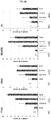
- self-antigens which show an antigen-antibody reactivity of 30% or higher ( FIG. 1a ) and to which peripheral blood monocytes secreting IFN- ⁇ shows a responsiveness of 50% or higher ( FIG. 1b ), were citrullinated filaggrin, PAD4 (peptidyl arginine deiminase 4), RA33 (heterogeneous nuclear ribonucleoprotein A2) and vimentin.
- the specific self-antigen (autoantigen) that is used to treat immature dendritic cells may be one or more selected from the group consisting of citrullinated filaggrin, PAD4 (peptidyl arginine deiminase 4), RA33 (heterogeneous nuclear ribonucleoprotein A2) and vimentin.
- self-antigen(autoantigen)-specific semi-mature dendritic cells may be produced by bring the specific antigen and the cells into "contact” with each other for a sufficient time and under sufficient conditions to enable the specific antigen to be presented from the cell surface by the semi-mature dendritic cells.
- the "contact” is "co-culture”.
- the co-culture is preferably performed for 3-10 hours, more preferably 3-8 hours, most preferably 3-4 hours.
- concentration of PEG2 used to treat the cells is preferably 0.01-5 ⁇ g/mL, and most preferably 0.5-5 ⁇ g/mL.
- the cytokine may be TNF- ⁇ (tumor necrosis factor-alpha), and is used at a concentration of 1-20 ng/ml, preferably 5-15 ng/ml.
- the semi-mature dendritic cells according to the present invention increased expression of NR4A2 and/or UBASH3B protein or a gene encoding the protein.
- the expression of PTGS2 and/or IDO protein or a gene encoding the protein in the semi-mature dendritic cells according to the present invention may further be increased, and in this case, the expression may be increased by at least twice that in immature dendritic cells.
- the NR4A2 protein a transcription factor
- dopaminergic neurons are damaged by inflammatory environments to cause Parkinson's disease, and regulated expression of NR4A2 in glias and astrocytes can provide neuron protective effects, by inhibiting the secretion of TNF- ⁇ , IL-1 ⁇ , NO and ROS.
- the UBASH3B protein has a PGM-like domain in the C-terminal region, is evolutionally similar to PGM/AcP enzyme, and has phosphorylase activity.
- This UBASH3B protein can dephosphorylate activated Src tyrosine kinase (phosphorylated) of T cells, resulting in negative regulation of T cells.
- MLN-DCs that express the PTGS2 protein interfere with Th2 differentiation and play an important role in the development of Treg.
- DCs are treated with IV IG, the secretion of PGE2 from the DCs can increase, and the expression of Cox2 in the DCs can increase while the expression of Treg can also increase to reduce EAE scores.
- the IDO protein is an enzyme functioning as indoleamine 2,3-dioxygenase, and is involved in degradation of the essential amino acid L-tryptophan.
- NR4A2 gene (NM_006186: SEQ ID NO: 11), UBASH3B gene (NM_032873: SEQ ID NO: 12), PTGS2 gene (NM_000963: SEQ ID NO: 13), and IDO gene (NM_002164: SEQ ID NO: 14) mean genes that encode NR4A2 protein, UBASH3B protein, PTGS2 protein, and IDO protein, respectively.
- the expression of the protein can be measured by an analysis method, for example, such as Western blotting, ELISA, radioimmunoassay analysis, radial immunodiffusion, tissue immunohistochemistry, immunoprecipitation assay, complement fixation assay, or FACS, and the expression of the gene may be measured by PCR, real-time PCR or RT PCR, but is not limited thereto.
- an analysis method for example, such as Western blotting, ELISA, radioimmunoassay analysis, radial immunodiffusion, tissue immunohistochemistry, immunoprecipitation assay, complement fixation assay, or FACS, and the expression of the gene may be measured by PCR, real-time PCR or RT PCR, but is not limited thereto.
- expression of a specific gene in the semi-mature dendritic cells of the present invention was measured by real-time PCR. As a result, it could be confirmed that, when the concentration of PGE2 used for treatment of the cells was 0.01-5 ⁇ g/mL, expression of NR4A2 and/or UBASH3B protein or a gene encoding the protein in the semi-mature dendritic cells increased at least 2-fold compared to that in immature dendritic cells. In addition, it could be found that expression of PTGS2 and/or IDO gene in the semi-mature dendritic cells increased, for example, at least twice.
- the semi-mature dendritic cells of the present invention showed an at least 2-fold increase in expression of NR4A2 gene, an at least 5-fold increase in expression of UBASH3B gene, an at least 4-fold increase in expression of PTGS2 gene, and an at least 2-fold increase in expression of IDO gene, compared to mature dendritic cells ( FIG. 2 ).
- the present invention provides a method for measuring an expression level of NR4A2 and/or UBASH3B protein or a gene encoding the protein in semi-mature dendritic cells for the prevention or treatment of autoimmune disease, the method comprising the steps of:
- measurement of the expression level of the protein or the gene may be performed using a conventional method, and specific examples of this method are as mentioned above.
- measurement of the expression level of the protein or the gene may be performed, for example, by amplifying specific genes using a primer pair selected from among primers of SEQ ID NOs: 1 to 10, measuring the expression levels of the specific genes by real-time PCR, normalizing the expression levels with ⁇ -actin, and analyzing relative values, which is resulted from dividing the normalized expression levels by expression levels measured in immature dendritic cells.
- ⁇ -actin is the main component of the cytoskeleton, is expressed uniformly in most cells, is not easily changed by external conditions in nature thereof, and shows a certain high expression level.
- Such genes are referred to as housekeeping genes, and are used as reference values to compare the expression levels of specific genes.
- the expression level of ⁇ -actin as a reference value
- the expression level of NR4A2 and/or UBASH3B gene in semi-mature dendritic cells was measured and divided by the expression level of NR4A2 and/or UBASH3B gene in immature dendritic cells used as a control group.
- the method of the present invention may further comprise measuring the expression level of PTGS2 and/or IDO gene.
- the concentration of PGE2 used for treatment of the cells may be 0.05-5 ⁇ g/ml, and the time required to treat the immature dendritic cells with PGE2, the self-antigen (autoantigen) and the cytokine may be 3-10 hours.
- the self-antigen (autoantigen) may be one or more selected from the group consisting of (citrullinated filaggrin, PAD4(peptidyl arginine deiminase 4), RA33 (heterogeneous nuclear ribonucleoprotein A2), and vimentin.
- the cytokine is TNF- ⁇ (tumor necrosis factor-alpha).
- a disease or disorder to which the composition of the present invention is applied may be rheumatoid arthritis, but is not necessarily limited thereto.
- an autoimmune disease that can be treated with the composition comprising the semi-mature dendritic cells of the present invention includes any disease or disorder caused by an autoimmune response in vivo.
- autoimmune disease examples include type 1 diabetes, rheumatoid arthritis, celiac disease, IgA deficiency, Crohn's disease, multiple sclerosis, systemic lupus erythematosus, Sjogren's syndrome, scleroderma, polymyositis, chronic active hepatitis, mixed connective tissue disease, primary biliary cirrhosis, pernicious anemia, autoimmune thyroiditis, idiopathic Addison's disease, vitiligo, gluten-sensitive enteropathy, Grave's disease, myasthenia gravis, autoimmune neutropenia, idiopathic thrombocytopenic purpura, cirrhosis, pemphigus vulgaris, autoimmune infertility, goodpastures syndrome, bullous pemphigoid, discoid lupus erythematosus, ulcerative colitis, dense deposits disease, and the like.
- semi-mature dendritic cells sensitized with specific self-antigens(autoantigens) were administered to 12 active rheumatoid arthritis patients.
- the patients were divided into two groups, each consisting of 6 persons, and then the cells were administered subcutaneously to each group at a concentration of 5 x 10 6 cells/0.5 mL or 1.5 x 10 7 cells/1.5 mL.
- the cells were administered a total of three times at 2-week intervals up to the first 4 weeks, and then administered a total of twice at 2-week intervals after a drug holiday of 4 weeks.
- the cells were administered for a total of 10 weeks.
- blood was sampled from the rheumatoid arthritis patients, and the reactivities of autoantibodies and T cells in the blood were measured.
- autoantibodies against specific self-antigens (autoantigens: citrullinated filaggrin, PAD4, RA33, and vimentin) decreased ( FIGS. 10 and 11 ).
- the present invention provides a cell therapeutic agent for treatment of an autoimmune disease having responsiveness to the same self-antigen (autoantigen) as an self-antigen(autoantigen) used for treatment of semi-mature dendritic cells produced by the method of the present invention, the cell therapeutic agent containing, as an active ingredient, the semi-mature dendritic cells.
- the term "cell therapeutic agent” refers to a drug used for treatment, diagnosis and prevention of diseases through a series of processes including the process of changing a cell's biological property by growing or selecting autologous, allogenic, and xenogenic cells outside the body or using other methods.
- the U.S. and Korea have controlled the cell therapeutic agent as a drug since 1993 and 2002, respectively.
- Such a cell therapeutic agent can be largely classified into two types: “a stem cell therapeutic agent” for the regeneration of tissues and the recovery of organ functions and “an immunocyte therapeutic agent” for the regulation of immune reaction, including the inhibition of immune reaction in-vivo or the accentuation of immune reaction.
- the cell therapeutic agent composition of the present invention may comprise a therapeutically effective amount of a cell therapeutic agent for the treatment of diseases.
- a therapeutically effective amount is refers to an amount of an active ingredient or a pharmaceutical composition that induces a biological or medical reaction in tissue systems, animals, or humans, and is considered by researchers, veterinarians, doctors, or other clinicians.
- the therapeutically effective amount comprises an amount of inducing the alleviation of the symptoms of the disease or disorder being treated. It is obvious to those skilled in the art that cell therapeutic agent contained in the composition of the present invention will be changed according to a desired effect.
- the optimum content of the cell therapeutic agent in the composition of the present invention can be easily determined by those skilled in the art, and may be adjusted depending on various factors including the type and severity of a disease, the contents of other components contained in the composition, the type of formulation, the patient's age, body weight, general health condition, sex and diet, administration time, administration route, the secretion rate of the composition, duration of treatment, and concurrently used medications.
- the semi-mature dendritic cells of the present invention were administered to 12 rheumatoid arthritis at a concentration of 5 x 10 6 cells/0.5 mL or 1.5 ⁇ 10 7 cells/1.5 mL.
- the semi-mature dendritic cells functioned to reduce an autoantibody against the specific antigen, indicating that the cells have therapeutic effects ( FIGS. 10 and 11 ).
- the cell therapeutic agent of the present invention contain the semi-mature dendritic cells in an amount of 5 x 10 6 to 1.5x10 7 cells/mL, more preferably 1.5 x 10 7 cells/ml.
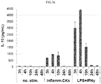
- IL-10 a cytokine secreted from Th2, regulatory T cells, dendritic cells and the like, is known to attack self-tissue to induce immune tolerance.
- IL-10 was quantified by an ELISA kit, and as a result, it could be seen that the semi-mature dendritic cells of the present invention secreted IL-10 in a manner dependent on the time and concentration of treatment with PGE2 ( FIG. 4 ).
- a control group transfected with siRNA for each gene at a concentration of 10-100 pmol (10, 50 and 100 pmol) it could be seen that the secretion of IL-10 from the semi-mature dendritic cells is attributable to specific genes, particularly NR4A2 and/or UBASH3B gene ( FIG. 5 ).
- the semi-mature dendritic cells of the present invention which show IL-10 secretion that was increased by expression of the NR4A2 and/or UBASH3B gene, was effective for treatment of rheumatoid arthritis ( FIGS. 7a, 7b and 8 ).
- the semi-mature dendritic cells of the present invention induced immune tolerance of T cells by IL-10 ( FIGS. 6 and 9 ).
- the present invention provides a cell therapeutic agent for treatment of autoimmune disease, which has the effects of increasing IL-10 secretion, reducing the secretion of IFN- ⁇ from T cells, and reducing autoantibody production.
- the semi-mature dendritic cells of the present invention may be formulated in a suitable form with a pharmaceutically acceptable carrier that is generally used in cell therapy.
- the present invention provides a method for preparing a cell therapeutic agent for prevention or treatment of autoimmune disease, the cell therapeutic agent containing semi-mature dendritic cells, the method comprising the steps of:
- the expression of PTGS2 or IDO protein or a gene encoding the protein in the semi-mature dendritic cells may further be increased.
- the expression of PTGS2 or IDO protein or the gene encoding the protein in the semi-mature dendritic cells may be increased at least 2-fold compared to that in immature dendritic cells.
- measurement of the expression levels of specific genes may be performed by amplifying the specific genes using each primer pair selected from among primers of SEQ ID NOs: 1 to 10, measuring the expression levels of the specific genes by real-time PCR, normalizing the expression levels with ⁇ -actin, and dividing the normalized expression levels by expression levels measured in immature dendritic cells, thereby determining relative values.
- the expression level of NR4A2 or UBASH3B gene in the semi-mature dendritic cells is measured using as a reference value the expression level of ⁇ -actin, a kind of housekeeping gene which is not easily changed by external conditions and shows a certain high expression level, and the measured expression level is divided by the expression level of the NR4A2 or UBASH3B gene in immature dendritic cells used as a control group.
- the concentration of PGE2 used for treatment of the cells was 0.05-5 ⁇ g/mL
- the expression levels of the NR4A2 and UBASH3B genes in the semi-mature dendritic cells increased at least 2-fold and at least 5-fold, respectively, compared to those in immature dendritic cells.
- the expression levels of PTGS2 and IDO genes in the semi-mature dendritic cells increased at least 4-fold and at least 2-fold, respectively, compared to those in immature dendritic cells.
- a pharmaceutically acceptable carrier necessary for formulation of the cell therapeutic agent of the present invention refers to a composition that is physiologically acceptable and does not cause gastric disorder, allergic reactions such as gastrointestinal disorder or vertigo, or similar reactions, when administered to humans.
- the pharmaceutically acceptable carrier include carriers for parenteral administration, such as water, suitable oil, saline solution, aqueous glucose, and glycol.
- the composition of the present invention may further comprise a stabilizer and a preservative. Suitable stabilizers include antioxidants, such as sodium bisulphite, sodium sulphite and ascorbic acid. Suitable preservatives include benzalkonium chloride, methyl- or propyl-paraben, and chlorobutanol.
- Other pharmaceutically acceptable carriers can be found in Remington's Pharmaceutical Sciences, 19th ed., Mack Publishing Company, Easton, Pa., 1995 .
- the time required to treat the cells with the self-antigen (autoantigen), the cytokine and PGE2 may be 3-10 hours.
- the self-antigen (autoantigen) may be, for example, one or more selected from the group consisting of ⁇ -enolase, filaggrin, PAD4 (peptidyl arginine deiminase 4), RA33 (heterogeneous nuclear ribonucleoprotein A2), fibrinogen- ⁇ , fibrinogen- ⁇ , collagen II, histone B, aggrecan, fibrin, rheumatoid factor, GPI (glucose-6-phosphate isomerase), and vimentin peptides and proteins, and citrullinated peptides and proteins thereof, and the cytokine may be TNF- ⁇ (tumor necrosis factor-alpha).
- RA rheumatoid arthritis
- each of a total of 9 antigens including citrullinated or non-citrullinated filaggrin (JW CreaGene), PAD4 (JW CreaGene), RA33 (JW CreaGene), vimentin (JW CreaGene), fibrinogen (Sigma-Aldrich), fibrin (Sigma-Aldrich), IgG Fc (Cell Science), GPI (JW CreaGene) and collagen (Genway), was dispensed into a 96-well plate at a concentration of 1 ⁇ g/mL, and then after 24 hours, the plate was washed with 0.05% PBS-T and blocked with 1% BSA-containing PBS for 1 hour.
- JW CreaGene citrullinated or non-citrullinated filaggrin
- PAD4 JW CreaGene
- RA33 JW CreaGene
- vimentin JW CreaGene
- fibrinogen Sigma-Aldrich
- fibrin Sigma-Ald
- the plate was washed with 0.05% PBS-T, and 100 ⁇ L of 1 ⁇ g/mL-p-nitrophenyl phosphate (Sigma-Aldrich) was dispensed into each well of the plate. After color development, 50 ⁇ L of 0.2M sodium hydroxide was dispensed into each well of the plate to stop the reaction, and the absorbance of each well at 405 nm was measured by an ELISA reader.
- the antigen-antibody reactivity of each of citrullinated filaggrin, PAD4, RA33 and vimentin was 30% or higher ( FIG. 1b ).
- the antigen-antibody reactivity means the percentage (%) of patients having a value equal to or higher than ⁇ average absorbance of normal persons x 1.5> among 100 rheumatoid arthritis patients.
- the plate was washed with 0.05% PBS-T, and 100 ⁇ L of biotinylated detection antibody was dispensed into each well, followed by incubation at room temperature for 2 hours. After 2 hours, the plate was washed with 0.05% PBS-T, and 100 ⁇ L of a 1:100 dilution of HRP-conjugated avidin was dispensed into each well, followed by incubation at room temperature for 1 hour. After 1 hour, the plate was washed with PBS, and 100 ⁇ L of an AEC substrate (BD Biosciences) was dispensed into each well. The degree of color development was measured and compared with the degree of color development measured in a control group not treated with the self-antigen(autoantigen). The spot in each well was analyzed using an ImmunoSPOT-ELISPOT reader (Cellular Technology).
- IFN- ⁇ responsiveness means the percentage (%) of patients having a value equal to or higher than ⁇ average absorbance of normal persons x 1.5> among 9 RA patients.
- citrullinated filaggrin, PAD4, RA33 and vimentin which show an antigen-antibody reactivity of 30% or higher and to which peripheral blood monocytes secreting IFN- ⁇ show a responsiveness of 50% or higher, were selected as specific self-antigens(autoantigens) to be used to sensitize semi-mature dendritic cells.
- a certain amount of peripheral blood monocytes extracted from normal persons or rheumatoid arthritis patients were dispensed into a plastic culture dish and cultured for 30 minutes to 1 hour, and then the floating cells were removed.
- the monocytes attached to the bottom were cultured in a CellGro (CellGenix) medium containing 20 ng/mL of IL-4 (JW CreaGene) and 30 ng/mL of GM-CSF (JW CreaGene) at 37°C for 3 days, and then the suspended immature dendritic cells were collected (among these cells, some immature dendritic cells were used as a control group for confirming expression of specific genes).
- the collected immature dendritic cells were suspended in the CellGro medium and dispensed into a fresh plate in a certain amount, and then the cells were stimulated for 3-10 hours by addition of 5-10 ⁇ g of the self-antigens (autoantigens: JW CreaGene) selected in Examples 1 and 2, 10 ng/mL of TNF ⁇ (Peprotech) and 0.05-5 ⁇ g/mL of PGE2 (Sigma-Aldrich).
- the stimulated semi-mature dendritic cells were frozen with human plasma albumin (JW Pharm.) containing 5% DMSO (Sigma-Aldrich) and 5% glucose (Green Cross Corp.).
- autoantigens specific self-antigens
- the semi-mature dendritic cells when the semi-mature dendritic cells were treated with PGE2 for 0-48 hours (particularly 3-10 hours), the semi-mature dendritic cells showed an at least 2-fold increase in expression of NR4A2 gene (NM_006186), an at least 5-fold increase in expression of UBASH3B gene (NM_032873) and an at least 3-fold increase in expression of PTGS2 gene (NM_000963), compared to the immature dendritic cells, and that expression of IDO gene (NM_002164) increased about at least 60-fold at a treatment time of 10-48 hours ( FIG. 2a ).
- the semi-mature dendritic cells treated with 0.01-5 ⁇ g/mL of PGE2 showed an at least 2-fold increase in expression of NR4A2 gene (NM_006186), an at least 5-fold increase in expression of UBASH3B gene (NM_032873), an at least 4-fold increase in expression of PTGS2 gene (NM_000963) and an at least 2-fold increase in expression of IDO gene (NM_002164), compared to the immature dendritic cells ( FIG. 2b ).
- a specific gene was knocked out by transfection with siRNA for the gene, and then changes in IL-10 secretion from the semi-mature dendritic cells and IFN- ⁇ secretion from T cells were analyzed.
- Transfection of siRNA into the semi-mature dendritic cells was performed in the following manner. A certain amount of human peripheral blood monocytes were dispensed into a plastic culture dish and allowed to stand for 30 minutes to 1 hour so as to adhere.
- the floating cells were removed, and the monocytes attached to the bottom were cultured in a CellGro (CellGenix) containing 20 ng/mL of IL-4 (JW CreaGene) and 30 ng/mL of GM-CSF (JW CreaGene) for 2 days, after which the floating immature cells were collected.
- the immature dendritic cells suspended in the CellGro medium were dispensed at a density of 5 x 10 5 -10 x 10 5 cells/well (6-well plate).
- the produced semi-mature dendritic cells were thawed, and then cultured in a 10% human AB serum (Lonza)-containing RPMI1640 containing 10 ng/mL of TNF ⁇ , IL-6, IL-1 ⁇ (Peprotech) and 200 units/mL of IFN ⁇ (LG) or containing 0.1 ⁇ g/mL of LPS (Sigma-Aldrich) or 1 ⁇ g/mL of CD40L (Peprotech).
- the culture was collected, and IL-10 in the culture was quantified using an ELISA kit (BD Biosciences).
- CD3 positive T cells with a purity of 90% or higher were separated from human peripheral blood monocytes by use of a nylon wool column, and suspended in a 10% human AB serum (Lonza)-containing RPMI1640 (Lonza) and dispensed into each well of a 48-well plate at a density of 1x10 6 cells.
- the semi-mature dendritic cells, produced and frozen by the method of Example 3, were thawed, washed with RPMI1640, suspended in the same medium, dispensed into each well at a density of 1 x 10 5 cells, and cultured under the conditions of 37°C and CO 2 .
- the cells were stimulated with 50 ng/mL of PMA (Sigam-Aldrich), 500 ng/mL of ionomycin (Sigam- Aldrich) and 1 ⁇ L/mL of brefeldin A (Ebioscience) for 4 hours, and the cell population was collected and subjected to FACS staining with anti-human CD3 antibody (BD Biosciences) and anti-human CD4 antibody (Ebioscience).
- the surface-stained FACS samples were permeabilized using a fixation/permeabilization kit (BD Biosciences), and subjected to intracellular cytokine FACS staining for anti-human IFN- ⁇ antibody.
- cytokine secretion in T cells was analyzed using a FACScalibur (BD).
- T cells were isolated according to the above-described method, and suspended in FBS (Gibco)-containing RPMI1640 (Gibco), and cultured with each test group for 7 days under the conditions of 37°C and CO 2 . After 7 days, T cells of each test group were collected and primarily FACS-stained with anti-human CD4 antibody and CD25 antibody. The surface-stained FACS samples were permeabilized using a fixation/permeabilization kit (Ebioscience). Each sample was subjected to intranuclear FACS staining for anti-human Foxp3 antibody, and then the induction (%) of regulatory cells in each sample was analyzed using a FACScalibur (BD).
- the semi-mature dendritic cells were administered to rheumatoid arthritis patients under the same conditions as described in Example 2, and then an assay was performed to monitor changes in the IFN- ⁇ reactivities of autoreactive T cells against self-antigens (autoantigens) (immune response monitoring).
- Citrullinated filaggrin (JW CreaGene), PAD4 (JW CreaGene), RA33 (JW CreaGene) and vimentin (JW CreaGene) antigens (10 ⁇ g/mL) were used, and the spot in each well was analyzed using an ImmunoSPOT-ELISPOT reader (Cellular Technology).
- treatment with the semi-mature dendritic cells of the present invention inhibited the secretion of IFN- ⁇ from T-cells in proportion to the treatment time to increase immune tolerance properties such as regulatory T-cell induction ( FIG. 9 ).
- Example 8 Administration of Semi-Mature Dendritic Cells and Blood Sampling for Immune Response Monitoring
- Vials containing the frozen semi-mature dendritic cells were thawed, and the cells were injected subcutaneously into both femoral regions of 12 rheumatoid arthritis patients (divided into two groups (high-dose group and low-dose group), each consisting of 6 persons)) in an amount of 5 ⁇ 10 6 cells/0.5 mL or 1.5 ⁇ 10 7 cells/1.5 mL.
- the semi-mature dendritic cells were administered in the same amount and manner as described above.
- blood was sampled from the rheumatoid arthritis patients, and effectiveness evaluation and immune response monitoring were performed.
- semi-mature dendritic cells may be produced by treating human immature dendritic cells with a selected specific self-antigen(autoantigen), a cytokine and PGE2.
- the produced semi-mature dendritic cells show an at least 2-fold increase in expression of NR4A2 or UBASH3B protein or a gene encoding the protein, compared to immature dendritic cells, and thus show increased secretion of IL-10, indicating that the semi-mature dendritic cells have increased immune tolerance properties.
- a cell therapeutic agent for prevention or treatment of autoimmune disease which contains the semi-mature dendritic cells of the present invention, is administered to a patient showing responsiveness to the same self-antigen(autoantigen) as the autoantibody used in the production of the semi-mature dendritic cells, it exhibits excellent effects compared to conventional autoimmune disease therapeutics.
- the cell therapeutic agent enables cell therapy based on the characteristics of an individual patient.
Abstract
Description
- The present invention relates to a method for generating semi-mature dendritic cells for treatment of autoimmune diseases, particularly rheumatoid arthritis, and a composition for the treatment or prevention of autoimmune diseases, which contains, as an active ingredient, dendritic cells produced using the method.
- Dendritic cells in the skin were first found by Langerhans in 1868, and the function thereof as immunity-enhancing cells was reported by Cohn and Steinmann in 1973. In 1990s, dendritic cells were found to function as professional antigen presenting cells (APCs), and were found to play an important role in immune activation and regulation. Dendritic cells in the human body represent only about 0.3% of total circulating leukocytes, but consist of a heterogeneous population having a phenotype differentiated from that of macrophages. Dendritic cells are differentiated in that they are professional antigen-presenting cells, unlike B cells or macrophages displaying a relatively weak antigen-presenting capability. Dendritic cells have the ability to induce a primary immune response capable of stimulating naive T cells that have not been exposed to antigen, and these dendritic cells are the only cells having the capability to induce immunological memory. It is known that dendritic cells can function to induce strong immune responses, because these dendritic cells are antigen presenting cells (APCs) that express, on the cell surface, high levels of both antigen presenting MHC molecules (I/II) and co-stimulatory molecules, for example, CD-80 and CD-86, and adhesion molecules, for example, ICAM-1, and secrete various cytokines (IFN-alpha, IL-12, IL-18, etc.). It is known that, because dendritic cells express high levels of antigen presenting molecules (HMC molecules and co-stimulatory molecules) on the cell surface, and secrete various cytokines such as IFN-alpha, IL-12 and the like, these dendritic cells can induce the generation of antigen-specific killer T cells and the proliferation and activation of Th1 cells.
- As described above, dendritic cells are the strongest antigen-presenting cells. A very small number of dentritic cells are present in vivo, but these cells strongly induce T cell immunity, and thus have been studied as therapeutic agents against cancers or infectious diseases in clinical studies focusing on the induction of immunity against the specific antigens. It was found that antigen immunogenicity was triggered by adoptive transfer of dendritic cells that were isolated from tissues or blood, antigen-pulsed, and matured in vitro. Thus, these dendritic cells are highly valuable as cellular vaccines for inducing antigen-specific immunity against cancer or pathogenic microbes (Inaba, K. et al., 3. Exp. Med., 178:479, 1993; Inaba, K. et al., Int. Rev.Immunol., 6:197, 1990; Hsu, F. et al., Nature Med., 2:52, 1996). Techniques for the isolation and maturation of dendritic cells are described in a number of documents, and there are various methods, including a method comprising generating mature dendritic cells from immature dendritic cells derived from pluripotent cells having the capability to express any one of the characteristics of macrophages or dendritic cells, and bringing the immature dendritic cells into contact with dendritic cell maturation factors including IFN-α (European Patent No.
922,758 663,930 WO 95/28479 - However, prior art documents and patents are merely directed to methods of generating dendritic cells by sensitizing immature dendritic cells with non-specific antigen, and do not teach a method of generating semi-mature dendritic cells by sensitizing immature dendritic cells with a specific selected self-antigen(autoantigen).
- Accordingly, based on the function of antigen-specific semi-mature dendritic cells in immune- tolerance, the present inventors have found that semi-mature dendritic cells can be developed from immature dendritic cells pulsed with self-antigens selected among the overexpressed self-antigens in patients with rheumatoid arthritis. These semi-mature dendritic cells increase the expression of a specific gene and mediate immune tolerance to the selected self-antigens(autoantigen) and thus improve therapeutic efficacy on rheumatoid arthritis.
- It is an object of the present invention to provide a method of generating semi-mature dendritic cells having increased expression of a specific gene after pulsing immature dendritic cells with a specific self-antigen (autoantigen) .
- Another object of the present invention is to provide a cell therapeutic agent that contains semi-mature dendritic cells generated by the method described above, and increases the therapeutic efficacy on an autoimmune disease by inducing immune tolerance to the same self-antigen (autoantigen) used for semi-mature dendritic cell generation, and a method for preparing the cell therapeutic agent.
- Still another object of the present invention is to provide a method for measuring an expression level of a gene or a protein, which is specifically increased by treatment with a specific self-antigen(autoantigen).
- To achieve the above objects, the present invention provides a method for generating semi-mature dendritic cells comprising a step of treating the immature dendritic cells with the self-antigen(autoantigen), a cytokine and prostaglandin E2 (PGE2), wherein the semi-mature dendritic cells have at least 2-fold increased expressions of NR4A2 and/or UBASH3B at gene or protein level compared to immature dendritic cells.
- The present invention also provides a cell therapeutic agent for treatment of an autoimmune disease by inducing tolerance to the same self-antigen (autoantigen) used for semi-mature dendritic cell generation as described above, the cell therapeutic agent containing, as an active ingredient, the semi-mature dendritic cells.
- The present invention also provides a method for preparing a cell therapeutic agent for prevention or treatment of autoimmune disease, the cell therapeutic agent containing semi-mature dendritic cells, the method comprising the steps of:
- (a) treating immature dendritic cells with an self-antigen(autoantigen), a cytokine and prostaglandin E2 (PGE2) to produce semi-mature dendritic cells;
- (b) confirming that expression of NR4A2 and/or UBASH3B protein or a gene encoding the protein in the semi-mature dendritic cells increased at least 2-fold compared to expression of the NR4A2 and/or UBASH3B protein or the protein-encoding gene in immature dendritic cells; and
- (c) preparing a cell therapeutic agent containing the semi-mature dendritic cells in which the expression of the NR4A2 and/or UBASH3B protein or the protein-encoding gene increased at least 2-fold.
- The present invention also provides a method for measuring an expression level of NR4A2 and/or UBASH3B protein or a gene encoding the protein in semi-mature dendritic cells for the prevention or treatment of autoimmune disease, the method comprising the steps of:
- (a) treating immature dendritic cells with an self-antigen(autoantigen), a cytokine and PGE2 to produce semi-mature dendritic cells;
- (b) measuring the expression level of the NR4A2 and/or UBASH3B protein or the protein-encoding gene in the immature dendritic cells;
- (c) measuring the expression level of the NR4A2 and/or UBASH3B protein or the protein-encoding gene in the produced semi-mature dendritic cells; and
- (d) comparing the expression levels of the NR4A2 and/or UBASH3B protein or the protein-encoding gene, measured in step (b) and step (c).
-
-
FIG. 1a shows the results of an experiment performed to measure T-cell responses (IFN-γ) to self-antigens (autoantigens) using the blood of normal persons and rheumatoid arthritis patients in order to select a specific self-antigen(autoantigen) to be used to sensitize semi-mature dendritic cells. -
FIG. 1b shows the results of an experiment performed to measure self-antigen(autoantigen) responses to self-antigens (autoantigens) using the blood of normal persons and rheumatoid arthritis patients in order to select a specific self-antigen(autoantigen) to be used to sensitize semi-mature dendritic cells. -
FIG. 2a shows experimental results indicating the expression levels of specific genes as a function of time of treatment with TNF-α + PGE2. -
FIG. 2b shows experimental results indicating the expression levels of specific genes as a function of the concentration of PGE2. -
FIG. 3a shows experimental results indicating the secretion level of IL-10 as a function of the time of treatment with TNF-α + PGE2. -
FIG. 3b shows experimental results indicating the secretion level of IL-10 as a function of the concentration of PGE2. -
FIG. 4 shows experimental results indicating the correlation between specific gene expression and IL-10 secretion. -
FIG. 5a shows experimental results indicating that NR4A2 gene expressed in semi-mature dendritic cells according to a production method of the present invention regulates IL-10 secretion. -
FIG. 5b shows experimental results indicating that UBASH3B gene expressed in semi-mature dendritic cells according to a production method of the present invention regulates IL-10 secretion. -
FIG. 6a shows experimental results indicating that semi-mature dendritic cells according to a production method of the present invention induce T-cell immune tolerance by IL-10 secretion, and experimental results indicating IL-10 secretion levels of semi-mature dendritic cells under various stimulation conditions. -
FIG. 6b shows experimental results indicating the IL-10-mediated inhibition of T-IFNγ secretion by semi-mature dendritic cells. -
FIG. 6c shows experimental results indicating the IL-10-mediated induction of regulatory T cells by semi-mature dendritic cells. -
FIG. 7a shows experimental results indicating that gene expression in semi-mature dendritic cells, produced by a method of the present invention and having increased expression of a specific gene and increased secretion of IL-10, is effective for treatment of rheumatoid arthritis. -
FIG. 7b experimental results indicating specific gene expression and IL-10 secretion and indicating that semi-mature dendritic cells, produced by a method of the present invention are effective for treatment of rheumatoid arthritis. -
FIG. 8 shows the clinical effectiveness of semi-mature dendritic cells for 14 weeks and 24 weeks on 12 patients administered five times with semi-mature dendritic cells in varying doses (NA: patient in which the swollen joint count is 0 at the baseline time, and thus ACR evaluation is impossible). -
FIG. 9a shows the results of an ELISPOT assay performed to measure changes in T-cell responses (IFN-γ) of patients administered with semi-mature dendritic cells. -
FIG. 9b shows the results of an IFN-γ intracellular staining assay performed to measure changes in T-cell responses (IFN-γ) of patients administered with semi-mature dendritic cells. -
FIG. 10 shows experimental results indicating the decreases in specific autoantibodies (citrullinated filaggrin, PAD4, RA33, and vimentin) in 12 patients at 14 weeks after administration of semi-mature dendritic cells. -
FIG. 11 shows experimental results indicating the time-dependent decreases in autoantibodies (citrullinated filaggrin, PAD4, RA33, and vimentin), measured at 14 weeks after semi-mature dendritic cells to 9 patients having one or more self-antigen(autoantigens). - Unless defined otherwise, all technical and scientific terms used herein have the same meaning as commonly understood by one of ordinary skill in the art to which the invention pertains. Generally, the nomenclature used herein and the experiment methods, which will be described below, are those well known and commonly employed in the art.
- In one aspect, the present invention is directed to a method for generating semi-mature dendritic cells in which expression of NR4A2 and/or UBASH3B protein or a gene encoding the protein is increased at least 2-fold compared to that in immature dendritic cells, the method comprising a step of treating the immature dendritic cells with an self-antigen(autoantigen), a cytokine and prostaglandin E2 (PGE2).
- In an example of the present invention, immature dendritic cells produced by differentiation of peripheral blood monocytes (PBMCs) from normal persons or rheumatoid arthritis patients were treated with a selected specific self-antigen(autoantigen), a cytokine and prostaglandin E2 (PGE2), thereby generating semi-mature dendritic cells.
- As used herein, the term "dendritic cells" are meant to include a population of dendritic cells, and refers to professional antigen presenting cells that absorb and treat an antigen and present the treated antigen together with a MHC (major histocompatibility complex) class I complex or a MHC class II complex. Dendritic cells that are used in the present invention refer to cells having the typical phenotype and characteristics of dendritic cells disclosed in Steinman et al., Annual Rev. Immunol. 9:271-296, 1991 and Banchereau and Steinman Nature 392:245-252, 1998. Dendritic cells include both immunogenic and tolerogenic antigen presenting cells, and are classified into immature dendritic cells (imDCs), semi-mature dendritic cells (smDCs), and mature dendritic cells (mDCs).
- As used herein, the term "immature dendritic cells (imDCs)" refers to dendritic cells which do not express cell surface markers such as CD14, like mature dendritic cells, and which express CCR7 and cytoplasmic protein DC-LAMP at low levels, express co-simulatory molecules (CD40, CD80 and CD86) at low levels, and express CD1a, CCR1, CCR2, CCR5 and CXCR1 at conventional levels.
- As used herein, the term "mature dendritic cells (mDCs)" refers to cells formed by maturation of immature dendritic cells. Mature dendritic cells show increased expression of not only DC-LAMP, but also MHC class II, CD40, CD80, CD83 and CD86, release proinflammatory cytokines, and induce increased proliferation of allogeneic T cells and syngeneic T cells in a mixed lymphocyte reaction and/or increased production of dendritic cell cytokines. The mature dendritic cells (mDCs) express CCR7 and CXCR4 at high levels.
- As used herein, the term "semi-mature dendritic cells (smDCs)" refers to dendritic cells that lack some of the characteristics of immature dendritic cells, have some of the phenotypes of mature dendritic cells, and show partially or incompletely maturated morphological and phenotypic characters. Semi-mature dendritic cells generally have the capability to induce an immune tolerance response to an self-antigen(autoantigen).
- According to a preferred embodiment of the present invention, monocytes are obtained from normal persons or rheumatoid arthritis patients, and then the cells are primarily cultured using a medium (preferably a medium supplemented with GM-CSF) on a suitable substrate. Substances that promote differentiation of pluripotent cells or multipotent cells into immature dendritic cells, particularly GM-CSF, are disclosed in
US Patent Nos. 5,851,756 and5,994,126 , the contents of which are incorporated herein by reference. The substrate is preferably a substrate to which cells can adhere. More preferably, the substrate is a plastic substrate which is used in tissue culture. - In addition to GM-CSF, factors that prevent or inhibit proliferation of non-dendritic cells may be added to the culture medium for improving yield of immature dendritic cells. The factors include, for example, IL-4 and/or IL-13 which inhibits macrophages. While the factors promote the proliferation of dendritic progenitor cells, they inhibit the growth of non-dendritic cells to thereby increase the number of immature dendritic cells in the medium.
- The self-antigen (autoantigen) that is used in the present invention may be, for example, one or more selected from the group consisting of α-enolase, filaggrin, PAD4 (peptidyl arginine deiminase 4), RA33 (heterogeneous nuclear ribonucleoprotein A2), fibrinogen-α, fibrinogen-β, collagen II, histone B, aggrecan, fibrin, rheumatoid factor, GPI (glucose-6-phosphate isomerase), and vimentin peptides and proteins, and citrullinated peptides and proteins thereof.
- According to a preferred embodiment of the present invention, peripheral blood monocytes extracted from 9 rheumatoid arthritis patients showing an effective ELISPOT response or 2 normal persons were sensitized with respective seven antigens, including citrullinated filaggrin (JW CreaGene), PAD4 (JW CreaGene), RA33 (JW CreaGene), vimentin (JW CreaGene), fibrinogen (Sigma-Aldrich), fibrin (Sigma-Aldrich), IgGFc (Cell Science), and a certain self-antigen(autoantigen) showing an autoreactive T cell responsiveness of 50% or more was screened. In addition, on 100 Korean rheumatoid arthritis patients, 9 autoantibodies, including citrullinated or non-citrullinated filaggrin (JW CreaGene), PAD4 (JW CreaGene), RA33 (JW CreaGene), vimentin (JW CreaGene), fibrinogen (Sigma-Aldrich), fibrin (Sigma-Aldrich), IgG Fc (Cell Science), GPI (JW CreaGene) and collagen (Genway), were screened, and a specific self-antigen(autoantigen) was screened by using a method measuring antigen-antibody reactivity based on a case showing an OD value that is at least 1.5 times higher than that of normal persons regarded as positive.
- As a result, it could be seen that self-antigens(autoantigens), which show an antigen-antibody reactivity of 30% or higher (
FIG. 1a ) and to which peripheral blood monocytes secreting IFN-γ shows a responsiveness of 50% or higher (FIG. 1b ), were citrullinated filaggrin, PAD4 (peptidyl arginine deiminase 4), RA33 (heterogeneous nuclear ribonucleoprotein A2) and vimentin. - Thus, in the present invention, the specific self-antigen (autoantigen) that is used to treat immature dendritic cells may be one or more selected from the group consisting of citrullinated filaggrin, PAD4 (peptidyl arginine deiminase 4), RA33 (heterogeneous nuclear ribonucleoprotein A2) and vimentin.
- In the present invention, self-antigen(autoantigen)-specific semi-mature dendritic cells may be produced by bring the specific antigen and the cells into "contact" with each other for a sufficient time and under sufficient conditions to enable the specific antigen to be presented from the cell surface by the semi-mature dendritic cells. Preferably, the "contact" is "co-culture".
- The co-culture is preferably performed for 3-10 hours, more preferably 3-8 hours, most preferably 3-4 hours. The concentration of PEG2 used to treat the cells is preferably 0.01-5 µg/mL, and most preferably 0.5-5 µg/mL. The cytokine may be TNF-α (tumor necrosis factor-alpha), and is used at a concentration of 1-20 ng/ml, preferably 5-15 ng/ml.
- It was confirmed that the semi-mature dendritic cells according to the present invention increased expression of NR4A2 and/or UBASH3B protein or a gene encoding the protein. Alternatively, the expression of PTGS2 and/or IDO protein or a gene encoding the protein in the semi-mature dendritic cells according to the present invention may further be increased, and in this case, the expression may be increased by at least twice that in immature dendritic cells.
- The NR4A2 protein, a transcription factor, can bind to the promoter and enhancer of Foxp3 to act together with other transcription factors, thereby inducing Treg differentiation of CD4+ T and finally inducing antiinflammatory responses. In addition, it is known that dopaminergic neurons are damaged by inflammatory environments to cause Parkinson's disease, and regulated expression of NR4A2 in glias and astrocytes can provide neuron protective effects, by inhibiting the secretion of TNF-α, IL-1β, NO and ROS.
- The UBASH3B protein has a PGM-like domain in the C-terminal region, is evolutionally similar to PGM/AcP enzyme, and has phosphorylase activity. This UBASH3B protein can dephosphorylate activated Src tyrosine kinase (phosphorylated) of T cells, resulting in negative regulation of T cells.
- MLN-DCs that express the PTGS2 protein interfere with Th2 differentiation and play an important role in the development of Treg. When DCs are treated with IV IG, the secretion of PGE2 from the DCs can increase, and the expression of Cox2 in the DCs can increase while the expression of Treg can also increase to reduce EAE scores.
- The IDO protein is an enzyme functioning as
indoleamine 2,3-dioxygenase, and is involved in degradation of the essential amino acid L-tryptophan. - In the present invention, NR4A2 gene (NM_006186: SEQ ID NO: 11), UBASH3B gene (NM_032873: SEQ ID NO: 12), PTGS2 gene (NM_000963: SEQ ID NO: 13), and IDO gene (NM_002164: SEQ ID NO: 14) mean genes that encode NR4A2 protein, UBASH3B protein, PTGS2 protein, and IDO protein, respectively.
- The expression of the protein can be measured by an analysis method, , for example, such as Western blotting, ELISA, radioimmunoassay analysis, radial immunodiffusion, tissue immunohistochemistry, immunoprecipitation assay, complement fixation assay, or FACS, and the expression of the gene may be measured by PCR, real-time PCR or RT PCR, but is not limited thereto.
- In an example of the present invention, expression of a specific gene in the semi-mature dendritic cells of the present invention was measured by real-time PCR. As a result, it could be confirmed that, when the concentration of PGE2 used for treatment of the cells was 0.01-5 µg/mL, expression of NR4A2 and/or UBASH3B protein or a gene encoding the protein in the semi-mature dendritic cells increased at least 2-fold compared to that in immature dendritic cells. In addition, it could be found that expression of PTGS2 and/or IDO gene in the semi-mature dendritic cells increased, for example, at least twice. Specifically, it could be found that the semi-mature dendritic cells of the present invention showed an at least 2-fold increase in expression of NR4A2 gene, an at least 5-fold increase in expression of UBASH3B gene, an at least 4-fold increase in expression of PTGS2 gene, and an at least 2-fold increase in expression of IDO gene, compared to mature dendritic cells (
FIG. 2 ). - Meanwhile, in order to confirm the characteristics of the semi-mature dendritic cells of the present invention and to find the reason why the semi-mature dendritic cells of the present invention exhibit excellent therapeutic effects in rheumatoid arthritis patients compared to conventional therapeutic agents, it is important to measure the characteristically increased expression level of a specific gene in the semi-mature dendritic cells of the present invention.
- Therefore, in another aspect, the present invention provides a method for measuring an expression level of NR4A2 and/or UBASH3B protein or a gene encoding the protein in semi-mature dendritic cells for the prevention or treatment of autoimmune disease, the method comprising the steps of:
- (a) treating immature dendritic cells with an self-antigen(autoantigen), a cytokine and PGE2 to produce semi-mature dendritic cells;
- (b) measuring the expression level of the NR4A2 and/or UBASH3B protein or the protein-encoding gene in the immature dendritic cells;
- (c) measuring the expression level of the NR4A2 and/or UBASH3B protein or the protein-encoding gene in the produced semi-mature dendritic cells; and
- (d) comparing the expression levels of the NR4A2 and/or UBASH3B protein or the protein-encoding gene, measured in step (b) and step (c).
- In the present invention, measurement of the expression level of the protein or the gene may be performed using a conventional method, and specific examples of this method are as mentioned above. In the present invention, measurement of the expression level of the protein or the gene may be performed, for example, by amplifying specific genes using a primer pair selected from among primers of SEQ ID NOs: 1 to 10, measuring the expression levels of the specific genes by real-time PCR, normalizing the expression levels with β-actin, and analyzing relative values, which is resulted from dividing the normalized expression levels by expression levels measured in immature dendritic cells. β-actin is the main component of the cytoskeleton, is expressed uniformly in most cells, is not easily changed by external conditions in nature thereof, and shows a certain high expression level. Such genes are referred to as housekeeping genes, and are used as reference values to compare the expression levels of specific genes. In an example of the present invention, using the expression level of β-actin as a reference value, the expression level of NR4A2 and/or UBASH3B gene in semi-mature dendritic cells was measured and divided by the expression level of NR4A2 and/or UBASH3B gene in immature dendritic cells used as a control group. As a result, it could be seen that, when the concentration of PGE2 used for treatment of the cells was 0.05-5 µg/mL, the expression levels of the NR4A2 and UBASH3B genes increased at least 2-fold and 5-fold, respectively, compared to those in immature dendritic cells.
- In addition, the method of the present invention may further comprise measuring the expression level of PTGS2 and/or IDO gene.
- The concentration of PGE2 used for treatment of the cells may be 0.05-5 µg/ml, and the time required to treat the immature dendritic cells with PGE2, the self-antigen (autoantigen) and the cytokine may be 3-10 hours. The self-antigen (autoantigen) may be one or more selected from the group consisting of (citrullinated filaggrin, PAD4(peptidyl arginine deiminase 4), RA33 (heterogeneous nuclear ribonucleoprotein A2), and vimentin. The cytokine is TNF-α (tumor necrosis factor-alpha).
- In addition, according to a preferred embodiment, a disease or disorder to which the composition of the present invention is applied may be rheumatoid arthritis, but is not necessarily limited thereto. In other words, an autoimmune disease that can be treated with the composition comprising the semi-mature dendritic cells of the present invention includes any disease or disorder caused by an autoimmune response in vivo. Examples of the autoimmune disease include
type 1 diabetes, rheumatoid arthritis, celiac disease, IgA deficiency, Crohn's disease, multiple sclerosis, systemic lupus erythematosus, Sjogren's syndrome, scleroderma, polymyositis, chronic active hepatitis, mixed connective tissue disease, primary biliary cirrhosis, pernicious anemia, autoimmune thyroiditis, idiopathic Addison's disease, vitiligo, gluten-sensitive enteropathy, Grave's disease, myasthenia gravis, autoimmune neutropenia, idiopathic thrombocytopenic purpura, cirrhosis, pemphigus vulgaris, autoimmune infertility, goodpastures syndrome, bullous pemphigoid, discoid lupus erythematosus, ulcerative colitis, dense deposits disease, and the like. - In another example of the present invention, in order to confirm the clinical effect of the semi-mature dendritic cells of the present invention, semi-mature dendritic cells sensitized with specific self-antigens(autoantigens) were administered to 12 active rheumatoid arthritis patients. The patients were divided into two groups, each consisting of 6 persons, and then the cells were administered subcutaneously to each group at a concentration of 5 x 10⁶ cells/0.5 mL or 1.5 x 10⁷ cells/1.5 mL. The cells were administered a total of three times at 2-week intervals up to the first 4 weeks, and then administered a total of twice at 2-week intervals after a drug holiday of 4 weeks. Thus, the cells were administered for a total of 10 weeks. Before and after administration and at 8 and 14 weeks after administration, blood was sampled from the rheumatoid arthritis patients, and the reactivities of autoantibodies and T cells in the blood were measured.
- As a result, it could be found that autoantibodies against specific self-antigens (autoantigens: citrullinated filaggrin, PAD4, RA33, and vimentin) decreased (
FIGS. 10 and11 ). - Therefore, in another aspect, the present invention provides a cell therapeutic agent for treatment of an autoimmune disease having responsiveness to the same self-antigen (autoantigen) as an self-antigen(autoantigen) used for treatment of semi-mature dendritic cells produced by the method of the present invention, the cell therapeutic agent containing, as an active ingredient, the semi-mature dendritic cells.
- As used herein, the term "cell therapeutic agent" refers to a drug used for treatment, diagnosis and prevention of diseases through a series of processes including the process of changing a cell's biological property by growing or selecting autologous, allogenic, and xenogenic cells outside the body or using other methods. The U.S. and Korea have controlled the cell therapeutic agent as a drug since 1993 and 2002, respectively. Such a cell therapeutic agent can be largely classified into two types: "a stem cell therapeutic agent" for the regeneration of tissues and the recovery of organ functions and "an immunocyte therapeutic agent" for the regulation of immune reaction, including the inhibition of immune reaction in-vivo or the accentuation of immune reaction.
- The cell therapeutic agent composition of the present invention may comprise a therapeutically effective amount of a cell therapeutic agent for the treatment of diseases. As used herein, the term "therapeutically effective amount" is refers to an amount of an active ingredient or a pharmaceutical composition that induces a biological or medical reaction in tissue systems, animals, or humans, and is considered by researchers, veterinarians, doctors, or other clinicians. The therapeutically effective amount comprises an amount of inducing the alleviation of the symptoms of the disease or disorder being treated. It is obvious to those skilled in the art that cell therapeutic agent contained in the composition of the present invention will be changed according to a desired effect. Therefore, the optimum content of the cell therapeutic agent in the composition of the present invention can be easily determined by those skilled in the art, and may be adjusted depending on various factors including the type and severity of a disease, the contents of other components contained in the composition, the type of formulation, the patient's age, body weight, general health condition, sex and diet, administration time, administration route, the secretion rate of the composition, duration of treatment, and concurrently used medications.
- In an example of the present invention, the semi-mature dendritic cells of the present invention were administered to 12 rheumatoid arthritis at a concentration of 5 x 10⁶ cells/0.5 mL or 1.5 × 10⁷ cells/1.5 mL. As a result, it could be seen that, when the semi-mature dendritic cells were added to the patients showing responsiveness to the specific antigen, the semi-mature dendritic cells functioned to reduce an autoantibody against the specific antigen, indicating that the cells have therapeutic effects (
FIGS. 10 and11 ). Thus, when the amount of the semi-mature dendritic cells, which can exhibit the highest effect in the smallest amount is determined by considering all the above-described factors, the cell therapeutic agent of the present invention contain the semi-mature dendritic cells in an amount of 5 x 10⁶ to 1.5x10⁷ cells/mL, more preferably 1.5 x 10⁷ cells/ml. - In order to examine the correlation between the therapeutic effect of the semi-mature dendritic cells of the present invention and the specific genes that are expressed in the cells and identify the mechanism related thereto, whether or not dendritic cells secrete IL-10 and the functions of specific genes were examined. IL-10, a cytokine secreted from Th2, regulatory T cells, dendritic cells and the like, is known to attack self-tissue to induce immune tolerance.
- In an example of the present invention, IL-10 was quantified by an ELISA kit, and as a result, it could be seen that the semi-mature dendritic cells of the present invention secreted IL-10 in a manner dependent on the time and concentration of treatment with PGE2 (
FIG. 4 ). In another example of the present invention, based on a comparison with a control group transfected with siRNA for each gene at a concentration of 10-100 pmol (10, 50 and 100 pmol), it could be seen that the secretion of IL-10 from the semi-mature dendritic cells is attributable to specific genes, particularly NR4A2 and/or UBASH3B gene (FIG. 5 ). In addition, it could be seen that the semi-mature dendritic cells of the present invention, which show IL-10 secretion that was increased by expression of the NR4A2 and/or UBASH3B gene, was effective for treatment of rheumatoid arthritis (FIGS. 7a, 7b and8 ). In another example, it was found that the semi-mature dendritic cells of the present invention induced immune tolerance of T cells by IL-10 (FIGS. 6 and9 ). - Therefore, the present invention provides a cell therapeutic agent for treatment of autoimmune disease, which has the effects of increasing IL-10 secretion, reducing the secretion of IFN-γ from T cells, and reducing autoantibody production.
- Meanwhile, it is obvious to those skilled in the art that the semi-mature dendritic cells of the present invention may be formulated in a suitable form with a pharmaceutically acceptable carrier that is generally used in cell therapy.
- Therefore, in another aspect, the present invention provides a method for preparing a cell therapeutic agent for prevention or treatment of autoimmune disease, the cell therapeutic agent containing semi-mature dendritic cells, the method comprising the steps of:
- (a) treating immature dendritic cells with an self-antigen(autoantigen), a cytokine and prostaglandin E2 (PGE2) to produce semi-mature dendritic cells;
- (b) confirming that expression of NR4A2 and/or UBASH3B protein or a gene encoding the protein in the semi-mature dendritic cells increased at least 2-fold compared to expression of the NR4A2 and/or UBASH3B protein or the protein-encoding gene in immature dendritic cells; and
- (c) preparing a cell therapeutic agent containing the semi-mature dendritic cells in which the expression of the NR4A2 and/or UBASH3B protein or the protein-encoding gene increased at least 2-fold.
- In the present invention, the expression of PTGS2 or IDO protein or a gene encoding the protein in the semi-mature dendritic cells may further be increased. In this case, the expression of PTGS2 or IDO protein or the gene encoding the protein in the semi-mature dendritic cells may be increased at least 2-fold compared to that in immature dendritic cells.
- In the present invention, measurement of the expression levels of specific genes may be performed by amplifying the specific genes using each primer pair selected from among primers of SEQ ID NOs: 1 to 10, measuring the expression levels of the specific genes by real-time PCR, normalizing the expression levels with β-actin, and dividing the normalized expression levels by expression levels measured in immature dendritic cells, thereby determining relative values. Specifically, the expression level of NR4A2 or UBASH3B gene in the semi-mature dendritic cells is measured using as a reference value the expression level of β-actin, a kind of housekeeping gene which is not easily changed by external conditions and shows a certain high expression level, and the measured expression level is divided by the expression level of the NR4A2 or UBASH3B gene in immature dendritic cells used as a control group. As a result, it could be seen that, when the concentration of PGE2 used for treatment of the cells was 0.05-5 µg/mL, the expression levels of the NR4A2 and UBASH3B genes in the semi-mature dendritic cells increased at least 2-fold and at least 5-fold, respectively, compared to those in immature dendritic cells. When the expression levels were measured by the above-described method, it could be seen that, when the concentration of PGE2 used for treatment of the cells was 0.05-5 µg/mL, the expression levels of PTGS2 and IDO genes in the semi-mature dendritic cells increased at least 4-fold and at least 2-fold, respectively, compared to those in immature dendritic cells.
- A pharmaceutically acceptable carrier necessary for formulation of the cell therapeutic agent of the present invention refers to a composition that is physiologically acceptable and does not cause gastric disorder, allergic reactions such as gastrointestinal disorder or vertigo, or similar reactions, when administered to humans. Examples of the pharmaceutically acceptable carrier include carriers for parenteral administration, such as water, suitable oil, saline solution, aqueous glucose, and glycol. The composition of the present invention may further comprise a stabilizer and a preservative. Suitable stabilizers include antioxidants, such as sodium bisulphite, sodium sulphite and ascorbic acid. Suitable preservatives include benzalkonium chloride, methyl- or propyl-paraben, and chlorobutanol. Other pharmaceutically acceptable carriers can be found in Remington's Pharmaceutical Sciences, 19th ed., Mack Publishing Company, Easton, Pa., 1995.
- In the process of preparing the cell therapeutic agent for prevention or treatment of autoimmune disease according to the present invention, the time required to treat the cells with the self-antigen (autoantigen), the cytokine and PGE2 may be 3-10 hours. Herein, the self-antigen (autoantigen) may be, for example, one or more selected from the group consisting of α-enolase, filaggrin, PAD4 (peptidyl arginine deiminase 4), RA33 (heterogeneous nuclear ribonucleoprotein A2), fibrinogen-α, fibrinogen-β, collagen II, histone B, aggrecan, fibrin, rheumatoid factor, GPI (glucose-6-phosphate isomerase), and vimentin peptides and proteins, and citrullinated peptides and proteins thereof, and the cytokine may be TNF-α (tumor necrosis factor-alpha).
- Hereinafter, the present invention will be described in further detail with reference to examples. It will be obvious to a person having ordinary skill in the art that these examples are illustrative purposes only and are not to be construed to limit the scope of the present invention.
- In order to identify plasma autoantibodies present in rheumatoid arthritis (RA) patients at higher levels than those in normal persons or to monitor changes in autoplasma (immune response monitoring) after administration of semi-mature dendritic cells to RA patients, the following assay was performed. Specifically, each of a total of 9 antigens, including citrullinated or non-citrullinated filaggrin (JW CreaGene), PAD4 (JW CreaGene), RA33 (JW CreaGene), vimentin (JW CreaGene), fibrinogen (Sigma-Aldrich), fibrin (Sigma-Aldrich), IgG Fc (Cell Science), GPI (JW CreaGene) and collagen (Genway), was dispensed into a 96-well plate at a concentration of 1 µg/mL, and then after 24 hours, the plate was washed with 0.05% PBS-T and blocked with 1% BSA-containing PBS for 1 hour. After 1 hour, 50 µL of a 1:10 or 1:50 dilution of plasma from each of 100 rheumatoid arthritis patients and 14 normal persons was dispensed in duplicate, followed by incubation at room temperature for 2 hours. After 2 hours, the plate was washed with 0.05% PBS-T, and 50 µL of a 1: 2000 dilution of an AP-conjugated anti-human IgG antibody (Sigma-Aldrich) in 1% BSA-containing PBS was dispensed into each well of the plate. After 1 hour, the plate was washed with 0.05% PBS-T, and 100 µL of 1 µg/mL-p-nitrophenyl phosphate (Sigma-Aldrich) was dispensed into each well of the plate. After color development, 50 µL of 0.2M sodium hydroxide was dispensed into each well of the plate to stop the reaction, and the absorbance of each well at 405 nm was measured by an ELISA reader.
- As a result, it could be seen that the antigen-antibody reactivity of each of citrullinated filaggrin, PAD4, RA33 and vimentin was 30% or higher (
FIG. 1b ). Herein, the antigen-antibody reactivity means the percentage (%) of patients having a value equal to or higher than <average absorbance of normal persons x 1.5> among 100 rheumatoid arthritis patients. - In order to identify self-antigens(autoantigens) that increase the IFN-γ responsiveness of autoreactive T cells in the blood of rheumatoid arthritis (RA) patients compared to that in normal persons and to monitor the IFN-γ responsiveness of autoreactive T cells to self-antigens (autoantigens) (immune response monitoring) after administration of semi-mature dendritic cells to rheumatoid arthritis (RA) patients, the following assay was performed. An IFNγ-ELISPOT (BD Bioscience) assay was performed according to the manufacturer's guideline. Specifically, 100 µL of capture antibody was dispensed into each well of an ELSISPOT 96-well plate and incubated at 4°C for 24 hours, and then the plate was blocked with 10% FBS-containing PBS for 2 hours. After blocking, 10 µg/mL of each of a total of 7 antigens, including citrullinated filaggrin (JW CreaGene), PAD4 (JW CreaGene), RA33 (JW CreaGene), vimentin (JW CreaGene), fibrinogen (Sigma-Aldrich), fibrin (Sigma-Aldrich), and IgGFc (Cell Science), was added to 2x10⁵ peripheral blood monocytes (PBMCs) extracted from each of 9 rheumatoid arthritis and 2 normal persons, and then the cells were suspended in 10% human AB serum-containing RPMI1640 and dispensed into each well in duplicate. After incubation in a CO2 incubator at 37°C for 24 hours, the plate was washed with 0.05% PBS-T, and 100 µL of biotinylated detection antibody was dispensed into each well, followed by incubation at room temperature for 2 hours. After 2 hours, the plate was washed with 0.05% PBS-T, and 100 µL of a 1:100 dilution of HRP-conjugated avidin was dispensed into each well, followed by incubation at room temperature for 1 hour. After 1 hour, the plate was washed with PBS, and 100 µL of an AEC substrate (BD Biosciences) was dispensed into each well. The degree of color development was measured and compared with the degree of color development measured in a control group not treated with the self-antigen(autoantigen). The spot in each well was analyzed using an ImmunoSPOT-ELISPOT reader (Cellular Technology).
- As a result, it could be seen that antigens to which peripheral blood monocytes secreting IFN-γ show a responsiveness of 50% or higher were citrullinated filaggrin, PAD4, RA33 and vimentin (
FIG. 1a ). IFN-γ responsiveness means the percentage (%) of patients having a value equal to or higher than <average absorbance of normal persons x 1.5> among 9 RA patients. - Considering the results of Examples 1 and 2 together, citrullinated filaggrin, PAD4, RA33 and vimentin, which show an antigen-antibody reactivity of 30% or higher and to which peripheral blood monocytes secreting IFN-γ show a responsiveness of 50% or higher, were selected as specific self-antigens(autoantigens) to be used to sensitize semi-mature dendritic cells.
- A certain amount of peripheral blood monocytes extracted from normal persons or rheumatoid arthritis patients were dispensed into a plastic culture dish and cultured for 30 minutes to 1 hour, and then the floating cells were removed. The monocytes attached to the bottom were cultured in a CellGro (CellGenix) medium containing 20 ng/mL of IL-4 (JW CreaGene) and 30 ng/mL of GM-CSF (JW CreaGene) at 37°C for 3 days, and then the suspended immature dendritic cells were collected (among these cells, some immature dendritic cells were used as a control group for confirming expression of specific genes). The collected immature dendritic cells were suspended in the CellGro medium and dispensed into a fresh plate in a certain amount, and then the cells were stimulated for 3-10 hours by addition of 5-10 µg of the self-antigens (autoantigens: JW CreaGene) selected in Examples 1 and 2, 10 ng/mL of TNFα (Peprotech) and 0.05-5 µg/mL of PGE2 (Sigma-Aldrich). The stimulated semi-mature dendritic cells were frozen with human plasma albumin (JW Pharm.) containing 5% DMSO (Sigma-Aldrich) and 5% glucose (Green Cross Corp.).
- The semi-mature dendritic cells sensitized with specific self-antigens (autoantigens), obtained in Example 3, were thawed and washed with RPMI1640 (Lonza), and then mRNA was isolated from the cells using a RNeasy mini kit (Qiagen) and quantified. Then, each cDNA was synthesized from the mRNA according to the method of high capacity RNA to cDNA kit (AB). The synthesized cDNA was mixed with each primer pair of SEQ ID NOs: 1 to 10 using power SYBR green (AB), followed by real time PCR (step one plus). The expression level of each gene, normalized with β-actin, was divided by the expression level of each gene in the immature dendritic cells, thereby determining relative values.
- As a result, it could be seen that, when the semi-mature dendritic cells were treated with PGE2 for 0-48 hours (particularly 3-10 hours), the semi-mature dendritic cells showed an at least 2-fold increase in expression of NR4A2 gene (NM_006186), an at least 5-fold increase in expression of UBASH3B gene (NM_032873) and an at least 3-fold increase in expression of PTGS2 gene (NM_000963), compared to the immature dendritic cells, and that expression of IDO gene (NM_002164) increased about at least 60-fold at a treatment time of 10-48 hours (
FIG. 2a ). - In addition, it could be seen that the semi-mature dendritic cells treated with 0.01-5 µg/mL of PGE2 showed an at least 2-fold increase in expression of NR4A2 gene (NM_006186), an at least 5-fold increase in expression of UBASH3B gene (NM_032873), an at least 4-fold increase in expression of PTGS2 gene (NM_000963) and an at least 2-fold increase in expression of IDO gene (NM_002164), compared to the immature dendritic cells (
FIG. 2b ).Table 1 SEQ ID NO: Genes Nucleotide sequences 1 NR4A2 forward:GCC ATG CTT GGT TGT TGC AGT TCA 2 NR4A2 reverse:TCA TGC CAC CCA CGC AAC ATT TAG 3 UBASH3B forward:AAG CAA GAC TAG TGG GTG AAG CCT 4 UBASH3B reverse:AAA TAA GCC GGG CTC TAC ACG GAT 5 PTGS2 forward:GCC TAT GTG CTA GCC CAC AAA GAA 6 PTGS2 reverse:ACG AAG CAT CCA CAG ATC CCT CAA 7 IDO forward: GAT GAA GAA GTG GGG TTT GC 8 IDO reverse: CGC TGT GAC TTG TGG TCT GT 9 β-actin forward:GGC ACC CAG CAC AAT GAA GAT CAA 10 β-actin reverse:ACT CGT CAT ACT CCT GCT TGC TGA - According to the method of Example 3, semi-mature dendritic cells stimulated for 3-48 hours by addition of 5 µg/mL of PGE2 (Sigma-Aldrich), and semi-mature dendritic cells stimulated by addition of 0, 0.05, 0.5 and 5 µg/mL of PGE2 (Sigma-Aldrich) were thawed, and then washed using RPMI1640 (Lonza). 2 x 10⁵ cells of the cell population were cultured in duplicate with a 10% human AB serum (Lonza)-containing RPMI1640 containing 10 ng/mL of TNFα, IL-6, IL-1β (Peprotech) or 200 units/mL of IFNγ (LG) or containing 0.1 µg/mL of LPS (Sigma-Aldrich), 200 units/mL of IFNγ (LG) or 1 µg/mL of CD40L (Peprotech), in a 96-well plate under the conditions of 37°C and CO2 for 24 hours. After 24 hours, the culture was recovered, and IL-12 and IL-10 in the culture were quantified using an ELISA kit (BD Biosciences) according to the manufacturer's a guideline.
- As a result, it could be seen that, when the semi-mature dendritic cells sensitized with PGE2 were treated with PGE2 for 3-10 hours, IL-10 secretion from the cells increased (
FIG. 3a ). In addition, it could be seen that, when the self-antigen(autoantigen) antigen PGE2 was used at a concentration of 0.05-5 µg/mL (corresponding to P (0.05), P (0.5) and P (5)), IL-10 secretion from the cells increased (FIG. 3b ). - In order to examine the correlation between IL-10 secreted from the semi-mature dendritic cells sensitized with a specific self-antigen(autoantigen) as confirmed in Example 5 and the expression of specific genes as confirmed in Example 4, a specific gene was knocked out by transfection with siRNA for the gene, and then changes in IL-10 secretion from the semi-mature dendritic cells and IFN-γ secretion from T cells were analyzed. Transfection of siRNA into the semi-mature dendritic cells was performed in the following manner. A certain amount of human peripheral blood monocytes were dispensed into a plastic culture dish and allowed to stand for 30 minutes to 1 hour so as to adhere. Then, the floating cells were removed, and the monocytes attached to the bottom were cultured in a CellGro (CellGenix) containing 20 ng/mL of IL-4 (JW CreaGene) and 30 ng/mL of GM-CSF (JW CreaGene) for 2 days, after which the floating immature cells were collected. The immature dendritic cells suspended in the CellGro medium were dispensed at a density of 5 x 10⁵-10
x 105 cells/well (6-well plate). 10-100 pmol of siRNA for each of NR4A2 (Invitrogen), UBASH3B, GAPDH (positive control) and a negative control (N.C.), and 5 µL of the transporter RNAiMAX (Invitrogen) were added to 100 µL of the cultured cells which were then carefully dropped into each well and cultured for 24-36 hours. After 24 hours, the cells were stimulated for 4 hours by 5-10 µg/mL of a specific self-antigen(autoantigen) (JW CreaGene), 10 ng/mL of TNFα (Peprotech) and 5 µg/mL of PGE2 (Sigma-Aldrich), and then frozen. The produced semi-mature dendritic cells were thawed, and then cultured in a 10% human AB serum (Lonza)-containing RPMI1640 containing 10 ng/mL of TNFα, IL-6, IL-1β (Peprotech) and 200 units/mL of IFNγ (LG) or containing 0.1 µg/mL of LPS (Sigma-Aldrich) or 1 µg/mL of CD40L (Peprotech). The culture was collected, and IL-10 in the culture was quantified using an ELISA kit (BD Biosciences). - As a result, it could be seen that the specific genes expressed in the semi-mature dendritic cells of the present invention and IL-10 secretion from the cells had a high correlation (
FIG. 4 ). Particularly, it could be seen that the NR4A2 gene regulated IL-10 secretion (FIG. 5a ) and the UBASH3B gene secreted IL-10 secretion (FIG. 5b ). - CD3 positive T cells with a purity of 90% or higher were separated from human peripheral blood monocytes by use of a nylon wool column, and suspended in a 10% human AB serum (Lonza)-containing RPMI1640 (Lonza) and dispensed into each well of a 48-well plate at a density of 1x10⁶ cells. The semi-mature dendritic cells, produced and frozen by the method of Example 3, were thawed, washed with RPMI1640, suspended in the same medium, dispensed into each well at a density of 1 x 10⁵ cells, and cultured under the conditions of 37°C and CO2. After 2 days, the cells were stimulated with 50 ng/mL of PMA (Sigam-Aldrich), 500 ng/mL of ionomycin (Sigam- Aldrich) and 1 µL/mL of brefeldin A (Ebioscience) for 4 hours, and the cell population was collected and subjected to FACS staining with anti-human CD3 antibody (BD Biosciences) and anti-human CD4 antibody (Ebioscience). The surface-stained FACS samples were permeabilized using a fixation/permeabilization kit (BD Biosciences), and subjected to intracellular cytokine FACS staining for anti-human IFN-γ antibody. Using the resulting FACS samples, cytokine secretion in T cells was analyzed using a FACScalibur (BD). To induce regulatory T cells, T cells were isolated according to the above-described method, and suspended in FBS (Gibco)-containing RPMI1640 (Gibco), and cultured with each test group for 7 days under the conditions of 37°C and CO2. After 7 days, T cells of each test group were collected and primarily FACS-stained with anti-human CD4 antibody and CD25 antibody. The surface-stained FACS samples were permeabilized using a fixation/permeabilization kit (Ebioscience). Each sample was subjected to intranuclear FACS staining for anti-human Foxp3 antibody, and then the induction (%) of regulatory cells in each sample was analyzed using a FACScalibur (BD).
- As a result, it could be seen that immune tolerance of T cells was achieved by IL-10 secreted from the semi-mature dendritic cells produced by the method of the present invention (
FIG. 6 ). - In addition, the semi-mature dendritic cells were administered to rheumatoid arthritis patients under the same conditions as described in Example 2, and then an assay was performed to monitor changes in the IFN-γ reactivities of autoreactive T cells against self-antigens (autoantigens) (immune response monitoring). Citrullinated filaggrin (JW CreaGene), PAD4 (JW CreaGene), RA33 (JW CreaGene) and vimentin (JW CreaGene) antigens (10 µg/mL) were used, and the spot in each well was analyzed using an ImmunoSPOT-ELISPOT reader (Cellular Technology).
- As a result, it could be seen that treatment with the semi-mature dendritic cells of the present invention inhibited the secretion of IFN-γ from T-cells in proportion to the treatment time to increase immune tolerance properties such as regulatory T-cell induction (
FIG. 9 ). - To immature dendritic cells extracted from the peripheral blood monocytes of rheumatoid arthritis patients, 7 µg/mL of each of the specific self-antigens (autoantigens: JW CreaGene) selected in Example 1 or 2, 10 ng/mL of TNFα (Peprotech) and 5 µg/mL of PGE2 (Sigma-Aldrich) were added, and the cells were stimulated by each of the antigens for 4 hours. After stimulation, the resulting semi-mature dendritic cells were mixed at a ratio of 1:1:1:1 and frozen at a concentration of 0.5 x10⁷ cells/0.5 mL or 1.5 x10⁷ cells/1.5 mL. Vials containing the frozen semi-mature dendritic cells were thawed, and the cells were injected subcutaneously into both femoral regions of 12 rheumatoid arthritis patients (divided into two groups (high-dose group and low-dose group), each consisting of 6 persons)) in an amount of 5×10⁶ cells/0.5 mL or 1.5×10⁷ cells/1.5 mL. Next, at 2, 4, 8 and 10 weeks, the semi-mature dendritic cells were administered in the same amount and manner as described above. Before administration and 8 and 14 weeks after administration, blood was sampled from the rheumatoid arthritis patients, and effectiveness evaluation and immune response monitoring were performed.
- The results of the effectiveness evaluation indicated that the semi-mature dendritic cells of the present invention, in which the secretion of IL-10 was increased by expression of the NR4A2 and/or UBASH3B gene, were effective for treatment of rheumatoid arthritis (
FIGS. 7a, 7b and8 ). - In addition, the results of the immune response monitoring indicated that autoantibodies against citrullinated filaggrin, PAD4 (peptidyl arginine deiminase 4), RA33 (heterogeneous nuclear ribonucleoprotein A2) and vimentin antigens significantly decreased, suggesting that the semi-mature dendritic cells of the present invention, sensitized with the specific antigens, are effective for treatment of rheumatoid arthritis (
FIGS. 10 and11 ). - Although the present invention has been described in detail with reference to the specific features, it will be apparent to those skilled in the art that this description is only for a preferred embodiment and does not limit the scope of the present invention. Thus, the substantial scope of the present invention will be defined by the appended claims and equivalents thereof.
- As described above, according to the present invention, semi-mature dendritic cells may be produced by treating human immature dendritic cells with a selected specific self-antigen(autoantigen), a cytokine and PGE2. The produced semi-mature dendritic cells show an at least 2-fold increase in expression of NR4A2 or UBASH3B protein or a gene encoding the protein, compared to immature dendritic cells, and thus show increased secretion of IL-10, indicating that the semi-mature dendritic cells have increased immune tolerance properties.
- When a cell therapeutic agent for prevention or treatment of autoimmune disease, which contains the semi-mature dendritic cells of the present invention, is administered to a patient showing responsiveness to the same self-antigen(autoantigen) as the autoantibody used in the production of the semi-mature dendritic cells, it exhibits excellent effects compared to conventional autoimmune disease therapeutics. Thus, the cell therapeutic agent enables cell therapy based on the characteristics of an individual patient.


Claims (25)
- A method for generating semi-mature dendritic cells comprising a step of treating immature dendritic cells with an self-antigen(autoantigen), a cytokine and prostaglandin E2 (PGE2), in which expression of NR4A2 and/or UBASH3B protein or a gene encoding NR4A2 and/or UBASH3B protein is increased at least 2-fold compared to immature dendritic cells.
- The method of claim 1, further comprising a step of confirming that expression of NR4A2 protein and/or UBASH3B protein or a gene encoding the protein in the semi-mature dendritic cells increased at least 2-fold compared to that in immature dendritic cells.
- The method of claim 1, wherein expression of PTGS2 and/or IDO protein or a gene encoding the protein in the semi-mature dendritic cells is further increased.
- The method of claim 1, wherein the immature dendritic cells are treated with the self-antigen(autoantigen), the cytokine, and PGE2 for 3-10 hours.
- The method of claim 1, wherein the self-antigen(autoantigen) is one or more selected from the group consisting of α-enolase, filaggrin, PAD4 (peptidyl arginine deiminase 4), RA33 (heterogeneous nuclear ribonucleoprotein A2), fibrinogen-α, fibrinogen-β, collagen II, histone B, aggrecan, fibrin, rheumatoid factor, GPI (glucose-6-phosphate isomerase), and vimentin peptides and proteins, and citrullinated peptides and proteins thereof.
- The method of claim 1, wherein the cytokine is TNF-α (tumor necrosis factor-alpha).
- The method of claim 1, wherein the concentration of PEG2 used to treat the cells is 0.05-5 µg/mL.
- A cell therapeutic agent for treatment of an autoimmune disease comprising the semi-mature dendritic cells generated by the method of claim 1 as an active ingredient, wherein the autoimmune disease has responsiveness to the same self-antigen(autoantigen) used for generating the semi-mature dendritic cells.
- The cell therapeutic agent for treatment of anautoimmune disease of claim 8, wherein the autoimmune disease is type 1 diabetes, rheumatoid arthritis, celiac disease, IgA deficiency, Crohn's disease, multiple sclerosis, systemic lupus erythematosus, Sjogren's syndrome, scleroderma, polymyositis, chronic active hepatitis, mixed connective tissue disease, primary biliary cirrhosis, pernicious anemia, autoimmune thyroiditis, idiopathic Addison's disease, vitiligo, gluten-sensitive enteropathy, Grave's disease, myasthenia gravis, autoimmune neutropenia, idiopathic thrombocytopenic purpura, cirrhosis, pemphigus vulgaris, autoimmune infertility, goodpastures syndrome, bullous pemphigoid, discoid lupus erythematosus, ulcerative colitis, or dense deposits disease.
- The cell therapeutic agent for treatment of an autoimmune disease of claim 8, wherein the expression of NR4A2 and/or UBASH3B protein or a gene encoding the protein in the semi-mature dendritic cells increased at least 2-fold compared to that in immature dendritic cells.
- The cell therapeutic agent for treatment of an autoimmune disease of claim 10, wherein expression of PTGS2 protein and/or IDO protein or a gene encoding PTGS2 protein and/or IDO protein in the semi-mature dendritic cells is further increased.
- The cell therapeutic agent for treatment of an autoimmune disease of claim 8, wherein the self-antigen(autoantigen) is one or more selected from the group consisting of α-enolase, filaggrin, PAD4 (peptidyl arginine deiminase 4), RA33 (heterogeneous nuclear ribonucleoprotein A2), fibrinogen-α, fibrinogen-β, collagen II, histone B, aggrecan, fibrin, rheumatoid factor, GPI (glucose-6-phosphate isomerase), and vimentin peptides and proteins, and citrullinated peptides and proteins thereof.
- A method for measuring an expression levels of NR4A2 protein and/or UBASH3B protein or a gene encoding the protein in semi-mature dendritic cells for the prevention or treatment of autoimmune disease, the method comprising the steps of:(a) treating immature dendritic cells with an self-antigen(autoantigen), a cytokine and PGE2 to produce semi-mature dendritic cells;(b) measuring the expression level of the NR4A2 protein and/or UBASH3B protein or the protein-encoding gene in the immature dendritic cells;(c) measuring the expression levels of the NR4A2 protein and/or UBASH3B protein or the protein-encoding gene in the semi-mature dendritic cells; and(d) comparing the expression level of the NR4A2 protein and/or UBASH3B protein or the protein-encoding gene, measured in step (b) and step (c).
- The method of claim 13, wherein the autoimmune disease is type 1 diabetes, rheumatoid arthritis, celiac disease, IgA deficiency, Crohn's disease, multiple sclerosis, systemic lupus erythematosus, Sjogren's syndrome, scleroderma, polymyositis, chronic active hepatitis, mixed connective tissue disease, primary biliary cirrhosis, pernicious anemia, autoimmune thyroiditis, idiopathic Addison's disease, vitiligo, gluten-sensitive enteropathy, Grave's disease, myasthenia gravis, autoimmune neutropenia, idiopathic thrombocytopenic purpura, cirrhosis, pemphigus vulgaris, autoimmune infertility, goodpastures syndrome, bullous pemphigoid, discoid lupus erythematosus, ulcerative colitis, or dense deposits disease.
- The method of claim 13, wherein expression of PTGS2 protein and/or IDO protein or a gene encoding the protein in the semi-mature dendritic cells is further measured.
- The method of claim 13, wherein the immature dendritic cells are treated with the self-antigen(autoantigen), the cytokine, and PGE2 for 3-10 hours.
- The method of claim 13, wherein the self-antigen (autoantigen) is one or more selected from the group consisting of α-enolase, filaggrin, PAD4 (peptidyl arginine deiminase 4), RA33 (heterogeneous nuclear ribonucleoprotein A2), fibrinogen-α, fibrinogen-β, collagen II, histone B, aggrecan, fibrin, rheumatoid factor, GPI (glucose-6-phosphate isomerase), and vimentin peptides and proteins, and citrullinated peptides and proteins thereof.
- The method of claim 13, wherein the cytokine is TNF-α (tumor necrosis factor-alpha).
- The method of claim 13, wherein the concentration of PEG2 used to treat the cells is 0.05-5 µg/mL.
- A method for measuring an expression level of NR4A2 and/or UBASH3B protein or a gene encoding the protein in semi-mature dendritic cells for the prevention or treatment of autoimmune disease, the method comprising the steps of:(a) treating immature dendritic cells with a self-antigen(autoantigen), a cytokine and PGE2 to produce semi-mature dendritic cells;(b) measuring the expression level of the NR4A2 protein and/or UBASH3B protein or the protein-encoding gene in the immature dendritic cells;(c) measuring the expression level of the NR4A2 protein and/or UBASH3B protein or the protein-encoding gene in the produced semi-mature dendritic cells; and(d) comparing the expression levels of the NR4A2 protein and/or UBASH3B protein or the protein-encoding gene, measured in step (b) and step (c).
- The method of claim 20, wherein expression of PTGS2 and/or IDO protein or a gene encoding the protein in the semi-mature dendritic cells is further increased.
- The method of claim 20, wherein the immature dendritic cells are treated with the self-antigen(autoantigen), the cytokine, and PGE2 for 3-10 hours.
- The method of claim 20, wherein the self-antigen(autoantigen) is one or more selected from the group consisting of α-enolase, filaggrin, PAD4 (peptidyl arginine deiminase 4), RA33 (heterogeneous nuclear ribonucleoprotein A2), fibrinogen-α, fibrinogen-β, collagen II, histone B, aggrecan, fibrin, rheumatoid factor, GPI (glucose-6-phosphate isomerase), and vimentin peptides and proteins, and citrullinated peptides and proteins thereof.
- The method of claim 20, wherein the cytokine is TNF-α (tumor necrosis factor-alpha).
- The method of claim 20, wherein a concentration of PEG2 used to treat the cells is 0.05-5 µg/mL.
Applications Claiming Priority (2)
| Application Number | Priority Date | Filing Date | Title |
|---|---|---|---|
| KR20140076405 | 2014-06-23 | ||
| PCT/KR2015/006337 WO2015199402A1 (en) | 2014-06-23 | 2015-06-23 | Method for preparing dendritic cells with increased specific gene expression, and composition for treating or preventing autoimmune diseases, containing dendritic cells prepared using same |
Publications (3)
| Publication Number | Publication Date |
|---|---|
| EP3159403A1 true EP3159403A1 (en) | 2017-04-26 |
| EP3159403A4 EP3159403A4 (en) | 2018-01-24 |
| EP3159403B1 EP3159403B1 (en) | 2020-08-05 |
Family
ID=54938421
Family Applications (1)
| Application Number | Title | Priority Date | Filing Date |
|---|---|---|---|
| EP15812791.0A Active EP3159403B1 (en) | 2014-06-23 | 2015-06-23 | Method for preparing dendritic cells with increased specific gene expression, and composition for treating or preventing autoimmune diseases, containing dendritic cells prepared using same |
Country Status (7)
| Country | Link |
|---|---|
| US (1) | US10350279B2 (en) |
| EP (1) | EP3159403B1 (en) |
| JP (1) | JP6434139B2 (en) |
| KR (1) | KR101807778B1 (en) |
| CN (1) | CN106687583B (en) |
| ES (1) | ES2817325T3 (en) |
| WO (1) | WO2015199402A1 (en) |
Families Citing this family (4)
| Publication number | Priority date | Publication date | Assignee | Title |
|---|---|---|---|---|
| CN107475193B (en) * | 2017-08-29 | 2020-04-24 | 常晓天 | PADI4 stimulated DC-CIK cell composition and application |
| CA3087418A1 (en) * | 2018-01-18 | 2019-07-25 | University Of South Florida | Dead antigen stimulated immature heterogenous dendritic cells as therapeutics for diseases |
| WO2021038283A1 (en) * | 2019-08-30 | 2021-03-04 | Universidad De Chile | Autoantigenic peptides (calvicifiv), presented by tolerogenic dentritic cells, useful for the personalized treatment of rheumatoid arthritis |
| CN113249223A (en) * | 2021-05-13 | 2021-08-13 | 深圳罗兹曼国际转化医学研究院 | Application of leishmania transformed with expression plasmid in promotion of DC maturation, method for promoting DC maturation, and expression plasmid |
Family Cites Families (10)
| Publication number | Priority date | Publication date | Assignee | Title |
|---|---|---|---|---|
| EP0633929B1 (en) | 1992-04-01 | 2004-03-03 | The Rockefeller University | METHOD FOR $i(IN VITRO) PROLIFERATION OF DENDRITIC CELL PRECURSORS AND THEIR USE TO PRODUCE IMMUNOGENS |
| FR2696751B1 (en) | 1992-10-09 | 1994-12-09 | Cray Valley Sa | Compositions of filled resins moldable into sheets capable of being transformed by compression and their application to the production of molded parts. |
| DE4412794A1 (en) | 1994-04-14 | 1995-12-14 | Univ Ludwigs Albert | Process for producing dendritic cells, cells thus obtained and containers for carrying out this process |
| US6274378B1 (en) | 1997-10-27 | 2001-08-14 | The Rockefeller University | Methods and compositions for obtaining mature dendritic cells |
| AU2002952834A0 (en) * | 2002-09-16 | 2002-12-05 | The Walter And Eliza Hall Institute Of Medical Research | A method of treating an autoimmune disease |
| JP5954918B2 (en) * | 2005-12-08 | 2016-07-20 | ノースウエスト バイオセラピューティクス, インコーポレイテッド | Compositions and methods for inducing activation of immature monocytic dendritic cells |
| KR100887560B1 (en) * | 2007-04-24 | 2009-03-09 | 크레아젠 주식회사 | Pharmaceutical Compositions for Treating Rheumatoid Arthritis Comprising Semi-mature Dendritic Cell |
| EP2072617A1 (en) * | 2007-12-12 | 2009-06-24 | Trimed Biotech GmbH | Method for producing dendritic cells |
| KR20100109099A (en) * | 2009-03-31 | 2010-10-08 | 주식회사 중외제약 | Method for preparing dendritic cells having potential to induce cancer specific immune response, pharmaceutical composition and kit for treating or preventing cancer or inhibiting cancer metastasis comprising the same |
| KR101704745B1 (en) * | 2012-05-31 | 2017-02-08 | 제이더블유크레아젠 주식회사 | Composition for maturing dendritic cells, and method for preparing antigen-specific dendritic cells using same |
-
2015
- 2015-06-23 JP JP2017520841A patent/JP6434139B2/en active Active
- 2015-06-23 WO PCT/KR2015/006337 patent/WO2015199402A1/en active Application Filing
- 2015-06-23 KR KR1020150089170A patent/KR101807778B1/en active IP Right Grant
- 2015-06-23 EP EP15812791.0A patent/EP3159403B1/en active Active
- 2015-06-23 US US15/320,010 patent/US10350279B2/en active Active
- 2015-06-23 ES ES15812791T patent/ES2817325T3/en active Active
- 2015-06-23 CN CN201580039910.3A patent/CN106687583B/en active Active
Also Published As
| Publication number | Publication date |
|---|---|
| CN106687583A (en) | 2017-05-17 |
| ES2817325T3 (en) | 2021-04-07 |
| US10350279B2 (en) | 2019-07-16 |
| CN106687583B (en) | 2020-07-24 |
| JP6434139B2 (en) | 2018-12-05 |
| WO2015199402A1 (en) | 2015-12-30 |
| EP3159403A4 (en) | 2018-01-24 |
| KR20150146465A (en) | 2015-12-31 |
| JP2017527302A (en) | 2017-09-21 |
| US20170209557A1 (en) | 2017-07-27 |
| EP3159403B1 (en) | 2020-08-05 |
| KR101807778B1 (en) | 2018-01-18 |
Similar Documents
| Publication | Publication Date | Title |
|---|---|---|
| AU2016318762B2 (en) | A new subpopulation of CD8+CD45RClow Tregs and uses thereof | |
| Liu et al. | Distinct subsets of human invariant NKT cells differentially regulate T helper responses via dendritic cells | |
| EP3159403B1 (en) | Method for preparing dendritic cells with increased specific gene expression, and composition for treating or preventing autoimmune diseases, containing dendritic cells prepared using same | |
| JP2018518975A (en) | Method for obtaining regulatory T cells and use thereof | |
| TWI572718B (en) | Immunosuppressive cells and making methods and composition thereof | |
| Meisel et al. | The kinase PKCα selectively upregulates interleukin-17A during Th17 cell immune responses | |
| Hira et al. | Downregulation of endogenous STAT3 augments tumoricidal activity of interleukin 15 activated dendritic cell against lymphoma and leukemia via TRAIL | |
| US20050032210A1 (en) | Method of preparing immuno-regulatory dendritic cells and the use thereof | |
| US11096967B2 (en) | Pharmaceutical composition for preventing or treating regulatory T cell-mediated diseases | |
| Hernandez et al. | Inhibition of NF-κB during human dendritic cell differentiation generates anergy and regulatory T-cell activity for one but not two human leukocyte antigen DR mismatches | |
| van Laar et al. | Extrathymic AIRE-expressing cells: Friends or foes in autoimmunity and cancer? | |
| Tao et al. | Bortezomib attenuates acute graft-vs.-host disease through interfering with host immature dendritic cells | |
| WO2023165573A1 (en) | Culture medium formula for activating whole anti-tumor immune system and method for preparing agonist activated whole immune effector cells | |
| Wei et al. | Combination therapy of HIFα inhibitors and Treg depletion strengthen the anti‐tumor immunity in mice | |
| RU2766691C9 (en) | NEW SUBPOPULATION OF TREG CD8+CD45RClow CELLS AND ITS APPLICATIONS | |
| WO2023235511A1 (en) | Targeted elimination of senescent cells by gamma-delta t cells | |
| CN114657123A (en) | Exosome cell-free vaccine of over-expression RAE-1 derived from leukemia specific dendritic cell and preparation method thereof | |
| AU2022319971A1 (en) | Selective tolerization - methods of selectively generating tolerogenic dendritic cells | |
| JP2023554034A (en) | Exosomes derived from cells treated with endoplasmic reticulum stress inducers and their uses | |
| Banks | The role of the Akt2 isoform in Th17 differentiation in vitro and peripheral CD4 T cell immune responses in vivo | |
| Lan et al. | SPHINGOSINE 1-PHOSPHATE RECEPTOR AGONISM IMPAIRS SKIN DENDRITIC CELL MIGRATION AND HOMING TO SECONDARY LYMPHOID TISSUE: ASSOCIATION WITH PROLONGED ALLOGRAFT SURVIVAL. | |
| Rogers et al. | CURCUMIN INDUCES CLINICALLY APPLICABLE MATURATION-ARRESTED DENDRITIC CELLS AND EXPANDS REGULATORY T CELLS IN VITRO AND IN VIVO.: 1993 |
Legal Events
| Date | Code | Title | Description |
|---|---|---|---|
| STAA | Information on the status of an ep patent application or granted ep patent |
Free format text: STATUS: THE INTERNATIONAL PUBLICATION HAS BEEN MADE |
|
| PUAI | Public reference made under article 153(3) epc to a published international application that has entered the european phase |
Free format text: ORIGINAL CODE: 0009012 |
|
| STAA | Information on the status of an ep patent application or granted ep patent |
Free format text: STATUS: REQUEST FOR EXAMINATION WAS MADE |
|
| 17P | Request for examination filed |
Effective date: 20170112 |
|
| AK | Designated contracting states |
Kind code of ref document: A1 Designated state(s): AL AT BE BG CH CY CZ DE DK EE ES FI FR GB GR HR HU IE IS IT LI LT LU LV MC MK MT NL NO PL PT RO RS SE SI SK SM TR |
|
| AX | Request for extension of the european patent |
Extension state: BA ME |
|
| DAV | Request for validation of the european patent (deleted) | ||
| DAX | Request for extension of the european patent (deleted) | ||
| A4 | Supplementary search report drawn up and despatched |
Effective date: 20171222 |
|
| RIC1 | Information provided on ipc code assigned before grant |
Ipc: C12Q 1/68 20180101ALI20171218BHEP Ipc: A61K 35/12 20150101ALI20171218BHEP Ipc: A61P 37/06 20060101ALI20171218BHEP Ipc: C12N 5/078 20100101AFI20171218BHEP |
|
| STAA | Information on the status of an ep patent application or granted ep patent |
Free format text: STATUS: EXAMINATION IS IN PROGRESS |
|
| 17Q | First examination report despatched |
Effective date: 20190206 |
|
| GRAP | Despatch of communication of intention to grant a patent |
Free format text: ORIGINAL CODE: EPIDOSNIGR1 |
|
| STAA | Information on the status of an ep patent application or granted ep patent |
Free format text: STATUS: GRANT OF PATENT IS INTENDED |
|
| INTG | Intention to grant announced |
Effective date: 20200210 |
|
| GRAS | Grant fee paid |
Free format text: ORIGINAL CODE: EPIDOSNIGR3 |
|
| GRAA | (expected) grant |
Free format text: ORIGINAL CODE: 0009210 |
|
| STAA | Information on the status of an ep patent application or granted ep patent |
Free format text: STATUS: THE PATENT HAS BEEN GRANTED |
|
| AK | Designated contracting states |
Kind code of ref document: B1 Designated state(s): AL AT BE BG CH CY CZ DE DK EE ES FI FR GB GR HR HU IE IS IT LI LT LU LV MC MK MT NL NO PL PT RO RS SE SI SK SM TR |
|
| REG | Reference to a national code |
Ref country code: GB Ref legal event code: FG4D |
|
| REG | Reference to a national code |
Ref country code: CH Ref legal event code: EP |
|
| REG | Reference to a national code |
Ref country code: AT Ref legal event code: REF Ref document number: 1298748 Country of ref document: AT Kind code of ref document: T Effective date: 20200815 |
|
| REG | Reference to a national code |
Ref country code: DE Ref legal event code: R096 Ref document number: 602015057068 Country of ref document: DE |
|
| REG | Reference to a national code |
Ref country code: IE Ref legal event code: FG4D |
|
| REG | Reference to a national code |
Ref country code: LT Ref legal event code: MG4D |
|
| REG | Reference to a national code |
Ref country code: NL Ref legal event code: MP Effective date: 20200805 |
|
| REG | Reference to a national code |
Ref country code: AT Ref legal event code: MK05 Ref document number: 1298748 Country of ref document: AT Kind code of ref document: T Effective date: 20200805 |
|
| PG25 | Lapsed in a contracting state [announced via postgrant information from national office to epo] |
Ref country code: FI Free format text: LAPSE BECAUSE OF FAILURE TO SUBMIT A TRANSLATION OF THE DESCRIPTION OR TO PAY THE FEE WITHIN THE PRESCRIBED TIME-LIMIT Effective date: 20200805 Ref country code: HR Free format text: LAPSE BECAUSE OF FAILURE TO SUBMIT A TRANSLATION OF THE DESCRIPTION OR TO PAY THE FEE WITHIN THE PRESCRIBED TIME-LIMIT Effective date: 20200805 Ref country code: SE Free format text: LAPSE BECAUSE OF FAILURE TO SUBMIT A TRANSLATION OF THE DESCRIPTION OR TO PAY THE FEE WITHIN THE PRESCRIBED TIME-LIMIT Effective date: 20200805 Ref country code: NO Free format text: LAPSE BECAUSE OF FAILURE TO SUBMIT A TRANSLATION OF THE DESCRIPTION OR TO PAY THE FEE WITHIN THE PRESCRIBED TIME-LIMIT Effective date: 20201105 Ref country code: GR Free format text: LAPSE BECAUSE OF FAILURE TO SUBMIT A TRANSLATION OF THE DESCRIPTION OR TO PAY THE FEE WITHIN THE PRESCRIBED TIME-LIMIT Effective date: 20201106 Ref country code: PT Free format text: LAPSE BECAUSE OF FAILURE TO SUBMIT A TRANSLATION OF THE DESCRIPTION OR TO PAY THE FEE WITHIN THE PRESCRIBED TIME-LIMIT Effective date: 20201207 Ref country code: AT Free format text: LAPSE BECAUSE OF FAILURE TO SUBMIT A TRANSLATION OF THE DESCRIPTION OR TO PAY THE FEE WITHIN THE PRESCRIBED TIME-LIMIT Effective date: 20200805 Ref country code: BG Free format text: LAPSE BECAUSE OF FAILURE TO SUBMIT A TRANSLATION OF THE DESCRIPTION OR TO PAY THE FEE WITHIN THE PRESCRIBED TIME-LIMIT Effective date: 20201105 Ref country code: LT Free format text: LAPSE BECAUSE OF FAILURE TO SUBMIT A TRANSLATION OF THE DESCRIPTION OR TO PAY THE FEE WITHIN THE PRESCRIBED TIME-LIMIT Effective date: 20200805 |
|
| PG25 | Lapsed in a contracting state [announced via postgrant information from national office to epo] |
Ref country code: NL Free format text: LAPSE BECAUSE OF FAILURE TO SUBMIT A TRANSLATION OF THE DESCRIPTION OR TO PAY THE FEE WITHIN THE PRESCRIBED TIME-LIMIT Effective date: 20200805 Ref country code: PL Free format text: LAPSE BECAUSE OF FAILURE TO SUBMIT A TRANSLATION OF THE DESCRIPTION OR TO PAY THE FEE WITHIN THE PRESCRIBED TIME-LIMIT Effective date: 20200805 Ref country code: LV Free format text: LAPSE BECAUSE OF FAILURE TO SUBMIT A TRANSLATION OF THE DESCRIPTION OR TO PAY THE FEE WITHIN THE PRESCRIBED TIME-LIMIT Effective date: 20200805 Ref country code: RS Free format text: LAPSE BECAUSE OF FAILURE TO SUBMIT A TRANSLATION OF THE DESCRIPTION OR TO PAY THE FEE WITHIN THE PRESCRIBED TIME-LIMIT Effective date: 20200805 Ref country code: IS Free format text: LAPSE BECAUSE OF FAILURE TO SUBMIT A TRANSLATION OF THE DESCRIPTION OR TO PAY THE FEE WITHIN THE PRESCRIBED TIME-LIMIT Effective date: 20201205 |
|
| REG | Reference to a national code |
Ref country code: ES Ref legal event code: FG2A Ref document number: 2817325 Country of ref document: ES Kind code of ref document: T3 Effective date: 20210407 |
|
| PG25 | Lapsed in a contracting state [announced via postgrant information from national office to epo] |
Ref country code: DK Free format text: LAPSE BECAUSE OF FAILURE TO SUBMIT A TRANSLATION OF THE DESCRIPTION OR TO PAY THE FEE WITHIN THE PRESCRIBED TIME-LIMIT Effective date: 20200805 Ref country code: CZ Free format text: LAPSE BECAUSE OF FAILURE TO SUBMIT A TRANSLATION OF THE DESCRIPTION OR TO PAY THE FEE WITHIN THE PRESCRIBED TIME-LIMIT Effective date: 20200805 Ref country code: EE Free format text: LAPSE BECAUSE OF FAILURE TO SUBMIT A TRANSLATION OF THE DESCRIPTION OR TO PAY THE FEE WITHIN THE PRESCRIBED TIME-LIMIT Effective date: 20200805 Ref country code: RO Free format text: LAPSE BECAUSE OF FAILURE TO SUBMIT A TRANSLATION OF THE DESCRIPTION OR TO PAY THE FEE WITHIN THE PRESCRIBED TIME-LIMIT Effective date: 20200805 Ref country code: SM Free format text: LAPSE BECAUSE OF FAILURE TO SUBMIT A TRANSLATION OF THE DESCRIPTION OR TO PAY THE FEE WITHIN THE PRESCRIBED TIME-LIMIT Effective date: 20200805 |
|
| REG | Reference to a national code |
Ref country code: DE Ref legal event code: R097 Ref document number: 602015057068 Country of ref document: DE |
|
| PG25 | Lapsed in a contracting state [announced via postgrant information from national office to epo] |
Ref country code: AL Free format text: LAPSE BECAUSE OF FAILURE TO SUBMIT A TRANSLATION OF THE DESCRIPTION OR TO PAY THE FEE WITHIN THE PRESCRIBED TIME-LIMIT Effective date: 20200805 |
|
| PLBE | No opposition filed within time limit |
Free format text: ORIGINAL CODE: 0009261 |
|
| STAA | Information on the status of an ep patent application or granted ep patent |
Free format text: STATUS: NO OPPOSITION FILED WITHIN TIME LIMIT |
|
| PG25 | Lapsed in a contracting state [announced via postgrant information from national office to epo] |
Ref country code: SK Free format text: LAPSE BECAUSE OF FAILURE TO SUBMIT A TRANSLATION OF THE DESCRIPTION OR TO PAY THE FEE WITHIN THE PRESCRIBED TIME-LIMIT Effective date: 20200805 |
|
| 26N | No opposition filed |
Effective date: 20210507 |
|
| PG25 | Lapsed in a contracting state [announced via postgrant information from national office to epo] |
Ref country code: SI Free format text: LAPSE BECAUSE OF FAILURE TO SUBMIT A TRANSLATION OF THE DESCRIPTION OR TO PAY THE FEE WITHIN THE PRESCRIBED TIME-LIMIT Effective date: 20200805 |
|
| PG25 | Lapsed in a contracting state [announced via postgrant information from national office to epo] |
Ref country code: MC Free format text: LAPSE BECAUSE OF FAILURE TO SUBMIT A TRANSLATION OF THE DESCRIPTION OR TO PAY THE FEE WITHIN THE PRESCRIBED TIME-LIMIT Effective date: 20200805 |
|
| REG | Reference to a national code |
Ref country code: CH Ref legal event code: PL |
|
| REG | Reference to a national code |
Ref country code: BE Ref legal event code: MM Effective date: 20210630 |
|
| PG25 | Lapsed in a contracting state [announced via postgrant information from national office to epo] |
Ref country code: LU Free format text: LAPSE BECAUSE OF NON-PAYMENT OF DUE FEES Effective date: 20210623 |
|
| PG25 | Lapsed in a contracting state [announced via postgrant information from national office to epo] |
Ref country code: LI Free format text: LAPSE BECAUSE OF NON-PAYMENT OF DUE FEES Effective date: 20210630 Ref country code: IE Free format text: LAPSE BECAUSE OF NON-PAYMENT OF DUE FEES Effective date: 20210623 Ref country code: CH Free format text: LAPSE BECAUSE OF NON-PAYMENT OF DUE FEES Effective date: 20210630 |
|
| PG25 | Lapsed in a contracting state [announced via postgrant information from national office to epo] |
Ref country code: BE Free format text: LAPSE BECAUSE OF NON-PAYMENT OF DUE FEES Effective date: 20210630 |
|
| PGFP | Annual fee paid to national office [announced via postgrant information from national office to epo] |
Ref country code: FR Payment date: 20230321 Year of fee payment: 9 |
|
| PG25 | Lapsed in a contracting state [announced via postgrant information from national office to epo] |
Ref country code: HU Free format text: LAPSE BECAUSE OF FAILURE TO SUBMIT A TRANSLATION OF THE DESCRIPTION OR TO PAY THE FEE WITHIN THE PRESCRIBED TIME-LIMIT; INVALID AB INITIO Effective date: 20150623 |
|
| PGFP | Annual fee paid to national office [announced via postgrant information from national office to epo] |
Ref country code: IT Payment date: 20230321 Year of fee payment: 9 |
|
| PG25 | Lapsed in a contracting state [announced via postgrant information from national office to epo] |
Ref country code: CY Free format text: LAPSE BECAUSE OF FAILURE TO SUBMIT A TRANSLATION OF THE DESCRIPTION OR TO PAY THE FEE WITHIN THE PRESCRIBED TIME-LIMIT Effective date: 20200805 |
|
| PGFP | Annual fee paid to national office [announced via postgrant information from national office to epo] |
Ref country code: DE Payment date: 20230320 Year of fee payment: 9 |
|
| PGFP | Annual fee paid to national office [announced via postgrant information from national office to epo] |
Ref country code: GB Payment date: 20230405 Year of fee payment: 9 Ref country code: ES Payment date: 20230714 Year of fee payment: 9 |