EP3139964B1 - Magnetoenzymatisches trägersystem zur abbildung und gezielten abgabe und freisetzung von wirkstoffen - Google Patents
Magnetoenzymatisches trägersystem zur abbildung und gezielten abgabe und freisetzung von wirkstoffen Download PDFInfo
- Publication number
- EP3139964B1 EP3139964B1 EP15723866.8A EP15723866A EP3139964B1 EP 3139964 B1 EP3139964 B1 EP 3139964B1 EP 15723866 A EP15723866 A EP 15723866A EP 3139964 B1 EP3139964 B1 EP 3139964B1
- Authority
- EP
- European Patent Office
- Prior art keywords
- liposomes
- composition
- smase
- imaging
- release
- Prior art date
- Legal status (The legal status is an assumption and is not a legal conclusion. Google has not performed a legal analysis and makes no representation as to the accuracy of the status listed.)
- Not-in-force
Links
Images




Classifications
-
- A—HUMAN NECESSITIES
- A61—MEDICAL OR VETERINARY SCIENCE; HYGIENE
- A61K—PREPARATIONS FOR MEDICAL, DENTAL OR TOILETRY PURPOSES
- A61K33/00—Medicinal preparations containing inorganic active ingredients
- A61K33/24—Heavy metals; Compounds thereof
- A61K33/243—Platinum; Compounds thereof
-
- A—HUMAN NECESSITIES
- A61—MEDICAL OR VETERINARY SCIENCE; HYGIENE
- A61K—PREPARATIONS FOR MEDICAL, DENTAL OR TOILETRY PURPOSES
- A61K9/00—Medicinal preparations characterised by special physical form
- A61K9/10—Dispersions; Emulsions
- A61K9/127—Synthetic bilayered vehicles, e.g. liposomes or liposomes with cholesterol as the only non-phosphatidyl surfactant
-
- A—HUMAN NECESSITIES
- A61—MEDICAL OR VETERINARY SCIENCE; HYGIENE
- A61K—PREPARATIONS FOR MEDICAL, DENTAL OR TOILETRY PURPOSES
- A61K31/00—Medicinal preparations containing organic active ingredients
- A61K31/33—Heterocyclic compounds
- A61K31/335—Heterocyclic compounds having oxygen as the only ring hetero atom, e.g. fungichromin
- A61K31/337—Heterocyclic compounds having oxygen as the only ring hetero atom, e.g. fungichromin having four-membered rings, e.g. taxol
-
- A—HUMAN NECESSITIES
- A61—MEDICAL OR VETERINARY SCIENCE; HYGIENE
- A61K—PREPARATIONS FOR MEDICAL, DENTAL OR TOILETRY PURPOSES
- A61K31/00—Medicinal preparations containing organic active ingredients
- A61K31/70—Carbohydrates; Sugars; Derivatives thereof
- A61K31/7028—Compounds having saccharide radicals attached to non-saccharide compounds by glycosidic linkages
- A61K31/7034—Compounds having saccharide radicals attached to non-saccharide compounds by glycosidic linkages attached to a carbocyclic compound, e.g. phloridzin
- A61K31/704—Compounds having saccharide radicals attached to non-saccharide compounds by glycosidic linkages attached to a carbocyclic compound, e.g. phloridzin attached to a condensed carbocyclic ring system, e.g. sennosides, thiocolchicosides, escin, daunorubicin
-
- A—HUMAN NECESSITIES
- A61—MEDICAL OR VETERINARY SCIENCE; HYGIENE
- A61K—PREPARATIONS FOR MEDICAL, DENTAL OR TOILETRY PURPOSES
- A61K41/00—Medicinal preparations obtained by treating materials with wave energy or particle radiation ; Therapies using these preparations
-
- A—HUMAN NECESSITIES
- A61—MEDICAL OR VETERINARY SCIENCE; HYGIENE
- A61K—PREPARATIONS FOR MEDICAL, DENTAL OR TOILETRY PURPOSES
- A61K47/00—Medicinal preparations characterised by the non-active ingredients used, e.g. carriers or inert additives; Targeting or modifying agents chemically bound to the active ingredient
- A61K47/50—Medicinal preparations characterised by the non-active ingredients used, e.g. carriers or inert additives; Targeting or modifying agents chemically bound to the active ingredient the non-active ingredient being chemically bound to the active ingredient, e.g. polymer-drug conjugates
- A61K47/69—Medicinal preparations characterised by the non-active ingredients used, e.g. carriers or inert additives; Targeting or modifying agents chemically bound to the active ingredient the non-active ingredient being chemically bound to the active ingredient, e.g. polymer-drug conjugates the conjugate being characterised by physical or galenical forms, e.g. emulsion, particle, inclusion complex, stent or kit
- A61K47/6905—Medicinal preparations characterised by the non-active ingredients used, e.g. carriers or inert additives; Targeting or modifying agents chemically bound to the active ingredient the non-active ingredient being chemically bound to the active ingredient, e.g. polymer-drug conjugates the conjugate being characterised by physical or galenical forms, e.g. emulsion, particle, inclusion complex, stent or kit the form being a colloid or an emulsion
- A61K47/6911—Medicinal preparations characterised by the non-active ingredients used, e.g. carriers or inert additives; Targeting or modifying agents chemically bound to the active ingredient the non-active ingredient being chemically bound to the active ingredient, e.g. polymer-drug conjugates the conjugate being characterised by physical or galenical forms, e.g. emulsion, particle, inclusion complex, stent or kit the form being a colloid or an emulsion the form being a liposome
-
- A—HUMAN NECESSITIES
- A61—MEDICAL OR VETERINARY SCIENCE; HYGIENE
- A61K—PREPARATIONS FOR MEDICAL, DENTAL OR TOILETRY PURPOSES
- A61K49/00—Preparations for testing in vivo
- A61K49/001—Preparation for luminescence or biological staining
- A61K49/0013—Luminescence
- A61K49/0017—Fluorescence in vivo
- A61K49/0019—Fluorescence in vivo characterised by the fluorescent group, e.g. oligomeric, polymeric or dendritic molecules
- A61K49/0021—Fluorescence in vivo characterised by the fluorescent group, e.g. oligomeric, polymeric or dendritic molecules the fluorescent group being a small organic molecule
- A61K49/0032—Methine dyes, e.g. cyanine dyes
- A61K49/0034—Indocyanine green, i.e. ICG, cardiogreen
-
- A—HUMAN NECESSITIES
- A61—MEDICAL OR VETERINARY SCIENCE; HYGIENE
- A61K—PREPARATIONS FOR MEDICAL, DENTAL OR TOILETRY PURPOSES
- A61K49/00—Preparations for testing in vivo
- A61K49/001—Preparation for luminescence or biological staining
- A61K49/0063—Preparation for luminescence or biological staining characterised by a special physical or galenical form, e.g. emulsions, microspheres
- A61K49/0069—Preparation for luminescence or biological staining characterised by a special physical or galenical form, e.g. emulsions, microspheres the agent being in a particular physical galenical form
- A61K49/0076—Preparation for luminescence or biological staining characterised by a special physical or galenical form, e.g. emulsions, microspheres the agent being in a particular physical galenical form dispersion, suspension, e.g. particles in a liquid, colloid, emulsion
- A61K49/0084—Preparation for luminescence or biological staining characterised by a special physical or galenical form, e.g. emulsions, microspheres the agent being in a particular physical galenical form dispersion, suspension, e.g. particles in a liquid, colloid, emulsion liposome, i.e. bilayered vesicular structure
-
- A—HUMAN NECESSITIES
- A61—MEDICAL OR VETERINARY SCIENCE; HYGIENE
- A61K—PREPARATIONS FOR MEDICAL, DENTAL OR TOILETRY PURPOSES
- A61K49/00—Preparations for testing in vivo
- A61K49/06—Nuclear magnetic resonance [NMR] contrast preparations; Magnetic resonance imaging [MRI] contrast preparations
- A61K49/18—Nuclear magnetic resonance [NMR] contrast preparations; Magnetic resonance imaging [MRI] contrast preparations characterised by a special physical form, e.g. emulsions, microcapsules, liposomes
- A61K49/1818—Nuclear magnetic resonance [NMR] contrast preparations; Magnetic resonance imaging [MRI] contrast preparations characterised by a special physical form, e.g. emulsions, microcapsules, liposomes particles, e.g. uncoated or non-functionalised microparticles or nanoparticles
- A61K49/1821—Nuclear magnetic resonance [NMR] contrast preparations; Magnetic resonance imaging [MRI] contrast preparations characterised by a special physical form, e.g. emulsions, microcapsules, liposomes particles, e.g. uncoated or non-functionalised microparticles or nanoparticles coated or functionalised microparticles or nanoparticles
- A61K49/1824—Nuclear magnetic resonance [NMR] contrast preparations; Magnetic resonance imaging [MRI] contrast preparations characterised by a special physical form, e.g. emulsions, microcapsules, liposomes particles, e.g. uncoated or non-functionalised microparticles or nanoparticles coated or functionalised microparticles or nanoparticles coated or functionalised nanoparticles
- A61K49/1827—Nuclear magnetic resonance [NMR] contrast preparations; Magnetic resonance imaging [MRI] contrast preparations characterised by a special physical form, e.g. emulsions, microcapsules, liposomes particles, e.g. uncoated or non-functionalised microparticles or nanoparticles coated or functionalised microparticles or nanoparticles coated or functionalised nanoparticles having a (super)(para)magnetic core, being a solid MRI-active material, e.g. magnetite, or composed of a plurality of MRI-active, organic agents, e.g. Gd-chelates, or nuclei, e.g. Eu3+, encapsulated or entrapped in the core of the coated or functionalised nanoparticle
- A61K49/1851—Nuclear magnetic resonance [NMR] contrast preparations; Magnetic resonance imaging [MRI] contrast preparations characterised by a special physical form, e.g. emulsions, microcapsules, liposomes particles, e.g. uncoated or non-functionalised microparticles or nanoparticles coated or functionalised microparticles or nanoparticles coated or functionalised nanoparticles having a (super)(para)magnetic core, being a solid MRI-active material, e.g. magnetite, or composed of a plurality of MRI-active, organic agents, e.g. Gd-chelates, or nuclei, e.g. Eu3+, encapsulated or entrapped in the core of the coated or functionalised nanoparticle having a (super)(para)magnetic core coated or functionalised with an organic macromolecular compound, i.e. oligomeric, polymeric, dendrimeric organic molecule
- A61K49/1857—Nuclear magnetic resonance [NMR] contrast preparations; Magnetic resonance imaging [MRI] contrast preparations characterised by a special physical form, e.g. emulsions, microcapsules, liposomes particles, e.g. uncoated or non-functionalised microparticles or nanoparticles coated or functionalised microparticles or nanoparticles coated or functionalised nanoparticles having a (super)(para)magnetic core, being a solid MRI-active material, e.g. magnetite, or composed of a plurality of MRI-active, organic agents, e.g. Gd-chelates, or nuclei, e.g. Eu3+, encapsulated or entrapped in the core of the coated or functionalised nanoparticle having a (super)(para)magnetic core coated or functionalised with an organic macromolecular compound, i.e. oligomeric, polymeric, dendrimeric organic molecule the organic macromolecular compound being obtained otherwise than by reactions only involving carbon-to-carbon unsaturated bonds, e.g. PLGA
- A61K49/186—Nuclear magnetic resonance [NMR] contrast preparations; Magnetic resonance imaging [MRI] contrast preparations characterised by a special physical form, e.g. emulsions, microcapsules, liposomes particles, e.g. uncoated or non-functionalised microparticles or nanoparticles coated or functionalised microparticles or nanoparticles coated or functionalised nanoparticles having a (super)(para)magnetic core, being a solid MRI-active material, e.g. magnetite, or composed of a plurality of MRI-active, organic agents, e.g. Gd-chelates, or nuclei, e.g. Eu3+, encapsulated or entrapped in the core of the coated or functionalised nanoparticle having a (super)(para)magnetic core coated or functionalised with an organic macromolecular compound, i.e. oligomeric, polymeric, dendrimeric organic molecule the organic macromolecular compound being obtained otherwise than by reactions only involving carbon-to-carbon unsaturated bonds, e.g. PLGA the organic macromolecular compound being polyethyleneglycol [PEG]
-
- A—HUMAN NECESSITIES
- A61—MEDICAL OR VETERINARY SCIENCE; HYGIENE
- A61K—PREPARATIONS FOR MEDICAL, DENTAL OR TOILETRY PURPOSES
- A61K9/00—Medicinal preparations characterised by special physical form
- A61K9/14—Particulate form, e.g. powders, Processes for size reducing of pure drugs or the resulting products, Pure drug nanoparticles
- A61K9/16—Agglomerates; Granulates; Microbeadlets ; Microspheres; Pellets; Solid products obtained by spray drying, spray freeze drying, spray congealing,(multiple) emulsion solvent evaporation or extraction
- A61K9/1605—Excipients; Inactive ingredients
- A61K9/1611—Inorganic compounds
-
- A—HUMAN NECESSITIES
- A61—MEDICAL OR VETERINARY SCIENCE; HYGIENE
- A61P—SPECIFIC THERAPEUTIC ACTIVITY OF CHEMICAL COMPOUNDS OR MEDICINAL PREPARATIONS
- A61P35/00—Antineoplastic agents
Definitions
- the present invention relates to a magnetoenzymatic carrier system in the form of a liposomal composition for imaging and targeted delivery and release of active agents as defined in the claims. Enzymatic degradation of phospholipids leads to the release of the carried active agents.
- the release rate can be amplified and modulated by applying an alternating magnetic field (AMF).
- AMF alternating magnetic field
- the release rate can be monitored by imaging and regulated as desired.
- the present invention further relates to the use of the magnetoenzymatic carrier system in therapy and/or diagnostics, in particular to the use in therapy or diagnosis of illnesses typified by local apoptosis and necrosis like cancer and of inflammatory diseases like rheumatoid arthritis.
- Liposomes and micelles are among the most common nanostructures used in clinical drug delivery applications. By preferentially enhancing localization of pharmaceutical activity in the organ or tissue of interest, their use has the potential to reduce the required systemic drug doses, thus minimizing the risks of adverse side effects while increasing treatment efficacy. Liposomes have been generated in multiple settings and in different formulations for clinical use and are generally well tolerated. Generally, liposomes are used for solubilization of drugs and prolonging their blood half-life. Liposomes also tend to accumulate at tumor tissues due to the enhanced permeation and retention (EPR) effect. Once the liposomes have accumulated at the target site, e.g., the cancer site, the active agents carried by the liposomes have to be released, preferably in an efficient and controllable manner, in order to obtain the desired treatment effects.
- EPR enhanced permeation and retention
- thermosensitive liposomes designed to release drugs locally in response to hyperthemia are known for use in site-specific drug delivery. Near their gel to liquid crystalline phase transition temperature such thermosensitive liposomes release their encapsulated active agents more quickly and, thus, can achieve selective drug release.
- the drug release from thermosensitive liposomes by hyperthermia through the action of an alternating magnetic field (AMF) on paramagnetic particles is, for example, described in US 2009/0004258 .
- AMF alternating magnetic field
- drug release from thermosensitive liposomes using superparamagnetic particles embedded in the liposome membrane is known from WO 2011/147926
- drug release from liposomes using a paramagnetic entity is described in WO 2012/104275 .
- thermosensitive liposomes are very fragile to handle.
- thermosensitive liposomes suffer from a high leakiness in the blood of between about 5% to 20% per hour, which makes them poor vehicles for drug delivery.
- the known clinically used thermosensitive liposomes all have half-lifes in the blood of less than 30 minutes.
- Another problem is that the heating of the target tissue required for drug release also damages the surrounding tissue. Although intense efforts were directed towards limiting heat-induced tissue damages, it is still not possible to complete eliminate the risk of tissue damages.
- SMase sphingomyelinase
- SMase is one of the key enzymes involved in cell stress response pathways. Elevated SMase activity is often associated with tumors, inflammation and necrosis, or it can be induced by cell stress factors like UV light, heat, oxidative stress, ionizing radiation, and chemotherapeutic agents (e.g., cisplatin, doxorubicin, gemcitabine, and etoposide).
- SMase converts sphingomyelin (SM) of SM containing liposomes (SM liposomes) to ceramide, and has been found to disrupts the liposome membrane and enable leakage of the contents of the liposomes in an animal model system (Oula Penate Medina, 2013).
- the induction of SMase at the treatment site in a sufficient level to effect release of the active agents is not without problems.
- an undesirably high radiation dose has to be used in order to achieve a satisfactory release of the contents of SM liposomes.
- composition comprising liposomes (also referred to herein as “liposomal composition”), as defined in the claims, wherein the liposomes comprise:
- the liposomes comprise components (a), (b) and an imaging agent but no imaging label and no active agent(s). In another preferred embodiment, the liposomes comprise components (a), (b), and an imaging label but no imaging agent and no active agent(s). In another preferred embodiment, the liposomes comprise components (a), (b), an imaging agent and an imaging label but no active agent(s). In another preferred embodiment, the liposomes comprise components (a), (b), (d) and an imaging agent but no imaging label. In another preferred embodiment, the liposomes comprise components (a), (b), (d) and an imaging label but no imaging agent. In another preferred embodiment, the liposomes comprise components (a), (b) and (d) but no imaging agent and no imaging label. In a particularly preferred embodiment, the liposomes comprise components (a), (b), (d), and an imaging label and an imaging agent.
- the liposomes used within the present invention are capable of releasing the imaging agent, if present (and comprised in the liposomes in a releasable form), and the at least one active agent, if present, into a target environment by the concomitant action of a phospholipase, which is able to degrade at least one phospholipid type of said phospholipids, and an alternating magnetic field (AMF).
- a phospholipase which is able to degrade at least one phospholipid type of said phospholipids, and an alternating magnetic field (AMF).
- imaging agent refers to a molecule that can be detected using imaging techniques and that changes its signaling characteristics upon release of the liposome content at the target site (also referred to as “active” imaging agent).
- the imaging agent acts as indicator of release of the liposomal contents.
- the imaging agent is located in the inside (i.e. the lumen) of the liposome but, however, it may also be embedded in the membrane or, less preferred, bound to the outside of the liposomal membrane.
- An “imaging label” within the meaning of the present invention refers to a molecule that can be detected using imaging techniques and that serves to monitor liposome delivery and localization (also referred to as "passive" imaging label).
- a liposome with an imaging label can be in situ, or in vivo, visualized.
- the imaging label may also act as reference signal against which the signal intensity of the imaging agent after release is compared.
- the imaging label is bound (e. g., covalently attached to) the outside of the liposome membrane. However, it may also be embedded in the liposome membrane.
- the term "diagnostic agents” means the imaging agent, the imaging label or a combination thereof. It is also contemplated within the scope of the present invention that the imaging agent can additionally act as an imaging label and vice versa. In this case only one molecule may be used for both the imaging agent and the imaging label (i.e. the imaging agent and the imaging label are the same chemical compounds).
- the liposomal composition of the present invention enables the targeted delivery and release of one or more active agents to a target tissue and the efficient and specific release of the active agent at the target site, or into a target environment, by the concomitant action of an alternating magnetic field (AMF) and a phospholipase, which is able to degrade at least one phospholipid type of the liposome.
- AMF alternating magnetic field
- a phospholipase which is able to degrade at least one phospholipid type of the liposome.
- the invention is based on the surprising finding that the local enzymatic degradation induced active agent release of SM liposomes can be synergistically increased by the action of an alternating magnetic field (AMF) on magnetic nanoparticle incorporated in a SM liposome.
- AMF alternating magnetic field
- This synergistic effect leads to the local pathophysiological effect of enzyme (SMase) being strong enough to trigger the release of active drugs and/or (active) imaging agents.
- SMase enzyme
- the release is significantly stronger than that achieved by SMase alone.
- the desired release is achievable with the naturally occurring SMase enzyme concentration found normally in a given disease.
- the release is stronger than the SMase based release alone in SMase concentration enriched situations like after strong irradiation.
- the AMF effect is self-limiting in the sense that the heating of the magnetic nanoparticles is restricted to intact carriers with negligible heating once the membrane is degraded.
- the magnitude of the AMF strength required is more suitable (i.e. lower) for use in clinical settings than that proposed in the prior art approaches.
- a suitable AMF for use herein is characterized by a magnetic flux density of about 60 mT or less, in particular 40 or 20 mT or less, preferably 10 mT or less, more preferably 5 mT or less and most preferably 3 mT or less.
- the lower limit of the AMF may be 0.1 mT, 0.3 mT, 0.5 mT or 1.0 mT.
- a preferred range is about 0.5 mT to 10 mT, in particular 1 mT to 7 mT, especially 2 mT to 5 mT.
- the field modulation frequency may be in the range of 50 kHz to 2000 kHz, typically in the range of 75 kHz to 1200 kHz, in particular in the range of 100 kHz to 1000 kHz. It is stressed that the indicated values correspond to a low to very low AMF level, which is much lower than that conventially used in the art.
- the magnetoenzymatic carrier system of the present invention enables site specific regulation of liposomal active agent release at the target (e.g., tumor or infection) site. This strategy further opens the possibility to use cancer drugs, which already themselves have the ability to activate cellular SMase, thus leading to an activation loop. Furthermore, the liposomes used in the magnetoenzymatic carrier system of the present invention are initially not thermosensitive, which avoids the problematic leakiness of thermosensitive liposomes. In addition, the leakiness of the liposomes can not only be altered as a function of time, space and enzyme concentration (i.e.
- the magnetoenzymatic carrier system described here may have imaging components that help to localize the carrier and provide information about the state of the carrier. Magnetic fields can be applied at the site of the accumulated carrier system and will result in the opening of the liposomes if a sufficient amount of SMase is present at the site. This leaves the non-activated carriers unaffected, i.e. carriers other than those close to the disease area will not become leaky and tissue surrounding the carrier system will not be directly heated.
- the phospholipase is preferably a phosphatase that hydrolyzes sphingomyelin to ceramide, in particular sphingomyelinase (SMase), more preferably acid SMase (aSMase) or neutral SMase, and most preferably aSMase, and the phospholipid degraded by SMase is sphingomyelin (SM).
- SMase hydrolyzes SM to ceramide which is believed to cluster on the surface of the liposomal membrane, thereby destabilizing the liposomes and increasing their leakiness.
- the AMF results in a magnetic nanoparticle-mediated increase of the temperature, or more precisely of the brownian motion, of the liposomal membrane, which additionally destabilizes the liposomal membrane leading to increased leakiness.
- the liposomes can have various chemical compositions as long as they can be used for the above-mentioned purpose.
- the preparation of liposomes is known in the art, including the preparation of liposomes that deliver active agents to a specific target site (site-specific delivery).
- site-specific delivery site-specific delivery
- targeted is to be broadly construed and encompasses the use of antigen-antibody binding, ligand-receptor binding, and other chemical binding interactions, as well as non-chemical means. It also refers to the use of liposomes developed for targeting to a specific target within a subject or test sample using target components, moieties or groups, as known in the art. It further refers to passive targeting, e.g., by means of accumulation mediated by the EPR effect.
- the magnetic nanoparticles have preferably a core diameter of between 2 nm and 10 nm, more preferably between 2 nm and 8 nm, and most preferably of about 5 nm.
- the magnetic nanoparticles are integrated in the liposomal membrane.
- active agent is intended to refer to any compound sufficient to obtain the desired result and may include therapeutic agents, nutritional agents, and the like. Within the context of the present invention, the term “active agent” is interchangeably used with “active ingredient”, and similar terms.
- the nature of the active agent is not particularly limited and may, for therapeutic use, include small organic molecules, enzymes, nucleic acids, antibodies, growth factors, proteins, peptides, carbohydrates, and nanoparticles.
- the active agent may, for example, include microbes, prebiotics, vitamins, trace elements and antioxidants.
- the active agent is a cytostatic agent such as irinotecan, vinorelbine, gemcitabine, gefinitib, paclitaxel, taxotere, doxorubicin, cisplatin, carboplatin, BCNU, CCNU, DTIC, melphalan, cyclophosphamide, ara A, ara C, etoposide, vincristine, vinblastine, actinomycin D, 5-fluorouracil, methotrexate, herceptin, mitomycin C, or combinations thereof.
- Another preferred active agent is an antioxidant.
- the active agent(s) is (are) preferably located in the inside of the liposome, i.e. the cavity formed by the liposomal membrane, but may also, or additionally, be incorporated in the liposomal membrane or bound to the surface of the liposome (i.e. the outside of the liposomal membrane).
- the liposomal composition of the present invention are particularly advantageous in that they allow for the monitoring whether the targeting has been successful as a condition to initiate release of the payload (e.g., active agent(s)), and subsequently also control and, if needed, adjust the extent of release in a reliable and convenient manner.
- the payload e.g., active agent(s)
- the above-mentioned imaging label may be bound to the outside of the liposomes or be embedded in the membrane.
- the release of an active agent carried in the inside of a liposome can be assessed by monitoring the change in signal originating from the active agent or, preferably, from the concomitant release of an imaging agent to the target environment (i.e. a target tissue).
- composition of the present invention allows delivery of an active agent to a target site and at the same time also allows (i) monitoring the distribution of the liposomes in the target tissue (e.g., by imaging labels bound or attached to the outside of the liposomal membrane) and/or (ii) assessing the release, including the extent of release, of the active agent by monitoring the change of a signal prior to and after release, such as a fluorescent signal, of an imaging agent carried by, preferably encapsulated in, the liposome.
- a signal prior to and after release such as a fluorescent signal
- an imaging agent may lead to a detectable fluorescence signal only after release from the liposome into the surrounding tissue, or the signal intensity may significantly change, or there may be changes in signal characteristics, e.g.
- the magnitude of the signal of the imaging label may serve as a reference to determine the relative amount of liposomes that have released their contents (or loads) (characterized by the signal changes of the imaging agent) relative to all liposomes in the target volume (characterized by the imaging label).
- composition of the present invention is therefore suitable for enzymatically and magnetically-controlled active agent release, and for use in monitoring the liposomes, wherein imaging labels or imaging agents are used for tracking, localization and quantification of the concentration of the liposomes and/or for monitoring the release of the active agents and/or for monitoring sphingomyelinase levels in the proximity of the liposomes.
- composition of the present invention may consist of only the liposomes describe herein, or comprise additional compounds, ingredients or substances.
- the term “comprise”, as used herein, is intended to encompass both the open-ended term “includes” and the closed term “consists of”.
- the composition of the present invention further includes a pharmaceutically acceptable carrier.
- pharmaceutically acceptable refers to those compounds or substances which are, within the scope of sound medical judgment, suitable for contact with the tissues of mammals, especially humans.
- carrier as used herein, relates to a diluent, adjuvant, excipient or vehicle whereby the active ingredient is administered.
- compositions of the present invention may be included in the composition of the present invention that are co-administered with the liposomes.
- the composition may contain additional pharmaceutically acceptable substances, for example pharmaceutical acceptable excipients such as solubilizing agents, surfactants, tonicity modifiers and the like.
- the present invention relates to a liposomal composition as described herein for use in therapy and/or diagnostics.
- the composition of the present invention can particularly advantageously be simultaneously used for both therapeutic and diagnostic purposes or sequentially, i.e. first in diagnostic use and then in therapeutic or nutritional use, wherein the composition of the present invention to be used for diagnostic purposes is very similar or identical to the composition to be used for therapeutic or nutritional use except for the lack of incorporation of the active therapeutic or nutritional agent(s).
- diagnostic agents i.e. imaging agents/labels
- target accumulation and release pattern can be assessed for a given patient for the purpose of selecting the optimum candidate compound.
- the therapeutic or nutritional agents matching the optimum diagnostic agent may be administered to the patient.
- This type of application is commonly referred to as "companion diagnostics" and the invention described herein is particularly well suited for this kind of approach since it allows the incorporated compounds to be so well shielded that the delivery, accumulation, and release pattern of the diagnostic agent on the one hand and of the therapeutic or nutritional agent on the other hand are generally very similar, allowing for the accurate selection of the best therapeutic or nutritional agent among the candidate compounds tested.
- the use in therapy typically includes the targeted delivery and release of an active agent to the treatment site, and the use in diagnostics typically includes the use of the composition as imaging means.
- the composition is generally used in an amount such that an effective amount of the active agent(s) is administered to the subject in need thereof.
- the term "effective amount” refers to the amount of a compound sufficient to effect beneficial or desired results, in particular desired therapeutic results.
- An effective amount can be administered in one or more administrations, applications or dosages and is not intended to be limited to a particular formulation or administration route.
- the present invention relates to a liposomal composition as described herein for use in the treatment of cancer and/or for use in diagnosis of cancer, such as head and neck cancer.
- SCC cell lines can be targeted via parenteral injection of SM liposomes containing encapsulated cisplatin and ICG. This cancer cell line is known to secrete aSMases and the accumulation can be imaged. The ceramide containing liposomes can be triggered to leak with local AMF pulse.
- the present invention relates to a method of delivering and releasing an active agent to a target site, comprising the following steps
- the method includes the additional step (step (d)) of monitoring the release of the active agent by measuring a signal of a first imaging agent prior to applying energy to the liposomes and/or increasing SMase activity at the target site and measuring a signal of the first imaging agent during and/or after applying energy to the liposomes and/or increasing SMase activity at the target site, wherein a change (i.e. increase or decrease) in the intensity of said signal indicates increased leakiness of the liposomes and release of the contents of the liposomes (i.e., of the at least one active agent and, usually, said first imaging agent).
- the magnitude of the signal of the imaging label may serve as a reference to determine the relative amount of liposomes that have released their contents (or labels) (characterized by the signal changes of the imaging agent) relative to all liposomes in the target volume (characterized by the imaging label).
- the method preferably comprises the additional step (step (e)) of adjusting (or controlling) the extent of active agent release by adjusting the AMF and/or SMase activity at the target site.
- the method may also include the additional step of monitoring the localization or accumulation of the liposomes at the target site by means of a second imaging agent, also referred to herein as "imaging label” or "passive imaging label".
- imaging label also referred to herein as "imaging label” or "passive imaging label”.
- the second imaging agent is preferably bound or attached to the outer surface of the liposomal membrane.
- This additional step may be performed after step (b) but prior to step (c). It enables to determine whether the targeting has been successful and, if yes, to conduct step (c) to effect release of the active agent and, preferably, the first imaging agent.
- the energy applied in step (c) generally results in a phase transition and/or change in the membrane structure and/or heating of the liposomal membrane, thereby effecting release of the at least one active agent (and any imaging agent optionally present in the inside of the liposome).
- the energy is applied by means of the magnetic nanoparticles under application of an AMF to the target site.
- the SMase activity in step (c) may be increased by administering SMase from the outside (exogenous SMase) or by applying means to induce endogenous SMase activity.
- Said means may, for example, include irradiation, exposure to UV light, application of heat, and administration of chemotherapeutic agents.
- the present invention combines enzymatic action together with AMF triggered magnetic nanoparticle containing liposome release, resulting in a more specific and more selective release than can be achieved at physiological SMase concentrations found in various diseases and with AMF ranges suitable for use in clinical settings.
- the liposome carriers of the invention feature a specific externally controllable release mechanism which allows the rupture of the liposome when being in correct place and when needed, leading to release of the payload.
- two different mechanisms are combined: (a) degradation of sphingomyelin as a lipid constituent of the liposome by enzymatic action of SMase secreted by pathologic cells, e.g. in cancer and in inflammation, and (b) magnetic nanoparticles used for destabilizing the liposome membrane by the action of AMF, thereby inducing leakiness.
- acid or neutral SMase secretion can be enhanced by localized irradiation, various stress stimulators like lipopolysaccharides, disruption of integrin signaling, UV-light, heat, oxidative stress and accumulation of Cu 2+ , participation of platelet-activating factor, chemotherapeutic agents (e.g., cisplatin, etoposide, gemcitabine, fenretinide, paclitaxel, rituximab, daunorubicin, Ara-C, and doxorubicin), or it can be activated via TNF- receptor superfamily members (Fas, CD40, DR5, and TNF ⁇ ).
- chemotherapeutic agents e.g., cisplatin, etoposide, gemcitabine, fenretinide, paclitaxel, rituximab, daunorubicin, Ara-C, and doxorubicin
- TNF- receptor superfamily members Fas, CD
- SMase from external sources may also be used.
- parasites, bacterial strains and viruses that utilize SMase in their life cycle.
- This innovation allows the usage of these non-human sources of SMase to be detected and utilized for targeted release and imaging.
- SM sphingomyelin
- commonly occurring SMs such as 16:0 SM, 16:0 SM (d18:1/16:0), 17:0 SM (d18:1/17:0), 18:0 SM (d18:1/18:0) may be used.
- the SM content of the liposomes is in the range of 10 to 45 mol%. In the context of the present invention, the SM content is in the range of 10 to 45 mol% or 15 to 40 mol%, e.g., 20 to 35 mol% or 25 to 35 mol%.
- the amount of SM as substrate for SMase has to be sufficient to induce creation of ceramide rich platforms that make the liposomes sensitive to the AMF treatment.
- a level of about 5% ceramide in the membrane has to be reached for ceramide-rich domains to form (Sot et al., 2006) and 10% of ceramide in the membrane for vesicle budding to happen (Nurminen et al., 2002).
- liposome forming lipids may be used such as those selected from the group consisting of phospholipids, tocopherols, sterols (e.g., cholesterol and derivatives thereof, ergosterol and derivatives thereof, lanosterol and derivatives thereof), glycoproteins, and mixtures thereof.
- the phospholipids may be selected from the group consisting of: (1) phosphatidylcholine (PC) (e.g., dioleoylphosphatidylcholine (DOPC), dilinoleoylphosphatidylcholine (DLPC), dimyristoylphosphatidylcholine (DMPC), dipalmitoylphophatidylcholine (DPPC), disaturated-phosphatidylcholine (DSPC), dioleoylphosphatidylcholine (DOPC), egg PC, hydrogenated soy PC); (2) phosphatidylglycerol (PG) (e.g., dimyristoylphosphatidylglycerol (DMPG), dipalmitoylphosphatidylglycerol (DPPG), distearoylphosphatidylglycerol (DSPG), egg PG); (3) phosphatidylethanolamine (e.g., dimethylphosphatidylethanolamine (DMPG), dip
- Suitable liposomes for use herein comprise at least one sphingomyelin in an amount of, 10 to 45 mol%, 15 to 40 mol%, 20 to 35 mol% or 25 to 35 mol%) and at least one lipid selected from the group consisting of: phosphatidylcholine (PC) (e.g., DPPC, DSPC, DOPC and DMPC) in a total amount of about 10 to 85 mol% (e.g., 15 to 50 mol%, 15 to 40 mol% or 20 to 35 mol%), cholesterol (or derivatives thereof) in an amount of 5 to 40 mol% (e.g., 10 to 35 mol% or 20 to 30 mol%), phospatidylethanolamine (e.g., DMPE, DPPE, DSPE) in a total amount of 0.1 to 30 mol% (e.g., 0.1 to 20 mol%, or 1 to 10 mol%), phosphatidylserine (e.g.,
- the other liposome forming lipids may be: cholesterol 10-30% mol%, phosphatidycholine (PC) 25-80 mol%, cationic lipids (DOTAP) 5-20 mol%, phosphatidylethanolamine (PE) 0.1-10 mol%, or phosphatidylserine (PS) 5-20 mol%.
- the preferred size of the liposomes is from 50 to 300 nm, more preferably from 80 to 250 nm, which allows sterile filtration prior to in vivo injection (using standard 0.22 micron sterile filters), but it may range up to 800 nm.
- the generated ceramide forms microdomains (Nurminen et al., 2002).
- this represents an isothermal transition triggered by an enzymatic reaction, causing a shift in the lipid phase diagram from the fluid disordered phase into the two phase region, consisting of the fluid disordered and the solid ordered phases, the latter being enriched in ceramide.
- This isothermal transition will permanently alter the state of the liposome before and after enzymatic cleavage.
- the liposome Before SMase action the liposome is rigid and non-leaky, after SMase action the liposome is fragile and unstable.
- This instability of the lipid membrane allows to increase leakage by applying heat or by inducing increasing lateral movement on the membrane resulting in further destabilization of the membrane.
- These effects can be generated by AMF induction through beads agglomerated in the membrane and/or by external heat sources like ultrasound, laser or heating probes.
- the liposomes also referred to as primary carrier, may carry medical agents, diagnostic agents, nutritional agent, a radiation sensitizer, a contrast agent, an enzyme, nucleic acid, an antibody, a growth factor, a protein, a peptide, carbohydrate, a targeting group or combinations of those.
- the core size of the Fe 3 O 4 nanoparticles may be in the range of 2 to 10 nm, preferably 5 nm. Also other magnetic, superparamagnetic, paramagnetic and ferromagnetic particles in the same size range can be used. Small nanoparticles incorporate spontaneously in the membrane. Carboxy coated 5 nm Fe 3 O 4 nanoparticles associate spontaneously to the membrane (5 nm particles with cathechol-PEG-COOH group). When nanoparticles in the bloodstream have a size of 2 to 10 nm they can be cleared through the kidney.
- the iron oxide nanoparticles are therefore preferably 10 nm or smaller, but larger or equal to 2 nm. Kidney clearance is difficult or nearly impossible if the particle size is larger than 10 nm in mice. In humans the exact shape and size of the kidney threshold has not been established but it is thought to be comparable and vary with the age and health of the patient.
- the magnetic field strength for drug release by AMF may be in the range of 0.3 mT to 60 mT, and the field modulation frequency in the range of 100 to 1000 kHz.
- the application of the AMF may be totally local and can be done after or during imaging, enabling total control of the process.
- the fact that SMase activity is only associated with severe disease conditions provides an insurance against agent release in false-positive areas of disease or off-target accumulation that will naturally arise from the uneven distribution of the liposomes in the body.
- the enhanced sensitivity to AMF in the presence of SMase brings advantages for clinical application since it may permit the use of surface coils for generating the AMF.
- Small portable coils allow selective AMF induction at the sites of the tumor only. This spares the other parts of the body from drug release from the liposomes and from heating. It is known that energy dissipation, i.e. heat generation, is linearly dependent on the frequency, it increases with the second power of the magnetic field, and for surface coils it decreases with the third power of the distance. While this allows for more spatially selective exposure it may also limit the usage of small surface coils outside the body of the patients.
- the effective magnetic field used can be lower and this way the usability of the surface coils for initiating liposomal drug release is enhanced. While it also would be possible to place a patient inside a magnetic coil this requires higher magnetic fields which, due to medical risks, might be increase procedural complexity and may be contraindicated for some patients (e.g. with pacemakers) and medical personnel.
- thermosensitive liposomes Unlike “magnetoenzyme” sensitive SM liposomes that - when exposed to AMF - will only open in the vicinity of pathologies expressing SMase, "thermosensitive" liposomes described in the state-of-the-art do not demonstrate such a selective release - all the liposomes in the magnetic field will become permeable once a certain level of energy is disposed. Moreover, in those systems the heating of the surrounding tissues continues even after liposomes have been broken as long as AMF exposure continues. Furthermore, thermosensitive liposomes, if equipped with imaging markers that change emission patterns upon release of the agent, will yield signals independent of the SMase state of the diseased target. Therefore, they lack the site specific SMase mediated information about disease activity reflected in the signal of the imaging marker released, a critical advantage in the assessment of disease status allowing to regulate and thus optimize the aggressiveness of therapy.
- the combination of image control and disease mediated release rates provides the components required to realize a feedback loop system for adaptive and controlled therapy. Since AMF exposure allows to adjust release sensitivity to detectable levels of image signal modulation for varying levels of SMase expression this invention can also be used to follow the pathophysiological status of the cancer or the severity of inflammation by monitoring the release from SM liposomes caused by secreted SMase in the site of disease. The level of AMF required to detect a certain level of release of a marker would reflect SMase expression levels, e.g. associated with apoptosis, and thus can indicate both the effectiveness of the drug treatment and/or the severity of the illness.
- This invention can also bring about improved methods for clearing the drug that did not reach the intended target pathology.
- most cancer drugs 95.0 to 99.9 % of the injected drug never associate to the target tissue and target receptors (for an example involving doxorubicine see Gasselhuber et al., 2012).
- This off-target drug load causes adverse effects that limit the usability of the drug.
- the invention enables the encapsulated drug to circulate in the blood without interacting with off-target tissue. Clearance of off-target drug can be achieved in several different ways, e.g. by extracting the non-opened off-target iron oxide SM liposomes with a magnet from the blood (e.g. in a dialysis like setting).
- phosphate that can be found in the phospholipid head groups is known to work as a stabilizer of the iron nanoparticles. It is also known that catechols have a tendency to leave the nanoparticles and this way allow the phosphate ions to interact with the iron core.
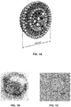
- FIG 1A A schematic illustration of SM liposomes containing iron nanoparticles embedded in the lipid membrane stabilized by the phosphates of the lipids and containing payload ( FIG 1A ) shows that the payload active agent may be in the aqueous compartment inside the liposome. However, alternatively it also may be embedded in or attached to the membrane. Because isolated iron containing particles are too small to generate enough heat in aqueous solutions under AMF (Goya et al., 2008), they have to be clustered as depicted in figure 1B . This figure presents the TEM image of SM liposomes with 5 nm Fe 3 O 4 nanoparticles. The agglomeration of the nanoparticles can be clearly seen.
- TEM imaging shows both clear association of the 5 nm nanoparticles on the membrane and large agglomerates in the membrane. The agglomerates also make the membrane to bulge. In the membrane of stained control liposomes without iron particles, smooth symmetrical contours are seen on the TEM image ( Fig 1C ).
- Fe 3 O 4 nanoparticles does not affect leakage.
- the Fe 3 O 4 nanoparticles associate on the liposomes even though they are stabilized by catechol-PEG 400. This might be due to the dissociation of the catechol PEG coating from Fe 3 O 4 nanoparticles and association of the Fe 3 O 4 nanoparticles with phosphate groups of the phospho- and sphingolipids.
- Phosphate is a known stabilizer of iron nanoparticles and in this sense the iron particles could be stabilized on the liposomal water lipid interface where the charged head groups are available without additional stabilizers like catechols.
- Binding of iron nanoparticles to the membrane might also be strengthened by the opposite surface charges of the liposome membrane and iron-nanoparticle surface. Association of iron nanoparticles on the liposomal membrane is dependent of the state of the membrane and is reversible. Particles can dissociate from the membrane if the state of the membrane changes, for example when ceramide rich micro-domains are formed in the membrane.
- PEG 400 may be used as solubilizing linker to the stabilizer catechol with a total MW of 760.
- This stabilizer linker complex is one order of magnitude smaller than the known efficient steric stabilizers.
- catechols are more unstable than nitrocatechols and can dissociate from nanoparticles. These two facts allow the particle to interact with the surrounding phosphates from the phospholipids.
- the nanoparticles that have catechol DOPA and PEGs shorter that 1000 Da have a tendency to loosely associate with phospholipids when DOPA PEG can be depleted and this may help the nanoparticles to agglomerate at the membrane (Isa et al., 2010, and Goldmann et al., 2010).
- the more stable dispersants such as nitro-DOPA-PEG5000 provide a dense, thin layer which is sufficiently thick to prevent nanoparticle agglomeration.
- PEGs from 5000 Da to 10 000 Da make the most stable nanoparticles that do not allow agglomeration and interaction of the iron nanoparticle with the lipid film.
- Reimhult et al. (2012) used Fe-particles of the roughly the same size as in the present invention but they used magnetic nanoparticles, which are stabilized using nitrocatechols with high-affinity palmitoyl-anchors to establish a thin but very dense hydrophobic coating, which can be incorporated into a membrane.
- Nitrocatechols are more stable and form less radicals than catechols (Amstad et al., 2009, and Amstad et al., 2010). However, this nitrocatechol bonding and anchoring is extremely stable and almost irreversible and makes the nanoparticle elimination from the body extremely difficult because the palmitoyl anchors can anchor the complex to any membrane.
- SMs Commonly occurring SMs were used, which can be in a bilayer membrane of liposomes such as 16:0 SM, 16:0 SM (d18:1/16:0), 17:0 SM (d18:1/17:0), 18:0 SM (d18:1/18:0).
- the lipids used have been widely tested in clinical settings and thus are acceptable for clinical translation.
- the SM liposomes containing drugs, ICG and iron nanoparticles were stable and the encapsulation efficiency was generally good.
- Doxorubicine 0.5 mg/ml was encapsulated using a pH gradient and the encapsulation efficiency was high, as could be seen from the gel filtration elution curves.
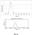
- a typical elution curve from a PD-10 column for DSPC:SM:DOTAP:chol (20:30:20:30) liposomes with Fe 3 O 4 nanoparticles (coated with catechol-PEG400 and carboxy group) fraction number three contained the intact liposomes and was collected for further use ( Fig 2A ).
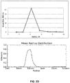
- Cisplatin (4 mg/ml) encapsulation was performed by combining the drug with the lipids when the lipids and cisplatin were hydrated. Fraction number three contained the intact liposomes and was collected for further use ( Fig 2B ) Paclitaxel (0.7 mg/ml) was encapsulated by dissolving paclitaxel in the lipid mix when in chloroform. Fraction number three contained the intact liposomes and was collected for further use ( Fig 2C ). ICG (0.5 mg/ml) was encapsulated with a similar strategy and added after the formation of the lipid film. Fraction number three contained the intact liposomes and was collected for further use ( Fig 2D ). Homogeneity and size distribution were checked by using dynamic light scattering (DLS).
- DLS dynamic light scattering
- SM DSPC liposomes can be used to keep the liposomes always intact in physiological situations. Incorporation of iron nanoparticles makes them vulnerable only in case of exposure to magnetic fields that induce a phase transition. In this case, the initial sensitivity to low SMase concentrations may be reduced but better control of the release could be achieved.
- the liposomes of or used in the present invention are not thermosensitive.
- the term "not thermosensitive”, as used herein, preferably means that the liposomes are not sensitive (i.e. are stable, in particular in the sense that no leakage of, i.e., drugs or other active agents, occurs) in the typical range of body temperatures (e.g., 36-38°C or 36.5-37.5°C).
- Liposomes that are composed of lipids that have T m of 55 °C, DSPC of 41 °C sphingomyelin and of 4 °C cholesterol DOTAP are rigid and non-thermosensitive. They are also stable, with a shelf-life of several weeks. If not exposed to SMase (e.g., aSMase), phase transitions of liposomes with, e.g., sphingomyelin, cholesterol, phosphatidylcholine and DOTAP occur between 60-75°C, depending on the SM and helper lipids (i.e. all lipids except SM) concentrations. These such liposomes are not thermosensitive.
- SMase e.g., aSMase
- phase transitions of liposomes with, e.g., sphingomyelin, cholesterol, phosphatidylcholine and DOTAP occur between 60-75°C, depending on the SM and help
- the liposomes of this rigidity do not leak either, even under AMF exposure.
- heat induced either directly or via ultrasound, or other thermal device would work as well as AMF, but applying heat to inner parts of the body in a controlled selective manner is challenging.
- the SM liposomes described here permit permeabilization even at low energy AMF (i.e. energies that do not increase the tissue temperature over 40 °C). Thus this avoids heat and other damage (e.g. by exposure to higher levels of ionizing radiation to induce higher levels of SMase).
- ICG fluorescence The level and spectral distribution of ICG fluorescence is dependent on ICG concentration and the local environment. In vitro, ICG fluorescence is brightest at a concentration of 80 ⁇ g/ml and above that the fluorescence decreases rapidly when concentration increases (Mordon et al., 1998). In the experiments presented here the liposomal ICG concentration was 750 ⁇ g/ml, but the actual local concentration was several fold higher because the ICG was trapped in liposomes and possibly in the liposomal bilayer. This leads to an efficient quenching in cationic liposomes (Hua et al., 2012).
- the doxorubicine fluorescence increases 10-fold when released from liposomes. Doxorubicine fluorescence can be used to monitor the release (Kheirolomoom et al., 2010).
- Such monitoring can be very informative when performed during surgery: intra operative imaging using near infrared markers such as ICG encapsulated in SM liposomes may be used to detect tumor tissue in order to verify successful surgery, i.e. complete removal of all tumor tissue. These optical signals can also be helpful as input for steering robot surgery devices.
- SMase treatment can release the molecules entrapped in the lumen of liposomes.
- a liposomal hairpin test was carried out.
- a dose dependent increase of the fluorescent signal associated with SMase exposure was observed when hairpin DNA was liberated from liposomes.
- Figure 4A Figure 4B demonstrates that there is leakage from SM containing liposomes when SMase concentration reaches 0.5 U/ml ( Figure 4A).
- Figure 4B demonstrates that the level of aSMase required for permeation of SM liposomes can be produced by cells, in this case human aortic endothelial cells (HAoEC), exposed to levels of ionizing radiation as applied in clinical conditions like in cancer radiation therapy.
- HAoEC human aortic endothelial cells
- the increase of the power absorption is dependent on the nanoparticle concentration in the liposomal environment. Still there is a limit: in a lipid membrane there is only space for limited amount of the agglomerated particles and this defines the upper limit for the SPA values. If more particles are added to the membrane they just are not optimally distributed to the membrane and that results in lower SPA values.
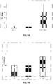
- Figure 7A depicts results for the doxorubicine liposomal formulation for an AMF treatment at 5 mT with 229 kHz
- Figure 7B shows similar results for the cisplatin liposomal formulation with an AMF treatment at 23 mT with 823 kHz. Additional exposure to 0.8 U/ml of SMase led to an increase in the release rate, documenting that relatively low magnetic field strength may be sufficient, which may facilitate clinical application.
- Fig. 6 shows that 30 min incubation with 0.8 U/ml SMase is not enough to release the content alone, but the additional exposure to AMF leads to a leakage rate of more than 50%.
- mice were injected with the same liposomal composition but not subjected to AMF treatment.
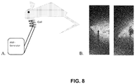
- the leaked ICG could be clearly seen in the tumor area under the chin of the AMF treated mouse ( Fig 8B right) but not on the control mouse ( Fig 8B left).
- This phenomenon with ICG quenching when encapsulated in liposomes is shown in Figure 3B .
- Lipids were purchased from Avanti Polar Lipids and SMase (from B. cereus) from Sigma Aldrich.
- Liposomes were made using a lipid mixture consisting of a total of 20 ⁇ M lipids DPPC/Cholesterol/SM/DOTAP or DSPC/cholesterol/SM/DOTAP (20:30:30:20 mol%, respectively).
- a pH-gradient was used in order to encapsulate doxorubicin into the liposomes. Lipids stored in chloroform were pipetted to a round bottomed flask, dried under nitrogen and lyophilized for 24 h to remove trace amounts of chloroform.
- Lipids were allowed to hydrate for 30 min, at 50 °C in the case of DPPC and 60 °C for DSPC, in 1 ml of 250 mM (NH 4 ) 2 SO 4 , 150 mM NaCl, pH 4,1-buffer solution containing iron at a final concentration of 0.6 or 0.3 mg/ml after PD-10 purification (Fe 3 O 4 particles coated with catechol-PEG400 and carboxy group).
- the liposome solution was freeze-thawed 3 times and unilamellar liposomes were prepared with a needle tip sonicator (5x15 sec low energy on ice).
- the buffer was changed to PBS pH 6,5 by using a PD-10 column.
- Both doxorubicin (1,4 mM doxorubicin for 10 ⁇ M lipids) and liposomes were pre-heated to 50 °C if DPPC was used and to 60 °C if DSPC was used and then combined and incubated for 30 min at 50 °C or 60° C in a water bath.
- Liposomes were purified from the unbound compounds using a PD-10 column. Elution curves were established by using doxorubicin absorbance at 485 nm. Liposomes were controlled by liposome size measurements using dynamic light scattering (DLS).
- DLS dynamic light scattering
- Liposomes were made using a lipid mixture of DSPC/cholesterol/SM/DOTAP (20:30:30:20 mol%, respectively) consisting of a total of 20 ⁇ M of lipids. Lipids stored in chloroform were pipetted to a round bottomed flask, dried under nitrogen and lyophilized for 2 h to remove trace amounts of chloroform. Lipids were allowed to hydrate for 30 min in 60 °C buffered water solutions containing cisplatin (9 mM) and nanoparticle iron at a final concentration of 0.6 or 0.3 mg/ml after PD-10 purification (Fe 3 O 4 particles coated with catechol-PEG400 and carboxy group). The liposome solution was freeze-thawed 10 times.
- Extrusion was performed 11 times through a 100 nm polycarbon membrane by using a small volume extruder, or unilamellar liposomes were prepared using a needle tip sonicator (4x15 sec low energy on ice). Liposomes were purified from the unbound compounds using a PD-10 column. Elution curves were obtained using cisplatin absorbance (at 301 nm in 0.1 M HCI). Liposomes were controlled by liposome size measurements using DLS.
- Liposomes were made using a lipid mixture of DSPC/cholesterol/SM/DOTAP (20:30:30:20 mol%, respectively) consisting of a total of 20 ⁇ M of lipids. Lipids stored in chloroform were pipetted to a round bottomed flask, dried under nitrogen and lyophilized for 2 h to remove trace amounts of chloroform. Paclitaxel was added to the lipids (0.7 to 1 mg) and traces of chloroform were lyophilized overnight. Lipids and paclitaxel were allowed to hydrate for 30 min at 60 °C in buffered water solutions containing Fe 3 O 4 nanoparticles (0.6 mg/ml or 0.3 mg/ml final concentration).
- Liposome solutions were freeze-thawed 7-10 times. Extrusion was performed 11 times through a 100 nm polycarbon membrane using a small volume extruder, or unilamellar liposomes were prepared using a needle tip sonicator (5x15 sec low energy on ice). Liposomes were purified from the unbound compounds using a PD-10 column. Elution curves were obtained using paclitaxel absorbance (227 nm). Liposomes were controlled by liposome size measurements using DLS.
- Liposomes were made using a lipid mixture consisting of a total of 20 ⁇ M of lipids (DSPC/Cholesterol/SM/DOTAP (20:30:30:20) in mol/mol ratios). Lipids stored in chloroform were pipetted to a round bottomed flask, dried under nitrogen and lyophilized for 2 h to remove trace amounts of chloroform. Lipids were allowed to hydrate for 30 min at 60 °C in buffered water solutions containing ICG (0.5 mg/ml) and Fe 3 O 4 nanoparticles in final concentration 0.6 or 0.3 mg/ml after PD-10 purification (Fe 3 O 4 particles coated with catechol-PEG400 and carboxy group).
- ICG 0.5 mg/ml
- Fe 3 O 4 nanoparticles in final concentration 0.6 or 0.3 mg/ml after PD-10 purification (Fe 3 O 4 particles coated with catechol-PEG400 and carboxy group).
- Extrusion was performed 11 times through a 100 nm polycarbon membrane using a small volume extruder, or unilamellar liposomes were prepared using a needle tip sonicator (3x15 sec low energy on ice). Liposomes were purified from the unbound compounds using a PD-10 column. Elution curves were obtained using ICG absorbance at 800 nm. Liposomes were controlled by liposome size measurements using DLS.
- the inventors used a coil with a magnetic field strength of 1,5 mT for in vivo experiments and the same 1,5 mT coil as well as a device with variable field strength ranging from 5-23 mT for in vitro studies.
- AMF treatments were performed for 20 min (100 kHz, 15V, 1,5 mT) for each sample containing 300-500 ⁇ l liposomes. If a sample was pretreated, SMase 0.4 U were added (final activity 0.77 U/ml) 30 minutes before applying the AMF treatment. After treatment, samples were inserted into a snake skin dialysis bag and dialyzed for 24 h against PBS, or PBS pH 6,5 in order to analyze the amount of free doxorubicin, cisplatin, paclitaxel or free ICG. After 24 h incubation both solutions inside and outside of the bag were analyzed by measuring absorbance of doxorubicin, cisplatin, paclitaxel or ICG and the leakage rate was calculated.
- AMF was also generated using instrumentation from nanoScale Biomagnetics (nB, Zaragoza, Spain). In this case, fields between 5-23 mT and 229-823 kHz were used. Different concentrations (0.15 to 1,2 mg/ml of Fe 3 O 4 nanoparticles per 13 ⁇ mol/ml lipids in PBS) and also different sizes (5 to 20 nm) of Fe 3 O 4 nanoparticles were incubated for 10 min in 5 - 23 mT in 750 ⁇ l, to study both leakage of the liposomes and magnetic hyperthermia, as well as to analyze specific power absorption values.
- aSMase detection was performed with the aSMase detection kit by Cayman Chemicals (USA) according to the instructions of the manufacturer. aSMase levels were measured 10 minutes after irradiation with 0 to 20 Gy gamma radiation.
- liposomes containing ICG fluorophores and Fe 3 O 4 nanoparticles in a volume of 150 ⁇ l were administered by tail vein injection.
- the mice were imaged with a Berthold NightOWL camera (Berthold Technologies, Bad Wildbad). Image analysis was performed with indigo software. Mice were also imaged with a fluorescent tomograph (FMT 2500, Perkin Elmer, Inc., Waltham, MA, USA) and under a surgical microscope (Moller-Wedel, Wedel, Germany).
- mice were positioned so that the tumor was inside the magnetic coil of the AMF apparatus.
- AMF (1,5 mT and 100 kHz) was applied to the tumor for 20 min.
- Mice were imaged after one hour together with the control mice that had gotten the same liposomal injection but had not been submitted to AMF.
- a clear increase in the ICG signal due to leaked ICG showed on the tumor area under the chin of the mouse.
Landscapes
- Health & Medical Sciences (AREA)
- Chemical & Material Sciences (AREA)
- Life Sciences & Earth Sciences (AREA)
- Animal Behavior & Ethology (AREA)
- General Health & Medical Sciences (AREA)
- Public Health (AREA)
- Veterinary Medicine (AREA)
- Epidemiology (AREA)
- Medicinal Chemistry (AREA)
- Pharmacology & Pharmacy (AREA)
- Engineering & Computer Science (AREA)
- Nanotechnology (AREA)
- Nuclear Medicine, Radiotherapy & Molecular Imaging (AREA)
- Bioinformatics & Cheminformatics (AREA)
- Dispersion Chemistry (AREA)
- Molecular Biology (AREA)
- Inorganic Chemistry (AREA)
- Biomedical Technology (AREA)
- Immunology (AREA)
- Radiology & Medical Imaging (AREA)
- Organic Chemistry (AREA)
- General Chemical & Material Sciences (AREA)
- Chemical Kinetics & Catalysis (AREA)
- Medicinal Preparation (AREA)
- Medicines Containing Antibodies Or Antigens For Use As Internal Diagnostic Agents (AREA)
- Pharmaceuticals Containing Other Organic And Inorganic Compounds (AREA)
Claims (14)
- Zusammensetzung, enthaltend Liposomen, wobei die Liposomen umfassen:(a) Phospholipide, wobei die Phospholipide Sphingomyelin (SM) umfassen,(b) magnetische Nanopartikel, und eines oder beide von(c) einem Bildgebungsmittel und/oder einem Bildgebungslabel und(d) mindestens einem Wirkstoff,wobei der Sphingomyelingehalt der Liposomen im Bereich von 10 mol% bis 45 mol% liegt und
wobei die Liposomen In der Lage sind, ein Bildgebungsmittel und/oder einen Wirkstoff durch die gleichzeitige Wirkung von Sphingomyelinase (SMase) und eines magnetischen Wechselfelds (AMF) in eine Zielumgebung freizusetzen. - Zusammensetzung nach Anspruch 1, wobei die magnetischen Nanopartikel einen Kerndurchmesser von 2 nm bis 10 nm aufweisen.
- Zusammensetzung nach Anspruch 1 oder 2, wobei die magnetischen Nanopartikel ein Metall, eine Metalllegierung, ein Metalloxid, ein Metallhydroxid, ein gemischtes Metalloxid, ein gemischtes Metallhydroxid, ein Metallnitrid, ein Metallsulfid, ein gemischtes Metallnitrid oder ein gemischtes Metallsulfid umfassen, wobei das Metall ausgewählt ist aus der Gruppe bestehend aus Chrom, Mangan, Eisen, Cobalt und Nickel.
- Zusammensetzung nach einem der Ansprüche 1 bis 3, wobei die Liposomen ferner mindestens ein Bildgebungsmittel, einschließlich radioaktiven Isotopen, Kontrastmittel, Fluorophore und PET-Label, umfassen.
- Zusammensetzung nach einem der Ansprüche 1 bis 4, wobei der Wirkstoff ein Zytostatikum ist.
- Zusammensetzung nach einem der Ansprüche 1 bis 5, wobei die Liposomen mindestens ein Therapeutikum, mindestens ein Bildgebungsmittel, und ein Bildgebungslabel umfassen.
- Zusammensetzung nach einem der Ansprüche 1 bis 6 zur Verwendung in der Therapie und/oder Diagnostik.
- Zusammensetzung zur Verwendung nach Anspruch 7, wobei die Verwendung in der Therapie die Verwendung der Zusammensetzung zur gezielten Abgabe und Freisetzung eines Wirkstoffs an einem Zielort einschließt und die Verwendung in der Diagnostik die Verwendung der Zusammensetzung als Bildgebungsmittel einschließt.
- Zusammensetzung nach einem der Ansprüche 1 bis 6 zur Verwendung in der Behandlung von Krebs und/oder zur Verwendung in der Diagnose von Krebs.
- Zusammensetzung zur Verwendung in einem Verfahren zum Abgeben und Freisetzen eines Wirkstoffs an einem Zielort, umfassend die folgenden Schritte:(a) Bereitstellen einer Zusammensetzung nach einem der Ansprüche 1 bis 6,(b) Verabreichen der Zusammensetzung, um die in der Zusammensetzung enthaltenen Liposomen an einem Zielort abzugeben; und(c) Anwenden von Energie auf die Liposomen und/oder Erhöhen der SMase-Aktivität an dem Zielort, um ein Freisetzen des mindestens einen Wirkstoffs zu bewirken.
- Zusammensetzung zur Verwendung nach Anspruch 10, wobei der Schritt des Anwendens von Energie auf die Liposomen zu einem Phasenübergang und/oder einer Änderung in der liposomalen Membranstruktur und/oder zu einem Erwärmen der Liposomen führt, um ein Freisetzen des mindestens einen Wirkstoffs zu bewirken.
- Zusammensetzung zur Verwendung nach Anspruch 10 oder 11, wobei in Schritt (c) die Energie durch Anwenden eines magnetischen Wechselfeldes auf den Zielort angewendet wird.
- Zusammensetzung zur Verwendung nach einem der Ansprüche 10 bis 12, wobei die SMase-Aktivität durch Verabreichen von exogener SMase oder durch Anwenden von Mitteln zur Induzierung von endogener SMase-Aktivität erhöht wird, um dadurch eine Freisetzung des mindestens einen Wirkstoffs zu bewirken.
- Zusammensetzung zur Verwendung nach Anspruch 13, wobei die Mittel zum Induzieren der endogenen SMase-Aktivität Strahlung, Aussetzen gegenüber UV-Licht, Anwendung von Wärme und Verabreichung von Chemotherapeutika oder Zellstressinduzierungsmitteln einschließt.
Applications Claiming Priority (2)
| Application Number | Priority Date | Filing Date | Title |
|---|---|---|---|
| EP14167196 | 2014-05-06 | ||
| PCT/EP2015/059933 WO2015169843A1 (en) | 2014-05-06 | 2015-05-06 | Magnetoenzymatic carrier system for imaging and targeted delivery and release of active agents |
Publications (2)
| Publication Number | Publication Date |
|---|---|
| EP3139964A1 EP3139964A1 (de) | 2017-03-15 |
| EP3139964B1 true EP3139964B1 (de) | 2019-04-24 |
Family
ID=50639345
Family Applications (1)
| Application Number | Title | Priority Date | Filing Date |
|---|---|---|---|
| EP15723866.8A Not-in-force EP3139964B1 (de) | 2014-05-06 | 2015-05-06 | Magnetoenzymatisches trägersystem zur abbildung und gezielten abgabe und freisetzung von wirkstoffen |
Country Status (4)
| Country | Link |
|---|---|
| US (1) | US20170151174A1 (de) |
| EP (1) | EP3139964B1 (de) |
| ES (1) | ES2733098T3 (de) |
| WO (1) | WO2015169843A1 (de) |
Cited By (1)
| Publication number | Priority date | Publication date | Assignee | Title |
|---|---|---|---|---|
| CN110755611A (zh) * | 2019-10-18 | 2020-02-07 | 中国药科大学 | 一种纳米簇载药热敏脂质体制剂及其制法和应用 |
Families Citing this family (9)
| Publication number | Priority date | Publication date | Assignee | Title |
|---|---|---|---|---|
| WO2017179045A1 (en) * | 2016-04-14 | 2017-10-19 | Yissum Research Development Company Of The Hebrew University Of Jerusalem | Indocyanine green formulations and methods for intraoperative localization of rectal tumors |
| WO2018222339A1 (en) * | 2017-05-29 | 2018-12-06 | Bionaut Labs Ltd. | Ultrasound resonance triggering of payload release from miniaturized devices |
| RU2693485C1 (ru) * | 2018-06-13 | 2019-07-03 | федеральное государственное автономное образовательное учреждение высшего образования "Национальный исследовательский ядерный университет МИФИ" (НИЯУ МИФИ) | Носитель для диагностики, направленной доставки и контролируемого высвобождения лекарственных средств |
| PT115474B (pt) | 2019-04-26 | 2021-04-30 | Univ Do Minho | Nanossistema magnético e método para produzir o nanossistema |
| RU196226U1 (ru) * | 2019-09-24 | 2020-02-21 | федеральное государственное автономное учреждение высшего образования "Национальный исследовательский ядерный университет "МИФИ" (НИЯУ МИФИ) | Носитель для диагностики, направленной доставки и контролируемого высвобождения лекарственных средств |
| WO2022060924A1 (en) * | 2020-09-16 | 2022-03-24 | Imagion Biosystems, Inc. | Methods and apparatuses for the synthesis of drug-loaded magnetic micelle aggregates |
| US20220105206A1 (en) * | 2020-10-02 | 2022-04-07 | Board Of Regents, The University Of Texas System | Sensitivity advances in ultrasound switchable fluorescence systems and techniques |
| CN115068630B (zh) * | 2022-08-24 | 2022-11-04 | 山东大学齐鲁医院 | 一种温敏控释四氧化三铁/pd-l1单抗修饰纳米颗粒的制备方法 |
| GB202315012D0 (en) * | 2023-09-29 | 2023-11-15 | Univ Bath | Composition |
Family Cites Families (10)
| Publication number | Priority date | Publication date | Assignee | Title |
|---|---|---|---|---|
| WO1998040049A2 (de) * | 1997-03-07 | 1998-09-17 | Max-Delbrück-Centrum für Molekulare Medizin | Spezifische magnetosomen, verfahren zu ihrer herstellung und ihre verwendung |
| WO2001056546A2 (en) * | 2000-02-05 | 2001-08-09 | Florida State University Research Foundation | Magnetoliposome composition for targeted treatment of biological tissue |
| US20030211045A1 (en) * | 2001-02-05 | 2003-11-13 | Danuta Leszcyznska | Magnetoliposome composition for targeted treatment of biological tissue and associated methods |
| EP1537858A1 (de) * | 2003-12-04 | 2005-06-08 | Vectron Therapeutics AG | Trägerstoffe zur Arzneistoffabgabe und deren Verwendung |
| WO2008073856A2 (en) * | 2006-12-08 | 2008-06-19 | Massachusetts Institute Of Technology | Delivery of nanoparticles and/or agents to cells |
| US20090004258A1 (en) | 2007-06-27 | 2009-01-01 | National Health Research Institutes | Drug Release from Thermosensitive Liposomes by Applying an Alternative Magnetic Field |
| WO2011112597A1 (en) * | 2010-03-08 | 2011-09-15 | Northeastern University | Magnetic nanoplatforms for theranostic and multi-modal imaging applications |
| EP2575896A2 (de) | 2010-05-26 | 2013-04-10 | Erik Reimhult | Magnetisch reaktive membranstrukturen |
| CN103429227B (zh) | 2011-01-31 | 2017-12-19 | 纳米生物技术公司 | 纳米粒子递送系统、其制备及应用 |
| US20140205543A1 (en) * | 2013-01-24 | 2014-07-24 | Memorial Sloan-Kettering Cancer Center | Method for diagnosing or treating tumors using sphingomyelin containing liposomes |
-
2015
- 2015-05-06 EP EP15723866.8A patent/EP3139964B1/de not_active Not-in-force
- 2015-05-06 WO PCT/EP2015/059933 patent/WO2015169843A1/en not_active Ceased
- 2015-05-06 ES ES15723866T patent/ES2733098T3/es active Active
- 2015-05-06 US US15/308,770 patent/US20170151174A1/en not_active Abandoned
Non-Patent Citations (1)
| Title |
|---|
| None * |
Cited By (1)
| Publication number | Priority date | Publication date | Assignee | Title |
|---|---|---|---|---|
| CN110755611A (zh) * | 2019-10-18 | 2020-02-07 | 中国药科大学 | 一种纳米簇载药热敏脂质体制剂及其制法和应用 |
Also Published As
| Publication number | Publication date |
|---|---|
| WO2015169843A1 (en) | 2015-11-12 |
| ES2733098T3 (es) | 2019-11-27 |
| EP3139964A1 (de) | 2017-03-15 |
| US20170151174A1 (en) | 2017-06-01 |
Similar Documents
| Publication | Publication Date | Title |
|---|---|---|
| US20170151174A1 (en) | Magnetoenzymatic carrier system for imaging and targeted delivery and release of active agents | |
| Martina et al. | The effect of magnetic targeting on the uptake of magnetic-fluid-loaded liposomes by human prostatic adenocarcinoma cells | |
| Peiris et al. | Enhanced delivery of chemotherapy to tumors using a multi-component nanochain with radiofrequency-tunable drug release | |
| Reddy et al. | Magnetic nanoparticles: design and characterization, toxicity and biocompatibility, pharmaceutical and biomedical applications | |
| CN102149414B (zh) | 提供mri对比增强的药物载体 | |
| McLoughlin et al. | Overcoming the blood–brain barrier: multifunctional nanomaterial‐based strategies for targeted drug delivery in neurological disorders | |
| RU2477146C2 (ru) | Полимерный носитель лекарственных средств для доставки под визуальным контролем | |
| Lorenzato et al. | MRI contrast variation of thermosensitive magnetoliposomes triggered by focused ultrasound: a tool for image‐guided local drug delivery | |
| Al-Ahmady et al. | Engineering thermosensitive liposome-nanoparticle hybrids loaded with doxorubicin for heat-triggered drug release | |
| US20110059020A1 (en) | Liposome composition, and diagnostic contrast agent, therapeutic enhancer, and pharmaceutical composition using the same | |
| Szuplewska et al. | Magnetic field-assisted selective delivery of doxorubicin to cancer cells using magnetoliposomes as drug nanocarriers | |
| JP2017193550A (ja) | ナノ粒子デリバリーシステム、その製造および使用 | |
| Escoffre et al. | Focused ultrasound mediated drug delivery from temperature-sensitive liposomes: in-vitro characterization and validation | |
| Huang et al. | Cellular organelle-dependent cytotoxicity of iron oxide nanoparticles and its implications for cancer diagnosis and treatment: a mechanistic investigation | |
| US20100254913A1 (en) | Non-spherical contrast agents for cest mri based on bulk magnetic susceptibility effect | |
| WO2011096230A1 (ja) | 磁性粒子、及びその製造方法、並びに磁性粒子含有製剤 | |
| WO2008033031A2 (en) | Mri trackable drug delivery particles, uses and methods thereof | |
| CN103619802B (zh) | 脂质模拟物化合物及其用途 | |
| Salatin et al. | Magnetic hybrid nanovesicles for the precise diagnosis and treatment of central nervous system disorders | |
| Sun et al. | Magnetically‐activated lipid nanocarriers in biomedical applications: A review of current status and perspective | |
| Ma et al. | Magnetic targeting after femoral artery administration and biocompatibility assessment of superparamagnetic iron oxide nanoparticles | |
| US20210106819A1 (en) | Combination Therapy of Electric Fields and an Additional Treatment for Cancer and Imaging | |
| Barmin et al. | Multifunctional ultrasound microbubbles | |
| Quarta et al. | Multilayered magnetic nanobeads for the delivery of peptides molecules triggered by intracellular proteases | |
| Zhang et al. | Application of mechanical force generated by magnetic nanoparticles under magnetic field in tumor treatment |
Legal Events
| Date | Code | Title | Description |
|---|---|---|---|
| STAA | Information on the status of an ep patent application or granted ep patent |
Free format text: STATUS: THE INTERNATIONAL PUBLICATION HAS BEEN MADE |
|
| PUAI | Public reference made under article 153(3) epc to a published international application that has entered the european phase |
Free format text: ORIGINAL CODE: 0009012 |
|
| STAA | Information on the status of an ep patent application or granted ep patent |
Free format text: STATUS: REQUEST FOR EXAMINATION WAS MADE |
|
| 17P | Request for examination filed |
Effective date: 20161118 |
|
| AK | Designated contracting states |
Kind code of ref document: A1 Designated state(s): AL AT BE BG CH CY CZ DE DK EE ES FI FR GB GR HR HU IE IS IT LI LT LU LV MC MK MT NL NO PL PT RO RS SE SI SK SM TR |
|
| AX | Request for extension of the european patent |
Extension state: BA ME |
|
| DAV | Request for validation of the european patent (deleted) | ||
| DAX | Request for extension of the european patent (deleted) | ||
| REG | Reference to a national code |
Ref country code: DE Ref legal event code: R079 Ref document number: 602015028848 Country of ref document: DE Free format text: PREVIOUS MAIN CLASS: A61K0047500000 Ipc: A61K0041000000 |
|
| GRAP | Despatch of communication of intention to grant a patent |
Free format text: ORIGINAL CODE: EPIDOSNIGR1 |
|
| STAA | Information on the status of an ep patent application or granted ep patent |
Free format text: STATUS: GRANT OF PATENT IS INTENDED |
|
| RIC1 | Information provided on ipc code assigned before grant |
Ipc: A61P 35/00 20060101ALI20181122BHEP Ipc: A61K 9/127 20060101ALI20181122BHEP Ipc: A61K 41/00 20060101AFI20181122BHEP Ipc: A61K 49/18 20060101ALI20181122BHEP Ipc: A61K 47/50 20170101ALI20181122BHEP Ipc: A61K 9/16 20060101ALI20181122BHEP |
|
| INTG | Intention to grant announced |
Effective date: 20181212 |
|
| GRAS | Grant fee paid |
Free format text: ORIGINAL CODE: EPIDOSNIGR3 |
|
| GRAA | (expected) grant |
Free format text: ORIGINAL CODE: 0009210 |
|
| STAA | Information on the status of an ep patent application or granted ep patent |
Free format text: STATUS: THE PATENT HAS BEEN GRANTED |
|
| AK | Designated contracting states |
Kind code of ref document: B1 Designated state(s): AL AT BE BG CH CY CZ DE DK EE ES FI FR GB GR HR HU IE IS IT LI LT LU LV MC MK MT NL NO PL PT RO RS SE SI SK SM TR |
|
| REG | Reference to a national code |
Ref country code: GB Ref legal event code: FG4D |
|
| REG | Reference to a national code |
Ref country code: CH Ref legal event code: EP |
|
| REG | Reference to a national code |
Ref country code: AT Ref legal event code: REF Ref document number: 1123341 Country of ref document: AT Kind code of ref document: T Effective date: 20190515 Ref country code: IE Ref legal event code: FG4D |
|
| REG | Reference to a national code |
Ref country code: DE Ref legal event code: R096 Ref document number: 602015028848 Country of ref document: DE |
|
| REG | Reference to a national code |
Ref country code: NL Ref legal event code: MP Effective date: 20190424 |
|
| REG | Reference to a national code |
Ref country code: LT Ref legal event code: MG4D |
|
| PG25 | Lapsed in a contracting state [announced via postgrant information from national office to epo] |
Ref country code: NL Free format text: LAPSE BECAUSE OF FAILURE TO SUBMIT A TRANSLATION OF THE DESCRIPTION OR TO PAY THE FEE WITHIN THE PRESCRIBED TIME-LIMIT Effective date: 20190424 |
|
| PG25 | Lapsed in a contracting state [announced via postgrant information from national office to epo] |
Ref country code: NO Free format text: LAPSE BECAUSE OF FAILURE TO SUBMIT A TRANSLATION OF THE DESCRIPTION OR TO PAY THE FEE WITHIN THE PRESCRIBED TIME-LIMIT Effective date: 20190724 Ref country code: SE Free format text: LAPSE BECAUSE OF FAILURE TO SUBMIT A TRANSLATION OF THE DESCRIPTION OR TO PAY THE FEE WITHIN THE PRESCRIBED TIME-LIMIT Effective date: 20190424 Ref country code: PT Free format text: LAPSE BECAUSE OF FAILURE TO SUBMIT A TRANSLATION OF THE DESCRIPTION OR TO PAY THE FEE WITHIN THE PRESCRIBED TIME-LIMIT Effective date: 20190824 Ref country code: AL Free format text: LAPSE BECAUSE OF FAILURE TO SUBMIT A TRANSLATION OF THE DESCRIPTION OR TO PAY THE FEE WITHIN THE PRESCRIBED TIME-LIMIT Effective date: 20190424 Ref country code: FI Free format text: LAPSE BECAUSE OF FAILURE TO SUBMIT A TRANSLATION OF THE DESCRIPTION OR TO PAY THE FEE WITHIN THE PRESCRIBED TIME-LIMIT Effective date: 20190424 Ref country code: HR Free format text: LAPSE BECAUSE OF FAILURE TO SUBMIT A TRANSLATION OF THE DESCRIPTION OR TO PAY THE FEE WITHIN THE PRESCRIBED TIME-LIMIT Effective date: 20190424 Ref country code: LT Free format text: LAPSE BECAUSE OF FAILURE TO SUBMIT A TRANSLATION OF THE DESCRIPTION OR TO PAY THE FEE WITHIN THE PRESCRIBED TIME-LIMIT Effective date: 20190424 |
|
| REG | Reference to a national code |
Ref country code: ES Ref legal event code: FG2A Ref document number: 2733098 Country of ref document: ES Kind code of ref document: T3 Effective date: 20191127 |
|
| PG25 | Lapsed in a contracting state [announced via postgrant information from national office to epo] |
Ref country code: LV Free format text: LAPSE BECAUSE OF FAILURE TO SUBMIT A TRANSLATION OF THE DESCRIPTION OR TO PAY THE FEE WITHIN THE PRESCRIBED TIME-LIMIT Effective date: 20190424 Ref country code: RS Free format text: LAPSE BECAUSE OF FAILURE TO SUBMIT A TRANSLATION OF THE DESCRIPTION OR TO PAY THE FEE WITHIN THE PRESCRIBED TIME-LIMIT Effective date: 20190424 Ref country code: BG Free format text: LAPSE BECAUSE OF FAILURE TO SUBMIT A TRANSLATION OF THE DESCRIPTION OR TO PAY THE FEE WITHIN THE PRESCRIBED TIME-LIMIT Effective date: 20190724 Ref country code: PL Free format text: LAPSE BECAUSE OF FAILURE TO SUBMIT A TRANSLATION OF THE DESCRIPTION OR TO PAY THE FEE WITHIN THE PRESCRIBED TIME-LIMIT Effective date: 20190424 Ref country code: GR Free format text: LAPSE BECAUSE OF FAILURE TO SUBMIT A TRANSLATION OF THE DESCRIPTION OR TO PAY THE FEE WITHIN THE PRESCRIBED TIME-LIMIT Effective date: 20190725 |
|
| REG | Reference to a national code |
Ref country code: AT Ref legal event code: MK05 Ref document number: 1123341 Country of ref document: AT Kind code of ref document: T Effective date: 20190424 |
|
| REG | Reference to a national code |
Ref country code: CH Ref legal event code: PL |
|
| PG25 | Lapsed in a contracting state [announced via postgrant information from national office to epo] |
Ref country code: IS Free format text: LAPSE BECAUSE OF FAILURE TO SUBMIT A TRANSLATION OF THE DESCRIPTION OR TO PAY THE FEE WITHIN THE PRESCRIBED TIME-LIMIT Effective date: 20190824 |
|
| REG | Reference to a national code |
Ref country code: DE Ref legal event code: R097 Ref document number: 602015028848 Country of ref document: DE |
|
| PG25 | Lapsed in a contracting state [announced via postgrant information from national office to epo] |
Ref country code: MC Free format text: LAPSE BECAUSE OF FAILURE TO SUBMIT A TRANSLATION OF THE DESCRIPTION OR TO PAY THE FEE WITHIN THE PRESCRIBED TIME-LIMIT Effective date: 20190424 Ref country code: CZ Free format text: LAPSE BECAUSE OF FAILURE TO SUBMIT A TRANSLATION OF THE DESCRIPTION OR TO PAY THE FEE WITHIN THE PRESCRIBED TIME-LIMIT Effective date: 20190424 Ref country code: RO Free format text: LAPSE BECAUSE OF FAILURE TO SUBMIT A TRANSLATION OF THE DESCRIPTION OR TO PAY THE FEE WITHIN THE PRESCRIBED TIME-LIMIT Effective date: 20190424 Ref country code: DK Free format text: LAPSE BECAUSE OF FAILURE TO SUBMIT A TRANSLATION OF THE DESCRIPTION OR TO PAY THE FEE WITHIN THE PRESCRIBED TIME-LIMIT Effective date: 20190424 Ref country code: AT Free format text: LAPSE BECAUSE OF FAILURE TO SUBMIT A TRANSLATION OF THE DESCRIPTION OR TO PAY THE FEE WITHIN THE PRESCRIBED TIME-LIMIT Effective date: 20190424 Ref country code: EE Free format text: LAPSE BECAUSE OF FAILURE TO SUBMIT A TRANSLATION OF THE DESCRIPTION OR TO PAY THE FEE WITHIN THE PRESCRIBED TIME-LIMIT Effective date: 20190424 Ref country code: CH Free format text: LAPSE BECAUSE OF NON-PAYMENT OF DUE FEES Effective date: 20190531 Ref country code: LI Free format text: LAPSE BECAUSE OF NON-PAYMENT OF DUE FEES Effective date: 20190531 Ref country code: SK Free format text: LAPSE BECAUSE OF FAILURE TO SUBMIT A TRANSLATION OF THE DESCRIPTION OR TO PAY THE FEE WITHIN THE PRESCRIBED TIME-LIMIT Effective date: 20190424 |
|
| REG | Reference to a national code |
Ref country code: BE Ref legal event code: MM Effective date: 20190531 |
|
| PG25 | Lapsed in a contracting state [announced via postgrant information from national office to epo] |
Ref country code: LU Free format text: LAPSE BECAUSE OF NON-PAYMENT OF DUE FEES Effective date: 20190506 Ref country code: SM Free format text: LAPSE BECAUSE OF FAILURE TO SUBMIT A TRANSLATION OF THE DESCRIPTION OR TO PAY THE FEE WITHIN THE PRESCRIBED TIME-LIMIT Effective date: 20190424 |
|
| PLBE | No opposition filed within time limit |
Free format text: ORIGINAL CODE: 0009261 |
|
| STAA | Information on the status of an ep patent application or granted ep patent |
Free format text: STATUS: NO OPPOSITION FILED WITHIN TIME LIMIT |
|
| PG25 | Lapsed in a contracting state [announced via postgrant information from national office to epo] |
Ref country code: TR Free format text: LAPSE BECAUSE OF FAILURE TO SUBMIT A TRANSLATION OF THE DESCRIPTION OR TO PAY THE FEE WITHIN THE PRESCRIBED TIME-LIMIT Effective date: 20190424 |
|
| 26N | No opposition filed |
Effective date: 20200127 |
|
| PG25 | Lapsed in a contracting state [announced via postgrant information from national office to epo] |
Ref country code: IE Free format text: LAPSE BECAUSE OF NON-PAYMENT OF DUE FEES Effective date: 20190506 |
|
| PG25 | Lapsed in a contracting state [announced via postgrant information from national office to epo] |
Ref country code: BE Free format text: LAPSE BECAUSE OF NON-PAYMENT OF DUE FEES Effective date: 20190531 Ref country code: SI Free format text: LAPSE BECAUSE OF FAILURE TO SUBMIT A TRANSLATION OF THE DESCRIPTION OR TO PAY THE FEE WITHIN THE PRESCRIBED TIME-LIMIT Effective date: 20190424 |
|
| PG25 | Lapsed in a contracting state [announced via postgrant information from national office to epo] |
Ref country code: CY Free format text: LAPSE BECAUSE OF FAILURE TO SUBMIT A TRANSLATION OF THE DESCRIPTION OR TO PAY THE FEE WITHIN THE PRESCRIBED TIME-LIMIT Effective date: 20190424 |
|
| PG25 | Lapsed in a contracting state [announced via postgrant information from national office to epo] |
Ref country code: MT Free format text: LAPSE BECAUSE OF FAILURE TO SUBMIT A TRANSLATION OF THE DESCRIPTION OR TO PAY THE FEE WITHIN THE PRESCRIBED TIME-LIMIT Effective date: 20190424 Ref country code: HU Free format text: LAPSE BECAUSE OF FAILURE TO SUBMIT A TRANSLATION OF THE DESCRIPTION OR TO PAY THE FEE WITHIN THE PRESCRIBED TIME-LIMIT; INVALID AB INITIO Effective date: 20150506 |
|
| PGFP | Annual fee paid to national office [announced via postgrant information from national office to epo] |
Ref country code: GB Payment date: 20220323 Year of fee payment: 8 |
|
| PGFP | Annual fee paid to national office [announced via postgrant information from national office to epo] |
Ref country code: FR Payment date: 20220308 Year of fee payment: 8 |
|
| PG25 | Lapsed in a contracting state [announced via postgrant information from national office to epo] |
Ref country code: MK Free format text: LAPSE BECAUSE OF FAILURE TO SUBMIT A TRANSLATION OF THE DESCRIPTION OR TO PAY THE FEE WITHIN THE PRESCRIBED TIME-LIMIT Effective date: 20190424 |
|
| PGFP | Annual fee paid to national office [announced via postgrant information from national office to epo] |
Ref country code: IT Payment date: 20220412 Year of fee payment: 8 Ref country code: ES Payment date: 20220602 Year of fee payment: 8 Ref country code: DE Payment date: 20220316 Year of fee payment: 8 |
|
| REG | Reference to a national code |
Ref country code: DE Ref legal event code: R119 Ref document number: 602015028848 Country of ref document: DE |
|
| GBPC | Gb: european patent ceased through non-payment of renewal fee |
Effective date: 20230506 |
|
| PG25 | Lapsed in a contracting state [announced via postgrant information from national office to epo] |
Ref country code: IT Free format text: LAPSE BECAUSE OF NON-PAYMENT OF DUE FEES Effective date: 20230506 Ref country code: DE Free format text: LAPSE BECAUSE OF NON-PAYMENT OF DUE FEES Effective date: 20231201 Ref country code: GB Free format text: LAPSE BECAUSE OF NON-PAYMENT OF DUE FEES Effective date: 20230506 |
|
| PG25 | Lapsed in a contracting state [announced via postgrant information from national office to epo] |
Ref country code: FR Free format text: LAPSE BECAUSE OF NON-PAYMENT OF DUE FEES Effective date: 20230531 |
|
| REG | Reference to a national code |
Ref country code: ES Ref legal event code: FD2A Effective date: 20240626 |
|
| PG25 | Lapsed in a contracting state [announced via postgrant information from national office to epo] |
Ref country code: ES Free format text: LAPSE BECAUSE OF NON-PAYMENT OF DUE FEES Effective date: 20230507 |
|
| PG25 | Lapsed in a contracting state [announced via postgrant information from national office to epo] |
Ref country code: ES Free format text: LAPSE BECAUSE OF NON-PAYMENT OF DUE FEES Effective date: 20230507 |