EP3127913A1 - Chromogenic and fluorogenic peptide substrates for the detection of serine protease activity - Google Patents
Chromogenic and fluorogenic peptide substrates for the detection of serine protease activity Download PDFInfo
- Publication number
- EP3127913A1 EP3127913A1 EP15002304.2A EP15002304A EP3127913A1 EP 3127913 A1 EP3127913 A1 EP 3127913A1 EP 15002304 A EP15002304 A EP 15002304A EP 3127913 A1 EP3127913 A1 EP 3127913A1
- Authority
- EP
- European Patent Office
- Prior art keywords
- substrates
- arg
- compound
- factor
- peptide
- Prior art date
- Legal status (The legal status is an assumption and is not a legal conclusion. Google has not performed a legal analysis and makes no representation as to the accuracy of the status listed.)
- Withdrawn
Links
- 239000000758 substrate Substances 0.000 title claims abstract description 102
- 230000000694 effects Effects 0.000 title claims abstract description 33
- 102000012479 Serine Proteases Human genes 0.000 title claims abstract description 32
- 108010022999 Serine Proteases Proteins 0.000 title claims abstract description 32
- 238000001514 detection method Methods 0.000 title claims abstract description 23
- 108090000765 processed proteins & peptides Proteins 0.000 title claims description 39
- 238000000034 method Methods 0.000 claims abstract description 41
- 238000009007 Diagnostic Kit Methods 0.000 claims abstract description 6
- 108090000190 Thrombin Proteins 0.000 claims description 58
- 229960004072 thrombin Drugs 0.000 claims description 57
- 150000001875 compounds Chemical class 0.000 claims description 47
- 102000035195 Peptidases Human genes 0.000 claims description 35
- 108091005804 Peptidases Proteins 0.000 claims description 35
- 239000004365 Protease Substances 0.000 claims description 33
- HSSLDCABUXLXKM-UHFFFAOYSA-N resorufin Chemical compound C1=CC(=O)C=C2OC3=CC(O)=CC=C3N=C21 HSSLDCABUXLXKM-UHFFFAOYSA-N 0.000 claims description 26
- 108010074860 Factor Xa Proteins 0.000 claims description 22
- -1 factor Xla Proteins 0.000 claims description 20
- 229940125904 compound 1 Drugs 0.000 claims description 19
- 239000008280 blood Substances 0.000 claims description 18
- 210000002381 plasma Anatomy 0.000 claims description 17
- 210000004369 blood Anatomy 0.000 claims description 16
- 150000003839 salts Chemical class 0.000 claims description 11
- 238000012360 testing method Methods 0.000 claims description 10
- 201000010099 disease Diseases 0.000 claims description 8
- 208000037265 diseases, disorders, signs and symptoms Diseases 0.000 claims description 8
- 102100027612 Kallikrein-11 Human genes 0.000 claims description 7
- 108090000631 Trypsin Proteins 0.000 claims description 7
- 102000004142 Trypsin Human genes 0.000 claims description 7
- 101710152431 Trypsin-like protease Proteins 0.000 claims description 7
- 229940125782 compound 2 Drugs 0.000 claims description 7
- 239000000126 substance Substances 0.000 claims description 7
- 239000012588 trypsin Substances 0.000 claims description 7
- 108010054265 Factor VIIa Proteins 0.000 claims description 6
- 108060005986 Granzyme Proteins 0.000 claims description 6
- 102000001398 Granzyme Human genes 0.000 claims description 6
- 102000001399 Kallikrein Human genes 0.000 claims description 6
- 108060005987 Kallikrein Proteins 0.000 claims description 6
- 108010091175 Matriptase Proteins 0.000 claims description 6
- 108010067372 Pancreatic elastase Proteins 0.000 claims description 6
- 102000016387 Pancreatic elastase Human genes 0.000 claims description 6
- 102100037942 Suppressor of tumorigenicity 14 protein Human genes 0.000 claims description 6
- 108060005989 Tryptase Proteins 0.000 claims description 6
- 102000001400 Tryptase Human genes 0.000 claims description 6
- 229940012414 factor viia Drugs 0.000 claims description 6
- 230000003287 optical effect Effects 0.000 claims description 6
- 108010048049 Factor IXa Proteins 0.000 claims description 5
- 108010088842 Fibrinolysin Proteins 0.000 claims description 5
- 108090000373 Tissue Plasminogen Activator Proteins 0.000 claims description 5
- 102000003978 Tissue Plasminogen Activator Human genes 0.000 claims description 5
- 230000002159 abnormal effect Effects 0.000 claims description 5
- 230000000295 complement effect Effects 0.000 claims description 5
- 229940012957 plasmin Drugs 0.000 claims description 5
- 229960000187 tissue plasminogen activator Drugs 0.000 claims description 5
- 210000001519 tissue Anatomy 0.000 claims description 4
- 108090000317 Chymotrypsin Proteins 0.000 claims description 3
- 108010071241 Factor XIIa Proteins 0.000 claims description 3
- 108010080805 Factor XIa Proteins 0.000 claims description 3
- NTUPOKHATNSWCY-JYJNAYRXSA-N Phe-Pro-Arg Chemical compound C([C@H](N)C(=O)N1[C@@H](CCC1)C(=O)N[C@@H](CCCN=C(N)N)C(O)=O)C1=CC=CC=C1 NTUPOKHATNSWCY-JYJNAYRXSA-N 0.000 claims description 3
- 101800004937 Protein C Proteins 0.000 claims description 3
- 102000017975 Protein C Human genes 0.000 claims description 3
- 101800001700 Saposin-D Proteins 0.000 claims description 3
- 229960002376 chymotrypsin Drugs 0.000 claims description 3
- 238000003745 diagnosis Methods 0.000 claims description 3
- 229960000856 protein c Drugs 0.000 claims description 3
- 210000002966 serum Anatomy 0.000 claims description 3
- 210000002700 urine Anatomy 0.000 claims description 3
- 125000001433 C-terminal amino-acid group Chemical group 0.000 claims description 2
- 206010036790 Productive cough Diseases 0.000 claims description 2
- 210000004027 cell Anatomy 0.000 claims description 2
- 210000001175 cerebrospinal fluid Anatomy 0.000 claims description 2
- 230000013872 defecation Effects 0.000 claims description 2
- 239000012530 fluid Substances 0.000 claims description 2
- 229940100689 human protein c Drugs 0.000 claims description 2
- 108010054213 protease C1 Proteins 0.000 claims description 2
- 210000003296 saliva Anatomy 0.000 claims description 2
- 210000000582 semen Anatomy 0.000 claims description 2
- 210000003802 sputum Anatomy 0.000 claims description 2
- 208000024794 sputum Diseases 0.000 claims description 2
- 239000007850 fluorescent dye Substances 0.000 abstract description 14
- 239000003593 chromogenic compound Substances 0.000 abstract description 8
- 238000002405 diagnostic procedure Methods 0.000 abstract description 2
- 235000019419 proteases Nutrition 0.000 description 27
- YBSJFWOBGCMAKL-UHFFFAOYSA-N dabigatran Chemical compound N=1C2=CC(C(=O)N(CCC(O)=O)C=3N=CC=CC=3)=CC=C2N(C)C=1CNC1=CC=C(C(N)=N)C=C1 YBSJFWOBGCMAKL-UHFFFAOYSA-N 0.000 description 22
- UPSFMJHZUCSEHU-JYGUBCOQSA-N n-[(2s,3r,4r,5s,6r)-2-[(2r,3s,4r,5r,6s)-5-acetamido-4-hydroxy-2-(hydroxymethyl)-6-(4-methyl-2-oxochromen-7-yl)oxyoxan-3-yl]oxy-4,5-dihydroxy-6-(hydroxymethyl)oxan-3-yl]acetamide Chemical compound CC(=O)N[C@@H]1[C@@H](O)[C@H](O)[C@@H](CO)O[C@H]1O[C@H]1[C@H](O)[C@@H](NC(C)=O)[C@H](OC=2C=C3OC(=O)C=C(C)C3=CC=2)O[C@@H]1CO UPSFMJHZUCSEHU-JYGUBCOQSA-N 0.000 description 22
- 239000000523 sample Substances 0.000 description 22
- 229960003850 dabigatran Drugs 0.000 description 21
- ZMXDDKWLCZADIW-UHFFFAOYSA-N N,N-Dimethylformamide Chemical compound CN(C)C=O ZMXDDKWLCZADIW-UHFFFAOYSA-N 0.000 description 15
- 102000004190 Enzymes Human genes 0.000 description 14
- 108090000790 Enzymes Proteins 0.000 description 14
- 229940088598 enzyme Drugs 0.000 description 14
- IAZDPXIOMUYVGZ-UHFFFAOYSA-N Dimethylsulphoxide Chemical compound CS(C)=O IAZDPXIOMUYVGZ-UHFFFAOYSA-N 0.000 description 11
- 238000003556 assay Methods 0.000 description 11
- ODHCTXKNWHHXJC-VKHMYHEASA-N 5-oxo-L-proline Chemical compound OC(=O)[C@@H]1CCC(=O)N1 ODHCTXKNWHHXJC-VKHMYHEASA-N 0.000 description 10
- 125000001584 benzyloxycarbonyl group Chemical group C(=O)(OCC1=CC=CC=C1)* 0.000 description 10
- UYWQUFXKFGHYNT-UHFFFAOYSA-N phenylmethyl ester of formic acid Natural products O=COCC1=CC=CC=C1 UYWQUFXKFGHYNT-UHFFFAOYSA-N 0.000 description 10
- 239000000243 solution Substances 0.000 description 10
- HEMHJVSKTPXQMS-UHFFFAOYSA-M Sodium hydroxide Chemical compound [OH-].[Na+] HEMHJVSKTPXQMS-UHFFFAOYSA-M 0.000 description 9
- 230000015572 biosynthetic process Effects 0.000 description 9
- GLNDAGDHSLMOKX-UHFFFAOYSA-N coumarin 120 Chemical class C1=C(N)C=CC2=C1OC(=O)C=C2C GLNDAGDHSLMOKX-UHFFFAOYSA-N 0.000 description 9
- 125000005931 tert-butyloxycarbonyl group Chemical group [H]C([H])([H])C(OC(*)=O)(C([H])([H])[H])C([H])([H])[H] 0.000 description 9
- 238000006243 chemical reaction Methods 0.000 description 8
- YMWUJEATGCHHMB-UHFFFAOYSA-N Dichloromethane Chemical compound ClCCl YMWUJEATGCHHMB-UHFFFAOYSA-N 0.000 description 7
- JGFZNNIVVJXRND-UHFFFAOYSA-N N,N-Diisopropylethylamine (DIPEA) Chemical compound CCN(C(C)C)C(C)C JGFZNNIVVJXRND-UHFFFAOYSA-N 0.000 description 7
- 239000012317 TBTU Substances 0.000 description 7
- 125000001797 benzyl group Chemical group [H]C1=C([H])C([H])=C(C([H])=C1[H])C([H])([H])* 0.000 description 7
- 230000035945 sensitivity Effects 0.000 description 7
- 238000003786 synthesis reaction Methods 0.000 description 7
- BVAUMRCGVHUWOZ-ZETCQYMHSA-N (2s)-2-(cyclohexylazaniumyl)propanoate Chemical compound OC(=O)[C@H](C)NC1CCCCC1 BVAUMRCGVHUWOZ-ZETCQYMHSA-N 0.000 description 6
- QOSSAOTZNIDXMA-UHFFFAOYSA-N Dicylcohexylcarbodiimide Chemical compound C1CCCCC1N=C=NC1CCCCC1 QOSSAOTZNIDXMA-UHFFFAOYSA-N 0.000 description 6
- FAPWRFPIFSIZLT-UHFFFAOYSA-M Sodium chloride Chemical compound [Na+].[Cl-] FAPWRFPIFSIZLT-UHFFFAOYSA-M 0.000 description 6
- ZMANZCXQSJIPKH-UHFFFAOYSA-N Triethylamine Chemical compound CCN(CC)CC ZMANZCXQSJIPKH-UHFFFAOYSA-N 0.000 description 6
- 239000007983 Tris buffer Substances 0.000 description 6
- CLZISMQKJZCZDN-UHFFFAOYSA-N [benzotriazol-1-yloxy(dimethylamino)methylidene]-dimethylazanium Chemical compound C1=CC=C2N(OC(N(C)C)=[N+](C)C)N=NC2=C1 CLZISMQKJZCZDN-UHFFFAOYSA-N 0.000 description 6
- QWCKQJZIFLGMSD-UHFFFAOYSA-N alpha-aminobutyric acid Chemical group CCC(N)C(O)=O QWCKQJZIFLGMSD-UHFFFAOYSA-N 0.000 description 6
- 230000015271 coagulation Effects 0.000 description 6
- 238000005345 coagulation Methods 0.000 description 6
- 238000005259 measurement Methods 0.000 description 6
- LENZDBCJOHFCAS-UHFFFAOYSA-N tris Chemical compound OCC(N)(CO)CO LENZDBCJOHFCAS-UHFFFAOYSA-N 0.000 description 6
- AXKGIPZJYUNAIW-UHFFFAOYSA-N (4-aminophenyl)methanol Chemical compound NC1=CC=C(CO)C=C1 AXKGIPZJYUNAIW-UHFFFAOYSA-N 0.000 description 5
- ODHCTXKNWHHXJC-GSVOUGTGSA-N Pyroglutamic acid Natural products OC(=O)[C@H]1CCC(=O)N1 ODHCTXKNWHHXJC-GSVOUGTGSA-N 0.000 description 5
- 238000010521 absorption reaction Methods 0.000 description 5
- ODHCTXKNWHHXJC-UHFFFAOYSA-N acide pyroglutamique Natural products OC(=O)C1CCC(=O)N1 ODHCTXKNWHHXJC-UHFFFAOYSA-N 0.000 description 5
- 230000004913 activation Effects 0.000 description 5
- 229940024606 amino acid Drugs 0.000 description 5
- 150000001413 amino acids Chemical class 0.000 description 5
- 239000003153 chemical reaction reagent Substances 0.000 description 5
- 238000005859 coupling reaction Methods 0.000 description 5
- 230000005284 excitation Effects 0.000 description 5
- 230000007062 hydrolysis Effects 0.000 description 5
- 238000006460 hydrolysis reaction Methods 0.000 description 5
- 125000004170 methylsulfonyl group Chemical group [H]C([H])([H])S(*)(=O)=O 0.000 description 5
- 239000000203 mixture Substances 0.000 description 5
- 239000000047 product Substances 0.000 description 5
- WAMWSIDTKSNDCU-ZETCQYMHSA-N (2s)-2-azaniumyl-2-cyclohexylacetate Chemical compound OC(=O)[C@@H](N)C1CCCCC1 WAMWSIDTKSNDCU-ZETCQYMHSA-N 0.000 description 4
- 238000001327 Förster resonance energy transfer Methods 0.000 description 4
- LRQKBLKVPFOOQJ-YFKPBYRVSA-N L-norleucine Chemical compound CCCC[C@H]([NH3+])C([O-])=O LRQKBLKVPFOOQJ-YFKPBYRVSA-N 0.000 description 4
- WYURNTSHIVDZCO-UHFFFAOYSA-N Tetrahydrofuran Chemical compound C1CCOC1 WYURNTSHIVDZCO-UHFFFAOYSA-N 0.000 description 4
- 239000003146 anticoagulant agent Substances 0.000 description 4
- 229940127219 anticoagulant drug Drugs 0.000 description 4
- 239000000872 buffer Substances 0.000 description 4
- 238000011088 calibration curve Methods 0.000 description 4
- 238000003776 cleavage reaction Methods 0.000 description 4
- 239000000562 conjugate Substances 0.000 description 4
- 230000008878 coupling Effects 0.000 description 4
- 238000010168 coupling process Methods 0.000 description 4
- 239000003112 inhibitor Substances 0.000 description 4
- 239000002105 nanoparticle Substances 0.000 description 4
- 238000006862 quantum yield reaction Methods 0.000 description 4
- FSYKKLYZXJSNPZ-UHFFFAOYSA-N sarcosine Chemical group C[NH2+]CC([O-])=O FSYKKLYZXJSNPZ-UHFFFAOYSA-N 0.000 description 4
- 230000007017 scission Effects 0.000 description 4
- 230000001225 therapeutic effect Effects 0.000 description 4
- XLYOFNOQVPJJNP-UHFFFAOYSA-N water Substances O XLYOFNOQVPJJNP-UHFFFAOYSA-N 0.000 description 4
- BGOFOVHACSQSIE-ZDUSSCGKSA-N (2s)-5-[bis[(2-methylpropan-2-yl)oxycarbonylamino]methylideneamino]-2-[(2-methylpropan-2-yl)oxycarbonylamino]pentanoic acid Chemical compound CC(C)(C)OC(=O)N[C@H](C(O)=O)CCCNC(NC(=O)OC(C)(C)C)=NC(=O)OC(C)(C)C BGOFOVHACSQSIE-ZDUSSCGKSA-N 0.000 description 3
- 125000004080 3-carboxypropanoyl group Chemical group O=C([*])C([H])([H])C([H])([H])C(O[H])=O 0.000 description 3
- JMRQLMOQNRXVDP-UHFFFAOYSA-N 5-(diaminomethylideneamino)-2-[[1-(5-oxopyrrolidine-2-carbonyl)pyrrolidine-2-carbonyl]amino]pentanoic acid Chemical compound NC(N)=NCCCC(C(O)=O)NC(=O)C1CCCN1C(=O)C1NC(=O)CC1 JMRQLMOQNRXVDP-UHFFFAOYSA-N 0.000 description 3
- XTHFKEDIFFGKHM-UHFFFAOYSA-N Dimethoxyethane Chemical compound COCCOC XTHFKEDIFFGKHM-UHFFFAOYSA-N 0.000 description 3
- 108010016626 Dipeptides Proteins 0.000 description 3
- KCXVZYZYPLLWCC-UHFFFAOYSA-N EDTA Chemical compound OC(=O)CN(CC(O)=O)CCN(CC(O)=O)CC(O)=O KCXVZYZYPLLWCC-UHFFFAOYSA-N 0.000 description 3
- 208000032843 Hemorrhage Diseases 0.000 description 3
- 108010000499 Thromboplastin Proteins 0.000 description 3
- 102000002262 Thromboplastin Human genes 0.000 description 3
- GPDHNZNLPKYHCN-DZOOLQPHSA-N [[(z)-(1-cyano-2-ethoxy-2-oxoethylidene)amino]oxy-morpholin-4-ylmethylidene]-dimethylazanium;hexafluorophosphate Chemical compound F[P-](F)(F)(F)(F)F.CCOC(=O)C(\C#N)=N/OC(=[N+](C)C)N1CCOCC1 GPDHNZNLPKYHCN-DZOOLQPHSA-N 0.000 description 3
- 238000002835 absorbance Methods 0.000 description 3
- 238000000862 absorption spectrum Methods 0.000 description 3
- 208000034158 bleeding Diseases 0.000 description 3
- 230000000740 bleeding effect Effects 0.000 description 3
- 230000003197 catalytic effect Effects 0.000 description 3
- 230000008859 change Effects 0.000 description 3
- 238000005660 chlorination reaction Methods 0.000 description 3
- MGNCLNQXLYJVJD-UHFFFAOYSA-N cyanuric chloride Chemical compound ClC1=NC(Cl)=NC(Cl)=N1 MGNCLNQXLYJVJD-UHFFFAOYSA-N 0.000 description 3
- KJOZJSGOIJQCGA-UHFFFAOYSA-N dichloromethane;2,2,2-trifluoroacetic acid Chemical compound ClCCl.OC(=O)C(F)(F)F KJOZJSGOIJQCGA-UHFFFAOYSA-N 0.000 description 3
- 229940079593 drug Drugs 0.000 description 3
- 239000003814 drug Substances 0.000 description 3
- 238000000295 emission spectrum Methods 0.000 description 3
- 238000006911 enzymatic reaction Methods 0.000 description 3
- 238000011533 pre-incubation Methods 0.000 description 3
- 102000004196 processed proteins & peptides Human genes 0.000 description 3
- KGFYHTZWPPHNLQ-AWEZNQCLSA-N rivaroxaban Chemical compound S1C(Cl)=CC=C1C(=O)NC[C@@H]1OC(=O)N(C=2C=CC(=CC=2)N2C(COCC2)=O)C1 KGFYHTZWPPHNLQ-AWEZNQCLSA-N 0.000 description 3
- 239000011780 sodium chloride Substances 0.000 description 3
- 229960003766 thrombin (human) Drugs 0.000 description 3
- ZYJPUMXJBDHSIF-LLVKDONJSA-N (2r)-2-[(2-methylpropan-2-yl)oxycarbonylamino]-3-phenylpropanoic acid Chemical compound CC(C)(C)OC(=O)N[C@@H](C(O)=O)CC1=CC=CC=C1 ZYJPUMXJBDHSIF-LLVKDONJSA-N 0.000 description 2
- OYIFNHCXNCRBQI-UHFFFAOYSA-N 2-aminoadipic acid Chemical compound OC(=O)C(N)CCCC(O)=O OYIFNHCXNCRBQI-UHFFFAOYSA-N 0.000 description 2
- SNDPXSYFESPGGJ-UHFFFAOYSA-N 2-aminopentanoic acid Chemical compound CCCC(N)C(O)=O SNDPXSYFESPGGJ-UHFFFAOYSA-N 0.000 description 2
- TYMLOMAKGOJONV-UHFFFAOYSA-N 4-nitroaniline Chemical compound NC1=CC=C([N+]([O-])=O)C=C1 TYMLOMAKGOJONV-UHFFFAOYSA-N 0.000 description 2
- PAYRUJLWNCNPSJ-UHFFFAOYSA-N Aniline Chemical compound NC1=CC=CC=C1 PAYRUJLWNCNPSJ-UHFFFAOYSA-N 0.000 description 2
- 108010039627 Aprotinin Proteins 0.000 description 2
- 239000004475 Arginine Substances 0.000 description 2
- WVDDGKGOMKODPV-UHFFFAOYSA-N Benzyl alcohol Chemical compound OCC1=CC=CC=C1 WVDDGKGOMKODPV-UHFFFAOYSA-N 0.000 description 2
- 102000015081 Blood Coagulation Factors Human genes 0.000 description 2
- 108010039209 Blood Coagulation Factors Proteins 0.000 description 2
- 208000024172 Cardiovascular disease Diseases 0.000 description 2
- 108010052832 Cytochromes Proteins 0.000 description 2
- 102000018832 Cytochromes Human genes 0.000 description 2
- 102000010911 Enzyme Precursors Human genes 0.000 description 2
- 108010062466 Enzyme Precursors Proteins 0.000 description 2
- LFQSCWFLJHTTHZ-UHFFFAOYSA-N Ethanol Chemical compound CCO LFQSCWFLJHTTHZ-UHFFFAOYSA-N 0.000 description 2
- 241000282326 Felis catus Species 0.000 description 2
- DHMQDGOQFOQNFH-UHFFFAOYSA-N Glycine Chemical compound NCC(O)=O DHMQDGOQFOQNFH-UHFFFAOYSA-N 0.000 description 2
- 229920000209 Hexadimethrine bromide Polymers 0.000 description 2
- 108010062374 Myoglobin Proteins 0.000 description 2
- 102000036675 Myoglobin Human genes 0.000 description 2
- 206010028980 Neoplasm Diseases 0.000 description 2
- GLUUGHFHXGJENI-UHFFFAOYSA-N Piperazine Chemical compound C1CNCCN1 GLUUGHFHXGJENI-UHFFFAOYSA-N 0.000 description 2
- 229940124158 Protease/peptidase inhibitor Drugs 0.000 description 2
- 108010094028 Prothrombin Proteins 0.000 description 2
- 102100027378 Prothrombin Human genes 0.000 description 2
- 206010061481 Renal injury Diseases 0.000 description 2
- 108010077895 Sarcosine Proteins 0.000 description 2
- VYPSYNLAJGMNEJ-UHFFFAOYSA-N Silicium dioxide Chemical compound O=[Si]=O VYPSYNLAJGMNEJ-UHFFFAOYSA-N 0.000 description 2
- UIIMBOGNXHQVGW-UHFFFAOYSA-M Sodium bicarbonate Chemical compound [Na+].OC([O-])=O UIIMBOGNXHQVGW-UHFFFAOYSA-M 0.000 description 2
- 208000007536 Thrombosis Diseases 0.000 description 2
- XSQUKJJJFZCRTK-UHFFFAOYSA-N Urea Chemical compound NC(N)=O XSQUKJJJFZCRTK-UHFFFAOYSA-N 0.000 description 2
- 125000002777 acetyl group Chemical group [H]C([H])([H])C(*)=O 0.000 description 2
- 230000029936 alkylation Effects 0.000 description 2
- 238000005804 alkylation reaction Methods 0.000 description 2
- 125000003277 amino group Chemical group 0.000 description 2
- 229940124277 aminobutyric acid Drugs 0.000 description 2
- 230000003321 amplification Effects 0.000 description 2
- 230000006907 apoptotic process Effects 0.000 description 2
- 229960004405 aprotinin Drugs 0.000 description 2
- ODKSFYDXXFIFQN-UHFFFAOYSA-N arginine Natural products OC(=O)C(N)CCCNC(N)=N ODKSFYDXXFIFQN-UHFFFAOYSA-N 0.000 description 2
- 230000008901 benefit Effects 0.000 description 2
- 125000000051 benzyloxy group Chemical group [H]C1=C([H])C([H])=C(C([H])=C1[H])C([H])([H])O* 0.000 description 2
- 230000008033 biological extinction Effects 0.000 description 2
- 230000023555 blood coagulation Effects 0.000 description 2
- 239000003114 blood coagulation factor Substances 0.000 description 2
- 230000036765 blood level Effects 0.000 description 2
- 239000007853 buffer solution Substances 0.000 description 2
- 239000004202 carbamide Substances 0.000 description 2
- 238000003271 compound fluorescence assay Methods 0.000 description 2
- 230000021615 conjugation Effects 0.000 description 2
- 230000007423 decrease Effects 0.000 description 2
- 238000010511 deprotection reaction Methods 0.000 description 2
- 238000013461 design Methods 0.000 description 2
- 238000011161 development Methods 0.000 description 2
- 238000009509 drug development Methods 0.000 description 2
- 239000000975 dye Substances 0.000 description 2
- 230000002255 enzymatic effect Effects 0.000 description 2
- 230000006870 function Effects 0.000 description 2
- BTCSSZJGUNDROE-UHFFFAOYSA-N gamma-aminobutyric acid Chemical group NCCCC(O)=O BTCSSZJGUNDROE-UHFFFAOYSA-N 0.000 description 2
- 238000011534 incubation Methods 0.000 description 2
- ZPNFWUPYTFPOJU-LPYSRVMUSA-N iniprol Chemical compound C([C@H]1C(=O)NCC(=O)NCC(=O)N[C@H]2CSSC[C@H]3C(=O)N[C@@H](CCCCN)C(=O)N[C@@H](C)C(=O)N[C@@H](CCCNC(N)=N)C(=O)N[C@H](C(N[C@H](C(=O)N[C@@H](CCCNC(N)=N)C(=O)N[C@@H](CC=4C=CC(O)=CC=4)C(=O)N[C@@H](CC=4C=CC=CC=4)C(=O)N[C@@H](CC=4C=CC(O)=CC=4)C(=O)N[C@@H](CC(N)=O)C(=O)N[C@@H](C)C(=O)N[C@@H](CCCCN)C(=O)N[C@@H](C)C(=O)NCC(=O)N[C@@H](CC(C)C)C(=O)N[C@@H](CSSC[C@H](NC(=O)[C@H](CC(O)=O)NC(=O)[C@H](CCC(O)=O)NC(=O)[C@H](C)NC(=O)[C@H](CO)NC(=O)[C@H](CCCCN)NC(=O)[C@H](CC=4C=CC=CC=4)NC(=O)[C@H](CC(N)=O)NC(=O)[C@H](CC(N)=O)NC(=O)[C@H](CCCNC(N)=N)NC(=O)[C@H](CCCCN)NC(=O)[C@H](C)NC(=O)[C@H](CCCNC(N)=N)NC2=O)C(=O)N[C@@H](CCSC)C(=O)N[C@@H](CCCNC(N)=N)C(=O)N[C@@H]([C@@H](C)O)C(=O)N[C@@H](CSSC[C@H](NC(=O)[C@H](CC=2C=CC=CC=2)NC(=O)[C@H](CC(O)=O)NC(=O)[C@H]2N(CCC2)C(=O)[C@@H](N)CCCNC(N)=N)C(=O)N[C@@H](CC(C)C)C(=O)N[C@@H](CCC(O)=O)C(=O)N2[C@@H](CCC2)C(=O)N2[C@@H](CCC2)C(=O)N[C@@H](CC=2C=CC(O)=CC=2)C(=O)N[C@@H]([C@@H](C)O)C(=O)NCC(=O)N2[C@@H](CCC2)C(=O)N3)C(=O)NCC(=O)NCC(=O)N[C@@H](C)C(O)=O)C(=O)N[C@@H](CCC(N)=O)C(=O)N[C@H](C(=O)N[C@@H](CC=2C=CC=CC=2)C(=O)N[C@H](C(=O)N1)C(C)C)[C@@H](C)O)[C@@H](C)CC)=O)[C@@H](C)CC)C1=CC=C(O)C=C1 ZPNFWUPYTFPOJU-LPYSRVMUSA-N 0.000 description 2
- 125000001160 methoxycarbonyl group Chemical group [H]C([H])([H])OC(*)=O 0.000 description 2
- 238000003199 nucleic acid amplification method Methods 0.000 description 2
- 239000000137 peptide hydrolase inhibitor Substances 0.000 description 2
- 230000036470 plasma concentration Effects 0.000 description 2
- 238000012123 point-of-care testing Methods 0.000 description 2
- BWHMMNNQKKPAPP-UHFFFAOYSA-L potassium carbonate Chemical compound [K+].[K+].[O-]C([O-])=O BWHMMNNQKKPAPP-UHFFFAOYSA-L 0.000 description 2
- 230000008569 process Effects 0.000 description 2
- 102000004169 proteins and genes Human genes 0.000 description 2
- 108090000623 proteins and genes Proteins 0.000 description 2
- 230000002797 proteolythic effect Effects 0.000 description 2
- 229940039716 prothrombin Drugs 0.000 description 2
- 238000011002 quantification Methods 0.000 description 2
- 238000010791 quenching Methods 0.000 description 2
- 230000000171 quenching effect Effects 0.000 description 2
- 239000012429 reaction media Substances 0.000 description 2
- PYWVYCXTNDRMGF-UHFFFAOYSA-N rhodamine B Chemical class [Cl-].C=12C=CC(=[N+](CC)CC)C=C2OC2=CC(N(CC)CC)=CC=C2C=1C1=CC=CC=C1C(O)=O PYWVYCXTNDRMGF-UHFFFAOYSA-N 0.000 description 2
- 229960001148 rivaroxaban Drugs 0.000 description 2
- 229940043230 sarcosine Drugs 0.000 description 2
- 238000011896 sensitive detection Methods 0.000 description 2
- 125000006850 spacer group Chemical group 0.000 description 2
- 230000002269 spontaneous effect Effects 0.000 description 2
- VZGDMQKNWNREIO-UHFFFAOYSA-N tetrachloromethane Chemical compound ClC(Cl)(Cl)Cl VZGDMQKNWNREIO-UHFFFAOYSA-N 0.000 description 2
- FYSNRJHAOHDILO-UHFFFAOYSA-N thionyl chloride Chemical compound ClS(Cl)=O FYSNRJHAOHDILO-UHFFFAOYSA-N 0.000 description 2
- DTQVDTLACAAQTR-UHFFFAOYSA-N trifluoroacetic acid Substances OC(=O)C(F)(F)F DTQVDTLACAAQTR-UHFFFAOYSA-N 0.000 description 2
- 230000001960 triggered effect Effects 0.000 description 2
- 230000007306 turnover Effects 0.000 description 2
- PGOHTUIFYSHAQG-LJSDBVFPSA-N (2S)-6-amino-2-[[(2S)-5-amino-2-[[(2S)-2-[[(2S)-2-[[(2S)-2-[[(2S)-4-amino-2-[[(2S)-2-[[(2S)-2-[[(2S)-2-[[(2S)-2-[[(2S)-5-amino-2-[[(2S)-5-amino-2-[[(2S)-2-[[(2S)-2-[[(2S)-2-[[(2S,3R)-2-[[(2S)-5-amino-2-[[(2S)-2-[[(2S)-2-[[(2S,3R)-2-[[(2S)-2-[[(2S)-2-[[(2S)-2-[[(2S)-2-[[(2S)-5-amino-2-[[(2S)-1-[(2S,3R)-2-[[(2S)-2-[[(2S)-2-[[(2R)-2-[[(2S)-2-[[(2S)-2-[[2-[[(2S)-2-[[(2S)-2-[[(2S)-2-[[(2S)-1-[(2S)-2-[[(2S)-2-[[(2S)-2-[[(2S)-2-amino-4-methylsulfanylbutanoyl]amino]-3-(1H-indol-3-yl)propanoyl]amino]-5-carbamimidamidopentanoyl]amino]propanoyl]pyrrolidine-2-carbonyl]amino]-3-methylbutanoyl]amino]-4-methylpentanoyl]amino]-4-methylpentanoyl]amino]acetyl]amino]-3-hydroxypropanoyl]amino]-4-methylpentanoyl]amino]-3-sulfanylpropanoyl]amino]-4-methylsulfanylbutanoyl]amino]-5-carbamimidamidopentanoyl]amino]-3-hydroxybutanoyl]pyrrolidine-2-carbonyl]amino]-5-oxopentanoyl]amino]-3-hydroxypropanoyl]amino]-3-hydroxypropanoyl]amino]-3-(1H-imidazol-5-yl)propanoyl]amino]-4-methylpentanoyl]amino]-3-hydroxybutanoyl]amino]-3-(1H-indol-3-yl)propanoyl]amino]-5-carbamimidamidopentanoyl]amino]-5-oxopentanoyl]amino]-3-hydroxybutanoyl]amino]-3-hydroxypropanoyl]amino]-3-carboxypropanoyl]amino]-3-hydroxypropanoyl]amino]-5-oxopentanoyl]amino]-5-oxopentanoyl]amino]-3-phenylpropanoyl]amino]-5-carbamimidamidopentanoyl]amino]-3-methylbutanoyl]amino]-4-methylpentanoyl]amino]-4-oxobutanoyl]amino]-5-carbamimidamidopentanoyl]amino]-3-(1H-indol-3-yl)propanoyl]amino]-4-carboxybutanoyl]amino]-5-oxopentanoyl]amino]hexanoic acid Chemical compound CSCC[C@H](N)C(=O)N[C@@H](Cc1c[nH]c2ccccc12)C(=O)N[C@@H](CCCNC(N)=N)C(=O)N[C@@H](C)C(=O)N1CCC[C@H]1C(=O)N[C@@H](C(C)C)C(=O)N[C@@H](CC(C)C)C(=O)N[C@@H](CC(C)C)C(=O)NCC(=O)N[C@@H](CO)C(=O)N[C@@H](CC(C)C)C(=O)N[C@@H](CS)C(=O)N[C@@H](CCSC)C(=O)N[C@@H](CCCNC(N)=N)C(=O)N[C@@H]([C@@H](C)O)C(=O)N1CCC[C@H]1C(=O)N[C@@H](CCC(N)=O)C(=O)N[C@@H](CO)C(=O)N[C@@H](CO)C(=O)N[C@@H](Cc1cnc[nH]1)C(=O)N[C@@H](CC(C)C)C(=O)N[C@@H]([C@@H](C)O)C(=O)N[C@@H](Cc1c[nH]c2ccccc12)C(=O)N[C@@H](CCCNC(N)=N)C(=O)N[C@@H](CCC(N)=O)C(=O)N[C@@H]([C@@H](C)O)C(=O)N[C@@H](CO)C(=O)N[C@@H](CC(O)=O)C(=O)N[C@@H](CO)C(=O)N[C@@H](CCC(N)=O)C(=O)N[C@@H](CCC(N)=O)C(=O)N[C@@H](Cc1ccccc1)C(=O)N[C@@H](CCCNC(N)=N)C(=O)N[C@@H](C(C)C)C(=O)N[C@@H](CC(C)C)C(=O)N[C@@H](CC(N)=O)C(=O)N[C@@H](CCCNC(N)=N)C(=O)N[C@@H](Cc1c[nH]c2ccccc12)C(=O)N[C@@H](CCC(O)=O)C(=O)N[C@@H](CCC(N)=O)C(=O)N[C@@H](CCCCN)C(O)=O PGOHTUIFYSHAQG-LJSDBVFPSA-N 0.000 description 1
- GVIXTVCDNCXXSH-CQSZACIVSA-N (2r)-5-[[amino-[(2,2,4,6,7-pentamethyl-3h-1-benzofuran-5-yl)sulfonylamino]methylidene]amino]-2-azaniumylpentanoate Chemical compound OC(=O)[C@H](N)CCCN=C(N)NS(=O)(=O)C1=C(C)C(C)=C2OC(C)(C)CC2=C1C GVIXTVCDNCXXSH-CQSZACIVSA-N 0.000 description 1
- YSRMCZDHEZNRPS-HOTGVXAUSA-N (2s)-5-(diaminomethylideneamino)-2-[[(2s)-1-[2-[(4-methylphenyl)sulfonylamino]acetyl]pyrrolidine-2-carbonyl]amino]pentanoic acid Chemical compound C1=CC(C)=CC=C1S(=O)(=O)NCC(=O)N1[C@H](C(=O)N[C@@H](CCCNC(N)=N)C(O)=O)CCC1 YSRMCZDHEZNRPS-HOTGVXAUSA-N 0.000 description 1
- FWMNVWWHGCHHJJ-SKKKGAJSSA-N 4-amino-1-[(2r)-6-amino-2-[[(2r)-2-[[(2r)-2-[[(2r)-2-amino-3-phenylpropanoyl]amino]-3-phenylpropanoyl]amino]-4-methylpentanoyl]amino]hexanoyl]piperidine-4-carboxylic acid Chemical compound C([C@H](C(=O)N[C@H](CC(C)C)C(=O)N[C@H](CCCCN)C(=O)N1CCC(N)(CC1)C(O)=O)NC(=O)[C@H](N)CC=1C=CC=CC=1)C1=CC=CC=C1 FWMNVWWHGCHHJJ-SKKKGAJSSA-N 0.000 description 1
- QTBSBXVTEAMEQO-UHFFFAOYSA-M Acetate Chemical compound CC([O-])=O QTBSBXVTEAMEQO-UHFFFAOYSA-M 0.000 description 1
- PIPTUBPKYFRLCP-NHCYSSNCSA-N Ala-Ala-Phe Chemical compound C[C@H](N)C(=O)N[C@@H](C)C(=O)N[C@H](C(O)=O)CC1=CC=CC=C1 PIPTUBPKYFRLCP-NHCYSSNCSA-N 0.000 description 1
- WEZNQZHACPSMEF-QEJZJMRPSA-N Ala-Phe-Lys Chemical compound NCCCC[C@@H](C(O)=O)NC(=O)[C@@H](NC(=O)[C@@H](N)C)CC1=CC=CC=C1 WEZNQZHACPSMEF-QEJZJMRPSA-N 0.000 description 1
- 108091023037 Aptamer Proteins 0.000 description 1
- 108010051109 Cell-Penetrating Peptides Proteins 0.000 description 1
- 102000020313 Cell-Penetrating Peptides Human genes 0.000 description 1
- VEXZGXHMUGYJMC-UHFFFAOYSA-M Chloride anion Chemical compound [Cl-] VEXZGXHMUGYJMC-UHFFFAOYSA-M 0.000 description 1
- 206010053567 Coagulopathies Diseases 0.000 description 1
- QWCKQJZIFLGMSD-GSVOUGTGSA-N D-alpha-aminobutyric acid Chemical group CC[C@@H](N)C(O)=O QWCKQJZIFLGMSD-GSVOUGTGSA-N 0.000 description 1
- 150000008574 D-amino acids Chemical class 0.000 description 1
- KDXKERNSBIXSRK-RXMQYKEDSA-N D-lysine Chemical compound NCCCC[C@@H](N)C(O)=O KDXKERNSBIXSRK-RXMQYKEDSA-N 0.000 description 1
- 108091008102 DNA aptamers Proteins 0.000 description 1
- 206010014513 Embolism arterial Diseases 0.000 description 1
- 108090000371 Esterases Proteins 0.000 description 1
- 108010074105 Factor Va Proteins 0.000 description 1
- 108010014173 Factor X Proteins 0.000 description 1
- 108010049003 Fibrinogen Proteins 0.000 description 1
- 102000008946 Fibrinogen Human genes 0.000 description 1
- JLXVRFDTDUGQEE-YFKPBYRVSA-N Gly-Arg Chemical compound NCC(=O)N[C@H](C(O)=O)CCCN=C(N)N JLXVRFDTDUGQEE-YFKPBYRVSA-N 0.000 description 1
- HQRHFUYMGCHHJS-LURJTMIESA-N Gly-Gly-Arg Chemical compound NCC(=O)NCC(=O)N[C@H](C(O)=O)CCCN=C(N)N HQRHFUYMGCHHJS-LURJTMIESA-N 0.000 description 1
- 239000004471 Glycine Substances 0.000 description 1
- HTTJABKRGRZYRN-UHFFFAOYSA-N Heparin Chemical compound OC1C(NC(=O)C)C(O)OC(COS(O)(=O)=O)C1OC1C(OS(O)(=O)=O)C(O)C(OC2C(C(OS(O)(=O)=O)C(OC3C(C(O)C(O)C(O3)C(O)=O)OS(O)(=O)=O)C(CO)O2)NS(O)(=O)=O)C(C(O)=O)O1 HTTJABKRGRZYRN-UHFFFAOYSA-N 0.000 description 1
- 102000007625 Hirudins Human genes 0.000 description 1
- 108010007267 Hirudins Proteins 0.000 description 1
- 208000008839 Kidney Neoplasms Diseases 0.000 description 1
- SNDPXSYFESPGGJ-BYPYZUCNSA-N L-2-aminopentanoic acid Chemical compound CCC[C@H](N)C(O)=O SNDPXSYFESPGGJ-BYPYZUCNSA-N 0.000 description 1
- ZGUNAGUHMKGQNY-ZETCQYMHSA-N L-alpha-phenylglycine zwitterion Chemical compound OC(=O)[C@@H](N)C1=CC=CC=C1 ZGUNAGUHMKGQNY-ZETCQYMHSA-N 0.000 description 1
- 150000008575 L-amino acids Chemical class 0.000 description 1
- CKLJMWTZIZZHCS-REOHCLBHSA-N L-aspartic acid Chemical compound OC(=O)[C@@H](N)CC(O)=O CKLJMWTZIZZHCS-REOHCLBHSA-N 0.000 description 1
- HNDVDQJCIGZPNO-YFKPBYRVSA-N L-histidine Chemical compound OC(=O)[C@@H](N)CC1=CN=CN1 HNDVDQJCIGZPNO-YFKPBYRVSA-N 0.000 description 1
- 239000004472 Lysine Substances 0.000 description 1
- KDXKERNSBIXSRK-UHFFFAOYSA-N Lysine Natural products NCCCCC(N)C(O)=O KDXKERNSBIXSRK-UHFFFAOYSA-N 0.000 description 1
- 102000018697 Membrane Proteins Human genes 0.000 description 1
- 108010052285 Membrane Proteins Proteins 0.000 description 1
- NQTADLQHYWFPDB-UHFFFAOYSA-N N-Hydroxysuccinimide Chemical compound ON1C(=O)CCC1=O NQTADLQHYWFPDB-UHFFFAOYSA-N 0.000 description 1
- LFZAGIJXANFPFN-UHFFFAOYSA-N N-[3-[4-(3-methyl-5-propan-2-yl-1,2,4-triazol-4-yl)piperidin-1-yl]-1-thiophen-2-ylpropyl]acetamide Chemical compound C(C)(C)C1=NN=C(N1C1CCN(CC1)CCC(C=1SC=CC=1)NC(C)=O)C LFZAGIJXANFPFN-UHFFFAOYSA-N 0.000 description 1
- LGMBKOAPPTYKLC-JYJNAYRXSA-N Pro-Phe-Arg Chemical compound C([C@@H](C(=O)N[C@@H](CCCNC(=N)N)C(O)=O)NC(=O)[C@H]1NCCC1)C1=CC=CC=C1 LGMBKOAPPTYKLC-JYJNAYRXSA-N 0.000 description 1
- 208000010378 Pulmonary Embolism Diseases 0.000 description 1
- KZFDRSALOYPRBE-UHFFFAOYSA-N Pyroglu-phe-lys Chemical compound C1CC(=O)NC1C(=O)NC(C(=O)NC(CCCCN)C(O)=O)CC1=CC=CC=C1 KZFDRSALOYPRBE-UHFFFAOYSA-N 0.000 description 1
- 206010038389 Renal cancer Diseases 0.000 description 1
- 206010062237 Renal impairment Diseases 0.000 description 1
- 108010083644 Ribonucleases Proteins 0.000 description 1
- 102000006382 Ribonucleases Human genes 0.000 description 1
- 229910006124 SOCl2 Inorganic materials 0.000 description 1
- MTCFGRXMJLQNBG-UHFFFAOYSA-N Serine Natural products OCC(N)C(O)=O MTCFGRXMJLQNBG-UHFFFAOYSA-N 0.000 description 1
- LSNNMFCWUKXFEE-UHFFFAOYSA-N Sulfurous acid Chemical compound OS(O)=O LSNNMFCWUKXFEE-UHFFFAOYSA-N 0.000 description 1
- 101000712605 Theromyzon tessulatum Theromin Proteins 0.000 description 1
- 229940122388 Thrombin inhibitor Drugs 0.000 description 1
- OTJMMKPMLUNTQT-AVGNSLFASA-N Val-Leu-Arg Chemical compound CC(C)C[C@@H](C(=O)N[C@@H](CCCN=C(N)N)C(=O)O)NC(=O)[C@H](C(C)C)N OTJMMKPMLUNTQT-AVGNSLFASA-N 0.000 description 1
- 206010047249 Venous thrombosis Diseases 0.000 description 1
- 208000036142 Viral infection Diseases 0.000 description 1
- 208000027418 Wounds and injury Diseases 0.000 description 1
- 230000005856 abnormality Effects 0.000 description 1
- 239000012190 activator Substances 0.000 description 1
- 150000001408 amides Chemical class 0.000 description 1
- 150000001412 amines Chemical class 0.000 description 1
- 229940030225 antihemorrhagics Drugs 0.000 description 1
- KXNPVXPOPUZYGB-XYVMCAHJSA-N argatroban Chemical compound OC(=O)[C@H]1C[C@H](C)CCN1C(=O)[C@H](CCCN=C(N)N)NS(=O)(=O)C1=CC=CC2=C1NC[C@H](C)C2 KXNPVXPOPUZYGB-XYVMCAHJSA-N 0.000 description 1
- 229960003856 argatroban Drugs 0.000 description 1
- 229940009098 aspartate Drugs 0.000 description 1
- 230000009286 beneficial effect Effects 0.000 description 1
- 108010005774 beta-Galactosidase Proteins 0.000 description 1
- 102000005936 beta-Galactosidase Human genes 0.000 description 1
- 239000000090 biomarker Substances 0.000 description 1
- 210000001124 body fluid Anatomy 0.000 description 1
- 239000010839 body fluid Substances 0.000 description 1
- 239000006227 byproduct Substances 0.000 description 1
- 201000011510 cancer Diseases 0.000 description 1
- 238000012512 characterization method Methods 0.000 description 1
- 125000001309 chloro group Chemical group Cl* 0.000 description 1
- 230000035602 clotting Effects 0.000 description 1
- 230000024203 complement activation Effects 0.000 description 1
- 210000001151 cytotoxic T lymphocyte Anatomy 0.000 description 1
- 230000006378 damage Effects 0.000 description 1
- 230000007123 defense Effects 0.000 description 1
- 230000001419 dependent effect Effects 0.000 description 1
- 230000005595 deprotonation Effects 0.000 description 1
- 238000010537 deprotonation reaction Methods 0.000 description 1
- 230000004069 differentiation Effects 0.000 description 1
- 210000002249 digestive system Anatomy 0.000 description 1
- 238000010790 dilution Methods 0.000 description 1
- 239000012895 dilution Substances 0.000 description 1
- BFMYDTVEBKDAKJ-UHFFFAOYSA-L disodium;(2',7'-dibromo-3',6'-dioxido-3-oxospiro[2-benzofuran-1,9'-xanthene]-4'-yl)mercury;hydrate Chemical compound O.[Na+].[Na+].O1C(=O)C2=CC=CC=C2C21C1=CC(Br)=C([O-])C([Hg])=C1OC1=C2C=C(Br)C([O-])=C1 BFMYDTVEBKDAKJ-UHFFFAOYSA-L 0.000 description 1
- 108010085662 ecarin Proteins 0.000 description 1
- 239000012636 effector Substances 0.000 description 1
- 230000007071 enzymatic hydrolysis Effects 0.000 description 1
- 238000006047 enzymatic hydrolysis reaction Methods 0.000 description 1
- 210000003743 erythrocyte Anatomy 0.000 description 1
- 150000002148 esters Chemical class 0.000 description 1
- VFRSADQPWYCXDG-LEUCUCNGSA-N ethyl (2s,5s)-5-methylpyrrolidine-2-carboxylate;2,2,2-trifluoroacetic acid Chemical compound OC(=O)C(F)(F)F.CCOC(=O)[C@@H]1CC[C@H](C)N1 VFRSADQPWYCXDG-LEUCUCNGSA-N 0.000 description 1
- 125000001495 ethyl group Chemical group [H]C([H])([H])C([H])([H])* 0.000 description 1
- 238000002474 experimental method Methods 0.000 description 1
- 230000004720 fertilization Effects 0.000 description 1
- 229940012952 fibrinogen Drugs 0.000 description 1
- 238000002189 fluorescence spectrum Methods 0.000 description 1
- 238000007478 fluorogenic assay Methods 0.000 description 1
- 239000011521 glass Substances 0.000 description 1
- 108010062266 glycyl-glycyl-argininal Proteins 0.000 description 1
- PCHJSUWPFVWCPO-UHFFFAOYSA-N gold Chemical compound [Au] PCHJSUWPFVWCPO-UHFFFAOYSA-N 0.000 description 1
- 239000010931 gold Substances 0.000 description 1
- 229910052737 gold Inorganic materials 0.000 description 1
- 230000012010 growth Effects 0.000 description 1
- 230000000025 haemostatic effect Effects 0.000 description 1
- 230000036541 health Effects 0.000 description 1
- 230000002439 hemostatic effect Effects 0.000 description 1
- 229960002897 heparin Drugs 0.000 description 1
- 229920000669 heparin Polymers 0.000 description 1
- 229940006607 hirudin Drugs 0.000 description 1
- WQPDUTSPKFMPDP-OUMQNGNKSA-N hirudin Chemical compound C([C@@H](C(=O)N[C@@H](CCC(O)=O)C(=O)N[C@@H](CCC(O)=O)C(=O)N[C@@H]([C@@H](C)CC)C(=O)N1[C@@H](CCC1)C(=O)N[C@@H](CCC(O)=O)C(=O)N[C@@H](CCC(O)=O)C(=O)N[C@@H](CC=1C=CC(OS(O)(=O)=O)=CC=1)C(=O)N[C@@H](CC(C)C)C(=O)N[C@@H](CCC(N)=O)C(O)=O)NC(=O)[C@H](CC(O)=O)NC(=O)CNC(=O)[C@H](CC(O)=O)NC(=O)[C@H](CC(N)=O)NC(=O)[C@H](CC=1NC=NC=1)NC(=O)[C@H](CO)NC(=O)[C@H](CCC(N)=O)NC(=O)[C@H]1N(CCC1)C(=O)[C@H](CCCCN)NC(=O)[C@H]1N(CCC1)C(=O)[C@@H](NC(=O)CNC(=O)[C@H](CCC(O)=O)NC(=O)CNC(=O)[C@@H](NC(=O)[C@@H](NC(=O)[C@H]1NC(=O)[C@H](CCC(N)=O)NC(=O)[C@H](CC(N)=O)NC(=O)[C@H](CCCCN)NC(=O)[C@H](CCC(O)=O)NC(=O)CNC(=O)[C@H](CC(O)=O)NC(=O)[C@H](CO)NC(=O)CNC(=O)[C@H](CC(C)C)NC(=O)[C@H]([C@@H](C)CC)NC(=O)[C@@H]2CSSC[C@@H](C(=O)N[C@@H](CCC(O)=O)C(=O)NCC(=O)N[C@@H](CO)C(=O)N[C@@H](CC(N)=O)C(=O)N[C@H](C(=O)N[C@H](C(NCC(=O)N[C@@H](CCC(N)=O)C(=O)NCC(=O)N[C@@H](CC(N)=O)C(=O)N[C@@H](CCCCN)C(=O)N2)=O)CSSC1)C(C)C)NC(=O)[C@H](CC(C)C)NC(=O)[C@H]1NC(=O)[C@H](CC(C)C)NC(=O)[C@H](CC(N)=O)NC(=O)[C@H](CCC(N)=O)NC(=O)CNC(=O)[C@H](CO)NC(=O)[C@H](CCC(O)=O)NC(=O)[C@H]([C@@H](C)O)NC(=O)[C@@H](NC(=O)[C@H](CC(O)=O)NC(=O)[C@@H](NC(=O)[C@H](CC=2C=CC(O)=CC=2)NC(=O)[C@@H](NC(=O)[C@@H](N)C(C)C)C(C)C)[C@@H](C)O)CSSC1)C(C)C)[C@@H](C)O)[C@@H](C)O)C1=CC=CC=C1 WQPDUTSPKFMPDP-OUMQNGNKSA-N 0.000 description 1
- 210000003630 histaminocyte Anatomy 0.000 description 1
- HNDVDQJCIGZPNO-UHFFFAOYSA-N histidine Natural products OC(=O)C(N)CC1=CN=CN1 HNDVDQJCIGZPNO-UHFFFAOYSA-N 0.000 description 1
- 239000005556 hormone Substances 0.000 description 1
- 229940088597 hormone Drugs 0.000 description 1
- 238000003384 imaging method Methods 0.000 description 1
- 230000003100 immobilizing effect Effects 0.000 description 1
- 230000001771 impaired effect Effects 0.000 description 1
- 238000000338 in vitro Methods 0.000 description 1
- 238000001727 in vivo Methods 0.000 description 1
- 238000010348 incorporation Methods 0.000 description 1
- 208000014674 injury Diseases 0.000 description 1
- 230000002452 interceptive effect Effects 0.000 description 1
- 201000010982 kidney cancer Diseases 0.000 description 1
- 208000020816 lung neoplasm Diseases 0.000 description 1
- 238000004519 manufacturing process Methods 0.000 description 1
- 239000000463 material Substances 0.000 description 1
- 239000011159 matrix material Substances 0.000 description 1
- 239000012528 membrane Substances 0.000 description 1
- QARBMVPHQWIHKH-UHFFFAOYSA-N methanesulfonyl chloride Chemical compound CS(Cl)(=O)=O QARBMVPHQWIHKH-UHFFFAOYSA-N 0.000 description 1
- BLWYXBNNBYXPPL-YFKPBYRVSA-N methyl (2s)-pyrrolidine-2-carboxylate Chemical compound COC(=O)[C@@H]1CCCN1 BLWYXBNNBYXPPL-YFKPBYRVSA-N 0.000 description 1
- 150000004702 methyl esters Chemical class 0.000 description 1
- 238000012544 monitoring process Methods 0.000 description 1
- 210000000822 natural killer cell Anatomy 0.000 description 1
- 229940127066 new oral anticoagluant drug Drugs 0.000 description 1
- 108091008104 nucleic acid aptamers Proteins 0.000 description 1
- 230000000269 nucleophilic effect Effects 0.000 description 1
- 230000004768 organ dysfunction Effects 0.000 description 1
- LVSJDHGRKAEGLX-UHFFFAOYSA-N oxolane;2,2,2-trifluoroacetic acid Chemical compound C1CCOC1.OC(=O)C(F)(F)F LVSJDHGRKAEGLX-UHFFFAOYSA-N 0.000 description 1
- BSCHIACBONPEOB-UHFFFAOYSA-N oxolane;hydrate Chemical compound O.C1CCOC1 BSCHIACBONPEOB-UHFFFAOYSA-N 0.000 description 1
- 239000000863 peptide conjugate Substances 0.000 description 1
- 125000001997 phenyl group Chemical group [H]C1=C([H])C([H])=C(*)C([H])=C1[H] 0.000 description 1
- OAHKWDDSKCRNFE-UHFFFAOYSA-N phenylmethanesulfonyl chloride Chemical compound ClS(=O)(=O)CC1=CC=CC=C1 OAHKWDDSKCRNFE-UHFFFAOYSA-N 0.000 description 1
- 125000003170 phenylsulfonyl group Chemical group C1(=CC=CC=C1)S(=O)(=O)* 0.000 description 1
- 230000035790 physiological processes and functions Effects 0.000 description 1
- HXEACLLIILLPRG-UHFFFAOYSA-N pipecolic acid Chemical compound OC(=O)C1CCCCN1 HXEACLLIILLPRG-UHFFFAOYSA-N 0.000 description 1
- 229920001184 polypeptide Polymers 0.000 description 1
- 230000002980 postoperative effect Effects 0.000 description 1
- 229910000027 potassium carbonate Inorganic materials 0.000 description 1
- 229940066336 pradaxa Drugs 0.000 description 1
- 238000002360 preparation method Methods 0.000 description 1
- 230000002265 prevention Effects 0.000 description 1
- 229940002612 prodrug Drugs 0.000 description 1
- 239000000651 prodrug Substances 0.000 description 1
- OVARTBFNCCXQKS-UHFFFAOYSA-N propan-2-one;hydrate Chemical compound O.CC(C)=O OVARTBFNCCXQKS-UHFFFAOYSA-N 0.000 description 1
- 235000019833 protease Nutrition 0.000 description 1
- 230000013777 protein digestion Effects 0.000 description 1
- 230000004844 protein turnover Effects 0.000 description 1
- 229940024999 proteolytic enzymes for treatment of wounds and ulcers Drugs 0.000 description 1
- 230000005588 protonation Effects 0.000 description 1
- 208000005069 pulmonary fibrosis Diseases 0.000 description 1
- 238000011160 research Methods 0.000 description 1
- 230000004044 response Effects 0.000 description 1
- 210000004739 secretory vesicle Anatomy 0.000 description 1
- 125000003607 serino group Chemical group [H]N([H])[C@]([H])(C(=O)[*])C(O[H])([H])[H] 0.000 description 1
- 239000000377 silicon dioxide Substances 0.000 description 1
- 229910000030 sodium bicarbonate Inorganic materials 0.000 description 1
- 238000006467 substitution reaction Methods 0.000 description 1
- 238000001356 surgical procedure Methods 0.000 description 1
- 230000002194 synthesizing effect Effects 0.000 description 1
- YLQBMQCUIZJEEH-UHFFFAOYSA-N tetrahydrofuran Natural products C=1C=COC=1 YLQBMQCUIZJEEH-UHFFFAOYSA-N 0.000 description 1
- 238000012032 thrombin generation assay Methods 0.000 description 1
- 239000003868 thrombin inhibitor Substances 0.000 description 1
- 230000001732 thrombotic effect Effects 0.000 description 1
- RIOQSEWOXXDEQQ-UHFFFAOYSA-N triphenylphosphine Chemical compound C1=CC=CC=C1P(C=1C=CC=CC=1)C1=CC=CC=C1 RIOQSEWOXXDEQQ-UHFFFAOYSA-N 0.000 description 1
- 230000001810 trypsinlike Effects 0.000 description 1
- 230000009385 viral infection Effects 0.000 description 1
- 238000012800 visualization Methods 0.000 description 1
- PJVWKTKQMONHTI-UHFFFAOYSA-N warfarin Chemical compound OC=1C2=CC=CC=C2OC(=O)C=1C(CC(=O)C)C1=CC=CC=C1 PJVWKTKQMONHTI-UHFFFAOYSA-N 0.000 description 1
- 229960005080 warfarin Drugs 0.000 description 1
- 238000013389 whole blood assay Methods 0.000 description 1
- 229940055725 xarelto Drugs 0.000 description 1
- ORQXBVXKBGUSBA-QMMMGPOBSA-N β-cyclohexyl-alanine Chemical compound OC(=O)[C@@H](N)CC1CCCCC1 ORQXBVXKBGUSBA-QMMMGPOBSA-N 0.000 description 1
Images


Classifications
-
- C—CHEMISTRY; METALLURGY
- C07—ORGANIC CHEMISTRY
- C07K—PEPTIDES
- C07K5/00—Peptides containing up to four amino acids in a fully defined sequence; Derivatives thereof
- C07K5/04—Peptides containing up to four amino acids in a fully defined sequence; Derivatives thereof containing only normal peptide links
- C07K5/08—Tripeptides
- C07K5/0815—Tripeptides with the first amino acid being basic
- C07K5/0817—Tripeptides with the first amino acid being basic the first amino acid being Arg
-
- C—CHEMISTRY; METALLURGY
- C07—ORGANIC CHEMISTRY
- C07K—PEPTIDES
- C07K5/00—Peptides containing up to four amino acids in a fully defined sequence; Derivatives thereof
- C07K5/04—Peptides containing up to four amino acids in a fully defined sequence; Derivatives thereof containing only normal peptide links
- C07K5/06—Dipeptides
- C07K5/06008—Dipeptides with the first amino acid being neutral
- C07K5/06017—Dipeptides with the first amino acid being neutral and aliphatic
- C07K5/06026—Dipeptides with the first amino acid being neutral and aliphatic the side chain containing 0 or 1 carbon atom, i.e. Gly or Ala
-
- C—CHEMISTRY; METALLURGY
- C07—ORGANIC CHEMISTRY
- C07K—PEPTIDES
- C07K5/00—Peptides containing up to four amino acids in a fully defined sequence; Derivatives thereof
- C07K5/04—Peptides containing up to four amino acids in a fully defined sequence; Derivatives thereof containing only normal peptide links
- C07K5/06—Dipeptides
- C07K5/06139—Dipeptides with the first amino acid being heterocyclic
- C07K5/06165—Dipeptides with the first amino acid being heterocyclic and Pro-amino acid; Derivatives thereof
-
- C—CHEMISTRY; METALLURGY
- C07—ORGANIC CHEMISTRY
- C07K—PEPTIDES
- C07K5/00—Peptides containing up to four amino acids in a fully defined sequence; Derivatives thereof
- C07K5/04—Peptides containing up to four amino acids in a fully defined sequence; Derivatives thereof containing only normal peptide links
- C07K5/08—Tripeptides
- C07K5/0802—Tripeptides with the first amino acid being neutral
- C07K5/0804—Tripeptides with the first amino acid being neutral and aliphatic
- C07K5/0806—Tripeptides with the first amino acid being neutral and aliphatic the side chain containing 0 or 1 carbon atoms, i.e. Gly, Ala
-
- C—CHEMISTRY; METALLURGY
- C07—ORGANIC CHEMISTRY
- C07K—PEPTIDES
- C07K5/00—Peptides containing up to four amino acids in a fully defined sequence; Derivatives thereof
- C07K5/04—Peptides containing up to four amino acids in a fully defined sequence; Derivatives thereof containing only normal peptide links
- C07K5/08—Tripeptides
- C07K5/0802—Tripeptides with the first amino acid being neutral
- C07K5/0804—Tripeptides with the first amino acid being neutral and aliphatic
- C07K5/0808—Tripeptides with the first amino acid being neutral and aliphatic the side chain containing 2 to 4 carbon atoms, e.g. Val, Ile, Leu
-
- C—CHEMISTRY; METALLURGY
- C07—ORGANIC CHEMISTRY
- C07K—PEPTIDES
- C07K5/00—Peptides containing up to four amino acids in a fully defined sequence; Derivatives thereof
- C07K5/04—Peptides containing up to four amino acids in a fully defined sequence; Derivatives thereof containing only normal peptide links
- C07K5/08—Tripeptides
- C07K5/0802—Tripeptides with the first amino acid being neutral
- C07K5/0812—Tripeptides with the first amino acid being neutral and aromatic or cycloaliphatic
-
- C—CHEMISTRY; METALLURGY
- C07—ORGANIC CHEMISTRY
- C07K—PEPTIDES
- C07K5/00—Peptides containing up to four amino acids in a fully defined sequence; Derivatives thereof
- C07K5/04—Peptides containing up to four amino acids in a fully defined sequence; Derivatives thereof containing only normal peptide links
- C07K5/08—Tripeptides
- C07K5/0815—Tripeptides with the first amino acid being basic
-
- C—CHEMISTRY; METALLURGY
- C07—ORGANIC CHEMISTRY
- C07K—PEPTIDES
- C07K5/00—Peptides containing up to four amino acids in a fully defined sequence; Derivatives thereof
- C07K5/04—Peptides containing up to four amino acids in a fully defined sequence; Derivatives thereof containing only normal peptide links
- C07K5/08—Tripeptides
- C07K5/0819—Tripeptides with the first amino acid being acidic
-
- C—CHEMISTRY; METALLURGY
- C07—ORGANIC CHEMISTRY
- C07K—PEPTIDES
- C07K5/00—Peptides containing up to four amino acids in a fully defined sequence; Derivatives thereof
- C07K5/04—Peptides containing up to four amino acids in a fully defined sequence; Derivatives thereof containing only normal peptide links
- C07K5/08—Tripeptides
- C07K5/0821—Tripeptides with the first amino acid being heterocyclic, e.g. His, Pro, Trp
- C07K5/0823—Tripeptides with the first amino acid being heterocyclic, e.g. His, Pro, Trp and Pro-amino acid; Derivatives thereof
-
- C—CHEMISTRY; METALLURGY
- C07—ORGANIC CHEMISTRY
- C07K—PEPTIDES
- C07K5/00—Peptides containing up to four amino acids in a fully defined sequence; Derivatives thereof
- C07K5/04—Peptides containing up to four amino acids in a fully defined sequence; Derivatives thereof containing only normal peptide links
- C07K5/10—Tetrapeptides
- C07K5/1002—Tetrapeptides with the first amino acid being neutral
- C07K5/1005—Tetrapeptides with the first amino acid being neutral and aliphatic
- C07K5/1008—Tetrapeptides with the first amino acid being neutral and aliphatic the side chain containing 0 or 1 carbon atoms, i.e. Gly, Ala
-
- C—CHEMISTRY; METALLURGY
- C07—ORGANIC CHEMISTRY
- C07K—PEPTIDES
- C07K5/00—Peptides containing up to four amino acids in a fully defined sequence; Derivatives thereof
- C07K5/04—Peptides containing up to four amino acids in a fully defined sequence; Derivatives thereof containing only normal peptide links
- C07K5/10—Tetrapeptides
- C07K5/1002—Tetrapeptides with the first amino acid being neutral
- C07K5/1005—Tetrapeptides with the first amino acid being neutral and aliphatic
- C07K5/101—Tetrapeptides with the first amino acid being neutral and aliphatic the side chain containing 2 to 4 carbon atoms, e.g. Val, Ile, Leu
-
- C—CHEMISTRY; METALLURGY
- C12—BIOCHEMISTRY; BEER; SPIRITS; WINE; VINEGAR; MICROBIOLOGY; ENZYMOLOGY; MUTATION OR GENETIC ENGINEERING
- C12Q—MEASURING OR TESTING PROCESSES INVOLVING ENZYMES, NUCLEIC ACIDS OR MICROORGANISMS; COMPOSITIONS OR TEST PAPERS THEREFOR; PROCESSES OF PREPARING SUCH COMPOSITIONS; CONDITION-RESPONSIVE CONTROL IN MICROBIOLOGICAL OR ENZYMOLOGICAL PROCESSES
- C12Q1/00—Measuring or testing processes involving enzymes, nucleic acids or microorganisms; Compositions therefor; Processes of preparing such compositions
- C12Q1/34—Measuring or testing processes involving enzymes, nucleic acids or microorganisms; Compositions therefor; Processes of preparing such compositions involving hydrolase
- C12Q1/37—Measuring or testing processes involving enzymes, nucleic acids or microorganisms; Compositions therefor; Processes of preparing such compositions involving hydrolase involving peptidase or proteinase
-
- G—PHYSICS
- G01—MEASURING; TESTING
- G01N—INVESTIGATING OR ANALYSING MATERIALS BY DETERMINING THEIR CHEMICAL OR PHYSICAL PROPERTIES
- G01N2333/00—Assays involving biological materials from specific organisms or of a specific nature
- G01N2333/90—Enzymes; Proenzymes
- G01N2333/914—Hydrolases (3)
- G01N2333/948—Hydrolases (3) acting on peptide bonds (3.4)
- G01N2333/95—Proteinases, i.e. endopeptidases (3.4.21-3.4.99)
- G01N2333/964—Proteinases, i.e. endopeptidases (3.4.21-3.4.99) derived from animal tissue
- G01N2333/96402—Proteinases, i.e. endopeptidases (3.4.21-3.4.99) derived from animal tissue from non-mammals
- G01N2333/96405—Proteinases, i.e. endopeptidases (3.4.21-3.4.99) derived from animal tissue from non-mammals in general
- G01N2333/96408—Proteinases, i.e. endopeptidases (3.4.21-3.4.99) derived from animal tissue from non-mammals in general with EC number
- G01N2333/96411—Serine endopeptidases (3.4.21)
Definitions
- the present invention relates to chromogenic and fluorogenic substrates that can be used for the highly sensitive and selective detection of the activity of serine proteases.
- the present invention further relates to methods for the detection of the activity of serine proteases, said methods using the substrates of the present invention.
- the present invention relates to diagnostic kits and test strips using the above substrates, as well as uses of said substrates.
- proteases There are over 500 proteases, which account for approximately 2% of all proteins in the human body. As one of the largest and most important groups of enzymes, they are involved in many physiological processes, including protein digestion and turnover, blood clotting, apoptosis, hormone activation, fertilization, and growth differentiation. A dysregulated protease activity may be associated with organ dysfunction and consequently with a disease. Therefore, there is a strong demand for the development of sensitive protease assays for proteomic research, disease diagnosis, and drug development and measurement. In addition, triggered protease activation has been applied in cascade-like amplification schemes for the highly sensitive detection of various analytes.
- kits for detection of protease activity include a chromogenic or fluorogenic substrate that releases a colored or fluorescent compound upon selective cleavage by the target protease.
- chromogenic substrates include peptide p -nitroanilides that release yellow colored p- nitroaniline with an absorbance maximum at 380 nm and a modest molar absorbance ( ⁇ ) of 13500 M -1 cm -1 .
- Peptide derivatives of 7-amino-4-methylcoumarin (AMC) are widely used fluorogenic substrates that display an increase of 460 nm fluorescence upon release of the fluorophore.
- protease subgroups are the serine proteases in which serine serves as a nucleophilic amino acid in the active site. Most serine proteases can be classified as trypsin-like proteases that utilize a catalytic triad for activity, composed of highly conserved histidine, aspartate and serine residues. Trypsins, chymotrypsins, and elastases break down polypeptides in the digestive system. Thrombin, factor Xa and other coagulation factors define the blood coagulation and complement cascades. Tryptases are major components in the secretory granules of mast cells, whereas matriptases are membrane bound proteases associated with a variety of cancers.
- Granzymes are mediators of directed apoptosis by natural killer cells and cytotoxic T-cells that play key roles in the defense against viral infection. Because of their abundance and involvement in health and disease, serine proteases and in particular trypsin-like proteases present obvious targets of therapeutic intervention.
- Tissue factor is a type-I integral membrane protein that functions as an obligate cofactor for activation of zymogen factor X by factor VIIa.
- Factor Xa (with the assistance of cofactor factor Va) then converts prothrombin to active thrombin.
- Other zymogen conversions provide both amplification and negative feedback loops that regulate thrombin production.
- Thrombin is a major therapeutic target for thrombosis and stroke intervention/prevention through indirect inhibitors such as heparin or warfarin, and direct inhibitors such as hirudin (bivalent), and argatroban (monovalent).
- indirect inhibitors such as heparin or warfarin
- direct inhibitors such as hirudin (bivalent), and argatroban (monovalent).
- thrombin is reported as a relevant player in cardiovascular disease, renal injury, and cancer.
- thrombin Concentration of thrombin in blood can vary considerably; indeed, it is not present in blood under normal conditions, but can reach low-micromolar concentrations during the coagulation process. Apart from the haemostatic process, thrombin circulates at the high-picomolar level in the blood of patients suffering from diseases known to be associated with coagulation abnormalities. A variety of methods have been developed to detect thrombin at low concentrations. The majority of these methods requires covalent conjugation of nucleic acid aptamers to nanoparticles (NPs) or electrodes and provides insufficient sensitivity in the low nanomolar range.
- NPs nanoparticles
- a simpler way to measure the enzyme activity is using short peptides conjugated to chromophores or fluorophores as cleavable substrates, wherein p -nitroanilides are the most commonly used chromogenic substrates in thrombin assays.
- Fluorogenic substrates are of a greater interest as they provide a much more sensitive read-out.
- latent fluorophores pro-fluorophores
- enzyme activities such as esterases, beta-galactosidase, proteases, ribonucleases.
- fluorogenic thrombin substrates currently on the market: 7-amino-4-methylcoumarin (AMC) and rhodamine derivatives.
- AMC is partially protonated at low pH (less than ⁇ 5) but fully deprotonated at physiological pH.
- its fluorescence spectrum is not subject to variability due to pH-dependent protonation/deprotonation when assayed near or above physiological pH.
- Rhodamine-based substrates provide better results, but because they incorporate two peptide moieties, each serves as a substrate for the enzyme and complicates the interpretation of hydrolysis kinetics.
- Chromogenic and fluorogenic substrates for detecting the activity of serine proteases known in the art usually absorb and emit light at lower wavelengths (e.g. below 570 nm), or show only weak absorption and fluorescence at higher wavelengths (e.g. at 570 nm or more). This renders the use of such substrates difficult in samples having a high optical density, and/or a high autofluorescence, such as whole blood, and/or in samples that are in contact with a surface having a high autofluorescence, and/or in samples containing biological structures that are labeled with further chromogenic and/or fluorogenic substances.
- an optically dense sample can impair excitation of the substrates and absorb emitted fluorescent light.
- substrates known in the art are often characterized by a low molar absorption ( ⁇ ) and/or low fluorescence quantum yield ( ⁇ ). Further, some substrates are not commercially available and/or are very expensive.
- the technical problem underlying the present invention is the provision of chromogenic and fluorogenic substrates for the highly sensitive and selective detection of the activity of serine proteases in a sample, wherein said substrates should be usable in samples having a high optical density and/or a high autofluorescence, and/or in samples that are in contact with a surface having a high autofluorescence, and/or in samples containing biological structures that are labeled with chromogenic and/or fluorogenic substances. Further, said substrates should be characterized by a high molar absorption and high fluorescence quantum yield, in particular at longer wavelengths.
- the present invention relates to a compound having Formula (I): wherein Peptide is a peptide or peptide derivative, or a salt of said peptide or peptide derivative.
- Peptide is a peptide or peptide derivative, preferably a tripeptide or tripeptide derivative, whose C-terminal amino acid is arginine or lysine, preferably arginine.
- the salt of the peptide or peptide derivative according to the present invention is preferably a chloride, acetate, or trifluoroacetate salt.
- the peptide or peptide derivative is selected from the group consisting of thrombin substrates, factor Xa substrates, trypsin substrates, chymotrypsin substrates, factor VIIa substrates, factor IXa substrates, factor XIa substrates, factor XIIa substrates, kallikrein substrates, plasmin substrates, tissue plasminogen activator substrates, activated protein C substrates, tryptase substrates, matriptase substrates, granzyme substrates, elastase substrates, and human complement protease C1r substrates.
- preferred thrombin substrates are D-Phe-Pro-Arg, D-Phe-HomoPro-Arg, Tos-Gly-Pro-Arg, Boc-Val-Arg, Boc-Val-Pro-Arg, Boc-Asp(O-Benzyl)-Pro-Arg, Bz-Phe-Val-Arg, Sar-Pro-Arg, Z-Gly-Gly-Arg, Z-Pro-Arg, Ethylmalonate-Gly-Arg, beta-Ala-Gly-Arg, Moc-Gly-Pro-Arg, D-CHG-Ala-Arg, D-CHG-Pro-Arg, D-CHG-But-Arg, D-CHA-Gly-Arg, and D-CHA-Ala-Arg (wherein: HomoPro is homoproline, Tos is p-toluenesulfonyl, Boc is tert- butyloxycarbonyl, Bz is benzyl, Z is Benzyloxycarbonyl, Moc is methoxy
- preferred trypsin substrates are Bz-Ile-Glu-Gly-Arg (SEQ ID NO: 3), Bz-Phe-Val-Arg, Boc-Gln-Ala-Arg, Bz-Val-Gly-Arg, Boc-Val-Pro-Arg, Boc-Glu(O-Benzyl)-Gly-Arg, Z-Gly-Gly-Arg, Z-Phe-Val-Arg, Boc-Gln-Gly-Arg, Z-Val-Gly-Arg, and Z-D-Ala-Gly-Arg (wherein Bz is benzyl, Boc is tert -butyloxycarbonyl, Bz is benzyl, and Z is benzyloxycarbonyl).
- preferred chymotrypsin substrates are Ala-Ala-Phe, Ala-Ala-Val-Ala (SEQ ID NO: 4), Glutaryl-Ala-Ala-Phe, Suc-Ala-Ala-Pro-Phe (SEQ ID NO: 5), and Suc-Gly-Gly-Phe (wherein Suc is succinyl).
- preferred factor VIIa substrates are Bz-Ile-Glu-Gly-Arg (SEQ ID NO: 6), Boc-Leu-Thr-Arg, and Mes-D-CHA-Abu-Arg (wherein Bz is benzyl, Boc is tert -butyloxycarbonyl, Mes is methanesulfonyl, CHA is cyclohexylalanine, and Abu is aminobutyric acid).
- preferred factor IXa substrates are Mes-D-CHG-Gly-Arg, and D-Leu-PHG-Arg (wherein Mes is methanesulfonyl, CHG is cyclohexylglycine, and PHG is phenylglycine).
- preferred factor XIa substrates are pyroGlu-Pro-Arg, and Z-Aad-Pro-Arg (wherein pyroGlu is pyroglutamic acid, Z is benzyloxycarbonyl, and Aad is alpha-aminoadipic acid).
- preferred factor Xlla substrates are Boc-Gln-Gly-Arg, Bz-Ile-Glu-Gly-Arg (SEQ ID NO: 6), and D-CHA-Gly-Arg (wherein Boc is tert -butyloxycarbonyl, Bz is benzyl, and CHA is cyclohexylalanine).
- preferred kallikrein substrates are Pro-Phe-Arg, D-Pro-Phe-Arg, Val-Leu-Arg, D-Val-Leu-Arg, Bz-Pro-Phe-Arg, D-Val-CHA-Arg, and D-Abu-CHA-Arg (wherein Bz is benzyl, CHA is cyclohexylalanine, and Abu is alpha-aminobutyric acid).
- preferred plasmin substrates are Gly-Arg, D-Val-Leu-Lys, D-Val-Phe-Lys, pyroGlu-Phe-Lys, Tos-Gly-Pro-Lys, D-Ile-Phe-Lys, Suc-Ala-Phe-Lys, Isovaleryl-Phe-Lys, Boc-Val-Leu-Lys, Boc-Glu-Lys-Lys, Ala-Phe-Lys, D-Ala-Leu-Lys, D-Ala-Phe-Lys, Z-Ala-Ala-Lys, D-Ala-CHA-Lys, D-But-CHA-Lys, D-Nva-CHA-Lys, and D-Nle-CHA-Lys (wherein pyroGlu is pyroglutamic acid, Tos is p -toluenesulfonyl, Suc is succinyl, Boc is
- tissue plasminogen activator substrates are D-Ile-Pro-Arg, D-Val-Gly-Arg, Z-Gly-Gly-Arg, Gly-Gly-Arg, Glutaryl-Gly-Arg, D-Val-Leu-Lys, Mes-D-CHA-Gly-Arg, Mes-D-Phe-Gly-Arg, and Mes-D-Abu-Gly-Arg (wherein Z is benzyloxycarbonyl, Mes is methanesulfonyl, and Abu is aminobutyric acid).
- preferred activated protein C substrates are pyroGlu-Pro-Arg, Boc-Leu-Ser-Thr-Arg (SEQ ID NO: 7), D-Lys(Z)-Pro-Arg, D-CHA-Pro-Arg, and pyroGlu-CHG-Arg (wherein pyroGlu is pyroglutamic acid, Boc is tert -butyloxycarbonyl, Z is benzyloxycarbonyl, CHA is cyclohexylalanine, and CHG is cyclohexylglycine.)
- preferred tryptase substrates are pyroGlu-Pro-Arg, Boc-Phe-Ser-Arg, Boc-Val-Pro-Arg, D-Leu-Thr-Arg, Tos-Gly-Pro-Lys, and Ac-Lys-Pro-Arg (wherein pyroGlu is pyroglutamic acid, Boc is tert -butyloxycarbonyl, Tos is p-toluenesulfonyl, and Ac is acetyl).
- preferred elastase substrates are Succinyl-Ala-Ala-Ala, Methoxysuccinyl-Ala-Ala-Pro-Val (SEQ ID NO: 8), Succinyl-Ala-Pro-Ala, pyroGlu-Pro-Val, and Glutaryl-Ala-Ala-Pro-Leu (SEQ ID NO: 9) (wherein pyroGlu is pyroglutamic acid).
- a preferred complement protease C1r substrate is Z-Gly-Arg (wherein Z is benzyloxycarbonyl).
- preferred matriptase substrates are Boc-Gln-Ala-Arg, and Z-Gly-Pro-Arg (wherein Boc is tert -butyloxycarbonyl, and Z is benzyloxycarbonyl).
- a preferred granzyme substrate is Z-Gly-Pro-Arg (wherein Z is benzyloxycarbonyl).
- Peptide is the peptide D-Phe-Pro-Arg (Compound 1 ) or a salt thereof.
- Peptide is the peptide derivative Benzylsulfonyl-D-Arg-Gly-Arg (Compound 2 ) or a salt thereof.
- D-Xaa wherein Xaa is any amino acid
- Xaa is any amino acid
- All other amino acids are L-amino acids.
- the rightmost amino acid in all of the above sequences is the amino acid that is bound to the amino group of the compounds of the present invention to which the group "Peptide” is bound.
- an intermediate compound is formed which spontaneously converts to the chromogenic and fluorogenic compound resorufin (7-Hydroxy-3H-phenoxazin-3-one) ( Fig. 1 ).
- Methods and means for synthesizing the compounds of the present invention are not particularly limited and are known in the art. Preferably, synthesis is achieved as indicated in the Examples of the present application.
- the present invention relates to a method for the detection of the activity of at least one serine protease in a sample, comprising the steps of contacting said sample with a compound according to the present invention, and measuring the amount of resorufin released from said compound.
- the term "detection of the activity of at least one serine protease” encompasses the qualitative and/or quantitative detection/determination of the activity of said protease.
- qualitative detection determines if activity is present or not
- quantitative detection determines protease activity in enzyme units (U or units ), defined as the amount of protease that converts one ⁇ mole substrate per minute under standard conditions, or in katal ( kat ), defined as the amount of protease that converts one mole of substrate per second under standard conditions.
- Methods for determining presence or absence of protease activity, as well as for quantitatively determining protease activity are not particularly limited and are known in the art. Quantitative determination of protease activity can for example be achieved by establishing a standard curve using samples of known protease activity and relating the samples of interest to said standard curve.
- Serine proteases that can be analyzed using the methods of the present invention are only limited by their specificity for the peptide portion of the compounds of the present invention and are known in the art.
- the serine protease is a trypsin-like protease, preferably a trypsin-like protease selected from the group consisting of trypsins, chymotrypsins, elastases, thrombin, factor VIIa, factor IXa, factor Xa, factor Xla, factor Xlla, kallikreins, plasmin, tissue plasminogen activator, activated protein C, human complement protease C1 r, tryptases, matriptases, and granzymes.
- thrombin and factor Xa are each particularly preferred.
- Substrates for use in the detection of the above proteases are preferably selected from the group consisting of the substrates indicated above.
- the trypsin-like protease whose activity is to be detected is thrombin, and the compound used in the method of the present invention is Compound 1 or a salt thereof.
- the trypsin-like protease whose activity is to be detected is factor Xa, and the compound used in the method of the present invention is Compound 2 or a salt thereof.
- Samples in which the activity of serine proteases can be determined according to the methods of the present invention are not particularly limited. However, the particular advantages of said methods are best employed in samples having a high optical density, in particular a high optical density at wavelengths of below 570 nm, and/or a high autofluorescence, in particular a high autofluorescence at wavelengths of below 570 nm.
- the sample is in contact with a surface having a high autofluorescence.
- This surface can be part of e.g . a microtiter plate, a glass or plastic cuvette, or a microscope slide or cover slip, which all can show high amounts of autofluorescence at certain wavelengths which can substantially impair excitation and/or emission of chromogenic and/or fluorogenic substances.
- the sample contains at least one biological structure that is labeled with at least one chromogenic and/or fluorogenic substance. This embodiment relates e.g. to samples in which certain markers of interest have been labeled with e.g. antibodies having a conjugated fluorochrome, which can also impair excitation and/or emission of chromogenic and/or fluorogenic substances used for detecting the activity of serine proteases.
- samples/sample materials include whole blood, serum, plasma, urine, saliva, sputum, semen, lacrimal fluid, cerebrospinal fluid, defecation, cells and tissues, wherein whole blood, plasma, serum and urine are particularly preferred.
- the methods of the present invention can be used for the detection of inhibitors of serine proteases.
- the sample is spiked with a respective serine protease and the activity thereof detected.
- the step of contacting the sample with a compound of the present invention according to the methods of the present invention is preferably performed at conditions in which the serine proteases can exert their function, i.e . in which protease activity is possible, and in which the compounds of the present invention are stable. Respective conditions are known to a person skilled in the art.
- the step of measuring the amount of resorufin released from the compounds of the present invention according to the methods of the present invention is not particularly limited and encompasses methods known in the art. Such methods include for example colorimetric and/or fluorimetric methods known in the art.
- the present invention relates to a test strip having a surface on which a compound of the present invention is immobilized.
- a protease may be immobilized on the surface of the test strip in addition to the compound of the present invention.
- the test strip of the present invention may be used for the detection of a protease inhibitor.
- Respective test strips are not particularly limited and are known in the art.
- Means for immobilizing the compounds of the present invention on the test strips are not particularly limited and are known in the art.
- the present invention relates to the compounds of the present invention according to the first aspect of the invention for use in the diagnosis of a condition or disease in a subject that is characterized by abnormal levels of at least one serine protease.
- Conditions or diseases that are characterized by abnormal levels of at least one serine protease are preferably selected from the group consisting of postoperative period with overwhelming thrombin formation and/or enhanced risk of venous thrombosis, pulmonary embolism, pulmonary fibrosis and cancers, arterial thromboembolism, cardiovascular disease, renal injury, and impaired thrombin formation predisposing patients to enhanced bleeding.
- plasma levels of free thrombin represent a promising biomarker reflecting a patient's individual hemostatic status to guide successful treatment decisions.
- the subject is a human subject.
- abnormal levels of at least one serine protease relates to levels of a respective protease that are higher or lower as compared to healthy control subjects. While normal blood levels of active thrombin are very low and difficult to detected by currently available assays, peak concentration during hip surgery may exceed 100 pM.
- the present invention relates to a method of diagnosing a condition or disease in a subject that is characterized by abnormal levels of at least one serine protease, comprising the steps of providing a sample from the subject, contacting said sample with a compound according to the present invention, and measuring the amount of resorufin (7-Hydroxy-3H-phenoxazin-3-one) released from said compound.
- the method of diagnosing according to the present invention is an in vitro method.
- the step of providing a sample from the subject is preferably expressly intended to exclude the actual obtaining of said sample.
- the present invention relates to uses of the compounds of the present invention for the detection of the activity of at least one serine protease in a sample.
- the present invention relates to a diagnostic kit, said kit comprising at least one compound of the present invention.
- said kit further comprises means for performing the methods of the present invention according to the above second and/or fifth aspect.
- said kit comprises at least one test strip of the present invention.
- means for performing the methods of the present invention according to the above second and/or fifth aspect are not particularly limited and are known in the art.
- said kit may comprise a reaction medium such as a buffer solution or lyophilized buffer, and a calibrator or standard containing the protease, in addition to the at least one compound of the present invention.
- said kit in case of a diagnostic kit for assaying the concentration or presence of a protease inhibitor, may comprise a reaction medium such as a buffer solution or lyophilized buffer, the protease itself, and a calibrator or standard containing the inhibitor, in addition to the at least one compounds of the present invention.
- the present invention provides novel chromogenic protease substrates based on resorufin, a highly colored and highly fluorescent, red-emissive dye.
- Application of the substrates is exemplified by the highly sensitive detection of the trypsin-like coagulation proteases thrombin and factor Xa as well as of the thrombin inhibitor dabigatran in human plasma and whole blood.
- Point-of-care testing of new oral anticoagulants including the blockbuster drugs dabigatran (marketed as Pradaxa®) and rivaroxaban (marketed as Xarelto®), is an unmet need.
- Described herein is the synthesis, characterization and preliminary studies of a fluorogenic and chromogenic probe for detection of serine proteases such as thrombin and factor Xa with high selectivity and sensitivity. It is also shown that dabigatran, a commonly used anticoagulant, can be detected in plasma as well as in whole-blood, by using the compounds of the present invention, which makes it very attractive for diagnostics.
- the present invention may also have applications in whole-blood thrombin generation assays.
- D-Phe-Pro-Arg-PABA-Res (1) thrombin substrate
- Bzls-D-Arg-Gly-Arg-PABA-Res (2) factor Xa substrate
- the synthesis of the second building block started with the coupling of Boc-D-Phe-OH (7) with H-Pro-OMe ( 10 ) to provide the methyl ester 8 in good yield.
- the ester was hydrolyzed in THF/H 2 O mixture with NaOH at 0°C, providing the free dipeptide in 79% yield.
- Coupling of the dipeptide 9 with the building block 6 was best achieved using TBTU as activator in the coupling reaction. COMU was also used, but some byproducts which form are difficult to separate from the product.
- the conjugate was chromatographed on a column packed with C18 silica. The final Boc-deprotection with TFA/DCM mixture afforded 1 in good yield as TFA salt.
- the Factor Xa substrate 2 was synthesized similarly ( Figure 3 ). H-D-Arg(Pbf)-OH was first protected with benzylsulfonyl chloride. The coupling with glycine via NHS-ester afforded dipeptide 13 , which was further coupled to the building block 6 following the same procedure as used for the preparation of 1 .
- the Factor Xa substrate 2 surprisingly showed a very low K M value, a relatively good turnover number k cat and excellent catalytic efficiency ( Figure 5B ).
- compound 2 of the present invention performs better than the best factor Xa substrate so far reported.
- Compound 1 of the present invention toward thrombin was tested.
- Compound 1 (5 ⁇ M) was incubated in the presence of thrombin (100 pM) and some possible interfering proteases and proteins, like trypsin, factor Xa, myoglobin, cytochrome C and BSA (100 pM).
- the increase of fluorescence in response to factor Xa, myoglobin, cytochrome C and BSA was negligible ( Figure 6 ). Only trypsin hydrolyzed Compound 1 , but at a slower rate compared to thrombin (7.5-fold more selective for thrombin over trypsin).
- a commercial assay can detect 1 pM thrombin, by fishing it out of plasma samples using microwells coated with DNA-aptamer.
- the AMC-based substrate is converted by thrombin to fluorescent product after the enrichment step.
- the fluorescence assay according to the present invention allowed the detection of thrombin at the concentrations as low as 0.5 pM in water solution ( Figure 7 ), which was achieved without any enrichment step.
- Dabigatran is a commonly used anticoagulant in the clinic. While routine monitoring of dabigatran is not recommended, the determination of its blood level in specific situations (such as bleeding complications, emergency, self-compliance) and/or patient populations (such as the elderly, renal impairment) may increase drug safety. Specific assays for dabigatran have not been established along with drug development and further clinical trials are required to determine the relation of assay results to bleeding or thrombotic complications. In many laboratories only qualitative coagulation-based tests are available, such as prothrombin time (PT) assay or the activated partial thromboplastin time (APTT) assay. Unfortunately these tests often give false-negative results. Other coagulation-based test, such as thrombin clotting time (TCT) detects only minimal dabigatran plasma levels.
- PT prothrombin time
- APTT activated partial thromboplastin time
- TCT thrombin clotting time
- Ecarin chromogenic assay uses a p -nitroanilide substrate and determines accurately therapeutic and supratherapeutic dabigatran levels in plasma.
- Described herein is an assay that uses Compound 1 of the present invention for quantification of dabigatran in plasma, and, most importantly, in whole blood, as a key step for the development of a point-of-care test.
- the next step was to construct a similar calibration curve, but using whole blood instead of plasma.
- the experimental procedure is similar to the one with plasma. Fresh blood portions (20 ⁇ L) were spiked with dabigatran solution (2 ⁇ L) and added to thrombin (2 mL) in a single-use fluorescence plastic cuvette. After a short preincubation at room temperature, the fluorogenic substrate was added and the fluorescence change was monitored using a portable fluorescence device (Aquafluor from Turner Designs). Advantageously, it was found that the rate of the enzymatic reaction decreases linearly with the increasing dabigatran concentration ( Figure 9 ).
- thrombin substrate 1 based on 3 modules: resorufin fluorophore, self-cleavable PABA linker and recognition tripeptide.
- a new factor Xa substrate 2 was synthesized.
- the new substrates are chemically stable toward spontaneous hydrolysis. Fluorogenic Compounds 1 and 2 do not lose their specificity for thrombin and factor Xa correspondingly, if compared to the commercial substrates, due to PABA linker incorporation. Furthermore, 1 and 2 are both chromogenic and fluorogenic substrates: upon reaction with the enzyme it results in more than 300-fold increase in fluorescence; simultaneously a color change from yellow to purple is observed. It could also be shown that compound 1 is 7.5 times more specific for thrombin if compared to trypsin and 400 times more specific for thrombin if compared to factor Xa. Surprisingly, as low as 0.5 pM thrombin in water could be detected using the substrate 1 .
- fluorogenic substrate 1 was applied for quantification of a commonly used anticoagulant dabigatran in the therapeutic range (27-411 ng/mL) in plasma and whole blood.
- Compound 2 could be used similarly for detection of another important anticoagulant rivaroxaban.
- the whole blood assay was also adapted for use at the point of care. To our knowledge, this is the first fluorogenic assay which can measure directly the dabigatran concentration without separating the red blood cells.
Landscapes
- Chemical & Material Sciences (AREA)
- Organic Chemistry (AREA)
- Life Sciences & Earth Sciences (AREA)
- Health & Medical Sciences (AREA)
- Proteomics, Peptides & Aminoacids (AREA)
- General Health & Medical Sciences (AREA)
- Genetics & Genomics (AREA)
- Molecular Biology (AREA)
- Biophysics (AREA)
- Biochemistry (AREA)
- Medicinal Chemistry (AREA)
- Zoology (AREA)
- Engineering & Computer Science (AREA)
- Wood Science & Technology (AREA)
- Analytical Chemistry (AREA)
- Biotechnology (AREA)
- Immunology (AREA)
- Microbiology (AREA)
- Physics & Mathematics (AREA)
- Bioinformatics & Cheminformatics (AREA)
- General Engineering & Computer Science (AREA)
- Measuring Or Testing Involving Enzymes Or Micro-Organisms (AREA)
- Peptides Or Proteins (AREA)
Abstract
The present invention relates to chromogenic and fluorogenic substrates that can be used for the highly sensitive and selective detection of the activity of serine proteases. The present invention further relates to methods for the detection of the activity of serine proteases, said methods using the substrates of the present invention. Furthermore, the present invention relates to diagnostic kits and test strips using the above substrates, as well as uses of said substrates.
Description
- The present invention relates to chromogenic and fluorogenic substrates that can be used for the highly sensitive and selective detection of the activity of serine proteases. The present invention further relates to methods for the detection of the activity of serine proteases, said methods using the substrates of the present invention. Furthermore, the present invention relates to diagnostic kits and test strips using the above substrates, as well as uses of said substrates.
- There are over 500 proteases, which account for approximately 2% of all proteins in the human body. As one of the largest and most important groups of enzymes, they are involved in many physiological processes, including protein digestion and turnover, blood clotting, apoptosis, hormone activation, fertilization, and growth differentiation. A dysregulated protease activity may be associated with organ dysfunction and consequently with a disease. Therefore, there is a strong demand for the development of sensitive protease assays for proteomic research, disease diagnosis, and drug development and measurement. In addition, triggered protease activation has been applied in cascade-like amplification schemes for the highly sensitive detection of various analytes. The majority of commercial kits for detection of protease activity include a chromogenic or fluorogenic substrate that releases a colored or fluorescent compound upon selective cleavage by the target protease. Among the most widely used chromogenic substrates are peptide p-nitroanilides that release yellow colored p-nitroaniline with an absorbance maximum at 380 nm and a modest molar absorbance (ε) of 13500 M-1cm-1. Peptide derivatives of 7-amino-4-methylcoumarin (AMC) are widely used fluorogenic substrates that display an increase of 460 nm fluorescence upon release of the fluorophore. These optical properties, however, limit the sensitivity of such substrates for the detection of protease activity in body fluids or tissues due to interference with the colored or autofluorescent biological matrix. Very few protease substrates have been described that liberate colored and/or fluorescent dyes at wavelengths larger than ≈600 nm, corresponding to the edge of the blood and tissue transparency window.
- Major protease subgroups are the serine proteases in which serine serves as a nucleophilic amino acid in the active site. Most serine proteases can be classified as trypsin-like proteases that utilize a catalytic triad for activity, composed of highly conserved histidine, aspartate and serine residues. Trypsins, chymotrypsins, and elastases break down polypeptides in the digestive system. Thrombin, factor Xa and other coagulation factors define the blood coagulation and complement cascades. Tryptases are major components in the secretory granules of mast cells, whereas matriptases are membrane bound proteases associated with a variety of cancers. A similar association with cancer is found in the Kallikrein family. Granzymes are mediators of directed apoptosis by natural killer cells and cytotoxic T-cells that play key roles in the defense against viral infection. Because of their abundance and involvement in health and disease, serine proteases and in particular trypsin-like proteases present obvious targets of therapeutic intervention.
- This is exemplified by thrombin, the main effector protease of the coagulation cascade, a series of zymogen conversions that is triggered when circulating coagulation factors contact tissue factor. Tissue factor is a type-I integral membrane protein that functions as an obligate cofactor for activation of zymogen factor X by factor VIIa. Factor Xa (with the assistance of cofactor factor Va) then converts prothrombin to active thrombin. Other zymogen conversions provide both amplification and negative feedback loops that regulate thrombin production.
- Thrombin is a major therapeutic target for thrombosis and stroke intervention/prevention through indirect inhibitors such as heparin or warfarin, and direct inhibitors such as hirudin (bivalent), and argatroban (monovalent). In addition to its role in thrombosis and stroke, thrombin is reported as a relevant player in cardiovascular disease, renal injury, and cancer.
- Concentration of thrombin in blood can vary considerably; indeed, it is not present in blood under normal conditions, but can reach low-micromolar concentrations during the coagulation process. Apart from the haemostatic process, thrombin circulates at the high-picomolar level in the blood of patients suffering from diseases known to be associated with coagulation abnormalities. A variety of methods have been developed to detect thrombin at low concentrations. The majority of these methods requires covalent conjugation of nucleic acid aptamers to nanoparticles (NPs) or electrodes and provides insufficient sensitivity in the low nanomolar range. Recently, it has been shown that gold NPs in conjunction with fibrinogen can detect thrombin in buffer and 10-fold diluted plasma at very low concentrations (0.04 and 0.1 pM, respectively). Other methods determine the thrombin concentration by measuring proteolytic activity using different substrates. The proteolytic activity can be monitored by FRET (Förster resonance energy transfer) through quantum-dot-peptide conjugates, or ratiometric activatable cell-penetrating peptides. The latter substrates provide in vivo readout of thrombin activation at the injury site.
- A simpler way to measure the enzyme activity is using short peptides conjugated to chromophores or fluorophores as cleavable substrates, wherein p-nitroanilides are the most commonly used chromogenic substrates in thrombin assays.
- Fluorogenic substrates are of a greater interest as they provide a much more sensitive read-out. Up to now, latent fluorophores (pro-fluorophores) are some of the most widely used tools in visualization of biologically relevant molecules (such as H2O2, NO, sulfite, O3,1O2 etc.) and enzyme activities (such as esterases, beta-galactosidase, proteases, ribonucleases). There are only two fluorogenic thrombin substrates currently on the market: 7-amino-4-methylcoumarin (AMC) and rhodamine derivatives.
- AMC is widely used to prepare peptidase substrates in which the amide has shorter-wavelength absorption and emission spectra than the amine hydrolysis product (excitation (ex) 360 nm / emission (em) 460 nm; ε = 16.000 M-1cm-1). AMC is partially protonated at low pH (less than ~5) but fully deprotonated at physiological pH. Thus, its fluorescence spectrum is not subject to variability due to pH-dependent protonation/deprotonation when assayed near or above physiological pH. The big disadvantages of this fluorophore are the high emission background in complex biological matrices, low extinction coefficient and low fluorescence quantum yield (ϕ = 0.18). Rhodamine-based substrates provide better results, but because they incorporate two peptide moieties, each serves as a substrate for the enzyme and complicates the interpretation of hydrolysis kinetics.
- Chromogenic and fluorogenic substrates for detecting the activity of serine proteases known in the art usually absorb and emit light at lower wavelengths (e.g. below 570 nm), or show only weak absorption and fluorescence at higher wavelengths (e.g. at 570 nm or more). This renders the use of such substrates difficult in samples having a high optical density, and/or a high autofluorescence, such as whole blood, and/or in samples that are in contact with a surface having a high autofluorescence, and/or in samples containing biological structures that are labeled with further chromogenic and/or fluorogenic substances. In particular, an optically dense sample can impair excitation of the substrates and absorb emitted fluorescent light. Further, autofluorescence or fluorescence from further substances can interfere with the fluorescent light to be detected, thus impairing sensitivity of detection. Moreover, substrates known in the art are often characterized by a low molar absorption (ε) and/or low fluorescence quantum yield (ϕ). Further, some substrates are not commercially available and/or are very expensive.
- In view of the above, the technical problem underlying the present invention is the provision of chromogenic and fluorogenic substrates for the highly sensitive and selective detection of the activity of serine proteases in a sample, wherein said substrates should be usable in samples having a high optical density and/or a high autofluorescence, and/or in samples that are in contact with a surface having a high autofluorescence, and/or in samples containing biological structures that are labeled with chromogenic and/or fluorogenic substances. Further, said substrates should be characterized by a high molar absorption and high fluorescence quantum yield, in particular at longer wavelengths.
- The solution to the above technical problem is achieved by the embodiments characterized in the claims.
- In particular, in a first aspect, the present invention relates to a compound having Formula (I):

Peptide is a peptide or peptide derivative, or a salt of said peptide or peptide derivative. - In a preferred embodiment, Peptide is a peptide or peptide derivative, preferably a tripeptide or tripeptide derivative, whose C-terminal amino acid is arginine or lysine, preferably arginine. The salt of the peptide or peptide derivative according to the present invention is preferably a chloride, acetate, or trifluoroacetate salt.
- In preferred embodiments, the peptide or peptide derivative is selected from the group consisting of thrombin substrates, factor Xa substrates, trypsin substrates, chymotrypsin substrates, factor VIIa substrates, factor IXa substrates, factor XIa substrates, factor XIIa substrates, kallikrein substrates, plasmin substrates, tissue plasminogen activator substrates, activated protein C substrates, tryptase substrates, matriptase substrates, granzyme substrates, elastase substrates, and human complement protease C1r substrates.
- In this context, preferred thrombin substrates are D-Phe-Pro-Arg, D-Phe-HomoPro-Arg, Tos-Gly-Pro-Arg, Boc-Val-Arg, Boc-Val-Pro-Arg, Boc-Asp(O-Benzyl)-Pro-Arg, Bz-Phe-Val-Arg, Sar-Pro-Arg, Z-Gly-Gly-Arg, Z-Pro-Arg, Ethylmalonate-Gly-Arg, beta-Ala-Gly-Arg, Moc-Gly-Pro-Arg, D-CHG-Ala-Arg, D-CHG-Pro-Arg, D-CHG-But-Arg, D-CHA-Gly-Arg, and D-CHA-Ala-Arg
(wherein: HomoPro is homoproline, Tos is p-toluenesulfonyl, Boc is tert-butyloxycarbonyl, Bz is benzyl, Z is Benzyloxycarbonyl, Moc is methoxycarbonyl, CHG is cyclohexylglycine, CHA is 3-cyclohexylalanine, Sar is sarcosine, and But is 2-aminobutyric acid). - Further, preferred factor Xa substrates are D-Arg-Gly-Arg, Bz-Ile-GluOR-Gly-Arg (R=H, Me; SEQ ID NO: 1), Z-Ile-GluOR-Gly-Arg (R=H, Me; SEQ ID NO: 2), Suc-Ile-Glu(gammaPip)-Gly-Arg, Z-D-Arg-Gly-Arg, Boc-D-Arg-Gly-Arg, Ets-D-Arg-Gly-Arg, Bs-D-Arg-Gly-Arg, 4-Nz-D-Arg-Gly-Arg, 4-Nbs-D-Arg-Gly-Arg, Tos-D-Arg-Gly-Arg, Moz-D-Arg-Gly-Arg, Mbs-D-Arg-Gly-Arg, 4-CIBs-D-Arg-Gly-Arg, Ns-D-Arg-Gly-Arg, Bzls-D-Arg-Gly-Arg, Eoc-D-Arg-Gly-Arg, Mes-D-Arg-Gly-Arg, Z-D-Arg-Sar-Arg, Ac-D-Arg-Gly-Arg, Moc-D-CHA-Gly-Arg, Moc-D-CHG-Gly-Arg, Moc-D-Val-Gly-Arg, Mes-D-CHA-Gly-Arg, Moc-D-Nle-Gly-Arg, and Mes-D-Leu-Gly-Arg
(wherein Bz is benzyl, Z is benzyloxycarbonyl, Suc is succinyl, Pip is piperazine, Boc is tert-butyloxycarbonyl, Ets is ethanesulfonyl, Bs is benzenesulfonyl, 4-Nz is 4-nitrobenzyloxycarbonyl, 4-Nbs is 4-nitrobenzenesulfonyl, Tos is p-toluenesulfonyl, Moz is 4-methoxybenzyloxycarbonyl, Mbs is 4-methoxybenzenesulfonyl, 4-CIBs is 4-chlorobenzenesulfonyl, Ns is beta-naphthalenesulfonyl, Bzls is benzylsulfonyl, Eoc is ethyloxycarbonyl, Mes is methanesulfonyl, Sar is sarcosine, Ac is acetyl, Moc is methoxycarbonyl, CHA is cyclohexylalanine, CHG is cyclohexylglycine, and Nle is norleucine). - Further, preferred trypsin substrates are Bz-Ile-Glu-Gly-Arg (SEQ ID NO: 3), Bz-Phe-Val-Arg, Boc-Gln-Ala-Arg, Bz-Val-Gly-Arg, Boc-Val-Pro-Arg, Boc-Glu(O-Benzyl)-Gly-Arg, Z-Gly-Gly-Arg, Z-Phe-Val-Arg, Boc-Gln-Gly-Arg, Z-Val-Gly-Arg, and Z-D-Ala-Gly-Arg
(wherein Bz is benzyl, Boc is tert-butyloxycarbonyl, Bz is benzyl, and Z is benzyloxycarbonyl). - Further, preferred chymotrypsin substrates are Ala-Ala-Phe, Ala-Ala-Val-Ala (SEQ ID NO: 4), Glutaryl-Ala-Ala-Phe, Suc-Ala-Ala-Pro-Phe (SEQ ID NO: 5), and Suc-Gly-Gly-Phe
(wherein Suc is succinyl). - Further, preferred factor VIIa substrates are Bz-Ile-Glu-Gly-Arg (SEQ ID NO: 6), Boc-Leu-Thr-Arg, and Mes-D-CHA-Abu-Arg
(wherein Bz is benzyl, Boc is tert-butyloxycarbonyl, Mes is methanesulfonyl, CHA is cyclohexylalanine, and Abu is aminobutyric acid). - Further, preferred factor IXa substrates are Mes-D-CHG-Gly-Arg, and D-Leu-PHG-Arg
(wherein Mes is methanesulfonyl, CHG is cyclohexylglycine, and PHG is phenylglycine). - Further, preferred factor XIa substrates are pyroGlu-Pro-Arg, and Z-Aad-Pro-Arg (wherein pyroGlu is pyroglutamic acid, Z is benzyloxycarbonyl, and Aad is alpha-aminoadipic acid).
- Further, preferred factor Xlla substrates are Boc-Gln-Gly-Arg, Bz-Ile-Glu-Gly-Arg (SEQ ID NO: 6), and D-CHA-Gly-Arg
(wherein Boc is tert-butyloxycarbonyl, Bz is benzyl, and CHA is cyclohexylalanine). - Further, preferred kallikrein substrates are Pro-Phe-Arg, D-Pro-Phe-Arg, Val-Leu-Arg, D-Val-Leu-Arg, Bz-Pro-Phe-Arg, D-Val-CHA-Arg, and D-Abu-CHA-Arg (wherein Bz is benzyl, CHA is cyclohexylalanine, and Abu is alpha-aminobutyric acid).
- Further, preferred plasmin substrates are Gly-Arg, D-Val-Leu-Lys, D-Val-Phe-Lys, pyroGlu-Phe-Lys, Tos-Gly-Pro-Lys, D-Ile-Phe-Lys, Suc-Ala-Phe-Lys, Isovaleryl-Phe-Lys, Boc-Val-Leu-Lys, Boc-Glu-Lys-Lys, Ala-Phe-Lys, D-Ala-Leu-Lys, D-Ala-Phe-Lys, Z-Ala-Ala-Lys, D-Ala-CHA-Lys, D-But-CHA-Lys, D-Nva-CHA-Lys, and D-Nle-CHA-Lys
(wherein pyroGlu is pyroglutamic acid, Tos is p-toluenesulfonyl, Suc is succinyl, Boc is tert-butyloxycarbonyl, Z is benzyloxycarbonyl, CHA is cyclohexylalanine, Nva is norvaline, and Nle is norleucine). - Further, preferred tissue plasminogen activator substrates are D-Ile-Pro-Arg, D-Val-Gly-Arg, Z-Gly-Gly-Arg, Gly-Gly-Arg, Glutaryl-Gly-Arg, D-Val-Leu-Lys, Mes-D-CHA-Gly-Arg, Mes-D-Phe-Gly-Arg, and Mes-D-Abu-Gly-Arg
(wherein Z is benzyloxycarbonyl, Mes is methanesulfonyl, and Abu is aminobutyric acid). - Further, preferred activated protein C substrates are pyroGlu-Pro-Arg, Boc-Leu-Ser-Thr-Arg (SEQ ID NO: 7), D-Lys(Z)-Pro-Arg, D-CHA-Pro-Arg, and pyroGlu-CHG-Arg
(wherein pyroGlu is pyroglutamic acid, Boc is tert-butyloxycarbonyl, Z is benzyloxycarbonyl, CHA is cyclohexylalanine, and CHG is cyclohexylglycine.) - Further, preferred tryptase substrates are pyroGlu-Pro-Arg, Boc-Phe-Ser-Arg, Boc-Val-Pro-Arg, D-Leu-Thr-Arg, Tos-Gly-Pro-Lys, and Ac-Lys-Pro-Arg (wherein pyroGlu is pyroglutamic acid, Boc is tert-butyloxycarbonyl, Tos is p-toluenesulfonyl, and Ac is acetyl).
- Further, preferred elastase substrates are Succinyl-Ala-Ala-Ala, Methoxysuccinyl-Ala-Ala-Pro-Val (SEQ ID NO: 8), Succinyl-Ala-Pro-Ala, pyroGlu-Pro-Val, and Glutaryl-Ala-Ala-Pro-Leu (SEQ ID NO: 9)
(wherein pyroGlu is pyroglutamic acid). - Furthermore, a preferred complement protease C1r substrate is Z-Gly-Arg (wherein Z is benzyloxycarbonyl).
- Moreover, preferred matriptase substrates are Boc-Gln-Ala-Arg, and Z-Gly-Pro-Arg
(wherein Boc is tert-butyloxycarbonyl, and Z is benzyloxycarbonyl). - Finally, a preferred granzyme substrate is Z-Gly-Pro-Arg (wherein Z is benzyloxycarbonyl).
- In a particularly preferred embodiment, Peptide is the peptide D-Phe-Pro-Arg (Compound 1) or a salt thereof. In an equally preferred embodiment, Peptide is the peptide derivative Benzylsulfonyl-D-Arg-Gly-Arg (Compound 2) or a salt thereof.
- In this context, the term "D-Xaa", wherein Xaa is any amino acid, denotes the respective D-amino acid. All other amino acids are L-amino acids. Further, the rightmost amino acid in all of the above sequences is the amino acid that is bound to the amino group of the compounds of the present invention to which the group "Peptide" is bound.
- Upon cleavage of the compounds of the present invention by the serine protease to be detected, an intermediate compound is formed which spontaneously converts to the chromogenic and fluorogenic compound resorufin (7-Hydroxy-3H-phenoxazin-3-one) (
Fig. 1 ). - Methods and means for synthesizing the compounds of the present invention are not particularly limited and are known in the art. Preferably, synthesis is achieved as indicated in the Examples of the present application.
- In a second aspect, the present invention relates to a method for the detection of the activity of at least one serine protease in a sample, comprising the steps of contacting said sample with a compound according to the present invention, and measuring the amount of resorufin released from said compound.
- As used herein the term "detection of the activity of at least one serine protease" encompasses the qualitative and/or quantitative detection/determination of the activity of said protease. In particular, qualitative detection determines if activity is present or not, and quantitative detection determines protease activity in enzyme units (U or units), defined as the amount of protease that converts one µmole substrate per minute under standard conditions, or in katal (kat), defined as the amount of protease that converts one mole of substrate per second under standard conditions. Methods for determining presence or absence of protease activity, as well as for quantitatively determining protease activity are not particularly limited and are known in the art. Quantitative determination of protease activity can for example be achieved by establishing a standard curve using samples of known protease activity and relating the samples of interest to said standard curve.
- Serine proteases that can be analyzed using the methods of the present invention are only limited by their specificity for the peptide portion of the compounds of the present invention and are known in the art. In a preferred embodiment, the serine protease is a trypsin-like protease, preferably a trypsin-like protease selected from the group consisting of trypsins, chymotrypsins, elastases, thrombin, factor VIIa, factor IXa, factor Xa, factor Xla, factor Xlla, kallikreins, plasmin, tissue plasminogen activator, activated protein C, human complement protease C1 r, tryptases, matriptases, and granzymes. In this context, thrombin and factor Xa are each particularly preferred.
- Substrates for use in the detection of the above proteases are preferably selected from the group consisting of the substrates indicated above.
- In a particularly preferred embodiment, the trypsin-like protease whose activity is to be detected is thrombin, and the compound used in the method of the present invention is
Compound 1 or a salt thereof. In an equally preferred embodiment, the trypsin-like protease whose activity is to be detected is factor Xa, and the compound used in the method of the present invention isCompound 2 or a salt thereof. - Samples in which the activity of serine proteases can be determined according to the methods of the present invention are not particularly limited. However, the particular advantages of said methods are best employed in samples having a high optical density, in particular a high optical density at wavelengths of below 570 nm, and/or a high autofluorescence, in particular a high autofluorescence at wavelengths of below 570 nm.
- In another embodiment, the sample is in contact with a surface having a high autofluorescence. This surface can be part of e.g. a microtiter plate, a glass or plastic cuvette, or a microscope slide or cover slip, which all can show high amounts of autofluorescence at certain wavelengths which can substantially impair excitation and/or emission of chromogenic and/or fluorogenic substances. In a further embodiment, the sample contains at least one biological structure that is labeled with at least one chromogenic and/or fluorogenic substance. This embodiment relates e.g. to samples in which certain markers of interest have been labeled with e.g. antibodies having a conjugated fluorochrome, which can also impair excitation and/or emission of chromogenic and/or fluorogenic substances used for detecting the activity of serine proteases.
- Particular examples of samples/sample materials include whole blood, serum, plasma, urine, saliva, sputum, semen, lacrimal fluid, cerebrospinal fluid, defecation, cells and tissues, wherein whole blood, plasma, serum and urine are particularly preferred.
- In certain embodiments, the methods of the present invention can be used for the detection of inhibitors of serine proteases. In these embodiments, the sample is spiked with a respective serine protease and the activity thereof detected.
- The step of contacting the sample with a compound of the present invention according to the methods of the present invention is preferably performed at conditions in which the serine proteases can exert their function, i.e. in which protease activity is possible, and in which the compounds of the present invention are stable. Respective conditions are known to a person skilled in the art.
- The step of measuring the amount of resorufin released from the compounds of the present invention according to the methods of the present invention is not particularly limited and encompasses methods known in the art. Such methods include for example colorimetric and/or fluorimetric methods known in the art.
- In a third aspect, the present invention relates to a test strip having a surface on which a compound of the present invention is immobilized. According to a particular embodiment, a protease may be immobilized on the surface of the test strip in addition to the compound of the present invention. According to this embodiment, the test strip of the present invention may be used for the detection of a protease inhibitor.
- In this aspect, all definitions and limitations defined for the compounds of the present invention according to the first aspect of the invention apply in an analogous manner.
- Respective test strips are not particularly limited and are known in the art.
- Means for immobilizing the compounds of the present invention on the test strips are not particularly limited and are known in the art.
- In a fourth aspect, the present invention relates to the compounds of the present invention according to the first aspect of the invention for use in the diagnosis of a condition or disease in a subject that is characterized by abnormal levels of at least one serine protease.
- In this aspect, all definitions and limitations defined for the compounds of the present invention according to the first aspect of the invention, and the methods of the present invention according to the second aspect of the present invention, apply in an analogous manner.
- Conditions or diseases that are characterized by abnormal levels of at least one serine protease are preferably selected from the group consisting of postoperative period with overwhelming thrombin formation and/or enhanced risk of venous thrombosis, pulmonary embolism, pulmonary fibrosis and cancers, arterial thromboembolism, cardiovascular disease, renal injury, and impaired thrombin formation predisposing patients to enhanced bleeding. These examples document that plasma levels of free thrombin represent a promising biomarker reflecting a patient's individual hemostatic status to guide successful treatment decisions.
- In a preferred embodiment, the subject is a human subject.
- The term "abnormal levels of at least one serine protease" as used herein relates to levels of a respective protease that are higher or lower as compared to healthy control subjects. While normal blood levels of active thrombin are very low and difficult to detected by currently available assays, peak concentration during hip surgery may exceed 100 pM.
- In a related fifth aspect, the present invention relates to a method of diagnosing a condition or disease in a subject that is characterized by abnormal levels of at least one serine protease, comprising the steps of providing a sample from the subject, contacting said sample with a compound according to the present invention, and measuring the amount of resorufin (7-Hydroxy-3H-phenoxazin-3-one) released from said compound.
- In this aspect, all definitions and limitations defined for the compounds of the present invention according to the first aspect of the invention, the methods of the present invention according to the second aspect of the present invention, and the compounds for use according to the fourth aspect of the present invention, apply in an analogous manner.
- Preferably, the method of diagnosing according to the present invention is an in vitro method. Further, the step of providing a sample from the subject is preferably expressly intended to exclude the actual obtaining of said sample.
- In a sixth aspect, the present invention relates to uses of the compounds of the present invention for the detection of the activity of at least one serine protease in a sample.
- In this aspect, all definitions and limitations defined for the compounds of the present invention according to the first aspect of the invention, and the methods of the present invention according to the second aspect of the present invention, apply in an analogous manner.
- In a final seventh aspect, the present invention relates to a diagnostic kit, said kit comprising at least one compound of the present invention. Preferably, said kit further comprises means for performing the methods of the present invention according to the above second and/or fifth aspect. In another embodiment, said kit comprises at least one test strip of the present invention. In this context, means for performing the methods of the present invention according to the above second and/or fifth aspect are not particularly limited and are known in the art.
- In preferred embodiments, in case of a diagnostic kit for assaying the concentration or presence of a protease, said kit may comprise a reaction medium such as a buffer solution or lyophilized buffer, and a calibrator or standard containing the protease, in addition to the at least one compound of the present invention. In other preferred embodiments, in case of a diagnostic kit for assaying the concentration or presence of a protease inhibitor, said kit may comprise a reaction medium such as a buffer solution or lyophilized buffer, the protease itself, and a calibrator or standard containing the inhibitor, in addition to the at least one compounds of the present invention.
- In this aspect, all definitions and limitations defined for the compounds of the present invention according to the first aspect of the invention, and the methods of the present invention according to the second and fifth aspect of the present invention, apply in an analogous manner.
- As used herein, the terms "comprising/comprises", "consisting of/consists of" and "consisting essentially of/consists essentially of" are used interchangeably, i.e., each of said terms can expressly be exchanged against one of the other two terms.
- The present invention provides novel chromogenic protease substrates based on resorufin, a highly colored and highly fluorescent, red-emissive dye. Application of the substrates is exemplified by the highly sensitive detection of the trypsin-like coagulation proteases thrombin and factor Xa as well as of the thrombin inhibitor dabigatran in human plasma and whole blood. Point-of-care testing of new oral anticoagulants, including the blockbuster drugs dabigatran (marketed as Pradaxa®) and rivaroxaban (marketed as Xarelto®), is an unmet need.
- Resorufin-based substrates provide significantly greater sensitivity in fluorescence-based assays due to lower background absorbance and fluorescence. It is a longer wavelength dye (
ex 570 nm / em 585 nm) with higher quantum yield (ϕ = 0.75) and extinction coefficient (ε = 60.000 M-1cm-1). Further, the relatively low pKa of resorufin (-6.0) permits continuous measurement of enzymatic activity. - Described herein is the synthesis, characterization and preliminary studies of a fluorogenic and chromogenic probe for detection of serine proteases such as thrombin and factor Xa with high selectivity and sensitivity. It is also shown that dabigatran, a commonly used anticoagulant, can be detected in plasma as well as in whole-blood, by using the compounds of the present invention, which makes it very attractive for diagnostics. The present invention may also have applications in whole-blood thrombin generation assays.
- The figures show:
-
Figure 1 :
Activation of pro-fluorophore by proteolytic enzymes
After cleavage of the compounds of the present invention by a protease, an intermediate compound is formed which spontaneously converts to the chromogenic and fluorogenic compound resorufin. -
Figure 2 :
Synthesis ofCompound 1 of the present invention
Reagents and conditions: (a) TBTU, DIEA, DMF, rt, 12h (93%); (b) cyanuric chloride, DMSO, rt, 1 h (30%); (c) K2CO3, DMF, rt, 12h (95%); (d) TFA-DCM 1:1, rt, 3h (88%); (e) TBTU, DIEA, DMF, rt, 12h (73%); (f) THF-H2O, NaOH, 0°C, 3h (79%); (g) TBTU, DIEA, DMF, rt, 12h; (h) TFA-DCM 1:1, rt, 1 h (60% over 2 steps). -
Figure 3 :
Synthesis ofCompound 2 of the present invention
Reagents and conditions: (a) NaOH, Bzls-Cl, Et3N, acetone-water (27%); (b) NHS-ester: NHS, DCC, DME then NaHCO3 (19%); (c) TBTU, DIEA, DMF, rt, 12h; (d) TFA-DCM 1:1, rt, 3h (10% over 2 steps). -
Figure 4 :- A. Absorption spectra of resorufin and
compound 1 of the present invention; - B. Enzymatic hydrolysis of 1 (5 µM) in the presence of thrombin (100pM) with Tris buffer pH 8.3 (λex = 570 nm; λem = 583 nm) at 24°C; inset: full emission spectra recorded after 60 min, with and without enzyme; C. Emission spectra demonstrating the stability of 1 in Tris buffer pH 8.3, at 24°C.
- A. Absorption spectra of resorufin and
-
Figure 5 :
Kinetic parameters obtained for compound 1 (A) and compound 2 (B). -
Figure 6 :
Fluorescence assay to evaluate the specificity ofcompound 1 toward thrombin
The enzymatic reactions were carried out in 50 mM Tris buffer pH 8.3 and 130 mM NaCl. The activity shows the increase of resorufin fluorescence over time. -
Figure 7 :
Representative standard curve for determination of thrombin in water.
The mean change of resorufin fluorescence dF/min is plotted versus the corresponding thrombin concentration (in pM). -
Figure 8 :
Calibration curve for determination of dabigatran concentration in human plasma
The measurements were carried out in triplicate. Plasma spiked with dabigatran was added to a thrombin solution and assayed at the 1:25 dilution. The thrombin solution had the following composition:human thrombin 100 pM; Tris buffer pH 8.3 50 mM; NaCl 130 mM;urea 500 mM; BSA 0.01%;polybrene 100 ng/mL; aprotinin 0.15 U/mL.Compound 1 of the present invention (5 µM) was added after 5 min preincubation of plasma sample with thrombin solution. The inverted reaction rate of the enzyme with the substrate, determined from the increase of resorufin fluorescence in time, was plotted versus the dabigatran concentration. The picture shows the reaction wells with different dabigatran concentrations after 60min (taken under UV-lamp). -
Figure 9 :
Calibration curve for determination of dabiqatran concentration in whole human blood
The fresh blood sample (20 µL) was stabilized with 20 mM EDTA solution (2 µL), which also contained dabigatran at desired concentration. The thrombin solution had the following composition:human thrombin 250 pM; Tris buffer pH 8.3 50 mM; NaCl 130 mM;urea 500 mM; BSA 0.01%;polybrene 100 ng/mL; aprotinin 40 mU/mL. The fluorogenic substrate 1 (10 µM) was added after 5 min preincubation of blood sample with thrombin solution. The reaction rate of the enzyme with the substrate, determined from the increase of resorufin fluorescence in time, is plotted versus the dabigatran concentration. - The present invention will be further illustrated by the following examples without being limited thereto.
- Successful probes for biomolecular imaging applications need to fulfill several requirements: increase in emission intensity upon reaction with the enzyme, efficiency and stability. Herein, the model of a self-cleavable linker as spacer between peptide substrate and the fluorescent label was chosen. The prodrug linker p-aminobenzyl alcohol (PABA) allows coupling of peptides through its amino group and conjugation of alcohol and aniline-based fluorophores and drugs. The spacer is also beneficial to prevent steric hindrance around the cleavage site. As the fluorescent reporter molecule, resorufin (Res) was chosen which can be used to visualize enzyme activities. Very important features are good solubility in water, long emission wavelength and extremely efficient quenching via 7-hydroxy substitution.
- To evaluate the efficiency of thrombin and factor Xa to activate the probes bearing the PABA spacer, D-Phe-Pro-Arg-PABA-Res (1) (thrombin substrate) and Bzls-D-Arg-Gly-Arg-PABA-Res (2) (factor Xa substrate) were synthesized (
Figure 1 ). - The synthesis of the
building block 6, which was used for both enzyme substrates started from Boc-Arg(Boc)2-OH (3) (Figure 2 ). It coupled to PABA under standard conditions affording the benzylic alcohol (4) in almost quantitative yield. The chlorination of the alcohol proved to be very tricky due to acid-labile Boc groups. Commonly used chlorination reagents (SOCl2/Et3N, CCl4/Ph3P) failed to deliver the desired compound. Even MsCl/Et3N, a mild chlorination reagent, afforded 5 in unsatisfactory yields (9-15%). The best results were obtained with a cyanuric chloride/DMSO mixture, which gave 5 in 30% yield. It is worthwhile mentioning that 0.5eq of cyanuric chloride was optimal; more reagent led to dramatic decrease of isolated product. Alkylation of resorufin and Boc-deprotection proceeded smoothly yielding theconjugate 6 in very good yield. - As shown in the
Figure 2 , the synthesis of the second building block started with the coupling of Boc-D-Phe-OH (7) with H-Pro-OMe (10) to provide themethyl ester 8 in good yield. The ester was hydrolyzed in THF/H2O mixture with NaOH at 0°C, providing the free dipeptide in 79% yield. - Coupling of the dipeptide 9 with the
building block 6 was best achieved using TBTU as activator in the coupling reaction. COMU was also used, but some byproducts which form are difficult to separate from the product. The conjugate was chromatographed on a column packed with C18 silica. The final Boc-deprotection with TFA/DCM mixture afforded 1 in good yield as TFA salt. - The
Factor Xa substrate 2 was synthesized similarly (Figure 3 ). H-D-Arg(Pbf)-OH was first protected with benzylsulfonyl chloride. The coupling with glycine via NHS-ester affordeddipeptide 13, which was further coupled to thebuilding block 6 following the same procedure as used for the preparation of 1. - The photophysical properties of
compound 1 of the present invention, as well as its enzymatic conversion to fluorescent product resorufin were investigated.Compound 1 displays a blue shift in the absorption spectra (-90 nm) relative to resorufin (Figure 4A ). It also has a negligible emission, if exited either at its maxima (480 nm) or at resorufin maxima (570 nm). Additionally, no spontaneous hydrolysis is observed during incubation with thrombin buffer (Tris pH 8.3), indicating high stability of the conjugate (Figure 4C ). Thrombin-induced substrate hydrolysis gives rise to a ~300-fold increase in fluorescence, which demonstrates that quenching of resorufin upon alkylation is extremely efficient (Figure 4B ). -
Commercial substrate 1a, depicted inFigure 2 , was used as reference in the kinetic measurements. The kinetic values K M, k cat and k cad/K M were established for thefluorogenic compound 1 using Human Thrombin. In order to compare the results of 1 and 1a, the absorption of the released chromophores was measured during the enzymatic reaction (570 nm for resorufin and 380 nm for p-nitroaniline). Advantageously, thrombin turnover of 1 did not suffer at all, suggesting that the PABA linker plays a very important role in the recognition of the substrate by the enzyme, placing the fluorophore away from the active site. - The same parameters were also determined for 1 by measuring the emission of resorufin at 583 nm and similar values to the ones obtained from absorption were found (
Figure 5A ). - The
Factor Xa substrate 2 surprisingly showed a very low K M value, a relatively good turnover number k cat and excellent catalytic efficiency (Figure 5B ). A similar conjugate, with the same peptide sequence, but having p-nitroanilide as reporter, has a much higher K M (40 µM) and the enzyme has poorer catalytic efficiency for this substrate (2.7×10⁶ M-1 s-1). Thus,compound 2 of the present invention performs better than the best factor Xa substrate so far reported. - the specificity of
Compound 1 of the present invention toward thrombin was tested. Compound 1 (5 µM) was incubated in the presence of thrombin (100 pM) and some possible interfering proteases and proteins, like trypsin, factor Xa, myoglobin, cytochrome C and BSA (100 pM). The increase of fluorescence in response to factor Xa, myoglobin, cytochrome C and BSA was negligible (Figure 6 ). Only trypsin hydrolyzedCompound 1, but at a slower rate compared to thrombin (7.5-fold more selective for thrombin over trypsin). A commercial assay can detect 1 pM thrombin, by fishing it out of plasma samples using microwells coated with DNA-aptamer. The AMC-based substrate is converted by thrombin to fluorescent product after the enrichment step. The fluorescence assay according to the present invention allowed the detection of thrombin at the concentrations as low as 0.5 pM in water solution (Figure 7 ), which was achieved without any enrichment step. - Dabigatran is a commonly used anticoagulant in the clinic. While routine monitoring of dabigatran is not recommended, the determination of its blood level in specific situations (such as bleeding complications, emergency, self-compliance) and/or patient populations (such as the elderly, renal impairment) may increase drug safety. Specific assays for dabigatran have not been established along with drug development and further clinical trials are required to determine the relation of assay results to bleeding or thrombotic complications. In many laboratories only qualitative coagulation-based tests are available, such as prothrombin time (PT) assay or the activated partial thromboplastin time (APTT) assay. Unfortunately these tests often give false-negative results. Other coagulation-based test, such as thrombin clotting time (TCT) detects only minimal dabigatran plasma levels.
- Ecarin chromogenic assay (ECA) uses a p-nitroanilide substrate and determines accurately therapeutic and supratherapeutic dabigatran levels in plasma.
- Described herein is an assay that uses
Compound 1 of the present invention for quantification of dabigatran in plasma, and, most importantly, in whole blood, as a key step for the development of a point-of-care test. - First it was tested how our
Compound 1 works in the presence of human plasma. Human plasma was spiked with dabigatran (25-500 ng/mL) and added to a thrombin solution. After incubation for 5 min, thesubstrate 1 is added and the fluorescence increase at 585 nm is measured over time. A calibration curve could be constructed, which can be used to determine the dabigatran concentration in an unknown sample (Figure 8 , lower part). The results of a measurement can even be visualized by naked eye under an UV-lamp (figure 8 , upper part). - The next step was to construct a similar calibration curve, but using whole blood instead of plasma.
- The experimental procedure is similar to the one with plasma. Fresh blood portions (20 µL) were spiked with dabigatran solution (2 µL) and added to thrombin (2 mL) in a single-use fluorescence plastic cuvette. After a short preincubation at room temperature, the fluorogenic substrate was added and the fluorescence change was monitored using a portable fluorescence device (Aquafluor from Turner Designs). Advantageously, it was found that the rate of the enzymatic reaction decreases linearly with the increasing dabigatran concentration (
Figure 9 ). - Herein described is the synthesis and the application of a
new thrombin substrate 1 based on 3 modules: resorufin fluorophore, self-cleavable PABA linker and recognition tripeptide. Similarly, a newfactor Xa substrate 2 was synthesized. - The new substrates are chemically stable toward spontaneous hydrolysis.
Fluorogenic Compounds compound 1 is 7.5 times more specific for thrombin if compared to trypsin and 400 times more specific for thrombin if compared to factor Xa. Surprisingly, as low as 0.5 pM thrombin in water could be detected using thesubstrate 1. This sensitivity is way lower than the aptamer-based methods reported in the literature. Taking advantage of its high selectivity and sensitivity,fluorogenic substrate 1 was applied for quantification of a commonly used anticoagulant dabigatran in the therapeutic range (27-411 ng/mL) in plasma and whole blood.Compound 2 could be used similarly for detection of another important anticoagulant rivaroxaban. The whole blood assay was also adapted for use at the point of care. To our knowledge, this is the first fluorogenic assay which can measure directly the dabigatran concentration without separating the red blood cells. -
- AMC:
- 7-Amino-4-methylcoumarin
- Boc:
- tert.-Butyloxycarbonyl
- Bzls:
- benzylsulfonyl
- COMU:
- 1-Cyano-2-ethoxy-2-oxoethylidenaminooxy)dimethylamino-morpholinocarbenium hexafluorophosphate
- DCC:
- N,N'-Dicyclohexylcarbodiimide
- DCM:
- Dichloromethane
- DIEA:
- N,N-Diisopropylethylamine
- DME:
- Dimethoxyethane
- DMF:
- Dimethylformamide
- DMSO:
- Dimethyl sulfoxide
- EDTA:
- Ethylenediaminetetraacetic acid
- em:
- emission
- ex:
- excitation
- Et:
- ethyl
- FRET:
- Forster resonance energy transfer
- Ms:
- Methanesulfonyl
- NHS:
- N-Hydroxysuccinimide
- NP:
- nanoparticle
- PABA:
- p-aminobenzyl alcohol
- Pbf:
- 2,2,4,6,7-Pentamethyldihydrobenzofuran-5-sulfonyl
- Ph:
- phenyl
- Res:
- resorufin
- rt:
- room temperature
- TBTU:
- N,N,N',N'-Tetramethyl-O-(benzotriazol-1-yl)uronium tetrafluoroborate
- TFA:
- Trifluoroacetic acid
- THF:
- Tetrahydrofuran


Claims (15)
- A compound having Formula (I)
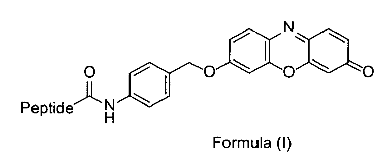
Peptide is a peptide or peptide derivative, or a salt of said peptide or peptide derivative. - The compound according to claim 1, wherein the C-terminal amino acid of said peptide or peptide derivative is Arg or Lys.
- The compound according to claim 1 or claim 2, wherein the peptide or peptide derivative is selected from the group consisting of thrombin substrates, factor Xa substrates, trypsin substrates, chymotrypsin substrates, factor VIIa substrates, factor IXa substrates, factor XIa substrates, factor XIIa substrates, kallikrein substrates, plasmin substrates, tissue plasminogen activator substrates, activated protein C substrates, tryptase substrates, matriptase substrates, granzyme substrates, elastase substrates, and human complement protease C1r substrates.
- The compound according to any one of claims 1 to 3, wherein Peptide is the peptide D-Phe-Pro-Arg (Compound 1) or a salt thereof, or the peptide derivative Benzylsulfonyl-D-Arg-Gly-Arg (Compound 2) or a salt thereof.
- A method for the detection of the activity of at least one serine protease in a sample, comprising the steps of contacting said sample with a compound according to any one of claims 1 to 4, and measuring the amount of resorufin released from said compound.
- The method according to claim 5, wherein the at least one serine protease is a trypsin-like protease.
- The method according to claim 5 or claim 6, wherein the at least one serine protease is selected from the group consisting of trypsins, chymotrypsins, elastases, thrombin, factor VIIa, factor IXa, factor Xa, factor Xla, factor XIIa, kallikreins, plasmin, tissue plasminogen activator, activated protein C, human complement protease C1 r, tryptases, matriptases, and granzymes.
- The method according to any one of claims 5 to 7, wherein the at least one serine protease is thrombin or factor Xa.
- The method according to claim 8, wherein the compound is Compound 1 or a salt thereof, or is Compound 2 or a salt thereof.
- The method according to any one of claims 5 to 9, wherein the sample (i) is a sample having a high optical density and/or a high autofluorescence, and/or (ii) is in contact with a surface having a high autofluorescence, and/or (iii) contains at least one biological structure that is labeled with at least one chromogenic and/or fluorogenic substance.
- The method according to any one of claims 5 to 10, wherein the sample is selected from the group consisting of whole blood, serum, plasma, urine, saliva, sputum, semen, lacrimal fluid, cerebrospinal fluid, defecation, cells and tissues.
- A test strip having a surface on which a compound according to any one of claims 1 to 4 is immobilized.
- A compound according to any one of claims 1 to 4 for use in the diagnosis of a conditions or disease in a subject that is characterized by abnormal levels of at least one serine protease.
- Use of a compound according to any one of claims 1 to 4 for the detection of the activity of at least one serine protease in a sample.
- A diagnostic kit, comprising at least one compound according to any one of claims 1 to 4.
Priority Applications (4)
| Application Number | Priority Date | Filing Date | Title |
|---|---|---|---|
| EP15002304.2A EP3127913A1 (en) | 2015-08-03 | 2015-08-03 | Chromogenic and fluorogenic peptide substrates for the detection of serine protease activity |
| US15/748,481 US20180215787A1 (en) | 2015-08-03 | 2016-08-03 | Chromogenic And Fluorogenic Peptide Substrates For The Detection Of Serine Protease Activity |
| PCT/EP2016/001334 WO2017021004A1 (en) | 2015-08-03 | 2016-08-03 | Chromogenic and fluorogenic peptide substrates for the detection of serine protease activity |
| EP16756940.9A EP3331898A1 (en) | 2015-08-03 | 2016-08-03 | Chromogenic and fluorogenic peptide substrates for the detection of serine protease activity |
Applications Claiming Priority (1)
| Application Number | Priority Date | Filing Date | Title |
|---|---|---|---|
| EP15002304.2A EP3127913A1 (en) | 2015-08-03 | 2015-08-03 | Chromogenic and fluorogenic peptide substrates for the detection of serine protease activity |
Publications (1)
| Publication Number | Publication Date |
|---|---|
| EP3127913A1 true EP3127913A1 (en) | 2017-02-08 |
Family
ID=53886793
Family Applications (2)
| Application Number | Title | Priority Date | Filing Date |
|---|---|---|---|
| EP15002304.2A Withdrawn EP3127913A1 (en) | 2015-08-03 | 2015-08-03 | Chromogenic and fluorogenic peptide substrates for the detection of serine protease activity |
| EP16756940.9A Withdrawn EP3331898A1 (en) | 2015-08-03 | 2016-08-03 | Chromogenic and fluorogenic peptide substrates for the detection of serine protease activity |
Family Applications After (1)
| Application Number | Title | Priority Date | Filing Date |
|---|---|---|---|
| EP16756940.9A Withdrawn EP3331898A1 (en) | 2015-08-03 | 2016-08-03 | Chromogenic and fluorogenic peptide substrates for the detection of serine protease activity |
Country Status (3)
| Country | Link |
|---|---|
| US (1) | US20180215787A1 (en) |
| EP (2) | EP3127913A1 (en) |
| WO (1) | WO2017021004A1 (en) |
Cited By (3)
| Publication number | Priority date | Publication date | Assignee | Title |
|---|---|---|---|---|
| CN108948140A (en) * | 2017-05-18 | 2018-12-07 | 首都医科大学 | A kind of new compound of warfarin -4-O- acetyl-Arg-AA ten, synthesis, activity and application |
| US20210317505A1 (en) * | 2017-09-22 | 2021-10-14 | Doasense Gmbh | Improved detection of anticoagulants in body fluids |
| CN114184562A (en) * | 2021-11-19 | 2022-03-15 | 北京赛升药业股份有限公司 | Method for determining urokinase activity by chromogenic substrate method |
Families Citing this family (4)
| Publication number | Priority date | Publication date | Assignee | Title |
|---|---|---|---|---|
| CN108948139B (en) * | 2017-05-18 | 2021-10-26 | 首都医科大学 | warfarin-4-O-acetyl-Arg-AA compounds, their synthesis, activity and use |
| WO2018222773A2 (en) * | 2017-05-30 | 2018-12-06 | Chan Eugene Y | Fluorogenic peptide substrates for in solution and solid phase factor xa measurements |
| CN112110989A (en) * | 2020-08-28 | 2020-12-22 | 南方医科大学 | Polypeptide molecular probe for in vivo detection of thrombin and preparation method thereof |
| CN119178757B (en) * | 2024-09-03 | 2025-06-10 | 南京市第一医院 | Dual-channel colorimetric fluorescent composite probe for detecting trypsin, detection method and application |
Citations (3)
| Publication number | Priority date | Publication date | Assignee | Title |
|---|---|---|---|---|
| WO2011094185A1 (en) * | 2010-01-27 | 2011-08-04 | Adventist Health System/Sunbelt, Inc. | Thrombin generation determination method |
| WO2015039211A1 (en) * | 2013-09-17 | 2015-03-26 | Mcmaster University | Method for assaying a protease |
| WO2015123783A1 (en) * | 2014-02-21 | 2015-08-27 | Thunder Bay Regional Research Institute | Cathepsin b-targeting probes |
-
2015
- 2015-08-03 EP EP15002304.2A patent/EP3127913A1/en not_active Withdrawn
-
2016
- 2016-08-03 WO PCT/EP2016/001334 patent/WO2017021004A1/en not_active Ceased
- 2016-08-03 EP EP16756940.9A patent/EP3331898A1/en not_active Withdrawn
- 2016-08-03 US US15/748,481 patent/US20180215787A1/en not_active Abandoned
Patent Citations (3)
| Publication number | Priority date | Publication date | Assignee | Title |
|---|---|---|---|---|
| WO2011094185A1 (en) * | 2010-01-27 | 2011-08-04 | Adventist Health System/Sunbelt, Inc. | Thrombin generation determination method |
| WO2015039211A1 (en) * | 2013-09-17 | 2015-03-26 | Mcmaster University | Method for assaying a protease |
| WO2015123783A1 (en) * | 2014-02-21 | 2015-08-27 | Thunder Bay Regional Research Institute | Cathepsin b-targeting probes |
Cited By (4)
| Publication number | Priority date | Publication date | Assignee | Title |
|---|---|---|---|---|
| CN108948140A (en) * | 2017-05-18 | 2018-12-07 | 首都医科大学 | A kind of new compound of warfarin -4-O- acetyl-Arg-AA ten, synthesis, activity and application |
| CN108948140B (en) * | 2017-05-18 | 2021-10-26 | 首都医科大学 | warfarin-4-O-acetyl-Arg-AA eleven compounds, and synthesis, activity and application thereof |
| US20210317505A1 (en) * | 2017-09-22 | 2021-10-14 | Doasense Gmbh | Improved detection of anticoagulants in body fluids |
| CN114184562A (en) * | 2021-11-19 | 2022-03-15 | 北京赛升药业股份有限公司 | Method for determining urokinase activity by chromogenic substrate method |
Also Published As
| Publication number | Publication date |
|---|---|
| EP3331898A1 (en) | 2018-06-13 |
| WO2017021004A1 (en) | 2017-02-09 |
| US20180215787A1 (en) | 2018-08-02 |
| WO2017021004A8 (en) | 2019-08-08 |
Similar Documents
| Publication | Publication Date | Title |
|---|---|---|
| US20180215787A1 (en) | Chromogenic And Fluorogenic Peptide Substrates For The Detection Of Serine Protease Activity | |
| EP0988394B1 (en) | Compositions for the detection of enzyme activity in biological samples and methods of use thereof | |
| US5714342A (en) | Compositions for the detection of protease in biological samples and methods of use therefo | |
| US8535898B2 (en) | Thrombin substrate and assay for determining the level of bioactive thrombin in a sample | |
| US10465230B2 (en) | Chemiluminescence-based haemostasis assay | |
| JP7456707B2 (en) | Novel chemiluminescent substrate for factor Xa | |
| JP4562018B2 (en) | Peptide-immobilized substrate and target protein measurement method using the same | |
| GB2388190A (en) | Method for measuring enzyme activity | |
| EP0076042A1 (en) | Novel substrates for measuring thrombin | |
| JPH0411199B2 (en) | ||
| US5917012A (en) | Peptide derivatives | |
| Naqvi et al. | β galactosidase enzyme fragment complementation as a high-throughput screening protease technology | |
| Wysocka et al. | Future of protease activity assays | |
| JPH04501460A (en) | Immunoassay for catalytically active serine proteases | |
| AU7368800A (en) | Compositions for the detection of enzyme activity in biological samples and methods of use thereof | |
| Krafft et al. | [6] Synthetic approaches to continuous assays of retroviral proteases | |
| EP1517997A2 (en) | Luminogenic protease substrates | |
| JPS6117956A (en) | Novel substrate for measuring kallikrein in urine | |
| US5063152A (en) | Synthetic peptidic substrate for determination of trypsin and α1 | |
| Budzynski | Chromogenic substrates in coagulation and fibrinolytic assays | |
| RU2820234C2 (en) | NOVEL CHEMILUMINESCENT SUBSTRATES FOR Xa FACTOR | |
| US8163513B2 (en) | Method for determining the total clotting activity of a blood or plasma sample | |
| US20180346960A1 (en) | Fluorogenic peptide substrates for in solution and solid phase factor Xa measurements | |
| JP2004533489A (en) | A novel chromogenic substrate and its use in assaying carboxypeptidase activity | |
| Sato | Development of Novel Doubly Labelled Peptide Probes with Identical Fluorophores for Protease Activity Measurement |
Legal Events
| Date | Code | Title | Description |
|---|---|---|---|
| PUAI | Public reference made under article 153(3) epc to a published international application that has entered the european phase |
Free format text: ORIGINAL CODE: 0009012 |
|
| AK | Designated contracting states |
Kind code of ref document: A1 Designated state(s): AL AT BE BG CH CY CZ DE DK EE ES FI FR GB GR HR HU IE IS IT LI LT LU LV MC MK MT NL NO PL PT RO RS SE SI SK SM TR |
|
| AX | Request for extension of the european patent |
Extension state: BA ME |
|
| STAA | Information on the status of an ep patent application or granted ep patent |
Free format text: STATUS: THE APPLICATION IS DEEMED TO BE WITHDRAWN |
|
| 18D | Application deemed to be withdrawn |
Effective date: 20170809 |