EP3119411B1 - Salvinorin compositions and uses thereof - Google Patents
Salvinorin compositions and uses thereof Download PDFInfo
- Publication number
- EP3119411B1 EP3119411B1 EP15765744.6A EP15765744A EP3119411B1 EP 3119411 B1 EP3119411 B1 EP 3119411B1 EP 15765744 A EP15765744 A EP 15765744A EP 3119411 B1 EP3119411 B1 EP 3119411B1
- Authority
- EP
- European Patent Office
- Prior art keywords
- salvinorin
- administration
- hypoxia
- cerebral
- ischemia
- Prior art date
- Legal status (The legal status is an assumption and is not a legal conclusion. Google has not performed a legal analysis and makes no representation as to the accuracy of the status listed.)
- Active
Links
- OBSYBRPAKCASQB-AGQYDFLVSA-N salvinorin A Chemical compound C=1([C@H]2OC(=O)[C@@H]3CC[C@]4(C)[C@@H]([C@]3(C2)C)C(=O)[C@@H](OC(C)=O)C[C@H]4C(=O)OC)C=COC=1 OBSYBRPAKCASQB-AGQYDFLVSA-N 0.000 title claims description 418
- 229930188950 salvinorin Natural products 0.000 title description 62
- 239000000203 mixture Substances 0.000 title description 35
- OBSYBRPAKCASQB-UHFFFAOYSA-N Episalvinorin A Natural products COC(=O)C1CC(OC(C)=O)C(=O)C(C2(C3)C)C1(C)CCC2C(=O)OC3C=1C=COC=1 OBSYBRPAKCASQB-UHFFFAOYSA-N 0.000 claims description 352
- IQXUYSXCJCVVPA-UHFFFAOYSA-N salvinorin A Natural products CC(=O)OC1CC(OC(=O)C)C2(C)CCC34CC(CC3(C)C2C1=O)(OC4=O)c5occc5 IQXUYSXCJCVVPA-UHFFFAOYSA-N 0.000 claims description 352
- 229920000858 Cyclodextrin Polymers 0.000 claims description 46
- 239000008194 pharmaceutical composition Substances 0.000 claims description 36
- 201000006474 Brain Ischemia Diseases 0.000 claims description 26
- 208000032851 Subarachnoid Hemorrhage Diseases 0.000 claims description 25
- 208000010496 Heart Arrest Diseases 0.000 claims description 24
- 208000006011 Stroke Diseases 0.000 claims description 24
- 208000037265 diseases, disorders, signs and symptoms Diseases 0.000 claims description 24
- 206010008118 cerebral infarction Diseases 0.000 claims description 22
- 230000001965 increasing effect Effects 0.000 claims description 22
- HFHDHCJBZVLPGP-UHFFFAOYSA-N schardinger α-dextrin Chemical compound O1C(C(C2O)O)C(CO)OC2OC(C(C2O)O)C(CO)OC2OC(C(C2O)O)C(CO)OC2OC(C(O)C2O)C(CO)OC2OC(C(C2O)O)C(CO)OC2OC2C(O)C(O)C1OC2CO HFHDHCJBZVLPGP-UHFFFAOYSA-N 0.000 claims description 22
- 206010008120 Cerebral ischaemia Diseases 0.000 claims description 21
- 201000010099 disease Diseases 0.000 claims description 21
- ODLHGICHYURWBS-LKONHMLTSA-N trappsol cyclo Chemical compound CC(O)COC[C@H]([C@H]([C@@H]([C@H]1O)O)O[C@H]2O[C@@H]([C@@H](O[C@H]3O[C@H](COCC(C)O)[C@H]([C@@H]([C@H]3O)O)O[C@H]3O[C@H](COCC(C)O)[C@H]([C@@H]([C@H]3O)O)O[C@H]3O[C@H](COCC(C)O)[C@H]([C@@H]([C@H]3O)O)O[C@H]3O[C@H](COCC(C)O)[C@H]([C@@H]([C@H]3O)O)O3)[C@H](O)[C@H]2O)COCC(O)C)O[C@@H]1O[C@H]1[C@H](O)[C@@H](O)[C@@H]3O[C@@H]1COCC(C)O ODLHGICHYURWBS-LKONHMLTSA-N 0.000 claims description 21
- 208000002381 Brain Hypoxia Diseases 0.000 claims description 17
- 206010008089 Cerebral artery occlusion Diseases 0.000 claims description 14
- 230000004083 survival effect Effects 0.000 claims description 13
- 239000007864 aqueous solution Substances 0.000 claims description 7
- 231100000878 neurological injury Toxicity 0.000 claims description 7
- 208000001286 intracranial vasospasm Diseases 0.000 claims description 6
- 230000000694 effects Effects 0.000 description 95
- 210000001367 artery Anatomy 0.000 description 90
- 206010021143 Hypoxia Diseases 0.000 description 89
- 230000007954 hypoxia Effects 0.000 description 77
- 102000048260 kappa Opioid Receptors Human genes 0.000 description 67
- 108020001588 κ-opioid receptors Proteins 0.000 description 67
- PYDQMXRFUVDCHC-XAGHGKQISA-N herkinorin Chemical compound O([C@H]1C[C@H]([C@@]2(C)[C@@H]([C@]3(C[C@H](OC(=O)[C@@H]3CC2)C2=COC=C2)C)C1=O)C(=O)OC)C(=O)C1=CC=CC=C1 PYDQMXRFUVDCHC-XAGHGKQISA-N 0.000 description 64
- 208000028867 ischemia Diseases 0.000 description 63
- 230000010339 dilation Effects 0.000 description 62
- 108010007457 Extracellular Signal-Regulated MAP Kinases Proteins 0.000 description 56
- 102000007665 Extracellular Signal-Regulated MAP Kinases Human genes 0.000 description 56
- IAZDPXIOMUYVGZ-UHFFFAOYSA-N Dimethylsulphoxide Chemical compound CS(C)=O IAZDPXIOMUYVGZ-UHFFFAOYSA-N 0.000 description 52
- 230000004044 response Effects 0.000 description 50
- 230000002490 cerebral effect Effects 0.000 description 47
- 210000001175 cerebrospinal fluid Anatomy 0.000 description 42
- 239000003814 drug Substances 0.000 description 39
- 238000000034 method Methods 0.000 description 38
- APSUXPSYBJVPPS-YAUKWVCOSA-N Norbinaltorphimine Chemical compound N1([C@@H]2CC3=CC=C(C=4O[C@@H]5[C@](C3=4)([C@]2(CC=2C=3C[C@]4(O)[C@]67CCN(CC8CC8)[C@@H]4CC=4C7=C(C(=CC=4)O)O[C@H]6C=3NC=25)O)CC1)O)CC1CC1 APSUXPSYBJVPPS-YAUKWVCOSA-N 0.000 description 36
- 229940079593 drug Drugs 0.000 description 35
- 208000001953 Hypotension Diseases 0.000 description 34
- 206010020591 Hypercapnia Diseases 0.000 description 33
- 239000000556 agonist Substances 0.000 description 33
- 230000036543 hypotension Effects 0.000 description 33
- 239000005557 antagonist Substances 0.000 description 28
- 230000002792 vascular Effects 0.000 description 28
- 210000004556 brain Anatomy 0.000 description 26
- 238000011282 treatment Methods 0.000 description 26
- JWZZKOKVBUJMES-UHFFFAOYSA-N (+-)-Isoprenaline Chemical compound CC(C)NCC(O)C1=CC=C(O)C(O)=C1 JWZZKOKVBUJMES-UHFFFAOYSA-N 0.000 description 25
- 229940039009 isoproterenol Drugs 0.000 description 25
- IVOMOUWHDPKRLL-KQYNXXCUSA-N Cyclic adenosine monophosphate Chemical compound C([C@H]1O2)OP(O)(=O)O[C@H]1[C@@H](O)[C@@H]2N1C(N=CN=C2N)=C2N=C1 IVOMOUWHDPKRLL-KQYNXXCUSA-N 0.000 description 24
- 241001465754 Metazoa Species 0.000 description 24
- 150000001875 compounds Chemical class 0.000 description 23
- 230000010455 autoregulation Effects 0.000 description 22
- 238000001990 intravenous administration Methods 0.000 description 22
- 230000000926 neurological effect Effects 0.000 description 22
- MRAUNPAHJZDYCK-BYPYZUCNSA-N L-nitroarginine Chemical compound OC(=O)[C@@H](N)CCCNC(=N)N[N+]([O-])=O MRAUNPAHJZDYCK-BYPYZUCNSA-N 0.000 description 21
- 230000006378 damage Effects 0.000 description 21
- 102000051367 mu Opioid Receptors Human genes 0.000 description 20
- XLYOFNOQVPJJNP-UHFFFAOYSA-N water Substances O XLYOFNOQVPJJNP-UHFFFAOYSA-N 0.000 description 20
- 108020001612 μ-opioid receptors Proteins 0.000 description 20
- 208000027418 Wounds and injury Diseases 0.000 description 19
- ZOOGRGPOEVQQDX-KHLHZJAASA-N cyclic guanosine monophosphate Chemical compound C([C@H]1O2)O[P@](O)(=O)O[C@@H]1[C@H](O)[C@H]2N1C(N=C(NC2=O)N)=C2N=C1 ZOOGRGPOEVQQDX-KHLHZJAASA-N 0.000 description 19
- 208000014674 injury Diseases 0.000 description 19
- 238000012360 testing method Methods 0.000 description 19
- 108091006146 Channels Proteins 0.000 description 18
- 241000699666 Mus <mouse, genus> Species 0.000 description 18
- DVEXZJFMOKTQEZ-JYFOCSDGSA-N U0126 Chemical compound C=1C=CC=C(N)C=1SC(\N)=C(/C#N)\C(\C#N)=C(/N)SC1=CC=CC=C1N DVEXZJFMOKTQEZ-JYFOCSDGSA-N 0.000 description 17
- 230000001225 therapeutic effect Effects 0.000 description 17
- 102000043136 MAP kinase family Human genes 0.000 description 16
- 108091054455 MAP kinase family Proteins 0.000 description 16
- 241000699670 Mus sp. Species 0.000 description 16
- 230000004913 activation Effects 0.000 description 15
- 239000003446 ligand Substances 0.000 description 15
- 230000001681 protective effect Effects 0.000 description 15
- 230000036772 blood pressure Effects 0.000 description 14
- FCTRVTQZOUKUIV-MCDZGGTQSA-M potassium;[[[(2r,3s,4r,5r)-5-(6-aminopurin-9-yl)-3,4-dihydroxyoxolan-2-yl]methoxy-hydroxyphosphoryl]oxy-hydroxyphosphoryl] hydrogen phosphate Chemical compound [K+].C1=NC=2C(N)=NC=NC=2N1[C@@H]1O[C@H](COP(O)(=O)OP(O)(=O)OP(O)([O-])=O)[C@@H](O)[C@H]1O FCTRVTQZOUKUIV-MCDZGGTQSA-M 0.000 description 14
- 239000000243 solution Substances 0.000 description 14
- 230000006399 behavior Effects 0.000 description 13
- PQKHESYTSKMWFP-WZJCLRDWSA-N beta-Funaltrexamine Chemical compound C([C@]12[C@H]3OC=4C(O)=CC=C(C2=4)C[C@@H]2[C@]1(O)CC[C@H]3NC(=O)/C=C/C(=O)OC)CN2CC1CC1 PQKHESYTSKMWFP-WZJCLRDWSA-N 0.000 description 13
- 210000004369 blood Anatomy 0.000 description 13
- 239000008280 blood Substances 0.000 description 13
- 239000003795 chemical substances by application Substances 0.000 description 13
- XEYBHCRIKKKOSS-UHFFFAOYSA-N disodium;azanylidyneoxidanium;iron(2+);pentacyanide Chemical compound [Na+].[Na+].[Fe+2].N#[C-].N#[C-].N#[C-].N#[C-].N#[C-].[O+]#N XEYBHCRIKKKOSS-UHFFFAOYSA-N 0.000 description 13
- 230000007774 longterm Effects 0.000 description 13
- 230000001404 mediated effect Effects 0.000 description 13
- 230000003389 potentiating effect Effects 0.000 description 13
- 241000282414 Homo sapiens Species 0.000 description 12
- FAPWRFPIFSIZLT-UHFFFAOYSA-M Sodium chloride Chemical compound [Na+].[Cl-] FAPWRFPIFSIZLT-UHFFFAOYSA-M 0.000 description 12
- 108010068927 iberiotoxin Proteins 0.000 description 12
- VDNVVLOBNHIMQA-UHFFFAOYSA-N iberiotoxin Chemical compound C1SSCC(C(=O)NC(CC=2C=CC(O)=CC=2)C(=O)NC(CCC(N)=O)C(O)=O)NC(=O)C(CCCNC(N)=N)NC(=O)C1NC(=O)C(CCCCN)NC(=O)C(CCCCN)NC(=O)CNC(=O)C(CCSC)NC(=O)C(NC(=O)C(CCCCN)NC(=O)CNC(=O)C(CCCNC(N)=N)NC(=O)C(CC(O)=O)NC(=O)C(C(C)C)NC(=O)CNC(=O)C(CC=1C=CC=CC=1)NC(=O)C(CC(C)C)NC(=O)C(CC(O)=O)NC(=O)C(CCCCN)NC1=O)CSSCC1NC(=O)C(C(C)C)NC(=O)C(CO)NC(=O)C(CC=1C2=CC=CC=C2NC=1)NC(=O)C(NC(=O)C(CCC(O)=O)NC(=O)C(CCCCN)NC(=O)C(CO)NC(=O)C(C(C)C)NC(=O)C(CO)NC1=O)CSSCC1NC(=O)C(CC(O)=O)NC(=O)C(C(C)C)NC(=O)C(CC(O)=O)NC(=O)C(C(C)O)NC(=O)C(NC(=O)C1NC(=O)CC1)CC1=CC=CC=C1 VDNVVLOBNHIMQA-UHFFFAOYSA-N 0.000 description 12
- 101150086096 Eif2ak3 gene Proteins 0.000 description 11
- 102000002045 Endothelin Human genes 0.000 description 11
- 108050009340 Endothelin Proteins 0.000 description 11
- 230000008859 change Effects 0.000 description 11
- 239000013078 crystal Substances 0.000 description 11
- ZUBDGKVDJUIMQQ-UBFCDGJISA-N endothelin-1 Chemical compound C([C@@H](C(=O)N[C@@H](CC(C)C)C(=O)N[C@@H](CC(O)=O)C(=O)N[C@@H]([C@@H](C)CC)C(=O)N[C@@H]([C@@H](C)CC)C(=O)N[C@@H](CC=1C2=CC=CC=C2NC=1)C(O)=O)NC(=O)[C@H]1NC(=O)[C@H](CC=2C=CC=CC=2)NC(=O)[C@@H](CC=2C=CC(O)=CC=2)NC(=O)[C@H](C(C)C)NC(=O)[C@H]2CSSC[C@@H](C(N[C@H](CO)C(=O)N[C@@H](CO)C(=O)N[C@H](CC(C)C)C(=O)N[C@@H](CCSC)C(=O)N[C@H](CC(O)=O)C(=O)N[C@@H](CCCCN)C(=O)N[C@@H](CCC(O)=O)C(=O)N2)=O)NC(=O)[C@@H](CO)NC(=O)[C@H](N)CSSC1)C1=CNC=N1 ZUBDGKVDJUIMQQ-UBFCDGJISA-N 0.000 description 11
- 230000001146 hypoxic effect Effects 0.000 description 11
- 239000003921 oil Substances 0.000 description 11
- 235000019198 oils Nutrition 0.000 description 11
- 230000000384 rearing effect Effects 0.000 description 11
- 229940083618 sodium nitroprusside Drugs 0.000 description 11
- LFQSCWFLJHTTHZ-UHFFFAOYSA-N Ethanol Chemical compound CCO LFQSCWFLJHTTHZ-UHFFFAOYSA-N 0.000 description 10
- 102000008299 Nitric Oxide Synthase Human genes 0.000 description 10
- 108010021487 Nitric Oxide Synthase Proteins 0.000 description 10
- MWUXSHHQAYIFBG-UHFFFAOYSA-N Nitric oxide Chemical compound O=[N] MWUXSHHQAYIFBG-UHFFFAOYSA-N 0.000 description 10
- 241001136613 Salvia divinorum Species 0.000 description 10
- 235000011771 Salvia divinorum Nutrition 0.000 description 10
- 208000029028 brain injury Diseases 0.000 description 10
- 230000007423 decrease Effects 0.000 description 10
- 230000003247 decreasing effect Effects 0.000 description 10
- 230000006735 deficit Effects 0.000 description 10
- 239000000463 material Substances 0.000 description 10
- 210000002565 arteriole Anatomy 0.000 description 9
- 231100000673 dose–response relationship Toxicity 0.000 description 9
- 230000007574 infarction Effects 0.000 description 9
- PQCAUHUKTBHUSA-UHFFFAOYSA-N 7-nitro-1h-indazole Chemical compound [O-][N+](=O)C1=CC=CC2=C1NN=C2 PQCAUHUKTBHUSA-UHFFFAOYSA-N 0.000 description 8
- 206010061216 Infarction Diseases 0.000 description 8
- 102100022397 Nitric oxide synthase, brain Human genes 0.000 description 8
- 101710111444 Nitric oxide synthase, brain Proteins 0.000 description 8
- 230000001054 cortical effect Effects 0.000 description 8
- 239000003112 inhibitor Substances 0.000 description 8
- 239000002632 kappa opiate receptor agonist Substances 0.000 description 8
- 238000005259 measurement Methods 0.000 description 8
- 230000037361 pathway Effects 0.000 description 8
- 230000011664 signaling Effects 0.000 description 8
- 239000011780 sodium chloride Substances 0.000 description 8
- 238000007619 statistical method Methods 0.000 description 8
- 239000000126 substance Substances 0.000 description 8
- SMPNJFHAPJOHPP-PUHOFUEYSA-N (Rp)-cAMPS Chemical compound C([C@H]1O2)O[P@](S)(=O)O[C@H]1[C@@H](O)[C@@H]2N1C(N=CN=C2N)=C2N=C1 SMPNJFHAPJOHPP-PUHOFUEYSA-N 0.000 description 7
- CSCPPACGZOOCGX-UHFFFAOYSA-N Acetone Chemical compound CC(C)=O CSCPPACGZOOCGX-UHFFFAOYSA-N 0.000 description 7
- 208000019901 Anxiety disease Diseases 0.000 description 7
- WQZGKKKJIJFFOK-GASJEMHNSA-N Glucose Natural products OC[C@H]1OC(O)[C@H](O)[C@@H](O)[C@@H]1O WQZGKKKJIJFFOK-GASJEMHNSA-N 0.000 description 7
- 102100033098 Neurochondrin Human genes 0.000 description 7
- CDMGBJANTYXAIV-UHFFFAOYSA-N SB 203580 Chemical compound C1=CC(S(=O)C)=CC=C1C1=NC(C=2C=CC(F)=CC=2)=C(C=2C=CN=CC=2)N1 CDMGBJANTYXAIV-UHFFFAOYSA-N 0.000 description 7
- ACPOUJIDANTYHO-UHFFFAOYSA-N anthra[1,9-cd]pyrazol-6(2H)-one Chemical compound C1=CC(C(=O)C=2C3=CC=CC=2)=C2C3=NNC2=C1 ACPOUJIDANTYHO-UHFFFAOYSA-N 0.000 description 7
- 230000036506 anxiety Effects 0.000 description 7
- 230000017531 blood circulation Effects 0.000 description 7
- 239000008121 dextrose Substances 0.000 description 7
- 238000002474 experimental method Methods 0.000 description 7
- 239000007789 gas Substances 0.000 description 7
- 230000001771 impaired effect Effects 0.000 description 7
- 108010075803 neurochondrin Proteins 0.000 description 7
- 108010032028 norbin Proteins 0.000 description 7
- 230000002829 reductive effect Effects 0.000 description 7
- 230000010410 reperfusion Effects 0.000 description 7
- 230000001624 sedative effect Effects 0.000 description 7
- 210000005166 vasculature Anatomy 0.000 description 7
- BGRJTUBHPOOWDU-NSHDSACASA-N (S)-(-)-sulpiride Chemical compound CCN1CCC[C@H]1CNC(=O)C1=CC(S(N)(=O)=O)=CC=C1OC BGRJTUBHPOOWDU-NSHDSACASA-N 0.000 description 6
- CURLTUGMZLYLDI-UHFFFAOYSA-N Carbon dioxide Chemical compound O=C=O CURLTUGMZLYLDI-UHFFFAOYSA-N 0.000 description 6
- TVZCRIROJQEVOT-CABCVRRESA-N Cromakalim Chemical compound N1([C@@H]2C3=CC(=CC=C3OC([C@H]2O)(C)C)C#N)CCCC1=O TVZCRIROJQEVOT-CABCVRRESA-N 0.000 description 6
- 108010055717 JNK Mitogen-Activated Protein Kinases Proteins 0.000 description 6
- 206010047163 Vasospasm Diseases 0.000 description 6
- 238000004458 analytical method Methods 0.000 description 6
- 230000003728 cerebral autoregulation Effects 0.000 description 6
- 238000013270 controlled release Methods 0.000 description 6
- 229950004210 cromakalim Drugs 0.000 description 6
- 230000003111 delayed effect Effects 0.000 description 6
- 210000001105 femoral artery Anatomy 0.000 description 6
- 210000003191 femoral vein Anatomy 0.000 description 6
- 229960004580 glibenclamide Drugs 0.000 description 6
- ZNNLBTZKUZBEKO-UHFFFAOYSA-N glyburide Chemical compound COC1=CC=C(Cl)C=C1C(=O)NCCC1=CC=C(S(=O)(=O)NC(=O)NC2CCCCC2)C=C1 ZNNLBTZKUZBEKO-UHFFFAOYSA-N 0.000 description 6
- 230000002631 hypothermal effect Effects 0.000 description 6
- 230000006872 improvement Effects 0.000 description 6
- 238000002347 injection Methods 0.000 description 6
- 239000007924 injection Substances 0.000 description 6
- 239000002618 kappa opiate receptor antagonist Substances 0.000 description 6
- UZHSEJADLWPNLE-GRGSLBFTSA-N naloxone Chemical compound O=C([C@@H]1O2)CC[C@@]3(O)[C@H]4CC5=CC=C(O)C2=C5[C@@]13CCN4CC=C UZHSEJADLWPNLE-GRGSLBFTSA-N 0.000 description 6
- 229960004127 naloxone Drugs 0.000 description 6
- 230000000144 pharmacologic effect Effects 0.000 description 6
- 238000004321 preservation Methods 0.000 description 6
- 230000009467 reduction Effects 0.000 description 6
- 230000011514 reflex Effects 0.000 description 6
- 239000000932 sedative agent Substances 0.000 description 6
- 159000000000 sodium salts Chemical class 0.000 description 6
- 239000002904 solvent Substances 0.000 description 6
- 229960004940 sulpiride Drugs 0.000 description 6
- 238000001356 surgical procedure Methods 0.000 description 6
- 230000024883 vasodilation Effects 0.000 description 6
- 238000012800 visualization Methods 0.000 description 6
- 206010063659 Aversion Diseases 0.000 description 5
- 102100038518 Calcitonin Human genes 0.000 description 5
- 241000196324 Embryophyta Species 0.000 description 5
- 241000282412 Homo Species 0.000 description 5
- PIWKPBJCKXDKJR-UHFFFAOYSA-N Isoflurane Chemical compound FC(F)OC(Cl)C(F)(F)F PIWKPBJCKXDKJR-UHFFFAOYSA-N 0.000 description 5
- YFGBQHOOROIVKG-FKBYEOEOSA-N Met-enkephalin Chemical compound C([C@@H](C(=O)N[C@@H](CCSC)C(O)=O)NC(=O)CNC(=O)CNC(=O)[C@@H](N)CC=1C=CC(O)=CC=1)C1=CC=CC=C1 YFGBQHOOROIVKG-FKBYEOEOSA-N 0.000 description 5
- 102000003840 Opioid Receptors Human genes 0.000 description 5
- 108090000137 Opioid Receptors Proteins 0.000 description 5
- 239000002202 Polyethylene glycol Substances 0.000 description 5
- DNIAPMSPPWPWGF-UHFFFAOYSA-N Propylene glycol Chemical compound CC(O)CO DNIAPMSPPWPWGF-UHFFFAOYSA-N 0.000 description 5
- 241000700159 Rattus Species 0.000 description 5
- 206010047139 Vasoconstriction Diseases 0.000 description 5
- 230000008499 blood brain barrier function Effects 0.000 description 5
- 210000001218 blood-brain barrier Anatomy 0.000 description 5
- 229910002092 carbon dioxide Inorganic materials 0.000 description 5
- 210000004027 cell Anatomy 0.000 description 5
- 239000003153 chemical reaction reagent Substances 0.000 description 5
- 230000003750 conditioning effect Effects 0.000 description 5
- 238000009472 formulation Methods 0.000 description 5
- 230000008014 freezing Effects 0.000 description 5
- 238000007710 freezing Methods 0.000 description 5
- 239000011521 glass Substances 0.000 description 5
- 210000003128 head Anatomy 0.000 description 5
- 238000001802 infusion Methods 0.000 description 5
- 230000000302 ischemic effect Effects 0.000 description 5
- 229960002725 isoflurane Drugs 0.000 description 5
- 239000007788 liquid Substances 0.000 description 5
- 230000007246 mechanism Effects 0.000 description 5
- 230000015654 memory Effects 0.000 description 5
- 201000007309 middle cerebral artery infarction Diseases 0.000 description 5
- 239000002756 mu opiate receptor agonist Substances 0.000 description 5
- 210000000056 organ Anatomy 0.000 description 5
- 229920001223 polyethylene glycol Polymers 0.000 description 5
- 239000000725 suspension Substances 0.000 description 5
- 230000002459 sustained effect Effects 0.000 description 5
- 230000025033 vasoconstriction Effects 0.000 description 5
- 239000003981 vehicle Substances 0.000 description 5
- HPZJMUBDEAMBFI-WTNAPCKOSA-N (D-Ala(2)-mephe(4)-gly-ol(5))enkephalin Chemical compound C([C@H](N)C(=O)N[C@H](C)C(=O)NCC(=O)N(C)[C@@H](CC=1C=CC=CC=1)C(=O)NCCO)C1=CC=C(O)C=C1 HPZJMUBDEAMBFI-WTNAPCKOSA-N 0.000 description 4
- GHCCBWMZKJQGLS-UHFFFAOYSA-N 2-(3,4-dichlorophenyl)-1-[2-(1-pyrrolidinylmethyl)-1-piperidinyl]ethanone Chemical compound C1=C(Cl)C(Cl)=CC=C1CC(=O)N1C(CN2CCCC2)CCCC1 GHCCBWMZKJQGLS-UHFFFAOYSA-N 0.000 description 4
- NIXOWILDQLNWCW-UHFFFAOYSA-N Acrylic acid Chemical compound OC(=O)C=C NIXOWILDQLNWCW-UHFFFAOYSA-N 0.000 description 4
- 206010002091 Anaesthesia Diseases 0.000 description 4
- IJGRMHOSHXDMSA-UHFFFAOYSA-N Atomic nitrogen Chemical compound N#N IJGRMHOSHXDMSA-UHFFFAOYSA-N 0.000 description 4
- 238000008157 ELISA kit Methods 0.000 description 4
- 208000032382 Ischaemic stroke Diseases 0.000 description 4
- TWRXJAOTZQYOKJ-UHFFFAOYSA-L Magnesium chloride Chemical compound [Mg+2].[Cl-].[Cl-] TWRXJAOTZQYOKJ-UHFFFAOYSA-L 0.000 description 4
- 108010042237 Methionine Enkephalin Proteins 0.000 description 4
- 229940121954 Opioid receptor agonist Drugs 0.000 description 4
- VYPSYNLAJGMNEJ-UHFFFAOYSA-N Silicium dioxide Chemical compound O=[Si]=O VYPSYNLAJGMNEJ-UHFFFAOYSA-N 0.000 description 4
- UIIMBOGNXHQVGW-UHFFFAOYSA-M Sodium bicarbonate Chemical compound [Na+].OC([O-])=O UIIMBOGNXHQVGW-UHFFFAOYSA-M 0.000 description 4
- 102000003978 Tissue Plasminogen Activator Human genes 0.000 description 4
- 108090000373 Tissue Plasminogen Activator Proteins 0.000 description 4
- XSQUKJJJFZCRTK-UHFFFAOYSA-N Urea Chemical compound NC(N)=O XSQUKJJJFZCRTK-UHFFFAOYSA-N 0.000 description 4
- 230000037005 anaesthesia Effects 0.000 description 4
- 238000000540 analysis of variance Methods 0.000 description 4
- 238000010171 animal model Methods 0.000 description 4
- 230000003502 anti-nociceptive effect Effects 0.000 description 4
- 230000004872 arterial blood pressure Effects 0.000 description 4
- 230000001042 autoregulative effect Effects 0.000 description 4
- 230000008901 benefit Effects 0.000 description 4
- 239000002775 capsule Substances 0.000 description 4
- OJYGBLRPYBAHRT-IPQSZEQASA-N chloralose Chemical compound O1[C@H](C(Cl)(Cl)Cl)O[C@@H]2[C@@H](O)[C@@H]([C@H](O)CO)O[C@@H]21 OJYGBLRPYBAHRT-IPQSZEQASA-N 0.000 description 4
- 230000004087 circulation Effects 0.000 description 4
- MSBXTPRURXJCPF-DQWIULQBSA-N cucurbit[6]uril Chemical compound N1([C@@H]2[C@@H]3N(C1=O)CN1[C@@H]4[C@@H]5N(C1=O)CN1[C@@H]6[C@@H]7N(C1=O)CN1[C@@H]8[C@@H]9N(C1=O)CN([C@H]1N(C%10=O)CN9C(=O)N8CN7C(=O)N6CN5C(=O)N4CN3C(=O)N2C2)C3=O)CN4C(=O)N5[C@@H]6[C@H]4N2C(=O)N6CN%10[C@H]1N3C5 MSBXTPRURXJCPF-DQWIULQBSA-N 0.000 description 4
- 230000034994 death Effects 0.000 description 4
- 231100000517 death Toxicity 0.000 description 4
- 238000011161 development Methods 0.000 description 4
- 230000018109 developmental process Effects 0.000 description 4
- 239000000839 emulsion Substances 0.000 description 4
- 210000003194 forelimb Anatomy 0.000 description 4
- 230000029251 gravitaxis Effects 0.000 description 4
- 238000010438 heat treatment Methods 0.000 description 4
- 230000000004 hemodynamic effect Effects 0.000 description 4
- 230000006698 induction Effects 0.000 description 4
- 230000005764 inhibitory process Effects 0.000 description 4
- 230000003993 interaction Effects 0.000 description 4
- 238000007917 intracranial administration Methods 0.000 description 4
- 238000007912 intraperitoneal administration Methods 0.000 description 4
- 230000008018 melting Effects 0.000 description 4
- 238000002844 melting Methods 0.000 description 4
- 230000007659 motor function Effects 0.000 description 4
- 230000001537 neural effect Effects 0.000 description 4
- 230000004112 neuroprotection Effects 0.000 description 4
- QJGQUHMNIGDVPM-UHFFFAOYSA-N nitrogen group Chemical group [N] QJGQUHMNIGDVPM-UHFFFAOYSA-N 0.000 description 4
- 238000001543 one-way ANOVA Methods 0.000 description 4
- 238000012346 open field test Methods 0.000 description 4
- 239000003402 opiate agonist Substances 0.000 description 4
- 239000003960 organic solvent Substances 0.000 description 4
- 230000009984 peri-natal effect Effects 0.000 description 4
- 208000033300 perinatal asphyxia Diseases 0.000 description 4
- 229920001184 polypeptide Polymers 0.000 description 4
- 239000013641 positive control Substances 0.000 description 4
- 102000004196 processed proteins & peptides Human genes 0.000 description 4
- 108090000765 processed proteins & peptides Proteins 0.000 description 4
- 230000000069 prophylactic effect Effects 0.000 description 4
- -1 quick onset Chemical class 0.000 description 4
- 230000000284 resting effect Effects 0.000 description 4
- 150000003839 salts Chemical group 0.000 description 4
- 230000002269 spontaneous effect Effects 0.000 description 4
- 229910001220 stainless steel Inorganic materials 0.000 description 4
- 239000010935 stainless steel Substances 0.000 description 4
- 230000007704 transition Effects 0.000 description 4
- 238000009423 ventilation Methods 0.000 description 4
- 230000031836 visual learning Effects 0.000 description 4
- ZKHQWZAMYRWXGA-KQYNXXCUSA-J ATP(4-) Chemical compound C1=NC=2C(N)=NC=NC=2N1[C@@H]1O[C@H](COP([O-])(=O)OP([O-])(=O)OP([O-])([O-])=O)[C@@H](O)[C@H]1O ZKHQWZAMYRWXGA-KQYNXXCUSA-J 0.000 description 3
- WEVYAHXRMPXWCK-UHFFFAOYSA-N Acetonitrile Chemical compound CC#N WEVYAHXRMPXWCK-UHFFFAOYSA-N 0.000 description 3
- ZKHQWZAMYRWXGA-UHFFFAOYSA-N Adenosine triphosphate Natural products C1=NC=2C(N)=NC=NC=2N1C1OC(COP(O)(=O)OP(O)(=O)OP(O)(O)=O)C(O)C1O ZKHQWZAMYRWXGA-UHFFFAOYSA-N 0.000 description 3
- 108700022183 Ala(2)-MePhe(4)-Gly(5)- Enkephalin Proteins 0.000 description 3
- 229920001450 Alpha-Cyclodextrin Polymers 0.000 description 3
- 229920002261 Corn starch Polymers 0.000 description 3
- 102000004980 Dopamine D2 Receptors Human genes 0.000 description 3
- 108090001111 Dopamine D2 Receptors Proteins 0.000 description 3
- 239000001856 Ethyl cellulose Substances 0.000 description 3
- ZZSNKZQZMQGXPY-UHFFFAOYSA-N Ethyl cellulose Chemical compound CCOCC1OC(OC)C(OCC)C(OCC)C1OC1C(O)C(O)C(OC)C(CO)O1 ZZSNKZQZMQGXPY-UHFFFAOYSA-N 0.000 description 3
- 239000001116 FEMA 4028 Substances 0.000 description 3
- PEDCQBHIVMGVHV-UHFFFAOYSA-N Glycerine Chemical compound OCC(O)CO PEDCQBHIVMGVHV-UHFFFAOYSA-N 0.000 description 3
- 229920002907 Guar gum Polymers 0.000 description 3
- 229920002153 Hydroxypropyl cellulose Polymers 0.000 description 3
- 206010070511 Hypoxic-ischaemic encephalopathy Diseases 0.000 description 3
- KFZMGEQAYNKOFK-UHFFFAOYSA-N Isopropanol Chemical compound CC(C)O KFZMGEQAYNKOFK-UHFFFAOYSA-N 0.000 description 3
- 241000124008 Mammalia Species 0.000 description 3
- 208000026139 Memory disease Diseases 0.000 description 3
- OKKJLVBELUTLKV-UHFFFAOYSA-N Methanol Chemical compound OC OKKJLVBELUTLKV-UHFFFAOYSA-N 0.000 description 3
- 208000037212 Neonatal hypoxic and ischemic brain injury Diseases 0.000 description 3
- 208000012902 Nervous system disease Diseases 0.000 description 3
- 208000025966 Neurological disease Diseases 0.000 description 3
- 235000010676 Ocimum basilicum Nutrition 0.000 description 3
- 240000007926 Ocimum gratissimum Species 0.000 description 3
- 208000004756 Respiratory Insufficiency Diseases 0.000 description 3
- 206010038678 Respiratory depression Diseases 0.000 description 3
- DBMJMQXJHONAFJ-UHFFFAOYSA-M Sodium laurylsulphate Chemical compound [Na+].CCCCCCCCCCCCOS([O-])(=O)=O DBMJMQXJHONAFJ-UHFFFAOYSA-M 0.000 description 3
- 229920002125 Sokalan® Polymers 0.000 description 3
- 241000282887 Suidae Species 0.000 description 3
- HFHDHCJBZVLPGP-RWMJIURBSA-N alpha-cyclodextrin Chemical compound OC[C@H]([C@H]([C@@H]([C@H]1O)O)O[C@H]2O[C@@H]([C@@H](O[C@H]3O[C@H](CO)[C@H]([C@@H]([C@H]3O)O)O[C@H]3O[C@H](CO)[C@H]([C@@H]([C@H]3O)O)O[C@H]3O[C@H](CO)[C@H]([C@@H]([C@H]3O)O)O3)[C@H](O)[C@H]2O)CO)O[C@@H]1O[C@H]1[C@H](O)[C@@H](O)[C@@H]3O[C@@H]1CO HFHDHCJBZVLPGP-RWMJIURBSA-N 0.000 description 3
- 229940043377 alpha-cyclodextrin Drugs 0.000 description 3
- 238000013459 approach Methods 0.000 description 3
- 230000002238 attenuated effect Effects 0.000 description 3
- 230000003542 behavioural effect Effects 0.000 description 3
- WQZGKKKJIJFFOK-VFUOTHLCSA-N beta-D-glucose Chemical compound OC[C@H]1O[C@@H](O)[C@H](O)[C@@H](O)[C@@H]1O WQZGKKKJIJFFOK-VFUOTHLCSA-N 0.000 description 3
- WHGYBXFWUBPSRW-FOUAGVGXSA-N beta-cyclodextrin Chemical compound OC[C@H]([C@H]([C@@H]([C@H]1O)O)O[C@H]2O[C@@H]([C@@H](O[C@H]3O[C@H](CO)[C@H]([C@@H]([C@H]3O)O)O[C@H]3O[C@H](CO)[C@H]([C@@H]([C@H]3O)O)O[C@H]3O[C@H](CO)[C@H]([C@@H]([C@H]3O)O)O[C@H]3O[C@H](CO)[C@H]([C@@H]([C@H]3O)O)O3)[C@H](O)[C@H]2O)CO)O[C@@H]1O[C@H]1[C@H](O)[C@@H](O)[C@@H]3O[C@@H]1CO WHGYBXFWUBPSRW-FOUAGVGXSA-N 0.000 description 3
- 235000011175 beta-cyclodextrine Nutrition 0.000 description 3
- 229960004853 betadex Drugs 0.000 description 3
- 230000037396 body weight Effects 0.000 description 3
- 208000009973 brain hypoxia - ischemia Diseases 0.000 description 3
- 230000011496 cAMP-mediated signaling Effects 0.000 description 3
- 229960001631 carbomer Drugs 0.000 description 3
- 230000003727 cerebral blood flow Effects 0.000 description 3
- 230000010456 cerebrovascular autoregulation Effects 0.000 description 3
- KRKNYBCHXYNGOX-UHFFFAOYSA-N citric acid Chemical compound OC(=O)CC(O)(C(O)=O)CC(O)=O KRKNYBCHXYNGOX-UHFFFAOYSA-N 0.000 description 3
- 239000008120 corn starch Substances 0.000 description 3
- 229940099112 cornstarch Drugs 0.000 description 3
- 230000009193 crawling Effects 0.000 description 3
- 230000001419 dependent effect Effects 0.000 description 3
- 208000037765 diseases and disorders Diseases 0.000 description 3
- 208000035475 disorder Diseases 0.000 description 3
- 238000009826 distribution Methods 0.000 description 3
- 235000019325 ethyl cellulose Nutrition 0.000 description 3
- 229920001249 ethyl cellulose Polymers 0.000 description 3
- 230000006870 function Effects 0.000 description 3
- 239000000665 guar gum Substances 0.000 description 3
- 235000010417 guar gum Nutrition 0.000 description 3
- 229960002154 guar gum Drugs 0.000 description 3
- 235000010977 hydroxypropyl cellulose Nutrition 0.000 description 3
- 239000001863 hydroxypropyl cellulose Substances 0.000 description 3
- 238000001361 intraarterial administration Methods 0.000 description 3
- 239000007927 intramuscular injection Substances 0.000 description 3
- 229940126470 kappa opioid receptor agonist Drugs 0.000 description 3
- 150000002632 lipids Chemical class 0.000 description 3
- 230000006742 locomotor activity Effects 0.000 description 3
- 238000007726 management method Methods 0.000 description 3
- 238000004519 manufacturing process Methods 0.000 description 3
- 238000003032 molecular docking Methods 0.000 description 3
- 230000003188 neurobehavioral effect Effects 0.000 description 3
- 230000007971 neurological deficit Effects 0.000 description 3
- 230000000324 neuroprotective effect Effects 0.000 description 3
- 229940005483 opioid analgesics Drugs 0.000 description 3
- 230000036961 partial effect Effects 0.000 description 3
- 239000008188 pellet Substances 0.000 description 3
- 230000002085 persistent effect Effects 0.000 description 3
- 229920003023 plastic Polymers 0.000 description 3
- 239000004033 plastic Substances 0.000 description 3
- 235000010482 polyoxyethylene sorbitan monooleate Nutrition 0.000 description 3
- 229920000053 polysorbate 80 Polymers 0.000 description 3
- 230000002035 prolonged effect Effects 0.000 description 3
- 235000013772 propylene glycol Nutrition 0.000 description 3
- 230000004224 protection Effects 0.000 description 3
- 230000000241 respiratory effect Effects 0.000 description 3
- 230000029058 respiratory gaseous exchange Effects 0.000 description 3
- 230000028527 righting reflex Effects 0.000 description 3
- 230000009183 running Effects 0.000 description 3
- 235000019333 sodium laurylsulphate Nutrition 0.000 description 3
- 230000006886 spatial memory Effects 0.000 description 3
- 238000000528 statistical test Methods 0.000 description 3
- 230000000638 stimulation Effects 0.000 description 3
- 238000007920 subcutaneous administration Methods 0.000 description 3
- 229960000187 tissue plasminogen activator Drugs 0.000 description 3
- 238000012549 training Methods 0.000 description 3
- 230000009184 walking Effects 0.000 description 3
- JVKUCNQGESRUCL-UHFFFAOYSA-N 2-Hydroxyethyl 12-hydroxyoctadecanoate Chemical compound CCCCCCC(O)CCCCCCCCCCC(=O)OCCO JVKUCNQGESRUCL-UHFFFAOYSA-N 0.000 description 2
- CIWBSHSKHKDKBQ-JLAZNSOCSA-N Ascorbic acid Chemical compound OC[C@H](O)[C@H]1OC(=O)C(O)=C1O CIWBSHSKHKDKBQ-JLAZNSOCSA-N 0.000 description 2
- 238000010152 Bonferroni least significant difference Methods 0.000 description 2
- COXVTLYNGOIATD-HVMBLDELSA-N CC1=C(C=CC(=C1)C1=CC(C)=C(C=C1)\N=N\C1=C(O)C2=C(N)C(=CC(=C2C=C1)S(O)(=O)=O)S(O)(=O)=O)\N=N\C1=CC=C2C(=CC(=C(N)C2=C1O)S(O)(=O)=O)S(O)(=O)=O Chemical compound CC1=C(C=CC(=C1)C1=CC(C)=C(C=C1)\N=N\C1=C(O)C2=C(N)C(=CC(=C2C=C1)S(O)(=O)=O)S(O)(=O)=O)\N=N\C1=CC=C2C(=CC(=C(N)C2=C1O)S(O)(=O)=O)S(O)(=O)=O COXVTLYNGOIATD-HVMBLDELSA-N 0.000 description 2
- VTYYLEPIZMXCLO-UHFFFAOYSA-L Calcium carbonate Chemical compound [Ca+2].[O-]C([O-])=O VTYYLEPIZMXCLO-UHFFFAOYSA-L 0.000 description 2
- UXVMQQNJUSDDNG-UHFFFAOYSA-L Calcium chloride Chemical compound [Cl-].[Cl-].[Ca+2] UXVMQQNJUSDDNG-UHFFFAOYSA-L 0.000 description 2
- HEDRZPFGACZZDS-UHFFFAOYSA-N Chloroform Chemical compound ClC(Cl)Cl HEDRZPFGACZZDS-UHFFFAOYSA-N 0.000 description 2
- 206010011224 Cough Diseases 0.000 description 2
- 102100020756 D(2) dopamine receptor Human genes 0.000 description 2
- 206010015866 Extravasation Diseases 0.000 description 2
- 108010010803 Gelatin Proteins 0.000 description 2
- 101000931901 Homo sapiens D(2) dopamine receptor Proteins 0.000 description 2
- 241001406989 Iberis Species 0.000 description 2
- 208000015580 Increased body weight Diseases 0.000 description 2
- 208000034486 Multi-organ failure Diseases 0.000 description 2
- 208000010718 Multiple Organ Failure Diseases 0.000 description 2
- WHNWPMSKXPGLAX-UHFFFAOYSA-N N-Vinyl-2-pyrrolidone Chemical compound C=CN1CCCC1=O WHNWPMSKXPGLAX-UHFFFAOYSA-N 0.000 description 2
- YLFMCMWKHSDUCT-UHFFFAOYSA-N NS 1619 Chemical compound OC1=CC=C(C(F)(F)F)C=C1N1C(=O)NC2=CC(C(F)(F)F)=CC=C21 YLFMCMWKHSDUCT-UHFFFAOYSA-N 0.000 description 2
- 239000008896 Opium Substances 0.000 description 2
- 206010033799 Paralysis Diseases 0.000 description 2
- 235000019483 Peanut oil Nutrition 0.000 description 2
- 102000004257 Potassium Channel Human genes 0.000 description 2
- 206010037394 Pulmonary haemorrhage Diseases 0.000 description 2
- 206010063837 Reperfusion injury Diseases 0.000 description 2
- 241000283984 Rodentia Species 0.000 description 2
- BLTMVAIOAAGYAR-UHFFFAOYSA-N Salvinorin B Natural products COC(=O)C1CC(O)C(=O)C(C2(C3)C)C1(C)CCC2C(=O)OC3C=1C=COC=1 BLTMVAIOAAGYAR-UHFFFAOYSA-N 0.000 description 2
- BLTMVAIOAAGYAR-CEFSSPBYSA-N Salvinorin B Chemical compound C=1([C@H]2OC(=O)[C@@H]3CC[C@]4(C)[C@@H]([C@]3(C2)C)C(=O)[C@@H](O)C[C@H]4C(=O)OC)C=COC=1 BLTMVAIOAAGYAR-CEFSSPBYSA-N 0.000 description 2
- 229920001304 Solutol HS 15 Polymers 0.000 description 2
- 229920002472 Starch Polymers 0.000 description 2
- 229910000831 Steel Inorganic materials 0.000 description 2
- 206010049418 Sudden Cardiac Death Diseases 0.000 description 2
- 235000019486 Sunflower oil Nutrition 0.000 description 2
- 239000004480 active ingredient Substances 0.000 description 2
- 239000002671 adjuvant Substances 0.000 description 2
- 239000000808 adrenergic beta-agonist Substances 0.000 description 2
- 230000002411 adverse Effects 0.000 description 2
- 239000003125 aqueous solvent Substances 0.000 description 2
- QVGXLLKOCUKJST-UHFFFAOYSA-N atomic oxygen Chemical compound [O] QVGXLLKOCUKJST-UHFFFAOYSA-N 0.000 description 2
- 230000004888 barrier function Effects 0.000 description 2
- 230000009286 beneficial effect Effects 0.000 description 2
- 230000015572 biosynthetic process Effects 0.000 description 2
- 230000000740 bleeding effect Effects 0.000 description 2
- 239000001110 calcium chloride Substances 0.000 description 2
- 229910001628 calcium chloride Inorganic materials 0.000 description 2
- 239000004202 carbamide Substances 0.000 description 2
- 230000002612 cardiopulmonary effect Effects 0.000 description 2
- 239000000969 carrier Substances 0.000 description 2
- 210000001627 cerebral artery Anatomy 0.000 description 2
- 229940125400 channel inhibitor Drugs 0.000 description 2
- 229950009941 chloralose Drugs 0.000 description 2
- 238000011260 co-administration Methods 0.000 description 2
- 238000000576 coating method Methods 0.000 description 2
- 229940075614 colloidal silicon dioxide Drugs 0.000 description 2
- 238000012937 correction Methods 0.000 description 2
- 229940097362 cyclodextrins Drugs 0.000 description 2
- 230000003001 depressive effect Effects 0.000 description 2
- 238000013461 design Methods 0.000 description 2
- FLKPEMZONWLCSK-UHFFFAOYSA-N diethyl phthalate Chemical compound CCOC(=O)C1=CC=CC=C1C(=O)OCC FLKPEMZONWLCSK-UHFFFAOYSA-N 0.000 description 2
- 239000003085 diluting agent Substances 0.000 description 2
- FDPIMTJIUBPUKL-UHFFFAOYSA-N dimethylacetone Natural products CCC(=O)CC FDPIMTJIUBPUKL-UHFFFAOYSA-N 0.000 description 2
- 230000003292 diminished effect Effects 0.000 description 2
- 239000006185 dispersion Substances 0.000 description 2
- 229930004069 diterpene Natural products 0.000 description 2
- 238000001647 drug administration Methods 0.000 description 2
- 239000003937 drug carrier Substances 0.000 description 2
- 238000012377 drug delivery Methods 0.000 description 2
- 230000001544 dysphoric effect Effects 0.000 description 2
- 235000013601 eggs Nutrition 0.000 description 2
- 206010015037 epilepsy Diseases 0.000 description 2
- 229960004667 ethyl cellulose Drugs 0.000 description 2
- 229960003699 evans blue Drugs 0.000 description 2
- 230000036251 extravasation Effects 0.000 description 2
- 229920000159 gelatin Polymers 0.000 description 2
- 239000008273 gelatin Substances 0.000 description 2
- 229940014259 gelatin Drugs 0.000 description 2
- 235000019322 gelatine Nutrition 0.000 description 2
- 235000011852 gelatine desserts Nutrition 0.000 description 2
- 210000004884 grey matter Anatomy 0.000 description 2
- 230000003400 hallucinatory effect Effects 0.000 description 2
- 230000002209 hydrophobic effect Effects 0.000 description 2
- 229940071676 hydroxypropylcellulose Drugs 0.000 description 2
- 238000007918 intramuscular administration Methods 0.000 description 2
- 238000010255 intramuscular injection Methods 0.000 description 2
- 238000011835 investigation Methods 0.000 description 2
- 208000037906 ischaemic injury Diseases 0.000 description 2
- ZLVXBBHTMQJRSX-VMGNSXQWSA-N jdtic Chemical compound C1([C@]2(C)CCN(C[C@@H]2C)C[C@H](C(C)C)NC(=O)[C@@H]2NCC3=CC(O)=CC=C3C2)=CC=CC(O)=C1 ZLVXBBHTMQJRSX-VMGNSXQWSA-N 0.000 description 2
- 230000013016 learning Effects 0.000 description 2
- 201000003723 learning disability Diseases 0.000 description 2
- 239000002502 liposome Substances 0.000 description 2
- 239000012669 liquid formulation Substances 0.000 description 2
- 230000007787 long-term memory Effects 0.000 description 2
- 229910001629 magnesium chloride Inorganic materials 0.000 description 2
- HQKMJHAJHXVSDF-UHFFFAOYSA-L magnesium stearate Chemical compound [Mg+2].CCCCCCCCCCCCCCCCCC([O-])=O.CCCCCCCCCCCCCCCCCC([O-])=O HQKMJHAJHXVSDF-UHFFFAOYSA-L 0.000 description 2
- 238000012423 maintenance Methods 0.000 description 2
- 238000002483 medication Methods 0.000 description 2
- 239000012528 membrane Substances 0.000 description 2
- 230000002503 metabolic effect Effects 0.000 description 2
- NIQQIJXGUZVEBB-UHFFFAOYSA-N methanol;propan-2-one Chemical compound OC.CC(C)=O NIQQIJXGUZVEBB-UHFFFAOYSA-N 0.000 description 2
- 235000013336 milk Nutrition 0.000 description 2
- 239000008267 milk Substances 0.000 description 2
- 210000004080 milk Anatomy 0.000 description 2
- 238000012544 monitoring process Methods 0.000 description 2
- 208000029744 multiple organ dysfunction syndrome Diseases 0.000 description 2
- 230000016273 neuron death Effects 0.000 description 2
- 230000009223 neuronal apoptosis Effects 0.000 description 2
- 239000004090 neuroprotective agent Substances 0.000 description 2
- 239000002840 nitric oxide donor Substances 0.000 description 2
- 230000010417 nitric oxide pathway Effects 0.000 description 2
- 229910052757 nitrogen Inorganic materials 0.000 description 2
- 239000004006 olive oil Substances 0.000 description 2
- 235000008390 olive oil Nutrition 0.000 description 2
- 229960001027 opium Drugs 0.000 description 2
- 239000001301 oxygen Substances 0.000 description 2
- 229910052760 oxygen Inorganic materials 0.000 description 2
- 230000001575 pathological effect Effects 0.000 description 2
- 239000000312 peanut oil Substances 0.000 description 2
- 230000002093 peripheral effect Effects 0.000 description 2
- 239000002831 pharmacologic agent Substances 0.000 description 2
- 239000000244 polyoxyethylene sorbitan monooleate Substances 0.000 description 2
- 229940068968 polysorbate 80 Drugs 0.000 description 2
- 229920000036 polyvinylpyrrolidone Polymers 0.000 description 2
- 235000013855 polyvinylpyrrolidone Nutrition 0.000 description 2
- 108020001213 potassium channel Proteins 0.000 description 2
- 238000002360 preparation method Methods 0.000 description 2
- 230000009979 protective mechanism Effects 0.000 description 2
- 208000002815 pulmonary hypertension Diseases 0.000 description 2
- GHBFNMLVSPCDGN-UHFFFAOYSA-N rac-1-monooctanoylglycerol Chemical compound CCCCCCCC(=O)OCC(O)CO GHBFNMLVSPCDGN-UHFFFAOYSA-N 0.000 description 2
- 102000005962 receptors Human genes 0.000 description 2
- 108020003175 receptors Proteins 0.000 description 2
- 230000000630 rising effect Effects 0.000 description 2
- 150000004432 salvinorin A derivatives Chemical class 0.000 description 2
- 238000005070 sampling Methods 0.000 description 2
- 239000003421 short acting drug Substances 0.000 description 2
- 230000019491 signal transduction Effects 0.000 description 2
- 229910000030 sodium bicarbonate Inorganic materials 0.000 description 2
- 239000007787 solid Substances 0.000 description 2
- 239000003549 soybean oil Substances 0.000 description 2
- 235000012424 soybean oil Nutrition 0.000 description 2
- 239000008107 starch Substances 0.000 description 2
- 235000019698 starch Nutrition 0.000 description 2
- 229940032147 starch Drugs 0.000 description 2
- 239000010959 steel Substances 0.000 description 2
- 208000014221 sudden cardiac arrest Diseases 0.000 description 2
- 239000002600 sunflower oil Substances 0.000 description 2
- 239000004094 surface-active agent Substances 0.000 description 2
- 238000003786 synthesis reaction Methods 0.000 description 2
- 230000009885 systemic effect Effects 0.000 description 2
- 239000003826 tablet Substances 0.000 description 2
- 230000002123 temporal effect Effects 0.000 description 2
- 238000011200 topical administration Methods 0.000 description 2
- 238000002627 tracheal intubation Methods 0.000 description 2
- 230000004865 vascular response Effects 0.000 description 2
- 235000013311 vegetables Nutrition 0.000 description 2
- 230000000007 visual effect Effects 0.000 description 2
- BCEHBSKCWLPMDN-MGPLVRAMSA-N voriconazole Chemical compound C1([C@H](C)[C@](O)(CN2N=CN=C2)C=2C(=CC(F)=CC=2)F)=NC=NC=C1F BCEHBSKCWLPMDN-MGPLVRAMSA-N 0.000 description 2
- 210000004885 white matter Anatomy 0.000 description 2
- DNIAPMSPPWPWGF-GSVOUGTGSA-N (R)-(-)-Propylene glycol Chemical compound C[C@@H](O)CO DNIAPMSPPWPWGF-GSVOUGTGSA-N 0.000 description 1
- RBORURQQJIQWBS-UHFFFAOYSA-N 6-(6-amino-8-bromopurin-9-yl)-2-hydroxy-2-sulfanylidene-4a,6,7,7a-tetrahydro-4h-furo[3,2-d][1,3,2]dioxaphosphinin-7-ol Chemical compound O1C2COP(O)(=S)OC2C(O)C1N1C(N=CN=C2N)=C2N=C1Br RBORURQQJIQWBS-UHFFFAOYSA-N 0.000 description 1
- QTBSBXVTEAMEQO-UHFFFAOYSA-M Acetate Chemical compound CC([O-])=O QTBSBXVTEAMEQO-UHFFFAOYSA-M 0.000 description 1
- NIXOWILDQLNWCW-UHFFFAOYSA-M Acrylate Chemical compound [O-]C(=O)C=C NIXOWILDQLNWCW-UHFFFAOYSA-M 0.000 description 1
- 102000009027 Albumins Human genes 0.000 description 1
- 108010088751 Albumins Proteins 0.000 description 1
- GUBGYTABKSRVRQ-XLOQQCSPSA-N Alpha-Lactose Chemical compound O[C@@H]1[C@@H](O)[C@@H](O)[C@@H](CO)O[C@H]1O[C@@H]1[C@@H](CO)O[C@H](O)[C@H](O)[C@H]1O GUBGYTABKSRVRQ-XLOQQCSPSA-N 0.000 description 1
- 241001535291 Analges Species 0.000 description 1
- 108010011485 Aspartame Proteins 0.000 description 1
- 206010003497 Asphyxia Diseases 0.000 description 1
- 208000002333 Asphyxia Neonatorum Diseases 0.000 description 1
- 208000006096 Attention Deficit Disorder with Hyperactivity Diseases 0.000 description 1
- 208000036864 Attention deficit/hyperactivity disease Diseases 0.000 description 1
- 241000283690 Bos taurus Species 0.000 description 1
- 208000007204 Brain death Diseases 0.000 description 1
- 206010048962 Brain oedema Diseases 0.000 description 1
- 239000004255 Butylated hydroxyanisole Substances 0.000 description 1
- 238000011746 C57BL/6J (JAX™ mouse strain) Methods 0.000 description 1
- 102000005702 Calcium-Activated Potassium Channels Human genes 0.000 description 1
- 108010045489 Calcium-Activated Potassium Channels Proteins 0.000 description 1
- 241000282472 Canis lupus familiaris Species 0.000 description 1
- 108010051152 Carboxylesterase Proteins 0.000 description 1
- 102000013392 Carboxylesterase Human genes 0.000 description 1
- 229920002134 Carboxymethyl cellulose Polymers 0.000 description 1
- 241000282693 Cercopithecidae Species 0.000 description 1
- 208000002330 Congenital Heart Defects Diseases 0.000 description 1
- 206010010356 Congenital anomaly Diseases 0.000 description 1
- 208000028399 Critical Illness Diseases 0.000 description 1
- FBPFZTCFMRRESA-KVTDHHQDSA-N D-Mannitol Chemical compound OC[C@@H](O)[C@@H](O)[C@H](O)[C@H](O)CO FBPFZTCFMRRESA-KVTDHHQDSA-N 0.000 description 1
- 206010012335 Dependence Diseases 0.000 description 1
- 229920002307 Dextran Polymers 0.000 description 1
- 108050004812 Dopamine receptor Proteins 0.000 description 1
- 102000015554 Dopamine receptor Human genes 0.000 description 1
- 108010065372 Dynorphins Proteins 0.000 description 1
- 206010013954 Dysphoria Diseases 0.000 description 1
- LVGKNOAMLMIIKO-UHFFFAOYSA-N Elaidinsaeure-aethylester Natural products CCCCCCCCC=CCCCCCCCC(=O)OCC LVGKNOAMLMIIKO-UHFFFAOYSA-N 0.000 description 1
- 241000283086 Equidae Species 0.000 description 1
- 108090000371 Esterases Proteins 0.000 description 1
- 241000282326 Felis catus Species 0.000 description 1
- 238000000729 Fisher's exact test Methods 0.000 description 1
- 206010019196 Head injury Diseases 0.000 description 1
- 206010021118 Hypotonia Diseases 0.000 description 1
- DGAQECJNVWCQMB-PUAWFVPOSA-M Ilexoside XXIX Chemical compound C[C@@H]1CC[C@@]2(CC[C@@]3(C(=CC[C@H]4[C@]3(CC[C@@H]5[C@@]4(CC[C@@H](C5(C)C)OS(=O)(=O)[O-])C)C)[C@@H]2[C@]1(C)O)C)C(=O)O[C@H]6[C@@H]([C@H]([C@@H]([C@H](O6)CO)O)O)O.[Na+] DGAQECJNVWCQMB-PUAWFVPOSA-M 0.000 description 1
- GUBGYTABKSRVRQ-QKKXKWKRSA-N Lactose Natural products OC[C@H]1O[C@@H](O[C@H]2[C@H](O)[C@@H](O)C(O)O[C@@H]2CO)[C@H](O)[C@@H](O)[C@H]1O GUBGYTABKSRVRQ-QKKXKWKRSA-N 0.000 description 1
- 241000207923 Lamiaceae Species 0.000 description 1
- 240000007472 Leucaena leucocephala Species 0.000 description 1
- 235000010643 Leucaena leucocephala Nutrition 0.000 description 1
- 229930195725 Mannitol Natural products 0.000 description 1
- 208000036626 Mental retardation Diseases 0.000 description 1
- 235000006679 Mentha X verticillata Nutrition 0.000 description 1
- 235000002899 Mentha suaveolens Nutrition 0.000 description 1
- 235000001636 Mentha x rotundifolia Nutrition 0.000 description 1
- 229920000168 Microcrystalline cellulose Polymers 0.000 description 1
- 208000019695 Migraine disease Diseases 0.000 description 1
- 241001529936 Murinae Species 0.000 description 1
- FXHOOIRPVKKKFG-UHFFFAOYSA-N N,N-Dimethylacetamide Chemical compound CN(C)C(C)=O FXHOOIRPVKKKFG-UHFFFAOYSA-N 0.000 description 1
- SECXISVLQFMRJM-UHFFFAOYSA-N N-Methylpyrrolidone Chemical compound CN1CCCC1=O SECXISVLQFMRJM-UHFFFAOYSA-N 0.000 description 1
- 206010028851 Necrosis Diseases 0.000 description 1
- 206010028923 Neonatal asphyxia Diseases 0.000 description 1
- 206010050081 Neonatal hypoxia Diseases 0.000 description 1
- 229940123921 Nitric oxide synthase inhibitor Drugs 0.000 description 1
- 102100028452 Nitric oxide synthase, endothelial Human genes 0.000 description 1
- 101710090055 Nitric oxide synthase, endothelial Proteins 0.000 description 1
- 229940118548 Opioid mu receptor agonist Drugs 0.000 description 1
- 229940123257 Opioid receptor antagonist Drugs 0.000 description 1
- 241000283973 Oryctolagus cuniculus Species 0.000 description 1
- 102000016979 Other receptors Human genes 0.000 description 1
- 208000004957 Out-of-Hospital Cardiac Arrest Diseases 0.000 description 1
- 229910019142 PO4 Inorganic materials 0.000 description 1
- 208000002193 Pain Diseases 0.000 description 1
- 229920002319 Poly(methyl acrylate) Polymers 0.000 description 1
- 229920000037 Polyproline Polymers 0.000 description 1
- 229920001213 Polysorbate 20 Polymers 0.000 description 1
- 239000004372 Polyvinyl alcohol Substances 0.000 description 1
- ZLMJMSJWJFRBEC-UHFFFAOYSA-N Potassium Chemical compound [K] ZLMJMSJWJFRBEC-UHFFFAOYSA-N 0.000 description 1
- 241000288906 Primates Species 0.000 description 1
- 102100024622 Proenkephalin-B Human genes 0.000 description 1
- 102000001708 Protein Isoforms Human genes 0.000 description 1
- 108010029485 Protein Isoforms Proteins 0.000 description 1
- 102000001253 Protein Kinase Human genes 0.000 description 1
- 101100244562 Pseudomonas aeruginosa (strain ATCC 15692 / DSM 22644 / CIP 104116 / JCM 14847 / LMG 12228 / 1C / PRS 101 / PAO1) oprD gene Proteins 0.000 description 1
- FQPFAKLWYQMQER-UHFFFAOYSA-N Salvinorin C Natural products O1C(=O)C2CCC3(C)C(C(=O)OC)=CC(OC(C)=O)C(OC(C)=O)C3C2(C)CC1C=1C=COC=1 FQPFAKLWYQMQER-UHFFFAOYSA-N 0.000 description 1
- MKTFCDSHCGDUDE-UHFFFAOYSA-N Salvinorin D Natural products O1C(=O)C2CCC3(C)C(C(=O)OC)=CC(O)C(OC(C)=O)C3C2(C)CC1C=1C=COC=1 MKTFCDSHCGDUDE-UHFFFAOYSA-N 0.000 description 1
- IGQMBQQLKAUYLJ-UHFFFAOYSA-N Salvinorin E Natural products O1C(=O)C2CCC3(C)C(C(=O)OC)=CC(OC(C)=O)C(O)C3C2(C)CC1C=1C=COC=1 IGQMBQQLKAUYLJ-UHFFFAOYSA-N 0.000 description 1
- GFUASHMLKMINGK-UHFFFAOYSA-N Salvinorin F Natural products O1C(=O)C2CCC3(C)C(C(=O)OC)=CCC(O)C3C2(C)CC1C=1C=COC=1 GFUASHMLKMINGK-UHFFFAOYSA-N 0.000 description 1
- 206010039897 Sedation Diseases 0.000 description 1
- 240000006394 Sorghum bicolor Species 0.000 description 1
- 208000005392 Spasm Diseases 0.000 description 1
- 208000014604 Specific Language disease Diseases 0.000 description 1
- 208000036982 Spinal cord ischaemia Diseases 0.000 description 1
- 235000021355 Stearic acid Nutrition 0.000 description 1
- 238000000692 Student's t-test Methods 0.000 description 1
- 229930006000 Sucrose Natural products 0.000 description 1
- CZMRCDWAGMRECN-UGDNZRGBSA-N Sucrose Chemical compound O[C@H]1[C@H](O)[C@@H](CO)O[C@@]1(CO)O[C@@H]1[C@H](O)[C@@H](O)[C@H](O)[C@@H](CO)O1 CZMRCDWAGMRECN-UGDNZRGBSA-N 0.000 description 1
- 206010043087 Tachyphylaxis Diseases 0.000 description 1
- AOBORMOPSGHCAX-UHFFFAOYSA-N Tocophersolan Chemical compound OCCOC(=O)CCC(=O)OC1=C(C)C(C)=C2OC(CCCC(C)CCCC(C)CCCC(C)C)(C)CCC2=C1C AOBORMOPSGHCAX-UHFFFAOYSA-N 0.000 description 1
- 208000030886 Traumatic Brain injury Diseases 0.000 description 1
- DOOTYTYQINUNNV-UHFFFAOYSA-N Triethyl citrate Chemical compound CCOC(=O)CC(O)(C(=O)OCC)CC(=O)OCC DOOTYTYQINUNNV-UHFFFAOYSA-N 0.000 description 1
- 206010047141 Vasodilatation Diseases 0.000 description 1
- 238000010521 absorption reaction Methods 0.000 description 1
- 150000001252 acrylic acid derivatives Chemical class 0.000 description 1
- 230000009471 action Effects 0.000 description 1
- 239000013543 active substance Substances 0.000 description 1
- 230000001154 acute effect Effects 0.000 description 1
- 239000000654 additive Substances 0.000 description 1
- 230000000996 additive effect Effects 0.000 description 1
- 230000002776 aggregation Effects 0.000 description 1
- 238000004220 aggregation Methods 0.000 description 1
- 230000008484 agonism Effects 0.000 description 1
- 230000001476 alcoholic effect Effects 0.000 description 1
- 235000010443 alginic acid Nutrition 0.000 description 1
- 239000000783 alginic acid Substances 0.000 description 1
- 229920000615 alginic acid Polymers 0.000 description 1
- 229960001126 alginic acid Drugs 0.000 description 1
- 150000004781 alginic acids Chemical class 0.000 description 1
- 230000004075 alteration Effects 0.000 description 1
- 230000003444 anaesthetic effect Effects 0.000 description 1
- 229940035676 analgesics Drugs 0.000 description 1
- 239000000730 antalgic agent Substances 0.000 description 1
- 230000003466 anti-cipated effect Effects 0.000 description 1
- 239000003963 antioxidant agent Substances 0.000 description 1
- 235000006708 antioxidants Nutrition 0.000 description 1
- 230000007529 anxiety like behavior Effects 0.000 description 1
- 230000000949 anxiolytic effect Effects 0.000 description 1
- 201000007201 aphasia Diseases 0.000 description 1
- 230000006907 apoptotic process Effects 0.000 description 1
- 239000008365 aqueous carrier Substances 0.000 description 1
- 235000010323 ascorbic acid Nutrition 0.000 description 1
- 229960005070 ascorbic acid Drugs 0.000 description 1
- 239000011668 ascorbic acid Substances 0.000 description 1
- 239000000605 aspartame Substances 0.000 description 1
- IAOZJIPTCAWIRG-QWRGUYRKSA-N aspartame Chemical compound OC(=O)C[C@H](N)C(=O)N[C@H](C(=O)OC)CC1=CC=CC=C1 IAOZJIPTCAWIRG-QWRGUYRKSA-N 0.000 description 1
- 235000010357 aspartame Nutrition 0.000 description 1
- 229960003438 aspartame Drugs 0.000 description 1
- 238000003556 assay Methods 0.000 description 1
- 208000015802 attention deficit-hyperactivity disease Diseases 0.000 description 1
- 210000001841 basilar artery Anatomy 0.000 description 1
- 230000006741 behavioral dysfunction Effects 0.000 description 1
- 238000009227 behaviour therapy Methods 0.000 description 1
- 229940049706 benzodiazepine Drugs 0.000 description 1
- 150000001557 benzodiazepines Chemical class 0.000 description 1
- 102000000072 beta-Arrestins Human genes 0.000 description 1
- 108010080367 beta-Arrestins Proteins 0.000 description 1
- 230000002146 bilateral effect Effects 0.000 description 1
- 239000003613 bile acid Substances 0.000 description 1
- 239000003833 bile salt Substances 0.000 description 1
- 229940093761 bile salts Drugs 0.000 description 1
- 239000011230 binding agent Substances 0.000 description 1
- 230000004071 biological effect Effects 0.000 description 1
- 230000033228 biological regulation Effects 0.000 description 1
- 230000036770 blood supply Effects 0.000 description 1
- 210000004204 blood vessel Anatomy 0.000 description 1
- 210000000988 bone and bone Anatomy 0.000 description 1
- 208000006752 brain edema Diseases 0.000 description 1
- 230000003925 brain function Effects 0.000 description 1
- 201000008247 brain infarction Diseases 0.000 description 1
- 210000000133 brain stem Anatomy 0.000 description 1
- 210000005013 brain tissue Anatomy 0.000 description 1
- 239000000872 buffer Substances 0.000 description 1
- 235000019282 butylated hydroxyanisole Nutrition 0.000 description 1
- CZBZUDVBLSSABA-UHFFFAOYSA-N butylated hydroxyanisole Chemical compound COC1=CC=C(O)C(C(C)(C)C)=C1.COC1=CC=C(O)C=C1C(C)(C)C CZBZUDVBLSSABA-UHFFFAOYSA-N 0.000 description 1
- 229940043253 butylated hydroxyanisole Drugs 0.000 description 1
- 238000013262 cAMP assay Methods 0.000 description 1
- 229910000019 calcium carbonate Inorganic materials 0.000 description 1
- 230000009460 calcium influx Effects 0.000 description 1
- BPKIGYQJPYCAOW-FFJTTWKXSA-I calcium;potassium;disodium;(2s)-2-hydroxypropanoate;dichloride;dihydroxide;hydrate Chemical compound O.[OH-].[OH-].[Na+].[Na+].[Cl-].[Cl-].[K+].[Ca+2].C[C@H](O)C([O-])=O BPKIGYQJPYCAOW-FFJTTWKXSA-I 0.000 description 1
- 238000004364 calculation method Methods 0.000 description 1
- 239000001569 carbon dioxide Substances 0.000 description 1
- 235000010948 carboxy methyl cellulose Nutrition 0.000 description 1
- 239000001768 carboxy methyl cellulose Substances 0.000 description 1
- 239000008112 carboxymethyl-cellulose Substances 0.000 description 1
- 238000007675 cardiac surgery Methods 0.000 description 1
- 238000002680 cardiopulmonary resuscitation Methods 0.000 description 1
- 230000005779 cell damage Effects 0.000 description 1
- 208000037887 cell injury Diseases 0.000 description 1
- 230000001413 cellular effect Effects 0.000 description 1
- 235000010980 cellulose Nutrition 0.000 description 1
- 229920002678 cellulose Polymers 0.000 description 1
- 239000001913 cellulose Substances 0.000 description 1
- 210000003169 central nervous system Anatomy 0.000 description 1
- 206010008129 cerebral palsy Diseases 0.000 description 1
- 230000035606 childbirth Effects 0.000 description 1
- 238000004587 chromatography analysis Methods 0.000 description 1
- 239000011248 coating agent Substances 0.000 description 1
- 230000007278 cognition impairment Effects 0.000 description 1
- 230000001149 cognitive effect Effects 0.000 description 1
- 230000001447 compensatory effect Effects 0.000 description 1
- 238000007906 compression Methods 0.000 description 1
- 230000006835 compression Effects 0.000 description 1
- 208000028831 congenital heart disease Diseases 0.000 description 1
- 238000010276 construction Methods 0.000 description 1
- 239000000599 controlled substance Substances 0.000 description 1
- 229940125368 controlled substance Drugs 0.000 description 1
- 229920001577 copolymer Polymers 0.000 description 1
- 230000009509 cortical damage Effects 0.000 description 1
- 230000008878 coupling Effects 0.000 description 1
- 238000010168 coupling process Methods 0.000 description 1
- 238000005859 coupling reaction Methods 0.000 description 1
- 239000006071 cream Substances 0.000 description 1
- 229960000913 crospovidone Drugs 0.000 description 1
- 238000002425 crystallisation Methods 0.000 description 1
- 230000008025 crystallization Effects 0.000 description 1
- 239000007857 degradation product Substances 0.000 description 1
- 230000001934 delay Effects 0.000 description 1
- 108700023159 delta Opioid Receptors Proteins 0.000 description 1
- 102000048124 delta Opioid Receptors Human genes 0.000 description 1
- 239000003599 detergent Substances 0.000 description 1
- 230000001627 detrimental effect Effects 0.000 description 1
- 230000007976 developmental deficit Effects 0.000 description 1
- 231100000020 developmental retardation Toxicity 0.000 description 1
- 235000014113 dietary fatty acids Nutrition 0.000 description 1
- 230000000916 dilatatory effect Effects 0.000 description 1
- 229940042399 direct acting antivirals protease inhibitors Drugs 0.000 description 1
- 150000004141 diterpene derivatives Chemical class 0.000 description 1
- 125000000567 diterpene group Chemical group 0.000 description 1
- 238000013104 docking experiment Methods 0.000 description 1
- 239000006196 drop Substances 0.000 description 1
- 210000001951 dura mater Anatomy 0.000 description 1
- 230000004064 dysfunction Effects 0.000 description 1
- 239000003792 electrolyte Substances 0.000 description 1
- 238000010828 elution Methods 0.000 description 1
- 230000008918 emotional behaviour Effects 0.000 description 1
- 239000003995 emulsifying agent Substances 0.000 description 1
- 239000003623 enhancer Substances 0.000 description 1
- 150000002148 esters Chemical class 0.000 description 1
- HQPMKSGTIOYHJT-UHFFFAOYSA-N ethane-1,2-diol;propane-1,2-diol Chemical compound OCCO.CC(O)CO HQPMKSGTIOYHJT-UHFFFAOYSA-N 0.000 description 1
- LVGKNOAMLMIIKO-QXMHVHEDSA-N ethyl oleate Chemical compound CCCCCCCC\C=C/CCCCCCCC(=O)OCC LVGKNOAMLMIIKO-QXMHVHEDSA-N 0.000 description 1
- 229940093471 ethyl oleate Drugs 0.000 description 1
- 230000003203 everyday effect Effects 0.000 description 1
- 230000000763 evoking effect Effects 0.000 description 1
- 210000003414 extremity Anatomy 0.000 description 1
- 239000000194 fatty acid Substances 0.000 description 1
- 229930195729 fatty acid Natural products 0.000 description 1
- 150000004665 fatty acids Chemical class 0.000 description 1
- 230000002349 favourable effect Effects 0.000 description 1
- 230000006390 fear memory Effects 0.000 description 1
- 229960002428 fentanyl Drugs 0.000 description 1
- PJMPHNIQZUBGLI-UHFFFAOYSA-N fentanyl Chemical compound C=1C=CC=CC=1N(C(=O)CC)C(CC1)CCN1CCC1=CC=CC=C1 PJMPHNIQZUBGLI-UHFFFAOYSA-N 0.000 description 1
- 239000012530 fluid Substances 0.000 description 1
- 235000013305 food Nutrition 0.000 description 1
- 235000003599 food sweetener Nutrition 0.000 description 1
- 125000000524 functional group Chemical group 0.000 description 1
- 239000000499 gel Substances 0.000 description 1
- 239000003193 general anesthetic agent Substances 0.000 description 1
- 229940087068 glyceryl caprylate Drugs 0.000 description 1
- 150000002334 glycols Chemical class 0.000 description 1
- 239000008187 granular material Substances 0.000 description 1
- 230000035876 healing Effects 0.000 description 1
- 230000036541 health Effects 0.000 description 1
- 238000007490 hematoxylin and eosin (H&E) staining Methods 0.000 description 1
- 238000004128 high performance liquid chromatography Methods 0.000 description 1
- 210000001879 hippocampal ca1 region Anatomy 0.000 description 1
- 210000001320 hippocampus Anatomy 0.000 description 1
- 125000004435 hydrogen atom Chemical group [H]* 0.000 description 1
- 230000007062 hydrolysis Effects 0.000 description 1
- 238000006460 hydrolysis reaction Methods 0.000 description 1
- 235000010979 hydroxypropyl methyl cellulose Nutrition 0.000 description 1
- 239000001866 hydroxypropyl methyl cellulose Substances 0.000 description 1
- 229920003088 hydroxypropyl methyl cellulose Polymers 0.000 description 1
- UFVKGYZPFZQRLF-UHFFFAOYSA-N hydroxypropyl methyl cellulose Chemical compound OC1C(O)C(OC)OC(CO)C1OC1C(O)C(O)C(OC2C(C(O)C(OC3C(C(O)C(O)C(CO)O3)O)C(CO)O2)O)C(CO)O1 UFVKGYZPFZQRLF-UHFFFAOYSA-N 0.000 description 1
- 230000002102 hyperpolarization Effects 0.000 description 1
- 239000012729 immediate-release (IR) formulation Substances 0.000 description 1
- 230000005847 immunogenicity Effects 0.000 description 1
- 239000007943 implant Substances 0.000 description 1
- 238000002513 implantation Methods 0.000 description 1
- 238000000338 in vitro Methods 0.000 description 1
- 238000001727 in vivo Methods 0.000 description 1
- 208000035231 inattentive type attention deficit hyperactivity disease Diseases 0.000 description 1
- 238000003780 insertion Methods 0.000 description 1
- 230000037431 insertion Effects 0.000 description 1
- 230000004068 intracellular signaling Effects 0.000 description 1
- 239000007928 intraperitoneal injection Substances 0.000 description 1
- 238000007913 intrathecal administration Methods 0.000 description 1
- 238000010253 intravenous injection Methods 0.000 description 1
- 208000012947 ischemia reperfusion injury Diseases 0.000 description 1
- 230000002530 ischemic preconditioning effect Effects 0.000 description 1
- 230000000366 juvenile effect Effects 0.000 description 1
- 239000008101 lactose Substances 0.000 description 1
- 239000002960 lipid emulsion Substances 0.000 description 1
- 239000006210 lotion Substances 0.000 description 1
- 239000000314 lubricant Substances 0.000 description 1
- 239000000395 magnesium oxide Substances 0.000 description 1
- CPLXHLVBOLITMK-UHFFFAOYSA-N magnesium oxide Inorganic materials [Mg]=O CPLXHLVBOLITMK-UHFFFAOYSA-N 0.000 description 1
- 235000019359 magnesium stearate Nutrition 0.000 description 1
- AXZKOIWUVFPNLO-UHFFFAOYSA-N magnesium;oxygen(2-) Chemical compound [O-2].[Mg+2] AXZKOIWUVFPNLO-UHFFFAOYSA-N 0.000 description 1
- 239000000594 mannitol Substances 0.000 description 1
- 235000010355 mannitol Nutrition 0.000 description 1
- 206010027175 memory impairment Diseases 0.000 description 1
- 230000004630 mental health Effects 0.000 description 1
- MKTFCDSHCGDUDE-XMTZKCFKSA-N methyl (2s,4ar,6ar,9s,10r,10ar,10br)-10-acetyloxy-2-(furan-3-yl)-9-hydroxy-6a,10b-dimethyl-4-oxo-1,2,4a,5,6,9,10,10a-octahydrobenzo[f]isochromene-7-carboxylate Chemical compound C=1([C@@H]2C[C@]3(C)[C@H]4[C@@H](OC(C)=O)[C@@H](O)C=C([C@@]4(CC[C@H]3C(=O)O2)C)C(=O)OC)C=COC=1 MKTFCDSHCGDUDE-XMTZKCFKSA-N 0.000 description 1
- FQPFAKLWYQMQER-DZQZDKHXSA-N methyl (2s,4ar,6ar,9s,10r,10ar,10br)-9,10-diacetyloxy-2-(furan-3-yl)-6a,10b-dimethyl-4-oxo-1,2,4a,5,6,9,10,10a-octahydrobenzo[f]isochromene-7-carboxylate Chemical compound C=1([C@@H]2C[C@]3(C)[C@H]4[C@@H](OC(C)=O)[C@@H](OC(C)=O)C=C([C@@]4(CC[C@H]3C(=O)O2)C)C(=O)OC)C=COC=1 FQPFAKLWYQMQER-DZQZDKHXSA-N 0.000 description 1
- IGQMBQQLKAUYLJ-XMTZKCFKSA-N methyl (2s,4ar,6ar,9s,10r,10ar,10br)-9-acetyloxy-2-(furan-3-yl)-10-hydroxy-6a,10b-dimethyl-4-oxo-1,2,4a,5,6,9,10,10a-octahydrobenzo[f]isochromene-7-carboxylate Chemical compound C=1([C@@H]2C[C@]3(C)[C@H]4[C@@H](O)[C@@H](OC(C)=O)C=C([C@@]4(CC[C@H]3C(=O)O2)C)C(=O)OC)C=COC=1 IGQMBQQLKAUYLJ-XMTZKCFKSA-N 0.000 description 1
- 229940016286 microcrystalline cellulose Drugs 0.000 description 1
- 235000019813 microcrystalline cellulose Nutrition 0.000 description 1
- 239000008108 microcrystalline cellulose Substances 0.000 description 1
- 239000004005 microsphere Substances 0.000 description 1
- DDLIGBOFAVUZHB-UHFFFAOYSA-N midazolam Chemical compound C12=CC(Cl)=CC=C2N2C(C)=NC=C2CN=C1C1=CC=CC=C1F DDLIGBOFAVUZHB-UHFFFAOYSA-N 0.000 description 1
- 229960003793 midazolam Drugs 0.000 description 1
- 206010027599 migraine Diseases 0.000 description 1
- 238000012986 modification Methods 0.000 description 1
- 230000004048 modification Effects 0.000 description 1
- BQJCRHHNABKAKU-KBQPJGBKSA-N morphine Chemical compound O([C@H]1[C@H](C=C[C@H]23)O)C4=C5[C@@]12CCN(C)[C@@H]3CC5=CC=C4O BQJCRHHNABKAKU-KBQPJGBKSA-N 0.000 description 1
- 238000010172 mouse model Methods 0.000 description 1
- 239000002623 mu opiate receptor antagonist Substances 0.000 description 1
- 210000004877 mucosa Anatomy 0.000 description 1
- 210000003205 muscle Anatomy 0.000 description 1
- 230000036640 muscle relaxation Effects 0.000 description 1
- 229930014626 natural product Natural products 0.000 description 1
- 230000017074 necrotic cell death Effects 0.000 description 1
- 239000013642 negative control Substances 0.000 description 1
- 210000001577 neostriatum Anatomy 0.000 description 1
- 210000005036 nerve Anatomy 0.000 description 1
- 230000007472 neurodevelopment Effects 0.000 description 1
- 230000007970 neurodevelopmental deficit Effects 0.000 description 1
- 230000001123 neurodevelopmental effect Effects 0.000 description 1
- 230000009251 neurologic dysfunction Effects 0.000 description 1
- 208000015015 neurological dysfunction Diseases 0.000 description 1
- 210000002569 neuron Anatomy 0.000 description 1
- 230000007991 neuronal integrity Effects 0.000 description 1
- 230000006576 neuronal survival Effects 0.000 description 1
- 230000003557 neuropsychological effect Effects 0.000 description 1
- 239000000236 nitric oxide synthase inhibitor Substances 0.000 description 1
- 239000012457 nonaqueous media Substances 0.000 description 1
- 231100000252 nontoxic Toxicity 0.000 description 1
- 230000003000 nontoxic effect Effects 0.000 description 1
- 239000000346 nonvolatile oil Substances 0.000 description 1
- 235000015097 nutrients Nutrition 0.000 description 1
- QIQXTHQIDYTFRH-UHFFFAOYSA-N octadecanoic acid Chemical compound CCCCCCCCCCCCCCCCCC(O)=O QIQXTHQIDYTFRH-UHFFFAOYSA-N 0.000 description 1
- OQCDKBAXFALNLD-UHFFFAOYSA-N octadecanoic acid Natural products CCCCCCCC(C)CCCCCCCCC(O)=O OQCDKBAXFALNLD-UHFFFAOYSA-N 0.000 description 1
- 239000002674 ointment Substances 0.000 description 1
- 230000003287 optical effect Effects 0.000 description 1
- 150000002895 organic esters Chemical class 0.000 description 1
- 230000003204 osmotic effect Effects 0.000 description 1
- 125000004430 oxygen atom Chemical group O* 0.000 description 1
- 230000036407 pain Effects 0.000 description 1
- 230000001936 parietal effect Effects 0.000 description 1
- 231100000915 pathological change Toxicity 0.000 description 1
- 230000036285 pathological change Effects 0.000 description 1
- 230000001991 pathophysiological effect Effects 0.000 description 1
- 239000000137 peptide hydrolase inhibitor Substances 0.000 description 1
- 230000010412 perfusion Effects 0.000 description 1
- 201000005936 periventricular leukomalacia Diseases 0.000 description 1
- 239000000546 pharmaceutical excipient Substances 0.000 description 1
- WVDDGKGOMKODPV-ZQBYOMGUSA-N phenyl(114C)methanol Chemical compound O[14CH2]C1=CC=CC=C1 WVDDGKGOMKODPV-ZQBYOMGUSA-N 0.000 description 1
- NBIIXXVUZAFLBC-UHFFFAOYSA-K phosphate Chemical compound [O-]P([O-])([O-])=O NBIIXXVUZAFLBC-UHFFFAOYSA-K 0.000 description 1
- 239000010452 phosphate Substances 0.000 description 1
- 230000035790 physiological processes and functions Effects 0.000 description 1
- 239000006187 pill Substances 0.000 description 1
- 239000004014 plasticizer Substances 0.000 description 1
- 229920001983 poloxamer Polymers 0.000 description 1
- 229920001993 poloxamer 188 Polymers 0.000 description 1
- 229920001987 poloxamine Polymers 0.000 description 1
- 229920000642 polymer Polymers 0.000 description 1
- 229920000193 polymethacrylate Polymers 0.000 description 1
- 235000010486 polyoxyethylene sorbitan monolaurate Nutrition 0.000 description 1
- 239000000256 polyoxyethylene sorbitan monolaurate Substances 0.000 description 1
- 108010026466 polyproline Proteins 0.000 description 1
- 229920001451 polypropylene glycol Polymers 0.000 description 1
- 239000004810 polytetrafluoroethylene Substances 0.000 description 1
- 229920001343 polytetrafluoroethylene Polymers 0.000 description 1
- 229920002451 polyvinyl alcohol Polymers 0.000 description 1
- 235000013809 polyvinylpolypyrrolidone Nutrition 0.000 description 1
- 229920000523 polyvinylpolypyrrolidone Polymers 0.000 description 1
- 239000001267 polyvinylpyrrolidone Substances 0.000 description 1
- 238000010149 post-hoc-test Methods 0.000 description 1
- 239000011591 potassium Substances 0.000 description 1
- 229910052700 potassium Inorganic materials 0.000 description 1
- 229920001592 potato starch Polymers 0.000 description 1
- 229940116317 potato starch Drugs 0.000 description 1
- 229940069328 povidone Drugs 0.000 description 1
- 239000003755 preservative agent Substances 0.000 description 1
- 230000002335 preservative effect Effects 0.000 description 1
- 238000002203 pretreatment Methods 0.000 description 1
- 238000000159 protein binding assay Methods 0.000 description 1
- 108060006633 protein kinase Proteins 0.000 description 1
- 229940001470 psychoactive drug Drugs 0.000 description 1
- 239000004089 psychotropic agent Substances 0.000 description 1
- 230000009257 reactivity Effects 0.000 description 1
- 229940044601 receptor agonist Drugs 0.000 description 1
- 239000000018 receptor agonist Substances 0.000 description 1
- 230000033300 receptor internalization Effects 0.000 description 1
- 238000011084 recovery Methods 0.000 description 1
- 230000007115 recruitment Effects 0.000 description 1
- 229940100618 rectal suppository Drugs 0.000 description 1
- 239000006215 rectal suppository Substances 0.000 description 1
- 230000000306 recurrent effect Effects 0.000 description 1
- 230000033904 relaxation of vascular smooth muscle Effects 0.000 description 1
- 238000009877 rendering Methods 0.000 description 1
- 210000004761 scalp Anatomy 0.000 description 1
- 230000036280 sedation Effects 0.000 description 1
- 229960002078 sevoflurane Drugs 0.000 description 1
- DFEYYRMXOJXZRJ-UHFFFAOYSA-N sevoflurane Chemical compound FCOC(C(F)(F)F)C(F)(F)F DFEYYRMXOJXZRJ-UHFFFAOYSA-N 0.000 description 1
- 230000006403 short-term memory Effects 0.000 description 1
- 239000000377 silicon dioxide Substances 0.000 description 1
- 235000012239 silicon dioxide Nutrition 0.000 description 1
- 229960001866 silicon dioxide Drugs 0.000 description 1
- 210000003625 skull Anatomy 0.000 description 1
- 239000011734 sodium Substances 0.000 description 1
- 229940083542 sodium Drugs 0.000 description 1
- 229910052708 sodium Inorganic materials 0.000 description 1
- 235000015424 sodium Nutrition 0.000 description 1
- HRZFUMHJMZEROT-UHFFFAOYSA-L sodium disulfite Chemical compound [Na+].[Na+].[O-]S(=O)S([O-])(=O)=O HRZFUMHJMZEROT-UHFFFAOYSA-L 0.000 description 1
- 229940001584 sodium metabisulfite Drugs 0.000 description 1
- 235000010262 sodium metabisulphite Nutrition 0.000 description 1
- 239000008109 sodium starch glycolate Substances 0.000 description 1
- 229940079832 sodium starch glycolate Drugs 0.000 description 1
- 229920003109 sodium starch glycolate Polymers 0.000 description 1
- 208000027765 speech disease Diseases 0.000 description 1
- 230000006641 stabilisation Effects 0.000 description 1
- 238000011105 stabilization Methods 0.000 description 1
- 239000003381 stabilizer Substances 0.000 description 1
- 239000008117 stearic acid Substances 0.000 description 1
- 239000005720 sucrose Substances 0.000 description 1
- 229940097346 sulfobutylether-beta-cyclodextrin Drugs 0.000 description 1
- 230000003319 supportive effect Effects 0.000 description 1
- 239000000829 suppository Substances 0.000 description 1
- 238000013268 sustained release Methods 0.000 description 1
- 239000012730 sustained-release form Substances 0.000 description 1
- 239000003765 sweetening agent Substances 0.000 description 1
- 208000024891 symptom Diseases 0.000 description 1
- 230000002194 synthesizing effect Effects 0.000 description 1
- 238000007910 systemic administration Methods 0.000 description 1
- 238000012353 t test Methods 0.000 description 1
- 239000000454 talc Substances 0.000 description 1
- 229910052623 talc Inorganic materials 0.000 description 1
- UWHCKJMYHZGTIT-UHFFFAOYSA-N tetraethylene glycol Chemical compound OCCOCCOCCOCCO UWHCKJMYHZGTIT-UHFFFAOYSA-N 0.000 description 1
- 229940124597 therapeutic agent Drugs 0.000 description 1
- 229940124598 therapeutic candidate Drugs 0.000 description 1
- 238000002560 therapeutic procedure Methods 0.000 description 1
- RTKIYNMVFMVABJ-UHFFFAOYSA-L thimerosal Chemical compound [Na+].CC[Hg]SC1=CC=CC=C1C([O-])=O RTKIYNMVFMVABJ-UHFFFAOYSA-L 0.000 description 1
- 229940033663 thimerosal Drugs 0.000 description 1
- 210000001519 tissue Anatomy 0.000 description 1
- 238000004448 titration Methods 0.000 description 1
- 230000000699 topical effect Effects 0.000 description 1
- 239000012049 topical pharmaceutical composition Substances 0.000 description 1
- 231100000331 toxic Toxicity 0.000 description 1
- 230000002588 toxic effect Effects 0.000 description 1
- 210000003437 trachea Anatomy 0.000 description 1
- 230000009261 transgenic effect Effects 0.000 description 1
- 238000013519 translation Methods 0.000 description 1
- 238000011277 treatment modality Methods 0.000 description 1
- 238000011269 treatment regimen Methods 0.000 description 1
- 239000001069 triethyl citrate Substances 0.000 description 1
- VMYFZRTXGLUXMZ-UHFFFAOYSA-N triethyl citrate Natural products CCOC(=O)C(O)(C(=O)OCC)C(=O)OCC VMYFZRTXGLUXMZ-UHFFFAOYSA-N 0.000 description 1
- 235000013769 triethyl citrate Nutrition 0.000 description 1
- 238000011144 upstream manufacturing Methods 0.000 description 1
- 229940096973 urethral suppository Drugs 0.000 description 1
- 239000006217 urethral suppository Substances 0.000 description 1
- 210000004509 vascular smooth muscle cell Anatomy 0.000 description 1
- 230000006442 vascular tone Effects 0.000 description 1
- 230000002227 vasoactive effect Effects 0.000 description 1
- 229940010175 vfend Drugs 0.000 description 1
- 229960004740 voriconazole Drugs 0.000 description 1
- 229920003169 water-soluble polymer Polymers 0.000 description 1
- 239000001993 wax Substances 0.000 description 1
- 230000003442 weekly effect Effects 0.000 description 1
- 238000005303 weighing Methods 0.000 description 1
Images

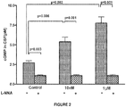
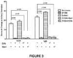


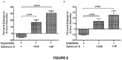
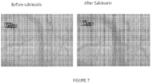


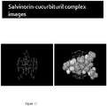
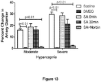

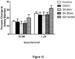


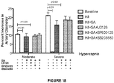
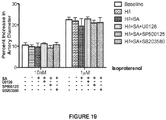
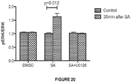

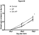
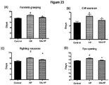
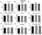


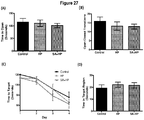


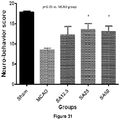

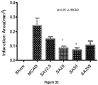


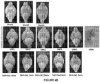
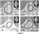
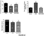
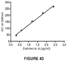
Classifications
-
- A—HUMAN NECESSITIES
- A61—MEDICAL OR VETERINARY SCIENCE; HYGIENE
- A61K—PREPARATIONS FOR MEDICAL, DENTAL OR TOILETRY PURPOSES
- A61K31/00—Medicinal preparations containing organic active ingredients
- A61K31/33—Heterocyclic compounds
- A61K31/335—Heterocyclic compounds having oxygen as the only ring hetero atom, e.g. fungichromin
- A61K31/365—Lactones
- A61K31/366—Lactones having six-membered rings, e.g. delta-lactones
-
- A—HUMAN NECESSITIES
- A61—MEDICAL OR VETERINARY SCIENCE; HYGIENE
- A61K—PREPARATIONS FOR MEDICAL, DENTAL OR TOILETRY PURPOSES
- A61K47/00—Medicinal preparations characterised by the non-active ingredients used, e.g. carriers or inert additives; Targeting or modifying agents chemically bound to the active ingredient
- A61K47/30—Macromolecular organic or inorganic compounds, e.g. inorganic polyphosphates
- A61K47/36—Polysaccharides; Derivatives thereof, e.g. gums, starch, alginate, dextrin, hyaluronic acid, chitosan, inulin, agar or pectin
- A61K47/40—Cyclodextrins; Derivatives thereof
-
- A—HUMAN NECESSITIES
- A61—MEDICAL OR VETERINARY SCIENCE; HYGIENE
- A61K—PREPARATIONS FOR MEDICAL, DENTAL OR TOILETRY PURPOSES
- A61K9/00—Medicinal preparations characterised by special physical form
- A61K9/0012—Galenical forms characterised by the site of application
- A61K9/0043—Nose
-
- A—HUMAN NECESSITIES
- A61—MEDICAL OR VETERINARY SCIENCE; HYGIENE
- A61K—PREPARATIONS FOR MEDICAL, DENTAL OR TOILETRY PURPOSES
- A61K9/00—Medicinal preparations characterised by special physical form
- A61K9/08—Solutions
-
- A—HUMAN NECESSITIES
- A61—MEDICAL OR VETERINARY SCIENCE; HYGIENE
- A61P—SPECIFIC THERAPEUTIC ACTIVITY OF CHEMICAL COMPOUNDS OR MEDICINAL PREPARATIONS
- A61P25/00—Drugs for disorders of the nervous system
-
- A—HUMAN NECESSITIES
- A61—MEDICAL OR VETERINARY SCIENCE; HYGIENE
- A61P—SPECIFIC THERAPEUTIC ACTIVITY OF CHEMICAL COMPOUNDS OR MEDICINAL PREPARATIONS
- A61P9/00—Drugs for disorders of the cardiovascular system
-
- A—HUMAN NECESSITIES
- A61—MEDICAL OR VETERINARY SCIENCE; HYGIENE
- A61P—SPECIFIC THERAPEUTIC ACTIVITY OF CHEMICAL COMPOUNDS OR MEDICINAL PREPARATIONS
- A61P9/00—Drugs for disorders of the cardiovascular system
- A61P9/08—Vasodilators for multiple indications
Definitions
- the invention relates to salvinorin compositions and to salvinorin compositions for use in treating diseases and disorders associated with vasoconstriction, vaso-occlusion, or disruption of blood flow and autoregulation.
- Salvinorin A is an active component of Salvia divinorum, a perennial herb of the Lamiaceae (mint) family, indigenous to Mexico. Salvia divinorum has long been traditionally used to produce visionary states of consciousness during spiritual healing sessions for religious purposes. It has been shown that salvinorin A is the most highly efficacious, naturally-occurring, nonpeptide, and the only non-nitrogenous kappa opioid receptor (KOR) agonist.
- KOR non-nitrogenous kappa opioid receptor
- Salvia divinorum as a naturally abundant plant has been used by human beings for recreational purposes for several centuries, and it has been proposed that salvinorin A could be a potential new opioid receptor agonist to be used in clinical practice, i.e. to treat depression or addiction etc. None of the other opioid KOR agonists have been used clinically so far because of their side effects. These include induction of significant dysphoria, low selectivity, respiratory depression, and unknown safety profiles. Salvinorin A does not belong to opioids despite being a KOR agonist, and it is not a controlled substance in most countries. Many intrinsic characters of the compound, i.e. quick onset, short acting, readily cross the blood brain barrier, and no respiratory depression, etc., make it an attractive possible medication, especially for neurological diseases.
- CA Cardiac arrest
- IR ischemia/reperfusion
- therapeutic hypothermia is considered the only effective post-CA treatment for neurological injuries.
- this treatment can only be applied to eligible patient populations and can be dangerous in under-resourced facilities, thereby leading to, among other things, an increased incidence of mortality, multiple organ failure, pulmonary hypertension, and bleeding, Therefore, a significant medical need exists for an easily manageable medication to improve survival rate and reduce the burden of neuronal injury from CA.
- Stroke is a leading cause of death worldwide. Each year, about 795,000 people experience a new or recurrent stroke in the United States; every 40 seconds, someone in the U.S. has a stroke; every 4 minutes, someone dies from a stroke. Stroke is a leading cause of serious long-term disability. 87% of all strokes are ischemic when blood supply to the brain is interrupted, a typical example of ischemia/reperfusion (IR) organ injury. Many potential neuroprotectants that reduce such neuronal injuries in experimental animals have failed in clinical trials. Due to the narrow therapeutic window of stroke, a potential neuroprotectant has to be able to be delivered in a timely manner without significant barrier with quick onset time.
- IR ischemia/reperfusion
- SAH Subarachnoid hemorrhage
- Salvinorin A is a very hydrophobic molecule and is insoluble in water. Salvinorin A is soluble in known to be soluble in organic solvents like ethanol, DMSO, and acetone. However, these solvent are not suitable for routine clinical use, especially for intravenous (IV) delivery. Since salvinorin A is useful clinically, for neurological disorders, there is a need to identify materials, preferably FDA approved materials, which can be used to formulate salvinorin A for clinical delivery.
- the present invention provides a pharmaceutical composition of salvinorin A comprising: an aqueous solution of salvinorin A and between 1% (w/v) and 50% (w/v) of a cyclodextrin, wherein the cyclodextrin is a 2-hydroxypropyl-cyclodextrin, and wherein the salvinorin A concentration is at least 50 ⁇ g/mL.
- the cyclodextrin is 2-hydroxypropyl- ⁇ -cyclodextrin (HPBCD).
- the cyclodextrin concentration is about 20% (w/v).
- the present invention also provides the pharmaceutical composition of salvinorin A of the invention for use in treating cerebral vasospasm in a subject with a subarachnoid hemorrhage, or for use in treating neurological injuries associated with cardiac arrest or increasing the likelihood of survival in a subject suffering from or having suffered cardiac arrest, or for use in treating a disease associated with cerebrovasodilation in a subject, or for use in treating a stroke in a subject suffering from or having suffered a stroke, or for use in treating a cerebral artery occlusion in a subject suffering from or having suffered a cerebral artery occlusion or cerebral hypoxia/ischemia.
- the present application relates to salvinorin compositions and uses thereof. Specifically, the application relates to salvinorin compositions for use in increasing survival of a subject following cardiac arrest or stroke in that subject, or for use in treating a subject with cerebral vascular spasm, a subarachnoid hemorrhage, cerebral hypoxia/ischemia, cerebral artery occlusion, or any condition involved in autoregulation impairment.
- the present invention thus provides a pharmaceutical composition of salvinorin A comprising: an aqueous solution of salvinorin A and between 1% (w/v) and 50% (w/v) of a cyclodextrin, wherein the cyclodextrin is a 2-hydroxypropyl-cyclodextrin, and wherein the salvinorin A concentration is at least 50 ⁇ g/mL.
- the cyclodextrin is 2-hydroxypropyl- ⁇ -cyclodextrin (HPBCD).
- the cyclodextrin concentration is about 20% (w/v).
- the present invention also provides the pharmaceutical composition of salvinorin A of the invention for use in treating cerebral vasospasm in a subject with a subarachnoid hemorrhage, or for use in treating neurological injuries associated with cardiac arrest or increasing the likelihood of survival in a subject suffering from or having suffered cardiac arrest, or for use in treating a disease associated with cerebrovasodilation in a subject, or for use in treating a stroke in a subject suffering from or having suffered a stroke, or for use in treating a cerebral artery occlusion in a subject suffering from or having suffered a cerebral artery occlusion or cerebral hypoxia/ischemia.
- methods of increasing the likelihood of survival in a subject suffering from or having suffered cardiac arrest comprising: administering to said subject a therapeutically effective amount of salvinorin or a pharmaceutical composition thereof.
- Also disclosed herein are methods of treating cerebral vasospasm in a subject with a subarachnoid hemorrhage comprising: administering to said subject a therapeutically effective amount of salvinorin or a pharmaceutical composition thereof.
- methods for treating a disease associated with vasoconstriction, vaso-occlusion, or disruption of blood flow and autoregulation in a subject comprising: administering to said subject a therapeutically effective amount of salvinorin or a pharmaceutical composition thereof.
- methods for producing a sedative or anesthetic effect in a subject in need thereof comprising: administering to said subject a therapeutically effective amount of salvinorin or pharmaceutical composition thereof.
- the pharmaceutical compositions comprise a therapeutically effective amount of salvinorin, wherein said salvinorin is present in an amount effective to produce cerebrovasodilation in a subject in need thereof, in an amount effective to treat a disease associated with cerebrovasospasm, hypoxia, and/or ischemia in a subject, in an amount therapeutically effective to treat cerebral vasospasm in a subject with a subarachnoid hemorrhage, in an amount effective to provide organ protection from hypoxia/ischemia in a subject, or in an amount effective to treat a disease associated with vasoconstriction, vaso-occlusion, or disruption of blood flow and autoregulation in a subject.
- compositions comprise a therapeutically effective amount of salvinorin, wherein said salvinorin is present in an amount effective to produce a sedative or antinociceptive effect in a subject in need thereof.
- the pharmaceutical compositions comprise an aqueous solution of salvinorin A and a cyclodextrin.
- the cyclodextrin is a 2-hydroxypropyl-cyclodextrin, such as 2-hydroxypropyl- ⁇ -cyclodextrin (HPBCD) or 2-hydroxypropyl- ⁇ -cyclodextrin (HPGCD). More preferably, the cyclodextrin is 2-hydroxypropyl- ⁇ -cyclodextrin (HPBCD).
- the composition is adapted for intravenous administration.
- the salvinorin A concentration is at least is at least 50 ⁇ g/mL.
- the cyclodextrin concentration in the solution is between 1% (w/v) and 50% (w/v).
- the cyclodextrin (e.g., HPBCD) concentration in the aqueous salvinorin A solution is at least 1% (w/v), at least 2.5% (w/v), at least 5% (w/v), at least 7.5% (w/v), at least 10% (w/v), at least 12.5% (w/v), at least 15% (w/v), at least 17.5% (w/v), at least 20% (w/v), at least 22.5% (w/v), or at least 25% (w/v).
- the cyclodextrin (e.g., HPBCD) concentration in the aqueous salvinorin A solution is less than 50% (w/v), less than 45% (w/v), less than 40% (w/v), less than 35% (w/v), less than 30% (w/v), less than 25% (w/v), less than 22.5% (w/v), less than 20% (w/v), less than 17.5% (w/v), less than 15% (w/v).
- the cyclodextrin is HPBCD with a concentration in the aqueous salvinorin A solution of aboutt least 20% (w/v).
- the inventor of the instant application surprisingly and unexpectedly found that salvinorin dilates cerebral vessels dramatically with rapid onset and offset, and without a change in hemodynamics.
- the diameter of the cerebral artery dilated up to 40% with 1 micromolar salvinorin as shown in Figure 7 .
- the vessels dilated immediately after application of salvinorin and the dilation effect lasted less than 3 to 5 minutes.
- salvinorin can be used to treat cerebral vascular spasm in stroke, brain injury, or other related clinical situations associated with cerebral vascular spasm, including post subarachnoid hemorrhage and head trauma.
- salvinorin may be used to treat towards spinal cord ischemia and other nerve ischemia.
- Salvinorin A and its analogues are known compounds.
- Salvinorin A the active component of Salvia divinorum, which is used by nearly a million people for recreational purposes annually in United States, is the only known non-nitrogenous selective kappa opioid receptor (KOR) agonist.
- KOR kappa opioid receptor
- a diterpene salvinorin A has been shown to be a high affinity and selective kappa opioid receptor agonist. See Roth et al., Proc. Natl. Acad. Sci. USA 99:11934 (2002 ); and Butelman et al., Psychopharmacology 172:220 (2004 ).
- Salvinorins and their derivatives are known in the art. For example, salvinorins, their derivatives, and methods for synthesizing them are described in U.S. 2006/0052439 , U.S. 2007/0213394 , WO 2005/089745 , and WO2008/119097 .
- salvinorin or its derivative may be used in the methods, compositions and uses described herein.
- known salvinorins include, but are not limited to, salvinorin A, B, C, D, E, or F.
- the salvinorin is salvinorin A.
- salvinorin is an analogue of salvinorin A.
- herkinorin an analogue of salvinorin A ( Ji F, et al., Brain Res. 2013, 1490:95-100 ).
- administering a therapeutically effective amount of a salvinorin produces vasodilation in a subject in need thereof.
- compositions described herein may include a "therapeutically effective amount.”
- a “therapeutically effective amount” refers to an amount effective, at dosages and for periods of time necessary, to achieve the desired therapeutic result.
- a therapeutically effective amount of a molecule may vary according to factors such as the disease state, age, sex, and weight of the individual, and the ability of the molecule to elicit a desired response in the individual.
- a therapeutically effective amount is also one in which toxic or detrimental effects of the molecule are outweighed by the therapeutically beneficial effects.
- treating neurological injuries associated with cardiac arrest includes, but is not limited to, preventing neurological injuries in a subject suffering from or having suffered cardiac arrest.
- Beneficial or desired clinical results include, but are not limited to, alleviation of symptoms, diminishment of the extent of a disease or condition, stabilization of a disease or condition ( i.e.
- Treatment can also mean prolonging survival as compared to expected survival if not receiving treatment.
- Those in need of treatment include those already with the disease or condition as well as those prone to having the disease or condition or those in which the disease or condition is to be prevented.
- diseases or disorders caused by or otherwise associated with vasoconstriction, vaso-occlusion, or disruption of blood flow and autoregulation include, but are not limited to, a cerebral vascular spasm, a subarachnoid hemorrhage, a stroke, a brain trauma or injury, an ischemia reperfusion injury, low perfusion status, and hypoxia.
- compositions described herein can administered to a subject by a method known in the art, such as parenterally, paracancerally, transmucosally, transdermally, intramuscularly, intravenously, intra-dermally, subcutaneously, intra-peritonealy, intra-ventricularly, intracranially, intra-vaginally or intra-tumorally, intrathecally, and inhalationally.
- they are administered transmucosally.
- they are administered intranasally.
- the pharmaceutical compositions are to be administered orally, and are thus formulated in a form suitable for oral administration, i.e. as a solid or a liquid preparation.
- suitable solid oral formulations include tablets, capsules, pills, granules, pellets and the like.
- Suitable liquid oral formulations include solutions, suspensions, dispersions, emulsions, oils and the like.
- the active ingredient is formulated in a capsule.
- the compositions of the present invention comprise, in addition to the active compound and the inert carrier or diluent, a hard gelating capsule.
- the pharmaceutical compositions are to be administered by intravenous, intra-arterial, or intra-muscular injection of a liquid preparation.
- suitable liquid formulations include solutions, suspensions, dispersions, emulsions, oils and the like.
- the pharmaceutical compositions are to be administered intravenously and are thus formulated in a form suitable for intravenous administration.
- the pharmaceutical compositions are to be administered intra-arterially and are thus formulated in a form suitable for intra-arterial administration.
- the pharmaceutical compositions are to be administered intra-muscularly and are thus formulated in a form suitable for intra-muscular administration.
- compositions are to be administered intranasally and are thus formulated in a form suitable for intranasal administration.
- compositions are to be administered topically to body surfaces and are thus formulated in a form suitable for topical administration.
- Topical formulations include gels, ointments, creams, lotions, drops and the like.
- the pharmaceutical composition is to be administered as a suppository, for example, a rectal suppository or a urethral suppository.
- the pharmaceutical composition is to be administered by subcutaneous implantation of a pellet.
- the pellet provides for controlled release of active agent over a period of time.
- the active compound is delivered in a vesicle, e.g. a liposome.
- carriers or diluents used in compositions of the present invention include, but are not limited to, a gum, a starch (e.g. corn starch, pregeletanized starch), a sugar (e.g., lactose, mannitol, sucrose, dextrose), a cellulosic material (e.g. microcrystalline cellulose), an acrylate (e.g. polymethylacrylate), calcium carbonate, magnesium oxide, talc, or mixtures thereof.
- a gum e.g. corn starch, pregeletanized starch
- a sugar e.g., lactose, mannitol, sucrose, dextrose
- a cellulosic material e.g. microcrystalline cellulose
- an acrylate e.g. polymethylacrylate
- pharmaceutically acceptable carriers for liquid formulations are aqueous or non-aqueous solutions, suspensions, emulsions or oils.
- non-aqueous solvents are propylene glycol, polyethylene glycol, and injectable organic esters such as ethyl oleate.
- Aqueous carriers include water, alcoholic/aqueous solutions, emulsions or suspensions, including saline and buffered media.
- oils are those of animal, vegetable, or synthetic origin, for example, peanut oil, soybean oil, olive oil, sunflower oil, fish-liver oil, another marine oil, or a lipid from milk or eggs.
- parenteral vehicles for subcutaneous, intravenous, intra-arterial, or intramuscular injection
- parenteral vehicles include sodium chloride solution, Ringer's dextrose, dextrose and sodium chloride, lactated Ringer's and fixed oils.
- Intravenous vehicles include fluid and nutrient replenishers, electrolyte replenishers such as those based on Ringer's dextrose, and the like.
- sterile liquids such as water and oils, with or without the addition of a surfactant and other pharmaceutically acceptable adjuvants.
- water, saline, aqueous dextrose and related sugar solutions, and glycols such as propylene glycols or polyethylene glycol are preferred liquid carriers, particularly for injectable solutions.
- oils are those of animal, vegetable, or synthetic origin, for example, peanut oil, soybean oil, olive oil, sunflower oil, fish-liver oil, another marine oil, or a lipid from milk or eggs.
- compositions further comprise binders (e.g., acacia, cornstarch, gelatin, carbomer, ethyl cellulose, guar gum, hydroxypropyl cellulose, hydroxypropyl methyl cellulose, povidone), disintegrating agents (e.g., binders, e.g., acacia, cornstarch, gelatin, carbomer, ethyl cellulose, guar gum, hydroxypropyl cellulose, hydroxypropyl methyl cellulose, povidone), disintegrating agents (e.g.
- binders e.g., acacia, cornstarch, gelatin, carbomer, ethyl cellulose, guar gum, hydroxypropyl cellulose, hydroxypropyl methyl cellulose, povidone
- disintegrating agents e.g.
- cornstarch potato starch, alginic acid, silicon dioxide, croscarmelose sodium, crospovidone, guar gum, sodium starch glycolate), buffers (e.g., Tris-HCI., acetate, phosphate) of various pH and ionic strength, additives such as albumin or gelatin to prevent absorption to surfaces, detergents (e.g., Tween 20, Tween 80, Pluronic F68, bile acid salts), protease inhibitors, surfactants (e.g., sodium lauryl sulfate), permeation enhancers, solubilizing agents (e.g., glycerol, polyethylene glycerol), antioxidants (e.g., ascorbic acid, sodium metabisulfite, butylated hydroxyanisole), stabilizers (e.g.
- viscosity increasing agents e.g. carbomer, colloidal silicon dioxide, ethyl cellulose, guar gum
- sweeteners e.g. aspartame, citric acid
- preservatives e.g., Thimerosal, benzyl alcohol, parabens
- lubricants e.g. stearic acid, magnesium stearate, polyethylene glycol, sodium lauryl sulfate
- flow-aids e.g. colloidal silicon dioxide
- plasticizers e.g. diethyl phthalate, triethyl citrate
- emulsifiers e.g.
- carbomer hydroxypropyl cellulose, sodium lauryl sulfate
- polymer coatings e.g., poloxamers or poloxamines
- coating and film forming agents e.g. ethyl cellulose, acrylates, polymethacrylates
- the pharmaceutical compositions provided herein are controlled-release compositions, i.e. compositions in which the active compound is released over a period of time after administration.
- Controlled- or sustained-release compositions include formulation in lipophilic depots (e.g. fatty acids, waxes, oils).
- the composition is an immediate-release composition, i.e. a composition in which of the active compound is released immediately after administration.
- the pharmaceutical composition is delivered in a controlled release system.
- the agent may be administered using intravenous infusion, an implantable osmotic pump, a transdermal patch, liposomes, or other modes of administration.
- a pump may be used (see Langer, supra; Sefton, CRC Crit. Ref. Biomed. Eng. 14:201 (1987 ); Buchwald et al., Surgery 88:507 (1980 ); Saudek et al., N. Engl. J. Med. 321:574 (1989 )).
- polymeric materials are used; e.g. in microspheres in or an implant.
- a controlled release system is placed in proximity to the target cell, thus requiring only a fraction of the systemic dose (see, e.g., Goodson, in Medical Applications of Controlled Release, supra, vol. 2, pp. 115-138 (1984 ); and Langer R, Science 249: 1527-1533 (1990 ).
- Also comprehended by the invention are compounds modified by the covalent attachment of water-soluble polymers such as polyethylene glycol, copolymers of polyethylene glycol and polypropylene glycol, carboxymethyl cellulose, dextran, polyvinyl alcohol, cyclodextrin, cucurbituril, polyvinylpyrrolidone or polyproline.
- the modified compounds are known to exhibit substantially longer half-lives in blood following intravenous injection than do the corresponding unmodified compounds (Abuchowski et al., 1981; Newmark et al., 1982; and Katre et al., 1987).
- Such modifications may also increase the compound's solubility in aqueous solution, eliminate aggregation, enhance the physical and chemical stability of the compound, and greatly reduce the immunogenicity and reactivity of the compound.
- the desired in vivo biological activity may be achieved by the administration of such polymer-compound abducts less frequently or in lower doses than with the unmodified compound.
- the methods comprise administering an active salvinorin composition as the sole active ingredient.
- the active salvinorin composition for use in the treatment of diseases and disorders in combination with one or more additional therapeutic agents. These additional agents are appropriate for the disease or disorder that is being treated, as is known in the art.
- the other therapeutically effective agent may be conjugated to the salvinorin, incorporated into the same composition as the salvinorin, or may be administered as a separate composition.
- the other therapeutically agent or treatment may be administered prior to, during and/or after the administration of the salvinorin.
- the administration of the salvinorin with other agents and/or treatments may occur simultaneously, or separately, via the same or different route, at the same or different times. Dosage regimens may be adjusted to provide the optimum desired response (e.g. , a therapeutic or prophylactic response).
- Effective doses of the compositions described herein, for treatment of conditions or diseases as described herein vary depending upon many different factors, including means of administration, target site, physiological state of the patient, whether the patient is human or an animal, other medications administered, and whether treatment is prophylactic or therapeutic.
- the patient is a human but non-human mammals including transgenic mammals can also be treated.
- Treatment dosages may be titrated using routine methods known to those of skill in the art to optimize safety and efficacy.
- Dosage unit form refers to physically discrete units suited as unitary dosages for treating mammalian subjects. Each unit may contain a predetermined quantity of active compound calculated to produce a desired therapeutic effect. In some embodiments, the dosage unit forms are dictated by and directly dependent on the unique characteristics of the active compound and the particular therapeutic or prophylactic effect to be achieved.
- the composition may be administered only once, or it may be administered multiple times or continuous infusion.
- the composition may be, for example, administered three times a day, twice a day, once a day, once every two days, twice a week, weekly, once every two weeks, or monthly.
- Dosage values may vary with the type and severity of the condition to be alleviated. It is further understood that for any particular subject, specific dosage regimens may be adjusted over time according to the individual need and the professional judgment of the person administering or supervising administration of the compositions, and that dosage ranges set forth herein are exemplary and are not intended to limit the scope or practice of the methods.
- administering to a subject is not limited to any particular delivery system and may include, without limitation, parenteral (including subcutaneous, intravenous, intramedullary, intraarticular, intramuscular, or intraperitoneal injection) rectal, topical, transdermal or oral (for example, in capsules, suspensions or tablets), intrathecal, intranasal and inhalational.
- Administration to a subject may occur in a single dose or in repeat administrations or continuous infusion, and in any of a variety of physiologically acceptable salt forms, and/or with an acceptable pharmaceutical carrier and/or additive as part of a pharmaceutical composition.
- physiologically acceptable salt forms and standard pharmaceutical formulation techniques are known to persons skilled in the art (see, for example, Remington's Pharmaceutical Sciences, Mack Publishing Co. ).
- the salvinorin compositions described herein can be used to treat a suitable mammal, including primates, such as monkeys and humans, horses, cows, cats, dogs, rabbits, and rodents such as rats and mice.
- a suitable mammal including primates, such as monkeys and humans, horses, cows, cats, dogs, rabbits, and rodents such as rats and mice.
- the mammal to be treated is human.
- Salvinorin A Produces Cerebrovasodilation through Activation of Nitric Oxide Synthase, Kappa Receptor and Adenosine Triphosphate Sensitive Potassium Channel
- Salvinorin A purity ⁇ 98%), sodium nitroprusside (SNP), N(G)-nitro-L-arginine (L-NNA), glibenclamide, iberiotoxin, cromakalim, calcitonin-gene related polypeptide (CGRP), NS 1619, naloxone, methionine enkephalin, norbinaltorphimine, 7-nitroindazole(7-NINA), sulpiride and isoproterenol are obtained from Sigma-Aldrich (St. Louis, MO, USA). All other chemicals were also obtained from Sigma and were of reagent grade.
- Newborn pigs (1-6 days old, weighing 1.3-1.8 kg) of both genders were used for this study. Protocols were approved by the Institutional Animal Care and Use Committee of the University of Pennsylvania. The animals were induced with isoflurane (1-2 minimal alveolar concentration) and then maintained with alpha-chloralose (80-100mg/kg supplemented with 5mg/kg/h IV). Both femoral arteries were catheterized to monitor blood pressure and blood gas. A catheter was inserted into right femoral vein for medication administration. The animals were ventilated with room air after trachea cannulation. Rectal temperature was maintained at 37-39 °C by a heating pad. A closed cranial window was placed for direct pial artery visualization and diameter measurement.
- the closed cranial window consisted of three parts: a stainless steel ring, a circular glass cover-slip, and three ports consisting of 17-gauge hypodermic needles attached to three precut holes in the stainless steel ring.
- Cortical periarachnoid cerebrospinal fluid (CSF) was collected through the cranial window port for cyclic guanosine monophosphate (cGMP) determination.
- CSF Cortical periarachnoid cerebrospinal fluid
- cGMP cyclic guanosine monophosphate
- the space under the window was filled with artificial CSF with the following composition (in mM): 3.0 KCl, 1.5 MgCl 2 , 1.5 CaCl 2 , 132 NaCl, 6.6 Urea, 3.7 Dextrose, and 24.6 NaHCO 3 per liter, pH 7.33, PCO 2 46 mmHg and PO 2 43 mmHg.
- the artificial CSF was warmed to 37-38 °C before application to the cerebral cortical surface.
- Pial arteries were observed with a television camera mounted on a dissecting microscope. Vascular diameter was measured from a video monitor connected the camera with a video microscaler (model VPA 550, For-A-Corp., Los Angeles, CA).
- Pial artery diameter small artery diameter 120-160 micro meter; arteriole diameter 50-70 micro meter
- the window was flushed over 30 s with 1-2 ml CSF through the port connected into the side of the window.
- CSF samples were collected for cGMP analysis before and at 10 min after medication administration.
- We collected the cerebral cortical periarachnoid CSF by slowly infusing CSF into one port of the window and allowing the CSF to drip freely into a collection tube on the opposite port.
- pial artery diameter changes were recorded after SNP (100pM), a sub threshold vascular concentration of a nitric oxide donor, administration together with L-NNA and salvinorin A.
- sulpiride 100nM
- a dopamine receptor D2 antagonist glibenclamide (100 nM), a K ATP channel antagonist
- iberiotoxin 100 nM, Sigma-Aldrich
- K Ca channel antagonist on pial artery response to salvinorin A cromakalim(1 ⁇ M) and calcitonin-gene related polypeptide (10 nM, 1 ⁇ M), a K ATP agonist, and NS 1619(10 nM, 1 ⁇ M), a KCa channel agonist, were also determined.
- naloxone (1 mg/kg IV) and norbinaltorphimine (1 ⁇ M topical administration, a kappa opioid receptor antagonist, on the response to the salvinorin A, methionine enkephalin (10 nM, 1 ⁇ M) and isoproterenol (10 nM, 1 ⁇ M), a beta adrenergic receptor agonist, were investigated. All tested drug solutions were made fresh on the day of use. Pial artery response to salvinorin A administrated every 2 minutes was recorded for 30 min to determine whether the sustained vascular dilatation could be achieved.
- CSF samples were collected for cGMP determination before and after salvinorin A administration with or without L-NNA and norbinaltorphimine pretreatment.
- Commercially available ELISA kits (Enzo Life Sciences International, Inc. Plymouth Meeting, PA) were used to quantify cGMP concentration.
- the dilation effect is observed immediately after salvinorin administration and the duration of dilation lasted less than 5 minutes for both doses.
- the salvinorin A was administration administered every 2 minutes, sustained dilation effects was observed as shown in Fig. 1B .
- the dilation response was abolished by L-NNA, the NOS inhibitor, but not 7-nitroindazole (100 nM), the antagonist of neuronal NOS (nNOS) ( Fig. 1A,C ).
- Dilation response to SNP was not affected by L-NNA ( Fig. 1A ).
- SNP 100pM had no effects on the pial artery, but it restored the constriction induced by L-NNA. However, it didn't restore the dilation response of salvinorin A blocked by the L-NNA (Fig. ID). Dilation in response to salvinorin A was associated with elevated cGMP in CSF, and L-NNA blocked the elevation of the cGMP ( Fig. 2 ). No significant blood pressure changes occurred during salvinorin administration.
- Glibenclamide (100nM), the K ATP channel inhibitor, but not iberiotoxin(100nM), the Ca2+-activated K+ (KCa) channel inhibitor, blocked the dilation effects of salvinorin A.
- Glibenclamide with iberiotoxin in any sequence also blocked the dilation induced by salvinorin A ( Fig. 3 ).
- Glibenclamide (100nM) but not iberiotoxin (100 nM) blocked the dilation in response to cromakalim (an agonist of K ATP channel, 10 nM and 1 ⁇ M) and calcitonin-gene related polypeptide (another K ATP channel agonist, 10 nM and 1 ⁇ M); iberiotoxin (100 nM) but not glibenclamide (100nM) blocked the dilation effects of NS1619 (KCa channel agonist, 10 nM and 1 ⁇ M) ( Fig. 4 ).
- salvinorin A is demonstrated to be a potent pial artery dilator in piglet in normal and vessel constricted conditions induced by endothelin and hypocarbia.
- the dilatation effect was observed immediately after salvinorin administration, lasted less than 5 min for both tested doses, and was dose-dependent.
- Sustained dilation effects of salvinorin A was observed with continuous administration.
- the activation of the opioid receptor, NOS and K ATP channel were involved in the signal pathway of such dilation effects.
- the unique structure of salvinorin A contributes to its short acting character, because continuous administration results in a sustained dilatory effect.
- the ester linkage in its structure can be easily metabolized by esterase in the blood and tissues.
- carboxylesterase mainly involved in salvinorin A hydrolysis in rat plasma and the degradation products include salvinorin B and the lactone-ring-open forms of salvinorin A, both of which are not pharmacologically active.
- KOR agonist BRL 52537 Similar to other KOR agonists, the activation of NOS, K ATP channels and opioid receptors mediated the dilation effects of salvinorin A. Another group has demonstrated that selective KOR agonist BRL 52537 protected the ischemia brain via attenuating nitric oxide production in ischemic striatum. It was proved that the neuroprotection of BRL 52537 was lost in nNOS null mice. Therefore, the KOR agonist BRL 52537 attenuated nNOS activity and ischemia-evoked nitric oxide production.
- K ATP channels activation may result in hyperpolarization of the membrane of vascular smooth muscle cell. Membrane potential changes would then regulate muscle relaxation through alterations in Ca 2+ influx through voltage-dependent Ca 2+ channels.
- This Example demonstrates that salvinorin A activates the K ATP channel directly or indirectly. Different from many other agents that can activate K ATP channel, salvinorin A can easily penetrate the blood-brain barrier. Since the K ATP channel plays a crucial protective role against brain injury from hypoxia, ischemia or metabolic inhibition, salvinorin A can be a neuro-protective agent for clinical use.
- vascular dilative effect observed in normal and constricted cerebral vessels by both hypocarbia and endothelin can allow for its clinical application to treat cerebral vessel spasm in many clinical situations including migraine and cerebral vascular spasm after subarachnoid hemorrhage in which increase of endothelin plays an important role. Similar to other short acting agents, continuous infusion could be used to titrate and achieve sustained effects by adjusting dosage.
- newborn piglets were used as the study subject.
- the gyrencephalic brain of pig has more white than gray matter which is selectively vulnerable to injury similar to the human, and also similar in maturity.
- the newborn piglet was also used because it is large enough for easy cranial window placement and vascular visualization.
- the newborn's cerebral vascular responses are similar to that in human subjects in many clinical situations.
- salvinorin A is a fast and short acting potent pial artery dilator in piglet in normal and vessel constricted conditions induced by endothelin or hypocarbia.
- the mechanism involves the activation of NOS, K ATP channel and kappa receptor.
- HIE moderate hypoxic-ischemic encephalopathy
- KOR agonists cause dilation of cerebral vessels, a key feature required to maintain cerebral autoregulation and reduce brain injury from ischemia. After ischemia, cerebral autoregulation is impaired, resulting in decreased cerebral blood flow and neuron death.
- ischemia cerebral autoregulation is impaired, resulting in decreased cerebral blood flow and neuron death.
- Salvinorin A (purity ⁇ 98%) was obtained from ChromaDex, Inc. (Irvine, CA, USA).
- Isoproterenol (ISO), nor-binaltorphimine (Nor-BIN) were obtained from Sigma-Aldrich (MO, St. Louis, MO, USA). All other chemicals (reagent grade) were obtained from Sigma as well.
- Newborn pigs (1-5 days old, 1.2-1.5 kg) of both sexes were used in this study.
- the animal experimental protocol was approved by the Institutional Animal Care and Use Committee of the University of Pennsylvania.
- Isoflurane (1 to 2 minimum alveolar concentration) was initially used for anesthesia induction, followed by ⁇ -chloralose (30-50 mg/kg supplemented with 5 mg/kg/hr intravenously) for maintenance of anesthesia.
- ⁇ -chloralose (30-50 mg/kg supplemented with 5 mg/kg/hr intravenously) for maintenance of anesthesia.
- the animals were initially ventilated with room air and kept warm with heating pad to maintain the rectal temperature at 37-39 °C.
- Bilateral femoral arteries were catheterized to monitor the blood pressure, blood gas tensions and pH.
- the femoral vein was catheterized for medication administration.
- a closed cranial window consisting of a steel ring with a glass cover slip, connecting to 3 ports, was placed for direct pial artery visualization and diameter measurement.
- Small pial arteries 120 to 160 ⁇ m
- arterioles 50 to 70 ⁇ m
- the ports attached to the cranial window ring fit 17-gauge hypodermic needles for CSF sampling, washout, and drug administration.
- Cortical periarachnoid CSF was collected at baseline and 60 min after HI for ERK activity analysis.
- hypoxia was induced for 10 minutes by switching room air to N2 for ventilation, followed by restoring ventilation to room air.
- Global cerebral ischemia was then induced by infusing saline through a hollow bolt in the cranium to maintain intracranial pressure higher than the mean blood pressure for 20 min.
- Global cerebral ischemia is confirmed when the blood flow in pial arteries were stopped visible on the monitor connected to the microscope over the cranial window.
- blood was withdrawn when necessary to maintain the mean arterial blood pressure below 100 mmHg. The blood was returned via femoral vein at the end of ischemia.
- DMSO group with DMSO (vehicle of salvinorin A) 1 ⁇ l/kg administrated immediately after HI
- SA 0min group with salvinorin A(1 ⁇ g/ ⁇ l in DMSO) 10 ⁇ g/kg immediately after HI
- SA 30min group with salvinorin A (1 ⁇ g/ ⁇ l in DMSO) 10 ⁇ g/kg 30 min after HI
- SA+Norbin group with salvinorin A (10 ⁇ g/kg) and nor-BIN (1 ⁇ M, topically injected through one port of cranial windows) immediately after HI.
- Pial artery responses to hypercapnia, hypotension and isoproterenol (10 nM, 1 ⁇ M) were obtained before HI and 60 minutes after HI as previously described.
- Isoproterenol was used as a positive control since it is a short acting agent and its vascular dilatation effect in such model is well established in our lab.
- Two levels of hypercapnia (PaCO 2 of 50 to 60 mmHg for low level, 70 to 80 mmHg for high level) were produced by inhalation of high concentration CO 2 mixture gas (10% CO 2 ; 21% O 2 ; 69% N 2 ).
- Two levels of hypotension were produced by the rapid withdrawal of either 5-8 or 10-15 ml/Kg blood from the femoral artery to induce moderate and severe hypotension (25% decrease in mean blood pressure as moderate and 45% decrease as severe). Such decreases in blood pressure were maintained constantly for 10 min by withdrawal or reinfusion of additional blood.
- ERK activity were then determined from frozen CSF samples described above. The levels of pERK and ERK were measured by ELISA kits (Enzo Life Sciences International, Inc., Plymouth Meeting, PA).
- the data of pial artery diameter were analyzed by repeated-measures ANOVA followed by Bonferroni method as post hoc tests.
- One way ANOVA was used to compare the ERK activity changes (quantified as the ratio of pERK over ERK) in each group before and 60 min after HI and in the groups with or without salvinorin A administration.
- the baseline data were excluded in the repeated-measures analysis (Graph Pad Prism version 5.02).
- An alpha level of P ⁇ 0.05 was considered significant in all statistical tests. Values are represented as means ⁇ standard error.
- Salvinorin A preserves pial artery autoregulation to hypercapnia after global cerebral HI
- the small pial artery dilated in response to two levels of hypercapnia before HI (presented as baselines).
- the dilatation responses to hypercapnia were blunted after HI when DMSO was administrated immediately at the end of HI (ps ⁇ 0.01 as compared with the baselines before HI).
- Salvinorin A preserves pial artery autoregulation to hypotension after global cerebral HI
- pial artery responses to isoproterenol ( figure 15 ) were measured and no change was observed among all groups before and after HI.
- the ERK activities are quantified as the ratio of pERK/ERK levels in CSF.
- the ERK activity data in groups without salvinorin effects (DMSO group and SA+Norbin group; renamed as DMSO+Nornin group) are combined.
- the ERK activity in groups without salvinorin increased significantly 60 min after HI (p ⁇ 0.05 as compared with pre-HI baseline).
- the ERK activity of salvinorin A administration groups reduced to the baseline level.
- ischemic stroke Both birth asphyxia and pediatric ischemic stroke are the common complications of childbirth. Perinatal HI, occurring in both these complications, can induce severe and permanent neuropsychological deficits, including delayed cognitive and behavioral development, mental retardation, cerebral palsy, and epilepsy, which is devastating for the patients, the families, and society. Unfortunately, there is no medication available for effective perinatal HI management. Hypothermia is the only treatment for HIE to reduce negative complications. But it is not widely accepted in clinical practice or recommended to combine with pharmacologic agents. Recombinant tissue type plasminogen activator (t-PA), a FDA approved treatment for acute ischemic stroke, showed adverse effects including increased stroke infarct volume in mice subjected to induced stroke and impaired cerebral hemodynamics.
- t-PA tissue type plasminogen activator
- KOR agonists have been demonstrated in other ischemia animal models.
- KOR agonist BRL 52537 and CI-977 reduces cortical damages, including brain swelling and infarction volumes, in response to different levels of ischemia when administrated 30 min before or up to 6 hours after the insult.
- Salvinorin A as a novel medication
- KOR agonists exhibit tremendous therapeutic value, most KOR agonists have not been used in clinical settings because of their intrinsic characteristics as opioids, (low selectivity and/or lack of an acceptable safety profile).
- salvinorin A is the most potent, highly selective, and the only non-opioid KOR agonist known to be derived from natural sources.
- Salvinorin A is the active component of Salvia divinorum, a naturally abundant perennial herb that has been consumed by humans for recreational and sacred purposes for several centuries. Many intrinsic characteristics of this compound make it a potential therapeutic medication for various neurological conditions.
- Salvinorin A has been evaluated as a potential medication for depression. In addition to our findings demonstrating the protective effect of salvinorin A administration before HI insult, we have now demonstrated that salvinorin A administration (up to 30 min) after HI insult preserved the autoregulation of pial artery.
- ERK signaling stimulated by cerebral ischemia/reperfusion is a crucial pathway for HI injury.
- HI induced increases in the CSF ERK activities were blocked by salvinorin A, which promoted protection of cerebral autoregulation post insult. It is worth noting that the role of ERK signaling in HI may be different before and after HI insult. Activation of ERK signaling may be related to the protective effects of preconditioning.
- Salvinorin A pretreatment preserves cerebrovascular autoregulation after brain hypoxic/ischemic injury via ERK/MAPK in piglets
- Cerebral hypoxia/ischemia because of the interruption of cerebral blood flow during cardiopulmonary bypass with deep hypothermia circulation arrest (DHCA) surgery for congenital cardiac surgery is a significant clinical issue.
- Fifty percent of children with complex congenital heart disease undergoing cardiopulmonary bypass with DHCA have developmental deficits, such as disabilities in speech and attention deficit disorder by school age. Cerebral hypoxia /ischemia occurred during DHCA is predictable, thus, it is possible to minimize the brain injury induced by ischemia with pharmacologic approaches. Unfortunately, no pharmacological agent with proven clinical benefit has been identified yet.
- Loss of cerebral vascular autoregulation is one of the key features of cerebral hypoxia/ischemia.
- the loss of autoregulation to hypotension could result in a pressure passive cerebral circulation, which may decrease cerebral blood flow and further aggravate brain ischemia.
- Loss of cerebrovascular regulation to hypercapnia also contributes to the development of the pressure passive circulation and periventricular leukomalacia.
- preservation of cerebral vascular autoregulation from ischemia is very important to reduce the brain injury from ischemia.
- salvinorin A an active component of Salvia divinorum and a non-opioid kappa opioid receptor (KOR) agonist, is a potent cerebral vascular dilator in normal and pathological conditions.
- salvinorin A can protect cerebral vasculature from ischemia.
- salvinorin A has long been used by different ethnic groups for various purposes, including spiritual experiences and "treating" illnesses, indicating its high potential as a clinically acceptable medication.
- MAPK mitogen-activated protein kinase
- MAPK is a key intracellular signaling system, which includes extracellular signal regulated kinase (ERK), c-Jun-N-terminal kinase (JNK) and p38.
- ERK extracellular signal regulated kinase
- JNK c-Jun-N-terminal kinase
- p38 extracellular signal regulated kinase
- ERK extracellular signal regulated kinase
- JNK c-Jun-N-terminal kinase
- p38 extracellular signal regulated kinase
- ERK extracellular signal regulated kinase
- JNK c-Jun-N-terminal kinase
- p38 extracellular signal regulated kinase
- ERK extracellular signal regulated kinase
- JNK c-Jun-N-terminal kinase
- p38 extra
- Salvinorin A (purity ⁇ 98%) is from ChromaDex, Inc. (Irvine, CA, USA).
- Isoproterenol, U0126, sp600125 and sb203580 are obtained from Sigma-Aldrich (MO, St. Louis, MO, USA). All other chemicals were also obtained from Sigma and were of reagent grade.
- piglets One to five days old piglets were used. Protocols were approved by the Institutional Animal Care and Use Committee of the University of Pennsylvania (Philadelphia). Isoflurane (1-2 minimum alveolar concentration) was initially used for induction, followed by alpha-chloralose for maintenance of anesthesia (30-100 mg/kg, supplemented with 5-30mg/kg every 20-30min IV). After tracheotomy, piglets were mechanically ventilated with room air and kept warm with a heating pad, maintaining rectal temperature at 37 to 39°C. Femoral arteries were cannulated for continuous blood pressure monitoring or intermittent blood gas monitoring, and the femoral vein was catheterized for medication administration.
- a closed cranial window was placed for direct pial artery visualization and diameter measurement (21).
- Small pial artery (120 to 160 ⁇ m) and arteriole (50 to 70 ⁇ m) are identified under microscope, visualized on a monitor connected to the microscope, and measured via a video microscaler (model VPA 550, For-A-Corp., Los Angeles, CA).
- the cranial window is a steel ring with a glass cover slip, connecting to three ports for cerebrospinal fluid (CSF) sampling, washout and medicine administration.
- CSF cerebrospinal fluid
- Cortical periarachnoid CSF was collected through one of the above ports at baseline and 30 minutes after administration of salvinorin A or U0126 plus salvinorin A for ERK/MAPK analysis.
- hypoxia was induced for 10 minutes by switching room air to N2 for ventilation, followed by restoring ventilation to room air; and then global cerebral ischemia was induced by infusing saline through a hollow bolt in the cranium to maintain intracranial pressure higher than the mean blood pressure for 20 min.
- Global ischemia is confirmed when the blood flow in pial artery stopped, visualized on the monitor connected to the microscope over the cranial window.
- blood was withdrawn when necessary to maintain mean arterial blood pressure no higher than 100 mmHg. The blood was returned via femoral vein at the end of ischemia.
- sp600125 and sb203580 are administrated 30 minutes before salvinorin A.
- Sp600125 and sb203580 were co-administered with the vasoactive stimulus so as to have continued exposure of the cerebral cortical surface after injury.
- Hypercapnia (PaCO 2 of 50 to 60 mmHg for low level, 70 to 80 mmHg for high level) was produced by inhalation of high concentration CO 2 mixture gas (10% CO 2 ; 21% O 2 ; 69% N 2 ). Hypotension was produced by withdrawing blood from the femoral artery (25% decrease in mean blood pressure as moderate and 45% as severe). Pial artery responses to hypotension, hypercapnia, and isoproterenol (10 nM, 1 ⁇ M) were obtained before hypoxia/ischemia and 60 minutes after injury as described previously.
- MAPK isoforms were measured by commercially available ELISA kits (Enzo Life Sciences International, Inc., Plymouth Meeting, PA).
- Salvinorin A preserved pial artery autoregulation to hypotension after hypoxia/ischemia.
- Salvinorin A preserved pial artery autoregulation to hypercapnia after hypoxia/ischemia.
- pial artery response to isoproterenol was unchanged in all groups before and after hypoxia/ischemia in the presence or absence of the above interventions.
- the ratio of pERK/ERK in CSF increased significantly 30 minutes after salvinorin A pretreatment ( Fig. 20 ). However, if U0126, an antagonist of ERK, was administrated 30 minutes before salvinorin administration, the ratio of pERK/ERK was unchanged 30 minutes after salvinorin A pretreatment ( Fig. 20 ).
- cerebrovascular dysfunction plays a very important role in neurological insult after hypoxia/ischemia.
- the only medication approved by the Food and Drug Administration for stroke is recombinant tissue plasminogen activator (tPA).
- tPA tissue plasminogen activator
- L-NNA an inhibitor of nitric oxide synthesis, was proven to be able to restore cerebral vascular auto regulation administrated after ischemia, however, its safety profile is unclear for clinical usage.
- salvinorin A is extracted from an abundant natural plant, Salvia divinorum. Very similar to the history of opium, Salvia divinorum as a naturally abundant plant has been used by human beings for various purposes, for centuries. It has been proposed that salvinorin A, the active component of Salvia Divinorum, could be a potential new kappa agonist to be used in clinical practice.
- Salvinorin A is a potent cerebral vascular dilator in normal and constricted conditions as we have demonstrated. However, this dilatation effect is short lived unless with continued administration; thus, the preservation of autoregulation is unlike induced from the dilatation effects since salvinorin was administrated 30 min prior hypoxia/ischemia.
- MAPK has been proven to be important in signal transduction from the cell surface to the nucleus. Elevation of ERK/MAPK before ischemia is related to neuronal survival after ischemia(15). Activation of ERK/MAPK in the hippocampal CA1 region after sublethal ischemia correlates with neuro-protection induced by preconditioning.
- Exercise preconditioning reduces neuronal apoptosis in stroke by up-regulating ERK/MAPK.
- pERK/ERK in CSF increased significantly 30 minutes after administration of salvinorin A, indicating the activation of ERK and also suggesting that salvinorin A might be vascularly or neuronally protective when administered prior to brain ischemia.
- elevation of ERK/MAPK in the immediate post injury reperfusion period is associated with impairment of responses to cerebrovasodilators as well as histopathology after hypoxia/ischemia in the piglet (32).
- the reason for the observed duality of ERK/MAPK function is uncertain but may relate to the cellular site of origin, signal coupling, or temporal pattern of release.
- Newborn piglets used in this Example offer the unique advantage of a gyrencephalic brain containing substantial white matter, which is more sensitive to ischemic damage than rodent brain, and more similar to humans.
- salvinorin A pretreatment preserved cerebrovascular autoregulation to hypotension and hypercapnia after brain hypoxia/ischemia via ERK/MAPK in a piglet model.
- Salvinorin as an adjunctive medication for difficult airway management
- Salvinorin A meets these criteria, at least based on data available in the literature. It is a kappa opioid receptor agonist which has sedative and dissociative effects, inhibits cough and has minimal respiratory depressive effects, at least as compared to conventional mu receptor agonists (morphine).
- salvinorin can be useful during awake intubation.
- salvinorin is poorly soluble in water.
- One option is to use its salt form to increase water solubility, and the other is to use lipid emulsion. Both techniques are readily available and have been successfully used for other anesthetic drugs in the perioperative settings.
- Cucurbituril ( Fig. 11 ) could be used as a carrier. As shown in Figures 12-13 , a complex of salvinorin and cucurbituril can be used.
- Another concern related to this medication is its dysphoric effects, which can be addressed with short-acting benzodiazepines like midazolam.
- Salvinorin A decreases mortality and improves neurological outcome in a neonatal mouse hypoxia model
- Neonatal hypoxic-ischemic (HI) injury can induce high mortality and lifelong catastrophic neurologic and neuro-developmental deficits that include epilepsy, learning disabilities, and behavioral disorders.
- no effective drug is available for managing HI-related neurological deficits.
- Therapeutic hypothermia along with supportive treatment is considered the only effective approach.
- well-controlled hypothermia can only be applied to highly selected patient populations in well-established facilities and it can only produce an outcome improvement of only about 30% in asphyxiated infants. It requires extensive training and multidisciplinary collaborations.
- therapeutic hypothermia can be dangerous, leading to an increased incidence of mortality, multiple organ failure, sudden cardiac arrest, pulmonary hypertension, and bleeding.
- there is a significant medical need to develop a novel medication and/or easily manageable therapeutic strategy to reduce neuronal injury from cerebral HI in the perinatal period.
- SA Salvinorin A
- KOR kappa opioid receptor
- SA does not produce dysphasia and its intrinsic characteristics make it an ideal therapeutic candidate for various neurological conditions. These characteristics include: rapid onset, easy passage to the central nervous system, antinociceptive and sedative effects, no negative pathological changes in organs following prolonged or high dose exposure, and no respiratory depressive effect. Although the compound has been reported to have hallucinogenic activity, such effects are short-lived and do not produce blood pressure and heart rate changes or cognition impairment. In a recent human study, no persisting adverse effects related to SA were observed.
- the mouse model was chosen due to the availability of a well-established model, neurobehavioral testing strategies and the capability to investigate both short-term and long-term neurological outcomes.
- Salvinorin A (purity ⁇ 98%) was obtained from Apple Pharms (Asheville NC, USA). All other chemicals (reagent grade) were obtained from Sigma-Aldrich (MO, St. Louis, MO, USA).
- hypoxic insult was induced on postnatal day 1. After i.p. injections, pups were put into a glass chamber in a water bath where the temperature was maintained at 37°C. The chamber was tightly closed and filled with 8% oxygen with balanced nitrogen. Following 120 minutes of hypoxic gas exposure, the chamber was opened and the pups were exposed to the air. Chest compressions and limb stretches were performed for up to 20 minutes to regain spontaneous breathing. The pups that successfully regained spontaneous breathing were then returned to their mothers after recovery for 30 minutes.
- the neonate mouse will be considered as non-survival if no spontaneous breathing can be restored for 20 min resuscitation immediately after anoxic insult.
- the open field test was performed to gauge locomotor activity on postnatal day 21 at which the mouse was considered juvenile. Mice were individually placed in a 41cm (L) ⁇ 41 cm (W) ⁇ 30cm (H) plastic box.
- the "central area” was defined as a 20.5cm x 20.5cm square in the center of the box. The rest of the area was defined as the "peripheral area” (see Figure 25A ). The time each mouse spent on exploring the central and periphery parts and the number of rearing behaviors in each respective area were recorded for the first 5 minutes and 30 minutes.
- mice were tested in several behavioral tasks in the order of: zero mazes, barnes maze and fear conditioning. These tasks were performed to examine the basal anxiety level, spontaneous locomotor activity, motor learning, spatial learning and associative memory of the mice. The mice were given 5 days for rest between each behavioral test.
- Zeromaze anxiety-like behavior: "Time in open” measures mice anxiety due to their tendency to avoid open spaces. The anxiety was measured by recording the time spent in the open vs. enclosed space. Increased anxiety correlates to decreased time in the open. Open/Closed transitions measure overall activity. Increased transitions equate to greater activity.
- Barnes Maze spatial learning: Measures the ability to learn with visual cues. Time to Target measures the time taken to find the target hole in an arena with 19 other holes and several visual cues. The Barnes Maze is easier to set-up and probably less stressful and a valid alternative to the Water Maze to study spatial memory.
- One of the advantages of the Barnes maze task was that it was not influenced by stress as much as other similar tasks and no strong aversive stimuli or deprivation were used.
- Fear Conditioning Training trial freezing, short term contextual freezing, long term contextual freezing and cued trial freezing were recorded to examine and assess the memory deficits after hypoxia injury.
- there was no statistical significance with other developmental parameters such as crawling ( Figure 24A ), walking ( Figure 24B ), running ( Figure 24C ), head point and sniffing ( Figure 24D ), sitting ( Figure 24E ), rearing with support ( Figure 24F ) negative geotaxis test ( Figure 24G ) and placing reflex ( Figure 24H ), though hypoxic pups also showed delayed appearance compared to both the control and SA groups.
- Intranasal Salvinorin A reduces infarct size and improves neurological outcome in a mouse stroke model
- Salvinorin A is the only known naturally occurring non-opioid KOR agonist that has been consumed by humans for centuries with a known safety profile. It has a rapid onset (within minutes) when delivered either via mucosa or inhalation. SA intranasal can be performed quickly in acute settings where intravenous (IV) access is unavailable (which is very common for out-of-hospital cardiac arrest events). A major barrier for effective therapies for ischemic stroke is how quickly the medication can be delivered to the patient. While IV administration ensures quick onset, IV access is generally not available in an out-of-hospital situation. Oral administration generally requires more than 30 min for pharmacological effects to be achieved. Thus, rapid therapeutic delivery intranasal would make SA an extremely practical and favorable medication for treating brain hypoxia/ischemic events.
- FIG. 29 depicts the protocol used. Briefly, a middle cerebral artery occlusion (MCAO) was induced in the mice and the indicated dose of salvinorin A in DMSO was administered intranasally. The mouse was reperfused for 24 hours after which the mice were euthanized and infarct size and vascular leakage was measured. Salvinorin A administration demonstrated dose-dependent improvements in motor function as measured by a grip strength score ( Figure 30 ) and neurobehavior ( Figure 31 ).
- MCAO middle cerebral artery occlusion
- Intranasal administration of salvinorin A also led to a dose-dependent reduction (except that the protective effect was diminished at a dose of 250 ⁇ g/kg) in infarct size as can be seen by the reduction of white infarcted area in TTC stains of the brain ( Figures 32 and 33 ).
- Evans blue extravasation indicated blood brain barrier disruption and vascular leakage after ischemia and reperfusion of the brain.
- Intranasal salvinorin A administration reduced the disruption and leakage, while the administration of the kappa receptor antagonist Norbinaltorphimine (norbin) inhibited the protective effect of salvinorin A ( Figure 34 ).
- Intranasal salvinorin A improves overall motor function as shown in Figure 35 .
- the mouse that has not been administered salvinorin A (the mouse on the ground) cannot walk normally since the left side was paralyzed 24 hours after the MCAO; the mouse that has been administered intranasal salvinorin A (the mouse on the string) can crawl over a hanging string.
- Intranasal administration of salvinorin A circumvents the need for intravenous access for quick drug delivery, reduces the time to deliver medication to patient, and increases the probability of successfully protecting the brain since the therapeutic window for ischemic brain injury is very narrow.
- Herkinorin is the first non-opioid mu agonist derived from the structurally related compound salvinorin A. Since kappa opioid receptor activation elicits pial artery dilation and salvinorin A is a potent cerebral vasculature dilator that activates nitric oxide synthases, kappa receptors, and adenosine triphosphate-sensitive potassium channels, it is likely that herkinorin could also elicit cerebrovasodilation. Herkinorin has an approximately 8-fold selectivity for mu over kappa receptors and an approximately 98-fold selectivity for mu over delta receptors in competition binding assays. Thus, it is important to elucidate whether its mu agonism plays any role in the cerebral vasculature effects for compounds from this category due to their potential clinical implications as non-opioid receptor agonist.
- cAMP is a key modulator downstream of opioid receptors (Liu and Anand, 2001) and activation of cAMP signaling elicits vascular smooth muscle relaxation, resulting in cerebrovasodilation in the pig brain.
- opioid receptor antagonists attenuated cAMP analog -induced pial, suggesting a potential connection between cAMP-mediated and opioid-mediated vasodilations. It is possible that herkinorin could induce cerebral vascular dilation via the cAMP pathway.
- herkinorin the first non-opioid mu agonist derived from salvinorin A, could dilate cerebral vasculature via mu and kappa opioid receptors and cAMP pathway. This hypothesis is distinctive from our previous study related to salvinorin A since herkinorin is categorized as a mu receptor agonist despite its structural similarity to the highly selective kappa opioid receptor agonist salvinorin A.
- Herkinorin (purity ⁇ 99 %) was obtained from Ascent Scientific LLC (Cambridge, MA, USA). Isoproterenol, NTP, ⁇ -FNA, Rp-cAMPS and Sp-8-Br-2'-O-Me-cAMPS (Sp-cAMPS) were purchased from Sigma-Aldrich (St. Louis, MO, USA). All other chemicals were of reagent grade and were obtained from Sigma as well.
- Newborn pigs (aged up to 6 days, 1.1-2.0 kg) of both genders were used for this study. Protocols were approved by the Institutional Animal Care and Use Committee of the University of Pennsylvania (Philadelphia). The newborn piglet model was used because its brain is gyrencepahalic and contains more white matter than grey matter, which is similar to that of humans. Furthermore, the head is large enough for the insertion of a cranial window and vascular visualization. Animals were induced with isoflurane (1-2 minimum alveolar concentration) and maintained with ⁇ -chloralose (80-100 mg/kg supplemented with 5 mg /kg ⁇ h, IV). Both femoral arteries were catheterized to monitor blood pressure and blood gas to maintain a constant carbon dioxide and pH.
- a catheter was inserted into the right femoral vein for medication administration. Animals were ventilated with room air after tracheal intubation. A heating pad was used to maintain the rectal temperature of animals at 37-39°C.
- a closed cranial window on the top of the head of the piglet was placed for direct pial artery visualization and diameter measurement.
- the closed cranial window consisted of three parts: a stainless steel ring, a circular glass cover slip, and three ports consisting of 17-gauge hypodermic needles attached to three precut holes in the stainless steel ring. CSF was collected through a cranial window port for cAMP measurement in some animals.
- the space under the window was filled with artificial CSF with the following composition (in mM): 3.0 KCl; 1.5 MgCl 2 ; 1.5 calcium chloride; 132 NaCl; 6.6 urea; 3.7 dextrose; 24.6 NaHCO 3 ; pH 7.33; PaCO 2 , 46 mmHg; and PO 2 43 mmHg.
- Artificial CSF was warmed to 37-38 °C before applying to the cerebral cortical surface.
- Pial arteries were observed with a video camera mounted on a dissecting microscope.
- Vascular diameter was measured from a video monitor that was connected to the camera with a video microscaler (VPA 550; For-A-Corp., Los Angeles, CA) by the investigator who administered the medication.
- the cerebral cortical periarachnoid CSF samples were collected before and 10 min afterward administration of herkinorin and NTP to measure the cAMP level. Artificial CSF was slowly infused into one port of the window, allowing the CSF to drip freely into a collection tube on the opposite port.
- Commercially available ELISA kits (Assay Designs, Ann Arbor, MI) were used to quantify cAMP concentrations.
- the binding site of herkinorin overlaps with that of ⁇ -FNA, a selective mu opioid receptor ligand in the crystal structure shown in figure 36B .
- the binding affinity of herkinorin to mu receptor is approximately 4-fold stronger than that to kappa receptor.
- Herkinorin induced kappa receptor-dependent vasodilation upon administration Herkinorin induced kappa receptor-dependent vasodilation upon administration.
- the pial artery diameters increased after herkinorin administration without significant systemic blood pressure variation. Applying 0.1 nM herkinorin induced a 10.6% diameter dilation while 10 nM herkinorin induced a 17.8% diameter dilation on average. The dilation effects were totally abolished by norbinaltorphimine (NTP), a kappa receptor antagonist ( figure 38A , Ps ⁇ 0.05 compared with herkinorin administration groups), but not affected by ⁇ -FNA ( figure 38B ). Isoproterenol-induced pial artery dilation was unchanged by either NTP or ⁇ -FNA. ⁇ -FNA itself elicited minimal pial artery dilation (P ⁇ 0.05, Dunnett's multiple comparison tests). These results indicate that the herkinorin-induced vasodilation is mediated via kappa, but not mu opioid receptor.
- Pial artery dilation by herkinorin was mediated via cAMP signaling.
- herkinorin significantly increased cAMP levels in the CSF.
- the elevated cAMP levels were blocked by NTP, but not affected after ⁇ -FNA administration ( figure 39A ).
- the PKA antagonist Rp-cAMPS blunted herkinorin- mediated pial artery dilation.
- Co-administration of 10 ⁇ M Rp-cAMPS with 0.1 nM or 10 nM herkinorin attenuated the changes in pial artery diameters ( figure 39B ).
- the artery dilation induced by cAMP analog Sp-cAMPS is similar to that of Isoproterenol (Ps>0.05).
- herkinorin is a potent pial artery dilator despite its classification as a non-opioid mu receptor agonist.
- the herkinorin-induced pial artery dilation effects are modulated via kappa opioid receptors. No significant involvement of the mu opioid receptor is observed.
- cAMP is demonstrated to be involved in the kappa agonist induced cerebral vascular dilatation previously. This study also confirms previous findings that herkinorin interacts with both mu and kappa opioid receptors and the binding site for this interaction overlaps with that of other traditional opioid receptor ligands.
- Herkinorin as a non-nitrogenous opioid receptor agonist
- herkinorin is an opioid receptor agonist, it does not contain nitrogen, an essential element for the traditional nitrogenous opioid ligand. Thus, herkinorin is the first non-opioid mu opioid receptor ligand.
- Herkinorin was discovered in 2005 when various analogues of the natural product Salvinorin A were synthesized to study the structure and the function of neoclerodane diterpenes. While salvinorin A is a selective kappa opioid agonist with no significant mu opioid receptor affinity, herkinorin acts on both mu and kappa receptors. Its affinity for the mu receptor is much stronger than that for the kappa receptor as demonstrated here and in other studies. Thus, unlike salvinorin A, herkinorin is categorized as a mu opioid receptor ligand. Its binding site overlaps well with the sites for other receptor ligands as demonstrated in the docking experiments.
- herkinorin may not induce significant tolerance or dependence as traditional opioids do.
- a recent study indicates that herkinorin could produce a dose-dependent antinociceptive effect in a rat pain model, suggesting that herkinorin may be a promising starting point for developing novel analgesics without significant risk of dependence or tolerance.
- herkinorin exhibits similar pharmacological features for cerebral vasculature to salvinorin A as we demonstrated previously.
- Herkinorin seems to be a more potent artery dilator than salvinorin A because the concentration required to effectively dilate pial arteries (10 %-16 % changes compared to the baseline) is much lower for herkinorin (0.1 nM) compared to that of salvinorin A (10 nM).
- the cerebrovasodilation effect of herkinorin is blocked by NTP, but not ⁇ -FNA. Similar to our previous study, neither NTP nor ⁇ -FNA shows any effect on pial diameter by itself.
- the cerebral vascular dilatation effects of herkinorin are mediated through the kappa opioid receptor rather than the mu receptor.
- Cerebrovasodilation is mediated through several mechanisms, including cGMP, cAMP, and K+ channels.mIsoproterenol and cAMP increase the activities of calcium-dependent potassium channels in the cerebral vascular muscle, which is believed to induce vasodilatation.
- cerebrovasodilation is associated with the elevation of cAMP levels in the CSF after administration of herkinorin.
- the cerebrovasodilation induced by herkinorin administration is abolished by the cAMP antagonist Rp-cAMPS. This observation is consistent with reports that administration of cAMP analog Sp-cAMPS elicits vasodilation, which can be blunted by Rp-cAMPS.
- salvinorin A could dilate cerebral vasculature and preserve cerebral autoregulation from cerebral hypoxia/ischemia injury in a piglet model, it is highly likely that herkinorin has similar properties given that its cerebrovasodilative effects are modulated in a manner similar to salvinorin A. Thus, it can be an alternative non-opioid medication to be used during the perioperative period for patients who are at risk for cerebral vascular spasm or ischemia.
- Salvinorin A treats subarachnoid hemorage
- sham control
- SAH subarachnoid hemorrhage
- SA Salvanorin A
- Salvinorin A is a very hydrophobic molecule and is insoluble in water. Salvinorin A is soluble in known to be soluble in organic solvents like ethanol, DMSO, and acetone. However, these solvent are not suitable for routine clinical use, especially for intravenous (IV) delivery. Since salvinorin A is usefull clinically, for neurological disorders, there is a need to identify materials, preferably FDA approved materials, which can be used to formulate salvinorin A for clinical delivery. Physical and chemical properties of salvinorin are presented in Table 1 below. Table 1.
- Salvinorin A cannot form soluble salts.
- Salvinorin A has eight hydrogen-bond acceptor sites, all oxygen atoms, and no hydrogen-bond donor groups. It would thus appear likely that its crystal lattice should comprise only weak non-bonded interactions between adjacent molecules and a resultant diminished lattice energy reflected in a low melting point.
- salvinorin A has a rather high melting point range reported as 238-240 °C.
- An x-ray crystal structure of SA revealed that crystallization from aqueous organic solvents (acetone, methanol) yields SA as a stoichiometric hydrate with one molecule of water per three molecules of salvinorin A.
- a standard curve with salvinorin A in Methanol-Acetone (4:1) was determined by chromatography using a C-18 HPLC column, using an elution speed of 1 mL/min water/acetonitrile ( Figure 43 ).
- Salvinorin A is only minimally soluble in water and either ⁇ -cyclodextrin sulfated sodium salt or ⁇ -cyclodextrin sulfated sodium salt.
- HPBCD demonstrated solubility of salvinorin A with a maximum concentration of more than 100 ⁇ g/mL.
- HPBCD can be used to formulate 50 ⁇ g/mL of salvinorin A, which is equivalent to the clinical dose for fentanyl.
- Salvinorin A solubility in other solvents Vehicle Salvinorin A Solubility ( ⁇ g/mL ) Water ⁇ 2 Glyceryl monocaprylate ⁇ 2 2-Propanol 22 1,2-Propanediol 59 2.5% Polysorbate 80 ⁇ 2 1.25% Polysorbate 80 and 2.5% Solutol HS15 25 5% Solutol HS15 26 10% Tetraglycol ⁇ 2 5% Vitamin E TPGS 45 N,N-dimethylacetamide >5000 N-methylpyrolidinone >10000 DMSO >1000 Ethanol >1000 10% Bile salts ⁇ 2
- HPBCD is a FDA approved material for IV drug delivery in clinical practice.
- voriconazole VFEND®, Pfizer
- Sulfobutylether ⁇ -cyclodextrin for IV delivery.
Landscapes
- Health & Medical Sciences (AREA)
- Chemical & Material Sciences (AREA)
- Pharmacology & Pharmacy (AREA)
- Veterinary Medicine (AREA)
- Public Health (AREA)
- General Health & Medical Sciences (AREA)
- Animal Behavior & Ethology (AREA)
- Medicinal Chemistry (AREA)
- Life Sciences & Earth Sciences (AREA)
- Epidemiology (AREA)
- General Chemical & Material Sciences (AREA)
- Organic Chemistry (AREA)
- Nuclear Medicine, Radiotherapy & Molecular Imaging (AREA)
- Chemical Kinetics & Catalysis (AREA)
- Bioinformatics & Cheminformatics (AREA)
- Engineering & Computer Science (AREA)
- Heart & Thoracic Surgery (AREA)
- Cardiology (AREA)
- Neurosurgery (AREA)
- Neurology (AREA)
- Biomedical Technology (AREA)
- Inorganic Chemistry (AREA)
- Otolaryngology (AREA)
- Acyclic And Carbocyclic Compounds In Medicinal Compositions (AREA)
- Pharmaceuticals Containing Other Organic And Inorganic Compounds (AREA)
- Medicines That Contain Protein Lipid Enzymes And Other Medicines (AREA)
- Medicinal Preparation (AREA)
- Medicines Containing Material From Animals Or Micro-Organisms (AREA)
- Medicines Containing Plant Substances (AREA)
Description
- The invention relates to salvinorin compositions and to salvinorin compositions for use in treating diseases and disorders associated with vasoconstriction, vaso-occlusion, or disruption of blood flow and autoregulation.
- Salvinorin A is an active component of Salvia divinorum, a perennial herb of the Lamiaceae (mint) family, indigenous to Mexico. Salvia divinorum has long been traditionally used to produce visionary states of consciousness during spiritual healing sessions for religious purposes. It has been shown that salvinorin A is the most highly efficacious, naturally-occurring, nonpeptide, and the only non-nitrogenous kappa opioid receptor (KOR) agonist.
- Similar to the history of opium, Salvia divinorum as a naturally abundant plant has been used by human beings for recreational purposes for several centuries, and it has been proposed that salvinorin A could be a potential new opioid receptor agonist to be used in clinical practice, i.e. to treat depression or addiction etc. None of the other opioid KOR agonists have been used clinically so far because of their side effects. These include induction of significant dysphoria, low selectivity, respiratory depression, and unknown safety profiles. Salvinorin A does not belong to opioids despite being a KOR agonist, and it is not a controlled substance in most countries. Many intrinsic characters of the compound, i.e. quick onset, short acting, readily cross the blood brain barrier, and no respiratory depression, etc., make it an attractive possible medication, especially for neurological diseases.
- Cardiac arrest (CA), especially sudden cardiac arrest, is a leading cause of death in the United States. Each year, there are approximately 295,000 cases of out-of hospital CA with a survival rate of 7% and 200,000 cases of in-hospital CA with a survival rate of 20-30%. More than half of CA survivors subsequently suffer from various degrees of permanent neurological dysfunction including brain death. Studies in human subjects and different CA animal models demonstrate that these neurological conditions are induced by ischemia/reperfusion (IR) injury to the brain. Within seconds of CA, blood flow and oxygen delivery to the brain ceases, causing a cascade of metabolic events that may lead to brain tissue hypoxia and necrosis (hypoxia/ischemic injury). When circulation is restored via cardiopulmonary resuscitation (CPR), secondary injury from reperfusion may further exacerbate the brain injury during this time (reperfusion injury).
- Presently, therapeutic hypothermia is considered the only effective post-CA treatment for neurological injuries. However, this treatment can only be applied to eligible patient populations and can be dangerous in under-resourced facilities, thereby leading to, among other things, an increased incidence of mortality, multiple organ failure, pulmonary hypertension, and bleeding, Therefore, a significant medical need exists for an easily manageable medication to improve survival rate and reduce the burden of neuronal injury from CA.
- Stroke is a leading cause of death worldwide. Each year, about 795,000 people experience a new or recurrent stroke in the United States; every 40 seconds, someone in the U.S. has a stroke; every 4 minutes, someone dies from a stroke. Stroke is a leading cause of serious long-term disability. 87% of all strokes are ischemic when blood supply to the brain is interrupted, a typical example of ischemia/reperfusion (IR) organ injury. Many potential neuroprotectants that reduce such neuronal injuries in experimental animals have failed in clinical trials. Due to the narrow therapeutic window of stroke, a potential neuroprotectant has to be able to be delivered in a timely manner without significant barrier with quick onset time.
- Subarachnoid hemorrhage (SAH) occurs in the U.S. in approximately 30,000 patients each year. In up to 70% of SAH patients, cerebral vasospasm is estimated and is a major source of morbidity and mortality. A large number of treatment methods have been proposed and evaluated. Nevertheless, no single treatment modality has proven effective.
- Salvinorin A is a very hydrophobic molecule and is insoluble in water. Salvinorin A is soluble in known to be soluble in organic solvents like ethanol, DMSO, and acetone. However, these solvent are not suitable for routine clinical use, especially for intravenous (IV) delivery. Since salvinorin A is useful clinically, for neurological disorders, there is a need to identify materials, preferably FDA approved materials, which can be used to formulate salvinorin A for clinical delivery.
- Accordingly, there exists a need for exploring new uses and formulations of salvinorin A as a clinical medication to treat various diseases and disorders.
- The present invention provides a pharmaceutical composition of salvinorin A comprising: an aqueous solution of salvinorin A and between 1% (w/v) and 50% (w/v) of a cyclodextrin, wherein the cyclodextrin is a 2-hydroxypropyl-cyclodextrin, and wherein the salvinorin A concentration is at least 50 µg/mL.
- In an embodiment, the cyclodextrin is 2-hydroxypropyl-β-cyclodextrin (HPBCD).
- In an embodiment, the cyclodextrin concentration is about 20% (w/v).
- The present invention also provides the pharmaceutical composition of salvinorin A of the invention for use in treating cerebral vasospasm in a subject with a subarachnoid hemorrhage, or for use in treating neurological injuries associated with cardiac arrest or increasing the likelihood of survival in a subject suffering from or having suffered cardiac arrest, or for use in treating a disease associated with cerebrovasodilation in a subject, or for use in treating a stroke in a subject suffering from or having suffered a stroke, or for use in treating a cerebral artery occlusion in a subject suffering from or having suffered a cerebral artery occlusion or cerebral hypoxia/ischemia.
- Other features and advantages of the present invention will become apparent from the following detailed description examples and figures.
-
-
Figure 1 . Salvinorin A dilated the pial artery of piglet. Panel A: Salvinorin A dose-dependently dilated brain pial artery. L-NNA, a Nitric Oxide Synthase Inhibitor, blocks the dilation effects of salvinorin but not SNP. Panel B: Salvinorin A administration per 2 minutes can sustain the pial artery dilatation. Panel C: 7-NINA the nNOS inhibitor did not block the dilation effects of salvinorin. Panel D: SNP(100pM) restored the construction effects of L-NNA but didn't restore the dilation effects induced by salvinorin A which was blocked by L-NNA(n=5). L-NNA: N(G)-nitro-L-arginine; SNP: sodium nitroprusside; 7-NINA: 7-nitroindazole. -
Figure 2 . Salvinorin A increases cGMP in the CSF, and L-NNA blocked salvinorin induced cGMP elevation and vascular dilation (n=5). cGMP: cyclic guanosine monophosphate; CSF: cerebrospinal fluid; L-NNA; N(G)-nitro-L-arginine -
Figure 3 . Glibencamide, but not Iberiotoxin, blocked the dilation effects of salvinorin A.Glibencamide given with Iberiotoxin in any sequences blocksed the dilation effects of salvinorin (n=5). S: salvinorin A; Glib: glibencamide; Iberi: Iberiotoxin; *: the agent administered first. -
Figure 4 . Glibencamide, but not iberiotoxin, blocks the dilation effects of cromakalim and CGRP. Iberiotoxin but not glibencamide block the dilation effects of NS1619. Panel A demonstrates the effects of 10 nM of cromakalim, CGRP and NS1619, in the presence or absence of the pretreatment agents. Panel B demonstrates the effects of 1 M of cromakalim,CGRP and NS1619 (n=5). Glib: glibencamide; Iberi: Iberiotoxin; CGRP: calcitonin-gene related polypeptide; *: the agent administered first. -
Figure 5 . Naloxone and norbinaltrophimine but not sulpiride block the dilation effects of salvinorin A. Panel A: Naloxone and Met-enkaphlin but not isoprotenol block the dilation effects of salvinorin A. Panel B: norbinaltrophimine block the dilation effects of salvinorin A. Panel C: Sulpiride didn't block the dilation effects of salvinorin A (n=5). Met-enk: methionine enkephalin. -
Figure 6 . Salvinorin A dose dilated the constricted pial artery induced by hypocarbia and endothelin for both tested dose in a dose-dependent manner. Panel A: hypocarbia (n=4); Panel B: Endothelin (n=5). -
Figure 7 . Adding 1 µM of salvinorin A over the pial artery dilated the vessel immediately within seconds. It dilated the artery ∼ 40% and the vessel diameter returned to baseline within 5 minutes. -
Figure 8 is an image of salvinorin A. -
Figure 9 is an image of cucurbituril. -
Figure 10 is an image of salvinorin-cucurbituril complex. -
Figure 11 is an image of salvinorin-cucurbituril complex. -
Figure 12 is an image of a salvinorin-cucurbituril complex. -
Figure 13 : Effects of post HI salvinorin A administration on pial artery dilation to hypercapnia. HI with DMSO impaired dilation of pial artery to hypercapnia. SA administrated at onset and 30 min after HI preserved the dilation of pial artery to moderate and severe hypercapnia, which were blunted by norbinaltorphimine (Norbin). N=5 in each group. Percentage change = (diameter after hypercapnia-diameter before hypercapnia)/diameter before hypercapnia) × 100. SA: Salvinorin A; Moderate: hypercapnia with PaCO2 of 50 to 60 mmHg; Severe: hypercapnia with PaCO2 of 70 to 80 mmHg. -
Figure 14 : Effects of post HI salvinorin A administration on pial artery dilation to hypotension. HI with DMSO damaged dilation of pial artery to hypotension. SA administrated at onset and 30 min after HI preserved the dilations of pial artery to moderate and severe hypotension, which were blunted by co-administration of norbinaltorphimine (Norbin). Percentage change= (diameter after hypotension- diameter before hypotension) / diameter before hypotension) × 100. N=5 in each group. SA: Salvinorin A. Moderate: 25% decrease of mean blood pressure. Severe: 45% decrease of mean blood pressure. -
Figure 15 : Isoproterenol induced artery dilation was independent of KOR or ERK signaling. Effects of isoproterenol (10 nM, 1 µM) on pial artery diameter before (baseline) and after HI did not change significantly in the presence and absence of various interventions, compared to baseline p>0.05. N=5 in each group. Percentage change= (diameter after isoproterenol- diameter before isoproterenol)/ diameter before isoproterenol)∗100; SA: Salvinorin A; Norbin: norbinaltorphimine -
Figure 16 . Salvinorin A administration blocked the elevated CSF ERK activity observed 1 h after HI. The ration of pERK/ERK at 1 hour after HI in the control groups (n= 10, DMSO and nor-BIN groups) increased significantly compared with the baseline. The baseline for all the groups are pulled together (n=20) and the data from DMSO and nor-BIN groups were pulled together and presented as DMSO+Norbin (n=10) to increase the power of the statistical analysis because of some large variances were observed. The elevated ERK activities were abolished in the groups with salvinorin A administrated immediately (n=5) or 30 min (n=5) after HI. Norbin: norbinaltorphimine; SA: Salvinorin A. -
Figure 17 . Effects of hypotension on pial artery diameter before (baseline), after hypoxia/ischemia (H/I; PO2 of 35 mm Hg for 10 minutes followed by global cerebral ischemia for 20 minutes), after H/I pretreated with salvinorin A (10µg/kg i.v.; H/I+SA) 30 minutes before H/I, and after H/I pretreated with U0126 (1mg/kg, i.v.; H/I+SA+U0126), the antagonist of ERK,30 minutes before salvinorin A, SP600125 (1µM, administrated topically; H/I+SA+SP600125), the antagonist of JNK, 30 minutes before salvinorin A, SB203580 (10µM, administrated topically; H/I+SA+SB203580), the antagonist of P38, 30 minutes before salvinorin A. Pretreatment with salvinorin A preserved the dilation response of pial artery to hypotension, which is abolished by U0126. SA: Salvinorin A; H/I: Hypoxia/ischemia; Moderate: moderate hypotension (25 % decrease of MAP); Severe: severe hypotension (45% decrease of MAP). N= 5 each group; baseline bar represents the data from all 25 animals. All non-listed corrected P-values >0.405. All corrected 95% confidence interval width <10.32. -
Figure 18 . Effects of hypercarbia on pial artery diameter before (baseline), after hypoxia/ischemia (H/I; PO2 of 35 mm Hg for 10 minutes followed by global cerebral ischemia for 20 minutes), after H/I pretreated with salvinorin A (10µg/kg i.v.; H/I+SA) 30 minutes before H/I, and after H/I pretreated with U0126 (1mg/kg, i.v.; H/I+SA+U0126), the antagonist of ERK,30 minutes before salvinorin A, SP600125 (1µM, administrated topically; H/I+SA+SP600125), the antagonist of JNK, 30 minutes before salvinorin A, SB203580 (10µM, administrated topically; H/I+SA+SB203580), the antagonist of P38, 30 minutes before salvinorin A. Pretreatment with salvinorin A preserved the dilation response of pial artery to hypercarbia, which is abolished by U0126. SA: Salvinorin A; H/I: Hypoxia/ischemia; Moderate: moderate hypercapnia with PaCO2 of 50 to 60 mmHg; Severe: severe hypercapnia with PaCO2 of 70 to 80 mmHg. N= 5 each group; baseline bar represents the data from all 25 animals. All non-listed corrected P-values >0.108. All corrected 95% confidence interval width <10.43. -
Figure 19 . Effects of isoproterenol (10nM, 1µM) on pial artery diameter before (baseline) and after hypoxia/ischemia did not change significantly in the presence and absence of various interventions. SA: Salvinorin A; H/I: Hypoxia/ischemia. N= 5 each group; baseline bar represents the data from all 25 animals. All non-listed corrected P-values =1. All corrected 95% confidence interval width <10.13. -
Figure 20 . The ratio of pERK/ERK before administration of salvinorin A and 30 minutes after pretreatment of salvinorin A or U0126 plus salvinorin A. The ratio of pERK/ERK in CSF increased significantly 30 minutes in the salvinorin A pretreatment group; and such increase was abolished by the ERK antagonist (U0126) pretreatment SA: Salvinorin A. H/I: Hypoxia/ischemia. All non-listed corrected P-values =1. All corrected 95% confidence interval width <0.33. -
Figure 21 shows the mortality of different groups in Example 5. The mortality of HP and SA group is 70% and 38.5%, respectively. There was no death in the control group. The difference between SA and HP groups indicated that SA statistically decreased the mortality significantly (*p=0.014, SA+HP vs. HP. SA: Salvinorin A; HP: Hypoxia). -
Figure 22 shows that SA elevated the body weight gaining onpostnatal day 2 andday 3 in Example 5. Body weight of pups in control, hypoxia, and SA treated groups on P1, P2, P3, P7, P14, and P21 were recorded in our study. SA treatment elevated the body weight gaining compared to day1 on P2 (p=0.0318) and P3 (p=0.0221). However, on P3, P7, P14 and P21, no significant difference was observed between different groups (data not shown) (*p<0.05 SA+HP vs. HP. P: postnatal; SA: Salvinorin A; HP: Hypoxia). -
Figure 23 shows that SA improved some developmental neurological outcomes in Example 5. Hypoxia induced significant delay in forelimb grasping (A), cliff aversion (B), righting response (C), and eye opening (D). SA rescued such hypoxia induced neurological outcomes significantly (#p<0.05 HP vs. Control; *p<0.05 SA+HP vs. HP. SA: Salvinorin A; HP: Hypoxia). -
Figure 24 . SA showed no statistical improvement on some of the development parameters in Example 5. There are no statistical significance on some of the development parameters such as walking (A), crawling (B), running (C), head point and sniffing (D), sitting (E), rearing with support (F), negative geotaxis test (G) and placing reflex (H), though hypoxic pups also showed delayed appearance compared to control and SA groups. Hypoxia pups showed delayed appearance of rearing without support at day 19.86±0.29 compared to day 18.18±0.4435 in control group. Hypoxic pups treated with SA presented better performance than hypoxic pups at day 18.69±0.3843(I), which is similar with control group (#p<0.05 HP vs. Control; *p<0.05 SA+HP vs. HP. SA: Salvinorin A; HP: Hypoxia). -
Figure 25 . SA did not improve the outcome of open field test significantly in Example 5. (A) The left figure shows the sketch of the open field test. Mice were individually placed in a 41cm (L) x 41 cm (W) x 30cm (H) plastic box. "Central part of the area" was defined as a 20.5cm x 20.5cm square in the center of the box. The rest of the area was defined as "peripheral part of the area". The time each mouse spent on exploring the central part and periphery part of the area and the number of rearing behavior were recorded respectively in the first 5 minutes and 30 minutes. (B) The seconds in the central field was recorded in open field test at P21 which showed that HP significantly decreased the seconds compared to the control and SA administration increased seconds in the central filed after hypoxia, but the difference was not statistically significant (#p<0.05 HP vs. Control. P: postnatal; SA: Salvinorin A; HP: Hypoxia). -
Figure 26 shows that SA improved rearing activity at P21 significantly in Example 5. Rearing test at P21 in the first 5 min and 30 min was impaired by hypoxia and such impairment was not observed with SA administration which was similar to the control group, indicating that SA could improve some of the hypoxia induced long term neurological deficit (#p<0.05 HP vs. Control; *p<0.05 SA+HP vs. HP. P: postnatal; SA: Salvinorin A; HP: Hypoxia). -
Figure 27 shows that SA did not improve the long term anxiety level and spatial memory in Example 5. As for the elevated zero maze to detect the anxiety level and locomotor activity, there were no significant differences in the percentage of time spent in the open arm (A) and open/closed transitions (B) of the maze between SA administration and hypoxia alone groups .The Barnes maze task was used to assess spatial reference memory. In our study, there was no significant difference between the groups of the time to target (C) and time in target region (D). There was also no difference between the control and HP groups (SA: Salvinorin A; HP: Hypoxia). -
Figure 28 shows that SA did not change the long term memory deficit in Example 5. All groups learned at similar rates and there was no significant difference in the long-term behavior of fear conditioning test. Control group showed no impairment compared to HP group in the training trial (A), cued trial (B), short term (C) and long term (D) contextual freezing behavior (p > 0.05). Pretreatment with SA did not change the fear memory compared with HP and Control mice (p > 0.05. SA: Salvinorin A; HP: Hypoxia). -
Figure 29 depicts the protocol used in the mouse study of Example 6. -
Figure 30 shows that intranasal salvinorin A administration demonstrated dose-dependent improvements in motor function as measured by a grip strength score. However, such protective effects diminish beyond the dose of 250 µg/kg. -
Figure 31 shows that intranasal salvinorin A administration demonstrated dose-dependent improvements in neurobehavior. -
Figure 32 shows that intranasal salvinorin A administration demonstrated dose-dependent reduction in infarct size as can be seen by the reduction of white infarcted area in TTC stains of the brain. Similar to the results indicated infigure 30 , the protective effects diminish beyond the dose of 250 µg/kg. -
Figure 33 shows the infarcted area as a function of the dose of intranasal salvinorin A administered. -
Figure 34 . Evans blue extravasation indicated blood brain barrier disruption and vascular leakage after ischemia and reperfusion of the brain. Intranasal salvinorin A administration reduced the disruption and leakage, while the administration of the kappa receptor antagonist Norbinaltorphimine (norbin) inhibited the protective effect of salvinorin A. -
Figure 35 shows that intranasal salvinorin A improves overall motor function. The mouse that has not been administered salvinorin A (the mouse on the ground) cannot walk normally since the left side was paralyzed 24 hours after 120 min of middle cerebral artery occlusion (MCAO). The mouse that has been administered intranasal salvinorin A after the injury (the mouse on the string) can crawl over a hanging string. -
Figure 36 . Affinity determination for herkinorin in HEK cells over-expressed with mu and kappa opioid receptor.Figure 36A demonstrates the binding affinity of herkinorin with the mu receptor as compared to DAMGO, a potent mu agonist. The Ki is 2.5 nM for DAMGO and 45 nM for herkinorin. The model illustrated infigure 36B suggests that herkinorin (labeled as H over the red sphere ligand in the binding pocket) binds to the same binding site as that for β-funaltrexamine (labeled as β over the light blue sphere ligand in the binding pocket), a selective mu opioid receptor ligand found in the crystal structure. -
Figure 37 . Affinity determination for herkinorin in HEK cells over-expressed with kappa opioid receptor and the location of the binding site.Figure37A demonstrates the binding affinity of herkinorin with kappa receptor as compare to U69593, a potent kappa agonist. The Ki is 0.8 nM for U69593 and 184 nM for herkinorin. The model illustrated infigure 37B suggests that herkinorin (labeled as H over the red sphere ligand in the binding pocket) binds to the same binding site as that for JDTic (labeled as J over the green sphere ligand in the binding pocket), a selective kappa opioid receptor ligand found in the crystal structure. -
Figure 38 . The cerebrovasodilation effects of herkinorin is mediated though kappa opioid receptor.Figure 38 demonstrates the dilatation effect of herkinoin (Herk) on pial artery is blocked by the kappa receptor antagonist norbinaltorphimine (NTP) (figure 38A ); but not blocked by the mu receptor antagonist β-funaltrexamine (β-FNA,figure 38B ). The dilatation effect of herkinoin is equvelent to that of isoproterenol (ISO), a potent beta adrenergic agonist (figure 38A )(Ps>0.05). Administration of NTP or β-FNA alone does not have any dilatation effects (One way Anova followed by Dunnett's multipal comparison tests). n=5 for each group. *for p<0.05, **for p<0.01, ***for p<0.005 in t-tests, and #for p<0.05 in ANOVA tests. -
Figure 39 . Cerebrovasodilation effects of herkinorin is mediated via cAMP signaling.Figure 39A demonstrates that the levels of cAMP in CSF elevated with herkinorin administration, which were abolished with administration of the kappa antagonist NTP.Figure 39B demonstrates that the cerebrovasodilation effects of herkinorin were abolished by administration of 10µM Rp-cAMPS, a cAMP antagonist (n=5) Sp: Sp-cAMPS, Rp: Rp-8-Br-cAMPs. *for p<0.05, **for p<0.01, ***for p<0.005 -
Figure 40 . Whole brain images (Ventral) after 24 hours for Sham, subarachnoid hemorrhage (SAH) and Salvanorin A (SA) treatment groups. -
Figure 41 . H&E staining of basil artery of Sham, SAH and SA treatment groups. -
Figure 42 . Salvinorin A given after SAH (A) increased the diameter and (B) decreased the thickness of the wall of the basil artery significantly 24 hours after SAH. (C) Salvinorin A given after SAH has no significant effect on the neurological score #p<0.05 vs. Sham, *p<0.05 vs.SAH -
Figure 43 . Standard curve for Salvinorin A in methanol-acetone (4:1). - The present application relates to salvinorin compositions and uses thereof. Specifically, the application relates to salvinorin compositions for use in increasing survival of a subject following cardiac arrest or stroke in that subject, or for use in treating a subject with cerebral vascular spasm, a subarachnoid hemorrhage, cerebral hypoxia/ischemia, cerebral artery occlusion, or any condition involved in autoregulation impairment.
- The present invention thus provides a pharmaceutical composition of salvinorin A comprising: an aqueous solution of salvinorin A and between 1% (w/v) and 50% (w/v) of a cyclodextrin, wherein the cyclodextrin is a 2-hydroxypropyl-cyclodextrin, and wherein the salvinorin A concentration is at least 50 µg/mL. In an embodiment, the cyclodextrin is 2-hydroxypropyl-β-cyclodextrin (HPBCD). In an embodiment, the cyclodextrin concentration is about 20% (w/v).
- The present invention also provides the pharmaceutical composition of salvinorin A of the invention for use in treating cerebral vasospasm in a subject with a subarachnoid hemorrhage, or for use in treating neurological injuries associated with cardiac arrest or increasing the likelihood of survival in a subject suffering from or having suffered cardiac arrest, or for use in treating a disease associated with cerebrovasodilation in a subject, or for use in treating a stroke in a subject suffering from or having suffered a stroke, or for use in treating a cerebral artery occlusion in a subject suffering from or having suffered a cerebral artery occlusion or cerebral hypoxia/ischemia.
- Also disclosed herein are methods of treating neurological injuries associated with cardiac arrest in a subject suffering from or having suffered cardiac arrest, the methods comprising: administering to said subject a therapeutically effective amount of salvinorin or a pharmaceutical composition thereof. In another aspect, provided herein are methods of increasing the likelihood of survival in a subject suffering from or having suffered cardiac arrest, the methods comprising: administering to said subject a therapeutically effective amount of salvinorin or a pharmaceutical composition thereof.
- Also disclosed herein are methods of treating cerebral vasospasm in a subject with a subarachnoid hemorrhage, the methods comprising: administering to said subject a therapeutically effective amount of salvinorin or a pharmaceutical composition thereof.
- Also disclosed herein are methods of treating a cerebral artery occlusion in a subject suffering from or having suffered a cerebral artery occlusion, the methods comprising: administering to said subject a therapeutically effective amount of salvinorin or a pharmaceutical composition thereof.
- Also disclosed herein are methods of reducing infarct size in a subject suffering from or having suffered a cerebral hypoxia/ischemia, the methods comprising: administering to said subject a therapeutically effective amount of salvinorin or a pharmaceutical composition thereof. Also disclosed herein are methods of reducing vascular leakage in a subject suffering from or having suffered a cerebral hypoxia/ischemia, the methods comprising: administering to said subject a therapeutically effective amount of salvinorin or a pharmaceutical composition thereof.
- Also disclosed herein are methods for producing cerebrovasodilation in a subject in need thereof, the methods comprising: administering to said subject a therapeutically effective amount of salvinorin or a pharmaceutical composition thereof. Also disclosed herein are methods for treating a disease associated with cerebrovasospasm, hypoxia, and/or ischemia in a subject, the method comprising: administering to said subject a therapeutically effective amount of salvinorin or a pharmaceutical composition thereof.
- Further disclosed herein are methods for treating a disease associated with vasoconstriction, vaso-occlusion, or disruption of blood flow and autoregulation in a subject, the methods comprising: administering to said subject a therapeutically effective amount of salvinorin or a pharmaceutical composition thereof. Also disclosed herein are methods for producing a sedative or anesthetic effect in a subject in need thereof, the methods comprising: administering to said subject a therapeutically effective amount of salvinorin or pharmaceutical composition thereof.
- As provided herein, the pharmaceutical compositions comprise a therapeutically effective amount of salvinorin, wherein said salvinorin is present in an amount effective to produce cerebrovasodilation in a subject in need thereof, in an amount effective to treat a disease associated with cerebrovasospasm, hypoxia, and/or ischemia in a subject, in an amount therapeutically effective to treat cerebral vasospasm in a subject with a subarachnoid hemorrhage, in an amount effective to provide organ protection from hypoxia/ischemia in a subject, or in an amount effective to treat a disease associated with vasoconstriction, vaso-occlusion, or disruption of blood flow and autoregulation in a subject.
- In another aspect, the pharmaceutical compositions comprise a therapeutically effective amount of salvinorin, wherein said salvinorin is present in an amount effective to produce a sedative or antinociceptive effect in a subject in need thereof.
- In this invention the pharmaceutical compositions comprise an aqueous solution of salvinorin A and a cyclodextrin. Preferably, the cyclodextrin is a 2-hydroxypropyl-cyclodextrin, such as 2-hydroxypropyl-β-cyclodextrin (HPBCD) or 2-hydroxypropyl-γ-cyclodextrin (HPGCD). More preferably, the cyclodextrin is 2-hydroxypropyl-β-cyclodextrin (HPBCD). In some embodiments, the composition is adapted for intravenous administration. In this invention, the salvinorin A concentration is at least is at least 50 µg/mL.
- In this invention, the cyclodextrin concentration in the solution is between 1% (w/v) and 50% (w/v). In some embodiments, the cyclodextrin (e.g., HPBCD) concentration in the aqueous salvinorin A solution is at least 1% (w/v), at least 2.5% (w/v), at least 5% (w/v), at least 7.5% (w/v), at least 10% (w/v), at least 12.5% (w/v), at least 15% (w/v), at least 17.5% (w/v), at least 20% (w/v), at least 22.5% (w/v), or at least 25% (w/v). In some embodiments, the cyclodextrin (e.g., HPBCD) concentration in the aqueous salvinorin A solution is less than 50% (w/v), less than 45% (w/v), less than 40% (w/v), less than 35% (w/v), less than 30% (w/v), less than 25% (w/v), less than 22.5% (w/v), less than 20% (w/v), less than 17.5% (w/v), less than 15% (w/v). Preferably, the cyclodextrin is HPBCD with a concentration in the aqueous salvinorin A solution of aboutt least 20% (w/v).
- The inventor of the instant application surprisingly and unexpectedly found that salvinorin dilates cerebral vessels dramatically with rapid onset and offset, and without a change in hemodynamics. The diameter of the cerebral artery dilated up to 40% with 1 micromolar salvinorin as shown in
Figure 7 . The vessels dilated immediately after application of salvinorin and the dilation effect lasted less than 3 to 5 minutes. This finding demonstrates that salvinorin can be used to treat cerebral vascular spasm in stroke, brain injury, or other related clinical situations associated with cerebral vascular spasm, including post subarachnoid hemorrhage and head trauma. Furthermore, salvinorin may be used to treat towards spinal cord ischemia and other nerve ischemia. - Salvinorin A and its analogues are known compounds. Salvinorin A, the active component of Salvia divinorum, which is used by nearly a million people for recreational purposes annually in United States, is the only known non-nitrogenous selective kappa opioid receptor (KOR) agonist.
- A diterpene salvinorin A has been shown to be a high affinity and selective kappa opioid receptor agonist. See Roth et al., Proc. Natl. Acad. Sci. USA 99:11934 (2002); and Butelman et al., Psychopharmacology 172:220 (2004).
- Salvinorins and their derivatives are known in the art. For example, salvinorins, their derivatives, and methods for synthesizing them are described in
U.S. 2006/0052439 ,U.S. 2007/0213394 ,WO 2005/089745 , andWO2008/119097 . - A salvinorin or its derivative, known to one of skilled in the art, may be used in the methods, compositions and uses described herein. Examples of known salvinorins include, but are not limited to, salvinorin A, B, C, D, E, or F. In this invention, however, , the salvinorin is salvinorin A. In another embodiment, salvinorin is an analogue of salvinorin A. For example, herkinorin, an analogue of salvinorin A (Ji F, et al., Brain Res. 2013, 1490:95-100).
- As disclosed herein, administering a therapeutically effective amount of a salvinorin produces vasodilation in a subject in need thereof.
- The pharmaceutical compositions described herein may include a "therapeutically effective amount." A "therapeutically effective amount" refers to an amount effective, at dosages and for periods of time necessary, to achieve the desired therapeutic result. A therapeutically effective amount of a molecule may vary according to factors such as the disease state, age, sex, and weight of the individual, and the ability of the molecule to elicit a desired response in the individual. A therapeutically effective amount is also one in which toxic or detrimental effects of the molecule are outweighed by the therapeutically beneficial effects.
- As used herein, the terms "treat" and "treatment" refer to therapeutic treatment, including prophylactic or preventative measures, wherein the object is to prevent or slow down (lessen) an undesired physiological change associated with a disease or condition. For example, treating neurological injuries associated with cardiac arrest includes, but is not limited to, preventing neurological injuries in a subject suffering from or having suffered cardiac arrest. Beneficial or desired clinical results include, but are not limited to, alleviation of symptoms, diminishment of the extent of a disease or condition, stabilization of a disease or condition (i.e., where the disease or condition does not worsen), delay or slowing of the progression of a disease or condition, amelioration or palliation of the disease or condition, and remission (whether partial or total) of the disease or condition. "Treatment" can also mean prolonging survival as compared to expected survival if not receiving treatment. Those in need of treatment include those already with the disease or condition as well as those prone to having the disease or condition or those in which the disease or condition is to be prevented.
- Examples of diseases or disorders caused by or otherwise associated with vasoconstriction, vaso-occlusion, or disruption of blood flow and autoregulation, include, but are not limited to, a cerebral vascular spasm, a subarachnoid hemorrhage, a stroke, a brain trauma or injury, an ischemia reperfusion injury, low perfusion status, and hypoxia.
- The salvinorin pharmaceutical compositions described herein can administered to a subject by a method known in the art, such as parenterally, paracancerally, transmucosally, transdermally, intramuscularly, intravenously, intra-dermally, subcutaneously, intra-peritonealy, intra-ventricularly, intracranially, intra-vaginally or intra-tumorally, intrathecally, and inhalationally. In preferred embodiments, they are administered transmucosally. In more preferred embodiments, they are administered intranasally.
- In another embodiment, the pharmaceutical compositions are to be administered orally, and are thus formulated in a form suitable for oral administration, i.e. as a solid or a liquid preparation. Suitable solid oral formulations include tablets, capsules, pills, granules, pellets and the like. Suitable liquid oral formulations include solutions, suspensions, dispersions, emulsions, oils and the like. In another embodiment of the present invention, the active ingredient is formulated in a capsule. In accordance with this embodiment, the compositions of the present invention comprise, in addition to the active compound and the inert carrier or diluent, a hard gelating capsule.
- In another embodiment, the pharmaceutical compositions are to be administered by intravenous, intra-arterial, or intra-muscular injection of a liquid preparation. Suitable liquid formulations include solutions, suspensions, dispersions, emulsions, oils and the like. In another embodiment, the pharmaceutical compositions are to be administered intravenously and are thus formulated in a form suitable for intravenous administration. In another embodiment, the pharmaceutical compositions are to be administered intra-arterially and are thus formulated in a form suitable for intra-arterial administration. In another embodiment, the pharmaceutical compositions are to be administered intra-muscularly and are thus formulated in a form suitable for intra-muscular administration.
- In another embodiment, the pharmaceutical compositions are to be administered intranasally and are thus formulated in a form suitable for intranasal administration.
- In another embodiment, the pharmaceutical compositions are to be administered topically to body surfaces and are thus formulated in a form suitable for topical administration. Topical formulations include gels, ointments, creams, lotions, drops and the like.
- In another embodiment, the pharmaceutical composition is to be administered as a suppository, for example, a rectal suppository or a urethral suppository. In another embodiment, the pharmaceutical composition is to be administered by subcutaneous implantation of a pellet. In another embodiment, the pellet provides for controlled release of active agent over a period of time.
- In another embodiment, the active compound is delivered in a vesicle, e.g. a liposome.
- In other embodiments, carriers or diluents used in compositions of the present invention include, but are not limited to, a gum, a starch (e.g. corn starch, pregeletanized starch), a sugar (e.g., lactose, mannitol, sucrose, dextrose), a cellulosic material (e.g. microcrystalline cellulose), an acrylate (e.g. polymethylacrylate), calcium carbonate, magnesium oxide, talc, or mixtures thereof.
- In other embodiments, pharmaceutically acceptable carriers for liquid formulations are aqueous or non-aqueous solutions, suspensions, emulsions or oils. Examples of non-aqueous solvents are propylene glycol, polyethylene glycol, and injectable organic esters such as ethyl oleate. Aqueous carriers include water, alcoholic/aqueous solutions, emulsions or suspensions, including saline and buffered media. Examples of oils are those of animal, vegetable, or synthetic origin, for example, peanut oil, soybean oil, olive oil, sunflower oil, fish-liver oil, another marine oil, or a lipid from milk or eggs.
- In another embodiment, parenteral vehicles (for subcutaneous, intravenous, intra-arterial, or intramuscular injection) include sodium chloride solution, Ringer's dextrose, dextrose and sodium chloride, lactated Ringer's and fixed oils. Intravenous vehicles include fluid and nutrient replenishers, electrolyte replenishers such as those based on Ringer's dextrose, and the like. Examples are sterile liquids such as water and oils, with or without the addition of a surfactant and other pharmaceutically acceptable adjuvants. In general, water, saline, aqueous dextrose and related sugar solutions, and glycols such as propylene glycols or polyethylene glycol are preferred liquid carriers, particularly for injectable solutions. Examples of oils are those of animal, vegetable, or synthetic origin, for example, peanut oil, soybean oil, olive oil, sunflower oil, fish-liver oil, another marine oil, or a lipid from milk or eggs.
- In other embodiments, the compositions further comprise binders (e.g., acacia, cornstarch, gelatin, carbomer, ethyl cellulose, guar gum, hydroxypropyl cellulose, hydroxypropyl methyl cellulose, povidone), disintegrating agents (e.g. cornstarch, potato starch, alginic acid, silicon dioxide, croscarmelose sodium, crospovidone, guar gum, sodium starch glycolate), buffers (e.g., Tris-HCI., acetate, phosphate) of various pH and ionic strength, additives such as albumin or gelatin to prevent absorption to surfaces, detergents (e.g., Tween 20, Tween 80, Pluronic F68, bile acid salts), protease inhibitors, surfactants (e.g., sodium lauryl sulfate), permeation enhancers, solubilizing agents (e.g., glycerol, polyethylene glycerol), antioxidants (e.g., ascorbic acid, sodium metabisulfite, butylated hydroxyanisole), stabilizers (e.g. hydroxypropyl cellulose, hyroxypropylmethyl cellulose), viscosity increasing agents(e.g. carbomer, colloidal silicon dioxide, ethyl cellulose, guar gum), sweeteners (e.g. aspartame, citric acid), preservatives (e.g., Thimerosal, benzyl alcohol, parabens), lubricants (e.g. stearic acid, magnesium stearate, polyethylene glycol, sodium lauryl sulfate), flow-aids (e.g. colloidal silicon dioxide), plasticizers (e.g. diethyl phthalate, triethyl citrate), emulsifiers (e.g. carbomer, hydroxypropyl cellulose, sodium lauryl sulfate), polymer coatings (e.g., poloxamers or poloxamines), coating and film forming agents (e.g. ethyl cellulose, acrylates, polymethacrylates) and/or adjuvants.
- In another embodiment, the pharmaceutical compositions provided herein are controlled-release compositions, i.e. compositions in which the active compound is released over a period of time after administration. Controlled- or sustained-release compositions include formulation in lipophilic depots (e.g. fatty acids, waxes, oils). In another embodiment, the composition is an immediate-release composition, i.e. a composition in which of the active compound is released immediately after administration.
- In another embodiment, the pharmaceutical composition is delivered in a controlled release system. For example, the agent may be administered using intravenous infusion, an implantable osmotic pump, a transdermal patch, liposomes, or other modes of administration. In one embodiment, a pump may be used (see Langer, supra; Sefton, CRC Crit. Ref. Biomed. Eng. 14:201 (1987); Buchwald et al., Surgery 88:507 (1980); Saudek et al., N. Engl. J. Med. 321:574 (1989)). In another embodiment, polymeric materials are used; e.g. in microspheres in or an implant. In yet another embodiment, a controlled release system is placed in proximity to the target cell, thus requiring only a fraction of the systemic dose (see, e.g., Goodson, in Medical Applications of Controlled Release, supra, vol. 2, pp. 115-138 (1984); and Langer R, Science 249: 1527-1533 (1990).
- Also comprehended by the invention are compounds modified by the covalent attachment of water-soluble polymers such as polyethylene glycol, copolymers of polyethylene glycol and polypropylene glycol, carboxymethyl cellulose, dextran, polyvinyl alcohol, cyclodextrin, cucurbituril, polyvinylpyrrolidone or polyproline. The modified compounds are known to exhibit substantially longer half-lives in blood following intravenous injection than do the corresponding unmodified compounds (Abuchowski et al., 1981; Newmark et al., 1982; and Katre et al., 1987). Such modifications may also increase the compound's solubility in aqueous solution, eliminate aggregation, enhance the physical and chemical stability of the compound, and greatly reduce the immunogenicity and reactivity of the compound. As a result, the desired in vivo biological activity may be achieved by the administration of such polymer-compound abducts less frequently or in lower doses than with the unmodified compound.
- In one embodiment, the methods comprise administering an active salvinorin composition as the sole active ingredient. However, also encompassed within the scope of the present invention is the active salvinorin composition for use in the treatment of diseases and disorders in combination with one or more additional therapeutic agents. These additional agents are appropriate for the disease or disorder that is being treated, as is known in the art.
- The other therapeutically effective agent may be conjugated to the salvinorin, incorporated into the same composition as the salvinorin, or may be administered as a separate composition. The other therapeutically agent or treatment may be administered prior to, during and/or after the administration of the salvinorin.
- The administration of the salvinorin with other agents and/or treatments may occur simultaneously, or separately, via the same or different route, at the same or different times. Dosage regimens may be adjusted to provide the optimum desired response (e.g., a therapeutic or prophylactic response).
- Effective doses of the compositions described herein, for treatment of conditions or diseases as described herein vary depending upon many different factors, including means of administration, target site, physiological state of the patient, whether the patient is human or an animal, other medications administered, and whether treatment is prophylactic or therapeutic. Usually, the patient is a human but non-human mammals including transgenic mammals can also be treated. Treatment dosages may be titrated using routine methods known to those of skill in the art to optimize safety and efficacy.
- In one example, a single bolus may be administered. In another example, several divided doses may be administered over time. In yet another example, a dose may be proportionally reduced or increased as indicated by the exigencies of the therapeutic situation. Dosage unit form, as used herein, refers to physically discrete units suited as unitary dosages for treating mammalian subjects. Each unit may contain a predetermined quantity of active compound calculated to produce a desired therapeutic effect. In some embodiments, the dosage unit forms are dictated by and directly dependent on the unique characteristics of the active compound and the particular therapeutic or prophylactic effect to be achieved.
- The composition may be administered only once, or it may be administered multiple times or continuous infusion. For multiple dosages, the composition may be, for example, administered three times a day, twice a day, once a day, once every two days, twice a week, weekly, once every two weeks, or monthly.
- Dosage values may vary with the type and severity of the condition to be alleviated. It is further understood that for any particular subject, specific dosage regimens may be adjusted over time according to the individual need and the professional judgment of the person administering or supervising administration of the compositions, and that dosage ranges set forth herein are exemplary and are not intended to limit the scope or practice of the methods.
- "Administration" to a subject is not limited to any particular delivery system and may include, without limitation, parenteral (including subcutaneous, intravenous, intramedullary, intraarticular, intramuscular, or intraperitoneal injection) rectal, topical, transdermal or oral (for example, in capsules, suspensions or tablets), intrathecal, intranasal and inhalational. Administration to a subject may occur in a single dose or in repeat administrations or continuous infusion, and in any of a variety of physiologically acceptable salt forms, and/or with an acceptable pharmaceutical carrier and/or additive as part of a pharmaceutical composition. Once again, physiologically acceptable salt forms and standard pharmaceutical formulation techniques are known to persons skilled in the art (see, for example, Remington's Pharmaceutical Sciences, Mack Publishing Co.).
- The term "about" or "approximately" means within an acceptable error range for the particular value as determined by one of ordinary skill in the art, which will depend in part on how the value is measured or determined, i.e., the limitations of the measurement system. For example, "about" can mean within 1 or more than 1 standard deviations, per practice in the art. Alternatively, when referring to a measurable value such as an amount, a temporal duration, a concentration, and the like, may encompass variations of ±20% or ±10%, more preferably ±5%, even more preferably ±1%, and still more preferably ±0.1% from the specified value, as such variations are appropriate to perform the disclosed methods.
- The salvinorin compositions described herein can be used to treat a suitable mammal, including primates, such as monkeys and humans, horses, cows, cats, dogs, rabbits, and rodents such as rats and mice. Preferably, the mammal to be treated is human.
- The following examples are presented in order to more fully illustrate the preferred embodiments of the invention.
- In this Example, we demonstrated that salvinorin A dilates pial arteries under resting tone and elevated tone conditions induced by hypocarbia or endothelin via activation of nitric oxide synthase, Adenosine Triphosphate Sensitive Potassium (KATP) channels and kappa receptors.
- Salvinorin A (purity ≥98%), sodium nitroprusside (SNP), N(G)-nitro-L-arginine (L-NNA), glibenclamide, iberiotoxin, cromakalim, calcitonin-gene related polypeptide (CGRP), NS 1619, naloxone, methionine enkephalin, norbinaltorphimine, 7-nitroindazole(7-NINA), sulpiride and isoproterenol are obtained from Sigma-Aldrich (St. Louis, MO, USA). All other chemicals were also obtained from Sigma and were of reagent grade.
- Newborn pigs (1-6 days old, weighing 1.3-1.8 kg) of both genders were used for this study. Protocols were approved by the Institutional Animal Care and Use Committee of the University of Pennsylvania. The animals were induced with isoflurane (1-2 minimal alveolar concentration) and then maintained with alpha-chloralose (80-100mg/kg supplemented with 5mg/kg/h IV). Both femoral arteries were catheterized to monitor blood pressure and blood gas. A catheter was inserted into right femoral vein for medication administration. The animals were ventilated with room air after trachea cannulation. Rectal temperature was maintained at 37-39 °C by a heating pad. A closed cranial window was placed for direct pial artery visualization and diameter measurement. The closed cranial window consisted of three parts: a stainless steel ring, a circular glass cover-slip, and three ports consisting of 17-gauge hypodermic needles attached to three precut holes in the stainless steel ring. Cortical periarachnoid cerebrospinal fluid (CSF) was collected through the cranial window port for cyclic guanosine monophosphate (cGMP) determination. Before placing the window, the scalp was reflected and an opening was made in the skull over the parietal cortex. Then the dura mater was cut and retracted over the bone edge. The cranial window was placed on the cranial opening and cemented in place with dental acrylic. The space under the window was filled with artificial CSF with the following composition (in mM): 3.0 KCl, 1.5 MgCl2, 1.5 CaCl2, 132 NaCl, 6.6 Urea, 3.7 Dextrose, and 24.6 NaHCO3 per liter, pH 7.33, PCO2 46 mmHg and PO2 43 mmHg. The artificial CSF was warmed to 37-38 °C before application to the cerebral cortical surface. Pial arteries were observed with a television camera mounted on a dissecting microscope. Vascular diameter was measured from a video monitor connected the camera with a video microscaler (model VPA 550, For-A-Corp., Los Angeles, CA).
- Pial artery diameter (small artery diameter 120-160 micro meter; arteriole diameter 50-70 micro meter) was monitored and recorded every half minute for 10 min after injection of artificial CSF in the presence or absence of the investigated drug. In general, the window was flushed over 30 s with 1-2 ml CSF through the port connected into the side of the window. CSF samples were collected for cGMP analysis before and at 10 min after medication administration. We collected the cerebral cortical periarachnoid CSF by slowly infusing CSF into one port of the window and allowing the CSF to drip freely into a collection tube on the opposite port.
- Responses to salvinorin A (10 nM, 1 µM, dissolved with alcohol) and SNP (10 nM, 1 µM), were obtained in the absence and presence of N(G)-nitro-L-arginine (L-NNA, 1µM), a nitric oxide synthase (NOS) inhibitor and 7-nitroindazole (100 nM), an antagonist of neuronal NOS.
- In order to distinguish the direct or permissive roles of nitric oxide in the dilation response to salvinorin A, pial artery diameter changes were recorded after SNP (100pM), a sub threshold vascular concentration of a nitric oxide donor, administration together with L-NNA and salvinorin A. The influences of sulpiride (100nM), a dopamine receptor D2 antagonist: glibenclamide (100 nM), a KATP channel antagonist; iberiotoxin (100 nM, Sigma-Aldrich), a KCa channel antagonist on pial artery response to salvinorin A, cromakalim(1µM) and calcitonin-gene related polypeptide (10 nM, 1 µM), a KATP agonist, and NS 1619(10 nM, 1 µM), a KCa channel agonist, were also determined. Finally, the effect of naloxone (1 mg/kg IV) and norbinaltorphimine (1 µM topical administration, a kappa opioid receptor antagonist, on the response to the salvinorin A, methionine enkephalin (10 nM, 1 µM) and isoproterenol (10 nM, 1 µM), a beta adrenergic receptor agonist, were investigated. All tested drug solutions were made fresh on the day of use. Pial artery response to salvinorin A administrated every 2 minutes was recorded for 30 min to determine whether the sustained vascular dilatation could be achieved.
- To determine the role of nitric oxide pathway on the effect of salvinorin on cerebral vasculature, CSF samples were collected for cGMP determination before and after salvinorin A administration with or without L-NNA and norbinaltorphimine pretreatment. Commercially available ELISA kits (Enzo Life Sciences International, Inc. Plymouth Meeting, PA) were used to quantify cGMP concentration.
- To test the cerebrovascular effect of salvinorin during elevated cerebrovascular tone, we induced vasoconstriction via hypocarbia (PaCO2 reduced by 20-30% for 10 min) and endothelin (0.1 pM). The pial artery diameter were monitored at baseline, after hypocarbia or administration of endothelin, and after salvinorin A (10 nM, 1 µM) administration (n=4 for hypocarbia, n=5 for endothelin).
- All data (diameters and cGMP) were analyzed using ANOVA with repeated measures followed by Bonferroni post hoc test (two-tailed) with a Statistical Package for the Social Sciences (SPSS) of 10.0. A level of p<0.05 was considered statistically significant. Values are represented as mean ± SEM of the absolute value; or as percentage changes from the baseline values. Distributions of all values were evaluated by histograms.
- Salvinorin A dose dependently (10 nM, 1 µM) dilated the pial artery of piglet as shown in
Fig. 1A . The dilation effect is observed immediately after salvinorin administration and the duration of dilation lasted less than 5 minutes for both doses. When the salvinorin A was administration administered every 2 minutes, sustained dilation effects was observed as shown inFig. 1B . The dilation response was abolished by L-NNA, the NOS inhibitor, but not 7-nitroindazole (100 nM), the antagonist of neuronal NOS (nNOS) (Fig. 1A,C ). Dilation response to SNP was not affected by L-NNA (Fig. 1A ). SNP (100pM) had no effects on the pial artery, but it restored the constriction induced by L-NNA. However, it didn't restore the dilation response of salvinorin A blocked by the L-NNA (Fig. ID). Dilation in response to salvinorin A was associated with elevated cGMP in CSF, and L-NNA blocked the elevation of the cGMP (Fig. 2 ). No significant blood pressure changes occurred during salvinorin administration. - Glibenclamide (100nM), the KATP channel inhibitor, but not iberiotoxin(100nM), the Ca2+-activated K+ (KCa) channel inhibitor, blocked the dilation effects of salvinorin A. Glibenclamide with iberiotoxin in any sequence also blocked the dilation induced by salvinorin A (
Fig. 3 ). Glibenclamide (100nM) but not iberiotoxin (100 nM) blocked the dilation in response to cromakalim (an agonist of KATP channel, 10 nM and 1 µM) and calcitonin-gene related polypeptide (another KATP channel agonist, 10 nM and 1 µM); iberiotoxin (100 nM) but not glibenclamide (100nM) blocked the dilation effects of NS1619 (KCa channel agonist, 10 nM and 1 µM) (Fig. 4 ). - Norbinaltorphimine, the kappa opioid receptor selective antagonist, and naloxone blocked the dilation effects to salvinorin A and methionine enkephalin. The response to isoproterenol was unaffected (
Fig. 5A,B ). Sulpiride, the dopamine receptor (D2) antagonist, had no effect on the dilation response to the salvinorin A (Fig. 5C ). Norbinaltorphimine blocked the elevation of cGMP in the CSF from salvinorin A administration. There were no differences among the levels of cGMP before and after salvinorin A (10 nM and 1 µM) administration with norbinaltorphimine pre-treatment (p = 1). - Hypocarbia and endothelin significantly decreased the diameter of pial arteries (
Fig. 6 ). Salvinorin dilated pial arteries under elevated tone conditions similar to that observed under normocapnic (resting tone) conditions (Fig. 6 ). No significant differences between responses of small artery and arteriole were observed in any of above experiments, thus, only changes in small artery was presented. - In this Example, salvinorin A is demonstrated to be a potent pial artery dilator in piglet in normal and vessel constricted conditions induced by endothelin and hypocarbia. The dilatation effect was observed immediately after salvinorin administration, lasted less than 5 min for both tested doses, and was dose-dependent. Sustained dilation effects of salvinorin A was observed with continuous administration. The activation of the opioid receptor, NOS and KATP channel were involved in the signal pathway of such dilation effects.
- It had been demonstrated that U50488, an exogenous KOR agonist, and dynorphin, an endogenous KOR agonist, dilated the pial artery. It has also been demonstrated that U50488 dose-dependently relaxed the isolated aortic artery in rat. Different than other KOR agonists, the dilatation effect of salvinorin is very short-lived, less than 5 minutes. This is the shortest acting agent so far among kappa agonists, which is an important pharmacological feature for many clinical medications for easy management and titration in perioperative settings and critical care. To achieve longer effects, infusion is the most common technique to be used in clinical anesthesia. In this Example, we found that persistent vascular dilatation can be achieved via continuous administration.
- The unique structure of salvinorin A, not tachyphylaxis, contributes to its short acting character, because continuous administration results in a sustained dilatory effect. The ester linkage in its structure can be easily metabolized by esterase in the blood and tissues. One group demonstrated that carboxylesterase mainly involved in salvinorin A hydrolysis in rat plasma and the degradation products include salvinorin B and the lactone-ring-open forms of salvinorin A, both of which are not pharmacologically active.
- Similar to other KOR agonists, the activation of NOS, KATP channels and opioid receptors mediated the dilation effects of salvinorin A. Another group has demonstrated that selective KOR agonist BRL 52537 protected the ischemia brain via attenuating nitric oxide production in ischemic striatum. It was proved that the neuroprotection of BRL 52537 was lost in nNOS null mice. Therefore, the KOR agonist BRL 52537 attenuated nNOS activity and ischemia-evoked nitric oxide production. However, in this Example, pial artery dilation to salvinorin A was abolished by the L-NNA, a non-specific NOS inhibitor, but not 7-nitroindazole, a selective nNOS antagonist, indicating that nNOS is not involved in the dilative effective of salvinorin A. Since sub-threshold amount of the nitric oxide donor, SNP, failed to restore the dilation response, the elevation of cGMP in CSF is likely due to stimulation rather than inhibition of nitric oxide production, direct activation of salvinorin on endothelial NOS rather than as a permissive enabler.
- KATP channels activation may result in hyperpolarization of the membrane of vascular smooth muscle cell. Membrane potential changes would then regulate muscle relaxation through alterations in Ca2+ influx through voltage-dependent Ca2+ channels. This Example demonstrates that salvinorin A activates the KATP channel directly or indirectly. Different from many other agents that can activate KATP channel, salvinorin A can easily penetrate the blood-brain barrier. Since the KATP channel plays a crucial protective role against brain injury from hypoxia, ischemia or metabolic inhibition, salvinorin A can be a neuro-protective agent for clinical use.
- Although salvinorin A has been proved to have a high affinity for dopamine D2 receptors, sulpiride, the dopamine D2 receptor selective antagonist, has no effects on the dilation effects of salvinorin A. On the contrary, naloxone and norbinaltorphimine, the kappa opioid receptor selective antagonist, abolished the vascular dilative effects of salvinorin A, suggesting the important role of kappa receptor rather than the dopamine D2 receptor.
- The vascular dilative effect observed in normal and constricted cerebral vessels by both hypocarbia and endothelin can allow for its clinical application to treat cerebral vessel spasm in many clinical situations including migraine and cerebral vascular spasm after subarachnoid hemorrhage in which increase of endothelin plays an important role. Similar to other short acting agents, continuous infusion could be used to titrate and achieve sustained effects by adjusting dosage.
- In this Example, newborn piglets were used as the study subject. The gyrencephalic brain of pig has more white than gray matter which is selectively vulnerable to injury similar to the human, and also similar in maturity. The newborn piglet was also used because it is large enough for easy cranial window placement and vascular visualization. The newborn's cerebral vascular responses are similar to that in human subjects in many clinical situations.
- In conclusion, salvinorin A is a fast and short acting potent pial artery dilator in piglet in normal and vessel constricted conditions induced by endothelin or hypocarbia. The mechanism involves the activation of NOS, KATP channel and kappa receptor. These findings show the clinical value of salvinorin A in the setting of demanding cerebral vascular dilation.
- The neuronal death and behavioral dysfunction caused by hypoxia/ischemia (HI) induced brain injury is not uncommon during the perinatal period. Worldwide, 23% of all new birth deaths are associated with asphyxia and 30% of children who's births involved moderate hypoxic-ischemic encephalopathy (HIE) may develop mental retardation, learning difficulties, and other disabilities. Unfortunately, there is no medication available to manage this devastating situation.
- KOR agonists cause dilation of cerebral vessels, a key feature required to maintain cerebral autoregulation and reduce brain injury from ischemia. After ischemia, cerebral autoregulation is impaired, resulting in decreased cerebral blood flow and neuron death. We have demonstrated in a piglet model that administration of salvinorin A before global HI preserved the autoregulation of the cerebral artery, an important mechanism for the preservation of neuronal integrity. this Example, we hypothesized and tested whether administration of salvinorin A after HI could preserve cerebral autoregulation via the KOR and ERK pathway.
- Salvinorin A (purity ≥98%) was obtained from ChromaDex, Inc. (Irvine, CA, USA). Isoproterenol (ISO), nor-binaltorphimine (Nor-BIN) were obtained from Sigma-Aldrich (MO, St. Louis, MO, USA). All other chemicals (reagent grade) were obtained from Sigma as well.
- Newborn pigs (1-5 days old, 1.2-1.5 kg) of both sexes were used in this study. The animal experimental protocol was approved by the Institutional Animal Care and Use Committee of the University of Pennsylvania. As described previously, Isoflurane (1 to 2 minimum alveolar concentration) was initially used for anesthesia induction, followed by α-chloralose (30-50 mg/kg supplemented with 5 mg/kg/hr intravenously) for maintenance of anesthesia. After tracheotomy, the animals were initially ventilated with room air and kept warm with heating pad to maintain the rectal temperature at 37-39 °C. Bilateral femoral arteries were catheterized to monitor the blood pressure, blood gas tensions and pH. The femoral vein was catheterized for medication administration. A closed cranial window, consisting of a steel ring with a glass cover slip, connecting to 3 ports, was placed for direct pial artery visualization and diameter measurement. Small pial arteries (120 to 160 µm) and arterioles (50 to 70 µm) were identified under a microscope, visualized on a monitor connected to the microscope, and measured via a video microscaler (model VPA 550, For-A-Corp., Los Angeles, CA). The ports attached to the cranial window ring fit 17-gauge hypodermic needles for CSF sampling, washout, and drug administration. Cortical periarachnoid CSF was collected at baseline and 60 min after HI for ERK activity analysis.
- Hypoxia was induced for 10 minutes by switching room air to N2 for ventilation, followed by restoring ventilation to room air. Global cerebral ischemia was then induced by infusing saline through a hollow bolt in the cranium to maintain intracranial pressure higher than the mean blood pressure for 20 min. Global cerebral ischemia is confirmed when the blood flow in pial arteries were stopped visible on the monitor connected to the microscope over the cranial window. In order to avoid Cushing response (arterial pressure rising dramatically because of high intracranial pressure), blood was withdrawn when necessary to maintain the mean arterial blood pressure below 100 mmHg. The blood was returned via femoral vein at the end of ischemia.
- Four groups of i.v. drug administration were performed after HI (n=5 in each group): (1) DMSO group: with DMSO (vehicle of salvinorin A) 1 µl/kg administrated immediately after HI; (2) SA 0min group: with salvinorin A(1 µg/µl in DMSO) 10 µg/kg immediately after HI; (3) SA 30min group: with salvinorin A (1 µg/µl in DMSO) 10 µg/
kg 30 min after HI ; (4) SA+Norbin group: with salvinorin A (10 µg/kg) and nor-BIN (1 µM, topically injected through one port of cranial windows) immediately after HI. - Pial artery responses to hypercapnia, hypotension and isoproterenol (10 nM, 1 µM) were obtained before HI and 60 minutes after HI as previously described. Isoproterenol was used as a positive control since it is a short acting agent and its vascular dilatation effect in such model is well established in our lab. Two levels of hypercapnia (PaCO2 of 50 to 60 mmHg for low level, 70 to 80 mmHg for high level) were produced by inhalation of high concentration CO2 mixture gas (10% CO2; 21% O2; 69% N2). Two levels of hypotension were produced by the rapid withdrawal of either 5-8 or 10-15 ml/Kg blood from the femoral artery to induce moderate and severe hypotension (25% decrease in mean blood pressure as moderate and 45% decrease as severe). Such decreases in blood pressure were maintained constantly for 10 min by withdrawal or reinfusion of additional blood.
- ERK activity were then determined from frozen CSF samples described above. The levels of pERK and ERK were measured by ELISA kits (Enzo Life Sciences International, Inc., Plymouth Meeting, PA).
- The data of pial artery diameter were analyzed by repeated-measures ANOVA followed by Bonferroni method as post hoc tests. One way ANOVA was used to compare the ERK activity changes (quantified as the ratio of pERK over ERK) in each group before and 60 min after HI and in the groups with or without salvinorin A administration. The baseline data were excluded in the repeated-measures analysis (Graph Pad Prism version 5.02). An alpha level of P<0.05 was considered significant in all statistical tests. Values are represented as means ± standard error.
- As shown in
figure 13 , the small pial artery dilated in response to two levels of hypercapnia before HI (presented as baselines). The dilatation responses to hypercapnia were blunted after HI when DMSO was administrated immediately at the end of HI (ps<0.01 as compared with the baselines before HI). Administration of salvinorin A (10 µg/kg), immediately or 30min after HI, preserved the dilation responses of pial artery to hypercapnia. Such responses were abolished completely when salvinorin A and norbinaltorphimine (nor-BIN) were co-administrated 30 min after HI (ps<0.01 as compared with baselines before HI). Similar observations were obtained in pial arterioles (data not shown). - Similar to the result of hypotension, the small pial artery dilated in response to two levels of hypotension before HI hypercapnia (presented as baselines,
figure 14 ) before HI. The dilatation responses were blunted after HI when DMSO was administrated immediately at the end of HI (ps<0.01 as compared with the baselines before HI). Salvinorin A (10 µg/kg), administrated immediately or 30 min after HI, preserved the dilation responses of pial artery to hypotension. Such responses to hypotension were abolished when salvinorin A and nor-BIN were co-administrated 30 min after HI (ps<0.01 as compared with the baselines). Similar observations were obtained in pial arterioles. - As a positive control, pial artery responses to isoproterenol (
figure 15 ) were measured and no change was observed among all groups before and after HI. - The ERK activities are quantified as the ratio of pERK/ERK levels in CSF. The ERK activity data in groups without salvinorin effects (DMSO group and SA+Norbin group; renamed as DMSO+Nornin group) are combined. As indicated in
figure 16 , the ERK activity in groups without salvinorin increased significantly 60 min after HI (p<0.05 as compared with pre-HI baseline). The ERK activity of salvinorin A administration groups reduced to the baseline level. - There are three new findings from this Example. First, administration of salvinorin A instantly or 30 minutes after HI preserves the pial artery dilation response to hypercapnia and hypotension. Second, the preservation effects of salvinorin A were blunted KOR antagonist, nor-BIN. And third, salvinorin A blocked the increase of CSF ERK activity after HI.
- Both birth asphyxia and pediatric ischemic stroke are the common complications of childbirth. Perinatal HI, occurring in both these complications, can induce severe and permanent neuropsychological deficits, including delayed cognitive and behavioral development, mental retardation, cerebral palsy, and epilepsy, which is devastating for the patients, the families, and society. Unfortunately, there is no medication available for effective perinatal HI management. Hypothermia is the only treatment for HIE to reduce negative complications. But it is not widely accepted in clinical practice or recommended to combine with pharmacologic agents. Recombinant tissue type plasminogen activator (t-PA), a FDA approved treatment for acute ischemic stroke, showed adverse effects including increased stroke infarct volume in mice subjected to induced stroke and impaired cerebral hemodynamics.
- The neuroprotective effects of KOR agonists have been demonstrated in other ischemia animal models. For example, KOR agonist BRL 52537 and CI-977 reduces cortical damages, including brain swelling and infarction volumes, in response to different levels of ischemia when administrated 30 min before or up to 6 hours after the insult. These findings suggest that KOR agonists could be a valuable alternative medication for HI treatment in clinical settings.
- Although KOR agonists exhibit tremendous therapeutic value, most KOR agonists have not been used in clinical settings because of their intrinsic characteristics as opioids, (low selectivity and/or lack of an acceptable safety profile). Unlike other opioid KOR agonists, salvinorin A is the most potent, highly selective, and the only non-opioid KOR agonist known to be derived from natural sources. Salvinorin A is the active component of Salvia divinorum, a naturally abundant perennial herb that has been consumed by humans for recreational and sacred purposes for several centuries. Many intrinsic characteristics of this compound make it a potential therapeutic medication for various neurological conditions. Those characteristics include its ability to be extracted and purified from an abundant plant or produced via synthesis, a rapid onset of action, lipid solubility, easy passage through the blood brain barrier, sedative and antinociceptive effects (features of particular relevance for the critically ill patient), negative pathological findings in vital organs with high dose or prolonged exposure (non-toxic), no respiratory depression and no frank hallucinatory or dysphoric effects. Salvinorin A has been evaluated as a potential medication for depression. In addition to our findings demonstrating the protective effect of salvinorin A administration before HI insult, we have now demonstrated that salvinorin A administration (up to 30 min) after HI insult preserved the autoregulation of pial artery. This protective effect was abolished by the addition of the KOR antagonist, nor-BIN, which indicates that the protective effect of salvinorin A is mediated via KOR. Various studies have proven that autoregulation, a key protective mechanism of the brain, tends to worsen after cerebral HI. Autoregulation of cerebral vascular tone is a key protective mechanism of the brain. And impaired compensatory cerebrovasodilation during hypotension contributes to worsened outcome in the setting of ischemic stroke.
- ERK signaling stimulated by cerebral ischemia/reperfusion is a crucial pathway for HI injury. We have demonstrated that ERK activity increases after HI and the increase relates to neuronal impairment of HI, and inhibition of such an increase is associated with neuroprotective responses against ischemia, which may be associated with the reduction in apoptosis. In this Example, HI induced increases in the CSF ERK activities were blocked by salvinorin A, which promoted protection of cerebral autoregulation post insult. It is worth noting that the role of ERK signaling in HI may be different before and after HI insult. Activation of ERK signaling may be related to the protective effects of preconditioning. Upregulating ERK signaling induced by preconditioning reduces neuronal apoptosis in stroke and activation of ERK signaling in the hippocampus after sublethal ischemia correlates with neuroprotection induced by preconditioning. These results are consistent with our work demonstrating that pre-injury administration of salvinorin A is protective of the impairment of cerebral autoregulation post insult. In conclusion, Salvinorin A
administration - Cerebral hypoxia/ischemia because of the interruption of cerebral blood flow during cardiopulmonary bypass with deep hypothermia circulation arrest (DHCA) surgery for congenital cardiac surgery is a significant clinical issue. Fifty percent of children with complex congenital heart disease undergoing cardiopulmonary bypass with DHCA have developmental deficits, such as disabilities in speech and attention deficit disorder by school age. Cerebral hypoxia /ischemia occurred during DHCA is predictable, thus, it is possible to minimize the brain injury induced by ischemia with pharmacologic approaches. Unfortunately, no pharmacological agent with proven clinical benefit has been identified yet.
- Loss of cerebral vascular autoregulation is one of the key features of cerebral hypoxia/ischemia. The loss of autoregulation to hypotension could result in a pressure passive cerebral circulation, which may decrease cerebral blood flow and further aggravate brain ischemia. Loss of cerebrovascular regulation to hypercapnia also contributes to the development of the pressure passive circulation and periventricular leukomalacia. Thus, preservation of cerebral vascular autoregulation from ischemia is very important to reduce the brain injury from ischemia. We have demonstrated that salvinorin A, an active component of Salvia divinorum and a non-opioid kappa opioid receptor (KOR) agonist, is a potent cerebral vascular dilator in normal and pathological conditions. Thus, salvinorin A can protect cerebral vasculature from ischemia. Unlike other KOR agonist, salvinorin A has long been used by different ethnic groups for various purposes, including spiritual experiences and "treating" illnesses, indicating its high potential as a clinically acceptable medication.
- It has been demonstrated that systemic administration of KOR agonists has neuro-protective effects in animal models of cerebral ischemia. KOR agonist could activate mitogen-activated protein kinase (MAPK). MAPK is a key intracellular signaling system, which includes extracellular signal regulated kinase (ERK), c-Jun-N-terminal kinase (JNK) and p38. It was demonstrated that a prolonged and persistent activation of the ERK cascade is an important contributory mechanism of cerebral ischemic preconditioning. This pathway is also involved in many other forms of pharmacological preconditioning, such as isoflurane and sevoflurane. Thus, salvinorin A may generate cerebral protective effects via this pathway. Based on the above evidence, we hypothesized that salvinorin A pretreatment might preserve autoregulation of pial vessel to hypotension and hypercapnia from hypoxia/ischemia via activation of MAPK.
- Salvinorin A (purity ≥98%) is from ChromaDex, Inc. (Irvine, CA, USA). Isoproterenol, U0126, sp600125 and sb203580 are obtained from Sigma-Aldrich (MO, St. Louis, MO, USA). All other chemicals were also obtained from Sigma and were of reagent grade.
- One to five days old piglets were used. Protocols were approved by the Institutional Animal Care and Use Committee of the University of Pennsylvania (Philadelphia). Isoflurane (1-2 minimum alveolar concentration) was initially used for induction, followed by alpha-chloralose for maintenance of anesthesia (30-100 mg/kg, supplemented with 5-30mg/kg every 20-30min IV). After tracheotomy, piglets were mechanically ventilated with room air and kept warm with a heating pad, maintaining rectal temperature at 37 to 39°C. Femoral arteries were cannulated for continuous blood pressure monitoring or intermittent blood gas monitoring, and the femoral vein was catheterized for medication administration. As described previously (21), a closed cranial window was placed for direct pial artery visualization and diameter measurement (21). Small pial artery (120 to 160 µm) and arteriole (50 to 70 µm) are identified under microscope, visualized on a monitor connected to the microscope, and measured via a video microscaler (model VPA 550, For-A-Corp., Los Angeles, CA). The cranial window is a steel ring with a glass cover slip, connecting to three ports for cerebrospinal fluid (CSF) sampling, washout and medicine administration. Cortical periarachnoid CSF was collected through one of the above ports at baseline and 30 minutes after administration of salvinorin A or U0126 plus salvinorin A for ERK/MAPK analysis.
- Hypoxia was induced for 10 minutes by switching room air to N2 for ventilation, followed by restoring ventilation to room air; and then global cerebral ischemia was induced by infusing saline through a hollow bolt in the cranium to maintain intracranial pressure higher than the mean blood pressure for 20 min. Global ischemia is confirmed when the blood flow in pial artery stopped, visualized on the monitor connected to the microscope over the cranial window. In order to avoid Cushing response (arterial pressure rising dramatically because of high intracranial pressure), blood was withdrawn when necessary to maintain mean arterial blood pressure no higher than 100 mmHg. The blood was returned via femoral vein at the end of ischemia.
- Five sets of experiments were performed (n=5 in each set of experiment): (1) hypoxia/ischemia with vehicle of salvinorin A, DMSO, 1µl/kg administrated 30 minutes before hypoxia/ischemia; (2) hypoxia/ischemia with salvinorin A, 1µg/µl in DMSO, 10 µg/kg i.v.; (3) hypoxia/ischemia with salvinorin A (10 µg/kg i.v.) and U0126 (1mg/kg, i.v.), an inhibitor for the protein kinase upstream of ERK, (4) hypoxia/ischemia with salvinorin A and sp600125 (1µM, topically injected through one port of cranial windows), an inhibitor of JNK, (5) hypoxia/ischemia with salvinorin A and sb203580 (10µM, topically injected through one port of cranial windows), an inhibitor of P38. U0126, sp600125 and sb203580 are administrated 30 minutes before salvinorin A. Sp600125 and sb203580 were co-administered with the vasoactive stimulus so as to have continued exposure of the cerebral cortical surface after injury.
- Hypercapnia (PaCO2 of 50 to 60 mmHg for low level, 70 to 80 mmHg for high level) was produced by inhalation of high concentration CO2 mixture gas (10% CO2; 21% O2; 69% N2). Hypotension was produced by withdrawing blood from the femoral artery (25% decrease in mean blood pressure as moderate and 45% as severe). Pial artery responses to hypotension, hypercapnia, and isoproterenol (10 nM, 1µM) were obtained before hypoxia/ischemia and 60 minutes after injury as described previously.
- To test the role of ERK on the observed effects of salvinorin A on brain hypoxia/ischemia, CSF samples were collected for MAPK. MAPK isoforms were measured by commercially available ELISA kits (Enzo Life Sciences International, Inc., Plymouth Meeting, PA).
- Data obtained for the investigation of the effects of cerebral hypoxia/ischemia on pial artery responses to hypercapnia, hypotension, and isoproterenol on pial artery diameter were analyzed by repeated measures ANOVA with a Greenhouse Geisser correction. Bonferroni correction was used for all post hoc analyses (10 comparisons for each stimulation). Five different treatments (DMSO, SA, SB203580,U0126, and SP600125) were as the factors for the comparisons between group and four times measure for three stimulation before and after hypoxia/ischemia was used as the repeated measure factor (5 x 4 x 3 x 2). The baseline was not included in the repeated measure analysis. The same statistical methods were used for the pERK/ERK data to compare the ratio changes before and after administration of salvinorin A, three different treatment (DMSO, SA and SA+U0126) as the between groups factor, times(before and 30min after SA) as the repeated measure factor. An alpha level of P<0.05 was considered significant in all statistical tests. All values are represented as means ± standard error. All P-values reported in this Example have been corrected for the effect of multiple comparisons. Although the sample size in this Exampleis rather small, there was no apparent violation of the assumptions of lack of interaction, homogeneity of variance, and normal distribution.
- As shown in
figure 17 , small pial artery dilated to two levels of hypotension at baseline before hypoxia/ischemia, but the dilatation response was decreased significantly after hypoxia/ischemia (p<0.001 compared with that before hypoxia/ischemia). Pretreatment with salvinorin A (10 µg/kg, iv.) preserved the dilation response of pial artery to hypotension. This is abolished by U0126(p<0.001 compared with SA group), the antagonist of ERK. However, there were no significantly changes after treatment of SP600125 (antagonist of JNK, p>0.05 compared with SA group) and SB203580 (antagonist of P38, p>0.05 compared with SA group) which were administered 30 minutes before administration of salvinorin A. Similar observations were obtained in pial arterioles (data not shown). - Similar to the response to hypotension, small pial artery dilated to two levels of hypercapnia at baseline before hypoxia/ischemia (
Figure 18 ). The dilatation response was blunted after hypoxia/ischemia. Pretreatment with salvinorin A (10 µg/kg, iv.) preserved the dilation response of pial artery to hypercapnia. This is abolished by U0126, the antagonist of ERK. No significant change in the preservative effects was observed from SP600125 and SB203580 administered 30 minutes before administration of salvinorin A (P>0.05). Similar observations were obtained in pial arterioles (data not shown). - As a positive control and shown in
figure 19 , pial artery response to isoproterenol was unchanged in all groups before and after hypoxia/ischemia in the presence or absence of the above interventions. - The ratio of pERK/ERK in CSF increased significantly 30 minutes after salvinorin A pretreatment (
Fig. 20 ). However, if U0126, an antagonist of ERK, was administrated 30 minutes before salvinorin administration, the ratio of pERK/ERK was unchanged 30 minutes after salvinorin A pretreatment (Fig. 20 ). - There are two principal new findings in this Example. First, pretreatment of salvinorin A preserved cerebrovascular autoregulatory ability after hypoxia/ischemia. Second, ERK/MAPK is involved in the ability of salvinorin A to preserve autoregulation. This Example also confirmed earlier findings that global hypoxia/ischemia in the newborn piglets blunts the autoregulatory ability of the cerebral vascular response to hypotension and hypercapnia.
- Although the pathophysiological responses to cerebral hypoxia/ischemia in infants is not fully elucidated, cerebrovascular dysfunction plays a very important role in neurological insult after hypoxia/ischemia. There is no optimal pharmacological intervention that could be used to prevent or preserve cerebral vascular autoregulatory responses to hypotension and hypercapnia secondary to brain injury. The only medication approved by the Food and Drug Administration for stroke is recombinant tissue plasminogen activator (tPA). However, despite its salutary role in reopening the clotted blood vessel, tPA increases stroke infarct volume in mice and it potentiates the impairment of autoregulation induced by hypoxia/ischemia. L-NNA, an inhibitor of nitric oxide synthesis, was proven to be able to restore cerebral vascular auto regulation administrated after ischemia, however, its safety profile is unclear for clinical usage.
- In this Example we demonstrat that salvinorin A pretreatment preserved the autoregulatory responses to hypotension and hypercapnia after hypoxia/ischemia in a piglet model, which opens its clinical applications to attenuate cerebral hypoxia/ischemia, especially for anticipated brain ischemia during DHCA in infant. More studies are needed to provide direct evidence to demonstrate whether it could attenuate neuronal cell injury from hypoxia/ischemia.
- Unlike other KOR receptors that have no proven clinical values, salvinorin A is extracted from an abundant natural plant, Salvia divinorum. Very similar to the history of opium, Salvia divinorum as a naturally abundant plant has been used by human beings for various purposes, for centuries. It has been proposed that salvinorin A, the active component of Salvia Divinorum, could be a potential new kappa agonist to be used in clinical practice.
- Salvinorin A is a potent cerebral vascular dilator in normal and constricted conditions as we have demonstrated. However, this dilatation effect is short lived unless with continued administration; thus, the preservation of autoregulation is unlike induced from the dilatation effects since salvinorin was administrated 30 min prior hypoxia/ischemia. MAPK has been proven to be important in signal transduction from the cell surface to the nucleus. Elevation of ERK/MAPK before ischemia is related to neuronal survival after ischemia(15). Activation of ERK/MAPK in the hippocampal CA1 region after sublethal ischemia correlates with neuro-protection induced by preconditioning. Exercise preconditioning reduces neuronal apoptosis in stroke by up-regulating ERK/MAPK. In this Example, pERK/ERK in CSF increased significantly 30 minutes after administration of salvinorin A, indicating the activation of ERK and also suggesting that salvinorin A might be vascularly or neuronally protective when administered prior to brain ischemia. However, other studies have observed that elevation of ERK/MAPK in the immediate post injury reperfusion period is associated with impairment of responses to cerebrovasodilators as well as histopathology after hypoxia/ischemia in the piglet (32). The reason for the observed duality of ERK/MAPK function is uncertain but may relate to the cellular site of origin, signal coupling, or temporal pattern of release.
- Newborn piglets used in this Example offer the unique advantage of a gyrencephalic brain containing substantial white matter, which is more sensitive to ischemic damage than rodent brain, and more similar to humans. In conclusion, salvinorin A pretreatment preserved cerebrovascular autoregulation to hypotension and hypercapnia after brain hypoxia/ischemia via ERK/MAPK in a piglet model.
- We often have to manage a patient's airway in the awake (unanesthetized) state when difficult airway is an issue, but partial sedation facilitates patient comfort and conduct of the procedure. An ideal sedative should meet the following criteria: (1) be sedative; (2) have a rapid onset; (3) be short acting; (4) inhibit coughing; (5) have minimal respiratory inhibition; (6) have minimal hemodynamic effects, and (7) be easy to be deliver intravenously. Salvinorin A meets these criteria, at least based on data available in the literature. It is a kappa opioid receptor agonist which has sedative and dissociative effects, inhibits cough and has minimal respiratory depressive effects, at least as compared to conventional mu receptor agonists (morphine). It has fast onset and offset with minimal hemodynamic changes. Thus, salvinorin can be useful during awake intubation. However, salvinorin is poorly soluble in water. One option is to use its salt form to increase water solubility, and the other is to use lipid emulsion. Both techniques are readily available and have been successfully used for other anesthetic drugs in the perioperative settings. Cucurbituril (
Fig. 11 ) could be used as a carrier. As shown inFigures 12-13 , a complex of salvinorin and cucurbituril can be used. - Another concern related to this medication is its dysphoric effects, which can be addressed with short-acting benzodiazepines like midazolam.
- Neonatal hypoxic-ischemic (HI) injury can induce high mortality and lifelong catastrophic neurologic and neuro-developmental deficits that include epilepsy, learning disabilities, and behavioral disorders. Unfortunately, no effective drug is available for managing HI-related neurological deficits. Therapeutic hypothermia along with supportive treatment is considered the only effective approach. However, well-controlled hypothermia can only be applied to highly selected patient populations in well-established facilities and it can only produce an outcome improvement of only about 30% in asphyxiated infants. It requires extensive training and multidisciplinary collaborations. In under-resourced facilities, therapeutic hypothermia can be dangerous, leading to an increased incidence of mortality, multiple organ failure, sudden cardiac arrest, pulmonary hypertension, and bleeding. Thus, there is a significant medical need to develop a novel medication and/or easily manageable therapeutic strategy to reduce neuronal injury from cerebral HI in the perinatal period.
- Salvinorin A (SA) is a non-opioid, highly selective and potent kappa opioid receptor (KOR) agonist extracted from Salvia divinorum, a plant that has been consumed by human for several centuries. Using a piglet model, it has previously been demonstrated that SA administration before or after brain HI protects the brain from HI (Su D, et al., Anesth Analg. 2012; 114:200-204; Su D, et al., Anesthesiology. 2011; 114:374-379; and Wang Z, et al., PLoS One. 2012; 7:e41724), indicating that SA could be a potential medication for brain protection for neonates. Unlike other opioid KOR agonists, SA does not produce dysphasia and its intrinsic characteristics make it an ideal therapeutic candidate for various neurological conditions. These characteristics include: rapid onset, easy passage to the central nervous system, antinociceptive and sedative effects, no negative pathological changes in organs following prolonged or high dose exposure, and no respiratory depressive effect. Although the compound has been reported to have hallucinogenic activity, such effects are short-lived and do not produce blood pressure and heart rate changes or cognition impairment. In a recent human study, no persisting adverse effects related to SA were observed.
- In thisExample, we hypothesized that SA administration could improve outcomes in a neonatal mouse hypoxia model. The mouse model was chosen due to the availability of a well-established model, neurobehavioral testing strategies and the capability to investigate both short-term and long-term neurological outcomes.
- Salvinorin A (purity ≥98%) was obtained from Apple Pharms (Asheville NC, USA). All other chemicals (reagent grade) were obtained from Sigma-Aldrich (MO, St. Louis, MO, USA).
- The protocol was approved by the Institutional Animal Care and Use Committee (IACUC) of the University of Pennsylvania and carried out in accordance with National Institutes of Health guidelines for the use of animals. C57BL/6J mice were purchased from The Jackson Laboratory (Bar Harbor, ME) and inbreed pups were used for subsequent experiments. Pups were housed under a 12-hour light/dark cycle after birth with free access to food and water throughout the study. Pups from different litters were divided into 3 groups randomly. Pups in the control group (n=11) received i.p. injection of 17% dimethyl sulfoxide (DMSO) without hypoxia insult; the hypoxia group (n=46) received i.p. injection of 17% DMSO and hypoxia insult; SA group (n=26) received i.p. injection of 0.5 mg/kg SA in 17% DMSO and hypoxia insult. Pups of low weight (<1.2g) were excluded from the study.
- The hypoxic insult was induced on
postnatal day 1. After i.p. injections, pups were put into a glass chamber in a water bath where the temperature was maintained at 37°C. The chamber was tightly closed and filled with 8% oxygen with balanced nitrogen. Following 120 minutes of hypoxic gas exposure, the chamber was opened and the pups were exposed to the air. Chest compressions and limb stretches were performed for up to 20 minutes to regain spontaneous breathing. The pups that successfully regained spontaneous breathing were then returned to their mothers after recovery for 30 minutes. - The neonate mouse will be considered as non-survival if no spontaneous breathing can be restored for 20 min resuscitation immediately after anoxic insult. The mortality rate will be calculated as: survival rate = (number of mice in a group-number of non-survival)/number of mice in a group.
- From
postnatal day 2 to postnatal day 21, the pups were weighed and motor behavior observation was performed every day. Pups were observed individually in a plastic testing box. Each of the following behaviors was recorded for 3 minutes: crawling, walking, running, head point and sniffing, sitting, rearing and eye opening. After the observation, placing reflex, cliff aversion, negative geotaxis, righting response and forelimb grasping testing were performed. The first appearance of the behavior or reflex continued for two consecutive days was recorded. - The open field test was performed to gauge locomotor activity on postnatal day 21 at which the mouse was considered juvenile. Mice were individually placed in a 41cm (L) × 41 cm (W) × 30cm (H) plastic box. The "central area" was defined as a 20.5cm x 20.5cm square in the center of the box. The rest of the area was defined as the "peripheral area" (see
Figure 25A ). The time each mouse spent on exploring the central and periphery parts and the number of rearing behaviors in each respective area were recorded for the first 5 minutes and 30 minutes. - At the age of 10-11 weeks, all mice were tested in several behavioral tasks in the order of: zero mazes, barnes maze and fear conditioning. These tasks were performed to examine the basal anxiety level, spontaneous locomotor activity, motor learning, spatial learning and associative memory of the mice. The mice were given 5 days for rest between each behavioral test.
- Zero Maze (anxiety-like behavior): "Time in open" measures mice anxiety due to their tendency to avoid open spaces. The anxiety was measured by recording the time spent in the open vs. enclosed space. Increased anxiety correlates to decreased time in the open. Open/Closed transitions measure overall activity. Increased transitions equate to greater activity.
- Barnes Maze (spatial learning): Measures the ability to learn with visual cues. Time to Target measures the time taken to find the target hole in an arena with 19 other holes and several visual cues. The Barnes Maze is easier to set-up and probably less stressful and a valid alternative to the Water Maze to study spatial memory. One of the advantages of the Barnes maze task was that it was not influenced by stress as much as other similar tasks and no strong aversive stimuli or deprivation were used.
- Fear Conditioning (memory): Training trial freezing, short term contextual freezing, long term contextual freezing and cued trial freezing were recorded to examine and assess the memory deficits after hypoxia injury.
- Mortality of pups in different groups was compared using a 2×2 contingency table. P-value was calculated with the Fisher exact test. For motor behavior observation, the first days of appearance for each parameter were compared with one-way ANOVA followed by Turkey's test. Statistical analyses were performed with Graph-Pad Prism (Version 5.0). Data were showed as mean ± SEM. A value of p<0.05 was considered to be statistically significant.
- 14 out of 46 pups in the hypoxia group and 16 out of 26 pups in the SA group survived after HI injury. SA significantly decreased the mortality rate from 70% to 38.5% (p=0.014) (
Figure 21 ). - SA administration increased body weight compared with those in the hypoxia group on postnatal day 2 (P2) (p=0.0318) and day 3 (P3) (p=0.0221). However, no significant difference was observed among groups on P7, P14 and P21 (
Figure 22 ). - Pups in the hypoxia group showed delayed appearance of forelimb grasping, cliff aversion, righting response and eye opening. SA administration prevented these delays (
Figure 23 ). - Hypoxia pups showed delayed appearance of rearing without support (
Figure 24I ) at 19.86±0.29 day compared to 18.18±0.44 day in control group (p=0.0034). Hypoxia pups with SA administration presented significant improved performance than hypoxic pups at 18.69±0.38 (p=0.0252), which is similar to the control group. However, there was no statistical significance with other developmental parameters such as crawling (Figure 24A ), walking (Figure 24B ), running (Figure 24C ), head point and sniffing (Figure 24D ), sitting (Figure 24E ), rearing with support (Figure 24F ) negative geotaxis test (Figure 24G ) and placing reflex (Figure 24H ), though hypoxic pups also showed delayed appearance compared to both the control and SA groups. - Although SA mice spent more time in the central area during open field testing at P21, this difference was not statistically significant (
Figure 25 ). Meanwhile, the rearing test at P21 in the first 5 minutes and 30 minutes was impaired after hypoxia. This impairment was not observed with SA administration, similar to that in the control group, suggesting that SA could improve hypoxia induced the neurological outcomes (p=0.0167 and p=0.0203, respectively) (Figure 26 ). - The effects of SA on the anxiolytic properties after HI were tested using an elevated zero maze. There were no significant differences in the percentage of time spent in the open arm and open/closed transitions of the maze between SA and hypoxia groups (
Figure 27A, B ). The Barnes maze task was used to assess spatial memory deficits. However, there was no significant difference among groups (Figure 27C and D ). Also, tests of fear conditioning showed that all groups learned at similar rates and no significant differences were observed among groups either (Figure 28 ). - In this Example, we found that SA administration before hypoxia reduces the mortality rate and improves several neurological development parameters. However, no significant differences for neurological outcomes beyond 21 days were observed among groups. The findings in this Example are consistent with the study in piglets, which revealed that SA could reduce HI induced injury.
- The reduction of the mortality rate is one of the most striking findings in this study. While it could be attributed to the neurological protective effects of SA, it is possible that SA could offer protective effects in other vital organs like heart during hypoxia insults. It has been demonstrated that KOR agonist could protect heart from ischemia. Further well defined and targeted studies using SA as a potential therapeutics are needed for future studies. The mortality in the control group for this model was very high (70%), which is higher than that was reported using the modified Rice-Vannucci model. A possible explanation may be related to the use of the pups at
postnatal day 1 in our study instead ofpostnatal day 10 that was used in the Rice-Vannucci model. Due to the high mortality rate, we remained the design of the study relatively simple for neurological outcome observation only without dose-response studies and therapeutic time window investigation or mechanism studies. - Neurodevelopmental was impaired by hypoxic insult as indicated by the deficits in neurological developmental behaviors and reflexes (righting, placing, negative geotaxis and cliff aversion). SA improved some of the developmental behaviors (forelimb grasping, eye opening and rearing without support) and some of the reflexes (righting and cliff aversion). Although some significant improvements were observed, we found that SA did not provide neuroprotection on all aspects of the developmental parameters which are related to various regions of the brain. It is possible that not all the observation is sensitive enough to detect the subtle changes in behavior which may need fairly large sample size.
- To gauge brain function and the influences on emotional behaviors of SA in the long-term at 10-11 weeks (adult age), we chose zero mazes to measure the anxiety level, barnes maze to measure spatial and visual learning, and fear conditioning to measure associative memory. Unfortunately, there was not any significant difference among groups. Most importantly, no difference of long-term neurological outcome was found between control and hypoxia group, indicating that this model may be not suitable for long-term neurological outcome studies. Although other reports have indicated that SA impairs long-term memory without affecting short-term memory, there is not any memory or learning disability observed after SA administration in this study.
- In this Example, the administration of SA is associated with improved neurological outcomes in several aspects and reduced mortality rate after hypoxic injury, suggesting that SA could serve as a treatment strategy for neonatal hypoxia.
- Salvinorin A (SA) is the only known naturally occurring non-opioid KOR agonist that has been consumed by humans for centuries with a known safety profile. It has a rapid onset (within minutes) when delivered either via mucosa or inhalation. SA intranasal can be performed quickly in acute settings where intravenous (IV) access is unavailable (which is very common for out-of-hospital cardiac arrest events). A major barrier for effective therapies for ischemic stroke is how quickly the medication can be delivered to the patient. While IV administration ensures quick onset, IV access is generally not available in an out-of-hospital situation. Oral administration generally requires more than 30 min for pharmacological effects to be achieved. Thus, rapid therapeutic delivery intranasal would make SA an extremely practical and favorable medication for treating brain hypoxia/ischemic events.
- This Example demonstrates that intranasal administration of SA reduced infarct size and improved neurological outcome in a mouse stroke model.
Figure 29 depicts the protocol used. Briefly, a middle cerebral artery occlusion (MCAO) was induced in the mice and the indicated dose of salvinorin A in DMSO was administered intranasally. The mouse was reperfused for 24 hours after which the mice were euthanized and infarct size and vascular leakage was measured. Salvinorin A administration demonstrated dose-dependent improvements in motor function as measured by a grip strength score (Figure 30 ) and neurobehavior (Figure 31 ). Intranasal administration of salvinorin A also led to a dose-dependent reduction (except that the protective effect was diminished at a dose of 250 µg/kg) in infarct size as can be seen by the reduction of white infarcted area in TTC stains of the brain (Figures 32 and33 ). Evans blue extravasation indicated blood brain barrier disruption and vascular leakage after ischemia and reperfusion of the brain. Intranasal salvinorin A administration reduced the disruption and leakage, while the administration of the kappa receptor antagonist Norbinaltorphimine (norbin) inhibited the protective effect of salvinorin A (Figure 34 ). Intranasal salvinorin A improves overall motor function as shown inFigure 35 . The mouse that has not been administered salvinorin A (the mouse on the ground) cannot walk normally since the left side was paralyzed 24 hours after the MCAO; the mouse that has been administered intranasal salvinorin A (the mouse on the string) can crawl over a hanging string. - Intranasal administration of salvinorin A circumvents the need for intravenous access for quick drug delivery, reduces the time to deliver medication to patient, and increases the probability of successfully protecting the brain since the therapeutic window for ischemic brain injury is very narrow.
- Herkinorin is the first non-opioid mu agonist derived from the structurally related compound salvinorin A. Since kappa opioid receptor activation elicits pial artery dilation and salvinorin A is a potent cerebral vasculature dilator that activates nitric oxide synthases, kappa receptors, and adenosine triphosphate-sensitive potassium channels, it is likely that herkinorin could also elicit cerebrovasodilation. Herkinorin has an approximately 8-fold selectivity for mu over kappa receptors and an approximately 98-fold selectivity for mu over delta receptors in competition binding assays. Thus, it is important to elucidate whether its mu agonism plays any role in the cerebral vasculature effects for compounds from this category due to their potential clinical implications as non-opioid receptor agonist.
- cAMP is a key modulator downstream of opioid receptors (Liu and Anand, 2001) and activation of cAMP signaling elicits vascular smooth muscle relaxation, resulting in cerebrovasodilation in the pig brain. In addition, administration of opioid receptor antagonists attenuated cAMP analog -induced pial, suggesting a potential connection between cAMP-mediated and opioid-mediated vasodilations. It is possible that herkinorin could induce cerebral vascular dilation via the cAMP pathway.
- Here, we hypothesized that herkinorin, the first non-opioid mu agonist derived from salvinorin A, could dilate cerebral vasculature via mu and kappa opioid receptors and cAMP pathway. This hypothesis is distinctive from our previous study related to salvinorin A since herkinorin is categorized as a mu receptor agonist despite its structural similarity to the highly selective kappa opioid receptor agonist salvinorin A.
- Herkinorin (purity ≥99 %) was obtained from Ascent Scientific LLC (Cambridge, MA, USA). Isoproterenol, NTP, β -FNA, Rp-cAMPS and Sp-8-Br-2'-O-Me-cAMPS (Sp-cAMPS) were purchased from Sigma-Aldrich (St. Louis, MO, USA). All other chemicals were of reagent grade and were obtained from Sigma as well.
- Cell lines transfected with cloned rat kappa receptor and cloned human mu receptor were used for affinity determination as described previously. [3H]U69593, a potent kappa receptor agonist, and [3H]DAMGO, a µ receptor agonist, were used as competitors of the testing compounds. Herkinorin was prepared as 1 mg/ml stock in DMSO. A similar stock of salvinorin A (positive control) was also prepared. Ki determinations were performed at the National Institute of Mental Health's Psychoactive Drug Screening Program (Contract # HHSN-271-2008-00025-C, NIMH PDSP; http://pdsp.med.unc.edu/).
- Docking calculations were carried out using DockingServer as described previously (http://www.dockingserver.com) to locate ligand binding site. The herkinorin coordinates were downloaded from the PubChem server (http://pubchem.ncbi.nlm.nih.gov/). The coordinates of the high-resolution structures of the murine µ opioid receptor (PDB code: 4DKL and kappa opioid receptor (PDB code: 4DJH) were downloaded from the protein data bank (PDB) server. We added Gasteiger partial charges to the herkinorin atoms through the server. Affinity (grid) maps of 30×30×30 Å grid points and 0.375 Å spacing were used in this study. The top tanked pose of the docking results were used to compare the overlapping of the ligand for each receptor. We used PyMOL (http://www.pymol.org/, Version 1.3, Schrödinger, LLC) to generate the graphical renderings.
- Newborn pigs (aged up to 6 days, 1.1-2.0 kg) of both genders were used for this study. Protocols were approved by the Institutional Animal Care and Use Committee of the University of Pennsylvania (Philadelphia). The newborn piglet model was used because its brain is gyrencepahalic and contains more white matter than grey matter, which is similar to that of humans. Furthermore, the head is large enough for the insertion of a cranial window and vascular visualization. Animals were induced with isoflurane (1-2 minimum alveolar concentration) and maintained with α-chloralose (80-100 mg/kg supplemented with 5 mg /kg·h, IV). Both femoral arteries were catheterized to monitor blood pressure and blood gas to maintain a constant carbon dioxide and pH. A catheter was inserted into the right femoral vein for medication administration. Animals were ventilated with room air after tracheal intubation. A heating pad was used to maintain the rectal temperature of animals at 37-39°C. A closed cranial window on the top of the head of the piglet was placed for direct pial artery visualization and diameter measurement. The closed cranial window consisted of three parts: a stainless steel ring, a circular glass cover slip, and three ports consisting of 17-gauge hypodermic needles attached to three precut holes in the stainless steel ring. CSF was collected through a cranial window port for cAMP measurement in some animals. The space under the window was filled with artificial CSF with the following composition (in mM): 3.0 KCl; 1.5 MgCl2; 1.5 calcium chloride; 132 NaCl; 6.6 urea; 3.7 dextrose; 24.6 NaHCO3; pH 7.33; PaCO2, 46 mmHg; and PO2 43 mmHg. Artificial CSF was warmed to 37-38 °C before applying to the cerebral cortical surface. Pial arteries were observed with a video camera mounted on a dissecting microscope. Vascular diameter was measured from a video monitor that was connected to the camera with a video microscaler (VPA 550; For-A-Corp., Los Angeles, CA) by the investigator who administered the medication.
- Two types of pial vessels, small arteries (resting diameter 120-160 µm) and arterioles (resting diameter 50-70 µm), were monitored and recorded every 30 sec after injection of artificial CSF in the presence or absence of the investigated drugs. Typically, the window was flushed with 1-2 ml CSF through the port in 30 sec. Responses to herkinorin (0.1 nM and 10nM, dissolved with DMSO) and isoproterenol (10 nM and 1 µM), were obtained with and without β-FNA, NTP, Sp-cAMPS and Rp-cAMPS. All tested drug solutions were made fresh on the day of use.
- The cerebral cortical periarachnoid CSF samples were collected before and 10 min afterward administration of herkinorin and NTP to measure the cAMP level. Artificial CSF was slowly infused into one port of the window, allowing the CSF to drip freely into a collection tube on the opposite port. Commercially available ELISA kits (Assay Designs, Ann Arbor, MI) were used to quantify cAMP concentrations.
- All data were analyzed with one way-ANOVA (two-tailed) followed by Bonferroni post hoc test or by Dunnett's multiple comparison tests (SPSS 11.0 for Windows). An alpha level of P<0.05 was considered significant in all statistical tests. All values are represented as means ± standard error. All P-values reported in the paper have been corrected for the effect of multiple comparisons. Although the sample size in this study was rather small, there was no apparent violation of the assumptions of lack of interaction, homogeneity of variance, and normal distribution.
- As shown in
figure 36A , herkinorin has a relatively weaker binding affinity with the mu receptor (Ki = 45 nM) compared with DAMGO (Ki = 2.5 nM). The binding site of herkinorin overlaps with that of β-FNA, a selective mu opioid receptor ligand in the crystal structure shown infigure 36B . Similarly, herkinorin has a relatively weaker affinity with kappa receptor (Ki = 184 nM) compared with U69593 (Ki = 0.8 nM,figure 37A ) and the binding site overlaps with JDTic, a selective kappa receptor ligand in the crystal structure shown infigure 37B . The binding affinity of herkinorin to mu receptor is approximately 4-fold stronger than that to kappa receptor. - The pial artery diameters increased after herkinorin administration without significant systemic blood pressure variation. Applying 0.1 nM herkinorin induced a 10.6% diameter dilation while 10 nM herkinorin induced a 17.8% diameter dilation on average. The dilation effects were totally abolished by norbinaltorphimine (NTP), a kappa receptor antagonist (
figure 38A , Ps<0.05 compared with herkinorin administration groups), but not affected by β-FNA (figure 38B ). Isoproterenol-induced pial artery dilation was unchanged by either NTP or β-FNA. β-FNA itself elicited minimal pial artery dilation (P<0.05, Dunnett's multiple comparison tests). These results indicate that the herkinorin-induced vasodilation is mediated via kappa, but not mu opioid receptor. - Administration of herkinorin significantly increased cAMP levels in the CSF. The elevated cAMP levels were blocked by NTP, but not affected after β-FNA administration (
figure 39A ). Furthermore, the PKA antagonist Rp-cAMPS blunted herkinorin- mediated pial artery dilation. Co-administration of 10 µM Rp-cAMPS with 0.1 nM or 10 nM herkinorin attenuated the changes in pial artery diameters (figure 39B ). The artery dilation induced by cAMP analog Sp-cAMPS is similar to that of Isoproterenol (Ps>0.05). These data suggest that cAMP/PKA pathway modulates the herkinorin-mediated cerebrovasodilation. - There are at least three findings in this Example. First, herkinorin is a potent pial artery dilator despite its classification as a non-opioid mu receptor agonist. Second, the herkinorin-induced pial artery dilation effects are modulated via kappa opioid receptors. No significant involvement of the mu opioid receptor is observed. Third, cAMP is demonstrated to be involved in the kappa agonist induced cerebral vascular dilatation previously. This study also confirms previous findings that herkinorin interacts with both mu and kappa opioid receptors and the binding site for this interaction overlaps with that of other traditional opioid receptor ligands.
- Although herkinorin is an opioid receptor agonist, it does not contain nitrogen, an essential element for the traditional nitrogenous opioid ligand. Thus, herkinorin is the first non-opioid mu opioid receptor ligand. Herkinorin was discovered in 2005 when various analogues of the natural product Salvinorin A were synthesized to study the structure and the function of neoclerodane diterpenes. While salvinorin A is a selective kappa opioid agonist with no significant mu opioid receptor affinity, herkinorin acts on both mu and kappa receptors. Its affinity for the mu receptor is much stronger than that for the kappa receptor as demonstrated here and in other studies. Thus, unlike salvinorin A, herkinorin is categorized as a mu opioid receptor ligand. Its binding site overlaps well with the sites for other receptor ligands as demonstrated in the docking experiments.
- Interestingly, the fact that herkinorin does not induce β-arrestin recruitment or promote receptor internalization suggests that herkinorin may not induce significant tolerance or dependence as traditional opioids do. A recent study indicates that herkinorin could produce a dose-dependent antinociceptive effect in a rat pain model, suggesting that herkinorin may be a promising starting point for developing novel analgesics without significant risk of dependence or tolerance.
- In this Example, herkinorin exhibits similar pharmacological features for cerebral vasculature to salvinorin A as we demonstrated previously. Herkinorin seems to be a more potent artery dilator than salvinorin A because the concentration required to effectively dilate pial arteries (10 %-16 % changes compared to the baseline) is much lower for herkinorin (0.1 nM) compared to that of salvinorin A (10 nM). The cerebrovasodilation effect of herkinorin is blocked by NTP, but not β-FNA. Similar to our previous study, neither NTP nor β-FNA shows any effect on pial diameter by itself. Thus, the cerebral vascular dilatation effects of herkinorin are mediated through the kappa opioid receptor rather than the mu receptor.
- Cerebrovasodilation is mediated through several mechanisms, including cGMP, cAMP, and K+ channels.mIsoproterenol and cAMP increase the activities of calcium-dependent potassium channels in the cerebral vascular muscle, which is believed to induce vasodilatation. In this study, we observed that cerebrovasodilation is associated with the elevation of cAMP levels in the CSF after administration of herkinorin. Moreover, the cerebrovasodilation induced by herkinorin administration is abolished by the cAMP antagonist Rp-cAMPS. This observation is consistent with reports that administration of cAMP analog Sp-cAMPS elicits vasodilation, which can be blunted by Rp-cAMPS. Together, these findings indicate that cAMP plays an important role in the herkinorin induced cerebral vascular dilatation. P, it had been reviously demonstrated the involvement of cGMP in salvinorin A-mediated vascular dilatation. It is highly likely that cGMP is also be involved in the dilatation effects of herkinorin.
- Because salvinorin A could dilate cerebral vasculature and preserve cerebral autoregulation from cerebral hypoxia/ischemia injury in a piglet model, it is highly likely that herkinorin has similar properties given that its cerebrovasodilative effects are modulated in a manner similar to salvinorin A. Thus, it can be an alternative non-opioid medication to be used during the perioperative period for patients who are at risk for cerebral vascular spasm or ischemia.
- In this Example, three groups were used, i.e., sham (control), subarachnoid hemorrhage (SAH) and Salvanorin A (SA) treatment groups. Whole brain images (Ventral) were obtained 24 hours after SAH for the three groups, as shown in
Figure 40 . In order to determine the diameters and thickness of the vessel wall in each group, brain stem sections with basilar arteries were stained with H&E in eachgroup 24 hour after SAH (Figure 41 ). The diameters of the vessels and the thickness of the vessel wall were measured using Image J after acquiring the image under microscope. Neurological tests were performed in a blinded manner and were assessed 24 hour after SAH. As shown inFigure 42 , Salvinorin A given 24 hours after SAH significantly (A) increased the diameter and (B) decreased the thickness of the wall of the basil artery significantly 24 hours after SAH, while (C) Salvinorin A given had no significant effect on the neurological score. - Salvinorin A is a very hydrophobic molecule and is insoluble in water. Salvinorin A is soluble in known to be soluble in organic solvents like ethanol, DMSO, and acetone. However, these solvent are not suitable for routine clinical use, especially for intravenous (IV) delivery. Since salvinorin A is usefull clinically, for neurological disorders, there is a need to identify materials, preferably FDA approved materials, which can be used to formulate salvinorin A for clinical delivery. Physical and chemical properties of salvinorin are presented in Table 1 below.
Table 1. Physical and chemical properties of salvinorin A Property Value Comments Molecular weight 432.464 g/mol Anhydrous Molecular formula C23H28O8 Anhydrous Crystal hydrate molecular formula C23H28O8.1/3 H2O Trienhydrate crystal form Melting point 238-240 °C Trienhydrate crystal form Calculated LogP 2.49 ChemAxon/Marvin pKa None Non-ionizable Chiral centers 7 3 epimerizable Optical rotation -41 deg C at 25 °C c = 1 in CHCl3 Water content 1.4% (Karl Fischer) Trienhydrate crystal form - Lacking ionizable functional groups, salvinorin A cannot form soluble salts. Salvinorin A has eight hydrogen-bond acceptor sites, all oxygen atoms, and no hydrogen-bond donor groups. It would thus appear likely that its crystal lattice should comprise only weak non-bonded interactions between adjacent molecules and a resultant diminished lattice energy reflected in a low melting point. In fact, salvinorin A has a rather high melting point range reported as 238-240 °C. An x-ray crystal structure of SA revealed that crystallization from aqueous organic solvents (acetone, methanol) yields SA as a stoichiometric hydrate with one molecule of water per three molecules of salvinorin A. Unfortunately, the x-ray structure did not resolve the positions of the water hydrogen atoms, so a detailed picture of the hydrogen-bonding linking salvinorin A molecules into the crystal lattice remains unclear. The existence of Salvinorin A as a high melting crystalline hydrate can be expected to influence its solubility behavior in organic and aqueous solvents Poor solubility will likely limit formulation approaches accessible for translation of the drug into a clinically applicable medicine.
- The solubility of salvinorin A in various solutions containing different types of cyclodextrin were tested. The following variants of cyclodextrin (Ashland) and water (as a negative control) were used.
- 1. 100% water;
- 2. 20% 2-Hydroxypropyl-β-cyclodextrin (HPBCD);
- 3. 20% 2-Hydroxypropyl-γ-cyclodextrin (HPGCD);
- 4. 20% α-cyclodextrin sulfated sodium salt; and
- 5. 20% β-cyclodextrin sulfated sodium salt.
- For each compound, 20 g of that compound was dissolved in 100 mL water. Then, 0.3 mg salvinorin A was mixed into each solution. The solution was stirred overnight. Three duplicates of each sample were used. Samples were filtered with 0.2 µm PTFE filter (Waterman) before concentration determination. The solubility of salvinorin A was also tested in various other compounds.
- A standard curve with salvinorin A in Methanol-Acetone (4:1) was determined by chromatography using a C-18 HPLC column, using an elution speed of 1 mL/min water/acetonitrile (
Figure 43 ). - Salvinorin A is only minimally soluble in water and either α-cyclodextrin sulfated sodium salt or β-cyclodextrin sulfated sodium salt. Of the tested cyclodextrins (Table 2), HPBCD demonstrated solubility of salvinorin A with a maximum concentration of more than 100 µg/mL. Thus, HPBCD can be used to formulate 50 µg/mL of salvinorin A, which is equivalent to the clinical dose for fentanyl. Interestingly, there is a significant solubility difference between HPBCD and HPGCD as indicated in Table 2, i.e., 122.5 ± 0.6 µg/mL in 20% HPBCD vs. 52.6 ± 0.9 µg/mL in 20% HPGCD. Salvinorin A solubility in other solvents is presented in Table 3.
Table 2. Solubility of Salvinorin A in various cyclodextrins Salvinorin A in Concentration 100%water 1.0±0.1 ug/ml N=3 20%HPBCD 122.5±0.6 ug/ml N=3 20%HPGCD 52.6±0.9 ug/ml N=3 20%α-Cyclodextrin sulfated sodium salt 1.0±0.1 ug/ml N=3 20%β-Cyclodextrin sulfated sodium salt 1.5±0.1 ug/ml N=3 Data are presented as mean ± SD Table 3. Salvinorin A solubility in other solvents Vehicle Salvinorin A Solubility (µg/mL) Water <2 Glyceryl monocaprylate <2 2- Propanol 22 1,2-Propanediol 59 2.5 % Polysorbate 80<2 1.25 % Polysorbate 80 and 2.5% Solutol HS15 25 5 % Solutol HS15 26 10% Tetraglycol <2 5% Vitamin E TPGS 45 N,N-dimethylacetamide >5000 N-methylpyrolidinone >10000 DMSO >1000 Ethanol >1000 10% Bile salts <2 - Multiple acceptable solvents for salvinorin A were discovered, including some FDA materials approved for clinical usage. 20% HPBCD is a FDA approved material for IV drug delivery in clinical practice. For example, voriconazole (VFEND®, Pfizer) uses Sulfobutylether β-cyclodextrin for IV delivery.
Claims (4)
- A pharmaceutical composition of salvinorin A comprising: an aqueous solution of salvinorin A and between 1% (w/v) and 50% (w/v) of a cyclodextrin, wherein the cyclodextrin is a 2-hydroxypropyl-cyclodextrin, and wherein the salvinorin A concentration is at least 50 µg/mL.
- The pharmaceutical composition of claim 1, wherein the cyclodextrin is 2-hydroxypropyl-β-cyclodextrin (HPBCD).
- The pharmaceutical composition of any of claims 1 or 2, wherein the cyclodextrin concentration is about 20% (w/v).
- A pharmaceutical composition of salvinorin A according to any of claims 1-3 for use in treating cerebral vasospasm in a subject with a subarachnoid hemorrhage, or for use in treating neurological injuries associated with cardiac arrest or increasing the likelihood of survival in a subject suffering from or having suffered cardiac arrest, or for use in treating a disease associated with cerebrovasodilation in a subject, or for use in treating a stroke in a subject suffering from or having suffered a stroke, or for use in treating a cerebral artery occlusion in a subject suffering from or having suffered a cerebral artery occlusion or cerebral hypoxia/ischemia.
Priority Applications (1)
| Application Number | Priority Date | Filing Date | Title |
|---|---|---|---|
| EP19210883.5A EP3685843A3 (en) | 2014-03-21 | 2015-03-23 | Uses of salvinorin a |
Applications Claiming Priority (2)
| Application Number | Priority Date | Filing Date | Title |
|---|---|---|---|
| US201461968667P | 2014-03-21 | 2014-03-21 | |
| PCT/US2015/021994 WO2015143429A2 (en) | 2014-03-21 | 2015-03-23 | Salvinorin compositions and uses thereof |
Related Child Applications (1)
| Application Number | Title | Priority Date | Filing Date |
|---|---|---|---|
| EP19210883.5A Division EP3685843A3 (en) | 2014-03-21 | 2015-03-23 | Uses of salvinorin a |
Publications (3)
| Publication Number | Publication Date |
|---|---|
| EP3119411A2 EP3119411A2 (en) | 2017-01-25 |
| EP3119411A4 EP3119411A4 (en) | 2018-02-28 |
| EP3119411B1 true EP3119411B1 (en) | 2019-11-27 |
Family
ID=54145490
Family Applications (2)
| Application Number | Title | Priority Date | Filing Date |
|---|---|---|---|
| EP15765744.6A Active EP3119411B1 (en) | 2014-03-21 | 2015-03-23 | Salvinorin compositions and uses thereof |
| EP19210883.5A Withdrawn EP3685843A3 (en) | 2014-03-21 | 2015-03-23 | Uses of salvinorin a |
Family Applications After (1)
| Application Number | Title | Priority Date | Filing Date |
|---|---|---|---|
| EP19210883.5A Withdrawn EP3685843A3 (en) | 2014-03-21 | 2015-03-23 | Uses of salvinorin a |
Country Status (4)
| Country | Link |
|---|---|
| EP (2) | EP3119411B1 (en) |
| JP (2) | JP2017508768A (en) |
| CN (1) | CN106604737B (en) |
| WO (1) | WO2015143429A2 (en) |
Families Citing this family (2)
| Publication number | Priority date | Publication date | Assignee | Title |
|---|---|---|---|---|
| EP3119411B1 (en) | 2014-03-21 | 2019-11-27 | The Trustees Of The University Of Pennsylvania | Salvinorin compositions and uses thereof |
| CN110664799B (en) * | 2019-10-14 | 2020-06-19 | 河北医科大学第二医院 | Pharmaceutical composition for treating cerebral ischemia and application thereof |
Family Cites Families (8)
| Publication number | Priority date | Publication date | Assignee | Title |
|---|---|---|---|---|
| US7531569B2 (en) * | 2003-12-02 | 2009-05-12 | Sanofi-Aventis Deutschland Gmbh | Process for preparing (3-oxo-2,3-dihydro-1H-isoindol-1-yl) acetylguanidine derivatives |
| WO2005089745A1 (en) | 2004-03-12 | 2005-09-29 | The Mclean Hospital Corporation | Salvinorin derivatives and uses thereof |
| AT505086B1 (en) | 2007-04-02 | 2009-06-15 | Planta Naturstoffe Vertriebsge | PHARMACEUTICAL AGENT AGAINST JUCKREIZ AND JUCKREIZED PAIN |
| US20100093872A1 (en) * | 2008-10-15 | 2010-04-15 | Erimos Pharmaceuticals Llc | Stable aqueous formulations of water insoluble or poorly soluble drugs |
| CN103313601B (en) * | 2010-06-29 | 2015-04-08 | 宾夕法尼亚大学理事会 | Salvinorin compositions and uses thereof |
| US11000505B2 (en) * | 2010-06-29 | 2021-05-11 | The Trustees Of The University Of Pennsylvania | Salvinorin compositions and uses thereof |
| US8563470B2 (en) * | 2010-12-13 | 2013-10-22 | E I Du Pont De Nemours And Company | Anthranilic diamide and cyclodextrin compositions for propagule coating |
| EP3119411B1 (en) | 2014-03-21 | 2019-11-27 | The Trustees Of The University Of Pennsylvania | Salvinorin compositions and uses thereof |
-
2015
- 2015-03-23 EP EP15765744.6A patent/EP3119411B1/en active Active
- 2015-03-23 CN CN201580026976.9A patent/CN106604737B/en active Active
- 2015-03-23 JP JP2016558399A patent/JP2017508768A/en active Pending
- 2015-03-23 WO PCT/US2015/021994 patent/WO2015143429A2/en active Application Filing
- 2015-03-23 EP EP19210883.5A patent/EP3685843A3/en not_active Withdrawn
-
2019
- 2019-12-02 JP JP2019217715A patent/JP2020045359A/en active Pending
Non-Patent Citations (1)
| Title |
|---|
| None * |
Also Published As
| Publication number | Publication date |
|---|---|
| EP3685843A2 (en) | 2020-07-29 |
| JP2020045359A (en) | 2020-03-26 |
| WO2015143429A2 (en) | 2015-09-24 |
| EP3119411A2 (en) | 2017-01-25 |
| EP3685843A3 (en) | 2020-09-02 |
| EP3119411A4 (en) | 2018-02-28 |
| CN106604737B (en) | 2020-07-21 |
| JP2017508768A (en) | 2017-03-30 |
| WO2015143429A3 (en) | 2015-12-10 |
| CN106604737A (en) | 2017-04-26 |
Similar Documents
| Publication | Publication Date | Title |
|---|---|---|
| US11072648B2 (en) | Mast cell stabilizers for treatment of fever | |
| Parashchanka et al. | Role of novel drugs in sedation outside the operating room: dexmedetomidine, ketamine and remifentanil | |
| Donato et al. | Pharmacology of common analgesic and sedative drugs used in the neonatal intensive care unit | |
| Lauretti et al. | Transdermal nitroglycerine enhances spinal neostigmine postoperative analgesia following gynecological surgery | |
| Redfors et al. | Cardioprotective effects of isoflurane in a rat model of stress-induced cardiomyopathy (takotsubo) | |
| JP2020045359A (en) | Salvinorin compositions and uses thereof | |
| Chu et al. | A single low dose of dexmedetomidine efficiently attenuates esketamine-induced overactive behaviors and neuronal hyperactivities in mice | |
| US11000505B2 (en) | Salvinorin compositions and uses thereof | |
| Varvarousi et al. | Epinephrine, vasopressin, and nitroglycerin improve neurologic outcome in porcine asphyxial cardiac arrest | |
| Bertotto et al. | ERK activation in the amygdala and hippocampus induced by fear conditioning in ethanol withdrawn rats: modulation by MK-801 | |
| US10973800B2 (en) | Salvinorin compositions and uses thereof | |
| Wang et al. | Effect of preoperative intravenous oxycodone administration on sufentanil consumption after retroperitoneal laparoscopic nephrectomy | |
| Li et al. | Isoflurane prevents neurocognitive dysfunction after cardiopulmonary bypass in rats | |
| Chu et al. | Midazolam attenuates esketamine-induced overactive behaviors in mice before the sedation, but not during the recovery | |
| Niu et al. | Hyperbaric oxygen causes both antiinflammation and antipyresis in rabbits | |
| Stadlbauer et al. | An observational study of vasopressin infusion during uncontrolled haemorrhagic shock in a porcine trauma model: effects on bowel function | |
| Dar | Functional interaction and cross-tolerance between ethanol and Δ9-THC: Possible modulation by mouse cerebellar adenosinergic A1/GABAergic-A receptors | |
| TWI544928B (en) | USE OF ERGOSTATRIEN-3β-OL IN THE MANUFACTURE OF MEDICAMENT | |
| Wang et al. | Small-dose naloxone combined with epinephrine improves the resuscitation of cardiopulmonary arrest | |
| WO2017084631A9 (en) | Composition for preventing or treating pancreas fatty infiltration and relieving pancreatic lesions, diabetes or other related symptoms caused by pancreas fatty infiltration, and method | |
| Kaur et al. | Toxicities and toxicodynamic of anesthetics | |
| Harinath et al. | Comparative Study of IV Dexmedetomidine and IV propofol for short surgical procedures | |
| Marechal et al. | Choice of sedation in neurointensive care | |
| Kaye et al. | The effects of meperidine in the pulmonary vascular bed of the cat | |
| AU2001258592A1 (en) | Pharmaceutical composition comprising a free-radical scavenging sedative agent and a metal ion chelating agent |
Legal Events
| Date | Code | Title | Description |
|---|---|---|---|
| STAA | Information on the status of an ep patent application or granted ep patent |
Free format text: STATUS: THE INTERNATIONAL PUBLICATION HAS BEEN MADE |
|
| PUAI | Public reference made under article 153(3) epc to a published international application that has entered the european phase |
Free format text: ORIGINAL CODE: 0009012 |
|
| STAA | Information on the status of an ep patent application or granted ep patent |
Free format text: STATUS: REQUEST FOR EXAMINATION WAS MADE |
|
| 17P | Request for examination filed |
Effective date: 20160930 |
|
| AK | Designated contracting states |
Kind code of ref document: A2 Designated state(s): AL AT BE BG CH CY CZ DE DK EE ES FI FR GB GR HR HU IE IS IT LI LT LU LV MC MK MT NL NO PL PT RO RS SE SI SK SM TR |
|
| AX | Request for extension of the european patent |
Extension state: BA ME |
|
| RIC1 | Information provided on ipc code assigned before grant |
Ipc: A61K 36/537 20060101AFI20171017BHEP |
|
| REG | Reference to a national code |
Ref country code: HK Ref legal event code: DE Ref document number: 1233536 Country of ref document: HK |
|
| A4 | Supplementary search report drawn up and despatched |
Effective date: 20180129 |
|
| RIC1 | Information provided on ipc code assigned before grant |
Ipc: A61K 36/537 20060101AFI20180123BHEP |
|
| STAA | Information on the status of an ep patent application or granted ep patent |
Free format text: STATUS: EXAMINATION IS IN PROGRESS |
|
| 17Q | First examination report despatched |
Effective date: 20190118 |
|
| GRAP | Despatch of communication of intention to grant a patent |
Free format text: ORIGINAL CODE: EPIDOSNIGR1 |
|
| STAA | Information on the status of an ep patent application or granted ep patent |
Free format text: STATUS: GRANT OF PATENT IS INTENDED |
|
| INTG | Intention to grant announced |
Effective date: 20190626 |
|
| GRAS | Grant fee paid |
Free format text: ORIGINAL CODE: EPIDOSNIGR3 |
|
| GRAA | (expected) grant |
Free format text: ORIGINAL CODE: 0009210 |
|
| STAA | Information on the status of an ep patent application or granted ep patent |
Free format text: STATUS: THE PATENT HAS BEEN GRANTED |
|
| AK | Designated contracting states |
Kind code of ref document: B1 Designated state(s): AL AT BE BG CH CY CZ DE DK EE ES FI FR GB GR HR HU IE IS IT LI LT LU LV MC MK MT NL NO PL PT RO RS SE SI SK SM TR |
|
| AX | Request for extension of the european patent |
Extension state: BA ME |
|
| REG | Reference to a national code |
Ref country code: GB Ref legal event code: FG4D |
|
| REG | Reference to a national code |
Ref country code: CH Ref legal event code: EP |
|
| REG | Reference to a national code |
Ref country code: AT Ref legal event code: REF Ref document number: 1205958 Country of ref document: AT Kind code of ref document: T Effective date: 20191215 |
|
| REG | Reference to a national code |
Ref country code: DE Ref legal event code: R096 Ref document number: 602015042550 Country of ref document: DE |
|
| REG | Reference to a national code |
Ref country code: IE Ref legal event code: FG4D |
|
| REG | Reference to a national code |
Ref country code: NL Ref legal event code: MP Effective date: 20191127 |
|
| REG | Reference to a national code |
Ref country code: LT Ref legal event code: MG4D |
|
| PG25 | Lapsed in a contracting state [announced via postgrant information from national office to epo] |
Ref country code: BG Free format text: LAPSE BECAUSE OF FAILURE TO SUBMIT A TRANSLATION OF THE DESCRIPTION OR TO PAY THE FEE WITHIN THE PRESCRIBED TIME-LIMIT Effective date: 20200227 Ref country code: NO Free format text: LAPSE BECAUSE OF FAILURE TO SUBMIT A TRANSLATION OF THE DESCRIPTION OR TO PAY THE FEE WITHIN THE PRESCRIBED TIME-LIMIT Effective date: 20200227 Ref country code: GR Free format text: LAPSE BECAUSE OF FAILURE TO SUBMIT A TRANSLATION OF THE DESCRIPTION OR TO PAY THE FEE WITHIN THE PRESCRIBED TIME-LIMIT Effective date: 20200228 Ref country code: SE Free format text: LAPSE BECAUSE OF FAILURE TO SUBMIT A TRANSLATION OF THE DESCRIPTION OR TO PAY THE FEE WITHIN THE PRESCRIBED TIME-LIMIT Effective date: 20191127 Ref country code: FI Free format text: LAPSE BECAUSE OF FAILURE TO SUBMIT A TRANSLATION OF THE DESCRIPTION OR TO PAY THE FEE WITHIN THE PRESCRIBED TIME-LIMIT Effective date: 20191127 Ref country code: LV Free format text: LAPSE BECAUSE OF FAILURE TO SUBMIT A TRANSLATION OF THE DESCRIPTION OR TO PAY THE FEE WITHIN THE PRESCRIBED TIME-LIMIT Effective date: 20191127 Ref country code: NL Free format text: LAPSE BECAUSE OF FAILURE TO SUBMIT A TRANSLATION OF THE DESCRIPTION OR TO PAY THE FEE WITHIN THE PRESCRIBED TIME-LIMIT Effective date: 20191127 Ref country code: LT Free format text: LAPSE BECAUSE OF FAILURE TO SUBMIT A TRANSLATION OF THE DESCRIPTION OR TO PAY THE FEE WITHIN THE PRESCRIBED TIME-LIMIT Effective date: 20191127 |
|
| PG25 | Lapsed in a contracting state [announced via postgrant information from national office to epo] |
Ref country code: IS Free format text: LAPSE BECAUSE OF FAILURE TO SUBMIT A TRANSLATION OF THE DESCRIPTION OR TO PAY THE FEE WITHIN THE PRESCRIBED TIME-LIMIT Effective date: 20200327 Ref country code: HR Free format text: LAPSE BECAUSE OF FAILURE TO SUBMIT A TRANSLATION OF THE DESCRIPTION OR TO PAY THE FEE WITHIN THE PRESCRIBED TIME-LIMIT Effective date: 20191127 Ref country code: RS Free format text: LAPSE BECAUSE OF FAILURE TO SUBMIT A TRANSLATION OF THE DESCRIPTION OR TO PAY THE FEE WITHIN THE PRESCRIBED TIME-LIMIT Effective date: 20191127 |
|
| PG25 | Lapsed in a contracting state [announced via postgrant information from national office to epo] |
Ref country code: AL Free format text: LAPSE BECAUSE OF FAILURE TO SUBMIT A TRANSLATION OF THE DESCRIPTION OR TO PAY THE FEE WITHIN THE PRESCRIBED TIME-LIMIT Effective date: 20191127 |
|
| PG25 | Lapsed in a contracting state [announced via postgrant information from national office to epo] |
Ref country code: ES Free format text: LAPSE BECAUSE OF FAILURE TO SUBMIT A TRANSLATION OF THE DESCRIPTION OR TO PAY THE FEE WITHIN THE PRESCRIBED TIME-LIMIT Effective date: 20191127 Ref country code: RO Free format text: LAPSE BECAUSE OF FAILURE TO SUBMIT A TRANSLATION OF THE DESCRIPTION OR TO PAY THE FEE WITHIN THE PRESCRIBED TIME-LIMIT Effective date: 20191127 Ref country code: CZ Free format text: LAPSE BECAUSE OF FAILURE TO SUBMIT A TRANSLATION OF THE DESCRIPTION OR TO PAY THE FEE WITHIN THE PRESCRIBED TIME-LIMIT Effective date: 20191127 Ref country code: PT Free format text: LAPSE BECAUSE OF FAILURE TO SUBMIT A TRANSLATION OF THE DESCRIPTION OR TO PAY THE FEE WITHIN THE PRESCRIBED TIME-LIMIT Effective date: 20200419 Ref country code: EE Free format text: LAPSE BECAUSE OF FAILURE TO SUBMIT A TRANSLATION OF THE DESCRIPTION OR TO PAY THE FEE WITHIN THE PRESCRIBED TIME-LIMIT Effective date: 20191127 Ref country code: DK Free format text: LAPSE BECAUSE OF FAILURE TO SUBMIT A TRANSLATION OF THE DESCRIPTION OR TO PAY THE FEE WITHIN THE PRESCRIBED TIME-LIMIT Effective date: 20191127 |
|
| REG | Reference to a national code |
Ref country code: DE Ref legal event code: R097 Ref document number: 602015042550 Country of ref document: DE |
|
| PG25 | Lapsed in a contracting state [announced via postgrant information from national office to epo] |
Ref country code: SM Free format text: LAPSE BECAUSE OF FAILURE TO SUBMIT A TRANSLATION OF THE DESCRIPTION OR TO PAY THE FEE WITHIN THE PRESCRIBED TIME-LIMIT Effective date: 20191127 Ref country code: SK Free format text: LAPSE BECAUSE OF FAILURE TO SUBMIT A TRANSLATION OF THE DESCRIPTION OR TO PAY THE FEE WITHIN THE PRESCRIBED TIME-LIMIT Effective date: 20191127 |
|
| REG | Reference to a national code |
Ref country code: AT Ref legal event code: MK05 Ref document number: 1205958 Country of ref document: AT Kind code of ref document: T Effective date: 20191127 |
|
| PLBE | No opposition filed within time limit |
Free format text: ORIGINAL CODE: 0009261 |
|
| STAA | Information on the status of an ep patent application or granted ep patent |
Free format text: STATUS: NO OPPOSITION FILED WITHIN TIME LIMIT |
|
| PG25 | Lapsed in a contracting state [announced via postgrant information from national office to epo] |
Ref country code: MC Free format text: LAPSE BECAUSE OF FAILURE TO SUBMIT A TRANSLATION OF THE DESCRIPTION OR TO PAY THE FEE WITHIN THE PRESCRIBED TIME-LIMIT Effective date: 20191127 |
|
| REG | Reference to a national code |
Ref country code: CH Ref legal event code: PL |
|
| 26N | No opposition filed |
Effective date: 20200828 |
|
| PG25 | Lapsed in a contracting state [announced via postgrant information from national office to epo] |
Ref country code: PL Free format text: LAPSE BECAUSE OF FAILURE TO SUBMIT A TRANSLATION OF THE DESCRIPTION OR TO PAY THE FEE WITHIN THE PRESCRIBED TIME-LIMIT Effective date: 20191127 Ref country code: AT Free format text: LAPSE BECAUSE OF FAILURE TO SUBMIT A TRANSLATION OF THE DESCRIPTION OR TO PAY THE FEE WITHIN THE PRESCRIBED TIME-LIMIT Effective date: 20191127 Ref country code: SI Free format text: LAPSE BECAUSE OF FAILURE TO SUBMIT A TRANSLATION OF THE DESCRIPTION OR TO PAY THE FEE WITHIN THE PRESCRIBED TIME-LIMIT Effective date: 20191127 |
|
| REG | Reference to a national code |
Ref country code: BE Ref legal event code: MM Effective date: 20200331 |
|
| PG25 | Lapsed in a contracting state [announced via postgrant information from national office to epo] |
Ref country code: LU Free format text: LAPSE BECAUSE OF NON-PAYMENT OF DUE FEES Effective date: 20200323 |
|
| PG25 | Lapsed in a contracting state [announced via postgrant information from national office to epo] |
Ref country code: IE Free format text: LAPSE BECAUSE OF NON-PAYMENT OF DUE FEES Effective date: 20200323 Ref country code: CH Free format text: LAPSE BECAUSE OF NON-PAYMENT OF DUE FEES Effective date: 20200331 Ref country code: IT Free format text: LAPSE BECAUSE OF FAILURE TO SUBMIT A TRANSLATION OF THE DESCRIPTION OR TO PAY THE FEE WITHIN THE PRESCRIBED TIME-LIMIT Effective date: 20191127 Ref country code: LI Free format text: LAPSE BECAUSE OF NON-PAYMENT OF DUE FEES Effective date: 20200331 |
|
| PG25 | Lapsed in a contracting state [announced via postgrant information from national office to epo] |
Ref country code: BE Free format text: LAPSE BECAUSE OF NON-PAYMENT OF DUE FEES Effective date: 20200331 |
|
| VS25 | Lapsed in a validation state [announced via postgrant information from nat. office to epo] |
Ref country code: MA Free format text: LAPSE BECAUSE OF FAILURE TO SUBMIT A TRANSLATION OF THE DESCRIPTION OR TO PAY THE FEE WITHIN THE PRESCRIBED TIME-LIMIT Effective date: 20191127 |
|
| PG25 | Lapsed in a contracting state [announced via postgrant information from national office to epo] |
Ref country code: TR Free format text: LAPSE BECAUSE OF FAILURE TO SUBMIT A TRANSLATION OF THE DESCRIPTION OR TO PAY THE FEE WITHIN THE PRESCRIBED TIME-LIMIT Effective date: 20191127 Ref country code: MT Free format text: LAPSE BECAUSE OF FAILURE TO SUBMIT A TRANSLATION OF THE DESCRIPTION OR TO PAY THE FEE WITHIN THE PRESCRIBED TIME-LIMIT Effective date: 20191127 Ref country code: CY Free format text: LAPSE BECAUSE OF FAILURE TO SUBMIT A TRANSLATION OF THE DESCRIPTION OR TO PAY THE FEE WITHIN THE PRESCRIBED TIME-LIMIT Effective date: 20191127 |
|
| PG25 | Lapsed in a contracting state [announced via postgrant information from national office to epo] |
Ref country code: MK Free format text: LAPSE BECAUSE OF FAILURE TO SUBMIT A TRANSLATION OF THE DESCRIPTION OR TO PAY THE FEE WITHIN THE PRESCRIBED TIME-LIMIT Effective date: 20191127 |
|
| PGFP | Annual fee paid to national office [announced via postgrant information from national office to epo] |
Ref country code: FR Payment date: 20230208 Year of fee payment: 9 |
|
| P01 | Opt-out of the competence of the unified patent court (upc) registered |
Effective date: 20230524 |
|
| PGFP | Annual fee paid to national office [announced via postgrant information from national office to epo] |
Ref country code: DE Payment date: 20231229 Year of fee payment: 10 Ref country code: GB Payment date: 20240108 Year of fee payment: 10 |