CN106604737B - Salvianic acid compositions and uses thereof - Google Patents
Salvianic acid compositions and uses thereof Download PDFInfo
- Publication number
- CN106604737B CN106604737B CN201580026976.9A CN201580026976A CN106604737B CN 106604737 B CN106604737 B CN 106604737B CN 201580026976 A CN201580026976 A CN 201580026976A CN 106604737 B CN106604737 B CN 106604737B
- Authority
- CN
- China
- Prior art keywords
- salvinorin
- subject
- administration
- cerebral
- composition
- Prior art date
- Legal status (The legal status is an assumption and is not a legal conclusion. Google has not performed a legal analysis and makes no representation as to the accuracy of the status listed.)
- Active
Links
Images


Classifications
-
- A—HUMAN NECESSITIES
- A61—MEDICAL OR VETERINARY SCIENCE; HYGIENE
- A61K—PREPARATIONS FOR MEDICAL, DENTAL OR TOILETRY PURPOSES
- A61K31/00—Medicinal preparations containing organic active ingredients
- A61K31/33—Heterocyclic compounds
- A61K31/335—Heterocyclic compounds having oxygen as the only ring hetero atom, e.g. fungichromin
- A61K31/365—Lactones
- A61K31/366—Lactones having six-membered rings, e.g. delta-lactones
-
- A—HUMAN NECESSITIES
- A61—MEDICAL OR VETERINARY SCIENCE; HYGIENE
- A61K—PREPARATIONS FOR MEDICAL, DENTAL OR TOILETRY PURPOSES
- A61K47/00—Medicinal preparations characterised by the non-active ingredients used, e.g. carriers or inert additives; Targeting or modifying agents chemically bound to the active ingredient
- A61K47/30—Macromolecular organic or inorganic compounds, e.g. inorganic polyphosphates
- A61K47/36—Polysaccharides; Derivatives thereof, e.g. gums, starch, alginate, dextrin, hyaluronic acid, chitosan, inulin, agar or pectin
- A61K47/40—Cyclodextrins; Derivatives thereof
-
- A—HUMAN NECESSITIES
- A61—MEDICAL OR VETERINARY SCIENCE; HYGIENE
- A61K—PREPARATIONS FOR MEDICAL, DENTAL OR TOILETRY PURPOSES
- A61K9/00—Medicinal preparations characterised by special physical form
- A61K9/0012—Galenical forms characterised by the site of application
- A61K9/0043—Nose
-
- A—HUMAN NECESSITIES
- A61—MEDICAL OR VETERINARY SCIENCE; HYGIENE
- A61K—PREPARATIONS FOR MEDICAL, DENTAL OR TOILETRY PURPOSES
- A61K9/00—Medicinal preparations characterised by special physical form
- A61K9/08—Solutions
-
- A—HUMAN NECESSITIES
- A61—MEDICAL OR VETERINARY SCIENCE; HYGIENE
- A61P—SPECIFIC THERAPEUTIC ACTIVITY OF CHEMICAL COMPOUNDS OR MEDICINAL PREPARATIONS
- A61P25/00—Drugs for disorders of the nervous system
-
- A—HUMAN NECESSITIES
- A61—MEDICAL OR VETERINARY SCIENCE; HYGIENE
- A61P—SPECIFIC THERAPEUTIC ACTIVITY OF CHEMICAL COMPOUNDS OR MEDICINAL PREPARATIONS
- A61P9/00—Drugs for disorders of the cardiovascular system
-
- A—HUMAN NECESSITIES
- A61—MEDICAL OR VETERINARY SCIENCE; HYGIENE
- A61P—SPECIFIC THERAPEUTIC ACTIVITY OF CHEMICAL COMPOUNDS OR MEDICINAL PREPARATIONS
- A61P9/00—Drugs for disorders of the cardiovascular system
- A61P9/08—Vasodilators for multiple indications
Abstract
The present invention relates to salvinorin compositions and uses thereof. In particular, the invention relates to the administration of salvinorin compositions to treat diseases and disorders associated with vasoconstriction, vascular obstruction, or disruption of blood flow and autoregulation. For example, a salvinorin composition may be administered to a subject suffering from cardiac arrest, subarachnoid hemorrhage, stroke, cerebral vasospasm, cerebral hypoxia/ischemia, cerebral arterial occlusion, or any condition involving impairment of autoregulation.
Description
Technical Field
The present invention relates to salvinorin (salvinorin) compositions and uses thereof. In particular, the invention relates to the administration of salvinorin compositions to treat diseases and disorders associated with vasoconstriction, vascular obstruction, or disruption of blood flow and autoregulation. For example, a salvinorin composition may be administered to a subject suffering from cardiac arrest, subarachnoid hemorrhage, stroke, cerebral vasospasm, cerebral hypoxia/ischemia, cerebral arterial occlusion, or any condition involving impairment of autoregulation.
Background
Salvinorin A is an active ingredient of Salvia mexicana (Salvia divarorum), which is a perennial herb of the Labiatae (Mentha) family, indigenous to Mexico. For religious purposes, salvia mexicana has long been traditionally used to create a state of consciousness with perspective during a psychiatric treatment session. Carnosine a has been shown to be the most potent, naturally occurring non-peptide and the only non-nitrogenous Kappa Opioid Receptor (KOR) agonist.
Similar to the history of opioids, salvia mexicana has been used by humans as a naturally abundant plant for leisure purposes for up to centuries, and it has been proposed that carnosine a could be a possible new opioid receptor agonist for clinical practice (i.e., for the treatment of depression or addiction, etc.). To date, none of the other opioid KOR agonists have been used clinically due to their side effects. These side effects include induction of significant dysphoria, low selectivity, respiratory depression and an unknown safety profile. Although salvinorin a is a KOR agonist, it is not an opioid and is not a controlled substance in most countries. The many intrinsic characteristics of the compounds (i.e., rapid onset of action, short duration of action, ease of crossing the blood brain barrier, and no respiratory depression, etc.) make them attractive potential drugs, especially for neurological diseases.
Sudden Cardiac Arrest (CA), especially sudden cardiac arrest, is a major cause of death in the united states. Each year, there are about 295,000 out-of-hospital CAs with a survival rate of 7%, and 200,000 in-hospital CAs with a survival rate of 20% -30%. More than half of CA survivors later suffer from various degrees of permanent neurological dysfunction, including brain death. Studies in human individuals and different CA animal models have shown that these neurological conditions are induced by ischemia/reperfusion (IR) injury to the brain. Within seconds of CA, blood flow and oxygen cease to be delivered to the brain, causing a cascade of metabolic events that can lead to hypoxia and necrosis (hypoxic/ischemic injury) in brain tissue. When circulation is restored via cardiopulmonary resuscitation (CPR), secondary damage from reperfusion may further exacerbate brain damage (reperfusion injury) at this time.
Currently, therapeutic hypothermia is considered the only effective post-CA treatment for nerve damage. However, such treatment is only applicable to a qualified patient population and can be dangerous in resource-poor institutions, thereby causing, among other things, increased incidence of death, multiple organ failure, pulmonary hypertension and hemorrhage. Thus, there is a clear medical need for an easily administered drug to improve survival and reduce the burden of neuronal damage from CA.
Stroke is a leading cause of death worldwide. Approximately 795,000 people experience new or recurrent strokes each year in the united states; in the united states, people suffer a stroke every 40 seconds; every 4 minutes, someone dies from a stroke. Stroke is the leading cause of severe long-term disability. 87% of all strokes are ischemic when the blood supply to the brain is interrupted, a typical example of ischemia/reperfusion (IR) organ injury. Many potential neuroprotective agents that reduce such neuronal damage in experimental animals have failed clinical trials. Due to the narrow therapeutic window of stroke, possible neuroprotective agents can be delivered in a timely manner without a significant barrier at a rapid onset time.
Approximately 30,000 patients develop subarachnoid hemorrhage (SAH) annually in the united states. Cerebral vasospasm is assessed and is a major source of morbidity and mortality in up to 70% of SAH patients. A number of treatments have been proposed and evaluated. However, no single treatment modality has proven effective.
Salvinorin a is a very hydrophobic molecule and is not soluble in water. Salvinorin a is soluble and is known to be soluble in organic solvents such as ethanol, DMSO and acetone. However, these solvents are not suitable for routine clinical use, in particular for Intravenous (IV) delivery. Since salvinorin a is clinically useful for neurological disorders, there is a need to identify materials, preferably FDA approved materials, that can be used to formulate salvinorin a for clinical delivery.
Therefore, new uses and formulations of salvinorin a as a clinical drug for the treatment of various diseases and disorders need to be explored.
Disclosure of Invention
In one aspect, provided herein is a method of treating a neurological injury associated with cardiac arrest in a subject suffering from or suffering from cardiac arrest, the method comprising: administering to the subject a therapeutically effective amount of salvinorin or a pharmaceutical composition thereof. In another aspect, provided herein is a method of increasing the likelihood of survival in a subject suffering from or suffering from cardiac arrest, the method comprising: administering to the subject a therapeutically effective amount of salvinorin or a pharmaceutical composition thereof.
In another aspect, provided herein is a method of treating cerebral vasospasm in a subject having subarachnoid hemorrhage, the method comprising: administering to the subject a therapeutically effective amount of salvinorin or a pharmaceutical composition thereof.
In another aspect, provided herein is a method of treating a cerebral arterial occlusion in a subject suffering from or suffering from a cerebral arterial occlusion, the method comprising: administering to the subject a therapeutically effective amount of salvinorin or a pharmaceutical composition thereof.
In another aspect, provided herein is a method of reducing infarct size in an individual suffering from or suffering from cerebral hypoxia/ischemia injury, the method comprising: administering to the subject a therapeutically effective amount of salvinorin or a pharmaceutical composition thereof. In another aspect, provided herein is a method of reducing vascular leakage in a subject suffering from or suffering from cerebral hypoxia/ischemia, the method comprising: administering to the subject a therapeutically effective amount of salvinorin or a pharmaceutical composition thereof.
In another aspect, provided herein is a pharmaceutical composition of salvinorin a, the composition comprising: aqueous solutions of salvinorin a and cyclodextrin.
In another aspect, provided herein is a pharmaceutical composition comprising: salvinorin for use in the methods described herein.
Other features and advantages of the present invention will become apparent from the following embodiment examples and drawings. It should be understood, however, that the description and specific examples, while indicating preferred embodiments of the invention, are given by way of illustration only, since various changes and modifications within the spirit and scope of the invention will become apparent to those skilled in the art from this description.
Drawings
FIG. 1. Salvianic acid A dilates the pia mater arteries of piglets, Panel A, Salvianic acid A dose-dependently dilates the cerebral pia mater arteries, L-NNA (nitric oxide synthase inhibitor) blocks the dilating action of Salvianic acid, but not SNP, Panel B, administration of Salvianic acid A every 2 minutes maintains the pia mater arterial dilation, Panel C, nNOS inhibitor 7-NINA does not block the dilating action of Salvianic acid, Panel D, SNP (100pM) restores the building action of L-NNA but does not restore the dilating action induced by Salvianic acid A (n ═ 5), L-NNA: N (G) -nitro-L-arginine, SNP: sodium nitroprusside, 7-NINA: 7-nitroindazole blocked by L-NNA.
Figure 2. carnosine a increases cGMP in CSF and L-NNA blocks carnosine-induced cGMP elevation and vasodilation (n ═ 5). cGMP: cyclic guanosine monophosphate, CSF: cerebrospinal fluid, L-NNA; n- (g) -nitro-L-arginine
Figure 3 glibenclamide but not ibeibixin blocked the expansion of salvinorin a. Glibenclamide administered in any order with ibeiritoxin blocks the expansion effect of carnosine (n-5). S: salvinorin A; glib: glibenclamide; iberi: ibeiry toxin; *: the agent administered first.
FIG. 4. Glibenclamide, but not ibeiritoxin, blocks the dilating effects of chromacarine and CGRP. Ibeiry toxin but not glibenclamide blocked the dilating effect of NS 1619. Panel a shows the effect of 10nM chromakalin, CGRP and NS1619 in the presence or absence of a pretreatment agent. Panel B shows the effect of 1M chromakalin, CGRP and NS1619 (n-5). Glib: glibenclamide; iberi: ibeiry toxin; CGRP: calcitonin gene-related polypeptide; *: the agent administered first.
Figure 5 naloxone and noralanatol but not sulpiride blocked the dilating action of salvinorin a. FIG. A: naloxone and Met-enkephalin, but not isoproterenol, block the expansion of salvinorin a. And B: noralanatol blocks the expansion of carnosine a non-sensitively. And (C) figure: sulpiride does not block the expansion of carnosine a (n-5). Met-enk: methionine enkephalin.
Figure 6. salvinorin a dose dilates the contracted pial artery induced by hypocapnia and endothelin in a dose-dependent manner for the two test doses. FIG. A: hypocapnia (n ═ 4); and B: endothelin (n-5).
Figure 7. addition of 1 μ M salvinorin A to the pial artery dilates the vessel immediately within seconds. It dilates the artery by about 40% and the vessel diameter returns to baseline within 5 minutes.
Figure 8 is an image of salvinorin a.
Fig. 9 is an image of cucurbituril.
Figure 10 is an image of a salvinorin-cucurbituril complex.
Figure 11 is an image of a salvinorin-cucurbituril complex.
Figure 12 is an image of a salvinorin-cucurbituril complex.
FIG. 13: effect of post-HI administration of salvinorin a on hypercapnic buffalo artery dilation. HI and DMSO impairmentSA administered at onset of HI and 30 minutes retained the dilatation of the pial artery for moderate and severe hypercapnia, which was blunted by norpropionatole (Norbin), N ═ 5 in each group, change percentage ═ diameter after hypercapnia-diameter before hypercapnia)/diameter before hypercapnia × SA: salvinorin a; moderate: PaCO a: (r) SA, salvinorin a; moderate: PaCO b.2Hypercapnia of 50 to 60 mmHg; and (3) severe degree: PaCO2Hypercapnia of 70 to 80 mmHg.
Figure 14 effect of post-HI salvinorin a administration on hypotensive pial arterial dilation HI and DMSO abolished hypotensive pial arterial dilation HI at onset and after 30 minutes administered SA retained pial arterial dilation on moderate and severe hypotension, which was blunted by co-administration of norpropatol non-sensitivity (Norbin) × 100 change percentage (diameter after hypotension-diameter before hypotension)/diameter before hypotension) × N in each group 5 SA: salvinorin a moderate: mean blood pressure decreased 25%. severe: mean blood pressure decreased 45%.
FIG. 15 Isoprenalin-induced arterial dilation is independent of KOR or ERK signaling the effect of isoproterenol (10nM, 1 μ M) on pial artery diameter did not change significantly before HI (baseline) and after HI with and without various interventions, p >0.05 compared to baseline, N ═ 5 in each group change percentage (diameter after isoproterenol-diameter before isoproterenol)/diameter before isoproterenol) × 100, SA: carnosine A, Norbin: noralanatot non-sensitive 89100
Figure 16 administration of salvinorin a blocks CSF ERK activity elevation, observed 1 hour after HI. The pERK/ERK ratio at 1 hour after HI in the control group (n-10, DMSO and nor-BIN group) was significantly increased compared to baseline. The baselines of all groups were pulled together (n-20), and the data from the DMSO and nor-BIN groups were pulled together and presented as DMSO + Norbin (n-10) to increase the energy of the statistical analysis, as some large changes were observed. Elevated ERK activity was abolished in the group administered immediately (n-5) or 30 min (n-5) after HI. Norbin: norpropatol is non-sensitive; and SA: salvinorin A.
FIG. 17. before (baseline) hypoxia/ischemia (H/I; PO2 at 35mm Hg for 10 minutes followed by global cerebral ischemia for 20 minutes), after H/I, effect of hypotension on the pial artery diameter after H/I pretreated with salvinorin A (10. mu.g/kg, intravenously; H/I + SA) (30 minutes before H/I), and after H/I pretreated with U0126(1mg/kg, intravenously; H/I + SA + U0126) (antagonist of ERK) (30 minutes before salvinorin A), SP600125 (1. mu.M, topical administration; H/I + SA + SP600125) (antagonist of JNK) (30 minutes before salvinorin A), SB203580 (10. mu.M, topical administration; H/I + SA + SB203580) (antagonist of P38) (30 minutes before salvinorin A). Pretreatment with salvinorin a retained the dilated response of the pial arteries to hypotension, which was eliminated by U0126. And SA: salvinorin A; H/I: hypoxia/ischemia; medium: moderate hypotension (25% reduction in MAP); and (3) severe degree: severe hypotension (45% reduction in MAP). Each group of N is 5; the baseline bars represent data from all 25 animals. All not listed correction P values > 0.405. The 95% confidence interval width for all corrections is < 10.32.
FIG. 18. before hypoxia/ischemia (H/I; PO2 at 35mm Hg for 10 minutes followed by global cerebral ischemia for 20 minutes) (baseline), after H/I, hypercapnia effect on pial artery diameter after H/I pretreated with salvinorin A (10. mu.g/kg, intravenously; H/I + SA) (30 minutes before H/I), and after H/I pretreated with U0126(1mg/kg, intravenously; H/I + SA + U0126) (antagonist of ERK) (30 minutes before salvinorin A), SP600125 (1. mu.M, topically administered; H/I + SA + SP600125) (antagonist of JNK) (30 minutes before salvinorin A), SB203580 (10. mu.M, topically administered; H/I + SA + SB203580) (antagonist of P38) (30 minutes before salvinorin A). Pretreatment with salvinorin a preserved the dilatation response of the pial arteries to hypercapnia, which was eliminated by U0126. And SA: salvinorin A; H/I: hypoxia/ischemia; medium: PaCO2 is moderate hypercapnia of 50 to 60 mmHg; and (3) severe degree: PaCO2 is a severe hypercapnia of 70 to 80 mmHg. Each group of N is 5; the baseline bars represent data from all 25 animals. All correction P values >0.108 are not listed. The 95% confidence interval width for all corrections is < 10.43.
Figure 19 the effect of isoproterenol (10nM, 1 μ M) on pial artery diameter was not significantly altered before (baseline) and after hypoxia/ischemia in the presence and absence of various interventions. And SA: salvinorin A; H/I: hypoxia/ischemia. Each group of N is 5; the baseline bars represent data from all 25 animals. All correction P values not listed are 1. The 95% confidence interval width for all corrections was < 10.13.
FIG. 20. ratio of pERK/ERK before administration of salvinorin A and 30 minutes after salvinorin A or U0126 plus salvinorin A pretreatment. In the salvinorin a pretreatment group, pERK/ERK ratio in CSF was significantly increased for 30 minutes; and such increase was abrogated by ERK antagonist (U0126) pretreatment. And SA: salvinorin A. H/I: hypoxia/ischemia. All correction P values not listed are 1. The 95% confidence interval width for all corrections is < 0.33.
Figure 21 shows mortality for the different groups in example 5. Mortality was 70% and 38.5% for the HP and SA groups, respectively. There was no mortality in the control group. The difference between the SA and HP groups indicated that SA statistically significantly reduced mortality (sp. 0.014, SA + HP vs. hp.sa: salvinorin a; HP: hypoxia).
Figure 22 shows SA elevated weight gain on day 2 and day 3 post partum in example 5. The body weights of pups in the control, hypoxic and SA treated groups were recorded in our study at P1, P2, P3, P7, P14 and P21. SA treatment increased weight gain compared to day 1 at P2(P ═ 0.0318) and P3(P ═ 0.0221). However, no significant differences were observed between the different groups at P3, P7, P14 and P21 (data not shown) (. P <0.05, SA + HP vs hp.p: postpartum; SA: carnosine a; HP: hypoxia).
Figure 23 shows that SA in example 5 improves some developmental neurological outcomes. Hypoxia induced significant delays in forelimb grasping (a), cliff rejection (B), righting response (C) and eye opening (D). SA significantly rescued such hypoxia-induced neurological outcome (# p <0.05, HP vs control;. p <0.05, SA + HP vs. SA: salvinorin A; HP: hypoxia).
Figure 24.SA shows no statistical improvement for some of the developmental parameters in example 5. There was no statistical significance for some developmental parameters like walking (a), crawling (B), running (C), nodding and sniffing (D), sitting (E), standing with support (F), negative geotaxis test (G) and resting reflex (H), although hypoxic pups also showed a delay in appearance compared to control and SA groups. In the control group, the appearance of the support for hypoxic pups showing erections was delayed at day 19.86. + -. 0.29 compared to day 18.18. + -. 0.4435. On day 18.69 ± 0.3843, hypoxic pups treated with SA showed better performance compared to hypoxic pups (I), similar to the control group (# p <0.05, HP control;. p <0.05, SA + HP contrast hp.sa: salvinorin a; HP: hypoxia).
Figure 25 SA did not significantly improve the results of the open field test in example 5 (a) left panel shows a sketch of the open field test mice were individually placed in a 41cm (L) × 41cm (w) × 30cm (h) plastic box, 'the central part of the area' defined as the 20.5cm × 20.5.5 cm square in the center of the box, 'the remainder of the area defined as the peripheral part of the area,' the time spent by each mouse in exploring the central and peripheral parts of the area and the number of upright behaviours were recorded for the first 5 and 30 minutes, respectively, (B) the number of seconds recorded in the central field in P21 in the open field test, which showed a significant reduction in HP compared to the control, and SA and the number of seconds increased in the central field after hypoxia, but the difference was not statistically significant (# P <0.05, HP compared to control P: postpartum SA: sage a; hypoxia: HP).
Fig. 26 shows that SA significantly improved upright activity at P21 in example 5. The upright test at P21 in the first 5 and 30 minutes passed hypoxia impairment, and no such impairment was observed with SA administration, similar to the control, indicating that SA could improve some hypoxia-induced long-term neurological deficits (# P <0.05, HP control;. P <0.05, SA + HP contrast HP. P: postpartum; SA: salvinon a; HP: hypoxia).
Figure 27 shows that SA in example 5 did not improve long term anxiety levels and spatial memory. As for the elevated zero maze to detect anxiety levels and motor activity, there was no significant difference in the percentage of time spent in the open group (a) and open/closed transition (B) of the maze between SA administration and the hypoxic group alone. The baynes maze task is used to evaluate spatial reference memory. In our study, there was no significant difference between the time to reach the target (C) and the time in the target region (D) between groups. There was also no difference between the control and HP groups (SA: Salvianic A; HP: hypoxia).
Fig. 28 shows that SA did not alter long-term memory loss in example 5. All groups learned at similar rates and there were no significant differences in the long-term behavior of the fear-conditioned reflex test. The control group had no impairment compared to the HP group in training test (a), cue test (B), short term (C) and long term (D) situational stupor behaviour (p > 0.05). Pretreatment with SA did not alter fear memory compared to HP and control mice (p > 0.05. SA: salvinorin a; HP: hypoxia).
Figure 29 depicts the protocol used in the mouse study of example 6.
Figure 30 shows that intranasal salvinorin a administration exhibited dose-dependent improvement in motor function as measured by grip strength scores. However, such protection is reduced beyond the 250 μ g/kg dose.
Figure 31 shows that intranasal salvinorin a administration exhibited dose-dependent improvement in neurobehavior.
Figure 32 shows that intranasal salvinorin a administration exhibited a dose-dependent decrease in infarct size, as seen by a decrease in white infarct area in TTC staining of the brain. Similar to the results indicated in figure 30, the protective effect was reduced beyond the 250 μ g/kg dose.
Figure 33 shows infarct size as a function of the intranasal salvinorin a dose administered.
Figure 34 evans blue extravasation indicates blood brain barrier disruption and vascular leakage following ischemia and reperfusion of the brain. Intranasal administration of salvinorin a reduced disruption and leakage, while administration of the kappa receptor antagonist noralanatone-non-sensitive (norbin) inhibited the protective effects of salvinorin a.
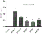
Figure 35 shows that intranasal salvinorin a improved overall motor function. Mice that have not been administered salvinorin a (above ground mice) are generally unable to walk because the left side is paralyzed 24 hours after 120 minutes of Middle Cerebral Artery Occlusion (MCAO). Mice that have been intranasally administered salvinorin a after injury (mice on cord) can crawl on hanging cord.
FIG. 36. affinity assay for hecolonin in HEK cells overexpressing both mu and kappa opioid receptors. Fig. 36A shows the binding affinity of hecologenin to the μ receptor compared to DAMGO (a potent μ agonist). K of DAMGOiAt 2.5nM, and hecolone because 45 nM. the model shown in fig. 36B suggests that hecolone (labeled H on red spherical ligands in the binding pocket) binds to the same binding site as β -funalatrexamine (β -funaltrexamine) (labeled β on light blue spherical ligands in the binding pocket), a selective mu opioid receptor ligand found in the crystal structure.
FIG. 37. affinity determination of hercronine and location of binding sites in HEK cells overexpressing kappa opioid receptors. Fig. 37A shows the binding affinity of hecologenin to the kappa receptor compared to U69593 (a potent kappa agonist). K of U69593iIt was 0.8nM, and hecolox was 184 nM. The model shown in fig. 37B suggests that hecogenin (labeled H on red spherical ligand in the binding pocket) binds to the same binding site as JDTic (labeled J on green spherical ligand in the binding pocket), a selective kappa opioid receptor ligand found in the crystal structure.
Fig. 38 cerebrovascular dilation with hercronin (Herk) is mediated via kappa opioid receptors fig. 38 shows that hercronin (Herk) dilation of the pia mater is blocked by the kappa receptor antagonist Norpropionatophen (NTP) (fig. 38A) rather than by the mu receptor antagonist β -enrotriamine (β -FNA, fig. 38B) the dilation of hercronin is equivalent to that of Isoproterenol (ISO), a potent β adrenergic agonist (fig. 38A) (Ps >0.05) administration of NTP alone or β -FNA without any dilation (one way Anova followed by multiple comparison test of nint) n ═ 5 per group in t test, for p <0.05, < p <0.01, < p > 0.005, and in ana test, # for p < 0.05.
FIG. 39 cerebrovascular dilation by Heraclosine is mediated via cAMP signaling. Fig. 39A shows an increase in cAMP levels in CSF with hercronine administration, which is abolished with administration of the kappa antagonist NTP. Fig. 39B shows that cerebral vasodilation of hecolonine is abolished by administration of 10 μ M Rp-cAMPS, a cAMP antagonist (n-5). Sp: Sp-cAMPS, Rp: Rp-8-Br-cAMPs. For p <0.05, for p <0.01, for p <0.005
Figure 40.24 hours post sham treatment, subarachnoid hemorrhage (SAH) and salvinorin a (sa) treatment groups whole brain image (ventral).
FIG. 41H & E staining of basilar arteries in sham, SAH and SA treated groups.
Figure 42 salvinorin a administered after SAH significantly (a) increased the diameter of the basal artery and (B) decreased its wall thickness 24 hours after SAH. (C) Salvinorin a given after SAH had no significant effect on neural scores,#p<0.05 vs. sham treatment group,. p<0.05 comparison SAH.
FIG. 43 Standard Curve for salvinorin A in methanol-acetone (4: 1).
Detailed Description
The present invention relates to salvinorin compositions and uses thereof. In particular, the invention relates to administration of salvinorin to increase survival of a subject following cardiac arrest or stroke in said subject. The invention further relates to the treatment of subjects suffering from cerebral vasospasm, subarachnoid hemorrhage, cerebral hypoxia/ischemia, cerebral arterial occlusion, or any condition involving self-regulating impairment.
In one aspect, provided herein is a method of treating a neurological injury associated with cardiac arrest in a subject suffering from or suffering from cardiac arrest, the method comprising: administering to the subject a therapeutically effective amount of salvinorin or a pharmaceutical composition thereof. In another aspect, provided herein is a method of increasing the likelihood of survival in a subject suffering from or suffering from cardiac arrest, the method comprising: administering to the subject a therapeutically effective amount of salvinorin or a pharmaceutical composition thereof.
In another aspect, provided herein is a method of treating cerebral vasospasm in a subject having subarachnoid hemorrhage, the method comprising: administering to the subject a therapeutically effective amount of salvinorin or a pharmaceutical composition thereof.
In another aspect, provided herein is a method of treating a cerebral arterial occlusion in a subject suffering from or suffering from a cerebral arterial occlusion, the method comprising: administering to the subject a therapeutically effective amount of salvinorin or a pharmaceutical composition thereof.
In another aspect, provided herein is a method of reducing infarct size in an individual suffering from or suffering from cerebral hypoxia/ischemia, the method comprising: administering to the subject a therapeutically effective amount of salvinorin or a pharmaceutical composition thereof. In another aspect, provided herein is a method of reducing vascular leakage in a subject suffering from or suffering from cerebral hypoxia/ischemia, the method comprising: administering to the subject a therapeutically effective amount of salvinorin or a pharmaceutical composition thereof.
In another aspect, provided herein is a pharmaceutical composition comprising: salvinorin for use in the methods described herein.
In another aspect, provided herein is a method of producing cerebral vasodilation in an individual in need thereof, the method comprising: administering to the subject a therapeutically effective amount of salvinorin or a pharmaceutical composition thereof. In another aspect, provided herein is a method of treating a disease associated with cerebral vasospasm, hypoxia and/or ischemia in a subject, the method comprising: administering to the subject a therapeutically effective amount of salvinorin or a pharmaceutical composition thereof.
In another aspect, provided herein is a method of treating a disease associated with vasoconstriction, vascular obstruction, or disruption of blood flow and self-regulation in an individual, the method comprising: administering to the subject a therapeutically effective amount of salvinorin or a pharmaceutical composition thereof. In another aspect, provided herein is a method of producing a sedative or anesthetic effect in a subject in need thereof, the method comprising: administering to the subject a therapeutically effective amount of salvinorin or a pharmaceutical composition thereof.
In another aspect, provided herein is a pharmaceutical composition comprising: a therapeutically effective amount of salvinorin, wherein the salvinorin is present in an amount effective to produce cerebral vasodilation in a subject in need thereof. In another aspect, provided herein is a pharmaceutical composition comprising: a therapeutically effective amount of salvinorin, wherein the salvinorin is present in an amount effective to treat a disease associated with cerebral vasospasm, hypoxia and/or ischemia in a subject. In another aspect, provided herein is a pharmaceutical composition comprising: a therapeutically effective amount of salvinorin, thereby treating cerebral vasospasm in a subject suffering from subarachnoid hemorrhage.
In another aspect, provided herein is a pharmaceutical composition comprising: a therapeutically effective amount of salvinorin, wherein the salvinorin is present in an amount effective to provide protection of an organ from hypoxia/ischemia in a subject. In another aspect, provided herein is a pharmaceutical composition comprising: a therapeutically effective amount of salvinorin, wherein the salvinorin is present in an amount effective to treat a disease associated with vasoconstriction, vascular obstruction, or disruption of blood flow and autoregulation in a subject.
In another aspect, provided herein is a pharmaceutical composition comprising: a therapeutically effective amount of salvinorin, wherein the salvinorin is present in an amount effective to produce a sedative or anti-nociceptive effect in a subject in need thereof.
In another aspect, provided herein is a pharmaceutical composition of salvinorin a comprising an aqueous solution of salvinorin a and cyclodextrin preferably the cyclodextrin is 2-hydroxypropyl-cyclodextrin, such as 2-hydroxypropyl- β -cyclodextrin (HPBCD) or 2-hydroxypropyl- γ -cyclodextrin (HPGCD) more preferably the cyclodextrin is 2-hydroxypropyl- β -cyclodextrin (HPBCD) in some embodiments the composition is suitable for intravenous administration in some embodiments the salvinorin a concentration is at least 25 μ g/m L preferably the salvinorin a concentration is at least 50 μ g/m L.
In some embodiments, the cyclodextrin (e.g., HPBCD) concentration in the aqueous solution of salvinorin a is at least 1% (w/v), at least 2.5% (w/v), at least 5% (w/v), at least 7.5% (w/v), at least 10% (w/v), at least 12.5% (w/v), at least 15% (w/v), at least 17.5% (w/v), at least 20% (w/v), at least 22.5% (w/v), or at least 25% (w/v). In some embodiments, the cyclodextrin (e.g., HPBCD) concentration in the aqueous solution of salvinorin a is less than 50% (w/v), less than 45% (w/v), less than 40% (w/v), less than 35% (w/v), less than 30% (w/v), less than 25% (w/v), less than 22.5% (w/v), less than 20% (w/v), less than 17.5% (w/v), less than 15% (w/v). Preferably, the cyclodextrin is HPBCD and the concentration in the aqueous solution of salvinorin a is about at least 20% (w/v).
The inventors of the present application have unexpectedly and unexpectedly found that salvinorin significantly dilates cerebral blood vessels, is rapid in onset and regression, and does not alter hemodynamics. The diameter of the cerebral arteries expanded up to 40% with 1 micromolar salvinorin as shown in figure 7. The blood vessels were dilated immediately after administration of the salvinorin, and the dilatation effect lasted for less than 3 to 5 minutes. This finding shows that salvinorin can be used to treat cerebral vasospasm in stroke, brain injury, or other related clinical situations associated with cerebral vasospasm, including after subarachnoid hemorrhage and head trauma. In addition, salvinorin can be used to treat spinal cord ischemia and other nerve ischemia.
Salvinorin a and analogs thereof are known compounds. Salvinorin a, an active component of salvia mexicana, used annually in the united states by nearly one million people for leisure purposes is the only known non-nitrogenous selective Kappa Opioid Receptor (KOR) agonist.
Diterpene carnosin a has been shown to be a high affinity and selective kappa opioid receptor agonist. See Ross (Roth), et al, Proc. Natl.Acad.Sci.USA 99:11934 (2002); and Butelman et al, Psychopharmacology 172:220 (2004).
Salvinorin and its derivatives are known in the art. For example, carnosin, its derivatives, and methods of synthesis thereof are described in U.S.2006/0052439, U.S.2007/0213394, WO 2005/089745, and WO2008/119097, each of which is incorporated herein by reference in its entirety.
Salvinorin or derivatives thereof known to those skilled in the art may be used in the methods and compositions described herein. Examples of salvinorin include, but are not limited to, salvinorin A, B, C, D, E or F. In one embodiment, the salvinorin is salvinorin a. In another embodiment, the salvinorin is salvinorin B. In another embodiment, the salvinorin is salvinorin C. In another embodiment, the salvinorin is salvinorin D. In another embodiment, the salvinorin is salvinorin E. In another embodiment, the salvinorin is salvinorin F. In another embodiment, the salvinorin is a salvinorin ester. In another embodiment, the salvinorin is salvinorin benzoate. In another embodiment, the salvinorin is a metabolite of salvinorin. In another embodiment, the salvinorin is an analog of salvinorin a. For example, hecolorine (herkinorin), an analog of salvinorin A (Gui F (Ji F), et al, Brain research (Brain Res.) 2013,1490:95-100, which is incorporated herein by reference in its entirety). Other salvinorin analogs that can be used in the methods and compositions described herein are 2-O-ethoxymethyl salvinorin B and 2-O-methoxymethyl salvinorin B.
According to one embodiment, administration of a therapeutically effective amount of salvinorin produces vasodilation in a subject in need thereof.
The invention further provides a method of treating a disease or condition comprising administering to a mammal in need thereof a therapeutically effective amount of a salvinorin.
In one embodiment, there is provided a method of treating a disease associated with vasoconstriction, vascular obstruction, or disruption of blood flow and self-regulation in an individual, the method comprising: administering to the subject a therapeutically effective amount of salvinorin or a pharmaceutical composition thereof.
The pharmaceutical compositions described herein may comprise a "therapeutically effective amount". A "therapeutically effective amount" is an amount effective, at the dosages and for the periods of time necessary, to achieve the desired therapeutic result. The therapeutically effective amount of the molecule may vary depending on factors such as the disease state, the age, sex, and weight of the individual, and the ability of the molecule to elicit a desired response in the individual. A therapeutically effective amount is also an amount of a molecule that has a therapeutically beneficial effect over its toxic or detrimental effects.
The terms "treatment" and "treatment" as used herein refer to therapeutic treatment, including prophylactic or preventative measures, wherein the object is to prevent or slow down (reduce) an undesired physiological change associated with a disease or condition. For example, treating neurological damage associated with cardiac arrest includes, but is not limited to, preventing neurological damage in a subject suffering from or suffering from cardiac arrest. Beneficial or desired clinical results include, but are not limited to, alleviation of symptoms, diminishment of extent of a disease or condition, stabilization of a disease or condition (i.e., wherein the disease or condition is not exacerbated), delay or slowing of progression of a disease or condition, diminishment or alleviation of a disease or condition, and remission (whether partial or total) of a disease or condition. "treatment" may also refer to an extended survival time compared to the expected survival time without treatment. Those in need of treatment include those already with the disease or condition, as well as those susceptible to the disease or condition, or those in which the disease or condition is to be prevented.
Examples of diseases or conditions caused by or otherwise associated with vasoconstriction, vascular occlusion, or disruption of blood flow and homeostasis include, but are not limited to, cerebral vasospasm, subarachnoid hemorrhage, stroke, brain trauma or injury, ischemia reperfusion injury, hypoperfusion, and hypoxia.
The salvinorin and pharmaceutical compositions thereof described herein can be administered to a subject by methods known in the art, such as parenteral, paracancerous, transmucosal, transdermal, intramuscular, intravenous, intradermal, subcutaneous, intraperitoneal, intraventricular, intracranial, intravaginal or intratumoral, intrathecal, and inhalation administration. In a preferred embodiment, it is administered transmucosally. In a more preferred embodiment, it is administered intranasally.
In another embodiment of the methods and compositions described herein, the pharmaceutical composition is administered orally, and thus is formulated in a form suitable for oral administration, i.e., as a solid or liquid formulation. Suitable solid oral formulations include tablets, capsules, pills, granules, pellets and the like. Suitable liquid oral formulations include solutions, suspensions, dispersions, emulsions, oils and the like. In another embodiment of the invention, the active ingredient is formulated in a capsule. According to this embodiment, the compositions of the invention comprise, in addition to the active compound and the inert carrier or diluent, a hard gelatin capsule.
In another embodiment, the pharmaceutical composition is administered by intravenous, intraarterial, or intramuscular injection of a liquid formulation. Suitable liquid formulations include solutions, suspensions, dispersions, emulsions, oils, and the like. In another embodiment, the pharmaceutical composition is administered intravenously, and is therefore formulated in a form suitable for intravenous administration. In another embodiment, the pharmaceutical composition is administered intraarterially, and is therefore formulated in a form suitable for intraarterial administration. In another embodiment, the pharmaceutical composition is administered intramuscularly, and is therefore formulated in a form suitable for intramuscular administration.
In another embodiment, the pharmaceutical composition is administered intranasally, and thus is formulated in a form suitable for intranasal administration.
In another embodiment, the pharmaceutical composition is administered topically to the body surface, and is therefore formulated in a form suitable for topical administration. Topical formulations include gels, ointments, creams, lotions, drops, and the like.
In another embodiment, the pharmaceutical composition is administered as a suppository (e.g., a rectal suppository or a urethral suppository). In another embodiment, the pharmaceutical composition is administered by subcutaneous implantation of the bolus. In another embodiment, the bolus provides controlled release of the active agent over a period of time.
In another embodiment, the active compound is delivered in liposomes (e.g., liposomes).
In other embodiments, carriers or diluents used in the methods of the invention include, but are not limited to, gums, starches (e.g., corn starch, pregelatinized starch), sugars (e.g., lactose, mannitol, sucrose, dextrose), cellulosic materials (e.g., microcrystalline cellulose), acrylates (e.g., polymethacrylates), calcium carbonate, magnesium oxide, talc, or mixtures thereof.
In other embodiments, the pharmaceutically acceptable carrier of the liquid formulation is an aqueous or non-aqueous solution, suspension, emulsion, or oil. Examples of non-aqueous solvents are propylene glycol, polyethylene glycol, and injectable organic esters (e.g., ethyl oleate). Aqueous carriers include water, alcoholic/aqueous solutions, emulsions or suspensions, including saline and buffered media. Examples of oils are oils of animal, vegetable or synthetic origin, for example, peanut oil, soybean oil, olive oil, sunflower oil, cod liver oil, another marine oil, or lipids from milk or eggs.
In another embodiment, parenteral vehicles (for subcutaneous, intravenous, intraarterial, or intramuscular injection) include sodium chloride solution, Ringer's dextrose, dextrose and sodium chloride, lactated Ringer's, and fixed oils. Intravenous vehicles include fluid and nutrient replenishers, electrolyte replenishers such as those based on ringer's dextrose, and the like. Examples are sterile liquids (such as water and oil), with or without the addition of surfactants and other pharmaceutically acceptable adjuvants. In general, water, saline, aqueous dextrose and related sugars, and glycols such as propylene glycol or polyethylene glycol are preferred liquid carriers, particularly for injectable solutions. Examples of oils are oils of animal, vegetable or synthetic origin, for example, peanut oil, soybean oil, olive oil, sunflower oil, cod liver oil, another marine oil, or lipids from milk or eggs.
In other embodiments, the composition further comprises a binder (e.g., acacia, corn starch, gelatin, carbomer, ethylcellulose, guar gum, hydroxypropyl cellulose, hydroxypropyl methylcellulose, povidone), a disintegrant (e.g., corn starch, potato starch, alginic acid, silicon dioxide, croscarmellose sodium, crospovidone, guar gum, sodium starch glycolate), a buffer having various phs and ionic strengths (e.g., Tris-HCl, acetate, phosphate), an additive to prevent absorption to a surface (e.g., albumin or gelatin), a detergent (e.g., Tween20, Tween 80, pluronic f68, cholate), a protease inhibitor, a surfactant (e.g., sodium lauryl sulfate), a permeation enhancer, a solubilizing agent (e.g., glycerol, polyethylene glycerol), an antioxidant (e.g., ascorbic acid, sodium, sodium metabisulfite, butylated hydroxyanisole), stabilizers (e.g. hydroxypropyl cellulose, hydroxypropylmethyl cellulose), viscosity increasing agents (e.g. carbomer, colloidal silica, ethylcellulose, guar gum), sweeteners (e.g. aspartame, citric acid), preservatives (e.g. thimerosal, benzyl alcohol, parabens), lubricants (e.g. stearic acid, magnesium stearate, polyethylene glycol, sodium lauryl sulphate), flow aids (e.g. colloidal silicon dioxide), plasticizers (e.g. diethyl phthalate, triethyl citrate), emulsifiers (e.g. carbomer, hydroxypropyl cellulose, sodium lauryl sulphate), polymeric coatings (e.g. poloxamers or poloxamines), coatings and film formers (e.g. ethylcellulose, acrylates, polymethacrylates) and/or adjuvants.
In another embodiment, the pharmaceutical compositions provided herein are controlled release compositions, i.e., compositions in which the active compound is released over a period of time after administration. Controlled or sustained release compositions include formulations in lipophilic reservoirs (e.g., fatty acids, waxes, oils). In another embodiment, the composition is an immediate release composition, i.e., a composition in which the active compound is released immediately after administration.
In another embodiment, the pharmaceutical composition is delivered in a Controlled Release system, for example, the agent may be administered using intravenous infusion, implantable osmotic pump, transdermal patch, liposomes, or other modes of administration, in one embodiment, a pump (see Lange (L anger), supra; Sefton (Sefton), "review of biomedical engineering reviews (CRCCrit. Ref. biomed. Eng.) -14: 201 (1987); Buchwald (Buchwald.) et al, Surgery (Surgery) 88:507 (1980); Soedek (Saudek) et al, New England Medical (N.Engl. J.Med.) -321: 1989.) in another embodiment, a polymeric material is used; for example, in microspheres or in an implant.) in another embodiment, the Controlled Release system is placed in proximity to target cells, thus only requiring a partial volume of the systemic Release system (see, e.g. Controlon.) (1992.) (1989); scientific use) (Goldon) (Per et al.) (1982, Tex et al.) (1989); 9) and (application of the aforementioned controls).
In another embodiment, the composition further comprises the incorporation of the active substance into or onto a particulate formulation of a polymeric compound (e.g., polylactic acid, polyglycolic acid, hydrogel, etc.) or onto liposomes, microemulsions, micelles, unilamellar or multilamellar liposomes, erythrocyte ghosts, or spheroplasts. The composition will affect physical state, solubility, stability, rate of in vivo release and rate of in vivo clearance.
The invention also includes particulate compositions that are coated with a polymer (e.g., a poloxamer or a poloxamine) and the compound is coupled to an antibody directed against a tissue-specific receptor, ligand or antigen or coupled to a ligand for a tissue-specific receptor.
The invention also includes compounds modified by covalent attachment of water soluble polymers such as polyethylene glycol, copolymers of polyethylene glycol and polypropylene glycol, carboxymethyl cellulose, polydextrose, polyvinyl alcohol, cyclodextrin, cucurbituril (cucurbitaril), polyvinylpyrrolidone or polyproline. Modified compounds are known to exhibit substantially longer half-lives in the blood following intravenous injection than the corresponding unmodified compounds (Abuchofsky et al, 1981; Newmark et al, 1982; and Katre et al, 1987). Such modifications may also increase the solubility of the compound in aqueous solutions, eliminate aggregation, enhance the physical and chemical stability of the compound, and greatly reduce the immunogenicity and reactivity of the compound. Thus, the desired in vivo biological activity can be obtained by administering such polymer-compound inducers less frequently or at lower doses than the unmodified compound.
In one embodiment, the method comprises administering the active compound as the sole active ingredient. However, also encompassed within the scope of the present invention are methods of treating diseases and disorders comprising administering the active compound in combination with one or more other therapeutic agents. These other agents are suitable for the disease or disorder being treated, as is known in the art.
Other therapeutically effective agents may bind to salvinorin, be incorporated into the same composition as salvinorin, or may be administered as a separate composition. Other therapeutic agents or treatments can be administered before, during, and/or after administration of the salvinorin.
Administration of salvinorin with other agents and/or treatments can be simultaneous or separate, via the same or different routes, at the same or different times. Dosage regimens can be adjusted to provide the optimal desired response (e.g., a therapeutic response or a prophylactic response).
The effective dosage of the compositions described herein for treating a condition or disease as described herein will vary depending on a number of different factors, including the mode of administration, the target site, the physiological state of the patient, whether the patient is a human or an animal, other drugs being administered, and whether the treatment is prophylactic or therapeutic. Typically, the patient is a human, but non-human mammals, including transgenic mammals, can also be treated. Therapeutic doses can be titrated using routine methods known to those skilled in the art to optimize safety and efficacy.
In one example, a single bolus may be administered. In another example, several divided doses may be administered over time. In another example, the dose may be proportionally reduced or increased, as indicated by the exigencies of the treatment situation. Unit dosage form as used herein refers to physically discrete units suitable as unitary dosages for the treatment of mammalian subjects. Each unit may contain a predetermined amount of active compound calculated to produce the desired therapeutic effect. In some embodiments, the unit dosage form is dictated by and directly dependent on the unique characteristics of the active compound and the particular therapeutic or prophylactic effect to be achieved.
The composition may be administered only once, or it may be administered in multiple or continuous infusions. For multiple doses, the composition can be administered, for example, three times a day, twice a day, once every two days, twice a week, once every two weeks, or once a month.
Dosage values may vary with the type and severity of the condition to be alleviated. It is further understood that for any particular individual, the particular dosage regimen may be adjusted over time according to the individual needs and the professional judgment of the person administering or supervising the administration of the compositions, and that the dosage ranges set forth herein are exemplary and are not intended to limit the scope or practice of the methods.
"administration" to a subject is not limited to any particular delivery system and can include, but is not limited to, parenteral (including subcutaneous, intravenous, intramedullary, intraarticular, intramuscular, or intraperitoneal injection), rectal, topical, transdermal or oral (e.g., in capsule, suspension, or tablet form), intrathecal, intranasal, and inhalation administration. Administration to a subject can be in the form of a single dose or in the form of repeated administrations or continuous infusions, as well as in any of a variety of physiologically acceptable salt forms and/or as part of a pharmaceutical composition with acceptable pharmaceutical carriers and/or additives. Likewise, physiologically acceptable salt forms and standard Pharmaceutical formulation techniques are known to those skilled in the art (see, e.g., Remington's Pharmaceutical Sciences, Mark Publishing Co., Ltd.).
The term "about" or "approximately" means within an acceptable deviation of the particular value as determined by one of ordinary skill in the art, which will depend in part on how the value is measured or determined, i.e., the limitations of the measurement system. For example, "about" can mean within 1 or more than 1 standard deviation, as practiced in the art. Alternatively, when referring to measurable values (e.g., amounts, durations, concentrations, etc.), variations from the specified values of ± 20% or ± 10%, more preferably ± 5%, even more preferably ± 1% and more preferably ± 0.1% may be encompassed as the variations are suitable for carrying out the disclosed methods.
The treatment methods described herein can be used to treat suitable mammals, including primates (e.g., monkeys and humans), horses, cows, cats, dogs, rabbits, and rodents (e.g., rats and mice). Preferably, the mammal to be treated is a human.
Any reference cited herein, including patents, patent applications, or scientific publications, is incorporated by reference in its entirety.
In order to more fully illustrate the preferred embodiments of the present invention, the following examples are presented. However, these examples should in no way be construed as limiting the broad scope of the invention.
Examples of the invention
Example 1
Salvianolin A produces cerebral vasodilation via activation of nitric oxide synthase, kappa receptor and adenosine triphosphate sensitive potassium channels
In this example, we demonstrate that salvinorin a is activated via nitric oxide synthase, adenosine triphosphate-sensitive potassium (K) under resting and elevated tonicity conditions induced by hypocapnia or endothelinATP) The channels and kappa receptors dilate the pial arteries.
Materials and methods
Carnosol a (purity ≧ 98%), Sodium Nitroprusside (SNP), n (g) -nitro-L-arginine (L-NNA), glibenclamide (glibenclamide), iberiotoxin (iberiotoxin), chromancalin (cromaALim), calcitonin gene-related polypeptide (CGRP), NS1619, naloxone (naloxone), methionine enkephalin, noralanatopheniramine (norbinaltoritorphine), 7-nitroindazole (7-NINA), sulpiride (sulpiride), and isoproterenol were obtained from Sigma-Aldrich, St L, MO, USA.
Animals and surgery
New pigs of both sexes (1-6 days old, weighing 1.3-1.8kg) were used for this study, the protocol was approved by the institutional animal Care and Use Committee (institutional animal Care and Use Committee) of the University of Pennsylvania (University of Pennsylvania). animals were induced with isoflurane (1-2 minimum alveolar concentrations) and then maintained with α -aldochlorous (80-100mg/kg, supplemented at 5mg/kg/h, intravenously) both femoral arteries were catheterized to monitor blood pressure and blood gases.catheterized into the right femoral vein for drug administration.animals were ventilated under room air following tracheal cannula insertion. rectal temperature was maintained at 37-39 ℃ by a heating padAn opening is made in the skull above the layer. The dura mater is then incised and retracted at the bone margin. The skull window was placed over the skull opening and cemented in place with dental acrylic. The space under the window was filled with artificial CSF (in mM) having the following composition: 3.0KCl, 1.5MgCl2、1.5CaCl2132NaCl, 6.6 Urea, 3.7 dextrose, and 24.6NaHCO3Perliter, pH 7.33, PCO246mmHg and PO2The adventitial artery was viewed with a video camera mounted on an anatomical microscope the vessel diameter was measured from a video monitor connected to the camera with a video micro-scale (model VPA 550, frey corporation of los Angeles, california, For-a-corp., L os Angeles, CA)).
Experimental protocol
The pial artery diameter (arteriolar diameter 120-160 microns; arteriolar diameter 50-70 microns) was monitored and recorded every half minute for 10 minutes following injection of artificial CSF in the presence or absence of study drug. Typically, the window is flushed with 1-2ml CSF via an orifice attached into the side of the window for more than 30 seconds. CSF samples were collected for cGMP analysis before and 10 minutes after drug administration. We collected cortical peripheral arachnoid CSF by slowly pouring CSF into one orifice of the window and allowing CSF to drip freely into a collection tube over the opposite orifice.
Reactions to salvinorin a (10nM, 1 μ M, solubilized with alcohol) and SNP (10nM, 1 μ M) were obtained in the absence or presence of n (g) -nitro-L-arginine (L-NNA, 1 μ M), Nitric Oxide Synthase (NOS) inhibitors, and 7-nitroindazole (100nM, an antagonist of neuronal NOS).
To distinguish the direct or permissive role of nitric oxide in a dilatation response to salvinorin A, changes in pial artery diameter were recorded after administration of SNP (100pM, subthreshold vascular concentration of nitric oxide donor) with L-NNA and salvinorin A sulpiride (100nM, a dopamine receptor D2 antagonist), glibenclamide (100nM, a K) were also determinedATPChannel antagonists); ibereamycin (100nM, Sigma-Aldrich, a K)CaChannel antagonist) of Sargassu A, chromaffin (1. mu.M) and calcitonin gene-related polypeptide (10nM, 1. mu.M, a K) against the pia mater arteryATPAgonist), and NS1619 (10nM, 1 μ M, a KCa channel agonist) the effects of naloxone (1mg/kg, intravenously) and noralantin-insensitive (1 μ M, topical administration, a kappa opioid receptor antagonist) on responses to salvinorin a, methionine enkephalin (10nM, 1 μ M) and isoproterenol (10nM, 1 μ M, an β adrenergic receptor agonist) were finally studied all test drug solutions were freshly prepared on the day of use recording the response of the pial artery to the administered salvinorin a every 2 minutes for 30 minutes to determine if sustained vasodilation could be obtained.
cGMP assay
To determine the effect of the nitric oxide pathway on the effect of carnosine on cerebral vasculature, CSF samples were collected for cGMP determination before and after administration of carnosine a with or without L-NNA and norpropionito non-sensitive pretreatment a commercial E L ISA kit (Enzo L ifeSciences International, inc. plobouth Meeting, PA)) was used to quantify cGMP concentrations.
Salvianolide A for contracting blood vessels
To test the cerebrovascular effect of salvinorin during the rise in cerebral vascular tension, we experienced hypocapnia (PaCO)2Decrease 20-30% for 10 min) and endothelin (0.1pM) induced vasoconstriction. The pial artery diameter was monitored at baseline, after hypocapnia or administration of endothelin, and after administration of salvinorin a (10nM, 1 μ M) (n-4 for hypocapnia and n-5 for endothelin).
Statistical analysis
All data (diameter and cGMP) were analyzed using ANOVA with repeated measures, followed by Bonferroni post hoc test (Bonferroni post house, two-tailed) with a social scientific Statistical software Package of 10.0 (Statistical Package for the social sciences, SPSS). Levels of p <0.05 were considered statistically significant. Values are expressed as mean ± SEM of absolute values; or as a percentage change from the baseline value. The distribution of all values is evaluated by means of a histogram.
Results
Dilation and action of nitric oxide pathway
Salvinorin a dose-dependently (10nM, 1 μ M) dilated the pia mater arteries of piglets as shown in fig. 1A. a dilation effect was observed immediately after the administration of salvinorin and the duration of dilation lasted less than 5 minutes for both doses when salvinorin a was administered every 2 minutes, a continued dilation effect was observed as shown in fig. 1B. the dilation response was abolished by L-NNA (NOS inhibitor) but not 7-nitroindazole (100nM, antagonist of neuronal NOS (nnos)) (fig. 1A, C) the dilation response to SNP was not affected by L-NNA (fig. 1A) SNP (100pM) had no effect on the pia mater arteries, but it restored the contraction induced by L-NNA. however, it did not restore the dilation response to salvinorin a blocked by L-NNA (fig. 1D), the dilation response to salvinorin a was associated with elevated cGMP in CSF, while the elevated cGMP 38-NNA blockade does not change during administration of salvinorin (fig. L).
KATPChannels other than the KCa channels participate in dilation
Glibenclamide (100nM, K)ATPChannel inhibitors) but not ibeibixin (100nM, Ca2+ -activated K + (KCa) channel inhibitor) blocked the expansion of salvinorin a. Glibenclamide and ibeibixin in any order also blocked the expansion induced by salvinorin a (figure 3). Glibenclamide (100nM) but not ibeiritoxin (100nM) blocks chromancalin, a KATPAgonist of the channel, 10nM and 1. mu.M) and calcitonin Gene-related polypeptide (Another K)ATPChannel agonists, 10nM and 1 μ M); ibebrexin (100nM) but not glibenclamide (100nM) blocked the dilating effect of NS1619 (a KCa channel agonist, 10nM and 1 μ M) (fig. 4).
Opioid receptor other than dopamine receptor D2 antagonists block the dilating action of carnosine
Nornalatole is non-sensitive (kappa opioid receptor selective antagonist) and naloxone blocks the expansion effects of carnosine a and methionine enkephalin. The response to isoproterenol was not affected (fig. 5A, B). Sulpiride (dopamine receptor (D2) antagonist) had no effect on the vasodilatory response of salvinorin a (fig. 5C). Norpropatol non-sensitively blocks cGMP elevation in CSF from administration of salvinorin a. There was no difference between cGMP levels before and after administration of carnosine a (10nM and 1 μ M) in the case of noralanatol-insensitive pre-treatment (p ═ 1).
Salvinorin dilates pial arteries in elevated cerebral vascular tension conditions
Hypocapnia and endothelin significantly reduced the diameter of the pial artery (fig. 6). Salvinorin dilated the pia mater artery under elevated tension conditions, similar to that observed under normal carbonated (resting tension) conditions (figure 6). No significant difference between arteriolar and arteriolar responses was observed in any of the above experiments, and therefore, only arteriolar changes were exhibited.
Discussion of the invention
In this example, salvinorin a is exhibited as a potent pial arterial dilator in piglets under normal conditions and vasoconstrictive conditions induced by endothelin and hypocapnia. Dilation was observed immediately after salvinorin administration, lasting less than 5 minutes for both test doses, and was dose-dependent. Sustained expansion of salvinorin a was observed with continuous administration. Opioid receptors, NOS and KATPActivation of the channel participates in the signaling pathway for such dilation.
U50488 (an exogenous KOR agonist) and dynorphin (an endogenous KOR agonist) have been shown to dilate the pial artery. It was also shown that U50488 dose-dependently relaxes isolated aorta in rats. Unlike other KOR agonists, the expansion of salvinorin occurs for a very short time, less than 5 minutes. This is the agent with the shortest time of action among kappa agonists to date, which is an important pharmacological feature for many clinical drugs, easy to manage and titrate in perioperative settings and emergency care. To achieve longer action, infusion is the most common technique used in clinical anesthesia. In this example, we found that sustained vasodilation can be obtained via continuous administration.
The unique structure of salvinorin a (without rapid desensitization) contributes to its short duration of action characteristics, as continuous administration causes a sustained expansion effect. The ester bonds in their structure can be readily metabolized by esterases in blood and tissues. One group showed that the carboxylesterase enzymes were primarily involved in the hydrolysis of salvinorin a in rat plasma and the degradation products included salvinorin B and the lactone ring open form of salvinorin a, both of which were pharmacologically inactive.
NOS, K similar to other KOR agonistsATPYet, in this example, the pia mater arterial dilation of salvinorin a was eliminated by L-NNA (a non-specific NOS inhibitor) rather than 7-nitroindazole (a selective nNOS antagonist), indicating that nNOS is not involved in the dilation of salvinorin a, since subthreshold amounts of nitric oxide donor SNPs failed to restore the dilation response, the mp cgcgelevation in CSF is likely not due to stimulation of nitric oxide production not inhibited, and direct activation of endothelial NOS by salvinorin rather than as a permissive enabler.
KATPChannel activation can cause hyperpolarization of the vascular smooth muscle cell membrane. The membrane potential change will then be via Ca2+By voltage-dependent Ca2+The internal flow of the channel is altered to regulate muscle relaxation. This example shows direct or indirect K activation by salvinorin AATPA channel. With activatable KATPUnlike many other agents of the pathway, carnosine a can readily penetrate the blood brain barrier. Due to KATPChannels play a crucial protective role from brain damage due to hypoxia, ischemia or metabolic inhibition, so salvinorin a can be a neuroprotective agent for clinical use.
Although salvinorin a has been shown to have high affinity for the dopamine D2 receptor, sulpiride (a dopamine D2 receptor selective antagonist) has no effect on the dilating action of salvinorin a. In contrast, naloxone and norkinatol are insensitive (kappa opioid receptor selective antagonists) to abrogate the vasodilatory effects of salvinorin a, suggesting an important role for the kappa receptor rather than the dopamine D2 receptor.
The vasodilatory effect observed in normal cerebral vessels, as well as in constricted cerebral vessels caused by both hypocapnia and endothelin, may allow its clinical use in the treatment of cerebral vasospasm in a number of clinical situations, including migraine and cerebral vasospasm following subarachnoid hemorrhage where increased endothelin plays an important role. Similar to other short acting medications, continuous infusion can be used for titration and to achieve sustained action by adjusting the dosage.
In this example, newborn piglets were used as study subjects. The porcine brains of the brains have more white matter than gray matter, are selectively vulnerable, similar to humans, and are similar in adulthood. Newborn piglets are also used because they are large enough to easily place the cranial window and visualize the blood vessels. The cerebrovascular response of a newborn is similar to that of a human individual in many clinical situations.
In summary, salvinorin a is a strong pial arterial dilator with a fast onset and short duration of action in normal conditions and vasoconstrictive conditions induced by endothelin and hypocapnia in piglets. Mechanism involving NOS, KATPActivation of channels and kappa receptors. These findings demonstrate the clinical value of salvinorin a in harsh cerebral vasodilatory environments.
Example 2
Retention of cerebrovascular autoregulation in piglets via kappa opioid receptors following global cerebral hypoxia/ischemia administration of salvinorin A
Neuronal death and behavioral dysfunction caused by hypoxia/ischemia (HI) -induced brain injury are not uncommon during perinatal periods. Worldwide, 23% of all new births are associated with asphyxia, and 30% of children born with moderate hypoxia-ischemic encephalopathy (HIE) may develop mental retardation, learning difficulties, and other disabilities. Unfortunately, there are no drugs available to manage this destructive situation.
KOR agonists cause cerebral vasodilation, a key feature required to maintain brain autoregulation and to reduce brain damage from ischemia. Following ischemia, brain autoregulation is impaired, leading to reduced cerebral blood flow and neuronal death. We show in the piglet model that administration of salvinorin a prior to global HI preserved autoregulation of cerebral arteries, an important mechanism to preserve neuronal integrity. In this example, we hypothesized and tested whether administration of salvinorin a after HI could preserve brain self-regulation via the KOR and ERK pathways.
Materials and methods
Salvinorin A (purity ≧ 98%) was obtained from Kymakex corporation (ChromaDex, Inc., U.S. gulf, Calif. (Irvine, Calif., USA)). Isoproterenol (ISO), noralanatole (Nor-BIN) was obtained from sigma-aldrich (st louis, missouri, usa). All other chemicals (reagent grade) were also obtained from sigma.
Animal and surgery for sealing a cranial window
The animal experimental protocol was approved by the institutional animal care and use committee at university of pennsylvania as previously described, isoflurane (1 to 2 lowest alveolar concentrations) was initially used for anesthesia induction, followed by α -chloroaldose (30-50mg/kg, supplemented at 5mg/kg/hr, intravenously) for maintenance of anesthesia after tracheotomy, the animals were initially ventilated under room air and kept warm with a heating pad to maintain rectal temperature at 37-39 ℃, the bilateral femoral arteries were catheterized to monitor blood pressure, blood gas tension and pH. the femoral vein was catheterized for drug delivery and the sealed femoral window consisting of a HI glass cover sheet steel ring connected to 3 orifices was placed for direct pial artery visualization and diameter measurement.the small pial artery (120 to 160 μm) and arteriole (50 to 70 μm) were connected to CSF under a microscope, and the peripheral vascular sheath was connected to a microscopical microscope for transretinal visual observation and peripheral vascular monitoring, and transretinal sampling after transretinal vein analysis, and transretinal vein analysis.
HI induction
Hypoxia was induced for 10 minutes by: the indoor air is changed to N2 for ventilation, and then the indoor air ventilation is resumed. Global cerebral ischemia was then induced by infusing saline through a hollow bolt in the cranium, maintaining intracranial pressure above mean blood pressure for 20 minutes. Global cerebral ischemia was confirmed when blood flow in the pial artery was stopped as seen on a monitor attached to a microscope above the cranial window. To avoid Cushing response (a significant increase in arterial pressure due to high intracranial pressure), blood was drawn as necessary to maintain mean arterial blood pressure below 100 mmHg. At the end of ischemia, blood is returned via the femoral vein.
Medical treatment
Four groups of intravenous drug administration were performed after HI (n ═ 5 in each group): (1) DMSO group: DMSO (vehicle of salvinorin a) was administered 1 μ Ι/kg immediately after HI; (2) SA 0 min group: salvinorin A (1. mu.g/. mu.l in DMSO) 10. mu.g/kg immediately after HI; (3) SA 30 min group: 30 min after HI with salvinorin A (1. mu.g/. mu.l in DMSO) 10. mu.g/kg; (4) SA + Norbin group: immediately after HI, with salvinorin A (10. mu.g/kg) and nor-BIN (1. mu.M, via a single port injection through the cranial window).
Pia mater arterial response
Before and 60 min after HI, the response of the pial arteries to hypercapnia, hypotension and isoproterenol (10nM, 1 μ M) was obtained as previously described. Isoproterenol was used as a positive control because it is a short acting agent and its vasodilatory effect in such models is well established in our laboratory. By inhaling high concentration CO2Mixed gas (10% CO)2;21%O2;69%N2) Two levels of hypercapnia (PaCO) occur250 to 60mmHg at a low level and 70 to 80mmHg at a high level). Two levels of hypotension were produced by rapid withdrawal of 5-8 or 10-15ml/Kg of blood from the femoral artery to induce moderate and severe hypotension (mean blood pressure reduction of 25% to moderate and 45% to severe). Blood pressure of this kindThe reduction was maintained constantly for 10 minutes by withdrawing or reinfusing additional blood.
ERK Activity measurement
ERK activity was then determined from frozen CSF samples as described above the levels of pERK and ERK were measured using the E L ISA kit (Enzo Life sciences International Inc. of Primex Midin, Pa.).
Statistical analysis
One-way ANOVA was used to compare the change in ERK activity (quantified as the ratio of pERK to ERK) in each group before and 60 minutes after HI, and in groups with or without salvinorin a administration, baseline data were excluded in repeated measures analysis (Graph Pad Prism version 5.02), α levels of P <0.05 were considered significant in all statistical tests.
Results
Salvianolin A retains the self-regulation of the pial artery to hypercapnia after whole brain HI
As shown in fig. 13, the small pial artery dilated (presented as baseline) in response to the two levels of hypercapnia prior to HI. When DMSO was administered immediately at the end of HI, the dilatation response to hypercapnia was blunted after HI (ps <0.01 compared to baseline before HI). Administration of salvinorin a (10 μ g/kg) immediately or 30 minutes after HI preserved the dilatation response of the pial arteries to hypercapnia. Such reactions were completely abolished when salvinorin a and norpropatol non-sensitive (nor-BIN) were co-administered 30 min post-HI (ps <0.01 compared to baseline before HI). Similar observations were obtained in pial arterioles (data not shown).
Salvianolin A retains the self-regulation of the pial artery to hypotension after whole brain HI
Similar to the results for hypotension, the small pial artery dilated in response to the two levels of hypotension before HI hypercapnia (presented as baseline, fig. 14). When DMSO was administered immediately at the end of HI, the dilatation reaction was blunted after HI (ps <0.01 compared to baseline before HI). Salvinorin a (10 μ g/kg), administered immediately or 30 minutes after HI, retained the dilatation response of the pial arteries to hypotension. Such responses to hypotension were abolished at 30 min post-HI co-administration of salvinorin a and nor-BIN (ps <0.01 compared to baseline). Similar observations were obtained in pial arterioles.
The response of the pial artery to isoproterenol remained unchanged in all groups of experiments
As a positive control, the response of the pial artery to isoproterenol was measured (fig. 15), and no change was observed between all groups before and after HI.
ERK signaling is involved in the retention of salvinorin A
ERK activity was quantified as the ratio of pERK/ERK levels in CSF. Data on ERK activity in the groups without carnosine effect were pooled (DMSO group and SA + Norbin group; renamed DMSO + Nornin group). As indicated in figure 16, ERK activity in the carnosine-free group was significantly increased 60 min after HI (p <0.05 compared to pre-HI baseline). ERK activity was reduced to baseline levels in the salvinorin a administered group.
Discussion of the invention
Three new findings were obtained from this example. First, administration of salvinorin a immediately or 30 minutes after HI retained the dilatation response of the pial arteries to hypercapnia and hypotension. Second, the retention of salvinorin a acts as a passive KOR antagonist nor-BIN. And third, the increase in CSF ERK activity after salvinorin a blocks HI.
Problems with brain HI and potential effects of KOR agonists
Birth suffocation and infantile ischemic stroke are common complications of childbirth. Perinatal HI occurring in both complications can induce severe and permanent neuropsychological deficits including delayed cognitive and behavioral development, mental retardation, cerebral palsy and epilepsy, which are devastating to patients, families and society. Unfortunately, there are no medications available for effective perinatal HI management. Hypothermia is the only treatment for HIE to reduce negative complications. It is not widely accepted in clinical practice or recommended to be combined with pharmacological agents. Recombinant tissue type plasminogen activator (t-PA, FDA approved for treatment of acute ischemic stroke) exhibits adverse effects, including increasing stroke infarct volume in mice undergoing induced stroke and impaired cerebral hemodynamics.
For example, when administered 30 minutes before or up to 6 hours after injury, KOR agonists BR L52537 and CI-977 reduce cortical damage, including brain swelling and infarct volume, in response to different levels of ischemia.
Novel use of salvinorin A as a medicament
While KOR agonists exhibit great therapeutic value, most KOR agonists have not been used in a clinical setting due to their intrinsic characteristics as opioids (low selectivity and/or lack of an acceptable safety profile). Unlike other opioid KOR agonists, salvinorin a is the most potent, highly selective and only non-opioid KOR agonist known to be derived from natural sources. Salvinorin a is an active component of salvia mexicana, a naturally abundant perennial herb that has been consumed by humans for leisure and sacrifice purposes for centuries. The many inherent characteristics of this compound make it a potential therapeutic drug for a variety of neurological conditions. Those features include their ability to be extracted and purified from abundant plants or produced synthetically; the effect is quick; fat solubility; easy passage through the blood-brain barrier; sedative and antinociceptive effects (features particularly relevant for critically ill patients); negative pathological findings (no toxicity) in vital organs in case of high dose or long-term exposure; no respiratory depression; and no obvious hallucination or dysphoria effects. Salvinorin a has been evaluated as a potential drug for inhibition. In addition to our findings that demonstrate protective effects of administration of salvinorin a prior to HI injury, we have now shown that administration of salvinorin a after HI injury (up to 30 minutes) retains autoregulation of the pial arteries. This protection was abrogated by the addition of the KOR antagonist nor-BIN, indicating that the protection of salvinorin a was mediated via KOR. Various studies have demonstrated that self-regulation (a key protective mechanism of the brain) tends to deteriorate after brain HI. The self-regulation of cerebral vascular tone is a key protective mechanism of the brain. And compensatory cerebral vasodilation, impaired during hypotension, contributes to worsening the outcome in the context of ischemic stroke.
Effect of ERK signalling
ERK signaling stimulated by cerebral ischemia/reperfusion is a critical pathway for HI injury. We have shown that ERK activity increases after HI, and that the increase is related to neuronal impairment of HI, and that inhibition of the increase is associated with a neuroprotective response against ischemia, which may be associated with decreased apoptosis. In this example, the HI-induced increase in CSFERK activity was blocked by salvinorin a, which promotes protection of brain self-regulation after injury. Notably, the role of ERK signaling in HI may differ before and after HI injury. Activation of ERK signaling may be associated with protection from pre-adaptation. Upregulation of ERK signaling induced by pre-adaptation reduces neuronal apoptosis in stroke, and activation of ERK signaling in hippocampus after sublethal ischemia correlates with neuroprotection induced by pre-adaptation. These results are consistent with our work showing that pre-injury administration of salvinorin a is protective against impairment of postinjury brain autoregulation. In summary, administration of salvinorin a 0 and 30 min after HI retained the self-regulation of the pial arteries to hypercapnia and hypotension via the KOR and ERK pathways in a piglet model.
Example 3
Salvianic A pretreatment preserving cerebrovascular autoregulation via ERK/MAPK following cerebral hypoxia/ischemic injury in piglets
Cerebral hypoxia/ischemia due to interruption of cerebral blood flow during cardiopulmonary bypass and deep hypothermia arrest (DHCA) surgery for congenital heart surgery is a significant clinical problem. 50% of children with complex congenital heart disease who experience cardiopulmonary bypass and DHCA have developmental defects such as speech disability and attention deficit disorder by school age. The occurrence of cerebral hypoxia/ischemia during DHCA is predictable, and therefore, it is possible to minimize ischemia-induced brain damage using pharmacological approaches. Unfortunately, no drug has been identified that has proven clinical benefit.
Cerebrovascular autoregulatory loss is one of the key features of cerebral hypoxia/ischemia. Loss of homeostasis in the hypotension can cause stress-passive cerebral circulation, which can reduce cerebral blood flow and further exacerbate cerebral ischemia. The loss of cerebrovascular regulation for hypercapnia also contributes to the development of stress passive circulation and periventricular leukomalacia. Therefore, preservation of cerebrovascular autoregulation from ischemia is of great importance to reduce brain damage from ischemia. We have shown that salvinorin a, an active component of salvia mexicana and a non-opioid Kappa Opioid Receptor (KOR) agonist, is a potent cerebral vasodilator in normal and pathological conditions. Thus, salvinorin a protects cerebral blood vessels from ischemia. Unlike other KOR agonists, salvinorin a has long been used by different ethnic groups for a variety of purposes, including mental experience and "treatment" of disease, indicating its high potential as a clinically acceptable drug.
Systemic administration of KOR agonists has been shown to have neuroprotective effects in animal models of cerebral ischemia. KOR agonists activate mitogen-activated protein kinase (MAPK). MAPK is a key intracellular signaling system that includes extracellular signal-regulated kinases (ERKs), c-Jun-N-terminal kinases (JNK), and p 38. It has been shown that long-term and sustained activation of the ERK cascade is an important contributing mechanism to the pre-adaptation of cerebral ischemia. This pathway is also involved in many other forms of pharmacological pre-adaptation, such as isoflurane and sevoflurane. Therefore, salvinorin a can produce brain protection via this pathway. Based on the above evidence, we hypothesized that salvinorin a pretreatment can preserve the autoregulation of hypotension and hypercapnia of the pial vessels from hypoxia/ischemia via activation of MAPK.
Method of producing a composite material
Salvianolide A (purity 98% or more) was obtained from Kmadex corporation (L.gulf California, USA). Isoproterenol, U0126, sp600125, and sb203580 were obtained from sigma-aldrich (st. louis, missouri, usa). All other chemicals were also obtained from sigma and were of reagent grade.
Animals and surgery
The procedure was approved by the institutional animal care and use committee of the university of pennsylvania (Philadelphia) using one to five days old piglets, the protocol was approved by the institutional animal care and use committee of isoflurane (1-2 lowest alveolar concentrations) initially for induction, followed by α -aldochlorous for maintenance of anesthesia (30-100mg/kg, supplemented at 5-30mg/kg every 20-30 minutes, intravenously) following tracheotomy, piglets were mechanically ventilated under room air and kept warm with a heated pad, rectal temperature was maintained at 37 to 39 ℃, femoral artery cannulated for continuous blood pressure monitoring or intermittent blood gas monitoring, and femoral vein cannulated for drug administration as previously described (CSF), cranial window was closed for direct pial artery observation and diameter measurement (21), small pial artery (120 to 160 μm) and arteriole (50 to 70 μm) were identified under a monitor, on a microscope, and were connected to state observation via a microscope, and were connected to a microcarpsa microscope for taking a sample via a microcarpal microscope for post rat tail vein injection with MAPK and rat tail mounted transretinal vein analysis, rat tail surgery, rat amputation, rat surgery, rat.
Scheme(s)
Hypoxia was induced for 10 minutes by: changing the indoor air to N2 for ventilation, followed by restoring the indoor air ventilation; and then global cerebral ischemia was induced by infusing saline through a hollow bolt in the cranium, maintaining intracranial pressure above mean blood pressure for 20 minutes. Global cerebral ischemia was confirmed when cessation of blood flow in the pial artery was observed on a monitor attached to a microscope above the cranial window. To avoid the cushing reaction (significant increase in arterial pressure due to high intracranial pressure), blood was drawn as necessary to maintain mean arterial blood pressure no higher than 100 mmHg. At the end of ischemia, blood is returned via the femoral vein.
Five experiments were performed (n ═ 5 in each experiment): (1) vehicle of hypoxia/ischemia and salvinorin a DMSO, 1 μ Ι/kg, administered 30 minutes prior to hypoxia/ischemia; (2) hypoxia/ischemia and salvinorin a, 1 μ g/μ l in DMSO, 10 μ g/kg, intravenously; (3) hypoxia/ischemia with salvinorin a (10 μ g/kg, intravenous) and U0126(1mg/kg, intravenous, protein kinase inhibitor upstream of ERK); (4) hypoxia/ischemia with salvinorin a and sp600125(1 μ M, local injection via one orifice of the cranial window, inhibitor of JNK); (5) hypoxia/ischemia with salvinorin a and sb203580(10 μ M, local injection via one orifice of the cranial window, inhibitor of P38). U0126, sp600125 and sb203580 were administered 30 minutes before salvinorin a. Sp600125 and sb203580 were co-administered with vasoactive stimulators in order to have sustained cortical surface exposure after injury.
Hypercapnia (PaCO)250 to 60mmHg at a low level and 70 to 80mmHg at a high level) by inhaling CO at a high concentration2Mixed gas (10% CO)2;21%O2;69%N2) To produce. Hypotension is produced by drawing blood from the femoral artery (mean blood pressure decreases by 25% to moderate and 45% to severe). The response of the pial artery to hypotension, hypercapnia and isoproterenol (10nM, 1 μ M) was obtained before hypoxia/ischemia and 60 minutes after injury, as described previously.
ERK and pERK measurements
To test the effect of ERK on the observed effect of carnosine a on cerebral hypoxia/ischemia, CSF samples were collected for mapk. mapk isoforms measured using a commercially available E L ISA kit (enzo life science international, pregamustine, pa).
Statistical analysis
Data obtained when studying the effects of cerebral hypoxia/ischemia on the response of the pial artery to hypercapnia, hypotension and isoproterenol were analyzed by repeated measures ANOVA with the greenhauser Geisser correction (Greenhouse Geisser correction) for the pial artery diameter bonefraney correction for all post hoc analyses (10 comparisons for each stimulus) five different treatments (DMSO, SA, SB203580, U0126 and SP600125) were used as factors for comparisons between groups, and four measurements of the three stimuli before and after hypoxia/ischemia were used as repeated measures factors (5 × × ×) baseline was not included in the repeated measures analysis the same statistical method was used for pERK/ERK data to compare the ratio changes before and after administration with salvinorin a, three different treatments (DMSO, SA and SA + U0126) were used as violated factors, time (30 minutes before and after SA) was used as repeated measures factor P. P35 is considered a less significant error in all samples, although this is not considered a less significant error of the interaction in the sample.
Results
Salvinorin a retained the buffy coat artery self-regulation of hypotension after hypoxia/ischemia.
As shown in fig. 17, at baseline prior to hypoxia/ischemia, the small pial artery dilated to two levels of hypotension, but the dilatation response was significantly reduced after hypoxia/ischemia (p <0.001 compared to the dilatation response prior to hypoxia/ischemia). Pretreatment with salvinorin A (10. mu.g/kg, intravenous) retained the dilatation response of the pial arteries to hypotension. This was abolished by U0126 (antagonist of ERK) (p <0.001 compared to SA group). However, there was no significant change following treatment with SP600125 (antagonist of JNK, P >0.05 compared to SA group) and SB203580 (antagonist of P38, P >0.05 compared to SA group) administered 30 minutes prior to administration of salvinorin a. Similar observations were obtained in pial arterioles (data not shown).
Salvinorin a retained the buffalo artery self-regulation of hypercapnia after hypoxia/ischemia.
Similar to the response to hypotension, at baseline prior to hypoxia/ischemia, the small pial artery dilated to two levels of hypercapnia (fig. 18). The dilatation response is inactivated after hypoxia/ischemia. Pretreatment with salvinorin A (10. mu.g/kg, intravenous) preserved the dilatation response of the pial artery to hypercapnia. This was eliminated by U0126 (an antagonist of ERK). There was no significant change in retention observed from SP600125 and SB203580 administered 30 minutes prior to salvinorin a administration (P > 0.05). Similar observations were obtained in pial arterioles (data not shown).
The response of the pial artery to isoproterenol was unchanged in all groups of experiments
As a positive control and shown in fig. 19, the response of the pial artery to isoproterenol before and after hypoxia/ischemia was unchanged in all groups, with or without the intervention described above.
ERK is involved in the retention of salvinorin A
pERK/ERK ratio in CSF increased significantly 30 min after salvinorin a pretreatment (fig. 20). However, if U0126 (an antagonist of ERK) was administered 30 minutes before the administration of salvinorin, the pERK/ERK ratio did not change 30 minutes after the salvinorin a pretreatment (fig. 20).
Discussion of the invention
There are two main new findings in this example. First, pretreatment of salvinorin a retained cerebrovascular autoregulation after hypoxia/ischemia. Second, ERK/MAPK is involved in the ability of salvinorin a to remain self-regulating. This example also confirms the previous findings: whole brain hypoxia/ischemia in newborn piglets blunts the self-regulating ability of cerebral vessels to react to hypotension and hypercapnia.
The only drugs approved by the Food and Drug Administration (Food and Drug Administration) for stroke are recombinant tissue plasminogen activator (tPA). however, despite its beneficial effect in reopening clotted vessels, tPA increases stroke infarct volume in mice and it enhances the self-regulated impairment induced by hypoxia/ischemia. L-NNA (an inhibitor of nitric oxide synthesis) has been demonstrated to be able to restore cerebrovascular autoregulation following ischemia, however, its safety profile has not been clear for clinical use.
In this example, we show that after hypoxia/ischemia, salvinorin a pretreatment retains a self-regulating response to hypotension and hypercapnia in a piglet model, which opens its clinical application to attenuate cerebral hypoxia/ischemia, especially for the expected cerebral ischemia during DHCA in infants. More research is needed to provide direct evidence to show whether it can attenuate neuronal cell damage due to hypoxia/ischemia.
Unlike other KOR receptors, which do not have proven clinical value, salvinorin a is extracted from the abundant natural plant salvia mexicana. Very similar to the history of opium, salvia mexicana has been used by humans for centuries for various purposes as a naturally abundant plant. It has been proposed that the active component of salvia mexicana, salvinorin a, may be a potential novel kappa agonist for use in clinical practice.
As we have demonstrated, salvinorin a is a potent cerebral vasodilator in normal and contractile conditions. However, this expansion effect is transient, except in the case of sustained administration; thus, the preservation of self-regulation is different from the induction of self-expansion from administration of salvinorin 30 minutes prior to hypoxia/ischemia. MAPK has been demonstrated to be important in signal transduction from the cell surface to the nucleus. Elevation of ERK/MAPK prior to ischemia was correlated with neuronal survival following ischemia (15). ERK/MAPK activation in the hippocampal CA1 region after sublethal ischemia was associated with neuroprotection induced by pre-adaptation. Motor preconditioning reduces neuronal apoptosis in stroke by upregulating ERK/MAPK. In this example, pERK/ERK in CSF increased significantly 30 minutes after administration of salvinorin a, indicating activation of ERK and also suggesting that salvinorin a may be vasoprotective or neuroprotective when administered prior to cerebral ischemia. However, other studies have observed that the rise in ERK/MAPK immediately following the injury reperfusion period is associated with a decreased response to cerebral vasodilators in piglets and histopathology following hypoxia/ischemia (32). The observed duality of ERK/MAPK function is due to uncertain but possible involvement in the initiation of cellular sites, temporal patterns of signal coupling or release.
The newborn piglets used in this example offer the unique advantage of a multi-gyroglobus brain containing a large amount of white matter, which is more sensitive to ischemic injury than rodent brains, and more similar to humans. In summary, salvinorin a pretreatment retained cerebrovascular autoregulation for hypotension and hypercapnia following cerebral hypoxia/ischemia via ERK/MAPK in the piglet model.
Example 4
Salvinorin as an auxiliary drug for difficult tracheal management
When difficult airways become problematic, we must typically manage the patient's trachea while awake (not anesthetized), but partial sedation aids in patient comfort and procedure performance. Ideal sedatives should meet the following criteria: (1) has tranquility; (2) has quick effect; (3) the action time is short; (4) suppressing cough; (5) with minimal respiratory depression; (6) has minimal hemodynamic effects; and (7) ease of intravenous delivery. Salvinorin a meets these criteria, based at least on data available in the literature. It is a kappa opioid receptor agonist with sedative and dissociative effects, suppressing cough with minimal respiratory depression, at least as compared to conventional mu receptor agonists (morphinines). It has rapid onset and regression with minimal hemodynamic changes. Thus, salvinorin may be useful during the resuscitation intubation. However, carnosine is poorly soluble in water. One option is to use a salt form to increase water solubility, and the other option is to use a lipid emulsion. Both techniques are readily available and have been successfully used for other anesthetic drugs in perioperative environments. Cucurbituril (figure 11) can be used as a carrier. As shown in fig. 12-13, a complex of carnosine and cucurbituril may be used.
Another problem associated with this drug is its fidgeting effect, which may be addressed with short-acting benzodiazepines, such as midazolam (midazolam).
Example 5
Salvianolin A reduces mortality and improves neurological outcome in neonatal mouse hypoxia models
Hypoxic-ischemic (HI) injury in newborns can induce high mortality and life-long catastrophic neurological and neurodevelopmental defects, including epilepsy, learning disability, and behavioral disorders. Unfortunately, no effective drug is available for managing HI-related neurological deficits. Therapeutic hypothermia and supportive therapy are considered the only effective methods. However, well-controlled hypothermia may only be applicable to highly selected patient populations in recognized institutions, and it may only produce a result improvement of only about 30% in asphyxiated infants. Which requires extensive training and multidisciplinary collaboration. In resource-inefficient facilities, therapeutic hypothermia can be dangerous, leading to increased incidence of death, multiple organ failure, sudden cardiac arrest, pulmonary hypertension, and hemorrhage. Therefore, there is a significant medical need to develop novel drugs and/or easily manageable treatment strategies to reduce neuronal damage due to brain HI in perinatal period.
Salvianolide A (SA) is a non-opioid, highly selective and potent Kappa Opioid Receptor (KOR) agonist, extracted from Salvia mexicana, a plant that has been consumed by humans for centuries using a piglet model, previously demonstrated, administration of SA before or after brain HI protects the brain from HI (SuD) (Su D) et al, [ anesthesia analgesia (Anesthanal.) 2012; 114: 200-204; SuD et al, [ anesthesia (Anestheology) 2011; 114: 374-379; and Z Wang Z) et al, [ public science library complex (P L oS) [ 2012 ] (P L oS) ]; 7: e 41724; each incorporated herein by reference in its entirety), indicating that SA may be a potential drug for neonatal protection, unlike other opioid SAs, does not produce agonists and do not produce adverse effects in the normal nervous system, although these adverse side effects are not reported to be a long-lasting adverse effect in studies, and do not produce adverse side effects in the central nervous system activity of the study of the central nervous system.
In this example, we hypothesized that SA administration could improve outcomes in a neonatal mouse hypoxia model. Mouse models were selected due to the availability of accepted models, neurobehavioral testing strategies and the ability to study both short-term and long-term neurological outcomes.
Individuals and methods
Salvianolide A (purity ≧ 98%) was obtained from Aipo drugs (Apple Pharms, Asheville, North Carolina, USA). All other chemicals (reagent grade) were obtained from sigma-aldrich (st louis, missouri, usa).
Experimental protocol
The protocol was approved by The Institutional Animal Care and Use Committee (IACUC) at university of pennsylvania and was conducted according to The National Institutes of Health for The use of animals guidelines C57B L/6J mice were purchased from Jackson laboratories (The Jackson L biology, barton, ME) and inbred pups were used for subsequent experiments The pups were housed after birth under 12 hours light/dark cycles and were free to obtain food and water throughout The study period The pups from different litters were randomly divided into 3 groups The pups in The control group (n 11) received intraperitoneal injections of 17% dimethyl sulfoxide (DMSO) and no hypoxia injury, The hypoxia group (n 46) received intraperitoneal injections of 17% and weight of SA and The hypoxia injury group (n 26) received intraperitoneal injections of 0.5% DMSO and 1 kg of low oxygen DMSO in The study (17.2 kg).
Hypoxic injury
Hypoxic injury was induced on postnatal day 1. After intraperitoneal injection, pups were placed in glass chambers in a water bath maintained at 37 ℃. The chamber was closed tightly and filled with 8% oxygen (remainder nitrogen). After 120 minutes of hypoxic gas exposure, the chamber was opened and the pups were exposed to air. Chest compressions and limb stretches are performed for up to 20 minutes to restore spontaneous breathing. Subsequently, pups that successfully recovered spontaneous breathing were returned to their mothers after a 30 minute recovery duration.
Mortality determination
If spontaneous respiration is not restored by resuscitation for 20 minutes immediately after the hypoxic injury, the neonatal mouse will be considered non-viable. Mortality will be calculated as: survival ═ number of mice in group-number of non-surviving/number of mice in group.
Short term developmental motor behavior observation
From day 2 postpartum to day 21 postpartum, the pups were weighed and observed for locomotor activity daily. The pups were observed individually in plastic test boxes. Each of the following behaviors was recorded for 3 minutes: crawling, walking, running, nodding and sniffing, sitting, standing and eye opening. After observation, placement reflexes, cliff rejection, negative geotaxis, righting reactions, and forelimb grasping tests were performed. The first appearance of behavior or reflections lasting two consecutive days was recorded.
Long-term neurobehavioral effects of SA
Open field testing was performed to assess athletic activity on day 21 post-partum, at which time the mice were considered adolescents, mice were individually placed in a 41cm (L) × 41cm (w) × 30cm (h) plastic box, "central area" was defined as the 20.5cm × 20.5.5 cm square in the center of the box, the remainder of the area was defined as the "peripheral area" (see fig. 25A), the time each mouse spent exploring the central and peripheral portions and the number of upright behaviors in each respective area were recorded at the first 5 and 30 minutes.
At 10-11 weeks of age, all mice were tested in several behavioral tasks in the following order: the zero maze, the Barnes maze, and fear adaptation. These tasks were performed to examine the basal anxiety level, spontaneous motor activity, motor learning, spatial learning, and associative memory of mice. Mice were allowed to rest for 5 days between each behavioral test.
Maze zero (anxiety-like behavior): "time in the open" measures anxiety in mice due to their tendency to avoid open spaces. Anxiety is measured by recording the time spent in an open versus closed space. Increased anxiety is associated with decreased time in patency. The open/closed transition measures the overall activity. Increased transitions equate to greater mobility.
Bayns maze (spatial learning): the ability to learn under visual cues is measured. The time to reach the target measures the time it takes to find the target hole in the venue under 19 other holes and several visual cues. The bayns maze is easy to set up and may be less stressed and is an effective alternative to the water maze to study spatial memory. One of the advantages of the baynes maze task is that it is not subject to as much stress as other similar tasks and does not use strong repulsive stimuli or deprivation.
Fear adaptation (memory): training test rungs, short term condition rungs, long term condition rungs, and cue test rungs were recorded to examine and assess memory deficits after hypoxic injury.
Statistical analysis
Mortality of pups in different groups was compared using a2 × 2 series table, P values were calculated using fisher's exact test (Fisherexact test), for motor behavior observations, the first appearance of each parameter was compared using one-way ANOVA followed by Turkey's test, statistical analysis was performed using Graph-Pad Prism (version 5.0), data was shown as mean ± SEM, values of P <0.05 were considered statistically significant.
Results
SA significantly reduced mortality
14 of 46 pups in the hypoxic group and 16 of 26 pups in the SA group survived the HI injury. SA significantly reduced mortality from 70% to 38.5% (p ═ 0.014) (fig. 21).
SA weight gain on postnatal days 2 and 3
SA administration increased body weight compared to hypoxic group on postnatal day 2 (P2) (P ═ 0.0318) and day 3 (P3) (P ═ 0.0221). However, no significant difference between groups was observed at P7, P14, and P21 (fig. 22).
SA improvement of developmental outcomes
Pups in hypoxic groups exhibited delayed onset of forelimb grasping, cliff rejection, righting response, and eye opening. SA administration prevented these delays (fig. 23).
Hypoxic pups exhibited a delayed appearance (p 0.0034) without support erection at 19.86 ± 0.29 days (fig. 24I) compared to 18.18 ± 0.44 days in the control group. Hypoxic pups administered SA showed a significant performance improvement (p-0.0252) compared to hypoxic pups at 18.69 ± 0.38 similar to the control group. However, other developmental parameters were not statistically significant, such as crawling (fig. 24A), walking (fig. 24B), running (fig. 24C), head pointing and sniffing (fig. 24D), sitting (fig. 24E), standing with supports (fig. 24F), negative geotaxis testing (fig. 24G), and resting reflex (fig. 24H), although hypoxic pups also showed delayed appearance compared to both control and SA groups.
Long term neurobehavioral consequences
Although SA mice spent more time in the central area during the P21 open field test, this difference was not statistically significant (fig. 25). Meanwhile, the upright test at P21 for the first 5 and 30 minutes was depleted after hypoxia. This impairment was not observed with SA administration, similar to the control group, indicating that SA improved hypoxia-induced neurological outcome (p 0.0167 and p 0.0203, respectively) (fig. 26).
Effect of SA on anxiolytic properties after HI was tested using the elevated zero-maze. There was no significant difference in the percentage of time spent in the open and open/closed transitions of the maze between SA and hypoxic groups (fig. 27A, B). The baynes maze task is used to assess spatial memory deficits. However, there were no significant differences between groups (fig. 27C and D). Furthermore, the fear-adapted test showed that all groups learned at similar rates, and no significant difference was observed between groups (fig. 28).
Discussion of the invention
In this example, we found that administration of SA prior to hypoxia reduced mortality and improved several neurodevelopmental parameters. However, no significant difference in neurological outcome beyond 21 days was observed between groups. The findings in this example are consistent with studies in piglets, which revealed that SA can reduce HI-induced damage.
Mortality following SA and hypoxic injury
The reduction in mortality was one of the most striking findings in this study. While it may be due to the neuroprotective effects of SA, it is likely that SA may provide protection in other vital organs (such as the heart) during hypoxic injury. KOR agonists have been shown to protect the heart from ischemia. Future studies will require further well-defined and targeted studies using SA as a potential therapeutic agent. The mortality rate in the control group of this model was extremely high (70%), which was higher than that reported using the modified Rice-vannunci model. A possible explanation might be related to the use of postpartum day 1 pups in our study instead of postpartum day 10 pups used in the rice-vannous model. Because of the higher mortality, we kept the study design relatively simple for neural-only outcome observations, no dose response studies and therapeutic time window studies or mechanistic studies.
Short term developmental outcomes
Neurodevelopment is impaired by hypoxic injury, as indicated by defects in neurodevelopmental behavior and reflexes (righting, resting, negative geotaxis, and cliff rejection). SA improves some developmental behavior (forelimb grip, eye opening and standing without support) and some reflexes (righting and cliff rejection). Although some significant improvement was observed, we found that SA did not provide neuroprotection in all aspects of developmental parameters associated with various regions of the brain. It is possible that not all observations are sensitive enough to detect subtle changes in behavior, which may require a considerable sample size.
Long term neurological outcome
To assess brain function and impact on SA emotional behavior over the long term at 10-11 weeks (adult age), we selected the zero-form maze to measure anxiety levels, the baynes maze to measure spatial and visual learning, and the fear adaptation to measure associative memory. Unfortunately, there were no significant differences between groups. Most importantly, no difference in long-term neurological outcome was found between the control and hypoxic groups, indicating that this model may not be suitable for long-term neurological outcome studies. While other reports have indicated that SA impairs long-term memory and does not affect short-term memory, no memory or learning disability was observed in this study following SA administration.
Conclusion
In this example, administration of SA was associated with several aspects of improved neurological outcome and decreased mortality following hypoxic injury, suggesting that SA may be useful as a therapeutic strategy for neonatal hypoxia.
Example 6
Intranasal carnosine A reduces infarct size and improves neurological outcome in mouse stroke models
Salvinorin a (sa) is the only known naturally occurring non-opioid KOR agonist that has been consumed by humans for centuries with a known safety profile. It has a rapid onset of action (within minutes) when delivered via mucosa or inhalant. SA intranasal can be performed rapidly in acute settings where Intravenous (IV) access is not available, which is very common for out-of-hospital cardiac arrest events. A major obstacle to effective therapy for ischemic stroke is how rapidly the drug can be delivered to the patient. Although intravenous administration ensures a rapid onset of action, intravenous access is generally not available in an out-of-hospital setting. Oral administration typically requires more than 30 minutes for the pharmacological effect to be obtained. Therefore, rapid therapeutic intranasal delivery would make SA a very practical and advantageous drug for treating cerebral hypoxic/ischemic events.
This example shows that intranasal administration of SA reduces infarct size and improves neurological outcome in a mouse stroke model. Fig. 29 depicts the scheme used. Briefly, Middle Cerebral Artery Occlusion (MCAO) was induced in mice, and indicated doses of salvinorin a in DMSO were administered intranasally. Mice were reperfused for 24 hours, after which they were euthanized and infarct size and vascular leakage were measured. Salvinorin a administration exhibited dose-dependent improvement in motor function as measured by grip strength scores (fig. 30) and neurobehavioral (fig. 31). Intranasal administration of salvinorin a also caused a dose-dependent reduction in infarct size (except that the protective effect was attenuated at a dose of 250 μ g/kg), as seen by a reduction in white infarct area in TTC staining of the brain (fig. 32 and 33). Evans blue (Evans blue) extravasation indicates blood brain barrier disruption and vascular leakage following ischemia and reperfusion of the brain. Intranasal administration of salvinorin a reduced disruption and leakage, while administration of the kappa receptor antagonist noralanatone non-sensitive (norbin) inhibited the protective effect of salvinorin a (fig. 34). Intranasal salvinorin a improved overall motor function as shown in figure 35. Mice that had not been administered salvinorin a (above ground mice) were generally unable to walk because the left side was paralyzed 24 hours after MCAO; mice that have been intranasally administered salvinorin a (mice on a rope) can crawl on hanging ropes.
Intranasal administration of salvinorin a avoids the need for intravenous access for rapid drug delivery, shortens the time of drug delivery to the patient, and increases the chance of successful brain protection because the therapeutic window for ischemic brain injury is extremely narrow.
Example 7
Expansion of cerebral blood vessels by hecolonin via kappa opioid receptors and cyclic adenosine monophosphate (cAMP) in a piglet model
Hercronine is the first non-opioid μ agonist derived from the structurally related compound salvinorin a. Since activation of kappa opioid receptors triggers pia-artery dilation and carnosine a is a powerful cerebral vasodilator that activates nitric oxide synthase, kappa receptors and adenosine triphosphate-sensitive potassium channels, hecolonin is likely to also trigger cerebral vasodilation. Hercropine has approximately 8-fold selectivity for μ over the kappa receptor and approximately 98-fold selectivity for μ over the receptor in a competitive binding assay. It is therefore important to elucidate whether the mu-agonism plays any role in cerebrovascular action for compounds from this class, due to its potential clinical implications as non-opioid receptor agonists.
In addition, administration of opioid receptor antagonists would alleviate the cAMP analog-induced pia, suggesting a potential association between cAMP-mediated and opioid-mediated vasodilation.
Here we hypothesize that hecolonine (the first non-opioid μ agonist derived from salvinorin a) can dilate cerebral vessels via μ and κ opioid receptors and cAMP pathways. This hypothesis is different from our previous studies related to salvinorin a, since hecolonin is classified as a mu receptor agonist, although structurally similar to the highly selective kappa opioid receptor agonist, salvinorin a.
Experimental procedures
Hecologenin (purity ≧ 99%) was obtained from exxon science llc (Ascent Scientific LL C, Cambridge, MA, USA) isoproterenol, NTP, β -FNA, Rp-cAMPS, and Sp-8-Br-2' -O-Me-cAMPS (Sp-cAMPS) were purchased from sigma-aldrich (st louis, missouri) all other chemicals were reagent grade and also obtained from sigma.
In vitro affinity assay
Cell lines transfected with cloned rat kappa receptor and cloned human mu receptor were used for affinity assays as described previously. [3H ] U69593 (a potent kappa receptor agonist) and [3H ] DAMGO (a mu receptor agonist) were used as competitors for the test compounds. Hercronine was prepared as a 1mg/ml stock solution in DMSO. Similar stocks of salvinorin a were also prepared (positive control). Ki measurements were performed under the Psychoactive Drug Screening Program (National Institute of Mental Health), contract number HHSN-271-.
Binding site location using computational docking
Docking calculations were performed using a DockingServer (http:// www.dockingserver.com) as previously described to locate ligand binding sites Hercropin coordinates were downloaded from a PubChem server (http:// PubChem. ncbi. nlm. nih. gov. /). murine mu opioid receptor (PDB code: 4DK L) and coordinates of the high resolution structure of kappa opioid receptor (PDB code: 4DJH) were downloaded from a Protein Database (PDB) server Grid points and
 affinity (grid) plot of the intervals. Ranking top poses of docking results for comparison of ligand pairs to each receptorWe used PyMO L (http:// www.pymol.org/, 1.3 th edition, Schrodinger Limited liability company: (
 LL C)) to generate a graphics rendering.
Animals and surgery
The neonatal pig (age up to 6 days, 1.1-2.0kg) was used for this study with both sexes (age up to 6 days, 1.1-2.0kg) approved by the institutional animal Care and use Committee of the university of Pennsylvania (Philadelphia.) the neonatal model was used because its brain was multigyral and contained more white matter than gray matter, which is similar to the human brain, furthermore, the head was large enough to insert cranial windows and vascular observations the animal was induced with isoflurane (1-2 minimum alveolar concentrations) and maintained with α -chloroaldose (80-100mg/kg, supplemented at 5 mg/kg. h, intravenously), both femoral arteries were inserted into the catheter to monitor blood pressure and blood gas to maintain constant carbon dioxide and pH. a catheter was inserted into the right femoral vein for drug administration, the animal was ventilated under room air after tracheal intubation, a heating pad was used to maintain the rectal temperature of the animal at 37-39 ℃, CSF was placed on top of the piglet to block the cranial window for direct visualization of cranial arteries and the cranial window was measured by three vascular observations via a vascular access needle consisting of a vascular access port, three artificial vascular access needle, a vascular access port, a surgical site2(ii) a 1.5 calcium chloride; 132 NaCl; 6.6 Urea; 3.7 dextrose; 24.6NaHCO3;pH 7.33;PaCO246 mmHg; and PO243 mmHg. The artificial CSF was warmed to 37-38 ℃ and then applied to the surface of the cortex. The pial artery was observed with a camera mounted on the dissecting microscope. The vessel diameter was measured by a drug-administered researcher with a video mini-scaler (VPA 550; Foley, los Angeles, Calif.) from a video monitor connected to a video camera.
Scheme(s)
Two types of pial vessels, namely, arterioles (resting diameter 120-.
cAMP assay
The peripheral arachnoid CSF samples of the cortex before and 10 minutes after administration of hecolonine and NTP were collected to measure cAMP levels artificial CSF was slowly infused into one aperture of the window, allowing CSF to drip freely into a collection tube over the opposing aperture a commercial E L ISA kit (Assay Designs, Ann Arbor, MI), was used to quantify cAMP concentration.
Statistical analysis
All data were analyzed using one-way ANOVA (two-tailed), followed by either the bang-langen post-hoc test or the Dunnett multiple comparison test (Dunnett's multiple comparison test) (SPSS 11.0 for Windows) the α level of P <0.05 was considered significant in all statistical tests.
Results
Heraclokine binding to mu and kappa receptors
As shown in FIG. 36A, with DAMGO (K)i2.5nM) the binding affinity of hercropine to the μ receptor is relatively weak (K)iH 45nM) the binding site of hercronine overlaps with the binding site of β -FNA (a selective mu opioid receptor ligand in crystal structure, shown in figure 36B)i0.8nM, fig. 37A) has a relatively weak affinity for the kappa receptor (Ki 184nM), andthe binding site overlaps with JDTic (a selective kappa receptor ligand in crystal structure, shown in fig. 37B). The binding affinity of hercrolein to μ receptors is about 4-fold stronger than to κ receptors.
Hercronine induces kappa receptor dependent vasodilation upon administration.
The application of 0.1nM heckelin induced 10.6% diameter dilation, while 10nM heckelin induced an average of 17.8% diameter dilation, dilation was completely abolished by norpropionaton-insensitive (NTP, a kappa receptor antagonist) (fig. 38A, Ps <0.05 compared to heckelin administration group, but not affected by β -FNA (fig. 38B). isoproterenol induced pial artery dilation was unchanged by NTP or β -FNA β -FNA itself caused minimal pial artery dilation (P <0.05, dunnit multiple comparison test). these results indicate that heckelin induced vasodilation is mediated via kappa rather than mu class of opiate receptors.
The dilatation of the pial artery by hercrolein is mediated via cAMP signaling.
Administration of hecologen significantly increased cAMP levels in CSF elevated cAMP levels were blocked by NTP, but not affected after β -FNA administration (fig. 39A) in addition, PKA antagonists Rp-cAMPS blunted hecologen-mediated pia-artery dilation.co-administration of 10 μ M Rp-cAMPS with 0.1nM or 10nM hecologen attenuated changes in pia-artery diameter (fig. 39B). artery dilation induced by the cAMP analog Sp-cAMPS was similar to isoproterenol (Ps > 0.05). these data indicate that the cAMP/PKA pathway regulates hecologen-mediated cerebral vasodilation.
There are at least three findings in this example. First, hecolor is a potent pial arterial dilator, but is classified as a non-opioid μ receptor agonist. Second, hecolonin-induced pial artery dilation is mediated via kappa opioid receptors. No significant involvement of the mu opioid receptor was observed. Third, cAMP is shown to be involved in brain vasodilation induced by previous kappa agonists. This study also confirmed previous findings: herxolone interacts with both mu and kappa opioid receptors and the binding site of this interaction overlaps with that of other conventional opioid receptor ligands.
Discussion of the invention
Heraclonine as non-nitrogenous opioid receptor agonists
Although hecolor is an opioid receptor agonist, it does not contain nitrogen, which is an essential element of traditional nitrogen-containing opioid ligands. Thus, hecolor is the first non-opioid μ opioid receptor ligand. In 2005, hercrolein was found when various analogues of the natural product salvinorin a were synthesized to study the structure and function of neoclodane diterpene. Although salvinorin a is a selective kappa opioid agonist, with no significant mu opioid receptor affinity, hecolonine acts on both the mu and kappa receptors. Its affinity for the μ receptor is much stronger than that for the κ receptor, as demonstrated here and in other studies. Thus, unlike salvinorin a, hecolonin is classified as a mu opioid receptor ligand. Its binding site overlaps sufficiently with the site of other receptor ligands as demonstrated in the docking experiments.
Interestingly, the fact that hercronine does not induce P-arrestin recruitment or promote receptor internalization suggests that hercronine may not induce significant tolerance or dependence as traditional opioids do. Recent studies indicate that hercronin can produce a dose-dependent anti-nociceptive effect in a rat pain model, suggesting that hercronin may be a promising starting point for the development of novel analgesics without significant dependency or tolerability risks.
Action on cerebral blood vessels and action on receptors
In this example, hecolonin exhibits similar pharmacological profile to that of salvinorin A for cerebral vessels, as we have previously demonstrated that hecolonin appears to be a more potent arterial dilator than salvinorin A because the concentration required for hecolonin (0.1nM) to effectively dilate the pia-media artery (10% -16% change from baseline) is much lower compared to salvinorin A (10 nM). The cerebral vasodilation of hecolonin is blocked by NTP rather than β -FNA. similar to our previous studies, neither NTP nor β -FNA by themselves exhibit any effect on pia-media diameter.
Effect of cAMP
Cerebral vasodilation is mediated via several mechanisms, including cGMP, cAMP, and K + channels. Isoproterenol and cAMP increase the activity of calcium-dependent potassium channels in cerebrovascular muscle, which are believed to induce vasodilation. In this study, we observed that cerebral vasodilation was associated with elevated cAMP levels in CSF following administration of hercrolonine. In addition, cerebral vasodilation induced by administration of hercropine is eliminated by the cAMP antagonist Rp-cAMPS. This observation is consistent with the report that administration of the cAMP analog Sp-cAMPS triggers vasodilation, which can be blunted by Rp-cAMPS. Taken together, these findings indicate that cAMP plays an important role in hecogenin-induced cerebral vasodilation. cGMP has previously been shown to be involved in carnosine a mediated vasodilation. It is highly likely that cGMP is also involved in the dilation of hecologen.
Potential implications
Since salvinorin a can dilate cerebral vessels in a piglet model and retain brain autoregulation from cerebral hypoxic/ischemic injury, it is highly likely that hecolonin has similar properties, given its cerebral vessel dilating effect is regulated in a similar manner as salvinorin a. Thus, it may be an alternative non-opioid drug to be used during perioperative periods for patients at risk of cerebral vasospasm or ischemia.
Example 8
Salvianic acid A for treating subarachnoid hemorrhage
In this example, three groups, namely sham-treated (control), subarachnoid hemorrhage (SAH) and salvinorin a (sa) treated groups were used. Three sets of whole brain images (ventral) were obtained 24 hours after SAH, as shown in figure 40. To determine the diameter and thickness of the vessel wall in each group, the brainstem portions with basilar arteries in each group were stained with H & E24 hours after SAH (fig. 41). After obtaining images under a microscope, the diameter of the blood vessel and the thickness of the blood vessel wall were measured using Image J. The neurological tests were performed in a blind manner and evaluated 24 hours after SAH. As shown in figure 42, salvinorin a administered 24 hours after SAH significantly (a) increased diameter and (B) significantly reduced the thickness of the basal arterial wall 24 hours after SAH, while (C) salvinorin a administered had no significant effect on the neurological score.
Example 9
Solubility of salvinorin A
Salvinorin a is a very hydrophobic molecule and is not soluble in water. Salvinorin a is soluble and is known to be soluble in organic solvents such as ethanol, DMSO and acetone. However, these solvents are not suitable for routine clinical use, in particular for Intravenous (IV) delivery. Since salvinorin a is clinically useful for neurological disorders, there is a need to identify materials, preferably FDA approved materials, that can be used to formulate salvinorin a for clinical delivery. The physical and chemical properties of salvinorin are presented in table 1 below.
TABLE 1 physical and chemical Properties of Salvianic A

Absent ionizable functional groups, carnosine a cannot form soluble salts. Salvinorin a has eight hydrogen bond acceptor sites, all oxygen atoms, and no hydrogen bond donor groups. Thus, it seems likely that its lattice should only include weak non-bonding interactions between adjacent molecules, and that the resulting diminished lattice energy is reflected in a low melting point form. In fact, salvinorin A has a relatively high melting range, reported as 238-. The X-ray crystal structure of SA revealed that SA crystallized from aqueous organic solvents (acetone, methanol) as a stoichiometric hydrate with one molecule of water for every three carnosine a molecules. Unfortunately, the x-ray structure does not resolve the position of the water hydrogen atom, so the detailed picture of the hydrogen bonding of the carnosine a molecule into the crystal lattice remains unclear. The presence of salvinorin a in the form of a high melting crystalline hydrate is expected to affect its dissolution behavior in organic and aqueous solvents. The poor solubility will likely limit the available formulation methods for converting the drug into a clinically applicable drug.
Method of producing a composite material
The solubility of salvinorin a in various solutions containing different types of cyclodextrins was tested. The following variants of cyclodextrin (Ashland) and water were used (as negative controls).
1.100% water;
2.20% 2-hydroxypropyl- β -cyclodextrin (HPBCD);
3.20% 2-hydroxypropyl- γ -cyclodextrin (HPGCD);
4.20% α -Cyclodextrin sulphated sodium salt, and
5.20% β -Cyclodextrin sulphated sodium salt.
For each compound, 20g of the compound was dissolved in 100m L water, then, 0.3mg of salvinorin a was mixed into each solution, the solution was stirred overnight, three replicates of each sample were used, the samples were filtered with a 0.2 μm PTFE filter (whatman) followed by determination of the concentration, the solubility of salvinorin a in various other compounds was also tested.
Standard curve of salvinorin A in methanol-acetone (4:1) was determined by chromatography using a C-18HP L C column with an elution rate of 1m L/min water/acetonitrile (FIG. 43).
Results
Carnosine a was only minimally soluble in water and α -cyclodextrin sulfated sodium salt or β -cyclodextrin sulfated sodium salt in the cyclodextrins tested (table 2), HPBCD showed solubility of carnosine a with the maximum concentration exceeding 100 μ g/m L therefore HPBCD could be used to formulate 50 μ g/m L carnosine a, which is equal to the clinical dose of fentanyl (fentanyl). interestingly, there was a significant solubility difference between HPBCD and HPGCD as indicated in table 2, i.e. 122.5 ± 0.6 μ g/m L versus 20.6 ± 0.9 μ g/m L versus 20% HPGCD. the solubility of carnosine a in other solvents is presented in table 3.
TABLE 2 solubility of salvinorin A in various cyclodextrins

Data are presented as mean ± SD
TABLE 3 solubility of salvinorin A in other solvents

Conclusion
A number of acceptable salvinorin a solvents were found, including some FDA substances approved for clinical use. 20% HPBCD is an FDA approved substance for intravenous drug delivery in clinical practice. For example, voriconazole (voriconazole, pfizer) used sulfobutylether β -cyclodextrin for intravenous delivery.
While certain features of the invention have been illustrated and described herein, many modifications, substitutions, changes, and equivalents will now occur to those of ordinary skill in the art. It is, therefore, to be understood that the appended claims are intended to cover all such modifications and changes as fall within the true spirit of the invention.
Claims (27)
1. A pharmaceutical composition of salvinorin A comprising an aqueous solution of salvinorin A and cyclodextrin, the cyclodextrin concentration being between 1% (w/v) and 50% (w/v), wherein the cyclodextrin is 2-hydroxypropyl-cyclodextrin and the salvinorin A concentration is at least 50 μ g/m L.
2. The composition of claim 1, wherein the cyclodextrin is 2-hydroxypropyl- β -cyclodextrin (HPBCD).
3. The composition of claim 1, wherein the cyclodextrin concentration is about 20%.
4. Use of a composition according to any one of claims 1 to 3 for the preparation of a medicament for treating a disease associated with cerebral vasodilation in an individual.
5. Use of a composition according to any one of claims 1 to 3 for the preparation of a medicament for producing cerebral vasodilation in an individual.
6. Use of a composition according to any one of claims 1 to 3 for the preparation of a medicament for treating neurological damage associated with cardiac arrest or increasing the likelihood of survival in a subject suffering from or suffering from cardiac arrest, the treatment comprising: administering to the subject a therapeutically effective amount of the composition.
7. The use of claim 6, wherein the administration is intranasal administration.
8. The use of claim 6, wherein the composition is administered during the cardiac arrest.
9. The use of claim 6, wherein the composition is administered simultaneously with cardiopulmonary resuscitation (CPR).
10. The use of claim 6, wherein the composition is administered after cardiopulmonary resuscitation (CPR) has been performed.
11. The use of claim 6, wherein the composition is administered to a subject undergoing coronary bypass surgery.
12. The use of claim 6, wherein the subject is a neonatal subject.
13. The use of claim 6, wherein the subject is a human.
14. Use of a composition according to any one of claims 1 to 3 for the preparation of a medicament for treating a stroke in a subject suffering from or suffering from a stroke, the treatment comprising: administering intranasally to the subject a therapeutically effective amount of the composition.
15. The use of claim 14, wherein the composition is administered during or after the stroke.
16. The use of claim 14, wherein the subject is a neonatal subject.
17. The use of claim 14, wherein the subject is a human.
18. Use of a composition according to any one of claims 1 to 3 for the preparation of a medicament for treating a cerebral artery occlusion in a subject suffering from or suffering from a cerebral artery occlusion or cerebral hypoxia/ischemia, the treatment comprising: administering to the subject a therapeutically effective amount of the composition.
19. The use of claim 18, wherein the administration is intranasal administration.
20. The use of claim 18, wherein the composition is administered simultaneously with or after the subject suffers from the cerebral hypoxia/ischemia or cerebral artery occlusion.
21. The use of claim 18, wherein the subject is a neonatal subject.
22. The use of claim 18, wherein the subject is a human.
23. Use of a composition according to any one of claims 1 to 3 for the preparation of a medicament for the treatment of cerebral vasospasm in a subject suffering from subarachnoid hemorrhage, the treatment comprising: administering to the subject a therapeutically effective amount of the composition.
24. The use of claim 23, wherein the administration is intravenous administration.
25. The use of claim 23, wherein the administration is intranasal administration.
26. The use of claim 23, wherein the subject is a neonatal subject.
27. The use of claim 23, wherein the subject is a human.
Applications Claiming Priority (3)
| Application Number | Priority Date | Filing Date | Title |
|---|---|---|---|
| US201461968667P | 2014-03-21 | 2014-03-21 | |
| US61/968,667 | 2014-03-21 | ||
| PCT/US2015/021994 WO2015143429A2 (en) | 2014-03-21 | 2015-03-23 | Salvinorin compositions and uses thereof |
Publications (2)
| Publication Number | Publication Date |
|---|---|
| CN106604737A CN106604737A (en) | 2017-04-26 |
| CN106604737B true CN106604737B (en) | 2020-07-21 |
Family
ID=54145490
Family Applications (1)
| Application Number | Title | Priority Date | Filing Date |
|---|---|---|---|
| CN201580026976.9A Active CN106604737B (en) | 2014-03-21 | 2015-03-23 | Salvianic acid compositions and uses thereof |
Country Status (4)
| Country | Link |
|---|---|
| EP (2) | EP3685843A3 (en) |
| JP (2) | JP2017508768A (en) |
| CN (1) | CN106604737B (en) |
| WO (1) | WO2015143429A2 (en) |
Families Citing this family (2)
| Publication number | Priority date | Publication date | Assignee | Title |
|---|---|---|---|---|
| EP3685843A3 (en) | 2014-03-21 | 2020-09-02 | The Trustees of the University of Pennsylvania | Uses of salvinorin a |
| CN110664799B (en) * | 2019-10-14 | 2020-06-19 | 河北医科大学第二医院 | Pharmaceutical composition for treating cerebral ischemia and application thereof |
Citations (2)
| Publication number | Priority date | Publication date | Assignee | Title |
|---|---|---|---|---|
| CN102186503A (en) * | 2008-10-15 | 2011-09-14 | 埃里莫斯医药品有限公司 | Stable aqueous formulations of water insoluble or poorly soluble drugs |
| CN103313601A (en) * | 2010-06-29 | 2013-09-18 | 宾夕法尼亚大学理事会 | Salvinorin compositions and uses thereof |
Family Cites Families (6)
| Publication number | Priority date | Publication date | Assignee | Title |
|---|---|---|---|---|
| US7531569B2 (en) * | 2003-12-02 | 2009-05-12 | Sanofi-Aventis Deutschland Gmbh | Process for preparing (3-oxo-2,3-dihydro-1H-isoindol-1-yl) acetylguanidine derivatives |
| CA2559207A1 (en) | 2004-03-12 | 2005-09-29 | The Mclean Hospital Corporation | Salvinorin derivatives and uses thereof |
| AT505086B1 (en) | 2007-04-02 | 2009-06-15 | Planta Naturstoffe Vertriebsge | PHARMACEUTICAL AGENT AGAINST JUCKREIZ AND JUCKREIZED PAIN |
| US11000505B2 (en) * | 2010-06-29 | 2021-05-11 | The Trustees Of The University Of Pennsylvania | Salvinorin compositions and uses thereof |
| US8563470B2 (en) * | 2010-12-13 | 2013-10-22 | E I Du Pont De Nemours And Company | Anthranilic diamide and cyclodextrin compositions for propagule coating |
| EP3685843A3 (en) | 2014-03-21 | 2020-09-02 | The Trustees of the University of Pennsylvania | Uses of salvinorin a |
-
2015
- 2015-03-23 EP EP19210883.5A patent/EP3685843A3/en not_active Withdrawn
- 2015-03-23 EP EP15765744.6A patent/EP3119411B1/en active Active
- 2015-03-23 JP JP2016558399A patent/JP2017508768A/en active Pending
- 2015-03-23 CN CN201580026976.9A patent/CN106604737B/en active Active
- 2015-03-23 WO PCT/US2015/021994 patent/WO2015143429A2/en active Application Filing
-
2019
- 2019-12-02 JP JP2019217715A patent/JP2020045359A/en active Pending
Patent Citations (2)
| Publication number | Priority date | Publication date | Assignee | Title |
|---|---|---|---|---|
| CN102186503A (en) * | 2008-10-15 | 2011-09-14 | 埃里莫斯医药品有限公司 | Stable aqueous formulations of water insoluble or poorly soluble drugs |
| CN103313601A (en) * | 2010-06-29 | 2013-09-18 | 宾夕法尼亚大学理事会 | Salvinorin compositions and uses thereof |
Non-Patent Citations (1)
| Title |
|---|
| 紫珠属植物的化学成分及生物活性研究进展;王艳晶等;《中草药》;20080112;第39卷(第01期);133-138 * |
Also Published As
| Publication number | Publication date |
|---|---|
| EP3685843A3 (en) | 2020-09-02 |
| JP2020045359A (en) | 2020-03-26 |
| EP3119411A4 (en) | 2018-02-28 |
| WO2015143429A2 (en) | 2015-09-24 |
| EP3119411B1 (en) | 2019-11-27 |
| WO2015143429A3 (en) | 2015-12-10 |
| JP2017508768A (en) | 2017-03-30 |
| EP3119411A2 (en) | 2017-01-25 |
| EP3685843A2 (en) | 2020-07-29 |
| CN106604737A (en) | 2017-04-26 |
Similar Documents
| Publication | Publication Date | Title |
|---|---|---|
| US11278529B2 (en) | Methods for treating aggression associated with Alzheimer's disease | |
| ES2750728T3 (en) | Use of cannabinoids in combination with Aripriprazole | |
| CN105705151A (en) | A method for treating haemorrhage, shock and brain injury | |
| JP2014065724A (en) | Trauma therapy | |
| JP2009544628A5 (en) | ||
| EP3442581B1 (en) | Use of cannabidivarin in the treatment of autism spectrum disorder, associated disorders and schizophrenia | |
| WO2010015260A2 (en) | Administration by infusion for the treatment of ischemic effects | |
| KR20090027689A (en) | Use of cannabinoid receptor agonists as hypothermia inducing drugs for the treatment of ischemia | |
| EP3970706A1 (en) | Use of cannabidiolic acid in the treatment of cognitive disorders | |
| Redfors et al. | Cardioprotective effects of isoflurane in a rat model of stress-induced cardiomyopathy (takotsubo) | |
| van Saet et al. | The effect of adult and pediatric cardiopulmonary bypass on pharmacokinetic and pharmacodynamic parameters | |
| JP2007537184A (en) | Use of dipyridamole to treat resistance to antiplatelet agents | |
| JP2005519906A (en) | Use of cholinesterase antagonists for the treatment of insulin resistance | |
| Ono et al. | Chewing prevents stress-induced hippocampal LTD formation and anxiety-related behaviors: a possible role of the dopaminergic system | |
| JP2020045359A (en) | Salvinorin compositions and uses thereof | |
| Bertotto et al. | ERK activation in the amygdala and hippocampus induced by fear conditioning in ethanol withdrawn rats: modulation by MK-801 | |
| BRPI0616144A2 (en) | therapeutic composition, use of a therapeutic composition, pharmaceutical composition, and a first therapeutic composition comprising an s-nitrosothiol compound and a second therapeutic composition comprising a second compound other than the s-nitrosothiol compound | |
| US11000505B2 (en) | Salvinorin compositions and uses thereof | |
| US10973800B2 (en) | Salvinorin compositions and uses thereof | |
| Ohbuchi et al. | Neuroprotective effects of fasudil, a Rho-kinase inhibitor, after spinal cord ischemia and reperfusion in rats | |
| Li et al. | Isoflurane prevents neurocognitive dysfunction after cardiopulmonary bypass in rats | |
| CA3194825A1 (en) | Administration of antipurinergic compositions for treating nervous system disorders | |
| Gavra et al. | Use of nebulized milrinone in cardiac surgery; Comparison of vibrating mesh and simple jet nebulizers | |
| Marechal et al. | Choice of sedation in neurointensive care | |
| JP2003267863A (en) | Heat shock protein-inducing agent |
Legal Events
| Date | Code | Title | Description |
|---|---|---|---|
| PB01 | Publication | ||
| PB01 | Publication | ||
| SE01 | Entry into force of request for substantive examination | ||
| SE01 | Entry into force of request for substantive examination | ||
| GR01 | Patent grant | ||
| GR01 | Patent grant |