EP3046589B1 - In-vivo-abbildung reaktiver spezies - Google Patents
In-vivo-abbildung reaktiver spezies Download PDFInfo
- Publication number
- EP3046589B1 EP3046589B1 EP14781359.6A EP14781359A EP3046589B1 EP 3046589 B1 EP3046589 B1 EP 3046589B1 EP 14781359 A EP14781359 A EP 14781359A EP 3046589 B1 EP3046589 B1 EP 3046589B1
- Authority
- EP
- European Patent Office
- Prior art keywords
- particles
- subject
- composition
- group
- luminescence
- Prior art date
- Legal status (The legal status is an assumption and is not a legal conclusion. Google has not performed a legal analysis and makes no representation as to the accuracy of the status listed.)
- Active
Links
Images







Classifications
-
- A—HUMAN NECESSITIES
- A61—MEDICAL OR VETERINARY SCIENCE; HYGIENE
- A61K—PREPARATIONS FOR MEDICAL, DENTAL OR TOILETRY PURPOSES
- A61K49/00—Preparations for testing in vivo
- A61K49/001—Preparation for luminescence or biological staining
- A61K49/0013—Luminescence
- A61K49/0017—Fluorescence in vivo
- A61K49/005—Fluorescence in vivo characterised by the carrier molecule carrying the fluorescent agent
- A61K49/0052—Small organic molecules
-
- A—HUMAN NECESSITIES
- A61—MEDICAL OR VETERINARY SCIENCE; HYGIENE
- A61K—PREPARATIONS FOR MEDICAL, DENTAL OR TOILETRY PURPOSES
- A61K49/00—Preparations for testing in vivo
- A61K49/001—Preparation for luminescence or biological staining
- A61K49/0013—Luminescence
- A61K49/0017—Fluorescence in vivo
- A61K49/0019—Fluorescence in vivo characterised by the fluorescent group, e.g. oligomeric, polymeric or dendritic molecules
-
- A—HUMAN NECESSITIES
- A61—MEDICAL OR VETERINARY SCIENCE; HYGIENE
- A61K—PREPARATIONS FOR MEDICAL, DENTAL OR TOILETRY PURPOSES
- A61K49/00—Preparations for testing in vivo
- A61K49/001—Preparation for luminescence or biological staining
- A61K49/0013—Luminescence
- A61K49/0017—Fluorescence in vivo
- A61K49/005—Fluorescence in vivo characterised by the carrier molecule carrying the fluorescent agent
- A61K49/0058—Antibodies
-
- A—HUMAN NECESSITIES
- A61—MEDICAL OR VETERINARY SCIENCE; HYGIENE
- A61K—PREPARATIONS FOR MEDICAL, DENTAL OR TOILETRY PURPOSES
- A61K49/00—Preparations for testing in vivo
- A61K49/001—Preparation for luminescence or biological staining
- A61K49/0063—Preparation for luminescence or biological staining characterised by a special physical or galenical form, e.g. emulsions, microspheres
- A61K49/0069—Preparation for luminescence or biological staining characterised by a special physical or galenical form, e.g. emulsions, microspheres the agent being in a particular physical galenical form
- A61K49/0089—Particulate, powder, adsorbate, bead, sphere
- A61K49/0091—Microparticle, microcapsule, microbubble, microsphere, microbead, i.e. having a size or diameter higher or equal to 1 micrometer
- A61K49/0093—Nanoparticle, nanocapsule, nanobubble, nanosphere, nanobead, i.e. having a size or diameter smaller than 1 micrometer, e.g. polymeric nanoparticle
Definitions
- This disclosure relates to imaging of reactive oxygen species, including imaging in living subjects.
- ROS Reactive oxygen species
- intracellular changes in ROS can influence cell signaling by thiol oxidation within target proteins, resulting in changes that affect the structure-function properties of the proteins.
- Oxidation by ROS has also been attributed to activating kinases and inhibiting phosphatases, leading to enhancement of phosphorylation cascades.
- Conformational changes due to oxidation by ROS can also result in changes in protein stability, protein-protein and protein-DNA interactions, and subcellular localization.
- Redox regulation in which ROS play a role, has been attributed to various signaling pathways and may affect cellular responses such as transcription regulation, proliferation, migration, metabolism, survival and inflammatory response.
- EP 0 515 194 A2 describes a method comprising treating a medium suspected of containing an analyte under conditions such that the analyte; if present, causes a photosensitizer and a chemiluminescent compound to come into close proximity.
- the photosensitizer generates singlet oxygen and activates the chemiluminescent compound when it is in close proximity.
- the activated chemiluminescent compound subsequently produces light.
- WO 01/67105 A1 describes polysaccharide coated carrier having a coating of at least two successive layers of polysaccharide, wherein the first polysaccharide layer spontaneously associates with a second polysaccharide layer and, optionally, the carrier. Each successive layer of polysaccharide spontaneously associates with a preceding layer. Spontaneous association occurs due to the presence of oppositely charged functional groups on each layer of polysaccharide or due to a spontaneous reaction between the functional groups the layers.
- ROS sources include NADPH oxidases and mitochondria (e.g., the mitochondrial electron transport chain).
- NADPH oxidases produce superoxide (O 2 - ), which is rapidly converted to hydrogen peroxide (H 2 O 2 ) by superoxide dismutases.
- O 2 - superoxide
- H 2 O 2 hydrogen peroxide
- Aberrant redox signaling often accompanies other symptoms of the condition. Aberrant redox signaling, in turn, may be the result of disruption of the balance between ROS production and ROS scavenging by antioxidant species.
- ROS in diseased tissue can lead to such imbalances.
- antioxidant species such as superoxide dismutases, catalase, glutathione peroxidase, thioredoxin, peroxiredoxin, and/or the glutathione family
- localization and quantification of ROS in-vivo can provide important diagnostic information for identifying various cancers and inflammatory conditions.
- the disclosure features methods that include: administering to a subject a composition that includes particles, where each one of the particles features at least one targeting group that binds to a structural entity in the subject and at least one reacting group that reacts chemically with a reactive oxygen species in the subject, and where the particle emits luminescence when the reaction occurs; detecting the luminescence emission from the particles; and displaying an image of the subject showing locations of at least some reactive oxygen species in the subject based on the detected luminescence.
- Embodiments of the methods can include any one or more of the following features.
- Administering the composition can include injecting the particles in a body of the subject.
- the subject can be a living human.
- the subject can be a living mammal (e.g., a mouse, a rat).
- the subject can be a living bird, a living amphibian, or a living fish.
- the methods can include detecting luminescence emission at a wavelength of 500 nm or more (e.g., 600 nm or more, 700 nm or more).
- the methods can include displaying the image of the subject based on unfiltered emitted radiation from the subject, where the unfiltered emitted radiation includes the luminescence emission.
- the at least one targeting group can include at least one antibody.
- the reactive oxygen species can include singlet oxygen and/or hydroxide radical and/or hypochlorous acid and/or superoxide radical and/or nitric oxide and/or hydrogen peroxide.
- Each one of a first subset of the particles can include a first targeting group and a first reacting group, and each one of a second subset of the particles can include a second targeting group and a second reacting group, where the first and second targeting groups are different.
- the first and second targeting groups can bind to different structural entities in the subject.
- the first and second targeting groups can include different antibodies.
- Each one of the first subset of the particles can emit luminescence at a first central wavelength, and each one of the second subset of the particles can emit luminescence at a second central wavelength different from the first central wavelength.
- Each of the first and second central wavelengths can be greater than 500 nm.
- the first and second reacting groups can be the same.
- Embodiments of the methods can also include any of the other features or steps disclosed herein, in any combination, as appropriate.
- compositions that include a suspension medium and a plurality of particles, where each one of the plurality of particles features: a core that includes at least one reacting group that reacts chemically with a reactive oxygen species in the subject, and at least one luminescent agent that emits luminescence in response to the reaction of the at least one reacting group; a first coating material encapsulating the core; and a second coating material encapsulating the first coating material and including at least one targeting group that binds to a structural entity in the subject.
- Embodiments of the compositions can include any one or more of the following features.
- the core can include at least one of latex and polystyrene.
- the first coating material can include aminodextran and the second coating material can include dextran aldehyde.
- the at least one targeting group can include at least one antibody.
- the at least one luminescent agent can include at least one lanthanide element.
- the at least one lanthanide element can include at least one of europium and terbium.
- the at least one reacting group can include thioxene or a thioxene derivative.
- the plurality of particles can include: a first subset of particles featuring a first targeting group, a first luminescent agent, and a first reacting group; and a second subset of particles featuring a second targeting group, a second luminescent agent, and a second reacting group, where the first and second targeting groups are different.
- the first and second targeting groups can include different antibodies.
- the first and second luminescent agents can be different.
- the first and second luminescent agents can include different lanthanide elements.
- compositions disclosed herein can be included in a kit for imaging reactive oxygen species in a living subject.
- the reactive oxygen species can include at least one member selected from the group consisting of singlet oxygen, hydroxide radical, hypochlorous acid, superoxide radical, nitric oxide, and hydrogen peroxide.
- Embodiments of the compositions can also include any of the other features disclosed herein, in any combination, as appropriate.
- ROS Reactive oxygen species
- combinations of donor and acceptor beads have been used as in-vitro ROS sensors.
- the donor and acceptor beads are brought into proximity by an analyte in a sandwich where illuminating the donor particles at the wavelength of the sensitizer generates singlet oxygen or other ROS.
- ROS thus generated chemically react with an electron rich olefin or reactive oxygen sensor in the acceptor beads, which emit light following the reaction.
- the emitted light from the acceptor beads can be transferred to an appropriate acceptor in a FRET manner which can be imaged or read in the plate reader.
- the acceptor beads are able to sense ROS only within a finite distance as ROS decay rapidly within their lifetime of approximately 2 microseconds. For example, singlet oxygen typically traverses distance of only about 200 nm before it decays.
- efficient excitation of an acceptor bead by a donor bead typically occurs when the acceptor and donor beads approach relatively closely (e.g., within a few hundred nanometers).
- the use of donor and acceptor beads can generate autofluorescence in tissue. Autofluorescence is typically observed as emission of light across a relatively wide band of visible wavelengths. Autofluorescence functions as a background signal against which fluorescence emission signals from acceptor beads that indicate the presence of ROS are distinguished.
- compositions of particles for detecting and imaging ROS in-vivo that do not include both donor and acceptor beads.
- particles in the compositions disclosed herein feature targeting groups that bind to structural entities in the subject's body, reacting groups that react with ROS to generate radiation, and luminescent agents that absorb the generated radiation and emit luminescence.
- the luminescence can be detected (e.g., imaged) to show spatial location and quantification of the ROS in the subject's body.
- ROS reactive oxygen species
- ROS include single oxygen, hydroxide radical, hypochlorous acid, superoxide radical, nitric oxide, hydrogen peroxide, and other products of mediation by oxidases such as myeloperoxidase and horseradish peroxidase.
- FIG. 1 is a schematic diagram of a particle 100 that includes a core 102, a first coating material 104, and a second coating material 106.
- Particle 100 reacts with ROS in a subject's body and emits luminescence.
- Core 102 typically corresponds to a bead formed of one or more biologically compatible materials.
- core 102 can be formed of latex, polystyrene, acrylic acid, amides, and other materials bearing unsaturated groups such as olefinic and/or acetylenic groups that can be polymerized, e.g., under emulsive conditions.
- First coating material 104 and second coating material 106 typically each include one or more polysaccharides.
- suitable polysaccharides that can be used to form coating materials 104 and 106 include , carbohydrates that include three or more non-modified or modified monosaccharide units, such as, e.g., dextran, starch, glycogen, inulin, levan, mannan, agarose, galactan, carboxydextran and/or aminodextran.
- suitable polysaccharides include, but are not limited to, dextran, starch, glycogen, polyribose, and functionalized derivatives thereof.
- Core 102 also includes at least one reacting group that reacts with one or more ROS to generate radiation.
- reacting groups include thioxene derivatives, electron rich olefins in dioxetanes, dienes, and aromatics, either as isolated species or conjugated with an acceptor dye moiety.
- Reacting groups are further disclosed in the following U.S. Patents: 6,406,913 ; 6,692,975 ; 6,916,667 ; and 6,406,667 .
- Core 102 also includes at least one luminescent agent.
- the at least one luminescent agent absorbs radiation emitted by the reacting groups, and emits luminescence.
- the central wavelength of the emitted luminescence is larger than the central wavelength of the absorbed radiation, where the central wavelength refers to the center of the full-width at half-maximum of the distribution of the absorbed or emitted radiation.
- the central wavelength of the emitted luminescence is 500 nm or more (e.g., 550 nm or more, 600 nm or more, 650 nm or more, 700 nm or more, 750 nm or more, 800 nm or more, 850 nm or more).
- the luminescent agents can include one or more lanthanide elements (e.g., one or more of lanthanum, cerium, praseodymium, neodymium, promethium, samarium, europium, gadolinium, terbium, dysprosium, holmium, erbium, thulium, ytterbium, and lutetium).
- lanthanide elements e.g., one or more of lanthanum, cerium, praseodymium, neodymium, promethium, samarium, europium, gadolinium, terbium, dysprosium, holmium, erbium, thulium, ytterbium, and lutetium.
- europium and terbium are particularly advantageous for use as luminescent agents.
- Second coating material 106 includes at least one targeting group that binds to a structural entity in the subject.
- Structural entities in the subject typically include proteins, antibodies, nucleic acids, aptamers, truncated forms of antibodies such as Fabs, cysbodies, and diabodies, and other chemical structural moieties that are of interest.
- Structural entities that are targeted by the at least one targeting group include, but are not limited to, poly(amino acids), i.e., polypeptides and proteins, polysaccharides, nucleic acids, and combinations thereof. Such combinations include components cells, such as chromosomes, genes, mitochondria, nuclei, and cell membranes.
- the poly(amino acids) have a molecular weight of between 5,000 and 5,000,000 Daltons.
- Structural entities can also include oligonucleotides and polynucleotides such as m-RNA, r-RNA, t-RNA, DNA (double stranded ("ds") or single stranded ("ss")), RNA (ds or ss), DNA-RNA duplexes, etc.
- An "oligonucleotide” is a single stranded polynucleotide including a synthetic polynucleotide. Oligonucleotides typically include a sequence of 10 to 100 nucleotides.
- polynucleotide is a compound or composition which is a polymeric nucleotide having in the natural state about 50 to 500,000 or more nucleotides.
- Polynucleotides include nucleic acids from any source, such as DNA (dsDNA and ssDNA) and RNA (dsRNA and ssRNA), t-RNA, m-RNA, r-RNA, mitochondrial DNA and RNA, chloroplast DNA and RNA, DNA-RNA hybrids and/or mixtures thereof, genes, chromosomes, plasmids, and genomes of biological material.
- Targeting groups that bind to the structural entities include groups that include a structural feature or region that specifically binds to, and is therefore complementary with, one or more of the structural entities in the subject.
- a variety of different targeting groups can be present in second coating material 106, including one or more naturally occurring and/or synthetic molecules, e.g., thyroxine binding globulin, steroid-binding proteins, antibodies, Fab fragments or other antigen-binding fragments of antibodies, enzymes, lectins, nucleic acids, repressors, oligonucleotides, protein A, protein G, avidin, streptavidin, biotin, complement component Clq, DNA binding proteins, and RNA binding proteins.
- naturally occurring and/or synthetic molecules e.g., thyroxine binding globulin, steroid-binding proteins, antibodies, Fab fragments or other antigen-binding fragments of antibodies, enzymes, lectins, nucleic acids, repressors, oligonucle
- the targeting groups can be members (together with the structural entities in the subject) of an immunological pair such as antigen-antibody or haptenantibody, operator-repressor, nuclease-nucleotide, biotin-avidin, lectin-polysaccharide, steroid-steroid binding protein, drug-drug-receptor, hormone-hormone receptor, enzyme-substrate, IgG-protein A, and/or oligo- or polynucleotide-complementary oligo- or polynucleotide.
- an immunological pair such as antigen-antibody or haptenantibody, operator-repressor, nuclease-nucleotide, biotin-avidin, lectin-polysaccharide, steroid-steroid binding protein, drug-drug-receptor, hormone-hormone receptor, enzyme-substrate, IgG-protein A, and/or oligo- or polynucleotide-comp
- first coating material 104 and second coating material 106 includes amine functional groups
- the other coating material includes amine-reactive functional groups.
- the polysaccharide(s) of first coating material 104 can be covalently coupled to core 102 by reaction between amine-reactive functional groups of the core (i.e. carboxyl groups) and amine groups of the polysaccharide(s).
- the polysaccharide(s) of second coating material 106 can be covalently coupled to the polysaccharides of first coating material 104 by reaction between the amine functional groups of the polysaccharide(s) of the first coating material 104 and amine-reactive functional groups of the polysaccharide(s) of second coating material 106.
- core 102 has amine functional groups that react with amine-reactive functional groups of the polysaccharide(s) of first coating material 104.
- the polysaccharide(s) of second coating material 106 also include amine-reactive functional groups.
- Particle 100 in FIG. 1 includes two coating materials 104 and 106. More generally, however, more than two coating materials can be used. In some embodiments, for example, one or more additional coating materials can be applied to particle 100.
- the additional coating materials can generally include any of the materials disclosed herein in connection with coating materials 104 and 106.
- the additional coating materials can each include one or more polysaccharides.
- Additional coating materials can also include any one or more of the additional components disclosed herein.
- additional coating materials can include one or more additional targeting groups that bind to specific structural entities within the subject.
- the targeting groups can include any of the targeting groups disclosed above. Providing additional targeting groups, either in second coating material 106 or in additional coating materials, can allow particle 100 to bind to a wide variety of structural entities.
- the following describes a preparative method for a plurality of particles 100 that include a first coating material 104 featuring aminodextran (AmDex) and a second coating material featuring dextran aldehyde (DexAl), with specific reactive groups and luminescent agents.
- this method is merely illustrative of only certain embodiments, and can readily be adapted to include any of the first and second coating materials, reactive groups, luminescent agents, and targeting groups disclosed herein.
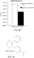
- thioxene dye C-28 thioxene, shown in FIG. 10
- the particles had a diameter of between 190 nm and 210
- the suspension was filtered, and 200 mL EtOH 10% at pH > 10 was added to the filtrate and mixed.
- the combined suspension was centrifuged for 20 minutes at 15,000 rcf using an Eppendorf 5417C centrifuge. After centrifugation, the liquid was decanted and the particles re-suspended in 10% EtOH at pH > 10, and briefly sonicated.
- Chelated Eu and/or Tb was prepared in a one-step synthesis.
- the procedure below discloses the preparation of chelated Tb, but can also be used for chelated Eu with minor modifications.
- 3.72 grams (10 mmol) of TbCl 3 ⁇ 6H 2 O in 30 mL water were added to a 250 mL Erlenmeyer flask. 90 mL of absolute EtOH was added to the solution and mixed to achieve a homogeneous distribution.
- 6.48 g (30 mmol) of 1-benzoyl-3,3,3-trifluoroacetonate (BTA) was added to the solution with stirring for 1 hour to complete dissolution.
- 6.6 mL of 5 M NaOH (33 mmol) was added slowly, maintaining the pH between 6 and 7.
- the solution was heated to between 50° and 60° C for 1 hour with stirring.
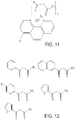
- diketonates can be used in the procedure described above (e.g., with different substituents corresponding to aromatic ring R in FIG. 11 ). Examples of suitable diketonates are shown in FIG. 12 .
- phenanthrolines can be used in the procedure described above (e.g., with different substituents corresponding to group R'). Examples of suitable phenanthrolines are shown in FIG. 13 .
- the particles i.e., cores 102 with reacting groups and luminescent agents added
- AmDex molecules having multiple amino groups per AmDex molecule
- EDAC carbodiimide
- the polysaccharide spontaneously associates with the particles, leading to an increased concentration of amino groups near to the surface of the particles which in turn improves the efficiency of the EDAC conjugation method.
- the coated particles had a diameter of between 240 nm and 270 nm, with 1 amino group for every 12 saccharides.
- the particles were subsequently coated with a second polysaccharide layer by reacting the particles with a relatively large excess of dextran aldehyde (DexAl) having multiple aldehyde groups per DexAl molecule.
- This spontaneous reaction is a form of spontaneous association of the polysaccharide layers.
- a fraction of the aldehyde groups on a DexAl molecule react with amino groups on the AmDex layer forming shiff-base, which then can be reduced with a mild reducing agent such as cyanoborohydride (NaBH 3 CN) to form a chemically stable carbon-nitrogen bond.
- a mild reducing agent such as cyanoborohydride (NaBH 3 CN)
- the resulting coated particles have reactive aldehyde groups on the surface and can react with amino group containing molecules (such as antibodies or other proteins or further AmDex molecules).
- the coated particles had a diameter of 260 nm to 310 nm, with 1 aldehyde group for every 16 saccharides (approximately 30,000 to 50,000 per particle).
- the particle suspension (concentration 20 mg/mL) was added to a glass vial, along with 0.8 mL dextran aldehyde solution (concentration 50 mg/mL), 4.2 mL of 0.1 M MES at pH 6.0, and 0.5 mL NaBH 3 CN at a concentration of 80 mg/mL in water (freshly prepared).
- the vial was closed with a screw cap and incubated at 37° C overnight.
- the particles were washed twice with 0.1 M MES at pH 6.0 by centrifugation at 15,000 rcf for 20 minutes. The particles were then sonicated and collected in approximately 4 mL of 0.1 M MES at pH 6.0.
- One or more targeting groups were then conjugated to the second polysaccharide layer.
- the conjugation was performed by a reductive amination method, in which purified antibody in native form (not modified) was incubated with the particles in presence of NaBH 3 CN for a certain period of time, preferably at room temperature or at 37° C.
- the remaining free aldehyde groups were capped (various molecules can be used for this purpose, for example carboxymethyl oxime or carboxymethoxylamine, etc.)
- Conjugation typically occurs best when the antibody concentration is at least 1 mg/mL (for conjugation of 1-2 mg of particles) or 0.53 mg/mL (for conjugation of 2.5 mg or more of particles).
- Antibody solutions can be concentrated, for example, using an iCON Concentrator (available from ThermoFisher Scientific, Waltham, MA).
- the antibodies are not in an amine-based buffer (e.g., Tris, glycine, bicine, tricine). If buffer exchange is undertaken, the buffer solution is generally replaced by a neutral or slightly alkaline buffer such as PBS or carbonate buffer at a pH of about 8.0.
- antibody solutions used for conjugation typically do not include protein- or peptide-based stabilizers (e.g., BSA, gelatin) or glycerol.
- Protein stabilizers can be removed using PhyTip affinity columns (available from PhyNexus, San Jose, CA) or a liquid handling system such as the JANUS automated workstation (available from PerkinElmer, Waltham, MA). Dialysis can be used to remove glycerol.
- Typical coupling ratios are either 10:1 (for 1-2 mg of particles) or 50:1 (for 2.5 mg of particles or more). For example, for 5 mg of particles, 0.1 mg of antibody is typically used.
- the following procedure was used to conjugate 5 mg of particles, using a 50:1 coupling ratio with an antibody.
- the antibody solution used typically has a concentration of 0.53 mg/mL or more.
- the particles were first washed in a 1.5 mL Eppendorf tube in 250 ⁇ L of water, and then 250 ⁇ L of PBS was added. The suspension was centrifuged at 16,000g (or maximum speed) for 15 minutes and the supernatant liquid discarded using a pipet tip.
- a fresh working solution of NaBH 3 CN (obtained in powder form from Sigma-Aldrich, St. Louis, MO) at a concentration of 400 mM in water was prepared by adding 25 mg of NaBH 3 CN powder to 1 mL of water.
- the suspension was incubated for 18-24 hours at 37° C using a rotary shaker at 6-10 RPM.
- CMO carboxymethoxylamine
- the conjugated particles were then washed. First, the particles were centrifuged for 15 minutes at 16,000g (or maximum speed) at 4° C. The supernatant was removed using a micropipette and the dehydrated particle pellet was resuspended in 1 mL of 100 mM Tris-HCl pH 8.0 (using about 200 ⁇ L per mg of particles). The particle suspension was briefly sonicated (10 short pulses of 1 second using a probe sonicator) to ensure the particles were not aggregated at a sonicator power of approximately 20% or maximum power. The suspension was then centrifuged for 15 minutes at 16,000g or maximum speed at 4° C, and the supernatant removed.
- Conjugated particles were stored in opaque vials at a temperature of 4° C. Prior to use, the suspensions were vortexed again to counteract settling during storage.
- the particle coupling procedure disclosed herein can be optimized (e.g., to increase the number of antibodies bound per particle) by reducing the reaction volume, as coupling efficiency typically increases with particle concentration.
- Increased particle concentrations, up to 100 mg/mL, can be prepared by adding a smaller volume of a more concentrated buffer solution to the particles.
- the procedure can also be optimized by increasing the ratio of antibody to particles, as coupling efficiency typically increases with antibody concentration.
- the antibody stock solution should be sufficiently concentrated so that only a small additional volume of the antibody solution is added to increase the antibody concentration without significantly diluting the particle concentration.
- a 10:1 ratio of particles:antibody when implemented at a particle concentration of 25 mg/mL, can improve conjugation efficiency.
- Increasing the particle concentration to 75 mg/mL while maintaining a ratio of 50:1 will typically involve an antibody concentration of 1.7 mg/mL.
- Increasing the relative proportion of antibody to a ratio of 25:1 will involve an antibody concentration of 3.4 mg/mL, which is close to the practical upper limit.
- Other buffers can also be used to optimize the foregoing procedure (e.g., 100 mM sodium phosphate at pH 8.0).
- the particles disclosed herein can be introduced into a living subject using a variety of methods, including injection, e.g., subcutaneous injection, intravenously, intratracheally, intranasally).
- the particles are introduced in proximity to a region of interest in the subject, e.g., near the site of a suspected tumor or tissue inflammation.
- the targeting groups are selected so that the particles bind selectively to certain structural entities, e.g., entities that are present in the region of interest.
- the targeting of specific structural entities within the subject allows the particles to function as sensitive diagnostic reporters for reactive species, e.g., reactive oxygen species in the subject's body.
- the particles can be used detect and image reactive species such as ROS in a variety of different subjects.
- the particles can be used for in-vivo detection and imaging in humans, in mammals (including, but not limited to, mice, rats, dogs, and cats, and more generally, any laboratory mammalian subject), in birds, reptiles, amphibians, and fish.
- the particles can also be used for in-vitro detection and imaging of tissue samples and/or biological fluids extracted from any of the foregoing subjects.
- luminescence emitted by the particles is imaged using a detector (e.g., a CCD-based detector or a CMOS-based detector).
- a detector e.g., a CCD-based detector or a CMOS-based detector.
- the emitted luminescence shows where reactive species, such as reactive oxygen species, are located within the subject's body.
- the detected luminescence can be displayed directly to a viewer without filtering the luminescence.
- the detected luminescence can be overlaid on an image of the subject's body. Both of these display modalities can yield images that localize the ROS relative to other features of the subject's body.
- the intensity and distribution of luminescence detected can also provide quantitative information about ROS in the subject's body. For example, by spatially integrating portions of the luminescence image of the subject and correlating the integrated intensity with calibration data that relates light intensity to ROS concentration, the concentration of ROS in the integrated region of the subject's body can be estimated. Quantitative estimates of ROS concentrations can provide additional information for purposes of diagnosing diseases in the subject.
- more than one type of particle can be administered to the subject.
- the plurality of particles that is administered can include a first subset of particles with one type of targeting group and a second subset of particles with a second type of targeting group.
- the particles can bind to different structural entities within the subject, thereby providing diagnostic ROS information about multiple regions of the subject's body.
- the first subset of particles can include a first luminescent agent and the second subject of particles can include a second luminescent agent.
- Different luminescent agents typically emit luminescence at different central wavelengths.
- subsets of particles with different luminescent agents can be used to provide information about different regions of a subject's body.
- the first subset of particles can include both targeting groups and luminescent agents that are different from the targeting groups and luminescent agents of the second subset of particles.
- Providing subsets of particles with these features yields two-fold selectivity in targeting particular structures or regions within a subject's body.
- the two subsets of particles bind to different structural entities in the subject on account of their different targeting groups, thereby providing a first mechanism for distinguishing the different entities.
- luminescence from the different subsets of particles occurs at different central wavelengths. Accordingly, by filtering and/or selectively detecting luminescence at certain wavelengths, ROS in the different structural entities can be separately identified and quantified.
- FIG. 2 is a schematic diagram of a kit 200 that includes a housing (e.g., a wrapper) 202. Enclosed within the housing is a container 204 that includes a plurality of particles 100.
- kit 200 can include a second container 206 that includes, for example, one or more buffer solutions for introducing the particles into a subject's body (e.g., via injection).
- particles 100 are provided in container 204 in an unsuspended (e.g., dry) form. Prior to administering the particles, the particles are suspended in a solution provided in second container 206.
- particles 100 are provided in container 204 already suspended in a physiologically-compatible solution. The particles can either be administered directly to the subject, or diluted to a desired concentration (e.g., in a buffer or other solution) before they are administered to the subject.
- the particles disclosed herein provide a number of important advantages.
- the particles emit luminescence, which is detected and used to localize and quantify ROS in the subject.
- luminescence typically occurs toward the red edge of the visible region of the spectrum, and in the near-infrared region.
- particles which emit luminescence are better suited to act as reporters for ROS in deep tissue applications (e.g., where ROS are located in tissues more than about 2 mm below the skin surface) because tissue-dependent scattering of radiation is not as strong at longer wavelengths.
- the particles disclosed herein provided significantly improved diagnostic sensitivity for ROS located in deep tissues relative to fluorescence-based particles and reporters.
- fluorescence-based reporters are typically accompanied by autofluorescence in the subject's tissue, which functions as a background signal against which fluorescence emission due to detection of ROS is distinguished.
- the luminescence-based particles disclosed herein do not typically excite, and are not typically accompanied by, significant autofluorescence in the subject's tissue.
- the luminescence emission typically occurs against an essentially "dark" background; removal of autofluorescence contributions (e.g., by sophisticated analysis algorithms) is generally not required.
- the detected luminescence can be used to visualize and quantify ROS in the subject's body without any filtering of the signal measured by the detector.
- the particles disclosed combine selective binding, reaction with ROS, and luminescent reporting of ROS in a single particle.
- Conventional two-particle methods for detecting ROS which involve both donor and acceptor particles, provide acceptable signals only when donor and acceptor particles approach one another closely enough.
- the particles disclosed herein do not rely on the distance between particles to detect ROS. Instead, each particle includes a reacting group that reacts with ROS, and a luminescent agent that emits luminescence in response to energy transfer from the reacting group. Because the luminescent agent and reacting group are located on the same particle, the energy transfer is efficient.
- particles 100 can include one or more reacting groups that react with reactive nitrogen species, emitting radiation that is absorbed by the luminescent agent(s) to cause luminescence emission.
- Suitable reacting groups include, for example DAF-FM (available from Thermo Fisher Scientific), which is a reagent that is used to detect and quantify low concentrations of nitric oxide (NO).
- DAF-FM is essentially non-fluorescent until it reacts with NO to form a fluorescent benzotriazole.
- DAF-FM fluorescence can be detected using a variety of different techniques and devices, including flow cytometers, microscopes, fluorescent microplate readers and fluorometers, and imaging systems such as the IVIS and FMT systems, available from from Perkin Elmer.
- quantities of HOCI, hydroxide radical, superoxide, nitric oxide, and hydrogen peroxide were prepared in vitro. Each of these in vitro species was treated with particles 100, which were prepared as described above and included latex cores, a first coating of aminodextran, and a second coating of dextran aldehyde. A thioxene dye and chelated europium were introduced into the latex cores before coating.
- FIG. 3 is a graph showing a comparison of luminescence signal/background detected for each of the different ROS. Each of the species was reliably detected, with hydroxyl radical and superoxide having the highest signal above background.
- a sample of BALB/cJ mice subjects were divided into two groups.
- the first group was challenged by intracheal delivery of 50 ⁇ L of 1 mg/kg lipopolysaccharide (LPS) at a concentration of 1 mg/kg i.n. to induce an inflammatory response.
- the second group functioning as the control, was challenged with phosphate buffered saline (PBS).
- PBS phosphate buffered saline
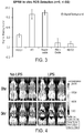
- a 10 ⁇ g quantity of particles (prepared as described above, with aminodextran and dextra aldehyde coatings, thioxene dye and chelated europium introduced into the latex core) were introduced into both lung and liver sites via subcutaneous injection 3 hours after the LPS challenge.
- Luminescence emission from the injected particles was imaged as a function of time following the injection of the particles.
- FIG. 4 shows images of control (left side) and LPS-challenged (right side) subjects immediately following the injection, and after a period of 5 hours.
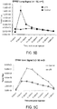
- FIG. 5A shows images of control (left side) and LPS-challenged (right side) subjects in 5 minute increments in the first hour following injection of the particles.
- FIGS. 5B and 5C show measured luminescence fluxes (arbitrary units) from both lung and liver tissues as a function of time, determined from the images in FIG. 5A . Measured luminescence from the lung tissue demonstrated that ROS were efficiently detected compared to the background signal corresponding to the control. Luminescence signals from liver tissue showed high background levels of luminescence due to the presence in the liver of cytochrome P450 oxidoreductases, which generate ROS.
- particles were prepared as described above (e.g., with aminodextran and dextra aldehyde coatings, thioxene dye and chelated europium introduced into the latex core) and conjugated to Ly6G antibodies, which bind to lung tissues.
- Ly6G antibodies conjugated to Ly6G antibodies, which bind to lung tissues.
- One group of BALB/cJ mice subjects was challenged with LPS intranasally with five 25 ⁇ L doses of 0.25 mg/mL LPS, followed by 25 ⁇ L of PBS.
- a control group of mice was challenged intranasally with five 25 ⁇ L doses of PBS. Both groups of mice were allowed to wake up between doses.
- Five hours after the initial challenge the mice were injected with the Ly6G-conjugated particles, and luminescence emission from the particles was imaged.
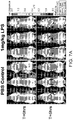
- FIG. 6A shows images of the control group (left) and the LPS-challenged group (right) immediately after injection of the particles, and after 5 hours.
- mice To compare luminescence signals from lung and liver tissues form the Ly6G-conjugated particles, BALB/cJ mice subjects were divided into two groups. One group was challenged with LPS (four 25 ⁇ L doses of 0.25 mg/mL followed by 25 ⁇ L PBS, delivered intratracheally) and the other (control) group was challenged with PBS (five 25 ⁇ L doses delivered intratracheally). Five hours post-challenge, the mice were injected intravenously with Ly6G1A8-conjugated particles (prepared as described above) and luminescence emission from the particles was imaged.
- LPS four 25 ⁇ L doses of 0.25 mg/mL followed by 25 ⁇ L PBS, delivered intratracheally
- PBS five 25 ⁇ L doses delivered intratracheally
- FIG. 7B is a graph showing the relative contributions to the measured luminescence signals for the control group (left bars) and LPS-challenged groups (right bars) for measured signals from lung tissues, liver tissues, and the total measured signal.
- lung signals account for 28% of the measured luminescence, while liver signals account for the remaining 72%.
- lung signals account for the LPS-challenged group
- lung signals account for 55% of the measured luminescence, while liver signals account for the remaining 45%. Both groups have similar total measured luminescence fluxes.
- FIG. 8A shows images of the normal-eating and fasting mice in the control and LPS-challenged groups as a function of time following injection of the particles.
- FIG. 8B is a graph showing contributions to each of the measured luminescence signals (lung signal, liver signal, and total signal) from each of the different types of mice in each of the control groups.
- the contributions from left to right correspond to normal-eating control group mice, fasting control group mice, normal eating LPS-challenged mice, and fasting LPS-challenged mice.
- the control group which the mice ate normally, 87% of the measured luminescence signal was contributed by liver tissues, while 13% was contributed by lung tissues.
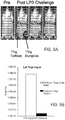
- Luminescence emission was imaged with no filter, with a 620 nm emission filter (the central wavelength of europium luminescence occurs at 614 nm), and with a 540 nm emission filter (the central wavelength of terbium emission occurs at 540 nm).
- FIG. 9A shows a series of images of a murine subject before particle injection, and after particle injection under the three different filtering arrangements.
- FIGS. 9B and 9C are graphs showing quantification of the Tb and Eu luminescence signals from the left and right thigh, respectively.
- the Tb signal level is only 6% of the total Tb luminescence signal (e.g., with no filter)
- the Eu signal level is 65% of the total Eu luminescence signal (e.g., with no filter).
Landscapes
- Health & Medical Sciences (AREA)
- Engineering & Computer Science (AREA)
- General Health & Medical Sciences (AREA)
- Epidemiology (AREA)
- Life Sciences & Earth Sciences (AREA)
- Animal Behavior & Ethology (AREA)
- Biomedical Technology (AREA)
- Public Health (AREA)
- Veterinary Medicine (AREA)
- Chemical & Material Sciences (AREA)
- Nanotechnology (AREA)
- Immunology (AREA)
- Investigating, Analyzing Materials By Fluorescence Or Luminescence (AREA)
- Medicines Containing Antibodies Or Antigens For Use As Internal Diagnostic Agents (AREA)
Claims (15)
- Zusammensetzung, die Partikel umfasst,
wobei jedes der Partikel mindestens eine an eine Struktureinheit in einem Subjekt bindende zielführende Gruppe und mindestens eine chemisch mit einer reaktiven Sauerstoffspezies in dem Subjekt reagierende Reaktionsgruppe umfasst, wobei das Partikel während der Reaktion Lumineszenz emittiert, und
wobei eine erste Untergruppe der Partikel jeweils eine erste zielführende Gruppe und eine erste Reaktionsgruppe umfasst und wobei eine zweite Untergruppe der Partikel jeweils eine zweite zielführende Gruppe und eine zweite Reaktionsgruppe umfasst, wobei die ersten und die zweiten zielführenden Gruppen voneinander verschieden sind,
zur Verwendung in einem diagnostischen Verfahren zur bildlichen Darstellung reaktiver Sauerstoffspezies in vivo zum Identifizieren von Krebserkrankungen oder entzündlichen Leiden in dem Subjekt, wobei das Verfahren Folgendes umfasst:die Verabreichung der Zusammensetzung an das Subjekt,die Detektion der Lumineszenzemission von den Partikeln unddas Abbilden eines Bilds des Subjekts, das, basierend auf der detektierten Lumineszenz, Stellen mindestens einiger reaktiver Sauerstoffspezies im Subjekt zeigt. - Zusammensetzung zur Verwendung nach Anspruch 1, weiterhin umfassend die Abbildung des Bilds des Subjekts basierend auf ungefilterter emittierter Strahlung von dem Subjekt, wobei die ungefilterte emittierte Strahlung die Lumineszenzemission umfasst.
- Zusammensetzung zur Verwendung nach Anspruch 1, wobei die reaktive Sauerstoffspezies mindestens ein Mitglied ausgewählt aus der Gruppe bestehend aus Singulettsauerstoff, einem Hydroxidradikal, hypochloriger Säure, einem Superoxidradikal, Stickstoffmonoxid und Wasserstoffperoxid umfasst.
- Zusammensetzung zur Verwendung nach Anspruch 1, wobei die ersten und die zweiten zielführenden Gruppen an verschiedene Struktureinheiten in dem Subjekt binden.
- Zusammensetzung zur Verwendung nach Anspruch 1, wobei die ersten und die zweiten zielführenden Gruppen voneinander verschiedene Antikörper umfassen.
- Zusammensetzung zur Verwendung nach Anspruch 1, wobei die erste Untergruppe der Partikel jeweils Lumineszenz bei einer ersten zentralen Wellenlänge emittiert und die zweite Untergruppe der Partikel jeweils Lumineszenz bei einer zweiten zentralen Wellenlänge, die von der ersten zentralen Wellenlänge verschieden ist, emittiert.
- Zusammensetzung zur Verwendung nach Anspruch 6, wobei sowohl die erste als auch die zweite zentrale Wellenlänge über 500 nm liegt.
- Zusammensetzung zur Verwendung nach Anspruch 1, wobei die ersten und die zweiten Reaktionsgruppen gleich sind.
- Zusammensetzung zur Verwendung nach Anspruch 1, wobei das Subjekt aus der aus einem lebenden Säugetier, einem lebenden Vogel, einem lebenden Amphibium und einem lebenden Fisch bestehenden Gruppe ausgewählt ist.
- Zusammensetzung, umfassend:ein Suspensionsmedium undeine Vielzahl von Partikeln, wobei jedes der Vielzahl von Partikeln Folgendes umfasst:einen Kern, der mindestens eine chemisch mit einer reaktiven Sauerstoffspezies in dem Subjekt reagierende Reaktionsgruppe und mindestens ein als Folge auf die Reaktion der mindestens einen Reaktionsgruppe Lumineszenz emittierendes lumineszentes Mittel umfasst,ein erstes, den Kern einkapselndes Beschichtungsmaterial undein zweites Beschichtungsmaterial, das das erste Beschichtungsmaterial einkapselt und mindestens eine an eine Struktureinheit in dem Subjekt bindende zielführende Gruppe umfasst, undwobei die Vielzahl von Partikeln Folgendes umfasst:eine erste Untergruppe von Partikeln, die eine erste zielführende Gruppe, ein erstes lumineszentes Mittel und eine erste Reaktionsgruppe umfasst, undeine zweite Untergruppe von Partikeln, die eine zweite zielführende Gruppe, ein zweites lumineszentes Mittel und eine zweite Reaktionsgruppe umfasst,wobei die ersten und die zweiten zielführenden Gruppen verschieden sind.
- Zusammensetzung nach Anspruch 10, wobei der Kern Latex und/oder Polystyrol umfasst.
- Zusammensetzung nach Anspruch 10, wobei das erste Beschichtungsmaterial Aminodextran umfasst und das zweite Beschichtungsmaterial Dextranaldehyd umfasst.
- Zusammensetzung nach Anspruch 10, wobei die mindestens eine Reaktionsgruppe Thioxen oder ein Thioxenderivat umfasst.
- Zusammensetzung nach Anspruch 10, wobei das erste und das zweite lumineszente Mittel verschieden sind, wobei insbesondere das erste und das zweite lumineszente Mittel verschiedene Lanthanidelemente umfassen.
- Kit zur bildlichen Darstellung reaktiver Sauerstoffspezies in einem lebenden Subjekt, wobei das Kit die Zusammensetzung nach Anspruch 10 umfasst.
Applications Claiming Priority (2)
| Application Number | Priority Date | Filing Date | Title |
|---|---|---|---|
| US14/030,428 US10124072B2 (en) | 2013-09-18 | 2013-09-18 | In-vivo reactive species imaging |
| PCT/US2014/056292 WO2015042256A1 (en) | 2013-09-18 | 2014-09-18 | In-vivo reactive species imaging |
Publications (2)
| Publication Number | Publication Date |
|---|---|
| EP3046589A1 EP3046589A1 (de) | 2016-07-27 |
| EP3046589B1 true EP3046589B1 (de) | 2020-01-08 |
Family
ID=51662323
Family Applications (1)
| Application Number | Title | Priority Date | Filing Date |
|---|---|---|---|
| EP14781359.6A Active EP3046589B1 (de) | 2013-09-18 | 2014-09-18 | In-vivo-abbildung reaktiver spezies |
Country Status (4)
| Country | Link |
|---|---|
| US (2) | US10124072B2 (de) |
| EP (1) | EP3046589B1 (de) |
| CN (1) | CN105792854B (de) |
| WO (1) | WO2015042256A1 (de) |
Families Citing this family (3)
| Publication number | Priority date | Publication date | Assignee | Title |
|---|---|---|---|---|
| US10124072B2 (en) | 2013-09-18 | 2018-11-13 | Caliper Life Sciences, Inc. | In-vivo reactive species imaging |
| FR3039649B1 (fr) * | 2015-07-29 | 2017-09-08 | Ecole Polytech | Procede de detection d'especes oxydantes |
| CN112500434B (zh) * | 2020-11-20 | 2021-10-22 | 复旦大学 | 基于铱配合物的光扳机分子及应用 |
Family Cites Families (17)
| Publication number | Priority date | Publication date | Assignee | Title |
|---|---|---|---|---|
| US5108899A (en) | 1989-10-31 | 1992-04-28 | Exoxemis, Inc. | Chemiluminescence assay of in vivo inflammation |
| US5578498A (en) | 1991-05-22 | 1996-11-26 | Behringwerke Ag | Metal chelate containing compositions for use in chemiluminescent assays |
| EP0984282B1 (de) | 1991-05-22 | 2003-07-30 | Dade Behring Marburg GmbH | Partikel, die eine Zusammensetzung beinhalten, die eine chemilumineszierende Verbindung umfassen |
| US5340716A (en) | 1991-06-20 | 1994-08-23 | Snytex (U.S.A.) Inc. | Assay method utilizing photoactivated chemiluminescent label |
| US6251581B1 (en) | 1991-05-22 | 2001-06-26 | Dade Behring Marburg Gmbh | Assay method utilizing induced luminescence |
| WO2001067105A1 (en) | 2000-03-06 | 2001-09-13 | Dade Behring Marburg Gmbh | Carriers coated with polysaccharides, their preparation and use |
| WO2005094902A2 (en) | 2004-04-01 | 2005-10-13 | Philips Intellectual Property & Standards Gmbh | Nanoparticles comprising luminescent substances as contrast agent for optical imaging |
| JP2008524203A (ja) | 2004-12-17 | 2008-07-10 | コーニンクレッカ フィリップス エレクトロニクス エヌ ヴィ | 分子イメージングのためのターゲティング剤 |
| US7842823B2 (en) | 2005-10-27 | 2010-11-30 | The Regents Of The University Of California | Fluorogenic probes for reactive oxygen species |
| CN101183073B (zh) | 2007-11-07 | 2010-09-15 | 博阳生物科技(上海)有限公司 | 质控微粒、其制备及其应用 |
| US20100069726A1 (en) * | 2008-06-04 | 2010-03-18 | Seventh Sense Biosystems, Inc. | Compositions and methods for rapid one-step diagnosis |
| WO2010040062A2 (en) | 2008-10-02 | 2010-04-08 | Advanced Throughput Ltd.Co. | Nanoparticle labeling reagents and methods of use |
| WO2010062787A1 (en) * | 2008-11-03 | 2010-06-03 | Washington University | Bioluminescence imaging of myeloperoxidase activity in vivo, methods, compositions and apparatuses therefor |
| BRPI0924549A2 (pt) | 2009-06-12 | 2016-04-26 | Amrita Vishwa Vidyapeetham University | nano fotomedicamentos como alvo para terapia fotodinâmica do câncer |
| US8795731B1 (en) * | 2009-10-12 | 2014-08-05 | University Of Central Florida Research Foundation, Inc. | Cerium oxide nanoparticle-based device for the detection of reactive oxygen species and monitoring of chronic inflammation |
| EP2697726B1 (de) | 2011-04-13 | 2020-04-01 | Caliper Life Sciences, Inc. | In-vivo-bilddarstellung einer enzymaktivität |
| US10124072B2 (en) | 2013-09-18 | 2018-11-13 | Caliper Life Sciences, Inc. | In-vivo reactive species imaging |
-
2013
- 2013-09-18 US US14/030,428 patent/US10124072B2/en active Active
-
2014
- 2014-09-18 CN CN201480051542.XA patent/CN105792854B/zh active Active
- 2014-09-18 WO PCT/US2014/056292 patent/WO2015042256A1/en not_active Ceased
- 2014-09-18 EP EP14781359.6A patent/EP3046589B1/de active Active
-
2018
- 2018-11-07 US US16/182,941 patent/US11040118B2/en active Active
Non-Patent Citations (1)
| Title |
|---|
| None * |
Also Published As
| Publication number | Publication date |
|---|---|
| US20150079002A1 (en) | 2015-03-19 |
| US10124072B2 (en) | 2018-11-13 |
| WO2015042256A1 (en) | 2015-03-26 |
| CN105792854A (zh) | 2016-07-20 |
| US20190070317A1 (en) | 2019-03-07 |
| EP3046589A1 (de) | 2016-07-27 |
| US11040118B2 (en) | 2021-06-22 |
| CN105792854B (zh) | 2020-09-15 |
Similar Documents
| Publication | Publication Date | Title |
|---|---|---|
| He et al. | AIE-based theranostic systems for detection and killing of pathogens | |
| Arai et al. | Exploring the use of upconversion nanoparticles in chemical and biological sensors: from surface modifications to point-of-care devices | |
| Wang et al. | Upconversion nanoprobes for the ratiometric luminescent sensing of nitric oxide | |
| Meng et al. | Multiple functional nanoprobe for contrast-enhanced bimodal cellular imaging and targeted therapy | |
| Liu et al. | Ultrasensitive nanosensors based on upconversion nanoparticles for selective hypoxia imaging in vivo upon near-infrared excitation | |
| Pan et al. | Ratiometric luminescence aptasensor based on dual-emissive persistent luminescent nanoparticles for autofluorescence-and exogenous interference-free determination of trace aflatoxin B1 in food samples | |
| Wang et al. | Plasmon‐enhanced fluorescence‐based core–shell gold nanorods as a near‐IR fluorescent turn‐on sensor for the highly sensitive detection of pyrophosphate in aqueous solution | |
| Luo et al. | Simplified aptamer-based colorimetric method using unmodified gold nanoparticles for the detection of carcinoma embryonic antigen | |
| Zhu et al. | Silver nanoparticles-enhanced time-resolved fluorescence sensor for VEGF165 based on Mn-doped ZnS quantum dots | |
| Xing et al. | Surface modifications technology of quantum dots based biosensors and their medical applications | |
| Zhang et al. | Gold nanobipyramid hotspot aggregation-induced surface-enhanced Raman scattering for the ultrasensitive detection of miRNA | |
| US11040118B2 (en) | In-vivo reactive species imaging | |
| Ozdemir et al. | Generation of singlet oxygen by persistent luminescent nanoparticle–photosensitizer conjugates: a proof of principle for photodynamic therapy without light | |
| Zhao et al. | Bioluminescent microcapsules: applications in activating a photosensitizer | |
| Xu et al. | Hydrothermal synthesis of polyethylenimine-protected high luminescent Pt-nanoclusters and their application to the detection of nitroimidazoles | |
| Zhang et al. | Facile and sensitive detection of protamine by enhanced room-temperature phosphorescence of Mn-doped ZnS quantum dots | |
| Li et al. | Near-infrared-fluorescent probe for turn-on lipopolysaccharide analysis based on PEG-modified gold nanorods with plasmon-enhanced fluorescence | |
| Li et al. | Fluorescent nanozyme based dual-channel lipopolysaccharide sensing | |
| Esmaelpourfarkhani et al. | Time-resolved fluorescence DNA-based sensors for reducing background fluorescence of environment | |
| Yue et al. | One-step synthesis of green fluorescent carbon dots for Chloride detecting and for bioimaging | |
| Liu et al. | Luminescent Rhodamine B doped core–shell silica nanoparticle labels for protein microarray detection | |
| Zhang et al. | Magnetic reduced graphene oxide (MrGO) nanocomposites as nano carries for loading and transferring of 10-hydroxycamptothecin | |
| Zhang et al. | A novel electrochemiluminescence aptasensor for ultrasensitive lincomycin detection using Ti3C2–TiO2–Ru probe | |
| Maruthi et al. | Advances in aggregation induced emission (AIE) materials in biosensing and imaging of bacteria | |
| Jin et al. | Magnetic core/shell Fe3O4/Au nanoparticles for studies of quinolones binding to protein by fluorescence spectroscopy |
Legal Events
| Date | Code | Title | Description |
|---|---|---|---|
| PUAI | Public reference made under article 153(3) epc to a published international application that has entered the european phase |
Free format text: ORIGINAL CODE: 0009012 |
|
| 17P | Request for examination filed |
Effective date: 20160418 |
|
| AK | Designated contracting states |
Kind code of ref document: A1 Designated state(s): AL AT BE BG CH CY CZ DE DK EE ES FI FR GB GR HR HU IE IS IT LI LT LU LV MC MK MT NL NO PL PT RO RS SE SI SK SM TR |
|
| AX | Request for extension of the european patent |
Extension state: BA ME |
|
| RIN1 | Information on inventor provided before grant (corrected) |
Inventor name: ANSALDI, DANIEL Inventor name: SINGH, RAJENDRA Inventor name: RAY, SUNETRA |
|
| DAX | Request for extension of the european patent (deleted) | ||
| STAA | Information on the status of an ep patent application or granted ep patent |
Free format text: STATUS: EXAMINATION IS IN PROGRESS |
|
| 17Q | First examination report despatched |
Effective date: 20190314 |
|
| GRAP | Despatch of communication of intention to grant a patent |
Free format text: ORIGINAL CODE: EPIDOSNIGR1 |
|
| STAA | Information on the status of an ep patent application or granted ep patent |
Free format text: STATUS: GRANT OF PATENT IS INTENDED |
|
| INTG | Intention to grant announced |
Effective date: 20190719 |
|
| RAP1 | Party data changed (applicant data changed or rights of an application transferred) |
Owner name: RAY, SUNETRA Owner name: CALIPER LIFE SCIENCES, INC. Owner name: ANSALDI, DANIEL Owner name: SINGH, RAJENDRA |
|
| GRAS | Grant fee paid |
Free format text: ORIGINAL CODE: EPIDOSNIGR3 |
|
| GRAA | (expected) grant |
Free format text: ORIGINAL CODE: 0009210 |
|
| STAA | Information on the status of an ep patent application or granted ep patent |
Free format text: STATUS: THE PATENT HAS BEEN GRANTED |
|
| AK | Designated contracting states |
Kind code of ref document: B1 Designated state(s): AL AT BE BG CH CY CZ DE DK EE ES FI FR GB GR HR HU IE IS IT LI LT LU LV MC MK MT NL NO PL PT RO RS SE SI SK SM TR |
|
| REG | Reference to a national code |
Ref country code: GB Ref legal event code: FG4D |
|
| REG | Reference to a national code |
Ref country code: CH Ref legal event code: EP |
|
| REG | Reference to a national code |
Ref country code: IE Ref legal event code: FG4D |
|
| REG | Reference to a national code |
Ref country code: DE Ref legal event code: R096 Ref document number: 602014059717 Country of ref document: DE |
|
| REG | Reference to a national code |
Ref country code: AT Ref legal event code: REF Ref document number: 1221883 Country of ref document: AT Kind code of ref document: T Effective date: 20200215 |
|
| REG | Reference to a national code |
Ref country code: NL Ref legal event code: FP |
|
| REG | Reference to a national code |
Ref country code: LT Ref legal event code: MG4D |
|
| PG25 | Lapsed in a contracting state [announced via postgrant information from national office to epo] |
Ref country code: NO Free format text: LAPSE BECAUSE OF FAILURE TO SUBMIT A TRANSLATION OF THE DESCRIPTION OR TO PAY THE FEE WITHIN THE PRESCRIBED TIME-LIMIT Effective date: 20200408 Ref country code: RS Free format text: LAPSE BECAUSE OF FAILURE TO SUBMIT A TRANSLATION OF THE DESCRIPTION OR TO PAY THE FEE WITHIN THE PRESCRIBED TIME-LIMIT Effective date: 20200108 Ref country code: FI Free format text: LAPSE BECAUSE OF FAILURE TO SUBMIT A TRANSLATION OF THE DESCRIPTION OR TO PAY THE FEE WITHIN THE PRESCRIBED TIME-LIMIT Effective date: 20200108 Ref country code: LT Free format text: LAPSE BECAUSE OF FAILURE TO SUBMIT A TRANSLATION OF THE DESCRIPTION OR TO PAY THE FEE WITHIN THE PRESCRIBED TIME-LIMIT Effective date: 20200108 Ref country code: PT Free format text: LAPSE BECAUSE OF FAILURE TO SUBMIT A TRANSLATION OF THE DESCRIPTION OR TO PAY THE FEE WITHIN THE PRESCRIBED TIME-LIMIT Effective date: 20200531 |
|
| PG25 | Lapsed in a contracting state [announced via postgrant information from national office to epo] |
Ref country code: IS Free format text: LAPSE BECAUSE OF FAILURE TO SUBMIT A TRANSLATION OF THE DESCRIPTION OR TO PAY THE FEE WITHIN THE PRESCRIBED TIME-LIMIT Effective date: 20200508 Ref country code: GR Free format text: LAPSE BECAUSE OF FAILURE TO SUBMIT A TRANSLATION OF THE DESCRIPTION OR TO PAY THE FEE WITHIN THE PRESCRIBED TIME-LIMIT Effective date: 20200409 Ref country code: LV Free format text: LAPSE BECAUSE OF FAILURE TO SUBMIT A TRANSLATION OF THE DESCRIPTION OR TO PAY THE FEE WITHIN THE PRESCRIBED TIME-LIMIT Effective date: 20200108 Ref country code: SE Free format text: LAPSE BECAUSE OF FAILURE TO SUBMIT A TRANSLATION OF THE DESCRIPTION OR TO PAY THE FEE WITHIN THE PRESCRIBED TIME-LIMIT Effective date: 20200108 Ref country code: HR Free format text: LAPSE BECAUSE OF FAILURE TO SUBMIT A TRANSLATION OF THE DESCRIPTION OR TO PAY THE FEE WITHIN THE PRESCRIBED TIME-LIMIT Effective date: 20200108 Ref country code: BG Free format text: LAPSE BECAUSE OF FAILURE TO SUBMIT A TRANSLATION OF THE DESCRIPTION OR TO PAY THE FEE WITHIN THE PRESCRIBED TIME-LIMIT Effective date: 20200408 |
|
| REG | Reference to a national code |
Ref country code: DE Ref legal event code: R097 Ref document number: 602014059717 Country of ref document: DE |
|
| PG25 | Lapsed in a contracting state [announced via postgrant information from national office to epo] |
Ref country code: ES Free format text: LAPSE BECAUSE OF FAILURE TO SUBMIT A TRANSLATION OF THE DESCRIPTION OR TO PAY THE FEE WITHIN THE PRESCRIBED TIME-LIMIT Effective date: 20200108 Ref country code: RO Free format text: LAPSE BECAUSE OF FAILURE TO SUBMIT A TRANSLATION OF THE DESCRIPTION OR TO PAY THE FEE WITHIN THE PRESCRIBED TIME-LIMIT Effective date: 20200108 Ref country code: CZ Free format text: LAPSE BECAUSE OF FAILURE TO SUBMIT A TRANSLATION OF THE DESCRIPTION OR TO PAY THE FEE WITHIN THE PRESCRIBED TIME-LIMIT Effective date: 20200108 Ref country code: SK Free format text: LAPSE BECAUSE OF FAILURE TO SUBMIT A TRANSLATION OF THE DESCRIPTION OR TO PAY THE FEE WITHIN THE PRESCRIBED TIME-LIMIT Effective date: 20200108 Ref country code: SM Free format text: LAPSE BECAUSE OF FAILURE TO SUBMIT A TRANSLATION OF THE DESCRIPTION OR TO PAY THE FEE WITHIN THE PRESCRIBED TIME-LIMIT Effective date: 20200108 Ref country code: EE Free format text: LAPSE BECAUSE OF FAILURE TO SUBMIT A TRANSLATION OF THE DESCRIPTION OR TO PAY THE FEE WITHIN THE PRESCRIBED TIME-LIMIT Effective date: 20200108 Ref country code: DK Free format text: LAPSE BECAUSE OF FAILURE TO SUBMIT A TRANSLATION OF THE DESCRIPTION OR TO PAY THE FEE WITHIN THE PRESCRIBED TIME-LIMIT Effective date: 20200108 |
|
| PLBE | No opposition filed within time limit |
Free format text: ORIGINAL CODE: 0009261 |
|
| STAA | Information on the status of an ep patent application or granted ep patent |
Free format text: STATUS: NO OPPOSITION FILED WITHIN TIME LIMIT |
|
| REG | Reference to a national code |
Ref country code: AT Ref legal event code: MK05 Ref document number: 1221883 Country of ref document: AT Kind code of ref document: T Effective date: 20200108 |
|
| 26N | No opposition filed |
Effective date: 20201009 |
|
| PG25 | Lapsed in a contracting state [announced via postgrant information from national office to epo] |
Ref country code: AT Free format text: LAPSE BECAUSE OF FAILURE TO SUBMIT A TRANSLATION OF THE DESCRIPTION OR TO PAY THE FEE WITHIN THE PRESCRIBED TIME-LIMIT Effective date: 20200108 Ref country code: IT Free format text: LAPSE BECAUSE OF FAILURE TO SUBMIT A TRANSLATION OF THE DESCRIPTION OR TO PAY THE FEE WITHIN THE PRESCRIBED TIME-LIMIT Effective date: 20200108 |
|
| PG25 | Lapsed in a contracting state [announced via postgrant information from national office to epo] |
Ref country code: SI Free format text: LAPSE BECAUSE OF FAILURE TO SUBMIT A TRANSLATION OF THE DESCRIPTION OR TO PAY THE FEE WITHIN THE PRESCRIBED TIME-LIMIT Effective date: 20200108 Ref country code: PL Free format text: LAPSE BECAUSE OF FAILURE TO SUBMIT A TRANSLATION OF THE DESCRIPTION OR TO PAY THE FEE WITHIN THE PRESCRIBED TIME-LIMIT Effective date: 20200108 |
|
| PG25 | Lapsed in a contracting state [announced via postgrant information from national office to epo] |
Ref country code: MC Free format text: LAPSE BECAUSE OF FAILURE TO SUBMIT A TRANSLATION OF THE DESCRIPTION OR TO PAY THE FEE WITHIN THE PRESCRIBED TIME-LIMIT Effective date: 20200108 |
|
| REG | Reference to a national code |
Ref country code: CH Ref legal event code: PL |
|
| REG | Reference to a national code |
Ref country code: BE Ref legal event code: MM Effective date: 20200930 |
|
| PG25 | Lapsed in a contracting state [announced via postgrant information from national office to epo] |
Ref country code: LU Free format text: LAPSE BECAUSE OF NON-PAYMENT OF DUE FEES Effective date: 20200918 |
|
| PG25 | Lapsed in a contracting state [announced via postgrant information from national office to epo] |
Ref country code: BE Free format text: LAPSE BECAUSE OF NON-PAYMENT OF DUE FEES Effective date: 20200930 Ref country code: CH Free format text: LAPSE BECAUSE OF NON-PAYMENT OF DUE FEES Effective date: 20200930 Ref country code: LI Free format text: LAPSE BECAUSE OF NON-PAYMENT OF DUE FEES Effective date: 20200930 Ref country code: IE Free format text: LAPSE BECAUSE OF NON-PAYMENT OF DUE FEES Effective date: 20200918 |
|
| PG25 | Lapsed in a contracting state [announced via postgrant information from national office to epo] |
Ref country code: TR Free format text: LAPSE BECAUSE OF FAILURE TO SUBMIT A TRANSLATION OF THE DESCRIPTION OR TO PAY THE FEE WITHIN THE PRESCRIBED TIME-LIMIT Effective date: 20200108 Ref country code: MT Free format text: LAPSE BECAUSE OF FAILURE TO SUBMIT A TRANSLATION OF THE DESCRIPTION OR TO PAY THE FEE WITHIN THE PRESCRIBED TIME-LIMIT Effective date: 20200108 Ref country code: CY Free format text: LAPSE BECAUSE OF FAILURE TO SUBMIT A TRANSLATION OF THE DESCRIPTION OR TO PAY THE FEE WITHIN THE PRESCRIBED TIME-LIMIT Effective date: 20200108 |
|
| PG25 | Lapsed in a contracting state [announced via postgrant information from national office to epo] |
Ref country code: MK Free format text: LAPSE BECAUSE OF FAILURE TO SUBMIT A TRANSLATION OF THE DESCRIPTION OR TO PAY THE FEE WITHIN THE PRESCRIBED TIME-LIMIT Effective date: 20200108 Ref country code: AL Free format text: LAPSE BECAUSE OF FAILURE TO SUBMIT A TRANSLATION OF THE DESCRIPTION OR TO PAY THE FEE WITHIN THE PRESCRIBED TIME-LIMIT Effective date: 20200108 |
|
| P01 | Opt-out of the competence of the unified patent court (upc) registered |
Effective date: 20230525 |
|
| PGFP | Annual fee paid to national office [announced via postgrant information from national office to epo] |
Ref country code: DE Payment date: 20250929 Year of fee payment: 12 |
|
| PGFP | Annual fee paid to national office [announced via postgrant information from national office to epo] |
Ref country code: NL Payment date: 20250926 Year of fee payment: 12 |
|
| PGFP | Annual fee paid to national office [announced via postgrant information from national office to epo] |
Ref country code: GB Payment date: 20250929 Year of fee payment: 12 |
|
| PGFP | Annual fee paid to national office [announced via postgrant information from national office to epo] |
Ref country code: FR Payment date: 20250925 Year of fee payment: 12 |