EP3019005B1 - Mrap2 knockouts - Google Patents
Mrap2 knockouts Download PDFInfo
- Publication number
- EP3019005B1 EP3019005B1 EP14822448.8A EP14822448A EP3019005B1 EP 3019005 B1 EP3019005 B1 EP 3019005B1 EP 14822448 A EP14822448 A EP 14822448A EP 3019005 B1 EP3019005 B1 EP 3019005B1
- Authority
- EP
- European Patent Office
- Prior art keywords
- mrap2
- mice
- dna
- animal
- artificial sequence
- Prior art date
- Legal status (The legal status is an assumption and is not a legal conclusion. Google has not performed a legal analysis and makes no representation as to the accuracy of the status listed.)
- Active
Links
- 238000000034 method Methods 0.000 claims description 79
- 230000004048 modification Effects 0.000 claims description 76
- 238000012986 modification Methods 0.000 claims description 76
- 238000012217 deletion Methods 0.000 claims description 30
- 230000037430 deletion Effects 0.000 claims description 30
- 101100024157 Mus musculus Mrap2 gene Proteins 0.000 claims description 23
- 235000013305 food Nutrition 0.000 claims description 23
- 238000003307 slaughter Methods 0.000 claims description 2
- 210000004027 cell Anatomy 0.000 description 161
- 241001465754 Metazoa Species 0.000 description 143
- 241000699670 Mus sp. Species 0.000 description 141
- 108090000623 proteins and genes Proteins 0.000 description 123
- 108020004414 DNA Proteins 0.000 description 106
- 101150110867 MC4R gene Proteins 0.000 description 72
- 239000013615 primer Substances 0.000 description 66
- 150000007523 nucleic acids Chemical class 0.000 description 53
- 102000039446 nucleic acids Human genes 0.000 description 50
- 108020004707 nucleic acids Proteins 0.000 description 50
- 102000004169 proteins and genes Human genes 0.000 description 47
- 230000014509 gene expression Effects 0.000 description 42
- 108090000765 processed proteins & peptides Proteins 0.000 description 42
- 239000013598 vector Substances 0.000 description 39
- 230000000694 effects Effects 0.000 description 36
- 230000008685 targeting Effects 0.000 description 31
- 241000124008 Mammalia Species 0.000 description 30
- 241000282414 Homo sapiens Species 0.000 description 28
- 241000699666 Mus <mouse, genus> Species 0.000 description 28
- 235000012631 food intake Nutrition 0.000 description 28
- 230000037406 food intake Effects 0.000 description 28
- 108010091086 Recombinases Proteins 0.000 description 27
- 102000018120 Recombinases Human genes 0.000 description 27
- 230000009261 transgenic effect Effects 0.000 description 27
- 102000004196 processed proteins & peptides Human genes 0.000 description 26
- 239000003550 marker Substances 0.000 description 25
- 108091028043 Nucleic acid sequence Proteins 0.000 description 24
- 229920001184 polypeptide Polymers 0.000 description 22
- 238000010459 TALEN Methods 0.000 description 21
- 108010043645 Transcription Activator-Like Effector Nucleases Proteins 0.000 description 21
- 230000037396 body weight Effects 0.000 description 21
- 210000001519 tissue Anatomy 0.000 description 20
- 208000008589 Obesity Diseases 0.000 description 19
- 150000001413 amino acids Chemical class 0.000 description 19
- 235000020824 obesity Nutrition 0.000 description 19
- 238000002744 homologous recombination Methods 0.000 description 18
- WHNFPRLDDSXQCL-UAZQEYIDSA-N α-msh Chemical compound C([C@@H](C(=O)N[C@@H](CO)C(=O)N[C@@H](CCSC)C(=O)N[C@@H](CCC(O)=O)C(=O)N[C@@H](CC=1NC=NC=1)C(=O)N[C@@H](CC=1C=CC=CC=1)C(=O)N[C@@H](CCCNC(N)=N)C(=O)N[C@@H](CC=1C2=CC=CC=C2NC=1)C(=O)NCC(=O)N[C@@H](CCCCN)C(=O)N1[C@@H](CCC1)C(=O)N[C@@H](C(C)C)C(N)=O)NC(=O)[C@H](CO)NC(C)=O)C1=CC=C(O)C=C1 WHNFPRLDDSXQCL-UAZQEYIDSA-N 0.000 description 18
- 241000282898 Sus scrofa Species 0.000 description 17
- 108700019146 Transgenes Proteins 0.000 description 17
- 230000006801 homologous recombination Effects 0.000 description 17
- 230000011664 signaling Effects 0.000 description 17
- 102100040148 Melanocortin-2 receptor accessory protein 2 Human genes 0.000 description 16
- 101710148380 Melanocortin-2 receptor accessory protein 2 Proteins 0.000 description 16
- FAPWRFPIFSIZLT-UHFFFAOYSA-M Sodium chloride Chemical compound [Na+].[Cl-] FAPWRFPIFSIZLT-UHFFFAOYSA-M 0.000 description 16
- 210000004556 brain Anatomy 0.000 description 15
- 238000005516 engineering process Methods 0.000 description 15
- 239000000523 sample Substances 0.000 description 15
- 108090000950 Melanocortin Receptors Proteins 0.000 description 14
- 102000004378 Melanocortin Receptors Human genes 0.000 description 14
- 102100027467 Pro-opiomelanocortin Human genes 0.000 description 14
- 239000000047 product Substances 0.000 description 14
- 239000003607 modifier Substances 0.000 description 13
- 239000002773 nucleotide Substances 0.000 description 13
- 125000003729 nucleotide group Chemical group 0.000 description 13
- 239000000243 solution Substances 0.000 description 13
- 239000012634 fragment Substances 0.000 description 12
- 210000004602 germ cell Anatomy 0.000 description 12
- 238000003757 reverse transcription PCR Methods 0.000 description 12
- 108700028369 Alleles Proteins 0.000 description 11
- 108091026890 Coding region Proteins 0.000 description 11
- 238000000338 in vitro Methods 0.000 description 11
- 238000003780 insertion Methods 0.000 description 11
- 230000037431 insertion Effects 0.000 description 11
- 210000001161 mammalian embryo Anatomy 0.000 description 11
- 238000001262 western blot Methods 0.000 description 11
- 108091032973 (ribonucleotides)n+m Proteins 0.000 description 10
- 101710163270 Nuclease Proteins 0.000 description 10
- 238000004458 analytical method Methods 0.000 description 10
- 101150036876 cre gene Proteins 0.000 description 10
- 230000001404 mediated effect Effects 0.000 description 10
- 108010051219 Cre recombinase Proteins 0.000 description 9
- 238000003556 assay Methods 0.000 description 9
- 230000001965 increasing effect Effects 0.000 description 9
- 238000005259 measurement Methods 0.000 description 9
- 230000035772 mutation Effects 0.000 description 9
- 210000002569 neuron Anatomy 0.000 description 9
- 102000040430 polynucleotide Human genes 0.000 description 9
- 108091033319 polynucleotide Proteins 0.000 description 9
- 239000002157 polynucleotide Substances 0.000 description 9
- 230000008569 process Effects 0.000 description 9
- 239000011780 sodium chloride Substances 0.000 description 9
- 241000894007 species Species 0.000 description 9
- IVOMOUWHDPKRLL-KQYNXXCUSA-N Cyclic adenosine monophosphate Chemical compound C([C@H]1O2)OP(O)(=O)O[C@H]1[C@@H](O)[C@@H]2N1C(N=CN=C2N)=C2N=C1 IVOMOUWHDPKRLL-KQYNXXCUSA-N 0.000 description 8
- 241001494479 Pecora Species 0.000 description 8
- IVOMOUWHDPKRLL-UHFFFAOYSA-N UNPD107823 Natural products O1C2COP(O)(=O)OC2C(O)C1N1C(N=CN=C2N)=C2N=C1 IVOMOUWHDPKRLL-UHFFFAOYSA-N 0.000 description 8
- 210000004978 chinese hamster ovary cell Anatomy 0.000 description 8
- 239000002299 complementary DNA Substances 0.000 description 8
- 230000000875 corresponding effect Effects 0.000 description 8
- 229940095074 cyclic amp Drugs 0.000 description 8
- 235000005911 diet Nutrition 0.000 description 8
- 230000037213 diet Effects 0.000 description 8
- 210000001671 embryonic stem cell Anatomy 0.000 description 8
- 230000006870 function Effects 0.000 description 8
- 210000003016 hypothalamus Anatomy 0.000 description 8
- 238000007901 in situ hybridization Methods 0.000 description 8
- 239000002502 liposome Substances 0.000 description 8
- 230000004584 weight gain Effects 0.000 description 8
- 235000019786 weight gain Nutrition 0.000 description 8
- 101150051050 MC3R gene Proteins 0.000 description 7
- 101150093308 POMC gene Proteins 0.000 description 7
- HCHKCACWOHOZIP-UHFFFAOYSA-N Zinc Chemical compound [Zn] HCHKCACWOHOZIP-UHFFFAOYSA-N 0.000 description 7
- 108010017070 Zinc Finger Nucleases Proteins 0.000 description 7
- 230000009471 action Effects 0.000 description 7
- 230000001186 cumulative effect Effects 0.000 description 7
- 230000007423 decrease Effects 0.000 description 7
- 238000010363 gene targeting Methods 0.000 description 7
- 230000002068 genetic effect Effects 0.000 description 7
- 238000010362 genome editing Methods 0.000 description 7
- 230000000670 limiting effect Effects 0.000 description 7
- 238000000520 microinjection Methods 0.000 description 7
- 230000004044 response Effects 0.000 description 7
- 102200052962 rs1137101 Human genes 0.000 description 7
- 238000001890 transfection Methods 0.000 description 7
- XLYOFNOQVPJJNP-UHFFFAOYSA-N water Chemical compound O XLYOFNOQVPJJNP-UHFFFAOYSA-N 0.000 description 7
- 229910052725 zinc Inorganic materials 0.000 description 7
- 239000011701 zinc Substances 0.000 description 7
- FWMNVWWHGCHHJJ-SKKKGAJSSA-N 4-amino-1-[(2r)-6-amino-2-[[(2r)-2-[[(2r)-2-[[(2r)-2-amino-3-phenylpropanoyl]amino]-3-phenylpropanoyl]amino]-4-methylpentanoyl]amino]hexanoyl]piperidine-4-carboxylic acid Chemical compound C([C@H](C(=O)N[C@H](CC(C)C)C(=O)N[C@H](CCCCN)C(=O)N1CCC(N)(CC1)C(O)=O)NC(=O)[C@H](N)CC=1C=CC=CC=1)C1=CC=CC=C1 FWMNVWWHGCHHJJ-SKKKGAJSSA-N 0.000 description 6
- 102100022455 Adrenocorticotropic hormone receptor Human genes 0.000 description 6
- 102000053602 DNA Human genes 0.000 description 6
- LFQSCWFLJHTTHZ-UHFFFAOYSA-N Ethanol Chemical compound CCO LFQSCWFLJHTTHZ-UHFFFAOYSA-N 0.000 description 6
- 108091029865 Exogenous DNA Proteins 0.000 description 6
- 108700024394 Exon Proteins 0.000 description 6
- 101000678419 Homo sapiens Adrenocorticotropic hormone receptor Proteins 0.000 description 6
- 101800001751 Melanocyte-stimulating hormone alpha Proteins 0.000 description 6
- 241000283973 Oryctolagus cuniculus Species 0.000 description 6
- 238000011529 RT qPCR Methods 0.000 description 6
- 238000004520 electroporation Methods 0.000 description 6
- 239000005090 green fluorescent protein Substances 0.000 description 6
- 235000009200 high fat diet Nutrition 0.000 description 6
- 238000009396 hybridization Methods 0.000 description 6
- 230000002267 hypothalamic effect Effects 0.000 description 6
- NOESYZHRGYRDHS-UHFFFAOYSA-N insulin Chemical compound N1C(=O)C(NC(=O)C(CCC(N)=O)NC(=O)C(CCC(O)=O)NC(=O)C(C(C)C)NC(=O)C(NC(=O)CN)C(C)CC)CSSCC(C(NC(CO)C(=O)NC(CC(C)C)C(=O)NC(CC=2C=CC(O)=CC=2)C(=O)NC(CCC(N)=O)C(=O)NC(CC(C)C)C(=O)NC(CCC(O)=O)C(=O)NC(CC(N)=O)C(=O)NC(CC=2C=CC(O)=CC=2)C(=O)NC(CSSCC(NC(=O)C(C(C)C)NC(=O)C(CC(C)C)NC(=O)C(CC=2C=CC(O)=CC=2)NC(=O)C(CC(C)C)NC(=O)C(C)NC(=O)C(CCC(O)=O)NC(=O)C(C(C)C)NC(=O)C(CC(C)C)NC(=O)C(CC=2NC=NC=2)NC(=O)C(CO)NC(=O)CNC2=O)C(=O)NCC(=O)NC(CCC(O)=O)C(=O)NC(CCCNC(N)=N)C(=O)NCC(=O)NC(CC=3C=CC=CC=3)C(=O)NC(CC=3C=CC=CC=3)C(=O)NC(CC=3C=CC(O)=CC=3)C(=O)NC(C(C)O)C(=O)N3C(CCC3)C(=O)NC(CCCCN)C(=O)NC(C)C(O)=O)C(=O)NC(CC(N)=O)C(O)=O)=O)NC(=O)C(C(C)CC)NC(=O)C(CO)NC(=O)C(C(C)O)NC(=O)C1CSSCC2NC(=O)C(CC(C)C)NC(=O)C(NC(=O)C(CCC(N)=O)NC(=O)C(CC(N)=O)NC(=O)C(NC(=O)C(N)CC=1C=CC=CC=1)C(C)C)CC1=CN=CN1 NOESYZHRGYRDHS-UHFFFAOYSA-N 0.000 description 6
- 239000000203 mixture Substances 0.000 description 6
- 230000002829 reductive effect Effects 0.000 description 6
- 239000013603 viral vector Substances 0.000 description 6
- 239000000275 Adrenocorticotropic Hormone Substances 0.000 description 5
- 241000283690 Bos taurus Species 0.000 description 5
- 241000283707 Capra Species 0.000 description 5
- 101800000414 Corticotropin Proteins 0.000 description 5
- SHIBSTMRCDJXLN-UHFFFAOYSA-N Digoxigenin Natural products C1CC(C2C(C3(C)CCC(O)CC3CC2)CC2O)(O)C2(C)C1C1=CC(=O)OC1 SHIBSTMRCDJXLN-UHFFFAOYSA-N 0.000 description 5
- 241000282412 Homo Species 0.000 description 5
- 206010020710 Hyperphagia Diseases 0.000 description 5
- 229930193140 Neomycin Natural products 0.000 description 5
- 241000282887 Suidae Species 0.000 description 5
- 108010073062 Transcription Activator-Like Effectors Proteins 0.000 description 5
- 101150022052 UCP1 gene Proteins 0.000 description 5
- 210000003486 adipose tissue brown Anatomy 0.000 description 5
- 238000000540 analysis of variance Methods 0.000 description 5
- 238000013459 approach Methods 0.000 description 5
- 230000008901 benefit Effects 0.000 description 5
- 210000001109 blastomere Anatomy 0.000 description 5
- IDLFZVILOHSSID-OVLDLUHVSA-N corticotropin Chemical compound C([C@@H](C(=O)N[C@@H](CO)C(=O)N[C@@H](CCSC)C(=O)N[C@@H](CCC(O)=O)C(=O)N[C@@H](CC=1NC=NC=1)C(=O)N[C@@H](CC=1C=CC=CC=1)C(=O)N[C@@H](CCCNC(N)=N)C(=O)N[C@@H](CC=1C2=CC=CC=C2NC=1)C(=O)NCC(=O)N[C@@H](CCCCN)C(=O)N1[C@@H](CCC1)C(=O)N[C@@H](C(C)C)C(=O)NCC(=O)N[C@@H](CCCCN)C(=O)N[C@@H](CCCCN)C(=O)N[C@@H](CCCNC(N)=N)C(=O)N[C@@H](CCCNC(N)=N)C(=O)N1[C@@H](CCC1)C(=O)N[C@@H](C(C)C)C(=O)N[C@@H](CCCCN)C(=O)N[C@@H](C(C)C)C(=O)N[C@@H](CC=1C=CC(O)=CC=1)C(=O)N1[C@@H](CCC1)C(=O)N[C@@H](CC(N)=O)C(=O)NCC(=O)N[C@@H](C)C(=O)N[C@@H](CCC(O)=O)C(=O)N[C@@H](CC(O)=O)C(=O)N[C@@H](CCC(O)=O)C(=O)N[C@@H](CO)C(=O)N[C@@H](C)C(=O)N[C@@H](CCC(O)=O)C(=O)N[C@@H](C)C(=O)N[C@@H](CC=1C=CC=CC=1)C(=O)N1[C@@H](CCC1)C(=O)N[C@@H](CC(C)C)C(=O)N[C@@H](CCC(O)=O)C(=O)N[C@@H](CC=1C=CC=CC=1)C(O)=O)NC(=O)[C@@H](N)CO)C1=CC=C(O)C=C1 IDLFZVILOHSSID-OVLDLUHVSA-N 0.000 description 5
- 229960000258 corticotropin Drugs 0.000 description 5
- QONQRTHLHBTMGP-UHFFFAOYSA-N digitoxigenin Natural products CC12CCC(C3(CCC(O)CC3CC3)C)C3C11OC1CC2C1=CC(=O)OC1 QONQRTHLHBTMGP-UHFFFAOYSA-N 0.000 description 5
- SHIBSTMRCDJXLN-KCZCNTNESA-N digoxigenin Chemical compound C1([C@@H]2[C@@]3([C@@](CC2)(O)[C@H]2[C@@H]([C@@]4(C)CC[C@H](O)C[C@H]4CC2)C[C@H]3O)C)=CC(=O)OC1 SHIBSTMRCDJXLN-KCZCNTNESA-N 0.000 description 5
- 230000006698 induction Effects 0.000 description 5
- 230000005764 inhibitory process Effects 0.000 description 5
- 208000001022 morbid obesity Diseases 0.000 description 5
- 229960004927 neomycin Drugs 0.000 description 5
- 239000013612 plasmid Substances 0.000 description 5
- 230000009467 reduction Effects 0.000 description 5
- 108091008146 restriction endonucleases Proteins 0.000 description 5
- 210000002966 serum Anatomy 0.000 description 5
- 230000003612 virological effect Effects 0.000 description 5
- 108020004463 18S ribosomal RNA Proteins 0.000 description 4
- QKNYBSVHEMOAJP-UHFFFAOYSA-N 2-amino-2-(hydroxymethyl)propane-1,3-diol;hydron;chloride Chemical compound Cl.OCC(N)(CO)CO QKNYBSVHEMOAJP-UHFFFAOYSA-N 0.000 description 4
- 241000283726 Bison Species 0.000 description 4
- 108020004705 Codon Proteins 0.000 description 4
- -1 DOPE Chemical compound 0.000 description 4
- KCXVZYZYPLLWCC-UHFFFAOYSA-N EDTA Chemical compound OC(=O)CN(CC(O)=O)CCN(CC(O)=O)CC(O)=O KCXVZYZYPLLWCC-UHFFFAOYSA-N 0.000 description 4
- 102100031780 Endonuclease Human genes 0.000 description 4
- 102000004190 Enzymes Human genes 0.000 description 4
- 108090000790 Enzymes Proteins 0.000 description 4
- 241000283073 Equus caballus Species 0.000 description 4
- 102000003688 G-Protein-Coupled Receptors Human genes 0.000 description 4
- 108090000045 G-Protein-Coupled Receptors Proteins 0.000 description 4
- 101000978418 Homo sapiens Melanocortin receptor 4 Proteins 0.000 description 4
- 102100023724 Melanocortin receptor 4 Human genes 0.000 description 4
- 238000000636 Northern blotting Methods 0.000 description 4
- 108091034117 Oligonucleotide Proteins 0.000 description 4
- 102220609028 SH2B adapter protein 1_T484A_mutation Human genes 0.000 description 4
- 238000002105 Southern blotting Methods 0.000 description 4
- 210000000593 adipose tissue white Anatomy 0.000 description 4
- 230000004075 alteration Effects 0.000 description 4
- 210000004102 animal cell Anatomy 0.000 description 4
- 210000004369 blood Anatomy 0.000 description 4
- 239000008280 blood Substances 0.000 description 4
- 210000005013 brain tissue Anatomy 0.000 description 4
- 210000004899 c-terminal region Anatomy 0.000 description 4
- 238000006243 chemical reaction Methods 0.000 description 4
- 238000010367 cloning Methods 0.000 description 4
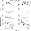
- 230000036757 core body temperature Effects 0.000 description 4
- 230000001419 dependent effect Effects 0.000 description 4
- 238000013461 design Methods 0.000 description 4
- 238000011161 development Methods 0.000 description 4
- LOKCTEFSRHRXRJ-UHFFFAOYSA-I dipotassium trisodium dihydrogen phosphate hydrogen phosphate dichloride Chemical compound P(=O)(O)(O)[O-].[K+].P(=O)(O)([O-])[O-].[Na+].[Na+].[Cl-].[K+].[Cl-].[Na+] LOKCTEFSRHRXRJ-UHFFFAOYSA-I 0.000 description 4
- 239000012153 distilled water Substances 0.000 description 4
- 238000001962 electrophoresis Methods 0.000 description 4
- 210000002257 embryonic structure Anatomy 0.000 description 4
- 238000003018 immunoassay Methods 0.000 description 4
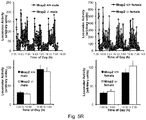
- 230000006742 locomotor activity Effects 0.000 description 4
- 210000004962 mammalian cell Anatomy 0.000 description 4
- 238000004519 manufacturing process Methods 0.000 description 4
- 239000000463 material Substances 0.000 description 4
- 210000004940 nucleus Anatomy 0.000 description 4
- 210000002963 paraventricular hypothalamic nucleus Anatomy 0.000 description 4
- 239000002953 phosphate buffered saline Substances 0.000 description 4
- 230000006798 recombination Effects 0.000 description 4
- 238000005215 recombination Methods 0.000 description 4
- 238000012552 review Methods 0.000 description 4
- 102200143520 rs6265 Human genes 0.000 description 4
- 238000012216 screening Methods 0.000 description 4
- 210000000130 stem cell Anatomy 0.000 description 4
- 238000012546 transfer Methods 0.000 description 4
- 238000011144 upstream manufacturing Methods 0.000 description 4
- 238000010200 validation analysis Methods 0.000 description 4
- SFLSHLFXELFNJZ-QMMMGPOBSA-N (-)-norepinephrine Chemical compound NC[C@H](O)C1=CC=C(O)C(O)=C1 SFLSHLFXELFNJZ-QMMMGPOBSA-N 0.000 description 3
- WFDIJRYMOXRFFG-UHFFFAOYSA-N Acetic anhydride Chemical compound CC(=O)OC(C)=O WFDIJRYMOXRFFG-UHFFFAOYSA-N 0.000 description 3
- 108091033409 CRISPR Proteins 0.000 description 3
- HEDRZPFGACZZDS-UHFFFAOYSA-N Chloroform Chemical compound ClC(Cl)Cl HEDRZPFGACZZDS-UHFFFAOYSA-N 0.000 description 3
- OMFXVFTZEKFJBZ-UHFFFAOYSA-N Corticosterone Natural products O=C1CCC2(C)C3C(O)CC(C)(C(CC4)C(=O)CO)C4C3CCC2=C1 OMFXVFTZEKFJBZ-UHFFFAOYSA-N 0.000 description 3
- 238000002965 ELISA Methods 0.000 description 3
- 108010042407 Endonucleases Proteins 0.000 description 3
- WSFSSNUMVMOOMR-UHFFFAOYSA-N Formaldehyde Chemical compound O=C WSFSSNUMVMOOMR-UHFFFAOYSA-N 0.000 description 3
- 102000004877 Insulin Human genes 0.000 description 3
- 108090001061 Insulin Proteins 0.000 description 3
- 102000016267 Leptin Human genes 0.000 description 3
- 108010092277 Leptin Proteins 0.000 description 3
- 108010069820 Pro-Opiomelanocortin Proteins 0.000 description 3
- 206010038997 Retroviral infections Diseases 0.000 description 3
- 238000012300 Sequence Analysis Methods 0.000 description 3
- 108020004440 Thymidine kinase Proteins 0.000 description 3
- GSEJCLTVZPLZKY-UHFFFAOYSA-N Triethanolamine Chemical compound OCCN(CCO)CCO GSEJCLTVZPLZKY-UHFFFAOYSA-N 0.000 description 3
- 241000700605 Viruses Species 0.000 description 3
- 230000001919 adrenal effect Effects 0.000 description 3
- 239000011324 bead Substances 0.000 description 3
- 210000000133 brain stem Anatomy 0.000 description 3
- 238000007707 calorimetry Methods 0.000 description 3
- 125000003178 carboxy group Chemical group [H]OC(*)=O 0.000 description 3
- 230000001413 cellular effect Effects 0.000 description 3
- 210000000349 chromosome Anatomy 0.000 description 3
- 230000000295 complement effect Effects 0.000 description 3
- OMFXVFTZEKFJBZ-HJTSIMOOSA-N corticosterone Chemical compound O=C1CC[C@]2(C)[C@H]3[C@@H](O)C[C@](C)([C@H](CC4)C(=O)CO)[C@@H]4[C@@H]3CCC2=C1 OMFXVFTZEKFJBZ-HJTSIMOOSA-N 0.000 description 3
- 238000005520 cutting process Methods 0.000 description 3
- 238000001514 detection method Methods 0.000 description 3
- 230000034431 double-strand break repair via homologous recombination Effects 0.000 description 3
- 239000003814 drug Substances 0.000 description 3
- 229940079593 drug Drugs 0.000 description 3
- 235000013601 eggs Nutrition 0.000 description 3
- 230000019439 energy homeostasis Effects 0.000 description 3
- 239000003623 enhancer Substances 0.000 description 3
- 210000003608 fece Anatomy 0.000 description 3
- 230000012953 feeding on blood of other organism Effects 0.000 description 3
- 239000000835 fiber Substances 0.000 description 3
- 239000000499 gel Substances 0.000 description 3
- 238000012239 gene modification Methods 0.000 description 3
- 238000003205 genotyping method Methods 0.000 description 3
- 210000002980 germ line cell Anatomy 0.000 description 3
- 230000012010 growth Effects 0.000 description 3
- 238000001114 immunoprecipitation Methods 0.000 description 3
- 208000015181 infectious disease Diseases 0.000 description 3
- 229940125396 insulin Drugs 0.000 description 3
- 230000003993 interaction Effects 0.000 description 3
- 238000007912 intraperitoneal administration Methods 0.000 description 3
- 229940039781 leptin Drugs 0.000 description 3
- NRYBAZVQPHGZNS-ZSOCWYAHSA-N leptin Chemical compound O=C([C@H](CO)NC(=O)[C@H](CC(C)C)NC(=O)[C@H](CC(O)=O)NC(=O)[C@H](CC(C)C)NC(=O)[C@H](CCC(N)=O)NC(=O)[C@H](CC=1C2=CC=CC=C2NC=1)NC(=O)[C@H](CC(C)C)NC(=O)[C@@H](NC(=O)[C@H](CC(O)=O)NC(=O)[C@H](CCC(N)=O)NC(=O)[C@H](CC(C)C)NC(=O)[C@H](CO)NC(=O)CNC(=O)[C@H](CCC(N)=O)NC(=O)[C@@H](N)CC(C)C)CCSC)N1CCC[C@H]1C(=O)NCC(=O)N[C@@H](CS)C(O)=O NRYBAZVQPHGZNS-ZSOCWYAHSA-N 0.000 description 3
- 239000006166 lysate Substances 0.000 description 3
- 230000007246 mechanism Effects 0.000 description 3
- 108020004999 messenger RNA Proteins 0.000 description 3
- 239000008267 milk Substances 0.000 description 3
- 235000013336 milk Nutrition 0.000 description 3
- 210000004080 milk Anatomy 0.000 description 3
- 238000010369 molecular cloning Methods 0.000 description 3
- 230000006780 non-homologous end joining Effects 0.000 description 3
- 229960002748 norepinephrine Drugs 0.000 description 3
- SFLSHLFXELFNJZ-UHFFFAOYSA-N norepinephrine Natural products NCC(O)C1=CC=C(O)C(O)=C1 SFLSHLFXELFNJZ-UHFFFAOYSA-N 0.000 description 3
- 208000014258 obesity due to melanocortin 4 receptor deficiency Diseases 0.000 description 3
- 230000001105 regulatory effect Effects 0.000 description 3
- 230000000241 respiratory effect Effects 0.000 description 3
- 230000033764 rhythmic process Effects 0.000 description 3
- 102200087712 rs3734354 Human genes 0.000 description 3
- 102200087713 rs3734355 Human genes 0.000 description 3
- 230000001052 transient effect Effects 0.000 description 3
- 241001430294 unidentified retrovirus Species 0.000 description 3
- 210000002700 urine Anatomy 0.000 description 3
- QRXMUCSWCMTJGU-UHFFFAOYSA-L (5-bromo-4-chloro-1h-indol-3-yl) phosphate Chemical compound C1=C(Br)C(Cl)=C2C(OP([O-])(=O)[O-])=CNC2=C1 QRXMUCSWCMTJGU-UHFFFAOYSA-L 0.000 description 2
- UCTWMZQNUQWSLP-VIFPVBQESA-N (R)-adrenaline Chemical compound CNC[C@H](O)C1=CC=C(O)C(O)=C1 UCTWMZQNUQWSLP-VIFPVBQESA-N 0.000 description 2
- 229930182837 (R)-adrenaline Natural products 0.000 description 2
- 108020005065 3' Flanking Region Proteins 0.000 description 2
- 108020005029 5' Flanking Region Proteins 0.000 description 2
- WOVKYSAHUYNSMH-RRKCRQDMSA-N 5-bromodeoxyuridine Chemical compound C1[C@H](O)[C@@H](CO)O[C@H]1N1C(=O)NC(=O)C(Br)=C1 WOVKYSAHUYNSMH-RRKCRQDMSA-N 0.000 description 2
- CIWBSHSKHKDKBQ-JLAZNSOCSA-N Ascorbic acid Chemical compound OC[C@H](O)[C@H]1OC(=O)C(O)=C1O CIWBSHSKHKDKBQ-JLAZNSOCSA-N 0.000 description 2
- 102100035080 BDNF/NT-3 growth factors receptor Human genes 0.000 description 2
- WOVKYSAHUYNSMH-UHFFFAOYSA-N BROMODEOXYURIDINE Natural products C1C(O)C(CO)OC1N1C(=O)NC(=O)C(Br)=C1 WOVKYSAHUYNSMH-UHFFFAOYSA-N 0.000 description 2
- 241000894006 Bacteria Species 0.000 description 2
- 102000004219 Brain-derived neurotrophic factor Human genes 0.000 description 2
- 108090000715 Brain-derived neurotrophic factor Proteins 0.000 description 2
- YQYJSBFKSSDGFO-UHFFFAOYSA-N Epihygromycin Natural products OC1C(O)C(C(=O)C)OC1OC(C(=C1)O)=CC=C1C=C(C)C(=O)NC1C(O)C(O)C2OCOC2C1O YQYJSBFKSSDGFO-UHFFFAOYSA-N 0.000 description 2
- 108010046276 FLP recombinase Proteins 0.000 description 2
- ZHNUHDYFZUAESO-UHFFFAOYSA-N Formamide Chemical compound NC=O ZHNUHDYFZUAESO-UHFFFAOYSA-N 0.000 description 2
- WQZGKKKJIJFFOK-GASJEMHNSA-N Glucose Chemical compound OC[C@H]1OC(O)[C@H](O)[C@@H](O)[C@@H]1O WQZGKKKJIJFFOK-GASJEMHNSA-N 0.000 description 2
- WZUVPPKBWHMQCE-UHFFFAOYSA-N Haematoxylin Chemical compound C12=CC(O)=C(O)C=C2CC2(O)C1C1=CC=C(O)C(O)=C1OC2 WZUVPPKBWHMQCE-UHFFFAOYSA-N 0.000 description 2
- 101000596896 Homo sapiens BDNF/NT-3 growth factors receptor Proteins 0.000 description 2
- 101000978431 Homo sapiens Melanocortin receptor 3 Proteins 0.000 description 2
- 101001128694 Homo sapiens Neuroendocrine convertase 1 Proteins 0.000 description 2
- 101000707152 Homo sapiens SH2B adapter protein 1 Proteins 0.000 description 2
- 102100030874 Leptin Human genes 0.000 description 2
- HLFSDGLLUJUHTE-SNVBAGLBSA-N Levamisole Chemical compound C1([C@H]2CN3CCSC3=N2)=CC=CC=C1 HLFSDGLLUJUHTE-SNVBAGLBSA-N 0.000 description 2
- 102100023726 Melanocortin receptor 3 Human genes 0.000 description 2
- 108010050258 Mitochondrial Uncoupling Proteins Proteins 0.000 description 2
- 102000015494 Mitochondrial Uncoupling Proteins Human genes 0.000 description 2
- 241000699660 Mus musculus Species 0.000 description 2
- 102100032132 Neuroendocrine convertase 1 Human genes 0.000 description 2
- 229920012196 Polyoxymethylene Copolymer Polymers 0.000 description 2
- 239000013614 RNA sample Substances 0.000 description 2
- 108010092799 RNA-directed DNA polymerase Proteins 0.000 description 2
- 102000006382 Ribonucleases Human genes 0.000 description 2
- 108010083644 Ribonucleases Proteins 0.000 description 2
- 102100031770 SH2B adapter protein 1 Human genes 0.000 description 2
- 102000006601 Thymidine Kinase Human genes 0.000 description 2
- AUYYCJSJGJYCDS-LBPRGKRZSA-N Thyrolar Chemical class IC1=CC(C[C@H](N)C(O)=O)=CC(I)=C1OC1=CC=C(O)C(I)=C1 AUYYCJSJGJYCDS-LBPRGKRZSA-N 0.000 description 2
- 239000007983 Tris buffer Substances 0.000 description 2
- 229920004890 Triton X-100 Polymers 0.000 description 2
- 239000013504 Triton X-100 Substances 0.000 description 2
- 241001416177 Vicugna pacos Species 0.000 description 2
- 230000001594 aberrant effect Effects 0.000 description 2
- 238000007818 agglutination assay Methods 0.000 description 2
- XAGFODPZIPBFFR-UHFFFAOYSA-N aluminium Chemical compound [Al] XAGFODPZIPBFFR-UHFFFAOYSA-N 0.000 description 2
- 229910052782 aluminium Inorganic materials 0.000 description 2
- 230000003321 amplification Effects 0.000 description 2
- 230000000692 anti-sense effect Effects 0.000 description 2
- 230000001580 bacterial effect Effects 0.000 description 2
- 230000006583 body weight regulation Effects 0.000 description 2
- 238000009395 breeding Methods 0.000 description 2
- 230000001488 breeding effect Effects 0.000 description 2
- 229950004398 broxuridine Drugs 0.000 description 2
- 150000003943 catecholamines Chemical class 0.000 description 2
- 125000002091 cationic group Chemical group 0.000 description 2
- 210000001638 cerebellum Anatomy 0.000 description 2
- 230000008859 change Effects 0.000 description 2
- 239000003153 chemical reaction reagent Substances 0.000 description 2
- 210000001728 clone cell Anatomy 0.000 description 2
- 230000002860 competitive effect Effects 0.000 description 2
- 230000001276 controlling effect Effects 0.000 description 2
- 238000012258 culturing Methods 0.000 description 2
- 230000007812 deficiency Effects 0.000 description 2
- 235000018823 dietary intake Nutrition 0.000 description 2
- 230000005782 double-strand break Effects 0.000 description 2
- 229960005139 epinephrine Drugs 0.000 description 2
- 230000029142 excretion Effects 0.000 description 2
- 230000004720 fertilization Effects 0.000 description 2
- 239000011888 foil Substances 0.000 description 2
- 229940088597 hormone Drugs 0.000 description 2
- 239000005556 hormone Substances 0.000 description 2
- 238000001727 in vivo Methods 0.000 description 2
- 230000002779 inactivation Effects 0.000 description 2
- 238000010348 incorporation Methods 0.000 description 2
- 238000011534 incubation Methods 0.000 description 2
- 230000001939 inductive effect Effects 0.000 description 2
- 230000010354 integration Effects 0.000 description 2
- 230000003834 intracellular effect Effects 0.000 description 2
- 239000007928 intraperitoneal injection Substances 0.000 description 2
- 229960001614 levamisole Drugs 0.000 description 2
- 238000001638 lipofection Methods 0.000 description 2
- 238000003670 luciferase enzyme activity assay Methods 0.000 description 2
- 230000013011 mating Effects 0.000 description 2
- 235000013372 meat Nutrition 0.000 description 2
- 230000002503 metabolic effect Effects 0.000 description 2
- 238000013508 migration Methods 0.000 description 2
- 230000005012 migration Effects 0.000 description 2
- JPXMTWWFLBLUCD-UHFFFAOYSA-N nitro blue tetrazolium(2+) Chemical compound COC1=CC(C=2C=C(OC)C(=CC=2)[N+]=2N(N=C(N=2)C=2C=CC=CC=2)C=2C=CC(=CC=2)[N+]([O-])=O)=CC=C1[N+]1=NC(C=2C=CC=CC=2)=NN1C1=CC=C([N+]([O-])=O)C=C1 JPXMTWWFLBLUCD-UHFFFAOYSA-N 0.000 description 2
- 238000003199 nucleic acid amplification method Methods 0.000 description 2
- 230000035764 nutrition Effects 0.000 description 2
- 235000016709 nutrition Nutrition 0.000 description 2
- 230000036961 partial effect Effects 0.000 description 2
- 230000001717 pathogenic effect Effects 0.000 description 2
- 238000001556 precipitation Methods 0.000 description 2
- 238000002360 preparation method Methods 0.000 description 2
- 239000013608 rAAV vector Substances 0.000 description 2
- 238000003127 radioimmunoassay Methods 0.000 description 2
- 230000004043 responsiveness Effects 0.000 description 2
- 102220133997 rs1805096 Human genes 0.000 description 2
- 102220133994 rs1805134 Human genes 0.000 description 2
- 102220149198 rs6233 Human genes 0.000 description 2
- 102200025738 rs6234 Human genes 0.000 description 2
- 102200025741 rs6235 Human genes 0.000 description 2
- 238000005070 sampling Methods 0.000 description 2
- 230000003248 secreting effect Effects 0.000 description 2
- 210000001082 somatic cell Anatomy 0.000 description 2
- UCSJYZPVAKXKNQ-HZYVHMACSA-N streptomycin Chemical compound CN[C@H]1[C@H](O)[C@@H](O)[C@H](CO)O[C@H]1O[C@@H]1[C@](C=O)(O)[C@H](C)O[C@H]1O[C@@H]1[C@@H](NC(N)=N)[C@H](O)[C@@H](NC(N)=N)[C@H](O)[C@H]1O UCSJYZPVAKXKNQ-HZYVHMACSA-N 0.000 description 2
- 238000006467 substitution reaction Methods 0.000 description 2
- 239000005495 thyroid hormone Substances 0.000 description 2
- 229940036555 thyroid hormone Drugs 0.000 description 2
- 230000001131 transforming effect Effects 0.000 description 2
- 238000013519 translation Methods 0.000 description 2
- 230000032258 transport Effects 0.000 description 2
- LENZDBCJOHFCAS-UHFFFAOYSA-N tris Chemical compound OCC(N)(CO)CO LENZDBCJOHFCAS-UHFFFAOYSA-N 0.000 description 2
- OPCHFPHZPIURNA-MFERNQICSA-N (2s)-2,5-bis(3-aminopropylamino)-n-[2-(dioctadecylamino)acetyl]pentanamide Chemical compound CCCCCCCCCCCCCCCCCCN(CC(=O)NC(=O)[C@H](CCCNCCCN)NCCCN)CCCCCCCCCCCCCCCCCC OPCHFPHZPIURNA-MFERNQICSA-N 0.000 description 1
- JDKLPDJLXHXHNV-MFVUMRCOSA-N (3s,6s,9r,12s,15s,23s)-15-[[(2s)-2-acetamidohexanoyl]amino]-9-benzyl-6-[3-(diaminomethylideneamino)propyl]-12-(1h-imidazol-5-ylmethyl)-3-(1h-indol-3-ylmethyl)-2,5,8,11,14,17-hexaoxo-1,4,7,10,13,18-hexazacyclotricosane-23-carboxamide Chemical compound C([C@@H]1C(=O)N[C@@H](CCCN=C(N)N)C(=O)N[C@@H](CC=2C3=CC=CC=C3NC=2)C(=O)N[C@@H](CCCCNC(=O)C[C@@H](C(N[C@@H](CC=2NC=NC=2)C(=O)N1)=O)NC(=O)[C@@H](NC(C)=O)CCCC)C(N)=O)C1=CC=CC=C1 JDKLPDJLXHXHNV-MFVUMRCOSA-N 0.000 description 1
- LDGWQMRUWMSZIU-LQDDAWAPSA-M 2,3-bis[(z)-octadec-9-enoxy]propyl-trimethylazanium;chloride Chemical compound [Cl-].CCCCCCCC\C=C/CCCCCCCCOCC(C[N+](C)(C)C)OCCCCCCCC\C=C/CCCCCCCC LDGWQMRUWMSZIU-LQDDAWAPSA-M 0.000 description 1
- JKMHFZQWWAIEOD-UHFFFAOYSA-N 2-[4-(2-hydroxyethyl)piperazin-1-yl]ethanesulfonic acid Chemical compound OCC[NH+]1CCN(CCS([O-])(=O)=O)CC1 JKMHFZQWWAIEOD-UHFFFAOYSA-N 0.000 description 1
- KISWVXRQTGLFGD-UHFFFAOYSA-N 2-[[2-[[6-amino-2-[[2-[[2-[[5-amino-2-[[2-[[1-[2-[[6-amino-2-[(2,5-diamino-5-oxopentanoyl)amino]hexanoyl]amino]-5-(diaminomethylideneamino)pentanoyl]pyrrolidine-2-carbonyl]amino]-3-hydroxypropanoyl]amino]-5-oxopentanoyl]amino]-5-(diaminomethylideneamino)p Chemical compound C1CCN(C(=O)C(CCCN=C(N)N)NC(=O)C(CCCCN)NC(=O)C(N)CCC(N)=O)C1C(=O)NC(CO)C(=O)NC(CCC(N)=O)C(=O)NC(CCCN=C(N)N)C(=O)NC(CO)C(=O)NC(CCCCN)C(=O)NC(C(=O)NC(CC(C)C)C(O)=O)CC1=CC=C(O)C=C1 KISWVXRQTGLFGD-UHFFFAOYSA-N 0.000 description 1
- UMCMPZBLKLEWAF-BCTGSCMUSA-N 3-[(3-cholamidopropyl)dimethylammonio]propane-1-sulfonate Chemical compound C([C@H]1C[C@H]2O)[C@H](O)CC[C@]1(C)[C@@H]1[C@@H]2[C@@H]2CC[C@H]([C@@H](CCC(=O)NCCC[N+](C)(C)CCCS([O-])(=O)=O)C)[C@@]2(C)[C@@H](O)C1 UMCMPZBLKLEWAF-BCTGSCMUSA-N 0.000 description 1
- 108700034262 4-Nle-7-Phe-alpha- MSH Proteins 0.000 description 1
- 241000251468 Actinopterygii Species 0.000 description 1
- 102000007469 Actins Human genes 0.000 description 1
- 108010085238 Actins Proteins 0.000 description 1
- 108010072151 Agouti Signaling Protein Proteins 0.000 description 1
- 102000006822 Agouti Signaling Protein Human genes 0.000 description 1
- 102000054930 Agouti-Related Human genes 0.000 description 1
- 101710127426 Agouti-related protein Proteins 0.000 description 1
- 241000282979 Alces alces Species 0.000 description 1
- 108091093088 Amplicon Proteins 0.000 description 1
- 206010002091 Anaesthesia Diseases 0.000 description 1
- 244000303258 Annona diversifolia Species 0.000 description 1
- 235000002198 Annona diversifolia Nutrition 0.000 description 1
- 241000237519 Bivalvia Species 0.000 description 1
- 241001416152 Bos frontalis Species 0.000 description 1
- 241001416153 Bos grunniens Species 0.000 description 1
- 108091003079 Bovine Serum Albumin Proteins 0.000 description 1
- 238000011740 C57BL/6 mouse Methods 0.000 description 1
- 241000282836 Camelus dromedarius Species 0.000 description 1
- CURLTUGMZLYLDI-UHFFFAOYSA-N Carbon dioxide Chemical compound O=C=O CURLTUGMZLYLDI-UHFFFAOYSA-N 0.000 description 1
- 241000700199 Cavia porcellus Species 0.000 description 1
- 241000282994 Cervidae Species 0.000 description 1
- VEXZGXHMUGYJMC-UHFFFAOYSA-M Chloride anion Chemical compound [Cl-] VEXZGXHMUGYJMC-UHFFFAOYSA-M 0.000 description 1
- 108091060290 Chromatid Proteins 0.000 description 1
- 108020004635 Complementary DNA Proteins 0.000 description 1
- 241000484025 Cuniculus Species 0.000 description 1
- COLNVLDHVKWLRT-MRVPVSSYSA-N D-phenylalanine Chemical compound OC(=O)[C@H](N)CC1=CC=CC=C1 COLNVLDHVKWLRT-MRVPVSSYSA-N 0.000 description 1
- 239000003155 DNA primer Substances 0.000 description 1
- 238000012270 DNA recombination Methods 0.000 description 1
- 108010053770 Deoxyribonucleases Proteins 0.000 description 1
- 102000016911 Deoxyribonucleases Human genes 0.000 description 1
- 241000702421 Dependoparvovirus Species 0.000 description 1
- 108010053187 Diphtheria Toxin Proteins 0.000 description 1
- 108010015720 Dopamine beta-Hydroxylase Proteins 0.000 description 1
- 102100033156 Dopamine beta-hydroxylase Human genes 0.000 description 1
- 241000196324 Embryophyta Species 0.000 description 1
- 102000004533 Endonucleases Human genes 0.000 description 1
- 241000283074 Equus asinus Species 0.000 description 1
- 241001331845 Equus asinus x caballus Species 0.000 description 1
- 241000702191 Escherichia virus P1 Species 0.000 description 1
- 241000282326 Felis catus Species 0.000 description 1
- 241000197200 Gallinago media Species 0.000 description 1
- 102100028652 Gamma-enolase Human genes 0.000 description 1
- 101710191797 Gamma-enolase Proteins 0.000 description 1
- 108010010803 Gelatin Proteins 0.000 description 1
- 230000010558 Gene Alterations Effects 0.000 description 1
- 229940123611 Genome editing Drugs 0.000 description 1
- 102100039289 Glial fibrillary acidic protein Human genes 0.000 description 1
- 101710193519 Glial fibrillary acidic protein Proteins 0.000 description 1
- 206010072079 Glucocorticoid deficiency Diseases 0.000 description 1
- 108020005004 Guide RNA Proteins 0.000 description 1
- 206010019708 Hepatic steatosis Diseases 0.000 description 1
- 101001063991 Homo sapiens Leptin Proteins 0.000 description 1
- 101001134060 Homo sapiens Melanocyte-stimulating hormone receptor Proteins 0.000 description 1
- 101000703681 Homo sapiens Single-minded homolog 1 Proteins 0.000 description 1
- 108091092195 Intron Proteins 0.000 description 1
- KFZMGEQAYNKOFK-UHFFFAOYSA-N Isopropanol Chemical compound CC(C)O KFZMGEQAYNKOFK-UHFFFAOYSA-N 0.000 description 1
- 241000270322 Lepidosauria Species 0.000 description 1
- LAZPBGZRMVRFKY-HNCPQSOCSA-N Levamisole hydrochloride Chemical compound Cl.C1([C@H]2CN3CCSC3=N2)=CC=CC=C1 LAZPBGZRMVRFKY-HNCPQSOCSA-N 0.000 description 1
- 102000001796 Melanocortin 4 receptors Human genes 0.000 description 1
- 102000030612 Melanocortin 5 receptor Human genes 0.000 description 1
- 108010088565 Melanocortin 5 receptor Proteins 0.000 description 1
- 102220611249 Melanocortin-2 receptor accessory protein 2_N88Y_mutation Human genes 0.000 description 1
- 102100034216 Melanocyte-stimulating hormone receptor Human genes 0.000 description 1
- 101000928178 Mus musculus Agouti-related protein Proteins 0.000 description 1
- 101100129532 Mus musculus Mc4r gene Proteins 0.000 description 1
- 101000725553 Mus musculus Pro-opiomelanocortin Proteins 0.000 description 1
- 108010021466 Mutant Proteins Proteins 0.000 description 1
- 102000008300 Mutant Proteins Human genes 0.000 description 1
- 102000047918 Myelin Basic Human genes 0.000 description 1
- 101710107068 Myelin basic protein Proteins 0.000 description 1
- 230000004988 N-glycosylation Effects 0.000 description 1
- 108010088225 Nestin Proteins 0.000 description 1
- 102000008730 Nestin Human genes 0.000 description 1
- 206010033307 Overweight Diseases 0.000 description 1
- 238000012408 PCR amplification Methods 0.000 description 1
- 229930040373 Paraformaldehyde Natural products 0.000 description 1
- 208000007683 Pediatric Obesity Diseases 0.000 description 1
- 229930182555 Penicillin Natural products 0.000 description 1
- JGSARLDLIJGVTE-MBNYWOFBSA-N Penicillin G Chemical compound N([C@H]1[C@H]2SC([C@@H](N2C1=O)C(O)=O)(C)C)C(=O)CC1=CC=CC=C1 JGSARLDLIJGVTE-MBNYWOFBSA-N 0.000 description 1
- 241000009328 Perro Species 0.000 description 1
- 102000045595 Phosphoprotein Phosphatases Human genes 0.000 description 1
- 108700019535 Phosphoprotein Phosphatases Proteins 0.000 description 1
- 102000004160 Phosphoric Monoester Hydrolases Human genes 0.000 description 1
- 108090000608 Phosphoric Monoester Hydrolases Proteins 0.000 description 1
- 108091000080 Phosphotransferase Proteins 0.000 description 1
- 208000012641 Pigmentation disease Diseases 0.000 description 1
- 239000000683 Pro-Opiomelanocortin Substances 0.000 description 1
- 102100038931 Proenkephalin-A Human genes 0.000 description 1
- 108010057464 Prolactin Proteins 0.000 description 1
- 102000003946 Prolactin Human genes 0.000 description 1
- 239000012083 RIPA buffer Substances 0.000 description 1
- 241000283011 Rangifer Species 0.000 description 1
- 241000700162 Rattus sp. Species 0.000 description 1
- 108020004511 Recombinant DNA Proteins 0.000 description 1
- 108010052090 Renilla Luciferases Proteins 0.000 description 1
- 241000283984 Rodentia Species 0.000 description 1
- 240000004808 Saccharomyces cerevisiae Species 0.000 description 1
- 102000003800 Selectins Human genes 0.000 description 1
- 108090000184 Selectins Proteins 0.000 description 1
- 229920002684 Sepharose Polymers 0.000 description 1
- BQCADISMDOOEFD-UHFFFAOYSA-N Silver Chemical compound [Ag] BQCADISMDOOEFD-UHFFFAOYSA-N 0.000 description 1
- 108020004682 Single-Stranded DNA Proteins 0.000 description 1
- 102100031980 Single-minded homolog 1 Human genes 0.000 description 1
- 238000000692 Student's t-test Methods 0.000 description 1
- 102000001435 Synapsin Human genes 0.000 description 1
- 108050009621 Synapsin Proteins 0.000 description 1
- 101710137500 T7 RNA polymerase Proteins 0.000 description 1
- 108020005038 Terminator Codon Proteins 0.000 description 1
- 102000040945 Transcription factor Human genes 0.000 description 1
- 108091023040 Transcription factor Proteins 0.000 description 1
- 108010021436 Type 4 Melanocortin Receptor Proteins 0.000 description 1
- 241000251539 Vertebrata <Metazoa> Species 0.000 description 1
- ISXSJGHXHUZXNF-LXZPIJOJSA-N [(3s,8s,9s,10r,13r,14s,17r)-10,13-dimethyl-17-[(2r)-6-methylheptan-2-yl]-2,3,4,7,8,9,11,12,14,15,16,17-dodecahydro-1h-cyclopenta[a]phenanthren-3-yl] n-[2-(dimethylamino)ethyl]carbamate;hydrochloride Chemical compound Cl.C1C=C2C[C@@H](OC(=O)NCCN(C)C)CC[C@]2(C)[C@@H]2[C@@H]1[C@@H]1CC[C@H]([C@H](C)CCCC(C)C)[C@@]1(C)CC2 ISXSJGHXHUZXNF-LXZPIJOJSA-N 0.000 description 1
- HMNZFMSWFCAGGW-XPWSMXQVSA-N [3-[hydroxy(2-hydroxyethoxy)phosphoryl]oxy-2-[(e)-octadec-9-enoyl]oxypropyl] (e)-octadec-9-enoate Chemical compound CCCCCCCC\C=C\CCCCCCCC(=O)OCC(COP(O)(=O)OCCO)OC(=O)CCCCCCC\C=C\CCCCCCCC HMNZFMSWFCAGGW-XPWSMXQVSA-N 0.000 description 1
- 238000002679 ablation Methods 0.000 description 1
- 230000002159 abnormal effect Effects 0.000 description 1
- 238000010521 absorption reaction Methods 0.000 description 1
- 239000008186 active pharmaceutical agent Substances 0.000 description 1
- 210000004100 adrenal gland Anatomy 0.000 description 1
- UAHFGYDRQSXQEB-LEBBXHLNSA-N afamelanotide Chemical compound C([C@@H](C(=O)N[C@@H](CO)C(=O)N[C@@H](CCCC)C(=O)N[C@@H](CCC(O)=O)C(=O)N[C@@H](CC=1NC=NC=1)C(=O)N[C@H](CC=1C=CC=CC=1)C(=O)N[C@@H](CCCNC(N)=N)C(=O)N[C@@H](CC=1C2=CC=CC=C2NC=1)C(=O)NCC(=O)N[C@@H](CCCCN)C(=O)N1[C@@H](CCC1)C(=O)N[C@@H](C(C)C)C(N)=O)NC(=O)[C@H](CO)NC(C)=O)C1=CC=C(O)C=C1 UAHFGYDRQSXQEB-LEBBXHLNSA-N 0.000 description 1
- 239000011543 agarose gel Substances 0.000 description 1
- 239000000556 agonist Substances 0.000 description 1
- 125000000539 amino acid group Chemical class 0.000 description 1
- 229940072049 amyl acetate Drugs 0.000 description 1
- 230000037005 anaesthesia Effects 0.000 description 1
- PGMYKACGEOXYJE-UHFFFAOYSA-N anhydrous amyl acetate Natural products CCCCCOC(C)=O PGMYKACGEOXYJE-UHFFFAOYSA-N 0.000 description 1
- 235000019728 animal nutrition Nutrition 0.000 description 1
- 125000000129 anionic group Chemical group 0.000 description 1
- 230000001539 anorectic effect Effects 0.000 description 1
- 239000005557 antagonist Substances 0.000 description 1
- 239000003242 anti bacterial agent Substances 0.000 description 1
- 229940088710 antibiotic agent Drugs 0.000 description 1
- 210000000436 anus Anatomy 0.000 description 1
- 230000001174 ascending effect Effects 0.000 description 1
- 238000004166 bioassay Methods 0.000 description 1
- 230000003115 biocidal effect Effects 0.000 description 1
- 230000005540 biological transmission Effects 0.000 description 1
- 210000002459 blastocyst Anatomy 0.000 description 1
- 230000000903 blocking effect Effects 0.000 description 1
- 230000036760 body temperature Effects 0.000 description 1
- 210000004958 brain cell Anatomy 0.000 description 1
- 239000000872 buffer Substances 0.000 description 1
- 230000003491 cAMP production Effects 0.000 description 1
- 238000010805 cDNA synthesis kit Methods 0.000 description 1
- 239000001506 calcium phosphate Substances 0.000 description 1
- 229910000389 calcium phosphate Inorganic materials 0.000 description 1
- 235000011010 calcium phosphates Nutrition 0.000 description 1
- 235000011089 carbon dioxide Nutrition 0.000 description 1
- 239000000969 carrier Substances 0.000 description 1
- 230000003197 catalytic effect Effects 0.000 description 1
- 238000004113 cell culture Methods 0.000 description 1
- 239000013592 cell lysate Substances 0.000 description 1
- 230000036755 cellular response Effects 0.000 description 1
- 210000003850 cellular structure Anatomy 0.000 description 1
- 210000003710 cerebral cortex Anatomy 0.000 description 1
- 239000002962 chemical mutagen Substances 0.000 description 1
- 210000004756 chromatid Anatomy 0.000 description 1
- 230000002759 chromosomal effect Effects 0.000 description 1
- 235000020639 clam Nutrition 0.000 description 1
- 238000003776 cleavage reaction Methods 0.000 description 1
- 238000000749 co-immunoprecipitation Methods 0.000 description 1
- 238000011490 co-immunoprecipitation assay Methods 0.000 description 1
- 239000003086 colorant Substances 0.000 description 1
- 238000012875 competitive assay Methods 0.000 description 1
- 239000003184 complementary RNA Substances 0.000 description 1
- 150000001875 compounds Chemical class 0.000 description 1
- 239000000356 contaminant Substances 0.000 description 1
- 238000011109 contamination Methods 0.000 description 1
- 238000012937 correction Methods 0.000 description 1
- 230000002596 correlated effect Effects 0.000 description 1
- 239000002537 cosmetic Substances 0.000 description 1
- 238000009402 cross-breeding Methods 0.000 description 1
- 239000013078 crystal Substances 0.000 description 1
- 235000013365 dairy product Nutrition 0.000 description 1
- 206010061428 decreased appetite Diseases 0.000 description 1
- 230000003247 decreasing effect Effects 0.000 description 1
- 230000007547 defect Effects 0.000 description 1
- 229960003964 deoxycholic acid Drugs 0.000 description 1
- 239000005547 deoxyribonucleotide Substances 0.000 description 1
- 125000002637 deoxyribonucleotide group Chemical group 0.000 description 1
- 230000001066 destructive effect Effects 0.000 description 1
- 229960000633 dextran sulfate Drugs 0.000 description 1
- 238000010586 diagram Methods 0.000 description 1
- 235000020979 dietary recommendations Nutrition 0.000 description 1
- 238000009792 diffusion process Methods 0.000 description 1
- 230000029087 digestion Effects 0.000 description 1
- 238000010790 dilution Methods 0.000 description 1
- 239000012895 dilution Substances 0.000 description 1
- 238000006471 dimerization reaction Methods 0.000 description 1
- 229940042399 direct acting antivirals protease inhibitors Drugs 0.000 description 1
- 230000002222 downregulating effect Effects 0.000 description 1
- 230000009977 dual effect Effects 0.000 description 1
- 238000009547 dual-energy X-ray absorptiometry Methods 0.000 description 1
- 238000000835 electrochemical detection Methods 0.000 description 1
- 210000002308 embryonic cell Anatomy 0.000 description 1
- 239000000839 emulsion Substances 0.000 description 1
- 230000002255 enzymatic effect Effects 0.000 description 1
- YQGOJNYOYNNSMM-UHFFFAOYSA-N eosin Chemical compound [Na+].OC(=O)C1=CC=CC=C1C1=C2C=C(Br)C(=O)C(Br)=C2OC2=C(Br)C(O)=C(Br)C=C21 YQGOJNYOYNNSMM-UHFFFAOYSA-N 0.000 description 1
- ZMMJGEGLRURXTF-UHFFFAOYSA-N ethidium bromide Chemical compound [Br-].C12=CC(N)=CC=C2C2=CC=C(N)C=C2[N+](CC)=C1C1=CC=CC=C1 ZMMJGEGLRURXTF-UHFFFAOYSA-N 0.000 description 1
- 229960005542 ethidium bromide Drugs 0.000 description 1
- 210000003527 eukaryotic cell Anatomy 0.000 description 1
- 230000001747 exhibiting effect Effects 0.000 description 1
- 238000002474 experimental method Methods 0.000 description 1
- 238000000249 far-infrared magnetic resonance spectroscopy Methods 0.000 description 1
- 230000002550 fecal effect Effects 0.000 description 1
- 230000035558 fertility Effects 0.000 description 1
- 239000003337 fertilizer Substances 0.000 description 1
- 239000012091 fetal bovine serum Substances 0.000 description 1
- 230000001605 fetal effect Effects 0.000 description 1
- 210000002950 fibroblast Anatomy 0.000 description 1
- 238000000684 flow cytometry Methods 0.000 description 1
- 230000004927 fusion Effects 0.000 description 1
- 238000012817 gel-diffusion technique Methods 0.000 description 1
- 239000008273 gelatin Substances 0.000 description 1
- 229920000159 gelatin Polymers 0.000 description 1
- 235000019322 gelatine Nutrition 0.000 description 1
- 235000011852 gelatine desserts Nutrition 0.000 description 1
- 238000001415 gene therapy Methods 0.000 description 1
- 230000004077 genetic alteration Effects 0.000 description 1
- 231100000118 genetic alteration Toxicity 0.000 description 1
- 230000037442 genomic alteration Effects 0.000 description 1
- 239000011521 glass Substances 0.000 description 1
- 210000005046 glial fibrillary acidic protein Anatomy 0.000 description 1
- 239000008103 glucose Substances 0.000 description 1
- 229940093181 glucose injection Drugs 0.000 description 1
- 238000007446 glucose tolerance test Methods 0.000 description 1
- 239000003292 glue Substances 0.000 description 1
- PCHJSUWPFVWCPO-UHFFFAOYSA-N gold Chemical compound [Au] PCHJSUWPFVWCPO-UHFFFAOYSA-N 0.000 description 1
- 244000144993 groups of animals Species 0.000 description 1
- 230000035931 haemagglutination Effects 0.000 description 1
- 230000036541 health Effects 0.000 description 1
- MNWFXJYAOYHMED-UHFFFAOYSA-M heptanoate Chemical compound CCCCCCC([O-])=O MNWFXJYAOYHMED-UHFFFAOYSA-M 0.000 description 1
- 239000000833 heterodimer Substances 0.000 description 1
- 238000004128 high performance liquid chromatography Methods 0.000 description 1
- 238000013537 high throughput screening Methods 0.000 description 1
- 239000000710 homodimer Substances 0.000 description 1
- 235000003642 hunger Nutrition 0.000 description 1
- 230000004179 hypothalamic–pituitary–adrenal axis Effects 0.000 description 1
- 230000002631 hypothermal effect Effects 0.000 description 1
- 238000003119 immunoblot Methods 0.000 description 1
- 230000000951 immunodiffusion Effects 0.000 description 1
- 238000000760 immunoelectrophoresis Methods 0.000 description 1
- 238000010166 immunofluorescence Methods 0.000 description 1
- 239000012133 immunoprecipitate Substances 0.000 description 1
- 238000002513 implantation Methods 0.000 description 1
- 238000003017 in situ immunoassay Methods 0.000 description 1
- 238000010874 in vitro model Methods 0.000 description 1
- 239000004615 ingredient Substances 0.000 description 1
- 230000002401 inhibitory effect Effects 0.000 description 1
- 238000011813 knockout mouse model Methods 0.000 description 1
- 238000002372 labelling Methods 0.000 description 1
- 239000010985 leather Substances 0.000 description 1
- 102000005861 leptin receptors Human genes 0.000 description 1
- 108010019813 leptin receptors Proteins 0.000 description 1
- 229960003734 levamisole hydrochloride Drugs 0.000 description 1
- 239000003446 ligand Substances 0.000 description 1
- 108020001756 ligand binding domains Proteins 0.000 description 1
- 210000004185 liver Anatomy 0.000 description 1
- 244000144972 livestock Species 0.000 description 1
- 239000012160 loading buffer Substances 0.000 description 1
- 238000011068 loading method Methods 0.000 description 1
- 238000004020 luminiscence type Methods 0.000 description 1
- 239000012139 lysis buffer Substances 0.000 description 1
- 238000013507 mapping Methods 0.000 description 1
- 238000010297 mechanical methods and process Methods 0.000 description 1
- 244000005700 microbiome Species 0.000 description 1
- 210000004939 midgestation embryo Anatomy 0.000 description 1
- 238000002156 mixing Methods 0.000 description 1
- 238000012544 monitoring process Methods 0.000 description 1
- 229940126619 mouse monoclonal antibody Drugs 0.000 description 1
- 238000002703 mutagenesis Methods 0.000 description 1
- 231100000350 mutagenesis Toxicity 0.000 description 1
- 239000013642 negative control Substances 0.000 description 1
- 210000005055 nestin Anatomy 0.000 description 1
- 210000004498 neuroglial cell Anatomy 0.000 description 1
- 229920001220 nitrocellulos Polymers 0.000 description 1
- 230000036963 noncompetitive effect Effects 0.000 description 1
- 238000010606 normalization Methods 0.000 description 1
- 210000000633 nuclear envelope Anatomy 0.000 description 1
- 238000010397 one-hybrid screening Methods 0.000 description 1
- 210000000287 oocyte Anatomy 0.000 description 1
- 210000003101 oviduct Anatomy 0.000 description 1
- 229920002866 paraformaldehyde Polymers 0.000 description 1
- 230000001936 parietal effect Effects 0.000 description 1
- 229940049954 penicillin Drugs 0.000 description 1
- 239000000137 peptide hydrolase inhibitor Substances 0.000 description 1
- 239000002304 perfume Substances 0.000 description 1
- 230000002093 peripheral effect Effects 0.000 description 1
- 230000035790 physiological processes and functions Effects 0.000 description 1
- 230000008488 polyadenylation Effects 0.000 description 1
- 229920000642 polymer Polymers 0.000 description 1
- 230000023603 positive regulation of transcription initiation, DNA-dependent Effects 0.000 description 1
- 238000010149 post-hoc-test Methods 0.000 description 1
- 108010074732 preproenkephalin Proteins 0.000 description 1
- 238000004321 preservation Methods 0.000 description 1
- 229940097325 prolactin Drugs 0.000 description 1
- AAEVYOVXGOFMJO-UHFFFAOYSA-N prometryn Chemical compound CSC1=NC(NC(C)C)=NC(NC(C)C)=N1 AAEVYOVXGOFMJO-UHFFFAOYSA-N 0.000 description 1
- 238000013197 protein A assay Methods 0.000 description 1
- 102000005962 receptors Human genes 0.000 description 1
- 108020003175 receptors Proteins 0.000 description 1
- 108010054624 red fluorescent protein Proteins 0.000 description 1
- 230000001172 regenerating effect Effects 0.000 description 1
- 238000009877 rendering Methods 0.000 description 1
- 230000008439 repair process Effects 0.000 description 1
- 238000011160 research Methods 0.000 description 1
- 230000001177 retroviral effect Effects 0.000 description 1
- 238000010839 reverse transcription Methods 0.000 description 1
- 230000002441 reversible effect Effects 0.000 description 1
- 229920002477 rna polymer Polymers 0.000 description 1
- 150000003839 salts Chemical class 0.000 description 1
- 230000007017 scission Effects 0.000 description 1
- 230000028327 secretion Effects 0.000 description 1
- 239000006152 selective media Substances 0.000 description 1
- 238000012163 sequencing technique Methods 0.000 description 1
- 229910052709 silver Inorganic materials 0.000 description 1
- 239000004332 silver Substances 0.000 description 1
- 239000002356 single layer Substances 0.000 description 1
- 239000001509 sodium citrate Substances 0.000 description 1
- NLJMYIDDQXHKNR-UHFFFAOYSA-K sodium citrate Chemical compound O.O.[Na+].[Na+].[Na+].[O-]C(=O)CC(O)(CC([O-])=O)C([O-])=O NLJMYIDDQXHKNR-UHFFFAOYSA-K 0.000 description 1
- FHHPUSMSKHSNKW-SMOYURAASA-M sodium deoxycholate Chemical compound [Na+].C([C@H]1CC2)[C@H](O)CC[C@]1(C)[C@@H]1[C@@H]2[C@@H]2CC[C@H]([C@@H](CCC([O-])=O)C)[C@@]2(C)[C@@H](O)C1 FHHPUSMSKHSNKW-SMOYURAASA-M 0.000 description 1
- FQENQNTWSFEDLI-UHFFFAOYSA-J sodium diphosphate Chemical compound [Na+].[Na+].[Na+].[Na+].[O-]P([O-])(=O)OP([O-])([O-])=O FQENQNTWSFEDLI-UHFFFAOYSA-J 0.000 description 1
- 238000002415 sodium dodecyl sulfate polyacrylamide gel electrophoresis Methods 0.000 description 1
- 229960002901 sodium glycerophosphate Drugs 0.000 description 1
- 229940048086 sodium pyrophosphate Drugs 0.000 description 1
- REULQIKBNNDNDX-UHFFFAOYSA-M sodium;2,3-dihydroxypropyl hydrogen phosphate Chemical compound [Na+].OCC(O)COP(O)([O-])=O REULQIKBNNDNDX-UHFFFAOYSA-M 0.000 description 1
- 239000007787 solid Substances 0.000 description 1
- 238000010561 standard procedure Methods 0.000 description 1
- 230000037351 starvation Effects 0.000 description 1
- 238000007619 statistical method Methods 0.000 description 1
- 150000003431 steroids Chemical class 0.000 description 1
- 230000004936 stimulating effect Effects 0.000 description 1
- 229960005322 streptomycin Drugs 0.000 description 1
- 239000000126 substance Substances 0.000 description 1
- 239000006228 supernatant Substances 0.000 description 1
- 230000001629 suppression Effects 0.000 description 1
- 208000001608 teratocarcinoma Diseases 0.000 description 1
- 238000012360 testing method Methods 0.000 description 1
- 235000019818 tetrasodium diphosphate Nutrition 0.000 description 1
- 239000001577 tetrasodium phosphonato phosphate Substances 0.000 description 1
- 229940104230 thymidine Drugs 0.000 description 1
- 239000003104 tissue culture media Substances 0.000 description 1
- 238000013518 transcription Methods 0.000 description 1
- 230000035897 transcription Effects 0.000 description 1
- 230000002103 transcriptional effect Effects 0.000 description 1
- 238000010361 transduction Methods 0.000 description 1
- 230000026683 transduction Effects 0.000 description 1
- 230000009466 transformation Effects 0.000 description 1
- 238000011830 transgenic mouse model Methods 0.000 description 1
- QORWJWZARLRLPR-UHFFFAOYSA-H tricalcium bis(phosphate) Chemical compound [Ca+2].[Ca+2].[Ca+2].[O-]P([O-])([O-])=O.[O-]P([O-])([O-])=O QORWJWZARLRLPR-UHFFFAOYSA-H 0.000 description 1
- 241001515965 unidentified phage Species 0.000 description 1
- 210000004291 uterus Anatomy 0.000 description 1
- 230000009278 visceral effect Effects 0.000 description 1
- 238000005406 washing Methods 0.000 description 1
- 210000004340 zona pellucida Anatomy 0.000 description 1
Images


Classifications
-
- A—HUMAN NECESSITIES
- A01—AGRICULTURE; FORESTRY; ANIMAL HUSBANDRY; HUNTING; TRAPPING; FISHING
- A01K—ANIMAL HUSBANDRY; CARE OF BIRDS, FISHES, INSECTS; FISHING; REARING OR BREEDING ANIMALS, NOT OTHERWISE PROVIDED FOR; NEW BREEDS OF ANIMALS
- A01K67/00—Rearing or breeding animals, not otherwise provided for; New breeds of animals
- A01K67/027—New breeds of vertebrates
- A01K67/0275—Genetically modified vertebrates, e.g. transgenic
- A01K67/0276—Knockout animals
-
- C—CHEMISTRY; METALLURGY
- C07—ORGANIC CHEMISTRY
- C07K—PEPTIDES
- C07K14/00—Peptides having more than 20 amino acids; Gastrins; Somatostatins; Melanotropins; Derivatives thereof
- C07K14/435—Peptides having more than 20 amino acids; Gastrins; Somatostatins; Melanotropins; Derivatives thereof from animals; from humans
- C07K14/705—Receptors; Cell surface antigens; Cell surface determinants
- C07K14/72—Receptors; Cell surface antigens; Cell surface determinants for hormones
-
- A—HUMAN NECESSITIES
- A01—AGRICULTURE; FORESTRY; ANIMAL HUSBANDRY; HUNTING; TRAPPING; FISHING
- A01K—ANIMAL HUSBANDRY; CARE OF BIRDS, FISHES, INSECTS; FISHING; REARING OR BREEDING ANIMALS, NOT OTHERWISE PROVIDED FOR; NEW BREEDS OF ANIMALS
- A01K2217/00—Genetically modified animals
- A01K2217/07—Animals genetically altered by homologous recombination
- A01K2217/075—Animals genetically altered by homologous recombination inducing loss of function, i.e. knock out
-
- A—HUMAN NECESSITIES
- A01—AGRICULTURE; FORESTRY; ANIMAL HUSBANDRY; HUNTING; TRAPPING; FISHING
- A01K—ANIMAL HUSBANDRY; CARE OF BIRDS, FISHES, INSECTS; FISHING; REARING OR BREEDING ANIMALS, NOT OTHERWISE PROVIDED FOR; NEW BREEDS OF ANIMALS
- A01K2217/00—Genetically modified animals
- A01K2217/20—Animal model comprising regulated expression system
- A01K2217/206—Animal model comprising tissue-specific expression system, e.g. tissue specific expression of transgene, of Cre recombinase
-
- A—HUMAN NECESSITIES
- A01—AGRICULTURE; FORESTRY; ANIMAL HUSBANDRY; HUNTING; TRAPPING; FISHING
- A01K—ANIMAL HUSBANDRY; CARE OF BIRDS, FISHES, INSECTS; FISHING; REARING OR BREEDING ANIMALS, NOT OTHERWISE PROVIDED FOR; NEW BREEDS OF ANIMALS
- A01K2227/00—Animals characterised by species
- A01K2227/10—Mammal
- A01K2227/105—Murine
-
- A—HUMAN NECESSITIES
- A01—AGRICULTURE; FORESTRY; ANIMAL HUSBANDRY; HUNTING; TRAPPING; FISHING
- A01K—ANIMAL HUSBANDRY; CARE OF BIRDS, FISHES, INSECTS; FISHING; REARING OR BREEDING ANIMALS, NOT OTHERWISE PROVIDED FOR; NEW BREEDS OF ANIMALS
- A01K2267/00—Animals characterised by purpose
- A01K2267/03—Animal model, e.g. for test or diseases
- A01K2267/035—Animal model for multifactorial diseases
Definitions
- the technology described herein relates to knock-out and transgenic animals.
- the inventor has discovered that by inhibiting the level and/or activity of Mrap2 in an animal, the animal gains weight at a very rapid rate as compared to a wildtype animal. In fact, the animal can reach mature adult size as much as 50% faster than a wildtype animal. Further, the animal lacking Mrap2 activity can grow just as quickly as a wildtype animal when restricted to a smaller diet. Accordingly, described herein are cells with modifications of Mrap2, animals comprising those cells, and methods of using such animals in order to provide more efficient production of animal products.
- a non-human mammalian cell comprising a modification of the Mrap2 gene, wherein the cell does not express a detectable level of functional Mrap2.
- the modification comprises a deletion of the Mrap2 coding sequence.
- the modification comprises a deletion of exon 3 of the Mrap2 gene.
- the modification comprises a deletion of the transmembrane domain of Mrap2.
- the modification comprises a deletion of the intracellular domain of Mrap2.
- the modification comprises a mutation of at least one amino acid (or at least one nucleotide encoding such an amino acid, e.g., a point mutation, a SNP, etc.) of SEQ ID NO. 1.
- the cell is heterozygous for the modification. In some embodiments described herein, the cell is homozygous for the modification. In some embodiments described herein, the non-human mammalian cell is of a species selected from the group consisting of pig; cow; sheep; rabbit; and goat.
- the modification is produced by the action of a CRISPR, TALENs, or ZFN nuclease. In some embodiments the modification is produced by the action of a recombinase. In some embodiments, the recombinase is cre recombinase. In some embodiments, the recombinase is under the control of a tissue-specific promoter. In some embodiments, the promoter is specific for neurons or brain tissue. In some embodiments, the promoter is a Sim1 promoter.
- the modification is an engineered modification, e.g.. a modification that is not naturally-ocurring.
- engineered refers to the aspect of having been manipulated by the hand of man.
- a polynucleotide can be considered to be “engineered” when two or more sequences, that are not linked together in that order in nature, are manipulated by the hand of man to be directly linked to one another in the engineered polynucleotide.
- a polynucleotide can be considered to be “engineered” when the sequence of the polynucleotide is altered (e.g. by deletion) such that the sequence varies from that found in nature.
- progeny and copies of an engineered polynucleotide are typically still referred to as "engineered” even though the actual manipulation was performed on a prior entity.
- described herein is a non-human mammal comprising a cell as described herein. In one aspect, described herein is a non-human mammal consisting of cells as described herein.
- a knockout non-human mammal comprising a modification of the Mrap2 gene, wherein the mammal does not express a detectable level of functional Mrap2.
- the modification comprises a deletion of the Mrap2 coding sequence. In some embodiments, the modification comprises a deletion of exon 3 of the Mrap2 gene. In some embodiments, the modification comprises a deletion of the transmembrane domain of Mrap2. In some embodiments, the modification comprises a deletion of the intracellular domain of Mrap2. In some embodiments, the modification comprises a mutation of at least one amino acid (or at least one nucleotide encoding such an amino acid, e.g., a point mutation, a SNP, etc.) of SEQ ID NO. 1. In some embodiments described herein, the mammal is heterozygous for the modification. In some embodiments described herein, the mammal is homozygous for the modification. In some embodiments, the non-human mammal is of a species selected from the group consisting of pig; cow; bison; horse; sheep; rabbit; and goat.
- the modification is produced by the action of a CRISPR, TALENs, or ZFN nuclease. In some embodiments the modification is produced by the action of a recombinase. In some embodiments, the recombinase is cre recombinase. In some embodiments, the recombinase is under the control of a tissue-specific promoter. In some embodiments, the promoter is specific for neurons or brain tissue. In some embodiments, the promoter is a Sim1 promoter.
- described herein is a method of producing a mammal for use of the carcass, the method comprising: providing species-appropriate food to a mammal described herein and butchering the mammal when it reaches the desired size for use.
- the mammal is butchered as much as 80% earlier than a wild-type mammal.
- the mammal is butchered as much as 50% earlier than a wild-type mammal.
- the mammal is provided feed substantially ad libitum.
- the mammal is a pig.
- Other non-limiting examples described herein of a mammal for use with the methods described herein include a cow, a horse, a sheep, a goat, a rabbit, a bison, etc.
- the mammal is butchered at from about 75 to about 125 days of age. In other embodiments described herein, the mammal is butchered between 4-8 months of age ( e.g., at 4 months, 5 months, 6 months, 7 months, 8 months or any time period between).
- the mammal is provided a restricted level of food. In some embodiments described herein, the restricted level of food is from about 60% to about 95% of the level provided to a wild-type mammal. In some embodiments described herein, the restricted level of food is from about 80% to about 95% of the level provided to a wild-type mammal.
- the mammal is a pig. In alternative embodiments described herein, the mammal is a cow, a horse, a sheep, a rabbit, a goat, a bison etc.
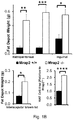
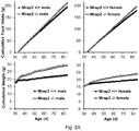
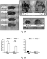
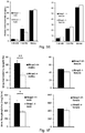
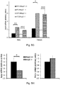
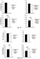
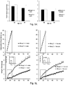
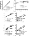
- knock-outs of Mrap2 modulate melanocortin receptor signaling in the brain such that the knock-out animal exhibits accelerated weight gain during development, reaching a mature weight much more rapidly than a wildtype animal.
- cells and pigs with reduced and/or undetectable levels of Mrap2 and/or Mrap2 activity. Such pigs will reach mature weight in less time, and accordingly, by consuming fewer resources, than wildtype animals, permitting more efficient production of various animal products (e.g. meat, fiber, milk, and other products).
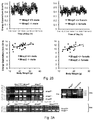
- Mrap2 or “melanocortin receptor accessory protein 2” refers to a transmembrane expressed in the brain, adrenal glands, and hypothalamus that interacts with the five melanocortin receptors (MCRs) to modulate their signaling.
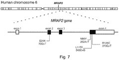
- the Mrap2 gene comprises 3 exons, encoding a protein subject to N-linked glycosylation at the N-terminus (specifically, at N9).
- Mrap2 comprises an N-terminal domain (amino acids 1-44 of SEQ ID NO: 1), a transmembrane domain (amino acids 45-67 of SEQ ID NO: 1), and a C-terminal domain (amino acids 68-205 of SEQ ID NO: 1).
- Mrap2 for a number of species is known, e.g. human Mrap2 (NCBI Gene ID No: 112609; polypeptide (NCBI Ref Seq: NP_612418, SEQ ID NO: 1); mRNA (NCBI Ref Seq: NM_138409; SEQ ID NO: 2)) and porcine Mrap2 (NCBI Gene ID No: 100515980; polypeptide (NCBI Ref Seq: XP_003353296, SEQ ID NO: 3); mRNA (NCBI Ref Seq: XM_003353248; SEQ ID NO: 4)). Further details of the structure and function of Mrap2 can be found, e.g. in Chan et al. PNAS 2009 106:6146-6151 and Hofland et al. J Clin Endocrinol Metab 2012 97:E747-E754 .
- the Mrap2 gene is considered any sequence associated with the Mrap2 locus. Thus, it would at least include the chromosomal nucleic acid contained within any organism that expresses a Mrap2, such as, the introns, exons, 5' upstream sequence involved with the Mrap2 coding and noncoding sequence, and 3' downstream sequence involved with the Mrap2 coding and non coding sequence.
- Exon 1 of Mrap2 consists of the nucleotides corresponding to positions 1-183 of SEQ ID NO: 2; exon 2 of Mrap2 consists of the nucleotides corresponding to positions 184-317 of SEQ ID NO: 2; exon 3 of Mrap2 consists of the nucleotides corresponding to positions 318-417 of SEQ ID NO: 2, and exon 4 of Mrap2 consists of the nucleotides corresponding to positions 418-2214 of SEQ ID NO: 2.
- the coding sequence of Mrap2 consists of the nucleotides corresponding to positions 191-808 of SEQ ID NO: 2.
- Mrap2 functions by interacting with melanocortin receptors, particularly Mc4r in the brain. Via this interaction, it enhances the generation of cyclic AMP (cAMP) mediated by Mc4R.
- the functionality of Mrap2 can be measured using a bioassy for Mc4r activity.
- cells expressing Mc4r can be contacted with ⁇ -melanocyte stimulating hormone.
- the addition of ⁇ -melanocyte stimulating hormone will induce PKA activity (which can be measured, e.g. by directly assaying the level of cAMP, or by measuring the level of a reporter, e.g.
- a cell lacking a detectable level of functional Mrap2 will not display a level of induction of PKA activity that is greater than the level of activity prior to the addition of ⁇ -melanocyte stimulating hormone, e.g. a level of induction that is less than 3x, less than 2x, or less than 1.5x, of the level prior to addition of the hormone.
- a modification rendering Mrap2 nonfunctional is a modification that results in an inhibition of expression of Mrap2, or in an inhition of the expression of Mrap2 lacking domains required for functionality, as described elsewhere herein.
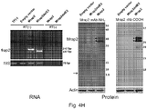
- the level of Mrap2 expression can be measured by methods known in the art for determining the level of nucleic acid and/or polypeptide expression, including, but not limited to, RT-PCR, qRT-PCR, northern blotting, western blotting, immunochemistry, and the like.
- Primers suitable for amplification of a Mrap2 expression product are provided herein in Table 4.
- Antibodies suitable for the detection of various domains of Mrap2 are described in the Examples herein.
- a commercial antibody for Mrap2 can be used with the methods described herein, e.g., an antibody specific for the transmembrane and/or C-terminal domains of Mrap2 (ab129397 from Abcam, Cambridge, MA).
- a functional fragment can retain at least 50% of the activity of the native polypeptide, e.g. 50% or more of the activity, 60% or more of the activity, 75% or more of the activity, or 90% or more of the activity of the native polypeptide.
- a “variant,” as referred to herein, is a polypeptide substantially homologous to a native or reference polypeptide, but which has an amino acid sequence different from that of the native or reference polypeptide because of one or a plurality of deletions, insertions or substitutions.
- a reduced level of Mrap2 and/or Mrap2 activity is less than 10% of the level found in a wildtype cell and/or animal of the same type, e.g. 10% or less, 9% or less, 8% or less, 7% or less, 6% or less, 5% or less, 4% or less, 3% or less, 2% or less, or 1% or less.
- a “modification” is a detectable change in the genetic material in the animal, which is transmitted to the animal's progeny.
- a modification is usually a change of one or more deoxyribonucleotides, the modification being obtained by, for example, adding, deleting, inverting, or substituting nucleotides.
- a modification of Mrap2 renders the Mrap2 gene nonfuctional under at least some circumstances, producing a "knockout" animal.
- Mrap2 is rendered nonfuctional in any cell comprising the modification.
- Mrap2 is rendered nonfunctional in a specific cell type comprising the modification, e.g. in the case of tissue-specific recombinase modifications, as described below herein.
- the genome of the cell and/or animal can comprise a modification of Mrap2, such that a detectable level of Mrap2 is not produced in the cell and/or animal.
- the genome of the cell and/or animal can comprise one or more deletions in one or more exons of Mrap2.
- the genome of the cell and/or animal can comprise a stop codon inserted into the Mrap2 sequence such that the translation of the peptide terminates early, e.g. before the transmembrane domain and/or the C-terminal domain are translated.
- a knock-out cell or animal as described herein can have a modification of Mrap2 present in one or both copies of Mrap2.
- the mutation is termed a "null" mutation.
- the knockout animal is termed a "heterozygous knockout animal".
- the pigs for use in the method of the invention are typically homozygous for the disruption of both copies of Mrap2.
- the genome of the transgenic non-human animal can further comprise a heterologous selectable marker gene, e.g. a marker that is introduced into the genome with the modification of Mrap2.
- a heterologous selectable marker gene e.g. a marker that is introduced into the genome with the modification of Mrap2.
- a cell or animal as described herein can be of any non-human species, e.g. a mammalian species.
- the cell or animal can be a species that is raised for agricultural purposes, e.g. for the use of a product (e.g. milk or fiber) produced by the animl or for the use of its carcass (e.g. for meat for consumption).
- the cell or animal can be a species and/or variety that is domesticated.
- the cell or animal can be, e.g. a fish, bird or reptile.
- the cell or animal can be a non-human mammal.
- Non-limiting examples of mammals disclosed herein can include, pigs, cows, sheep, goats, rabbits, alpaca, buffalo, bison, camel, cats, deer, donkey, dog, gayal, guinea pig, horse, llama, mule, reindeer, elk, yak.
- the non-human animal is a pig.
- a cell as described herein can be any type of cell, including but not limited to, an embryonic stem cell, an embryonic cell, a germline cell, a somatic cell, a brain cell, a neuron, a cell that can be cultured (e.g. an ES cell), a progenitor cell, a stem cell, or the like.
- transgenic Mrap2 knockout cell and/or animal e.g. one in which the Mrap2 coding sequence is not modified, but where expression of functional Mrap2 is not detectable.
- a modification can be introduced into the genome at a location other than at the Mrap2 gene.
- Such a transgenic Mrap2 knockout can comprise an antisense molecule targeting the Mrap2 gene.
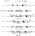
- the modified Mrap2 gene comprises sites for recombination by a recombinase, e.g. wherein the sites are lox sites and the recombinase is cre recombinase.
- the recombinase is present, the nucleic acid sequence between the recombinase sites will be excised from the genome, creating a deletion, e.g. of a portion of Mrap 2 which renders it non-functional as described elsewhere herein.
- the modification is produced by the action of a recombinase.
- the recombinase is cre recombinase.
- the recombinase is under the control of a tissue-specific promoter.
- the promoter is specific for neurons or brain tissue, e.g. the Sim1 promoter. Promoters specific for neurons and/or brain tissue are known in the art, e.g. the promoters of myelin basic protein, prolactin, GFAP, NSE, nestin, synapsin I, preproenkephalin, and dopamine beta-hydroxylase. See, e.g Papadakis et al. Current Gene Therapy 2004 4:89-113 .
- described herein are animals comprising or consisting of cells as described herein.
- the animal is a pig.
- described herein is an animal comprising a modification of the Mrap2 gene, wherein the mammal does not express a detectable level of functional Mrap2.
- the animal is a non-human mammal.
- the animal is a transgenic animal.
- the animal is a knockout animal.
- Non-limiting examples of methods of introducing a modification into the genome of a cell and/or animal can include microinjection, viral delivery, recombinase technologies, homologous recombination, TALENS, CRISPR, and/or ZFN, see, e.g. Clark and Whitelaw Nature Reviews Genetics 2003 4:825-833 .
- Zinc finger nucleases ZFNs
- Cas9/ CRISPR system the Cas9/ CRISPR system
- transcription-activator like effector nucleases TALENs
- Meganucleases are found commonly in microbial species and have the unique property of having very long recognition sequences (>14bp) thus making them naturally very specific for cutting at a desired location. This can be exploited to make site-specific double-stranded breaks in, e.g. a genome.
- nucleases can cut and create specific double-stranded breaks at a desired location(s) in the genome, which are then repaired by cellular endogenous processes such as, homologous recombination (HR), homology directed repair (HDR) and non-homologous end-joining (NHEJ).
- HR homologous recombination
- HDR homology directed repair
- NHEJ non-homologous end-joining
- HDR utilizes a homologous sequence as a template for regenerating the missing DNA sequence at the break point.
- meganuclease variants that recognize unique sequences.
- various meganucleases have been fused to create hybrid enzymes that recognize a new sequence.
- DNA interacting amino acids of the meganuclease can be altered to design sequence specific meganucleases (see e.g., US Patent 8,021,867 ).
- Meganucleases can be designed using the methods described in e.g., Certo, MT et al. Nature Methods (2012) 9:073-975 ; U.S. Patent Nos.
- meganucleases with site specific cutting characteristics can be obtained using commercially available technologies e.g., Precision BioSciences' Directed Nuclease EditorTM genome editing technology.
- ZFNs and TALENs restriction endonuclease technology utilizes a non-specific DNA cutting enzyme which is linked to a specific DNA sequence recognizing peptide(s) such as zinc fingers and transcription activator-like effectors (TALEs).
- TALEs transcription activator-like effectors
- an endonuclease whose DNA recognition site and cleaving site are separate from each other is selected and its cleaving portion is separated and then linked to a sequence recognizing peptide, thereby yielding an endonuclease with very high specificity for a desired sequence.
- An exemplary restriction enzyme with such properties is FokI.
- FokI has the advantage of requiring dimerization to have nuclease activity and this means the specificity increases dramatically as each nuclease partner recognizes a unique DNA sequence.
- FokI nucleases have been engineered that can only function as heterodimers and have increased catalytic activity. The heterodimer functioning nucleases avoid the possibility of unwanted homodimer activity and thus increase specificity of the double-stranded break.
- ZFNs rely on Cys2-His2 zinc fingers and TALENs on TALEs. Both of these DNA recognizing peptide domains have the characteristic that they are naturally found in combinations in their proteins. Cys2-His2 Zinc fingers typically happen in repeats that are 3 bp apart and are found in diverse combinations in a variety of nucleic acid interacting proteins such as transcription factors. TALEs on the other hand are found in repeats with a one-to-one recognition ratio between the amino acids and the recognized nucleotide pairs.
- Zinc fingers correlated with a triplet sequence are attached in a row to cover the required sequence
- OPEN low-stringency selection of peptide domains vs. triplet nucleotides followed by high-stringency selections of peptide combination vs. the final target in bacterial systems
- ZFNs for use with the methods and compositions described herein can be obtained commercially from e.g., Sangamo BiosciencesTM (Richmond, CA).
- the Cas9/ CRISPR system can be used to create a modification in an Mrap2 gene as described herein.
- Clustered regularly interspaced short palindromic repeats (CRISPR)/CRISPR-associated (Cas) systems are useful for, e.g. RNA-programmable genome editing (see e.g., Marraffini and Sontheimer. Nature Reviews Genetics 2010 11:181-190 ; Sorek et al. Nature Reviews Microbiology 2008 6:181-6 ; Karginov and Hannon. Mol Cell 2010 1:7-19 ; Hale et al. Mol Cell 2010:45:292-302 ; Jinek et al.
- a CRISPR guide RNA is used that can target a Cas enzyme to the desired location in the genome, where it generates a double strand break. This technique is known in the art and described, e.g. at Mali et al. Science 2013 339:823-6 ; and kits for the design and use of CRISPR-mediated genome editing are commercially available, e.g. the PRECISION X CAS9 SMART NUCLEASETM System (Cat No. CAS900A-1) from System Biosciences, Mountain View, CA.
- a CRISPR, TALENs, or ZFN molecule e.g. a peptide and/or peptide/nucleic acid complex
- a cell e.g. a cultured ES cell, such that the presence of the CRISPR, TALENs, or ZFN molecule is transient and will not be detectable in the progeny of, or an animal derived from, that cell.
- a nucleic acid encoding a CRISPR, TALENs, or ZFN molecule e.g. a peptide and/or multiple nucleic acids encoding the parts of a peptide/nucleic acid complex
- a cell e.g.
- a nucleic acid encoding a CRISPR, TALENs, or ZFN molecule can be introduced into a cell, e.g.
- a cultured ES cell such that the nucleic acid is maintained in the cell (e.g. incorporated into the genome) and the nucleic acid encoding the CRISPR, TALENs, or ZFN molecule and/or the CRISPR, TALENs, or ZFN molecule will be detectable in the progeny of, or an animal derived from, that cell.
- a TALENs targeting the 5' end of exon 3 of Mrap2 and a TALENs targeting the 3' end of exon 3 of Mrap2 can be introduced into a cell, thereby causing a modification of Mrap2 in which exon 3 is deleted.
- a modification in Mrap2 can be introduced into the genome of a cell using recombinant adeno-associated virus (rAAV) based genome engineering, which is a genome-editing platform centered around the use of rAAV vectors that enables insertion, deletion or substitution of DNA sequences into the genomes of live mammalian cells.
- the rAAV genome is a single-stranded deoxyribonucleic acid (ssDNA) molecule, either positive- or negative-sensed, which is about 4.7 kilobase long.
- ssDNA deoxyribonucleic acid
- These single-stranded DNA viral vectors have high transduction rates and have a unique property of stimulating endogenous homologous recombination in the absence of causing double strand DNA breaks in the genome.
- rAAV genome editing has the advantage in that it targets a single allele and does not result in any off-target genomic alterations.
- rAAV genome editing technology is commercially available, for example, the rAAV GENESISTM system from HorizonTM (Cambridge, UK).
- a targeting vector can be used to introduce a modification of Mrap2.
- a “targeting vector” is a vector comprising sequences that can be inserted into the gene to be disrupted, e.g., by homologous recombination.
- the targeting vector generally has a 5' flanking region and a 3' flanking region homologous to segments of the gene of interest, surrounding a DNA sequence comprising a modification and/or a foreign DNA sequence to be inserted into the gene.
- the foreign DNA sequence may encode a selectable marker, such as an antibiotics resistance gene. Examples for suitable selectable markers are the neomycin resistance gene (NEO) and the hygromycin ⁇ -phosphotransferase gene.
- the 5' flanking region and the 3' flanking region are homologous to regions within the gene surrounding the portion of the gene to be replaced with the unrelated DNA sequence.
- the targeting vector does not comprise a selectable marker.
- DNA comprising the targeting vector and the native gene of interest are contacted under conditions that favor homologous recombination.
- the targeting vector can be used to transform embryonic stem (ES) cells, in which they can subsequently undergo homologous recombination.
- a typical targeting vector contains nucleic acid fragments of not less than about 0.5 kb nor more than about 10.0 kb from both the 5' and the 3' ends of the genomic locus which encodes the gene to be modified (e.g. Mrap2). These two fragments are separated by an intervening fragment of nucleic acid which encodes the modification to be introduced.
- the resulting construct recombines homologously with the chromosome at this locus, it results in the introduction of the modification, e.g. a deletion of an exon or the insertion of a stop codon.
- the homologous recombination of the above-described targeting vectors is sometimes rare and such a construct can insert nonhomologously into a random region of the genome where it has no effect on the gene which has been targeted for deletion, and where it can potentially recombine so as to disrupt another gene which was otherwise not intended to be altered.
- such non-homologous recombination events can be selected against by modifying the above-mentioned targeting vectors so that they are flanked by negative selectable markers at either end (particularly through the use of two allelic variants of the thymidine kinase gene, the polypeptide product of which can be selected against in expressing cell lines in an appropriate tissue culture medium well known in the art-i.e.
- Non-homologous recombination between the resulting targeting vector comprising the negative selectable marker and the genome will usually result in the stable integration of one or both of these negative selectable marker genes and hence cells which have undergone non-homologous recombination can be selected against by growth in the appropriate selective media (e.g. media containing a drug such as 5-bromodeoxyuridine for example). Simultaneous selection for the positive selectable marker and against the negative selectable marker will result in a vast enrichment for clones in which the targeting vector has recombined homologously at the locus of the gene intended to be mutated.
- the appropriate selective media e.g. media containing a drug such as 5-bromodeoxyuridine for example
- each targeting vector to be inserted into the cell is linearized. Linearization is accomplished by digesting the DNA with a suitable restriction endonuclease selected to cut only within the vector sequence and not the 5' or 3' homologous regions or the modification region.
- a targeting vector refers to a nucleic acid that can be used to decrease or suppress expression of a protein encoded by endogenous DNA sequences in a cell.
- the knockout construct is comprised of a Mrap2 polynucleotide with a deletion in a critical portion of the polynucleotide (e.g. the transmembrane domain) so that a functional Mrap2 cannot be expressed therefrom.
- a number of termination codons can be added to the native polynucleotide to cause early termination of the protein or an intron junction can be inactivated.
- Proper homologous recombination can be confirmed by Southern blot analysis of restriction endonuclease digested DNA using, as a probe, a non-modifed region of the gene. Since the native gene will exhibit a restriction pattern different from that of the disrupted gene, the presence of a disrupted gene can be determined from the size of the restriction fragments that hybridize to the probe.
- a targeting vector can comprise the whole or a fragment of the genomic sequence of a Mrap2 and optionally, a selection marker, e.g., a positive selection marker.
- a selection marker e.g., a positive selection marker.
- Several kilobases of unaltered flanking DNA can be included in the vector (see e.g., Thomas and Capecchi, (1987) Cell, 51:503 for a description of homologous recombination vectors).
- the genomic sequence of the Mrap2 gene comprises at least part of an exon of Mrap2.
- a selection marker of the invention can include a positive selection marker, a negative selection marker or include both a positive and negative selection marker.
- positive selection marker include but are not limited to, e.g., a neomycin resistance gene (neo), a hygromycin resistance gene, etc.
- the positive selection marker is a neomycin resistance gene.
- the genomic sequence further comprises a negative selection marker.
- negative selection markers include but are not limited to, e.g., a diphtheria toxin gene, an HSV-thymidine kinase gene (HSV-TK), etc.
- modifier is used herein to collectively refer to any molecule which can effect a modification of Mrap2, e.g. a targeting vector or a TALENs, CRISPR, or ZFN molecule, complex, and/or one or more nucleic acids encoding such a molecule or the parts of such a complex.
- a modifier can be introduced into a cell by any technique that allows for the addition of the exogenous genetic material into nucleic genetic material can be utilized so long as it is not destructive to the cell, nuclear membrane, or other existing cellular or genetic structures.
- Such techniques include, but are not limited to transfection, scrape-loading or infection with a vector, pronuclear microinjection ( U.S. Pat. Nos. 4,873,191 , 4,736,866 and 4,870,009 ); retrovirus mediated transfer into germ lines (an der Putten, et al., Proc. Natl. Acad. Sci., USA, 82:6148-6152 (1985 )); gene targeting in embryonic stem cells ( Thompson, et al., Cell.
- the modifier can be delivered through a number of direct delivery systems such as, electroporation, lipofection, calcium phosphate precipitation, plasmids, viral vectors, viral nucleic acids, phage nucleic acids, phages, cosmids, or via transfer of material in cells or carriers such as cationic liposomes.
- direct delivery systems such as, electroporation, lipofection, calcium phosphate precipitation, plasmids, viral vectors, viral nucleic acids, phage nucleic acids, phages, cosmids, or via transfer of material in cells or carriers such as cationic liposomes.
- Appropriate means for transfection, including viral vectors, chemical transfectants, or physico-mechanical methods such as electroporation and direct diffusion of DNA are described by, for example, Wolff, J. A., et al., Science, 247, 1465-1468, (1990 ); and Wolff, J. A.
- compositions and methods described herein can be used to deliver a modifier to any cell type, e.g. a germline cell, a zygote, an embryo, or a somatic cell.
- the cells can be cultured in vitro or present in vivo. Non-limiting examples are provided herein below.
- a modifier inserted into the nucleic genetic material by microinjection is known and is used in the art.
- the embryo may be incubated in vitro for varying amounts of time, or reimplanted into the surrogate host, or both. In vitro incubation to maturity is within the scope of this invention.
- One common method is to incubate the embryos in vitro for about 1-7 days, depending on the species, and then reimplant them into the surrogate host.
- a zygote is microinjected.
- zygotes as a target for modification of a host gene has an advantage in that in most cases the injected DNA will be incorporated into the host gene before the first cleavage ( Brinster et al. (1985) PNAS 82:4438-4442 ). As a consequence, all cells of the transgenic animal will carry the incorporated nucleic acids of the targeting vector. This will in general also be reflected in the efficient transmission to offspring of the founder since 50% of the germ cells will harbor the modification.
- One route of introducing foreign DNA into a germ line entails the direct microinjection of linear DNA molecules into a pronucleus of a fertilized one-cell egg.
- Microinjected eggs are subsequently transferred into the oviducts of pseudopregnant foster mothers and allowed to develop. About 25% of the progeny inherit one or more copies of the micro-injected DNA.
- Techniques suitable for obtaining transgenic animals have been amply described.
- a suitable technique for obtaining completely ES cell derived transgenic non-human animals is described in WO 98/06834 .
- a modifier can be introduced into a cell by electroporation.
- the cells and the targeting vector can be exposed to an electric pulse using an electroporation machine and following the manufacturer's guidelines for use. After electroporation, the cells are typically allowed to recover under suitable incubation conditions. The cells are then screened for the presence of the targeting vector as explained herein.
- Retroviral infection can also be used to introduce a nucleic acid modifier (e.g. a targeting vector) or a nucleic acid encoding a modifier into a cell, e.g. a non-human animal cell.
- a nucleic acid modifier e.g. a targeting vector
- a nucleic acid encoding a modifier e.g. a cell, e.g. a non-human animal cell.
- a retrovirus can be used to introduce the Mrap2 modification to a cell or cells, e.g. an embryo.
- the developing non-human embryo can be cultured in vitro to the blastocyst stage.
- the blastomeres can be targets for retroviral infection ( Jaenich, Proc. Natl. Acad. Sci. USA, 73:1260-1264 (1976 )).
- Efficient infection of the blastomeres is obtained by enzymatic treatment to remove the zona pellucida ( Manipulating the Mouse Embryo, Hogan, ed. (Cold Spring Harbor Laboratory Press, Cold Spring Harbor, N.Y., 1986 )).
- the viral vector system used to introduce the modifier is typically a replication-defective retrovirus carrying the transgene ( Jahner et al., Proc. Natl. Acad. Sci. USA, 82: 6972-6931 (1985 ); and, Van der Putten et al., Proc. Natl. Acad. Sci. USA, 82: 6148-6152 (1985 )).
- Transfection is easily and efficiently obtained by culturing the blastomeres on a monolayer of virus-producing cells (Van der Putten et al., supra; and, Stewart et al., EMBO J., 6: 383-388 (1987 )).
- infection can be performed at a later stage.
- Virus or virus-producing cells can be injected into the blastocoele ( Jahner et al., Nature, 298: 623-628(1982 )). Most of the founders will be mosaic for the transgene since incorporation occurs only in a subset of the cells that formed the transgenic non-human animal. In addition, it is also possible to introduce transgenes into the germ line by intrauterine retroviral infection of the mid-gestation embryo (Jahner et al. (1982), supra).
- viral vectors can include, but are not limited to, adenoviral vectors ( Mitani et al., Hum. Gene Ther. 5:941- 948, 1994 ), adeno-associated viral (AAV) vectors ( Goodman et al., Blood 84:1492-1500, 1994 ), lentiviral vectors ( Naidini et al., Science 272:263-267, 1996 ), pseudotyped retroviral vectors ( Agrawal et al., Exper. Hematol. 24:738-747, 1996 ).
- adenoviral vectors Mitani et al., Hum. Gene Ther. 5:941- 948, 1994
- AAV adeno-associated viral
- lentiviral vectors Naidini et al., Science 272:263-267, 1996
- pseudotyped retroviral vectors Agrawal et al., Exper. Hematol. 24:738-747, 1996 ).
- a modifier can be introduced to a cell by the use of liposomes, e.g. cationic liposomes (e.g., DOTMA, DOPE, DC-cholesterol) or anionic liposomes.
- Liposomes can further comprise proteins to facilitate targeting a particular cell, if desired.
- cationic liposomes see, e.g., DOTMA, DOPE, DC-cholesterol
- anionic liposomes can further comprise proteins to facilitate targeting a particular cell, if desired.
- liposomes see, e.g., Brigham et al. Am. J. Resp. Cell. Mol. Biol. 1:95-100 (1989 ); Feigner et al. Proc. Natl. Acad. Sci USA 84:7413-7417 (1987 ); U.S. Pat. No.4,897,355 .
- Commercially available liposome preparations include, e.g.
- LIPOFECTIN LIPOFECTAMINE
- SUPERFECT Qiagen, Inc. Hilden, Germany
- TRANSFECTAM Promega Biotec, Inc., Madison, WI
- the number of copies of a modifier (e.g., the targeting vector or TALENs molecule) which are added to the cell is dependent upon the total amount of exogenous genetic material added and will be the amount which enables the genetic transformation to occur. Theoretically only one copy is required; however, generally, numerous copies are utilized, for example, 1,000-20,000 copies of a targeting vector, in order to insure that one copy is functional.
- cells contacted with a modifier are subsequently screened for accurate targeting to identify and isolate those which have been properly modified at the Mrap2 locus.
- an animal can be produced from this cell through either stem cell technology or cloning technology.
- stem cell technology e.g. an embryonic stem cell
- this cell after transfection and culturmg, can be used to produce an organism which will contain the gene modification in germ line cells, which can then in turn be used to produce another animal that possesses the gene modification or disruption in all of its cells.
- cloning technologies can be used.
- cells used to produce the knockout animals will be of the same species as the knockout animal to be generated.
- pig embryonic stem cells will usually be used for generation of knockout pigs.
- Methods of isolating, culturing, and manipulating various cells types are known in the art.
- embryonic stem cells are generated and maintained using methods well known to the skilled artisan such as those described by Doetschman et al. (1985) J. Embryol. Exp. Mol. Biol. 87:27-45 ).
- the cells are cultured and prepared for knockout construct insertion using methods well known to the skilled artisan, such as those set forth by Robertson in: Teratocarcinomas and Embryonic Stem Cells: A Practical Approach, E. J. Robertson, ed. IRL Press, Washington, D.C. [1987 ]); by Bradley et al. (1986) Current Topics in Devel. Biol. 20:357-371 ); and by Hogan et al. (Manipulating the Mouse Embryo: A Laboratory Manual, Cold Spring Harbor Laboratory Press, Cold Spring Harbor, N.Y. (1986 )).
- the cells can be inserted into an embryo. Insertion may be accomplished in a variety of ways known to the skilled artisan, however the typical method is by microinjection. For microinjection, about 10-30 cells are collected into a micropipet and injected into embryos that are at the proper stage of development to permit integration of the ES cell containing the Mrap2 modification into the developing embryo. For instance, the ES cells can be microinjected into blastocytes. The suitable stage of development for the embryo used for insertion of ES cells is very species dependent, however for mice it is about 3.5 days. The embryos are obtained by perfusing the uterus of pregnant females. Suitable methods for accomplishing this are known to the skilled artisan.
- a modification of Mrap2 that renders it nonfunctional can be generated by a recombinase.
- sites for a recombinase can be inserted into the native Mrap2 gene, such that they flank an area that can be deleted in order to render Mrap2 nonfunctional (e.g. exon 3).
- the flanked area of Mrap2 will be deleted. This permits inducible or tissue-specific modification of Mrap2, e.g. in the brain only.
- a widely used site-specific DNA recombination system uses the Cre recombinase, e.g., from bacteriophage P1, or the Flp recombinase from S . cerevisiae, which has also been adapted for use in animals.
- the loxP-Cre system utilizes the expression of the PI phage Cre recombinase to catalyze the excision of DNA located between flanking lox sites.
- site-specific recombination may be employed to inactivate endogenous genes in a spatially or time controlled manner. See, e.g., U.S. Pat. Nos. 6,080,576 , 5,434,066 , and 4,959,317 ; and Joyner, A. L., et al. Laboratory Protocols for Conditional Gene Targeting, Oxford University Press, New York (1997 ).
- the cre-lox system an approach based on the ability of transgenic mice, carrying the bacteriophage Cre gene, to promote recombination between, for example, 34 by repeats termed loxP sites, allows ablation of a given gene in a tissue specific and a developmentally regulated manner ( Orban et al. (1992) PNAS 89:6861-6865 ).
- the Cre-lox system has been successfully applied for tissue-specific transgene expression ( Orban PC, Chui D, Marth JD. Proc Natl Acad Sci U S A.
- the recombinase can be delivered at different stages.
- a recombinase can be added to an embryonic stem cell containing a disrupted gene prior to the production of chimeras or implantation into an animal.
- the recombinase is delivered after the generation of an animal containing at least one gene allele with introduced recombinase sites.
- the recombinase is delivered by cross breeding the animal containing a gene with recombinase sites with an animal expressing the recombinase.
- the animal expressing the recombinase may express it, e.g., ubiquitously, in a tissue-restricted manner, or in a temporal-restricted manner.
- Cre/Flp activity can also be controlled temporally by delivering cre/FLP-encoding transgenes in viral vectors, by administering exogenous steroids to the animals that carry a chimeric transgene consisting of the cre gene fused to a mutated ligand-binding domain, or by using transcriptional transactivation to control cre/FLP expression.
- mutated recombinase sites may be used.
- Tissue-specific, temporally-regulated, and inducible promoters for controlling the expression of, e.g. Cre recombinase are known in the art.
- a Sim1 promoter controlled Cre which permits brain-specific expression of Cre recombinase is described in the Examples herein.
- Animals with germline cells comprising the desired modification described herein can be selected, e.g. by genotyping or assaying Mrap2 levels or activity in the germline cells and/or progeny.
- methods for such genotyping or assaying can include, RNA analysis (Northern blotting or RT-PCR, including qRT-PCR), assays for determing the activity of Mrap2 as described elsewhere herein, protein analysis (e.g. Western blotting), histological stains, flow cytometric analysis and the like.
- the extent of the contribution of the modified cells in an animal described herein can also be assessed visually by choosing animals strains for the modified cells (e.g. the ES cells that will be modified) and the blastocytes that have different coat colors.
- Transgenic offspring can be screened for the presence and/or expression of the transgene by any suitable method. Screening is often accomplished by Southern blot or Northern blot analysis, using a probe that is complementary to at least a portion of the transgene. Typically, DNA is prepared from, e.g. tail tissue and analyzed by Southern analysis or PCR for the transgene. Alternatively, the tissues or cells believed to have a modification of Mrap2 are tested for the presence and expression of the modified Mrap2 using Southern analysis or PCR, although any tissues or cell types may be used for this analysis. See, e.g., southern hybridization. ( Southern J. Mol. Biol. 98:503-517 (1975 )), northern hybridization (see, e.g., Freeman et al.
- Protein levels can be detected by immunoassays using antibodies specific to protein. For example, western blot analysis using an antibody against Mra2 or the modified Mrap2 encoded by the transgene may be employed as an alternative or additional method for screening for the presence of the transgene product.
- immunoassays known in the art can be used, including but not limited to competitive and non-competitive assay systems using techniques such as radioimmunoassay, ELISA (enzyme linked immunosorbent assay), "sandwich” immunoassays, immunoradiometric assays, gel diffusion precipitin reactions, immunodiffusion assays, in situ immunoassays (using colloidal gold, enzyme or radioisotope labels), western blot analysis, precipitation reactions, agglutination assays (e.g., gel agglutination assays, hemagglutination assays), complement fixation assays, immunofluorescence assays, protein A assays, immunoelectrophoresis assays, etc.
- ELISA enzyme linked immunosorbent assay
- sandwich immunoassays immunoradiometric assays
- gel diffusion precipitin reactions e.g., gel agglutination assays, hemagglutination assays
- antibody binding is detected by detecting a label on the primary antibody.
- the primary antibody is detected by detecting binding of a secondary antibody or reagent to the primary antibody.
- the secondary antibody is labeled. Many means are known in the art for detecting binding in an immunoassay and are within the scope of the present invention.
- Progeny of the transgenic animals may be obtained by mating the transgenic animal with a suitable partner, or by in vitro fertilization of eggs and/or sperm obtained from the transgenic animal.
- the partner may or may not be transgenic and/or a knockout; where it is transgenic, it may contain the same or a different knockout, or both.
- the partner may be a parental line.
- in vitro fertilization is used, the fertilized embryo may be implanted into a surrogate host or incubated in vitro, or both. Using either method, the progeny may be evaluated using methods described above, or other appropriate methods.
- a cell and/or animal described herein can comprise one or more additional modifications or transgenes (e.g. an additional transgene and/or second knockout targeting a gene other than Mrap2).
- Cells and/or animals described herein containing more than one knockout construct and/or more than one transgene expression construct can be prepared in any of several ways. A typical manner of preparation is to generate a series of mammals, each containing one of the desired transgenic phenotypes.
- Such animals are bred together through a series of crosses, backcrosses and selections, to ultimately generate a single animal containing all desired knockout constructs and/or expression constructs, where the animal is otherwise congenic (genetically identical) to the wild type except for the presence of the knockout construct(s) and/or transgene(s).
- nucleic acid molecules comprising a modification of Mrap2 as described herein, e.g. a targeting vector comprising a modified variant of Mrap2 according to the embodiments described herein.
- Described herein are cells produced by the process of transforming the cell with any of the described nucleic acids. Described herein are cells produced by the process of transforming the cell with any of the non-naturally occurring described nucleic acids.
- Described herein are peptides produced by the process of expressing any of the described nucleic acids. Described herein are any of the non-naturally occurring peptides produced by the process of expressing any of the described nucleic acids.
- Described herein are any of the described peptides produced by the process of expressing any of the non-naturally occurring described nucleic acids. Described herein are animals produced by the process of transfecting a cell within the animal with any of the nucleic acid molecules described herein. Described herein are animals produced by the process of transfecting a cell within the animal any of the nucleic acid molecules described herein, wherein the animal is a mammal. Described herein are animals produced by the process of adding to the animal any of the cells described herein.
- an animal can be used for, e.g. competition, breeding, products obtained from a live animal (e.g. milk from dairy animals or fibers from, e.g. a sheep or an alpaca), or work (e.g. a draft animal). Regardless of what use the animal is to serve, there will be a desired minimum size for that animal to have achieved prior to being used. By minimizing the time it takes for the animal to grow to that size, resources and time are conserved, lowering the cost of producing the useful animal.
- a live animal e.g. milk from dairy animals or fibers from, e.g. a sheep or an alpaca
- work e.g. a draft animal
- the carcass of the animal is to be used.
- the carcass of the animal can be used for human consumption as well as for other purposes (e.g. to produce leather, fertilizer, cosmetics, drugs, hair products, perfumes, gelatin, glue, etc).
- described herein is a method of producing an animal for use of the carcass, the method comprising providing species-appropriate food to an animal as described herein and butchering the animal when it reaches the desired size for use.
- Species-appropriate foods and desired size(s) for use of any of the animals described herein, e.g. are well known in the art, see, e.g. Pond et al.
- a pig is typically butchered when it weighs from about 240 to about 280 pounds and dietary recommendations are well-known in the art, e.g. Albrecht et al. "Swine Feeding Suggestions” Circular 509 ; Clemson Extension; (1995).
- species-appropriate food can be purchased commercially, e.g. PURINA MILLSTM NATURE'S MATCHTM Sow and Pig Complete Feed, Cat No. 0034068; Purina, St. Louis, MO.
- the animal as described herein can reach the desired size for use as much as 80% earlier than a wild-type animal, e.g. 10% earlier, 20% earlier, 30% earlier, 40% earlier, 50% earlier, 60% earlier, 70% earlier, or 80% earlier. In some embodiments, the animal as described herein can reach the desired size for use as much as 50% earlier than a wild-type animal. In some embodiments, the animal as described herein can be butchered as much as 80% earlier than a wild-type animal, e.g. 10% earlier, 20% earlier, 30% earlier, 40% earlier, 50% earlier, 60% earlier, 70% earlier, or 80% earlier. In some embodiments, the animal as described herein can be butchered as much as 50% earlier than a wild-type animal. In some embodiments described herein, the animal can be provided feed substantially ad libitum. The animal is a pig.
- the pig can be butchered at from about 50 to about 150 days of age. In some embodiments, the pig can be butchered at from about 75 to about 125 days of age. In some embodiments, the pig can be butchered at from 75 to 125 days of age.
- the animal, the pig can be provided a restricted level of food as compared to a wild-type animal.
- a restricted diet an animal as described herein can reach a given desired size for use in the same amount of time as the wild-type animal.
- the restricted level of food is from about 60% to about 95% of the level provided to a wild-type mammal. In some embodiments, the restricted level of food is from about 80% to about 95% of the level provided to a wild-type mammal.
- “decrease”, “reduced”, “reduction”, or “inhibit” are all used herein to mean a decrease by a statistically significant amount.
- “reduce,” “reduction” or “decrease” or “inhibit” typically means a decrease by at least 10% as compared to a reference level (e.g.
- the absence of a given treatment can include, for example, a decrease by at least about 10%, at least about 20%, at least about 25%, at least about 30%, at least about 35%, at least about 40%, at least about 45%, at least about 50%, at least about 55%, at least about 60%, at least about 65%, at least about 70%, at least about 75%, at least about 80%, at least about 85%, at least about 90%, at least about 95%, at least about 98%, at least about 99% , or more.
- “reduction” or “inhibition” does not encompass a complete inhibition or reduction as compared to a reference level.
- “Complete inhibition” is a 100% inhibition as compared to a reference level.
- transgenic animal refers to an animal to which exogenous DNA has been introduced.
- the transgenic approach aims at specific modifications of the genome, e.g., by introducing whole transcriptional units into the genome, or by up- or down-regulating pre-existing cellular genes.
- the targeted character of certain of these procedures sets transgenic technologies apart from experimental methods in which random mutations are conferred to the germline, such as administration of chemical mutagens or treatment with ionizing solution.
- knock-out refers to partial or complete suppression of the expression of a protein encoded by an endogenous DNA sequence in a cell.
- the "knock-out” can be affected by targeted deletion of the whole or part of a gene encoding a protein in an cell. In some embodiments, the deletion may prevent or reduce the expression of the functional protein in any cell in the whole animal in which it is normally expressed.
- a “Mrap2 knock-out animal” refers to an animal in which the expression of functional Mrap2 has been reduced or suppressed by the introduction of a recombinant modifier that introduces a modification in the sequence of the Mrap2 gene.
- a knock-out animal can be a transgenic animal, or can be created without transgenic methods, e.g. by transient introduction of a TALENs molecule, such that a deletion of part or all of the Mrap2 gene occurs, but without the introduction of exogenous DNA to the genome.
- animal is used herein to include all vertebrate animals, except humans. It also includes an individual animal in all stages of development, including embryonic and fetal stages.
- the term “germ cell line transgenic animal” refers to a transgenic animal in which the genetic alteration or genetic information was introduced into a germ line cell, thereby conferring the ability to transfer the genetic information to offspring. If such offspring in fact possess some or all of that alteration or genetic information, they are transgenic animals as well.
- chimera refers to a transgenic and/or knock-out animal with exogenous DNA and/or a modification of Mrap2 in some of its genome-containing cells.
- heterozygote refers to a transgenic and/or knock-out animal with exogenous DNA and/or a modification of Mrap2 on one of a chromosome pair in all of its genome-containing cells.
- homozygote refers to a transgenic mammal with with exogenous DNA and/or a modification of Mrap2 on both members of a chromosome pair in all of its genome-containing cells.
- homologous recombination refers to the exchange of DNA fragments between two DNA molecules or chromatids at the site of homologous nucleotide sequences.
- gene targeting refers to a type of homologous recombination that occurs when a fragment of genomic DNA is introduced into a mammalian cell and that fragment locates and recombines with endogenous homologous sequences. Gene targeting by homologous recombination employs recombinant DNA technologies to replace specific genomic sequences with exogenous DNA of particular design and/or a modified sequence.
- a disruption in which there is “no native expression” of the endogenous gene, e.g., a Mrap2 gene refers to a partial or complete reduction of the expression of at least a portion of a polypeptide encoded by an endogenous gene, e.g., an endogenous Mrap2 gene, of a single cell, selected cells, or all of the cells of an animal.
- control sequences refers to DNA sequences necessary for the expression of an operably linked coding sequence in a particular host organism.
- the control sequences that are suitable for prokaryotes include a promoter, optionally an operator sequence, and a ribosome binding site.
- Eukaryotic cells are known to utilize promoters, polyadenylation signals, and enhancers.
- Nucleic acid is "operably linked” when it is placed into a functional relationship with another nucleic acid sequence.
- DNA for a presequence or secretory leader is operably linked to DNA for a polypeptide if it is expressed as a preprotein that participates in the secretion of the polypeptide; a promoter or enhancer is operably linked to a coding sequence if it affects the transcription of the sequence; or a ribosome binding site is operably linked to a coding sequence if it is positioned so as to facilitate translation.
- "operably linked" means that the DNA sequences being linked are contiguous, and, in the case of a secretory leader, contiguous and in reading phase. However, enhancers do not have to be contiguous. Linking is accomplished by ligation at convenient restriction sites. If such sites do not exist, the synthetic oligonucleotide adaptors or linkers are used in accordance with conventional practice.
- the term "marker” refers to a reporter, a positive selection marker, and a negative selection marker.
- a reporter refers to any molecule the expression of which in a cell produces a detectable signal, e.g., detectable signal, e.g., luminescence.
- Exemplary markers are disclosed in, e.g., Sambrook, J., et al., (2001) Molecular Cloning: A Lbaroratory Manual, 2nd ed. (2001), Cold Spring Harbor Laboratory Press, Cold Spring Harbor, N.Y .; and, U.S. Pat. No. 5,464,764 and No. 5,625,048 . Many procedures for selecting and detecting markers are known in the art. See, e.g., Joyner, A.
- the terms "selective marker” and "position selection marker” refer to a gene, the expression of which allows cells containing the gene to be identified, e.g., antibiotic resistance genes and detectable, e.g., fluorescent, molecules.
- the gene encoding a product enables only the cells that carry the gene to survive and/or grow under certain conditions. For example, plant and animal cells that express the introduced neomycin resistance (Neo r ) gene are resistant to the compound G418. Cells that do not carry the Neo r gene marker are killed by G418.
- a "negative selectin marker” refers to a gene, the expression of which inhibits cells containing the gene to be identified, e.g., the HSV-tk gene.
- protein and “polypeptide” are used interchangeably herein to designate a series of amino acid residues, connected to each other by peptide bonds between the alpha-amino and carboxy groups of adjacent residues.
- protein and “polypeptide” refer to a polymer of amino acids, including modified amino acids (e.g., phosphorylated, glycated, glycosylated, etc.) and amino acid analogs, regardless of its size or function.
- modified amino acids e.g., phosphorylated, glycated, glycosylated, etc.
- amino acid analogs regardless of its size or function.
- polypeptide proteins and “polypeptide” are used interchangeably herein when referring to a gene product and fragments thereof.
- exemplary polypeptides or proteins include gene products, naturally occurring proteins, homologs, orthologs, paralogs, fragments and other equivalents, variants, fragments, and analogs of the foregoing.
- nucleic acid or “nucleic acid sequence” refers to any molecule, preferably a polymeric molecule, incorporating units of ribonucleic acid, deoxyribonucleic acid or an analog thereof.
- the nucleic acid can be either single-stranded or double-stranded.
- a single-stranded nucleic acid can be one nucleic acid strand of a denatured double- stranded DNA. Alternatively, it can be a single-stranded nucleic acid not derived from any double-stranded DNA.
- the nucleic acid can be DNA.
- nucleic acid can be RNA.
- Suitable nucleic acid molecules are DNA, including genomic DNA or cDNA. Other suitable nucleic acid molecules are RNA, including mRNA.
- nucleic acid molecule is a nucleic acid molecule that is identified and separated from at least one contaminant nucleic acid molecule with which it is ordinarily associated in the natural source of the nucleic acid.
- An isolated nucleic acid molecule is other than in the form or setting in which it is found in nature. Isolated nucleic acid molecules therefore are distinguished from the nucleic acid molecule as it exists in natural cells.
- gene refers to (a) a gene containing a DNA sequence encoding a protein, e.g., Mrap2; (b) any DNA sequence that encodes a protein, e.g., Mrap2 amino acid sequence, and/or; (c) any DNA sequence that hybridizes to the complement of the coding sequences of a protein.
- the term includes coding as well as noncoding regions, and preferably includes all sequences necessary for normal gene expression.
- target gene refers to any nucleic acid molecule, polynucleotide, or gene to be modified by, e.g. homologous recombination or a meganuclease-mediated method of modification.
- the target sequence can include, e.g. an intact gene, an exon or intron, a regulatory sequence or any region between genes.
- the target gene may comprise a portion of a particular gene or genetic locus in the individual's genomic DNA.
- statically significant or “significantly” refers to statistical significance and generally means a two standard deviation (2SD) or greater difference.
- compositions, methods, and respective component(s) thereof are used in reference to compositions, methods, and respective component(s) thereof, that are essential to the method or composition, yet open to the inclusion of unspecified elements, whether essential or not.
- compositions, methods, and respective components thereof as described herein, which are exclusive of any element not recited in that description of the embodiment.
- the term "consisting essentially of' refers to those elements required for a given embodiment. The term permits the presence of elements that do not materially affect the basic and novel or functional characteristic(s) of that embodiment.
- EXAMPLE 1 Loss of Function of the Melanocortin 2 Receptor Accessory Protein 2 Is Associated with Mammalian Obesity
- Mrap2 Melanocortin receptor accessory proteins
- GPCRs Membrane-expressed G protein-coupled receptors modulate cellular responses to numerous physiological stimuli.
- the melanocortin receptors (MCRs) are a subfamily of GPCRs that mediate signaling in response to the pro-opiomelanocortin-derived peptides, adrenocorticotropic hormone (ACTH) and ⁇ -melanocyte-stimulating hormone ( ⁇ MSH) and their competitive antagonists, agouti and agouti-related protein.
- ACTH adrenocorticotropic hormone
- ⁇ MSH ⁇ -melanocyte-stimulating hormone
- MCRs mediate a diverse range of physiological functions: MC1R is involved in skin pigmentation, MC2R plays a critical role in the hypothalamic-pituitary-adrenal axis, MC3R and MC4R are involved in energy homeostasis and MC5R is implicated in exocrine function ( 1 ).
- accessory proteins can modulate GPCR trafficking, as well as ligand binding and signaling ( 2 ).
- An accessory protein for MC2R, MC2R accessory protein (MRAP) is required for the trafficking of MC2R to the surface of adrenal cells and for signaling in response to ACTH ( 3, 4 ).
- Loss of either MC2R or MRAP in humans causes severe resistance to ACTH, with resulting glucocorticoid deficiency ( 5 , 6 ).
- MRAP2 paralogous gene
- MC3R and MC4R are predominantly expressed in the brain ( 7 ), most prominently in the pons and cerebellum, but including in regions involved in energy homeostasis such as the hypothalamus and brainstem ( Figs. 4A-4C ).
- PVN paraventricular nucleus of the hypothalamus
- Mc4r mRNAs are co-expressed in many cells (data not shown).
- the inventor hypothesized that Mrap2 might modulate signaling through a melanocortin receptor and potentially affect energy homeostasis.
- Targeted deletion of Mrap2 in mice was performed, using Cre-lox-mediated excision of the 100 bp exon 3 (which encodes the highly conserved transmembrane domain ( 7 )) to create mice with normal levels of an mRNA predicted to encode a truncated protein that includes the first 55 amino acids of Mrap2, with the transmembrane domain replaced by 11 aberrant amino acids specified by the out-of-frame exon 4, followed by a stop codon ( Figs. 4D-4G ).
- mutant mRNA Normal levels of the mutant mRNA indicate preservation of Mrap2-containing neurons in null mice, although these neurons likely do not express the predicted mutant protein, since mutant Mrap2 mRNA, but not protein, is present in cells transfected with the same Mrap2 mutant construct used to create the null mice ( Fig. 4H ).
- Mrap2 null mice appeared normal at birth, with normal weight gain and post-weaning food intake during early life (0-32 days and 23-32 days, respectively), although young Mrap2- / - male mice trended toward greater weight and food intake with advancing age ( Fig. 4I ). However, null mice of both genders gradually became extremely obese on a diet of regular chow ad libitum ( Fig. 1A , 5A ).
- mice Heterozygous mice were significantly heavier than wild-type animals on standard chow (160-175 days; males, Mrap2 +/+ 26.0 ⁇ 0.4 g, Mrap2 +/- 29.9 ⁇ 0.9 g; females Mrap2 +/+ 24.5 ⁇ 0.9 g, Mrap2 +/- 28.1 ⁇ 0.7 g), and at younger ages (56-95 days) on a high fat diet ( Fig. 1A ).
- Mrap2 -/- mice had increased length ( Fig. 4J ) and per cent of weight due to fat, and decreased per cent of weight due to lean mass ( Fig. 4K ).
- Mrap2 null mice had, as expected, elevated leptin concentrations corresponding to their increased fat mass, which normalized with diet-induced weight normalization ( FIG. 5B ). Obese adult mice had normal fasting insulin ( Fig. 5C ) and normal tolerance to intraperitoneal glucose injection ( Fig. 5D ). Mrap2 has been postulated to play a role in the adrenal response to ACTH ( 8 ). Diurnal rhythmicity and stress responsiveness of the adrenal axis was measured in Mrap2 null mice, which were normal ( Fig. 5E ). Thyroid hormone levels were also normal (Table 2). Epinephrine and norepinephrine excretion were reduced in male Mrap2 -/- mice only ( Fig.
- null mice were switched to ad libitum access to chow after 40 days of restricted feeding (upward arrow, Fig. 5O ).
- food intake almost doubled in null mice (from 2.9 ⁇ 0.1 to 5.6 ⁇ 0.5 g/d in males, and from 2.8 ⁇ 0.1 to 5.3 ⁇ 0.2 g/d in females), with a corresponding marked increase in body weight.
- hyperphagia develops in an age-dependent manner in older mice, independent of body weight.
- Mrap2 -/- mice had an intact anorectic response to the melanocortin receptor (Mc4r and Mc3r) agonist, MTII ( Fig. 5P ), corresponding to their normal ad libitum food intake at this age.
- Mc4r and Mc3r melanocortin receptor
- Mc4r deficiency should create an equivalent or more severe obesity phenotype than does Mrap2 deficiency, depending on the degree to which Mrap2 interferes with Mc4r function. Supporting this, Mrap2 +/- mice of both genders were less obese than either Mc4r +/- or doubly heterozygous mice ( Fig. 6B ). The differences between Mc4r +/- and doubly heterozygous mice were not statistically significant, although the latter trended toward being heavier. Among homozygous knockouts, those with Mc4r deficiency alone were more obese than those with Mrap2 deficiency alone ( Fig. 6B ). The Mc4r knockout mice were more obese than mice with deletion of both Mc4r and Mrap2 (in males, with a trend in females), suggesting that Mrap2 may promote weight gain through both Mc4r-dependent and Mc4r-independent actions.
- Mrap2 causes obesity in mice and that rare heterozygous variants in MRAP2 are associated with early-onset, severe obesity in humans.
- the mechanism(s) by which Mrap2 exerts its effects on body weight regulation remain to be firmly established but likely involve altered signaling through Mc4r and perhaps other melanocortin receptors.
- mice Animals. Mice were housed at 22°C unless otherwise noted, on a 12 h:12 h light:dark cycle with lights on at 0700. Mice were fed either standard rodent chow (Isopro RMH 3000, PMI Nutrition International, St. Louis, MO) or high fat diet chow (D12451, Research Diets, New Brunswick, NJ). Ad libitum food intake was measured in individually-housed mice as the difference in weight between food in a suspended food bin, accounting for food spillage onto the cage floor on subsequent days, measured either daily for up to 55 days, or in group-housed mice (4/cage of the same gender) over 4 days.
- standard rodent chow Isopro RMH 3000, PMI Nutrition International, St. Louis, MO
- high fat diet chow D12451, Research Diets, New Brunswick, NJ.
- Ad libitum food intake was measured in individually-housed mice as the difference in weight between food in a suspended food bin, accounting for food spillage onto the cage floor
- mice For pair feeding of individually-housed mice, the average amount of food consumed each day by wild-type mice was given to the experimental genotype of the same gender on the subsequent day. Body weights were measured at the same time as was food intake. For restricted feeding, between 10-15% less than the average amount of food consumed each day by wild-type mice was given to the experimental genotype of the same gender on the subsequent day, to maintain the same body weights between the two groups.
- Phlebotomy was performed by retro-orbital venipuncture without anesthesia, with the interval between touching the mouse cage and phlebotomy being less than 60 sec.
- Diurnal rhythmicity and stress responsiveness of blood corticosterone was performed by sequential phlebotomy at 1900, 0800, and following 30 min of restraint stress, at 0830, in mice fed a high fat diet. All mouse protocols were approved by the Boston Children's Hospital Animal Care and Use Committee.
- Mrap2 -/- and Mrap2 flox/ flox mice were generated using a common targeting vector and recombineering ( 15, 16 ). All the required plasmids (PL253, PL452, and PL451) and recombinogenic bacteria strains (EL350) were obtained from David Conner (Harvard Medical School Department of Genetics). The 129Sv BAC clone (bMQ-392015) containing Mrap2 (accession number: XM_147017, which had been replaced by NM_001101482) in DH10B was obtained from the Sanger Institute (http://www.sanger.ac.uk/).
- Primers were designed to create miniarms for PL253 to retrieve the Mrap2 genomic region, miniarms for PL452 for the upstream LoxP insertion, and miniarms for PL451 for the downstream FRT-Neo-FRT-LoxP insertion.
- the final targeting vector was linearized with NotI, electroporated into 129Sv ES cells, and selected with G418 (Invitrogen, Carlsbad, CA).
- G418-resistant ES cell lines were isolated and analyzed by long range PCR using LA TaqTM (TAKARA, Otsu, Japan), plus external Mrap2 genomic primers and internal vector primers at both 5' and 3' ends to confirm by DNA sequence analysis the correct 5' and 3' incorporation of the construct into the proper Mrap2 genomic locus.
- LA TaqTM TAKARA, Otsu, Japan
- Mrap2 +/- ES cells clones were transfected with pOG231 that encodes CMV-Cre.
- Mrap2 +/flox ES cells clones were transfected with pCAGGS-Flpe.
- Validated ES cells were karyotyped and surgically implanted into C57BL/6 pseudo-pregnant female mice.
- mice Male chimeric animals were obtained and crossed with 129S6/SvEvTac female mice (Taconic, Hudson, NY). F1 heterozygous male mice were crossed with F1 heterozygous female mice to generate F2 colonies on a pure 129 background. These mice were then bred to produce wild-type ( Mrap2 +/+ ), heterozygous ( Mrap2 +/- ) and null ( Mrap2 -/- ) mice for subsequent studies, at the expected frequencies of 1:2:1. The Mrap2 null allele was also backcrossed for 10 generations onto a C57BL/6J (Jackson Laboratories, Bar Harbor, ME) background. Mrap2 null mice have normal fertility.
- Sim1 CreBAC ::Mrap2 f / f mice Mrap2 flo/flox mice were mated with Siml CreBAC mice (006451, Jackson Laboratories, Bar Harbor, ME) to produce Sim1 CreBAC ::Mrap2 f / f mice. These mice were bred to R26 flox - stop - tdTomato reporter mice (007576, Jackson Laboratories, Bar Harbor, ME), in which tdTomato expression is induced by Cre-mediated recombination, to map Sim1-Cre expression in the brain.
- Mc4r null mice (006414, mixed C57BL/6-129 background, Jackson Laboratories, Bar Harbor, ME) were mated with Mrap2 null mice to create mice with heterozygous and homozygous combinations of knockout of both genes. Body weights were measured daily from ages 28-84 days in littermate siblings as described above.
- Tissues were collected and immersed into 4% paraformaldehyde. Sections (5-7 ⁇ m) of paraffin-embedded tissues were cut and stained with hematoxylin and eosin. Images were captured using a Nikon Eclipse 800TM microscope. All images of a given tissue were recorded at the same magnification and exposure conditions, without subsequent manipulation.
- RNA samples were treated with DNase (6.8 Kunitz units/sample) (Qiagen, Valencia, CA) to eliminate genomic DNA contamination and were purified using the RNeasyTM Mini kit (Qiagen) according to the manufacturer's protocol. Purified RNA samples were subjected to reverse-transcription using iScriptTM cDNA Synthesis kit (Biorad, Hercules, CA).
- RNA equivalent-cDNA was PCR-amplified for 35 cycles using gene-specific primer sets following the program: 94°C 2 min, (94 °C 10 sec, 60 °C 30 sec) x 35 cycles, 72 °C 7 min.
- PCR products were electrophoresed on 2.0 % agarose gels stained with ethidium bromide.
- Real-time qPCR was carried out using the same gene-specific primer sets and using iQ SYBR green Supermix (Biorad) following the program: 95 °C 3 min, (95 °C 10 sec, 60 °C 40 sec) x 45 cycles and the ⁇ CT method ( 17 ).
- Gene-specific primer sets are listed in Table 4.
- Double-label 35 S / digoxigenin (DIG) in situ hybridization Tissue sections (10 ⁇ m) were thaw-mounted on SuperFrostTM slides (Fisher Scientific, Pittsburgh, PA) and stored at -20°C. Before hybridization, sections were fixed in 4% formaldehyde in phosphate buffered saline (PBS) for 15 min on ice. The 521 bp Mc4r template plasmid for in situ hybridization ( 18 ) and cRNA was labeled with 35 S-UTP (Perkin Elmer, Waltham, MA).
- a 443 bp- Mrap2 cDNA PCR fragment (Table S1) was cloned into PCRII TOPOTM (Invitrogen, Carlsbad, CA), and a cRNA antisense probe was labeled with digoxigenin (DIG) using DIG RNA Labeling MixTM (Roche) and T7 RNA polymerase (Roche) according to the manufacturer's protocol.
- DIG digoxigenin
- Roche DIG RNA Labeling MixTM
- T7 RNA polymerase Roche
- cRNA probes were denatured at 90°C for 3 min before adding to the hybridization solution containing 50 % deionized formamide, 10 % dextran sulfate, 0.5 M NaCl, 1x Denhardt's solution, 10 mM Tris (pH 8), 1 mM EDTA (pH 8), 500 ⁇ g/ml yeast tRNA, 10mM DTT. Sections were pretreated with 2x SSC (0.3 M sodium chloride, 0.03 M sodium citrate) for 2 min, 0.1 M triethanolamine (TEA) for 2 min, 0.25% acetic anhydride in 0.1 M TEA for 10 min, and 2x SSC for 2 min in order, all at room temperature (RT).
- 2x SSC 0.3 M sodium chloride, 0.03 M sodium citrate
- TEA triethanolamine
- acetic anhydride in 0.1 M TEA for 10 min
- 2x SSC for 2 min in order, all at room temperature (RT).
- Sections were dehydrated in ascending concentrations of ethanol, and were subjected to de-lipidation in chloroform for 5 min. Sections were rinsed in 95% ethanol for 2 min and air-dried. Approximately 150 ⁇ l of the hybridization solution was spread over the sections and incubated overnight at 65°C. After hybridization, sections were soaked in 2x SSC/1 mM dithiothreithol (DTT) for 15 min at 22°C twice, followed by RNase digestion solution (20 ⁇ g/ml RNAase A (Roche), 0.5 M NaCl, 10 mM Tris (pH8), and 1 mM EDTA) for 30 min at 37 °C.
- DTT dithiothreithol
- Sections were washed in 2x SSC/1 mM DTT for 10 min at 22°C, 0.5x SSC/1 mM DTT for 10 min three times at RT, 0.5x SSC/1 mM DTT at 65 °C for 30 min, 2x SSC for 2 min at 22°C.
- Sections were incubated in blocking solution (2x SSC, 2 % normal sheep serum, 0.05 % Triton X-100) for 2h at 22°C, rinsed in GB1 buffer (100 mM Tris-HCl, pH 7.5, 150 mM NaCl) for 5 min twice, and incubated in anti-DIG-Alkaline Phosphatase antibody solution (100 mM Tris-HCl, pH 7.5, 150 mM NaCl, 1 % normal sheep serum, 0.3 % Triton X-100, anti-DIG-Alkaline Phosphatase, Roche, Indianapolis, IN) overnight.
- blocking solution 2x SSC, 2 % normal sheep serum, 0.05 % Triton X-100
- GB3 100 mM Tris-HCl, pH 9.5, 100 mM NaCl, 50 mM MgCl 2 -6H 2 0
- levamisole solution 250 mg levamisole hydrochloride (Sigma, St. Louis, MO) in 250 ml GB3 for 5 min.
- Chromagen solution was prepared by mixing 7.5 ml GB3, 2.5 ml levamisole solution, 33.8 ml of 100 mg/ml Nitroblue tetrazolium (NBT) chloride solution (Roche), and 35 ml of 50 mg/ml 5-Bromo-4-chloro-3-indolyl-phosphate (BCIP) solution (Roche). Sections were soaked in chromagen solution in a glass Coplin jar wrapped with aluminum foil for 48 hours. The color reaction was stopped by rinsing with 10 mM Tris-HCl, pH 8.0, 1 mM EDTA, pH8.0 twice, and 4 % PFA in 1x PBS, and distilled water.
- NBT Nitroblue tetrazolium
- BCIP 5-Bromo-4-chloro-3-indolyl-phosphate
- Sections were air-dried completely, coated with 2 % Parlodion in amyl acetate (SPI Supplies, West Chester, PA) and air-dried. Sections were dipped in pre-warmed silver emulsion NTB2 diluted 1:1 with distilled water (Kodak, Rochester, NY) at 42°C. Dipped and air-dried sections were placed in slide boxes, wrapped with aluminum foil, and stored for 1 month at 4°C. Sections were developed by soaking the slides in developer D-19 (Kodak, Rochester, NY) for 4 min, by rinsing in distilled water for 10 sec and by fixing with Professional Fixer (Kodak) for 5 min. Sections were rinsed in distilled water, dried and coverslipped.
- Leptin and insulin were measured by ELISA (Crystal Chem, Downers Grove, IL) using the manufacturer's protocol. Corticosterone was measured by radioimmunoassay (MP Biomedicals, Santa Ana, CA). Urine and norepinephrine were measured by HPLC using a C-18 reverse phase column followed by electrochemical detection.
- CHO cells ATCC, Manassas, VA
- F12 media supplemented with 10% fetal bovine serum and 1% penicillin/streptomycin.
- Plasmids (Mrap2-pCDNA-Myc-His and Mc4r-pEGFP-N1) were introduced into CHO cells by transient transfected using FuGENE 6TM (Promega, Madison, WI).
- Lysates were prepared 24 hours after transfection by washing cells in phosphate-buffered saline and sonicating them in RIPA buffer (40mM Hepes pH 7.4, 2mM EDTA, 10mM sodium pyrophosphate, 10mM sodium glycerophosphate, 1% sodium deoxycholate, 0.3% CHAPS, 0.1% SDS with protease inhibitors).
- RIPA buffer 40mM Hepes pH 7.4, 2mM EDTA, 10mM sodium pyrophosphate, 10mM sodium glycerophosphate, 1% sodium deoxycholate, 0.3% CHAPS, 0.1% SDS with protease inhibitors.
- lysates were incubated overnight at 4°C with anti-GFP (290, Abcam, Cambridge, MA) or anti-Myc (9106, Abcam, Cambridge, MA) antibodies.
- Protein A Sepharose beads (CL-4B17-0963-03, GE Healthcare Biosciences, Pittsburgh, PA) were added and incubated for 2 hours at 4°C. Beads were washed with high salt (150mM NaCl) lysis buffer, supernatant was removed, and beads were resuspended in SDS loading buffer (Laemmle). To detect the Myc epitope, samples were heated for 5 min at 100°C. To detect Mc4r-GFP, samples were not heated. Proteins were resolved by SDS-PAGE. Western blotting was performed using anti-GFP or anti-Myc primary antibodies and HRP-linked Protein A secondary antibody (NA9120, GE Healthcare Biosciences, Pittsburgh, PA).
- Mc4r bioassay for Mrap2 effect The coding sequences of mouse Mrap2 and Mc4r were isolated by PCR of brain cDNA ( Mrap2 ) or genomic DNA ( Mc4r ) (Table S4), subcloned into TOPO-pCRIITM (Invitrogen, Carlsbad, CA), sequence-verified, and ligated into pRC/CMV (Invitrogen, Carlsbad, CA). Mrap2delE3, identical to the product of Mrap2 -/- mice, was also created and ligated into pRC/CMV.
- a 6:1 ratio of Mrap2:Mc4r DNA (3 ⁇ g Mrap2 :0.5 ⁇ g Mc4r ) was transfected into 0.5 million CHO cells (ATCC, Manassas, VA) in a 60mm dish, along with 1 ⁇ g pCRE-Luc, (Clontech, Mountain View, CA) and 05 ⁇ g of pRL-TK (renilla luciferase, Promega, Madison, WI) using 20 ⁇ L Fugene HDTM (Promega). On the next day, transfected cells (0.12 million cells / well) were seeded on 12-well plates.
- ⁇ -melanocyte stimulating hormone ⁇ MSH, M4135-1MG, Sigma, St. Louis, MO
- ⁇ MSH ⁇ -melanocyte stimulating hormone
- HEK293 cells ECACC, DS Pharma Biomedical, Osaka, Japan
- Mrap2delE3 expression constructs Table 4
- RT-PCR of Mrap2 mRNA using the same Mrap2- specific RTPCR primers used in Fig.1A
- 18S rRNA or western blotting of Mrap2 protein using either anti-mouse Mrap2 3-12 mouse monoclonal antibody (Mrap2 mAb-NH 2 ) directed against the amino-terminus (amino acids MSAQRLASNR (SEQ ID NO: 64)) or anti-rat Mrap2 189-204 rabbit polyclonal antibody (Mrap2 rAb-COOH) directed against the carboxyl-terminus (amino acids LLENKPVSQTARTDLD (SEQ ID NO: 65)) ( 7 ).
- Western blots were then stripped and reprobed with anti-beta act
- the Swedish obese children cohort has been described previously ( 12 ) and consists of a group of 376 obese children and adolescents (age 6-21 years, BMI >30 kg/m 2 or +3 SDS) referred to the National Childhood Obesity Centre at the Karolinska University Hospital and Karolinska Institute, and 376 non-obese adolescents (age 15-20 years, BMI 20-25 kg/m 2 ) recruited from 17 upper secondary schools around Swiss to match the obese group with respect to ethnicity and socioeconomic status and asked to participate by their school nurse. The study was approved by the Regional Committee of Ethics, Swiss, and all subjects gave their informed consent.
- DNA sequence analysis for MRAP2 and nine other genes associated with severe obesity was performed using di-deoxynucleotide (Sanger) methodology.
- Variants in other obesity-associated genes in obese subjects with rare MRAP2 variants E24X (het) L115V (het) R125C (het) LEP WT WT WT LEPR c.668A>G, p.Q223R (het), rs1137101; c.
- Mrap2 / Mc4r RT-PCR and qRT-PCR 2.1 Mrap2 and Mc4r Mouse Mrap2 (exon4-5 spanning), amplicon 150 bp, accession number NM 001101482.2 Mrap2 e4-5 F 5'-TGCTGACTTTGCTGACGAAGACAGGTG-3' 40 Mrap2 e4-5 R 5'-TGGACTCCTCGTTGCCCTGACG-3' 41 TAGGCCATGAGGCCCATCCAGT Mouse Mc4R, 171 bp, NM 016977.3 Mc4r F 5'-AGGCGGGTCGGGATCATCATAAGTTGTA-3' 42 Mc4r R 5'- CCTCGCCATCAGGAACATGTGGACATAGA -3' 43 Mouse/Human 18S ribosomal RNA, 70 bp, NR 003278 18S F 5'-AGTCCCTGCCCTTTGTACACA-3' 44 18S R 5'-CGATCCGAGGGCCTCACTA-3' 45 2.2 Mrap2 null validation (
- Mouse Mrap2 (exon3-4), 247 bp for WT, 147 bp for null, NM 001101482.2 Mrap2 nul val F 5'-GTCTGCCCAGAGGCTGGCTT-3' 46 Mrap2 nul val R 5'-CCTCTCTGAGGACTCCGCGT-3' 47 2.3 Mrap2 exon1a identification Mouse Mrap2 (exon1-2), 175 bp for variant 1, NM 001101482.2 Mrap2 e1a F 5'-AGCACCGGAGGAGCTGAACC-3' 48 Mrap2 e1a R 5'-CAGTCCTTCAAAGGAAACTGGTCCG-3' 49 3.
- Ucp1 Uncoupling protein 1
- Mrap2 / Mc4r in situ hybridization Mrap2 probe template plasmid for nonradioactive ISH: 443 bp-Mrap2 cDNA: Mrap2 ISH probe F 5'-TTTGCTGACGAAGACAGGTG-3' 56 Mrap2 ISH probe R 5'-CAGTCCAGGTCTATGCGTGA-3' 57 5.
- Mouse Mrap2 luciferase assay 4.1 Mouse Mrap2 assay Mouse Mrap2 wild type, NM_001101482.2 Mrap2 exp F 5'-AAGCTTGCCACCATGGAGATGTCTGCCCAGAG-3' 58 Mrap2 exp R 5'-GCGGCCGCTCAGTCCAGGTCTATGCGTG-3' 59 Mouse Mrap2 del exon3 mutant Mrap2 delE3 F 60 Mrap2 delE3 R 61 Mouse Mc4r, NM_016977.3 Mc4r exp F 5'-AAGCTTGCCACCATGAACTCCACCCACCACCA-3' 62 Mc4r exp R 5'-GCGGCCGCTTAATACCTGCTAGACAACT-3' 63
Description
- This application claims benefit under 35 U.S.C. § 119(e) of
U.S. Provisional Application No. 61/844,666 filed July 10, 2013 - This invention was made with federal funding under Grant Nos. T32DK07699 and NIHP30-HD18655 awarded by the National Institutes of Health. The U.S. government has certain rights in the invention.
- The instant application contains a Sequence Listing which has been submitted electronically in ASCII format and is hereby incorporated by reference in its entirety. Said ASCII copy, created on June 27, 2014, is named 068570-078611-PCT_SL.txt and is 25,182 bytes in size.
- The technology described herein relates to knock-out and transgenic animals.
- When raising animals for purposes such as food or breeding, a significant contributor to the costs associated is the time it takes the animal to mature to the necessary size and/or weight. The faster the animal reaches the necessary size and/or weight, the lower the costs associated with raising the animal and the fewer resources are consumed in producing a functional animal. Animal husbandry often seeks to minimize this time by controlling feed, environment, and verterinary care in such a way as to encourage rapid growth. Additionally, animal feed is the major cost of bringing animals to market, and therefore increased 'feed efficiency (weight of animal growth/weight of food consumed)' is sought to decrease the cost of livestock.
- The invention is defined in the claims. As described herein, the inventor has discovered that by inhibiting the level and/or activity of Mrap2 in an animal, the animal gains weight at a very rapid rate as compared to a wildtype animal. In fact, the animal can reach mature adult size as much as 50% faster than a wildtype animal. Further, the animal lacking Mrap2 activity can grow just as quickly as a wildtype animal when restricted to a smaller diet. Accordingly, described herein are cells with modifications of Mrap2, animals comprising those cells, and methods of using such animals in order to provide more efficient production of animal products.
- In one aspect, described herein is a non-human mammalian cell comprising a modification of the Mrap2 gene, wherein the cell does not express a detectable level of functional Mrap2. In some embodiments described herein, the modification comprises a deletion of the Mrap2 coding sequence. In some embodiments described herein, the modification comprises a deletion of
exon 3 of the Mrap2 gene. In some embodiments described herein, the modification comprises a deletion of the transmembrane domain of Mrap2. In some embodiments described herein, the modification comprises a deletion of the intracellular domain of Mrap2. In some embodiments described herein, the modification comprises a mutation of at least one amino acid (or at least one nucleotide encoding such an amino acid, e.g., a point mutation, a SNP, etc.) of SEQ ID NO. 1. - In some embodiments described herein, the cell is heterozygous for the modification. In some embodiments described herein, the cell is homozygous for the modification. In some embodiments described herein, the non-human mammalian cell is of a species selected from the group consisting of pig; cow; sheep; rabbit; and goat.
- In some embodiments, the modification is produced by the action of a CRISPR, TALENs, or ZFN nuclease. In some embodiments the modification is produced by the action of a recombinase. In some embodiments, the recombinase is cre recombinase. In some embodiments, the recombinase is under the control of a tissue-specific promoter. In some embodiments, the promoter is specific for neurons or brain tissue. In some embodiments, the promoter is a Sim1 promoter.
- In some embodiments, the modification is an engineered modification, e.g.. a modification that is not naturally-ocurring. As used herein, "engineered" refers to the aspect of having been manipulated by the hand of man. For example, a polynucleotide can be considered to be "engineered" when two or more sequences, that are not linked together in that order in nature, are manipulated by the hand of man to be directly linked to one another in the engineered polynucleotide. By way of further example, a polynucleotide can be considered to be "engineered" when the sequence of the polynucleotide is altered (e.g. by deletion) such that the sequence varies from that found in nature. As is common practice and is understood by those in the art, progeny and copies of an engineered polynucleotide are typically still referred to as "engineered" even though the actual manipulation was performed on a prior entity.
- In one aspect, described herein is a non-human mammal comprising a cell as described herein. In one aspect, described herein is a non-human mammal consisting of cells as described herein.
- In one aspect, described herein is a knockout non-human mammal, comprising a modification of the Mrap2 gene, wherein the mammal does not express a detectable level of functional Mrap2.
- In some embodiments, the modification comprises a deletion of the Mrap2 coding sequence. In some embodiments, the modification comprises a deletion of
exon 3 of the Mrap2 gene. In some embodiments, the modification comprises a deletion of the transmembrane domain of Mrap2. In some embodiments, the modification comprises a deletion of the intracellular domain of Mrap2. In some embodiments, the modification comprises a mutation of at least one amino acid (or at least one nucleotide encoding such an amino acid, e.g., a point mutation, a SNP, etc.) of SEQ ID NO. 1. In some embodiments described herein, the mammal is heterozygous for the modification. In some embodiments described herein, the mammal is homozygous for the modification. In some embodiments, the non-human mammal is of a species selected from the group consisting of pig; cow; bison; horse; sheep; rabbit; and goat. - In some embodiments, the modification is produced by the action of a CRISPR, TALENs, or ZFN nuclease. In some embodiments the modification is produced by the action of a recombinase. In some embodiments, the recombinase is cre recombinase. In some embodiments, the recombinase is under the control of a tissue-specific promoter. In some embodiments, the promoter is specific for neurons or brain tissue. In some embodiments, the promoter is a Sim1 promoter.
- In one aspect, described herein is a method of producing a mammal for use of the carcass, the method comprising: providing species-appropriate food to a mammal described herein and butchering the mammal when it reaches the desired size for use. In some embodiments described herein, the mammal is butchered as much as 80% earlier than a wild-type mammal. In some embodiments described herein, the mammal is butchered as much as 50% earlier than a wild-type mammal. In some embodiments described herein, the mammal is provided feed substantially ad libitum. The mammal is a pig. Other non-limiting examples described herein of a mammal for use with the methods described herein include a cow, a horse, a sheep, a goat, a rabbit, a bison, etc.
- In some embodiments described herein, the mammal is butchered at from about 75 to about 125 days of age. In other embodiments described herein, the mammal is butchered between 4-8 months of age (e.g., at 4 months, 5 months, 6 months, 7 months, 8 months or any time period between). In some embodiments described herein, the mammal is provided a restricted level of food. In some embodiments described herein, the restricted level of food is from about 60% to about 95% of the level provided to a wild-type mammal. In some embodiments described herein, the restricted level of food is from about 80% to about 95% of the level provided to a wild-type mammal. The mammal is a pig. In alternative embodiments described herein, the mammal is a cow, a horse, a sheep, a rabbit, a goat, a bison etc.
-
-
Figs. 1A-1B depict the phenotype of Mrap2 -/- mice.Fig. 1A depicts weight curves for Mrap +/ + vs. Mrap +/- vs. Mrap2 -/- mice on standard chow (Chow, upper panels: male n= 9 vs. 28 vs. 15, female n=12 vs. 18 vs. 10) or high fat diets (HFD, ages 56-95 days, lower panels, superimposed on standard chow curves: male n= 10 vs. 8 vs. 10, female n=7 vs. 12 vs. 7). For both genders the weight curves of Mrap +/+ and Mrap +/- mice on standard chow differ significantly at older (161-175 days) ages, and at younger ages (56-95 days) on a high fat diet. ). *p=.02, **p=.001, ***p=.0003.Fig. 1B depicts graphs of fat depots on standard chow diet. Upper panel: White adipose tissue (WAT) weights in Mrap +/ + vs. Mrap2 -/- (males and females, ages 117-122 days, n=5 vs. 4, respectively). Lower left panel: BAT weight in Mrap +/ + vs. Mrap2 -/- mice (males and females, ages 117-122 days, n=5 vs. 4). Lower right panel: WAT cell size in in Mrap +/ + vs. Mrap2 -/- mice (females, 50 cells counted from each mouse). *p=.009, **p=.003, ***p=.0003, ****p<.00001. -
Figs. 2A-2B depict the energy balance in Mrap2 -/- mice.Fig. 2A depicts graphs of cumulative food intake (upper panels) and weight (lower panels) in ad libitum fed Mrap +/ + vs. Mrap2 -/- males (n=10 vs. 11) and females (n=11 vs. 8).Fig. 2B depicts graphs of energy expenditure in ad libitum-fed Mrap +/+ vs. Mrap2 -/- mice. Upper panels, continuous measurement over 3 days, males (n=3 vs. 4), females (n=4 vs. 3), ages 30-34 days. Lower panels: body weight vs. energy expenditure, integrated over 24 h, males (n=18 vs. 14, ages 30-45 days), females (n=16 vs. 11, ages 30-42 days). Analysis by ANCOVA showed no differences between genotypes (males, p=.38, females, p=.67). -
Figs. 3A-3C depict the interaction between Mrap2 and Mc4r.Fig. 3A depicts electrophoresis gels demonstrating the conditional deletion of Mrap2 in Simlneurons. Top right, Cre DNA analysis by PCR. HT DNA from Sim1CreBAC ::Mrap2 f/f mice contains Cre (374 bp), but from Mrap2 f/f mice does not. Molecular weight marker (M) on right (bp). Top left, Mrap2 DNA analysis in Sim1CreBAC ::Mrap2 f/f and Mrap2 f/f mice by PCR. Both genotypes contain floxed, intact Mrap2 DNA in CX, HT, and BS (314 bp in upper electropherogram, and 1013 bp in lower electropherogram, molecular weight markers on left). Only Sim1CreBAC ::Mrap2 f/f mice contain Mrap2Del (400 bp, lower electropherogram), and only in HT and BS, but not in CX, consistent with fluorescent reporter data (Fig. S3A). No PCR products are present without added DNA (H2O). Bottom, Mrap2 mRNA expression in Sim1CreBAC ::Mrap2 f/f and Mrap2 f/f mice by RT-PCR. Both genotypes express floxed, intact Mrap2 mRNA in CX, HT, and BS (247 bp). Only Sim1CreBAC ::Mrap2 f/f mice express Mrap2Del mRNA (147 bp), and only in HT. Global Mrap2 Del/Del mice express Mrap2Del mRNA in all 3 sites.Fig. 3B depicts graphs of body weights of Mrap2 +/+ (male n=6, female n=11), Mrap2 -/- (male n=11, female n=7), Mrap2 f/f (male n=8, female n=12) and conditional Sim1CreBAC ::Mrap2 f/f (male n=8, female n=7) mice, all age 133 days. *p=.04, **p=.007, ***p=.0002, ****p<.0001.Fig. 3C depicts graphs of the effect of Mrap2 on Mc4r signaling. Left panel: Level of cAMP reporter activity (CRELuc) in CHO cells alone, or co-transfected with Mc4r, with or without Mrap2 or the Mrap2 knockout construct, Mrap2delE3, 5 h following exposure to 0-10 nM αMSH (n=3/group). Right panel: cAMP activity of these same constructs, expressed as percent induction following 0-10 nM αMSH, relative to 0 nM αMSH. *p<.0001, Mc4r+Mrap2 vs. Mc4r at same [αMSH], by ANOVA. For most data points, error bars are obscured by symbols. -
Figs. 4A-4K demonstrate Mrap2 mRNA expression and creation of Mrap2 -/- mice.Fig. 4A depicts images of gels of Mrap2 (150 base pairs (bp), upper panel) and Mc4r (171 bp, lower panel) mRNA in brain and peripheral tissues of male (M) and female (F) mice, by RT-PCR.Fig. 4B depicts a graphical comparison of Mrap2 mRNA and Mc4r mRNA in male mouse tissues, relative to their amounts in hypothalamus, by qRT-PCR corrected for 18s RNA. Both mRNAs are expressed principally in the brain.Fig. 4C depicts images of expression of Mrap2 mRNA in coronal sections of mouse brain, by in situ hybridization, with major sites of expression in medial preoptic nucleus (MPN), paraventricular nucleus (PVN) of the hypothalamus, pons, laternal reticular nucleus, magnocellular region (LRNm), and diffusely throughout the cerebellum.Fig. 4D depicts the detection of minor Mrap2 mRNA variant containing 145bp exon 1a in hypothalamus in wild-type mice.Fig. 4E depicts a schematic of a strategy for creating Mrap2 -/- (null) mice. A targeting vector containing a neomycin resistance (neo) gene cassette flanked by FRT sites (open triangles) and a thymidine kinase (TK) gene, along withMrap2 exon 3 flanked with loxP sites (solid triangles), was incorporated by homologous recombination into the genomic Mrap2 locus of 129Sv embryonic stem cells.Exon 3 and the neo cassette were removed using Cre-mediated excision in vitro to create Mrap2 null ES cells, and the neo cassette alone was removed using Flpe-mediated excision in vitro to create Mrap2 floxed ES cells. ES cells were implanted into C56BL/6J pseudopregnant mice and male offspring were bred to 129S6/SvEvTac females to create Mrap2 null or Mrap2 floxed mice on a pure 129 background.Fig. 4F depicts hypothalamic Mrap2 mRNA in Mrap2 -/- mice. Upper left panel, diagram of Mrap2 gene and mRNA in Mrap +/+ and Mrap2 -/- mice. In Mrap2 -/- mice, the 100-base pair (bp) exon 3 (encoding the transmembrane domain) is replaced with a LoxP site (triangle), such that Mrap2 -/- mRNA (Mrap2 Del mRNA) is 100 nucleotides shorter than Mrap +/+ mRNA. Upper right panel, RT-PCR of hypothalamic Mrap2 mRNA in Mrap2 +/+ or Mrap2 -/- mice or no RNA control (water), in presence (RT+) or absence (RT-) of reverse transcriptase. RT-PCR of cDNA reverse-transcribed from Mrap2 -/- mouse hypothalamic RNA is 147 bp vs. 247 bp long. Lower panel, hypothalamic Mrap2 mRNA sequence across either the exon 2-exon 3 junction of Mrap2 +/+ mice (left) or across the exon 2-exon 4 junction of Mrap2 -/- mice (right). In the latter, the transmembrane domain ofexon 3 is replaced by the out-of-frame exon 4 encoding 11 aberrant amino acids followed by a stop codon. Sequences disclosed asSEQ ID NOS 67, 69, 66 and 68, respectively, in order of appearance.Fig. 4G demonstrates that Mrap2 -/- mice express a normal amount of mutant hypothalamic Mrap2 mRNA. Using primers (P1F, P1R) withinexons 1 and 2 (and which do not detect theexon 3 deletion in Mrap2 -/- mRNA), semiquantiative RT-PCR reveals similar amounts of Mrap2 mRNA (includingmajor variant 1 and minor variant 2) in Mrap2 +/+ and Mrap2 -/- mouse hypothalami.Fig. 4H depicts the levels of Mrap2 RNA and protein in HEK293 cells. Cells were transfected with nothing [TF(-)], empty vector, Mrap2, or Mrap2delE3 expression constructs, followed by RTPCR of Mrap2 mRNA and 18S rRNA (left panel), or western blot of Mrap2 protein (middle and right panels), respectively. WT Mrap2 cDNA, 247 bp, mutant Mrap2delE3 cDNA, 147 bp (as inFig. 1A ). Middle panel western blot probed with Mrap2 mAb-NH2, directed against the amino-terminus, right panel western blot probed with Mrap2 rAb-COOH, directed against the carboxyl-terminus. Mrap2 protein predicted size, 23.7 kD, mutant Mrap2delE3 protein predicted size, 5.9 kD. Asterisks denote positions of migration of Mrap2, and horizontal arrow denotes predicted position of migration of mutant Mrap2delE3. Molecular weight markers (kD) to right of each blot. cDNAs transcribed from Mrap2 and Mrap2delE3 mRNAs are detected only with transfection of their respective constructs and in the presence of reverse transcriptase [RT(+)]. Mrap2, but not Mrap2delE3 protein, is detected by both antibodies.Fig. 4I depicts graphs of body weight (upper panels, ages 0-32 days) and cumulative food intake (lower panels, ages 23-32 days) in young Mrap2 +/+ vs. Mrap2 -/- male (n=6 vs. 3) and female (n=3 vs. 6) mice, weaned at 21 days (upward arrow). There is no significant difference between genotypes in body weight or food intake in either gender, although Mrap2 -/- male mice have a trend towards greater weight and food intake with advancing age.Fig. 4J depicts graphs of body length of Mrap +l+ vs. Mrap2 -/- (age 84 days, male n=9 vs. 15, female n=12 vs. 10). *p=.015, **p=.01Fig. 4K depicts fat mass percentage (upper panels) and lean mass percentage (lower panels) of Mrap +/+ vs. Mrap2 -/- (male n=35 vs. 33, female n=34 vs. 40) mice. *p=.001, **p=.0001. -
Figs. 5A-5S demonstrate the phenotype of Mrap2 -/- mice.Fig. 5A depicts images of the appearance of male and female Mrap +/+ and Mrap2 -/- mice. Left panel: External appearance on standard chow (age 175 days). Upper right: mesenteric fat in female Mrap +/+ and Mrap2 -/- mice (age 175 days). Lower right: Brown adipose tissue (BAT) in female Mrap +/+ and Mrap2 -/- mice (age 175 days).Fig. 5B depicts the level of serum leptin in Mrap2 +/+ and Mrap2 -/- mice. Left panel: Mrap2 +/+ vs. Mrap2 -/- ad libitum fed male (n=4 vs. 6) and female (n=8 vs. 4) mice (ages 302-349 days). Right panel: Mrap2 +/+ vs. Mrap2 -/- food-restricted, weight-matched male (n=4 vs. 6, weight=27.4±0.6 vs. 24.8±0.2 g), and female (n=4 vs. 6, weight=21.5±0.6 vs. 20.8±0.2 g) mice (ages 135-151 days). *p=.005, **p<.00001.Fig. 5C depicts the level of fasting serum insulin in Mrap2 +/+ and Mrap2 -/- male (n=4 vs. 6) and female (n=6 vs. 8) mice (ages 88-194 days). There was no significant difference between genotypes of either gender.Fig. 5D depicts the results of a glucose tolerance test (intraperitoneal) in Mrap2 +/+ and Mrap2 -/- male (n=4 vs. 6) and female (n=6 vs. 8) mice (ages 88-194 days). There was no significant difference between genotypes of either gender.Fig. 5E depicts levels of blood corticosterone in morning, evening, and following 30 minutes of restraint stress in Mrap2 +/+ and Mrap2 -/- male (n=4 vs. 3) and female (n=6 vs. 7) mice (ages 124-178 days). With all 3 conditions, there were no significant differences between genotypes of either gender. (G) Urine catecholamine excretion in Mrap2 +/+ vs. Mrap2 -/- male (n=8 vs. 8) and female (n=9 vs. 10) mice (age 90 days). In males but not in females, epinephrine and norepinephrine differed significantly between the genotypes. *p=.02, **p<.006.Fig. 5F depicts the uncoupling protein (Ucpl) mRNA content in BAT from in Mrap2 +/+ vs. Mrap2 -/- mice (ages 3-9 months) at 22°C (male n=4 vs. 4, female n=6 vs. 3) and after 18 h at 4°C (male n=8 vs. 7, female n=7 vs. 5). *p=.1, **p=.02, ***p=.003.Fig. 5H depicts Agrp and Pomc mRNA content in hypothalami of ad libitum standard chow diet-fed Mrap2 +/+ vs. Mrap2 -/- male mice (age 40 days, n=5 vs. 6), collected at 1600 h. Agrp but not Pomc mRNA differed significantly between genotypes, *p=.03.Fig. 5I depicts graphs of food intake (upper panels) and body weight (lower panels) averaged over a 4-day period in Mrap2 +/+ vs. Mrap2 -/- male (n=9 vs. 7) and female (n=13 vs. 11) mice (age 42 days). For both genders, weight but not food intake differed significantly between the two genotypes. *p=.01, **p=.004.Fig. 5J depicts graphs of food intake (upper panels) and body weight (lower panels) averaged over a 4-day period in Mrap2 +/+ vs. Mrap2 -/- male (n=14 vs. 19) and female (n=18 vs. 15) mice (age 84 days). For both genders, weight but not food intake differed significantly between the two genotypes. *p<.00001.Fig. 5K depicts graphs of fecal energy content in Mrap2 +/+ and Mrap2 -/- male (left panel, n=5 vs. 4) and female (right panel, n=7 vs. 5) mice atages Fig. 5L depicts graphs of cumulative food intake (upper panels) and weight gain (lower panels) from 34-44 days of age in ad libitum fed Mrap +/+ vs. Mrap2 -/- males (n=10 vs. 11) and females (n=11 vs. 8). Inset in lower panel documents significantly greater weight gain in Mrap2 -/- vs. Mrap +/+ mice.Fig. 5M depicts graphs of cumulative food intake (upper panels) and weight (lower panels) in ad libitum fed Mrap +/+ vs. pair-fed (PF) Mrap2 -/- mice males (n=10 vs. 11) and females (n=11 vs. 10).Fig. 5N depicts graphs of cumulative food intake (upper panels) and weight gain (lower panels) from 50-72 days of age in pair-fed Mrap +/ + vs. Mrap2 -/- males (n=10 vs. 11) and females (n=11 vs. 10). Inset in lower panel documents significantly greater weight gain in Mrap2 -/- vs. Mrap +/+ mice.Fig. 5O depicts graphs of cumulative food intake (upper panels) and weight (lower panels) in ad libitum fed Mrap +/ + vs. restricted-fed (RF) Mrap2 -/- males (n=16 vs. 30) and females (n=16 vs. 21). At 69 days of age (upward arrow) 10 RF Mrap2 -/- males and 7 RF Mrap2 -/- females were released to ad libitum feeding, and the remainder continued on RF.Fig. 5P depicts graphs of food intake following saline or MTII (2 µg/g body weight, i.p.) in Mrap +/+ vs. Mrap2 -/- males (n=4 vs. 4) and females (n=6 vs. 7), ages 38-45 days. *p=.056, **p=.019, ***p=.016, ****p<.008.Fig. 5Q depicts graphs of respiratory exchange ratio (RER) in Mrap +/+ vs. Mrap2 -/- males (n=3 vs. 5) and females (n=4 vs. 3), ages 27-31 days. Upper panels, continuous measurement over 3 days. Lower panels, integrated measurement over 12 h periods, daytime vs. nighttime.Fig. 5R depicts graphs of locomotor activity in Mrap +/+ vs. Mrap2 -/- males (n=18 vs. 14) and females (n=20 vs. 22), ages 31-43 days. Upper panels, continuous measurement over 3 days. Lower panels, integrated measurement over 12 h periods, daytime vs. nighttime. For both genders, there were no significant differences between genotypes.Fig. 5S depicts graphs of core body temperature in Mrap +/ + vs. Mrap2 -/- mice (ages 243-261 days). Upper panels: Diurnal temperature rhythm at 22°C in male (n=7 vs. 5) and female (n=5 vs. 5) mice. Lower panels: Body temperature at 16:00 h, 22°C (male n=7 vs. 14, female n= 8 vs. 10) and following placement at 4°C (downward arrow, male n=3 vs. 4, female n=2 vs. 2). For both genders, there were no significant temperature differences between genotypes in either diurnal rhythm or response to 4°C. -
Figs. 6A-6D demonstrate the interaction between Mrap2 and Mc4r.Fig. 6A depicts graphs of food intake (left panel) and body weight (right panel) in female Mrap2 flox/ flox vs. Sim1Cre ::Mrap2 flox/flox mice. Sim1Cre ::Mrap2 flox/flox mice were either ad libitum-fed or pair-fed starting atage 70 days (upward arrow) to ad libitum-fed Mrap2 flox/flox mice. Body weights were significantly different between all three conditions. *p<.001 by ANOVA.Fig. 6B depicts graphs of body weights in mice with combined knockout of Mrap2 and Mc4r. Top panels: Body weights in mixed Mrap +/- ::Mc4r +/- genotypes (male Mrap +/-::Mc4r +/-, n=10; Mrap +/+::Mc4r +/-, n=10; Mrap +/-::Mc4r +/+, n=8; Mrap +/+::Mc4r +/+, n=9; female Mrap +/-::Mc4r +/-, n=9; Mrap +/+::Mc4r +/-, n=12; Mrap +/-::Mc4r +/+, n=8; Mrap +/+::Mc4r +/+, n=8). Bottom panels: Body weights in mixed Mrap -/-::Mc4r -/- genotypes (male Mrap -/-::Mc4r -/-, n=9; Mrap +/+::Mc4r -/-, n=8; Mrap -/-::Mc4r +/+, n=8; Mrap +/+::Mc4r +/+, n=9; female Mrap -/-::Mc4r -/-, n=7; Mrap +/+::Mc4r -/-, n=8; Mrap -/-::Mc4r +/+, n=9; Mrap +/+::Mc4r +/+, n=8). *p<.05, **p<.001 by ANOVA.Fig. 6C depicts the results of co-immunoprecipitation assay between Mrap2 and Mc4r. Transfection into CHO cells of Myc-tagged Mrap2 and GFP-tagged Mc4r was followed by anti-Myc immunoprecipitation and anti-GFP immunoblotting. L: input lysate; IP: immunoprecipitation. 64 kD Mc4r-GFP is present only in cell lysates transfected with Mc4r-GFP, and only in immunoprecipitates transfected with both constructs. Molecular weight markers (in kD) on left.Fig. 6D depicts graphs of the effect of Mrap2 on Mc3r signaling. Left panel: Level of cAMP reporter activity (CRELuc) in CHO cells alone, or co-transfected with Mc3r, with or without Mrap2 or the Mrap2 knockout construct, Mrap2delE3, 5 h following exposure to 0-10 nM αMSH (n=3/group). Right panel: cAMP activity of these same constructs, expressed as percent induction following 0-10 nM αMSH, relative to 0 nM αMSH. *p<.0001, Mc3r+Mrap2 vs. Mc3r at same [αMSH], by ANOVA. For most data points, error bars are obscured by symbols. -
Fig. 7 depicts mutations in MRAP2 associated with human obesity. The 4 MRAP2 exons are depicted in rectangles, with coding regions in black. 4 missense variants associated with severe obesity are present inexons - As described herein, the inventors have discovered that knock-outs of Mrap2 modulate melanocortin receptor signaling in the brain such that the knock-out animal exhibits accelerated weight gain during development, reaching a mature weight much more rapidly than a wildtype animal. Provided herein are cells and pigs with reduced and/or undetectable levels of Mrap2 and/or Mrap2 activity. Such pigs will reach mature weight in less time, and accordingly, by consuming fewer resources, than wildtype animals, permitting more efficient production of various animal products (e.g. meat, fiber, milk, and other products).
- As used herein, "Mrap2" or "melanocortin
receptor accessory protein 2" refers to a transmembrane expressed in the brain, adrenal glands, and hypothalamus that interacts with the five melanocortin receptors (MCRs) to modulate their signaling. The Mrap2 gene comprises 3 exons, encoding a protein subject to N-linked glycosylation at the N-terminus (specifically, at N9). Mrap2 comprises an N-terminal domain (amino acids 1-44 of SEQ ID NO: 1), a transmembrane domain (amino acids 45-67 of SEQ ID NO: 1), and a C-terminal domain (amino acids 68-205 of SEQ ID NO: 1). The sequence of Mrap2 for a number of species is known, e.g. human Mrap2 (NCBI Gene ID No: 112609; polypeptide (NCBI Ref Seq: NP_612418, SEQ ID NO: 1); mRNA (NCBI Ref Seq: NM_138409; SEQ ID NO: 2)) and porcine Mrap2 (NCBI Gene ID No: 100515980; polypeptide (NCBI Ref Seq: XP_003353296, SEQ ID NO: 3); mRNA (NCBI Ref Seq: XM_003353248; SEQ ID NO: 4)). Further details of the structure and function of Mrap2 can be found, e.g. in Chan et al. PNAS 2009 106:6146-6151 and Hofland et al. J Clin Endocrinol Metab 2012 97:E747-E754. - The Mrap2 gene is considered any sequence associated with the Mrap2 locus. Thus, it would at least include the chromosomal nucleic acid contained within any organism that expresses a Mrap2, such as, the introns, exons, 5' upstream sequence involved with the Mrap2 coding and noncoding sequence, and 3' downstream sequence involved with the Mrap2 coding and non coding sequence.
Exon 1 of Mrap2 consists of the nucleotides corresponding to positions 1-183 of SEQ ID NO: 2;exon 2 of Mrap2 consists of the nucleotides corresponding to positions 184-317 of SEQ ID NO: 2;exon 3 of Mrap2 consists of the nucleotides corresponding to positions 318-417 of SEQ ID NO: 2, andexon 4 of Mrap2 consists of the nucleotides corresponding to positions 418-2214 of SEQ ID NO: 2. The coding sequence of Mrap2 consists of the nucleotides corresponding to positions 191-808 of SEQ ID NO: 2. - Mrap2 functions by interacting with melanocortin receptors, particularly Mc4r in the brain. Via this interaction, it enhances the generation of cyclic AMP (cAMP) mediated by Mc4R. In some embodiments, the functionality of Mrap2 can be measured using a bioassy for Mc4r activity. For example, cells expressing Mc4r can be contacted with α-melanocyte stimulating hormone. The addition of α-melanocyte stimulating hormone will induce PKA activity (which can be measured, e.g. by directly assaying the level of cAMP, or by measuring the level of a reporter, e.g. pCRE-LUC, which is induced in response to cAMP production) if a functional Mc4r and a functional Mrap2 are present. A cell lacking a detectable level of functional Mrap2 will not display a level of induction of PKA activity that is greater than the level of activity prior to the addition of α-melanocyte stimulating hormone, e.g. a level of induction that is less than 3x, less than 2x, or less than 1.5x, of the level prior to addition of the hormone.
- By transfecting cells with altered versions of Mrap2 and measuring the induction of PKA activity by α-melanocyte stimulating hormone, it is possible to determine if that alteration is a modification that renders Mrap2 non-functional.
- In some embodiments, a modification rendering Mrap2 nonfunctional is a modification that results in an inhibition of expression of Mrap2, or in an inhition of the expression of Mrap2 lacking domains required for functionality, as described elsewhere herein. The level of Mrap2 expression can be measured by methods known in the art for determining the level of nucleic acid and/or polypeptide expression, including, but not limited to, RT-PCR, qRT-PCR, northern blotting, western blotting, immunochemistry, and the like. Primers suitable for amplification of a Mrap2 expression product are provided herein in Table 4. Antibodies suitable for the detection of various domains of Mrap2 are described in the Examples herein. In some embodiments, a commercial antibody for Mrap2 can be used with the methods described herein, e.g., an antibody specific for the transmembrane and/or C-terminal domains of Mrap2 (ab129397 from Abcam, Cambridge, MA).
- As used herein, "functional" refers to a portion and/or variant of a polypeptide that retains at least a detectable level of the activity of the native polypeptide from which it is derived. Methods of detecting, e.g. Mrap2 activity and/or functionality are described elsewhere herein. In some embodiments, a functional fragment can retain at least 50% of the activity of the native polypeptide, e.g. 50% or more of the activity, 60% or more of the activity, 75% or more of the activity, or 90% or more of the activity of the native polypeptide. A "variant," as referred to herein, is a polypeptide substantially homologous to a native or reference polypeptide, but which has an amino acid sequence different from that of the native or reference polypeptide because of one or a plurality of deletions, insertions or substitutions.
- As used herein, "detectable" refers to a level (e.g. a concentration or an activity) which can be distinguished, by a statistically significant amount, from the background or negative control levels. In some embodiments described herein, a reduced level of Mrap2 and/or Mrap2 activity is less than 10% of the level found in a wildtype cell and/or animal of the same type, e.g. 10% or less, 9% or less, 8% or less, 7% or less, 6% or less, 5% or less, 4% or less, 3% or less, 2% or less, or 1% or less.
- A "modification" is a detectable change in the genetic material in the animal, which is transmitted to the animal's progeny. A modification is usually a change of one or more deoxyribonucleotides, the modification being obtained by, for example, adding, deleting, inverting, or substituting nucleotides. As described herein, a modification of Mrap2 renders the Mrap2 gene nonfuctional under at least some circumstances, producing a "knockout" animal. In some embodiments, Mrap2 is rendered nonfuctional in any cell comprising the modification. In some embodiments, Mrap2 is rendered nonfunctional in a specific cell type comprising the modification, e.g. in the case of tissue-specific recombinase modifications, as described below herein.
- In some embodiments described herein, the genome of the cell and/or animal can comprise a modification of Mrap2, such that a detectable level of Mrap2 is not produced in the cell and/or animal. In some embodiments described herein, the genome of the cell and/or animal can comprise one or more deletions in one or more exons of Mrap2. In some embodiments, the genome of the cell and/or animal can comprise a stop codon inserted into the Mrap2 sequence such that the translation of the peptide terminates early, e.g. before the transmembrane domain and/or the C-terminal domain are translated.
- A knock-out cell or animal as described herein can have a modification of Mrap2 present in one or both copies of Mrap2. In the latter case, in which a homozygous modification is present, the mutation is termed a "null" mutation. In the case where only one copy of Mrap2 is modified, the knockout animal is termed a "heterozygous knockout animal". The pigs for use in the method of the invention are typically homozygous for the disruption of both copies of Mrap2.
- In some embodiments described herein, the genome of the transgenic non-human animal can further comprise a heterologous selectable marker gene, e.g. a marker that is introduced into the genome with the modification of Mrap2.
- A cell or animal as described herein can be of any non-human species, e.g. a mammalian species. In some embodiments described herein, the cell or animal can be a species that is raised for agricultural purposes, e.g. for the use of a product (e.g. milk or fiber) produced by the animl or for the use of its carcass (e.g. for meat for consumption). In some embodiments described herein, the cell or animal can be a species and/or variety that is domesticated. In some embodiments, the cell or animal can be, e.g. a fish, bird or reptile. In some embodiments, the cell or animal can be a non-human mammal. Non-limiting examples of mammals disclosed herein can include, pigs, cows, sheep, goats, rabbits, alpaca, buffalo, bison, camel, cats, deer, donkey, dog, gayal, guinea pig, horse, llama, mule, reindeer, elk, yak. In the present invention the non-human animal is a pig.
- A cell as described herein can be any type of cell, including but not limited to, an embryonic stem cell, an embryonic cell, a germline cell, a somatic cell, a brain cell, a neuron, a cell that can be cultured (e.g. an ES cell), a progenitor cell, a stem cell, or the like.
- In some aspects, described herein is a transgenic Mrap2 knockout cell and/or animal, e.g. one in which the Mrap2 coding sequence is not modified, but where expression of functional Mrap2 is not detectable. In some embodiments, a modification can be introduced into the genome at a location other than at the Mrap2 gene. Such a transgenic Mrap2 knockout can comprise an antisense molecule targeting the Mrap2 gene.
- In some embodiments, the modified Mrap2 gene comprises sites for recombination by a recombinase, e.g. wherein the sites are lox sites and the recombinase is cre recombinase. When the recombinase is present, the nucleic acid sequence between the recombinase sites will be excised from the genome, creating a deletion, e.g. of a portion of
Mrap 2 which renders it non-functional as described elsewhere herein. - In some embodiments, the modification is produced by the action of a recombinase. In some embodiments, the recombinase is cre recombinase. In some embodiments, the recombinase is under the control of a tissue-specific promoter. In some embodiments, the promoter is specific for neurons or brain tissue, e.g. the Sim1 promoter. Promoters specific for neurons and/or brain tissue are known in the art, e.g. the promoters of myelin basic protein, prolactin, GFAP, NSE, nestin, synapsin I, preproenkephalin, and dopamine beta-hydroxylase. See, e.g Papadakis et al. Current Gene Therapy 2004 4:89-113.
- In some aspects, described herein are animals comprising or consisting of cells as described herein. The animal is a pig.
- In one aspect, described herein is an animal comprising a modification of the Mrap2 gene, wherein the mammal does not express a detectable level of functional Mrap2. In some embodiments described herein, the animal is a non-human mammal. In some embodiments described herein, the animal is a transgenic animal. In some embodiments described herein, the animal is a knockout animal.
- Techniques for obtaining the cells and/or animals described herein are well known in the art. Non-limiting examples of methods of introducing a modification into the genome of a cell and/or animal can include microinjection, viral delivery, recombinase technologies, homologous recombination, TALENS, CRISPR, and/or ZFN, see, e.g. Clark and Whitelaw Nature Reviews Genetics 2003 4:825-833.
- Zinc finger nucleases (ZFNs), the Cas9/ CRISPR system, and transcription-activator like effector nucleases (TALENs) are meganucleases. Meganucleases are found commonly in microbial species and have the unique property of having very long recognition sequences (>14bp) thus making them naturally very specific for cutting at a desired location. This can be exploited to make site-specific double-stranded breaks in, e.g. a genome. These nucleases can cut and create specific double-stranded breaks at a desired location(s) in the genome, which are then repaired by cellular endogenous processes such as, homologous recombination (HR), homology directed repair (HDR) and non-homologous end-joining (NHEJ). NHEJ directly joins the DNA ends in a double-stranded break, while HDR utilizes a homologous sequence as a template for regenerating the missing DNA sequence at the break point. Thus, by introducing a ZFN, CRISPR, and/or TALENs specific for Mrap2 into a cell, at least one double strand-break can be generated in Mrap2, resulting in an excision of at least part of the Mrap2 gene (i.e. introducing a modification as described herein) (see, e.g. Gaj et al. Trends in Biotechnology 2013 31:397-405; Carlson et al. PNAS 2012 109:17382-7; and Wang et al. Cell 2013 153:910-8. Alternatively, if a specifically-designed homologous donor DNA is provided in combination with, e.g., the ZFNs, this template can result in gene correction or insertion, as repair of the DSB can include a few nucleotides changed at the endogenous site or the addition of a new and/or modified gene at the break site. One of skill in the art can use these naturally occurring meganucleases, however the number of such naturally occurring meganucleases is limited.
- To overcome this challenge, mutagenesis and high throughput screening methods have been used to create meganuclease variants that recognize unique sequences. For example, various meganucleases have been fused to create hybrid enzymes that recognize a new sequence. Alternatively, DNA interacting amino acids of the meganuclease can be altered to design sequence specific meganucleases (see e.g.,
US Patent 8,021,867 ). Meganucleases can be designed using the methods described in e.g., Certo, MT et al. Nature Methods (2012) 9:073-975;U.S. Patent Nos. 8,304,222 ;8,021,867 ;8,119,381 ;8,124,369 ;8,129,134 ;8,133,697 ;8,143,015 ;8,143,016 ;8,148,098 ; or8,163,514 . Alternatively, meganucleases with site specific cutting characteristics can be obtained using commercially available technologies e.g., Precision BioSciences' Directed Nuclease Editor™ genome editing technology. - ZFNs and TALENs restriction endonuclease technology utilizes a non-specific DNA cutting enzyme which is linked to a specific DNA sequence recognizing peptide(s) such as zinc fingers and transcription activator-like effectors (TALEs). Typically an endonuclease whose DNA recognition site and cleaving site are separate from each other is selected and its cleaving portion is separated and then linked to a sequence recognizing peptide, thereby yielding an endonuclease with very high specificity for a desired sequence. An exemplary restriction enzyme with such properties is FokI. Additionally FokI has the advantage of requiring dimerization to have nuclease activity and this means the specificity increases dramatically as each nuclease partner recognizes a unique DNA sequence. To enhance this effect, FokI nucleases have been engineered that can only function as heterodimers and have increased catalytic activity. The heterodimer functioning nucleases avoid the possibility of unwanted homodimer activity and thus increase specificity of the double-stranded break.
- Although the nuclease portions of both ZFNs and TALENs have similar properties, the difference between these engineered nucleases is in their DNA recognition peptide. ZFNs rely on Cys2-His2 zinc fingers and TALENs on TALEs. Both of these DNA recognizing peptide domains have the characteristic that they are naturally found in combinations in their proteins. Cys2-His2 Zinc fingers typically happen in repeats that are 3 bp apart and are found in diverse combinations in a variety of nucleic acid interacting proteins such as transcription factors. TALEs on the other hand are found in repeats with a one-to-one recognition ratio between the amino acids and the recognized nucleotide pairs. Because both zinc fingers and TALEs happen in repeated patterns, different combinations can be tried to create a wide variety of sequence specificities. Approaches for making site-specific zinc finger endonucleases include, e.g., modular assembly (where Zinc fingers correlated with a triplet sequence are attached in a row to cover the required sequence), OPEN (low-stringency selection of peptide domains vs. triplet nucleotides followed by high-stringency selections of peptide combination vs. the final target in bacterial systems), and bacterial one-hybrid screening of zinc finger libraries, among others. ZFNs for use with the methods and compositions described herein can be obtained commercially from e.g., Sangamo Biosciences™ (Richmond, CA).
- In some embodimetns, the Cas9/ CRISPR system can be used to create a modification in an Mrap2 gene as described herein. Clustered regularly interspaced short palindromic repeats (CRISPR)/CRISPR-associated (Cas) systems are useful for, e.g. RNA-programmable genome editing (see e.g., Marraffini and Sontheimer. Nature Reviews Genetics 2010 11:181-190; Sorek et al. Nature Reviews Microbiology 2008 6:181-6; Karginov and Hannon. Mol Cell 2010 1:7-19; Hale et al. Mol Cell 2010:45:292-302; Jinek et al. Science 2012 337:815-820; Bikard and Marraffini Curr Opin Immunol 2012 24:15-20; Bikard et al. Cell Host & Microbe 2012 12:177-186. A CRISPR guide RNA is used that can target a Cas enzyme to the desired location in the genome, where it generates a double strand break. This technique is known in the art and described, e.g. at Mali et al. Science 2013 339:823-6; and kits for the design and use of CRISPR-mediated genome editing are commercially available, e.g. the PRECISION X CAS9 SMART NUCLEASE™ System (Cat No. CAS900A-1) from System Biosciences, Mountain View, CA.
- In some embodiments, a CRISPR, TALENs, or ZFN molecule (e.g. a peptide and/or peptide/nucleic acid complex) can be introduced into a cell, e.g. a cultured ES cell, such that the presence of the CRISPR, TALENs, or ZFN molecule is transient and will not be detectable in the progeny of, or an animal derived from, that cell. In some embodiments, a nucleic acid encoding a CRISPR, TALENs, or ZFN molecule (e.g. a peptide and/or multiple nucleic acids encoding the parts of a peptide/nucleic acid complex) can be introduced into a cell, e.g. a cultured ES cell, such that the nucleic acid is present in the cell transiently and the nucleic acid encoding the CRISPR, TALENs, or ZFN molecule as well as the CRISPR, TALENs, or ZFN molecule itself will not be detectable in the progeny of, or an animal derived from, that cell. In some embodiments, a nucleic acid encoding a CRISPR, TALENs, or ZFN molecule (e.g. a peptide and/or multiple nucleic acids encoding the parts of a peptide/nucleic acid complex) can be introduced into a cell, e.g. a cultured ES cell, such that the nucleic acid is maintained in the cell (e.g. incorporated into the genome) and the nucleic acid encoding the CRISPR, TALENs, or ZFN molecule and/or the CRISPR, TALENs, or ZFN molecule will be detectable in the progeny of, or an animal derived from, that cell.
- By way of non-limiting example, a TALENs targeting the 5' end of
exon 3 of Mrap2 and a TALENs targeting the 3' end ofexon 3 of Mrap2 can be introduced into a cell, thereby causing a modification of Mrap2 in whichexon 3 is deleted. - Alternatively, a modification in Mrap2 can be introduced into the genome of a cell using recombinant adeno-associated virus (rAAV) based genome engineering, which is a genome-editing platform centered around the use of rAAV vectors that enables insertion, deletion or substitution of DNA sequences into the genomes of live mammalian cells. The rAAV genome is a single-stranded deoxyribonucleic acid (ssDNA) molecule, either positive- or negative-sensed, which is about 4.7 kilobase long. These single-stranded DNA viral vectors have high transduction rates and have a unique property of stimulating endogenous homologous recombination in the absence of causing double strand DNA breaks in the genome. One of skill in the art can design a rAAV vector to target a desired genomic locus and perform both gross and/or subtle endogenous gene alterations in a cell, such as a deletion. rAAV genome editing has the advantage in that it targets a single allele and does not result in any off-target genomic alterations. rAAV genome editing technology is commercially available, for example, the rAAV GENESIS™ system from Horizon™ (Cambridge, UK).
- In some embodiments of the various aspects described herein, a targeting vector can be used to introduce a modification of Mrap2. A "targeting vector" is a vector comprising sequences that can be inserted into the gene to be disrupted, e.g., by homologous recombination. The targeting vector generally has a 5' flanking region and a 3' flanking region homologous to segments of the gene of interest, surrounding a DNA sequence comprising a modification and/or a foreign DNA sequence to be inserted into the gene. For example, the foreign DNA sequence may encode a selectable marker, such as an antibiotics resistance gene. Examples for suitable selectable markers are the neomycin resistance gene (NEO) and the hygromycin β-phosphotransferase gene. The 5' flanking region and the 3' flanking region are homologous to regions within the gene surrounding the portion of the gene to be replaced with the unrelated DNA sequence. In some embodiments, the targeting vector does not comprise a selectable marker. DNA comprising the targeting vector and the native gene of interest are contacted under conditions that favor homologous recombination. For example, the targeting vector can be used to transform embryonic stem (ES) cells, in which they can subsequently undergo homologous recombination.
- A typical targeting vector contains nucleic acid fragments of not less than about 0.5 kb nor more than about 10.0 kb from both the 5' and the 3' ends of the genomic locus which encodes the gene to be modified (e.g. Mrap2). These two fragments are separated by an intervening fragment of nucleic acid which encodes the modification to be introduced. When the resulting construct recombines homologously with the chromosome at this locus, it results in the introduction of the modification, e.g. a deletion of an exon or the insertion of a stop codon.
- The homologous recombination of the above-described targeting vectors is sometimes rare and such a construct can insert nonhomologously into a random region of the genome where it has no effect on the gene which has been targeted for deletion, and where it can potentially recombine so as to disrupt another gene which was otherwise not intended to be altered. In some embodiments, such non-homologous recombination events can be selected against by modifying the above-mentioned targeting vectors so that they are flanked by negative selectable markers at either end (particularly through the use of two allelic variants of the thymidine kinase gene, the polypeptide product of which can be selected against in expressing cell lines in an appropriate tissue culture medium well known in the art-i.e. one containing a drug such as 5-bromodeoxyuridine). Non-homologous recombination between the resulting targeting vector comprising the negative selectable marker and the genome will usually result in the stable integration of one or both of these negative selectable marker genes and hence cells which have undergone non-homologous recombination can be selected against by growth in the appropriate selective media (e.g. media containing a drug such as 5-bromodeoxyuridine for example). Simultaneous selection for the positive selectable marker and against the negative selectable marker will result in a vast enrichment for clones in which the targeting vector has recombined homologously at the locus of the gene intended to be mutated.
- In some embodiments, each targeting vector to be inserted into the cell is linearized. Linearization is accomplished by digesting the DNA with a suitable restriction endonuclease selected to cut only within the vector sequence and not the 5' or 3' homologous regions or the modification region.
- Thus, a targeting vector refers to a nucleic acid that can be used to decrease or suppress expression of a protein encoded by endogenous DNA sequences in a cell. In a simple example, the knockout construct is comprised of a Mrap2 polynucleotide with a deletion in a critical portion of the polynucleotide (e.g. the transmembrane domain) so that a functional Mrap2 cannot be expressed therefrom. Alternatively, a number of termination codons can be added to the native polynucleotide to cause early termination of the protein or an intron junction can be inactivated. Proper homologous recombination can be confirmed by Southern blot analysis of restriction endonuclease digested DNA using, as a probe, a non-modifed region of the gene. Since the native gene will exhibit a restriction pattern different from that of the disrupted gene, the presence of a disrupted gene can be determined from the size of the restriction fragments that hybridize to the probe.
- A targeting vector can comprise the whole or a fragment of the genomic sequence of a Mrap2 and optionally, a selection marker, e.g., a positive selection marker. Several kilobases of unaltered flanking DNA (both at the 5' and 3' ends) can be included in the vector (see e.g., Thomas and Capecchi, (1987) Cell, 51:503 for a description of homologous recombination vectors). In one aspect of the invention, the genomic sequence of the Mrap2 gene comprises at least part of an exon of Mrap2.
- A selection marker of the invention can include a positive selection marker, a negative selection marker or include both a positive and negative selection marker. Examples of positive selection marker include but are not limited to, e.g., a neomycin resistance gene (neo), a hygromycin resistance gene, etc. In one embodiment, the positive selection marker is a neomycin resistance gene. In certain embodiments of the invention, the genomic sequence further comprises a negative selection marker. Examples of negative selection markers include but are not limited to, e.g., a diphtheria toxin gene, an HSV-thymidine kinase gene (HSV-TK), etc.
- The term "modifier" is used herein to collectively refer to any molecule which can effect a modification of Mrap2, e.g. a targeting vector or a TALENs, CRISPR, or ZFN molecule, complex, and/or one or more nucleic acids encoding such a molecule or the parts of such a complex.
- A modifier can be introduced into a cell by any technique that allows for the addition of the exogenous genetic material into nucleic genetic material can be utilized so long as it is not destructive to the cell, nuclear membrane, or other existing cellular or genetic structures. Such techniques include, but are not limited to transfection, scrape-loading or infection with a vector, pronuclear microinjection (
U.S. Pat. Nos. 4,873,191 ,4,736,866 and4,870,009 ); retrovirus mediated transfer into germ lines (an der Putten, et al., Proc. Natl. Acad. Sci., USA, 82:6148-6152 (1985)); gene targeting in embryonic stem cells (Thompson, et al., Cell. 56:313-321 (1989)); nonspecific insertional inactivation using a gene trap vector (U.S. Pat. No. 6,436,707 ); electroporation of embryos (Lo, Mol. Cell. Biol., 3:1803-1814 (1983)); lipofection; and sperm-mediated gene transfer (Lavitrano, et al., Cell. 57:717-723 (1989)); These methods and compositions can largely be broken down into two classes: viral based delivery systems and non-viral based delivery systems. For example, the modifier can be delivered through a number of direct delivery systems such as, electroporation, lipofection, calcium phosphate precipitation, plasmids, viral vectors, viral nucleic acids, phage nucleic acids, phages, cosmids, or via transfer of material in cells or carriers such as cationic liposomes. Appropriate means for transfection, including viral vectors, chemical transfectants, or physico-mechanical methods such as electroporation and direct diffusion of DNA, are described by, for example, Wolff, J. A., et al., Science, 247, 1465-1468, (1990); and Wolff, J. A. Nature, 352, 815-818, (1991); each of which are incorporated by reference herein in its entirety. Such methods are well known in the art and readily adaptable for use with the compositions and methods described herein. The methods described herein can be used to deliver a modifier to any cell type, e.g. a germline cell, a zygote, an embryo, or a somatic cell. The cells can be cultured in vitro or present in vivo. Non-limiting examples are provided herein below. - In one example, a modifier inserted into the nucleic genetic material by microinjection. Microinjection of cells and cellular structures is known and is used in the art. Following introduction of the transgene nucleotide sequence into the embryo, the embryo may be incubated in vitro for varying amounts of time, or reimplanted into the surrogate host, or both. In vitro incubation to maturity is within the scope of this invention. One common method is to incubate the embryos in vitro for about 1-7 days, depending on the species, and then reimplant them into the surrogate host. In some embodiments, a zygote is microinjected. The use of zygotes as a target for modification of a host gene has an advantage in that in most cases the injected DNA will be incorporated into the host gene before the first cleavage (Brinster et al. (1985) PNAS 82:4438-4442). As a consequence, all cells of the transgenic animal will carry the incorporated nucleic acids of the targeting vector. This will in general also be reflected in the efficient transmission to offspring of the founder since 50% of the germ cells will harbor the modification. One route of introducing foreign DNA into a germ line entails the direct microinjection of linear DNA molecules into a pronucleus of a fertilized one-cell egg. Microinjected eggs are subsequently transferred into the oviducts of pseudopregnant foster mothers and allowed to develop. About 25% of the progeny inherit one or more copies of the micro-injected DNA. Techniques suitable for obtaining transgenic animals have been amply described. A suitable technique for obtaining completely ES cell derived transgenic non-human animals is described in
WO 98/06834 - In some embodiments, a modifier can be introduced into a cell by electroporation. The cells and the targeting vector can be exposed to an electric pulse using an electroporation machine and following the manufacturer's guidelines for use. After electroporation, the cells are typically allowed to recover under suitable incubation conditions. The cells are then screened for the presence of the targeting vector as explained herein.
- Retroviral infection can also be used to introduce a nucleic acid modifier (e.g. a targeting vector) or a nucleic acid encoding a modifier into a cell, e.g. a non-human animal cell.
- In some embodiments, a retrovirus can be used to introduce the Mrap2 modification to a cell or cells, e.g. an embryo. For example, the developing non-human embryo can be cultured in vitro to the blastocyst stage. During this time, the blastomeres can be targets for retroviral infection (Jaenich, Proc. Natl. Acad. Sci. USA, 73:1260-1264 (1976)). Efficient infection of the blastomeres is obtained by enzymatic treatment to remove the zona pellucida (Manipulating the Mouse Embryo, Hogan, ed. (Cold Spring Harbor Laboratory Press, Cold Spring Harbor, N.Y., 1986)). The viral vector system used to introduce the modifier is typically a replication-defective retrovirus carrying the transgene (Jahner et al., Proc. Natl. Acad. Sci. USA, 82: 6972-6931 (1985); and, Van der Putten et al., Proc. Natl. Acad. Sci. USA, 82: 6148-6152 (1985)). Transfection is easily and efficiently obtained by culturing the blastomeres on a monolayer of virus-producing cells (Van der Putten et al., supra; and, Stewart et al., EMBO J., 6: 383-388 (1987)). Alternatively, infection can be performed at a later stage. Virus or virus-producing cells can be injected into the blastocoele (Jahner et al., Nature, 298: 623-628(1982)). Most of the founders will be mosaic for the transgene since incorporation occurs only in a subset of the cells that formed the transgenic non-human animal. In addition, it is also possible to introduce transgenes into the germ line by intrauterine retroviral infection of the mid-gestation embryo (Jahner et al. (1982), supra).
- Other viral vectors can include, but are not limited to, adenoviral vectors (Mitani et al., Hum. Gene Ther. 5:941- 948, 1994), adeno-associated viral (AAV) vectors (Goodman et al., Blood 84:1492-1500, 1994), lentiviral vectors (Naidini et al., Science 272:263-267, 1996), pseudotyped retroviral vectors (Agrawal et al., Exper. Hematol. 24:738-747, 1996).
- In some embodiments, a modifier can be introduced to a cell by the use of liposomes, e.g. cationic liposomes (e.g., DOTMA, DOPE, DC-cholesterol) or anionic liposomes. Liposomes can further comprise proteins to facilitate targeting a particular cell, if desired. Regarding liposomes, see, e.g., Brigham et al. Am. J. Resp. Cell. Mol. Biol. 1:95-100 (1989); Feigner et al. Proc. Natl. Acad. Sci USA 84:7413-7417 (1987);
U.S. Pat. No.4,897,355 . Commercially available liposome preparations include, e.g. as LIPOFECTIN, LIPOFECTAMINE (GIBCO-BRL, Inc., Gaithersburg, MD), SUPERFECT (Qiagen, Inc. Hilden, Germany) and TRANSFECTAM (Promega Biotec, Inc., Madison, WI), as well as other liposomes developed according to procedures standard in the art. - The number of copies of a modifier (e.g., the targeting vector or TALENs molecule) which are added to the cell is dependent upon the total amount of exogenous genetic material added and will be the amount which enables the genetic transformation to occur. Theoretically only one copy is required; however, generally, numerous copies are utilized, for example, 1,000-20,000 copies of a targeting vector, in order to insure that one copy is functional.
- In some embodiments, cells contacted with a modifier are subsequently screened for accurate targeting to identify and isolate those which have been properly modified at the Mrap2 locus.
- Once the cell comprising a modification of Mrap2 is produced through the methods described herein, an animal can be produced from this cell through either stem cell technology or cloning technology. For example, if the cell into which the nucleic acid was transfected was a stem cell for the organism (e.g. an embryonic stem cell), then this cell, after transfection and culturmg, can be used to produce an organism which will contain the gene modification in germ line cells, which can then in turn be used to produce another animal that possesses the gene modification or disruption in all of its cells. In other methods for production of an animal containing the gene modification or disruption in all of its cells, cloning technologies can be used. These technologies generally take the nucleus of the transfected cell and either through fusion or replacement fuse the transfected nucleus with an oocyte which can then be manipulated to produce an animal. The advantage of procedures that use cloning instead of ES technology is that cells other than ES cells can be transfected. For example, a fibroblast cell, which is very easy to culture can be used as the cell which is transfected and has a Mrap2 modification event take place, and then cells derived from this cell can be used to clone a whole animal.
- Generally, cells (e.g. ES cells) used to produce the knockout animals will be of the same species as the knockout animal to be generated. Thus for example, pig embryonic stem cells will usually be used for generation of knockout pigs. Methods of isolating, culturing, and manipulating various cells types are known in the art. By way of non-limiting example, embryonic stem cells are generated and maintained using methods well known to the skilled artisan such as those described by Doetschman et al. (1985) J. Embryol. Exp. Mol. Biol. 87:27-45). The cells are cultured and prepared for knockout construct insertion using methods well known to the skilled artisan, such as those set forth by Robertson in: Teratocarcinomas and Embryonic Stem Cells: A Practical Approach, E. J. Robertson, ed. IRL Press, Washington, D.C. [1987]); by Bradley et al. (1986) Current Topics in Devel. Biol. 20:357-371); and by Hogan et al. (Manipulating the Mouse Embryo: A Laboratory Manual, Cold Spring Harbor Laboratory Press, Cold Spring Harbor, N.Y. (1986)).
- In some embodiments, after cells comprising the modification of Mrap2 have been generated, and optionally, selected, the cells can be inserted into an embryo. Insertion may be accomplished in a variety of ways known to the skilled artisan, however the typical method is by microinjection. For microinjection, about 10-30 cells are collected into a micropipet and injected into embryos that are at the proper stage of development to permit integration of the ES cell containing the Mrap2 modification into the developing embryo. For instance, the ES cells can be microinjected into blastocytes. The suitable stage of development for the embryo used for insertion of ES cells is very species dependent, however for mice it is about 3.5 days. The embryos are obtained by perfusing the uterus of pregnant females. Suitable methods for accomplishing this are known to the skilled artisan.
- In some embodiments, a modification of Mrap2 that renders it nonfunctional can be generated by a recombinase. For example, sites for a recombinase can be inserted into the native Mrap2 gene, such that they flank an area that can be deleted in order to render Mrap2 nonfunctional (e.g. exon 3). In the presence of the recombinase, the flanked area of Mrap2 will be deleted. This permits inducible or tissue-specific modification of Mrap2, e.g. in the brain only.
- A widely used site-specific DNA recombination system uses the Cre recombinase, e.g., from bacteriophage P1, or the Flp recombinase from S. cerevisiae, which has also been adapted for use in animals. The loxP-Cre system utilizes the expression of the PI phage Cre recombinase to catalyze the excision of DNA located between flanking lox sites. By using gene-targeting techniques to produce binary transgene animals with modified endogenous genes that can be acted on by Cre or Flp recombinases expressed under the control of tissue-specific promoters, site-specific recombination may be employed to inactivate endogenous genes in a spatially or time controlled manner. See, e.g.,
U.S. Pat. Nos. 6,080,576 ,5,434,066 , and4,959,317 ; and Joyner, A. L., et al. Laboratory Protocols for Conditional Gene Targeting, Oxford University Press, New York (1997). The cre-lox system, an approach based on the ability of transgenic mice, carrying the bacteriophage Cre gene, to promote recombination between, for example, 34 by repeats termed loxP sites, allows ablation of a given gene in a tissue specific and a developmentally regulated manner (Orban et al. (1992) PNAS 89:6861-6865). The Cre-lox system has been successfully applied for tissue-specific transgene expression (Orban PC, Chui D, Marth JD. Proc Natl Acad Sci U S A. 1992 .), for site specific gene targeting and for exchange of gene sequence by the "knock-in" method (Aguzzi A, Brandner S, Isenmann S, Steinbach JP, Sure U. Glia. 1995 Nov;15(3):348-64. Review). - The recombinase can be delivered at different stages. For example, a recombinase can be added to an embryonic stem cell containing a disrupted gene prior to the production of chimeras or implantation into an animal. In certain embodiments of the invention, the recombinase is delivered after the generation of an animal containing at least one gene allele with introduced recombinase sites. For example, the recombinase is delivered by cross breeding the animal containing a gene with recombinase sites with an animal expressing the recombinase. The animal expressing the recombinase may express it, e.g., ubiquitously, in a tissue-restricted manner, or in a temporal-restricted manner. Cre/Flp activity can also be controlled temporally by delivering cre/FLP-encoding transgenes in viral vectors, by administering exogenous steroids to the animals that carry a chimeric transgene consisting of the cre gene fused to a mutated ligand-binding domain, or by using transcriptional transactivation to control cre/FLP expression. In certain embodiments of the invention, mutated recombinase sites may be used. Tissue-specific, temporally-regulated, and inducible promoters for controlling the expression of, e.g. Cre recombinase are known in the art. By way of non-limiting example, a Sim1 promoter controlled Cre, which permits brain-specific expression of Cre recombinase is described in the Examples herein.
- Animals with germline cells comprising the desired modification described herein can be selected, e.g. by genotyping or assaying Mrap2 levels or activity in the germline cells and/or progeny. Non-limiting examples of methods for such genotyping or assaying can include, RNA analysis (Northern blotting or RT-PCR, including qRT-PCR), assays for determing the activity of Mrap2 as described elsewhere herein, protein analysis (e.g. Western blotting), histological stains, flow cytometric analysis and the like. The extent of the contribution of the modified cells in an animal described herein can also be assessed visually by choosing animals strains for the modified cells (e.g. the ES cells that will be modified) and the blastocytes that have different coat colors.
- Transgenic offspring can be screened for the presence and/or expression of the transgene by any suitable method. Screening is often accomplished by Southern blot or Northern blot analysis, using a probe that is complementary to at least a portion of the transgene. Typically, DNA is prepared from, e.g. tail tissue and analyzed by Southern analysis or PCR for the transgene. Alternatively, the tissues or cells believed to have a modification of Mrap2 are tested for the presence and expression of the modified Mrap2 using Southern analysis or PCR, although any tissues or cell types may be used for this analysis. See, e.g., southern hybridization. (Southern J. Mol. Biol. 98:503-517 (1975)), northern hybridization (see, e.g., Freeman et al. Proc. Natl. Acad. Sci. USA 80:4094-4098 (1983)), restriction endonuclease mapping (Sambrook et al. (2001) Molecular Cloning, A Laboratory Manual, 3rd Ed. Cold Spring Harbor Laboratory Press, New York), RNase protection assays (Current Protocols in Molecular Biology, John Wiley and Sons, New York, 1997), DNA sequence analysis, and polymerase chain reaction amplification (PCR;
U.S. Pat. Nos. 4,683,202 ,4,683,195 , and4,889,818 ; Gyllenstein et al. Proc Natl. Acad. Sci. USA 85:7652-7657 (1988); Ochman et al. Genetics 120:621-623 (1988); and, Loh et al. Science 243:217-220 (1989). Other methods of amplification commonly known in the art can be employed. The stringency of the hybridization conditions for northern or Southern blot analysis can be manipulated to ensure detection of nucleic acids with the desired degree of relatedness to the specific probes used. The expression of gene in a cell or tissue sample can also be detected and quantified using in situ hybridization techniques according to, for example, Current Protocols in Molecular Biology, John Wiley and Sons, New York, 1997. - Protein levels can be detected by immunoassays using antibodies specific to protein. For example, western blot analysis using an antibody against Mra2 or the modified Mrap2 encoded by the transgene may be employed as an alternative or additional method for screening for the presence of the transgene product. Various immunoassays known in the art can be used, including but not limited to competitive and non-competitive assay systems using techniques such as radioimmunoassay, ELISA (enzyme linked immunosorbent assay), "sandwich" immunoassays, immunoradiometric assays, gel diffusion precipitin reactions, immunodiffusion assays, in situ immunoassays (using colloidal gold, enzyme or radioisotope labels), western blot analysis, precipitation reactions, agglutination assays (e.g., gel agglutination assays, hemagglutination assays), complement fixation assays, immunofluorescence assays, protein A assays, immunoelectrophoresis assays, etc. In one embodiment, antibody binding is detected by detecting a label on the primary antibody. In another embodiment, the primary antibody is detected by detecting binding of a secondary antibody or reagent to the primary antibody. In a further embodiment, the secondary antibody is labeled. Many means are known in the art for detecting binding in an immunoassay and are within the scope of the present invention.
- Progeny of the transgenic animals may be obtained by mating the transgenic animal with a suitable partner, or by in vitro fertilization of eggs and/or sperm obtained from the transgenic animal. Where mating with a partner is to be performed, the partner may or may not be transgenic and/or a knockout; where it is transgenic, it may contain the same or a different knockout, or both. Alternatively, the partner may be a parental line. Where in vitro fertilization is used, the fertilized embryo may be implanted into a surrogate host or incubated in vitro, or both. Using either method, the progeny may be evaluated using methods described above, or other appropriate methods.
- In some embodiments a cell and/or animal described herein can comprise one or more additional modifications or transgenes (e.g. an additional transgene and/or second knockout targeting a gene other than Mrap2). Cells and/or animals described herein containing more than one knockout construct and/or more than one transgene expression construct can be prepared in any of several ways. A typical manner of preparation is to generate a series of mammals, each containing one of the desired transgenic phenotypes. Such animals are bred together through a series of crosses, backcrosses and selections, to ultimately generate a single animal containing all desired knockout constructs and/or expression constructs, where the animal is otherwise congenic (genetically identical) to the wild type except for the presence of the knockout construct(s) and/or transgene(s).
- Described herein are nucleic acid molecules comprising a modification of Mrap2 as described herein, e.g. a targeting vector comprising a modified variant of Mrap2 according to the embodiments described herein. Described herein are cells produced by the process of transforming the cell with any of the described nucleic acids. Described herein are cells produced by the process of transforming the cell with any of the non-naturally occurring described nucleic acids. Described herein are peptides produced by the process of expressing any of the described nucleic acids. Described herein are any of the non-naturally occurring peptides produced by the process of expressing any of the described nucleic acids. Described herein are any of the described peptides produced by the process of expressing any of the non-naturally occurring described nucleic acids. Described herein are animals produced by the process of transfecting a cell within the animal with any of the nucleic acid molecules described herein. Described herein are animals produced by the process of transfecting a cell within the animal any of the nucleic acid molecules described herein, wherein the animal is a mammal. Described herein are animals produced by the process of adding to the animal any of the cells described herein.
- In one aspect, described herein is a method of producing an animal for use. An animal can be used for, e.g. competition, breeding, products obtained from a live animal (e.g. milk from dairy animals or fibers from, e.g. a sheep or an alpaca), or work (e.g. a draft animal). Regardless of what use the animal is to serve, there will be a desired minimum size for that animal to have achieved prior to being used. By minimizing the time it takes for the animal to grow to that size, resources and time are conserved, lowering the cost of producing the useful animal.
- In some embodiments described herein, the carcass of the animal is to be used. The carcass of the animal can be used for human consumption as well as for other purposes (e.g. to produce leather, fertilizer, cosmetics, drugs, hair products, perfumes, gelatin, glue, etc). In some embodiments, described herein is a method of producing an animal for use of the carcass, the method comprising providing species-appropriate food to an animal as described herein and butchering the animal when it reaches the desired size for use. Species-appropriate foods and desired size(s) for use of any of the animals described herein, e.g. are well known in the art, see, e.g. Pond et al. "Basic Animal Nutrition and Feeding" 5th Edition, Wiley, Hoboken, NJ, 2004 or Marshall et al. "Animal Feeding and Nutrition" 11th Edition, Kendall Hunt Publishing 2012. Where the animal is to be butchered (i.e. "slaughtered"), the desired size can be referred to in the art as "slaughter weight" or "finish weight." By way of non-limiting example, a pig is typically butchered when it weighs from about 240 to about 280 pounds and dietary recommendations are well-known in the art, e.g. Albrecht et al. "Swine Feeding Suggestions" Circular 509; Clemson Extension; (1995). Alternatively, species-appropriate food can be purchased commercially, e.g. PURINA MILLS™ NATURE'S MATCH™ Sow and Pig Complete Feed, Cat No. 0034068; Purina, St. Louis, MO.
- In some embodiments, the animal as described herein can reach the desired size for use as much as 80% earlier than a wild-type animal, e.g. 10% earlier, 20% earlier, 30% earlier, 40% earlier, 50% earlier, 60% earlier, 70% earlier, or 80% earlier. In some embodiments, the animal as described herein can reach the desired size for use as much as 50% earlier than a wild-type animal. In some embodiments, the animal as described herein can be butchered as much as 80% earlier than a wild-type animal, e.g. 10% earlier, 20% earlier, 30% earlier, 40% earlier, 50% earlier, 60% earlier, 70% earlier, or 80% earlier. In some embodiments, the animal as described herein can be butchered as much as 50% earlier than a wild-type animal. In some embodiments described herein, the animal can be provided feed substantially ad libitum. The animal is a pig.
- In some embodiments, the pig can be butchered at from about 50 to about 150 days of age. In some embodiments, the pig can be butchered at from about 75 to about 125 days of age. In some embodiments, the pig can be butchered at from 75 to 125 days of age.
- In some embodiments, the animal, the pig can be provided a restricted level of food as compared to a wild-type animal. On a restricted diet, an animal as described herein can reach a given desired size for use in the same amount of time as the wild-type animal. In some embodiments, the restricted level of food is from about 60% to about 95% of the level provided to a wild-type mammal. In some embodiments, the restricted level of food is from about 80% to about 95% of the level provided to a wild-type mammal.
- For convenience, the meaning of some terms and phrases used in the specification, examples, and appended claims, are provided below. Unless stated otherwise, or implicit from context, the following terms and phrases include the meanings provided below. The definitions are provided to aid in describing particular embodiments, and are not intended to limit the claimed invention, because the scope of the invention is limited only by the claims. Unless otherwise defined, all technical and scientific terms used herein have the same meaning as commonly understood by one of ordinary skill in the art to which this invention belongs. If there is an apparent discrepancy between the usage of a term in the art and its definition provided herein, the definition provided within the specification shall prevail.
- For convenience, certain terms employed herein, in the specification, examples and appended claims are collected here.
- The terms "decrease", "reduced", "reduction", or "inhibit" are all used herein to mean a decrease by a statistically significant amount. In some embodiments, "reduce," "reduction" or "decrease" or "inhibit" typically means a decrease by at least 10% as compared to a reference level (e.g. the absence of a given treatment) and can include, for example, a decrease by at least about 10%, at least about 20%, at least about 25%, at least about 30%, at least about 35%, at least about 40%, at least about 45%, at least about 50%, at least about 55%, at least about 60%, at least about 65%, at least about 70%, at least about 75%, at least about 80%, at least about 85%, at least about 90%, at least about 95%, at least about 98%, at least about 99% , or more. As used herein, "reduction" or "inhibition" does not encompass a complete inhibition or reduction as compared to a reference level. "Complete inhibition" is a 100% inhibition as compared to a reference level.
- As used herein, a "transgenic animal" refers to an animal to which exogenous DNA has been introduced. In most cases, the transgenic approach aims at specific modifications of the genome, e.g., by introducing whole transcriptional units into the genome, or by up- or down-regulating pre-existing cellular genes. The targeted character of certain of these procedures sets transgenic technologies apart from experimental methods in which random mutations are conferred to the germline, such as administration of chemical mutagens or treatment with ionizing solution.
- As used herein, "knock-out" refers to partial or complete suppression of the expression of a protein encoded by an endogenous DNA sequence in a cell. The "knock-out" can be affected by targeted deletion of the whole or part of a gene encoding a protein in an cell. In some embodiments, the deletion may prevent or reduce the expression of the functional protein in any cell in the whole animal in which it is normally expressed. For example, a "Mrap2 knock-out animal" refers to an animal in which the expression of functional Mrap2 has been reduced or suppressed by the introduction of a recombinant modifier that introduces a modification in the sequence of the Mrap2 gene. A knock-out animal can be a transgenic animal, or can be created without transgenic methods, e.g. by transient introduction of a TALENs molecule, such that a deletion of part or all of the Mrap2 gene occurs, but without the introduction of exogenous DNA to the genome.
- The term "animal" is used herein to include all vertebrate animals, except humans. It also includes an individual animal in all stages of development, including embryonic and fetal stages. The term "germ cell line transgenic animal" refers to a transgenic animal in which the genetic alteration or genetic information was introduced into a germ line cell, thereby conferring the ability to transfer the genetic information to offspring. If such offspring in fact possess some or all of that alteration or genetic information, they are transgenic animals as well. The term "chimera," "mosaic," "chimeric animal" and the like, refers to a transgenic and/or knock-out animal with exogenous DNA and/or a modification of Mrap2 in some of its genome-containing cells.
- The term "heterozygote," "heterozygotic mammal" and the like, refers to a transgenic and/or knock-out animal with exogenous DNA and/or a modification of Mrap2 on one of a chromosome pair in all of its genome-containing cells. The term "homozygote," "homozygotic mammal" and the like, refers to a transgenic mammal with with exogenous DNA and/or a modification of Mrap2 on both members of a chromosome pair in all of its genome-containing cells.
- The term "homologous recombination" refers to the exchange of DNA fragments between two DNA molecules or chromatids at the site of homologous nucleotide sequences. The term "gene targeting" refers to a type of homologous recombination that occurs when a fragment of genomic DNA is introduced into a mammalian cell and that fragment locates and recombines with endogenous homologous sequences. Gene targeting by homologous recombination employs recombinant DNA technologies to replace specific genomic sequences with exogenous DNA of particular design and/or a modified sequence.
- The term "native expression" refers to the expression of the full-length polypeptide encoded by a gene, e.g., a Mrap2 gene, at expression levels present in the wild-type cell and/or animal. Thus, a disruption in which there is "no native expression" of the endogenous gene, e.g., a Mrap2 gene, refers to a partial or complete reduction of the expression of at least a portion of a polypeptide encoded by an endogenous gene, e.g., an endogenous Mrap2 gene, of a single cell, selected cells, or all of the cells of an animal.
- The expression "control sequences" refers to DNA sequences necessary for the expression of an operably linked coding sequence in a particular host organism. The control sequences that are suitable for prokaryotes, for example, include a promoter, optionally an operator sequence, and a ribosome binding site. Eukaryotic cells are known to utilize promoters, polyadenylation signals, and enhancers. Nucleic acid is "operably linked" when it is placed into a functional relationship with another nucleic acid sequence. For example, DNA for a presequence or secretory leader is operably linked to DNA for a polypeptide if it is expressed as a preprotein that participates in the secretion of the polypeptide; a promoter or enhancer is operably linked to a coding sequence if it affects the transcription of the sequence; or a ribosome binding site is operably linked to a coding sequence if it is positioned so as to facilitate translation. Generally, "operably linked" means that the DNA sequences being linked are contiguous, and, in the case of a secretory leader, contiguous and in reading phase. However, enhancers do not have to be contiguous. Linking is accomplished by ligation at convenient restriction sites. If such sites do not exist, the synthetic oligonucleotide adaptors or linkers are used in accordance with conventional practice.
- As used herein, the term "marker" refers to a reporter, a positive selection marker, and a negative selection marker. A reporter refers to any molecule the expression of which in a cell produces a detectable signal, e.g., detectable signal, e.g., luminescence. Exemplary markers are disclosed in, e.g., Sambrook, J., et al., (2001) Molecular Cloning: A Lbaroratory Manual, 2nd ed. (2001), Cold Spring Harbor Laboratory Press, Cold Spring Harbor, N.Y.; and,
U.S. Pat. No. 5,464,764 and No.5,625,048 - As used herein, the terms "protein" and "polypeptide" are used interchangeably herein to designate a series of amino acid residues, connected to each other by peptide bonds between the alpha-amino and carboxy groups of adjacent residues. The terms "protein", and "polypeptide" refer to a polymer of amino acids, including modified amino acids (e.g., phosphorylated, glycated, glycosylated, etc.) and amino acid analogs, regardless of its size or function. "Protein" and "polypeptide" are often used in reference to relatively large polypeptides, whereas the term "peptide" is often used in reference to small polypeptides, but usage of these terms in the art overlaps. The terms "protein" and "polypeptide" are used interchangeably herein when referring to a gene product and fragments thereof. Thus, exemplary polypeptides or proteins include gene products, naturally occurring proteins, homologs, orthologs, paralogs, fragments and other equivalents, variants, fragments, and analogs of the foregoing.
- As used herein, the term "nucleic acid" or "nucleic acid sequence" refers to any molecule, preferably a polymeric molecule, incorporating units of ribonucleic acid, deoxyribonucleic acid or an analog thereof. The nucleic acid can be either single-stranded or double-stranded. A single-stranded nucleic acid can be one nucleic acid strand of a denatured double- stranded DNA. Alternatively, it can be a single-stranded nucleic acid not derived from any double-stranded DNA. In one aspect, the nucleic acid can be DNA. In another aspect, the nucleic acid can be RNA. Suitable nucleic acid molecules are DNA, including genomic DNA or cDNA. Other suitable nucleic acid molecules are RNA, including mRNA.
- An "isolated" nucleic acid molecule is a nucleic acid molecule that is identified and separated from at least one contaminant nucleic acid molecule with which it is ordinarily associated in the natural source of the nucleic acid. An isolated nucleic acid molecule is other than in the form or setting in which it is found in nature. Isolated nucleic acid molecules therefore are distinguished from the nucleic acid molecule as it exists in natural cells.
- The term "gene" refers to (a) a gene containing a DNA sequence encoding a protein, e.g., Mrap2; (b) any DNA sequence that encodes a protein, e.g., Mrap2 amino acid sequence, and/or; (c) any DNA sequence that hybridizes to the complement of the coding sequences of a protein. In certain embodiments, the term includes coding as well as noncoding regions, and preferably includes all sequences necessary for normal gene expression.
- The term "target gene" (alternatively referred to as "target gene sequence" or "target DNA sequence") refers to any nucleic acid molecule, polynucleotide, or gene to be modified by, e.g. homologous recombination or a meganuclease-mediated method of modification. The target sequence can include, e.g. an intact gene, an exon or intron, a regulatory sequence or any region between genes. The target gene may comprise a portion of a particular gene or genetic locus in the individual's genomic DNA.
- The term "statistically significant" or "significantly" refers to statistical significance and generally means a two standard deviation (2SD) or greater difference.
- Other than in the operating examples, or where otherwise indicated, all numbers expressing quantities of ingredients or reaction conditions used herein should be understood as modified in all instances by the term "about." The term "about" when used in connection with percentages can mean ±1%.
- As used herein the term "comprising" or "comprises" is used in reference to compositions, methods, and respective component(s) thereof, that are essential to the method or composition, yet open to the inclusion of unspecified elements, whether essential or not.
- The term "consisting of' refers to compositions, methods, and respective components thereof as described herein, which are exclusive of any element not recited in that description of the embodiment.
- As used herein the term "consisting essentially of' refers to those elements required for a given embodiment. The term permits the presence of elements that do not materially affect the basic and novel or functional characteristic(s) of that embodiment.
- The singular terms "a," "an," and "the" include plural referents unless context clearly indicates otherwise. Similarly, the word "or" is intended to include "and" unless the context clearly indicates otherwise. Although methods and materials similar or equivalent to those described herein can be used in the practice or testing of this disclosure, suitable methods and materials are described below. The abbreviation, "e.g." is derived from the Latin exempli gratia, and is used herein to indicate a non-limiting example. Thus, the abbreviation "e.g." is synonymous with the term "for example."
- Definitions of common terms in cell biology and molecular biology can be found in The Encyclopedia of Molecular Biology, published by Blackwell Science Ltd., 1994 (ISBN 0-632-02182-9; Benjamin Lewin, Genes X, published by Jones & Bartlett Publishing, 2009 (ISBN-10: 0763766321); Kendrew et al. (eds.), , Molecular Biology and Biotechnology: a Comprehensive Desk Reference, published by VCH Publishers, Inc., 1995 (ISBN 1-56081-569-8) and Current Protocols in Protein Sciences 2009, Wiley Intersciences, Coligan et al., eds.
- Unless otherwise stated, the present invention was performed using standard procedures, as described, for example in Sambrook et al., Molecular Cloning: A Laboratory Manual (3 ed.), Cold Spring Harbor Laboratory Press, Cold Spring Harbor, N.Y., USA (2001); Davis et al., Basic Methods in Molecular Biology, Elsevier Science Publishing, Inc., New York, USA (1995); Current Protocols in Protein Science (CPPS) (John E. Coligan, et. al., ed., John Wiley and Sons, Inc.), Current Protocols in Cell Biology (CPCB) (Juan S. Bonifacino et. al. ed., John Wiley and Sons, Inc.), and Culture of Animal Cells: A Manual of Basic Technique by R. Ian Freshney, Publisher: Wiley-Liss; 5th edition (2005), Animal Cell Culture Methods (Methods in Cell Biology, Vol. 57, Jennie P. Mather and David Barnes editors, Academic Press, 1st edition, 1998).
- Other terms are defined herein within the description of the various aspects of the invention.
- All patents and other publications; including literature references, issued patents, published patent applications, and co-pending patent applications; cited throughout this application are for the purpose of describing and disclosing, for example, the methodologies described in such publications that might be used in connection with the technology described herein. These publications are provided solely for their disclosure prior to the filing date of the present application. Nothing in this regard should be construed as an admission that the inventors are not entitled to antedate such disclosure by virtue of prior invention or for any other reason. All statements as to the date or representation as to the contents of these documents is based on the information available to the applicants and does not constitute any admission as to the correctness of the dates or contents of these documents.
- The description of embodiments of the disclosure is not intended to be exhaustive or to limit the disclosure to the precise form disclosed. While specific embodiments of, and examples for, the disclosure are described herein for illustrative purposes, various equivalent modifications are possible within the scope of the disclosure, as those skilled in the relevant art will recognize.
- The technology described herein is further illustrated by the following examples which in no way should be construed as being further limiting.
- Melanocortin receptor accessory proteins (MRAPs) modulate signaling of melanocortin receptors in vitro. To investigate the physiological role of brain-expressed
Melanocortin 2 Receptor Accessory Protein 2 (MRAP2), mice with whole body and brain-specific targeted deletion of Mrap2 were characterized, both of which develop severe obesity at a young age. Mrap2 interacts directly withMelanocortin 4 Receptor (Mc4r), a protein previously implicated in mammalian obesity, and it enhances Mc4r-mediated generation of the second messenger cyclic AMP, suggesting that alterations in Mc4r signaling may be one mechanism underlying the association between Mrap2 disruption and obesity. In a study of humans with severe, early-onset obesity, four rare, potentially pathogenic genetic variants in MRAP2 were found, suggesting that the gene may also contribute to body weight regulation in humans. - Membrane-expressed G protein-coupled receptors (GPCRs) modulate cellular responses to numerous physiological stimuli. The melanocortin receptors (MCRs) are a subfamily of GPCRs that mediate signaling in response to the pro-opiomelanocortin-derived peptides, adrenocorticotropic hormone (ACTH) and α-melanocyte-stimulating hormone (αMSH) and their competitive antagonists, agouti and agouti-related protein. The MCRs mediate a diverse range of physiological functions: MC1R is involved in skin pigmentation, MC2R plays a critical role in the hypothalamic-pituitary-adrenal axis, MC3R and MC4R are involved in energy homeostasis and MC5R is implicated in exocrine function (1).
- There is increasing recognition that accessory proteins can modulate GPCR trafficking, as well as ligand binding and signaling (2). An accessory protein for MC2R, MC2R accessory protein (MRAP), is required for the trafficking of MC2R to the surface of adrenal cells and for signaling in response to ACTH (3, 4). Loss of either MC2R or MRAP in humans causes severe resistance to ACTH, with resulting glucocorticoid deficiency (5, 6).
- All mammals have a paralogous gene, MRAP2, which, like MC3R and MC4R, is predominantly expressed in the brain (7), most prominently in the pons and cerebellum, but including in regions involved in energy homeostasis such as the hypothalamus and brainstem (
Figs. 4A-4C ). Within the paraventricular nucleus of the hypothalamus (PVN), Mrap2 and Mc4r mRNAs are co-expressed in many cells (data not shown). The inventor hypothesized that Mrap2 might modulate signaling through a melanocortin receptor and potentially affect energy homeostasis. Targeted deletion of Mrap2 in mice was performed, using Cre-lox-mediated excision of the 100 bp exon 3 (which encodes the highly conserved transmembrane domain (7)) to create mice with normal levels of an mRNA predicted to encode a truncated protein that includes the first 55 amino acids of Mrap2, with the transmembrane domain replaced by 11 aberrant amino acids specified by the out-of-frame exon 4, followed by a stop codon (Figs. 4D-4G ). Normal levels of the mutant mRNA indicate preservation of Mrap2-containing neurons in null mice, although these neurons likely do not express the predicted mutant protein, since mutant Mrap2 mRNA, but not protein, is present in cells transfected with the same Mrap2 mutant construct used to create the null mice (Fig. 4H ). - Mrap2 null mice appeared normal at birth, with normal weight gain and post-weaning food intake during early life (0-32 days and 23-32 days, respectively), although young Mrap2-/- male mice trended toward greater weight and food intake with advancing age (
Fig. 4I ). However, null mice of both genders gradually became extremely obese on a diet of regular chow ad libitum (Fig. 1A ,5A ). Heterozygous mice were significantly heavier than wild-type animals on standard chow (160-175 days; males, Mrap2 +/+ 26.0±0.4 g, Mrap2 +/- 29.9±0.9 g; females Mrap2 +/+ 24.5±0.9 g, Mrap2 +/- 28.1±0.7 g), and at younger ages (56-95 days) on a high fat diet (Fig. 1A ). In addition, Mrap2 -/- mice had increased length (Fig. 4J ) and per cent of weight due to fat, and decreased per cent of weight due to lean mass (Fig. 4K ). Both genders of Mrap2 -/- mice had increased visceral adiposity, over twice the normal white adipose tissue cell size, enlarged brown adipose tissue depots, normal liver histology on a regular chow diet, but much greater hepatic steatosis compared with wild-type mice on a high fat diet (Fig. 1B ,Fig. 5A ). - Adult Mrap2 null mice had, as expected, elevated leptin concentrations corresponding to their increased fat mass, which normalized with diet-induced weight normalization (
Fig. 5B ). Obese adult mice had normal fasting insulin (Fig. 5C ) and normal tolerance to intraperitoneal glucose injection (Fig. 5D ). Mrap2 has been postulated to play a role in the adrenal response to ACTH (8). Diurnal rhythmicity and stress responsiveness of the adrenal axis was measured in Mrap2 null mice, which were normal (Fig. 5E ). Thyroid hormone levels were also normal (Table 2). Epinephrine and norepinephrine excretion were reduced in male Mrap2 -/- mice only (Fig. 5F ), but Ucp1 mRNA concentrations increased appropriately in both genders of null mice following exposure to 4°C for 18h (Fig. 5G ). Hypothalamic Agrp mRNA concentration was reduced in Mrap2 null mice, whereas Pomc mRNA was normal (Fig. 5H ). - To characterize the mechanisms underlying the obesity in these mice, food intake was measured under a variety of conditions. At 42 (
Fig. 5I ) and 84 (Fig. 5J ) days of age, when Mrap2 -/- mice were clearly overweight, no difference in food intake was detected between the two genotypes when analyzed over a 4-day interval. Obesity was not caused by more efficient absorption of calories in null mice (Fig. 5K ). Only when monitored daily over 50 days (ages 34-84 days) was a subtle increase in cumulative food intake discernable in the null animals (Fig. 2A ), with the onset of obesity preceding hyperphagia (Fig. 2A andFig. 5L ). To further understand the contribution of hyperphagia to obesity in Mrap2 -/- mice, their food intake was limited to that amount consumed by their normal siblings (pair feeding). Even when fed the same amount of chow, null mice gained more weight than did wild-type mice (Figs. 5M-5N ). Only when the amount of food intake in null mice was further restricted to 10% (females) and 13% (males) less than that of wild-type mice was there equivalent weight gain (Fig. 5O ) in the two genotypes. To determine whether the late onset hyperphagia in Mrap2 -/- mice (Fig. 2A ) could simply be the consequence of an increased body mass at this older age caused by a separate metabolic defect, null mice were switched to ad libitum access to chow after 40 days of restricted feeding (upward arrow,Fig. 5O ). During the first 24 h of ad libitum feeding, food intake almost doubled in null mice (from 2.9±0.1 to 5.6±0.5 g/d in males, and from 2.8±0.1 to 5.3±0.2 g/d in females), with a corresponding marked increase in body weight. Thus, hyperphagia develops in an age-dependent manner in older mice, independent of body weight. Consistent with this, young (age 38-45 days) Mrap2 -/- mice had an intact anorectic response to the melanocortin receptor (Mc4r and Mc3r) agonist, MTII (Fig. 5P ), corresponding to their normal ad libitum food intake at this age. - It was hypothesized that young Mrap2 -/- mice might display abnormal energy expenditure because obesity develops early during ad libitum feeding prior to the onset of hyperphagia, persists in mutant mice pair-fed to a normal dietary intake, and is abolished only by underfeeding. To explore this, energy expenditure and respiratory exchange ratio (RER) were measured by indirect calorimetry, as well as locomotor activity and core body temperature, in young (30-45 days of age) wild-type and Mrap2 null mice, just as their weights began to diverge (
Fig. 2A ). Surprisingly, the wild-type and mutant mice had indistinguishable 24h total energy expenditure, as analyzed by ANCOVA (9) (Fig. 2B ). There were also no differences between Mrap2 +/+ and Mrap2 -/- mice in RER (Fig. 5Q ), locomotor activity (Fig. 5R ), or core body temperature at 22°C (Fig. 5S ), with both genotypes exhibiting the expected increase in all three parameters during the active night period. Following exposure to 4°C for 18h, null and wild-type mice became significantly hypothermic to the same extent (Fig. 5S ). - Since 1) MRAP is essential for signalling through MC2R (3, 4), 2) MRAP's paralog, Mrap2, is expressed principally in the brain, and 3) Mc2r's paralog, Mc4r, has a key role in energy balance in Sim1-containing neurons (10), it was asked whether deletion of Mrap2 causes obesity in part by altering signaling through centrally-expressed Mc4r. We created a SimlCre ::Mrap2 fox/flox mouse with conditional deletion of Mrap2 exclusively in these neurons, and expression of Mrap2 Del mRNA only in hypothalamus and not cerebral cortex or brainstem (
Fig. 3A ). Like global null mice, conditional mutants were similarly obese (Fig. 3B ), and pair-feeding to a normal dietary intake only partially reversed their obesity (Fig. 6A ). - If Mrap2 facilitates the action of Mc4r, then Mc4r deficiency should create an equivalent or more severe obesity phenotype than does Mrap2 deficiency, depending on the degree to which Mrap2 interferes with Mc4r function. Supporting this, Mrap2+/- mice of both genders were less obese than either Mc4r+/- or doubly heterozygous mice (
Fig. 6B ). The differences between Mc4r+/- and doubly heterozygous mice were not statistically significant, although the latter trended toward being heavier. Among homozygous knockouts, those with Mc4r deficiency alone were more obese than those with Mrap2 deficiency alone (Fig. 6B ). The Mc4r knockout mice were more obese than mice with deletion of both Mc4r and Mrap2 (in males, with a trend in females), suggesting that Mrap2 may promote weight gain through both Mc4r-dependent and Mc4r-independent actions. - To determine whether mouse Mrap2 and Mc4r can interact directly, transiently expressed, N-terminally Myc-tagged Mrap2 and N-terminally GFP-tagged Mc4r were co-immunoprecipitated in CHO cells (devoid of endogenous Mrap, Mrap2 and melanocortin receptors). It was found that mouse Mrap2 and Mc4r interact (
Fig. 6C ), consistent with previous data (7). The impact of Mrap2 on Mc4r (Fig. 3C ) and Mc3r (Fig. 6D ) signalling was investigated. The combined expression of Mc4r and Mrap2 in CHO cells suppressed basal PKA signaling compared with Mc4r alone (Fig. 3C , left panel), as previously reported with the human orthologs (7). But in contrast to that report (which used NDP-MSH), it was found that αMSH caused a 5-fold increase above basal PKA activity (Fig. 3C , right panel) compared with less than a 2-fold increase with Mc4r alone or Mc4r plus the Mrap2 null construct, Mrap2delE3 (in vitro model for in vivo disruption of Mrap2). The presence of Mrap2 increased signaling through Mc3r at the two highest αMSH doses (Fig. 6C ). These findings suggest Mrap2 may alter signalling through Mc4r and perhaps other receptors. - To investigate whether alterations in MRAP2 are associated with human obesity, the coding region and intron/exon boundaries of MRAP2 in obese and control individuals from the Genetics of Obesity Study (GOOS) cohort (11) and the Swedish obese children's cohort (12) were sequenced. Four rare heterozygous variants were found in unrelated, nonsyndromic, severely obese individuals, that were absent from cohort-specific controls and 1000 genomes (Table 1), with all but one in the C-terminal region of the protein (
Fig. 7 ). In three of these subjects, no pathogenic variants were found in the coding region or intron/exon boundaries of all known nonsyndromic human obesity genes (Table 3). Only one of the variants (E24X) is clearly disruptive, and overall few rare variants were found in the obese cohorts, indicating that MRAP2 mutations, if they contribute to severe human obesity, do so rarely. - In summary, it is demonstrated herein that global or brain-specific inactivation of Mrap2 causes obesity in mice and that rare heterozygous variants in MRAP2 are associated with early-onset, severe obesity in humans. The mechanism(s) by which Mrap2 exerts its effects on body weight regulation remain to be firmly established but likely involve altered signaling through Mc4r and perhaps other melanocortin receptors.
-
- 1. R. D. Cone, Studies on the physiological functions of the melanocortin system. Endocrine reviews 27, 736 (Dec, 2006).
- 2. D. L. Hay, D. R. Poyner, P. M. Sexton, GPCR modulation by RAMPs. Pharmacology & therapeutics 109, 173 (Jan, 2006).
- 3. P. M. Hinkle, J. A. Sebag, Structure and function of the melanocortin2 receptor accessory protein (MRAP). Molecular and ).
- 4. S. N. Cooray, A. J. Clark, Melanocortin receptors and their accessory proteins. Molecular and cellular endocrinology 331, 215 ().
- 5. L. A. Metherell et al., Mutations in MRAP, encoding a new interacting partner of the ACTH receptor, cause familial ).
- 6. L. F. Chan, L. A. Metherell, A. J. Clark, Effects of melanocortins on adrenal gland physiology. Eur J Pharmacol 660, 171 (Jun 11, 2011).
- 7. L. F. Chan et al., MRAP and MRAP2 are bidirectional regulators of the melanocortin receptor family. Proc Natl Acad Sci U S A 106, 6146 ().
- 8. J. A. Sebag, P. M. Hinkle, Regulation of G protein-coupled receptor signaling: specific dominant-negative effects of ).
- 9. M. H. Tschop et al., A guide to analysis of mouse energy metabolism. Nature methods 9, 57 (Jan, 2012).
- 10. N. Balthasar et al., Divergence of melanocortin pathways in the control of food intake and energy expenditure. Cell 123, 493 ().
- 11. S. Farooqi, S. O'Rahilly, Genetics of obesity in humans. Endocrine reviews 27, 710 (Dec, 2006).
- 12. L. E. Johansson et al., Genetic variance in the adiponutrin gene family and childhood obesity. PLoS One 4, e5327 (2009).
- 13. J. Y. Altarejos et al., The Creb1 coactivator Crtcl is required for energy balance and fertility. ).
- 14. J. T. Halley, T. S. Nelson, L. K. Kirby, Z. B. Johnson, Relationship between dry matter digestion and metabolizable energy. Poultry science 64, 1934 (Oct, 1985).
- 15. D. J. Adams et al., A genome-wide, end-sequenced 129Sv BAC library resource for targeting vector construction. Genomics 86, 753 (Dec, 2005).
- 16. P. Liu, N. A. Jenkins, N. G. Copeland, A highly efficient recombineering-based method for generating conditional knockout mutations. ).
- 17. T. D. Schmittgen, K. J. Livak, Analyzing real-time PCR data by the comparative C(T) method. ).
- 18. H. Liu et al., Transgenic mice expressing green fluorescent protein under the control of the melanocortin-4 receptor promoter. ).
- Animals. Mice were housed at 22°C unless otherwise noted, on a 12 h:12 h light:dark cycle with lights on at 0700. Mice were fed either standard rodent chow (Isopro RMH 3000, PMI Nutrition International, St. Louis, MO) or high fat diet chow (D12451, Research Diets, New Brunswick, NJ). Ad libitum food intake was measured in individually-housed mice as the difference in weight between food in a suspended food bin, accounting for food spillage onto the cage floor on subsequent days, measured either daily for up to 55 days, or in group-housed mice (4/cage of the same gender) over 4 days. For pair feeding of individually-housed mice, the average amount of food consumed each day by wild-type mice was given to the experimental genotype of the same gender on the subsequent day. Body weights were measured at the same time as was food intake. For restricted feeding, between 10-15% less than the average amount of food consumed each day by wild-type mice was given to the experimental genotype of the same gender on the subsequent day, to maintain the same body weights between the two groups. The effect of melanotan II (MTII: acetyl-(Nle4, Asp5, D-Phe7, Lys10)-cyclo-□-MSH (4-10) amide acetate salt, Bachem Americas, Torrance, CA) on food intake was carried out in individually-housed mice as described (13). Food was removed from mice at 1800. Saline or MTII (2µg/g body weight) were administered by intraperitoneal injection at 1830. Pre-weighed food was provided at 1900, and was re-weighed at 2030. Feces (for energy measurement) were collected over a 24 h period from the cage floor of individually-housed mice and lyophilized at 22°C for 16-24 h until the feces weight had stabilized. Energy of feces and chow was measured by using a Parr Adiabatic Calorimeter (14) in two separate groups of animals and laboratories (University of Arkansas and North Carolina State University) with identical results. Urine (for catecholamine measurements) was collected into tubes cooled in Dry Ice over a 24 h period from mice individually-housed in metabolic cages. Response to intraperitoneal injection of glucose (2 g/kg body weight) was performed in the morning after a 12 h fast from 1900 to 0700 by measuring tail venous blood using a clinical glucometer. Core body temperature was measured using either a handheld rectal probe (YSI model 43 telethermometer with
series 500 probe, Yellow Springs Instrument Co., Yellow Springs, OH) inserted 1 cm beyond the anus for 1 min in conscious mice following exposure to 4°C, or a subcutaneously-implanted microchip transponder (DAS-5002, BioMedic Data Systems, Seaford, DE) to measure the diurnal temperature rhythm during exposure to 22°C. Total energy expenditure and respiratory exchange ratio were measured in either ad libitum or food-restricted mice by indirect calorimetry using an Oxymax Comprehensive Lab Animal Monitoring System (CLAMS, Columbus Instruments, Columbus, OH). A 24 h acclimation period preceded 48 h of measurement (sampling once per 17 minutes) in the calorimetry cages, in which locomotor activity was also measured by the rate of interruption of a light-photocell circuit (sampling once per minute). Lean and fat mass were measured by dual energy xray absorptiometry (DEXA, Lunar PIXImus Densitometer, Fitchburg, WI). Phlebotomy was performed by retro-orbital venipuncture without anesthesia, with the interval between touching the mouse cage and phlebotomy being less than 60 sec. Diurnal rhythmicity and stress responsiveness of blood corticosterone was performed by sequential phlebotomy at 1900, 0800, and following 30 min of restraint stress, at 0830, in mice fed a high fat diet. All mouse protocols were approved by the Boston Children's Hospital Animal Care and Use Committee. - Creation of Mrap2 -/- and Mrap2 flox/ flox mice. Both Mrap2 -/- and Mrap2 flox/flox mice were generated using a common targeting vector and recombineering (15, 16). All the required plasmids (PL253, PL452, and PL451) and recombinogenic bacteria strains (EL350) were obtained from David Conner (Harvard Medical School Department of Genetics). The 129Sv BAC clone (bMQ-392015) containing Mrap2 (accession number: XM_147017, which had been replaced by NM_001101482) in DH10B was obtained from the Sanger Institute (http://www.sanger.ac.uk/). Primers were designed to create miniarms for PL253 to retrieve the Mrap2 genomic region, miniarms for PL452 for the upstream LoxP insertion, and miniarms for PL451 for the downstream FRT-Neo-FRT-LoxP insertion. The final targeting vector was linearized with NotI, electroporated into 129Sv ES cells, and selected with G418 (Invitrogen, Carlsbad, CA). G418-resistant ES cell lines were isolated and analyzed by long range PCR using LA Taq™ (TAKARA, Otsu, Japan), plus external Mrap2 genomic primers and internal vector primers at both 5' and 3' ends to confirm by DNA sequence analysis the correct 5' and 3' incorporation of the construct into the proper Mrap2 genomic locus. To generate Mrap2 +/- ES cells, clones were transfected with pOG231 that encodes CMV-Cre. To generate Mrap2+/flox ES cells, clones were transfected with pCAGGS-Flpe. Validated ES cells were karyotyped and surgically implanted into C57BL/6 pseudo-pregnant female mice. Male chimeric animals were obtained and crossed with 129S6/SvEvTac female mice (Taconic, Hudson, NY). F1 heterozygous male mice were crossed with F1 heterozygous female mice to generate F2 colonies on a pure 129 background. These mice were then bred to produce wild-type (Mrap2 +/+), heterozygous (Mrap2 +/-) and null (Mrap2 -/- ) mice for subsequent studies, at the expected frequencies of 1:2:1. The Mrap2 null allele was also backcrossed for 10 generations onto a C57BL/6J (Jackson Laboratories, Bar Harbor, ME) background. Mrap2 null mice have normal fertility.
- Creation of Sim1CreBAC::Mrap2 f/ f mice. Mrap2 flo/flox mice were mated with SimlCreBAC mice (006451, Jackson Laboratories, Bar Harbor, ME) to produce Sim1CreBAC::Mrap2 f/f mice. These mice were bred to R26 flox-stop-tdTomato reporter mice (007576, Jackson Laboratories, Bar Harbor, ME), in which tdTomato expression is induced by Cre-mediated recombination, to map Sim1-Cre expression in the brain. Site-specific deletion of
Mrap2 exon 3 in brain was also assessed by Mrap2-specific PCR of genomic DNA and RT-PCR of RNA in parietal cortex, hypothalamus, and brain stem (Table 2). Body weight and food intake of Mrap2 flox/flox and Sim1CreBAC::Mra2 f/f siblings were measured daily from ages 35-89. Mice were fed ad libitum untilage 70 days, when one half of the Sim1CreBAC::Mrap2 f/f mice were switched to be pair-fed to the food intake of the ad libitum-fed Mrap2 flox/flox mice. - Creation of mice with combined Mrap2 and Mc4r deficiency. Mc4r null mice (006414, mixed C57BL/6-129 background, Jackson Laboratories, Bar Harbor, ME) were mated with Mrap2 null mice to create mice with heterozygous and homozygous combinations of knockout of both genes. Body weights were measured daily from ages 28-84 days in littermate siblings as described above.
- Histology. Tissues were collected and immersed into 4% paraformaldehyde. Sections (5-7 µm) of paraffin-embedded tissues were cut and stained with hematoxylin and eosin. Images were captured using a
Nikon Eclipse 800™ microscope. All images of a given tissue were recorded at the same magnification and exposure conditions, without subsequent manipulation. - RT-PCR. Total RNA was isolated from approximately 50 mg of tissue/cells using TRIZOL™ Reagent (Invitrogen, Carlsbad, CA). RNA samples were treated with DNase (6.8 Kunitz units/sample) (Qiagen, Valencia, CA) to eliminate genomic DNA contamination and were purified using the RNeasy™ Mini kit (Qiagen) according to the manufacturer's protocol. Purified RNA samples were subjected to reverse-transcription using iScript™ cDNA Synthesis kit (Biorad, Hercules, CA). For conventional RT-PCR, 50 ng RNA equivalent-cDNA was PCR-amplified for 35 cycles using gene-specific primer sets following the program: 94°
C 2 min, (94 °C 10 sec, 60 °C 30 sec) x 35 cycles, 72 °C 7 min. PCR products were electrophoresed on 2.0 % agarose gels stained with ethidium bromide. Real-time qPCR was carried out using the same gene-specific primer sets and using iQ SYBR green Supermix (Biorad) following the program: 95 °C 3 min, (95 °C 10 sec, 60 °C 40 sec) x 45 cycles and the ΔΔCT method (17). Gene-specific primer sets are listed in Table 4. - Double-label 35S/digoxigenin (DIG) in situ hybridization. Tissue sections (10 µm) were thaw-mounted on SuperFrost™ slides (Fisher Scientific, Pittsburgh, PA) and stored at -20°C. Before hybridization, sections were fixed in 4% formaldehyde in phosphate buffered saline (PBS) for 15 min on ice. The 521 bp Mc4r template plasmid for in situ hybridization (18) and cRNA was labeled with 35S-UTP (Perkin Elmer, Waltham, MA). For the Mrap2 probe template, a 443 bp-Mrap2 cDNA PCR fragment (Table S1) was cloned into PCRII TOPO™ (Invitrogen, Carlsbad, CA), and a cRNA antisense probe was labeled with digoxigenin (DIG) using DIG RNA Labeling Mix™ (Roche) and T7 RNA polymerase (Roche) according to the manufacturer's protocol. cRNA probes were denatured at 90°C for 3 min before adding to the hybridization solution containing 50 % deionized formamide, 10 % dextran sulfate, 0.5 M NaCl, 1x Denhardt's solution, 10 mM Tris (pH 8), 1 mM EDTA (pH 8), 500 µg/ml yeast tRNA, 10mM DTT. Sections were pretreated with 2x SSC (0.3 M sodium chloride, 0.03 M sodium citrate) for 2 min, 0.1 M triethanolamine (TEA) for 2 min, 0.25% acetic anhydride in 0.1 M TEA for 10 min, and 2x SSC for 2 min in order, all at room temperature (RT). Sections were dehydrated in ascending concentrations of ethanol, and were subjected to de-lipidation in chloroform for 5 min. Sections were rinsed in 95% ethanol for 2 min and air-dried. Approximately 150 µl of the hybridization solution was spread over the sections and incubated overnight at 65°C. After hybridization, sections were soaked in 2x SSC/1 mM dithiothreithol (DTT) for 15 min at 22°C twice, followed by RNase digestion solution (20 µg/ml RNAase A (Roche), 0.5 M NaCl, 10 mM Tris (pH8), and 1 mM EDTA) for 30 min at 37 °C. Sections were washed in 2x SSC/1 mM DTT for 10 min at 22°C, 0.5x SSC/1 mM DTT for 10 min three times at RT, 0.5x SSC/1 mM DTT at 65 °C for 30 min, 2x SSC for 2 min at 22°C. Sections were incubated in blocking solution (2x SSC, 2 % normal sheep serum, 0.05 % Triton X-100) for 2h at 22°C, rinsed in GB1 buffer (100 mM Tris-HCl, pH 7.5, 150 mM NaCl) for 5 min twice, and incubated in anti-DIG-Alkaline Phosphatase antibody solution (100 mM Tris-HCl, pH 7.5, 150 mM NaCl, 1 % normal sheep serum, 0.3 % Triton X-100, anti-DIG-Alkaline Phosphatase, Roche, Indianapolis, IN) overnight. Sections were washed with GB1 for 10 min, GB3 (100 mM Tris-HCl, pH 9.5, 100 mM NaCl, 50 mM MgCl2-6H20) for 10 min, and incubated in levamisole solution (250 mg levamisole hydrochloride (Sigma, St. Louis, MO) in 250 ml GB3 for 5 min. Chromagen solution was prepared by mixing 7.5 ml GB3, 2.5 ml levamisole solution, 33.8 ml of 100 mg/ml Nitroblue tetrazolium (NBT) chloride solution (Roche), and 35 ml of 50 mg/ml 5-Bromo-4-chloro-3-indolyl-phosphate (BCIP) solution (Roche). Sections were soaked in chromagen solution in a glass Coplin jar wrapped with aluminum foil for 48 hours. The color reaction was stopped by rinsing with 10 mM Tris-HCl, pH 8.0, 1 mM EDTA, pH8.0 twice, and 4 % PFA in 1x PBS, and distilled water. Sections were air-dried completely, coated with 2 % Parlodion in amyl acetate (SPI Supplies, West Chester, PA) and air-dried. Sections were dipped in pre-warmed silver emulsion NTB2 diluted 1:1 with distilled water (Kodak, Rochester, NY) at 42°C. Dipped and air-dried sections were placed in slide boxes, wrapped with aluminum foil, and stored for 1 month at 4°C. Sections were developed by soaking the slides in developer D-19 (Kodak, Rochester, NY) for 4 min, by rinsing in distilled water for 10 sec and by fixing with Professional Fixer (Kodak) for 5 min. Sections were rinsed in distilled water, dried and coverslipped.
- Hormone Measurements. Leptin and insulin were measured by ELISA (Crystal Chem, Downers Grove, IL) using the manufacturer's protocol. Corticosterone was measured by radioimmunoassay (MP Biomedicals, Santa Ana, CA). Urine and norepinephrine were measured by HPLC using a C-18 reverse phase column followed by electrochemical detection.
- Co-immunoprecipitation and Western blotting. CHO cells (ATCC, Manassas, VA) were grown in F12 media supplemented with 10% fetal bovine serum and 1% penicillin/streptomycin. Plasmids (Mrap2-pCDNA-Myc-His and Mc4r-pEGFP-N1) were introduced into CHO cells by transient transfected using
FuGENE 6™ (Promega, Madison, WI). Lysates were prepared 24 hours after transfection by washing cells in phosphate-buffered saline and sonicating them in RIPA buffer (40mM Hepes pH 7.4, 2mM EDTA, 10mM sodium pyrophosphate, 10mM sodium glycerophosphate, 1% sodium deoxycholate, 0.3% CHAPS, 0.1% SDS with protease inhibitors). For immunoprecipitation, lysates were incubated overnight at 4°C with anti-GFP (290, Abcam, Cambridge, MA) or anti-Myc (9106, Abcam, Cambridge, MA) antibodies. Protein A Sepharose beads (CL-4B17-0963-03, GE Healthcare Biosciences, Pittsburgh, PA) were added and incubated for 2 hours at 4°C. Beads were washed with high salt (150mM NaCl) lysis buffer, supernatant was removed, and beads were resuspended in SDS loading buffer (Laemmle). To detect the Myc epitope, samples were heated for 5 min at 100°C. To detect Mc4r-GFP, samples were not heated. Proteins were resolved by SDS-PAGE. Western blotting was performed using anti-GFP or anti-Myc primary antibodies and HRP-linked Protein A secondary antibody (NA9120, GE Healthcare Biosciences, Pittsburgh, PA). - Mc4r bioassay for Mrap2 effect. The coding sequences of mouse Mrap2 and Mc4r were isolated by PCR of brain cDNA (Mrap2) or genomic DNA (Mc4r) (Table S4), subcloned into TOPO-pCRII™ (Invitrogen, Carlsbad, CA), sequence-verified, and ligated into pRC/CMV (Invitrogen, Carlsbad, CA). Mrap2delE3, identical to the product of Mrap2 -/- mice, was also created and ligated into pRC/CMV. A 6:1 ratio of Mrap2:Mc4r DNA (3µg Mrap2:0.5µg Mc4r) was transfected into 0.5 million CHO cells (ATCC, Manassas, VA) in a 60mm dish, along with 1µg pCRE-Luc, (Clontech, Mountain View, CA) and 05µg of pRL-TK (renilla luciferase, Promega, Madison, WI) using 20µL Fugene HD™ (Promega). On the next day, transfected cells (0.12 million cells / well) were seeded on 12-well plates. 24 h later, following 7 h of serum starvation, cells were treated with α-melanocyte stimulating hormone (αMSH, M4135-1MG, Sigma, St. Louis, MO), 0-10 nM, for 5 hours followed by luciferase assay.
- Expression of Mouse Mrap2 in HEK293 cells. HEK293 cells (ECACC, DS Pharma Biomedical, Osaka, Japan) were transfected with nothing, empty vector, Mrap2, or Mrap2delE3 expression constructs (Table 4), followed by RT-PCR of Mrap2 mRNA (using the same Mrap2-specific RTPCR primers used in
Fig.1A ) and 18S rRNA, or western blotting of Mrap2 protein using either anti-mouse Mrap23-12 mouse monoclonal antibody (Mrap2 mAb-NH2) directed against the amino-terminus (amino acids MSAQRLASNR (SEQ ID NO: 64)) or anti-rat Mrap2189-204 rabbit polyclonal antibody (Mrap2 rAb-COOH) directed against the carboxyl-terminus (amino acids LLENKPVSQTARTDLD (SEQ ID NO: 65)) (7). Western blots were then stripped and reprobed with anti-beta actin antibody (Sigma, A1978) at a dilution of 1:5,000. - Human Studies. The Genetics of Obesity Study (GOOS) (11) protocol was approved by the Anglia and Oxford multiregional ethics committee. Each subject, or parent (children <16 years), provided written informed consent (oral consent was obtained from the minors themselves). Clinical studies were conducted in accordance with the principles of the Declaration of Helsinki. The Swedish obese children cohort has been described previously (12) and consists of a group of 376 obese children and adolescents (age 6-21 years, BMI >30 kg/m2 or +3 SDS) referred to the National Childhood Obesity Centre at the Karolinska University Hospital and Karolinska Institute, and 376 non-obese adolescents (age 15-20 years, BMI 20-25 kg/m2) recruited from 17 upper secondary schools around Stockholm to match the obese group with respect to ethnicity and socioeconomic status and asked to participate by their school nurse. The study was approved by the Regional Committee of Ethics, Stockholm, and all subjects gave their informed consent. DNA sequence analysis for MRAP2 and nine other genes associated with severe obesity (LEP, LEPR, POMC, MC4R, PC1/3, BDNF, TRKB, SIM1, and SH2B1) was performed using di-deoxynucleotide (Sanger) methodology.
- Statistical Analysis. Data were analyzed by Student's t-test or by ANOVA followed by Bonferroni's post hoc test, as appropriate for comparison or two or more samples respectively (Statview 5.0, SAS Institute Inc.). For energy expenditure, the effect of body weight on total energy expenditure in Mrap2 +/+ and Mrap2 -/- mice of each gender was compared using analysis of covariance (SAS, SAS Institute Inc.) (9). All data are shown as the mean ± SEM, with p< 0.05 considered statistically significant.
Table 2. Thyroid hormone concentrations in Mrap +/+ and Mrap2 -/- mice Male Female Mrap2 +/+ Mrap2 -/- Mrap2 +/+ Mrap2 -/- T4 [µg/dL] 3.3±0.3 3.2±0.6 2.5±0.3 3.2±0.4 T3 [ng/dL] 8.5±1.1 10.5±1.6 11.7±4.8 11.1±4.4 Table 3. Variants in other obesity-associated genes in obese subjects with rare MRAP2 variants E24X (het) L115V (het) R125C (het) LEP WT WT WT LEPR c.668A>G, p.Q223R (het), rs1137101; c. 1029T>C, p.S343S (het), rs1805134 c.668A>G, p.Q223R (het), rs1137101 c.668A>G, p.Q223R (het), rs1 137101; c.3057G>A, p.P1019P (het), rs1805096 POMC WT WT WT MC4R WT WT WT PC1/3 c. 1650 T>C, p. N550N, rs6233 (hom) WT c. 1650 T>C, p. N550N, rs6233 (Het); c. 1993 C>G, p. Q665E, rs6234 (Het); c. 2069 G>C, p. S690T, rs6235 (Het) BDNF c. 196 G>A, p. V66M (het) rs6265; c. 450 A>G, p. A150A, (hom) rs2353512 c. 196 G>A, p. V66M (het), rs6265 c. 450 A>G, p. A150A (hom) rs2353512 TRKB WT WT WT SIM1 c.1054C>A, p.P352T (het), rs3734354; c.1112C>T, p.A371V (het), rs3734355 WT WT SH2B1 c. 1450 A>G, p. T484A (hom), rs7498665 c. 1450 A>G, p. T484A (hom), rs7498665 WT Table 4. Oligonucleotide primers used in this study Function of oligonucleotide DNA sequence SEQ ID NO: 1. Generation of Mrap2 null and flox mice 1.1 Retrieving vector PL253 miniarm1 F 5' -GCGGCCGCCAAACAAAAAAAGGTATTTGG-3' 5 PL253 miniarm1 R 5'-ACTAGTCTACAGTATGTATTAC-3' 6 PL253 miniarm2 F 5'-ACTAGTTACTATAGTATCATTA-3' 7 PL253 miniarm2 R 5'-GGATCCGTTCTTGGTCTTGCTCAGAAA-3' 8 1.2 Upstream loxP insertion PL452 miniarm1 F 5' -GTCGACA TGTGAAGCGTGGACCAAAA-3' 9 PL452 miniarm1 R 5'-GAATTCATTCATGTTCATGTTGACAC-3' 10 PL452 miniarm2 F 5'-GGATCCGAAGTGGAGTAAATTCAATC-3' 11 PL452 miniarm2 R 5' -GCGGCCGCGCACAGGTGCCCCGCCCACC-3' 12 1.3 Downstream loxP insertion PL451 miniarm1 F 5'-GTCGACCGCGATGTTTCTTTAGCATA-3' 13 PL451 miniarm1 R 5'-GAATTCTCACGAGGGCAAAAACCAGG-3' 14 PL451 miniarm2 F 5'-GGATCCAATTAGGTGTTTTGGTTCTT-3' 15 PL451 miniarm2 R 5'-GCGGCCGCCTTTTCTTACTATTAAAAAT-3' 16 1.4 PL253 multiple cloning site (MCS) sequencing PL253 MCS seq F 5'-GCAAGGCGATTAAGTTGGG-3' 17 PL253 MCS seq R 5'-GGAAACATTCCAGGCCTGG-3' 18 1.5 Neo ES cells long PCR validation 1.5.1 5'-end (7109 bp) 5P external F 
19 5P PL452 internal R 
20 1.5.2 3'-end (8954 bp) 3P Neo internal F 
21 3P external R 
22 1.6 Null mouse screening PCR (1200 bp for WT allele, and 789 bp for null allele) Null screen F 5'-ATGTGAAGCGTGGACCAAAATGCTGAGA-3' 23 Null screen R 5'-TTCTCTAGCTCCGGACATCTCAAGCACA-3' 24 1.7 Flox mouse screening PCR 1.7.1 Upstream loxP (814 bp for WT allele, and 912 bp for flox allele) Flox screen up F 5'-GTCGACATGTGAAGCGTGGACCAAAA-3' 25 Flox screen up R 5'-GCGGCCGCGCACAGGTGCCCCGCCCACC- 3' 26 1.7.2 Downstream loxP (814 bp for WT allele, and 918 bp for flox allele) Flox screen down F 5'-GTCGACCGCGATGTTTCTTTAGCATA-3' 27 Flox screen down R 5'-GCGGCCGCCTTTTCTTACTATTAAAAAT-3' 28 1.8 Null mouse long PCR validation 1.8.1 5'-end (7100 bp) 5P external F 
29 5P null internal R 
30 1.8.2 3'-end (8734 bp) 3P null internal F 
31 3P external R 
32 1.9 Flox mouse long PCR validation 1.9.1 5'-end (7109 bp) 5P external F 
33 5P PL452 internal R 
34 1.9.2 3'-end (8745 bp) 3P PL451 internal F 
35 3P external R 
36 1.10 Null/Flox mice genotyping (210 bp for WT allele, 314 bp for flox, and 400 bp for null) Mrap2 genotype F1 5'-TCAGCCTCCGTGTCAGAGCTGG-3' 37 Mrap2 genotype F2 5'-GGCATGGATTGCTACCCAGATGCAGGT-3' 38 Mrap2 genotype R 5'-GGCTCAACTCTGCAAACACGTCAGCGT-3' 39 2. Mrap2 / Mc4r RT-PCR and qRT-PCR 2.1 Mrap2 and Mc4r Mouse Mrap2 (exon4-5 spanning), amplicon 150 bp, accession number NM 001101482.2 Mrap2 e4-5 F 5'-TGCTGACTTTGCTGACGAAGACAGGTG-3' 40 Mrap2 e4-5 R 5'-TGGACTCCTCGTTGCCCTGACG-3' 41 TAGGCCATGAGGCCCATCCAGT Mouse Mc4R, 171 bp, NM 016977.3 Mc4r F 5'-AGGCGGGTCGGGATCATCATAAGTTGTA-3' 42 Mc4r R 5'- CCTCGCCATCAGGAACATGTGGACATAGA -3' 43 Mouse/ Human 18S ribosomal RNA, 70 bp, NR 00327818S F 5'-AGTCCCTGCCCTTTGTACACA-3' 44 18S R 5'-CGATCCGAGGGCCTCACTA-3' 45 2.2 Mrap2 null validation ( Fig. 1C )Mouse Mrap2 (exon3-4), 247 bp for WT, 147 bp for null, NM 001101482.2 Mrap2 nul val F 5'-GTCTGCCCAGAGGCTGGCTT-3' 46 Mrap2 nul val R 5'-CCTCTCTGAGGACTCCGCGT-3' 47 2.3 Mrap2 exon1a identification Mouse Mrap2 (exon1-2), 175 bp for variant 1, NM 001101482.2Mrap2 e1a F 5'-AGCACCGGAGGAGCTGAACC-3' 48 Mrap2 e1a R 5'-CAGTCCTTCAAAGGAAACTGGTCCG-3' 49 3. Ucp1, Agrp, Pomc qRT-PCR 3.1 Mouse uncoupling protein 1 (Ucp1), 89 bp, NM 009463.3 Ucp1 F 5'- GGATTGGCCTCTACGACTCA -3' 50 Ucp1 R 5'-TAAGCCGGCTGAGATCTTGT -3' 51 3.2 Mouse agouti related protein (Agrp), 100 bp, NM 007427.3 Agrp F 5'-TGTGTAAGGCTGCACGAGTC-3' 52 Agrp R 5'-GGCAGTAGCAAAAGGCATTG-3' 53 3.3 Mouse proopiomelanocortin (Pomc), 150 bp, NM 008895 Pomc F 5'-GAGTTCAAGAGGGAGCTGGA-3' 54 Pomc R 5'- GGTCATGAAGCCACCGTAAC -3' 55 4. Mrap2 / Mc4r in situ hybridization Mrap2 probe template plasmid for nonradioactive ISH: 443 bp-Mrap2 cDNA: Mrap2 ISH probe F 5'-TTTGCTGACGAAGACAGGTG-3' 56 Mrap2 ISH probe R 5'-CAGTCCAGGTCTATGCGTGA-3' 57 5. Mouse Mrap2 luciferase assay 4.1 Mouse Mrap2 assay Mouse Mrap2 wild type, NM_001101482.2 Mrap2 exp F 5'-AAGCTTGCCACCATGGAGATGTCTGCCCAGAG-3' 58 Mrap2 exp R 5'-GCGGCCGCTCAGTCCAGGTCTATGCGTG-3' 59 Mouse Mrap2 del exon3 mutant Mrap2 delE3 F 
60 Mrap2 delE3 R 
61 Mouse Mc4r, NM_016977.3 Mc4r exp F 5'-AAGCTTGCCACCATGAACTCCACCCACCACCA-3' 62 Mc4r exp R 5'-GCGGCCGCTTAATACCTGCTAGACAACT-3' 63 -
- <110> MAJZOUB, JOSEPH A. ASAI, MASATO
- <120> MRAP2 KNOCKOUTS
- <130> 068570-078611-PCT
- <140>
<141> - <150>
61/844,666
<151> 2013-07-10 - <160> 69
- <170> PatentIn version 3.5
- <210> 1
<211> 205
<212> PRT
<213> Homo sapiens - <400> 1


- <210> 2
<211> 2229
<212> DNA
<213> Homo sapiens - <400> 2


- <210> 3
<211> 206
<212> PRT
<213> Sus scrofa - <400> 3


- <210> 4
<211> 2180
<212> DNA
<213> Sus scrofa - <400> 4


- <210> 5
<211> 29
<212> DNA
<213> Artificial Sequence - <220>
<223> Description of Artificial Sequence: Synthetic primer - <400> 5
gcggccgcca aacaaaaaaa ggtatttgg 29 - <210> 6
<211> 22
<212> DNA
<213> Artificial Sequence - <220>
<223> Description of Artificial Sequence: Synthetic primer - <400> 6
actagtctac agtatgtatt ac 22 - <210> 7
<211> 22
<212> DNA
<213> Artificial Sequence - <220>
<223> Description of Artificial Sequence: Synthetic primer - <400> 7
actagttact atagtatcat ta 22 - <210> 8
<211> 27
<212> DNA
<213> Artificial Sequence - <220>
<223> Description of Artificial Sequence: Synthetic primer - <400> 8
ggatccgttc ttggtcttgc tcagaaa 27 - <210> 9
<211> 26
<212> DNA
<213> Artificial Sequence - <220>
<223> Description of Artificial Sequence: Synthetic primer - <400> 9
gtcgacatgt gaagcgtgga ccaaaa 26 - <210> 10
<211> 26
<212> DNA
<213> Artificial Sequence - <220>
<223> Description of Artificial Sequence: Synthetic primer - <400> 10
gaattcattc atgttcatgt tgacac 26 - <210> 11
<211> 26
<212> DNA
<213> Artificial Sequence - <220>
<223> Description of Artificial Sequence: Synthetic primer - <400> 11
ggatccgaag tggagtaaat tcaatc 26 - <210> 12
<211> 28
<212> DNA
<213> Artificial Sequence - <220>
<223> Description of Artificial Sequence: Synthetic primer - <400> 12
gcggccgcgc acaggtgccc cgcccacc 28 - <210> 13
<211> 26
<212> DNA
<213> Artificial Sequence - <220>
<223> Description of Artificial Sequence: Synthetic primer - <400> 13
gtcgaccgcg atgtttcttt agcata 26 - <210> 14
<211> 26
<212> DNA
<213> Artificial Sequence - <220>
<223> Description of Artificial Sequence: Synthetic primer - <400> 14
gaattctcac gagggcaaaa accagg 26 - <210> 15
<211> 26
<212> DNA
<213> Artificial Sequence - <220>
<223> Description of Artificial Sequence: Synthetic primer - <400> 15
ggatccaatt aggtgttttg gttctt 26 - <210> 16
<211> 28
<212> DNA
<213> Artificial Sequence - <220>
<223> Description of Artificial Sequence: Synthetic primer - <400> 16
gcggccgcct tttcttacta ttaaaaat 28 - <210> 17
<211> 19
<212> DNA
<213> Artificial Sequence - <220>
<223> Description of Artificial Sequence: Synthetic primer - <400> 17
gcaaggcgat taagttggg 19 - <210> 18
<211> 19
<212> DNA
<213> Artificial Sequence - <220>
<223> Description of Artificial Sequence: Synthetic primer - <400> 18
ggaaacattc caggcctgg 19 - <210> 19
<211> 35
<212> DNA
<213> Artificial Sequence - <220>
<223> Description of Artificial Sequence: Synthetic primer - <400> 19
cttagctgtc cctgtggctg tcccctaaag ttggc 35 - <210> 20
<211> 98
<212> DNA
<213> Artificial Sequence - <220>
<223> Description of Artificial Sequence: Synthetic primer - <400> 20

- <210> 21
<211> 44
<212> DNA
<213> Artificial Sequence - <220>
<223> Description of Artificial Sequence: Synthetic primer - <400> 21
tgccactccc actgtccttt cctaataaaa tgaggaaatt gcat 44 - <210> 22
<211> 50
<212> DNA
<213> Artificial Sequence - <220>
<223> Description of Artificial Sequence: Synthetic primer - <400> 22
accaacatcc ctgtgatgca cctgtagcct gctggactgg ctgaacagaa 50 - <210> 23
<211> 28
<212> DNA
<213> Artificial Sequence - <220>
<223> Description of Artificial Sequence: Synthetic primer - <400> 23
atgtgaagcg tggaccaaaa tgctgaga 28 - <210> 24
<211> 28
<212> DNA
<213> Artificial Sequence - <220>
<223> Description of Artificial Sequence: Synthetic primer - <400> 24
ttctctagct ccggacatct caagcaca 28 - <210> 25
<211> 26
<212> DNA
<213> Artificial Sequence - <220>
<223> Description of Artificial Sequence: Synthetic primer - <400> 25
gtcgacatgt gaagcgtgga ccaaaa 26 - <210> 26
<211> 28
<212> DNA
<213> Artificial Sequence - <220>
<223> Description of Artificial Sequence: Synthetic primer - <400> 26
gcggccgcgc acaggtgccc cgcccacc 28 - <210> 27
<211> 26
<212> DNA
<213> Artificial Sequence - <220>
<223> Description of Artificial Sequence: Synthetic primer - <400> 27
gtcgaccgcg atgtttcttt agcata 26 - <210> 28
<211> 28
<212> DNA
<213> Artificial Sequence - <220>
<223> Description of Artificial Sequence: Synthetic primer - <400> 28
gcggccgcct tttcttacta ttaaaaat 28 - <210> 29
<211> 35
<212> DNA
<213> Artificial Sequence - <220>
<223> Description of Artificial Sequence: Synthetic primer - <400> 29
cttagctgtc cctgtggctg tcccctaaag ttggc 35 - <210> 30
<211> 89
<212> DNA
<213> Artificial Sequence - <220>
<223> Description of Artificial Sequence: Synthetic primer - <400> 30

- <210> 31
<211> 89
<212> DNA
<213> Artificial Sequence - <220>
<223> Description of Artificial Sequence: Synthetic primer - <400> 31

- <210> 32
<211> 50
<212> DNA
<213> Artificial Sequence - <220>
<223> Description of Artificial Sequence: Synthetic primer - <400> 32
accaacatcc ctgtgatgca cctgtagcct gctggactgg ctgaacagaa 50 - <210> 33
<211> 35
<212> DNA
<213> Artificial Sequence - <220>
<223> Description of Artificial Sequence: Synthetic primer - <400> 33
cttagctgtc cctgtggctg tcccctaaag ttggc 35 - <210> 34
<211> 97
<212> DNA
<213> Artificial Sequence - <220>
<223> Description of Artificial Sequence: Synthetic primer - <400> 34


- <210> 35
<211> 103
<212> DNA
<213> Artificial Sequence - <220>
<223> Description of Artificial Sequence: Synthetic primer - <400> 35

- <210> 36
<211> 50
<212> DNA
<213> Artificial Sequence - <220>
<223> Description of Artificial Sequence: Synthetic primer - <400> 36
accaacatcc ctgtgatgca cctgtagcct gctggactgg ctgaacagaa 50 - <210> 37
<211> 22
<212> DNA
<213> Artificial Sequence - <220>
<223> Description of Artificial Sequence: Synthetic primer - <400> 37
tcagcctccg tgtcagagct gg 22 - <210> 38
<211> 27
<212> DNA
<213> Artificial Sequence - <220>
<223> Description of Artificial Sequence: Synthetic primer - <400> 38
ggcatggatt gctacccaga tgcaggt 27 - <210> 39
<211> 27
<212> DNA
<213> Artificial Sequence - <220>
<223> Description of Artificial Sequence: Synthetic primer - <400> 39
ggctcaactc tgcaaacacg tcagcgt 27 - <210> 40
<211> 27
<212> DNA
<213> Artificial Sequence - <220>
<223> Description of Artificial Sequence: Synthetic primer - <400> 40
tgctgacttt gctgacgaag acaggtg 27 - <210> 41
<211> 44
<212> DNA
<213> Artificial Sequence - <220>
<223> Description of Artificial Sequence: Synthetic primer - <400> 41
tggactcctc gttgccctga cgtaggccat gaggcccatc cagt 44 - <210> 42
<211> 28
<212> DNA
<213> Artificial Sequence - <220>
<223> Description of Artificial Sequence: Synthetic primer - <400> 42
aggcgggtcg ggatcatcat aagttgta 28 - <210> 43
<211> 29
<212> DNA
<213> Artificial Sequence - <220>
<223> Description of Artificial Sequence: Synthetic primer - <400> 43
cctcgccatc aggaacatgt ggacataga 29 - <210> 44
<211> 21
<212> DNA
<213> Artificial Sequence - <220>
<223> Description of Artificial Sequence: Synthetic primer - <400> 44
agtccctgcc ctttgtacac a 21 - <210> 45
<211> 19
<212> DNA
<213> Artificial Sequence - <220>
<223> Description of Artificial Sequence: Synthetic primer - <400> 45
cgatccgagg gcctcacta 19 - <210> 46
<211> 20
<212> DNA
<213> Artificial Sequence - <220>
<223> Description of Artificial Sequence: Synthetic primer - <400> 46
gtctgcccag aggctggctt 20 - <210> 47
<211> 20
<212> DNA
<213> Artificial Sequence - <220>
<223> Description of Artificial Sequence: Synthetic primer - <400> 47
cctctctgag gactccgcgt 20 - <210> 48
<211> 20
<212> DNA
<213> Artificial Sequence - <220>
<223> Description of Artificial Sequence: Synthetic primer - <400> 48
agcaccggag gagctgaacc 20 - <210> 49
<211> 25
<212> DNA
<213> Artificial Sequence - <220>
<223> Description of Artificial Sequence: Synthetic primer - <400> 49
cagtccttca aaggaaactg gtccg 25 - <210> 50
<211> 20
<212> DNA
<213> Artificial Sequence - <220>
<223> Description of Artificial Sequence: Synthetic primer - <400> 50
ggattggcct ctacgactca 20 - <210> 51
<211> 20
<212> DNA
<213> Artificial Sequence - <220>
<223> Description of Artificial Sequence: Synthetic primer - <400> 51
taagccggct gagatcttgt 20 - <210> 52
<211> 20
<212> DNA
<213> Artificial Sequence - <220>
<223> Description of Artificial Sequence: Synthetic primer - <400> 52
tgtgtaaggc tgcacgagtc 20 - <210> 53
<211> 20
<212> DNA
<213> Artificial Sequence - <220>
<223> Description of Artificial Sequence: Synthetic primer - <400> 53
ggcagtagca aaaggcattg 20 - <210> 54
<211> 20
<212> DNA
<213> Artificial Sequence - <220>
<223> Description of Artificial Sequence: Synthetic primer - <400> 54
gagttcaaga gggagctgga 20 - <210> 55
<211> 20
<212> DNA
<213> Artificial Sequence - <220>
<223> Description of Artificial Sequence: Synthetic primer - <400> 55
ggtcatgaag ccaccgtaac 20 - <210> 56
<211> 20
<212> DNA
<213> Artificial Sequence - <220>
<223> Description of Artificial Sequence: Synthetic probe - <400> 56
tttgctgacg aagacaggtg 20 - <210> 57
<211> 20
<212> DNA
<213> Artificial Sequence - <220>
<223> Description of Artificial Sequence: Synthetic probe - <400> 57
cagtccaggt ctatgcgtga 20 - <210> 58
<211> 32
<212> DNA
<213> Artificial Sequence - <220>
<223> Description of Artificial Sequence: Synthetic primer - <400> 58
aagcttgcca ccatggagat gtctgcccagag 32 - <210> 59
<211> 28
<212> DNA
<213> Artificial Sequence - <220>
<223> Description of Artificial Sequence: Synthetic primer - <400> 59
gcggccgctc agtccaggtc tatgcgtg 28 - <210> 60
<211> 49
<212> DNA
<213> Artificial Sequence - <220>
<223> Description of Artificial Sequence: Synthetic primer - <400> 60
tttcctttga aggactgaag gctcataaat caacgcggag tcctcagag 49 - <210> 61
<211> 49
<212> DNA
<213> Artificial Sequence - <220>
<223> Description of Artificial Sequence: Synthetic primer - <400> 61
ctctgaggac tccgcgttga tttatgagcc ttcagtcctt caaaggaaa 49 - <210> 62
<211> 32
<212> DNA
<213> Artificial Sequence - <220>
<223> Description of Artificial Sequence: Synthetic primer - <400> 62
aagcttgcca ccatgaactc cacccaccacca 32 - <210> 63
<211> 28
<212> DNA
<213> Artificial Sequence - <220>
<223> Description of Artificial Sequence: Synthetic primer - <400> 63
gcggccgctt aatacctgct agacaact 28 - <210> 64
<211> 10
<212> PRT
<213> Mus musculus - <400> 64

- <210> 65
<211> 16
<212> PRT
<213> Rattus sp. - <400> 65

- <210> 66
<211> 21
<212> DNA
<213> Artificial Sequence - <220>
<223> Description of Artificial Sequence: Synthetic oligonucleotide - <220>
<221> CDS
<222> (1)..(21) - <400> 66

- <210> 67
<211> 7
<212> PRT
<213> Artificial Sequence - <220>
<223> Description of Artificial Sequence: Synthetic peptide - <400> 67

- <210> 68
<211> 21
<212> DNA
<213> Artificial Sequence - <220>
<223> Description of Artificial Sequence: Synthetic oligonucleotide - <220>
<221> CDS
<222> (1)..(21) - <400> 68

- <210> 69
<211> 7
<212> PRT
<213> Artificial Sequence - <220>
<223> Description of Artificial Sequence: Synthetic peptide - <400> 69

| MRAP2 variant | Subjects with variant | *Subject Sex/ Age/BMI/BMI SDS | Controls with variant | **MAF: European American | **MAF: African American |
*** |
| E24X | ||||||
| 1/488 | M/19/63/4.7 | 0/488 | 0.000% (0/8600) | 0.000% (0/4406) | damaging | |
| |
1/376 | M/11/29.6/3.3 | 0/376 | 0.000% (0/8600) | 0.000% (0/4406) | possibly |
| L115V | ||||||
| 1/488 | M/5/24/4.2 | 0/488 | 0.012% (1/8600) | 0.000% (0/4406) | | |
| R125C | ||||||
| 1/488 | F/8/29/3.5 | 0/488 | 0.047% (4/8600) | 0.045% (2/4406) | possibly damaging |
| *Subject Sex (Male[M], Female[F])/Age (y)/BMI (kg/m2)/SDS (standard deviation score) **MAF (minor allele frequency, NHLBI exome variant server http://evs.gs.washington.edu/EVS/) ***Polyphen (http://genetics.bwh.harvard.edu/pph2/) |
Claims (6)
- A method of producing a carcass of a pig, the method comprising:providing pig-appropriate food to a knockout pig comprising an engineered modification of the Mrap2 gene, wherein the pig does not express a detectable level of functional Mrap2;butchering the pig when it reaches slaughter weight.
- The method of claim 1, wherein the pig is butchered as much as 80% earlier than a wild-type pig or wherein the pig is butchered as much as 50% earlier than a wild-type pig, and/or wherein the pig is provided feed substantially ad libitum.
- The method of claim 1, wherein the pig is provided a restricted level of food compared to a wild-type pig, optionally wherein the restricted level of food is from about 60% to about 95% of the level provided to a wild-type pig or wherein the restricted level of food is from about 80% to about 95% of the level provided to a wild-type pig.
- The method of any of claims 1-3, wherein the pig is butchered at from about 75 to about 125 days of age.
- The method of any of claims 1-4, wherein the engineered modification is:a deletion of at least exon 3 of the Mrap2 gene; ora deletion of at least the transmembrane domain of Mrap2.
- The method of any of claims 1-5, wherein the pig is heterozygous for the modification, or wherein the pig is homozygous for the modification.
Applications Claiming Priority (2)
| Application Number | Priority Date | Filing Date | Title |
|---|---|---|---|
| US201361844666P | 2013-07-10 | 2013-07-10 | |
| PCT/US2014/045934 WO2015006437A1 (en) | 2013-07-10 | 2014-07-09 | Mrap2 knockouts |
Publications (3)
| Publication Number | Publication Date |
|---|---|
| EP3019005A1 EP3019005A1 (en) | 2016-05-18 |
| EP3019005A4 EP3019005A4 (en) | 2016-12-21 |
| EP3019005B1 true EP3019005B1 (en) | 2019-02-20 |
Family
ID=52280563
Family Applications (1)
| Application Number | Title | Priority Date | Filing Date |
|---|---|---|---|
| EP14822448.8A Active EP3019005B1 (en) | 2013-07-10 | 2014-07-09 | Mrap2 knockouts |
Country Status (8)
| Country | Link |
|---|---|
| US (2) | US20160143256A1 (en) |
| EP (1) | EP3019005B1 (en) |
| JP (1) | JP6443811B2 (en) |
| KR (1) | KR102297747B1 (en) |
| CN (1) | CN105555132B (en) |
| AU (1) | AU2014287250A1 (en) |
| CA (1) | CA2917961A1 (en) |
| WO (1) | WO2015006437A1 (en) |
Families Citing this family (30)
| Publication number | Priority date | Publication date | Assignee | Title |
|---|---|---|---|---|
| US10323236B2 (en) | 2011-07-22 | 2019-06-18 | President And Fellows Of Harvard College | Evaluation and improvement of nuclease cleavage specificity |
| US9163284B2 (en) | 2013-08-09 | 2015-10-20 | President And Fellows Of Harvard College | Methods for identifying a target site of a Cas9 nuclease |
| US9359599B2 (en) | 2013-08-22 | 2016-06-07 | President And Fellows Of Harvard College | Engineered transcription activator-like effector (TALE) domains and uses thereof |
| US9340799B2 (en) | 2013-09-06 | 2016-05-17 | President And Fellows Of Harvard College | MRNA-sensing switchable gRNAs |
| US9737604B2 (en) | 2013-09-06 | 2017-08-22 | President And Fellows Of Harvard College | Use of cationic lipids to deliver CAS9 |
| US9322037B2 (en) | 2013-09-06 | 2016-04-26 | President And Fellows Of Harvard College | Cas9-FokI fusion proteins and uses thereof |
| US9068179B1 (en) | 2013-12-12 | 2015-06-30 | President And Fellows Of Harvard College | Methods for correcting presenilin point mutations |
| AU2015298571B2 (en) | 2014-07-30 | 2020-09-03 | President And Fellows Of Harvard College | Cas9 proteins including ligand-dependent inteins |
| WO2016089433A1 (en) | 2014-12-03 | 2016-06-09 | Agilent Technologies, Inc. | Guide rna with chemical modifications |
| CN107787367B (en) | 2015-04-06 | 2021-10-26 | 里兰斯坦福初级大学理事会 | Chemically modified guide RNAs for CRISPR/CAS mediated gene regulation |
| IL258821B (en) | 2015-10-23 | 2022-07-01 | Harvard College | Nucleobase editors and uses thereof |
| CN108883176A (en) * | 2016-03-28 | 2018-11-23 | 东丽株式会社 | The treatment of cancer and/or prophylactic compositions |
| JP7087387B2 (en) * | 2016-03-28 | 2022-06-21 | 東レ株式会社 | Immune inducer |
| KR101863653B1 (en) * | 2016-06-02 | 2018-06-05 | 전남대학교 산학협력단 | Knock in vector for expressing lactoferrin and knock in method using thereof |
| US10767175B2 (en) | 2016-06-08 | 2020-09-08 | Agilent Technologies, Inc. | High specificity genome editing using chemically modified guide RNAs |
| IL264565B1 (en) | 2016-08-03 | 2024-03-01 | Harvard College | Adenosine nucleobase editors and uses thereof |
| CA3033327A1 (en) | 2016-08-09 | 2018-02-15 | President And Fellows Of Harvard College | Programmable cas9-recombinase fusion proteins and uses thereof |
| WO2018039438A1 (en) | 2016-08-24 | 2018-03-01 | President And Fellows Of Harvard College | Incorporation of unnatural amino acids into proteins using base editing |
| JP2019530464A (en) | 2016-10-14 | 2019-10-24 | プレジデント アンド フェローズ オブ ハーバード カレッジ | Nucleobase editor AAV delivery |
| US10745677B2 (en) | 2016-12-23 | 2020-08-18 | President And Fellows Of Harvard College | Editing of CCR5 receptor gene to protect against HIV infection |
| US11898179B2 (en) | 2017-03-09 | 2024-02-13 | President And Fellows Of Harvard College | Suppression of pain by gene editing |
| CN110914310A (en) | 2017-03-10 | 2020-03-24 | 哈佛大学的校长及成员们 | Cytosine to guanine base editor |
| WO2018176009A1 (en) | 2017-03-23 | 2018-09-27 | President And Fellows Of Harvard College | Nucleobase editors comprising nucleic acid programmable dna binding proteins |
| US11560566B2 (en) | 2017-05-12 | 2023-01-24 | President And Fellows Of Harvard College | Aptazyme-embedded guide RNAs for use with CRISPR-Cas9 in genome editing and transcriptional activation |
| WO2019023680A1 (en) | 2017-07-28 | 2019-01-31 | President And Fellows Of Harvard College | Methods and compositions for evolving base editors using phage-assisted continuous evolution (pace) |
| WO2019139645A2 (en) | 2017-08-30 | 2019-07-18 | President And Fellows Of Harvard College | High efficiency base editors comprising gam |
| WO2019079347A1 (en) | 2017-10-16 | 2019-04-25 | The Broad Institute, Inc. | Uses of adenosine base editors |
| WO2020191239A1 (en) | 2019-03-19 | 2020-09-24 | The Broad Institute, Inc. | Methods and compositions for editing nucleotide sequences |
| DE112021002672T5 (en) | 2020-05-08 | 2023-04-13 | President And Fellows Of Harvard College | METHODS AND COMPOSITIONS FOR EDIT BOTH STRANDS SIMULTANEOUSLY OF A DOUBLE STRANDED NUCLEOTIDE TARGET SEQUENCE |
| CA3230927A1 (en) | 2021-09-10 | 2023-03-16 | Agilent Technologies, Inc. | Guide rnas with chemical modification for prime editing |
Family Cites Families (9)
| Publication number | Priority date | Publication date | Assignee | Title |
|---|---|---|---|---|
| US4683195A (en) | 1986-01-30 | 1987-07-28 | Cetus Corporation | Process for amplifying, detecting, and/or-cloning nucleic acid sequences |
| US4683202A (en) | 1985-03-28 | 1987-07-28 | Cetus Corporation | Process for amplifying nucleic acid sequences |
| US4889818A (en) | 1986-08-22 | 1989-12-26 | Cetus Corporation | Purified thermostable enzyme |
| US6060642A (en) * | 1997-08-15 | 2000-05-09 | The Regents Of The University Of California | Serotonin 5-HT6 receptor knockout mouse |
| EP1353549A2 (en) * | 2000-06-01 | 2003-10-22 | PHARMACIA & UPJOHN COMPANY | Mice heterozygous for wfs1 gene as mouse models for depression |
| US20080299042A1 (en) * | 2004-04-30 | 2008-12-04 | Biogen Idec Ma Inc. | Membrane Associated Molecules |
| EP2368981A3 (en) | 2005-10-18 | 2011-10-12 | Precision Biosciences | Rationally-designed meganucleases with altered sequence specificity and DNA-binding affinity |
| WO2008016356A2 (en) * | 2006-08-02 | 2008-02-07 | Genizon Biosciences | Genemap of the human genes associated with psoriasis |
| US9394545B2 (en) * | 2011-09-21 | 2016-07-19 | Sangamo Biosciences, Inc. | Methods and compositions for regulation of transgene expression |
-
2014
- 2014-07-09 CA CA2917961A patent/CA2917961A1/en not_active Abandoned
- 2014-07-09 WO PCT/US2014/045934 patent/WO2015006437A1/en active Application Filing
- 2014-07-09 JP JP2016525449A patent/JP6443811B2/en active Active
- 2014-07-09 EP EP14822448.8A patent/EP3019005B1/en active Active
- 2014-07-09 US US14/903,621 patent/US20160143256A1/en not_active Abandoned
- 2014-07-09 KR KR1020167003290A patent/KR102297747B1/en active IP Right Grant
- 2014-07-09 AU AU2014287250A patent/AU2014287250A1/en not_active Abandoned
- 2014-07-09 CN CN201480049908.XA patent/CN105555132B/en active Active
-
2018
- 2018-12-06 US US16/211,672 patent/US20190313614A1/en not_active Abandoned
Non-Patent Citations (1)
| Title |
|---|
| None * |
Also Published As
| Publication number | Publication date |
|---|---|
| JP6443811B2 (en) | 2018-12-26 |
| JP2016523561A (en) | 2016-08-12 |
| AU2014287250A1 (en) | 2016-02-18 |
| US20160143256A1 (en) | 2016-05-26 |
| CN105555132B (en) | 2018-07-17 |
| KR102297747B1 (en) | 2021-09-06 |
| WO2015006437A1 (en) | 2015-01-15 |
| EP3019005A4 (en) | 2016-12-21 |
| US20190313614A1 (en) | 2019-10-17 |
| EP3019005A1 (en) | 2016-05-18 |
| CN105555132A (en) | 2016-05-04 |
| CA2917961A1 (en) | 2015-01-15 |
| KR20160042884A (en) | 2016-04-20 |
Similar Documents
| Publication | Publication Date | Title |
|---|---|---|
| EP3019005B1 (en) | Mrap2 knockouts | |
| JP4368522B2 (en) | Mutations in the myostatin gene that cause double massing in mammals | |
| Sodhi et al. | Generation of mice harbouring a conditional loss-of-function allele of Gata6 | |
| US11154041B2 (en) | Genetically modified non-human animal with human or chimeric genes | |
| Koerner et al. | Toxicity of overexpressed MeCP2 is independent of HDAC3 activity | |
| US20200359610A1 (en) | Humanized transgenic animal | |
| Aho et al. | Periplakin gene targeting reveals a constituent of the cornified cell envelope dispensable for normal mouse development | |
| Dragatsis et al. | Huntingtin-associated protein 1 (Hap1) mutant mice bypassing the early postnatal lethality are neuroanatomically normal and fertile but display growth retardation | |
| JPWO2003004637A1 (en) | Use of histamine receptor H3 gene involved in regulation of body weight or food intake | |
| Menten-Dedoyart et al. | Development and validation of a new mouse model to investigate the role of SV2A in epilepsy | |
| JP2003513645A (en) | Melanocortin-3 receptor deficient cells, non-human transgenic animals and methods for selecting compounds that regulate body weight | |
| US6207878B1 (en) | Sarcospan-deficient mouse as a model for clinical disorders associated with sarcospan mutations | |
| JP2006506968A (en) | Agouti-related protein-deficient cells, non-human transgenic animals, and methods for selecting compounds that modulate energy metabolism | |
| Pereira et al. | Targeted deletion of the antisilencer/enhancer (ASE) element from intron 1 of the myelin proteolipid protein gene (Plp1) in mouse reveals that the element is dispensable for Plp1 expression in brain during development and remyelination | |
| US20100107265A1 (en) | Double-muscling in mammals | |
| US6080911A (en) | Mice models of growth hormone insensitivity | |
| US20220386574A1 (en) | P21-cre animal models | |
| AU2006281607B2 (en) | Isolation of the t-complex distorters and applications thereof | |
| US20090007283A1 (en) | Transgenic Rodents Selectively Expressing Human B1 Bradykinin Receptor Protein | |
| Orquera et al. | NKX2. 1 is critical for melanocortin neuron identity, hypothalamic Pomc expression and body weight | |
| Vasiliou et al. | Generation of a transgenic model to address regulation and function of the human neurokinin 1 receptor (NK1R) | |
| US20150245597A1 (en) | Heterozygous mouse with an inactivated brd1 allele and uses in psychiatry | |
| Schürmann | Endogenous opioid peptides in drug addiction | |
| JP2003169679A (en) | HUMAN beta3 ADRENALIN RECEPTOR EXPRESSION ANIMAL |
Legal Events
| Date | Code | Title | Description |
|---|---|---|---|
| PUAI | Public reference made under article 153(3) epc to a published international application that has entered the european phase |
Free format text: ORIGINAL CODE: 0009012 |
|
| 17P | Request for examination filed |
Effective date: 20160122 |
|
| AK | Designated contracting states |
Kind code of ref document: A1 Designated state(s): AL AT BE BG CH CY CZ DE DK EE ES FI FR GB GR HR HU IE IS IT LI LT LU LV MC MK MT NL NO PL PT RO RS SE SI SK SM TR |
|
| AX | Request for extension of the european patent |
Extension state: BA ME |
|
| DAX | Request for extension of the european patent (deleted) | ||
| A4 | Supplementary search report drawn up and despatched |
Effective date: 20161118 |
|
| RIC1 | Information provided on ipc code assigned before grant |
Ipc: A01K 67/027 20060101AFI20161114BHEP |
|
| STAA | Information on the status of an ep patent application or granted ep patent |
Free format text: STATUS: EXAMINATION IS IN PROGRESS |
|
| 17Q | First examination report despatched |
Effective date: 20171030 |
|
| GRAP | Despatch of communication of intention to grant a patent |
Free format text: ORIGINAL CODE: EPIDOSNIGR1 |
|
| STAA | Information on the status of an ep patent application or granted ep patent |
Free format text: STATUS: GRANT OF PATENT IS INTENDED |
|
| INTG | Intention to grant announced |
Effective date: 20180921 |
|
| RAP1 | Party data changed (applicant data changed or rights of an application transferred) |
Owner name: EFFSTOCK, LLC |
|
| RIN1 | Information on inventor provided before grant (corrected) |
Inventor name: ASAI MASATO Inventor name: MAJZOUB JOSEPH A. |
|
| GRAS | Grant fee paid |
Free format text: ORIGINAL CODE: EPIDOSNIGR3 |
|
| GRAA | (expected) grant |
Free format text: ORIGINAL CODE: 0009210 |
|
| STAA | Information on the status of an ep patent application or granted ep patent |
Free format text: STATUS: THE PATENT HAS BEEN GRANTED |
|
| AK | Designated contracting states |
Kind code of ref document: B1 Designated state(s): AL AT BE BG CH CY CZ DE DK EE ES FI FR GB GR HR HU IE IS IT LI LT LU LV MC MK MT NL NO PL PT RO RS SE SI SK SM TR |
|
| REG | Reference to a national code |
Ref country code: GB Ref legal event code: FG4D |
|
| REG | Reference to a national code |
Ref country code: CH Ref legal event code: EP |
|
| REG | Reference to a national code |
Ref country code: DE Ref legal event code: R096 Ref document number: 602014041436 Country of ref document: DE |
|
| REG | Reference to a national code |
Ref country code: AT Ref legal event code: REF Ref document number: 1097008 Country of ref document: AT Kind code of ref document: T Effective date: 20190315 |
|
| REG | Reference to a national code |
Ref country code: IE Ref legal event code: FG4D |
|
| REG | Reference to a national code |
Ref country code: NL Ref legal event code: FP |
|
| REG | Reference to a national code |
Ref country code: LT Ref legal event code: MG4D |
|
| PG25 | Lapsed in a contracting state [announced via postgrant information from national office to epo] |
Ref country code: LT Free format text: LAPSE BECAUSE OF FAILURE TO SUBMIT A TRANSLATION OF THE DESCRIPTION OR TO PAY THE FEE WITHIN THE PRESCRIBED TIME-LIMIT Effective date: 20190220 Ref country code: NO Free format text: LAPSE BECAUSE OF FAILURE TO SUBMIT A TRANSLATION OF THE DESCRIPTION OR TO PAY THE FEE WITHIN THE PRESCRIBED TIME-LIMIT Effective date: 20190520 Ref country code: FI Free format text: LAPSE BECAUSE OF FAILURE TO SUBMIT A TRANSLATION OF THE DESCRIPTION OR TO PAY THE FEE WITHIN THE PRESCRIBED TIME-LIMIT Effective date: 20190220 Ref country code: PT Free format text: LAPSE BECAUSE OF FAILURE TO SUBMIT A TRANSLATION OF THE DESCRIPTION OR TO PAY THE FEE WITHIN THE PRESCRIBED TIME-LIMIT Effective date: 20190620 Ref country code: SE Free format text: LAPSE BECAUSE OF FAILURE TO SUBMIT A TRANSLATION OF THE DESCRIPTION OR TO PAY THE FEE WITHIN THE PRESCRIBED TIME-LIMIT Effective date: 20190220 |
|
| PG25 | Lapsed in a contracting state [announced via postgrant information from national office to epo] |
Ref country code: BG Free format text: LAPSE BECAUSE OF FAILURE TO SUBMIT A TRANSLATION OF THE DESCRIPTION OR TO PAY THE FEE WITHIN THE PRESCRIBED TIME-LIMIT Effective date: 20190520 Ref country code: RS Free format text: LAPSE BECAUSE OF FAILURE TO SUBMIT A TRANSLATION OF THE DESCRIPTION OR TO PAY THE FEE WITHIN THE PRESCRIBED TIME-LIMIT Effective date: 20190220 Ref country code: LV Free format text: LAPSE BECAUSE OF FAILURE TO SUBMIT A TRANSLATION OF THE DESCRIPTION OR TO PAY THE FEE WITHIN THE PRESCRIBED TIME-LIMIT Effective date: 20190220 Ref country code: HR Free format text: LAPSE BECAUSE OF FAILURE TO SUBMIT A TRANSLATION OF THE DESCRIPTION OR TO PAY THE FEE WITHIN THE PRESCRIBED TIME-LIMIT Effective date: 20190220 Ref country code: GR Free format text: LAPSE BECAUSE OF FAILURE TO SUBMIT A TRANSLATION OF THE DESCRIPTION OR TO PAY THE FEE WITHIN THE PRESCRIBED TIME-LIMIT Effective date: 20190521 Ref country code: IS Free format text: LAPSE BECAUSE OF FAILURE TO SUBMIT A TRANSLATION OF THE DESCRIPTION OR TO PAY THE FEE WITHIN THE PRESCRIBED TIME-LIMIT Effective date: 20190620 |
|
| REG | Reference to a national code |
Ref country code: AT Ref legal event code: MK05 Ref document number: 1097008 Country of ref document: AT Kind code of ref document: T Effective date: 20190220 |
|
| PG25 | Lapsed in a contracting state [announced via postgrant information from national office to epo] |
Ref country code: AL Free format text: LAPSE BECAUSE OF FAILURE TO SUBMIT A TRANSLATION OF THE DESCRIPTION OR TO PAY THE FEE WITHIN THE PRESCRIBED TIME-LIMIT Effective date: 20190220 Ref country code: SK Free format text: LAPSE BECAUSE OF FAILURE TO SUBMIT A TRANSLATION OF THE DESCRIPTION OR TO PAY THE FEE WITHIN THE PRESCRIBED TIME-LIMIT Effective date: 20190220 Ref country code: DK Free format text: LAPSE BECAUSE OF FAILURE TO SUBMIT A TRANSLATION OF THE DESCRIPTION OR TO PAY THE FEE WITHIN THE PRESCRIBED TIME-LIMIT Effective date: 20190220 Ref country code: EE Free format text: LAPSE BECAUSE OF FAILURE TO SUBMIT A TRANSLATION OF THE DESCRIPTION OR TO PAY THE FEE WITHIN THE PRESCRIBED TIME-LIMIT Effective date: 20190220 Ref country code: RO Free format text: LAPSE BECAUSE OF FAILURE TO SUBMIT A TRANSLATION OF THE DESCRIPTION OR TO PAY THE FEE WITHIN THE PRESCRIBED TIME-LIMIT Effective date: 20190220 Ref country code: ES Free format text: LAPSE BECAUSE OF FAILURE TO SUBMIT A TRANSLATION OF THE DESCRIPTION OR TO PAY THE FEE WITHIN THE PRESCRIBED TIME-LIMIT Effective date: 20190220 Ref country code: IT Free format text: LAPSE BECAUSE OF FAILURE TO SUBMIT A TRANSLATION OF THE DESCRIPTION OR TO PAY THE FEE WITHIN THE PRESCRIBED TIME-LIMIT Effective date: 20190220 Ref country code: CZ Free format text: LAPSE BECAUSE OF FAILURE TO SUBMIT A TRANSLATION OF THE DESCRIPTION OR TO PAY THE FEE WITHIN THE PRESCRIBED TIME-LIMIT Effective date: 20190220 |
|
| REG | Reference to a national code |
Ref country code: DE Ref legal event code: R097 Ref document number: 602014041436 Country of ref document: DE |
|
| PG25 | Lapsed in a contracting state [announced via postgrant information from national office to epo] |
Ref country code: SM Free format text: LAPSE BECAUSE OF FAILURE TO SUBMIT A TRANSLATION OF THE DESCRIPTION OR TO PAY THE FEE WITHIN THE PRESCRIBED TIME-LIMIT Effective date: 20190220 Ref country code: PL Free format text: LAPSE BECAUSE OF FAILURE TO SUBMIT A TRANSLATION OF THE DESCRIPTION OR TO PAY THE FEE WITHIN THE PRESCRIBED TIME-LIMIT Effective date: 20190220 |
|
| PLBE | No opposition filed within time limit |
Free format text: ORIGINAL CODE: 0009261 |
|
| STAA | Information on the status of an ep patent application or granted ep patent |
Free format text: STATUS: NO OPPOSITION FILED WITHIN TIME LIMIT |
|
| PG25 | Lapsed in a contracting state [announced via postgrant information from national office to epo] |
Ref country code: AT Free format text: LAPSE BECAUSE OF FAILURE TO SUBMIT A TRANSLATION OF THE DESCRIPTION OR TO PAY THE FEE WITHIN THE PRESCRIBED TIME-LIMIT Effective date: 20190220 |
|
| 26N | No opposition filed |
Effective date: 20191121 |
|
| PG25 | Lapsed in a contracting state [announced via postgrant information from national office to epo] |
Ref country code: MC Free format text: LAPSE BECAUSE OF FAILURE TO SUBMIT A TRANSLATION OF THE DESCRIPTION OR TO PAY THE FEE WITHIN THE PRESCRIBED TIME-LIMIT Effective date: 20190220 Ref country code: SI Free format text: LAPSE BECAUSE OF FAILURE TO SUBMIT A TRANSLATION OF THE DESCRIPTION OR TO PAY THE FEE WITHIN THE PRESCRIBED TIME-LIMIT Effective date: 20190220 |
|
| REG | Reference to a national code |
Ref country code: CH Ref legal event code: PL |
|
| PG25 | Lapsed in a contracting state [announced via postgrant information from national office to epo] |
Ref country code: TR Free format text: LAPSE BECAUSE OF FAILURE TO SUBMIT A TRANSLATION OF THE DESCRIPTION OR TO PAY THE FEE WITHIN THE PRESCRIBED TIME-LIMIT Effective date: 20190220 |
|
| REG | Reference to a national code |
Ref country code: BE Ref legal event code: MM Effective date: 20190731 |
|
| PG25 | Lapsed in a contracting state [announced via postgrant information from national office to epo] |
Ref country code: BE Free format text: LAPSE BECAUSE OF NON-PAYMENT OF DUE FEES Effective date: 20190731 Ref country code: LI Free format text: LAPSE BECAUSE OF NON-PAYMENT OF DUE FEES Effective date: 20190731 Ref country code: CH Free format text: LAPSE BECAUSE OF NON-PAYMENT OF DUE FEES Effective date: 20190731 Ref country code: LU Free format text: LAPSE BECAUSE OF NON-PAYMENT OF DUE FEES Effective date: 20190709 |
|
| PG25 | Lapsed in a contracting state [announced via postgrant information from national office to epo] |
Ref country code: IE Free format text: LAPSE BECAUSE OF NON-PAYMENT OF DUE FEES Effective date: 20190709 |
|
| PG25 | Lapsed in a contracting state [announced via postgrant information from national office to epo] |
Ref country code: CY Free format text: LAPSE BECAUSE OF FAILURE TO SUBMIT A TRANSLATION OF THE DESCRIPTION OR TO PAY THE FEE WITHIN THE PRESCRIBED TIME-LIMIT Effective date: 20190220 |
|
| PG25 | Lapsed in a contracting state [announced via postgrant information from national office to epo] |
Ref country code: HU Free format text: LAPSE BECAUSE OF FAILURE TO SUBMIT A TRANSLATION OF THE DESCRIPTION OR TO PAY THE FEE WITHIN THE PRESCRIBED TIME-LIMIT; INVALID AB INITIO Effective date: 20140709 Ref country code: MT Free format text: LAPSE BECAUSE OF FAILURE TO SUBMIT A TRANSLATION OF THE DESCRIPTION OR TO PAY THE FEE WITHIN THE PRESCRIBED TIME-LIMIT Effective date: 20190220 |
|
| PG25 | Lapsed in a contracting state [announced via postgrant information from national office to epo] |
Ref country code: MK Free format text: LAPSE BECAUSE OF FAILURE TO SUBMIT A TRANSLATION OF THE DESCRIPTION OR TO PAY THE FEE WITHIN THE PRESCRIBED TIME-LIMIT Effective date: 20190220 |
|
| PGFP | Annual fee paid to national office [announced via postgrant information from national office to epo] |
Ref country code: NL Payment date: 20230719 Year of fee payment: 10 |
|
| PGFP | Annual fee paid to national office [announced via postgrant information from national office to epo] |
Ref country code: GB Payment date: 20230721 Year of fee payment: 10 |
|
| PGFP | Annual fee paid to national office [announced via postgrant information from national office to epo] |
Ref country code: FR Payment date: 20230726 Year of fee payment: 10 Ref country code: DE Payment date: 20230719 Year of fee payment: 10 |