EP3013242B1 - Ultrasound acquisition feedback guidance to a target view - Google Patents
Ultrasound acquisition feedback guidance to a target view Download PDFInfo
- Publication number
- EP3013242B1 EP3013242B1 EP14739560.2A EP14739560A EP3013242B1 EP 3013242 B1 EP3013242 B1 EP 3013242B1 EP 14739560 A EP14739560 A EP 14739560A EP 3013242 B1 EP3013242 B1 EP 3013242B1
- Authority
- EP
- European Patent Office
- Prior art keywords
- image
- view
- state space
- probe
- images
- Prior art date
- Legal status (The legal status is an assumption and is not a legal conclusion. Google has not performed a legal analysis and makes no representation as to the accuracy of the status listed.)
- Not-in-force
Links
- 0 *C=CC(CCC12)CC1C2=C Chemical compound *C=CC(CCC12)CC1C2=C 0.000 description 2
Images

Classifications
-
- A—HUMAN NECESSITIES
- A61—MEDICAL OR VETERINARY SCIENCE; HYGIENE
- A61B—DIAGNOSIS; SURGERY; IDENTIFICATION
- A61B8/00—Diagnosis using ultrasonic, sonic or infrasonic waves
- A61B8/08—Clinical applications
-
- A—HUMAN NECESSITIES
- A61—MEDICAL OR VETERINARY SCIENCE; HYGIENE
- A61B—DIAGNOSIS; SURGERY; IDENTIFICATION
- A61B8/00—Diagnosis using ultrasonic, sonic or infrasonic waves
- A61B8/52—Devices using data or image processing specially adapted for diagnosis using ultrasonic, sonic or infrasonic waves
- A61B8/5269—Devices using data or image processing specially adapted for diagnosis using ultrasonic, sonic or infrasonic waves involving detection or reduction of artifacts
-
- A—HUMAN NECESSITIES
- A61—MEDICAL OR VETERINARY SCIENCE; HYGIENE
- A61B—DIAGNOSIS; SURGERY; IDENTIFICATION
- A61B8/00—Diagnosis using ultrasonic, sonic or infrasonic waves
- A61B8/46—Ultrasonic, sonic or infrasonic diagnostic devices with special arrangements for interfacing with the operator or the patient
- A61B8/461—Displaying means of special interest
- A61B8/466—Displaying means of special interest adapted to display 3D data
-
- A—HUMAN NECESSITIES
- A61—MEDICAL OR VETERINARY SCIENCE; HYGIENE
- A61B—DIAGNOSIS; SURGERY; IDENTIFICATION
- A61B8/00—Diagnosis using ultrasonic, sonic or infrasonic waves
- A61B8/48—Diagnostic techniques
- A61B8/483—Diagnostic techniques involving the acquisition of a 3D volume of data
-
- A—HUMAN NECESSITIES
- A61—MEDICAL OR VETERINARY SCIENCE; HYGIENE
- A61B—DIAGNOSIS; SURGERY; IDENTIFICATION
- A61B8/00—Diagnosis using ultrasonic, sonic or infrasonic waves
- A61B8/48—Diagnostic techniques
- A61B8/488—Diagnostic techniques involving Doppler signals
-
- A—HUMAN NECESSITIES
- A61—MEDICAL OR VETERINARY SCIENCE; HYGIENE
- A61B—DIAGNOSIS; SURGERY; IDENTIFICATION
- A61B8/00—Diagnosis using ultrasonic, sonic or infrasonic waves
- A61B8/52—Devices using data or image processing specially adapted for diagnosis using ultrasonic, sonic or infrasonic waves
- A61B8/5292—Devices using data or image processing specially adapted for diagnosis using ultrasonic, sonic or infrasonic waves using additional data, e.g. patient information, image labeling, acquisition parameters
-
- A—HUMAN NECESSITIES
- A61—MEDICAL OR VETERINARY SCIENCE; HYGIENE
- A61B—DIAGNOSIS; SURGERY; IDENTIFICATION
- A61B8/00—Diagnosis using ultrasonic, sonic or infrasonic waves
- A61B8/54—Control of the diagnostic device
-
- G—PHYSICS
- G06—COMPUTING OR CALCULATING; COUNTING
- G06F—ELECTRIC DIGITAL DATA PROCESSING
- G06F16/00—Information retrieval; Database structures therefor; File system structures therefor
- G06F16/50—Information retrieval; Database structures therefor; File system structures therefor of still image data
- G06F16/58—Retrieval characterised by using metadata, e.g. metadata not derived from the content or metadata generated manually
- G06F16/583—Retrieval characterised by using metadata, e.g. metadata not derived from the content or metadata generated manually using metadata automatically derived from the content
-
- G—PHYSICS
- G06—COMPUTING OR CALCULATING; COUNTING
- G06T—IMAGE DATA PROCESSING OR GENERATION, IN GENERAL
- G06T7/00—Image analysis
- G06T7/70—Determining position or orientation of objects or cameras
- G06T7/73—Determining position or orientation of objects or cameras using feature-based methods
- G06T7/74—Determining position or orientation of objects or cameras using feature-based methods involving reference images or patches
-
- A—HUMAN NECESSITIES
- A61—MEDICAL OR VETERINARY SCIENCE; HYGIENE
- A61B—DIAGNOSIS; SURGERY; IDENTIFICATION
- A61B8/00—Diagnosis using ultrasonic, sonic or infrasonic waves
- A61B8/08—Clinical applications
- A61B8/0891—Clinical applications for diagnosis of blood vessels
-
- A—HUMAN NECESSITIES
- A61—MEDICAL OR VETERINARY SCIENCE; HYGIENE
- A61B—DIAGNOSIS; SURGERY; IDENTIFICATION
- A61B8/00—Diagnosis using ultrasonic, sonic or infrasonic waves
- A61B8/44—Constructional features of the ultrasonic, sonic or infrasonic diagnostic device
- A61B8/4416—Constructional features of the ultrasonic, sonic or infrasonic diagnostic device related to combined acquisition of different diagnostic modalities, e.g. combination of ultrasound and X-ray acquisitions
-
- A—HUMAN NECESSITIES
- A61—MEDICAL OR VETERINARY SCIENCE; HYGIENE
- A61B—DIAGNOSIS; SURGERY; IDENTIFICATION
- A61B8/00—Diagnosis using ultrasonic, sonic or infrasonic waves
- A61B8/46—Ultrasonic, sonic or infrasonic diagnostic devices with special arrangements for interfacing with the operator or the patient
- A61B8/461—Displaying means of special interest
- A61B8/463—Displaying means of special interest characterised by displaying multiple images or images and diagnostic data on one display
-
- A—HUMAN NECESSITIES
- A61—MEDICAL OR VETERINARY SCIENCE; HYGIENE
- A61B—DIAGNOSIS; SURGERY; IDENTIFICATION
- A61B8/00—Diagnosis using ultrasonic, sonic or infrasonic waves
- A61B8/52—Devices using data or image processing specially adapted for diagnosis using ultrasonic, sonic or infrasonic waves
- A61B8/5207—Devices using data or image processing specially adapted for diagnosis using ultrasonic, sonic or infrasonic waves involving processing of raw data to produce diagnostic data, e.g. for generating an image
-
- A—HUMAN NECESSITIES
- A61—MEDICAL OR VETERINARY SCIENCE; HYGIENE
- A61B—DIAGNOSIS; SURGERY; IDENTIFICATION
- A61B8/00—Diagnosis using ultrasonic, sonic or infrasonic waves
- A61B8/52—Devices using data or image processing specially adapted for diagnosis using ultrasonic, sonic or infrasonic waves
- A61B8/5215—Devices using data or image processing specially adapted for diagnosis using ultrasonic, sonic or infrasonic waves involving processing of medical diagnostic data
-
- G—PHYSICS
- G06—COMPUTING OR CALCULATING; COUNTING
- G06T—IMAGE DATA PROCESSING OR GENERATION, IN GENERAL
- G06T2207/00—Indexing scheme for image analysis or image enhancement
- G06T2207/10—Image acquisition modality
- G06T2207/10132—Ultrasound image
-
- G—PHYSICS
- G06—COMPUTING OR CALCULATING; COUNTING
- G06T—IMAGE DATA PROCESSING OR GENERATION, IN GENERAL
- G06T2207/00—Indexing scheme for image analysis or image enhancement
- G06T2207/30—Subject of image; Context of image processing
- G06T2207/30004—Biomedical image processing
-
- G—PHYSICS
- G06—COMPUTING OR CALCULATING; COUNTING
- G06T—IMAGE DATA PROCESSING OR GENERATION, IN GENERAL
- G06T2207/00—Indexing scheme for image analysis or image enhancement
- G06T2207/30—Subject of image; Context of image processing
- G06T2207/30004—Biomedical image processing
- G06T2207/30101—Blood vessel; Artery; Vein; Vascular
-
- G—PHYSICS
- G06—COMPUTING OR CALCULATING; COUNTING
- G06T—IMAGE DATA PROCESSING OR GENERATION, IN GENERAL
- G06T2207/00—Indexing scheme for image analysis or image enhancement
- G06T2207/30—Subject of image; Context of image processing
- G06T2207/30244—Camera pose
Definitions
- the present invention relates to ultrasound image matching for user guidance and, more particularly, to such matching to a pre-existing image to achieve a target view.
- S. R. et al. discloses comparing a live image, from an ultrasound imaging probe, to a target image via correlation between contours fitted to one or more frames of the live image and the target image .
- Snare further discloses a processor calculating changes needed from the current probe position in order to relocate the probe into a new position that would result in the acquisition of additional ultrasound data that may be used to generate an image that more closely matches the target image.
- the Snare publication rates the current view, but does not guide the user toward a target view.
- a means by which the clinician can be automatically guided along a path to achieving a target view is needed.
- the apparatus is configured for guidance in acquiring ultrasound imaging of a subject to achieve a target view and comprises: an imaging probe for emitting ultrasound to the subject and for, in response, receiving a current ultrasound view; an image matching module configured matching the received image to a pre-existing image; a user assistance module; and a database comprising a plurality of images of an organ or a vessel of interest and/or surrounding tissue from multiple imaging subjects.
- the database is a statistical database which is organized as a state space, wherein the plurality of images comprised in said database are incorporated into the state space and labeled with image attributes including a viewed image anatomy, an image quality and a probe position and orientation on a superficial anatomy, and wherein the image matching module is further configured for estimating a location of said received view in the state space, wherein dimensions of each point within said state space correspond to the image attributes.
- the user assistance module is configured for selecting, based on said current ultrasound view and its location in the state space, a respective trajectory in said state space toward said target view.
- the probe has a current placement, and at least one of showing and instructing occurs on how to move the probe from its current placement so as to thereby realize the achieving of the target view.
- a match between the target view and the received view is detected.
- an apparatus automatically, and without need for user intervention, performing either or both a) user notification responsive to the detecting of the match; and by acquiring image data, via the probe, responsive to the detecting of the match.
- the target view is a standard anatomical view that, prior to a time of the guidance, has already been set by an authoritative medical entity.
- feedback is presented that instructs, or shows, how to proceed toward the goal of achieving the target view.
- instruction is provided on how to move the probe for the achieving of the target view.
- a speaker for the instructing issues audible language instructions, instructions are issued on a display, or both the speaker and the display are provided for these purposes.
- a location of the received view in a state space is estimated.
- Doppler settings are, automatically by default, initialized according to those that were pre-set for the target view in building a database organized as the state space.
- a selection is made, based on the current ultrasound view, of a respective trajectory in the state space toward the target view.
- the feedback is based on the selection.
- a scanner configured for forming the state space does so by steps that include: acquiring, via the scanner and from multiple imaging subjects, the plurality of images specialized for a specific body organ or vessel such that the organ or vessel, and/or surrounding tissue, are depicted in all of the plural images; and labeling the images with the respective image attributes.
- forming the state space includes linking, to particular images such as those other than target, i.e., standard, images, respective instructions on how to navigate the probe from the particular image to another one of the images.
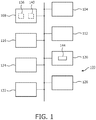
- FIG. 1 depicts, by way of illustrative and non-limitative example, an ultrasound clinician interactive guidance apparatus 100.
- the apparatus 100 includes, among other component devices, a controller 104, an image matching module 108, a user assistance module 112, a memory 116, a scanner 120, a display 124, a speaker 128 and user controls 132.
- the image matching module 108 includes an image registering module 136 and/or a state space processing module 140.
- the scanner includes, among other component devices, an imaging probe 144.
- an overall procedure 200 for ultrasound clinician interactive guidance is as follows, as shown in FIG. 2 .
- an authoritative medical entity such as a physician, medical board, medical standards organization, or hospital sets standard ultrasound views for the body organ, or vessel, of interest (step S204).
- the set of standard views is specified for use on the apparatus 100 (step S208).
- One or more image matching references are prepared (step S212) which is explained in more detail below with reference to FIG. 3 .
- the clinician selects a scan type, which may be for a particular body organ such as the heart, or vessel such a particular artery (step S216).
- the apparatus 100 pulls up the corresponding image matching reference (step S218).
- the apparatus 100 determines which target view from among the standard views is to be acquired next.
- the apparatus 100 also now loads Doppler settings that have been pre-selected for the target view, as discussed below in connection with FIG. 4 .
- Doppler settings are, automatically by default, initialized according to those that were pre-set for the target view in building a database organized as the state space (step S220).
- the apparatus 100 indicates to the user how, based on textbook guidelines for example, to place the imaging probe 144 on the superficial anatomy of the imaging subject, such as an animal or human patient (step S224).
- the user i.e., clinician, positions the probe 144 (step S228).
- step S232 the state space processing module 140 is implemented and makes an estimate of the location, in the state space, of the current, or "live", view acquired via the probe 144 (step S236). If, on the other hand, the state space processing module 140 is not implemented (step S232) but the image registering module 136 is implemented, the current view is registered to a corresponding position, and orientation, in a three-dimensional (3D) reference image (step S240).
- step S244 If it is now determined that the current view does not match, or sufficiently represent, the target view (step S244), the apparatus 100 gives user feedback that instructs, or shows, how to proceed toward the goal of achieving the target view (step S248), and processing branches back to the user positioning step S228. Description in more detail of the feedback is provided further below in the discussion accompanying FIGs. 5 and 6 . If, instead, a match has been attained (step S244) and automatic acquisition is to be performed (step S252), the current view is recorded for further analysis, e.g., by a physician (step S256).
- a green light is lit on the probe 144 or elsewhere on the scanner, such as on a console that houses the user controls 132 (step S260).
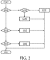
- the image matching reference preparation step (S212) is described in more detail in the flow chart of FIG. 3 .
- a statistical atlas is prepared (step S330).
- the statistical atlas is built based on the computed tomography (CT) and/or magnetic resonance imaging (MR) scans of a wide variety of subjects to cover anatomical variation. It may be stored on a hard drive which is part of the memory 116. Per voxel, the atlas includes a distribution of image intensities reflective of individual members of the population.
- Neighboring information is also included for each voxel. Image matching to achieve registration is performed quicker due to the statistical nature of the statistical atlas. If, on the other hand, image matching is to be based on an anatomical atlas that is not a statistical atlas (steps S310, S320), the anatomical atlas is prepared as the 3D image matching reference, typically via CT and/or MR scans from a wide variety of subjects (step S340). If, instead of an atlas (step S310), CT and/or MR scans of the same patient are to be used to build the 3D image matching reference (step S350), the "same-patient" reference is prepared (step S360).
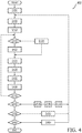
- step S350 If, on the other hand, the same-patient CT/MR scans are not available or are otherwise not to be used (step S350), and a state space is to be used (step S370), a state space is prepared (step S380). Preparation of the state space is described in more detail immediately below in connection with FIG. 4 .
- a state space preparation process 400 involves building a statistical database of a set of hundreds of scans of and around the organ, or vessel, of interest from multiple imaging subjects.
- the database is organized as a state space. Images to be incorporated into the state space are labeled with attributes such as viewed image anatomy, image quality, and corresponding probe position and orientation on the superficial anatomy. A subset of this set of images is the set of goal states, corresponding to the standard ultrasound views with good image quality.
- the images of the database can be described as points within a state space whose dimensions are the image attributes. Within the state space, it is possible to define a spatial relationship between ultrasound images, and in particular a trajectory between any ultrasound image and any of the goal images.
- the images to be acquired for incorporation into the state space are specialized for a specific body organ, or vessel, such that the organ or vessel, and/or surrounding tissue, are depicted in each image.
- the process 400 is initialized to a first imaging subject, a first target view, a first trajectory and a first image (step S404). Thus, the respective pointers or counters are zeroed out.
- a current image is acquired via the imaging probe 144 (step S408). Attributes of the image are recorded and the current image is labeled with its attributes (step S412). The recording can be done in part automatically and in part via entry by the person building the database.
- the current image may be labeled according to: the viewed anatomy (e.g., carotid (left, right, common, internal, external, bulb, bifurcation, proximal, medial, distal, longitudinal, transverse, oblique, etc.) such as jugular vein, thyroid gland, vertebral bodies, vertebral artery, vertebral vein, subclavian artery, etc.; the position and orientation of the probe 144 with respect to the superficial anatomy to obtain these images (e.g., anterior, anterior, posterior, cranial, caudal, lateral, medial, neck, clavicle, mandible, Adam's apple, horizontal, vertical, oblique); optionally the current imaging mode and settings (e.g., for B-mode, power, focal depth, harmonics, spatial compounding; for color flow, gain, maximum velocity, color box orientation, sample volume size; and for spectral Doppler, maximum velocity and Doppler angle); and optionally the presence of artifacts and a measure
- the current image acquired may have been acquired via ultrasound contact that is less than good. This would be done intentionally, so that matching to this image, once it is in the database, allows the deficient contact to be detected. If the contact is deficient (step S416), the person building the database applies or reapplies acoustic coupling medium, such as gel, restores the probe 144 to the same position and orientation with respect to the imaging subject for an improved image (step S420). Otherwise, if the contact was not deficient (step S416), the database builder, via probe movement or adjustment of imaging settings, prepares for a next image acquisition (step S422). The movement or adjustment is made so as to navigate toward the target image.
- acoustic coupling medium such as gel
- step S424 processing now points to that next image (step S424).
- the image is acquired (step S428).
- the attributes are recorded in part manually and in part automatically (step S432).
- the most recent probe movement, contact adjustment or imaging setting adjustment, ordinarily for B-mode, made in the corresponding above steps S420, S422 is entered or selected by the database builder, or automatically, and linked to the previous image, i.e., the image acquired just prior to step S428 (step S436).
- the entry could be, with respect to probe position, "left", "right", “up” or “down.”
- "up” would be mean generally in the head to toe direction.
- the entry could instead or in addition be, with respect orientation, i.e., tilting, "left", “right”, “up”, or “down.”
- the entry could additonally or instead be, with respect in place rotation of the probe 144, "clockwise” or “counterclockwise.”
- distance or magnitude need not be recorded, because the updating of the feedback loop in steps S228 to S248 occurs in real time.
- the database image having the location closest, according to Euclidean distance for example, to the estimate made in step S236 dynamically keeps the user on a trajectory toward the target view. Even if, during operation, a user wanders into another trajectory, that other trajectory will similary navigate toward the target view.
- the entry or selection by the database builder may be "reapply gel to probe and return to same position and orientation.”
- the automatic selection may be, for example, "increase imaging depth.”
- step S440 If the current view is not the target view (step S440), processing returns to step S416. Otherwise, if the current view is the target view as evidenced by actuation of the appropriate user control 132 by the database builder (step S440), and another trajectory is to be recorded for the current target view of the current imaging subject (step S444), the database builder is advised, via an onscreen message, to enter Doppler mode settings (step S446). Interactively, according to a series of screen prompts and responsive actuations by the database builder, the Doppler settings are stored as attributes of the target view (step S448). The trajectory pointer is incremented (step S450) and return is made to step S408.
- step S444 If, on the other hand, no such further trajectory is to be recorded (step S444), but another target view for the current imaging subject is to be used in building up the database (step S452), the view pointer is incremented (step S456) and return is likewise made to step S408. If, however, no target view for the current imaging subject remains in terms of building up the database (step S452), but a next imaging subject is to be used in building the database (step S460), the subject pointer is incremented (step S464) and return is likewise made to step S408.
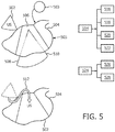
- FIG. 5 provides examples of the user feedback of step S248, which can take the form of onscreen illustrations or messages, or audible language.
- An ultrasound image representative of a current view 502 such as a B-mode image can be displayed alongside a cross-sectional image 504 derived from a 3D reference image 503 stored on a hard drive, i.e., from an atlas or from a 3D image constructed from patient-specific CT and/or MR scans.
- the cross-sectional image 504, here of a body organ 505, i.e., the heart, has been sectioned and enhanced to spatially indicate where the received (or "live") view registers to the reference image.
- an enhanced region 506, that is colored for example corresponds spatially with where the current image would cut into the atlas.
- a graphic indication 508 of the plane of the current target view 510 can be added to the onscreen presentation.
- the ultrasound image can be fused 512 to the cross-sectional image 504 such as by a pixel for pixel replacement.
- the graphic indication 508 can be added.
- screen messages or audible language instructions can guide the clinician.
- four possible indications 516-522 are “right”, “left”, “up” and “down”, just as in the state space based embodiment.
- in-place rotation 524 can be "clockwise” 526 or “counterclockwise” 528.
- the registration in step S240 involves image-based pattern matching of the current view 502 to the 3D reference image and a coordinate transformation on the current view to bring it into registration with the 3D image in accordance with the matching.
- the feedback instructions, based on the transformation can be representative of a single kind, or more than one kind, of suggested probe movement 514, 524.
- the estimate in step S236 is made as a result of pattern recognition from comparisons between the current view 502 and the database images acquired in the acquisition steps S408, S428.
- the one or more types of feedback instructions i.e., probe movement, probe contact and imaging settings linked to the current database image are presented.
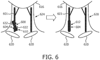
- FIG. 6 is a specific example of user feedback and feedback generation. This example relates to the left medial common carotid artery 601 and acquiring a standard view of the artery.
- a transducer array face graphic 604 is shown in an oblique position 608, representative of a current placement 610 of the probe 144, and in a non-oblique position 612.
- the transducer array may be a linear array or a matrix array.
- Mandible graphics 616 and clavicle graphics 620 are also shown in FIG. 6 .
- a current graphic 624 corresponds conceptually to the current view 502
- a target graphic 628 corresponds conceptually to the target view 510.
- both graphics 624, 628 may be displayed onscreen in addition to, or in place of, any other graphic or ultrasound image representative of the current view 502.
- the viewed anatomy label for a matched database image is "left medial common carotid artery, oblique view.”
- the probe position label is “midway between clavicle and mandible.”
- the clavicle and mandible graphics 616, 620 represent surrounding tissue.
- the probe orientation label is “oblique.”
- the imaging mode label is "B-mode.”
- An imaging setting label is "spatial compounding.”
- An artifact label is "artifact-free.”
- An image quality label is "good image contrast", based for example on average pixel intensity. All of the labels can be displayed, on the display 124, upon matching to the database image 604.
- a different instruction that was stored during database building in step S436, is sent for viewing on the display 124.
- the instruction here would be the instruction 526 to "rotate in-place clockwise.” This is indicated by illustrative arrows 632.
- the resulting movement of the probe 144 by the clinician is, as mentioned herein above, monitored in real time via the feedback loop in steps S228 to S248.
- the instruction is resent for display repeatedly, but will change in the event of the current view 502 matching to a new database image, such as that corresponding to the target view 510.
- the instruction 526 "rotate clockwise" is derivable almost by definition, since the only transformation involved in registering the current view 502 to the 3D reference image is, in fact, the clockwise rotation.
- reaching the target view 510 entails, for instance, probe in-place rotation and translation, whether rotation or translation predominates is decided by the apparatus 100.
- the criteria can involve thresholds selected based on empirical experience, although, for example, location will ordinarily dominate over tilting until the probe location is close to that needed for a target view 510.
- Guidance in acquiring ultrasound imaging of a subject to achieve a target view entails emitting ultrasound to the subject and receiving, in response, a current ultrasound view; matching the received image to a pre-existing image, such as a three-dimensional reference image; and, for user assistance, generating, based on the matching, feedback for the guidance.
- the reference image may be a statistical atlas or it may be derived from patient-specific CT or MR scans.
- the pre-existing image may instead be a database image corresponding to a state in a state space.
- the feedback can be an image derived from the reference image; a graphic indication of a plane of the target view; the received view fused to an image derived from the reference image; or the received view and an image derived from said reference image, the derived image appearing concurrently and enhanced to spatially indicate where the received view registers to the reference image.
- the target view may be a view of a body organ, or vessel, of the subject. Both the atlas and database can be specialized for imaging of a user selected organ, or vessel, and its surrounding tissue.
- the interactive visual guidance apparatus 100 can guide novice sonographers.
- the novel visual feedback of the apparatus 100 can speed up the work flow of trained or experienced sonographers.
- the probe 144 may alternatively or additionally use tactile feedback on the appropriate probe movement toward a standard view.
- a computer program can be stored momentarily, temporarily or for a longer period of time on a suitable computer-readable medium, such as an optical storage medium or a solid-state medium.
- a suitable computer-readable medium such as an optical storage medium or a solid-state medium.
- Such a medium is non-transitory only in the sense of not being a transitory, propagating signal, but includes other forms of computer-readable media such as register memory, processor cache, RAM and other volatile memory.
- a single processor or other unit may fulfill the functions of several items recited in the claims.
- the mere fact that certain measures are recited in mutually different dependent claims does not indicate that a combination of these measures cannot be used to advantage.
Landscapes
- Health & Medical Sciences (AREA)
- Life Sciences & Earth Sciences (AREA)
- Engineering & Computer Science (AREA)
- Physics & Mathematics (AREA)
- Surgery (AREA)
- Public Health (AREA)
- Radiology & Medical Imaging (AREA)
- Nuclear Medicine, Radiotherapy & Molecular Imaging (AREA)
- Biomedical Technology (AREA)
- Heart & Thoracic Surgery (AREA)
- Medical Informatics (AREA)
- Molecular Biology (AREA)
- Biophysics (AREA)
- Animal Behavior & Ethology (AREA)
- General Health & Medical Sciences (AREA)
- Pathology (AREA)
- Veterinary Medicine (AREA)
- Computer Vision & Pattern Recognition (AREA)
- Theoretical Computer Science (AREA)
- General Engineering & Computer Science (AREA)
- General Physics & Mathematics (AREA)
- Library & Information Science (AREA)
- Computer Graphics (AREA)
- Data Mining & Analysis (AREA)
- Databases & Information Systems (AREA)
- Ultra Sonic Daignosis Equipment (AREA)
Applications Claiming Priority (2)
| Application Number | Priority Date | Filing Date | Title |
|---|---|---|---|
| US201361840727P | 2013-06-28 | 2013-06-28 | |
| PCT/IB2014/062523 WO2014207642A1 (en) | 2013-06-28 | 2014-06-23 | Ultrasound acquisition feedback guidance to a target view |
Publications (2)
| Publication Number | Publication Date |
|---|---|
| EP3013242A1 EP3013242A1 (en) | 2016-05-04 |
| EP3013242B1 true EP3013242B1 (en) | 2018-11-07 |
Family
ID=51205526
Family Applications (1)
| Application Number | Title | Priority Date | Filing Date |
|---|---|---|---|
| EP14739560.2A Not-in-force EP3013242B1 (en) | 2013-06-28 | 2014-06-23 | Ultrasound acquisition feedback guidance to a target view |
Country Status (7)
| Country | Link |
|---|---|
| US (1) | US10702248B2 (enExample) |
| EP (1) | EP3013242B1 (enExample) |
| JP (1) | JP6527860B2 (enExample) |
| CN (1) | CN105451663B (enExample) |
| BR (1) | BR112015032573B1 (enExample) |
| RU (1) | RU2683720C2 (enExample) |
| WO (1) | WO2014207642A1 (enExample) |
Cited By (1)
| Publication number | Priority date | Publication date | Assignee | Title |
|---|---|---|---|---|
| EP4613206A1 (en) * | 2024-03-04 | 2025-09-10 | FUJIFILM Corporation | Ultrasound diagnostic apparatus and method of controlling ultrasound diagnostic apparatus |
Families Citing this family (28)
| Publication number | Priority date | Publication date | Assignee | Title |
|---|---|---|---|---|
| WO2014134188A1 (en) * | 2013-02-28 | 2014-09-04 | Rivanna Medical, LLC | Systems and methods for ultrasound imaging |
| EP2996561B1 (en) * | 2013-03-05 | 2017-05-03 | Koninklijke Philips N.V. | Consistent sequential ultrasound acquisitions for intra-cranial monitoring |
| WO2016141449A1 (en) * | 2015-03-09 | 2016-09-15 | Her Majesty The Queen In Right Of Canada, As Represented By The Minister Of The Department Of National Defence | Computer-assisted focused assessment with sonography in trauma |
| GB201509164D0 (en) * | 2015-05-28 | 2015-07-15 | Intelligent Ultrasound Ltd | Imaging feedback system and method |
| US10682122B2 (en) * | 2015-12-03 | 2020-06-16 | Siemens Medical Solutions Usa, Inc. | Image-based user interface for controlling medical imaging |
| US10964424B2 (en) | 2016-03-09 | 2021-03-30 | EchoNous, Inc. | Ultrasound image recognition systems and methods utilizing an artificial intelligence network |
| KR20190021344A (ko) | 2016-06-20 | 2019-03-05 | 버터플라이 네트워크, 인크. | 초음파 디바이스를 작동하는 사용자를 보조하기 위한 자동화된 영상 취득 |
| US10905402B2 (en) | 2016-07-27 | 2021-02-02 | Canon Medical Systems Corporation | Diagnostic guidance systems and methods |
| FR3060966B1 (fr) * | 2016-12-23 | 2019-05-31 | Azoth Systems | Dispositif de mesure du flux sanguin |
| AU2018323621A1 (en) | 2017-08-31 | 2020-02-06 | Butterfly Network, Inc. | Methods and apparatus for collection of ultrasound data |
| EP3681405B1 (en) * | 2017-09-15 | 2025-07-09 | Elesta S.P.A. | Device and computer program for needle sonographic guidance in minimally invasive procedures |
| WO2019099364A1 (en) | 2017-11-14 | 2019-05-23 | Verathon Inc. | Real-time feedback and semantic-rich guidance on quality ultrasound image acquisition |
| EP3549529A1 (en) * | 2018-04-05 | 2019-10-09 | Koninklijke Philips N.V. | Ultrasound imaging system and method |
| WO2019199781A1 (en) * | 2018-04-09 | 2019-10-17 | Butterfly Network, Inc. | Methods and apparatus for configuring an ultrasound system with imaging parameter values |
| FI3793447T3 (fi) | 2018-05-15 | 2023-03-18 | Univ New York | Järjestelmä ja menetelmä ultraäänikuvien ottamisen suuntaamiseksi |
| US20220225963A1 (en) * | 2019-05-31 | 2022-07-21 | Koninklijke Philips N.V. | Methods and systems for guiding the acquisition of cranial ultrasound data |
| US11798677B2 (en) * | 2019-12-31 | 2023-10-24 | GE Precision Healthcare LLC | Method and system for providing a guided workflow through a series of ultrasound image acquisitions with reference images updated based on a determined anatomical position |
| EP3868303A1 (en) * | 2020-02-18 | 2021-08-25 | Koninklijke Philips N.V. | Ultrasound guidance method and system |
| CN111938700B (zh) * | 2020-08-21 | 2021-11-09 | 电子科技大学 | 基于人体解剖结构实时匹配的超声探头引导系统及方法 |
| CN111938699B (zh) * | 2020-08-21 | 2022-04-01 | 电子科技大学 | 一种引导使用超声设备的系统及方法 |
| US20220338836A1 (en) * | 2021-04-21 | 2022-10-27 | Ultrasight Ltd | System and method for guiding positioning and orienting of an ultrasound probe |
| CN113476098B (zh) * | 2021-07-26 | 2022-06-24 | 首都医科大学附属北京儿童医院 | 一种封堵管的监测系统 |
| US11903760B2 (en) | 2021-09-08 | 2024-02-20 | GE Precision Healthcare LLC | Systems and methods for scan plane prediction in ultrasound images |
| US11974884B2 (en) * | 2021-11-15 | 2024-05-07 | GE Precision Healthcare LLC | Method and system for dynamically adjusting imaging parameters during an ultrasound scan |
| US12239484B2 (en) | 2021-12-27 | 2025-03-04 | GE Precision Healthcare LLC | Medical imaging method |
| CN116236280B (zh) * | 2023-02-02 | 2024-06-18 | 逸超医疗科技(北京)有限公司 | 一种基于多模态图像融合的介入治疗引导方法及系统 |
| CN117017355B (zh) * | 2023-10-08 | 2024-01-12 | 合肥合滨智能机器人有限公司 | 一种基于多模态生成式对话的甲状腺自主扫查系统 |
| US12505551B1 (en) * | 2025-06-28 | 2025-12-23 | Anumana, Inc. | Apparatus and method for automatically extracting canonical views from ultrasound imaging data |
Family Cites Families (16)
| Publication number | Priority date | Publication date | Assignee | Title |
|---|---|---|---|---|
| US5906578A (en) * | 1997-06-18 | 1999-05-25 | Rajan; Govinda N. | Method and system for probe positioning in transesophageal echocardiography |
| JP4088104B2 (ja) * | 2002-06-12 | 2008-05-21 | 株式会社東芝 | 超音波診断装置 |
| EP2460474B1 (en) * | 2003-05-08 | 2015-12-16 | Hitachi Medical Corporation | Reference image display method for ultrasonography and ultrasonic diagnosis apparatus |
| US7221972B2 (en) | 2003-08-29 | 2007-05-22 | Siemens Medical Solutions Usa, Inc. | Ultrasound system with protocol-driven user interface |
| US20050187472A1 (en) | 2004-01-30 | 2005-08-25 | Peter Lysyansky | Protocol-driven ultrasound examination |
| EP1751712A2 (en) * | 2004-05-14 | 2007-02-14 | Philips Intellectual Property & Standards GmbH | Information enhanced image guided interventions |
| JP4699062B2 (ja) * | 2005-03-29 | 2011-06-08 | 株式会社日立メディコ | 超音波装置 |
| US20070081706A1 (en) * | 2005-09-28 | 2007-04-12 | Xiang Zhou | Systems and methods for computer aided diagnosis and decision support in whole-body imaging |
| JP2009089736A (ja) * | 2007-10-03 | 2009-04-30 | Toshiba Corp | 超音波診断装置 |
| WO2009081339A1 (en) * | 2007-12-21 | 2009-07-02 | Koninklijke Philips Electronics, N.V. | Systems and methods for tracking and guiding high intensity focused ultrasound beams |
| US8172753B2 (en) * | 2008-07-11 | 2012-05-08 | General Electric Company | Systems and methods for visualization of an ultrasound probe relative to an object |
| JP5481108B2 (ja) * | 2009-06-26 | 2014-04-23 | 株式会社東芝 | 超音波診断装置及び自動診断支援装置 |
| JP2011110102A (ja) * | 2009-11-24 | 2011-06-09 | Toshiba Corp | 超音波診断装置 |
| US8352494B1 (en) * | 2009-12-07 | 2013-01-08 | Google Inc. | Distributed image search |
| US20120065510A1 (en) * | 2010-09-09 | 2012-03-15 | General Electric Company | Ultrasound system and method for calculating quality-of-fit |
| EP2726899A1 (en) * | 2011-07-01 | 2014-05-07 | Koninklijke Philips N.V. | Intra-operative image correction for image-guided interventions |
-
2014
- 2014-06-23 EP EP14739560.2A patent/EP3013242B1/en not_active Not-in-force
- 2014-06-23 CN CN201480042594.0A patent/CN105451663B/zh active Active
- 2014-06-23 RU RU2016102416A patent/RU2683720C2/ru active
- 2014-06-23 WO PCT/IB2014/062523 patent/WO2014207642A1/en not_active Ceased
- 2014-06-23 JP JP2016522921A patent/JP6527860B2/ja not_active Expired - Fee Related
- 2014-06-23 BR BR112015032573-4A patent/BR112015032573B1/pt not_active IP Right Cessation
- 2014-06-23 US US14/901,104 patent/US10702248B2/en active Active
Non-Patent Citations (1)
| Title |
|---|
| None * |
Cited By (1)
| Publication number | Priority date | Publication date | Assignee | Title |
|---|---|---|---|---|
| EP4613206A1 (en) * | 2024-03-04 | 2025-09-10 | FUJIFILM Corporation | Ultrasound diagnostic apparatus and method of controlling ultrasound diagnostic apparatus |
Also Published As
| Publication number | Publication date |
|---|---|
| JP2016522074A (ja) | 2016-07-28 |
| CN105451663B (zh) | 2019-03-19 |
| BR112015032573B1 (pt) | 2022-04-19 |
| BR112015032573A2 (pt) | 2017-07-25 |
| RU2016102416A (ru) | 2017-08-01 |
| WO2014207642A1 (en) | 2014-12-31 |
| JP6527860B2 (ja) | 2019-06-05 |
| CN105451663A (zh) | 2016-03-30 |
| EP3013242A1 (en) | 2016-05-04 |
| US20160143627A1 (en) | 2016-05-26 |
| RU2683720C2 (ru) | 2019-04-01 |
| US10702248B2 (en) | 2020-07-07 |
Similar Documents
| Publication | Publication Date | Title |
|---|---|---|
| EP3013242B1 (en) | Ultrasound acquisition feedback guidance to a target view | |
| CN106037797B (zh) | 超声成像中感兴趣的三维容积 | |
| JP5707148B2 (ja) | 医用画像診断装置及び医用画像処理装置 | |
| JP5624258B2 (ja) | 超音波診断装置、超音波画像処理装置及び超音波画像処理プログラム | |
| CN103997971B (zh) | 用于超声心动描记的自动成像平面选择 | |
| US9380999B2 (en) | Ultrasonic diagnostic apparatus, ultrasonic image processing apparatus, and medical diagnostic imaging apparatus | |
| JP5972569B2 (ja) | 超音波診断装置、超音波画像処置装置、医用画像診断装置及び超音波画像処理プログラム | |
| US9888905B2 (en) | Medical diagnosis apparatus, image processing apparatus, and method for image processing | |
| JP7171168B2 (ja) | 医用画像診断装置及び医用画像処理装置 | |
| US20110301457A1 (en) | Ultrasonic diagnostic apparatus, ultrasonic image processing apparatus, and medical image diagnostic apparatus | |
| JP5897674B2 (ja) | 超音波診断装置、画像処理装置及び画像処理プログラム | |
| US20160030008A1 (en) | System and method for registering ultrasound information to an x-ray image | |
| KR102278893B1 (ko) | 의료영상처리장치 및 이를 이용한 의료영상정합방법 | |
| CN1853571A (zh) | 使用超声轮廓重建进行三维心脏成像的软件产品 | |
| EP2679158B1 (en) | Method and apparatus for displaying ultrasonic image and information related to the ultrasonic image | |
| JP5689591B2 (ja) | 超音波診断装置及び超音波画像処理プログラム | |
| US10667796B2 (en) | Method and system for registering a medical image with a graphical model | |
| CN107198546A (zh) | 超声波诊断装置、图像处理装置以及图像处理方法 | |
| US20220313214A1 (en) | Ultrasonic diagnostic apparatus, image processing apparatus, and image processing method | |
| JP2012075794A (ja) | 超音波診断装置、医用画像処理装置及び医用画像処理プログラム | |
| CN111317508B (zh) | 超声波诊断装置、医用信息处理装置、计算机程序产品 | |
| JP6863774B2 (ja) | 超音波診断装置、画像処理装置及び画像処理プログラム | |
| JP5784085B2 (ja) | 超音波診断装置、超音波画像処理装置及び超音波画像処理プログラム | |
| US20190008482A1 (en) | Medical image diagnostic apparatus, medical image processing apparatus, and medical image processing method | |
| JP5624581B2 (ja) | 超音波診断装置、超音波画像処理装置及び超音波画像処理プログラム |
Legal Events
| Date | Code | Title | Description |
|---|---|---|---|
| PUAI | Public reference made under article 153(3) epc to a published international application that has entered the european phase |
Free format text: ORIGINAL CODE: 0009012 |
|
| 17P | Request for examination filed |
Effective date: 20160128 |
|
| AK | Designated contracting states |
Kind code of ref document: A1 Designated state(s): AL AT BE BG CH CY CZ DE DK EE ES FI FR GB GR HR HU IE IS IT LI LT LU LV MC MK MT NL NO PL PT RO RS SE SI SK SM TR |
|
| AX | Request for extension of the european patent |
Extension state: BA ME |
|
| DAX | Request for extension of the european patent (deleted) | ||
| STAA | Information on the status of an ep patent application or granted ep patent |
Free format text: STATUS: EXAMINATION IS IN PROGRESS |
|
| 17Q | First examination report despatched |
Effective date: 20170627 |
|
| GRAP | Despatch of communication of intention to grant a patent |
Free format text: ORIGINAL CODE: EPIDOSNIGR1 |
|
| STAA | Information on the status of an ep patent application or granted ep patent |
Free format text: STATUS: GRANT OF PATENT IS INTENDED |
|
| INTG | Intention to grant announced |
Effective date: 20180524 |
|
| RIN1 | Information on inventor provided before grant (corrected) |
Inventor name: PARTHASARATHY, VIJAY Inventor name: VIGNON, FRANCOIS GUY GERARD MARIE Inventor name: JAIN, AMEET KUMAR Inventor name: ANAND, AJAY |
|
| GRAS | Grant fee paid |
Free format text: ORIGINAL CODE: EPIDOSNIGR3 |
|
| GRAA | (expected) grant |
Free format text: ORIGINAL CODE: 0009210 |
|
| STAA | Information on the status of an ep patent application or granted ep patent |
Free format text: STATUS: THE PATENT HAS BEEN GRANTED |
|
| AK | Designated contracting states |
Kind code of ref document: B1 Designated state(s): AL AT BE BG CH CY CZ DE DK EE ES FI FR GB GR HR HU IE IS IT LI LT LU LV MC MK MT NL NO PL PT RO RS SE SI SK SM TR |
|
| REG | Reference to a national code |
Ref country code: GB Ref legal event code: FG4D |
|
| REG | Reference to a national code |
Ref country code: CH Ref legal event code: EP Ref country code: AT Ref legal event code: REF Ref document number: 1061134 Country of ref document: AT Kind code of ref document: T Effective date: 20181115 |
|
| REG | Reference to a national code |
Ref country code: DE Ref legal event code: R096 Ref document number: 602014035567 Country of ref document: DE |
|
| REG | Reference to a national code |
Ref country code: IE Ref legal event code: FG4D |
|
| REG | Reference to a national code |
Ref country code: DE Ref legal event code: R084 Ref document number: 602014035567 Country of ref document: DE |
|
| REG | Reference to a national code |
Ref country code: NL Ref legal event code: MP Effective date: 20181107 |
|
| REG | Reference to a national code |
Ref country code: LT Ref legal event code: MG4D |
|
| REG | Reference to a national code |
Ref country code: AT Ref legal event code: MK05 Ref document number: 1061134 Country of ref document: AT Kind code of ref document: T Effective date: 20181107 |
|
| PG25 | Lapsed in a contracting state [announced via postgrant information from national office to epo] |
Ref country code: NO Free format text: LAPSE BECAUSE OF FAILURE TO SUBMIT A TRANSLATION OF THE DESCRIPTION OR TO PAY THE FEE WITHIN THE PRESCRIBED TIME-LIMIT Effective date: 20190207 Ref country code: LT Free format text: LAPSE BECAUSE OF FAILURE TO SUBMIT A TRANSLATION OF THE DESCRIPTION OR TO PAY THE FEE WITHIN THE PRESCRIBED TIME-LIMIT Effective date: 20181107 Ref country code: BG Free format text: LAPSE BECAUSE OF FAILURE TO SUBMIT A TRANSLATION OF THE DESCRIPTION OR TO PAY THE FEE WITHIN THE PRESCRIBED TIME-LIMIT Effective date: 20190207 Ref country code: HR Free format text: LAPSE BECAUSE OF FAILURE TO SUBMIT A TRANSLATION OF THE DESCRIPTION OR TO PAY THE FEE WITHIN THE PRESCRIBED TIME-LIMIT Effective date: 20181107 Ref country code: AT Free format text: LAPSE BECAUSE OF FAILURE TO SUBMIT A TRANSLATION OF THE DESCRIPTION OR TO PAY THE FEE WITHIN THE PRESCRIBED TIME-LIMIT Effective date: 20181107 Ref country code: LV Free format text: LAPSE BECAUSE OF FAILURE TO SUBMIT A TRANSLATION OF THE DESCRIPTION OR TO PAY THE FEE WITHIN THE PRESCRIBED TIME-LIMIT Effective date: 20181107 Ref country code: FI Free format text: LAPSE BECAUSE OF FAILURE TO SUBMIT A TRANSLATION OF THE DESCRIPTION OR TO PAY THE FEE WITHIN THE PRESCRIBED TIME-LIMIT Effective date: 20181107 Ref country code: ES Free format text: LAPSE BECAUSE OF FAILURE TO SUBMIT A TRANSLATION OF THE DESCRIPTION OR TO PAY THE FEE WITHIN THE PRESCRIBED TIME-LIMIT Effective date: 20181107 Ref country code: IS Free format text: LAPSE BECAUSE OF FAILURE TO SUBMIT A TRANSLATION OF THE DESCRIPTION OR TO PAY THE FEE WITHIN THE PRESCRIBED TIME-LIMIT Effective date: 20190307 |
|
| PG25 | Lapsed in a contracting state [announced via postgrant information from national office to epo] |
Ref country code: GR Free format text: LAPSE BECAUSE OF FAILURE TO SUBMIT A TRANSLATION OF THE DESCRIPTION OR TO PAY THE FEE WITHIN THE PRESCRIBED TIME-LIMIT Effective date: 20190208 Ref country code: PT Free format text: LAPSE BECAUSE OF FAILURE TO SUBMIT A TRANSLATION OF THE DESCRIPTION OR TO PAY THE FEE WITHIN THE PRESCRIBED TIME-LIMIT Effective date: 20190307 Ref country code: NL Free format text: LAPSE BECAUSE OF FAILURE TO SUBMIT A TRANSLATION OF THE DESCRIPTION OR TO PAY THE FEE WITHIN THE PRESCRIBED TIME-LIMIT Effective date: 20181107 Ref country code: SE Free format text: LAPSE BECAUSE OF FAILURE TO SUBMIT A TRANSLATION OF THE DESCRIPTION OR TO PAY THE FEE WITHIN THE PRESCRIBED TIME-LIMIT Effective date: 20181107 Ref country code: RS Free format text: LAPSE BECAUSE OF FAILURE TO SUBMIT A TRANSLATION OF THE DESCRIPTION OR TO PAY THE FEE WITHIN THE PRESCRIBED TIME-LIMIT Effective date: 20181107 Ref country code: AL Free format text: LAPSE BECAUSE OF FAILURE TO SUBMIT A TRANSLATION OF THE DESCRIPTION OR TO PAY THE FEE WITHIN THE PRESCRIBED TIME-LIMIT Effective date: 20181107 |
|
| PG25 | Lapsed in a contracting state [announced via postgrant information from national office to epo] |
Ref country code: PL Free format text: LAPSE BECAUSE OF FAILURE TO SUBMIT A TRANSLATION OF THE DESCRIPTION OR TO PAY THE FEE WITHIN THE PRESCRIBED TIME-LIMIT Effective date: 20181107 Ref country code: CZ Free format text: LAPSE BECAUSE OF FAILURE TO SUBMIT A TRANSLATION OF THE DESCRIPTION OR TO PAY THE FEE WITHIN THE PRESCRIBED TIME-LIMIT Effective date: 20181107 Ref country code: IT Free format text: LAPSE BECAUSE OF FAILURE TO SUBMIT A TRANSLATION OF THE DESCRIPTION OR TO PAY THE FEE WITHIN THE PRESCRIBED TIME-LIMIT Effective date: 20181107 Ref country code: DK Free format text: LAPSE BECAUSE OF FAILURE TO SUBMIT A TRANSLATION OF THE DESCRIPTION OR TO PAY THE FEE WITHIN THE PRESCRIBED TIME-LIMIT Effective date: 20181107 |
|
| REG | Reference to a national code |
Ref country code: DE Ref legal event code: R097 Ref document number: 602014035567 Country of ref document: DE |
|
| REG | Reference to a national code |
Ref country code: GB Ref legal event code: 746 Effective date: 20190730 |
|
| PG25 | Lapsed in a contracting state [announced via postgrant information from national office to epo] |
Ref country code: SK Free format text: LAPSE BECAUSE OF FAILURE TO SUBMIT A TRANSLATION OF THE DESCRIPTION OR TO PAY THE FEE WITHIN THE PRESCRIBED TIME-LIMIT Effective date: 20181107 Ref country code: EE Free format text: LAPSE BECAUSE OF FAILURE TO SUBMIT A TRANSLATION OF THE DESCRIPTION OR TO PAY THE FEE WITHIN THE PRESCRIBED TIME-LIMIT Effective date: 20181107 Ref country code: SM Free format text: LAPSE BECAUSE OF FAILURE TO SUBMIT A TRANSLATION OF THE DESCRIPTION OR TO PAY THE FEE WITHIN THE PRESCRIBED TIME-LIMIT Effective date: 20181107 Ref country code: RO Free format text: LAPSE BECAUSE OF FAILURE TO SUBMIT A TRANSLATION OF THE DESCRIPTION OR TO PAY THE FEE WITHIN THE PRESCRIBED TIME-LIMIT Effective date: 20181107 |
|
| PLBE | No opposition filed within time limit |
Free format text: ORIGINAL CODE: 0009261 |
|
| STAA | Information on the status of an ep patent application or granted ep patent |
Free format text: STATUS: NO OPPOSITION FILED WITHIN TIME LIMIT |
|
| 26N | No opposition filed |
Effective date: 20190808 |
|
| PG25 | Lapsed in a contracting state [announced via postgrant information from national office to epo] |
Ref country code: SI Free format text: LAPSE BECAUSE OF FAILURE TO SUBMIT A TRANSLATION OF THE DESCRIPTION OR TO PAY THE FEE WITHIN THE PRESCRIBED TIME-LIMIT Effective date: 20181107 |
|
| PG25 | Lapsed in a contracting state [announced via postgrant information from national office to epo] |
Ref country code: MC Free format text: LAPSE BECAUSE OF FAILURE TO SUBMIT A TRANSLATION OF THE DESCRIPTION OR TO PAY THE FEE WITHIN THE PRESCRIBED TIME-LIMIT Effective date: 20181107 |
|
| REG | Reference to a national code |
Ref country code: CH Ref legal event code: PL |
|
| REG | Reference to a national code |
Ref country code: BE Ref legal event code: MM Effective date: 20190630 |
|
| PG25 | Lapsed in a contracting state [announced via postgrant information from national office to epo] |
Ref country code: TR Free format text: LAPSE BECAUSE OF FAILURE TO SUBMIT A TRANSLATION OF THE DESCRIPTION OR TO PAY THE FEE WITHIN THE PRESCRIBED TIME-LIMIT Effective date: 20181107 |
|
| PG25 | Lapsed in a contracting state [announced via postgrant information from national office to epo] |
Ref country code: IE Free format text: LAPSE BECAUSE OF NON-PAYMENT OF DUE FEES Effective date: 20190623 |
|
| PG25 | Lapsed in a contracting state [announced via postgrant information from national office to epo] |
Ref country code: BE Free format text: LAPSE BECAUSE OF NON-PAYMENT OF DUE FEES Effective date: 20190630 Ref country code: CH Free format text: LAPSE BECAUSE OF NON-PAYMENT OF DUE FEES Effective date: 20190630 Ref country code: LI Free format text: LAPSE BECAUSE OF NON-PAYMENT OF DUE FEES Effective date: 20190630 Ref country code: LU Free format text: LAPSE BECAUSE OF NON-PAYMENT OF DUE FEES Effective date: 20190623 |
|
| PG25 | Lapsed in a contracting state [announced via postgrant information from national office to epo] |
Ref country code: CY Free format text: LAPSE BECAUSE OF FAILURE TO SUBMIT A TRANSLATION OF THE DESCRIPTION OR TO PAY THE FEE WITHIN THE PRESCRIBED TIME-LIMIT Effective date: 20181107 |
|
| PG25 | Lapsed in a contracting state [announced via postgrant information from national office to epo] |
Ref country code: MT Free format text: LAPSE BECAUSE OF FAILURE TO SUBMIT A TRANSLATION OF THE DESCRIPTION OR TO PAY THE FEE WITHIN THE PRESCRIBED TIME-LIMIT Effective date: 20181107 Ref country code: HU Free format text: LAPSE BECAUSE OF FAILURE TO SUBMIT A TRANSLATION OF THE DESCRIPTION OR TO PAY THE FEE WITHIN THE PRESCRIBED TIME-LIMIT; INVALID AB INITIO Effective date: 20140623 |
|
| PG25 | Lapsed in a contracting state [announced via postgrant information from national office to epo] |
Ref country code: MK Free format text: LAPSE BECAUSE OF FAILURE TO SUBMIT A TRANSLATION OF THE DESCRIPTION OR TO PAY THE FEE WITHIN THE PRESCRIBED TIME-LIMIT Effective date: 20181107 |
|
| PGFP | Annual fee paid to national office [announced via postgrant information from national office to epo] |
Ref country code: GB Payment date: 20220621 Year of fee payment: 9 |
|
| PGFP | Annual fee paid to national office [announced via postgrant information from national office to epo] |
Ref country code: FR Payment date: 20220623 Year of fee payment: 9 |
|
| GBPC | Gb: european patent ceased through non-payment of renewal fee |
Effective date: 20230623 |
|
| PG25 | Lapsed in a contracting state [announced via postgrant information from national office to epo] |
Ref country code: GB Free format text: LAPSE BECAUSE OF NON-PAYMENT OF DUE FEES Effective date: 20230623 |
|
| PG25 | Lapsed in a contracting state [announced via postgrant information from national office to epo] |
Ref country code: FR Free format text: LAPSE BECAUSE OF NON-PAYMENT OF DUE FEES Effective date: 20230630 |
|
| PGFP | Annual fee paid to national office [announced via postgrant information from national office to epo] |
Ref country code: DE Payment date: 20240627 Year of fee payment: 11 |
|
| REG | Reference to a national code |
Ref country code: DE Ref legal event code: R119 Ref document number: 602014035567 Country of ref document: DE |
|
| PG25 | Lapsed in a contracting state [announced via postgrant information from national office to epo] |
Ref country code: DE Free format text: LAPSE BECAUSE OF NON-PAYMENT OF DUE FEES Effective date: 20260101 |