EP3009512A1 - Targeting aptamer for atherosclerosis and preparation method and application thereof - Google Patents
Targeting aptamer for atherosclerosis and preparation method and application thereof Download PDFInfo
- Publication number
- EP3009512A1 EP3009512A1 EP13886916.9A EP13886916A EP3009512A1 EP 3009512 A1 EP3009512 A1 EP 3009512A1 EP 13886916 A EP13886916 A EP 13886916A EP 3009512 A1 EP3009512 A1 EP 3009512A1
- Authority
- EP
- European Patent Office
- Prior art keywords
- atherosclerosis
- targeting
- contrast agent
- same
- ferriferrous oxide
- Prior art date
- Legal status (The legal status is an assumption and is not a legal conclusion. Google has not performed a legal analysis and makes no representation as to the accuracy of the status listed.)
- Granted
Links
- 201000001320 Atherosclerosis Diseases 0.000 title claims abstract description 143
- 230000008685 targeting Effects 0.000 title claims abstract description 96
- 108091023037 Aptamer Proteins 0.000 title claims abstract description 72
- 238000002360 preparation method Methods 0.000 title claims abstract description 17
- 239000002872 contrast media Substances 0.000 claims abstract description 57
- 238000012216 screening Methods 0.000 claims abstract description 23
- 238000000034 method Methods 0.000 claims abstract description 21
- 210000004027 cell Anatomy 0.000 claims abstract description 20
- 210000000191 macrophage derived foam cell Anatomy 0.000 claims abstract description 17
- 210000000329 smooth muscle myocyte Anatomy 0.000 claims abstract description 13
- 239000012634 fragment Substances 0.000 claims abstract description 11
- 210000002889 endothelial cell Anatomy 0.000 claims abstract description 9
- 230000002441 reversible effect Effects 0.000 claims abstract description 7
- BLRPTPMANUNPDV-UHFFFAOYSA-N Silane Chemical group [SiH4] BLRPTPMANUNPDV-UHFFFAOYSA-N 0.000 claims description 41
- 229910000077 silane Inorganic materials 0.000 claims description 41
- 238000005406 washing Methods 0.000 claims description 34
- XLYOFNOQVPJJNP-UHFFFAOYSA-N water Substances O XLYOFNOQVPJJNP-UHFFFAOYSA-N 0.000 claims description 34
- LFQSCWFLJHTTHZ-UHFFFAOYSA-N Ethanol Chemical compound CCO LFQSCWFLJHTTHZ-UHFFFAOYSA-N 0.000 claims description 30
- 238000010992 reflux Methods 0.000 claims description 26
- 239000002539 nanocarrier Substances 0.000 claims description 25
- 239000008367 deionised water Substances 0.000 claims description 18
- 229910021641 deionized water Inorganic materials 0.000 claims description 18
- 239000006228 supernatant Substances 0.000 claims description 17
- 239000013598 vector Substances 0.000 claims description 17
- 238000003756 stirring Methods 0.000 claims description 15
- 239000000047 product Substances 0.000 claims description 14
- 239000002105 nanoparticle Substances 0.000 claims description 12
- AHYFYQKMYMKPKD-UHFFFAOYSA-N 3-ethoxysilylpropan-1-amine Chemical group CCO[SiH2]CCCN AHYFYQKMYMKPKD-UHFFFAOYSA-N 0.000 claims description 11
- QTBSBXVTEAMEQO-UHFFFAOYSA-N Acetic acid Chemical compound CC(O)=O QTBSBXVTEAMEQO-UHFFFAOYSA-N 0.000 claims description 10
- 239000002773 nucleotide Substances 0.000 claims description 10
- 125000003729 nucleotide group Chemical group 0.000 claims description 10
- SXRSQZLOMIGNAQ-UHFFFAOYSA-N Glutaraldehyde Chemical compound O=CCCCC=O SXRSQZLOMIGNAQ-UHFFFAOYSA-N 0.000 claims description 7
- 229960000587 glutaral Drugs 0.000 claims description 7
- RBTARNINKXHZNM-UHFFFAOYSA-K iron trichloride Chemical compound Cl[Fe](Cl)Cl RBTARNINKXHZNM-UHFFFAOYSA-K 0.000 claims description 7
- 238000002156 mixing Methods 0.000 claims description 7
- 239000011790 ferrous sulphate Substances 0.000 claims description 6
- 235000003891 ferrous sulphate Nutrition 0.000 claims description 6
- 238000010438 heat treatment Methods 0.000 claims description 6
- BAUYGSIQEAFULO-UHFFFAOYSA-L iron(2+) sulfate (anhydrous) Chemical compound [Fe+2].[O-]S([O-])(=O)=O BAUYGSIQEAFULO-UHFFFAOYSA-L 0.000 claims description 6
- 229910000359 iron(II) sulfate Inorganic materials 0.000 claims description 6
- VHUUQVKOLVNVRT-UHFFFAOYSA-N Ammonium hydroxide Chemical compound [NH4+].[OH-] VHUUQVKOLVNVRT-UHFFFAOYSA-N 0.000 claims description 5
- 229960000583 acetic acid Drugs 0.000 claims description 5
- 235000011114 ammonium hydroxide Nutrition 0.000 claims description 5
- 230000015572 biosynthetic process Effects 0.000 claims description 5
- 239000012295 chemical reaction liquid Substances 0.000 claims description 5
- 238000009833 condensation Methods 0.000 claims description 5
- 230000005494 condensation Effects 0.000 claims description 5
- 239000012362 glacial acetic acid Substances 0.000 claims description 5
- 230000005415 magnetization Effects 0.000 claims description 5
- 239000002245 particle Substances 0.000 claims description 5
- 229920006395 saturated elastomer Polymers 0.000 claims description 5
- 238000000926 separation method Methods 0.000 claims description 5
- 238000003786 synthesis reaction Methods 0.000 claims description 5
- 238000006243 chemical reaction Methods 0.000 claims description 4
- 239000011248 coating agent Substances 0.000 claims description 4
- 238000000576 coating method Methods 0.000 claims description 4
- 229910021578 Iron(III) chloride Inorganic materials 0.000 claims description 2
- 239000012299 nitrogen atmosphere Substances 0.000 claims description 2
- 239000002994 raw material Substances 0.000 claims description 2
- 230000003902 lesion Effects 0.000 abstract description 20
- 230000000694 effects Effects 0.000 abstract description 10
- 238000003745 diagnosis Methods 0.000 abstract description 8
- 210000001519 tissue Anatomy 0.000 abstract description 6
- 230000002792 vascular Effects 0.000 abstract description 5
- 230000009870 specific binding Effects 0.000 abstract description 4
- 208000011775 arteriosclerosis disease Diseases 0.000 abstract 1
- 239000000243 solution Substances 0.000 description 36
- 238000002595 magnetic resonance imaging Methods 0.000 description 35
- SZVJSHCCFOBDDC-UHFFFAOYSA-N ferrosoferric oxide Chemical compound O=[Fe]O[Fe]O[Fe]=O SZVJSHCCFOBDDC-UHFFFAOYSA-N 0.000 description 30
- 108020004414 DNA Proteins 0.000 description 22
- 210000004204 blood vessel Anatomy 0.000 description 22
- 102000053602 DNA Human genes 0.000 description 21
- 238000012258 culturing Methods 0.000 description 14
- IJGRMHOSHXDMSA-UHFFFAOYSA-N Atomic nitrogen Chemical compound N#N IJGRMHOSHXDMSA-UHFFFAOYSA-N 0.000 description 12
- MHMNJMPURVTYEJ-UHFFFAOYSA-N fluorescein-5-isothiocyanate Chemical compound O1C(=O)C2=CC(N=C=S)=CC=C2C21C1=CC=C(O)C=C1OC1=CC(O)=CC=C21 MHMNJMPURVTYEJ-UHFFFAOYSA-N 0.000 description 11
- YBJHBAHKTGYVGT-ZKWXMUAHSA-N (+)-Biotin Chemical compound N1C(=O)N[C@@H]2[C@H](CCCCC(=O)O)SC[C@@H]21 YBJHBAHKTGYVGT-ZKWXMUAHSA-N 0.000 description 10
- 210000001367 artery Anatomy 0.000 description 10
- 238000003384 imaging method Methods 0.000 description 10
- 241000283977 Oryctolagus Species 0.000 description 9
- 239000007788 liquid Substances 0.000 description 9
- 210000002540 macrophage Anatomy 0.000 description 9
- 238000011144 upstream manufacturing Methods 0.000 description 8
- 241000699666 Mus <mouse, genus> Species 0.000 description 7
- 230000027455 binding Effects 0.000 description 7
- 238000009739 binding Methods 0.000 description 7
- 230000007850 degeneration Effects 0.000 description 7
- 238000005516 engineering process Methods 0.000 description 7
- 239000002609 medium Substances 0.000 description 7
- 241000894006 Bacteria Species 0.000 description 6
- XEEYBQQBJWHFJM-UHFFFAOYSA-N Iron Chemical compound [Fe] XEEYBQQBJWHFJM-UHFFFAOYSA-N 0.000 description 6
- 238000012408 PCR amplification Methods 0.000 description 6
- 238000005520 cutting process Methods 0.000 description 6
- 229910052757 nitrogen Inorganic materials 0.000 description 6
- 240000004808 Saccharomyces cerevisiae Species 0.000 description 5
- 229960002685 biotin Drugs 0.000 description 5
- 235000020958 biotin Nutrition 0.000 description 5
- 239000011616 biotin Substances 0.000 description 5
- 230000008859 change Effects 0.000 description 5
- 208000037265 diseases, disorders, signs and symptoms Diseases 0.000 description 5
- 238000002474 experimental method Methods 0.000 description 5
- 210000000497 foam cell Anatomy 0.000 description 5
- OZFAFGSSMRRTDW-UHFFFAOYSA-N (2,4-dichlorophenyl) benzenesulfonate Chemical compound ClC1=CC(Cl)=CC=C1OS(=O)(=O)C1=CC=CC=C1 OZFAFGSSMRRTDW-UHFFFAOYSA-N 0.000 description 4
- WYTZZXDRDKSJID-UHFFFAOYSA-N (3-aminopropyl)triethoxysilane Chemical compound CCO[Si](OCC)(OCC)CCCN WYTZZXDRDKSJID-UHFFFAOYSA-N 0.000 description 4
- 239000012591 Dulbecco’s Phosphate Buffered Saline Substances 0.000 description 4
- VEXZGXHMUGYJMC-UHFFFAOYSA-N Hydrochloric acid Chemical compound Cl VEXZGXHMUGYJMC-UHFFFAOYSA-N 0.000 description 4
- TWRXJAOTZQYOKJ-UHFFFAOYSA-L Magnesium chloride Chemical compound [Mg+2].[Cl-].[Cl-] TWRXJAOTZQYOKJ-UHFFFAOYSA-L 0.000 description 4
- 108010090804 Streptavidin Proteins 0.000 description 4
- 230000007423 decrease Effects 0.000 description 4
- 238000001514 detection method Methods 0.000 description 4
- 201000010099 disease Diseases 0.000 description 4
- 230000001939 inductive effect Effects 0.000 description 4
- 238000012163 sequencing technique Methods 0.000 description 4
- 210000002966 serum Anatomy 0.000 description 4
- 229920000936 Agarose Polymers 0.000 description 3
- 108090000790 Enzymes Proteins 0.000 description 3
- 102000004190 Enzymes Human genes 0.000 description 3
- 208000031481 Pathologic Constriction Diseases 0.000 description 3
- HEMHJVSKTPXQMS-UHFFFAOYSA-M Sodium hydroxide Chemical compound [OH-].[Na+] HEMHJVSKTPXQMS-UHFFFAOYSA-M 0.000 description 3
- 230000003321 amplification Effects 0.000 description 3
- 238000002583 angiography Methods 0.000 description 3
- 238000000137 annealing Methods 0.000 description 3
- 244000309466 calf Species 0.000 description 3
- 230000003247 decreasing effect Effects 0.000 description 3
- 238000001035 drying Methods 0.000 description 3
- 238000001962 electrophoresis Methods 0.000 description 3
- 238000001727 in vivo Methods 0.000 description 3
- 238000003199 nucleic acid amplification method Methods 0.000 description 3
- 230000001376 precipitating effect Effects 0.000 description 3
- 238000011084 recovery Methods 0.000 description 3
- 230000036262 stenosis Effects 0.000 description 3
- 208000037804 stenosis Diseases 0.000 description 3
- WSFSSNUMVMOOMR-UHFFFAOYSA-N Formaldehyde Chemical compound O=C WSFSSNUMVMOOMR-UHFFFAOYSA-N 0.000 description 2
- MHAJPDPJQMAIIY-UHFFFAOYSA-N Hydrogen peroxide Chemical compound OO MHAJPDPJQMAIIY-UHFFFAOYSA-N 0.000 description 2
- 241001465754 Metazoa Species 0.000 description 2
- 206010028980 Neoplasm Diseases 0.000 description 2
- 239000012980 RPMI-1640 medium Substances 0.000 description 2
- 102000004142 Trypsin Human genes 0.000 description 2
- 108090000631 Trypsin Proteins 0.000 description 2
- 229960000723 ampicillin Drugs 0.000 description 2
- AVKUERGKIZMTKX-NJBDSQKTSA-N ampicillin Chemical compound C1([C@@H](N)C(=O)N[C@H]2[C@H]3SC([C@@H](N3C2=O)C(O)=O)(C)C)=CC=CC=C1 AVKUERGKIZMTKX-NJBDSQKTSA-N 0.000 description 2
- 239000003242 anti bacterial agent Substances 0.000 description 2
- 229940088710 antibiotic agent Drugs 0.000 description 2
- 210000000709 aorta Anatomy 0.000 description 2
- 238000010170 biological method Methods 0.000 description 2
- 230000005540 biological transmission Effects 0.000 description 2
- 210000004369 blood Anatomy 0.000 description 2
- 239000008280 blood Substances 0.000 description 2
- 230000017531 blood circulation Effects 0.000 description 2
- 239000000872 buffer Substances 0.000 description 2
- 238000010367 cloning Methods 0.000 description 2
- 230000002860 competitive effect Effects 0.000 description 2
- 208000029078 coronary artery disease Diseases 0.000 description 2
- 210000004351 coronary vessel Anatomy 0.000 description 2
- 230000008878 coupling Effects 0.000 description 2
- 238000010168 coupling process Methods 0.000 description 2
- 238000005859 coupling reaction Methods 0.000 description 2
- 230000007547 defect Effects 0.000 description 2
- 238000011161 development Methods 0.000 description 2
- 230000029087 digestion Effects 0.000 description 2
- 230000036541 health Effects 0.000 description 2
- 239000004615 ingredient Substances 0.000 description 2
- 238000002347 injection Methods 0.000 description 2
- 239000007924 injection Substances 0.000 description 2
- 229910052742 iron Inorganic materials 0.000 description 2
- 229910001629 magnesium chloride Inorganic materials 0.000 description 2
- 239000003550 marker Substances 0.000 description 2
- 210000000865 mononuclear phagocyte system Anatomy 0.000 description 2
- 230000003472 neutralizing effect Effects 0.000 description 2
- 239000012188 paraffin wax Substances 0.000 description 2
- 238000002264 polyacrylamide gel electrophoresis Methods 0.000 description 2
- 239000002244 precipitate Substances 0.000 description 2
- 230000008569 process Effects 0.000 description 2
- 238000000746 purification Methods 0.000 description 2
- 230000001502 supplementing effect Effects 0.000 description 2
- 239000012588 trypsin Substances 0.000 description 2
- 239000011534 wash buffer Substances 0.000 description 2
- 208000004476 Acute Coronary Syndrome Diseases 0.000 description 1
- 206010002091 Anaesthesia Diseases 0.000 description 1
- 101710095339 Apolipoprotein E Proteins 0.000 description 1
- 102100029470 Apolipoprotein E Human genes 0.000 description 1
- 206010060965 Arterial stenosis Diseases 0.000 description 1
- 108091003079 Bovine Serum Albumin Proteins 0.000 description 1
- 229920001661 Chitosan Polymers 0.000 description 1
- 101800004637 Communis Proteins 0.000 description 1
- 239000006144 Dulbecco’s modified Eagle's medium Substances 0.000 description 1
- 229920001503 Glucan Polymers 0.000 description 1
- 241000699670 Mus sp. Species 0.000 description 1
- 206010049816 Muscle tightness Diseases 0.000 description 1
- CTQNGGLPUBDAKN-UHFFFAOYSA-N O-Xylene Chemical compound CC1=CC=CC=C1C CTQNGGLPUBDAKN-UHFFFAOYSA-N 0.000 description 1
- 241000283973 Oryctolagus cuniculus Species 0.000 description 1
- 241000700159 Rattus Species 0.000 description 1
- 238000002835 absorbance Methods 0.000 description 1
- 210000001789 adipocyte Anatomy 0.000 description 1
- 230000037005 anaesthesia Effects 0.000 description 1
- 238000004458 analytical method Methods 0.000 description 1
- 238000010171 animal model Methods 0.000 description 1
- 239000000427 antigen Substances 0.000 description 1
- 108091007433 antigens Proteins 0.000 description 1
- 102000036639 antigens Human genes 0.000 description 1
- 210000002376 aorta thoracic Anatomy 0.000 description 1
- 230000009286 beneficial effect Effects 0.000 description 1
- 230000008901 benefit Effects 0.000 description 1
- 239000012148 binding buffer Substances 0.000 description 1
- 230000004071 biological effect Effects 0.000 description 1
- 230000000740 bleeding effect Effects 0.000 description 1
- 230000037396 body weight Effects 0.000 description 1
- 238000009835 boiling Methods 0.000 description 1
- 238000004113 cell culture Methods 0.000 description 1
- 238000005119 centrifugation Methods 0.000 description 1
- 239000003153 chemical reaction reagent Substances 0.000 description 1
- 230000001684 chronic effect Effects 0.000 description 1
- 239000007979 citrate buffer Substances 0.000 description 1
- 238000003759 clinical diagnosis Methods 0.000 description 1
- 238000010968 computed tomography angiography Methods 0.000 description 1
- 210000002808 connective tissue Anatomy 0.000 description 1
- 238000002586 coronary angiography Methods 0.000 description 1
- 239000013078 crystal Substances 0.000 description 1
- 230000007812 deficiency Effects 0.000 description 1
- 230000018044 dehydration Effects 0.000 description 1
- 238000006297 dehydration reaction Methods 0.000 description 1
- 238000001739 density measurement Methods 0.000 description 1
- 239000012153 distilled water Substances 0.000 description 1
- 238000013399 early diagnosis Methods 0.000 description 1
- 239000012894 fetal calf serum Substances 0.000 description 1
- 239000012467 final product Substances 0.000 description 1
- 238000003209 gene knockout Methods 0.000 description 1
- 238000003505 heat denaturation Methods 0.000 description 1
- 238000010562 histological examination Methods 0.000 description 1
- 230000002757 inflammatory effect Effects 0.000 description 1
- 238000007912 intraperitoneal administration Methods 0.000 description 1
- 230000005865 ionizing radiation Effects 0.000 description 1
- WTFXARWRTYJXII-UHFFFAOYSA-N iron(2+);iron(3+);oxygen(2-) Chemical compound [O-2].[O-2].[O-2].[O-2].[Fe+2].[Fe+3].[Fe+3] WTFXARWRTYJXII-UHFFFAOYSA-N 0.000 description 1
- JEIPFZHSYJVQDO-UHFFFAOYSA-N iron(III) oxide Inorganic materials O=[Fe]O[Fe]=O JEIPFZHSYJVQDO-UHFFFAOYSA-N 0.000 description 1
- 210000005229 liver cell Anatomy 0.000 description 1
- 210000001165 lymph node Anatomy 0.000 description 1
- 238000004519 manufacturing process Methods 0.000 description 1
- 238000013507 mapping Methods 0.000 description 1
- 239000012528 membrane Substances 0.000 description 1
- 239000011859 microparticle Substances 0.000 description 1
- 239000000203 mixture Substances 0.000 description 1
- 230000004048 modification Effects 0.000 description 1
- 238000012986 modification Methods 0.000 description 1
- 230000003589 nefrotoxic effect Effects 0.000 description 1
- 231100000381 nephrotoxic Toxicity 0.000 description 1
- 230000008520 organization Effects 0.000 description 1
- 230000036961 partial effect Effects 0.000 description 1
- 230000002093 peripheral effect Effects 0.000 description 1
- 230000000704 physical effect Effects 0.000 description 1
- 238000012805 post-processing Methods 0.000 description 1
- 238000004393 prognosis Methods 0.000 description 1
- 230000000750 progressive effect Effects 0.000 description 1
- 210000002254 renal artery Anatomy 0.000 description 1
- 230000008439 repair process Effects 0.000 description 1
- 238000011160 research Methods 0.000 description 1
- 230000029058 respiratory gaseous exchange Effects 0.000 description 1
- 238000011895 specific detection Methods 0.000 description 1
- 239000008223 sterile water Substances 0.000 description 1
- 238000006467 substitution reaction Methods 0.000 description 1
- 150000003467 sulfuric acid derivatives Chemical class 0.000 description 1
- 238000010257 thawing Methods 0.000 description 1
- 230000008719 thickening Effects 0.000 description 1
- 231100000331 toxic Toxicity 0.000 description 1
- 230000002588 toxic effect Effects 0.000 description 1
- 239000008096 xylene Substances 0.000 description 1
Images




Classifications
-
- A—HUMAN NECESSITIES
- A61—MEDICAL OR VETERINARY SCIENCE; HYGIENE
- A61K—PREPARATIONS FOR MEDICAL, DENTAL OR TOILETRY PURPOSES
- A61K49/00—Preparations for testing in vivo
- A61K49/06—Nuclear magnetic resonance [NMR] contrast preparations; Magnetic resonance imaging [MRI] contrast preparations
- A61K49/18—Nuclear magnetic resonance [NMR] contrast preparations; Magnetic resonance imaging [MRI] contrast preparations characterised by a special physical form, e.g. emulsions, microcapsules, liposomes
- A61K49/1818—Nuclear magnetic resonance [NMR] contrast preparations; Magnetic resonance imaging [MRI] contrast preparations characterised by a special physical form, e.g. emulsions, microcapsules, liposomes particles, e.g. uncoated or non-functionalised microparticles or nanoparticles
- A61K49/1821—Nuclear magnetic resonance [NMR] contrast preparations; Magnetic resonance imaging [MRI] contrast preparations characterised by a special physical form, e.g. emulsions, microcapsules, liposomes particles, e.g. uncoated or non-functionalised microparticles or nanoparticles coated or functionalised microparticles or nanoparticles
- A61K49/1824—Nuclear magnetic resonance [NMR] contrast preparations; Magnetic resonance imaging [MRI] contrast preparations characterised by a special physical form, e.g. emulsions, microcapsules, liposomes particles, e.g. uncoated or non-functionalised microparticles or nanoparticles coated or functionalised microparticles or nanoparticles coated or functionalised nanoparticles
- A61K49/1827—Nuclear magnetic resonance [NMR] contrast preparations; Magnetic resonance imaging [MRI] contrast preparations characterised by a special physical form, e.g. emulsions, microcapsules, liposomes particles, e.g. uncoated or non-functionalised microparticles or nanoparticles coated or functionalised microparticles or nanoparticles coated or functionalised nanoparticles having a (super)(para)magnetic core, being a solid MRI-active material, e.g. magnetite, or composed of a plurality of MRI-active, organic agents, e.g. Gd-chelates, or nuclei, e.g. Eu3+, encapsulated or entrapped in the core of the coated or functionalised nanoparticle
- A61K49/1833—Nuclear magnetic resonance [NMR] contrast preparations; Magnetic resonance imaging [MRI] contrast preparations characterised by a special physical form, e.g. emulsions, microcapsules, liposomes particles, e.g. uncoated or non-functionalised microparticles or nanoparticles coated or functionalised microparticles or nanoparticles coated or functionalised nanoparticles having a (super)(para)magnetic core, being a solid MRI-active material, e.g. magnetite, or composed of a plurality of MRI-active, organic agents, e.g. Gd-chelates, or nuclei, e.g. Eu3+, encapsulated or entrapped in the core of the coated or functionalised nanoparticle having a (super)(para)magnetic core coated or functionalised with a small organic molecule
- A61K49/1848—Nuclear magnetic resonance [NMR] contrast preparations; Magnetic resonance imaging [MRI] contrast preparations characterised by a special physical form, e.g. emulsions, microcapsules, liposomes particles, e.g. uncoated or non-functionalised microparticles or nanoparticles coated or functionalised microparticles or nanoparticles coated or functionalised nanoparticles having a (super)(para)magnetic core, being a solid MRI-active material, e.g. magnetite, or composed of a plurality of MRI-active, organic agents, e.g. Gd-chelates, or nuclei, e.g. Eu3+, encapsulated or entrapped in the core of the coated or functionalised nanoparticle having a (super)(para)magnetic core coated or functionalised with a small organic molecule the small organic molecule being a silane
-
- A—HUMAN NECESSITIES
- A61—MEDICAL OR VETERINARY SCIENCE; HYGIENE
- A61K—PREPARATIONS FOR MEDICAL, DENTAL OR TOILETRY PURPOSES
- A61K49/00—Preparations for testing in vivo
- A61K49/06—Nuclear magnetic resonance [NMR] contrast preparations; Magnetic resonance imaging [MRI] contrast preparations
- A61K49/08—Nuclear magnetic resonance [NMR] contrast preparations; Magnetic resonance imaging [MRI] contrast preparations characterised by the carrier
- A61K49/085—Nuclear magnetic resonance [NMR] contrast preparations; Magnetic resonance imaging [MRI] contrast preparations characterised by the carrier conjugated systems
-
- A—HUMAN NECESSITIES
- A61—MEDICAL OR VETERINARY SCIENCE; HYGIENE
- A61K—PREPARATIONS FOR MEDICAL, DENTAL OR TOILETRY PURPOSES
- A61K49/00—Preparations for testing in vivo
- A61K49/06—Nuclear magnetic resonance [NMR] contrast preparations; Magnetic resonance imaging [MRI] contrast preparations
- A61K49/18—Nuclear magnetic resonance [NMR] contrast preparations; Magnetic resonance imaging [MRI] contrast preparations characterised by a special physical form, e.g. emulsions, microcapsules, liposomes
- A61K49/1818—Nuclear magnetic resonance [NMR] contrast preparations; Magnetic resonance imaging [MRI] contrast preparations characterised by a special physical form, e.g. emulsions, microcapsules, liposomes particles, e.g. uncoated or non-functionalised microparticles or nanoparticles
- A61K49/1821—Nuclear magnetic resonance [NMR] contrast preparations; Magnetic resonance imaging [MRI] contrast preparations characterised by a special physical form, e.g. emulsions, microcapsules, liposomes particles, e.g. uncoated or non-functionalised microparticles or nanoparticles coated or functionalised microparticles or nanoparticles
- A61K49/1824—Nuclear magnetic resonance [NMR] contrast preparations; Magnetic resonance imaging [MRI] contrast preparations characterised by a special physical form, e.g. emulsions, microcapsules, liposomes particles, e.g. uncoated or non-functionalised microparticles or nanoparticles coated or functionalised microparticles or nanoparticles coated or functionalised nanoparticles
- A61K49/1827—Nuclear magnetic resonance [NMR] contrast preparations; Magnetic resonance imaging [MRI] contrast preparations characterised by a special physical form, e.g. emulsions, microcapsules, liposomes particles, e.g. uncoated or non-functionalised microparticles or nanoparticles coated or functionalised microparticles or nanoparticles coated or functionalised nanoparticles having a (super)(para)magnetic core, being a solid MRI-active material, e.g. magnetite, or composed of a plurality of MRI-active, organic agents, e.g. Gd-chelates, or nuclei, e.g. Eu3+, encapsulated or entrapped in the core of the coated or functionalised nanoparticle
- A61K49/1851—Nuclear magnetic resonance [NMR] contrast preparations; Magnetic resonance imaging [MRI] contrast preparations characterised by a special physical form, e.g. emulsions, microcapsules, liposomes particles, e.g. uncoated or non-functionalised microparticles or nanoparticles coated or functionalised microparticles or nanoparticles coated or functionalised nanoparticles having a (super)(para)magnetic core, being a solid MRI-active material, e.g. magnetite, or composed of a plurality of MRI-active, organic agents, e.g. Gd-chelates, or nuclei, e.g. Eu3+, encapsulated or entrapped in the core of the coated or functionalised nanoparticle having a (super)(para)magnetic core coated or functionalised with an organic macromolecular compound, i.e. oligomeric, polymeric, dendrimeric organic molecule
-
- A—HUMAN NECESSITIES
- A61—MEDICAL OR VETERINARY SCIENCE; HYGIENE
- A61K—PREPARATIONS FOR MEDICAL, DENTAL OR TOILETRY PURPOSES
- A61K49/00—Preparations for testing in vivo
- A61K49/06—Nuclear magnetic resonance [NMR] contrast preparations; Magnetic resonance imaging [MRI] contrast preparations
- A61K49/18—Nuclear magnetic resonance [NMR] contrast preparations; Magnetic resonance imaging [MRI] contrast preparations characterised by a special physical form, e.g. emulsions, microcapsules, liposomes
- A61K49/1818—Nuclear magnetic resonance [NMR] contrast preparations; Magnetic resonance imaging [MRI] contrast preparations characterised by a special physical form, e.g. emulsions, microcapsules, liposomes particles, e.g. uncoated or non-functionalised microparticles or nanoparticles
- A61K49/1821—Nuclear magnetic resonance [NMR] contrast preparations; Magnetic resonance imaging [MRI] contrast preparations characterised by a special physical form, e.g. emulsions, microcapsules, liposomes particles, e.g. uncoated or non-functionalised microparticles or nanoparticles coated or functionalised microparticles or nanoparticles
- A61K49/1824—Nuclear magnetic resonance [NMR] contrast preparations; Magnetic resonance imaging [MRI] contrast preparations characterised by a special physical form, e.g. emulsions, microcapsules, liposomes particles, e.g. uncoated or non-functionalised microparticles or nanoparticles coated or functionalised microparticles or nanoparticles coated or functionalised nanoparticles
- A61K49/1827—Nuclear magnetic resonance [NMR] contrast preparations; Magnetic resonance imaging [MRI] contrast preparations characterised by a special physical form, e.g. emulsions, microcapsules, liposomes particles, e.g. uncoated or non-functionalised microparticles or nanoparticles coated or functionalised microparticles or nanoparticles coated or functionalised nanoparticles having a (super)(para)magnetic core, being a solid MRI-active material, e.g. magnetite, or composed of a plurality of MRI-active, organic agents, e.g. Gd-chelates, or nuclei, e.g. Eu3+, encapsulated or entrapped in the core of the coated or functionalised nanoparticle
- A61K49/1851—Nuclear magnetic resonance [NMR] contrast preparations; Magnetic resonance imaging [MRI] contrast preparations characterised by a special physical form, e.g. emulsions, microcapsules, liposomes particles, e.g. uncoated or non-functionalised microparticles or nanoparticles coated or functionalised microparticles or nanoparticles coated or functionalised nanoparticles having a (super)(para)magnetic core, being a solid MRI-active material, e.g. magnetite, or composed of a plurality of MRI-active, organic agents, e.g. Gd-chelates, or nuclei, e.g. Eu3+, encapsulated or entrapped in the core of the coated or functionalised nanoparticle having a (super)(para)magnetic core coated or functionalised with an organic macromolecular compound, i.e. oligomeric, polymeric, dendrimeric organic molecule
- A61K49/1857—Nuclear magnetic resonance [NMR] contrast preparations; Magnetic resonance imaging [MRI] contrast preparations characterised by a special physical form, e.g. emulsions, microcapsules, liposomes particles, e.g. uncoated or non-functionalised microparticles or nanoparticles coated or functionalised microparticles or nanoparticles coated or functionalised nanoparticles having a (super)(para)magnetic core, being a solid MRI-active material, e.g. magnetite, or composed of a plurality of MRI-active, organic agents, e.g. Gd-chelates, or nuclei, e.g. Eu3+, encapsulated or entrapped in the core of the coated or functionalised nanoparticle having a (super)(para)magnetic core coated or functionalised with an organic macromolecular compound, i.e. oligomeric, polymeric, dendrimeric organic molecule the organic macromolecular compound being obtained otherwise than by reactions only involving carbon-to-carbon unsaturated bonds, e.g. PLGA
-
- C—CHEMISTRY; METALLURGY
- C12—BIOCHEMISTRY; BEER; SPIRITS; WINE; VINEGAR; MICROBIOLOGY; ENZYMOLOGY; MUTATION OR GENETIC ENGINEERING
- C12N—MICROORGANISMS OR ENZYMES; COMPOSITIONS THEREOF; PROPAGATING, PRESERVING, OR MAINTAINING MICROORGANISMS; MUTATION OR GENETIC ENGINEERING; CULTURE MEDIA
- C12N15/00—Mutation or genetic engineering; DNA or RNA concerning genetic engineering, vectors, e.g. plasmids, or their isolation, preparation or purification; Use of hosts therefor
- C12N15/09—Recombinant DNA-technology
- C12N15/11—DNA or RNA fragments; Modified forms thereof; Non-coding nucleic acids having a biological activity
- C12N15/115—Aptamers, i.e. nucleic acids binding a target molecule specifically and with high affinity without hybridising therewith ; Nucleic acids binding to non-nucleic acids, e.g. aptamers
-
- C—CHEMISTRY; METALLURGY
- C12—BIOCHEMISTRY; BEER; SPIRITS; WINE; VINEGAR; MICROBIOLOGY; ENZYMOLOGY; MUTATION OR GENETIC ENGINEERING
- C12N—MICROORGANISMS OR ENZYMES; COMPOSITIONS THEREOF; PROPAGATING, PRESERVING, OR MAINTAINING MICROORGANISMS; MUTATION OR GENETIC ENGINEERING; CULTURE MEDIA
- C12N2310/00—Structure or type of the nucleic acid
- C12N2310/10—Type of nucleic acid
- C12N2310/16—Aptamers
-
- G—PHYSICS
- G01—MEASURING; TESTING
- G01R—MEASURING ELECTRIC VARIABLES; MEASURING MAGNETIC VARIABLES
- G01R33/00—Arrangements or instruments for measuring magnetic variables
- G01R33/20—Arrangements or instruments for measuring magnetic variables involving magnetic resonance
- G01R33/44—Arrangements or instruments for measuring magnetic variables involving magnetic resonance using nuclear magnetic resonance [NMR]
- G01R33/48—NMR imaging systems
- G01R33/54—Signal processing systems, e.g. using pulse sequences ; Generation or control of pulse sequences; Operator console
- G01R33/56—Image enhancement or correction, e.g. subtraction or averaging techniques, e.g. improvement of signal-to-noise ratio and resolution
- G01R33/5601—Image enhancement or correction, e.g. subtraction or averaging techniques, e.g. improvement of signal-to-noise ratio and resolution involving use of a contrast agent for contrast manipulation, e.g. a paramagnetic, super-paramagnetic, ferromagnetic or hyperpolarised contrast agent
Definitions
- the invention relates to a reagent for a magnetic resonance diagnosis, in particular to a targeting aptamer for atherosclerosis and preparation method and application thereof.
- AS World Health Organization
- AS The appearance and development of AS is a chronic and progressive process, typical AS conditions can be seen in young adults, but it is not easily to be detected, and serious clinical manifestations always appears a dozen years or several decades latter. Early diagnosis and intervention are crucially important to the disease development and prognosis of AS patients,
- Magnetic resonance angiography is a noninvasive diagnostic technology used for diagnosis of blood vessel diseases in magnetic resonance imaging (MRI); he technology can display the stenosis degree by blood flow imaging, and avoid the disadvantages such as using a nephrotoxic contrast agent and ionizing radiation and the like in CT blood vessel image technology.
- MRI black blood technique an imaging technique for observing blood vessel walls by means of the blood flow darkening
- initial data such as the existences, sizes and morphologies etc. of plaques at a high resolution and repeatability, so as to indicate that using MRI for evaluating blood vessel walls is feasible.
- Ultra-small superparamagnetic iron oxide is constituted by a single crystal core consisting of Fe 3 O 4 or Fe 2 O 3 and an outer layer coating.
- USPIO particles After entering into an applied magnetic field, USPIO particles are distributed unevenly, which makes a non-uniform local magnetic field, generates a magnetizing effect, thus accelerates T 2 relaxation of proton dephasing, decreases signals of the tissue where they locates, but their influence to T 1 relaxation is relatively small, thus making them have properties of a relatively good negative contrast agent.
- the half life of USPIO is long, R1/R2 of USPIO is appropriate, and USPIO is low-osmotic in normal blood vessels, so USPIO can perform a first pass imaging and a high resolution scanning in the balanced phase. USPIO can be recognized and swallowed by macrophagocytes in AS plaques, so they can mark AS plaques. Schmitz et al.
- USPIO is swallowed by macrophagocytes in the body, and it can be detected that the focal signal ratio in the inflammatory areas of AS plaques decreases.
- the positive rate of iron microparticle histological examination in plaques significantly rises as the dosage increases, and USPIO mainly deposits on unstable plaques, and can be effectively imaged in MRI.
- USPIO as the contrast agent in MRA for observing renal artery and coronary artery, as a result, they found that the imaging effect of the contrast agent for distal blood vessels and their branches is significant.
- USPIO mainly are uptaken by Reticuloendothelial system (RES); USPIO has some targeting effect for AS blood vessels but the requirement of clinical diagnosis is difficult to achieve. Therefore, how to realize the targeting enrichment of USPIO in AS blood vessels, it is an important research direction for improving the accuracy rate in MRI diagnosis of AS.
- RES Reticuloendothelial system
- the purpose of the present invention is providing a targeting aptamer for atherosclerosis, which is used for a MRI targeting nano-contrast agent for atherosclerosis , only specifically binds macrophage-derived foam cells, binds avascular tissues with AS lesion with a high specificity, thus improving the targeting effect of the MRI targeting nano-contrast agent for atherosclerosis.
- the second purpose of the present invention is providing a method for preparing the targeting aptamer for atherosclerosis.
- the third purpose of the present invention is using the above-motioned targeting aptamer for atherosclerosis in an MRI targeting nano-contrast agent for atherosclerosis.
- the fourth purpose of the present invention is providing an MRI targeting nano-contrast agent for atherosclerosis, improving the diagnosis effect for atherosclerosis, and lowering the toxic and side effect for normal tissues.
- the fifth purpose of the present invention is providing a method for preparing an MRI targeting nano-contrast agent for atherosclerosis.
- a nucleotide sequence of said targeting aptamer fragment for atherosclerosis is SEQ ID No:1.
- the nucleotide sequence of said targeting aptamer fragment for atherosclerosis is a sequence which has at least 80% homology with SEQ ID No:2.
- the nucleotide sequence of said targeting aptamer fragment for atherosclerosis is SEQ ID No:2.
- a method for preparing the targeting aptamer for atherosclerosis comprising the following steps using the SELEX method:
- step 1) the method for establishing the macrophage-derived foam cell model is: culturing THP-1 cells in a cell culture plate, incubating same with 100 nmol/L PMA for 60-84 hours, inducing same to differentiate into macrophages, then incubating same in 80mg/L oxLDL for 60-84 hours, inducing same to foam cells.
- the method for constructing a FITC marked random ssDNA library is: constructing a random ssDNA library with a length of 81nt using a biological method, the ssDNA library being as the nucleotide sequence of SEQ ID No:1, both ends of the sequence of SEQ ID No:1 are fixed sequences, 35 nucleotides in the middle is a random sequence; designing an upstream primer SEQ ID No:3 and a downstream primer SEQ ID No:4, wherein the upstream primer is marked with FITC and the downstream primer is marked with Biotin, the random ssDNA library is subject to a PCR amplification, the PRC product is dissolved in PBS solution after agarose electrophoresis, gel cutting and recovery, binding same with streptavidin magnesphere via a chain with Biotin, performing degeneration, eluting the chain with FITC marker from the streptavidin magnesphere, neutralizing same with hydrochloric acid, precipit
- the PCR amplification system is; ssDNA template 0.1 ug, 10 ⁇ PCR buffer 10 ul, MgCl 2 2 ul, dNTPs 1 ul, the upstream primer represented by SEQ ID No:3 50 pmol, the downstream primer represented by SEQ ID No:4 50 pmol, Taq enzyme 2U, supplementing deionized water to 100 ul;
- the amplification condition is: pre-degeneration at 94°C for 3 minutes, degeneration at 94°C for 40 seconds, annealing at 72°C for 1 minute, extension at 72°C for 1 minute, finally, extension at 72°C for 7 minutes again.
- the method for preparing the targeting aptamer for atherosclerosis of the present invention further comprises in the reaction of step 4), adding yeast tRNA as a competitive conjugate, and performing a cycle of reverse screening after each cycle of screening.
- a MRI targeting nano-contrast agent for atherosclerosis comprising the above-mentioned targeting aptamer for atherosclerosis and vector which connected to the targeting aptamer for atherosclerosis.
- said vector can be selected from glucan coated Fe 3 O 4 , chitosan coated Fe 3 O 4 or silane coated Fe 3 O 4 nanoparticle vectors.
- said vector is a silane coated Fe 3 O 4 nanoparticle vector; said targeting aptamer for atherosclerosis is covalently connected to the silane in said vector via glutaral as a bridge.
- the mass ratio of said vector and the targeting aptamer is (5-10) : 1.
- the ratio of the total mass of the ferriferrous oxide nanoparticles and the mass of the silane is 1 : (3-8).
- the mean particle size of the ferriferrous oxide nanoparticles is 10-15 nanometer and the total particle size of the silane coated ferriferrous oxide nanoparticles is 30-100 nanometer.
- the magnetic parameters of the MRI targeting nano-contrast agent for atherosclerosis of the present invention are T 2 relaxation rate of 0.2842 ⁇ 10 6 mol -1 ⁇ s -1 , and the mass saturated magnetization intensity of 108 emu ⁇ g -1 Fe.
- the silane used in the present invention is 3-aminopropylethoxysilane.
- a method for preparing the MRI targeting nano-contrast agent for atherosclerosis comprises following steps:
- step 1) is carried out according to following steps:
- the pH of the PBS solution used in step 2) is 7.4.
- the frequency of said shaking table is 100 r/min
- the temperature is 25°C
- the vibration time is 0.5-1.5 hours.
- the beneficial effects of the present invention are: the targeting aptamer for atherosclerosis of the present invention only specifically binds macrophage-derived foam cells, and does not binds macrophages, normal smooth muscle cells, endothelial cells, fat cells, and liver cells etc; the in vivo binding experiments using apolipoprotein E gene knockout mice and New Zealand rabbit high-fat feeding AS model show that the aptamer can bind to vascular tissues with AS lesion with a high specificity.
- the MRI targeting nano-contrast agent for atherosclerosis of the present invention can distribute to living atherosclerosis plaques in a large amount, this increases the target/nontarget ratio; furthermore, the magnetic resonance images have the advantages of a longer imaging time window, a high spatial and time resolutions, and a good contrast etc., scanning an animal into which the contrast agent of the present invention is introduced results a magnetic resonance image having a clear anatomic structure, high signal noise ratio and good contrast, not only the sizes and sites of atherosclerosis plaques, the anatomical relationship thereof with the surrounding structure and the change of signal strength can be seen, but also the imaging effect of the present invention in different stages of atherosclerosis disease can be seen.
- the atherosclerosis site and the plaque sizes can be detected using the MRI targeting nano-contrast agent for atherosclerosis of the present invention, and the disease stages also can be further indicated, therefore the early specific diagnosis of atherosclerosis can be realized.
- a targeting aptamer for atherosclerosis the preparation method thereof is:
- the contrast agent of the present invention is detected by a vibration sample magnetometer and magnetic resonance, the detection result show that the saturated magnetization intensity of the vector is 108 emu ⁇ g -1 Fe, the relaxation rate is 0.2842 ⁇ 10 6 mol -1 ⁇ s -1 .
- the magnetization curve is a single curve passing through the origin point, indicating that the contrast agent of the present invention has superparamagnetism.
- FIG. 5-16 Respectively injecting the targeting nano-contrast agent for atherosclerosis and magnetic ferriferrous oxide nanoparticles to AS mouse group and normal mouse group for magnetic resonance imaging in vivo, the results are in figures 5-16 .
- the results of figures 5-16 show that in AS mouse group, at 30 minutes after injecting the targeting nano-contrast agent for atherosclerosis, the signal strength of AS lesion area shows decreasing trend, the vascular walls are not smooth, showing multiple spot signal defects; at 45 minutes, the signal strength decreases to the lowest, after that the signal begin to enhance, until after 2 hours, the signal strength recovers to almost the same as that before the injection.
- the signal strength of blood vessel walls has no significant change all the time.
- FIGS 17-32 show that in AS New Zealand rabbit group, at 1 hour after injecting the target nano-contrast agent for atherosclerosis, the signal strength of AS lesion area shows decreasing trend, the vascular walls are not smooth, showing spotted signal defects, at 2 hours, the signal strength decreases to the lowest, after that the signal begin to enhance, until after 3 hours, the signal strength recovers to almost the same as that before the injection. In the normal New Zealand rabbit group, the signal strength of blood vessel walls has no significant change all the time.
Landscapes
- Health & Medical Sciences (AREA)
- Chemical & Material Sciences (AREA)
- Engineering & Computer Science (AREA)
- Nanotechnology (AREA)
- Life Sciences & Earth Sciences (AREA)
- Nuclear Medicine, Radiotherapy & Molecular Imaging (AREA)
- General Health & Medical Sciences (AREA)
- Genetics & Genomics (AREA)
- Molecular Biology (AREA)
- Epidemiology (AREA)
- Radiology & Medical Imaging (AREA)
- Animal Behavior & Ethology (AREA)
- Public Health (AREA)
- Veterinary Medicine (AREA)
- Biomedical Technology (AREA)
- Zoology (AREA)
- General Engineering & Computer Science (AREA)
- Wood Science & Technology (AREA)
- Organic Chemistry (AREA)
- Immunology (AREA)
- Bioinformatics & Cheminformatics (AREA)
- Biotechnology (AREA)
- Microbiology (AREA)
- Plant Pathology (AREA)
- Biophysics (AREA)
- Physics & Mathematics (AREA)
- Biochemistry (AREA)
- Medicines Containing Antibodies Or Antigens For Use As Internal Diagnostic Agents (AREA)
- Pharmaceuticals Containing Other Organic And Inorganic Compounds (AREA)
Abstract
Description
- The invention relates to a reagent for a magnetic resonance diagnosis, in particular to a targeting aptamer for atherosclerosis and preparation method and application thereof.
- According to the report of World Health Organization (WHO), the number of people died from atherosclerosis (AS) accounts for 52% of the number of people died from all diseases, which is far more than the number of people died from tumours that is the second cause of death (24%) and atherosclerosis has become the first killer threatening human's health. AS mainly involves large and medium scale muscle tension type arteries, wherein thickening of the arterial wall and luminal stenosis are main basis of clinical imaging diagnosis. However, recent studies indicate that the occurrence of clinical events caused by AS, such as acute coronary syndrome, always has no significant relevance with the arterial stenosis caused by AS. The appearance and development of AS is a chronic and progressive process, typical AS conditions can be seen in young adults, but it is not easily to be detected, and serious clinical manifestations always appears a dozen years or several decades latter. Early diagnosis and intervention are crucially important to the disease development and prognosis of AS patients,
- Current AS imaging diagnostic technologies are classified into two categories:
- 1. invasive diagnostic technologies: now they are considered as "gold standard" for diagnosing AS, and comprise intravascular unltrasound, digital angiography, and angioscopy, all of them are invasive diagnostic technologies, wherein a catheter has to be inserted for forming images, their operations are complex, and they have a certain dangerousness and complications;
- 2. noninvasive AS diagnostic technologies comprise 1) multi-slice helical CT, which can obtain 256 slices simultaneously in less than 25 ms, and can provide an intact coronary angiography within 15 s. Its limitations are that in order to ensure image quality, the heart rate has to be decreased, and the spatial resolution is not high, limited by the time resolution, partial volume effect affects its density measurement, and at noncalcified plaques, especially at places with non-significant stenosis, plaques and blood vessel walls cannot be distinguished; 2) CT angiography, when being used for AS diagnosis, the display of lesion areas is interfered by skeletal images, removing skeletal images by post processing consumes a long time and the effect is not good.
- Magnetic resonance angiography (MRA) is a noninvasive diagnostic technology used for diagnosis of blood vessel diseases in magnetic resonance imaging (MRI); he technology can display the stenosis degree by blood flow imaging, and avoid the disadvantages such as using a nephrotoxic contrast agent and ionizing radiation and the like in CT blood vessel image technology. MRI black blood technique (an imaging technique for observing blood vessel walls by means of the blood flow darkening) can accurately describe initial data, such as the existences, sizes and morphologies etc. of plaques at a high resolution and repeatability, so as to indicate that using MRI for evaluating blood vessel walls is feasible. MRI is suitable for all blood vessel areas, but now mainly used for peripheral angiography, there still are some challenges in the examination of coronary artery diseases, this is mainly because that the diameter of coronary artery is small, the direction thereof is bend, MRI is easily affected by respiration and heart rate, the scanning time is too long and the spatial resolution is low. During MRI imaging for coronary arteriosclerosis, the current bottleneck problem is how to improve the resolution of magnetic resonance images. Ultra-small superparamagnetic iron oxide (USPIO) is constituted by a single crystal core consisting of Fe3O4 or Fe2O3 and an outer layer coating. After entering into an applied magnetic field, USPIO particles are distributed unevenly, which makes a non-uniform local magnetic field, generates a magnetizing effect, thus accelerates T2 relaxation of proton dephasing, decreases signals of the tissue where they locates, but their influence to T1 relaxation is relatively small, thus making them have properties of a relatively good negative contrast agent. The half life of USPIO is long, R1/R2 of USPIO is appropriate, and USPIO is low-osmotic in normal blood vessels, so USPIO can perform a first pass imaging and a high resolution scanning in the balanced phase. USPIO can be recognized and swallowed by macrophagocytes in AS plaques, so they can mark AS plaques. Schmitz et al. found that in a part of tumour patients of lymph node mapping, USPIO is swallowed by macrophagocytes in the body, and it can be detected that the focal signal ratio in the inflammatory areas of AS plaques decreases. In animal models, it is demonstrated that the positive rate of iron microparticle histological examination in plaques significantly rises as the dosage increases, and USPIO mainly deposits on unstable plaques, and can be effectively imaged in MRI. Researchers have used USPIO as the contrast agent in MRA for observing renal artery and coronary artery, as a result, they found that the imaging effect of the contrast agent for distal blood vessels and their branches is significant.
- However, since there is no targeting guidance, after injected into the body, USPIO mainly are uptaken by Reticuloendothelial system (RES); USPIO has some targeting effect for AS blood vessels but the requirement of clinical diagnosis is difficult to achieve. Therefore, how to realize the targeting enrichment of USPIO in AS blood vessels, it is an important research direction for improving the accuracy rate in MRI diagnosis of AS.
- In order to overcome the deficiency of prior art, the purpose of the present invention is providing a targeting aptamer for atherosclerosis, which is used for a MRI targeting nano-contrast agent for atherosclerosis , only specifically binds macrophage-derived foam cells, binds avascular tissues with AS lesion with a high specificity, thus improving the targeting effect of the MRI targeting nano-contrast agent for atherosclerosis.
- The second purpose of the present invention is providing a method for preparing the targeting aptamer for atherosclerosis.
- The third purpose of the present invention is using the above-motioned targeting aptamer for atherosclerosis in an MRI targeting nano-contrast agent for atherosclerosis.
- The fourth purpose of the present invention is providing an MRI targeting nano-contrast agent for atherosclerosis, improving the diagnosis effect for atherosclerosis, and lowering the toxic and side effect for normal tissues.
- The fifth purpose of the present invention is providing a method for preparing an MRI targeting nano-contrast agent for atherosclerosis.
- In order to solve the above-mentioned problems, the technical solution used by the present invention is as follows:
- a targeting aptamer for atherosclerosis, characterized in that: the targeting aptamer for atherosclerosis is a targeting aptamer fragment for atherosclerosis obtained through screening of macrophage-derived foam cells together with reverse screening of smooth muscle cells, endothelial cells, and THP-1 cells using a SELEX method.
- In order to improve the specificity of the targeting aptamer for atherosclerosis, as a preferred solution, a nucleotide sequence of said targeting aptamer fragment for atherosclerosis is SEQ ID No:1.
- In order to further improve the specificity of the targeting aptamer for atherosclerosis, as a preferred solution, the nucleotide sequence of said targeting aptamer fragment for atherosclerosis is a sequence which has at least 80% homology with SEQ ID No:2.
- In order to further improve the specificity of the targeting aptamer for atherosclerosis, as a preferred solution, the nucleotide sequence of said targeting aptamer fragment for atherosclerosis is SEQ ID No:2.
- A method for preparing the targeting aptamer for atherosclerosis, comprising the following steps using the SELEX method:
- 1) establish a macrophage-derived foam cell model;
- 2) constructing a FITC marked random ssDNA library;
- 3) screening an oigonucleotide aptamer of macrophage-derived foam cells by SELEX: the oigonucleotide aptamer is obtained using screening of macrophage-derived foam cells together with reverse screening of smooth muscle cells, endothelial cells, and THP-1 cells;
- 4) cloning and sequencing: the oigonucleotide aptamer screened in step 3) is subject to a PCR amplification, the product of the PCR amplification, after purification, is ligated to a pGEM-T vector, then is introduced into competent cells, white colonies are picked out using a blue-white spot screen plate and put into LB liquid medium containing 2 ml ampicillin for culturing, performing a PCR identification, sequencing, observing same by a fluorescence microscope, and picking out a specific aptamer;
- 5) identification of specific binding between the aptamer and foam cells: adding DPBS solution of above-mentioned specific aptamer and yeast tRNA into THP-1 cells, smooth muscle cells, and macrophage-derived foam cells respectively, incubating same at 37°C in an incubator for 30 minutes, washing same after removing supernatant, observing under a fluorescence microscope, and obtaining the targeting aptamer fragment for atherosclerosis.
- In the above-mentioned preparation method, in step 1), the method for establishing the macrophage-derived foam cell model is: culturing THP-1 cells in a cell culture plate, incubating same with 100 nmol/L PMA for 60-84 hours, inducing same to differentiate into macrophages, then incubating same in 80mg/L oxLDL for 60-84 hours, inducing same to foam cells.
- In the above-mentioned preparation method, the method for constructing a FITC marked random ssDNA library is: constructing a random ssDNA library with a length of 81nt using a biological method, the ssDNA library being as the nucleotide sequence of SEQ ID No:1, both ends of the sequence of SEQ ID No:1 are fixed sequences, 35 nucleotides in the middle is a random sequence; designing an upstream primer SEQ ID No:3 and a downstream primer SEQ ID No:4, wherein the upstream primer is marked with FITC and the downstream primer is marked with Biotin, the random ssDNA library is subject to a PCR amplification, the PRC product is dissolved in PBS solution after agarose electrophoresis, gel cutting and recovery, binding same with streptavidin magnesphere via a chain with Biotin, performing degeneration, eluting the chain with FITC marker from the streptavidin magnesphere, neutralizing same with hydrochloric acid, precipitating over night, dissolving the precipitate in DPBS solution after centrifugation and drying, and the FITC marked random ssDNA library is obtained.
- In the above-mentioned preparation method, during constructing the FITC marked random ssDNA library,the PCR amplification system is; ssDNA template 0.1 ug, 10 ×
PCR buffer 10 ul,MgCl 2 2 ul,dNTPs 1 ul, the upstream primer represented by SEQ ID No:3 50 pmol, the downstream primer represented by SEQ ID No:4 50 pmol, Taq enzyme 2U, supplementing deionized water to 100 ul; the amplification condition is: pre-degeneration at 94°C for 3 minutes, degeneration at 94°C for 40 seconds, annealing at 72°C for 1 minute, extension at 72°C for 1 minute, finally, extension at 72°C for 7 minutes again. - As an embodiment of the present invention, the method for preparing the targeting aptamer for atherosclerosis of the present invention further comprises in the reaction of step 4), adding yeast tRNA as a competitive conjugate, and performing a cycle of reverse screening after each cycle of screening.
- The use of the targeting aptamer for atherosclerosis of the present invention in the preparation of a MRI targeting nano-contrast agent for atherosclerosis.
- A MRI targeting nano-contrast agent for atherosclerosis, comprising the above-mentioned targeting aptamer for atherosclerosis and vector which connected to the targeting aptamer for atherosclerosis.
- In the MRI targeting nano-contrast agent for atherosclerosis, said vector can be selected from glucan coated Fe3O4, chitosan coated Fe3O4 or silane coated Fe3O4 nanoparticle vectors. In a preferred solution of the present invention, said vector is a silane coated Fe3O4 nanoparticle vector; said targeting aptamer for atherosclerosis is covalently connected to the silane in said vector via glutaral as a bridge.
- In the above-mentioned solution, the mass ratio of said vector and the targeting aptamer is (5-10) : 1. In said vector, the ratio of the total mass of the ferriferrous oxide nanoparticles and the mass of the silane is 1 : (3-8).
- The mean particle size of the ferriferrous oxide nanoparticles is 10-15 nanometer and the total particle size of the silane coated ferriferrous oxide nanoparticles is 30-100 nanometer.
- The magnetic parameters of the MRI targeting nano-contrast agent for atherosclerosis of the present invention are T2 relaxation rate of 0.2842 × 10⁶ mol-1·s-1, and the mass saturated magnetization intensity of 108 emu·g-1 Fe.
- In order to realize the preferred solution, the silane used in the present invention is 3-aminopropylethoxysilane.
- A method for preparing the MRI targeting nano-contrast agent for atherosclerosis, the method comprises following steps:
- 1) preparation of a silane coated ferriferrous oxide nanoparticle vector: using ferric chloride and ferrous sulfate as raw materials, refluxing same under a nitrogen atmosphere, after the end of the refluxing, separating out ferriferrous oxide by using a magnet, and refluxing the ferriferrous oxide with silane, after the end of the reaction, obtaining the silane coated ferriferrous oxide nanoparticle vector by a separation using a magnet;
- 2) synthesis of a contrast agent: washing the silane coated ferriferrous oxide nanoparticle vector obtained from step 1) with water and an alcohol alternately, dissolving the above-mentioned targeting aptamer and silane coated ferriferrous oxide nanoparticle vector in PBS solutions respectively, then adding the PBS solution of the targeting aptamer for atherosclerosis into the PBS solution of the silane coated ferriferrous oxide nanoparticle vector, and adding a glutaral, placing same on a shaking table for vibration; separating products with a magnet, removing the supernatant, and washing same with the PBS solution.
- In the above-mentioned preparation method, step 1) is carried out according to following steps:
- a) preparation of ferriferrous oxide: mixing ferric trichloride and ferrous sulfate at a molar ratio of (1.5-2.5) : 1, the temperature is set at 27-35°C when refluxing, slowly dropwise adding ammonia water during the refluxing to pH > 9, after curing for 20-40 minutes, raising the temperature to 60°C, continuously refluxing same for 0.5-1.5 hours, then separating out ferriferrous oxide with a magnet, and washing same with deionized water and anhydrous ethanol, each 2-4 times, to obtain ferriferrous oxide;
- b) preliminary treatment of silane, adding silane into deionized water and stirring same, and dropwise adding glacial acetic acid, adjusting pH to 4-6;
- c) coating: adding anhydrous ethanol to the ferriferrous oxide of step a), then adding the silane solution of step b), condensation and reflux, stirring same for 3-6 hours at 55-65°C, stopping the heating, further stirring same down to room temperature, taking the reaction liquid, separating same with a magnet, removing the supernatant, adding deionized water and anhydrous ethanol for washing in sequence, each repeating 2-4 times.
- The pH of the PBS solution used in step 2) is 7.4.
- In the step 2), the frequency of said shaking table is 100 r/min, the temperature is 25°C, and the vibration time is 0.5-1.5 hours.
- Compared with prior art, the beneficial effects of the present invention are: the targeting aptamer for atherosclerosis of the present invention only specifically binds macrophage-derived foam cells, and does not binds macrophages, normal smooth muscle cells, endothelial cells, fat cells, and liver cells etc; the in vivo binding experiments using apolipoprotein E gene knockout mice and New Zealand rabbit high-fat feeding AS model show that the aptamer can bind to vascular tissues with AS lesion with a high specificity. The MRI targeting nano-contrast agent for atherosclerosis of the present invention can distribute to living atherosclerosis plaques in a large amount, this increases the target/nontarget ratio; furthermore, the magnetic resonance images have the advantages of a longer imaging time window, a high spatial and time resolutions, and a good contrast etc., scanning an animal into which the contrast agent of the present invention is introduced results a magnetic resonance image having a clear anatomic structure, high signal noise ratio and good contrast, not only the sizes and sites of atherosclerosis plaques, the anatomical relationship thereof with the surrounding structure and the change of signal strength can be seen, but also the imaging effect of the present invention in different stages of atherosclerosis disease can be seen. The atherosclerosis site and the plaque sizes can be detected using the MRI targeting nano-contrast agent for atherosclerosis of the present invention, and the disease stages also can be further indicated, therefore the early specific diagnosis of atherosclerosis can be realized.
- Hereafter the present invention is further described in detail combined with drawings and particular embodiments.
-
- Figure is the polyacrylamide gel electrophoresis photo of the contrast agent of the present invention: 1: 100 bp Mark; 2: 5'-NH2-targeting aptamer; 3-6: the final product of 0.1, 0.2, 0.5, 1 mL 3-aminopropyltriethoxysilane coated Fe3O4 coupling with 33 µg 5'-NH2-targeting aptamer; 7: 3-aminopropyltriethoxysilane coated Fe3O4;
-
figure 2 is the transmission electron microscope photo in the detection method using the contrast agent of the present invention; -
figure 3 is the relaxation rate profile of the silane coated ferriferrous oxide nanoparticle vector of the present invention; -
figure 4 is the magnetization curve of the silane coated ferriferrous oxide nanoparticle vector of the present invention; -
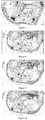
figures 5-7 respectively are the 7T MRI photos ofAS mouse group 30 min, 45 min and 120 min after being injected the contrast agent of the present invention; infigure 5 , where the white arrow points is AS lesion area; infigure 6 , where the white arrow points is multiple spot signal absent of AS lesion area; infigure 7 , where the white arrow points is signal recover of AS lesion area; -
figures 8-10 respectively are the 7T MRI photos ofAS mouse group 30 min, 45 min and 120 min after being injected magnetic ferriferrous oxide nanoparticles; infigure 8 , where the white arrow points is AS lesion area; infigure 9 , the AS lesion signal does not change at which the white arrow points, infigure 10 , the AS lesion signal does not change at which the white arrow points; -
figures 11-13 respectively are the 7T MRI photos ofnormal mouse group 30 min, 45 min and 120 min after being injected the contrast agent of the present invention; infigure 11 , where the white arrow points is normal artery; infigure 12 , where the white arrow points is normal artery, no signal changes; infigure 13 , where the white arrow points is normal artery, no signal changes; -
figures 14-16 respectively are the 7T MRI photos ofnormal mouse group 30 min, 45 min and 120 min after being injected magnetic ferriferrous oxide nanoparticles; infigure 14 , where the white arrow points is normal artery; infigure 15 , where the white arrow points is normal artery, no signal changes; infigure 16 , where the white arrow points is normal artery, no signal changes; -
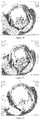
figures 17-20 respectively are the 3.0T MRI photos of AS New Zealand rabbit group 0 h, 1 h, 2 h, 3 h after being injected the contrast agent of the present invention; infigure 17 , where the white arrow points is AS lesion area; infigure 18 , where the white arrow points, the AS lesion area signal gradually disappear, the vascular wall is unclear; infigure 19 , where the white arrow points is signal spotted absent of AS lesion area; infigure 20 , where the white arrow points is signal recover of AS lesion area; -
figures 21-24 respectively are the 3.0T MRI photos of AS New Zealand rabbit group 0 h, 1 h, 2 h, 3 h after being injected magnetic ferriferrous oxide nanoparticles; infigure 21 , where the white arrow points is AS lesion area; infigure 22 , where the white arrow points is AS lesion area, no signal changes; infigure 23 , where the white arrow points is AS lesion area, no signal changes; infigure 24 , where the white arrow points is AS lesion area, no signal changes; -
figures 25-28 respectively are the 3.0T MRI photos of normal New Zealand rabbit group 0 h, 1 h, 2 h, 3 h after being injected the contrast agent of the present invention; infigure 25 , where the white arrow points is normal blood vessel; infigure 26 , where the white arrow points is normal blood vessel, no signal changes; infigure 27 , where the white arrow points is normal blood vessel, no signal changes; infigure 28 , where the white arrow points is normal blood vessel, no signal changes; -
figures 29-32 respectively are the 3.0T MRI photos of normal New Zealand rabbit group 0 h, 1 h, 2 h, 3 h after being injected magnetic ferriferrous oxide nanoparticles; infigure 29 , where the white arrow points is normal blood vessel; infigure 30 , where the white arrow points is normal blood vessel, no signal changes; infigure 31 , where the white arrow points is normal blood vessel, no signal changes; infigure 32 , where the white arrow points is normal blood vessel, no signal changes. - A targeting aptamer for atherosclerosis, the preparation method thereof is:
- 1) culturing smooth muscle cells: SD rats, either gender, the body weight is 150-200 g, age is 4-8 weeks; after intraperitoneal anesthesia, taking out thoracic aorta segment by sterile operation, placing same in a plate containing PBS solution for rinsing 3 times, after rinsing the clots clean, stripping the fibrofatty layer of the adventitia, then cutting the blood vessel open in longitudinal direction, striking off intima, i.e. endothelial cells, rapidly tearing down the inner layer and middle layer of the medial membrane, cutting same into 1 mm wide small strips, steeping same in PBS soltion containing blood serum, and planting the cut small tissue blocks on the culture bottle wall, putting same in an incubator at 37°C for about 2 h, taking out the culture bottle, adding a culture solution containing 20% fetal calf serum, then putting same back to the incubator for stationary culture for 4 days, after the cells overspread the bottom, performing digestion passage using 0.25% trypsin, once every 2-3 days, culturing the cells in a 6 well plate before the experiments;
- 2) culturing endothelial cell line: culturing endothelial cell line using high sugar DMEM medium containing 15% new-born calf serum in a CO2incubator with 5%, CO2 and saturated humidity at 37°C. Performing digestion passage using 0.25% trypsin, once every 2-3 days, culturing the cells in a 6 well plate before the experiments;
- 3) culturing THP-1 cells: culturing THP-1 cell line using RPMI1640 medium containing 10% new-born calf serum in a CO2 incubator with 5% CO2 and saturated humidity at 37°C; Performing passage using a direct separating bottle method, once every 2-3 days, culturing the cells in a 6 well plate before the experiments;
- 4) establishing a macrophage-derived foam cell model: culturing above-mentioned THP-1 cells in 6 well plate using RPMI1640 medium containing 10% new-born calf serum in an incubator with 5% CO2 at 37°C, before each experiment, using 100 nmol/L PMA to incubate the cells for 72 hours, inducing same to differentiate into macrophages, then incubating same in 80 mg/L oxLDL for 72 hours, and inducing same into foam cells;
- 5) constructing an FITC marked random ssDNA library: constructing a random ssDNA library with a length of 81nt using a biological method, the ssDNA library being as the nucleotide sequence of SEQ ID No:1, both ends of the sequence of SEQ ID No:1 are fixed sequences, 35 nucleotides in the middle is a random sequence; designing an upstream primer SEQ ID No:3 and a downstream primer SEQ ID No:4, wherein the upstream primer is marked with FITC and the downstream primer is marked with Biotin, the random ssDNA library is subject to a PCR amplification, the PCR amplification system is: ssDNA template 0.1 ug, 10 ×
PCR buffer 10 ul,MgCl 2 2 ul, dNTPs lul, the upstream primer represented by SEQ ID No:3 50 pmol, the downstream primer represented by SEQ ID No:4 50 pmol, Taq enzyme 2U, supplementing deionized water to 100 ul; the amplification condition is: pre-degeneration at 94°C for 3 minutes, degeneration at 94°C for 40 seconds, annealing at 72°C for 1 minute, extension at 72°C for 1 minute, finally, extension at 72°C for 7 minutes; the PRC product is dissolved in PBS solution after agarose electrophoresis, gel cutting and recovery, binding same with streptavidin magnesphere via a chain with Biotin; washing same withPBS solution 3 times, performing degeneration using 0.15mol/L NaOH at 37°C for 15 minutes, eluting the other chain with FITC marker from streptavidin magnesphere, neutralizing same with a little hydrochloric acid, adding 2 volumes of anhydrous ethanol, precipitating same in a -80°C refrigerator for 3 hours or over night, centrifuging same at 12000 rpm for 30 minutes, removing the supernatant, washing same with 70% ethanol once, dissolving same in DPBS solution after drying in the air, determining the ssDNA concentration using an ultraviolet spectrophotometer, and using same in the next cycle of screening; - 6) screening an oigonucleotide aptamer of macrophage-derived foam cells by SELEX: adding about 800 pmol random ssDNA library into a microcentrifuge tube, performing degeneration at 80°C for 10 minutes, then placing same on ice for annealing 10 minutes. Washing with 37°C washing buffer 6 times before screening of smooth muscle cells, macrophages, and macrophage-derived foam cells. Adding the library and 2 ml 37°C 1 × binding Buffer, in sequence, into the six well plate in which about 1 × 10⁶ smooth muscle cells and macrophages are cultured, incubating same for 30 minutes respectively, then transferring the supernatant into a six well plate in which about 1 × 10⁶ foam cells are cultured, incubating same for 30 minutes, removing the supernatant, washing same with 37°C washing buffer 6 times, washing off ssDNA which does not bind foam cells, then adding 800 ul eluant, performing heat denaturation at 80°C for 10 minutes, taking the supernatant and centrifuging same at 800 rpm for 5 minutes, discarding the small amount of precipitate of the lower layer, adding 2 volumes of pre-cooled anhydrous ethanol, precipitating same in a -80°C refrigerator for 3 hours or overnight, centrifuging same at 12000 rpm for 30 minutes, washing same with 70% ethanol once, drying same in the air, dissolving same in a small amount of TE or sterile water, determining the ssDNA concentration using an ultraviolet spectrophotometer, PCR amplifying same with TFITC and Biotin marked primers to double marked dsDNA for the next cycle of screening; repeating the above-montioned steps, screening for 18 cycles repeatedly, all the screening process is in a darkroom or under a dark condition; after performing each cycle of screening, performing a reverse screening to remove the ssDNA ingredients binding the six well plate, smooth muscle cells and macrophages; in order to enhance the specificity of the screening, adding yeast tRNA into the reaction as a competitive conjugate; after performing each cycle of screening, performing a reverse screening to remove the ssDNA ingredients binding the six well plate, smooth muscle cells and macrophages;
- 7) cloning and sequencing: after 18 cycles of SELEX screening, PCR amplifying ssDNA with upstream primer SEQ ID No:3 and upstream primer SEQ ID No:4, using the amplification product for 2% agarose electrophoresis, cutting the target band on a UV transmission reflectometer, cutting same up and putting into a centrifugal column of a high performance DNA purification and recovery kit, the centrifugal column is put into a 1.5 ml EP tube, centrifuging at 12000 rpm for 1 minute, discarding the centrifugal column, adding 1/10 volume of 3 mol/L NaAC (pH 5.2) and 2 volumes of pre-cooled anhydrous ethanol into the EP tube, mixing same evenly on a vortex, putting same in a -80°C refrigerator for 3 hours or overnight, ligating same with pGEM-T vector, then placing same on ice, picking a small amount of bacterium liquid with an inoculating loop and inoculating same on a LB plate without antibiotics, culturing same in an incubator at 37°C overnight, after monoclonal colonies appear, picking a clone with an inoculating loop and putting same in a tube containing 2 ml LB medium, shaking the tube in a constant temperature vibrator at 37°C and 150 rpm overnight, taking 0.5 ml bacterium liquid and adding same in a 250 ml conical flask containing 50 ml LB medium, putting the conical flask in a constant temperature vibrator and shaking at 37°C and 150 rpm until the absorbance value at 600 nm of the bacterium liquid being about 0.35, placing the bacterium liquid on ice for 10 minutes, centrifuge same in a 50 ml centrifuge tube at 4000 rpm for 5 minutes, removing the supernatant and sucking the residual liquid dry, adding 16 ml solution A, gently resuspending the lower layer cells and placing same on ice for 15 minutes, centrifuging same at 4°C and 4000 rpm for 5 minutes, collecting the cells, adding 4 ml solution A and gently resuspending the cells, subpackaging the cells at 200 ul/tube, storing same at -70°C, and making same into competent cells, placing 200 ul competent cells on ice for thawing, and then adding 2 ul ligation product, gently mixing the mixture evenly, placing same on ice for 20 minutes, heat shocking same in 42°C water bath for 90 seconds, placing same on ice for 2 minutes, adding 500 ul 37°C preheated LB medium without antibiotics, vibrating and culturing same at 37°C and 150 rpm for 1.5 hours, taking 200 ul bacterium liquid, evenly coating same on a prepared blue-white spot screen plate with a spreader, culturing same at 37°C for 16-18 hours, placing the plate with colonies in a 4°C refrigerator for 2 hour, making the blue-white spots develop fully, randomly picking 45 white colonies, putting same into LB liquid medium containing 2 ml ampicillin, after shaking same in a constant temperature vibrator at 37°C and 150 rpm overnight, taking 5 ul bacterium liquid for each clone for performing a PCR identification; observing by means of a fluorescence microscope, picking out a specific monoclone; sequencing the pickied specific aptamer;
- 8) identification of specific binding between the aptamer and macrophage-derived foam cells: respectively washing 1 × 10⁶ THP-1 macrophages and smooth muscle cells, and 1 × 10⁶ macrophage-derived foam cells which are cultured in a six well plate with 37°C PBS three times, then adding 37°C DPBS solution containing 200 pmol FITC marked aptamer and 1ug yeast tRNA, incubating same at 37°C in an incubator for 30 minutes, washing same with 37°C PBS three times after removing supernatant, observing and shooting same by means of a fluorescence microscope, and the targeting aptamer fragment for atherosclerosis is obtained.
- Bleeding an atherosclerosis model rabbit to death, taking out the aorta from ascending aorta to the arterial branch of arteria iliaca communis, washing out blood with PBS, fixing same with 10% formalin, performing conventional dehydration, imbedding same with paraffin, serial sectioning same with a Leitz1560 section cutter.
- taking an artery section having plaques and a normal artery section, conventional dewaxing same with xylene to water, dropwise adding 3% hydrogen peroxide, keeping same under room temperature for 5-10 minutes to inactivate endogenous enzymes, washing same with distilled
water 3 times. Steeping the sections in 0.01 M citrate buffer (pH 6.0), heating same to boiling with an electric oven or microwave oven, after 5-10 minutes, repeating 1-2 times to repair antigens. After cool down, washing with PBS (pH 7.2-7.6) 1-2 times. Dropwise adding 50 pmol FITC marked targeting aptamer for atherosclerosis of the present invention and 1 ug pmol yeast tRNA, incubating same in an incubator at 37°C for 30 minutes, washing same with deionized water once for 5 minutes, than washing same withPBS 2 times, eachtime 5 minutes, observing and shooting the binding between the aptamer and atherosclerosis plaques or normal artery by means of a fluorescence microscope. - The results show that all the targeting aptamers for atherosclerosis prepared by the present invention specifically bind macrophage-derived foam cells, but almost never bind THP-1 macrophages, blood vessel smooth muscle cells, and endothelial cells.
- Preparing a MRI targeting nano-contrast agent for atherosclerosis using the targeting aptamer for atherosclerosis of example 1, the particular steps are:
- a) preparation of a silane coated ferriferrous oxide nanoparticle vector: mixing ferric trichloride and ferrous sulfate at a molar ratio of 2 : 1, using nitrogen for protection, the temperature is set at 30°C when refluxing, slowly dropwise adding ammonia water during the refluxing to pH > 9, after curing for 30 minutes, raising the temperature to 60°C, continuously refluxing for 1 hour, then separating out ferriferrous oxide with a magnet, and washing same with deionized water and anhydrous ethanol in sequence, each 3 times, to obtain ferriferrous oxide; adding 3-aminopropylethoxysilane into deionized water , stirring same under room temperature, dropwise adding glacial acetic acid, adjusting pH to pH ≈ 5; placing alcohol washed Fe3O4 in a flask, adding anhydrous ethanol, then adding 3-aminopropylethoxysilane at pH ≈ 5 (wherein the mass ratio of 3-aminopropylethoxysilane and Fe3O4 is 5 : 1), using nitrogen for protection, reflux condensation, stirring same at 60°C for 5 hours, stop heating, further stirring same to room temperature, taking the reaction liquid, performing separation with a magnet, removing the supernatant, washing same with deionized water and anhydrous ethanol in sequence, each 3 times, and the silane coated ferriferrous oxide nanoparticle vector is obtained;
- b) synthesis of a contrast agent: washing the silane coated ferriferrous oxide nanoparticle vector obtained from step a) with water and an alcohol alternately, finally dissolving same in PBS solutions at pH = 7.4; then adding the PBS solution at pH = 7.4 to dissolve the targeting aptamer for atherosclerosis represented by SEQ ID No:2, and adding the above-mentioned vector solution, and finally adding a glutaral, placing same on a shaking table for vibration at 100 r/min and 25°C for 1 hour; separating products with a magnet, removing the supernatant, and washing the products with the PBS solution, repeating 3 times.
- Preparing a MRI targeting nano-contrast agent for atherosclerosis using the targeting aptamer for atherosclerosis represented by SEQ ID No:2, the particular steps are:
- 1) preparation of a silane coated ferriferrous oxide nanoparticle vector: mixing ferric trichloride and ferrous sulfate at a molar ratio of 1.5 : 1, using nitrogen for protection, the temperature is set at 27°C when refluxing, slowly dropwise adding ammonia water during the refluxing to pH > 9, after curing for 40 minutes, raising the temperature to 60°C, continuously refluxing for 1.5 hour, then separating out ferriferrous oxide with a magnet, and washing same with deionized water and anhydrous ethanol in sequence, each 2 times, to obtain ferriferrous oxide; adding 3-aminopropylethoxysilane into deionized water , stirring same under room temperature, dropwise adding glacial acetic acid, adjusting pH to pH ≈ 5; placing alcohol washed Fe3O4 in a flask, adding anhydrous ethanol, then adding 3-aminopropylethoxysilane at pH ≈ 5 (wherein the mass ratio of 3-aminopropylethoxysilane and Fe3O4 is 3 : 1), using nitrogen for protection, reflux condensation, stirring same at 60°C for 3 hours, stop heating, further stirring same to room temperature, taking the reaction liquid, performing separation with a magnet, removing the supernatant, washing same with deionized water and anhydrous ethanol in sequence, each 2 times, and the silane coated ferriferrous oxide nanoparticle vector is obtained;
- 2) synthesis of a contrast agent: washing the silane coated ferriferrous oxide nanoparticle vector obtained from step 1) with water and an alcohol alternately, finally dissolving same in PBS solutions at pH = 7.4; then adding the PBS solution at pH = 7.4 to dissolve the targeting aptamer for atherosclerosis represented by SEQ ID No:2, and adding the above-mentioned vector solution, and finally adding a glutaral, placing same on a shaking table for vibration at 100 r/minand 25°C for 1 hour; separating products with a magnet, removing the supernatant, and washing the products with the PBS solution, repeating 3 times.
- Preparing a MRI targeting nano-contrast agent for atherosclerosis using the targeting aptamer for atherosclerosis represented by SEQ ID No:2, the particular steps are:
- 1) preparation of a silane coated ferriferrous oxide nanoparticle vector: mixing ferric trichloride and sulphates of iron at a molar ratio of 2.5 : 1, using nitrogen for protection, the temperature is set at 35°C when refluxing, slowly dropwise adding ammonia water during the refluxing to pH > 9, after curing for 20 minutes, raising the temperature to 60°C, continuously refluxing for 1.5 hour, then separating out ferriferrous oxide with a magnet, and washing same with deionized water and anhydrous ethanol in sequence, each 4 times, to obtain ferriferrous oxide; adding 3-aminopropylethoxysilane into deionized water, stirring same under room temperature, dropwise adding glacial acetic acid, adjusting pH to pH ≈ 5; placing alcohol washed Fe3O4 in a flask, adding anhydrous ethanol, then adding 3-aminopropylethoxysilane at pH ≈ 5 (wherein the mass ratio of 3-aminopropylethoxysilane and Fe3O4 is 8 : 1), using nitrogen for protection, reflux condensation, stirring same at 65°C for 6 hours, stop heating, further stirring same to room temperature, taking the reaction liquid, performing separation with a magnet, removing the supernatant, washing same with deionized water and anhydrous ethanol in sequence, each 4 times, and the silane coated ferriferrous oxide nanoparticle vector is obtained;
- 2) synthesis of a contrast agent: washing the silane coated ferriferrous oxide nanoparticle vector obtained from step 1) with water and an alcohol alternately, finally dissolving same in PBS solutions at pH = 7.4; then adding the PBS solution at pH = 7.4 to dissolve the targeting aptamer for atherosclerosis represented by SEQ ID No:2, and adding the above-mentioned vector solution, and finally adding a glutaral, placing same on a shaking table for vibration at 100 r/minand 25°C for 1 hour; separating products with a magnet, removing the supernatant, and washing the products with the PBS solution, repeating 3 times.
- Polyacrylamide gel electrophoresis analysis of the contrast agent of the present invention: in
figure 1 , 1: 100 bp Mark; 2: 5'-NH2-targeting aptamer; 3-6: the final products of 0.1, 0.2, 0.5, 1 mL 3-aminopropyltriethoxysilane coated Fe3O4 coupling with 33 µg 5'-NH2-targeting aptamer; 7: 3-aminopropyltriethoxysilane coated Fe3O4; the results offigure 1 show that the silane coated ferriferrous oxide nanoparticle vector in the contrast agent of the present invention can effectively connect to the targeting aptamer for atherosclerosis. - The contrast agent of the present invention is detected by a vibration sample magnetometer and magnetic resonance, the detection result show that the saturated magnetization intensity of the vector is 108 emu·g-1 Fe, the relaxation rate is 0.2842 × 10⁶ mol-1·s-1. The magnetization curve is a single curve passing through the origin point, indicating that the contrast agent of the present invention has superparamagnetism.
- Respectively injecting the targeting nano-contrast agent for atherosclerosis and magnetic ferriferrous oxide nanoparticles to AS mouse group and normal mouse group for magnetic resonance imaging in vivo, the results are in
figures 5-16 . The results offigures 5-16 show that in AS mouse group, at 30 minutes after injecting the targeting nano-contrast agent for atherosclerosis, the signal strength of AS lesion area shows decreasing trend, the vascular walls are not smooth, showing multiple spot signal defects; at 45 minutes, the signal strength decreases to the lowest, after that the signal begin to enhance, until after 2 hours, the signal strength recovers to almost the same as that before the injection. In the normal animal group, after injecting the targeting nano-contrast agent for atherosclerosis, the signal strength of blood vessel walls has no significant change all the time. - Respectively injecting the targeting nano-contrast agent for atherosclerosis and magnetic ferriferrous oxide nanoparticles to AS New Zealand rabbit group and normal New Zealand rabbit group for magnetic resonance imaging in vivo, the results are in
figures 17-32. Figures 17-32 show that in AS New Zealand rabbit group, at 1 hour after injecting the target nano-contrast agent for atherosclerosis, the signal strength of AS lesion area shows decreasing trend, the vascular walls are not smooth, showing spotted signal defects, at 2 hours, the signal strength decreases to the lowest, after that the signal begin to enhance, until after 3 hours, the signal strength recovers to almost the same as that before the injection. In the normal New Zealand rabbit group, the signal strength of blood vessel walls has no significant change all the time. - Above-mentioned embodiments only are preferred embodiments of the present invention, they cannot limit the scope of protection of the present invention, furthermore, all the non-substantial modifications and substitutions made by a person skilled in the art based on the present invention belong the scope of protection of the present invention.


Claims (14)
- A MRI targeting nano-contrast agent for atherosclerosis, characterized in that the MRI targeting nano-contrast agent for atherosclerosis comprises a targeting aptamer for atherosclerosis and a vector which is connected to the targeting aptamer for atherosclerosis, wherein the targeting aptamer for atherosclerosis is a targeting aptamer fragment for atherosclerosis obtained through screening of macrophage-derived foam cells together with reverse screening of smooth muscle cells, endothelial cells, and THP-1 cells using a SELEX method.
- The targeting aptamer for atherosclerosis according to claim 1, characterized in that a nucleotide sequence of said targeting aptamer fragment for atherosclerosis is SEQ ID No:1.
- The targeting aptamer for atherosclerosis according to claim 2, characterized in that the nucleotide sequence of said targeting aptamer fragment for atherosclerosis is a sequence which has at least 80% homology with SEQ ID No:2.
- The targeting aptamer for atherosclerosis according to claim 3, characterized in that the nucleotide sequence of said targeting aptamer fragment for atherosclerosis is SEQ ID No:2.
- The MRI targeting nano-contrast agent for atherosclerosis according to any one of claims 1-4, characterized in that said vector is a silane coated ferriferrous oxide nanoparticle vector; said targeting aptamer for atherosclerosis is covalently connected to the silane in said vector via glutaral as a bridge.
- The MRI targeting nano-contrast agent for atherosclerosis according to claim 5, characterized in that the mass ratio of the vector and the targeting aptamer for atherosclerosis is (5-10) : 1.
- The MRI targeting nano-contrast agent for atherosclerosis according to claim 5, characterized in that the ratio of the total mass of the ferriferrous oxide nanoparticles and the mass of the silane is 1 : (3-8) in said vector.
- The MRI targeting nano-contrast agent for atherosclerosis according to claim 7, characterized in that the mean particle size of said ferriferrous oxide nanoparticles is 10-15 nanometer and the total particle size of the silane coated ferriferrous oxide nanoparticles is 30-100 nanometer.
- The MRI targeting nano-contrast agent for atherosclerosis according to claim 5, characterized in that the magnetic parameters thereof are T2 relaxation rate of 0.2842 × 10⁶ mol-1·s-1, and the mass saturated magnetization intensity of 108 emu·g-1 Fe.
- The MRI targeting nano-contrast agent for atherosclerosis according to claim 5, characterized in that said silane is 3-aminopropylethoxysilane.
- A method for preparing the MRI targeting nano-contrast agent for atherosclerosis of claim 5, characterized in that the method comprises following steps:1) preparation of a silane coated ferriferrous oxide nanoparticle vector: using ferric chloride and ferrous sulfate as raw materials, refluxing same under a nitrogen atmosphere, after the end of the refluxing, separating out ferriferrous oxide by using a magnet, and refluxing the ferriferrous oxide with silane, after the end of the reaction, obtaining the silane coated ferriferrous oxide nanoparticle vector by a separation using a magnet;2) synthesis of a contrast agent: washing the silane coated ferriferrous oxide nanoparticle vector obtained from step 1) with water and an alcohol alternately, dissolving the above-mentioned targeting aptamer and silane coated ferriferrous oxide nanoparticle vector in PBS solutions respectively, then adding the PBS solution of the targeting aptamer for atherosclerosis into the PBS solution of the silane coated ferriferrous oxide nanoparticle vector, and adding a glutaral, placing same on a shaking table for vibration; separating products with a magnet, removing the supernatant, and washing same with the PBS solution.
- The method for preparing the MRI targeting nano-contrast agent for atherosclerosis according to claims 11, characterized in that the step 1) is carried out according to the flowing steps:a) preparation of ferriferrous oxide: mixing ferric trichloride and ferrous sulfate at a molar ratio of (1.5-2.5) : 1, the temperature is set at 27-35°C when refluxing, slowly dropwise adding ammonia water during the refluxing to pH > 9, after curing for 20-40 minutes, raising the temperature to 60°C, continuously refluxing same for 0.5-1.5 hours, then separating out ferriferrous oxide with a magnet, and washing same with deionized water and anhydrous ethanol, each 2-4 times, to obtain ferriferrous oxide;b) preliminary treatment of silane, adding silane into deionized water and stirring same, and dropwise adding glacial acetic acid, adjusting pH to 4-6;c) coating: adding anhydrous ethanol to the ferriferrous oxide of step a), then adding the silane solution of step b), condensation and reflux, stirring same for 3-6 hours at 55-65°C, stopping the heating, further stirring same down to room temperature, taking the reaction liquid, separating same with a magnet, removing the supernatant, adding deionized water and anhydrous ethanol for washing in sequence, each repeating 2-4 times.
- The method for preparing the MRI targeting nano-contrast agent for atherosclerosis according to claim 11, characterized in that in the step 2), the pH of the employed PBS solution is 7.4.
- The method for preparing the MRI targeting nano-contrast agent for atherosclerosis according to claim 13, characterized in that in the step 2), the frequency of said shaking table is 100 r/min, the temperature is 25°C, and the vibration time is 0.5-1.5 hours.
Applications Claiming Priority (2)
| Application Number | Priority Date | Filing Date | Title |
|---|---|---|---|
| CN201310237217.XA CN103290019B (en) | 2013-06-14 | 2013-06-14 | Atherosclerosis targeting aptamer as well as preparation method and application thereof |
| PCT/CN2013/001248 WO2014198010A1 (en) | 2013-06-14 | 2013-10-14 | Targeting aptamer for atherosclerosis and preparation method and application thereof |
Publications (3)
| Publication Number | Publication Date |
|---|---|
| EP3009512A1 true EP3009512A1 (en) | 2016-04-20 |
| EP3009512A4 EP3009512A4 (en) | 2017-02-22 |
| EP3009512B1 EP3009512B1 (en) | 2020-05-13 |
Family
ID=49091550
Family Applications (1)
| Application Number | Title | Priority Date | Filing Date |
|---|---|---|---|
| EP13886916.9A Active EP3009512B1 (en) | 2013-06-14 | 2013-10-14 | Targeting aptamer for atherosclerosis and preparation method and application thereof |
Country Status (5)
| Country | Link |
|---|---|
| US (1) | US10350313B2 (en) |
| EP (1) | EP3009512B1 (en) |
| CN (1) | CN103290019B (en) |
| AU (1) | AU2013392536B2 (en) |
| WO (1) | WO2014198010A1 (en) |
Families Citing this family (10)
| Publication number | Priority date | Publication date | Assignee | Title |
|---|---|---|---|---|
| CN103290019B (en) * | 2013-06-14 | 2014-03-12 | 严鹏科 | Atherosclerosis targeting aptamer as well as preparation method and application thereof |
| CN103627701B (en) * | 2013-12-04 | 2015-07-15 | 武汉大学 | Screening of aptamers specially targeting gastric cancer cells and aptamers obtained after screening |
| CN104974998A (en) * | 2014-04-11 | 2015-10-14 | 中国中医科学院中医临床基础医学研究所 | DNA double-chain separation method used for aptamer screening, aptamer screening method and new aptamer |
| CN104789696B (en) * | 2015-03-20 | 2017-07-11 | 中国科学院南海海洋研究所 | A kind of DNA aptamers and its screening technique and application for being used to detect grouper irido virus infection |
| CN104789570B (en) * | 2015-03-20 | 2018-07-06 | 中国科学院南海海洋研究所 | A kind of DNA aptamers for being used to detect grouper irido virus infection and its screening technique and application |
| JP2019531706A (en) * | 2016-08-12 | 2019-11-07 | インブイティ・インコーポレイテッドInvuity, Inc. | Tissue-specific markers for tissue localization and visualization before and during surgery |
| CN112137961A (en) * | 2020-09-30 | 2020-12-29 | 严鹏科 | Rapamycin composition and preparation method thereof |
| CN114874972B (en) * | 2022-04-19 | 2023-05-09 | 四川大学 | A kind of in vitro foam cell model and its construction method |
| CN115369070A (en) * | 2022-09-05 | 2022-11-22 | 西南交通大学 | In vitro atherosclerotic plaque model, preparation method and application |
| CN121674406B (en) * | 2026-02-11 | 2026-04-21 | 三峡大学 | Aptamer targeting M1 type macrophage and screening method and application thereof |
Family Cites Families (11)
| Publication number | Priority date | Publication date | Assignee | Title |
|---|---|---|---|---|
| US6229002B1 (en) * | 1995-06-07 | 2001-05-08 | Nexstar Pharmaceuticlas, Inc. | Platelet derived growth factor (PDGF) nucleic acid ligand complexes |
| US7727969B2 (en) * | 2003-06-06 | 2010-06-01 | Massachusetts Institute Of Technology | Controlled release nanoparticle having bound oligonucleotide for targeted delivery |
| KR101005561B1 (en) * | 2008-06-21 | 2011-01-05 | 한국생명공학연구원 | Multimode Contrast Method Using Nanoemulsion Consisting of Optical Nanoparticles and Perfluorocarbons |
| US20120107229A1 (en) * | 2009-04-15 | 2012-05-03 | Xuefei Huang | Novel nano-probes for molecular imaging and targeted therapy of diseases |
| AR076361A1 (en) * | 2009-04-21 | 2011-06-08 | Immunoligtht Llc | PHARMACEUTICAL COMPOSITION KIT NON-INVASIVE ASCENDING ENERGY CONVERSION METHODS AND SYSTEMS FOR IN-SITU PHOTOBIOMODULATION |
| GB201102189D0 (en) * | 2011-02-08 | 2011-03-23 | King S College London | Materials and methods relating to cardiovascular imaging |
| EP2691401A4 (en) * | 2011-03-30 | 2014-05-21 | Luna Innovations Inc | BIOMARKER-TARGETING CONTRAST AGENTS AND THEIR USE IN MAGNETIC RESONANCE IMAGING FOR ATHEROSCLEROSIS PLATE DETECTION |
| CN102552944B (en) * | 2012-01-18 | 2013-03-27 | 中南大学 | Nasopharyngeal carcinoma targeted magnetic resonance contrast agent and preparation method thereof |
| CN102580120B (en) * | 2012-03-21 | 2014-04-02 | 海南医学院附属医院 | Targeted magnetic resonance imaging (MRI) contrast agent and preparation method thereof |
| CN102772808B (en) * | 2012-07-02 | 2017-03-01 | 重庆医科大学 | A kind of multi-modality imaging microbubble construction, Preparation method and use |
| CN103290019B (en) * | 2013-06-14 | 2014-03-12 | 严鹏科 | Atherosclerosis targeting aptamer as well as preparation method and application thereof |
-
2013
- 2013-06-14 CN CN201310237217.XA patent/CN103290019B/en not_active Expired - Fee Related
- 2013-10-14 WO PCT/CN2013/001248 patent/WO2014198010A1/en not_active Ceased
- 2013-10-14 AU AU2013392536A patent/AU2013392536B2/en not_active Ceased
- 2013-10-14 US US14/763,815 patent/US10350313B2/en not_active Expired - Fee Related
- 2013-10-14 EP EP13886916.9A patent/EP3009512B1/en active Active
Non-Patent Citations (1)
| Title |
|---|
| See references of WO2014198010A1 * |
Also Published As
| Publication number | Publication date |
|---|---|
| AU2013392536B2 (en) | 2018-02-01 |
| EP3009512B1 (en) | 2020-05-13 |
| US20150359910A1 (en) | 2015-12-17 |
| AU2013392536A1 (en) | 2016-01-28 |
| WO2014198010A1 (en) | 2014-12-18 |
| EP3009512A4 (en) | 2017-02-22 |
| CN103290019B (en) | 2014-03-12 |
| US10350313B2 (en) | 2019-07-16 |
| CN103290019A (en) | 2013-09-11 |
Similar Documents
| Publication | Publication Date | Title |
|---|---|---|
| EP3009512B1 (en) | Targeting aptamer for atherosclerosis and preparation method and application thereof | |
| CN103446597B (en) | For the preparation method of atherosclerosis vulnerable plaque MRI/PET bimodal molecular imaging image probe | |
| Song et al. | Multimodal SPION-CREKA peptide based agents for molecular imaging of microthrombus in a rat myocardial ischemia-reperfusion model | |
| CN103055329B (en) | Manufacturing method of targeted magnetic nanometer probe for early diagnosis of atherosclerosis vulnerable plaques | |
| CN101365495B (en) | Compositions containing magnetic iron oxide particles and use of said compositions in magnetic particle imaging | |
| Sun et al. | Targeted trapping of endogenous endothelial progenitor cells for myocardial ischemic injury repair through neutrophil-mediated SPIO nanoparticle-conjugated CD34 antibody delivery and imaging | |
| CA2869698A1 (en) | Magnetic nanoparticle dispersion, its preparation and diagnostic and therapeutic use | |
| CN101378785A (en) | Compositions comprising magnetic iron oxide particles and use thereof in medical imaging | |
| Shevtsov et al. | Detection of experimental myocardium infarction in rats by MRI using heat shock protein 70 conjugated superparamagnetic iron oxide nanoparticle | |
| CN108949750A (en) | Extract the method and kit of faeces DNA | |
| CN108265055B (en) | DNA nucleic acid aptamer that specifically binds to human breast cancer cell MCF-7 and its application | |
| CN111205411B (en) | Blood vessel and tumor enhancement macromolecule magnetic resonance contrast agent and preparation method and application thereof | |
| TW200928353A (en) | Imaging nucleic acid binding proteins | |
| CN102234679B (en) | Preparation method of tumor detection nanoprobe | |
| CN107096042A (en) | Magnetic resonance molecular probe for detecting early hepatocyte cancer | |
| Chaabane et al. | Contrast enhancement in atherosclerosis development in a mouse model: in vivo results at 2 Tesla | |
| Hartung et al. | Labeling of macrophages using bacterial magnetosomes and their characterization by magnetic resonance imaging | |
| CN112062843B (en) | Human anti-atherosclerosis single chain antibody and its application | |
| CN106109495B (en) | Method for dynamically tracing infiltration of tumor cells into sciatic nerve in vivo | |
| CN101347625B (en) | Nuclear magnetic resonance thrombus targeting contrast agent and preparation method thereof | |
| CN119619307A (en) | Ultrasonic molecular probe, preparation method and use thereof in preparing drugs for diagnosing and treating atherosclerotic plaques | |
| Goldhaber et al. | DEEP VEIN THROMBOSIS, PULMONARY EMBOLISM | |
| CN102091027B (en) | Method for preparing antitumor small interfering RNA carrying magnetic nano probe | |
| CN117838892A (en) | Superparamagnetism iron oxide nanoparticle coupled with CRT (cathode ray tube) and application thereof | |
| CN113337511A (en) | Aptamer QQ-4 targeting human highly invasive choroidal melanoma and application |
Legal Events
| Date | Code | Title | Description |
|---|---|---|---|
| PUAI | Public reference made under article 153(3) epc to a published international application that has entered the european phase |
Free format text: ORIGINAL CODE: 0009012 |
|
| 17P | Request for examination filed |
Effective date: 20160112 |
|
| AK | Designated contracting states |
Kind code of ref document: A1 Designated state(s): AL AT BE BG CH CY CZ DE DK EE ES FI FR GB GR HR HU IE IS IT LI LT LU LV MC MK MT NL NO PL PT RO RS SE SI SK SM TR |
|
| AX | Request for extension of the european patent |
Extension state: BA ME |
|
| DAX | Request for extension of the european patent (deleted) | ||
| A4 | Supplementary search report drawn up and despatched |
Effective date: 20170125 |
|
| RIC1 | Information provided on ipc code assigned before grant |
Ipc: A61K 49/12 20060101ALI20170119BHEP Ipc: G01N 24/08 20060101ALI20170119BHEP Ipc: C12Q 1/68 20060101ALI20170119BHEP Ipc: C12N 15/115 20100101AFI20170119BHEP |
|
| STAA | Information on the status of an ep patent application or granted ep patent |
Free format text: STATUS: REQUEST FOR EXAMINATION WAS MADE |
|
| STAA | Information on the status of an ep patent application or granted ep patent |
Free format text: STATUS: EXAMINATION IS IN PROGRESS |
|
| 17Q | First examination report despatched |
Effective date: 20190124 |
|
| GRAJ | Information related to disapproval of communication of intention to grant by the applicant or resumption of examination proceedings by the epo deleted |
Free format text: ORIGINAL CODE: EPIDOSDIGR1 |
|
| STAA | Information on the status of an ep patent application or granted ep patent |
Free format text: STATUS: GRANT OF PATENT IS INTENDED |
|
| GRAP | Despatch of communication of intention to grant a patent |
Free format text: ORIGINAL CODE: EPIDOSNIGR1 |
|
| INTG | Intention to grant announced |
Effective date: 20191217 |
|
| GRAS | Grant fee paid |
Free format text: ORIGINAL CODE: EPIDOSNIGR3 |
|
| GRAA | (expected) grant |
Free format text: ORIGINAL CODE: 0009210 |
|
| STAA | Information on the status of an ep patent application or granted ep patent |
Free format text: STATUS: THE PATENT HAS BEEN GRANTED |
|
| RIN1 | Information on inventor provided before grant (corrected) |
Inventor name: YAN, PENGKE |
|
| AK | Designated contracting states |
Kind code of ref document: B1 Designated state(s): AL AT BE BG CH CY CZ DE DK EE ES FI FR GB GR HR HU IE IS IT LI LT LU LV MC MK MT NL NO PL PT RO RS SE SI SK SM TR |
|
| REG | Reference to a national code |
Ref country code: GB Ref legal event code: FG4D |
|
| REG | Reference to a national code |
Ref country code: CH Ref legal event code: EP |
|
| REG | Reference to a national code |
Ref country code: DE Ref legal event code: R096 Ref document number: 602013069152 Country of ref document: DE |
|
| REG | Reference to a national code |
Ref country code: AT Ref legal event code: REF Ref document number: 1270321 Country of ref document: AT Kind code of ref document: T Effective date: 20200615 |
|
| REG | Reference to a national code |
Ref country code: LT Ref legal event code: MG4D |
|
| REG | Reference to a national code |
Ref country code: NL Ref legal event code: MP Effective date: 20200513 |
|
| PG25 | Lapsed in a contracting state [announced via postgrant information from national office to epo] |
Ref country code: IS Free format text: LAPSE BECAUSE OF FAILURE TO SUBMIT A TRANSLATION OF THE DESCRIPTION OR TO PAY THE FEE WITHIN THE PRESCRIBED TIME-LIMIT Effective date: 20200913 Ref country code: PT Free format text: LAPSE BECAUSE OF FAILURE TO SUBMIT A TRANSLATION OF THE DESCRIPTION OR TO PAY THE FEE WITHIN THE PRESCRIBED TIME-LIMIT Effective date: 20200914 Ref country code: GR Free format text: LAPSE BECAUSE OF FAILURE TO SUBMIT A TRANSLATION OF THE DESCRIPTION OR TO PAY THE FEE WITHIN THE PRESCRIBED TIME-LIMIT Effective date: 20200814 Ref country code: NO Free format text: LAPSE BECAUSE OF FAILURE TO SUBMIT A TRANSLATION OF THE DESCRIPTION OR TO PAY THE FEE WITHIN THE PRESCRIBED TIME-LIMIT Effective date: 20200813 Ref country code: FI Free format text: LAPSE BECAUSE OF FAILURE TO SUBMIT A TRANSLATION OF THE DESCRIPTION OR TO PAY THE FEE WITHIN THE PRESCRIBED TIME-LIMIT Effective date: 20200513 Ref country code: SE Free format text: LAPSE BECAUSE OF FAILURE TO SUBMIT A TRANSLATION OF THE DESCRIPTION OR TO PAY THE FEE WITHIN THE PRESCRIBED TIME-LIMIT Effective date: 20200513 Ref country code: LT Free format text: LAPSE BECAUSE OF FAILURE TO SUBMIT A TRANSLATION OF THE DESCRIPTION OR TO PAY THE FEE WITHIN THE PRESCRIBED TIME-LIMIT Effective date: 20200513 |
|
| PG25 | Lapsed in a contracting state [announced via postgrant information from national office to epo] |
Ref country code: BG Free format text: LAPSE BECAUSE OF FAILURE TO SUBMIT A TRANSLATION OF THE DESCRIPTION OR TO PAY THE FEE WITHIN THE PRESCRIBED TIME-LIMIT Effective date: 20200813 Ref country code: RS Free format text: LAPSE BECAUSE OF FAILURE TO SUBMIT A TRANSLATION OF THE DESCRIPTION OR TO PAY THE FEE WITHIN THE PRESCRIBED TIME-LIMIT Effective date: 20200513 Ref country code: LV Free format text: LAPSE BECAUSE OF FAILURE TO SUBMIT A TRANSLATION OF THE DESCRIPTION OR TO PAY THE FEE WITHIN THE PRESCRIBED TIME-LIMIT Effective date: 20200513 Ref country code: HR Free format text: LAPSE BECAUSE OF FAILURE TO SUBMIT A TRANSLATION OF THE DESCRIPTION OR TO PAY THE FEE WITHIN THE PRESCRIBED TIME-LIMIT Effective date: 20200513 |
|
| REG | Reference to a national code |
Ref country code: AT Ref legal event code: MK05 Ref document number: 1270321 Country of ref document: AT Kind code of ref document: T Effective date: 20200513 |
|
| PG25 | Lapsed in a contracting state [announced via postgrant information from national office to epo] |
Ref country code: NL Free format text: LAPSE BECAUSE OF FAILURE TO SUBMIT A TRANSLATION OF THE DESCRIPTION OR TO PAY THE FEE WITHIN THE PRESCRIBED TIME-LIMIT Effective date: 20200513 Ref country code: AL Free format text: LAPSE BECAUSE OF FAILURE TO SUBMIT A TRANSLATION OF THE DESCRIPTION OR TO PAY THE FEE WITHIN THE PRESCRIBED TIME-LIMIT Effective date: 20200513 |
|
| PG25 | Lapsed in a contracting state [announced via postgrant information from national office to epo] |
Ref country code: DK Free format text: LAPSE BECAUSE OF FAILURE TO SUBMIT A TRANSLATION OF THE DESCRIPTION OR TO PAY THE FEE WITHIN THE PRESCRIBED TIME-LIMIT Effective date: 20200513 Ref country code: AT Free format text: LAPSE BECAUSE OF FAILURE TO SUBMIT A TRANSLATION OF THE DESCRIPTION OR TO PAY THE FEE WITHIN THE PRESCRIBED TIME-LIMIT Effective date: 20200513 Ref country code: EE Free format text: LAPSE BECAUSE OF FAILURE TO SUBMIT A TRANSLATION OF THE DESCRIPTION OR TO PAY THE FEE WITHIN THE PRESCRIBED TIME-LIMIT Effective date: 20200513 Ref country code: SM Free format text: LAPSE BECAUSE OF FAILURE TO SUBMIT A TRANSLATION OF THE DESCRIPTION OR TO PAY THE FEE WITHIN THE PRESCRIBED TIME-LIMIT Effective date: 20200513 Ref country code: RO Free format text: LAPSE BECAUSE OF FAILURE TO SUBMIT A TRANSLATION OF THE DESCRIPTION OR TO PAY THE FEE WITHIN THE PRESCRIBED TIME-LIMIT Effective date: 20200513 Ref country code: CZ Free format text: LAPSE BECAUSE OF FAILURE TO SUBMIT A TRANSLATION OF THE DESCRIPTION OR TO PAY THE FEE WITHIN THE PRESCRIBED TIME-LIMIT Effective date: 20200513 Ref country code: IT Free format text: LAPSE BECAUSE OF FAILURE TO SUBMIT A TRANSLATION OF THE DESCRIPTION OR TO PAY THE FEE WITHIN THE PRESCRIBED TIME-LIMIT Effective date: 20200513 Ref country code: ES Free format text: LAPSE BECAUSE OF FAILURE TO SUBMIT A TRANSLATION OF THE DESCRIPTION OR TO PAY THE FEE WITHIN THE PRESCRIBED TIME-LIMIT Effective date: 20200513 |
|
| REG | Reference to a national code |
Ref country code: DE Ref legal event code: R097 Ref document number: 602013069152 Country of ref document: DE |
|
| PG25 | Lapsed in a contracting state [announced via postgrant information from national office to epo] |
Ref country code: PL Free format text: LAPSE BECAUSE OF FAILURE TO SUBMIT A TRANSLATION OF THE DESCRIPTION OR TO PAY THE FEE WITHIN THE PRESCRIBED TIME-LIMIT Effective date: 20200513 Ref country code: SK Free format text: LAPSE BECAUSE OF FAILURE TO SUBMIT A TRANSLATION OF THE DESCRIPTION OR TO PAY THE FEE WITHIN THE PRESCRIBED TIME-LIMIT Effective date: 20200513 |
|
| PLBE | No opposition filed within time limit |
Free format text: ORIGINAL CODE: 0009261 |
|
| STAA | Information on the status of an ep patent application or granted ep patent |
Free format text: STATUS: NO OPPOSITION FILED WITHIN TIME LIMIT |
|
| 26N | No opposition filed |
Effective date: 20210216 |
|
| PG25 | Lapsed in a contracting state [announced via postgrant information from national office to epo] |
Ref country code: SI Free format text: LAPSE BECAUSE OF FAILURE TO SUBMIT A TRANSLATION OF THE DESCRIPTION OR TO PAY THE FEE WITHIN THE PRESCRIBED TIME-LIMIT Effective date: 20200513 |
|
| REG | Reference to a national code |
Ref country code: CH Ref legal event code: PL |
|
| GBPC | Gb: european patent ceased through non-payment of renewal fee |
Effective date: 20201014 |
|
| PG25 | Lapsed in a contracting state [announced via postgrant information from national office to epo] |
Ref country code: LU Free format text: LAPSE BECAUSE OF NON-PAYMENT OF DUE FEES Effective date: 20201014 Ref country code: MC Free format text: LAPSE BECAUSE OF FAILURE TO SUBMIT A TRANSLATION OF THE DESCRIPTION OR TO PAY THE FEE WITHIN THE PRESCRIBED TIME-LIMIT Effective date: 20200513 |
|
| REG | Reference to a national code |
Ref country code: BE Ref legal event code: MM Effective date: 20201031 |
|
| PG25 | Lapsed in a contracting state [announced via postgrant information from national office to epo] |
Ref country code: CH Free format text: LAPSE BECAUSE OF NON-PAYMENT OF DUE FEES Effective date: 20201031 Ref country code: BE Free format text: LAPSE BECAUSE OF NON-PAYMENT OF DUE FEES Effective date: 20201031 Ref country code: LI Free format text: LAPSE BECAUSE OF NON-PAYMENT OF DUE FEES Effective date: 20201031 Ref country code: GB Free format text: LAPSE BECAUSE OF NON-PAYMENT OF DUE FEES Effective date: 20201014 |
|
| PG25 | Lapsed in a contracting state [announced via postgrant information from national office to epo] |
Ref country code: IE Free format text: LAPSE BECAUSE OF NON-PAYMENT OF DUE FEES Effective date: 20201014 |
|
| PGFP | Annual fee paid to national office [announced via postgrant information from national office to epo] |
Ref country code: FR Payment date: 20211027 Year of fee payment: 9 |
|
| PGFP | Annual fee paid to national office [announced via postgrant information from national office to epo] |
Ref country code: DE Payment date: 20211228 Year of fee payment: 9 |
|
| PG25 | Lapsed in a contracting state [announced via postgrant information from national office to epo] |
Ref country code: TR Free format text: LAPSE BECAUSE OF FAILURE TO SUBMIT A TRANSLATION OF THE DESCRIPTION OR TO PAY THE FEE WITHIN THE PRESCRIBED TIME-LIMIT Effective date: 20200513 Ref country code: MT Free format text: LAPSE BECAUSE OF FAILURE TO SUBMIT A TRANSLATION OF THE DESCRIPTION OR TO PAY THE FEE WITHIN THE PRESCRIBED TIME-LIMIT Effective date: 20200513 Ref country code: CY Free format text: LAPSE BECAUSE OF FAILURE TO SUBMIT A TRANSLATION OF THE DESCRIPTION OR TO PAY THE FEE WITHIN THE PRESCRIBED TIME-LIMIT Effective date: 20200513 |
|
| PG25 | Lapsed in a contracting state [announced via postgrant information from national office to epo] |
Ref country code: MK Free format text: LAPSE BECAUSE OF FAILURE TO SUBMIT A TRANSLATION OF THE DESCRIPTION OR TO PAY THE FEE WITHIN THE PRESCRIBED TIME-LIMIT Effective date: 20200513 |
|
| REG | Reference to a national code |
Ref country code: DE Ref legal event code: R119 Ref document number: 602013069152 Country of ref document: DE |
|
| PG25 | Lapsed in a contracting state [announced via postgrant information from national office to epo] |
Ref country code: FR Free format text: LAPSE BECAUSE OF NON-PAYMENT OF DUE FEES Effective date: 20221031 Ref country code: DE Free format text: LAPSE BECAUSE OF NON-PAYMENT OF DUE FEES Effective date: 20230503 |