EP3004388B2 - Detection and quantification of donor cell-free dna in the circulation of organ transplant recipients - Google Patents
Detection and quantification of donor cell-free dna in the circulation of organ transplant recipients Download PDFInfo
- Publication number
- EP3004388B2 EP3004388B2 EP14804474.6A EP14804474A EP3004388B2 EP 3004388 B2 EP3004388 B2 EP 3004388B2 EP 14804474 A EP14804474 A EP 14804474A EP 3004388 B2 EP3004388 B2 EP 3004388B2
- Authority
- EP
- European Patent Office
- Prior art keywords
- snp
- donor
- cfdna
- recipient
- transplant
- Prior art date
- Legal status (The legal status is an assumption and is not a legal conclusion. Google has not performed a legal analysis and makes no representation as to the accuracy of the status listed.)
- Active
Links
- 210000000056 organ Anatomy 0.000 title description 33
- 238000001514 detection method Methods 0.000 title description 15
- 230000004087 circulation Effects 0.000 title description 7
- 238000011002 quantification Methods 0.000 title description 7
- 108700028369 Alleles Proteins 0.000 claims description 103
- 238000000034 method Methods 0.000 claims description 102
- 210000004369 blood Anatomy 0.000 claims description 32
- 239000008280 blood Substances 0.000 claims description 32
- 239000000463 material Substances 0.000 claims description 22
- 238000002054 transplantation Methods 0.000 claims description 22
- 238000006243 chemical reaction Methods 0.000 claims description 21
- 238000012544 monitoring process Methods 0.000 claims description 20
- 210000004185 liver Anatomy 0.000 claims description 19
- 210000002381 plasma Anatomy 0.000 claims description 18
- 210000002216 heart Anatomy 0.000 claims description 11
- 210000000265 leukocyte Anatomy 0.000 claims description 11
- 210000002966 serum Anatomy 0.000 claims description 10
- 210000003734 kidney Anatomy 0.000 claims description 9
- 238000003753 real-time PCR Methods 0.000 claims description 7
- 210000005259 peripheral blood Anatomy 0.000 claims description 6
- 239000011886 peripheral blood Substances 0.000 claims description 6
- 239000000523 sample Substances 0.000 description 98
- 108020004414 DNA Proteins 0.000 description 53
- 239000000090 biomarker Substances 0.000 description 41
- 238000003199 nucleic acid amplification method Methods 0.000 description 35
- 230000003321 amplification Effects 0.000 description 34
- 238000003556 assay Methods 0.000 description 33
- 230000006378 damage Effects 0.000 description 20
- 238000004458 analytical method Methods 0.000 description 18
- 238000007847 digital PCR Methods 0.000 description 14
- 108091034117 Oligonucleotide Proteins 0.000 description 11
- 150000007523 nucleic acids Chemical class 0.000 description 11
- 238000000605 extraction Methods 0.000 description 10
- 230000006870 function Effects 0.000 description 10
- 230000003252 repetitive effect Effects 0.000 description 10
- 210000004027 cell Anatomy 0.000 description 9
- 108020004707 nucleic acids Proteins 0.000 description 9
- 102000039446 nucleic acids Human genes 0.000 description 9
- QJJXYPPXXYFBGM-LFZNUXCKSA-N Tacrolimus Chemical compound C1C[C@@H](O)[C@H](OC)C[C@@H]1\C=C(/C)[C@@H]1[C@H](C)[C@@H](O)CC(=O)[C@H](CC=C)/C=C(C)/C[C@H](C)C[C@H](OC)[C@H]([C@H](C[C@H]2C)OC)O[C@@]2(O)C(=O)C(=O)N2CCCC[C@H]2C(=O)O1 QJJXYPPXXYFBGM-LFZNUXCKSA-N 0.000 description 8
- 229960001967 tacrolimus Drugs 0.000 description 8
- QJJXYPPXXYFBGM-SHYZHZOCSA-N tacrolimus Natural products CO[C@H]1C[C@H](CC[C@@H]1O)C=C(C)[C@H]2OC(=O)[C@H]3CCCCN3C(=O)C(=O)[C@@]4(O)O[C@@H]([C@H](C[C@H]4C)OC)[C@@H](C[C@H](C)CC(=C[C@@H](CC=C)C(=O)C[C@H](O)[C@H]2C)C)OC QJJXYPPXXYFBGM-SHYZHZOCSA-N 0.000 description 8
- 210000001519 tissue Anatomy 0.000 description 8
- 206010052779 Transplant rejections Diseases 0.000 description 7
- 208000027418 Wounds and injury Diseases 0.000 description 7
- 238000011156 evaluation Methods 0.000 description 7
- 239000003018 immunosuppressive agent Substances 0.000 description 7
- 208000014674 injury Diseases 0.000 description 7
- 108091093088 Amplicon Proteins 0.000 description 6
- BPYKTIZUTYGOLE-IFADSCNNSA-N Bilirubin Chemical compound N1C(=O)C(C)=C(C=C)\C1=C\C1=C(C)C(CCC(O)=O)=C(CC2=C(C(C)=C(\C=C/3C(=C(C=C)C(=O)N\3)C)N2)CCC(O)=O)N1 BPYKTIZUTYGOLE-IFADSCNNSA-N 0.000 description 6
- 206010063837 Reperfusion injury Diseases 0.000 description 6
- 238000004590 computer program Methods 0.000 description 6
- 238000009396 hybridization Methods 0.000 description 6
- 241000700605 Viruses Species 0.000 description 5
- -1 for example Substances 0.000 description 5
- 229940124589 immunosuppressive drug Drugs 0.000 description 5
- 238000002650 immunosuppressive therapy Methods 0.000 description 5
- 230000001338 necrotic effect Effects 0.000 description 5
- 108091033319 polynucleotide Proteins 0.000 description 5
- 102000040430 polynucleotide Human genes 0.000 description 5
- 239000002157 polynucleotide Substances 0.000 description 5
- 238000011084 recovery Methods 0.000 description 5
- 108091081062 Repeated sequence (DNA) Proteins 0.000 description 4
- ISAKRJDGNUQOIC-UHFFFAOYSA-N Uracil Chemical compound O=C1C=CNC(=O)N1 ISAKRJDGNUQOIC-UHFFFAOYSA-N 0.000 description 4
- 230000036765 blood level Effects 0.000 description 4
- 238000010790 dilution Methods 0.000 description 4
- 239000012895 dilution Substances 0.000 description 4
- 208000006454 hepatitis Diseases 0.000 description 4
- 231100000283 hepatitis Toxicity 0.000 description 4
- 230000001506 immunosuppresive effect Effects 0.000 description 4
- 208000028867 ischemia Diseases 0.000 description 4
- 239000002773 nucleotide Substances 0.000 description 4
- 125000003729 nucleotide group Chemical group 0.000 description 4
- 230000010410 reperfusion Effects 0.000 description 4
- 238000012163 sequencing technique Methods 0.000 description 4
- 238000001356 surgical procedure Methods 0.000 description 4
- 238000012360 testing method Methods 0.000 description 4
- RWQNBRDOKXIBIV-UHFFFAOYSA-N thymine Chemical compound CC1=CNC(=O)NC1=O RWQNBRDOKXIBIV-UHFFFAOYSA-N 0.000 description 4
- 102000053602 DNA Human genes 0.000 description 3
- 230000001154 acute effect Effects 0.000 description 3
- 238000000137 annealing Methods 0.000 description 3
- 230000001640 apoptogenic effect Effects 0.000 description 3
- 239000012634 fragment Substances 0.000 description 3
- 230000002068 genetic effect Effects 0.000 description 3
- 210000003494 hepatocyte Anatomy 0.000 description 3
- RAXXELZNTBOGNW-UHFFFAOYSA-N imidazole Natural products C1=CNC=N1 RAXXELZNTBOGNW-UHFFFAOYSA-N 0.000 description 3
- 238000005259 measurement Methods 0.000 description 3
- 230000010412 perfusion Effects 0.000 description 3
- 108090000623 proteins and genes Proteins 0.000 description 3
- 230000035945 sensitivity Effects 0.000 description 3
- RFLVMTUMFYRZCB-UHFFFAOYSA-N 1-methylguanine Chemical compound O=C1N(C)C(N)=NC2=C1N=CN2 RFLVMTUMFYRZCB-UHFFFAOYSA-N 0.000 description 2
- FZWGECJQACGGTI-UHFFFAOYSA-N 2-amino-7-methyl-1,7-dihydro-6H-purin-6-one Chemical compound NC1=NC(O)=C2N(C)C=NC2=N1 FZWGECJQACGGTI-UHFFFAOYSA-N 0.000 description 2
- OIVLITBTBDPEFK-UHFFFAOYSA-N 5,6-dihydrouracil Chemical compound O=C1CCNC(=O)N1 OIVLITBTBDPEFK-UHFFFAOYSA-N 0.000 description 2
- CKOMXBHMKXXTNW-UHFFFAOYSA-N 6-methyladenine Chemical compound CNC1=NC=NC2=C1N=CN2 CKOMXBHMKXXTNW-UHFFFAOYSA-N 0.000 description 2
- LRFVTYWOQMYALW-UHFFFAOYSA-N 9H-xanthine Chemical class O=C1NC(=O)NC2=C1NC=N2 LRFVTYWOQMYALW-UHFFFAOYSA-N 0.000 description 2
- 206010008635 Cholestasis Diseases 0.000 description 2
- 230000006820 DNA synthesis Effects 0.000 description 2
- 108020004437 Endogenous Retroviruses Proteins 0.000 description 2
- 208000034706 Graft dysfunction Diseases 0.000 description 2
- HYVABZIGRDEKCD-UHFFFAOYSA-N N(6)-dimethylallyladenine Chemical compound CC(C)=CCNC1=NC=NC2=C1N=CN2 HYVABZIGRDEKCD-UHFFFAOYSA-N 0.000 description 2
- 108091028043 Nucleic acid sequence Proteins 0.000 description 2
- CZPWVGJYEJSRLH-UHFFFAOYSA-N Pyrimidine Chemical compound C1=CN=CN=C1 CZPWVGJYEJSRLH-UHFFFAOYSA-N 0.000 description 2
- JLCPHMBAVCMARE-UHFFFAOYSA-N [3-[[3-[[3-[[3-[[3-[[3-[[3-[[3-[[3-[[3-[[3-[[5-(2-amino-6-oxo-1H-purin-9-yl)-3-[[3-[[3-[[3-[[3-[[3-[[5-(2-amino-6-oxo-1H-purin-9-yl)-3-[[5-(2-amino-6-oxo-1H-purin-9-yl)-3-hydroxyoxolan-2-yl]methoxy-hydroxyphosphoryl]oxyoxolan-2-yl]methoxy-hydroxyphosphoryl]oxy-5-(5-methyl-2,4-dioxopyrimidin-1-yl)oxolan-2-yl]methoxy-hydroxyphosphoryl]oxy-5-(6-aminopurin-9-yl)oxolan-2-yl]methoxy-hydroxyphosphoryl]oxy-5-(6-aminopurin-9-yl)oxolan-2-yl]methoxy-hydroxyphosphoryl]oxy-5-(6-aminopurin-9-yl)oxolan-2-yl]methoxy-hydroxyphosphoryl]oxy-5-(6-aminopurin-9-yl)oxolan-2-yl]methoxy-hydroxyphosphoryl]oxyoxolan-2-yl]methoxy-hydroxyphosphoryl]oxy-5-(5-methyl-2,4-dioxopyrimidin-1-yl)oxolan-2-yl]methoxy-hydroxyphosphoryl]oxy-5-(4-amino-2-oxopyrimidin-1-yl)oxolan-2-yl]methoxy-hydroxyphosphoryl]oxy-5-(5-methyl-2,4-dioxopyrimidin-1-yl)oxolan-2-yl]methoxy-hydroxyphosphoryl]oxy-5-(5-methyl-2,4-dioxopyrimidin-1-yl)oxolan-2-yl]methoxy-hydroxyphosphoryl]oxy-5-(6-aminopurin-9-yl)oxolan-2-yl]methoxy-hydroxyphosphoryl]oxy-5-(6-aminopurin-9-yl)oxolan-2-yl]methoxy-hydroxyphosphoryl]oxy-5-(4-amino-2-oxopyrimidin-1-yl)oxolan-2-yl]methoxy-hydroxyphosphoryl]oxy-5-(4-amino-2-oxopyrimidin-1-yl)oxolan-2-yl]methoxy-hydroxyphosphoryl]oxy-5-(4-amino-2-oxopyrimidin-1-yl)oxolan-2-yl]methoxy-hydroxyphosphoryl]oxy-5-(6-aminopurin-9-yl)oxolan-2-yl]methoxy-hydroxyphosphoryl]oxy-5-(4-amino-2-oxopyrimidin-1-yl)oxolan-2-yl]methyl [5-(6-aminopurin-9-yl)-2-(hydroxymethyl)oxolan-3-yl] hydrogen phosphate Polymers Cc1cn(C2CC(OP(O)(=O)OCC3OC(CC3OP(O)(=O)OCC3OC(CC3O)n3cnc4c3nc(N)[nH]c4=O)n3cnc4c3nc(N)[nH]c4=O)C(COP(O)(=O)OC3CC(OC3COP(O)(=O)OC3CC(OC3COP(O)(=O)OC3CC(OC3COP(O)(=O)OC3CC(OC3COP(O)(=O)OC3CC(OC3COP(O)(=O)OC3CC(OC3COP(O)(=O)OC3CC(OC3COP(O)(=O)OC3CC(OC3COP(O)(=O)OC3CC(OC3COP(O)(=O)OC3CC(OC3COP(O)(=O)OC3CC(OC3COP(O)(=O)OC3CC(OC3COP(O)(=O)OC3CC(OC3COP(O)(=O)OC3CC(OC3COP(O)(=O)OC3CC(OC3COP(O)(=O)OC3CC(OC3COP(O)(=O)OC3CC(OC3CO)n3cnc4c(N)ncnc34)n3ccc(N)nc3=O)n3cnc4c(N)ncnc34)n3ccc(N)nc3=O)n3ccc(N)nc3=O)n3ccc(N)nc3=O)n3cnc4c(N)ncnc34)n3cnc4c(N)ncnc34)n3cc(C)c(=O)[nH]c3=O)n3cc(C)c(=O)[nH]c3=O)n3ccc(N)nc3=O)n3cc(C)c(=O)[nH]c3=O)n3cnc4c3nc(N)[nH]c4=O)n3cnc4c(N)ncnc34)n3cnc4c(N)ncnc34)n3cnc4c(N)ncnc34)n3cnc4c(N)ncnc34)O2)c(=O)[nH]c1=O JLCPHMBAVCMARE-UHFFFAOYSA-N 0.000 description 2
- PYMYPHUHKUWMLA-LMVFSUKVSA-N aldehydo-D-ribose Chemical compound OC[C@@H](O)[C@@H](O)[C@@H](O)C=O PYMYPHUHKUWMLA-LMVFSUKVSA-N 0.000 description 2
- PYMYPHUHKUWMLA-UHFFFAOYSA-N arabinose Natural products OCC(O)C(O)C(O)C=O PYMYPHUHKUWMLA-UHFFFAOYSA-N 0.000 description 2
- SRBFZHDQGSBBOR-UHFFFAOYSA-N beta-D-Pyranose-Lyxose Natural products OC1COC(O)C(O)C1O SRBFZHDQGSBBOR-UHFFFAOYSA-N 0.000 description 2
- 239000000872 buffer Substances 0.000 description 2
- 238000004364 calculation method Methods 0.000 description 2
- 238000005119 centrifugation Methods 0.000 description 2
- 231100000359 cholestasis Toxicity 0.000 description 2
- 230000007870 cholestasis Effects 0.000 description 2
- 210000000349 chromosome Anatomy 0.000 description 2
- 230000001684 chronic effect Effects 0.000 description 2
- 108091092240 circulating cell-free DNA Proteins 0.000 description 2
- 230000000295 complement effect Effects 0.000 description 2
- 230000001351 cycling effect Effects 0.000 description 2
- OPTASPLRGRRNAP-UHFFFAOYSA-N cytosine Chemical compound NC=1C=CNC(=O)N=1 OPTASPLRGRRNAP-UHFFFAOYSA-N 0.000 description 2
- 238000013461 design Methods 0.000 description 2
- 238000009826 distribution Methods 0.000 description 2
- 238000011304 droplet digital PCR Methods 0.000 description 2
- UYTPUPDQBNUYGX-UHFFFAOYSA-N guanine Chemical compound O=C1NC(N)=NC2=C1N=CN2 UYTPUPDQBNUYGX-UHFFFAOYSA-N 0.000 description 2
- FDGQSTZJBFJUBT-UHFFFAOYSA-N hypoxanthine Chemical class O=C1NC=NC2=C1NC=N2 FDGQSTZJBFJUBT-UHFFFAOYSA-N 0.000 description 2
- 230000000670 limiting effect Effects 0.000 description 2
- 238000007449 liver function test Methods 0.000 description 2
- 238000004519 manufacturing process Methods 0.000 description 2
- 238000002844 melting Methods 0.000 description 2
- 230000008018 melting Effects 0.000 description 2
- PTMHPRAIXMAOOB-UHFFFAOYSA-L phosphoramidate Chemical compound NP([O-])([O-])=O PTMHPRAIXMAOOB-UHFFFAOYSA-L 0.000 description 2
- 102000004169 proteins and genes Human genes 0.000 description 2
- 230000007420 reactivation Effects 0.000 description 2
- 230000010076 replication Effects 0.000 description 2
- 230000004044 response Effects 0.000 description 2
- 238000000926 separation method Methods 0.000 description 2
- 238000003860 storage Methods 0.000 description 2
- 230000001225 therapeutic effect Effects 0.000 description 2
- RYYWUUFWQRZTIU-UHFFFAOYSA-K thiophosphate Chemical compound [O-]P([O-])([O-])=S RYYWUUFWQRZTIU-UHFFFAOYSA-K 0.000 description 2
- 238000013518 transcription Methods 0.000 description 2
- 230000035897 transcription Effects 0.000 description 2
- 229940035893 uracil Drugs 0.000 description 2
- 230000009385 viral infection Effects 0.000 description 2
- 230000003612 virological effect Effects 0.000 description 2
- CADQNXRGRFJSQY-UOWFLXDJSA-N (2r,3r,4r)-2-fluoro-2,3,4,5-tetrahydroxypentanal Chemical compound OC[C@@H](O)[C@@H](O)[C@@](O)(F)C=O CADQNXRGRFJSQY-UOWFLXDJSA-N 0.000 description 1
- WJNGQIYEQLPJMN-IOSLPCCCSA-N 1-methylinosine Chemical compound C1=NC=2C(=O)N(C)C=NC=2N1[C@@H]1O[C@H](CO)[C@@H](O)[C@H]1O WJNGQIYEQLPJMN-IOSLPCCCSA-N 0.000 description 1
- RHCSKNNOAZULRK-APZFVMQVSA-N 2,2-dideuterio-2-(3,4,5-trimethoxyphenyl)ethanamine Chemical compound NCC([2H])([2H])C1=CC(OC)=C(OC)C(OC)=C1 RHCSKNNOAZULRK-APZFVMQVSA-N 0.000 description 1
- HLYBTPMYFWWNJN-UHFFFAOYSA-N 2-(2,4-dioxo-1h-pyrimidin-5-yl)-2-hydroxyacetic acid Chemical compound OC(=O)C(O)C1=CNC(=O)NC1=O HLYBTPMYFWWNJN-UHFFFAOYSA-N 0.000 description 1
- SGAKLDIYNFXTCK-UHFFFAOYSA-N 2-[(2,4-dioxo-1h-pyrimidin-5-yl)methylamino]acetic acid Chemical compound OC(=O)CNCC1=CNC(=O)NC1=O SGAKLDIYNFXTCK-UHFFFAOYSA-N 0.000 description 1
- YSAJFXWTVFGPAX-UHFFFAOYSA-N 2-[(2,4-dioxo-1h-pyrimidin-5-yl)oxy]acetic acid Chemical compound OC(=O)COC1=CNC(=O)NC1=O YSAJFXWTVFGPAX-UHFFFAOYSA-N 0.000 description 1
- KQKXKPADJJEYHY-UHFFFAOYSA-N 2-benzyl-n-tert-butyl-7h-purin-6-amine Chemical compound N=1C=2N=CNC=2C(NC(C)(C)C)=NC=1CC1=CC=CC=C1 KQKXKPADJJEYHY-UHFFFAOYSA-N 0.000 description 1
- ASJSAQIRZKANQN-CRCLSJGQSA-N 2-deoxy-D-ribose Chemical compound OC[C@@H](O)[C@@H](O)CC=O ASJSAQIRZKANQN-CRCLSJGQSA-N 0.000 description 1
- XMSMHKMPBNTBOD-UHFFFAOYSA-N 2-dimethylamino-6-hydroxypurine Chemical compound N1C(N(C)C)=NC(=O)C2=C1N=CN2 XMSMHKMPBNTBOD-UHFFFAOYSA-N 0.000 description 1
- SMADWRYCYBUIKH-UHFFFAOYSA-N 2-methyl-7h-purin-6-amine Chemical compound CC1=NC(N)=C2NC=NC2=N1 SMADWRYCYBUIKH-UHFFFAOYSA-N 0.000 description 1
- KOLPWZCZXAMXKS-UHFFFAOYSA-N 3-methylcytosine Chemical compound CN1C(N)=CC=NC1=O KOLPWZCZXAMXKS-UHFFFAOYSA-N 0.000 description 1
- GJAKJCICANKRFD-UHFFFAOYSA-N 4-acetyl-4-amino-1,3-dihydropyrimidin-2-one Chemical class CC(=O)C1(N)NC(=O)NC=C1 GJAKJCICANKRFD-UHFFFAOYSA-N 0.000 description 1
- OVONXEQGWXGFJD-UHFFFAOYSA-N 4-sulfanylidene-1h-pyrimidin-2-one Chemical compound SC=1C=CNC(=O)N=1 OVONXEQGWXGFJD-UHFFFAOYSA-N 0.000 description 1
- MQJSSLBGAQJNER-UHFFFAOYSA-N 5-(methylaminomethyl)-1h-pyrimidine-2,4-dione Chemical compound CNCC1=CNC(=O)NC1=O MQJSSLBGAQJNER-UHFFFAOYSA-N 0.000 description 1
- WPYRHVXCOQLYLY-UHFFFAOYSA-N 5-[(methoxyamino)methyl]-2-sulfanylidene-1h-pyrimidin-4-one Chemical compound CONCC1=CNC(=S)NC1=O WPYRHVXCOQLYLY-UHFFFAOYSA-N 0.000 description 1
- LQLQRFGHAALLLE-UHFFFAOYSA-N 5-bromouracil Chemical class BrC1=CNC(=O)NC1=O LQLQRFGHAALLLE-UHFFFAOYSA-N 0.000 description 1
- VKLFQTYNHLDMDP-PNHWDRBUSA-N 5-carboxymethylaminomethyl-2-thiouridine Chemical compound O[C@@H]1[C@H](O)[C@@H](CO)O[C@H]1N1C(=S)NC(=O)C(CNCC(O)=O)=C1 VKLFQTYNHLDMDP-PNHWDRBUSA-N 0.000 description 1
- ZFTBZKVVGZNMJR-UHFFFAOYSA-N 5-chlorouracil Chemical class ClC1=CNC(=O)NC1=O ZFTBZKVVGZNMJR-UHFFFAOYSA-N 0.000 description 1
- KSNXJLQDQOIRIP-UHFFFAOYSA-N 5-iodouracil Chemical class IC1=CNC(=O)NC1=O KSNXJLQDQOIRIP-UHFFFAOYSA-N 0.000 description 1
- KELXHQACBIUYSE-UHFFFAOYSA-N 5-methoxy-1h-pyrimidine-2,4-dione Chemical compound COC1=CNC(=O)NC1=O KELXHQACBIUYSE-UHFFFAOYSA-N 0.000 description 1
- ZLAQATDNGLKIEV-UHFFFAOYSA-N 5-methyl-2-sulfanylidene-1h-pyrimidin-4-one Chemical compound CC1=CNC(=S)NC1=O ZLAQATDNGLKIEV-UHFFFAOYSA-N 0.000 description 1
- LRSASMSXMSNRBT-UHFFFAOYSA-N 5-methylcytosine Chemical compound CC1=CNC(=O)N=C1N LRSASMSXMSNRBT-UHFFFAOYSA-N 0.000 description 1
- LMEHJKJEPRYEEB-UHFFFAOYSA-N 5-prop-1-ynylpyrimidine Chemical compound CC#CC1=CN=CN=C1 LMEHJKJEPRYEEB-UHFFFAOYSA-N 0.000 description 1
- DCPSTSVLRXOYGS-UHFFFAOYSA-N 6-amino-1h-pyrimidine-2-thione Chemical compound NC1=CC=NC(S)=N1 DCPSTSVLRXOYGS-UHFFFAOYSA-N 0.000 description 1
- MSSXOMSJDRHRMC-UHFFFAOYSA-N 9H-purine-2,6-diamine Chemical compound NC1=NC(N)=C2NC=NC2=N1 MSSXOMSJDRHRMC-UHFFFAOYSA-N 0.000 description 1
- DLFVBJFMPXGRIB-UHFFFAOYSA-N Acetamide Chemical compound CC(N)=O DLFVBJFMPXGRIB-UHFFFAOYSA-N 0.000 description 1
- GFFGJBXGBJISGV-UHFFFAOYSA-N Adenine Chemical compound NC1=NC=NC2=C1N=CN2 GFFGJBXGBJISGV-UHFFFAOYSA-N 0.000 description 1
- 229930024421 Adenine Natural products 0.000 description 1
- 102000004625 Aspartate Aminotransferases Human genes 0.000 description 1
- 108010003415 Aspartate Aminotransferases Proteins 0.000 description 1
- KXDHJXZQYSOELW-UHFFFAOYSA-M Carbamate Chemical compound NC([O-])=O KXDHJXZQYSOELW-UHFFFAOYSA-M 0.000 description 1
- BVKZGUZCCUSVTD-UHFFFAOYSA-L Carbonate Chemical compound [O-]C([O-])=O BVKZGUZCCUSVTD-UHFFFAOYSA-L 0.000 description 1
- 108091035707 Consensus sequence Proteins 0.000 description 1
- ZAQJHHRNXZUBTE-WUJLRWPWSA-N D-xylulose Chemical compound OC[C@@H](O)[C@H](O)C(=O)CO ZAQJHHRNXZUBTE-WUJLRWPWSA-N 0.000 description 1
- 238000007400 DNA extraction Methods 0.000 description 1
- 102000016928 DNA-directed DNA polymerase Human genes 0.000 description 1
- 108010014303 DNA-directed DNA polymerase Proteins 0.000 description 1
- KCXVZYZYPLLWCC-UHFFFAOYSA-N EDTA Chemical compound OC(=O)CN(CC(O)=O)CCN(CC(O)=O)CC(O)=O KCXVZYZYPLLWCC-UHFFFAOYSA-N 0.000 description 1
- GHASVSINZRGABV-UHFFFAOYSA-N Fluorouracil Chemical class FC1=CNC(=O)NC1=O GHASVSINZRGABV-UHFFFAOYSA-N 0.000 description 1
- 208000005176 Hepatitis C Diseases 0.000 description 1
- 241000282412 Homo Species 0.000 description 1
- UGQMRVRMYYASKQ-UHFFFAOYSA-N Hypoxanthine nucleoside Chemical class OC1C(O)C(CO)OC1N1C(NC=NC2=O)=C2N=C1 UGQMRVRMYYASKQ-UHFFFAOYSA-N 0.000 description 1
- 206010062016 Immunosuppression Diseases 0.000 description 1
- UGQMRVRMYYASKQ-KQYNXXCUSA-N Inosine Chemical compound O[C@@H]1[C@H](O)[C@@H](CO)O[C@H]1N1C2=NC=NC(O)=C2N=C1 UGQMRVRMYYASKQ-KQYNXXCUSA-N 0.000 description 1
- 229930010555 Inosine Natural products 0.000 description 1
- 102100034343 Integrase Human genes 0.000 description 1
- 101000606069 Lasiodora sp. (strain IBSP 8539) U1-theraphotoxin-Lsp1a Proteins 0.000 description 1
- 101000760804 Lasiodora sp. (strain IBSP 8539) U1-theraphotoxin-Lsp1c Proteins 0.000 description 1
- 206010067125 Liver injury Diseases 0.000 description 1
- 108091092878 Microsatellite Proteins 0.000 description 1
- 108091092919 Minisatellite Proteins 0.000 description 1
- SGSSKEDGVONRGC-UHFFFAOYSA-N N(2)-methylguanine Chemical compound O=C1NC(NC)=NC2=C1N=CN2 SGSSKEDGVONRGC-UHFFFAOYSA-N 0.000 description 1
- 229930182474 N-glycoside Natural products 0.000 description 1
- 206010028980 Neoplasm Diseases 0.000 description 1
- TTZMPOZCBFTTPR-UHFFFAOYSA-N O=P1OCO1 Chemical compound O=P1OCO1 TTZMPOZCBFTTPR-UHFFFAOYSA-N 0.000 description 1
- 108091005804 Peptidases Proteins 0.000 description 1
- 239000004365 Protease Substances 0.000 description 1
- KDCGOANMDULRCW-UHFFFAOYSA-N Purine Natural products N1=CNC2=NC=NC2=C1 KDCGOANMDULRCW-UHFFFAOYSA-N 0.000 description 1
- 108010066717 Q beta Replicase Proteins 0.000 description 1
- 108010092799 RNA-directed DNA polymerase Proteins 0.000 description 1
- 102100037486 Reverse transcriptase/ribonuclease H Human genes 0.000 description 1
- 108020004487 Satellite DNA Proteins 0.000 description 1
- 108020004682 Single-Stranded DNA Proteins 0.000 description 1
- 239000002253 acid Substances 0.000 description 1
- 229960000643 adenine Drugs 0.000 description 1
- 230000002411 adverse Effects 0.000 description 1
- 230000006907 apoptotic process Effects 0.000 description 1
- PYMYPHUHKUWMLA-WDCZJNDASA-N arabinose Chemical compound OC[C@@H](O)[C@@H](O)[C@H](O)C=O PYMYPHUHKUWMLA-WDCZJNDASA-N 0.000 description 1
- 238000003491 array Methods 0.000 description 1
- 210000003719 b-lymphocyte Anatomy 0.000 description 1
- 239000011324 bead Substances 0.000 description 1
- 239000012148 binding buffer Substances 0.000 description 1
- 229920001222 biopolymer Polymers 0.000 description 1
- 238000001574 biopsy Methods 0.000 description 1
- 230000015572 biosynthetic process Effects 0.000 description 1
- 230000017531 blood circulation Effects 0.000 description 1
- 230000015556 catabolic process Effects 0.000 description 1
- 230000005779 cell damage Effects 0.000 description 1
- 208000037887 cell injury Diseases 0.000 description 1
- 230000008859 change Effects 0.000 description 1
- 239000003153 chemical reaction reagent Substances 0.000 description 1
- 239000003795 chemical substances by application Substances 0.000 description 1
- 230000001010 compromised effect Effects 0.000 description 1
- 238000010276 construction Methods 0.000 description 1
- 229940104302 cytosine Drugs 0.000 description 1
- 230000003247 decreasing effect Effects 0.000 description 1
- 238000006731 degradation reaction Methods 0.000 description 1
- 230000001419 dependent effect Effects 0.000 description 1
- 230000006866 deterioration Effects 0.000 description 1
- 238000003745 diagnosis Methods 0.000 description 1
- 230000004069 differentiation Effects 0.000 description 1
- NAGJZTKCGNOGPW-UHFFFAOYSA-K dioxido-sulfanylidene-sulfido-$l^{5}-phosphane Chemical compound [O-]P([O-])([S-])=S NAGJZTKCGNOGPW-UHFFFAOYSA-K 0.000 description 1
- KPUWHANPEXNPJT-UHFFFAOYSA-N disiloxane Chemical class [SiH3]O[SiH3] KPUWHANPEXNPJT-UHFFFAOYSA-N 0.000 description 1
- 238000006073 displacement reaction Methods 0.000 description 1
- 229940079593 drug Drugs 0.000 description 1
- 239000003814 drug Substances 0.000 description 1
- 238000002651 drug therapy Methods 0.000 description 1
- 230000000694 effects Effects 0.000 description 1
- 238000005516 engineering process Methods 0.000 description 1
- 239000000284 extract Substances 0.000 description 1
- 229960002949 fluorouracil Drugs 0.000 description 1
- 210000002064 heart cell Anatomy 0.000 description 1
- 231100000234 hepatic damage Toxicity 0.000 description 1
- 208000010710 hepatitis C virus infection Diseases 0.000 description 1
- 150000002402 hexoses Chemical class 0.000 description 1
- 230000007062 hydrolysis Effects 0.000 description 1
- 238000006460 hydrolysis reaction Methods 0.000 description 1
- 150000002460 imidazoles Chemical class 0.000 description 1
- 210000000987 immune system Anatomy 0.000 description 1
- 229960003444 immunosuppressant agent Drugs 0.000 description 1
- 230000001861 immunosuppressant effect Effects 0.000 description 1
- 229940125721 immunosuppressive agent Drugs 0.000 description 1
- 238000000126 in silico method Methods 0.000 description 1
- 230000000977 initiatory effect Effects 0.000 description 1
- 229960003786 inosine Drugs 0.000 description 1
- 238000011835 investigation Methods 0.000 description 1
- YWXYYJSYQOXTPL-SLPGGIOYSA-N isosorbide mononitrate Chemical compound [O-][N+](=O)O[C@@H]1CO[C@@H]2[C@@H](O)CO[C@@H]21 YWXYYJSYQOXTPL-SLPGGIOYSA-N 0.000 description 1
- 238000011901 isothermal amplification Methods 0.000 description 1
- 210000003292 kidney cell Anatomy 0.000 description 1
- 208000017169 kidney disease Diseases 0.000 description 1
- 238000007834 ligase chain reaction Methods 0.000 description 1
- 238000011528 liquid biopsy Methods 0.000 description 1
- 230000008818 liver damage Effects 0.000 description 1
- 230000005381 magnetic domain Effects 0.000 description 1
- 238000012423 maintenance Methods 0.000 description 1
- 239000003550 marker Substances 0.000 description 1
- YACKEPLHDIMKIO-UHFFFAOYSA-N methylphosphonic acid Chemical compound CP(O)(O)=O YACKEPLHDIMKIO-UHFFFAOYSA-N 0.000 description 1
- 238000010369 molecular cloning Methods 0.000 description 1
- XJVXMWNLQRTRGH-UHFFFAOYSA-N n-(3-methylbut-3-enyl)-2-methylsulfanyl-7h-purin-6-amine Chemical compound CSC1=NC(NCCC(C)=C)=C2NC=NC2=N1 XJVXMWNLQRTRGH-UHFFFAOYSA-N 0.000 description 1
- 230000017074 necrotic cell death Effects 0.000 description 1
- 239000002777 nucleoside Substances 0.000 description 1
- 229940124276 oligodeoxyribonucleotide Drugs 0.000 description 1
- 239000013307 optical fiber Substances 0.000 description 1
- 238000005457 optimization Methods 0.000 description 1
- 150000004713 phosphodiesters Chemical class 0.000 description 1
- 230000036470 plasma concentration Effects 0.000 description 1
- 229920000642 polymer Polymers 0.000 description 1
- 238000006116 polymerization reaction Methods 0.000 description 1
- 230000002980 postoperative effect Effects 0.000 description 1
- 238000003793 prenatal diagnosis Methods 0.000 description 1
- 238000004321 preservation Methods 0.000 description 1
- 230000008569 process Effects 0.000 description 1
- 238000004393 prognosis Methods 0.000 description 1
- 238000000746 purification Methods 0.000 description 1
- IGFXRKMLLMBKSA-UHFFFAOYSA-N purine Chemical compound N1=C[N]C2=NC=NC2=C1 IGFXRKMLLMBKSA-UHFFFAOYSA-N 0.000 description 1
- 150000003212 purines Chemical class 0.000 description 1
- 230000009467 reduction Effects 0.000 description 1
- 230000008929 regeneration Effects 0.000 description 1
- 238000011069 regeneration method Methods 0.000 description 1
- 238000011160 research Methods 0.000 description 1
- 230000002441 reversible effect Effects 0.000 description 1
- 150000003839 salts Chemical class 0.000 description 1
- 230000008054 signal transmission Effects 0.000 description 1
- 239000007787 solid Substances 0.000 description 1
- 238000000638 solvent extraction Methods 0.000 description 1
- 150000003457 sulfones Chemical class 0.000 description 1
- 230000000153 supplemental effect Effects 0.000 description 1
- 230000002459 sustained effect Effects 0.000 description 1
- 238000003786 synthesis reaction Methods 0.000 description 1
- 150000003568 thioethers Chemical class 0.000 description 1
- ZEMGGZBWXRYJHK-UHFFFAOYSA-N thiouracil Chemical compound O=C1C=CNC(=S)N1 ZEMGGZBWXRYJHK-UHFFFAOYSA-N 0.000 description 1
- 229940113082 thymine Drugs 0.000 description 1
- 239000001226 triphosphate Substances 0.000 description 1
- 235000011178 triphosphate Nutrition 0.000 description 1
- 238000011144 upstream manufacturing Methods 0.000 description 1
- 230000000007 visual effect Effects 0.000 description 1
- WCNMEQDMUYVWMJ-JPZHCBQBSA-N wybutoxosine Chemical compound C1=NC=2C(=O)N3C(CC([C@H](NC(=O)OC)C(=O)OC)OO)=C(C)N=C3N(C)C=2N1[C@@H]1O[C@H](CO)[C@@H](O)[C@H]1O WCNMEQDMUYVWMJ-JPZHCBQBSA-N 0.000 description 1
- 229940075420 xanthine Drugs 0.000 description 1
Images


Classifications
-
- C—CHEMISTRY; METALLURGY
- C12—BIOCHEMISTRY; BEER; SPIRITS; WINE; VINEGAR; MICROBIOLOGY; ENZYMOLOGY; MUTATION OR GENETIC ENGINEERING
- C12Q—MEASURING OR TESTING PROCESSES INVOLVING ENZYMES, NUCLEIC ACIDS OR MICROORGANISMS; COMPOSITIONS OR TEST PAPERS THEREFOR; PROCESSES OF PREPARING SUCH COMPOSITIONS; CONDITION-RESPONSIVE CONTROL IN MICROBIOLOGICAL OR ENZYMOLOGICAL PROCESSES
- C12Q1/00—Measuring or testing processes involving enzymes, nucleic acids or microorganisms; Compositions therefor; Processes of preparing such compositions
- C12Q1/68—Measuring or testing processes involving enzymes, nucleic acids or microorganisms; Compositions therefor; Processes of preparing such compositions involving nucleic acids
- C12Q1/6876—Nucleic acid products used in the analysis of nucleic acids, e.g. primers or probes
- C12Q1/6883—Nucleic acid products used in the analysis of nucleic acids, e.g. primers or probes for diseases caused by alterations of genetic material
-
- C—CHEMISTRY; METALLURGY
- C12—BIOCHEMISTRY; BEER; SPIRITS; WINE; VINEGAR; MICROBIOLOGY; ENZYMOLOGY; MUTATION OR GENETIC ENGINEERING
- C12Q—MEASURING OR TESTING PROCESSES INVOLVING ENZYMES, NUCLEIC ACIDS OR MICROORGANISMS; COMPOSITIONS OR TEST PAPERS THEREFOR; PROCESSES OF PREPARING SUCH COMPOSITIONS; CONDITION-RESPONSIVE CONTROL IN MICROBIOLOGICAL OR ENZYMOLOGICAL PROCESSES
- C12Q1/00—Measuring or testing processes involving enzymes, nucleic acids or microorganisms; Compositions therefor; Processes of preparing such compositions
- C12Q1/68—Measuring or testing processes involving enzymes, nucleic acids or microorganisms; Compositions therefor; Processes of preparing such compositions involving nucleic acids
- C12Q1/6876—Nucleic acid products used in the analysis of nucleic acids, e.g. primers or probes
- C12Q1/6881—Nucleic acid products used in the analysis of nucleic acids, e.g. primers or probes for tissue or cell typing, e.g. human leukocyte antigen [HLA] probes
-
- G—PHYSICS
- G01—MEASURING; TESTING
- G01N—INVESTIGATING OR ANALYSING MATERIALS BY DETERMINING THEIR CHEMICAL OR PHYSICAL PROPERTIES
- G01N33/00—Investigating or analysing materials by specific methods not covered by groups G01N1/00 - G01N31/00
- G01N33/48—Biological material, e.g. blood, urine; Haemocytometers
- G01N33/50—Chemical analysis of biological material, e.g. blood, urine; Testing involving biospecific ligand binding methods; Immunological testing
- G01N33/68—Chemical analysis of biological material, e.g. blood, urine; Testing involving biospecific ligand binding methods; Immunological testing involving proteins, peptides or amino acids
- G01N33/6854—Immunoglobulins
-
- C—CHEMISTRY; METALLURGY
- C12—BIOCHEMISTRY; BEER; SPIRITS; WINE; VINEGAR; MICROBIOLOGY; ENZYMOLOGY; MUTATION OR GENETIC ENGINEERING
- C12Q—MEASURING OR TESTING PROCESSES INVOLVING ENZYMES, NUCLEIC ACIDS OR MICROORGANISMS; COMPOSITIONS OR TEST PAPERS THEREFOR; PROCESSES OF PREPARING SUCH COMPOSITIONS; CONDITION-RESPONSIVE CONTROL IN MICROBIOLOGICAL OR ENZYMOLOGICAL PROCESSES
- C12Q2600/00—Oligonucleotides characterized by their use
- C12Q2600/106—Pharmacogenomics, i.e. genetic variability in individual responses to drugs and drug metabolism
-
- C—CHEMISTRY; METALLURGY
- C12—BIOCHEMISTRY; BEER; SPIRITS; WINE; VINEGAR; MICROBIOLOGY; ENZYMOLOGY; MUTATION OR GENETIC ENGINEERING
- C12Q—MEASURING OR TESTING PROCESSES INVOLVING ENZYMES, NUCLEIC ACIDS OR MICROORGANISMS; COMPOSITIONS OR TEST PAPERS THEREFOR; PROCESSES OF PREPARING SUCH COMPOSITIONS; CONDITION-RESPONSIVE CONTROL IN MICROBIOLOGICAL OR ENZYMOLOGICAL PROCESSES
- C12Q2600/00—Oligonucleotides characterized by their use
- C12Q2600/156—Polymorphic or mutational markers
-
- C—CHEMISTRY; METALLURGY
- C12—BIOCHEMISTRY; BEER; SPIRITS; WINE; VINEGAR; MICROBIOLOGY; ENZYMOLOGY; MUTATION OR GENETIC ENGINEERING
- C12Q—MEASURING OR TESTING PROCESSES INVOLVING ENZYMES, NUCLEIC ACIDS OR MICROORGANISMS; COMPOSITIONS OR TEST PAPERS THEREFOR; PROCESSES OF PREPARING SUCH COMPOSITIONS; CONDITION-RESPONSIVE CONTROL IN MICROBIOLOGICAL OR ENZYMOLOGICAL PROCESSES
- C12Q2600/00—Oligonucleotides characterized by their use
- C12Q2600/16—Primer sets for multiplex assays
-
- C—CHEMISTRY; METALLURGY
- C12—BIOCHEMISTRY; BEER; SPIRITS; WINE; VINEGAR; MICROBIOLOGY; ENZYMOLOGY; MUTATION OR GENETIC ENGINEERING
- C12Q—MEASURING OR TESTING PROCESSES INVOLVING ENZYMES, NUCLEIC ACIDS OR MICROORGANISMS; COMPOSITIONS OR TEST PAPERS THEREFOR; PROCESSES OF PREPARING SUCH COMPOSITIONS; CONDITION-RESPONSIVE CONTROL IN MICROBIOLOGICAL OR ENZYMOLOGICAL PROCESSES
- C12Q2600/00—Oligonucleotides characterized by their use
- C12Q2600/172—Haplotypes
-
- G—PHYSICS
- G01—MEASURING; TESTING
- G01N—INVESTIGATING OR ANALYSING MATERIALS BY DETERMINING THEIR CHEMICAL OR PHYSICAL PROPERTIES
- G01N2800/00—Detection or diagnosis of diseases
- G01N2800/24—Immunology or allergic disorders
- G01N2800/245—Transplantation related diseases, e.g. graft versus host disease
Definitions
- Simplified methods to differentiate between DNA from donors and recipients can involve the use of single-nucleotide-polymorphisms (SNPs).
- SNPs single-nucleotide-polymorphisms
- One possibility is to interrogate both donor and recipient for certain SNPs and use those, where both SNPs are homozygous, but different in donor and recipient.
- this would require DNA from the donor to be available, which isn't always the case in the clinical setting, in particular if the transplantation was some years before. There is thus a need for sensitive, easily implemented techniques for early detection of transplant rejection.
- a method of evaluating the integrity of a transplant in a recipient comprising monitoring the level of graft cell-free DNA (cfDNA) by assessing the amount of a donor SNP allele in a cfDNA sample obtained from the blood of a patient, wherein the SNP allele is pre-selected as having a minor allele frequency (MAF) of at least 0.4, and further, wherein the donor SNP allele is present in the donor and the recipient is homozygous for an alternative allele for that SNP, wherein the SNP allele is identified without employing a separate sample from a donor.
- cfDNA graft cell-free DNA
- Also provided is a method of detecting a SNP from a donor to monitor transplant integrity in a recipient receiving tissue from said donor comprising (a) identifying that a SNP having a minor allele frequency of 0.4 or greater is homozygous in the recipient, (b) amplifying cell-free (cfDNA) from a serum or plasma sample obtained from the recipient at least 5 days after transplantation of material from the donor to generate a cf library, (c) performing a quantitative PCR reaction for the SNPs identified in (a) to detect the presence of the alternative allele for one or more of the SNPs; and (d) selecting a SNP where the alternative SNP is present in the cf library to monitor transplant integrity of the patient; wherein the method does not employ a separate sample from the donor.
- cfDNA cell-free
- Also provided is a method of detecting a SNP from a donor to monitor transplant integrity in a recipient receiving tissue from said donor comprising (a) identifying that a SNP having a minor allele frequency of 0.4 or greater is homozygous in the recipient, (b) amplifying cell-free (cfDNA) from a blood sample obtained from the recipient 24 hrs or less after transplantation of graft material from the donor to generate a cf library, (c) identifying a SNP having a minor allele frequency of 0.4 or greater as homozygous in the donor using the cf library of step (b), (d) performing a quantitative PCR reaction for the SNPs identified in (a) to detect the presence of the alternative allele for one or more of the SNPs in the donor, and (e) selecting a SNP where the alternative SNP is homozygous in the donor; wherein the method does not employ a separate sample from the donor.
- cfDNA cell-free
- Also provided is a method of monitoring the integrity of a transplant in a transplant recipient comprising, quantifying, in a cfDNA sample obtained from the transplant recipient, the amount of a donor SNP allele for a SNP selected by a method of the invention; wherein the SNP allele is identified without employing a separate sample from the donor.
- Described herein is a method of detecting transplant rejection in a patient based on the use of SNPs that have been investigated for their minor allelic frequency (MAF) where such frequencies are 0.4 or higher.
- MAF minor allelic frequency
- a SNP with a MAF of ⁇ 0.40 would show homozygosis in both donor and recipient 23%-25% of the time for each allele.
- the probability of both having a different allele (homozygous) is therefore ⁇ 11.3% to ⁇ 12.5%.
- 30 to 35 different SNPs with the mentioned characteristics can be interrogated.
- the amount of graft DNA released by an organ into the circulation can be assessed and used as a biomarker for organ integrity.
- SNPs that differ between donor and recipient are identified only SNPs with the best sensitivity (e.g., homozygous in both but different in donor and recipient) need to be measured subsequently.
- the only limitation of such a method is the amount of DNA that is interrogated, which is mainly driven by the volume of blood that is analyzed. For example, it can be estimated that the number of genome equivalents in one milliliter of blood is about one thousand. If all molecules take part in a PCR reaction and the graft DNA accounts for 5% of cfDNA then there would be 50 such molecules in 1mL of blood.
- amplification step can be done by several techniques, for the usually short apoptotic cfDNA (e.g., Beck et al., Clin Chem 55:730-738, 2009 ) a direct adaptor ligation is often most suitable ( Lo et al., Sci Transl Med 2: 61ra91, 2010 ).
- a moderate number of amplification cycles are performed (generally not more than 12 to 15) and the resulting library is cleaned from primers and adaptors and used as template for the SNP interrogations. If the initial sample size is not limited to a small sample, the amplification step, which is also referred to herein as a pre-amplification step, may be omitted.
- the disclosure provides a method of detecting a SNP from a donor to monitor transplant status of a recipient receiving tissue from said donor, the method comprising: (a) identifying a SNP having a minor allele frequency of 0.40 or greater, as homozygous in the recipient; (b) amplifying cell-free (cfDNA) from a serum or plasma sample obtained from the recipient at least 5 days after transplantation of material from the donor to generate a cf library; (c) performing a digital PCR reaction for the SNPs identified in (a) to detect the presence of an alternative allele for one or more of the SNPs, and (d) selecting a SNP where the alternative SNP allele is present in the cf library to monitor transplant status of the patient; wherein the method does not employ a separate sample from the donor.
- cfDNA cell-free
- the SNP selected in (d) is homozygous in the donor, but may be heterozygous in the donor.
- Step (a) can be performed using any sample from the patient that would not contain donor material, for example, peripheral blood leukocytes (PBLs) obtained from the patient may be used to identify a suitable SNP.
- step (a) is performed using the cfDNA library of step (b).
- a suitable SNP to be evaluated for monitoring transplant status is a SNP set forth in Table 1.
- at least 5, 10, 15, 20, 25, 30, 35, 40, or all of the SNPs set forth in Table 1 are evaluated to determine those SNPs that are homozygous in the recipient.
- the disclosure provides a method of detecting a SNP from a donor to monitor transplant status of a recipient receiving tissue from said donor, the method comprising: (a) identifying that a SNP having a minor allele frequency of 0.40 or greater, as homozygous in the recipient; (b) amplifying cell-free (cfDNA) from a blood sample, e.g ., serum or plasma, obtained from the recipient 24 hours or less after transplantation of graft material from the donor to generate a cf library; (c) identifying a SNP having a minor allele frequency of 0.40 or greater, as homozygous in the donor using the cf library of step (b); (d) performing a digital PCR reaction for the SNPs identified in (a) to detect the presence of the alternative allele for one or more of the SNPs in the donor, and (e) selecting a SNP where the alternative SNP allele is present in the donor; wherein the method does not employ a separate sample from the donor.
- cfDNA cell
- Step (a) can be performed using any sample from the patient that would not contain donor material, for example, peripheral blood leukocytes (PBLs) obtained from the patient may be used to identify a suitable SNP.
- PBLs peripheral blood leukocytes
- a suitable SNP is a SNP set forth in Table 1. In some aspects, at least 5, 10, 15, 20, 25, 30, 35, 40, or all of the SNPs set forth in Table 1 are evaluated to determine those SNPs that are homozygous in the recipient.
- Described herein is a method of detecting a SNP from a donor to monitor transplant status of a recipient receiving tissue from said donor DNA, the method comprising : (a) identifying a SNP having a minor allele frequency of 0.40 or greater, as homozygous in a recipient using a DNA sample from the recipient from a source that is free of donor DNA e.g., DNA obtained from a PBL sample from the patient; (b) identifying a SNP having a minor allele frequency of 0.40 or greater, as homozygous in a recipient using a DNA sample obtained from donor cells or tissue; and (c) selecting a SNP that is homozygous in the recipient for which the donor is homozygous or heterozygous for the alternative allele.
- SNP genotype is determined in recipient and/or donor DNA for at least 10, 20, 30, or 40 of the SNPs identified in Table 1.
- the SNP evaluation may employ one or more probes having a sequence as shown in Table 1. SNPs for which the transplant material has a different allele for the SNP compared to the recipient can then be used for future determination of graft cfDNA percentage, e.g., in a digital PCR reaction.
- Described herein is a method of monitoring transplant rejection in a transplant recipient, the method comprising : obtaining a cfDNA sample from the patient; and detecting the presence or absence, or quantifying, a donor SNP allele for a SNP selected using a method as described herein.
- cfDNA samples are obtained from the patient at desired time points following transplantation and the level of the donor SNP allele is quantified.
- Described herein is a method of monitoring the status of a transplant in a transplant recipient to evaluate immunosuppressive therapy where the method comprises quantifying the amount of the donor allele SNP at desired time points and adjusting the immunosuppressive therapy, e.g. , adjusting the amount of immunosuppressive drug.
- the lowest dose of an immunosuppressive drug can be identified for that individual patient.
- Described herein is a method of monitoring the status of a transplant in a transplant recipient, e.g., a liver transplant recipient, to determine changes in the transplant status related to reactivation of a virus, such as a hepatitis virus, where the method comprises quantifying the amount of donor SNP allele as described herein present in the blood of a transplant recipient.
- a transplant recipient e.g., a liver transplant recipient
- a virus such as a hepatitis virus
- graft cfDNA graft cfDNA
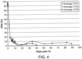
- the amounts of graft cfDNA are determined over a time course, for example, a time course of days or weeks up to a month following transplant.
- GcfDNA is monitored over the first 7 days after engraftment.
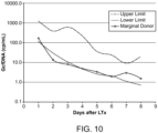
- Described herein is a method of determining the status of a transplanted organ where the organ is a marginal organ, wherein the method comprises determining the level of graft cfDNA present in the blood of a patient. In some aspects, the method comprises determining the level of graft cfDNA over a course of seven days, or up to 30 days following transplant.
- a method of evaluating the transplant status of a transplant recipient comprising monitoring the level of graft cfDNA by assessing the amount of a donor SNP allele in a cfDNA sample obtained from the blood of a patient, typically where the SNP has a MAF of at least 0.40, wherein the donor SNP allele is present in the donor and the recipient is homozygous for an alternative allele.
- the donor may be heterozygous or homozygous for the SNP allele.
- Quantifying the level of the donor SNP allele in the cfDNA sample may comprise determining copy number of the donor SNP allele in the cfDNA sample.
- quantifying the level of the donor SNP allele in the cfDNA sample comprises determining the percentage of the donor SNP allele in the cfDNA sample.
- the transplanted material may be a marginal organ.
- the cfDNA sample may be from a blood sample, e.g ,. serum or plasma, that is obtained ten days or longer following transplant.
- the cfDNA sample may be obtained from a blood sample e.g., serum or plasma, obtained a year or longer following transplant.
- the cfDNA sample may be from a blood sample, e.g., serum or plasma, that is obtained within seven days of transplant.
- Monitoring the level of graft cfDNA may further comprise adjusting an administration schedule or dosage or of one or more immunosuppressive drugs.
- the donor material is a liver, heart, or kidney.
- Monitoring the level of graft cfDNA can be performed to monitor transplant damage that may arise from donor-specific antibodies in the blood of the recipient.
- a method of the disclosure may further comprise detecting donor-specific antibodies in the blood of the recipient.
- Also described herein is use of a method of monitoring graft cfDNA using a SNP assay as described herein to detect transplant damage from various causes, including, but not limited to, reperfusion damage to the organ in a transplant recipient, liver damage from a reactivated hepatitis virus in a liver transplant recipient, transplant damage from donor-specific antibodies, or damage from a chronic transplant injury, e.g., chronic nephropathy in a kidney transplant or vasculopathy in a heart transplant.
- the method of monitoring graft cfDNA using a SNP assay as described herein may be used for determining a minimal effective immunosuppressive regimen.
- cell-free DNA or "cfDNA” as used herein means free DNA molecules of 25 nucleotides or longer that are not contained within any intact cells.
- cfDNA is typically evaluated in human blood, e.g., can be obtained from human serum or plasma.
- a “single nucleotide polymorphism (SNP) biomarker” in the context of this invention refers to a SNP where a recipient of a transplant is homozygous for one SNP allele and the donor has at least one alternative allele for that SNP. Such a SNP is a biomarker for donor material.
- SNP profile refers to the allele pattern, i.e., which alleles are present, in a sample.
- a "graft” as used herein refers to tissue material, from a donor that is transplanted into a recipient.
- a graft may be from liver, heart, kidney, or any other organ.
- primer refers to an oligonucleotide that acts as a point of initiation of DNA synthesis under conditions in which synthesis of a primer extension product complementary to a nucleic acid strand is induced, i.e., in the presence of four different nucleoside triphosphates and an agent for polymerization (i.e., DNA polymerase or reverse transcriptase) in an appropriate buffer and at a suitable temperature.
- a primer is preferably a single-stranded oligodeoxyribonucleotide.
- the primer includes a "hybridizing region" exactly or substantially complementary to the target sequence, preferably about 15 to about 35 nucleotides in length.
- a primer oligonucleotide can either consist entirely of the hybridizing region or can contain additional features which allow for the detection, immobilization, or manipulation of the amplified product, but which do not alter the ability of the primer to serve as a starting reagent for DNA synthesis.
- a nucleic acid sequence tail can be included at the 5' end of the primer that hybridizes to a capture oligonucleotide.
- probe refers to an oligonucleotide that selectively hybridizes to a target nucleic acid under suitable conditions.
- a probe for detection of the biomarker sequences described herein can be any length, e.g., from 15-500 bp in length. Typically, in probe-based assays, hybridization probes that are less than 50 bp are preferred.
- target sequence or "target region” refers to a region of a nucleic acid that is to be analyzed and comprises the sequence of interest, e.g., a region containing a SNP biomarker.
- nucleic acid refers to primers, probes, and oligomer fragments.
- the terms are not limited by length and are generic to linear polymers of polydeoxyribonucleotides (containing 2-deoxy-D-ribose), polyribonucleotides (containing D-ribose), and any other N-glycoside of a purine or pyrimidine base, or modified purine or pyrimidine bases. These terms include double- and single-stranded DNA, as well as double- and single-stranded RNA.
- Oligonucleotides may be used as primers and/or probes.
- a nucleic acid, polynucleotide or oligonucleotide can comprise phosphodiester linkages or modified linkages including, but not limited to phosphotriester, phosphoramidate, siloxane, carbonate, carboxymethylester, acetamidate, carbamate, thioether, bridged phosphoramidate, bridged methylene phosphonate, phosphorothioate, methylphosphonate, phosphorodithioate, bridged phosphorothioate or sulfone linkages, and combinations of such linkages.
- a nucleic acid, polynucleotide or oligonucleotide can comprise the five biologically occurring bases (adenine, guanine, thymine, cytosine and uracil) and/or bases other than the five biologically occurring bases. These bases may serve a number of purposes, e.g., to stabilize or destabilize hybridization; to promote or inhibit probe degradation; or as attachment points for detectable moieties or quencher moieties.
- bases may serve a number of purposes, e.g., to stabilize or destabilize hybridization; to promote or inhibit probe degradation; or as attachment points for detectable moieties or quencher moieties.
- a polynucleotide can contain one or more modified, non-standard, or derivatized base moieties, including, but not limited to, N6-methyl-adenine, N6-tert-butyl-benzyl-adenine, imidazole, substituted imidazoles, 5-fluorouracil, 5 bromouracil, 5-chlorouracil, 5-iodouracil, hypoxanthine, xanthine, 4-acetylcytosine, 5 (carboxyhydroxymethyl)uracil, 5 carboxymethylaminomethyl-2-thiouridine, 5 carboxymethylaminomethyluracil, dihydrouracil, beta-D-galactosylqueosine, inosine, N6 isopentenyladenine, 1-methylguanine, 1-methylinosine, 2,2-dimethylguanine, 2-methyladenine, 2-methylguanine, 3-methylcytosine, 5-methylcytosine, N6-methyladenine,
- nucleic acid, polynucleotide or oligonucleotide can comprise one or more modified sugar moieties including, but not limited to, arabinose, 2-fluoroarabinose, xylulose, and a hexose.
- Repetitive sequences refer to highly repeated DNA elements present in a genome. These sequences are usually categorized in sequence families and are broadly classified as interspersed repetitive DNA (see, e.g., Jelinek and Schmid, Ann. Rev. Biochem. 51:831-844, 1982 ; Hardman, Biochem J. 234:1-11, 1986 ; and Vogt, Hum. Genet. 84:301-306, 1990 ) or tandemly repeated DNA. Repetitive elements include satellite, minisatellite, and microsatellite DNA. In humans, interspersed repetitive DNA includes Alu sequences, short interspersed nuclear elements (SINES) and long interspersed nuclear elements (LINES), and endogenous retroviruses (ERVs).
- SINES short interspersed nuclear elements
- LINES long interspersed nuclear elements
- ERPs endogenous retroviruses
- the present disclosure is based, in part, on the discovery that SNPs having an allele frequency of 0.20 or greater, often 0.30 or greater, and preferably 0.40 or greater, for example, 0.44 or 0.45, or greater, can be surveyed in cfDNA obtained from a transplant patient to identity one or more of such SNP that can be used thereon as a biomarker to monitor rejection status of transplant material.
- a SNP biomarker identified in accordance with the disclosure is one for which the transplant recipient is homozygous for the allele and the donor material has an alternative allele.
- the methods of the disclosure do not require a separate sample from the donor to identity a SNP biomarker.
- a SNP for use in determining a transplantation biomarker in accordance with the disclosure has a minor allele frequency of at least 0.20 or 0.30 and typically has a minor allele frequency of at least 0.40, 0.41, 0.42, 0.43, 0.44, or 0.45 or greater. Further, such a SNP is not contained within or directly adjacent to a repetitive element.
- a SNP that is not contained within or directly adjacent to a repetitive element means that the SNP is sufficiently removed from repetitive sequences such that primers can be designed that specifically amplify the SNP-containing target region.
- a SNP that is not directly adjacent to a repetitive element may be at a distance of 50 base pairs or greater, upstream or downstream from a repetitive element.
- Table 1 provides illustrative SNPs for use in the disclosure.
- the SNPs were identified from public databases (e.g, the websites hapmap.ncbi.nlm.nih.gov or www 1000genomes.org). Compilations can also be found for available SNP-arrays, e.g. at the Illumina website for SNPs used on the "HumanOmin5M" SNP chip. As appreciated by one of skill in the art alternative SNPs can be identified based on these criteria.
- Allele frequency can vary within different populations. For example, allele frequency may be different in a Caucasian population, such as a Caucasian northern European population in comparison to an Asian population, such as a Japanese population. Accordingly, the determination of a SNP suitable for use for identifying a transplantation SNP biomarker as described herein may also take into account the genetic background information of the transplant recipient and donor with respect to minor allele frequency.
- a SNP that can be used as a donor biomarker is identified in a sample obtained from the transplant patient without employing a separate sample from a donor.
- cfDNA from a patient can be used to identify a SNP biomarker for transplant tissue from a donor.
- the sample from the patient can be obtained at any given time following transplantation to evaluate donor SNPs.
- a blood sample e.g., serum or plasma sample
- a blood sample from a patient can be evaluated at a later time frame after transplantation, typically at least five days after transplantation, to determine a SNP that can serve as a biomarker for transplantation. At such times, usually less than 10% of graft DNA is present in the cfDNA in a transplant recipient.
- a blood sample from the recipient is used to isolate cfDNA.
- the cfDNA is then subjected to an amplification step to generate a cfDNA library. This initial amplification step to obtain cfDNA library is also referred to herein as a "pre-amplification".
- Any amplification method can be used to generate the cfDNA library, including, but not limited to PCR. Additional amplification methods are described below.
- the number of rounds of amplification for this pre-amplification step is sufficient to obtain a quantity of cfDNA library that can be evaluated to identify a SNP from a donor. As an illustrative, non-limiting example, anywhere from 8 to 12 rounds may be performed, although other numbers of rounds may also be performed.
- the cf DNA is then assessed for pre-selected SNPs that can serve as biomarkers using primers and probes that amplify target regions containing SNPs that were identified as homozygous in the recipient. This analysis is performed using a digital PCR.
- Those SNPs that provide a signal for a SNP allele that was not identified in the recipient are selected as a transplant biomarker for that transplant patient. Two groups of percentages will be seen: the one that is twice as high as the other e.g ., 2% vs 1% is homozygous in the donor transplant material whereas the lower percentage indicates that the SNP is heterozygous in the donor material.
- the homozygous SNPs are used preferentially for all other samples of the patient. Heterozygous SNPs may also be employed, but are less sensitive.
- any method can be used to determine SNPs that are homozygous in the transplant recipient, including array hybridization, quantitative PCR, sequencing, or an alternative method.
- the recipient SNP genotype for SNPs having a minor allele frequency of 0.20 or greater, or 0.30 or greater, or preferably 0.40 or greater, in accordance with the disclosure is determined using a pre-amplified cfDNA library as described above.
- the SNP profile of the transplant recipient is performed using DNA obtained from peripheral blood leukocytes or other sample from the patient that is free of donor cells. Evaluation of the SNP profile of the recipient using the pre-amplified cfDNA library can employ, but is not limited to, a technique that is not as sensitive as digital PCR to identify recipient SNP alleles. Those SNPs that are homozygous in the patient are used in the analysis of the cfDNA library for donor SNP alleles as described above.
- SNP probes and primers that target one or more SNPs identified in Table 1, e.g., 10, 20, or 30, or more, SNPs identified in Table 1, are used to determine SNPs that are homozygous in the patient.
- a SNP probe having a sequence shown in Table 1 is employed.
- the SNP profile for the donor transplant material can be determined using cfDNA isolated from a sample obtained early after transplantation, where much of the cfDNA, e.g., the majority of the cfDNA, is from the graft.
- SNP biomarkers are identified using a blood sample obtained from the recipient typically less than one day following transplantation.
- the cfDNA isolated from the sample is pre-amplified as described above to obtain a cf library.
- SNPs that are homozygous in the graft are detected by real-time PCR, or an alternative method that does not require digital PCR, although digital PCR may also be employed. Homozygous SNPs are determined in the recipient.
- DNA is isolated from a recipient sample, e.g., a PBL sample, and used to determine those SNPs that are homozygous. Any method can be used to assess the recipient for homozygous SNP alleles, including real-time PCR, a SNP array and the like. SNPs are then selected where the recipient and the transplant material, i.e., the donor, are each homozygous, but have different alleles for the SNP. These SNPs can be used as biomarkers for future measurements to assess transplant status.
- a recipient sample e.g., a PBL sample
- Any method can be used to assess the recipient for homozygous SNP alleles, including real-time PCR, a SNP array and the like. SNPs are then selected where the recipient and the transplant material, i.e., the donor, are each homozygous, but have different alleles for the SNP. These SNPs can be used as biomarkers for future measurements to assess transplant status.
- SNP probes and primers that target one or more SNPs identified in Table 1, e.g., 10, 20, or 30, or more SNPs identified in Table 1, are used to determine SNPs that are homozygous in the patient.
- a SNP probe having a sequence shown in Table 1 is employed.
- the recipient SNP profile is typically determined first so that only the SNPs that are homozygous in the transplant are surveyed in the cfDNA sample.
- these steps need not be performed in this order. For example, SNPs can be evaluated in the various samples in reactions performed concurrently.
- SNPs may also be identified for use as a biomarker where samples from the patient and genetic material from the donor are both available.
- DNA isolated from the transplant recipient and donor samples are evaluated for SNPs where the minor allele frequency is 0.20 or higher, typically 0.30 or higher, and preferably 0.40 or higher.
- SNP profiles from the patient and donor samples are determined for at least 10, 20, 30, or 40 of the SNPs identified in Table 1.
- the SNP evaluation may employ one or more probes having a sequence as shown in Table 1. SNPS where the transplant material and recipient are homozygous, but with different alleles, can then be used for future determination of graft cfDNA percentage.
- Amplification reactions are performed on DNA obtained from nucleic acid samples isolated from various recipient or donor sources. For evaluation of samples where it is desired to have only recipient or donor cells present in the sample, peripheral blood leukocytes are conveniently used; however, any other sample from the recipient, or donor, may be employed. Pre-amplification reactions or amplification reactions that do not require the sensitivity of digital PCR can be performed using any number of well-known amplification techniques.
- Exemplary references include manuals such as Current Protocols in Molecular Biology, Ausubel, 1994-1999 , including supplemental updates through 2013; Sambrook & Russell, Molecular Cloning, A Laboratory Manual (3rd Ed, 2001 ). Although the methods typically employ PCR steps, other amplification protocols may also be used. Suitable amplification methods include ligase chain reaction (see, e.g., Wu & Wallace, Genomics 4:560-569, 1988 ); strand displacement assay ( see, e.g., Walker et al., Proc. Natl. Acad. Sci. USA 89:392-396, 1992 ; U.S. Pat. No. 5,455,166 ); and several transcription-based amplification systems, including the methods described in U.S.
- DNA is amplified using adaptor-ligation and single primer PCR.
- Other available methods of amplification such as balanced PCR ( Makrigiorgos, et al., Nature Biotechnol, 20:936-9 (2002 )) and isothermal amplification methods such as nucleic acid sequence based amplification (NASBA) and self-sustained sequence replication ( Guatelli et al., PNAS USA 87:1874 (1990 )).
- NASBA nucleic acid sequence based amplification
- self-sustained sequence replication Guatelli et al., PNAS USA 87:1874 (1990 )
- multiplex reactions can be performed in which multiple target regions are amplified and detected in a single reaction.
- Digital PCR is a technique where a limiting dilution of the sample is made across a large number of separate PCR reactions so that most of the reactions have no template molecules and give a negative amplification result. Those reactions that are positive at the reaction endpoint are counted as individual template molecules present in the original sample in a 1 to 1 relationship.
- Quantitative partitioning is assumed, and the dynamic range is governed by the number of containers available for stochastic separation.
- a digital PCR may be a microfluidics-based digital PCR.
- a droplet digital PCR may be employed.
- a SNP that is evaluated as a potential transplant biomarker in accordance with the disclosure has a minor allele frequency of at least 0.20 or at least 0.30, and preferably at least 0.40, 0.41, 0.42, 0.43, 0.44, or 0.45, or greater.
- Such primers and probes are used to detect individual SNP alleles.
- SNP-specific amplification methods can be used (e.g., using SNP-specific amplification primers).
- primers are used to amplify a target region and the SNP alleles are detected using probes specific for each allele.
- Oligonucleotides that are employed as primers and/or probes to detect biomarkers can be selected using methods well-known in the art.
- PCR primers may be designed using standard primer design computer software techniques known to individuals skilled in the art. The variables considered during PCR primer design may include primer length, GC pair content, melting temperature, and size of the target nucleic acid amplified by the primer pair.
- the biomarker is identified by hybridization under sequence-specific hybridization conditions with a probe that targets the biomarker region (e.g., targets some unambiguously assigned portion of, the target biomarker) with or without a preceding amplification of DNA.
- a probe that targets the biomarker region e.g., targets some unambiguously assigned portion of, the target biomarker
- Principals for designing such a probe are well known in the art.
- a SNP transplant biomarker identified in accordance with the disclosure may be used to evaluate transplant rejection status in the recipient. Such an evaluation can be performed, e.g., using an amplification reaction to detect transplant biomarker in the cfDNA present in a blood sample from the patient.
- the cfDNA of the patient may be evaluated periodically, for example, over the course of days, weeks, months, or years, for SNP biomarkers in cfDNA to monitor the status of the transplant, i.e., whether there are signs of rejection or damage. If the percentage of graft cfDNA rises either higher than the mean, typically + 2SD of values seen in uncomplicated courses, or shows a sustained increase, this is indicative of a rejection.
- a blood sample is obtained from the patient at the desired time point following transplant.
- a cfDNA sample is obtained from the blood sample and is analyzed to determine the level of donor material by identifying the presence of donor SNP alleles in the cfDNA. Any method can be used to evaluate the sample.
- digital PCR such as a microfluidics-based digital PCR or droplet-based PCR is employed.
- Other methods can be based on direct hybridization of detection probes (without prior amplification) or sequencing, e.g., sequencing of an amplicon defined in Table 1.
- the SNP region is amplified by PCR and then the percentage of the minor allele is determined by amplicon sequencing.
- the percentage of donor cfDNA (also referred to as graft cfDNA) in the cfDNA sample can be determined.
- the copy number of donor cfDNA can be determined.
- Analysis of graft cfDNA levels in the blood using a SNP analysis as described herein can be used to detect any kind of injury to or deterioration of transplant organ cells.
- graft cfDNA analysis can be used to assess perfusion injury.
- monitoring graft cfDNA to determine the presence of reperfusion injury comprises monitoring cfDNA samples from the transplant recipient that are obtained soon after transplant, e.g., within 7 days of transplant.
- perfusion is used interchangeably with "reperfusion”.
- Damage that arises from reactivation of a virus infection can be assessed using a SNP graft cfDNA assay.
- Such methods may further comprise identifying the presence of the virus, e.g., where the transplant is a liver, the presence of a hepatitis virus.
- a "marginal organ” is an art-recognized term that describes an organ from a donor that has a medical history that does not meet the optimal history for organ donors, for example, the donor, may have one of the following criteria: extremes of age, adverse past medical history, etc. These criteria vary from organ to organ and depend on the patient history.
- Graft cfDNA SNP analysis in accordance with the disclosure can be used to adjust an immunosuppressive regimen in a patient.
- the lowest effective amount of an immunosuppressive drug regimen that achieves a level of graft cfDNA that is observed in stable transplant patients can be determined.
- monitoring the status of the graft to adjust an immunosuppressive regimen comprises monitoring cfDNA samples from the transplant recipient that are obtained at about ten days or two weeks or longer. Monitoring can be performed for an extended period of time of up to years at desired intervals.
- Graft cfDNA analysis in accordance with the disclosure can be used to detect a solid organ transplant injury caused by donor-specific antibodies.
- a transplant recipient can be monitored over the course of years for such damage.
- the method may further comprise detecting the presence of donor-specific antibodies circulating in the blood of the transplant recipient.
- donor-specific antibodies are specific for the HLA type of the donor organ and can be detected using known assays.
- a patient having such donor-specific antibodies may be additionally treated with immunosuppressive agents that suppress B cells.
- the information obtained from the SNP biomarker analysis may be stored in a computer readable form.
- a computer system typically comprises major subsystems such as a central processor, a system memory (typically RAM), an input/output (I/O) controller, an external device such as a display screen via a display adapter, serial ports, a keyboard, a fixed disk drive via a storage interface and a floppy disk drive operative to receive a floppy disc, and a CD-ROM (or DVD-ROM) device operative to receive a CD-ROM.
- Many other devices can be connected, such as a network interface connected via a serial port.
- the computer system may also be linked to a network, comprising a plurality of computing devices linked via a data link, such as an Ethernet cable (coax or 10BaseT), telephone line, ISDN line, wireless network, optical fiber, or other suitable signal transmission medium, whereby at least one network device (e.g., computer, disk array, etc.) comprises a pattern of magnetic domains (e.g., magnetic disk) and/or charge domains (e.g., an array of DRAM cells) composing a bit pattern encoding data acquired from an assay of the disclosure.
- a network device e.g., computer, disk array, etc.
- a pattern of magnetic domains e.g., magnetic disk
- charge domains e.g., an array of DRAM cells
- the computer system can comprise code for interpreting the results of a study to determine SNP transplant biomarkers or to evaluating the presence of one or more of the SNP transplant biomarkers identified in accordance with the disclosure to aid in prognosis.
- the biomarker analysis results are provided to a computer where a central processor executes a computer program for evaluating the one or more biomarkers.
- a computer system such as that described above, which comprises: (1) a computer; (2) a stored bit pattern encoding the biomarker testing results obtained by the methods of the disclosure, which may be stored in the computer; (3) and, optionally, (4) a program for evaluating a biomarker.
- a system for analyzing circulating cell-free DNA comprising: (1) a sample analyzer for executing the method of analyzing circulating cell-free DNA in a patient's blood, serum or plasma as described in the various aspects above; (2) a computer system for automatically receiving and analyzing data obtained in step (1) to provide a test value representing the status (presence or absence or amount, i.e., concentration or copy number) of a SNP transplant biomarker for the patient.
- the computer-based analysis function can be implemented in any suitable language and/or browsers. For example, it may be implemented with C language and preferably using object-oriented high-level programming languages such as Visual Basic, SmallTalk, C++, and the like.
- the application can be written to suit environments such as the Microsoft Windows TM environment including Windows TM 8, Windows TM 7,Windows TM 98, Windows TM 2000, Windows TM NT, and the like.
- the application can also be written for the MacIntosh TM , SUN TM , UNIX or LINUX environment.
- the functional steps can also be implemented using a universal or platform-independent programming language.
- Examples of such multi-platform programming languages include, but are not limited to, hypertext markup language (HTML), JAVA TM , JavaScript TM , Flash programming language, common gateway interface/structured query language (CGI/SQL), practical extraction report language (PERL), AppleScript TM and other system script languages, programming language/structured query language (PL/SQL), and the like.
- Java TM - or JavaScript TM -enabled browsers such as HotJava TM or Microsoft TM Explorer TM can be used.
- active content web pages may include Java TM applets or ActiveX TM controls or other active content technologies.
- the analysis function can also be embodied in computer program products and used in the systems described above or other computer- or internet-based systems. Accordingly, also described herein is a computer program product comprising a computer-usable medium having computer-readable program codes or instructions embodied thereon for enabling a processor to carry out the analysis and correlating functions as described above.
- These computer program instructions may be loaded onto a computer or other programmable apparatus to produce a machine, such that the instructions which execute on the computer or other programmable apparatus create means for implementing the functions or steps described above.
- These computer program instructions may also be stored in a computer-readable memory or medium that can direct a computer or other programmable apparatus to function in a particular manner, such that the instructions stored in the computer-readable memory or medium produce an article of manufacture including instruction means which implement the analysis.
- the computer program instructions may also be loaded onto a computer or other programmable apparatus to cause a series of operational steps to be performed on the computer or other programmable apparatus to produce a computer implemented process such that the instructions which execute on the computer or other programmable apparatus provide steps for implementing the functions or steps described above.
- SNPs were selected from public databases considering those which show a known and validated minor allelic frequency of >40% in Caucasians and >45% over all reported ethnicities.
- SNP that are in or directly adjacent to a repetitive element were eliminated.
- the remaining SNPs were then investigated for their usefulness in a probe hydrolysis assay. This was done in silico by using thermodynamic calculations ( Schütz & von Ahsen, Biotechniques 27:1218-1222, 1224, 1999 ) to optimize the binding differences for the two probes that hybridize to the two alleles at the desired temperature of 65°C at standard PCR buffer conditions (e.g. 0.18mol/L salt and 0.5 ⁇ mol/L DNA/primer).
- Each of the assays was first optimized in a LightCycler480, using the ddPCR Supermix for Probes (Bio-Rad) and subsequently optimized for digital droplet PCR (ddPCR), which yielded slightly different cycling conditions. Two different annealing temperatures were established in order to maximize efficiency and differentiation of alleles.
- Table 1 lists probes and other characteristics for each of the selected SNPs.
- Table 1 part A Col. 1, SNP designation in Table 1; Col. 2, SNP name/ reference; Col. 3, chromosome; Col 4, position on chromosome; Col. 5, minor allele frequency (MAF) (all populations); Col. 5, MAP, Caucasian; Cols. 6 and 7: illustrative primers for amplification of target region containing SNP; Col. 8, Annealing and Extension temperatures.
- Table 1 part B Col. 1, SNP designation in Table 1; Col. 2, SNP (nucleotide change); Col. 3, length of amplicon containing SNP obtained with primers shown in part A, Cols. 6 and 7; Cols. 4-7, SNP probes and characteristics.
- genomic DNA and cfDNA were extracted from EDTA-anticoagulated blood collected from healthy volunteers.
- plasma was separated from the cells by centrifugation (2500 x g for 10 min at 4°C, followed by a second centrifugation of the plasma at 4000 x g for 20 min at 4°C to remove any cell debris.
- DNA from both the plasma (>1mL) and the harvested buffy coat was extracted with the Roche Total Viral Acid Extraction Kit using manufacturer's instructions. The results reported here were from samples that were drawn under an IRB approved protocol with informed consent.
- ddPCR reactions were prepared using the ddPCR Supermix for Probes (Bio-Rad). Each reaction contained 30 ng or 100 ng of the cfDNA library as template, 900 nmol/L of each primer and 250 nmol/L of each probe. Droplets were generated using the QX100 droplet generator (Bio-Rad) according to the manufacturer's protocols. The cycling conditions were: 95° for 10 min, 50x(94° for 30 sec, 95°/61° for 1 min), 98° for 10 min. Droplets were read in the QX100 droplet reader and analyzed using the software Quantasoft version 1.3.2.0 (Bio-Rad).
- the embedded "Rare Event Detection" calculation was used, which basically takes the underlying Poisson distribution into account to calculate the template molecule concentration of either allele. These values are then used to express the minor allele in percent of the total concentration.
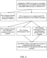
- Figure 2 illustrates a procedure deployed to determine the graft DNA content in the recipients' circulation.
- SNPs SNPs for each recipient that gives the highest theoretical sensitivity
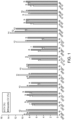
- the ddPCRs of the clinical samples were performed using 30 ng (LTx samples) or 100 ng (HTx and KTx samples) of the cfDNA library per well, which translated to about 0.5 and 1.5 copies per droplet respectively.
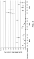
- Figure 3 show the results for stable liver, kidney and heart transplant recipients with no signs of rejection.
- a total of 10 different ddPCR assays were performed for the LTx patients, and a total of 16 different ddPCR assays were performed for the KTx and HTx patients.
- the number of informative assays used to determine the graft cfDNA content is given below the abscissa.
- the percentage of graft DNA in the circulation of liver recipients was lower than 10% in all patients.
- the higher amount in LTx compared to the other organs may reflect the higher regeneration rate that is usually seen for hepatocytes compared to other e.g. heart and kidney cells.
- the total ddPCR assay performed were 12 for LTx1, 16 for LTx3, and 18 for LTx6.
- the results of five different informative assays were used to determine the graft cfDNA content.
- the percentage of graft cfDNA was always ⁇ 15% from day 10 onwards if no complications occurred.
- Figure 5 displays the time course of the conventionally used sensitive rejection markers bilirubin and AST (aspartate aminotransferase) together with the percentage of graft cfDNA in this patient. After having an initial graft cfDNA value of ⁇ 15% on day 7, the values increased and never returned to values seen in the uncomplicated patients during the entire observation period. Moreover, this percentage was markedly increased on day 32, several days before the conventional parameters suggested possible rejection.
- bilirubin and AST aspartate aminotransferase
- This example thus illustrates the identification of SNPs to evaluate transplant status and demonstrated that there was a significant increase of graft cfDNA that preceded AST and bilirubin elevations in a case of LTx rejection.
- a cost effective technique was developed that can determine relative amounts of graft DNA in cfDNA of LTx patients in one working day. This technique makes graft cfDNA a promising biomarker for early detection of rejection, potentially enabling more timely therapeutic intervention.
- ratio of graft to host cfDNA has analytical advantages by eliminating disturbing variables, such as DNA extraction efficiency, variablities in host cfDNA may obfuscate the view on the engrafted organ.
- the early phase after transplant was used as model to compare the percentage or absolute plasma concentration of GcfDNA is a more valuable graft integrity measure.
- LTx liver
- HTx heart
- KTx kidney
- EDTA-whole blood was drawn and processed within 4 hours.
- cfDNA tubes (9mL) Streck Inc. were used for a subset of samples. Extraction of cfDNA from 1-2.5 ml of plasma was performed using the High Pure Viral Extraction Large Volume Kit (Roche) according to the manufacturer's instructions, but without the addition of carrier RNA.
- an artificial DNA (referred to as a "spike” in the example) was added to samples in a known quantity.
- the spike consisted of a non-human derived 320bp DNA that was cloned into a pGEM-T vector (Promega) and produced using Phusion polymerase (NEB) with m13 primers.
- the resulting product was cleaned using AMPure XP (Beckman-Coulter) purification and stored in a 1,400,000-fold concentration of the final spike dilution used for extraction.
- the spike Prior to each extraction, the spike was diluted and 20 ⁇ L were added to the plasma just prior to adding the protease and binding buffer. This resulted in an approximate amount of 5,000 spike copies per mL of plasma.
- cfDNA was corrected for the PCR length based efficacy of 98bp and 90bp, based on the published size distribution ( Beck, et al. Clin Chem 55:730-38, 2009 ).
- a second primer set of 223 bp and 224 bp that targets the same genetic regions was used to assess the presence of longer DNA fragments in the cfDNA extracts.
- the ratio between cp/mL determined using the long amplicon assays versus the short amplicon assays was calculated as a measure of necrotic DNA content.
- the GcfDNA was the calculated by multiplying the GcfDNA [%] with the cfDNA [cp/mL] divided by the extraction efficiency of the internal standard.
- GcfDNA [%] was measured as described ( Beck et al., Clin Chem 59:1732-41, 2013 ) on a QX100/200 system, which was used for all other described ddPCR assays throughout the study.
- HCV+ hepatitis C infection
- the GcfDNA values in LTx patients were compared with the determined Tacrolimus levels at the same time point ( Oellerich et al., Ther Dug Monit 36:136-140, 2014 ). It can be shown that sub-therapeutic Tacrolimus blood levels are associated with both elevated GcfDNA(%) as well as GcfDNA(cp/ml) values. In an extention of the report by Oellerich et al , to evaluate 260 sample, the separation was found to be slightly better with the absolute (cp/mL) determinations ( Figures 7 and 8 ), if a Tacrolimus blood level of 6.7 ⁇ g/mL was used.
- GcfDNA is suitable for the rapid, specific, and early detection of graft injury after LTx and is a useful measure of individual responses to immunosuppressive therapy. Accordingly, the method can be used for the assessment of lowest effective immunosuppressant exposure in minimization strategies.
- This example demonstrates the associations found between the clinical course and quantified graft-derived cell-free DNA (GcfDNA) as biomarker of graft integrity in a liver transplant (LTx) patient who received a marginal donor liver and the severity of early damage due to reperfusion.
- GcfDNA graft-derived cell-free DNA
- the effect of early damage can be quantified by the determination of the GcfDNA (cp/mL), which shows a clear reduction over the first week after LTx.
- Figure 10 shows the results obtained from an analysis of a marginal donor in comparison to the range seen in 14 other patients.
- the LTx of this marginal organ did show a good initial outcome and function, which is predicted by the rapid decrease of GcfDNA at the lower end of the whole group.
- the amount of necrosis of the graft can be assessed by estimating the length of the cfDNA, which is short if released by apoptotic cell damage and longer if of necrotic origin.
- an apoptotic index can be defined. The higher that value is, the more cfDNA is of necrotic origin.
- Figure 11 shows the time course during the first days after LTx.
- GcfDNA determinations can be used to monitor early graft recovery and subsequent damage as well as responses to therapeutic interventions. Furthermore, GcfDNA was an immediate and sensitive indicator of compromised graft perfusion. This test can be used as a "liquid biopsy" to assess the integrity of the transplanted organ, especially in recipients of marginal donor livers.
Description
- Using modern molecular biological techniques the detection of trace amounts of divergent genetic material in a single sample is feasible. This has potential applications for a number of conditions such as prenatal diagnosis, tumor diagnosis, and detection of transplant rejection. An increase of heart donor DNA in the circulation of stable heart transplant recipients during rejection episodes has been reported (Snyder, et al., Proc Natl Acad Sci U S A 108:6229-6234, 2011). However, to be clinically useful the method used for the detection of graft DNA must not only be specific and sensitive, it must also have a rapid turn-around-time and be economically feasible to perform. The methods described to date are extremely time consuming and expensive to perform (Lo, Clin Chem 57:941-942, 2011).
- Simplified methods to differentiate between DNA from donors and recipients can involve the use of single-nucleotide-polymorphisms (SNPs). One possibility is to interrogate both donor and recipient for certain SNPs and use those, where both SNPs are homozygous, but different in donor and recipient. However, this would require DNA from the donor to be available, which isn't always the case in the clinical setting, in particular if the transplantation was some years before. There is thus a need for sensitive, easily implemented techniques for early detection of transplant rejection.
- The invention is defined by the appended claims. Provided herein is a method of evaluating the integrity of a transplant in a recipient, the method comprising monitoring the level of graft cell-free DNA (cfDNA) by assessing the amount of a donor SNP allele in a cfDNA sample obtained from the blood of a patient, wherein the SNP allele is pre-selected as having a minor allele frequency (MAF) of at least 0.4, and further, wherein the donor SNP allele is present in the donor and the recipient is homozygous for an alternative allele for that SNP, wherein the SNP allele is identified without employing a separate sample from a donor.
- Also provided is a method of detecting a SNP from a donor to monitor transplant integrity in a recipient receiving tissue from said donor, the method comprising (a) identifying that a SNP having a minor allele frequency of 0.4 or greater is homozygous in the recipient, (b) amplifying cell-free (cfDNA) from a serum or plasma sample obtained from the recipient at least 5 days after transplantation of material from the donor to generate a cf library, (c) performing a quantitative PCR reaction for the SNPs identified in (a) to detect the presence of the alternative allele for one or more of the SNPs; and (d) selecting a SNP where the alternative SNP is present in the cf library to monitor transplant integrity of the patient; wherein the method does not employ a separate sample from the donor.
- Also provided is a method of detecting a SNP from a donor to monitor transplant integrity in a recipient receiving tissue from said donor, the method comprising (a) identifying that a SNP having a minor allele frequency of 0.4 or greater is homozygous in the recipient, (b) amplifying cell-free (cfDNA) from a blood sample obtained from the recipient 24 hrs or less after transplantation of graft material from the donor to generate a cf library, (c) identifying a SNP having a minor allele frequency of 0.4 or greater as homozygous in the donor using the cf library of step (b), (d) performing a quantitative PCR reaction for the SNPs identified in (a) to detect the presence of the alternative allele for one or more of the SNPs in the donor, and (e) selecting a SNP where the alternative SNP is homozygous in the donor; wherein the method does not employ a separate sample from the donor.
- Also provided is a method of monitoring the integrity of a transplant in a transplant recipient, the method comprising, quantifying, in a cfDNA sample obtained from the transplant recipient, the amount of a donor SNP allele for a SNP selected by a method of the invention; wherein the SNP allele is identified without employing a separate sample from the donor.
- Certain aspects of the disclosure are summarized below. The disclosure is not limited to the particular aspects described in this summary.
- Described herein is a method of detecting transplant rejection in a patient based on the use of SNPs that have been investigated for their minor allelic frequency (MAF) where such frequencies are 0.4 or higher. For example, assuming Hardy-Weinberg equilibrium, a SNP with a MAF of ∼0.40 would show homozygosis in both donor and recipient 23%-25% of the time for each allele. The probability of both having a different allele (homozygous) is therefore ∼11.3% to ∼12.5%. In order to identify at least three such SNPs in Caucasians 30 to 35 different SNPs with the mentioned characteristics can be interrogated. In contrast, if unselected SNPs were employed, it can be estimated that over 3,000 assays are needed to achieve the same discriminating power, based on the overall median MAF of 0.023 reported for known human SNPs in Caucasians (e.g. for the Illumina HumanOmni5M bead chip).
- Using such SNP comparisons, the amount of graft DNA released by an organ into the circulation can be assessed and used as a biomarker for organ integrity. In addition, once SNPs that differ between donor and recipient are identified only SNPs with the best sensitivity (e.g., homozygous in both but different in donor and recipient) need to be measured subsequently. The only limitation of such a method is the amount of DNA that is interrogated, which is mainly driven by the volume of blood that is analyzed. For example, it can be estimated that the number of genome equivalents in one milliliter of blood is about one thousand. If all molecules take part in a PCR reaction and the graft DNA accounts for 5% of cfDNA then there would be 50 such molecules in 1mL of blood. The entire analysis of several different SNPs, therefore, when working with small samples, entails as a first step the random unbiased amplification of extracted cfDNA coming from a blood sample, e.g., typically a sample of at least 2 mL of blood. Such an amplification step can be done by several techniques, for the usually short apoptotic cfDNA (e.g., Beck et al., Clin Chem 55:730-738, 2009) a direct adaptor ligation is often most suitable (Lo et al., Sci Transl Med 2: 61ra91, 2010). Once the amplification adaptors are ligated, a moderate number of amplification cycles are performed (generally not more than 12 to 15) and the resulting library is cleaned from primers and adaptors and used as template for the SNP interrogations. If the initial sample size is not limited to a small sample, the amplification step, which is also referred to herein as a pre-amplification step, may be omitted.
- Thus, in one aspect, the disclosure provides a method of detecting a SNP from a donor to monitor transplant status of a recipient receiving tissue from said donor, the method comprising: (a) identifying a SNP having a minor allele frequency of 0.40 or greater, as homozygous in the recipient; (b) amplifying cell-free (cfDNA) from a serum or plasma sample obtained from the recipient at least 5 days after transplantation of material from the donor to generate a cf library; (c) performing a digital PCR reaction for the SNPs identified in (a) to detect the presence of an alternative allele for one or more of the SNPs, and (d) selecting a SNP where the alternative SNP allele is present in the cf library to monitor transplant status of the patient; wherein the method does not employ a separate sample from the donor. Preferably, the SNP selected in (d) is homozygous in the donor, but may be heterozygous in the donor. Step (a) can be performed using any sample from the patient that would not contain donor material, for example, peripheral blood leukocytes (PBLs) obtained from the patient may be used to identify a suitable SNP. In alternative aspects, step (a) is performed using the cfDNA library of step (b). Thus, in some aspects, e.g., using a PBL sample from the patient to obtain DNA, step (a) may be performed before or after step (b) whereas in other aspects, the amplified cfDNA library is generated first. In some aspects, a suitable SNP to be evaluated for monitoring transplant status is a SNP set forth in Table 1. In some aspects, at least 5, 10, 15, 20, 25, 30, 35, 40, or all of the SNPs set forth in Table 1 are evaluated to determine those SNPs that are homozygous in the recipient.
- In another aspect, the disclosure provides a method of detecting a SNP from a donor to monitor transplant status of a recipient receiving tissue from said donor, the method comprising: (a) identifying that a SNP having a minor allele frequency of 0.40 or greater, as homozygous in the recipient; (b) amplifying cell-free (cfDNA) from a blood sample, e.g., serum or plasma, obtained from the recipient 24 hours or less after transplantation of graft material from the donor to generate a cf library; (c) identifying a SNP having a minor allele frequency of 0.40 or greater, as homozygous in the donor using the cf library of step (b); (d) performing a digital PCR reaction for the SNPs identified in (a) to detect the presence of the alternative allele for one or more of the SNPs in the donor, and (e) selecting a SNP where the alternative SNP allele is present in the donor; wherein the method does not employ a separate sample from the donor. Step (a) can be performed using any sample from the patient that would not contain donor material, for example, peripheral blood leukocytes (PBLs) obtained from the patient may be used to identify a suitable SNP. In some aspects, a suitable SNP is a SNP set forth in Table 1. In some aspects, at least 5, 10, 15, 20, 25, 30, 35, 40, or all of the SNPs set forth in Table 1 are evaluated to determine those SNPs that are homozygous in the recipient.
- Described herein is a method of detecting a SNP from a donor to monitor transplant status of a recipient receiving tissue from said donor DNA, the method comprising : (a) identifying a SNP having a minor allele frequency of 0.40 or greater, as homozygous in a recipient using a DNA sample from the recipient from a source that is free of donor DNA e.g., DNA obtained from a PBL sample from the patient; (b) identifying a SNP having a minor allele frequency of 0.40 or greater, as homozygous in a recipient using a DNA sample obtained from donor cells or tissue; and (c) selecting a SNP that is homozygous in the recipient for which the donor is homozygous or heterozygous for the alternative allele. In some aspects, SNP genotype is determined in recipient and/or donor DNA for at least 10, 20, 30, or 40 of the SNPs identified in Table 1. In some aspects, the SNP evaluation may employ one or more probes having a sequence as shown in Table 1. SNPs for which the transplant material has a different allele for the SNP compared to the recipient can then be used for future determination of graft cfDNA percentage, e.g., in a digital PCR reaction.
- Described herein is a method of monitoring transplant rejection in a transplant recipient, the method comprising : obtaining a cfDNA sample from the patient; and detecting the presence or absence, or quantifying, a donor SNP allele for a SNP selected using a method as described herein. In typical aspects, cfDNA samples are obtained from the patient at desired time points following transplantation and the level of the donor SNP allele is quantified.
- Described herein is a method of monitoring the status of a transplant in a transplant recipient to evaluate immunosuppressive therapy where the method comprises quantifying the amount of the donor allele SNP at desired time points and adjusting the immunosuppressive therapy, e.g., adjusting the amount of immunosuppressive drug. Thus, the lowest dose of an immunosuppressive drug can be identified for that individual patient.
- Described herein is a method of monitoring the status of a transplant in a transplant recipient, e.g., a liver transplant recipient, to determine changes in the transplant status related to reactivation of a virus, such as a hepatitis virus, where the method comprises quantifying the amount of donor SNP allele as described herein present in the blood of a transplant recipient.
- Described herein is a method of monitoring the status of a transplant in a transplant recipient to evaluate reperfusion injury to the transplant. In such aspects, the amounts of graft cfDNA (GcfDNA) are determined over a time course, for example, a time course of days or weeks up to a month following transplant. In typical aspects, GcfDNA is monitored over the first 7 days after engraftment.
- Described herein is a method of determining the status of a transplanted organ where the organ is a marginal organ, wherein the method comprises determining the level of graft cfDNA present in the blood of a patient. In some aspects, the method comprises determining the level of graft cfDNA over a course of seven days, or up to 30 days following transplant.
- Disclosed herein is a method of evaluating the transplant status of a transplant recipient, the method comprising monitoring the level of graft cfDNA by assessing the amount of a donor SNP allele in a cfDNA sample obtained from the blood of a patient, typically where the SNP has a MAF of at least 0.40, wherein the donor SNP allele is present in the donor and the recipient is homozygous for an alternative allele. The donor may be heterozygous or homozygous for the SNP allele. Quantifying the level of the donor SNP allele in the cfDNA sample may comprise determining copy number of the donor SNP allele in the cfDNA sample. In some aspects quantifying the level of the donor SNP allele in the cfDNA sample comprises determining the percentage of the donor SNP allele in the cfDNA sample. The transplanted material may be a marginal organ. The cfDNA sample may be from a blood sample, e.g,. serum or plasma, that is obtained ten days or longer following transplant. The cfDNA sample may be obtained from a blood sample e.g., serum or plasma, obtained a year or longer following transplant. The cfDNA sample may be from a blood sample, e.g., serum or plasma, that is obtained within seven days of transplant. Monitoring the level of graft cfDNA may further comprise adjusting an administration schedule or dosage or of one or more immunosuppressive drugs. In some aspects, the donor material is a liver, heart, or kidney. Monitoring the level of graft cfDNA can be performed to monitor transplant damage that may arise from donor-specific antibodies in the blood of the recipient. A method of the disclosure may further comprise detecting donor-specific antibodies in the blood of the recipient.
- Also described herein is use of a method of monitoring graft cfDNA using a SNP assay as described herein to detect transplant damage from various causes, including, but not limited to, reperfusion damage to the organ in a transplant recipient, liver damage from a reactivated hepatitis virus in a liver transplant recipient, transplant damage from donor-specific antibodies, or damage from a chronic transplant injury, e.g., chronic nephropathy in a kidney transplant or vasculopathy in a heart transplant. The method of monitoring graft cfDNA using a SNP assay as described herein may be used for determining a minimal effective immunosuppressive regimen.
-
-
Figure 1 provides a graph showing coefficients of variation (%) for 15 assays with 2% minor allele concentration. For each assay the CV for 2% allele A and 2% allele B are shown (black bars). The intra-assay precision was obtained in 9 repetitions within in the same QX100 run using 100ng total input DNA. Grey bars show the theoretical CVs as calculated from the number of droplets positive for the minor allele. -
Figure 2 provides a schematic of the workflow deployed to obtain measures of graft cfDNA content. -
Figure 3 provides illustrative data for the graft cfDNA content measured in the circulation of 10 stable LTx , 9 stable KTx and of 8 stable HTx patients. -
Figure 4 provides illustrative data of a time course measurement of cfDNA content in the circulation of 3 patients with liver transplantations. The earliest sample analyzed was obtained from patient LTx6 atday 1 after surgery. Nearly 100% of the cfDNA in the patient's plasma is graft derived DNA. -
Figure 5 provides illustrative data from a time course measurement of one LTx patient with an acute rejection episode at day 43 after surgery. Graft cfDNA content shows a marked increase on day 32 well before conventional biomarkers AST and bilirubin indicate the rejection. -
Figure 6 provides illustrative data comparing graft cfDNA measured as copies/ml to those values expressed as percentage. -
Figures 7 and8 provide illustrative data showing that sub-therapeutic Tacrolimus blood levels are associated with both graft cfDNA (%) (Figure 7 ) as well as graft cfDNA copy number (Figure 8 ). -
Figure 9 provides illustrative data showing graft cfDNA copy number at various times after liver transplant. -
Figure 10 shows the results obtained from graft cfDNA analysis of the transplant status of a transplant recipient who received a marginal donor organ in comparison to the range seen in fourteen other patients. -
Figure 11 provides illustrative data showing the time course of necrotic graft cfDNA during the first days after LTx. - The term "cell-free DNA" or "cfDNA" as used herein means free DNA molecules of 25 nucleotides or longer that are not contained within any intact cells. In the context of the current invention, "cfDNA" is typically evaluated in human blood, e.g., can be obtained from human serum or plasma.
- A "single nucleotide polymorphism (SNP) biomarker" in the context of this invention refers to a SNP where a recipient of a transplant is homozygous for one SNP allele and the donor has at least one alternative allele for that SNP. Such a SNP is a biomarker for donor material.
- A "SNP profile" as used herein refers to the allele pattern, i.e., which alleles are present, in a sample.
- A "graft" as used herein refers to tissue material, from a donor that is transplanted into a recipient. For example, a graft may be from liver, heart, kidney, or any other organ.
- The term "primer" refers to an oligonucleotide that acts as a point of initiation of DNA synthesis under conditions in which synthesis of a primer extension product complementary to a nucleic acid strand is induced, i.e., in the presence of four different nucleoside triphosphates and an agent for polymerization (i.e., DNA polymerase or reverse transcriptase) in an appropriate buffer and at a suitable temperature. A primer is preferably a single-stranded oligodeoxyribonucleotide. The primer includes a "hybridizing region" exactly or substantially complementary to the target sequence, preferably about 15 to about 35 nucleotides in length. A primer oligonucleotide can either consist entirely of the hybridizing region or can contain additional features which allow for the detection, immobilization, or manipulation of the amplified product, but which do not alter the ability of the primer to serve as a starting reagent for DNA synthesis. For example, a nucleic acid sequence tail can be included at the 5' end of the primer that hybridizes to a capture oligonucleotide.
- The term "probe" refers to an oligonucleotide that selectively hybridizes to a target nucleic acid under suitable conditions. A probe for detection of the biomarker sequences described herein can be any length, e.g., from 15-500 bp in length. Typically, in probe-based assays, hybridization probes that are less than 50 bp are preferred.
- The term "target sequence" or "target region" refers to a region of a nucleic acid that is to be analyzed and comprises the sequence of interest, e.g., a region containing a SNP biomarker.
- As used herein, the terms "nucleic acid," "polynucleotide" and "oligonucleotide" refer to primers, probes, and oligomer fragments. The terms are not limited by length and are generic to linear polymers of polydeoxyribonucleotides (containing 2-deoxy-D-ribose), polyribonucleotides (containing D-ribose), and any other N-glycoside of a purine or pyrimidine base, or modified purine or pyrimidine bases. These terms include double- and single-stranded DNA, as well as double- and single-stranded RNA. Oligonucleotides may be used as primers and/or probes.
- A nucleic acid, polynucleotide or oligonucleotide can comprise phosphodiester linkages or modified linkages including, but not limited to phosphotriester, phosphoramidate, siloxane, carbonate, carboxymethylester, acetamidate, carbamate, thioether, bridged phosphoramidate, bridged methylene phosphonate, phosphorothioate, methylphosphonate, phosphorodithioate, bridged phosphorothioate or sulfone linkages, and combinations of such linkages.
- A nucleic acid, polynucleotide or oligonucleotide can comprise the five biologically occurring bases (adenine, guanine, thymine, cytosine and uracil) and/or bases other than the five biologically occurring bases. These bases may serve a number of purposes, e.g., to stabilize or destabilize hybridization; to promote or inhibit probe degradation; or as attachment points for detectable moieties or quencher moieties. For example, a polynucleotide can contain one or more modified, non-standard, or derivatized base moieties, including, but not limited to, N6-methyl-adenine, N6-tert-butyl-benzyl-adenine, imidazole, substituted imidazoles, 5-fluorouracil, 5 bromouracil, 5-chlorouracil, 5-iodouracil, hypoxanthine, xanthine, 4-acetylcytosine, 5 (carboxyhydroxymethyl)uracil, 5 carboxymethylaminomethyl-2-thiouridine, 5 carboxymethylaminomethyluracil, dihydrouracil, beta-D-galactosylqueosine, inosine, N6 isopentenyladenine, 1-methylguanine, 1-methylinosine, 2,2-dimethylguanine, 2-methyladenine, 2-methylguanine, 3-methylcytosine, 5-methylcytosine, N6-methyladenine, 7-methylguanine, 5-methylaminomethyluracil, 5-methoxyaminomethyl-2-thiouracil, beta-D mannosylqueosine, 5'-methoxycarboxymethyluracil, 5-methoxyuracil, 2-methylthio-N6- isopentenyladenine, uracil-5-oxyacetic acid (v), wybutoxosine, pseudouracil, queosine, 2 thiocytosine, 5-methyl-2-thiouracil, 2-thiouracil, 4-thiouracil, 5-methyluracil, uracil-5- oxyacetic acidmethylester, 3-(3-amino-3-N-2-carboxypropyl) uracil, (acp3)w, 2,6- diaminopurine, and 5-propynyl pyrimidine. Other examples of modified, non-standard, or derivatized base moieties may be found in
U.S. Patent Nos. 6,001,611 ;5,955,589 ;5,844,106 ;5,789,562 ;5,750,343 ;5,728,525 ; and5,679,785 , each of which is incorporated herein by reference in its entirety. Furthermore, a nucleic acid, polynucleotide or oligonucleotide can comprise one or more modified sugar moieties including, but not limited to, arabinose, 2-fluoroarabinose, xylulose, and a hexose. - "Repetitive sequences" refer to highly repeated DNA elements present in a genome. These sequences are usually categorized in sequence families and are broadly classified as interspersed repetitive DNA (see, e.g., Jelinek and Schmid, Ann. Rev. Biochem. 51:831-844, 1982; Hardman, Biochem J. 234:1-11, 1986; and Vogt, Hum. Genet. 84:301-306, 1990) or tandemly repeated DNA. Repetitive elements include satellite, minisatellite, and microsatellite DNA. In humans, interspersed repetitive DNA includes Alu sequences, short interspersed nuclear elements (SINES) and long interspersed nuclear elements (LINES), and endogenous retroviruses (ERVs). The categorization of repetitive elements and families of repetitive elements and their reference consensus sequences are defined in public databases (e.g., repbase (version 12.09) - Genetic Information Research Institute (Jurka et al., Cytogenet Genome Res 2005;110:462-7)).
- As used herein, the singular forms "a," "an" and "the" include plural referents unless the context clearly dictates otherwise. Thus, for example, reference to "a molecule" includes a plurality of such molecules, and the like.
- The present disclosure is based, in part, on the discovery that SNPs having an allele frequency of 0.20 or greater, often 0.30 or greater, and preferably 0.40 or greater, for example, 0.44 or 0.45, or greater, can be surveyed in cfDNA obtained from a transplant patient to identity one or more of such SNP that can be used thereon as a biomarker to monitor rejection status of transplant material. A SNP biomarker identified in accordance with the disclosure is one for which the transplant recipient is homozygous for the allele and the donor material has an alternative allele. The methods of the disclosure do not require a separate sample from the donor to identity a SNP biomarker.
- A SNP for use in determining a transplantation biomarker in accordance with the disclosure has a minor allele frequency of at least 0.20 or 0.30 and typically has a minor allele frequency of at least 0.40, 0.41, 0.42, 0.43, 0.44, or 0.45 or greater. Further, such a SNP is not contained within or directly adjacent to a repetitive element. A SNP that is not contained within or directly adjacent to a repetitive element" as used here means that the SNP is sufficiently removed from repetitive sequences such that primers can be designed that specifically amplify the SNP-containing target region. For example, a SNP that is not directly adjacent to a repetitive element may be at a distance of 50 base pairs or greater, upstream or downstream from a repetitive element.
- Table 1 provides illustrative SNPs for use in the disclosure. The SNPs were identified from public databases (e.g, the websites hapmap.ncbi.nlm.nih.gov or www 1000genomes.org). Compilations can also be found for available SNP-arrays, e.g. at the Illumina website for SNPs used on the "HumanOmin5M" SNP chip. As appreciated by one of skill in the art alternative SNPs can be identified based on these criteria.
- Allele frequency can vary within different populations. For example, allele frequency may be different in a Caucasian population, such as a Caucasian northern European population in comparison to an Asian population, such as a Japanese population. Accordingly, the determination of a SNP suitable for use for identifying a transplantation SNP biomarker as described herein may also take into account the genetic background information of the transplant recipient and donor with respect to minor allele frequency.
- In typical aspects, a SNP that can be used as a donor biomarker is identified in a sample obtained from the transplant patient without employing a separate sample from a donor. Thus, cfDNA from a patient can be used to identify a SNP biomarker for transplant tissue from a donor. The sample from the patient can be obtained at any given time following transplantation to evaluate donor SNPs.
- In one aspect, a blood sample, e.g., serum or plasma sample, from a patient can be evaluated at a later time frame after transplantation, typically at least five days after transplantation, to determine a SNP that can serve as a biomarker for transplantation. At such times, usually less than 10% of graft DNA is present in the cfDNA in a transplant recipient. In this aspect, a blood sample from the recipient is used to isolate cfDNA. The cfDNA is then subjected to an amplification step to generate a cfDNA library. This initial amplification step to obtain cfDNA library is also referred to herein as a "pre-amplification". Any amplification method can be used to generate the cfDNA library, including, but not limited to PCR. Additional amplification methods are described below. The number of rounds of amplification for this pre-amplification step is sufficient to obtain a quantity of cfDNA library that can be evaluated to identify a SNP from a donor. As an illustrative, non-limiting example, anywhere from 8 to 12 rounds may be performed, although other numbers of rounds may also be performed. The cf DNA is then assessed for pre-selected SNPs that can serve as biomarkers using primers and probes that amplify target regions containing SNPs that were identified as homozygous in the recipient. This analysis is performed using a digital PCR. Those SNPs that provide a signal for a SNP allele that was not identified in the recipient are selected as a transplant biomarker for that transplant patient. Two groups of percentages will be seen: the one that is twice as high as the other e.g., 2% vs 1% is homozygous in the donor transplant material whereas the lower percentage indicates that the SNP is heterozygous in the donor material. The homozygous SNPs are used preferentially for all other samples of the patient. Heterozygous SNPs may also be employed, but are less sensitive.
- Any method can be used to determine SNPs that are homozygous in the transplant recipient, including array hybridization, quantitative PCR, sequencing, or an alternative method. In some instances, the recipient SNP genotype for SNPs having a minor allele frequency of 0.20 or greater, or 0.30 or greater, or preferably 0.40 or greater, in accordance with the disclosure, is determined using a pre-amplified cfDNA library as described above. In other instances, the SNP profile of the transplant recipient is performed using DNA obtained from peripheral blood leukocytes or other sample from the patient that is free of donor cells. Evaluation of the SNP profile of the recipient using the pre-amplified cfDNA library can employ, but is not limited to, a technique that is not as sensitive as digital PCR to identify recipient SNP alleles. Those SNPs that are homozygous in the patient are used in the analysis of the cfDNA library for donor SNP alleles as described above.
- In some aspects, SNP probes and primers that target one or more SNPs identified in Table 1, e.g., 10, 20, or 30, or more, SNPs identified in Table 1, are used to determine SNPs that are homozygous in the patient. In some aspects, a SNP probe having a sequence shown in Table 1 is employed.
- In some aspects, the SNP profile for the donor transplant material can be determined using cfDNA isolated from a sample obtained early after transplantation, where much of the cfDNA, e.g., the majority of the cfDNA, is from the graft. In this aspect, SNP biomarkers are identified using a blood sample obtained from the recipient typically less than one day following transplantation. The cfDNA isolated from the sample is pre-amplified as described above to obtain a cf library. SNPs that are homozygous in the graft are detected by real-time PCR, or an alternative method that does not require digital PCR, although digital PCR may also be employed. Homozygous SNPs are determined in the recipient. For example, DNA is isolated from a recipient sample, e.g., a PBL sample, and used to determine those SNPs that are homozygous. Any method can be used to assess the recipient for homozygous SNP alleles, including real-time PCR, a SNP array and the like. SNPs are then selected where the recipient and the transplant material, i.e., the donor, are each homozygous, but have different alleles for the SNP. These SNPs can be used as biomarkers for future measurements to assess transplant status. In some aspects, SNP probes and primers that target one or more SNPs identified in Table 1, e.g., 10, 20, or 30, or more SNPs identified in Table 1, are used to determine SNPs that are homozygous in the patient. In some aspects, a SNP probe having a sequence shown in Table 1 is employed.
- As appreciated by one of skill in the art, when cfDNA is used to identify donor SNP alleles to serve as biomarkers, the recipient SNP profile is typically determined first so that only the SNPs that are homozygous in the transplant are surveyed in the cfDNA sample. However, these steps need not be performed in this order. For example, SNPs can be evaluated in the various samples in reactions performed concurrently.
- SNPs may also be identified for use as a biomarker where samples from the patient and genetic material from the donor are both available. In this instance, DNA isolated from the transplant recipient and donor samples are evaluated for SNPs where the minor allele frequency is 0.20 or higher, typically 0.30 or higher, and preferably 0.40 or higher. In some instances, SNP profiles from the patient and donor samples are determined for at least 10, 20, 30, or 40 of the SNPs identified in Table 1. In some instances, the SNP evaluation may employ one or more probes having a sequence as shown in Table 1. SNPS where the transplant material and recipient are homozygous, but with different alleles, can then be used for future determination of graft cfDNA percentage.
- Amplification reactions are performed on DNA obtained from nucleic acid samples isolated from various recipient or donor sources. For evaluation of samples where it is desired to have only recipient or donor cells present in the sample, peripheral blood leukocytes are conveniently used; however, any other sample from the recipient, or donor, may be employed. Pre-amplification reactions or amplification reactions that do not require the sensitivity of digital PCR can be performed using any number of well-known amplification techniques.
- Exemplary references include manuals such as Current Protocols in Molecular Biology, Ausubel, 1994-1999, including supplemental updates through 2013; Sambrook & Russell, Molecular Cloning, A Laboratory Manual (3rd Ed, 2001). Although the methods typically employ PCR steps, other amplification protocols may also be used. Suitable amplification methods include ligase chain reaction (see, e.g., Wu & Wallace, Genomics 4:560-569, 1988); strand displacement assay (see, e.g., Walker et al., Proc. Natl. Acad. Sci. USA 89:392-396, 1992;
U.S. Pat. No. 5,455,166 ); and several transcription-based amplification systems, including the methods described inU.S. Pat. Nos. 5,437,990 ;5,409,818 ; and5,399,491 ; the transcription amplification system (TAS) (Kwoh et al., Proc. Natl. Acad. Sci. USA 86:1173-1177, 1989); and self-sustained sequence replication (3SR) (Guatelli et al., Proc. Natl. Acad. Sci. USA 87:1874-1878, 1990;WO 92/08800 - Digital PCR is a technique where a limiting dilution of the sample is made across a large number of separate PCR reactions so that most of the reactions have no template molecules and give a negative amplification result. Those reactions that are positive at the reaction endpoint are counted as individual template molecules present in the original sample in a 1 to 1 relationship. (See, e.g., Kalina et al. NAR 25:1999-2004 (1997) and Vogelstein and Kinzler, PNAS 96:9236-9241 (1999);
U.S. Pat. Nos. 6,440,706 ,6,753,147 , and7,824,889 .) Quantitative partitioning is assumed, and the dynamic range is governed by the number of containers available for stochastic separation. The molecules are then detected by PCR and the number of positive containers is counted. Each successful amplification is counted as one molecule, independent of the actual amount of product. In some aspects, a digital PCR may be a microfluidics-based digital PCR. In some aspects, a droplet digital PCR may be employed. - One of skill in the art can readily design primers and probes to target regions of a SNP of interest. As described above, a SNP that is evaluated as a potential transplant biomarker in accordance with the disclosure has a minor allele frequency of at least 0.20 or at least 0.30, and preferably at least 0.40, 0.41, 0.42, 0.43, 0.44, or 0.45, or greater. Such primers and probes are used to detect individual SNP alleles.
- In some aspects, SNP-specific amplification methods can be used (e.g., using SNP-specific amplification primers). In some aspects,, primers are used to amplify a target region and the SNP alleles are detected using probes specific for each allele. Oligonucleotides that are employed as primers and/or probes to detect biomarkers can be selected using methods well-known in the art. For example, PCR primers may be designed using standard primer design computer software techniques known to individuals skilled in the art. The variables considered during PCR primer design may include primer length, GC pair content, melting temperature, and size of the target nucleic acid amplified by the primer pair.
- In one aspect, the biomarker is identified by hybridization under sequence-specific hybridization conditions with a probe that targets the biomarker region (e.g., targets some unambiguously assigned portion of, the target biomarker) with or without a preceding amplification of DNA. Principals for designing such a probe are well known in the art.
- A SNP transplant biomarker identified in accordance with the disclosure may be used to evaluate transplant rejection status in the recipient. Such an evaluation can be performed, e.g., using an amplification reaction to detect transplant biomarker in the cfDNA present in a blood sample from the patient. The cfDNA of the patient may be evaluated periodically, for example, over the course of days, weeks, months, or years, for SNP biomarkers in cfDNA to monitor the status of the transplant, i.e., whether there are signs of rejection or damage. If the percentage of graft cfDNA rises either higher than the mean, typically + 2SD of values seen in uncomplicated courses, or shows a sustained increase, this is indicative of a rejection.
- In order to detect the presence of the transplant biomarker, a blood sample is obtained from the patient at the desired time point following transplant. A cfDNA sample is obtained from the blood sample and is analyzed to determine the level of donor material by identifying the presence of donor SNP alleles in the cfDNA. Any method can be used to evaluate the sample. In typical aspects, digital PCR, such as a microfluidics-based digital PCR or droplet-based PCR is employed. Other methods can be based on direct hybridization of detection probes (without prior amplification) or sequencing, e.g., sequencing of an amplicon defined in Table 1. For example, the SNP region is amplified by PCR and then the percentage of the minor allele is determined by amplicon sequencing. The percentage of donor cfDNA (also referred to as graft cfDNA) in the cfDNA sample can be determined. The copy number of donor cfDNA can be determined.
- Analysis of graft cfDNA levels in the blood using a SNP analysis as described herein can be used to detect any kind of injury to or deterioration of transplant organ cells. For example, graft cfDNA analysis can be used to assess perfusion injury. Typically, monitoring graft cfDNA to determine the presence of reperfusion injury comprises monitoring cfDNA samples from the transplant recipient that are obtained soon after transplant, e.g., within 7 days of transplant. In the context of the present disclosure, the term "perfusion" is used interchangeably with "reperfusion".
- Damage that arises from reactivation of a virus infection, e.g., a hepatitis virus infection, can be assessed using a SNP graft cfDNA assay. Such methods may further comprise identifying the presence of the virus, e.g., where the transplant is a liver, the presence of a hepatitis virus.
- Evaluation of graft cfDNA in a transplant recipient using the methods described herein can be employed to monitor the status of a donor organ that is a marginal organ. A "marginal organ" is an art-recognized term that describes an organ from a donor that has a medical history that does not meet the optimal history for organ donors, for example, the donor, may have one of the following criteria: extremes of age, adverse past medical history, etc. These criteria vary from organ to organ and depend on the patient history.
- Graft cfDNA SNP analysis in accordance with the disclosure can be used to adjust an immunosuppressive regimen in a patient. For example, the lowest effective amount of an immunosuppressive drug regimen that achieves a level of graft cfDNA that is observed in stable transplant patients can be determined. Typically, monitoring the status of the graft to adjust an immunosuppressive regimen comprises monitoring cfDNA samples from the transplant recipient that are obtained at about ten days or two weeks or longer. Monitoring can be performed for an extended period of time of up to years at desired intervals.
- Graft cfDNA analysis in accordance with the disclosure can be used to detect a solid organ transplant injury caused by donor-specific antibodies. A transplant recipient can be monitored over the course of years for such damage. The method may further comprise detecting the presence of donor-specific antibodies circulating in the blood of the transplant recipient. Such antibodies are specific for the HLA type of the donor organ and can be detected using known assays. A patient having such donor-specific antibodies may be additionally treated with immunosuppressive agents that suppress B cells.
- The information obtained from the SNP biomarker analysis may be stored in a computer readable form. Such a computer system typically comprises major subsystems such as a central processor, a system memory (typically RAM), an input/output (I/O) controller, an external device such as a display screen via a display adapter, serial ports, a keyboard, a fixed disk drive via a storage interface and a floppy disk drive operative to receive a floppy disc, and a CD-ROM (or DVD-ROM) device operative to receive a CD-ROM. Many other devices can be connected, such as a network interface connected via a serial port.
- The computer system may also be linked to a network, comprising a plurality of computing devices linked via a data link, such as an Ethernet cable (coax or 10BaseT), telephone line, ISDN line, wireless network, optical fiber, or other suitable signal transmission medium, whereby at least one network device (e.g., computer, disk array, etc.) comprises a pattern of magnetic domains (e.g., magnetic disk) and/or charge domains (e.g., an array of DRAM cells) composing a bit pattern encoding data acquired from an assay of the disclosure.
- The computer system can comprise code for interpreting the results of a study to determine SNP transplant biomarkers or to evaluating the presence of one or more of the SNP transplant biomarkers identified in accordance with the disclosure to aid in prognosis. Thus in an exemplary aspect, the biomarker analysis results are provided to a computer where a central processor executes a computer program for evaluating the one or more biomarkers.
- Disclosed herein is the use of a computer system, such as that described above, which comprises: (1) a computer; (2) a stored bit pattern encoding the biomarker testing results obtained by the methods of the disclosure, which may be stored in the computer; (3) and, optionally, (4) a program for evaluating a biomarker.
- Also disclosed are methods of generating a report based on the detection of one or SNP transplant biomarkers for the patient.
- Further disclosed are systems related to the above methods. Described herein is a system for analyzing circulating cell-free DNA, comprising: (1) a sample analyzer for executing the method of analyzing circulating cell-free DNA in a patient's blood, serum or plasma as described in the various aspects above; (2) a computer system for automatically receiving and analyzing data obtained in step (1) to provide a test value representing the status (presence or absence or amount, i.e., concentration or copy number) of a SNP transplant biomarker for the patient.
- The computer-based analysis function can be implemented in any suitable language and/or browsers. For example, it may be implemented with C language and preferably using object-oriented high-level programming languages such as Visual Basic, SmallTalk, C++, and the like. The application can be written to suit environments such as the Microsoft Windows™
environment including Windows ™ 8,Windows ™ 7,Windows ™ 98,Windows ™ 2000, Windows™ NT, and the like. In addition, the application can also be written for the MacIntosh™, SUN™, UNIX or LINUX environment. In addition, the functional steps can also be implemented using a universal or platform-independent programming language. Examples of such multi-platform programming languages include, but are not limited to, hypertext markup language (HTML), JAVA™, JavaScript™, Flash programming language, common gateway interface/structured query language (CGI/SQL), practical extraction report language (PERL), AppleScript™ and other system script languages, programming language/structured query language (PL/SQL), and the like. Java™- or JavaScript™-enabled browsers such as HotJava™ or Microsoft™ Explorer™ can be used. When active content web pages are used, they may include Java™ applets or ActiveX™ controls or other active content technologies. - The analysis function can also be embodied in computer program products and used in the systems described above or other computer- or internet-based systems. Accordingly, also described herein is a computer program product comprising a computer-usable medium having computer-readable program codes or instructions embodied thereon for enabling a processor to carry out the analysis and correlating functions as described above. These computer program instructions may be loaded onto a computer or other programmable apparatus to produce a machine, such that the instructions which execute on the computer or other programmable apparatus create means for implementing the functions or steps described above. These computer program instructions may also be stored in a computer-readable memory or medium that can direct a computer or other programmable apparatus to function in a particular manner, such that the instructions stored in the computer-readable memory or medium produce an article of manufacture including instruction means which implement the analysis. The computer program instructions may also be loaded onto a computer or other programmable apparatus to cause a series of operational steps to be performed on the computer or other programmable apparatus to produce a computer implemented process such that the instructions which execute on the computer or other programmable apparatus provide steps for implementing the functions or steps described above.
- The following examples are provided by way of illustration only and not by way of limitation. Those of skill in the art will readily recognize a variety of non-critical parameters that could be changed or modified to yield essentially similar results.
- SNPs were selected from public databases considering those which show a known and validated minor allelic frequency of >40% in Caucasians and >45% over all reported ethnicities. As a next step, SNP that are in or directly adjacent to a repetitive element were eliminated. The remaining SNPs were then investigated for their usefulness in a probe hydrolysis assay. This was done in silico by using thermodynamic calculations (Schütz & von Ahsen, Biotechniques 27:1218-1222, 1224, 1999) to optimize the binding differences for the two probes that hybridize to the two alleles at the desired temperature of 65°C at standard PCR buffer conditions (e.g. 0.18mol/L salt and 0.5µmol/L DNA/primer). Because the slope of a dsDNA probe melting curve is mainly dependent on the enthalpy of the probes (Marky & Breslauer, Biopolymers 26:1601-1620, 1987), the latter dominates the selection for a maximized difference of free Gibbs energy between allele binding at a given condition. A total of 41 probe sets (Table 1) were designed with one probe for each of the two alleles where FAM and HEX were used in conjunction with BHQ1 as quencher. Respective PCR primers were designed to exhibit a Tm of 68°C and a binding of >95% at 60°C.
- Each of the assays was first optimized in a LightCycler480, using the ddPCR Supermix for Probes (Bio-Rad) and subsequently optimized for digital droplet PCR (ddPCR), which yielded slightly different cycling conditions. Two different annealing temperatures were established in order to maximize efficiency and differentiation of alleles.
- Table 1 lists probes and other characteristics for each of the selected SNPs. Table 1 part A: Col. 1, SNP designation in Table 1; Col. 2, SNP name/ reference; Col. 3, chromosome;
Col 4, position on chromosome; Col. 5, minor allele frequency (MAF) (all populations); Col. 5, MAP, Caucasian; Cols. 6 and 7: illustrative primers for amplification of target region containing SNP; Col. 8, Annealing and Extension temperatures. Table 1 part B: Col. 1, SNP designation in Table 1; Col. 2, SNP (nucleotide change); Col. 3, length of amplicon containing SNP obtained with primers shown in part A, Cols. 6 and 7; Cols. 4-7, SNP probes and characteristics.Table 1-part A Short Name Name Chr Pos (Mb) MAF (all) MAF (Caucasian) Forward primer Reverse Primer Annealing & Extension °C S43 GA002729 2 217.6 47.1% 47.1% gtctctgggggtctgttggcc agaggaaggactcccaggggg 61 S46 kgp2846187 16 13.3 49.1% 49.1% tccagcagaggaaatagtacttgc agccacctggtctcctttca 59 S38 kgp3586059 10 71.6 48.6% 48.6% tcaatcctcacaacttccctaaggg agtgggagggaggtacagtga 61 S48 kgp3469073 16 87.7 48.8% 48.8% ggggtgtggggtgaggga gcgcggctggggtgttta 61 S50 kgp5728993 1 4.8 48.5% 48.5% tcttgtcgaggctgccctgaaagg acagagccggccggtcgc 61 S51 kgp7257211 15 96.7 48.8% 48.8% ctgacccaattgtgtgtgcagagca tcttgagcaccttaccagccttcaca 61 S53 kgp779610 8 104.2 48.7% 48.7% tgtgggcagtctcactggagca acccccagtgtggctctgct 59 S54 kgp8186648 7 7.8 49.4% 49.4% acctgccccctagaaaactgct gcagtactatactagaaacacatggcagc 59 S55 kgp9738136 18 3.8 49.7% 49.7% cgccccaaattgcgcacaacca acttccctcccaaccccaccact 61 S57 rs2523860 6 31 49.9% 49.9% cagcctctggttccaggcct ggagaatcccagaagcaggctga 59 S58 kgp10934323 2 47.9 49.9% 49.9% gtgcagcccctgttcatgcct catgccaggccaggggtgg 59 S59 kgp12078903 8 20.8 48.7% 48.7% agaaagaaagaagcagggaagggac tggagctaaaatgagcctgcgt 61 S63 kgp5942754 7 153.1 49.3% 49.3% gctgttgctgcctcacaggt agggcaaaggcaaatgcacca 61 S66 kgp7251638 11 80.8 50.0% 50.0% accctgaccctcagttcctt aagagcccttataaggtgtgagaaa 61 S67 kgp7882745 19 30.1 43.5% 43.5% atgaagagtaagcggggccg cggacccatttcacccacca 61 S68 kgp88374 8 105.4 46.7% 46.7% ggacactcactggggcctct aggactgaaactagaagaaaaggtcgg 59 S70 rs6436409 2 224.4 49.1% 49.1% tggcccagttagaaggtgtgga cggccacccatcctggagat 61 S77 rs2072042 16 1 49.7% 49.7% gggcctcagttctagacgagt gtttccgtgaagtaggcgct 61 S78 kgp12502655 7 55 49.1% 49.1% aggcagaactaaacgttggctt tgcggaacagtgacaatttgttc 59 S79 rs11103106 9 138.5 49.7% 49.7% cagggagtgctttactgaggca actcaaacacggagctgggc 59 S80 kgp3747074 6 35.7 50.0% 50.0% aacttagctgctcttgcttcagt gtacctgccttaactcagtatgatctt 61 S82 rs10228737 7 4.2 49.7% 49.7% tttgcacttgacgcaccagc ccgaggcagaggaaggaagtg 61 S83 rs13317873 3 150.9 49.7% 49.7% ggttttgcttctgatgatccctct agcattgtgtagggactggtaaatt 61 S84 rs10164176 18 44.8 49.3% 49.3% ccccaaactaagtacctaatcactcgt ccaaggggagcatccaccat 61 S85 rs251022 5 140.9 49.9% 49.9% acacacacacacgcaattcgg atgagctgaggtgggtgctg 61 S86 rs12096438 1 25.9 50.0% 50.0% gtctccctccccaaaggtgc gccaacctcaaggggcagtt 59 S87 rs10734083 10 131 49.9% 49.9% ggcatctgaattcaagctttggtc ttcttctagttggtctggtaggct 61 S88 kgp187715 19 41.1 49.7% 49.7% tggttattgttactaggtccccacc agaataagcaagatgttggcagtgag 61 S90 kgp5357482 22 25.5 49.6% 49.6% tggttgaacgtcca cagaagga caagcacacgtggctgctc 59 S91 rs2298065 X 44 49.6% 49.6% gcagagggaagagaagaggca gcagtagataactctggctttcagc 59 S92 kgp5971873 5 149.6 48.8% 48.8% gtgagcagaatccaagcttcagc ccccaccctcataacaaccctc 61 S94 rs7072759 10 18.6 49.1% 49.1% ctggggcagagtggagagtc atccacctctgaacccagcc 61 S96 kgp5873854 15 70.7 49.7% 49.7% tcccaggctccaggtcagat ggatcaatgtggctgctccct 61 S97 kgp9771053 18 8.6 49.6% 49.6% agccctgcacactcacttacc tggcattcagatcatcaggcttct 59 S99 rs12064796 1 20.1 49.6% 49.6% ggcaaagtgggcaagggtct gcctcctaaagcttgagccaca 61 S102 kgp1474040 13 27.6 49.4% 49.4% aacagtggcagccctcttgt acacttggttcatggggttgtg 59 S103 rs4632826 5 141.9 49.3% 49.3% agctttcttgcttctgcccca gggtgccattgcccagagat 61 S105 rs1265094 6 31.1 49.1% 49.1% accccaagaggctttatagggg ccttcccaacgggtttgacc 61 S107 kgp4246032 9 28 49.9% 49.9% cttcccttgcccctcttcca tgctctgtggatccctggag 59 S108 rs11610836 12 113.2 49.0% 49.0% acactcctgctgcgtgtctg ttcctccccaccactcccat 61 S110 rs13185616 5 13.7 49.7% 49.7% ggtcctaccgaggtgggtga cattgccaaggacagagggaga 61 Table 1-B Short Name SNP Length of Amplicon (bp) Probe_A∗ Probe_B∗ ΔG- Matching Allele** ΔG-Non-Matching Allele∗∗ ProbeA ProbeB ProbeA ProbeB S43 C>G 100 tggagacgggtccgCagag tggcacaggtgctctCcgg -1.19 -1.57 2.33 1.54 S46 C>G 90 ctgggagagaaagaacaaaCagcat catttccccaaatgctCtttgttct -0.08 -0.19 3.61 3.25 S38 C>G 90 aaaagggggtggtgtCaatgtc agggactgacattCacaccacc -0.04 -0.80 2.59 1.83 S48 C>G 91 cgggagccctgcgCtttg tttccatgacaaaCcgcaggg -1.59 -0.09 2.74 4.08 S50 C>G 91 cggttttcgctcCcgtgaa agtccatttcacgCgagcg -0.32 -0.30 3.74 4.18 S51 C>G 106 ctttagctgccaagaaggatCagag agaatgtgtgttctcactctCatcct -0.63 -0.66 2.00 1.96 S53 C>G 107 aggCctgggtggagaagt ccagCccttgtctcaaaagcc -0.42 -0.13 3.58 3.87 S54 C>G 171 atgaaaccaagcagtaCtgtggaat accaacaaattccacaCtactgct -0.13 -0.17 4.38 4.33 S55 C>G 98 acttctcagcaacagCctgga ctctggaaattcatccagCctgt -0.52 -0.44 3.48 3.56 S57 C>G 107 cactcacgtttgggatacttCgtttc cccagtaaggaatggagaaacCaagta -0.14 -0.26 3.45 3.32 S58 C>G 87 attacaggcatgagCcaccg caaggcacggtgCctcat -0.40 -0.06 3.60 3.94 S59 C>G 92 attacatagcttatcaCttgcagagcc actcctggctctgcaaCtgat -0.08 -0.24 4.42 4.26 S63 C>G 103 aactggaagtaacacCtgcacca cttgactcttggtgcaCgtgt -0.42 -0.17 3.97 4.48 S66 C>G 98 aggatattgctagagtggagtCagaac accactgttatttgttctCactccact -0.13 -0.82 2.50 1.81 S67 C>G 86 cccgacccttaacCtcccc tggagagggttggggaCgtta -0.12 -0.39 4.27 4.26 S68 C>G 105 agacaCttgtgggactcagaagg acaaCtgtctcctgctgtcct -0.49 -0.43 4.01 4.07 S70 C>G 97 accctcctgtactgCgcac acagtgaaggtgtgcCcagt -0.66 -0.60 3.82 3.47 S77 T>C 96 atgctcagcacacAgggga cactgcttccccCgtgtg -0.18 -0.03 2.80 4.10 S78 A>G 88 atgcAgctttggcatgaggt atgccaaagcCgcatattttctct -0.29 -0.54 2.68 3.58 S79 A>G 98 ggcagcaggtgccAagca aggcattactgctCggcacc -0.17 -1.12 2.47 2.40 S80 A>G 96 cccagcaggaaagcgAgtc aagtaagaatcagacCcgctttcc -0.53 -0.04 2.06 3.70 S82 T>C 79 tgcAatgagagcagaggcct catCgcagccctcctgca -0.04 -0.19 2.60 3.33 S83 T>C 116 atacActctgttgttgagtgccac cagagCgtatgtatgaagtccagagt -0.02 -0.16 2.86 4.02 S84 T>C 96 cccaCgggaggaatgtctttg cccAtgggacttctggcc -0.69 -0.25 2.55 1.89 S85 A>G 94 acacaAagtggcctcccg acaGagtggcctcccgat -0.15 -0.24 2.15 2.34 S86 T>C 95 aggaaagaaacctttcAgatgtcagt tgaggattaactgacatcCgaaaggt -0.06 -0.31 2.92 3.81 S87 T>C 94 aggcttgtacactCtccccc acactgggatgggggaAagt -0.35 -0.61 2.56 1.69 S88 C>G 95 aggactttattggggaggCtgac ctggaagccaaagtcaCcctc -0.76 -0.36 3.56 3.82 S90 A>C 89 cagTgccctctgccaggaa gggcCctgcctgagcatag -0.49 -0.51 2.51 3.39 S91 A>C 96 cctcctcCccccaaaattttagt tggggTgaggaggactgga -0.28 -0.03 3.62 2.98 S92 A>G 149 ccCgcagttgcacagcttg actgcAggccacaaggtg -0.71 -0.09 3.42 2.89 S94 A>G 83 aggacActgcagctgtgg cagCgtcctctgtgctacct -0.06 -0.50 2.82 3.69 S96 T>C 81 tctcCgcccttctgagatgc agggcAgagactctggaact -0.13 -0.19 3.99 2.79 S97 A>G 83 ccatcaggtgctggcActc tgcagggaagagCgccag -0.55 -0.28 2.33 3.91 S99 A>G 96 ttggggccaGgtacctgg tggggccaAgtacctggt -0.31 -0.02 2.26 2.62 S102 C>G 80 tggccttatctttggccctaaCatg aggcacatcctacatCttagggc -0.97 -0.58 2.72 2.85 S103 T>C 143 ccctggggccatcaGgtt ccctggggccatcaAgttt -1.22 -0.52 1.35 2.12 S105 A>G 96 ccactgggctggCccctc agtggaggagggAccagc -1.66 -0.26 2.14 2.24 S107 T>C 76 aggttgtgtgaaAgtgccct agccctcagggcacCttca -0.12 -0.32 2.52 3.20 S108 T>C 96 ggtcccagctggtCgtgg atgctccccacAaccagct -0.70 -0.81 2.81 1.83 S110 A>C 90 tttggtagggaaggaactcCcaat atcagtggccattgTgagttcc -0.15 -0.25 3.75 2.76 - For initial assay establishment and optimization, genomic DNA and cfDNA were extracted from EDTA-anticoagulated blood collected from healthy volunteers. Within one hour after collection, plasma was separated from the cells by centrifugation (2500 x g for 10 min at 4°C, followed by a second centrifugation of the plasma at 4000 x g for 20 min at 4°C to remove any cell debris. DNA from both the plasma (>1mL) and the harvested buffy coat was extracted with the Roche Total Viral Acid Extraction Kit using manufacturer's instructions. The results reported here were from samples that were drawn under an IRB approved protocol with informed consent.
- Samples were either from patients early (<4 months) after transplantation (Liver: LTx, n=6) or from stable outpatients during the later maintenance course after transplantation of liver (LTx, n=9), heart (HTx, n=8), and Kidney (KTx, n=9).
- Tyically, about 1,000 to 1,500 genome copies are expected to be present in one mL of blood. The recovery if 2mL EDTA plasma is used, therefore, is about 4,000 to 6,000 copies. If 2% are to be detected in a quantitative manner, the required number of fragments for testing of several SNPs can only be achieved if a pre-amplification is performed. For this purpose, we used the NEBNext Ultra DNA Library Prep Kit (New England Biolabs), since this gave the best efficiency at amounts as low as 5 ng of DNA, which reflects the usual amount of cfDNA, when samples as given above. We amplified the ligated cfDNA to 1,100ng on average (SD: 325) using a maximum of 11 PCR cycles using real-time monitoring of library amplification in a LightCycler480 (Roche Applied Sciences).
- ddPCR reactions were prepared using the ddPCR Supermix for Probes (Bio-Rad). Each reaction contained 30 ng or 100 ng of the cfDNA library as template, 900 nmol/L of each primer and 250 nmol/L of each probe. Droplets were generated using the QX100 droplet generator (Bio-Rad) according to the manufacturer's protocols. The cycling conditions were: 95° for 10 min, 50x(94° for 30 sec, 95°/61° for 1 min), 98° for 10 min. Droplets were read in the QX100 droplet reader and analyzed using the software Quantasoft version 1.3.2.0 (Bio-Rad). For the quantification of the minor allele fractional abundance, the embedded "Rare Event Detection" calculation was used, which basically takes the underlying Poisson distribution into account to calculate the template molecule concentration of either allele. These values are then used to express the minor allele in percent of the total concentration.
- We first investigated how sensitive the methods were in terms of the limits of detection of minor amounts of one allele. To do so, known amounts of genomic DNAs with known genotypes per SNP were mixed at a minor allele concentration of 2%. The intra-assay precision was determined in series of 9 repetitions in one run to calculate a coefficient of variation (CV).
Figure 1 illustrates CV profiles for 13 of the SNP assays. It can be seen that even at a 2% minor allele content a CV of <15% (range 4%-14%) was achieved, which is comparable to the theoretical obtainable CV as based on the number of droplets positive for the minor allele (151 SD:54). The reported CVs were sufficient for the purpose of graft DNA detection. The recovery of the spiked in 2% was on average 1.87% (94% of spiked value) over the thirteen SNP assays with a standard deviation of 0.24% (13%). -
Figure 2 illustrates a procedure deployed to determine the graft DNA content in the recipients' circulation. In order to select those SNPs for each recipient that gives the highest theoretical sensitivity, one sample was tested for all SNPs on one LightCycler480 run. SNPs heterozygous in the recipient were eliminated from the consecutive ddPCRs. If multiple samples from one patient were to be tested, only one sample was used for this pre selection step. This yielded on average 17 (SD: 4) useful SNP assays for each of the tested recipients (n= 32). - The ddPCRs of the clinical samples were performed using 30 ng (LTx samples) or 100 ng (HTx and KTx samples) of the cfDNA library per well, which translated to about 0.5 and 1.5 copies per droplet respectively.
Figure 3 show the results for stable liver, kidney and heart transplant recipients with no signs of rejection. A total of 10 different ddPCR assays were performed for the LTx patients, and a total of 16 different ddPCR assays were performed for the KTx and HTx patients. The number of informative assays used to determine the graft cfDNA content is given below the abscissa. The percentage of graft DNA in the circulation of liver recipients was lower than 10% in all patients. The average amount of graft DNA was 3.7% (SD: 2.9%) in the LTx group (n=10). The KTx (n=9) and HTx (n=8) average graft DNA contents with 1.2% (SD: 1.2%) and 0.9% (SD: 1.1%) are lower. The higher amount in LTx compared to the other organs may reflect the higher regeneration rate that is usually seen for hepatocytes compared to other e.g. heart and kidney cells. - In the early (acute) phase after LTx the detected amount of graft cfDNA was very different from the stable phase, most probably reflecting the ischemia/reperfusion damage and the recovery from that. We analyzed samples within 5hrs after re-establishing blood flow to the donated organ.
Figure 4 shows the time course of the graft cfDNA in this patient, who later had excellent recovery of graft function. During the early post-engraftment phase graft DNA was the vast majority of the cfDNA (up to > 95%), but then decreased with an approximate half-life of 24 hours. Five patients were followed early after LTx. Of these, 3 had no severe reported complications or rejection episodes during the first 3 months. The total ddPCR assay performed were 12 for LTx1, 16 for LTx3, and 18 for LTx6. The results of five different informative assays were used to determine the graft cfDNA content. The percentage of graft cfDNA was always < 15% fromday 10 onwards if no complications occurred. - In contrast, one patient had some presumed early rejection episodes that were proven by biopsy at
day 42 after LTx. A total of 16 ddPCR assays were performed for this patient. Five different informative assays were averaged in order to obtain the graft cfDNA content.Figure 5 displays the time course of the conventionally used sensitive rejection markers bilirubin and AST (aspartate aminotransferase) together with the percentage of graft cfDNA in this patient. After having an initial graft cfDNA value of ∼ 15% onday 7, the values increased and never returned to values seen in the uncomplicated patients during the entire observation period. Moreover, this percentage was markedly increased on day 32, several days before the conventional parameters suggested possible rejection. - This example thus illustrates the identification of SNPs to evaluate transplant status and demonstrated that there was a significant increase of graft cfDNA that preceded AST and bilirubin elevations in a case of LTx rejection. Thus, a cost effective technique was developed that can determine relative amounts of graft DNA in cfDNA of LTx patients in one working day. This technique makes graft cfDNA a promising biomarker for early detection of rejection, potentially enabling more timely therapeutic intervention.
- Where the ratio of graft to host cfDNA has analytical advantages by eliminating disturbing variables, such as DNA extraction efficiency, variablities in host cfDNA may obfuscate the view on the engrafted organ. The early phase after transplant was used as model to compare the percentage or absolute plasma concentration of GcfDNA is a more valuable graft integrity measure.
- Blood samples from patients after liver (LTx), heart (HTx) and kidney (KTx) were drawn according to IRB approved protocols. Samples (288) from 23 LTx were included for evaluation of the potency to measure copy numbers of GcfDNA in the initial post-operative phase. For the cfDNA extraction investigations, pools from normal volunteers were used.
- EDTA-whole blood was drawn and processed within 4 hours. For LTx patients, cfDNA tubes (9mL) Streck Inc. were used for a subset of samples. Extraction of cfDNA from 1-2.5 ml of plasma was performed using the High Pure Viral Extraction Large Volume Kit (Roche) according to the manufacturer's instructions, but without the addition of carrier RNA.
- For the in-assay assessment of the extraction yield, an artificial DNA (referred to as a "spike" in the example) was added to samples in a known quantity. The spike consisted of a non-human derived 320bp DNA that was cloned into a pGEM-T vector (Promega) and produced using Phusion polymerase (NEB) with m13 primers. The resulting product was cleaned using AMPure XP (Beckman-Coulter) purification and stored in a 1,400,000-fold concentration of the final spike dilution used for extraction. Prior to each extraction, the spike was diluted and 20µL were added to the plasma just prior to adding the protease and binding buffer. This resulted in an approximate amount of 5,000 spike copies per mL of plasma.
- For the quantification of total cfDNA, two non-SNP-specific assays of single copy genes were used in ddPCR together with the quantification of the internal standard in one assay. 20 µl of the spike-dilution used for the respective extraction was diluted to a final volume of 50 µL using H2O three times and each independent dilution was measured in ddPCR duplicates. The cfDNA was corrected for the PCR length based efficacy of 98bp and 90bp, based on the published size distribution (Beck, et al. Clin Chem 55:730-38, 2009). A second primer set of 223 bp and 224 bp that targets the same genetic regions was used to assess the presence of longer DNA fragments in the cfDNA extracts. The ratio between cp/mL determined using the long amplicon assays versus the short amplicon assays was calculated as a measure of necrotic DNA content. The GcfDNA was the calculated by multiplying the GcfDNA [%] with the cfDNA [cp/mL] divided by the extraction efficiency of the internal standard.
- GcfDNA [%] was measured as described (Beck et al., Clin Chem 59:1732-41, 2013) on a QX100/200 system, which was used for all other described ddPCR assays throughout the study.
- When GcfDNA measured as copies/ml were compared with those values expressed as percentage, a correlation of r=0.81 was observed in samples from LTx patients from
day 6 post surgery onward. (Figure 6 ). The closest value that would be comparable to the 10% delimiter used in the percentages was defined as being 3,000 cp/mL for LTx patients fromday 6 post surgery onward. - Immunosuppression minimization requires tools to assess the minimal necessary exposure in individual patients. Drug concentrations and conventional markers are not precise predictors for this purpose. Therefore, in the present study a new practical and cost-effective method for determination of graft-derived cell-free DNA (GcfDNA) was investigated as a sensitive marker of graft injury after liver transplantation (LTx).
- Methods: GcfDNA was quantified (n=171) using droplet digital PCR assay in N=12 adult patients predominantly during the early phase (days 8-30) after LTx to determine the amount of graft DNA. Values obtained in patients with various causes of graft dysfunction (i.e. hepatitis C infection [HCV+], cholestasis, low tacrolimus concentrations, and rejection) were compared to a published cut-off (10%) from a historical control group (N=10) of stable adult LTx patients without any clinical or laboratory indications of graft dysfunction or rejection.
- The results showed that subtherapeutic tacrolimus levels <8µg/L, HCV+, and rejection episodes, but not cholestasis, were associated with significantly elevated GcfDNA. Furthermore, significant elevations of GcfDNA were observed 4-6 days before acute rejection was diagnosed.
- In an attempt to optimize the immunosuppressive therapy, the GcfDNA values in LTx patients were compared with the determined Tacrolimus levels at the same time point (Oellerich et al., Ther Dug Monit 36:136-140, 2014). It can be shown that sub-therapeutic Tacrolimus blood levels are associated with both elevated GcfDNA(%) as well as GcfDNA(cp/ml) values. In an extention of the report by Oellerich et al, to evaluate 260 sample, the separation was found to be slightly better with the absolute (cp/mL) determinations (
Figures 7 and8 ), if a Tacrolimus blood level of 6.7 µg/mL was used. Both ways (percentage and copy number) of GcfDNA determinations were useful as an aid in guiding the immunosuppressive drug therapy towards the needed minimal blood level, where no injury is detectable. When stratified over time, the total predictive accuracy as defined by the sum of the samples that are >the delimiter of GcfDNA and < the delimiter of Tacrolimus and < the delimiter of GcfDNA and > the delimiter of Tacrolimus, divided by all samples, a slightly better trend was seen for the cp/mL expression. The limit of GcfDNA(cp/ml) depends on the time after LTx, as the needed Tacro level to control the immune system is (Figure 9 ). - These results show that GcfDNA is suitable for the rapid, specific, and early detection of graft injury after LTx and is a useful measure of individual responses to immunosuppressive therapy. Accordingly, the method can be used for the assessment of lowest effective immunosuppressant exposure in minimization strategies.
- During the initial phase after transplantation, it is assumed that an initial damage is due to cold organ procurement with consecutive warm reperfusion. During cold preservation it is likely that a certain number of hepatocytes will enter a necrotic state and cannot survive the reperfusion phase, which is mostly dominated by apoptotic processes that are initiated during cold storage. An assessment of the severity of this early damage is difficult by conventional liver function tests (LFT), since the hepatocytes are both the production side as well as the affector cells of the damage. Further, the disparity between the number of potential organ recipients and the number of available donor organs has resulted in the increasing use of organs from marginal donors. This example demonstrates the associations found between the clinical course and quantified graft-derived cell-free DNA (GcfDNA) as biomarker of graft integrity in a liver transplant (LTx) patient who received a marginal donor liver and the severity of early damage due to reperfusion.
- The effect of early damage can be quantified by the determination of the GcfDNA (cp/mL), which shows a clear reduction over the first week after LTx.
Figure 10 shows the results obtained from an analysis of a marginal donor in comparison to the range seen in 14 other patients. The LTx of this marginal organ did show a good initial outcome and function, which is predicted by the rapid decrease of GcfDNA at the lower end of the whole group. In addition, the amount of necrosis of the graft can be assessed by estimating the length of the cfDNA, which is short if released by apoptotic cell damage and longer if of necrotic origin. By comparing the amount of cfDNA determined with a digital droplet PCR of short length with those determined with a PCR directed to a longer target, an apoptotic index can be defined. The higher that value is, the more cfDNA is of necrotic origin.Figure 11 shows the time course during the first days after LTx. - A comparison of the AUC (d1-d5) of GcfDNA percentage and concentration with cold ischemia time, warm ischemia time, age of donor and recipient as well as AST in a multivariate regression showed a better F-value with absolute GcfDNA (F=5.8;p<0.05) compared to percentages (F=0.8;p=0.6).
- The disparity between the number of potential organ recipients and the number of available donor organs has resulted in the increasing use of organs from marginal donors.
- Overall in the initial phase after LTx the absolute level of GcfDNA was an effective predictor of ischemia/reperfusion damage, with the capability of assessing the initial graft function and, importantly, the function over time if marginal organs are engrafted.
- These results indicate that GcfDNA determinations can be used to monitor early graft recovery and subsequent damage as well as responses to therapeutic interventions. Furthermore, GcfDNA was an immediate and sensitive indicator of compromised graft perfusion. This test can be used as a "liquid biopsy" to assess the integrity of the transplanted organ, especially in recipients of marginal donor livers.
Claims (15)
- A method of evaluating the integrity of a transplant in a recipient, the method comprising monitoring the level of graft cell-free DNA (cfDNA) by assessing the amount of a donor SNP allele in a cfDNA sample obtained from the blood of the recipient, wherein the SNP allele is pre-selected as having a minor allele frequency (MAF) of at least 0.4, and further, wherein the donor SNP allele is present in the donor and the recipient is homozygous for an alternative allele for that SNP, wherein the SNP allele is identified without employing a separate sample from the donor.
- The method of claim 1, wherein quantifying the level of the donor SNP allele in the cfDNA sample comprises determining the percentage of the donor SNP allele in the cfDNA sample.
- The method of claim 1 or 2, wherein the cfDNA sample is evaluated over the course of days, weeks, months or years.
- The method of any one of claims 1-3, wherein the donor tissue is a liver, a heart or a kidney.
- A method of detecting a SNP from a donor to monitor transplant integrity in a recipient receiving tissue from said donor, the method comprising:(a) identifying that a SNP having a minor allele frequency of 0.4 or greater is homozygous in the recipient;(b) amplifying cell-free (cfDNA) from a serum or plasma sample obtained from the recipient at least 5 days after transplantation of material from the donor to generate a cf library;(c) performing a quantitative PCR reaction for the SNPs identified in (a) to detect the presence of the alternative allele for one or more of the SNPs; and(d) selecting a SNP where the alternative SNP is present in the cf library to monitor transplant integrity of the patient;wherein the method does not employ a separate sample from the donor.
- The method of claim 5, wherein step (a) is performed using DNA isolated from peripheral blood leukocytes obtained from the patient or using the cfDNA of step (b).
- The method of claim 5 or 6, wherein the SNP having a minor allele frequency of 0.4 or greater is a SNP set forth in Table 1.
- The method of any one of claims 5-7, wherein the SNP selected in (d) is homozygous in the donor.
- A method of detecting a SNP from a donor to monitor transplant integrity in a recipient receiving tissue from said donor, the method comprising:(a) identifying that a SNP having a minor allele frequency of 0.4 or greater is homozygous in the recipient;(b) amplifying cell-free (cfDNA) from a blood sample obtained from the recipient 24 hrs or less after transplantation of graft material from the donor to generate a cf library;(c) identifying a SNP having a minor allele frequency of 0.4 or greater as homozygous in the donor using the cf library of step (b);(d) performing a quantitative PCR reaction for the SNPs identified in (a) to detect the presence of the alternative allele for one or more of the SNPs in the donor, and(e) selecting a SNP where the alternative SNP is homozygous in the donor;wherein the method does not employ a separate sample from the donor.
- The method of claim 9, wherein step (a) is performed using DNA isolated from peripheral blood leukocytes obtained from the patient.
- The method of claim 9 or 10, wherein the SNP having a minor allele frequency of 0.4 or greater is a SNP set forth in Table 1.
- The method of any one of claims 5 to 11, further comprising obtaining a cfDNA sample from the recipient following transplantation of donor tissue; and
quantifying the level of the donor SNP allele in the cfDNA sample, optionally wherein(i) the quantifying step comprises determining the percentage of the donor SNP allele in the cfDNA sample; and/or(ii) the donor tissue is a liver, a heart or a kidney. - A method of monitoring the integrity of a transplant in a transplant recipient, the method comprising, quantifying, in a cfDNA sample obtained from the transplant recipient, the amount of a donor SNP allele for a SNP selected in any one of claims 1 to 9; wherein the SNP allele is identified without employing a separate sample from the donor.
- The method of claim 13, wherein the transplant is a liver, kidney, or heart.
- The method of any one of claims 1 to 4, wherein the SNP is selected using a method of any one of claims 5 to 12.
Priority Applications (3)
| Application Number | Priority Date | Filing Date | Title |
|---|---|---|---|
| HRP20190209TT HRP20190209T4 (en) | 2013-05-29 | 2014-05-29 | Detection and quantification of donor cell-free dna in the circulation of organ transplant recipients |
| SI201431041T SI3004388T2 (en) | 2013-05-29 | 2014-05-29 | Detection and quantification of donor cell-free dna in the circulation of organ transplant recipients |
| PL14804474.6T PL3004388T5 (en) | 2013-05-29 | 2014-05-29 | Detection and quantification of donor cell-free dna in the circulation of organ transplant recipients |
Applications Claiming Priority (2)
| Application Number | Priority Date | Filing Date | Title |
|---|---|---|---|
| US201361828553P | 2013-05-29 | 2013-05-29 | |
| PCT/US2014/040055 WO2014194113A2 (en) | 2013-05-29 | 2014-05-29 | Detection and quantification of donor cell-free dna in the circulation of organ transplant recipients |
Publications (4)
| Publication Number | Publication Date |
|---|---|
| EP3004388A2 EP3004388A2 (en) | 2016-04-13 |
| EP3004388A4 EP3004388A4 (en) | 2017-01-11 |
| EP3004388B1 EP3004388B1 (en) | 2018-10-31 |
| EP3004388B2 true EP3004388B2 (en) | 2023-05-31 |
Family
ID=51989530
Family Applications (1)
| Application Number | Title | Priority Date | Filing Date |
|---|---|---|---|
| EP14804474.6A Active EP3004388B2 (en) | 2013-05-29 | 2014-05-29 | Detection and quantification of donor cell-free dna in the circulation of organ transplant recipients |
Country Status (12)
| Country | Link |
|---|---|
| US (3) | US20160115541A1 (en) |
| EP (1) | EP3004388B2 (en) |
| CY (1) | CY1122332T1 (en) |
| DK (1) | DK3004388T4 (en) |
| ES (1) | ES2704682T5 (en) |
| HR (1) | HRP20190209T4 (en) |
| HU (1) | HUE041935T2 (en) |
| LT (1) | LT3004388T (en) |
| PL (1) | PL3004388T5 (en) |
| PT (1) | PT3004388T (en) |
| SI (1) | SI3004388T2 (en) |
| WO (1) | WO2014194113A2 (en) |
Families Citing this family (40)
| Publication number | Priority date | Publication date | Assignee | Title |
|---|---|---|---|---|
| US9424392B2 (en) | 2005-11-26 | 2016-08-23 | Natera, Inc. | System and method for cleaning noisy genetic data from target individuals using genetic data from genetically related individuals |
| US9677118B2 (en) | 2014-04-21 | 2017-06-13 | Natera, Inc. | Methods for simultaneous amplification of target loci |
| US10316362B2 (en) | 2010-05-18 | 2019-06-11 | Natera, Inc. | Methods for simultaneous amplification of target loci |
| US20190010543A1 (en) | 2010-05-18 | 2019-01-10 | Natera, Inc. | Methods for simultaneous amplification of target loci |
| US11939634B2 (en) | 2010-05-18 | 2024-03-26 | Natera, Inc. | Methods for simultaneous amplification of target loci |
| US11408031B2 (en) | 2010-05-18 | 2022-08-09 | Natera, Inc. | Methods for non-invasive prenatal paternity testing |
| CA2798758C (en) | 2010-05-18 | 2019-05-07 | Natera, Inc. | Methods for non-invasive prenatal ploidy calling |
| US11339429B2 (en) | 2010-05-18 | 2022-05-24 | Natera, Inc. | Methods for non-invasive prenatal ploidy calling |
| US11322224B2 (en) | 2010-05-18 | 2022-05-03 | Natera, Inc. | Methods for non-invasive prenatal ploidy calling |
| US11326208B2 (en) | 2010-05-18 | 2022-05-10 | Natera, Inc. | Methods for nested PCR amplification of cell-free DNA |
| US11332793B2 (en) | 2010-05-18 | 2022-05-17 | Natera, Inc. | Methods for simultaneous amplification of target loci |
| US11332785B2 (en) | 2010-05-18 | 2022-05-17 | Natera, Inc. | Methods for non-invasive prenatal ploidy calling |
| WO2013159035A2 (en) | 2012-04-19 | 2013-10-24 | Medical College Of Wisconsin, Inc. | Highly sensitive surveillance using detection of cell free dna |
| HUE043288T2 (en) | 2014-03-14 | 2019-08-28 | Caredx Inc | Methods of monitoring immunosuppressive therapies in a transplant recipient |
| RU2717641C2 (en) | 2014-04-21 | 2020-03-24 | Натера, Инк. | Detection of mutations and ploidy in chromosomal segments |
| WO2016015058A2 (en) * | 2014-07-25 | 2016-01-28 | University Of Washington | Methods of determining tissues and/or cell types giving rise to cell-free dna, and methods of identifying a disease or disorder using same |
| WO2016123698A1 (en) * | 2015-02-06 | 2016-08-11 | Uti Limited Partnership | Diagnostic assay for post-transplant assessment of potential rejection of donor organs |
| KR101850437B1 (en) * | 2015-04-14 | 2018-04-20 | 이원다이애그노믹스(주) | Method for predicting transplantation rejection using next generation sequencing |
| MX2017013878A (en) * | 2015-04-30 | 2018-06-22 | Medical College Of Wisconsin Inc | Multiplexed optimized mismatch amplification (moma)-real time pcr for assessing cell-free dna. |
| US11479812B2 (en) | 2015-05-11 | 2022-10-25 | Natera, Inc. | Methods and compositions for determining ploidy |
| CN106544407B (en) * | 2015-09-18 | 2019-11-08 | 广州华大基因医学检验所有限公司 | The method for determining donor source cfDNA ratio in receptor cfDNA sample |
| US20190153521A1 (en) * | 2016-04-29 | 2019-05-23 | The Medical College Of Wisconsin, Inc. | Multiplexed optimized mismatch amplification (moma)-target number |
| US11485996B2 (en) | 2016-10-04 | 2022-11-01 | Natera, Inc. | Methods for characterizing copy number variation using proximity-litigation sequencing |
| WO2018085603A1 (en) * | 2016-11-02 | 2018-05-11 | Medical College Of Wisconsin, Inc. | Methods for assessing risk using total and specific cell-free dna |
| US20190360033A1 (en) * | 2016-11-02 | 2019-11-28 | The Medical College Of Wisconsin, Inc. | Methods for assessing risk using mismatch amplification and statistical methods |
| US10011870B2 (en) | 2016-12-07 | 2018-07-03 | Natera, Inc. | Compositions and methods for identifying nucleic acid molecules |
| EP3642356A4 (en) * | 2017-06-20 | 2021-03-17 | The Medical College of Wisconsin, Inc. | Transplant patient monitoring with cell-free dna |
| WO2018237075A1 (en) | 2017-06-20 | 2018-12-27 | The Medical College Of Wisconsin, Inc. | Assessing transplant complication risk with total cell-free dna |
| AU2018301716A1 (en) | 2017-07-14 | 2020-02-13 | The Regents Of The University Of California | Novel methods of predicting transplant rejection risk |
| US20210269879A1 (en) * | 2017-12-14 | 2021-09-02 | Tai Diagnostics, Inc. | Assessing graft suitability for transplantation |
| WO2019116393A1 (en) | 2017-12-15 | 2019-06-20 | Acrannolife Genomics Pvt. Ltd. | A non-invasive method for monitoring transplanted organ status in organ-transplant recipients |
| EP3517629A1 (en) | 2018-01-30 | 2019-07-31 | Myway Genetics S.r.L. | Use of cfdna fragments as biomarkers in patients after organ transplantation |
| US11525159B2 (en) | 2018-07-03 | 2022-12-13 | Natera, Inc. | Methods for detection of donor-derived cell-free DNA |
| US20200109449A1 (en) * | 2018-10-09 | 2020-04-09 | Tai Diagnostics, Inc. | Cell lysis assay for cell-free dna analysis |
| US20220073989A1 (en) * | 2018-12-20 | 2022-03-10 | The Regents Of The University Of California | Optimizing Detection of Transplant Injury by Donor-Derived Cell-Free DNA |
| US11931674B2 (en) | 2019-04-04 | 2024-03-19 | Natera, Inc. | Materials and methods for processing blood samples |
| CN114645077A (en) * | 2020-12-17 | 2022-06-21 | 厦门大学 | Method and kit for detecting existence or proportion of donor in receptor sample |
| CN113061666A (en) * | 2020-12-30 | 2021-07-02 | 中南大学湘雅二医院 | Detection method, application and system of cytomegalovirus after liver transplantation |
| WO2023116717A1 (en) * | 2021-12-22 | 2023-06-29 | The First Affiliated Hospital Of Guangzhou Medical University | Method for monitoring donar dna fraction |
| CN114381511A (en) * | 2021-12-31 | 2022-04-22 | 上海药明奥测医疗科技有限公司 | Detection method for evaluating injury and rejection risk of transplanted plant after liver transplantation |
Citations (4)
| Publication number | Priority date | Publication date | Assignee | Title |
|---|---|---|---|---|
| WO2008098142A2 (en) † | 2007-02-08 | 2008-08-14 | Sequenom, Inc. | Nucleic acid-based tests for rhd typing, gender determination and nucleic acid quantification |
| US20120115737A1 (en) † | 2007-03-26 | 2012-05-10 | Sequenom, Inc. | Restriction endonuclease enhanced polymorphic sequence detection |
| WO2012115851A1 (en) † | 2011-02-22 | 2012-08-30 | City Of Hope | Ultra-high sensitive monitoring of early transplantation failure |
| WO2013159035A2 (en) † | 2012-04-19 | 2013-10-24 | Medical College Of Wisconsin, Inc. | Highly sensitive surveillance using detection of cell free dna |
Family Cites Families (17)
| Publication number | Priority date | Publication date | Assignee | Title |
|---|---|---|---|---|
| WO1989001050A1 (en) | 1987-07-31 | 1989-02-09 | The Board Of Trustees Of The Leland Stanford Junior University | Selective amplification of target polynucleotide sequences |
| CA1340807C (en) | 1988-02-24 | 1999-11-02 | Lawrence T. Malek | Nucleic acid amplification process |
| CA2020958C (en) | 1989-07-11 | 2005-01-11 | Daniel L. Kacian | Nucleic acid sequence amplification methods |
| US5955589A (en) | 1991-12-24 | 1999-09-21 | Isis Pharmaceuticals Inc. | Gapped 2' modified oligonucleotides |
| ZA918965B (en) | 1990-11-13 | 1992-08-26 | Siska Diagnostics Inc | Nucleic acid amplification by two-enzyme,self-sustained sequence replication |
| YU187991A (en) | 1990-12-11 | 1994-09-09 | Hoechst Aktiengesellschaft | 3- (2) -AMINO-ALI THIOL-MODIFIED, FLUORESCENT-DYED NUCLEOSIDES, NUCLEOTIDS AND OLIGONUCLEOTIDES, PROCESS FOR THEIR OBTAINING AND THEIR USE |
| US5455166A (en) | 1991-01-31 | 1995-10-03 | Becton, Dickinson And Company | Strand displacement amplification |
| US5652099A (en) | 1992-02-12 | 1997-07-29 | Conrad; Michael J. | Probes comprising fluorescent nucleosides and uses thereof |
| US5446137B1 (en) | 1993-12-09 | 1998-10-06 | Behringwerke Ag | Oligonucleotides containing 4'-substituted nucleotides |
| DE4415370A1 (en) | 1994-05-02 | 1995-11-09 | Hoechst Ag | Modified oligonucleotides, their preparation and their use |
| DE4438918A1 (en) | 1994-11-04 | 1996-05-09 | Hoechst Ag | Modified oligonucleotides, their preparation and their use |
| EP0866071B1 (en) | 1997-03-20 | 2004-10-20 | F. Hoffmann-La Roche Ag | Modified primers |
| US6440706B1 (en) | 1999-08-02 | 2002-08-27 | Johns Hopkins University | Digital amplification |
| ATE340871T1 (en) * | 2001-12-24 | 2006-10-15 | Wolfgang Prof Holzgreve | METHOD FOR THE NON-INVASIVE DIAGNOSIS OF TRANSPLANTATIONS AND TRANSFUSIONS |
| US20100209926A1 (en) * | 2007-09-28 | 2010-08-19 | Stryker Corporation | Methods for detecting neutralizing antibodies for bone morphogenetic proteins |
| US20120034603A1 (en) * | 2010-08-06 | 2012-02-09 | Tandem Diagnostics, Inc. | Ligation-based detection of genetic variants |
| HUE043288T2 (en) * | 2014-03-14 | 2019-08-28 | Caredx Inc | Methods of monitoring immunosuppressive therapies in a transplant recipient |
-
2014
- 2014-05-29 EP EP14804474.6A patent/EP3004388B2/en active Active
- 2014-05-29 DK DK14804474.6T patent/DK3004388T4/en active
- 2014-05-29 HR HRP20190209TT patent/HRP20190209T4/en unknown
- 2014-05-29 PL PL14804474.6T patent/PL3004388T5/en unknown
- 2014-05-29 WO PCT/US2014/040055 patent/WO2014194113A2/en active Application Filing
- 2014-05-29 PT PT14804474T patent/PT3004388T/en unknown
- 2014-05-29 ES ES14804474T patent/ES2704682T5/en active Active
- 2014-05-29 SI SI201431041T patent/SI3004388T2/en unknown
- 2014-05-29 US US14/893,807 patent/US20160115541A1/en not_active Abandoned
- 2014-05-29 LT LTEP14804474.6T patent/LT3004388T/en unknown
- 2014-05-29 HU HUE14804474A patent/HUE041935T2/en unknown
-
2018
- 2018-03-13 US US15/920,356 patent/US11155872B2/en active Active
-
2019
- 2019-01-24 CY CY20191100106T patent/CY1122332T1/en unknown
-
2021
- 2021-10-15 US US17/503,056 patent/US20220025459A1/en active Pending
Patent Citations (4)
| Publication number | Priority date | Publication date | Assignee | Title |
|---|---|---|---|---|
| WO2008098142A2 (en) † | 2007-02-08 | 2008-08-14 | Sequenom, Inc. | Nucleic acid-based tests for rhd typing, gender determination and nucleic acid quantification |
| US20120115737A1 (en) † | 2007-03-26 | 2012-05-10 | Sequenom, Inc. | Restriction endonuclease enhanced polymorphic sequence detection |
| WO2012115851A1 (en) † | 2011-02-22 | 2012-08-30 | City Of Hope | Ultra-high sensitive monitoring of early transplantation failure |
| WO2013159035A2 (en) † | 2012-04-19 | 2013-10-24 | Medical College Of Wisconsin, Inc. | Highly sensitive surveillance using detection of cell free dna |
Non-Patent Citations (4)
Also Published As
| Publication number | Publication date |
|---|---|
| EP3004388A4 (en) | 2017-01-11 |
| US11155872B2 (en) | 2021-10-26 |
| LT3004388T (en) | 2019-01-25 |
| US20220025459A1 (en) | 2022-01-27 |
| PL3004388T3 (en) | 2019-04-30 |
| ES2704682T5 (en) | 2023-11-10 |
| PT3004388T (en) | 2019-01-29 |
| EP3004388A2 (en) | 2016-04-13 |
| PL3004388T5 (en) | 2023-10-09 |
| WO2014194113A3 (en) | 2015-01-22 |
| CY1122332T1 (en) | 2021-01-27 |
| WO2014194113A2 (en) | 2014-12-04 |
| US20180346982A1 (en) | 2018-12-06 |
| EP3004388B1 (en) | 2018-10-31 |
| HRP20190209T1 (en) | 2019-04-05 |
| SI3004388T1 (en) | 2019-03-29 |
| ES2704682T3 (en) | 2019-03-19 |
| DK3004388T4 (en) | 2023-08-28 |
| US20160115541A1 (en) | 2016-04-28 |
| DK3004388T3 (en) | 2019-02-04 |
| HRP20190209T4 (en) | 2023-09-15 |
| HUE041935T2 (en) | 2019-06-28 |
| SI3004388T2 (en) | 2023-11-30 |
Similar Documents
| Publication | Publication Date | Title |
|---|---|---|
| US11155872B2 (en) | Detection and quantification of donor cell-free DNA in the circulation of organ transplant recipients | |
| US20230087365A1 (en) | Prostate cancer associated circulating nucleic acid biomarkers | |
| US20220333213A1 (en) | Breast cancer associated circulating nucleic acid biomarkers | |
| AU2005334466B2 (en) | Diagnosis and prognosis of infectious disease clinical phenotypes and other physiologic states using host gene expression biomarkers in blood | |
| EP2768985B1 (en) | Colorectal cancer associated circulating nucleic acid biomarkers | |
| EP2931922B1 (en) | Personalized biomarkers for cancer | |
| US20090305900A1 (en) | Genemap of the human genes associated with longevity | |
| WO2003016494A2 (en) | Diagnosis and treatment of vascular disease | |
| EP1699939A1 (en) | Method of detecting mutations associated with thrombosis | |
| US8053182B2 (en) | Predictors of transplant rejection determined by peripheral blood gene-expression profiling | |
| WO2009026116A2 (en) | Genemap of the human genes associated with longevity | |
| WO2014181107A1 (en) | Genetic method of aiding the diagnosis and treatment of familial hypercholersterolaemia | |
| KR100590547B1 (en) | A method for selecting optimized SNP marker sets associated with a complex disease from multiple SNP markers | |
| Lu et al. | De novo transcriptomic analysis and development of EST-SSR markers in the Siberian tiger (Panthera tigris altaica) | |
| KR102156699B1 (en) | Composition for determining Soeumin | |
| EP1527197B1 (en) | Association of edg5 polymorphism v286a with type ii diabetes mellitus and venous thrombosis/pulmonary embolism and the use thereof | |
| EP3030676B1 (en) | Predictive method for bone fracture risk in horses | |
| Yahya-Graison et al. | Classification of variations of HSA21 gene expression in down syndrome lymphoblastoid cell lines: impact on disease phenotype | |
| Cho et al. | Suggestive Evidence for Linkage of Schizophrenia to Chromosome 8p21-12 in Multiplex Korean Families | |
| Chen | Microarray data analysis for SNP effects and inferring alternative splicing |
Legal Events
| Date | Code | Title | Description |
|---|---|---|---|
| PUAI | Public reference made under article 153(3) epc to a published international application that has entered the european phase |
Free format text: ORIGINAL CODE: 0009012 |
|
| 17P | Request for examination filed |
Effective date: 20151216 |
|
| AK | Designated contracting states |
Kind code of ref document: A2 Designated state(s): AL AT BE BG CH CY CZ DE DK EE ES FI FR GB GR HR HU IE IS IT LI LT LU LV MC MK MT NL NO PL PT RO RS SE SI SK SM TR |
|
| AX | Request for extension of the european patent |
Extension state: BA ME |
|
| DAX | Request for extension of the european patent (deleted) | ||
| A4 | Supplementary search report drawn up and despatched |
Effective date: 20161213 |
|
| RIC1 | Information provided on ipc code assigned before grant |
Ipc: C12Q 1/68 20060101AFI20161207BHEP Ipc: C40B 20/08 20060101ALI20161207BHEP Ipc: G06F 19/22 20110101ALI20161207BHEP |
|
| STAA | Information on the status of an ep patent application or granted ep patent |
Free format text: STATUS: EXAMINATION IS IN PROGRESS |
|
| 17Q | First examination report despatched |
Effective date: 20170913 |
|
| REG | Reference to a national code |
Ref country code: DE Ref legal event code: R079 Ref document number: 602014035229 Country of ref document: DE Free format text: PREVIOUS MAIN CLASS: C12Q0001680000 Ipc: C12Q0001688300 |
|
| GRAP | Despatch of communication of intention to grant a patent |
Free format text: ORIGINAL CODE: EPIDOSNIGR1 |
|
| STAA | Information on the status of an ep patent application or granted ep patent |
Free format text: STATUS: GRANT OF PATENT IS INTENDED |
|
| RIC1 | Information provided on ipc code assigned before grant |
Ipc: C12Q 1/6883 20180101AFI20180417BHEP Ipc: C12Q 1/6881 20180101ALI20180417BHEP |
|
| INTG | Intention to grant announced |
Effective date: 20180514 |
|
| GRAS | Grant fee paid |
Free format text: ORIGINAL CODE: EPIDOSNIGR3 |
|
| GRAA | (expected) grant |
Free format text: ORIGINAL CODE: 0009210 |
|
| STAA | Information on the status of an ep patent application or granted ep patent |
Free format text: STATUS: THE PATENT HAS BEEN GRANTED |
|
| AK | Designated contracting states |
Kind code of ref document: B1 Designated state(s): AL AT BE BG CH CY CZ DE DK EE ES FI FR GB GR HR HU IE IS IT LI LT LU LV MC MK MT NL NO PL PT RO RS SE SI SK SM TR |
|
| REG | Reference to a national code |
Ref country code: GB Ref legal event code: FG4D Ref country code: CH Ref legal event code: EP |
|
| REG | Reference to a national code |
Ref country code: AT Ref legal event code: REF Ref document number: 1059436 Country of ref document: AT Kind code of ref document: T Effective date: 20181115 |
|
| REG | Reference to a national code |
Ref country code: DE Ref legal event code: R096 Ref document number: 602014035229 Country of ref document: DE |
|
| REG | Reference to a national code |
Ref country code: IE Ref legal event code: FG4D |
|
| REG | Reference to a national code |
Ref country code: CH Ref legal event code: NV Representative=s name: VENI GMBH, CH |
|
| REG | Reference to a national code |
Ref country code: RO Ref legal event code: EPE |
|
| REG | Reference to a national code |
Ref country code: PT Ref legal event code: SC4A Ref document number: 3004388 Country of ref document: PT Date of ref document: 20190129 Kind code of ref document: T Free format text: AVAILABILITY OF NATIONAL TRANSLATION Effective date: 20190117 |
|
| REG | Reference to a national code |
Ref country code: HR Ref legal event code: TUEP Ref document number: P20190209 Country of ref document: HR |
|
| REG | Reference to a national code |
Ref country code: DK Ref legal event code: T3 Effective date: 20190130 |
|
| REG | Reference to a national code |
Ref country code: NL Ref legal event code: FP |
|
| REG | Reference to a national code |
Ref country code: SE Ref legal event code: TRGR |
|
| REG | Reference to a national code |
Ref country code: NO Ref legal event code: T2 Effective date: 20181031 |
|
| REG | Reference to a national code |
Ref country code: ES Ref legal event code: FG2A Ref document number: 2704682 Country of ref document: ES Kind code of ref document: T3 Effective date: 20190319 |
|
| REG | Reference to a national code |
Ref country code: HR Ref legal event code: T1PR Ref document number: P20190209 Country of ref document: HR |
|
| REG | Reference to a national code |
Ref country code: GR Ref legal event code: EP Ref document number: 20190400293 Country of ref document: GR Effective date: 20190422 |
|
| PG25 | Lapsed in a contracting state [announced via postgrant information from national office to epo] |
Ref country code: BG Free format text: LAPSE BECAUSE OF FAILURE TO SUBMIT A TRANSLATION OF THE DESCRIPTION OR TO PAY THE FEE WITHIN THE PRESCRIBED TIME-LIMIT Effective date: 20190131 Ref country code: FI Free format text: LAPSE BECAUSE OF FAILURE TO SUBMIT A TRANSLATION OF THE DESCRIPTION OR TO PAY THE FEE WITHIN THE PRESCRIBED TIME-LIMIT Effective date: 20181031 |
|
| PG25 | Lapsed in a contracting state [announced via postgrant information from national office to epo] |
Ref country code: RS Free format text: LAPSE BECAUSE OF FAILURE TO SUBMIT A TRANSLATION OF THE DESCRIPTION OR TO PAY THE FEE WITHIN THE PRESCRIBED TIME-LIMIT Effective date: 20181031 |
|
| REG | Reference to a national code |
Ref country code: HR Ref legal event code: ODRP Ref document number: P20190209 Country of ref document: HR Payment date: 20190401 Year of fee payment: 6 |
|
| REG | Reference to a national code |
Ref country code: HU Ref legal event code: AG4A Ref document number: E041935 Country of ref document: HU |
|
| REG | Reference to a national code |
Ref country code: DE Ref legal event code: R026 Ref document number: 602014035229 Country of ref document: DE |
|
| PLBI | Opposition filed |
Free format text: ORIGINAL CODE: 0009260 |
|
| PLAX | Notice of opposition and request to file observation + time limit sent |
Free format text: ORIGINAL CODE: EPIDOSNOBS2 |
|
| PG25 | Lapsed in a contracting state [announced via postgrant information from national office to epo] |
Ref country code: SK Free format text: LAPSE BECAUSE OF FAILURE TO SUBMIT A TRANSLATION OF THE DESCRIPTION OR TO PAY THE FEE WITHIN THE PRESCRIBED TIME-LIMIT Effective date: 20181031 Ref country code: SM Free format text: LAPSE BECAUSE OF FAILURE TO SUBMIT A TRANSLATION OF THE DESCRIPTION OR TO PAY THE FEE WITHIN THE PRESCRIBED TIME-LIMIT Effective date: 20181031 Ref country code: EE Free format text: LAPSE BECAUSE OF FAILURE TO SUBMIT A TRANSLATION OF THE DESCRIPTION OR TO PAY THE FEE WITHIN THE PRESCRIBED TIME-LIMIT Effective date: 20181031 |
|
| 26 | Opposition filed |
Opponent name: GILLARD, RICHARD EDWARD Effective date: 20190731 |
|
| PLBB | Reply of patent proprietor to notice(s) of opposition received |
Free format text: ORIGINAL CODE: EPIDOSNOBS3 |
|
| PG25 | Lapsed in a contracting state [announced via postgrant information from national office to epo] |
Ref country code: GR Free format text: LAPSE BECAUSE OF NON-PAYMENT OF DUE FEES Effective date: 20191205 |
|
| PG25 | Lapsed in a contracting state [announced via postgrant information from national office to epo] |
Ref country code: TR Free format text: LAPSE BECAUSE OF FAILURE TO SUBMIT A TRANSLATION OF THE DESCRIPTION OR TO PAY THE FEE WITHIN THE PRESCRIBED TIME-LIMIT Effective date: 20181031 |
|
| PLAY | Examination report in opposition despatched + time limit |
Free format text: ORIGINAL CODE: EPIDOSNORE2 |
|
| REG | Reference to a national code |
Ref country code: HR Ref legal event code: ODRP Ref document number: P20190209 Country of ref document: HR Payment date: 20200407 Year of fee payment: 7 |
|
| PLAP | Information related to despatch of examination report in opposition + time limit deleted |
Free format text: ORIGINAL CODE: EPIDOSDORE2 |
|
| PLAT | Information related to reply to examination report in opposition deleted |
Free format text: ORIGINAL CODE: EPIDOSDORE3 |
|
| PLBC | Reply to examination report in opposition received |
Free format text: ORIGINAL CODE: EPIDOSNORE3 |
|
| PLAY | Examination report in opposition despatched + time limit |
Free format text: ORIGINAL CODE: EPIDOSNORE2 |
|
| PLBC | Reply to examination report in opposition received |
Free format text: ORIGINAL CODE: EPIDOSNORE3 |
|
| REG | Reference to a national code |
Ref country code: HR Ref legal event code: ODRP Ref document number: P20190209 Country of ref document: HR Payment date: 20210524 Year of fee payment: 8 |
|
| PLAY | Examination report in opposition despatched + time limit |
Free format text: ORIGINAL CODE: EPIDOSNORE2 |
|
| PG25 | Lapsed in a contracting state [announced via postgrant information from national office to epo] |
Ref country code: MK Free format text: LAPSE BECAUSE OF FAILURE TO SUBMIT A TRANSLATION OF THE DESCRIPTION OR TO PAY THE FEE WITHIN THE PRESCRIBED TIME-LIMIT Effective date: 20181031 |
|
| REG | Reference to a national code |
Ref country code: HR Ref legal event code: ODRP Ref document number: P20190209 Country of ref document: HR Payment date: 20220527 Year of fee payment: 9 |
|
| PLBC | Reply to examination report in opposition received |
Free format text: ORIGINAL CODE: EPIDOSNORE3 |
|
| REG | Reference to a national code |
Ref country code: CH Ref legal event code: PK Free format text: BERICHTIGUNGEN |
|
| RIC2 | Information provided on ipc code assigned after grant |
Ipc: G01N 33/68 20060101ALI20220818BHEP Ipc: C12Q 1/6881 20180101ALI20220818BHEP Ipc: C12Q 1/6883 20180101AFI20220818BHEP |
|
| REG | Reference to a national code |
Ref country code: AT Ref legal event code: UEP Ref document number: 1059436 Country of ref document: AT Kind code of ref document: T Effective date: 20181031 |
|
| REG | Reference to a national code |
Ref country code: FR Ref legal event code: PLFP Year of fee payment: 10 |
|
| PGFP | Annual fee paid to national office [announced via postgrant information from national office to epo] |
Ref country code: MC Payment date: 20230330 Year of fee payment: 10 |
|
| PUAH | Patent maintained in amended form |
Free format text: ORIGINAL CODE: 0009272 |
|
| STAA | Information on the status of an ep patent application or granted ep patent |
Free format text: STATUS: PATENT MAINTAINED AS AMENDED |
|
| 27A | Patent maintained in amended form |
Effective date: 20230531 |
|
| AK | Designated contracting states |
Kind code of ref document: B2 Designated state(s): AL AT BE BG CH CY CZ DE DK EE ES FI FR GB GR HR HU IE IS IT LI LT LU LV MC MK MT NL NO PL PT RO RS SE SI SK SM TR |
|
| PGFP | Annual fee paid to national office [announced via postgrant information from national office to epo] |
Ref country code: SE Payment date: 20230314 Year of fee payment: 10 Ref country code: PL Payment date: 20230315 Year of fee payment: 10 Ref country code: LV Payment date: 20230321 Year of fee payment: 10 |
|
| REG | Reference to a national code |
Ref country code: DE Ref legal event code: R102 Ref document number: 602014035229 Country of ref document: DE |
|
| REG | Reference to a national code |
Ref country code: HR Ref legal event code: ODRP Ref document number: P20190209 Country of ref document: HR Payment date: 20230515 Year of fee payment: 10 |
|
| P01 | Opt-out of the competence of the unified patent court (upc) registered |
Effective date: 20230404 |
|
| PGFP | Annual fee paid to national office [announced via postgrant information from national office to epo] |
Ref country code: NL Payment date: 20230417 Year of fee payment: 10 Ref country code: LU Payment date: 20230512 Year of fee payment: 10 |
|
| PGFP | Annual fee paid to national office [announced via postgrant information from national office to epo] |
Ref country code: RO Payment date: 20230427 Year of fee payment: 10 Ref country code: PT Payment date: 20230601 Year of fee payment: 10 Ref country code: NO Payment date: 20230510 Year of fee payment: 10 Ref country code: LT Payment date: 20230525 Year of fee payment: 10 Ref country code: IT Payment date: 20230412 Year of fee payment: 10 Ref country code: IE Payment date: 20230412 Year of fee payment: 10 Ref country code: FR Payment date: 20230411 Year of fee payment: 10 Ref country code: ES Payment date: 20230605 Year of fee payment: 10 Ref country code: DK Payment date: 20230511 Year of fee payment: 10 Ref country code: DE Payment date: 20230404 Year of fee payment: 10 Ref country code: CZ Payment date: 20230517 Year of fee payment: 10 Ref country code: CY Payment date: 20230426 Year of fee payment: 10 Ref country code: CH Payment date: 20230602 Year of fee payment: 10 |
|
| REG | Reference to a national code |
Ref country code: DK Ref legal event code: T4 Effective date: 20230825 |
|
| PGFP | Annual fee paid to national office [announced via postgrant information from national office to epo] |
Ref country code: SI Payment date: 20230324 Year of fee payment: 10 Ref country code: IS Payment date: 20230412 Year of fee payment: 10 Ref country code: HU Payment date: 20230419 Year of fee payment: 10 Ref country code: HR Payment date: 20230515 Year of fee payment: 10 Ref country code: AT Payment date: 20230425 Year of fee payment: 10 |
|
| REG | Reference to a national code |
Ref country code: NL Ref legal event code: FP |
|
| REG | Reference to a national code |
Ref country code: SE Ref legal event code: RPEO |
|
| REG | Reference to a national code |
Ref country code: HR Ref legal event code: T4IZ Ref document number: P20190209 Country of ref document: HR |
|
| PGFP | Annual fee paid to national office [announced via postgrant information from national office to epo] |
Ref country code: BE Payment date: 20230418 Year of fee payment: 10 |
|
| REG | Reference to a national code |
Ref country code: NO Ref legal event code: T2 Effective date: 20181031 |
|
| PGFP | Annual fee paid to national office [announced via postgrant information from national office to epo] |
Ref country code: MT Payment date: 20230516 Year of fee payment: 10 Ref country code: GB Payment date: 20230406 Year of fee payment: 10 |
|
| REG | Reference to a national code |
Ref country code: ES Ref legal event code: DC2A Ref document number: 2704682 Country of ref document: ES Kind code of ref document: T5 Effective date: 20231110 |
|
| PG25 | Lapsed in a contracting state [announced via postgrant information from national office to epo] |
Ref country code: GR Free format text: LAPSE BECAUSE OF FAILURE TO SUBMIT A TRANSLATION OF THE DESCRIPTION OR TO PAY THE FEE WITHIN THE PRESCRIBED TIME-LIMIT Effective date: 20230901 |
|
| PGFP | Annual fee paid to national office [announced via postgrant information from national office to epo] |
Ref country code: AL Payment date: 20230511 Year of fee payment: 10 |
|
| REG | Reference to a national code |
Ref country code: SI Ref legal event code: SP73 Owner name: CHRONIX BIOMEDICAL; US Effective date: 20231122 |