EP2996733B1 - Verbundgerüst zur gewebereparatur - Google Patents
Verbundgerüst zur gewebereparatur Download PDFInfo
- Publication number
- EP2996733B1 EP2996733B1 EP14725162.3A EP14725162A EP2996733B1 EP 2996733 B1 EP2996733 B1 EP 2996733B1 EP 14725162 A EP14725162 A EP 14725162A EP 2996733 B1 EP2996733 B1 EP 2996733B1
- Authority
- EP
- European Patent Office
- Prior art keywords
- layer
- mould
- collagen
- scaffold
- composite material
- Prior art date
- Legal status (The legal status is an assumption and is not a legal conclusion. Google has not performed a legal analysis and makes no representation as to the accuracy of the status listed.)
- Active
Links
Images




Classifications
-
- A—HUMAN NECESSITIES
- A61—MEDICAL OR VETERINARY SCIENCE; HYGIENE
- A61L—METHODS OR APPARATUS FOR STERILISING MATERIALS OR OBJECTS IN GENERAL; DISINFECTION, STERILISATION OR DEODORISATION OF AIR; CHEMICAL ASPECTS OF BANDAGES, DRESSINGS, ABSORBENT PADS OR SURGICAL ARTICLES; MATERIALS FOR BANDAGES, DRESSINGS, ABSORBENT PADS OR SURGICAL ARTICLES
- A61L27/00—Materials for grafts or prostheses or for coating grafts or prostheses
- A61L27/14—Macromolecular materials
- A61L27/22—Polypeptides or derivatives thereof, e.g. degradation products
- A61L27/24—Collagen
-
- A—HUMAN NECESSITIES
- A61—MEDICAL OR VETERINARY SCIENCE; HYGIENE
- A61L—METHODS OR APPARATUS FOR STERILISING MATERIALS OR OBJECTS IN GENERAL; DISINFECTION, STERILISATION OR DEODORISATION OF AIR; CHEMICAL ASPECTS OF BANDAGES, DRESSINGS, ABSORBENT PADS OR SURGICAL ARTICLES; MATERIALS FOR BANDAGES, DRESSINGS, ABSORBENT PADS OR SURGICAL ARTICLES
- A61L27/00—Materials for grafts or prostheses or for coating grafts or prostheses
- A61L27/02—Inorganic materials
- A61L27/04—Metals or alloys
- A61L27/06—Titanium or titanium alloys
-
- A—HUMAN NECESSITIES
- A61—MEDICAL OR VETERINARY SCIENCE; HYGIENE
- A61L—METHODS OR APPARATUS FOR STERILISING MATERIALS OR OBJECTS IN GENERAL; DISINFECTION, STERILISATION OR DEODORISATION OF AIR; CHEMICAL ASPECTS OF BANDAGES, DRESSINGS, ABSORBENT PADS OR SURGICAL ARTICLES; MATERIALS FOR BANDAGES, DRESSINGS, ABSORBENT PADS OR SURGICAL ARTICLES
- A61L27/00—Materials for grafts or prostheses or for coating grafts or prostheses
- A61L27/02—Inorganic materials
- A61L27/12—Phosphorus-containing materials, e.g. apatite
-
- A—HUMAN NECESSITIES
- A61—MEDICAL OR VETERINARY SCIENCE; HYGIENE
- A61L—METHODS OR APPARATUS FOR STERILISING MATERIALS OR OBJECTS IN GENERAL; DISINFECTION, STERILISATION OR DEODORISATION OF AIR; CHEMICAL ASPECTS OF BANDAGES, DRESSINGS, ABSORBENT PADS OR SURGICAL ARTICLES; MATERIALS FOR BANDAGES, DRESSINGS, ABSORBENT PADS OR SURGICAL ARTICLES
- A61L27/00—Materials for grafts or prostheses or for coating grafts or prostheses
- A61L27/14—Macromolecular materials
- A61L27/16—Macromolecular materials obtained by reactions only involving carbon-to-carbon unsaturated bonds
-
- A—HUMAN NECESSITIES
- A61—MEDICAL OR VETERINARY SCIENCE; HYGIENE
- A61L—METHODS OR APPARATUS FOR STERILISING MATERIALS OR OBJECTS IN GENERAL; DISINFECTION, STERILISATION OR DEODORISATION OF AIR; CHEMICAL ASPECTS OF BANDAGES, DRESSINGS, ABSORBENT PADS OR SURGICAL ARTICLES; MATERIALS FOR BANDAGES, DRESSINGS, ABSORBENT PADS OR SURGICAL ARTICLES
- A61L27/00—Materials for grafts or prostheses or for coating grafts or prostheses
- A61L27/36—Materials for grafts or prostheses or for coating grafts or prostheses containing ingredients of undetermined constitution or reaction products thereof, e.g. transplant tissue, natural bone, extracellular matrix
- A61L27/38—Materials for grafts or prostheses or for coating grafts or prostheses containing ingredients of undetermined constitution or reaction products thereof, e.g. transplant tissue, natural bone, extracellular matrix containing added animal cells
- A61L27/3804—Materials for grafts or prostheses or for coating grafts or prostheses containing ingredients of undetermined constitution or reaction products thereof, e.g. transplant tissue, natural bone, extracellular matrix containing added animal cells characterised by specific cells or progenitors thereof, e.g. fibroblasts, connective tissue cells, kidney cells
- A61L27/3817—Cartilage-forming cells, e.g. pre-chondrocytes
-
- A—HUMAN NECESSITIES
- A61—MEDICAL OR VETERINARY SCIENCE; HYGIENE
- A61L—METHODS OR APPARATUS FOR STERILISING MATERIALS OR OBJECTS IN GENERAL; DISINFECTION, STERILISATION OR DEODORISATION OF AIR; CHEMICAL ASPECTS OF BANDAGES, DRESSINGS, ABSORBENT PADS OR SURGICAL ARTICLES; MATERIALS FOR BANDAGES, DRESSINGS, ABSORBENT PADS OR SURGICAL ARTICLES
- A61L27/00—Materials for grafts or prostheses or for coating grafts or prostheses
- A61L27/36—Materials for grafts or prostheses or for coating grafts or prostheses containing ingredients of undetermined constitution or reaction products thereof, e.g. transplant tissue, natural bone, extracellular matrix
- A61L27/38—Materials for grafts or prostheses or for coating grafts or prostheses containing ingredients of undetermined constitution or reaction products thereof, e.g. transplant tissue, natural bone, extracellular matrix containing added animal cells
- A61L27/3839—Materials for grafts or prostheses or for coating grafts or prostheses containing ingredients of undetermined constitution or reaction products thereof, e.g. transplant tissue, natural bone, extracellular matrix containing added animal cells characterised by the site of application in the body
- A61L27/3843—Connective tissue
- A61L27/3852—Cartilage, e.g. meniscus
-
- A—HUMAN NECESSITIES
- A61—MEDICAL OR VETERINARY SCIENCE; HYGIENE
- A61L—METHODS OR APPARATUS FOR STERILISING MATERIALS OR OBJECTS IN GENERAL; DISINFECTION, STERILISATION OR DEODORISATION OF AIR; CHEMICAL ASPECTS OF BANDAGES, DRESSINGS, ABSORBENT PADS OR SURGICAL ARTICLES; MATERIALS FOR BANDAGES, DRESSINGS, ABSORBENT PADS OR SURGICAL ARTICLES
- A61L27/00—Materials for grafts or prostheses or for coating grafts or prostheses
- A61L27/50—Materials characterised by their function or physical properties, e.g. injectable or lubricating compositions, shape-memory materials, surface modified materials
- A61L27/54—Biologically active materials, e.g. therapeutic substances
-
- A—HUMAN NECESSITIES
- A61—MEDICAL OR VETERINARY SCIENCE; HYGIENE
- A61L—METHODS OR APPARATUS FOR STERILISING MATERIALS OR OBJECTS IN GENERAL; DISINFECTION, STERILISATION OR DEODORISATION OF AIR; CHEMICAL ASPECTS OF BANDAGES, DRESSINGS, ABSORBENT PADS OR SURGICAL ARTICLES; MATERIALS FOR BANDAGES, DRESSINGS, ABSORBENT PADS OR SURGICAL ARTICLES
- A61L27/00—Materials for grafts or prostheses or for coating grafts or prostheses
- A61L27/50—Materials characterised by their function or physical properties, e.g. injectable or lubricating compositions, shape-memory materials, surface modified materials
- A61L27/56—Porous materials, e.g. foams or sponges
-
- A—HUMAN NECESSITIES
- A61—MEDICAL OR VETERINARY SCIENCE; HYGIENE
- A61L—METHODS OR APPARATUS FOR STERILISING MATERIALS OR OBJECTS IN GENERAL; DISINFECTION, STERILISATION OR DEODORISATION OF AIR; CHEMICAL ASPECTS OF BANDAGES, DRESSINGS, ABSORBENT PADS OR SURGICAL ARTICLES; MATERIALS FOR BANDAGES, DRESSINGS, ABSORBENT PADS OR SURGICAL ARTICLES
- A61L2300/00—Biologically active materials used in bandages, wound dressings, absorbent pads or medical devices
- A61L2300/40—Biologically active materials used in bandages, wound dressings, absorbent pads or medical devices characterised by a specific therapeutic activity or mode of action
- A61L2300/412—Tissue-regenerating or healing or proliferative agents
- A61L2300/414—Growth factors
-
- A—HUMAN NECESSITIES
- A61—MEDICAL OR VETERINARY SCIENCE; HYGIENE
- A61L—METHODS OR APPARATUS FOR STERILISING MATERIALS OR OBJECTS IN GENERAL; DISINFECTION, STERILISATION OR DEODORISATION OF AIR; CHEMICAL ASPECTS OF BANDAGES, DRESSINGS, ABSORBENT PADS OR SURGICAL ARTICLES; MATERIALS FOR BANDAGES, DRESSINGS, ABSORBENT PADS OR SURGICAL ARTICLES
- A61L2300/00—Biologically active materials used in bandages, wound dressings, absorbent pads or medical devices
- A61L2300/60—Biologically active materials used in bandages, wound dressings, absorbent pads or medical devices characterised by a special physical form
- A61L2300/64—Animal cells
-
- A—HUMAN NECESSITIES
- A61—MEDICAL OR VETERINARY SCIENCE; HYGIENE
- A61L—METHODS OR APPARATUS FOR STERILISING MATERIALS OR OBJECTS IN GENERAL; DISINFECTION, STERILISATION OR DEODORISATION OF AIR; CHEMICAL ASPECTS OF BANDAGES, DRESSINGS, ABSORBENT PADS OR SURGICAL ARTICLES; MATERIALS FOR BANDAGES, DRESSINGS, ABSORBENT PADS OR SURGICAL ARTICLES
- A61L2430/00—Materials or treatment for tissue regeneration
- A61L2430/02—Materials or treatment for tissue regeneration for reconstruction of bones; weight-bearing implants
-
- A—HUMAN NECESSITIES
- A61—MEDICAL OR VETERINARY SCIENCE; HYGIENE
- A61L—METHODS OR APPARATUS FOR STERILISING MATERIALS OR OBJECTS IN GENERAL; DISINFECTION, STERILISATION OR DEODORISATION OF AIR; CHEMICAL ASPECTS OF BANDAGES, DRESSINGS, ABSORBENT PADS OR SURGICAL ARTICLES; MATERIALS FOR BANDAGES, DRESSINGS, ABSORBENT PADS OR SURGICAL ARTICLES
- A61L2430/00—Materials or treatment for tissue regeneration
- A61L2430/06—Materials or treatment for tissue regeneration for cartilage reconstruction, e.g. meniscus
Definitions
- the present invention relates to a synthetic composite material comprising a first layer comprising an organic material and a second porous layer comprising an inorganic material wherein said first layer is in direct contact with said second layer and wherein the side walls of the first layer and the side walls of the second layer are coated with a layer of the organic material, use and preparation thereof.
- articular cartilage and subchondral bone form the load-bearing system that provides a large range of joint motion with excellent lubrication, stability and uniform distribution of high acting loads [1-3].
- articular cartilage and subchondral bone undergoes degeneration as the result of traumas, osteoarthritis and related disorders [4-6], leading to severe pain, joint deformity and loss of joint motion [7] thus requiring surgical procedures for treatment of osteochondral defects.
- the origin of the cartilage and subchondral bone degradation is often unknown.
- One of the scenarios could be that the subchondral bone becomes weaker [8-10] and unable to support cartilage in transmitting loads to the cancellous and cortical bones. As a result, cartilage fractures arise.
- the document WO2006095154 refers to a process for the preparation of a composite biomaterial comprising an inorganic material and an organic material, the process comprising providing a first slurry composition comprising a liquid carrier, an inorganic material and an organic material; providing a mould for the slurry; depositing the slurry in the mould; cooling the slurry deposited in the mould to a temperature at which the liquid carrier transforms into a plurality of solid crystals or particles; removing at least some of the plurality of solid crystals or particles by sublimation and/or evaporation to leave a porous composite material comprising an inorganic material and an organic material; and removing the material from the mould.
- the document also discloses a synthetic composite biomaterial, wherein at least part of the biomaterial is formed from a porous co- precipitate comprising a calcium phosphate material and one of collagen (including recombinant human (rh) collagen), a glycosaminoglycan, albumin, hyaluronan, chitosan or a synthetic polypeptides comprising a portion of the polypeptide sequence of collagen, wherein the macropore size range (pore diameter) is preferably from 1 - 1000 microns, more preferably from 200 - 600 microns.
- the patent application WO03094703 relates to a transplantable osteochondral implant and a method for its production.
- the transplantable osteochondral implant comprising engineered cartilage tissue attached to a biocompatible support scaffold comprising a plurality of pores, wherein the cartilage tissue is derived from chondrogenic cells cultured in vitro, the cells having a cell associated matrix (CM), and wherein the scaffold is selected from the group consisting of at least one of natural cancellous bone, demineralized natural cancellous bone, collagen, and bone substitute material.
- CM cell associated matrix
- WO2008157608 refers to a porous multi-layer composite scaffold useful for tissue regeneration and a method of fabricating the same.
- the porous multi-layer composite scaffold comprises a first layer comprising crosslinked collagen and a polysaccharide; a second layer of crosslinked collagen and calcium based minerals, which is covalently bonded to the first layer; and a third layer of crosslinked collagen and a polysaccharide, which is covalently bonded to the second layer.
- the second layer further comprises a polysaccharide.
- the ratio of collagen to polysaccharide in each of the three layers is from about 3:1 to about 1:1 by weight.
- the porous multi-layer composite scaffold may further comprises a biologically active agent.
- WO2011107807 refers to a process for fabricating a biomaterial, comprising joining a porous collagen based-material with a non-porous collagen based-material by applying a controlled amount of a bonding layer of a gel comprising collagen to a bonding surface of the non-porous collagen based-material, and contacting a surface of the porous collagen based-material with the gel applied to the bonding surface to partially hydrate a section of the porous material at the interface between the materials.
- the application also relates to a fabricated biomaterial comprising porous and non-porous collagen-based materials bonded with a bonding layer of dried gel comprising collagen, wherein the collagens in the bonding layer are cross-linked.
- US2012/015003 A1 discloses multilayer scaffolds suitable for use in tissue-engineering, e.g. osteochondral defect repair, skin repair.
- the distinct layers of the scaffold are seamlessly integrated.
- the preferred method of preparing the scaffold includes preparing a first homogenous suspension of Type I collagen and hydroxyapatite (HA) in the form of a slurry and freezing or lyophilising the slurry to provide a first layer; preparing a second homogenous suspension of Type I collagen, a non-Type I collagen (i.e.
- Type II collagen Type II collagen
- HA HA and optionally GAG in the form of a slurry and pouring the slurry onto the first layer to form a two-layer composite, and freezing or lyophilising the two-layer composite
- cross-linking one or more of the layers of the scaffold e.g. by dehydrothermal cross-linking.
- the scaffold may comprise growth factors, included during the production process.
- Chondrocytes can also be seeded into the scaffold.
- the publication Gervaso et al. (Key Engineering Materials Vols. 493-494 (2012) pp 890-895 ) [22] refers to a three-dimensional ostechondral substitute made of an inorganic/organic hybrid material, namely collagen/hydroxyapatite. The two components of the substitute have been characterized separately.
- the inorganic part, a hydroxyapatite scaffold was fabricated by a polymer sponge templating method using a reactive sub-micron powder synthesized by hydroxide precipitation sol-gel route.
- the organic part, a collagen scaffold was fabricated by a freeze-dying technique varying design parameters.
- the collagen scaffolds were seeded with chondrocytes, processed for histological analysis and tested in compression. However, there is still the need for a whole osteochondral substitute that guarantees the perfect integration between the various layers or scaffolds in order to be successfully implanted for tissue repair.
- osteochondral lesions were treated with chondrocytes-seeded scaffolds or with unseeded osteochondral material and compared also to untreated lesions.
- a three-dimensional biphasic substitute that is made of an organic/inorganic hybrid material is proposed for the repair of osteochondral lesion.
- the organic part a collagen scaffold
- the inorganic part a hydroxyapatite (HA) scaffold, is fabricated by a polymer sponge templating method using a reactive sub-micron powder synthesized in the authors' laboratory by hydroxide precipitation sol-gel route.
- HA hydroxyapatite
- the whole osteochondral substitute is fabricated by a method that guarantees the perfect integration between the two scaffolds.
- the substitutes were produced in the suitable dimensions for the in vivo test and successfully implanted in osteochondral lesions of swine knees. Thirty-six osteochondral lesions were created in the trochlea of 6 pigs (6 lesions for pig); in each pig two scaffolds with autologous chondrocytes and two scaffolds unseeded were implanted, while the other two lesions were left untreated. After three months, the animals were sacrificed and the composites were removed and characterized to evaluate the degree of maturation of the repaired tissue.
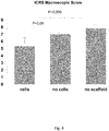
- the gross repair assessment of specimens was nearly normal (grade 2) only for the untreated group while for the lesions treated with seeded or unseeded scaffold the repair assessment was abnormal (grade 3).
- Microscopic evaluation (ICRS Visual Histological Assessment Scale II) showed significant higher scores for chondrocytes morphology and the superficial layer recovery in the unseeded scaffolds group, with respect to the seeded scaffold group and the untreated lesions; while the immunohistochemical analysis showed an increasing immunopositivity to the collagen type 2 from the superficial to the deep level of the chondral defect in the seeded scaffold group.
- Biochemical evaluation showed a significant higher cellularity in seeded scaffold group while the GAGs (glycosaminoglycans)/DNA ratio was higher in the untreated lesions with respect to the seeded and unseeded scaffolds.
- the present invention showed that the scaffold was easy to handle for surgical implant and stable in the site of implant; at the end of experimental time no signs of synovitis were revealed and all implants were well integrated to the surrounding tissue. The quality of the repaired tissue is superior in the lesions treated with the unseeded scaffolds, thus it is a promising biomaterial.
- the present scaffold displays various advantages: the scaffold is more rigid and has an increased compactness and biomechanical integrity with respect to commercially available scaffold (Maioregen). It is then useful for early physiotherapy and is able to better withstand weight. Further, the components (collagen/HA) are better kept together and are less likely to peel off. The external walls of collagen that wrap the HA portion of the scaffold allow a press fit more simple and the entry of chondrocytes in the scaffold. In addition, the scaffold can be better handled by the medical staff when implanted. Moreover, the scaffolds may be "tailor-made” and personalized.
- the synthetic composite material according the invention further comprises an interconnection zone between the first layer and the second layer wherein the organic material and the inorganic material are co-present.
- the first layer is 0.1-20 mm high and/or the second layer is 2-400 mm and/or the third layer is 0.1-2 mm high and/or the interconnection zone is 0.5-3 mm high, preferably 1 mm high.
- the first layer comprises from 0.5 wt% to 5 wt% of organic material.
- the second layer comprises from 50 wt % to 80 wt % of inorganic material.
- the second porous layer has a pore dimension between 50 ⁇ m and 700 ⁇ m.
- the organic material of the first layer is preferably complexed with at least one polyanionic substance, wherein the polyanionic substance is preferably a signaling molecule, more preferably a growth factor, even more preferably a growth facor selected from the group consisting of: transforming growth factor- ⁇ 1(TGF- ⁇ 1), bone morphogenetic proteins (BMPs) and insulin growth factor-1 (IGF-1).
- the organic material complexed with the above polyanionic substance is chitosan.
- the first layer (2) is seeded with cells, which may penetrate within the full thickness of the first layer.
- the cells are chondrocytes.
- a further object of the invention is a process for the preparation of the synthetic composite material as above disclosed.
- the intermediate material obtained in step d) is also defined as a bi-layer scaffold.
- the first mould of step a) may herein also be defined as MOLD A and the second mould of step f) may herein also be defined as MOLD B.
- the number of the intermediate material produced as above defined that has to be introduced in the second mould of step f) goes from 1 to 18 in a volume of 1 cc.
- the first mould may allow to obtain an intermediate material having a transversal section of a circle, square, polygon or any closed polyline.
- the first layer preferably comprises from 0.5 wt% to 5 wt% of organic material.
- the second layer preferably comprises from 50 wt % to 80 wt % of inorganic material and/or has a pore dimension between 50 ⁇ m and 700 ⁇ m.
- the organic material of the first layer is complexed with at least one polyanionic substance, wherein the polyanionic substance is preferably a signaling molecule, more preferably a growth factor, even more preferably a growth facor selected from the group consisting of: transforming growth factor- ⁇ 1(TGF- ⁇ 1), bone morphogenetic proteins (BMPs) and insulin growth factor-1 (IGF-1).
- the polyanionic substance is preferably a signaling molecule, more preferably a growth factor, even more preferably a growth facor selected from the group consisting of: transforming growth factor- ⁇ 1(TGF- ⁇ 1), bone morphogenetic proteins (BMPs) and insulin growth factor-1 (IGF-1).
- TGF- ⁇ 1 transforming growth factor- ⁇ 1
- BMPs bone morphogenetic proteins
- IGF-1 insulin growth factor-1
- dehydrothermal cross linking is carried out in an oven at a temperature between 110°C and 130°C under vacuum for between 48 and 96 hours.
- Preferably sintering occurs at a temperature between 500°C and 1500°C, more preferably 500°C and 700°C or 1200°C and 1350°C.
- a preferred range of temperature is 500°C-700°C
- HA a preferred range of temperature is 1200°C-1350°C.
- the process further comprises seeding the first layer with cells.
- the cells are chondrocytes.
- the synthetic composite material obtainable according to the above process further comprises a third layer comprising the organic material coating the side walls of the first layer and the side walls of the second layer and/or further comprises an interconnection zone between the first layer and the second layer wherein the organic material and the inorganic material are co-present.
- the first layer is preferably 0.1-20 mm high and/or the second layer is preferably 2-400 mm and/or the third layer is preferably 0.1-2 mm high and/or the interconnection zone is preferably 0.5-3 mm high, more preferably 1 mm high.
- the second layer has a transversal section of a circle, a square, a polygon or any closed polyline.
- the first layer has a transversal section of a circle, a square, a polygon or any closed polyline.
- the inorganic material inserted in the mould in step b) and/or the intermediate material of step d) has preferably a transversal section of a circle, a square, a polygon or any closed polyline.
- the obtainable synthetic composite when two or more intermediate materials are introduced in the second mould of step f), the obtainable synthetic composite may be defined as a composite structure.
- a further object of the invention is a composite structure comprising one or more synthetic composite material as defined above.
- the composite structure comprises at least two synthetic composite materials or intermediate materials as defined above.
- the composite structure will result made of different percentage of volume occupied by the inorganic material, occupied by organic material and of volume occupied by both inorganic and organic material (compenetration between). A preferred percentage is: 19% inorganic, 72% organic, 9% co-presence of both (compenetration).
- the composite structure may comprise one or more layer of synthetic composite material or intermediate materials as above defined, and the synthetic composite material or intermediate materials may be of different dimensions and shape (circle and squared section e.g.).
- tissue engineering comprises e.g. the treatment of osteochondral or skin defects or lesions.
- tissue is soft or hard tissue.
- Another object of the invention is a synthetic skin material, plaster, bandage comprising the synthetic composite material as above defined, or the composite structure as above defined.
- the first layer comprising an organic material is porous.
- the organic material is collagen type 1 and the inorganic material is hydroxyapatite.
- Other types of natural organic polymer may further be employed, as for example, collagen type 2, collagen type 3 or chitosan.
- Chitosan is a natural polysaccharide that is structurally similar to GAGs and has recently emerged as an alternative material for cartilage tissue engineering.
- Chitosan shows good biocompatibility, biodegradability and capacity to stimulate the activity of growth factors. In addition, it is known to contribute to the maintenance of the chondrogenic phenotype, especially in terms of its morphology.
- One of the interesting features of chitosan is its cationic nature occurring from primary amine groups, thus providing a high charge density in an acidic solution. The cationic charges allow chitosan to form water-insoluble ionic complexes with variety of polyanionic substances: e.g. for drug delivery applications. Therefore, the opportunity of the scaffolds to carry and provide signaling molecules, such as growth factors, to the site of implantation is of great interest as it could compensate or potentiate some of the parameters necessary to achieve the desired tissue formation.
- TGF- ⁇ 1 transforming growth factor- ⁇ 1
- BMPs bone morphogenetic proteins
- IGF-1 insulin growth factor-1
- scaffolds associated with growth factors should provide the means to precisely control their doses and supplementation rate at a local level.
- growth factors are labile and have a short half-life in the body.
- the present invention allows to provide more complex systems.
- a desired number of the obtained bi-layer scaffolds can be inserted in the MOLD B in order to get a composite structure with an increased compliance in transversal and/or longitudinal direction ( Fig.10B , C ).
- the bi-layer scaffold that can be produced with MOLD A can be of different shape.
- the transversal section can be a circle, square, polygon or any closed polyline ( Fig.10D ).
- the number of the bi-layer scaffolds produced with MOLD A and that has to be inserted in MOLD B can be variable: from 1 to 18 in a volume of 1 cc.
- the final complex scaffold made of a variable number of bi-layer scaffold herein referred also as composite structure comprising one or more synthetic composite material of the invention, can also be of different shape, even custom-made from clinical image of the defect to be repaired.
- the final complex scaffold will result made of different percentage of volume occupied by the inorganic scaffold, occupied by organic scaffold and of volume occupied by both inorganic and organic scaffold (compenetration between). A preferred percentage is: 19% inorganic, 72% organic, 9% co-presence of both (compenetration).
- Table I an example of the volume % of the inorganic and organic part that it is possible to obtain with different number of bi-layer scaffolds.
- Table I Number of bi-layer scaffolds % Inorganic % Organic % Inorganic-Organic 1 36% 46% 18% 2 34% 49% 17% 3 32% 53% 16% 6 25% 62% 13% 12 22% 67% 11% GOLD STANDARD - 18 19% 72% 9%
- the composite structure comprising one or more synthetic composite as above described presents a greater flexibility and an increased cell repopulation in vivo.
- the scaffold is a three-dimensional structure made of superior collagen scaffold partially penetrating into a bottom hydroxyapatite scaffold.
- the two fabrication processes of respectively collagen and hydroxyapatite scaffold were separately optimised in previous studies [22].
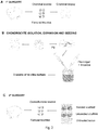
- a multi-step procedure was assessed that is described in the six steps reported below ( Fig.1 ).
- the chondral plugs were placed in 50 ml test tubes containing phosphate buffered saline and the antibiotic/antimycotic solution. The wound was then closed in layers in standard fashion. An impermeable dressing using betadine gel was applied (Viatris spa, Milan, Italy). Prophylactic antibiotic therapy was instituted using 1,200 units of penicillin G (Bicillin L-A; Wyeth Laboratories Inc. Philadelphia, PA, USA), given on days two and four postoperatively. Pain management consisted of Duragesic 75 ⁇ g/h (Fentanyl transdermal system; Janssen Pharmaceutica Inc. Titusville, NJ, USA) and 0.3 mg of buprenorphine (Buprenex injectable; Reckitt & Colman Pharmaceuticals Inc. Richmond, VA, USA). No immobilization was applied after operation and the animal was allowed to move freely.
- cartilage slices (“native cartilage”) harvested from the same animals were frozen as a control tissue for biochemical analysis; all other cartilage slices were digested in Ham's medium (Celbio, Pero, MI, Italy) containing 0.1% collagenase Type 2 (DBA-Italia Srl, Segrate, MI, Italy) and 1% of the antibiotic/antimycotic solution (10,000 units Penicillin, 10 mg Streptomycin and 25 ⁇ g Amphotericin B/ml in 0.9% sodium chloride; Sigma Chemical Co., St. Louis, MO, USA). The specimens were incubated overnight in an oscillating water bath at 37° Celsius.
- Undigested tissue and debris were removed by filtering the cell suspension using a 100 micron sterile filter (BD Falcon, Bedford, MA, USA). The cell suspension obtained was centrifuged at 1400 rpm for 10 minutes. The cell pellet was washed twice in phosphate buffered saline (Celbio) and 2% antibiotic/antimycotic solution. Viability of the chondrocytes was assessed by Trypan blue staining (Sigma) and recorded as a percentage of viable chondrocytes per high power field. The exact cell count per milliliter was established using a haemocytometer.
- Fresh chondrocytes were plated at a concentration of 10000 cells/cm 2 and cultured in DMEM (Lonza, Italia) containing 10% FBS (Euroclone), 1% glutamine (EuroClone, Milan, Italy), and 1% antibiotic/antimycotic solution (Sigma), 5 ng/ml FGF-2 (R&D Systems, Minneapolis, MN, USA), 10 ng/ml TGF ⁇ -1 (R&D Systems, Minneapolis, MN, USA); medium was changed three times a week.
- FBS Euroclone
- glutamine EuroClone, Milan, Italy
- antibiotic/antimycotic solution Sigma
- 5 ng/ml FGF-2 R&D Systems, Minneapolis, MN, USA
- 10 ng/ml TGF ⁇ -1 R&D Systems, Minneapolis, MN, USA
- the de-differentiated chondrocytes were collected, resuspended in a solution containing bovine fibrinogen (for example 110 mg/ml, FlukaChemie GmbH, Buchs, SG, Switzerland), aprotinin (0,2 mg/ml) (Sigma), tranexamic acid (1,5 mg/ml) (Sigma) and adjusted to a concentration of 80x10 6 cells/ml. Then, 100 ⁇ l of the fibrinogen-cell suspension was seeded onto the scaffold on the collagen layer (8 x 10 6 cells/ scaffold; possible range: 0,5-16 x 10 6 cells/ scaffold) prepared according to the design parameters optimized previously [24].
- bovine fibrinogen for example 110 mg/ml, FlukaChemie GmbH, Buchs, SG, Switzerland
- aprotinin 0.,2 mg/ml
- tranexamic acid (1,5 mg/ml)
- 100 ⁇ l of the fibrinogen-cell suspension was seeded onto the scaffold on the collagen layer (8 x
- thrombin (1,37 mg/ml, Chemicon International, Inc., Temecula, CA, USA) were added in order to form fibrin glue. After 30 minutes, complete polymerization was reached and the scaffolds were placed in culture flasks for the in vitro culture.
- the seeded scaffolds were cultured in vitro for 3 weeks in DMEM (Lonza, Italia) containing 10% FBS (Euroclone), 1% glutamine (EuroClone, Milan, Italy), and 1% antibiotic/antimycotic solution (Sigma), ascorbic acid 50 ⁇ g/ml (Sigma), 10 ng/ml TGF ⁇ -3 (R&D Systems, Minneapolis, MN, USA); medium was changed three times a week ( Fig. 2B ).
- the macroscopic appearance of the repaired tissue was examined using the ICRS Macroscopic Score [25], which evaluates the degree of defect repair, the integration to the border zone, and the macroscopic appearance. Two observers, both blind to the treatment, independently scored the specimens.
- the repair areas were removed with healthy tissue surrounding the lesions. Each sample was cut in half, along the central axis of the lesions. Half sample was fixed in 10% (v/v) phosphate-buffered formaldehyde. The samples were then dehydrated in a graded 50% (v/v), 70% (v/v), 95% (v/v) and 100% (v/v) ethanol series, embedded in paraffin and cut into 4 ⁇ m-thick sections. Finally, the sections were stained with SAFRANIN-0, using a standard staining protocol, for the evaluation of the morphology and GAGs deposition. Some sections were used for immunohistochemical analysis of collagen type I and II (Chondrex staining kit, ChondrexInc, USA).
- Biotin secondary antibody- streptavidine peroxidase (1:500) was applied for 1 hour at room temperature. Peroxidase activity was visualized with diaminobenzidine (DAB; Dakocytomation, Milan, Italy) and H 2 O 2 as substrates. All incubations were performed in a moist chamber at room temperature, using PBS for washes between incubation steps. Sections were counterstained with hematoxylin, dehydrated and mounted in mounting medium. Photomicrographs were taken with an Olympus BX51 microscope (Olympus, Italy) equipped with a digital camera and final magnifications were calculated.
- DAB diaminobenzidine
- the biochemical analysis was performed on the other half of samples for each experimental condition (1 seeded scaffold, 1 unseeded scaffold and 1 untreated lesion).
- the samples were digested in papain (Sigma) for 16-24 h at 60°C; the digestion solution was composed of 125 ⁇ g/mL of papain (Sigma) in 100 mM sodium phosphate, 10 mM sodium EDTA (Sigma), 10 mM cysteine hydrochloride (Sigma), 5 mM EDTA adjusted to pH 6.5 and brought to 100 mL of solution with distilled water. After the digestion, the samples were stored at -80°C until analysis. Aliquots of the digested samples were assayed separately for proteoglycan and DNA contents.
- Proteoglycan content was estimated by quantifying the amount of sulphated glycosaminoglycans using the 1,9-dimethylmethylene (DMB) blue dye binding assay (Polysciences Inc., Washington, PA, USA) and a microplate reader (wavelength: 540 nm).
- the standard curve for the analysis was generated by using bovine trachea chondroitin sulfate A (Sigma).
- DNA content was evaluated with the Quant-iT Picogreen dsDNA Assay Kit (Molecular Probes, Invitrogen., Eugene, OR, USA) and a fluorescence microplate reader and standard fluorescein wavelengths (excitation 485 nm, emission 538 nm, cut-off 530nm).
- the standard curve for the analysis was generated using the bacteriophage lambda DNA supplied with the kit.
- Descriptive statistics were used to determine groups' means and standard deviations for numerical data, and analysis was performed using analysis of variance. Statistical significance was defined as a p-value of ⁇ 0.05.
- the unseeded scaffolds were characterized by a linear superficial layer with an uniform GAGs distribution throughout the lesion site ( Fig.6A ); moreover, the cells were chondrocytes-like and some of them were surrounded by lacunae ( Fig.7A,D ).
- the seeded scaffolds and the repairing tissue of the untreated lesions showed irregular superficial layers with hollows ( Fig.6B,C ); in the superficial layer, the cell morphology is mainly fibroblast-like with no lacunae ( Fig.7B,C ), while in the deeper region some cells showed a chondrocyte-like phenotype, but no lacunae were present around them ( Fig.7E,F ).
- Immunohistochemical analysis for collagen type 2 showed in the untreated group a scarce immunoreactivity, while in the cell - group a high reactivity in the lateral part of the defect was observed; additionally, in the cells + group an increasing immunopositivity from the superficial to the deep level of the defect was also detected.
- the evaluation of collagen type 1 showed in all groups a scarce immunopositivity that becomes stronger in a deep level of the defect only in the cell + group ( Fig.8 ).
- the experimental samples were digested in papain in order to measure either the DNA content, an index of cellularity, or GAGs production as a marker of the extra-cellular matrix.
- the seeded scaffolds showed a significative higher cellularity while the GAGs/DNA ratio was higher in the untreated lesions with respect to the seeded and unseeded scaffolds(P ⁇ 0.05); no differences were observed in GAGs production among the different experimental groups ( Fig. 9B ).
- the scaffold is preferably made of the combination of a collagen type 1 sponge, previously characterized for its compatibility and application as a supporting materials for cartilage tissue engineering applications, and a HA component.
- the bilayer scaffold was successfully produced thanks to the fabrication of a properly designed mould via stereolithography that allowed for obtaining an interconnection zone where the two materials (collagen and HA) were co-present for a pre-established thickness ( Fig.1 ).
- the two materials are co-present for 1 mm height, but this height can have a variable range: generally, this can be fabricated with a range of 0.5-3 mm.
- This particular structure has the advantage of allowing for a greater integration of the two materials and also, duplicating the native tissues, disposing the ideal substratum for a proper re-population following in vivo implantation.
- the second novelty introduced i.e. the fabrication via stereolithografy of a second mould for a further collagen lyophilisation around the HA cylinder, has the double aim of both improving the collagen/hydroxyapatite integration and facilitating the substitute insertion in the osteochondral lesion, allowing for a certain degree of change in the diameter of the cylinder through a different hydration.
- Osteochondral defects created in the surface of the trochlea of adult pigs were treated by implanting a biphasic scaffold, which were either seeded with autologous chondrocytes or left unseeded.
- the use of collagen type 1 sponges has been already introduced by several groups for the in vitro engineering of cartilage [27-30] demonstrating that this biocompatible material can support chondrocytes survival and synthetic activity during time with the consequent maturation of the scaffold into a chondral substitute.
- Some works introduced the combination of fibrin glue to ameliorate scaffold seeding and maturation [31,32]; in particular, Malicev et al. combined fibrin glue with collagen type 1 sponge improving scaffold seeding and the in vitro maturation [33].
- the bone phase was left unseeded allowing the mesenchymal stem cells from the adjacent bone marrow to migrate following the implantation and undergo an osteogenic differentiation.
- the repair induced by the acellular scaffold was better regarding the histological aspect of the superficial layer and the morphology of cells, demonstrating that the scaffold was able to allow the colonization and the maturation of the cells deriving from the local sources. So, the structure of the scaffolds permitted the migration of stem cells and their subsequently chondrogenic differentiation. The authors cannot exclude, however, the contribution of other cells deriving from blood or from the surrounding tissues, which were able to differentiate into a cartilaginous or fibrocartilaginous phenotype in the joint environment.
- the scaffold seeded with autologous chondrocytes promoted the formation of a repairing tissue with high cellularity but low GAGs production, suggesting a low chondrocytes activity. So, at the experimental time of this study, the quantity and the quality of the repaired tissue was far to be similar to the native cartilage.
- the immunohistochemical analysis revealed in the cells + group an increasing immunopositivity for the collagen type 2 from the superficial to the deep level of the defect, showing a more hyaline aspect of the repaired tissue, compared with the others groups.
- the bilayer scaffold of the present invention is easy to handle for surgical implant and stable in the implant site; at the end of experimental time, no signs of synovitis were revealed and all implants were well integrated to the surrounding tissue.
- the quality of the repaired tissue produced within the bulk of the unseeded osteochondral scaffolds demonstrated the potential of this material for one-step procedure for articular cartilage repair.
Landscapes
- Health & Medical Sciences (AREA)
- Chemical & Material Sciences (AREA)
- Life Sciences & Earth Sciences (AREA)
- Medicinal Chemistry (AREA)
- General Health & Medical Sciences (AREA)
- Public Health (AREA)
- Transplantation (AREA)
- Epidemiology (AREA)
- Dermatology (AREA)
- Animal Behavior & Ethology (AREA)
- Oral & Maxillofacial Surgery (AREA)
- Veterinary Medicine (AREA)
- Engineering & Computer Science (AREA)
- Biomedical Technology (AREA)
- Cell Biology (AREA)
- Chemical Kinetics & Catalysis (AREA)
- Inorganic Chemistry (AREA)
- Botany (AREA)
- Zoology (AREA)
- Dispersion Chemistry (AREA)
- Rheumatology (AREA)
- Urology & Nephrology (AREA)
- Biophysics (AREA)
- Molecular Biology (AREA)
- Vascular Medicine (AREA)
- Materials For Medical Uses (AREA)
- Polymers & Plastics (AREA)
- Organic Chemistry (AREA)
Claims (16)
- Synthetisches Verbundmaterial (1) zur Gewebereparatur, aufweisend:- eine erste Schicht (2) aus Kollagen vom Typ 1 als organisches Material, welches Seitenwände (3) sowie eine externe Fläche (4) aufweist;- eine zweite poröse Schicht (5) aus Hydroxyapatit als anorganisches Material, welches Seitenwände (6) aufweist;wobei die erste Schicht in direktem Kontakt mit der zweiten Schicht angeordnet ist, und wobei die Seitenwände (3) der ersten Schicht und die Seitenwände (6) der zweiten Schicht mit einer dritten Schicht aus Kollagen überzogen sind.
- Synthetisches Verbundmaterial (1) nach Anspruch 1, ferner aufweisend einen Verbindungsbereich zwischen der ersten Schicht und der zweiten Schicht, in welchem sowohl das organische Material als auch das anorganische Material vorhanden ist.
- Synthetisches Verbundmaterial (1) nach Anspruch 1 oder 2, wobei die erste Schicht 0,1 - 20 mm hoch ist und/oder die zweite Schicht 2 - 400 mm hoch ist und/oder die dritte Schicht 0,1 - 2 mm hoch ist,
und/oder der Verbindungsbereich 0,5 - 3 mm hoch, vorzugsweise 1 mm hoch, ist. - Synthetisches Verbundmaterial nach einem der vorstehenden Ansprüche, wobei die zweite poröse Schicht eine Porengröße von zwischen 50 µm und 700 µm aufweist.
- Synthetisches Verbundmaterial nach einem der vorstehenden Ansprüche, wobei das organische Material der ersten Schicht mit mindestens einer polyanionischen Substanz komplexiert ist, wobei die polyanionische Substanz vorzugsweise ein Signalmolekül, bevorzugter ein Wachstumsfaktor, und noch bevorzugter ein Wachstumsfaktor ausgewählt aus der Gruppe umfassend transformierenden Wachstumsfaktor-β1 (TGF-β1), morphogenetische Knochenproteine (BMPs) und Insulinwachstumsfaktor-1 (IGF-1) ist.
- Synthetisches Verbundmaterial nach einem der vorstehenden Ansprüche, wobei die erste Schicht (2) mit Zellen beimpft ist, wobei die Zellen vorzugsweise Chondrozyten sind.
- Prozess zur Herstellung des synthetischen Verbundmaterials nach einem der Ansprüche 1 - 6, umfassend die Schritte:a) Eingießen einer Aufschlämmungszusammensetzung, welche einen flüssigen Träger und das organische Material enthält, in eine erste Form;b) Einbringen eines Materials, welches das anorganische Material enthält, in die Form;c) Durchführen eines Gefriertrocknungszyklus;d) Induzieren dehydrothermaler Vernetzung zum Erlangen eines Zwischenmaterials;e) Entnehmen des Zwischenmaterials aus der ersten Form;f) Einbringen mindestens eines Zwischenmaterials in eine zweite Form, welche dadurch gekennzeichnet ist, dass sie einen größeren Innenbereich aufweist als die erste Form;g) Eingießen der Aufschlämmungszusammensetzung, welche den flüssigen Träger und Kollagen enthält, bis die Form gefüllt ist;h) Durchführen eines Gefriertrocknungszyklus;i) Induzieren dehydrothermaler Vernetzung; undj) optional Sterilisieren des Materials.
- Prozess zur Herstellung eines synthetischen Verbundmaterials aufweisend eine erste Schicht, welche ein organisches Material enthält, und eine zweite Schicht, welche ein anorganisches Material enthält, umfassend die Schritte:a) Eingießen einer Aufschlämmungszusammensetzung, welche einen flüssigen Träger und Kollagen vom Typ 1 als organisches Material enthält, in eine erste Form;b) Einbringen eines Materials, welches Hydroxyapatit als anorganisches Material enthält, in die Form;c) Durchführen eines Gefriertrocknungszyklus;d) Induzieren dehydrothermaler Vernetzung zum Erlangen eines Zwischenmaterials;e) Entnehmen des Zwischenmaterials aus der ersten Form;f) Einbringen mindestens eines Zwischenmaterials in eine zweite Form, welche dadurch gekennzeichnet ist, dass sie einen größeren Innenbereich aufweist als die erste Form;g) Eingießen der Aufschlämmungszusammensetzung, welche den flüssigen Träger und Kollagen enthält, bis die Form gefüllt ist;h) Durchführen eines Gefriertrocknungszyklus;i) Induzieren dehydrothermaler Vernetzung; undj) optional Sterilisieren des Materials,wobei die erste Schicht vorzugsweise zwischen 0,5 Gew.-% und 5 Gew.-% organischen Materials enthält,
und/oder wobei die zweite Schicht vorzugsweise zwischen 50 Gew.-% und 80 Gew.-% anorganischen Materials enthält,
und/oder wobei die dehydrothermale Vernetzung in einem Ofen bei einer Temperatur von zwischen 110 °C und 130 °C unter Vakuum für zwischen 48 und 96 Stunden durchgeführt wird. - Prozess nach Anspruch 8, wobei das anorganische Material erlangt werden kann durch:- Imprägnieren eines porösen Trägers mit einer Aufschlämmung, welche 50 - 80 Gew.-% anorganischen Materials enthält;- Trockenen und Erwärmen des Materials, um den porösen Träger zu entfernen;- Sintern, wobei das Sintern vorzugsweise bei einer Temperatur von zwischen 500 °C und 1500 °C, vorzugsweise 500 °C und 700 °C oder 1200 °C und 1350 °C, erfolgt.
- Prozess nach einem der Ansprüche 8 oder 9, ferner umfassend Beimpfen der ersten Schicht mit Zellen, wobei die Zellen vorzugsweise Chondrozyten sind.
- Synthetisches Verbundmaterial, welches im Einklang mit dem Prozess nach einem der Ansprüche 8 bis 10 erlangt werden kann.
- Synthetisches Verbundmaterial nach einem der Ansprüche 1 - 6 oder nach Anspruch 11, wobei die zweite Schicht einen Querschnitt in Form eines Kreises, eines Quadrats, eines Polygons oder einer beliebigen geschlossenen Polylinie aufweist.
- Verbundstruktur aufweisend ein oder mehrere synthetische Verbundmaterialien nach einem der Ansprüche 1 - 6 oder der Ansprüche 11 oder 12.
- Synthetisches Verbundmaterial nach einem der Ansprüche 1 - 6 oder der Ansprüche 11 - 12, oder die Verbundstruktur nach Anspruch 13 zur Verwendung in einem Verfahren zur Gewebezüchtung, wobei das Gewebe vorzugsweise weiches oder hartes Gewebe ist.
- Synthetisches Knochenmaterial, Knochenimplantat, Knochentransplantat, Knochenersatzmaterial, Knochengerüst, Füllstoff, Beschichtung oder Zement, welcher/welches/welche das synthetische Verbundmaterial nach einem der Ansprüche 1 - 6 oder der Ansprüche 11 - 12 oder die Verbundstruktur nach Anspruch 13 enthält.
- Synthetisches Hautmaterial, Pflaster, Bandage, welches/welche das synthetische Verbundmaterial nach einem der Ansprüche 1 - 6 oder der Ansprüche 11 - 12 oder die Verbundstruktur nach Anspruch 13 enthält.
Applications Claiming Priority (2)
| Application Number | Priority Date | Filing Date | Title |
|---|---|---|---|
| US201361824706P | 2013-05-17 | 2013-05-17 | |
| PCT/EP2014/060264 WO2014184391A1 (en) | 2013-05-17 | 2014-05-19 | Composite scaffold for tissue repair |
Publications (2)
| Publication Number | Publication Date |
|---|---|
| EP2996733A1 EP2996733A1 (de) | 2016-03-23 |
| EP2996733B1 true EP2996733B1 (de) | 2021-07-28 |
Family
ID=50736095
Family Applications (1)
| Application Number | Title | Priority Date | Filing Date |
|---|---|---|---|
| EP14725162.3A Active EP2996733B1 (de) | 2013-05-17 | 2014-05-19 | Verbundgerüst zur gewebereparatur |
Country Status (3)
| Country | Link |
|---|---|
| US (1) | US10940236B2 (de) |
| EP (1) | EP2996733B1 (de) |
| WO (1) | WO2014184391A1 (de) |
Families Citing this family (5)
| Publication number | Priority date | Publication date | Assignee | Title |
|---|---|---|---|---|
| WO2014130883A2 (en) * | 2013-02-22 | 2014-08-28 | Allosource | Cartilage mosaic compositions and methods |
| EP2967874B1 (de) | 2013-03-15 | 2019-11-20 | AlloSource | Perforierte osteochondrale allograft-zusammensetzungen |
| GB2549714A (en) * | 2016-04-25 | 2017-11-01 | Pharmaceutical Business Consultants Ltd | Vascularity affinity precursor structure for musculo-skeletal tissue healing |
| KR102183048B1 (ko) * | 2017-06-19 | 2020-11-25 | 주식회사 나이벡 | 골조직 재생을 위한 일체형 바이오소재 및 그 제조방법 |
| CN107412875A (zh) * | 2017-08-03 | 2017-12-01 | 上海长海医院 | 介孔硅酸钙镁/小麦蛋白/聚已内酯复合支架及其制备方法和应用 |
Family Cites Families (2)
| Publication number | Priority date | Publication date | Assignee | Title |
|---|---|---|---|---|
| JP4968639B2 (ja) | 2005-12-15 | 2012-07-04 | 独立行政法人物質・材料研究機構 | リン酸カルシウム及びアパタイト/コラーゲン複合物からなる複合体 |
| CA2750605C (en) * | 2009-01-23 | 2019-01-22 | Royal College Of Surgeons In Ireland | Layered scaffold suitable for osteochondral repair |
-
2014
- 2014-05-19 WO PCT/EP2014/060264 patent/WO2014184391A1/en not_active Ceased
- 2014-05-19 US US14/891,932 patent/US10940236B2/en active Active
- 2014-05-19 EP EP14725162.3A patent/EP2996733B1/de active Active
Non-Patent Citations (1)
| Title |
|---|
| None * |
Also Published As
| Publication number | Publication date |
|---|---|
| US10940236B2 (en) | 2021-03-09 |
| US20160106885A1 (en) | 2016-04-21 |
| EP2996733A1 (de) | 2016-03-23 |
| WO2014184391A1 (en) | 2014-11-20 |
Similar Documents
| Publication | Publication Date | Title |
|---|---|---|
| KR102248576B1 (ko) | 세포 및 조직 성장을 촉진하기 위한 고체 기질 | |
| US9072815B2 (en) | Layered collagen and HA scaffold suitable for osteochondral repair | |
| Bernstein et al. | Microporous calcium phosphate ceramics as tissue engineering scaffolds for the repair of osteochondral defects: Histological results | |
| Wei et al. | Mesenchymal stem cell-loaded porous tantalum integrated with biomimetic 3D collagen-based scaffold to repair large osteochondral defects in goats | |
| Liu et al. | Osteochondral regeneration using an oriented nanofiber yarn‐collagen type I/hyaluronate hybrid/TCP biphasic scaffold | |
| US20150110846A1 (en) | Biphasic osteochondral scaffold for reconstruction of articular cartilage | |
| Marmotti et al. | Autologous cartilage fragments in a composite scaffold for one stage osteochondral repair in a goat model | |
| Nie et al. | Decellularized orthopaedic tissue-engineered grafts: biomaterial scaffolds synthesised by therapeutic cells | |
| US20060083729A1 (en) | Biocompatible tissue sealant for treatment of osteochondral and bone defects using an acellular matrix implant | |
| US10940236B2 (en) | Composite scaffold for tissue repair | |
| CN108992212B (zh) | 骨-软骨一体修复支架及其制备方法 | |
| KR20090038035A (ko) | 바이오물질 | |
| CN101417151A (zh) | 生物源性含钙化层骨软骨组织工程支架 | |
| Oryan et al. | Effectiveness of a biodegradable 3D polylactic acid/poly (ɛ‐caprolactone)/hydroxyapatite scaffold loaded by differentiated osteogenic cells in a critical‐sized radius bone defect in rat | |
| Cai et al. | A Col I and BCP ceramic bi-layer scaffold implant promotes regeneration in osteochondral defects | |
| Thomas | The Development of a Novel Cartilage Analog Scaffold for Chondral Regeneration | |
| Kafili et al. | Tissue-Specific Biomaterials for Use in Musculoskeletal Tissue Engineering | |
| Huang et al. | Nano-calcium phosphate/PLLA incorporated with bFGF as in situ-transplantable carrier for cartilage regeneration | |
| de Almeida | Doctor in Philosophy | |
| CN122070892A (en) | Cartilage implants and related devices and methods | |
| Kim et al. | Regeneration of cartilage tissue by autologous chondrocytes transplantation for cartilage defects in a experimental bovine model | |
| WO2009056804A2 (en) | Elastic composite biomaterial | |
| Somerson | Mesenchymal Stem Cell Constructs for Repair of Focal Cartilage Defects in an Ovine Model | |
| Meagher | Engineering Synthetic Bone Graft Substitutes with Improved Mechanical Properties, Enhanced Bioactivity, and On-Demand Growth Factor Delivery | |
| Zhao | Biomaterial and Cell Based Cartilage Repair |
Legal Events
| Date | Code | Title | Description |
|---|---|---|---|
| PUAI | Public reference made under article 153(3) epc to a published international application that has entered the european phase |
Free format text: ORIGINAL CODE: 0009012 |
|
| 17P | Request for examination filed |
Effective date: 20151216 |
|
| AK | Designated contracting states |
Kind code of ref document: A1 Designated state(s): AL AT BE BG CH CY CZ DE DK EE ES FI FR GB GR HR HU IE IS IT LI LT LU LV MC MK MT NL NO PL PT RO RS SE SI SK SM TR |
|
| AX | Request for extension of the european patent |
Extension state: BA ME |
|
| DAX | Request for extension of the european patent (deleted) | ||
| STAA | Information on the status of an ep patent application or granted ep patent |
Free format text: STATUS: EXAMINATION IS IN PROGRESS |
|
| 17Q | First examination report despatched |
Effective date: 20171110 |
|
| GRAP | Despatch of communication of intention to grant a patent |
Free format text: ORIGINAL CODE: EPIDOSNIGR1 |
|
| STAA | Information on the status of an ep patent application or granted ep patent |
Free format text: STATUS: GRANT OF PATENT IS INTENDED |
|
| INTG | Intention to grant announced |
Effective date: 20210202 |
|
| RAP1 | Party data changed (applicant data changed or rights of an application transferred) |
Owner name: OSPEDALE SAN RAFFAELE S.R.L. Owner name: UNIVERSITA DEL SALENTO |
|
| RIN1 | Information on inventor provided before grant (corrected) |
Inventor name: FRASCHINI, GIANFRANCO Inventor name: GERVASO, FRANCESCA Inventor name: SANNINO, ALESSANDRO Inventor name: PERETTI, GIUSEPPE Inventor name: DI GIANCAMILLO, ALESSIA Inventor name: SCALERA, FRANCESCO Inventor name: DEPONTI, DANIELA ROSA Inventor name: DOMENEGHINI, CINZIA |
|
| GRAS | Grant fee paid |
Free format text: ORIGINAL CODE: EPIDOSNIGR3 |
|
| GRAA | (expected) grant |
Free format text: ORIGINAL CODE: 0009210 |
|
| STAA | Information on the status of an ep patent application or granted ep patent |
Free format text: STATUS: THE PATENT HAS BEEN GRANTED |
|
| AK | Designated contracting states |
Kind code of ref document: B1 Designated state(s): AL AT BE BG CH CY CZ DE DK EE ES FI FR GB GR HR HU IE IS IT LI LT LU LV MC MK MT NL NO PL PT RO RS SE SI SK SM TR |
|
| REG | Reference to a national code |
Ref country code: GB Ref legal event code: FG4D |
|
| REG | Reference to a national code |
Ref country code: CH Ref legal event code: EP |
|
| REG | Reference to a national code |
Ref country code: AT Ref legal event code: REF Ref document number: 1414114 Country of ref document: AT Kind code of ref document: T Effective date: 20210815 |
|
| REG | Reference to a national code |
Ref country code: IE Ref legal event code: FG4D |
|
| REG | Reference to a national code |
Ref country code: DE Ref legal event code: R096 Ref document number: 602014079038 Country of ref document: DE |
|
| REG | Reference to a national code |
Ref country code: LT Ref legal event code: MG9D |
|
| REG | Reference to a national code |
Ref country code: NL Ref legal event code: MP Effective date: 20210728 |
|
| REG | Reference to a national code |
Ref country code: AT Ref legal event code: MK05 Ref document number: 1414114 Country of ref document: AT Kind code of ref document: T Effective date: 20210728 |
|
| PG25 | Lapsed in a contracting state [announced via postgrant information from national office to epo] |
Ref country code: AT Free format text: LAPSE BECAUSE OF FAILURE TO SUBMIT A TRANSLATION OF THE DESCRIPTION OR TO PAY THE FEE WITHIN THE PRESCRIBED TIME-LIMIT Effective date: 20210728 Ref country code: BG Free format text: LAPSE BECAUSE OF FAILURE TO SUBMIT A TRANSLATION OF THE DESCRIPTION OR TO PAY THE FEE WITHIN THE PRESCRIBED TIME-LIMIT Effective date: 20211028 Ref country code: LT Free format text: LAPSE BECAUSE OF FAILURE TO SUBMIT A TRANSLATION OF THE DESCRIPTION OR TO PAY THE FEE WITHIN THE PRESCRIBED TIME-LIMIT Effective date: 20210728 Ref country code: ES Free format text: LAPSE BECAUSE OF FAILURE TO SUBMIT A TRANSLATION OF THE DESCRIPTION OR TO PAY THE FEE WITHIN THE PRESCRIBED TIME-LIMIT Effective date: 20210728 Ref country code: FI Free format text: LAPSE BECAUSE OF FAILURE TO SUBMIT A TRANSLATION OF THE DESCRIPTION OR TO PAY THE FEE WITHIN THE PRESCRIBED TIME-LIMIT Effective date: 20210728 Ref country code: PT Free format text: LAPSE BECAUSE OF FAILURE TO SUBMIT A TRANSLATION OF THE DESCRIPTION OR TO PAY THE FEE WITHIN THE PRESCRIBED TIME-LIMIT Effective date: 20211129 Ref country code: NO Free format text: LAPSE BECAUSE OF FAILURE TO SUBMIT A TRANSLATION OF THE DESCRIPTION OR TO PAY THE FEE WITHIN THE PRESCRIBED TIME-LIMIT Effective date: 20211028 Ref country code: NL Free format text: LAPSE BECAUSE OF FAILURE TO SUBMIT A TRANSLATION OF THE DESCRIPTION OR TO PAY THE FEE WITHIN THE PRESCRIBED TIME-LIMIT Effective date: 20210728 Ref country code: HR Free format text: LAPSE BECAUSE OF FAILURE TO SUBMIT A TRANSLATION OF THE DESCRIPTION OR TO PAY THE FEE WITHIN THE PRESCRIBED TIME-LIMIT Effective date: 20210728 Ref country code: RS Free format text: LAPSE BECAUSE OF FAILURE TO SUBMIT A TRANSLATION OF THE DESCRIPTION OR TO PAY THE FEE WITHIN THE PRESCRIBED TIME-LIMIT Effective date: 20210728 Ref country code: SE Free format text: LAPSE BECAUSE OF FAILURE TO SUBMIT A TRANSLATION OF THE DESCRIPTION OR TO PAY THE FEE WITHIN THE PRESCRIBED TIME-LIMIT Effective date: 20210728 |
|
| PG25 | Lapsed in a contracting state [announced via postgrant information from national office to epo] |
Ref country code: PL Free format text: LAPSE BECAUSE OF FAILURE TO SUBMIT A TRANSLATION OF THE DESCRIPTION OR TO PAY THE FEE WITHIN THE PRESCRIBED TIME-LIMIT Effective date: 20210728 Ref country code: LV Free format text: LAPSE BECAUSE OF FAILURE TO SUBMIT A TRANSLATION OF THE DESCRIPTION OR TO PAY THE FEE WITHIN THE PRESCRIBED TIME-LIMIT Effective date: 20210728 Ref country code: GR Free format text: LAPSE BECAUSE OF FAILURE TO SUBMIT A TRANSLATION OF THE DESCRIPTION OR TO PAY THE FEE WITHIN THE PRESCRIBED TIME-LIMIT Effective date: 20211029 |
|
| REG | Reference to a national code |
Ref country code: DE Ref legal event code: R081 Ref document number: 602014079038 Country of ref document: DE Owner name: OSPEDALE SAN RAFFAELE S.R.L, IT Free format text: FORMER OWNERS: OSPEDALE SAN RAFFAELE S.R.L, MILANO, IT; UNIVERSITA DEL SALENTO, LECCE, IT |
|
| RAP2 | Party data changed (patent owner data changed or rights of a patent transferred) |
Owner name: OSPEDALE SAN RAFFAELE S.R.L. |
|
| REG | Reference to a national code |
Ref country code: GB Ref legal event code: 732E Free format text: REGISTERED BETWEEN 20220331 AND 20220406 |
|
| PG25 | Lapsed in a contracting state [announced via postgrant information from national office to epo] |
Ref country code: DK Free format text: LAPSE BECAUSE OF FAILURE TO SUBMIT A TRANSLATION OF THE DESCRIPTION OR TO PAY THE FEE WITHIN THE PRESCRIBED TIME-LIMIT Effective date: 20210728 |
|
| REG | Reference to a national code |
Ref country code: DE Ref legal event code: R097 Ref document number: 602014079038 Country of ref document: DE |
|
| PG25 | Lapsed in a contracting state [announced via postgrant information from national office to epo] |
Ref country code: SM Free format text: LAPSE BECAUSE OF FAILURE TO SUBMIT A TRANSLATION OF THE DESCRIPTION OR TO PAY THE FEE WITHIN THE PRESCRIBED TIME-LIMIT Effective date: 20210728 Ref country code: SK Free format text: LAPSE BECAUSE OF FAILURE TO SUBMIT A TRANSLATION OF THE DESCRIPTION OR TO PAY THE FEE WITHIN THE PRESCRIBED TIME-LIMIT Effective date: 20210728 Ref country code: RO Free format text: LAPSE BECAUSE OF FAILURE TO SUBMIT A TRANSLATION OF THE DESCRIPTION OR TO PAY THE FEE WITHIN THE PRESCRIBED TIME-LIMIT Effective date: 20210728 Ref country code: EE Free format text: LAPSE BECAUSE OF FAILURE TO SUBMIT A TRANSLATION OF THE DESCRIPTION OR TO PAY THE FEE WITHIN THE PRESCRIBED TIME-LIMIT Effective date: 20210728 Ref country code: CZ Free format text: LAPSE BECAUSE OF FAILURE TO SUBMIT A TRANSLATION OF THE DESCRIPTION OR TO PAY THE FEE WITHIN THE PRESCRIBED TIME-LIMIT Effective date: 20210728 Ref country code: AL Free format text: LAPSE BECAUSE OF FAILURE TO SUBMIT A TRANSLATION OF THE DESCRIPTION OR TO PAY THE FEE WITHIN THE PRESCRIBED TIME-LIMIT Effective date: 20210728 |
|
| PLBE | No opposition filed within time limit |
Free format text: ORIGINAL CODE: 0009261 |
|
| STAA | Information on the status of an ep patent application or granted ep patent |
Free format text: STATUS: NO OPPOSITION FILED WITHIN TIME LIMIT |
|
| 26N | No opposition filed |
Effective date: 20220429 |
|
| REG | Reference to a national code |
Ref country code: BE Ref legal event code: MM Effective date: 20220531 |
|
| PG25 | Lapsed in a contracting state [announced via postgrant information from national office to epo] |
Ref country code: MC Free format text: LAPSE BECAUSE OF FAILURE TO SUBMIT A TRANSLATION OF THE DESCRIPTION OR TO PAY THE FEE WITHIN THE PRESCRIBED TIME-LIMIT Effective date: 20210728 Ref country code: LU Free format text: LAPSE BECAUSE OF NON-PAYMENT OF DUE FEES Effective date: 20220519 |
|
| PG25 | Lapsed in a contracting state [announced via postgrant information from national office to epo] |
Ref country code: IE Free format text: LAPSE BECAUSE OF NON-PAYMENT OF DUE FEES Effective date: 20220519 |
|
| PG25 | Lapsed in a contracting state [announced via postgrant information from national office to epo] |
Ref country code: BE Free format text: LAPSE BECAUSE OF NON-PAYMENT OF DUE FEES Effective date: 20220531 |
|
| P01 | Opt-out of the competence of the unified patent court (upc) registered |
Effective date: 20230523 |
|
| P02 | Opt-out of the competence of the unified patent court (upc) changed |
Effective date: 20230527 |
|
| REG | Reference to a national code |
Ref country code: DE Ref legal event code: R082 Ref document number: 602014079038 Country of ref document: DE Representative=s name: KRAUS & LEDERER PARTGMBB, DE |
|
| PG25 | Lapsed in a contracting state [announced via postgrant information from national office to epo] |
Ref country code: HU Free format text: LAPSE BECAUSE OF FAILURE TO SUBMIT A TRANSLATION OF THE DESCRIPTION OR TO PAY THE FEE WITHIN THE PRESCRIBED TIME-LIMIT; INVALID AB INITIO Effective date: 20140519 |
|
| PG25 | Lapsed in a contracting state [announced via postgrant information from national office to epo] |
Ref country code: MK Free format text: LAPSE BECAUSE OF FAILURE TO SUBMIT A TRANSLATION OF THE DESCRIPTION OR TO PAY THE FEE WITHIN THE PRESCRIBED TIME-LIMIT Effective date: 20210728 Ref country code: CY Free format text: LAPSE BECAUSE OF FAILURE TO SUBMIT A TRANSLATION OF THE DESCRIPTION OR TO PAY THE FEE WITHIN THE PRESCRIBED TIME-LIMIT Effective date: 20210728 |
|
| PG25 | Lapsed in a contracting state [announced via postgrant information from national office to epo] |
Ref country code: MT Free format text: LAPSE BECAUSE OF FAILURE TO SUBMIT A TRANSLATION OF THE DESCRIPTION OR TO PAY THE FEE WITHIN THE PRESCRIBED TIME-LIMIT Effective date: 20210728 |
|
| PGFP | Annual fee paid to national office [announced via postgrant information from national office to epo] |
Ref country code: DE Payment date: 20250530 Year of fee payment: 12 |
|
| PGFP | Annual fee paid to national office [announced via postgrant information from national office to epo] |
Ref country code: GB Payment date: 20250530 Year of fee payment: 12 |
|
| PGFP | Annual fee paid to national office [announced via postgrant information from national office to epo] |
Ref country code: IT Payment date: 20250523 Year of fee payment: 12 |
|
| PGFP | Annual fee paid to national office [announced via postgrant information from national office to epo] |
Ref country code: FR Payment date: 20250530 Year of fee payment: 12 |
|
| PGFP | Annual fee paid to national office [announced via postgrant information from national office to epo] |
Ref country code: CH Payment date: 20250606 Year of fee payment: 12 |
|
| PG25 | Lapsed in a contracting state [announced via postgrant information from national office to epo] |
Ref country code: TR Free format text: LAPSE BECAUSE OF FAILURE TO SUBMIT A TRANSLATION OF THE DESCRIPTION OR TO PAY THE FEE WITHIN THE PRESCRIBED TIME-LIMIT Effective date: 20210728 |