EP2981555B1 - New uranium-chelating peptides derived from ef-hand calcium-binding motif useful for uranium biodetection and biodecontamination - Google Patents
New uranium-chelating peptides derived from ef-hand calcium-binding motif useful for uranium biodetection and biodecontamination Download PDFInfo
- Publication number
- EP2981555B1 EP2981555B1 EP14718767.8A EP14718767A EP2981555B1 EP 2981555 B1 EP2981555 B1 EP 2981555B1 EP 14718767 A EP14718767 A EP 14718767A EP 2981555 B1 EP2981555 B1 EP 2981555B1
- Authority
- EP
- European Patent Office
- Prior art keywords
- polypeptide
- protein
- loop
- uranyl
- calcium
- Prior art date
- Legal status (The legal status is an assumption and is not a legal conclusion. Google has not performed a legal analysis and makes no representation as to the accuracy of the status listed.)
- Active
Links
Images






Classifications
-
- C—CHEMISTRY; METALLURGY
- C07—ORGANIC CHEMISTRY
- C07K—PEPTIDES
- C07K14/00—Peptides having more than 20 amino acids; Gastrins; Somatostatins; Melanotropins; Derivatives thereof
- C07K14/435—Peptides having more than 20 amino acids; Gastrins; Somatostatins; Melanotropins; Derivatives thereof from animals; from humans
- C07K14/46—Peptides having more than 20 amino acids; Gastrins; Somatostatins; Melanotropins; Derivatives thereof from animals; from humans from vertebrates
- C07K14/47—Peptides having more than 20 amino acids; Gastrins; Somatostatins; Melanotropins; Derivatives thereof from animals; from humans from vertebrates from mammals
- C07K14/4701—Peptides having more than 20 amino acids; Gastrins; Somatostatins; Melanotropins; Derivatives thereof from animals; from humans from vertebrates from mammals not used
- C07K14/4728—Calcium binding proteins, e.g. calmodulin
-
- G—PHYSICS
- G21—NUCLEAR PHYSICS; NUCLEAR ENGINEERING
- G21F—PROTECTION AGAINST X-RADIATION, GAMMA RADIATION, CORPUSCULAR RADIATION OR PARTICLE BOMBARDMENT; TREATING RADIOACTIVELY CONTAMINATED MATERIAL; DECONTAMINATION ARRANGEMENTS THEREFOR
- G21F9/00—Treating radioactively contaminated material; Decontamination arrangements therefor
- G21F9/28—Treating solids
- G21F9/30—Processing
- G21F9/301—Processing by fixation in stable solid media
- G21F9/307—Processing by fixation in stable solid media in polymeric matrix, e.g. resins, tars
-
- C—CHEMISTRY; METALLURGY
- C07—ORGANIC CHEMISTRY
- C07K—PEPTIDES
- C07K2319/00—Fusion polypeptide
- C07K2319/60—Fusion polypeptide containing spectroscopic/fluorescent detection, e.g. green fluorescent protein [GFP]
Definitions
- the present relates to new uranium-chelating peptides derived from EF-hand calcium-binding motif, useful for uranium biodetection and biodecontamination.
- Uranium is a radioactive heavy metal, which is naturally present in varying concentrations in the environment.
- the wide use of uranium for industrial and military applications increases the risk of its distribution in the environment, which is aggravated by such factors as mining activities, uranium processing, or leaching of radioactive wastes.
- Uranium presents radiological and chemical toxicity to living organisms.
- the linear dioxo uranyl form (UO 2 2+ ) which corresponds to uranium in its hexavalent oxidation state U(VI) is the prevalent form of uranium in the presence of oxygen and the most stable chemical form of uranium in water. It is soluble, bioavailable, and thus potentially toxic.
- molecules derived from peptides are advantageous in that they are non toxic, and can be easily produced either synthetically or using recombinant techniques.
- the development of organisms bearing uranyl biosensors, capable of detecting the uranyl fraction that is bioavailable and thus potentially toxic, is needed to assess water quality on a long term basis.
- bioremediation is an emerging technique that would allow the decontamination of large areas of contaminated water and/or soils.
- the helix-loop-helix calcium-binding motif (EF-hand motif) is the most prevalent Ca 2+ -binding site in proteins.
- the canonical EF-hand motif of about 30 amino acids in length is structured by two alpha-helices bridged by a flexible metal-binding loop (also referred to as calcium-binding loop, calcium-binding site, loop or site) composed of 12 highly conserved residues, with calcium coordinating positions at 1 (+X), 3 (+Y), 5 (+Z), 7 (-Y), 9 (-X) and 12 (-Z).
- the residues at the ligand positions 1, 3, 5 and 12 and also at the non-ligand positions 6 and 8 are highly conserved.
- the calcium ion is coordinated in a pentagonal bipyramidal configuration with a coordination number of 7 to side-chain oxygen bearing groups at positions 1, 3, and 5, a water molecule hydrogen bonded to residue at position 9, and the main chain carbonyl group of residue 7.
- the pentagonal bipyramidal configuration arises from two additional ligands provided by a bidendate glutamic or a monodentate aspartic acid which stabilizes a water ligand, in position 12.
- the residue at position 1 of the loop is most frequently occupied by an aspartate (D); two residues are found at position 3: aspartic acid (D) or asparagine (N), but most frequently D; position 5 is most often occupied by aspartic acid (D), serine (S) or asparagine (N); the residue at position 7 is variable; position 9 shows a preference for residues with a side-chain oxygen (D, N, E, S, T), and the residues more frequently found at position 12 are glutamate (E) and aspartate (D), most frequently E.
- the highly conserved residues at the non ligand positions 6 and 8 are Gly (G) and Ile (I), respectively.
- the majority of the known EF-hand Calcium-Binding proteins (CaBPs) contain paired EF-hand motifs.
- the EF-hand proteins can be divided into two classes: 1) signaling proteins and 2) buffering/transport proteins.
- the first group is the largest and includes the most well-known members of the family such as calmodulin, troponin C and S100B. These proteins typically undergo a calcium-dependent conformational change which opens a target-binding site.
- the latter group is represented by calbindin D9k which remains in a closed conformation upon calcium binding.
- Calmodulin is the most studied representative of the ubiquitous EF-hand protein family and a calcium-binding protein involved in the regulation of a wide range of target enzymes.
- the calmodulin structure (PDB code 1EXR) includes two pairs of EF-hand motifs (EF-hands 1 and 2; EF-hands 3 and 4) in two domains (domain 1 and 2) separated by a flexible ⁇ -helix ( Figure 1A ).
- calcium ligands are provided by three monodentate aspartate (D) at positions 1, 3, 5 of the metal-binding loop (D1, D3, D5), a bidentate glutamate at position 12 (E12), a main chain carbonyl at position 7 and a water molecule stabilized by threonine 9 (T9) side chain, as schematized in Figure 1B .
- Fluorescent sensors for calcium have been constructed based on green fluorescent proteins and calmodulin ( Miyawaki et al., Nature, 1997, 388, 882-887 ; Nagai et al., PNAS, 2004, 101, 10554-10559 ). They are chimeric proteins composed of a short-wavelength variant of GFP, CaM, a glycylglycine linker, the CaM-binding peptide of myosin light-chain kinase (M13), and a long-wavelength variant of GFP.
- Ca 2+ binding to CaM initiates an intramolecular interaction between CaM and M13, which changes the chimeric protein from an extended to a more compact conformation, thereby increasing the efficiency of Fluorescence Resonance Energy Transfer (FRET) from the shorter to the longer-wavelength variant of GFP.
- Yellow cameleons (YCs) have cyan and yellow fluorescent proteins (CFP and GFP) as the FRET donor and acceptor, respectively.
- Synthetic cyclic-peptide variants of calmodulin site 1 consisting of a 33 amino acid sequence in which one, two or three of the aspartic acid residues in positions 1, 3 and 5 of the calcium-binding loop (D1, D3, D5) are substituted with neutral amino acids (T, N or S) and a tyrosine residue is introduced in position 7 of the loop to monitor metal binding using tyrosine fluorescence, bind uranyl with an apparent dissociation constant in the micromolar range (K d of 9.8 to 54.10 -6 M) while they don't bind calcium ( WO 2005/012336 ).
- uranyl biosensors with detection limits in the nanomolar range or below are required to detect uranium content in waters at the maximum concentration levels defined by WHO.
- CaM IP is derived from CaM1 which consists of a 77 amino acids sequence, in which: (i) the T 9 TKE 12 sequence of EF-hand1 loop is substituted by the CK2 recognition sequence TAAE to allow phosphorylation of the threonine 9, in vitro , using recombinant catalytic subunit of protein kinase CK2, (ii) a tyrosine residue is introduced in position 7 of the EF-hand 1 loop so that uranyl and calcium-binding affinities could be determined by following tyrosine fluorescence emission at 302 nm, and (iii) the metal-binding ability of site 2 is impaired by substituting the two aspartate residues in positions 1 and 3 of the loop with alanine residues.
- CaMlP affinity for calcium ( ⁇ 20.10 -6 M) is similar to that of CaM domain 1 so that its selectivity for uranyl over calcium is low (4.10 3 at pH 6 and 6.10 4 at pH 7).
- CaM1P cannot be expressed in a recombinant cell, microorganism or plant, which is then used for the in situ biodetection or bioremediation of uranyl because the phosphorylation of the Threonine 9 is performed in vitro.
- the inventors have determined a structural model of the complex formed by the phosphorylated CaM1 peptide (CaMlP) with uranyl using a molecular dynamics approach.
- This structural model suggested that at least two residues of the calcium-binding loop are not necessary to bind uranyl in the complex: at least one of the calcium-ligating residues (aspartate at position 3 or D3) and its directly adjacent residue (Lysine at position 2 or K2; Figure 2A ).
- This result encouraged the inventors to produce a variant CaM ⁇ by the deletion of these two residues of the loop (K2 and D3) formerly to analyse the resulting properties of the phosphorylated peptide CaM ⁇ -P ( Figures 1C and 2B ).
- Asp at position 1 of the loop is not a uranyl ligand and it is situated below the uranyl equatorial plane ( Figure 2C ).
- the loop arrangement in the model structure of the CaMl-U complex is not ideal as compared to the structural model of CaM ⁇ ( Figure 2D ), in that the distance is too long between the Asp ligand and the Glu ligand, and the structure involves a carbonyl ligand (the carbonyl group of Tyr7) that is considered as a weaker ligand as compared to the side chain of aspartate.
- the present description concerns the optimisation of uranyl binding sites in EF-hand motifs by decreasing the size of the binding loop by two amino acids, and by suppressing at least one calcium ligand, to obtain affine and specific uranyl binding sites.
- two deletions are necessary to increase uranyl affinity.
- deletion of the amino acids at positions 1 and 2 or 2 and 3 is highly efficient to increase uranyl affinity and specificity. More generally for other EF-hand motifs, the position of the deletions may depend on the sequence of these motifs. This is illustrated for site 2 by showing different possibilities to increase uranyl affinity and specificity, obtained using structure prediction by molecular dynamics simulations ( Figure 10 ).
- One aspect of the present description relates to a polypeptide comprising at least one helix-loop-helix calcium-binding (EF-hand) motif, which comprises a deletion of at least two amino acid residues in the 12-amino-acid calcium-binding loop sequence, and wherein said polypeptide binds uranyl.
- EF-hand helix-loop-helix calcium-binding
- polypeptide of the description which is an isolated recombinant or synthetic polypeptide, has a signicantly higher binding affinity for uranyl and a significantly lower binding affinity for calcium than the corresponding peptide without said deletion.
- "significant" means that the binding affinity is different with P ⁇ 0.01. Binding affinity for metals can be measured by any standard technique which is known by those skilled in the art such as those described in the examples of the present application.
- the polypeptide of the description has a binding affinity for uranyl which is increased by a factor of at least 2, more preferably at least 5, 10, 30, 100 or more and a binding affinity for calcium which is decreased by a factor of at least 2, more preferably at least 5, 10, 30, 100, 300 or more, compared to the corresponding peptide without the deletion in the calcium-binding loop.
- the polypeptide of the description has a selectivity for uranyl over calcium which is of at least 10 3 , more preferably at least 10 4 , 10 5 , 10 6 , 10 7 or more.
- the CaM ⁇ peptide of the example has a binding affinity for uranyl which is increased by a factor of at least 100 and a binding affinity for calcium which is decreased by a factor of at least 500 compared to the corresponding peptide CaM1 which does not have the deletion in the calcium-binding loop. Therefore the selectivity of CaM ⁇ for uranyl over calcium is of the order of 10 7 whereas that of CaM1 is only of the order of 10 3 .
- polypeptide of the description and its derivatives like the cameleon-based biosensors have the following advantages:
- EF-hand motif' refers to the canonical EF-hand calcium-binding motif as described just before.
- the deletion includes at least one calcium-ligating residue (residue in position 1, 3, 5, 7, 9, 12 of the calcium-binding loop sequence).
- the deletion includes at least one of the calcium-ligating residues in positions 1, 3, and/or 5 of the calcium-binding loop sequence, more preferably in positions 1 and/or 3. Even more preferably, the deletion includes at least the calcium-ligating residue in position 1 or 3 of the calcium-binding loop.
- the polypeptide comprises a deletion of at least one calcium-ligating residue and its directly adjacent residue (i.e. , residue in position +1 or -1 relative to the calcium-ligating residue).
- the polypeptide comprises a deletion of at least one of the following pairs of residues: positions 1 and 2, 2 and 3, 3 and 4, 4 and 5, 5 and 6 of the calcium-binding loop. More preferably, the polypeptide comprises a deletion of at least the pair of residues in positions 1 and 2 or 2 and 3 of the calcium-binding loop.
- the invention relates to a polypeptide comprising at least one helix-loop-helix calcium-binding EF-hand motif which comprises a deletion of the two amino acid residues in position 2 and 3 or 1 and 2 of the 12-amino-acid calcium-binding loop sequence, and wherein said polypeptide binds uranyl.
- the polypeptide comprises a deletion of at least one calcium-ligating residue and at least one of the residues in positions 10 and/or 11 of the calcium-binding loop.
- the polypeptide comprises a deletion of at least one calcium-ligating residue in position 1 or 3 and at least one of the residues in positions 10 and/or 11 of the calcium-binding loop.
- the polypeptide comprises a deletion of at least the residues chosen from positions 3 and 10 and positions 3, 10 and 11.
- the polypeptide comprises at least two EF-hand motifs, wherein at least one EF-hand motif comprises said deletion in the loop sequence and the other EF-hand motif(s) comprise(s) said deletion in the loop sequence or not.
- the polypeptide comprises two to four EF-hand motifs. More preferably, the polypeptide comprises two or four EF-hand motifs.
- the invention relates to the polypeptide as defined above, which comprises two or four EF-hand motifs, wherein at least one EF-hand motif comprises said deletion in the calcium-binding loop sequence and the other EF-hand motif(s) comprise said deletion or not.
- the polypeptide comprises further alterations in said EF-hand motif(s) which comprise said deletion or not, such as for example the substitution and/or the modification of one or more amino acid residues.
- Said alterations are introduced when the amino acid residues of interest are not naturally present at the appropriate positions in said EF-hand motif(s).
- said further alterations are in the loop sequence.
- the polypeptide comprises a fluorescent residue, preferably a tyrosine or tryptophan residue.
- the fluorescent residue is advantageously in the loop from said at least one EF-hand motif which comprises said deletion or not, or in close proximity to said loop. It is preferably in position 7 of said loop, by reference to the numbering of the 12-amino-acid loop sequence.
- the fluorescence residue is in the loop from said at least one EF-hand motif which comprises said deletion. More preferably, the polypeptide comprises a tyrosine residue in position 7 of the loop from said at least one EF-hand motif which comprises said deletion.
- the fluorescent residue allows the monitoring of uranyl and/or calcium binding and the determination of their binding affinities, by following tyrosine fluorescence emission at 302 nm.
- the fluorescent residue is an amino acid residue naturally present in the EF-hand motif(s) or substituted.
- the polypeptide comprises at least two aspartic acid residues in positions 1, 3 and/or 5 of the loop from said at least one EF-hand motif which comprises said deletion, by reference to the numbering of the 12-amino-acid loop sequence.
- the aspartic acid residues are amino acid residues naturally present in the EF-hand motif(s) or substituted.
- the polypeptide comprises a glycine residue in position 4 of the loop from said at least one EF-hand motif which comprises said deletion or not, by reference to the numbering of the 12-amino-acid loop sequence.
- the glycine residue is in the loop from said at least one EF-hand motif which comprises said deletion.
- the glycine residue is an amino acid residue naturally present in the EF-hand motif(s) or substituted.
- the polypeptide comprises at least one phosphorylated serine or threonine residue.
- the phosphorylated serine or threonine residues are advantageously in the loop from said EF-hand motif(s) which comprise said deletion or not, preferably in position 9 and/or 12 of the loop from said EF-hand motif(s), by reference to the numbering of the 12-amino-acid loop sequence. The phosphorylation of said residue(s) increases the uranyl binding affinity of the polypeptide.
- the residues in positions +1 to +3 relative to the phosphorylated threonine are advantageously modified to provide a CK2 recognition sequence TXXE, in which X is a neutral or acidic amino acid different from T, for example XX is AA or AE.
- Phosphorylation of the threonine may be performed in vitro , using recombinant catalytic subunit of protein kinase CK2.
- the polypeptide comprises a phosphorylated threonine residue in position 9 of the loop from said EF-hand motif(s) which comprise(s) said deletion or not and further comprises alanine residues in positions 10 and 11 of said loop, by reference to the numbering of the 12-amino-acid loop sequence.
- alanine residues provide a CK2 recognition sequence TAAE to allow phosphorylation of the Threonine 9.
- the phosphorylated serine or threonine residues are advantageously in the loop from said at least one EF-hand motif which comprises said deletion.
- the serine and threonine residues of the EF-hand motif(s) which are modified by phosphorylation and the adjacent residues which are substituted to provide a CK2 recognition site are naturally present in the EF-hand motif(s) or substituted.
- the polypeptide comprises at least two EF-hand motifs, at least one comprising said deletion in the loop sequence and at least another one not comprising said deletion, wherein at least one of said EF-hand motif(s) not comprising the deletion comprises at least one mutation in the loop sequence which impairs calcium binding.
- said mutation is the substitution with alanine residues of the residues in positions 1 and 3 of the loop by reference to the numbering of the 12-amino-acid loop sequence.
- the EF-hand motif sequence is modified by standard mutagenesis technique on the polypeptide coding sequence.
- the phosphorylation of the serine and/or threonine residues is performed using standard methods which are known from those skilled in the art.
- amino acid-sequence of the EF-hand motif(s) of the polypeptide of the description is that of the corresponding wild-type EF-hand protein(s) except at said amino acid position(s) which are altered in the present description.
- the polypeptide of the description may be derived from EF-hand motif(s) of any proteins of the EF-hand family (EF-hand protein) having a canonical EF-hand motif as above described.
- the polypeptide of the description is derived from EF-hand protein(s) from the class of EF-hand signaling proteins, i.e. , the EF-hand proteins which undergo a calcium-dependent conformational change. More preferably, it is derived from EF-hand signaling protein(s) of the calmodulin superfamily. Even more preferably, from EF-hand signaling protein(s) of the calmodulin superfamily selected from the group consisting of calmodulin and troponin C. It is advantageously derived from EF-handl, 2, 3 and/or 4 of calmodulin protein(s).
- the invention relates to the polypeptide as defined above, wherein said EF-hand motif(s) are derived from signaling EF-hand protein(s) of the calmodulin superfamily selected from the group consisting of calmodulin and troponin C.
- the polypeptide is derived from Arabidopsis thaliana calmodulin 3 (amino acid sequence SEQ ID NO: 2 encoded by the cDNA of SEQ ID NO: 1).
- the calmodulin EF-handl, 2, 3 and 4 correspond respectively to positions 12 to 41, 48 to 76, 85 to 114, and 121 to 149, by reference to the amino acid numbering of Arabidopsis thaliana calmodulin 3 (SEQ ID NO: 2).
- the calcium-binding loop of EF-handl, 2, 3 and 4 correspond respectively to positions 21 to 32, 57 to 68, 94 to 105, and 130 to 141, by reference to the amino acid numbering of SEQ ID NO: 2.
- the calmodulin domain 1 and 2 correspond respectively to positions 1 to 76 and 77 to 149, by reference to the amino acid numbering of SEQ ID NO: 2.
- the polypeptide comprises a EF-hand motif derived from a calmodulin EF-hand1 having a 12 amino-acid loop of sequence (I): X 1 -X 2 -X 3 -X 4 -X 5 -X 6 -X 7 X 8 -X 9 -X 10 -X 11 -X 12 , in which: X 1 is D; X 2 is K or R, preferably K; X 3 is D; X 4 is G, N, Q, preferably G; X 5 is D or N, preferably D; X 6 is G; X 7 is T, C, S or N, preferably, T; X 8 is I; X 9 is T or S, preferably T; X 10 is T or S, preferably T; X 11 is K, S, M or N, preferably K, and X 12 is E. More preferably, said EF-hand1 has a 12 amino-acid loop of sequence DKDGDGCITTKE (I): X 1
- the polypeptide comprises a EF-hand motif derived from a calmodulin EF-hand2 having a 12 amino-acid loop of sequence (II): X 1 -X 2 -X 3 -X 4 -X 5 -X 6 -X 7 X 8 -X 9 -X 10 -X 11 -X 12 , in which: X 1 is D; X 2 is A, V or Q, preferably A; X 3 is D; X 4 is G, N; X 5 is N, D or S, preferably N; X 6 is G or H, preferably, G; X 7 is T, N or Q, preferably, T; X 8 is I; X 9 is D or E, preferably D; X 10 is F; X 11 is P, T or S, preferably P, and X 12 is E. More preferably, said EF-hand2 has a 12 amino-acid loop of sequence DADGNGTIDFPE (SEQ ID NO)
- the polypeptide comprises a EF-hand motif derived from a calmodulin EF-hand3 having a 12 amino-acid loop of sequence DKDQNGFISAAE (SEQ ID NO: 5)
- the polypeptide comprises a EF-hand motif derived from a calmodulin EF-hand4 having a 12 amino-acid loop of sequence DVDGDGQINYEE (SEQ ID NO: 6).
- a more preferred polypeptide of the description is derived from calmodulin EF-handl, EF-hand2, EF-hand3 and/or EF-hand 4 and comprises a calcium-binding loop sequence having a deletion of at least two amino acids, which is selected from the group consisting of the sequences: DGDGCITTKE (SEQ ID NO: 7), DGDGYITTKE (SEQ ID NO: 8), DGDGYITAAE (SEQ ID NO: 9), DGNGTIDFPE (SEQ ID NO: 10), DGNGYIDFPE (SEQ ID NO: 11), DGDGTIDFPE (SEQ ID NO: 12) DGDGYIDFPE (SEQ ID NO: 13), DQNGFISAAE (SEQ ID NO: 14) and DGDGQINYEE (SEQ ID NO: 15), KDGDGCITKE (SEQ ID NO: 70), KDGDGCITTE (SEQ ID NO: 71), DKGDGCITKE (SEQ ID NO: 72), DKGDGCIT
- the polypeptide of the invention is a calmodulin domain 1 variant comprising two EF-hand motifs, respectively from EF-hand1 and EF-hand2 of calmodulin protein(s).
- a preferred calmodulin domain 1 variant polypeptide comprises a EF-hand1 comprising the deletion of the amino acid residues in positions 1 and 2 or 2 and 3 and a EF-hand2 comprising the substitution of the residues in positions 1 and 3 of the loop with alanine residues, which impairs metal binding in site 2.
- the EF-hand1 further comprises a tyrosine residue in position 7 and/or neutral or acidic residues different from T in positions 10 and 11 of the loop, for example AA or AE.
- said calmodulin domain 1 variant polypeptide comprises or consists of the sequence SEQ ID NO: 17 or 60, which correspond to the peptides referred to as CaM ⁇ and CaM ⁇ -WT in the examples of the present application.
- CaM ⁇ and CaM ⁇ -WT are variants of the domain 1 from Arabidopsis thaliana calmodulin 3.
- Another preferred calmodulin domain 1 variant polypeptide comprises a EF-hand1 comprising the deletion of the amino acid residues in positions 1 and 2 or 2 and 3 and a EF-hand2 comprising the deletion of at least one calcium-ligating residue in position 1 or 3 and at least one of the residues in positions 10 and/or 11 of the calcium-binding loop, preferably the residues in positions 3 and 10 or 3, 10 and 11.
- Another preferred calmodulin domain 1 variant polypeptide comprises a EF-hand1 and EF-hand2, each comprising the deletion of the amino acid residues in positions 1 and 2 or 2 and 3 of the calcium-binding loop.
- the polypeptide of the description is a calmodulin variant comprising four EF-hand motifs, respectively from EF-handl, 2, 3 and 4 of calmodulin protein(s).
- the EF-hand1 comprises the deletion of the amino acid residues in positions 1 and 2 or 2 and 3 and the EF-hand2, EF-hand3 and EF-hand4 comprise no deletion. More preferably, said polypeptide comprises or consists of the sequence SEQ ID NO: 18 derived from Arabidopsis thaliana calmodulin 3.
- the EF-hand1 comprises the deletion of the amino acid residues in positions 1 and 2 or 2 and 3
- the EF-hand2 comprises the deletion of at least one calcium-ligating residue in position 1 or 3 and at least one of the residues in positions 10 and/or 11 of the calcium-binding loop, preferably the residues in positions 3 and 10 or 3, 10 and 11, and the EF-hand3 and EF-hand4 comprise no deletion.
- each of the EF-hand1 and EF-hand2 comprise the deletion of the amino acid residues in positions 1 and 2 or 2 and 3 and the EF-hand3 and EF-hand4 comprise no deletion.
- each of the EF-hand1, 2 and 3 comprise the deletion of the amino acid residues in positions 1 and 2 or 2 and 3 and the EF-hand4 comprises no deletion.
- each of the EF-hand1, 2, 3 and 4 comprise the deletion of the amino acid residues in positions 1 and 2 or 2 and 3.
- said polypeptide comprises or consists of the sequence SEQ ID NO: 20 which is derived from Arabidopsis thaliana calmodulin 3.
- each of the EF-hand1 and EF-hand3 comprise the deletion of the amino acid residues in positions 1 and 2 or 2 and 3 and the EF-hand2 and EF-hand4 comprise no deletion.
- the invention relates to the polypeptide as defined above, which comprises or consists of an amino acid sequence selected from the group consisting of the sequences SEQ ID NO: 17, 18, 20 and 60.
- the polypeptide is coupled to a labeling agent which produces a detectable and/or quantifiable signal, in particular a radioactive, magnetic or luminescent (radioluminescent, chemiluminescent, bioluminescent, fluorescent or phosphorescent) agent.
- a labeling agent which produces a detectable and/or quantifiable signal
- a radioactive, magnetic or luminescent agent in particular a radioactive, magnetic or luminescent (radioluminescent, chemiluminescent, bioluminescent, fluorescent or phosphorescent) agent.
- the labeled polypeptide may be labeled directly or indirectly, via covalent or non-covalent bonds, using standard conjugation techniques that are well-known to those skilled in the art.
- the invention relates to a fusion protein comprising a polypeptide as defined above fused to another protein moiety.
- said polypeptide is fused to another protein moiety directly or through a peptide spacer.
- Another aspect of the present description relates to a chimeric protein comprising the polypeptide fused to another protein moiety, directly or through a peptide spacer.
- the protein/peptide moieties include those which allow the purification, detection, immobilization, and/or cellular targeting of the polypeptide of the description, and/or which increase the affinity for uranyl, the bioavailability, and/or the production in expression systems of said polypeptide.
- moieties may be selected from: (i) a labeling moiety such as a fluorescent protein (GFP and its derivatives, BlueFP, CyanFP, YellowFP, GreenFP and red-shifted GFP), (ii) a reporter moiety such as an enzyme tag (luciferase, alkaline phosphatase, glutathione-S-transferase (GST), ⁇ -galactosidase), (iii) a binding moiety such as an epitope tag (polyHis6, FLAG, HA, myc.), a DNA-binding domain, a hormone-binding domain, a poly-lysine tag for immobilization onto a support, and (iv) a targeting moiety for addressing the chimeric protein to a specific cell type or cell compartment.
- a labeling moiety such as a fluorescent protein (GFP and its derivatives, BlueFP, CyanFP, YellowFP, GreenFP and red-shifted GFP
- a reporter moiety such as an enzyme tag (
- sequence(s) advantageously comprise a linker which is long enough to avoid inhibiting interactions between said sequence(s) and the polypeptide sequence.
- the linker may also comprise a recognition site for a protease, for example, for removing affinity tags from the purified chimeric protein according to the present description.
- the invention relates to the fusion protein as defined above, which is a cameleon protein comprising tandem fusions of a fluorescence-donor protein, a polypeptide as defined above, and a fluorescence-acceptor protein.
- the chimeric protein is a cameleon protein comprising tandem fusions of (1) the polypeptide of the description, (2) a EF-hand protein-binding peptide that binds a complex between the polypeptide in (1) and calcium and/or uranyl, (3) a fluorescence-donor protein, and (4) a fluorescence-acceptor protein, wherein the fluorescence donor and acceptor proteins are at each end of the cameleon protein.
- the EF-hand protein-binding peptide is absent; in these arrangements, the polypeptide of the description usually comprises two EF-hand motifs.
- Cameleon proteins without EF-hand protein-binding peptide may be derived from the sequence SEQ ID NO: 65.
- the cameleon protein further comprises a linker between the fluorescence donor and/or acceptor and the polypeptide of the description, between the polypeptide of the description and the EF-hand protein-binding peptide, and/or between the EF-hand protein-binding peptide and the fluorescence acceptor.
- the fluorescence donor and acceptor proteins which are advantageously at each end of the cameleon protein are chosen from any protein capable of producing FRET such as with no limitation: Alexa proteins and GFP variants such as those disclosed for example in Shaner et al., Nature Methods, 2005, 12, 905-909 ; Erard et al., Molecular Biosystems, 2013, 9, 258-267 ; Fredj et al., PLOS ONE, 2012, 7, e49149 and PCT Application WO/2012/172095 .
- the fluorescence donor and acceptor proteins are advantageously chosen from a short-wavelength variant of GFP such as CyanFP or BlueFP, Turquoise, Turquoise 2, Aquamarine, Cerulean, Cerulean3, TFP1 and a long wave-length variant of GFP such as YellowFP, GreenFP and red-shifted GFP, Citrine, Venus and circularly permuted fluorescent proteins.
- a short-wavelength variant of GFP such as CyanFP or BlueFP, Turquoise, Turquoise 2, Aquamarine, Cerulean, Cerulean3, TFP1
- a long wave-length variant of GFP such as YellowFP, GreenFP and red-shifted GFP, Citrine, Venus and circularly permuted fluorescent proteins.
- the polypeptide of the description comprises advantageously four EF-hand motifs from EF-hand signaling protein(s) as defined above.
- said EF-hand motifs are from EF-hand signaling protein(s) of the calmodulin superfamily selected from the group consisting of calmodulin and troponin C.
- the cameleon protein is derived from calmodulin.
- peptides which bind calmodulin in complex with calcium are known in the art (see for example Carafoli et al., Proc. Natl. Acad. Sci. USA., 2002, 99 :1115-1122 ). Any of these peptides can be used in the cameleon protein of the description, including with no limitations: the peptides M13 (amino acid sequence SEQ ID NO: 22 encoded by the nucleotide sequence SEQ ID NO: 21) and skMLCK (SEQ ID NO: 23) from skeletal myosin light chain kinase; the peptides MLCKp (SEQ ID NO: 24) and smMLCK (SEQ ID NO: 25) from myosin light chain kinase; the peptide named wasp venom or polistes mastoparan (SEQ ID NO: 26); the peptide p21 (SEQ ID NO: 27); the peptides disclosed in Shifman J.M.
- the cameleon protein comprises a short-wavelength variant of GFP such as CyanFP or BlueFP, a polypeptide of the description derived from calmodulin, a linker, the calmodulin-binding peptide of myosin light chain kinase (peptide M13), and a long wave-length variant of GFP such as YellowFP, GreenFP and red-shifted GFP.
- GFP such as CyanFP or BlueFP
- a polypeptide of the description derived from calmodulin such as CyanFP or BlueFP
- a linker the calmodulin-binding peptide of myosin light chain kinase (peptide M13)
- a long wave-length variant of GFP such as YellowFP, GreenFP and red-shifted GFP.
- Preferred cameleon proteins are derived from the calmodulin variant polypeptides as defined above.
- Examples of preferred calmodulin-derived cameleon proteins of the description comprise or consist of an amino acid sequence selected from the group consisting of the sequences SEQ ID NO: 35, 38, 61, 63 and 67.
- the present invention relates to the fusion protein as defined above, which comprises or consists of a sequence selected from the group consisting of the sequences SEQ ID NO: 35, 38, 61, 63 and 67.
- the four elements of the cameleon protein as described above can be divided in two separate fusion proteins, which are then combined together to obtain a functional biosensor capable of detecting uranium: a first fusion protein with the fluorescence donor fused to one of the polypeptide or the polypeptide-binding peptide, and a second fusion protein with the fluorescence acceptor fused to the polypeptide or polypeptide-binding peptide which is not fused to the fluorescence donor.
- a first fusion protein with the fluorescence donor fused to one of the polypeptide or the polypeptide-binding peptide and a second fusion protein with the fluorescence acceptor fused to the polypeptide or polypeptide-binding peptide which is not fused to the fluorescence donor.
- the cameleon protein is a uranyl biosensor that can be used in vitro as uranyl analysis reagent for the detection of uranyl in an environment (water, soil, effluents) or in biological samples from individuals (biological fluids). It is also used as cell imaging reagent or diagnostic reagent for the detection of uranyl in situ in recombinant cells (whole-cell biosensor) or non-human transgenic organisms expressing the cameleon protein.
- polypeptides and derived fusion proteins comprising or consisting of natural amino acids (20 gene-encoded amino acids in a L- and/or D-configuration) linked via a peptide bond as well as peptidomimetics of such protein where the amino acid(s) and/or peptide bond(s) have been replaced by functional analogues.
- Such functional analogues include all known amino acids other than said 20 gene-encoded amino acids.
- a non-limitative list of non-coded amino acids is provided in Table 1A of US 2008/0234183 .
- the description also encompasses modified polypeptides/fusion proteins derived from the above polypeptides/fusion proteins by introduction of any modification into one or more amino acid residues, peptide bonds, N-and/or C-terminal ends of the protein, as long as the uranyl-binding activity is maintained in the modified polypeptide/protein.
- modifications which are introduced into the polypeptide/protein by the conventional methods known to those skilled in the art include, in a non-limiting manner: the substitution of a natural amino acid with a non-proteinogenic amino acid (D amino acid or amino acid analog); the modification of the peptide bond, in particular with a bond of the retro or retro-inverso type or a bond different from the peptide bond; the cyclization, and the addition of a chemical group to the side chain or the end(s) of the protein, in particular for coupling an agent of interest to the polypeptide/fusion protein of the description.
- D amino acid or amino acid analog non-proteinogenic amino acid
- the invention relates to the polypeptide as defined above or the fusion protein as defined above, which is immobilized onto a solid support, such as with no limitation, a plate, a slide, a strip, a fiber, a gel, a felt support, wells, microparticles, or biologically modified ceramics (biocers; Böttcher et al., J. Mater. Chem., 2004, 14, 2176-2188 ).
- the invention related to a polynucleotide encoding the polypeptide as defined above or the fusion protein as defined above.
- polynucleotide encoding a polypeptide or chimeric protein of the description.
- the synthetic or recombinant polynucleotide may be DNA, RNA or combination thereof, either single- and/or double-stranded.
- the polynucleotide comprises a coding sequence which is optimized for the host in which the polypeptide or chimeric protein is expressed.
- the polynucleotide comprises or consists of a sequence selected from the group consisting of the sequences SEQ ID NO: 16, 34, 59, 62, 64 and 68.
- a recombinant vector comprising said polynucleotide.
- said recombinant vector is an expression vector capable of expressing said polynucleotide when transfected or transformed into a host cell such as a eukaryotic or prokaryotic cell like a mammalian, bacterial or fungal cell.
- the polynucleotide is inserted into the expression vector in proper orientation and correct reading frame for expression.
- the polynucleotide is operably linked to at least one transcriptional regulatory sequence and, optionally to at least one translational regulatory sequence.
- the polynucleotide is under the control of a promoter which is upregulated in response to uranium such as for example Caulobacter urc-A promoter ( Hillson et al., Applied and Environmental Microbiology, 2007, 73, 7615-7621 ).
- a promoter which is upregulated in response to uranium such as for example Caulobacter urc-A promoter ( Hillson et al., Applied and Environmental Microbiology, 2007, 73, 7615-7621 ).
- Recombinant vectors include usual vectors used in genetic engineering and gene therapy including for example plasmids and viral vectors.
- Another aspect of the description provides a host cell or a non-human organism transformed with said polynucleotide or recombinant vector.
- the invention relates to a host cell comprising the polynucleotide as defined above.
- said modified host cell or non-human transgenic organism is resistant to radiations, and/or pollutants such as for example nitrates and toxic metals.
- the non-human transgenic organism is obtained from a unicellular or pluricellular microorganism or a higher eukaryotic organism.
- said modified host cell is a prokaryotic cell such as a bacterium.
- said non-human transgenic organism is a transgenic plant, nematode, zebrafish or algae.
- the invention relates to a transgenic bacteria or transgenic plant comprising the polynucleotide as defined above.
- polynucleotide, vector, cell, and non-human transgenic organism of the description are useful for the production of the polypeptide/chimeric protein of the description using well-known recombinant DNA techniques.
- Another aspect of the description relates to the use of the polypeptide, fusion protein, host cell, non-human transgenic organism, in vitro or in vivo , as uranyl chelating agent.
- the chelating agent is useful for the detection, the decontamination, and/or the bioremediation of uranium contamination in an environment (water, soil, effluents,%) or in individuals.
- the invention relates to the use of the polypeptide as defined above, the fusion protein as defined above, the host cell as defined above or the transgenic bacterium or transgenic plant as defined above as uranyl chelating agent, for the detection of uranium contamination in vitro or for the decontamination and/or the bioremediation of uranium contamination in an environment.
- the labelled polypeptide/fusion protein such as the polypeptide comprising a fluorescent residue like a tyrosine residue in position 7 of the loop or the cameleon protein, is a uranyl biosensor. It can be used in vitro as uranyl analysis or diagnostic reagent for the detection of uranyl in an environment (water, soil, effluents,...) or in biological samples from individuals (biological fluids,..).
- the cameleon protein is also used as cell imaging reagent, diagnostic reagent and bioindicator for the detection of uranyl in situ in modified cells (for example recombinant cells) or non-human transgenic organisms expressing the cameleon protein.
- the polynucleotide according to the description is prepared by the conventional methods known in the art. For example, it is produced by amplification of a nucleic sequence by PCR or RT-PCR, by screening genomic DNA libraries by hybridization with a homologous probe, or else by total or partial chemical synthesis.
- the recombinant vectors are constructed and introduced into host cells by the conventional recombinant DNA and genetic engineering techniques, which are known in the art.
- polypeptide/chimeric protein is prepared by the conventional techniques known to those skilled in the art, in particular by solid-phase or liquid-phase synthesis or by expression of a recombinant DNA in a suitable cell system (eukaryotic or prokaryotic). More specifically, the polypeptide can be solid-phase synthesized, according to the Fmoc technique, originally described by Merrifield et al. (J. Am. Chem.
- polypeptide/chimeric protein can be produced from the corresponding cDNAs, obtained by any means known to those skilled in the art; the cDNA is cloned into a eukaryotic or prokaryotic expression vector and the protein produced in the cells modified with the recombinant vector is purified by any suitable means, in particular by affinity chromatography.
- the recombinant peptides were produced in E. coli.
- a histidine-tag followed by the Tobacco Etch Virus protease (TEV) recognition sequence was introduced at the N-terminus, allowing the purification of the peptides using two subsequent chromatography steps on Ni-columns.
- TSV Tobacco Etch Virus protease
- the CaM1 construct containing the Arabidopsis thaliana sequence of calmodulin domain 1 was obtained as previously described ( Pardoux et al., PLoS One, 2012, 7, e41922 ) and used as a template for new constructs.
- the CaM1 construct (nucleotide sequence SEQ ID NO: 39 / amino acid sequence SEQ ID NO: 40) comprises the following mutations, by reference to CaM1 amino acid sequence: (1) C28Y mutation to allow the monitoring of uranyl- and calcium-binding and the determination of their binding affinities, by following tyrosine fluorescence emission at 302 nm, (2) T31A and K32A mutations to enable efficient phosphorylation of T30 by CK2, and (3) D58A and D60A mutations to inactivate the metal-binding site 2 of domain 1.
- CaM ⁇ construct (nucleotide sequence SEQ ID NO: 16 / amino acid sequence SEQ ID NO: 17), deletions of K23 and D24 were produced with the QuickChange site-directed mutagenesis kit (STRATAGENE) and specific primer pairs DGD S (SEQ ID NO: 41) and DGD AS (SEQ ID NO: 42), according to the manufacturer's instructions.
- the engineering plasmid was called pQE-CaM ⁇ .
- CaM ⁇ 3 construct (nucleotide sequence SEQ ID NO: 55/ amino acid sequence SEQ ID NO: 56), deletion of D24 was produced with the QuickChange site-directed mutagenesis kit (STRATAGENE) and specific primer pairs S- ⁇ 3Y (SEQ ID NO: 57) and AS-A3Y (SEQ ID NO: 58), according to the manufacturer's instructions.
- the engineering plasmid was called pQE-CaM ⁇ 3.
- CaM ⁇ -WT nucleotide sequence SEQ ID NO: 59 / amino acid sequence SEQ ID NO: 60.
- the protein sequence is the same than those of CaM ⁇ except that A31 and A32 were replaced by T31 and K32.
- Recombinant fusion proteins expressed in E. coli strain M15Rep4 were grown at 37°C in LB medium containing ampicillin (50 ⁇ g/mL) and kanamycin (50 ⁇ g/mL). Expression was induced by addition of 0.1 mM isopropyl-D-thiogalactoside once OD 600 reached 0.5, and the cultures were further incubated for 5 h at 37°C.
- Cellular extracts were obtained by French press lysis and a centrifugation step of 30 min at 15000 rpm, and were applied at a 1 mL/min flow rate on a 5 mL HiTrap Chelating Column (GE HEALTHCARE) in buffer A (50 mM Tris-HCl, 0.5 M NaCl, 25 mM imidazole buffer pH 7.5) containing 1 mM 4-(2-Aminoethyl)benzenesulfonyl fluoride hydrochloride (AEBSF).
- AEBSF 4-(2-Aminoethyl)benzenesulfonyl fluoride hydrochloride
- the proteins were dialyzed against buffer A and the His-Tags were removed by incubation overnight at 4°C with TEV protease, followed by separation using a HiTrap Chelating Column. Recombinant proteins were dialyzed against 50 mM Tris-HCl, 150 mM NaCl, pH 7.5. The protein concentrations were measured according to the BC Assay (UPTIMA) with bovine serum albumin as standard. The proteins were concentrated using the Microcon filtration system (Amicon Millipore®), with a cut-off point of 3 kDa.
- the uranyl solutions were prepared extemporaneously by diluting a 0.1 M stock solution of uranyl nitrate (pH 3.5, stored frozen at -20°C) in the final buffer. Fluorescence titrations in the presence of uranyl were performed using a 10 ⁇ M peptide solution in MES (20 mM, pH 6) or Tris (20 mM, pH 7) buffer with 100 mM KCl and 100 ⁇ M iminodiacetate (IDA). Fluorescence titrations in the presence of calcium were performed in a 10 ⁇ M peptide solution in MES (20 mM, pH 6) or Tris (20 mM, pH 7) buffer with 100 mM KCl. To remove any trace of calcium from the samples, each sample solution was incubated 1 h with Chelex®-100 before uranyl or calcium addition.
- Spectra were collected on a Cary Eclipse spectrofluorimeter at 25°C, with 270 nm excitation. Emission was observed from 290 to 350 nm. The excitation and emission slits were 10 nm. A 15 min equilibration time was respected before each measurement. The reported stability constants are averages of three experimental values.
- IDA has a moderate affinity for uranyl and forms three major complexes: UO 2 IDA, [UO 2 (IDA) 2 ] 2- , and [(UO 2 ) 2 (IDA) 2 (OH) 2 ] 2- .
- the conditional stability constants of these three species were calculated from the pK as and the stability constants at 25°C and 0.1 M ionic strength given by Jiang et al. (Inorg. Chem., 2003, 42, 1233-1240 ).
- conditional stability constants were fixed in the spectral data analysis, which was performed using the program SPECFIT ( Binstead et al., Specfit Global Analysis System Version 3.0.34, 2003 ). Identical values were obtained for the conditional stability constants of the UO 2 -P complexes (where P stands for peptide), either considering that the UO 2 -P complex emits or not. In the former case, the spectrum of the UO 2 -P complex was calculated to be zero, as the fluorescence emission of tyrosine was totally quenched in the complex.
- F max corresponds to the maximum of fluorescence determined by the software.
- the peptides were prepared at a 10 ⁇ M concentration in 20 mM MES pH 6 or Tris pH 7, with 0.1 M KCl and 100 ⁇ M IDA. Increasing concentrations of uranyl nitrate were added to the peptide solution, until the peptide to uranyl ratio was approximately 1:4. By using this stoichiometric ratio, the protein samples were not affected by uranyl addition (as monitored by UV-Vis absorption), which is crucial for the interpretation of the results. Addition of uranyl nitrate decreased the fluorescence signal emitted by the single tyrosine present in the peptides at position 7 of the metal-binding loop ( Figure 3 ). Tyrosine fluorescence quenching by uranyl has been reported in the literature for other proteins such as transferrin.
- Conditional dissociation constants of 1.8 ( ⁇ 0.5) 10 -10 M and 2 ( ⁇ 0.1) 10 -10 M were calculated at pH 6 and pH 7 for the CaM ⁇ - uranyl complex. There is no significant effect of pH on the affinity of the peptide CaM ⁇ for uranyl.
- the affinity of the CaM ⁇ peptide is two orders of magnitude greater than that of the CaM1 peptide, possessing a 12 amino acid long binding loop, which has a Kd of 25 10 -9 M at pH 6 ( Pardoux et al., PLoS One, 2012, 7, e41922 ).
- both peptides have a very low affinity for calcium.
- the selectivity of the two peptides (CaM ⁇ and CaM ⁇ P) for uranyl as compared to calcium is of the order of 10 7 .
- the binding isotherm of uranyl was measured in the presence of 10 mM CaCl 2 in the solution.
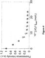
- Figure 4 shows the superimposition of binding thermograms corresponding to Tyr fluorescence quenching ( i.e . uranyl binding) in the absence and in the presence of 10 mM CaCl 2 . These results show that calcium has a very modest effect on uranyl titration.
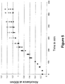
- Figure 5 shows growth curves recorded with E . coli cells exposed either to 50 ⁇ M uranyl acetate, or to 100 ⁇ M Na acetate as a control, in LB glucose medium at pH 4.5. Exposure conditions are detailed in the Methods.
- the figure 5 shows that uranyl exposure stops E . coli growth rapidly (full signs as compared to empty signs) except for the cells expressing the CaM ⁇ peptide.
- the growth of the cells expressing the CaM ⁇ peptide is lower than that of the other strains. This may be due to a too strong protein overexpression.
- addition of uranyl didn't affect anymore the growth of this strain.
- CaM ⁇ peptide presents an affinity for uranyl in the subnanomolar range and a very high selectivity towards calcium. It is expressed in high quantities in E. coli cells.
- the use of the binding loop sequence of CaM ⁇ for the other sites of calmodulin may not be as efficient for selecting uranyl binding at these sites as at site one. Therefore, structural parameters that could increase the affinity of site 2 toward uranyl as well as its uranyl/calcium specificity were explored using molecular dynamics.
- Two indicative informations can be obtained using molecular dynamic simulations; the first one concerns the structural model; the second one concerns the stabilisation energy associated with uranyl binding on the protein.
- the structural models of the Ef-hand2 obtained by molecular dynamics simulations show that uranyl coordination may be efficiently achieved in shorter binding loops, i.e. using deletion of at least two residues ( Figure 10 ).
- Cloning steps were made with standard methods using XL1Blue cells as E. coli strain. All mutations were made using a QuickChange site-directed mutagenesis kit (Stratagene) and specific primer pairs according to the manufacturer.
- the gene coding for the cameleon biosensor WT (denoted eCFP-CaM-Linker-M13-eYFP) was constructed in three steps.
- the enhanced Cyan Fluorescent Protein (eCFP) gene containing the TEV protease recognition site upstream of the coding sequence of eCFP was PCR-amplified using the S-TEV-eCFP- BamH I (SEQ ID NO: 43) and AS-eCFP- Sac I (SEQ ID NO: 44) primers and cloned upstream the CaM-linker-M13 gene, between BamH I and Sac I restriction sites.
- S-TEV-eCFP- BamH I SEQ ID NO: 43
- AS-eCFP- Sac I SEQ ID NO: 44
- eYFP Yellow Fluorescent Protein
- SEQ ID NO: 45 SEQ ID NO: 45
- AS-eYFP- Hind III SEQ ID NO: 46
- Both genes contained no stop codon except for the eYFP gene.
- the cameleon biosensor WT corresponds to the cDNA of SEQ ID NO: 47 and the protein of SEQ ID NO: 48.
- the construction of expression vector for the cameleon biosensor ⁇ was made by using the the cameleon biosensor WT gene as a template and primers S- ⁇ (SEQ ID NO: 49) and AS- ⁇ (SEQ ID NO: 50).
- the constructions of expression vectors for the cameleon biosensor WT-S2M or for the cameleon biosensor ⁇ -S2M (S2M corresponding to the inactivation of site 2 of the domain 1) were made using as a template the cameleon biosensor WT gene or the cameleon biosensor ⁇ gene respectively and primers S-S2M (SEQ ID NO: 51) and AS-S2M (SEQ ID NO: 52).
- the cameleon biosensor ⁇ corresponds to the cDNA of SEQ ID NO: 34 and the protein of SEQ ID NO: 35.
- the cameleon biosensor WT-S2M corresponds to the cDNA of SEQ ID NO: 53 and the protein of SEQ ID NO: 54.
- the cameleon biosensor ⁇ -S2M corresponds to the cDNA of SEQ ID NO: 36 and the protein of SEQ ID NO: 37.
- the recombinant vectors pQE30 containing the biosensor genes were introduced in the E. coli strain M15Rep4.
- Recombinant fusion proteins were expressed as follows: the overexpression strain was grown at 37°C in LB medium containing ampicillin (50 ⁇ g/mL) and kanamycin (50 ⁇ g/mL) until OD 600 reached 0.5. Expression was then induced by addition of 0.1 mM isopropyl-D-thiogalactoside (IPTG) and the cultures were further incubated for 20 h at 17°C. Cells were collected by centrifugation 20 min at 5 000 rpm, and the bacterial pellet was frozen and stored at -80°C.
- IPTG isopropyl-D-thiogalactoside
- Bacteria were resuspended in buffer A (50 mM Tris-HCl, 0.5 M NaCl, 25 mM imidazole pH 7.5) containing 1 mM 4-(2-Aminoethyl)benzenesulfonyl fluoride hydrochloride (AEBSF) + 15ug/mL DNAse1 + 30 mM MgSO 4 .
- the cellular extracts were obtained by French Press lysis and a centrifugation step of 30 min at 15 000 rpm. The cellular extracts were applied on a 5 mL HiTrapTM Column (GE Healthcare) in buffer A at 1 mL/min flow rate.
- the proteins were eluted from the nickel resin at 4 mL/min flow rate using an imidazole gradient. The proteins were dialyzed against buffer A and the His-Tags were removed by incubation overnight at 4°C in presence of the TEV protease followed by separation using HiTrap Chelating Column. Gel Filtration was performed for further purification of the proteins using a 26/600 Superdex 200 column (GE HealthCare) and 50 mM Tris-HCl buffer, pH 7.5, supplemented with 150 mM NaCl. The protein concentrations were measured according to the BC Assay from Uptima with bovine serum albumin as standard. The proteins were concentrated using Microcon® filtration system (Amicon Millipore®, with a cut off of 10 kDa).
- Fluorescence experiments were performed using an Infinite 1000 (TECAN). Cameleon biosensor WT proteins were first incubated with an excess of ethylenediaminetetraacetic acid (EDTA) and dialyzed overnight against 50 mM Tris-Cl pH 7 containing Chelex® resin. This step is used to remove calcium likely to be present in the different CaM-binding sites. For each measurement, 1 ⁇ M of protein was mixed in 200 ⁇ L of 50 mM Tris-Cl pH 7 buffer (treated with Chelex®) at 25°C. CaCl 2 or uranyl nitrate were added at varying concentrations between 0 and 10 ⁇ M. Excitation was performed at 440 nm and the emission spectrum recorded between 450 and 570 nm.
- EDTA ethylenediaminetetraacetic acid
- This example illustrates the possibility to use peptides derived from calmodulin immobilized on a solid support to chelate uranyl from a solution.
- the peptide CaM ⁇ was grafted on a gold strip surface modified by a 5 to 200 nm thick layer of polyacrylic acid, using activation via succinimide esters.
- a solution of CaM ⁇ at 100 ⁇ g/mL in MES buffer 20 mM was used.
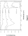
- Stable grafting of the protein was verified by Fourier transform infrared spectroscopy monitoring of the presence of the two amide I and amide II bands characteristic for proteins ( Figure 8 ).
- the protein was grafted on one half of the gold strip, the other half being used as a control area to identify possible nonspecific adsorption of uranyl.
- X-ray photoelectron spectrometry was used to identify the presence of nitrogen and uranyl at the gold strip surface.
- Figure 8 shows the FTIR transmission spectra recorded with the gold strip covered either with the polyacrylic acid alone (middle spectrum) or with protein grafted to polyacrylic acid layers of 5 nm (upper spectrum) and 50 nm (lower spectrum).
- the bands at 1673 and 1555 cm -1 are representative of the presence of the protein. These bands are observed in the upper and lower spectra. They are more intense in the lower spectrum, showing the impact of the polyacrylic layer thickness on the protein load onto the gold strip. These data show that CaM ⁇ was successfully grafted onto the gold strip.
- Figure 9 shows the XPS spectra recorded on the gold strip: the upper spectrum was recorded at a spot containing protein and the lower spectrum was recorded on a spot corresponding to the gold strip without protein.
- the band at ⁇ 382.5 eV corresponds to the presence of nitrogen
- the bands at ⁇ 394 eV and 401 eV correspond to the presence of uranium.
- the presence of uranium is only observed concomitant to the presence of nitrogen (upper spectrum), indicating that uranyl is immobilized by the protein.
Landscapes
- Health & Medical Sciences (AREA)
- Chemical & Material Sciences (AREA)
- Life Sciences & Earth Sciences (AREA)
- Organic Chemistry (AREA)
- General Health & Medical Sciences (AREA)
- Medicinal Chemistry (AREA)
- Biochemistry (AREA)
- Proteomics, Peptides & Aminoacids (AREA)
- Biophysics (AREA)
- Genetics & Genomics (AREA)
- Gastroenterology & Hepatology (AREA)
- Molecular Biology (AREA)
- Zoology (AREA)
- Toxicology (AREA)
- Physics & Mathematics (AREA)
- Engineering & Computer Science (AREA)
- General Engineering & Computer Science (AREA)
- High Energy & Nuclear Physics (AREA)
- Peptides Or Proteins (AREA)
- Food Science & Technology (AREA)
- Analytical Chemistry (AREA)
- General Physics & Mathematics (AREA)
- Immunology (AREA)
- Pathology (AREA)
Description
- The present relates to new uranium-chelating peptides derived from EF-hand calcium-binding motif, useful for uranium biodetection and biodecontamination.
- Uranium is a radioactive heavy metal, which is naturally present in varying concentrations in the environment. However, the wide use of uranium for industrial and military applications increases the risk of its distribution in the environment, which is aggravated by such factors as mining activities, uranium processing, or leaching of radioactive wastes.
- Uranium presents radiological and chemical toxicity to living organisms. The linear dioxo uranyl form (UO2 2+) which corresponds to uranium in its hexavalent oxidation state U(VI) is the prevalent form of uranium in the presence of oxygen and the most stable chemical form of uranium in water. It is soluble, bioavailable, and thus potentially toxic.
- Therefore, it is of high interest to develop systems allowing uranyl detection in the environment as well as molecules allowing its complexation in various environments with high affinity and selectivity. In particular, sensitive and easy to use systems allowing the detection of uranyl content in waters at the concentration levels defined by World Health Organization (WHO)(inferior to 0.03 mg/L, i.e. 126 nM; Guidelines for drinking-water quality, Fourth Edition, 2011, World Health Organization, WHO Press, Geneva) would be of great help for the water level monitoring.
- Among these systems, molecules derived from peptides are advantageous in that they are non toxic, and can be easily produced either synthetically or using recombinant techniques. Similarly, the development of organisms bearing uranyl biosensors, capable of detecting the uranyl fraction that is bioavailable and thus potentially toxic, is needed to assess water quality on a long term basis.
- Finally, bioremediation is an emerging technique that would allow the decontamination of large areas of contaminated water and/or soils.
- The helix-loop-helix calcium-binding motif (EF-hand motif) is the most prevalent Ca2+-binding site in proteins. The canonical EF-hand motif of about 30 amino acids in length is structured by two alpha-helices bridged by a flexible metal-binding loop (also referred to as calcium-binding loop, calcium-binding site, loop or site) composed of 12 highly conserved residues, with calcium coordinating positions at 1 (+X), 3 (+Y), 5 (+Z), 7 (-Y), 9 (-X) and 12 (-Z). The residues at the
ligand positions positions position 9, and the main chain carbonyl group of residue 7. The pentagonal bipyramidal configuration arises from two additional ligands provided by a bidendate glutamic or a monodentate aspartic acid which stabilizes a water ligand, in position 12. The residue atposition 1 of the loop is most frequently occupied by an aspartate (D); two residues are found at position 3: aspartic acid (D) or asparagine (N), but most frequently D;position 5 is most often occupied by aspartic acid (D), serine (S) or asparagine (N); the residue at position 7 is variable;position 9 shows a preference for residues with a side-chain oxygen (D, N, E, S, T), and the residues more frequently found at position 12 are glutamate (E) and aspartate (D), most frequently E. The highly conserved residues at the non ligand positions 6 and 8 are Gly (G) and Ile (I), respectively. The majority of the known EF-hand Calcium-Binding proteins (CaBPs) contain paired EF-hand motifs. - Functionally, the EF-hand proteins can be divided into two classes: 1) signaling proteins and 2) buffering/transport proteins. The first group is the largest and includes the most well-known members of the family such as calmodulin, troponin C and S100B. These proteins typically undergo a calcium-dependent conformational change which opens a target-binding site. The latter group is represented by calbindin D9k which remains in a closed conformation upon calcium binding.
- Calmodulin (CaM) is the most studied representative of the ubiquitous EF-hand protein family and a calcium-binding protein involved in the regulation of a wide range of target enzymes. The calmodulin structure (PDB code 1EXR) includes two pairs of EF-hand motifs (EF-
hands hands 3 and 4) in two domains (domain 1 and 2) separated by a flexible α-helix (Figure 1A ). In EF-hand1, calcium ligands are provided by three monodentate aspartate (D) atpositions Figure 1B . - Fluorescent sensors for calcium, called cameleons, have been constructed based on green fluorescent proteins and calmodulin (Miyawaki et al., Nature, 1997, 388, 882-887; Nagai et al., PNAS, 2004, 101, 10554-10559). They are chimeric proteins composed of a short-wavelength variant of GFP, CaM, a glycylglycine linker, the CaM-binding peptide of myosin light-chain kinase (M13), and a long-wavelength variant of GFP. Ca2+ binding to CaM initiates an intramolecular interaction between CaM and M13, which changes the chimeric protein from an extended to a more compact conformation, thereby increasing the efficiency of Fluorescence Resonance Energy Transfer (FRET) from the shorter to the longer-wavelength variant of GFP. Yellow cameleons (YCs) have cyan and yellow fluorescent proteins (CFP and GFP) as the FRET donor and acceptor, respectively.
- Synthetic cyclic-peptide variants of calmodulin site 1 (CaM-Mc peptides), consisting of a 33 amino acid sequence in which one, two or three of the aspartic acid residues in
positions WO 2005/012336 ). However, uranyl biosensors with detection limits in the nanomolar range or below are required to detect uranium content in waters at the maximum concentration levels defined by WHO. - A recombinant phosphorylated variant of
calmodulin domain 1, CaMlP, in which the threonine atposition 9 of the EF-hand1 loop is phosphorylated, was shown to bind uranyl with a dissociation constant in the subnanomolar range, at pH 7 (Kd of 3.10-10 M; Pardoux et al., 2012 PLoS ONE, 2012, 7, e41922). CaM IP is derived from CaM1 which consists of a 77 amino acids sequence, in which: (i) the
T9TKE12 sequence of EF-hand1 loop is substituted by the CK2 recognition sequence TAAE to allow phosphorylation of thethreonine 9, in vitro, using recombinant catalytic subunit of protein kinase CK2, (ii) a tyrosine residue is introduced in position 7 of the EF-hand 1 loop so that uranyl and calcium-binding affinities could be determined by following tyrosine fluorescence emission at 302 nm, and (iii) the metal-binding ability ofsite 2 is impaired by substituting the two aspartate residues inpositions - Phosphorylation of the threonine at
position 9 of the metal-binding loop increased uranyl-binding affinity by a factor of 5 at pH 6, while increasing the pH to 7 led to a further enhancement in uranyl affinity by a factor of 15.6. Analysis of the infrared modes of the phosphoryl group indicated that this group was deprotonated at pH 7 and directly involved in uranyl coordination. - However, CaMlP affinity for calcium (∼ 20.10-6 M) is similar to that of
CaM domain 1 so that its selectivity for uranyl over calcium is low (4.103 at pH 6 and 6.104 at pH 7). - In addition CaM1P cannot be expressed in a recombinant cell, microorganism or plant, which is then used for the in situ biodetection or bioremediation of uranyl because the phosphorylation of the
Threonine 9 is performed in vitro. - In this context, new sensitive and specific metal biosensors and/or chelators would be useful for the development of cost-effective uranium biodetection and bioremediation strategies.
- The subject-matter of the invention is defined by
claims 1 to 13. - The inventors have determined a structural model of the complex formed by the phosphorylated CaM1 peptide (CaMlP) with uranyl using a molecular dynamics approach. This structural model suggested that at least two residues of the calcium-binding loop are not necessary to bind uranyl in the complex: at least one of the calcium-ligating residues (aspartate at
position 3 or D3) and its directly adjacent residue (Lysine atposition 2 or K2;Figure 2A ). This result encouraged the inventors to produce a variant CaMΔ by the deletion of these two residues of the loop (K2 and D3) formerly to analyse the resulting properties of the phosphorylated peptide CaMΔ-P (Figures 1C and2B ). They also analyzed, the non phosphorylated peptide CaMΔ (Figure 2D ), which showed a very high affinity for uranyl, and a low affinity for calcium, as demonstrated in the examples of the present application (Figures 3 and4 ). The rationale of this high affinity for uranyl on one hand and for high specificity for uranyl as compared to calcium on the other hand resides in that less ligands are necessary to complete the coordination sphere of uranyl (i.e. 5 to 6 ligands disposed in the equatorial plane) than to coordinate calcium (7 to 8 ligands, 7 ligands insite 1 of calmodulin) and in that the binding loop containing 12 residues insite 1 is too large to accommodate uranyl, i.e. to optimally dispose the uranyl ligands in an equatorial plane around the uranyl UO2 axis. These latter findings were obtained from the molecular dynamics simulations on phosphorylated CaMlP peptide (Figure 2A ) but they were also observed on the non-phosphorylated peptide CaM1 (Figure 2C ). In particular, according to the structural model of the CaMl-U complex, Asp atposition 1 of the loop is not a uranyl ligand and it is situated below the uranyl equatorial plane (Figure 2C ). In addition, the loop arrangement in the model structure of the CaMl-U complex is not ideal as compared to the structural model of CaMΔ (Figure 2D ), in that the distance is too long between the Asp ligand and the Glu ligand, and the structure involves a carbonyl ligand (the carbonyl group of Tyr7) that is considered as a weaker ligand as compared to the side chain of aspartate. - Therefore, the present description concerns the optimisation of uranyl binding sites in EF-hand motifs by decreasing the size of the binding loop by two amino acids, and by suppressing at least one calcium ligand, to obtain affine and specific uranyl binding sites. As shown in the examples by the comparison of uranyl binding affinities of CaMΔ and CaMΔ3 (where only the aspartate at
position 3 was deleted), two deletions are necessary to increase uranyl affinity. For the EF-hand1 of calmodulin, as demonstrated in the examples, deletion of the amino acids atpositions site 2 by showing different possibilities to increase uranyl affinity and specificity, obtained using structure prediction by molecular dynamics simulations (Figure 10 ). - One aspect of the present description relates to a polypeptide comprising at least one helix-loop-helix calcium-binding (EF-hand) motif, which comprises a deletion of at least two amino acid residues in the 12-amino-acid calcium-binding loop sequence, and wherein said polypeptide binds uranyl.
- The polypeptide of the description which is an isolated recombinant or synthetic polypeptide, has a signicantly higher binding affinity for uranyl and a significantly lower binding affinity for calcium than the corresponding peptide without said deletion. In the present application, "significant" means that the binding affinity is different with P<0.01. Binding affinity for metals can be measured by any standard technique which is known by those skilled in the art such as those described in the examples of the present application.
- Preferably, the polypeptide of the description has a binding affinity for uranyl which is increased by a factor of at least 2, more preferably at least 5, 10, 30, 100 or more and a binding affinity for calcium which is decreased by a factor of at least 2, more preferably at least 5, 10, 30, 100, 300 or more, compared to the corresponding peptide without the deletion in the calcium-binding loop. Preferably, the polypeptide of the description has a selectivity for uranyl over calcium which is of at least 103, more preferably at least 104, 105, 106, 107 or more.
- For example, the CaMΔ peptide of the example has a binding affinity for uranyl which is increased by a factor of at least 100 and a binding affinity for calcium which is decreased by a factor of at least 500 compared to the corresponding peptide CaM1 which does not have the deletion in the calcium-binding loop. Therefore the selectivity of CaMΔ for uranyl over calcium is of the order of 107 whereas that of CaM1 is only of the order of 103.
- The polypeptide of the description and its derivatives like the cameleon-based biosensors, have the following advantages:
- they have a high binding affinity for uranyl combined with a low binding affinity for calcium, which means that they are highly sensitive and selective for uranyl. Therefore, they can detect low uranium concentration in complex media containing divalent cations like Ca2+ (for example, biological media, calcium-rich water). Uranyl detection in a sample to analyze can be performed in vitro using the isolated polypeptide or cameleon-based biosensor, as well as in situ in recombinant cells (whole-cell biosensors) or non-human transgenic organisms expressing the cameleon protein derived from the polypeptide of the description. Their affinity and selectivity for uranium are higher than those of all calmodulin-derived uranium biosensors.
- the cameleon biosensors derived from the polypeptide of the description can also be used as imaging biosensors to visualize uranyl in situ in recombinant cells or non-human transgenic organisms expressing the cameleon protein derived from the polypeptide of the description,
- the recombinant cells and non-human transgenic organisms expressing the polypeptide or its derivatives can be used for the biodecontamination and the bioremediation of uranium contamination in an environment, as well as for the production of large quantities of the polypeptide of the description and its derivatives (cameleon-based biosensors).
- as recombinant proteins, their production and use are easy, fast not-toxic, and cost-effective, and they are designed for in vitro and in vivo uses.
- In the following description, the standard one letter amino acid code is used. The expression "EF-hand motif' refers to the canonical EF-hand calcium-binding motif as described just before.
- In one embodiment, the deletion includes at least one calcium-ligating residue (residue in
position positions positions 1 and/or 3. Even more preferably, the deletion includes at least the calcium-ligating residue inposition - In an advantageous arrangement of said embodiment, the polypeptide comprises a deletion of at least one calcium-ligating residue and its directly adjacent residue (i.e., residue in position +1 or -1 relative to the calcium-ligating residue). Preferably, the polypeptide comprises a deletion of at least one of the following pairs of residues:
positions positions - The invention relates to a polypeptide comprising at least one helix-loop-helix calcium-binding EF-hand motif which comprises a deletion of the two amino acid residues in
position - In another advantageous arrangement of said embodiment, the polypeptide comprises a deletion of at least one calcium-ligating residue and at least one of the residues in
positions 10 and/or 11 of the calcium-binding loop. Preferably, the polypeptide comprises a deletion of at least one calcium-ligating residue inposition positions 10 and/or 11 of the calcium-binding loop. More preferably, the polypeptide comprises a deletion of at least the residues chosen frompositions positions - In another preferred embodiment, the polypeptide comprises at least two EF-hand motifs, wherein at least one EF-hand motif comprises said deletion in the loop sequence and the other EF-hand motif(s) comprise(s) said deletion in the loop sequence or not. Preferably, the polypeptide comprises two to four EF-hand motifs. More preferably, the polypeptide comprises two or four EF-hand motifs.
- In an embodiment, the invention relates to the polypeptide as defined above, which comprises two or four EF-hand motifs, wherein at least one EF-hand motif comprises said deletion in the calcium-binding loop sequence and the other EF-hand motif(s) comprise said deletion or not.
- In another preferred embodiment, the polypeptide comprises further alterations in said EF-hand motif(s) which comprise said deletion or not, such as for example the substitution and/or the modification of one or more amino acid residues. Said alterations are introduced when the amino acid residues of interest are not naturally present at the appropriate positions in said EF-hand motif(s). Preferably, said further alterations are in the loop sequence.
- In a first advantageous arrangement of said embodiment, the polypeptide comprises a fluorescent residue, preferably a tyrosine or tryptophan residue. The fluorescent residue is advantageously in the loop from said at least one EF-hand motif which comprises said deletion or not, or in close proximity to said loop. It is preferably in position 7 of said loop, by reference to the numbering of the 12-amino-acid loop sequence. Preferably, the fluorescence residue is in the loop from said at least one EF-hand motif which comprises said deletion. More preferably, the polypeptide comprises a tyrosine residue in position 7 of the loop from said at least one EF-hand motif which comprises said deletion. The fluorescent residue allows the monitoring of uranyl and/or calcium binding and the determination of their binding affinities, by following tyrosine fluorescence emission at 302 nm. The fluorescent residue is an amino acid residue naturally present in the EF-hand motif(s) or substituted.
- In a second advantageous arrangement of said embodiment, the polypeptide comprises at least two aspartic acid residues in
positions - In a third advantageous arrangement of said embodiment, the polypeptide comprises a glycine residue in
position 4 of the loop from said at least one EF-hand motif which comprises said deletion or not, by reference to the numbering of the 12-amino-acid loop sequence. Preferably, the glycine residue is in the loop from said at least one EF-hand motif which comprises said deletion. The glycine residue is an amino acid residue naturally present in the EF-hand motif(s) or substituted. - In another advantageous arrangement of said embodiment, the polypeptide comprises at least one phosphorylated serine or threonine residue. The phosphorylated serine or threonine residues are advantageously in the loop from said EF-hand motif(s) which comprise said deletion or not, preferably in
position 9 and/or 12 of the loop from said EF-hand motif(s), by reference to the numbering of the 12-amino-acid loop sequence. The phosphorylation of said residue(s) increases the uranyl binding affinity of the polypeptide. The residues in positions +1 to +3 relative to the phosphorylated threonine are advantageously modified to provide a CK2 recognition sequence TXXE, in which X is a neutral or acidic amino acid different from T, for example XX is AA or AE. Phosphorylation of the threonine may be performed in vitro, using recombinant catalytic subunit of protein kinase CK2. Preferably, the polypeptide comprises a phosphorylated threonine residue inposition 9 of the loop from said EF-hand motif(s) which comprise(s) said deletion or not and further comprises alanine residues inpositions 10 and 11 of said loop, by reference to the numbering of the 12-amino-acid loop sequence. These alanine residues provide a CK2 recognition sequence TAAE to allow phosphorylation of theThreonine 9. The phosphorylated serine or threonine residues are advantageously in the loop from said at least one EF-hand motif which comprises said deletion. The serine and threonine residues of the EF-hand motif(s) which are modified by phosphorylation and the adjacent residues which are substituted to provide a CK2 recognition site are naturally present in the EF-hand motif(s) or substituted. - In another advantageous arrangement of said embodiment, the polypeptide comprises at least two EF-hand motifs, at least one comprising said deletion in the loop sequence and at least another one not comprising said deletion, wherein at least one of said EF-hand motif(s) not comprising the deletion comprises at least one mutation in the loop sequence which impairs calcium binding. Preferably, said mutation is the substitution with alanine residues of the residues in
positions - The EF-hand motif sequence is modified by standard mutagenesis technique on the polypeptide coding sequence. The phosphorylation of the serine and/or threonine residues is performed using standard methods which are known from those skilled in the art.
- The amino acid-sequence of the EF-hand motif(s) of the polypeptide of the description is that of the corresponding wild-type EF-hand protein(s) except at said amino acid position(s) which are altered in the present description.
- The polypeptide of the description may be derived from EF-hand motif(s) of any proteins of the EF-hand family (EF-hand protein) having a canonical EF-hand motif as above described. Preferably, the polypeptide of the description is derived from EF-hand protein(s) from the class of EF-hand signaling proteins, i.e., the EF-hand proteins which undergo a calcium-dependent conformational change. More preferably, it is derived from EF-hand signaling protein(s) of the calmodulin superfamily. Even more preferably, from EF-hand signaling protein(s) of the calmodulin superfamily selected from the group consisting of calmodulin and troponin C. It is advantageously derived from EF-handl, 2, 3 and/or 4 of calmodulin protein(s).
- In an embodiment, the invention relates to the polypeptide as defined above, wherein said EF-hand motif(s) are derived from signaling EF-hand protein(s) of the calmodulin superfamily selected from the group consisting of calmodulin and troponin C.
- In an advantageous arrangement of said embodiment, the polypeptide is derived from Arabidopsis thaliana calmodulin 3 (amino acid sequence SEQ ID NO: 2 encoded by the cDNA of SEQ ID NO: 1).
- The calmodulin EF-handl, 2, 3 and 4 correspond respectively to positions 12 to 41, 48 to 76, 85 to 114, and 121 to 149, by reference to the amino acid numbering of Arabidopsis thaliana calmodulin 3 (SEQ ID NO: 2). The calcium-binding loop of EF-handl, 2, 3 and 4 correspond respectively to positions 21 to 32, 57 to 68, 94 to 105, and 130 to 141, by reference to the amino acid numbering of SEQ ID NO: 2. The
calmodulin domain positions 1 to 76 and 77 to 149, by reference to the amino acid numbering of SEQ ID NO: 2. - In another advantageous arrangement of said embodiment, the polypeptide comprises a EF-hand motif derived from a calmodulin EF-hand1 having a 12 amino-acid loop of sequence (I):
X1-X2-X3-X4-X5-X6-X7X8-X9-X10-X11-X12, in which:
X1 is D; X2 is K or R, preferably K; X3 is D; X4 is G, N, Q, preferably G; X5 is D or N, preferably D; X6 is G; X7 is T, C, S or N, preferably, T; X8 is I; X9 is T or S, preferably T; X10 is T or S, preferably T; X11 is K, S, M or N, preferably K, and X12 is E. More preferably, said EF-hand1 has a 12 amino-acid loop of sequence DKDGDGCITTKE (SEQ ID NO: 3). - In another advantageous arrangement of said embodiment, the polypeptide comprises a EF-hand motif derived from a calmodulin EF-hand2 having a 12 amino-acid loop of sequence (II):
X1-X2-X3-X4-X5-X6-X7X8-X9-X10-X11-X12, in which:
X1 is D; X2 is A, V or Q, preferably A; X3 is D; X4 is G, N; X5 is N, D or S, preferably N; X6 is G or H, preferably, G; X7 is T, N or Q, preferably, T; X8 is I; X9 is D or E, preferably D; X10 is F; X11 is P, T or S, preferably P, and X12 is E. More preferably, said EF-hand2 has a 12 amino-acid loop of sequence DADGNGTIDFPE (SEQ ID NO: 4). - In another advantageous arrangement of said embodiment, the polypeptide comprises a EF-hand motif derived from a calmodulin EF-hand3 having a 12 amino-acid loop of sequence DKDQNGFISAAE (SEQ ID NO: 5)
- In another advantageous arrangement of said embodiment, the polypeptide comprises a EF-hand motif derived from a calmodulin EF-hand4 having a 12 amino-acid loop of sequence DVDGDGQINYEE (SEQ ID NO: 6).
- A more preferred polypeptide of the description is derived from calmodulin EF-handl, EF-hand2, EF-hand3 and/or EF-hand 4 and comprises a calcium-binding loop sequence having a deletion of at least two amino acids, which is selected from the group consisting of the sequences: DGDGCITTKE (SEQ ID NO: 7), DGDGYITTKE (SEQ ID NO: 8), DGDGYITAAE (SEQ ID NO: 9), DGNGTIDFPE (SEQ ID NO: 10), DGNGYIDFPE (SEQ ID NO: 11), DGDGTIDFPE (SEQ ID NO: 12) DGDGYIDFPE (SEQ ID NO: 13), DQNGFISAAE (SEQ ID NO: 14) and DGDGQINYEE (SEQ ID NO: 15), KDGDGCITKE (SEQ ID NO: 70), KDGDGCITTE (SEQ ID NO: 71), DKGDGCITKE (SEQ ID NO: 72), DKGDGCITTE (SEQ ID NO: 73), KDGDGCITE (SEQ ID NO: 74), DKGDGCITE (SEQ ID NO: 75), ADGNGTIDPE (SEQ ID NO: 76), ADGNGTIDFE (SEQ ID NO: 77), DAGNGTIDPE (SEQ ID NO: 78), DAGNGTIDFE (SEQ ID NO: 79), ADGNGTIDE (SEQ ID NO: 80), DAGNGTIDE (SEQ ID NO: 81), KDQNGFISAE (SEQ ID NO: 82), KDQNGFISAE (SEQ ID NO: 83), DKQNGFISAE (SEQ ID NO: 84), DKQNGFISAE (SEQ ID NO: 85), KDQNGFISE (SEQ ID NO: 86), DKQNGFISE (SEQ ID NO: 87), VDGDGQINEE (SEQ ID NO: 88), VDGDGQINYE (SEQ ID NO: 89), DVGDGQINEE (SEQ ID NO: 90), DVGDGQINYE (SEQ ID NO: 91), VDGDGQINE (SEQ ID NO: 92), DVGDGQINE (SEQ ID NO: 93). Preferably from the group consisting of the sequences SEQ ID NO: 7 to 15, 72, 75, 78, 81, 84, 87, 90 and 93.
- In another advantageous arrangement of said embodiment, the polypeptide of the invention is a
calmodulin domain 1 variant comprising two EF-hand motifs, respectively from EF-hand1 and EF-hand2 of calmodulin protein(s). - A
preferred calmodulin domain 1 variant polypeptide comprises a EF-hand1 comprising the deletion of the amino acid residues inpositions positions site 2. Advantageously, the EF-hand1 further comprises a tyrosine residue in position 7 and/or neutral or acidic residues different from T inpositions 10 and 11 of the loop, for example AA or AE. More preferably, saidcalmodulin domain 1 variant polypeptide comprises or consists of the sequence SEQ ID NO: 17 or 60, which correspond to the peptides referred to as CaMΔ and CaMΔ-WT in the examples of the present application. CaMΔ and CaMΔ-WT are variants of thedomain 1 fromArabidopsis thaliana calmodulin 3. - Another
preferred calmodulin domain 1 variant polypeptide comprises a EF-hand1 comprising the deletion of the amino acid residues inpositions position positions 10 and/or 11 of the calcium-binding loop, preferably the residues inpositions - Another
preferred calmodulin domain 1 variant polypeptide comprises a EF-hand1 and EF-hand2, each comprising the deletion of the amino acid residues inpositions - In another advantageous arrangement of said embodiment, the polypeptide of the description is a calmodulin variant comprising four EF-hand motifs, respectively from EF-handl, 2, 3 and 4 of calmodulin protein(s).
- In a preferred calmodulin variant polypeptide, the EF-hand1 comprises the deletion of the amino acid residues in
positions Arabidopsis thaliana calmodulin 3. - In another preferred calmodulin variant polypeptide, the EF-hand1 comprises the deletion of the amino acid residues in
positions position positions 10 and/or 11 of the calcium-binding loop, preferably the residues inpositions - In another preferred calmodulin variant polypeptide, each of the EF-hand1 and EF-hand2 comprise the deletion of the amino acid residues in
positions - In another preferred calmodulin variant polypeptide, each of the EF-hand1, 2 and 3 comprise the deletion of the amino acid residues in
positions - In another preferred calmodulin variant polypeptide, each of the EF-hand1, 2, 3 and 4 comprise the deletion of the amino acid residues in
positions Arabidopsis thaliana calmodulin 3. - In another preferred calmodulin variant polypeptide, each of the EF-hand1 and EF-hand3 comprise the deletion of the amino acid residues in
positions - In an embodiment, the invention relates to the polypeptide as defined above, which comprises or consists of an amino acid sequence selected from the group consisting of the sequences SEQ ID NO: 17, 18, 20 and 60.
- In another preferred embodiment, the polypeptide is coupled to a labeling agent which produces a detectable and/or quantifiable signal, in particular a radioactive, magnetic or luminescent (radioluminescent, chemiluminescent, bioluminescent, fluorescent or phosphorescent) agent. The labeled polypeptide may be labeled directly or indirectly, via covalent or non-covalent bonds, using standard conjugation techniques that are well-known to those skilled in the art.
- The invention relates to a fusion protein comprising a polypeptide as defined above fused to another protein moiety. In an aspect of the description, said polypeptide is fused to another protein moiety directly or through a peptide spacer.
- Another aspect of the present description relates to a chimeric protein comprising the polypeptide fused to another protein moiety, directly or through a peptide spacer. The protein/peptide moieties include those which allow the purification, detection, immobilization, and/or cellular targeting of the polypeptide of the description, and/or which increase the affinity for uranyl, the bioavailability, and/or the production in expression systems of said polypeptide. These moieties may be selected from: (i) a labeling moiety such as a fluorescent protein (GFP and its derivatives, BlueFP, CyanFP, YellowFP, GreenFP and red-shifted GFP), (ii) a reporter moiety such as an enzyme tag (luciferase, alkaline phosphatase, glutathione-S-transferase (GST), β-galactosidase), (iii) a binding moiety such as an epitope tag (polyHis6, FLAG, HA, myc.), a DNA-binding domain, a hormone-binding domain, a poly-lysine tag for immobilization onto a support, and (iv) a targeting moiety for addressing the chimeric protein to a specific cell type or cell compartment. In addition, the sequence(s) advantageously comprise a linker which is long enough to avoid inhibiting interactions between said sequence(s) and the polypeptide sequence. The linker may also comprise a recognition site for a protease, for example, for removing affinity tags from the purified chimeric protein according to the present description.
- In an embodiment, the invention relates to the fusion protein as defined above, which is a cameleon protein comprising tandem fusions of a fluorescence-donor protein, a polypeptide as defined above, and a fluorescence-acceptor protein.
- In a preferred embodiment the chimeric protein is a cameleon protein comprising tandem fusions of (1) the polypeptide of the description, (2) a EF-hand protein-binding peptide that binds a complex between the polypeptide in (1) and calcium and/or uranyl, (3) a fluorescence-donor protein, and (4) a fluorescence-acceptor protein, wherein the fluorescence donor and acceptor proteins are at each end of the cameleon protein.
- In some arrangements of said embodiment, the EF-hand protein-binding peptide is absent; in these arrangements, the polypeptide of the description usually comprises two EF-hand motifs. Cameleon proteins without EF-hand protein-binding peptide may be derived from the sequence SEQ ID NO: 65.
- In other arrangements of said embodiment, the cameleon protein further comprises a linker between the fluorescence donor and/or acceptor and the polypeptide of the description, between the polypeptide of the description and the EF-hand protein-binding peptide, and/or between the EF-hand protein-binding peptide and the fluorescence acceptor.
- The fluorescence donor and acceptor proteins which are advantageously at each end of the cameleon protein are chosen from any protein capable of producing FRET such as with no limitation: Alexa proteins and GFP variants such as those disclosed for example in Shaner et al., Nature Methods, 2005, 12, 905-909; Erard et al., Molecular Biosystems, 2013, 9, 258-267; Fredj et al., PLOS ONE, 2012, 7, e49149 and
PCT Application WO/2012/172095 . - The fluorescence donor and acceptor proteins are advantageously chosen from a short-wavelength variant of GFP such as CyanFP or BlueFP, Turquoise,
Turquoise 2, Aquamarine, Cerulean, Cerulean3, TFP1 and a long wave-length variant of GFP such as YellowFP, GreenFP and red-shifted GFP, Citrine, Venus and circularly permuted fluorescent proteins. - The polypeptide of the description comprises advantageously four EF-hand motifs from EF-hand signaling protein(s) as defined above. Preferably said EF-hand motifs are from EF-hand signaling protein(s) of the calmodulin superfamily selected from the group consisting of calmodulin and troponin C.
- In an advantageous arrangement of said embodiment, the cameleon protein is derived from calmodulin.
- Many peptides which bind calmodulin in complex with calcium are known in the art (see for example Carafoli et al., Proc. Natl. Acad. Sci. USA., 2002, 99 :1115-1122). Any of these peptides can be used in the cameleon protein of the description, including with no limitations: the peptides M13 (amino acid sequence SEQ ID NO: 22 encoded by the nucleotide sequence SEQ ID NO: 21) and skMLCK (SEQ ID NO: 23) from skeletal myosin light chain kinase; the peptides MLCKp (SEQ ID NO: 24) and smMLCK (SEQ ID NO: 25) from myosin light chain kinase; the peptide named wasp venom or polistes mastoparan (SEQ ID NO: 26); the peptide p21 (SEQ ID NO: 27); the peptides disclosed in Shifman J.M. and Mayo, L., PNAS, 2003, 100, 13274- such as melittin (SEQ ID NO: 28), spectrin (SEQ ID NO: 29), CaMKI (SEQ ID NO: 30), CaMKII (SEQ ID NO: 31), CaMKK (SEQ ID NO: 32) and peptide1 (SEQ ID NO: 33); the peptides from cyclic nucleotide phosphodiesterase (Olwin & Storm, 1985); the peptides from caldesmon (Yazawa et al., 1987) and the synthetic peptide derived from the plasma membrane Ca2+ pump (Yazawa et al., 1992).
- Preferably, the cameleon protein comprises a short-wavelength variant of GFP such as CyanFP or BlueFP, a polypeptide of the description derived from calmodulin, a linker, the calmodulin-binding peptide of myosin light chain kinase (peptide M13), and a long wave-length variant of GFP such as YellowFP, GreenFP and red-shifted GFP.
- Preferred cameleon proteins are derived from the calmodulin variant polypeptides as defined above. Examples of preferred calmodulin-derived cameleon proteins of the description comprise or consist of an amino acid sequence selected from the group consisting of the sequences SEQ ID NO: 35, 38, 61, 63 and 67.
- In an embodiment, the present invention relates to the fusion protein as defined above, which comprises or consists of a sequence selected from the group consisting of the sequences SEQ ID NO: 35, 38, 61, 63 and 67.
- Alternatively, the four elements of the cameleon protein as described above can be divided in two separate fusion proteins, which are then combined together to obtain a functional biosensor capable of detecting uranium: a first fusion protein with the fluorescence donor fused to one of the polypeptide or the polypeptide-binding peptide, and a second fusion protein with the fluorescence acceptor fused to the polypeptide or polypeptide-binding peptide which is not fused to the fluorescence donor. These types of cameleon proteins are described for example in Miyawaki et al., Proc. Natl. Acad. Sci. U S A., 1999, 96, 2135-40.
- The cameleon protein is a uranyl biosensor that can be used in vitro as uranyl analysis reagent for the detection of uranyl in an environment (water, soil, effluents) or in biological samples from individuals (biological fluids). It is also used as cell imaging reagent or diagnostic reagent for the detection of uranyl in situ in recombinant cells (whole-cell biosensor) or non-human transgenic organisms expressing the cameleon protein.
- The description encompasses polypeptides and derived fusion proteins comprising or consisting of natural amino acids (20 gene-encoded amino acids in a L- and/or D-configuration) linked via a peptide bond as well as peptidomimetics of such protein where the amino acid(s) and/or peptide bond(s) have been replaced by functional analogues. Such functional analogues include all known amino acids other than said 20 gene-encoded amino acids. A non-limitative list of non-coded amino acids is provided in Table 1A of
US 2008/0234183 . The description also encompasses modified polypeptides/fusion proteins derived from the above polypeptides/fusion proteins by introduction of any modification into one or more amino acid residues, peptide bonds, N-and/or C-terminal ends of the protein, as long as the uranyl-binding activity is maintained in the modified polypeptide/protein. These modifications which are introduced into the polypeptide/protein by the conventional methods known to those skilled in the art, include, in a non-limiting manner: the substitution of a natural amino acid with a non-proteinogenic amino acid (D amino acid or amino acid analog); the modification of the peptide bond, in particular with a bond of the retro or retro-inverso type or a bond different from the peptide bond; the cyclization, and the addition of a chemical group to the side chain or the end(s) of the protein, in particular for coupling an agent of interest to the polypeptide/fusion protein of the description. - In another preferred embodiment, the invention relates to the polypeptide as defined above or the fusion protein as defined above, which is immobilized onto a solid support, such as with no limitation, a plate, a slide, a strip, a fiber, a gel, a felt support, wells, microparticles, or biologically modified ceramics (biocers; Böttcher et al., J. Mater. Chem., 2004, 14, 2176-2188).
- The invention related to a polynucleotide encoding the polypeptide as defined above or the fusion protein as defined above.
- Another aspect of the description relates to an isolated polynucleotide encoding a polypeptide or chimeric protein of the description. The synthetic or recombinant polynucleotide may be DNA, RNA or combination thereof, either single- and/or double-stranded. Preferably the polynucleotide comprises a coding sequence which is optimized for the host in which the polypeptide or chimeric protein is expressed. In a preferred embodiment, the polynucleotide comprises or consists of a sequence selected from the group consisting of the sequences SEQ ID NO: 16, 34, 59, 62, 64 and 68.
- Another aspect of the description relates to a recombinant vector comprising said polynucleotide. Preferably, said recombinant vector is an expression vector capable of expressing said polynucleotide when transfected or transformed into a host cell such as a eukaryotic or prokaryotic cell like a mammalian, bacterial or fungal cell. The polynucleotide is inserted into the expression vector in proper orientation and correct reading frame for expression. Preferably, the polynucleotide is operably linked to at least one transcriptional regulatory sequence and, optionally to at least one translational regulatory sequence. In a preferred embodiment, the polynucleotide is under the control of a promoter which is upregulated in response to uranium such as for example Caulobacter urc-A promoter (Hillson et al., Applied and Environmental Microbiology, 2007, 73, 7615-7621). Recombinant vectors include usual vectors used in genetic engineering and gene therapy including for example plasmids and viral vectors.
- Another aspect of the description provides a host cell or a non-human organism transformed with said polynucleotide or recombinant vector.
- The invention relates to a host cell comprising the polynucleotide as defined above.
- Preferably, said modified host cell or non-human transgenic organism is resistant to radiations, and/or pollutants such as for example nitrates and toxic metals. The non-human transgenic organism is obtained from a unicellular or pluricellular microorganism or a higher eukaryotic organism. In a preferred embodiment said modified host cell is a prokaryotic cell such as a bacterium. In another embodiment, said non-human transgenic organism is a transgenic plant, nematode, zebrafish or algae.
- The invention relates to a transgenic bacteria or transgenic plant comprising the polynucleotide as defined above.
- The polynucleotide, vector, cell, and non-human transgenic organism of the description are useful for the production of the polypeptide/chimeric protein of the description using well-known recombinant DNA techniques.
- Another aspect of the description relates to the use of the polypeptide, fusion protein, host cell, non-human transgenic organism, in vitro or in vivo, as uranyl chelating agent. The chelating agent is useful for the detection, the decontamination, and/or the bioremediation of uranium contamination in an environment (water, soil, effluents,...) or in individuals.
- The invention relates to the use of the polypeptide as defined above, the fusion protein as defined above, the host cell as defined above or the transgenic bacterium or transgenic plant as defined above as uranyl chelating agent, for the detection of uranium contamination in vitro or for the decontamination and/or the bioremediation of uranium contamination in an environment.
- The labelled polypeptide/fusion protein such as the polypeptide comprising a fluorescent residue like a tyrosine residue in position 7 of the loop or the cameleon protein, is a uranyl biosensor. It can be used in vitro as uranyl analysis or diagnostic reagent for the detection of uranyl in an environment (water, soil, effluents,...) or in biological samples from individuals (biological fluids,..). In addition, the cameleon protein is also used as cell imaging reagent, diagnostic reagent and bioindicator for the detection of uranyl in situ in modified cells (for example recombinant cells) or non-human transgenic organisms expressing the cameleon protein.
- The polynucleotide according to the description is prepared by the conventional methods known in the art. For example, it is produced by amplification of a nucleic sequence by PCR or RT-PCR, by screening genomic DNA libraries by hybridization with a homologous probe, or else by total or partial chemical synthesis. The recombinant vectors are constructed and introduced into host cells by the conventional recombinant DNA and genetic engineering techniques, which are known in the art.
- The polypeptide/chimeric protein is prepared by the conventional techniques known to those skilled in the art, in particular by solid-phase or liquid-phase synthesis or by expression of a recombinant DNA in a suitable cell system (eukaryotic or prokaryotic). More specifically, the polypeptide can be solid-phase synthesized, according to the Fmoc technique, originally described by Merrifield et al. (J. Am. Chem. Soc., 1964, 85: 2149-), and purified by reverse-phase high performance liquid chromatography; the polypeptide/chimeric protein can be produced from the corresponding cDNAs, obtained by any means known to those skilled in the art; the cDNA is cloned into a eukaryotic or prokaryotic expression vector and the protein produced in the cells modified with the recombinant vector is purified by any suitable means, in particular by affinity chromatography.
- The practice of the present description will employ, unless otherwise indicated, conventional techniques which are within the skill of the art. Such techniques are explained fully in the literature.
- In addition to the above arrangements, the description also comprises other arrangements, which will emerge from the description which follows, which refers to exemplary embodiments of the subject of the present description, with reference to the attached drawings in which:
-
Figure 1 is a schematic of calmodulin. A. Schematic ofdomain 1 of Paramecium tetraurelia calmodulin (PDB code 1EXR) which comprises two calcium-binding sites (site 1 and site 2) which are part of a canonical EF-hand motif (EF-handl and EF-hand2, respectively forsite 1 and site 2). B. Structural model of the calcium-bindingsite 1 of Arabidopsis thaliana calmodulin showing the coordinating amino acids. C. Amino acid sequence ofcalmodulin site 1 from Arabidopsis thaliana (CaM-WT) and from the peptide CaM1 and CaMΔ analysed in the present study. In the CaM1 and CaMΔ peptides, the metal-binding ability ofsite 2 was impaired by replacing the aspartate residues in positions 57 and 59 of calmodulin (positions 58 and 60 of CaM1) with alanines. In addition, the threonine residue in position 31 and the lysine residue in position 32 of CaM1 (positions 10 and 11 of site 1) have each been substituted with an alanine residue to obtain an efficient CK2 consensus sequence that targets phosphorylation of threonine 9 (T9) of the metal-binding loop (T30 in the peptide) and a tyrosine was introduced at position 28 of peptide CaM1 (position 7 of the metal-binding loop) to allow the monitoring of uranyl and calcium-binding and the determination of their binding affinities, by following tyrosine fluorescence emission at 302 nm. -
Figure 2 represents Structural models obtained by molecular dynamics of the CaM1P-U (A), CaMΔP-U (B) CaMl-U (C) and CaMΔ-U (D) complexes. E = glutamic acid, D = aspartic acid, Y = tyrosine, T = threonine, Tp = Phosphothreonine. -
Figure 3 shows the binding thermograms of the CaMΔ and CaMΔP peptides with uranyl at pH 6 (A) and pH 7 (B). Conditions: 10 µM CaMΔ or CaMΔP, 100 µM IDA, 20 mM MES and 100 mM KCl. Full square: CaMΔP. Full circle: CaMΔ. -
Figure 4 shows the binding thermograms of the CaMΔ with uranyl in the absence (empty triangle) and in presence (empty circle) of 10 mM CaCl2 Conditions: 10 µM CaMΔ, 100 µM IDA, 20 mM MES and 100 mM KCl. -
Figure 5 shows the growth curve of three E. coli strains transformed with respectively, the expression vector without insert (as a control) or vectors expressing recombinant CaM1 or CaMΔ peptide, in the presence or absence of uranyl acetate (U). Control (empty circle). Control + U (full circle). CaM1 (empty triangle). CaM1 + U (full triangle). CaMΔ (empty square). CaMΔ + U (full square). IPTG was added at t=105 min; 50 µM uranyl acetate (U) or 100 µM sodium acetate were added at t=135 min. -
Figure 6 shows the emission spectra of cameleon biosensor WT protein (excited at 440 nM) at varying CaCl2 concentrations between 0 µM to 10 µM. 0 µM Ca (full diamond). 2 µM Ca (empty circle). 4 µM Ca (empty triangle). 6 µM Ca (full circle). 8 µM Ca (cross). 10 µM Ca (empty square). Vertical arrow indicates an increase in FRET detection with increasing Ca concentrations. -
Figure 7 shows the emission spectra of cameleon biosensor WT protein (excited at 440 nM) at varying uranyl nitrate concentrations between 0 µM to 10 µM. 0 µM UO2 (full black circle). 2 µM UO2 (empty square). 4 µM UO2 (full triangle). 6 µM UO2 (empty circle). 8 µM UO2 (full grey circle). 10 µM UO2 (empty diamond). Vertical arrow indicates an increase in FRET detection with increasing uranyl concentrations. -
Figure 8 shows the transmission ATR-FTIR spectra of the gold strip covered with a 5 nm layer of polyacrylic acid and the CaMΔ (upper spectrum), with the polyacrylic acid layer only (middle spectrum) and with a 50 nm layer of polyacrylic acid and the CaMΔ (lower spectrum). -
Figure 9 shows XPS spectra recorded on the grafted gold strip. The upper spectrum is recorded in a spot exposed to the protein and the lower spectrum is recorded on a spot that was not contacted with the protein solution. -
Figure 10 shows structural models of the Ef-hand2 obtained by molecular dynamics simulations. This figure shows that uranyl coordination may be efficiently achieved in shorter binding loops, i.e. using deletion of at least two residues. - The recombinant peptides were produced in E. coli. A histidine-tag followed by the Tobacco Etch Virus protease (TEV) recognition sequence was introduced at the N-terminus, allowing the purification of the peptides using two subsequent chromatography steps on Ni-columns.
- The CaM1 construct containing the Arabidopsis thaliana sequence of
calmodulin domain 1 was obtained as previously described (Pardoux et al., PLoS One, 2012, 7, e41922) and used as a template for new constructs. The CaM1 construct (nucleotide sequence SEQ ID NO: 39 / amino acid sequence SEQ ID NO: 40) comprises the following mutations, by reference to CaM1 amino acid sequence: (1) C28Y mutation to allow the monitoring of uranyl- and calcium-binding and the determination of their binding affinities, by following tyrosine fluorescence emission at 302 nm, (2) T31A and K32A mutations to enable efficient phosphorylation of T30 by CK2, and (3) D58A and D60A mutations to inactivate the metal-bindingsite 2 ofdomain 1. - To obtain CaMΔ construct (nucleotide sequence SEQ ID NO: 16 / amino acid sequence SEQ ID NO: 17), deletions of K23 and D24 were produced with the QuickChange site-directed mutagenesis kit (STRATAGENE) and specific primer pairs DGD S (SEQ ID NO: 41) and DGD AS (SEQ ID NO: 42), according to the manufacturer's instructions. The engineering plasmid was called pQE-CaMΔ.
- To obtain CaMΔ3 construct (nucleotide sequence SEQ ID NO: 55/ amino acid sequence SEQ ID NO: 56), deletion of D24 was produced with the QuickChange site-directed mutagenesis kit (STRATAGENE) and specific primer pairs S-Δ3Y (SEQ ID NO: 57) and AS-A3Y (SEQ ID NO: 58), according to the manufacturer's instructions. The engineering plasmid was called pQE-CaMΔ3.
- We also produced CaMΔ-WT (nucleotide sequence SEQ ID NO: 59 / amino acid sequence SEQ ID NO: 60). The protein sequence is the same than those of CaMΔ except that A31 and A32 were replaced by T31 and K32.
- Recombinant fusion proteins expressed in E. coli strain M15Rep4 (QIAGEN) were grown at 37°C in LB medium containing ampicillin (50 µg/mL) and kanamycin (50 µg/mL). Expression was induced by addition of 0.1 mM isopropyl-D-thiogalactoside once OD600 reached 0.5, and the cultures were further incubated for 5 h at 37°C. Cellular extracts were obtained by French press lysis and a centrifugation step of 30 min at 15000 rpm, and were applied at a 1 mL/min flow rate on a 5 mL HiTrap Chelating Column (GE HEALTHCARE) in buffer A (50 mM Tris-HCl, 0.5 M NaCl, 25 mM imidazole buffer pH 7.5) containing 1 mM 4-(2-Aminoethyl)benzenesulfonyl fluoride hydrochloride (AEBSF). The proteins were eluted from the nickel resin at a 4 mL/min flow rate using buffer A supplemented with 150 mM imidazole. The proteins were dialyzed against buffer A and the His-Tags were removed by incubation overnight at 4°C with TEV protease, followed by separation using a HiTrap Chelating Column. Recombinant proteins were dialyzed against 50 mM Tris-HCl, 150 mM NaCl, pH 7.5. The protein concentrations were measured according to the BC Assay (UPTIMA) with bovine serum albumin as standard. The proteins were concentrated using the Microcon filtration system (Amicon Millipore®), with a cut-off point of 3 kDa.
- Phosphorylation of the peptide CaMΔ was performed as previously described in Pardoux et al., PLoS One, 2012, 7, e41922.
- The metal-binding affinity of the various peptides for calcium and uranyl was examined by monitoring the fluorescence intensity of the single tyrosine residue (Tyr28).
- The uranyl solutions were prepared extemporaneously by diluting a 0.1 M stock solution of uranyl nitrate (pH 3.5, stored frozen at -20°C) in the final buffer. Fluorescence titrations in the presence of uranyl were performed using a 10 µM peptide solution in MES (20 mM, pH 6) or Tris (20 mM, pH 7) buffer with 100 mM KCl and 100 µM iminodiacetate (IDA). Fluorescence titrations in the presence of calcium were performed in a 10 µM peptide solution in MES (20 mM, pH 6) or Tris (20 mM, pH 7) buffer with 100 mM KCl. To remove any trace of calcium from the samples, each sample solution was incubated 1 h with Chelex®-100 before uranyl or calcium addition.
- Spectra were collected on a Cary Eclipse spectrofluorimeter at 25°C, with 270 nm excitation. Emission was observed from 290 to 350 nm. The excitation and emission slits were 10 nm. A 15 min equilibration time was respected before each measurement. The reported stability constants are averages of three experimental values.
- Competition experiments between calmodulin-derived peptides CaM peptides and IDA were performed to determine the conditional dissociation constants of the peptide-uranyl complexes at pH 6 and pH 7. IDA has a moderate affinity for uranyl and forms three major complexes: UO2IDA, [UO2(IDA)2]2-, and [(UO2)2(IDA)2(OH)2]2-. The conditional stability constants of these three species were calculated from the pKas and the stability constants at 25°C and 0.1 M ionic strength given by Jiang et al. (Inorg. Chem., 2003, 42, 1233-1240). These three conditional stability constants were fixed in the spectral data analysis, which was performed using the program SPECFIT (Binstead et al., Specfit Global Analysis System Version 3.0.34, 2003). Identical values were obtained for the conditional stability constants of the UO2-P complexes (where P stands for peptide), either considering that the UO2-P complex emits or not. In the former case, the spectrum of the UO2-P complex was calculated to be zero, as the fluorescence emission of tyrosine was totally quenched in the complex.
- For titrations in the presence of calcium, the conditional dissociation constants (Kd) were determined by fitting the difference between fluorescence intensities measured in the presence (F) and in the absence (F0) of calcium, according to a one site saturation model : ΔF = (Fmax x [Ca]) / (Kd + [Ca]) using SigmaPlot 10.0 software (Systat Software). In this equation, Fmax corresponds to the maximum of fluorescence determined by the software.
- E. coli cells were grown overnight in LB-
MES 100 mM, pH 5.5. The cells where transferred to a LB-glucose (4g/L) medium at pH 4.5, inoculated at 1/100 volume. IPTG was added at a OD600nm=0.4. After 30 minutes, 50 µM uranyl(acetate)2 or 100 µM Na-acetate was added to the medium. Cell growth was followed by measuring the absorption at 600 nm. - The peptides were prepared at a 10 µM concentration in 20 mM MES pH 6 or Tris pH 7, with 0.1 M KCl and 100 µM IDA. Increasing concentrations of uranyl nitrate were added to the peptide solution, until the peptide to uranyl ratio was approximately 1:4. By using this stoichiometric ratio, the protein samples were not affected by uranyl addition (as monitored by UV-Vis absorption), which is crucial for the interpretation of the results. Addition of uranyl nitrate decreased the fluorescence signal emitted by the single tyrosine present in the peptides at position 7 of the metal-binding loop (
Figure 3 ). Tyrosine fluorescence quenching by uranyl has been reported in the literature for other proteins such as transferrin. - Conditional dissociations constants of the peptide - uranyl complexes (Kd) resulting from the competition experiments with IDA were determined at pH 6 and pH 7 for the CaMΔ peptide and at pH 6 for the CaMΔWT and CaMΔ3 peptides.
- Conditional dissociation constants of 1.8 (± 0.5) 10-10 M and 2 (± 0.1) 10-10 M were calculated at pH 6 and pH 7 for the CaMΔ - uranyl complex. There is no significant effect of pH on the affinity of the peptide CaMΔ for uranyl. The affinity of the CaMΔ peptide is two orders of magnitude greater than that of the CaM1 peptide, possessing a 12 amino acid long binding loop, which has a Kd of 25 10-9 M at pH 6 (Pardoux et al., PLoS One, 2012, 7, e41922). Interestingly the peptide CaMΔ3, in which only one aspartate at
position 3 of the loop has been deleted, has a much lower affinity for uranyl. A conditional dissociation constant of 130 ± 10 10-9M was obtained for uranyl at pH 6. The affinity of this peptide for uranyl is 722 times lower than that of the CaMΔ peptide. It is also lower than the affinity of CaM1 for uranyl. This experiment demonstrates that it is not sufficient to suppress (at least) one of the aspartate ligands to increase the affinity for uranyl, but that structural factors significantly affect the affinity of the peptide binding loop for uranyl. - Finally, a conditional dissociation constant of 2 ± 0.1 10-10M was obtained for the CaMΔ-WT peptide, differing from the CaMΔ peptide by residues at
positions 10 and 11 of the metal binding loop (numbering according to the native sequence of 12 AA). A threonine and a lysine are present in this peptide instead of two alanines in the CaMΔ peptide. The affinity for uranyl is equivalent to that of CaMΔ. - The phosphorylated peptide CaMΔP presents similar binding affinities for uranyl, with conditional dissociation constants Kd = 4 (±0.09) 10-10 M at pH 6 and Kd = 1.3 (±0.3) 10-10M at pH 7.
- Moreover, both peptides have a very low affinity for calcium. Conditional dissociation constants in the millimolar range were observed for the CaMΔ - Ca2+ complex at pH 6 (Kd > 1 mM) and at pH 7 (Kd = 8.7 mM). Similar dissociation constants were observed for the phosphorylated peptide CaMΔP with Kd of 7.8 mM at pH 6 and Kd = 4.2 mM at pH 7.
- The selectivity of the two peptides (CaMΔ and CaMΔP) for uranyl as compared to calcium is of the order of 107. To check if this selectivity is actually observed in a medium containing both uranyl and calcium, the binding isotherm of uranyl was measured in the presence of 10 mM CaCl2 in the solution.
-
Figure 4 shows the superimposition of binding thermograms corresponding to Tyr fluorescence quenching (i.e. uranyl binding) in the absence and in the presence of 10 mM CaCl2. These results show that calcium has a very modest effect on uranyl titration. - The results of this competition experiment show that these peptides can be used for uranyl detection in the presence of large concentrations of calcium.
-
Figure 5 shows growth curves recorded with E. coli cells exposed either to 50 µM uranyl acetate, or to 100 µM Na acetate as a control, in LB glucose medium at pH 4.5. Exposure conditions are detailed in the Methods. Thefigure 5 shows that uranyl exposure stops E. coli growth rapidly (full signs as compared to empty signs) except for the cells expressing the CaMΔ peptide. The growth of the cells expressing the CaMΔ peptide is lower than that of the other strains. This may be due to a too strong protein overexpression. However, addition of uranyl didn't affect anymore the growth of this strain. These results suggest that the chelation of uranyl by the CaMΔ peptide protect the whole cells by reducing uranyl toxicity. - CaMΔ peptide presents an affinity for uranyl in the subnanomolar range and a very high selectivity towards calcium. It is expressed in high quantities in E. coli cells.
- For these reasons, it is a promising tool for the development of biosensors (in vivo and in vitro) or efficient chelating systems in vitro.
- The use of the binding loop sequence of CaMΔ for the other sites of calmodulin may not be as efficient for selecting uranyl binding at these sites as at site one. Therefore, structural parameters that could increase the affinity of
site 2 toward uranyl as well as its uranyl/calcium specificity were explored using molecular dynamics. Two indicative informations can be obtained using molecular dynamic simulations; the first one concerns the structural model; the second one concerns the stabilisation energy associated with uranyl binding on the protein. The structural models of the Ef-hand2 obtained by molecular dynamics simulations show that uranyl coordination may be efficiently achieved in shorter binding loops, i.e. using deletion of at least two residues (Figure 10 ). - Cloning steps were made with standard methods using XL1Blue cells as E. coli strain. All mutations were made using a QuickChange site-directed mutagenesis kit (Stratagene) and specific primer pairs according to the manufacturer.
- The gene coding for the cameleon biosensor WT (denoted eCFP-CaM-Linker-M13-eYFP) was constructed in three steps. The gene encoding the wild-type CaM from A. thaliana fused with a linker and the CaM-binding peptide of myosin light-chain kinase (M13) was synthetized by Eurofins MWG and cloned into the pQE30 plasmid (QIAGEN) between Sac I and Sal I restriction sites.
- Then, the enhanced Cyan Fluorescent Protein (eCFP) gene containing the TEV protease recognition site upstream of the coding sequence of eCFP was PCR-amplified using the S-TEV-eCFP- BamHI (SEQ ID NO: 43) and AS-eCFP-SacI (SEQ ID NO: 44) primers and cloned upstream the CaM-linker-M13 gene, between BamH I and Sac I restriction sites.
- Finally, the enhanced Yellow Fluorescent Protein (eYFP) was PCR-amplified using the S-eYFP- SalI (SEQ ID NO: 45) and AS-eYFP-HindIII (SEQ ID NO: 46) primers and cloned downstream the CaM-linker-M13 gene, between the Sal I and Hind III restriction sites. Both genes contained no stop codon except for the eYFP gene. The cameleon biosensor WT corresponds to the cDNA of SEQ ID NO: 47 and the protein of SEQ ID NO: 48.
- The construction of expression vector for the cameleon biosensor Δ was made by using the the cameleon biosensor WT gene as a template and primers S-Δ (SEQ ID NO: 49) and AS-Δ (SEQ ID NO: 50). The constructions of expression vectors for the cameleon biosensor WT-S2M or for the cameleon biosensor Δ-S2M (S2M corresponding to the inactivation of
site 2 of the domain 1) were made using as a template the cameleon biosensor WT gene or the cameleon biosensor Δ gene respectively and primers S-S2M (SEQ ID NO: 51) and AS-S2M (SEQ ID NO: 52). The cameleon biosensor Δ corresponds to the cDNA of SEQ ID NO: 34 and the protein of SEQ ID NO: 35. The cameleon biosensor WT-S2M corresponds to the cDNA of SEQ ID NO: 53 and the protein of SEQ ID NO: 54. The cameleon biosensor Δ-S2M corresponds to the cDNA of SEQ ID NO: 36 and the protein of SEQ ID NO: 37. - The recombinant vectors pQE30 containing the biosensor genes were introduced in the E. coli strain M15Rep4. Recombinant fusion proteins were expressed as follows: the overexpression strain was grown at 37°C in LB medium containing ampicillin (50 µg/mL) and kanamycin (50 µg/mL) until OD600 reached 0.5. Expression was then induced by addition of 0.1 mM isopropyl-D-thiogalactoside (IPTG) and the cultures were further incubated for 20 h at 17°C. Cells were collected by
centrifugation 20 min at 5 000 rpm, and the bacterial pellet was frozen and stored at -80°C. - Bacteria were resuspended in buffer A (50 mM Tris-HCl, 0.5 M NaCl, 25 mM imidazole pH 7.5) containing 1 mM 4-(2-Aminoethyl)benzenesulfonyl fluoride hydrochloride (AEBSF) + 15ug/mL DNAse1 + 30 mM MgSO4. The cellular extracts were obtained by French Press lysis and a centrifugation step of 30 min at 15 000 rpm. The cellular extracts were applied on a 5 mL HiTrap™ Column (GE Healthcare) in buffer A at 1 mL/min flow rate. The proteins were eluted from the nickel resin at 4 mL/min flow rate using an imidazole gradient. The proteins were dialyzed against buffer A and the His-Tags were removed by incubation overnight at 4°C in presence of the TEV protease followed by separation using HiTrap Chelating Column. Gel Filtration was performed for further purification of the proteins using a 26/600
Superdex 200 column (GE HealthCare) and 50 mM Tris-HCl buffer, pH 7.5, supplemented with 150 mM NaCl. The protein concentrations were measured according to the BC Assay from Uptima with bovine serum albumin as standard. The proteins were concentrated using Microcon® filtration system (Amicon Millipore®, with a cut off of 10 kDa). - Fluorescence experiments were performed using an Infinite 1000 (TECAN). Cameleon biosensor WT proteins were first incubated with an excess of ethylenediaminetetraacetic acid (EDTA) and dialyzed overnight against 50 mM Tris-Cl pH 7 containing Chelex® resin. This step is used to remove calcium likely to be present in the different CaM-binding sites. For each measurement, 1 µM of protein was mixed in 200 µL of 50 mM Tris-Cl pH 7 buffer (treated with Chelex®) at 25°C. CaCl2 or uranyl nitrate were added at varying concentrations between 0 and 10 µM. Excitation was performed at 440 nm and the emission spectrum recorded between 450 and 570 nm.
- The results obtained with the cameleon biosensor WT protein (
Figures 6 and7 , the spectra were normalized at 476 nm) show that this biosensor is able to do some FRET in presence of calcium, and that the maximum of FRET is obtained at 8 µM of calcium and above. Similar results are obtained with 8 µM of uranyl nitrate, showing that the cameleon biosensor obtained with the WT calmodulin shows similar sensitivities for calcium and uranyl. - This example illustrates the possibility to use peptides derived from calmodulin immobilized on a solid support to chelate uranyl from a solution.
- The peptide CaMΔ was grafted on a gold strip surface modified by a 5 to 200 nm thick layer of polyacrylic acid, using activation via succinimide esters. A solution of CaMΔ at 100 µg/mL in
MES buffer 20 mM was used. Stable grafting of the protein was verified by Fourier transform infrared spectroscopy monitoring of the presence of the two amide I and amide II bands characteristic for proteins (Figure 8 ). The protein was grafted on one half of the gold strip, the other half being used as a control area to identify possible nonspecific adsorption of uranyl. The interaction with uranyl was performed by a 2 hours dipping in an uranyl chloride solution (in ultrapure water) followed by thorough rinsing with ultrapure water (18 MΩ). X-ray photoelectron spectrometry (XPS) was used to identify the presence of nitrogen and uranyl at the gold strip surface. -
Figure 8 shows the FTIR transmission spectra recorded with the gold strip covered either with the polyacrylic acid alone (middle spectrum) or with protein grafted to polyacrylic acid layers of 5 nm (upper spectrum) and 50 nm (lower spectrum). The bands at 1673 and 1555 cm-1 are representative of the presence of the protein. These bands are observed in the upper and lower spectra. They are more intense in the lower spectrum, showing the impact of the polyacrylic layer thickness on the protein load onto the gold strip. These data show that CaMΔ was successfully grafted onto the gold strip.Figure 9 shows the XPS spectra recorded on the gold strip: the upper spectrum was recorded at a spot containing protein and the lower spectrum was recorded on a spot corresponding to the gold strip without protein. The band at ∼ 382.5 eV corresponds to the presence of nitrogen, while the bands at ∼ 394 eV and 401 eV correspond to the presence of uranium. The presence of uranium is only observed concomitant to the presence of nitrogen (upper spectrum), indicating that uranyl is immobilized by the protein. These results show that specific uranyl adsorption by CaMΔ occurs when CaMΔ is immobilized on a solid metal support. - These results show that CaMΔ may be used for uranyl chelation from water, for depollution applications.
TABLE: Amino acid and nucleotide sequences SEQ ID NO: Name Sequence 1 Arabidopsis thaliana CaM3 
2 Arabidopsis thaliana CaM3 
3 CaM3 EF-hand1 loop DKDGDGCITTKE 4 CaM3 EF-hand2 loop DADGNGTIDFPE 5 CaM3 EF-hand3 loop DKDQNGFISAAE 6 CaM3 EF-hand4 loop DVDGDGQINYEE 7 EF-hand1 loop ΔK2D3/ΔD1K2 DGDGCITTKE 8 EF-hand1 loop ΔK2D3/ΔD1K2 +C7Y DGDGYITTKE 9 EF-hand1 loop ΔK2D3/ΔD1K2 +C7Y+T10A+K11A DGDGYITAAE 10 EF-hand2 loop ΔA2D3/ΔD1A2 DGNGTIDFPE 11 EF-hand2 loop ΔA2D3/ΔD1A2+T7Y DGNGYIDFPE 12 EF-hand2 loop ΔA2D3/ΔD1A2+N5D DGDGTIDFPE 13 EF-hand2 loop ΔA2D3/ΔD1A2+T7Y+N5D DGDGYIDFPE 14 EF-hand3 loop ΔK2D3/ΔD1K2 DQNGFISAAE 15 EF-hand4 loop ΔV2D3/ΔD1V2 DGDGQINYEE 16 CaMΔ 
17 CaMΔ 
18 Calmodulin variant from Cameleon biosensor Δ 
19 Calmodulin variant from Cameleon biosensor Δ-S2M 
20 Calmodulin variant (Δ sites 1, 2, 3, 4) 
21 N13 
22 M13 KRRWKKNFIAVSAANRFKKISSSGAL 23 skMLCK KRRWKKNFIAVSAANRFKKISSSGA 24 MLCKp RRKWQKTGHAVRAIGRL 25 smMLCK ARRKWQKTGHAVRAIGRLSS 26 wasp venom VNWKKIGQHILSV 27 p21 KRRQTSMTDFYHSKRRLIFSKRKP 28 melittin QQRKRKIWSILAPLGTTLVKLVAGIG 29 spectrin KTASPWKSARLMVHTVATFNSIKE 30 CaMKI AKSKWKQAFNATAVVRHMRKLQ 31 CaMKII LKKFNARRKLKGAILTTMLATRNFS 32 CaMKK RFPNGFRKRHGMAKVLILTDLRPIRRV 33 peptidel LKWKKLLKLLKKLLKLG 34 Cameleon biosensor Δ 
35 Cameleon biosensor Δ 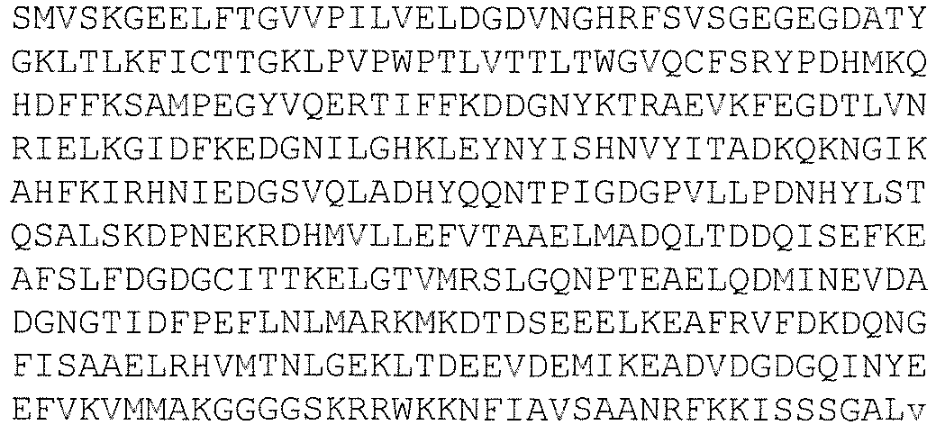

36 Cameleon biosensor Δ-32M 
37 Cameleon biosensor Δ-S2M 

38 Cameleon biosensor ΔΔΔΔ 
39 CaM1 
40 CaM1 
41 Primer DGD S 
42 Primer DGD AS 
43 Primer S-TEV-eCFP-BamHI 
44 Primer AS-eCFP-SacI TAAA GAGCTC GGCGGCGGTCACGAACTCCAGCA 45 Primer S-eYFP-SalI TATA GTCGAC ATG GTG AGC AAG GGC GAG GAG 46 Primer AS-eYFP-HindIII 
47 Cameleon biosensor WT 

48 Cameleon biosensor WT 

49 Primer S-Δ GGAAGCCTTCAGCTTATTCGACGGTGATGGTTGCATTACC 50 Primer AS-Δ GGTAATGCAACCATCACCGTCGAATAAGCTGAAGGCTTCC 51 Primer S-S2M 
52 Primer AS-S2M 
53 Cameleon biosensor WT-S2M 
54 Cameleon biosensor WT-S2M 

55 CaMΔ3 
56 CaMΔ3 
57 Primer S-Δ3Y TTCAGCTTATTCGACAAGGGTGATGGTTACATTACC 58 Primer AS-Δ3Y GGTAATGTAACCATCACCCTTGTCGAATAAGCTGAA 59 CaMΔ-WT 
60 CaMΔ-WT 
61 Cameleon Biosensor Δ1 Δ3 
62 Cameleon Biosensor Δ1 Δ3 
63 Cameleon Biosensor Δ1 Δ2 Δ3 

64 Cameleon Biosensor Δ1 Δ2 Δ3 
65 Cameleon Biosensor N-ter 

66 Cameleon Biosensor N-ter 
67 Cameleon Biosensor Δ1 Δ2 

68 Cameleon Biosensor Δ1 Δ2 
69 EF-hand1 loop +C7Y+T10A+K11A DKDGDGYITAAE 70 EF-hand1 loop ΔD1T10 KDGDGCITKE 71 EF-hand1 loop ΔD1K11 KDGDGCITTE 72 EF-hand1 loop ΔD3T10 DKGDGCITKE 73 EF-hand1 loop ΔD3K11 DKGDGCITTE 74 EF-hand1 loop ΔD1T10K11 KDGDGCITE 75 EF-hand1 loop ΔD3T10K11 DKGDGCITE 76 EF-hand2 loop ΔD1F10 ADGNGTIDPE 77 EF-hand2 loop ΔD1P11 ADGNGTIDFE 78 EF-hand2 loop ΔD3F10 DAGNGTIDPE 79 EF-hand2 loop ΔD3P11 DAGNGTIDFE 80 EF-hand2 loop ΔD1F10P11 ADGNGTIDE 81 EF-hand2 loop ΔD3F10P11 DAGNGTIDE 82 EF-hand3 loop ΔD1A10 KDQNGFISAE 83 EF-hand3 loop ΔD1A11 KDQNGFISAE 84 EF-hand3 loop ΔD3A10 DKQNGFISAE 85 EF-hand3 loop ΔD3A11 DKQNGFISAE 86 EF-hand3 loop ΔD1A10A11 KDQNGFISE 87 EF-hand3 loop ΔD3A10A11 DKQNGFISE 88 EF-hand4 loop ΔD1Y10 VDGDGQINEE 89 EF-hand4 loop ΔD1E11 VDGDGQINYE 90 EF-hand4 loop ΔD3Y10 DVGDGQINEE 91 EF-hand4 loop ΔD3E11 DVGDGQINYE 92 EF-hand4 loop ΔD1Y10E11 VDGDGQINE 93 EF-hand4 loop ΔD3Y10E11 DVGDGQINE -
- <110> Commissariat a l'Energie Atomique et aux Energies Alternatives
- <120> New uranium-chelating peptides derived from EF-hand calcium-binding motif useful for uranium biodetection and biodecontamination
- <130> 263PCT570
- <160> 93
- <170> PatentIn version 3.5
- <210> 1
<211> 452
<212> DNA
<213> Arabidopsis thaliana - <400> 1

- <210> 2
<211> 149
<212> PRT
<213> Arabidopsis thaliana - <400> 2


- <210> 3
<211> 12
<212> PRT
<213> artificial sequence - <220>
<223> synthetic peptide CaM3 EF-hand1 loop - <400> 3

- <210> 4
<211> 12
<212> PRT
<213> artificial sequence - <220>
<223> synthetic peptide CaM3 EF-hand2 loop - <400> 4

- <210> 5
<211> 12
<212> PRT
<213> artificial sequence - <220>
<223> synthetic peptide CaM3 EF-hand3 loop - <400> 5

- <210> 6
<211> 12
<212> PRT
<213> artificial sequence - <220>
<223> synthetic peptide CaM3 EF-hand4 loop - <400> 6

- <210> 7
<211> 10
<212> PRT
<213> artificial sequence - <220>
<223> synthetic peptide EF-hand1 loop delta-K2D3 - <400> 7

- <210> 8
<211> 10
<212> PRT
<213> artificial sequence - <220>
<223> artificial sequence EF-hand1 loop delta-K2D3/C7Y - <400> 8

- <210> 9
<211> 10
<212> PRT
<213> artificial sequence - <220>
<223> synthetic peptide EF-hand1 loop delta-K2D3/C7Y/T10A/K11A - <400> 9

- <210> 10
<211> 10
<212> PRT
<213> artificial sequence - <220>
<223> synthetic peptide EF-hand2 loop delta-A2D3 - <400> 10

- <210> 11
<211> 10
<212> PRT
<213> artificial sequence - <220>
<223> synthetic peptide EF-hand2 loop delta-A2D3/T7Y - <400> 11

- <210> 12
<211> 10
<212> PRT
<213> artificial sequence - <220>
<223> synthetic peptide EF-hand2 loop delta-A2D3/N5D - <400> 12

- <210> 13
<211> 10
<212> PRT
<213> artificial sequence - <220>
<223> synthetic peptide EF-hand2 loop delta-A2D3/T7Y/N5D - <400> 13

- <210> 14
<211> 10
<212> PRT
<213> artificial sequence - <220>
<223> synthetic peptide EF-hand3 loop delta-K2D3 - <400> 14

- <210> 15
<211> 10
<212> PRT
<213> artificial sequence - <220>
<223> synthetic peptide EF-hand4 loop delta-V2D3 - <400> 15

- <210> 16
<211> 228
<212> DNA
<213> artificial sequence - <220>
<223> synthetic polynucleotide encoding CaM-delta - <400> 16

- <210> 17
<211> 75
<212> PRT
<213> artificial sequence - <220>
<223> synthetic peptide CaM-delta - <400> 17

- <210> 18
<211> 147
<212> PRT
<213> artificial sequence - <220>
<223> synthetic Calmodulin variant protein from Cameleon biosensor-delta protein - <400> 18

- <210> 19
<211> 147
<212> PRT
<213> artificial sequence - <220>
<223> synthetic Calmodulin variant protein from Cameleon biosensor -delta-S2M protein - <400> 19

- <210> 20
<211> 141
<212> PRT
<213> artificial sequence - <220>
<223> synthetic calmodulin variantprotein delta sites - <400> 20


- <210> 21
<211> 78
<212> DNA
<213> artificial sequence - <220>
<223> synthetic polynucleotide encoding M13 peptide - <400> 21

- <210> 22
<211> 26
<212> PRT
<213> artificial sequence - <220>
<223> synthetic peptide M13 - <400> 22

- <210> 23
<211> 25
<212> PRT
<213> artificial sequence - <220>
<223> synthetic peptide skMLCK - <400> 23

- <210> 24
<211> 17
<212> PRT
<213> artificial sequence - <220>
<223> synthetic peptide MLCKp - <400> 24

- <210> 25
<211> 20
<212> PRT
<213> artificial sequence - <220>
<223> synthetic peptide smMLCK - <400> 25

- <210> 26
<211> 13
<212> PRT
<213> artificial sequence - <220>
<223> synthetic peptide from wasp venom - <400> 26

- <210> 27
<211> 24
<212> PRT
<213> artificial sequence - <220>
<223> synthetic peptide p21 - <400> 27

- <210> 28
<211> 26
<212> PRT
<213> artificial sequence - <220>
<223> synthetic peptide from melittin - <400> 28

- <210> 29
<211> 24
<212> PRT
<213> artificial sequence - <220>
<223> synthetic peptide from spectrin - <400> 29

- <210> 30
<211> 22
<212> PRT
<213> artificial sequence - <220>
<223> synthetic peptide CaMKI - <400> 30


- <210> 31
<211> 25
<212> PRT
<213> artificial sequence - <220>
<223> synthetic peptide CaMKII - <400> 31

- <210> 32
<211> 27
<212> PRT
<213> artificial sequence - <220>
<223> synthetic peptide CaMKK - <400> 32

- <210> 33
<211> 17
<212> PRT
<213> artificial sequence - <220>
<223> synthetic peptide1 - <400> 33

- <210> 34
<211> 1953
<212> DNA
<213> artificial sequence - <220>
<223> synthetic polynucleotide encoding biosensor-delta - <400> 34


- <210> 35
<211> 650
<212> PRT
<213> artificial sequence - <220>
<223> synthetic protein biosensor-delta - <400> 35
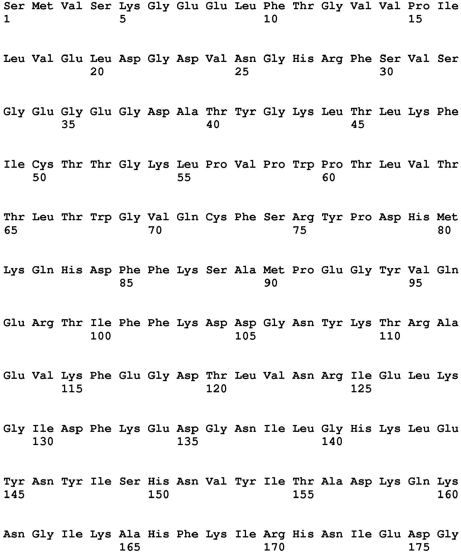
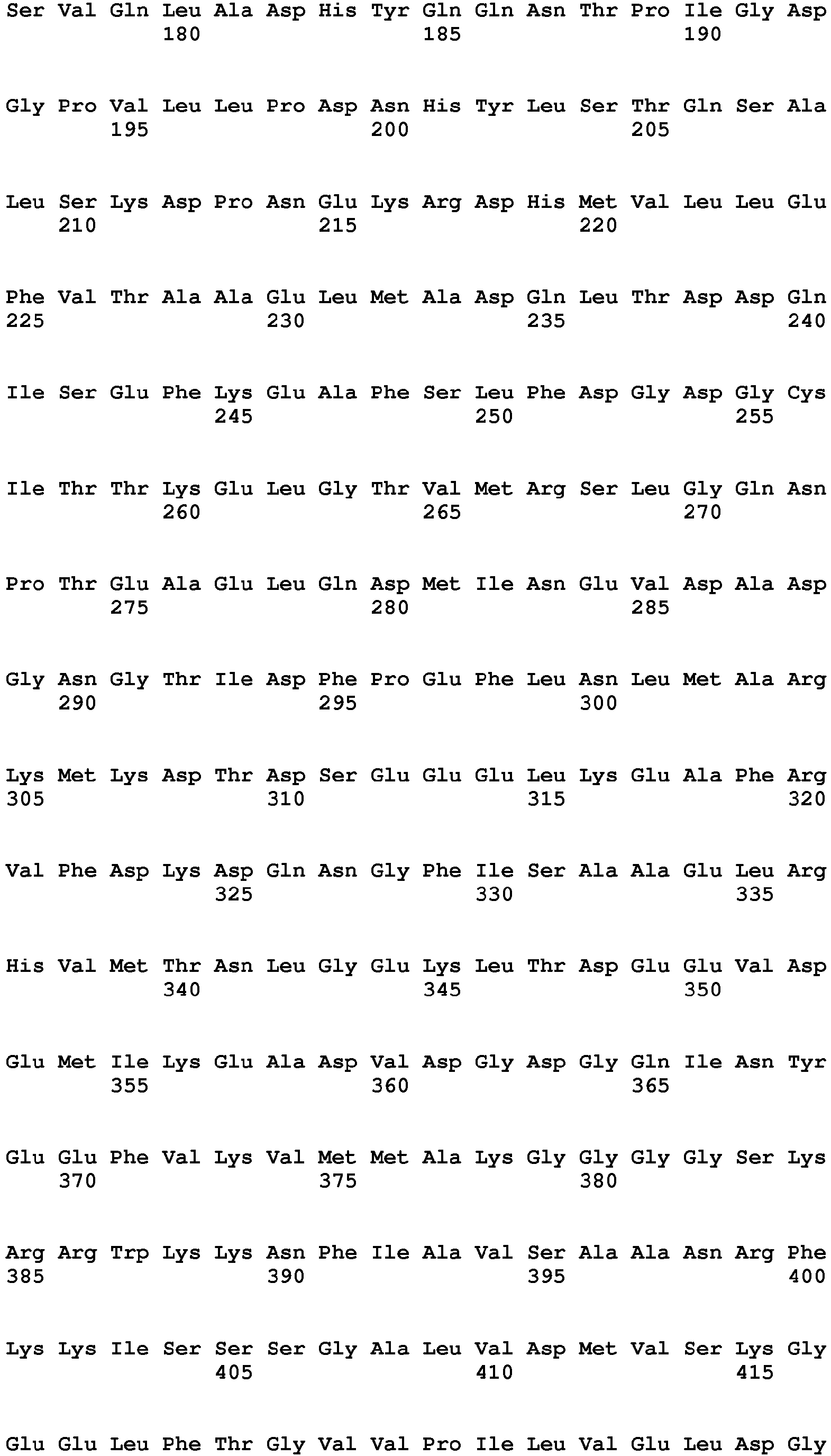

- <210> 36
<211> 1953
<212> DNA
<213> artificial sequence - <220>
<223> synthetic polynucleotide encoding cameleon biosensor delta-S2M - <400> 36


- <210> 37
<211> 650
<212> PRT
<213> artificial sequence - <220>
<223> synthetic protein Cameleon biosensor delta-S2M - <400> 37


- <210> 38
<211> 644
<212> PRT
<213> artificial sequence - <220>
<223> synthetic protein Cameleon biosensor delta-delta-delta-delta - <400> 38


- <210> 39
<211> 234
<212> DNA
<213> artificial sequence - <220>
<223> synthetic polynucleotide encoding CaM1 - <400> 39

- <210> 40
<211> 77
<212> PRT
<213> artificial sequence - <220>
<223> synthetic peptide CaM1 - <400> 40

- <210> 41
<211> 45
<212> DNA
<213> artificial sequence - <220>
<223> synthetic oligonucleotide primer DGD S - <400> 41
gaagctttca gcttattcga cggtgatggt tacattaccg ccgcg 45 - <210> 42
<211> 45
<212> DNA
<213> artificial sequence - <220>
<223> synthetic oligonucleotide primer DGD AS - <400> 42
cgcggcggta atgtaaccat caccgtcgaa taagctgaaa gcttc 45 - <210> 43
<211> 52
<212> DNA
<213> artificial sequence - <220>
<223> synthetic oligonucleotide primer S-TEV-eCFP- BamHI - <400> 43
gagaggatcc gagaacctgt acttccagtc catggtgagc aagggcgagg ag 52 - <210> 44
<211> 33
<212> DNA
<213> artificial sequence - <220>
<223> synthetic oligonucleotide primer AS-eCFP-SacI - <400> 44
taaagagctc ggcggcggtc acgaactcca gca 33 - <210> 45
<211> 31
<212> DNA
<213> artificial sequence - <220>
<223> synthetic oligonucleotide primer S-eYFP- SalI - <400> 45
tatagtcgac atggtgagca agggcgagga g 31 - <210> 46
<211> 35
<212> DNA
<213> artificial sequence - <220>
<223> synthetic oligonucleotide primer AS-eYFP-HindIII - <400> 46
gggcaagctt ttacttgtac agctcgtcca tgccg 35 - <210> 47
<211> 1959
<212> DNA
<213> artificial sequence - <220>
<223> synthetic polynucleotide encoding Cameleon biosensor WT - <400> 47


- <210> 48
<211> 652
<212> PRT
<213> artificial sequence - <220>
<223> synthetic protein Cameleon biosensor WT - <400> 48


- <210> 49
<211> 40
<212> DNA
<213> artificial sequence - <220>
<223> synthetic oligonucleotide primer S-delta - <400> 49
ggaagccttc agcttattcg acggtgatgg ttgcattacc 40 - <210> 50
<211> 40
<212> DNA
<213> artificial sequence - <220>
<223> synthetic oligonucleotide primer AS-delta - <400> 50
ggtaatgcaa ccatcaccgt cgaataagct gaaggcttcc 40 - <210> 51
<211> 34
<212> DNA
<213> artificial sequence - <220>
<223> synthetic oligonucleotide primer S-S2M - <400> 51
ggtaccgtta ccagcagcag ccacttcgtt gatc 34 - <210> 52
<211> 34
<212> DNA
<213> artificial sequence - <220>
<223> synthetic oligonucleotide primer AS-S2M - <400> 52
gatcaacgaa gtggctgctg ctggtaacgg tacc 34 - <210> 53
<211> 1959
<212> DNA
<213> artificial sequence - <220>
<223> synthetic polynucleotide encoding Cameleon biosensor WT-S2M - <400> 53
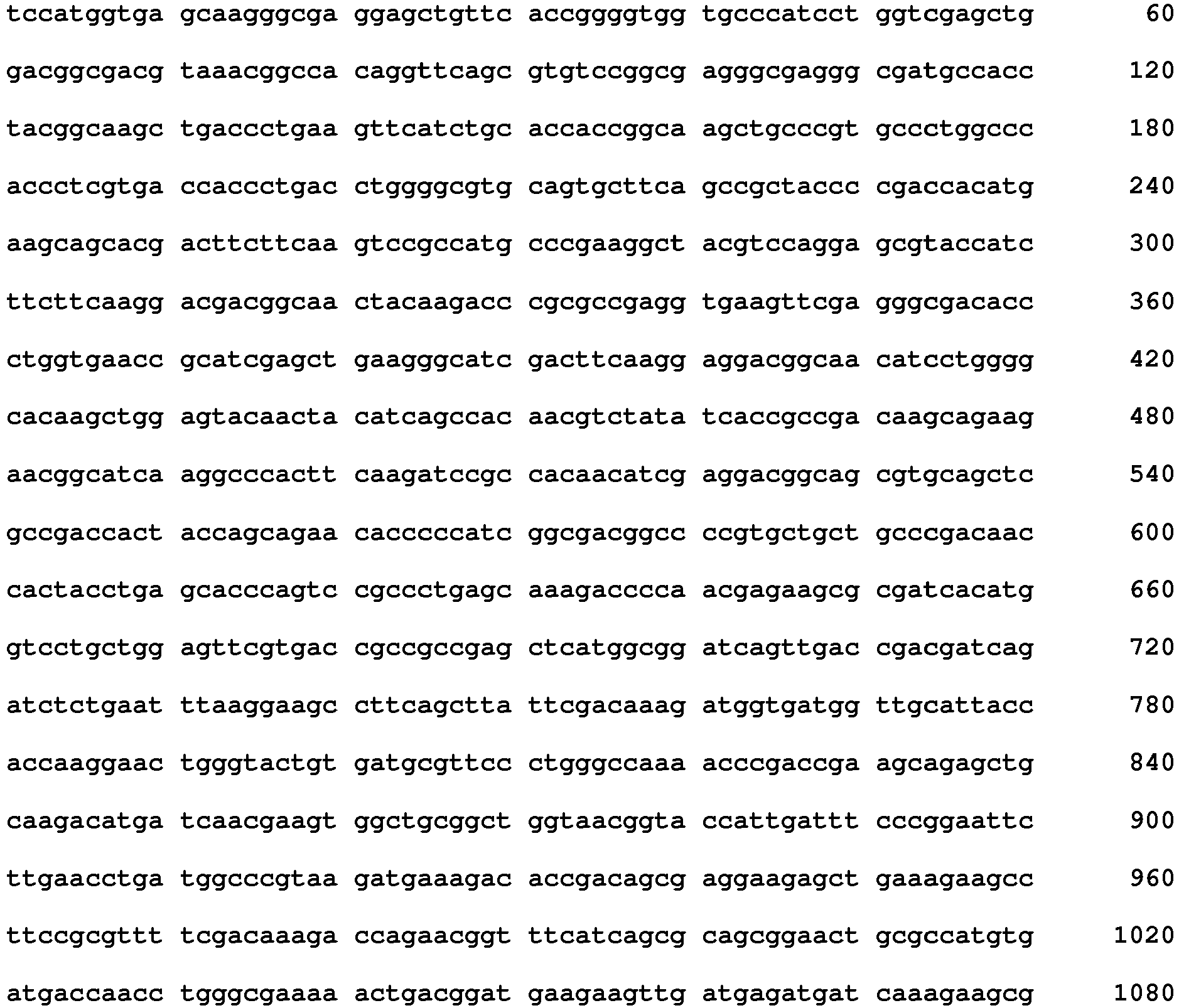

- <210> 54
<211> 652
<212> PRT
<213> artificial sequence - <220>
<223> synthetic protein Cameleon biosensor WT-S2M - <400> 54


- <210> 55
<211> 231
<212> DNA
<213> artificial sequence - <220>
<223> synthetic nucleotide encoding CaM-delta3 polypeptide - <400> 55

- <210> 56
<211> 76
<212> PRT
<213> artificial sequence - <220>
<223> synthetic polypeptide CaM-delta3 - <400> 56
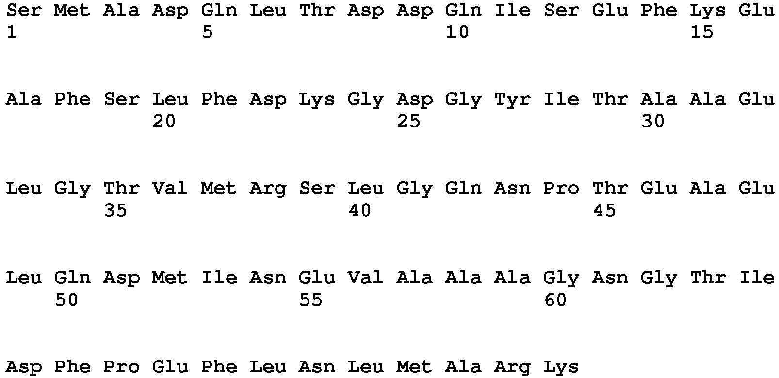

- <210> 57
<211> 36
<212> DNA
<213> artificial sequence - <220>
<223> synthetic primer S-delta3Y - <400> 57
ttcagcttat tcgacaaggg tgatggttac attacc 36 - <210> 58
<211> 36
<212> DNA
<213> artificial sequence - <220>
<223> synthetic primer AS-delta3Y - <400> 58
ggtaatgtaa ccatcaccct tgtcgaataa gctgaa 36 - <210> 59
<211> 228
<212> DNA
<213> artificial sequence - <220>
<223> synthetic polynucleotide encoding polypeptide CaM-delta-WT - <400> 59

- <210> 60
<211> 75
<212> PRT
<213> artificial sequence - <220>
<223> synthetic polypeptide CaM-delta-WT - <400> 60


- <210> 61
<211> 648
<212> PRT
<213> artificial sequence - <220>
<223> synthetic protein Cameleon biosensor delta1-delta3 - <400> 61
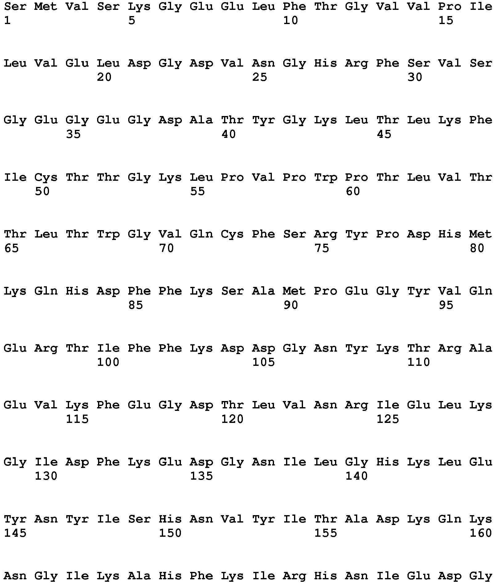


- <210> 62
<211> 1947
<212> DNA
<213> artificial sequence - <220>
<223> synthetic polynucleotide encoding protein cameleon biosensor delta1-delta3 - <400> 62

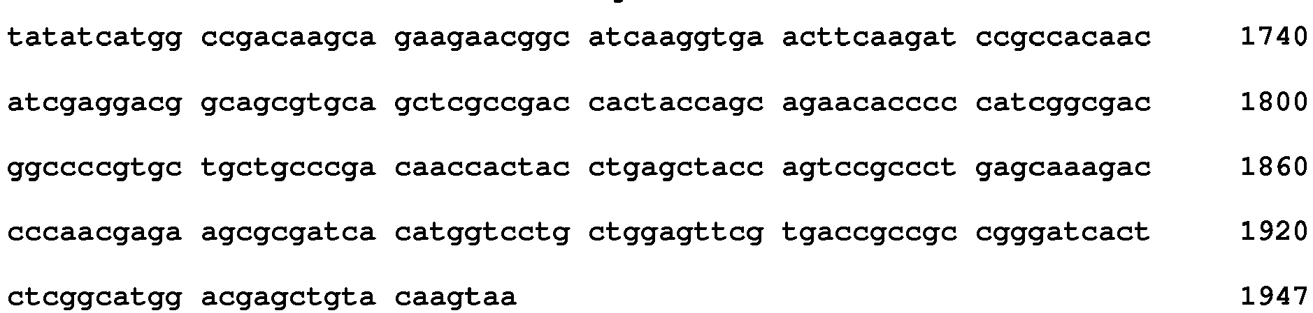
- <210> 63
<211> 646
<212> PRT
<213> artificial sequence - <220>
<223> synthetic protein Cameleon biosensor delta1-delta2-delta3 - <400> 63


- <210> 64
<211> 1941
<212> DNA
<213> artificial sequence - <220>
<223> synthetic polynucleotide encoding protein cameleon biosensor delta1-delta2-delta3 - <400> 64


- <210> 65
<211> 550
<212> PRT
<213> artificial sequence - <220>
<223> synthetic protein cameleon biosensor N-ter - <400> 65


- <210> 66
<211> 1653
<212> DNA
<213> artificial sequence - <220>
<223> synthetic polynucleotide encoding protein cameleon biosensor N-ter - <400> 66


- <210> 67
<211> 648
<212> PRT
<213> artificial sequence - <220>
<223> synthetic protein Cameleon biosensor delta1-delta2 - <400> 67


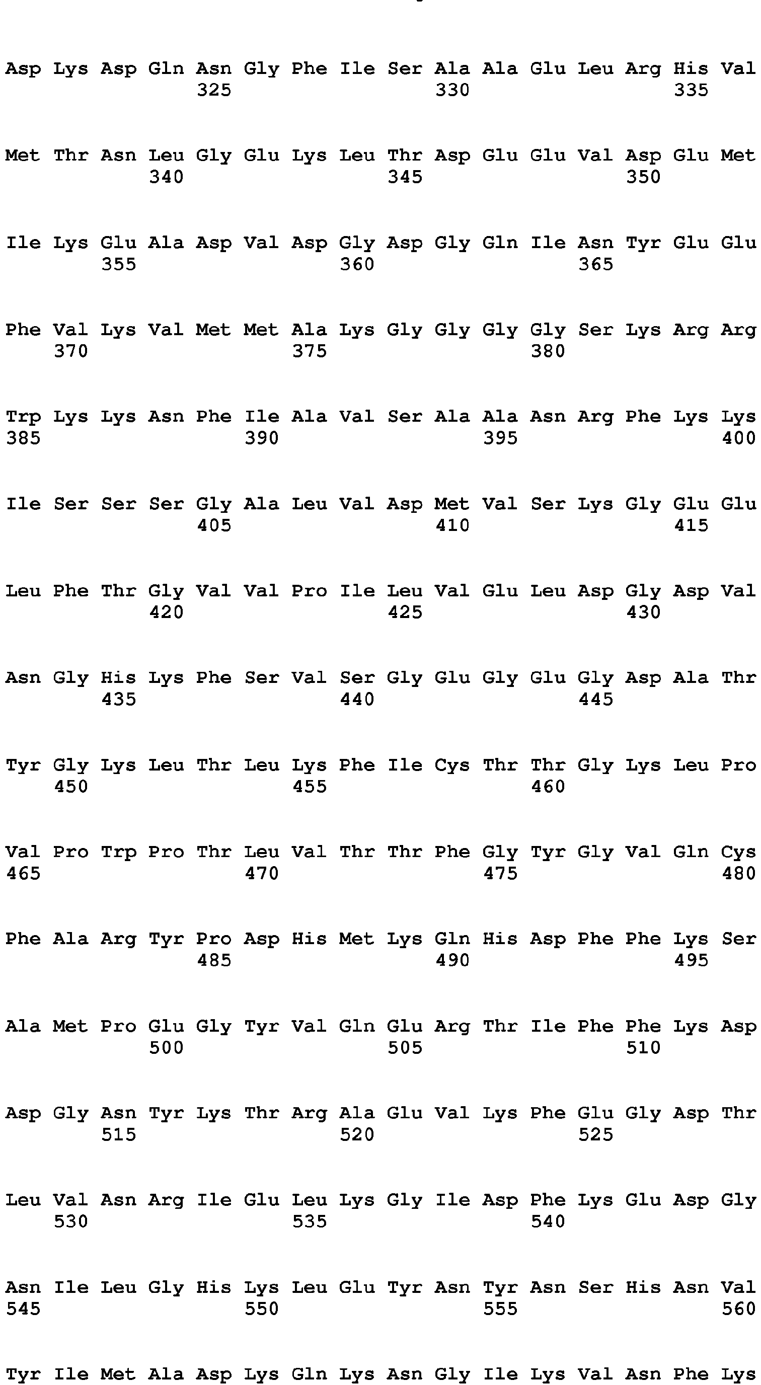

- <210> 68
<211> 1947
<212> DNA
<213> artificial sequence - <220>
<223> synthetic polynucleotide encoding cameleon biosensor delta1-delta2 - <400> 68


- <210> 69
<211> 12
<212> PRT
<213> artificial sequence - <220>
<223> synthetic peptide EF-hand1 loop +C7Y+T10A+K11A - <400> 69

- <210> 70
<211> 10
<212> PRT
<213> artificial sequence - <220>
<223> synthetic peptide EF-hand1 loop deltaD1T10 - <400> 70

- <210> 71
<211> 10
<212> PRT
<213> artificial sequence - <220>
<223> synthetic peptide EF-hand1 loop deltaD1K11 - <400> 71

- <210> 72
<211> 10
<212> PRT
<213> artificial sequence - <220>
<223> synthetic peptide EF-hand1 loop deltaD3T10 - <400> 72

- <210> 73
<211> 10
<212> PRT
<213> artificial sequence - <220>
<223> synthetic peptide EF-hand1 loop delta D3K11 - <400> 73

- <210> 74
<211> 9
<212> PRT
<213> artificial sequence - <220>
<223> synthetic peptide EF-hand1 loop delta D1T10K11 - <400> 74

- <210> 75
<211> 9
<212> PRT
<213> artificial sequence - <220>
<223> synthetic peptide EF-hand1 loop delta D3T10K11 - <400> 75

- <210> 76
<211> 10
<212> PRT
<213> artificial sequence - <220>
<223> synthetic peptide EF-hand2 loop delta-DlFlO - <400> 76

- <210> 77
<211> 10
<212> PRT
<213> artificial sequence - <220>
<223> synthetic peptide EF-hand2 loop deltaD1P11 - <400> 77

- <210> 78
<211> 10
<212> PRT
<213> artificial sequence - <220>
<223> synthetic peptide EF-hand2 loop delta D3F10 - <400> 78

- <210> 79
<211> 10
<212> PRT
<213> artificial sequence - <220>
<223> synthetic peptide EF-hand2 loop delta D3P11 - <400> 79

- <210> 80
<211> 9
<212> PRT
<213> artificial sequence - <220>
<223> synthetic peptide EF-hand2 loop delta D1F10P11 - <400> 80

- <210> 81
<211> 9
<212> PRT
<213> artificial sequence - <220>
<223> synthetic peptide EF-hand2 loop delta D3F10P11 - <400> 81

- <210> 82
<211> 10
<212> PRT
<213> artificial sequence - <220>
<223> synthetic peptide EF-hand3 loop delta D1A10 - <400> 82

- <210> 83
<211> 10
<212> PRT
<213> artificial sequence - <220>
<223> synthetic peptide EF-hand3 loop delta D1A11 - <400> 83

- <210> 84
<211> 10
<212> PRT
<213> artificial sequence - <220>
<223> synthetic peptide EF-hand3 loop delta D3A10 - <400> 84

- <210> 85
<211> 10
<212> PRT
<213> artificial sequence - <220>
<223> synthetic peptide EF-hand3 loop delta D3A11 - <400> 85

- <210> 86
<211> 9
<212> PRT
<213> artificial sequence - <220>
<223> synthetic peptide EF-hand3 loop delta D1A10A11 - <400> 86

- <210> 87
<211> 9
<212> PRT
<213> artificial sequence - <220>
<223> synthetic peptide EF-hand3 loop delta D3A10A11 - <400> 87

- <210> 88
<211> 10
<212> PRT
<213> artificial sequence - <220>
<223> synthetic peptide EF-hand4 loop delta D1Y10 - <400> 88

- <210> 89
<211> 10
<212> PRT
<213> artificial sequence - <220>
<223> synthetic peptide EF-hand4 loop delta D1E11 - <400> 89

- <210> 90
<211> 10
<212> PRT
<213> artificial sequence - <220>
<223> synthetic peptide EF-hand4 loop delta D3Y10 - <400> 90

- <210> 91
<211> 10
<212> PRT
<213> artificial sequence - <220>
<223> synthetic peptide EF-hand4 loop delta D3E11 - <400> 91

- <210> 92
<211> 9
<212> PRT
<213> artificial sequence - <220>
<223> synthetic peptide EF-hand4 loop delta D1Y10E11 - <400> 92

- <210> 93
<211> 9
<212> PRT
<213> artificial sequence - <220>
<223> synthetic peptide EF-hand4 loop delta D3Y10E11 - <400> 93

Claims (13)
- A polypeptide comprising at least one helix-loop-helix calcium-binding EF-hand motif which comprises a deletion of the two amino acid residues in position 2 and 3 or 1 and 2 of the 12-amino-acid calcium-binding loop sequence, and wherein said polypeptide binds uranyl.
- The polypeptide of claim 1 , wherein said EF-hand motif(s) are derived from signaling EF-hand protein(s) of the calmodulin superfamily selected from the group consisting of calmodulin and troponin C.
- The polypeptide of claim 1 or 2, which comprises two or four EF-hand motifs, wherein at least one EF-hand motif comprises said deletion in the calcium-binding loop sequence and the other EF-hand motif(s) comprise said deletion or not.
- The polypeptide of claim 3, which is a calmodulin domain 1 variant comprising two EF-hand motifs, respectively from the EF-hand1 and the EF-hand2 of calmodulin protein(s).
- The polypeptide of claim 3or 4, which comprises or consists of an amino acid sequence selected from the group consisting of the sequences SEQ ID NO: 17, 18, 20 and 60.
- A fusion protein comprising a polypeptide according to any one of claims 1 to 5 fused to another protein moiety.
- The fusion protein of claim 6, which is a cameleon protein comprising tandem fusions of a fluorescence-donor protein, a polypeptide of any one of claims 3 to 5, and a fluorescence-acceptor protein.
- The fusion protein of claim 7, which comprises or consists of a sequence selected from the group consisting of the sequences SEQ ID NO: 35, 38, 61, 63 and 67.
- The polypeptide of any one of claims 1 to 5 or the fusion protein of any one of claims claim 6 to 8, which is immobilized onto a solid support.
- A polynucleotide encoding the polypeptide of any one of claims 1 to 5 or the fusion protein of any one of claims 6 to 8.
- A host cell comprising the polynucleotide of claim 10.
- A transgenic bacterium or transgenic plant comprising the polynucleotide of claim 10.
- Use of the polypeptide of any one of claims 1 to 5, the fusion protein of any one of claims 6 to 8, the host cell of claim 11 or the transgenic bacterium or transgenic plant of claim 12 as uranyl chelating agent, for the detection of uranium contamination in vitro or for the decontamination and/or the bioremediation of uranium contamination in an environment.
Priority Applications (1)
| Application Number | Priority Date | Filing Date | Title |
|---|---|---|---|
| EP14718767.8A EP2981555B1 (en) | 2013-03-28 | 2014-03-28 | New uranium-chelating peptides derived from ef-hand calcium-binding motif useful for uranium biodetection and biodecontamination |
Applications Claiming Priority (4)
| Application Number | Priority Date | Filing Date | Title |
|---|---|---|---|
| EP13305400.7A EP2784094A1 (en) | 2013-03-28 | 2013-03-28 | New uranium-chelating peptides derived from EF-hand calcium-binding motif useful for uranium biodetection and biodecontamination |
| EP13305532 | 2013-04-23 | ||
| PCT/IB2014/060271 WO2014155356A1 (en) | 2013-03-28 | 2014-03-28 | New uranium-chelating peptides derived from ef-hand calcium-binding motif useful for uranium biodetection and biodecontamination |
| EP14718767.8A EP2981555B1 (en) | 2013-03-28 | 2014-03-28 | New uranium-chelating peptides derived from ef-hand calcium-binding motif useful for uranium biodetection and biodecontamination |
Publications (2)
| Publication Number | Publication Date |
|---|---|
| EP2981555A1 EP2981555A1 (en) | 2016-02-10 |
| EP2981555B1 true EP2981555B1 (en) | 2020-04-15 |
Family
ID=50543274
Family Applications (1)
| Application Number | Title | Priority Date | Filing Date |
|---|---|---|---|
| EP14718767.8A Active EP2981555B1 (en) | 2013-03-28 | 2014-03-28 | New uranium-chelating peptides derived from ef-hand calcium-binding motif useful for uranium biodetection and biodecontamination |
Country Status (6)
| Country | Link |
|---|---|
| US (1) | US9845343B2 (en) |
| EP (1) | EP2981555B1 (en) |
| AU (1) | AU2014240783B2 (en) |
| CA (1) | CA2907827C (en) |
| EA (1) | EA201591760A1 (en) |
| WO (1) | WO2014155356A1 (en) |
Families Citing this family (1)
| Publication number | Priority date | Publication date | Assignee | Title |
|---|---|---|---|---|
| US20210107956A1 (en) * | 2017-04-03 | 2021-04-15 | Washington State University | Ultrasensitive detection platform using luminescent metals and uses thereof |
Family Cites Families (3)
| Publication number | Priority date | Publication date | Assignee | Title |
|---|---|---|---|---|
| SE0201863D0 (en) | 2002-06-18 | 2002-06-18 | Cepep Ab | Cell penetrating peptides |
| FR2857014B1 (en) | 2003-07-04 | 2006-11-24 | Commissariat Energie Atomique | URANIUM CHELATING PEPTIDES AND THEIR APPLICATIONS |
| FR2976586B1 (en) | 2011-06-15 | 2015-01-23 | Centre Nat Rech Scient | METHOD FOR GENERATING CYAN FLUORESCENT PROTEINS HAVING REDUCED SENSITIVITY TO PH |
-
2014
- 2014-03-28 US US14/779,114 patent/US9845343B2/en active Active
- 2014-03-28 WO PCT/IB2014/060271 patent/WO2014155356A1/en not_active Ceased
- 2014-03-28 AU AU2014240783A patent/AU2014240783B2/en active Active
- 2014-03-28 EP EP14718767.8A patent/EP2981555B1/en active Active
- 2014-03-28 CA CA2907827A patent/CA2907827C/en active Active
- 2014-03-28 EA EA201591760A patent/EA201591760A1/en unknown
Non-Patent Citations (2)
| Title |
|---|
| ANITA LEWIT-BENTLEY ET AL: "EF-hand calcium-binding proteins", CURRENT OPINION IN STRUCTURAL BIOLOGY, 1 January 2000 (2000-01-01), England, pages 637 - 643, XP055654413, Retrieved from the Internet <URL:https://reader.elsevier.com/reader/sd/pii/S0959440X00001421?token=19CDEAFFFBCF2B8601DEFAE226DD8907295A74A7D6087ACFE72819BA2B8CB7203FEC8391708ED1C6899230C8A0D8BEA4> DOI: 10.1016/S0959-440X(00)00142-1 * |
| YUBIN ZHOU ET AL: "Prediction of EF-hand calcium-binding proteins and analysis of bacterial EF-hand proteins", PROTEINS: STRUCTURE, FUNCTION, AND BIOINFORMATICS, vol. 65, no. 3, 15 November 2006 (2006-11-15), US, pages 643 - 655, XP055654412, ISSN: 0887-3585, DOI: 10.1002/prot.21139 * |
Also Published As
| Publication number | Publication date |
|---|---|
| US20160130316A1 (en) | 2016-05-12 |
| CA2907827C (en) | 2021-09-07 |
| AU2014240783B2 (en) | 2019-02-14 |
| WO2014155356A8 (en) | 2015-01-08 |
| CA2907827A1 (en) | 2014-10-02 |
| EP2981555A1 (en) | 2016-02-10 |
| WO2014155356A1 (en) | 2014-10-02 |
| US9845343B2 (en) | 2017-12-19 |
| AU2014240783A1 (en) | 2015-10-15 |
| EA201591760A1 (en) | 2016-02-29 |
Similar Documents
| Publication | Publication Date | Title |
|---|---|---|
| US12492451B2 (en) | Metal-binding protein and use thereof | |
| Pardoux et al. | Modulating uranium binding affinity in engineered calmodulin EF-hand peptides: effect of phosphorylation | |
| Stribny et al. | The human Golgi protein TMEM165 transports calcium and manganese in yeast and bacterial cells | |
| Chen et al. | A split GAL4 RUBY assay for visual in planta detection of protein–protein interactions | |
| Furukawa et al. | The fungal CCAAT-binding complex and HapX display highly variable but evolutionary conserved synergetic promoter-specific DNA recognition | |
| Gallois et al. | Discovery and characterization of UipA, a uranium-and iron-binding PepSY protein involved in uranium tolerance by soil bacteria | |
| Aldema et al. | Purification of the Tn 10‐specified tetracycline efflux antiporter TetA in a native state as a polyhistidine fusion protein | |
| Wei et al. | Engineering a gold-specific regulon for cell-based visual detection and recovery of gold | |
| US9845343B2 (en) | Uranium-chelating peptides derived from EF-hand calcium-binding motif useful for uranium biodetection and biodecontamination | |
| Luo et al. | Kinetic and structural characterization of human mortalin | |
| JP4505439B2 (en) | Fluorescently labeled protein with improved signal intensity relative to sugar concentration and use thereof | |
| EP2784094A1 (en) | New uranium-chelating peptides derived from EF-hand calcium-binding motif useful for uranium biodetection and biodecontamination | |
| JP5161588B2 (en) | Environmentally stable sensor and method of using the same | |
| JP5086636B2 (en) | Uranium-chelating peptides and their use | |
| KR20100119104A (en) | Fast maturating red fluorescent protein, fmred, as a novel reporter and molecular probe | |
| Pal et al. | PBP isolation and DD-carboxypeptidase assay | |
| CN116042766B (en) | Composition, method and application of bioluminescence-based protein ligase activity assay | |
| WO2017013400A1 (en) | Single-stranded dna sensor | |
| Paavola et al. | Crystal structures of a dodecameric multicopper oxidase from Marinithermus hydrothermalis | |
| EP4421166A1 (en) | Improved split halotags | |
| JP5115954B2 (en) | Functional magnetic bacteria | |
| KR20200066465A (en) | A microorganism diagnostic kit for sensing mercury and the preparation method thereof | |
| Chen et al. | Functional and fluorescence analyses of tryptophan residues in H+-pyrophosphatase of Clostridium tetani | |
| CN120099052A (en) | A fusion protein of annexin A5 and fluorescent protein for apoptotic cell marking, and preparation method and application thereof | |
| WO2005042748A1 (en) | Coloured fusion proteins |
Legal Events
| Date | Code | Title | Description |
|---|---|---|---|
| PUAI | Public reference made under article 153(3) epc to a published international application that has entered the european phase |
Free format text: ORIGINAL CODE: 0009012 |
|
| 17P | Request for examination filed |
Effective date: 20151027 |
|
| AK | Designated contracting states |
Kind code of ref document: A1 Designated state(s): AL AT BE BG CH CY CZ DE DK EE ES FI FR GB GR HR HU IE IS IT LI LT LU LV MC MK MT NL NO PL PT RO RS SE SI SK SM TR |
|
| AX | Request for extension of the european patent |
Extension state: BA ME |
|
| DAX | Request for extension of the european patent (deleted) | ||
| RIN1 | Information on inventor provided before grant (corrected) |
Inventor name: SAUGE-MERLE, SANDRINE Inventor name: BREMOND, NICOLAS Inventor name: LEMAIRE, DAVID Inventor name: GUILBAUD, PHILIPPE Inventor name: PARDOUX, ROMAIN Inventor name: BERTHOMIEU, CATHERINE Inventor name: DELANGLE, PASCALE Inventor name: BECCIA, MARIA-ROSA |
|
| STAA | Information on the status of an ep patent application or granted ep patent |
Free format text: STATUS: EXAMINATION IS IN PROGRESS |
|
| 17Q | First examination report despatched |
Effective date: 20170213 |
|
| GRAP | Despatch of communication of intention to grant a patent |
Free format text: ORIGINAL CODE: EPIDOSNIGR1 |
|
| STAA | Information on the status of an ep patent application or granted ep patent |
Free format text: STATUS: GRANT OF PATENT IS INTENDED |
|
| INTG | Intention to grant announced |
Effective date: 20200122 |
|
| RIN1 | Information on inventor provided before grant (corrected) |
Inventor name: BREMOND, NICOLAS Inventor name: LEMAIRE, DAVID Inventor name: PARDOUX, ROMAIN Inventor name: BECCIA, MARIA-ROSA Inventor name: DELANGLE, PASCALE Inventor name: GUILBAUD, PHILIPPE Inventor name: SAUGE-MERLE, SANDRINE Inventor name: BERTHOMIEU, CATHERINE |
|
| GRAS | Grant fee paid |
Free format text: ORIGINAL CODE: EPIDOSNIGR3 |
|
| GRAA | (expected) grant |
Free format text: ORIGINAL CODE: 0009210 |
|
| STAA | Information on the status of an ep patent application or granted ep patent |
Free format text: STATUS: THE PATENT HAS BEEN GRANTED |
|
| AK | Designated contracting states |
Kind code of ref document: B1 Designated state(s): AL AT BE BG CH CY CZ DE DK EE ES FI FR GB GR HR HU IE IS IT LI LT LU LV MC MK MT NL NO PL PT RO RS SE SI SK SM TR |
|
| REG | Reference to a national code |
Ref country code: CH Ref legal event code: EP |
|
| REG | Reference to a national code |
Ref country code: DE Ref legal event code: R096 Ref document number: 602014063763 Country of ref document: DE |
|
| REG | Reference to a national code |
Ref country code: IE Ref legal event code: FG4D |
|
| REG | Reference to a national code |
Ref country code: AT Ref legal event code: REF Ref document number: 1257182 Country of ref document: AT Kind code of ref document: T Effective date: 20200515 |
|
| REG | Reference to a national code |
Ref country code: NL Ref legal event code: MP Effective date: 20200415 |
|
| REG | Reference to a national code |
Ref country code: LT Ref legal event code: MG4D |
|
| PG25 | Lapsed in a contracting state [announced via postgrant information from national office to epo] |
Ref country code: SE Free format text: LAPSE BECAUSE OF FAILURE TO SUBMIT A TRANSLATION OF THE DESCRIPTION OR TO PAY THE FEE WITHIN THE PRESCRIBED TIME-LIMIT Effective date: 20200415 Ref country code: PT Free format text: LAPSE BECAUSE OF FAILURE TO SUBMIT A TRANSLATION OF THE DESCRIPTION OR TO PAY THE FEE WITHIN THE PRESCRIBED TIME-LIMIT Effective date: 20200817 Ref country code: NL Free format text: LAPSE BECAUSE OF FAILURE TO SUBMIT A TRANSLATION OF THE DESCRIPTION OR TO PAY THE FEE WITHIN THE PRESCRIBED TIME-LIMIT Effective date: 20200415 Ref country code: LT Free format text: LAPSE BECAUSE OF FAILURE TO SUBMIT A TRANSLATION OF THE DESCRIPTION OR TO PAY THE FEE WITHIN THE PRESCRIBED TIME-LIMIT Effective date: 20200415 Ref country code: IS Free format text: LAPSE BECAUSE OF FAILURE TO SUBMIT A TRANSLATION OF THE DESCRIPTION OR TO PAY THE FEE WITHIN THE PRESCRIBED TIME-LIMIT Effective date: 20200815 Ref country code: FI Free format text: LAPSE BECAUSE OF FAILURE TO SUBMIT A TRANSLATION OF THE DESCRIPTION OR TO PAY THE FEE WITHIN THE PRESCRIBED TIME-LIMIT Effective date: 20200415 Ref country code: NO Free format text: LAPSE BECAUSE OF FAILURE TO SUBMIT A TRANSLATION OF THE DESCRIPTION OR TO PAY THE FEE WITHIN THE PRESCRIBED TIME-LIMIT Effective date: 20200715 Ref country code: GR Free format text: LAPSE BECAUSE OF FAILURE TO SUBMIT A TRANSLATION OF THE DESCRIPTION OR TO PAY THE FEE WITHIN THE PRESCRIBED TIME-LIMIT Effective date: 20200716 |
|
| REG | Reference to a national code |
Ref country code: AT Ref legal event code: MK05 Ref document number: 1257182 Country of ref document: AT Kind code of ref document: T Effective date: 20200415 |
|
| PG25 | Lapsed in a contracting state [announced via postgrant information from national office to epo] |
Ref country code: LV Free format text: LAPSE BECAUSE OF FAILURE TO SUBMIT A TRANSLATION OF THE DESCRIPTION OR TO PAY THE FEE WITHIN THE PRESCRIBED TIME-LIMIT Effective date: 20200415 Ref country code: HR Free format text: LAPSE BECAUSE OF FAILURE TO SUBMIT A TRANSLATION OF THE DESCRIPTION OR TO PAY THE FEE WITHIN THE PRESCRIBED TIME-LIMIT Effective date: 20200415 Ref country code: BG Free format text: LAPSE BECAUSE OF FAILURE TO SUBMIT A TRANSLATION OF THE DESCRIPTION OR TO PAY THE FEE WITHIN THE PRESCRIBED TIME-LIMIT Effective date: 20200715 Ref country code: RS Free format text: LAPSE BECAUSE OF FAILURE TO SUBMIT A TRANSLATION OF THE DESCRIPTION OR TO PAY THE FEE WITHIN THE PRESCRIBED TIME-LIMIT Effective date: 20200415 |
|
| PG25 | Lapsed in a contracting state [announced via postgrant information from national office to epo] |
Ref country code: AL Free format text: LAPSE BECAUSE OF FAILURE TO SUBMIT A TRANSLATION OF THE DESCRIPTION OR TO PAY THE FEE WITHIN THE PRESCRIBED TIME-LIMIT Effective date: 20200415 |
|
| REG | Reference to a national code |
Ref country code: DE Ref legal event code: R097 Ref document number: 602014063763 Country of ref document: DE |
|
| PG25 | Lapsed in a contracting state [announced via postgrant information from national office to epo] |
Ref country code: CZ Free format text: LAPSE BECAUSE OF FAILURE TO SUBMIT A TRANSLATION OF THE DESCRIPTION OR TO PAY THE FEE WITHIN THE PRESCRIBED TIME-LIMIT Effective date: 20200415 Ref country code: IT Free format text: LAPSE BECAUSE OF FAILURE TO SUBMIT A TRANSLATION OF THE DESCRIPTION OR TO PAY THE FEE WITHIN THE PRESCRIBED TIME-LIMIT Effective date: 20200415 Ref country code: RO Free format text: LAPSE BECAUSE OF FAILURE TO SUBMIT A TRANSLATION OF THE DESCRIPTION OR TO PAY THE FEE WITHIN THE PRESCRIBED TIME-LIMIT Effective date: 20200415 Ref country code: ES Free format text: LAPSE BECAUSE OF FAILURE TO SUBMIT A TRANSLATION OF THE DESCRIPTION OR TO PAY THE FEE WITHIN THE PRESCRIBED TIME-LIMIT Effective date: 20200415 Ref country code: AT Free format text: LAPSE BECAUSE OF FAILURE TO SUBMIT A TRANSLATION OF THE DESCRIPTION OR TO PAY THE FEE WITHIN THE PRESCRIBED TIME-LIMIT Effective date: 20200415 Ref country code: EE Free format text: LAPSE BECAUSE OF FAILURE TO SUBMIT A TRANSLATION OF THE DESCRIPTION OR TO PAY THE FEE WITHIN THE PRESCRIBED TIME-LIMIT Effective date: 20200415 Ref country code: SM Free format text: LAPSE BECAUSE OF FAILURE TO SUBMIT A TRANSLATION OF THE DESCRIPTION OR TO PAY THE FEE WITHIN THE PRESCRIBED TIME-LIMIT Effective date: 20200415 Ref country code: DK Free format text: LAPSE BECAUSE OF FAILURE TO SUBMIT A TRANSLATION OF THE DESCRIPTION OR TO PAY THE FEE WITHIN THE PRESCRIBED TIME-LIMIT Effective date: 20200415 |
|
| PLBE | No opposition filed within time limit |
Free format text: ORIGINAL CODE: 0009261 |
|
| STAA | Information on the status of an ep patent application or granted ep patent |
Free format text: STATUS: NO OPPOSITION FILED WITHIN TIME LIMIT |
|
| PG25 | Lapsed in a contracting state [announced via postgrant information from national office to epo] |
Ref country code: SK Free format text: LAPSE BECAUSE OF FAILURE TO SUBMIT A TRANSLATION OF THE DESCRIPTION OR TO PAY THE FEE WITHIN THE PRESCRIBED TIME-LIMIT Effective date: 20200415 Ref country code: PL Free format text: LAPSE BECAUSE OF FAILURE TO SUBMIT A TRANSLATION OF THE DESCRIPTION OR TO PAY THE FEE WITHIN THE PRESCRIBED TIME-LIMIT Effective date: 20200415 |
|
| 26N | No opposition filed |
Effective date: 20210118 |
|
| PG25 | Lapsed in a contracting state [announced via postgrant information from national office to epo] |
Ref country code: SI Free format text: LAPSE BECAUSE OF FAILURE TO SUBMIT A TRANSLATION OF THE DESCRIPTION OR TO PAY THE FEE WITHIN THE PRESCRIBED TIME-LIMIT Effective date: 20200415 |
|
| PG25 | Lapsed in a contracting state [announced via postgrant information from national office to epo] |
Ref country code: MC Free format text: LAPSE BECAUSE OF FAILURE TO SUBMIT A TRANSLATION OF THE DESCRIPTION OR TO PAY THE FEE WITHIN THE PRESCRIBED TIME-LIMIT Effective date: 20200415 |
|
| REG | Reference to a national code |
Ref country code: CH Ref legal event code: PL |
|
| REG | Reference to a national code |
Ref country code: BE Ref legal event code: MM Effective date: 20210331 |
|
| PG25 | Lapsed in a contracting state [announced via postgrant information from national office to epo] |
Ref country code: CH Free format text: LAPSE BECAUSE OF NON-PAYMENT OF DUE FEES Effective date: 20210331 Ref country code: LI Free format text: LAPSE BECAUSE OF NON-PAYMENT OF DUE FEES Effective date: 20210331 Ref country code: LU Free format text: LAPSE BECAUSE OF NON-PAYMENT OF DUE FEES Effective date: 20210328 Ref country code: IE Free format text: LAPSE BECAUSE OF NON-PAYMENT OF DUE FEES Effective date: 20210328 |
|
| PG25 | Lapsed in a contracting state [announced via postgrant information from national office to epo] |
Ref country code: BE Free format text: LAPSE BECAUSE OF NON-PAYMENT OF DUE FEES Effective date: 20210331 |
|
| PG25 | Lapsed in a contracting state [announced via postgrant information from national office to epo] |
Ref country code: HU Free format text: LAPSE BECAUSE OF FAILURE TO SUBMIT A TRANSLATION OF THE DESCRIPTION OR TO PAY THE FEE WITHIN THE PRESCRIBED TIME-LIMIT; INVALID AB INITIO Effective date: 20140328 |
|
| PG25 | Lapsed in a contracting state [announced via postgrant information from national office to epo] |
Ref country code: CY Free format text: LAPSE BECAUSE OF FAILURE TO SUBMIT A TRANSLATION OF THE DESCRIPTION OR TO PAY THE FEE WITHIN THE PRESCRIBED TIME-LIMIT Effective date: 20200415 |
|
| PG25 | Lapsed in a contracting state [announced via postgrant information from national office to epo] |
Ref country code: MK Free format text: LAPSE BECAUSE OF FAILURE TO SUBMIT A TRANSLATION OF THE DESCRIPTION OR TO PAY THE FEE WITHIN THE PRESCRIBED TIME-LIMIT Effective date: 20200415 |
|
| PG25 | Lapsed in a contracting state [announced via postgrant information from national office to epo] |
Ref country code: TR Free format text: LAPSE BECAUSE OF FAILURE TO SUBMIT A TRANSLATION OF THE DESCRIPTION OR TO PAY THE FEE WITHIN THE PRESCRIBED TIME-LIMIT Effective date: 20200415 |
|
| PG25 | Lapsed in a contracting state [announced via postgrant information from national office to epo] |
Ref country code: MT Free format text: LAPSE BECAUSE OF FAILURE TO SUBMIT A TRANSLATION OF THE DESCRIPTION OR TO PAY THE FEE WITHIN THE PRESCRIBED TIME-LIMIT Effective date: 20200415 |
|
| PGFP | Annual fee paid to national office [announced via postgrant information from national office to epo] |
Ref country code: GB Payment date: 20260324 Year of fee payment: 13 |
|
| PGFP | Annual fee paid to national office [announced via postgrant information from national office to epo] |
Ref country code: DE Payment date: 20260320 Year of fee payment: 13 |
|
| PGFP | Annual fee paid to national office [announced via postgrant information from national office to epo] |
Ref country code: FR Payment date: 20260324 Year of fee payment: 13 |